A Researcher's Guide to Quality Control in Low-Biomass Microbiome Sequencing
Low-biomass microbiome studies, focusing on environments like human tissues, the atmosphere, and treated drinking water, are rapidly expanding but are uniquely susceptible to contamination and technical artifacts that can compromise...
A Researcher's Guide to Quality Control in Low-Biomass Microbiome Sequencing
Abstract
Low-biomass microbiome studies, focusing on environments like human tissues, the atmosphere, and treated drinking water, are rapidly expanding but are uniquely susceptible to contamination and technical artifacts that can compromise data integrity and lead to spurious conclusions. This article provides a comprehensive framework for researchers and drug development professionals to navigate the unique challenges of low-biomass sequencing. It covers foundational concepts of contamination sources and their impact, outlines rigorous methodological best practices from sample collection to data analysis, presents advanced troubleshooting and optimization strategies for common pitfalls, and reviews validation techniques and comparative method analyses. By integrating these principles, this guide aims to enhance the reliability, reproducibility, and interpretability of low-biomass microbiome research, thereby strengthening its application in clinical and biomedical contexts.
Understanding the Low-Biomass Challenge: Why Contamination is a Critical Roadblock
Troubleshooting Common Low-Biomass Sequencing Challenges
Low-biomass microbiome research presents unique technical challenges that can compromise data quality and biological conclusions. The table below summarizes frequent issues, their causes, and recommended solutions.
| Problem | Possible Causes | Recommended Solutions |
|---|---|---|
| High contamination background [1] [2] | - Contaminated reagents/supplies- Inadequate environmental controls during sampling- Insufficient personal protective equipment (PPE) | - Use DNA-free, single-use collection materials [1]- Implement extensive decontamination (e.g., 80% ethanol + DNA degrading solution) [1]- Wear appropriate PPE (gloves, coveralls, masks) during sampling [1] |
| Inconsistent results between sample replicates or batches [2] [3] | - Batch effects from different processing batches/labs- Lysis bias from different DNA extraction methods [4]- Well-to-well leakage (cross-contamination) [2] | - Avoid batch confounding by design; use randomization tools like BalanceIT [2]- Use robust, mechanical lysis (bead beating) for all cell types [4]- Include negative controls in each processing batch [2] |
| Low sequencing signal or failed reactions [5] | - Template DNA concentration too low or too high [5]- Poor DNA quality or presence of inhibitors [5] [4]- Secondary structure in template (e.g., homopolymers) [5] | - Precisely quantify DNA (e.g., with NanoDrop); optimize concentration [5]- Clean up DNA to remove salts, contaminants, and PCR primers [5] [4]- Use alternate sequencing chemistry or re-design primers [5] |
| Host DNA misclassification [2] | - Overwhelming host DNA in samples (e.g., from tissues)- Inefficient host DNA depletion | - Use methods designed for high host DNA content (e.g., 2bRAD-M) [6]- Verify microbial signals are not confounded by host nucleic acids [2] |
| Inaccurate microbial community profile [3] [4] | - Inefficient lysis of tough cell walls (e.g., Gram-positive bacteria) [4]- PCR amplification biases [6] [4] | - Include a defined, whole-cell mock community standard to assess lysis bias [4]- Use minimal PCR cycles and optimize library prep protocols [4] |
Essential Experimental Protocols
Comprehensive Quality Control and Contamination Monitoring
Implementing a rigorous system of controls is non-negotiable for reliable low-biomass research [1] [2].
Sample Collection Controls:
- Field/Equipment Blanks: Collect an empty collection vessel swab or a swab exposed to the air in the sampling environment [1].
- Process Blanks: Include an aliquot of the preservation solution or sampling fluid carried through all steps [1].
- Surface Swabs: Swab PPE (e.g., gloves) or surfaces the sample may contact [1].
Laboratory Processing Controls:
- Negative (Blank) Controls: Use sterile water or buffer through the entire DNA extraction and library preparation process to identify reagent or laboratory contaminants [2] [4].
- Positive Controls:
- Whole-Cell Mock Community: A defined mixture of intact microbial cells run through the entire workflow (extraction to sequencing) to evaluate lysis efficiency and overall technical bias [4].
- DNA Mock Community: Purified genomic DNA from a defined microbial mixture introduced after the extraction step to evaluate biases in library prep, amplification, and sequencing [4].
Control Frequency: We recommend including multiple control replicates for each contamination source and processing batch. At minimum, include controls in every 96-well plate or processing batch [2].
Specialized Method for Challenging Samples: The 2bRAD-M Protocol
For samples with extremely low biomass, high host DNA contamination, or degraded DNA (e.g., FFPE tissues), the 2bRAD-M method provides a robust solution [6].
Workflow Overview:
Detailed Procedure:
- DNA Digestion: Digest total genomic DNA with the Type IIB restriction enzyme BcgI (or other enzymes like AlfI, BslFI). This enzyme recognizes the CGA-N6-TGC sequence and cleaves at specific offsets, producing uniform, short fragments (32 bp for BcgI) [6].
- Library Construction: Ligate the iso-length fragments to sequencing adaptors and amplify them by PCR. The consistent fragment size minimizes amplification bias, which is crucial for low-biomass samples requiring more PCR cycles [6].
- Sequencing and Analysis: Sequence the libraries. Map the resulting reads to a custom 2b-Tag-DB database containing species-specific 2bRAD tags identified from all sequenced bacterial, archaeal, and fungal genomes. A sample-specific secondary database is dynamically built for more accurate abundance estimation [6].
Key Advantages:
- Requires minimal input DNA (as low as 1 pg) [6].
- Tolerates high host DNA contamination (up to 99%) [6].
- Works effectively with severely degraded DNA (fragments as short as 50 bp) [6].
- Provides species-level resolution for bacteria, archaea, and fungi simultaneously [6].
Frequently Asked Questions (FAQs)
Q1: What defines a "low-biomass" environment in microbiome research? While sometimes defined quantitatively (e.g., <10,000 microbial cells/mL), it is more practical to consider biomass as a continuum. The key factor is that the level of microbial biomass approaches the limits of detection for standard DNA-based methods, meaning that even small amounts of contaminating DNA can disproportionately influence the results and lead to spurious conclusions [1] [2].
Q2: How can I distinguish a true microbial signal from contamination in my data? There is no single solution; a combination of approaches is required. First, the signals in your experimental samples must be compared against those found in your negative controls. True signals should be significantly more abundant in samples than in controls. Second, the microbial taxa identified should be biologically plausible for the environment sampled (e.g., oral bacteria in a saliva study). Finally, using computational decontamination tools that leverage your control data can help statistically separate signal from noise [1] [2].
Q3: Our study cannot be perfectly balanced across batches. How do we handle this? When complete de-confounding of batches and phenotypes is impossible (e.g., all cases processed at one clinical site), we recommend assessing the generalizability of results explicitly across batches. Analyze the data from different batches separately or include batch-covariate interactions in statistical models to determine if the observed signal is consistent and reproducible across all technical contexts [2].
Q4: Why is a "sterilized" surface not necessarily "DNA-free"? Sterilization (e.g., by autoclaving or ethanol) kills viable cells, but the DNA from those dead cells can remain intact on the surface. This extracellular DNA can then be picked up during sampling and sequenced. To achieve a DNA-free state, surfaces must be treated with a DNA-degrading agent such as sodium hypochlorite (bleach), UV-C light, or commercial DNA removal solutions [1].
The Scientist's Toolkit: Key Research Reagent Solutions
| Item | Function in Low-Biomass Research | Key Considerations |
|---|---|---|
| DNA Decontamination Solutions (e.g., bleach, DNA-ExitusPlus) | Degrades contaminating extracellular DNA on surfaces and equipment [1]. | Essential for pre-treating work surfaces and non-disposable equipment before sample processing. |
| Stabilization/Preservation Buffers (e.g., DNA/RNA Shield) | Immediately "freezes" the microbial community at collection, preventing shifts in composition and nucleic acid degradation [4]. | Allows for ambient temperature transport and storage, critical for field and clinical sampling. |
| Mechanical Lysis Kits (e.g., ZymoBIOMICS, PowerSoil) | Ensures equal lysis of microbes with tough cell walls (Gram-positives, spores) via bead beating to prevent "lysis bias" [4]. | Avoid kits without a mechanical lysis step to ensure comprehensive community representation. |
| Type IIB Restriction Enzymes (e.g., BcgI) | Used in 2bRAD-M to generate uniform, short fragments for sequencing, minimizing amplification bias [6]. | Enables profiling of highly challenging samples (low DNA, high host, degraded). |
| Mock Community Standards (Whole-cell & DNA) | Provides a known "ground truth" to quantify technical bias and validate the entire workflow from extraction to analysis [3] [4]. | Run both types in parallel to pinpoint the source of bias (upstream vs. downstream). |
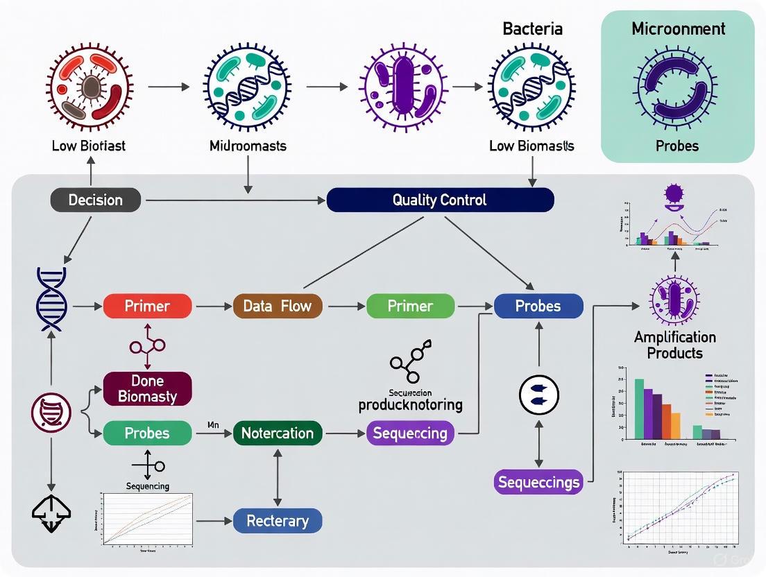
Workflow Diagram: Integrated Quality Control Strategy
A successful low-biomass study integrates vigilance and validation at every stage.
Frequently Asked Questions (FAQs)
1. Why is contamination a particularly critical issue in low-biomass microbiome studies? In low-biomass environments (such as human tissues, treated drinking water, or hyper-arid soils), the amount of target microbial DNA is very small. Any contaminating DNA introduced from external sources or other samples can make up a large proportion of the sequenced DNA, potentially overwhelming the true biological signal and leading to incorrect conclusions [7] [2].
2. What are the most common sources of contamination in a sequencing workflow? The primary sources are:
- Reagents and Kits: Laboratory reagents, DNA extraction kits, and polymerases often contain trace amounts of microbial DNA [7] [8].
- Human Operators: Microbial cells and DNA can be introduced from researchers' skin, hair, or breath via shedding and aerosols [7] [8].
- Cross-Contamination: DNA can transfer between samples during processing, especially between adjacent wells on a plate, a phenomenon known as well-to-well leakage [7] [9].
3. How can I detect cross-contamination in my dataset? Strain-resolved analysis of metagenomic data can reveal cross-contamination. By examining strain-sharing patterns across the extraction plate, you can identify if nearby samples are more likely to share strains than distant ones, which is a key signature of well-to-well leakage [9].
4. What is the minimum number of negative controls I should include? While the optimal number can vary, it is recommended to include multiple controls for each contamination source. Including at least two controls per batch is preferable to a single control, as it helps account for variability and provides more robust contamination profiling [2].
Troubleshooting Guides
Problem: Suspected Contamination from Reagents or Kits
Symptoms:
- Consistent detection of the same microbial taxa (e.g., Cutibacterium acnes) across multiple unrelated samples and negative controls [9].
- High background noise in negative controls that undergo the same DNA extraction and library preparation as biological samples [7].
Diagnostic and Mitigation Strategies:
| Strategy | Description | Key Details |
|---|---|---|
| Use Process Controls | Include negative controls containing only the reagents used for sampling, DNA extraction, and library preparation. | These controls should be processed alongside every batch of samples to capture the "background" contaminant profile [7] [2]. |
| Source DNA-Free Reagents | Purchase reagents that are certified DNA-free or have been treated to remove microbial DNA. | Request contamination profiles from vendors for critical reagents [8]. |
| Treat Reagents | Pre-treat reagents with methods to degrade DNA, such as UV irradiation or DNase treatment, where protocols allow [7]. | UV-C light exposure or DNA-degrading solutions can be effective [7]. |
Problem: Suspected Contamination from Human Operators
Symptoms:
- Detection of human skin commensals, such as Cutibacterium and Staphylococcus, in samples that should theoretically be sterile [9].
Diagnostic and Mitigation Strategies:
| Strategy | Description | Key Details |
|---|---|---|
| Use Personal Protective Equipment (PPE) | Wear gloves, masks, lab coats, and hair covers during sample handling. | Gloves should be decontaminated with ethanol and nucleic acid degrading solutions and changed frequently [7]. |
| Decontaminate Surfaces and Tools | Regularly clean work areas and equipment with agents that remove DNA. | Use 80% ethanol to kill cells, followed by a DNA-degrading solution like sodium hypochlorite (bleach) to remove residual DNA [7]. |
| Minimize Sample Handling | Reduce direct contact with the sample by using single-use, sterile equipment and automating processes where possible [7] [8]. |
Problem: Suspected Cross-Contamination (Well-to-Well Leakage)
Symptoms:
- Unusual strain-sharing between samples that are not biologically related.
- Negative controls contain strains that are also found in other samples processed on the same plate [9].
- Contamination is more likely between samples located on the same or adjacent columns/rows of an extraction plate [9].
Diagnostic and Mitigation Strategies:
| Strategy | Description | Key Details |
|---|---|---|
| Analyze Plate Layout | Map strain-sharing events back to the physical layout of the DNA extraction plate. | A significant pattern where adjacent wells share more strains than distant wells indicates well-to-well leakage [9]. |
| Randomize Sample Placement | When designing plate layouts, do not group samples by experimental group. Instead, randomize samples from different groups across the plate. | This prevents batch effects where contamination becomes confounded with a specific phenotype [2]. |
| Include Blank Wells | Place blank controls (e.g., water) interspersed throughout the plate, not just in one corner, to detect spatial contamination patterns [2]. |
Experimental Protocols
Protocol 1: Comprehensive Contamination Control During Sample Collection
This protocol outlines steps to minimize contamination introduction during the initial sampling phase [7].
- Decontaminate Equipment: Use single-use, DNA-free collection vessels whenever possible. Reusable equipment should be decontaminated with 80% ethanol followed by a DNA-degrading solution like diluted sodium hypochlorite.
- Wear Appropriate PPE: Personnel should wear gloves, masks, and protective suits. Gloves should be decontaminated or changed between handling different samples or equipment.
- Collect Sampling Controls: During collection, also gather controls such as:
- An empty collection vessel.
- A swab of the air in the sampling environment.
- An aliquot of any preservation solution used.
- Minimize Exposure: Keep samples sealed until the moment of processing and avoid unnecessary handling.
Protocol 2: Computational Detection of Cross-Contamination Using Strain-Resolved Analysis
This workflow helps identify cross-contamination in metagenomic sequencing data [9].
- Sequence and Assemble: Perform metagenomic sequencing on all samples and controls. Conduct de novo genome assembly to reconstruct metagenome-assembled genomes (MAGs) from your samples.
- Dereplicate Genomes: Cluster highly similar genomes to create a non-redundant set of representative genomes.
- Map Reads: Map sequencing reads from all samples and controls back to this representative genome set.
- Identify Strain Sharing: Determine which samples share identical strains of the same organism.
- Map to Extraction Plate: Visualize strain-sharing patterns on a diagram of the multi-well plate used for DNA extraction. Statistically test whether nearby wells share significantly more strains than distant wells.
The following diagram illustrates the core workflow for this computational detection method:
The Scientist's Toolkit: Essential Reagents and Materials
| Item | Function in Contamination Control |
|---|---|
| DNA-Free Water | Serves as a blank control and dilution reagent; certified to be free of microbial DNA to prevent introduction of contaminants from water itself [7]. |
| UV-C Crosslinker | Used to pre-treat reagents and plasticware with ultraviolet light to degrade any contaminating DNA present before use [7]. |
| Sodium Hypochlorite (Bleach) | A chemical DNA-degrading agent used for surface and equipment decontamination after initial cleaning with ethanol [7]. |
| Unique Dual Indexed (UDI) Primers | Primers with unique barcode combinations on both ends used during library preparation to drastically reduce misassignment of reads between samples (index switching) [9]. |
| Certified DNA-Free Extraction Kits | DNA extraction kits that have been tested and treated to minimize the background levels of microbial DNA within the kit components [8]. |
| Sample Collection Swabs | Pre-sterilized, single-use swabs designed for DNA-free collection of samples from surfaces or tissues [7]. |
In low-biomass microbiome research—the study of environments with minimal microbial life, such as human tissues like placenta and tumors, or austere environments like deep subsurface and treated drinking water—the signal from the actual sample can be dwarfed by the noise from contamination [7] [2]. This contamination can originate from a myriad of sources, including laboratory reagents, sampling equipment, human operators, and even cross-contamination between samples on a sequencing plate [7] [2]. When working near the limits of detection, these contaminants are not merely minor nuisances; they can drastically skew results, leading to false ecological patterns, incorrect attribution of pathogen exposure, and ultimately, retractions and scientific controversies [7] [2]. The stakes for rigorous quality control have never been higher. This guide provides actionable troubleshooting and FAQs to help you navigate these pitfalls.
Frequently Asked Questions (FAQs) and Troubleshooting
FAQ-1: What makes low-biomass research so vulnerable to contamination?
In high-biomass samples (e.g., stool), the target DNA "signal" is far larger than the contaminant "noise." In low-biomass samples, this relationship is inverted. Contaminating DNA, which is inevitable, can constitute a large proportion, or even the majority, of the sequenced DNA [7]. This can lead to two primary types of errors:
- False Positives: Mistaking contaminants for genuine sample constituents.
- Distorted Biological Signals: Contamination can obscure true signals or create artifactual patterns, especially if the contamination is confounded with experimental groups or batches [2]. A well-known example is the debate over the existence of a placental microbiome, where initial findings were later attributed to contamination [7] [2].
Contamination can be introduced at virtually every stage of your workflow. The table below summarizes the key sources and their origins.
Table 1: Key Contamination Sources in Low-Biomass Microbiome Studies
| Contamination Source | Description | Common Examples |
|---|---|---|
| External Contamination [7] [2] | DNA introduced from sources outside the sample. | Laboratory reagents and kits [7] [10], sampling equipment [7], human operators (skin, hair, breath) [7], and the collection environment (e.g., air) [7]. |
| Cross-Contamination (Well-to-Well Leakage) [2] | The transfer of DNA between samples processed concurrently, often in adjacent wells on a plate. | Can lead to the "splashome," where signals from one high-biomass sample appear in a neighboring low-biomass sample [2]. |
| Host DNA Misclassification [2] | Not contamination in the traditional sense, but host-derived DNA (e.g., human) can be misidentified as microbial during analysis. | A significant problem for metagenomic studies of human tissues, where the vast majority of sequenced reads are from the host and can be misannotated as microbial if not properly filtered [2]. |
| Computational Contamination [11] | Contaminant sequences that are present in public reference databases, leading to misclassification. | Human DNA embedded in non-primate reference genomes, or common control sequences (e.g., PhiX) present in published genomes [11]. |
FAQ-3: What are the most critical steps to prevent contamination during sample collection?
Prevention is always more effective than post-hoc correction. Key steps include:
- Decontaminate Equipment: Use single-use, DNA-free collection vessels. Reusable equipment should be decontaminated with 80% ethanol (to kill cells) followed by a nucleic acid degrading solution (e.g., bleach, UV-C light) to remove residual DNA [7].
- Use Personal Protective Equipment (PPE): Wear gloves, masks, clean suits, and other PPE to create a barrier between the operator and the sample, reducing contamination from skin cells and aerosols [7].
- Standardize Nomenclature: Use precise terminology (e.g., "urinary bladder" vs. "urogenital" for urine samples) to ensure clarity about a sample's origin and the potential for contamination during collection [12].
FAQ-4: Which experimental controls are non-negotiable for my study?
Including the right controls is essential for identifying contaminants during data analysis. We recommend incorporating multiple types of controls throughout your workflow.
Table 2: Essential Process Controls for Low-Biomass Studies
| Control Type | Purpose | Implementation |
|---|---|---|
| Negative Controls (Blanks) [7] [2] | To profile the "background noise" of contamination introduced during wet-lab procedures. | Include an empty collection tube, a swab exposed to the air, and an aliquot of pure preservation solution. These should be processed alongside your real samples through DNA extraction and sequencing [7]. |
| Positive Controls (Mock Communities) [10] [4] | To assess bias and accuracy in your entire workflow, from DNA extraction to sequencing. | Use a defined mix of microbial cells (whole-cell mock) or their DNA (DNA mock) with a known composition. Deviation from the expected result reveals protocol-specific biases, such as lysis inefficiency for tough cells [10] [4]. |
| Process-Specific Controls [2] | To pinpoint the exact stage where contamination is introduced. | Examples include swabbing the inside of a glove, sampling the DNA extraction kit reagents alone, or a no-template PCR control [2]. |
The following workflow diagram illustrates how to integrate these controls and key steps into a robust low-biomass research pipeline.
FAQ-5: My sequences are generated. How can I computationally identify and remove contaminants?
Several robust computational tools and strategies exist to decontaminate your data.
- Host DNA Removal: It is critical to filter out host-derived sequences before microbiome analysis. This can be done by aligning reads to the host reference genome (e.g., GRCh37/hg19 or GRCh38/hg38) using tools like BWA or Bowtie and retaining only the unmapped reads [13].
- Contaminant Identification with Controls: The
Decontampackage in R uses the prevalence or frequency of sequence variants in your negative controls to identify and remove contaminants present in your true samples [2]. - Comprehensive Pipeline Tools: All-in-one pipelines like CLEAN can remove a wide range of unwanted sequences, including host DNA, common spike-ins (e.g., PhiX), and ribosomal RNA, in a single reproducible step [14]. For targeted removal of human contamination from FASTQ files, bbsplit (from the BBTools suite) is also a validated option [15].
FAQ-6: Troubleshooting: I followed best practices, but my results still look suspicious. What now?
If your results are still questionable, investigate these common pitfalls:
- Check for Batch Confounding: Ensure your experimental batches (e.g., DNA extraction plates, sequencing runs) are not perfectly correlated with your sample groups (e.g., all cases processed in one batch and all controls in another). This confounding can make batch-specific contamination appear as a biological signal [2].
- Validate with Mock Communities: Analyze your positive control mock community. If the observed composition deviates significantly from the expected one, you have a quantitative measure of your workflow's bias, which may preclude confident biological conclusions [10] [4].
- Beware of "Kitome": The microbial background of your specific DNA extraction kits and reagents, known as the "kitome," can vary by manufacturer and even by lot number. If you switch kits mid-study, this can introduce a major batch effect [10].
The Scientist's Toolkit: Essential Research Reagents and Materials
Having the right materials is fundamental to success. The following table details key reagents and their critical functions in ensuring data integrity.
Table 3: Key Research Reagent Solutions for Low-Biomass Research
| Item | Function | Key Considerations |
|---|---|---|
| DNA/RNA Stabilizing Solution (e.g., DNA/RNA Shield) [4] | Immediately halts microbial activity and enzymatic degradation at collection, "freezing" the microbial profile. | Prevents shifts in community structure during transport. Enables room-temperature shipping, unlike freezing which risks cell lysis during thaw [4]. |
| Mechanical Lysis Kits (e.g., ZymoBIOMICS) [10] [4] | DNA extraction kits that include bead-beating to physically disrupt tough cell walls. | Critical for lysing Gram-positive bacteria, which are often under-represented with chemical-only lysis methods, preventing "lysis bias" [10] [4]. |
| Mock Community Standards [10] [4] | Defined mixtures of microorganisms (whole-cell) or their DNA, serving as positive controls. | Whole-cell mocks assess the entire workflow (including lysis). DNA mocks assess steps from library prep onward. Comparing them helps pinpoint the source of bias [10] [4]. |
| PCR Inhibitor Removal Technology [4] | Specialized columns or buffers in extraction kits that remove humic acids, bile salts, etc. | Inhibitors from complex samples (stool, soil) can cause PCR failure or skew communities. Effective removal ensures results reflect biology, not chemistry [4]. |
| Human DNA Depletion Kits [13] | Selectively degrade or remove abundant host DNA from samples rich in human cells (e.g., tissue, blood). | Increases the proportion of microbial reads in metagenomic sequencing, improving detection sensitivity and reducing sequencing costs [13]. |
Investigations into low-biomass microbial communities, such as those potentially residing in the placenta and internal tumors, hold great promise for advancing human health but are fraught with methodological challenges that can compromise biological conclusions [2]. The core controversy centers on distinguishing true microbial signals from contamination introduced during sampling, laboratory processing, or data analysis [7] [2]. In these environments, where microbial DNA is scarce, even minute amounts of contaminating DNA can dominate the signal, leading to false discoveries and enduring scientific debates [7] [16]. This technical support center outlines the critical lessons from these controversies and provides actionable troubleshooting guides to ensure the integrity of low-biomass microbiome research.
Case Study 1: The Placental Microbiome Controversy
The Debate and Its Resolution
The long-standing dogma that the human placenta is a sterile environment was challenged in 2014 when a study using high-throughput sequencing identified a unique placental microbiome composed of specific bacterial phyla, including Firmicutes, Tenericutes, Proteobacteria, Bacteroidetes, and Fusobacteria [17] [18]. This suggested that the in utero environment was not sterile and that the fetus could be exposed to microorganisms before birth. However, subsequent studies with more rigorous controls demonstrated that the bacterial DNA detected in many of these studies likely originated from contamination, either from laboratory reagents or during sample collection [7] [19]. The scientific community remains divided, with some experts arguing that the evidence is more consistent with the "sterile womb" hypothesis, given the existence of germ-free animal models and the inconsistent findings across studies [19].
Key Methodological Flaws and Lessons
The primary lesson from the placental microbiome debate is the absolute necessity of comprehensive contamination controls in low-biomass studies [2]. Key flaws in early studies included:
- Inadequate Controls: Many initial studies failed to include critical negative controls, such as blank extraction kits, no-template PCR controls, and sterile swabs exposed to the air during sample collection [7] [19]. Without these, distinguishing contamination from true signal is impossible.
- Reagent Contamination: Commercial DNA extraction kits and PCR reagents are known to contain trace amounts of bacterial DNA, which become disproportionately amplified in low-biomass samples [7].
- Sample Collection Contamination: Insufficient decontamination of the placental surface before sampling can introduce maternal vaginal or skin microbiota, leading to misleading results [17] [18].
Case Study 2: The Tumor Microbiome Controversy
The Emergence of a Contentious Field
Similar to the placental debate, research claiming the existence of unique microbiomes within tumors of internal organs (e.g., pancreas, breast, lung) has sparked significant controversy [16] [20]. While it is established that some microbes can directly cause cancer (e.g., Helicobacter pylori in stomach cancer) and that the gut microbiome can influence cancer therapy effectiveness, the claim that internal tumors harbor their own thriving microbial communities is hotly contested [16]. A high-profile 2020 study claiming that tumors from 33 different cancers had unique microbiomes was later heavily critiqued for potential contamination in databases and methodological flaws, leading to a retraction of a related paper and heightened scrutiny of the field [16].
Key Methodological Flaws and Lessons
The tumor microbiome debate underscores several critical points:
- Host DNA Misclassification: In metagenomic studies, sequences originating from the host can be misclassified as microbial, generating noise or artifactual signals, especially when host DNA levels are confounded with a phenotype [2].
- Cross-Contamination (Well-to-Well Leakage): DNA can leak between adjacent wells on a plate during library preparation, a phenomenon known as the "splashome," which can transfer signals between samples and controls [7] [2].
- Database Contamination: Reference databases can contain contaminants or human sequences, leading to false positive microbial identifications during bioinformatics analysis [16].
The Scientist's Toolkit: Essential Reagents and Controls
The following table details essential materials and controls required for robust low-biomass microbiome studies.
Table 1: Research Reagent Solutions for Low-Biomass Studies
| Item | Function | Critical Consideration |
|---|---|---|
| DNA-Free Collection Swabs/Tubes | To collect samples without introducing contaminating DNA. | Pre-treat with UV irradiation or bleach to degrade any contaminating DNA [7]. |
| Personal Protective Equipment (PPE) | To limit contamination from human operators (skin, hair, breath). | Use gloves, masks, and clean suits as a barrier between the sample and the researcher [7]. |
| Multiple Negative Controls | To identify the profile and level of contamination from various sources. | Includes blank extraction kits, no-template PCR controls, and sampling controls (e.g., air swabs) [7] [2]. |
| DNA Degrading Solution (e.g., Bleach) | To decontaminate surfaces and equipment. | More effective than ethanol alone at removing contaminating DNA [7]. |
| High-Fidelity Polymerase | For PCR amplification of marker genes. | Reduces amplification bias and errors in community representation [21]. |
| Quantification Standards (Qubit, qPCR) | For accurate measurement of DNA concentration. | Preferable to NanoDrop, which can overestimate concentration due to contaminants [22]. |
Troubleshooting Guide: FAQs for Low-Biomass Experiments
Table 2: Common Problems and Solutions in Low-Biomass Sequencing
| Problem Category | Failure Signals | Root Causes | Corrective Actions |
|---|---|---|---|
| External Contamination | Microbial profiles dominated by taxa common in reagents (e.g., Burkholderia, Ralstonia) or on human skin. | Contaminated reagents, improper sample collection, inadequate surface decontamination. | Implement rigorous negative controls at every stage; decontaminate surfaces with bleach; use DNA-free consumables [7] [2]. |
| Low Library Yield | Low final DNA concentration; poor amplification; flat coverage. | Sample loss during purification; inhibitor carryover; inaccurate quantification. | Re-purify input sample; use fluorometric quantification (Qubit) over UV; optimize bead-based cleanup ratios [22]. |
| Cross-Contamination (Well-to-Well Leakage) | Correlation between microbial signals and sample position on plates; contaminants appear in negative controls. | Splashing or aerosol transfer between wells during pipetting. | Use physical barriers between wells; randomize sample positions; include multiple negative controls dispersed across the plate [7] [2]. |
| High Duplicate Rate / Low Complexity | Overamplification artifacts; skewed community representation. | Too many PCR cycles; low input DNA; poor ligation efficiency. | Reduce the number of PCR cycles; titrate adapter-to-insert ratios; verify fragmentation size distribution [22]. |
| Host DNA Misclassification | High percentage of host reads in metagenomic data; false microbial assignments. | Insufficient host DNA depletion; misannotation in reference databases. | Use probe-based host DNA depletion kits; carefully curate reference databases to remove human sequences [2] [16]. |
Experimental Protocols for Robust Low-Biomass Research
Recommended Workflow for Sample Collection and Processing
The following diagram visualizes a rigorous end-to-end workflow designed to minimize and monitor contamination.
Protocol: Contamination-Aware Sample Collection
Objective: To collect placental or tumor tissue samples while minimizing and tracking contamination. Materials: Sterile surgical tools, DNA-free swabs and containers, DNA decontamination solution (e.g., 5% bleach), personal protective equipment (PPE). Procedure:
- Pre-collection: Decontaminate all surfaces and tools with a DNA-degrading solution. Personnel should wear full PPE (gloves, mask, gown) [7].
- Control Collection:
- Sample Collection:
- For placenta: After delivery, use sterile instruments to collect tissue from both the maternal and fetal sides, avoiding passage through the vagina if possible [17].
- For tumors: Collect tissue from the internal part of the tumor using sterile techniques.
- Storage: Immediately freeze all samples and controls at -80°C.
Protocol: DNA Extraction and Library Preparation with Controls
Objective: To generate sequencing libraries while controlling for reagent contamination and cross-contamination. Materials: DNA extraction kit, library preparation kit, fluorometric quantification kit. Procedure:
- DNA Extraction: Process actual samples and the following controls simultaneously in the same batch:
- Library Preparation:
- Quantification and Pooling: Quantify libraries using qPCR-based methods to measure only amplifiable molecules. Pool libraries equimolarly.
Pathway: From Contamination to Artifactual Discovery
The diagram below illustrates how methodological pitfalls can lead to false conclusions in low-biomass studies.
Building a Bulletproof Workflow: Best Practices from Sample Collection to Sequencing
Decontamination Protocols for Sampling Equipment and Surfaces
Frequently Asked Questions (FAQs)
FAQ 1: Why is a two-step decontamination process (ethanol followed by bleach) recommended for sampling equipment? A two-step process is critical because sterility and being DNA-free are not the same. The first step, using a solution like 80% ethanol, kills contaminating microorganisms. The second step, using a DNA-degrading solution like sodium hypochlorite (bleach), removes residual cell-free DNA that can persist on surfaces even after autoclaving or ethanol treatment. This comprehensive approach minimizes both viable contaminants and environmental DNA that could be amplified in sequencing [7].
FAQ 2: What are the most common sources of contamination I need to guard against during sampling? The major contamination sources during sampling include:
- Human operators: From skin, hair, or aerosol droplets generated while breathing or talking.
- Sampling equipment: Such as tools, collection vessels, and swabs that are not properly decontaminated.
- Adjacent environments: For example, a patient's skin during a blood draw, or overlying water during sediment collection [7] [2].
- Reagents and kits: Even DNA extraction kits and preservation solutions can contain microbial DNA [7] [12].
FAQ 3: My study involves patient samples. How do I select the appropriate level of disinfection for different types of equipment? The level of disinfection or sterilization required depends on how the patient-care device is used, in accordance with CDC guidelines:
- Sterilization is required for critical devices that enter sterile tissue or the vascular system (e.g., surgical instruments) [23].
- High-level disinfection is required for semicritical devices that contact mucous membranes or nonintact skin (e.g., endoscopes, endotracheal tubes) [23].
- Low-level disinfection is sufficient for noncritical devices and surfaces that contact intact skin (e.g., blood pressure cuffs, bedrails) [23].
Troubleshooting Guides
Problem: Consistent detection of common laboratory contaminants in negative controls.
- Potential Cause: Ineffective decontamination of reusable equipment or contaminated reagents.
- Solution:
- Implement the two-step decontamination protocol (ethanol followed by DNA removal solution) for all reusable equipment [7].
- Check that sampling reagents (e.g., preservation solutions) are certified DNA-free.
- Use single-use, pre-sterilized disposable items (e.g., swabs, collection vessels) wherever possible [7] [12].
- Increase the number of negative controls (e.g., empty collection vessels, aliquots of preservation solution) to better identify the contamination source [7] [2].
Problem: High variation in contamination profiles between sample batches.
- Potential Cause: Differences in reagent lots, personnel, or environmental conditions between processing batches (batch effects) [2].
- Solution:
- Avoid batch confounding: Design your experiment so that case and control samples are distributed across all processing batches [2].
- Use process controls: Collect multiple types of control samples (e.g., blank extraction controls, library preparation controls) in every processing batch to account for batch-specific contamination [2].
- Document meticulously: Record reagent lot numbers and personnel for all steps to help trace the source of variation [12].
Problem: Suspected cross-contamination (well-to-well leakage) between samples on a plate.
- Potential Cause: Splashing or aerosol transfer between adjacent wells during liquid handling [7] [2].
- Solution:
- Physical separation: If possible, leave empty wells between samples, especially between high-biomass and low-biomass samples [2].
- Analytical decontamination: Use computational tools designed to identify and subtract contamination arising from well-to-well leakage during data analysis [2] [24].
- Include controls: Place negative control samples throughout the plate to map the spatial pattern of any leakage [2].
Decontamination Methods for Sampling Equipment
The table below summarizes common decontamination methods, their primary mechanisms, and applications in microbiome research.
Table 1: Summary of Decontamination Methods and Applications
| Method | Mechanism | Common Applications | Key Considerations |
|---|---|---|---|
| Autoclaving | High-pressure saturated steam sterilizes by killing all microorganisms, including spores. | Glassware, metal tools, heat-stable plastics [7]. | Does not remove persistent environmental DNA; items may not be DNA-free post-treatment [7]. |
| Ethanol (e.g., 80%) | Denatures proteins and lyses cells, effectively killing microorganisms. | Initial decontamination of surfaces, gloves, and some equipment [7]. | Often used as a first step; does not effectively remove contaminant DNA [7]. |
| Sodium Hypochlorite (Bleach) | Oxidizes and degrades microbial DNA and proteins. | Secondary treatment to remove DNA; surface decontamination [7] [23]. | Effective for making surfaces DNA-free; requires proper concentration and safety precautions [7] [23]. |
| UV-C Irradiation | Damages DNA/RNA through pyrimidine dimer formation, preventing replication. | Sterilization of plasticware, surfaces in hoods, and laboratory air [7] [25]. | Effectiveness depends on exposure time, distance, and surface shading; may not fully degrade all DNA [7]. |
Experimental Protocol: Two-Step Decontamination of Reusable Sampling Tools
This protocol is designed for metal or heat-stable plastic tools (e.g., forceps, spatulas) used in low-biomass environments.
1. Principle: To render sampling tools free from both viable microbial cells and environmental DNA contaminants through a sequential process of sterilization and DNA degradation.
2. Reagents and Equipment:
- Reusable sampling tools
- 80% (v/v) Ethanol solution
- Freshly prepared 1-3% (v/v) Sodium hypochlorite (bleach) solution
- DNA-free water (e.g., PCR-grade water)
- Autoclave
- UV-C crosslinker or cabinet (optional)
- Sterile containers
3. Step-by-Step Procedure:
- Initial Cleaning: Meticulously clean tools with water and detergent to remove any visible organic or inorganic residue [23].
- Sterilization: Autoclave the cleaned tools using a standard cycle (e.g., 121°C for 15-30 minutes) [7] [23]. Store sterilized tools in sealed bags or containers until use.
- Pre-Sampling Decontamination: a. Ethanol Treatment: Submerge or thoroughly wipe the tools with an 80% ethanol solution to kill any contaminating organisms [7]. b. DNA Removal: Submerge or thoroughly wipe the tools with a 1-3% sodium hypochlorite solution to degrade any residual DNA [7]. c. Rinsing: Rinse the tools thoroughly with DNA-free water to remove any residual bleach, which can interfere with downstream molecular assays [7]. d. Drying: Allow the tools to air dry completely in a clean, DNA-free environment. Alternatively, use UV-C irradiation for final sterilization and to degrade any potential residual DNA [7].
Workflow for Selecting a Decontamination Method
The following diagram outlines a logical decision-making workflow for selecting an appropriate decontamination protocol based on the sample type and equipment.
The Scientist's Toolkit: Key Research Reagent Solutions
Table 2: Essential Materials for Decontamination and Contamination Control
| Item | Function / Purpose |
|---|---|
| Sodium Hypochlorite (Bleach) | DNA removal solution for surfaces and equipment to degrade contaminant DNA [7]. |
| 80% Ethanol | Initial decontamination agent to kill viable microorganisms on surfaces and equipment [7]. |
| DNA-Free Water | Used for preparing solutions and final rinsing of equipment to prevent introduction of environmental DNA [7]. |
| Personal Protective Equipment (PPE) | Gloves, masks, goggles, and coveralls act as barriers to limit contamination from human operators [7] [25]. |
| Pre-Sterilized Swabs & Collection Tubes | Single-use items to avoid cross-contamination between samples and eliminate the need for in-house decontamination [7] [12]. |
| UV-C Lamp or Crosslinker | Provides ultraviolet germicidal irradiation for decontaminating surfaces, air, and equipment in laboratories [7] [25]. |
The Critical Role of Personal Protective Equipment (PPE) and Physical Barriers
Troubleshooting Guides and FAQs
FAQ: Why is PPE so critical in low-biomass microbiome studies?
In low-biomass environments, the microbial DNA from the sample is minimal. Contaminant DNA from researchers, the lab environment, or reagents can constitute a significant portion, or even all, of the recovered genetic material, leading to false positives and incorrect conclusions. Proper PPE acts as a physical barrier, minimizing the introduction of this contaminant "noise" from personnel [7].
FAQ: I wear a lab coat and gloves. Is that sufficient for low-biomass work?
For very low-biomass samples, standard lab coats and gloves are often insufficient. Best practices recommend more extensive PPE, similar to protocols used in ancient DNA laboratories or cleanrooms. This can include face masks, goggles, coveralls or cleansuits, and shoe covers. The goal is to cover all exposed body parts to protect the sample from human aerosol droplets and cells shed from skin, hair, and clothing [7].
FAQ: A common issue in our lab is cross-contamination between samples. Could PPE be a factor?
Yes. PPE can be a vector for cross-contamination if not managed correctly. Gloves should be decontaminated or changed between handling different samples. Furthermore, PPE like suits or lab coats should not be worn in non-lab areas (like break rooms) and then brought back into clean sample processing areas, as this can transport contaminants [7] [26].
FAQ: What are the most common mistakes in using PPE for contamination control?
Common mistakes that compromise safety and experimental integrity include:
- Poor Fit: Ill-fitting PPE can leave gaps for contaminants to escape or enter, and can be a safety hazard [27] [28] [29].
- Inadequate Cleaning: PPE must be cleaned according to manufacturer specifications to maintain its protective function. Cleaning contaminated PPE at home is prohibited, as it can introduce pathogens into the home environment and domestic machines can damage protective impregnations [27].
- Shared Use Without Sanitization: PPE is generally intended for use by one person. If different employees must use the same equipment, the employer must ensure there are no hygiene or health hazards [27].
- Lack of Training: Employees may not use PPE correctly without comprehensive training on its proper use, limitations, and maintenance [27] [29].
Experimental Protocols for Contamination Control
Protocol for Sampling Ultra-Low Biomass Surfaces
This protocol, adapted from cleanroom and spacecraft assembly facility procedures, details a method for sampling surfaces with minimal microbial biomass [30].
- Objective: To collect microbial biomass from large surface areas (e.g., cleanroom floors, equipment) for downstream DNA analysis while minimizing contamination.
- Key Materials:
- Methodology:
- Don PPE: Before entering the sampling environment, don full cleanroom PPE (coveralls, gloves, face masks, shoe covers) to minimize human-derived contamination [7].
- Pre-wet Surface: Spray a defined area (e.g., 12" x 12") with sterile, DNA-free water [30].
- Collect Sample: Using a sterile, disposable collection head on the SALSA device, squeegee and aspirate the liquid from the target area. The liquid is deposited directly into a sterile collection tube, bypassing the need for an elution step required with swabs [30].
- Process Controls: Collect "process control" samples by aspirating aliquots of the sprayer water using the same collection equipment without active sampling. This controls for contamination from the reagents and equipment itself [30].
- Concentrate Sample: Immediately concentrate the sample using a device like an InnovaPrep CP-150 with a hollow fiber concentrating pipette tip, eluting into a small volume (e.g., 150 µL) for downstream processing [30].
Protocol for Incorporating Controls in a Low-Biomass Study
This is a critical meta-protocol that should accompany all experimental procedures.
- Objective: To identify and account for contaminating DNA introduced during sampling and laboratory processing.
- Key Materials: Same reagents and kits used for actual samples.
- Methodology:
- Sample Collection Controls: During sampling, include controls such as an empty collection vessel, a swab exposed to the air, or an aliquot of the preservation solution [7].
- DNA Extraction & Library Preparation Controls: With each batch of samples, include multiple "negative control" or "blank" samples that contain only the DNA extraction and PCR/purification reagents. These "kitome" controls are essential for identifying contaminating DNA inherent in the molecular biology reagents themselves [7] [30].
- Sequencing and Analysis: Sequence all controls alongside the actual samples. In downstream bioinformatic analysis, the taxa and sequences found in the control samples should be compared to and potentially subtracted from those in the experimental samples [7].
Workflow Visualization
The following diagram illustrates the logical relationship between contamination sources, control measures, and desired outcomes in a low-biomass research setting.
The Scientist's Toolkit: Essential Research Reagent Solutions
The following table details key materials and their specific functions for ensuring contamination control in low-biomass research.
| Item | Function in Low-Biomass Research |
|---|---|
| DNA-Decontaminating Solutions (e.g., bleach, UV-C light, hydrogen peroxide) | Used to decontaminate surfaces and non-disposable equipment. Critical for removing cell-free DNA that remains even after ethanol treatment or autoclaving [7]. |
| DNA-Free Collection Tubes & Swabs | Single-use, pre-sterilized materials certified to be DNA-free to prevent introduction of contaminants at the first point of sample contact [7]. |
| Personal Protective Equipment (PPE) (Coveralls, gloves, masks, shoe covers) | Acts as a primary barrier, preventing microbial cells and DNA from the researcher from entering the sample collection and processing environment [7] [26]. |
| Sterile DNA-Free Water/Buffers | Used for sample collection, rehydration, or dilution. Must be certified sterile and DNA-free to avoid being a source of contaminating DNA [30]. |
| Concentration Devices (e.g., Hollow Fiber Concentrators) | Used to concentrate the often-dilute samples from large surface areas into a small volume suitable for DNA extraction and library preparation [30]. |
| Commercial DNA Removal Kits | Specialized solutions designed to degrade contaminating DNA on surfaces and equipment, providing a higher level of decontamination than standard cleaning [7]. |
Frequently Asked Questions (FAQs)
Q1: Why are controls so critical in low-biomass microbiome studies? In low-biomass environments, the microbial DNA from the sample itself is minimal. Consequently, any small amount of contaminating DNA introduced during sampling or laboratory processing can make up a large, and sometimes dominant, proportion of your final sequencing data [7] [2]. This contamination can distort the true microbial community, inflate diversity metrics, and lead to spurious biological conclusions [31]. Controls are essential for detecting this contaminating DNA so it can be accounted for.
Q2: What is the difference between a 'negative control' and a 'no-template control (NTC)'? The terms are sometimes used interchangeably, but they can be distinguished:
- No-Template Control (NTC): A control that contains only the PCR-grade water or buffer used in your amplification reactions. It is designed to identify contaminants introduced during the PCR and library preparation steps [32].
- Negative Control (Broader Term): This can encompass NTCs but also includes other controls that track contamination from earlier stages. Examples include blank extraction controls (which undergo the DNA extraction process with no sample) and sampling controls (like an empty collection tube or a swab of the air in the sampling environment) [7] [2].
Q3: How many negative controls should I include in my experiment? There is no universal number, but the consensus is that more than one is necessary. Including at least two controls is always preferable to a single control [2]. For large studies, you should include multiple controls distributed across your processing batches (e.g., one NTC and one blank extraction per plate) to accurately capture contamination variability [2].
Q4: Can I just subtract sequences found in my negative controls from my samples?
Simple subtraction is not recommended because it is too aggressive. This approach can erroneously remove true, low-abundance biological sequences that are also present in the control due to well-to-well leakage or other artifacts [31] [32]. Instead, use specialized computational tools like Decontam that use statistical methods to identify contaminants without over-correcting [31] [32].
Q5: What is "well-to-well leakage" or the "splashome"? This is a form of cross-contamination where DNA or amplicons physically "leak" from one sample well into adjacent wells on a PCR plate during laboratory processing [2]. This can cause sequences from a high-biomass sample to appear in neighboring low-biomass samples and negative controls, violating the assumptions of some decontamination methods [2].
Troubleshooting Guide
| Problem | Potential Cause | Recommended Solution |
|---|---|---|
| High biomass in negative controls | Contaminated reagents, improper sterile technique, or well-to-well leakage. | Use UV-irradiated water and reagents, include multiple control types, randomize sample plating to avoid confounding, and use physical barriers on plates [2] [33]. |
| Unexpected microbial taxa in samples | Contamination from kit reagents, laboratory environment, or personnel. | Profile all your reagents directly. Compare your sample taxa to those found in your negative controls using a tool like Decontam to identify likely contaminants [7] [31]. |
| Inconsistent profiles between technical replicates | Very low starting biomass, leading to stochastic amplification of contaminants or true signal. | Process multiple replicates. If replicates are highly inconsistent, it suggests the biomass is too low for reliable detection above the contaminant noise [32]. |
| Poor recovery of a mock community | DNA extraction bias against hard-to-lyse cells, or PCR bias. | Benchmark different DNA extraction kits using a diluted mock community to identify which kit provides the most accurate representation of the known composition [32]. |
| Strong batch effects | Samples processed in different batches (e.g., different extraction dates, reagent lots, or personnel) show artificial differences. | Design your study to ensure experimental groups are distributed evenly across all processing batches (avoid batch confounding). Include controls in every batch [2]. |
Key Experimental Protocols & Data
Protocol: Implementing a Comprehensive Control Strategy
The following workflow visualizes the integration of different controls throughout a typical low-biomass microbiome study:
Data Presentation: Evaluating Computational Decontamination Methods
A critical study compared the performance of different computational methods for identifying contaminant sequences in 16S rRNA data from a dilution series of a mock microbial community [31]. The results are summarized below:
Table 1: Performance of Computational Decontamination Methods on a Mock Community Dilution Series [31]
| Method | Principle | Key Finding | Performance |
|---|---|---|---|
| Subtract Contaminants in NTC | Removes any sequence found in a negative control. | Overly aggressive; erroneously removed >20% of expected sequences from the mock community. | Poor |
| Abundance Filtering | Removes sequences below a set relative abundance threshold. | Assumes contaminants are always low abundance, which is often incorrect in low-biomass samples. | Variable / Unreliable |
| SourceTracker | Bayesian method to predict proportion from contaminant sources. | Excellent (>98% contaminants removed) when contaminant sources are well-defined; poor (<3% removed) when sources are unknown. | Situation-Dependent |
| Decontam (Frequency) | Identifies sequences with an inverse correlation to DNA concentration. | Successfully removed 70-90% of contaminants without removing expected sequences. | Recommended |
Protocol: Optimizing 16S rRNA Gene PCR for Low-Biomass Samples
Based on benchmarking studies, the following protocol is recommended for amplifying the 16S rRNA gene from low-biomass samples [33]:
- Input DNA: Use the total extracted DNA, undiluted, especially if the concentration is low (<20 pg/µL). For higher biomass samples, dilute to a standardized input (e.g., 125 pg) [33].
- PCR Cycles: Perform amplification with 30 cycles. Studies show that varying the cycle number (25, 30, or 35) did not significantly alter the resulting microbial community profile for low-biomass samples [33].
- Library Purification: Clean the amplified PCR product using a double-size selection with AMPure XP beads (e.g., two consecutive clean-up steps) to remove primer dimers and other artifacts most effectively [33].
- Sequencing: Sequence the pooled libraries using an Illumina MiSeq with a V3 reagent kit, which provided the most robust results for low-biomass communities in a comparative study [33].
The Scientist's Toolkit: Essential Research Reagents & Materials
Table 2: Key Reagents and Materials for Low-Biomass Control Experiments
| Item | Function in Control Strategy | Example & Notes |
|---|---|---|
| Mock Microbial Community | Serves as a positive control to evaluate DNA extraction efficiency, PCR bias, and overall fidelity of the workflow. | ZymoBIOMICS Microbial Community Standard (cells) or DNA Standard (pre-extracted DNA) [32] [33]. |
| DNA-Free Water | Used to prepare No-Template Controls (NTCs) and to dilute samples/reagents. Must be certified DNA-free. | HPLC-grade water, UV-irradiated to fragment any contaminating DNA [33]. |
| DNA Decontamination Reagents | Used to remove contaminating DNA from work surfaces and non-disposable equipment. | Sodium hypochlorite (bleach), DNA removal solutions, or UV-C light exposure [7]. |
| DNA Extraction Kits | Different kits have varying efficiencies and contaminant profiles. Must be benchmarked. | Kits like the DSP Virus/Pathomen Mini Kit or ZymoBIOMICS DNA Miniprep Kit have been used in studies [32]. |
| AMPure XP Beads | For purifying amplicon libraries post-PCR. A double clean-up is recommended for low-biomass amplicons [33]. | A magnetic bead-based solution for size selection and clean-up. |
Frequently Asked Questions (FAQs)
1. Why is mechanical lysis considered essential for samples with tough cell walls? Mechanical lysis is crucial for disrupting the robust structural barriers found in many sample types. It uses physical force to break open tough cell walls that chemical or enzymatic methods alone cannot efficiently penetrate [34]. This is particularly important for materials like plant tissues (with cellulose and lignin), gram-positive bacteria (with thick peptidoglycan layers), fungal spores, and soil microbes, ensuring a representative and high-yield DNA extraction [34] [35].
2. How does mechanical lysis impact DNA quality and downstream applications? The intensity of mechanical lysis directly influences the trade-off between DNA yield and fragment length. High-intensity lysis can maximize yield but fragments DNA, which is detrimental for long-read sequencing technologies (e.g., Oxford Nanopore, PacBio) [36]. Optimized, lower-intensity lysis preserves High Molecular Weight (HMW) DNA, leading to longer sequenced read lengths (N50) and better genome assembly continuity in downstream metagenomic analyses [36].
3. What are the best practices for mechanical lysis in low-biomass microbiome studies? In low-biomass research, the primary goal is to minimize contamination while efficiently lysing the sparse native cells [7] [2]. Best practices include:
- Using DNA-free reagents and consumables.
- Decontaminating equipment (e.g., with 80% ethanol and DNA-degrading solutions like bleach) before use [7].
- Including comprehensive negative controls (e.g., blank extraction controls) to identify contaminating DNA sources [2].
- Avoiding over-lysing the sample, which can release excessive inhibitor compounds and degrade DNA [36].
4. Can I use mechanical lysis for all sample types? While highly effective for tough samples, mechanical lysis can be too harsh for easy-to-lyse cells like those from blood or tissue cultures, where chemical lysis is often sufficient and gentler [34] [37]. For delicate samples or those with very low microbial biomass, harsh mechanical beating may disproportionately lyse contaminating cells, skewing the microbial profile [2]. The method must be matched to the sample's physical properties.
Troubleshooting Guide
| Problem | Possible Cause | Solution |
|---|---|---|
| Low DNA Yield | Insufficient lysis; tough cell walls remain intact [37]. | Increase homogenization speed/time within limits; combine mechanical lysis with enzymatic pre-treatment (e.g., lysozyme for bacteria) [34]. |
| Short DNA Fragments | Mechanical lysis is too intense or prolonged [36]. | Reduce homogenization intensity. For soil, 4 m s⁻¹ for 10 s increased fragment length by 70% vs. manufacturer settings [36]. |
| Poor Microbial Community Representation | Lysis efficiency varies between cell types; some resistant cells remain unlysed [38]. | Use a consistent, optimized lysis protocol across all samples. Bead-beating with small beads provides more uniform lysis of Gram-positive bacteria [38]. |
| High Contamination in Low-Biomass Samples | Contaminant DNA from reagents, kit components, or the lab environment is co-extracted [7]. | Use dedicated, decontaminated equipment; include negative controls; employ computational decontamination tools post-sequencing [7] [2]. |
| Inconsistent Results Between Replicates | Inhomogeneous sample powder or uneven lysis during grinding/homogenization. | Ensure samples are ground to a fine, consistent powder in liquid nitrogen before homogenization [34] [35]. |
Optimized Experimental Protocols
Protocol 1: Optimized Bead-Beating for Soil Metagenomics
This protocol is designed to maximize DNA fragment length for long-read sequencing from soil samples, based on a statistical design of experiments approach [36].
- Key Materials: Homogenizer (e.g., FastPrep-24), Lysing Matrix E tubes, Phosphate Buffered Saline (PBS).
- Procedure:
- Weigh 0.25-0.5 g of soil into a lysing tube.
- Add the recommended lysis buffer.
- Homogenize at 4 m s⁻¹ for 10 seconds. This low-energy setting is critical for obtaining long DNA fragments [36].
- Centrifuge the lysate briefly to pellet soil particles and debris.
- Transfer the supernatant to a new tube for subsequent DNA purification using a silica-column or magnetic bead-based kit [34].
Table: Impact of Homogenization Parameters on Soil DNA Extraction [36]
| Homogenization Speed | Homogenization Time | Calculated Distance Travelled | Mean DNA Fragment Length | Total DNA Yield |
|---|---|---|---|---|
| 6 m s⁻¹ | 30 s | 180 m | ~4,400 bp | High |
| 4 m s⁻¹ | 10 s | 40 m | ~7,500 bp | Sufficient for library prep |
| 4 m s⁻¹ | 5 s | 20 m | ~9,300 bp | Sufficient for library prep |
Protocol 2: Integrated Lysis for Plant Tissues
Plant tissues require mechanical disruption to break rigid cell walls, followed by chemical steps to remove common inhibitors [37] [35].
- Key Materials: Liquid Nitrogen, Mortar and Pestle, CTAB Lysis Buffer, Chloroform, Silica-column purification kit.
- Procedure:
- Flash-freeze fresh leaf tissue (100 mg) in liquid nitrogen.
- Grind tissue to a fine powder using a pre-chilled mortar and pestle. Work quickly to prevent thawing and nuclease degradation. [35]
- Transfer the powder to a tube containing pre-warmed CTAB lysis buffer. CTAB helps remove polysaccharides and polyphenols [35].
- Incubate at 65°C for 30-60 minutes with occasional mixing.
- Add an equal volume of chloroform:isoamyl alcohol (24:1) to separate proteins and lipids.
- Centrifuge and transfer the aqueous upper phase to a new tube.
- Complete DNA purification using a silica-based column or magnetic beads [34] [37].
Workflow Visualization
Diagram 1: Integrated Lysis Strategy. Mechanical lysis is the critical first step for samples with robust cellular structures.
Diagram 2: Lysis Optimization for Long-Read Sequencing. Reducing homogenization intensity preserves DNA integrity for advanced genomic applications. [36]
The Scientist's Toolkit: Research Reagent Solutions
| Item | Function in Mechanical Lysis |
|---|---|
| Lysing Matrix E Tubes | Pre-filled tubes containing a mixture of ceramic and silica particles optimized for efficient mechanical disruption of a wide range of sample types, including soil and microbial cultures. |
| CTAB Buffer | Cetyltrimethylammonium bromide (CTAB) is a cationic detergent effective in lysing plant cells and precipitating polysaccharides and polyphenols, which are common PCR inhibitors [35]. |
| Proteinase K | A broad-spectrum serine protease used after initial mechanical disruption to digest contaminating proteins and nucleases, improving DNA purity and yield [34] [39]. |
| MagneSil Paramagnetic Particles | Silica-coated magnetic beads used in high-throughput, automated DNA purification workflows following mechanical lysis. They bind DNA in the presence of chaotropic salts for easy magnetic separation [34]. |
| Guanidine Hydrochloride | A chaotropic salt that disrupts cellular structures, inactivates nucleases, and promotes the binding of DNA to silica matrices during the purification phase [34]. |
Sequencing Technology Comparison Table
The table below summarizes the core characteristics of the three main sequencing technologies used in microbiome studies, with a focus on low-biomass applications.
| Feature | 16S rRNA Sequencing | Shotgun Metagenomics | 2bRAD-M |
|---|---|---|---|
| Taxonomic Resolution | Genus level (species level is often unreliable) [40] [41] | Species to strain level [42] | Species to strain level [43] [44] |
| Organisms Detected | Bacteria and Archaea only [41] | Bacteria, Archaea, Fungi, Viruses [42] | Bacteria, Archaea, Fungi [43] [44] |
| Ideal Sample Type | High microbial biomass; early decomposition stages [40] | High microbial biomass; minimal host DNA [40] [42] | Low-biomass, degraded, or high host-contamination samples (e.g., pg-level DNA, FFPE tissues) [40] [43] [44] |
| Relative Cost | Low [41] | High [40] [42] | Medium (lower than shotgun) [44] |
| Key Limitation | Low strain resolution; cannot identify microbial functions [40] [41] | High host DNA contamination leads to significant data loss; expensive [40] [42] | Relies on a pre-constructed reference database [40] |
| Contamination Risk | High risk in low-biomass samples; requires stringent controls [41] [7] | High risk of host "contaminating" reads [42] [2] | High resistance to host DNA contamination [44] |
Frequently Asked Questions & Troubleshooting
Which sequencing method is best for my low-biomass sample?
For true low-biomass samples (e.g., tissue biopsies, blood, forensic swabs), 2bRAD-M is often the superior choice due to its high sensitivity and resilience to host contamination [43] [44]. While 16S rRNA sequencing is cost-effective, its low taxonomic resolution and high susceptibility to contamination can lead to misleading results in low-biomass contexts [7] [45]. Shotgun metagenomics is comprehensive but can be wasteful and expensive for these samples, as over 99% of your data might be from the host [40] [2].
> Troubleshooting Tip: Validate Your 16S rRNA Results If you must use 16S rRNA sequencing, always include:
- Negative Controls: Empty collection vessels, swabs exposed to air, and blank extraction controls to identify contaminating sequences [7] [2].
- Mock Communities: Samples with known microbial composition to verify your workflow's accuracy and sensitivity [45].
Why is my sequencing yield so low, and how can I fix it?
Low library yield is a common issue in next-generation sequencing preparation. The causes and solutions are often related to sample quality and library preparation steps [22].
> Step-by-Step Diagnostic Guide:
- Check Input DNA Quality:
- Cause: Degraded DNA or contaminants (phenol, salts) inhibit enzymes.
- Fix: Re-purify input DNA. Check purity via spectrophotometry (260/280 ratio ~1.8, 260/230 > 1.8) [22].
- Verify Quantification Method:
- Cause: UV absorbance methods (e.g., NanoDrop) overestimate concentration by counting non-template DNA.
- Fix: Use fluorometric methods (e.g., Qubit) for accurate quantification of double-stranded DNA [22].
- Inspect Fragmentation & Ligation:
- Cause: Over- or under-fragmentation; inefficient ligation due to poor enzyme activity or incorrect adapter-to-insert ratio.
- Fix: Optimize fragmentation parameters. Titrate adapter concentrations and ensure fresh ligase buffer [22].
- Review Amplification:
- Cause: Too many PCR cycles lead to over-amplification artifacts and high duplication rates.
- Fix: Use the minimum number of PCR cycles necessary. Repeat the amplification from leftover ligation product if needed [22].
How can I prevent contamination in my low-biomass microbiome study?
Contamination is the primary confounder in low-biomass research. A multi-layered strategy is essential from sample collection to data analysis [7] [2].
> Essential Prevention Protocol:
- During Sampling:
- Decontaminate: Use single-use, DNA-free equipment. Decontaminate reusable tools with 80% ethanol followed by a DNA-degrading solution (e.g., bleach, UV-C light) [7].
- Use Barriers: Wear appropriate personal protective equipment (PPE) like gloves, masks, and clean suits to limit sample contact with skin, hair, or aerosols [7].
- During DNA Extraction and Library Prep:
| Control Type | Function | Example |
|---|---|---|
| Field/Collection Blank | Identifies contaminants from the sampling environment or equipment. | An empty collection vessel or a swab exposed to the air at the sampling site [7]. |
| Extraction Blank | Identifies contaminants from DNA extraction kits and reagents. | A tube with no sample added that goes through the entire DNA extraction process [2]. |
| Library Preparation Blank | Identifies contaminants introduced during library construction. | A water sample that undergoes the library prep and sequencing workflow [2]. |
- During Data Analysis:
- Bioinformatic Decontamination: Use tools to identify and subtract contaminants based on their prevalence in your negative controls. Be aware that well-to-well leakage can complicate this process [2].
My sample is highly degraded. Which method should I use?
2bRAD-M is specifically designed for this challenge [44]. The technology relies on sequencing very short, uniform tags (e.g., 32 bp) generated by restriction enzyme digestion. These tags are more likely to be preserved in degraded samples and can be evenly amplified, making the method far more robust than 16S or shotgun metagenomics when DNA is fragmented [40] [43].
Experimental Workflow Diagrams
16S rRNA Sequencing Workflow
Shotgun Metagenomic Sequencing Workflow
2bRAD-M Sequencing Workflow
Research Reagent Solutions
This table lists key reagents and materials critical for successful low-biomass microbiome sequencing experiments.
| Reagent/Material | Function | Critical Consideration for Low-Biomass |
|---|---|---|
| DNA-Free Collection Swabs/Tubes | Sample collection and storage. | Must be pre-sterilized and certified DNA-free to prevent introduction of contaminants at the first step [7]. |
| DNA Extraction Kit (for Stool/Soil) | Lyses microbial cells and purifies DNA. | Kit choice greatly impacts community profile. Select kits proven effective for your sample type and known to minimize contamination [41] [42]. |
| Type IIB Restriction Enzyme (BcgI) | Digests genomic DNA for 2bRAD-M library prep. | The core of 2bRAD-M; produces uniform, species-specific tags that enable analysis of degraded samples [46] [44]. |
| PCR Enzymes (High-Fidelity) | Amplifies target regions (16S or 2bRAD tags). | High-fidelity polymerase reduces amplification errors. Use minimal PCR cycles to avoid bias and chimeras [40] [22]. |
| Magnetic Beads (SPRI) | Purifies and size-selects DNA fragments post-amplification. | Incorrect bead-to-sample ratios cause loss of desired fragments or failure to remove adapter dimers. Precisely follow protocols [22]. |
| Negative Control Kits | Reagents for processing blank controls. | Use the same manufacturing lot of extraction kits and reagents as your actual samples to accurately control for kit-borne contaminants [7] [2]. |
Solving Common Problems: A Troubleshooting Guide for Low-Biomass Data
Frequently Asked Questions (FAQs)
1. What is batch confounding, and why is it a critical issue in low-biomass microbiome studies? Batch confounding occurs when your experimental batches (e.g., sample processing groups) are systematically linked to the biological groups you are comparing (e.g., case vs. control). In low-biomass research, where the genuine biological signal is weak, this can generate artifactual findings that are indistinguishable from true biological signals. For example, if all case samples are processed in one batch and all control samples in another, any technical differences between these batches (e.g., from reagents, personnel, or protocols) can be misinterpreted as disease-associated differences [2].
2. How can I identify if my study design has batch confounding? The primary indicator is a perfect or near-perfect correlation between your key biological variable (e.g., disease status) and batch identity. Before starting your experiment, review your sample processing schedule. If you see that all samples from one group are processed together in a single batch or on a specific day, your design is confounded. A well-designed study will intersperse samples from all biological groups across all processing batches [2].
3. What is the single most important step to prevent batch confounding? The most crucial step is proactive experimental planning. Rather than relying on post-hoc statistical correction, you should actively design your experiment so that batches are balanced across your key biological variables and covariates. This means ensuring that each processing batch contains a similar mix of case and control samples, representative of the overall study. Randomization can help, but a more active approach using tools like BalanceIT is recommended to achieve optimal balance [2].
4. My samples are collected from different clinical sites with different case-control ratios. How can I avoid confounding? In this scenario, where complete de-confounding is impossible (e.g., one site contributes only cases), it is not advisable to simply pool the data and apply batch correction. Instead, a more robust approach is to analyze the data from each site separately and then assess the generalizability and consistency of your findings across these independent batches [2].
Troubleshooting Guide: Identifying and Resolving Batch Confounding
Problem: Suspected artifactual signals due to batch confounding.
Step 1: Diagnose the Problem
- Visual Check: Create an ordination plot (e.g., NMDS or PCoA using Bray-Curtis dissimilarity) colored by both batch ID and biological group. If the samples cluster more strongly by batch than by the biological condition, batch effects are present and may be confounded [47].
- Statistical Test: Perform a PERMANOVA to test the statistical significance of the variance explained by batch and by your biological variable. If the effect of batch is significant and strong, and it is confounded with your biological variable, the results are compromised [47].
Step 2: Evaluate Corrective Actions
If confounding is detected, the appropriate action depends on your experimental design.
- If the design was unbalanced but not perfectly confounded: Statistical batch-effect correction methods may be applicable.
- If the design was perfectly confounded (e.g., all cases in one batch): Statistical correction is highly risky as it may remove the biological signal of interest. The most reliable course of action is to re-process a subset of samples in a balanced design to validate key findings [2].
Step 3: Implement a Robust Experimental Design for Future Studies
The table below summarizes the core components of a design that prevents batch confounding.
Table: Pillars of an Experimental Design to Avoid Batch Confounding
| Design Principle | Implementation Strategy | Benefit |
|---|---|---|
| Active Balancing | Use tools like BalanceIT during planning to assign samples from all biological groups to each processing batch [2]. | Prevents the entanglement of technical and biological variation from the start. |
| Randomization | Randomize the order of sample processing across all groups after active balancing. | Mitigates the effect of unknown or unmeasured technical biases. |
| Comprehensive Controls | Include multiple types of process controls (e.g., blank extractions, mock communities) in every batch [2] [4]. | Provides data to measure and account for technical noise and contamination. |
| Blinded Processing | Ensure laboratory personnel are blinded to the biological group of samples during processing. | Preforms unconscious introduction of bias during sample handling. |
Experimental Protocols for Robust Low-Biomass Research
Protocol 1: Integrating Process Controls in Every Batch
Process controls are essential for detecting contamination and technical variation.
- Blank Extraction Control: Include a tube containing no sample but only lysis buffer through the entire DNA extraction process. This identifies contaminants from extraction kits and reagents [2] [4].
- Mock Community Control: Use a whole-cell mock microbial community with a known composition. Process it alongside your samples from extraction to sequencing. This allows you to quantify lysis bias, PCR amplification bias, and overall technical accuracy [4].
- Placement: Distribute these controls randomly within each processing batch (e.g., 96-well plate) to also monitor cross-contamination between wells [2].
Protocol 2: A Standardized Workflow for Ultra-Low Biomass Samples
Optimizing the entire pipeline is critical for success in low-biomass settings. The following workflow, adapted from ultra-low biomass bioaerosol research, can be tailored for other sample types [48].
Table: Key Parameters for an Ultra-Low Biomass Pipeline [48]
| Pipeline Stage | Optimal Parameter | Impact on Yield/Quality |
|---|---|---|
| Amassment | Higher flow rates (e.g., 300 L/min) for shorter durations. | Maximizes biomass collection per unit time, enabling higher temporal resolution. |
| Storage | Immediate processing or short-term storage at -20°C. | Room temperature storage for 5 days led to a 20-30% DNA loss and compositional shifts. |
| Biomass Retrieval | Washing filter in buffer with sonication, then concentrating on a 0.2µm membrane. | Significantly higher DNA recovery compared to direct extraction on the filter. |
| DNA Extraction | Protocol including robust mechanical lysis (bead beating). | Essential for lysing tough Gram-positive cells to avoid "lysis bias" [4]. |
Protocol 3: Statistical Batch Effect Correction for Balanced Designs
When your experimental design is balanced (i.e., not confounded), you can apply statistical methods to remove residual batch effects during data analysis. The choice of method depends on your data type and analysis goals.
Table: Comparison of Microbiome Batch Effect Correction Methods
| Method | Mechanism | Best For | Considerations |
|---|---|---|---|
| ConQuR [49] | Conditional Quantile Regression non-parametrically models zero-inflated count data, correcting the entire conditional distribution per sample. | Comprehensive analysis goals (visualization, association, prediction) on raw read counts. | Robust to complex distributions; provides corrected counts for any downstream analysis. |
| Percentile Normalization [47] | Converts case sample abundances to percentiles of the control distribution within each batch. | Case-control study designs for meta-analysis. | Simple, non-parametric model-free approach. |
| ComBat [50] | Empirical Bayes method to adjust for location and scale batch effects in transformed (e.g., Gaussian) data. | Machine learning models and other analyses using transformed data. | Assumes data follows a parametric distribution after transformation [49]. |
The Scientist's Toolkit: Essential Research Reagent Solutions
Table: Key Reagents and Kits for Quality Control in Low-Biomass Microbiome Research
| Item | Function | Example & Notes |
|---|---|---|
| DNA/RNA Stabilizer | Immediately halts microbial activity and nuclease degradation at collection. | DNA/RNA Shield; allows ambient temperature shipment and storage [4]. |
| Bead-Beating DNA Extraction Kit | Ensures equal lysis of microbes with varying cell wall toughness (Gram-positive vs. Gram-negative). | ZymoBIOMICS or PureLink Microbiome kits; includes specialized beads and inhibitor removal buffers [51] [4]. |
| Whole-Cell Mock Community | A defined mix of intact microbial cells used as a positive control to test the entire workflow from lysis to sequencing. | ZymoBIOMICS Microbial Community Standard; reveals lysis and extraction biases [4]. |
| DNA Mock Community | Purified genomic DNA from a defined community used to test downstream steps (PCR, sequencing). | Helps isolate bias originating after DNA extraction [4]. |
| Fluorometric Quantification Kit | Accurately measures concentration of double-stranded DNA, ignoring contaminants. | Qubit assays; more accurate for microbiome samples than spectrophotometry (NanoDrop) [51]. |
| Inhibitor-Resistant Polymerase | Enzymes designed to perform PCR in the presence of common sample inhibitors. | TaqPath polymerases; can rescue amplification of difficult samples [51]. |
Mitigating Well-to-Well Leakage and the 'Splashome' Effect
FAQs and Troubleshooting Guides
What are "Well-to-Well Leakage" and the "Splashome" Effect?
Well-to-well leakage (also known as cross-contamination or the "splashome" effect) is a specific type of contamination in microbiome studies where microbial DNA, amplicons, or entire samples physically transfer between adjacent wells on laboratory plates during experimental procedures [52] [53] [2]. This is distinct from background environmental contamination (e.g., from reagents or kits, known as the "kitome") because the contaminating signal originates from other samples within the same study batch [53] [54]. This cross-talk can occur during DNA extraction or library preparation and is a major concern for low-biomass samples, where the contaminant DNA can constitute a large, misleading proportion of the final sequencing data [52] [2].
Why are Low-Biomass Samples Particularly Vulnerable?
In low-biomass samples (e.g., placenta, blood, skin, lungs), the amount of authentic microbial DNA from the sample itself is very small [7] [2]. Consequently, even a tiny amount of contaminating DNA from a neighboring high-biomass sample (e.g., stool) can overwhelm the true signal, leading to false positives and incorrect biological conclusions [53] [2]. Studies on purported placental and tumor microbiomes have been famously disputed after well-to-well contamination was accounted for [2] [54].
At Which Stage of My Workflow Does This Contamination Occur?
Empirical studies demonstrate that well-to-well contamination occurs primarily during DNA extraction when using plate-based methods, and to a lesser extent during library preparation [52]. Contamination from barcode misassignment during sequencing (barcode hopping) is negligible when using error-correcting barcodes (e.g., 12-bp Golay codes) [52].
How Can I Detect Well-to-Well Contamination in My Data?
Detection relies on a well-designed experiment. Key indicators include:
- Spatial Patterns on the Plate: A clear signal of contamination is observing sequences from a known, unique source (e.g., a specific bacterial isolate in a source well) appearing disproportionately in immediately adjacent wells or in blanks located near high-biomass samples [52] [53].
- Distance-Decay Effect: The level of contamination is highest in wells immediately adjacent to the source and decreases with distance, rarely occurring beyond 10 wells apart [52].
The diagram below illustrates how this contamination spreads and its impact on data.
Quantitative Evidence and Best Practices
Contamination Levels by Extraction Method
The choice of DNA extraction method significantly impacts the risk and level of well-to-well contamination. The table below summarizes findings from a controlled experiment using unique bacterial isolates in specific wells [52].
Table 1: Impact of DNA Extraction Method on Well-to-Well Contamination
| Extraction Method | Relative Level of Well-to-Well Contamination | Primary Contamination Profile | Notes |
|---|---|---|---|
| Automated Plate-Based (e.g., on Epmotion/Kingfisher systems) | Higher | Stronger spatial, distance-decay pattern; primarily from nearby samples. | Increased risk of physical splash-between between closely spaced wells. |
| Manual Single-Tube | Lower | Less spatial structure; higher background ("kitome") contaminants. | Reduced opportunity for sample mixing, but more exposure to lab environment/reagents. |
Effectiveness of Spatial Separation
A key study investigating the placental microbiome systematically tested and identified a simple and effective wet-lab solution to the "splashome" problem [53] [54].
Table 2: Mitigating Contamination Through Plate Layout
| Plate Layout Strategy | Procedure | Outcome and Effectiveness |
|---|---|---|
| Standard Layout | High- and low-biomass samples placed in adjacent wells. | Significant transfer of microbial reads from high-biomass samples (e.g., vaginal-rectal swabs) to low-biomass/blank samples. |
| Spatially Separated Layout | A minimum of four empty wells placed between high-biomass and low-biomass/blank samples. | Reduction of bacterial 16S rRNA gene reads in low-biomass samples to insignificant levels; eliminated the "splashome" effect. |
The following workflow outlines the procedural steps for effective sample plating to prevent this issue.
Experimental Protocols for Mitigation and Detection
Protocol 1: Designing a Contamination-Resistant Plate Layout
This protocol is adapted from studies that successfully eliminated the splashome effect [53] [54].
Key Materials:
- Ultra-clean DNA extraction kits (e.g., QIAamp UCP Pathogen Kit) to minimize "kitome" [53] [54].
- 96-well plates and sealing films.
- Sample set, including your low-biomass samples, high-biomass positive controls, and multiple negative controls (e.g., blank extraction controls).
Procedure:
- Categorize Samples: Identify all samples as high-biomass (e.g., stool, vaginal swabs), low-biomass (e.g., placenta, blood, negative controls), or blanks.
- Create Plate Map: Design the layout for your 96-well plate.
- Group samples by type (high-biomass, low-biomass, blanks).
- Implement Spatial Separation: Ensure that no high-biomass sample is placed within four wells of any low-biomass sample or blank. Use empty wells or buffer as a physical barrier.
- Randomize Samples: If processing multiple sample groups (e.g., case and control), randomize them across the plate and across different processing batches to avoid confounding biological signals with batch effects [2].
- Include Controls: Populate the plate with multiple types of negative controls (e.g., blank extractions, no-template controls) distributed throughout the plate to capture location-specific contamination [7] [2].
Protocol 2: A Controlled Experiment to Quantify Well-to-Well Leakage
This protocol is based on a seminal study that empirically characterized well-to-well contamination [52].
Objective: To quantify the frequency and distance-dependent nature of well-to-well leakage in your lab's specific workflow.
Key Materials:
- 16 unique bacterial isolates (or a manageable subset) to serve as identifiable source material.
- A low-biomass "sink" organism (e.g., Aliivibrio fischeri at ~100,000 cells/well).
- Sterile water or buffer for blank wells.
Procedure:
- Plate Design:
- Design a 96-well plate layout with:
- Source Wells: 16 wells, each containing a high concentration (~10 million cells) of a unique bacterial isolate.
- Sink Wells: 24 wells containing the low-biomass "sink" organism.
- Blank Wells: 48 wells with sterile water.
- Arrange these wells in a checkerboard or defined pattern to assess the effect of distance.
- Design a 96-well plate layout with:
- Sample Processing:
- Extract DNA from the entire plate using your standard plate-based method and, if possible, a single-tube method for comparison.
- Proceed with library preparation and sequencing.
- Data Analysis:
- Track Source Sequences: Bioinformatically track the 16S rRNA sequences unique to each source isolate.
- Quantify Contamination: Calculate the frequency and proportion at which each source sequence appears in non-source wells (sink wells and blanks).
- Model Distance Effect: Plot contamination levels against the physical distance (number of wells apart) from the source well. Expect a strong distance-decay relationship, especially for plate-based extractions [52].
The Scientist's Toolkit
Table 3: Essential Research Reagent Solutions for Contamination Control
| Item | Function and Importance in Mitigation |
|---|---|
| Ultra-Clean DNA Extraction Kits | Kits specifically designed for pathogen or low-biomass work (e.g., with pre-treatment steps) significantly reduce background microbial DNA from reagents ("kitome"), providing a cleaner baseline [53] [54]. |
| Multiple Negative Controls | Include various negative controls like blank extraction controls (reagents only) and no-template PCR controls. These are essential for identifying the contaminant profile in your specific experimental run [7] [2]. |
| Positive Controls (High-Biomass) | Known, high-biomass samples (e.g., mock communities, stool, vaginal swabs) help monitor for well-to-well leakage when placed near low-biomass samples on the plate [53]. |
| Spatial Separation Buffers | Sterile water or buffer used to fill interstitial wells, creating the critical minimum 4-well gap between high- and low-biomass samples to prevent the "splashome" [53] [54]. |
| Computational Decontamination Tools | Software packages like micRoclean (in R) can be used post-sequencing to statistically identify and remove contaminant sequences, incorporating well-location data to account for cross-contamination [24]. |
Strategies for Overcoming High Host DNA and PCR Inhibition
FAQ: Navigating Common Challenges in Low-Biomass Research
1. Why is high host DNA a significant problem in low-biomass microbiome studies? In low-biomass samples, the proportion of microbial DNA to host DNA is very small. During PCR, universal primers can mistakenly bind to and amplify host DNA sequences, a process known as "mis-priming" or "off-target amplification" [55]. This not only consumes sequencing resources but can also lead to false bacterial identifications and obscure true differences in microbiota composition [55]. In shotgun metagenomics, high host DNA can mean that over 99% of your sequenced reads are from the host, drastically reducing the microbial signal and making it very difficult to detect genuine microbial residents [2].
2. What are the main sources of PCR inhibition in these samples? PCR inhibitors often co-purify with nucleic acids during extraction. Common inhibitors include:
- Polyphenols and Polysaccharides: These are prevalent in plant tissues (like grapevine leaves) and can complicate DNA isolation [56].
- Host Cell Components: Samples with high host cell burden, such as urine in diseased states or certain animal tissues, can introduce inhibitors during DNA extraction [57]. Inhibitors prevent the PCR from working efficiently, leading to false-negative results, where the pathogen or microbe is present but not detected [58].
3. How can I verify that a negative PCR result is truly negative and not due to inhibition? The most robust method is to use an Internal Control (IC) [58]. An IC is a non-target nucleic acid (e.g., a synthetic plasmid) that is added to each sample reaction mixture. This IC contains binding sites for the same primers used to amplify your target. A positive signal from the IC confirms that the PCR conditions were adequate. If the IC fails to amplify, it indicates the presence of inhibitors in the sample, invalidating a negative result for the primary target [58].
4. What are the best practices to prevent contamination in low-biomass workflows? Contamination is a critical concern as it can constitute a large proportion of your final dataset [7]. Key strategies include:
- Use of Process Controls: Include various negative controls, such as no-template controls (water instead of sample), empty collection kit controls, and blank extraction controls. These should be processed alongside your real samples to identify contaminants introduced from reagents or the environment [7] [2].
- Meticulous Laboratory Practice: Decontaminate surfaces and equipment with DNA-degrading solutions (e.g., bleach or UV irradiation). Use personal protective equipment (PPE) like gloves, masks, and clean suits to minimize contamination from human operators [7].
- Avoid Batch Confounding: Design your experiment so that case and control samples are distributed evenly across all processing batches (e.g., DNA extraction plates, sequencing runs). This prevents batch-specific contamination or bias from being misinterpreted as a biological signal [2].
Troubleshooting Guide: Experimental Strategies
Strategy 1: Optimized DNA Extraction
Choosing the right DNA extraction method is the first line of defense. The goal is to maximize microbial DNA yield while minimizing co-extraction of host DNA and PCR inhibitors.
Detailed Protocol: HotShot Vitis (HSV) Method for Challenging Plant Tissues This protocol is an example of an optimized, rapid method designed for tissues rich in polyphenols and polysaccharides [56].
- Homogenization: Place 500 mg of tissue (e.g., grapevine leaf midribs and veins) in a bag with 3 mL of an alkaline buffer (60 mM NaOH, 0.2 mM disodium EDTA, 1% PVP-40, 0.1% SDS, 0.5% sodium metabisulfite, pH 12) [56].
- Lysate Incubation: Transfer 500 µL of the homogenate to a microcentrifuge tube and incubate at 95°C for 10 minutes with shaking (300 rpm) [56].
- Cooling: Cool the sample on ice for 3 minutes [56].
- Neutralization: Add an equal volume (500 µL) of neutralization buffer (40 mM Tris-HCl, pH 5), mix gently, and centrifuge at 10,000 × g for 5 minutes at 12°C [56].
- Recovery: Carefully transfer the supernatant to a new tube for use in downstream PCR applications [56]. This method reduces extraction time to about 30 minutes and efficiently produces DNA suitable for pathogen detection [56].
Comparison of Host DNA Depletion Methods for Urine A comparative study of commercial kits for urine samples (a low-biomass, potentially high-host environment) yielded the following data [57]:
| Method Name | Technology / Principle | Reported Performance Notes |
|---|---|---|
| QIAamp DNA Microbiome Kit | Sequential lysis of host and microbial cells | Yielded the greatest microbial diversity and maximized MAG (metagenome-assembled genome) recovery [57]. |
| NEBNext Microbiome DNA Enrichment Kit | Enzymatic degradation of methylated host DNA | Not specified in excerpt. |
| Molzym MolYsis | Selective lysis of host cells | Not specified in excerpt. |
| Zymo HostZERO | Chemical-based host cell depletion | Not specified in excerpt. |
| Propidium Monoazide (PMA) | Light-activated dye that penetrates compromised host cells | Not specified in excerpt. |
| QIAamp BiOstic Bacteremia (No depletion) | Standard lysis without host depletion | Baseline method; high host DNA background [57]. |
Strategy 2: Employing Blocking Primers
Blocking primers are oligonucleotides designed to bind specifically to host DNA sequences and prevent their amplification by PCR.
- Detailed Protocol: Developing and Using a Blocking Primer
- Identify a Unique Host Sequence: Align the target gene (e.g., 18S rDNA for eukaryotes) from the host and common microorganisms in your sample to find a region unique to the host [59].
- Design the Primer: The blocking primer should be complementary to this host-specific region. To make it non-extendable, modify its 3' end with a C3 spacer [59]. This modification allows the primer to bind to the host DNA but prevents the polymerase from elongating it, effectively "blocking" the host template [59].
- Optimize Concentration: Test different concentrations of the blocking primer in your PCR mix. The optimal concentration is one that maximizes host DNA suppression without inhibiting the amplification of your target microbial DNA [59]. For example, one study achieved a 99% inhibition rate for shrimp host DNA amplification using this strategy [59].
Strategy 3: Using an Internal Control for PCR Assurance
An Internal Control (IC) is essential for validating PCR results, especially when inhibition is suspected.
- Detailed Protocol: Implementing a Synthetic Internal Control
- IC Design: The IC is a synthetic nucleic acid (DNA or RNA) that contains the same primer binding regions as your target pathogen. However, it has a unique, randomized internal sequence that is detected by a different probe, allowing it to be differentiated from the true target [58].
- Add IC to Sample: A low, defined copy number (e.g., 20 copies) of the IC is introduced into each sample lysis buffer or master mix [58].
- Co-amplification and Detection: The sample is processed normally. Amplification is then detected for both the target and the IC. A valid result requires:
- Target Positive: Sample is positive for the pathogen.
- Target Negative, IC Positive: Sample is a true negative for the pathogen.
- Target Negative, IC Negative: The test result is invalid due to PCR inhibition; the sample must be retested [58].
Research Reagent Solutions Toolkit
| Reagent / Tool | Function | Example Use Case |
|---|---|---|
| C3-Spacer Modified Oligonucleotides | 3' end modification to create non-extendable blocking primers. | Selectively inhibits amplification of host 18S rDNA in shrimp gut content studies [59]. |
| Sodium Metabisulfite | Antioxidant used in DNA extraction buffers. | Reduces oxidation of polyphenols in plant tissues (e.g., grapevine), preventing them from becoming PCR inhibitors [56]. |
| Polyvinylpyrrolidone (PVP) | Polymer that binds to and co-precipitates polyphenols. | Added to extraction buffer (e.g., HotShot Vitis protocol) to cleanly separate polyphenols from DNA in plant samples [56]. |
| Commercial Host Depletion Kits | Selectively lyse host cells or degrade host DNA based on differential cell wall structure or methylation patterns. | Enriching for microbial DNA in high-host-biomass samples like urine, saliva, or tissue biopsies [57] [60]. |
| Synthetic Internal Control (IC) | Non-target nucleic acid sequence used to monitor PCR efficiency and detect inhibition. | Added to each clinical sample in diagnostic PCR tests for Chlamydia trachomatis to distinguish true negatives from false negatives caused by inhibition [58]. |
Workflow Diagram: A Comprehensive Strategy
The following diagram outlines a logical workflow integrating the key strategies discussed to overcome high host DNA and PCR inhibition.
Frequently Asked Questions
What defines a "low-biomass" sample? A low-biomass sample is one where the amount of microbial DNA is near the detection limit of standard sequencing methods [7]. Rather than a single universal threshold, biomass exists on a continuum. The key challenge is that in these samples, the contaminant DNA "noise" can easily overwhelm or distort the true biological "signal" [7] [2].
What are the most critical steps for a low-biomass study? Two steps are paramount: a contamination-conscious experimental design and the inclusion of appropriate controls [7] [2]. Contamination cannot be fully eliminated, but its effects can be minimized and detected through careful planning. Using process controls is non-negotiable for credible results [2].
My negative controls have microbial sequences. Does this invalidate my study? Not necessarily. The presence of sequences in controls is expected. The critical issue is whether the contamination profile is confounded with your experimental groups [2]. If case and control samples are processed in separate batches with different contaminants, artifactual signals can arise. If batches are balanced, contamination typically adds random noise, which is less likely to produce false conclusions [2].
Can I just use a computational tool to remove contaminants from my data? Computational decontamination is a valuable tool, but it has limitations. These methods often struggle to distinguish signal from noise in extensively contaminated datasets [7]. Furthermore, their assumptions can be violated by phenomena like well-to-well leakage into your negative controls [2]. The most robust strategy is to prevent contamination at the source and use controls to inform the decontamination process [7].
Quantitative Guide: Biomass, Contamination, and Controls
The tables below summarize key quantitative data and methodological standards for robust low-biomass analysis.
Table 1: Key Challenge Summary and Mitigation Strategies
| Challenge | Impact on Data | Recommended Mitigation Strategy |
|---|---|---|
| External Contamination [2] | Introduces non-biological signal; proportionally greater impact in low-biomass samples [7]. | Use process controls (e.g., blank extractions); decontaminate equipment with bleach/UV-C [7]. |
| Well-to-Well Leakage (Cross-Contamination) [2] | Causes transfer of DNA or sequence reads between samples processed close together (e.g., on a plate) [7]. | Randomize sample positions on plates; include multiple control types; account for it in design [2]. |
| Host DNA Misclassification [2] | Host DNA can be misidentified as microbial, generating noise or artifactual signals if confounded. | Use tools to identify and account for host-derived sequences in metagenomic data [2]. |
| Batch Effects [2] | Differences from reagent batches, personnel, or labs can distort inferred signals. | Avoid batch confounding by balancing experimental groups across all processing batches [2]. |
Table 2: Essential Research Reagent Solutions
| Item | Function in Low-Biomass Research |
|---|---|
| DNA-Free Collection Swabs/Tubes | Pre-collected contaminant DNA to minimize initial contamination [7] [61]. |
| MO BIO Powersoil DNA Extraction Kit | A common and optimized chemistry for isolating DNA from complex samples, often with a bead-beating step for robust lysis [61]. |
| Sodium Hypochlorite (Bleach) / DNA Removal Solutions | Critical for decontaminating reusable equipment and surfaces by degrading contaminating DNA, as autoclaving and ethanol do not remove DNA fragments [7]. |
| Personal Protective Equipment (PPE) | Clean suits, gloves, masks, and shoe covers act as a barrier to limit contamination from human operators [7]. |
| Process Control Reagents | Sterile water or buffers used in blank extractions and no-template PCRs to identify contaminating DNA introduced from reagents and the lab environment [7] [2]. |
Detailed Experimental Protocol for Low-Biomass Analysis
The following workflow, based on published guidelines and protocols, outlines a rigorous methodology for profiling low-biomass microbial communities from sample collection through data analysis [7] [62].
Experimental workflow for low-biomass microbiome analysis
1. Sample Collection & Control
- Decontamination: All equipment, tools, and surfaces that contact samples must be decontaminated. A two-step process is recommended: 80% ethanol to kill microorganisms, followed by a nucleic acid degrading solution (e.g., sodium hypochlorite, UV-C light) to remove residual DNA [7].
- Personal Protective Equipment (PPE): Researchers should wear PPE—including gloves, masks, coveralls, and shoe covers—to create a barrier against contamination from skin, hair, and aerosols [7].
- Process Controls: It is critical to collect multiple types of control samples during collection to identify contamination sources. These should be processed alongside your samples. Recommended controls include [7] [2]:
- Blank Reagent Controls: An empty, sterile collection tube or a swab moistened with preservation solution.
- Environmental Controls: Swabs of the air in the sampling environment or of the PPE itself.
- Sample-Specific Controls: For host-associated studies, this could include swabs of adjacent skin or tissue.
2. Laboratory Processing
- DNA Extraction: Use a kit designed for complex samples, such as the MO BIO Powersoil DNA extraction kit, with an incorporated bead-beating step to ensure lysis of tough microbial cells. Perform blank extraction controls (reagents without sample) in parallel [61].
- Library Preparation & Sequencing: For 16S rRNA gene sequencing, the V4 region is often selected due to its optimal amplicon length for Illumina MiSeq sequencing [61] [62]. Always include no-template (water) controls in your PCR and sequencing runs to detect contamination introduced during amplification [2].
- Avoid Batch Confounding: A key design principle is to ensure that your experimental groups (e.g., case vs. control) are evenly distributed across DNA extraction plates and sequencing runs. This prevents technical batch effects from being misinterpreted as biological signals [2].
3. Data Analysis & Decontamination
- Bioinformatic Processing: Standard pipelines are used for quality filtering, denoising, and amplicon sequence variant (ASV) or operational taxonomic unit (OTU) clustering.
- Computational Decontamination: Use the data from your process controls to identify and subtract contaminant sequences found in both controls and true samples. Be aware that well-to-well leakage can complicate this process [2]. The data should be analyzed with batch effects as a covariate.
- Reporting: Minimal standards for reporting should include details of all controls used, decontamination workflows applied, and how batch effects were accounted for [7].
Using Mock Communities and Spike-In Controls to Diagnose Workflow Bias
Frequently Asked Questions
What is the fundamental difference between a mock community and a spike-in control? Mock communities are artificial samples with a defined composition of known microbes, used as a parallel positive control to benchmark the entire workflow or specific parts of it. In contrast, spike-in controls are composed of unique microbial species not typically found in the sample type and are added directly to the experimental samples. They serve as an internal control for absolute quantification and quality assessment for each individual sample [63].
How can I tell if my DNA extraction method is introducing bias? A cellular mock community standard is the ideal tool for this. After processing the mock community through your workflow, compare the observed microbial profile to the expected "theoretical" profile. A common sign of lysis bias is an under-representation of Gram-positive bacteria (which have tougher cell walls) and an over-representation of Gram-negative bacteria. This indicates your lysis method may be insufficient for breaking open thicker cell walls [63].
My negative controls show high levels of contamination. Are my sample results still usable? The usability of your data depends on the biomass of your samples and the level of contamination. For high-biomass samples, the contaminant signal may be negligible. For low-biomass samples, however, contamination can dominate the signal. In such cases, it is critical to:
- Use multiple types of negative controls (e.g., extraction blanks, no-template PCR controls) to profile the contaminant background [7] [2].
- Employ computational decontamination tools that use the control profiles to subtract contaminant sequences from your samples.
- If the contamination level in a sample is similar to or lower than that in the negative controls, interpret the results for that sample with extreme caution, as the true signal may be obscured [7].
What is an MIQ Score and how do I use it? The Measurement Integrity Quotient (MIQ) is a standardized score (0-100) that quantifies the bias in your workflow when using a mock community standard. It functions like a grade:
- >90: Excellent
- 80-89: Good
- <80: Indicates significant bias requiring investigation [64]. The score is calculated based on the root mean square error (RMSE) between your observed data and the expected composition of the mock community, accounting for manufacturing tolerance. Free online tools are available to calculate your MIQ score [64].
Why should I use a spike-in control for low-biomass samples? In low-biomass samples, the small amount of target microbial DNA can be lost or distorted during processing. A spike-in control added directly to the sample allows you to:
- Monitor extraction efficiency in each sample.
- Estimate the absolute microbial abundance in the original sample, moving beyond relative proportions [63].
- Act as a positive control for each sample, which is crucial for diagnostic applications [63]. Use a spike-in control specifically designed for low microbial load to ensure it does not overwhelm your native signal [63].
Troubleshooting Guide: Diagnosing Workflow Bias
This guide helps you identify and correct common sources of bias using controls.
Problem: Under-representation of Specific Taxonomic Groups in Mock Community
Potential Cause 1: Inefficient Cell Lysis
- Diagnosis: The mock community profile shows a consistent drop in abundance for microbes with tough cell walls (e.g., Gram-positive bacteria, yeast) compared to the theoretical abundance [63].
- Solution: Optimize your mechanical lysis step. Implement or increase the duration of bead-beating using a mixture of different bead sizes to ensure thorough disruption of all cell types [65].
Potential Cause 2: PCR Amplification Bias
- Diagnosis: Biases can arise from using too many PCR cycles during library preparation, which can also increase background contaminants [65].
- Solution: Titrate the input DNA and reduce the number of PCR cycles to the minimum required for efficient library generation. For example, one study found ~125 pg input DNA and 25 PCR cycles to be optimal for their 16S rRNA gene sequencing protocol [65].
Problem: Inconsistent Results Across Samples and Batches
- Potential Cause: Well-to-well Cross-contamination or Batch Effects
- Diagnosis: Unexpected microbial signatures appear in samples and negative controls, often with patterns corresponding to their placement on sample plates [7] [2].
- Solution:
- Physical: Re-spin plate seals before opening, and use physical barriers between wells during pipetting [7].
- Experimental Design: Randomize samples across processing batches to ensure technical variation is not confounded with biological groups [66].
- Analytical: Include multiple negative controls distributed across the entire experiment (e.g., on each plate) to profile and computationally subtract contamination [2].
Problem: Inaccurate Absolute Abundance in Low-Biomass Samples
- Potential Cause: Unaccounted Losses During DNA Extraction and Library Prep
- Diagnosis: You cannot distinguish whether a low signal is due to genuinely low starting biomass or technical losses during processing.
- Solution: Spike a known quantity of a synthetic control (e.g., ZymoBIOMICS Spike-in Control II for low biomass) into each sample during the initial lysis step. The recovery rate of the spike-in in the sequencing data can be used to estimate the absolute abundance of native taxa in the sample [63].
Research Reagent Solutions
The table below summarizes key reagents for diagnosing and correcting bias in microbiome workflows [63].
| Reagent Type | Example Product | Primary Function | Ideal Application |
|---|---|---|---|
| Cellular Mock Community | ZymoBIOMICS Microbial Community Standard | Assess and optimize cell lysis efficiency and the entire workflow [63]. | General benchmarking; comparing DNA extraction methods [63]. |
| Log-distributed Mock Community | ZymoBIOMICS Microbial Community Standard II (Log Distribution) | Evaluate the detection limit and dynamic range of the entire workflow [63]. | Determining the lower limit of detection for rare taxa [63]. |
| DNA Mock Community | ZymoBIOMICS Microbial Community DNA Standard | Control for biases in library preparation and bioinformatic analysis [63]. | Optimizing PCR/sequencing protocols and bioinformatic pipelines [63]. |
| True Diversity Reference | ZymoBIOMICS Fecal Reference with TruMatrix | Assess taxonomic assignment and data processing parameters with a true-to-life profile [63]. | Inter-lab and inter-study comparisons; challenging bioinformatic tools [63]. |
| Spike-in Control (High Biomass) | ZymoBIOMICS Spike-in Control I | In situ extraction control and absolute quantification for high biomass samples [63]. | Stool samples; absolute quantification [63]. |
| Spike-in Control (Low Biomass) | ZymoBIOMICS Spike-in Control II | In situ extraction control and absolute quantification for low biomass samples [63]. | Sputum, BAL fluid, other low-biomass samples; in-situ QC [63]. |
Experimental Protocols for Bias Diagnosis
Protocol 1: Quantifying DNA Extraction Bias with a Cellular Mock Community
- Selection: Choose a cellular mock community standard that includes species with a range of cell wall hardness (e.g., Gram-positive, Gram-negative, yeast) [63].
- Processing: Include the mock community as a sample in your DNA extraction batch, treating it identically to your experimental samples.
- Sequencing and Analysis: Sequence the mock community and perform standard bioinformatic analysis.
- Bias Calculation: Compare the output relative abundances to the known theoretical input. A simple metric is the Measurement Integrity Quotient (MIQ) [64]. Visually, a radar plot is excellent for displaying the deviation of observed values from expected values across all species.
Protocol 2: Implementing Spike-in Controls for Absolute Quantification
- Selection: Choose a spike-in control with species alien to your ecosystem. For low-biomass samples, use a control designed for low microbial load [63].
- Spike-in: Add a defined, consistent volume of the spike-in control directly to your experimental samples at the very beginning of DNA extraction (lysis step).
- Processing and Sequencing: Process samples and perform sequencing as normal.
- Calculation: Calculate the absolute abundance of native taxa using the formula:
Absolute Abundance (Native Taxon) = (Reads Native Taxon / Reads Spike-in Taxon) × Known Cells of Spike-in Taxon Added
The following diagram illustrates the key decision points for selecting and using these controls within a typical microbiome sequencing workflow.
Ensuring Data Fidelity: Validation Techniques and Method Comparisons
Benchmarking Extraction Kits and Lysis Methods with Mock Communities
Frequently Asked Questions
1. Why is a mock community essential for benchmarking my DNA extraction kit? A mock community, which is a mixture of known microorganisms at defined abundances, serves as a critical in-situ positive control [67]. When processed alongside your experimental samples, it allows you to directly measure technical biases introduced by your specific choice of DNA extraction kit and lysis method [68] [69]. By comparing your sequencing results to the known "ground truth" of the mock community, you can quantify metrics such as DNA yield, extent of DNA fragmentation, efficiency of cell lysis for different bacterial taxa, and the introduction of contamination ("kitome") [68] [70]. This process is indispensable for validating protocols, especially for low-biomass studies where technical artifacts can easily overwhelm the true biological signal [2].
2. My mock community results show a bias against Gram-positive bacteria. How can I improve their lysis? A bias against Gram-positive bacteria typically indicates insufficient lysis of their robust cell walls. To address this, you should consider kits or protocols that incorporate a mechanochemical lysis step using bead beating [68]. Kits such as the QIAamp PowerFecal Pro DNA Kit or the DNeasy PowerSoil Pro Kit include this step [68]. Ensure you are using a homogenizer like the TissueLyser LT (Qiagen) and follow the recommended beating conditions (e.g., 50 Hz for 10 minutes) [68]. The inclusion of this physical disruption method significantly improves the lysis efficiency of hard-to-lyse cells like Firmicutes and Actinobacteria, leading to a more representative community profile [68] [70].
3. For low-biomass samples, how much should the mock community be diluted? The optimal dilution of your mock community should mimic the microbial load of your experimental low-biomass samples. Benchmarking studies often use a dilution series to cover a range of biomasses. For example, one study used a serial dilution from 10^8 down to 10^3 bacterial cells to simulate different biomass levels [71]. A key guideline is to use a dose where the mock community's DNA does not dominate the sequencing library; a high dose of mock community DNA (>10% of total reads) can distort the diversity estimates of your actual sample [67]. It is crucial to perform a pilot dilution series with your specific mock community and extraction kit to identify the dose that provides a sufficient signal without compromising your sample's profile.
4. My negative control has high microbial DNA. How do I distinguish kit contamination from environmental contamination? The set of contaminating taxa inherent to a specific DNA purification kit is known as its "kitome" [68]. To identify this, you must include negative controls (or "process controls") that contain only the reagents from your DNA extraction kit, processed in parallel with your samples [7] [2]. The microbial profile of these kit-only controls defines your specific kit's contamination background. In contrast, environmental contamination can be identified by other process controls, such as swabs of the sampling environment or empty collection vessels [7]. Bioinformatic decontamination tools like Decontam or MicrobIEM can then use data from these controls to statistically identify and remove contaminating sequences from your dataset [71] [2].
5. After bioinformatic decontamination, my microbial diversity appears low. Did the decontamination tool remove real signals? Overly aggressive decontamination is a possible risk. To diagnose this, check the performance of your decontamination tool using your mock community data [71]. An effective tool should remove contaminant sequences while retaining the true sequences from the mock community. Evaluate the results using metrics like Youden's index, which balances sensitivity and specificity, rather than accuracy alone, as it is less biased [71]. If the tool is incorrectly filtering out true members of your mock community, you may need to adjust its parameters (e.g., a less stringent threshold in MicrobIEM's ratio filter) [71]. The mock community provides an objective benchmark to fine-tune your decontamination pipeline and ensure it preserves true biological signals.
Troubleshooting Guides
Problem: Inconsistent Microbial Community Profiles Across Replicates
Potential Causes and Solutions:
- Cause: Inefficient or Inconsistent Cell Lysis. Variations in lysis efficiency, particularly for difficult-to-lyse Gram-positive bacteria, can lead to major fluctuations in observed community structure [68].
- Solution: Standardize the mechanical lysis step. Use a bead-beating homogenizer and ensure consistent timing and intensity across all samples [68]. Validate lysis efficiency using a mock community that includes both Gram-positive and Gram-negative strains.
- Cause: Co-purification of PCR Inhibitors. Substances like humic acids from sediments or bile salts from gut samples can co-purify with DNA and inhibit downstream PCR, leading to low sequencing depth and biased profiles [68].
- Solution: Select a DNA isolation kit that features "Inhibitor Removal Technology" [68]. Check DNA purity by measuring UV absorbance ratios (A260/230 and A260/280). If inhibitors are suspected, consider additional purification steps or kit dilutions in the PCR step.
- Cause: DNA Fragmentation. Excessive DNA shearing during extraction can negatively impact long-read sequencing technologies and reduce the quality of metagenomic assemblies [68].
- Solution: For protocols requiring long DNA fragments, avoid vigorous pipetting or vortexing. Use a mock community to assess the extent of fragmentation by running extracted DNA on a gel or using a Fragment Analyzer. Choose kits known to produce higher molecular weight DNA if long-read sequencing is the goal.
Problem: Low DNA Yield from Low-Biomass Samples
Potential Causes and Solutions:
- Cause: Inadequate Sample Input.
- Cause: High Host DNA Background. In samples like tissue biopsies, host DNA can constitute over 99% of the total DNA, drastically reducing the relative yield and detection of microbial DNA [72].
- Solution: Employ a host DNA depletion method. Kits such as the NEBNext Microbiome DNA Enrichment Kit or the QIAamp DNA Microbiome Kit are designed to selectively lyse human cells and degrade their DNA prior to microbial lysis [72]. Alternatively, for shotgun sequencing on Oxford Nanopore platforms, use adaptive sampling, a software-based enrichment technique that rejects host DNA reads in real-time [72].
- Cause: DNA Loss During Purification.
- Solution: If DNA yield is critically low, consider eluting the DNA in a smaller volume or using a low-binding DNA elution buffer. Ensure you are not exceeding the binding capacity of the silica membrane in spin columns.
Experimental Protocols
Protocol 1: Benchmarking DNA Isolation Kits Using a Mock Community
This protocol is adapted from systematic evaluations of DNA purification methods [68] [70].
1. Research Reagent Solutions
| Item | Function in the Experiment |
|---|---|
| Defined Mock Community | A standardized mixture of known microbial strains (e.g., from Zymo Research or ATCC) that serves as the "ground truth" for benchmarking [69] [67]. |
| DNA Isolation Kits | Kits employing different lysis principles (e.g., QIAamp PowerFecal Pro, DNeasy Blood & Tissue, PureLink Microbiome) are compared head-to-head [68]. |
| Bead-Beating Homogenizer | Instrument for mechanical cell disruption (e.g., TissueLyser LT) critical for lysing Gram-positive bacteria [68]. |
| Fluorometer | For accurate quantification of double-stranded DNA yield (e.g., Qubit) [70]. |
| Bioanalyzer/Fragment Analyzer | For assessing the integrity and fragment size distribution of the purified DNA [68]. |
2. Procedure
- Step 1: Experimental Design. Aliquot your defined mock community into multiple equal portions. Include at least three biological replicates for each DNA isolation kit you are testing [68]. In parallel, prepare negative controls (kit-only reagents with no added sample) for every kit to determine the "kitome" [68] [7].
- Step 2: DNA Extraction. Extract DNA from all mock community aliquots and negative controls according to each manufacturer's protocol. It is critical to keep all parameters (e.g., bead-beating time, incubation temperatures, elution volume) strictly consistent within each kit group [68] [70].
- Step 3: Quality and Quantity Assessment. For each extracted DNA sample, measure:
- Step 4: Downstream Sequencing and Analysis. Proceed with your chosen sequencing method (16S rRNA amplicon or shotgun metagenomics). Bioinformatic analysis should then focus on comparing the results to the known composition of the mock community.
3. Data Analysis and Key Performance Metrics After sequencing, analyze the data to calculate the following metrics for each kit [68] [70]:
- Trueness (gmAFD): The geometric mean of the absolute fold-difference between the measured abundance and the expected abundance for each taxon in the mock. Lower values indicate better accuracy [70].
- Precision (qmCV): The quadratic mean of the coefficients of variation for each taxon's abundance across replicates. Lower values indicate higher reproducibility [70].
- Alpha-diversity: Compare the observed species richness and diversity to the expected values.
- Kitome Composition: Identify the taxa present in the negative controls for each kit.
The table below summarizes how to interpret the quantitative data from your benchmarking study:
Table 1: Interpreting DNA Extraction Kit Benchmarking Results
| Metric | Ideal Outcome | Indication of a Problem |
|---|---|---|
| DNA Yield | Sufficient for library prep (e.g., >1 μg for Nanopore) [68] | Yields are low or highly variable between replicates. |
| A260/280 Ratio | ~1.8 | Significant deviation indicates protein contamination. |
| A260/230 Ratio | >2.0 | Low ratio suggests contamination by humic acids or other organics [68]. |
| DNA Fragmentation | High molecular weight band on a gel | A smear of low molecular weight DNA indicates excessive shearing. |
| Trueness (gmAFD) | Close to 1.0 (e.g., 1.06) [70] | High values (>1.2) indicate poor accuracy and significant bias. |
| Precision (qmCV) | Low value (e.g., <5%) [70] | High values indicate poor reproducibility between replicates. |
Protocol 2: Evaluating Lysis Efficiency for Different Cell Types
Objective: To determine if your DNA extraction method recovers both Gram-positive and Gram-negative bacteria equally well.
Procedure:
- Step 1: Select or create a mock community that includes known, difficult-to-lyse Gram-positive bacteria (e.g., Bacillus pumilus, Lacticaseibacillus paracasei) and easier-to-lyse Gram-negative bacteria (e.g., Escherichia coli) [68] [69].
- Step 2: Extract DNA using your protocol(s) of interest.
- Step 3: After sequencing, calculate the ratio of observed-to-expected abundance for each bacterial strain. A consistent under-representation of Gram-positive strains is a clear indicator of insufficient lysis [68] [69].
- Step 4: If lysis bias is detected, modify your protocol to include or intensify a mechanical lysis step (bead beating) and re-benchmark with the mock community.
Workflow and Data Analysis Diagrams
Diagram 1: Mock Community Benchmarking Workflow
This diagram illustrates the end-to-end process for using a mock community to benchmark DNA extraction methods, from experimental setup to data interpretation.
Diagram 2: Decontamination Decision Process
This flowchart guides the user through the steps of identifying and handling contamination in low-biomass microbiome studies, based on the analysis of controls and mock communities.
In low-biomass microbiome sequencing research, the quality of your results is directly dependent on the sensitivity and specificity of your amplification protocol. The challenge of detecting trace amounts of microbial DNA amidst high levels of host contamination requires optimized molecular approaches. This technical support center provides a detailed comparison between standard and semi-nested PCR protocols, offering troubleshooting guidance and methodological frameworks to enhance your research outcomes.
Protocol Comparison: Standard PCR vs. Semi-Nested PCR
The table below summarizes the core differences between standard and semi-nested PCR approaches, crucial for selecting the appropriate method for low-biomass applications.
| Feature | Standard PCR | Semi-Nested PCR |
|---|---|---|
| Basic Principle | Single round of amplification using one pair of primers [73] | Two successive rounds; the second uses one original primer and one new, internal primer [73] |
| Typical Sensitivity | Standard sensitivity, may fail with very low template concentrations [74] [75] | High sensitivity; effective for low-concentration targets and samples dominated by host DNA [74] [76] |
| Specificity | Good, but can produce non-specific products [77] | Enhanced, as the second round amplifies only the correct product from the first round [73] |
| Primary Application | Routine amplification from moderate to high-template samples [77] | Detecting low-abundance targets (e.g., pathogens, low-biomass microbiota) [74] [76] |
| Key Advantage | Simplicity, speed, lower risk of contamination [73] | Greatly increased sensitivity and specificity for challenging samples [73] [74] |
| Key Disadvantage | Lower yield for low-biomass samples; can lack specificity for polymorphic targets [73] [75] | Higher risk of contamination from amplicon carryover; requires more optimization [73] |
Experimental Protocols for Low-Biomass Research
Semi-Nested rpoB Metabarcoding Protocol
This protocol, optimized for characterizing host-associated bacterial microbiota, uses the protein-coding rpoB gene for improved species-level resolution [74].
Step 1: First-Round PCR (Outer Amplification)
Step 2: Second-Round PCR (Inner Amplification)
- Primers: Use inner primers
Uni_rpoB_deg_F/R. These primers include the Illumina adapter sequences for subsequent sequencing [74]. - Template: Use a diluted product from the first PCR (e.g., 1/50 dilution) as the template [74].
- Cycling Conditions: Perform 15 cycles of denaturation, annealing, and extension [74].
- Total Cycle Note: The total of 40 cycles is critical. A single-step PCR with 40 cycles fails to achieve the same sensitivity, demonstrating that the two-step process itself is key [74].
- Primers: Use inner primers
Alternative Amplicon-PCR Protocol for 16S-rDNA and ITS
This protocol maximizes success for bacterial and fungal sequencing in low-biomass samples where standard library preparation fails [75].
- Protocol 1 (P1 - Standard): Amplify the target (e.g., 16S V4 region or ITS) in a single PCR step using primers with overhang Illumina adapters. Use ~35 cycles [75].
- Protocol 2 (P2 - Alternative):
- First PCR: Amplify the target using the same specific primers but without Illumina adapters.
- Second PCR: Use 1 µL of the first PCR product as a template for a second round of amplification with primers that include the Illumina adapter sequences. This approach maximizes the target amplicon yield for reliable library preparation [75].
Workflow Visualization: Semi-Nested PCR
The following diagram illustrates the logical workflow and key steps of a semi-nested PCR protocol.
Frequently Asked Questions (FAQs) & Troubleshooting
How do I prevent contamination in sensitive nested PCR assays?
Contamination is a major concern due to the high sensitivity and manipulation of amplified products [73].
- Physical Separation: Perform reagent preparation, first-round PCR, and second-round PCR in separate, dedicated areas [73] [7].
- Meticulous Technique: Use aerosol-resistant pipette tips and wear gloves at all times [78].
- Control Reactions: Always include negative controls (no-template and extraction controls) to monitor for contamination at every step [7] [2].
- Decontamination: Use DNA removal solutions (e.g., bleach, UV irradiation) on surfaces and equipment [7].
My semi-nested PCR shows no product. What should I check?
- Verify Template Quality: Confirm the presence and integrity of your initial DNA template using gel electrophoresis or spectrophotometry [77] [78].
- Optimize Cycle Numbers: Ensure the total number of cycles is sufficient. For the rpoB protocol, 25 cycles in the first PCR and 15 in the second was optimal [74]. Avoid excessive total cycles (e.g., >40) to prevent non-specific amplification [74].
- Check Primer Design: Ensure your inner primer binds specifically to a sequence within the first amplicon. Verify primer specificity using in silico tools [77] [74].
- Adjust Mg²⁺ Concentration: Optimize the Mg²⁺ concentration in your reaction buffer, as it is critical for polymerase activity. Test in 0.2-1 mM increments [77] [78].
I get non-specific bands or primer-dimer. How can I improve specificity?
- Hot-Start Polymerase: Use a hot-start DNA polymerase to prevent non-specific amplification during reaction setup [77] [78] [79].
- Increase Annealing Temperature: Optimize the annealing temperature by testing a gradient. Increase the temperature stepwise by 1-2°C increments [77] [78].
- Optimize Primer Concentration: High primer concentrations can promote primer-dimer formation. Titrate primer concentrations, typically between 0.1–1 µM [77] [79].
- Use PCR Additives: Additives like bovine serum albumin (BSA) or betaine can help reduce the effects of inhibitors and improve specificity for difficult templates [77] [79].
The Scientist's Toolkit: Essential Research Reagents
The following table lists key reagents and their functions for successfully implementing semi-nested PCR in low-biomass studies.
| Reagent / Material | Critical Function |
|---|---|
| High-Fidelity Hot-Start DNA Polymerase | Provides accurate amplification and prevents non-specific product formation during reaction setup [77] [78]. |
| Ultra-Pure, DNA-Free Water | Serves as the reaction solvent; ensures no exogenous DNA contaminates the sensitive reaction [77] [7]. |
| Magnesium Salt (MgCl₂ or MgSO₄) | Cofactor for DNA polymerase; concentration must be optimized for each primer-template system [77] [78]. |
| dNTP Mix | Building blocks for new DNA strands; use balanced, equimolar concentrations for high fidelity [77] [78]. |
| Outer and Inner Primer Pairs | Outer primers initiate the first amplification. The inner primer(s) bind internally for the second, specific amplification [73] [74]. |
| DNA Removal Solution (e.g., Bleach) | For decontaminating work surfaces and equipment to prevent false positives from amplicon carryover [7]. |
| Nucleic Acid Staining Dye | For visualizing successful amplification and assessing product size and purity via gel electrophoresis [74]. |
Evaluating Bioinformatic Decontamination Tools and Their Limitations
In low-biomass microbiome research—such as studies of blood, skin, fetal tissues, or certain environmental samples—the small amount of microbial DNA present makes results highly susceptible to contamination from reagents, laboratory environments, and cross-contamination between samples. Contaminant DNA can constitute a large proportion of the sequencing signal, potentially obscuring true biological findings. Bioinformatic decontamination tools are therefore essential for distinguishing genuine microbial signals from contamination. This guide provides a technical overview of these tools, their limitations, and best practices for their application.
FAQ: Decontamination Tools and Limitations
Q1: What are the main categories of bioinformatic decontamination tools?
Bioinformatic decontamination approaches fall into three primary categories, each with distinct methodologies and use cases [24] [71]:
- Control-based methods: These tools identify contaminants by comparing sample data to negative controls (e.g., extraction blanks, PCR blanks, or sampling controls). Contaminants are identified based on their higher relative abundance or prevalence in these controls. Examples include the prevalence filter in
Decontamand the ratio filter inMicrobIEM[71]. - Sample-based methods: These tools identify contaminants based on patterns within the sample data itself, without requiring negative controls. For instance, the frequency filter in
Decontamassumes contaminants are more abundant in samples with lower DNA concentrations [24] [71]. - Blocklist methods: These tools remove microbial taxa that are pre-defined in a list of common contaminants (e.g., genera frequently found in laboratory reagents or kits) [24].
Q2: What are the key limitations of current decontamination tools I should be aware of?
Despite their utility, all decontamination tools have significant limitations that must be considered when interpreting results [24] [71] [80]:
- Risk of Over-filtering: Overly aggressive decontamination can remove rare but genuine biological signals, potentially discarding taxa of interest. This is a critical concern in low-biomass studies where true signal is already limited [24].
- Dependence on Experimental Design: Control-based methods are entirely dependent on the quality and appropriateness of the included negative controls. Poorly designed controls will lead to inaccurate decontamination [7].
- Variable Performance: The performance of different tools and their parameters varies significantly depending on the microbial community structure (e.g., even vs. staggered composition) and biomass level. No single tool performs best in all scenarios [71].
- Inability to Handle All Contamination Types: Many tools struggle with certain types of contamination, such as well-to-well leakage (cross-contamination between samples on a sequencing plate) or contaminants that are also present in the authentic microbiome [24] [14].
- Database Biases: Tools that rely on reference databases can introduce biases if the databases themselves are incomplete or contain mislabeled sequences, leading to false positives or negatives [80].
Q3: How do I choose the right tool for my specific research goal?
The choice of tool and pipeline should be guided by your primary research objective. The micRoclean R package, for example, formalizes this by offering two distinct pipelines [24]:
- Use the "Original Composition Estimation" pipeline (
research_goal = "orig.composition") when your goal is to characterize the sample's original microbial composition as accurately as possible. This pipeline is ideal if you are concerned about well-to-well contamination and have well location information, as it leverages tools likeSCRuBthat can account for this [24]. - Use the "Biomarker Identification" pipeline (
research_goal = "biomarker") when your primary aim is to identify microbial biomarkers. This pipeline takes a more conservative approach, aggressively removing all likely contaminants to minimize false positives in downstream association analyses [24].
Q4: A common problem is low library yield after decontamination. What are the main causes?
Low final library yield can occur due to issues at various preparation steps. The root causes and corrective actions are summarized below [22]:
| Cause Category | Mechanism of Yield Loss | Corrective Action |
|---|---|---|
| Sample Input / Quality | Enzyme inhibition from contaminants (phenol, salts); degraded DNA. | Re-purify input; use fluorometric quantification (Qubit) over UV; ensure high purity ratios (260/230 > 1.8). |
| Fragmentation & Ligation | Over-/under-fragmentation reduces ligation efficiency; suboptimal adapter ratio. | Optimize fragmentation parameters; titrate adapter:insert molar ratios; ensure fresh ligase. |
| Amplification / PCR | Too many PCR cycles; enzyme inhibitors; primer exhaustion. | Reduce PCR cycles; use clean, high-quality inputs; optimize primer concentration and annealing. |
| Purification & Cleanup | Incorrect bead-to-sample ratio; over-drying beads; inefficient washing. | Precisely follow cleanup protocol ratios; avoid over-drying beads; ensure fresh wash buffers. |
Q5: How can I quantify the impact of decontamination to avoid over-filtering?
The micRoclean package implements a Filtering Loss (FL) statistic to address this exact problem. The FL statistic quantifies the impact of contaminant removal on the overall covariance structure of your data. It is calculated as follows [24]:
FL = 1 - ( ||Y<sup>T</sup>Y||<sub>F</sub><sup>2</sup> / ||X<sup>T</sup>X||<sub>F</sub><sup>2</sup> )
Where X is the pre-filtering count matrix and Y is the post-filtering count matrix. An FL value close to 0 indicates that the removed features contributed little to the overall data structure, suggesting appropriate decontamination. An FL value closer to 1 indicates that the removed features were major contributors to the covariance, which is a potential warning sign of over-filtering [24].
Comparative Analysis of Decontamination Tools
The table below summarizes key tools, their methodologies, and their limitations based on current benchmarking studies [24] [14] [71].
Table 1: Comparison of Bioinformatic Decontamination Tools
| Tool Name | Method Category | Primary Methodology | Key Limitations |
|---|---|---|---|
| Decontam (Frequency) | Sample-based | Identifies contaminants via negative correlation with sample DNA concentration. | Requires accurate DNA concentration data; performs poorly if contaminant abundance is not inversely related to biomass [71]. |
| Decontam (Prevalence) | Control-based | Identifies contaminants more prevalent in negative controls than true samples. | Highly dependent on the quality and number of negative controls; can misclassify low-abundance true signals [71]. |
| MicrobIEM | Control-based | Uses ratio of abundance in controls vs. samples and consistency of occurrence. | Performance depends on user-selected threshold parameters; requires negative controls [71]. |
| SCRuB | Control-based | Uses negative controls and can incorporate well-location to model and subtract contamination. | Requires negative controls; well-location information is needed to correct for well-to-well leakage [24]. |
| CLEAN | Reference-based | Maps reads to a database of contaminants (e.g., spike-ins, host DNA, rRNA) for removal. | Can only remove sequences that are in the provided reference database; may miss novel contaminants [14]. |
| micRoclean | Integrated Pipelines | Offers two pipelines for different research goals, integrating other tools and providing a filtering loss metric. | Acts as a wrapper for other tools; its effectiveness depends on the underlying methods chosen [24]. |
| MicrobIEM (Ratio Filter) | Control-based | Identifies contaminants based on relative abundance in negative controls compared to environmental samples. | Performance depends on user-selected threshold parameters; requires negative controls [71]. |
Experimental Protocols
Protocol 1: Benchmarking Decontamination Tool Performance Using Staggered Mock Communities
Objective: To empirically evaluate the effectiveness and limitations of different decontamination tools using a mock microbial community with a known composition [71].
Reagent Solutions:
- Staggered Mock Community: A mixture of 15 bacterial strains with cell counts varying by two orders of magnitude (e.g., from 18% to 0.18% of the community). This mimics the uneven taxon distribution found in real-world samples, making it more realistic than an even mock community for benchmarking [71].
- Serial Dilutions: Prepare a tenfold dilution series of the mock community, from 10^9 down to 10^2 cells.
- Negative Controls: Include multiple pipeline negative controls (undergoing the entire DNA extraction and library prep process without any sample) and PCR controls [71].
Methodology:
- Sample Processing: Process the entire dilution series and controls through DNA extraction, 16S rRNA gene amplification (e.g., V4 region), and sequencing on an Illumina platform alongside your experimental samples [71].
- Bioinformatic Processing: Process raw sequencing reads with a standard pipeline (e.g., DADA2 for ASV inference) to generate a feature table.
- Tool Application: Apply the decontamination tools and parameters you wish to benchmark to the feature table.
- Evaluation: Compare the resulting tables to the known composition of the mock community. Use unbiased evaluation metrics like Youden's index (which balances sensitivity and specificity) or the Matthews Correlation Coefficient (MCC) to quantify performance, rather than raw accuracy, as these are more robust for imbalanced datasets [71].
Protocol 2: Implementing a Combined Wet-Lab and Dry-Lab Decontamination Strategy for Ancient Dental Calculus
Objective: To minimize contaminating DNA in ancient low-biomass samples through physical decontamination prior to DNA extraction, followed by bioinformatic cleaning.
Reagent Solutions:
- Ethylenediaminetetraacetic Acid (EDTA): A chelating agent used to demineralize the outer layer of calculus, releasing and removing environmental contaminants [81].
- Sodium Hypochlorite (NaClO): A chemical disinfectant (bleach) that degrades DNA on sample surfaces [81].
- Ultraviolet (UV) Radiation: Damages DNA through thymine dimer formation, effectively neutralizing surface contamination [81].
Methodology:
- Decontamination Treatment: Sub-sample calculus fragments and apply one of the following pre-extraction treatments [81]:
- EDTA Pre-digestion: Submerge fragments in 0.5 M EDTA for 1 hour.
- Combined UV+NaClO: Expose fragments to UV light for 30 minutes per side, followed by immersion in 5% sodium hypochlorite for 3 minutes.
- DNA Extraction and Sequencing: Extract DNA from treated samples and controls in a dedicated ancient DNA facility. Perform 16S rRNA amplicon and/or shotgun sequencing.
- Bioinformatic Decontamination: Process sequences and apply control-based bioinformatic tools (e.g.,
Decontamprevalence filter), using the extraction and PCR controls as references to remove any remaining contaminating sequences [81].
Workflow and Decision Diagrams
Tool Selection and Application Workflow
The following diagram outlines a logical workflow for selecting and applying decontamination tools in a low-biomass study, based on the available data and research goals.
Research Reagent Solutions
This table outlines essential reagents and materials used in experiments designed to evaluate and implement decontamination protocols.
Table 2: Key Research Reagents and Materials for Decontamination Studies
| Item | Function in Decontamination | Example Application |
|---|---|---|
| ZymoBIOMICS Microbial Community Standard | A defined, even mock community of bacteria and fungi used as a positive control and for benchmarking decontamination tools. | Serial dilution to create low-biomass samples for testing tool performance at different biomass levels [71]. |
| DNA/RNA Decontamination Solutions (e.g., Bleach) | Degrades free DNA and RNA on surfaces and equipment to prevent introduction of contaminants during sample processing. | Decontaminating laboratory work surfaces, equipment, and sample collection tools prior to handling low-biomass samples [7]. |
| Ultra-clean DNA Extraction Kits | Kits designed with reagents that have low microbial biomass to minimize the introduction of kit-derived contaminants. | Extracting DNA from low-biomass samples (e.g., plasma, skin swabs) to reduce background contamination from the outset [7]. |
| Ethylenediaminetetraacetic Acid (EDTA) | A chelating agent used in pre-digestion to demineralize and remove the outer layer of ancient samples like dental calculus. | Pre-extraction decontamination of ancient dental calculus to remove environmental contaminants acquired during burial [81]. |
| Personal Protective Equipment (PPE) & Clean Suits | Forms a physical barrier to prevent contamination of samples from researchers (e.g., skin cells, hair, aerosols). | Essential PPE during sampling and library preparation for low-biomass studies to reduce human-derived contamination [7]. |
Analyzing microbial communities in challenging samples—those with low microbial biomass, high host DNA contamination, or severely degraded DNA—presents unique obstacles for researchers. The choice of sequencing methodology significantly impacts the accuracy, reliability, and interpretability of results in these demanding contexts. This review provides a technical performance comparison of three prominent approaches: 16S rRNA amplicon sequencing, shotgun metagenomics, and the newer 2bRAD-M method, with a specific focus on their application to problematic samples within quality control frameworks for low-biomass microbiome research.
Each technique offers distinct advantages and limitations in sensitivity, taxonomic resolution, cost, and robustness to contamination. Understanding these trade-offs is crucial for forensic scientists, clinical researchers, and drug development professionals working with samples such as compromised tissues, forensic specimens, clinical biopsies, and other environments where microbial signals are faint or overwhelmed by host material.
Technical Specifications and Performance Comparison
The table below outlines the core principles and optimal use cases for each method.
Table 1: Fundamental characteristics of the three sequencing methods.
| Method | Principle | Target | Optimal Use Cases |
|---|---|---|---|
| 16S rRNA Sequencing [41] [82] | Amplifies and sequences hypervariable regions of the 16S rRNA gene. | Bacteria and Archaea only. | Rapid, cost-effective profiling of bacterial communities in samples with sufficient biomass [40]. |
| Shotgun Metagenomics [82] | Randomly fragments and sequences all DNA in a sample. | All domains: Bacteria, Archaea, Fungi, Viruses. | Comprehensive taxonomic profiling (strain-level) and functional potential analysis in medium-to-high biomass samples [6]. |
| 2bRAD-M [40] [6] | Uses Type IIB restriction enzymes to generate and sequence uniform, species-specific tags. | All domains: Bacteria, Archaea, Fungi. | Species-level profiling of challenging samples: very low biomass, high host DNA, or highly degraded DNA [40] [6]. |
Quantitative Performance Metrics for Challenging Samples
Performance across critical parameters for low-biomass and contaminated samples varies significantly between the techniques, as summarized below.
Table 2: Performance comparison of the three methods under challenging conditions relevant to low-biomass research.
| Performance Parameter | 16S rRNA Sequencing | Shotgun Metagenomics | 2bRAD-M |
|---|---|---|---|
| Taxonomic Resolution | Genus level; poor species-level resolution [40] [41]. | Species to strain level [6] [82]. | Species level [6]. |
| Minimum DNA Input | Low (but requires sufficient microbial DNA) [31]. | High (typically ≥20 ng); sensitivity drops with low input [6]. | Extremely low (1 pg total DNA) [6]. |
| Tolerance to Host DNA | Moderate (targeted amplification). | Low; high host DNA drastically reduces microbial sequencing depth and sensitivity [40] [83]. | High; effective even with 99% host DNA [6]. |
| Tolerance to DNA Degradation | Moderate (short amplicons possible). | Low; requires relatively intact DNA. | High; works on severely fragmented DNA (~50 bp) [6]. |
| Cost-Effectiveness | High; most cost-effective for large-scale bacterial profiling [40]. | Low; requires deep sequencing, higher cost [40] [6]. | Moderate; more cost-effective than deep shotgun sequencing [6]. |
| Contamination Risk | High in low-biomass samples; requires rigorous controls [7] [31]. | High; contaminants can dominate in low-biomass samples [83]. | Moderate; sensitive but designed for low-input/degraded samples [40] [6]. |
Method Selection Workflow
The following diagram illustrates a decision-making workflow to select the most appropriate method based on sample characteristics and research goals.
The Scientist's Toolkit: Essential Reagents and Controls
Implementing robust experimental controls is non-negotiable for generating credible data, especially in low-biomass studies where contaminants can constitute over 80% of the sequenced material [31]. The following table lists critical resources for ensuring data quality.
Table 3: Essential research reagents and controls for reliable low-biomass microbiome sequencing.
| Reagent/Control | Type | Function & Importance |
|---|---|---|
| Mock Community (Whole Cell) [4] [31] | Positive Control | A defined mix of intact microbial cells. Tests the entire workflow (lysis, extraction, sequencing) for biases and accuracy. |
| Mock Community (DNA) [4] | Positive Control | Purified genomic DNA from a defined community. Tests downstream steps (library prep, sequencing, bioinformatics) for technical biases. |
| DNA/RNA Stabilizing Solution [4] | Sample Preservation | "Freezes" the microbial community at collection, preventing shifts and nucleic acid degradation during storage/transport. |
| Bead-Beating Kits [4] | DNA Extraction | Ensures lysis of tough cell walls (e.g., Gram-positive bacteria), preventing under-representation of sturdy taxa. |
| Negative Control (Blank) [7] [31] | Contamination Control | A sterile swab or tube processed alongside samples. Identifies contaminating DNA from reagents, kits, or the environment. |
| Decontam (R package) [83] [31] | Bioinformatics Tool | Statistically identifies and removes contaminant sequences from feature tables based on DNA concentration or presence in negatives. |
Frequently Asked Questions (FAQs) and Troubleshooting Guides
Q1: My shotgun metagenomic data from a tissue sample is dominated by host reads, and I cannot detect low-abundance microbes. What can I do?
- Problem: High host DNA concentration (>90%) severely reduces the sequencing depth for microbial genomes, making detection of rare species challenging [83].
- Solutions:
- Wet-lab: Consider host DNA depletion kits prior to library preparation, though be aware they may also affect some microbial groups [83].
- Bioinformatics: Use sensitive read-classification tools like Kraken 2 with Bracken, which can maintain sensitivity even with high host DNA content, unlike marker-gene-based tools like MetaPhlAn2 [83].
- Future Planning: For similar future samples, switch to the 2bRAD-M method, which is specifically designed to be robust to high levels of host DNA contamination and can accurately profile samples with 99% host DNA [6].
Q2: My 16S rRNA sequencing of a low-biomass swab reveals a high diversity of microbes, but I suspect it's contaminated. How can I verify and correct this?
- Problem: Contaminant DNA from reagents and kits is ubiquitous and can dominate low-biomass samples, inflating diversity metrics and distorting community composition [7] [31].
- Solutions:
- Experimental Controls: Always include negative control samples (e.g., blank extraction kits, sterile swabs) processed in the same batch as your experimental samples [7] [31].
- Computational Decontamination: Apply contaminant identification tools like the frequency method in the Decontam R package. This method effectively identifies and removes contaminants without erroneously removing true signal sequences, which can happen with simple "remove if in negative control" filtering [31].
- Validation: Use a dilution series of a mock microbial community to empirically evaluate the effectiveness of your chosen decontamination protocol [31].
Q3: I need to analyze the microbiome from degraded DNA, such as that from FFPE (Formalin-Fixed Paraffin-Embedded) tissues. Which method should I use?
- Answer: 2bRAD-M is the most suitable choice. Its design, which relies on short (32 bp), uniform sequencing tags, allows it to accurately generate species-level taxonomic profiles from highly degraded DNA fragments as short as 50 bp [6]. This makes it uniquely capable of profiling otherwise recalcitrant samples like FFPE tissues, where both 16S and standard shotgun metagenomics often fail due to DNA fragmentation [40] [6].
Q4: For a large-scale study with thousands of samples where cost is a primary factor, is 16S rRNA sequencing still the best option?
- Answer: Yes, for large-scale studies focused solely on bacterial community composition, 16S rRNA sequencing remains the most cost-effective method [40] [82]. However, strict quality control measures are paramount. This includes using bead-beating for lysis to avoid Gram-positive bias, employing well-curated positive controls (mock communities), and implementing rigorous decontamination protocols with negative controls to ensure the reported signal is biological and not technical [4] [21] [31].
Establishing Minimal Reporting Standards for Reproducibility and Peer Review
Troubleshooting Guides
Low Biomass Contamination Control
Issue: Failure of Negative Controls in 16S rRNA Sequencing
- Problem: Amplification in negative control samples (e.g., no-template water blanks) indicates contamination.
- Solution:
- Immediate Action: Pause experiments and quarantine all data from the affected sequencing run.
- Identify Source: Review your reagent lot numbers and clean your nucleic acid extraction workstation with a DNA-degrading solution.
- Verify Remediation: Perform a new set of negative controls with fresh, aliquoted reagents before processing any new samples.
Issue: Inconsistent Replicate Results in Metagenomic Analysis
- Problem: High technical variability between replicate samples.
- Solution:
- Check Protocol: Ensure the exact same sample input mass is used for all replicates.
- Review Logs: Verify that all processing steps were performed by the same trained individual within a narrow timeframe.
- Assess Reagents: Confirm that a single, large-volume master mix was used for all library preparations to minimize pipetting error.
Bioinformatics & Data Analysis
Issue: Poor Classification of Taxa with Low Read Counts
- Problem: Bioinformatics pipeline fails to assign confident taxonomy to rare species.
- Solution:
- Parameter Tuning: Adjust the confidence threshold in your classifier (e.g., QIIME 2's
q2-feature-classifier). - Database Check: Ensure you are using a curated, high-quality reference database (e.g., SILVA, Greengenes) that is appropriate for your sample type.
- Data Aggregation: Consider analyzing data at a higher taxonomic level (e.g., Genus instead of Species) for initial ecological assessments.
- Parameter Tuning: Adjust the confidence threshold in your classifier (e.g., QIIME 2's
Issue: Batch Effect Obscures Biological Signal
- Problem: Samples cluster by sequencing run date rather than by experimental group.
- Solution:
- Statistical Test: Run PERMANOVA to statistically confirm the batch effect.
- Apply Correction: Use batch-effect correction tools like ComBat or ARSyN before downstream differential abundance analysis.
- Future Planning: Include internal control samples across all batches to normalize future data.
Frequently Asked Questions (FAQs)
Q1: How many negative controls are sufficient for a low biomass study? A1: A minimum of one negative control for every 10-12 experimental samples is recommended. These should be interspersed throughout the sample processing workflow.
Q2: What is the minimum acceptable DNA yield from a sample to include it in analysis? A2: There is no universal standard, as it depends on the downstream application. For 16S rRNA sequencing, a common practice is to set a threshold based on the quantitation results of your negative controls (e.g., sample concentration must be 10x higher than the mean of the negatives).
Q3: Our positive control amplified, but our samples did not. What should we do? A3: This suggests sample inhibition or extremely low biomass. You should:
- Dilute the sample extract to reduce the concentration of potential inhibitors.
- Re-run the quantification assay.
- If dilution fails, consider using a kit designed to remove common inhibitors (e.g., humic acids) during extraction.
Q4: Which multivariate statistical method is most robust for low biomass data? A4: None is universally "best," but a distance-based method like Bray-Curtis PCoA is widely used for beta-diversity. For low biomass data, it is crucial to use a variance-stabilizing data transformation (e.g., Aitchison's centered log-ratio) before analysis.
Experimental Protocols
Protocol 1: DNA Extraction from Low Biomass Filters with Contamination Tracking
This protocol is designed for extracting DNA from samples like collected air filters, with stringent monitoring for contamination.
1. Materials and Reagents
- DNA-free 0.22 µm filter housings
- DNA-free water
- DNeasy PowerWater Kit (or equivalent)
- DNA-decontaminated workbench and equipment
- Pre-packaged, sterile reagent aliquots
2. Step-by-Step Procedure
- Pre-cleaning: Wipe down the entire biosafety cabinet and all equipment with a DNA-decontamination solution. Let it sit for 10 minutes, then wipe with 70% ethanol.
- Negative Controls: Prepare two types of negative controls:
- Process Blank: A sterile, unused filter taken through the entire extraction process.
- Reagent Blank: A tube containing only molecular-grade water, to which extraction reagents are added.
- Sample Processing:
- Aseptically cut the sample filter into small pieces using sterile scissors.
- Place the pieces into the provided bead tube from the kit.
- Proceed with the manufacturer's protocol, but note: all centrifugation steps should be performed with closed lids.
- Post-Extraction:
- Elute DNA in a small volume (e.g., 50 µL) of the provided elution buffer to maximize concentration.
- Quantify DNA using a fluorescence-based assay (e.g., Qubit) and record yields for all samples and controls.
Protocol 2: Bioinformatics QC and Preprocessing for 16S Data
This workflow ensures data integrity before statistical analysis.
1. Input Data
- Paired-end FASTQ files from the sequencer.
- Sample metadata file mapping sample IDs to experimental groups.
2. Step-by-Step Procedure (using QIIME 2)
- Demultiplexing: Use
q2-demuxto assign sequences to samples based on barcodes. Visualize sequence quality plots withq2-quality-filter. - Denoising: Use DADA2 (
q2-dada2) to correct errors, merge paired-end reads, and remove chimeras. This outputs an Amplicon Sequence Variant (ASV) table. - Taxonomy Assignment: Use a pre-fitted classifier (e.g., Silva classifier) with
q2-feature-classifierto assign taxonomy to each ASV. - Filtering:
- Remove any ASVs classified as "Chloroplast" or "Mitochondria."
- Filter out any ASVs found in your negative controls using the
q2-quality-controlplugin'sfilter-seqsorfilter-tablefunctions.
Experimental Workflow Visualization
The following diagram outlines the core experimental and bioinformatics workflow for a low biomass microbiome study, highlighting critical quality control checkpoints.
Low Biomass Microbiome Study Workflow
The Scientist's Toolkit: Research Reagent Solutions
The following table details essential materials and reagents for conducting robust low biomass microbiome research.
| Item Name | Function & Application | Critical Quality Control Notes |
|---|---|---|
| DNA-degrading Solution | Decontaminates work surfaces and equipment to inactivate ambient DNA, reducing false positives. | Verify solution activity with a mock contamination test using a known DNA standard. |
| Ultra-Pure Water | Serves as a no-template negative control and a solvent for preparing reagent aliquots. | Must be certified nuclease-free and tested via amplification to confirm the absence of bacterial DNA. |
| Pre-packaged Reagent Aliquots | Single-use volumes of enzymes and buffers to minimize cross-contamination and freeze-thaw cycles. | Purchase from manufacturers that provide contamination testing data for each lot. |
| Mock Microbial Community | A defined mix of known microbial cells or DNA, used as a positive control to evaluate extraction efficiency, PCR bias, and bioinformatic fidelity. | Compare the observed composition in sequencing data to the expected composition to benchmark performance. |
| Inhibition Removal Additives | Compounds (e.g., polyvinylpolypyrrolidone) added to lysis buffer to bind and remove humic acids and other PCR inhibitors common in environmental samples. | Test effectiveness by spiking a difficult sample with the mock community and measuring recovery. |
Conclusion
Successful low-biomass microbiome research hinges on a paradigm shift from standard protocols to a contamination-aware framework that integrates vigilant experimental design, rigorous controls, and transparent reporting. The key takeaways underscore that contamination cannot be entirely eliminated but must be minimized, measured, and accounted for. The combination of optimized wet-lab protocols—featuring robust lysis, strategic controls, and unconfounded batch design—with careful bioinformatic validation is non-negotiable for data integrity. As sequencing technologies like 2bRAD-M evolve to better handle minimal input and high host DNA, the field must concurrently adopt standardized reporting guidelines to ensure findings are both reliable and comparable. The future of clinical applications, from diagnostics to therapeutics, depends on the foundational rigor established in these early research stages, moving the field beyond controversy and toward robust, actionable insights into the microbial worlds within us and our environment.
References
- 1. Guidelines for preventing and reporting contamination in low - biomass ... [https://www.nature.com/articles/s41564-025-02035-2?error=cookies_not_supported&code=0502396d-230e-464c-a7a6-e0ca12082356]
- 2. Planning and analyzing a low-biomass microbiome study [https://pmc.ncbi.nlm.nih.gov/articles/PMC12445924/]
- 3. The microbiome quality control project: baseline study design ... [https://pmc.ncbi.nlm.nih.gov/articles/PMC4674991/]
- 4. Microbiome QC: From Sample to Sequencing [https://nanodiagnostika.lt/microbiome-quality-control-ensuring-reproducible-results-from-sample-to-sequencing-2/]
- 5. Sanger DNA Sequencing: Troubleshooting [https://dnacore.mgh.harvard.edu/new-cgi-bin/site/pages/sequencing_pages/seq_troubleshooting.jsp]
- 6. Species-resolved sequencing of low - biomass or degraded... [https://genomebiology.biomedcentral.com/articles/10.1186/s13059-021-02576-9]
- 7. Guidelines for preventing and reporting contamination in ... [https://www.nature.com/articles/s41564-025-02035-2]
- 8. Comprehensive Microbial Control Strategy [https://www.bioprocessintl.com/qa-qc/beyond-traditional-microbiology-testing-developing-a-comprehensive-strategy-for-microbial-control-and-detection]
- 9. Using strain-resolved analysis to identify contamination in ... [https://microbiomejournal.biomedcentral.com/articles/10.1186/s40168-023-01477-2]
- 10. De-biasing microbiome sequencing data - PubMed Central [https://pmc.ncbi.nlm.nih.gov/articles/PMC11792448/]
- 11. bacterial, viral, and computational contamination in whole ... [https://www.nature.com/articles/s41598-022-13269-z]
- 12. Best Practice Guidelines for Collecting Microbiome ... [https://www.sciencedirect.com/science/article/abs/pii/S2405456924002657]
- 13. from clinical information collection to microbiome profiling ... [http://ophrp.org/journal/view.php?number=824]
- 14. Targeted decontamination of sequencing data with CLEAN [https://pmc.ncbi.nlm.nih.gov/articles/PMC12288876/]
- 15. How to remove contamination from WGS fastq files [https://www.biostars.org/p/463258/]
- 16. The Long, Controversial Search for a 'Cancer Microbiome' [https://www.mdedge.com/content/long-controversial-search-cancer-microbiome]
- 17. Maternal and placental microbiome and immune crosstalk in ... [https://pmc.ncbi.nlm.nih.gov/articles/PMC12229884/]
- 18. Unveiling the placental bacterial microbiota: implications ... [https://www.frontiersin.org/journals/physiology/articles/10.3389/fphys.2025.1544216/full]
- 19. Lessons learned from the prenatal microbiome controversy [https://microbiomejournal.biomedcentral.com/articles/10.1186/s40168-020-00946-2]
- 20. Parallel Alterations in Gut and Tumor Microbiota in Pediatric ... [https://pmc.ncbi.nlm.nih.gov/articles/PMC12609490/]
- 21. Current challenges and best-practice protocols for ... [https://pmc.ncbi.nlm.nih.gov/articles/PMC7820839/]
- 22. How to Troubleshoot Sequencing Preparation Errors (NGS ... [https://www.cd-genomics.com/resource-sequencing-preparation-troubleshooting.html]
- 23. Recommendations for Disinfection and Sterilization in ... [https://www.cdc.gov/infection-control/hcp/disinfection-sterilization/summary-recommendations.html]
- 24. an R package for decontaminating low-biomass 16S-rRNA ... [https://www.frontiersin.org/journals/bioinformatics/articles/10.3389/fbinf.2025.1556361/full]
- 25. Tuberculosis Infection Control | TB Prevention in Health ... [https://www.cdc.gov/tb-healthcare-settings/hcp/infection-control/index.html]
- 26. Cleanroom Contamination Control: Cleaning Procedures ... [https://blog.ansi.org/ansi/cleanroom-contamination-control-cleaning-and-ppe/]
- 27. 7 mistakes that you should avoid when it comes to PPE [https://www.uvex-safety.com/blog/7-mistakes-that-you-should-definitely-avoid-when-it-comes-to-ppe/]
- 28. OSHA's Final PPE Rule for Construction Industry 2025 [https://www.compliancequest.com/blog/osha-final-ppe-rule-for-construction-industry/]
- 29. The Most Common PPE Mistakes—and How to Avoid Them [https://store.oshacademy.com/blogs/news/the-most-common-ppe-mistakes-and-how-to-avoid-them]
- 30. Analysis of Microbiomes from Ultra-Low Biomass Surfaces ... [https://pmc.ncbi.nlm.nih.gov/articles/PMC10644977/]
- 31. Controlling for Contaminants in Low-Biomass 16S rRNA ... [https://pmc.ncbi.nlm.nih.gov/articles/PMC6550369/]
- 32. Optimizing 16S rRNA gene profile analysis from low biomass ... [https://bmcmicrobiol.biomedcentral.com/articles/10.1186/s12866-020-01795-7]
- 33. Benchmarking laboratory processes to characterise low ... [https://www.nature.com/articles/s41598-021-96556-5]
- 34. DNA Purification | DNA Extraction Methods [https://www.promega.com/resources/guides/nucleic-acid-analysis/dna-purification/]
- 35. DNA extraction and purification - an overview [https://www.abcam.com/en-us/knowledge-center/dna-and-rna/dna-extraction]
- 36. Statistical design approach enables optimised mechanical ... [https://www.nature.com/articles/s41598-024-80584-y]
- 37. DNA Extraction Guide: Mastering 7 Essential Sample Types [https://www.thermofisher.com/blog/life-in-the-lab/dna-extraction-guide-mastering-7-essential-sample-types/]
- 38. Impact of DNA Extraction Methods on Gut Microbiome ... [https://pubmed.ncbi.nlm.nih.gov/40313603/]
- 39. Troubleshooting Guide for Genomic DNA Extraction & ... [https://www.neb.com/en-us/tools-and-resources/troubleshooting-guides/troubleshooting-guide-for-t3010?srsltid=AfmBOoq-oxbOizr5HoOC7VfeUq1LXIXcRANxqbcef17PQHPYEafAk7nm]
- 40. 16S rRNA, metagenomics and 2bRAD-M sequencing to ... [https://www.nature.com/articles/s41597-024-03518-3]
- 41. 16S rRNA Sequencing Guide [https://blog.microbiomeinsights.com/16s-rrna-sequencing-guide]
- 42. Shotgun Metagenomic Sequencing Guide [https://blog.microbiomeinsights.com/shotgun-metagenomic-sequencing-guide]
- 43. 2bRAD-M reveals the difference in microbial distribution ... [https://pmc.ncbi.nlm.nih.gov/articles/PMC10484612/]
- 44. 2bRAD-M Analysis for Microbiome [https://www.cd-genomics.com/microbioseq/2brad-m-analysis-for-microbiome.html]
- 45. Primer, Pipelines, Parameters: Issues in 16S rRNA Gene ... [https://pmc.ncbi.nlm.nih.gov/articles/PMC8544895/]
- 46. shihuang047/2bRAD-M [https://github.com/shihuang047/2bRAD-M]
- 47. Correcting for batch effects in case-control microbiome studies [https://pmc.ncbi.nlm.nih.gov/articles/PMC5940237/]
- 48. Experimental parameters defining ultra-low biomass ... [https://www.nature.com/articles/s41522-021-00209-4]
- 49. Batch effects removal for microbiome data via conditional ... [https://www.nature.com/articles/s41467-022-33071-9]
- 50. Best practices for developing microbiome-based disease ... [https://pmc.ncbi.nlm.nih.gov/articles/PMC11980492/]
- 51. Microbiome Webinar Q & A [https://www.thermofisher.com/cn/zh/home/brands/invitrogen/invitrogen-webinars/microbiome-webinar-q-and-a.html]
- 52. Quantifying and Understanding Well-to- ... [https://pmc.ncbi.nlm.nih.gov/articles/PMC6593221/]
- 53. Elimination of “kitome” and “splashome” contamination ... [https://pmc.ncbi.nlm.nih.gov/articles/PMC7291729/]
- 54. Elimination of “kitome” and “splashome” contamination results ... [https://bmcmicrobiol.biomedcentral.com/articles/10.1186/s12866-020-01839-y]
- 55. Human DNA hijacking microbiota surveys: causes and ... [https://pmc.ncbi.nlm.nih.gov/articles/PMC12455517/]
- 56. An optimized DNA extraction protocol for reliable PCR-based ... [https://pmc.ncbi.nlm.nih.gov/articles/PMC12532460/]
- 57. Evaluating urine volume and host depletion methods to ... [https://microbiomejournal.biomedcentral.com/articles/10.1186/s40168-025-02191-x]
- 58. An Internal Control for Routine Diagnostic PCR [https://pmc.ncbi.nlm.nih.gov/articles/PMC124832/]
- 59. Development of a Blocking Primer to Inhibit the PCR ... [https://www.frontiersin.org/journals/microbiology/articles/10.3389/fmicb.2019.00830/full]
- 60. Advances in Host Depletion and Pathogen Enrichment ... [https://www.sciencedirect.com/science/article/pii/S1525157824001284]
- 61. Frequently Asked Questions [https://microbiomeinsights.com/faq/]
- 62. Protocol for microbial profiling of low-biomass upper ... [https://www.sciencedirect.com/science/article/pii/S2666166725001467]
- 63. How to Choose a Microbiome Standard | ZYMO RESEARCH [https://www.zymoresearch.com/blogs/blog/how-to-choose-a-microbiome-standard]
- 64. Measure metagenomics accuracy with a simple 0-100 score [https://www.zymoresearch.com/blogs/blog/how-to-quantify-bias-with-mock-microbial-community-standards?srsltid=AfmBOoqKphZOtZMF-29LuGtTs22dw0x_LLUNIJSrWYEg3lWP9HMUZjaQ]
- 65. Reducing bias in microbiome research: Comparing ... [https://pmc.ncbi.nlm.nih.gov/articles/PMC10101209/]
- 66. Identifying biases and their potential solutions in human ... [https://microbiomejournal.biomedcentral.com/articles/10.1186/s40168-021-01059-0]
- 67. Mock community as an in situ positive control for amplicon ... [https://www.nature.com/articles/s41598-023-30916-1]
- 68. Benchmarking DNA isolation methods for marine ... [https://www.nature.com/articles/s41598-023-48804-z]
- 69. Impact of DNA extraction, PCR amplification, sequencing ... [https://pmc.ncbi.nlm.nih.gov/articles/PMC11622462/]
- 70. Validation and standardization of DNA extraction and library ... [https://microbiomejournal.biomedcentral.com/articles/10.1186/s40168-021-01048-3]
- 71. Benchmarking MicrobIEM – a user-friendly tool ... - BMC Biology [https://bmcbiol.biomedcentral.com/articles/10.1186/s12915-023-01737-5]
- 72. Benchmarking microbial DNA enrichment protocols from ... [https://pmc.ncbi.nlm.nih.gov/articles/PMC10169731/]
- 73. Advantages & Limits of Nested PCR vs. Standard PCR [https://www.aatbio.com/resources/application-notes/advantages-limits-of-nested-pcr-vs-standard-pcr]
- 74. Nested PCR to optimize rpoB metabarcoding for low- ... [https://pmc.ncbi.nlm.nih.gov/articles/PMC12403564/]
- 75. Alternative amplicon-PCR protocol for maximizing bacterial ... [https://www.sciencedirect.com/science/article/abs/pii/S0003269723004141]
- 76. Semi-nested RT-PCR enables sensitive and high- ... [https://www.sciencedirect.com/science/article/abs/pii/S0009898122011263]
- 77. PCR Troubleshooting Guide | Thermo Fisher Scientific - US [https://www.thermofisher.com/us/en/home/life-science/cloning/cloning-learning-center/invitrogen-school-of-molecular-biology/pcr-education/pcr-reagents-enzymes/pcr-troubleshooting.html]
- 78. PCR Troubleshooting Guide [https://www.neb.com/en-us/tools-and-resources/troubleshooting-guides/pcr-troubleshooting-guide?srsltid=AfmBOorXVf6A4fFl2vSfcyqx9LVPlQeZkAK4yNe46Ph_HqIcMSD0sjg8]
- 79. PCR Troubleshooting: Common Problems and Solutions [https://www.mybiosource.com/learn/pcr-troubleshooting-common-problems-and-solutions/]
- 80. Contamination detection in genomic data: more is not enough [https://genomebiology.biomedcentral.com/articles/10.1186/s13059-022-02619-9]
- 81. Effectiveness of decontamination protocols when analyzing ancient... [https://www.nature.com/articles/s41598-021-86100-w?error=cookies_not_supported]
- 82. 16S rRNA Sequencing VS Shotgun ... [https://www.cd-genomics.com/microbioseq/resource-16s-rrna-sequencing-vs-shotgun-sequencing-for-microbial-research]
- 83. Sensitivity of shotgun metagenomics to host DNA [https://pmc.ncbi.nlm.nih.gov/articles/PMC7523627/]