Probing the Nanoscale: Single-Molecule Force Spectroscopy with AFM to Decipher Biofilm Adhesins
This article explores the transformative role of Atomic Force Microscopy (AFM)-based single-molecule force spectroscopy (SMFS) in quantifying the nanoscale forces governing biofilm adhesion.
Probing the Nanoscale: Single-Molecule Force Spectroscopy with AFM to Decipher Biofilm Adhesins
Abstract
This article explores the transformative role of Atomic Force Microscopy (AFM)-based single-molecule force spectroscopy (SMFS) in quantifying the nanoscale forces governing biofilm adhesion. Aimed at researchers, scientists, and drug development professionals, we detail how SMFS and single-cell force spectroscopy (SCFS) unravel the strength, specificity, and dynamics of microbial adhesins. The content covers foundational principles, advanced methodologies like FluidFM, and troubleshooting for quantitative force measurement. It further validates AFM's application in profiling genetic mutants, evaluating anti-fouling coatings, and comparing its capabilities against other biophysical techniques, providing a comprehensive resource for developing novel anti-adhesion strategies.
The Nanomechanical World of Biofilms: How Force Governs Microbial Adhesion
Basic Principles of Atomic Force Microscopy (AFM)
Atomic Force Microscopy (AFM) is a powerful scanning probe technique that generates high-resolution, three-dimensional topography maps of sample surfaces. Invented in 1986 by Binnig, Quate, and Gerber, AFM operates by measuring the interaction forces between a sharp probe and the sample surface [1] [2]. A key advantage of AFM over other microscopy techniques is its ability to image samples under physiological conditions (in air or liquid) without requiring destructive preparation methods such as drying, metal coating, or freezing [3] [2].
The core components of a conventional AFM system include [1] [3]:
- Probe: A microfabricated sharp tip (typically silicon or silicon nitride with a 1-50 nm apical radius) attached to the end of a flexible cantilever.
- Piezoelectric Scanner: Moves the sample or probe with high precision in three dimensions (X, Y, and Z).
- Optical Lever Detection System: A laser beam focused on the back of the cantilever reflects into a position-sensitive photodiode to detect cantilever deflection.
- Feedback Controller: Maintains constant imaging parameters by adjusting scanner position based on detected deflection.
AFM operates in two primary modes. In static mode (contact mode), the tip remains in continuous contact with the sample surface, measuring repulsive forces through cantilever deflection [1]. In dynamic mode (tapping or intermittent contact mode), the cantilever oscillates near its resonant frequency, and changes in oscillation amplitude, frequency, or phase due to tip-sample interactions are monitored, reducing lateral forces and sample damage [3] [2]. AFM can detect forces as small as 7-10 pN and achieve lateral resolution of 0.5-1 nm and axial resolution of 0.1-0.2 nm [3].
Fundamentals of Single-Molecule Force Spectroscopy (SMFS)
Single-Molecule Force Spectroscopy (SMFS) is a specialized AFM modality that investigates the mechanical properties, unfolding pathways, and interaction forces of individual molecules [4] [5]. In SMFS, the AFM tip is functionalized with specific molecules, brought into contact with a surface-bound partner, and then retracted while measuring the force required to rupture the bond or extend the molecule [4] [6].
The fundamental measurement in SMFS is the force-distance curve, which records cantilever deflection versus vertical piezo displacement [6]. As the tip retracts, characteristic peaks in the force curve reveal discrete molecular events such as bond rupture or domain unfolding. The cantilever behaves as a spring obeying Hooke's law (F = k·Δx), where force (F) is calculated from the predetermined spring constant (k) of the cantilever and its measured deflection (Δx) [1] [3].
SMFS enables reconstruction of energy landscapes and understanding of molecular biomechanics by tilting the underlying energy landscape through applied force [5]. This accelerates conformational changes and allows observation of transient states that are biologically relevant but difficult to capture otherwise. SMFS has illuminated mechanisms in diverse biological systems including muscle proteins, hearing mechanisms, blood coagulation, cell adhesion, and extracellular matrix components [5].
Quantitative Parameters in AFM-SMFS
Table 1: Key Quantitative Parameters Measurable by AFM-SMFS
| Parameter | Description | Typical Range/Values | Biological Significance |
|---|---|---|---|
| Rupture Force | Force required to break an intermolecular bond or unfold a protein domain | Varies from pN to nN scale [7] | Reveals binding strength and mechanical stability of molecular complexes [4] |
| Young's Modulus | Measure of material stiffness or resistance to elastic deformation | GigaPascals for amyloid fibrils [1] | Indicates mechanical properties of cells and tissues; changes in pathology [3] |
| Adhesion Forces | Attractive forces between tip and sample during retraction | pN to μN range [8] | Quantifies cell-cell and cell-surface interactions in biofilm formation [7] |
| Unfolding Length | Protein elongation upon mechanical unfolding | ~24 nm for C3 domain of cardiac myosin-binding protein [9] | Provides insights into protein folding pathways and domain organization [5] |
| Loading Rate | Rate of force application during bond rupture | 40 pN/s used in protein unfolding studies [9] | Affects measured rupture forces; reveals energy landscape parameters [4] |
Table 2: AFM-SMFS Operational Characteristics and Limitations
| Characteristic | Specifications | Implications for Research |
|---|---|---|
| Force Sensitivity | As low as 7-10 pN [3] | Enables detection of individual molecular interactions |
| Spatial Resolution | Lateral: 0.5-1 nm; Axial: 0.1-0.2 nm [3] | Permits visualization of single molecules and atomic-scale features |
| Calibration Uncertainty | Typically up to 25% for spring constant [9] | Limits absolute force accuracy; concurrent methods improve relative measurements [9] |
| Imaging Environment | Air, liquid, or vacuum [3] [2] | Enables study of biological samples in physiological conditions |
| Scanning Speed | Traditional AFM: slow; High-speed AFM: video rates [3] | HS-AFM allows real-time observation of biomolecular processes |
Experimental Protocols for SMFS on Biofilm Adhesins
Sample Preparation and Immobilization
Proper sample immobilization is critical for successful SMFS experiments. For bacterial cells and adhesins, the following protocol is recommended:
Substrate Selection: Use freshly cleaved mica or glass substrates for their atomically flat surfaces [8]. Functionalize with 0.1% poly-L-lysine (PLL) or 0.01% polyethyleneimine (PEI) to enhance cell adhesion [8].
Cell Immobilization:
- Grow bacterial cultures to mid-log phase (OD600 ≈ 0.5-0.8)
- Centrifuge at 5,000 × g for 5 minutes and resuspend in appropriate buffer
- Apply 50-100 μL bacterial suspension to functionalized substrate
- Incubate for 30-60 minutes at room temperature to allow attachment
- Gently rinse with buffer to remove non-adherent cells [7] [8]
Alternative Entrapment Method: For weakly adhering cells, use mechanical entrapment in porous polycarbonate filters with 0.4-3 μm pore size or soft 0.5-2% agarose gels [8].
Cantilever Functionalization
Specific interaction measurements require functionalization of AFM tips with molecules of interest:
Tip Cleaning: Expose cantilevers to UV-ozone for 30 minutes or oxygen plasma for 5-10 minutes [5].
Surface Activation: Incubate tips with 1-10 mM reactive linkers such as NHS-PEG-biotin or NHS-PEG-maleimide for 1 hour at room temperature [5].
Ligand Attachment:
Quality Control: Verify functionalization by testing specific binding against control surfaces before main experiments [4].
Force Spectroscopy Measurements
Instrument Setup:
Data Collection:
- Acquire force curves (1,000-10,000 per sample) at multiple locations
- Vary loading rates (100-100,000 pN/s) to probe energy landscape [4]
- Include control measurements with blocked tips or irrelevant ligands
Specificity Controls:
- Perform competition experiments with free ligands
- Use adhesin-deficient mutant strains as negative controls
- Test tip functionalization with blocking antibodies [7]
Data Analysis and Interpretation
Force Curve Analysis
Processing Steps:
- Convert deflection and position data to force-distance curves [6]
- Correct baseline and offset
- Identify specific adhesion events and rupture forces
Single-Molecule Validation:
Bond Parameter Extraction:
Statistical Analysis
Distribution Analysis:
- Plot histograms of rupture forces and unfolding lengths
- Fit with Gaussian or other appropriate distributions
- Compare populations using statistical tests (t-test, ANOVA)
Energy Landscape Reconstruction:
- Combine data from multiple loading rates
- Apply Jarzynski equality or Crooks fluctuation theorem for equilibrium information from non-equilibrium measurements [5]
Research Reagent Solutions
Table 3: Essential Research Reagents for AFM-SMFS Biofilm Studies
| Reagent/Category | Specific Examples | Function/Application |
|---|---|---|
| Substrates | Freshly cleaved mica, Glass coverslips, HOPG [1] [8] | Provide atomically flat surfaces for sample immobilization |
| Immobilization Agents | Poly-L-lysine (PLL), Polyethyleneimine (PEI), Polydopamine [8] | Enhance adhesion of bacterial cells to substrates |
| Cantilever Types | Silicon nitride tips, Sharpened pyramidal tips, Colloidal probes [1] [8] | Physical probe for surface interaction and force measurement |
| Functionalization Chemistry | NHS-PEG linkers, Maleimide-PEG, Biotin-avidin systems [5] | Covalently attach specific ligands to AFM tips |
| Biological Ligands | Recombinant adhesins, Specific antibodies, Lectins [7] [5] | Enable specific interaction measurements with target molecules |
| Buffer Systems | Phosphate-buffered saline (PBS), Tris-HCl, HEPES [3] | Maintain physiological conditions during liquid imaging |
Experimental Workflows and Signaling Pathways
AFM-SMFS Experimental Workflow
Adhesin Mechanoresponse Pathway
The Critical Role of Adhesins in Biofilm Initiation and Pathogenesis
Biofilms are structured microbial communities anchored to biotic or abiotic surfaces and encased in a self-produced extracellular polymeric substance (EPS) matrix. The initial and most critical step in biofilm formation is bacterial surface adhesion, a process primarily mediated by a class of specialized bacterial surface proteins and appendages known as adhesins [10]. These molecular structures enable planktonic bacteria to transition from a free-floating state to a surface-associated lifestyle, initiating a cascade of events that leads to biofilm maturation and, in pathogenic contexts, disease pathogenesis.
The transition from reversible to irreversible attachment represents a committed step in biofilm development, setting the stage for microcolony formation, EPS production, and eventual maturation of complex, three-dimensional biofilm structures [11]. Within the context of infectious diseases, biofilms pose a significant clinical challenge as their structural integrity and physiological state confer enhanced tolerance to antimicrobial agents and evasion of host immune defenses [10]. Understanding the precise mechanisms by which adhesins function at the molecular and nanoscale levels is therefore paramount for developing novel therapeutic strategies to disrupt biofilm-associated infections.
Molecular Diversity and Functions of Key Adhesins
Bacteria employ a diverse arsenal of adhesins to facilitate surface attachment, with different species expressing distinct yet functionally analogous structures. The table below summarizes major adhesin types and their roles in biofilm initiation for key bacterial pathogens.
Table 1: Major Bacterial Adhesins and Their Roles in Biofilm Initiation
| Bacterial Species | Adhesin Type | Molecular Function | Role in Biofilm Initiation |
|---|---|---|---|
| Escherichia coli [12] | Type 1 Fimbriae (T1F) | Mannose-sensitive adhesion | Initial docking to surfaces, cell-cell aggregation |
| Curli Fimbriae | Surface protein binding, cell aggregation | Microcolony formation, strengthens biofilm architecture | |
| Antigen 43 (Ag43) | Autotransporter protein, cell-to-cell adhesion | Autoaggregation, promotes microcolony formation | |
| Staphylococcus spp. [13] | Polysaccharide Intercellular Adhesin (PIA) / Poly-β(1,6)-N-acetylglucosamine (PNAG) | Exopolysaccharide production | Critical for cell-cell adhesion and biofilm matrix integrity |
| General Gram-positive [14] | MSCRAMMs (Microbial Surface Components Recognizing Adhesive Matrix Molecules) | Host extracellular matrix protein binding | Mediates attachment to host tissues and implanted devices |
The functional expression of these adhesins is highly regulated and influenced by environmental conditions. For instance, in E. coli, the expression of curli fimbriae and cellulose is positively correlated with robust biofilm formation, while type 1 fimbriae and autotransporter proteins like Ag43 further contribute to the persistence of these organisms in the environment [12]. The exopolysaccharide PIA/PNAG, produced by Staphylococcus epidermidis and E. coli, is biochemically indistinguishable between the species and plays a conserved role in providing structural integrity to the biofilm matrix and protecting against host immune responses [13].
Quantitative Analysis of Adhesin-Mediated Forces
Atomic Force Microscopy (AFM) based single-molecule force spectroscopy (SMFS) has revolutionized the quantitative study of adhesin function by allowing researchers to measure the piconewton-scale forces involved in bacterial adhesion at the single-cell and single-molecule level [15]. This technique functionalizes the AFM tip with a specific molecule (e.g., an antibody, ligand, or even a single bacterial cell) and measures the interaction forces with a surface or receptor.
Table 2: AFM-Based Force Spectroscopy Measurements of Bacterial Adhesion
| Bacterial System / Material | Measured Parameter | Reported Value | Experimental Context |
|---|---|---|---|
| S. mutans on various biomaterials [16] | Maximum Adhesion Force | Varied by surface | Early attachment to 12 different biomaterial types |
| E. coli on 58S Bioactive Glass [14] | Adhesion Force | ~6 nN | Initial (1 sec) contact; attributed to more adhesive nanodomains |
| S. aureus on 58S Bioactive Glass [14] | Adhesion Force | ~3 nN | Initial (1 sec) contact |
| Gram-positive Adhesin-Ligand [14] | Binding Strength | ~0.05 to ~2 nN | Single-molecule interaction range |
| Oral Bacteria-Biomaterial [16] | Adhesion Energy | Quantified | Work of adhesion during AFM retraction |
| Oral Bacteria-Biomaterial [16] | Rupture Length | Quantified | Length scale of bond disruption events |
AFM studies have revealed that adhesion is not a static event but a dynamic process. The bond strength between a bacterium and a surface increases with contact time according to the function F(t) = F0 + (F∞ - F0) exp(-t/τk), where F(t) is the adhesion force at time t, F0 is the initial adhesion force, F∞ is the force after bond strengthening, and τk is a characteristic time constant [14]. This transition from reversible to irreversible adhesion is characterized in force-distance curves by an increase in the number of minor rupture peaks, indicating the formation of multiple specific molecular bonds [14].
Experimental Protocol: AFM Single-Cell Force Spectroscopy for Quantifying Bacterial Adhesion
Principle: This protocol measures the adhesion forces between a single bacterial cell and a substrate of interest by immobilizing a living bacterial cell onto an AFM cantilever and performing force-distance curves against the target surface [15] [14].
Materials:
- Atomic Force Microscope
- Tipless, gold-coated cantilevers (e.g., NP-O, Bruker)
- Bacterial culture in mid-log phase
- Target substrate (e.g., biomaterial, coated surface)
- Polyethyleneimine (PEI) or similar cell-friendly glue
- Phosphate Buffered Saline (PBS) or appropriate physiological buffer
Procedure:
- Cantilever Functionalization: Clean tipless cantilevers using UV-ozone treatment for 15-20 minutes.
- Cell Probe Preparation: Apply a minute amount of PEI glue to the end of the cantilever using a microinjection system under a microscope. Carefully approach a single bacterial cell from the cultured population with the glued cantilever, making brief contact to attach the cell. Retract the cantilever and allow the glue to cure fully.
- AFM Mounting and Calibration: Mount the cell-functionalized cantilever into the AFM holder. Submerge both the probe and the target substrate in a liquid cell filled with buffer. Calibrate the cantilever's spring constant using the thermal tuning method.
- Force-Distance Curve Acquisition: Position the cell probe above the substrate. Program the AFM to collect multiple force-distance curves (e.g., 256-1024) over a grid on the substrate surface. Typical parameters include: approach/retract speed of 0.5-1 µm/s, applied force trigger of 0.5-2 nN, and contact times ranging from 0.1 to 1 second to study bond maturation.
- Data Analysis: Use dedicated software to analyze the retraction portion of the force curves. Key parameters to extract include:
- Adhesion Force: The maximum force recorded during retraction (minimum of the curve).
- Adhesion Energy (Work of Detachment): The area under the retraction curve.
- Rupture Events: The number and length of unbinding events observed as peaks in the retraction curve.
Signaling Pathways Regulating Adhesin Expression and Biofilm Development
The decision to transition from a planktonic to a biofilm lifestyle is governed by sophisticated, integrated surface sensing networks and cell-cell communication systems. The following diagram illustrates the key signaling pathway in P. aeruginosa that links surface sensing to biofilm inhibition via adhesin regulation.
Figure 1: QS-Mediated Autolubrication Pathway. This pathway, identified through microtopographical screening, shows how specific surface topographies can activate a quorum sensing system that leads to the production of anti-adhesive biosurfactants, thereby preventing biofilm formation [11].
This pathway highlights a counter-intuitive mechanism where specific surface topographies activate the Rhl QS system, leading to the production of rhamnolipids that act as an "autolubricant," preventing irreversible bacterial attachment [11]. This finding was confirmed through genetic studies where deletion of rhlI, rhlR, or rhlA genes restored biofilm formation on anti-attachment topographies, and genetic complementation or exogenous addition of the signaling molecule C4-HSL reinstated the biofilm-resistant phenotype [11].
Therapeutic Targeting of Adhesins
The critical role of adhesins in the initial stages of biofilm formation makes them attractive targets for novel anti-biofilm strategies. One promising approach is the use of antibodies to target and disrupt key structural components of the biofilm matrix.
Experimental Protocol: Assessing Anti-Adhesin Antibody Efficacy in Biofilm Inhibition
Principle: This protocol evaluates the ability of antibodies raised against specific adhesin molecules (e.g., PIA/PNAG) to inhibit biofilm formation and promote opsonophagocytic killing of bacteria [13].
Materials:
- Target bacterial strain (e.g., E. coli ATCC 25922, S. epidermidis)
- Purified adhesin antigen (e.g., PIA/PNAG)
- Experimental animals (e.g., mouse model) for antibody generation
- Polystyrene microtiter plates
- Crystal violet stain (1%)
- Acetic acid (30%)
- Tryptic Soy Broth (TSB) or other suitable growth media
- Phosphate Buffered Saline (PBS)
- Fresh human blood or isolated neutrophils for opsonophagocytosis assay
Procedure: Part A: Antibody Generation and Purification
- Antigen Preparation: Extract and purify the target adhesin polysaccharide (e.g., PIA from S. epidermidis) using established mechanical and chemical methods, confirming its structure via FTIR or NMR [13].
- Immunization: Immunize mice with the purified antigen according to a standard schedule (e.g., primary immunization followed by boosts). Collect serum from immunized and control (non-immunized) groups.
Part B: In Vitro Biofilm Inhibition Assay
- Biofilm Growth: Adjust the optical density (OD600) of a bacterial culture to 0.7. Dilute the culture 1:200 in a biofilm-promoting medium (e.g., BHI broth with 1% glucose). Add 200 µL of this suspension to the wells of a polystyrene microtiter plate. Include test wells containing a pre-determined dilution of the immune serum (e.g., 10% v/v) [13].
- Incubation and Staining: Incubate the plate statically for 24 hours at 37°C. Carefully remove planktonic cells and wash each well three times with PBS. Fix the adherent cells and stain with 150 µL of 1% crystal violet for 15-20 minutes.
- Quantification: Wash off excess stain, solubilize the crystal violet bound to the biofilm in 160 µL of 30% acetic acid, and measure the absorbance of the solution spectrophotometrically at 595 nm. Compare the absorbance values between immune serum-treated and control groups.
Part C: Opsonophagocytosis Assay
- Opsonization: Mix bacteria with the immune serum and a complement source. Incubate to allow antibody binding (opsonization).
- Phagocytosis: Add fresh human neutrophils or whole blood to the opsonized bacteria and incubate under rotation to facilitate phagocytosis.
- Viability Assessment: Plate serial dilutions of the mixture onto agar plates after incubation to determine bacterial viability. Calculate the percentage of bacterial killing in the immune serum group compared to controls, where a lethality of ~40% has been reported for anti-PIA antibodies against E. coli [13].
The Scientist's Toolkit: Essential Research Reagents and Materials
The following table lists key reagents and materials essential for conducting research on adhesins and biofilm pathogenesis using the techniques described in this document.
Table 3: Essential Research Reagents for Adhesin and Biofilm Research
| Reagent / Material | Specifications / Example | Primary Research Application |
|---|---|---|
| AFM Cantilevers | Tipless, gold-coated (e.g., NP-O, Bruker); V-shaped for force spectroscopy | Bacterial cell probe preparation for Single-Cell Force Spectroscopy (SCFS) [14] |
| Cell Adhesive | Polyethyleneimine (PEI) | Immobilizing live bacterial cells onto AFM cantilevers for SCFS [14] |
| Biofilm Growth Media | Brain Heart Infusion (BHI) + 1% D-Glucose (BHIGlc) | Enhancing polysaccharide production for robust in vitro biofilm formation in microtiter assays [13] |
| Biofilm Staining Reagent | Crystal Violet (1% aqueous solution) | Semi-quantitative staining of adherent bacterial biomass in microtiter plate assays [13] |
| Purified Adhesin Antigens | e.g., PIA/PNAG polysaccharide from S. epidermidis | Immunization for functional antibody production and surface coating for binding studies [13] |
| Anti-Adhesin Antibodies | Polyclonal or monoclonal antibodies (e.g., anti-PIA/PNAG) | Functional blockade of adhesion, biofilm disruption, and opsonophagocytosis assays [13] |
| Ophiopogonin D' | Ophiopogonin D|High-Purity Reference Standard | |
| Fenbendazole-d3 | Fenbendazole-d3, CAS:1228182-47-5, MF:C15H13N3O2S, MW:302.4 g/mol | Chemical Reagent |
Atomic Force Microscopy (AFM) has emerged as a powerful tool for quantifying the key mechanical properties of bacterial biofilms and their constituent adhesins at the single-molecule level. By functionalizing AFM probes with specific molecules or even single bacterial cells, researchers can directly measure adhesion forces, binding kinetics, and surface stiffness that govern biofilm initiation, structure, and resilience [17]. These measurements provide critical insights into the fundamental mechanisms underlying microbial attachment to abiotic surfaces and host tissues—the crucial first step in biofilm-associated infections and biofouling [17] [18]. The ability to probe these properties under physiological conditions makes AFM particularly valuable for research aimed at developing novel anti-biofilm strategies, as it preserves the native state of biological interactions [19].
This protocol details the application of AFM force spectroscopy techniques to characterize the mechanical properties of biofilm adhesins, providing standardized methodologies for data acquisition and analysis. The approaches described enable the absolute quantitation of parameters essential for understanding biofilm mechanics and developing targeted therapeutic interventions.
Quantified Mechanical Properties of Biofilms and Adhesins
The mechanical properties of biofilms and their molecular components vary significantly across bacterial species, growth conditions, and environmental factors. The tables below summarize key quantitative measurements obtained through AFM force spectroscopy.
Table 1: Single-Cell and Single-Molecule Adhesion Forces
| Measurement Type | Specimen | Adhesion Force | Experimental Conditions | Citation |
|---|---|---|---|---|
| Single-Cell Adhesion | E. coli to goethite | -3.0 ± 0.4 nN | In water, after 4s contact | [20] |
| Single-Cell Adhesion | Chromatium okenii to soft surfaces | 0.21 ± 0.10 nN to 2.42 ± 1.16 nN | Substrate stiffness: 20-120 kPa | [21] |
| Single-Molecule Binding | Gram-negative type I, IV pili | ~250 pN | Characteristic constant force plateaus | [17] |
| Single-Molecule Binding | Gram-positive pilus subunits | >500 pN | Covalent subunit bonds | [17] |
| Single-Molecule Binding | Staphylococcal adhesins (Cna, SpsL, SdrG) | ~1-2 nN | "Dock, lock, and latch" mechanism | [17] |
Table 2: Biofilm Viscoelastic and Material Properties
| Property | Biofilm System | Value | Measurement Technique | Citation |
|---|---|---|---|---|
| Adhesive Pressure | P. aeruginosa PAO1 (early biofilm) | 34 ± 15 Pa | Microbead Force Spectroscopy (MBFS) | [19] |
| Adhesive Pressure | P. aeruginosa PAO1 (mature biofilm) | 19 ± 7 Pa | Microbead Force Spectroscopy (MBFS) | [19] |
| Adhesive Pressure | P. aeruginosa wapR mutant (early biofilm) | 332 ± 47 Pa | Microbead Force Spectroscopy (MBFS) | [19] |
| Young's Modulus | S. epidermidis (Native EPS) | Baseline Value | AFM nanoindentation | [22] |
| Young's Modulus | S. epidermidis (EPS-modified) | Significant change (p<0.05) | AFM after EPS modifier treatment | [22] |
Experimental Protocols for AFM Force Spectroscopy
Single-Molecule Force Spectroscopy (SMFS)
Objective: To quantify the specific binding forces and kinetics of individual adhesin-ligand interactions.
Procedure:
- Probe Functionalization: A sharp AFM tip (e.g., Si₃N₄) is chemically functionalized with a purified target ligand (e.g., host extracellular matrix protein such as fibrinogen or collagen). This is typically achieved via PEG-linkers to allow for flexible, specific binding [17].
- Sample Preparation: Bacterial cells (or purified adhesins immobilized on a solid substrate) are deposited on a freshly cleaved mica or glass surface and gently rinsed with an appropriate buffer (e.g., PBS) to remove unattached cells [17] [20].
- Force-Distance Curve Acquisition: The functionalized tip is brought into contact with the bacterial surface at a defined contact force (typically 100-500 pN) and contact time (0.1-1 s) before retraction. This cycle is repeated hundreds to thousands of times at different locations on the cell surface.
- Data Analysis: Force-distance curves are analyzed for specific unbinding events. A force histogram is constructed from the rupture events, with the most probable unbinding force corresponding to the single-molecule adhesion strength. Binding kinetics (on- and off-rates) can be extracted from dynamic force spectroscopy measurements performed at different retraction speeds [17].
Single-Cell Force Spectroscopy (SCFS)
Objective: To measure the total adhesion force between a single living bacterial cell and a substrate.
Procedure:
- Cell Probe Preparation: A tipless AFM cantilever is functionalized with a bio-compatible adhesive (e.g., polydopamine or a thin layer of UV-curable glue). A single bacterial cell, harvested from the mid-exponential growth phase and washed, is then attached to the cantilever [17] [21].
- Substrate Preparation: The target substrate (e.g., coated glass, biomaterial surface, or host tissue mimic) is mounted in the AFM liquid cell and immersed in buffer.
- Adhesion Mapping: The cell-probe is approached to the substrate with a set contact force and time to simulate initial attachment. Upon retraction, the force-distance curve is recorded, and the maximum adhesion force (the minimum force in the retraction curve) is measured.
- Quantitative Analysis: The adhesion force is calculated by multiplying the cantilever deflection by its spring constant. Statistics are gathered from multiple measurements on different cells and locations [21] [20]. This method can also be adapted to study cell-cell adhesion [17].
Microbead Force Spectroscopy (MBFS) for Intact Biofilms
Objective: To simultaneously quantify the adhesive and viscoelastic properties of an intact biofilm over a defined, reproducible contact area [19].
Procedure:
- Probe Preparation: A glass microbead (e.g., 50 µm diameter) is attached to a tipless AFM cantilever to create a spherical probe with a known geometry.
- Biofilm Coating: The microbead probe is coated with a layer of biofilm by bringing it into gentle contact with a mature biofilm and then retracting it, transferring a consistent amount of biofilm material to the bead.
- Standardized Force Measurement: The biofilm-coated bead is approached onto a clean glass substrate in liquid with a defined loading force, contact time, and retraction speed. The force-distance curve during retraction provides the adhesive pressure (adhesion force divided by contact area).
- Viscoelasticity Measurement: During the force cycle, a constant load is held for a defined period (creep test). The resulting indentation vs. time data is fitted to a viscoelastic model (e.g., Voigt Standard Linear Solid model) to extract elastic moduli and viscosity [19].
Nanoindentation for Stiffness Mapping
Objective: To map the local mechanical stiffness (Young's modulus) of a biofilm surface at the micro- to nanoscale.
Procedure:
- Sample Preparation: Biofilms are grown directly on a solid substrate (e.g., glass coverslip) suitable for AFM imaging. They are measured in their native hydrated state in an appropriate fluid [22].
- Force Volume Imaging: The AFM tip performs an array of force-distance curves over the biofilm surface. At each point, the approach curve is analyzed using a contact mechanics model (e.g., Hertz, Sneddon, or JKR model) to calculate the local Young's modulus from the indentation depth versus applied force.
- Data Processing: A spatial stiffness map is generated, correlating topography with mechanical properties. This allows researchers to identify heterogeneity in the biofilm matrix, such as stiff cell bodies versus softer extracellular polymeric substance (EPS) regions [22].
The Scientist's Toolkit: Research Reagent Solutions
Table 3: Essential Reagents and Materials for AFM Biofilm Mechanics
| Reagent/Material | Function in Protocol | Example Specifications |
|---|---|---|
| Tipless Cantilevers | Base for creating cell- or bead-probes for SCFS and MBFS. | Material: Silicon; Spring Constant: 0.01-0.08 N/m; Resonance Frequency: ~10 kHz [19]. |
| Functionalized Sharp Tips | For high-resolution SMFS and topography imaging. | Material: Si₃N₄; Tip Radius: <20 nm; PEG-linked ligands for specific binding [17]. |
| Glass Microbeads | Provides defined geometry for quantifiable contact area in MBFS. | Diameter: 50 µm; Attached to tipless cantilevers with epoxy [19]. |
| EPS Modifier Agents | To dissect the role of specific EPS components in biofilm mechanics. | Proteinase K (degrades proteins), DNase I (degrades eDNA), Periodic Acid (oxidizes polysaccharides), Ca²âº/Mg²⺠(cross-links EPS) [22]. |
| Bio-Immobilization Substrates | Provides a flat, clean surface for immobilizing cells or biofilms. | Freshly cleaved Mica, Plasma-cleaned Glass, or PFOTS-treated glass for hydrophobic surfaces [23] [20]. |
| Maxadilan | Maxadilan, CAS:135374-80-0, MF:C291H465N85O95S6, MW:6867 g/mol | Chemical Reagent |
| Eupaglehnin C | Eupaglehnin C|476630-49-6|Sesquiterpenoid Inhibitor | High-purity Eupaglehnin C (CAS 476630-49-6), a sesquiterpenoid for research. For Research Use Only. Not for human or personal use. |
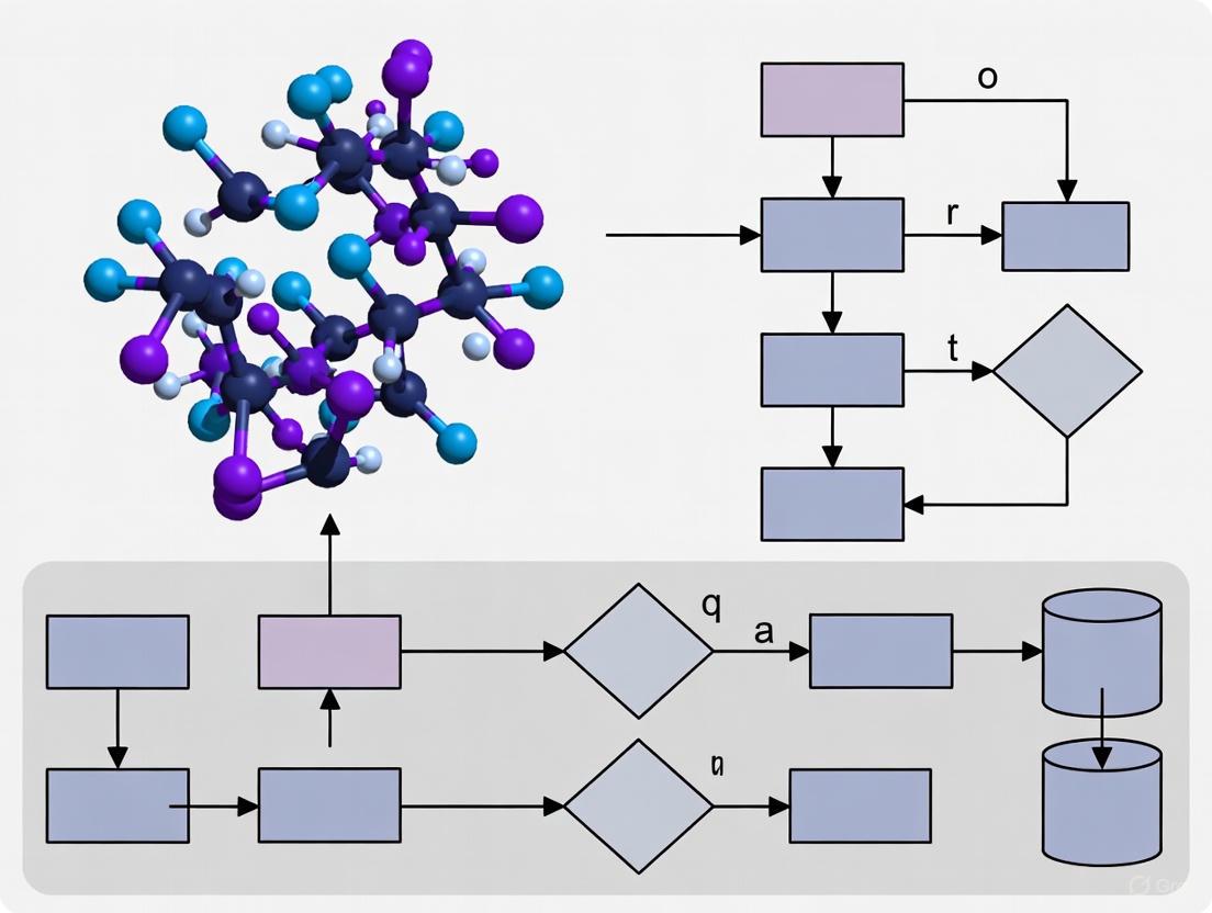
Workflow and Data Analysis Diagrams
Concluding Remarks
The AFM force spectroscopy protocols detailed herein provide a robust framework for quantitatively characterizing the mechanical properties of biofilm adhesins. The integration of SMFS, SCFS, MBFS, and nanoindentation offers a comprehensive toolkit for dissecting the molecular and cellular-scale forces that underpin biofilm adhesion, cohesion, and resistance. The quantitative data generated through these standardized methods are invaluable for validating theoretical models, screening anti-biofilm agents that target mechanical integrity, and informing the design of anti-fouling surfaces. As the field advances, the integration of machine learning for automated data analysis and large-area scanning will further enhance the throughput and predictive power of these techniques, solidifying AFM's role as an indispensable tool in biofilm research and therapeutic development [23] [24].
Bacterial Pili and Fimbriae as Force-Bearing Nanosprings
In the realm of bacterial pathogenesis, adhesion is a critical first step. Bacterial pili and fimbriae are hair-like appendages that function not merely as static tethers but as dynamic, force-bearing nanosprings. These structures are engineered to undergo significant, often superelastic, conformational changes in response to mechanical stress, enabling sustained bacterial attachment under fluid shear forces. Within the context of single-molecule force spectroscopy (SMFS) and Atomic Force Microscopy (AFM) research on biofilm adhesins, understanding the biomechanical properties of these nanosprings is paramount. This Application Note details the quantitative biophysics of these structures and provides standardized protocols for their investigation, providing researchers with the tools to probe the molecular mechanisms of bacterial adhesion.
Structural Mechanisms of Force Reception and Dissipation
The nanospring functionality of pili is not a generic property but is precisely engineered through distinct structural solutions that govern their response to mechanical force. High-resolution structural studies, particularly cryo-electron microscopy (cryo-EM), have elucidated three primary mechanical stabilization strategies within pilus assemblies [25].
- Layer-to-Layer (LL) Interactions: These relatively weak, non-covalent interactions occur between pilin subunits on adjacent turns of the helical filament. They are the first to break under low external forces, facilitating the initial unwinding of the pilus helix and providing the first stage of extension [26] [25].
- N-terminal Staple: Found in pili like CS20 and P pili, this structural motif involves the first several N-terminal amino acids of a pilin subunit reaching to form stabilizing contacts with subunits several positions away in the filament (e.g., the n+4 subunit). This "stapling" significantly reinforces the quaternary structure and increases the force required for unwinding [25].
- Extended Loop Interactions: Loops projecting from the core β-sheet of one subunit interact with adjacent subunits (e.g., n+1, n+2). The length and composition of these loops vary between pilus types, directly influencing the stability and biophysical properties of the filament [25].
The combination of these solutions allows pili to be "optimized to withstand harsh motion without breaking," which is essential for pathogens to maintain attachment in turbulent environments like the urinary tract or intestine [26] [25]. The Table 1 below summarizes the key structural and functional differences among major pilus types from enterotoxigenic E. coli (ETEC) and uropathogenic E. coli (UPEC).
Table 1: Comparative Biophysical and Structural Properties of Bacterial Pili
| Pilus Type (Pathovar) | Pilin Subunit | Pilus Class | Average Unwinding Force (pN) | Key Structural Stabilization Features | Major Functional Role |
|---|---|---|---|---|---|
| CFA/I (ETEC) | CfaB | 5 | ~7.5 [26] | Layer-to-layer interactions, N-terminal extension to n+4 subunit [25] | Sustained adhesion in the gastrointestinal tract [25] |
| CS17 (ETEC) | CsbA | 5 | Data pending (Structurally similar to CFA/I) [25] | Similar to CFA/I; specific force data limited [25] | Sustained adhesion in the gastrointestinal tract [25] |
| CS20 (ETEC) | CsnA | 1 | Data pending | Extended loops, N-terminal "staple" [25] | Sustained adhesion in the gastrointestinal tract [25] |
| Type 1 (UPEC) | FimA | 1 | Displays catch-bond behavior [27] | Hooked conformation of FimH adhesin at tip [27] | Shear-enhanced binding to bladder epithelium via FimH-mannose catch bond [27] |
| P Pilus (UPEC) | PapA | 1 | ~25-30 [25] | N-terminal "staple" motif [25] | Adhesion to kidney epithelium [25] |
The interplay of these structural features results in a common biomechanical response: the ability to unwind and rewind. When a tensile force is applied, the helical pilus filament reversibly unwinds, increasing its length by up to six-fold without breaking the backbone of the polymer. This unwinding, governed by the sequential breaking of the weaker layer-to-layer and loop interactions, serves as a critical shock-absorbing mechanism, modulating the force experienced by the adhesin at the tip and facilitating persistent attachment [25].
Diagram 1: The mechanical cycle of a pilus under force, from unwinding to rewinding.
Quantitative Biomechanics of Pilus Unwinding
The response of pili to mechanical force can be quantified to understand their role in adhesion. SMFS and optical tweezers have been instrumental in revealing that the unwinding force is a direct function of the cumulative strength of subunit-subunit interactions, rather than genetic sequence similarity [25]. Different pilus types are optimized for their specific environmental niches, with unwinding forces varying significantly.
For example, CFA/I pili from ETEC require a remarkably low force of approximately 7.5 pN to unwind, which is attributed to weak layer-to-layer interactions between subunits on adjacent turns of the helix [26]. In contrast, P pili from UPEC, which are stabilized by an N-terminal staple, require a much higher unwinding force, typically in the range of 25-30 pN [25]. This higher force reflects the need for stronger attachment in the dynamic environment of the urinary tract.
A key feature of the adhesive tips of many pili, such as the FimH protein of type 1 pili, is the catch bond mechanism. Unlike typical bonds that weaken under force, catch bonds become stronger. In FimH, tensile force causes a separation between its lectin domain (Ld) and pilin domain (Pd), triggering an allosteric shift in the Ld from a low-affinity to a high-affinity state for mannose. This results in a force-dependent decrease in the dissociation rate (off-rate) between approximately 30 and 80 pN, mechanically reinforcing adhesion under shear stress [27]. The Table 2 below summarizes key quantitative parameters for different pili.
Table 2: Experimentally Determined Biomechanical Parameters of Pili
| Pilus Type | Typical Unwinding Force (pN) | Extension Ratio (Unwound/Original) | Key Biomechanical Feature | Measurement Technique |
|---|---|---|---|---|
| CFA/I | ~7.5 [26] | Up to 6-fold [25] | Easy to unwind, hard to linearize; superelasticity [26] | Force-Measuring Optical Tweezers (FMOT) [26] |
| P Pilus | ~25-30 [25] | Up to 6-fold [25] | High unwinding force due to N-terminal staple [25] | Optical Tweezers, Steered MD [25] |
| Type 1 FimH | N/A (Adhesin) | N/A (Adhesin) | Catch bond with decreased off-rate at 30-80 pN [27] | Single Molecule Force Spectroscopy (SMFS) [27] |
| General Fimbria | Varies by type | Can stretch to several times original length [28] | "Catch-bond" mechanism at adhesin tip [28] | AFM, Optical Tweezers [28] |
Experimental Protocols for SMFS of Pili and Biofilm Adhesins
Probing the nanomechanical properties of pili requires precise methodologies. The following protocols outline standardized procedures for using AFM-based SMFS and functionalized bead assays to quantify adhesion forces and dynamics.
Protocol: Single-Molecule Force Spectroscopy of Pilus-Mediated Adhesion
Objective: To measure the specific unbinding forces and kinetics of pilus-adhesin interactions with host receptors at the single-molecule level.
Materials:
- Atomic Force Microscope (e.g., Bruker MultiMode or JPK NanoWizard)
- AFM cantilevers (e.g., MLCT-BIO from Bruker, nominal spring constant 0.01-0.1 N/m)
- Bacterial strain expressing the pilus of interest
- Target substrate (e.g., mannosylated BSA for Type 1 pili, specific glycans for other pili)
- Phosphate Buffered Saline (PBS), pH 7.4
Method:
- Functionalization:
- Chemically immobilize intact bacteria or purified pili/fimbrial tips onto a freshly cleaned, APTES-coated glass slide or directly onto the AFM substrate. Incubate for 1 hour at room temperature or 30 minutes at 37°C in a humid chamber [27].
- Alternatively, for specific receptor studies, functionalize the AFM cantilever tip with the relevant ligand (e.g., mannosylated BSA) using standard cross-linker chemistry (e.g., PEG-linker) [7].
Force Spectroscopy Measurement:
- Submerge the functionalized substrate and cantilever in a liquid cell filled with PBS.
- Approach the surface with the cantilever at a controlled velocity (e.g., 0.5-1 µm/s).
- Upon contact, apply a controlled contact force (100-500 pN) for a dwell time (0.1-1 s) to allow bond formation.
- Retract the cantilever at a constant velocity (0.5-1 µm/s) or a range of loading rates to probe kinetic properties.
Data Acquisition & Analysis:
- Record at least 500-1000 force-distance curves from random locations on the sample surface.
- Analyze the resulting curves using the instrument's software or custom scripts (e.g., in Igor Pro or MATLAB) to identify adhesion events.
- Extract the rupture force and rupture length for each adhesion event.
- Plot a force histogram; a peak at a characteristic force (e.g., ~50-200 pN for single FimH-mannose bonds under various loading rates) indicates a specific interaction [27].
- For catch bond analysis, perform experiments at different retraction speeds or under constant force to measure force-dependent lifetime [27].
Protocol: Functionalized Bead Assay for Shear-Dependent Adhesion
Objective: To confirm the shear-enhanced binding phenotype of pili using a macroscopic flow chamber assay with purified fimbrial tips.
Materials:
- Purified fimbrial tip complexes (e.g., containing FimH, FimG, FimF) [27]
- Fluorescent or plain latex beads (e.g., 1-10 µm diameter)
- Flow chamber system (e.g., µ-Slide I from ibidi)
- Mannosylated-BSA coating solution
- Peristaltic pump or syringe pump
Method:
- Surface Preparation: Coat the flow chamber with mannosylated-BSA (50-100 µg/mL) for 1-2 hours at 37°C, then block with 1% BSA for 1 hour to prevent non-specific binding.
- Bead Functionalization: Covalently couple purified fimbrial tip complexes to latex beads according to the manufacturer's protocol (e.g., using carbodiimide chemistry).
- Flow Assay:
- Introduce a suspension of functionalized beads into the flow chamber.
- Allow beads to settle onto the coated surface under zero or very low flow (shear stress ~0.01 Pa) for 5-10 minutes.
- Initiate flow, gradually increasing the shear stress (e.g., to 0.1 Pa).
- Monitor and image the number of firmly adherent beads at each shear stress interval using microscopy.
- Data Analysis: Quantify the number of adherent beads per field of view. A significant increase (e.g., >20-fold) in adherent beads at a higher shear stress (0.1 Pa) compared to low shear (0.01 Pa) is indicative of shear-dependent, catch-bond mediated adhesion [27].
Diagram 2: A generalized workflow for Single-Molecule Force Spectroscopy (SMFS).
The Scientist's Toolkit: Research Reagent Solutions
Successful investigation into the biomechanics of pili requires a specific set of reagents and tools. The following table lists essential materials and their functions in SMFS and adhesion assays.
Table 3: Essential Research Reagents and Materials for Pilus Biomechanics Studies
| Reagent/Material | Function/Application | Example Use Case |
|---|---|---|
| Purified Fimbrial Tip Complexes | Isolated quaternary structures of adhesin and adapter proteins (e.g., FimH-FimG-FimF). | Used in functionalized bead assays to study shear-dependent adhesion without whole bacteria [27]. |
| Functionalizable AFM Cantilevers | Probes for SMFS; can be coated with specific ligands or used to probe immobilized cells. | MLCT-BIO probes for measuring specific unbinding forces between FimH and mannose [7] [27]. |
| PEG-based Crosslinkers | Heterobifunctional crosslinkers (e.g., NHS-PEG-Maleimide) for attaching ligands to AFM tips. | Creates a flexible tether between the AFM tip and a mannosylated ligand, enabling precise force measurements [7]. |
| Mannosylated BSA | A neoglycoprotein used as a surrogate receptor for Type 1 fimbriae (FimH). | Coating surfaces in flow chambers or AFM substrates to study FimH-mediated adhesion [27]. |
| Microfluidic Flow Chambers | Devices for applying controlled laminar flow and quantifiable shear stress to adherent cells/beads. | Quantifying shear-enhanced adhesion of bacteria or functionalized beads [27]. |
| APTES ((3-Aminopropyl)triethoxysilane) | A silane used to create a positively charged amine-functionalized surface on glass substrates. | Promotes strong electrostatic immobilization of negatively charged bacterial cells for AFM probing [7]. |
| Acantrifoic acid A | Acantrifoic acid A|C32H48O7|Natural Triterpenoid | Acantrifoic acid A is a high-purity natural triterpenoid for research use only (RUO). Explore its potential in anti-inflammatory and pharmacological studies. |
| Ecliptasaponin D | Ecliptasaponin D, CAS:206756-04-9, MF:C36H58O9, MW:634.851 | Chemical Reagent |
Concluding Remarks
The paradigm of bacterial pili and fimbriae as force-bearing nanosprings underscores a sophisticated biological adaptation where mechanical structure dictates function in pathogenesis. The combination of SMFS, AFM, and computational modeling has been instrumental in moving from a static, structural view of these appendages to a dynamic understanding of their shock-absorbing and force-sensing capabilities. The protocols and data summarized in this Application Note provide a framework for researchers to systematically investigate these properties. Future research leveraging these tools will continue to uncover the intricate relationship between the structural biology of adhesins and their biomechanical performance, paving the way for novel anti-adhesive therapeutic strategies that target these critical virulence factors.
The adhesion of cells, whether bacterial or human, is fundamentally governed by receptor-ligand interactions that are constantly subjected to mechanical forces in physiological environments. These forces profoundly influence bond kinetics and stability, leading to two distinct categories of binding behaviors: catch-bonds and slip-bonds. Slip-bonds represent the conventional behavior where bond lifetime decreases exponentially with increasing applied force, as the mechanical work lowers the energy barrier between bound and unbound states. In contrast, catch-bonds exhibit a counterintuitive response where lifetime initially increases with force before eventually decreasing, representing a sophisticated biological mechanism for regulating adhesion under shear stress [29] [30].
The discovery and characterization of these bond types have revolutionized our understanding of cellular adhesion mechanisms, particularly in the context of bacterial colonization and infection. While slip-bonds have been widely observed across biological systems, catch-bonds have more recently emerged as a specialized adaptation that enables pathogens to fine-tune their adhesion based on local mechanical conditions [29]. This mechanoresponsive behavior allows bacteria to bind loosely and spread under low flow conditions while resisting detachment and maintaining firm attachment under high shear stress, ultimately enhancing their ability to colonize host tissues and form biofilms under dynamic fluid environments.
Fundamental Principles and Molecular Mechanisms
Quantitative Bond Characterization
The fundamental difference between catch and slip bonds can be quantitatively described through their force-dependent lifetime profiles, which are governed by distinct energy landscapes.
Table 1: Key Characteristics of Catch-Bonds and Slip-Bonds
| Characteristic | Catch-Bonds | Slip-Bonds |
|---|---|---|
| Force Response | Lifetime increases then decreases with force | Lifetime decreases exponentially with force |
| Functional Advantage | Enhanced adhesion under shear; flow-dependent regulation | Facilitates detachment under stress; reversible binding |
| Typical Lifetime Range | Can extend up to several seconds under optimal force | Shortens continuously from basal lifetime (milliseconds to seconds) |
| Shear-Enhanced Adhesion | Yes | No |
| Molecular Mechanism | Allosteric regulation; force-induced conformational change | Accelerated dissociation under load |
| Biological Examples | FimH-mannose (E. coli); SpsD-Fg (S. aureus); P-selectin-PSGL-1 | Many conventional receptor-ligand pairs |
Molecular Mechanisms of Catch-Bond Behavior
The molecular basis for catch-bond behavior involves sophisticated force-induced conformational changes that enhance binding affinity under mechanical stress. In the well-characterized FimH-mannose system of uropathogenic E. coli, the adhesin consists of a pilin domain that anchors it to bacterial fimbriae and a lectin domain that binds mannose residues on host epithelial cells. Under low force conditions, the lectin domain exists in a low-affinity state. However, when subjected to tensile force, an allosteric mechanism involving the twisting of β-sheets transitions the lectin domain to a high-affinity conformation, thereby strengthening adhesion under shear stress [29].
Similarly, in Gram-positive pathogens like Staphylococcus aureus, the SpsD protein binding to fibrinogen (Fg) demonstrates an unusual catch–slip transition with remarkably strong bond strength (rupture force of ~2 nN). This interaction exhibits a characteristic pattern where bond lifetime grows with increasing force until reaching a critical threshold, beyond which it transitions to slip-bond behavior [29]. This dual response provides a mechanical adaptation that allows bacteria to remain firmly attached under varying fluid shear conditions while permitting detachment and dissemination at exceptionally high shear forces.
Experimental Characterization Using Single-Molecule Force Spectroscopy
Technical Approaches and Instrumentation
The identification and characterization of catch-bonds have been enabled primarily by single-molecule force spectroscopy techniques, particularly atomic force microscopy (AFM) and optical tweezers. These methods allow precise application and measurement of piconewton-scale forces on individual receptor-ligand pairs, providing direct observation of bond kinetics under mechanical stress [30] [31].
Table 2: Single-Molecule Force Spectroscopy Techniques for Catch-Bond Studies
| Technique | Force Range | Spatial Resolution | Temporal Resolution | Key Applications | Advantages | Limitations |
|---|---|---|---|---|---|---|
| Atomic Force Microscopy (AFM) | 10–10,000 pN | 0.5–1 nm | 1 ms | High-force pulling; interaction assays; cell-cell adhesion | High force resolution; ability to image | Large minimal force; potential nonspecific binding |
| Optical Tweezers | 0.1–100 pN | 0.1–2 nm | 0.1 ms | 3D manipulation; tethered assays; molecular motors | Low noise and drift; high spatial and temporal resolution | Photodamage; sample heating |
| Magnetic Tweezers | 0.01–100 pN | 5–10 nm | 0.1–0.01 s | Tethered assays; DNA topology; force clamping | Stable force clamping; parallel measurements | Lower spatial resolution; limited manipulation |
Advanced AFM Methodologies for Cell-Adhesion Studies
The application of AFM to study cell-cell adhesion requires specialized methodologies to address the challenges associated with long-distance unbinding events and substantial cell deformations. A technical approach employing extended pulling range (up to 100 μm in the z-direction) using closed-loop, linearized piezo elements has been developed to quantify cell-cell adhesion forces without compromising imaging capabilities [32]. This system, when coupled with an inverted optical microscope equipped with a piezo-driven objective, enables simultaneous monitoring of cell morphology and force spectroscopy measurements, providing correlative data on structural changes and adhesion strength.
Critical parameters that must be controlled in AFM-based adhesion studies include contact conditions (constant force or position during determined time), pulling conditions (displacement and speed), and sufficient effective pulling range to ensure complete bond separation before system extension limits are reached [32]. These considerations are particularly important for studying bacterial adhesion, where multiple adhesins may engage simultaneously, and force-induced conformational changes can occur over extended distances.
Detailed Experimental Protocols
Protocol 1: AFM Force Spectroscopy for Bacterial Catch-Bond Characterization
This protocol details the procedure for quantifying catch-bond behavior in bacterial adhesins using single-molecule force spectroscopy with atomic force microscopy.
4.1.1 Sample Preparation
- Bacterial Probe Preparation: Immobilize bacterial cells or purified adhesins on AFM cantilevers using functionalized tips. For whole cells, use concanavalin A or similar non-inhibitory coatings to attach bacteria to tipless cantilevers. For purified adhesins, employ covalent coupling chemistry (e.g., PEG linkers) to attach proteins to sharpened tips.
- Substrate Functionalization: Prepare surfaces with relevant ligands (e.g., mannosylated surfaces for FimH studies, fibrinogen-coated surfaces for SpsD studies). Use freshly cleaved mica or gold surfaces functionalized with self-assembled monolayers presenting terminal groups for ligand immobilization.
- Control Surfaces: Include surfaces blocked with inert proteins (e.g., BSA) or lacking specific ligands to account for nonspecific interactions.
4.1.2 Force Measurement Configuration
- Cantilever Selection: Choose cantilevers with appropriate spring constants (typically 0.01-0.1 N/m for single-molecule measurements). Precisely calibrate each cantilever using thermal fluctuation methods prior to experiments.
- Buffer Conditions: Use physiologically relevant buffers (e.g., PBS or Tris buffer) with optional addition of divalent cations if required for adhesin function. Maintain temperature control at 37°C for physiological relevance.
- Approach/Retraction Parameters: Set approach velocity to 100-500 nm/s, contact force to 100-300 pN, and contact time to 0.1-1 second. Systematically vary retraction velocities from 100-10,000 nm/s to probe force-dependent kinetics.
4.1.3 Data Collection and Analysis
- Force Curve Acquisition: Collect a minimum of 1000-3000 force curves per condition to ensure statistical significance. Include adequate controls for specific binding (e.g., soluble inhibitors, adhesin-deficient mutants).
- Bond Lifetime Analysis: For observed unbinding events, plot bond lifetime against applied force to identify catch-bond characteristics (initial increase in lifetime with force followed by decrease).
- Catch-Bond Validation: Fit data to established models (e.g., two-state catch-slip model) to extract kinetic parameters. Verify specificity through inhibition controls and reproducibility across multiple biological replicates.
Protocol 2: Extended-Range AFM for Cell-Cell Adhesion Measurements
This protocol adapts AFM methodology for studying long-distance cell-unbinding events, particularly relevant for intact bacterial adhesion to eukaryotic cells.
4.2.1 Instrument Modification
- Piezo System Enhancement: Implement a sample stage with 100 μm z-range movement capability using closed-loop, linearized piezo elements to accommodate large cell deformations during detachment.
- Simultaneous Optical Monitoring: Integrate with an inverted optical microscope featuring piezo-driven objective for continuous visualization of cell morphology during force measurements.
- Environmental Control: Maintain temperature at 37°C and CO₂ at 5% (if using mammalian cells) throughout experiments using environmental chambers.
4.2.2 Cell Preparation and Mounting
- Bacterial Preparation: Culture bacterial strains under conditions that express target adhesins. Harvest during mid-log phase, wash, and resuspend in appropriate buffer.
- Eukaryotic Cell Monolayers: Culture adherent eukaryotic cells (e.g., HUVEC for endothelial studies) on appropriate substrates until 70-80% confluent.
- AFM Cantilever Functionalization: Functionalize tipless cantilevers with concanavalin A or poly-L-lysine to facilitate bacterial attachment. Attach single bacterial cells to cantilever apex using minimal contact force and time.
4.2.3 Adhesion Force Quantification
- Contact Parameters: Set contact force to 200-500 pN with contact times varying from 0.5-5 seconds to probe adhesion dynamics.
- Retraction Protocol: Use extended retraction distances (up to 50-100 μm) at constant velocities (0.5-2 μm/s) to ensure complete detachment.
- Adhesion Event Analysis: Identify specific unbinding events from force-distance curves. Quantify adhesion probability, rupture force, and work of adhesion. Classify unbinding events based on characteristic patterns (single vs. multiple ruptures).
Visualization of Catch-Bond Mechanisms and Experimental Workflows
Catch-Bond Mechanism in Bacterial Adhesins
AFM Workflow for Catch-Bond Characterization
Research Reagent Solutions and Essential Materials
Table 3: Essential Research Reagents for Catch-Bond Studies
| Reagent/Material | Function/Application | Specific Examples | Technical Considerations |
|---|---|---|---|
| Functionalized AFM Cantilevers | Bacterial cell or adhesin immobilization | Tipless cantilevers for whole cells; sharpened tips for single molecules | Spring constant calibration critical; appropriate surface chemistry for specific attachment |
| Ligand-Coated Substrates | Presentation of target receptors | Mannosylated surfaces for FimH; fibrinogen-coated surfaces for SpsD | Control over ligand density and orientation essential for single-molecule studies |
| Specific Inhibitors | Validation of binding specificity | Soluble mannose for FimH; monoclonal antibodies for specific epitopes | Use at multiple concentrations to demonstrate dose-dependent inhibition |
| Adhesin-Deficient Mutants | Specificity controls | FimH-negative E. coli; SpsD-negative S. aureus | Isogenic strains recommended to eliminate confounding genetic factors |
| PEG Linkers | Covalent attachment for single-molecule studies | Heterobifunctional PEG spacers | Appropriate length (typically 50-100 nm) to reduce surface interactions |
| Physiological Buffers | Maintain adhesin functionality and structure | PBS; Tris buffers with divalent cations as needed | Temperature and pH control critical for physiological relevance |
Biological Significance and Research Applications
Bacterial Pathogenesis and Virulence Strategies
Catch-bonds represent a sophisticated evolutionary adaptation that enhances bacterial virulence by enabling mechanical regulation of adhesion. For uropathogenic E. coli, the FimH-mannose catch-bond mechanism allows these pathogens to colonize the urinary tract despite high fluid shear forces, initiating infections that can lead to cystitis and pyelonephritis [29]. The catch-bond behavior ensures that bacteria remain loosely attached under low flow conditions, permitting surface exploration, while strengthening adhesion as shear stress increases, thereby resisting detachment and facilitating biofilm formation.
In staphylococcal infections, the SpsD-fibrinogen catch-bond with its exceptionally high rupture force (~2 nN) enables these pathogens to withstand extreme mechanical stress, contributing to their ability to form persistent infections on medical implants and host tissues [29]. The catch-slip transition in these systems provides a dual advantage: firm attachment under typical physiological shear stresses, with controlled detachment at extreme forces that may facilitate dissemination to new colonization sites. This mechanoresponsive adhesion represents a key virulence factor that enhances bacterial persistence in dynamic host environments.
Therapeutic Implications and Future Directions
The unique properties of catch-bonds present both challenges and opportunities for therapeutic intervention. Conventional anti-adhesion approaches that target binding interfaces may be less effective against catch-bond mediated adhesion due to its force-enhanced stability. However, the allosteric nature of catch-bond mechanisms reveals potential targets for novel anti-infective strategies that lock adhesins in low-affinity states or prevent force-induced activation [29].
Future research directions include the development of catch-bond inhibitors that specifically target the allosteric regulation pathways, engineering of catch-bond mimetics for improved tissue adhesion in regenerative medicine applications, and exploitation of catch-slip transitions for designing drug delivery systems with shear-dependent release properties. The continued application of single-molecule force spectroscopy techniques, particularly with extended measurement capabilities and improved temporal resolution, will undoubtedly reveal new catch-bond systems and deepen our understanding of their role in host-pathogen interactions and microbial ecology.
From Single Molecules to Communities: AFM Methodologies for Biofilm Analysis
Single-Molecule Force Spectroscopy (SMFS) for Probing Individual Adhesins
Single-Molecule Force Spectroscopy (SMFS) has emerged as a powerful biophysical technique for quantifying the nanoscale forces that govern molecular interactions. Within the field of microbiology, SMFS provides unprecedented insights into the adhesion mechanisms of pathogenic bacteria, a critical first step in biofilm formation and infection development [33]. By applying controlled mechanical forces to single adhesin proteins, researchers can directly measure their binding strength, mechanical stability, and structural responses to stress. This application note details standardized protocols for investigating individual adhesins using AFM-based SMFS, with a specific focus on the mechanostable properties of bacterial adhesins such as Staphylococcus aureus bone sialoprotein-binding protein (Bbp) and Pseudomonas fluorescens large adhesin LapA [34] [35]. The quantitative data and methodologies presented herein serve as essential resources for researchers and drug development professionals working to develop novel anti-adhesion therapies.
Quantitative SMFS Data on Bacterial Adhesins
The exceptional mechanical strength of bacterial adhesins is a key factor in the resilience of biofilms. The following table summarizes single-molecule rupture forces for characterized adhesin-ligand complexes, demonstrating their remarkable mechanostability.
Table 1: Mechanostability of Bacterial Adhesin-Ligand Complexes
| Adhesin | Source Bacterium | Ligand/Target | Rupture Force (Most Probable) | Experimental Conditions | Reference |
|---|---|---|---|---|---|
| Bone Sialoprotein-Binding Protein (Bbp) | Staphylococcus aureus | Fibrinogen-α (Fgα) | > 2,000 pN (up to ~3,510 pN) | In silico SMFS, pulling velocity 2.5x10â»â´ nm/ps | [34] |
| Large Adhesin A (LapA) | Pseudomonas fluorescens | Anti-HA Antibody / Surface | 150 - 250 pN (WT, non-induced); 250 - 1,000 pN (LapA+ mutant) | SMFS with antibody-functionalized tips | [35] |
| SdrG | Staphylococcus epidermidis | Fibrinogen-β (Fgβ) | ~2,000 pN | In vitro SMFS | [34] |
The data reveal that certain adhesins, particularly Bbp, can withstand forces that surpass the strength of non-covalent biological complexes and even approach the realm of covalent bond strengths [34]. Furthermore, the adhesion strength is highly dependent on cellular regulation; for LapA, hyper-adherent phenotypes (LapA+ mutant) exhibit significantly higher rupture forces due to the accumulation of the adhesin on the cell surface [35].
Experimental Protocols
SMFS of Purified Adhesin-Ligand Complexes (In silico Protocol)
This protocol outlines the procedure for probing the mechanostability of an adhesin-ligand complex using steered molecular dynamics (SMD) simulations, as applied to the Bbp:Fgα complex [34].
- System Preparation:
- Obtain the atomic-resolution structure of the adhesin-ligand complex (e.g., from the Protein Data Bank).
- Solvate the complex in an explicit water box (e.g., TIP3P water model) and add ions to neutralize the system and achieve a physiological salt concentration.
- Energy Minimization and Equilibration:
- Perform energy minimization using a steepest descent algorithm to remove any steric clashes.
- Carry out equilibration in two phases: first with positional restraints on the protein and ligand heavy atoms (NVT ensemble, 100 ps), followed by a second equilibration without restraints (NPT ensemble, 100 ps) to stabilize temperature and pressure.
- Steered Molecular Dynamics (SMD):
- Anchoring: Fix the C-terminus of the adhesin (Bbp) by applying harmonic restraints to its Cα atoms.
- Pulling: Attach a virtual spring to the C-terminus of the ligand (Fgα). Pull the spring at a constant velocity (e.g., 2.5 × 10â»â´ nm/ps) away from the anchored adhesin.
- Replication: Perform a wide sampling of simulations (e.g., 160 replicas) to ensure statistical significance.
- Data Analysis:
- Record the force exerted on the pulling spring versus the extension of the system for each trajectory.
- Plot the data as a force-extension curve and identify the peak rupture force for each simulation.
- Construct a histogram of the rupture forces and fit it with the Bell-Evans model to determine the most probable rupture force at the given pulling velocity.
SMFS on Live Bacterial Cells
This protocol describes the use of functionalized AFM tips to probe individual adhesin molecules directly on the surface of live bacteria, as demonstrated for LapA on P. fluorescens [35].
- Sample and Probe Preparation:
- Bacterial Immobilization: Grow bacterial cells (e.g., WT and LapA+ mutant) under relevant conditions (e.g., in phosphate-rich medium to induce biofilm formation). Gently immobilize the cells on a porous polycarbonate membrane by filtration.
- AFM Tip Functionalization: a. Clean AFM cantilevers in an ozone/UV cleaner. b. Incubate the tips with a solution of aminopropyltriethoxysilane (APTES) to create an amine-reactive surface. c. Activate the surface with a heterobifunctional crosslinker (e.g., GMBS). d. Immobilize the specific ligand (e.g., monoclonal anti-HA antibody for HA-tagged LapA) or the host protein (e.g., fibrinogen) onto the activated surface.
- Force Spectroscopy Mapping:
- Mount the functionalized AFM probe and the sample with immobilized bacteria in the liquid cell of the AFM.
- Approach the tip to the cell surface with a trigger force of ~250 pN to ensure gentle contact.
- Retract the tip at a constant velocity (e.g., 500-1000 nm/s). Record thousands of force-distance curves across multiple cells and from independent cultures.
- Data Analysis:
- Adhesion Frequency: Calculate the percentage of force curves that contain adhesion events.
- Rupture Force and Length: For curves with adhesion events, measure the magnitude of the rupture peaks and the elongation length. Plot histograms to analyze the distributions.
- Specificity Control: Perform blocking experiments by incubating the surface with free ligands or antibodies prior to measurement. Compare results with a non-adhesin expressing mutant (LapA-) to confirm the origin of the signals.
Visualization of SMFS Workflows
The following diagrams illustrate the core workflows and biophysical principles of the SMFS protocols described above.
SMFS on Live Bacterial Cells
In silico Steered Molecular Dynamics
The Scientist's Toolkit: Research Reagent Solutions
Successful SMFS experiments require carefully selected reagents and materials. The following table lists key solutions for probing adhesins.
Table 2: Essential Research Reagents for Adhesin SMFS
| Reagent/Material | Function and Application in SMFS | Example Use Case |
|---|---|---|
| Functionalized AFM Cantilevers | Serve as the force sensor; specific chemistry (e.g., antibody, ligand) on the tip enables probing of target adhesins. | Probing HA-tagged LapA adhesin with anti-HA antibody-coated tips [35]. |
| Polycarbonate Membranes | Provide a porous, mechanically stable substrate for immobilizing live bacterial cells without chemical fixation. | Immobilizing P. fluorescens for single-molecule and single-cell force spectroscopy [35]. |
| APTES (Aminopropyltriethoxysilane) | A silane coupling agent used to create an amine-functionalized surface on AFM tips for subsequent crosslinking. | First step in tip functionalization protocol for covalent antibody attachment [35]. |
| GMBS Crosslinker | A heterobifunctional crosslinker (N-γ-Maleimidobutyryloxy succinimide ester) that links amine-modified surfaces to thiol groups on proteins. | Covalently linking antibodies to APTES-functionalized AFM tips [35]. |
| Bell-Evans Model | A theoretical model used to analyze force spectroscopy data, relating the most probable rupture force to the loading rate. | Extracting the most probable rupture force and kinetic parameters from SMD simulation data [34] [36]. |
| Fibrinogen-α (Fgα) Peptide | The natural ligand for the Bbp adhesin; used as a binding target in both in silico and in vitro experiments. | Studying the dock, lock, and latch mechanism and mechanostability of S. aureus Bbp [34]. |
| Forsythoside E | Forsythoside E, MF:C20H30O12, MW:462.4 g/mol | Chemical Reagent |
| Helicianeoide A | Helicianeoide A, MF:C32H38O19, MW:726.6 g/mol | Chemical Reagent |
Single-Cell Force Spectroscopy (SCFS) for Whole-Cell Adhesion Measurements
Single-Cell Force Spectroscopy (SCFS) is an atomic force microscopy (AFM)-based technique that enables precise quantification of cell adhesion forces at the single-cell level. Within the broader context of single-molecule force spectroscopy AFM biofilm adhesins research, SCFS provides a critical bridge between molecular-level interactions and whole-cell adhesive behavior. This technique allows researchers to quantitatively assess how microbial cells initiate surface attachment—the critical first step in biofilm formation that mediates bacterial persistence in medical, industrial, and environmental contexts [37] [23].
The fundamental principle of SCFS involves mechanically detaching a single living cell from a substrate while precisely measuring the forces involved, generating characteristic force-distance curves that reveal key adhesion parameters [38]. Recent technological advancements, particularly the development of robotic fluidic force microscopy (FluidFM), have transformed SCFS from a low-throughput technique (a few cells per day) to a high-throughput method capable of characterizing population distributions of adhesion parameters [38]. This capability is essential for understanding the inherent heterogeneity in biofilm formation and for evaluating novel antifouling strategies aimed at combating biofilm-associated infections and surface contamination [39].
Key Applications in Biofilm and Antimicrobial Research
SCFS provides unique insights into microbial adhesion mechanisms with particular relevance for biofilm and antifouling research:
- Antifouling Surface Evaluation: SCFS has demonstrated that both tripeptide-based (DOPA-Phe(4F)-Phe(4F)-OMe) and polymer-based (poly(ethylene glycol)) antifouling surfaces significantly reduce initial E. coli adhesion compared to glass surfaces, with bacteria employing different adhesion mechanisms depending on the surface chemistry [39].
- Adhesion Mechanism Elucidation: Using mutant E. coli strains deficient in specific surface appendages, SCFS has revealed that type-1 fimbriae and curli amyloid fibers mediate adhesion to different antifouling surfaces via separate mechanisms [39].
- Single-Cell Heterogeneity Assessment: SCFS measurements have uncovered significant variability in adhesion strength between individual bacterial cells, highlighting how population diversity affects biofilm formation initiation [39].
- Biofilm Assembly Studies: High-resolution AFM imaging of Pantoea sp. YR343 has revealed intricate structural details during early biofilm formation, including flagellar coordination and the emergence of honeycomb patterns, providing insights into surface attachment dynamics [23].
Quantitative Adhesion Parameters Measured by SCFS
SCFS experiments generate force-distance curves during cell detachment, from which key quantitative parameters are derived [38]:
Table 1: Key Adhesion Parameters Obtained from SCFS Force-Distance Curves
| Parameter | Symbol | Definition | Biological Significance |
|---|---|---|---|
| Maximum Adhesion Force | Fmax | Highest force recorded during cell detachment | Reflects overall adhesion strength |
| Adhesion Energy | Emax | Total work required to detach the cell | Represents the energy dissipated during detachment |
| Detachment Distance | Dmax | Cantilever travel distance until Fmax | Indicates cell deformability and adhesion complex elasticity |
| Cell Contact Area | Acell | Projected area of cell-substrate contact | Relates to available area for adhesion complex formation |
| Spring Coefficient | k | Ratio of Fmax to Dmax | Represents cellular stiffness during detachment |
Population-level SCFS studies using high-throughput robotic FluidFM have revealed that single-cell adhesion parameters (Fmax, Emax, Dmax, and Acell) typically follow lognormal distributions rather than normal distributions, emphasizing the importance of measuring large cell populations to avoid misleading conclusions from small sample sizes [38].
Table 2: Representative SCFS Adhesion Measurements Across Cell Types and Conditions
| Cell Type | Substrate | Max Adhesion Force | Key Findings | Citation |
|---|---|---|---|---|
| Mesenchymal Stem Cells (MSCs) | RGD-coated glass | Significant increase vs. bare glass | Enhanced adhesion correlated with improved cell attachment in conventional assays | [40] |
| E. coli (wild type) | Glass | Baseline adhesion | Reference for antifouling surface comparison | [39] |
| E. coli (wild type) | Tripeptide antifouling | Significant reduction vs. glass | Effective prevention of initial bacterial adhesion | [39] |
| E. coli (wild type) | PEG polymer brush | Significant reduction vs. glass | Effective prevention of initial bacterial adhesion | [39] |
| HeLa Fucci Cells | Various stages of cell cycle | Log-normal distribution | Cell cycle-dependent adhesion with distinct mechanical properties | [38] |
Experimental Protocols for SCFS Measurements
Substrate Preparation and Functionalization
Protocol: RGD-Coated Surface Preparation for Enhanced Cell Adhesion Studies
Surface Activation:
- Clean borosilicate glass coverslips using oxygen plasma treatment or appropriate cleaning protocol
- Functionalize surfaces using activated vapor silanization (AVS) process to introduce reactive groups [40]
Peptide Decoration:
- Prepare RGD-containing peptide solution in appropriate buffer
- Immerse functionalized substrates in peptide solution
- Use EDC/NHS crosslinking chemistry to covalently attach peptides to activated surfaces [40]
- Rinse thoroughly with sterile buffer to remove unbound peptides
Quality Control:
- Verify peptide coating uniformity through control experiments
- Store functionalized substrates under appropriate conditions until use
Protocol: Antifouling Surface Preparation for Bacterial Adhesion Studies
Surface Selection:
- Prepare tripeptide DOPA-Phe(4F)-Phe(4F)-OMe surfaces using appropriate deposition methods
- Create poly(ethylene glycol) polymer-brush surfaces using established grafting protocols [39]
Surface Characterization:
- Verify surface chemistry through appropriate analytical methods
- Ensure uniform coating across measurement areas
Cell Preparation and Mounting
Protocol: Microbial Cell Culture for SCFS
Bacterial Culture:
- Grow Pantoea sp. YR343 or E. coli strains in appropriate liquid growth medium
- For mutant studies: cultivate isogenic strains lacking specific adhesins (e.g., type-1 fimbriae or curli amyloid fibers) [39]
- Harvest cells at mid-logarithmic phase by gentle centrifugation
- Resuspend in fresh medium or appropriate buffer at desired density
Surface Attachment:
- Incubate substrates with bacterial suspension for selected time intervals (e.g., 30 minutes for initial attachment studies) [23]
- Gently rinse to remove unattached cells using appropriate buffer
- For time-course studies: allow biofilm development for extended periods (e.g., 6-8 hours) to observe cluster formation [23]
Mammalian Cell Culture:
- Maintain HeLa Fucci cells or MSCs under standard culture conditions
- For cell cycle studies: utilize Fucci construct that expresses fluorescent proteins specific to cell cycle phases [38]
- Detach cells using gentle methods that preserve surface receptors
- Allow cells to adhere to test substrates for appropriate duration to form mature adhesions
SCFS Measurement Procedure
Protocol: Robotic Fluidic Force Microscopy for High-Throughput SCFS
Instrument Setup:
- Mount appropriate hollow cantilever on robotic FluidFM system
- Calibrate cantilever sensitivity using established protocols
- Position sample on motorized XY-stage with optical microscope monitoring [38]
Single-Cell Capture:
- Approach target cell with hollow cantilever
- Apply gentle vacuum through microfluidic channel to securely capture individual cell
- Verify single-cell capture via optical monitoring [38]
Adhesion Measurement:
- Retract cantilever at constant velocity (typically 0.5-2 µm/s)
- Record force-distance curve during cell detachment
- Continue retraction until cell completely detaches from substrate
- Repeat measurement on multiple cells across different surface regions [38]
Data Collection:
- Automate measurement process using robotic positioning for high-throughput data collection
- Record minimum of 50-100 cells per condition to account for population heterogeneity [38]
- For cell cycle studies: document fluorescent protein expression prior to measurement
Figure 1: SCFS experimental workflow using robotic FluidFM technology, showing single-cell capture, force measurement, and population distribution analysis.
Data Analysis and Interpretation
Protocol: Analysis of SCFS Force-Distance Curves
Parameter Extraction:
- Identify maximum adhesion force (Fmax) from force-distance curve
- Calculate adhesion energy (Emax) by integrating force over distance
- Record detachment distance (Dmax) at point of Fmax
- Determine cell contact area (Acell) from optical images [38]
Population Analysis:
- Compile adhesion parameters from multiple cells into distribution histograms
- Test for lognormal distribution using appropriate statistical tests
- Compare population distributions between experimental conditions using non-parametric statistical tests [38]
Normalization:
- Normalize Fmax by Acell to account for cell size variations
- Calculate spring coefficient (k = Fmax/Dmax) as measure of cellular stiffness [38]
The Scientist's Toolkit: Essential Materials and Reagents
Table 3: Essential Research Reagent Solutions for SCFS Experiments
| Reagent/Material | Function/Application | Example Specifications |
|---|---|---|
| Borosilicate Glass Coverslips | Standard substrate for functionalization | Various thicknesses, plasma-cleaned |
| RGD-Containing Peptides | Enhances eukaryotic cell adhesion | Typically cyclic RGDfK, 95% purity |
| EDC/NHS Crosslinkers | Covalent peptide immobilization | Freshly prepared solutions |
| Tripeptide Antifouling Coatings | Prevents bacterial adhesion | DOPA-Phe(4F)-Phe(4F)-OMe |
| PEG Polymer Brushes | Antifouling surface modification | Various molecular weights |
| Cell Culture Media | Cell maintenance and experiments | Appropriate for cell type (LB, DMEM, etc.) |
| FluidFM Cantilevers | Single-cell manipulation | Hollow silicon nitride, 2-6 µm diameter |
| Appropriate Buffers | Measurement environment | PBS, HBSS, or cell-specific buffers |
| Siegesmethyletheric acid | Siegesmethyletheric acid, MF:C21H34O3, MW:334.5 g/mol | Chemical Reagent |
| AF3485 | N-[9-(2-Hydroxyethyl)-9H-carbazol-3-yl]-2-(trifluoromethyl)benzamide |
Data Interpretation and Integration with Broader Research
Figure 2: Data interpretation workflow for SCFS experiments, showing the relationship between experimental parameters and biological significance in biofilm research.
The application of SCFS within biofilm adhesin research provides unique insights into the nanoscale forces governing initial microbial attachment. Recent advancements in large-area automated AFM, combined with machine learning approaches for image analysis, now enable correlation of single-cell adhesion measurements with larger scale biofilm organization [23]. This multi-scale approach reveals how individual cell adhesion events propagate to form the complex architecture of mature biofilms, characterized by heterogeneous structures such as the honeycomb patterns observed in Pantoea sp. YR343 biofilms [23].
SCFS data integration with complementary techniques provides a comprehensive understanding of adhesion mechanisms:
- With genetic approaches: SCFS of mutant strains reveals specific adhesin functions [39]
- With high-resolution imaging: Correlates adhesion strength with structural features like flagellar arrangement [23]
- With population biology: High-throughput SCFS captures heterogeneity in adhesion properties within clonal populations [39] [38]
This integrated approach positions SCFS as a powerful tool in the development of novel anti-biofilm strategies, enabling rational design of surfaces that resist pathogenic adhesion while supporting beneficial host cell integration.
Innovative Fluidic Force Microscopy (FluidFM) for Biofilm-Scale Probing
Fluidic Force Microscopy (FluidFM) represents a pivotal advancement in the field of atomic force microscopy (AFM) by integrating microfluidic channels within force-controlled cantilevers. This synergy creates a force-sensitive nanopipette capable of single-cell manipulation and quantification under physiological conditions. Within the broader context of single-molecule force spectroscopy AFM research on biofilm adhesins, FluidFM addresses a critical methodological gap: the transition from single-cell to biofilm-scale force spectroscopy. Traditional single-cell force spectroscopy (SCFS) depends on the irreversible chemical fixation of cells to cantilevers, a process that is not only low-throughput but also ill-suited for studying the complex, multi-cellular structure of biofilms [41] [42]. FluidFM overcomes these limitations by enabling the reversible, physical immobilization of single cells or biofilm-functionalized probes via suction applied through the cantilever's aperture [43] [44]. This technical note details the application of FluidFM for quantifying adhesion forces at the biofilm scale, providing researchers with robust protocols and a framework for interpreting data relevant to microbial colonization, antifouling strategies, and therapeutic interventions.
Technical Background and Principles
The core of the FluidFM technology is a hollow, microchanneled cantilever connected to a pressure controller. This setup functions as a combined force sensor and microfluidic probe [44] [45]. The key operational modes for biofilm probing are:
- Reversible Immobilization: Application of negative pressure (underpressure) at the cantilever's aperture allows for the pickup and secure holding of a single cell, a colloidal probe, or a biofilm-coated bead. This immobilization is physical, requires no chemical glue, and the specimen can be released unharmed by applying a slight overpressure [46] [42].
- Force Spectroscopy: Once a probe is immobilized, the cantilever is positioned against a surface of interest (e.g., a functionalized membrane or another cell). The force required to detach the probe is measured with piconewton sensitivity via laser deflection, generating a force-distance curve [47] [48].
This platform is particularly powerful for biofilm research because it allows the probe to be scaled up. Instead of a single bacterial cell, a microbead on which a mature biofilm has been grown can be aspirated, enabling the measurement of adhesion forces that incorporate the contribution of the extracellular polymeric substance (EPS) matrix [47]. This biofilm-scale probing more accurately reflects real-world microbial adhesion compared to single-cell measurements.
Application Note: Quantifying Biofilm Adhesion to Anti-Fouling Membranes
Experimental Objective and Workflow
This application demonstrates the use of FluidFM to directly quantify the reduction in adhesion forces between bacterial biofilms and filtration membranes modified with the anti-biofouling agent vanillin [47]. The objective was to move beyond single-cell assessments and validate the anti-fouling performance at a more relevant, biofilm-scale level. The following diagram illustrates the core experimental workflow.
Key Findings and Quantitative Data
The FluidFM-based force spectroscopy experiments revealed a statistically significant decrease in all adhesion parameters after membrane modification with vanillin. The table below summarizes the quantitative findings, demonstrating the efficacy of vanillin and the critical difference between single-cell and biofilm-scale adhesion behavior.
Table 1: Summary of Adhesion Force Measurements for Vanillin-Modified Membranes
| Probe Type | Surface | Median Adhesion Force | Adhesion Work | Number of Adhesion Events | Key Interpretation |
|---|---|---|---|---|---|
| Single Bacterial Cell | Unmodified PES Membrane | High | High | Frequent | Represents initial attachment phase [47] |
| Single Bacterial Cell | Vanillin-Modified PES Membrane | Significantly Decreased | Significantly Decreased | Significantly Reduced | Confirms anti-adhesive properties at a single-cell level [47] |
| Biofilm-Covered Bead | Unmodified PES Membrane | High | High | Complex, multiple | Captures the contribution of the EPS matrix [47] |
| Biofilm-Covered Bead | Vanillin-Modified PES Membrane | Significantly Decreased | Significantly Decreased | Significantly Reduced | Validates anti-fouling efficacy against mature biofilms [47] |
The data underscored that biofilm adhesion behavior is fundamentally different from that of single cells, with the EPS matrix contributing to more complex force curves with higher adhesion work. This highlights the necessity of biofilm-scale probing for realistic anti-fouling assessments [47].
Detailed Experimental Protocol
Protocol 1: Biofilm-Scale Adhesion Force Spectroscopy
This protocol describes the procedure for measuring adhesion forces between a bacterial biofilm and a surface of interest, such as an anti-fouling membrane [47].
I. Materials and Reagents Table 2: Research Reagent Solutions and Essential Materials
| Item | Function / Specification | Notes / Rationale |
|---|---|---|
| FluidFM System | AFM with pressure controller and microchanneled cantilevers. | Cantilevers with apertures of 300 nm to 8 µm are available [45]. |
| FluidFM Probes | Hollow, tipless cantilevers (micropipettes). | Choose aperture size suitable for bead diameter [45]. |
| Polystyrene Beads | ~1 µm to 10 µm diameter, COOH-functionalized. | COOH-functionalization enhances bacterial attachment and biofilm growth [47]. |
| Bacterial Strain | e.g., Pseudomonas aeruginosa. | Select relevant model or target organism. |
| Growth Medium | Appropriate sterile liquid medium for selected strain. | - |
| Target Surface | e.g., Polyethersulfone (PES) filtration membrane. | Can be unmodified and modified (e.g., with vanillin) for comparison [47]. |
| Phosphate Buffered Saline (PBS) | For washing and measurements. | Provides a physiologically relevant ionic environment. |
II. Step-by-Step Procedure
- Bead Functionalization and Biofilm Growth: Incubate COOH-functionalized polystyrene beads in a suspension of the target bacteria in growth medium for 3 hours to allow for biofilm formation on the bead surfaces [47].
- Sample Mounting: Immobilize the target surface (e.g., a piece of filtration membrane) on the FluidFM sample stage. Deposit a suspension of the biofilm-coated beads in a suitable buffer (e.g., PBS) onto the same stage or a separate glass substrate for pickup.
- Cantilever Preparation: Fill a sterile, tipless FluidFM micropipette with the same buffer used in the measurement. Calibrate the cantilever's spring constant using the instrument's built-in thermal noise method [42] [48].
- Probe Immobilization: Under optical control, position the FluidFM cantilever above a single biofilm-coated bead. Apply a negative pressure (typically -800 mbar) to aspirate and securely hold the bead on the cantilever's aperture. Reduce the pressure to a lower holding value (e.g., -50 to -100 mbar) for the measurement [42].
- Force Spectroscopy Measurement:
- Navigate the probe to a clean spot on the target surface.
- Approach the biofilm bead towards the surface at a defined speed (e.g., 2-5 µm/s) until a set contact force (e.g., 10 nN) is reached.
- Maintain contact for a defined dwell time (e.g., 5 seconds) to allow for bond formation.
- Retract the cantilever at the same speed and record the force-distance curve. The adhesion force is derived from the maximum negative peak in the retraction curve.
- Replication and Probe Exchange: Repeat step 5 for at least 10-20 different locations on the surface. To ensure cleanliness and prevent cross-contamination, expel the bead by applying a brief overpressure and immobilize a new biofilm-coated bead periodically [46].
Protocol 2: High-Throughput Single-Cell Hydrophobicity Screening
This protocol leverages FluidFM's modularity to rapidly profile the hydrophobic adhesion forces of a phylogenetically diverse collection of bacterial isolates, correlating these forces with retention on plant surfaces [46]. The following workflow outlines the specific steps for this bead-based approach.
I. Key Materials
- Functionalized Beads: C30-alkyl chain functionalized silica beads to mimic the hydrophobic leaf surface. Unfunctionalized silica beads serve as a control [46].
- Bacterial Strains: A collection of bacterial isolates (e.g., from leaf microbiota).
- Polydopamine Coating: Used to immobilize live bacterial cells on a glass substrate to prevent displacement during force measurement [46].
II. Procedure Summary
- A C30-functionalized bead is aspirated onto a FluidFM cantilever.
- Individual, live bacterial cells are immobilized on a polydopamine-coated glass surface.
- The hydrophobic bead is brought into contact with a single bacterial cell (setpoint: 10 nN, dwell time: 5 s) and then retracted to record the adhesion force.
- The same bead is used to measure multiple cells of the same strain to assess single-cell heterogeneity.
- Beads are regularly exchanged to ensure a clean surface.
- The median adhesion force for each strain is correlated with data from plant washing assays to establish a biologically relevant link between hydrophobicity and colonization success [46].
Critical Parameters and Optimization
Successful implementation of FluidFM requires careful optimization of several parameters to ensure high-quality, reproducible data:
- Immobilization Pressure: A high negative pressure (e.g., -800 mbar) is used for initial cell or bead pickup. This should be reduced to a lower holding pressure (e.g., -50 to -100 mbar) during measurements to minimize baseline noise and potential damage while maintaining secure immobilization [42] [48].
- Force Spectroscopy Settings: Systematically optimize z-speed, setpoint, and pause time. Higher setpoints can increase measured adhesion, while very high z-speeds may lead to an underestimation of forces due to hydrodynamic drag and viscous effects [42] [48].
- Probe and Aperture Size: Match the FluidFM probe aperture to the size of the object being immobilized. Focused ion beam milling can be used to customize apertures for specific cell morphologies [46].
- Surface Functionalization: The choice of bead functionalization (e.g., C30 for hydrophobicity, COOH for biofilm growth) is critical for tailoring the experiment to the specific biological interaction being studied [46] [47].
Fluidic Force Microscopy has emerged as a uniquely powerful tool for extending the principles of single-molecule force spectroscopy into the complex realm of biofilm mechanics. By enabling reversible, physical immobilization of both single cells and biofilm-coated probes, it provides a versatile and higher-throughput platform for quantifying adhesion forces. The detailed protocols and data presented herein demonstrate its application in directly evaluating anti-fouling surfaces and deciphering the fundamental drivers of microbial colonization. Integrating FluidFM into research on biofilm adhesins offers an unprecedented path to link molecular-scale interactions with community-scale phenomena, thereby accelerating the development of novel anti-biofilm strategies in medical, industrial, and environmental contexts.
Quantifying Adhesion to Host Tissues and Biomedical Materials
Atomic force microscopy (AFM) has emerged as a powerful multifunctional platform for probing microbial cell surfaces at the single-molecule level, uncovering biophysical properties and interactions that are otherwise inaccessible to ensemble techniques [49]. Single-molecule force spectroscopy (SMFS) and single-cell force spectroscopy (SCFS), two specialized AFM modalities, provide a wealth of information on the strength, specificity, and dynamics of microbial cell surface interactions with unprecedented spatiotemporal resolution [49]. Unlike other microscopy techniques, AFM can detect, localize, and manipulate individual molecules on live cells under physiological conditions without staining, marking, or drying, while simultaneously providing nanoscale-resolution topographic images of cell surface features [7]. This application note details standardized protocols for quantifying adhesion forces of pathogens to host tissues and biomedical materials, with particular emphasis on single-molecule investigations of biofilm adhesins within the broader context of antimicrobial therapeutic development.
Background and Significance
Microbial adhesion represents the crucial initiating step in biofilm formation and infection pathogenesis, making its quantitative understanding essential for developing efficient antimicrobial strategies complementary to antibiotic treatments [49]. Pathogens such as Staphylococcus aureus and Escherichia coli trigger difficult-to-treat nosocomial infections by adhering to biomedical devices (implants, catheters) and developing surface-associated biofilm communities that demonstrate significant antibiotic resistance [49]. Single-molecule experiments probe individual molecules directly, revealing events and properties that would be obscured in ensemble measurements [49].
Perhaps the most striking discovery of recent years is that staphylococcal adhesins SdrG, ClfA, and ClfB bind to their protein ligands via extremely strong forces of approximately 2 nN, equivalent to covalent bond strength and an order of magnitude stronger than the prototypical streptavidin-biotin interaction previously considered nature's strongest non-covalent bond [49]. This revelation demonstrates that pathogens have evolved specialized mechanisms to achieve remarkable adhesion strength under mechanical stress conditions.
Quantitative Adhesion Data from AFM Studies
Table 1: Measured Adhesion Forces of Pathogenic Systems
| Pathogen/System | Ligand/Target | Measured Force | Technique | Reference |
|---|---|---|---|---|
| Staphylococcus aureus (SdrG adhesin) | Fibrinogen | ~2 nN | SMFS | [49] |
| Staphylococcus aureus (ClfA adhesin) | Fibrinogen | ~2 nN | SMFS | [49] |
| Staphylococcus aureus (ClfB adhesin) | Fibrinogen | ~2 nN | SMFS | [49] |
| Candida albicans (Als adhesins) | Various substrates | Force-induced nanodomain formation | SMFS | [7] |
| Pseudomonas aeruginosa to HEK293 cells | HEK293 cell surface | Confluence-dependent adhesion kinetics | High-throughput fluorescence | [50] |
| Biomphalysin toxin to Micrococcus luteus | Bacterial surface | Selective interaction with patch-like distribution | SMFS with antibody-functionalized tip | [7] |
Table 2: Comparison of Force Spectroscopy Techniques
| Technique | Spatial Resolution | Temporal Resolution | Typical Applications | Sample Requirements |
|---|---|---|---|---|
| Single-Molecule Force Spectroscopy (SMFS) | Single molecule | Seconds to minutes per force curve | Functional analysis of individual receptors, unbinding forces, kinetic parameters | Low to moderate receptor density |
| Single-Cell Force Spectroscopy (SCFS) | Whole cell | Minutes per measurement | Quantification of cellular adhesion to surfaces, cells, or substrates | Viable, immobilizable cells |
| Recognition Imaging-Guided SMFS | Nanometer precision | Minutes for imaging plus spectroscopy | Targeted force measurements on specific, pre-identified molecules | Sparse distribution of target molecules |
| High-Throughput Fluorescence Adhesion | Population-level with kinetic data | Seconds between measurements | Screening adhesion inhibitors, kinetic profiling of bacterial adhesion to tissue | Fluorescently tagged bacteria, transparent substrates |
Experimental Protocols
AFM-Based Single-Molecule Force Spectroscopy Guided by Recognition Imaging
This protocol combines recognition imaging and force spectroscopy for studying interactions between membrane receptors and ligands at the single-molecule level, allowing for the selection of individual receptor molecules with subsequent quantification of receptor-ligand unbinding forces [51].
Materials and Reagents
Table 3: Key Research Reagent Solutions
| Reagent | Function/Application | Specifications/Alternatives |
|---|---|---|
| Acetal-PEG₂₇-NHS | Heterobifunctional crosslinker for tip functionalization | Monodisperse polyethylene glycol linker with protected benzaldehyde function [51] |
| (3-Aminopropyl)triethoxysilane (APTES) | Surface aminofunctionalization | Distill before use and store under argon at -20°C [51] |
| Dimethylsulfoxide (DMSO) | Solvent for PEG linker | Anhydrous, >99.9% purity [51] |
| Sodium cyanoborohydride | Reduction of Schiff bases to stable amine linkages | Handle with care due to high toxicity [51] |
| Ethanolamine hydrochloride | Blocking of unreacted NHS esters | Prepared in aqueous solution [51] |
| MSNL cantilevers | AFM probes for combined imaging/spectroscopy | Silicon nitride, nominal spring constant 0.03 N/m [51] |
Instrument Setup and Calibration
- Cantilever Selection: Use silicon nitride tips with a nominal spring constant of 0.03 N/m as an acceptable compromise for both recognition imaging and force spectroscopy [51].
- Excitation Method: Employ acoustic excitation as these tips are not typically available with magnetic coating required for standard TREC imaging [51].
- Drift Minimization: Utilize a closed-loop scanner with inductive sensors to detect and correct deviations from ideal movement, significantly improving thermal drift issues [51].
- Environmental Control: Maintain constant laboratory temperature and allow the system (sample, measurement buffer, and cantilever) to equilibrate before starting measurements [51].
Cantilever Functionalization
- Aminofunctionalization: Vapor-phase silanization of cantilevers with APTES following thorough cleaning [51].
- Linker Attachment: Incubate aminofunctionalized tips with Acetal-PEG₂₇-NHS (1 mg in 500 μL DMSO) for 2 hours at room temperature to form amide bonds between the NHS ester and surface amine groups [51].
- Ligand Conjugation: After washing, immerse tips in ligand solution (100-500 μg/mL in PBS) with 1-2 mM NaCNBH₃ for 30-45 minutes to form stable amine linkages between the benzaldehyde group and primary amines of the ligand [51].
- Blocking: Quench unreacted aldehyde groups with ethanolamine hydrochloride (1 M, pH 9.5) for 10-20 minutes [51].
Recognition Imaging and Targeted Force Spectroscopy
- Simultaneous Topography and Recognition Imaging: Oscillate the ligand-functionalized cantilever across the membrane surface using AFM tapping mode in liquid, analyzing downward and upward oscillation parts for topography and recognition, respectively [51].
- Molecular Selection: Identify functional receptor molecules from the recognition image with nanometer resolution [51].
- Mode Switching: Switch AFM to force spectroscopy mode using positional feedback control while maintaining the same cantilever [51].
- Force Curve Acquisition: Approach the pre-selected molecule until contact is made, then retract while recording cantilever deflection at constant velocity in the z-direction at fixed x-y coordinates [51].
- Data Collection: Repeat force measurements on different pre-selected molecules to build statistics on unbinding forces, kinetic parameters, and energy landscapes [51].
Diagram Title: AFM Recognition Imaging-Guided Force Spectroscopy Workflow
High-Throughput Kinetic Adhesion Assay for Bacterial Adhesion to Tissue
This protocol enables high-temporal resolution monitoring of bacterial adhesion to tissue culture cells using fluorescence masking, providing second-to-minute kinetic resolution for screening applications [50].
Materials and Reagents
- Fluorescent Bacteria: Pseudomonas aeruginosa PAO1 constitutively expressing GFPmut2 [50]
- Tissue Culture Cells: HEK293 cells (or other relevant cell lines) [50]
- Fluorescence Quencher: Allura Red AC dye (0.8 mg/mL final concentration) [50]
- Buffer: Dulbecco's Phosphate Buffered Saline (DPBS) without calcium and magnesium [50]
- Equipment: Plate fluorimeter with bottom-reading capability (e.g., Biotek Synergy HT) [50]
Experimental Procedure
- Cell Culture: Seed HEK293 cells in a 96-well plate at varying densities (5K-30K cells per well) and incubate for 24 hours in a CO₂ incubator at 37°C to achieve different confluencies [50].
- Bacterial Preparation: Grow fluorescent P. aeruginosa PAO1 for 18 hours at 37°C with shaking, wash twice in DPBS, and resuspend to OD₆₀₀ₙₘ = 0.1 in DPBS supplemented with 1.6 mg/mL Allura Red AC dye [50].
Fluorescence Normalization: Measure OD₆₀₀ₙₘ and fluorescence (without dye) of each bacterial strain to establish normalization factors using the equation:
Normalized fluorescence = Fluorescence(with dye) / [Base fluorescence(no dye) / OD₆₀₀ₙₘ(no dye)] [50]
Adhesion Kinetics Measurement: Replace cell culture medium with 50 μL DPBS, add 50 μL prepared bacterial suspension to each well, and immediately load plate into fluorimeter [50].
- Data Acquisition: Read bottom fluorescence every 30-60 seconds for 1 hour using appropriate GFP filters (excitation 485/20 nm, emission 528/20 nm) [50].
- Strong Adhesion Assessment: After kinetic measurements, gently remove liquid from wells and replace with 100 μL DPBS supplemented with 0.8 mg/mL dye to measure fluorescence from strongly adhered bacteria only [50].
Diagram Title: High-Throughput Kinetic Adhesion Assay Workflow
Data Analysis and Interpretation
Force Curve Analysis
AFM force spectroscopy data analysis involves converting cantilever deflection into quantitative force measurements through several standard steps [52]:
- Cantilever Calibration: Determine the exact spring constant of the cantilever using thermal noise or other calibration methods [52].
- Force Calculation: Convert deflection data to force using Hooke's law (F = -k × d, where k is spring constant and d is deflection) [52].
- Adhesion Peak Identification: Identify unbinding events in retraction curves characterized by sudden jumps in the force curve [52].
- Statistical Analysis: Compile data from hundreds of force curves to obtain histograms of unbinding forces and determine most probable adhesion forces [52].
For recognition imaging-guided SMFS, the combination of techniques allows for dynamic force probing on different pre-selected molecules, significantly increasing throughput compared to traditional force mapping approaches [51]. The dynamics of force loading can be systematically varied to elucidate binding dynamics and map interaction energy landscapes [51].
Adhesion Kinetic Analysis
For high-throughput adhesion assays, normalized fluorescence values are plotted against time to generate adhesion kinetic profiles [50]. The initial adhesion rate can be determined from the linear portion of the curve, while the plateau phase indicates saturation of available adhesion sites [50]. Statistical analysis should include multiple replicates (typically 6-12 wells per condition) with appropriate post-hoc testing such as Tukey LSD [50].
Applications in Therapeutic Development
The quantitative assessment of pathogen adhesion has significant implications for anti-infective therapeutic strategies:
- Anti-Adhesion Therapy: AFM has been instrumental in assessing the inhibition of pathogen adhesion by carbohydrates, antibodies, and peptides, showing promise for anti-adhesion approaches that complement conventional antibiotics [49].
- Biomaterial Design: Quantitative adhesion data inform the development of anti-adhesive coatings for biomedical devices to prevent biofilm formation and nosocomial infections [49].
- Virulence Assessment: SMFS enables researchers to correlate specific adhesins with pathogenic potential and identify key molecular targets for intervention [7].
- Probiotic Interactions: These methods similarly apply to studying beneficial bacterial interactions with host tissues for probiotic development [50].
The protocols detailed herein provide standardized methodologies for quantifying adhesion forces across multiple scales, from single molecules to cellular populations, offering comprehensive tools for researchers investigating host-pathogen interactions and developing novel therapeutic strategies.
Staphylococcal infections, particularly those involving Staphylococcus aureus, represent a significant challenge in healthcare settings due to their ability to cause persistent device-related infections. The pathogen's virulence is intrinsically linked to its capacity to adhere to biotic and abiotic surfaces through specialized surface proteins that form exceptionally strong bonds with host ligands. This case study focuses on the molecular mechanisms underpinning this adhesion, with particular emphasis on the dock, lock, and latch (DLL) mechanism and its variant, the close, dock, lock, and latch (CDLL) mechanism. Through the application of single-molecule force spectroscopy (SMFS) using atomic force microscopy (AFM), we quantify the biophysical forces governing these interactions, providing a framework for understanding bacterial adhesion within the broader context of biofilm formation and immune evasion [17] [53]. The insights gained are critical for researchers and drug development professionals aiming to disrupt these tenacious interactions therapeutically.
The Molecular Mechanism of Adhesion
The DLL mechanism is a multi-step binding process that results in a hyper-stable adhesion complex. In the classical DLL model, an adhesin's N2N3 domains first dock a target peptide via hydrophobic interactions. The complex is then locked through the folding of the C-terminal segment of the N3 domain over the ligand. Finally, this segment latches onto the N2 domain, creating a complementary surface that is secured by an extensive network of hydrogen bonds [17]. This mechanism is observed in staphylococcal adhesins such as SdrG, which binds to fibrinogen [17].
Recent research on the S. aureus adhesin SdrE reveals a variation of this theme—the CDLL mechanism. In its unliganded state, the N2N3 domains of SdrE adopt a closed conformation. Upon encountering its ligand, the human complement regulator factor H (fH), the domains undergo a large conformational change to engage fH via the subsequent DLL steps [53]. This conformational plasticity allows the pathogen to efficiently capture fH from plasma, a key immune evasion strategy.
A critical feature of these interactions, as revealed by AFM, is their mechanical strength and their behavior as catch bonds. Unlike typical slip bonds that weaken under force, catch bonds strengthen when subjected to mechanical stress. The SdrE-fH complex, for instance, exhibits an extraordinary catch-bond characteristic, with its bond lifetime increasing up to a critical force of approximately 1,400 pN before transitioning to slip-bond behavior. This stress-enhanced binding is thought to originate from force-induced conformational changes that optimize the hydrogen bond network within the DLL complex, allowing the pathogen to maintain a firm grip on host proteins under physiological shear forces [53].
Quantitative Analysis of Binding Forces
Single-molecule AFM studies provide direct, quantitative measurements of the forces that characterize staphylococcal adhesion. The following table summarizes the key biophysical parameters for major staphylococcal adhesins, illustrating the exceptional strength of DLL-mediated interactions.
Table 1: Biophysical Properties of Staphylococcal Adhesins Measured by SMFS
| Adhesin | Ligand | Mean Unbinding Force (pN) | Binding Mechanism | Critical Catch-Bond Force (pN) |
|---|---|---|---|---|
| SdrE | Factor H | 1,355 - 1,550 [53] | CDLL / Catch Bond | ~1,400 [53] |
| SdrG | Fibrinogen | ~1,500 - 2,000 [17] [53] | DLL | Not Reported |
| ClfA, FnBPs | Fibrinogen | ~1,000 - 2,000 [17] | DLL / Catch Bond | Not Reported |
| SasG | SasG (Homophilic) | ~500 [17] | Homophilic | Not Applicable |
The data show that DLL-based adhesin-ligand complexes, such as SdrE-fH and SdrG-fibrinogen, are among the strongest non-covalent biological interactions known, with forces comparable to a covalent bond [53]. This explains the remarkable ability of staphylococci to resist detachment under fluid shear stress. Furthermore, dynamic force spectroscopy experiments, which measure adhesion forces across different loading rates, have been instrumental in characterizing the kinetic properties of these bonds, providing insights into their stability and lifetime under force [53].
Essential Reagents and Research Tools
To replicate the AFM studies discussed, a specific set of reagents and tools is required. The following table lists the core "Research Reagent Solutions" essential for investigating DLL mechanisms.
Table 2: Key Research Reagent Solutions for DLL Mechanism Studies
| Reagent / Tool | Function / Application | Example & Notes |
|---|---|---|
| Recombinant Adhesins | SMFS with purified proteins; structural studies. | N2N3 domains of SdrE, SdrG [53]. |
| Live Bacterial Cells | Single-cell force spectroscopy (SCFS) under native conditions. | S. aureus strains or Lactococcus lactis expressing adhesin of interest [53]. |
| Host Ligand Proteins | Functional partner for adhesin in force probing. | Human factor H (fH), Fibrinogen (Fg) [17] [53]. |
| AFM Probes & Linkers | Functionalization of AFM tips for ligand presentation. | PEG-benzaldehyde linkers for covalent, oriented ligand attachment [53]. |
| Blocking Peptides | Inhibition assays to validate binding specificity. | Fibrinogen α-chain peptide for SdrE-fH inhibition [53]. |
Detailed Experimental Protocols
Protocol 1: Single-Molecule Force Spectroscopy (SMFS) of Purified Adhesins
This protocol details the measurement of single-bond forces between a purified adhesin and its ligand.
- Adhesin Immobilization: Purified N2N3 domains of the target adhesin (e.g., SdrE) are immobilized on a clean, flat gold surface. This can be achieved via His-tag coordination to a Ni-NTA-functionalized surface or through covalent chemistry.
- AFM Tip Functionalization: The AFM cantilever tip is functionalized with the purified ligand (e.g., factor H). The ligand is attached using a flexible poly(ethylene glycol) (PEG)-benzaldehyde linker, which ensures a defined, oriented attachment and allows for proper force transduction by minimizing non-specific interactions [53].
- Force Curve Acquisition: The ligand-functionalized tip is brought into contact with the adhesin-coated surface with a defined force (e.g., 250 pN) and for a controlled contact time (e.g., 0.5 s). The tip is then retracted at a constant velocity, and the force versus tip-sample separation distance is recorded.
- Data Analysis: Thousands of force-distance curves are collected. Adhesion events are identified, and the rupture force and rupture length are measured. Specific adhesin-ligand interactions are identified by their characteristic force signatures (e.g., a single, strong rupture peak ~1,500 pN for SdrE-fH). Specificity is confirmed by a dramatic reduction in adhesion events after the addition of a soluble blocking peptide [53].
Protocol 2: Single-Cell Force Spectroscopy (SCFS) on Live Bacteria
This protocol measures the adhesion forces between a single bacterial cell and a substrate or ligand-coated surface, providing data in a more physiological context.
- Cell Preparation: A live bacterial cell (e.g., Lactococcus lactis heterologously expressing SdrE) is harvested from the mid-exponential growth phase, washed, and resuspended in an appropriate physiological buffer (e.g., PBS) [53].
- Cell Probe Preparation: A single bacterial cell is firmly attached to a tipless AFM cantilever using a thin layer of a non-reactive, UV-curing glue. The spring constant of the cell-loaded cantilever is calibrated using the thermal noise method [19].
- Adhesion Measurement: The cell probe is approached towards a ligand-coated surface (e.g., a surface with immobilized factor H) or a relevant abiotic material (e.g., polymer used in medical implants). The cell is pressed against the surface with a defined force and for a set contact time before being retracted.
- Data Analysis: The retraction force curve is analyzed to determine the maximum detachment force and the work of adhesion. Experiments are repeated on multiple cells from different cultures to ensure statistical significance. Control experiments using non-adhesive strains or blocked receptors are essential to confirm the specific nature of the measured forces [17] [53].
Diagram 1: AFM-SMFS Experimental Workflow for analyzing adhesin binding mechanisms.
Discussion and Research Context
The quantitative data obtained from AFM studies fundamentally advances our understanding of staphylococcal pathogenesis. The extreme strength and catch-bond properties of DLL/CDLL interactions are not mere curiosities; they are functional adaptations that enable the bacterium to withstand physiological shear forces in the bloodstream and firmly anchor to host tissues or implanted medical devices [17] [53]. This firm anchoring is the critical first step in the development of a biofilm, a protected mode of growth that confers profound resistance to antibiotics and the host immune system [54] [55].
From a therapeutic perspective, the detailed mechanistic understanding of the DLL pathway, particularly the conformational changes involved in the lock and latch steps, reveals new vulnerabilities. The SdrE-fH interaction, for example, can be inhibited by a peptide derived from the fibrinogen α-chain, which competes for the binding site [53]. This suggests that small molecules or peptide mimetics designed to stabilize the adhesin in its closed conformation or to prevent the latching step could act as potent anti-adhesion therapeutics. Such agents would function as "anti-virulence" drugs, disarming the pathogen without applying direct lethal pressure, potentially slowing the development of resistance.
Diagram 2: Molecular binding pathway of the Dock, Lock, and Latch mechanism.
This application note has detailed how single-molecule AFM techniques can be leveraged to dissect the ultra-strong binding mechanisms employed by staphylococcal pathogens. The DLL and CDLL mechanisms, characterized by their force-resilient catch-bond behavior, are key to understanding the initial stages of biofilm-associated infections. The provided protocols, quantitative data, and reagent tables offer a roadmap for researchers to further investigate these critical virulence determinants. The continued elucidation of these pathways at the single-molecule level promises to unlock new strategies for combating multidrug-resistant bacterial infections by targeting the very mechanisms that allow pathogens to gain a tenacious foothold in the host.
Mastering the Technique: Standardization and Pitfalls in Force Spectroscopy
In the field of single-molecule force spectroscopy (SMFS) research on biofilm adhesins, the accuracy of force measurements is paramount. Such investigations aim to quantify the piconewton-scale forces that govern the adhesion of microbial pathogens to surfaces, a critical step in biofilm formation and infection processes [49]. The atomic force microscope (AFM) has emerged as a powerful tool for these studies, capable of probing everything from single-molecule interactions of individual adhesins to the collective adhesion forces of entire bacterial cells [49] [35]. However, the reliability of this data is fundamentally dependent on the precise calibration of the AFM cantilever and the careful optimization of operational parameters such as approach and retraction speed. Inaccurate spring constant calibration can lead to significant errors in measured adhesion forces and erroneous conclusions about molecular mechanisms [56] [57]. This application note details the critical protocols and parameters necessary for obtaining quantitatively accurate force measurements in the study of biofilm adhesins, providing a standardized framework for researchers in microbiology and drug development.
Cantilever Calibration Methods
The cantilever's spring constant (k) is the fundamental parameter converting measured deflection into force via Hooke's Law (F = -k×x). Relying on manufacturer-provided values is insufficient for quantitative research, as deviations of over 30% from nominal values are common, and batch-to-batch variations can be significant [56] [58]. Several calibration methods have been developed, each with its own advantages, limitations, and optimal applications.
Table 1: Comparison of Common AFM Cantilever Calibration Methods
| Method | Underlying Principle | Reported Uncertainty | Best For | Key Limitations |
|---|---|---|---|---|
| Reference Cantilever [56] | Measures test cantilever deflection against a reference spring of known stiffness. | ~6-11% (with careful placement) | General purpose SMFS; good for triangular cantilevers. | Accuracy depends on reference cantilever quality; potential tip damage. |
| Thermal Noise [58] [59] | Applies equipartition theorem to analyze cantilever's Brownian motion. | Varies with implementation; can be >10% | Speed and ease; in-situ calibration; soft cantilevers. | Less accurate for stiff cantilevers; requires careful data analysis. |
| Laser Doppler Vibrometry (LDV) [60] [61] | Measures vibrational spectrum with sub-picometer deflection sensitivity. | ~1-2% (highly accurate, SI-traceable) | High-accuracy applications; establishing traceable standards. | Requires specialized, expensive instrumentation. |
| Added Mass (Cleveland) [56] [58] | Measures shift in resonant frequency after adding a known mass. | Highly dependent on mass/position measurement | Cantilevers where theoretical models are unreliable. | Destructive; time-intensive; requires precise mass measurement. |
For most research applications in biofilm adhesin studies, the Thermal Noise Method offers a practical balance of accuracy and convenience, as it can be performed in-situ without additional artifacts. However, for studies requiring the highest level of metrological traceability or those investigating the effect of new surface treatments on adhesion, the Laser Doppler Vibrometry (LDV) method is the gold standard [60] [61]. The Reference Cantilever method is widely used but its accuracy is contingent upon the quality of the reference lever, with variations in commercial references sometimes reaching 30% [56].
Detailed Calibration and Measurement Protocols
Protocol: Thermal Noise Method for Spring Constant Calibration
This protocol allows for the in-situ calibration of the cantilever's spring constant directly in the AFM [58].
- Mounting and Setup: Mount a flat, rigid substrate (e.g., clean silicon wafer). Mount the cantilever and align the laser for a strong, stable signal on the photodetector. Zero the deflection signal.
- Deflection Sensitivity (s):
- Engage the tip onto the rigid substrate.
- Perform a force curve with a known piezo displacement (e.g., 500 nm ramp).
- In the contact portion of the force curve, the slope dV/dz is equal to the inverse of the deflection sensitivity (1/s). The sensitivity s (in m/V) is calculated as the reciprocal of this slope.
- Thermal Noise Data Acquisition:
- Withdraw the cantilever several microns from the surface to be free from surface interactions.
- At the highest sensitivity setting, record the deflection signal (over several million data points across tens of seconds) at a sampling rate comfortably above twice the cantilever's resonant frequency.
- Data Analysis:
- Calculate the Power Spectral Density (PSD) of the thermal noise data.
- Identify the fundamental resonance peak.
- Integrate the area under the resonant peak (P) in the PSD (units: V²/Hz).
- Apply the calibration formula to calculate the intrinsic spring constant káµ¢ [58]:
kᵢ = 0.8174 × (k_B × T) / (s² × P)where k_B is Boltzmann's constant and T is the absolute temperature in Kelvin.
- Tilt Correction: The intrinsic spring constant kᵢ must be corrected for the inclined angle (Θ, typically ~11-15°) of the cantilever in the AFM holder to obtain the effective spring constant k_e used in force measurements [61]:
k_e = kᵢ / cos²(Θ)
Protocol: Single-Molecule Force Spectroscopy (SMFS) of Adhesins
This protocol describes how to probe individual biofilm adhesin proteins, such as staphylococcal SdrG or Pseudomonas LapA, which can exhibit remarkably strong binding forces approaching 2 nN [49] [35].
- Probe Functionalization: AFM tips are chemically functionalized with a cognate ligand (e.g., fibrinogen for SdrG) or a specific antibody against an epitope tag on the adhesin [35]. This ensures specific interaction with the target molecule.
- Sample Preparation: Microbial cells expressing the adhesin of interest are immobilized on a solid support (e.g., a porous membrane) [35]. Alternatively, purified adhesin proteins can be deposited on a substrate.
- Parameter Setting:
- Approach Speed: Set to a relatively high speed (e.g., 1-2 µm/s) to minimize non-specific interactions during approach. The trigger threshold should be set low to avoid high-impact forces.
- Retraction Speed: This is a critical parameter. To probe the dynamic strength and mechanical unfolding of adhesin proteins, perform measurements over a range of retraction speeds (e.g., from 0.1 to 10 µm/s). This allows for the construction of dynamic force spectra.
- Loading Rate: The product of the spring constant and the retraction speed defines the loading rate, which directly influences the measured rupture force.
- Contact Force and Time: Use a minimal contact force (typically 100-500 pN) and a short contact time (0.1-1 s) to promote the formation of a single molecular bond.
- Data Collection & Analysis: Record thousands of force-distance curves at different locations on the cell surface. Specific adhesin-ligand interactions are identified by their characteristic force signatures (e.g., a single rupture peak or a sawtooth pattern indicating sequential domain unfolding) and are confirmed by blocki ng experiments with free ligand or specific antibodies [35].
Protocol: Single-Cell Force Spectroscopy (SCFS) for Bacterial Adhesion
SCFS quantifies the adhesion forces of a whole bacterial cell, which involves the collective action of many adhesins [49] [62].
- Cell Probe Preparation:
- Bead Immobilization (Recommended): A silica bead is coated with polyethylenimine (PEI) to create a positively charged surface. The bead is then incubated with a concentrated bacterial suspension, forming a monolayer of cells held by electrostatic interactions. This E. coli-PEI-bead complex is then glued to a tipless cantilever [62]. This method allows for pre-selection of optimally loaded beads.
- Direct Immobilization: The tipless cantilever can be directly dipped into a concentrated bacterial suspension, with cells fixed by drying or chemical glue [49].
- Parameter Setting:
- Approach Speed: Typically 1-2 µm/s.
- Retraction Speed: A slow retraction speed (e.g., 0.5-1.0 µm/s) is often used to quantify the overall work of adhesion and to allow for the slow dissociation of multiple bonds. The speed can be varied to study adhesion dynamics.
- Contact Force and Time: Parameters should be optimized to mimic physiological conditions. A force of 1-10 nN and a contact time of 1-10 seconds are common starting points [62].
- Measurement: Force-distance curves are recorded on the substrate of interest (e.g., a bio-inspired surface or host protein coating). Multiple curves (e.g., a 3x3 grid) are taken on at least three randomly chosen areas [62].
- Analysis: Adhesion is quantified by the maximum detachment force (the highest force peak in the retraction curve), the number of rupture events, and the total work of adhesion (area under the retraction curve).
The Scientist's Toolkit: Research Reagent Solutions
Table 2: Essential Materials and Reagents for AFM Biofilm Adhesin Studies
| Item | Function / Application | Example / Note |
|---|---|---|
| Tipless Cantilevers | Base for creating single-cell probes in SCFS. | PNP-TR-TL-Au; allows for gluing of a cell-loaded bead [62]. |
| Functionalized Tips | For SMFS; tips coated with specific ligands for probing target adhesins. | Anti-HA antibody tips for mapping HA-tagged LapA [35]. |
| Standard Reference Cantilevers | Artifact for reference cantilever calibration method. | NIST SRM 3461 provides SI-traceable calibration [56] [61]. |
| Polyethylenimine (PEI) | Cationic polymer for immobilizing bacterial cells onto silica beads via electrostatic interactions. | Used to prepare E. coli-PEI-bead complexes for robust SCFS probes [62]. |
| Aminated Silica Beads | Substrate for cell immobilization in SCFS. | PSI-20.0NH2; surface amination allows for PEI coating [62]. |
| Bio-inspired Surfaces | Substrates to study anti-adhesion properties. | Nanostructured surfaces mimicking cicada wings [62] [23]. |
| Ketoprofen-d4 | Ketoprofen-d4, CAS:1219805-29-4, MF:C16H14O3, MW:258.30 g/mol | Chemical Reagent |
Experimental Workflow and Data Analysis
The following diagram illustrates the integrated workflow for conducting SMFS and SCFS studies, from probe preparation to data interpretation.
The path to reliable and quantitative AFM force measurements in biofilm adhesin research is built upon rigorous cantilever calibration and meticulous control of operational parameters. The choice of calibration method must align with the required level of accuracy, with thermal noise serving as a practical standard and LDV-based methods providing metrological traceability for the most demanding applications. Furthermore, the careful tuning of approach and retraction speeds is not merely a technical detail but a critical experimental variable that directly probes the energy landscapes and kinetic properties of adhesive interactions. By adhering to the detailed protocols and guidelines outlined in this document, researchers can ensure the collection of high-quality, reproducible data that truly reflects the sophisticated mechanics of microbial adhesion, thereby accelerating the development of novel anti-fouling strategies and anti-adhesion therapies.
Optimizing Sample Preparation for Live Cell Imaging under Physiological Conditions
This application note provides detailed protocols for optimizing sample preparation to maintain microbial biofilms under physiological conditions for live cell imaging and single-molecule force spectroscopy (AFM) studies. Preserving native physiological states is crucial for investigating the structure-function relationships of biofilm adhesins, as non-physiological conditions significantly alter gene expression, adhesive properties, and ultrastructural organization. We present integrated workflows that combine dynamic live-cell imaging with high-resolution AFM, enabling researchers to quantitatively probe the mechanical forces of microbial adhesion while maintaining cells in controlled physiological environments. These methodologies provide the foundation for obtaining biologically relevant data in drug development research aimed at combating biofilm-associated infections.
In biofilm and adhesin research, maintaining physiological conditions throughout experimentation is not merely optimal—it is essential for obtaining translatable data. Most physiological phenotypes remain hidden when studies are conducted under standard laboratory conditions that differ significantly from native microbial environments [63]. Physoxia (physiological oxygen levels ranging from 3% to 10%), rather than the toxic ambient 21% oxygen typically used in laboratories, is required for authentic cellular responses [63]. Temperature stability, nutrient availability, and carbon dioxide regulation similarly influence biological processes down to their underlying ultrastructural basis, directly affecting adhesion mechanisms and drug susceptibility.
For researchers employing single-molecule force spectroscopy AFM to study biofilm adhesins, proper sample preparation ensures that measured binding forces, nanomechanical properties, and structural observations accurately reflect native biological behavior rather than stress-induced artifacts. This application note establishes comprehensive protocols for maintaining physiological conditions from cell seeding through live imaging and AFM analysis, with particular emphasis on integrating environmental control with high-resolution force spectroscopy methodologies.
Physiological Parameter Optimization
Oxygen Control: Implementing Physoxia
Background: Atmospheric oxygen levels (21%) are hyperoxic and toxic to most microbial pathogens and human tissues, promoting damaging reactive oxygen species (ROS) that alter adhesive macromolecules, including proteins, lipids, and DNA [63]. Different tissues and infection sites exhibit varying oxygen concentrations—intestinal mucosa is practically anaerobic, while liver to lung epithelium operates at approximately 14% O₂ [63].
Implementation: The OxyGenie system (Baker Ruskinn) provides a minimized, portable incubation platform that maintains samples under physoxia conditions or desired atmospheres [63]. Key specifications for integration with AFM studies include:
- Operation: Battery-operated for transport between imaging systems
- Chamber Configuration: 1-6 individual 1-well culture chambers
- Gas Control: Two refillable cylinders with user-defined gas mixtures
- Operation Time: Approximately 16 hours with fully loaded gas cylinders for six sample chambers
- Temperature Control: ITO heater and controller (Okolab s.r.l., Italy)
Table 1: Physiological Oxygen Levels in Native Environments
| Biological Niche | Oâ‚‚ Level (%) | Significance for Biofilm Research |
|---|---|---|
| Ambient Air | 21% | Toxic to cells; induces ROS formation and alters adhesin function |
| Liver/Lung Epithelium | ~14% | Relevant for respiratory and hepatic infection models |
| Most Normal Tissues | 3-10% (Physoxia) | Ideal range for maintaining physiological phenotypes |
| Intestinal Mucosa | ~0% (Anaerobic) | Essential for gut microbiome and enteric pathogen studies |
Integrated Workflow for Physiological Maintenance
The Coral Life workflow (Leica Microsystems) demonstrates how physiological conditions can be maintained from cell seeding through high-pressure freezing, incorporating live-cell imaging capabilities [63]. For AFM-based adhesin research, this approach can be adapted by:
- Growing samples under controlled physoxia conditions
- Selecting and imaging regions of interest without time pressure
- Transitioning seamlessly to AFM analysis with minimal environmental disturbance
The system utilizes SampLink chambers with modified bases compatible with EM ICE middle plates and sapphire substrates, maintaining physiological conditions until the moment of analysis [63].
Diagram 1: Integrated workflow for maintaining physiological conditions during AFM adhesin studies.
AFM Methodologies for Biofilm Adhesin Research
Atomic force microscopy provides powerful capabilities for quantitatively probing the mechanical forces involved in microbial adhesion at the single-molecule level while characterizing cell morphology [17]. AFM techniques particularly relevant to biofilm adhesin research include:
Single-Molecule Force Spectroscopy (SMFS)
Functionalizing AFM probes with specific ligands (e.g., extracellular matrix proteins, host receptors) enables probing molecular interactions with single adhesin receptors on living microbial cells [17]. This approach has revealed that staphylococcal adhesins bind target ligands with extremely strong forces (∼1-2 nN) through specialized mechanisms like the "dock, lock and latch" mechanism [17].
Single-Cell Force Spectroscopy (SCFS)
Attaching a single microbial cell to the AFM cantilever allows researchers to probe force interactions between the entire cell and relevant substrates, including biomaterials, host tissues, or other cells [17]. SCFS has demonstrated that large cell wall proteins from Staphylococcus aureus are responsible for long-range (50-nm) attractive forces toward hydrophobic surfaces [17].
Advanced AFM Imaging Modalities
Large-area automated AFM addresses traditional limitations of small imaging areas by combining automated scanning with machine learning for image stitching, cell detection, and classification [23] [64]. This innovation enables high-resolution imaging over millimeter-scale areas, capturing spatial heterogeneity and cellular morphology during early biofilm formation that was previously obscured [23].
Table 2: AFM Techniques for Biofilm Adhesin Research
| AFM Technique | Key Applications | Representative Findings |
|---|---|---|
| Single-Molecule Force Spectroscopy (SMFS) | Probe specific adhesin-ligand interactions; measure binding strength and dynamics | Staphylococcal adhesins bind with ~1-2 nN forces via "dock, lock, latch" mechanisms [17] |
| Single-Cell Force Spectroscopy (SCFS) | Measure whole-cell adhesion to substrates; characterize nonspecific binding forces | S. aureus cell wall proteins create 50-nm long-range attractive forces [17] |
| Force-Distance-Based Imaging | Map nanoscale distribution of adhesive properties on cell surfaces | Patchy hydrophobic nanodomains observed on Acinetobacter venetianus [17] |
| Large-Area Automated AFM | Link cellular features to macroscale biofilm organization; analyze spatial heterogeneity | Revealed honeycomb pattern and flagellar coordination in Pantoea sp. YR343 biofilms [23] |
Experimental Protocols
Protocol: Maintaining Physiological Conditions for AFM Adhesion Studies
Principle: Preserve physoxia and physiological parameters throughout sample preparation and imaging to ensure biologically relevant adhesion data.
Materials:
- OxyGenie system (Baker Ruskinn) or equivalent portable incubator [63]
- SampLink chambers or custom AFM-compatible flow cells
- Gas mixture tanks (appropriate physoxia blend, 5% COâ‚‚, balance Nâ‚‚)
- Temperature controller (e.g., ITO heater system)
- Relevant microbial strain in mid-exponential growth phase
- Physiological culture media
Procedure:
- Environment Setup: Calibrate OxyGenie system to desired physoxia level (typically 3-10% O₂ based on research model) and temperature (37°C for human pathogens) [63].
- Sample Seeding: In controlled environment, seed microbial cells onto AFM-compatible substrates (e.g., sapphire disks, glass coverslips, biomaterial surfaces).
- Incubation: Maintain samples in physiological conditions for appropriate adhesion period (typically 30 minutes to several hours depending on research questions).
- AFM Integration: Transfer samples directly to AFM stage with minimal environmental disturbance using portable incubation systems.
- In-situ Imaging: Perform AFM imaging in liquid using appropriate mode (tapping mode for topography, force spectroscopy for adhesion measurements).
- Force Spectroscopy: Conduct SCFS or SMFS experiments with functionalized probes to quantify adhesion forces.
Validation: Cell viability should be confirmed using standardized assays such as MTT staining, which demonstrated 85 ± 7% viability in SampLink chambers under physiological conditions [63].
Protocol: Single-Cell Force Spectroscopy of Microbial Adhesion
Principle: Quantify adhesion forces between individual microbial cells and relevant substrates under physiological conditions.
Materials:
- AFM with temperature and environmental control chamber
- Tipless cantilevers (e.g., NP-O, Bruker)
- Cell-friendly bioadhesive (e.g., polydopamine, concanavalin A)
- Physiological buffer solution
- Relevant substrate (biomaterial, host protein-coated surface)
Procedure:
- Cantilever Functionalization: Coat tipless cantilevers with bioadhesive following manufacturer protocols.
- Cell Probing: Approach a single microbial cell with the functionalized cantilever and apply minimal force (typically 200-500 pN) for 1-2 seconds to attach the cell.
- Adhesion Measurement: Position cell probe above target substrate, approach surface until set contact force (typically 0.5-1 nN), maintain contact for set time (0.1-10 s), then retract at constant velocity (0.5-2 μm/s).
- Data Collection: Record minimum 100 force-distance curves from different locations on the substrate.
- Data Analysis: Extract adhesion force, work of adhesion, rupture event length, and number of rupture events from retraction curves.
Application Notes: This approach has quantified adhesion forces of E. coli to goethite at 97 ± 34 pN with bond strengthening to -3.0 ± 0.4 nN within 4 seconds [20]. For oral bacteria, AFM has characterized maximum adhesion force, adhesion energy, rupture lengths, and rupture events on 12 different biomaterial surface types [16].
Diagram 2: Single-cell force spectroscopy workflow for quantifying microbial adhesion.
The Scientist's Toolkit: Essential Research Reagents and Materials
Table 3: Research Reagent Solutions for Physiological AFM Studies
| Item | Function | Example Applications/Specifications |
|---|---|---|
| OxyGenie System | Portable incubation platform maintaining physoxia conditions | Battery-operated; 1-6 individual chambers; 16-hour operation [63] |
| SampLink Chambers | Sealed sample chambers with optical-compatible bases | Modified with 35×35×1 mm glass slide with 6.5 mm hole for sapphire substrates [63] |
| Sapphire Disks | Chemically inert, optically superior substrates for imaging | 6 mm diameter; compatible with high-pressure freezing and live imaging [63] |
| Polydopamine Coating | Cell-friendly bioadhesive for SCFS cantilever functionalization | Enables firm attachment of single microbial cells to tipless cantilevers [17] |
| MTT Assay Kit | Cell viability and cytotoxicity testing | 3-(4,5-dimethylthizol-2-y)-2,5-diphenyltetrazolium bromide; colorimetric readout [63] |
| Functionalized AFM Probes | SMFS with specific molecular recognition | Tips coated with extracellular matrix proteins (e.g., fibrinogen, collagen) or host receptors [17] |
Quantitative Data Analysis and Reference Values
Table 4: Experimentally Measured Adhesion Parameters for Microbial Systems
| Microbial System | Substrate | Measured Parameters | Reported Values |
|---|---|---|---|
| Escherichia coli | Goethite | Adhesion force, adhesion energy, bond strengthening time | 97 ± 34 pN initial attraction; -3.0 ± 0.4 nN maximum force; -330 ± 43 aJ energy; 4 s bonding time [20] |
| Staphylococcus aureus | Hydrophobic surfaces | Interaction distance, adhesive forces | Long-range (50 nm) attractive forces mediated by cell wall proteins [17] |
| Oral Bacteria | 12 biomaterial types | Maximum adhesion force, adhesion energy, rupture length, rupture events | Systematic review of AFM-based adhesion parameters [16] |
| Gram-negative pili | Various substrates | Pilus elongation forces | Characteristic constant force plateaus; bear ~250 pN forces [17] |
| Gram-positive pili | Various substrates | Pilus nanospring properties | Resist forces >500 pN due to covalent bonds between subunits [17] |
Optimizing sample preparation for live cell imaging under physiological conditions is fundamental for meaningful single-molecule force spectroscopy studies of biofilm adhesins. The integrated workflows and detailed protocols presented here enable researchers to maintain physoxia and physiological parameters throughout experimental procedures, thereby preserving native adhesive properties and yielding biologically relevant data. As AFM technologies advance with large-area scanning automation and machine learning integration [23], the ability to correlate nanoscale adhesion measurements with broader biofilm architecture will continue to expand, providing unprecedented insights for therapeutic development against biofilm-associated infections.
In single-molecule force spectroscopy (SMFS) research on biofilm adhesins, the interpretation of force-distance (f-d) curves is paramount. The Hertz and Worm-Like Chain (WLC) models are two foundational mechanical models used to extract quantitative parameters from these curves, converting raw data into insights about biofilm adhesion and resilience. The Hertz model is primarily applied to quantify the elastic response of materials during the indentation phase, ideal for characterizing the local stiffness of bacterial cell walls or extracellular polymeric substance (EPS). In contrast, the WLC model is predominantly used during the retraction phase to analyze the forced unfolding of extensible biomolecules, such as adhesins, providing data on their molecular elasticity and binding strength. This application note details the protocols for applying these models within the context of biofilm adhesin research, providing structured quantitative data and standardized methodologies for researchers and drug development professionals.
Model Summaries and Quantitative Comparison
Theoretical Foundations and Key Parameters
The successful application of mechanical models requires a clear understanding of their governing equations, underlying assumptions, and primary outputs. The table below summarizes the core characteristics of the Hertz and WLC models for easy comparison and reference.
Table 1: Key Characteristics of the Hertz and Worm-Like Chain (WLC) Models
| Feature | Hertz Contact Model | Worm-Like Chain (WLC) Model |
|---|---|---|
| Primary Application | Analyzing elastic indentation during approach; mapping cell/EPS stiffness [65] [66] | Analyzing forced unfolding & detachment during retraction; studying adhesin elasticity [67] |
| Typical Force Curve Phase | Approach | Retraction |
| Fundamental Equation | ( F = \frac{4}{3} E^* \sqrt{R} h^{3/2} )Where ( E^* = \frac{E}{1 - \nu^2} ) is the reduced Young's modulus [65] | ( F(x) = \frac{kB T}{p} \left[ \frac{1}{4} \left(1 - \frac{x}{Lc}\right)^{-2} + \frac{x}{Lc} - \frac{1}{4} \right] )Where ( F ) is force, ( x ) is extension, ( p ) is persistence length, and ( Lc ) is contour length [67] |
| Key Fitted Parameters | - Young's Modulus (E): A measure of material stiffness [65] [66]- Poisson's Ratio (ν): Often assumed to be ~0.5 for soft, biological tissues [65] | - Persistence Length (p): A measure of the chain's bending rigidity / flexibility [67]- Contour Length (L_c): The full, straightened length of the polymer chain [67] |
| Critical Assumptions | - Material is elastic, isotropic, homogeneous [65]- Indentation is small versus sample size & tip radius (elastic half-space) [65]- Sample surface is frictionless [65] | - Polymer is a continuous, isotropic elastic rod [67]- Entropic elasticity is the primary contribution to force [67]- Fitting often requires fixing the persistence length to a known value |
Model Selection and Output Interpretation
The parameters derived from these models provide direct insights into biofilm mechanical and adhesive properties. Young's modulus (E), obtained from the Hertz model, serves as a key indicator of cellular stiffness. It is crucial to note that the measured Young's modulus can be significantly influenced by cell geometry and dimensions, with larger cells or liposomes exhibiting a lower estimated modulus for the same material [65]. For the WLC model, the persistence length (p) quantifies the intrinsic stiffness of a single polymer chain, with shorter values indicating greater flexibility. The contour length (L_c) represents the total end-to-end length of a fully extended polymer, and increases during an AFM force curve often signify the unfolding of previously folded protein domains [67].
Table 2: Representative Parameter Values from Biofilm and Biophysical Studies
| Analyte | Model Applied | Key Parameter | Reported Value | Biological/Experimental Context |
|---|---|---|---|---|
| Liposomes (Cell Models) | Hertz | Young's Modulus (E) | 0.30 - 1.85 kPa | Filled with PBS or Hyaluronic Acid; value is size-dependent [65] |
| Fibrinogen Molecule | WLC | Detachment Force | ~210 pN | Most probable force for single molecule detachment from glass at 1400 nm/s retraction speed [67] |
| Cancer Cells | Hertz | Young's Modulus | Softer than healthy cells | AFM-based analysis reveals mechanical phenotype for metastatic cells [66] |
| Virus Capsids | Hertz | Young's Modulus | Stiffer capsids correlate with reduced infectivity | Nanomechanical characterization via AFM [66] |
Experimental Protocols
Sample Preparation and Functionalization
This protocol outlines the procedure for preparing bacterial biofilms and functionalizing AFM tips for single-molecule force spectroscopy studies of adhesins.
3.1.1 Bacterial Biofilm Culture
- Procedure:
- Strain Selection: Use relevant bacterial strains (e.g., Pantoea sp. YR343 [23] or Pseudomonas aeruginosa PAO1 [68]).
- Surface Inoculation: Inoculate a Petri dish containing appropriate substrate (e.g., PFOTS-treated glass coverslips [23] or AgNPs-based biomaterials [68]) with bacterial cells in a liquid growth medium.
- Incubation: Incubate at selected time points (e.g., 30 minutes for initial attachment studies [23] or up to 72 hours for mature biofilms [68]) under suitable conditions (static or dynamic/shear-stress flow [68]).
- Rinsing: Gently rinse the substrate with a buffer solution (e.g., PBS) to remove unattached cells [23]. Air-dry sample before imaging if not conducting measurements in liquid [23].
3.1.2 AFM Tip Functionalization for Single-Molecule Detection
- Procedure:
- Cantilever Calibration: Individually calibrate the cantilever's spring constant using the thermal method [67].
- Surface Activation: Modify Si3N4 AFM tips by introducing amine groups using 3-aminopropyl-dimethyl-ethoxysilane (APDES) to create a charged, hydrophilic surface [67].
- Linker Attachment: Covalently bind a heterobifunctional poly(ethylene glycol) (PEG) linker bearing N-hydroxysuccinimide ester (NHS) groups to the aminated tip surface [67].
- Ligand/Protein Coupling: Attach the protein of interest (e.g., fibrinogen [67] or a specific adhesin) to the terminal reactive group of the PEG linker. The PEG spacer provides mobility and helps distinguish single-molecule unbinding events.
AFM Force Spectroscopy and Data Acquisition
This protocol describes the steps for acquiring force-distance curves on biofilm samples, which form the raw data for subsequent analysis with the Hertz and WLC models.
3.2.1 Instrument Setup and Measurement
- Procedure:
- Mount Sample: Secure the prepared biofilm sample on the AFM stage. If measuring under physiological conditions, immerse the sample and tip in an appropriate liquid buffer.
- Cantilever Selection: Choose a cantilever with an appropriate spring constant. For single-molecule studies, softer cantilevers (e.g., nominal stiffness of ~60 pN/nm) are typically used to ensure sufficient sensitivity to weak interactions [67].
- Parameter Definition: Set the critical acquisition parameters in the AFM software:
- Maximum Trigger Force: A low force (e.g., 100-500 pN) to prevent sample damage and promote single-molecule detection [67].
- Approach/Velocity Retraction: Set the tip retraction speed (e.g., 1400 nm/s), as the detachment force is dependent on the loading rate [67].
- Data Points: Acquire a sufficient number of points per curve (e.g., 1024-4096) to ensure high resolution for model fitting.
- Grid Mapping: Program the AFM to obtain two-dimensional arrays of force-distance curves (force volume maps) over a defined region of the biofilm sample to assess spatial heterogeneity [66].
- Curve Acquisition: Execute the measurement. The AFM will automatically record thousands of force-distance curves, each containing approach and retraction data.
Data Analysis and Model Fitting
This protocol provides a workflow for processing the acquired force-distance curves and fitting them with the Hertz and WLC models to extract quantitative nanomechanical parameters.
3.3.1 Curve Processing and Selection
- Procedure:
- Baseline Correction: Subtract the baseline of the force curve to define zero force and zero separation.
- Elastic Indentation Analysis (Hertz Model):
- Isolate the non-linear contact region of the approach curve.
- Fit the force versus indentation data ( F(h) ) to the spherical Hertz model (see Table 1).
- Extract the Young's modulus (E) from the fit, assuming a known tip radius (R) and a Poisson's ratio (ν) of ~0.5 [65].
- Critical Consideration: Account for the influence of cell size on the estimated Young's modulus, as larger cells will exhibit a lower calculated stiffness for the same material [65].
- Retraction Curve Analysis (WLC Model):
- Identify retraction curves with characteristic non-linear, parabolic stretching patterns followed by a sharp rupture event, indicative of single-molecule detachment [67].
- Fit the non-linear stretching region of the curve prior to the final rupture with the WLC equation (see Table 1).
- Extract the persistence length (p) and contour length (L_c). The persistence length may be fixed to a literature value for the specific polymer (e.g., ~0.4 nm for a single polypeptide chain) to improve fit stability.
- Record the rupture force (the force at the final detachment peak) and the rupture length (the distance at which detachment occurs).
3.3.2 Statistical Analysis and Validation
- Procedure:
- Population Analysis: Gather parameters from hundreds to thousands of successful fits to build statistical distributions (e.g., histograms of Young's modulus, rupture force, or contour length).
- Identify Most Probable Values: For adhesion forces, fit the force histogram with a probability density function (e.g., using the Bell-Evans model) to find the most probable detachment force [67].
- Control Experiments: Validate specific binding by conducting blocking experiments with free ligands or using mutant strains lacking the target adhesin [23].
Experimental Workflow and Data Interpretation
The following diagram illustrates the end-to-end process for AFM-based force spectroscopy of biofilm adhesins, from sample preparation to data interpretation, highlighting the specific roles of the Hertz and WLC models.
The Scientist's Toolkit: Essential Research Reagents and Materials
Successful execution of these protocols requires specific, high-quality reagents and materials. The following table lists key solutions and their functions.
Table 3: Essential Research Reagent Solutions for SMFS of Biofilm Adhesins
| Reagent/Material | Function/Application | Key Details & Considerations |
|---|---|---|
| 3-Aminopropyl-dimethyl-ethoxysilane (APDES) | Functionalization of AFM tips (Si3N4, glass) to introduce surface amine groups for covalent linking [67]. | Creates a charged, hydrophilic monolayer. Effectiveness can be checked via contact angle measurements and AFM topography [67]. |
| Poly(ethylene glycol) (PEG) Linker | Heterobifunctional crosslinker (e.g., NHS-PEG-NHS) to tether proteins to the AFM tip [67]. | Provides molecular mobility, helps isolate single-molecule interactions, and reduces non-specific adhesion. |
| Purified Adhesin Proteins | Target biomolecules for functionalizing AFM tips to study specific receptor-ligand interactions in biofilms. | Requires high purity. Can be wild-type vs. mutant proteins to study binding mechanisms. |
| PFOTS-Treated Glass Coverslips | Hydrophobic substrates for studying bacterial attachment and early biofilm formation dynamics [23]. | Provides a consistent surface for fundamental studies of bacterial adhesion forces. |
| AgNPs-based Biomaterials | Functionalized surfaces to study the impact of antimicrobial surfaces on biofilm adhesion and resilience [68]. | Allows research into novel strategies for biofilm control and eradication. |
| Phosphate Buffered Saline (PBS) | Standard physiological buffer for conducting AFM measurements in liquid, maintaining ionic strength and pH. | Prevents sample dehydration and allows for near-native conditions during measurement. |
Identifying and Mitigating Common Artifacts in Topographical Imaging and Force Measurement
In single-molecule force spectroscopy (SMFS) studies of biofilm adhesins, data integrity is paramount. Atomic force microscopy (AFM) provides powerful capabilities for probing the nanomechanical properties of microbial surface proteins, but the technique is susceptible to artifacts that can compromise data interpretation. Artifacts—non-biological distortions in measurement data arising from instrumental, procedural, or environmental factors—represent a significant challenge in quantitative AFM research. Approximately 60-70% of published AFM images contain some form of artifact, with tip-induced distortions being the most prevalent [69]. In the context of biofilm adhesin research, where studies aim to quantify binding forces, structural elasticity, and molecular unfolding events, such artifacts can lead to erroneous conclusions about molecular mechanisms and kinetic parameters. This application note provides a structured framework for identifying, mitigating, and correcting common artifacts specific to topographical imaging and force measurement in biofilm adhesin research, enabling more reliable data collection and interpretation.
Common Artifact Types: Identification and Solutions
Table 1: Common Artifacts in AFM Topographical Imaging and Force Measurement
| Artifact Category | Common Causes | Impact on Biofilm Adhesin Research | Mitigation Strategies |
|---|---|---|---|
| Tip-Sample Interactions | Tip contamination, blunting, or incorrect geometry [69] | False topography, incorrect dimensions, obscured molecular features | Regular tip inspection and cleaning; use appropriate tip sharpness; validate with standard samples |
| Electrostatic & Magnetic Forces | Long-range forces interfering with magnetic force microscopy (MFM) or force measurements [70] | Distorted phase signals in MFM; superimposed forces in SMFS | Use combined Kelvin Probe Force Microscopy (KPFM)-MFM for real-time electrostatic compensation [70] |
| Thermal Drift | Temperature fluctuations during measurement [69] | Spatial inaccuracies, blurred images, imprecise force curves | Allow thermal equilibrium; use closed-loop scanners; implement drift correction algorithms |
| Scanner Nonlinearities | Hysteresis and creep in piezoelectric scanners [69] | Distorted image geometry, especially at scan edges | Regular scanner calibration; use closed-loop scanners; avoid extreme scan ranges |
| Topography-Induced Artifacts | Rough samples or sharp features causing crosstalk [71] | False thermal conductivity readings in SThM; obscured true surface properties | Apply compensation methods (e.g., finite element modeling, neural networks) [71] |
| Feedback Loop Artifacts | Improper gain settings [69] | Overshooting, ringing, parachuting effects on features | Optimize PID parameters for specific sample type; use adaptive gain controls |
Experimental Protocols for Artifact Mitigation
Protocol: Combined KPFM-MFM for Electrostatic Compensation
Principle: Electrostatic forces can distort magnetic force measurements and interfere with the study of magnetically-tagged adhesins. This protocol enables real-time compensation of electrostatic interactions [70].
Procedure:
- Setup: Mount sample on a non-magnetic holder to avoid magnetic signal interference.
- First Pass (Topography): Scan the surface topography in non-contact mode using the first lock-in amplifier for topography feedback.
- Second Pass (Compensation): Retrace the topography with a constant lift distance (50-500 nm). Simultaneously perform Sideband KPFM using the second and third lock-in amplifiers to detect the contact potential difference.
- Real-time Compensation: Apply a nullifying VDC bias to compensate for electrostatic contributions in real-time, leaving the magnetic component unaffected.
- Data Acquisition: Measure the magnetic contribution via amplitude or phase shift of the tip modulation from the first lock-in amplifier.
Application Note: For biofilm adhesin studies, this method is particularly valuable when investigating magnetically-labeled adhesins or working with electrically heterogeneous biofilm substrates.
Protocol: Large-Area Automated AFM for Representative Biofilm Imaging
Principle: Conventional AFM has limited scan range, potentially leading to non-representative sampling of heterogeneous biofilms. Large-area automated AFM overcomes this limitation [64].
Procedure:
- Surface Preparation: Treat surfaces (e.g., PFOTS-treated glass) to promote controlled bacterial attachment.
- Automated Imaging: Implement an automated AFM system capable of capturing high-resolution images over millimeter-scale areas.
- Image Stitching: Use machine learning algorithms for seamless stitching of multiple high-resolution images.
- Morphological Analysis: Analyze cellular orientation, spatial patterning (e.g., honeycomb structures), and flagellar distribution across large areas.
- Statistical Validation: Quantify heterogeneity metrics across the large scan area to ensure representative sampling.
Application Note: This approach reveals previously obscured structural features in early biofilm formation, such as preferred cellular orientation and flagellar coordination patterns that influence adhesin function [64].
Protocol: SMFS with Controlled Surface Chemistry for Adhesin Studies
Principle: Non-specific adsorption in single-molecule force spectroscopy can yield low data quality and spurious unfolding events [72].
Procedure:
- Surface Functionalization: Prepare gold or mica surfaces with appropriate functionalization for specific bio-conjugation.
- Protein Engineering: Design polyprotein constructs containing the adhesin domain of interest with controlled terminal attachment sites.
- Site-Specific Immobilization: Employ bio-conjugation strategies for immobilizing proteins with site-specific attachment to both surface and cantilever.
- Force Curve Acquisition: Approach and retract the functionalized cantilever from the surface at controlled velocities (typically 0.5-1 μm/s).
- Data Filtering: Implement strict criteria to select only curves showing characteristic polyprotein unfolding patterns.
Application Note: This protocol significantly improves the yield of useable single-molecule interaction curves from typically <1% to >20%, enhancing the reliability of adhesin unfolding measurements [72].
The Scientist's Toolkit: Essential Research Reagents and Materials
Table 2: Key Research Reagents and Materials for Artifact-Free AFM Biofilm Studies
| Item | Function/Application | Specific Examples/Considerations |
|---|---|---|
| Cobalt Alloy Coated Tips | Magnetic force sensitivity with electrostatic compensation capabilities [70] | PPP-MFMR (Nanosensors) for combined KPFM-MFM |
| Calibration Gratings | Scanner calibration and dimensional verification [69] | Silicon gratings with defined step heights and pitch distances |
| Functionalized Surfaces | Controlled immobilization for SMFS [72] | Gold surfaces with SAMs; PFOTS-treated glass for adhesion studies [64] |
| Polyprotein Constructs | SMFS with controlled loading geometry [72] | Engineered multi-domain proteins with specific terminal attachment sites |
| Automated AFM Systems | Large-area imaging with minimal operator-induced artifacts [64] | Systems with millimeter-scale scan range and automated image stitching |
| Closed-Loop Scanners | Minimization of piezoelectric nonlinearities and drift [69] | Piezoelectric scanners with integrated position sensors |
Workflow Visualization for Artifact Management
Advanced Methods: Topography Artifact Compensation
For rough biofilm samples that produce topography-induced artifacts in specialized AFM modes such as scanning thermal microscopy (SThM), advanced computational compensation methods are required [71]. Three approaches have shown efficacy:
Finite Element Method (FEM): Models the probe-sample interaction in 3D to estimate false conductivity contrast signals generated by sample topography. This method provides the highest accuracy but is computationally intensive.
Neural Network Analysis: Trains networks to predict and compensate for topography artifacts based on local sample geometry and estimated probe shape.
Local Geometry Approach: Uses a simple model based on local sample geometry in the probe apex vicinity for rapid but less comprehensive compensation.
Implementation of these methods generates a map of false conductivity contrast that can be subtracted from measured data or used to estimate measurement uncertainty [71].
Effective artifact management in AFM studies of biofilm adhesins requires integrated approach combining appropriate instrumentation, rigorous experimental design, and sophisticated data validation. The protocols and frameworks presented here address the most prevalent challenges in topographical imaging and force measurement, enabling more reliable extraction of biological insights from single-molecule experiments. As AFM technology continues to evolve toward higher speed and automation, these foundational practices will remain essential for maintaining data integrity in mechanobiological studies of microbial systems.
Developing Standardized Protocols for Reproducible Cross-Study Comparisons
Single-molecule force spectroscopy (SMFS) using atomic force microscopy (AFM) has emerged as a powerful technique for quantifying the fundamental adhesion forces in biofilm formation, a critical process in both pathogenic infections and industrial biofouling. The nanoscale resolution of AFM enables researchers to probe the specific adhesive properties of individual bacterial adhesins and entire microbial cells interacting with various surfaces. However, the absence of standardized protocols across laboratories has led to significant challenges in comparing results from different studies, hindering scientific consensus and progress. This application note addresses this critical gap by presenting detailed, reproducible methodologies for preparing cell probes, performing force spectroscopy measurements, and analyzing data, with the specific aim of enabling direct cross-study comparisons in biofilm adhesin research. By implementing these standardized approaches, researchers can generate quantitatively comparable data on biofilm adhesion forces, ultimately accelerating the development of novel anti-fouling strategies and therapeutic interventions.
Research Reagent Solutions: Essential Materials for AFM Biofilm Studies
Table 1: Key research reagents and materials for AFM-based biofilm adhesion studies.
| Item Name | Function/Application | Example Specifications |
|---|---|---|
| Aminated Silica Beads | Platform for bacterial immobilization via electrostatic interactions | PSI-20.0NH2 (5-20 µm diameter) [62] |
| Polyethylenimine (PEI) | Cationic polymer coating for bacterial attachment to beads | 50% (w/v) in H2O; analytical standard [62] |
| Tipless Cantilevers | Base for assembling the cell probe | V-shaped, nominal spring constant ~0.02 N/m (e.g., PNP-TR-TL-Au) [62] [73] |
| UV-Curable Glue | For immobilizing single bacteria or functionalized beads | Dymax Light Weld 429 [62] |
| Functionalized Thiols | Form self-assembled monolayers for specific protein immobilization | HS-C11-(EG)3-OH and HS-C11-(EG)3-NTA thiols [73] |
| Nickel Sulfate (NiSO4) | Activates NTA-terminated monolayers for His-tagged protein binding | 50 mM aqueous solution, pH 7.2 [73] |
Standardized Experimental Workflow for Single-Cell Force Spectroscopy
The following diagram illustrates the core procedural workflow for preparing reproducible cell probes and conducting single-cell force spectroscopy (SCFS) measurements, integrating the most reliable methods from current literature.
This integrated workflow emphasizes a critical innovation: the pre-assembly and quality control of the cell probe before its attachment to the cantilever. This step, which involves confirming bacterial viability and monolayer distribution via fluorescence microscopy prior to gluing the E. coli-PEI-bead complex, significantly enhances the reliability and reproducibility of probe production compared to older, sequential methods [62].
Detailed Protocol for Reproducible Cell Probe Preparation and SCFS
Bacterial Immobilization via PEI-Coated Beads
- PEI Coating of Beads: Transfer 10 μL of aminated silica beads (e.g., PSI-20.0NH2) into a conical tube containing 5 mL of PEI coating solution (50 μL PEI in 5 mL PBS). Shake the mixture horizontally at 150 rpm for 50 minutes. Sediment the beads for 5 minutes and wash three times with PBS to remove excess PEI [62].
- Bacterial Loading: Incubate approximately 10 μL of the prepared PEI-coated beads in 1 mL of bacterial suspension (OD600 = 5) for a defined period. Gently invert the tube for mixing. Let the beads sediment, replace the supernatant with 2.7 mL of fresh PBS, and slowly invert the tube by hand for 5 minutes. Repeat this washing step three times to ensure removal of non-adherent planktonic cells [62].
- Quality Control: Utilize fluorescence microscopy (e.g., Axioplan 2 Imaging or LSM 780) to visualize the distribution of GFP-tagged bacteria on the E. coli-PEI-beads. For viability assessment, stain samples with propidium iodide (2 mg/L). Calculate the coverage percentage of E. coli on the beads and confirm a high ratio of living to dead cells. Only beads with an optimal, homogeneous monolayer of viable bacteria should be selected for force spectroscopy [62].
Cantilever Functionalization for Single-Molecule Studies
For studies targeting specific adhesins rather than whole cells, a standardized functionalization protocol is essential.
- Cantilever Cleaning: Clean gold-coated cantilevers by rinsing in ethanol, followed by a 15-minute UV-ozone treatment. Rinse again with ethanol and dry with Nâ‚‚ [73].
- Self-Assembled Monolayer (SAM) Formation: Immerse the clean cantivers overnight in a 0.1 mM solution of 90% HS-C11-(EG)3-OH and 10% HS-C11-(EG)3-NTA thiols. Rinse with ethanol, dry with Nâ‚‚, and then activate the NTA groups by immersing the cantilevers in a 50 mM NiSOâ‚„ solution (pH 7.2) for 30 minutes [73].
- Protein Immobilization: Incubate the functionalized cantilevers and corresponding substrates in a 200 μL droplet of the protein solution (e.g., recombinant SdrCN2N3 at 100 μg/mL) for 1 hour. Rinse and store in PBS until use [73].
Standardized Force Spectroscopy Measurement Parameters
To ensure data comparability, the following core parameters should be consistently applied across experiments.
- Approach/Retraction: Use a constant approach and retraction speed of 1.0 μm/s for single-molecule studies [73]. For single-cell probes, a speed of 10 μm/s is recommended [62].
- Contact Force and Time: Apply a standardized loading force (e.g., 250 pN for single-molecule [73] or 10 nN for single-cell studies [62]) with a defined contact time (e.g., 100 ms to 1 s for SMFS [73] or 10 s for SCFS [62]).
- Data Acquisition: Record a minimum of 16 x 16 force-distance curves on areas of 300 x 300 nm for single-molecule studies [73]. For single-cell probes, record at least 3 x 9 measurement points on randomly chosen areas of the substrate [62].
- Environmental Control: All measurements must be performed at room temperature (e.g., 20°C) in PBS buffer to maintain physiological conditions [73].
Quantitative Data and Cross-Study Comparisons
The implementation of standardized protocols enables the direct comparison of quantitative adhesion data across different bacterial strains, genetic mutants, and surface conditions.
Table 2: Standardized quantitative data from AFM biofilm adhesion studies.
| Bacterial Strain / Condition | Surface Type | Measured Adhesion Force | Key Experimental Parameters | Reference |
|---|---|---|---|---|
| E. coli SM2029 (on PEI-bead) | Planar Aluminum Oxide | Detachment forces comparable to established methods | Loading force: 10 nN; Contact time: 10 s; Speed: 10 μm/s [62] | [62] |
| P. aeruginosa PAO1 (Early Biofilm) | Native Condition | Adhesive Pressure: 34 ± 15 Pa | Microbead Force Spectroscopy; Standardized conditions [74] | [74] |
| P. aeruginosa PAO1 (Mature Biofilm) | Native Condition | Adhesive Pressure: 19 ± 7 Pa | Microbead Force Spectroscopy; Standardized conditions [74] | [74] |
| P. aeruginosa wapR mutant (Early Biofilm) | Native Condition | Adhesive Pressure: 332 ± 47 Pa | Microbead Force Spectroscopy; Standardized conditions [74] | [74] |
| P. aeruginosa wapR mutant (Mature Biofilm) | Native Condition | Adhesive Pressure: 80 ± 22 Pa | Microbead Force Spectroscopy; Standardized conditions [74] | [74] |
| Recombinant SdrCN2N3 protein | Functionalized AFM Tip | Forces measured at single-molecule level | Applied force: 250 pN; Contact time: 100 ms/1 s; Speed: 1.0 μm/s [73] | [73] |
Data Analysis and Reporting Standards
Software Tools for Reproducible Analysis
Consistent data analysis is paramount for cross-study comparisons. The use of standardized, preferably open-source, software platforms is strongly encouraged.
- Mars (Molecule Archive Suite): This open-source platform is specifically designed for storing and processing image-derived properties of biomolecules, including data from single-molecule tracking and force spectroscopy. Its integrated graphical user interface facilitates feature exploration, charting, and tagging, ensuring analytical reproducibility [75].
- ImageJ/Fiji: A cornerstone of biological image analysis, this software is essential for processing fluorescence micrographs of cell probes (e.g., determining bacterial coverage on beads) and can be used for basic analysis of force spectroscopy data or colony counts [62] [76].
Adopting Viscoelastic Modeling
For a more comprehensive biophysical understanding, adhesion force data should be complemented with viscoelastic analysis. Fitting creep data from force curves to established models like the Voigt Standard Linear Solid model allows for the quantification of key parameters such as the instantaneous elastic modulus (Eâ‚€), delayed elastic modulus (Eâ‚), and viscosity (η) [74]. This provides deeper insights into the mechanical integrity of the biofilm matrix, which is crucial for understanding biofilm stability and resistance.
This application note outlines a comprehensive framework for standardizing protocols in single-molecule and single-cell force spectroscopy studies of biofilm adhesins. The core principles—modular probe preparation with pre-screening, strict parameter control during measurement, and unified data analysis—provide a clear path toward achieving reproducible and directly comparable results across different laboratories. Adopting these standardized methods will minimize technical variability, maximize data reliability, and accelerate collective scientific understanding of microbial adhesion mechanisms. This, in turn, will powerfully support drug development and biomaterials research aimed at controlling detrimental biofilms.
Validating and Contextualizing AFM Insights for Biomedical Impact
Within the framework of a broader thesis on single-molecule force spectroscopy of biofilm adhesins, the precise quantification of bacterial adhesion forces is paramount. Profiling the adhesive phenotypes of wild-type versus mutant bacterial strains provides critical, quantitative insights into the molecular mechanisms of biofilm formation. This application note details how Atomic Force Microscopy (AFM)-based force spectroscopy serves as a powerful tool to directly measure the force contributions of specific adhesins and extracellular polymeric substances (EPS) at the single-molecule and single-cell levels [77] [78]. The protocols herein enable researchers to rigorously characterize genetic mutants, linking specific genes to biophysical adhesion functions and identifying potential targets for anti-biofilm strategies in drug development.
Quantitative Profiling of Adhesive Phenotypes
AFM-based force spectroscopy allows for the direct measurement of adhesion forces, providing a quantitative profile of bacterial adhesive phenotypes. The tables below summarize key adhesive properties and parameters from seminal studies.
Table 1: Measured Adhesive Properties of Bacterial Strains and Biofilms
| Organism / System | Adhesive Property | Measured Value | Experimental Context |
|---|---|---|---|
| Pseudomonas fluorescens Pf0-1 (Wild-Type) | Adhesion Force | ~2x higher than ΔlapA mutant [77] | Single-cell AFM on glass substrate [77] |
| P. fluorescens ΔlapA Mutant | Adhesion Force | ~2x reduction vs. Wild-Type [77] | Single-cell AFM on glass substrate [77] |
| P. fluorescens ΔlapG Mutant | Adhesion Force | ~2x increase vs. Wild-Type [77] | Single-cell AFM on glass substrate [77] |
| Staphylococcus pseudintermedius ED99 (SpsD-Fg bond) | Unbinding Force (Single-Molecule) | 1518 ± 103 pN [79] | AFM force-clamp spectroscopy [79] |
| P. aeruginosa PAO1 (Early Biofilm) | Adhesive Pressure | 34 ± 15 Pa [19] | Microbead Force Spectroscopy (MBFS) [19] |
| P. aeruginosa PAO1 (Mature Biofilm) | Adhesive Pressure | 19 ± 7 Pa [19] | Microbead Force Spectroscopy (MBFS) [19] |
| Sulfate-Reducing Bacteria (SRB) | Cell-Substratum Adhesion Force | -5.1 to -5.9 nN [18] | AFM tip-cell interaction measurement [18] |
Table 2: Key Experimental Parameters for AFM Force Spectroscopy
| Parameter | Typical Range / Value | Impact on Measurement |
|---|---|---|
| Retraction Velocity | 500 nm/s - 10,000 nm/s [79] | Influences measured force; determines loading rate for dynamic force spectroscopy [79]. |
| Cantilever Spring Constant | ~0.03 N/m (tipless, for MBFS) [19] ~0.12 N/m (for cell probing) [77] | Must be calibrated for accurate force measurement; softer levers used for sensitive detection [77] [19]. |
| Contact Time | Standardized for comparability [19] | Affects molecular engagement; must be controlled to minimize variability [19]. |
| Loading Rate (LR) | Extracted from force curve [79] | Determines the force sensitivity of bond lifetime; key for identifying catch-bond behavior [79]. |
Experimental Protocols
Protocol 1: Single-Cell Adhesion Force Measurement
This protocol measures the adhesion force of a single bacterial cell immobilized on a substrate, typically used for profiling strains differing in surface adhesins like LapA [77].
Bacterial Culture and Harvesting:
- Inoculate bacterial strains (e.g., wild-type, ΔlapA, ΔlapG) in an appropriate liquid medium (e.g., LB or K10T-1) and grow to exponential phase [77].
- Harvest cells by centrifugation (e.g., 7,000 rpm for 10 min) and wash the pellet once with saline solution (0.85% NaCl) to remove residual medium [77].
Substrate Functionalization and Cell Immobilization:
- Clean glass slides via sonication in a detergent solution (e.g., 2% RBS-35), followed by rinsing with ultrapure water and methanol [77].
- Immerse the cleaned slides in a 30% (v/v) solution of 3-aminopropyltrimethoxysilane (APTMS) in methanol for 20 minutes to functionalize the surface with amine groups [77].
- Rinse the functionalized slides copiously with methanol and ultrapure water [77].
- Resuspend the washed bacterial cells in saline supplemented with a cross-linking agent (e.g., 3 mM EDC and 2.4 mM NHS). Pour the suspension over the functionalized glass slide and agitate gently (e.g., 70 rpm) for 2 hours to promote covalent attachment [77].
AFM Cantilever Calibration:
Force Spectroscopy and Data Collection:
- Mount the cell-immobilized slide in the AFM liquid cell filled with phosphate-buffered saline (PBS) or a suitable physiological buffer to maintain native conditions [77].
- Locate individual surface-bound bacterial cells by imaging in alternating contact mode.
- Position the AFM tip over the center of a selected cell.
- Program the AFM to obtain force-distance curves. A typical cycle consists of:
- Collect a minimum of 100-1000 force curves per cell from multiple cells and biological replicates to ensure statistical significance.
Data Analysis:
- Process force-distance curves to identify adhesive events, characterized by negative deflection (a "pull-off" force) during retraction.
- Extract the maximum adhesion force (the minimum force value) from each retraction curve.
- Plot the adhesion forces as a histogram and perform statistical analysis (e.g., Weibull analysis [77]) to compare the adhesive forces of different strains.
Protocol 2: Single-Molecule Force Spectroscopy of Specific Adhesins
This protocol measures the binding strength and kinetics of a specific adhesin-ligand pair, such as SpsD from S. pseudintermedius binding to fibrinogen (Fg), at the single-molecule level [79].
Functionalization of AFM Tip with Ligand:
- Use tipless cantilevers (e.g., CSC12/Tipless).
- Clean cantilevers in an ultraviolet/ozone cleaner for 20 minutes.
- Incubate the cantilevers with an ethanol solution containing 2% (v/v) 3-aminopropyltriethoxysilane (APTES) for 30 minutes to create an amine-functionalized surface.
- Rinse the cantilevers with ethanol and dry under a gentle stream of nitrogen.
- Further incubate the cantilevers with a 2.5% glutaraldehyde solution in PBS for 30 minutes.
- Rinse the cantilevers with PBS and incubate with a solution of the purified ligand (e.g., 100 µg/mL fibrinogen in PBS) for 1 hour.
- Rinse the functionalized tips with PBS and, if not used immediately, store in PBS at 4°C [78] [79].
Preparation of Bacterial Probe:
- Grow the bacterial strain expressing the adhesin of interest (e.g., SpsD) to the desired growth phase.
- Harvest cells by gentle centrifugation and wash with buffer.
- Immobilize a single bacterial cell onto the apex of a tipless, PEG-functionalized cantilever using a small amount of a non-interacting, fast-curing epoxy glue [78] [79].
Force-Clamp Spectroscopy:
- Mount the ligand-functionalized tip and a substrate with the immobilized bacterial cell (or vice-versa) in the AFM.
- Approach the cell with the tip until a predefined, small setpoint force is achieved to ensure brief, single-molecule contact.
- Retract the tip at a constant velocity or, for force-clamp measurements, use a force-feedback loop to maintain a constant tensile force on the bond and record the time until bond rupture [79].
- Perform thousands of approach-retraction cycles at different locations on the cell surface to collect sufficient single-molecule events.
Data Analysis for Bond Strength and Kinetics:
- Filter the data to select rupture events with a characteristic sawtooth pattern and consistent contour length, indicating single-molecule interactions [79].
- Construct a dynamic force spectrum by plotting the rupture force as a function of the loading rate (the force derivative just before rupture) [79].
- Fit the data with theoretical models (e.g., Bell-Evans model) to extract the zero-force off-rate ((k{off})) and the transition state distance ((x{\beta})) [79].
- For force-clamp data, plot the bond survival probability versus time under constant force to determine the force-dependent lifetime, identifying catch-or slip-bond behavior [79].
Workflow Visualization
The following diagram illustrates the logical and experimental workflow for profiling bacterial adhesive phenotypes using AFM, from sample preparation to data interpretation.
The Scientist's Toolkit: Research Reagent Solutions
Successful execution of these protocols requires specific materials and reagents. The following table lists essential solutions and their functions.
Table 3: Essential Research Reagents and Materials
| Reagent / Material | Function / Application | Example Usage |
|---|---|---|
| 3-Aminopropyltrimethoxysilane (APTMS) | Substrate functionalization; creates amine groups on glass for covalent cell immobilization [77]. | Immobilizing bacterial cells for single-cell force spectroscopy [77]. |
| EDC / NHS Cross-linker | Carbodiimide chemistry; activates carboxyl groups for covalent bonding with amines, enhancing cell attachment to substrates [77]. | Used with APTMS-functionalized surfaces to cross-link cells [77]. |
| Silicon Nitride AFM Probes (DNP) | Standard probes for force spectroscopy in liquid; suitable for single-cell adhesion measurements [77]. | Probing the surface of immobilized bacteria [77]. |
| Tipless Cantilevers (CSC12) | Base for creating functionalized tips or cell probes for single-molecule and single-cell experiments [19] [79]. | Functionalizing with ligands (e.g., fibrinogen) or gluing a single bacterium to the apex [78] [79]. |
| Purified Ligand (e.g., Fibrinogen) | The target molecule for a specific bacterial adhesin; coated on AFM tips to study specific receptor-ligand bonds [79]. | Functionalizing AFM tips to probe single-molecule interactions with staphylococcal adhesins [79]. |
| PEG Crosslinker | A flexible spacer; used to attach ligands to AFM tips, providing mobility and reducing non-specific interactions [78]. | Covalently linking proteins to AFM tips for single-molecule experiments [78]. |
The application of AFM-based force spectroscopy, as detailed in these protocols, provides an unparalleled method for quantitatively profiling the adhesive phenotypes of wild-type and mutant bacterial strains. By moving beyond qualitative assessments to precise, quantitative force measurements at the nanoscale, researchers can directly link genetic determinants to biophysical function. This approach is indispensable for fundamental research into biofilm formation mechanisms and for the preclinical development of anti-adhesion therapeutics aimed at mitigating biofilm-associated infections.
Quantifying the Efficacy of Anti-Adhesive and Anti-Biofouling Coatings
Within the broader context of single-molecule force spectroscopy (SMFS) research on biofilm adhesins, quantifying the efficacy of anti-adhesive and anti-biofouling coatings is a critical endeavor. Microbial adhesion to surfaces, the critical first step in biofilm formation, is a complex process mediated by physical forces, surface properties, and specific molecular interactions [17]. Atomic force microscopy (AFM), particularly in its force spectroscopy modes, has emerged as a powerful tool for directly measuring the forces involved in microbial adhesion at the single-molecule and single-cell level, providing unprecedented quantitative insights into coating performance [17] [49]. This protocol details the application of AFM-based techniques to rigorously evaluate anti-fouling coatings, enabling researchers to correlate nanoscale force measurements with macroscopic biofilm prevention strategies.
Background
Microbial Adhesion and Biofilm Challenges
Biofilms are surface-associated microbial communities encased in a self-produced extracellular polymeric matrix. They pose significant challenges in medical, industrial, and environmental contexts due to their inherent resistance to antibiotics and environmental stresses [17] [80]. The adhesion process involves a large variety of mechanisms, including physical properties of the cell surface (charge, hydrophobicity, stiffness) and specific receptor-ligand interactions mediated by adhesins and appendages such as pili and curli [17]. This adhesion can be so robust that some staphylococcal adhesins bind to their protein ligands with forces of ∼1 to 2 nN, outperforming classical non-covalent interactions [17] [49].
Classification of Anti-Fouling Strategies
Anti-fouling coatings operate through distinct mechanistic strategies to prevent biofilm formation. The table below summarizes the primary categories:
Table 1: Classification of Anti-Fouling Coating Strategies
| Strategy | Mechanism of Action | Key Characteristics | Representative Coating Types |
|---|---|---|---|
| Anti-Adhesive | Minimizes interaction forces between bacteria and substratum to prevent initial attachment [81] [82]. | Often based on electrostatic repulsion, high hydrophilicity, and steric hindrance [82]. | Polyzwitterionic polymer brushes [17], "Green" proanthocyanidin coatings [82], Sophorolipid coatings [17]. |
| Contact-Active | Provides antibacterial activity via agents permanently attached to the substratum [81]. | Kills upon contact; does not release biocides into the environment. | Surfaces with immobilized cationic compounds [81]. |
| Biocide Release | Integrates contact-killing with the release of toxic chemicals to adjacent bacteria [81]. | Offers initial high efficacy but may have limited longevity. | Antibiotic-eluting coatings [81], Biogenic silver nanoparticle (Bio-AgNP) composites [83]. |
| Topographical | Uses micro- or nanoscale physical features to mechanically inhibit adhesion or kill bacteria [81]. | Inspired by natural surfaces (e.g., shark skin); not affected by bacterial resistance. | Surfaces with nanopillars or other micrometric features [81]. |
Quantitative AFM Force Spectroscopy Data
AFM-based force spectroscopy provides direct, quantitative metrics for comparing coating efficacy. The following table compiles key adhesion parameters reported in recent studies for various coating types against clinically relevant microorganisms.
Table 2: AFM Force Measurements of Microbial Adhesion to Anti-Fouling Coatings
| Coating Material | Substrate | Microorganism | Key Adhesion Parameters | Reference |
|---|---|---|---|---|
| Polyzwitterionic Polymer Brushes | Not Specified | Yersinia pseudotuberculosis | Drastically reduced detachment force [17]. | [17] |
| Sophorolipid Biosurfactant | Not Specified | E. coli and S. aureus | Reduced adhesion forces [17]. | [17] |
| Cationic Nanoclusters in Polymer Brushes | Not Specified | Staphylococcus aureus | Enhanced bacterial removal [17]. | [17] |
| "Green Coating" (B-type Proanthocyanidins) | Permanox | S. epidermidis, S. aureus, E. faecalis | • Film Thickness: 39 ± 13 nm• Water Contact Angle: ~20° (Highly Hydrophilic)• Significant reduction in Gram-positive bacterial adhesion [82]. | [82] |
| Biogenic Silver Nanoparticles (Bio-AgNPs) | Polymer Membranes | Various Bacteria | • Strong antibacterial properties• Prevents bacterial adhesion and biofilm formation on membranes [83]. | [83] |
| Negatively Charged Polymer Brushes | Not Specified | Escherichia coli | Demonstrated antiadhesive properties [17]. | [17] |
Experimental Protocols
Protocol 1: Single-Cell Force Spectroscopy (SCFS) for Coating Evaluation
SCFS quantifies the adhesive forces between a single microbial cell and a coated surface.
1. Research Reagent Solutions
Table 3: Essential Materials for SCFS
| Item | Function/Description |
|---|---|
| AFM with Fluid Cell | Enforces force measurements in physiological buffer. |
| Soft Cantilevers (0.01-0.06 N/m) | Ensures high force sensitivity for cell adhesion measurements. |
| Polymeric Coating Substrates | Test surfaces (e.g., spin-coated with proanthocyanidins [82]). |
| Microbial Culture | Target pathogen (e.g., S. aureus, E. coli). |
| Cell Adhesive | (e.g., Polydopamine, Concanavalin A) for immobilizing a single cell onto the cantilever. |
2. Procedure
- Step 1: Substrate Preparation. Coat substrates (e.g., glass, acrylic) using the appropriate method (e.g., spin-coating for "Green" coatings [82]). Characterize coating homogeneity and thickness using ellipsometry and water contact angle measurements [82].
- Step 2: Cantilever Functionalization. Immerse tipless AFM cantilevers in a solution of cell adhesive (e.g., polydopamine). Calibrate the spring constant of each cantilever using the thermal noise method [17].
- Step 3: Single-Cell Probing. Carefully approach a single microbial cell with the functionalized cantilever to attach it firmly. Retract the cantilever to ensure a secure, single-cell attachment [17] [49].
- Step 4: Adhesion Force Measurement. Approach the coated substrate with the cell-loaded cantilever until a predefined setpoint force is reached (typically 0.5-1 nN). Retract the cantilever at a constant velocity (e.g., 1 µm/s) while recording hundreds of force-distance (F-D) curves at different locations on the sample [17] [16].
- Step 5: Data Analysis. Analyze F-D curves to extract parameters: Adhesion Force (minimum force value on the retraction curve), Adhesion Work/Energy (area under the retraction curve), Rupture Length (distance to final detachment), and the Number of Rupture Events [17] [16]. Compare these metrics between coated and uncoated control surfaces.
Protocol 2: Single-Molecule Force Spectroscopy (SMFS) of Adhesins
SMFS probes the specific unbinding forces between individual adhesin molecules and their ligands or the coating itself.
1. Procedure
- Step 1: Probe Functionalization. Covalently immobilize purified target ligands (e.g., fibrinogen, collagen) or coating molecules onto AFM tips using standard chemistry (e.g., PEG-linkers) [17] [49].
- Step 2: Force Mapping. Approach and retract the functionalized tip from the bacterial cell surface or a pure coating surface. Collect thousands of F-D curves in force-volume or peak-force tapping mode to map binding events [17] [49].
- Step 3: Specificity Control. Repeat measurements in the presence of free soluble ligands or antibodies that block the specific binding site. A significant reduction in adhesion events confirms specificity.
- Step 4: Data Analysis. Use automated algorithms to identify specific adhesion peaks in the retraction curves. Plot the adhesion force vs. the measured rupture length. Analyze the resulting histogram to determine the most probable unbinding force and characterize binding kinetics (e.g., catch vs. slip bond behavior) [17] [84].
Protocol 3: Large-Area AFM and Biofilm Assembly Analysis
Recent advancements allow high-resolution AFM imaging over millimeter-scale areas to assess coating performance against early biofilm formation.
1. Procedure
- Step 1: Inoculate and Incubate. Expose the coated substrate to a bacterial suspension (e.g., Pantoea sp. YR343) under relevant flow or static conditions for a set period (e.g., 30 minutes to 6-8 hours) [23].
- Step 2: Automated Large-Area Imaging. Use an AFM system equipped with automated stage control and machine learning (ML) algorithms for site selection. Acquire multiple contiguous high-resolution topographic images across a large (mm²) area of the coated surface [23].
- Step 3: Image Stitching and Analysis. Employ ML-based image stitching to create a seamless, high-resolution map. Use segmentation and classification algorithms to automatically extract quantitative data: bacterial density (cells/µm²), cellular orientation, cluster size distribution, and the presence of extracellular features like flagella [23].
- Step 4: Correlation with Coating Properties. Correlate the spatial distribution of attached cells with local variations in coating properties (e.g., topography, chemistry) if a gradient substrate is used [23].
Advanced Concepts: Force-Induced Bonding Mechanisms
SMFS studies have revealed that some microbial adhesins form "catch bonds" that strengthen under mechanical stress, such as shear flow, which is crucial for biofilm persistence in dynamic environments [17] [84]. Furthermore, fungal adhesins and bacterial matrix components can form strong, amyloid-like cross-β bonds. These bonds are often force-activated, where shear stress unfolds protein domains, exposing hidden amyloid-forming sequences that dramatically strengthen adhesion [84]. The following diagram illustrates this force-activated adhesion strengthening mechanism.
The protocols outlined herein provide a rigorous framework for quantifying the efficacy of anti-adhesive and anti-biofouling coatings. By leveraging AFM-based force spectroscopy—from single-molecule interactions to single-cell adhesion and large-area biofilm assembly analysis—researchers can move beyond qualitative assessments to obtain precise, quantitative data. This approach is indispensable for the rational design of next-generation coatings that effectively mitigate the significant challenges posed by microbial biofilms in clinical and industrial settings.
The study of biofilm adhesins—the key molecular complexes that mediate bacterial attachment, aggregation, and biofilm maturation—requires techniques capable of operating at the nanoscale. For research focused on single-molecule force spectroscopy (SMFS) of biofilm adhesins, Atomic Force Microscopy (AFM) serves as a cornerstone technique. However, a comprehensive experimental strategy necessitates an understanding of how AFM compares and integrates with other biophysical tools, namely optical tweezers, traction force microscopy (TFM), and microfluidics. This application note details these core techniques, providing structured quantitative comparisons, detailed protocols for key experiments, and visual workflows to guide researchers in selecting and implementing the optimal methodology for investigating the nanomechanical properties of biofilm adhesins.
Technical Comparison: Resolution, Force Range, and Throughput
The following table compares the core technical capabilities of AFM, Optical Tweezers, Traction Force Microscopy, and Microfluidics in the context of biofilm adhesin research.
Table 1: Technical comparison of core techniques for biofilm adhesin research
| Technique | Force Resolution | Spatial Resolution | Throughput | Key Application in Biofilm Adhesin Research |
|---|---|---|---|---|
| Atomic Force Microscopy (AFM) | ~10 pN [16] | Sub-nanometer (topography) [23] | Low (single molecules/cells) | Directly measure adhesion forces between a single adhesin and a substrate; map distribution on cell surface [16]. |
| Optical Tweezers | ~0.1-100 pN [85] | Nanometer (particle position) | Medium (multiple trapped clusters) | Probe forces in bacterial aggregation and cluster formation; manipulate single cells for adhesion studies [85] [86]. |
| Traction Force Microscopy (TFM) | ~1 nN (for embedded beads) | Micrometer (bead displacement) | Low to Medium | Quantify cell-scale contraction forces exerted by a cluster of cells during microcolony formation. |
| Microfluidics | N/A (applies controlled stress) | Micrometer (channel features) | High (population-level) | Apply defined shear stresses to study adhesion strength and biofilm development under flow [87]. |
Experimental Protocols for Key Techniques
Protocol: SMFS of Bacterial Adhesins using AFM
This protocol details measuring the unbinding force of single adhesin molecules from a surface, a quintessential SMFS experiment.
1. Probe Functionalization:
- Materials: AFM cantilevers, ethanol, PBS buffer, biotinylated ligand (e.g., a host matrix protein like fibronectin), NeutrAvidin.
- Procedure:
- Clean cantilevers in ethanol and UV/ozone treat for 20 minutes.
- Incubate cantilevers with a solution of NeutrAvidin (0.5 mg/mL in PBS) for 1 hour.
- Rinse thoroughly with PBS to remove unbound NeutrAvidin.
- Incubate the NeutrAvidin-coated cantilevers with the biotinylated ligand (e.g., 10 µg/mL in PBS) for 30 minutes. The surface (e.g., a glass slide) may be similarly coated with the receptor of interest.
2. Force Spectroscopy Measurement:
- Materials: Functionalized AFM probe, substrate with immobilized bacterial cells or purified adhesin, liquid cell with appropriate physiological buffer (e.g., 10 mM HEPES, 150 mM NaCl, pH 7.4).
- Procedure:
- Approach the functionalized probe to the surface at a constant speed of 0.5-1 µm/s.
- Upon contact, apply a controlled contact force of 100-500 pN for a dwell time of 0.1-1 second to allow bond formation.
- Retract the probe from the surface at the same constant speed.
- Record at least 1000 force-distance curves at different locations on the sample surface.
3. Data Analysis: - Identify force curves with specific adhesion events, characterized by a nonlinear rupture event in the retraction curve. - Fit the rupture events with the Worm-Like Chain (WLC) or Extended Freely Jointed Chain (EFJC) model to obtain the unbinding force and molecular elasticity. - Plot a histogram of all unbinding forces; a peak in this histogram corresponds to the strength of a single adhesin-ligand bond [16].
Protocol: Manipulating Bacterial Aggregation with Optical Tweezers
This protocol describes using optical tweezers to position bacterial clusters and study early-stage biofilm aggregation forces.
1. Sample and Optical Setup Preparation:
- Materials: Bacterial culture (e.g., Bacillus subtilis in MSgg media), microscope chamber, Near-Infrared (NIR) laser source (820-830 nm) [85] [88].
- Procedure:
- Grow bacteria to early biofilm formation stage (typically small clusters of 3-15 cells are ideal for manipulation) [85].
- Dilute the culture and introduce it into a microscope flow chamber.
- Calibrate the optical trap by capturing a single microsphere of known size and measuring the displacement against the restoring force to determine the trap stiffness.
2. Bacterial Cluster Manipulation: - Identify a small bacterial cluster in the sample. - Bring the focused NIR laser beam onto the cluster and hold it in the trap center. - At a wavelength of 820-830 nm, photodamage is minimized, allowing for prolonged trapping over an hour or more [85]. - Move the laser beam or the stage to translocate the trapped cluster to a specific location on the surface or near another cluster. - Observe and record (via time-lapse microscopy) the subsequent behavior, such as adhesion to the surface, aggregation with other clusters, or initiation of microcolony formation.
3. Stimulation and Suppression: - To suppress biofilm formation, a blue laser (e.g., 473 nm) can be used. This wavelength is highly absorbed by bacteria, causing cell rupture and cluster disintegration upon targeted application [85] [88].
Protocol: Quantifying Adhesion under Flow using Microfluidics
This protocol utilizes microfluidics to apply controlled shear stress and quantify its effect on initial bacterial attachment, a key function of adhesins.
1. Microfluidic Device Setup:
- Materials: PDMS-based microfluidic channel (e.g., rectangular cross-section), syringe pump, tubing, bacterial suspension in desired medium.
- Procedure:
- Sterilize the microfluidic device and tubing.
- Functionalize the channel surface (e.g., glass) with the desired substrate protein if necessary.
- Mount the device on an inverted microscope for observation.
2. Flow and Stagnant Phase Experiment: - Inject the bacterial suspension into the channel and stop the flow for a defined period (e.g., 2 hours) to allow for initial, gravity-dependent attachment. Note the asymmetric distribution that arises, with gravity enhancing attachment to the bottom surface and pulling cells away from the top surface [87]. - Initiate a controlled flow using the syringe pump to apply a defined wall shear stress. The shear stress (τ) in a rectangular channel is calculated as τ = (6μQ)/(w·h²), where μ is the dynamic viscosity, Q is the flow rate, and w and h are the channel width and height, respectively. - Apply shear stresses relevant to the environment being modeled (e.g., from low venous to high arterial flow).
3. Data Acquisition and Analysis: - Use time-lapse microscopy to record bacterial attachment and detachment events at different surfaces (top vs. bottom) of the channel. - Quantify metrics such as surface cell density, contamination level, and detachment rates as a function of applied shear stress and gravity vector direction [87]. - Analyze bacterial motility (e.g., motility coefficient and persistence time using Persistent Random Walk theory) under different flow conditions [87].
Integrated Experimental Workflow
The following diagram illustrates a potential integrated workflow, combining microfluidics, optical tweezers, and AFM to study biofilm adhesins from initial attachment to single-molecule analysis.
Research Reagent Solutions for Biofilm Adhesin Studies
Table 2: Essential research reagents and materials for featured experiments
| Item | Function/Application | Example/Notes |
|---|---|---|
| Bacillus subtilis | Model biofilm-forming organism | Non-pathogenic, well-characterized for biofilm studies; used in optical trapping [85]. |
| Pseudomonas fluorescens SBW25 | Model motile bacterium for flow studies | Used in microfluidics to study shear and gravity effects on motility and adhesion [87]. |
| Pantoea sp. YR343 | Model for AFM structural studies | Used in large-area AFM to visualize flagella and honeycomb patterns during attachment [23]. |
| MSgg Medium | Minimum biofilm-promoting medium | Induces robust biofilm formation in B. subtilis for optical trapping experiments [86]. |
| PFOTS-treated Glass | Hydrophobic surface for AFM | Promotes specific cellular orientation and pattern formation for high-resolution AFM studies [23]. |
| Near-Infrared (NIR) Laser | Optical trapping wavelength | 820-830 nm for prolonged trapping of bacterial clusters with minimal photodamage [85] [88]. |
| Blue Laser (473 nm) | Biofilm suppression | Wavelength highly absorbed by bacteria, causing cell rupture and cluster disintegration [85]. |
| NeutrAvidin | AFM probe functionalization | Creates a biotin-binding surface on cantilevers for attaching biotinylated ligands in SMFS [16]. |
Correlating Nanomechanical Data with Genetic and Biochemical Assays
In the field of single-molecule force spectroscopy AFM biofilm adhesins research, understanding the interplay between cellular nanomechanics and molecular composition is paramount. Atomic force microscopy (AFM) provides critically important high-resolution insights into the structural and functional properties of biofilms at the cellular and even sub-cellular level, enabling the measurement of nanomechanical properties like stiffness, adhesion, and viscoelasticity [23]. However, the limited scan range of conventional AFM has historically restricted the ability to link these smaller-scale features to the functional macroscale organization of biofilms, creating a critical methodological gap [23]. This protocol details advanced methodologies for correlating nanomechanical data obtained through AFM with genetic and biochemical assays, thereby providing researchers with a comprehensive framework for investigating biofilm adhesins. By integrating large-area AFM mapping with machine learning-driven analysis and molecular techniques, scientists can achieve unprecedented insights into the structure-function relationships that govern biofilm assembly, persistence, and resistance mechanisms.
Key Research Reagent Solutions
The following reagents and materials are essential for conducting correlated nanomechanical and molecular analyses of biofilm adhesins.
Table 1: Essential Research Reagents for Correlative Studies
| Reagent/Material | Function/Application | Specifications/Alternatives |
|---|---|---|
| PFOTS-treated Glass Surfaces | Provides hydrophobic surface for controlled bacterial attachment and biofilm formation [23] | Trichloro(1H,1H,2H,2H-perfluorooctyl)silane-treated coverslips |
| Agar-Collagen I Embedding Matrix | Maintains optimal viscoelastic properties of live tissue slices for high-quality AFM mapping [89] | Carefully regulated ratios of agar and collagen I for vibratomy |
| Laser-Shaped Cantilevers | Enables high-throughput nanomechanical mapping of delicate biological samples [89] | AFM probes rounded by focused nanosecond laser pulses; morphology verified by SEM |
| Pantoea sp. YR343 | Gram-negative model bacterium for biofilm adhesin studies; possesses peritrichous flagella and pil [23] | Isolated from poplar rhizosphere; known for plant-growth-promoting properties |
| Flagella-Deficient Control Strain | Genetic mutant used to confirm the identity of filamentous appendages in AFM images [23] | Derived from Pantoea sp. YR343; critical for control experiments |
Quantitative Nanomechanical Properties of Biofilms
AFM enables the quantitative mapping of nanomechanical properties across biofilm surfaces, providing insights into their structural heterogeneity and functional adaptations.
Table 2: Nanomechanical Properties of Biological Structures Relevant to Biofilm Research
| Biological Sample/Structure | Measured Property | Average Value | Significance/Interpretation |
|---|---|---|---|
| Pantoea sp. YR343 Cell | Cellular Dimensions | ~2 µm length, ~1 µm diameter [23] | Corresponds to surface area of ~2 µm²; typical for gram-negative bacteria |
| Bacterial Flagella | Appendage Height | ~20-50 nm [23] | Confirmed via flagella-deficient control strain; crucial for attachment |
| Normal Brain Tissue | Tissue Stiffness | Method-dependent [89] | Serves as biological reference for nanomechanical mapping studies |
| Brain Tumors (e.g., Glioma) | Tissue Stiffness | Method-dependent [89] | Altered mechanical properties compared to normal tissues |
| Text with Sufficient Contrast | Visual Clarity Ratio | ≥4.5:1 (small text), ≥3:1 (large text) [90] | Ensures accessibility and clear interpretation of data visualizations |
Experimental Protocols
Automated Large-Area AFM for Biofilm Topography and Nanomechanics
Principle: Conventional AFM suffers from limited scan areas (<100 µm), restricting its ability to capture the spatial complexity of biofilms [23]. Automated large-area AFM overcomes this limitation by capturing high-resolution images over millimeter-scale areas, providing a detailed view of spatial heterogeneity and cellular morphology during biofilm formation [23].
Procedure:
- Surface Preparation: Treat glass coverslips with PFOTS to create a hydrophobic surface [23].
- Biofilm Growth: Inoculate a Petri dish containing treated coverslips with Pantoea cells in liquid growth medium [23].
- Sample Harvesting: At selected time points (e.g., 30 minutes for initial attachment), remove a coverslip and gently rinse with appropriate buffer to remove unattached cells.
- Sample Drying: Air-dry the sample prior to imaging unless using a liquid cell compatible with your AFM system [23].
- Automated AFM Imaging: Utilize an AFM system equipped with automated stage control and machine learning algorithms for region selection and scanning optimization [23].
- Image Stitching: Apply machine learning-based algorithms to stitch multiple high-resolution AFM images into a seamless, millimeter-scale map with minimal overlap between scans to maximize acquisition speed [23].
- Data Analysis: Implement ML-based image segmentation to automatically extract quantitative parameters such as cell count, confluency, cell shape, and orientation across the large-area scan [23].
Correlative Nanomechanical Mapping and Vital Staining of Live Tissues
Principle: This protocol combines morphological and nanomechanical AFM mapping in high-throughput scanning mode on live tissue slices, allowing for direct correlation of mechanical properties with cell viability and identity [89].
Procedure:
- Embedding Matrix Preparation: Develop an appropriate embedding matrix by regulating the amounts of agar and collagen I to reach optimal viscoelastic properties for vibratomy [89].
- Tissue Slice Preparation: Obtain live slices of normal or tumor tissue (e.g., brain) using a vibratome. The entire preparation and AFM investigation can be completed within 55 minutes [89].
- AFM Probe Modification: Round conventional AFM tips by irradiating them with focused nanosecond laser pulses. Verify the resulting tip morphology using scanning electron microscopy [89].
- Nanomechanical Mapping: Perform AFM in high-throughput scanning mode across the tissue slice to generate simultaneous topographical and nanomechanical property maps (e.g., elasticity, adhesion) [89].
- Correlative Staining: Following AFM, subject the same tissue slice to vital cell tracer analysis or immunostaining to identify specific cell types or assess viability [89].
- Data Correlation: Overlay nanomechanical maps with fluorescence images from staining to correlate mechanical properties with specific biochemical or cellular markers.
Genetic and Biochemical Validation of AFM-Identified Structures
Principle: High-resolution AFM can reveal fine structural features such as flagella and pili, but their identity must be confirmed through genetic and biochemical methods [23].
Procedure:
- AFM-Based Structure Identification: Use high-resolution AFM to identify filamentous appendages (e.g., flagella) connecting bacterial cells during early attachment and biofilm development [23].
- Genetic Validation: Employ isogenic mutant strains (e.g., flagella-deficient control strain) under identical AFM imaging conditions. The absence of the structures in the mutant confirms their identity [23].
- Biochemical Inhibition: Treat wild-type biofilms with specific biochemical inhibitors (e.g., flagellar assembly inhibitors) and assess the effect on the structures observed by AFM.
- Immunogold Labeling: For further confirmation, use antibodies specific to the protein of interest (e.g., flagellin) coupled to gold nanoparticles and verify labeling at the structures via AFM or correlative electron microscopy.
Experimental Workflow Visualization
The following diagram outlines the comprehensive workflow for correlating nanomechanical data with genetic and biochemical assays, integrating the protocols described above.
Diagram 1: Workflow for Correlative AFM and Molecular Analysis
Data Integration and Analysis Framework
The integration of nanomechanical data with genetic and biochemical information requires a systematic approach facilitated by machine learning tools.
Diagram 2: Data Integration Framework
The transition from planktonic existence to a surface-attached, biofilm-mediated lifestyle is a pivotal event in bacterial pathogenesis, underlying approximately 80% of all microbial infections [80]. At the heart of this transition are specific microbial surface adhesins—nanoscale biological molecules that mediate attachment to host tissues and biomaterials. Single-molecule and single-cell force spectroscopy using Atomic Force Microscopy (AFM) have revolutionized our understanding of these adhesive processes by quantifying the fundamental forces governing bacterial attachment at unprecedented resolution [14] [41]. These techniques have revealed that bacterial adhesins operate not merely as passive glue, but as sophisticated molecular machines whose binding strength can be mechanically regulated [79].
The translational pipeline from basic nanomechanical measurements to clinical anti-adhesion therapies represents an emerging frontier in combating persistent infections. This document outlines specific application notes and standardized protocols to bridge this critical gap. We focus particularly on the quantitation of early-stage transient bacterial adhesion, the identification of catch-bond mechanisms in Gram-positive pathogens, and the development of standardized measurement conditions that enable direct comparison across research institutions—a prerequisite for robust drug development [14] [19] [79]. By providing a structured framework for quantifying adhesion forces and their mechanical regulation, we aim to equip researchers with the tools necessary to develop targeted anti-adhesion therapies that disrupt the initial attachment phase of biofilm formation, potentially preventing the establishment of resilient, treatment-resistant infections.
Quantitative Profiling of Bacterial Adhesion Forces
The precise measurement of adhesion forces between bacterial cells and biomaterial surfaces provides critical baseline data for evaluating potential anti-adhesion coatings or therapeutic compounds. These nanoscale interactions typically occur within the first seconds of contact and determine whether attachment becomes irreversible [14].
Application Note: Early-Stage Transient Adhesion Forces
Background: The critical early adhesion events between bacteria and implant surfaces dictate subsequent biofilm formation and infection persistence. Recent AFM studies have quantified these transient interactions at time scales as short as 250 milliseconds, revealing substantial binding forces that develop almost immediately upon contact [14].
Key Findings: Quantitative analysis of 58S bioactive glass interactions with planktonic cells demonstrated significantly stronger adhesion to Gram-negative E. coli (~6 nN) compared to Gram-positive S. aureus (~3 nN) within the first second of contact [14]. This differential adhesion was attributed to more adhesive nanodomains distributed uniformly on the E. coli surface. Furthermore, amorphous bioactive glass surfaces exhibited greater bacterial inhibition compared to semi-crystalline glass-ceramics, highlighting how surface chemistry and topography jointly modulate adhesive interactions [14].
Translational Relevance: These findings provide a quantitative framework for screening antimicrobial implant surfaces. The measured adhesion forces serve as benchmarks for evaluating surface modifications aimed at reducing bacterial attachment.
Table 1: Experimentally Measured Bacterial Adhesion Forces
| Bacterial Species | Surface Material | Contact Time | Mean Adhesion Force | Reference |
|---|---|---|---|---|
| Escherichia coli | 58S Bioactive Glass | 250 ms | ~6 nN | [14] |
| Staphylococcus aureus | 58S Bioactive Glass | 250 ms | ~3 nN | [14] |
| Staphylococcus pseudintermedius (via SpsD) | Fibrinogen-coated surface | N/A | 1812 ± 111 pN (single-cell); 1518 ± 103 pN (single-molecule) | [79] |
Protocol: Single-Cell Force Spectroscopy for Early Bacterial Adhesion
Principle: This protocol measures the adhesive forces between individual bacterial cells and relevant biomaterial surfaces using AFM-based single-cell force spectroscopy (SCFS), focusing on the early, transient interactions that precede irreversible attachment [14] [41].
Materials:
- AFM system with temperature-controlled fluid chamber
- Tipless cantilevers (e.g., CSC12/Tipless/No Al Type E)
- Ultraviolet ozone cleaner or plasma cleaner
- Polyethylene glycol (PEG) crosslinker
- Bacterial culture in mid-logarithmic growth phase
- Relevant biomaterial substrates (e.g., bioactive glass, titanium, polymer coatings)
- Appropriate physiological buffer (e.g., PBS or minimal media)
Procedure:
- Cantilever Functionalization:
- Clean tipless cantilevers using UV ozone treatment for 20 minutes
- Incubate with amine-functionalized PEG crosslinker for 1 hour at room temperature
- Wash thoroughly with ultrapure water to remove unbound crosslinker
Single-Cell Probe Preparation:
- Centrifuge bacterial culture at 2,300 × g for 5 minutes and resuspend in appropriate buffer
- Deposit a 10 μL droplet of bacterial suspension on a sterile glass slide
- Approach the functionalized cantilever to a single bacterial cell using optical navigation
- Gently press the cantilever against the cell with a force of 1-2 nN for 2 minutes to facilitate covalent attachment
- Retract the cantilever and verify single-cell attachment under optical microscope
Force Curve Acquisition:
- Mount the biomaterial substrate in the AFM fluid chamber with appropriate buffer
- Approach the cell-functionalized cantilever to the surface at velocity of 1 μm/s
- Set contact time between 0-1000 ms (250 ms recommended for early transient adhesion)
- Apply contact force of 1 nN to ensure consistent contact area
- Retract cantilever at velocity of 1 μm/s while recording force-distance curves
- Acquire minimum of 1000 force curves across different surface locations
Data Analysis:
- Calculate adhesion force from the maximum retraction force baseline
- Determine the number of unbinding events from peaks in retraction curve
- Calculate work of adhesion from area under retraction curve
- Statistically analyze using at least 10 independently prepared cell probes
Technical Notes: Maintain consistent physiological conditions (temperature, pH, ionic strength) throughout measurements. Include control measurements with unfunctionalized cantilevers to account for nonspecific adhesion. For studying time-dependent adhesion strengthening, systematically vary contact time from 0-1000 ms [14].
Targeting Mechanoregulated Adhesins in Gram-Positive Pathogens
The discovery that bacterial adhesins can function as catch bonds—where binding strength increases under mechanical stress—reveals an elegant evolutionary adaptation for persistence in dynamic host environments.
Application Note: Catch Bond Mechanisms in Staphylococcal Adhesins
Background: The Dock, Lock, and Latch (DLL) mechanism represents a sophisticated adhesion strategy employed by Gram-positive pathogens like staphylococci to withstand substantial mechanical forces during infection [79].
Key Findings: Force-clamp spectroscopy studies of the Staphylococcus pseudintermedius SpsD adhesin binding to fibrinogen have quantitatively demonstrated a distinctive catch-slip bond transition [79]. The bond lifetime initially increases with applied force, characteristic of a catch bond, until reaching a critical force of approximately 1.1 nN, beyond which it transitions to slip bond behavior. This catch-bond mechanism provides the pathogen with a mechanical sensor to tightly control adhesion during colonization and infection under fluid shear stress [79].
Translational Relevance: The identification of force-dependent binding behavior presents a novel therapeutic opportunity: developing compounds that disrupt the allosteric transitions underlying catch-bond reinforcement rather than simply competing with ligand binding.
Table 2: Dynamic Force Spectroscopy Parameters for Bacterial Adhesins
| Adhesin-Ligand Pair | Binding Mechanism | Characteristic Force Range | Critical Force (Catch-Slip Transition) | Kinetic Parameters (koff, xβ) |
|---|---|---|---|---|
| SpsD-Fibrinogen | Dock, Lock, and Latch | 1100-2300 pN | ~1100 pN | koff = 5×10^-13 s^-1, xβ = 0.09 nm [79] |
| FimH-Mannose | Catch bond | 50-250 pN | ~30 pN | Not specified in sources |
Protocol: Force-Clamp Spectroscopy for Catch Bond Characterization
Principle: This protocol employs force-clamp spectroscopy to quantify the force-dependent lifetime of single adhesin-ligand bonds, specifically identifying catch-bond behavior where bond lifetime increases with applied force [79].
Materials:
- AFM system with force-clamp capability
- Carboxylated latex beads (50 μm diameter) or AFM tips
- Fibrinogen or other relevant ligand protein
- EDC/NHS crosslinking chemistry
- Bacterial strains expressing adhesin of interest
- Tris or phosphate buffer saline
Procedure:
- Probe Functionalization:
- Incubate carboxylated beads with 10 mg/mL fibrinogen in MES buffer, pH 6.0
- Add EDC and NHS to final concentrations of 5 mM and 2 mM respectively
- React for 4 hours at room temperature with gentle agitation
- Block remaining active sites with 1 M ethanolamine, pH 8.5
- Wash extensively with buffer and store at 4°C until use
Sample Preparation:
- Grow bacterial culture to mid-logarithmic phase
- Gently wash cells with appropriate buffer without disrupting surface structures
- Immobilize bacteria on poly-L-lysine coated glass substrates for 15 minutes
- Rinse gently to remove non-adherent cells
Force-Clamp Measurements:
- Approach functionalized probe to bacterial surface at 1 μm/s
- Apply brief contact (0.5-1.0 s) with minimal force (100-200 pN) to form single bonds
- Retract quickly to predetermined force levels (range: 200-2000 pN)
- Maintain constant force and monitor bond survival time
- Record time until bond rupture occurs
- Repeat ≥500 times per force level for statistical significance
Data Analysis:
- Plot bond lifetime versus applied force to identify catch-bond regime
- Fit data to mathematical models (e.g., two-state catch-slip model)
- Extract kinetic parameters for force-free dissociation rate and transition state distance
- Identify critical force for catch-to-slip transition
Technical Notes: Use bacterial mutants lacking the specific adhesin as negative controls. Ensure that the density of functionalized ligand on the probe is sufficiently low to minimize probability of multiple simultaneous bonds. Perform experiments across a comprehensive force range (50-2500 pN) to fully characterize the force-response relationship [79].
Advanced Methodologies for Standardized Adhesion Quantitation
Standardization of experimental parameters is essential for generating comparable, reproducible data across research institutions—a critical requirement for translational drug development.
Application Note: Standardized Microbead Force Spectroscopy (MBFS)
Background: The inherent variability in AFM-based adhesion measurements arising from differences in experimental parameters has historically hindered direct comparison between studies and validation of therapeutic interventions [19].
Key Findings: Microbead Force Spectroscopy (MBFS) using 50-μm diameter glass beads attached to tipless cantilevers provides a defined contact geometry that enables accurate quantification of adhesive pressures over a controlled contact area [19]. Application of this method to Pseudomonas aeruginosa biofilms revealed significant differences in adhesive pressure between wild-type PAO1 (34 ± 15 Pa for early biofilms) and isogenic wapR mutant (332 ± 47 Pa for early biofilms) strains, highlighting the method's sensitivity to surface composition alterations [19].
Translational Relevance: The MBFS approach provides a standardized platform for evaluating anti-adhesion compounds, enabling direct comparison of efficacy across different research laboratories and facilitating the drug development pipeline.
Protocol: Standardized Microbead Force Spectroscopy
Principle: This protocol employs microbead-modified cantilevers with defined geometry to quantify adhesive and viscoelastic properties of bacterial biofilms under standardized conditions, enabling meaningful comparison between different samples and laboratories [19].
Materials:
- Tipless silicon cantilevers (e.g., CSC12/Tipless)
- 50 μm diameter glass microbeads
- Norland Optical Adhesive 63 or similar UV-curable adhesive
- Bacterial strains for biofilm growth
- Calibration samples of known spring constant
Procedure:
- Microbead Probe Fabrication:
- Mount a single 50 μm glass bead on cantilever apex using micromanipulator
- Secure with minimal UV-curable adhesive
- Cure adhesive with UV light for 5 minutes
- Verify bead attachment and position under high-magnification microscope
Cantilever Calibration:
- Determine spring constant using thermal fluctuation method
- Calculate inverse optical lever sensitivity using clean glass surface in fluid
- Validate calibration with reference samples of known mechanical properties
Biofilm Coating:
- Grow biofilms on suitable substrates under controlled conditions
- Gently rinse to remove non-adherent cells
- Bring microbead probe into contact with biofilm surface
- Apply minimal load (500 pN) for 30 seconds to transfer biofilm material
- Retract and verify biofilm attachment to bead surface
Standardized Force Measurements:
- Set standardized conditions: loading pressure = 50 Pa, contact time = 1.0 s, retraction speed = 2.0 μm/s
- Approach biofilm-coated bead to clean glass surface in fluid
- Acquire force-distance curves with standardized parameters
- Collect minimum of 300 curves across three independently prepared samples
Data Analysis:
- Calculate adhesive pressure as maximum adhesion force divided by contact area
- Determine work of adhesion from area under force-separation curve during retraction
- Fit creep data to Voigt Standard Linear Solid model to extract viscoelastic parameters
- Perform statistical analysis using ANOVA with post-hoc testing
Technical Notes: The contact area can be calculated using Hertzian mechanics for spherical contacts. Maintain consistent environmental conditions (temperature, humidity, buffer composition) throughout measurements. Include reference samples with known properties in each experimental session to ensure measurement consistency [19].
Visualizing Molecular Mechanisms and Workflows
The following diagrams illustrate key molecular mechanisms and standardized experimental workflows for investigating bacterial adhesion using AFM-based techniques.
Bacterial Catch Bond Mechanism
Single-Cell Force Spectroscopy Workflow
Dock, Lock, and Latch Mechanism
The Scientist's Toolkit: Essential Research Reagents and Materials
Table 3: Essential Research Reagents for AFM-Based Adhesion Studies
| Reagent/Material | Specification | Function/Application | Example Use Case |
|---|---|---|---|
| Tipless Cantilevers | CSC12/Tipless/No Al Type E, spring constant: 0.01-0.08 N/m | Base for functionalization with cells or microbeads | Single-cell force spectroscopy [19] |
| PEG Crosslinker | Amine-functionalized, heterobifunctional | Covalent attachment of cells to cantilevers | Single-cell probe preparation [41] |
| Glass Microbeads | 50 μm diameter, spherical | Defined contact geometry for standardized measurements | Microbead force spectroscopy [19] |
| Fibrinogen | Plasma-derived, >95% purity | Ligand for staphylococcal adhesin studies | Catch-bond characterization [79] |
| Bioactive Glass 58S | Amorphous composition: 57.72 wt% SiOâ‚‚, 35.09 wt% CaO, 7.1 wt% Pâ‚‚Oâ‚… | Antimicrobial surface substrate | Bacterial adhesion screening [14] |
| Norland Optical Adhesive 63 | UV-curable epoxy | Secure attachment of microbeads to cantilevers | MBFS probe fabrication [19] |
| Carboxylated Latex Beads | 2-10 μm diameter | Functionalization with protein ligands | Single-molecule force spectroscopy [79] |
The quantitative frameworks and standardized protocols outlined herein provide a roadmap for translating nanoscale adhesion measurements into targeted therapeutic strategies. By establishing robust, reproducible methods for quantifying bacterial adhesion forces and their mechanical regulation, we create a foundation for systematic screening of anti-adhesion compounds and surface modifications. The integration of single-molecule force spectroscopy with structural biology and molecular dynamics simulations will further accelerate the design of precision therapeutics that specifically disrupt the force-sensitive mechanisms underlying persistent bacterial adhesion. As these approaches mature, they hold significant promise for addressing the growing challenge of biofilm-associated antimicrobial resistance by preventing the initial attachment events that establish chronic infections.
Conclusion
Single-molecule force spectroscopy with AFM has fundamentally advanced our understanding of biofilm adhesins, revealing that pathogens exert and withstand extraordinary forces through sophisticated molecular mechanisms like catch bonds and nanospring-like structures. The synergy between foundational exploration, refined methodologies, rigorous troubleshooting, and comparative validation solidifies AFM's role as an indispensable tool. Future directions point toward high-speed AFM for dynamic observation, increased application in clinical settings for patient-specific diagnostics, and the rational design of anti-adhesive drugs and surfaces that target the very mechanical principles this technology has helped to uncover, paving the way for novel anti-infective strategies.
References
- 1. Atomic force microscopy for single molecule characterisation ... [https://pmc.ncbi.nlm.nih.gov/articles/PMC6420408/]
- 2. An Introduction to AFM [https://hansmalab.physics.ucsb.edu/afmhistory.html]
- 3. applications of atomic force microscopy in disease [https://link.springer.com/article/10.1007/s12551-025-01306-w]
- 4. Practical single molecule force spectroscopy [https://www.sciencedirect.com/science/article/abs/pii/S1046202313000832]
- 5. Next Generation Methods for Single-Molecule Force ... [https://www.frontiersin.org/journals/molecular-biosciences/articles/10.3389/fmolb.2020.00085/full]
- 6. Tech Note: A Practical Guide to AFM Force Spectroscopy ... [https://www.bruker.com/en/products-and-solutions/microscopes/bioafm/resource-library/a-practical-guide-to-afm-force-spectroscopy-and-data-analysis.html]
- 7. Probing Bacterial Interactions with the Schistosoma ... [https://pmc.ncbi.nlm.nih.gov/articles/PMC12197742/]
- 8. Physics Comes to the Aid of Medicine—Clinically-Relevant ... [https://www.mdpi.com/2076-0817/9/11/969]
- 9. Concurrent atomic force spectroscopy [https://www.nature.com/articles/s42005-019-0192-y]
- 10. A comprehensive review on biofilm-associated infections [https://www.sciencedirect.com/science/article/pii/S2950194625002043]
- 11. Combinatorial discovery of microtopographical landscapes ... [https://www.nature.com/articles/s41467-025-60567-x]
- 12. Determination of Adhesin Gene Sequences in, and Biofilm ... [https://pmc.ncbi.nlm.nih.gov/articles/PMC3067415/]
- 13. Investigation of the effect of anti-PIA/PNAG antibodies on ... [https://www.frontiersin.org/journals/microbiology/articles/10.3389/fmicb.2025.1552670/full]
- 14. Quantitative study of early-stage transient bacterial ... [https://www.nature.com/articles/s41598-024-67716-0]
- 15. applications of atomic force microscopy in disease [https://pmc.ncbi.nlm.nih.gov/articles/PMC12075069/]
- 16. Micro- and nano-scale adhesion of oral bacteria to ... [https://www.sciencedirect.com/science/article/pii/S1882761625000055]
- 17. How Microbes Use Force To Control Adhesion - PMC [https://pmc.ncbi.nlm.nih.gov/articles/PMC7253613/]
- 18. Quantification of bacterial adhesion forces using atomic ... [https://www.sciencedirect.com/science/article/abs/pii/S0167701299001372]
- 19. Absolute Quantitation of Bacterial Biofilm Adhesion and ... [https://pmc.ncbi.nlm.nih.gov/articles/PMC2711294/]
- 20. Atomic force microscopy measurements of bacterial ... [https://www.nature.com/articles/srep16857]
- 21. The Dual Role of Substrate Stiffness and Bacterial Growth ... [https://pubmed.ncbi.nlm.nih.gov/40142529/]
- 22. Matrix matters: How extracellular substances shape biofilm ... [https://www.sciencedirect.com/science/article/abs/pii/S0927776524006003]
- 23. Analysis of biofilm assembly by large area automated AFM [https://www.nature.com/articles/s41522-025-00704-y]
- 24. Artificial intelligence-empowered imaging technologies for ... [https://www.sciencedirect.com/science/article/abs/pii/S1385894725084311]
- 25. Three structural solutions for bacterial adhesion pilus ... [https://pmc.ncbi.nlm.nih.gov/articles/PMC10164138/]
- 26. Biomechanical Properties of CFA/I Pili [https://www.sciencedirect.com/science/article/pii/S0022283611012952]
- 27. The Bacterial Fimbrial Tip Acts as a Mechanical Force Sensor [https://pmc.ncbi.nlm.nih.gov/articles/PMC3091844/]
- 28. Mechanics of biofilms formed of bacteria with fimbriae ... [https://pmc.ncbi.nlm.nih.gov/articles/PMC7723297/]
- 29. Stress-Induced Catch-Bonds to Enhance Bacterial Adhesion - ScienceDirect [https://www.sciencedirect.com/science/article/abs/pii/S0966842X20303152]
- 30. Direct observation of catch bonds involving cell-adhesion molecules | Nature [https://www.nature.com/articles/nature01605]
- 31. Single-molecule force spectroscopy: optical tweezers ... [https://pmc.ncbi.nlm.nih.gov/articles/PMC3397402/]
- 32. A new technical approach to quantify cell–cell adhesion ... [https://www.sciencedirect.com/science/article/abs/pii/S0304399106000283]
- 33. The biophysics of bacterial infections: Adhesion events in ... [https://www.sciencedirect.com/science/article/pii/S2468233021000013]
- 34. May the force be with you: The role of hyper ... [https://www.frontiersin.org/journals/chemistry/articles/10.3389/fchem.2023.1107427/full]
- 35. Single-Cell and Single-Molecule Analysis Deciphers the ... [https://pmc.ncbi.nlm.nih.gov/articles/PMC4930830/]
- 36. A Simple and Practical Spreadsheet-Based Method to ... [https://pmc.ncbi.nlm.nih.gov/articles/PMC2700757/]
- 37. A Review of Single-Cell Adhesion Force Kinetics and ... [https://pmc.ncbi.nlm.nih.gov/articles/PMC8000588/]
- 38. Population distributions of single-cell adhesion parameters ... [https://www.nature.com/articles/s41598-022-11770-z]
- 39. Factors influencing initial bacterial adhesion to antifouling ... [https://www.sciencedirect.com/science/article/pii/S2589004224000245]
- 40. Application of single cell force spectroscopy (SCFS) to the ... [https://www.sciencedirect.com/science/article/pii/S0141813023022638]
- 41. Microbial adhesion and ultrastructure from the single- ... [https://www.sciencedirect.com/science/article/pii/S246823301930009X]
- 42. FluidFM as a tool to study adhesion forces of bacteria [https://journals.plos.org/plosone/article?id=10.1371/journal.pone.0227395]
- 43. Fluidic Force Microscopy Captures Amyloid Bonds ... [https://www.sciencedirect.com/science/article/abs/pii/S0966842X19301568]
- 44. FluidFM for single-cell biophysics | Nano Research [https://link.springer.com/article/10.1007/s12274-021-3573-y]
- 45. FluidFM - Fluidic force microscopy [https://www.nanosurf.com/en/fluidfm-fluidic-force-microscopy]
- 46. A modular atomic force microscopy approach reveals ... [https://www.nature.com/articles/s41396-019-0404-1]
- 47. Probing the reduction of adhesion forces between biofilms ... [https://www.sciencedirect.com/science/article/abs/pii/S0927776523005957]
- 48. FluidFM as a tool to study adhesion forces of bacteria [https://pmc.ncbi.nlm.nih.gov/articles/PMC7337302/]
- 49. Single-molecule atomic force microscopy studies of ... [https://www.sciencedirect.com/science/article/abs/pii/S2468451119300194]
- 50. Quantification of bacterial adhesion to tissue in high- ... [https://pmc.ncbi.nlm.nih.gov/articles/PMC10423040/]
- 51. AFM-Based Force Spectroscopy Guided by Recognition ... [https://pmc.ncbi.nlm.nih.gov/articles/PMC6481044/]
- 52. A Practical Guide to AFM Force Spectroscopy and Data ... [https://www.bruker.com/de/products-and-solutions/microscopes/bioafm/library-assets/a-practical-guide-to-afm-force-spectroscopy-and-data-analysis.html]
- 53. High-force catch bonds between the Staphylococcus ... [https://pmc.ncbi.nlm.nih.gov/articles/PMC10030832/]
- 54. Quantitative analysis of adhesion and biofilm formation on ... [https://pmc.ncbi.nlm.nih.gov/articles/PMC1356821/]
- 55. Imaging of bacterial multicellular behaviour in biofilms ... [https://www.nature.com/articles/srep25889]
- 56. Atomic Force Microscope Cantilever Flexural Stiffness ... [https://pmc.ncbi.nlm.nih.gov/articles/PMC4550330/]
- 57. A force-matching method for quantitative hardness ... [https://www.sciencedirect.com/science/article/abs/pii/S0304399110002536]
- 58. Calibration of AFM Cantilevers [https://physics.uwo.ca/~jhutter/afmcal.shtml]
- 59. Easy and direct method for calibrating atomic force ... [https://pmc.ncbi.nlm.nih.gov/articles/PMC3018784/]
- 60. Calibration of atomic force microscope cantilevers based ... [https://www.sciencedirect.com/science/article/pii/S2665917424007207]
- 61. Accurate Calibration of Atomic Force Microscope ... [https://www.nist.gov/mml/mmsd/nanomechanical/accurate-calibration-atomic-force-microscope-cantilever-spring-constants]
- 62. A novel protocol to prepare cell probes for the quantification of ... [https://pmc.ncbi.nlm.nih.gov/articles/PMC6999235/]
- 63. How to Keep Your Samples Under Physiological Conditions [https://www.leica-microsystems.com/science-lab/life-science/how-to-keep-your-samples-under-physiological-conditions/]
- 64. Analysis of biofilm assembly by large area automated AFM [https://pubmed.ncbi.nlm.nih.gov/40341406/]
- 65. Size Matters: Rethinking Hertz Model Interpretation for Cell ... [https://www.mdpi.com/1422-0067/25/13/7186]
- 66. Nanomechanical characterization of soft nanomaterial ... [https://pubmed.ncbi.nlm.nih.gov/40018054/]
- 67. Measuring the force of single protein molecule detachment ... [https://www.sciencedirect.com/science/article/abs/pii/S0927776509004020]
- 68. From adhesion to biofilms formation and resilience [https://www.sciencedirect.com/science/article/pii/S2590207525000152]
- 69. How To Tackle Artifacts In Atomic Force Microscopy Imaging [https://eureka.patsnap.com/report-how-to-tackle-artifacts-in-atomic-force-microscopy-imaging-solutions]
- 70. Park Knowledge Portal [https://www.parksystems.com/en/learning-center/lc-detail.learning120.html]
- 71. Methods for topography artifacts compensation in scanning ... [https://www.sciencedirect.com/science/article/pii/S0304399115000959]
- 72. Next Generation Methods for Single-Molecule Force ... [https://pmc.ncbi.nlm.nih.gov/articles/PMC7248566/]
- 73. Single-Molecule Force Spectroscopy. [https://bio-protocol.org/exchange/minidetail?id=2889426&type=30]
- 74. Article Absolute Quantitation of Bacterial Biofilm Adhesion ... [https://www.sciencedirect.com/science/article/pii/S0006349509004226]
- 75. Mars, a molecule archive suite for reproducible analysis ... [https://pubmed.ncbi.nlm.nih.gov/36098381/]
- 76. Quantitative and Qualitative Assessment Methods for Biofilm ... [https://pmc.ncbi.nlm.nih.gov/articles/PMC6133255/]
- 77. Atomic force and super-resolution microscopy support a ... [https://pmc.ncbi.nlm.nih.gov/articles/PMC3518552/]
- 78. Probing Bacterial Adhesion at the Single-Molecule and ... [https://pubmed.ncbi.nlm.nih.gov/29956246/]
- 79. Force-clamp spectroscopy identifies a catch bond ... [https://www.nature.com/articles/s41467-020-19216-8]
- 80. Biofilms Exposed: Innovative Imaging and Therapeutic ... [https://www.mdpi.com/2079-6382/14/9/865]
- 81. Strategies applied to modify structured and smooth surfaces [https://www.sciencedirect.com/science/article/pii/S2215038221002004]
- 82. Natural Green Coating Inhibits Adhesion of Clinically ... [https://www.nature.com/articles/srep08287]
- 83. A critical review of the anti-biofouling properties of biogenic ... [https://www.sciencedirect.com/science/article/pii/S1226086X25000930]
- 84. Not gently down the stream: flow induces amyloid bonding ... [https://pmc.ncbi.nlm.nih.gov/articles/PMC12153332/]
- 85. Researchers control biofilm formation using optical traps [https://www.optica.org/about/newsroom/news_releases/2024/january/researchers_control_biofilm_formation_using_optical_traps/]
- 86. Biofilm formation and manipulation with optical tweezers [https://pubmed.ncbi.nlm.nih.gov/38404331/]
- 87. Microfluidics unveils role of gravity and shear stress on ... [https://www.nature.com/articles/s41522-025-00744-4]
- 88. Optical Tweezers Control Bacterial Biofilm Formation [https://www.azooptics.com/News.aspx?newsID=28599]
- 89. and Morphological Nanomechanical Mapping of Normal Tissues... AFM [https://pubmed.ncbi.nlm.nih.gov/35885046/]
- 90. Elements must meet minimum color contrast ratio thresholds [https://dequeuniversity.com/rules/axe/4.8/color-contrast]