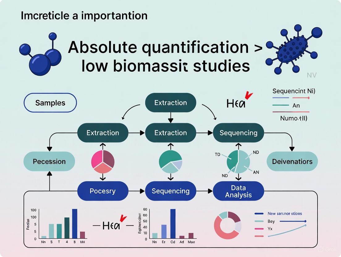
Absolute Quantification in Low-Biomass Microbiome Studies: Overcoming Bias to Unlock Clinical Insights
This article addresses the critical need for absolute quantification in low-biomass microbiome research, a field plagued by significant technical challenges and potential for data misinterpretation.
Absolute Quantification in Low-Biomass Microbiome Studies: Overcoming Bias to Unlock Clinical Insights
Abstract
This article addresses the critical need for absolute quantification in low-biomass microbiome research, a field plagued by significant technical challenges and potential for data misinterpretation. Aimed at researchers, scientists, and drug development professionals, it explores the fundamental limitations of relative abundance data, which can obscure true biological signals in samples from environments like skin, tumors, blood, and the respiratory tract. We provide a comprehensive overview of current and emerging methodologies—from flow cytometry and spike-in standards to novel computational approaches—for achieving absolute microbial counts. The content further details rigorous troubleshooting and optimization strategies to mitigate contamination and bias, reinforced by validation studies that demonstrate how absolute quantification transforms the interpretation of therapeutic interventions and disease mechanisms, ultimately paving the way for more reliable diagnostics and therapies.
Why Relative Abundance Fails in Low-Biomass Environments: The Foundational Pitfalls
The exploration of low-biomass environments represents a formidable frontier in microbiome research. These habitats, characterized by exceedingly low levels of microbial life, pose unique methodological challenges that distinguish them from their high-biomass counterparts. Low-biomass environments span a remarkable diversity, including specific human tissues, the atmosphere, plant seeds, treated drinking water, hyper-arid soils, and the deep subsurface [1]. The defining feature of these environments is that microbial biomass approaches the limits of detection using standard DNA-based sequencing approaches, making the inevitability of contamination from external sources a critical concern [1] [2].
The significance of studying these environments extends far beyond academic curiosity. In human health, purported microbiomes of tissues such as the placenta, brain, and blood have been the subject of intense debate and controversy, with subsequent rigorous studies often revealing that initial findings were driven by contamination [3] [4]. In environmental science, accurately characterizing microbial communities in extreme habitats informs our understanding of life's boundaries and has implications for astrobiology, bioremediation, and ecosystem monitoring [1] [5]. This technical guide frames the exploration of these challenging environments within the broader thesis that absolute quantification is paramount for generating biologically meaningful and reproducible results, moving beyond relative abundance measurements that can yield misleading conclusions [6] [5].
Defining the Low-Biomass Niche: A Spectrum of Challenging Environments
Conceptually, low-biomass environments exist on a continuum rather than representing a binary category. While some have proposed quantitative thresholds (e.g., <10,000 microbial cells/mL), it is more informative to consider biomass as a gradient where technical challenges become progressively more severe as microbial abundance decreases [3]. The fundamental challenge in these environments is that the target DNA "signal" can be dwarfed by the contaminant "noise" introduced during sampling or laboratory processing [1]. This problem is exacerbated by the proportional nature of sequence-based datasets, meaning even minute amounts of contaminating DNA can drastically influence the interpretation of a sample's microbial composition [1].
The table below categorizes and exemplifies the diverse range of low-biomass environments currently under investigation.
Table 1: Categories and Examples of Low-Biomass Environments
| Category | Specific Examples | Key Characteristics |
|---|---|---|
| Human Tissues | Placenta [1] [3], Fetal tissues [1], Brain [4], Blood [1] [3] [4], Lower Respiratory Tract [3], Breastmilk [1] | Very low microbial load relative to host DNA; high susceptibility to contamination during collection; often lack resident microbes altogether [1] [3]. |
| Built Environments | Cleanrooms [7], Hospital Operating Rooms [7], Spacecraft Assembly Facilities [7], Metal Surfaces [1] | Ultra-low biomass due to stringent cleaning; critical for planetary protection and human health [7]. |
| Natural & Extreme Environments | Atmosphere [1], Hyper-arid soils [1], Deep subsurface [1] [3], Ice cores [1], Hypersaline brines [1], Treated Drinking Water [1] | Approach limits of microbial life; subject to polyextreme conditions (e.g., temperature, pH, salinity, nutrient availability) [1]. |
| Other Biological Hosts | Plant Seeds [1], Certain Animal Guts (e.g., caterpillars) [1], Salmonid Blood and Brain [4] | Highlight that "sterile" compartments may not be universal across species; salmonids, for instance, have a more permeable blood-brain barrier [4]. |
Technical Hurdles and Methodological Pitfalls
Research in low-biomass environments is fraught with analytical challenges that can compromise biological conclusions if not properly addressed.
Contamination: The Primary Adversary
Contamination, defined as the introduction of external DNA, is the most significant hurdle. It can originate from multiple sources, including human operators, sampling equipment, laboratory reagents, and kits [1] [3]. The "kitome"—the microbial contamination associated with DNA extraction and library preparation kits—is a particularly pernicious source [7]. In high-biomass samples like human stool, contaminants represent a minor component of the total DNA. In low-biomass samples, however, these contaminants can constitute the majority, or even the entirety, of the observed microbial signal [1] [3].
Cross-Contamination and Host DNA Misclassification
Well-to-well leakage, or the "splashome," occurs when DNA from one sample contaminates adjacent samples during plate-based processing [3]. This cross-contamination can violate the core assumptions of computational decontamination tools [3]. Furthermore, in host-associated low-biomass samples, the vast majority of sequenced DNA is from the host. This host DNA can be misclassified as microbial during bioinformatic analysis if not properly accounted for, generating noise or even artifactual signals if confounded with an experimental phenotype [3].
Batch Effects and the Imperative of Careful Design
Technical variability between processing batches (batch effects) can easily overwhelm subtle biological signals [3]. These effects stem from differences in reagents, personnel, protocols, and equipment. A critical principle in study design is to avoid batch confounding, ensuring that the biological groups of interest (e.g., case vs. control) are distributed across all processing batches [3]. Failure to do so can make technical artifacts indistinguishable from true biological phenomena.
Foundational Principles for Rigorous Study Design
Overcoming the challenges of low-biomass research requires meticulous planning from sample collection to data analysis. The following workflow outlines a robust, contamination-aware approach.
Figure 1: An integrated workflow for low-biomass microbiome studies, highlighting critical steps from planning to reporting.
A Multi-Layered Control Strategy
The incorporation of comprehensive controls is non-negotiable. Two complementary approaches are recommended:
- Process Controls: These are blank samples that pass through the entire experimental workflow alongside real samples, representing the aggregate contamination from all stages [3]. Examples include empty collection tubes, swabs exposed to sampling air, and blank extractions with no sample input [1] [3].
- Source-Specific Controls: To better identify the origin of contaminants, specific controls should be implemented, such as swabs of PPE, sampling equipment, or aliquots of preservation solutions [1] [3]. The collection of multiple control replicates is essential for robust statistical identification of contaminants [3].
Decontamination and Barrier Protection
All equipment and surfaces that contact samples must be decontaminated. A two-step process is effective: first, using 80% ethanol to kill contaminating organisms, followed by a nucleic acid degrading solution (e.g., bleach, UV-C light) to remove residual DNA [1]. Personal protective equipment (PPE), including gloves, masks, and cleanroom suits, acts as a critical barrier to limit contamination from human operators, protecting samples from aerosolized droplets and skin cells [1].
The Scientist's Toolkit: Essential Reagents and Materials
Success in low-biomass research hinges on the use of specialized reagents and materials designed to minimize and monitor contamination.
Table 2: Key Research Reagent Solutions for Low-Biomass Studies
| Item | Function & Importance | Specific Examples & Considerations |
|---|---|---|
| DNA-Decontaminated Reagents | To prevent introduction of microbial DNA from the reagents themselves. Standard molecular biology reagents can contain trace DNA. | Use reagents certified DNA-free. Decontaminate solutions with UV irradiation or sodium hypochlorite where applicable [1]. |
| DNA-Free Sampling Kits | To collect samples without adding contaminating signal. | Use single-use, pre-sterilized swabs and collection vessels [1]. Consider innovative devices like the SALSA sampler for surfaces [7]. |
| Internal Standards (IS) | For absolute quantification. Added in known quantities to correct for technical variation and convert relative to absolute abundance. | Can be cellular standards or synthetic DNA spikes. Allows estimation of microbial load and gene copies per sample unit [5]. |
| Mock Communities | Positive controls with known composition. Used to assess accuracy and bias of the entire workflow. | Comprise defined mixes of microbial strains [8]. Essential for validating bioinformatic pipelines and identifying taxon-specific biases [8]. |
| Nucleic Acid Removal Solutions | To destroy contaminating DNA on surfaces and equipment. Sterilization (e.g., autoclaving) kills cells but may not remove DNA. | Use sodium hypochlorite (bleach), hydrogen peroxide, or commercial DNA removal solutions [1]. |
The Critical Role of Absolute Quantification
Moving from relative to absolute quantification is a paradigm shift essential for accurate interpretation of low-biomass studies. Relative abundance data, which sums to 100%, is compositional. An increase in the relative abundance of one taxon necessitates an apparent decrease in others, which can produce spurious correlations and mask true biological effects [5]. Absolute quantification contextualizes the microbial signal, distinguishing between a substantial population of resident microbes and trace-level contamination.
Methods for Absolute Quantification
Several methods can bridge the gap from relative to absolute abundance:
- Bacterial-to-Host (B:H) DNA Ratio: A recently developed method estimates bacterial biomass in host-associated samples using the ratio of bacterial to host DNA reads in metagenomic data. This uses the host DNA as an inherent internal standard, requiring no additional experiments [6].
- Cellular Internal Standards: Known quantities of non-native cells (e.g., from an unrelated environment) or synthetic DNA sequences are added to the sample prior to DNA extraction. By tracking the recovery of these spikes, researchers can compute the absolute abundance of native taxa in the sample [5].
- Traditional Cell Counting: Methods like flow cytometry (FCM) or quantitative PCR (qPCR) can provide total microbial load, which can then be multiplied by relative abundances from sequencing to obtain absolute counts [5].
Defining and accurately characterizing low-biomass environments—from human tissues to extreme habitats—remains one of the most technically demanding pursuits in microbiology. The history of controversies in this field underscores the necessity of rigorous, contamination-aware methodologies. As outlined in this guide, success depends on a multi-faceted strategy: comprehensive control schemes, stringent decontamination protocols, unconfounded study designs, and a commitment to moving beyond relative abundance data through absolute quantification. By adopting these practices, researchers can ensure that future discoveries in these elusive frontiers are robust, reproducible, and biologically meaningful, fully realizing the potential of low-biomass microbiome research to advance human health and environmental science.
High-throughput sequencing has revolutionized microbiome research, yet the standard analytical paradigm relies on relative abundance data that inherently distorts ecological reality. This compositional data, constrained to a constant sum, introduces severe analytical pathologies including spurious correlations, false positives in differential abundance testing, and an inability to discern true population dynamics. The problem becomes particularly acute in low-biomass environments where contaminant DNA disproportionately influences results. This technical review examines the mathematical foundations of the compositional data problem, demonstrates how relative abundance metrics can produce misleading biological conclusions, and presents rigorous experimental and computational solutions centered on absolute quantification. By integrating compositional data analysis (CoDA) principles with emerging absolute quantification techniques, researchers can overcome these limitations and achieve more accurate ecological interpretations of microbiome data.
Microbiome datasets generated by high-throughput sequencing (HTS) are fundamentally compositional because sequencing instruments deliver reads only up to their fixed capacity, imposing an arbitrary total on the data [9]. This means that HTS output contains information about the relationships between microbial taxa rather than their absolute abundances in the original environment [9]. The constant-sum constraint transforms the data into a closed array where individual components cannot vary independently—an increase in one taxon's relative abundance necessarily produces decreases in others, regardless of their actual absolute abundances [9] [10].
The distinction between absolute and relative abundance represents a critical conceptual divide in microbiome analysis. Absolute abundance refers to the actual number of a specific microorganism present in a sample, typically quantified as "number of microbial cells per gram/milliliter of sample" [11]. In contrast, relative abundance describes the proportion of a specific microorganism within the entire microbial community, where the sum of all relative abundances typically equals 100% [11]. This distinction becomes biologically significant when considering that two subjects may harbor the same relative abundance of a pathogen (e.g., 20%), but if one has double the total microbial load, they consequently harbor twice the absolute quantity of that pathogen [12].
Table 1: Fundamental Differences Between Absolute and Relative Abundance
| Characteristic | Absolute Abundance | Relative Abundance |
|---|---|---|
| Definition | Actual number of microorganisms in a sample | Proportion of a microorganism within the community |
| Measurement Unit | Cells per gram/milliliter | Percentage or proportion (0-100%) |
| Sum Constraint | No constant sum | Constant sum (typically 100%) |
| Dependence on Other Taxa | Independent | Dependent on abundances of all other taxa |
| Information Content | True quantitative abundance | Proportional relationships |
| Impact of Total Load Changes | Directly reflects changes | Can mask true changes |
In low-biomass environments—including certain human tissues (tumors, lungs, placenta, blood), the atmosphere, plant seeds, and treated drinking water—the compositional problem becomes particularly severe [3] [1]. With minimal starting microbial DNA, even small amounts of contaminant DNA can disproportionately influence results, potentially leading to spurious conclusions about microbial presence and community structure [3] [1].
The Mathematical Pathology of Compositional Data
The Spurious Correlation Problem
Compositional data exhibit a negative correlation bias and fundamentally different correlation structure than underlying count data [9]. This pathology arises because the data reside on a simplex space—a geometric representation where the whole is the sum of its parts—rather than in real Euclidean space [10]. The consequences are profound: correlation coefficients calculated from raw relative abundances are inherently misleading and cannot reliably indicate underlying biological relationships.
The mathematical basis for this distortion was first recognized by Pearson in 1897 and has been rediscovered repeatedly in various fields, including microbiome research [9]. The core issue stems from the closure property of compositional data, where the measurement of any single component depends on all other components in the system. This dependency creates a situation where apparent "increases" in one taxon may actually reflect decreases in others, completely reversing biological interpretation [10].
The severity of false-positive rates in differential abundance testing is particularly alarming. Studies have demonstrated that traditional analyses of relative abundance data can produce false-positive rates exceeding 30%, even with modest sample sizes [10]. This high error rate stems from the inherent interdependency of relative values, where an increase in one taxon's relative abundance mathematically necessitates decreases in others, creating the illusion of differential abundance where none exists in absolute terms [10].
Directional Misinterpretation in Differential Abundance
The inability to determine directionality of change represents one of the most clinically problematic aspects of compositional data analysis. Consider a community with only two taxa: an increase in the ratio between Taxon A and Taxon B could indicate (i) Taxon A increased, (ii) Taxon B decreased, (iii) a combination of both effects, (iv) both increased but Taxon A increased more, or (v) both decreased but Taxon B decreased more [13]. Knowing which scenario occurs is crucial for biological interpretation but cannot be determined from relative abundance data alone [13].
Real-world examples demonstrate how profoundly relative abundance can distort biological reality. In soil microbiome research, Yang et al. (2018) found that 33.87% of bacterial genera showed opposite change directions—described as decreased relative abundance but increased absolute abundance—when analyzed using absolute quantification methods [14]. Similarly, in sodium azide-treated soil, 40.58% of total genera exhibited an upregulation trend using relative quantification but downregulation via absolute quantification [14]. These discrepancies arise from failure to account for changes in total bacterial count, leading to false-positive results and incorrect biological interpretations [14].
Table 2: Common Analytical Pitfalls in Relative Abundance Analysis
| Pitfall | Mathematical Cause | Biological Consequence |
|---|---|---|
| Spurious Correlation | Negative bias due to sum constraint | False associations between taxa |
| Directional Ambiguity | Inability to distinguish increases from decreases | Misinterpretation of treatment effects |
| False Positives in Differential Abundance | Interdependency of relative values | Incorrect identification of biomarker taxa |
| Compositional Bias in Diversity Metrics | Uneven sampling depth and sensitivity to dominant taxa | Distorted alpha and beta diversity estimates |
| Subsetting/ Aggregation Artifacts | Change in reference frame when selecting taxa | Inconsistent results at different taxonomic levels |
Experimental Solutions: Absolute Quantification Frameworks
Digital PCR Anchoring
A rigorous absolute quantification framework based on digital PCR (dPCR) anchoring combines the precision of dPCR with the high-throughput nature of 16S rRNA gene amplicon sequencing [13]. This method involves using dPCR to obtain an absolute count of 16S rRNA gene copies in a sample, then using this value to convert relative abundances from sequencing to absolute quantities [13].
The experimental workflow begins with efficient DNA extraction across diverse sample types. Validation studies spiking a defined 8-member microbial community into gastrointestinal samples from germ-free mice demonstrated near-equal and complete recovery of microbial DNA over five orders of magnitude [13]. The lower limit of quantification (LLOQ) was established at 4.2 × 10⁵ 16S rRNA gene copies per gram for stool/cecum contents and 1 × 10⁷ copies per gram for mucosal samples [13]. The critical innovation lies in using dPCR to precisely quantify total 16S rRNA gene copies, then applying the formula: Absolute Abundance of Taxon A = (Relative Abundance of Taxon A) × (Total 16S rRNA Gene Copies) [13].
This approach was validated in a murine ketogenic-diet study comparing microbial loads in lumenal and mucosal samples along the GI tract. Quantitative measurements of absolute abundances revealed decreases in total microbial loads on the ketogenic diet that were undetectable using relative abundance analysis, enabling researchers to determine differential effects of diet on each taxon with unprecedented accuracy [13].
Internal Standard-Based Quantification
Internal standard (IS)-based absolute quantification involves adding known quantities of exogenous cells or DNA to samples prior to DNA extraction [5]. Also known as "spike-in" methods, these approaches use the recovery rate of the internal standard to calibrate the entire measurement process, accounting for variations in DNA extraction efficiency, PCR amplification bias, and other technical variables [5].
The optimal internal standard should be absent from native samples yet resemble the target microorganisms in cell structure and DNA extraction characteristics. Common choices include synthetic communities, purified DNA from non-native species, or genetically modified cells [5]. The absolute abundance of native taxa is calculated using the formula: Absolute Abundance = (Relative Abundance of Native Taxon) × (Amount of Spiked IS / Relative Abundance of IS) [5].
This method was applied to analyze microbial population dynamics in horizontal surface layer soil and parent material soil. The absolute quantification revealed that the total bacteria count in the developed surface layer soil was 4.78 times less than the parent material soil (3.55 × 10⁸ vs. 1.7 × 10⁹ cells/g) [14]. Crucially, absolute quantification detected significant changes in 20 out of 25 total phyla, while relative quantification detected only 12 phyla, demonstrating the enhanced sensitivity of absolute methods [14].
Flow Cytometry with Sequencing Integration
Flow cytometry provides a robust method for quantifying total microbial load by counting individual cells in a sample [12]. When combined with sequencing, flow cytometry enables conversion of relative abundances to absolute quantities without the need for standard curves [12]. The procedure involves analyzing sample aliquots using flow cytometry to obtain total cell counts, then applying the formula: Absolute Abundance = Relative Abundance × Total Cell Count [12].
This approach is particularly valuable for detecting clinically relevant changes in total microbial load. For example, healthy adult human fecal samples show up to tenfold variation (10¹⁰⁻¹¹ cells/g) with daily fluctuations of 3.8 × 10¹⁰ cells/g [14]. Similarly, mucosal bacterial loads in Crohn's disease and inflammatory bowel disease patients are higher than in healthy controls—differences that would be obscured in relative abundance analyses [14]. Flow cytometry counting is most suitable for environmental samples with low biomass and well-dispersed cells, such as drinking water, cooling water samples, and river samples [5].
Diagram 1: Absolute Quantification Experimental Workflow. This integrated approach combines internal standards, digital PCR, and high-throughput sequencing to derive absolute abundances.
Computational Solutions: Compositional Data Analysis (CoDA)
Log-Ratio Transformations
Compositional data analysis (CoDA) provides a mathematical framework that respects the relative nature of microbiome data while avoiding spurious conclusions [9] [10]. The core innovation involves transforming data from the simplex to real Euclidean space using log-ratio transformations, which effectively eliminates the sum constraint [10].
The center log-ratio (CLR) transformation normalizes abundances to the geometric mean of a sample. For a composition with D components, the CLR transformation is defined as:
CLR(x) = [ln(x₁/g(x)), ln(x₂/g(x)), ..., ln(x_D/g(x))]
where g(x) is the geometric mean of all components [10]. This transformation symmetrizes the data and enables application of standard statistical methods that assume Euclidean geometry [10].
The additive log-ratio (ALR) transformation normalizes abundances to a carefully chosen reference component. The transformation is defined as:
ALR(x) = [ln(x₁/xD), ln(x₂/xD), ..., ln(x{D-1}/xD)]
where x_D is the reference component [10]. The choice of reference is critical and should ideally be a taxon that is abundant, prevalent, and biologically stable across samples [10].
When applied to glycomics data (which share the compositional nature of microbiome data), CLR transformation resulted in dramatically improved clustering compared to raw relative abundances (Dunn index 0.828 vs. 8.647) [10]. Similarly, in a bacteremia N-glycomics dataset, Aitchison distance (Euclidean distance after ALR transformation) better separated patient and donor classes than clustering on log-transformed abundances (adjusted Rand index: 0.79 vs. 0.74) [10].
Experimental Design for Low-Biomass Studies
Low-biomass microbiome research requires specialized experimental designs to address contamination challenges [3] [1]. Process controls that represent contamination sources are essential, including blank extraction controls, no-template controls, and empty collection kit controls [3]. These controls should be included in every processing batch to account for batch-specific contamination [3].
Avoiding batch confounding is particularly critical. Experimental designs must ensure that phenotypes and covariates of interest are not confounded with batch structure at any experimental stage [3]. This requires active de-confounding through balanced sample allocation across batches rather than reliance on randomization alone [3].
Minimizing well-to-well leakage ("cross-contamination" or "splashome") requires physical separation of samples during processing and inclusion of negative controls interspersed with samples [3] [1]. Recent research demonstrates that well-to-well leakage into contamination controls violates the assumptions of most computational decontamination methods, highlighting the need for physical prevention rather than computational correction [3].
Table 3: Research Reagent Solutions for Absolute Quantification
| Reagent/Method | Function | Key Considerations |
|---|---|---|
| Digital PCR (dPCR) | Absolute quantification of 16S rRNA gene copies | Microfluidic format reduces host DNA amplification bias; no standard curve needed |
| Flow Cytometry | Total microbial cell counting | Distinguishes live/dead cells with appropriate dyes; requires single-cell suspensions |
| Internal Standards (Spike-ins) | Calibration of extraction and amplification efficiency | Should mimic native cells in lysis characteristics; must be absent from native samples |
| CARD-FISH Probes | Specific taxon quantification via fluorescence | Signal amplification enables low-abundance taxon detection; requires specialized expertise |
| DNA Decontamination Solutions | Remove contaminating DNA from reagents and surfaces | Sodium hypochlorite, UV-C exposure, or commercial DNA removal solutions |
Case Study: Absolute Quantification Reveals Drug Effects Obscured by Relative Abundance
A compelling demonstration of absolute quantification's power comes from a 2025 study comparing berberine (BBR) and sodium butyrate (SB) effects on gut microbiota in DSS-induced colitis mice [15]. Using both relative and absolute quantitative sequencing, researchers found that relative abundance measurements failed to accurately reflect the true microbial changes induced by these compounds [15].
Notably, absolute quantitative sequencing provided results more consistent with the actual microbial community and revealed drug effects that were obscured or misrepresented by relative abundance analysis [15]. Specifically, the regulatory effects of BBR on gut microbiota were more accurately captured using absolute quantification, demonstrating that relative quantitative sequencing analyses are prone to misinterpretation and incorrect correlation of results [15].
This study underscores how absolute quantitative analysis better represents true microbial counts when evaluating drug modulatory effects on the microbiome [15]. The findings have vital implications for pharmaceutical development targeting the microbiome, as relative abundance measurements might lead to erroneous conclusions about drug mechanisms or loss of key bacterial genera involved in therapeutic effects [15].
The compositional nature of relative abundance data represents a fundamental challenge in microbiome research that transcends analytical approaches. While compositional data analysis methods provide mathematical rigor for working within the relative abundance framework, absolute quantification approaches offer the most direct path to ecological truth by measuring actual cellular abundances in biological samples.
The future of rigorous microbiome science lies in integrating absolute quantification into standard practice, particularly for low-biomass environments where compositional effects are most pronounced. Methods such as dPCR anchoring, internal standard calibration, and flow cytometry integration now provide feasible pathways to absolute quantification without prohibitive cost or technical burden. By adopting these approaches and following rigorous experimental designs that minimize contamination, microbiome researchers can overcome the distortions of compositional data and build a more accurate understanding of microbial ecology in health and disease.
The Pervasive Challenge of Contamination and Environmental DNA
In the study of low-biomass environments—such as human tissues, treated drinking water, and hyper-arid soils—the inevitability of contamination presents a fundamental challenge that can compromise scientific validity. These environments harbor minimal microbial biomass, approaching the limits of detection for standard DNA-based sequencing approaches [1]. The proportional impact of contaminating DNA is dramatically amplified in these systems, where the target DNA 'signal' can be easily overwhelmed by contaminant 'noise' [1]. This challenge extends beyond mere technical nuisance; it has fueled major scientific controversies, including debates surrounding the existence of a placental microbiome and the accurate characterization of tumor microbiomes, where initial findings were later attributed to contamination [3]. Consequently, rigorous contamination control is not simply a best practice but a foundational requirement for generating reliable data, particularly for absolute quantification where accurate measurement of DNA copy numbers is paramount.
Contaminating DNA can infiltrate an experiment at virtually any stage, from sample collection to data analysis. Recognizing these sources is the first step in developing effective mitigation strategies.
The major vectors for introducing contamination include:
- Human Operators: Microbial cells and DNA can be shed from skin, hair, or clothing, or introduced via aerosols generated from breathing or talking [1].
- Sampling Equipment: Collection tools, vessels, and surfaces can carry microbial DNA from previous uses or the environment [1].
- Laboratory Reagents and Kits: Commercial kits for DNA extraction, amplification, and library preparation often contain trace amounts of microbial DNA that become detectable in low-biomass contexts [1] [3].
- Cross-Contamination (Well-to-Well Leakage): DNA can transfer between samples processed concurrently, for example, in adjacent wells of a 96-well plate, a phenomenon also termed the "splashome" [1] [3].
Table 1: Summary of Major Contamination Sources and Their Vectors
| Source | Vectors | Typical Impact |
|---|---|---|
| Human Operator | Skin cells, aerosols, improper personal protective equipment (PPE) | Introduction of human-associated microbes (e.g., Propionibacterium, Staphylococcus) |
| Laboratory Reagents | Extraction kits, polymerase enzymes, water | Dominated by low-diversity, ultra-clean-associated taxa (e.g., Caulobacter, Burkholderia) |
| Sampling Equipment | Non-sterile swabs, collection tubes, fluids | Environmental species (e.g., from soil or water) distorting in-situ signals |
| Cross-Contamination | Aerosols during pipetting, poorly sealed plates | False positives, blending of community signatures between samples |
The Analytical Challenge: Confounding and Signal Distortion
The presence of contamination is problematic enough, but its impact is magnified when confounded with the experimental variables of interest. If samples from different experimental groups (e.g., case vs. control) are processed in separate batches using different reagent lots or by different personnel, the differential contamination profiles can create artifactual signals that are misinterpreted as biological reality [3]. In such scenarios, what appears to be a statistically significant biomarker could merely reflect batch-specific contamination.
A Framework for Contamination Mitigation: From Collection to Analysis
A multi-layered defense strategy is essential to minimize, identify, and account for contamination throughout the research workflow.
Pre-Sampling and Collection Protocols
During the initial stages of research, proactive measures are critical:
- Decontamination of Equipment: Use single-use, DNA-free consumables where possible. Reusable equipment should be decontaminated with 80% ethanol to kill organisms, followed by a nucleic acid degrading solution (e.g., bleach, UV-C light) to remove residual DNA [1].
- Use of Personal Protective Equipment (PPE): Operators should wear gloves, masks, clean suits, and shoe covers as appropriate to create a barrier between the sample and potential human contaminants [1].
- Environmental Controls: Sample collection should minimize exposure to ambient air and surfaces. In some cases, cleanroom-level protocols may be necessary [1].
Essential Experimental Controls
The use of comprehensive process controls is non-negotiable for identifying the contaminant profile of a workflow [3]. These controls should be processed alongside actual samples through all stages.
Table 2: Critical Negative Controls for Low-Biomass Studies
| Control Type | Description | Function |
|---|---|---|
| Field Blank | An empty, sterile collection vessel taken into the field. | Identifies contamination from collection vessels and the field environment. |
| Extraction Blank | Reagents without a sample carried through the DNA extraction process. | Reveals contamination inherent to extraction kits and reagents. |
| PCR Blank | Molecular grade water used as a template in amplification. | Detects contamination in PCR/master mix reagents and the laboratory environment. |
| Internal Standards | Known quantities of synthetic or foreign DNA spikes. | Monitors PCR inhibition, quantifies efficiency, and enables absolute quantification [16]. |
Strategic Study Design
Proper experimental design is the most powerful tool for neutralizing the effects of unavoidable contamination.
- Avoid Batch Confounding: The highest priority is to ensure that biological groups of interest are distributed evenly across all processing batches (e.g., DNA extraction plates, sequencing runs). This prevents contamination and batch effects from being misinterpreted as biological signals [3].
- Randomization and Replication: Sample processing order should be randomized with respect to experimental groups. Including technical replicates helps assess variability and the impact of stochastic contamination.
Advanced Methodologies for Absolute Quantification
Moving from relative to absolute quantification is a crucial frontier in low-biomass research, as it allows researchers to distinguish genuine, abundant signals from low-level contamination.
The qMiSeq Approach
The quantitative MiSeq (qMiSeq) approach is a metabarcoding method that converts sequence read counts into absolute DNA copy numbers. This is achieved by spiking each sample with known quantities of synthetic internal standard DNA sequences (which are distinguishable from natural sequences) prior to library preparation [16]. A sample-specific linear regression is then created between the known standard copy numbers and their observed read counts. This regression model is used to convert the read counts of all other taxa in that sample into estimated DNA copies, thereby correcting for sample-specific PCR bias and inhibition [16]. This method has shown significant positive correlations with both the abundance and biomass of fish communities in environmental studies, validating its utility for quantitative assessment [16].
Digital PCR (dPCR) and Quantitative PCR (qPCR)
For targeted, species-specific detection, these methods provide high-sensitivity quantification:
- qPCR relies on comparing the amplification cycle threshold (Ct) of a sample to a standard curve of known DNA concentrations to estimate copy number. The development of species-specific assays, including careful design and validation of primers and probes, is critical for accuracy [17].
- dPCR partitions a sample into thousands of individual reactions, providing absolute quantification without the need for a standard curve. It often offers superior sensitivity and resistance to PCR inhibitors, making it well-suited for low-biomass targets.
The Scientist's Toolkit: Essential Reagent Solutions
Successful low-biomass research relies on a suite of specialized reagents and materials, each chosen to minimize interference and maximize fidelity.
Table 3: Key Research Reagent Solutions and Their Functions
| Reagent/Material | Function | Technical Considerations |
|---|---|---|
| DNA-Decontaminated Reagents | Molecular grade water, enzymes, and buffers treated to remove microbial DNA. | Critical for all molecular steps. Verify via rigorous negative controls. |
| Ultra-Clean Collection Swabs & Tubes | Pre-sterilized, DNA-free consumables for sample acquisition and storage. | Prefer plasticware treated by autoclaving and UV irradiation. |
| Internal Standard DNA | Synthetic, non-natural DNA sequences (e.g., gBlocks, Spike-ins). | Added to samples pre-extraction (for process control) or pre-amplification (for qMiSeq) [16]. |
| High-Fidelity DNA Polymerase | Enzyme for PCR with high processivity and low error rate. | Reduces amplification artifacts and chimeras in final sequences. |
| Barcoded Adapters & Index Primers | Oligonucleotides for labeling and preparing sequencing libraries. | Enable multiplexing of samples; unique dual indexing is essential to identify cross-talk [3]. |
| DNA Removal Solutions | Chemical agents like bleach or sodium hypochlorite. | Used for decontaminating work surfaces and non-disposable equipment [1]. |
Visualizing the Experimental Workflow for Quantitative Low-Biomass Analysis
The following diagram outlines a robust, contamination-aware workflow integrating the principles and methods discussed above.
The pervasive challenge of contamination in low-biomass and eDNA research demands a paradigm shift from simple detection to rigorous, quantification-focused science. By integrating meticulous experimental design, comprehensive controls, and advanced quantitative methods like qMiSeq, researchers can transcend mere contamination awareness and achieve true quantitative accuracy. This disciplined approach is the foundation upon which reliable, reproducible, and biologically meaningful conclusions are built, ultimately advancing our understanding of the hidden microbial worlds in low-biomass environments.
This case study examines the transformative role of relic-DNA depletion in skin microbiome research, a critical advancement for achieving absolute quantification in low-biomass environments. Traditional sequencing methods conflate DNA from live bacterial cells with extracellular DNA and genetic material from dead cells, significantly skewing microbial community profiles. By implementing innovative methodologies that discriminate between intact and relic DNA, researchers can overcome fundamental biases that have historically obstructed accurate characterization of the living skin microbiome. This technical analysis details experimental protocols, quantitative findings, and methodological frameworks that demonstrate how relic-DNA depletion reveals authentic microbial patterns, providing a refined baseline for mechanistic studies of skin health, disease progression, and therapeutic development.
The skin microbiome presents unique investigational challenges due to its inherently low microbial biomass, where standard sequencing approaches struggle to distinguish true biological signals from technical artifacts [18]. In these environments, relic DNA—extracellular DNA and genetic material from non-viable cells—can comprise a substantial portion of sequenced material, dramatically distorting community profiles [19] [20]. This relic DNA acts as a "genetic fossil record" of past microbial inhabitants rather than representing the currently living community, complicating efforts to establish causal relationships between microbiome composition and skin health or disease states.
The imperative for absolute quantification stems from the limitations of relative abundance data, which can produce misleading interpretations in dynamic microbial systems [14]. When data are expressed only as relative proportions, an apparent increase in one taxon's abundance may result from the actual decline of other community members rather than its true proliferation. This compositional nature of standard sequencing data obscures true population dynamics and interspecies interactions, necessitating methods that provide cell-count resolution for accurate ecological inference [14] [21].
The Problem: Relic DNA Obscures the Living Skin Microbiome
Quantifying the Relic DNA Burden in Skin Samples
Recent investigations have revealed the astonishing prevalence of relic DNA in skin microbiome samples. One landmark study demonstrated that up to 90% of microbial DNA obtained from standard skin swabs originates from non-viable sources rather than living bacterial communities [19]. This overwhelming proportion of relic material means that conventional sequencing approaches primarily capture a historical archive of microbial presence rather than the physiologically active community relevant to skin health and function.
The impact of this relic DNA burden is particularly pronounced in skin environments due to their low bacterial density compared to other body sites. With an estimated 10^4 to 10^6 bacteria inhabiting each square centimeter of skin, even minimal relic DNA contamination can disproportionately influence community profiles [18]. This effect varies across different skin sites, with dry regions typically exhibiting lower biomass and consequently greater susceptibility to relic DNA bias [18].
Consequences for Microbiome Data Interpretation
The presence of substantial relic DNA creates multiple interpretive challenges for skin microbiome researchers:
- Inflated Diversity Estimates: Relic DNA preserves genetic signatures of transient or deceased microorganisms, creating the illusion of greater microbial diversity than actually exists in the living community [20].
- Obscured Ecological Patterns: Spatiotemporal dynamics of viable microbial populations are masked by the stable background of relic DNA, blurring distinctions between body sites and individuals [19].
- Misattributed Biological Effects: Interventions that affect microbial viability without immediately removing DNA may produce misleading community profiles that fail to reflect actual therapeutic outcomes.
Methodological Framework: Approaches for Relic-DNA Depletion and Absolute Quantification
Integrated Workflow for Live Microbiome Characterization
The following workflow diagram illustrates the comprehensive integration of relic-DNA depletion with absolute quantification for authentic skin microbiome profiling:
Relic-DNA Depletion Techniques
Benzonase Endonuclease Treatment
Benzonase endonuclease has emerged as a highly effective method for relic-DNA removal in soil and skin microbiomes [20]. This enzyme digests all forms of DNA and RNA without cell membrane penetration, selectively eliminating extracellular nucleic acids while preserving genetic material within intact cells.
Optimized Protocol:
- Sample Preparation: Resuspend skin swab samples in 500μL phosphate-buffered saline (PBS)
- Enzyme Application: Add 2μL Benzonase endonuclease (≥250 units) and incubate at 37°C for 45 minutes
- Reaction Termination: Heat inactivate at 70°C for 10 minutes
- Efficiency Validation: Assess DNA reduction via fluorometric quantification
Comparative studies demonstrate that Benzonase removes relic DNA with 40-60% efficiency in skin samples, approximately double the performance of propidium monoazide (PMA) treatments (0-30% efficiency) [20]. Unlike light-dependent PMA methods, Benzonase functions effectively in opaque media like skin homogenates without requiring photoactivation.
Propidium Monoazide (PMA) Treatment
As an alternative approach, PMA selectively penetrates membrane-compromised cells and intercalates with DNA upon photoactivation, rendering it insoluble and unavailable for amplification [19]. While less efficient than Benzonase for skin applications, PMA remains valuable for specific experimental contexts requiring viability PCR.
Absolute Quantification Methods
Flow Cytometry for Total Bacterial Load
Flow cytometry provides rapid, single-cell enumeration of bacterial abundance in skin samples, establishing essential baseline data for converting relative sequencing abundances to absolute cell counts [14].
Implementation Protocol:
- Staining: Apply nucleic acid stains (e.g., SYBR Green I) to distinguish microbial cells from background particles
- Calibration: Use fluorescent microspheres of known concentration for instrument calibration
- Analysis: Process samples at low flow rate (≤100 events/sec) to ensure accurate counting of low-biomass samples
- Gating Strategy: Establish conservative gates based on positive control samples (e.g., pure bacterial cultures)
Quantitative PCR (qPCR) for Specific Taxa
qPCR enables sensitive, taxon-specific quantification with detection limits as low as 10^3 cells/gram in fecal samples, demonstrating compatibility with low-biomass skin applications [21].
Strain-Specific qPCR Design Workflow:
- Marker Identification: Identify strain-specific genomic regions through comparative genomics
- Primer Design: Develop primers with 18-25 bp length, 40-60% GC content, and melting temperature of 58-62°C
- Specificity Validation: BLAST analysis against non-target genomes; empirical testing against related strains
- Standard Curve Generation: Use gBlock gene fragments or genomic DNA from pure cultures
- Efficiency Optimization: Adjust annealing temperatures to achieve 90-110% amplification efficiency
Recent systematic comparisons indicate that qPCR provides superior dynamic range and cost-effectiveness compared to droplet digital PCR (ddPCR) for strain-specific quantification in complex samples, though ddPCR offers advantages for absolute quantification without standard curves [21].
Quantitative Findings: Comparative Impact of Relic-DNA Depletion
Methodological Performance Metrics
Table 1: Performance Comparison of Relic-DNA Depletion Methods
| Method | Removal Efficiency | Key Advantages | Limitations | Compatibility with Skin Samples |
|---|---|---|---|---|
| Benzonase | 40-60% [20] | No light activation required; broad substrate range | Potential impact on Gram-positive bacteria if lysis occurs | High - effective in opaque samples |
| PMA | 0-30% [20] | Selective for membrane-compromised cells | Requires transparent samples for photoactivation | Moderate - limited by skin sample opacity |
| DNase I | 20-40% [20] | Specific for DNA without RNase activity | Narrow optimal activity conditions | Moderate - sensitive to sample inhibitors |
Taxonomic Shifts Following Relic-DNA Depletion
Table 2: Taxonomic Abundance Changes After Relic-DNA Removal in Skin Microbiome
| Taxon | Change in Relative Abundance | Interpretation | Statistical Significance |
|---|---|---|---|
| Bacillus | Significant decrease [20] | High relic-DNA contributor in skin environments | p < 0.01 |
| Sphingomonas | Significant decrease [20] | Common environmental contaminant with persistent DNA | p < 0.05 |
| Cutibacterium | Variable response across skin sites [19] | Site-specific viability patterns revealed | p < 0.05 between sites |
| Staphylococcus | Increased relative abundance [19] | Underestimated in total DNA due to high relic from other taxa | p < 0.05 |
Implementation of relic-DNA depletion produces consistent methodological improvements across multiple parameters. Studies report approximately 10% reduction in microbial diversity and richness on average after removing relic DNA, reflecting the elimination of non-viable community members from diversity calculations [20]. Perhaps more importantly, relic-DNA depletion reduces intraindividual similarity between samples from different body sites, strengthening the resolution of true spatial patterning across skin microenvironments [19].
The Scientist's Toolkit: Essential Research Reagents and Materials
Table 3: Key Research Reagents for Relic-DNA Depletion and Absolute Quantification
| Reagent/Material | Function | Application Notes | Representative Product Examples |
|---|---|---|---|
| Flocked nylon swabs (eSwabs) | Sample collection | Superior biomass recovery compared to cotton swabs [22] [23] | Copan eSwab, Puritan HydraFlock |
| Benzonase endonuclease | Relic-DNA degradation | Digests all forms of DNA/RNA without cell penetration [20] | Millipore Sigma Benzonase, Novagen Benzonase |
| Propidium monoazide (PMA) | DNA intercalation in dead cells | Selective inhibition of relic-DNA amplification [19] | Biotium PMA, GenIUL PMA Dye |
| SYBR Green I | Nucleic acid staining | Flow cytometric bacterial enumeration [14] | Thermo Fisher SYBR Green I, Lonza SYBR Green |
| Kit-based DNA extraction | Nucleic acid purification | Higher yield and reproducibility for skin samples [21] [23] | QIAamp Fast DNA Stool Mini Kit, DNeasy PowerSoil Kit |
| Strain-specific primers | Targeted quantification | qPCR-based absolute abundance of specific taxa [21] | Custom-designed oligonucleotides |
Implications for Research and Therapeutic Development
The integration of relic-DNA depletion with absolute quantification methodologies addresses fundamental limitations in skin microbiome research, enabling more accurate associations between microbial community states and dermatological conditions. This technical advancement carries significant implications for multiple research domains:
Disease Mechanism Elucidation
By distinguishing the living microbial community from historical DNA signatures, researchers can establish more reliable correlations between specific viable taxa and skin disorders. The revealed differential abundance of live bacteria across skin regions provides important hypotheses for why certain sites demonstrate heightened susceptibility to pathogenic invasion or inflammatory conditions [19].
Therapeutic Development and Assessment
The accurate quantification of viable microbial populations enables precise monitoring of interventional outcomes, whether evaluating probiotic applications, antibiotic treatments, or microbiome-transplant therapies. Strain-specific qPCR assays permit sensitive tracking of therapeutic strains at levels below conventional sequencing detection limits [21].
Standardization Across Studies
The implementation of relic-DNA depletion creates opportunities for improved cross-study comparisons by eliminating technical variation introduced by differential relic-DNA preservation across sampling strategies and processing methods [22] [23]. This methodological harmonization is particularly valuable for multi-center clinical trials and longitudinal cohort studies.
Relic-DNA depletion represents a methodological paradigm shift in skin microbiome research, overcoming a fundamental bias that has obscured understanding of the living microbial community. The integration of enzymatic relic-DNA removal with absolute quantification techniques provides a powerful framework for generating biologically meaningful data from low-biomass skin samples, transforming our capacity to link microbial ecology with skin health and disease.
Future methodological developments will likely focus on single-cell viability assessments, integration with metatranscriptomic approaches to profile metabolically active communities, and streamlined workflows that combine relic-DNA removal with automated sample processing. As these refined methodologies become standardized, they will accelerate the translation of skin microbiome research into clinically actionable insights and targeted therapeutic interventions.
The Critical Consequences for Disease Association and Drug Mechanism Studies
The advent of high-throughput sequencing has revolutionized microbiome research, enabling large-scale profiling of microbial communities. However, standard microbiome analysis predominantly relies on relative abundance data, which ignores total bacterial load and presents significant interpretation challenges. This whitepaper examines the critical consequences of relying solely on relative abundance in disease association and drug mechanism studies, particularly in the context of low biomass samples. We detail how absolute quantification methods provide more accurate biological insights, prevent misleading conclusions in clinical studies, and enhance drug development research. Methodological guidance, technical protocols, and analytical frameworks are presented to assist researchers in implementing absolute quantification approaches.
The Fundamental Problem with Relative Abundance Data
Microbiome sequencing data is inherently compositional, meaning that all microbial abundances are expressed as proportions that sum to 100% [14] [12]. This fundamental characteristic leads to several critical limitations:
- The Constant Sum Constraint: In compositional data, an increase in one taxon's abundance inevitably forces a decrease in the relative abundance of other taxa, creating spurious negative correlations that may not reflect biological reality [5].
- Masking of True Biological Changes: Changes in absolute abundance of one taxon can artificially alter the relative proportions of all other taxa, even when their absolute counts remain unchanged [14].
- False Positive and False Negative Results: Relative abundance analysis frequently identifies differential abundance that disappears when absolute counts are considered, while simultaneously missing true biological changes [14] [24].
The following example illustrates how relative abundance data can be misleading: When two types of bacteria start with the same initial cell number, a treatment that doubles the cell number of bacteria A (while bacteria B remains unaffected) results in the same relative abundance between bacteria A and B (67% and 33%) as a treatment that halves bacteria B (while bacteria A remains unaffected). However, these two treatment effects are biologically completely different [14].
Critical Consequences for Disease Association Studies
Misinterpretation in Gut Microbiome Research
In gastrointestinal research, reliance on relative abundance has led to contradictory findings and obscured true disease mechanisms:
- Inflammatory Bowel Disease (IBD): Studies have revealed that the overall mucosal bacterial loads in patients with Crohn's disease and inflammatory bowel disease are significantly higher than in healthy controls, a finding that would be masked in relative abundance analyses [14].
- Microbial Load Variations: Healthy adult human fecal samples show substantial variation (10¹⁰–¹¹ cells/g) with daily fluctuations up to 3.8 × 10¹⁰ cells/g, meaning that relative abundance changes may simply reflect these total load variations rather than specific taxonomic shifts [14].
- Bacteroides-Enterotype Association: One study combining sequencing with flow cytometry found that microbial load in people with a Bacteroides-enterotype microbiome was associated with Crohn's disease, a relationship that could not be detected through relative abundance analysis alone [12].
Challenges in Low Biomass Microbiome Studies
Low biomass samples (skin, respiratory tract, air samples) present particular challenges where absolute quantification becomes essential:
- Quality Control Imperative: For low biomass samples, quantifying total microbial load via qPCR is essential to confirm whether bacterial load is sufficient for meaningful sequencing results, preventing false conclusions from amplification artifacts or contamination [12].
- Detection Sensitivity Issues: In low biomass environments, the presence of many rare taxa often results from sequencing artifacts rather than biological reality, requiring careful filtering and quantification approaches to distinguish true signals from noise [24].
- Antibiotic Intervention Studies: Antibiotics significantly reduce microbial load in addition to changing composition. Without absolute quantification, the true extent of microbial depletion and subsequent recovery patterns cannot be accurately assessed [12].
Longitudinal Study Complications
Longitudinal microbiome research suffers particularly from relative abundance limitations:
- Masked Microbial Blooms: A longitudinal study of preterm infants found that relative abundance analysis masked blooms in Klebsiella and Escherichia that occurred over time. Absolute quantification revealed these critical pathological changes that were invisible in relative data [12].
- Dynamic Load Changes: Microbial load and absolute quantities of specific microbes can change over time due to disease, medications, or diet, but these dynamics are frequently obscured by relative abundance normalization [12].
Consequences for Drug Mechanism Studies
Drug Microbiome Interactions
Understanding how pharmaceuticals interact with the microbiome requires absolute quantification to differentiate true effects from compositional artifacts:
- Antibiotic Efficacy Assessment: Evaluating antibiotic effects requires measuring both total bacterial reduction and specific taxonomic changes, as relative abundance alone cannot distinguish between actual pathogen reduction versus proportional shifts due to elimination of commensals [12].
- Drug Metabolism Studies: The absolute abundance of microbial communities responsible for drug metabolism (e.g., microbial enzymes that activate or inactivate pharmaceuticals) must be quantified to understand interindividual variation in drug response [25].
Therapeutic Development Implications
Drug development pipelines incorporating microbiome analysis face significant challenges without absolute quantification:
- False Biomarker Identification: Relative abundance data can identify spurious microbial biomarkers that disappear when absolute counts are considered, leading to failed clinical validation [14] [5].
- Dose-Response Relationships: Establishing proper dose-response relationships for microbiome-modulating therapeutics requires absolute quantification of target organisms, as relative proportions cannot distinguish between actual growth inhibition versus proportional shifts [14].
- Clinical Trial Stratification: Patient stratification based on microbiome signatures requires absolute abundance data to ensure that classifications reflect true biological differences rather than compositional artifacts [14].
Absolute Quantification Methods: Technical Approaches
Multiple absolute quantification approaches are available, each with distinct advantages and limitations:
Table 1: Comparison of Absolute Quantification Methods in Microbiome Research
| Quantification Method | Major Applications | Key Advantages | Key Limitations |
|---|---|---|---|
| Flow Cytometry (FCM) | Feces, aquatic, and soil samples | Rapid; single cell enumeration; distinguishes live/dead cells; high accuracy and reproducibility | Requires well-dispersed cells; interference from debris and aggregates; specialized equipment needed [14] [5] |
| 16S qPCR | Feces, clinical samples, soil, plant, air | Directly quantifies specific taxa; cost-effective; high sensitivity; compatible with low biomass | 16S rRNA copy number variation requires calibration; PCR amplification biases [14] [12] |
| 16S qRT-PCR | Clinical infections, food safety, feces | High resolution; detects metabolically active cells; compatible with low biomass | Unstable RNA requiring careful handling; approximates protein synthesis rather than direct cell count [14] |
| Digital PCR (ddPCR) | Clinical infections, air, feces, soil | No standard curve needed; high precision at low concentrations; resistant to PCR inhibitors | Requires dilution for high-concentration templates; may need numerous replicates [14] |
| Spike-in Internal Standards | Soil, sludge, feces | Easy incorporation into sequencing workflows; high sensitivity; no specialized equipment | Internal standard selection critically affects accuracy; 16S rRNA copy number calibration may be needed [14] [5] |
| Fluorescence Spectroscopy | Aquatic, soil, food, air | Multiple dye options to distinguish live/dead cells; high affinity | Fails to stain dead cells with complete DNA degradation; some dyes bind both DNA and RNA [14] |
Decision Framework for Method Selection
Choosing the appropriate quantification method depends on specific research questions and sample characteristics:
- For distinguishing live vs. dead cells: Flow cytometry or fluorescence spectroscopy with viability dyes provides the most reliable results [14].
- For low biomass samples: 16S qPCR, 16S qRT-PCR, or ddPCR offer the required sensitivity, with spike-in standards providing integration with sequencing workflows [14] [12].
- For large-scale studies: Spike-in standards or flow cytometry provide the necessary throughput, though cost considerations may favor spike-in approaches for very large sample sizes [14] [5].
- For specific taxa quantification: 16S qPCR, ddPCR, or CARD-FISH with flow cytometry enable targeted enumeration with high specificity [14].
Experimental Protocols for Absolute Quantification
Spike-in Internal Standard Protocol
The spike-in method incorporates known quantities of foreign cells or DNA into samples to convert relative sequencing data to absolute counts:
- Internal Standard Selection: Choose genetically distinct, non-competing organisms or synthetic DNA sequences that won't cross-react with sample DNA. Common choices include Pseudomonas fluorescens for soil samples or alien synthetic DNA sequences for human microbiome studies [5].
- Standard Quantification: Precisely quantify the internal standard using flow cytometry or quantitative PCR to establish exact cell counts or DNA copies [5].
- Sample Spiking: Add a known amount of internal standard to the sample either prior to or during DNA extraction, noting that pre-extraction spiking accounts for DNA extraction efficiency variations [14] [5].
- DNA Extraction and Sequencing: Process samples following standard protocols, ensuring consistent handling of both sample and standard DNA [5].
- Computational Conversion: Calculate absolute abundance using the formula: Absolute Abundance = (Sample Read Count / Spike-in Read Count) × Known Spike-in Amount [5].
Flow Cytometry Protocol for Microbial Load Quantification
Flow cytometry provides rapid, accurate total bacterial counts:
- Sample Preparation: For fecal samples, homogenize and filter through 40μm filters to remove large particles. For water samples, concentrate if necessary. For soil samples, separate cells from particles through density gradient centrifugation [5].
- Staining: Use DNA-binding fluorescent dyes such as SYBR Green I at 1× concentration in the dark for 15 minutes. For viability assessment, combine with propidium iodide to distinguish live/dead cells [14] [5].
- Instrument Calibration: Calibrate using fluorescent beads of known concentration. Set appropriate thresholding to exclude background noise and debris [5].
- Acquisition and Analysis: Acquire a minimum of 10,000 events per sample. Gate populations based on forward/side scatter and fluorescence to distinguish bacterial cells from debris [5].
- Quantification Calculation: Use the formula: Cells/g = (Event Count in Bacterial Gate / Total Bead Count) × (Known Bead Concentration / Sample Volume) [5].
Integrated Absolute Quantification Workflow
The following workflow diagram illustrates a comprehensive approach to absolute quantification in microbiome studies:
The Scientist's Toolkit: Essential Research Reagents and Materials
Table 2: Essential Research Reagents for Absolute Quantification Studies
| Reagent/Material | Function | Application Notes |
|---|---|---|
| SYBR Green I DNA Stain | Fluorescent nucleic acid binding for cell counting | Distinguishes DNA from background; use at 1× concentration; light sensitive [5] |
| Propidium Iodide | Membrane-impermeant dye for dead cell discrimination | Combines with SYBR Green for viability assessment; excludes dead cells from counts [14] |
| Pseudomonas fluorescens | Non-pathogenic spike-in internal standard | Genetically distinct from mammalian microbiomes; quantifiable by specific primers [5] |
| Synthetic Alien DNA | Artificial spike-in standard for human microbiome studies | Contains unique sequences absent in nature; eliminates cross-reactivity concerns [5] |
| Fluorescent Beads | Flow cytometry calibration and quantification | Enables absolute cell counting; use size-matched beads for bacterial applications [5] |
| DNA Extraction Kits with Bead Beating | Comprehensive cell lysis for diverse taxa | Essential for tough-to-lyse organisms; standardized protocols improve reproducibility [24] |
| 16S rRNA Gene Primers | Taxonomic quantification via qPCR | Target conserved regions; requires copy number correction for absolute quantification [14] |
| Viability Dyes | Metabolic activity assessment in flow cytometry | Distinguishes live cells based on enzymatic activity; complementary to DNA stains [14] |
Data Analysis and Statistical Considerations
Handling Compositional Data Challenges
The compositional nature of microbiome data requires specialized analytical approaches:
- Addressing Zero-Inflation: Microbiome data typically contains 80-95% zeros, requiring zero-inflated models like DESeq2-ZINBWaVE or proper filtering strategies to avoid false discoveries [24].
- Managing Group-wise Structured Zeros: When taxa are completely absent in one group but present in another (structural zeros), specialized approaches like penalized likelihood methods in DESeq2 are required for proper statistical inference [24].
- Normalization Strategies: Methods like trimmed mean of M-values (TMM) or median-of-ratios normalization help mitigate compositionality effects, but require careful handling of zeros through pseudo-counts or specialized algorithms [24].
Integrated Analysis Pipeline
A robust analytical framework for absolute quantification data incorporates multiple approaches:
- Differential Abundance Testing: Combine DESeq2-ZINBWaVE for zero-inflated data with standard DESeq2 for taxa with group-wise structured zeros to address both analytical challenges [24].
- Cross-Study Comparisons: Convert all data to absolute abundances using spike-in standards or total load measurements to enable valid meta-analyses across different studies [26] [5].
- Correlation Network Analysis: Use absolute abundances rather than relative proportions to construct microbial interaction networks, avoiding spurious correlations inherent to compositional data [14].
The implementation of absolute quantification in microbiome research represents a methodological imperative for robust disease association studies and accurate drug mechanism elucidation. The consequences of relying solely on relative abundance data extend beyond academic concerns to tangible impacts on drug development success and clinical translation.
Future methodological developments should focus on:
- Standardized reference materials for cross-laboratory reproducibility
- Integrated workflows combining multiple quantification approaches
- Computational tools specifically designed for absolute abundance data
- Expanded applications in therapeutic monitoring and personalized medicine
By adopting absolute quantification approaches, researchers can overcome the fundamental limitations of compositional data, leading to more reproducible findings, valid biological interpretations, and successful translation of microbiome science into clinical applications.
A Methodological Toolkit: From Cellular Counts to Spike-In Standards for Absolute Quantification
Flow cytometry has established itself as an indispensable tool in modern biological research, providing unparalleled capacity for multiparameter analysis at the single-cell level. This technical guide examines the foundational role of flow cytometry in precise cell enumeration and viability assessment, with particular emphasis on its growing importance in challenging fields such as low-biomass microbiome studies. The ability to obtain absolute quantitative data rather than relative measurements represents a critical advancement for applications requiring precise cellular quantification, including drug development, clinical diagnostics, and microbial ecology [27].
Traditional methods like colony-forming unit (CFU) counting have long been the gold standard for microbiological quantification but suffer from significant limitations, including extended time-to-results (often weeks for slow-growing organisms) and an inherent inability to detect non-culturable subpopulations or cellular aggregates [28]. In contrast, flow cytometry provides real-time quantification with single-cell resolution, enabling researchers to detect and characterize heterogeneous subpopulations that would otherwise remain obscure. This capability is particularly valuable when studying complex microbial communities or assessing physiological responses to therapeutic interventions [28] [27].
The integration of flow cytometry with advanced fluorescent probes and calibration standards has transformed it from a qualitative tool to a precise quantitative platform. Through the implementation of quantitative flow cytometry (QFCM) methodologies, researchers can now determine not just cellular identities but absolute molecule counts per cell, bringing unprecedented rigor to biomarker studies and functional assays [27] [29]. This level of quantification is revolutionizing our approach to low-biomass research, where accurate measurement near detection limits is paramount.
Technical Foundations of Quantitative Flow Cytometry
Core Principles and Instrumentation
Quantitative flow cytometry (QFCM) represents a specialized implementation of flow cytometry that enables precise measurement of the absolute number of specific molecules on individual cells or particles. While conventional flow cytometry provides relative fluorescence intensity to distinguish positive from negative staining, QFCM utilizes fluorescence calibration standards to convert fluorescence intensity into absolute counts, typically expressed as molecules per cell [27]. This quantitative approach requires stringent standardization but enables direct comparison across experiments, instruments, and laboratories—a critical capability for multicenter studies and longitudinal research [27].
The instrumental foundation of QFCM relies on several key components: a fluidics system for hydrodynamic focusing of cells into a single-file stream, an optics system with lasers for excitation and photomultiplier tubes for detection, and an electronics system for signal processing. For absolute counting, instruments like the BD Accuri C6 can record the volume of sample processed without counting beads, simplifying enumeration protocols [28]. Advanced implementations, including imaging flow cytometry, combine the high-throughput capabilities of conventional systems with spatial information from acquired cell images, though traditionally at lower throughput (approximately 100-10,000 events per second) [30]. Recent breakthroughs in optofluidic time-stretch (OTS) imaging flow cytometry have dramatically increased throughput to over 1,000,000 events per second while maintaining sub-micron resolution, opening new possibilities for rare cell detection and large-scale studies [31].
A critical advancement in QFCM has been the development of standardized units for reporting fluorescence quantification. The two most common units are MESF (Molecules of Equivalent Soluble Fluorochrome) and ABC (Antigen Binding Capacity). MESF, formally adopted by the National Institute of Standards and Technology (NIST) and National Committee for Clinical Laboratory Standards (NCCLS), represents the number of soluble fluorochrome molecules required to generate a fluorescence signal equivalent to that from the stained cell or particle [27]. This standardization enables cross-platform comparisons and is essential for clinical applications where precise biomarker quantification directly impacts diagnostic and therapeutic decisions.
Essential Research Reagents and Solutions
Successful implementation of quantitative flow cytometry requires careful selection and optimization of reagents. The table below outlines key research reagent solutions and their specific functions in QFCM workflows.
Table 1: Essential Research Reagents for Quantitative Flow Cytometry
| Reagent Category | Specific Examples | Function & Application |
|---|---|---|
| Viability Dyes | Propidium Iodide (PI), 7-AAD, Calcein AM, Fixable Viability Dyes (eFluor series) | Discrimination of live/dead cells; PI and 7-AAD exclude from live cells with intact membranes; Calcein AM retained in live cells; Fixable dyes compatible with intracellular staining [32]. |
| Absolute Counting Standards | Fluorescent calibration beads (Quantibrite, Quantum Simply Cellular, Quantum MESF beads) | Instrument calibration and conversion of fluorescence intensity to absolute molecule counts; enable quantitative comparisons across experiments [27]. |
| Metabolic Probes | Calcein-AM, SYBR-Gold | Assessment of cellular function; Calcein-AM detects esterase activity as marker of metabolic activity; SYBR-Gold probes membrane integrity and nucleic acid content [28]. |
| Staining Buffers | Flow Cytometry Staining Buffer, PBS with azide | Maintain cell viability and prevent non-specific antibody binding during staining procedures; azide- and protein-free PBS required for optimal Fixable Viability Dye staining [32]. |
| Reference Controls | CD4+ cell counting standards, extracellular vesicle standards | Validation of assay performance; WHO international standards for CD4+ counting in HIV/AIDS monitoring; NIST standards for extracellular vesicle quantification [29]. |
Fluorescence Threshold Optimization for Microbial Detection
A critical technical consideration in microbial flow cytometry is the optimization of fluorescence thresholds to distinguish true cellular events from background noise and debris. Research demonstrates that threshold strategies based solely on light scatter (forward and side scatter) produce unacceptably high false discovery rates (>10%) and inconsistent results across replicates [28]. In contrast, implementing a dual threshold approach combining side scatter (SSC) and fluorescence (FL1) channels consistently reduces false discovery rates to below 0.5% while increasing absolute cell counts by more than one logarithm compared to light scatter thresholding alone [28].
This optimized threshold strategy significantly improves measurement precision, reducing the coefficient of variation between technical replicates to <5% and providing near-perfect linearity (R² > 0.99) across serial dilutions [28]. For mycobacterial studies, staining with SYBR-Gold after heat killing establishes a robust total intact cell count denominator, while SYBR-Gold without heat killing probes membrane integrity, and Calcein-AM staining without heat killing assesses metabolic activity as a marker of cellular vitality [28]. This multiparametric approach enables researchers to distinguish between different physiological states within microbial populations, providing insights beyond mere enumeration.
Quantitative Flow Cytometry Applications
Single-Cell Enumeration and Viability Assessment
Flow cytometry provides a powerful platform for simultaneous single-cell enumeration and viability assessment, offering significant advantages over traditional methods. In mycobacterial research, flow cytometry using Calcein-AM and SYBR-Gold staining during exponential growth demonstrates high correlation with CFU counts while serving as a real-time alternative for standardizing inocula [28]. Importantly, unlike CFU counting, flow cytometry can detect and enumerate cell aggregates in samples, which represent a significant source of variance and bias in established methods [28].
The ability to resolve heterogeneous subpopulations is particularly valuable for viability assessment. Research demonstrates that CFUs comprise only a subpopulation of intact, metabolically active mycobacterial cells in liquid cultures, with the CFU-proportion varying significantly by growth conditions [28]. This finding has profound implications for understanding antimicrobial susceptibility, as flow cytometry-derived time-kill curves for Mycobacterium bovis BCG differ dramatically for various antibiotics (rifampicin and kanamycin versus isoniazid and ethambutol), revealing distinct relative dynamics of discrete morphologically-distinct subpopulations [28].
For mammalian cells, viability assessment typically employs membrane integrity dyes like propidium iodide (PI) or 7-AAD, which are excluded from live cells but penetrate compromised membranes of dead cells [32]. Alternatively, esterase substrate dyes like Calcein-AM easily cross intact membranes and are hydrolyzed to fluorescent products in live cells, providing a positive stain for viability [32]. Fixable viability dyes (FVDs) represent a significant advancement, as they brightly stain cells with compromised membranes and covalently cross-link to cellular proteins, allowing samples to undergo cryopreservation, fixation, and permeabilization procedures without loss of dead cell staining intensity [32].
Methodologies and Experimental Protocols
Protocol A: Viability Staining with Propidium Iodide or 7-AAD
This protocol is designed for dead cell discrimination in live cell surface staining applications [32]:
- After staining cells for surface antigens, wash cells 1-2 times with Flow Cytometry Staining Buffer.
- Resuspend cells in an appropriate volume of Flow Cytometry Staining Buffer.
- Add 5 µL of Propidium Iodide Staining Solution or 7-AAD Staining Solution per 100 µL of cells.
- Incubate for 5-15 minutes on ice or at room temperature. Do not wash cells.
- Analyze samples by flow cytometry within 4 hours due to adverse effects on cell viability from prolonged dye exposure.
Critical Note: Neither PI nor 7-AAD are compatible with intracellular staining protocols, as they require remaining in the buffer during acquisition and would be lost during permeabilization steps [32].
Protocol B: Metabolic Staining with Calcein-AM for Live Cells
This protocol utilizes the esterase activity of live cells for positive viability staining [32]:
- Prepare cells as a single-cell suspension and resuspend 1-5 × 10⁶ cells in 0.1-1 mL Flow Cytometry Staining Buffer.
- Add Calcein-AM dye at the predetermined optimal concentration and mix well.
- Incubate for 30 minutes at room temperature, protected from light.
- Add 2 mL Flow Cytometry Staining Buffer and centrifuge at 400-600 × g for 5 minutes.
- Discard supernatant and repeat wash step.
- Optionally stain cells for surface markers if required.
- Analyze samples by flow cytometry.
Critical Note: Calcein dyes are not retained in cells with compromised membranes and are not compatible with intracellular staining protocols that require permeabilization [32].
Protocol C: Absolute Counting of Mycobacteria with SYBR-Gold and Calcein-AM
This specialized protocol enables enumeration and phenotyping of mycobacteria [28]:
- Grow mycobacterial cultures in medium with 0.1-0.25% Tween-80 under continuous agitation (150-200 rpm).
- Prior to analysis, sonicate cultures briefly and perform needle emulsification to disrupt aggregates.
- Stain with SYBR-Gold (for total intact cells) and/or Calcein-AM (for metabolically active cells).
- Set flow cytometer thresholds using SSC and FL1 channels to minimize false discovery rate.
- Acquire data using a fixed volume for absolute counting or include counting beads for enumeration.
- Analyze distinct populations based on light scatter and fluorescence characteristics.
Technical Note: Needle emulsification significantly disrupts clumps compared to vortex or sonication alone, increasing single-cell populations and CFU counts by more than 0.5 log [28].
Quantitative Data from Flow Cytometry Studies
The implementation of quantitative flow cytometry has generated robust datasets across various applications. The table below summarizes key quantitative findings from recent studies.
Table 2: Quantitative Flow Cytometry Applications and Performance Metrics
| Application Domain | Key Quantitative Measures | Performance and Outcomes |
|---|---|---|
| CD34+ Hematopoietic Stem Cell Enumeration | Absolute counts of CD34+ cells in transplant products | Critical for dosing determination in hematopoietic transplantation; follows ISHAGE gating guidelines with internal reference counting beads [27]. |
| Mycobacterial Quantification | Correlation between Calcein-AM+ cells and CFU counts during exponential growth | High correlation with CFU counts; ability to detect and quantify cell aggregates that bias traditional methods [28]. |
| B-cell Chronic Lymphoproliferative Disorders (CLDs) | Quantitative surface marker expression (CD19, CD20, CD22, CD79b) | Differential diagnosis of CLDs with 81.8% sensitivity and 88.4% specificity for CLL diagnosis based on CD35 expression levels [27]. |
| Minimal Residual Disease (MRD) in ALL | TdT, CD10, CD19 molecules per cell | Discrimination of malignant vs. regenerating B-cell precursors: TdT >100×10³, CD10 <50×10³, and CD19 <10×10³ molecules per cell indicate ALL blasts [27]. |
| Throughput Performance | Imaging flow cytometry with optical time-stretch | Real-time throughput exceeding 1,000,000 events per second with 780 nm spatial resolution demonstrated on whole blood samples [31]. |
Specialized Considerations for Low-Biomass Research
Unique Challenges in Low-Biomass Studies
The application of flow cytometry to low-biomass systems presents distinctive challenges that require specialized methodological considerations. Low-biomass environments—including certain human tissues (tumors, lungs, placenta, blood), atmospheric samples, plant seeds, and treated drinking water—approach the limits of detection using standard analytical approaches [3]. In these systems, the inevitable introduction of contamination from external sources becomes a critical concern, as the contaminant "noise" can easily overwhelm the target "signal" [1]. This is particularly problematic for sequence-based analyses but also impacts flow cytometry where background fluorescence and electronic noise must be distinguished from true cellular events.
Several key challenges complicate low-biomass research:
External Contamination: Microbial DNA or cells introduced during sample collection, DNA extraction, or processing can disproportionately impact low-biomass samples [3]. Contamination sources include human operators, sampling equipment, reagents, and laboratory environments [1].
Well-to-Well Leakage: Also termed "cross-contamination" or the "splashome," this phenomenon involves transfer of material between samples processed concurrently, potentially compromising the inferred composition of every sample [3].
Batch Effects and Processing Bias: Differences between laboratories or processing batches attributable to variations in protocols, personnel, reagent batches, or ambient conditions can distort signals, particularly when batches are confounded with experimental groups [3].
Host DNA Misclassification: In host-associated low-biomass studies, the majority of sequenced reads may originate from the host, and unaccounted host DNA can be misidentified as microbial, generating noise or artifactual signals if confounded with phenotypes [3].
These challenges are compounded by the fact that low-biomass microbial ecosystems are often understudied, and reference genomic datasets may inadequately represent the microbes present, complicating accurate identification and classification [3].
Methodological Strategies for Low-Biomass Applications
Implementing appropriate experimental design and controls is essential for generating reliable flow cytometry data from low-biomass samples. Key strategies include:
Comprehensive Contamination Controls: The inclusion of process controls that represent all potential contamination sources is critical. These may include empty collection vessels, swabs exposed to sampling environment air, aliquots of preservation solutions, or sample-free extraction reagents [3] [1]. These controls should accompany samples through all processing steps to account for contaminants introduced during collection and downstream processing. For large studies, it is essential that control samples are present in each processing batch to capture batch-specific contamination [3].
Rigorous Contamination Prevention: During sample collection, implement thorough decontamination protocols for equipment, tools, vessels, and gloves. Where possible, use single-use DNA-free objects [1]. Decontamination should include treatment with 80% ethanol (to kill contaminating organisms) followed by a nucleic acid degrading solution (to remove traces of DNA) [1]. Personal protective equipment (PPE) or other barriers should limit contact between samples and contamination sources, protecting samples from human aerosol droplets and cells shed from clothing, skin, and hair [1].
Optimized Sample Processing: For flow cytometric analysis of low-biomass samples, pre-analytic concentration steps may be necessary to achieve sufficient event rates. However, concentration methods must be carefully validated to avoid introducing artifacts or selective losses. Staining protocols should be optimized for low cell numbers, and viability dyes should be selected for compatibility with any fixation or permeabilization steps required [32].
Diagram 1: Integrated workflow for low-biomass studies highlighting critical control points to ensure data quality and reliability.
Standardization and Quality Assurance
Reference Materials and Calibration Standards
The evolution of flow cytometry from a qualitative technique to a robust quantitative platform hinges on the availability and proper implementation of reference materials and calibration standards. The National Institute of Standards and Technology (NIST) plays a pivotal role in advancing quantitative flow cytometry through the development of reference materials, methodologies, and procedures that enable quantitative measurements of biological substances including cells, extracellular vesicles, viruses, and virus-like particles [29].
Key standardization resources include:
Fluorescence Calibration Standards: Commercially available bead kits (Quantibrite, Quantum Simply Cellular, Quantum MESF beads) enable establishment of calibration curves for converting fluorescence intensity to absolute molecule counts [27]. These kits typically include a series of beads with predefined fluorophore intensities and a blank bead for background determination.
NIST Flow Cytometry Standards Consortium (FCSC): This collaborative effort brings together government agencies, industry, academia, and professional societies to develop standards including biological reference materials, reference data, reference methods, and measurement services [29]. The consortium focuses on assigning equivalent number of reference fluorophores (ERF) to calibration microspheres and assessing associated measurement uncertainties.
Sub-Micrometer Particle Standards: As flow cytometric analysis expands to smaller particles like extracellular vesicles (30-1000 nm diameter) and viruses, appropriate size and fluorescence standards become increasingly important for ensuring measurement accuracy and reproducibility [29].
The implementation of these standards follows a consistent process: acquisition of calibration beads and test samples using identical instrument settings; generation of standard curves by plotting median fluorescence values of bead populations against vendor-provided fluorophore counts; and interpolation of sample fluorescence to determine absolute molecule counts [27]. This rigorous approach enables standardization across experiments and instruments, enhancing reproducibility particularly in multicenter studies [27].
Regulatory Considerations and Clinical Applications
Quantitative flow cytometry plays an increasingly important role in clinical diagnostics and therapeutic monitoring, with several applications now supported by FDA-cleared testing kits:
CD4+ T-cell Enumeration: Absolute CD4+ cell counts are critical for HIV/AIDS monitoring and determining when to initiate antiretroviral therapy, with international reference standards established (WHO BS/10.2153) [29].
CD34+ Hematopoietic Stem Cell Enumeration: Flow cytometric quantification of CD34+ cells determines hematopoietic stem cell levels in cord blood, peripheral blood, and apheresis products, with dosing for transplantation based on these counts [27].
Minimal Residual Disease (MRD) Detection: In acute lymphoblastic leukemia (ALL), quantitative flow cytometry distinguishes malignant from regenerating B-cell precursors based on differential antigen expression levels (TdT, CD10, CD19) [27].
The clinical implementation of quantitative flow cytometry requires adherence to rigorous quality assurance protocols, including regular instrument calibration, validation of reagent performance, and participation in proficiency testing programs. Professional organizations including the Clinical and Laboratory Standards Institute (CLSI) have developed guidelines for validation of flow cytometry assays to ensure reliability and accuracy in clinical settings [29].
Emerging Technologies and Methodologies
The field of quantitative flow cytometry continues to evolve with several emerging technologies poised to expand its capabilities:
High-Speed Imaging Flow Cytometry: Traditional imaging flow cytometry systems using CCD or CMOS sensors have been limited to approximately 1000 events per second [31]. The recent development of optofluidic time-stretch (OTS) imaging flow cytometry with real-time throughput exceeding 1,000,000 events per second while maintaining sub-micron resolution represents a transformative advancement [31]. This technology enables high-resolution imaging of cells flowing at speeds up to 15 m/s, making large-scale cell analysis with morphological information practically feasible for the first time.
Advanced Data Analysis Applications: The rich multivariate datasets generated by quantitative flow cytometry, particularly imaging flow cytometry, are increasingly analyzed using machine learning and deep learning approaches [30] [31]. These methods enable automated identification of subtle phenotypic patterns that may not be apparent through conventional gating strategies, potentially revealing new cell subtypes or functional states.
Standardization of Extracellular Vesicle Measurements: As interest in extracellular vesicles (EVs) as biomarkers and therapeutic vehicles grows, NIST and other organizations are developing process control materials and protocols for reliable EV measurements using flow cytometry [29]. This includes addressing challenges related to the small sizes of EVs, limitations of current fluorescent labels, and the need for precise instrument calibration.
Single-Cell Genomics Integration: The combination of flow cytometric analysis with single-cell genomic technologies enables correlation of phenotypic measurements with transcriptional or epigenetic states. The NIST rare event quantification project using Flow-FISH (fluorescence in situ hybridization combined with flow cytometry) contributes to simultaneous detection of rare genomic events and protein biomarkers at the single-cell level [29].
Flow cytometry has firmly established itself as the gold standard for single-cell enumeration and viability assessment, providing unparalleled capabilities for multiparameter analysis at the individual cell level. The evolution from qualitative to quantitative methodologies has transformed flow cytometry into a precise measurement platform capable of determining absolute cell counts and molecule numbers per cell. This quantitative rigor, combined with the technique's versatility, throughput, and ability to resolve heterogeneous subpopulations, makes it indispensable for modern biological research, drug development, and clinical diagnostics.
The application of flow cytometry to low-biomass research, while challenging, provides unique insights into microbial communities and host-associated microbiota that would be difficult to obtain through other methods. By implementing appropriate contamination controls, optimization strategies, and data analysis approaches, researchers can leverage the full potential of flow cytometry even near the limits of detection. As technologies continue to advance—with innovations in high-speed imaging, standardization, and data analysis—the role of flow cytometry in absolute single-cell analysis will undoubtedly expand, opening new possibilities for understanding and manipulating biological systems at their most fundamental level.
In the advancing field of microbiome research, the transition from relative to absolute microbial quantification is revolutionizing data interpretation, particularly for low-biomass samples where accurate measurement is most challenging. This technical guide details the implementation of internal standard normalization using spike-in workflows for metagenomic sequencing. We provide a comprehensive framework for employing genomic reference materials to generate absolute quantitative data, thereby overcoming the significant limitations of proportional, relative abundance profiles. Designed for researchers and drug development professionals, this whitepaper covers core principles, detailed experimental protocols, key reagent solutions, and analytical pipelines essential for robust, quantitative metagenomics.
High-throughput sequencing has fundamentally changed microbiome science, yet standard metagenomic analysis typically yields only relative abundances—proportions of microbial taxa that sum to 100% [12]. This compositional nature obscures true biological changes; an increase in one taxon's relative abundance may result from an actual expansion of its population or merely the decline of others [5]. In low-biomass environments—such as certain human tissues (skin, blood, cerebrospinal fluid), treated drinking water, and hyper-arid soils—this limitation is particularly acute [1]. Without absolute quantification, distinguishing genuine microbial signals from contamination becomes extraordinarily difficult, potentially leading to spurious ecological conclusions and incorrect clinical interpretations [1] [5].
Internal standard normalization directly addresses these challenges by anchoring relative sequencing data to known quantities of added reference materials, or "spike-ins." This approach transforms microbiome data from merely descriptive to truly quantitative, enabling reliable cross-sample comparisons and accurate assessment of microbial loads—a foundational capability for clinical diagnostics, therapeutic development, and rigorous environmental monitoring [33] [5].
Core Principles of Spike-In Normalization
Conceptual Foundation
Spike-in normalization operates on a simple but powerful principle: by introducing a known quantity of reference material (the internal standard) during sample processing, researchers can establish a quantitative relationship between sequencing read counts and absolute abundance of native microorganisms [5]. The internal standard serves as a calibrant, controlling for technical variability across the entire workflow—from DNA extraction efficiency and library preparation biases to sequencing depth and bioinformatic processing [33].
The quantitative relationship is established through a linear model that correlates the known input quantity of spike-in organisms with their resulting sequencing read counts. This model then enables the conversion of read counts for native sample organisms into absolute abundances, typically expressed as genome copies per unit volume or mass [33]. Studies have demonstrated that this response remains consistent across different sample matrices; for instance, the same taxa showed identical linear responses in both cerebrospinal fluid and stool samples despite large differences in background composition and limits of detection [33].
Advantages in Low-Biomass Contexts
In low-biomass samples, where microbial signals approach technical detection limits, spike-in workflows provide several critical advantages:
- Differentiation of Signal from Contamination: Low-biomass samples are exceptionally vulnerable to contamination from reagents, sampling equipment, or the laboratory environment [1]. Spike-in controls help distinguish true signal by providing a reference point for expected recovery rates.
- Accurate Limit of Detection (LOD) Determination: Using reference materials in serially diluted spikes allows researchers to empirically establish workflow-specific detection limits [33]. For example, one study established LODs for pathogens in cerebrospinal fluid ranging from approximately 100 to 300 copies/mL using NIST Reference Material 8376 [33].
- Mitigation of Compositional Effects: The constrained nature of relative abundance data is particularly problematic in low-biomass contexts where minor contaminants can appear as major community components. Absolute quantification resolves these artifacts [5].
Table 1: Comparison of Quantification Approaches in Microbiome Studies
| Method | Principle | Advantages | Limitations | Suitability for Low-Biomass |
|---|---|---|---|---|
| Relative Abundance (Standard Metagenomics) | Proportional assignment of reads to taxa | Identifies community structure; high-throughput | Compositional nature obscures true abundance; cross-sample comparisons unreliable | Poor - highly susceptible to contamination bias |
| Spike-In Normalization | Addition of known reference materials for calibration | Enables absolute quantification; controls for technical variability | Adds cost and complexity; requires careful standard selection | Excellent - provides essential calibration for low signals |
| qPCR/dPCR | Targeted amplification of specific genes | Highly sensitive and quantitative; well-established | Limited to known targets; not discovery-based | Good for specific targets but not community-wide |
| Flow Cytometry | Direct cell counting using fluorescent markers | Direct physical count; distinguishes live/dead cells | Does not provide taxonomic identity; requires specialized equipment | Moderate - may lack sensitivity for very low counts |
| Cultural Methods | Growth on selective media | Confirms viability; established protocols | Severe underestimation (unculturable majority); slow | Poor - typically insufficient sensitivity |
Implementing Spike-In Workflows: Experimental Design and Protocols
Selection of Appropriate Reference Materials
The foundation of a successful spike-in workflow lies in selecting appropriate reference materials. Ideal standards possess characteristics that make them distinguishable from, yet biologically comparable to, the native microbes in the samples of interest.
- Genomic Reference Materials: Well-characterized DNA extracts from microbial cultures with quantified genome copy numbers, such as the NIST Reference Material 8376, which consists of 19 individual tubes of pathogenic bacterial DNA and one tube of human DNA [33]. These materials are optimal for post-extraction spikes to control for library preparation and sequencing steps.
- Whole-Cell Standards: Intact microbial cells, such as those found in the ZymoBIOMICS reference materials, which can be spiked into samples prior to DNA extraction [34]. These controls capture variability introduced during cell lysis and DNA extraction, providing a more comprehensive normalization.
- Critical Considerations for Selection: Standards should be phylogenetically distinct from the sample microbiome yet biologically similar in cell structure and genome characteristics. For low-biomass applications, the spike-in concentration should be within the expected range of native microorganisms to maintain similar processing dynamics [33] [5].
Comprehensive Experimental Protocol
The following protocol outlines a complete spike-in workflow for absolute quantification in low-biomass samples, incorporating best practices for contamination control.
Step 1: Pre-analytical Planning and Sample Collection
- Sample Collection: Use single-use, DNA-free collection vessels. For body fluids, collect adequate volume (e.g., 3-13 mL blood) considering subsequent processing needs [34]. Snap-freeze immediately in liquid nitrogen or place in DNA/RNA preservation buffer for stabilization [35] [1].
- Contamination Control: Implement rigorous controls throughout collection. Decontaminate surfaces with 80% ethanol followed by DNA-degrading solutions (e.g., bleach, UV-C light). Personnel should wear appropriate PPE (gloves, masks, clean suits) to minimize human-derived contamination [1].
- Control Samples: Include multiple negative controls: empty collection vessels, swabs exposed to sampling environment, aliquots of preservation solution, and extraction blanks [1].
- Timing Considerations: For whole-cell standards, add to samples immediately after collection but before DNA extraction. For genomic DNA standards, add during or immediately after extraction but before library preparation [5].
- Spike-in Concentration: Determine appropriate spike-in levels through pilot experiments. For low-biomass samples, spike-in concentrations should approximate expected microbial loads. Studies have successfully used concentrations ranging from 10² to 10⁴ genome equivalents per mL [34].
- Volume Consistency: Maintain consistent spike-in volumes across samples to minimize pipetting error. Use calibrated pipettes and consider prediluting standards to enable larger, more accurate volume additions.
Step 3: DNA Extraction with Host Depletion
- Cell Lysis: Employ rigorous lysis methods combining physical (bead beating) and chemical (lysozyme) approaches to ensure efficient DNA recovery from diverse microorganisms, including Gram-positive bacteria with robust cell walls [35].
- Host DNA Depletion: For samples with high host-to-microbe ratios (e.g., blood, tissues), implement host depletion strategies. The novel ZISC-based filtration can achieve >99% white blood cell removal while preserving microbial integrity [34]. Alternative methods include differential lysis (QIAamp DNA Microbiome Kit) or methylated DNA removal (NEBNext Microbiome DNA Enrichment Kit) [34].
- Inhibition Removal: For complex matrices like soil, use extraction kits specifically designed to remove PCR inhibitors (e.g., humic acids) [35].
Step 4: Library Preparation and Sequencing
- Library Construction: Use library prep kits compatible with low DNA inputs, such as the Ultra-Low Library Prep Kit [34]. Fragment genomic DNA, add sample-specific indices, and amplify with minimal cycles to maintain representation.
- Sequencing Depth: For low-biomass samples, aim for sufficient sequencing depth—typically 8-10 million reads per sample for clinical specimens—to detect low-abundance organisms while accounting for high host DNA content [36] [34].
Step 5: Bioinformatic Analysis and Absolute Quantification
- Read Processing and Quality Control: Remove adapter sequences, trim low-quality bases, and filter host reads (if not depleted experimentally) using tools like KneadData or BMTagger.
- Taxonomic Profiling: Align reads to comprehensive databases (RefSeq, GTDB) using classifiers such as Kraken2 or MetaPhlAn. For low-biomass samples, apply stringent filtering: require reads to map to multiple genomic regions and implement z-score thresholds relative to negative controls [36].
- Absolute Abundance Calculation:
- Calculate spike-in recovery: (Observed spike-in reads / Total reads) × Total volume
- Compare observed recovery to expected input to determine correction factor
- Apply correction to native taxa: (Taxon reads / Total reads) × (Spike-in input / Spike-in reads) × Total volume
The following diagram illustrates the complete workflow from sample collection to absolute quantification:
Essential Research Reagents and Materials
Successful implementation of spike-in workflows requires carefully selected reagents and reference materials. The following table details essential components for establishing these quantitative methods.
Table 2: Key Research Reagent Solutions for Spike-In Workflows
| Reagent Category | Specific Examples | Function & Application | Critical Specifications |
|---|---|---|---|
| Quantified Reference Materials | NIST RM 8376 [33] | Provides genomic DNA from 19 bacterial pathogens with certified genome copy numbers for calibration | Quantified genome copies/mL; well-characterized identity |
| ZymoBIOMICS Microbial Community Standards [34] | Whole-cell reference communities with defined composition for pre-extraction spikes | Includes difficult-to-lyse species; defined cell counts | |
| Host Depletion Technologies | ZISC-based Filtration Device [34] | Removes >99% host white blood cells while preserving microbial cells | Non-clogging filter; compatible with various blood volumes |
| QIAamp DNA Microbiome Kit [34] | Differential lysis method for selective host cell removal | Effective for blood and tissue samples | |
| NEBNext Microbiome DNA Enrichment Kit [34] | Captures CpG-methylated host DNA post-extraction | Works on extracted DNA; no specialized equipment needed | |
| Specialized Extraction Kits | ZymoBIOMICS DNA Miniprep Kit [35] | Efficient lysis of Gram-positive and Gram-negative bacteria | Includes bead beating; inhibitor removal |
| Qiagen DNeasy PowerSoil Pro Kit [35] | Optimized for environmental samples with humic acids | Effective inhibitor removal; high DNA yield | |
| Library Preparation Systems | VAHTS Universal Pro DNA Library Prep Kit [36] | Compatible with low-input DNA for metagenomic sequencing | Low input requirements (1ng-1μg); streamlined protocol |
| Ultra-Low Library Prep Kit [34] | Specifically designed for minimal DNA inputs | Ideal for low-biomass samples; minimal amplification bias |
Analytical Performance and Validation
Establishing Method Performance Characteristics
Robust validation of spike-in workflows is essential, particularly for low-biomass applications where measurement certainty is critical. Key performance metrics include:
- Limit of Detection (LOD): Determined through serial dilution of reference materials in the matrix of interest. For CSF samples, LODs for various bacterial pathogens ranged from 100-300 copies/mL, while stool samples showed higher LODs (10-221 kcopy/mL) due to complex background [33].
- Linearity and Dynamic Range: Assessed by spiking reference materials across a concentration range. Optimal workflows demonstrate strong linearity (R² = 0.96-1.01) over several orders of magnitude [33].
- Precision and Reproducibility: Evaluated through replicate analysis of spiked samples. Flow cytometry methods can achieve relative standard deviations <3% for total cell counts [5].
Troubleshooting Common Challenges
- Low Spike-in Recovery: May indicate extraction inefficiency, inhibition, or standard degradation. Verify standard quality, optimize lysis conditions, and include inhibition controls.
- High Sample-to-Sample Variability: Often results from inconsistent handling or pipetting errors. Use calibrated pipettes, master mixes, and standardized protocols across samples.
- Discrepancies with Culture Results: Expected due to detection of non-viable organisms. Consider combining with viability markers (e.g., propidium monoazide) or relic DNA depletion [19].
- Background Contamination: Implement rigorous negative controls and establish threshold filters based on z-scores (e.g., requirement of 3-fold higher reads than negative controls) [36].
Internal standard normalization through spike-in workflows represents a fundamental advancement in metagenomic sequencing, transforming the data from compositional to truly quantitative. This transformation is particularly crucial for low-biomass microbiome studies, where accurate quantification distinguishes true biological signals from technical artifacts and contamination. The methodologies outlined in this guide—from reference material selection and experimental design to computational analysis—provide researchers with a comprehensive framework for implementing these powerful quantitative approaches.
As the field progresses, several emerging trends will further enhance absolute quantification in metagenomics: the development of more diverse and complex reference materials, integration of single-cell and viability markers to distinguish active versus relic DNA [19], and automated bioinformatic pipelines for streamlined data processing. Additionally, the growing emphasis on method standardization through initiatives such as the recent guidelines for low-biomass microbiome research [1] will improve reproducibility and cross-study comparisons.
For researchers embarking on quantitative metagenomic studies, particularly in low-biomass contexts, implementing spike-in workflows is no longer optional but essential for generating biologically meaningful and clinically actionable data. The investment in appropriate reference materials and controlled experimental design pays substantial dividends in data reliability and interpretability, ultimately advancing our understanding of microbial communities in even the most challenging environments.
Absolute quantification of microbial abundance is a critical, yet challenging, requirement in low biomass microbiome studies. Traditional high-throughput sequencing provides only relative proportions, which can obscure true biological changes and lead to misleading interpretations. This whitepaper details the core molecular techniques—quantitative PCR (qPCR) and droplet digital PCR (ddPCR)—for achieving absolute quantification. It further explores the complex challenge of 16S rRNA gene copy number (GCN) variation, evaluating the merits and limitations of bioinformatic correction methods. For researchers and drug development professionals, this guide provides a technical framework for selecting appropriate quantification strategies to generate robust, reproducible, and biologically accurate data in low microbial load environments.
In microbiome research, data derived from next-generation sequencing is predominantly compositional, meaning it reveals the relative proportions of microbial taxa within a sample but ignores the total microbial load [14] [12]. While sufficient for some applications, this approach can be profoundly misleading, particularly in low biomass environments such as skin, air, respiratory tract, and clinical samples like tissue or blood. In these contexts, relying solely on relative abundance can result in false positives and mask true biological changes [14] [5].
The limitation of relative abundance data is starkly illustrated when considering total microbial load. Two subjects may both have 20% Staphylococcus in their skin microbiome, but if one subject has double the total microbial load, they possess twice the absolute abundance of Staphylococcus [12]. This distinction is not merely academic; it has real-world implications for understanding host-microbe interactions and developing microbial diagnostics and therapeutics. In low biomass studies, absolute quantification acts as an essential quality control check, confirming that the microbial load is sufficient for reliable sequencing and that observed variations between experimental groups reflect genuine biological differences rather than compositional artifacts [12] [5].
This technical guide delves into the molecular methods that enable absolute quantification. We focus on two pivotal PCR-based technologies—qPCR and ddPCR—and address the persistent challenge of 16S rRNA GCN variation, providing a comprehensive resource for scientists demanding rigor and accuracy in their microbiome analyses.
Molecular Methods for Absolute Quantification
Quantitative PCR (qPCR)
qPCR is a well-established workhorse for nucleic acid quantification. It estimates the concentration of a target DNA sequence in a sample by measuring fluorescence during the PCR's exponential amplification phase, comparing the data to a standard curve of known concentrations [37].
- Principle: The cycle threshold (Ct), at which fluorescence crosses a predefined threshold, is inversely proportional to the log of the initial target concentration. A standard curve, generated from serial dilutions of a known template, is required for absolute quantification [37].
- Key Considerations: qPCR is highly sensitive and specific, with a broad dynamic range. However, its accuracy can be affected by PCR inhibitors present in complex samples like feces, and it relies on the precision of the external standard curve, which can introduce inter-laboratory variability [21] [38] [37].
Droplet Digital PCR (ddPCR)
ddPCR is a third-generation PCR technology that provides absolute quantification without the need for a standard curve, offering a different paradigm for measurement [39] [37].
- Principle: The PCR reaction mixture is partitioned into thousands of nanoliter-sized droplets. After end-point PCR amplification, each droplet is analyzed for fluorescence. The fraction of positive droplets is counted, and the absolute concentration of the target DNA is calculated directly using Poisson statistics [39] [38] [37].
- Key Considerations: ddPCR demonstrates superior sensitivity and is more tolerant to PCR inhibitors compared to qPCR [39] [37]. It is particularly adept at detecting rare targets and small fold-changes. However, it has a more limited dynamic range and can be less ideal for accurately quantifying high bacterial concentrations (>10⁶ CFU/mL) without dilution [39].
Systematic Comparison of qPCR and ddPCR
The choice between qPCR and ddPCR depends on the specific experimental needs. The table below summarizes a direct comparison based on performance in microbial quantification.
Table 1: Comparative Analysis of qPCR and ddPCR for Absolute Microbial Quantification
| Feature | qPCR | ddPCR | Key Evidence from Literature |
|---|---|---|---|
| Quantification Type | Relative or Absolute (requires standard curve) | Absolute (no standard curve) | [37] |
| Sensitivity (LOD) | ~10³ - 10⁴ cells/g feces | ~10-fold lower than qPCR (more sensitive) | [39] [21] |
| Dynamic Range | Wider | Limited for high concentrations (>10⁶ CFU/mL) | [39] [21] |
| Tolerance to Inhibitors | Moderate | Higher / More robust | [38] [37] |
| Precision & Reproducibility | Well-established, good reproducibility | Higher precision, excellent reproducibility across labs | [39] [37] |
| Cost & Speed | Cheaper and faster | More expensive and slower | [21] |
| Ideal Use Case | Routine quantification with broad dynamic range needs | Detection of rare targets, low biomass samples, requires high precision | [39] [21] |
A recent systematic comparison for quantifying Limosilactobacillus reuteri in human fecal samples found that while ddPCR showed slightly better reproducibility, qPCR offered comparable sensitivity and linearity (R² > 0.98), a wider dynamic range, and advantages in cost and speed [21]. This supports qPCR as a highly suitable method for strain-level quantification in gut microbiota studies. Conversely, for 16S rRNA gene quantification in low biomass environmental samples, chip-based dPCR demonstrated less susceptibility to common inhibitors like ethanol and humic acids, highlighting its suitability for challenging sample types [38].
Decision Workflow for Method Selection
The Challenge of 16S rRNA Gene Copy Number Variation
A fundamental, often overlooked, challenge in deriving true microbial cell counts from molecular data is the variable copy number of the 16S rRNA gene in bacterial and archaeal genomes.
The Problem of Compositional Bias
The 16S rRNA gene can vary from 1 to over 15 copies per genome across different taxa [40] [41]. During amplicon sequencing or qPCR/ddPCR targeting this gene, a species with 10 copies will be overrepresented compared to a species with 1 copy, even if both are present in equal cell numbers. This introduces a significant bias in estimating true relative cell abundances [14] [40]. For example, a treatment that doubles the cell number of one bacterium (Bacteria A) yields the same relative abundance profile as a treatment that halves the cell number of a competitor (Bacteria B), despite having opposite biological effects [14].
Methods and Controversies in GCN Correction
To correct for this bias, bioinformatic tools predict 16S GCNs for operational taxonomic units (OTUs) using phylogenetic methods, based on the principle that GCN exhibits a phylogenetic signal [40] [41]. The predicted numbers are then used to normalize sequencing read counts or gene abundances.
However, the accuracy and justification of this correction are subjects of active debate. A foundational study argued that GCN predictability decays rapidly with phylogenetic distance, falling below 0.5 at ~15% divergence. It concluded that GCN correction is inaccurate for a large fraction of taxa and should not be applied by default [41]. This finding was supported by an independent evaluation using mock communities, which found that GCN normalization failed to improve the accuracy of community profiles and often made them worse [42].
Conversely, more recent research has led to the development of advanced tools like RasperGade16S, which uses a heterogeneous pulsed evolution model to better account for prediction uncertainty and intraspecific GCN variation. This tool claims that GCN correction improves compositional profiles for 99% of the thousands of environmental communities tested [40].
Table 2: Key Studies on the Validity and Impact of 16S rRNA GCN Correction
| Study | Core Finding | Implication for GCN Correction |
|---|---|---|
| Louca et al. (2018) [41] | GCN prediction accuracy drops sharply with evolutionary distance; tools (PICRUSt, CopyRighter) explain little variance. | Not recommended by default. Risks adding more noise than it removes unless taxa are closely related to reference genomes. |
| Větrovský et al. (2023) [40] | New tool (RasperGade16S) models uncertainty and predicts GCN should improve profiles for 99% of communities. | Correction can be beneficial when using advanced methods that account for prediction uncertainty. |
| Klemetsen et al. (2020) [42] | GCN normalization did not improve, and often worsened, the fit to mock community composition. | Provides empirical evidence against the use of GCN normalization in standard 16S analyses with current databases. |
Decision Workflow for GCN Correction
Integrated Experimental Protocol for Absolute Quantification in Low Biomass Samples
The following protocol outlines a robust workflow for the absolute quantification of a specific bacterial strain in low biomass samples, such as fecal samples, integrating best practices from recent literature [21].
Step-by-Step Workflow
The Scientist's Toolkit: Essential Reagent Solutions
Table 3: Key Reagents and Kits for Absolute Quantification Experiments
| Reagent / Kit | Function / Application | Example Use Case |
|---|---|---|
| QIAamp Fast DNA Stool Mini Kit (Qiagen) | Kit-based DNA isolation from complex samples. | Provides high-quality, inhibitor-free DNA from fecal samples, offering the best balance of sensitivity and reproducibility for PCR [21]. |
| SsoAdvanced Universal SYBR Green Supermix (Bio-Rad) | qPCR master mix for detection. | Used in optimized qPCR assays for sensitive detection and quantification of target strains with added BSA to improve robustness [38]. |
| QIAcuity Nanoplate dPCR Kit (Qiagen) | Digital PCR for absolute quantification. | Enables nanoplate-based dPCR workflows, integrating partitioning, thermocycling, and imaging for high-precision applications [37]. |
| Strain-Specific Primers | Target unique genomic regions. | Designed in silico from whole genome sequences to uniquely identify and quantify a specific bacterial strain within a complex community [21]. |
| Synthetic DNA Standard | External calibration for qPCR. | A cloned fragment of the target gene used to generate a highly accurate standard curve for qPCR, free from background contamination [38]. |
| PBS Buffer | Sample dilution and washing. | Used for serial dilution of bacterial cultures and for washing fecal samples during DNA extraction to remove PCR inhibitors [21]. |
The move from relative to absolute quantification represents a necessary evolution in low biomass microbiome science. While qPCR and ddPCR provide powerful pathways to achieve this, the choice between them is application-dependent. qPCR remains a cost-effective and robust method for many scenarios, whereas ddPCR offers superior precision and inhibitor tolerance for the most challenging samples. The correction of 16S rRNA GCN variation, while conceptually sound, remains a complex issue. Researchers must carefully consider the phylogenetic context of their samples and the capabilities of modern prediction tools before applying such corrections. By integrating the absolute quantification frameworks and decision workflows outlined in this guide, scientists in research and drug development can generate more accurate, reliable, and interpretable data, ultimately advancing our understanding of microbiome dynamics in health and disease.
The study of microbial communities in low-biomass environments represents one of the most technically challenging frontiers in microbiome research. In environments such as specific human tissues (respiratory tract, urine, blood), the atmosphere, and hyper-arid soils, the overwhelming abundance of host or environmental DNA can obscure the minimal microbial signals present [1]. Traditional microbiome analysis relying on relative abundance—measuring how much of one bacterial species is present compared to others—fails to capture a crucial metric: the absolute amount of bacteria present in a sample [6]. This limitation becomes particularly problematic in low-biomass systems where contamination issues are magnified and where understanding true microbial abundance is essential for distinguishing signal from noise [1].
The Bacterial-to-Host (B:H) DNA ratio has emerged as an innovative computational method that addresses this fundamental challenge. Developed by scientists at the Institute for Systems Biology, this approach leverages the ratio of bacterial-to-host DNA reads in metagenomic data to estimate absolute bacterial biomass directly from sequencing information [6]. This breakthrough transforms a longstanding problem in microbiome research—the high cost and complexity of absolute quantification—into a simple, accessible metric that can be extracted from existing and future stool metagenomic data without added experimental complexity.
Core Methodology: The B:H Ratio Technique
Theoretical Foundation
The B:H ratio method is grounded in a simple but powerful concept: using host DNA as an internal normalization standard for quantifying bacterial abundance. In samples containing both host and microbial DNA, the proportion of sequencing reads aligning to host versus bacterial genomes provides a direct measure of their relative abundance in the original sample [6]. Unlike relative abundance approaches that can only describe compositional changes, the B:H ratio captures changes in the total bacterial load, offering a more ecologically meaningful understanding of microbial dynamics.
The method operates on the principle that the amount of host DNA in certain sample types (particularly stool) remains relatively stable across individuals and over time, providing a consistent reference point for measuring bacterial abundance [6]. This stability makes host DNA function similarly to an internal spike-in control—a known quantity added to samples for normalization purposes in molecular assays—but without requiring any additional reagents or processing steps.
Computational Workflow
Table 1: Key steps in the B:H ratio computational workflow
| Step | Description | Tools/Methods | Output |
|---|---|---|---|
| 1. Sequencing Data Acquisition | Obtain shotgun metagenomic sequencing data from samples containing host and microbial DNA | Illumina, Nanopore, or other sequencing platforms | Raw sequencing reads (FASTQ files) |
| 2. Host and Microbial Read Classification | Assign sequencing reads to host or microbial origins | Alignment to host reference genome (e.g., GRCh38) and microbial databases; or k-mer based classification | Counts of host-derived and bacterial-derived reads |
| 3. B:H Ratio Calculation | Compute the ratio of bacterial to host reads | B:H ratio = (Number of bacterial reads) / (Number of host reads) | Numerical B:H ratio value |
| 4. Normalization (Optional) | Adjust for technical variables if needed | Statistical normalization methods | Normalized B:H ratio |
Diagram 1: Computational workflow for calculating the B:H ratio from metagenomic sequencing data.
Technical Implementation
The B:H ratio method requires shotgun metagenomic sequencing data rather than 16S rRNA amplicon data, as the latter does not capture host DNA fragments. The computational pipeline begins with quality control and adapter removal from raw sequencing reads. The critical step involves taxonomic classification of reads, where sequences are aligned to reference genomes—both host (e.g., human GRCh38) and microbial databases.
Reads that align uniquely to the host genome with high confidence are counted as host-derived, while those aligning to bacterial genomes contribute to the bacterial count. Chimera detection and filtering are recommended to avoid misclassification. The B:H ratio is then calculated as:
B:H Ratio = (Number of bacterial reads) / (Number of host reads)
This simple calculation yields a quantitative measure that correlates with absolute bacterial biomass in the original sample. The method has demonstrated robustness even when human DNA has been partially removed during sample processing, making it compatible with diverse public datasets [6].
Experimental Validation and Performance
Validation Against Gold-Standard Methods
The B:H ratio method has undergone rigorous validation against established techniques for biomass quantification. In development studies, researchers compared B:H ratios to multiple gold-standard measurements across hundreds of samples from human and animal studies [6]. The method showed strong agreement with established techniques without requiring additional experimental measurements or training data.
Table 2: Validation studies of the B:H ratio method
| Validation Method | Sample Type | Agreement | Key Findings |
|---|---|---|---|
| Flow Cytometry | Human stool samples | Strong correlation | Eliminated need for separate equipment and complex workflows |
| Quantitative PCR (qPCR) | Animal and human studies | Consistent results | Avoided issues with primer efficiency and variability |
| Microbial Load Assessment | IBD patient samples | Reliable across disease states | Detected biomass fluctuations in disease conditions |
| Antibiotic Perturbation | Human and mouse models | Captured dramatic shifts | Detected up to 400-fold biomass drops in mice |
In one notable validation experiment, the research team tracked gut bacterial depletion and recovery following antibiotic treatment in humans and mice [6]. The B:H ratio successfully captured dramatic drops in biomass—up to 400-fold in mice—and subsequent rapid rebounds after treatment cessation, demonstrating the method's sensitivity to substantial changes in bacterial load that would be obscured by relative abundance approaches.
Performance Across Sample Types and Conditions
The B:H ratio method has proven effective across diverse sample types and conditions. In healthy individuals, the method showed consistent performance, with host DNA in stool remaining relatively stable, thus providing a reliable normalization factor [6]. The approach also maintained reliability in patients with diseases like inflammatory bowel disease (IBD) and cardiometabolic conditions, where microbial biomass may fluctuate significantly.
Notably, the method performed robustly even when applied to samples that had undergone partial host DNA depletion during processing. This compatibility with varied sample processing methods enhances its utility for analyzing existing datasets where different protocols were employed [6].
Applications in Low-Biomass Research
Addressing Critical Challenges in Low-Biomass Environments
Low-biomass environments present unique challenges for microbiome research, primarily because the limited microbial signal can be easily overwhelmed by contamination or host DNA [1]. In such environments, standard relative abundance approaches can produce misleading results, as they cannot distinguish between true changes in microbial abundance and apparent changes caused by fluctuations in other components of the sample.
The B:H ratio method offers particular advantages for:
- Distinguishing true colonization from contamination: By providing an absolute measure of bacterial load, the method helps determine whether detected microbes represent true colonization or incidental contamination [1].
- Tracking microbial dynamics in response to interventions: The ability to measure changes in total biomass enables researchers to monitor microbial depletion and recovery during antibiotic treatments, dietary interventions, or other perturbations [6].
- Identifying disease-associated biomass alterations: Certain disease states correlate with significant changes in total microbial burden that relative abundance approaches would miss.
Integration with Host Depletion Methods
The B:H ratio complements rather than replaces experimental host DNA depletion methods. While techniques like saponin lysis, nuclease digestion, and commercial kits (e.g., QIAamp DNA Microbiome Kit, Molzym MolYsis) can significantly improve microbial sequencing depth by reducing host DNA [43] [44], they introduce their own biases and challenges.
Some host depletion methods significantly reduce bacterial DNA along with host DNA, potentially distorting true abundance relationships [43]. The B:H ratio can help quantify these methodological impacts, providing researchers with valuable information about how depletion protocols affect their results. Furthermore, the B:H ratio remains calculable even after partial host DNA removal, offering a consistent metric across studies employing different depletion strategies.
Comparative Analysis with Alternative Methods
Advantages Over Existing Approaches
Table 3: Comparison of bacterial biomass quantification methods
| Method | Required Materials | Cost | Technical Complexity | Compatibility with Existing Data | Limitations |
|---|---|---|---|---|---|
| B:H Ratio | Sequencing data only | Low | Low | High | Requires host DNA in samples |
| Flow Cytometry | Flow cytometer, reagents | High | Medium | Low | Specialized equipment, cell integrity dependency |
| qPCR | Primers, standards, qPCR machine | Medium | Medium | Low | Primer bias, requires standards |
| Machine Learning Prediction | Training datasets, computational resources | Variable | High | Medium | Model dependency, training set biases |
The B:H ratio method offers several distinct advantages over traditional biomass quantification techniques. Unlike flow cytometry, it requires no specialized equipment and is not dependent on maintaining cell integrity [6]. Compared to qPCR, it avoids issues of primer bias and the need for standard curves. And unlike machine learning approaches that require extensive training datasets, the B:H ratio is based on a direct physical measurement—the proportion of sequencing reads—without model dependencies.
Limitations and Considerations
Despite its advantages, the B:H ratio method has specific limitations that researchers must consider:
- Requires host DNA presence: The method is unsuitable for samples without host DNA, such as environmental samples or purified microbial cultures.
- Sequencing depth dependency: Very low sequencing depth may limit accuracy, particularly for extremely low-biomass samples.
- Host DNA stability assumption: The method assumes relative stability of host DNA across comparisons, which may not hold in all tissue types or conditions.
- Reference database dependencies: Like all sequence-based methods, accuracy depends on comprehensive reference databases for proper classification.
Researchers should validate the method for their specific sample types and ensure adequate sequencing depth when implementing this approach.
Implementation Guidelines
Practical Application in Research Settings
Implementing the B:H ratio method requires attention to several practical considerations:
Sample Collection and Processing
- Ensure sufficient host DNA is preserved during sample collection
- Record any host depletion steps employed during DNA extraction
- Maintain consistent processing protocols across sample groups
Sequencing Considerations
- Use shotgun metagenomic sequencing rather than 16S amplicon sequencing
- Ensure adequate sequencing depth (typically >5 million reads per sample)
- Employ standard quality control measures for raw sequencing data
Computational Analysis
- Use appropriate reference genomes for host read identification
- Select comprehensive microbial databases for bacterial classification
- Implement careful quality filtering to minimize misclassification
Research Reagent Solutions
Table 4: Essential research reagents and materials for B:H ratio analysis
| Item | Function | Examples/Alternatives |
|---|---|---|
| DNA Extraction Kits | Isolation of total DNA from samples | QIAamp BiOstic Bacteremia Kit, MasterPure Complete DNA Purification Kit |
| Host Depletion Reagents | Selective removal of host DNA (optional) | MolYsis Basic Kit, QIAamp DNA Microbiome Kit, NEBNext Microbiome DNA Enrichment Kit |
| Library Preparation Kits | Preparation of sequencing libraries | Illumina DNA Prep, Nextera DNA Flex Library Prep Kit |
| Sequencing Reagents | Generation of metagenomic data | Illumina sequencing reagents, Nanopore flow cells |
| Reference Databases | Taxonomic classification of reads | GRCh38 (human), GTDB, NCBI RefSeq |
The B:H ratio method represents a significant advancement in quantitative microbiome research, particularly for low-biomass environments where absolute quantification is essential. By transforming a byproduct of metagenomic sequencing—host DNA reads—into a powerful normalization tool, this approach enables researchers to extract more meaningful information from existing and future datasets without additional experimental costs [6].
As microbiome research increasingly focuses on low-biomass environments and their clinical implications, methods that provide robust absolute quantification will become increasingly valuable. The B:H ratio offers a straightforward, cost-effective solution to the long-standing challenge of biomass measurement, potentially accelerating discoveries in how microbial communities influence human health and disease.
Future developments will likely expand the method's applications to additional sample types, refine computational approaches for read classification, and integrate the B:H ratio with other metrics for a more comprehensive understanding of microbial ecosystems. By making bacterial biomass quantification accessible to more researchers, the B:H ratio method promises to enhance reproducibility, comparability, and clinical relevance in microbiome science.
The field of microbiome research is undergoing a fundamental shift from purely compositional analysis toward spatial understanding of microbial communities. While high-throughput sequencing has revolutionized our ability to characterize microbial diversity, it inherently destroys the spatial information essential for understanding microbial interactions and functions [45]. This limitation is particularly critical in low-biomass environments where traditional sequencing approaches face significant challenges from contamination, host DNA misclassification, and well-to-well leakage that can compromise data integrity [3]. The emerging discipline of Environmental Analytical Microbiology (EAM) treats microbes and genetic elements as analytes requiring precise quantification and localization, analogous to chemical pollutants in environmental analytical chemistry [45].
Spectral imaging technologies represent a powerful solution to these challenges by providing spatially resolved quantification of microbial cells while preserving their native spatial context. By combining the specificity of spectroscopy with spatial imaging capabilities, these technologies enable researchers to address fundamental questions about microbial biogeography, host-microbe interactions, and metabolic exchange at micrometer scales where biological interactions actually occur [46]. This technical guide explores the principles, methodologies, and applications of spectral imaging for microbial enumeration and localization, with particular emphasis on addressing the critical need for absolute quantification in low-biomass microbiome research.
Technology Fundamentals: Principles of Spectral Imaging
Core Concepts and Definitions
Spectral imaging is a technique that collects and processes information across the electromagnetic spectrum to obtain the spectrum for each pixel in an image. Unlike conventional RGB imaging that uses only three broad bands (red, green, and blue), hyperspectral imaging captures hundreds of narrow, contiguous spectral bands, typically ranging from ultraviolet to long-wave infrared (250 nm to 15,000 nm) [47]. This creates a detailed spectral signature or "fingerprint" for each material in the image, enabling precise identification and quantification based on unique spectral properties [48].
The fundamental data structure in hyperspectral imaging is the three-dimensional data cube (M-by-N-by-C), where M and N represent the spatial dimensions (x, y coordinates) and C represents the spectral dimension (wavelengths or bands) [49]. Each pixel in the resulting image contains a complete spectrum, allowing for detailed material characterization based on chemical composition rather than just visual appearance [47]. This capability is particularly valuable for distinguishing between microbial taxa with similar morphological characteristics but distinct metabolic functions.
Comparison of Imaging Technologies
Table 1: Comparison of Spectral Imaging Modalities
| Technology | Spectral Bands | Spectral Resolution | Spatial Resolution | Primary Applications in Microbiology |
|---|---|---|---|---|
| RGB Imaging | 3 broad bands (R,G,B) | Low | High | Basic morphology, colony counting |
| Multispectral Imaging | 4-20 discrete bands | Medium | Medium | Preliminary classification, fluorescence imaging |
| Hyperspectral Imaging | 100-300 contiguous bands | High | Low-Medium | Detailed taxonomic identification, metabolic state assessment |
| CLASI-FISH | Multiple fluorescence labels | Very High | Very High | Spatial mapping of microbial communities at micron scales |
The key advantage of hyperspectral imaging lies in its high spectral resolution, which enables differentiation between materials with similar physical or visual characteristics that would be indistinguishable to conventional imaging systems or the human eye [47]. This capability is particularly valuable for distinguishing between closely related microbial taxa or assessing their metabolic states without the need for destructive sampling or staining procedures.
Methodological Approaches: Spectral Imaging for Microbial Analysis
Fluorescence In Situ Hybridization with Spectral Imaging
Combinatorial Labeling and Spectral Imaging-Fluorescence In Situ Hybridization (CLASI-FISH) represents one of the most powerful approaches for spatially resolving complex microbial communities. This technique uses multiple fluorescently-labeled oligonucleotide probes targeting phylogenetic markers (typically 16S rRNA) to simultaneously identify and localize numerous microbial taxa within their native spatial context [46]. The methodology involves several critical steps:
Sample Preparation and Hybridization:
- Fixation of samples to preserve spatial organization while maintaining permeability for probes
- Hybridization with phylogenetically-nested probe sets (domain-, phylum-, class-, and genus-level specificity)
- Stringency washes to remove non-specifically bound probes
- Mounting for spectral imaging with minimal spatial distortion [46]
Spectral Imaging and Analysis:
- Acquisition of spectral image stacks across multiple fluorescence channels
- Linear unmixing to separate overlapping fluorescence signals
- Segmentation to identify individual microbial cells
- Taxonomic classification based on spectral signatures
- Spatial analysis of microbial distributions and associations [46]
This approach was successfully applied to characterize the kelp microbiome, revealing a spatially differentiated biofilm with clustered cells of the dominant symbiont Granulosicoccus sp. near the kelp surface and filamentous Bacteroidetes and Alphaproteobacteria more abundant near the biofilm-seawater interface [46]. The method enabled quantification of microbial cell densities ranging from 10^5 to 10^7 cells/cm^2 across different kelp tissue ages and health states.
Hyperspectral Imaging Workflow
The standard workflow for hyperspectral data processing in microbial analysis involves multiple sequential steps to transform raw sensor data into biologically meaningful information:
Data Preprocessing:
- Radiometric calibration converts raw digital numbers to physical units (reflectance or radiance)
- Atmospheric correction removes effects of water vapor and aerosols using models or empirical methods
- Noise reduction employs techniques like the non-local meets global approach to filter sensor artifacts
- Geometric correction ensures spatial positioning accuracy [48]
Dimensionality Reduction:
- Band selection identifies the most informative and distinct spectral bands
- Orthogonal transforms like Principal Component Analysis (PCA) and Maximum Noise Fraction (MNF) decorrelate band information
- PCA maximizes variance in transformed data while MNF optimizes signal-to-noise ratio [49]
Spectral Analysis:
- Endmember extraction identifies pure spectral signatures using algorithms like Pixel Purity Index (PPI), Fast Iterative PPI (FIPPI), and N-FINDR
- Spectral unmixing decomposes mixed pixel signatures into constituent endmembers and estimates their relative abundances
- Target detection identifies specific materials based on known spectral libraries [49]
Technical Protocols: Implementing Spectral Imaging for Microbial Enumeration
CLASI-FISH Protocol for Microbial Spatial Organization
Materials and Reagents:
- Fluorescently-labeled oligonucleotide probes (phylogenetically nested set)
- Fixation buffer (e.g., 4% paraformaldehyde)
- Hybridization buffer with formamide
- Wash buffer
- Mounting medium with antifade agents
- Permeabilization enzymes (if needed for tissue penetration)
Experimental Procedure:
- Sample Collection and Fixation:
- Collect samples with minimal disturbance to spatial organization
- Fix immediately in appropriate fixative for 2-24 hours at 4°C
- Wash to remove excess fixative
- Store in ethanol/PBS at -20°C if not processed immediately
Sample Preparation for Imaging:
- For cross-section analysis: embed in methacrylate resin and section
- For whole-mount imaging: place tissue directly on microscope slides
- Apply permeabilization treatments if needed
Hybridization:
- Apply probe mixture in hybridization buffer
- Incubate at appropriate temperature (typically 46°C) for 2-16 hours
- Perform stringency washes at specific temperatures
Spectral Imaging:
- Acquire images using spectral imaging system with appropriate filters
- Collect z-stacks for 3D reconstruction if needed
- Include control samples for background subtraction and autofluorescence correction
Image Processing and Analysis:
- Perform linear unmixing of fluorescence signals
- Segment individual bacterial cells
- Assign taxonomic identities based on spectral signatures
- Quantify cell densities and spatial distributions [46]
Research Reagent Solutions
Table 2: Essential Research Reagents for Spectral Microbial Imaging
| Reagent/Category | Specific Examples | Function in Experimental Protocol |
|---|---|---|
| Fluorescent Probes | Phylum-specific 16S rRNA probes (e.g., for Bacteroidetes) | Target specific phylogenetic groups for identification |
| Class-level probes (e.g., Alphaproteobacteria) | Intermediate phylogenetic resolution | |
| Genus-specific probes (e.g., Granulosicoccus) | Fine-scale taxonomic identification | |
| Sample Preparation | Paraformaldehyde fixative | Preserves spatial organization and cell integrity |
| Methacrylate embedding resin | Enables cross-sectioning of host tissues | |
| Permeabilization enzymes (lysozyme, achromopeptidase) | Enhances probe accessibility to intracellular targets | |
| Imaging Reagents | Antifade mounting media | Prevents fluorescence photobleaching during imaging |
| DNA counterstains (DAPI, SYTO dyes) | General microbial detection and cell counting | |
| Spectral reference standards | Calibration for spectral imaging systems | |
| Analysis Tools | Spectral libraries (ECOSTRESS, USGS) | Reference spectra for material identification |
| Linear unmixing algorithms | Separation of overlapping fluorescence signals | |
| Spatial analysis software | Quantification of spatial patterns and associations |
Applications in Low-Biomass Microbiome Research
Addressing Low-Biomass Challenges
Spectral imaging technologies provide powerful solutions to the specific challenges of low-biomass microbiome research:
Contamination Identification and Correction: Spectral imaging enables visual identification of contaminant cells based on their spatial location and spectral signatures. Unlike sequencing-based approaches that require statistical decontamination, spectral imaging allows direct visualization of contaminants, enabling more reliable differentiation between true signal and contamination [3].
Absolute Quantification: By providing direct cell counts within a known spatial context, spectral imaging enables true absolute quantification of microbial abundances. This addresses the fundamental limitation of relative abundance data derived from sequencing, which can be misleading when total microbial loads vary between samples [45]. Studies using CLASI-FISH have successfully quantified absolute cell densities ranging from 10^5 to 10^7 cells/cm^2 on kelp surfaces, providing crucial data for understanding host-microbe interactions [46].
Spatial Organization Analysis: Spectral imaging reveals the micron-scale spatial relationships between different microbial taxa and between microbes and host tissues. This spatial information is essential for understanding potential interactions, as microbes primarily interact with immediately adjacent cells (within micrometers) through metabolite exchange, signaling, and direct contact [46].
Case Study: Kelp Microbiome Spatial Organization
A comprehensive study of the kelp (Nereocystis luetkeana) microbiome using CLASI-FISH demonstrated the power of spectral imaging for elucidating microbial spatial organization:
Experimental Design:
- Samples collected from healthy (Tatoosh Island) and declining (Squaxin Island) kelp populations
- Tissue samples from different developmental stages (new meristematic tissue vs. older blade tissue)
- Phylogenetically-nested probe set targeting >90% of microbial community
- Cross-section and whole-mount imaging approaches [46]
Key Findings:
- Microbial biofilms were spatially differentiated with distinct stratification
- Dominant Granulosicoccus cells clustered near kelp surface
- Filamentous Bacteroidetes and Alphaproteobacteria more abundant near biofilm-seawater interface
- Bacteroidetes-rich community colonized kelp tissue interior
- Microbial cell density increased dramatically along blade length (from base to tip)
- Declining kelp population hosted fewer microbial cells with reduced diversity [46]
Biological Insights:
- Spatial organization creates conditions necessary for metabolic exchange between microbes and host
- Biofilm positioning mediates host-environment interactions
- High cell densities (10^5-10^7 cells/cm^2) combined with immense kelp forest surface area suggest significant biogeochemical functions
- Microbial spatial patterns correlate with host health status [46]
Data Analysis and Quantification Methods
Spectral Data Processing Pipeline
Quantitative Analysis Approaches:
- Cell Enumeration: Automated cell counting using segmentation algorithms with manual verification
- Spatial Statistics: Analysis of spatial patterns using nearest-neighbor distances, Ripley's K-function, and spatial autocorrelation metrics
- Community Metrics: Calculation of diversity indices within defined spatial regions
- Absolute Abundance: Conversion of cell counts to absolute abundances per unit area or volume [46]
Validation Methods:
- Comparison with parallel sequencing data
- Spike-in controls for quantification accuracy
- Cross-validation with complementary methods (e.g., qPCR, flow cytometry)
- Reproducibility assessment across technical and biological replicates [45]
Comparison of Quantification Methods
Table 3: Quantitative Methods for Microbial Analysis in Low-Biomass Environments
| Method | Detection Limit | Spatial Resolution | Quantification Type | Key Applications | Major Limitations |
|---|---|---|---|---|---|
| CLASI-FISH | 10^3-10^4 cells/cm^2 | Micron-scale | Absolute cell counts | Spatial organization, host-microbe interactions | Limited to detectable taxa, probe dependency |
| Hyperspectral Imaging | Varies with target | Pixel-scale (meters to microns) | Relative abundance | Material identification, large-area mapping | Limited taxonomic resolution |
| Flow Cytometry | 10^2-10^3 cells/mL | Single-cell | Absolute counts | Rapid enumeration, cell sorting | No spatial information, requires cell suspension |
| qPCR/dPCR | 1-10 gene copies | None | Absolute gene copies | Specific target quantification | No spatial information, destructive |
| High-throughput Sequencing | Species-dependent | None | Relative abundance | Comprehensive community profiling | No spatial information, contamination-sensitive |
Spectral imaging technologies represent a transformative approach for microbial enumeration and localization, particularly in challenging low-biomass environments where traditional methods face significant limitations. By providing spatially explicit absolute quantification, these methods address critical gaps in our understanding of microbial ecology and host-microbe interactions.
The integration of spectral imaging with complementary approaches—such as sequencing, metabolomics, and computational modeling—holds particular promise for creating comprehensive models of microbial community structure and function. Future technical developments will likely focus on improving spatial resolution, multiplexing capacity, and sensitivity for low-biomass applications, as well as enhancing computational tools for analyzing complex spectral-spatial datasets.
For researchers investigating low-biomass microbiomes, spectral imaging offers a powerful toolkit for moving beyond compositional analysis to understand the spatial dynamics that ultimately govern microbial interactions and functions. As these technologies continue to evolve, they will play an increasingly essential role in environmental analytical microbiology, enabling precise quantification and localization of microbial cells and genes as fundamental analytes in complex ecosystems.
Navigating the Contaminome: Best Practices for Low-Biomass Study Design and Analysis
Consensus Guidelines for Preventing Contamination from Sample Collection to Sequencing
In low-biomass microbiome studies, where microbial signals approach the limits of detection, the inevitability of contamination becomes a fundamental challenge that can compromise research validity [1]. The absolute quantification of microbial load is particularly vulnerable to distortion from contaminating DNA, which can disproportionately influence sequence-based datasets when the target DNA 'signal' is minimal compared to the contaminant 'noise' [1]. Environments such as certain human tissues (respiratory tract, blood, fetal tissues), the atmosphere, treated drinking water, and hyper-arid soils present unique methodological challenges where practices suitable for higher-biomass samples may produce misleading results [1]. This guide outlines consensus strategies to minimize, identify, and account for contamination throughout the research workflow, with particular emphasis on maintaining data integrity for absolute quantification.
Sampling Strategies for Low-Biomass Environments
Contamination during sampling introduces DNA that is largely indistinguishable from the target signal. A contamination-informed sampling design is therefore essential [1].
Core Principles for Sample Collection
- Decontaminate Sources of Contaminant Cells or DNA: Equipment, tools, vessels, and gloves should be decontaminated. Use single-use DNA-free objects where possible. For reusable equipment, decontaminate with 80% ethanol (to kill organisms) followed by a nucleic acid degrading solution like sodium hypochlorite (bleach) to remove residual DNA [1].
- Use Personal Protective Equipment (PPE): Operators should cover exposed body parts with PPE (gloves, goggles, coveralls) to protect samples from human aerosol droplets and cells shed from skin, hair, and clothing [1].
- Implement Rigorous Sampling Controls: Collect and process controls from potential contamination sources. These may include [1]:
- Empty collection vessels
- Swabs exposed to sampling environment air
- Swabs of PPE and sampling surfaces
- Aliquots of preservation solutions
Table 1: Essential Sampling Controls for Low-Biomass Studies
| Control Type | Purpose | Example Implementation |
|---|---|---|
| Field/Collection Blanks | Identifies contaminants from collection equipment and environment | Swab of sterile container; air exposure during sampling [1] |
| Procedure Blanks | Monitors contaminants introduced during processing | Aliquot of sterile solution carried through all steps [1] |
| Tracer Dyes | Detects fluid intrusion during drilling/cutting | Add fluorescent dye to drilling fluid [1] |
Laboratory Processing and Contamination Mitigation
The laboratory phase introduces multiple contamination sources, primarily from reagents, laboratory environments, and cross-contamination between samples.
DNA Extraction and PCR Setup
- Reagent Contamination: Verify the purity of all reagents and use only those meeting rigorous standards. Even high-grade reagents can contain trace contaminants [50].
- Cross-Contamination: A significant problem occurs due to well-to-well leakage of DNA during PCR setup [1]. Mitigation strategies include:
- Using disposable plastic homogenizer probes to eliminate cleaning bottlenecks and cross-contamination risk [50].
- For reusable tools, validate cleaning procedures by running a blank solution after cleaning to ensure no residual analytes are present [50].
- For 96-well plates, spin down sealed plates to remove liquid from seals and remove seals slowly to prevent well-to-well contamination [50].
- Laboratory Environment: Maintain clean surfaces using disinfectants like 70% ethanol, 5-10% bleach, or specific decontamination solutions (e.g., DNA Away) to eliminate residual DNA on lab benches and equipment [50].
Essential Research Reagent Solutions
Table 2: Key Research Reagents and Their Functions in Contamination Control
| Reagent/Solution | Function in Contamination Control | Application Notes |
|---|---|---|
| Sodium Hypochlorite (Bleach) | Degrades contaminating DNA on surfaces and equipment [1] | Use after ethanol decontamination; effective for DNA removal |
| DNA Removal Solutions | Eliminates residual DNA from lab surfaces and equipment [50] | Commercial products like DNA Away |
| Ultra-Pure Reagents | Minimizes introduction of contaminating DNA from reagents [50] | Verify purity and use rigorous standards |
| Ethanol (80%) | Kills contaminating organisms on surfaces and equipment [1] | Use prior to nucleic acid degrading solution |
| UV-C Light | Sterilizes plasticware/glassware by damaging nucleic acids [1] | Use on equipment before sampling |
Laboratory Contamination Control Workflow
Data Analysis: Identifying and Filtering Contaminants
Post-sequencing data analysis requires careful handling to distinguish true signal from contamination, particularly when working with low-abundance operational taxonomic units (OTUs).
Filtering Low-Abundance OTUs
Low-abundance OTUs often represent spurious sequences that can account for up to 50% of detected OTUs, skewing microbial diversity metrics [51]. Filtering methods significantly impact the reliability of OTU detection:
- Without filtering, reliability of OTU detection in technical replicates is only 44.1% (SE=0.9) [51].
- Filtering OTUs with <0.1% abundance increases reliability to 87.7% (SE=0.6) but removes 6.97% of reads [51].
- Filtering OTUs with <10 copies in individual samples increases reliability to 73.1% while removing only 1.12% of reads [51].
Table 3: Impact of OTU Filtering Methods on Data Reliability
| Filtering Method | Reliability (% Agreement in Triplicates) | Reads Removed | Impact on Alpha-Diversity |
|---|---|---|---|
| No Filtering | 44.1% (SE=0.9) | 0% | Inflated richness estimates [51] |
| <0.1% Abundance in Dataset | 87.7% (SE=0.6) | 6.97% | Significant impact on metrics sensitive to rare species (Observed OTUs, Chao1) [51] |
| <10 Copies in Sample | 73.1% | 1.12% | Minimal impact on Shannon and Inverse Simpson indices [51] |
Data Analysis Recommendations
For studies where only one subsample per specimen is available, removing OTUs with fewer than 10 copies in individual samples provides an optimal balance between reliability and data retention [51]. High-abundance OTUs (>10 copies) demonstrate lower coefficients of variation (CV), indicating better quantification accuracy [51].
Data Analysis Contamination Filtering
Integrated Workflow from Collection to Analysis
A comprehensive, integrated approach is essential for contamination control across the entire research pipeline.
End-to-End Contamination Control Pipeline
In low-biomass microbiome research, where the validity of absolute quantification hinges on distinguishing true signal from contamination, implementing rigorous contamination control practices throughout the experimental workflow is not optional—it is fundamental to scientific accuracy. By adopting these consensus guidelines for sampling, laboratory processing, and data analysis, researchers can significantly reduce contamination bias and produce more reliable, reproducible results. As the field moves toward greater quantitative rigor, standardized contamination control will remain essential for advancing our understanding of microbial communities in low-biomass environments.
In low-biomass microbiome studies, where microbial signals approach the limits of detection, rigorous experimental design is not merely beneficial but essential for generating biologically valid conclusions. Such environments—including human tissues, certain environmental samples, and clinical specimens—present unique challenges where technical artifacts can easily obscure or mimic true biological signals. This technical guide examines the core principles of robust experimental design, focusing specifically on the critical roles of comprehensive process controls and strategic batch deconfounding. By framing these methodologies within the broader context of absolute quantification, we provide researchers with a structured framework to enhance the reliability, reproducibility, and interpretability of their low-biomass microbiome investigations.
The exploration of low-biomass environments represents a frontier in microbiome research, promising insights into microbial communities inhabiting human tissues, atmosphere, deep subsurface, and other extreme environments. However, these investigations have been marked by significant controversies and contradictory results, largely stemming from methodological challenges [3]. For instance, early claims of a placental microbiome were subsequently revealed to be driven largely by contamination, highlighting how easily technical artifacts can be misinterpreted as biological signals [3] [4].
The fundamental challenge in low-biomass research lies in the proportional nature of sequence-based data. When the target microbial DNA is minimal, even small amounts of contaminating DNA can constitute a substantial proportion of the final dataset, potentially leading to spurious conclusions [1]. This problem is exacerbated by multiple sources of technical variation, including batch effects, contamination, host DNA misclassification, and cross-contamination between samples [3]. In this context, absolute quantification—measuring the actual abundance of microorganisms rather than relative proportions—becomes particularly valuable for distinguishing true biological signals from technical artifacts [14].
Without proper controls and careful experimental design, findings from low-biomass studies risk being dominated by technical noise rather than biological signal, potentially misdirecting scientific understanding and clinical applications [4]. This guide addresses these challenges by providing a detailed framework for implementing process controls and batch deconfounding strategies specifically tailored to low-biomass microbiome studies.
Key Challenges in Low-Biomass Microbiome Research
Low-biomass microbiome studies face several distinct challenges that can compromise data integrity and interpretation. Understanding these challenges is essential for designing effective countermeasures.
- External Contamination: Microbial DNA from reagents, kits, laboratory environments, and personnel can be introduced during sample collection, processing, or analysis. This contamination often accounts for a substantial proportion of observed sequences in low-biomass samples [3] [1].
- Host DNA Misclassification: In host-associated samples, the majority of sequenced DNA often originates from the host. When this host DNA is misclassified as microbial—due to incomplete reference databases or analytical errors—it can generate false microbial signals [3].
- Well-to-Well Leakage: Also termed "cross-contamination" or the "splashome," this occurs when DNA from one sample contaminates adjacent samples during laboratory processing, particularly in high-throughput platforms using multi-well plates [3] [1].
- Batch Effects: Technical variations between different processing batches—due to differences in reagents, equipment, protocols, or personnel—can introduce systematic biases that confound biological comparisons [52] [3].
- Identification and Classification Challenges: Microbes in understudied low-biomass environments may not be well-represented in reference databases, complicating accurate taxonomic assignment and functional interpretation [3].
Table 1: Common Challenges in Low-Biomass Microbiome Studies and Their Impacts
| Challenge | Description | Potential Impact |
|---|---|---|
| External Contamination | Introduction of DNA from reagents, kits, or environment | False positive detection of contaminants as true signals |
| Host DNA Misclassification | Host sequences incorrectly assigned as microbial | Inflation of microbial diversity and abundance estimates |
| Well-to-Well Leakage | Cross-contamination between samples during processing | Correlation structures reflecting lab workflow rather than biology |
| Batch Effects | Technical variation between processing batches | Spurious associations confounded with processing groups |
| Reference Database Gaps | Underrepresentation of true community members in databases | Incomplete characterization of community composition |
These challenges are particularly problematic when they become confounded with the biological variables of interest. For example, if all case samples are processed in one batch and controls in another, batch effects can create artifactual case-control differences that are indistinguishable from true biological signals [3].
The Critical Role of Process Controls
Process controls are experimental samples specifically designed to characterize and account for technical artifacts rather than to measure biological signals. Their proper implementation is essential for distinguishing true signals from noise in low-biomass studies.
Types of Process Controls
Different types of process controls address distinct sources of technical variation:
- Negative Controls: These include blank extraction controls (no biological material added), no-template PCR controls, and library preparation controls. They help identify contamination introduced from reagents and laboratory processes [3] [1].
- Positive Controls: Defined synthetic microbial communities (mock communities) with known composition and abundance help assess accuracy in community composition determination, DNA extraction efficiency, amplification bias, and sequencing error [53].
- Sampling Controls: These may include empty collection vessels, swabs exposed to sampling environment air, or aliquots of preservation solutions. They help identify contamination introduced during sample collection [1].
- Internal Reference Standards: Known quantities of exogenous microbes or DNA sequences spiked into samples before processing can enable absolute quantification and normalization across samples [14].
Implementing a Comprehensive Control Strategy
A robust control strategy involves multiple control types distributed throughout the experimental workflow:
- Include controls for each potential contamination source: Different controls may be needed for sampling, DNA extraction, library preparation, and sequencing [3].
- Process controls alongside biological samples: Controls must experience identical processing conditions to accurately represent technical artifacts in biological samples [1].
- Include sufficient replication: The optimal number of controls depends on the study, but at least two replicates per control type are recommended to account for variability [3].
- Match control types to experimental questions: The specific controls needed depend on the biomass level, ecosystem type, and research questions [1].
The following diagram illustrates how different control types integrate throughout the experimental workflow:
Control Integration in Experimental Workflow
Deconfounding Batches in Experimental Design
Batch effects—technical variations between different processing groups—represent a major challenge in low-biomass studies. When batch structure correlates with biological variables of interest (batch confounding), technical artifacts can create spurious biological conclusions.
Understanding Batch Confounding
Batch effects arise from multiple sources, including different reagent lots, equipment, personnel, processing dates, or laboratory locations [52] [3]. In low-biomass studies, these technical variations can disproportionately impact results because the technical noise represents a larger proportion of the total signal.
The following diagram illustrates how batch confounding creates artifactual results and how proper deconfounding separates biological signals from technical noise:
Batch Confounding and Deconfounding
Strategies for Batch Deconfounding
- Active Batch Balancing: Rather than relying solely on randomization, use systematic approaches to ensure biological groups are proportionally represented across batches. Tools like BalanceIT can help optimize this distribution [3].
- Minimize Batch Numbers: While some batching is inevitable, consolidating processing into fewer, larger batches reduces overall technical variation.
- Include Batch Covariates in Analysis: Record detailed metadata about processing batches (reagent lots, dates, personnel) for inclusion as covariates in statistical models.
- Cross-Batch Validation: Assess whether results generalize across different batches rather than relying on findings from a single batch [3].
Absolute Quantification in Low-Biomass Contexts
Relative abundance data—which expresses the proportion of each taxon within a community—can be misleading in low-biomass studies because an apparent increase in one taxon's relative abundance might actually reflect a decrease in other taxa rather than true growth [14]. Absolute quantification methods that measure the actual abundance of microorganisms provide critical complementary information.
Absolute Quantification Methods
Table 2: Methods for Absolute Quantification in Microbiome Studies
| Method | Principle | Advantages | Limitations | Suitability for Low-Biomass |
|---|---|---|---|---|
| Flow Cytometry | Single-cell enumeration using fluorescent staining | Rapid; distinguishes live/dead cells; flexible parameters | Background noise; gating strategy required; not ideal for heterogeneous samples | Moderate [14] |
| 16S qPCR | Quantification of 16S rRNA gene copies using standard curves | Cost-effective; high sensitivity; compatible with low biomass | Requires calibration; PCR biases; 16S copy number variation | High [14] [21] |
| ddPCR | Partitioned PCR enabling absolute counting without standards | No standard curve needed; high precision; insensitive to inhibitors | Requires dilution for high concentrations; may need many replicates | High [14] [21] |
| Reference Spike-In | Addition of known quantities of exogenous reference molecules before extraction | Controls for extraction efficiency; enables normalization | Reference selection critical; may not mimic native community | High [14] |
| B:H Ratio | Ratio of bacterial to host DNA reads in metagenomic data | Uses existing data; no additional cost; simple calculation | Requires sufficient host DNA; validation needed across sample types | Emerging method [6] |
Implementing Absolute Quantification
For low-biomass studies, the selection of an appropriate quantification method depends on the specific research context:
- qPCR vs. ddPCR: While ddPCR offers advantages of absolute quantification without standard curves, qPCR provides comparable sensitivity and linearity for bacterial quantification in fecal samples, with a wider dynamic range and lower cost [21]. For low-biomass samples, both methods require careful optimization to achieve low detection limits.
- Spike-In Considerations: When using internal references, the spike-in material should be added as early as possible in the workflow (preferably before DNA extraction) to control for extraction efficiency variations [14]. The spike-in organism or DNA should be phylogenetically distinct from the expected community but behave similarly in extraction and amplification.
- Multimethod Approaches: Combining multiple quantification methods (e.g., flow cytometry for total bacterial load with sequencing for community composition) can provide the most comprehensive understanding [14].
Integrated Experimental Protocols
Protocol: Percentile Normalization for Batch Correction in Case-Control Studies
Percentile normalization is a model-free approach that converts case abundances to percentiles of the control distribution within each batch, effectively correcting for batch effects while preserving biological signals [52].
Procedure:
- Within-Batch Processing: For each study or batch separately, process case and control samples.
- Zero Replacement: Replace zero abundances with pseudo relative abundances drawn from a uniform distribution between 0.0 and 10⁻⁹ to avoid rank pile-ups.
- Percentile Calculation: For each feature (e.g., bacterial taxon), convert case abundance distributions to percentiles of the equivalent control abundance distributions using the
scipy.stats.percentileofscoremethod (withkind='mean'parameter). - Control Normalization: Convert control feature distributions to percentiles of themselves, resulting in a uniform distribution between 0 and 100.
- Data Pooling: After percentile normalization, pool data across studies or batches for subsequent statistical analysis.
Applications: This method is particularly useful for case-control meta-analyses where batch effects are diffuse and convolved with biological signals, and when parametric assumptions of other batch correction methods may not be appropriate [52].
Protocol: Strain-Specific Absolute Quantification by qPCR
This protocol enables absolute quantification of specific bacterial strains in complex samples like fecal material, with a detection limit of approximately 10³-10⁴ cells/g feces [21].
Procedure:
- Primer Design:
- Identify strain-specific marker genes by comparative genomics against closely related strains.
- Design primers with melting temperatures of 60-64°C, amplicon sizes of 80-150 bp, and verify specificity in silico.
- Validate primer specificity experimentally using DNA from target and non-target strains.
Standard Curve Preparation:
- Grow target strain under optimized conditions to late exponential phase.
- Determine cell concentration by quantitative plating or flow cytometry.
- Prepare serial 10-fold dilutions for standard curve (typically 10²-10⁸ cells/reaction).
DNA Extraction:
- Use kit-based methods (e.g., QIAamp Fast DNA Stool Mini Kit with modifications) for most consistent results.
- Include negative extraction controls.
- For low-biomass samples, consider carrier DNA or increased starting material.
qPCR Setup:
- Use 20μL reaction volumes with appropriate master mix.
- Include standard curve, no-template controls, and unknown samples in duplicate.
- Use the following cycling conditions: initial denaturation at 95°C for 3 min; 40 cycles of 95°C for 15s and 60°C for 30s; melting curve analysis.
Data Analysis:
- Calculate cell equivalents in unknown samples from standard curve.
- Normalize to sample mass or volume.
- Apply correction factors if necessary for DNA extraction efficiency.
The Researcher's Toolkit: Essential Reagents and Materials
Table 3: Essential Research Reagents and Solutions for Low-Biomass Microbiome Studies
| Reagent/Solution | Function | Key Considerations |
|---|---|---|
| DNA-Free Collection Supplies | Sample acquisition and preservation | Pre-sterilized; DNA-free; validated for low biomass [1] |
| Nucleic Acid Degrading Solutions | Surface and equipment decontamination | Sodium hypochlorite, UV-C, hydrogen peroxide, or commercial DNA removal solutions [1] |
| Mock Microbial Communities | Positive controls for extraction and sequencing | Should represent expected community; commercially available or custom-designed [53] |
| DNA Extraction Kits with Modifications | Microbial DNA isolation | Optimized for low biomass; include inhibitor removal; validated with mock communities [21] |
| PCR Reagents | Target amplification for detection/quantification | High fidelity; low DNA background; suitable for inhibitor-containing samples [21] |
| External Reference Standards | Spike-in controls for absolute quantification | Phylogenetically distinct from sample community; added pre-extraction [14] |
| Personal Protective Equipment (PPE) | Contamination prevention during sampling | Cleanroom-grade suits, masks, multiple glove layers to reduce human contamination [1] |
Designing robust experiments for low-biomass microbiome research requires meticulous attention to process controls and batch deconfounding. By implementing comprehensive control strategies, actively balancing batches, and incorporating absolute quantification methods, researchers can significantly enhance the reliability and interpretability of their findings. These approaches are particularly critical when working near the limits of detection, where technical artifacts can easily overshadow biological signals. As the field continues to evolve, adherence to these rigorous design principles will be essential for building a valid understanding of microbial communities in low-biomass environments and for translating this knowledge into clinical and environmental applications.
Mitigating Well-to-Well Leakage and Other Laboratory-Based Cross-Contamination
In low-biomass microbiome research, where microbial signals approach technical detection limits, contamination control transcends routine laboratory practice to become a scientific prerequisite. Studies of environments such as human tissues, atmosphere, deep subsurface, and treated drinking water are particularly vulnerable because contaminating DNA can constitute a substantial proportion, or even the majority, of the final sequence data [1]. This contamination risk fundamentally shapes research validity, as demonstrated by ongoing debates regarding the placental microbiome and other low-biomass environments [3]. When data interpretation relies solely on relative abundance (proportional representation of taxa), the introduction of external DNA or cross-contamination between samples can generate profoundly misleading conclusions. A contaminant appearing to increase in relative abundance might simply reflect a decrease in the true biological signal, rather than genuine microbial growth [54].
Absolute quantification provides a crucial framework for resolving this ambiguity by measuring the total number of microbial cells or genome copies in a sample. This approach shifts the analytical perspective from "what proportion" to "how many," allowing researchers to distinguish true colonization from technical artifact [54]. Within this context, well-to-well leakage and other laboratory-based cross-contamination represent significant threats to data integrity. These processes can introduce non-biological signal variation that confounds absolute quantification efforts, making robust contamination mitigation not merely a best practice, but the foundation for reliable biological inference in low-biomass studies.
In low-biomass studies, contamination can be introduced at virtually every experimental stage, from sample collection to sequencing. Major sources include:
- Reagents and Kits: Commercial DNA extraction kits and PCR reagents often contain trace microbial DNA that becomes detectable when target biomass is minimal [1].
- Laboratory Environment: Airborne particles and laboratory surfaces harbor microbial communities that can infiltrate samples during processing [1].
- Human Operators: Skin, hair, and breath of personnel constitute a persistent contamination source, particularly when samples are handled extensively [1].
- Cross-Contamination (Well-to-Well Leakage): During plate-based extraction and amplification, DNA can transfer between adjacent wells through aerosolization or shared seals, creating false signals that correlate with spatial proximity rather than biology [3] [55]. This specific phenomenon, sometimes termed the "splashome," occurs when the seal and minimal separation between wells in standard 96-well plates permit material transfer during handling [55].
Impact of Contamination on Data Interpretation
The consequences of contamination are particularly severe in low-biomass systems due to the proportional nature of sequencing data. When true biological signal is minimal, even small amounts of contaminating DNA can dominate the final dataset, leading to:
- False Positives: Misattribution of contaminants as authentic community members [1]
- Distorted Ecological Patterns: Artificial inflation of diversity metrics and spurious correlations [1]
- Compromised Absolute Quantification: Inaccurate estimation of total microbial load when contaminants contribute significantly to overall signal [54]
Table 1: Comparative Analysis of Contamination Mitigation Approaches for Nucleic Acid Extraction
| Method Feature | Conventional 96-Well Plate | Matrix Tube Approach |
|---|---|---|
| Physical Separation | Minimal separation between wells; shared seal | Individual barcoded tubes; complete physical isolation |
| Cross-Contamination Risk | High (well-to-well leakage demonstrated) | Significantly reduced |
| Compatibility with Metabolomics | Typically requires separate aliquots | Enables concurrent nucleic acid and metabolite extraction from single sample |
| Processing Time | Longer due to contamination monitoring | Shorter processing times |
| Automation Compatibility | Standardized but with contamination risk | Compatible with automated infrastructure |
| Evidence of Effectiveness | Quantitative PCR shows high contamination | 16S rRNA gene levels via qPCR demonstrate notable contamination decrease [55] |
The Matrix Method: A Strategic Solution for Well-to-Well Contamination
Methodology and Workflow
The Matrix Method represents an innovative high-throughput approach designed specifically to address well-to-well contamination while maintaining compatibility with large-scale study requirements. The protocol involves several key modifications to conventional plate-based workflows [55]:
Sample Acquisition: Employ barcoded Matrix Tubes instead of traditional 96-well plates for initial sample collection and processing. These tubes provide complete physical separation between samples, eliminating the shared seal that facilitates cross-contamination in plate-based systems.
Stabilization and Extraction: Utilize 95% (vol/vol) ethanol for dual-purpose sample stabilization and as a solvent for metabolite extraction. This approach stabilizes microbial communities while enabling integrated multi-omics analyses from a single sample.
Automated Processing: Leverage automated infrastructure for sample randomization and metadata generation, reducing manual handling and associated contamination risks while improving processing efficiency.
Experimental Validation
Comparative validation between conventional 96-well plate extractions and the Matrix Method demonstrates significant improvements in contamination control [55]:
- qPCR Assessment: Measurement of 16S rRNA gene levels via quantitative polymerase chain reaction showed a notable decrease in well-to-well contamination with the Matrix Method.
- Sequencing Confirmation: Metagenomics and 16S rRNA gene amplicon sequencing confirmed that the Matrix Method recovers reproducible microbial compositions capable of distinguishing between subjects.
- Metabolomic Integration: Untargeted metabolomics analysis via liquid chromatography-tandem mass spectrometry (LC-MS/MS) demonstrated that the method simultaneously yields reliable metabolite profiles alongside microbial community data.
Diagram 1: Matrix Method Integrated Workflow
Comprehensive Contamination Mitigation Strategy
Pre-Analytical Controls
Effective contamination mitigation begins before sample processing through strategic implementation of controls:
- Process-Specific Controls: Collect controls representing individual contamination sources (extraction blanks, no-template amplification controls, collection kit swabs) rather than relying solely on end-to-end controls [3].
- Environmental Monitoring: Include swabs of laboratory surfaces, air exposure plates, and sampling equipment to characterize potential environmental contaminants [1].
- Replication: Incorporate multiple control samples per contamination source (at least duplicates) to account for technical variability and improve contamination profile estimation [3].
Analytical and Computational Approaches
Following careful experimental design, analytical strategies further enhance contamination resistance:
- Batch Design Optimization: Actively balance sample batches to prevent confounding between biological conditions and processing groups using tools like BalanceIT [3]. Randomization alone may be insufficient for complete de-confounding.
- Computational Decontamination: Employ bioinformatic tools specifically designed for contaminant identification, though these methods may struggle when well-to-well leakage affects control samples [3].
- Absolute Quantification Integration: Combine relative abundance data with absolute quantification methods (flow cytometry, qPCR, spike-in standards) to distinguish true abundance changes from proportional shifts caused by contamination [54].
Table 2: Essential Research Reagents and Controls for Contamination Mitigation
| Reagent/Control | Function | Application Notes |
|---|---|---|
| DNA-Free Collection Tubes | Sample acquisition without introducing contaminating DNA | Pre-treated with UV-C or autoclaving; verify DNA-free status |
| Nucleic Acid Degrading Solution | Eliminates contaminating DNA from surfaces | Sodium hypochlorite (bleach) or commercial DNA removal solutions |
| Ethanol (95%) | Microbial community stabilization; metabolite extraction solvent | Enables integrated multi-omics from single sample [55] |
| Extraction Blank Controls | Profiles contamination from extraction reagents | Should be included in every processing batch |
| No-Template PCR Controls | Identifies contamination introduced during amplification | Essential for distinguishing amplification artifacts |
| Artificial Spike-in Standards | Enables absolute quantification | Distinguishes relative vs. absolute abundance changes [54] |
| Barcoded Matrix Tubes | Prevents well-to-well leakage during processing | Provides complete physical separation between samples [55] |
Integrated Workflow for Reliable Low-Biomass Research
Diagram 2: Integrated Contamination Control Workflow
Successful low-biomass microbiome research requires an integrated approach that connects careful experimental design with appropriate analytical techniques. The workflow begins with strategic study planning that emphasizes batch de-confounding and control selection, followed by contamination-aware sample collection using physical barriers and decontamination protocols [1]. Laboratory processing then implements the Matrix Method or equivalent approaches to minimize technical artifacts, while data analysis incorporates both computational decontamination and absolute quantification to distinguish biological signal from technical noise [55] [54]. This comprehensive strategy enables reliable biological interpretation by ensuring that observed patterns reflect true microbial ecology rather than procedural artifacts.
Well-to-well leakage and other laboratory-based contamination present formidable challenges for low-biomass microbiome research, particularly when aiming for absolute quantification of microbial communities. The Matrix Method offers a validated solution to the specific problem of cross-contamination in high-throughput workflows, while comprehensive control strategies address broader contamination sources. By integrating these mitigation approaches with absolute quantification frameworks, researchers can dramatically improve the reliability of low-biomass studies, transforming controversial findings into robust biological insights. As the field advances, continued refinement of contamination-aware methodologies will be essential for exploring the frontiers of microbial ecology in minimal-biomass environments.
Addressing Host DNA Misclassification in Metagenomic Analyses
Host DNA misclassification represents a significant bottleneck in metagenomic studies, particularly for low-biomass samples where microbial signals are easily obscured. This technical guide examines the impact of host contamination on data interpretation and explores integrated strategies for its removal. Within the broader thesis on absolute quantification, we demonstrate how effective host DNA depletion is not merely a data cleaning step but a critical prerequisite for generating accurate, biologically meaningful quantitative results in microbiome research. By synthesizing current methodologies from experimental wet-lab procedures to computational filtering, this review provides a structured framework for researchers to enhance the sensitivity and reliability of their metagenomic analyses, thereby supporting more robust drug development and mechanistic studies.
The pervasive presence of host DNA in metagenomic samples constitutes a fundamental challenge for microbiome researchers. In host-associated samples such as tissues and body fluids, microbial DNA often represents a minute fraction of the total genetic material, leading to substantial inefficiencies and biases in analysis. Data from the Human Microbiome Project has revealed that while stool samples contain less than 10% human DNA, samples from saliva, throat, buccal mucosa, and vaginal swabs typically contain more than 90% human-aligned reads [56]. This disproportion creates a "data dilution effect" where more than 99% of sequences in metagenomic data may originate from the host, effectively obscuring signals from pathogenic microorganisms and resulting in significant waste of sequencing resources [57].
The implications of host contamination are particularly severe in low-biomass microbiome studies, where legitimate microbial signals approach the detection limits of current technologies. In these contexts, which include investigations of lung tissue, placenta, and other minimal microbial populations, host DNA contamination can completely overwhelm true biological signals, leading to spurious conclusions and controversial findings [58] [59]. Without proper host DNA management, researchers risk misinterpreting contamination as biological signal, compromising both discovery and translational applications.
The shift toward absolute quantification in microbiome research further underscores the importance of addressing host DNA contamination. Relative quantification methods, which express microbial abundances as proportions of the total sequenced DNA, are inherently distorted by varying levels of host DNA between samples. A sample with 99% host DNA will artificially compress all microbial proportions, potentially obscuring biologically relevant changes in microbial populations that absolute quantification would reveal [15]. Therefore, effective host DNA removal is not merely a technical convenience but a fundamental requirement for advancing from qualitative microbial surveys to rigorous quantitative science.
Impact of Host DNA on Metagenomic Analysis
Effects on Sensitivity and Specificity
The overabundance of host DNA in metagenomic samples directly compromises analytical sensitivity by reducing sequencing coverage of microbial genomes. Experimental studies systematically evaluating this relationship have demonstrated that increasing proportions of host DNA lead to decreased sensitivity in detecting both very low and low abundant species [56]. In samples with high host DNA content (e.g., 90%), reduction of sequencing depth significantly increases the number of undetected species, potentially missing biologically relevant taxa and compromising study conclusions.
The consequences extend beyond simple detection failure to distorted ecological observations. Computational simulations reveal that high host contamination (90%) significantly alters perceived microbial community structure, with raw data showing significantly lower richness indices compared to samples processed with host DNA removal [60]. Without effective host DNA management, researchers risk basing interpretations on technical artifacts rather than biological reality, particularly in sensitive applications like therapeutic development where accurate microbial profiling is critical.
Computational and Economic Implications
Host DNA contamination carries substantial computational and economic burdens through inefficient resource utilization. Sequencing unwanted host DNA reads, followed by computational removal from large next-generation sequencing datasets, is both wasteful and time-consuming [60]. Empirical assessments demonstrate that processing datasets with high host contamination requires dramatically more computational time for downstream analyses—up to 20.55 times longer for genome assembly compared to host-depleted data [60].
Table 1: Computational Time Impact of Host DNA Contamination
| Analysis Step | Processing Time (Host-Removed Data) | Processing Time (Raw Data) | Time Increase |
|---|---|---|---|
| Assembly (MEGAHIT) | 106.59 minutes | 2,190.27 minutes | 20.55x |
| Function Annotation (HUMAnN3) | 308.92 minutes | 2,357.95 minutes | 7.63x |
| Binning (MetaWRAP) | 139.14 minutes | 832.64 minutes | 5.98x |
The economic impact extends to sequencing costs, as samples with high host DNA content require substantially deeper sequencing to achieve adequate microbial genome coverage. For example, samples containing 90% host DNA may require 10-20 times more sequencing to achieve the same microbial resolution as host-depleted samples, creating unsustainable cost structures for large-scale studies [56] [57].
Computational Strategies for Host DNA Removal
Tool Classification and Performance Metrics
Computational host DNA removal represents the final defense in metagenomic data cleaning, with current tools primarily employing alignment-based or k-mer-based strategies. Alignment-based tools like Bowtie2 and BWA map sequencing reads to reference genomes of the host organism, providing high accuracy but requiring substantial computational resources [60] [57]. K-mer-based tools such as Kraken2 and KMCP identify exact matches between small substrings from the reads in custom databases, typically offering faster processing at the potential cost of some precision [60].
Benchmarking studies using simulated datasets with varying levels of host contamination (10%, 50%, 90%) have systematically evaluated the performance characteristics of these tools. Kraken2 consistently emerges as a fast and low-resource option for host removal, particularly valuable in large-scale studies or resource-constrained environments [60]. KneadData, which integrates Bowtie2 with quality control tools, provides a balanced solution with robust performance across diverse sample types, though with greater computational demands [56] [60].
Table 2: Computational Host DNA Removal Tools Comparison
| Tool | Strategy | Advantages | Limitations | Best Applications |
|---|---|---|---|---|
| KneadData | Alignment-based (Bowtie2) | Integrated quality control, high accuracy | Higher computational demands | Routine samples requiring quality control |
| Bowtie2 | Alignment-based | High precision, well-established | Slow with large datasets | Small to medium datasets where precision is critical |
| BWA | Alignment-based | High accuracy for sequencing data | Memory-intensive | High-precision requirements with sufficient resources |
| Kraken2 | K-mer-based | Fast processing, low resource usage | Database-dependent | Large-scale studies, resource-limited environments |
| KMCP | K-mer-based | Efficient memory usage | Less established community | Large datasets with memory constraints |
Implementation Considerations
Effective implementation of computational host DNA removal requires careful consideration of several factors. The accuracy of the host reference genome significantly impacts decontamination performance across all tools, with incomplete or poorly assembled references leading to substantial false negatives [60]. This dependency creates particular challenges for non-model organisms or those with complex genomic architectures.
An often-overlooked limitation of computational approaches is their inability to remove sequences with high homology to the host genome, such as human endogenous retroviruses or integrated microbial elements [57]. Additionally, these methods cannot recover the opportunity costs of sequencing host DNA, as the resources expended on host sequencing remain wasted regardless of computational filtering efficacy. Consequently, computational removal should be viewed as a necessary complement to—rather than a replacement for—experimental host DNA reduction.
Experimental Methods for Host DNA Depletion
Pre-sequencing Host DNA Removal Techniques
Experimental methods for host DNA depletion employ diverse mechanisms to physically or chemically separate host and microbial DNA before sequencing. These approaches significantly increase the proportion of microbial reads in the final sequencing library, thereby enhancing detection sensitivity and reducing sequencing costs.
Table 3: Experimental Methods for Host DNA Removal
| Method | Mechanism | Advantages | Limitations | Applicable Scenarios |
|---|---|---|---|---|
| Physical Separation | Density differences or size exclusion | Low cost, rapid operation | Cannot remove intracellular host DNA | Virus enrichment, body fluid samples |
| Targeted Amplification | Selective PCR amplification of microbial genes | High specificity, strong sensitivity | Primer bias affects quantification | Low biomass, known pathogen screening |
| Host Genome Digestion | Enzymatic degradation of host DNA | Efficient removal of free host DNA | May damage microbial cell integrity | Tissue samples with high host content |
| Methylation-Sensitive Cleavage | Exploits differential methylation patterns | Targets host DNA specifically | Complex protocol optimization | Samples with well-characterized host methylation |
Physical separation methods, including centrifugation and filtration, exploit size and density differences between host cells and microorganisms. Filtration with pore sizes ranging from 0.22 to 5 μm can effectively trap host cells while allowing microbial DNA to pass through, particularly useful for enriching viruses or small bacteria [57]. A critical limitation of these approaches is their inability to remove intracellular host DNA, such as free DNA released from lysed host cells in tissue samples, which can represent a substantial portion of the contaminating material.
Host genome digestion methods utilize enzymatic treatments to selectively degrade host DNA while preserving microbial genetic material. DNase I treatment preferentially degrades host DNA fragments when combined with microbial cell wall protection strategies, such as bacterial fixation before lysis [57]. More sophisticated approaches exploit the high methylation characteristics of host DNA (e.g., CpG islands in the human genome) to selectively cut with methylation-sensitive restriction enzymes, offering potentially greater specificity but requiring careful protocol optimization.
Impact on Downstream Analyses
Empirical studies demonstrate that experimental host DNA depletion substantially improves metagenomic analysis outcomes. In studies using human and mouse colon biopsy samples, host DNA removal increased the number of bacterial reads and significantly enhanced species detection sensitivity without disrupting the native microbial composition [57]. Bacterial richness, as measured by the Chao1 index, showed significant increases in experimental groups following host DNA removal, confirming that depletion protocols recover previously obscured microbial diversity.
The benefits extend to functional analyses, where host DNA removal increases bacterial gene coverage by 33.89% in human colon biopsies and 95.75% in mouse colon tissues compared to non-depleted controls [57]. This enhanced functional resolution provides more comprehensive insights into microbial community activities and interactions, supporting more confident biological interpretations.
Absolute Quantification in Low-Biomass Microbiome Studies
Relative vs. Absolute Quantification
The distinction between relative and absolute quantification represents a critical consideration in low-biomass microbiome research. Relative quantification, which expresses microbial abundances as proportions of the total sequenced DNA, constitutes the standard approach in most microbiome studies but suffers from inherent limitations as a "compositional" data type [15]. The closed nature of compositional data (summing to 100%) creates artificial dependencies between taxa, where changes in one organism's abundance necessarily affect the perceived abundances of all others, potentially leading to spurious correlations.
Absolute quantification methods instead measure the actual concentrations of microorganisms or their genes within a sample, providing biologically meaningful measurements that enable direct comparisons between studies and sample types [15] [61]. This approach is particularly valuable in therapeutic contexts, where the absolute abundance of a pathogen or commensal organism may have clinical significance independent of its relative proportion within the community.
Research comparing these approaches has demonstrated that relative abundance measurements might not accurately reflect true microbial counts [15]. In some cases, while the relative abundance of bacteria remains stable, their absolute quantities vary considerably, leading to different biological interpretations. Since microbial function is directly linked to total cell numbers rather than proportional representation, absolute quantification provides a more physiologically relevant perspective on host-microbiome interactions.
Implementation Strategies
Effective absolute quantification in metagenomics requires specialized methodological approaches that address the unique challenges of low-biomass samples. Spike-in methods using known quantities of exogenous reference materials (e.g., synthetic DNA sequences or engineered cells) enable precise calibration of sequencing data to absolute abundance units [61]. These standards are added to samples before DNA extraction, controlling for variations in extraction efficiency, library preparation, and sequencing performance.
Advanced spike-in approaches now incorporate multiple reference types to address differential extraction efficiencies between Gram-positive and Gram-negative bacteria, a significant source of bias in conventional methods [61]. This refinement is particularly important for environmental and clinical samples containing diverse bacterial cell types, where unequal lysis efficiencies could dramatically distort community profiles.
The single cellular spike-in method integrated with metagenomic sequencing has been successfully applied to quantify absolute antibiotic resistance gene (ARG) concentrations in wastewater treatment systems [61]. This approach revealed removal efficiencies for different ARG types during anaerobic digestion, demonstrating how absolute quantification enables meaningful comparisons across treatment conditions and studies—a capability particularly valuable for drug development professionals evaluating interventional impacts on microbial communities.
Best Practices and Integrated Workflows
Recommendations for Different Sample Types
Optimal host DNA management requires sample-type-specific strategies that balance sensitivity, cost, and practical implementation constraints. For high-host-content tissues (e.g., lung, skin), combined experimental and computational approaches typically yield the best results, with enzymatic host DNA digestion followed by computational filtering providing comprehensive depletion [59] [57]. For low-biomass fluids (e.g., cerebrospinal fluid, bronchoalveolar lavage), targeted amplification approaches offer maximum sensitivity despite potential primer biases [57].
The choice of sampling method itself significantly impacts host DNA contamination levels. In murine lung microbiome studies, for example, whole lung tissue specimens demonstrate greater bacterial signal and less evidence of contamination compared to bronchoalveolar lavage (BAL) fluid, with distinct community composition, decreased sample-to-sample variation, and greater biological plausibility [59]. This empirical comparison underscores how strategic sampling decisions can mitigate host DNA challenges before processing begins.
Quality Control and Validation
Rigorous quality control is essential for reliable host DNA management, particularly in low-biomass contexts where contamination risks are highest. Sequencing and analysis of negative control specimens (e.g., reagent blanks, procedural controls) enables systematic identification and subtraction of background-derived signal [59]. The inclusion of positive controls from contiguous biological sites (e.g., oral samples in lung studies) provides biological reference points for assessing result plausibility.
Validation experiments should quantify host DNA removal efficiency and its impact on microbial detection sensitivity. Digital droplet PCR provides precise absolute quantification of bacterial DNA in both specimens and negative controls, offering an orthogonal validation method independent of sequencing-based approaches [59]. This verification is particularly important when implementing new host depletion protocols or working with novel sample types.
The Scientist's Toolkit
Table 4: Essential Research Reagent Solutions for Host DNA Management
| Reagent/Kit | Function | Application Context |
|---|---|---|
| DNeasy Blood & Tissue Kit | DNA extraction with modified protocol for bacterial DNA | Optimal for tissue samples with high host content |
| Nextera XT DNA Library Preparation Kit | Metagenomic library construction with limited input DNA | Low-biomass samples requiring amplification |
| DNase I | Enzymatic degradation of free host DNA | Host digestion protocols following selective lysis |
| Saponin-based reagents | Chemical disruption of host cell membranes | Release of intracellular microbes without DNA damage |
| QIAamp DNA Tissue kit | Host DNA isolation for control experiments | Quantification of host contamination levels |
| Agencourt AMPure XP beads | Library purification and size selection | Removal of small host DNA fragments after digestion |
Effective management of host DNA misclassification represents an essential competency in modern metagenomic research, particularly for low-biomass studies where signals approach detection limits. This review has outlined integrated strategies combining experimental depletion and computational filtering to maximize microbial detection sensitivity while minimizing technical artifacts. The transition from relative to absolute quantification frameworks further underscores the importance of host DNA management, as accurate quantification requires undistorted views of microbial abundances.
As microbiome science increasingly informs therapeutic development, implementing robust host DNA removal protocols will be essential for generating reproducible, biologically meaningful results. The methodologies outlined here provide a pathway toward more reliable metagenomic analyses, supporting continued advances in understanding host-microbiome interactions and their translational applications.
Workflow Diagrams
Host DNA Removal Workflow: This diagram illustrates the integrated experimental and computational approaches for addressing host DNA contamination in metagenomic studies, showing the parallel paths that can be combined for optimal results.
Quantification Pathways Comparison: This diagram contrasts absolute and relative quantification approaches, highlighting the incorporation of spike-in standards in absolute methods that enable more biologically meaningful measurements in low-biomass studies.
Optimizing for Sample Heterogeneity and Matrix Effects in Complex Environmental Samples
In the analysis of complex environmental samples, two pervasive challenges significantly compromise data reliability: matrix effects (MEs) and sample heterogeneity. Matrix effects occur when co-eluting substances in a sample alter the ionization efficiency of target analytes during mass spectrometry, typically causing signal suppression [62]. Sample heterogeneity refers to the substantial variability in chemical and biological composition between samples collected from similar locations or even the same location at different times [62]. These issues are particularly acute in low-biomass microbiome studies where the target signal is minimal and the risk of contamination or distortion is high [1].
The drive toward absolute quantification in environmental and microbiome research brings these challenges into sharp focus. Without accurate correction for matrix effects and heterogeneity, any attempt at absolute quantification is fundamentally unreliable. In low-biomass environments—such as certain human tissues, drinking water, or atmospheric samples—the microbial DNA yield is so low that it approaches the detection limits of standard DNA-based sequencing methods [1]. Here, the proportional impact of contaminants and matrix interference is magnified, potentially leading to false conclusions about microbial presence, diversity, and abundance. Overcoming these analytical hurdles is therefore not merely a technical refinement but a prerequisite for generating biologically meaningful quantitative data.
Understanding Matrix Effects and Sample Heterogeneity in Environmental Samples
Origins and Impact of Matrix Effects
Matrix effects present a major challenge in liquid chromatography–electrospray ionization–mass spectrometry (LC-ESI-MS) analysis. Co-eluting matrix constituents from complex environmental samples can enhance or, more commonly, suppress analyte signals, directly impacting detection sensitivity and quantitative accuracy [62]. The degree of suppression is highly variable and influenced by the sample's intrinsic properties. For example, urban runoff collected after prolonged dry periods ("dirty" samples) exhibits significantly stronger matrix effects, requiring higher dilution to maintain acceptable suppression levels compared to "clean" samples from other events [62].
The Challenge of Sample Heterogeneity
Unlike more uniform sample streams like wastewater, urban runoff is characterized by high spatial and temporal heterogeneity. Factors such as rainfall frequency, intensity, and the duration of dry periods between events substantially alter chemical composition due to pollutant accumulation [62]. This variability complicates the determination of appropriate analytical conditions, such as the relative enrichment factor (REF), which must be optimized for each sample rather than for a project as a whole.
Table 1: Impact of Sample Type on Matrix Effects in Urban Runoff Analysis
| Sample Characteristic | "Dirty" Samples (After Dry Periods) | "Clean" Samples |
|---|---|---|
| Typical Matrix Effect (Signal Suppression) | Up to 67% median suppression at REF 50 | Below 30% even at REF 100 |
| Recommended Max REF | Below 50 | Up to 100 |
| Goal to Avoid Excessive Suppression | Keep suppression <50% | Keep suppression <30% |
| Primary Challenge | High pollutant load requires greater dilution | Lower interference allows for higher sensitivity |
Methodological Approaches for Mitigation and Correction
Sample Preparation and Handling for Low-Biomass Studies
In low-biomass studies, stringent contamination control is essential throughout the entire workflow, from sample collection to data analysis [1]. Key recommendations include:
- Decontamination: Thoroughly decontaminate equipment, tools, vessels, and gloves with 80% ethanol followed by a nucleic acid-degrading solution (e.g., bleach, UV-C light) [1].
- Personal Protective Equipment (PPE): Use extensive PPE—including gloves, goggles, coveralls, and face masks—to limit contact between samples and contamination sources from human operators [1].
- Controls: Collect and process multiple negative controls (e.g., empty collection vessels, swabs exposed to air, aliquots of preservation solution) alongside samples to identify contamination sources [1].
Analytical Techniques for Matrix Effect Correction
Standard Method: Best-Matched Internal Standard (B-MIS)
The established B-MIS normalization method uses replicate injections of a pooled sample to optimize internal standard selection and reduce relative standard deviation (RSD). While effective for homogeneous samples, this strategy may introduce bias in heterogeneous samples like urban runoff due to unaccounted ME variability between individual samples [62].
Advanced Method: Individual Sample-Matched Internal Standard (IS-MIS)
A novel approach, Individual Sample-Matched Internal Standard (IS-MIS) normalization, has been developed to address the limitations of existing methods. IS-MIS involves analyzing each individual sample at multiple relative enrichment factors (REFs) as part of the analytical sequence to match features and internal standards specifically for that sample [62].
Key Advantages of IS-MIS:
- Effectively handles sample-specific matrix effects and instrumental drift.
- Consistently outperforms established ME correction methods, achieving <20% RSD for 80% of features compared to only 70% with pooled sample internal standard matching [62].
- Generates valuable data on peak reliability through measurements of signal intensities across multiple REFs, which can be used to remove "false" peaks and improve data preprocessing.
The trade-off is a 59% increase in analysis runs for the most cost-effective strategy, but this is offset by significant improvements in accuracy and reliability for large-scale monitoring [62].
Machine Learning and Data Processing Approaches
Machine learning (ML) workflows can address several domain-specific challenges in microbiome data analysis [63]. Key considerations include:
- Data Preprocessing: The use of compositional transformations and filtering methods does not always improve predictive performance and should be evaluated for each dataset [63].
- Feature Selection: Multivariate feature selection methods, such as the Statistically Equivalent Signatures algorithm, have proven effective in reducing classification error when combined with random forest modeling [63].
- Interpretability: Linear modeling with logistic regression coupled with visualization techniques like Individual Conditional Expectation (ICE) plots can yield interpretable results and biological insights, crucial for translational applications [63].
Essential Research Reagent Solutions
Table 2: Key Research Reagents and Materials for Complex Environmental Sample Analysis
| Reagent/Material | Function/Application | Specific Example |
|---|---|---|
| Isotopically Labeled Internal Standards | Correct for matrix effects, instrumental drift, and injection volume variations [62]. | 23 compounds covering a range of polarities and functional groups [62]. |
| Multilayer Solid-Phase Extraction (ML-SPE) Sorbents | Cleanup and concentrate analytes from complex matrices prior to analysis [62]. | Combination of Supelclean ENVI-Carb, Oasis HLB, and Isolute ENV+ sorbents [62]. |
| DNA-Free Collection and Preservation Solutions | Maintain sample integrity for microbiome studies without introducing contaminant DNA [1]. | Solutions treated with UV-C, bleach, or commercial DNA removal agents [1]. |
| Nucleic Acid Degrading Solutions | Decontaminate surfaces and equipment to remove trace DNA prior to sampling [1]. | Sodium hypochlorite (bleach), hydrogen peroxide, or commercial DNA removal solutions [1]. |
Experimental Workflows and Protocols
Detailed Protocol: Solid-Phase Extraction for Urban Runoff
The following workflow is adapted from methods for urban runoff analysis [62]:
- Sample Collection and Pre-treatment: Collect composite runoff samples. Measure standard parameters (turbidity, pH, conductivity, DOC/TOC). Adjust sample pH to 6.5 with formic acid.
- Filtration: Filter the pH-adjusted sample through 0.7 μm glass fiber filters.
- Multilayer SPE: Process the filtered sample using a multilayer SPE setup containing 250 mg Supelclean ENVI-Carb, plus 550 mg of a 1:1 mixture of Oasis HLB and Isolute ENV+ sorbents.
- Elution and Concentration: Elute analytes with 11 mL of methanol. Concentrate the eluate to a REF of 500 by evaporating to a final volume of 2 mL using a nitrogen evaporator (e.g., Biotage TurboVap) at 40°C.
Workflow for Low-Biomass Microbiome Sampling
For low-biomass samples, the sampling protocol is critical [1]:
- Pre-sampling Decontamination: Decontaminate all sampling equipment and surfaces with 80% ethanol and a nucleic acid-degrading solution.
- Operator Preparation: Don appropriate PPE (gloves, coveralls, face masks) to minimize human-derived contamination.
- Control Collection: Prior to sample collection, prepare multiple negative controls (e.g., swab of decontaminated skin, air swab, empty collection vessel).
- Sample Collection: Aseptically collect the target sample, minimizing exposure to potential contaminants.
- Preservation and Transport: Immediately place the sample in a DNA-free preservation solution, seal, and store in cold, dark conditions for transport to the laboratory.
Workflow Visualization
IS-MIS vs Traditional ME Correction
Low-Biomass Contamination Control
Effectively addressing sample heterogeneity and matrix effects is not merely an analytical exercise but a fundamental requirement for achieving absolute quantification in complex environmental and low-biomass microbiome studies. The integration of robust methodological controls—such as the IS-MIS correction strategy for chemical analysis and stringent contamination control protocols for microbiome work—provides a pathway toward more reliable and interpretable data. As the field moves increasingly toward translational applications and personalized medicine, adopting these rigorous practices will be essential for generating results that are both scientifically valid and clinically meaningful.
Absolute vs. Relative: Comparative Evidence Revealing Divergent Biological Conclusions
In microbiome research, the standard use of relative abundance data derived from high-throughput sequencing presents significant limitations for interpreting microbial dynamics, particularly in studies involving interventions like antibiotics that drastically alter total microbial load. This technical review explores how absolute quantification methods reveal critical shifts in bacterial abundance that are entirely masked by relative data. We provide a comprehensive framework for implementing these quantitative approaches, including detailed protocols and decision-making tools, to enable more accurate assessment of antibiotic effects on microbial communities, especially in challenging low-biomass environments.
The Critical Limitation of Relative Abundance Data
The standard use of relative abundance data in microbiome studies presents a fundamental constraint for interpreting microbial dynamics. Because relative data normalizes all taxa to a percentage of the total community (summing to 100%), any increase in one taxon necessitates an apparent decrease in others, regardless of actual population changes [14]. This compositional nature can lead to severely misleading interpretations in intervention studies.
A concrete example illustrates this problem: when two types of bacteria start with the same initial cell numbers, a treatment that doubles the cell number of Bacteria A (while Bacteria B remains unaffected) results in the same relative abundance pattern (67% and 33%) as a treatment that halves Bacteria B (while Bacteria A remains unaffected) [14]. Although the two treatment effects are biologically completely different, they appear identical in relative abundance analyses. This limitation becomes particularly problematic when studying antibiotics, which significantly reduce total microbial load while also changing community composition [12].
The consequences of relying solely on relative data are substantial. Research demonstrates that data interpretation initiated from relative abundance frequently leads to false-positive results, where changes in absolute count of individual members drive proportion changes within the group [14]. In one soil microbiome study, 40.58% of total genera exhibited an upregulation trend using relative quantification but downregulation via absolute quantification [14]. This discrepancy has direct relevance to antibiotic studies, where the overall suppression of microbial density creates particularly pronounced artifacts in relative abundance analyses.
Absolute Quantification Methodologies: A Technical Guide
Multiple experimental approaches enable researchers to move beyond relative abundance to obtain absolute quantification of microbial taxa. Each method offers distinct advantages and limitations for different experimental scenarios.
Molecular-Based Quantification Methods
Quantitative PCR (qPCR) provides a cost-effective and widely accessible approach for absolute quantification. A 2024 systematic comparison of qPCR and droplet digital PCR (ddPCR) for quantifying Limosilactobacillus reuteri strains in human fecal samples found that qPCR demonstrated strong reproducibility, sensitivity (limit of detection ≈ 10⁴ cells/g feces), and linearity (R² > 0.98) with kit-based DNA isolation methods [21]. qPCR further offered a wider dynamic range and faster, more economical processing compared to ddPCR [21]. The technique requires careful calibration with standard curves and is susceptible to PCR inhibitors in complex samples, but remains a robust choice for many applications.
Droplet Digital PCR (ddPCR) provides absolute quantification without requiring standard curves by partitioning samples into thousands of nanoliter-scale reactions [14] [21]. This approach shows slightly better reproducibility than qPCR and is particularly applicable to low concentrations of DNA [14]. However, it requires dilutions for high-concentration templates and may need numerous replicates [14]. A key advantage is its resilience to PCR inhibitors, making it valuable for complex sample types [21].
16S rRNA Gene qRT-PCR enables quantification of active bacterial cells by targeting the ribosomal RNA rather than genomic DNA [14]. This approach provides high resolution and sensitivity for detecting metabolically active populations, but requires careful handling due to RNA instability and may better approximate protein synthesis than overall cell count [14].
Spike-In Internal Standards involve adding known quantities of exogenous DNA or microbial standards during DNA isolation to provide an internal reference for absolute quantification [12] [64]. This method allows easy incorporation into high-throughput sequencing workflows but requires careful optimization of the spiking amount and timing [14]. The accuracy can be affected by the specific internal reference chosen and may require 16S rRNA copy number calibration [14].
Cell-Based Enumeration Methods
Flow Cytometry enables rapid single-cell enumeration and can differentiate between live and dead cells based on physiological characteristics [14] [12]. This flexibility makes it valuable for antibiotic studies where viability assessment is crucial. However, the technique may require background noise exclusion and optimized gating strategies, and is not ideal for highly complex or heterogeneous samples [14]. When combined with sequencing, flow cytometry can quantify absolute abundances of different species [12].
Fluorescence Spectroscopy offers high affinity binding with multiple dye selections to distinguish live and dead cells [14]. This approach is particularly useful for aquatic, soil, food, and air samples, though it may fail to stain dead cells with complete DNA degradation, and some dyes bind both DNA and RNA nonspecifically [14].
Table 1: Comparison of Absolute Quantification Methods
| Method | Major Applications | Key Advantages | Limitations |
|---|---|---|---|
| qPCR | Feces, clinical samples, soil, plant, air, aquatic | Cost-effective; easy handling; high sensitivity; compatible with low biomass samples | Requires standard curves; PCR-related biases; 16S rRNA copy number variation [14] |
| ddPCR | Clinical samples, air, feces, soil | No standard curve needed; applicable to low DNA concentrations; high throughput capabilities | Requires dilution for concentrated templates; may need many replicates [14] [21] |
| Flow Cytometry | Feces, aquatic, soil | Rapid; single cell enumeration; differentiates live/dead cells | Background noise exclusion; complex gating strategies; not ideal for heterogeneous samples [14] [12] |
| Spike-In Standards | Soil, sludge, feces | Easy incorporation into sequencing; high sensitivity; easy handling | Spiking amount/time critical; reference selection affects accuracy [14] [64] |
| 16S qRT-PCR | Clinical samples, food safety, feces, soil | Detects active cells; high resolution and sensitivity | RNA instability; approximates protein synthesis [14] |
Experimental Design and Protocol Implementation
Optimized DNA Extraction Protocol for Quantitative Studies
Proper DNA extraction is fundamental for accurate absolute quantification. A systematic comparison of three DNA isolation methods for fecal samples identified an optimized kit-based method as superior for quantitative applications [21]. The critical steps include:
Sample Preparation: Weigh 180-200 mg of stool sample and dilute in ice-cold PBS buffer. Vortex vigorously, then centrifuge (8000 × g for 5 min at 4°C) and wash pellet with ice-cold PBS buffer three times [21].
Cell Lysis: Resuspend cell pellets in 100 µl of lysis buffer and incubate at 37°C for 30 minutes. Add 1 ml of buffer InhibitEX to remove PCR inhibitors [21].
DNA Purification: Follow manufacturer protocols for column-based purification. Evaluate DNA purity spectrophotometrically with acceptable 260/280 ratios between 1.8-2.0 [21].
The kit-based approach demonstrated superior performance for downstream quantitative applications compared to phenol-chloroform methods [21]. For mucosal samples with high host DNA content, limit input mass to 8 mg to prevent column saturation [64].
Strain-Specific qPCR Assay Design and Validation
For targeted quantification of specific bacterial strains, follow this optimized workflow:
Primer Design: Identify strain-specific marker genes from genome sequences. Design primers with:
- Length: 18-22 base pairs
- TM: 60°C ± 1°C
- GC content: 40-60%
- Amplicon size: 80-150 bp [21]
Specificity Validation: Test primer specificity against closely related strains and background microbiota. Verify amplification efficiency of 90-110% with R² > 0.98 for standard curves [21].
Quantification Setup: Include standard curves of known cell concentrations (e.g., 10² to 10⁸ cells/g) from cultured target strains. Process samples and standards simultaneously using identical thermal cycling conditions [21].
This protocol enabled highly accurate quantification of L. reuteri strains in human fecal samples with a detection limit of approximately 10³ cells/g feces [21].
dPCR Anchoring for Sequencing Data
The quantitative sequencing framework combines dPCR with 16S rRNA gene amplicon sequencing to transform relative data to absolute abundances [64]:
Diagram 1: Absolute Quantification Workflow
This approach achieves ~2x accuracy in extraction across tissue types when total 16S rRNA gene input exceeds 8.3 × 10⁴ copies, with lower limits of quantification of 4.2 × 10⁵ 16S rRNA gene copies per gram for stool and 1 × 10⁷ copies per gram for mucosa [64].
Essential Research Reagents and Materials
Table 2: Essential Research Reagents for Absolute Quantification
| Reagent/Material | Function | Application Notes |
|---|---|---|
| QIAamp Fast DNA Stool Mini Kit | DNA isolation from complex samples | Superior for quantitative applications; includes inhibitor removal [21] |
| Strain-Specific Primers | Target amplification for qPCR/dPCR | Designed from unique genomic regions; validate specificity rigorously [21] |
| Digital PCR Reagents | Absolute quantification of target genes | Enables single molecule counting without standard curves [64] |
| Spike-in Standards | Internal reference for quantification | Use non-native DNA (e.g., synthetic sequences) as internal control [64] |
| PCR Inhibitor Removal Buffers | Improve amplification efficiency | Critical for complex samples like feces; enhances quantification accuracy [21] |
| Viability Stains | Differentiation of live/dead cells | Flow cytometry applications; assess antibiotic effects on cell viability [14] |
Interpreting Antibiotic Effects Through a Quantitative Lens
Antibiotics significantly reduce total microbial load in addition to changing community composition, making absolute quantification particularly valuable for these studies [12]. The distinction between relative and absolute abundance becomes critical when:
- Assessing colonization resistance: Absolute declines in protective taxa may be masked as stable relative abundances
- Evaluating pathogen expansion: True blooms of opportunistic pathogens are obscured by compositional data
- Monitoring recovery dynamics: Total microbial load recovery may lag behind compositional restoration
In a murine ketogenic diet study that modeled substantial microbial shifts, quantitative measurements of absolute abundances revealed decreases in total microbial loads that were undetectable through relative abundance analysis alone [64]. This framework enables researchers to determine the differential effects of interventions on each taxon with dramatically different biological interpretations depending on the quantification approach.
Diagram 2: Analytical Outcomes Comparison
Absolute quantification methods provide essential insights into antibiotic effects on microbial communities that remain inaccessible through relative abundance analysis alone. The methodological framework presented here—spanning qPCR, dPCR, flow cytometry, and spike-in standards—enables researchers to accurately measure microbial load changes critical for understanding antibiotic impacts. As microbiome research increasingly focuses on therapeutic interventions, embracing these quantitative approaches will be essential for developing accurate models of microbial dynamics and effective antimicrobial strategies.
The gut microbiome plays a critical role in the pathogenesis of various chronic diseases, including metabolic disorders and inflammatory bowel disease [65]. While drugs like berberine (BBR) and metformin (MET) demonstrate therapeutic efficacy partially through microbiome modulation, traditional relative quantitative sequencing methods often fail to capture true microbial abundance changes, potentially misleading research conclusions [65] [66]. This case study examines how absolute quantitative metagenomic analysis provides more accurate insights into the mechanistic actions of BBR and MET on the gut microbiome, with particular relevance for low biomass research where methodological limitations are most pronounced.
Absolute quantitative sequencing differs fundamentally from relative approaches by measuring taxon-specific absolute counts rather than proportional data, achieving enhanced sensitivity for detecting low-abundance species [65]. Growing evidence indicates that relative abundance measurements can obscure actual microbial dynamics, especially when total microbial loads fluctuate significantly between samples or in response to therapeutic interventions [65] [67]. This technical limitation is particularly critical in ultra-low biomass environments or when studying interventions with antimicrobial properties, such as berberine [65] [68].
Comparative Analysis of Berberine and Metformin
Therapeutic Effects on Host Physiology
Both berberine and metformin demonstrate significant efficacy in ameliorating metabolic disorders, though through partially distinct mechanisms.
Table 1: Host Physiological Effects of Berberine and Metformin
| Parameter | Berberine Effects | Metformin Effects | Experimental Models |
|---|---|---|---|
| Body Weight | Reduced in HFD-induced obese mice [68] | Reduced in db/db obese T2DM mice [69] | Mouse models of obesity/T2DM |
| Glucose Metabolism | Reduced blood glucose, improved glucose tolerance [68] | Reduced blood glucose and HbA1c levels [69] | HFD-fed mice, db/db mice |
| Lipid Profile | Reduced triglycerides, total cholesterol, LDL-C [68] | Improved lipid metabolism [69] | HFD-induced metabolic disorder mice |
| Intestinal Barrier | Preserved intestinal mucus layer and tight junctions [68] | Repaired intestinal barrier structure, increased tight junction proteins [69] | DSS-induced colitis mice, db/db mice |
| Inflammation | Reduced pro-inflammatory cytokines [70] | Relieved intestinal inflammation, reduced serum LPS [69] | DSS-induced colitis mice, db/db mice |
Gut Microbiota Modulation: Relative vs. Absolute Quantification
While both compounds modulate gut microbiota composition, absolute quantification reveals critical differences overlooked by relative methods.
Table 2: Microbial Modulations Revealed by Absolute Quantitative Sequencing
| Microbial Taxon | Berberine Impact | Metformin Impact | Quantification Method |
|---|---|---|---|
| Akkermansia | Restored depleted populations in HFD mice [68]; Key to BBR's benefits [65] | Increased abundance [65] [69]; A. muciniphila positively associated with treatment [71] | Absolute quantification provides valid measurements [65] |
| SCFA Producers | Increases beneficial genera [69] | Increases SCFA-producing bacteria [69] [71] | Relative methods may overestimate/underestimate changes |
| Opportunistic Pathogens | Decreases conditional pathogens [69] | Reduces opportunistic pathogens [69] | Discrepancies between relative and absolute data occur [65] |
| Overall Bacterial Load | Reduces non-redundant gene counts (antibiotic-like effect) [68] | Alters microbial community structure [71] | Only absolute quantification detects total load changes |
A pivotal study directly comparing quantification methods found that "while some relative quantitative sequencing results contradicted the absolute sequencing data, the latter was more consistent with the actual microbial community composition" [65]. This demonstrates that relative abundance measurements might not accurately reflect true abundance of microbial species, potentially leading to misinterpretation of a drug's actual effects on the microbiome [66].
Key Mechanistic Insights
Berberine-mediated bile acid metabolism: BBR promotes the conversion of cholesterol to bile acids by inhibiting AMPK, which enhances the expression of cholesterol 7-alpha hydroxylase (CYP7A1) [68]. This lipid-reduction effect is significantly enhanced by Akkermansia co-administration [68].
Metformin-induced glucose flux: MET regulates a substantial flux of glucose from circulation to the intestinal lumen (~1.65 g h⁻¹ per body), which is then metabolized by gut microbiota to produce short-chain fatty acids [72]. This represents a previously unrecognized mechanism contributing to symbiosis between gut microbiota and host.
Microbiome-dependent efficacy: The protective effects of berberine diminish in germ-free or antibiotic-treated mice, indicating a crucial role for gut microbiota in its mechanism of action [68].
Methodological Framework
Absolute Quantitative Metagenomic Sequencing
Absolute quantitative sequencing requires precise measurement of microbial DNA concentration and copy numbers, providing taxon-specific absolute counts rather than proportional data [65]. The Accu16STM methodology exemplifies this approach:
DNA Extraction and Quality Control: Total genomic DNA is extracted using kits such as the FastDNA SPIN Kit for Soil. Integrity is detected through agarose gel electrophoresis, while concentration and purity are assessed via Nanodrop 2000 and Qubit 3.0 Spectrophotometer [65].
Spike-in Internal Standards: Multiple spike-ins with identical conserved regions to natural 16S rRNA genes and variable regions replaced by random sequence with ~40% GC content are artificially synthesized [65].
Precise Spike-in Addition: An appropriate proportion of spike-ins mixture with known gradient copy numbers is added to the sample DNA before amplification [65].
Amplification and Sequencing: The V3–V4 hypervariable regions of the 16S rRNA gene and spike-ins are co-amplified, followed by sequencing on platforms such as the PacBio Sequel II [65].
Computational Analysis: Raw sequencing data undergoes quality filtering, sequence alignment, and amplicon sequence variant clustering at 97% similarity. Absolute abundances are calculated using the spike-in standards for calibration [65].
Ultra-Low Biomass Considerations
For low biomass samples (such as air, dust, or minimal microbial environments), specific modifications are essential:
Enhanced Biomass Recovery: Direct DNA extraction from filters is inefficient; instead, biomass should first be removed by washing filters in buffer (PBS) and concentrated on a thinner membrane with smaller mesh-size (0.2 µm PES or Anodisc membrane) [67].
Sonication Optimization: Water-bath sonication (room temperature, 1 minute) and use of detergent (Triton-X 100) during filter wash improve biomass recovery [67].
Storage Conditions: Temporary freezer storage (-20°C) shows no significant differences from immediate processing, while room temperature storage results in 20-30% DNA loss [67].
Essential Research Reagent Solutions
Table 3: Key Reagents for Absolute Quantitative Microbiome Studies
| Reagent/Category | Specific Examples | Function/Application | Considerations |
|---|---|---|---|
| DNA Extraction Kits | FastDNA SPIN Kit for Soil [65] | Efficient lysis of microbial cells | Critical for low biomass samples |
| Spike-in Standards | Artificially synthesized 16S constructs [65] | Internal standards for absolute quantification | Must have similar properties to target DNA |
| Quantification Instruments | Nanodrop 2000, Qubit 3.0 [65] | DNA concentration and purity assessment | Fluorometry preferred for accuracy |
| Amplification Primers | 27F/1492R (full-length) [65] | Target 16S rRNA gene regions | Choice affects taxonomic resolution |
| Sequencing Platforms | PacBio Sequel II [65] | High-throughput sequencing | Long-read enables species-level ID |
| Field Collection Supplies | Filter-based air samplers [67] | Amassment of low biomass samples | Flow rate and duration impact yield |
| Storage Solutions | PBS with Triton-X [67] | Biomass preservation and recovery | Cold chain maintenance essential |
Biological Pathways and Mechanisms
The therapeutic effects of berberine and metformin involve complex interactions between microbial modulation and host signaling pathways.
Absolute quantitative metagenomic analysis represents a paradigm shift in microbiome research, providing more accurate assessment of microbial community dynamics under therapeutic intervention. The cases of berberine and metformin demonstrate that relative abundance measurements can obscure true drug effects, particularly for interventions with antimicrobial properties or when studying low biomass environments. As microbiome research progresses toward clinical applications and therapeutic development, implementing absolute quantification methods will be essential for generating reliable, reproducible insights into host-microbiome-drug interactions.
Revealing True Correlations and Interactions in Microbial Communities
Understanding the true nature of microbial interactions is a fundamental goal in microbial ecology with significant implications for drug development and therapeutic interventions [73]. In low-biomass environments such as certain human tissues, the atmosphere, plant seeds, and treated drinking water, characterizing these interactions presents unique challenges [1]. The proportional nature of sequence-based datasets means that even small amounts of contaminating DNA can dramatically influence results and lead to spurious correlations [1]. Traditional relative abundance measurements fail to distinguish between DNA from live cells and remnant DNA from dead organisms (relic DNA), resulting in a combined readout of all microorganisms that were and are currently present rather than the actual living population [19]. This limitation is particularly problematic in low-biomass environments where relic DNA can constitute up to 90% of the total microbial DNA recovered [19]. Without absolute quantification and careful contamination control, researchers risk basing conclusions on methodological artifacts rather than biological reality, potentially misdirecting drug development efforts and therapeutic strategies.
Key Methodological Challenges in Low-Biomass Studies
Compositionality and Data Sparsity
Microbial ecology faces several computational and statistical challenges that complicate correlation detection. The compositionality of sequence-based data means that measurements are not independent—an increase in one taxon's abundance necessarily causes an apparent decrease in others [73]. This compositional nature limits standard statistical analyses because operational taxonomic units (OTUs) are constrained to a non-Euclidean simplex [73]. Additionally, microbial data sets are characterized by high dimensionality (many unique microbial taxa) paired with relatively low sample sizes, uneven sampling depths, a high proportion of zero counts, and the presence of rare microbes [73] [74]. These features obfuscate investigations of ecological interaction dynamics even in the most manageable and well-characterized biological communities [73].
Contamination and Relic DNA Bias
In low-biomass systems, the inevitable contamination from external sources becomes a critical concern when working near the limits of detection [1]. Contaminants can be introduced from various sources—notably human sources, sampling equipment, reagents/kits, and laboratory environments—and can be introduced at many stages including sampling, storage, DNA extraction, and sequencing [1]. Similarly, relic DNA significantly biases the quantification of low-biomass samples, with studies showing that reduced intraindividual similarity across samples following relic-DNA depletion highlights the bias introduced by traditional (total DNA) sequencing in diversity comparisons [19]. The divergent levels of cell viability measured across different skin sites, along with the inconsistencies in taxa differential abundance determined by total versus live cell DNA sequencing, demonstrate how relic DNA can distort ecological patterns [19].
Table 1: Key Challenges in Low-Biomass Microbial Interaction Studies
| Challenge Category | Specific Issue | Impact on Correlation Analysis |
|---|---|---|
| Data Characteristics | Compositionality of sequence data | Creates spurious correlations; violates independence assumptions of statistical tests [73] |
| High dimensionality with low sample size | Reduces statistical power; increases false discovery rates [73] [74] | |
| High proportion of zero counts | Obscures true co-occurrence patterns; may represent true absence or undersampling [74] | |
| Biological Factors | Relic DNA from dead cells | Can constitute up to 90% of DNA in skin samples, distorting abundance estimates [19] |
| Dynamic interaction plasticity | Interaction strengths and directionality can change with environmental factors [73] | |
| Technical Artifacts | Contamination introduction | Proportionally larger impact in low-biomass samples; can lead to false positives [1] |
| Cross-contamination between samples | Transfer of DNA between samples can create artificial correlations [1] |
Methodological Solutions for True Interaction Detection
Absolute Quantification Approaches
Moving beyond relative abundance measurements to absolute quantification is essential for revealing true microbial correlations. Integrated approaches that combine relic-DNA depletion with shotgun metagenomics and bacterial load determination enable quantification of live bacterial cell abundances across different sample types [19]. This methodology overcomes the significant bias relic DNA imposes on the quantification of low-biomass samples and provides a baseline for live microbiota that improves mechanistic studies of infection and disease progression [19]. Absolute quantification allows researchers to distinguish between actual changes in microbial abundance and apparent changes caused by shifts in the overall community composition, thereby enabling more accurate correlation detection between microbial taxa.
Robust Correlation Detection Methods
Various correlation techniques have been benchmarked on simulated and real microbial data to evaluate their performance in response to challenges specific to microbiome studies [74]. The sensitivity and precision of these methods vary widely in their ability to distinguish signals from noise and detect a range of ecological and time-series relationships [74]. To address the issue of compositionality, data transformation approaches such as the centered log-ratio (clr) transformation of raw OTU read counts or the Phylogenetic ILR (PhILR) transformation can transform microbial data to an unconstrained coordinate system [73]. These approaches help mitigate the compositionality problem, though careful interpretation remains necessary.
Table 2: Correlation Detection Methods for Microbial Data
| Method Category | Specific Techniques | Advantages | Limitations |
|---|---|---|---|
| Compositionally Aware | Centered Log-Ratio (CLR) [73] | Transforms data to Euclidean space; handles zeros reasonably | Interpretation of results remains challenging |
| Phylogenetic ILR (PhILR) [73] | Incorporates evolutionary relationships; produces unconstrained coordinates | Complex implementation; requires high-quality phylogeny | |
| Traditional Correlation | Pearson, Spearman [74] | Simple implementation and interpretation | Sensitive to compositionality; high false positive rates |
| Regularized/Sparse Methods | SPIEC-EASI [74] | Reduces false discoveries through regularization | May miss weak but biologically important interactions |
| Model-Based | Bayesian Approaches [74] | Quantifies uncertainty in interactions | Computationally intensive for large datasets |
Experimental Protocols for Reliable Results
Contamination Control and Sampling Strategies
Implementing rigorous contamination control measures throughout the experimental workflow is essential for reliable results in low-biomass studies [1]. The following protocols should be implemented:
Sample Collection: Decontaminate all equipment, tools, vessels, and gloves using 80% ethanol (to kill contaminating organisms) followed by a nucleic acid degrading solution (to remove traces of DNA) [1]. Use personal protective equipment (PPE) including gloves, goggles, coveralls, and shoe covers to limit contact between samples and contamination sources [1]. Collect and process sampling controls including empty collection vessels, swabs exposed to air, and aliquots of preservation solutions [1].
Laboratory Processing: Perform DNA extraction in clean, dedicated spaces. Include multiple negative controls (extraction blanks) throughout processing. Use UV-irradiated workspaces and DNA-free reagents when possible [1]. For low-biomass samples, consider using custom DNA-free reagents or specially treated commercial kits to reduce background contamination [1].
Post-Sequencing Contamination Identification: Apply bioinformatic tools to identify and remove contaminants based on negative controls. Utilize statistical methods that compare the prevalence and abundance of taxa in samples versus controls to distinguish likely contaminants from true signal [1].
Relic DNA Removal Protocol
To distinguish the active microbial community from relic DNA, implement the following protocol:
Sample Processing: Divide each sample for parallel processing with and without relic-DNA depletion treatment [19].
DNA Removal Treatment: Apply propidium monoazide (PMA) or similar DNA-intercalating dyes that penetrate membrane-compromised dead cells while being excluded from live cells with intact membranes [19].
Photolysis: Expose treated samples to high-intensity light, which crosslinks the dye to DNA in dead cells, preventing its amplification [19].
DNA Extraction and Sequencing: Extract DNA from both treated and untreated aliquots using protocols optimized for low biomass [19].
Absolute Quantification: Combine with bacterial load determination methods such as flow cytometry or quantitative PCR to enable absolute quantification [19].
Data Integration: Compare treated and untreated samples to determine the proportion of viable cells and calculate absolute abundances of live microorganisms [19].
Workflow for True Microbial Correlation Detection
Advanced Correlation Analysis in Microbial Networks
Network Analysis and Interpretation
Co-occurrence networks provide a powerful framework for summarizing vast arrays of pairwise associations into manageable network elements (edges and nodes) that can generate testable hypotheses [73]. In microbial correlation networks, a positive correlation may indicate a synergistic interaction where metabolites produced by one taxon are consumed by another, or perhaps an interaction where both taxa mutually benefit from the same secondary metabolites [73]. Conversely, a negative correlation may indicate antagonistic interactions where two microbes compete for limited resources or the products of one microbe inhibit the growth of another [73]. It is crucial to recognize that correlations cannot provide information about specific underlying mechanisms driving observed patterns of relative abundance, or even guarantee an interaction at all [73]. Rather, they should be viewed as starting points for hypothesis generation and experimental validation.
Context-Dependent Interpretation
The plastic and dynamic nature of microbial interactions must be considered when interpreting correlation networks. Interaction strengths and even directionality can change depending on a multitude of inter-specific, intra-specific, and environmental factors [73]. For example, mutualistic relationships can shift to parasitic ones under environmental stress [73]. This context-dependence means that correlation patterns detected in one condition may not hold in another, emphasizing the importance of studying microbial interactions across multiple environmental contexts and time points.
Microbial Correlation Network Analysis Pipeline
Essential Research Reagents and Tools
Table 3: Research Reagent Solutions for True Microbial Interaction Studies
| Reagent/Tool Category | Specific Examples | Function in Experimental Workflow |
|---|---|---|
| DNA Removal Agents | Sodium hypochlorite (bleach), UV-C light, hydrogen peroxide, DNA removal solutions | Decontaminate surfaces and equipment by degrading contaminating DNA [1] |
| Viability Stains | Propidium monoazide (PMA), EMA | Penetrate membrane-compromised dead cells; after photolysis, prevent DNA amplification from dead cells [19] |
| DNA-Free Reagents | Commercially available DNA-free extraction kits, DNA-free water | Reduce introduction of contaminating DNA during sample processing [1] |
| Absolute Quantification Tools | Flow cytometry standards, quantitative PCR assays, synthetic spike-in standards | Enable conversion of relative abundance to absolute cell counts [19] |
| Compositional Data Analysis Tools | Centered log-ratio transformation, Phylogenetic ILR transformation | Address compositionality of sequencing data for more accurate correlation detection [73] |
| Network Analysis Software | SPIEC-EASI, FlashWeave, MInt | Detect robust microbial interactions while accounting for data characteristics [74] |
Accurately revealing true correlations and interactions in microbial communities, particularly in low-biomass environments, requires an integrated approach that addresses the fundamental challenges of compositionality, contamination, and relic DNA bias. By implementing rigorous contamination control measures, applying absolute quantification methods, utilizing appropriate statistical approaches for correlation detection, and carefully interpreting results within ecological context, researchers can distinguish true biological interactions from methodological artifacts. These advanced methodologies provide a more reliable foundation for understanding microbial ecology in low-biomass environments and developing microbiome-based therapeutics, ultimately enabling more accurate insights into the true dynamics of microbial communities.
The study of microbiomes in human health and disease has been fundamentally transformed by high-throughput sequencing technologies. However, conventional metagenomic analyses predominantly yield relative abundance data, creating a compositional landscape where microbial taxa are represented as proportions rather than absolute quantities. This limitation poses a particular challenge in disease contexts such as inflammatory bowel disease (IBD), where microbial load fluctuations may serve as crucial biomarkers and mechanistic drivers of pathology. This technical review examines the methodological frameworks for absolute microbial quantification, their application in IBD research, and the profound implications for diagnostic, therapeutic, and drug development pipelines. By synthesizing evidence from recent studies and benchmarking analyses, we demonstrate how transitioning from relative to absolute quantification paradigms reveals previously obscured dimensions of host-microbe interactions in disease states.
Inflammatory bowel diseases, comprising primarily Crohn's disease (CD) and ulcerative colitis (UC), represent complex immune disorders arising from the interplay of genetic susceptibility, environmental factors, and gut microbiome dysbiosis [75]. While microbiome alterations in IBD have been extensively documented through relative abundance measurements, these approaches fundamentally limit our understanding of true microbial population dynamics. The compositional nature of relative sequencing data means that an increase in one taxon's abundance necessarily creates the appearance of decrease in others, independent of actual population changes [76] [77].
Emerging evidence positions microbial load as a critical determinant in IBD pathophysiology. A landmark machine-learning study analyzing 34,539 metagenomic samples demonstrated that fecal microbial load is the major determinant of gut microbiome variation and is associated with numerous host factors including age, diet, and medication [78]. Strikingly, for several diseases, changes in microbial load rather than the disease condition itself more strongly explained alterations in patients' gut microbiomes, with adjustment for this effect substantially reducing the statistical significance of most disease-associated species [78].
The clinical relevance of absolute quantification becomes particularly evident in low microbial biomass environments and in conditions characterized by dramatic microbial population shifts. In IBD, mucosal bacterial loads are frequently elevated compared to healthy controls [14], and specific microbial gradients strongly correlate with disease pathology and physiological manifestations of inflammation [79]. Without absolute quantification, critical diagnostic and therapeutic insights remain obscured by the limitations of proportional data analysis.
Methodological Frameworks for Absolute Microbial Quantification
Experimental Approaches for Absolute Quantification
Multiple experimental strategies have been developed to transform relative microbiome data into absolute quantities, each with distinct advantages, limitations, and appropriate applications.
Table 1: Comparison of Major Absolute Quantification Methods
| Method | Principle | Key Applications | Advantages | Limitations |
|---|---|---|---|---|
| Flow Cytometry | Cell counting via light scattering/fluorescence | Feces, aquatic samples [14] [5] | Rapid; single-cell enumeration; distinguishes live/dead cells | Requires specialized equipment; potential sampling bias |
| 16S qPCR | Quantification of 16S rRNA genes | Feces, soil, clinical samples [14] | Cost-effective; high sensitivity; compatible with low biomass | 16S copy number variation requires calibration; PCR biases |
| Spike-in Internal Standards | Addition of known quantity reference cells/DNA | Soil, sludge, feces [14] [5] | Directly integrates with sequencing; high sensitivity | Standard selection critical; potential quantification errors |
| Digital PCR (ddPCR) | Absolute nucleic acid quantification without standard curves | Clinical samples, air, feces [14] | High precision; no standard curves needed; low biomass compatible | Requires dilution for high-concentration templates |
| Fluorescence Spectroscopy | DNA staining and fluorescence measurement | Aquatic, soil, food samples [14] | Multiple dye options; distinguishes live/dead cells | May fail to stain dead cells with degraded DNA |
Quantitative Microbiome Profiling (QMP) has emerged as a particularly powerful framework that combines amplicon sequencing with parallel 16S rRNA qPCR to estimate cell counts [80]. This approach corrects for sampling intensity by rarefying to the lowest sampling depth (sequencing depth divided by cell counts) then multiplies the rarefied taxon abundance with estimated cell counts to obtain absolute abundances [80]. Benchmarking studies demonstrate that QMP outperforms relative approaches in diversity estimation, taxon-taxon associations, and taxon-metadata correlations, particularly in communities with varying microbial loads [76].
Computational Strategies for Compositional Data
When experimental quantification is not feasible, computational approaches offer alternative strategies for mitigating compositional effects:
Ratio-based analyses: Computing log-ratios between taxa cancels out the bias introduced by unknown microbial loads, as the constant factor introduced by microbial load cancels out in the ratio [77]. This approach produces identical statistical interpretations to those obtained from absolute abundance data.
Differential ranking (DR): This method ranks microbes based on relative differentials estimated through multinomial regression, identifying which taxa are changing most relative to each other without requiring total microbial load information [77].
Reference frames: Drawing on principles from physics, this conceptual framework acknowledges that all inferences from compositional data are necessarily relative to other microbial populations in the community [77].
Microbial Load Dynamics in Inflammatory Bowel Disease
IBD-Associated Microbial Load Alterations
The Kiel IBD Family Cohort Study (KINDRED) has provided significant insights into microbial ecology in IBD, identifying strong gradients that correspond with IBD pathologies, physiological inflammation manifestations, and genetic risk factors [79]. This research has revealed that anthropometric and medical factors influencing fecal transit time strongly modify bacterial communities, with various Enterobacteriaceae and opportunistic Clostridia pathogens characterizing the distinct IBD-specific communities [79].
A particularly notable finding is the phenomenon of oralization in IBD microbiomes, where ectopically colonizing oral taxa (e.g., Veillonella sp., Candida Saccharibacteria sp., Fusobacterium nucleatum) become prominent components of gut communities [79]. This spatial redistribution of microbial populations may both contribute to and result from the inflammatory environment in IBD.
Table 2: Microbial Taxa with Altered Absolute Abundance in IBD
| Taxonomic Group | Association with IBD | Potential Pathogenic Mechanisms |
|---|---|---|
| Enterobacteriaceae (e.g., Klebsiella sp.) | Increased in IBD [79] | Potential pathobionts; inflammatory potential |
| Opportunistic Clostridia (e.g., C. XIVa clostridioforme) | Increased in IBD [79] | Opportunistic pathogens in inflamed environment |
| Oral Taxa (e.g., Veillonella, Fusobacterium) | Ectopic colonization in gut [79] | Spatial mislocalization; potential novel inflammatory triggers |
| Ruminococcaceae | Decreased in IBD [75] | Loss of beneficial functions; anti-inflammatory metabolites |
| Lachnospiraceae | Decreased in IBD [75] | Reduced short-chain fatty acid production |
Regional Variation and Disease Subtypes
The gastrointestinal tract exhibits significant biogeographical variation in microbial composition and density, with gradients in pH, oxygen, mucus thickness, and bile acids creating distinct ecological niches [75]. This variation profoundly influences how microbial load alterations manifest in different IBD subtypes:
Crohn's disease: Patients with creeping fat (hyperplastic mesenteric adipose tissue wrapping inflamed intestinal lesions) exhibit distinct microbiome localization signatures, with microbial translocation through transmural lesions into surrounding adipose tissue [75].
Disease location impacts: Patients with ileum-only versus colon-only CD show distinct microbiome profiles, reflecting the different microbial ecosystems normally inhabiting these regions [75].
Multi-omics profiling: Integrated analysis reveals that the multi-omics profile of colon-only CD more closely resembles UC than ileal CD, suggesting location-specific pathophysiological mechanisms [75].
Experimental Workflows and Technical Considerations
Integrated Quantitative Microbiome Profiling Protocol
For comprehensive absolute quantification in IBD studies, we recommend the following workflow adapted from established protocols [80]:
Sample Collection and Preservation:
- Record exact sample mass/volume to enable per-unit calculations
- For fecal samples, immediately freeze at -80°C or preserve in specialized stabilization buffers
- For mucosal biopsies, record biopsy size and location within gastrointestinal tract
DNA Extraction with Internal Standards:
- Incorporate known quantities of synthetic cells (e.g., SILVA-based in vitro transcribed RNA standards) or DNA sequences not found in study samples
- The internal standard should be added at the beginning of extraction to control for efficiency variations across samples [5]
Parallel Molecular Analyses:
- Conduct 16S rRNA gene amplicon sequencing following standard protocols
- Perform 16S rRNA qPCR assays using universal primers (e.g., 1055f-1392r) with standard curves constructed from plasmid clones (10^2-10^8 copy numbers) [80]
- Estimate cell concentration by dividing 16S rRNA concentration by estimated average 16S rRNA gene copy number per bacterium (approximately 4.1, though phylum-specific adjustments improve accuracy) [80]
Data Integration and Absolute Abundance Calculation:
- Process sequencing data through standard bioinformatic pipelines (DADA2, QIIME2, or mothur)
- Apply quantitative microbiome profiling: Rarefy to the lowest sampling depth (sequencing depth divided by cell counts) then multiply rarefied taxon abundance with estimated cell counts to obtain absolute abundances [80]
Research Reagent Solutions
Table 3: Essential Research Reagents for Absolute Microbiome Quantification
| Reagent/Kit | Function | Technical Considerations |
|---|---|---|
| FastDNA SPIN Kit for Soil | DNA extraction from complex samples | Effective for difficult-to-lyse bacteria; suitable for fecal and environmental samples [80] |
| SsoAdvanced Universal SYBR Green Supermix | 16S rRNA qPCR quantification | Compatible with various DNA templates; includes additives for enhanced specificity [80] |
| QIAquick Nucleotide Removal Kit | DNA purification | Removes PCR inhibitors; improves sequencing library preparation [80] |
| Internal Standard Cells/DNA | Absolute quantification reference | Should be phylogenetically distant from sample microbiota; must be added at extraction start [5] |
| Flow Cytometry Stains (e.g., SYBR Green) | Cell enumeration and viability | Distinguishes live/dead cells; must be validated for specific sample types [5] |
Beyond IBD: Implications for Drug Development and Clinical Translation
The implications of absolute microbial quantification extend beyond mechanistic understanding to therapeutic development and clinical practice.
Biomarker Discovery and Patient Stratification
Microbial load represents an underutilized biomarker for patient stratification in clinical trials and treatment selection. The demonstration that microbial load confounds disease-associated signatures suggests that previous microbiome-based biomarkers may require reevaluation using quantitative frameworks [78]. This is particularly relevant for clinical trials of microbiome-based therapeutics, including:
- Fecal microbiota transplantation: Quantitative assessment of engraftment dynamics
- Probiotics and prebiotics: Absolute changes in target taxa rather than proportional shifts
- Phage therapies: Quantification of target depletion and ecological effects
Therapeutic Target Identification
Absolute quantification enables more accurate correlation of microbial features with clinical parameters, improving target identification. For instance, the identification of microbiome-derived small molecules associated with IBD [81] benefits from quantitative approaches that distinguish true production increases from apparent increases due to population declines of other taxa.
Additionally, quantitative approaches reveal novel therapeutic opportunities targeting microbial load regulation itself, rather than specific taxonomic compositions. This may include interventions aimed at modifying gastrointestinal transit time, nutrient availability, or bile acid profiles that collectively influence total microbial density.
Regulatory Considerations and Standardization
For microbiome-based therapies advancing through drug development pipelines, absolute quantification provides:
- Manufacturing quality control: Monitoring consistency of microbial density in therapeutic products
- Dosing optimization: Based on absolute quantities of therapeutic microbes
- Pharmacodynamic assessments: Quantitative tracking of microbial population dynamics post-administration
Standardization of quantitative methods across research centers and pharmaceutical companies will be essential for comparability of results and regulatory approval processes.
The integration of absolute quantification approaches in microbiome research represents a necessary paradigm shift from purely compositional to quantitative frameworks. In IBD and other complex diseases, this transition reveals microbial load as a fundamental variable confounding many previously reported associations while simultaneously opening new avenues for mechanistic investigation and therapeutic development.
The methodological frameworks outlined herein—from experimental quantification using flow cytometry, qPCR, and spike-in standards to computational approaches for compositional data—provide researchers with multiple pathways for implementing absolute quantification in their studies. As these methods become increasingly accessible and standardized, we anticipate that microbial load will emerge as a critical parameter in diagnostic algorithms, patient stratification strategies, and therapeutic monitoring protocols.
Future directions in this field should include: (1) development of standardized reference materials for cross-laboratory calibration; (2) implementation of absolute quantification in large-scale longitudinal studies to establish normative ranges and dynamic patterns; (3) integration of quantitative microbiome data with host parameters in multi-omics frameworks; and (4) application of these approaches in clinical trial contexts to validate microbial load as a biomarker for treatment response.
The journey from observational correlations to mechanistic understanding and therapeutic innovation in microbiome science demands that we account not only for who is present but how many are there. Absolute quantification provides this essential dimension, transforming our understanding of host-microbe relationships in health and disease.
The advance of high-throughput sequencing technologies has opened new frontiers in microbiome research, particularly for low-biomass environments where microbial signals approach the limits of detection. A critical benchmark in this field is the reliable identification of microbial taxa at 0.01% relative abundance, a level now achievable by leading metagenomic classification tools. This technical guide examines the platforms and methodologies demonstrating this sensitivity, with a specific focus on their application in absolute quantification contexts. We present comprehensive benchmarking data, detailed experimental protocols, and essential reagent solutions that enable researchers to push detection boundaries while addressing the profound challenges of compositional data and contamination inherent in low-biomass studies. The integration of these sensitive detection platforms with absolute quantification frameworks represents a paradigm shift in how we approach microbiome analysis, moving beyond relative proportions to true quantitative measurement of microbial communities.
Traditional microbiome sequencing generates relative abundance data, where taxon abundances are expressed as percentages that sum to 100% across all detected features [12]. This compositional nature means that an increase in one taxon's abundance necessarily causes an apparent decrease in others, potentially creating misleading biological interpretations [65] [5]. In low-biomass environments—such as certain human tissues, air samples, or treated drinking water—this problem is exacerbated by two factors: the near-limit detection thresholds and the disproportionate impact of contamination [3] [1].
Absolute quantification methods address these limitations by measuring the actual abundance of microbial cells or DNA copies within a sample, providing critical context for relative abundance data [12] [5]. Without absolute quantification, researchers cannot distinguish whether a 20% relative abundance of Staphylococcus represents 1,000 cells or 10,000 cells in a given sample [12]. This distinction becomes particularly crucial when studying environments where total microbial load varies significantly between samples, such as in antibiotic-treated subjects [12], longitudinal studies tracking microbial blooms [12], or low-biomass clinical samples like tumors or blood [3].
The 0.01% abundance threshold represents a critical sensitivity benchmark for detecting rare pathogens, low-abundance strains, or microbial signatures in complex matrices. Achieving reliable detection at this level requires both highly sensitive bioinformatic tools and rigorous experimental controls to distinguish true biological signals from contamination and technical artifacts [82] [1].
Benchmarking Platform Performance at 0.01% Abundance
Comparative Performance of Metagenomic Classification Tools
A systematic evaluation of four metagenomic classification tools simulated food metagenomes with defined pathogen abundance levels (0%, 0.01%, 0.1%, 1%, and 30%) within representative food microbiomes [82]. The performance metrics demonstrated significant variation in detection capabilities at the critical 0.01% threshold.
Table 1: Performance Benchmarking of Metagenomic Classification Tools at 0.01% Abundance
| Tool | Detection at 0.01% | Overall Accuracy | Limitations |
|---|---|---|---|
| Kraken2/Bracken | Yes (broadest detection range) | Highest F1-scores across all food metagenomes | - |
| Kraken2 | Yes | Strong performance, slightly lower than Kraken2/Bracken | - |
| MetaPhlAn4 | Limited/No detection at 0.01% | Strong performance at higher abundance levels (≥0.1%) | Higher limit of detection |
| Centrifuge | Limited/No detection at 0.01% | Weakest performance in benchmarking | Significantly higher limit of detection |
The benchmarking study revealed that Kraken2/Bracken consistently identified pathogen sequence reads down to the 0.01% level across all tested food matrices (chicken meat, dried food, and milk products) [82]. This sensitivity makes it particularly valuable for food safety surveillance where early detection of low-abundance pathogens like Listeria monocytogenes is critical for outbreak prevention.
ChronoStrain: Advanced Detection for Longitudinal Studies
For strain-level resolution in longitudinal studies, ChronoStrain represents a significant advancement in detecting low-abundance taxa [83]. This sequence quality- and time-aware Bayesian model specifically addresses the challenges of profiling low-abundance strains over time, a capability particularly relevant for tracking pathogens or therapeutic microbial strains in clinical settings.
Table 2: ChronoStrain Performance Metrics for Low-Abundance Strain Detection
| Metric | Performance | Comparative Advantage |
|---|---|---|
| Presence/Absence Prediction | Superior AUROC (Area Under Receiver-Operator Curve) | Explicit modeling of presence/absence with indicator variables |
| Abundance Estimation | Lowest RMSE-log (Root Mean Squared Error of log-abundances) | Utilizes temporal information in longitudinal study designs |
| Limit of Detection | Enhanced detection of low-abundance taxa | Bayesian framework incorporates base-call uncertainty and quality scores |
| Runtime | Comparable to other methods | Efficient processing despite sophisticated modeling |
In semi-synthetic benchmarks combining real reads with synthetic in silico reads, ChronoStrain significantly outperformed other methods (StrainGST, StrainEst, and mGEMS) for all simulated read depths in both abundance estimation accuracy and presence/absence prediction [83]. This performance advantage was particularly stark for low-abundance strains, where traditional methods often fail to distinguish true signals from noise.
Experimental Protocols for High-Sensitivity Detection
Absolute Quantitative Sequencing with Spike-In Standards
The Accu16STM (Accurate 16S Absolute Quantification Sequencing) protocol exemplifies the integration of sensitivity with absolute quantification [65]. This method enables the conversion of relative sequence counts to absolute microbial abundances by incorporating internal standards with known concentrations.
Protocol Overview:
- DNA Extraction and Integrity Check: Extract total genomic DNA using appropriate kits (e.g., FastDNA SPIN Kit for Soil). Verify DNA integrity through agarose gel electrophoresis, and measure concentration and purity using Nanodrop 2000 and Qubit 3.0 Spectrophotometer [65].
- Spike-In Preparation: Artificially synthesize multiple spike-ins with identical conserved regions to natural 16S rRNA genes but with variable regions replaced by random sequence (~40% GC content). Create a spike-in mixture with known gradient copy numbers [65].
- Sample Spiking: Add an appropriate proportion of the spike-in mixture to the sample DNA prior to amplification. The exact ratio should be determined by expected microbial load, with lower biomass samples typically requiring lower spike-in concentrations [84].
- Library Preparation and Sequencing: Amplify both the V3-V4 hypervariable regions of the 16S rRNA gene and spike-ins using primer pairs (e.g., 341F and 806R). Prepare libraries following standard protocols for the intended sequencing platform [65].
- Absolute Abundance Calculation: Calculate absolute abundances using the formula:
Absolute Abundance = (Taxon Read Count / Spike-in Read Count) × Known Spike-in Concentration[84].
This approach has demonstrated superior consistency with actual microbial community composition compared to relative quantitative methods, particularly in intervention studies where microbial load changes significantly [65].
Marine-Sourced Bacterial DNA Spike-In Protocol
An innovative approach using marine-sourced bacterial DNA as spike-in standards provides a phylogenetically distinct signature that minimizes overlap with typical host-associated microbiomes [84].
Protocol Details:
- Strain Selection: Select marine bacterial strains phylogenetically distant from the sample microbiome. The cited study used Pseudoalteromonas sp. APC 3896 and Planococcus sp. APC 3900 isolated from deep-sea fish, representing Pseudomonadota and Bacillota phyla, respectively [84].
- Culture Conditions: Grow strains in Difco 2216 marine broth aerobically with agitation at 30°C for 24 hours [84].
- DNA Extraction and Normalization: Extract genomic DNA and measure concentration using Qubit 1X dsDNA High Sensitivity assay. Calculate copy numbers based on genome size and 16S rRNA gene copy numbers obtained from databases (e.g., rrnDB) [84].
- Spike-In Addition: Add known quantities of marine bacterial DNA to sample DNA prior to PCR amplification. The optimal spike-in amount should be determined in pilot experiments to match the expected microbial load of samples [84].
- Data Analysis: Following sequencing, calculate absolute abundances by normalizing target taxon reads to spike-in reads and known spike-in concentrations.
This marine-sourced spike-in method has demonstrated strong correlation with established quantification methods (qPCR, total DNA quantification) while offering advantages in scalability and reduced amplification bias for specific taxa [84].
Diagram 1: Absolute quantification workflow for low-biomass samples
The Scientist's Toolkit: Essential Research Reagent Solutions
Implementing high-sensitivity detection with absolute quantification requires specific reagents and materials carefully selected to minimize contamination and maximize quantification accuracy.
Table 3: Essential Research Reagent Solutions for High-Sensitivity Microbiome Studies
| Reagent/Material | Function | Implementation Example |
|---|---|---|
| Marine Bacterial DNA Spike-Ins | Absolute quantification standard | Pseudoalteromonas sp. APC 3896 & Planococcus sp. APC 3900 provide phylogenetically distinct signatures absent from mammalian microbiomes [84] |
| Synthetic Spike-In Mixtures | Internal standards for metagenomic quantification | Artificially synthesized sequences with identical conserved regions but randomized variable regions; available in predefined concentration gradients [65] |
| DNA Decontamination Solutions | Remove contaminating DNA from surfaces and equipment | Sodium hypochlorite (bleach), UV-C exposure, hydrogen peroxide, or commercial DNA removal solutions applied to work surfaces and equipment [1] |
| Process Controls | Identify contamination sources throughout workflow | Empty collection kits, blank extraction controls, no-template PCR controls, and library preparation controls processed alongside samples [3] [1] |
| Viability Staining Kits | Distinguish live/dead cells for cell counting | LIVE/DEAD BacLight Bacterial Viability and Counting Kit with SYTO 9 and propidium iodide for flow cytometry [84] |
| Microsphere Calibration Standards | Accurate volume measurement for flow cytometry | Calibrated suspension of microspheres for cell counting in optimal concentration ranges (10⁵-10⁷ cells/mL) [84] |
Critical Methodological Considerations for Low-Biomass Applications
Contamination Control and Prevention
Low-biomass microbiome studies are particularly vulnerable to contamination, where exogenous DNA can constitute a substantial proportion of the final sequencing data [3] [1]. Effective contamination control requires a multi-faceted approach:
- Comprehensive Process Controls: Collect multiple control types representing all potential contamination sources, including empty collection kits, blank extractions, no-template PCR controls, and library preparation controls [3]. These controls should be processed alongside actual samples throughout the entire workflow.
- Rigorous Decontamination Protocols: Implement thorough decontamination of workspaces and equipment using both ethanol (to kill microorganisms) and DNA-degrading solutions (to remove trace DNA) [1]. Even autoclaved materials may retain amplifiable DNA, necessitating specific DNA removal treatments.
- Personal Protective Equipment (PPE): Utilize appropriate PPE including gloves, masks, and cleanroom suits to minimize operator-derived contamination, particularly for sample collection and DNA extraction steps [1].
- Unconfounded Study Design: Ensure experimental groups are evenly distributed across processing batches to prevent batch effects from being confounded with biological variables of interest [3].
Computational Decontamination Strategies
Beyond wet-lab controls, bioinformatic decontamination methods help identify and remove potential contaminants:
- Control-Based Filtering: Subtract taxa identified in process controls from corresponding samples, either through simple subtraction or using specialized tools like Decontam [3].
- Well-to-Well Leakage Correction: Account for cross-contamination between samples processed in proximity (e.g., on 96-well plates) using computational methods that model this "splashome" effect [3].
- Host DNA Depletion: In host-associated low-biomass samples (e.g., tissue biopsies), implement host DNA depletion methods during sample processing and bioinformatic filtering to enrich for microbial signals [3].
Diagram 2: Absolute vs relative quantification for low-abundance taxa
The achievement of reliable detection at 0.01% abundance represents a significant milestone in microbiome research capabilities, particularly for low-biomass environments where microbial signals approach technical detection limits. The integration of highly sensitive classification tools like Kraken2/Bracken and ChronoStrain with absolute quantification frameworks using spike-in standards enables researchers to move beyond the limitations of relative abundance data and make truly quantitative assessments of microbial communities. As these methodologies continue to mature and become more accessible, they promise to enhance our understanding of microbial ecology in low-biomass environments, improve pathogen detection in public health and food safety contexts, and uncover previously inaccessible microbial dynamics in clinical settings. The future of sensitive microbiome research lies in the thoughtful integration of these advanced platforms with rigorous experimental design and comprehensive contamination control measures.
Conclusion
The integration of absolute quantification is not merely a technical improvement but a fundamental paradigm shift essential for the rigor and clinical translation of low-biomass microbiome research. Synthesizing the key insights, it is clear that moving beyond relative abundance overcomes the crippling biases of relic DNA and contamination, reveals the true direction and magnitude of microbial changes in response to therapeutics, and provides a reliable foundation for understanding host-microbe interactions. The future of this field hinges on the widespread adoption of the methodologies and stringent controls outlined here, from optimized spike-in protocols and flow cytometry to innovative computational ratios. Embracing this quantitative framework will be pivotal for accurately identifying diagnostic biomarkers, rationally designing next-generation live biotherapeutics, and ultimately unlocking the profound clinical potential held within these elusive microbial ecosystems.
References
- 1. Guidelines for preventing and reporting contamination in ... [https://www.nature.com/articles/s41564-025-02035-2]
- 2. Guidelines for preventing and reporting contamination in ... [https://pubmed.ncbi.nlm.nih.gov/40542287/]
- 3. Planning and analyzing a low-biomass microbiome study [https://pmc.ncbi.nlm.nih.gov/articles/PMC12445924/]
- 4. Ensuring rigour of low-biomass microbiome research [https://www.nature.com/articles/s41564-025-01939-3]
- 5. The quest for environmental analytical microbiology: absolute ... [https://microbiomejournal.biomedcentral.com/articles/10.1186/s40168-024-02009-2]
- 6. A Simpler, Smarter Way to Measure Gut Bacterial Biomass [https://isbscience.org/news/health/isb-scientists-develop-new-gut-bacterial-biomass-measurement/]
- 7. Analysis of Microbiomes from Ultra-Low Biomass Surfaces ... [https://pmc.ncbi.nlm.nih.gov/articles/PMC10644977/]
- 8. The microbiome quality control project: baseline study design ... [https://genomebiology.biomedcentral.com/articles/10.1186/s13059-015-0841-8]
- 9. Microbiome Datasets Are Compositional: And This Is Not ... [https://pmc.ncbi.nlm.nih.gov/articles/PMC5695134/]
- 10. Compositional data analysis enables statistical rigor in ... [https://www.nature.com/articles/s41467-025-56249-3]
- 11. Absolute Abundance vs Relative Abundance in the ... [https://www.cd-genomics.com/resource-absolute-abundance-relative-abundance.html]
- 12. The Importance of Quantification in Microbiome Science [https://blog.microbiomeinsights.com/importance-quantification-microbiome-science]
- 13. A quantitative sequencing framework for absolute abundance ... [https://pmc.ncbi.nlm.nih.gov/articles/PMC7244552/]
- 14. Current Applications of Absolute Bacterial Quantification in ... [https://pmc.ncbi.nlm.nih.gov/articles/PMC8467167/]
- 15. Absolute Quantitative Metagenomic Analysis Provides More ... [https://pmc.ncbi.nlm.nih.gov/articles/PMC11940175/]
- 16. Quantitative environmental DNA metabarcoding shows ... [https://www.nature.com/articles/s41598-022-25274-3]
- 17. Development and Testing of Species-specific Quantitative ... [https://pubmed.ncbi.nlm.nih.gov/33226032/]
- 18. The Skin Microbiome: Current Techniques, Challenges ... [https://pmc.ncbi.nlm.nih.gov/articles/PMC10223452/]
- 19. Absolute quantification of the living skin microbiome ... - PubMed [https://pubmed.ncbi.nlm.nih.gov/40038838/]
- 20. Decoding the impact of relic DNA on soil microbiomes [https://link.springer.com/article/10.1007/s42832-024-0263-1]
- 21. Highly accurate and sensitive absolute quantification of ... [https://microbiomejournal.biomedcentral.com/articles/10.1186/s40168-024-01881-2]
- 22. Optimisation of cutaneous microbiota sampling methodology [https://www.frontiersin.org/journals/microbiomes/articles/10.3389/frmbi.2025.1559981/full]
- 23. Effects of sampling strategy and DNA extraction on human ... [https://www.nature.com/articles/s41598-019-53599-z]
- 24. A strategy for differential abundance analysis of sparse ... [https://www.nature.com/articles/s41598-024-62437-w]
- 25. Choice of LC-MS Methods for the Absolute Quantification ... [https://pmc.ncbi.nlm.nih.gov/articles/PMC4365102/]
- 26. Comparing Published Gut Microbiome Taxonomic Data ... [https://pmc.ncbi.nlm.nih.gov/articles/PMC8740627/]
- 27. Quantitative Flow Cytometry: Applications & Benefits [https://kcasbio.com/blogs/understanding-quantitative-flow-cytometry/]
- 28. Flow cytometry method for absolute counting and single- ... [https://www.nature.com/articles/s41598-021-98176-5]
- 29. Quantitative Flow Cytometry Measurements | NIST [https://www.nist.gov/programs-projects/quantitative-flow-cytometry-measurements]
- 30. Imaging flow cytometry: a primer - PMC - PubMed Central - NIH [https://pmc.ncbi.nlm.nih.gov/articles/PMC10468826/]
- 31. Imaging flow cytometry with a real-time throughput beyond ... [https://www.nature.com/articles/s41377-025-01754-9]
- 32. BestProtocols: Viability Staining Protocol for Flow Cytometry [https://www.thermofisher.com/us/en/home/references/protocols/cell-and-tissue-analysis/protocols/viability-staining-flow-cytometry.html]
- 33. Analytical assessment of metagenomic workflows for ... [https://pmc.ncbi.nlm.nih.gov/articles/PMC11960059/]
- 34. Optimization of Metagenomic Next-Generation Sequencing ... [https://pmc.ncbi.nlm.nih.gov/articles/PMC12436562/]
- 35. Key Metagenomics Techniques & Their Uses - Cmbio [https://insights.cmbio.io/key-metagenomics-techniques-their-uses]
- 36. Comparative analysis of metagenomic next-generation ... [https://bmcmicrobiol.biomedcentral.com/articles/10.1186/s12866-025-03887-8]
- 37. dPCR vs qPCR [https://www.qiagen.com/us/applications/digital-pcr/beginners/dpcr-vs-qpcr]
- 38. Validating digital polymerase chain reaction for 16S rRNA ... [https://pmc.ncbi.nlm.nih.gov/articles/PMC12342375/]
- 39. Comparison of Real-Time PCR and Droplet Digital PCR for ... [https://pmc.ncbi.nlm.nih.gov/articles/PMC9105557/]
- 40. Accounting for 16S rRNA copy number prediction ... [https://www.nature.com/articles/s43705-023-00266-0]
- 41. Correcting for 16S rRNA gene copy numbers in microbiome ... [https://microbiomejournal.biomedcentral.com/articles/10.1186/s40168-018-0420-9]
- 42. 16S rRNA Gene Copy Number Normalization Does Not ... [https://pmc.ncbi.nlm.nih.gov/articles/PMC7835310/]
- 43. Benefits and challenges of host depletion methods in ... [https://pmc.ncbi.nlm.nih.gov/articles/PMC12234985/]
- 44. Microbial DNA extraction of high-host content and low ... [https://www.frontiersin.org/journals/microbiology/articles/10.3389/fmicb.2022.1038120/full]
- 45. The quest for environmental analytical microbiology [https://pmc.ncbi.nlm.nih.gov/articles/PMC11773863/]
- 46. Spatial organization of the kelp microbiome at micron scales [https://microbiomejournal.biomedcentral.com/articles/10.1186/s40168-022-01235-w]
- 47. What is hyperspectral Imaging?: A Comprehensive Guide [https://www.specim.com/technology/what-is-hyperspectral-imaging/]
- 48. What is Hyperspectral Data Processing? Techniques & ... [https://www.livingoptics.com/glossary/hyperspectral-data-processing/]
- 49. Get Started with Hyperspectral Image Processing [https://www.mathworks.com/help/images/get-started-with-hyperspectral-image-processing.html]
- 50. How to Reduce Sample Contamination [https://blog.omni-inc.com/blog/how-to-reduce-sample-contamination]
- 51. The effect of low-abundance OTU filtering methods on ... [https://www.frontiersin.org/journals/cellular-and-infection-microbiology/articles/10.3389/fcimb.2023.1165295/full]
- 52. Correcting for batch effects in case-control microbiome studies [https://pmc.ncbi.nlm.nih.gov/articles/PMC5940237/]
- 53. Issues and current standards of controls in microbiome ... [https://pmc.ncbi.nlm.nih.gov/articles/PMC6469980/]
- 54. "Current Applications of Absolute Bacterial Quantification in ... [https://scholarworks.uark.edu/anscpub/23/]
- 55. a strategic approach to mitigate well-to-well contamination in ... [https://pubmed.ncbi.nlm.nih.gov/39283083/]
- 56. Frontiers | Impact of Host DNA and Sequencing Depth on the Taxonomic Resolution of Whole Metagenome Sequencing for Microbiome Analysis [https://www.frontiersin.org/journals/microbiology/articles/10.3389/fmicb.2019.01277/full]
- 57. Metagenomic Sequencing: Strategies for Host DNA Removal [https://www.cd-genomics.com/microbioseq/resource-metagenomic-sequencing-host-dna-removal.html]
- 58. Planning and Analyzing a Low - Biomass : A Data... Microbiome Study [https://pubmed.ncbi.nlm.nih.gov/39189314/]
- 59. Whole lung tissue is the preferred sampling method for amplicon-based... [https://microbiomejournal.biomedcentral.com/articles/10.1186/s40168-021-01055-4]
- 60. Benchmarking short-read metagenomics tools for removing ... [https://pmc.ncbi.nlm.nih.gov/articles/PMC11878760/]
- 61. Metagenomic absolute quantification of antibiotic ... [https://www.sciencedirect.com/science/article/abs/pii/S004313542400160X]
- 62. Overcoming Matrix Effects in Non-Target Screening [https://pmc.ncbi.nlm.nih.gov/articles/PMC12224152/]
- 63. Machine learning approaches in microbiome research [https://pmc.ncbi.nlm.nih.gov/articles/PMC10556866/]
- 64. A quantitative sequencing framework for absolute ... [https://www.nature.com/articles/s41467-020-16224-6]
- 65. Absolute quantitative metagenomic analysis reveals ... [https://pmc.ncbi.nlm.nih.gov/articles/PMC12403899/]
- 66. Absolute Quantitative Metagenomic Analysis Provides ... [https://pubmed.ncbi.nlm.nih.gov/40149936/]
- 67. Experimental parameters defining ultra-low biomass ... [https://www.nature.com/articles/s41522-021-00209-4]
- 68. Berberine ameliorates high-fat diet-induced metabolic ... [https://cmjournal.biomedcentral.com/articles/10.1186/s13020-025-01251-6]
- 69. Effects of berberine and metformin on intestinal ... [https://www.sciencedirect.com/science/article/pii/S0753332219314970]
- 70. Regulatory effects of berberine on intestinal microecology ... [https://www.frontiersin.org/journals/microbiology/articles/10.3389/fmicb.2025.1649947/full]
- 71. Metformin-induced changes in the gut microbiome and ... [https://www.sciencedirect.com/science/article/pii/S0026049524001689]
- 72. Metformin-regulated glucose flux from the circulation to ... [https://www.nature.com/articles/s43856-025-00755-4]
- 73. Conceptual strategies for characterizing interactions in ... [https://pmc.ncbi.nlm.nih.gov/articles/PMC8819398/]
- 74. Correlation detection strategies in microbial data sets vary ... [https://www.nature.com/articles/ismej2015235]
- 75. Emerging concepts and shifting paradigms for understanding ... [https://pmc.ncbi.nlm.nih.gov/articles/PMC12404754/]
- 76. Benchmarking microbiome transformations favors ... [https://www.nature.com/articles/s41467-021-23821-6]
- 77. Establishing microbial composition measurement ... [https://www.nature.com/articles/s41467-019-10656-5]
- 78. Fecal microbial load is a major determinant of gut ... [https://www.sciencedirect.com/science/article/pii/S0092867424012042]
- 79. First insights into microbial changes within an Inflammatory ... [https://pmc.ncbi.nlm.nih.gov/articles/PMC12502818/]
- 80. Improved quantitative microbiome profiling for ... [https://pmc.ncbi.nlm.nih.gov/articles/PMC8600772/]
- 81. A meta-analysis of the gut microbiome in inflammatory ... [https://www.sciencedirect.com/science/article/abs/pii/S1931312825000204]
- 82. Benchmarking Metagenomic Pipelines for the Detection of ... [https://www.sciencedirect.com/science/article/pii/S0362028X25001358]
- 83. Longitudinal profiling of low-abundance strains in ... [https://www.nature.com/articles/s41564-025-01983-z]
- 84. Assessment of absolute abundance in mother-infant gut ... [https://www.oaepublish.com/articles/mrr.2024.94]