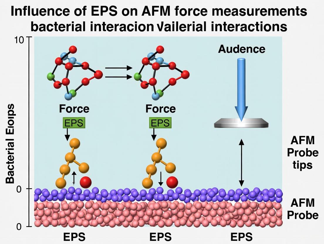
Accounting for EPS in AFM Force Measurements: A Guide for Accurate Biomechanical Data Interpretation
This article addresses the critical challenge of extracellular polymeric substance (EPS) influence on Atomic Force Microscopy (AFM) force measurements, a key concern for researchers and drug development professionals.
Accounting for EPS in AFM Force Measurements: A Guide for Accurate Biomechanical Data Interpretation
Abstract
This article addresses the critical challenge of extracellular polymeric substance (EPS) influence on Atomic Force Microscopy (AFM) force measurements, a key concern for researchers and drug development professionals. It explores the fundamental properties of EPS and its impact on nanomechanical data, detailing methodological best practices for sample preparation and immobilization to minimize artifacts. The content provides a troubleshooting guide for identifying and mitigating EPS-related distortions in force curves and adhesion measurements. Finally, it outlines validation strategies through correlative microscopy and data analysis techniques, empowering scientists to obtain reliable, physiologically relevant nanomechanical properties for biomedical applications.
Understanding the EPS Matrix: Composition, Structure, and Its Impact on AFM Probe Interaction
What are EPS? Defining the Complex Mixture of Biomolecules
What are Extracellular Polymeric Substances (EPS)?
Extracellular Polymeric Substances (EPS) are high molecular weight natural polymers secreted by microorganisms into their environment [1]. They are the fundamental building blocks of microbial biofilms, establishing the functional and structural integrity of these communities [1]. EPS form a hydrated, gel-like, three-dimensional matrix that traps bacterial cells and provides cohesion, protection, and nutrition [2]. This matrix constitutes 50% to 90% of a biofilm's total organic matter, making it the most abundant component [1]. The production of EPS enables bacteria to transition from a free-living (planktonic) state to a surface-attached (sessile) mode of growth, forming structured communities encased within this self-produced matrix [2].
Core Components of EPS
The term "EPS" refers to a complex mixture of biopolymers. The composition and chemical properties of these components directly contribute to the overall functionality of the EPS matrix [2].
- Polysaccharides: This is the most abundant and widely studied component of EPS [2]. These are sugar-based polymers, known as exopolysaccharides, which can be linear homopolymers (like cellulose or dextran) or branched heteropolymers containing three or more different monosaccharides (like alginate or xanthan) [1] [2]. They offer various functional roles, including adherence to surfaces and interaction with the environment [2].
- Proteins: Extracellular proteins are another major constituent. These can include enzymes (exoenzymes) that break down large molecules in the environment into smaller, absorbable nutrients [1].
- Extracellular DNA (eDNA): DNA is released into the EPS matrix and contributes to its structural stability [2].
- Lipids: These are also present within the complex mixture of EPS biopolymers [2].
- Other Macromolecules: The matrix can also include substances like lipopolysaccharides and humic substances [1].
- Minerals: Bacteria can regulate biomineralization processes, leading to the incorporation of minerals such as calcite (CaCO₃) into the EPS, which adds structural integrity [1].
Table 1: Major Components of Bacterial EPS
| Component | Description | Key Functions |
|---|---|---|
| Polysaccharides | Sugar-based polymers (e.g., galactose, glucose, xylose, uronic acids); can be linear or highly branched. | Primary structural scaffold, adhesion, water retention, interaction with environmental ions and pollutants [1] [2]. |
| Proteins | Includes structural proteins and functional exoenzymes. | Biofilm structural support, nutrient acquisition (e.g., proteases, phosphatases), signaling [1]. |
| Extracellular DNA (eDNA) | DNA released into the extracellular environment. | Structural stability, genetic information exchange, contributes to matrix cohesion [2]. |
| Lipids | A diverse group of hydrophobic or amphiphilic molecules. | Likely involved in hydrophobic interactions, cell surface modification [2]. |
FAQs: Accounting for EPS in AFM Force Measurements
How does EPS influence the adhesion and mechanical properties measured by AFM?
The EPS matrix significantly alters the nano-mechanical and adhesive interactions between the AFM tip and the sample surface.
- Adhesion Forces: The EPS is a complex, heterogeneous layer with its own adhesive properties. An AFM tip interacting with a cell may first encounter and adhere to the EPS, rather than the cell membrane itself. This can lead to measured adhesion forces that reflect the EPS-protein or EPS-polymeric interactions, not the underlying cell [3]. For instance, lateral force imaging has revealed that capsular EPS can exhibit regions with different frictional properties, suggesting segregation of hydrophobic fractions that influence adhesion measurements [3].
- Mechanical Properties (Elasticity/Stiffness): The EPS layer acts as a soft, compliant cushion over the cell. When an AFM tip indents a cell, the force curve captures the combined mechanical response of the EPS and the cell wall. If the EPS is not accounted for, the measured Young's modulus will be an average value that is lower than the true stiffness of the cell envelope, leading to inaccurate conclusions about cellular mechanics [4] [5].
What are common sample preparation artifacts when dealing with EPS-producing cells?
Sample preparation is a critical step where the native state of the EPS can be easily disrupted.
- Mechanical Trapping and Deformation: Immobilizing bacterial cells by mechanically trapping them into a filter can cause severe structural and mechanical deformation to the cell membrane [5]. This altered mechanical state directly influences parameters derived from AFM force curves.
- EPS Redistribution: During the filtering process, the EPS layer is not static. It can move and accumulate at the upper part of the cell [5]. This creates an uneven distribution, meaning AFM measurements taken on different parts of the same cell may yield vastly different results for adhesion (pull-off) forces, leading to distorted and non-representative data [5].
- Surface Contamination: Loosely adhered EPS and other particles on the sample surface can interact with the AFM tip, causing imaging streaks and unstable tip-sample interaction [6]. If these particles adhere to the tip, they can cause tip contamination and artifacts in both imaging and force spectroscopy [6].
Which AFM imaging modes are best suited for studying EPS?
Choosing the right AFM mode is essential to minimize sample disturbance and obtain accurate data.
- Tapping Mode vs. Contact Mode: TappingMode is highly recommended over Contact Mode for studying soft, fragile samples like EPS and living cells [7]. In Contact Mode, the sustained lateral forces exerted by the dragging tip can displace or even remove the loosely bound EPS, damaging the sample and producing artifacts. TappingMode minimizes these lateral forces by only intermittently contacting the surface, thereby preserving the native EPS structure [7].
- PeakForce Tapping Mode: This is an advanced, non-resonant mode that offers superior control for imaging EPS. It performs a force curve at every pixel, allowing for direct, precise control of the tip-sample interaction force down to ~10 pN [7]. This gentle force control maintains sample integrity and enables simultaneous mapping of topography and nanomechanical properties, which is ideal for characterizing the heterogeneous EPS matrix [7].
Table 2: Troubleshooting Common Issues with EPS in AFM Experiments
| Problem | Potential Cause | Solution |
|---|---|---|
| Inconsistent adhesion/mechanics data on a single cell. | Redistribution of EPS during preparation creates an uneven, heterogeneous layer [5]. | Use gentler immobilization methods (e.g., porous membranes). Verify EPS distribution with microscopy before AFM. |
| Streaks and unstable signals during imaging. | Loose EPS or surface contaminants interacting with or adhering to the AFM tip [6]. | Optimize sample rinsing protocols to remove loose material without disrupting the capsular EPS. Use sharper, cleaner probes. |
| Measured Young's modulus is lower than expected. | The AFM tip is indenting the soft EPS layer before reaching the stiffer cell wall, averaging the compliance of both [4]. | Use a sharp tip and model the force curve with a two-layer model (EPS + cell) to deconvolute their individual mechanical contributions. |
| Biological structures appear deformed or are moved by the tip. | Use of Contact Mode imaging, which exerts high lateral forces on soft samples [7]. | Switch to a gentle imaging mode such as TappingMode or PeakForce Tapping to minimize lateral forces [7]. |
Experimental Protocols for AFM Analysis of EPS
Protocol for Immobilizing EPS-Producing Bacteria
Goal: To securely immobilize bacterial cells without deforming the cell or displacing the native EPS matrix.
Detailed Methodology:
- Culture and Harvest: Grow the bacterial strain of interest under conditions known to promote EPS production. Harvest cells during the mid-to-late exponential growth phase by gentle centrifugation (e.g., 2000-5000 x g for 5-10 minutes).
- Washing (Optional): Carefully resuspend the cell pellet in a suitable buffer (e.g., PBS or a minimal salts solution). This step should be evaluated, as it may remove loosely associated planktonic EPS, which could be part of the study.
- Immobilization via Filtration:
- Use a sterile, porous polycarbonate membrane filter with a pore size smaller than the cells (e.g., 0.2-0.45 µm).
- Place the membrane on a filtration apparatus. Gently apply the cell suspension to the filter and use low vacuum pressure to draw the buffer through, leaving the cells trapped on the membrane surface.
- Critical Consideration: Be aware that this method can cause mechanical deformation and redistribute EPS towards the top of the cells [5].
- Alternative: Physical Adsorption:
- For a gentler approach, allow cells to physically adsorb onto a freshly cleaved mica or glass surface that has been treated with a cell-adhesive coating like poly-L-lysine.
- Incubate a small droplet of cell suspension on the substrate for 15-30 minutes in a humid chamber to prevent evaporation.
- Gently rinse with the imaging buffer to remove non-adhered cells. This method is less likely to cause severe EPS redistribution.
- Final Preparation: Carefully mount the prepared sample (membrane or substrate) onto the AFM sample puck. Add a small amount of the appropriate liquid buffer to keep the cells hydrated during measurement.
Protocol for AFM Force Spectroscopy on EPS
Goal: To obtain quantitative data on the mechanical and adhesive properties of the EPS matrix and the underlying cell.
Detailed Methodology:
- Probe Selection: Use a sharp, cantilever with a well-calibrated spring constant. For high-resolution measurements on soft samples, silicon nitride tips with nominal spring constants of 0.01 - 0.1 N/m are often suitable.
- AFM Mode Selection: Engage the AFM on a region of interest near a cell using PeakForce Tapping or a dedicated force spectroscopy mode. These modes provide the best force control for soft samples [7].
- Data Acquisition:
- Set the peak force setpoint to a very low value (e.g., 100-500 pN) to initially engage the soft EPS without collapsing it.
- Perform arrays of force-volume measurements or single point force curves on different locations: on top of a cell (with EPS), on the bare substrate, and if possible, on areas of planktonic EPS.
- Collect a sufficient number of curves (e.g., n > 100 per condition) for statistical significance.
- Data Analysis:
- Adhesion Force: Measure the minimum force in the retraction curve, which corresponds to the force required to separate the tip from the sample.
- Young's Modulus: Fit the approach segment of the force curve with an appropriate contact mechanics model (e.g., Hertz, Sneddon, or DMT models). Note that the presence of EPS may require more complex, multi-layer models to avoid underestimating the stiffness of the cell wall.
The Scientist's Toolkit: Key Research Reagents & Materials
Table 3: Essential Materials for AFM Studies of EPS-Producing Bacteria
| Item | Function/Application |
|---|---|
| Polycarbonate Membrane Filters (0.2 µm pore size) | For mechanical trapping and immobilization of bacterial cells for AFM analysis. |
| Freshly Cleaved Mica Substrates | Provides an atomically flat, clean surface for immobilizing cells via physical adsorption or poly-L-lysine treatment. |
| Sharp Silicon Nitride AFM Probes (e.g., nominal spring constant 0.01 - 0.1 N/m) | Essential for high-resolution imaging and force spectroscopy on soft biological samples with minimal sample damage. |
| Phosphate Buffered Saline (PBS) or Specific Growth Medium | Used as an imaging buffer to maintain physiological conditions and cell viability during liquid AFM experiments. |
| Poly-L-Lysine Solution | A cell-adhesive coating applied to mica or glass substrates to enhance the attachment of bacterial cells. |
Experimental Workflow: Accounting for EPS in AFM
The following diagram illustrates a logical workflow for designing an AFM experiment that accounts for the influence of EPS, guiding researchers from sample preparation to data interpretation.
Diagram 1: A logical workflow for AFM experiments accounting for EPS influence, from sample preparation to data interpretation.
The extracellular polymeric substance (EPS) is a complex, hydrated matrix that surrounds microbial cells in biofilms and aggregates. Its major components—exopolysaccharides, proteins, and extracellular DNA (eDNA)—fundamentally influence interactions with surfaces and other cells. For researchers employing Atomic Force Microscopy (AFM) for force measurements, the EPS presents a significant challenge. Its viscoelastic and adhesive properties can dominate force-distance curves, potentially obscuring the specific molecular interactions or cellular properties under investigation. This technical guide addresses the common pitfalls introduced by EPS in AFM studies and provides standardized protocols to account for its influence, ensuring more accurate and interpretable data.
Frequently Asked Questions (FAQs)
1. How does EPS lead to misinterpretation of AFM force spectroscopy data? EPS components, particularly exopolysaccharides and eDNA, create long-range, non-specific interactions that can mask the specific forces (e.g., ligand-receptor binding) you may be trying to measure. The EPS forms a soft, compressible layer around cells, leading to force-distance curves with features stemming from polymer extension and compression rather than from the cell wall or membrane itself [8] [9].
2. Why do my AFM measurements show high variability when probing bacterial cells? Heterogeneity in EPS composition, thickness, and distribution across a single cell or population of cells is a primary source of variability. The EPS layer is not a uniform shell; it has a dynamic, patchy structure. Measurements taken on a thick polysaccharide patch will differ significantly from those on a region with exposed surface proteins or eDNA [9] [10].
3. Can the EPS layer be controlled or modified for more consistent AFM measurements? Yes, both enzymatic and mechanical methods can be employed.
- Enzymatic Treatment: Incubating cells with DNase I can degrade the eDNA component, while polysaccharide-degrading enzymes (specific to the EPS type) can reduce the polysaccharide network [10].
- Mechanical Control: Using AFM in force spectroscopy mode, you can perform a compression cycle to mechanically compress the EPS layer before measuring adhesion or other properties in a subsequent retraction cycle. This helps standardize the initial contact point [8].
4. How can I confirm the presence of a capsule or EPS layer on my samples? AFM itself is an excellent tool for this. Compared to Transmission Electron Microscopy (TEM), which can fail to detect thin capsules due to sample preparation artifacts, AFM can unambiguously identify their presence through direct topographical imaging and phase imaging in tapping mode [9]. The capsule appears as a soft, halo-like structure surrounding the cell.
Troubleshooting Guides
Problem 1: Non-Specific Adhesion Obscuring Specific Interactions
- Symptoms: Force curves show large, continuous adhesion "pull-off" events over long distances (hundreds of nanometers) during retraction, with no clear single-molecule rupture events [8].
- Root Cause: The EPS, a network of long, entangled polymers, is adhering to the AFM tip. As the tip retracts, multiple polymers stretch and detach sequentially, creating a prolonged adhesion signature.
- Solutions:
- Functionalize the AFM Tip: Covalently link a specific ligand (e.g., an antibody) to the tip via a flexible polyethylene glycol (PEG) crosslinker. This method, known as Molecular Recognition Force Microscopy (MRFM), allows specific binding events to be distinguished from non-specific EPS adhesion by their characteristic rupture length and force [11].
- Modify the Buffer: Introduce monovalent salts (e.g., NaCl) to screen electrostatic interactions or use buffers that do not contain divalent cations like Mg²⁺, which can act as bridges between negatively charged EPS components and the tip or substrate [12] [13].
- Enzymatic Digestion: Treat the sample with DNase I to remove eDNA and/or specific glycosidases to digest exopolysaccharides, thereby simplifying the matrix [10].
Problem 2: False Feedback and Unstable Imaging
- Symptoms: The AFM image appears blurry, "out-of-focus," or the feedback system oscillates uncontrollably, making it impossible to resolve cellular details [14].
- Root Cause: The AFM tip is interacting with the soft, compliant EPS layer rather than the harder cell surface. The laser feedback system is "tricked" (false feedback) by the gradual bending of the cantilever as it pushes through the EPS, preventing it from reaching a stable setpoint [14].
- Solutions:
- Adjust the Setpoint: In vibrating (tapping) mode, decrease the setpoint amplitude to increase the tip-sample interaction force, allowing the tip to penetrate through the EPS layer to the stiffer cell surface [14].
- Use a Stiffer Cantilever: Switch from a very soft cantilever (e.g., < 0.1 N/m) to a moderately stiff one (e.g., 0.5 - 2 N/m). This reduces the cantilever's sensitivity to the long-range electrostatic and steric forces emanating from the EPS [14].
- Reduce Electrostatic Forces: Ensure your sample and cantilever are grounded to minimize electrostatic charges that can cause attraction or repulsion before physical contact [14].
Problem 3: Inconsistent Cell Immobilization
- Symptoms: Cells are washed away during buffer exchange or are moved by the AFM tip during scanning.
- Root Cause: The EPS layer can inhibit adhesion to the substrate by creating a repulsive force, or it can create a hydrogel that traps cells but does not firmly anchor them.
- Solutions:
- Use a Cationic Coating: Coat your substrate (e.g., glass, mica) with poly-L-lysine (PLL). The positive charge of PLL electrostatically attracts and immobilizes the negatively charged bacterial cells [8] [9].
- Chemical Cross-linking: For stronger immobilization, use an EDC-NHS cross-linking reaction to form covalent bonds between carboxylate groups on the cell surface and an aminosilane-treated substrate [8].
- Cation-Modified Mica: For imaging in liquid, use freshly cleaved mica pre-treated with a solution of divalent cations like Ni²⁺ or Mg²⁺ (1-10 mM). These cations act as bridges between the negatively charged mica and the negatively charged EPS/cell surface [12] [9].
The following table summarizes key quantitative findings on how different EPS components influence AFM force measurements, based on published research.
Table 1: Influence of EPS Components on AFM Force Measurements
| EPS Component | Measured Parameter | Experimental Finding | Experimental Context |
|---|---|---|---|
| eDNA & Exopolysaccharide (Psl) | Interaction Role | Forms a fibrous web that acts as a structural skeleton for the biofilm [10]. | Study of P. aeruginosa biofilm architecture. |
| eDNA | Furrow Depth in Biofilm | ~200 nm (native) vs. ~400 nm (after DNase I treatment). DNase I removed eDNA, deepening gaps between cells [10]. | AFM topographical imaging of B. subtilis biofilm. |
| A-band & B-band LPS + ECP | Adhesion Force (F_adh) | PAO1 (A+ B+): 0.56 nNAK1401 (A+ B-): 0.51 nN [8]. | Single-cell AFM force spectroscopy on P. aeruginosa. |
| A-band & B-band LPS + ECP | Adhesion Event Distance | PAO1 (A+ B+): >50% of events at >600 nmAK1401 (A+ B-): >90% of events at <600 nm [8]. | Single-cell AFM force spectroscopy on P. aeruginosa. |
| Exopolysaccharide (Alginate) | Decay Length (Steric Repulsion) | Longer polymers on wild-type strain caused greater steric repulsion (longer decay length) compared to mutant [8]. | AFM approach curves on P. aeruginosa. |
Standardized Experimental Protocols
Protocol 1: Enzymatic Removal of eDNA for Controlled Experiments
This protocol is used to quantify the specific contribution of eDNA to adhesion and structural integrity [10].
- Sample Preparation: Grow biofilms or prepare a cell suspension in an appropriate physiological buffer (e.g., HEPES or Tris).
- Treatment: Add DNase I to the sample at a final concentration of 10-100 µg/mL.
- Incubation: Incubate for 30-60 minutes at 37°C (or the optimal temperature for the enzyme).
- Control: Prepare an identical sample without DNase I but with the same buffer.
- Washing: Gently rinse the sample with buffer to remove enzymes and degradation products.
- AFM Analysis: Immediately proceed with AFM force spectroscopy or imaging on both treated and control samples. Compare force curves and topographical images to identify differences attributable to eDNA.
Protocol 2: Substrate Preparation for Firm Cell Immobilization
This protocol ensures cells remain fixed during AFM scanning, even in liquid [12] [8].
Method A: Poly-L-Lysine Coating
- Clean a glass cover slip or mica disk with acid or plasma.
- Apply a 0.1% (w/v) solution of poly-L-lysine (PLL) and let it dry for 2 hours.
- Rinse gently with ultrapure water to remove excess PLL.
- Apply the bacterial suspension in water and let it settle on the shaker for 2 hours.
- Rinse gently with buffer to remove non-adherent cells.
Method B: Cation-Assisted Adsorption to Mica
- Freshly cleave a mica sheet.
- Prepare your DNA or cell suspension in a deposition buffer containing 1-10 mM MgCl₂ (or NiCl₂) [12].
- Place a small droplet (e.g., 1-2 µL) of the suspension onto the mica.
- Incubate for 1-3 minutes.
- Rinse thoroughly with deionized water (for imaging in air) or buffer (for imaging in liquid) to remove salts and unbound material.
Experimental Workflow and EPS Interactions
The diagram below outlines the logical decision-making process for designing an AFM experiment that accounts for EPS influence, from sample preparation to data interpretation.
Diagram 1: A logical workflow for troubleshooting EPS-related issues in AFM experiments.
Research Reagent Solutions
Table 2: Essential Reagents for Managing EPS in AFM Studies
| Reagent | Function/Benefit | Key Consideration |
|---|---|---|
| DNase I | Degrades eDNA component of EPS; reduces long-range adhesion and biofilm integrity [10]. | Use an appropriate buffer (with Mg²⁺/Ca²⁺) for enzyme activity. |
| Poly-L-Lysine (PLL) | Cationic polymer for electrostatic immobilization of cells on substrates [8] [9]. | Coating time and concentration affect cell viability and morphology. |
| MgCl₂ / NiCl₂ | Divalent cations that bridge negatively charged samples to mica for stable immobilization [12]. | Concentration affects binding strength; can influence DNA and polysaccharide conformation. |
| Aminosilane (e.g., APTES) | Used to create a positively charged amine-functionalized surface on glass/silicon for cross-linking [8]. | Requires anhydrous conditions for consistent silanization. |
| EDC / NHS Crosslinkers | Forms covalent bonds between carboxyl groups on cells and amine groups on a functionalized substrate [8]. | Reaction is pH-dependent (optimal at pH 5.5-7.5) and must be performed in aqueous buffer. |
| PEG Crosslinkers | Flexible spacer for tip functionalization in MRFM; separates specific binding events from non-specific adhesion [11]. | The length of the PEG spacer determines the detectable rupture length. |
Frequently Asked Questions (FAQs)
FAQ 1: Why do my AFM force measurements show such high variability, even for the same bacterial strain? High variability in AFM force measurements is expected and often stems from the dynamic nature of the Extracellular Polymeric Substances (EPS) matrix. The EPS composition and structure are not constant; they vary significantly with biofilm age, environmental growth conditions, and between bacterial strains. This intrinsic variability directly influences nanomechanical properties measured by AFM, such as adhesion force and Young's modulus [15] [16]. For consistent results, it is crucial to standardize and meticulously report biofilm growth conditions and the age at which they are analyzed.
FAQ 2: How does biofilm age specifically affect the EPS matrix and my AFM results? Biofilm age profoundly impacts the EPS matrix's volume, structure, and mechanical properties. Confocal Laser Scanning Microscopy (CLSM) studies show that the volume of both live bacteria and EPS increases significantly as biofilms mature from 1 to 3 weeks [15]. Furthermore, the spatial organization and interaction of EPS components evolve over time. For instance, in Bacillus subtilis biofilms, extracellular DNA (eDNA) plays a cooperative role with exopolysaccharides in the early stages of development (under 12 hours), while exopolysaccharides take on a more dominant structural role in later stages (24-48 hours) [10]. This maturation process leads to measurable changes in AFM data, including a decrease in surface roughness and an increase in cell-cell adhesion forces [15].
FAQ 3: My AFM tip frequently contaminates when probing biofilms. How can I prevent this? Tip contamination is a common challenge when probing the adhesive EPS matrix. To mitigate this:
- Use Functionalized Probes: Employ AFM probes functionalized with larger, smoother colloids (e.g., borosilicate spheres) instead of sharp tips for force measurements. This reduces local stress and minimizes the penetration and trapping of the tip in the EPS [17].
- Optimize Imaging Mode: Consider using dynamic (tapping) AFM modes instead of contact mode when measuring topographical features, as this can reduce shear forces and adhesive interactions between the tip and the sample [18].
- Apply Appropriate Contact Mechanics Models: Use adhesive contact mechanics models (e.g., Johnson-Kendall-Roberts, JKR) that account for the strong adhesive forces present in soft, hydrated materials like biofilms. This ensures more accurate data interpretation and helps identify measurements that may be compromised by excessive adhesion [18].
FAQ 4: What is the best method to immobilize bacteria for AFM without altering their surface properties? The immobilization method is critical for obtaining reliable data. Mechanical trapping in porous membrane filters is widely considered the most reliable method for single bacterial cells. This method minimizes chemical and physical alterations to the cell surface and its associated EPS, unlike methods involving chemical fixation (e.g., glutaraldehyde) or electrostatic adsorption to coated surfaces, which can induce structural deformations and alter physicochemical surface properties [5] [19].
Troubleshooting Guides
Troubleshooting AFM Measurement Inconsistencies
| Symptom | Possible Cause | Solution |
|---|---|---|
| High variability in adhesion force measurements between samples. | Differences in biofilm age or maturation stage. | Standardize and precisely document the incubation time for all biofilm cultures. Use CLSM to correlate EPS volume with age [15]. |
| Inconsistent Young's modulus values. | Variations in EPS composition due to changes in growth environment (e.g., nutrient availability, sucrose concentration). | Control and report all environmental growth conditions meticulously. Use spectroscopic techniques (e.g., FTIR) to monitor EPS chemistry [17] [16]. |
| Artificially high width measurements of nanofibrils in EPS. | AFM tip convolution effect, where the tip and scanned features are of similar size [20]. | Use sharper AFM tips with a smaller radius of curvature. Apply tip deconvolution algorithms during data processing to determine the actual dimensions of surface objects [20]. |
| Deep penetration of AFM tip into biofilm, with no measurable resistance. | Invalid assumption of the biofilm as an elastic half-space; the model does not account for the sample's small dimensions and heterogeneity [20]. | Apply correction factors to traditional Hertzian contact mechanics models to account for the finite thickness and complex structure of the biofilm [20] [18]. |
Quantitative Data on EPS and Biofilm Maturation
The following table summarizes key quantitative changes in EPS and biofilm properties during maturation, as revealed by AFM and CLSM studies.
Table 1: Quantitative Changes in EPS and Biofilm Properties with Maturation
| Parameter | 1-Week-Old (Young) Biofilm | 3-Week-Old (Mature) Biofilm | Measurement Technique | Significance for AFM |
|---|---|---|---|---|
| EPS Volume | Lower | Significantly higher (P < 0.01) [15] | CLSM with fluorescent probes (e.g., Alexa Fluor 647-dextran) [15] | Increased EPS volume leads to greater tip-sample adhesion and altered viscoelastic response. |
| Surface Roughness | Significantly higher [15] | Lower [15] | AFM Topography Imaging [15] | Mature biofilms form a more uniform, cohesive layer, affecting contact area with the tip. |
| Adhesion Force (Cell-Cell) | Less attractive | More attractive [15] | AFM Force-Distance Curve [15] | Indicates stronger cohesive strength within the mature biofilm matrix. |
| Sensitivity to DNase I | High (biofilm formation suppressed) [10] | Low (minor effect) [10] | Crystal Violet Assay & AFM [10] | eDNA is a critical structural component in early biofilms; its role may be shielded or complemented later. |
Experimental Protocols
Protocol: Correlating Biofilm Age with EPS Volume and Nanomechanics
This protocol outlines a method to systematically investigate the influence of biofilm age using AFM and CLSM.
1. Biofilm Cultivation:
- Substrate: Use hydroxyapatite (HA) discs coated with type I collagen to mimic mineralized surfaces [15] [17].
- Inoculum: Grow anaerobic oral microcosm biofilms from pooled human saliva in Brain Heart Infusion (BHI) broth [15].
- Standardization: Incubate biofilm samples for distinct periods (e.g., 3 days, 1 week, 3 weeks) with a weekly change of fresh growth medium [15].
2. EPS and Live Bacteria Staining for CLSM:
- EPS Labeling: Incorporate 1 mM Alexa Fluor 647-labelled dextran into the growth medium before and during biofilm formation. This metabolic labeling allows for the visualization of the 3D EPS structure within intact biofilms [15].
- Live Bacteria Labeling: Stain the biofilms with SYTO 9 green-fluorescent nucleic acid stain [15].
- Imaging and Analysis: Rinse specimens gently and view using CLSM. Reconstruct 3D volume stacks using software like Imaris to quantify the volume of EPS and live bacteria [15].
3. AFM Nanomechanical Characterization:
- Sample Preparation: Gently fix biofilm specimens in 2% glutaraldehyde at 4°C for 3 minutes, followed by rinsing in phosphate-buffered saline (PBS) and drying overnight in a desiccator [15].
- Topography Imaging: Operate the AFM in contact mode using sharp silicon nitride cantilevers. Capture images at a standard scan size (e.g., 8 × 8 µm) and calculate the root mean square (RMS) surface roughness [15].
- Force Measurements: Perform force-distance measurements at a 15 Hz z-scan rate. Conduct force mapping over a grid (e.g., 64x64 points) on the sample surface. Measure adhesion forces at two distinct locations: (1) between the AFM tip and the surface of bacterial cells, and (2) at the cell-cell interface [15].
4. Data Correlation: Statistically correlate the quantified EPS volume and surface roughness from CLSM/AFM with the measured adhesion forces from AFM across the different age groups.
Protocol: Modifying EPS to Decipher its Mechanical Role
This protocol uses specific enzymes to target EPS components and observe the resultant mechanical changes.
1. Biofilm Growth and Treatment:
- Reactor: Grow model biofilms (e.g., Staphylococcus epidermidis) in a CDC biofilm reactor for 12 days under controlled shear and temperature conditions [16].
- EPS Modifiers: Prepare treatments with optimized concentrations of EPS-degrading agents [16]:
- Proteinase K: To degrade protein components.
- Dispersin B: To degrade polysaccharide poly-N-acetylglucosamine (PNAG).
- DNase I: To degrade extracellular DNA (eDNA).
- Sodium Meta-periodate: To oxidize polysaccharides.
- Treatment: Expose biofilms to these modifiers for a specified duration (e.g., 2 hours) [16].
2. Post-Treatment Analysis:
- Chemical Analysis: Use Fourier Transform Infrared Spectroscopy (FTIR) to detect changes in the chemical composition of the EPS (e.g., reduction in polysaccharide or protein peaks) [16].
- Structural Analysis: Use CLSM to observe physical changes in the biofilm architecture post-treatment [16].
- Mechanical Testing: Use AFM to measure the cohesive strength of the treated biofilms. A significant reduction in strength after a specific treatment indicates the targeted component's critical role in the biofilm's mechanical integrity [16].
Visualizing EPS Dynamics and Experimental Workflows
EPS-Component Interactions in Biofilm Development
Workflow for AFM Force Measurement Considering EPS Variability
The Scientist's Toolkit: Key Research Reagents & Materials
Table 2: Essential Reagents for Investigating EPS Dynamics
| Reagent/Material | Function in Experiment | Example Use Case |
|---|---|---|
| Alexa Fluor 647-dextran | Fluorescent probe for metabolic labeling and 3D visualization of EPS matrix via CLSM [15]. | Quantifying EPS volume changes during biofilm maturation [15]. |
| SYTO 9 | Green-fluorescent nucleic acid stain for labeling and quantifying live bacteria within the biofilm [15]. | Differentiating between bacterial biomass and EPS matrix in CLSM analysis [15]. |
| Hydroxyapatite (HA) Discs | Abiotic substrate that mimics tooth enamel or bone mineral, used for growing relevant oral or medical biofilms [15] [17]. | Studying biofilm formation under conditions that simulate the oral cavity [15]. |
| DNase I | Enzyme that degrades extracellular DNA (eDNA) within the EPS matrix [10]. | Probing the structural role of eDNA in early-stage biofilm formation and stability [10]. |
| Proteinase K | Enzyme that digests proteins by hydrolyzing peptide bonds [16]. | Assessing the contribution of proteinaceous components to the biofilm's mechanical strength [16]. |
| Dispersin B | Enzyme that specifically hydrolyzes the polysaccharide poly-N-acetylglucosamine (PNAG) [16]. | Determining the importance of PNAG in the cohesion of biofilms produced by pathogens like S. epidermidis [16]. |
| Borosilicate Sphere-Functionalized AFM Cantilevers | AFM probes with modified tips to create a well-defined, larger contact geometry for more reliable nanoindentation on soft samples [17]. | Performing force-volume imaging to map mechanical properties across heterogeneous biofilm surfaces [17]. |
In Atomic Force Microscopy (AFM) research, the extracellular polymeric substance (EPS) layer produced by microbial cells is not merely a passive coating; it is a dynamic, hydrated matrix that fundamentally alters tip-sample interactions. For researchers and drug development professionals, accounting for the influence of EPS is not optional—it is essential for generating accurate, reproducible nanomechanical and adhesion data. This guide details the specific challenges posed by EPS and provides proven methodologies to mitigate its confounding effects, ensuring the integrity of your force measurements.
FAQ: Understanding EPS and Its Impact on AFM
Q1: What is EPS, and why does it significantly interfere with AFM force measurements?
EPS is a complex, high-molecular-weight mixture of polymers excreted by bacteria, forming a highly hydrated nanogel layer on cell surfaces [21]. Its significance in AFM stems from its physical and chemical properties:
- Soft and Dynamic: The EPS layer is viscoelastic and can undergo substantial deformation during force curve acquisition, leading to overestimation of indentation depth and miscalculation of cell wall mechanical properties like elastic modulus [5] [22].
- Adhesive Nature: EPS contains both hydrophilic and hydrophobic sites, enabling it to form strong, often non-specific bonds with AFM tips. This can dominate adhesion (pull-off) force measurements, masking the specific receptor-ligand interactions you may intend to study [5] [21].
- Redistributable: Studies have shown that mechanical forces, such as those from filtering protocols used for cell immobilization, can cause the EPS layer to move and accumulate at the upper part of the cell. This redistribution creates a heterogeneous surface that yields distorted and non-representative adhesion data [5].
Q2: What specific imaging artifacts result from the presence of an EPS layer?
The EPS layer is a primary source of common AFM artifacts, including:
- Blurry or "Out-of-Focus" Images: This "false feedback" occurs when the AFM tip interacts with the soft, compliant EPS layer rather than the underlying hard surface forces of the cell membrane. The automated tip approach is tricked into stopping prematurely, resulting in a loss of resolution where nanoscale features cannot be visualized [23].
- Streaks and Unstable Tracking: Loose or weakly bound EPS components can interact with the AFM tip, causing instabilities in the tip-sample interaction. The AFM controller struggles to maintain constant force, resulting in streaks in the image as the tip momentarily sticks and then slips over the surface [6].
Q3: How does sample preparation for immobilization affect the EPS layer?
The choice of immobilization method can mechanically compromise the EPS layer and the cell itself.
- Mechanical Trapping: Filtering cells into porous membranes is a common immobilization technique. However, this process can cause severe structural and mechanical deformation to the cell membrane and the surrounding EPS. This alters the cell's native mechanical state, directly influencing parameters derived from force curves [5].
- Chemical Immobilization: Using adhesives like poly-l-lysine or performing carboxyl group cross-linking can provide secure attachment. However, some chemical treatments can negatively impact cell viability and nanocharacteristics, potentially cross-linking the EPS and altering its natural mechanical response [22].
Troubleshooting Guide: Mitigating EPS-Related Issues
| Problem | Primary Cause | Recommended Solution |
|---|---|---|
| Blurry Images & False Feedback | Tip trapped in soft EPS layer [23]. | Increase tip-sample interaction: In tapping mode, decrease the setpoint amplitude; in contact mode, increase the deflection setpoint to force the tip through the layer [23]. |
| High, Non-Specific Adhesion | EPS polymers forming multiple bonds with the tip [5] [21]. | Use chemical functionalization: Modify the tip with specific molecules (e.g., PEG linkers) to isolate specific interactions from non-specific EPS adhesion [22]. |
| Unstable Force Curves | Loosely bound EPS components or tip contamination [6]. | Optimize sample rinse protocol: Gently rinse the substrate with fluid media to remove unadsorbed EPS before imaging [24]. |
| Inconsistent Mechanical Data | Redistribution or deformation of EPS during immobilization [5]. | Validate immobilization method: Consider gentle chemical fixation (e.g., with divalent cations like Ca²⁺) as an alternative to high-stress mechanical trapping [22]. |
Experimental Protocols for EPS-inclusive Analysis
Protocol 1: Measuring Biofilm Cohesive Energy via AFM-based Abrasion
This protocol, adapted from a foundational study, allows for the in situ quantification of biofilm cohesive energy, a property directly governed by EPS [25].
Methodology:
- Biofilm Growth: Grow a 1-day biofilm from a mixed culture in a membrane-aerated biofilm reactor. Maintain constant humidity (e.g., ~90%) during sample transfer to preserve native biofilm hydration.
- Baseline Imaging: Engage the biofilm surface and collect a non-perturbative topographic image of a 5x5 µm area at a low applied load (~0 nN).
- Abrasion Phase: Zoom into a 2.5x2.5 µm sub-region. Set a high applied load (e.g., 40 nN) and perform repeated raster scans (e.g., 4 scans) to abrade the biofilm.
- Post-Abrasion Imaging: Reduce the load to ~0 nN and capture another non-perturbative 5x5 µm image of the abraded region.
- Data Analysis:
- Subtract the post-abrasion image from the pre-abrasion image to calculate the volume of displaced biofilm.
- The frictional energy dissipated during abrasion is determined from the friction force data (raw volts converted to force).
- The cohesive energy (nJ/µm³) is calculated as the frictional energy dissipated divided by the volume of biofilm displaced [25].
Experimental workflow for measuring biofilm cohesive energy.
Protocol 2: Reliable Immobilization of EPS-rich Cells for Liquid Imaging
Secure yet non-destructive immobilization is critical for accurate measurement.
Methodology:
- Substrate Selection: Use a freshly cleaved mica or silica substrate.
- Chemical Functionalization: Treat the substrate with poly-L-lysine or a solution containing divalent cations (e.g., 10 mM Mg²⁺ or Ca²⁺). These cations can enhance attachment via electrostatic bridging without severely compromising viability or EPS structure [22].
- Cell Deposition: Incubate a small volume of cell suspension on the functionalized substrate for a controlled period (e.g., 10-30 minutes).
- Gentle Rinsing: Gently rinse the substrate with the appropriate fluid media (e.g., buffer) to remove loosely adhered cells and excess, unbound EPS polymers that could contaminate the tip [24].
- Imaging: Set the substrate in a bath of clean media and proceed with AFM imaging in tapping mode to minimize lateral forces.
Quantitative Data: Cohesive Properties of Biofilms
The table below summarizes quantitative cohesive energy data obtained from AFM abrasion tests, demonstrating how cohesion varies with biofilm depth and environmental conditions [25].
Table 1: Biofilm Cohesive Energy Measurements under Different Conditions
| Biofilm Condition | Depth / Region | Cohesive Energy (nJ/µm³) | Notes |
|---|---|---|---|
| Standard Biofilm | Upper Layers | 0.10 ± 0.07 | Softer, more hydrated EPS dominates. |
| Standard Biofilm | Deeper Layers | 2.05 ± 0.62 | Increased density and cross-linking. |
| Biofilm + 10 mM Ca²⁺ | Not Specified | 1.98 ± 0.34 | Divalent cations significantly increase cohesion by cross-linking EPS polymers. |
The Scientist's Toolkit: Key Research Reagents
Table 2: Essential Materials for AFM Studies of EPS-rich Systems
| Item | Function in Experiment | Rationale |
|---|---|---|
| V-shaped Si₃N₄ Cantilevers | Force spectroscopy & imaging | Low spring constants (e.g., 0.58 N/m) suitable for soft samples; sharp tips for resolution [25]. |
| Divalent Cations (MgCl₂, CaCl₂) | Immobilization reagent | Promotes cell attachment to substrates via electrostatic bridging, can be gentler than strong adhesives [22]. |
| Poly-L-Lysine | Substrate coating | Creates a positively charged surface to enhance electrostatic attachment of generally negatively charged cells [22]. |
| Humidity Controller | Environmental control | Maintains constant humidity (~90%) for moist biofilm experiments, preventing dehydration artifacts [25]. |
Conceptual Framework: The Dual Nature of EPS in AFM
The EPS layer presents a dual challenge: it is both the object of study and a source of experimental interference. The following diagram synthesizes its primary mechanisms of influence on tip-sample interactions.
Mechanisms of EPS interference in AFM measurements.
Frequently Asked Questions (FAQs)
Q1: How does the extracellular polymeric substance (EPS) matrix influence my AFM force measurements on biofilms?
The EPS matrix is a critical, hydrated network that governs the nanomechanical properties of biofilms. Its influence is twofold: it directly contributes to biofilm cohesiveness and viscoelasticity, and its properties change over time, affecting measurement reproducibility. Quantitative studies show that the volume of EPS in a 3-week-old mature biofilm is significantly larger than in a 1-week-old young biofilm [15]. Furthermore, the addition of calcium ions (10 mM) during cultivation can increase biofilm cohesive energy from 0.10 ± 0.07 nJ/μm³ to 1.98 ± 0.34 nJ/μm³, demonstrating how the EPS chemical environment directly impacts the mechanical data you collect [25]. When measuring, the adhesion forces at the cell-cell interface (governed by EPS) are significantly more attractive than those at the surface of individual bacterial cells [15].
Q2: My force curves on a hydrated biofilm look inconsistent. Is this a measurement error or a sample property?
This is likely a reflection of the biofilm's inherent heterogeneity and soft, viscoelastic nature, rather than a pure measurement error. The EPS matrix is a soft, hydrous gel, and its response to the AFM tip is time- and load-dependent. For consistent results, it is essential to control environmental conditions such as humidity, which should be kept constant (e.g., ~90%) during measurements to maintain a consistent biofilm-water content [25]. Furthermore, the high roughness of young biofilms can lead to variable data, as surface roughness decreases significantly as the biofilm matures and forms a more uniform EPS layer [15].
Q3: Why do I get different Young's modulus values when probing the same biofilm with different AFM tips?
This is a classic sign of a probe-related artifact. The calculated Young's modulus is highly sensitive to the contact area between the tip and the sample. AFM probe tips are prone to wear and contamination, which alters their geometry. A study demonstrated that a tip modeled with a damaged, flattened triangular apex produced force curves that deviated significantly from those generated by an ideal, sharp tip [26]. It is crucial to calibrate your tip's actual geometry and account for it in your models, as using an incorrect geometry will lead to inaccurate and severely deformed property data [26].
Troubleshooting Guide: Accounting for EPS Influence
Problem 1: Inconsistent Adhesion and Cohesion Measurements
Potential Cause: Unaccounted spatial and temporal heterogeneity of the EPS matrix. Solutions:
- Map Adhesion Forces: Systematically measure adhesion forces at different locations: on top of cells, between cells (cell-cell interface), and on bare substrates. This will help deconvolute the contribution of EPS-mediated interactions [15].
- Control Biofilm Age: Document and standardize the biofilm cultivation time. Be aware that a 3-week-old mature biofilm will have a denser EPS matrix and different mechanical properties compared to a 1-week-old young biofilm [15].
- Chemical Environment: Report the exact composition of the liquid medium during growth and measurement. The presence of ions like calcium (Ca²⁺) can cross-link EPS polymers, dramatically increasing cohesiveness [25].
Problem 2: Overestimation of Elastic Modulus and Sample Deformation
Potential Cause: The use of an inappropriate contact mechanics model or a damaged AFM probe that underestimates the true contact area. Solutions:
- Model Selection: For soft, thin samples like biofilms, use advanced elastic models (e.g., Chen, Tu, or Cappella models) derived from the Hertz model that account for the influence of a hard substrate [27].
- Probe Calibration: Regularly characterize your probe tip's geometry using reference materials and/or electron microscopy. Implement a finite element model that uses the probe's realistic, "damaged" geometry to correctly interpret force curves and understand the impacted volume [26].
- Control Indentation Depth: Use shallow indentation depths to minimize the effect of the underlying stiff substrate and to probe the local EPS properties rather than a composite of the EPS and cell/substrate.
Problem 3: Non-Reproducible Topographical Imaging and Feature Loss
Potential Cause: Excessive scanning forces that mechanically deform or displace the soft EPS and cells. Solutions:
- Optimize Imaging Mode: Avoid contact mode for high-resolution imaging. Use tapping mode in liquid or air to minimize lateral (shear) forces that can sweep away weakly bound EPS structures [28].
- Reduce Applied Force: Use the lowest possible imaging force that provides stable feedback. Perform initial scans at a low applied load (~0 nN) to identify regions of interest before higher-force measurements [25].
- Validate with CLSM: Correlate your AFM findings with confocal laser scanning microscopy (CLSM) using fluorescently labelled EPS and live/dead stains. This provides a 3D, non-destructive view of the biofilm structure for comparison [15].
Experimental Protocols for Reproducible AFM on Biofilms
Protocol 1: In Situ Cohesive Energy Measurement
This protocol, adapted from a foundational study, measures the cohesive energy of a moist biofilm by correlating frictional energy dissipation with the volume of displaced material [25].
- Biofilm Growth: Grow biofilm on a suitable substrate (e.g., gas-permeable membrane) using a defined or mixed culture in a reactor. Control parameters like calcium concentration.
- Sample Preparation: After growth (e.g., 1 day), cut a small piece of the biofilm-coated substrate. Equilibrate it in a humidity-controlled chamber (e.g., ~90% RH) for at least 1 hour to maintain consistent hydration.
- AFM Setup: Mount the sample in an AFM equipped with a humidity controller. Use a sharp silicon nitride tip (e.g., pyramidal, oxide-sharpened) with a known spring constant (e.g., ~0.58 N/m).
- Non-Perturbative Topography: Collect a reference topographic image of a 5x5 μm area at a very low applied load (~0 nN).
- Abrasive Scanning: Zoom to a smaller region (e.g., 2.5x2.5 μm) within the scanned area. Perform repeated raster scans (e.g., 4 scans) at an elevated load (e.g., 40 nN) to abrade the biofilm.
- Volume Calculation: Return to low load and re-image the larger 5x5 μm area. Subtract the post-abrasion height image from the pre-abrasion image to determine the volume of displaced biofilm.
- Data Analysis: Calculate the cohesive energy (nJ/μm³) as the ratio of the frictional energy dissipated during abrasive scanning to the volume of biofilm removed.
Protocol 2: Correlative CLSM and AFM for EPS Characterization
This protocol combines the 3D chemical information from CLSM with the nanomechanical data from AFM [15].
- Fluorescent Staining: During biofilm growth, incorporate a fluorescent marker (e.g., Alexa Fluor 647-labelled dextran) into the culture medium to label the EPS matrix in situ.
- Live/Dead Staining: Label live bacteria in the biofilm using a nucleic acid stain (e.g., SYTO 9).
- CLSM Imaging: Acquire 3D image stacks of the stained, hydrated biofilm using CLSM. Use software to reconstruct the stacks and quantify the volumes of EPS and live bacteria.
- Sample Fixation: For subsequent AFM analysis, gently fix the biofilm sample (e.g., with 2% glutaraldehyde at 4°C for 3 minutes) and rinse with buffer. Air-dry in a desiccator.
- AFM Topography & Force Mapping: Image the fixed sample in AFM contact mode to obtain high-resolution topography and surface roughness. Perform force-distance curve measurements on a grid (e.g., 64x64 points) over the surface to map adhesion and elasticity.
- Data Correlation: Overlay the AFM adhesion/mechanical maps with the CLSM EPS distribution maps to directly link nanomechanical properties with the spatial organization of the EPS matrix.
Quantitative Data on Biofilm and EPS Properties
Table 1: Measured Cohesive Energy of Biofilms under Different Conditions [25]
| Biofilm Condition | Cohesive Energy (nJ/μm³) | Notes |
|---|---|---|
| 1-day biofilm (shallow depth) | 0.10 ± 0.07 | Measured in humid air (~90% RH) |
| 1-day biofilm (deeper depth) | 2.05 ± 0.62 | Cohesion increases with biofilm depth |
| With 10 mM Calcium | 1.98 ± 0.34 | Calcium addition significantly increases cohesion |
Table 2: Structural and Adhesive Properties of Oral Multispecies Biofilms [15]
| Property | 1-Week-Old (Young) Biofilm | 3-Week-Old (Mature) Biofilm | Statistical Significance |
|---|---|---|---|
| EPS Volume | Lower | Higher | P < 0.01 |
| Live Bacteria Volume | Lower | Higher | P < 0.01 |
| Surface Roughness | Significantly Higher | Lower | P < 0.01 |
| Adhesion Force (Cell-Cell) | Less Attractive | More Attractive | P < 0.01 |
Experimental Workflow Diagram
AFM-EPS Experimental Workflow
Research Reagent Solutions
Table 3: Essential Materials for AFM Biofilm Research
| Item | Function in Experiment | Example & Notes |
|---|---|---|
| Hydroxyapatite (HA) Discs | Model substrate for studying oral and orthopaedic biofilms. Coated with type I collagen to mimic organic surfaces [15]. | Clarkson Chromatography Products; diameter: 0.38-inch [15]. |
| Fluorescent Dextran Conjugates | In situ labelling of EPS matrix for visualization and volume quantification via CLSM [15]. | Alexa Fluor 647-labelled dextran (MW: 10 kDa); incorporated into growth medium [15]. |
| V-shaped Si₃N₄ Cantilevers | Standard probes for imaging and force spectroscopy in liquid or air. Pyramidal tips for nanoscale indentation. | Model NPS (Digital Instruments); spring constant ~0.58 N/m [25]. |
| Calcium Chloride (CaCl₂) | Ionic cross-linker for EPS. Used to investigate and control the effect of divalent cations on biofilm cohesiveness [25]. | Adding 10 mM CaCl₂ to reactor during cultivation significantly increases cohesive energy [25]. |
| Gas-Permeable Membranes | Substrate for growing membrane-aerated biofilms, creating oxygen and nutrient gradients relevant to natural environments. | Microporous polyolefin flat sheet membrane (3M Corporation) [25]. |
Best Practices in Sample Preparation and AFM Operation to Account for EPS
Atomic Force Microscopy (AFM) has become an indispensable tool for characterizing the nanomechanical properties of biological samples, including single cells and complex biofilms. A core tenet of this research is that measured properties must reflect the sample's native state. However, a significant challenge lies in sample preparation. Immobilization techniques that physically constrain soft, hydrated biological specimens are mandatory for AFM, but can inadvertently alter the very properties researchers seek to measure.
This guide focuses on a specific and often overlooked pitfall: the use of filtration for immobilization. This method can mechanically redistribute Extracellular Polymeric Substances (EPS) and deform cellular structures, thereby skewing force measurement data. Understanding and mitigating these artifacts is essential for generating accurate, reproducible, and biologically relevant nanomechanical data, which is the central thesis of this work.
FAQs & Troubleshooting Guides
How does filtration specifically alter the mechanical properties of a biofilm?
Filtering a biofilm to immobilize it for Atomic Force Microscopy (AFM) analysis applies significant shear and compressive forces. This process can mechanically disrupt the native biofilm architecture, leading to a densification of the EPS matrix and potential removal of loosely bound water and polymers.
- The Problem: The measured stiffness (Young's modulus) of a filtered biofilm may be artificially high because the filtration process has packed the EPS more tightly, not because the biofilm is inherently stiff in its native state.
- Underlying Principle: Biofilm cohesion and mechanical properties are depth-dependent. Research has shown that cohesive energy can increase with biofilm depth, from approximately 0.10 nJ/µm³ at the surface to 2.05 nJ/µm³ at greater depths [25]. Filtration collapses this structured, heterogeneous matrix into a more homogeneous, compressed layer.
- Impact on Data: This artifact can lead to incorrect conclusions about a biofilm's mechanical strength, its susceptibility to antimicrobials, or the effectiveness of targeting EPS.
What is the effect of cell deformation on nanomechanical measurements?
When cells are deformed or flattened during immobilization, the AFM tip interacts with a structure that is under pre-existing stress and strain, and the underlying rigid substrate influences the measurement.
- The Problem: An indenting AFM tip will encounter a strained cytoskeleton and a reduced distance to the underlying stiff filter membrane or substrate. This leads to an overestimation of the cell's stiffness.
- Underlying Principle: For accurate Young's modulus calculation using contact mechanics models (e.g., Hertz model), indentations should typically not exceed 10-20% of the sample's height and should be limited to around 200 nm to avoid the influence of the underlying stiff substrate [29]. Flattened cells make it impossible to stay within this safe indentation range.
- Impact on Data: The measured Young's modulus will be a composite of the cell's properties and the substrate's properties, not the true modulus of the cell. This invalidates direct comparisons with measurements from properly immobilized, rounded cells.
What are the signs of immobilization-induced artifacts in my AFM data?
Your data may indicate immobilization problems if you observe the following:
| Data Feature | Indication of Artifact |
|---|---|
| Unusually High Stiffness | Young's modulus values that are orders of magnitude higher than expected for soft biological matter in liquid. |
| Low Data Reproducibility | Large variability in mechanical properties across a single sample, caused by uneven redistribution of EPS or inconsistent cell deformation. |
| Abnormal Force Curve Shape | Force curves exhibiting multiple linear regions, sudden jumps, or other features that do not fit standard elastic or viscoelastic models, potentially indicating buckling or compression of layers. |
| Lack of Expected Biological Response | No measurable change in mechanics after a treatment expected to disrupt EPS or the cytoskeleton, because the sample is already maximally compressed by the immobilization method. |
How can I validate my immobilization method to minimize artifacts?
A robust validation strategy involves using multiple, complementary techniques.
- Correlative Microscopy: Perform AFM on a sample and then immediately image the same location with a high-resolution technique like Scanning Electron Microscopy (SEM) or Confocal Laser Scanning Microscopy (CLSM). SEM can reveal physical damage and compression, while CLSM using fluorescent EPS stains (e.g., Concanavalin A) can visualize the redistribution of polymeric substances [30].
- Control Experiments with Varying Immobilization Force: If possible, prepare identical samples using different immobilization pressures or techniques. A significant dependence of the measured Young's modulus on the preparation pressure is a clear sign of artifact induction.
- Compare with a Gold Standard Method: Whenever feasible, compare results obtained via filtration with results from a gentler, substrate-based immobilization method (see below).
Experimental Protocols: Alternative Immobilization Methodologies
Chemical Immobilization on Functionalized Substrates
This protocol describes a method to covalently attach cells or biofilms to a solid substrate, avoiding the shear forces of filtration.
Workflow Overview:
Diagram 1: Workflow for chemical immobilization on functionalized substrates.
Detailed Steps:
- Substrate Cleaning: Clean a glass or mica substrate in an oxygen plasma cleaner or with a strong acid solution (e.g., piranha solution: 3:1 H₂SO₄:H₂O₂) to create a pristine, hydrophilic surface.
- Surface Functionalization:
- Option A (Direct Covalent Binding): Silanize the clean substrate with (3-Aminopropyl)triethoxysilane (APTES) to create a surface rich in primary amine groups [31].
- Option B (Ligand-Receptor Binding): Further react the aminated surface with NHS-Biotin to create a biotinylated surface. This surface can then be incubated with streptavidin and subsequently with a biotin-conjugated sample (cells or molecules of interest) [31].
- Sample Incubation: Apply the cell suspension or biofilm to the functionalized substrate and allow it to incubate for a defined period (e.g., 30-60 minutes) in a humidified chamber to prevent evaporation.
- Gentle Rinsing: Carefully rinse the substrate with a mild buffer (e.g., PBS or the relevant culture medium) to remove non-adherent cells or debris. Avoid high flow rates that generate shear.
- AFM Measurement: Proceed with AFM analysis, ensuring the sample remains hydrated in the appropriate liquid buffer throughout.
Mechanical Entrapment in Porous Membranes
This method is gentler than vacuum filtration and is suitable for single cells.
Workflow Overview:
Diagram 2: Workflow for gentle mechanical entrapment of cells.
Detailed Steps:
- Membrane Selection: Select a porous membrane with a pore diameter slightly smaller than the cells being studied.
- Cell Concentration: Gently concentrate the cell suspension via light centrifugation.
- Sample Loading: Place a small volume of the concentrated cell suspension onto a clean AFM substrate (e.g., glass slide).
- Immobilization: Carefully place the porous membrane on top of the droplet. The cells will be physically restrained at the surface of the substrate without being subjected to the full force of a vacuum [22].
- Securing the Sample: The membrane can be secured at the edges using a low-tack adhesive or a specially designed fluid cell to prevent movement during scanning.
- Hydration: Ensure the sample is sufficiently hydrated with buffer throughout the measurement.
The Scientist's Toolkit: Essential Materials for Reliable Immobilization
| Research Reagent / Material | Function & Rationale |
|---|---|
| APTES ((3-Aminopropyl)triethoxysilane) | A silane coupling agent used to functionalize glass and mica substrates with reactive amine groups for covalent binding of cells or proteins [31]. |
| NHS-Biotin | Creates a biotinylated surface that acts as a universal anchor for streptavidin-linked molecules or biotin-conjugated samples, enabling highly specific immobilization [31]. |
| Streptavidin / NeutrAvidin | A tetravalent protein that forms a strong non-covalent bridge (K_d ~ 10⁻¹⁵ M) between a biotinylated surface and a biotinylated sample, providing a stable, oriented attachment [31]. |
| Poly-L-Lysine | A cationic polymer that promotes cell adhesion by electrostatic interaction with the generally negatively charged cell surfaces. It is a simple and widely used adhesion promoter. |
| Porous Polycarbonate Membranes | Used for gentle mechanical entrapment of single cells. The pore size should be selected to be smaller than the cell diameter to prevent passage while minimizing deformation [22]. |
| Polydimethylsiloxane (PDMS) Micro-Wells | A soft, lithographically patterned elastomer used to trap individual cells in an array format. This method minimizes physical stress and allows for high-throughput single-cell analysis [22]. |
Key Parameters for Accurate AFM Nanoindentation
When performing force measurements, controlling these parameters is crucial to prevent artifacts, even with perfect immobilization.
| Parameter | Typical Recommended Range | Rationale & Pitfall |
|---|---|---|
| Indentation Depth (δ) | < 200 nm or < 10-20% of sample height | Limits measurement to the cell cortex and avoids the influence of the underlying stiff substrate, which artificially increases apparent stiffness [29]. |
| Loading Force (F_thres) | 0.01 - 0.6 nN (cell-dependent) | Must be high enough to gather data but low enough to avoid damaging the cell or exceeding the linear elastic regime [29]. A pre-experiment is advised to determine this. |
| Poisson's Ratio (ν) | Often assumed as 0.5 for cells | This assumption treats the cell as an incompressible material. Deviations occur, and an incorrect value will skew Young's modulus calculations [29]. |
| Cantilever Spring Constant (k) | 0.01 - 0.6 N/m | The cantilever must be soft enough to be deflected by the sample. A too-stiff cantilever will not measure sample properties [29]. Accurate calibration is essential. |
| Approach / Retraction Speed | Optimize to minimize drag force | Higher speeds increase viscous drag force (Fdrag = μvtip), adding a non-mechanical component to the force curve [29]. |
Should you require further technical guidance on specific AFM modes or data analysis models, please submit a new query to the support center.
AFM Mode Comparison and Selection Guide
Atomic Force Microscopy (AFM) offers several operational modes, each with distinct advantages and disadvantages for imaging soft, EPS-covered biological samples. The key to successful experimentation lies in selecting the mode that minimizes sample damage while providing the required data quality.
The table below provides a quantitative comparison of the three primary AFM modes to guide your selection:
| Feature | Contact Mode | Tapping Mode | Non-Contact Mode |
|---|---|---|---|
| Tip-Sample Interaction | Tip in constant contact with surface [32] [33] | Tip oscillates and lightly "taps" surface at bottom of swing [32] [33] | Tip oscillates near surface without contact [33] |
| Interaction Forces | Higher (1-100 nN) [32]; Lateral forces present [33] | Lower; Lateral forces are negligible [32] [33] | Very low (van der Waals) [33] |
| Best For Sample Type | Hard surfaces without sharp edges [32] | Soft samples, samples with loosely attached objects [32] [33] | Very soft samples; best in Ultra-High Vacuum (UHV) [33] |
| Risk of Sample Damage | High (frictional forces, material abrasion) [32] [33] | Low (low force, no lateral friction) [32] [33] | Very Low [33] |
| Scan Speed | High [33] | Slower than contact mode [33] | Slowest [33] |
| Ambient Conditions Challenge | Strong capillary forces from adsorbed fluid layer [33] | Minimal adhesion issues [32] | Adsorbed fluid layer can be too thick for effective measurement [33] |
| Key Applications on Soft Matter | Lateral force measurements; Modes like C-AFM, TUNA, SSRM [32] | Phase imaging; Modes like EFM, MFM, SCM [32] | High-resolution imaging in UHV [33] |
Recommendation for EPS-Covered Samples: For soft, EPS-covered samples, Tapping Mode is highly recommended. It effectively minimizes both lateral forces and capillary forces, preserving the delicate sample structure and preventing distortion of the hydrated EPS [32] [33]. Non-contact mode is theoretically excellent but is often impractical for biological research as it functions best under ultra-high vacuum conditions, which are incompatible with hydrated samples [33].
Experimental Protocol: Probing EPS-Covered Biofilms
Accurate AFM analysis of soft, EPS-covered samples requires careful experimental design, from immobilization to data acquisition. The following workflow and detailed methodology ensure the preservation of sample integrity and the collection of meaningful biomechanical data.
Step-by-Step Detailed Methodology
Sample Immobilization
- Objective: Firmly attach microbial cells or biofilm to a substrate without altering their surface properties or crushing the EPS.
- Protocol:
- Chemical Adhesion: Treat a clean glass coverslip or mica surface with a poly-L-lysine solution or a commercial adhesive like Corning Cell-Tak to create a positively charged surface that promotes cell attachment [34]. Corning Cell-Tak may provide more robust adhesion for some organisms [34].
- Biofilm Growth: As an alternative, grow cells directly as a biofilm on a suitable substrate (e.g., a glass coverslip). This method avoids fixation agents but requires consideration of the EPS's influence on force data [34].
- Physical Trapping: For delicate cells like yeast, trap them in porous polycarbonate membranes or PDMS stamps to prevent lateral drift during measurement [34].
Cantilever Selection and Calibration
- Objective: Choose a cantilever with appropriate properties for Tapping Mode operation.
- Protocol:
- Selection: Use short, stiff cantilevers to avoid sticking and to have sufficient energy to overcome adhesive forces. Typical specifications are a spring constant (C) of ~40 N/m and a resonant frequency (f₀) of ~300 kHz [32].
- Calibration: Before the experiment, calibrate the cantilever's spring constant (k_cantilever) on a hard, clean surface (e.g., clean glass or mica) using standard thermal tuning or other methods [34].
Data Acquisition: Force-Distance Curves
- Objective: Obtain quantitative data on the sample's biomechanical properties.
- Protocol:
- Approach Curve: Lower the tip towards the cell surface until it makes contact, then continue to push to a predetermined setpoint force. The slope of the linear compression region of this curve is used to calculate the effective spring constant (k_effective) [34].
- Retraction Curve: Retract the tip from the surface. The adhesion forces between the tip and the EPS or cell surface will cause a hysteresis loop in the retraction curve, providing a measure of the adhesion strength or "pull-off" force [34] [35].
- Setpoint Caution: The setpoint force must be carefully determined to be high enough for good signal-to-noise ratio but low enough to avoid damaging the cell or compressing the EPS [34].
The Scientist's Toolkit: Essential Research Reagents and Materials
| Item | Function |
|---|---|
| Poly-L-Lysine | A synthetic polymer used to coat substrates, creating a positively charged surface that enhances the adhesion of negatively charged microbial cells [34]. |
| Corning Cell-Tak | A commercial bio-adhesive derived from mussels, providing stronger and more reliable immobilization of certain cells compared to poly-L-lysine [34]. |
| Polydimethylsiloxane (PDMS) Stamps | A soft polymer used to create micro-wells or patterns for physically trapping and immobilizing individual cells, minimizing lateral drift [34]. |
| Polycarbonate Porous Membranes | Filters with defined pore sizes used to physically trap and immobilize cells like yeast for stable AFM measurements [34]. |
Frequently Asked Questions (FAQs)
Q1: Why is Tapping Mode strongly recommended over Contact Mode for my EPS-covered bacterial samples? Tapping Mode is superior because it virtually eliminates lateral (shear) forces, which can displace or distort the soft, gel-like EPS network. In Contact Mode, the tip dragging across the surface can sweep away loosely bound material and cause significant damage, leading to image artifacts and non-representative force measurements [32] [33]. Tapping Mode preserves the native structure of the sample.
Q2: How does the EPS layer influence my force-distance curve measurements? The EPS layer directly contributes to the measured biomechanical properties. During the approach curve, the EPS will exhibit a nonlinear compression regime before the tip contacts the harder cell wall. This region reflects the elasticity and polymer brush behavior of the EPS [34]. Upon retraction, the adhesive properties measured are predominantly those of the EPS, including potential polymer unfolding and binding events, which manifest as multiple adhesion peaks in the retraction curve [34] [35]. Ignoring the EPS can lead to overestimation of cell wall stiffness and misinterpretation of adhesion forces.
Q3: Can I perform these measurements in liquid? Is it necessary? Yes, and it is highly recommended. Performing AFM force measurements in liquid is crucial for biological samples for two main reasons: it eliminates capillary forces from an adsorbed water layer that are present in ambient air and can dominate the measurement, and it keeps the EPS and cells in a hydrated, near-native physiological state [34]. Most modern AFMs are equipped with fluid cells for this purpose.
Q4: My cantilever is oscillating, but I'm getting poor image quality and inconsistent force curves on my biofilm. What should I check? This is a common issue. Follow this troubleshooting checklist:
- Immobilization: Ensure your sample is firmly attached. Lateral drift indicates poor immobilization. Consider switching to a stronger adhesive like Cell-Tak or using physical trapping methods [34].
- Cantilever Tuning: Verify that the cantilever is oscillating at its correct resonant frequency. Re-tune the cantilever.
- Oscillation Parameters: Adjust the amplitude setpoint. A setpoint that is too high may not maintain oscillation, while one that is too low applies excessive force.
- Scan Speed: Reduce the scan speed. Biofilms are soft and complex; scanning too fast can cause the tip to drag and lose track of the surface [33].
FAQs on Probe Functionalization for EPS Studies
Q1: Why is functionalizing an AFM probe necessary for studying Extracellular Polymeric Substances (EPS)?
Functionalization converts a standard AFM tip into a molecular biosensor. By attaching specific sensor molecules (e.g., antibodies, enzymes, or lectins) to the tip apex, you can move from merely mapping topography to actively probing specific molecular interactions (e.g., ligand-receptor binding) within the complex EPS matrix. This enables Molecular Recognition Force Spectroscopy (MRFS) and Recognition Imaging, allowing you to identify, localize, and quantify the binding forces and distribution of specific target molecules on your sample surface [36].
Q2: What are the key considerations when choosing a functionalization method?
The choice of method depends on the required sensitivity, specificity, and stability of your bio-interface. Key considerations are:
- Tip Geometry: The method should minimize the increase in tip radius to preserve spatial resolution [37].
- Orientation: For biomolecules like antibodies, site-specific and oriented coupling is crucial to ensure the binding site remains accessible to its target [36].
- Flexibility: Using a flexible tether, like a Polyethylene Glycol (PEG) chain, is highly recommended. It provides the sensor molecule with the freedom to freely orient and reconfigure to find its binding partner on the sample surface, greatly facilitating rapid imaging and reliable force spectroscopy [36].
- Stability: The covalent linkage must withstand the buffer conditions and mechanical forces of repeated measurements.
Q3: My force curves show high non-specific adhesion. How can I troubleshoot this?
High non-specific adhesion often indicates inadequate blocking of the functionalized tip or support surface.
- Verify Functionalization: Ensure your protocol includes a step to passivate the tip surface around the specific sensor molecule. Common blockings agents include bovine serum albumin (BSA) or casein.
- Check Specificity: Perform control experiments using a competitively inhibited tip (blocked with a soluble form of the target) or on a sample surface that does not contain the target molecule. A persistent high adhesion in these controls confirms non-specific interactions.
- Review Tethering: Non-specific interactions can sometimes originate from the tip surface itself if the functionalized layer is not uniform. Gas-phase deposition methods like PECVD can offer more uniform coatings than liquid-phase methods, potentially reducing this issue [37].
Q4: I am not getting consistent recognition events in my force spectroscopy. What could be wrong?
Inconsistent binding can stem from several issues:
- Low Binding Efficiency: The sensor molecules on the tip may be denatured, incorrectly oriented, or too sparse. Re-optimize the coupling chemistry and confirm the activity of your sensor molecules.
- Unstable Tip Coating: The chemical coating on the tip (e.g., the aminated layer) may be degrading or detaching. One study characterized the stability of an aminated PECVD coating by performing force titration over several hours and in solutions of different pH, confirming its robustness for repeated measurements [37].
- Probe Shape Changes: The mechanical response of the cantilever can be altered by the mass of a glued colloidal bead used in some functionalization protocols. While the first eigenmode is largely unaffected, higher resonance modes can change significantly, which could impact very sensitive measurements if not properly calibrated via thermal noise measurements [38].
Troubleshooting Common Experimental Issues
The table below outlines specific problems, their potential causes, and recommended solutions.
| Problem | Possible Cause | Solution |
|---|---|---|
| Low binding efficiency/rare unbinding events | 1. Sensor molecule denatured.2. Incorrect orientation on tip.3. Density on tip is too low. | 1. Use gentle coupling conditions and fresh reagents.2. Employ site-specific coupling chemistry (e.g., via PEG linker).3. Increase concentration of sensor molecule during coupling [36]. |
| High non-specific adhesion | 1. Insufficient blocking of tip/surface.2. Contaminated buffers or samples.3. Inherently sticky sample (common with EPS). | 1. Optimize blocking protocol with BSA or other agents.2. Use ultrapure water and filter buffers.3. Include a control with an irrelevant, blocked tip to establish baseline adhesion. |
| Poor spatial resolution in recognition imaging | 1. Probe tip is blunt or contaminated.2. PEG tether is too long.3. Scanner drift. | 1. Use sharp probes and check tip shape via SEM. Clean tips in plasma cleaner if possible.2. Use a shorter PEG linker (e.g., 5 nm instead of 10 nm) to reduce the "search volume" [36].3. Allow microscope to thermally equilibrate before imaging. |
| Inconsistent cantilever stiffness | 1. Mass loading from a glued bead alters higher eigenmodes.2. Contamination on the cantilever. | 1. For quantitative force measurements, calibrate the probe stiffness after functionalization using thermal noise measurements, accounting for the mass loading [38].2. Clean the cantilever using UV-ozone or plasma treatment before functionalization. |
Experimental Protocols for Key Techniques
Protocol 1: Tethering Proteins via PEG Linker for Recognition Imaging
This is a common method for creating a biospecific AFM tip with a flexible tether [36].
- Amination of the Tip: Create amino groups on the tip surface. This can be done via:
- Gas-phase deposition using plasma-enhanced chemical vapor deposition (PECVD) of an aminosilane (e.g., APTES) for a uniform coating [37].
- Liquid-phase silanization with APTES in ethanol.
- Linker Attachment: Incubate the aminated tip with a heterobifunctional PEG linker (e.g., NHS-PEG-Maleimide) in an organic base. The N-hydroxysuccinimide (NHS) end reacts with the amino group on the tip.
- Sensor Molecule Coupling: Conjugate the sensor protein (e.g., an antibody) to the other end of the PEG linker. This is done by incubating the modified tip with the protein. If the protein contains a free thiol group (cysteine), it will react with the maleimide group of the linker, ensuring oriented coupling.
- Blocking: Quench any remaining active groups on the tip and PEG linker by incubation with a blocking agent like BSA or ethanolamine to prevent non-specific adhesion.
- Validation: Before EPS experiments, test the functionalized tip on a control surface known to have a high density of the pure target molecule to confirm specific binding activity.
Protocol 2: Plasma-Enhanced Chemical Vapor Deposition (PECVD) for Amino-Functionalization
This protocol summarizes the gas-phase method for tip functionalization, which can be more reproducible and produce thinner, more uniform layers than liquid-phase methods [37].
- Preparation: Place clean silicon or silicon nitride AFM probes into a PECVD reactor.
- Plasma Activation: Generate an air plasma to clean and activate the probe surfaces, creating reactive hydroxyl groups.
- Precursor Deposition: Introduce the aminosilane precursor, (3-aminopropyl)triethoxysilane (APTES), into the chamber in vapor form.
- Coating Formation: Apply a radio-frequency (RF) plasma (e.g., 100 W for 30 seconds) to initiate the reaction, depositing a thin, stable, amino-functionalized coating on the probes.
- Characterization (on test wafers): The resulting coating can be characterized for thickness (~5 nm via ellipsometry), elemental composition (high nitrogen content via XPS confirming amine groups), and stability via contact angle measurements and chemical force titration [37].
Experimental Workflow and Signaling Pathways
Probe Functionalization and Force Spectroscopy Workflow
The following diagram illustrates the logical sequence from tip preparation to data analysis.
Molecular Recognition and Signal Generation
This diagram conceptualizes the interaction at the molecular level that generates the force signal.
Research Reagent Solutions
The table below lists essential materials for AFM probe functionalization, particularly for the PEG-based tethering method.
| Item | Function / Explanation |
|---|---|
| AFM Probes | Typically silicon or silicon nitride; the base substrate for functionalization. |
| Aminosilane (e.g., APTES) | Used to create an amino-functionalized surface on the probe, which serves as the anchor point for subsequent chemistry [37]. |
| Heterobifunctional PEG Linker (e.g., NHS-PEG-Maleimide) | A critical flexible spacer. The NHS ester reacts with amines on the tip, while the maleimide group reacts with a thiol group on the sensor molecule, enabling oriented coupling [36]. |
| Sensor Molecule | The biological entity (antibody, enzyme, lectin) that confers specificity to the probe by recognizing a unique target within the EPS. |
| Blocking Agent (e.g., BSA) | Used to passivate any remaining reactive surfaces on the functionalized tip, thereby minimizing non-specific interactions with the sample. |
| Plasma Cleaner / PECVD Reactor | Equipment for cleaning probes and, specifically for PECVD, depositing highly uniform and stable functional coatings like aminated layers [37]. |
Core Concepts and FAQs
What is the primary advantage of performing force spectroscopy in liquid?
Conducting force spectroscopy in a liquid environment is crucial for studying biological samples as it maintains physiologically relevant conditions. This allows for the analysis of biomolecules and cells in a state close to their native environment, preserving their structure and function [39]. Furthermore, working in liquid eliminates the disruptive capillary forces present in air, which can pull the probe into the sample with forces around 50 nN, enabling measurements at significantly lower, non-destructive forces [39].
What are the common challenges and their solutions when performing force spectroscopy in liquid?
The table below summarizes frequent issues, their causes, and solutions.
| Problem | Cause | Solution |
|---|---|---|
| High damping and low signal quality [39] | High damping from surrounding liquid reduces cantilever's quality factor (Q). | Use active Q control or cantilevers designed for low hydrodynamic drag [39] [40]. |
| Unstable baseline and drift | Thermal fluctuations or slow equilibration of the system. | Allow the system to thermally equilibrate; use a temperature controller [7]. |
| Contamination affecting adhesion | Loose particles or contaminants on the tip/sample surface [6]. | Ensure meticulous sample and tip cleaning protocols [41] [42]. |
| Electrostatic double-layer interactions | Electrostatic forces between the charged tip and sample in liquid. | Adjust the ionic concentration of the buffer to shield these interactions [43]. |
| Inaccurate force values | Using a cantilever with an incorrect or unknown spring constant. | Use pre-calibrated probes or perform in-situ thermal tuning to determine the accurate spring constant [7]. |
How do Extracellular Polymeric Substances (EPS) influence force measurements?
EPS can significantly impact the mechanical properties and adhesion forces of biological samples, such as bacterial cells [43]. When modeling EPS as a surface-grafted polyelectrolyte layer, scaling theory reveals these key influences:
- Adhesion Strength: Cells in later growth stages often exhibit greater adhesion to surfaces like silicon oxide due to increased EPS with more non-specific binding sites [43].
- Polymer Conformation: The density and extensibility of EPS chains change with growth phase. In the late exponential phase, EPS chains are less dense but can extend further, while in the late stationary phase, denser EPS sheath the cell [43].
- Environmental Response: The conformation of EPS chains is sensitive to the surrounding environment, such as pH, which alters their charge and thereby their extension and interaction forces [43].
Experimental Protocols
Workflow for Probing EPS-Influenced Cell Mechanics
This diagram outlines the key steps for conducting a force spectroscopy experiment to study the mechanical properties of cells with EPS.
Detailed Methodology for Cell Probe Preparation and Measurement
1. Bacterial Cell Probe Preparation [43]
- Objective: Mount a single bacterial cell onto a tipless AFM cantilever to function as the probing tip.
- Procedure:
- Cantilever Functionalization: Clean a tipless cantilever in an oxygen plasma cleaner for a few minutes. Coat it with a thin layer of a bio-adhesive like poly-L-lysine or concanavalin A.
- Cell Attachment: Under an optical microscope, use a micromanipulator to carefully bring the functionalized cantilever into contact with a single bacterial cell from a prepared pellet. A brief, gentle touch is sufficient for attachment.
- Curing: Allow the adhesive to cure for a short period (e.g., 10-20 minutes) to ensure stable cell attachment before immersing the probe in the measurement buffer.
2. Force Spectroscopy Measurement on Silicon Oxide Substrate [43]
- Objective: Quantify the adhesion force and mechanical properties between the cell probe and a model substrate.
- Procedure:
- System Setup: Engage the cell probe over a clean, atomically flat silicon oxide substrate in the appropriate liquid buffer (e.g., PBS at desired pH).
- Data Acquisition: Set the force spectroscopy software to perform hundreds of force-distance curves across different locations on the substrate. Typical parameters might be a ramp size of 500-1000 nm and a ramp rate of 500-1000 nm/s.
- Adhesion Force Analysis: The adhesion force is determined from the retraction curve. It is the maximum force required to separate the cell probe from the substrate, which includes contributions from the EPS and the cell body [43].
Cantilever Dynamics in Liquid with Active Q-Control
This diagram visualizes the challenge of cantilever damping in liquid and the principle of active Q-control.
The Scientist's Toolkit: Essential Research Reagents and Materials
| Item | Function in Experiment |
|---|---|
| Tipless Cantilevers | The base for creating a bacterial cell probe; must be compatible with functionalization and the liquid cell [43]. |
| Bio-Adhesives (e.g., Poly-L-lysine) | Used to firmly attach a single bacterial cell to the tipless cantilever, creating the cell probe [43]. |
| Silicon Oxide Substrate | An atomically flat, model surface for performing reproducible adhesion and force measurements [43]. |
| Physiological Buffers (e.g., PBS) | Maintain the sample in a physiologically relevant ionic and pH environment during measurement [39] [44]. |
| Cantilevers with Active Q-Control | Specialized probes or electronic feedback systems that counteract liquid damping, enabling higher sensitivity and lower imaging forces (down to ~10 pN) [39]. |
Identifying and Correcting EPS-Induced Artifacts in Force-Distance Curves
Core Concepts: Force-Distance Curves and EPS
What is a Force-Distance Curve?
A Force-Distance (F-D) curve is a fundamental measurement in Atomic Force Microscopy (AFM) that records the interaction forces between the AFM tip and a sample surface as a function of their separation distance [34]. Unlike imaging modes, F-D spectroscopy involves only the vertical movement of the probe toward and away from the sample at a single location, providing nanomechanical and adhesive properties [45].
The Critical Role of Extracellular Polymeric Substances (EPS)
Extracellular Polymeric Substances (EPS) are a complex mixture of polymers, including polysaccharides, proteins, and nucleic acids, secreted by microbial cells [46]. In AFM studies, EPS forms a hydrated, loosely structured layer on cell surfaces, creating a physical barrier that significantly influences force measurements. It can lead to repulsive steric forces or attractive polymer bridging, fundamentally altering the resulting F-D curves [46]. Accounting for the EPS is therefore crucial for accurate interpretation of cellular nanomechanics.
The Scientist's Toolkit: Essential Research Reagents & Materials
The table below lists key materials used in AFM experiments for immobilizing microbial cells, a critical step for reliable F-D measurements.
| Reagent/Material | Function in AFM Experiment | Key Considerations |
|---|---|---|
| Poly-L-lysine [34] | Adsorbs to surfaces, creating a positive charge to immobilize typically negatively charged microbial cells. | Common and relatively simple, but may not provide robust adhesion for all organisms. |
| Cell-Tak [34] | A commercial biological adhesive used to immobilize cells onto surfaces for AFM. | Can provide more robust and reliable cell adhesion compared to poly-L-lysine. |
| Porous Membranes (e.g., Polycarbonate) [34] | Used to physically trap cells (e.g., yeast) against the substrate. | Prevents lateral drift; useful for cells that are difficult to chemically adhere. |
| Polydimethylsiloxane (PDMS) Stamps [34] | Soft polymer stamps used to immobilize cells by trapping. | Provides a physiologically relevant setting for cells to grow during analysis. |
Interpreting a Standard Force-Distance Curve
The diagram below illustrates the typical regimes of a force-distance curve, highlighting the key information obtained during the approach and retraction of the AFM tip.
The Approach Curve (Tip Moving Toward Sample)
- A. No Interaction Regime: The tip is far from the surface. No detectable forces act on the cantilever, resulting in a flat, baseline force [34].
- B. Non-linear Compression Regime: The tip makes initial contact with the sample surface. For biological samples, this often involves the compression of soft, surface-bound polymers (like EPS). The non-linear response reflects the elasticity of the cell wall or polymer brush and can be modeled to calculate properties like Young's modulus [34].
- C. Linear Compression Regime: The tip encounters stronger resistance from the sample. The force increases linearly with piezo movement. The slope of this region is used to calculate the sample's effective stiffness (
k_effective), which is a combination of the cantilever's spring constant (k_cantilever) and the cell's spring constant (k_cell) [34].
The Retraction Curve (Tip Moving Away from Sample)
- D. Retraction & Adhesion Regime: As the tip retracts, adhesive forces (e.g., from EPS or specific molecular bonds) can cause the cantilever to bend downward, generating a negative force. The minimum force in this regime is the adhesion force [45]. For complex polymers like EPS, multiple unbinding events may appear as sawtooth patterns or "pull-off" events [47].
- E. Tip Release Regime: The cantilever's restoring force overcomes the adhesive forces, and the tip snaps back to its neutral position, breaking contact with the sample [45].
Troubleshooting Guide: FAQs on EPS-Related Distortions
Why is my approach curve showing a long, non-linear repulsion before the linear slope?
This is a classic signature of a polymer brush layer, such as EPS, on the sample surface [34] [46].
- Cause: The AFM tip is compressing the soft, hydrated EPS before it makes contact with the stiffer underlying cell wall. The polymers exert a steric repulsion force as they are confined.
- Solution:
- Model Correctly: Use polymer brush models like the Alexander and de Gennes (AdG) model to analyze this regime and extract information about the EPS layer (e.g., grafting density, polymer length) [34].
- Adjust Interpretation: Recognize that the "contact point" is not the cell wall itself but the beginning of EPS compression. Mechanical properties derived from the linear slope may still be valid if the EPS layer is penetrated.
Why does my retraction curve show multiple "pull-off" events or a long adhesion tail?
This indicates multiple, sequential bond-breaking events between the tip and the sample [47].
- Cause: EPS consists of long, flexible polymer chains that can form multiple attachment points with the AFM tip. As the tip retracts, these chains stretch and detach one after another.
- Solution:
- Analyze Adhesion Energy: Instead of just the maximum adhesion force, calculate the total adhesion energy (the area under the retraction curve), which can be a more robust measure of overall sample adhesiveness [45].
- Functionalize the Tip: To study specific interactions, coat the tip with a molecule of interest (e.g., a lectin for sugar groups in EPS) to see a clearer, single-molecule unbinding force [34] [45].
Why are my force measurements inconsistent across the same cell surface?
This is often due to the heterogeneous and dynamic nature of EPS.
- Cause: The thickness, density, and composition of EPS are not uniform across a single cell or a population of cells [46].
- Solution:
- Force Volume Mapping: Perform a "force volume" experiment, which collects an array of F-D curves over a grid on the sample surface. This creates maps of properties like adhesion and stiffness, visualizing the heterogeneity [45] [47].
- Increase Statistics: Ensure you collect a sufficiently large number of F-D curves (e.g., hundreds) from different cells and locations to obtain statistically significant data [47].
How can I confirm that the features I see are from EPS and not surface contamination?
- Cause: Contamination layers (e.g., from air) or improper sample preparation can cause features that mimic EPS [48].
- Solution:
- Control Experiments: Compare your samples with cells that are genetically modified to produce little or no EPS.
- Environmental Control: Perform experiments in liquid to eliminate capillary forces from air-borne contamination that dominate in ambient air [34] [48].
- * enzymatic Treatment:* Treat the cells with specific enzymes (e.g., proteases, DNase) that degrade EPS components and observe changes in the F-D curves.
Advanced Experimental Protocol: Isolating EPS Contribution
This workflow provides a methodological approach to deconvolute the influence of EPS on your force measurements.
Step-by-Step Procedure:
- Sample Preparation: Immobilize your wild-type microbial cells using an appropriate method from the "Scientist's Toolkit" (e.g., a porous membrane) [34].
- Baseline AFM Measurement: In liquid, perform a force-volume map over a defined area of the cell surface. Collect at least 50-100 force curves to establish a baseline [47].
- Intervention to Modify EPS:
- Path A (Enzymatic): Gently perfuse the liquid cell with a buffer containing a specific enzyme (e.g., proteinase K for proteins) and incubate. Then, repeat the force-volume measurement on the same or similar area [46].
- Path B (Genetic): As a parallel experiment, prepare a sample using a mutant strain known to be deficient in EPS production. Repeat the immobilization and force-volume measurement.
- Data Analysis:
- Interpretation: A significant increase in Young's Modulus (stiffer apparent response) and a decrease in adhesion energy after enzymatic treatment or in the mutant strain provides direct evidence that the EPS layer was responsible for the softer, more adhesive signature in the wild-type curves.
This technical support center provides a structured resource for researchers using Atomic Force Microscopy (AFM) to investigate the mechanical properties of bacterial biofilms, with a specific focus on the challenges posed by the Extracellular Polymeric Substance (EPS) layer. The EPS is a complex, hydrated matrix of polymers that introduces significant heterogeneity and viscoelasticity, complicating the interpretation of AFM force measurements. The guides and FAQs below address the specific experimental and analytical hurdles encountered in this research domain, framed within the context of a broader thesis on accounting for the EPS's influence.
Frequently Asked Questions (FAQs)
FAQ 1: What are the primary challenges when modeling AFM force curves obtained on biofilm EPS?
The EPS layer introduces three primary modeling challenges:
- Structural Heterogeneity: The EPS is not a uniform material. Its composition and density vary spatially, leading to highly variable force curves even at micron-scale distances [49]. A model that fits one location may fail at another.
- Viscoelasticity and Time-Dependence: The EPS is a viscoelastic material, meaning its mechanical response depends on the rate of loading. Standard elastic models (e.g., Hertz, Sneddon) do not account for this time-dependent behavior, leading to inaccurate estimations of moduli if the loading rate is not considered [50].
- Indentation Depth Dependence: The apparent mechanical properties can change with indentation depth. A shallow indentation might probe the EPS's surface polymer brush, while a deep indentation might measure the combined response of the EPS and the underlying cell wall, making it difficult to define a single "true" Young's modulus for the EPS [51] [50].
FAQ 2: How can I minimize artifacts from probe contamination when scanning soft EPS?
Probe contamination by EPS components is a major source of artifact, manifesting as unstable force curves and adhesion hysteresis.
- Pre-Clean Probes: Use UV-ozone cleaning or plasma treatment immediately before use to remove organic contaminants from the probe.
- In-Situ Cleaning: Periodically engage the probe on a clean, hard area of the substrate (e.g., bare glass or mica) at high force to scrape off adhered material. Follow this with a force curve measurement on the clean substrate to verify the probe's performance.
- Monitor Probe State: Continuously monitor the thermal tune of the cantilever. A significant shift in resonance frequency or a broadening of the peak can indicate material buildup on the probe, necessitating cleaning or probe replacement [51] [52].
FAQ 3: My AFM images of biofilms appear noisy and have scanning artifacts. How can I improve image quality for better spatial correlation with force maps?
Image quality is paramount for correlating topography with mechanical properties.
- Optimize Imaging Environment: Perform imaging in liquid to eliminate capillary forces that can distort soft samples [51]. Adjust the buffer's pH and ionic strength to optimize image quality by modulating tip-sample electrostatic interactions [51].
- Use Appropriate Imaging Mode: For delicate EPS structures, dynamic (tapping) mode is generally preferred over contact mode as it minimizes lateral (shear) forces that can damage the sample or sweep away loosely bound material [51] [53].
- Post-Processing: Apply careful data leveling (plane or line fitting) and noise filtering (e.g., median filter) using software like Gwyddion. Be cautious not to over-process, which can remove real sample features [52] [54] [55].
Troubleshooting Guides
Issue 1: Inconsistent Force Curves on a Seemingly Uniform EPS Region
Problem: Acquired force curves show significant variability in adhesion, slope (stiffness), and rupture events, even when the AFM topography image appears smooth and uniform.
| Observation | Potential Cause | Solution |
|---|---|---|
| Variable adhesion pull-off forces | Heterogeneous distribution of adhesive molecules (proteins, polysaccharides) within the EPS [51]. | Treat the data as a statistical population. Acquire hundreds of curves and use clustering algorithms (e.g., few-shot learning [56]) to group curves by type for separate analysis. |
| Non-linear, multi-sloped approach curve | Probing a multi-layered structure (e.g., a soft polymer brush over a stiffer core) [51] [50]. | Use a layered material model for fitting. Analyze the dependence of the fitted modulus on indentation depth. |
| Hysteresis between approach and retraction curves | Strong viscoelastic or plastic deformation of the EPS [50]. | Perform rate-dependent measurements. Fit the curves with viscoelastic models (e.g., Standard Linear Solid) instead of purely elastic ones. |
Experimental Protocol for Reliable Data Acquisition:
- Calibration: Precisely calibrate the AFM cantilever's spring constant (e.g., using thermal tune method) and the photodetector's sensitivity on a rigid, clean substrate (e.g., sapphire or silicon) [50] [52].
- Sparse Sampling: On a chosen region of interest, define a grid (e.g., 32x32 points) for force mapping.
- Parameter Setting: Set an appropriate approach distance to fully capture the interaction, and a retraction distance to observe adhesion events. Use a sufficiently high data sampling rate to resolve all features of the force curve.
- Environmental Control: Ensure the liquid cell is thermally equilibrated to minimize drift during the potentially long measurement time.
Issue 2: Poor Correlation Between Topography and Mechanical Property Maps
Problem: The Young's modulus map derived from force curves does not align well with the features observed in the topographic image.
| Observation | Potential Cause | Solution |
|---|---|---|
| High modulus measured on top of apparent cells, or low modulus in deep regions | Tip Convolution Artifact: The AFM tip has a finite size and shape, which broadens small features and prevents it from reaching the bottom of narrow gaps, leading to incorrect property assignment [51]. | Use the sharpest available probes (high-aspect-ratio, carbon nanotube tips if possible [57]). Deconvolve the tip shape from the data if possible. Acknowledge the limitation in interpretation. |
| Uniform modulus map despite clear topological features | Excessive indentation force: The force was too high, causing the tip to always probe the underlying, stiffer substrate [50]. | Reduce the maximum trigger force applied during force curve acquisition. Perform a force-depth series to find an indentation depth that is a small fraction of the feature's height. |
| Streaks or periodic patterns in property maps | Scanner Drift or Optical Interference Artifacts: Thermal drift or interference between the laser and reflective samples can create patterns that are not real [58]. | Allow the instrument to thermally stabilize. For reflective substrates, use a non-reflective probe or adjust the laser alignment. Apply FFT-based filtering in post-processing to remove periodic artifacts [58]. |
Issue 3: Model Fitting Fails to Converge or Produces Unphysical Parameters
Problem: When fitting a contact mechanics model (e.g., Hertz) to the force-indentation data, the fitting algorithm fails or returns a negative modulus.
| Observation | Potential Cause | Solution |
|---|---|---|
| Fitting fails at the contact point | Incorrect determination of the point of zero force and zero indentation [52]. | Manually review and adjust the contact point for a subset of curves. Develop an automated algorithm based on the second derivative of the approach curve to find the contact point more reliably. |
| Unphysically high or low fitted Young's modulus | The chosen model is inappropriate for the sample (e.g., using an elastic model for a viscoelastic EPS) or the sample is too thin for the model's assumptions [50]. | Use a model that accounts for sample thickness (e.g., Hayes model for a layer on a substrate) and viscoelasticity. Verify that the indentation depth is less than 10-20% of the sample thickness. |
| Large scatter in fitted values for the same material | The model is sensitive to noise in the data, or the probe shape parameter (e.g., tip radius) is incorrect [50] [52]. | Regularly characterize the tip shape using a tip characterization sample. Apply data smoothing before fitting, or use a robust fitting routine that is less sensitive to outliers. |
The Scientist's Toolkit: Key Research Reagent Solutions
Table 1: Essential Materials and Software for AFM-based Biofilm Mechanics Research.
| Item | Function/Description | Key Consideration |
|---|---|---|
| Silicon Nitride Probes | Standard probes for force spectroscopy in liquid. Low spring constants (e.g., 0.01 - 0.1 N/m) are essential for soft samples to avoid damage [51] [50]. | Choose a cantilever with a reflective gold coating for reliable laser alignment. |
| Ultra-Sharp/Carbon Nanotube Probes | High-aspect-ratio probes minimize tip convolution artifacts, allowing for more accurate measurement on steep features like cell-EPS boundaries [57]. | CNT probes are fragile and expensive but provide superior resolution for 3D nanoscale features [57]. |
| Mica or Glass Substrata | Atomically flat, inert substrates for immobilizing biofilms. Surface functionalization (e.g., with poly-L-lysine or aminosilanes) is often required for cell attachment [51]. | Mica is easily cleaved to obtain a fresh, clean surface. Glass allows for correlation with optical microscopy. |
| Liquid Cell | A sealed chamber that allows AFM operation under physiological buffer conditions, preserving the native state of the hydrated EPS [51] [49]. | Ensure compatibility with your AFM model and substrate size. |
| Gwyddion Software | A free, open-source software for SPM data visualization and analysis [54] [55]. | Used for critical image leveling, noise filtering, grain (particle) analysis, and line profile extraction [52] [54]. |
| Custom ML/Analysis Scripts | Python or Matlab scripts for automated analysis of large force curve datasets, including curve clustering, fitting, and statistical analysis [56] [49]. | Necessary for handling the heterogeneity of biofilm data, moving beyond single-curve analysis. |
Experimental Workflow and Conceptual Diagrams
Diagram 1: EPS-AFM Analysis Workflow
Diagram 2: Key Challenges in EPS Modeling
Frequently Asked Questions
1. What is the primary challenge in interpreting adhesion force measurements on cells surrounded by an EPS? The main challenge is differentiating the force signature of a true receptor-ligand bond (e.g., between integrins and the ECM) from the adhesive and elastic contribution of the hydrated EPS matrix. The EPS can produce strong adhesion forces that may be mistakenly attributed to specific cellular adhesion molecules [59] [60].
2. How can the AFM force-distance curve help distinguish an EPS pull-off event from a cellular adhesion event? EPS pull-off events often appear as long, non-specific rupture events with substantial viscoelastic deformation in the extending polymer chains before final detachment. In contrast, specific cellular adhesion events, like receptor-ligand bonds, typically produce shorter, sharper rupture peaks. Single-molecule unfolding events from proteins within the EPS or the cell may also show a characteristic "sawtooth" pattern due to sequential domain unfolding [61] [60].
3. My measurements show high adhesion force variability on a bacterial biofilm. Is this related to the EPS? Yes. The EPS matrix is not uniform. AFM studies on biofilms have shown that adhesion forces can vary significantly across different locations. For instance, adhesion forces at the cell-cell interface within a mature biofilm can be significantly stronger and more variable than the relatively constant forces measured directly on a bacterial cell surface, reflecting the heterogeneous nature of the EPS [60].
4. Besides AFM, what other techniques can be used to study cell-generated forces and avoid EPS confounding effects? Traction Force Microscopy (TFM) is a powerful alternative. It measures forces that cells exert on their substrate by observing the displacement of fluorescent beads embedded in a soft gel. Since it measures forces generated from within the cell, it is less directly confounded by the passive adhesion of the surface EPS matrix [59] [62].
5. How does biofilm maturation affect EPS adhesion? As biofilms mature, the volume of the EPS matrix increases, and the biofilm surface often becomes smoother. Concurrently, the overall adhesion forces, particularly the attractive forces at cell-cell interfaces, can increase significantly. Therefore, the age and maturity of a biological sample are critical factors to account for in force measurements [60].
Troubleshooting Guides
Problem: Inconsistent Adhesion Force Measurements on Live Cells
| Possible Cause | Diagnostic Steps | Recommended Solution |
|---|---|---|
| Heterogeneous EPS Matrix | Perform multiple force-volume maps across different cell locations. Compare force curves for consistency. | Characterize the EPS distribution first. Focus measurements on areas with minimal EPS or use a functionalized tip to target specific receptors. |
| Contaminated or Varied AFM Tip | Image a standard sample to check tip shape. Perform force spectroscopy on a clean, known surface. | Implement rigorous tip cleaning protocols. Use a new tip or re-functionalize the tip for specific molecular interactions. |
| Non-specific Tip-EPS Adhesion | Analyze retraction curves for long, multi-step ruptures characteristic of polymer pull-off. | Functionalize the AFM tip with a non-adhesive polymer (e.g., PEG) or use tips with specific chemical groups to minimize non-specific binding [61]. |
Problem: Suspected Overestimation of Cellular Adhesion Force Due to EPS
| Possible Cause | Diagnostic Steps | Recommended Solution |
|---|---|---|
| Dominant EPS Contribution | Compare force curves from the cell body vs. areas rich in EPS. Look for long, nonlinear extensions in the retraction curve. | Treat samples with specific enzymes (e.g., proteases, DNase) to degrade specific EPS components and compare adhesion forces before and after treatment. |
| Inappropriate Cantilever Spring Constant | Verify the spring constant of your cantilever. A too-stiff cantilever may not detect the subtle, initial EPS interactions. | Use soft cantilevers (spring constant ~0.01 N/m) to accurately measure piconewton-level molecular forces without damaging the cell [61]. |
| Complex Mature Biofilm | Assess biofilm age and maturity. Measure surface roughness; smoother biofilms are often more mature with stronger EPS adhesion [60]. | Account for biofilm age as a variable. For consistent results, standardize the culture time for your samples. |
Experimental Protocols & Data
Detailed Protocol: Atomic Force Microscopy for Adhesion Force Mapping
This protocol is tailored for quantifying adhesion forces on cells or biofilms while accounting for the EPS matrix, based on methodologies from the search results [61] [60].
- Sample Preparation: Grow cells or biofilms on a rigid, flat substrate suitable for AFM, such as collagen-coated hydroxyapatite discs or glass coverslips [60].
- Cantilever Selection and Functionalization:
- Selection: Choose a soft cantilever with a spring constant of approximately 0.01 N/m (10 pN/nm) to measure piconewton-level forces without damaging the sample [61].
- Functionalization (Optional): For specific receptor-ligand measurements, functionalize the tip with the molecule of interest using a flexible linker like PEG. For general surface adhesion mapping, an unfunctionalized, clean tip may be used.
- AFM Calibration: Calibrate the cantilever's sensitivity and spring constant using the thermal noise method or a force curve on a hard, clean surface.
- Force-Volume Imaging:
- Operate the AFM in force-volume or peak-force tapping mode.
- Program the AFM to acquire a grid of force-distance curves (e.g., 64x64 or 128x128 points) over the surface of the sample.
- Set the maximum applied force to a low value (e.g., 100-500 pN) to minimize sample damage.
- Set the approach and retraction velocity appropriately for the system; a common velocity is 0.5 - 1.0 µm/s.
- Data Acquisition: Collect all force-distance curves for subsequent analysis. Each curve contains information on the elastic deformation of the sample and adhesion forces upon retraction.
- Data Analysis:
- Use specialized software to batch-process the thousands of force curves.
- Extract key parameters from the retraction curve: Adhesion Force (pN or nN), rupture length, and the number of rupture events.
- Classify curves based on their shape (e.g., single rupture vs. multiple peeling events) to differentiate specific bonds from non-specific EPS pull-off.
Quantitative Data from Literature
The table below summarizes quantitative adhesion data from AFM studies, highlighting the influence of the EPS matrix [60].
Table 1: Adhesion Force Changes with Biofilm Maturation
| Biofilm Age | Live Bacteria Volume | EPS Volume | Surface Roughness | Adhesion Force (Cell-Cell Interface) | Adhesion Force (Bacterial Surface) |
|---|---|---|---|---|---|
| 1 Week | Lower | Lower | Significantly Higher | Less Attractive | Fairly Constant |
| 3 Weeks | Higher | Higher | Lower | Significantly More Attractive | Fairly Constant |
Table 2: AFM Technical Specifications for Molecular vs. Cellular Force Spectroscopy
| Measurement Type | Cantilever Spring Constant | Force Range | Typical Application |
|---|---|---|---|
| Single Molecule | ~0.01 N/m (10 pN/nm) | 5 - 250 pN | Unfolding proteins, receptor-ligand bonds [61] |
| Single Cell | Varies (softer for gentle handling) | 10 pN - 1 μN | Quantifying overall cell adhesion [61] |
The Scientist's Toolkit: Essential Research Reagents & Materials
Table 3: Key Reagents and Materials for Adhesion Force Experiments
| Item | Function in the Experiment |
|---|---|
| Soft AFM Cantilevers | The primary physical probe. A soft spring constant (~0.01 N/m) is essential for detecting piconewton-level molecular forces without damaging the cell [61]. |
| PEG Linker | A flexible polymer chain used to tether specific biomolecules (e.g., ligands) to the AFM tip. This allows for specific binding to cell surface receptors while minimizing non-specific tip-EPS interactions [61]. |
| Functionalized Tips (Chemical/Biological) | AFM tips coated with specific chemical groups, proteins, or whole cells to probe specific interactions (e.g., receptor-ligand, hydrophobic, electrostatic) [61]. |
| Collagen-Coated Substrata | A common biologically-relevant surface for promoting cell adhesion and growth during sample preparation for AFM studies [60]. |
| Enzymes (e.g., Proteases, DNase) | Used to selectively degrade protein or DNA components of the EPS matrix. This allows researchers to compare adhesion forces before and after treatment to deconvolute the EPS's contribution [60]. |
| Fluorescent Microspheres | Critical for Traction Force Microscopy (TFM). These beads are embedded in a soft hydrogel substrate, and their displacement under live cells is tracked to quantify cell-generated traction forces [62]. |
| Polyacrylamide Hydrogels | A tunable, soft substrate used in TFM. Its elasticity can be controlled to mimic different tissue environments for cell culture and force measurement [59] [62]. |
Experimental Workflow for Deconvoluting Adhesion Forces
This workflow outlines the logical process for planning and executing an experiment to distinguish cellular adhesion from EPS effects.
AFM Retraction Curve Analysis Logic
This diagram illustrates the decision process for analyzing different types of events in an AFM force-distance retraction curve.
Optimizing Setpoint and Loading Forces to Minimize EPS Compression Artifacts
This guide provides technical support for researchers using Atomic Force Microscopy (AFM) to study samples with Extracellular Polymeric Substances (EPS). EPS, a key component of biofilms and many biological samples, is highly compressible and viscoelastic. This can lead to significant measurement artifacts during force spectroscopy and imaging. The following sections offer detailed protocols and FAQs to help you optimize AFM parameters, specifically setpoint and loading forces, to obtain accurate, reliable data that accounts for the influence of EPS.
FAQs and Troubleshooting Guides
What are the signs of EPS compression artifacts in my AFM data?
EPS compression often manifests as inconsistent or implausible biomechanical measurements. Key indicators include:
- Inconsistent Young's Modulus: Measurements of elasticity that vary widely across the same sample surface or are significantly lower than expected values.
- Irreproducible Force Curves: A lack of repeatability in force-distance curves obtained from the same location or similar sample conditions.
- Blurred or Unstable Topographical Images: Images that appear blurry or "out of focus," especially when the tip dwells on the surface, can indicate the tip is sinking into the EPS layer rather than tracking the true topography [63].
- Height Discrepancies: The measured height of features decreases with increasing applied force, a direct result of compression.
How does the AFM setpoint influence EPS compression?
The setpoint is a critical parameter that determines the maximum force applied to the sample during a scan or force measurement.
- In Tapping Mode: A setpoint that is too high (too close to the free air amplitude) applies excessive force, causing the tip to indent and compress the EPS. This can obscure underlying structures and lead to inaccurate topographical and mechanical data [63].
- Optimization Strategy: Start with a high setpoint (low force) and gradually decrease it until stable, high-resolution imaging is achieved with minimal sample disturbance. The goal is to use the lowest possible force that maintains good feedback.
What is the recommended protocol for determining the optimal loading force on EPS?
A systematic approach to force calibration is essential. The following protocol helps establish a safe and effective loading force range.
Experimental Protocol: Determining Optimal Loading Force
- Objective: To establish the maximum permissible loading force that minimizes EPS compression for a given sample.
- Sample Preparation: Immobilize your biofilm or EPS-containing cells on a solid substrate using a robust adhesive like Corning Cell-tak or by growing them directly as a biofilm [34]. Ensure consistent hydration to maintain native EPS properties.
- Initial AFM Setup:
- Data Acquisition:
- Select a representative, flat area of your sample.
- Acquire a series of force-distance curves at the same location while incrementally increasing the loading force (or decreasing the setpoint in imaging mode).
- Record both the extension (approach) and retraction curves.
- Data Analysis:
- Analyze the extension curves. The slope of the linear compression regime (see Diagram 1) relates to the sample's stiffness [34].
- Plot the measured Young's Modulus (calculated from the force curves using an appropriate model like Hertz) against the applied loading force.
- Identify the "plateau region" where the calculated modulus becomes independent of the loading force. Forces within this plateau are optimal, as they indicate measurements are not dominated by compression artifacts.
The workflow below outlines this experimental protocol.
Diagram 1: Workflow for determining the optimal loading force on EPS.
How can I differentiate between a soft sample and "false feedback" from surface contamination?
Both soft samples and surface contamination can cause blurry images, but their origins differ.
- Soft Sample (e.g., EPS): The tip genuinely indents the sample material. Force curves will show a characteristic nonlinear and linear compression regime [34].
- Surface Contamination Layer: A layer of loose material or a fluid meniscus can trap the tip before it reaches the hard surface forces. The AFM's feedback loop is "tricked" into thinking it has found the surface, resulting in a blurry image of the contamination layer [63].
- Solution: Increase the probe-surface interaction force. In tapping mode, decrease the setpoint to push through the contamination layer. If the image suddenly becomes clear and the measured height changes significantly, contamination was the likely cause [63].
The table below summarizes key parameters and values to consider when optimizing AFM measurements on EPS-rich samples.
Table 1: Key Parameters for AFM on EPS-rich Samples
| Parameter | Recommended Range for EPS | Function & Rationale |
|---|---|---|
| Cantilever Spring Constant | 0.01 - 0.5 N/m | A softer cantilever reduces inherent indentation force, protecting the soft EPS structure from deformation [34]. |
| Setpoint (Tapping Mode) | >80% of free amplitude (low force) | A high setpoint minimizes the instantaneous force applied to the sample, reducing compression during imaging [63]. |
| Loading Force | Determined empirically via protocol | Should be within the "plateau region" where Young's Modulus is force-independent, ensuring accurate mechanical data [34]. |
| Loading Rate | Low to moderate (e.g., 0.5 - 1 Hz) | Lower rates allow viscoelastic EPS to relax during indentation, providing more accurate mechanical properties and reducing hysteresis. |
| Immobilization Reagent | Corning Cell-tak, Poly-L-lysine | Provides robust adhesion of cells/biofilms to the substrate, preventing detachment during scanning and ensuring data originates from sample, not drift [34]. |
The Scientist's Toolkit
This table lists essential reagents and materials used in the preparation and analysis of EPS samples via AFM.
Table 2: Essential Research Reagents and Materials for AFM on EPS
| Item | Function / Explanation |
|---|---|
| Corning Cell-tak | A robust adhesive used to immobilize cells onto AFM substrates (e.g., glass, mica). Prevents sample detachment during scanning, which is crucial for reproducible force measurements [34]. |
| Poly-L-lysine | A common alternative adhesive for immobilizing microbial cells on positively charged surfaces for AFM analysis [34]. |
| Polydimethylsiloxane (PDMS) Stamps | Used for immobilizing yeast and other cells by physical trapping, providing a physiologically relevant setting with minimal chemical interference [34]. |
| Silicon Nitride Cantilevers | The standard probe material for biological AFM. They are available with a range of spring constants and tip geometries (pyramidal, conical) [34] [50]. |
| Conical Tips | Superior to pyramidal tips for imaging non-planar features as their shape more accurately traces steep-edged structures, providing a more realistic "true" profile [6]. |
| High Aspect Ratio (HAR) Tips | Specially designed probes that can resolve deep and narrow trenches common in heterogeneous samples like biofilms, which conventional probes cannot access [6]. |
| Mica / HOPG Substrates | Atomically flat, clean surfaces that are ideal for calibrating cantilevers and for use as substrates for sample immobilization [34] [50]. |
Strategies for EPS Removal or Characterization Prior to AFM Measurement
Frequently Asked Questions (FAQs)
Q1: Why is it necessary to account for EPS in AFM force measurements? EPS significantly influences the nanomechanical and adhesive properties of microbial cells. When measuring cell-surface interactions using AFM, the EPS layer can dominate the force curves, masking the true properties of the cell wall. Failure to account for EPS can lead to misinterpretation of adhesion strength, surface elasticity, and interaction forces, ultimately skewing the data in studies focused on biofilm formation or antimicrobial efficacy [64] [65].
Q2: What are the primary methods for removing EPS from bacterial cells? The two most common strategies are physical and enzymatic removal.
- Cation Exchange Resin (CER) Treatment: This method gently removes EPS by disrupting the divalent cation bridges (e.g., Ca²⁺) that help stabilize the EPS matrix. It is considered a physical method for EPS extraction [65].
- Enzymatic Treatment: Specific enzymes, such as proteinase K or DNase, can be used to digest key components of the EPS, such as proteins or extracellular DNA. This method is useful for studying the contribution of specific EPS polymers to cell surface properties [64] [65].
Q3: How does EPS removal affect the surface properties of different bacterial types? The effect of EPS removal is strain-dependent. For instance, research shows that removing EPS from E. coli (Gram-negative) increases its adhesion to soil particles, while doing the same for Streptococcus suis (Gram-positive) decreases its adhesion. This contrast is attributed to the differing changes in cell surface hydrophobicity and charge after EPS extraction. Therefore, the impact of EPS removal must be validated for each specific microorganism studied [65].
Q4: How can I confirm that EPS has been successfully removed or characterized? A combination of techniques should be used to confirm the removal and characterize the changes:
- Chemical Analysis: Attenuated Total Reflection Fourier Transform Infrared (ATR-FTIR) spectroscopy can identify changes in functional groups (e.g., amides, polysaccharides) on the cell surface after EPS removal [65].
- Surface Property Measurements: Potentiometric titration, zeta potential, and hydrophobicity analysis quantify changes in surface charge and wettability [65].
- Direct Imaging: Atomic Force Microscopy (AFM) itself can be used in imaging mode to observe topographical changes on the cell surface before and after EPS removal [66].
Troubleshooting Guides
Problem: Inconsistent AFM force curves on bacterial cells
- Potential Cause: Heterogeneous EPS coating on the cell population, leading to varying measurements.
- Solutions:
- Standardize Culture Conditions: Ensure consistent growth medium, temperature, and harvest time, as EPS production is highly dependent on these factors [66].
- Implement a Cleaning Protocol: Gently treat the cells with a standardized CER method to create a more uniform surface by removing loosely bound EPS [65].
- Increase Sample Size: Perform a large number of force measurements (e.g., hundreds of curves) on different cells to account for natural variability.
Problem: Low signal during AFM measurement of cohesive energy in a biofilm
- Potential Cause: The EPS matrix is too soft or hydrated, resulting in weak interaction forces with the AFM tip.
- Solutions:
- Control Hydration: Maintain the biofilm in a moist, controlled humidity environment (e.g., ~90%) during measurement to prevent artifacts from complete drying, which alters EPS structure [25].
- Use an Elevated Load: As demonstrated in biofilm cohesion studies, use a higher applied load (e.g., 40 nN) during raster scanning to abrade the EPS and generate a measurable signal for calculating cohesive energy [25].
- Modify Ionic Strength: Introduce calcium ions (e.g., 10 mM CaCl₂) during biofilm cultivation, as Ca²⁺ is known to cross-link EPS and increase its cohesiveness, thereby strengthening the measurable signal [25].
Experimental Protocols for EPS Handling
Protocol 1: EPS Removal via Cation Exchange Resin (CER) This protocol is adapted from methods used to study pathogen adhesion to soil particles [65].
- Harvest and Wash: Grow bacteria to the desired growth phase. Harvest cells by centrifugation and wash them twice with a sterile electrolyte solution (e.g., 0.9% NaCl).
- CER Incubation: Suspend the cell pellet in a CER slurry (e.g., Dowex Marathon C). The typical ratio is 60 g CER per gram of dry cell weight.
- Mix and Separate: Gently mix the cell-CER suspension for 2 hours at 4°C to remove EPS. Separate the cells from the resin by low-speed centrifugation.
- Wash and Resuspend: Wash the resulting "partial EPS-cells" with the electrolyte solution to remove any residual CER and resuspend in an appropriate buffer for AFM analysis.
Protocol 2: Characterizing EPS Impact using AFM Cohesive Energy Measurement This protocol is based on a method developed for measuring the cohesive strength of biofilms [25].
- Biofilm Growth: Grow a biofilm on a suitable substrate (e.g., a polyolefin membrane) under defined conditions.
- AFM Setup: Mount the moist biofilm sample in an AFM chamber with controlled humidity (~90%). Use a sharp Si₃N₄ tip with a known spring constant.
- Topography Imaging: First, image a 5x5 μm area of the biofilm at a low applied load (~0 nN) to obtain a baseline topography.
- Abrasion and Measurement: Zoom into a 2.5x2.5 μm sub-region. Abrade the biofilm under repeated raster scanning at an elevated load (e.g., 40 nN). Repeat this abrasive scanning for a set number of cycles (e.g., 4 scans).
- Post-Abrasion Imaging: Return to a low load and image the original 5x5 μm area again to capture the abraded region.
- Data Analysis: Subtract the post-abrasion height image from the pre-abrasion image to determine the volume of displaced biofilm. The cohesive energy (nJ/μm³) is calculated as the frictional energy dissipated during abrasion divided by the displaced volume.
Table 1: Comparison of EPS Removal and Characterization Strategies
| Strategy | Principle | Key Advantages | Key Limitations | Primary Application in AFM |
|---|---|---|---|---|
| CER Extraction [65] | Displaces divalent cations (Ca²⁺) that cross-link EPS. | Considered a gentle, physical method; preserves cell viability. | May not remove all EPS components, especially capsular EPS. | Isolating the contribution of the cell wall to adhesion forces. |
| Enzymatic Treatment [64] [65] | Degrades specific EPS polymers (e.g., proteins, DNA). | High specificity; allows study of individual EPS components. | Potential for enzyme-induced cell surface alteration. | Probing the functional role of specific EPS macromolecules. |
| AFM Cohesive Measurement [25] | Measures energy required to abrade a defined biofilm volume. | Provides a direct, in situ quantification of EPS matrix strength. | Requires a well-developed biofilm; can be destructive. | Quantifying the bulk mechanical strength of the EPS matrix. |
| ATR-FTIR Spectroscopy [65] | Detects changes in chemical bond vibrations on the cell surface. | Provides a molecular fingerprint of the surface chemistry. | Requires specialized equipment; data interpretation can be complex. | Confirming chemical changes on the cell surface after EPS removal. |
Table 2: Research Reagent Solutions for EPS Studies
| Reagent / Material | Function / Purpose | Example Application in Protocol |
|---|---|---|
| Cation Exchange Resin (CER) [65] | Gently removes EPS by disrupting ionic bridges within the polymer matrix. | Creating "partial EPS-cells" to compare with "full EPS-cells" in adhesion studies. |
| Calcium Chloride (CaCl₂) [25] | Cross-links EPS polymers, increasing biofilm cohesiveness and mechanical strength. | Used during biofilm cultivation to enhance the measurable cohesive energy signal in AFM. |
| Proteinase K [64] [65] | An enzyme that digests and removes protein components from the EPS. | Studying the specific role of proteinaceous adhesins in initial cell attachment. |
| Si₃N₄ AFM Tips [25] | Standard probes for both imaging and force spectroscopy on soft biological samples. | Used for topographic imaging and performing abrasion tests on moist biofilms. |
Workflow Diagrams
The following diagrams outline the logical workflow for approaching EPS characterization and the specific experimental protocol for measuring biofilm cohesion.
Decision Workflow for EPS Management in AFM
AFM Biofilm Cohesion Measurement Protocol
Cross-Validation and Advanced Techniques for Confirming AFM Findings in EPS-Rich Environments
Frequently Asked Questions (FAQs)
Q1: What is the primary advantage of using correlative microscopy over individual techniques like AFM or SEM alone?
Correlative microscopy integrates the capabilities of separate techniques to provide complementary and unique information that one microscope alone cannot achieve [67]. For example, while AFM provides superb 3D topography and nanomechanical properties of surfaces under physiological conditions, it lacks chemical specificity [68]. SEM offers high-resolution spatial imaging and elemental composition via EDS, but typically requires conductive samples and vacuum conditions [67]. CLSM, particularly super-resolution variants, provides specific molecular localization through fluorescence but doesn't characterize mechanical properties or unlabeled sample structures [68]. Correlation allows researchers to understand the complicated relationship between structure and function by visualizing functional information in the context of structural information.
Q2: How can I account for the influence of Extracellular Polymeric Substances (EPS) in my AFM force measurements?
EPS presence can significantly impact AFM measurements by altering measured mechanical properties and adhesive forces. To account for this:
- Standardize sample preparation: Develop consistent protocols for handling biological samples to minimize uncontrolled EPS variation [69].
- Incorporate correlative staining: Use fluorescent dyes specific for EPS components (e.g., lectins for polysaccharides) in CLSM to visualize their distribution and thickness concurrently with AFM measurement points [69]. This provides a spatial context for your force curves.
- Control environmental conditions: As EPS production and consistency can be influenced by the cellular environment, maintain and document consistent temperature, pH, and nutrient conditions [67].
- Perform comparative measurements: Compare force measurements on native samples with those where EPS has been gently and selectively removed (e.g., via specific enzymatic treatment) to isolate its contribution.
Q3: What are the most common artifacts encountered in correlative AFM-SEM analysis, and how can they be minimized?
Common artifacts and their mitigation strategies are summarized in the table below.
Table: Common AFM-SEM Artifacts and Solutions
| Artifact Type | Description | Mitigation Strategies |
|---|---|---|
| Tip Contamination | Biological or particulate matter adhering to the AFM tip, causing distorted imaging [70]. | Use clean probes; operate in controlled environments; verify tip shape via SEM imaging [67]. |
| Sample Deformation | Soft samples (e.g., bacteria, EPS) can be deformed or damaged by the AFM tip force [70]. | Use ultra-sharp tips; operate in fluid tapping mode or similar low-force modes; calibrate spring constants accurately [67]. |
| Skew & Distortion | Misalignment of images from different microscopes due to different pixel sizes, units, or orientations [71]. | Use software functions to correct axes, rotate, or mirror images; use fiducial markers for alignment [71] [67]. |
| Sample Charging | Build-up of charge on non-conductive samples in SEM, causing image distortion [67]. | Use low-vacuum or environmental SEM modes; apply thin, conductive coatings if compatible with AFM [67]. |
Q4: What is the recommended workflow for aligning datasets from different microscopes?
A robust correlation workflow involves several key steps [71]:
- Renaming and Organization: Rename files and layers logically at the start to avoid confusion later.
- Data Optimization: Ensure correct pixel size and units for all images. Correct artifacts like gradients in AFM data and adjust orientation (rotate, mirror) as needed.
- Dataset Correlation: Use software functions to overlay datasets sequentially. Utilize transparency and organizational tools to fine-tune alignment, using clear fiducial markers or common sample features.
- Visualization Enhancement: Optimize brightness, contrast, and color palettes for each layer to create clear composite images.
- Quantitative Analysis: Extract the correlated multilayer studiable for final measurement and data presentation.
This process is visualized in the following workflow diagram:
Troubleshooting Guides
Problem: Poor Correlation Alignment Between AFM and SEM/CLSM Images
Potential Causes and Solutions:
- Cause 1: Incorrect or mismatched spatial scales and units.
- Solution: Use the software's
EDIT AXESoperator to ensure the pixel size and units are identical for all images in the correlation [71]. Check the magnification calibrations on all instruments.
- Solution: Use the software's
- Cause 2: Lack of clear, common fiducial markers.
- Solution: Incorporate finder grids with alphanumerical locators on your substrate [67]. Alternatively, use distinct, immutable sample features (e.g., cracks, specific particle shapes) that are visible across all modalities as reference points.
- Cause 3: Image orientation differences (flipped or rotated).
- Solution: Use the
ROTATEandMIRRORfunctions in your correlation software to correct the orientation of datasets from different microscopes [71].
- Solution: Use the
- Cause 4: Drift or deformation in soft samples.
- Solution: For biological samples, ensure proper fixation if live imaging is not required [69]. Minimize the time between measurements on different instruments and use stable sample mounting.
Problem: AFM Artifacts on Soft Biological Samples (e.g., Bacteria with EPS)
Potential Causes and Solutions:
- Cause 1: Tip contamination from sticky EPS.
- Solution: Use sharper, non-functionalized tips for topography. Regularly check and clean tips. In severe cases, perform light plasma cleaning of tips before use (if material allows).
- Cause 2: Sample deformation or damage from excessive force.
- Solution: Switch to a gentler imaging mode (e.g., tapping mode in fluid). Systematically reduce the setpoint and imaging force. Perform force spectroscopy first to determine an appropriate imaging force [70].
- Cause 3: Underlying gradients or tilt in AFM data.
- Solution: Use the
LEVELfunctions in your analysis software to correct for underlying gradients and flatten the AFM data baseline [71].
- Solution: Use the
Problem: Weak or Faded Fluorescence Signal in CLSM After AFM Measurement
Potential Causes and Solutions:
- Cause 1: Photobleaching during AFM laser alignment or previous CLSM imaging.
- Solution: Use fluorophores with high photostability. Minimize exposure to light and the AFM's positioning laser. Use antifade reagents in your mounting medium.
- Cause 2: Physical disruption of the sample by the AFM tip.
- Solution: Verify that the AFM contact force is minimized. Consider acquiring the CLSM image after AFM measurement only on adjacent, non-scanned areas if the correlation allows.
- Cause 3: Inefficient staining.
- Solution: Optimize your staining protocol. The "OneStep" fixation and staining method can achieve satisfactory staining within minutes, reducing protocol time and potential artifacts [69].
Experimental Protocols
Detailed Protocol: Correlative AFM and SEM Analysis of Bacteria-Nanomaterial Interaction
This protocol is adapted from research on bacteria-diamond-metal nanocomposites [67].
1. Sample Preparation Goal: Create a multi-component sample (bacteria-nanoparticle complex) on a locatable substrate.
- Substrate: Use a carbon-coated gold TEM grid with alphanumerical "finder" markers.
- Nanocomposite Synthesis: Synthesize a positively charged nanocarrier (e.g., hydrogenated nanodiamond, H-ND) to attract negatively charged bacteria. Decorate the nanocarrier with metal nanoparticles (e.g., Silver Nanoparticles, AgNPs) for enhanced contrast and elemental tagging [67].
- Sample Deposition: Incubate bacteria with the nanocomposite to form complexes. Deposit the complex onto the finder grid and use critical point drying to preserve structure for vacuum-based SEM.
2. Data Acquisition Workflow:
- Step 1 - Low-mag Navigation: Use the optical microscope integrated with the AFM or a low-magnification SEM mode to locate a Region of Interest (ROI) using the finder grid coordinates [67] [68].
- Step 2 - SEM/EDS Analysis: Acquire high-resolution SEM images (BSE/SE) of the ROI. Perform EDS analysis to confirm the elemental composition and distribution of the metal nanoparticles (e.g., Ag signal) [67].
- Step 3 - AFM Analysis: Precisely navigate the AFM cantilever to the same ROI using the SEM guidance or optical image. Acquire AFM topography and any nanomechanical properties (e.g., force-volume mapping) on the exact same bacteria-nanocomplex [67].
3. Data Correlation and Analysis:
- Import all SEM, EDS, and AFM images into correlation software (e.g., Relate [71]).
- Align the images using the finder grid markings and common sample features.
- Overlay the datasets, using transparency and color mixing to create composite correlative images.
- Extract quantitative data, such as measuring the height of a bacterial cluster via AFM that corresponds to a specific EDS Ag signal.
The logical sequence and output of this correlative experiment are shown below:
Quick Protocol: OneStep Fixation and Staining for CLSM-AFM Correlation
This protocol allows for rapid fixation and staining of cells during live imaging, ideal for correlating dynamic cellular behavior with AFM measurements and structural staining [69].
- Materials:
- Cells seeded in a glass-bottom imaging dish.
- OneStep Solution: 1x PBS, 7.4% Paraformaldehyde (PFA), 0.05% Triton X-100, 2 µg/ml DAPI (nuclei), 132 nM Alexa Fluor phalloidin (actin). Note: Concentrations of PFA and detergent may need optimization for different cell types [69].
- Procedure:
- Live Imaging: Begin time-lapse imaging of the cells in the desired buffer (e.g., PBS) using DIC/phase contrast and fluorescence channels.
- OneStep Application: After capturing the desired live behavior, add an equal volume of the pre-mixed OneStep solution directly to the well while imaging continues.
- Monitor Staining: Continue imaging until the fluorescence signal from the dyes (e.g., DAPI, phalloidin) plateaus, indicating complete fixation and staining. This typically takes minutes.
- Post-Processing: The sample is now fixed and stained. It can be washed and stored for subsequent AFM analysis or immediate high-resolution CLSM z-stack acquisition [69].
The Scientist's Toolkit: Essential Research Reagents & Materials
Table: Key Reagents for Correlative Microscopy Experiments Involving Biological Samples
| Item | Function / Application | Example / Specification |
|---|---|---|
| Finder Grids | Substrates with locatable coordinates to reliably find the same Region of Interest (ROI) across different instruments [67]. | Carbon-coated gold TEM grids with alphanumeric markers. |
| Poly-D-Lysine (PDL) | Coating for substrates to improve adhesion of cells (e.g., neurons) and biological samples [72]. | Typical coating concentration: 0.1 mg/mL. |
| Paraformaldehyde (PFA) | Cross-linking fixative used to preserve cellular structure for SEM and stained CLSM analysis [69]. | Common working concentration: 3-4% in buffer. |
| Triton X-100 | Non-ionic detergent used to permeabilize cell membranes, allowing fluorescent dyes to access intracellular structures [69]. | Common working concentration: 0.05 - 0.1%. |
| DAPI | Fluorescent dye that stains DNA, allowing visualization of cell nuclei in CLSM [69]. | Typical working concentration: 1-2 µg/mL. |
| Phalloidin (conjugated) | Fluorescently tagged toxin that binds to F-actin, used to visualize the cytoskeleton in CLSM [69]. | Typical working concentration: 100-200 nM. |
| Functionalized Nanoparticles | Used as fiducial markers or as experimental probes (e.g., positive H-ND:AgNP for bacterial studies) [67]. | Varies by application; requires defined surface charge (zeta potential). |
| Polyacrylamide (PAA) Hydrogels | Tunable, compliant substrates for Traction Force Microscopy (TFM) and cell culture, suitable for combined AFM/FM studies [72]. | Elastic modulus can be tuned from ~100 Pa to over 10 kPa. |
FAQs: Addressing Common Challenges in EPS Mechanics
1. Why is my AFM data on biofilm cohesion inconsistent and hard to reproduce? Inconsistent data often stems from variations in sample preparation and calibration. Biofilms are hydrous and soft, so even slight changes in water content during measurement can alter their mechanical properties. For reproducible results, maintain a consistent humidity level (e.g., ~90%) during experiments using an environmental control chamber [25]. Furthermore, ensure your AFM is properly calibrated, especially the Z-piezo, using standards with step heights relevant to your expected biofilm features (e.g., 20 nm or 100 nm standards) [73] [74].
2. My force curves on EPS are noisy and the contact point is difficult to identify. What can I do? The contact point (CP) is critical for calculating indentation and modulus but is often obscured by intermolecular forces and low signal-to-noise ratio. Instead of trying to identify the CP directly from the noisy region, use an automated algorithm that fits a linear-elastic region of the force curve (which has a higher SNR) to a Hertz-like model. This method has been shown to be both accurate (with <10 nm difference from manual selection) and precise for soft materials [75].
3. How does my choice of substrate affect biofilm formation and subsequent mechanical measurements? The substrate's physical and chemical characteristics significantly influence initial bacterial attachment and biofilm structure. Studies using AFM have shown that rougher surfaces, like polypropylene, result in a larger number of adherent bacteria compared to smoother surfaces like steel. The surface free energy of the substrate also plays an important role in this process [76]. Therefore, the substrate is not just a passive support; it is a key experimental variable that must be reported and considered when comparing results.
4. What AFM mode should I use to simultaneously image structure and measure mechanical properties of a live biofilm? PeakForce Tapping is a highly recommended mode for this purpose. It is a non-resonant technique based on performing force curves at each pixel with direct force control at ultralow forces. This provides high-resolution topographical images while simultaneously quantifying nanomechanical properties like modulus and adhesion in real time, with minimal lateral forces that could damage the soft biofilm [77].
Troubleshooting Guide: Common Experimental Issues and Solutions
| Problem | Potential Cause | Solution |
|---|---|---|
| Irreproducible modulus values | Uncalibrated or variable tip geometry and spring constant. | Use pre-calibrated probes with a well-defined tip radius (e.g., 30 nm) and a known spring constant. Calibrate the deflection sensitivity on a stiff reference sample like sapphire [77]. |
| Biofilm detachment during scanning | Inadequate cell immobilization and excessive lateral forces. | Use mechanical entrapment in porous membranes or PDMS micro-well stamps. Alternatively, use benign chemical immobilization with poly-L-lysine or by adding divalent cations (e.g., Mg²⁺, Ca²⁺) to facilitate attachment [22]. |
| Difficulty measuring cohesive strength | Lack of a defined methodology for quantifying internal biofilm cohesion. | Implement a scan-induced abrasion method. Measure the volume of biofilm displaced via AFM raster scanning at an elevated load and calculate the cohesive energy (nJ/μm³) from the frictional energy dissipated [25]. |
| Inaccurate height measurements on EPS features | Non-linear piezoelectric response of the AFM scanner. | Recalibrate the AFM's Z-drive using a standard with a step height similar to your biofilm features (e.g., 1.5 nm SiC standard for 2D materials) to ensure accuracy at the nanoscale [73]. |
Quantitative Data on EPS and Biofilm Mechanics
The table below summarizes key quantitative findings from research on EPS and biofilm mechanics using AFM-based methods.
Table 1: Measured Mechanical Properties of Biofilms and EPS-Influenced Materials
| Material / System | Measured Property | Value | Method & Context |
|---|---|---|---|
| Mixed-culture biofilm | Cohesive Energy | 0.10 ± 0.07 to 2.05 ± 0.62 nJ/μm³ | AFM scan-induced abrasion; increases with biofilm depth [25]. |
| Mixed-culture biofilm (+10mM Ca²⁺) | Cohesive Energy | 0.10 ± 0.07 to 1.98 ± 0.34 nJ/μm³ | AFM scan-induced abrasion; calcium increases cohesion [25]. |
| Lightweight Concrete with EPS | Thermal Conductivity | 0.72 - 2.39 W/m.K | Transient plane source method; decreases with higher EPS aggregate content [78]. |
| Tri-polymer Blend (PS, PE, PP) | DMT Modulus (Average via AFM) | PS: 2.63 GPa, PE: 1.24 GPa, PP: 1.98 GPa | PeakForce QNM; values correlated well with DMA reference data [77]. |
Experimental Protocols
Protocol 1: Measuring Biofilm Cohesive Energy via AFM Abrasion
This protocol, adapted from a foundational study, details how to quantify the cohesive energy within a hydrated biofilm [25].
- Biofilm Growth and Preparation: Grow a 1-day-old biofilm on a suitable substrate (e.g., a gas-permeable membrane) in a reactor. After growth, extract a sample (∼1x1 cm) and equilibrate it in a controlled humidity chamber (e.g., 90% RH) for at least 1 hour to maintain consistent water content.
- Baseline Imaging: Mount the hydrated sample on the AFM. On a 5x5 μm area of interest, collect a non-perturbative topographic image using a low applied load (≈0 nN).
- Abrasion Phase: Zoom into a 2.5x2.5 μm sub-region within the initial scan area. Set the AFM to perform repeated raster scans (e.g., 4 scans) at a significantly elevated load (e.g., 40 nN) to abrade and displace the biofilm material.
- Post-Abrasion Imaging: Reduce the applied load back to ≈0 nN and collect another non-perturbative 5x5 μm image of the abraded region.
- Data Analysis:
- Subtract the post-abrasion height image from the pre-abrasion image to determine the volume of displaced biofilm.
- From the friction force data collected during abrasive scanning, calculate the total frictional energy dissipated.
- Calculate the cohesive energy (nJ/μm³) as the frictional energy dissipated divided by the volume of biofilm displaced.
Protocol 2: Automated Contact Point Detection for Force Curves on Soft EPS
This protocol uses an algorithm to improve the accuracy and objectivity of determining the contact point in force curves on soft, adhesive materials [75].
- Data Collection: Perform a force map (a grid of force-distance curves) over the EPS or biofilm sample.
- Algorithmic Fitting: For each force curve, the algorithm searches for a region of data in the indentation regime that exhibits linear-elastic behavior. This region has a higher signal-to-noise ratio than the initial contact region.
- Hertz Model Regression: The selected linear-elastic data region is fitted to a Hertz-like contact mechanics model.
- Contact Point Calculation: The regression is used to extrapolate back and identify the precise cantilever Z-position where the tip first contacted the sample, thereby determining the contact point.
- Modulus Calculation: Using this calculated contact point, the indentation is accurately determined for the entire force curve, allowing for the robust extraction of the elastic modulus.
Experimental Workflow Visualization
The following diagram illustrates the key steps in the AFM-based method for measuring biofilm cohesive energy.
The Scientist's Toolkit: Key Research Reagents & Materials
Table 2: Essential Materials for AFM-based EPS and Biofilm Mechanics
| Item | Function | Example & Notes |
|---|---|---|
| Pre-calibrated AFM Probes | Ensures accurate force and modulus quantification by providing a known spring constant and well-defined tip geometry. | Bruker RTESPA-300-30 probes (30 nm radius) [77]. |
| Z-axis Calibration Standard | Calibrates the vertical (Z) piezo movement for accurate height and indentation measurements. | HS-series standards with step heights of 20 nm, 100 nm, or 500 nm [74]. Silicon Carbide (SiC) for sub-nm Z-calibration [73]. |
| X-Y Calibration Standard | Calibrates the lateral dimensions of the AFM image. | 2000 lines/mm cross line grating replica (500 nm pitch) [74]. |
| Tip Characterization Sample | Checks the sharpness and condition of the AFM tip, which is critical for resolution and mechanical models. | PELCO AFM Tip and Resolution Test Specimen or BudgetSensors TipChecker [74]. |
| Immobilization Substrates | Securely holds soft microbial cells or biofilms for stable imaging and force measurement. | Polydimethylsiloxane (PDMS) micro-well stamps [22]. Functionalized surfaces like poly-L-lysine coated glass [22]. |
| Model Biofilm Systems | Provides a defined and reproducible system for method development and validation. | Pseudomonas aeruginosa biofilms [76]. Mixed cultures from activated sludge [25]. |
The extracellular polymeric substance (EPS) is a complex matrix of biopolymers, including polysaccharides, proteins, and nucleic acids, that forms a protective layer around microbial cells and is fundamental to biofilm structure and function. In Atomic Force Microscopy (AFM) studies, the native EPS layer presents a significant challenge for researchers aiming to probe the intrinsic mechanical properties of the underlying cell envelope. This case study, framed within a broader thesis on accounting for the EPS influence on AFM force measurements, provides a comparative technical analysis. It details the experimental protocols, data interpretation, and troubleshooting necessary to differentiate between measurements of bacterial cells with an intact EPS and those where it has been removed. The guidance herein is designed to equip researchers with the methodologies to critically evaluate the contribution of EPS to their AFM force spectroscopy data.
FAQs: Understanding EPS in AFM Research
1. Why is it critical to account for EPS in AFM force measurements on bacterial cells? The EPS layer directly contributes to the measured biophysical properties. It can mask the mechanical properties of the actual cell wall, leading to an overestimation of parameters like adhesion and energy dissipation and an underestimation of cell elasticity [49] [79]. Failing to account for EPS can result in data that reflects the matrix's properties rather than the cell's, confounding comparative studies between bacterial strains, growth conditions, or treatments.
2. What are the primary methods for preparing bacterial cells without EPS for AFM? The two main approaches are chemical treatment and genetic modification.
- Chemical Treatment: Partial removal of surface components like lipopolysaccharides (LPS) in Gram-negative bacteria can be achieved using chelating agents such as Ethylenediaminetetraacetic acid (EDTA). For instance, a protocol involving resuspension of cell pellets in a 100 mM EDTA solution (pH 8.0) and incubation at 37°C for 30 minutes with gentle shaking has been shown to alter the outer membrane, reducing its complexity [79].
- Genetic Modification: Using mutant strains that are defective in the production of specific EPS components [49]. This method often provides a more specific and controlled alteration but requires advanced molecular biology techniques.
3. What specific changes in AFM force curves indicate the presence of EPS? The presence of EPS is often signaled by specific features in the retraction curve of a force-distance measurement. A pronounced non-linear adhesion "foot" or a long, multi-step adhesion profile is typical, indicating the stretching and eventual detachment of polymeric chains from the EPS matrix [34]. In contrast, cells without EPS typically show shorter-range forces and sharper, more discrete adhesion peaks.
Troubleshooting Guide: Common Issues in EPS-Influenced AFM Experiments
Problem 1: Inconsistent Adhesion Force Measurements on a Seemingly Homogeneous Bacterial Population
- Potential Cause: Underestimated phenotypic heterogeneity. Even within a clonal population, individual cells can exhibit significant variability in their surface composition and EPS production, leading to a wide distribution of adhesion forces [79].
- Solution: Increase your sample size. Instead of analyzing a few cells, perform force spectroscopy on a larger number (e.g., 20-30) of individual cells. Analyze the data using a heterogeneity index to quantify population diversity rather than relying solely on average values. This approach can reveal biologically meaningful subpopulations.
Problem 2: Unexpectedly Low Measured Cell Stiffness (Elastic Modulus)
- Potential Cause: The AFM tip is indenting a soft, compliant EPS layer rather than the stiffer cell wall. The EPS acts as a cushion, deforming easily under load [79].
- Solution: Compare measurements from native cells with those from EPS-removed cells (e.g., via EDTA treatment). If the measured stiffness increases significantly after treatment, it confirms that the EPS was influencing your initial readings. Ensure your data analysis model (e.g., Hertz model) is appropriate for the sample's properties.
Problem 3: Streaks and Unstable Imaging on Bacterial Biofilms
- Potential Cause: Loose or movable EPS fragments and contaminants on the sample surface interacting with the AFM tip [6].
- Solution: Optimize sample preparation to minimize loosely adhered material. Gently rinse the sample with an appropriate buffer (e.g., phosphate buffer) after immobilization to remove unattached cells and EPS debris without disrupting the sample [6] [79].
Problem 4: Difficulty Distinguishing Between EPS and Cell Surface Features in Topography Images
- Potential Cause: The EPS forms a dense, conformal layer that obscures underlying cellular structures.
- Solution: Employ AFM imaging in conjunction with a chemical removal protocol. Imaging the same sample region before and after a gentle wash with a chelating agent like EDTA can help reveal underlying cellular morphology by removing or reorganizing the outer membrane components [79].
Experimental Protocols for Comparative Analysis
Protocol A: Immobilization of Bacterial Cells for AFM
Reliable immobilization is critical for high-quality AFM force measurements.
- Gelatin-Coating Method: A robust method for single-cell analysis involves using gelatin-coated glass surfaces [79].
- Centrifuge a bacterial culture, wash the pellet twice with Milli-Q water, and resuspend.
- Adjust the suspension to approximately 10^6 CFU/ml.
- Deposit the suspension onto a gelatin-coated slide and allow it to sit for 30 minutes.
- Gently rinse with buffer to remove non-adherent cells.
- Mechanical Trapping: For a method that minimizes chemical interaction, mechanically trap cells in a porous membrane.
- Use a polycarbonate membrane with a pore size slightly smaller than the bacterial dimensions (e.g., 0.8 µm).
- Filter a bacterial suspension (e.g., 10 ml at 10^5 cells/ml) through the membrane using a vacuum filtration flask.
- Carefully fix the membrane onto a glass slide with double-stick tape for transfer to the AFM [19].
Protocol B: Partial Removal of Surface Polysaccharides with EDTA
This protocol is adapted from a recent study on E. coli to perturb the outer membrane [79].
- Harvesting: Centrifuge a bacterial culture at 2151 × g for 5 minutes at 24°C and wash the cell pellet with Milli-Q water.
- EDTA Treatment: Resuspend the cells in a 100 mM EDTA solution (pH 8.0).
- Incubation: Incubate the suspension at 37°C for 30 minutes with gentle shaking (e.g., vertical swinging motion at 20 rpm).
- Washing: Recentrifuge the cells under the same conditions, wash twice with Milli-Q water, and resuspend in a suitable buffer (e.g., 0.01 M phosphate buffer, pH 7.0) for subsequent immobilization and AFM analysis.
- Validation: Perform AFM imaging to confirm that EDTA treatment did not cause visible cell lysis or rupture while altering surface smoothness [79].
The following tables summarize typical changes in biophysical properties observed after EPS disruption, based on data from studies like those on E. coli.
Table 1: Comparative Mechanical Properties of Bacterial Cells
| Property | Cells with Native EPS | Cells after EPS Removal (e.g., EDTA) | Implication |
|---|---|---|---|
| Adhesion Force | Higher and more variable [79] | Substantially diminished [79] | EPS contributes significantly to cell-surface adhesion. |
| Young's Modulus (Stiffness) | Lower (softer) [79] | Increased (stiffer) [79] | The EPS layer is more compliant than the cell wall. |
| Cell-to-Cell Heterogeneity | High structural diversity [79] | Markedly reduced heterogeneity [79] | EPS is a key factor in phenotypic variability. |
Table 2: AFM Force Curve Characteristics with and without EPS
| Force Curve Phase | Signature with Native EPS | Signature without EPS |
|---|---|---|
| Approach Curve | Non-linear compression regime may be extended. | A more direct transition to linear compression. |
| Retraction Curve | Long, multi-step adhesion profiles; high adhesion energy [34]. | Sharper, single or few adhesion peaks; lower adhesion energy [79]. |
Experimental Workflow and Data Interpretation
Workflow for Comparative AFM Analysis of Bacterial EPS
Interpreting Force-Distance Curves in EPS Studies
The Scientist's Toolkit: Essential Research Reagents & Materials
Table 3: Key Reagents and Materials for AFM Studies on Bacterial EPS
| Item | Function/Application | Example & Notes |
|---|---|---|
| EDTA (Ethylenediaminetetraacetic acid) | Chelating agent for partial removal of LPS and perturbation of the outer membrane in Gram-negative bacteria [79]. | Use at 100 mM concentration, pH 8.0. |
| Poly-L-Lysine | Coating agent to create a positively charged surface for electrostatic immobilization of negatively charged bacterial cells [19] [34]. | A 0.01% (wt/vol) solution is commonly used. |
| Gelatin | Coating agent for a robust physical immobilization of bacterial cells on glass surfaces for force spectroscopy [79]. | Provides a stable anchor, minimizing cell drift. |
| Polycarbonate Membrane Filters | For mechanical trapping of single bacterial cells, a method considered reliable and minimizing chemical alteration [19]. | Pore size should be slightly smaller than the bacterium (e.g., 0.8 µm). |
| Colloidal AFM Probes | Tips with a spherical particle for single-cell force spectroscopy, averaging interactions over the entire contact area and minimizing local surface diversity effects [79]. | Superior to sharp tips for measuring whole-cell mechanics and adhesion. |
| High-Aspect Ratio (HAR) AFM Probes | Tips designed for accurately resolving highly non-planar features, such as deep trenches in biofilms or between clustered cells [6]. | Prevents image artifacts on rough samples. |
Troubleshooting Common AFM Issues with EPS-Influenced Samples
Encountering issues during Atomic Force Microscopy (AFM) is common. The table below outlines frequent problems, their potential causes, and solutions tailored to working with samples influenced by Extracellular Polymeric Substances (EPS), which are often soft, adhesive, and prone to contamination.
| Problem | Observed Symptom | Likely Cause | Recommended Solution |
|---|---|---|---|
| Tip Artefacts [6] | Duplicated structures, irregular features repeating across image, features appear larger or trenches smaller. | Contaminated or broken AFM probe, often due to sample material (e.g., EPS) adhering to the tip. | Replace the probe with a new, sharp one. Ensure sample is clean of loose debris to minimize contamination [6]. |
| False Feedback [80] | Image appears blurry and out-of-focus; probe stops approach before interacting with hard surface forces. | Probe trapped in a soft contamination layer or experiencing electrostatic forces with the sample; common with EPS. | Increase probe-surface interaction: In vibrating mode, decrease the setpoint; in non-vibrating mode, increase the setpoint. Use stiffer cantilevers to reduce electrostatic effects [80]. |
| Streaks on Images [6] | Lines or streaks appearing across the image. | Environmental noise/vibration or loose particles on the sample surface interacting with the tip. | Ensure anti-vibration table is functional. Image during quieter times. Improve sample preparation to minimize loose, adhered material (e.g., EPS) [6]. |
| Repetitive Lines [6] | Repetitive lines appearing across the image at a specific frequency. | Electrical noise (e.g., 50 Hz line frequency) or laser interference from reflective samples. | Identify and eliminate noise sources. Use a probe with a reflective coating to mitigate laser interference [6]. |
| Poor Image Resolution [42] | Inaccurate height image, often accompanied by a high-contrast, "nice-looking" amplitude image. | "Optimizing" settings for the error signal (amplitude/deflection image) instead of the height data. | Optimize imaging parameters for an accurate height image, not for a high-contrast error signal. The height image is the primary quantitative data [42]. |
Frequently Asked Questions (FAQs)
Q1: My AFM image of a bacterial biofilm looks blurry and the tip seems to be "dragging." What is the most likely cause and how can I fix it?
A: This is a classic symptom of false feedback due to a surface contamination layer, which is highly probable with soft, adhesive EPS samples. The probe is likely interacting with the soft, viscous EPS layer rather than the underlying cellular structures. To fix this, increase the probe-sample interaction force. In tapping mode, this is done by decreasing the setpoint amplitude. This forces the tip to penetrate through the contamination layer to achieve stable, true feedback with the sample's harder features [80].
Q2: I keep seeing the same unusual pattern repeated across my entire image. What is happening?
A: You are likely observing a tip artefact. This occurs when the AFM tip itself is damaged or has a cluster of sample material (like EPS) adhered to it. This contaminated or blunt tip then becomes the actual imaging object, and its shape is convolved with every feature on your sample. The solution is to replace the AFM probe with a new, clean one. Re-imaging a standard sample with known sharp features after replacement can confirm the tip is sharp and clean [6].
Q3: Why is sample preparation so critical for reproducible EPS measurements?
A: Proper sample preparation is the first and most important step. If your sample has a layer of contaminant or loose material covering the features you want to image, it will make obtaining accurate data almost impossible. For EPS-influenced samples, this means protocols must minimize loosely adhered material that can interact with and contaminate the AFM tip, leading to artefacts and unstable imaging [6] [42].
Q4: How can I be sure that the features I'm seeing are real and not imaging artefacts?
A: Distinguishing real features from artefacts is a crucial skill. A key strategy is to change your imaging parameters (e.g., scan size, scan angle, setpoint) or use a different probe. Real sample features will remain consistent, while many artefacts will change shape, size, or orientation. Learning to recognize common artefact patterns, like those from a damaged tip, is also essential [42].
Experimental Protocol: Reliable Force Curve Acquisition on EPS Samples
This protocol provides a step-by-step methodology for collecting reproducible force-distance curves on soft, adhesive EPS-influenced samples, which is fundamental for measuring mechanical properties and interaction forces.
Detailed Methodology
Cantilever Selection: Choose a cantilever with a low spring constant (e.g., 0.01 - 0.5 N/m) appropriate for soft materials. This prevents excessive deformation of the delicate EPS structures. A sharp, clean tip is non-negotiable [80].
Cantilever Calibration: Precisely calibrate the cantilever's spring constant (using thermal tune or other methods) and its optical lever sensitivity (on a rigid, clean surface). This quantitative calibration is critical for converting raw voltage data into nanonewton forces.
Initial Engagement: Begin by engaging the tip on a clean, rigid area of the sample, such as the bare substrate next to an EPS feature. This ensures a stable initial engagement and helps verify the calibration.
Parameter Configuration:
- Force Range: Set a maximum force limit that is high enough to achieve a measurable indentation but low enough to avoid damaging the sample or tip. Start with a small range (e.g., 50-100 nm) and increase if necessary.
- Approach/Retract Speed: Use a low speed (e.g., 0.1 - 1 µm/s) to minimize viscous and hydrodynamic effects, which are significant in hydrated EPS.
- Trigger Point: For a multi-step experiment (e.g., indentation followed by adhesion pull-off), define the trigger point (e.g., a specific force or deflection) that ends the approach segment and begins the retract segment.
Baseline Acquisition: Before moving to the EPS, acquire several force curves on the rigid substrate. This confirms the baseline response and the sensitivity calibration.
Data Acquisition on EPS: Move the tip to the area of interest on the EPS sample. Acquire a grid of force curves or multiple curves at specific points to account for spatial heterogeneity. A large number of replicates (n > 50) is recommended for statistical significance.
Data Storage: Clearly label and store all data, including all acquisition parameters (speed, setpoint, cantilever type, spring constant) for full traceability and reproducibility.
Research Reagent & Materials Toolkit
The table below lists essential materials and their functions for conducting reproducible AFM measurements on EPS-influenced samples.
| Item | Function/Justification |
|---|---|
| Sharp, Low-Stiffness Cantilevers | Essential for soft sample imaging and force spectroscopy. Low spring constants prevent sample damage, while sharp tips provide high spatial resolution [80]. |
| Conical or High-Aspect Ratio (HAR) Tips | Superior for accurately resolving steep-edged features and deep trenches often present in complex EPS structures, minimizing "side-wall" artefact [6]. |
| Reflective Coated Cantilevers | Coatings (e.g., gold, aluminum) improve laser reflection and prevent interference from laser light reflecting off the sample, which is crucial for stable feedback on reflective substrates [6]. |
| Clean Substrates (e.g., Mica, Silica) | Provides an atomically flat, rigid, and clean surface for sample deposition. A known baseline is critical for force calibration and verifying sample properties [81]. |
| Anti-Vibration Table | Isolates the AFM from environmental building vibrations, which are a common source of noise and streaks in images, especially at high resolutions [6]. |
| Calibration Gratings | Samples with known, precise topography (e.g., grids with pitched features) are used to verify the scanner's dimensional accuracy in X, Y, and Z axes, and to check tip sharpness. |
FAQs: Accounting for EPS in AFM Biofilm Studies
How does the presence of EPS influence the measurement of cell mechanical properties?
The Extracellular Polymeric Substance (EPS) forms a pervasive layer around cells in a biofilm, directly interacting with the AFM tip during force measurements. When you probe a cell within a biofilm, the indentation curve captured includes the deformation of both the EPS and the cellular envelope. If the EPS influence is not accounted for, the calculated Young's modulus for the cell wall will be inaccurate, typically resulting in a significant underestimation of cell stiffness because the soft EPS layer is misinterpreted as part of the cell's mechanical response [22].
What are the best practices for immobilizing biofilm cells without altering their mechanical properties?
Secure immobilization is critical for AFM but must be achieved without changing the native cell properties. Chemical fixation using agents like poly-L-lysine or glutaraldehyde can negatively impact nanomechanical properties [22]. Preferred methods include:
- Mechanical Trapping: Using porous membranes or polydimethylsiloxane (PDMS) stamps with micropockets sized to fit the cells. This method physically holds the cell without chemical modification [34] [22].
- Benign Adhesion: Using a substrate coated with poly-L-lysine or Corning Cell-Tak can be sufficient, but the addition of divalent cations (e.g., Mg²⁺, Ca²⁺) to the buffer has been shown to improve attachment without the negative physiological impacts associated with stronger cross-linkers [34] [22].
Which contact mechanics model should I use to analyze force curves on biofilms?
The choice of model depends on what you are probing.
- Hertz Model: This is the most common model for calculating Young's modulus (elasticity) from the approach curve. It assumes the sample is an elastic, homogeneous, and infinitely thick medium indented by a tip of known geometry (e.g., parabolic or spherical) [82] [22].
- Alexander-de Gennes (AdG) Model: This model is applied to analyze the non-linear compression regime of the approach curve, which often reflects the presence and behavior of polymer brushes on the cell surface, such as EPS polymers [34].
- Adhesion Forces: The retraction curve is analyzed by measuring the magnitude of the "pull-off" force or the work required to separate the tip from the sample to quantify adhesion [34] [22].
The table below summarizes the key information derived from different parts of a force-distance curve.
| Curve Segment | Measured Parameter | Typical Analysis Model | Biological Property |
|---|---|---|---|
| Approach / Extension | Elastic Deformation | Hertz Model [82] [22] | Cell Stiffness, Young's Modulus |
| Nonlinear Compression | Alexander-de Gennes (AdG) Model [34] | EPS & Polymer Brush Behavior | |
| Retraction | Adhesive "Pull-off" Force | Direct measurement from force baseline [34] [22] | Cell-Surface or Molecule-Specific Adhesion |
Troubleshooting Guides
Issue: Poor Quality Images or Inconsistent Force Curves on Hydrated Biofilms
| Possible Cause | Recommended Solution | Underlying Principle |
|---|---|---|
| Weak Sample Adhesion | Use PDMS stamps for mechanical trapping or optimize adhesion with divalent cations instead of harsh chemical fixatives [22]. | Provides robust immobilization necessary for stable tip-sample interaction while preserving native cell physiology [22]. |
| Lateral Cell Drift | Ensure the biofilm is adequately immobilized and allow the system to thermally equilibrate after placing the sample on the stage. | Minimizes spatial drift over time, which is essential for collecting consistent, location-specific force curves over prolonged periods. |
| Tip Contamination by EPS | Employ a "tip masking" protocol: engage the tip on a clean area of the substrate before introducing the biofilm sample, or gently rinse the substrate after sample adsorption to remove loose EPS [24]. | Prevents soft material from adhering to the tip, which alters its geometry and mechanical properties, leading to artifacts in both imaging and force spectroscopy [24]. |
Issue: Low Laser Sum or Poor Cantilever Tuning in Fluid
| Possible Cause | Recommended Solution | Underlying Principle |
|---|---|---|
| Air Bubbles on Cantilever | Gently squirt a stream of fluid over the tip and cantilever to dislodge bubbles, taking care not to splash fluid into the instrument's upper components [24]. | Bubbles diffract the laser beam, severely degrading or completely disrupting the signal needed for accurate deflection measurement [24]. |
| Poor Physical Coupling | Check that the fluid cell is properly seated and that the cantilever is firmly secured in its holder [24]. | Ensures that the piezo-scanner movements are accurately transmitted to the cantilever, which is critical for precise positioning and force application. |
Experimental Protocols for EPS-Aware Measurements
Protocol 1: Isolating EPS Contribution to Adhesion
This protocol helps differentiate between general, non-specific adhesion mediated by the EPS and specific ligand-receptor binding.
- Functionalize the AFM Tip: Coat the tip with a specific molecule of interest (e.g., an antibody or lectin) using standard chemical coupling techniques [34].
- Acquire Reference Curves: Collect multiple force-distance curves on the bare substrate or a non-biofilm area to establish a baseline for non-specific adhesion.
- Map Biofilm Adhesion: Collect a grid of force-distance curves across the surface of the biofilm cell.
- Block Specific Interactions: Introduce a solution of free (unbound) ligands into the fluid cell to competitively inhibit specific binding.
- Re-measure Adhesion: Collect a second grid of force curves in the same location.
- Data Analysis: The reduction in the measured adhesion force between steps 3 and 5 represents the contribution of specific binding. The remaining adhesive force is attributed to non-specific EPS interactions [34] [22].
Protocol 2: Nanoindentation for Stratified Mechanical Properties
This protocol outlines how to probe the layered structure of a biofilm, from the soft EPS to the stiffer cell wall.
- Calibrate the Cantilever: Determine the precise spring constant (k_cantilever) of your cantilever on a clean, hard surface (e.g., glass or mica) before engaging the biofilm [34].
- Collect a Reference Curve: Perform a force curve on the rigid substrate to define the "zero indentation" baseline.
- Engage Biofilm Surface: Position the tip over a region of interest on the biofilm and acquire a force curve using the same setpoint and parameters.
- Calculate Indentation: For any given applied force, the indentation depth (δ) is calculated as the difference in piezo displacement (Z) between the curve on the biofilm and the reference curve on the hard surface:
δ = Z_biofilm - Z_hard_surface[22]. - Fit with Mechanical Models:
- Fit the initial, non-linear part of the force-indentation data with the AdG model to characterize the compressibility of the superficial EPS layer [34].
- Fit the later, linear compression regime with the Hertz model to extract the apparent Young's modulus of the underlying cell. It is critical to note that this value may still be influenced by the EPS [82] [22].
Experimental Workflow for AFM of Biofilms
The following diagram outlines the core workflow for planning and executing an AFM experiment on biofilms, with emphasis on steps critical for accounting for the EPS.
The Scientist's Toolkit: Essential Research Reagents & Materials
The table below lists key materials and their functions for conducting robust AFM experiments on biofilms.
| Item | Function / Application |
|---|---|
| Polydimethylsiloxane (PDMS) Stamps | Used for mechanical trapping and immobilization of single cells or biofilms without chemical modification, preserving native mechanical properties [34] [22]. |
| Poly-L-Lysine | A common coating agent used to create a positively charged substrate that improves the adhesion of negatively charged bacterial cells [34]. |
| Corning Cell-Tak | A commercial biological adhesive that can provide more robust and reliable cell adhesion to substrates compared to poly-L-lysine for some organisms [34]. |
| Silicon Nitride Cantilevers | The standard material for cantilevers and tips used in bio-AFM, known for its consistent mechanical properties and compatibility with fluid imaging [82]. |
| Functionalized Tips | AFM tips coated with specific molecules (e.g., lectins, antibodies) to measure specific binding interactions (ligand-receptor) on biofilm surfaces [34] [22]. |
| Divalent Cations (Mg²⁺, Ca²⁺) | Added to imaging buffers to promote cell adhesion to substrates through charge screening, offering a benign alternative to chemical fixatives [22]. |
| Piezoelectric Scanner | The core component responsible for moving the tip or sample with sub-nanometer precision in the X, Y, and Z axes, enabling high-resolution imaging and force spectroscopy [82]. |
Conclusion
Accounting for the influence of EPS is not merely a technical obstacle but a fundamental requirement for deriving accurate and biologically meaningful data from AFM force measurements. A comprehensive approach—combining a deep understanding of EPS structure, meticulous sample preparation, careful operational practices, and robust data validation—is essential. Future directions point toward the increased use of standardized protocols, high-speed AFM for dynamic studies of EPS behavior, and the deeper integration of AFM with other analytical techniques. By systematically addressing the challenges posed by EPS, researchers can unlock the full potential of AFM to advance discoveries in antimicrobial development, biofilm research, and the nanomechanical characterization of cells in health and disease, ultimately bridging the gap between basic research and clinical application.
References
- 1. Extracellular polymeric substance [https://en.wikipedia.org/wiki/Extracellular_polymeric_substance]
- 2. Biosynthesis and interaction with environmental pollutants [https://www.sciencedirect.com/science/article/abs/pii/S0045653523011438]
- 3. AFM analysis of the extracellular polymeric substances ... [https://www.spiedigitallibrary.org/conference-proceedings-of-spie/4962/0000/AFM-analysis-of-the-extracellular-polymeric-substances-EPS-released-during/10.1117/12.485876.full]
- 4. AFM Force Measurements [https://afm.oxinst.com/application-detail/afm-force-measurements]
- 5. Atomic force microscopy of mechanically trapped bacterial ... [https://pubmed.ncbi.nlm.nih.gov/17234038/]
- 6. 4 Common Imaging Problems (and how to fix them) - NuNano [https://www.nunano.com/blog/2020/11/23/4-common-imaging-problems-and-how-to-fix-them]
- 7. Atomic Force Microscopy FAQs [https://www.bruker.com/en/products-and-solutions/microscopes/materials-afm/faq.html]
- 8. Atomic Force Microscopy Study of the Effect ... [https://pmc.ncbi.nlm.nih.gov/articles/PMC2168939/]
- 9. Use of Atomic Force Microscopy and Transmission ... [https://pmc.ncbi.nlm.nih.gov/articles/PMC2546619/]
- 10. The exopolysaccharide–eDNA interaction modulates 3D ... [https://bmcmicrobiol.biomedcentral.com/articles/10.1186/s12866-020-01789-5]
- 11. Review Biomolecular force measurements and the atomic ... [https://www.sciencedirect.com/science/article/abs/pii/S0958166902002835]
- 12. Imaging of nucleic acids with atomic force microscopy - PMC [https://pmc.ncbi.nlm.nih.gov/articles/PMC3114274/]
- 13. Atomic force microscopy imaging and pulling of nucleic acids [https://www.sciencedirect.com/science/article/abs/pii/S0959440X0400079X]
- 14. Tips and Tricks for Frustrated AFM Users [https://www.afmworkshop.com/newsletter/frustrated-with-your-afm?srsltid=AfmBOoqMaU-orNwuypXAfXWpmbQ7Bl-Mxc0qqGQHPEgg5lx2eIvhMxF8]
- 15. Evaluation of extracellular polymeric substances matrix ... [https://pmc.ncbi.nlm.nih.gov/articles/PMC10547949/]
- 16. Matrix matters: How extracellular substances shape biofilm ... [https://www.sciencedirect.com/science/article/abs/pii/S0927776524006003]
- 17. A Multi-scale Biophysical Approach to Develop Structure ... [https://www.nature.com/articles/s41598-018-23798-1]
- 18. Best practices and recommendations for accurate ... [https://www.sciencedirect.com/science/article/abs/pii/S0079670021000678]
- 19. Comparison of Atomic Force Microscopy Interaction ... [https://pmc.ncbi.nlm.nih.gov/articles/PMC520872/]
- 20. Overcoming Challenges and Limitations Regarding the ... [https://www.mdpi.com/2079-6439/11/10/83]
- 21. Deposition of extracellular polymeric substances (EPS) ... [https://www.sciencedirect.com/science/article/abs/pii/S037673880600576X]
- 22. Atomic Force Microscopy of Biofilms—Imaging, Interactions ... [https://www.intechopen.com/chapters/50739]
- 23. Tips and Tricks for Frustrated AFM Users [https://www.afmworkshop.com/newsletter/frustrated-with-your-afm?srsltid=AfmBOop4oBGYtJWOGNpxbuQqXjY5HUzPkXtTMLMI11PHAgUHXnnJjRZ2]
- 24. Troubleshooting [https://www.nanophys.kth.se/nanolab/afm/icon/bruker-help/Content/Fluid%20Imaging/Troubleshooting.htm]
- 25. Biofilm Cohesiveness Measurement Using a Novel Atomic ... [https://pmc.ncbi.nlm.nih.gov/articles/PMC1892862/]
- 26. Mechanically affected zone in AFM force measurements [https://www.sciencedirect.com/science/article/abs/pii/S0264127516306475]
- 27. Nanomechanical characterization of soft nanomaterial ... [https://www.sciencedirect.com/science/article/pii/S259000642500064X]
- 28. Atomic Force Microscopy: A Powerful Tool to Address ... [https://pmc.ncbi.nlm.nih.gov/articles/PMC5371880/]
- 29. Atomic Force Microscopy for the Study of Cell Mechanics in ... [https://pmc.ncbi.nlm.nih.gov/articles/PMC11207904/]
- 30. Microscale characterization of abiotic surfaces and ... [https://www.sciencedirect.com/science/article/abs/pii/S0001868622001981]
- 31. Immobilization of different biomolecules by atomic force ... [https://jnanobiotechnology.biomedcentral.com/articles/10.1186/1477-3155-8-10]
- 32. Contact Mode vs. Tapping Mode AFM Comparison [https://www.nanoandmore.com/contact-vs-tapping-mode-afm?srsltid=AfmBOoreVlneeGgjWGIkKmxIUJM4ZFhQTrIJHcB2iDDPA9M9HUcU0rFj]
- 33. Modes of Operation [https://www.doitpoms.ac.uk/tlplib/afm/modes_operation.php]
- 34. Using Atomic Force Microscopy To Illuminate the ... [https://pmc.ncbi.nlm.nih.gov/articles/PMC7447269/]
- 35. Quantification of bacterial adhesion forces using atomic ... [https://www.sciencedirect.com/science/article/abs/pii/S0167701299001372]
- 36. Functionalization of AFM Tips and Supports for Molecular ... [https://pubmed.ncbi.nlm.nih.gov/30374865/]
- 37. Plasma functionalization of AFM tips for measurement ... [https://www.sciencedirect.com/science/article/abs/pii/S0021979710004431]
- 38. Functionalized AFM probes for force spectroscopy ... [https://ens-lyon.hal.science/ensl-00787242v2]
- 39. Developments in Dynamic Force Microscopy and ... [https://www.sciencedirect.com/science/article/abs/pii/S0091679X02680173]
- 40. A structural biology compatible file format for atomic force ... [https://www.nature.com/articles/s41467-025-56760-7]
- 41. Frequently Asked Questions about Atomic Force Microscopy [https://www.afmhelp.com/index.php?option=com_content&view=article&id=49&Itemid=55]
- 42. Four Common AFM Mistakes [https://www.afmhelp.com/index.php?option=com_content&view=article&id=143:four-common-afm-mistakes&catid=39&Itemid=186]
- 43. Effect of extracellular polymeric substances on the ... [https://www.sciencedirect.com/science/article/pii/S0005273614003939]
- 44. applications of atomic force microscopy in disease [https://pmc.ncbi.nlm.nih.gov/articles/PMC12075069/]
- 45. Force-Distance Spectroscopy [https://www.parksystems.com/en/products/research-afm/AFM-modes/Nanomechanical-Modes/force-distance-spectroscopy]
- 46. In situ characterization of bacterial extracellular polymeric ... [https://www.sciencedirect.com/science/article/abs/pii/S0927776501002296]
- 47. Measuring and Analyzing Force-Distance Curves with AFM [https://www.afmworkshop.com/newsletter/measuring-and-analyzing-force-distance-curves-with-atomic-force-microscopy?srsltid=AfmBOopIJYfnOKRL0xZhc5LScWlrXdIcOZvFCpWbtRbPQfKM1Q8vuz2I]
- 48. Tips and Tricks for Frustrated AFM Users [https://www.afmworkshop.com/newsletter/frustrated-with-your-afm?srsltid=AfmBOoqywCL4MBNnEm761dWYEziRaS5RhIv17KFt4D4ZAW4XHFlHqvsY]
- 49. Analysis of biofilm assembly by large area automated AFM [https://www.nature.com/articles/s41522-025-00704-y]
- 50. Atomic force microscopy for single molecule characterisation ... [https://pmc.ncbi.nlm.nih.gov/articles/PMC6420408/]
- 51. Atomic Force Microscopy, a Powerful Tool in Microbiology - PMC [https://pmc.ncbi.nlm.nih.gov/articles/PMC135344/]
- 52. AFM image analysis: a comprehensive guide [https://www.digitalsurf.com/blog/demystifying-afm-image-analysis-a-comprehensive-guide/]
- 53. Very-high-frequency probes for atomic force microscopy ... [https://www.nature.com/articles/s41378-022-00364-4]
- 54. Gwyddion – Free SPM (AFM, SNOM/NSOM, STM, MFM ... [https://gwyddion.net/]
- 55. Free AFM Image Processing Software [https://www.afmworkshop.com/newsletter/free-afm-image-processing-software?srsltid=AfmBOorJKJwdzk8pICrZJXrlm4cXyVW51YjhHCJvhGmFb45LKUTL2mT9]
- 56. Few-shot deep learning for AFM force curve ... [https://www.sciencedirect.com/science/article/pii/S2666389922003191]
- 57. Three-dimensional atomic force microscopy for ultra-high- ... [https://www.sciencedirect.com/science/article/pii/S0169433218330940]
- 58. Optical interference artifacts in contact atomic force ... [https://pubmed.ncbi.nlm.nih.gov/12213026/]
- 59. A Review of Single-Cell Adhesion Force Kinetics and ... [https://pmc.ncbi.nlm.nih.gov/articles/PMC8000588/]
- 60. Evaluation of extracellular polymeric substances matrix ... [https://www.sciencedirect.com/science/article/pii/S199179022200352X]
- 61. Quantifying molecular- to cellular-level forces in living cells [https://pmc.ncbi.nlm.nih.gov/articles/PMC8635116/]
- 62. A detailed protocol for cell force measurement by traction ... [https://www.sciencedirect.com/science/article/pii/S259018342300039X]
- 63. Tips and Tricks for Frustrated AFM Users [https://www.afmworkshop.com/newsletter/frustrated-with-your-afm?srsltid=AfmBOop4LuIbwXZ_gn6VrzObtEWt7dvokA92llZRkrjmiemE3uxDPz35]
- 64. Impact of an extracellular polymeric substance (EPS) ... [https://pubmed.ncbi.nlm.nih.gov/22686692/]
- 65. Contrasting effects of extracellular polymeric substances ... [https://www.sciencedirect.com/science/article/abs/pii/S0009254115003010]
- 66. Methods for Detection, Extraction, Purification, and ... [https://pmc.ncbi.nlm.nih.gov/articles/PMC11594216/]
- 67. Correlative atomic force microscopy and scanning electron ... [https://www.sciencedirect.com/science/article/abs/pii/S0304399123002267]
- 68. Correlative nanoscopy: A multimodal approach to ... [https://pmc.ncbi.nlm.nih.gov/articles/PMC8518117/]
- 69. A OneStep Solution to Fix and Stain Cells for Correlative ... [https://pmc.ncbi.nlm.nih.gov/articles/PMC8630750/]
- 70. Artifacts and Practical Issues in Atomic Force Microscopy [https://pubmed.ncbi.nlm.nih.gov/30374859/]
- 71. A step by step guide for correlative data analysis [https://www.oxinst.com/blogs/a-step-by-step-guide-for-correlative-data-analysis]
- 72. Combined Traction Force–Atomic Force Microscopy ... [https://pmc.ncbi.nlm.nih.gov/articles/PMC9624305/]
- 73. AFM Calibration for 2D Materials [https://www.afmworkshop.com/newsletter/afm-calibration-for-2d-materials?srsltid=AfmBOoriDef-Wf7aJpHa2RO0jaPgsk8CawRHfB0yWL-61d8PW-ANQRVf]
- 74. AFM, Atomic Force Microscopy Calibration, Scanning ... [https://www.tedpella.com/Calibration_html/afm_spm_calibration.aspx]
- 75. Automated AFM analysis for determining elastic modulus... force curve [https://pmc.ncbi.nlm.nih.gov/articles/PMC4465402/]
- 76. Effects of substrates on biofilm formation observed by ... [https://www.sciencedirect.com/science/article/abs/pii/S0304399109000588]
- 77. Nanomechanical Measurements with Force - Curve -Based AFM [https://www.azonano.com/article.aspx?ArticleID=4749]
- 78. Developing lightweight structural concrete with enhanced ... [https://pmc.ncbi.nlm.nih.gov/articles/PMC12297375/]
- 79. Single-cell analysis of lipopolysaccharide-mediated ... [https://pmc.ncbi.nlm.nih.gov/articles/PMC12535489/]
- 80. Tips and Tricks for Frustrated AFM Users [https://www.afmworkshop.com/newsletter/frustrated-with-your-afm?srsltid=AfmBOoowCfj5pcReVyTqjqYMXi2sCOYWTMc0hgV5GyHShHQAxWEGl2N3]
- 81. Mechanical spectroscopy of materials using atomic force ... [https://www.sciencedirect.com/science/article/abs/pii/S1369702124001871]
- 82. A beginner's guide to atomic force microscopy probing for ... [https://pmc.ncbi.nlm.nih.gov/articles/PMC5217064/]