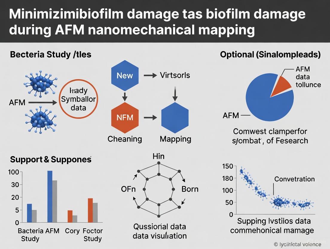
Advanced Strategies for Minimizing Biofilm Damage in AFM Nanomechanical Mapping
Atomic Force Microscopy (AFM) is a powerful tool for characterizing the nanomechanical properties of biofilms, which are crucial for understanding their recalcitrance in medical and industrial contexts.
Advanced Strategies for Minimizing Biofilm Damage in AFM Nanomechanical Mapping
Abstract
Atomic Force Microscopy (AFM) is a powerful tool for characterizing the nanomechanical properties of biofilms, which are crucial for understanding their recalcitrance in medical and industrial contexts. However, the soft, hydrated, and heterogeneous nature of biofilms makes them highly susceptible to damage during AFM analysis, potentially compromising data integrity. This article provides a comprehensive guide for researchers and drug development professionals on minimizing this damage. It covers the foundational principles of biofilm mechanics and AFM-induced damage, explores advanced, gentle mapping modes like force volume and nano-DMA, details optimization strategies involving machine learning and sample preparation, and validates findings through correlative microscopy and comparative analysis. The goal is to empower scientists with the methodologies needed to obtain high-fidelity, reliable nanomechanical data from intact biofilm structures.
Understanding Biofilm Mechanics and AFM-Induced Damage Mechanisms
Atomic Force Microscopy (AFM) is a powerful tool for studying biofilms, capable of imaging at nanometre resolution and measuring nanomechanical properties under near-physiological conditions without extensive sample preparation [1]. However, the very features that make biofilms complex and resilient—their heterogeneous architecture, extracellular polymeric substance (EPS) matrix, and soft, dynamic nature—also make them vulnerable to damage during AFM probing. This technical support guide addresses the specific challenges researchers face when performing nanomechanical mapping on biofilms and provides actionable troubleshooting and protocols to minimize experimental artifacts and obtain reliable data.
FAQs and Troubleshooting Guides
Frequently Asked Questions
Q1: Why is my AFM probe getting stuck or contaminating when scanning a biofilm? The sticky, viscous EPS matrix that surrounds bacterial cells can adhere to the AFM probe. This is a common issue caused by the high adhesion forces between the probe and the biofilm surface [2]. Using sharper probes and optimizing the setpoint force can reduce this, but some contamination may be inevitable in contact mode.
Q2: My force curves on a biofilm appear noisy and inconsistent. What could be the cause? Biofilms are inherently heterogeneous. A force curve measured on a single cell will be very different from one measured on a bare patch of EPS or a cluster of cells [3]. This is a feature of the sample, not an error. The solution is to acquire a large number of force curves across a large area to statistically capture the biofilm's variability.
Q3: How can I be sure that I'm not deforming the biofilm structure during imaging? It is difficult to avoid some degree of deformation on soft samples. To minimize it, use the gentlest possible imaging mode, such as tapping mode in liquid [1]. Furthermore, always start with a high setpoint (low force) and gradually decrease it until you achieve stable feedback, rather than starting with a low setpoint that could damage the sample.
Q4: My high-resolution images of single cells look good, but I'm missing the larger community structure. Why? Traditional AFM has a fundamental limitation: a narrow field of view (typically <100 µm) that makes it difficult to link single-cell features to the larger biofilm architecture [4] [5]. The solution is to use a large-area AFM approach that automatically stitches many individual high-resolution images together to create a millimeter-scale map [4].
Troubleshooting Common Problems
The table below summarizes key issues, their likely causes, and recommended solutions.
| Problem | Likely Cause | Recommended Solution |
|---|---|---|
| Probe contamination or sticking | High adhesion of EPS to probe [2] | Use sharper probes; reduce setpoint force; employ non-contact (tapping) mode [1] |
| Blurred or featureless images | Excessive imaging force deforming soft biofilm | Image in liquid using tapping mode; verify cantilever's spring constant; reduce setpoint voltage |
| Inconsistent nanomechanical data | Natural heterogeneity of biofilm structure [5] | Acquire large number of force curves (100s+); use large-area mapping to ensure data is representative [4] |
| Inability to see large-scale organization | Limited scan range of conventional AFM [5] | Implement automated large-area AFM platform with image stitching [4] |
| Difficulty locating areas of interest | Lack of navigational context on sample surface | Use integrated light microscopy or pre-gridded substrates to locate specific biofilm regions |
Standard Experimental Protocols
Protocol 1: Large-Area AFM Imaging of Biofilm Organization
This protocol, adapted from Oak Ridge National Laboratory studies, allows for the correlation of single-cell features with community-wide architecture [4] [5].
- Sample Preparation: Grow Pantoea sp. YR343 (or your model organism) on PFOTS-treated glass coverslips. At desired time points, gently rinse the coverslip to remove non-adherent cells and air-dry before imaging [5].
- AFM Setup: Use an automated large-area AFM platform. Select a cantilever appropriate for your mode (e.g., triangular silicon nitride for tapping mode).
- Automated Imaging: Program the AFM to acquire a grid of contiguous, high-resolution images (e.g., 50x50 µm) across a millimeter-scale area. Use minimal image overlap (e.g., 5-10%) to maximize speed.
- Image Stitching: Use integrated software or algorithms to seamlessly stitch the individual images into a single, large-area map.
- Data Analysis: Apply machine learning-based image segmentation to automatically detect individual cells, classify features, and quantify properties like cell count, confluency, and orientation across the entire dataset [4].
Protocol 2: In-Situ Nanomechanical Mapping During Treatment
This protocol details how to measure the mechanical response of biofilms to antimicrobial agents in real-time [3].
- Initial Setup: Obtain surface images and force data of the biofilm in intermittent contact mode in buffer solution (e.g., Phosphate Buffer, PB) using a liquid cell.
- Baseline Data Acquisition: Designate this as time zero. Acquire force curves on the center of selected cells. A typical protocol might use five force curves per location [3].
- In-Situ Treatment: Inject a concentrated solution of the compound of interest (e.g., MAG2) into the Petri dish without disturbing the setup. This allows for continuous data collection on the same cells.
- Kinetic Monitoring: Continue collecting force data at regular intervals (e.g., every five minutes). Take low-resolution images between force acquisitions to ensure cells remain intact and in place.
- Final Analysis: After the final force data is obtained, take a high-resolution image of the area. Analyze the effective spring constant (
k_effective) from the slope of the linear compression regime in the force curves and calculate the cellular spring constant (k_cell) using appropriate models [3].
Workflow for In-Situ Nanomechanical Mapping
Research Reagent Solutions
The table below lists key materials and reagents used in advanced AFM biofilm studies, along with their specific functions.
| Reagent / Material | Function in Experiment |
|---|---|
| PFOTS-treated Glass Coverslips | Creates a hydrophobic surface for controlled biofilm growth and attachment [5]. |
| Polystyrene Beads (for FluidFM) | Serves as a scaffold to grow biofilms for novel "biofilm-probe" adhesion force measurements [2]. |
| Vanillin Solution | Used as a surface modifier on filtration membranes (e.g., PES) to create anti-biofouling surfaces and study reduced adhesion [2]. |
| Silicon Nitride Cantilevers | Standard probe for bioimaging; high resolution and biocompatible, used for both imaging and force spectroscopy [3]. |
| Pantoea sp. YR343 | A model Gram-negative, biofilm-forming bacterium with well-characterized attachment dynamics and flagella [5]. |
| MAG2 Compound | An example antimicrobial agent used in in-situ AFM studies to observe its mechanical effect on live biofilms [3]. |
Advanced Technical Guides
Force Spectroscopy Data Interpretation
When performing nanomechanical mapping, the force-distance curve is the primary source of data. The extension curve consists of a linear approach, a nonlinear transition, and a linear compression regime. The slope of the linear compression phase (k_effective) is used to calculate the cellular spring constant (k_cell), which is a direct measure of cell stiffness [3]. On soft, heterogeneous biofilms, expect a wide distribution of k_cell values, reflecting the structural diversity from EPS to rigid cell walls.
Leveraging Machine Learning for Analysis
Manual analysis of large-area AFM datasets, which can contain over 19,000 individual cells, is impractical [4]. Integrating machine learning (ML) for image segmentation and analysis is now a best practice. ML algorithms can automatically:
- Detect and count cells across large areas.
- Classify cells based on morphology (e.g., rod-shaped vs. cocci).
- Generate detailed maps of properties like cell orientation and confluency, revealing patterns like the "honeycomb" organization observed in Pantoea biofilms [4] [5].
Machine Learning Data Analysis Pipeline
FAQs: Troubleshooting AFM Nanomechanical Mapping of Biofilms
Q1: Our AFM measurements on biofilm viscoelasticity show high variability. What could be the cause? High variability often stems from the inherent spatial heterogeneity of the biofilm matrix and inconsistent experimental conditions.
- Spatial Heterogeneity: The EPS matrix is a complex mix of polysaccharides, proteins, extracellular DNA (e-DNA), and other components whose distribution is not uniform [6]. Measurements taken on a polysaccharide-rich region will differ significantly from those on a region dense with e-DNA or amyloid fibers.
- Actionable Solution: Implement a large-area AFM scanning approach. Instead of a few small scans, use automated methods to collect high-resolution data over millimeter-scale areas. This provides a more statistically significant dataset that captures the true structural and mechanical diversity of the biofilm [7].
Q2: How can we minimize damage to soft, hydrated biofilms during AFM indentation? Biofilms are highly hydrated and soft, making them prone to damage. Key considerations are probe selection and operational mode.
- Probe Geometry: Avoid using sharp, pyramidal tips for mechanical measurements. Instead, use spherical probes (e.g., colloid-attached cantilevers). The larger contact area distributes the load, reducing localized pressure and preventing the tip from piercing the sample [8] [9].
- AFM Mode: Use force spectroscopy or nanoindentation modes with a closed-loop AFM system for accurate displacement control. Perform creep experiments at constant load to characterize viscoelasticity without excessive strain rates that could damage the matrix [8].
Q3: Our biofilm samples detach from the substrate during fluid imaging. How can we improve immobilization? Secure immobilization is critical for force measurements. Chemical methods can be optimized for minimal impact.
- Benign Chemical Immobilization: Use substrates functionalized with poly-L-lysine or polydopamine to enhance adhesion. Alternatively, adding divalent cations like Mg²⁺ and Ca²⁺ to the imaging buffer can promote bacterial attachment by cross-linking negatively charged EPS components without significantly altering cell viability or nanomechanical properties [10].
- Mechanical Entrapment: For more robust immobilization, use porous polymer membranes or micro-fabricated polydimethylsiloxane (PDMS) stamps with well sizes tailored to your bacterial cells to physically trap them [10].
Q4: How does the EPS composition specifically influence the nanomechanical data we collect? Different EPS components contribute uniquely to the biofilm's mechanical properties.
- Structural Components: Neutral polysaccharides and amyloid fibrils provide structural scaffolding and enhance mechanical stability [6].
- Cross-linking Agents: Charged polysaccharides like alginate can be cross-linked by multivalent cations (e.g., Ca²⁺), significantly increasing cohesive strength and stiffness [6] [11].
- Unexpected Players: Extracellular DNA (e-DNA) acts as a key intercellular connector, forming grid-like structures that stabilize the biofilm matrix and influence its viscoelastic response [6].
Quantitative Data on Biofilm Mechanical Properties
The following tables summarize key nanomechanical parameters obtained from AFM studies, providing benchmarks for your own research.
Table 1: Adhesive and Viscoelastic Properties of P. aeruginosa Biofilms
| Bacterial Strain | Biofilm Stage | Adhesive Pressure (Pa) | Instantaneous Elastic Modulus (kPa) | Delayed Elastic Modulus (kPa) | Viscosity (kPa·s) |
|---|---|---|---|---|---|
| P. aeruginosa PAO1 (Wild-type) | Early Biofilm | 34 ± 15 | Data from Voigt model fitting [8] | Data from Voigt model fitting [8] | Data from Voigt model fitting [8] |
| P. aeruginosa PAO1 (Wild-type) | Mature Biofilm | 19 ± 7 | Drastically reduced | Drastically reduced | Decreased |
| P. aeruginosa wapR (LPS mutant) | Early Biofilm | 332 ± 47 | Drastically reduced | Drastically reduced | No significant change |
| P. aeruginosa wapR (LPS mutant) | Mature Biofilm | 80 ± 22 | Drastically reduced | Drastically reduced | Decreased |
Note: Data obtained via Microbead Force Spectroscopy (MBFS) with a Voigt Standard Linear Solid model. The wapR mutant has a defective lipopolysaccharide (LPS) core, highlighting how cell surface chemistry drastically alters mechanical properties [8].
Table 2: Cohesive Energy of Mixed-Culture Biofilms
| Biofilm Type | Cultivation Condition | Cohesive Energy (nJ/μm³) | Measurement Technique |
|---|---|---|---|
| Mixed-Culture (Activated Sludge) | Standard | 0.10 ± 0.07 (top) to 2.05 ± 0.62 (bottom) | AFM abrasion/energy dissipation [11] |
| Mixed-Culture (Activated Sludge) | With 10 mM CaCl₂ | 0.10 ± 0.07 to 1.98 ± 0.34 | AFM abrasion/energy dissipation [11] |
Note: Cohesive energy increases with biofilm depth and is enhanced by the addition of calcium, which cross-links EPS components [11].
Experimental Protocols for Key AFM Methodologies
Protocol 1: Microbead Force Spectroscopy (MBFS) for Adhesion and Viscoelasticity
This protocol allows for simultaneous quantification of adhesive and viscoelastic properties under native conditions [8].
- Cantilever and Probe Preparation:
- Attach a ~50 µm diameter glass bead to a tipless AFM cantilever.
- Calibrate the cantilever's spring constant using the thermal method.
- Grow a biofilm directly on the microbead probe by incubating it in a bacterial culture.
- Standardized Force Measurement:
- Approach the biofilm-coated bead to a clean glass surface in liquid at a defined speed.
- Apply a controlled loading force and hold the bead at constant contact for a defined period (e.g., 1-2 seconds) to perform a creep test.
- Retract the bead from the surface at a standardized speed.
- Data Analysis:
- Adhesion: Calculate the adhesive pressure from the pull-off force in the retraction curve, divided by the contact area.
- Viscoelasticity: Fit the creep compliance data (indentation vs. time during hold) to a Voigt Standard Linear Solid model to extract the instantaneous elastic modulus, delayed elastic modulus, and viscosity.
Protocol 2: Measuring Biofilm Cohesive Strength via AFM Abrasion
This method quantifies the energy required to dislodge a defined volume of biofilm, providing a direct measure of cohesion [11].
- Sample Preparation:
- Grow a moist biofilm on a suitable substrate (e.g., a membrane).
- Equilibrate the sample in a humidity chamber (e.g., 90% RH) to maintain consistent water content without full submersion.
- AFM Scanning and Abrasion:
- Obtain a baseline topographic image of a biofilm region at a low applied load (~0 nN).
- On a smaller sub-region, perform repeated raster scans at a high load (e.g., 40 nN) to abrade the biofilm.
- Return to low load and obtain a post-abrasion topographic image of the larger area.
- Cohesive Energy Calculation:
- Subtract the post-abrasion image from the baseline image to determine the volume of biofilm displaced.
- From the friction signal during abrasive scanning, calculate the total frictional energy dissipated.
- Divide the frictional energy by the displaced volume to obtain the cohesive energy (nJ/μm³).
Protocol 3: FluidFM for Biofilm-Scale Adhesion Measurements
FluidFM technology overcomes the limitation of single-cell force spectroscopy by enabling adhesion measurements with whole biofilms [2].
- Probe Preparation:
- Use a FluidFM cantilever, which is a micro-channeled cantilever with an aperture at its end.
- Grow biofilms on micro-sized polystyrene beads.
- Aspirate a single biofilm-coated bead onto the cantilever's aperture by applying negative pressure through the fluidic system.
- Adhesion Force Spectroscopy:
- Approach the bead-probe to the surface of interest (e.g., a modified membrane) in liquid.
- After a set contact time, retract the probe while monitoring the force.
- Data Interpretation:
- Analyze the retraction curve to determine the maximum adhesion force and the work of adhesion. This provides a biofilm-scale adhesion metric that is more representative of real-world conditions than single-cell data.
Signaling Pathways and Experimental Workflows
Diagram 1: The Interplay of EPS Composition, Regulation, and AFM Measurement. This diagram outlines the logical relationship between the molecular composition of the EPS matrix, its regulatory systems, the resulting structural and mechanical properties, and the consequent choices that must be made for accurate AFM nanomechanical mapping.
Diagram 2: FluidFM Biofilm Adhesion Protocol. This workflow details the novel FluidFM method for measuring biofilm-scale adhesion forces, which more accurately represents real-world conditions than single-cell methods [2].
The Scientist's Toolkit: Research Reagent Solutions
Table 3: Essential Materials for AFM-based Biofilm Nanomechanics
| Item | Function/Benefit | Key Considerations |
|---|---|---|
| Tipless Cantilevers | Base for attaching custom probes (microbeads, cells). | Ensure compatibility with your AFM system and the chosen attachment method [8]. |
| Glass/Polystyrene Microbeads | Create a defined spherical probe for quantifiable contact mechanics. | Diameter (e.g., 50 µm) should be chosen based on the required contact area and resolution [8] [2]. |
| FluidFM Cantilevers | Microfluidic cantilevers for aspirating and manipulating single cells or biofilm-coated beads. | Enables biofilm-scale force measurements and reversible probe immobilization [2]. |
| Polydopamine | A versatile bio-adhesive for immobilizing cells or coating probes. | Provides strong, non-specific adhesion with minimal denaturation of biological samples [2]. |
| Poly-L-Lysine | Coating for substrates to enhance electrostatic immobilization of cells. | Widely used for attaching negatively charged bacterial cells to surfaces [10]. |
| Calcium Chloride (CaCl₂) | Cross-links anionic EPS components (e.g., alginate), modulating matrix stiffness and cohesion. | Useful for studying the effect of specific ions on biofilm mechanics [6] [11]. |
| PFOTS-Treated Glass | Creates a highly hydrophobic surface to study the effect of surface properties on initial cell attachment and biofilm assembly. | Used in large-area AFM studies to observe patterned cellular organization [7]. |
In the field of biofilm research, Atomic Force Microscopy (AFM) provides unparalleled nanoscale resolution for topographical and nanomechanical mapping. However, the soft, hydrated, and heterogeneous nature of biofilms makes them particularly susceptible to damage and imaging artifacts. This guide addresses common AFM artifacts encountered when studying biofilms, offering troubleshooting and methodologies to minimize data distortion and preserve sample integrity for reliable results.
Frequently Asked Questions (FAQs) and Troubleshooting
1. My AFM images show repeated, unnaturally sharp features that do not match other microscopy data. What is the cause? This is a classic sign of a damaged or contaminated probe tip [12]. A worn or dirty tip acts as a poor stylus, creating images that reflect the tip's own shape rather than the true sample surface. These tip-convolution artifacts appear as sharp peaks, duplicated features, or broadening of actual structures.
- Solution: Implement a rigorous tip inspection and cleaning protocol. Before and after imaging, inspect the tip under an optical microscope. For cleaning, follow manufacturer guidelines, which may involve UV-ozone treatment or solvent rinses. Using a tip characterization standard, such as a sample with sharp, known spikes, can help verify tip shape and integrity [12].
2. My biofilm sample appears compressed or shows signs of lysis during scanning. How can I prevent this? This indicates excessive imaging force is being applied. Biofilms are mechanically soft, and high lateral or vertical forces can compress the extracellular polymeric substance (EPS), displace cells, and even rupture cell membranes [13] [14].
- Solution: Optimize your scanning parameters for soft materials.
- Force Control: Use advanced modes like PeakForce Tapping or Quantitative Imaging (QI) that directly control and minimize the maximum force applied to the sample at each pixel [12] [14].
- Reduce Setpoint: In tapping mode, lower the amplitude setpoint to reduce tip-sample interaction forces.
- Softer Cantilevers: Select cantilevers with low spring constants (e.g., 0.01 - 0.5 N/m) to minimize indentation on soft samples [14].
3. My images are distorted, appearing stretched or skewed, especially at the edges. Why? This is typically caused by scanner nonlinearities and hysteresis, which are inherent properties of piezoelectric scanners [12]. The scanner doesn't move in a perfectly linear fashion, leading to positional inaccuracies.
- Solution:
- Use a Closed-Loop Scanner: If available, use an AFM system equipped with a closed-loop scanner. These systems have sensors that provide real-time position feedback to correct for nonlinearities and hysteresis [12].
- Regular Calibration: Frequently calibrate the scanner using traceable calibration standards with known pitch and height (e.g., silicon gratings) [12].
- Limit Scan Size: For critical measurements, scan smaller areas near the center of the scanner's range, where nonlinearities are less pronounced.
4. I observe a "shadow" or "ghost" of a previous feature in my image, even after moving to a new area. This is a feedback loop artifact, often caused by improper PID (Proportional, Integral, Derivative) controller settings. An overly slow feedback loop cannot track steep features, causing the tip to temporarily lose contact ("parachuting") and then re-engage incorrectly [12].
- Solution: Optimize the feedback gains.
- Start with low gains and gradually increase them until the feedback is stable without oscillating.
- Many modern AFMs offer auto-tune functions for the feedback loop, which can provide a good starting point for optimization.
5. How can I be sure I'm measuring the true mechanical properties of the biofilm and not an artifact? Accurate nanomechanical mapping requires careful selection of the contact mechanics model and validation of the data [14]. Using an incorrect model (e.g., applying a Hertz model for an adhesive sample) will yield meaningless values.
- Solution:
- Model Selection: Choose a contact mechanics model (e.g., Hertz, Sneddon, DMT, JKR) appropriate for your sample's properties (adhesion, elasticity) [14].
- Indentation Depth: Keep the indentation depth to a small percentage (typically 10-20%) of the sample's total thickness to avoid influence from the underlying substrate.
- Multiple Locations: Perform force spectroscopy measurements at multiple, random locations to ensure the data is representative and not an outlier.
Troubleshooting Guide: Common AFM Artifacts and Solutions
The table below summarizes key artifacts, their causes, and corrective actions.
| Artifact Type | Common Causes | Corrective Actions |
|---|---|---|
| Tip Convolution | Worn, damaged, or contaminated probe [12] | Inspect and clean probes regularly; use sharp, new tips; verify with tip characterization standard [12]. |
| Sample Damage (Compression/Lysis) | Excessive imaging force; inappropriate probe stiffness [13] [14] | Use low-force modes (e.g., PeakForce Tapping); select soft cantilevers (0.01-0.5 N/m); reduce setpoint/amplitude [12] [14]. |
| Thermal Drift | Temperature fluctuations in the lab environment [12] | Allow AFM and sample to thermally equilibrate; use environmental enclosure; perform faster scans; use drift correction algorithms [12]. |
| Scanner Nonlinearities | Hysteresis and creep of piezoelectric material [12] | Use closed-loop scanner; calibrate scanner regularly; restrict measurements to center of scan range [12]. |
| Feedback Artifacts | Improperly tuned PID gains; scanning too fast [12] | Manually optimize PID gains; reduce scan speed and line rate, especially for tall, steep features. |
| Adhesion Hysteresis | Tip-sample adhesion causing jump-to-contact and pull-off events [14] | Use functionalized tips with controlled chemistry; operate in fluid to minimize capillary forces; employ adhesion-reducing modes. |
Experimental Protocols for Minimizing Artifacts
Protocol 1: Non-Destructive Nanomechanical Mapping of Biofilms
This protocol is designed to obtain reliable elastic modulus and adhesion maps while preserving biofilm integrity.
- Sample Preparation: Gently rinse the biofilm to remove loose planktonic cells. For hydrated imaging, ensure the sample is fully immersed in an appropriate physiological buffer. Use a soft substrate (e.g., agar-coated glass) to minimize substrate effects and prevent sample detachment [14].
- Cantilever Selection: Choose a cantilever with a low spring constant (0.01 - 0.1 N/m) and a sharp, nominal tip radius (<10 nm). Calibrate the spring constant using the thermal tune method [14].
- AFM Mode Selection: Engage the PeakForce Tapping or Quantitative Imaging (QI) mode. These modes provide direct control over the peak force, minimizing sample damage [12] [14].
- Parameter Optimization:
- Set a very low Peak Force (typically 50-500 pN) and gradually increase it until a stable topographical image is obtained.
- Set a slow Scan Rate (0.5-1.0 Hz) to allow the feedback loop to accurately track the surface.
- Adjust the feedback gains to ensure responsive but non-oscillatory tracking.
- Data Acquisition and Validation: Collect nanomechanical maps over multiple, randomly selected areas. Validate the mechanical properties by comparing values from different locations and against measurements taken with different force setpoints to ensure consistency.
Protocol 2: Systematic Probe Management and Calibration
A reliable probe is the foundation of artifact-free AFM.
- Visual Inspection: Before use, inspect the cantilever and tip under an optical microscope at high magnification for visible contaminants or damage.
- Cleaning: If contamination is suspected, clean the tip by:
- UV-Ozone: Expose the tip to UV-ozone light for 10-15 minutes.
- Solvent Rinse: Gently rinse the cantilever chip with high-purity solvents (e.g., ethanol, isopropanol) and dry with a gentle stream of clean, dry air.
- Spring Constant Calibration: Perform thermal tune calibration in the same medium (air or liquid) that will be used for the experiment.
- Tip Shape Verification: Scan a tip characterization standard containing sharp, high-aspect-ratio features (e.g., TGT1 grating). Reconstruct the tip shape from the acquired image to check for blunting or double tips.
Workflow for Artifact Identification and Mitigation
The following diagram illustrates a systematic approach to diagnosing and resolving common AFM artifacts in biofilm research.
The Scientist's Toolkit: Essential Research Reagents and Materials
The table below lists key materials and their functions for reliable AFM biofilm analysis.
| Item | Function in AFM Biofilm Research |
|---|---|
| Soft Cantilevers (0.01 - 0.5 N/m) | Probes with low spring constants minimize indentation and prevent damage to delicate biofilm structures and living cells during nanomechanical mapping [14]. |
| Tip Characterization Standard (e.g., TGT1) | A grating with sharp, known spikes used to image the AFM tip itself, verifying its sharpness and identifying contamination or damage that would cause imaging artifacts [12]. |
| Scanner Calibration Grating | A standard with precise pitch and step height for calibrating the AFM scanner's X, Y, and Z dimensions, ensuring accurate and undistorted measurements [12]. |
| Physiological Buffer (e.g., PBS) | Maintains biofilm hydration and cell viability during imaging in liquid, preserving the native state of the sample and its mechanical properties. |
| Agar-coated Substrates | A soft mounting surface for biofilms that helps prevent sample detachment and reduces the "substrate effect" during deep mechanical indentation measurements. |
| PeakForce Tapping or QI | Advanced AFM operational modes that provide direct control over the maximum force applied to the sample, crucial for non-destructive imaging of soft matter [12] [14]. |
Linking Mechanical Heterogeneity to Biological Function
Atomic Force Microscopy (AFM) has emerged as a powerful tool for characterizing the nanomechanical properties of complex biological systems, including bacterial biofilms. Biofilms are multicellular microbial communities held together by self-produced extracellular polymeric substances (EPS), and their mechanical heterogeneity plays a critical role in their function and resilience [7] [10]. Understanding this mechanical heterogeneity is essential for developing effective strategies to control biofilms in medical, industrial, and environmental contexts.
The challenge for researchers lies in accurately measuring these mechanical properties without damaging the delicate biofilm structure. Biofilms are inherently soft, hydrated, and mechanically heterogeneous, ranging from individual cellular features to larger community architectures [7] [15]. This technical guide addresses the specific experimental issues researchers encounter when attempting to link mechanical heterogeneity to biological function in biofilm systems, with a focus on minimizing experimental artifacts and damage during AFM characterization.
Core Challenges in Biofilm Nanomechanics
The accurate nanomechanical characterization of biofilms presents several interconnected challenges that researchers must overcome to obtain biologically relevant data.
Key Technical Challenges:
- Spatial Heterogeneity: Biofilms exhibit significant spatial variations in mechanical properties across micron and nanometer scales, requiring high-resolution mapping techniques [7] [16]
- Scale Mismatch: Conventional AFM has limited imaging areas (<100 µm), making it difficult to link cellular-scale features to larger functional biofilm architectures [7]
- Soft Hydrated Nature: Biofilms contain up to 90% water, making them prone to deformation and damage during probing [10] [15]
- Complex Viscoelasticity: Biofilms exhibit time-dependent mechanical behavior that requires appropriate characterization models [17]
- Immobilization Difficulties: Microbial cells are often easily disrupted by AFM cantilever scanning, requiring effective yet benign immobilization strategies [10]
Troubleshooting Common Experimental Issues
Sample Preparation and Immobilization
Problem: Biofilm detachment or disruption during scanning. Solution: Implement appropriate immobilization strategies that secure samples without altering their mechanical properties.
Table: Biofilm Immobilization Techniques
| Technique | Methodology | Best For | Limitations |
|---|---|---|---|
| Mechanical Entrapment | Trapping cells in porous membranes or PDMS stamps with customized microwells [10] | Spherical microorganisms; single-cell analysis | Sporadic immobilization; reduced reproducibility |
| Poly-L-Lysine Coating | Coating substrates with adhesive polymers [10] | Firm attachment of diverse cell types | Potential physiological alterations |
| Divalent Cation Enhancement | Adding Mg²⁺ or Ca²⁺ to improve attachment [10] | Optimal attachment with maintained viability | Concentration-dependent effects |
| Functionalized Surfaces | Using chemically modified substrates (e.g., carboxyl groups) [10] | Organized immobilization | Possible reduction in cell viability |
Problem: Non-representative mechanical data due to sample dehydration. Solution: Maintain hydrated conditions throughout preparation and imaging.
- Perform all sample rinsing and transfer steps using appropriate buffers (e.g., PBS)
- Conduct AFM measurements in liquid cells with physiological buffers [10] [15]
- Limit air exposure time during sample transfer to prevent dehydration artifacts
- Use environmental chambers when extended imaging is required
AFM Operation and Parameter Optimization
Problem: Excessive forces causing biofilm damage during imaging. Solution: Optimize scanning parameters to minimize tip-sample interactions.
Table: AFM Mode Selection for Biofilms
| AFM Mode | Principle | Biofilm Applications | Damage Risk |
|---|---|---|---|
| Tapping Mode | Intermittent tip contact with surface [10] [15] | General topography imaging; heterogeneous samples | Low-Moderate |
| Force Volume | Array of force-distance curves across surface [17] [16] | Nanomechanical property mapping | Moderate (dependent on force) |
| Bimodal AFM | Simultaneous excitation at two frequencies [17] | High-resolution mechanical mapping | Low |
| Peak Force QNM | Controlled peak force tapping with feedback [15] | Quantitative nanomechanics of soft materials | Very Low |
Problem: Inconsistent mechanical measurements between experiments. Solution: Implement rigorous calibration protocols and standardized procedures.
- Calibrate cantilever spring constants using thermal tuning method before each experiment [16] [18]
- Characterize tip geometry and radius using reference samples
- Validate mechanical measurements against reference hydrogels with known properties [16]
- Use the Standardized Nanomechanical AFM Procedure (SNAP) for method validation [16]
Data Interpretation and Modeling
Problem: Incorrect mechanical models leading to erroneous modulus values. Solution: Select contact mechanics models appropriate for biofilm properties.
Diagram: Model Selection Workflow for Biofilm Nanomechanics
Problem: Subsurface heterogeneity affecting mechanical measurements. Solution: Account for layered structures and subsurface features in analysis.
- Use finite element modeling (FEM) to account for complex geometries and heterogeneities [19] [16] [18]
- Implement Hybrid Eshelby Decomposition (HED) for layered samples to extract layer-specific moduli [19]
- Consider sample thickness effects using appropriate corrections (e.g., Dimitriadis model) when indenting thin samples [18]
- Use spherical probes rather than sharp tips to better average local heterogeneities [16]
Experimental Protocols for Minimizing Biofilm Damage
Large-Area Automated AFM for Biofilm Mapping
Purpose: To characterize mechanical heterogeneity across relevant biofilm length scales while minimizing sampling bias and damage.
Methodology:
- Sample Preparation:
- Grow biofilms on appropriate substrates (e.g., glass coverslips, medical device materials)
- Gently rinse with buffer to remove planktonic cells without disrupting biofilm architecture
- For delicate biofilms, consider minimal chemical fixation (e.g., 0.5-1% glutaraldehyde) if live imaging isn't required [10]
AFM Setup:
Image Acquisition:
Mechanical Mapping:
- Conduct force volume mapping with appropriate force setpoints (typically 0.5-5 nN)
- Limit indentation depth to 10-15% of biofilm thickness to avoid substrate effects [18]
- Maintain physiological temperature and buffer conditions throughout
Data Analysis:
- Apply machine learning segmentation to identify different structural components [7]
- Use appropriate contact mechanics models for different biofilm regions
- Generate spatial modulus maps correlating mechanics with structure
Viscoelastic Characterization of Biofilm Matrix
Purpose: To quantify time-dependent mechanical properties of EPS matrix without permanent deformation.
Methodology:
- Probe Selection:
- Use colloidal probes with large radii (5-10 µm) to measure matrix properties rather than single cells
- Verify probe chemistry (e.g., hydrophilic coatings) to control adhesion
Force Measurement Protocol:
- Perform stress-relaxation experiments with rapid indentation followed by hold period
- Conduct dynamic mechanical analysis with frequency sweeps (0.1-100 Hz)
- Implement creep compliance measurements at multiple stress levels
Viscoelastic Modeling:
- Fit relaxation data to Prony series or fractional viscoelastic models
- Analyze frequency-dependent behavior using complex modulus representations
- Use appropriate viscoelastic contact models for data interpretation [17]
Research Reagent Solutions
Table: Essential Materials for Biofilm Nanomechanics
| Reagent/Material | Function | Application Notes |
|---|---|---|
| Soft Cantilevers (0.01-0.5 N/m) | Force sensing for soft materials | Critical for accurate modulus measurement; thermal calibration required [9] [16] |
| Colloidal Probes (2.5-10 µm spheres) | Reduced contact pressure during indentation | Minimize biofilm damage; simplify contact mechanics modeling [16] [18] |
| Polydimethylsiloxane (PDMS) Stamps | Microfabricated cell immobilization | Customizable microwell sizes for different microorganisms [10] |
| Poly-L-Lysine Solutions | Substrate coating for cell adhesion | Provides electrostatic immobilization; potential physiological effects [10] |
| Reference Hydrogel Standards | AFM calibration and validation | Agarose, PAA, or PNIPAM gels with known rheological properties [16] |
| Physiological Buffers (PBS, etc.) | Maintain hydrated conditions | Preserve native biofilm structure and mechanics [10] [15] |
Frequently Asked Questions
Q1: What is the maximum force I should use when indenting biofilms to avoid damage? A: The optimal force depends on your specific biofilm and research goals, but generally keep forces below 5 nN for most bacterial biofilms. Conduct a force series first to identify the range where measurements become force-independent, which indicates no permanent deformation is occurring. Use the minimum force that provides sufficient signal-to-noise ratio [10] [16].
Q2: How can I distinguish real mechanical heterogeneity from measurement artifacts? A: Implement multiple validation approaches: (1) Repeat measurements at different locations and times, (2) Use different probe sizes to check for consistency, (3) Compare force approach and retraction curves for reversibility, (4) Validate with complementary techniques like fluorescence microscopy when possible, and (5) Use reference materials with known heterogeneity to confirm measurement capability [16] [18].
Q3: What is the best way to handle the scale mismatch between AFM scan sizes and relevant biofilm features? A: Implement large-area automated AFM approaches that combine multiple high-resolution scans. Use machine learning algorithms to identify regions of interest and strategically sample across millimeter scales. This approach provides both cellular-resolution detail and macroscopic context without excessive imaging time or damage [7].
Q4: How does biofilm hydration affect mechanical measurements, and how can I control for it? A: Hydration dramatically affects biofilm mechanics, as EPS hydration determines polymer mobility and network properties. Always measure in liquid environments that approximate physiological conditions. Control for evaporation during long experiments using environmental chambers or fluid exchange systems. Note that even small changes in osmotic pressure can significantly alter measured mechanics [10] [15].
Q5: What are the most common mistakes in contact mechanics modeling of biofilm AFM data? A: The most frequent errors include: (1) Using Hertz model for thin samples without thickness corrections, (2) Applying linear elastic models to viscoelastic materials, (3) Ignoring adhesion forces in analysis, (4) Using inappropriate tip geometry assumptions, and (5) Neglecting substrate effects when indenting thin biofilms. Always validate your model choice with simulated data when possible [17] [18].
Q6: Can I use the same AFM tips for imaging and force measurement on biofilms? A: While possible, it's not recommended. Sharp tips for imaging typically cause higher contact pressures that may damage soft biofilms. Better practice is to use separate tips: sharper tips for high-resolution imaging and colloidal probes for mechanical measurements. If you must use the same tip, perform imaging at minimal forces and verify that no damage occurs by re-imaging areas after force mapping [16] [15].
Gentle by Design: AFM Modes for Non-Destructive Nanomechanical Mapping
Atomic Force Microscopy (AFM) based nanomechanical mapping is a dominant technique for characterizing mechanical properties at the nanoscale, transforming tip-sample interaction forces into quantitative mechanical parameters [20]. Force Volume mode is a specific nanomechanical mapping method based on acquiring a force-distance curve (FDC) in each pixel of the sample surface [20]. These curves are then transformed into maps of mechanical properties by fitting the data to contact mechanics models [20]. Within biofilm research, this technique is invaluable for studying the structural and mechanical properties of complex microbial communities without causing irreversible sample damage, enabling insights into cell attachment, biofilm assembly, and response to external stresses [7]. This guide provides the principles, protocols, and troubleshooting essential for performing low-stress indentation on delicate biofilms.
Principles of Force Volume Mode
In Force Volume mode, the tip-sample distance is modulated while the cantilever's deflection is recorded, generating a force-distance curve at every point [20]. The repulsive component of the interaction force is analyzed using contact mechanics models to extract quantitative mechanical properties such as elastic modulus and adhesion forces [20]. A key feature of FDC analysis on viscoelastic materials like biofilms is the observation of hysteresis between the approach and retraction curves, which indicates energy dissipation and the sample's viscoelastic nature [20].
The modulation of the tip-sample distance can be achieved using different waveforms. While early Force Volume implementations used triangular waveforms for constant tip velocity, modern methods often employ sinusoidal signals or photothermal cantilever actuation to achieve higher imaging rates and avoid artefacts associated with triangular waveforms [20]. The term "Force Volume" encompasses all modes based on acquiring a full FDC per pixel, regardless of the specific waveform or actuation method used [20].
Experimental Protocols
Sample Preparation
- Substrate Selection: Use rigid, atomically flat substrates (e.g., mica, glass) to minimize background signal from substrate deformation. Studies have successfully analyzed bacterial biofilms on PFOTS-treated glass and silicon substrates [7].
- Biofilm Immobilization: Gently rinse the biofilm to remove unattached cells and debris. For hydrated measurements, ensure the biofilm remains in an appropriate liquid buffer or growth medium to maintain its native state [7] [14]. For high-resolution imaging, some protocols involve gentle drying, but this should be done with caution as it can alter mechanical properties [7].
Cantilever Selection and Calibration
Choosing and calibrating the right cantilever is critical for low-stress measurements on soft biofilms. The table below summarizes key parameters.
- Calibration: Precisely calibrate the cantilever's spring constant and the optical lever sensitivity using established methods (e.g., thermal tune). Accurate calibration is fundamental for quantitative force measurements [14].
Table 1: Cantilever Selection Guide for Biofilm Nanomechanics
| Parameter | Recommended Specification | Function and Rationale |
|---|---|---|
| Spring Constant | 0.01 - 0.1 N/m | A soft cantilever ensures high force sensitivity and minimizes indentation stress, preventing damage to delicate biofilm structures [14]. |
| Tip Geometry | Conical tip shape | Conical tips are superior for resolving surface features as they trace steep-edged structures more accurately than pyramidal tips [21]. |
| Tip Sharpness | High aspect ratio (HAR) | A sharp, high-aspect-ratio tip improves spatial resolution and can access finer features within the biofilm matrix [21]. |
| Reflective Coating | Recommended (e.g., Au, Al) | The metal coating prevents laser interference, which is crucial when scanning reflective samples or to avoid interference from semi-transparent cantilevers [21]. |
Parameter Optimization for Low-Stress Indentation
Optimizing scanning parameters is essential to avoid sample damage and obtain reliable data.
Table 2: Key AFM Parameters for Low-Stress Biofilm Imaging
| AFM Parameter | Setting for Low-Stress | Rationale |
|---|---|---|
| Setpoint | Increased tip-sample interaction (decreased in vibrating mode) | Forces the probe through potential surface contamination layers and ensures interaction with the sample's hard forces, avoiding "false feedback" [22]. |
| Maximum Applied Force | 1–20 nN (typical for indentation) | A low maximum force limits the indentation depth and stress on the biofilm, preventing permanent deformation [20]. |
| Approach/Velocity Rate | Low to moderate | A slower approach rate is critical for studying viscoelastic materials like biofilms, as it allows the material to respond and reduces hydrodynamic forces [20]. |
| Z-modulation Frequency | Off-resonance (significantly below cantilever resonance) | Using an off-resonance frequency avoids exciting the cantilever's resonances, leading to more stable and controlled indentation [20]. |
Data Acquisition and Workflow
The following workflow outlines the key steps for a successful Force Volume experiment on biofilms.
Troubleshooting FAQs
FAQ 1: My images appear blurry and lack fine details. What is happening?
- Cause: The most common issue is "false feedback," where the automated tip approach stops before the probe interacts with the sample's hard forces. This is often caused by a thick surface contamination layer or electrostatic forces [22].
- Solution:
- For surface contamination: Increase the tip-sample interaction force. In vibrating (tapping) mode, decrease the setpoint value; in non-vibrating (contact) mode, increase the setpoint value to force the probe through the layer [22].
- For electrostatic forces: Create a conductive path between the cantilever and sample. If this is not possible, switch to a stiffer cantilever to reduce the effect of electrostatic forces on the cantilever's deflection [22].
FAQ 2: I see unexpected, repeating patterns or duplicated features in my images.
- Cause: This is a classic sign of a tip artefact, indicating a broken or contaminated AFM probe [21].
- Solution: Replace the probe with a new, clean one. To avoid this, regularly inspect probes and use manufacturers that guarantee tip sharpness and cleanliness [21].
FAQ 3: I am having difficulty resolving deep, narrow trenches or high aspect-ratio features in the biofilm matrix.
- Cause: This is typically a probe geometry limitation. Conventional pyramidal or low-aspect-ratio tips cannot reach the bottom of deep, narrow structures [21].
- Solution: Use a conical tip with a high aspect ratio (HAR). HAR probes are specifically designed to fit inside and accurately profile such features [21].
FAQ 4: Repetitive lines appear across my image at regular intervals.
- Cause A: Electrical noise. This is often 50/60 Hz noise from building wiring. If the scan rate is 1 Hz, you will see 25/30 lines on the image [21].
- Solution: Try imaging at different times (e.g., early morning) when electrical noise is lower, or ensure proper grounding of the instrument [21].
- Cause B: Laser interference. This occurs with highly reflective samples or semi-transparent cantilevers, where reflected laser light interferes with the signal at the photodetector [21].
- Solution: Use a probe with a reflective coating (e.g., gold or aluminum), which helps eliminate this interference [21].
The Scientist's Toolkit
Table 3: Essential Research Reagents and Materials for AFM Biofilm Studies
| Item | Function/Application |
|---|---|
| Soft Cantilevers (0.01-0.1 N/m) | Ensures high force sensitivity for mapping soft biological samples like biofilms and single cells without causing damage [14]. |
| High-Aspect-Ratio (HAR) Conical Tips | Provides superior imaging capability for resolving deep and narrow features within the heterogeneous biofilm matrix [21]. |
| Liquid Cell | Enables AFM imaging under physiological, liquid conditions, which is crucial for maintaining biofilm viability and native structure [7] [14]. |
| Rigid, Flat Substrates (Mica, Glass, Silicon) | Provides a smooth, stable surface for growing and immobilizing biofilms, minimizing background topography during mechanical mapping [7]. |
| Buffer Solutions | Maintains the biofilm in a hydrated state and controlled ionic environment during measurement, preserving its natural properties [7]. |
Nano-Dynamic Mechanical Analysis (Nano-DMA) for Viscoelastic Property Mapping
Troubleshooting Guides & FAQs
Common Nano-DMA Experimental Failures
Q1: The obtained storage modulus (E') values for my biofilm sample are inconsistent and vary by orders of magnitude between tests. What is the cause? A: This is a frequently reported issue in biofilm biomechanics. The primary causes and solutions are:
- Cause 1: Lack of standardized protocols. Biofilms are living, highly heterogeneous materials, and their measured mechanical properties are strongly dependent on the specific experimental method and conditions used [23].
- Fix: Meticulously document and control all experimental parameters, including biofilm growth time, nutrient medium, temperature during testing, and hydration levels. Adhere to a consistent protocol to enable valid comparisons.
- Cause 2: High sample-to-sample variability. Biofilms are inherently variable biological systems.
- Fix: Increase the sample size (number of measurements) significantly. Perform measurements at multiple locations on a single biofilm and replicate across multiple independently grown biofilms to ensure statistical significance [23].
- Cause 3: Uncontrolled environmental conditions.
- Fix: Conduct nano-DMA measurements in a controlled liquid environment to prevent sample dehydration, which drastically alters viscoelastic properties.
Q2: My AFM cantilever is becoming contaminated with biofilm debris during mapping, leading to drift and unreliable data. How can this be prevented? A: Contamination is a major challenge in soft, adhesive samples like biofilms.
- Cause: The sticky extracellular polymeric substance (EPS) matrix adheres to the probe.
- Fix:
- Functionalize AFM Probes: Use hydrophobic coatings or non-stick coatings on cantilevers to reduce adhesion.
- Optimize Loading Force: Minimize the normal force applied during indentation to the absolute minimum required for a stable measurement.
- Chemical Modification: As explored in recent research, coating the substrate or probe with anti-adherent materials like poly(methylmethacrylate-co-dimethyl acrylamide) (PMMDMA) can significantly reduce bacterial binding and potentially reduce fouling during measurement [24].
Q3: The loss tangent (tan δ) values for my biofilm are unexpectedly low, suggesting a more solid-like behavior than anticipated. What could be the reason? A: An abnormally low tan δ can indicate issues with the measurement itself or sample preparation.
- Cause 1: Quasi-static measurement artifacts. If the measurement frequency is too low, it may not adequately capture the viscous relaxation processes of the biofilm.
- Fix: Ensure you are using a true dynamic oscillation. Perform a frequency sweep (e.g., 0.1 Hz to 300 Hz) at a fixed temperature to accurately characterize the viscoelastic spectrum [25].
- Cause 2: Over-hydration or compromised biofilm structure. The sample may be too fluid, preventing accurate measurement of the viscous response.
- Fix: Verify the integrity of the biofilm under an optical microscope. Ensure your setup for liquid measurements is stable and that the biofilm is firmly attached to the substrate.
Q4: The creep compliance curve of my biofilm does not stabilize, making it difficult to fit to a mechanical model. How should I proceed? A: This is typical of viscoelastic materials with complex, ongoing relaxation processes.
- Cause:
- The applied stress may be too high, causing permanent deformation or flow.
- The measurement time might be insufficient for the material to reach a steady state.
- Fix:
- Reduce Stress: Lower the constant stress applied in the creep test.
- Extend Measurement Time: Allow the experiment to run for a longer duration to capture the full relaxation.
- Analyze Transient Data: Instead of waiting for a plateau, fit the transient part of the creep curve to a suitable viscoelastic model (e.g., a Burgers model or a power-law model) to extract meaningful parameters.
Interpreting Viscoelastic Data in an Antimicrobial Context
Q5: How can nano-DMA viscoelastic parameters serve as biomarkers for screening anti-biofilm molecules? A: Changes in mechanical properties can indicate the efficacy and mode of action of a treatment [23].
- Scenario 1: Matrix-Targeting Treatment: If an antibiotic or enzyme (e.g., DNase, dispersin B) targets the EPS matrix, you will likely observe a significant decrease in storage modulus (E') and cohesive strength, indicating the biofilm is becoming mechanically weaker and more easily dispersed [23].
- Scenario 2: Bactericidal Treatment: A treatment that kills cells but does not immediately disrupt the matrix might show less change in E' initially, but the loss modulus (E") and tan δ may change, reflecting alterations in the internal friction of the structure.
- Protocol: Measure the nano-DMA properties (E', E", tan δ) of a biofilm before and after exposure to the antimicrobial agent. A successful matrix-targeting treatment will show a statistically significant reduction in these moduli.
Q6: What does an increase in the loss factor (tan δ) after treatment indicate about the biofilm's mechanical state? A: An increase in tan δ (the ratio of E"/E') signifies that the material is becoming more viscous and liquid-like relative to its elastic character [25]. This often indicates:
- Successful disruption of the cross-linked EPS network by a treatment.
- Weakened internal structure, making the biofilm more susceptible to detachment by external forces like fluid flow.
- This is a positive indicator when screening for biofilm-dispersing agents.
Experimental Protocols for Biofilm Nano-DMA
Protocol: Sample Preparation for Reliable AFM-based Nano-DMA
Objective: To grow and prepare reproducible Pseudomonas aeruginosa biofilms for nanomechanical mapping with minimal experimental artifacts.
Materials:
- Bacterial strain (e.g., P. aeruginosa PAO1)
- Growth medium (e.g., Lysogeny Broth - LB)
- Sterile substrate (e.g., glass bottom Petri dish, polydimethylsiloxane - PDMS)
- AFM Cantilevers (e.g., silicon nitride, nominal spring constant 0.1 N/m, calibrated for sensitivity)
- Phosphate Buffered Saline (PBS)
Methodology:
- Substrate Preparation: Sterilize the substrate (e.g., by UV light for 30 minutes).
- Biofilm Growth: Inoculate the sterile substrate with a diluted overnight culture of bacteria (e.g., 1:100 dilution in fresh medium). Incubate under static or flow conditions for a defined period (e.g., 48 hours at 37°C) to form a mature biofilm.
- Sample Mounting: Gently rinse the biofilm with PBS to remove non-adherent planktonic cells. Do not let the biofilm dry out.
- AFM Setup: Mount the substrate securely in the AFM liquid cell. Immerse the biofilm completely in PBS.
- Cantilever Calibration: Perform thermal tune method to accurately determine the spring constant of the cantilever immediately before measurement.
Protocol: Nano-DMA Mapping of Biofilm Viscoelasticity
Objective: To create a spatial map of the storage modulus (E'), loss modulus (E"), and tan δ across a biofilm sample.
Materials:
- Prepared biofilm sample (from Protocol 2.1)
- Atomic Force Microscope with nano-DMA capability
- Liquid cell filled with PBS
Methodology:
- Engage and Set Point: Engage the AFM cantilever with the biofilm surface in fluid using a minimal set point to avoid excessive loading.
- Define Mapping Area: Select a representative scan area (e.g., 50 µm x 50 µm) with a resolution of at least 64x64 pixels.
- DMA Parameters:
- Frequency: Set a single driving frequency (e.g., 1 Hz) or a band of frequencies.
- Oscillation Amplitude: Typically 1-5 nm, ensuring the response remains in the linear viscoelastic regime.
- Point Delay: Incorporate a sufficient delay (~1-2 seconds) at each pixel to allow the material response to stabilize.
- Data Acquisition: Initiate the force-volume or mapping sequence. The instrument will record the amplitude and phase lag of the cantilever's oscillation at each pixel.
- Data Analysis: Software converts the amplitude and phase data into maps of E', E", and tan δ using an appropriate contact mechanics model (e.g., Sneddon's model for a pyramidal tip).
Quantitative Data & Material Properties
Key Viscoelastic Parameters in Nano-DMA
Table 1: Core viscoelastic parameters obtained from Nano-DMA and their significance in biofilm research.
| Parameter | Symbol | Definition | Significance in Biofilm Research |
|---|---|---|---|
| Storage Modulus | E' | Elastic component; measures stored energy | Indicates biofilm stiffness and structural integrity. A higher E' suggests a stronger, more robust matrix [25]. |
| Loss Modulus | E" | Viscous component; measures dissipated energy | Reflects the damping or liquid-like behavior. Important for understanding energy dissipation under flow [25]. |
| Loss Tangent | tan δ | Ratio E"/E' | Measures material damping. A high tan δ indicates a more fluid-like/viscous material; a low tan δ indicates a more solid-like/elastic material [25]. |
| Complex Modulus | E* | √(E'² + E"²); overall resistance to deformation | Represents the total mechanical resistance of the biofilm to dynamic deformation. |
The Scientist's Toolkit: Essential Research Reagents & Materials
Table 2: Key reagents, materials, and equipment for biofilm nano-DMA studies.
| Item | Function/Application | Example/Notes |
|---|---|---|
| Silicon Nitride AFM Probes | Nano-indentation and DMA mapping | Sharp, pyramidal tips are common. Spring constant must be precisely calibrated. |
| Polymer Coating (PMMDMA) | Anti-adherent coating for substrates/probes | Reduces non-specific bacterial adhesion, minimizing contamination and sample damage [24]. |
| Silver Nanoparticles (AgNPs) | Antimicrobial agent for composite studies | Can be incorporated into polymers (e.g., PMMDMA-AgNPs) to study combined mechanical and chemical anti-biofilm strategies [24]. |
| Matrix Degrading Enzymes (e.g., DNase I, Protease K) | To probe the mechanical contribution of specific EPS components (eDNA, proteins) | Treatment with these enzymes and subsequent DMA measurement reveals the role of specific polymers in biofilm mechanics [23]. |
| Dynamic Mechanical Analyzer (DMA) | Bulk-scale viscoelastic characterization | Instruments like the TA Instruments DMA Q800 provide bulk properties, useful for correlating with nano-scale AFM data [25]. |
Workflow and Relationship Visualizations
Biofilm Nano-DMA Experimental Pathway
Linking Viscoelastic Changes to Antimicrobial Efficacy
This technical support guide provides essential troubleshooting and methodological support for researchers employing parametric Atomic Force Microscopy (AFM) modes, specifically Bimodal AFM and Contact Resonance, for high-speed nanomechanical mapping of delicate biological samples. The content is framed within the critical context of minimizing damage to biofilms during analysis, a common challenge in microbiological and pharmaceutical research. These complex microbial communities, encased in a self-produced extracellular polymeric substance (EPS), are highly susceptible to alteration by invasive probing techniques [7] [26]. The following sections offer practical solutions to common experimental issues, detailed protocols, and resources to enhance the reliability and gentleness of your AFM measurements.
FAQs: Optimizing Parametric Modes for Soft Samples
1. What are the primary advantages of using Bimodal AFM over single-frequency tapping mode for imaging biofilms?
Bimodal AFM excites two cantilever eigenmodes (frequencies) simultaneously. The first mode is typically used for standard topography feedback, while the second mode provides enhanced contrast for nanomechanical properties such as stiffness and viscoelasticity [27]. This allows for the simultaneous quantitative mapping of topography, elastic modulus (Young's modulus), and energy dissipation on the nanoscale, all from a single scan [27]. For biofilms, this means you can correlate the structural architecture of the EPS and cellular microcolonies with their mechanical properties without needing separate, potentially damaging, force spectroscopy measurements.
2. My high-speed nanomechanical maps show significant noise. What parameters should I adjust to improve the signal-to-noise ratio?
High noise in high-speed nanomechanical mapping is often related to the maximum possible measurement time per pixel being limited by the excitation frequency [28]. To address this:
- Increase Averaging: The reliability of measurements can be enhanced by averaging multiple force curves per pixel [28]. This may require a slight reduction in scan speed or resolution to maintain stability.
- Verify Calibration: Ensure your photothermal excitation and deflection sensors are properly calibrated. Hysteretic and scaling effects in the cantilever's thermomechanical response can introduce artifacts that mimic noise [28].
- Optimize Excitation Position: For photothermal off-resonance tapping (PORT), the position of the excitation laser along the cantilever affects the signal quality. Experiment with different excitation positions to find the one that minimizes hysteretic behavior [28].
3. How does Photothermal Off-Resonance Tapping (PORT) increase imaging speed, and is it suitable for liquid environments?
Traditional force spectroscopy is limited by the resonant frequency of the z-scanner (typically up to ≈100 Hz). PORT uses photothermal excitation to directly actuate the cantilever at frequencies far exceeding the z-scanner's resonance, enabling force curve acquisition rates into the tens or hundreds of kilohertz [28] [29]. This translates to acquiring a standard 256 x 256 pixel image in under 30 seconds [29]. A key advantage is that photothermal excitation is compatible with both air and liquid environments, making it highly suitable for studying biofilms in their native, hydrated state [28].
4. I am concerned about damaging the delicate biofilm structure with the AFM tip. How can I minimize applied forces?
- Use Gentler Modes: Employ off-resonance tapping modes like PORT, which have been successfully used for rapid and gentle imaging of delicate samples including proteins, living cells, and virus capsids [28].
- Optimize Setpoint: Use the lowest possible setpoint (amplitude reduction) that maintains stable feedback. A high setpoint corresponds to more aggressive tip-sample interaction.
- Select Softer Cantilevers: Use cantilevers with a low spring constant to reduce the normal force exerted on the sample.
- Leverage Machine Learning: Implement AI-driven models that optimize scanning site selection and refine tip-sample interactions, which can automate gentle imaging protocols and reduce human error [7].
Troubleshooting Guides
Issue 1: Poor Material Contrast in Bimodal AFM Phase Images
Problem: The phase channel in Bimodal AFM shows weak or inconsistent contrast, failing to distinguish between the EPS matrix and bacterial cells.
| Possible Cause | Verification | Solution |
|---|---|---|
| Incorrect second mode amplitude | Check if the 2nd mode amplitude is too high, dominating the interaction, or too low, providing a weak signal. | Adjust the 2nd mode drive amplitude to a value that provides clear contrast without destabilizing the first mode. Start with a low amplitude and gradually increase. |
| Drift in second resonance frequency | Observe if the phase signal drifts over time. The 2nd mode resonance frequency can shift with changes in tip-sample interaction. | Use a phase-locked loop (PLL) to track and lock onto the 2nd resonance frequency continuously, ensuring a stable phase reference [30]. |
| Overwhelming adhesion forces | Check if the sample has high adhesion, which can complicate the phase response. | Consider using an analytical framework that accounts for both conservative and dissipative interactions to correctly interpret the phase signal [27]. |
Issue 2: Unstable Feedback and Tip Crashing during High-Speed Contact Resonance Mapping
Problem: When attempting high-speed scans, the feedback loop becomes unstable, leading to the tip crashing into the sample, which is catastrophic for soft biofilms.
| Possible Cause | Verification | Solution |
|---|---|---|
| Excessive scan speed | Reduce the scan speed and see if stability improves. | The pixel rate is limited by the excitation frequency. Lower the scan speed (lines per second) to ensure sufficient data points are collected for stable feedback. |
| Aggressive feedback gains | Observe oscillations in the topography signal. | Reduce the proportional and integral gains of the feedback controller. For high-speed imaging, gains are often lower than in conventional slow scanning. |
| Insufficient excitation for contact resonance | Verify that the oscillation amplitude at the contact resonance is sufficient for the feedback to detect surface changes. | Slightly increase the drive amplitude for the contact resonance mode, ensuring it remains within a linear response regime to avoid damaging the sample. |
Experimental Protocols for High-Speed, Gentle Mapping
Protocol 1: Photothermal Off-Resonance Tapping (PORT) for Nanomechanical Mapping
This protocol enables high-throughput, quantitative mapping of biofilm mechanical properties with minimal sample damage [28].
- Cantilever Selection: Choose a standard silicon cantilever with a known spring constant. PORT is compatible with a wide range of commercially available probes.
- Photothermal Actuation Setup: Focus the modulation laser close to the base of the cantilever. This provides a more direct actuation pathway.
- System Calibration:
- Critical Step: Record the cantilever's free oscillation (no tip-sample contact) in response to a sinusoidal laser modulation across a range of frequencies. This reference is essential for later force reconstruction.
- Account for hysteretic and scaling effects in the cantilever's thermomechanical response by following established calibration procedures [28].
- Engage and Scan: Engage the tip to the biofilm surface in a gentle, off-resonance tapping mode. The cantilever is oscillated sinusoidally at a frequency typically 1-10% of its fundamental resonance.
- Data Acquisition and Reconstruction:
- During scanning, record the cantilever deflection.
- Subtract the free deflection signal (Step 3) from the recorded deflection during surface contact to reconstruct the tip-sample interaction force at each pixel.
- Transform these signals into force-distance curves to extract properties like effective stiffness, adhesion, and Young's modulus via contact mechanics models.
The workflow for this quantitative nanomechanical mapping is outlined below.
Protocol 2: Bimodal AFM for Simultaneous Topography and Property Mapping
This protocol details the setup for Bimodal AFM to obtain correlated topographical and viscoelastic data from a biofilm [27].
- Cantilever Selection: Select a cantilever with two well-separated, high-quality factor resonances.
- Frequency Tuning: In non-contact conditions, identify the first and second flexural resonance frequencies (f1 and f2) of the cantilever.
- Mode Excitation: Drive the first eigenmode at a constant amplitude (A1) for topography feedback. Simultaneously, drive the second eigenmode at a constant frequency and a lower amplitude (A2).
- Setpoint Optimization: Set the amplitude setpoint of the first mode to a high value (low reduction) to ensure gentle imaging conditions.
- Data Collection: During scanning, record the following channels in addition to topography:
- Phase of the first mode (φ1): Related to energy dissipation.
- Amplitude of the second mode (A2): Sensitive to elasticity.
- Phase of the second mode (φ2): Provides additional viscoelastic information.
- Quantitative Analysis: Use analytical expressions that relate the amplitude and phase shifts of the second mode to the sample's Young's modulus, Hamaker constant, and viscosity [27]. This allows for fully quantitative, multiparametric material characterization.
Research Reagent Solutions
The following table lists key materials and their functions for conducting these advanced AFM experiments on biofilms.
| Item Name | Function / Application | Key Considerations |
|---|---|---|
| Silicon Cantilevers | Standard probes for topography and nanomechanical mapping in PORT. | Choose an appropriate spring constant for the expected sample stiffness. Compatible with photothermal excitation [28]. |
| Conductive Coated Cantilevers | Essential for electrical modes like KPFM and PFM. The coating enables application of a bias voltage. | The coating increases stiffness and damping, which can affect high-frequency and gentle tapping performance [29]. |
| Chemically Functionalized Tips | Used in Chemical Force Microscopy (CFM) to map specific chemical interactions (e.g., hydrophobicity) on the biofilm surface [29]. | Functionalization must be performed and validated prior to the experiment. |
| Liquid Cell | A sealed environment for imaging in buffer solutions, preserving the native state of hydrated biofilms. | Ensure compatibility with the AFM system and photothermal excitation lasers. |
| Calibration Grids | Used for verifying the scanner's dimensional accuracy in X, Y, and Z axes. | Critical for ensuring the accuracy of quantitative measurements, especially on millimeter-scale scans [7]. |
| PFOTS-treated Glass Slides | Create a hydrophobic surface to study initial bacterial attachment and biofilm assembly under controlled conditions [7]. | The surface properties directly influence bacterial adhesion and cluster formation. |
Workflow for Biofilm AFM Experiment Design
The following diagram illustrates the key decision points and pathways for planning a successful and non-destructive AFM experiment on a biofilm.
Large-Area Automated AFM and Machine Learning for Representative Sampling
Technical Support Center: Troubleshooting and FAQs for Biofilm AFM
This technical support center provides targeted troubleshooting guides and frequently asked questions (FAQs) for researchers employing Large-Area Automated Atomic Force Microscopy (AFM) combined with Machine Learning (ML) for the nanomechanical mapping of bacterial biofilms. The guidance is framed within the context of a research thesis focused on minimizing damage to these delicate biological structures during measurement, ensuring data reflects native physiological states. The content is structured to help scientists, particularly in drug development, overcome common experimental challenges.
Troubleshooting Common Experimental Issues
Users often encounter specific problems when integrating automated AFM with ML for biofilm studies. The table below summarizes these issues, their potential causes, and recommended solutions.
Table 1: Troubleshooting Guide for Large-Area Automated AFM on Biofilms
| Problem | Possible Causes | Solutions & Best Practices |
|---|---|---|
| Blurry/Out-of-focus images (False Feedback) | Probe trapped in surface contamination layer; Electrostatic forces between probe and sample [31]. | Increase probe-surface interaction: Decrease setpoint in vibrating (tapping) mode; Increase setpoint in non-vibrating (contact) mode [31]. Create conductive path to dissipate surface charge; Use a stiffer cantilever, especially in non-vibrating mode [31]. |
| Difficulty capturing representative data | Limited, small-area scans fail to capture biofilm heterogeneity; Manual site selection introduces bias [4] [7]. | Implement ML-guided region of interest (ROI) selection. Use Large-Area Automated AFM platforms to acquire high-resolution data over millimeter-scale areas [4] [32] [7]. |
| Cell damage or disruption during scanning | Excessive imaging force; Inappropriate cantilever or scanning parameters [33]. | Optimize setpoints carefully to avoid damage [33]. Use softer cantilevers for biological samples. Perform force spectroscopy first to determine safe interaction forces [33]. |
| Challenges analyzing large, complex datasets | Manual analysis is time-consuming and subjective; Inability to extract quantitative data from large-area scans [32] [7]. | Integrate machine learning-based image segmentation and analysis. Use tools like TopoStats, an open-source Python package for automated, high-throughput analysis of AFM image datasets [34]. |
Frequently Asked Questions (FAQs)
Q1: What is "false feedback" and why is it a particular problem for biofilms? False feedback occurs when the AFM's automated tip approach is tricked into stopping before the probe interacts with the sample's hard surface forces. This can happen because the probe becomes trapped in a soft surface contamination layer or is deflected by electrostatic charges [31]. Biofilms, being surrounded by a soft, hydrated matrix of extracellular polymeric substances (EPS), are especially prone to this issue. The result is a blurry image that lacks nanoscopic detail.
Q2: How does machine learning improve the representativeness of my AFM sampling? Traditional AFM has a small field of view, making it difficult to know if a scanned area is typical of the entire biofilm. Machine learning addresses this in two key ways:
- Intelligent Site Selection: ML algorithms can automatically analyze pre-scan or optical images to identify and target diverse regions of interest (e.g., single cells, cell clusters, honeycomb patterns) for AFM scanning, eliminating human bias [32] [34].
- Large-Area Analysis: ML enables the seamless stitching of hundreds of individual high-resolution AFM images and the automated analysis of thousands of cells across millimeter-scale areas. This provides a statistically robust, quantitative overview of the biofilm's heterogeneity [4] [7].
Q3: What are the best practices for immobilizing biofilm cells without affecting their nanomechanical properties? Proper immobilization is critical for successful and non-destructive AFM analysis. The goal is to secure the cells firmly enough to prevent lateral drift during scanning but without chemical or physical alteration.
- For robust biofilms: Grow cells directly on the substrate (e.g., PFOTS-treated glass) to allow natural biofilm attachment, eliminating the need for external fixatives [7].
- For individual cells: Use non-destructive adhesives like poly-L-lysine or Corning Cell-Tak to attach cells to a solid support [33].
- For live cell imaging under physiological conditions: Trap cells in polycarbonate porous membranes or polydimethylsiloxane (PDMS) stamps [33]. Avoid chemical fixatives whenever possible to preserve native mechanical properties.
Q4: Are there open-source software tools available for automated AFM data analysis? Yes. TopoStats is a high-throughput, open-source Python package designed specifically to automate the processing and analysis of AFM image datasets. It performs tasks like image flattening, feature segmentation, and extraction of quantitative data (e.g., surface roughness, feature dimensions), saving significant time and improving reproducibility [34].
Experimental Protocols for Minimizing Biofilm Damage
Protocol 1: ML-Guided Large-Area Topographical Imaging
This protocol is designed to obtain representative topographical data over large biofilm areas with minimal intervention.
- Sample Preparation: Grow Pantoea sp. YR343 (or your model organism) on PFOTS-treated glass coverslips for a defined period (e.g., 30 min to 8 hours). Gently rinse with buffer to remove non-adherent cells and air-dry before imaging [7].
- System Setup: Mount the sample on a Large-Area Automated AFM platform (e.g., a system with a motorized stage). Use a sharp, non-conductive silicon tip with a medium stiffness (e.g., ~0.1-5 N/m) to balance resolution and force.
- ML-Assisted Navigation:
- Acquire a low-resolution optical or AFM overview map of the sample.
- Input this map into a trained deep learning model (e.g., a Convolutional Neural Network) for automatic cell shape detection and classification [32] [34].
- The ML software outputs a set of coordinates targeting diverse cellular morphologies and organizational patterns (e.g., isolated cells, honeycomb clusters).
- Automated Data Acquisition: The AFM software automatically moves the stage to each pre-defined coordinate and acquires a high-resolution tapping-mode AFM image. Use a low amplitude setpoint to minimize lateral forces.
- Data Stitching and Analysis: The software stitches individual images using a stitching algorithm. Subsequently, use an ML-based segmentation tool (like those in TopoStats) to automatically identify and quantify cell count, confluency, orientation, and the presence of appendages like flagella [7].
Protocol 2: Non-Destructive Nanomechanical Mapping via Force Spectroscopy
This protocol outlines how to collect force-distance curves to map mechanical properties without damaging the biofilm structure.
- Cantilever Calibration: Before the experiment, calibrate the cantilever's spring constant on a clean, hard surface (e.g., silicon wafer) using the thermal tune method [33].
- Immobilization: Use a non-destructive method like a PDMS stamp or natural biofilm adhesion [33]. Ensure the biofilm is fully hydrated in an appropriate buffer throughout the experiment.
- Define Measurement Grid: Over a region of interest (identified via ML or manually), define a grid of points for force spectroscopy (e.g., 64x64 points).
- Set Safe Parameters:
- Use a soft cantilever (spring constant ~0.01 - 0.1 N/m).
- Set a low trigger threshold (e.g., 0.5 - 2 nN) to limit the maximum force applied to the cell.
- Keep the loading rate and contact time (dwell time) to a minimum.
- Automated Curve Acquisition: The AFM software automatically approaches each grid point, records a force-distance curve, and retracts.
- Data Analysis:
Workflow and System Architecture
The following diagram illustrates the integrated workflow of a Large-Area Automated AFM system enhanced with Machine Learning for representative and non-destructive biofilm analysis.
The Scientist's Toolkit: Essential Research Reagents & Materials
The table below lists key materials and their functions for conducting large-area, non-destructive AFM experiments on biofilms.
Table 2: Key Research Reagent Solutions for Biofilm AFM
| Item | Function / Application | Key Consideration for Minimizing Damage |
|---|---|---|
| PFOTS-Treated Glass | Creates a hydrophobic surface for controlled biofilm growth and robust cell adhesion for AFM [7]. | Enables imaging without harsh chemical fixatives that alter nanomechanics. |
| Soft Cantilevers (0.01 - 0.1 N/m) | Force spectroscopy and gentle imaging of soft biological samples [33]. | Low spring constant minimizes indentation force, protecting cell integrity. |
| Poly-L-lysine / Cell-Tak | Immobilization agents for securing individual cells or weak biofilms to substrates [33]. | Non-destructive physical adhesion preferred over chemical cross-linking. |
| PDMS Stamps / Porous Membranes | Physical entrapment of cells for live imaging under physiological buffer conditions [33]. | Maintains cell viability and native state without chemical interaction. |
| TopoStats Software | Open-source Python package for automated processing and analysis of AFM image datasets [34]. | Enables high-throughput, unbiased quantification from large-area scans. |
| ML Models (e.g., CNN) | For autonomous image quality assessment, probe conditioning, and region-of-interest selection [32]. | Reduces human error and bias, ensuring representative and reproducible sampling. |
Optimizing Parameters and Workflows for Pristine Biofilm Integrity
Atomic Force Microscopy (AFM) has become an indispensable tool in biofilm research, enabling researchers to probe the nanomechanical properties of these complex microbial communities under physiological conditions. However, a significant challenge persists: the inherent softness, fragility, and high compliance of biological samples like biofilms make them exceptionally susceptible to damage during AFM scanning. The very forces used to interrogate the sample can distort its structure, alter its mechanical properties, or even cause irreversible damage, compromising the validity of the collected data. This guide provides a structured troubleshooting framework, combining optimized probe selection, rigorous calibration, and refined operational protocols to minimize invasive forces, thereby enabling accurate and reproducible nanomechanical mapping of biofilms.
FAQ: Addressing Common Challenges in Biofilm AFM
Q1: My AFM images of biofilms appear blurry and lack nanoscale detail. What could be causing this?
This is a classic symptom of the probe interacting with a contamination layer or electrostatic forces instead of the sample's hard surface, a phenomenon known as "false feedback" [35]. In ambient air, a layer of contaminants (water vapor, hydrocarbons) covers every surface. The AFM's automated approach can be "tricked" into stopping when the probe encounters this soft layer, preventing it from reaching the actual biofilm surface.
- Solution: Increase the probe-sample interaction force to penetrate the contamination layer.
Q2: I see repeated, unexpected patterns or my images show features that look too large or too small. What is happening?
This is typically caused by tip artifacts [21]. A contaminated, worn, or broken tip will produce inaccurate images where the shape of the tip, rather than the sample, is convoluted with the true topography. A blunt tip will make features appear wider and trenches narrower, while a contaminated tip may produce duplicated or irregular structures.
- Solution: Regularly inspect your tip condition. Use a TipChecker sample for a fast and easy way to verify the tip's sharpness and condition without needing SEM inspection [36]. If artifacts are present, replace the probe. For high-resolution biofilm work, use sharp, new probes to guarantee image fidelity [21].
Q3: I'm having difficulty accurately measuring the stiffness and adhesion forces of my biofilm. Where should I look for problems?
Inaccurate nanomechanical data often stems from an uncalibrated cantilever [37]. The spring constant (k_cantilever) of each probe can vary significantly from its nominal value. Using an incorrect k_cantilever will directly translate into erroneous calculations of Young's modulus, adhesion force, and other biomechanical properties derived from force-distance curves [33].
- Solution: Implement a routine calibration of the cantilever's spring constant before every set of nanomechanical measurements. The thermal tune method is common, but for higher accuracy, especially for quantitative stiffness and force measurements, consider using SI-traceable calibration methods or reference cantilever arrays as developed by NIST [37].
The Scientist's Toolkit: Essential Materials for Minimally Invasive AFM
The table below lists key reagents and tools crucial for preparing and analyzing biofilms with minimal invasive forces.
Table 1: Essential Research Reagents and Tools for Biofilm AFM
| Item | Function in Biofilm AFM | Key Consideration |
|---|---|---|
| Soft Cantilevers (e.g., silicon nitride) | Minimizes applied force on delicate biofilm structures during imaging and force spectroscopy [33]. | Lower spring constant reduces sample indentation/deformation. |
| Sharp, Conical Tips | Provides superior resolution of biofilm topography and access to structural features [21]. | Superior to pyramidal tips for resolving steep-edged features in heterogeneous biofilms. |
| High Aspect Ratio (HAR) Probes | Enables accurate imaging of deep, narrow pores or channels within the biofilm EPS matrix [21]. | Prevents tip-sidewall contact artifacts in non-planar features. |
| Poly-L-lysine or Cell-Tak | Immobilizes bacterial cells or biofilms onto a substrate for stable imaging [33]. | Robust adhesion prevents sample drift; Cell-Tak may offer more reliable adhesion for some organisms [33]. |
| Calibration Standards (e.g., HS-20MG) | Verifies the accuracy of the AFM's lateral (X-Y) and vertical (Z) scaling [36] [38]. | Regular use ensures dimensional accuracy of topographic and mechanical data. |
| TipChecker Sample | Quickly assesses AFM tip condition for sharpness, wear, or contamination [36]. | Essential for diagnosing image artifacts and ensuring data reliability before scanning biofilms. |
Experimental Protocols: A Workflow for Minimally Invasive AFM
Probe Selection and Calibration Protocol
Objective: To select an appropriate probe and calibrate the AFM system to ensure accurate, quantitative, and non-destructive measurements on biofilms.
Probe Selection:
- Stiffness: Choose a cantilever with a low spring constant (e.g., 0.01 - 0.1 N/m) to minimize indentation forces on soft biofilms [33].
- Tip Geometry: Prefer sharp, conical tips over pyramidal ones for superior tracing of irregular biofilm surfaces [21]. For biofilms with deep crevices, select High Aspect Ratio (HAR) probes.
- Coating: A reflective coating (e.g., gold) on the cantilever can reduce laser interference from reflective or transparent samples, a common source of noise [21].
Cantilever Calibration:
- Spring Constant (
k_cantilever): Calibrate using the thermal tune method or, for higher accuracy, a reference cantilever array. An inaccuratek_cantileveris a primary source of error in nanomechanical property quantification [37]. - Photodetector Sensitivity: Calibrate on a hard, clean surface (e.g., silicon wafer) before engaging on the soft biofilm sample.
- Spring Constant (
System Calibration:
- Vertical (Z) Calibration: Use a step-height standard (e.g., HS-20MG with a nominal 20 nm step) to ensure accurate height measurements in topography and force-distance curves [38].
- Lateral (X-Y) Calibration: Use a grating standard (e.g., 2000 lines/mm grating) to calibrate the scanner's lateral dimensions [36].
Force Mapping Acquisition and Analysis Protocol
Objective: To acquire and analyze force-distance curves to extract nanomechanical properties like Young's modulus (elasticity) and adhesion.
Sample Immobilization: Immobilize the biofilm on a solid substrate using a method suitable for your organism (e.g., poly-L-lysine, Cell-Tak, or porous membrane) [33]. Ensure robust adhesion to prevent drift or detachment during force mapping.
Acquisition Parameters:
- Setpoint: Use the lowest possible setpoint (in tapping mode) or deflection (in contact mode) that maintains stable feedback. This minimizes the continuous load on the sample.
- Tip Velocity: Use a moderate retraction velocity to avoid hydrodynamic drag effects while capturing adhesive interactions.
- Force Curve Range: Ensure the maximum force applied is sufficient to get a clear linear compliance region without damaging the biofilm [33].
Data Analysis:
- Elasticity (Young's Modulus): Fit the approach curve's nonlinear compression region with an appropriate contact mechanics model (e.g., Hertz, Sneddon) to calculate Young's Modulus [33].
- Adhesion: Measure the minimum force in the retraction curve to quantify the adhesion force between the tip and the biofilm surface [33].
- Stiffness (
k_cell): The slope of the linear compression regime in the approach curve gives the effective spring constant, from which the sample stiffness can be derived using:1/k_effective = 1/k_cell + 1/k_cantilever[33].
Diagram 1: Force mapping workflow for biofilms.
Advanced Techniques and Future Outlook
The field of AFM is rapidly evolving with new technologies that directly address the challenge of minimally invasive characterization of complex biological systems like biofilms.
- Automated Large-Area AFM: Traditional AFM is limited by small scan areas, making it difficult to link nanoscale cellular features to the larger biofilm architecture. New automated large-area AFM systems can now capture high-resolution images over millimeter-scale areas, providing a comprehensive view of spatial heterogeneity and cellular morphology during biofilm formation that was previously obscured [7].
- Integration of AI and Machine Learning (ML): AI is transforming AFM by automating routine tasks and optimizing data analysis. Key applications relevant to biofilm research include:
- Autonomous Operation: ML algorithms can optimize scanning site selection and control tip-sample interactions, enabling continuous, multi-day experiments without human supervision and reducing operator-induced variability [7].
- Advanced Data Analysis: Machine learning enables automated segmentation, classification, and feature detection in large AFM image datasets, such as automatically identifying and counting cells within a complex biofilm matrix [39] [7].
- Correlative AFM Systems: Combining AFM with complementary techniques like fluorescence microscopy or spectral imaging provides a holistic view. This allows researchers to overlay nanomechanical property maps with chemical or molecular identity, linking structure and composition to function within the biofilm [39].
This guide addresses a central challenge in biofilm research: how to choose between air and liquid imaging environments when using Atomic Force Microscopy (AFM) for nanomechanical mapping. The imaging environment directly impacts biofilm structure, mechanics, and the biological relevance of your data. This resource provides troubleshooting and protocols to help you preserve native hydration states and minimize experimental artifacts.
Frequently Asked Questions (FAQs)
1. Why is liquid environment imaging generally preferred for live biofilm studies? Liquid imaging maintains the sample in a fully hydrated state, which is crucial for preserving the native structure and function of live biofilms [40]. It eliminates capillary forces present in air that can damage soft samples and allows for the study of biofilm dynamics under physiological conditions [40].
2. My AFM images in liquid appear blurry and lack detail. What could be wrong? This "false feedback" often occurs when the probe interacts with a surface contamination layer or electrostatic forces instead of the sample's hard surface [41]. To resolve this:
- Increase probe-sample interaction: In vibrating (tapping) mode, decrease the setpoint value to force the probe through the contamination layer [41].
- Check for surface charge: Create a conductive path between the cantilever and sample, or use a stiffer cantilever to reduce the effects of electrostatic forces [41].
- Verify cantilever tuning: In liquid, the resonance frequency is lower and multiple peaks are common. Select a prominent peak and ensure the phase response is monotonic for stable imaging [42].
3. How can I stably immobilize living bacterial cells for AFM imaging in liquid? Secure immobilization is critical to withstand lateral scanning forces. Avoid harsh chemical treatments that alter cell physiology. Effective strategies include:
- Mechanical entrapment: Use porous membranes or polydimethylsiloxane (PDMS) stamps with micro-wells sized to trap cells [10].
- Benign chemical attachment: Treat substrates (e.g., glass, ITO-coated slides) with cationic agents like poly-L-lysine or use divalent cations (Mg²⁺, Ca²⁺) to promote adhesion without significantly affecting viability [43] [10].
- Substrate selection: Indium-Tin-Oxide (ITO) coated glass provides a smooth, hydrophobic surface that facilitates better cell adhesion for stable imaging in liquid [43].
Troubleshooting Guides
Problem: Excessive Sample Damage or Deformation During Scanning
Potential Causes and Solutions:
- Cause 1: Excessive imaging force.
- Cause 2: Incorrect cantilever choice.
- Solution: Use soft, silicon nitride cantilevers with sharp tips. These are better suited for imaging soft biological samples with minimal force [42].
- Cause 3: Sample is not adequately immobilized.
- Solution: Revisit your immobilization protocol. Ensure the substrate is properly functionalized and that cells are firmly attached before scanning [10].
Problem: Unstable Feedback and Poor Image Quality in Liquid
Potential Causes and Solutions:
- Cause 1: Improper cantilever tuning in liquid.
- Cause 2: Drift or thermal instability.
- Solution: Allow the system sufficient time to thermally equilibrate after adding liquid. Use a liquid cell designed for high-resolution imaging to minimize thermal fluctuations [42].
- Cause 3: Contamination or debris on the tip or sample.
- Solution: Use clean probes and ensure your sample preparation environment and solutions are free of contaminants.
Experimental Protocols & Data Presentation
Protocol: High-Resolution AFM of Biofilms in Liquid Using QI Mode
This protocol is adapted from studies on bacterial nanotubes and cellular morphology, optimized for nanomechanical mapping [43].
Sample Preparation:
- Grow your biofilm on a suitable substrate (e.g., ITO-coated glass, functionalized mica).
- Gently rinse with a compatible buffer (e.g., PBS) to remove non-adherent cells. Do not let the sample dry.
- For living cells, use a non-perturbative immobilization method. A protocol using ITO-coated glass can provide excellent adhesion without chemical fixation [43].
AFM Setup:
- Cantilever Selection: Choose a sharp, soft silicon nitride cantilever (spring constant ~0.1 - 0.5 N/m) suitable for liquid operation [42].
- Mounting: Install the cantilever and place your sample in the liquid cell. Add an appropriate buffer solution to fully submerge the sample.
System Tuning:
Approach and Engagement:
- Approach the surface in semi-contact mode. Monitor the amplitude signal; a sudden drop indicates contact with the sample surface [42].
- Retune the cantilever after engagement, as the resonance frequency may shift slightly.
Imaging and Force Mapping:
- Use a fast-speed approach/retract mode like Quantitative Imaging (QI) mode.
- Set parameters for a total extension of 600 nm and an indentation speed of 17–175 mN/s [43].
- Acquire topographical images and simultaneous force-curve maps over the area of interest.
Data Analysis:
- Use software (e.g., JPK SPIP, OriginPro, homemade Matlab scripts) to analyze force curves.
- Apply the Sneddon model (for a conical indenter) to calculate the Young's modulus (E) at each pixel [43]:
F = (2/π) * [E/(1-ν²)] * δ² * tan(α)whereEis Young's Modulus,νis Poisson’s ratio (assume 0.5 for soft materials),αis the semi-top angle of the AFM tip, andδis the indentation [43].
Quantitative Comparison of Imaging Environments
The table below summarizes key differences between air and liquid imaging environments that are critical for experimental planning.
| Parameter | Imaging in Air | Imaging in Liquid |
|---|---|---|
| Sample Hydration | Dehydrated; native state not preserved [44] | Fully hydrated; preserves native state [40] |
| Capillary Forces | Present, can be significant and damaging [40] | Eliminated [40] |
| Typical Cantilever Spring Constant | Stiffer cantilevers often used | Softer cantilevers (e.g., silicon nitride) are preferred [42] |
| Resonance Frequency | Higher (e.g., 10s-100s of kHz) | Lower (typically < 20 kHz) [42] |
| Quality Factor (Q) | High (e.g., 1000+) [45] | Low (e.g., 1-30 for soft cantilevers; ~300 for stiff sensors) [45] |
| Best For | High-resolution topography of fixed samples, post-mortem analysis | Live cell imaging, nanomechanical mapping, dynamic processes [40] [43] |
Workflow Visualization
The following diagram illustrates the key decision points for choosing between air and liquid imaging in AFM biofilm studies.
The Scientist's Toolkit: Essential Research Reagents & Materials
The table below lists key materials and their functions for successful AFM imaging of biofilms, particularly in liquid environments.
| Item | Function / Application |
|---|---|
| Silicon Nitride (SiN) Cantilevers | Soft cantilevers with low spring constants, ideal for imaging delicate biological samples in liquid with minimal force [42]. |
| ITO-coated Glass Substrates | Provides a smooth, hydrophobic surface that promotes bacterial adhesion without aggressive chemical immobilization, enabling stable imaging of live cells in liquid [43]. |
| Polydimethylsiloxane (PDMS) Stamps | Micro-fabricated stamps for the mechanical entrapment and organized immobilization of microbial cells, preventing displacement during scanning [10]. |
| Functionalized Mica (e.g., APS-mica) | An atomically flat substrate that can be chemically treated to weakly bind molecules like DNA or proteins, useful for time-lapse studies in liquid [40]. |
| Cationic Agents (e.g., Poly-L-lysine) | Used to coat substrates (glass, mica) to create a positively charged surface that enhances the attachment of negatively charged bacterial cells [10]. |
| Physiological Buffers (e.g., PBS, Tris) | Maintain a stable pH and ionic strength in the liquid cell, keeping the biofilm in a physiologically relevant environment during imaging [40]. |
Leveraging Machine Learning for Automated Tip Conditioning and Scan Optimization
Technical support for high-resolution, high-throughput biofilm research.
Troubleshooting Guides
This section addresses common challenges researchers face when implementing Machine Learning (ML) for Atomic Force Microscopy (AFM) experiments focused on biofilm analysis. The goal is to minimize damage to delicate biofilm structures during nanomechanical mapping.
Troubleshooting Automated Tip Functionalization
Automated tip functionalization is crucial for achieving consistent, high-resolution imaging. The following guide addresses issues with the Auto-CO-AFM software package, an open-source solution for carbon monoxide (CO) tip functionalization [46].
| Problem | Possible Cause | Solution |
|---|---|---|
| Failure to identify CO molecules | Poor image quality or contrast; incorrect ML descriptor settings. | Ensure sample surface is clean. Adjust imaging parameters to enhance contrast. Retrain or validate the machine learning model on a reference sample. |
| Unsuccessful tip pickup of CO molecule | Tip is not correctly positioned; tip apex condition is poor. | Use the software to re-check tip centering. Manually inspect and possibly re-condition the tip before re-attempting automated functionalization. |
| Software fails to interface with microscope | Incorrect driver installation; version incompatibility. | Verify that the CreaTec STMAFM software and DSP are correctly installed and that the Auto-CO-AFM version is compatible [46]. |
| Tip quality inconsistent after functionalization | The automated process was rushed or validation step failed. | Increase the dwell time during the functionalization step to ensure a stable bond. Review the post-functionalization verification images. |
Troubleshooting ML-Guided Large-Area Scanning
Large-area scanning is essential for capturing the spatial heterogeneity of biofilms. Machine learning optimizes this process, but issues can arise [7].
| Problem | Possible Cause | Solution |
|---|---|---|
| Stitched image has visible seams or distortions | Insufficient overlap between individual scan tiles; stage drift. | Increase the overlap percentage between adjacent tiles from the default setting. Ensure the system is thermally and acoustically stabilized before starting a long-run experiment. |
| ML selects non-representative scan regions | Training data for the region-selection model was biased or incomplete. | Manually curate a diverse set of training images that represent various biofilm phases (isolated cells, microcolonies, mature biofilms). Retrain the region-selection model. |
| Scanning process is too slow for dynamic studies | Sparse scanning parameters are too conservative; system latency is high. | Optimize the sparse scanning protocol by increasing the step size between measurement points, using the ML model to fill in gaps [7]. Upgrade system hardware for faster data processing. |
| Cell detection/classification is inaccurate | Poor image resolution or staining artifacts confuse the segmentation model. | Use high-resolution AFM imaging conditions. Augment the ML model's training data with examples of biofilm features like flagella and EPS to improve its discriminative power [7]. |
Frequently Asked Questions (FAQs)
Q1: Why is automated tip conditioning so important for biofilm nanomechanical mapping? Consistent tip condition is paramount for reliable nanomechanical property mapping [47]. Biofilms are soft, viscoelastic materials, and variations in tip sharpness or chemistry can lead to significant errors in measured adhesion forces and elastic moduli. Manual tip functionalization is a time-consuming, operator-dependent process [46]. Automation via ML ensures reproducibility, saves researcher time, and provides the consistent tip quality needed for quantitative comparisons across different biofilm samples or treatment conditions.
Q2: How does machine learning optimize AFM scanning to minimize biofilm damage? ML algorithms can significantly reduce the interaction time and force between the tip and the sample, which is critical for preventing damage to soft biofilms. They achieve this by:
- Sparse Scanning: The ML model guides the tip to measure only a subset of points and intelligently reconstructs the full high-resolution image, drastically reducing scan time and sample disturbance [7].
- Adaptive Path Planning: The scan path can be optimized to avoid fragile, high-feature areas or to adjust the applied force in real-time based on topographical feedback [7].
- Automatic Probe Conditioning: ML can monitor tip wear and initiate cleaning or re-functionalization procedures automatically, maintaining optimal imaging conditions without user intervention [7].
Q3: Our biofilm research requires millimeter-scale statistics, but AFM fields are small. How can ML help? This is a key limitation of traditional AFM. A ML-enabled "large area automated AFM" approach directly addresses this [7]. The system automatically acquires hundreds of high-resolution images in a grid pattern across the millimeter-scale sample. Machine learning algorithms are then used to seamlessly stitch these images together into a single, high-resolution mosaic, and subsequently to automatically detect, count, and classify every cell within this massive dataset. This bridges the gap between nanoscale cellular features and the functional macroscale organization of the biofilm [7].
Q4: What are the specific biofilm features that ML can help identify in AFM images? With proper training, ML models can be trained to segment and identify key structural components of biofilms from AFM topographical data, including:
- Individual bacterial cells (e.g., rod-shaped Pantoea sp.) and their orientation [7].
- Appendages such as flagella and pili, which are critical for initial attachment and biofilm assembly [7].
- Extracellular Polymeric Substance (EPS) matrix, which provides the biofilm's structural integrity and protects it from antimicrobials [48] [49].
- Structural patterns like the "honeycomb" pattern observed in early-stage Pantoea sp. biofilms [7].
Experimental Protocols
Protocol 1: Automated CO Tip Functionalization using Auto-CO-AFM
This protocol is based on the open-source Auto-CO-AFM software and is essential for achieving sub-molecular resolution in biofilm studies [46].
- Preparation: Ensure the CreaTec AFM system with STMAFM software is operational and the CO source is available. Install the Auto-CO-AFM Python package.
- Initial Scan: Acquire a high-resolution image of the metal surface (e.g., Cu(111)) with adsorbed CO molecules.
- Molecule Identification: The software uses a pre-trained machine learning model to analyze the image and identify individual CO molecules on the surface.
- Tip Positioning: The software interfaces with the microscope's control system to precisely position the AFM tip directly above a selected CO molecule.
- Functionalization: The system performs a controlled approach and voltage pulse to pick up the CO molecule onto the tip apex.
- Verification: A final scan is performed to confirm the successful functionalization and assess the centeredness and quality of the tip. The software provides feedback on the success of the operation.
Protocol 2: ML-Aided Large-Area Scanning of Biofilms
This protocol enables the acquisition of high-resolution data over millimeter-scale areas, which is critical for capturing biofilm heterogeneity [7].
- Sample Preparation: Grow biofilms (e.g., Pantoea sp. YR343) on a suitable substrate (e.g., PFOTS-treated glass). Gently rinse and air-dry the sample if imaging in air [7].
- Low-Magnification Survey: Use an integrated optical microscope or a fast, low-resolution AFM scan to identify the region of interest (ROI) on the sample.
- Define Scan Grid: Specify the millimeter-sized area to be scanned. The software will automatically divide this area into a grid of individual, high-resolution scan tiles.
- Automated Serial Imaging: The system automatically moves the stage and acquires a high-resolution AFM image for each tile in the grid. A small overlap (e.g., 10-15%) is maintained between adjacent tiles.
- Image Stitching: A machine learning algorithm aligns and stitches the hundreds of individual images into a single, seamless, high-resolution mosaic. This algorithm is robust to minimal overlapping features.
- Automated Analysis: Apply a second ML model for image segmentation to automatically detect cells, classify their morphology, calculate confluency, and measure spatial parameters like preferred orientation.
Research Reagent Solutions
The following table lists key materials used in the featured experiments for AFM-based biofilm research.
| Item | Function in the Experiment |
|---|---|
| Pantoea sp. YR343 | A gram-negative, rod-shaped model bacterium with peritrichous flagella, used to study the early stages of biofilm formation and structure [7]. |
| PFOTS-treated glass | A silanized glass surface that modifies surface properties, used to study how surface chemistry affects bacterial adhesion and biofilm assembly [7]. |
| Carbon Monoxide (CO) | Molecule used for functionalizing the AFM tip apex, which is a prerequisite for achieving sub-molecular resolution imaging of surfaces [46]. |
| Hydroxyapatite Discs | A substrate used to mimic mineral surfaces (e.g., teeth, bone) for growing multi-species biofilms in vitro for antimicrobial efficacy testing [50]. |
| Ruthenium Red, Tannic Acid, Osmium Tetroxide | Chemicals used in customized SEM and AFM sample preparation protocols to better preserve the delicate structure of the biofilm's extracellular matrix [49]. |
Workflow Diagram
The diagram below illustrates the integrated human-machine learning workflow for automated AFM analysis of biofilms, from tip preparation to quantitative data extraction.
Sample Preparation Techniques to Minimize Pre-imaging Alterations
For researchers conducting AFM nanomechanical mapping of biofilms, the integrity of the data is paramount. A core challenge lies in sample preparation; improper techniques can alter the biofilm's native structure and mechanical properties, leading to unreliable results. This guide provides detailed, actionable protocols to minimize pre-imaging alterations, ensuring that your AFM data accurately reflects the true nature of the biofilm.
FAQs on Sample Preparation Fundamentals
Q1: Why is sample preparation so critical for AFM nanomechanical mapping of biofilms?
Biofilms are hydrated, viscoelastic structures held together by a soft matrix of extracellular polymeric substances (EPS). Their nanomechanical properties, such as stiffness and cohesive energy, are highly sensitive to environmental conditions [11] [51]. Preparation techniques that cause dehydration, contamination, or physical disruption can irrevocably alter these properties, making subsequent nanomechanical data meaningless. Proper preparation aims to preserve the biofilm in a state as close as possible to its native, physiological condition.
Q2: What are the most common consequences of poor sample preparation?
The primary issues researchers encounter include:
- Altered Mechanical Properties: Dehydration can significantly increase the measured stiffness and cohesive strength of a biofilm, as the EPS matrix loses its water content [51].
- Topographical Damage: Rough handling, rinsing with high shear forces, or drying can collapse delicate 3D structures, flattening the biofilm and removing critical spatial heterogeneity [7].
- Surface Contamination: Loose particles or salts on the surface can interact with the AFM tip, causing imaging artifacts such as streaks or false feedback, and potentially contaminating the probe [52] [21].
Troubleshooting Guide: Common Sample Preparation Issues
| Problem | Likely Cause | Solution |
|---|---|---|
| Blurry, out-of-focus AFM images | False feedback from a thick surface contamination layer or electrostatic forces [52]. | Increase probe-surface interaction; for tapping mode, decrease the setpoint. Ensure samples are rinsed gently with an appropriate buffer to remove loose ions and debris. |
| Streaks in images, tip instability | Loose particles on the sample surface adhering to or being pushed by the AFM tip [21]. | Optimize rinsing protocols to remove unattached cells and debris without damaging the biofilm. Use minimal force during initial engagement. |
| Collapsed, flattened biofilm structures | Sample dehydration or damage from high shear forces during rinsing [51]. | Image in liquid or maintain high humidity (~90%) [11]. Always use gentle rinsing techniques, such as pipetting buffer down the side of the dish. |
| Inconsistent nanomechanical measurements | Dehydration of the biofilm, leading to altered viscoelasticity [51]. | Perform AFM in liquid or a controlled humidity environment. For long scans, use a closed fluid cell or humidity chamber to prevent evaporation. |
| Unexpected patterns or repeated shapes | Tip contamination from a dirty sample surface, resulting in a damaged or contaminated probe [21]. | Ensure sample cleanliness. If this occurs, replace the AFM probe with a new, clean one. |
Detailed Experimental Protocols
Protocol 1: Preparing Hydrated Biofilms for Imaging in Liquid
This protocol is ideal for preserving the native state of the biofilm and obtaining the most accurate nanomechanical data.
- Growth and Fixation: Grow the biofilm on a substrate compatible with your liquid AFM cell (e.g., a glass coverslip, mica, or a treated surface) [7].
- Gentle Rinsing: To remove unattached (planktonic) cells, carefully immerse the substrate in a gentle stream of fresh growth medium or a physiological buffer (e.g., PBS). Avoid directing the stream directly onto the biofilm to prevent mechanical disruption.
- Mounting in Liquid Cell: Place the substrate into the AFM liquid cell. Ensure the biofilm remains fully submerged in buffer throughout the transfer process to prevent air-liquid interface stresses, which can damage the biofilm.
- AFM Imaging: Engage the AFM tip in liquid. Use a mode suitable for soft, hydrated samples, such as PeakForce Tapping or Tapping Mode, which minimize lateral forces and prevent sample damage [53].
Protocol 2: Preparing Moist Biofilms for Imaging in Controlled Humidity
When imaging in liquid is not feasible, maintaining high humidity is a suitable alternative to prevent dehydration.
- Biofilm Growth: Grow the biofilm as described in Protocol 1.
- Equilibration: After gentle rinsing, place the biofilm sample into a chamber containing a saturated salt solution (e.g., NaCl) for approximately one hour. This creates a constant high-humidity environment (~90%), allowing the biofilm to equilibrate without being fully submerged [11].
- Transfer to AFM: Mount the equilibrated sample on the AFM stage.
- Humidity Control: Use an AFM equipped with an environmental control chamber, and set the humidity to 90% or higher to match the equilibration condition and prevent water loss during scanning [11].
Workflow for Optimal AFM Sample Preparation
The following diagram illustrates the critical decision points for preparing a biofilm sample for AFM nanomechanical mapping.
The Scientist's Toolkit: Essential Research Reagent Solutions
| Item | Function in Preparation | Technical Considerations |
|---|---|---|
| Physiological Buffers (e.g., PBS) | Gently rinses away planktonic cells and salts while maintaining an isotonic environment to prevent osmotic shock to the biofilm [51]. | Use a buffer that matches the biofilm's growth medium. Filter before use to remove particulates. |
| Saturated Salt Solutions | Creates a constant, high-humidity environment (~90%) in a closed chamber for sample equilibration, preventing dehydration before imaging in air [11]. | Different salts yield specific relative humidity levels. Potassium sulfate provides ~97% RH, sodium chloride ~75% RH. |
| Liquid AFM Cells | Enables the AFM probe to engage and scan while the biofilm is fully submerged in liquid, preserving its native hydrated state [53]. | Ensure compatibility between your substrate, the O-rings, and the cell design to avoid leaks. |
| Humidity Control Chamber | An accessory for the AFM that actively regulates the humidity around the sample during scanning, crucial for ambient imaging [11]. | Monitor humidity levels closely; even brief drops can dehydrate and alter the biofilm's mechanics. |
| Chemically Inert Substrates (e.g., Mica, Treated Glass) | Provides an atomically flat, clean surface for biofilm growth, minimizing topographical interference and non-specific interactions [7]. | Substrate surface properties (hydrophobicity, charge) can influence initial biofilm attachment and structure. |
Key Takeaways for Researchers
Successful AFM nanomechanical mapping of biofilms begins long before the probe engages. The core principle is to preserve the biofilm's hydrated, viscoelastic nature. Whenever possible, imaging in liquid using gentle modes like PeakForce Tapping is the gold standard. When this is not an option, meticulous control of humidity is non-negotiable. By integrating these sample preparation techniques into your experimental workflow, you can significantly minimize pre-imaging alterations and ensure your data truly reflects the mechanical properties of the biofilm in its functional state.
Validating Data Fidelity and Cross-Correlating AFM with Complementary Techniques
Benchmarking Against Alternative Microrheology Methods
FAQs: Method Selection and Applications
What are the key advantages of AFM-based nanomechanical mapping over other microrheology methods for biofilm studies? Atomic Force Microscopy (AFM) provides exceptional spatial resolution for nanomechanical property mapping, allowing researchers to correlate mechanical properties with specific biofilm structures at the cellular and even sub-cellular level [7] [10]. Unlike bulk techniques, AFM can perform in situ measurements under physiological conditions with minimal sample preparation, preserving the native biofilm structure [10]. When operated in force spectroscopy mode, AFM can quantify properties like elastic modulus, turgor pressure, and adhesion forces by recording cantilever deflection as a function of tip-sample separation [10]. This enables direct measurement of interaction forces over very small contact areas, minimizing contamination problems common with other techniques [10].
How does Particle-Tracking Microrheology (PTM) complement AFM for biofilm characterization? PTM complements AFM by enabling 3D spatial mapping of mechanical properties within the biofilm interior rather than being limited to surface interactions [54]. By tracking the Brownian motion of embedded fluorescent beads using confocal laser scanning microscopy (CLSM), PTM calculates creep compliance and viscoelastic properties throughout different biofilm regions (bottom, middle, top) and microenvironments (voids vs. clusters) [54]. This approach is non-destructive and allows simultaneous assessment of structural and mechanical properties, providing insight into spatiotemporal development of biofilms [54]. The combination of AFM surface properties with PTM interior mapping offers a more complete mechanical characterization.
When should researchers consider Acoustic Force Microrheology (AFMR) for biofilm analysis? Acoustic Force Microrheology (AFMR) is particularly valuable when high-throughput, multiplexed measurements are needed across a wide frequency range (0.01-100 Hz) [55]. AFMR combines the multiplexing capabilities of magnetic tweezers with the force range of AFM and optical tweezers, allowing parallel measurements of multiple beads in a microfluidic chamber [55]. This method applies oscillating forces (~pN to ~nN) to probe linear viscoelastic responses and can detect local heterogeneities within samples [55]. AFMR is ideal for monitoring temporal changes in viscoelastic properties under controlled fluid flows that mimic physiological conditions [56] [55].
What are the main limitations of alternative microrheology methods that researchers should consider? Each microrheology method has specific limitations: Optical tweezers typically offer forces <1nN and lack multiplexing capability [55]. Magnetic tweezers are limited to forces <0.1nN [55]. Bulk rheometry requires large sample volumes, provides only average properties, and involves ex situ analysis that may not be practical for biofilm samples [54]. Additionally, methods like scanning electron microscopy (SEM) often require sample dehydration and metallic coatings that can distort native microbial structures [7] [51].
Troubleshooting Guides
Problem: Low throughput and reproducibility in AFM nanomechanical mapping Solution: Implement automated large-area AFM approaches with machine learning assistance [7] [57].
- Procedure: Use automated stitching algorithms to capture high-resolution images over millimeter-scale areas, overcoming the traditional AFM limitation of small imaging areas (<100 μm) [7].
- Implementation: Apply machine learning for seamless image stitching, cell detection, and classification to manage high-volume data and extract parameters like cell count, confluency, and orientation [7].
- Verification: Compare the spatial heterogeneity and cellular morphology data obtained from large-area AFM with traditional methods to ensure statistical significance [7].
Problem: Excessive biofilm damage during AFM indentation measurements Solution: Optimize cantilever selection, force parameters, and environmental conditions [10].
- Cantilever Selection: Use softer cantilevers with appropriate spring constants (typically 0.01-0.5 N/m for biofilms) to minimize indentation damage while maintaining measurement sensitivity [10].
- Force Calibration: Perform precise force calibration using the thermal noise method or reference samples before biofilm measurements [10] [47].
- Hydration Control: Maintain consistent humidity levels (≥90%) through environmental chambers to prevent biofilm dehydration during measurements [11].
- Validation: Compare mechanical properties obtained at different force setpoints to identify the linear response region where reversible, non-destructive measurements occur [55].
Problem: Inconsistent results in particle-tracking microrheology due to bead-biofilm interactions Solution: Implement rigorous bead preparation and classification protocols [54].
- Bead Preparation: Thoroughly wash fluorescent carboxylate microbeads (1 μm diameter) via centrifugation (10,000 RPM for 10 minutes) and resuspension in MilliQ water to remove surfactant contaminants. Repeat three times before final suspension in PBS buffer [54].
- Concentration Optimization: Use final concentration of 5×10^5 beads mL^-1 to avoid clustering while ensuring sufficient data points [54].
- Trajectory Classification: Employ statistical models to differentiate between confined and mobile bead populations based on the product of range [max(rx)-min(rx)] and standard deviation σx of particle coordinates [54].
- Regional Analysis: Classify beads based on biofilm architecture (voids vs. clusters) and vertical position (bottom, middle, top) to account for structural heterogeneity [54].
Quantitative Comparison of Microrheology Methods
Table 1: Technical Specifications of Microrheology Methods for Biofilm Research
| Method | Force Range | Spatial Resolution | Throughput | Key Measurable Parameters | Main Advantages |
|---|---|---|---|---|---|
| AFM Nanomechanical Mapping | ~pN to ~nN [55] | Nanoscale (cellular & sub-cellular) [7] [10] | Low (serial measurements) [55] | Elastic modulus, adhesion forces, turgor pressure, surface topography [10] | Highest spatial resolution; works under physiological conditions; combined imaging and force measurement [10] |
| Particle-Tracking Microrheology | N/A (passive) | Microscopic (1 μm bead size) [54] | Medium (multiple beads tracked) | Creep compliance, viscoelastic spectra, microrheological heterogeneity [54] | Non-destructive; interior biofilm mapping; combines with CLSM for structural correlation [54] |
| Acoustic Force Microrheology (AFMR) | ~pN to ~nN [55] | Microscopic (bead-based) [55] | High (parallel measurements) [55] | Complex shear modulus G*(ω), viscoelastic power-law exponents [55] | High-throughput; wide frequency range (0.01-100 Hz); microfluidic compatibility [56] [55] |
| Optical Tweezers | <1 nN [55] | Sub-micron [55] | Low (serial measurements) [55] | Viscoelastic moduli, molecular interactions [55] | High precision; well-established calibration methods [55] |
| Magnetic Tweezers | <0.1 nN [55] | Microscopic (bead-based) [55] | Medium (multiple beads) | Creep compliance, viscoelastic recovery [54] | Simple implementation; suitable for soft biofilms [54] |
Table 2: Method Selection Guide Based on Biofilm Research Objectives
| Research Objective | Recommended Primary Method | Complementary Methods | Key Implementation Considerations |
|---|---|---|---|
| Surface adhesion and nanoscale mechanics | AFM nanomechanical mapping [10] | SEM for surface topography [51] | Use chemical immobilization (poly-l-lysine) or mechanical entrapment; maintain hydration [10] |
| 3D interior mechanical heterogeneity | Particle-tracking microrheology [54] | CLSM for structural analysis [54] | Implement regional classification (voids vs. clusters); ensure proper bead distribution [54] |
| High-throughput screening of treatments | Acoustic Force Microrheology (AFMR) [55] | Bulk rheometry for validation [55] | Account for position-dependent force variations; precise temperature control [56] |
| Dynamic response to flowing conditions | AFMR with microfluidics [56] | PTM with flow cells [54] | Synchronize force application and distance detection; control for fluid-structure interactions [56] |
| Single-cell mechanical properties | AFM with single-cell probes [10] | Optical tweezers [55] | Develop secure but benign cell immobilization; consider physiological relevance [10] |
Experimental Protocols for Key Methods
Purpose: Quantify biofilm cohesive strength as a function of depth under controlled humidity conditions. Materials:
- Membrane-aerated biofilm reactor with activated sludge inoculum
- Polyolefin flat sheet membrane modules
- AFM with humidity chamber (90% RH)
- Si3N4 tips (0.58 N/m spring constant)
Procedure:
- Grow 1-day biofilms on membrane modules in reactor
- Equilibrate biofilm samples in 90% RH environment for 1 hour
- Mount samples in AFM humidity chamber maintained at 90% RH
- Collect non-perturbative topographic images at low applied load (~0 nN)
- Zoom into 2.5×2.5 μm subregion and abrade with repeated raster scanning at elevated load (40 nN)
- Return to low load and recollect topographic image of abraded region
- Calculate displaced volume from before/after image subtraction
- Determine cohesive energy from frictional energy dissipation and displaced volume
Technical Notes: Cohesive energy typically increases with biofilm depth (from 0.10±0.07 nJ/μm³ to 2.05±0.62 nJ/μm³). Calcium supplementation (10 mM) increases cohesive energy [11].
Purpose: Map local mechanical properties throughout 3D biofilm structure. Materials:
- mCherry-expressing P. fluorescens biofilms
- Green fluorescent carboxylate microbeads (1 μm diameter)
- CLSM with 60× oil objective
- Diatrack software for trajectory analysis
Procedure:
- Grow biofilms on coverslips partially submerged in culture medium
- Add fluorescent beads to final concentration of 5×10^5 beads mL^-1
- Incubate for 24h or 48h with/without CaCl₂ supplementation
- Rinse coverslips in sterile PBS and mount in chamber slides
- Acquire CLSM time series (2.25 s intervals for 135 s) at multiple z-positions
- Track bead trajectories using Diatrack software
- Calculate mean square displacement (MSD) for each trajectory
- Compute creep compliance: J = (3πd/4kBT)×MSD
- Classify beads into void and cluster regions based on structural analysis
Technical Notes: Textural energy and entropy parameters from ISA3D software help quantify structural heterogeneity correlating with mechanical properties [54].
Method Selection Workflow
Research Reagent Solutions
Table 3: Essential Materials for Biofilm Microrheology Studies
| Reagent/Material | Specifications | Primary Function | Application Notes |
|---|---|---|---|
| AFM Cantilevers | Si3N4 tips, pyramidal shape, spring constant: 0.01-0.5 N/m [11] [10] | Nanomechanical indentation and topography imaging | Softer cantilevers (0.01 N/m) minimize biofilm damage; calibrate spring constant thermally [10] |
| Fluorescent Microbeads | Carboxylate polystyrene, 1 μm diameter, green fluorescent [54] | Probes for particle-tracking microrheology | Remove surfactants via centrifugation; use 5×10^5 beads mL^-1 final concentration [54] |
| Acoustic Force Beads | Polystyrene, 10 μm diameter, carboxylated [56] [55] | Probes for acoustic force microrheology | Ensure compressibility difference from medium; precise size distribution critical for calibration [55] |
| Biofilm Immobilization | Poly-l-lysine, PDMS microstructures, divalent cations (Mg²⁺, Ca²⁺) [10] | Secure attachment for AFM measurements | Divalent cations provide optimal attachment without reduced viability; mechanical entrapment preferred for hydrated samples [10] |
| Matrix Modification | CaCl₂ (10-15 mM) [11] [54] | Alter biofilm cohesive properties | Calcium increases cohesive energy from 0.10±0.07 to 1.98±0.34 nJ/μm³; useful for probing matrix role [11] |
| Microfluidic Chips | Glass chips with piezoelectric actuators [56] [55] | Controlled environment for AFMR | Enable buffer exchange and fluid stress application during measurements [56] |
Correlative microscopy combines the unique strengths of multiple imaging techniques to provide a comprehensive understanding of complex biological systems. For biofilm research, where structure and function are intimately linked, integrating Atomic Force Microscopy (AFM) with Scanning Electron Microscopy (SEM) and Confocal Laser Scanning Microscopy (CLSM) enables researchers to correlate nanomechanical properties with ultrastructural and chemical information. This technical support center provides guidelines for successfully implementing these techniques while preserving delicate biofilm structures during nanomechanical mapping experiments.
Frequently Asked Questions (FAQs)
Q1: What are the primary advantages of correlating AFM with SEM and CLSM for biofilm studies?
AFM provides high-resolution topographical imaging and quantitative nanomechanical properties (elasticity, adhesion, stiffness) under physiological conditions, but has a limited field of view [7] [33]. SEM offers superb ultrastructural detail with high depth of field, but requires vacuum conditions and conductive coatings that can alter native biofilm architecture [58]. CLSM enables non-destructive, three-dimensional imaging of hydrated, living biofilms with molecular specificity through fluorescent labeling, though with lower resolution than AFM or SEM [59] [58]. Correlating these techniques allows researchers to locate regions of interest with CLSM, examine ultrastructure with SEM, and measure mechanical properties with AFM on the exact same biofilm regions, providing comprehensive structure-function relationships.
Q2: How can I minimize damage to delicate biofilm structures during AFM nanomechanical mapping?
- Use Appropriate AFM Mode: Employ Alternating Contact (AC) mode or non-contact mode instead of contact mode to minimize lateral forces and reduce sample damage [33].
- Optimize Cantilever Properties: Select cantilevers with low spring constants (typically 0.01-0.1 N/m) to apply minimal force while maintaining sensitivity [33].
- Control Applied Force: Carefully determine set points during force measurements to ensure sufficient indentation for data quality without damaging cells [33].
- Immobilize Samples Effectively: Use appropriate immobilization strategies such as poly-L-lysine, Cell-Tak, or porous membranes to prevent sample drift while maintaining biofilm viability [33].
- Conduct Measurements in Fluid: Perform AFM in liquid to maintain native conditions and eliminate capillary forces that can damage structures [33].
Q3: What fiducial markers work best for correlating AFM with SEM and CLSM data?
Finder grids with coordinate systems etched directly into the substrate provide reliable landmarks for all three modalities [58]. For higher-resolution correlation, gold nanoparticles (10-100 nm) serve as excellent fiducials due to their high electron contrast for SEM, distinct topography for AFM, and potential for functionalization with fluorophores for CLSM localization [58].
Q4: How can I address the challenge of AFM's limited scan range when studying heterogeneous biofilms?
Implement large-area automated AFM approaches that acquire multiple adjacent high-resolution images and stitch them together using machine learning algorithms [7]. This methodology enables millimeter-scale coverage while maintaining nanoscale resolution, effectively capturing biofilm heterogeneity previously obscured by traditional small scan areas [7].
Troubleshooting Guides
Problem: Poor Correlation Between AFM and SEM Images
Potential Causes and Solutions:
- Cause: Sample deformation or loss during transfer between instruments.
- Solution: Use compatible substrates that work across all platforms and minimize transfers. Consider using specialized correlation holders that maintain sample position.
- Cause: Charging artifacts in SEM obscuring surface details.
- Solution: Apply thin, continuous conductive coatings (2-5 nm platinum/palladium) rather than thicker coatings that obscure nanoscale features. Use low-voltage SEM imaging when possible.
- Cause: Insufficient fiducial markers for precise registration.
- Solution: Incorporate multiple types of fiducials at different scales (finder grids plus nanoparticles) to facilitate both low and high-magnification correlation.
Problem: Inconsistent AFM Force Measurements on Biofilms
Potential Causes and Solutions:
- Cause: Probe contamination from biofilm components.
- Solution: Clean cantilevers regularly using UV-ozone treatment or solvents appropriate for the probe coating. Verify probe integrity by imaging reference samples.
- Cause: Variability in biofilm mechanical properties due to hydration state.
- Solution: Conduct all measurements under controlled fluid conditions and allow sufficient equilibration time after mounting samples. Monitor temperature stability as it affects biofilm viscoelastic properties.
- Cause: Lateral drift during force curve acquisition.
- Solution: Ensure robust sample immobilization and allow the AFM system to thermally stabilize before measurements. Use drift compensation algorithms if available.
Problem: Fluorescence Signal Loss in CLSM After AFM or SEM Analysis
Potential Causes and Solutions:
- Cause: Photobleaching during extensive CLSM imaging prior to correlation.
- Solution: Optimize CLSM acquisition parameters (laser power, dwell time) and use antifade reagents when possible. Acquire only essential reference images before moving to other modalities.
- Cause: Sample degradation due to vacuum exposure in SEM.
- Solution: For samples requiring post-SEM fluorescence, use gentle fixation protocols (e.g., 2-4% PFA) before SEM processing [58]. Consider high-pressure freezing or critical point drying as alternative preparation methods.
- Cause: Quenching of fluorophores by SEM coating materials.
- Solution: When possible, acquire all CLSM data before SEM processing and coating. If post-SEM fluorescence is essential, use the thinnest possible conductive coatings.
Experimental Protocols
Protocol 1: Correlative AFM-CLSM on Live Biofilms
This protocol enables correlation between nanomechanical properties and fluorescently labeled components in living biofilms.
- Substrate Preparation: Use glass-bottom dishes or finder grids for easy location of regions of interest. Sterilize before use.
- Biofilm Growth: Grow biofilms directly on substrates under appropriate conditions. For non-adherent strains, use mild immobilization methods like poly-L-lysine or Cell-Tak [33].
- Fluorescent Staining: Apply appropriate fluorescent labels for specific biofilm components (e.g., membranes, EPS, specific cell types). Use viability-compatible dyes for live-cell imaging.
- CLSM Imaging: Acquire reference fluorescence images and locate regions of interest using grid coordinates or other fiducials.
- AFM Measurement: Transfer sample to AFM while maintaining hydration. Locate the same region using fiducials and acquire topography images and force curves.
- Post-Measurement CLSM: Re-image with CLSM to confirm sample integrity and check for any changes induced by AFM probing.
Protocol 2: Integrated AFM-SEM Correlation for Ultstructural Analysis
This protocol correlates surface mechanics with high-resolution ultrastructure.
- Sample Preparation: Grow biofilms on substrates compatible with both AFM and SEM (e.g., silicon wafers with finder grids).
- AFM Analysis: Conduct AFM imaging and force mapping under physiological conditions in fluid.
- Chemical Fixation: Fix samples with 2.5% glutaraldehyde in buffer for 1-2 hours at 4°C to preserve structure [58].
- Dehydration: Gradually dehydrate in ethanol series (30%, 50%, 70%, 90%, 100%) or use critical point drying to minimize structural collapse.
- SEM Coating: Apply thin (2-5 nm) conductive coating of platinum/palladium using sputter coater.
- SEM Imaging: Locate the same regions using fiducials and acquire SEM images at various magnifications.
- Data Correlation: Overlay AFM and SEM datasets using fiduciary markers and specialized correlation software.
Quantitative Data Tables
Table 1: Comparison of Microscopy Techniques for Biofilm Characterization
| Parameter | AFM | CLSM | SEM |
|---|---|---|---|
| Resolution | 0.5-1 nm (vertical), 1-10 nm (lateral) [33] | 200-250 nm (lateral), 500-700 nm (axial) [59] | 1-10 nm (with coating) [58] |
| * Imaging Environment* | Liquid, air, vacuum | Liquid (physiological conditions) | High vacuum (typically) |
| Key Measurements | Topography, elasticity, adhesion, stiffness [33] | 3D structure, chemical composition, cell viability [59] | Ultrastructure, surface morphology [58] |
| Sample Preparation | Minimal (in liquid); immobilization required [33] | Fixation or live staining with fluorophores | Fixation, dehydration, conductive coating |
| Field of View | Limited (<100 μm typically) [7] | Large (mm-scale) | Large (mm-scale) |
| Depth of Field | Low | Medium | Very high |
Table 2: Optimal AFM Parameters for Biofilm Nanomechanical Mapping
| Parameter | Recommended Setting | Rationale |
|---|---|---|
| Scanning Mode | Alternating Contact (AC) Mode | Minimizes lateral forces, reduces sample damage [33] |
| Cantilever Spring Constant | 0.01-0.1 N/m | Balances sensitivity with sufficient force for indentation [33] |
| Tip Geometry | Sharp pyramidal tips (10-20 nm radius) | High spatial resolution for topography and mechanical mapping |
| Set Point Force | 0.5-2 nN | Sufficient for reliable contact while minimizing biofilm damage |
| Scan Rate | 0.5-2 Hz | Balances image quality with reduced disturbance to soft samples |
| Indentation Depth | <10% of sample height | Maintains linear elastic regime, avoids substrate effects |
| Force Curve Acquisition | 256×256 points over region of interest | Provides sufficient spatial sampling for mechanical heterogeneity |
Research Reagent Solutions
Table 3: Essential Materials for Correlative Microscopy in Biofilm Research
| Reagent/Material | Function | Application Notes |
|---|---|---|
| Poly-L-lysine | Surface treatment for cell immobilization | Provides positive charge for adhesion of negatively charged cells; suitable for many bacterial strains [33] |
| Cell-Tak | Bioadhesive for sample immobilization | Provides more robust adhesion than poly-L-lysine for certain organisms; from Mytilus edulis [33] |
| Finder Grids | Fiducial markers for correlation | Grids with coordinate systems etched into silicon or glass substrates enable relocation of regions of interest [58] |
| Gold Nanoparticles | High-resolution fiducial markers | 10-100 nm particles for precise registration between modalities; can be functionalized with fluorophores [58] |
| Glutaraldehyde | Chemical fixative | Cross-links proteins to preserve structure during SEM processing; typically used at 2.5% concentration [58] |
| Conductive Coatings | Prevents charging in SEM | Thin (2-5 nm) platinum/palladium sputter coatings preserve surface details while providing conductivity [58] |
Workflow Diagrams
Quantifying the Impact of Parameters on Cell Viability and Matrix Integrity
Troubleshooting Guides
Guide 1: Poor Image Quality and Sample Damage in Biofilm Imaging
Problem: Blurry images, streaking, or visible damage to the biofilm structure during AFM scanning.
- Cause & Solution: The imaging forces are too high for the soft, delicate biofilm matrix.
- Solution: Switch from Contact Mode to a dynamic force mode like Tapping Mode or PeakForce Tapping. These modes minimize lateral (dragging) forces by intermittently contacting the surface, which is crucial for imaging fragile biological samples without damage [53].
- Cause & Solution: The cantilever is too stiff, indenting or scraping the biofilm.
- Solution: Use a soft cantilever with a spring constant < 1 N/m. Softer cantilevers deflect upon contact with the sample, reducing the applied force and preventing damage [9].
- Cause & Solution: Incorrect feedback loop gains cause the tip to crash into or lose contact with the sample.
- Solution: Iteratively adjust the proportional and integral gains. Start with low gains and increase until the system is stable but responsive. A stable feedback loop is vital for accurate imaging [9].
Guide 2: Inconsistent Nanomechanical Measurements
Problem: High variability in measured properties like Young's modulus between different locations or scans.
- Cause & Solution: The probe tip is contaminated with biofilm material, changing the contact geometry.
- Solution: Implement a tip-masking protocol. Before introducing the sample, engage the tip on a clean area of the substrate. After adding the sample, withdraw and then re-engage in a new location to image with a clean tip [60].
- Cause & Solution: The biofilm sample is dehydrating, artificially altering its mechanical properties.
- Solution: Perform all measurements in liquid. If this is not possible, use an environmental chamber to maintain a constant, high humidity level (e.g., ~90%) to preserve the biofilm's native hydrated state [11].
- Cause & Solution: An inappropriate contact mechanics model is used to analyze force-distance curves.
- Solution: Select the model based on your probe geometry. The Hertz model is standard for spherical tips. For thin samples on hard substrates, use models like Chen, Tu, or Cappella to account for the underlying substrate's effect [61].
Guide 3: Difficulty Locating and Targeting Specific Biofilm Features
Problem: Inability to find specific biofilm regions of interest (e.g., cell clusters, EPS) for high-resolution imaging or force mapping.
- Cause & Solution: The feature of interest is obscured or hidden directly beneath the cantilever.
- Solution: Move the probe to a new site after the sample has settled. During diffusion, particulates can be attracted to the cantilever, creating a "shadow" on the substrate beneath it where fewer samples are found [60].
- Cause & Solution: The small scan size of conventional AFM makes it hard to relate nanoscale features to the larger biofilm architecture.
- Solution: Employ an automated large-area AFM approach. This technique stitches together many high-resolution images to create a millimeter-scale map, allowing you to navigate the biofilm and target specific heterogeneous features effectively [7].
Frequently Asked Questions (FAQs)
Q1: What is the most critical setting to adjust to prevent biofilm damage during AFM? The most critical parameter is the control of the tip-sample interaction force. This is best managed by using PeakForce Tapping or Tapping Mode instead of Contact Mode. These modes directly control or minimize the peak force applied to the sample, enabling imaging at forces as low as ~10 pN, which preserves the integrity of the biofilm structure [53].
Q2: How does AFM measure cell viability without fluorescent labels? AFM can indirectly assess viability through the Nanomotion technique. This method detects the nanoscale oscillations of a cantilever to which a living cell is attached. Viable cells exhibit constant metabolic activity, which causes measurable oscillations. When a cell dies, these oscillations cease, providing a rapid, labelfree viability measurement [62].
Q3: Can AFM quantify the cohesive strength of the biofilm matrix? Yes. A specific AFM method can measure biofilm cohesive energy. This involves using the AFM tip to abrade a defined region of the biofilm under a controlled load. By measuring the frictional energy dissipated and the volume of biofilm displaced, the cohesive energy (in nJ/μm³) can be calculated, providing a direct metric for matrix integrity [11].
Q4: My height data looks accurate, but the amplitude image has low contrast. Should I be concerned? No. In fact, low contrast in the amplitude (or deflection) image often indicates that your height image is highly accurate. The amplitude image is an error signal; minimal error means the feedback loop is accurately tracking the topography. "Optimizing" for a high-contrast amplitude image can actually decrease the fidelity of your primary height data [63].
Q5: What is the advantage of performing AFM in liquid for biofilm research? Imaging in liquid is essential for maintaining physiological conditions. It preserves the native state of the biofilm, prevents dehydration, and allows for the study of live biofilms in real-time. This provides more relevant data on biofilm structure, mechanics, and function compared to imaging dried samples [7] [9].
Table 1: Representative Nanomechanical Properties of Biological Materials Relevant to Biofilm Research
| Material / Sample Type | Young's Modulus (Elasticity) | Measurement Technique | Experimental Conditions |
|---|---|---|---|
| Pantoea sp. YR343 (Bacterium) [7] | Cell dimensions: ~2 µm length, ~1 µm diameter [7] | Large Area AFM | PFOTS-treated glass surface, dried sample |
| Buccal Epithelium Cell [64] | Elastic response: (K) = 67.4 N/m [64] | AFM Force Spectroscopy | In air, with protective buffer layer |
| S. aureus Cell Membrane [64] | 0.0134 - 0.2062 N/m (stiffness, varies with cantilever) [64] | AFM Force Spectroscopy | Not specified |
| Mixed Culture Biofilm [11] | Cohesive Energy: 0.10 ± 0.07 to 2.05 ± 0.62 nJ/µm³ [11] | AFM Abrasion Method | Moist, 90% humidity |
| 16HBE Bronchial Cells [64] | ~5 kPa [64] | AFM Force Spectroscopy | Liquid medium |
Table 2: Comparison of Common AFM Imaging Modes for Biofilm Research
| Imaging Mode | Principle | Advantages for Biofilms | Limitations |
|---|---|---|---|
| Contact Mode [53] | Tip in constant contact with surface; deflection is maintained. | Simple to operate. | High lateral forces can damage soft samples or displace attached cells [53]. |
| Tapping Mode [53] | Tip oscillates and intermittently "taps" the surface. | Minimizes lateral forces, ideal for fragile samples like biofilms [53]. | Cannot directly measure forces [53]. |
| PeakForce Tapping [53] | Performs a force-distance curve at every pixel, controlling maximum force. | Direct force control (down to ~10 pN), simultaneous topography & property mapping [53]. | Requires precise tuning. |
Experimental Protocols
Protocol 1: In-situ Measurement of Biofilm Cohesive Energy
Objective: To reproducibly quantify the cohesive energy of a moist biofilm using AFM-based abrasion [11].
Materials:
- AFM with a humidity chamber (capable of ~90% RH)
- Soft, oxide-sharpened Si₃N₄ tips (e.g., spring constant ~0.58 N/m)
- Biofilm grown on a relevant substrate (e.g., polyolefin membrane)
Methodology:
- Sample Preparation: Equilibrate the biofilm sample in a chamber with saturated NaCl solution for 1 hour to achieve a constant humidity of ~90% [11].
- Initial Topography: Mount the sample in the AFM humidity chamber. On a 5x5 μm area, collect a non-perturbative topographic image at a very low applied load (~0 nN) [11].
- Abrasion: Zoom into a 2.5x2.5 μm sub-region. Set a high load (e.g., 40 nN) and perform repeated raster scans (e.g., 4 scans) to abrade the biofilm [11].
- Post-Abrasion Topography: Reduce the load back to ~0 nN and capture another 5x5 μm image of the abraded region [11].
- Data Analysis:
- Volume Displaced: Subtract the post-abrasion height image from the initial one to calculate the volume of biofilm removed [11].
- Frictional Energy: Calculate the energy dissipated during abrasion from the friction force data collected during high-load scanning [11].
- Cohesive Energy: Calculate the cohesive energy (Γ) using the formula: Γ = (Frictional Energy Dissipated) / (Volume of Biofilm Displaced). Units are typically nJ/μm³ [11].
Protocol 2: Automated Large-Area AFM for Spatial Heterogeneity
Objective: To map the spatial heterogeneity and cellular morphology of a biofilm over millimeter-scale areas [7].
Materials:
- AFM system with automated stage and large scan range
- Machine learning-enabled image analysis software
- Biofilm on a flat, rigid substrate (e.g., PFOTS-treated glass)
Methodology:
- Sample Preparation: Grow biofilm for desired time (e.g., 30 min to 8 hours for early attachment). Gently rinse to remove unattached cells and dry if necessary [7].
- Automated Imaging: Program the AFM to collect a grid of high-resolution images (e.g., 10x10 μm or 50x50 μm) with minimal overlap across the millimeter-scale area of interest [7].
- Image Stitching: Use a software algorithm to seamlessly stitch the individual images together into a single, large-area map [7].
- Data Analysis (ML): Apply machine learning-based segmentation and classification to the stitched image to automatically extract quantitative parameters such as:
Workflow and Pathway Diagrams
Biofilm AFM Integrity Workflow
Research Reagent Solutions
Table 3: Essential Materials for Non-Destructive AFM Biofilm Characterization
| Item | Function / Rationale | Specification / Notes |
|---|---|---|
| Soft Cantilevers [9] | To minimize indentation and applied force on soft biofilm samples. | Spring constant < 1 N/m; Silicon nitride material is common [9]. |
| Spherical Tips [9] | To simplify contact mechanics modeling and reduce stress concentration during nanomechanical mapping. | Tip radius significantly larger than sharp pyramidal tips [9]. |
| Liquid Cell [53] | To maintain biofilm under physiological conditions and prevent dehydration during imaging. | Compatible with the AFM system and sample substrate [53]. |
| Humidity Chamber [11] | To control water content for experiments on moist (not fully liquid) biofilms. | Must be capable of maintaining high, stable humidity (e.g., ~90%) [11]. |
| Image Stitching Software [7] | To correlate high-resolution nanoscale data with millimeter-scale biofilm heterogeneity. | Often integrated with machine learning for automated feature analysis [7]. |
| Pre-Calibrated AFM Probes [53] | To ensure accuracy in quantitative nanomechanical properties (e.g., Young's modulus). | Probes come with a QR code for easy integration of calibration data [53]. |
Understanding the nanomechanical properties of biofilms is essential for developing strategies to combat their resilience in medical, industrial, and environmental contexts. Biofilms are structured microbial communities encased in a self-produced extracellular polymeric substance (EPS) matrix, conferring significant resistance to antibiotics and environmental stresses [7] [26]. Atomic Force Microscopy (AFM) has emerged as a powerful tool for characterizing these properties at the nanoscale, enabling researchers to link structural features to mechanical function without extensive sample preparation [7] [65].
This case study focuses on the application of AFM to compare the mechanical profiles of wild-type and mutant bacterial strains, specifically within the context of biofilm research. Such comparisons are vital for identifying genetic determinants of mechanical stability and guiding anti-biofilm strategies. We place special emphasis on methodologies that minimize experimental artifacts and preserve native biofilm structure during nanomechanical mapping, ensuring data accurately reflects biological reality rather than preparation damage.
Core Concepts: AFM Nanomechanical Property Mapping
AFM operates by scanning a sharp probe attached to a cantilever across a sample surface. Interactions between the tip and the sample are measured via cantilever deflection, translated into high-resolution topographical images, and quantitative maps of nanomechanical properties [66] [65]. For biomechanical studies, two primary AFM modes are employed:
- Force Volume Mapping: This mode involves acquiring an array of force-distance (F-D) curves across the sample surface. Each curve records the cantilever's deflection as the tip approaches, indents, and retracts from the sample. Analysis of the approach portion of the curve using contact mechanics models, such as the Hertz model, allows calculation of the Young's Modulus (YM), a measure of sample stiffness [65] [67] [61].
- Parametric and Nanorheology Modes: These include techniques like bimodal AFM and nano-Dynamic Mechanical Analysis (nano-DMA). They probe viscoelastic properties by analyzing the tip's oscillation parameters or its response to an applied oscillatory signal while in contact with the sample [65]. This is crucial for understanding the time-dependent mechanical behavior of biofilms.
The following diagram illustrates the core workflow for AFM-based nanomechanical characterization of biofilms, from sample preparation to data analysis.
Key Experimental Protocols for Strain Comparison
Sample Preparation for Minimal Damage
Proper sample preparation is the most critical step for preserving the native mechanical state of biofilms.
- Protocol: Grow biofilms on suitable substrates (e.g., glass coverslips, filtration membranes). For the Gram-negative bacterium Pantoea sp. YR343, biofilms were grown on PFOTS-treated glass coverslips in a petri dish [7]. At desired time points, carefully remove the substrate and gently rinse with an appropriate buffer (e.g., phosphate-buffered saline) to remove non-adherent planktonic cells. Avoid harsh rinsing that can disrupt the delicate EPS matrix.
- Critical Point: Maintain hydration. Biofilms should be imaged in liquid buffers under physiological conditions to prevent dehydration-induced structural collapse and stiffness artifacts [66] [2]. If drying is unavoidable (for specific AFM modes), note that this will significantly alter mechanical properties.
Automated Large-Area AFM with Machine Learning
Traditional AFM is limited by small scan areas, making it difficult to capture the heterogeneity of biofilms.
- Protocol: Implement an automated large-area AFM platform [7] [68]. This involves programming the AFM to collect multiple contiguous high-resolution images over millimeter-scale areas. Advanced image stitching algorithms are then used to create a seamless composite image.
- Machine Learning Integration: Use machine learning (ML) models for two key tasks:
- Automated Scanning: ML can optimize scanning site selection and scanning parameters, reducing human intervention and enabling continuous, multi-day experiments [7].
- Image Analysis: ML-based segmentation can automatically identify and classify cells, extract parameters like cell count, confluency, shape, and orientation from large datasets, enabling robust statistical comparison between strains [7].
Nanomechanical Mapping via Force Volume
This is the primary method for quantifying stiffness.
- Protocol: Select a representative area on the stitched large-area scan. Set a grid of points (e.g., 64x64 or 128x128) over the region of interest. At each point, acquire a force-distance curve using a cantilever with a known spring constant. The force indentation depth should be controlled to avoid damaging the sample [65] [67].
- Data Analysis: Fit the approach curve of each F-D curve with the Hertz contact model (or other appropriate models like Sneddon) to calculate the Young's Modulus (YM) at every pixel. Generate a spatial stiffness map of the biofilm [67] [61].
Adhesion Force Measurement with FluidFM
Conventional single-cell force spectroscopy (SCFS) does not represent the community-based adhesion of biofilms.
- Protocol: To measure biofilm-scale adhesion, use Fluidic Force Microscopy (FluidFM) [2]. This technique uses a microfluidic cantilever with an aperture. Grow biofilms on micro-sized polystyrene beads. Aspirate a single biofilm-coated bead onto the cantilever aperture using suction. Use this probe to perform force-distance curves on the target surface (e.g., a modified membrane). The retraction curve will reveal the adhesion force between the entire biofilm and the surface [2].
- Application: This method is highly effective for testing the efficacy of anti-fouling surface modifications, such as vanillin-coated membranes, by directly quantifying the reduction in biofilm adhesion force [2].
Troubleshooting Common Experimental Issues (FAQs)
FAQ 1: Our AFM images of biofilms show repeated patterns or "ghost" structures that don't represent the actual sample. What is the cause and solution?
- Problem: These are classic tip artifacts, often occurring when the AFM tip is worn, contaminated, or when imaging structures smaller than the tip itself [69].
- Solution:
- Prevention: Regularly inspect and clean AFM tips. Use sharper, high-resolution tips suitable for soft biological samples.
- Correction: Acquire images by scanning in both the forward and reverse directions. Use advanced post-processing software with convolutional neural networks (CNNs) trained to recognize and remove these artifacts, effectively reconstructing the true surface topography [69].
FAQ 2: The measured stiffness values for our biofilm samples are highly variable and inconsistent between replicates. How can we improve reliability?
- Problem: This can stem from insufficient sampling of the biofilm's inherent heterogeneity or inconsistent sample preparation leading to partial dehydration.
- Solution:
- Adopt large-area automated AFM to collect data from multiple, representative locations across the biofilm, moving beyond single, potentially non-representative scans [7].
- Standardize and meticulously document the sample preparation protocol, especially rinsing steps and liquid immersion time before measurement. Ensure the biofilm remains fully hydrated in a buffer solution during imaging [2].
FAQ 3: We need to distinguish between the adhesion properties of a wild-type strain and a mutant lacking specific adhesins. What is the best AFM method?
- Problem: Conventional topography imaging may not reveal functional differences in adhesion.
- Solution: Employ single-cell force spectroscopy (SCFS) or the more advanced FluidFM technology.
- In SCFS, a single live bacterial cell is chemically glued to the cantilever, and its adhesion to a surface is directly measured [66].
- For a more realistic biofilm-scale measurement, use the FluidFM biofilm-bead probe method [2]. This directly compares the adhesion forces of wild-type and mutant biofilm communities, revealing the role of specific adhesins in collective attachment.
FAQ 4: Our force-distance curves on live biofilms show significant hysteresis. Does this indicate a problem with the measurement?
- Problem: No. Hysteresis between the approach and retraction curves is not an artifact but a valuable data point.
- Solution: Recognize that hysteresis is a characteristic of viscoelastic materials. It indicates energy dissipation within the sample. For a comprehensive analysis, use nano-rheology modes (nano-DMA) to quantify viscoelastic parameters such as the storage modulus (elastic response) and loss modulus (viscous response) [65]. This provides deeper insight into the biofilm's mechanical stability and response to stress.
Quantitative Data from Comparative Studies
The following table summarizes key quantitative findings from selected studies that compared mechanical properties between wild-type and mutant biological systems using AFM, providing a reference for expected outcomes.
Table 1: Comparative Nanomechanical Profiles from AFM Studies
| Organism/System | Wild-Type Property (Mean) | Mutant/Intervention Property (Mean) | Measurement Technique | Biological Implication |
|---|---|---|---|---|
| C. elegans (Ageing) [67] | Young's Modulus decreases with age (e.g., ~fold decrease from day 1 to day 19) | daf-2(e1370) mutant maintained a ~3.8 to 10.3-fold higher YM than wild-type at matched chronological ages | AFM force-indentation on cuticle | Reduced insulin signalling maintains body stiffness with age, decoupling healthspan from lifespan. |
| Pantoea sp. YR343 [7] | Forms structured biofilms with honeycomb pattern; flagella visible and involved in assembly. | Flagella-deficient mutant: No flagellar structures visible; altered biofilm architecture. | Large-area AFM topography & cell detection | Flagella are critical for initial surface attachment and structural coordination beyond motility. |
| Filtration Membrane Interaction [2] | Strong adhesion force between P. aeruginosa biofilm and unmodified PES membrane. | Statistically significant decrease in adhesion force on vanillin-modified membrane. | FluidFM with biofilm-coated probe | Surface modifications can effectively reduce biofilm adhesion, mitigating biofouling. |
Essential Research Reagent Solutions
The table below lists key materials and their functions for successfully executing AFM-based nanomechanical studies of biofilms.
Table 2: Key Research Reagents and Materials for AFM Biofilm Nanomechanics
| Item | Function/Application | Example/Note |
|---|---|---|
| PFOTS-Treated Glass | Hydrophobic surface for studying specific biofilm attachment dynamics in Pantoea sp. YR343 [7]. | Creates a defined surface chemistry to investigate surface property effects on biofilm structure. |
| Soft AFM Cantilevers | Essential for nanomechanical mapping of soft, biological samples without causing damage. | Use cantilevers with spring constants typically in the range of 0.01 - 0.1 N/m for biofilms [67]. |
| FluidFM Cantilevers | Enables biofilm-scale adhesion measurements and single-cell manipulation under physiological conditions [2]. | Microfluidic cantilevers with apertures for aspirating biofilm-coated beads or single cells. |
| Polystyrene Beads | Serve as scaffolds for growing biofilms for use as probes in FluidFM adhesion experiments [2]. | ~5-10 µm beads are commonly used for this purpose. |
| Vanillin Solution | An anti-biofouling agent used to functionalize surfaces and test their ability to reduce biofilm adhesion [2]. | Example: 3 g/L in PBS for modifying polyethersulfone (PES) filtration membranes. |
Successfully comparing mechanical profiles across wild-type and mutant strains requires a multifaceted approach that prioritizes sample integrity. The integration of automated large-area AFM, machine learning-driven analysis, and advanced force spectroscopy methods like FluidFM provides a comprehensive toolkit for robust and statistically significant characterization. By adhering to meticulous preparation protocols and understanding how to troubleshoot common artifacts, researchers can generate reliable nanomechanical data. This, in turn, offers profound insights into the genetic and molecular basis of biofilm mechanics, ultimately informing the development of targeted strategies to combat biofilm-related challenges in drug development and public health.
Conclusion
Minimizing damage during AFM nanomechanical mapping is not merely a technical hurdle but a prerequisite for generating biologically relevant data on biofilms. By integrating the foundational understanding of biofilm viscoelasticity with gentle, optimized AFM methodologies and rigorous validation, researchers can now probe these complex communities with unprecedented fidelity. The adoption of automated large-area scanning and machine learning, as highlighted in recent studies, is pivotal for overcoming historical limitations of small scan areas and operator-dependent variability. These advancements pave the way for future breakthroughs in clinical research, such as the precise evaluation of anti-biofilm therapeutic agents and the design of biofilm-resistant medical implant surfaces, ultimately contributing to the global fight against persistent microbial infections.
References
- 1. The use of atomic force microscopy for studying ... [https://www.sciencedirect.com/science/article/abs/pii/S0927776501002338]
- 2. Probing the reduction of adhesion forces between biofilms ... [https://www.sciencedirect.com/science/article/abs/pii/S0927776523005957]
- 3. 2.5. AFM [https://bio-protocol.org/exchange/minidetail?id=9679822&type=30]
- 4. New imaging approach transforms study of bacterial biofilms [https://www.ornl.gov/news/new-imaging-approach-transforms-study-bacterial-biofilms]
- 5. Analysis of biofilm assembly by large area automated AFM [https://pmc.ncbi.nlm.nih.gov/articles/PMC12062311/]
- 6. The EPS Matrix: The “House of Biofilm Cells” - PMC [https://pmc.ncbi.nlm.nih.gov/articles/PMC2168682/]
- 7. Analysis of biofilm assembly by large area automated AFM [https://www.nature.com/articles/s41522-025-00704-y]
- 8. Absolute Quantitation of Bacterial Biofilm Adhesion and ... [https://pmc.ncbi.nlm.nih.gov/articles/PMC2711294/]
- 9. Atomic force microscopy-mediated mechanobiological ... - PMC [https://pmc.ncbi.nlm.nih.gov/articles/PMC10842832/]
- 10. Atomic Force Microscopy of Biofilms—Imaging, Interactions ... [https://www.intechopen.com/chapters/50739]
- 11. Biofilm Cohesiveness Measurement Using a Novel Atomic ... [https://pmc.ncbi.nlm.nih.gov/articles/PMC1892862/]
- 12. How To Tackle Artifacts In Atomic Force Microscopy Imaging [https://eureka.patsnap.com/report-how-to-tackle-artifacts-in-atomic-force-microscopy-imaging-solutions]
- 13. AFM-Based Correlative Microscopy Illuminates Human ... [https://www.frontiersin.org/journals/cellular-and-infection-microbiology/articles/10.3389/fcimb.2021.655501/full]
- 14. A guide for nanomechanical characterization of soft matter ... [https://www.sciencedirect.com/science/article/pii/S2666166725002151]
- 15. Atomic Force Microscopy (AFM) on Biopolymers and ... [https://www.mdpi.com/2073-4360/14/6/1267]
- 16. Atomic force microscopy methodology and AFMech Suite ... [https://www.nature.com/articles/s41467-018-05902-1]
- 17. Nanomechanical mapping of soft materials with the atomic ... [https://pubs.rsc.org/en/content/articlehtml/2020/cs/d0cs00318b]
- 18. Determination of Mechanical Properties of Spatially ... [https://pmc.ncbi.nlm.nih.gov/articles/PMC5172611/]
- 19. Dehomogenized Elastic Properties of Heterogeneous ... [https://www.sciencedirect.com/science/article/pii/S0006349518305307]
- 20. Advances in nanomechanical property mapping by atomic ... [https://pmc.ncbi.nlm.nih.gov/articles/PMC12379807/]
- 21. 4 Common Imaging Problems (and how to fix them) - NuNano [https://www.nunano.com/blog/2020/11/23/4-common-imaging-problems-and-how-to-fix-them]
- 22. Tips and Tricks for Frustrated AFM Users [https://www.afmworkshop.com/newsletter/frustrated-with-your-afm?srsltid=AfmBOopMKO-BVzAAXCstyGispMyLLY1AHRtBUOZHuX7f9vuKx2zMhNQE]
- 23. Towards standardized mechanical characterization of ... [https://pmc.ncbi.nlm.nih.gov/articles/PMC6102240/]
- 24. silver nanocomposite prevents biofilm formation in medical ... [https://pmc.ncbi.nlm.nih.gov/articles/PMC11285241/]
- 25. Dynamic Mechanical Analysis - an overview [https://www.sciencedirect.com/topics/engineering/dynamic-mechanical-analysis]
- 26. New Methodologies as Opportunities in the Study of Bacterial ... [https://pmc.ncbi.nlm.nih.gov/articles/PMC12114119/]
- 27. Multiparametric analytical quantification of materials at ... [https://www.sciencedirect.com/science/article/abs/pii/S0169433220324557]
- 28. High‐Speed Quantitative Nanomechanical Mapping by ... [https://pmc.ncbi.nlm.nih.gov/articles/PMC12508714/]
- 29. AFM Modes Overview [https://www.nanosurf.com/en/support/afm-modes-overview]
- 30. Active bimodal afm operation for measurements of optical ... [https://patents.google.com/patent/US20220163559A1/en?oq=us2022163559]
- 31. Tips and Tricks for Frustrated AFM Users [https://www.afmworkshop.com/newsletter/frustrated-with-your-afm?srsltid=AfmBOorKjXYXPzOzseza8cKtSzUbCCQPFiKf8VfdbVuDxN_LTFKkR6Zz]
- 32. Machine learning approaches for improving atomic force ... [https://www.frontiersin.org/journals/physics/articles/10.3389/fphy.2024.1347648/full]
- 33. Using Atomic Force Microscopy To Illuminate the ... [https://pmc.ncbi.nlm.nih.gov/articles/PMC7447269/]
- 34. New Machine-Learning-Driven Opportunities for AFM Data ... [https://www.bruker.com/en/news-and-events/webinars/2023/new-machine-learning-driven-opportunities-for-afm-data-acquisiti.html]
- 35. Tips and Tricks for Frustrated AFM Users [https://www.afmworkshop.com/newsletter/frustrated-with-your-afm?srsltid=AfmBOor2anSJm0Xl46URRX5dwc-sklxxxSqkHMJNnS56IHQ6RoT5KkgP]
- 36. AFM, Atomic Force Microscopy Calibration, Scanning ... [https://www.tedpella.com/Calibration_html/afm_spm_calibration.aspx]
- 37. Scanning Probe Microscopy Calibrations and Standards [https://www.nist.gov/programs-projects/scanning-probe-microscopy-calibrations-and-standards]
- 38. HS-20MG Calibration Standard [https://www.nanoandmore.com/afm-calibration-standard-BUDGETSENSORS-HS-20MG?srsltid=AfmBOoqI8V1amRJmw30c8Ipqz3DzSPw2gYoXD_l2bvdxvpEl7RsgqFjL]
- 39. What does 2025 have in store for AFM? - NuNano [https://www.nunano.com/blog/2025/1/26/what-does-2025-have-in-store-for-afm]
- 40. Direct AFM Visualization of the Nanoscale Dynamics ... [https://pmc.ncbi.nlm.nih.gov/articles/PMC6217977/]
- 41. Tips and Tricks for Frustrated AFM Users [https://www.afmworkshop.com/newsletter/frustrated-with-your-afm?srsltid=AfmBOoqv_1xITAQMc0QVC4Fj5FrtbduBSgsnzdgnmCGzJXu-LkmgJu7E]
- 42. Imaging of biological samples in liquid environment [https://www.ntmdt-si.com/resources/applications/imaging-of-biological-samples-in-liquid-environment]
- 43. Atomic Force Microsocopy: Key Unconventional Approach for ... [https://pmc.ncbi.nlm.nih.gov/articles/PMC11603319/]
- 44. Atomic force microscopy study of morphological ... [https://www.sciencedirect.com/science/article/abs/pii/S0304399116301565]
- 45. Imaging in Biologically-Relevant Environments with AFM ... [https://www.nature.com/articles/s41598-018-27608-6]
- 46. Automated tip functionalization via machine learning in ... [https://www.sciencedirect.com/science/article/pii/S0010465521003702]
- 47. Advances in nanomechanical property mapping by atomic ... [https://pubs.rsc.org/en/content/articlelanding/2025/na/d5na00702j]
- 48. Biofilms Exposed: Innovative Imaging and Therapeutic ... [https://pmc.ncbi.nlm.nih.gov/articles/PMC12466655/]
- 49. Microscopy Methods for Biofilm Imaging: Focus on SEM ... [https://pmc.ncbi.nlm.nih.gov/articles/PMC7828176/]
- 50. Static biofilm removal around ultrasonic tips in vitro [https://pubmed.ncbi.nlm.nih.gov/24317957/]
- 51. Methods to probe the formation of biofilms: applications in ... [https://pubs.rsc.org/en/content/articlehtml/2020/ay/c9ay02214g]
- 52. Tips and Tricks for Frustrated AFM Users [https://www.afmworkshop.com/newsletter/frustrated-with-your-afm?srsltid=AfmBOor93SuO0nTZY8VsBOd9gKbQGp7-BtF_Q0Vdd68i_kzFtpAGtcvn]
- 53. Atomic Force Microscopy FAQs [https://www.bruker.com/en/products-and-solutions/microscopes/materials-afm/faq.html]
- 54. Revealing region-specific biofilm viscoelastic properties by ... [https://www.nature.com/articles/s41522-016-0005-y]
- 55. Viscoelasticity of diverse biological samples quantified by ... [https://www.nature.com/articles/s42003-024-06367-3]
- 56. Multi-oscillation microrheology via acoustic force ... [https://pmc.ncbi.nlm.nih.gov/articles/PMC8130676/]
- 57. Analysis of biofilm assembly by large area automated AFM [https://pubmed.ncbi.nlm.nih.gov/40341406/]
- 58. High-Resolution Correlative Microscopy Approach for ... [https://pmc.ncbi.nlm.nih.gov/articles/PMC12096433/]
- 59. A Technological Understanding of Biofilm Detection ... [https://pmc.ncbi.nlm.nih.gov/articles/PMC7412299/]
- 60. Troubleshooting [https://www.nanophys.kth.se/nanolab/afm/icon/bruker-help/Content/Fluid%20Imaging/Troubleshooting.htm]
- 61. Nanomechanical characterization of soft nanomaterial ... [https://www.sciencedirect.com/science/article/pii/S259000642500064X]
- 62. Living Sample Viability Measurement Methods from ... [https://pmc.ncbi.nlm.nih.gov/articles/PMC9313330/]
- 63. Four Common AFM Mistakes [https://www.afmhelp.com/index.php?option=com_content&view=article&id=143:four-common-afm-mistakes&catid=39&Itemid=186]
- 64. AFM methods for studying the morphology and ... [https://www.nature.com/articles/s41598-023-33881-x]
- 65. Advances in nanomechanical property mapping by atomic ... force [https://pubs.rsc.org/en/content/articlehtml/2025/na/d5na00702j]
- 66. How Microbes Use Force To Control Adhesion - PMC [https://pmc.ncbi.nlm.nih.gov/articles/PMC7253613/]
- 67. Mechanical properties measured by atomic force ... [https://www.nature.com/articles/s41467-020-14785-0]
- 68. Automated AFM for Large-Area Biofilm Imaging—A ... [https://www.mrs.org/meetings-events/annual-meetings/archive/meeting/presentations/view/2025-mrs-spring-meeting/2025-mrs-spring-meeting-4201414]
- 69. Correction of AFM data artifacts using a convolutional ... [https://www.sciencedirect.com/science/article/abs/pii/S0304399122001851]