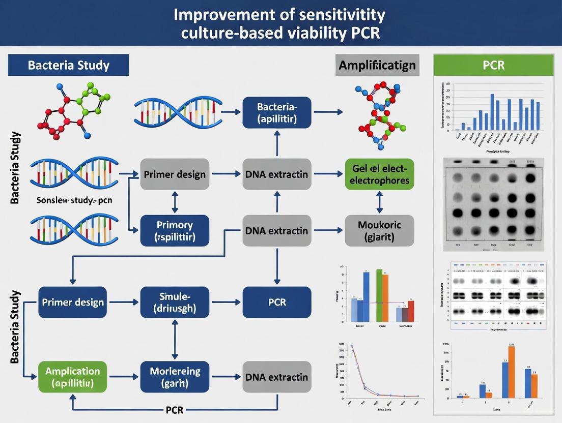
Advanced Strategies to Enhance Culture-Based Viability PCR Sensitivity for Accurate Pathogen Detection
Culture-based viability PCR represents a significant methodological advancement that bridges the gap between traditional culture methods and molecular detection.
Advanced Strategies to Enhance Culture-Based Viability PCR Sensitivity for Accurate Pathogen Detection
Abstract
Culture-based viability PCR represents a significant methodological advancement that bridges the gap between traditional culture methods and molecular detection. This technique combines short-term broth enrichment with quantitative PCR to differentiate viable from non-viable pathogens, addressing critical limitations in environmental monitoring, food safety, and clinical diagnostics. Recent research demonstrates its superior sensitivity compared to conventional culture methods and improved specificity over standard qPCR alone. This article provides a comprehensive framework for researchers and drug development professionals seeking to implement, optimize, and validate this powerful methodology across diverse applications and pathogen types, with particular emphasis on protocol optimization strategies to maximize detection sensitivity while minimizing false-positive results.
The Science Behind Culture-Based Viability PCR: Principles and Limitations of Current Detection Methods
The accurate and timely detection of pathogens is a cornerstone of effective clinical diagnostics and microbiological research. For decades, culture-based methods have been the gold standard, providing vital information on viable organisms and antimicrobial susceptibility. The advent of quantitative Polymerase Chain Reaction (qPCR) introduced a new era of speed and sensitivity. However, both techniques possess significant, inherent limitations that can create critical gaps in diagnosis and research, particularly for slow-growing, fastidious, or non-viable organisms. This technical support center provides troubleshooting guides and FAQs to help researchers navigate these challenges, with a focus on advancing the sensitivity and reliability of culture-based viability PCR.
Comparative Analysis of Detection Methods
The table below summarizes the core limitations of culture and standard qPCR, which contribute to the critical detection gap.
| Method | Key Limitations | Impact on Sensitivity/Specificity |
|---|---|---|
| Culture-Based | Long turnaround time (24 hours to several weeks) [1] [2] | Fails to detect viable but non-culturable (VBNC) bacteria, or those affected by prior antibiotic treatment [3] [4] |
| Low sensitivity for heterogeneous or low-abundance pathogens [3] | High false-negative rates; up to 20% culture-negative in periprosthetic joint infections [3] | |
| Requires viable, cultivable organisms [2] | Cannot detect viruses or pathogens that cannot be grown in vitro [1] | |
| Standard qPCR | Cannot distinguish between live and dead cells [5] | High false-positive risk from detecting non-viable pathogen DNA [5] |
| Susceptible to PCR inhibitors in sample matrices [6] [7] | Can lead to false negatives or inaccurate quantification [6] | |
| Requires pre-defined targets; no isolate for further tests [1] [2] | Provides no antimicrobial susceptibility profile (AST) [2] |
Troubleshooting Culture-Based Methods
Q1: How can I improve the detection of heterogeneously distributed or aggregated bacteria in tissue samples?
The Problem: Bacterial aggregates, often found in biofilm-associated infections like periprosthetic joint infections (PJIs) or osteomyelitis, are not uniformly distributed in tissue. This heterogeneity dramatically reduces the probability of a biopsy sample containing a detectable number of bacteria [3].
Solutions and Strategies:
- Increase Sample Number: Below a critical aggregation size, obtaining five tissue specimens significantly increases the probability of detection [3]. This principle is embedded in some diagnostic guidelines, where 2/5 positive tissue samples is a confirmatory criterion for infection [3].
- Homogenize Tissue Specimens: Mechanical homogenization of the entire tissue specimen increases the surface area for analysis and can help disperse bacterial aggregates, making the bacteria more accessible for detection [3].
- Mathematical Modeling for Sampling Strategy: Research indicates that the probability of detection is influenced by bacterial load, aggregate size, and sample volume. When bacterial aggregation is high, simply increasing the number of specimens provides limited benefit. In such cases, methods like homogenization that address the aggregation itself are more effective [3].
Q2: What can I do when culture results are negative despite strong clinical evidence of infection?
The Problem: Culture-negative infections can occur due to prior antibiotic administration, the presence of viable but non-culturable (VBNC) bacteria, or slow-growing, fastidious organisms [4] [8].
Solutions and Strategies:
- Extend Incubation Time: For certain fastidious pathogens, incubating cultures for at least 14 days is recommended instead of the standard 48-72 hours [3].
- Use a Combination of Culture and Molecular Methods: Integrate culture-independent methods to confirm an infection.
- PCR or mNGS: These methods can detect pathogen DNA even when cultures are negative. A study on kidney transplant patients showed that metagenomic Next-Generation Sequencing (mNGS) had a significantly higher positive detection rate in organ preservation fluids (47.5% vs 24.8%) and wound drainage fluids (27.0% vs 2.1%) compared to conventional culture [8].
- Histopathological Examination: Can confirm the presence of inflammation and microorganisms in tissue [3].
Troubleshooting Standard qPCR
Q3: How can I overcome the inability of standard qPCR to distinguish between live and dead cells?
The Problem: Standard qPCR detects DNA from both viable and non-viable cells, which can lead to false positives and overestimation of the infectious burden, especially after pathogen inactivation [5].
Solution: Implement Culture-Based Viability PCR This novel method combines the sensitivity of qPCR with the ability to confirm viability through a short incubation step [5].
Experimental Protocol for Culture-Based Viability PCR [5]:
- Sample Split: Divide the processed sample homogenate into three paths.
- T0 (Initial Load): Add a portion of the homogenate to growth broth, immediately extract DNA, and perform species-specific qPCR. This gives the baseline signal.
- T1 (Post-Incubation): Add another portion to growth broth and incubate under species-specific conditions (e.g., 24-48 hours at 37°C).
- Growth Negative Control (GNC): Treat a portion with a sterilizing agent (e.g., sodium hypochlorite) to kill any cells, then wash and add to growth broth. This control identifies false-positive signals from persistent environmental DNA.
- Post-Incubation Analysis: After incubation, extract DNA from the T1 and GNC samples and perform qPCR.
- Viability Determination: A sample is considered viable if:
- It is detected at T0, and the Ct value decreases by at least 1.0 at T1 compared to the GNC, indicating growth; or
- It is undetected at T0 but detected at T1 and undetected in the GNC.
This workflow harnesses the sensitivity of qPCR while confirming cell viability through proliferation. One study found that this method detected viable Staphylococcus aureus in 73% of environmental samples that were positive by qPCR, whereas traditional culture detected no viable cells in those same samples [5].
Q4: What are the common causes of high Ct values, low yield, or non-specific amplification in qPCR?
The Problem: qPCR assays can suffer from inefficiencies that lead to unreliable data.
Solutions and Strategies:
- High Ct Values/Low Yield:
- Cause: Often due to poor nucleic acid quality, inefficient cDNA synthesis (for RT-qPCR), suboptimal primer design, or the presence of PCR inhibitors [7].
- Fix: Ensure high-quality RNA/DNA extraction and purification. Optimize cDNA synthesis conditions. Redesign primers using specialized software to ensure appropriate length, GC content, and melting temperature (Tm) [7]. Use automation for precise pipetting to ensure consistent template concentrations [7].
- Non-Specific Amplification:
- General qPCR Failures: The MIQE 2.0 guidelines provide a critical framework for ensuring qPCR rigor and reproducibility. Common failures include using unvalidated reference genes, assuming rather than measuring assay efficiency, and poor sample handling [6]. Adherence to these guidelines is essential for generating trustworthy data.
Frequently Asked Questions (FAQs)
Q5: In which clinical scenarios does PCR clearly outperform culture?
PCR demonstrates superior performance in several key scenarios [4] [10] [2]:
- Chronic Obstructive Pulmonary Disease (COPD): In sputum samples from COPD patients, qPCR showed significantly higher positivity rates for key pathogens like Haemophilus influenzae (up to 47.1% vs 23.6% with culture) and Moraxella catarrhalis (up to 19.0% vs 6.0%) [4].
- Complicated Urinary Tract Infections (cUTIs): A randomized controlled trial found that PCR-guided treatment for cUTI resulted in significantly better clinical outcomes (88.1% vs 78.1%) and a much faster mean turnaround time (49.7 hours vs 104.4 hours) compared to culture-guided treatment [10].
- When Patients Have Received Prior Antibiotics: PCR can detect pathogen DNA even when the bacteria have been rendered non-viable by antimicrobial therapy, a situation where culture often fails [4].
- Detection of Atypical or Non-Culturable Pathogens: mNGS and PCR can identify viruses, fastidious bacteria, and atypical pathogens like Mycobacterium and Clostridium tetani that are missed by routine culture [8].
Q6: When is culture still an indispensable method?
Despite the advantages of molecular methods, culture remains critical when:
- Antimicrobial Susceptibility Testing (AST) is required: Culture is the only method that provides a live isolate for phenotypic AST, which is essential for guiding targeted antibiotic therapy [2].
- Broad-spectrum detection is needed: For unknown pathogens, culture does not require pre-selected targets, unlike specific PCR panels [2].
- Public health surveillance requires isolate biobanking: Isolates are needed for further characterization, typing, and epidemiological studies [2].
Q7: What emerging technologies are helping to bridge the detection gap?
- Metagenomic Next-Generation Sequencing (mNGS): This culture-independent method allows for the comprehensive detection of all nucleic acids in a sample (bacteria, viruses, fungi, parasites). It is highly valuable for diagnosing infections with atypical presentations or from difficult-to-culture pathogens [8].
- Automated Liquid Handling Systems: Automation significantly improves the accuracy and reproducibility of qPCR workflows by reducing pipetting errors and cross-contamination risks, leading to more consistent Ct values [7].
The Scientist's Toolkit: Key Research Reagent Solutions
| Item | Function | Considerations |
|---|---|---|
| Neutralizing Buffer | Inactivates disinfectants in environmental samples (e.g., from swabs), preserving viable cells for culture-based viability PCR [5]. | Essential for accurate environmental monitoring in healthcare settings. |
| Dithiothreitol (DTT) | Digests mucus in sputum samples, improving DNA extraction efficiency and pathogen release for both culture and PCR [4]. | Critical for processing viscous respiratory samples. |
| SYBR Green / TaqMan Probes | Fluorescent chemistries for real-time detection of amplified DNA in qPCR [5] [1]. | SYBR Green is cost-effective; TaqMan probes offer higher specificity through an additional hybridization step. |
| Hot-Start PCR Kits | Reduce non-specific amplification and primer-dimer formation by keeping the polymerase inactive until the first high-temperature denaturation step [9]. | Improves assay specificity and sensitivity, especially for low-abundance targets. |
| Automated Nucleic Acid Extractors | Standardize and streamline the DNA/RNA extraction process, increasing throughput and reducing human error and contamination [7]. | Key for reproducibility in high-volume laboratories. |
In the context of culture-based viability PCR, broth enrichment serves as a critical pre-amplification step that significantly increases the detection of viable pathogens. This process involves incubating a sample in a nutrient-rich, often selective, broth before DNA extraction and PCR analysis. By providing optimal conditions for viable cells to multiply, enrichment directly addresses key limitations of molecular methods, namely the inability to distinguish live from dead cells and the presence of inhibitory substances [5] [11]. This guide explores the fundamental principles behind this enhancement and provides troubleshooting support for researchers aiming to optimize this sensitive detection method.
Frequently Asked Questions (FAQs)
1. Why is broth enrichment necessary before PCR if the PCR itself is highly sensitive? While PCR can detect minute amounts of DNA, this sensitivity is a double-edged sword. It cannot distinguish between DNA from live, dead, or even free-floating DNA fragments. Broth enrichment ensures that a positive signal is generated primarily from viable organisms capable of replication, thereby confirming viability [5] [11]. Furthermore, enrichment dilutes PCR inhibitors that may be co-extracted from the sample matrix, reducing false-negative results [12].
2. How does selective enrichment broth suppress background flora without harming target pathogens? Selective enrichment broths are formulated with specific nutrients that favor the growth of target pathogens while incorporating agents—such as antibiotics, dyes, or salts—that inhibit the growth of common non-target microorganisms. For example, SEL broth is designed to allow the concurrent growth of Salmonella enterica, Escherichia coli O157:H7, and Listeria monocytogenes while suppressing competing flora [13]. The key is selecting inhibitory agents and concentrations that minimally impact the recovery and growth of the target, even if it is sub-lethally injured [14].
3. What are common signs that my enrichment process is failing? Enrichment failure can manifest in several ways during downstream analysis:
- Consistent False-Negatives by PCR: The target pathogen is known to be present (e.g., via an alternative method), but PCR fails to detect it. This can indicate that the broth was unable to resuscitate and propagate injured cells or that inhibitors were not sufficiently diluted [14] [12].
- Poor Growth in Post-Enrichment Culture: Even if PCR is positive, failure to culture the organism from the enriched broth may suggest that the enrichment conditions are too harsh, killing the target or preventing its growth to detectable levels on solid media [5].
- High Background Flora in Cultures: Excessive growth of non-target organisms on non-selective agar plates after enrichment indicates that the selectivity of the broth is insufficient for the sample type being tested [13].
4. My PCR results after enrichment are inconsistent. What could be the cause? Inconsistent results often point to issues with the sample or the enrichment broth itself:
- Injured Target Cells: If a sample contains cells sub-lethally injured by stress (e.g., cold, acid, chlorine), they may not recover uniformly in the enrichment broth, leading to variable detection [14] [12].
- Inhibitors Not Fully Diluted: Some complex sample matrices, like egg yolk or kimchi, contain potent PCR inhibitors that may not be fully neutralized or diluted in a single enrichment step, requiring a two-step enrichment process for reliable detection [12].
- Broth Selectivity: The selective agents in the broth might partially suppress the target pathogen, especially if the inoculum size is small, leading to stochastic growth and detection failures [13].
Troubleshooting Guides
Problem: Low Detection Sensitivity After Enrichment
This issue arises when the number of target organisms after enrichment remains below the detection limit of the PCR assay.
| Possible Cause | Recommendations | Supporting Evidence |
|---|---|---|
| Poor resuscitation of injured cells | Use a non-selective or mildly selective pre-enrichment for 2-6 hours before transferring to a selective enrichment broth. This two-step process aids the repair of sub-lethally injured cells [12]. | A study on detecting Salmonella in liquid egg products found that a one-step enrichment in a selective medium failed to resuscitate heat-injured cells, while a two-step process with BPW succeeded [12]. |
| Insufficient enrichment time | Extend the enrichment time. A 16-24 hour enrichment is standard, but some targets or sample types may require optimization of this duration [15]. | In MRSA detection, a 16-hour enrichment in a selective broth was sufficient to increase the detection of positive samples by 35% compared to direct plating [15]. |
| Inappropriate broth formulation | Select an enrichment broth validated for your specific target and sample matrix. Consider factors like the presence of selective agents and the nutritional composition [14]. | A comparison of broths for detecting E. coli O157:H7 in kimchi found that FDA broth with ACV supplements inhibited chlorine-injured cells, while ISO and KFC broths with novobiocin allowed recovery [14]. |
Problem: PCR Inhibition After Enrichment
In this scenario, the target pathogen grows in the broth, but components from the broth or sample co-extract with DNA and inhibit the PCR reaction.
| Possible Cause | Recommendations | Supporting Evidence |
|---|---|---|
| Carryover of enrichment broth components | Dilute the extracted DNA template or use a DNA purification kit designed to remove common inhibitors like humic acids, salts, and polysaccharides. | PCR troubleshooting guides recommend re-purifying or precipitating DNA to remove residual salts or inhibitors that can affect the DNA polymerase [16]. |
| High concentration of background flora | Optimize the selectivity of the enrichment broth. If using a non-selective broth, switch to a selective one to reduce the overall microbial load and complexity of co-extracted DNA. | SEL broth, a multiplex selective enrichment broth, was shown to inhibit greater numbers of non-target organisms than a universal preenrichment broth (UPB), improving detection efficiency [13]. |
| Sample-specific inhibitors | Incorporate a two-step enrichment protocol. The first step revives the cells, and the second step in a fresh medium further propagates them while drastically diluting the original inhibitors [12]. | For liquid egg yolk, a one-step enrichment in BPW was ineffective at removing PCR inhibitors. A second enrichment in Brain Heart Infusion (BHI) broth overcame this issue and achieved 100% detection accuracy [12]. |
Experimental Protocols
Protocol 1: Culture-Based Viability PCR for Environmental Sampling
This protocol, adapted from recent research, is designed to detect and confirm the viability of specific pathogens from environmental surfaces [5] [17].
Key Research Reagent Solutions
| Reagent | Function in the Experiment |
|---|---|
| Foam sponges premoistened in neutralizing buffer | To collect samples from large or irregular surfaces without degrading the target pathogens. |
| Trypticase Soy Broth (TSB) | A general-purpose, nutritious growth medium used to support the proliferation of a wide range of bacteria, including E. coli and S. aureus. |
| Sodium hypochlorite (Bleach) solution (8.25%) | Serves as a growth negative control by lethally sterilizing an aliquot of the sample, ensuring that subsequent DNA detection is due to viable organisms. |
| Species-Specific Primers & SYBR Green Master Mix | For the quantitative PCR (qPCR) step. SYBR Green intercalates into double-stranded DNA, allowing for quantification of the amplified product in real-time [11]. |
| Stomacher apparatus | Provides a gentle yet effective mechanical method to release microorganisms from the sample sponge into a homogeneous liquid suspension (homogenate). |
Methodology:
- Sample Collection & Processing: Collect samples from the environment (e.g., hospital bed footboards) using a premoistened neutralizing sponge. Process the sponge using a stomacher to create a 5 mL homogenate [5].
- Sample Splitting: Aseptically split the homogenate into three paths:
- T0 (Immediate Analysis): Combine 500 µL of homogenate with 4.5 mL of TSB. Immediately perform DNA extraction and qPCR.
- T1 (Post-Enrichment): Combine 500 µL of homogenate with 4.5 mL of TSB. Incubate under species-specific conditions (e.g., 24h at 37°C for E. coli).
- GNC (Growth Negative Control): Treat 500 µL of homogenate with 4.5 mL of sodium hypochlorite to kill all cells. After neutralization and washing, resuspend in TSB and incubate alongside T1 [5].
- Post-Enrichment Analysis: After incubation, extract DNA from the T1 and GNC samples and perform qPCR.
- Viability Assessment: A sample is considered viable if:
Protocol 2: Two-Step Enrichment for Detection in Inhibitory Matrices
This protocol is optimized for challenging samples like liquid egg products, where inherent components can inhibit PCR [12].
Methodology:
- First Enrichment (Resuscitation): Inoculate 25 g of sample into 225 mL of Buffered Peptone Water (BPW). Incubate at 37°C for 3-5 hours. This lower-stress environment helps resuscitate injured Salmonella cells without the pressure of selective agents.
- Second Enrichment (Amplification & Dilution): Transfer a small aliquot (e.g., 0.1-1 mL) of the pre-enriched BPW culture into 10 mL of Brain Heart Infusion (BHI) broth. Incubate at 37°C for an additional 9-11 hours (total enrichment time of ~14 hours). This step further multiplies the target pathogen and dilutes out PCR inhibitors present in the original sample matrix.
- DNA Extraction and Detection: Extract DNA from the second enrichment broth and proceed with your chosen PCR detection method (e.g., the BAX system or other qPCR assays) [12].
Workflow and Data Visualization
Broth Enrichment Workflow for Viability PCR
The following diagram illustrates the logical workflow of a culture-based viability PCR experiment, highlighting the role of broth enrichment in confirming the presence of live pathogens.
Quantitative Impact of Broth Enrichment
The table below summarizes quantitative data from key studies, demonstrating how broth enrichment enhances detection sensitivity compared to direct molecular or culture methods.
Table: Impact of Broth Enrichment on Pathogen Detection Sensitivity
| Pathogen | Sample Type | Detection Method | Key Finding: Enrichment vs. Direct Method | Reference |
|---|---|---|---|---|
| S. aureus | Clinical samples | Broth enrichment + qPCR vs. selective plating | Increased number of MRSA-positive samples by 35% (from 49 to 66) | [15] |
| S. aureus | Hospital surfaces | Culture-based viability PCR vs. traditional culture | Detected viable cells in 73% of qPCR-positive samples vs. 0% by direct culture* | [5] |
| E. coli O157:H7 | Kimchi | Enrichment in ISO broth vs. FDA broth | Supported recovery of chlorine-injured cells; FDA broth showed inhibition or no growth | [14] |
| Salmonella Enteritidis | Liquid egg yolk | Two-step enrichment + BAX vs. one-step | Achieved 100% diagnostic accuracy; one-step enrichment yielded unstable results | [12] |
Note: In the same study, 19% of S. aureus samples were culturable *after broth enrichment (T1), highlighting how enrichment enables later culture detection [5].
FAQs: Addressing Common Questions on Culture-Based Viability PCR
What is culture-based viability PCR and how does it improve upon traditional methods? Culture-based viability PCR is a two-step method that combines a short incubation in a growth broth with subsequent quantitative PCR (qPCR) analysis. It first enriches viable (living) microorganisms in a sample by incubating them in a culture medium. DNA is extracted and analyzed via qPCR both before and after this incubation. An increase in DNA after incubation indicates the presence of viable organisms that were able to replicate. This method outperforms traditional culture plates, which can be slow and have a high detection threshold, and standard qPCR, which cannot distinguish between live and dead cells because it detects all DNA present [5] [17] [18].
What are the primary causes of no amplification or low yield in viability PCR, and how can I resolve them? A lack of PCR product can stem from several issues related to the template DNA, reaction components, or cycling conditions [19] [16].
- Template DNA: Confirm the DNA template's quantity is sufficient and its quality is high, without contaminants like phenol or salts that can inhibit the polymerase enzyme. Solutions include repurifying the DNA, using polymerases known for high tolerance to inhibitors, and ensuring no reaction components were omitted [19] [16] [20].
- Reaction Components: Verify that primers are designed correctly and are at an adequate concentration. Optimize the concentration of magnesium ions (Mg2+), which is critical for polymerase activity. Using a hot-start DNA polymerase can prevent non-specific amplification and improve yield [19] [16].
- Thermal Cycling Conditions: Ensure the annealing temperature is optimal for your primer set (typically 3–5°C below the primer Tm). Check that the number of cycles and the extension time are sufficient for the target amplicon [16] [20].
Why am I seeing non-specific PCR products or primer-dimer formations? Non-specific amplification occurs when primers bind to unintended regions of the DNA template, often due to low reaction stringency [19] [16].
- Increase Annealing Temperature: Raise the temperature stepwise by 1–2°C increments to enhance specificity [16].
- Optimize Reagent Concentrations: High concentrations of primers, DNA template, or Mg2+ can promote non-specific binding. Use optimized concentrations, and consider using hot-start polymerases that are inactive until the high-temperature denaturation step [19] [16].
- Review Primer Design: Ensure primers are specific to the target and do not have complementary sequences to each other, which can lead to primer-dimer formation. Software tools can help design specific primers [19] [16].
My gel shows smeared bands. What does this indicate and how can I fix it? Smeared bands can result from degraded DNA template, non-specific products, or contaminants [19].
- Check DNA Integrity: Evaluate the quality of your input DNA by gel electrophoresis. Degraded DNA will appear as a smear and should be re-isolated [16].
- Optimize PCR Conditions: Increase the annealing temperature to reduce non-specific binding. Ensure the extension time is appropriate for the length of your target amplicon [19] [16].
- Address Contamination: Smearing can be caused by accumulated amplifiable DNA contaminants in the lab environment. Using a new set of primers with different sequences can often resolve this issue. Implementing strict physical separation between pre- and post-PCR workspaces is a key preventive measure [19].
Troubleshooting Guides: Common Problems and Solutions in Viability PCR
Troubleshooting Low or No Amplification
| Possible Cause | Recommended Solution |
|---|---|
| Poor DNA template quality or integrity | Re-purify template DNA to remove inhibitors (phenol, salts). Minimize shearing during isolation. Assess DNA integrity via gel electrophoresis [19] [16]. |
| Insufficient DNA template quantity | Increase the amount of input DNA. If the target copy number is very low, increase the number of PCR cycles (up to 40) [16]. |
| Incorrect primer concentration or design | Optimize primer concentration (typical range 0.1–1 µM). Redesign primers to ensure specificity and correct binding to the target [16] [20]. |
| Suboptimal Mg2+ concentration | Perform a series of test reactions with varying Mg2+ concentrations to determine the optimal level for your specific reaction [19] [16]. |
| Suboptimal annealing temperature | Use a gradient thermal cycler to determine the ideal annealing temperature. Increase the temperature if non-specific products are seen, or decrease it if yield is low [16] [20]. |
| Insufficient enzyme activity | Confirm the DNA polymerase is within its expiration date and has not been degraded by multiple freeze-thaw cycles. Increase the amount of polymerase if inhibitors are suspected [16] [20]. |
Troubleshooting Non-Specific Products and Primer-Dimers
| Possible Cause | Recommended Solution |
|---|---|
| Annealing temperature is too low | Increase the annealing temperature incrementally by 1–2°C. The optimal temperature is usually 3–5°C below the lowest primer Tm [16]. |
| Excess primers, template, or enzyme | Optimize concentrations of all reaction components. High primer concentrations specifically promote primer-dimer formation [19] [16]. |
| Poor primer design | Avoid primers with complementary sequences, especially at the 3' ends. Use software tools to design specific primers and check for secondary structures [19] [16]. |
| Non-hot-start DNA polymerase | Switch to a hot-start DNA polymerase to prevent enzyme activity during reaction setup at lower temperatures, which can cause primer-dimer and non-specific synthesis [19] [16]. |
| Excessive cycle number | Reduce the number of PCR cycles to minimize the accumulation of non-specific products in later cycles [16]. |
Experimental Protocols: Key Methodologies from Cited Studies
Protocol: Culture-Based Viability PCR for Healthcare Pathogens
This detailed protocol is adapted from a study detecting viable pathogens on hospital surfaces [5] [17].
1. Sample Collection and Processing:
- Collection: Sample environmental surfaces (e.g., bed footboards) using foam sponges pre-moistened with a neutralizing buffer.
- Homogenization: Process samples using a stomacher method to create a 5 mL homogenate.
2. Sample Split and Pre-Treatment: The sponge homogenate is divided into three separate paths:
- T0 Sample: 500 µL of homogenate is added to 4.5 mL of Trypticase Soy Broth (TSB). DNA is extracted immediately from 500 µL of this mixture for a baseline (T0) qPCR analysis.
- T1 Sample: 500 µL of homogenate is added to 4.5 mL of TSB for incubation.
- Growth Negative Control (GNC): 500 µL of homogenate is added to 4.5 mL of 8.25% sodium hypochlorite (bleach) to kill all cells. It is left at room temperature for 10 minutes, centrifuged, washed twice with PBS, and then resuspended in 5 mL of TSB.
3. Incubation:
- Incubate T1 and GNC samples under species-specific conditions.
- For E. coli and S. aureus: 24 hours at 37°C, aerobically.
- For C. difficile: 48 hours at 37°C, anaerobically.
4. Post-Incubation Analysis:
- After incubation, transfer 500 µL from both T1 and GNC samples for DNA extraction and qPCR analysis (T1 qPCR).
- In parallel, culture 200 µL from all paths (T0, T1, GNC) on TSA agar to compare with culture-based results.
5. Criteria for Viability: A sample is considered viable for a target species if it meets any of the following conditions:
- It is detected at T0, and the qPCR cycle threshold (CT) value decreases by at least 1.0 at T1 compared to the GNC.
- It is undetected at T0 but is detected at T1, and is undetected in the GNC.
- It grows on standard culture agar [5].
Quantitative Data from Healthcare Environment Study
The following table summarizes key quantitative results from the prospective analysis of patient room samples, demonstrating the performance of culture-based viability PCR [5] [17].
| Pathogen | Samples with Detectable DNA (T0 or T1) | Samples with Viable Cells (via Viability PCR) | Samples with Viable Cells (via Culture) |
|---|---|---|---|
| E. coli (N=26) | 24 (92%) | 3 (13%) | 0 (0%) |
| S. aureus (N=26) | 11 (42%) | 8 (73%) | 5 (19%) |
| C. difficile (N=26) | 2 (8%) | 0 (0%) | 0 (0%) |
Research Reagent Solutions: Essential Materials for Viability PCR
The following table lists key reagents and their critical functions in setting up and performing culture-based viability PCR experiments.
| Reagent/Chemical | Function in the Experiment |
|---|---|
| Trypticase Soy Broth (TSB) | A general-purpose liquid growth medium used to enrich viable bacterial cells from the sample during incubation [5]. |
| Species-Specific Broth | In some protocols, specialized broths are used to optimize the growth of particular target organisms, such as C. difficile [17]. |
| Neutralizing Buffer | Used to pre-moisten sampling sponges; neutralizes residual disinfectants on surfaces to prevent them from killing microbes during sample collection [5]. |
| Propidium Monoazide (PMA) or PMAxx | Viability dyes used in some v-PCR approaches. They penetrate only dead cells with compromised membranes and bind DNA, preventing its amplification in subsequent PCR, thus ensuring only DNA from live cells is detected [18] [21]. |
| SYBR Green Master Mix | A fluorescent dye used in qPCR that intercalates into double-stranded DNA, allowing for the quantification of amplified DNA in real-time [5]. |
| Hot-Start DNA Polymerase | A modified enzyme that is inactive at room temperature, preventing non-specific amplification and primer-dimer formation during reaction setup, which improves specificity and yield [19] [16]. |
| Sodium Hypochlorite | Used in the growth negative control (GNC) to kill microorganisms, providing a baseline to confirm that DNA detection after incubation is due to growth and not persistent DNA from dead cells [5]. |
Workflow and Troubleshooting Diagrams
Culture-Based Viability PCR Workflow
Systematic Troubleshooting for Low PCR Yield
The accurate detection of viable microbial cells is a cornerstone of public health, food safety, and pharmaceutical development. Traditional culture methods, while reliable, are often time-consuming and fail to detect viable but non-culturable (VBNC) cells. Polymerase chain reaction (PCR) offers speed and sensitivity but cannot differentiate between live cells, dead cells, and free DNA, leading to overestimation of viable populations. Viability PCR (vPCR) and culture-based viability PCR have emerged as powerful techniques that bridge this gap, combining molecular speed with viability assessment. This technical support guide addresses the key challenges and solutions in establishing clear, reliable criteria for live cell identification using these advanced PCR methodologies, providing essential troubleshooting and protocols for researchers and drug development professionals.
Core Concepts: vPCR and Culture-Based Viability PCR
Viability PCR (vPCR)
Principle: vPCR uses photoactive DNA-intercalating dyes, such as propidium monoazide (PMA), to selectively suppress DNA amplification from dead cells [22] [23]. PMA enters only cells with compromised membranes (considered dead), intercalates into DNA, and forms covalent bonds upon light exposure, rendering the DNA unamplifiable. DNA from viable cells with intact membranes remains accessible for PCR amplification [22].
Key Limitation: The method relies solely on membrane integrity. It cannot detect viability after treatments that do not damage membranes (e.g., UV disinfection) but can detect VBNC cells [22].
Culture-Based Viability PCR
Principle: This method combines the sensitivity of species-specific quantitative PCR (qPCR) with the viability assessment of culture enrichment [5]. A sample is tested via qPCR at time zero (T0) and again after a period of incubation in growth media (T1). A significant decrease in the quantification cycle (CT) value at T1 indicates the presence of viable cells that have proliferated during incubation [5].
Advantage: It provides a functional assessment of viability based on the ability to proliferate, overcoming limitations of membrane-integrity assays.
Frequently Asked Questions (FAQs)
1. What is the primary cause of false-positive results in vPCR, and how can it be minimized? False positives are primarily caused by incomplete suppression of DNA signals from a high background of dead cells [22]. Optimization strategies include using a double PMA treatment with a low dye concentration and ensuring proper light exposure by changing reaction tubes between the final dark incubation and light activation step to prevent shadowing effects [22] [24].
2. My vPCR shows no amplification. What are the first steps in troubleshooting? Begin by verifying your template DNA quality and concentration [19] [25]. Then, confirm that all PCR reagents were added correctly and have not degraded [25] [26]. Ensure that the photoactivation step for PMA was performed correctly with a functioning light source, as improperly inactivated PMA can inhibit PCR [22].
3. How does culture-based viability PCR confirm viability, and what defines a positive result? Viability is confirmed by demonstrating growth through a significant increase in target DNA. A sample is considered viable if [5]:
- It is detected at T0, and the CT value decreases by at least 1.0 at T1 compared to a growth-negative control.
- It is undetected at T0 but detected at T1, and the growth-negative control is negative.
4. I am getting non-specific bands or primer-dimer formations in my viability PCR. How can I resolve this? This is often due to suboptimal primer annealing [19] [26]. Solutions include:
- Increasing the annealing temperature in increments of 1-2°C.
- Using a hot-start polymerase to prevent non-specific amplification during reaction setup [19] [26].
- Redesigning primers to avoid self-complementarity and secondary structures [19].
- Lowering primer concentrations [26].
Troubleshooting Guides
Common vPCR and PCR Issues
| Observation | Possible Cause | Recommended Solution |
|---|---|---|
| No or Low Amplification | Inhibitors present in sample (e.g., from food matrix) [19] | Purify template DNA; use additives like BSA [19]. |
| Suboptimal PCR conditions [25] [26] | Optimize Mg2+ concentration; use a gradient cycler to optimize annealing temperature [26]. | |
| Degraded or low-concentration template [25] | Re-prepare template DNA; increase number of PCR cycles [25]. | |
| Non-Specific Products / Primer-Dimer | Annealing temperature too low [19] [26] | Increase annealing temperature incrementally [26]. |
| Primer concentration too high [19] | Lower primer concentration within 0.05–1 µM range [26]. | |
| Non-specific priming during setup [26] | Use a hot-start DNA polymerase [19] [26]. | |
| Incomplete Suppression of Dead Cell Signal | High dead cell background [22] | Implement a double PMA treatment protocol [22] [24]. |
| Improper PMA photoactivation [22] | Change tube type between incubation and light exposure; ensure even light distribution [22]. | |
| Smeared Bands on Gel | Non-specific products from contaminated reagents [19] | Use fresh reagents; designate pre- and post-PCR work areas; consider new primer sequences [19]. |
| Excessive cycling or extension times [19] | Reduce cycle number or extension time [19]. |
Interpreting Signal Dynamics for Viability Criteria
| Method | Signal Indicative of Viable Cells | Signal Indicative of Non-Viable Cells |
|---|---|---|
| Viability PCR (vPCR) | Strong PCR signal in PMA-treated sample (DNA from membrane-intact cells is amplified). | PCR signal is suppressed in PMA-treated sample (DNA from membrane-compromised cells is blocked) [22]. |
| Culture-Based Viability PCR | CT value at T1 (after incubation) is significantly lower (e.g., decrease ≥1.0) than CT at T0 [5]. | CT value shows no significant change or increases between T0 and T1 [5]. |
| Standard qPCR | Cannot distinguish viability. Signal indicates presence of target DNA, regardless of its source (live, dead, or free DNA) [22] [5]. |
Detailed Experimental Protocols
Optimized vPCR Protocol forStaphylococcus aureusin Food Matrices
This protocol, adapted from Dinh Thanh et al. (2025), successfully suppressed DNA from up to 5.0 × 10^7 dead cells in a 200 µl reaction [22] [24].
Key Reagents and Materials:
- PMA dye (e.g., PMAxx)
- Staphylococcus aureus strains
- Food matrices (e.g., ground spices, milk powder, meat)
- Buffered Peptone Water
- DNA extraction kit
- PCR reagents
- Light-emitting device (PMA-Lite LED or equivalent)
Procedure:
- Sample Preparation: Artificially contaminate food samples with a low number of viable cells and a high number of heat-inactivated cells [24].
- PMA Treatment:
- Add PMA to the sample to a final concentration of 10–50 µM.
- Incubate in the dark for 10–30 minutes with occasional mixing.
- Perform a tube change to a transparent, flat-bottom tube.
- Expose the sample to bright light for 15–30 minutes for photoactivation.
- Optional: Repeat the PMA treatment cycle (double PMA) for challenging matrices [22].
- DNA Extraction: Extract genomic DNA from the PMA-treated sample using a commercial kit.
- PCR Amplification: Perform qPCR or standard PCR with species-specific primers. The absence of amplification from the heat-killed control indicates successful dead-cell suppression [22].
Culture-Based Viability PCR Protocol for Healthcare Pathogens
This protocol, used for detecting viable E. coli, S. aureus, and C. difficile on hospital surfaces, outperformed traditional culture methods [5].
Procedure:
- Sample Collection & Homogenization: Sample surfaces with sponges pre-moistened in neutralizing buffer. Process via stomacher to create a homogenate [5].
- Time-Zero (T0) Analysis:
- Add 500 µL of homogenate to 4.5 mL of Trypticase Soy Broth (TSB).
- From this mixture, take a 500 µL aliquot for immediate DNA extraction and qPCR analysis (T0 CT value).
- Incubation (T1):
- Add another 500 µL of the original homogenate to 4.5 mL of TSB.
- Incubate under species-specific conditions (e.g., 24-48 hours at 37°C).
- Growth-Negative Control (GNC):
- Treat a 500 µL homogenate aliquot with sodium hypochlorite to kill cells, wash, and then add to TSB. Incubate alongside T1 samples.
- Post-Incubation Analysis:
- After incubation, take 500 µL from both T1 and GNC samples for DNA extraction and qPCR (T1 CT value).
- Viability Assessment:
- Apply the criteria outlined in the FAQ section to interpret results (e.g., a CT decrease of ≥1.0 in T1 compared to GNC confirms viability) [5].
Workflow Diagrams
vPCR Workflow for Viability Assessment
Culture-Based Viability PCR Logic
The Scientist's Toolkit: Key Research Reagent Solutions
| Reagent / Material | Function in Viability Assessment | Key Considerations |
|---|---|---|
| Propidium Monoazide (PMA) | DNA-intercalating dye that selectively enters dead cells with compromised membranes; upon photoactivation, it covalently binds DNA and inhibits PCR amplification [22] [23]. | Concentration and incubation conditions are critical; requires optimization for different sample matrices [22]. |
| Growth Media (e.g., TSB, BVFH) | Supports the proliferation of viable cells during incubation in culture-based viability PCR, enabling detection of growth through increased DNA signal [5] [27]. | Must be tailored to the specific microorganism (e.g., BVFH for Francisella tularensis) [27]. |
| Hot-Start DNA Polymerase | Reduces non-specific amplification and primer-dimer formation by remaining inactive until high temperatures are reached during PCR initialization [19] [26]. | Essential for improving the specificity and sensitivity of the final PCR readout. |
| PCR Additives (e.g., BSA, DMSO) | Helps overcome PCR inhibition caused by compounds co-extracted from complex sample matrices (e.g., spices, humic acid) [19] [27]. | BSA can bind inhibitors; DMSO improves amplification efficiency for GC-rich templates [19] [25]. |
Implementing Culture-Based Viability PCR: Step-by-Step Protocols for Diverse Pathogens and Matrices
Culture-based viability PCR (vPCR) represents a significant advancement in pathogen detection for clinical and food safety diagnostics. This method bridges the gap between traditional culture-based techniques, which confirm viability but are slow, and quantitative PCR (qPCR), which is rapid but cannot distinguish between live and dead cells [5]. The core principle involves comparing qPCR results before and after incubation in growth media to determine whether detected organisms can proliferate, thereby combining molecular sensitivity with viability assessment [5]. The sensitivity and accuracy of this technique are profoundly influenced by sample processing and enrichment strategies, particularly the selection of enrichment broth and optimization of incubation conditions. This guide provides targeted troubleshooting and FAQs to help researchers optimize these critical parameters within their viability PCR workflows.
Frequently Asked Questions (FAQs)
1. What is the fundamental principle behind culture-based viability PCR? Culture-based viability PCR detects viable pathogens by harnessing the sensitivity of PCR while incorporating a viability assessment. The process involves running a species-specific qPCR assay on a sample both immediately (T0) and after a period of incubation (T1) in a nutrient broth. A sample is considered viable if the cycle threshold (CT) value decreases significantly after incubation, indicating microbial growth and DNA amplification, or if the target is undetected at T0 but detected at T1 [5]. This approach outperforms traditional culture methods in detection sensitivity while providing the viability information that standard qPCR lacks [5].
2. Why is broth enrichment critical before PCR in this assay? Broth enrichment is a crucial step because it allows viable but potentially stressed or low-abundance cells to proliferate. This incubation period amplifies the target DNA to detectable levels, dramatically increasing the assay's sensitivity. Research shows that broth enrichment can enable the detection of pathogens via culture methods at T1 that were completely undetectable by direct culture at T0 [5]. Furthermore, for the viability PCR result to be valid, a growth negative control (GNC), often treated with a sterilizing agent like sodium hypochlorite, must be included to confirm that signals are due to viable growth and not persistent DNA [5].
3. How do sample matrices interfere with viability PCR results? The sample matrix can significantly impact the performance of viability PCR assays. Complex matrices, especially those with high organic content or particulate matter, can interfere with the assay in two primary ways: they can shield dead cells from viability dyes, leading to false-positive signals, and they can inhibit the PCR reaction itself, causing false negatives [24] [28]. For instance, studies on spiked stool samples have shown that samples with more solid matter (semi-solid stools) removed less dead-cell DNA and detected less live-cell DNA compared to samples with less solid matter (liquid stools) [28]. The consistency and concentration of the sample suspension are therefore critical factors to optimize.
Troubleshooting Guide
Table 1: Common Issues and Solutions in Culture-Based Viability PCR
| Problem | Potential Causes | Recommended Solutions |
|---|---|---|
| False Positive Signals | Incomplete suppression of DNA from dead cells; High load of non-viable organisms [24]. | Optimize viability dye (e.g., PMA/PMAxx) concentration and incubation; Implement a double dye treatment protocol; Include a rigorous growth negative control (GNC) [5] [24]. |
| Low Sensitivity / False Negatives | PCR inhibition from sample matrix; Suboptimal broth composition or incubation conditions; Insufficient incubation time [24] [28]. | Dilute sample to reduce inhibitors; Use DNA polymerases with high processivity and inhibitor tolerance; Validate and optimize broth type, temperature, and atmosphere for the target organism [16] [24]. |
| Poor Bacterial Growth in Broth | Incorrect incubation temperature or atmosphere; Inappropriate broth selection; Overly aggressive sample processing damaging cells. | Confirm species-specific incubation conditions (e.g., 37°C, aerobic/anaerobic); Use general-purpose enrichment broths like Trypticase Soy Broth (TSB); Ensure gentle sample processing methods [5] [29]. |
| Inconsistent qPCR Results | Suboptimal DNA extraction; PCR inhibitors carried over from broth or sample; Inconsistent thermal cycling [16]. | Re-purify DNA to remove inhibitors like salts or organics; Optimize denaturation time/temperature for complex templates; Validate primer specificity and annealing temperature [16] [30]. |
Optimized Experimental Protocols
Protocol 1: Culture-Based Viability PCR for Environmental Samples
This protocol, adapted from a study on healthcare surface monitoring, is designed for detecting viable bacterial pathogens like E. coli, S. aureus, and C. difficile [5].
- Sample Collection: Collect surface samples using foam sponges pre-moistened with a neutralizing buffer.
- Sample Processing: Process samples via a stomacher method to create a homogenate.
- Sample Splitting: Divide the homogenate into three paths:
- T0: Combine with Trypticase Soy Broth (TSB), then immediately perform DNA extraction and qPCR.
- T1: Combine with TSB and incubate under species-specific conditions (e.g., 24-48 hours at 37°C, aerobic or anaerobic).
- GNC (Growth Negative Control): Treat with sodium hypochlorite to kill cells, then wash and resuspend in TSB before incubation.
- Post-Incubation Analysis: After incubation, perform DNA extraction and qPCR on T1 and GNC samples.
- Viability Assessment: A sample is viable if: a) detected at T0 and the CT value decreases by ≥1.0 at T1 compared to GNC, b) undetected at T0 but detected at T1 and undetected in GNC, or c) growth is confirmed by parallel culturing on agar plates [5].
Protocol 2: Enhanced Viability PCR with PMAxx for Complex Matrices
This optimized protocol for S. aureus detection in food focuses on eliminating false positives from dead cells and can be adapted for other complex samples [24].
- Sample Preparation: Create a homogenate of the food or clinical sample.
- First PMAxx Treatment: Add PMAxx dye to the sample and incubate in the dark to allow dye penetration into dead cells.
- Tube Change: Transfer the sample to a new, clear reaction tube. This critical step prevents dead cell debris from adhering to tube walls and avoiding light exposure.
- Photoactivation: Expose the sample to bright visible light to cross-link the dye with DNA from dead cells, preventing its amplification.
- Second PMAxx Treatment: Perform a second, low-concentration treatment of PMAxx with another dark incubation and photoactivation cycle to ensure complete dead-cell DNA suppression.
- DNA Extraction & qPCR: Proceed with DNA extraction and quantitative PCR. The optimized protocol can completely suppress DNA signals from up to 5.0 × 10^7 dead cells in a pure culture [24].
Workflow Visualization
Research Reagent Solutions
Table 2: Essential Reagents and Materials for Viability PCR
| Reagent/Material | Function | Application Notes |
|---|---|---|
| Trypticase Soy Broth (TSB) | General-purpose liquid enrichment medium for bacterial growth. | Used for incubating samples between T0 and T1 qPCR assays to support proliferation of viable cells [5]. |
| Viability Dyes (PMA, PMAxx) | Photoactive DNA-intercalating dye that penetrates dead cells with compromised membranes. | Covalently binds to DNA upon light exposure, suppressing its amplification in PCR. Critical for differentiating viable and dead cells [24] [28]. |
| Neutralizing Buffer | Used in sampling to neutralize disinfectants or other inhibitory agents from surfaces. | Prevents carry-over of antimicrobial agents that could inhibit growth during broth enrichment [5]. |
| Hot-Start DNA Polymerase | Enzyme for PCR that remains inactive until a high-temperature activation step. | Increases specificity by preventing non-specific amplification and primer-dimer formation at low temperatures [16]. |
| Species-Specific Primers | Short DNA sequences designed to bind to and amplify unique target pathogen DNA. | Essential for the specificity of the qPCR assay. Must be validated for the target organism [5]. |
| SYBR Green Master Mix | A fluorescent dye that binds to double-stranded DNA for real-time PCR detection. | Allows for real-time monitoring of DNA amplification during qPCR [5]. |
Culture-based viability PCR (vPCR) is an advanced method that combines the sensitivity of molecular detection with the ability to confirm cell viability, which is crucial for accurate risk assessment in pharmaceutical, diagnostic, and food safety industries. This technique involves taking samples at critical time points—before (T0) and after (T1) a incubation period in growth media—and using species-specific qPCR to determine if detected organisms can proliferate [5]. A fundamental challenge, however, is that the diverse cellular structures of different bacteria require tailored experimental approaches to achieve accurate and sensitive results.
This guide provides targeted troubleshooting and protocols to address the specific needs of Gram-positive bacteria, Gram-negative bacteria, and bacterial spore-formers when using culture-based vPCR.
Frequently Asked Questions (FAQs) and Troubleshooting
Q1: Why do my viability PCR results show high background signal from dead cells?
This is a common issue where DNA from dead cells with compromised membranes is amplified, leading to overestimation of viable counts.
- Primary Cause: The viability dye (e.g., PMA) failed to penetrate and sufficiently suppress DNA amplification from dead cells.
- Solutions:
- For Gram-positive bacteria: Increase the dye concentration or incubation time. Their thick peptidoglycan layer is a major barrier to dye entry [24].
- Implement a double dye treatment: A second round of dye application and photoactivation can significantly improve suppression of dead cell signals, especially in samples with a high proportion of dead cells [24].
- Optimize the photoactivation step: Ensure the light source is bright enough and the exposure time is sufficient to fully activate the dye. Changing reaction tubes between the final dark incubation and light exposure step can minimize light scattering and improve efficacy [24].
- Review sample matrix effects: Components in food, environmental, or pharmaceutical samples can inhibit dye function. You may need to dilute the sample or add a wash step [24].
Q2: My protocol works for E. coli but fails for S. aureus. What should I adapt?
This indicates that species-specific adaptations are required, particularly due to differences in cell wall structure.
- Primary Cause: Gram-positive bacteria like S. aureus have a thick, multi-layered peptidoglycan cell wall that hinders the penetration of viability dyes like PMA, unlike the outer membrane of Gram-negative bacteria [24].
- Solutions:
- Adjust viability dye protocol: For Gram-positives, consider a higher PMA concentration or longer incubation. For Gram-negatives, a standard concentration is often sufficient, but excess dye can reduce signal from live cells.
- Extend incubation times: The growth (T1) incubation period may need to be longer for slower-growing or more fastidious species. For example, while E. coli and S. aureus might be incubated for 24 hours, spore-formers like C. difficile require 48 hours of anaerobic incubation [5].
- Verify growth media and conditions: Ensure the culture broth (e.g., Trypticase Soy Broth) and atmosphere (aerobic vs. anaerobic) are optimal for your target organism [5] [31].
Q3: How can I detect viable spore-formers, which are often dormant?
Detecting bacterial spores requires a protocol that triggers germination, as the spore coat is highly impermeable.
- Primary Cause: Spores are dormant structures with extreme resistance. Standard viability dyes and lysis methods cannot penetrate the protective spore coat, and the DNA inside is not amplified until the spore germinates into a vegetative cell [5].
- Solutions:
- Use an extended enrichment step: The T1 incubation in growth media must be long enough to allow spores to germinate and outgrow. For C. difficile, this requires 48 hours of anaerobic incubation [5].
- Apply viability dyes after enrichment: The dye should be added after the T1 incubation, as it will only penetrate the membranes of spores that have germinated or dead vegetative cells, preventing false positives from free DNA or dormant spores.
Experimental Protocols for Different Bacterial Types
The following workflows and reagent kits are designed to address the structural and physiological differences between bacterial types.
Workflow for Gram-Negative Bacteria (e.g., E. coli)
Gram-negative bacteria have an outer membrane that is more permeable to small molecules, making them relatively straightforward for vPCR.
Workflow for Gram-Positive Bacteria (e.g., S. aureus)
The thick peptidoglycan layer of Gram-positive bacteria necessitates a more aggressive dye treatment step.
Workflow for Spore-Formers (e.g., C. difficile)
Spore-formers require an extended incubation period under specific conditions to trigger germination.
Research Reagent Solutions
The table below lists essential reagents and their optimized use for different bacterial types in culture-based vPCR.
| Reagent/Kit | Function in Protocol | Species-Specific Notes |
|---|---|---|
| Propidium Monoazide (PMA) | Viability dye; penetrates dead cells with compromised membranes, binding DNA upon light exposure to prevent PCR amplification [24]. | Gram-negative: Standard concentration (e.g., 25 µM). Gram-positive: Use higher concentration (e.g., 50 µM) or extend incubation time to overcome peptidoglycan barrier [24]. |
| Growth Broth (e.g., TSB) | Enrichment medium to support the proliferation of viable cells during the T1 incubation step [5]. | General: Trypticase Soy Broth (TSB) is versatile. Fastidious species: May require enriched media with blood, serum, or specific supplements [31]. |
| DNA Polymerase (e.g., Q5, OneTaq) | Enzyme for PCR amplification during qPCR steps. Critical for sensitivity and specificity [32]. | GC-rich genomes: Use polymerases like Q5 with GC enhancers. Complex samples: Polymerases with high processivity tolerate inhibitors from sample matrices [16]. |
| Species-Specific Primers | Oligonucleotides that bind to unique genetic sequences of the target bacterium for specific detection via qPCR [5]. | All species: Must be designed and validated for the specific target organism. Verify sequence specificity and optimize concentration (typically 0.1–1 µM) [32] [16]. |
Data Presentation: Comparative Performance
The following data, adapted from a clinical study, demonstrates how culture-based vPCR outperforms traditional culture methods and standard qPCR in detecting viable pathogens.
Table 1: Comparison of Pathogen Detection Methods in Clinical Environmental Samples [5]
| Method | Target Species | Detected at T0/T1 | Determined Viable | Statistically Significant vs. Culture? |
|---|---|---|---|---|
| Culture-based vPCR | E. coli (N=26) | 24 (92%) | 3 (13%) | Yes (P < 0.01) |
| Traditional Culture | E. coli (N=26) | 0 (0%) | 0 (0%) | (Baseline) |
| Culture-based vPCR | S. aureus (N=26) | 11 (42%) | 8 (73%) | Yes (P < 0.01) |
| Traditional Culture | S. aureus (N=26) | 0 (0%) | 5 (19%)* | (Baseline) |
| Culture-based vPCR | C. difficile (N=26) | 2 (8%) | 0 (0%) | Not Applicable |
| Traditional Culture | C. difficile (N=26) | 0 (0%) | 0 (0%) | (Baseline) |
Note: *These 5 samples were only detected by culture after broth enrichment (T1), and all were also correctly identified as viable by the vPCR method [5].
For researchers in drug development and microbiology, accurately distinguishing live pathogens from dead ones is critical for assessing environmental contamination and therapeutic efficacy. While quantitative PCR (qPCR) offers a fast and sensitive method for detecting pathogens, a significant limitation is its inability to differentiate between viable and non-viable cells, as it amplifies DNA from both sources. This can lead to an overestimation of live bacteria and potentially false-positive results [5] [33]. Culture-based viability PCR and dye-based methods like PMA treatment have emerged as powerful techniques that combine the sensitivity of qPCR with the ability to assess cell viability, thereby improving the specificity of your research outcomes [5] [33].
Core Methodologies and Workflows
Culture-Based Viability PCR
This method involves using species-specific qPCR before and after a broth enrichment incubation step to determine if detected organisms can proliferate. The decrease in quantification cycle (Cq) values after incubation indicates the presence of viable, multiplying cells [5] [17].
Experimental Protocol [5] [17]:
- Sample Collection: Collect environmental samples (e.g., with foam sponges premoistened in neutralizing buffer) and process them into a homogenate.
- Initial (T0) qPCR: Add a portion of the homogenate (e.g., 500 µL) to a growth broth. Immediately subject part of this mixture to DNA extraction and species-specific qPCR to establish a baseline Cq value.
- Broth Enrichment: Add another portion of the homogenate to growth broth and incubate under species-specific conditions (e.g., 24-48 hours at 37°C).
- Growth Negative Control (GNC): Treat a separate portion of the homogenate with sodium hypochlorite to destroy viable cells, then wash and resuspend in growth broth. This control helps account for non-viable DNA.
- Post-Incubation (T1) qPCR: After incubation, perform DNA extraction and qPCR on both the enriched sample (T1) and the GNC.
- Viability Interpretation: A sample is considered viable if:
- It is detected at T0, and the Cq value at T1 decreases by at least 1.0 compared to the GNC, or
- It is undetected at T0 but detected at T1 and undetected in the GNC.
The diagram below illustrates this workflow:
Propidium Monoazide (PMA) Viability qPCR
PMA is a DNA-intercalating dye that penetrates only membrane-compromised (dead) cells. Upon photoactivation, the dye covalently binds to DNA, inhibiting its amplification in subsequent qPCR. This selectively eliminates signals from dead cells [33].
Experimental Protocol [33]:
- Sample Treatment: Add PMA to the sample (e.g., biofilm or bacterial suspension).
- Photoactivation: Expose the sample to bright light. This step cross-links PMA to the DNA within dead cells.
- DNA Extraction: Perform standard DNA extraction. The cross-linked DNA from dead cells is either removed or not amplified.
- qPCR: Perform qPCR. The signal predominantly represents DNA from viable cells with intact membranes.
The diagram below illustrates the mechanism of PMA-qPCR:
Troubleshooting Guides
DNA Extraction and Purification FAQs
Q1: My DNA yield is low from plant tissues. What method should I use? The CTAB (cetyltrimethylammonium bromide) extraction method is highly recommended for plant tissues. It effectively removes common interfering compounds like polysaccharides and polyphenols. The protocol can be further optimized by adjusting salt concentrations to differentially precipitate polysaccharides, and may be combined with a phenol/chloroform or solid-phase cleanup step to remove proteins [34].
Q2: I need high-quality plasmid DNA for cloning. What is the best purification method? Anion-exchange chromatography is a widely used method for plasmid DNA purification. It leverages the negative charge of DNA to bind it to a positively charged resin, effectively separating plasmid DNA from impurities like RNA and proteins. This method yields high-quality, pure DNA suitable for sensitive downstream applications like cloning and sequencing [34].
Q3: What is the fastest DNA extraction method for high-throughput workflows? Magnetic bead-based purification is a modern, efficient method ideal for high-throughput applications. The process involves binding DNA to functionalized magnetic beads, which are then separated using a magnetic field. This method is easily automated, provides high DNA purity with minimal contamination, and eliminates the need for centrifugation [34].
qPCR Setup and Plastic Consumables Troubleshooting
Q4: I am observing low or no amplification in my qPCR. What could be wrong?
- PCR Inhibitors: Dilute your template DNA to dilute away potential PCR inhibitors [35].
- Suboptimal Plastic Consumables: Ensure your PCR tubes/plates have uniform, thin walls for optimal thermal conductivity, especially for fast qPCR protocols. Verify they are compatible with your thermal cycler's block [36].
- Reagent or Template Issues: Confirm that reagents have not expired and are stored correctly. Check the quality and concentration of your DNA or RNA template [37].
Q5: My no-template control (NTC) shows amplification. How do I fix this?
- Contamination: Clean your work area and pipettes with 70% ethanol or 10% bleach. Replace all stocks and reagents. Consider using a master mix containing UDG (uracil-DNA glycosylase) to eliminate carryover contamination from previous PCR products [37].
- Splash Contamination: Be cautious when pipetting template to prevent splashing into adjacent wells. Place NTC wells away from sample wells on the plate [35].
- Primer-Dimer: Redesign primers with a Tm around 60°C. Include a dissociation curve at the end of cycling to check for primer-dimer formation, which typically shows a peak at a lower temperature than your specific amplicon [37].
Q6: I have high well-to-well variation in my qPCR data. What should I check?
- Improper Sealing: Ensure the qPCR plate is properly sealed with an optically clear film to prevent evaporation, which can cause significant variation. Press the film firmly along all edges and around well rims [36].
- Pipetting Errors: Ensure proper pipetting technique and mix reagents thoroughly before dispensing. Centrifuge the plate before running to eliminate bubbles that can interfere with fluorescence reading [37].
- Plate Selection: For qPCR, select plates with white wells instead of clear wells. White wells reduce signal crosstalk between adjacent wells, improving well-to-well consistency [36].
Viability-Specific Method Troubleshooting
Q7: My PMA treatment failed to completely suppress the signal from dead cells. Why?
- PMA Uptake Efficiency: PMA uptake can be less efficient in some cell types or if the dead cell population is very high. Optimize the PMA concentration and incubation time for your specific sample matrix [33].
- Photoactivation Step: Ensure even and sufficient photoactivation of the sample. Incomplete light exposure will fail to cross-link PMA to all dead cell DNA [33].
- PCR Target: Target single-copy genes and design longer amplicons. This increases the likelihood that the target sequence in dead cells is successfully modified by PMA, thereby inhibiting amplification [33].
Q8: After sodium hypochlorite disinfection, PMA-qPCR still shows some signal. Does this mean the disinfectant failed? Not necessarily. Sodium hypochlorite (NaOCl) can directly affect DNA and inhibit subsequent PCR amplification, even in samples without PMA. In such cases, the signal detected by PMA-qPCR may not originate from intact, viable cells but from PCR inhibition or other artifacts. It is crucial to include appropriate controls to interpret these results correctly [33].
Research Reagent Solutions
Table 1: Essential Reagents for Viability PCR Research
| Item | Function | Application Notes |
|---|---|---|
| Prolidium Monoazide (PMA) | DNA-intercalating dye that selectively suppresses amplification from dead cells [33]. | Optimize concentration for specific sample type. Requires photoactivation step. |
| SYBR Green Master Mix | Fluorescent dye for detecting double-stranded DNA PCR products [5] [33]. | Requires a dissociation curve analysis to verify amplicon specificity. |
| TaqMan Probe Master Mix | Enzyme and buffer system for probe-based qPCR detection [33]. | Offers higher specificity than SYBR Green; requires design of a target-specific probe. |
| DNase I | Enzyme that degrades DNA [37]. | Treat RNA samples to remove genomic DNA contamination before reverse transcription. |
| Proteinase K | Broad-spectrum serine protease [34]. | Used in lysis buffers to digest proteins and nucleases during DNA extraction. |
| Silica-column Kits | For solid-phase DNA purification via bind-wash-elute mechanism [34]. | Provide fast, high-quality DNA; ideal for routine molecular applications. |
| Magnetic Bead Kits | For automated, high-throughput nucleic acid purification [34]. | Enable scalable processing without centrifugation. |
| UDG (Uracil-DNA Glycosylase) | Enzyme that prevents PCR carryover contamination [37]. | Incorporated into some master mixes to degrade uracil-containing prior amplicons. |
Table 2: Performance Comparison of Viability Assessment Methods from Cited Studies
| Study/Method | Target | Key Finding | Quantitative Result |
|---|---|---|---|
| Culture-Based Viability PCR [5] | S. aureus | Detected more viable pathogens than traditional culture. | 73% (8/11) viable via qPCR vs. 0% (0/11) via culture alone. |
| Culture-Based Viability PCR [5] | E. coli | Detected viable cells missed by culture. | 13% (3/24) viable via qPCR vs. 0% (0/24) via culture alone. |
| PMA-qPCR on Biofilm [33] | Multi-species oral biofilm | PMA reduced PCR counts after chlorhexidine treatment. | 1 to 1.6 log10 reduction in PCR counts, bringing them closer to CFU counts. |
| PMA-qPCR [33] | F. nucleatum | PMA-qPCR can detect more bacteria than culture. | After disinfection, PMA-qPCR detected significantly more F. nucleatum than culture methods. |
Frequently Asked Questions (FAQs)
Q1: What are the essential controls for a viability PCR (vPCR) experiment? Essential controls for a vPCR experiment include live cell samples, heat-killed cell samples, and a no-template control (NTC). The live cell sample confirms that the procedure can detect the target, while the heat-killed cell sample verifies that the viability dye (e.g., PMAxx) is effectively suppressing the DNA signal from dead cells. The NTC checks for contamination in your reagents [38].
Q2: How many biological replicates are recommended for vPCR experiments? It is recommended to use a minimum of 5 biological replicates for vPCR experiments. This practice increases the statistical power and reliability of your results, making the data more robust [38].
Q3: My vPCR assay shows high false-positive signals from dead cells. How can I improve this? High false-positive signals often indicate that the viability dye treatment is not optimal. You can try increasing the concentration of the viability dye (e.g., PMAxx) or extending the dark incubation time before photoactivation. Furthermore, incorporating a eukaryotic cell lysis step can reduce interference from complex sample matrices like blood, improving dye efficiency [38].
Q4: Why is there no amplification or low yield in my PCR? Low or no product yield can be due to several factors: reagents may have been omitted or are compromised, the primer design might be inefficient, the template DNA could be of poor quality or concentration, or the annealing temperature may be incorrect. Check that all reaction components are fresh and present, and verify your primer sequences and thermal cycler program [20].
Q5: What does non-specific amplification in my qPCR results indicate? Non-specific amplification, such as multiple peaks in a melt curve, is frequently caused by primer-dimers, non-specific primer binding, or suboptimal salt conditions. Solutions include increasing the annealing temperature, optimizing magnesium salt concentration, using a hot-start polymerase, or redesigning the primers for greater specificity [20] [39].
Troubleshooting Guides
Guide 1: Troubleshooting Viability PCR (vPCR) Assays
| Problem | Potential Cause | Recommended Solution |
|---|---|---|
| Insufficient dead-cell signal suppression | Viability dye (PMA/PMAxx) concentration too low or incubation time too short. | Increase dye concentration (e.g., to 25 µM or higher) or extend dark incubation time [38]. |
| Complex sample matrix (e.g., blood, stool) interfering with dye binding. | Add a eukaryotic cell lysis step prior to dye treatment to reduce background interference [38]. | |
| High variation between replicates | Inconsistent sample preparation or low number of replicates. | Standardize the sample processing protocol and use at least 5 biological replicates [38]. |
| Poor live-cell detection | High concentration of stool matter or other PCR inhibitors. | Use a lower concentration of the sample (e.g., 5% stool suspension) to minimize inhibition [28]. |
| False positives in No-Template Control (NTC) | Contaminated reagents. | Use fresh, aliquoted reagents and work in a dedicated, clean pre-PCR area [20]. |
Guide 2: General qPCR Troubleshooting
| Problem | Potential Cause | Recommended Solution |
|---|---|---|
| No amplification | Omitted reagents, incorrect program, poor template quality. | Verify all reaction components, check the thermal cycler program, and re-assess template quality/purity [20]. |
| Non-specific amplification (multiple bands) | Annealing temperature too low, excessive primer concentration. | Perform a temperature gradient to optimize annealing; titrate primer concentration (0.05-1 µM) [20]. |
| Poor amplification efficiency | PCR inhibitors, suboptimal primer design, limiting reagents. | Purify the template DNA, check primer design for specificity, and ensure fresh reaction mixes [39]. |
| Amplification in NTC | Contaminated primers, probes, or water. | Prepare fresh reagent aliquots and use ultrapure, sterile water [20] [39]. |
Experimental Protocols and Data
Protocol 1: Optimized vPCR Workflow for Bacterial Detection in Blood
This protocol is adapted from research on detecting E. coli in whole blood [38].
- Spike and Lyse: Spike 1 mL of commercial blood with the bacterial sample. Add 3 mL of commercial red blood cell (RBC) lysis solution. Mix and incubate at room temperature for 15 minutes.
- Deplete Host DNA: Centrifuge to pellet cells. Resuspend the pellet in 200 µL PBS, then add 1 mL of Host DNA Depletion Solution. Incubate at room temperature for 15 minutes.
- Pelleting and PMA Treatment: Collect bacterial cells by centrifugation. Resuspend the pellet in brain heart infusion (BHI) broth. Add PMA or PMAxx dye to a final concentration of 25 µM. Incubate in the dark with rotation for 15 minutes.
- Photoactivation: Expose the sample to light for 20 minutes using a dedicated PMA lite device to activate the dye.
- DNA Extraction and qPCR: Extract DNA using a commercial kit (e.g., QIAamp DNA Mini Kit). Perform qPCR with target-specific primers.
Protocol 2: vPCR for Detection in Stool Samples
This protocol is adapted from a study on detecting Salmonella in diarrheal stools [28].
- Prepare Stool Suspension: Create a 5% stool suspension in an appropriate buffer. Higher stool concentrations can inhibit the assay and increase background.
- Spike and Treat: Spike the stool suspension with the target bacteria. Add PMAxx dye (testing concentrations between 100-200 µM) and incubate in the dark (10-30 minutes).
- Photoactivation: Expose the sample to light to activate PMAxx.
- DNA Extraction and qPCR: Proceed with standard DNA extraction and qPCR protocols.
Quantitative Data from vPCR Studies
Table 1: Performance Metrics of an Optimized vPCR Assay for E. coli in Blood [38]
| Parameter | Sample Type (in Blood) | Result |
|---|---|---|
| Lower Limit of Detection (LOD) | Live cells only | 10² CFU/mL |
| Live + Heat-killed cells | 10² CFU/mL | |
| Linear Range of Quantification | Live cells only | 10² to 10⁸ CFU/mL (R² = 0.997) |
| Live + Heat-killed cells | 10³ to 10⁸ CFU/mL (R² = 0.998) | |
| Bias vs. Plate Count (Log₁₀ CFU/mL) | Live cells only | +1.85 |
| Live + Heat-killed cells | +1.98 |
Table 2: Effect of Stool Concentration on vPCR Assay Performance [28]
| Stool Consistency | Stool Concentration | Effect on HK-cell DNA Removal | Effect on Live-cell DNA Detection |
|---|---|---|---|
| Liquid Stool | 5%, 10%, 20% | Similar efficiency across concentrations | Similar efficiency across concentrations |
| Semi-Solid Stool | 5% | Good removal (Higher Ct value) | Good detection (Lower Ct value) |
| 20% | Poor removal (Lower Ct value) | Reduced detection (Higher Ct value) |
Experimental Workflow Visualizations
Viability PCR Workflow
vPCR Experimental Controls
The Scientist's Toolkit: Research Reagent Solutions
Table 3: Essential Reagents for Viability PCR Research
| Item | Function | Example/Note |
|---|---|---|
| Viability Dye (PMA/PMAxx) | Penetrates membrane-compromised (dead) cells and binds DNA, preventing its amplification during PCR. | PMAxx is a next-generation dye with improved performance [38] [28]. |
| Eukaryotic Lysis Buffer | Lyses red blood cells and other host cells in a sample, reducing background and improving dye efficiency. | A critical step for complex samples like whole blood [38]. |
| Host DNA Depletion Solution | Selectively degrades host (e.g., human) DNA without damaging intact bacterial cells, enriching the microbial target. | Used in conjunction with eukaryotic lysis for blood samples [38]. |
| DNA Extraction Kit | Isolates and purifies DNA from processed samples for downstream qPCR analysis. | Standard kits like QIAamp DNA Mini Kit are used [38]. |
| Target-Specific Primers/Probes | Amplifies and detects a unique DNA sequence of the target bacterium in qPCR. | Must be designed for high specificity and efficiency [20] [39]. |
Optimizing Detection Sensitivity: Advanced Strategies to Overcome Technical Challenges and Reduce False Positives
This guide provides detailed protocols and troubleshooting advice for integrating viability dyes like Propidium Monoazide (PMA) and its superior alternative, PMAxx, into your PCR workflow. The primary goal is to suppress DNA amplification from dead cells and extracellular DNA, thereby improving the sensitivity and accuracy of culture-based viability PCR in pharmaceutical and microbiological research. When optimized, this technique allows for the specific detection and quantification of viable, including viable-but-non-culturable (VBNC), pathogens—a crucial capability for assessing disinfectant efficacy, environmental contamination, and product safety [33] [24] [40].
Core Principles and Workflow
Viability PCR (v-PCR) relies on membrane-impermeant dyes that selectively enter dead cells with compromised membranes. Upon photoactivation, the dye covalently binds to DNA, rendering it unavailable for PCR amplification. Consequently, subsequent PCR or qPCR reactions primarily amplify DNA from live cells with intact membranes [41].
The diagram below illustrates the standard v-PCR workflow and key enhancement strategies:
Research Reagent Solutions
The following table catalogs essential reagents and their specific functions in a viability PCR protocol.
Table 1: Essential Reagents for Viability PCR
| Reagent | Function & Rationale |
|---|---|
| PMAxx Dye | Next-generation viability dye; provides superior live/dead discrimination compared to original PMA by more effectively suppressing DNA amplification from dead cells [41]. |
| PMA Enhancer | Solution used with Gram-negative bacteria to improve dye penetration through the complex outer membrane without harming viable cells [41]. |
| Lactic Acid (LA) | An alternative, low-toxicity enhancer. Pre-treatment with 10 mM LA permeabilizes dead Gram-negative cells, significantly improving PMA penetration and reducing false positives [40] [42]. |
| Sodium Lauroyl Sarcosinate (Sarkosyl) | A mild detergent proven effective at boosting PMA signals for viable detection of E. coli and S. aureus in mixed populations [40]. |
| Photoactivation Device | Specialized LED illuminator (e.g., PMA-Lite) ensuring consistent, uniform light exposure for optimal dye activation, which is critical for reproducible results [41]. |
| Universal dnaK Primers | Degenerative primers targeting the conserved dnaK gene for SYBR Green-based quantification of total viable bacteria in a sample [33]. |
Optimized Experimental Protocols
Core PMA Treatment Protocol for Liquid Cultures
This is a foundational protocol adaptable for pure bacterial cultures.
Materials:
- PMAxx or PMA dye (20 mM in H₂O)
- Photoactivation device
- Microcentrifuge tubes
Procedure:
- Sample Preparation: Prepare 400 µL of your bacterial sample in a clear, flat-bottom microcentrifuge tube. For complex matrices, dilution is recommended to reduce turbidity [41].
- Dye Addition: Add 1 µL of 20 mM PMAxx stock solution to achieve a final concentration of 50 µM. Vortex briefly to mix. Note: Optimal concentration may require empirical testing and can range from 0.5 to 100 µM [43] [23].
- Dark Incubation: Incubate the tube in the dark for 10-15 minutes at room temperature to allow dye penetration into membrane-compromised cells.
- Photoactivation: Place the tube on ice and expose it to intense visible light in a dedicated photolysis device for 5-15 minutes. The ice bath prevents overheating that could damage cells [40] [42].
- DNA Extraction & PCR: Proceed with standard DNA extraction and PCR/qPCR/dPCR protocols.
Enhanced Protocol with Lactic Acid for Gram-Negative Bacteria
This protocol is optimized for robust detection of viable Gram-negative pathogens like E. coli.
Materials:
- L-(+)-Lactic acid (30% v/v stock)
- PMAxx dye
- Halogen light source or LED illuminator
Procedure:
- Sample & Kill Control: Prepare 400 µL aliquots of your sample containing a mix of live and dead cells.
- LA Pre-treatment: Add 400 µL of a 10 mM lactic acid solution (pH 5–5.5) to each aliquot. Incubate for 30 minutes at room temperature with gentle shaking (150 rpm) [40] [42].
- PMA Treatment & Photolysis: Add PMAxx to a final concentration of 50 µM. Incubate in the dark for 10 minutes. Conduct photoactivation for 5 minutes using a halogen light source placed 20 cm away, with samples on ice.
- Washing: Centrifuge samples at 12,000 rpm for 2 minutes. Discard the supernatant and wash the pellet twice with sterile distilled water to remove residual dye.
- DNA Extraction & Analysis: Extract DNA from the final pellet and perform qPCR.
Advanced Troubleshooting: Double PMA Treatment for Complex Matrices
For samples with very high loads of dead cells or inhibitory compounds (e.g., certain foods, biofilms), a double treatment can be necessary.
Procedure:
- First Treatment: Perform steps 1-4 of the core protocol.
- Tube Change: Critical Step: Transfer the sample to a new, clean microcentrifuge tube after the first photoactivation. This minimizes dye adsorbed to the tube walls, which can cause background signal [24].
- Second Treatment: Repeat the dye addition and photoactivation steps.
- Analysis: Proceed with DNA extraction and PCR.
Quantitative Data and Optimization Parameters
Successful implementation requires careful optimization of several interdependent parameters. The data below, synthesized from recent studies, provides a benchmark for your experiments.
Table 2: Key Optimization Parameters and Performance Outcomes
| Parameter | Optimized Condition | Experimental Impact & Rationale |
|---|---|---|
| PMA Concentration | 50 µM (common), but varies with dead cell load [43]. | A "hook effect" is observed; too low a concentration is ineffective, while too high can inhibit PCR from live cells. Must be titrated for each sample type [43]. |
| Amplicon Length | >500 bp, up to 966 bp [44]. | Longer targets increase the probability of PMA binding, enhancing suppression of dead cell DNA. A 966 bp amplicon showed much better suppression than a 74 bp one [44]. |
| PCR Cycle Number | Reduce from 40 to 30 cycles [44]. | Fewer cycles reduce the chance of amplifying low levels of DNA from dead cells that evaded PMA binding, lowering false positives. |
| Enhancer (for E. coli) | 10 mM Lactic Acid [40] [42]. | Pre-treatment increased PMA penetration, achieving a limit of detection of 1% viable cells in a mixture with dead cells in spiked milk samples. |
| Double PMA Treatment | Two rounds of 50 µM PMA with tube change [24]. | Enabled complete suppression of DNA from 5.0 × 10⁷ dead S. aureus cells in challenging food matrices like ground pepper and infant milk powder. |
Troubleshooting Guide (FAQs)
FAQ 1: I still get a strong PCR signal after PMA treatment of my heat-killed bacteria. What is wrong? This is a common issue. First, confirm the cells are truly dead by culture plating. Then, investigate these points:
- "Hook Effect": The concentration of dead cells/Biomass in your sample may be too high for the PMA concentration used. The dye gets saturated. Solution: Titrate the PMA concentration (e.g., test 10, 50, 100 µM) against a known number of dead cells [43].
- Insufficient Membrane Damage: Some killing methods (e.g., UV) may not sufficiently compromise membranes. Solution: Use a positive control killed with heat (80°C, 10-45 min) [24] [40].
- Suboptimal Photoactivation: Using the wrong tubes or an weak light source. Solution: Use clear glass or polypropylene tubes and a dedicated, high-intensity LED photolysis device [43] [41].
- Amplicon Too Short: Solution: Redesign assays to target longer amplicons (>500 bp) [44].
FAQ 2: My PMA treatment also suppresses the signal from my live cells. How can I avoid this? This indicates the dye is penetrating intact membranes.
- Toxic Enhancers: The concentration of enhancer (e.g., SDS, deoxycholate) may be too high. Solution: Use milder enhancers like lactic acid or sarkosyl, and carefully titrate their concentration to sublethal levels [40].
- PMA Concentration Too High: Solution: Reduce the PMA concentration and re-optimize [43].
- Viable Cell Stress: The physiological state of the cells can affect membrane integrity. Solution: Use cells from the mid-logarithmic growth phase and avoid harsh handling.
FAQ 3: How does viability PCR compare with the new "culture-based viability PCR" method?
- Viability PCR (PMA-based): Directly assesses membrane integrity. It is faster (hours) and can detect VBNC cells, but can overestimate viability if dead cells are not fully suppressed [33] [43].
- Culture-based Viability PCR: Involves a short enrichment step post-sample collection. qPCR is run at time T0 (post-enrichment) and T1 (after incubation). A decrease in Cq value indicates growth of viable cells. It combines PCR's sensitivity with a true viability assessment (ability to proliferate) and may offer better specificity than PMA alone in complex environments [5]. The choice depends on the need for speed versus definitive proof of proliferative viability.
FAQ 4: Are there sample types where PMA is fundamentally unreliable? Yes. PMA's effectiveness is highly dependent on sample composition. It is most reliable for qualitative assessment or in samples with a known and consistent amount of dead cells/Biomass. For absolute quantitative live/dead analysis in complex, variable samples (e.g., soil, feces, multispecies biofilms), PMA can be unreliable due to variable dye uptake between species and the mentioned "hook effect" [43] [23]. In these cases, culture-based viability PCR or careful method validation with culture counts is critical. Note that sodium hypochlorite (bleach) can directly damage DNA, inhibiting all PCR amplification and making viability assessment impossible [33].
What are the most effective reagents to overcome PCR inhibition in complex samples like wastewater?
PCR inhibition in complex matrices is a significant challenge that can lead to false-negative results and an underestimation of target concentrations. The table below summarizes the performance of various enhancers tested in wastewater samples, a representative complex matrix [45].
Table 1: Efficacy of PCR Enhancers in Wastewater Samples
| Enhancement Strategy | Key Finding | Optimal Concentration/Details |
|---|---|---|
| T4 Gene 32 Protein (gp32) | Most significant reduction of inhibition; improved virus detection and recovery [45]. | Final concentration of 0.2 μg/μL [45]. |
| Bovine Serum Albumin (BSA) | Eliminated false-negative results; effective for relieving amplification inhibition [45] [46]. | 0.4-4 mg/mL [46]. |
| Sample Dilution | Eliminated false-negative results; common and effective approach [45]. | 10-fold dilution of the extracted sample [45]. |
| Inhibitor Removal Kit | Eliminated false-negative results; uses a column matrix to remove polyphenolic compounds, humic acids, and tannins [45]. | Follow manufacturer's instructions [45]. |
| DMSO | Evaluated for lowering the melting temperature (Tm) of DNA or destabilizing the DNA helix [45]. | Concentration requires optimization [45]. |
| TWEEN-20 | Evaluated as a detergent to counteract inhibitory effects on Taq DNA polymerase [45]. | Concentration requires optimization [45]. |
How can I troubleshoot a PCR reaction that I suspect is being inhibited?
Inhibition should be suspected when reactions with a known positive control fail, or when the signal is unexpectedly weak or absent, especially when analyzing complex sample types. The following flowchart outlines a systematic troubleshooting approach.
What specific experimental protocol can I use to optimize a PCR assay for a inhibitory matrix?
This protocol provides a step-by-step method for evaluating and selecting the best inhibition-relief strategy for your specific sample matrix, based on a systematic approach used in wastewater research [45].
Objective: To identify the most effective PCR-enhancing approach for a specific complex sample matrix (e.g., wastewater, soil, stool) to achieve robust and inhibitor-tolerant detection.
Materials:
- Extracted nucleic acids from your target matrix.
- Positive control template (e.g., synthetic DNA/RNA fragment or cultured strain DNA).
- Standard PCR/RT-qPCR master mix and reagents.
- Selected PCR enhancers (e.g., T4 gp32, BSA, DMSO).
- Nuclease-free water for dilutions.
- Inhibitor removal kit (optional).
Procedure:
- Preparation of Enhancer Stocks: Prepare stock solutions of the enhancers you wish to test (e.g., BSA at 40 mg/mL, T4 gp32 at 2 μg/μL) according to manufacturer specifications [45] [46].
- Reaction Setup: Set up a series of PCR reactions spiked with a known, low concentration of your positive control template.
- Control 1 (No Inhibition): Positive control template in nuclease-free water.
- Control 2 (Inhibited): Positive control template mixed with the extracted nucleic acids from the complex matrix.
- Test Reactions: Include the extracted nucleic acids from the complex matrix and test each enhancer at its optimal concentration (e.g., BSA at 0.4-4 mg/mL, T4 gp32 at 0.2 μg/μL) [45] [46]. Also, test a 10-fold dilution of the extracted sample.
- Amplification and Analysis: Run the PCR/RT-qPCR protocol. Compare the quantification cycle (Cq) values and fluorescence amplitudes of the test reactions to the controls.
- Evaluation: The optimal condition is the one that successfully amplifies the target (Cq < detection limit) and shows a Cq value closest to the "No Inhibition" control, indicating successful relief of inhibition without significant loss of sensitivity [45].
What is a "Research Reagent Solution" and what are key items for tackling inhibition?
A "Research Reagent Solution" refers to a specific reagent or material that is essential for overcoming a particular experimental challenge. For PCR inhibition, these include additive proteins, specialized polymerases, and purification tools.
Table 2: Key Research Reagent Solutions for PCR Inhibition
| Reagent/Material | Function | Example Use Case |
|---|---|---|
| T4 Gene 32 Protein (gp32) | Binds to single-stranded DNA, preventing the action of inhibitory substances like humic acids on DNA polymerases; was the most effective enhancer in wastewater [45]. | Adding to PCR reactions at 0.2 μg/μL to improve detection in environmental samples [45]. |
| Bovine Serum Albumin (BSA) | Binds to and neutralizes a range of PCR inhibitors, including humic acids, polyphenols, and tannins [45] [46]. | Used at 0.4-4 mg/mL to relieve inhibition in reactions containing contaminants from soil or plant materials [46]. |
| Inhibitor-Tolerant DNA Polymerase | Polymerase enzymes engineered for high processivity and tolerance to common PCR inhibitors carried over from complex matrices [16] [47]. | Preferred for amplifying targets directly from crude samples or samples with minimal purification [16]. |
| Inhibitor Removal Kits | Kit containing a column matrix designed for efficient removal of specific inhibitory compounds like humic acids, polyphenolics, and tannins [45]. | Purifying DNA extracted from complex matrices like wastewater or soil prior to PCR setup [45]. |
| Mechanical Homogenizer (e.g., Bead Ruptor) | Provides controlled, efficient lysis of tough samples (e.g., bone, tissue) while minimizing the need for harsh chemical treatments that can introduce inhibitors [48]. | Processing forensic bone samples with a combination of mechanical and optimized chemical lysis to access DNA without compromising downstream PCR [48]. |
My PCR works but gives smeared or multiple bands. How can I improve specificity?
Nonspecific amplification is often caused by suboptimal reaction conditions or primer design. The table below lists common causes and their solutions [16] [47] [49].
Table 3: Troubleshooting Nonspecific Amplification and Smearing
| Possible Cause | Recommended Solution |
|---|---|
| Low Annealing Temperature | Increase the annealing temperature in increments of 2°C. Use a gradient thermal cycler to find the optimal temperature [47] [49]. |
| Excess Primer | Optimize primer concentration, typically between 0.1–1 μM. High concentrations promote primer-dimer formation and mispriming [16] [47]. |
| Suboptimal Mg2+ Concentration | Adjust Mg2+ concentration in 0.2–1 mM increments. Excess Mg2+ can reduce specificity [47]. |
| Non-Hot-Start Polymerase | Use a hot-start DNA polymerase to prevent nonspecific amplification and primer-dimer formation during reaction setup [16] [47]. |
| Too Many Cycles | Reduce the number of PCR cycles to prevent the accumulation of nonspecific products in later cycles [47] [49]. |
| Poor Primer Design | Verify primer specificity using BLAST; ensure primers are not complementary to each other or to non-target regions; avoid GC-rich 3' ends [47] [49]. |
| Contamination | Use aerosol-filter pipette tips; establish separate pre- and post-PCR work areas; use a dedicated set of reagents and equipment for setup [49]. |
How does digital PCR (dPCR) help with inhibition, and when should I use it?
Digital PCR (dPCR) is a valuable alternative when quantifying targets in samples where inhibition persistently affects qPCR accuracy. The partitioning of the reaction in dPCR dilutes inhibitors, making the technique more tolerant. Furthermore, it does not rely on a standard curve, making it less affected by inhibitors that alter amplification efficiency [45] [50].
Key Workflow Considerations for dPCR: When implementing dPCR, specific pre- and post-PCR steps require optimization to ensure accuracy. The following workflow highlights critical steps identified in the development of a Borrelia-specific dPCR assay [50].
When to Choose dPCR:
- When working with samples known to have persistent inhibitors that cannot be easily removed without significant loss of target material [45].
- For absolute quantification without a standard curve, especially at low target concentrations [45] [50].
- When your application requires high precision and resistance to amplification efficiency variations [45].
Limitations:
- Higher cost associated with instruments and consumables compared to qPCR [45].
- The platform and associated reagents are typically more expensive [45].
Frequently Asked Questions (FAQs) on Viability PCR Enhancement
FAQ 1: What are the most common causes of false positives in viability PCR, and how can they be addressed? False positives in viability PCR primarily occur when the viability dye fails to completely suppress the DNA amplification signal from dead cells with compromised membranes. This is often due to insufficient dye concentration or suboptimal photoactivation. To address this, research indicates that optimizing the dye concentration is critical. For Salmonella Enteritidis, a concentration of 100 µM PMAxx was required to completely suppress the signal from 10^8 CFU/mL heat-killed cells, whereas 50 µM was insufficient [51]. Furthermore, employing a double dye treatment (e.g., two sequential 50 µM treatments) can be as effective as a single 100 µM dose, but the total dye quantity is the most crucial factor [51].
FAQ 2: Why might my viability PCR results show false negatives, and how can I prevent them? False negatives, where DNA from live cells is not amplified, can result from the viability dye inadvertently binding to and suppressing DNA from intact, live cells. A key mechanism identified for this is the potential for dye molecules to adhere to the walls of polypropylene reaction tubes after photoactivation. Later, when cells are lysed in the same tube, this residual dye can bind to the released DNA from live cells. A simple yet effective solution is to transfer the sample to a new tube after the photoactivation step and before cell lysis and DNA extraction. Studies show that this tube-changing strategy significantly minimizes false-negative results without compromising the suppression of dead-cell signals [51].
FAQ 3: How does sample matrix affect viability PCR, and how can I optimize for it? The sample matrix (e.g., food, stool) can significantly interfere with viability PCR assays. Matrices with high organic content or particulates can shield dead cells from the viability dye and inhibit the PCR reaction itself. For instance, in spiked-stool studies, 5% stool suspensions minimized false positives and false negatives compared to higher concentrations (10% and 20%), as the lower concentration reduced interference from stool matter [28]. Similarly, in food matrices like ground paprika and pork, complete signal suppression from dead cells was more challenging than in infant milk powder or ground pepper [22]. Optimization involves diluting the sample and validating the protocol for each specific matrix.
FAQ 4: Are there any specific PCR setup steps that can improve viability PCR reliability? Yes, general qPCR best practices are essential for reliable viability PCR. Key considerations include:
- Amplicon Length: Short PCR amplicons, ideally between 70–200 bp, are recommended for maximum PCR efficiency [52].
- Primer Design: Primers should have a GC content of 40–60% and a Tm around 60°C, with paired primers having Tm values within 3°C of each other [52].
- Reaction Setup: Including a passive reference dye in the master mix and using no-template controls (NTCs) are critical for normalization and contamination checks [52].
Troubleshooting Common Experimental Issues
| Problem | Potential Causes | Recommended Solutions |
|---|---|---|
| High Background Signal (False Positives) | • Insufficient PMA dye concentration.• Dead cell concentration too high.• Sample matrix interference. | • Increase PMA concentration (e.g., to 100 µM) [51].• Use a double PMA treatment strategy [22] [51].• Dilute the sample matrix to reduce interference [28]. |
| Low Target Signal (False Negatives) | • Residual PMA binding to live cell DNA.• PCR inhibition from sample matrix.• Suboptimal PCR efficiency. | • Change tube after photoactivation and before DNA extraction [51].• Dilute sample or add matrix-mitigating reagents (e.g., BSA) [28].• Re-optimize PCR: check primer design, use high-quality DNA [52]. |
| Inconsistent Results Between Replicates | • Inconsistent photoactivation.• Uneven dye distribution during incubation.• Pipetting errors during multi-step protocol. | • Ensure consistent, high-intensity light exposure across all samples.• Ensure thorough mixing during dye incubation.• Use master mixes for dye addition and precise pipetting. |
Optimized Experimental Protocols
Protocol 1: Double Dye Treatment with Tube Change for Pure Cultures
This protocol, adapted from an S. aureus optimization study, is designed to completely suppress the DNA signal from a high dead cell count [22].
Materials:
- PMA or PMAxx dye
- Phosphate-buffered saline (PBS)
- Dark microcentrifuge tubes (e.g., polypropylene)
- LED photoactivation device
- Microcentrifuge
Method:
- Sample Preparation: Suspend your cell sample in PBS to a final volume of 200 µl.
- First Dye Addition: Add PMA to a pre-optimized concentration (e.g., 50 µM). Mix thoroughly by pipetting.
- Dark Incubation: Incubate the sample in the dark for 10–15 minutes at room temperature.
- First Photoactivation: Expose the tube to bright LED light for 15–30 minutes, ensuring the tube is positioned horizontally for even exposure.
- Second Dye Addition: Add a second, equal volume of PMA (e.g., another 50 µM) to the same tube. Mix thoroughly and repeat the dark incubation.
- Second Photoactivation: Expose the tube to LED light again.
- Tube Change: After the second photoactivation, transfer the entire sample suspension to a new, clean microcentrifuge tube. This critical step helps avoid residual dye bound to the original tube walls [51].
- DNA Extraction and PCR: Proceed with your standard DNA extraction and PCR amplification from the new tube.
Protocol 2: Viability PCR for Complex Matrices (e.g., Food, Stool)
This protocol highlights adjustments needed for challenging sample matrices, based on studies with food and stool [22] [28].
Materials:
- Stomacher or homogenizer for sample preparation
- PMAxx
- Neutralizing buffer (for surface samples)
Method:
- Sample Homogenization: Prepare a sample homogenate. For food, use a stomacher. For surfaces, use a sponge pre-moistened with neutralizing buffer [5].
- Sample Dilution: Dilute the sample homogenate to reduce matrix inhibition. For stool, a 5% (w/v) suspension is recommended [28].
- PMA Treatment: Add PMAxx to a final concentration of 100 µM. Incubate in the dark.
- Photoactivation: Expose to light. For complex matrices, ensure the sample is in a thin-walled tube or spread in a plate for maximum light penetration.
- DNA Extraction and PCR: Perform DNA extraction. Due to potential residual inhibitors, consider diluting the extracted DNA or using a PCR master mix designed for inhibitor tolerance.
Research Reagent Solutions
Table: Essential Reagents for Enhanced Viability PCR
| Reagent | Function & Rationale |
|---|---|
| PMAxx | A next-generation viability dye considered superior to PMA and ethidium monoazide (EMA) due to its improved ability to penetrate compromised membranes and reduced penetration into live cells, minimizing false negatives [51]. |
| Dimethyl Sulfoxide (DMSO) | Used as a solvent for viability dyes. Including 20% DMSO in the PMA treatment can improve dye permeability into dead cells without affecting live cells, enhancing dead-cell signal suppression [51]. |
| Brilliant Stain Buffer | Contains polyethylene glycol (PEG) and other components that reduce non-specific dye interactions and can help mitigate the effects of inhibitors in complex sample matrices [53]. |
| Tandem Stabilizer | A reagent used to protect fluorescent tandem dyes from degradation, which is crucial for maintaining signal integrity in multiplex qPCR detection following viability treatment [53]. |
Workflow and Signaling Pathways
Diagram: Impact of Tube Change on Viability PCR Accuracy
Table: Quantitative Guide to Viability PCR Optimization
| Parameter | Sub-Optimal Condition | Optimized Condition | Effect of Optimization | Key Reference |
|---|---|---|---|---|
| Dye Concentration | 50 µM PMAxx | 100 µM PMAxx | Complete suppression of 10^8 CFU/mL dead Salmonella; no suppression at 50 µM. | [51] |
| Treatment Strategy | Single treatment | Double treatment (2x 50 µM) | Achieves same dead-cell suppression as single 100 µM dose; total dye quantity is key. | [22] [51] |
| Sample Matrix Concentration | 20% stool suspension | 5% stool suspension | Minimized false positives/negatives; reduced matrix interference with dye and PCR. | [28] |
| Tube Handling | Lysis in same tube | Tube change before lysis | Prevents residual wall-bound dye from causing false negatives in live cell detection. | [51] |
| Amplicon Length | >400 bp | 70-200 bp | Maximizes PCR amplification efficiency, crucial for detecting low levels of live cells. | [52] |
A guide for researchers navigating the pitfalls of culture-based viability PCR.
This guide addresses two common challenges in culture-based viability PCR: incomplete signal suppression (leading to false positives) and low sensitivity (leading to false negatives). The following questions, answers, and protocols will help you refine your experiments for more reliable and accurate results.
Why is my viability PCR showing high background signal, and how can I suppress it?
Answer: High background signal, or incomplete signal suppression, often results from the amplification of DNA from non-viable cells or from non-specific amplification. This can obscure the results and lead to false positives. Several strategies can address this.
1. Incorporate a Growth Negative Control (GNC): A key method in culture-based viability PCR is to include a GNC treated with a DNA-crosslinking agent like sodium hypochlorite (bleach). This treatment destroys free DNA and non-viable cells, preventing their amplification. You can then compare the cycle threshold (Ct) values of your test sample (T1) to the GNC. A decrease in Ct of at least 1.0 in T1 compared to the GNC confirms the presence of viable, proliferating cells [5] [17].
2. Use Blocker Strands to Improve Specificity: Non-specific amplification can be suppressed using blocker strands (or clamps). These are nucleic acid strands designed to bind specifically to non-target or error-prone sequences, preventing primers from mishybridizing. Research shows that blocker strands work by creating both an energetic and kinetic barrier to mishybridization, effectively reducing false amplification. They are particularly useful for suppressing errors from homologous sequences or single-base mutants [54].
3. Optimize Primer Design and PCR Components: Re-evaluate your primer sequences and reaction setup.
- Primer Design: Ensure primers do not form hairpin loops or primer-dimers. The 3' ends should not be complementary to each other, and their melting temperatures (Tm) should not differ by more than 5°C [55].
- Hot-Start DNA Polymerase: Use a hot-start polymerase to prevent non-specific amplification during reaction setup at lower temperatures [16] [56].
- Annealing Temperature: Optimize the annealing temperature. If non-specific products are observed, increase the temperature incrementally by 1-2°C. Using a touchdown PCR protocol, which starts with a higher, more stringent annealing temperature and gradually lowers it, can dramatically increase specificity and sensitivity [16] [57] [56].
What can I do to improve the low sensitivity of my assays?
Answer: Low sensitivity, or low amplification yield, means you are failing to detect a target that is present. This is often due to inefficient amplification, poor primer design, or the presence of PCR inhibitors.
1. Verify and Optimize Primer Efficiency: Sub-optimal primers are a common cause of low yield.
- 5' Flaps: Adding a 5' A/T-rich overhang (a "flap") that is non-complementary to the target can significantly improve PCR sensitivity and DNA yield, especially for primers with sub-optimal thermodynamics [58].
- Tm and Concentration: Re-calculate primer Tm and test a gradient of annealing temperatures. Ensure primer concentrations are typically between 0.1–1 μM and are optimized for your reaction [16] [55] [56].
2. Employ an Internal Control (IC) to Identify Inhibition: Co-amplify an internal control to distinguish between a true negative result and a failed amplification due to inhibitors. A synthetic IC is a non-target nucleic acid sequence that uses the same primers as your target. It is added at a low, known copy number to each reaction.
- Interpretation:
- IC positive, Target negative: Valid negative result. Amplification worked, but the target is absent.
- IC negative, Target negative: Invalid result. The reaction failed, likely due to inhibitors in the sample [59].
- Studies on diagnostic PCR have shown that using an IC can increase test sensitivity by 1-6% by identifying and allowing for the retesting of inhibitory specimens [59].
3. Use PCR Additives and Optimize Reaction Components: Certain additives can enhance amplification, particularly for difficult templates.
- Additives: For GC-rich templates or sequences with secondary structures, additives like DMSO (1-10%), formamide (1.25-10%), or Betaine (0.5 M to 2.5 M) can help denature the DNA and improve yield [16] [55].
- Magnesium Concentration: Optimize the Mg²⁺ concentration, as it is critical for polymerase activity. Test concentrations in 0.2-1.0 mM increments [55] [56].
Experimental Protocol: Culture-Based Viability PCR
This protocol outlines the core methodology for assessing pathogen viability, combining the sensitivity of qPCR with the reliability of culture-based viability assessment [5] [17].
Workflow Diagram
1. Sample Processing:
- Collect environmental samples (e.g., from hospital bed rails) using foam sponges pre-moistened in a neutralizing buffer.
- Process the sponge using a stomacher to create a homogeneous liquid sample [5].
2. Sample Split and Treatment:
- Split the homogenate into three distinct processing paths:
- T0 (Baseline): Immediately add 500 µL of homogenate to 4.5 mL of species-specific broth. Proceed to DNA extraction and qPCR analysis. This establishes the baseline level of DNA present at the time of sampling [5] [17].
- T1 (Post-Growth): Add 500 µL of homogenate to 4.5 mL of species-specific broth. Incubate under conditions optimal for the target organism (e.g., 24-48 hours at 37°C). After incubation, perform DNA extraction and qPCR. A decrease in Ct value from T0 indicates growth of viable cells [5] [17].
- GNC (Growth Negative Control): Add 500 µL of homogenate to 4.5 mL of 8.25% sodium hypochlorite. Let it sit at room temperature for 10 minutes to inactivate free DNA and non-viable cells. Centrifuge for 15 minutes at high speed (e.g., 3100 RPM), decant the supernatant, and wash the pellet with PBS. Resuspend in 5 mL of broth, incubate, and then perform DNA extraction and qPCR. This control should show no significant decrease in Ct, confirming the bleach treatment suppressed non-viable signals [5] [17].
3. Viability Assessment: A sample is considered viable for a target species if it meets one of the following criteria [5] [17]:
- It is detected at T0, and the Ct value decreases by at least 1.0 at T1 compared to the GNC.
- It is undetected at T0 but is detected at T1 and remains undetected in the GNC.
Troubleshooting Quick Reference Tables
Table 1: Troubleshooting Low Sensitivity & Specificity
| Observation | Possible Cause | Recommended Solution |
|---|---|---|
| No Product | Poor primer design or specificity | Redesign primers; verify specificity with BLAST; add 5' flaps [55] [58] [56]. |
| Inhibitors in sample (e.g., from swab) | Further purify DNA; use a polymerase tolerant to inhibitors; employ an Internal Control to detect inhibition [59] [16] [56]. | |
| Suboptimal annealing temperature | Perform a gradient PCR to optimize annealing temperature [16] [56]. | |
| Multiple Bands / Non-specific Products | Primer annealing temperature too low | Increase annealing temperature; use Touchdown PCR [16] [57] [56]. |
| Mishybridization to non-target sequences | Use blocker strands (clamps) to block erroneous priming sites [54]. | |
| Excess primers or enzyme | Optimize primer and polymerase concentrations [16] [56]. | |
| High Background (Incomplete Suppression) | Amplification of non-viable cell DNA | Implement a rigorous Growth Negative Control (GNC) with bleach treatment [5] [17]. |
| Contamination from amplicons or environment | Use separate work areas, filter tips, and reagent aliquots; include negative controls [60]. |
Table 2: Key Research Reagent Solutions
| Reagent | Function in Culture-Based Viability PCR |
|---|---|
| Species-Specific Broth | Enriches viable target cells during incubation, allowing them to proliferate [5] [17]. |
| Sodium Hypochlorite (Bleach) | Used in the GNC to degrade free DNA and non-viable cells, preventing their detection and thus suppressing background signal [5] [17]. |
| Internal Control (IC) | A non-target nucleic acid co-amplified with the sample to distinguish true negatives from PCR inhibition [59]. |
| Blocker Strands (Clamps) | Nucleic acids that bind to non-target sequences to prevent primer mishybridization, improving specificity and suppressing false positives [54]. |
| Hot-Start DNA Polymerase | A polymerase inactive at room temperature, preventing non-specific primer extension during reaction setup and increasing yield of the desired product [16] [56]. |
| PCR Additives (e.g., DMSO, Betaine) | Assist in denaturing complex DNA templates (e.g., GC-rich regions), enhancing amplification efficiency and sensitivity [16] [55]. |
Validation and Performance Assessment: Benchmarking Against Traditional Methods and Establishing Reliability
Experimental Protocols & Workflows
Detailed Methodology: Culture-Based Viability PCR
The following protocol for culture-based viability PCR is adapted from a prospective microbiological analysis of healthcare environmental samples [5] [17].
Sample Collection and Initial Processing:
- Surface samples (e.g., patient bed footboards) are collected using foam sponges pre-moistened in a neutralizing buffer.
- Samples are processed via a stomacher method, resulting in a 5 mL homogenate.
Sample Split and Treatment Paths: The sponge homogenate is divided into three parallel processing paths:
- T0 (Immediate DNA Extraction): 500 µL of homogenate is added to 4.5 mL of Trypticase Soy Broth (TSB). From this mixture, 500 µL undergoes immediate DNA extraction and species-specific qPCR.
- T1 (Post-Incubation DNA Extraction): 500 µL of homogenate is added to 4.5 mL of TSB and incubated at species-specific conditions (e.g., 24 hours at 37°C aerobically for E. coli and S. aureus; 48 hours anaerobically for C. difficile). After incubation, 500 µL undergoes DNA extraction and qPCR.
- Growth Negative Control (GNC): 500 µL of homogenate is added to 4.5 mL of 8.25% sodium hypochlorite, left at room temperature for 10 minutes to kill viable cells, centrifuged, washed with PBS, and then added to 5 mL of TSB. It is incubated alongside T1 samples, after which 500 µL undergoes DNA extraction and qPCR.
- Culture Control: 200 µL from all three paths (T0, T1, GNC) are cultured on TSA agar or other species-specific agars in parallel.
Viability Assessment Criteria: A sample is considered viable for a target species if it meets any of the following conditions [5]:
- Detected via qPCR at T0, and the Cycle Threshold (CT) value decreases by at least 1.0 at T1 compared to the GNC.
- Undetected via qPCR at T0, but detected at T1, and undetected for the GNC.
- Grows on standard culture agar.
Workflow Diagram: Culture-Based Viability PCR
Comparative Detection Rates
The table below summarizes quantitative data from a study comparing detection rates of culture-based viability PCR versus traditional culture methods on environmental samples from 26 patient rooms [5] [17].
Table 1: Pathogen Detection via Culture-Based Viability PCR vs. Traditional Culture
| Target Organism | Samples with qPCR Detection (T0 or T1) | Viable via Culture-Based Viability PCR | Viable via Traditional Culture | P-Value |
|---|---|---|---|---|
| E. coli (N=26) | 24 (92%) | 3 (13%) | 0 (0%) | < 0.01 |
| S. aureus (N=26) | 11 (42%) | 8 (73%) | 5 (19%)* | < 0.01 |
| C. difficile (N=26) | 2 (8%) | 0 (0%) | 0 (0%) | N/A |
Note: All samples determined to be viable by culture were also identified as viable by the qPCR method [5].
Performance Advantages in Clinical Specimens
Other studies across different sample types reinforce the enhanced sensitivity of molecular methods. The following table compares positivity rates from sputum samples of patients with chronic obstructive pulmonary disease (COPD) [4].
Table 2: PCR vs. Culture Positivity Rates in COPD Sputum Samples
| Pathogen | Study | Positivity Rate: qPCR | Positivity Rate: Culture |
|---|---|---|---|
| Haemophilus influenzae | AERIS (N=2,293) | 43.4% | 26.2% |
| NTHI-004 (N=974) | 47.1% | 23.6% | |
| NTHI-MCAT-002 (N=1,736) | 32.7% | 10.4% | |
| Moraxella catarrhalis | AERIS | 12.9% | 6.3% |
| NTHI-004 | 19.0% | 6.0% | |
| NTHI-MCAT-002 | 15.5% | 4.1% | |
| Streptococcus pneumoniae | NTHI-004 | 15.6% | 6.1% |
| NTHI-MCAT-002 | 15.5% | 3.8% |
The Scientist's Toolkit: Essential Research Reagents & Materials
Table 3: Key Reagents and Materials for Culture-Based Viability PCR
| Item | Function / Application | Examples / Specifications |
|---|---|---|
| Neutralizing Buffer | Inactivates disinfectants on collected environmental samples to allow for microbial growth. | Used in pre-moistened collection sponges [5]. |
| Nucleic Acid Intercalating Dye (e.g., PMAxx) | Binds to DNA of dead cells with compromised membranes, suppressing its amplification in qPCR to reduce false positives. | Used in optimized viability PCR protocols; requires photoactivation [61]. |
| Species-Specific Broths | Enriches viable target organisms during the incubation step. | Trypticase Soy Broth (TSB); incubation conditions vary by species (aerobic/anaerobic) [5]. |
| Growth Negative Control (GNC) Reagent | Chemically treats sample to eliminate viable cells, creating a negative control for the incubation step. | 8.25% sodium hypochlorite (bleach) solution [5]. |
| Species-Specific qPCR Primers/Probes | Enables sensitive and specific detection of target pathogen DNA. | Primers for E. coli, S. aureus, and C. difficile [5]; SYBR Green or TaqMan assays [5] [61]. |
| DNA Polymerase for qPCR | Enzyme for amplifying target DNA sequences. Critical for sensitivity and specificity. | Hot-start DNA polymerases are recommended to prevent non-specific amplification [16]. |
Troubleshooting Guides & FAQs
Frequently Asked Questions (FAQs)
Q1: What is the core advantage of culture-based viability PCR over standard qPCR? A1: Standard qPCR cannot distinguish between DNA from live cells and dead cells, as it detects persistent genetic material. Culture-based viability PCR overcomes this by incorporating a broth enrichment step post-sample collection. Only viable organisms can proliferate during this incubation, and the subsequent change in qPCR signal (CT value) confirms viability, combining the speed of PCR with the reliability of viability assessment [5].
Q2: Why might a sample be qPCR-positive at T0 but deemed non-viable by the assay? A2: A qPCR-positive result at T0 indicates the presence of the target organism's DNA. If the CT value does not decrease after incubation (T1), or if the GNC is also positive, it suggests that the detected DNA came from non-viable (dead) cells that were unable to replicate during the enrichment step. The bleach treatment in the GNC path helps confirm this by destroying viable cells [5].
Q3: When should I use traditional culture instead of, or alongside, a molecular method? A3: Traditional culture remains essential for certain applications. It is the primary method for obtaining isolates for antibiotic susceptibility testing (AST) [62] [63]. Furthermore, culture allows for antigenic studies, experimental models, and extensive genetic characterization that may not be possible with molecular methods alone [64].
Troubleshooting Common Experimental Issues
Issue 1: High False-Positive Rates in vPCR (Amplification of Dead Cell DNA)
- Potential Cause: The viability dye (e.g., PMAxx) is not effectively excluding DNA from dead cells.
- Solutions:
- Optimize Dye Concentration: Test different concentrations of the viability dye. One study found that 100 µM PMAxx was sufficient to completely suppress signal from 10^8 CFU/mL of heat-killed Salmonella [61].
- Ensure Proper Photoactivation: Verify that the light source used for photoactivating the dye is intense enough and that the exposure time is adequate to ensure complete binding of the dye to dead cell DNA.
Issue 2: High False-Negative Rates (Failure to Detect Live Cells)
- Potential Cause: The viability dye may be inadvertently binding to DNA from live cells.
- Solutions:
- Change Tubes Post-Photoactivation: A key optimization found that PMAxx can bind to polypropylene tube walls and subsequently interfere with DNA from live cells during lysis. Transferring the sample to a new tube after photoactivation and before cell lysis significantly reduced false negatives [61].
- Avoid Over-treatment: Using excessively high concentrations of viability dye can increase the risk of dye permeating intact cells.
Issue 3: Low Sensitivity or Poor PCR Amplification
- Potential Causes: Problems with DNA template quality, primer design, or PCR reagent conditions.
- Solutions:
- Check DNA Template: Ensure template DNA is intact and free of inhibitors (e.g., salts, phenol, EDTA). Re-purify DNA if necessary [16].
- Verify Primer Design: Ensure primers are specific to the target and do not form primer-dimers. Using online design tools and aliquoting primers to prevent degradation is recommended [16].
- Optimize Reaction Components: Optimize Mg2+ concentration and the amount of DNA polymerase. Use hot-start DNA polymerases to prevent non-specific amplification [16].
Best Practices Flowchart
Accurately detecting and quantifying viable pathogens is a critical challenge in scientific research, clinical diagnostics, and drug development. While standard quantitative PCR (qPCR) offers rapid and sensitive detection of microbial DNA, it fundamentally lacks the ability to differentiate between live cells, dead cells, and free extracellular DNA, leading to potential overestimation of viable threat levels. To address this limitation, advanced techniques have been developed. Viability PCR (vPCR) uses photoactive DNA-intercalating dyes to selectively suppress signals from dead cells with compromised membranes. Further refining this concept, culture-based viability PCR integrates a short enrichment culture step prior to qPCR, confirming viability through bacterial proliferation. This technical guide explores these distinct approaches, providing troubleshooting and protocols to enhance the specificity and sensitivity of your viability detection assays.
Technical Comparison: Standard qPCR vs. Viability PCR Approaches
The table below summarizes the core principles, advantages, and limitations of the three main methodological approaches for pathogen detection.
| Method | Core Principle | Key Advantage | Primary Limitation |
|---|---|---|---|
| Standard qPCR | Amplifies target DNA sequences using fluorescent probes or dyes. | Rapid, highly sensitive, and quantitative [9] [65]. | Cannot distinguish between DNA from live and dead cells, leading to potential false positives [5] [24] [66]. |
| Dye-Based Viability PCR (vPCR) | Uses dyes like PMA/EMA that penetrate dead cells; upon photoactivation, dye binds DNA and inhibits its amplification in subsequent qPCR [24] [66]. | Faster than culture methods; can detect viable but non-culturable (VBNC) cells [24] [22]. | Differentiation is based solely on membrane integrity; ineffective for cells inactivated by methods that don't damage membranes (e.g., UV) [24] [22]. |
| Culture-Based Viability PCR | Sample is split for direct qPCR (T0) and post-enrichment culture qPCR (T1). Viability is confirmed by a significant decrease in Ct value after culture [5] [17]. | Directly confirms metabolic activity and proliferation; combines qPCR sensitivity with culture-based viability confirmation [5] [17]. | Longer turnaround time (24-48 hrs enrichment) compared to direct qPCR/vPCR [5]. |
Experimental Protocols for Viability Assessment
Protocol 1: Optimized Dye-Based vPCR forStaphylococcus aureus
This protocol, optimized for food samples, uses a double PMA treatment and tube change to achieve complete signal suppression from high concentrations of dead cells [24] [22].
- Sample Preparation: Re-suspend the sample in a final volume of 200 µL.
- PMA Treatment:
- Add PMA to a low, optimized concentration (specific concentration should be determined empirically).
- Incubate in the dark for 15-20 minutes with rotation.
- Key Optimization: Perform a tube change, transferring the sample to a new, clear reaction tube before light exposure. This minimizes dye binding to tube walls.
- Repeat the PMA addition and dark incubation (double treatment).
- Photoactivation: Expose the sample to bright visible light (e.g., a 500-W halogen lamp or dedicated PMA-Lite device) for 20 minutes to activate the dye.
- DNA Extraction & qPCR: Proceed with standard DNA extraction and qPCR analysis. A successful optimization will show complete suppression of the qPCR signal from a pure culture of 5.0 × 10^7 heat-killed cells [24].
Protocol 2: Culture-Based Viability PCR for Healthcare Pathogens
This protocol is designed to detect viable E. coli, S. aureus, and C. difficile from environmental healthcare surfaces [5] [17].
- Sample Collection & Homogenization: Collect samples using foam sponges pre-moistened in neutralizing buffer. Process via a stomacher method to create a 5 mL homogenate.
- Sample Splitting: Aseptically split the homogenate into three paths:
- T0 (Viability Baseline): Add 500 µL of homogenate to 4.5 mL of trypticase soy broth (TSB). From this mixture, take 500 µL for immediate DNA extraction and qPCR.
- T1 (Growth Test): Add 500 µL of homogenate to 4.5 mL of TSB.
- GNC (Growth Negative Control): Add 500 µL of homogenate to 4.5 mL of 8.25% sodium hypochlorite. Incubate for 10 minutes, centrifuge, wash with PBS, and re-suspend in 5 mL of TSB.
- Enrichment Culture: Incubate the T1 and GNC samples under species-specific conditions (e.g., 24 hours at 37°C aerobically for E. coli and S. aureus; 48 hours anaerobically for C. difficile).
- Post-Enrichment Analysis: After incubation, take 500 µL from T1 and GNC for DNA extraction and qPCR. Also, culture 200 µL from all paths on selective agar plates.
- Viability Interpretation: A sample is considered viable for a target species if:
- It is detected at T0, and the Ct value decreases by at least 1.0 cycle at T1 compared to the GNC; OR
- It is undetected at T0 but detected at T1, and is undetected in the GNC; OR
- It grows on the standard culture agar [5].
Workflow Diagrams
Dye-Based Viability PCR (vPCR) Workflow
Culture-Based Viability PCR Workflow
Troubleshooting Guides and FAQs
Frequently Asked Questions (FAQs)
Q1: My vPCR assay still shows a strong signal from heat-killed cells. What could be wrong? A1: Incomplete suppression of dead cell signals is a common challenge. Ensure you are using an optimal PMA concentration—sometimes lower concentrations are more effective. Critically, perform a tube change between the last dark incubation and light exposure to prevent dye adsorption, and verify that your light source is sufficiently powerful and that samples are in thin-walled, clear tubes for uniform photoactivation [24] [22].
Q2: Why did my culture-based viability PCR not detect a pathogen that grew on agar plates? A2: This discrepancy can occur if the enrichment broth was out of specification or incubated under incorrect atmospheric conditions (aerobic vs. anaerobic). Furthermore, species-specific qPCR inhibitors may have been co-concentrated during DNA extraction. Always include a positive control spiked with a known number of viable cells to confirm the entire process from culture to qPCR is functioning correctly [5].
Q3: My negative controls in qPCR show fluorescence. How do I address this? A3: Fluorescence in negative controls typically indicates contamination. Use Uracil-DNA Glycosylase (UDG/UNG) in your master mix to prevent carryover contamination from previous PCR products. Ensure all reagents are fresh, and use aerosol-filter pipette tips. Establish separate, clean work areas for sample preparation, reaction setup, and post-amplification analysis [67].
Q4: What does a high Ct value in my viability assay indicate? A4: A high Ct value indicates a low initial concentration of the target template. This could be due to a genuinely low level of viable cells, poor DNA extraction efficiency, or the presence of PCR inhibitors in the sample. Check the quality and integrity of your extracted DNA and consider diluting the sample to reduce the effect of inhibitors [9] [67].
Troubleshooting Common qPCR/vPCR Issues
| Problem | Potential Causes | Recommended Solutions |
|---|---|---|
| No or low amplification | PCR inhibitors, poor DNA quality, suboptimal primer design, faulty reagents. | Purify the DNA template, check primer quality and design (amplicons ~150 bp are ideal), use a high-quality master mix, and ensure complete initial denaturation [9] [67]. |
| Non-specific amplification | Annealing temperature too low, primer concentration too high, faulty primer design. | Optimize annealing temperature using a gradient PCR, empirically optimize primer concentrations, and use dedicated software for primer design to avoid dimers and secondary structures [9] [67]. |
| Irreproducible results | Pipetting errors, inconsistent reagent quality, nuclease contamination. | Prepare a master mix for all samples to reduce pipetting error, use high-quality reagents and nuclease-free water, and maintain a clean workspace [67]. |
| High background in vPCR | Inefficient PMA dye penetration or photoactivation, high dead cell load. | Optimize PMA concentration and incubation time, ensure sample is in a clear tube for even light exposure, and consider a double PMA treatment protocol for samples with very high dead cell counts [24] [66]. |
The Scientist's Toolkit: Research Reagent Solutions
The following table lists key reagents and their critical functions in establishing robust viability PCR assays.
| Reagent / Kit | Function / Application | Key Considerations |
|---|---|---|
| PMA / PMAxx Dye | DNA-intercalating dye for vPCR; penetrates dead cells with compromised membranes and inhibits DNA amplification after photoactivation [24] [66]. | More effective than EMA for Gram-positive bacteria. Concentration (e.g., 25 µM) and incubation conditions require optimization for specific sample matrices [66]. |
| HostZERO Microbial DNA Kit | Selectively lyses eukaryotic cells and depletes host DNA, enriching for microbial DNA in complex samples like blood [66]. | Critical for clinical samples like whole blood where host cell background can overwhelm the pathogen signal. |
| PowerUp SYBR Green Master Mix | A ready-to-use mix for SYBR Green-based qPCR, containing all necessary components including polymerase, dNTPs, and buffer [5]. | Follow manufacturer's guidelines for reaction setup. SYBR Green requires post-run melt curve analysis to verify amplification specificity. |
| Hot Start PCR Kits | Polymerase is inactive at room temperature, reducing non-specific amplification and primer-dimer formation during reaction setup [9]. | Improves assay specificity and sensitivity, which is crucial when detecting low levels of viable pathogens against a background of dead cells. |
| ZymoBIOMICS DNA Miniprep Kit | Efficiently extracts microbial DNA from complex and challenging sample matrices, including food and environmental samples. | Effective cell lysis is paramount for accurate quantification in both standard and viability PCR. |
Culture-based viability PCR represents an advanced methodological approach that combines the sensitivity of quantitative PCR (qPCR) with a viability assessment, addressing a critical limitation of conventional molecular methods in distinguishing between live and dead cells [5]. This technique is gaining traction in healthcare environmental monitoring and clinical diagnostics for its ability to detect viable but non-culturable (VBNC) pathogens that traditional culture methods would miss [5] [18]. However, the reliability of this method hinges on rigorous statistical validation of three fundamental parameters: the Limit of Detection (LoD), which defines the lowest number of viable organisms detectable with specified confidence; Precision, which quantifies the agreement between replicate measurements; and Reproducibility, which assesses method consistency across different operators, instruments, and days [5] [28]. Establishing these parameters is essential for ensuring that the method produces trustworthy, interpretable data for critical decision-making in research and diagnostic settings.
Establishing the Limit of Detection (LoD)
The Limit of Detection (LoD) is the lowest concentration of viable target organisms that can be reliably detected by an assay. For culture-based viability PCR, determining the LoD requires a systematic approach that accounts for both the molecular (qPCR) and biological (culture) components of the assay.
Experimental Protocol for LoD Determination
A robust LoD determination follows a standardized experimental workflow and data analysis procedure.
Experimental Workflow:
- Preparation of Viable Cells: Start with a pure culture of the target organism (e.g., Staphylococcus aureus, E. coli) in the late logarithmic growth phase [5]. Determine the exact concentration in Colony Forming Units per milliliter (CFU/mL) using standard plating methods.
- Generation of Serial Dilutions: Perform a series of 10-fold dilutions in a suitable diluent (e.g., PBS, neutralized buffer) to create samples spanning a wide concentration range, from a high of ~10^8 CFU/mL down to a theoretical concentration of 1-10 CFU/mL.
- Spiking and Processing: Spike a consistent volume of each dilution into a matrix that mimics the actual sample type (e.g., sterile stool suspension for clinical samples [28] or neutralizing buffer for environmental swabs [5]). Process each spiked sample through the full culture-based viability PCR protocol, including T0 sampling, broth enrichment (T1), and growth-negative control (GNC) setup [5] [17].
- Replication and Blinding: Test each dilution level across a minimum of 3 independent experimental runs, with multiple replicates per run (e.g., n=6 per dilution). Include negative controls (uninoculated matrix) in each run to confirm the absence of contamination.
Data Analysis:
For each dilution, calculate the proportion of replicates that return a positive result for viability. A sample is typically considered viable if: a) it is detected at T0 and the cycle threshold (Ct) value decreases by at least 1.0 at T1 compared to the GNC, or b) it is undetected at T0 but detected at T1 and undetected for the GNC [5] [17]. The LoD is defined as the lowest concentration at which ≥95% of the replicates test positive.
Factors Influencing LoD
The achieved LoD can be significantly affected by several technical and biological factors, which must be optimized and reported.
Table 1: Factors Affecting the Limit of Detection
| Factor | Impact on LoD | Optimization Strategy |
|---|---|---|
| Sample Matrix | Complex matrices like stool can inhibit PCR and bind to cells, raising the LoD [28]. | Dilute the sample matrix (e.g., 5% stool suspension); use sample clean-up kits; include an internal PCR control [28]. |
| Broth Enrichment Time | Insufficient time may not allow low numbers of cells to proliferate to detectable levels. | Optimize incubation time for each target species (e.g., 24h for E. coli, 48h for C. difficile) [5] [17]. |
| Primer/Probe Specificity & Efficiency | Inefficient amplification leads to higher Ct values and reduced sensitivity. | Use species-specific primers with validated high efficiency (90-110%); design amplicons of at least 100 bp for viability dyes [18]. |
| Nucleic Acid Extraction Efficiency | Inefficient lysis or purification can lead to loss of genetic material. | Use extraction methods with high and consistent recovery rates; include a carrier RNA for low-concentration viral targets if applicable. |
Figure 1: Experimental workflow for determining the Limit of Detection (LoD).
Evaluating Precision and Reproducibility
Precision and Reproducibility are measures of the random variation in results and are critical for assessing the reliability of the culture-based viability PCR method.
Experimental Design for Precision and Reproducibility
A nested experimental design is used to quantify variation at multiple levels.
- Sample Preparation: Prepare three distinct samples: a high-concentration viable control (e.g., 10^6 CFU/mL), a low-concentration viable control near the LoD (e.g., 10^2 CFU/mL), and a negative control (heat-killed cells or blank matrix).
- Reproducibility (Intermediate Precision): Over the course of multiple days (e.g., 3 days), have two different analysts test each sample. Each analyst should use different instruments (thermocyclers, qPCR machines) and different reagent lots to reflect normal laboratory variation.
- Repeatability (Intra-assay Precision): Within a single run, each analyst prepares multiple replicates (e.g., n=6) of each sample to capture within-run variation.
Data Analysis and Interpretation
The primary data output is the Cycle Threshold (Ct) value from the qPCR. For quantitative results, these Ct values can be converted to genomic units (GU)/mL using a standard curve [68].
Statistical Analysis:
- Calculate Mean, Standard Deviation (SD), and Coefficient of Variation (%CV): Do this for the Ct values or GU/mL for each sample level, first for the within-run replicates (repeatability) and then for all results across operators and days (reproducibility).
- Acceptance Criteria: While criteria are method-dependent, a common benchmark for precision in molecular methods is a %CV of less than 5-10% for GU/mL for repeatability, and less than 15-20% for reproducibility, especially at concentrations near the LoD. The method demonstrates acceptable reproducibility if there is no statistically significant difference between the results generated by different analysts on different days (e.g., using an ANOVA test).
Table 2: Quantitative Data from a Validation Study Example
This table simulates results from a validation study for S. aureus detection, illustrating how precision data can be structured.
| Sample Type | Concentration (CFU/mL) | Repeatability (Intra-assay, n=6) | Reproducibility (Intermediate, n=18) | ||
|---|---|---|---|---|---|
| Mean Ct ± SD | %CV (GU/mL) | Mean Ct ± SD | %CV (GU/mL) | ||
| High Viable Control | 1.0 x 10^6 | 18.5 ± 0.3 | 7.2% | 18.7 ± 0.5 | 12.5% |
| Low Viable Control (near LoD) | 1.5 x 10^2 | 32.1 ± 0.6 | 9.8% | 32.5 ± 1.1 | 19.3% |
| Heat-Killed Control | 1.0 x 10^6 | Not Detected | - | Not Detected | - |
Troubleshooting Guides and FAQs
Frequently Asked Questions (FAQs)
Q1: Our viability PCR shows good signal in the T0 read but no decrease in Ct after enrichment (T1). What could be wrong? A: This suggests the organisms detected at T0 are not viable and cannot proliferate during the enrichment phase. This is an expected result for samples containing only dead cells. However, if this pattern is inconsistent, verify that the growth conditions (broth type, temperature, atmosphere) are optimal for the target organism [5]. Also, check that the growth-negative control (GNC) was properly prepared with a biocide like sodium hypochlorite to confirm no growth occurs in that tube [5] [17].
Q2: Why do we get high Ct values and inconsistent results with stool samples? A: Stool is a complex matrix that contains PCR inhibitors and can interfere with the viability dye [28]. The consistency and concentration of stool matter significantly impact performance. To resolve this, reduce the stool suspension concentration to 5% to minimize inhibition [28]. Ensure thorough mixing of the sample to achieve homogeneity and consider using a DNA polymerase that is tolerant to inhibitors commonly found in complex samples [16].
Q3: We observe high background signal from dead cells. How can we improve the differentiation? A: This is a common challenge. First, optimize the concentration of the viability dye (e.g., PMAxx) and the duration of the dark incubation step before photoactivation to ensure complete dye entry into dead cells [18] [28]. Second, ensure you have a powerful and consistent light source for the photoactivation step to fully cross-link the dye to the DNA from dead cells [68] [18]. Finally, design your qPCR amplicon to be at least 100 bp in length, as longer amplicons are more likely to be inhibited by a bound dye molecule, improving discrimination [18].
Troubleshooting Common Experimental Issues
Table 3: Troubleshooting Guide for Culture-Based Viability PCR
| Problem | Possible Causes | Recommended Solutions |
|---|---|---|
| No Amplification in Samples | 1. PCR inhibitors from complex sample matrix [16] [28].2. Suboptimal primer design or poor primer quality [16] [69].3. Incorrect annealing temperature [16] [70]. | 1. Dilute sample matrix; re-purify DNA; use inhibitor-tolerant polymerases [16].2. Verify primer specificity and sequence; order HPLC-purified primers [69].3. Perform a gradient PCR to optimize the annealing temperature [70]. |
| Non-Specific Amplification or High Background | 1. Primer annealing temperature is too low [16] [70].2. Excess primer or DNA polymerase concentration [16].3. Viability dye treatment was ineffective (e.g., poor light source) [68]. | 1. Increase the annealing temperature in 1-2°C increments [16].2. Optimize primer and enzyme concentrations according to manufacturer guidelines [16].3. Check the viability dye light source for proper intensity and wavelength [68] [18]. |
| Poor Precision Between Replicates | 1. Inconsistent sample pipetting or homogenization [28].2. Inefficient or variable DNA extraction [16].3. Contamination in reagents [70]. | 1. Calibrate pipettes; ensure samples are thoroughly mixed and homogeneous before aliquoting [28].2. Use a DNA extraction kit with high and consistent recovery rates.3. Prepare fresh reagents and use sterile, nuclease-free tips and tubes [70]. |
The Scientist's Toolkit: Key Research Reagent Solutions
Table 4: Essential Reagents and Materials for Culture-Based Viability PCR
| Item | Function | Example & Notes |
|---|---|---|
| Viability Dye (PMAxx or EMA) | Selective entry into dead cells with compromised membranes; binds DNA and inhibits its PCR amplification [18] [28]. | PMAxx is generally preferred over EMA for better differentiation of live/dead cells and reduced false positives [18]. |
| Species-Specific Broth Media | Enriches viable target cells during the incubation step, allowing them to proliferate to detectable levels [5]. | e.g., Trypticase Soy Broth (TSB) for S. aureus and E. coli; specific anaerobic broth for C. difficile [5] [17]. |
| Halogen or High-Power LED Light Source | Photoactivates the viability dye after incubation, causing it to covalently bind to DNA [68] [18]. | Commercial systems are available. Ensure the light output and spectrum are appropriate for the dye used. |
| qPCR Master Mix with SYBR Green or Probes | Enables real-time detection and quantification of target DNA during PCR amplification [5]. | SYBR Green is a common, cost-effective choice. Probe-based chemistries (e.g., TaqMan) can offer greater specificity. |
| Neutralizing Buffer & Sampling Sponges | For environmental sampling; neutralizes disinfectants and maintains pathogen viability during transport [5]. | Essential for accurate monitoring of healthcare surfaces without false negatives from residual disinfectants. |
Figure 2: Core workflow of culture-based viability PCR.
This technical support center provides troubleshooting guides and FAQs to help researchers address specific challenges in culture-based viability PCR experiments, framed within the context of improving the sensitivity of this innovative research method.
Frequently Asked Questions (FAQs)
1. What is culture-based viability PCR and how does it improve upon traditional methods? Culture-based viability PCR is a method that combines the sensitivity of quantitative PCR (qPCR) with a viability assessment. It involves running species-specific qPCR before and after incubation in growth media to determine if detected organisms can proliferate. This approach outperforms traditional culture methods, which are slow and have a high detection threshold, and standard qPCR, which cannot distinguish between live and dead cells [5] [17].
2. My PCR reaction failed and produced no product. What are the most common causes? Common causes for complete PCR failure include [71] [72]:
- Incorrect Annealing Temperature: The temperature may be too high for the primers to bind effectively.
- Poor Primer Design or Quality: Primers may have secondary structures, form dimers, or have degraded.
- Suboptimal Reaction Conditions: Incorrect Mg++ concentration, unbalanced nucleotide concentrations, or missing reaction components.
- Poor Template Quality: DNA template may be degraded, contaminated with inhibitors, or at an insufficient concentration.
3. How can I reduce non-specific PCR products or multiple bands in my results? To reduce non-specific amplification [55] [71]:
- Increase Annealing Temperature: Start by increasing the temperature by 1-2°C increments.
- Use a Hot-Start Polymerase: This prevents premature replication before the thermal cycler reaches the denaturation temperature.
- Optimize Mg++ Concentration: Adjust Mg++ in 0.2–1 mM increments, as high concentrations can promote non-specific binding.
- Check Primer Design: Ensure primers are specific and do not have complementary regions, especially at their 3' ends.
Experimental Protocol: Culture-Based Viability PCR for Environmental Monitoring
The following detailed methodology is adapted from a prospective microbiological analysis of patient bed footboards in a healthcare setting [5] [17].
Sample Collection and Processing
- Collection: Samples are obtained from surfaces using foam sponges premoistened in a neutralizing buffer.
- Processing: Samples are processed via the stomacher method to create a 5 mL homogenate.
Viability PCR Workflow
The sponge homogenate is split into three parallel processing paths:
Path 1: T0 Sample (Initial Detection)
- Transfer 500 µL of homogenate into 4.5 mL of trypticase soy broth (TSB).
- From this mixture, 500 µL undergoes DNA extraction and subsequent qPCR with species-specific primers.
Path 2: T1 Sample (Post-Incubation Detection)
- Transfer 500 µL of homogenate into 4.5 mL of TSB.
- Incubate under species-specific conditions (e.g., 24 hours at 37°C aerobically for E. coli and S. aureus; 48 hours anaerobically for C. difficile).
- After incubation, 500 µL undergoes DNA extraction and qPCR.
Path 3: Growth Negative Control (GNC)
- Transfer 500 µL of homogenate into 4.5 mL of 8.25% sodium hypochlorite (bleach).
- Leave at room temperature for 10 minutes to kill viable cells.
- Centrifuge for 15 minutes at 3,100 RPM, decant the supernatant, and wash the pellet twice with PBS.
- After washing, add the pellet to 5 mL of TSB and incubate alongside the T1 sample.
- After incubation, 500 µL undergoes DNA extraction and qPCR.
Criteria for Determining Viability
A sample is considered viable for a target species if it meets one of the following criteria [5]:
- It is detected via qPCR at T0, and the cycle threshold (CT) value decreases by at least 1.0 at T1 compared to the GNC.
- It is undetected at T0 but detected at T1, and is undetected for the GNC.
- It grows on standard culture agar (used for parallel confirmation).
Real-World Performance Data
The table below summarizes quantitative results from a case study in a healthcare setting, demonstrating the performance of culture-based viability PCR against traditional culture methods [5].
| Target Organism | Samples with Detectable DNA (qPCR) | Samples with Viable Cells (Viability PCR) | Samples with Viable Cells (Traditional Culture) |
|---|---|---|---|
| E. coli | 24/26 (92%) | 3/24 (13%) | 0/26 (0%) |
| S. aureus | 11/26 (42%) | 8/11 (73%) | 5/26 (19%)* |
| C. difficile | 2/26 (8%) | 0/2 (0%) | 0/26 (0%) |
Note: The 5 culture-positive S. aureus samples were also correctly identified by viability PCR, indicating broth enrichment enhanced culture sensitivity, but viability PCR detected additional viable samples [5].
Troubleshooting Guide
Use this table to diagnose and resolve common issues encountered during culture-based viability PCR experiments.
| Observation | Possible Cause | Solution |
|---|---|---|
| No product in all samples, including positive control [72] | Incorrect master mix formulation, enzyme inactivation, or thermocycler failure. | Verify reagent concentrations and freshness. Check thermocycler block calibration. Run a new positive control with a previously successful assay. |
| No product in experimental samples only [71] | Inhibitors present in the sample DNA extract, or template DNA concentration is too low/ degraded. | Further purify the DNA template via alcohol precipitation or column cleanup. Analyze DNA quality via gel electrophoresis or spectrophotometry. |
| Multiple or non-specific bands/products [55] [71] | Annealing temperature is too low, primer concentration is too high, or Mg++ concentration is suboptimal. | Optimize by testing an annealing temperature gradient. Titrate primer and Mg++ concentrations. Use a hot-start polymerase. |
| Sequence errors in final amplicon [71] | Low fidelity of the DNA polymerase or unbalanced nucleotide concentrations. | Use a high-fidelity polymerase. Prepare fresh dNTP mixes. Reduce the number of PCR cycles. |
| Poor DNA yield after extraction from broth | Insufficient lysis of bacterial cells or inefficient DNA binding/recovery during purification. | Incorporate a more rigorous lysis step (e.g., bead beating). Ensure reagents for DNA purification are fresh and protocols are followed precisely. |
| Inconsistent viability results (e.g., no CT decrease post-incubation) | GNC treatment not fully effective, or incubation conditions not optimal for growth. | Verify the concentration and efficacy of the bleaching agent. Confirm optimal broth, temperature, and atmosphere for the target organism. |
The Scientist's Toolkit: Essential Research Reagents & Materials
This table details key reagents and their functions for setting up culture-based viability PCR experiments, based on cited protocols and general PCR best practices [5] [55].
| Item | Function / Explanation |
|---|---|
| Neutralizing Buffer | Used to pre-moisten sampling sponges; neutralizes disinfectants or other inhibitory agents from sampled surfaces, preventing them from killing cells or interfering with subsequent molecular assays [5]. |
| Trypticase Soy Broth (TSB) | A general-purpose enrichment broth used to support the growth and proliferation of a wide variety of bacteria, allowing viable cells to multiply during the incubation step [5]. |
| Species-Specific Primers | Short, synthetic oligonucleotides designed to complement and bind to unique DNA sequences of the target pathogen (e.g., E. coli, S. aureus), enabling specific amplification and detection via qPCR [5] [55]. |
| Hot-Start DNA Polymerase | A modified enzyme that remains inactive until a high-temperature step is reached, preventing non-specific amplification and primer-dimer formation during reaction setup, thereby increasing specificity and yield [71]. |
| SYBR Green Master Mix | An optimized ready-to-use solution containing dyes that fluoresce when bound to double-stranded DNA, allowing for real-time monitoring of PCR amplification without the need for separate probes [5] [73]. |
| Sodium Hypochlorite (Bleach) | Used in the Growth Negative Control (GNC) to kill viable cells in the sample aliquot, providing a baseline to distinguish between signal from viable cells and persistent DNA from dead cells [5] [17]. |
Conclusion
Culture-based viability PCR represents a transformative methodology that successfully merges the viability assessment of culture methods with the speed and sensitivity of molecular detection. The integration of short-term enrichment before qPCR analysis enables accurate differentiation between live and dead cells, addressing a fundamental limitation of standard molecular techniques. For researchers and drug development professionals, this approach offers a powerful tool for environmental monitoring, quality control, and clinical diagnostics with demonstrated superiority over traditional methods. Future directions should focus on developing standardized protocols adaptable across diverse pathogens and matrices, expanding applications to viral and fungal detection, and validating automated high-throughput implementations. As optimization strategies continue to evolve, culture-based viability PCR is poised to become an essential methodology for accurate pathogen viability assessment in biomedical research and clinical practice.
References
- 1. PCR-based diagnostics for infectious diseases [https://pmc.ncbi.nlm.nih.gov/articles/PMC7106425/]
- 2. qPCR vs Culture Testing: Speed, Accuracy & Use-Cases [https://devlabwerks.com/qpcr-vs-culture-which-respiratory-pathogen-test-fits-your-diagnostic-needs/]
- 3. Detection limitations of bacteria in tissue samples - PMC [https://pmc.ncbi.nlm.nih.gov/articles/PMC12178716/]
- 4. Real-time PCR has advantages over culture-based methods ... [https://pmc.ncbi.nlm.nih.gov/articles/PMC10000296/]
- 5. Culture-based viability PCR: strategies to harness ... [https://pmc.ncbi.nlm.nih.gov/articles/PMC12281229/]
- 6. MIQE 2.0 and the Urgent Need to Rethink qPCR Standards [https://pmc.ncbi.nlm.nih.gov/articles/PMC12154065/]
- 7. qPCR Troubleshooting: How to Ensure Successful ... [https://dispendix.com/blog/qpcr-troubleshooting]
- 8. Comparison of pathogen detection performance between ... [https://www.frontiersin.org/journals/cellular-and-infection-microbiology/articles/10.3389/fcimb.2025.1563962/full]
- 9. Troubleshooting qPCR: Common Issues and Solutions for ... [https://www.visiblegenetics.com/troubleshooting-qpcr-common-issues-and-solutions-for-accurate-pathogen-detection/]
- 10. Clinical utility of PCR compared to conventional culture ... [https://www.sciencedirect.com/science/article/pii/S0732889324004255]
- 11. Potentials and limitations of molecular diagnostic methods ... [https://pmc.ncbi.nlm.nih.gov/articles/PMC2654054/]
- 12. Development of an Effective Two-Step Enrichment Process ... [https://www.sciencedirect.com/science/article/pii/S0362028X22101523]
- 13. SEL, a Selective Enrichment Broth for Simultaneous ... [https://pmc.ncbi.nlm.nih.gov/articles/PMC2519329/]
- 14. Evaluation of standard enrichment broths for recovery ... [https://pmc.ncbi.nlm.nih.gov/articles/PMC7492321/]
- 15. Use of broth enrichment and real-time PCR to exclude the ... [https://www.sciencedirect.com/science/article/pii/S1198743X14624009]
- 16. PCR Troubleshooting Guide | Thermo Fisher Scientific - US [https://www.thermofisher.com/us/en/home/life-science/cloning/cloning-learning-center/invitrogen-school-of-molecular-biology/pcr-education/pcr-reagents-enzymes/pcr-troubleshooting.html]
- 17. P-252. Culture-based Viability PCR: Strategies to Harness ... [https://pmc.ncbi.nlm.nih.gov/articles/PMC11776743/]
- 18. Switching from agar plates to v-PCR for bacteria viability ... [https://biotium.com/blog/switching-from-agar-plates-to-v-pcr-for-bacteria-viability-testing-what-you-need-to-know/]
- 19. PCR Troubleshooting: Common Problems and Solutions [https://www.mybiosource.com/learn/pcr-troubleshooting-common-problems-and-solutions/]
- 20. PCR Basic Troubleshooting Guide [https://www.creative-biogene.com/blog/pcr-basic-troubleshooting-guide]
- 21. Viability qPCR, a new tool for Legionella risk management [https://www.sciencedirect.com/science/article/abs/pii/S1438463917302468]
- 22. Optimization of the viability PCR for accurate detection ... [https://pmc.ncbi.nlm.nih.gov/articles/PMC12101660/]
- 23. Sources of variability for viability PCR using propidium ... [https://www.sciencedirect.com/science/article/pii/S0944501325001806]
- 24. Optimization of the viability PCR for accurate detection of ... [https://journals.plos.org/plosone/article?id=10.1371/journal.pone.0324819]
- 25. The Essential PCR Troubleshooting Checklist [https://bitesizebio.com/343/the-essential-pcr-troubleshooting-checklist/]
- 26. PCR Troubleshooting Guide [https://www.neb.com/en-us/tools-and-resources/troubleshooting-guides/pcr-troubleshooting-guide?srsltid=AfmBOorpPuZKqaG7onC6cN8XVrVzyx72JiA36R5BsQ0sQLFrBKfcptNz]
- 27. Rapid viability polymerase chain reaction method for ... [https://pmc.ncbi.nlm.nih.gov/articles/PMC6951438/]
- 28. The application of a viability real-time PCR assay to detect ... [https://www.sciencedirect.com/science/article/pii/S2950194625000743]
- 29. CO2 Incubators: Precision Tools Driving Drug Discovery ... [https://www.labmanager.com/co2-incubators-precision-tools-driving-drug-discovery-success-31374]
- 30. PCR Cycling Parameters—Six Key Considerations for ... [https://www.thermofisher.com/us/en/home/life-science/cloning/cloning-learning-center/invitrogen-school-of-molecular-biology/pcr-education/pcr-reagents-enzymes/pcr-cycling-considerations.html]
- 31. How to culture bacteria - Integra Biosciences [https://www.integra-biosciences.com/global/en/blog/article/how-culture-bacteria]
- 32. PCR Troubleshooting Guide [https://www.neb.com/en-us/tools-and-resources/troubleshooting-guides/pcr-troubleshooting-guide?srsltid=AfmBOor8objapk0HNHUrm6y-Zn2kwZKTdQsz-HPj6oEv6r49bN1OTSOg]
- 33. PMA- qPCR to quantify viable cells in multispecies oral biofilm after... [https://pmc.ncbi.nlm.nih.gov/articles/PMC12051517/]
- 34. DNA extraction and purification - an overview [https://www.abcam.com/en-us/knowledge-center/dna-and-rna/dna-extraction]
- 35. qPCR Troubleshooting Guide [https://azurebiosystems.com/qpcr-resources/troubleshooting/]
- 36. PCR/qPCR Plates and Tubes Troubleshooting Guide [https://www.thermofisher.com/us/en/home/life-science/cloning/cloning-learning-center/invitrogen-school-of-molecular-biology/pcr-education/pcr-qpcr-plastics/pcr-qpcr-plastics-troubleshooting.html]
- 37. Luna® One-Step RT-qPCR Troubleshooting Guide [https://www.neb.com/en-us/tools-and-resources/troubleshooting-guides/luna-one-step-rt-qpcr-troubleshooting-guide?srsltid=AfmBOoqSX3Y5C_u33Y20OL5PA2aZri-fFT59GUo8pRLoVTTk374FDODf]
- 38. Optimization and Testing of a Commercial Viability PCR ... [https://pmc.ncbi.nlm.nih.gov/articles/PMC11052410/]
- 39. Real-Time PCR Basics Support—Troubleshooting [https://www.thermofisher.com/us/en/home/technical-resources/technical-reference-library/real-time-digital-PCR-applications-support-center/real-time-pcr-basics-support/real-time-pcr-basics-support-troubleshooting.html]
- 40. Improving the Efficiency of Viability-qPCR with Lactic Acid ... [https://www.mdpi.com/2304-8158/13/7/1021?]
- 41. Viability PCR [https://biotium.com/technology/pma-for-viability-pcr/]
- 42. Improving the Efficiency of Viability-qPCR with Lactic Acid ... [https://www.mdpi.com/2304-8158/13/7/1021]
- 43. Propidium Monoazide is Unreliable for Quantitative Live– ... [https://pmc.ncbi.nlm.nih.gov/articles/PMC11822742/]
- 44. Development of a viability digital PCR protocol for the ... [https://www.nature.com/articles/s41598-019-47976-x]
- 45. Evaluation of PCR-enhancing approaches to reduce ... [https://www.sciencedirect.com/science/article/abs/pii/S0048969724069250]
- 46. What troubleshooting is recommended if the reaction is ... [https://pcrbio.com/resources/faqs/qpcrbio-probe-blue-mix/what-troubleshooting-is-recommended-if-the-reaction-is-being-inhibited-2/]
- 47. PCR Troubleshooting Guide [https://www.neb.com/en-us/tools-and-resources/troubleshooting-guides/pcr-troubleshooting-guide?srsltid=AfmBOopUZclVGTgv5vHZ7CLynWLZ2GYNzt7XgkK60dXqp4LAeYa2lurD]
- 48. How to Work with Challenging or Limited Genomic ... [https://blog.omni-inc.com/blog/how-to-work-with-challenging-or-limited-genomic-samples-updated-for-2025]
- 49. Troubleshooting your PCR [https://www.takarabio.com/learning-centers/pcr/faq/troubleshooting?srsltid=AfmBOoopH3MyKdpmLvpF_0BRefWGjRuUxJ85aGbIRqU51ng2RFlCKo4U]
- 50. Establishment of a digital PCR method for detection ... [https://www.nature.com/articles/s41598-022-24041-8]
- 51. An Improved Real-Time Viability PCR Assay to Detect ... [https://pmc.ncbi.nlm.nih.gov/articles/PMC9738789/]
- 52. Optimization Tips for Luna® qPCR [https://www.neb.com/en-us/tools-and-resources/usage-guidelines/optimization-tips-for-luna-qpcr?srsltid=AfmBOopYb_VqOLG99yvsnn2jZCbpytlU22B6l_9dk_DWjlCN7l65CaxA]
- 53. Optimization of the Blocking and Signal Preservation ... [https://pmc.ncbi.nlm.nih.gov/articles/PMC12462741/]
- 54. Error-suppression mechanism of PCR by blocker strands - PMC [https://pmc.ncbi.nlm.nih.gov/articles/PMC10111364/]
- 55. Polymerase Chain Reaction: Basic Protocol Plus ... [https://pmc.ncbi.nlm.nih.gov/articles/PMC4846334/]
- 56. PCR Troubleshooting Guide [https://www.neb.com/en-us/tools-and-resources/troubleshooting-guides/pcr-troubleshooting-guide?srsltid=AfmBOork3w-i2ltMtqTgkcaUMCOhdfN7jcoxlj-ijnjo97vmj0d4gPwH]
- 57. Touchdown PCR for increased specificity and sensitivity in PCR ... [https://pubmed.ncbi.nlm.nih.gov/18772872/]
- 58. Comparative Assessment of 5′ A/T-Rich Overhang Sequences with... [https://link.springer.com/article/10.1007/s12033-012-9617-5]
- 59. An Internal Control for Routine Diagnostic PCR [https://pmc.ncbi.nlm.nih.gov/articles/PMC124832/]
- 60. How to prevent PCR contamination [https://www.minipcr.com/how-to-prevent-pcr-contamination/]
- 61. An Improved Real-Time Viability PCR Assay to Detect ... [https://www.mdpi.com/1422-0067/23/23/14708]
- 62. The Clinical Validity and Utility of PCR Compared to ... [https://www.mdpi.com/2076-2607/13/4/949]
- 63. PCR vs. Traditional Culture Methods in Diagnosing UTIs [https://realdxlabs.com/pcr-vs-traditional-culture-methods-in-diagnosing-utis/]
- 64. Traditional and Molecular Techniques for the Study of ... [https://wwwnc.cdc.gov/eid/article/8/2/01-0141_article]
- 65. A TaqMan qPCR for precise detection and quantification of ... [https://www.nature.com/articles/s41598-025-96960-1]
- 66. Optimization and Testing of a Commercial Viability PCR ... [https://www.mdpi.com/2076-2607/12/4/765]
- 67. QPCR Troubleshooting - Technical Support [https://www.biotechrabbit.com/support/technical-support/qpcr-troubleshooting.html]
- 68. Viability PCR, a Culture-Independent Method for Rapid ... [https://pmc.ncbi.nlm.nih.gov/articles/PMC2687295/]
- 69. PCR Troubleshooting Guide & Solutions [https://www.bosterbio.com/protocol-and-troubleshooting/pcr-troubleshooting?srsltid=AfmBOoo8KbaIl6pLTvsRZslM7T3B1aJgurpPznbH3jIkIckFo7inoUzg]
- 70. PCR Troubleshooting Guide [https://www.neb.com/en-us/tools-and-resources/troubleshooting-guides/pcr-troubleshooting-guide?srsltid=AfmBOopcu_m092m2zNK3K5EgeTtaqiae0WAvaQLTsEXM6UfqtM16dBuj]
- 71. PCR Troubleshooting Guide [https://www.neb.com/en-us/tools-and-resources/troubleshooting-guides/pcr-troubleshooting-guide?srsltid=AfmBOopkMEX_mFKQ2dXXW1W_D7kTVNJpfZsRb6eMmD9NmZBLCc_SAO9Y]
- 72. PCR Troubleshooting Tips [https://www.sigmaaldrich.com/US/en/technical-documents/technical-article/genomics/pcr/pcr-troubleshooting-tips?srsltid=AfmBOoqA69c0dQXFAr_2-0hdNYEknZ74APlRkDQC126f4nEAJgyPEi3b]
- 73. Food Microbiology Detection & Quantification [https://www.thermofisher.com/us/en/home/industrial/food-beverage/food-microbiology-testing/food-microbiology-detection-quantification.html]