AFM Cantilever Selection for Soft Bacterial Biofilms: A Guide for High-Resolution Nanomechanical Mapping
Atomic Force Microscopy (AFM) has become an indispensable tool for characterizing the structural and mechanical properties of bacterial biofilms, providing unprecedented insights into their resilience and response to treatments.
AFM Cantilever Selection for Soft Bacterial Biofilms: A Guide for High-Resolution Nanomechanical Mapping
Abstract
Atomic Force Microscopy (AFM) has become an indispensable tool for characterizing the structural and mechanical properties of bacterial biofilms, providing unprecedented insights into their resilience and response to treatments. However, obtaining accurate and reliable nanomechanical data on these soft, viscoelastic samples is highly dependent on appropriate cantilever selection. This article provides a comprehensive guide for researchers and drug development professionals on the critical principles of AFM cantilever choice for biofilm studies. Covering foundational biomechanics, methodological application, troubleshooting for common artifacts, and validation strategies, it synthesizes current best practices to enable robust quantification of biofilm properties such as Young's modulus, adhesion, and stiffness. By addressing the unique challenges posed by hydrated, heterogeneous biofilms, this guide aims to enhance the quality of AFM data, thereby supporting advancements in antimicrobial development and biofilm management strategies.
Understanding Biofilm Biomechanics and AFM Cantilever Fundamentals
Frequently Asked Questions (FAQs)
Q1: What is the most critical factor to consider when choosing an AFM mode for imaging soft bacterial biofilms? The most critical factor is minimizing sample deformation. For high-resolution imaging of soft biofilms in their native, hydrated state, tapping mode (in liquid) is highly recommended as it reduces lateral forces that can disrupt the delicate biofilm structure [1]. For quantitative mechanical property mapping, force spectroscopy modes are essential [2].
Q2: My biofilm samples are being damaged during imaging. What should I check first? First, verify that you are using a sufficiently soft cantilever (spring constant of 0.01–0.1 N/m) and operating in an appropriate fluid environment to maintain hydration [1]. Ensure the applied imaging force is minimized, as forces exceeding ~100 pN can damage sensitive biological structures [3]. Switching from contact mode to tapping mode can also significantly reduce sample damage [1].
Q3: How does biofilm maturity affect my experimental setup? Biofilm maturity significantly alters mechanical properties. Early and mature biofilms exhibit distinct adhesive and viscoelastic characteristics [4]. As a biofilm matures, its cohesive strength can increase with depth [5]. Therefore, your cantilever's stiffness and the applied load may need adjustment depending on the biofilm's developmental stage to ensure accurate data collection without sample destruction.
Q4: I need to correlate structure with mechanical properties. Which AFM techniques should I use? Combine high-speed imaging to capture dynamic structural changes with force spectroscopy to quantitatively map properties like adhesion, stiffness, and viscoelasticity [2]. A technique called Microbead Force Spectroscopy (MBFS) has been developed specifically for the simultaneous quantification of adhesion and viscoelasticity in bacterial biofilms under native conditions [4].
Troubleshooting Guides
Problem: Poor Quality Images with Excessive Noise in Liquid
Possible Causes and Solutions:
- Cause 1: Low Quality Factor (Q) of Cantilever in Liquid. Soft cantilevers fully immersed in liquid can have very low Q factors (1-30), leading to low sensitivity and noise [3].
- Solution: Consider using stiffer qPlus sensors (k ≥ 1 kN/m) which can maintain high Q factors (>300) in liquid, enabling high-resolution imaging [3].
- Cause 2: "Forest of Peaks" from Acoustic Excitation.
- Solution: Use a direct excitation method (e.g., magnetic or photothermal) or switch to a stiffer sensor with piezoelectric detection to avoid spurious peaks and ensure a stable phase response [3].
- Cause 3: Sample Drift or Instability.
- Solution: Allow the AFM liquid cell and sample to thermally equilibrate before imaging. Ensure the biofilm is securely immobilized on the substrate.
Problem: Inconsistent Adhesion or Mechanical Property Measurements
Possible Causes and Solutions:
- Cause 1: Unstandardized Force Measurement Conditions.
- Cause 2: Heterogeneous Nature of the Biofilm.
- Cause 3: Contaminated or Worn AFM Probe.
- Solution: Clean probes regularly and replace them frequently. For cell probe experiments, ensure consistent and secure attachment of bacterial cells to the cantilever.
The following table summarizes key quantitative measurements obtained from AFM studies on bacterial biofilms, highlighting how genetic mutations and growth conditions influence their physical properties.
Table 1: Measured Adhesive and Viscoelastic Properties of Bacterial Biofilms
| Biofilm Sample | Experimental Technique | Adhesive Pressure (Pa) | Elastic Modulus (Details) | Viscosity (Details) | Key Finding |
|---|---|---|---|---|---|
| P. aeruginosa PAO1 (Early Biofilm) | Microbead Force Spectroscopy (MBFS) [4] | 34 ± 15 | Not specified | Not specified | Biofilm maturation and LPS deficiency significantly alter adhesive and viscoelastic properties. |
| P. aeruginosa PAO1 (Mature Biofilm) | Microbead Force Spectroscopy (MBMS) [4] | 19 ± 7 | Not specified | Not specified | Adhesive pressure decreases with maturation in the wild-type strain. |
| P. aeruginosa wapR (LPS mutant, Early Biofilm) | Microbead Force Spectroscopy (MBFS) [4] | 332 ± 47 | Not specified | Not specified | LPS mutation leads to a dramatic increase in early biofilm adhesion. |
| P. aeruginosa wapR (LPS mutant, Mature Biofilm) | Microbead Force Spectroscopy (MBFS) [4] | 80 ± 22 | Not specified | Not specified | Adhesion remains higher than wild-type in mature biofilms. |
| Activated Sludge Biofilm (1-day old) | AFM Friction/Cohesion Measurement [5] | Not applicable | Not applicable | Not applicable | Cohesive energy increases with biofilm depth, from 0.10 ± 0.07 nJ/µm³ to 2.05 ± 0.62 nJ/µm³. |
| Activated Sludge Biofilm (+10 mM Ca²⁺) | AFM Friction/Cohesion Measurement [5] | Not applicable | Not applicable | Not applicable | Calcium increases cohesiveness, raising cohesive energy to 1.98 ± 0.34 nJ/µm³. |
Detailed Experimental Protocols
Protocol 1: Microbead Force Spectroscopy (MBFS) for Adhesion and Viscoelasticity
This protocol quantifies adhesive and viscoelastic properties of biofilms under native conditions [4].
Cantilever and Probe Preparation:
- Use rectangular tipless silicon cantilevers with a low spring constant (e.g., 0.03 N/m, range 0.01–0.08 N/m).
- Attach a 50 µm diameter glass bead to the cantilever to create a defined contact geometry.
- Calibrate the spring constant for each cantilever using the thermal method [4].
Biofilm Coating:
- Grow bacterial cultures to the desired growth phase (e.g., mid-exponential).
- Harvest cells by centrifugation, wash, and resuspend in a suitable buffer (e.g., deionized water) to a standardized optical density (e.g., OD600 = 2.0).
- Coat the glass microbead with the bacterial suspension to create a biofilm probe.
Force Spectroscopy Measurement:
- Perform force measurements using a closed-loop AFM system.
- Standardize conditions: Use defined loading pressure, retraction speed, and contact time for all experiments to ensure comparability.
- Bring the biofilm-coated bead into contact with a clean glass surface in a fluid cell.
- Record force-distance curves during approach, hold (for creep compliance tests), and retraction.
Data Analysis:
- Adhesion: Calculate adhesive pressure from the pull-off force in the retraction curve, divided by the contact area of the microbead.
- Viscoelasticity: Fit the creep data (indentation vs. time during hold) to a Voigt Standard Linear Solid model to extract instantaneous and delayed elastic moduli, as well as viscosity [4].
Protocol 2: In-situ Measurement of Biofilm Cohesive Strength
This protocol measures the cohesive energy of a biofilm by quantifying the volume removed by an AFM tip and the corresponding frictional energy dissipated [5].
Sample Preparation:
- Grow biofilms on a suitable substrate (e.g., a gas-permeable membrane).
- For measurement, equilibrate the hydrated biofilm sample in a chamber with controlled high humidity (~90%) to maintain consistent water content.
AFM Imaging and Abrasion:
- Baseline Imaging: First, obtain a non-perturbative topographic image of a selected biofilm area (e.g., 5x5 µm) using a low applied load (≈0 nN).
- Abrasion Scan: Zoom into a smaller sub-region (e.g., 2.5x2.5 µm) and perform repeated raster scans at a high applied load (e.g., 40 nN) to abrade the biofilm.
- Post-abrasion Imaging: Return to a low load and capture a new topographic image of the larger area.
Data Analysis:
- Volume Calculation: Subtract the post-abrasion height image from the pre-abrasion image to determine the volume of biofilm displaced.
- Energy Calculation: Calculate the frictional energy dissipated during abrasion from the friction force signals.
- Cohesive Energy: Divide the total frictional energy by the volume of removed biofilm to obtain the cohesive energy in units of nJ/µm³ [5].
Experimental Workflow and Cantilever Selection
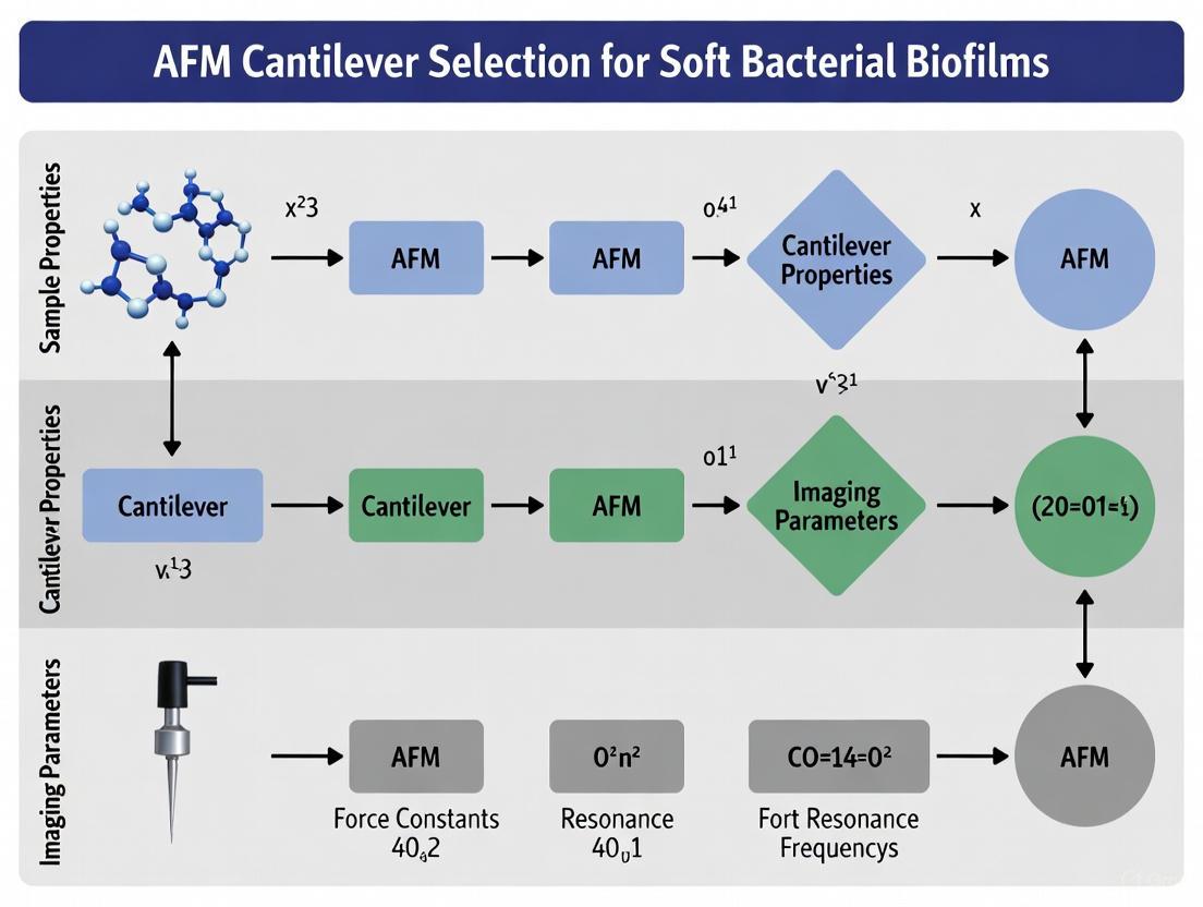
The following diagram illustrates the logical workflow for selecting an appropriate AFM cantilever and mode based on your research goals when studying soft bacterial biofilms.
The Scientist's Toolkit: Essential Research Reagents & Materials
Table 2: Key Materials and Reagents for AFM Biofilm Research
| Item | Function/Application | Specific Examples / Notes |
|---|---|---|
| Tipless Cantilevers | Base for creating custom probes, such as microbead sensors. | Rectangular silicon cantilevers with low spring constants (0.01–0.08 N/m) [4]. |
| Microbeads | Creating probes with defined, quantifiable contact geometry for force spectroscopy. | 50 µm diameter glass beads attached to tipless cantilevers for MBFS [4]. |
| qPlus Sensors | High-resolution imaging in liquid with high Q-factors, minimizing noise. | Stiff sensors (≥1 kN/m) for small amplitudes and precise imaging in biologically-relevant liquids [3]. |
| Gas-Permeable Membranes | Substrate for growing biofilms in membrane-aerated biofilm reactors (MABR). | Polyolefin flat sheet membranes used to support biofilm growth under controlled aeration [5]. |
| Calcium Chloride (CaCl₂) | Modifying biofilm cohesiveness by acting as a ionic cross-linker within the EPS matrix. | Addition to growth medium (e.g., 10 mM) significantly increases biofilm cohesive energy [5]. |
| Humidity Controller | Maintaining a consistent hydration environment for moist biofilms during AFM measurement outside of liquid. | Prevents sample dehydration, crucial for measuring native cohesive properties [5]. |
Troubleshooting Guides and FAQs
Frequently Asked Questions
Q1: Why are my measured Young's modulus values for my biofilm sample unexpectedly high? This is a common issue often related to an inappropriate cantilever choice. Using a cantilever that is too stiff or has a sharp tip can lead to overestimation of the Young's modulus on soft samples. For soft biofilms, use cantilevers with low spring constants (e.g., 0.01 to 0.5 N/m) and consider models like DMT that account for adhesion [7] [8].
Q2: My force-distance curves on bacterial cells are inconsistent. What could be the cause? Inconsistencies often stem from poor cell immobilization, leading to cell movement or drift during measurement. Ensure robust immobilization using methods like poly-L-lysine, Cell-tak, or porous membranes. Also, verify that your cantilever is clean and undamaged [9] [10].
Q3: I'm seeing repetitive streaks and lines in my AFM images of a biofilm. How can I fix this? Streaks are frequently caused by environmental noise or vibration. Ensure your AFM is on a functioning anti-vibration table. If the problem persists, try imaging during quieter times (e.g., evenings) or relocate the instrument to a basement room. Streaks can also be caused by loose contaminants on the sample surface [10].
Q4: How do I choose between Contact, Tapping, and Non-Contact mode for imaging living biofilms? For delicate, living biofilms, Tapping Mode is generally preferred. It minimizes lateral forces and damage to soft biological materials compared to Contact Mode, while offering higher resolution than Non-Contact Mode, especially in fluid [9] [11].
Q5: What does a "jump-in" event on my force curve indicate? A jump-in event is a sudden, non-linear deflection of the cantilever towards the sample during approach. It is caused by attractive forces (e.g., van der Waals, electrostatic) between the tip and the sample surface. When the gradient of these attractive forces exceeds the cantilever's spring constant, the tip snaps into contact [12].
Troubleshooting Common Problems
The table below outlines common experimental issues, their likely causes, and recommended solutions.
| Problem | Likely Cause | Recommended Solution |
|---|---|---|
| Unexpectedly high Young's modulus values [7] | Cantilever too stiff or sharp tip penetrating soft surface. | Use a softer cantilever (lower spring constant) and/or a larger tip radius. |
| Blurred images or duplicated features [10] | Contaminated or broken AFM tip (tip artifact). | Replace the AFM probe with a new, clean one. |
| Difficulty imaging deep trenches [10] | Low aspect ratio probe. | Use a High Aspect Ratio (HAR) or conical tip to access deep, narrow features. |
| Excessive noise in force curves [10] | Laser interference or electronic noise. | Use a cantilever with a reflective coating; image during periods of lower ambient electrical noise. |
| Weak or no adhesion signal in retraction curve | Poor surface functionalization or low setpoint force. | Check the chemistry used to bind molecules to the tip/surface; increase the setpoint force slightly. |
| Cell detachment during measurement [9] | Ineffective cell immobilization strategy. | Optimize the immobilization method (e.g., switch from poly-L-lysine to Cell-tak for stronger adhesion). |
Experimental Protocols & Data Analysis
Standard Protocol: Measuring Bacterial Adhesion with a Cell-Functionalized Cantilever
This protocol is used to directly measure the adhesion force between a bacterial cell and a mineral or engineered surface [9] [13].
- Cantilever Preparation: Calibrate the spring constant of a tipless or spherical-tipped cantilever using the thermal tune method [9] [12].
- Cell Immobilization: Treat the cantilever with a solution like poly-L-lysine to create a positively charged surface. Incubate the functionalized cantilever with a concentrated bacterial suspension to allow cells to adhere [9].
- Sample Preparation: Immobilize the target surface (e.g., a mineral like goethite or a polymer) on a substrate.
- Force Measurement: Mount the cell-probe in the AFM. Approach the surface and collect multiple force-distance curves at different locations.
- Data Analysis: Analyze the retraction curves. The adhesion force is the maximum negative force required to separate the cell from the surface. Bond strengthening can be observed by varying the contact time [13].
Standard Protocol: Nanomechanical Mapping of a Biofilm using Force Volume or PinPoint Mode
This protocol allows for the simultaneous measurement of topography and mechanical properties across a sample surface [7].
- Sample Preparation: Immobilize the biofilm on a solid substrate (e.g., glass coverslip) under hydrated conditions.
- Cantilever Selection: Choose a soft, cantilever with a spring constant appropriate for soft biological samples (see Table 1).
- Data Acquisition: Engage the AFM in a nanomechanical mapping mode (e.g., PinPoint, Force Volume, PeakForce Tapping). The system will automatically acquire a force-distance curve at every pixel in the scan area [7].
- Data Analysis: The software fits the contact portion of each approach curve with a contact mechanics model (e.g., Hertz, DMT, JKR) to calculate a spatial map of Young's modulus [7].
Interpreting Force-Distance Curves
The diagram below illustrates a typical force-distance curve and the key biomechanical properties that can be extracted from it [9] [12].
Selecting a Contact Mechanics Model
The choice of model for calculating Young's modulus from force-indentation data is critical [7].
| Model | Best For | Key Characteristics |
|---|---|---|
| Hertzian [7] | Purely elastic, non-adhesive contacts. | Does not account for adhesion forces. |
| DMT (Derjaguin-Muller-Toporov) [7] | Harder samples (modulus >1 GPa), low adhesion. | Accounts for adhesive forces outside the contact area. |
| JKR (Johnson-Kendall-Roberts) [7] | Soft, adhesive samples (e.g., polymers, biofilms). | Accounts for strong, short-range adhesive forces within the contact area. |
The Scientist's Toolkit
Research Reagent Solutions
Essential materials and their functions for AFM studies of bacterial biofilms [9] [14] [13].
| Item | Function | Application Notes |
|---|---|---|
| Soft Rectangular Cantilevers (Si or Si₃N₄) | High force sensitivity for imaging and force spectroscopy on soft samples. | Spring constant: 0.01 - 0.5 N/m. Resonant frequency: >300 kHz in air is recommended [8]. |
| Poly-L-Lysine | Positively charged polymer for immobilizing negatively charged bacterial cells on substrates. | Common and easy to use, but Corning Cell-Tak may provide more robust adhesion for some organisms [9]. |
| PDMS Stamps / Porous Membranes | To physically trap microbial cells (e.g., yeast) for immobilization without chemicals. | Provides a more physiologically relevant setting and reduces lateral cell drift [9]. |
| Functionalized Tips (e.g., with ligands) | To probe specific molecular interactions (e.g., antibody-antigen) on bacterial surfaces. | Allows measurement of unbinding forces of single molecules [9]. |
| Gold-Coated Cantilevers | Enhanced reflectivity of the laser for the photodetector. | Improves signal-to-noise ratio, especially in liquid or with thin cantilevers [14] [10]. |
Quantitative Cantilever Properties
Typical ranges for key cantilever parameters to guide selection [14] [15].
| Parameter | Typical Range | Influence on Experiment |
|---|---|---|
| Force Constant (k) | 0.01 N/m - 100 N/m | Softer levers (0.01-0.1 N/m) are for soft samples; stiffer levers (>1 N/m) for dynamic modes [15]. |
| Resonant Frequency (f₀) | 10 kHz - 2 MHz | Higher frequencies allow for faster scanning and are less susceptible to ambient noise [14] [8]. |
| Tip Radius | <10 nm - >30 nm | A sharper tip (<10 nm) provides higher resolution but can damage soft samples [7] [8]. |
| Aspect Ratio | Low (pyramidal) to High (conical) | High aspect ratio tips are superior for imaging deep trenches or complex surface structures [10]. |
The atomic force microscope (AFM) has evolved from a tool for surface imaging into a sophisticated platform for characterizing the nanomechanical properties of biological samples, including bacterial biofilms [16]. At the heart of this technique is the AFM cantilever, a micro-machined beam that serves as a sensitive force sensor. For researchers studying soft bacterial biofilms, understanding the core principles of cantilever design—specifically spring constant, resonance frequency, and tip geometry—is essential for obtaining accurate, reproducible mechanical data. Biofilms are sessile microbial communities that grow on surfaces and are encased in extracellular polymeric substances, exhibiting complex viscoelastic properties that dictate their structural integrity and resistance to stresses [4]. This technical guide details the fundamental principles of AFM cantilevers and provides practical troubleshooting advice for researchers investigating the mechanical properties of soft bacterial biofilms and other biological specimens.
Core Principles and Parameter Relationships
Fundamental Cantilever Properties
An AFM cantilever is characterized by several key mechanical and geometric properties that collectively determine its suitability for specific applications, particularly for probing soft biological materials.
Spring Constant (k or C): The spring constant, or force constant, represents the stiffness of the cantilever and is defined as the ratio of the applied force to the resulting deflection [15] [17]. It is typically measured in N/m. For soft materials like bacterial biofilms, softer cantilevers (with lower spring constants) are essential to minimize sample damage and achieve sufficient force sensitivity. The spring constant for a rectangular cantilever can be estimated using the formula:
( k = \frac{E w t^3}{4 l^3} )
where ( E ) is the Young's modulus of the cantilever material, ( w ) is the width, ( t ) is the thickness, and ( l ) is the length [15].
Resonance Frequency (f): The resonance frequency is the natural vibrational frequency of the cantilever and is typically measured in kHz [15] [17]. Cantilevers with higher resonance frequencies enable faster scanning speeds and are less susceptible to environmental noise. The resonance frequency for a rectangular cantilever is approximately:
( f = \frac{1}{2\pi} \sqrt{\frac{k}{m^*}} )
where ( m^* ) is the effective mass of the cantilever. In practice, a higher resonance frequency is achieved by using shorter and stiffer cantilevers [15].
Tip Geometry: The AFM tip is the nanoscale probe located at the free end of the cantilever that interacts directly with the sample. Key geometric parameters include the tip apex radius (sharpness), half-cone angle, and aspect ratio [18]. Tip geometry critically influences spatial resolution and the ability to accurately probe nanostructured biofilm surfaces. High-aspect-ratio (HAR) tips with sharp apex radii are particularly valuable for probing the intricate, three-dimensional architecture of biofilms [18].
Interrelationship of Cantilever Parameters
The physical parameters of an AFM cantilever are intrinsically linked through its material properties and geometry. Understanding these relationships is crucial for informed cantilever selection.
Quantitative Parameter Ranges for AFM Cantilevers
AFM cantilevers are available in a wide range of specifications to accommodate different measurement scenarios. The table below summarizes typical parameter ranges for commercial AFM cantilevers.
Table 1: Typical parameter ranges for AFM cantilevers and tips [15] [17] [19]
| Parameter | Typical Range | Specialized Probes | Key Influencing Factors |
|---|---|---|---|
| Length (L) | 7 - 500 µm [19] | Down to 90 µm [15] | Scanner range, stability requirements |
| Width (w) | 0.8 - 120 µm [19] | 13.5 - 50 µm [15] | Stiffness, torsional rigidity |
| Thickness (t) | 0.08 - 7 µm [19] | 0.5 - 7 µm [15] | Primary factor for stiffness (t³) |
| Spring Constant (k) | 0.01 - 2000 N/m [19] | 0.03 - 45 N/m [15] | Material, L, w, t |
| Resonance Frequency (f₀) | 6 - 5000 kHz [19] | 10 - 450 kHz [15] | k and effective mass |
| Tip Apex Radius | 1 - 20 nm [19] [18] | ~5 nm (ultra-sharp) [18] | Fabrication process (etching) |
| Tip Height | 2 - 50 µm [19] | >7 µm (HAR) [18] | Application (need deep features) |
| Half-Cone Angle | 15° - 70° | 7.5° (HAR) [18] | Fabrication process |
Cantilever Selection for Soft Bacterial Biofilms
Optimal Parameter Ranges for Biofilm Characterization
The mechanical characterization of soft bacterial biofilms presents unique challenges that demand specific cantilever properties. The following guidelines outline optimal parameter selection for biofilm studies:
Spring Constant: Low spring constants (0.01 - 0.1 N/m) are generally recommended for biofilm studies [4] [17]. These soft cantilevers provide high force sensitivity while minimizing indentation damage to delicate biofilm structures. For example, in a study of Pseudomonas aeruginosa biofilms, cantilevers with spring constants of approximately 0.03 N/m were successfully used to quantify adhesive and viscoelastic properties [4].
Resonance Frequency: While biofilms themselves don't typically require ultra-high-frequency cantilevers, selecting probes with reasonably high resonance frequencies (10-50 kHz) in fluid is advisable to improve thermal stability and reduce fluid oscillation effects during force mapping [4] [17].
Tip Geometry: For topographical imaging of biofilm architecture, sharp tips (apex radius < 10 nm) are preferred to resolve individual bacterial cells and extracellular polymer components [20] [18]. For mechanical property mapping through force spectroscopy, spherical colloidal probes or tips with moderate aspect ratios are often used as they provide well-defined contact geometries for quantitative adhesion and viscoelastic measurements [4] [16].
Material Considerations: Silicon nitride cantilevers often offer advantages for biofilm studies due to their typically lower spring constants compared to silicon cantilevers of similar dimensions [15]. However, silicon cantilevers can provide sharper tips, leading to the development of hybrid probes that combine the flexibility of silicon nitride cantilevers with the sharpness of silicon tips [15].
Research Reagent Solutions: Essential Materials for AFM Biofilm Studies
Table 2: Key materials and reagents for AFM-based biofilm mechanical characterization
| Item | Function/Description | Application Example |
|---|---|---|
| Tipless Cantilevers | Rectangular silicon cantilevers for attaching functionalized probes | Base for microbead force spectroscopy (MBFS) with biofilms [4] |
| Microbead Probes | Glass or colloidal spheres attached to cantilevers | Defined contact geometry for quantitative adhesion/viscoelasticity measurements [4] |
| Standard AFM Probes | Silicon or silicon nitride with sharp tips | High-resolution imaging of biofilm topography and cell organization [20] |
| High-Aspect-Ratio Tips | Ultra-sharp Si tips with half-cone angle of 7.5° [18] | Probing deep crevices in biofilm structures with minimal deformation |
| Pseudomonas aeruginosa Strains | Model Gram-negative bacterium for biofilm studies | Wild-type PAO1 and LPS mutant wapR for genetic studies of mechanical properties [4] |
| Trypticase Soy Broth | Growth medium for bacterial culture | Standardized preparation of bacterial suspensions for biofilm formation [4] |
| Reference Cantilevers | Cantilevers with calibrated spring constants | Calibration of test cantilevers for accurate force measurement [21] |
Experimental Protocols and Methodologies
Standardized Microbead Force Spectroscopy for Biofilms
The following protocol, adapted from Lau et al., details a method for quantitatively characterizing the adhesive and viscoelastic properties of bacterial biofilms [4]:
Cantilever Preparation: Select tipless silicon cantilevers with appropriate spring constants (e.g., 0.01-0.08 N/m). Calibrate the exact spring constant for each cantilever using the thermal fluctuation method [4] [21].
Probe Functionalization: Attach a 50-μm diameter glass bead to the cantilever end using an appropriate epoxy. Alternatively, use commercially available colloidal probes. For cell-coated probes, grow biofilms directly on the microbead by incubating in bacterial suspension.
Sample Preparation: Grow bacterial biofilms on clean glass substrates under controlled conditions. For P. aeruginosa, early biofilms may be examined after brief incubation (e.g., 24 hours), while mature biofilms require longer development (e.g., 72-96 hours) [4].
Force Measurement: Approach the biofilm-coated probe to a clean glass surface in fluid using a closed-loop AFM system. Standardize loading pressure, retraction speed, and contact time to enable reproducible comparisons. Apply a constant load and monitor creep behavior to assess viscoelasticity.
Data Analysis: Calculate adhesive pressure from retraction force curves. Fit creep compliance data to a Voigt Standard Linear Solid model to extract instantaneous and delayed elastic moduli, and viscosity [4].
Spring Constant Calibration Procedures
Accurate spring constant calibration is essential for quantitative force measurements. The following methods are commonly employed:
Thermal Method: This method analyzes the thermal vibration spectrum of the cantilever to determine its spring constant based on the equipartition theorem [4] [21]. It is non-destructive and can be performed in situ without additional equipment.
Reference Cantilever Method: This approach uses a cantilever with a known spring constant as a reference to calibrate the test cantilever [21]. The method involves performing force curves on both a rigid surface and the reference cantilever, applying the formula:
( k{\text{test}} = k{\text{ref}} \left( \frac{S{\text{rigid}}}{S{\text{cant}}} - 1 \right) \cos^2 \varphi )
where ( S ) represents slopes of compliance curves and ( \varphi ) is the inclined angle of the test cantilever [21].
Added Mass Method: This technique involves adding a known mass to the cantilever and measuring the resulting shift in resonance frequency to calculate the spring constant [21]. While accurate, it is less commonly used for routine calibrations.
Troubleshooting Guides and FAQs
Common Experimental Challenges and Solutions
Table 3: Troubleshooting guide for AFM cantilever issues in biofilm research
| Problem | Potential Causes | Solutions |
|---|---|---|
| Poor Image Resolution on Biofilms | Tip contamination; Excessive force; Blunt tip; Incorrect cantilever choice | Use sharper tips (apex <10 nm); Reduce imaging force; Employ HAR tips for rough surfaces; Clean tips with UV/O₃ plasma [22] [18] |
| Inconsistent Force Curves | Uncalibrated cantilever; Thermal drift; Sample heterogeneity; Biofilm deformation | Calibrate spring constant before measurements [21]; Allow thermal equilibrium; Take multiple measurements across sample; Standardize loading conditions [4] |
| Excessive Sample Damage | Too stiff cantilever; High setpoint force; Sharp tip geometry | Switch to softer cantilever (0.01-0.1 N/m) [17]; Reduce applied force; Use colloidal probes with larger radius [4] |
| Low Signal-to-Noise Ratio | Soft cantilever; Low resonance frequency; Environmental vibrations | Choose cantilever with higher resonance frequency; Use vibration isolation; Perform measurements in acoustic enclosure |
| Laser Detection Issues | Cantilever handle geometry; Improper alignment; Reflective coating degradation | Select cantilevers with stair-shaped handles to prevent laser blocking [18]; Realign laser path; Use cantilevers with intact reflective coating |
Frequently Asked Questions
Q1: What is the optimal spring constant for measuring the mechanical properties of soft bacterial biofilms? For soft bacterial biofilms, cantilevers with spring constants in the range of 0.01 to 0.1 N/m are generally recommended [4] [17]. These soft cantilevers provide sufficient sensitivity to measure the weak forces exerted by biofilms while minimizing sample deformation. For example, in a study of P. aeruginosa biofilms, cantilevers with spring constants of approximately 0.03 N/m successfully quantified adhesive pressures in the range of 15-330 Pa [4].
Q2: How does tip geometry affect nanomechanical measurements on biofilms? Tip geometry significantly influences both spatial resolution and quantitative mechanical measurements. Sharp tips (apex radius <10 nm) provide higher resolution for imaging individual bacterial cells [18], while spherical probes (colloidal tips) offer well-defined contact geometry for quantitative adhesion and viscoelastic measurements [4]. High-aspect-ratio tips are particularly valuable for probing the complex three-dimensional structure of biofilms without tip-sample convolution artifacts [18].
Q3: Why is cantilever calibration critical for biofilm mechanobiology studies? Proper spring constant calibration is essential for obtaining accurate, reproducible quantitative data that can be compared across different laboratories and studies. Uncalibrated cantilevers can introduce significant errors, as the actual spring constant may vary by more than 50% from the manufacturer's quoted value due to thickness variations in the fabrication process [15] [21]. International standardization efforts have shown that careful calibration reduces measurement uncertainties and improves interlaboratory reproducibility [21].
Q4: What AFM operational modes are most suitable for biofilm characterization? Both force volume mapping and nano-DMA (nanoscale dynamic mechanical analysis) modes provide valuable information about biofilm mechanical properties [16]. Force volume involves acquiring force-distance curves at each pixel and is ideal for mapping spatial variations in adhesion and elasticity [4] [16]. Nano-DMA applies small oscillatory deformations to characterize viscoelastic properties as a function of frequency, providing insights into time-dependent mechanical behavior [16].
Q5: How can I minimize damage to delicate biofilm structures during AFM imaging? To minimize biofilm damage: (1) Use soft cantilevers (spring constant <0.1 N/m) to reduce applied forces [17]; (2) Employ tapping mode or other dynamic modes instead of contact mode to minimize shear forces; (3) Optimize setpoint forces to the minimum necessary for stable imaging; (4) Consider using larger tip radii (colloidal probes) to distribute pressure over a larger contact area [4].
Advanced Techniques and Future Perspectives
Emerging Technologies in AFM for Biofilm Research
Recent advancements in AFM technology are expanding capabilities for biofilm characterization:
Large-Area AFM Imaging: Traditional AFM has been limited by a narrow field of view, making it difficult to contextualize nanoscale features within larger biofilm architectures. New automated large-area AFM platforms now enable researchers to visualize both individual bacterial cells and their organization across extensive areas, revealing previously unrecognized patterns such as honeycomb-like cellular arrangements [20].
Machine Learning Integration: The integration of machine learning with AFM enables automated analysis of large datasets generated by high-throughput AFM imaging. For example, researchers have automatically analyzed more than 19,000 individual cells to generate detailed maps of cell properties across biofilm surfaces [20].
High-Speed Nanomechanical Mapping: Advanced force volume techniques using sinusoidal z-modulation rather than traditional triangular waveforms enable faster data acquisition, reducing the time required for nanomechanical property mapping [16]. This is particularly valuable for studying dynamic processes in living biofilms.
Ultra-Sharp High-Aspect-Ratio Probes: Innovations in probe fabrication are producing silicon AFM probes with tip apex radii of ~5 nm and half-cone angles of 7.5°, enabling high-resolution imaging of deep biofilm structures with minimal deformation [18]. Novel stair-shaped handle designs ensure compatibility with commercial AFM systems while preventing laser detection issues [18].
Future Outlook
The field of AFM-based biofilm characterization continues to evolve with several promising directions. The integration of AFM with complementary techniques such as fluorescence microscopy and spectroscopy will provide correlative multimodal information about biofilm structure, composition, and mechanical properties. Standardization of measurement protocols across laboratories will enhance data comparability and reproducibility, building on existing efforts by international standards organizations [21] [23]. Further development of specialized cantilevers optimized specifically for soft biological samples will continue to improve measurement sensitivity and reduce artifacts. These advances will collectively enhance our understanding of biofilm mechanics and contribute to developing strategies for biofilm control in healthcare, industrial, and environmental applications.
Cantilever Fundamentals and Selection Criteria
The atomic force microscope (AFM) cantilever is the core sensor that mediates tip-sample interaction detection in all AFMs. [24] Its design and material properties directly determine the spring constant (stiffness) and resonant frequency, which are the primary physical properties governing its interaction with soft, delicate samples like bacterial biofilms. [25] Selecting the appropriate cantilever is not merely a technical step but a critical scientific decision that dictates the validity of nanomechanical data and the preservation of native sample conditions.
Core Cantilever Properties and Operational Modes
The interaction force between the tip and the sample is calculated using Hooke's law (F = k·dz), where F is the force, k is the cantilever's spring constant, and dz is its deflection. [25] AFM operation primarily occurs in three modes, each with distinct cantilever requirements:
- Contact Mode: The cantilever scans the surface in repulsive force mode, maintaining a constant distance of 1–2 Å. Softer cantilevers (lower spring constant) are preferred for high resolution on soft samples, but their low resonant frequency limits scanning speed. [25]
- Non-Contact Mode: The cantilever vibrates near its resonant frequency at a distance where attractive Van der Waals forces dominate. This mode is sensitive to ambient conditions and requires high resonant frequency cantilevers to avoid the tip being trapped in surface liquid layers. [25]
- Tapping (Intermittent Contact) Mode: This hybrid mode features kinematic excitation with a high amplitude (~200 nm). It reduces shear forces and minimizes sample damage, making it a preferred choice for soft, adhesive biological samples like biofilms. [23] [25]
Table 1: AFM Operational Modes for Soft Biological Samples
| Operating Mode | Principle | Optimal Cantilever Properties | Advantages for Biofilms | Risks/Limitations |
|---|---|---|---|---|
| Intermittent Contact (Tapping Mode) | The cantilever oscillates, briefly "tapping" the surface. Feedback maintains constant oscillation amplitude. [25] | Soft spring constant (0.1 - 5 N/m), high resonant frequency ( tens of kHz in liquid). [23] | Minimizes lateral (shear) forces, reducing sample damage. Good for high-resolution topography. [23] | Potential for indentation if amplitude is too high. Can be slower than contact mode. |
| Force Spectroscopy/Force Volume | A force-distance (f-d) curve is recorded at each pixel in a 2D array to map mechanical properties. [26] | Soft spring constant (<1 N/m), calibrated sensitivity. Spherical tips are sometimes used. [27] | Directly quantifies adhesion force, elasticity (Young's modulus), and deformation. Essential for mechanobiology. [26] [27] | Slow data acquisition. Requires careful model selection (e.g., Hertz, Sneddon) for data analysis. [26] |
| Quantitative Imaging (QI) Mode | A high-speed force-distance curve is acquired at every pixel, providing simultaneous topographical and nanomechanical data. [28] | Soft spring constant, high resonant frequency for speed. | Allows real-time nanomechanical mapping of living cells in liquid under physiological conditions with high resolution. [28] | Technically challenging; requires precise parameter optimization. |
Cantilever Selection Guide
Table 2: Cantilever Selection Criteria for Bacterial Biofilm Characterization
| Parameter | Ideal Range for Soft Biofilms | Impact on Data Accuracy & Sample Integrity |
|---|---|---|
| Spring Constant (k) | 0.01 - 0.5 N/m [28] [27] | Too stiff (>1 N/m): Causes excessive indentation, damaging cells and biofilm structure. Produces inaccurate force measurements. [27] Too soft (<0.01 N/m): Tip may not penetrate surface water layer; prone to snap-in events and instability. [25] |
| Resonant Frequency (in liquid) | As high as possible (e.g., >10 kHz) with a soft spring constant. [24] | Higher frequency enables faster scanning (High-Speed AFM), capturing dynamic processes and reducing drift. Low frequency limits speed and can lead to feedback instability. [24] |
| Tip Geometry | Sharp tip (nominal radius <10 nm): High-resolution topography of cell surfaces and nanostructures. [28] Spherical tip (diameter ~3.5μm): Quantifying cell-scale adhesion and mechanics; avoids sample piercing. [27] | Sharp tips concentrate stress, risking sample piercing. Spherical tips provide well-defined contact for mechanical models but sacrifice lateral resolution. [27] |
| Material | Silicon Nitride (Si₃N₄) | Commonly used for its biocompatibility and for fabricating soft, low-stress cantilevers suitable for biological applications. [28] |
| Cantilever Design | Traditional Beam vs. Innovative Seesaw | Beam Cantilevers: The laser-reflective and mechanical functions are combined. Miniaturization for speed reduces laser signal quality. [24] Seesaw Cantilevers: Feature a rigid reflective board on torsional hinges, decoupling reflection from mechanics. This design offers improved signal-to-noise ratio and high resonant frequency, promising for high-speed imaging of biofilms. [24] |
Diagram 1: A workflow for selecting the appropriate AFM cantilever for biofilm research.
Troubleshooting Common Cantilever-Related Issues
FAQ 1: My biofilm samples are consistently damaged or displaced during imaging. What is the cause and solution?
- Probable Cause: The cantilever's spring constant is too high, exerting excessive vertical force, or you are using contact mode which creates destructive shear forces.
- Solutions:
- Switch to a Softer Cantilever: Use a cantilever with a spring constant of 0.1 N/m or lower to minimize indentation forces. [28]
- Use a Dynamic Mode: Transition from contact mode to tapping mode or quantitative imaging mode. These modes eliminate lateral forces and gently probe the surface. [23] [28]
- Optimize Setpoint: Reduce the amplitude setpoint in tapping mode or the force setpoint in other modes to the lowest possible level that maintains stable feedback.
FAQ 2: My images appear noisy, lack detail, or have poor resolution, especially when scanning in liquid. How can I improve this?
- Probable Cause (1): The cantilever's resonant frequency in liquid is too low, leading to a poor signal-to-noise ratio and slow response.
- Solutions:
- Use a High-Frequency Cantilever: Select a cantilever designed for high-speed AFM, which is shorter and stiffer in a way that provides a high resonant frequency without a prohibitive increase in spring constant. [24]
- Consider Novel Designs: Emerging seesaw cantilevers are designed to decouple the laser-reflective board from the mechanical hinges, providing a higher signal-to-noise ratio and improved sensitivity compared to miniaturized traditional beams. [24]
- Probable Cause (2): The tip is contaminated or blunted.
- Solutions:
- Clean Tips: Follow manufacturer protocols for UV-ozone or plasma cleaning of cantilevers before use.
- Use New, Sharp Tips: For high-resolution imaging, always start with a new, sharp tip and verify its condition on a reference sample.
FAQ 3: My force spectroscopy measurements on bacterial cells are inconsistent. What are the critical factors to check?
- Probable Cause: Incorrect cantilever calibration or inappropriate data analysis model.
- Solutions:
- Calibrate Spring Constant: Before every experiment, rigorously calibrate the cantilever's spring constant and optical lever sensitivity using thermal tune or another validated method. [23] [27]
- Choose Correct Contact Model: For soft samples on stiff substrates, use extended Hertz models (e.g., Chen, Tu, Cappella) to account for the substrate effect. Use Sneddon's model for a conical/pyramidal tip. [26] [28]
- Functionalize the Tip Correctly: For single-cell force spectroscopy (SCFS), ensure a single bacterial cell is securely attached to a tipless cantilever using a biocompatible glue like polydopamine. Confirm cell viability after attachment. [27] [29]
FAQ 4: My probe "snaps" into contact uncontrollably, making gentle engagement difficult.
- Probable Cause: The cantilever is too soft relative to the strong adhesive and capillary forces present in the liquid environment or on the hydrated biofilm surface.
- Solutions:
- Slightly Increase Stiffness: If engagement is impossible, a cantilever with a spring constant of 0.2-0.5 N/m may provide more stability while still being soft enough for measurements. [25]
- Optimize Engagement Parameters: Reduce the engagement speed and set a very low initial contact force or amplitude setpoint.
Detailed Experimental Protocols
Protocol for Nanomechanical Mapping of a Bacterial Biofilm
This protocol uses Force Volume or Quantitative Imaging mode to simultaneously map topography and elasticity (Young's modulus). [26] [28]
Research Reagent Solutions Table 3: Essential Materials for Biofilm Nanomechanics
| Item | Function/Justification | Example |
|---|---|---|
| Soft AFM Cantilever | To minimize indentation force and avoid sample damage. | Silicon nitride cantilever, spring constant ~0.03 - 0.1 N/m, resonant frequency ~10-30 kHz in fluid, sharp tip (nominal radius < 10 nm). [28] |
| Liquid Cell | To maintain biofilm hydration and perform measurements under physiological conditions. | Bruker's ElectroChemical Cell (ECCell) or equivalent. [28] |
| Buffer Solution | To maintain cell viability and osmotic balance during measurement. | Appropriate growth medium (e.g., Lysogeny broth) or phosphate-buffered saline (PBS). [27] |
| Functionalization Reagents | For single-cell or single-molecule force spectroscopy. | Polydopamine or Poly-L-lysine for immobilizing cells or molecules on tipless cantilevers. [27] [29] |
| Calibration Sample | To verify tip shape and condition before/after experiment. | A grating with sharp features or a sample of known modulus. |
Step-by-Step Methodology:
- Cantilever Calibration: In liquid, calibrate the cantilever's spring constant using the thermal noise method. Precisely determine the optical lever sensitivity by acquiring a force curve on a rigid, non-deformable surface (e.g., clean glass or mica). [23] [27]
- Sample Preparation: Grow a bacterial biofilm directly on a suitable substrate (e.g., glass, ITO-coated glass, or mica). ITO-coated glass is highly recommended for its smooth surface and excellent cell adhesion in liquid, which avoids the need for chemical fixation that can alter biofilm mechanics. [28]
- Mounting and Engagement: Mount the substrate in the AFM liquid cell and submerge in the appropriate buffer. Carefully approach the surface with the cantilever using a low engagement force setpoint to avoid crashing.
- Parameter Optimization:
- Set the scan size to a representative area (e.g., 10x10 μm).
- In the force spectroscopy/volume menu, define the array size (e.g., 64x64 pixels).
- Set the maximum applied force to a low value (typically 0.5-2 nN) to stay within the linear elastic regime of the biofilm.
- Adjust the extension/retraction speed and duration to balance data quality and acquisition time. [28]
- Data Acquisition: Acquire the force volume map. The system will automatically record an array of force-distance (f-d) curves at each pixel.
- Data Analysis:
- Topography Image: Reconstructed from the contact point or setpoint of each f-d curve.
- Young's Modulus Map: Fit the approach segment of each valid f-d curve with an appropriate contact mechanics model (e.g., Sneddon for a conical tip). Poisson's ratio is typically assumed to be 0.5 for incompressible, hydrated biological samples. [28]
- Adhesion Map: The minimum force on the retract curve is used to map adhesion forces across the sample. [26]
Protocol for Single-Cell Adhesion Force Measurement
This protocol details how to measure the adhesion force between a single bacterial cell and a substrate using single-cell force spectroscopy (SCFS). [27] [29]
Step-by-Step Methodology:
- Cantilever Functionalization:
- Use a tipless cantilever with a soft spring constant (~0.2 N/m). [27]
- Apply a thin layer of a biocompatible adhesive (e.g., polydopamine) to the end of the cantilever.
- Briefly touch the functionalized cantilever onto a single bacterial cell from a concentrated suspension. The bright-field optical microscope integrated with the AFM is used to visually control this pick-up process. [27]
- Viability Check (Crucial): Confirm the presence and viability of the cell on the cantilever using bright-field imaging and, if possible, a subsequent growth assay on an agar plate. [27]
- Force Measurement:
- Position the cell-functionalized cantilever above a cell-free region of the substrate of interest (e.g., an LMP agarose gel of known stiffness).
- Approach and retract the cantilever at a controlled speed (e.g., 0.5-1 μm/s), recording multiple force-distance curves.
- The maximum adhesive force is determined from the retract curve's minimum (the "pull-off" force). [27] [29]
- Data Interpretation: Analyze hundreds of curves to build a statistical distribution of adhesion forces. Compare results across different substrates or bacterial strains to understand how substrate stiffness influences adhesion. [27]
Comparing Contact, Tapping, and Quantitative Imaging Modes for Biofilm Analysis
AFM Imaging Modes Comparison Table
The following table summarizes the core characteristics, advantages, and limitations of Contact, Tapping, and Quantitative Imaging modes for biofilm analysis.
| Imaging Mode | Principle of Operation | Best For | Lateral Resolution | Applied Force | Key Advantages | Key Limitations |
|---|---|---|---|---|---|---|
| Contact Mode | Tip is in constant, repulsive contact with the sample surface [30] [31]. | Mechanically robust biofilms; measuring frictional forces [30]. | ~0.5 nm [31] | High (constant repulsive force) | Fast scanning; simple operation; high-resolution imaging of hard samples [30]. | High lateral forces can damage soft samples and displace poorly immobilized cells [31]. |
| Tapping Mode | Tip oscillates and intermittently contacts the surface, reducing lateral forces [31]. | Standard high-resolution imaging of soft, hydrated, or adhesive biofilms [31]. | ~1 nm [31] | Low (intermittent contact) | Minimal sample damage; excellent for soft samples; phase imaging provides material contrast [31]. | Slower than contact mode; can be affected by a layer of surface contamination [32] [31]. |
| Quantitative Imaging (QI) | A force-distance curve is acquired at each pixel of the image, providing direct mechanical property mapping [28]. | Nanomechanical mapping (Young's modulus, adhesion) of living biofilms in liquid [28]. | Dependent on tip radius and pixel density | Controlled and quantifiable | Provides quantitative nanomechanical data simultaneously with topography; gentle enough for living, non-immobilized bacteria [28]. | Significantly slower data acquisition than other modes; complex data processing [28]. |
Troubleshooting Common AFM Imaging Problems with Biofilms
Question: My biofilm images appear blurry and lack fine detail, even though the AFM says it is in feedback. What could be wrong? This is a classic symptom of "false feedback," where the AFM tip interacts with a surface contamination layer or electrostatic forces instead of the actual sample [32].
- Cause 1: Surface Contamination. In ambient air, all surfaces have a contamination layer. The tip can become trapped in this layer [32].
- Solution: Increase the tip-sample interaction force. In Tapping Mode, decrease the setpoint amplitude. In Contact Mode, increase the deflection setpoint. This forces the probe through the contamination layer to interact with the hard surface forces of the sample [32].
- Cause 2: Sample or Cantilever Electrostatic Charge. Electrostatic forces can cause the cantilever to bend or its amplitude to change, tricking the system [32].
- Solution: Create a conductive path between the cantilever and sample. If this is not possible, use a stiffer cantilever to reduce the effect of the electrostatic force [32].
Question: I see unexpected, repeating patterns or features that look too wide on my biofilm images. What is happening? This is typically caused by a damaged or contaminated AFM tip, known as a tip artifact [10].
- Cause: A worn-out, broken tip or a tip with contamination (like a piece of the biofilm) stuck to it will produce distorted images where the shape of the tip, not the sample, is replicated [10].
- Solution: Replace the AFM probe with a new, clean one. Visually inspecting tips before use (e.g., with an optical microscope) is good practice [10].
- Cause: A standard pyramidal or low-aspect-ratio tip is too short and wide to reach the bottom of deep, narrow pores or trenches in the biofilm structure. The sides of the tip, not the apex, contact the sample walls first [10].
- Solution: Use a High Aspect Ratio (HAR) probe or a conical tip. These probes are taller and sharper, allowing them to penetrate deeper into complex biofilm architectures and produce a more accurate topographic image [10].
Question: I see repetitive horizontal lines across my image that are not part of the sample topography. This is usually caused by external noise interfering with the system [10].
- Cause 1: Electrical Noise. This often appears as a 50/60 Hz ripple across the image and is related to building electronics [10].
- Solution: Try imaging during quieter periods (e.g., early morning or late evening) when building electrical noise is lower. Ensure all equipment is properly grounded [10].
- Cause 2: Environmental Vibration. Vibrations from doors, people, or traffic can create periodic noise [10].
- Solution: Ensure the anti-vibration table is functioning. Relocate the instrument to a quieter room, such as a basement, and use a "STOP - AFM in Progress" sign to alert others [10].
Experimental Protocols for Key Applications
Protocol 1: High-Resolution Topography and Phase Imaging of a Hydrated Biofilm (Tapping Mode)
This protocol is adapted from studies imaging microbial biofilms in aqueous environments to visualize structure and differentiate material properties [31].
- Sample Immobilization: Immobilize the biofilm securely to withstand lateral scanning forces. For single-species biofilms, a simple method is to use a porous membrane filter with a pore diameter similar to the cell size to trap cells [31]. Alternatively, use a chemically treated substrate like poly-L-lysine-coated glass or mica [31].
- AFM Setup:
- Probe Selection: Use a sharp, etched silicon probe with a resonant frequency suitable for liquid operation (e.g., 10-30 kHz in fluid).
- Environment: Engage the AFM with the biofilm fully submerged in an appropriate buffer solution (e.g., PBS) to maintain native conditions [31].
- Imaging Parameters:
- Set the operational mode to Tapping Mode (Intermittent Contact Mode).
- Adjust the drive amplitude and setpoint to achieve stable feedback with minimal force. A setpoint value close to the free-air amplitude is a good starting point.
- Simultaneously capture both the Height (topography) and Phase channels. The phase signal will provide contrast between the bacterial cells and the surrounding extracellular polymeric substance (EPS) based on their differing mechanical properties [31].
Protocol 2: Nanomechanical Mapping of Living Bacteria (Quantitative Imaging Mode)
This protocol is based on a 2024 study that successfully visualized and mechanically characterized bacterial nanotubes on living cells without external immobilization [28].
- Sample Preparation:
- Use an Indium-Tin-Oxide (ITO)-coated glass substrate. The hydrophobic and smooth surface of ITO promotes bacterial adhesion, eliminating the need for chemical fixation that could alter cell physiology [28].
- Pipette a small volume (e.g., 500 µL) of bacterial culture in its exponential growth phase directly onto the ITO substrate placed in the AFM liquid cell [28].
- AFM Setup:
- Imaging Parameters:
- Set the operational mode to Quantitative Imaging (QI) Mode or a similar force-mapping mode.
- Set a pixel resolution of 64x64 or 128x128 over the area of interest.
- Define the essential force curve parameters: a total extension of 600 nm and a constant approach/retract speed of 125 µm/s [28].
- Data Analysis:
- The system will acquire a force-distance curve at every pixel.
- Use an appropriate contact model (e.g., Sneddon model for a conical indenter) to fit the retraction part of the force curve and calculate the Young's Modulus at each pixel [28].
- Construct a stiffness map that is overlaid on the topography image to correlate structure with mechanical properties.
Essential Research Reagent Solutions
The table below lists key materials and their functions for AFM-based biofilm research.
| Item | Function/Application |
|---|---|
| ITO-coated Glass Substrates | Provides a hydrophobic, smooth surface that promotes adhesion of living bacterial cells for AFM imaging in liquid without the need for chemical fixation [28]. |
| Poly-L-Lysine | A common chemical immobilization agent that provides a positively charged surface to strongly attach negatively charged bacterial cells [31]. |
| Porous Membrane Filters | Used for mechanical entrapment of bacterial cells for imaging, with pore sizes selected to match the cell diameter [31]. |
| Soft Conical Cantilevers (k ~0.1-0.5 N/m) | Essential for Quantitative Imaging and force measurements on soft biofilms to prevent sample damage and ensure accurate force sensitivity [28]. |
| High Aspect Ratio (HAR) Probes | Sharp, tall tips necessary for accurately resolving the deep, complex three-dimensional architecture of mature biofilms [10]. |
| PPP-CONTPt Cantilevers | A specific example of a conductive, Pt-coated probe with a conical tip, suitable for both high-resolution imaging and force spectroscopy [28]. |
Workflow and Decision Diagrams
Diagram 1: Decision workflow for selecting AFM imaging modes and probes for biofilm analysis.
Diagram 2: Step-by-step protocol for nanomechanical mapping of live biofilms using Quantitative Imaging mode.
A Practical Methodology for Cantilever Selection and Application in Liquid
FAQs and Troubleshooting Guides
What is the fundamental difference between soft triangular and sharp high-frequency probes?
The core difference lies in their design priority: soft triangular cantilevers prioritize low force to prevent sample damage, while sharp high-frequency probes prioritize high spatial resolution.
| Characteristic | Soft Triangular Probes | Sharp High-Frequency Probes |
|---|---|---|
| Primary Function | Minimize sample damage; measure mechanical properties [11] [9] | Achieve high-resolution topographical imaging [33] [34] |
| Typical Stiffness | 0.01 N/m to 0.5 N/m [15] [35] | 10 N/m to 50 N/m [33] [35] |
| Typical Resonance Frequency (in air) | 10 kHz to 70 kHz [35] | 200 kHz to 450 kHz [33] [35] |
| Ideal AFM Mode | Force Spectroscopy, Contact Mode [9] [15] | Tapping Mode, Non-Contact Mode, PeakForce Tapping [11] [33] [34] |
| Best for Measuring | Cell stiffness (Young's modulus), adhesion forces [9] | Ultrafine surface morphology, small, intricate features [33] [34] |
My biofilm images look blurred and lack detail. Should I switch to a sharper probe?
This is a common issue. Before switching probes, first troubleshoot your current setup and sample preparation.
- Confirm Sample Immobilization: Ensure your bacterial biofilm is securely attached to the substrate. Lateral cell drift during scanning causes blurring. Robust immobilization methods like using Corning Cell-Tak or growing biofilms directly on glass coverslips are recommended [9].
- Check for Probe Wear: A worn-out or contaminated tip significantly reduces resolution. Inspect your probe under a microscope before use.
- Optimize Scanning Parameters: Excessively high scanning forces or improper feedback gains can deform soft samples and distort images. Gradually reduce the setpoint force in tapping mode or the deflection setpoint in contact mode to the minimum stable value [11].
- Consider a Higher-Resolution Probe: If the above steps fail, a sharper probe may be necessary. For soft biofilms, a sharp but medium-stiffness probe (e.g., ~0.1-5 N/m) operating in PeakForce Tapping or fluid tapping mode offers an excellent balance of resolution and minimal damage [34].
My probe is damaging the delicate biofilm structure. How can I prevent this?
Sample damage occurs when the applied force from the probe exceeds the mechanical strength of the biofilm.
- Switch to a Softer Cantilever: This is the most direct solution. A cantilever with a lower spring constant (e.g., < 0.1 N/m) will exert lower forces on the sample for the same deflection [15].
- Use a Dynamic AFM Mode: Transition from contact mode to tapping mode or PeakForce Tapping. These modes minimize lateral forces and reduce the average force applied to the sample, making them much safer for soft materials [11] [9].
- Operate in Liquid: Performing AFM in a fluid environment eliminates capillary forces that can pull the tip into the sample with high force, allowing for much gentler imaging [9].
- Verify Cantilever Calibration: An incorrectly calibrated cantilever can lead to the application of much higher forces than intended. Always calibrate the spring constant and optical lever sensitivity before imaging soft samples [9].
How do I choose between a soft triangular probe and a sharp high-frequency probe for my biofilm experiment?
The choice hinges on your primary research question. The flowchart below outlines the decision-making process.
Quantitative Probe Comparison Table
The table below provides concrete examples of commercially available probes suitable for soft biological samples, illustrating the spectrum of available specifications.
| Probe Model / Type | Stiffness (N/m) | Resonant Frequency | Tip Radius | Primary Use Case |
|---|---|---|---|---|
| Soft Triangular (PNP-TR) [35] | 0.08 / 0.32 (Dual) | 17 / 67 kHz | Not Specified | General contact mode imaging of soft samples; low force. |
| All-In-One Probe (qp-BioAC) [35] | 0.06 - 0.3 | 30 - 90 kHz | Circular symmetric | Versatile; for non-contact, tapping, and contact mode on biology. |
| Sharp High-Res (SHR300) [33] | 40 (20-75) | 300 kHz (200-400) | 1 nm | High-resolution tapping mode imaging on delicate samples. |
| Ultra-Sharp (PEAKFORCE-HIRS-SSB) [34] | 0.12 (0.08-0.18) | 100 kHz (in air) | 1 nm (max 2) | Highest resolution PeakForce Tapping on delicate challenges. |
Experimental Protocol: Probing Biofilm Mechanics with a Soft Triangular Cantilever
This protocol outlines how to use a soft triangular cantilever for force spectroscopy to measure the stiffness and adhesion of a bacterial biofilm.
Objective: To obtain nanomechanical properties (Young's modulus and adhesion forces) of a bacterial biofilm using AFM force-distance curves.
Materials:
- AFM with a fluid cell
- Soft triangular cantilevers (e.g., stiffness ~0.1 N/m)
- Bacterial biofilm grown on a glass coverslip [9]
- Appropriate buffer solution (e.g., PBS)
Step-by-Step Method:
- Cantilever Calibration: Calibrate the cantilever's spring constant and optical lever sensitivity on a clean, rigid surface (e.g., clean glass or silicon) in fluid before engaging with the biofilm [9].
- Sample Immobilization: Secure the coverslip with the grown biofilm into the AFM fluid cell. Confirm the biofilm is securely immobilized to prevent drift during measurement [9].
- System Engagement: Engage the AFM on a robust, flat area of the biofilm using tapping mode or a very low-force contact mode to locate a region of interest.
- Force Volume Acquisition:
- Switch the AFM to force spectroscopy or "Force Volume" mode.
- Program a grid of points (e.g., 32x32 or 64x64) over the area of interest.
- Set the trigger threshold to a low force to avoid sample damage.
- At each point, the tip will approach, touch the surface, and retract, recording a force-distance curve.
- Data Analysis:
- Approach Curve Analysis: Fit the linear compliance region of the approach curve to obtain the effective spring constant. Use this in a model (e.g., Hertz model) to calculate Young's Modulus, a measure of sample stiffness [9].
- Retraction Curve Analysis: Analyze the retraction curve to measure the adhesion force (the minimum force) and the work required to separate the tip from the biofilm surface [9].
The Scientist's Toolkit: Essential Reagents and Materials
| Item | Function in Biofilm AFM |
|---|---|
| Poly-L-lysine or Corning Cell-Tak [9] | Coats substrates to create a positively charged surface for robust immobilization of (non-adherent) bacterial cells. |
| Polydimethylsiloxane (PDMS) Stamps [9] | A soft polymer used to create micro-wells or patterns to physically trap and immobilize microbial cells for scanning. |
| Polycarbonate Porous Membranes [9] | Used as a physical barrier to trap and immobilize yeast or bacterial cells for stable imaging and force measurement. |
| Standard Buffer Solutions (e.g., PBS) | Maintains physiological conditions and sample hydration during imaging and force measurement in fluid. |
Determining the Ideal Spring Constant Range for Minimizing Biofilm Deformation
Frequently Asked Questions (FAQs) on AFM Cantilever Selection for Soft Biofilms
FAQ 1: What is the primary function of the spring constant in AFM biofilm studies? The spring constant (k) of an AFM cantilever determines its stiffness and sensitivity to force. When imaging soft, hydrated biological samples like bacterial biofilms, a cantilever that is too stiff can cause excessive deformation, damage delicate surface structures, and lead to unreliable data. Selecting a cantilever with an appropriately low spring constant is therefore critical for applying minimal, non-destructive forces to preserve the biofilm's native state during measurement [9] [3] [31].
FAQ 2: What is a typical range of adhesion forces measured between bacteria and surfaces, which can inform spring constant selection? Direct force measurements provide a benchmark for the forces present in biofilm systems. For example, the adhesion force between E. coli and a goethite surface was measured to be approximately 97 ± 34 pN, with maximum adhesion forces and energies reaching -3.0 ± 0.4 nN and -330 ± 43 aJ, respectively [13]. Cantilevers must be sensitive enough to detect these forces without being overwhelmed by them.
FAQ 3: Which AFM imaging mode is most suitable for soft, easily deformed biofilms? Intermittent contact mode (also known as tapping mode) is highly recommended for imaging biofilms and microbial cells [31]. In this mode, the oscillating tip only intermittently contacts the sample, which significantly reduces lateral (dragging) forces and friction compared to contact mode, thereby minimizing sample deformation and damage [9] [31].
FAQ 4: Besides the spring constant, what other cantilever properties are important? The resonant frequency and the tip's geometry are also critical. A sharp tip with a small radius of curvature (typically nanometer-scale) is necessary for high-resolution imaging of cellular features [31]. For experiments in liquid, which are essential for maintaining biofilm hydration, cantilevers with a high resonance frequency in fluid are advantageous [3].
Troubleshooting Guide: Cantilever-Related Issues in Biofilm Imaging
| Problem | Potential Cause | Solution |
|---|---|---|
| Biofilm appears torn or scraped | Excessive imaging force; cantilever too stiff (high k); use of contact mode on soft material. | Switch to a cantilever with a lower spring constant; use intermittent contact (tapping) mode; reduce the set-point force [9] [31]. |
| Poor image quality or noisy data in liquid | Low quality factor (Q) of soft cantilevers in fluid; "forest of peaks" effect from acoustic excitation. | Use stiff qPlus sensors (k ≥ 1 kN/m) with small amplitudes for high Q-factors in liquid; consider magnetic or photothermal excitation to avoid spurious peaks [3]. |
| Cells detach from substrate during scanning | Inadequate cell immobilization; lateral scanning forces too high. | Improve immobilization using porous membranes, PDMS stamps, or optimized chemical treatments like poly-L-lysine or Cell-Tak [9] [31]. Use tapping mode to reduce lateral forces [31]. |
| Inconsistent force curve measurements | Cantilever spring constant is not accurately calibrated; biofilm surface is highly heterogeneous. | Re-calibrate the cantilever's spring constant on a hard surface before experiments [9]. Take multiple force curves across different locations to account for natural variability. |
Experimental Protocol: Measuring Biofilm Nanomechanics with Force-Distance Curves
Objective: To quantitatively map the nanomechanical properties (elasticity/adhesion) of a hydrated bacterial biofilm using AFM force-distance curves.
1. Sample Preparation and Immobilization
- Biofilm Growth: Grow biofilms directly on suitable substrates (e.g., glass coverslips, treated mica) placed in a reactor [5].
- Cell Immobilization (for single cells): For analyzing individual planktonic cells, immobilize them securely to prevent displacement by the tip. Effective methods include:
- Mechanical Trapping: Use porous polycarbonate membranes or polydimethylsiloxane (PDMS) stamps with custom-sized microwells to physically trap cells [9] [31].
- Chemical Adhesion: Treat substrates with poly-L-lysine or Corning Cell-Tak to create a positively charged surface for cell adhesion [9]. Note that chemical fixatives may alter native cell properties [31].
2. Cantilever Selection and Calibration
- Selection: Choose a soft cantilever with a nominal spring constant (k < 1 N/m) suitable for sensitive force measurements on soft matter [16].
- Calibration: Prior to the experiment, calibrate the exact spring constant (k_cantilever) of the chosen cantilever on a clean, hard surface (e.g., sapphire or clean glass) using established methods (e.g., thermal tune) [9].
3. AFM Force Spectroscopy in Fluid
- Environment: Perform all measurements in an appropriate liquid medium (e.g., buffer or growth medium) to maintain biofilm hydration and minimize capillary forces [9] [13].
- Data Acquisition: Approach the biofilm surface with the AFM tip and acquire force-distance curves. In each curve, record both the extension (tip approaching) and retraction (tip withdrawing) data [9].
4. Data Analysis
- Elasticity/Stiffness: Fit the linear compression region of the extension curve to determine the effective spring constant (k_effective). The sample's stiffness (k_cell) can be calculated using:
1/k_effective = 1/k_cell + 1/k_cantilever[9]. The nonlinear compliance region can be fitted with contact mechanics models (e.g., Hertz, Alexander-de Gennes) to extract Young's modulus and information on surface polymer brushes [9] [16]. - Adhesion: Analyze the retraction curve. The adhesion force is quantified by the minimum force value (the "pull-off" force). The area between the retraction and extension curves represents the work of adhesion [9] [13].
Experimental Workflow for Biofilm Nanomechanics
Research Reagent Solutions: Essential Materials for AFM Biofilm Studies
| Item | Function | Application Note |
|---|---|---|
| Soft AFM Cantilevers | To apply minimal force for high-resolution imaging and force spectroscopy on delicate samples. | Select a spring constant range of 0.01 - 0.5 N/m for standard measurements. Consider even softer levers (k < 0.01 N/m) for ultra-sensitive adhesion mapping [16]. |
| Poly-L-Lysine | A polymer used to coat substrates, creating a positive charge that enhances adhesion of negatively charged bacterial cells. | A common method for immobilization; however, Corning Cell-Tak may provide more robust adhesion for some organisms [9]. |
| PDMS Stamps | Polydimethylsiloxane stamps with micro-wells for mechanically trapping and immobilizing microbial cells. | Provides secure immobilization without chemicals that might alter cell physiology, ideal for single-cell analysis under aqueous conditions [9] [31]. |
| qPlus Sensors | Stiff, self-sensing cantilevers (k ≥ 1 kN/m) for frequency modulation AFM. | Enable high-resolution imaging in liquid with high quality factors (Q), avoiding the "forest of peaks" issue common with soft cantilevers [3]. |
| Membrane Test Modules | Supports for growing biofilms on gas-permeable membranes, allowing control of the aerobic environment. | Used in biofilm reactors to cultivate young, uniform biofilms for cohesive strength and mechanical testing [5]. |
The Role of Tip Sharpness and Shape in Resolving Nanoscale Biofilm Features
Troubleshooting Guide: FAQs on AFM Tip Selection for Biofilm Imaging
FAQ 1: Why am I seeing repeated, irregular patterns or duplicated features in my biofilm images?
Cause: This is a classic sign of a blunt or contaminated AFM tip, often referred to as tip artifacts. A damaged tip will not accurately trace the sample's topography, instead producing repeated patterns that reflect the shape of the tip itself rather than the biofilm [10].
Solution:
- Replace the probe. If structures appear larger than expected or trenches appear smaller, switch to a new, sharp probe to verify if the problem disappears [10].
- Use certified probes. Consider probes from suppliers that guarantee tip sharpness and lack of contamination to minimize these issues [10].
FAQ 2: Why can't I resolve deep, narrow pores or high vertical structures in my biofilm matrix?
Cause: This problem is typically due to the limited aspect ratio of standard pyramidal or tetrahedral tips. The tip geometry physically prevents it from reaching the bottom of narrow trenches or accurately profiling steep-edged features, leading to distorted images [10].
Solution:
- Switch to conical tips. Conical tips are superior for tracing surfaces with steep-edged features because their shape allows them to more closely resolve the real profile of the surface [10].
- Use High Aspect Ratio (HAR) probes. For highly non-planar features, HAR probes are essential. Their tall, narrow design can fit inside deep trenches and provide high-resolution images that conventional probes cannot achieve [10].
FAQ 3: My images appear blurry and the tip doesn't seem to be tracking the surface correctly. What is happening?
Cause: This symptom, known as "false feedback," occurs when the AFM's automated tip approach is tricked into stopping before the probe interacts with the sample's hard surface forces. This can be caused by a thick layer of surface contamination (common in humid environments or on exposed samples) or by substantial electrostatic force between the surface and the probe [36].
Solution:
- Increase probe-surface interaction. In vibrating (tapping) mode, decrease the setpoint value. In non-vibrating (contact) mode, increase the setpoint value. This forces the probe through the contamination layer [36].
- Reduce electrostatic forces. Create a conductive path between the cantilever and the sample. If that is not possible, use a stiffer cantilever to minimize the effect of surface charge [36].
Quantitative Guide: Tip Performance and Material Solutions
The following table summarizes the impact of different tip properties on image resolution and the recommended solutions for imaging complex biofilm structures.
Table 1: Troubleshooting Tip Geometry and Selection for Biofilm AFM
| Problem | Root Cause | Tip Property to Optimize | Recommended Solution |
|---|---|---|---|
| Duplicated or irregular features | Blunt or contaminated tip | Tip Sharpness | Replace with a new, certified sharp probe [10]. |
| Distorted trenches & vertical structures | Physical limitation of tip shape | Tip Shape & Aspect Ratio | Use conical or High Aspect Ratio (HAR) probes instead of standard pyramidal tips [10]. |
| Blurry images, poor tracking ("False Feedback") | Probe trapped in contamination layer or affected by electrostatic forces | Tip-Sample Interaction Force | Increase interaction force (adjust setpoint); use stiffer levers or create conductive path to dissipate charge [36]. |
To support the experiments discussed in this guide, the following key reagents and materials are essential.
Table 2: Research Reagent Solutions for AFM Biofilm Studies
| Material / Reagent | Function / Application | Specific Example in Context |
|---|---|---|
| Tipless Cantilevers | A base for attaching custom probes, such as microbeads or single cells. | Used in Microbead Force Spectroscopy (MBFS) to study biofilm adhesion and viscoelasticity [4]. |
| Functionalized Tips | Tips coated with specific molecules (e.g., ligands) to study specific binding interactions on cell surfaces. | Used to probe the interaction between a single molecule and a cell surface [9]. |
| Poly-L-Lysine / Cell-Tak | Adhesives for immobilizing microbial cells onto substrates for AFM imaging and force measurement. | Critical for attaching non-adherent cells to a glass coverslip to prevent lateral drift during measurement [9]. |
| Spherical Microbead Probes | Probes with a defined geometry (e.g., 50 µm glass beads) for quantitative adhesion and viscoelasticity measurements. | Coated with a bacterial biofilm and used to measure adhesive pressure and viscoelastic moduli against a clean glass surface [4]. |
| Standard Pyramidal Tips | General-purpose tips for high-resolution topography imaging in contact or tapping mode. | Suitable for imaging properly immobilized, flat microbial samples [37] [9]. |
Experimental Protocols: Standardized Methods for Reproducible Data
Protocol 1: Microbead Force Spectroscopy (MBFS) for Biofilm Adhesion and Viscoelasticity
This protocol, adapted from a study on Pseudomonas aeruginosa biofilms, details a method for the simultaneous quantification of adhesive and viscoelastic properties under native conditions [4].
1. Probe Preparation:
- Attach a spherical glass microbead (e.g., 50 µm diameter) to a tipless AFM cantilever.
- Calibrate the cantilever's spring constant using the thermal method to ensure accurate force measurements [4].
- Incubate the microbead with a bacterial suspension to allow a biofilm to form on its surface.
2. Sample Immobilization:
- Use a clean glass surface as the substrate. Ensure it is thoroughly cleaned to avoid contamination that could cause false feedback [4] [36].
3. Standardized Force Measurement:
- Approach the biofilm-coated bead to the glass surface in a liquid environment.
- Approach & Contact: Bring the bead into contact with a defined force setpoint and hold it for a precise contact time (e.g., 0.5-4 seconds). This dwell period allows the measurement of the material's creep response [4].
- Retraction: Retract the bead from the surface at a constant speed. The force versus separation distance during retraction provides the adhesion data [4].
4. Data Analysis:
- Adhesion: Calculate the adhesive pressure from the retraction force curves [4].
- Viscoelasticity: Fit the creep data (indentation vs. time during the hold period) to a viscoelasticity model, such as the Voigt Standard Linear Solid model, to extract elastic moduli and viscosity [4].
Protocol 2: High-Resolution Imaging of Biofilm Nanostructures
This protocol outlines the steps for achieving high-resolution images of bacterial cells and extracellular features like flagella [6].
1. Sample Preparation:
- Grow biofilms directly on suitable substrates (e.g., PFOTS-treated glass coverslips).
- At the desired time point, gently rinse the coverslip to remove unattached cells. Air-dry the sample before imaging [6].
- Alternatively, for live imaging in liquid, immobilize cells using a porous membrane or a polycarbonate stamp to prevent cell detachment or drift [9].
2. Tip and Microscope Setup:
- Select a sharp, high-aspect-ratio (HAR) conical tip to resolve fine nanostructures like flagella, which can be only 20–50 nm in height [6] [10].
- Engage the AFM in alternating contact (AC) mode (tapping mode). This mode minimizes lateral forces and is gentler on soft biological samples, reducing the risk of damage or displacement [9].
3. Imaging and Stitching:
- For large-area analysis, use an automated AFM system to capture multiple high-resolution image tiles over a millimeter-scale area.
- Apply machine learning-based algorithms to seamlessly stitch the individual tiles together, creating a comprehensive map of the biofilm's spatial heterogeneity [6].
Visualization and Decision Pathways
The following workflow diagram illustrates the critical decision points for selecting the appropriate AFM tip and method based on the specific research question in biofilm studies.
(Diagram: AFM Tip Selection Workflow for Biofilm Research)
Step-by-Step Protocol for AFM in Liquid under Physiological Conditions
FAQs and Troubleshooting Guides
Cantilever and Probe Selection
What is the most critical factor in cantilever selection for soft biofilms? The cantilever's spring constant is paramount. A low spring constant (e.g., 0.01–0.08 N/m) is essential for high-force sensitivity and to prevent damage to delicate biofilm structures [4]. Using a stiff cantilever on a soft sample can cause indentation or sample disruption [38].
My images show repeated, unnatural patterns. What is the cause? This is typically a tip artifact, often caused by a contaminated or broken tip. With a blunt tip, structures will appear larger and trenches will appear smaller than they are. The solution is to replace the probe with a new, sharp one [10].
Why can't I resolve deep, narrow trenches in my biofilm matrix? This is likely due to your probe's geometry and aspect ratio. Conventional, low-aspect-ratio pyramidal tips cannot reach the bottom of such features. Switch to a conical or high-aspect-ratio (HAR) probe to accurately resolve highly non-planar features [10].
Imaging in Liquid
My image appears blurry and out-of-focus in liquid. What is happening? This is a classic sign of "false feedback." The probe may be interacting with a surface contamination layer or electrostatic forces instead of the sample's hard surface forces. To solve this, increase the probe-surface interaction by decreasing the setpoint value in vibrating (tapping) mode. Ensuring meticulous sample preparation to minimize loose material is also crucial [39].
I see repetitive lines across my image. How can I reduce this noise? This is often caused by electrical noise (e.g., 50/60 Hz line interference) or laser interference. For electrical noise, try imaging during quieter periods (e.g., early mornings) when building electrical circuits are less active. For laser interference, use a probe with a reflective coating (e.g., gold or aluminum) to prevent spurious laser reflections from the sample surface from entering the detector [10].
My biofilm sample is being displaced or damaged during scanning. What can I do? Lateral forces in contact mode can easily damage soft samples. Switch to a dynamic (oscillating) mode such as tapping mode. This mode significantly reduces lateral forces and is the preferred method for imaging delicate biological samples like biofilms in their native state [38] [1] [40].
Data Interpretation
Should I optimize my settings for a high-contrast amplitude image? No. Optimizing for a "nice-looking" amplitude or deflection image decreases the accuracy of your height image. These images are error signals; less contrast in them indicates your height image is more accurate [41].
How can I be sure a feature is real and not an artifact? Always correlate features across multiple images and, if possible, with other techniques. Learn to recognize common artifacts like duplicated structures (tip artifacts) or straight lines repeating at regular intervals (electrical noise). When a feature looks suspicious, change a key parameter (like the scan rate or direction) or use a different probe to see if it persists [10] [41].
Quantitative Data and Reagent Solutions
Table 1: Standardized Adhesive and Viscoelastic Properties ofP. aeruginosaBiofilms
This table provides reference values obtained under standardized Microbead Force Spectroscopy (MBFS) conditions, allowing for meaningful comparison between samples [4].
| Biofilm Sample | Adhesive Pressure (Pa) | Instantaneous Elastic Modulus (Pa) | Delayed Elastic Modulus (Pa) | Viscosity (Pa·s) |
|---|---|---|---|---|
| PAO1 (Early Biofilm) | 34 ± 15 | Data from model fit | Data from model fit | Data from model fit |
| PAO1 (Mature Biofilm) | 19 ± 7 | Drastically reduced | Drastically reduced | Decreased |
| wapR mutant (Early Biofilm) | 332 ± 47 | Drastically reduced | Drastically reduced | No significant change |
| wapR mutant (Mature Biofilm) | 80 ± 22 | Drastically reduced | Drastically reduced | Decreased |
Table 2: The Scientist's Toolkit - Essential Research Reagents and Materials
A list of key materials and their functions for AFM studies of bacterial biofilms under physiological conditions.
| Item | Function & Application |
|---|---|
| Tipless Cantilevers (e.g., CSC12) | Base for attaching a microbead to create a defined contact geometry for quantitative force spectroscopy [4]. |
| 50 μm Glass Microbeads | Spherical probes attached to tipless cantilevers to create a defined contact area for standardized adhesion and viscoelasticity measurements [4]. |
| Mica / HOPG / Glass Substrata | Atomically flat or well-defined surfaces for immobilizing biofilms or forming a clean interaction surface for force measurements [38] [1]. |
| Closed-Loop AFM Instrument | An AFM system that provides accurate positioning and is essential for gathering precise force-versus-distance data over time [4]. |
| Optomechanical VHF Probes | Next-generation probes with >100 MHz resonance frequency for high-speed imaging and force sensing with picometer vibration amplitudes [40]. |
| Liquid Cell | A sealed chamber that allows the AFM to operate with the sample and probe fully submerged in buffer, maintaining physiological conditions [1]. |
Experimental Protocols
Protocol 1: Standardized Microbead Force Spectroscopy (MBFS) for Biofilm Adhesion and Viscoelasticity
This protocol enables the absolute quantitation of biofilm adhesive and viscoelastic properties [4].
Key Materials: Tipless cantilevers, glass microbeads, bacterial biofilm sample, closed-loop AFM.
- Probe Preparation: Attach a 50 μm diameter glass microbead to a tipless cantilever using a suitable epoxy. Calibrate the cantilever's spring constant using the thermal method.
- Biofilm Coating: Coat the glass microbead with a layer of the bacterial biofilm to be studied.
- Standardized Force Measurement:
- Bring the biofilm-coated bead into brief, standardized contact with a clean glass surface in liquid.
- Setpoint: Use a consistent loading force.
- Contact Time: Maintain a constant contact time (dwell period).
- Retraction Speed: Use a fixed retraction speed.
- Data Collection: Record force-versus-distance curves during the retraction (for adhesion) and indentation-versus-time curves during the constant-force hold period (for viscoelasticity).
- Data Analysis:
- Adhesion: Calculate the adhesive pressure from the retraction force curves.
- Viscoelasticity: Fit the creep compliance data from the hold period to a Voigt Standard Linear Solid model to extract elastic moduli and viscosity.
Protocol 2: High-Resolution Tapping-Mode AFM of Hydrated Biofilms
This protocol is for topographical imaging of biofilm structures in their native, hydrated state [1].
Key Materials: Sharp AFM probes (e.g., silicon nitride), biofilm sample immobilized on a substrate (e.g., mica), AFM with liquid cell.
- Sample Immobilization: Immobilize the biofilm on an atomically flat substrate (e.g., mica). Ensure the sample is firmly attached to prevent displacement by the tip.
- Liquid Cell Setup: Assemble the AFM liquid cell, ensuring the sample and probe are fully submerged in the appropriate physiological buffer.
- Probe Selection & Engagement: Use a sharp probe with a reflective coating and a medium-low spring constant. Engage the probe in liquid using the automated approach.
- Tuning: Tune the cantilever to find its resonance frequency in liquid.
- Imaging Parameter Optimization:
- Setpoint: Start with a high setpoint (low feedback gain) and gradually decrease it (increasing gain) until stable tracking is achieved without sample disruption.
- Scan Rate: Use a slow scan rate (e.g., 1-2 Hz) initially, increasing it only if image quality permits.
- Integral & Proportional Gains: Adjust gains to achieve responsive but stable feedback.
- Image Acquisition: Collect height and amplitude data simultaneously. Acquire images at multiple locations to assess heterogeneity.
Experimental Workflow and Signaling Pathways
Diagram: AFM Biofilm Analysis Workflow
Diagram: AFM Operational Principle and Feedback Loop
This technical support center provides targeted troubleshooting and methodological guidance for researchers using Atomic Force Microscopy (AFM) to characterize the nanomechanical properties of bacterial biofilms, specifically Staphylococcus aureus and Staphylococcus epidermidis. The content is framed within the critical context of AFM cantilever selection, a fundamental parameter that dictates the accuracy, resolution, and reliability of data acquired from soft, hydrated biological samples.
The Scientist's Toolkit: Essential Research Reagent Solutions
The table below details key materials and reagents essential for preparing and analyzing bacterial biofilms with AFM.
| Item | Function & Application in Biofilm AFM |
|---|---|
| Polydimethylsiloxane (PDMS) Stamps | Used for robust mechanical immobilization of spherical microbial cells without chemical fixation, preserving native cell physiology [31]. |
| Poly-L-lysine | A common chemical fixative for immobilizing bacterial cells to substrates like glass or mica; creates a positively charged surface [9] [31]. |
| Corning Cell-Tak | A commercial adhesion reagent that provides more robust and reliable cell adhesion to surfaces compared to poly-L-lysine for some organisms [9]. |
| Tipless Silicon Cantilevers | The base cantilever type used for attaching spherical probes or for direct cell attachment in single-cell probe force spectroscopy [4]. |
| Spherical Microbead Probes | Glass or colloidal beads (e.g., 50 µm diameter) attached to tipless cantilevers; provide a defined contact geometry for quantifiable adhesion and viscoelastic measurements [4] [13]. |
| Trypticase Soy Broth (TSB) | A standard nutrient-rich growth medium used for cultivating Staphylococcal biofilms prior to AFM analysis [4]. |
Frequently Asked Questions (FAQs) & Troubleshooting
Q1: My AFM cantilever consistently gets contaminated and stuck in the biofilm matrix. How can I prevent this?
- A: Biofilm extracellular polymeric substance (EPS) is highly adhesive. To mitigate this:
- Use a Larger Tip Radius: Employ spherical microbead probes (e.g., 50 µm diameter) instead of sharp tips. This increases the contact area, reduces peak pressure, and minimizes deep indentation into the EPS [4].
- Optimize Imaging Mode: Switch from contact mode to tapping mode (intermittent contact). This significantly reduces lateral forces and adhesive drag on the tip, preserving sample integrity and preventing contamination [1] [31].
- Control the Environment: Ensure the biofilm is fully hydrated. Performing measurements in liquid buffers prevents the formation of strong capillary forces that can contribute to tip sticking [9] [1].
Q2: How do I choose the right cantilever for my biofilm stiffness or adhesion measurements?
- A: Cantilever selection depends on the specific property being measured. The table below summarizes the optimal choices.
| Measurement Type | Recommended Cantilever Type | Rationale & Key Parameters |
|---|---|---|
| Adhesion Force | Soft, tipless cantilevers functionalized with a spherical microbead or a single bacterial cell. | Spring Constant (k): 0.01 - 0.08 N/m [4]. A soft spring ensures high sensitivity to weak adhesive forces without pushing the biofilm away. The defined geometry of a microbead allows for quantifiable pressure calculations [4] [9]. |
| Elastic Modulus (Stiffness) | Sharp tips with a known geometry (e.g., pyramidal) and moderate spring constants. | Spring Constant (k): ~0.1 - 1 N/m. Stiffness must be high enough to indent the cell wall without full compression, but low enough to prevent damage. The Hertz model, which requires a known tip shape (e.g., parabolic), is typically used for analysis [31] [42]. |
| Qualitative Stiffness Mapping | Standard contact mode cantilevers with a tip. | Spring Constant (k): ~1 N/m. Used for Force Modulation Microscopy (FMM) to map relative stiffness differences across a heterogeneous biofilm surface [43] [42]. |
Q3: My force-distance curves on biofilms are noisy and inconsistent. What could be the cause?
- A: Inconsistent data often stems from poor sample preparation or incorrect force settings.
- Ensure Secure Immobilization: The biofilm or cells must be firmly attached to the substrate. If using chemical fixation, confirm the protocol is optimized for your bacterial strain. Lateral drift of poorly immobilized cells is a major source of noise [9] [31].
- Calibrate Your Cantilever: Always calibrate the cantilever's spring constant immediately before measurements using the thermal tune method to ensure accurate force quantification [4] [9].
- Standardize Acquisition Parameters: Define and consistently use standardized settings for loading force, contact time, and retraction speed. This minimizes variability and allows for meaningful comparison between different samples or experiments [4].
Q4: Can I quantify the viscoelasticity of a living biofilm, and what cantilever is best for this?
- A: Yes, using force spectroscopy in a nanoindentation-style "creep" test.
- Method: Approach the biofilm with a soft cantilever (k ~0.03 N/m), apply a constant hold force, and monitor the depth change over time [4].
- Data Analysis: Fit the creep compliance data to a viscoelastic model (e.g., the Voigt Standard Linear Solid model) to extract parameters like the instantaneous elastic modulus, delayed elastic modulus, and viscosity [4].
- Probe Choice: A spherical microbead probe is highly recommended for these measurements as it provides a well-defined, constant contact area during the hold period [4].
Detailed Experimental Protocols
Protocol 1: Microbead Force Spectroscopy (MBFS) for Absolute Quantitation
This protocol, adapted from [4], allows for simultaneous quantification of biofilm adhesion and viscoelasticity.
1. Probe Preparation:
- Select a tipless silicon cantilever with a nominal spring constant of ~0.03 N/m.
- Calibrate the exact spring constant using the thermal method [4].
- Attach a 50 µm diameter glass microbead to the end of the cantilever using a suitable epoxy.
- Alternatively, for single-cell adhesion studies, chemically glue a single bacterial cell to the cantilever to create a "cell probe" [9] [13].
2. Biofilm Growth and Sample Immobilization:
- Grow S. aureus or S. epidermidis biofilms on a sterile, clean glass substrate suitable for AFM.
- Grow biofilms in TSB medium for defined periods (e.g., 24h for early biofilm, 72h for mature biofilm) [4].
- Gently rinse the biofilm with deionized water or a suitable buffer to remove non-adherent planktonic cells before measurement.
3. Force Spectroscopy Measurements:
- Mount the sample in the AFM liquid cell and engage with the microbead probe in the desired buffer.
- Approach the biofilm surface at a controlled speed (e.g., 1 µm/s).
- Upon contact, apply a predefined loading force and hold for a set "dwell time" (e.g., 0.5 - 2 seconds) to measure viscoelastic creep.
- Retract the probe at a constant speed to obtain the force-separation curve for adhesion analysis.
- Perform hundreds of measurements at different locations to account for biofilm heterogeneity.
4. Data Analysis:
- Adhesive Pressure: Calculate from the maximum pull-off force during retraction, divided by the contact area of the microbead [4].
- Viscoelastic Parameters: Fit the indentation-vs-time data from the hold period to a Voigt model to extract elastic moduli and viscosity [4].
Protocol 2: Force Modulation Microscopy (FMM) for Stiffness Mapping
This protocol maps relative stiffness variations across a biofilm surface [43] [42].
1. Probe and Sample Setup:
- Use a standard contact mode cantilever with a sharp tip and a moderate spring constant (~1 N/m).
- Immobilize the biofilm as described in Protocol 1.
2. FMM Imaging:
- Engage in contact mode on a relatively hard area of the sample (e.g., the substrate).
- Activate the FMM (force modulation) function. A small sinusoidal oscillation (e.g., <10 nm amplitude) is superimposed on the cantilever's z-position at a frequency below its resonance (e.g., 10-20 kHz) [43].
- Scan the surface as in contact mode. The feedback loop maintains a constant average deflection (DC signal) to track topography.
3. Data Acquisition:
- Simultaneously collect three data channels:
- Topography: From the DC deflection signal.
- FMM Amplitude: The amplitude of the AC deflection signal. Harder regions result in a higher amplitude; softer regions result in a lower amplitude.
- FMM Phase: The phase lag of the AC signal, which provides additional contrast related to the sample's viscoelastic properties [43].
4. Data Interpretation:
- FMM provides qualitative stiffness mapping. Brighter areas in the FMM amplitude image correspond to stiffer regions, while darker areas correspond to more compliant regions, allowing you to distinguish between EPS, cells, and other biofilm components [43].
Workflow and Data Analysis Visualization
The following diagram illustrates the logical pathway for selecting the appropriate AFM technique based on the research goal and for analyzing the resulting data.
AFM Technique Selection and Analysis Workflow for Biofilm Nanomechanics
The table below compiles key nanomechanical parameters for bacterial biofilms and cells measured by AFM, as reported in the literature. This provides a reference for interpreting your own data.
| Measured Property / Parameter | Value(s) Reported | Experimental Context & Conditions |
|---|---|---|
| Adhesive Pressure | 34 ± 15 Pa [4] | P. aeruginosa PAO1 early biofilm, measured via Microbead Force Spectroscopy (MBFS). |
| 332 ± 47 Pa [4] | P. aeruginosa wapR mutant (LPS-deficient) early biofilm, via MBFS. | |
| Adhesion Force (Single Cell) | -3.0 ± 0.4 nN [13] | Maximum adhesion force and energy between E. coli and a goethite surface. |
| Adhesion Energy | -330 ± 43 aJ (10⁻¹⁸ J) [13] | Accompanying adhesion energy for the above measurement. |
| Elastic Modulus (from Voigt Model) | Drastically reduced for LPS mutant and mature biofilms [4] | P. aeruginosa biofilm; MBFS with creep test. Specific values for instantaneous and delayed moduli are material-dependent. |
| Cantilever Spring Constant | 0.015 - 0.060 N/m [4] | Recommended range for soft biofilms in Microbead Force Spectroscopy. |
| ~1 N/m [42] | Used for Force Modulation Microscopy (FMM) on organic thin films in liquid. | |
| FMM Actuation Amplitude | < 10 nm [43] | Standard amplitude to prevent sample damage in Force Modulation Microscopy. |
Troubleshooting Common Artifacts and Optimizing Cantilever Performance
In atomic force microscopy (AFM) studies of soft bacterial biofilms, the integrity of your data is critically dependent on two key factors: the force applied by the cantilever and the cleanliness of the probe tip. Excessive force can compress, deform, or even rupture delicate bacterial cells and their extracellular polymeric substance (EPS) matrix, while tip contamination generates imaging artifacts that compromise data accuracy. This guide provides practical troubleshooting advice to help researchers identify, mitigate, and avoid these common yet destructive issues, preserving the native state of your biological samples throughout experimentation.
Troubleshooting Guide: FAQs on Sample Damage and Tip Issues
FAQ 1: How can I tell if my AFM tip is contaminated or broken?
Answer: A contaminated or broken tip produces characteristic artifacts that are visible in your AFM images.
- Double-Tip Effect: This occurs when the tip is broken or when contamination creates multiple scanning points. The artifact manifests as duplicate features, "ghost" images, or repeating patterns aligned in the fast-scan direction [44]. Each real feature appears with a "twin" [44].
- Strange Repeating Shapes: A dirty tip, often coated with parts of the sample, will stamp strange, non-sample-related shapes repeatedly across the image [44] [45].
- Diagnosis: The most reliable method to diagnose tip condition is to scan a well-known characterization sample, such as Biaxially Oriented PolyPropylene (BOPP) film or other tip-characterization standards. Comparing the resulting image to one from a known-sharp tip will immediately reveal abnormalities [44] [45].
FAQ 2: What are the consequences of using a blunt tip on soft biofilms?
Answer: A blunt tip significantly degrades image resolution and accuracy. It exacerbates the probe-sample convolution effect, which is an inherent feature of AFM where the image is a blend of the tip's shape and the sample's shape [44]. This effect makes protruding features appear wider and shallower, and holes appear smaller and less deep [44]. For complex biofilm structures like individual cells or nanotubes, this can lead to a massive overestimation of width and a loss of structural detail, rendering nanomechanical property mappings like Young's modulus inaccurate [44] [28].
FAQ 3: My biofilm appears flattened and featureless. Is this due to excessive force?
Answer: Yes, this is a classic sign of excessive imaging force. Soft biological samples like biofilms are easily compressed and deformed. Applying too much force flattens delicate structures such as the EPS matrix, collapses bacterial nanotubes, and can even rupture cell membranes. This not only destroys the sample's native architecture but also leads to erroneous measurements of mechanical properties like stiffness and adhesion [28] [46]. To preserve sample integrity, it is essential to use the lowest possible applied force (setpoint) that still provides stable feedback [44].
FAQ 4: Are there advanced AFM modes that can minimize sample damage?
Answer: Yes, several AFM modes are specifically designed to minimize invasive forces.
- Quantitative Imaging (QI) Mode: This is a force-curve-based, non-resonant mode that has been successfully used to image living bacteria in liquid without any immobilization, achieving excellent image quality and nanomechanical mapping with minimal disturbance [28].
- Non-Contact Non-Resonant (NCNR) Mode: A novel method that utilizes attractive forces between the tip and sample as a feedback signal, stopping the tip approach before physical contact occurs. This "negative setpoint" mode is highly effective for studying highly mobile molecules and fragile structures without sample modification or tip contamination [47].
- Large-Area AFM with Machine Learning: This automated platform allows for the correlation of nanoscale details with larger biofilm organization, and when combined with machine learning, enables the extraction of quantitative data from vast datasets without aggressive scanning parameters [20].
Table 1: Common AFM Tip Issues and Their Impacts on Biofilm Research
| Issue | Primary Effect | Impact on Data Quality | Solution |
|---|---|---|---|
| Tip Contamination [44] [45] | Strange, repeating artifacts; "double-tip" images | Loss of true sample detail; introduction of false features | Change tip; characterize with BOPP film; may attempt cleaning [44] [45] |
| Blunt/Broken Tip [44] | Poor resolution; widened features; obscured fine details | Inaccurate topographic and mechanical measurements | Replace tip; use low-wear tips; scan with lower force [44] |
| Excessive Force [28] [46] | Compression of biofilm, EPS, and bacterial cells | Flattened topography; overestimated Young's modulus | Use lowest possible setpoint; employ gentle modes (QI, NCNR) [28] [47] |
Table 2: Comparison of Gentle AFM Modes for Biofilm Imaging
| AFM Mode | Principle of Operation | Advantages for Biofilm Research | Considerations |
|---|---|---|---|
| Quantitative Imaging (QI) [28] | Fast approach/retract cycles with force-curve analysis at each pixel | High-resolution nanomechanical mapping on living, non-immobilized bacteria in liquid | Requires stable sample adhesion to substrate [28] |
| Non-Contact Non-Resonant (NCNR) [47] | Uses attractive force (negative setpoint) to avoid physical contact | Eliminates sample damage and tip contamination; ideal for highly mobile structures | A new method that may require software modification [47] |
| Large-Area AFM with ML [20] | Automated tiling of multiple AFM images combined with machine learning analysis | Correlates single-cell details with community-scale organization in biofilms | Platform and analysis pipeline may not be universally available [20] |
Experimental Protocols
Protocol 1: Tip Characterization and Cleaning Using BOPP Film
This protocol is adapted from established methods for evaluating and cleaning contaminated AFM tips [44] [45].
- Purpose: To diagnose the state of the AFM probe tip and potentially clean minor contamination.
- Materials:
- Method:
- Step 1: Image the BOPP film. Using your tip in question, acquire an image of the BOPP film. The well-defined fibrous structure of BOPP serves as a reference.
- Step 2: Analyze the image. Look for "double-tip" artifacts, strange repeating shapes, or a general lack of sharpness compared to a known-good image of BOPP. This confirms tip contamination or damage [44].
- Step 3: Attempt cleaning (if applicable). To clean the tip, perform a series of indentations into the BOPP film. The theory is that the soft polymer can absorb and remove contaminants from the tip apex [44] [45].
- Step 4: Re-image. After indentation, image the BOPP film again to check if the artifacts have been reduced.
- Note: Be aware that cleaning is not always successful and may not return the tip to its pristine, new state. If artifacts persist, the tip must be replaced [44].
Protocol 2: Imaging Bacterial Nanotubes in Liquid with QI Mode
This protocol is based on a study that successfully visualized bacterial intercellular nanotubes on native Rhodococcus wratislaviensis [28].
- Purpose: To image the topography and map the nanomechanical properties of living bacteria and their nanotubes in physiological conditions with minimal force.
- Materials:
- Bacterial culture (e.g., Rhodococcus wratislaviensis) in exponential growth phase.
- ITO-coated glass substrates (provide excellent adhesion for non-immobilized bacteria) [28].
- Liquid cell/AFM instrument equipped with Quantitative Imaging (QI) mode.
- Standard AFM probes (e.g., PPP-CONTPt, stiffness ~0.3 N/m) [28].
- Culture medium or phosphate-buffered saline (PBS).
- Method:
- Step 1: Sample Preparation. Pipette a volume of bacterial culture onto an ITO-coated glass substrate and place it in the liquid cell. No chemical or mechanical immobilization is used, preserving native bacterial physiology [28].
- Step 2: AFM Setup. Submerge the tip in the liquid medium. Calibrate the cantilever's sensitivity and spring constant in liquid.
- Step 3: QI Mode Imaging. Engage the QI mode. Typical parameters may include a total extension of 600 nm, a constant speed of 125 µm/s, and an indentation speed between 17-175 mN/s [28]. The mode will perform a force curve at every pixel of the image.
- Step 4: Data Acquisition & Analysis. The system will concurrently generate height data and force-distance curves. Use a Hertz/Sneddon model to fit the curves and create a map of the Young's modulus, revealing the mechanical properties of the bacteria and the finer, more flexible nanotubes [28].
Workflow Visualization
The Scientist's Toolkit: Research Reagent Solutions
Table 3: Essential Materials for Reliable AFM Biofilm Studies
| Item | Function / Application | Key Benefit |
|---|---|---|
| BOPP Film [44] [45] | Tip characterization sample and potential cleaning substrate | Well-defined structure for identifying tip artifacts and contamination. |
| ITO-coated Glass Substrates [28] | Substrate for bacterial adhesion in liquid AFM | Hydrophobic surface promotes stable adhesion of living bacteria without chemical immobilization. |
| Low Spring Constant Cantilevers (e.g., ~0.1-0.3 N/m) [28] | Probing soft biological samples | Minimizes applied force, reducing sample deformation and damage. |
| Glass Sphere-Modified Tips [46] | Force-volume imaging and nanoindentation | Defined geometry (e.g., 10 µm sphere) enables accurate Young's modulus calculation on heterogeneous biofilms. |
| Large-Area AFM Platform with ML [20] | Correlative imaging from nano-to-meso scale | Reveals how single-cell features fit into larger biofilm community organization. |
Frequently Asked Questions (FAQs)
FAQ 1: My AFM cantilever is consistently getting stuck or contaminated by the sticky EPS. What can I do? The adhesive EPS matrix often causes tip contamination. We recommend two primary solutions:
- Use Microbead Force Spectroscopy (MBFS): Replace sharp tips with a 50-μm diameter glass bead attached to a tipless cantilever. This provides a defined contact geometry, minimizes probe clogging, and enables accurate quantitation of adhesion over a larger, standardized area [4].
- Standardize Contact Conditions: Variability in adhesion measurements often stems from inconsistent loading pressures and contact times. Using standardized conditions for force spectroscopy allows for meaningful comparison between different experiments [4].
FAQ 2: How does biofilm maturation affect its mechanical properties, and how should this influence my AFM experiment design? Biofilm mechanical properties change significantly as they mature. Early and mature biofilms exhibit distinct adhesive and viscoelastic characteristics [4]. For example, in Pseudomonas aeruginosa:
- The adhesive pressure of a wild-type mature biofilm decreases compared to its early stage [4].
- The instantaneous and delayed elastic moduli drastically reduce with maturation [4].
- Experimental Implication: Always document and standardize the growth time and maturation stage of the biofilm you are testing. Comparisons should only be made between biofilms at equivalent stages of development.
FAQ 3: What is the best way to immobilize a soft bacterial biofilm for AFM analysis without affecting its native properties? Proper immobilization is critical. For single cells, poly-L-lysine or Corning Cell-Tak can be used to adhere cells to a substrate [9]. However, for biofilm research, a more physiologically relevant method is to grow the biofilm directly on the substrate, eliminating the need for external fixatives that could interfere with EPS properties [9]. Ensure the substrate is suitable for bacterial attachment and growth.
FAQ 4: How can I obtain statistically relevant data from AFM measurements given the inherent heterogeneity of biofilms? Biofilms are highly heterogeneous, making data from small scan areas potentially non-representative.
- Solution: Employ large-area automated AFM. This approach combines multiple high-resolution scans over millimeter-scale areas, providing a comprehensive view of the biofilm's organization [6] [20].
- Integration with Machine Learning (ML): Use ML algorithms to automatically analyze large-area datasets, extracting quantitative data (e.g., cell count, confluency, orientation) from thousands of individual cells across the biofilm. This overcomes the limitation of manual analysis of small areas [6] [20].
Troubleshooting Guides
Problem: High Variability in Adhesion Force Measurements
Issue: Measurements of adhesion force (from force-distance curve retraction) are inconsistent between samples or locations.
| Possible Cause | Solution | Underlying Principle |
|---|---|---|
| Unstandardized contact conditions. [4] | Implement a standardized MBFS protocol with fixed loading pressure, retraction speed, and contact time. | Standardization minimizes variability from experimental parameters, enabling direct comparison of data from different biofilms. |
| Heterogeneous nature of the biofilm. [48] | Use large-area AFM to map adhesion forces across a vast area instead of a few single points. Perform statistical analysis on a large number of force curves. | This accounts for spatial heterogeneity and provides a more representative value for the biofilm's adhesive properties. |
| Contaminated or worn-out cantilever. | Regularly inspect tips and clean or replace them. Use colloidal probes (microbeads) which are less prone to contamination than sharp tips. [4] | A contaminated tip will have altered surface chemistry and geometry, leading to inconsistent and unreliable adhesion measurements. |
Problem: Difficulty Quantifying Viscoelasticity
Issue: Inability to reliably extract viscoelastic parameters from force curves.
| Possible Cause | Solution | Underlying Principle |
|---|---|---|
| Using an inappropriate mechanical model. | Fit creep compliance data (indentation vs. time during a "hold" period) to a established viscoelastic model like the Voigt Standard Linear Solid model. [4] | Biofilms are viscoelastic materials. This model allows for the extraction of specific parameters like instantaneous elastic modulus, delayed elastic modulus, and viscosity. [4] |
| Applying excessive force, causing sample damage. | Use a stiff qPlus sensor ((k \geq 1\ kN/m)) which allows for high-resolution imaging with minimal forces ((<100\ pN)), preventing sample deformation. [3] | Soft biological samples can be easily damaged by excessive force. Stiff sensors in frequency modulation mode enable small amplitudes and minimal force, allowing non-destructive measurement. [3] |
| Inconsistent environmental conditions (e.g., humidity). | Perform measurements in liquid using a fluid cell or a controlled humidity chamber to maintain a consistent hydration state. [5] [3] | The mechanical properties of the hydrated EPS are highly dependent on water content. Drying the biofilm will significantly alter its properties. [5] |
The following tables consolidate key quantitative findings from AFM studies on bacterial biofilms to serve as a reference for your experimental outcomes.
| Bacterial Strain | Biofilm Stage | Adhesive Pressure (Pa) |
|---|---|---|
| PAO1 (Wild-type) | Early | 34 ± 15 |
| PAO1 (Wild-type) | Mature | 19 ± 7 |
| wapR (LPS mutant) | Early | 332 ± 47 |
| wapR (LPS mutant) | Mature | 80 ± 22 |
Data obtained by fitting creep data to a Voigt Standard Linear Solid model.
| Factor Influencing Viscoelasticity | Effect on Instantaneous Elastic Modulus | Effect on Delayed Elastic Modulus | Effect on Viscosity |
|---|---|---|---|
| Lipopolysaccharide (LPS) Deficiency | Drastically Reduced | Drastically Reduced | No Significant Change |
| Biofilm Maturation | Drastically Reduced | Drastically Reduced | Decreased |
| Biofilm Condition | Cohesive Energy (nJ/μm³) | Notes |
|---|---|---|
| Standard 1-day biofilm | 0.10 ± 0.07 to 2.05 ± 0.62 | Increases with biofilm depth. |
| With 10 mM Calcium added | 0.10 ± 0.07 to 1.98 ± 0.34 | Calcium increases cohesiveness. |
Detailed Experimental Protocols
Protocol: Microbead Force Spectroscopy (MBFS) for Adhesion and Viscoelasticity
This protocol is adapted from the method used to characterize P. aeruginosa biofilms [4].
1. Principle MBFS uses a glass microbead as an AFM probe to quantify the adhesive and viscoelastic properties of a biofilm over a defined contact area. The adhesive properties are derived from the force-versus-separation curve during retraction, while viscoelasticity is determined from the indentation-versus-time curve during a constant load hold period [4].
2. Reagents and Equipment
- Atomic Force Microscope with a closed-loop scanner.
- Rectangular tipless silicon cantilevers (e.g., Mikromasch CSC12).
- 50-μm diameter glass microbeads.
- Bacterial strain of interest (e.g., P. aeruginosa PAO1).
- Growth medium (e.g., Trypticase Soy Broth).
- Glass coverslips or other relevant substrates.
3. Step-by-Step Procedure Step 1: Cantilever and Probe Preparation
- Attach a glass microbead to the end of a tipless cantilever using a suitable epoxy.
- Calibrate the spring constant of each cantilever using the thermal method [4]. Use only cantilevers with a spring constant within a predefined range (e.g., 0.015–0.060 N/m) [4].
Step 2: Biofilm Coating on the Microbead
- Grow the bacterial culture overnight in an appropriate medium.
- Harvest cells by centrifugation, wash, and resuspend in deionized water to a standardized optical density (e.g., OD600 = 2.0).
- Incubate the microbead-probe with the bacterial suspension to allow a biofilm to form on the bead's surface.
Step 3: Standardized Force Measurement
- Approach the biofilm-coated microbead to a clean glass surface in liquid.
- Standardized Contact: Use predetermined and consistent parameters for loading pressure, surface contact time, and retraction speed [4].
- Data Acquisition:
- Collect force-distance curves to analyze adhesive pressure from the retraction curve.
- For viscoelasticity, perform a creep test: apply a constant load (set point force) and hold for a defined period (e.g., 1-5 seconds), recording the tip displacement over time.
Step 4: Data Analysis
- Adhesion: Calculate adhesive pressure from the pull-off force in the retraction curve, divided by the contact area of the microbead.
- Viscoelasticity: Fit the creep compliance data (indentation vs. time during hold) to a viscoelastic model (e.g., Voigt Standard Linear Solid model) to extract parameters like elastic moduli and viscosity [4].
Protocol: Measuring Biofilm Cohesive Energy by AFM Abrasion
This protocol describes a method to measure the energy required to disrupt the biofilm matrix, indicating its cohesive strength [5].
1. Principle The cohesive energy of a biofilm is determined by using the AFM tip to abrade a defined region under a high load. The volume of displaced biofilm is measured from topographic images, and the corresponding frictional energy dissipated is used to calculate the cohesive energy (nJ/μm³) [5].
2. Reagents and Equipment
- Atomic Force Microscope (e.g., PicoSPM with humidity control).
- V-shaped cantilevers with pyramidal, oxide-sharpened Si3N4 tips (e.g., model NPS).
- Biofilm grown on a membrane or relevant substrate.
3. Step-by-Step Procedure Step 1: Sample and Environment Preparation
- Grow a 1-day biofilm on a membrane in a reactor.
- Before imaging, equilibrate the biofilm sample in a chamber with constant high humidity (e.g., ~90%) for 1 hour to maintain a consistent water content [5].
Step 2: Non-Perturbative Baseline Imaging
- Select a region of interest (e.g., 5 × 5 μm).
- Collect a topographic image of this region at a very low applied load (∼0 nN) to establish a baseline without damaging the biofilm.
Step 3: Abrasion Scanning
- Zoom into a smaller sub-region (e.g., 2.5 × 2.5 μm) within the originally scanned area.
- Set a high applied load (e.g., 40 nN) and perform repeated raster scans (e.g., 4 scans) to abrade the biofilm surface.
Step 4: Post-Abrasion Imaging
- Return to the low applied load and image the original 5 × 5 μm area again. The abraded sub-region will now appear as a depression.
Step 5: Data Analysis
- Subtract the "after" image from the "before" image to determine the volume of biofilm displaced.
- The cohesive energy (Γ) is calculated as Γ = (Ef / V), where Ef is the frictional energy dissipated during abrasion and V is the volume of displaced biofilm [5].
Experimental Workflow and Signaling Visualization
AFM Viscoelasticity Measurement Workflow
EPS Adhesion Troubleshooting Logic
The Scientist's Toolkit: Essential Research Reagents & Materials
Table: Key Materials for AFM-based Biofilm Adhesion Studies
| Item | Function/Application in Research | Key Characteristics |
|---|---|---|
| Tipless Cantilevers (CSC12) | Base for attaching microbead probes in MBFS. [4] | Rectangular silicon; low spring constant (e.g., 0.03 N/m); resonance frequency ~10 kHz. |
| Glass Microbeads (50 µm) | Spherical probe for MBFS to define contact area and reduce clogging. [4] | Defined geometry; enables quantification of adhesive pressure over a known area. |
| qPlus Sensors | Stiff cantilevers for high-resolution imaging with minimal force. [3] | High stiffness (k ≥ 1 kN/m); allows use of small amplitudes; high Q-factor in liquid; prevents sample damage. |
| Poly-L-lysine / Cell-Tak | Coating agents for immobilizing single bacterial cells to a substrate. [9] | Creates a positive surface charge for cell adhesion. Cell-Tak provides more robust adhesion. |
| Controlled Humidity Chamber | Maintains consistent hydration of the biofilm during measurement. [5] | Prevents drying artifacts; crucial for measuring native mechanical properties of the hydrated EPS. |
| Voigt Standard Linear Solid Model | Analytical model for fitting creep data to extract viscoelastic parameters. [4] | Provides quantitative values for instantaneous elastic modulus, delayed elastic modulus, and viscosity. |
Calibration Best Practices for Accurate Spring Constant and Deflection Sensitivity
For researchers studying the nanomechanical properties of soft bacterial biofilms, achieving accurate atomic force microscopy (AFM) measurements is crucial. The reliability of your force data hinges on the precise calibration of two fundamental parameters: the cantilever's spring constant and the system's deflection sensitivity. This guide outlines established and emerging calibration protocols, helping you minimize measurement uncertainties and obtain trustworthy quantitative data on biofilm mechanics for your drug development research.
Frequently Asked Questions (FAQs)
Q1: Why is conventional deflection sensitivity calibration problematic for biofilm research?
The conventional method for calibrating deflection sensitivity requires pressing the AFM tip against an infinitely stiff surface (like sapphire) to measure the slope of the resulting force curve [49]. This "hard surface contact method" poses a significant risk of damaging or contaminating your sharp AFM tip [50]. For biofilm research, where tips are often functionalized with specific molecules to probe binding affinities, this tip damage can ruin experiments and compromise data before they even begin [50] [51].
Q2: What are the non-contact alternatives for full cantilever calibration?
Non-contact methods that use the cantilever's thermal fluctuations are available to calibrate both the spring constant and the deflection sensitivity without ever touching a surface. The Thermal Tuning Method can calibrate the spring constant by analyzing the thermal noise spectrum of a free-vibrating cantilever [49] [52]. Furthermore, a method proposed by Higgins et al. allows for the determination of deflection sensitivity (InvOLS) from the same thermal spectrum, provided the spring constant is known beforehand [50] [52]. This complete non-contact approach is ideal for preserving delicate tips.
Q3: How does the Sader method work for spring constant calibration?
The Sader method is a widely used technique that calculates the spring constant (k) based on the cantilever's plan view dimensions (length L and width w), its fundamental resonant frequency (f), and the quality factor (Q) of that resonance in a fluid [53] [52]. The formula is k = 0.1906 ρ w² L Q Γᵢ(f) f², where ρ is the fluid density and Γᵢ(f) is the hydrodynamic function [52]. This method is popular because it can be performed retracted from the surface and does not require a contact-based sensitivity calibration [49].
Q4: My thermal spectra in liquid are noisy with low Q-factors. Can I still use the Sader method?
Yes, but with consideration. While the Sader method was originally developed for environments with high quality factors (like air), it has been applied in liquids with satisfactory results [52]. As an alternative, the Global Calibration Initiative (GCI) method can be more robust for low-Q environments. The GCI uses a community-derived coefficient for specific cantilever types, requiring only the measurement of f and Q in liquid via the formula k = A Q f^1.3 [52]. Research suggests that using the GCI method with a Simple Harmonic Oscillator (SHO) model for fitting the thermal power spectral density provides higher accuracy and is less prone to systematic errors in liquid [52].
Troubleshooting Guides
Issue: High Variability in Calculated Young's Modulus of Biofilms
Potential Cause 1: Inaccurate Deflection Sensitivity (InvOLS) The calibration of the inverse optical lever sensitivity is often a major source of error, as its uncertainty propagates into the spring constant and all subsequent force values [52].
- Solution:
- Adopt a Non-Contact Method: Use the thermal spectrum of the cantilever to determine the InvOLS, a method central to the Standardized Nanomechanical AFM Procedure (SNAP) [52]. This avoids errors from tip damage, surface contamination, or laser spot movement associated with the hard contact method.
- Ensure Proper Thermal Equilibrium: Metal-coated cantilevers can heat up significantly from the laser. After alignment, allow at least 30 minutes for the cantilever to thermally equilibrate with its environment to prevent drift in sensitivity measurements [50].
Potential Cause 2: Uncalibrated or Poorly Calibrated Spring Constant Relying on the manufacturer's nominal spring constant values can lead to large errors, as the actual value can vary significantly [49] [51].
- Solution:
- Always Calibrate In-Situ: Calibrate the spring constant immediately before your biofilm experiments, in the same medium (e.g., liquid buffer).
- Choose the Right Method: For rectangular cantilevers, the Sader hydrodynamic method or the thermal noise method are standard and well-supported [49]. Laser Doppler Vibrometry (LDV) is a highly accurate, traceable method but requires specialized equipment [54] [53].
Issue: Damaged or Contaminated AFM Tip After Calibration
Potential Cause: Use of Hard Contact Method on a Stiff Surface The process of acquiring force-distance curves on a rigid surface like sapphire for sensitivity calibration can blunt or break sharp tips [50].
- Solution:
- Implement a Non-Contact Calibration Workflow: Use the one-step calibration method in liquid. This involves calibrating the spring constant using the GCI or Sader method, and then deriving the InvOLS from the same thermal spectrum, completely avoiding tip-surface contact [52].
- Use an Inverted Probe: If a contact method is necessary, an alternative is to use a second, stiffer "inverted" AFM cantilever to deflect the test cantilever at a known distance from its tip. This avoids contact with a hard, potentially contaminating surface [50].
Comparison of Calibration Methods
Table 1: Common methods for calibrating the spring constant of AFM cantilevers.
| Method | Principle | Key Requirements | Typical Uncertainty | Best For |
|---|---|---|---|---|
| Sader Method [49] [52] | Hydrodynamic damping of an oscillating cantilever | Cantilever dimensions (L, w), resonant frequency (f), quality factor (Q) | Varies; can be >10% | Rectangular cantilevers; labs without specialized equipment |
| Thermal Tuning [49] | Equipartition theorem; analysis of thermal noise spectrum | Thermal spectrum, accurate Deflection Sensitivity (for k) | ~10-30% (lower if InvOLS is known) | All cantilever geometries; in-situ calibration |
| Laser Doppler Vibrometry (LDV) [54] | Non-contact measurement of thermal vibrations | LDV instrument | Combination of ease, accuracy, and precision beyond previous methods | High-accuracy applications requiring minimal uncertainty and traceability |
| Global Calibration (GCI) [52] | Community-derived coefficients based on reference cantilevers | Cantilever type, f, Q | Less prone to systematic uncertainties | Low-Q environments (e.g., liquid); standardized procedures |
Table 2: Common methods for calibrating the deflection sensitivity (InvOLS).
| Method | Principle | Key Requirements | Pros | Cons |
|---|---|---|---|---|
| Hard Surface Contact [50] [49] | Force-distance curve slope on a rigid surface | Infinitely stiff sample (e.g., sapphire) | Simple, performed in-situ | High risk of tip damage, inaccurate for soft levers |
| Thermal Method [50] [52] | Analysis of thermal noise spectrum with known k | Pre-calibrated spring constant | Non-contact, preserves tip, high precision | Relies on accuracy of spring constant value |
| Inverted Probe [50] | Deflection by a second, stiffer reference cantilever | FIB markers on cantilever, high-k reference probe | Avoids hard surface, good accuracy | Complex setup, requires FIB for highest accuracy |
Experimental Protocols
Protocol 1: One-Step Non-Contact Calibration in Liquid
This protocol allows you to calibrate both the spring constant and deflection sensitivity using only the thermal spectrum, protecting your functionalized tip [52].
- Mounting: Install your cantilever in the AFM and immerse it in the buffer solution that will be used for biofilm measurement.
- Laser Alignment: Align the laser spot on the cantilever and center the signal on the position-sensitive photodetector (PSPD).
- Retraction: Retract the cantilever at least 100 µm from any surface to avoid hydrodynamic damping effects [49].
- Thermal Spectrum Acquisition: Record the power spectral density (PSD) of the cantilever's thermal fluctuations.
- PSD Fitting: Fit the fundamental resonance peak in the PSD using a Simple Harmonic Oscillator (SHO) model to extract the resonant frequency (f) and quality factor (Q) [52].
- Spring Constant Calculation:
- Deflection Sensitivity Calculation: Using the now-calibrated spring constant (k), apply the equipartition theorem to the thermal signal. Calculate the InvOLS using the formula: InvOLS = √(k_B T / k) / (χ σ_V), where k_B is Boltzmann's constant, T is temperature, σ_V is the standard deviation of the deflection voltage in volts, and χ is the correction factor to convert from free oscillation to end-loaded InvOLS (χ ≈ 1.09 for rectangular cantilevers) [52].
Protocol 2: Standard Spring Constant Calibration via Sader Method
- Dimensional Measurement: Obtain the length (L) and width (w) of the cantilever, either from manufacturer datasheets or via optical/SEM microscopy [53] [52].
- Frequency Acquisition: With the cantilever retracted in the medium of choice, perform a frequency sweep (or thermal tune) to obtain its resonance frequency (f).
- Quality Factor Measurement: From the same resonance peak, measure the quality factor Q (defined as f / Δf, where Δf is the peak width at half maximum).
- Calculation: Input L, w, f, Q, and the fluid density (ρ) into the Sader formula (or use manufacturer-provided software) to compute the spring constant k [49] [52].
Workflow Visualization
The following diagram illustrates the key decision points for selecting an appropriate calibration workflow for soft biofilm research.
Calibration Workflow for Biofilm Research
The Scientist's Toolkit: Essential Research Reagents & Materials
Table 3: Key materials and reagents for AFM cantilever calibration and biofilm mechanics.
| Item | Function/Best Practice | Application Note |
|---|---|---|
| Sapphire Disk | Provides an "infinitely stiff" surface for the conventional hard contact sensitivity calibration [50]. | Ensure the surface is clean and flat to minimize errors from surface roughness or contaminants. |
| Rectangular Cantilevers | Preferred geometry for simplified calibration using the Sader method [49] [52]. | Common spring constant range for biofilms: 0.01 - 0.5 N/m. |
| Gold-Coated Cantilevers | Enhanced laser reflectivity. Recommended for photothermal excitation in liquid [49]. | More stable than aluminum coatings in buffer solutions. Respond more strongly to photothermal actuation [49]. |
| Reference Cantilevers | Cantilevers with spring constants calibrated via a primary method (e.g., LDV) [52]. | Used for relative calibration methods or to validate in-house calibration protocols. |
| Global Calibration Initiative (GCI) Database | Online resource providing averaged calibration coefficients for specific cantilever models [52]. | Check for your cantilever model to enable the simple and accurate GCI calibration method. |
This guide provides a structured approach to optimizing Atomic Force Microscopy (AFM) scan parameters for stable and high-resolution imaging, specifically within the context of researching soft bacterial biofilms. Mastering the interplay between scan speed, setpoint, and feedback gains is crucial for obtaining accurate topographical and nanomechanical data from these delicate biological samples. Improper settings can lead to imaging artifacts, sample damage, or the collection of non-representative data, which is a significant concern in drug development research where quantitative accuracy is paramount.
FAQ: Fundamental Concepts
Q1: Why is parameter optimization particularly critical for imaging soft bacterial biofilms? Soft bacterial biofilms are easily deformed or damaged by excessive tip-sample forces. They also often exhibit low height contrast and complex, heterogeneous structures. Precise parameter tuning minimizes interaction forces to preserve the native structure of the biofilm while ensuring the feedback loop can accurately track the surface topography without introducing noise or artifacts [28].
Q2: What is the recommended sequence for adjusting the key scan parameters? A systematic approach is recommended for stability and efficiency [55] [56]:
- Optimize Imaging Speed/Scan Rate: Start by reducing the scan rate until the trace and retrace profiles closely overlap.
- Adjust Feedback Gains (Proportional & Integral): Increase gains until the trace and retrace profiles overlap closely without introducing high-frequency noise.
- Fine-tune the Setpoint: Reduce the amplitude setpoint (in tapping mode) until optimal surface tracking is achieved, balancing image quality with minimal tip wear [55].
Q3: What is "false feedback" and how can I correct for it? False feedback occurs when the AFM's control system is tricked into thinking the tip is interacting with the hard surface forces when it is actually trapped in a soft contamination layer or influenced by electrostatic charges [57]. This results in blurry, out-of-focus images that lack nanoscopic details.
- Solution: Increase the tip-sample interaction force to penetrate the contamination layer. In tapping mode, this is done by decreasing the setpoint value. Using a stiffer cantilever can also help mitigate the effects of electrostatic forces [57].
Troubleshooting Guide: Common Imaging Problems
Blurry Images and Poor Tracking
- Symptoms: Images appear blurry or "smeared;" trace and retrace lines do not overlap [55].
- Possible Causes and Solutions:
- Scan Rate Too High: The tip is moving too fast to accurately follow the topography. Solution: Gradually reduce the scan rate until the trace and retrace lines converge [55].
- Feedback Gains Too Low: The control system is not responsive enough to topography changes. Solution: Gradually increase the Proportional and Integral gains until tracking improves, but stop before oscillations occur [55].
- Setpoint Too High: The tip is not engaging with the surface with sufficient force. Solution: In tapping mode, gradually decrease the setpoint to increase the tip-sample interaction [55] [56].
- False Feedback: The tip is interacting with a contamination layer. Solution: Clean the sample and probe, and decrease the setpoint (tapping mode) to penetrate the layer [57].
High-Frequency Noise and Oscillations
- Symptoms: "Noise" or spikes in the image, particularly in the trace and retrace lines; a "ringing" artifact after sharp features [55].
- Possible Causes and Solutions:
- Feedback Gains Too High: The control system is over-correcting and becoming unstable. Solution: Gradually reduce the Proportional and Integral gains until the high-frequency noise disappears [55].
- Scan Rate Too Low: An excessively slow scan rate can sometimes couple with the feedback loop's dynamics. Solution: Slightly increase the scan rate or adjust gains [56].
Streaks and Repetitive Lines in the Image
- Symptoms: Straight, repetitive lines running across the image, often in the fast-scan direction.
- Possible Causes and Solutions:
- Electronic Noise: 50/60 Hz noise from building electrical circuits. Solution: Compare the noise frequency to your scan rate. If possible, operate the AFM during quieter periods (e.g., early morning) or use a dedicated power line [10].
- Laser Interference: Reflection of the laser off a reflective sample surface causes interference at the photodetector. Solution: Use a probe with a reflective back-coating (e.g., gold or aluminium) to minimize interference [10].
- Environmental Vibration: Noise from people, doors, or traffic. Solution: Ensure the anti-vibration table is functional. Image during quieter times or relocate the instrument to a quieter location [10].
Tip Artifacts and Unusual Image Features
- Symptoms: Features appear duplicated, elongated, or as irregular, repeating shapes. Trenches may appear narrower and bumps wider than expected [10] [56].
- Possible Causes and Solutions:
- Contaminated or Broken Tip: A damaged tip or one with debris will produce characteristic artifacts. Solution: Replace the AFM probe with a new, sharp one. Ensure sample preparation minimizes loose debris [10] [56].
- Incorrect Probe for Sample Topography: Imaging high-aspect-ratio features (like bacterial clusters) with a standard pyramidal tip. Solution: Use a high-aspect-ratio (HAR) or conical tip to better resolve steep edges and deep trenches [10].
The following workflow summarizes the systematic parameter optimization process and links it to the resulting image quality.
Quantitative Parameter Ranges and Guidelines
The table below provides a summary of key AFM parameters, their effects, and typical values for imaging soft materials like bacterial biofilms.
Table 1: AFM Scan Parameters for Soft Biological Samples
| Parameter | Function | Effect Too Low | Effect Too High | Guideline for Soft Biofilms |
|---|---|---|---|---|
| Scan Rate | Controls tip velocity over surface [55] | Long acquisition times; possible drift | Blurring; poor tracking; sample damage | Start low (0.5-1 Hz), increase until trace/retrace diverge [55] |
| Setpoint | Defines tip-sample interaction force [56] | Poor tracking; no contact with surface | Excessive force; sample deformation; tip wear | Set as high as possible while maintaining tracking [55] |
| Proportional Gain (P) | Feedback loop responsiveness [55] | Slow response; blurring on edges | Oscillations; high-frequency noise | Increase until noise appears, then slightly reduce [55] |
| Integral Gain (I) | Corrects for steady-state errors [55] | Constant offset errors | Slow oscillations; instability | Increase to eliminate offsets, but keep below oscillation threshold [55] |
| Cantilever Stiffness | Determines force applied to sample [15] | High adhesion; stuck to surface | Excessive indentation; damage | Use soft levers (0.1 - 2 N/m) in liquid [15] [28] |
The Scientist's Toolkit: Research Reagent Solutions
Table 2: Essential Materials for AFM of Bacterial Biofilms
| Item | Function/Justification |
|---|---|
| Soft Cantilevers (k = 0.1 - 0.5 N/m) | Minimizes indentation and deformation of delicate biofilm structures, preserving their native state during imaging [15] [28]. |
| Liquid Cell | Enables imaging under physiological buffer conditions (e.g., PBS), maintaining biofilm viability and structure [28] [58]. |
| Indium-Tin-Oxide (ITO) Coated Substrate | Provides a smooth, hydrophobic surface that promotes bacterial adhesion without aggressive chemical fixation, allowing for imaging of live, native bacteria [28]. |
| Quantitative Imaging (QI) Mode | A force-mapping mode that acquires a force-distance curve per pixel. Ideal for simultaneously capturing topography and nanomechanical properties (e.g., Young's modulus) of biofilms with high resolution [28]. |
| Reflective Coating (e.g., Gold) | Applied to the cantilever backside to improve laser reflection and signal-to-noise ratio in liquid environments, reducing interference artifacts [10] [59]. |
Frequently Asked Questions (FAQs)
Q1: What is the fundamental principle behind Force Volume mapping in AFM? Force Volume (FV) mapping is a nanomechanical mapping mode based on acquiring a force-distance curve (FDC) in each pixel of the sample surface [16]. These curves are generated by modulating the tip-sample distance and recording the cantilever's deflection as a function of this distance. The repulsive component of the interaction force is then transformed into maps of mechanical parameters by fitting the curves to a contact mechanics model, such as the Hertz model for elastic properties [16].
Q2: Why is cantilever selection critical for studying soft bacterial biofilms? Bacterial biofilms are viscoelastic and exceptionally soft materials. Using a cantilever with too high a spring constant can lead to excessive deformation or even damage of the native biofilm structure and individual bacterial cells [60]. A soft cantilever is necessary to ensure that the measured forces are within a range that accurately captures the biofilm's mechanical properties without causing irreversible sample damage [23].
Q3: What are the main AFM modes for nanomechanical spectroscopy, and how do they differ? The main modes can be classified into three groups [16]:
- Force–Distance Curve-based Modes (Force Volume): Acquire a full force-distance curve at every pixel, providing the most comprehensive data but at a slower speed.
- Parametric Modes (e.g., bimodal AFM): Derive mechanical properties from the observables (amplitude, phase shift) of the tip's oscillation at its resonant frequency, enabling faster mapping.
- Nanorheology Modes (e.g., nano-DMA): The tip is approached to a set indentation, and an oscillatory signal is applied to characterize viscoelastic properties as a function of frequency.
Q4: My Force Volume maps show a high degree of noise and poor spatial resolution. What could be the cause? This issue often stems from a combination of factors related to cantilever selection and experimental parameters [23]:
- Inappropriate Cantilever Stiffness: A cantilever that is too stiff will not deflect sufficiently when interacting with the soft biofilm, leading to a poor force signal.
- Excessive Loading Force: The set point force used during indentation may be too high, causing the tip to penetrate through the soft extracellular polymeric substance (EPS) rather than probing its mechanical properties.
- Poor Thermal Stability: Drift in the system can degrade spatial resolution during long acquisition times typical of FV mapping.
- Tip Contamination: The AFM tip can become clogged with EPS, altering its geometry and leading to erroneous data.
Q5: How can I verify the quantitative accuracy of my nanomechanical measurements on biofilms? Ensuring quantitative accuracy requires a rigorous experimental approach [23] [16]:
- Cantilever Calibration: Precisely calibrate the cantilever's spring constant and the photodetector's sensitivity.
- Tip Shape Characterization: Determine the exact geometry of the tip, as this is a critical parameter in contact mechanics models.
- Model Selection: Choose the appropriate contact mechanics model (e.g., Hertz, Sneddon, Johnson-Kendall-Roberts) for your sample geometry and properties.
- Control Measurements: Perform measurements on reference samples with known mechanical properties to validate your entire setup and methodology.
Troubleshooting Guides
Issue 1: Inconsistent or Drifting Nanomechanical Values
Problem: Measured Young's modulus values for a homogeneous region of the biofilm vary significantly between scans or drift over time.
| Possible Cause | Solution |
|---|---|
| Environmental instability | Allow the AFM and sample to thermally equilibrate for at least 30-60 minutes before measurement. Use an environmental hood if available. |
| Sample dehydration | Ensure the biofilm is fully submerged in an appropriate liquid buffer or growth medium throughout the experiment [60]. |
| Tip-sample adhesion | If adhesion forces are high, consider reducing dwell time or using a sharper tip to minimize contact area. Analyze the retraction curve for adhesion "pull-off" events [26]. |
| Cantilever drift | Use cantilevers with a reflective coating that is stable in liquid. Check the laser alignment stability before and during the experiment. |
Issue 2: Tip Contamination by Extracellular Polymeric Substances (EPS)
Problem: A gradual change in the topographic image quality and a sudden jump in the measured adhesion force indicate material sticking to the AFM tip.
| Possible Cause | Solution |
|---|---|
| High adhesion forces | Reduce the maximum applied force to the minimum necessary for a reliable measurement. |
| Sticky biofilm matrix | Use sharper, high-aspect-ratio tips to reduce contact area. Increase the retraction speed slightly to help "snap off" the adhesive material. |
| Contaminated tip | If contamination occurs, carefully clean the tip using standard protocols (e.g., UV-ozone cleaning, solvent rinses). As a last resort, replace the cantilever. |
Issue 3: Low Throughput and Excessive Measurement Time
Problem: Acquiring a full Force Volume map over a statistically relevant area takes too long, risking changes in the living biofilm.
| Possible Cause | Solution |
|---|---|
| Traditional Force Volume mode | Transition to high-speed Force Volume modes that use sinusoidal excitation of the tip-sample distance instead of triangular waveforms [16]. |
| Excessive number of points/pixels | Optimize the map resolution (pixels) based on the feature size you want to resolve. A 64x64 map may be sufficient instead of 256x256. |
| Slow data acquisition settings | Increase the tip approach/retract speed to the maximum value that still provides a clean, artifact-free force curve. |
Research Reagent Solutions
The following materials are essential for conducting robust AFM nanomechanical experiments on bacterial biofilms.
| Item | Function & Importance |
|---|---|
| Soft Cantilevers | Probes with spring constants typically in the range of 0.01 to 0.5 N/m are essential for accurately measuring the low stiffness of biofilms without causing damage [23] [60]. |
| Sharp, High-Aspect-Ratio Tips | Tips with a small radius of curvature (<10 nm) and high aspect ratio improve spatial resolution and help minimize tip contamination when penetrating the fibrous EPS network. |
| Liquid Cell | A sealed fluid cell is mandatory for maintaining biofilm hydration, allowing imaging under physiological conditions, and preserving native structure and mechanics [1] [60]. |
| Bio-Compatible Substrates | Glass, mica, or Indium-Tin-Oxide (ITO)-coated glass are commonly used. ITO offers excellent adhesion for bacterial cells and is compatible with AFM imaging in liquid [28]. |
| PFOTS-treated Glass | Surfaces treated with perfluorooctyltrichlorosilane (PFOTS) create a hydrophobic background that can be used to study specific attachment dynamics of bacteria [6]. |
Experimental Workflow & Data Analysis
The following diagram illustrates the key decision points and steps in a typical Force Volume mapping experiment for biofilm characterization.
Quantitative Data Reference Tables
Table 1: Typical Nanomechanical Properties of Biological Materials
This table provides reference values for the Young's modulus of various biological structures to aid in the interpretation of biofilm measurements [26] [60] [28].
| Material / Structure | Approximate Young's Modulus (kPa) | Experimental Conditions |
|---|---|---|
| Mammalian Cells (Healthy) | 1 - 10 | AFM indentation in liquid |
| Cancer Cells | 0.5 - 2 | AFM indentation in liquid |
| Bacterial Cell Body | 10 - 1,000 | AFM indentation in liquid |
| Bacterial Nanotubes | Lower than cell body | AFM in liquid, QI mode [28] |
| Biofilm EPS Matrix | 0.1 - 100 | Highly variable; depends on species and environment |
Table 2: Key Parameters for Force Volume Mapping on Biofilms
This table summarizes critical experimental parameters to consider when configuring a Force Volume experiment on soft biofilms [6] [23] [16].
| Parameter | Recommended Range | Notes |
|---|---|---|
| Cantilever Spring Constant | 0.01 - 0.1 N/m | Softer cantilevers for EPS, stiffer for single cells. |
| Tip Velocity / Strain Rate | 0.5 - 5 µm/s | Faster rates can measure viscoelastic effects. |
| Maximum Indentation Force | 0.1 - 2 nN | Use the minimum force to obtain a reliable signal. |
| Indentation Depth | < 500 nm | Avoid bottoming out or probing the substrate. |
| Spatial Resolution (Pixels) | 64x64 to 256x256 | Balance between statistical relevance and acquisition time. |
| Trigger Threshold | 5 - 20 nN | Set low to detect the soft biofilm surface accurately. |
Validating AFM Data and Comparative Analysis with Complementary Techniques
Technical FAQs: Resolving Core Experimental Challenges
FAQ 1: What are the most critical factors for successfully integrating AFM with light microscopy for live-cell biofilm studies?
The successful integration of Atomic Force Microscopy (AFM) with Light Scanning Confocal Microscopy (LSCM) for live-cell imaging hinges on several critical factors:
- Sample Immobilization: Cells must be robustly immobilized to prevent detachment during AFM scanning, but without using fixatives that alter native physiological conditions. For bacterial and fungal cells, effective methods include using Corning Cell-Tak or poly-L-lysine to create a positively charged surface for adhesion [61] [9]. Alternatively, growing cells as biofilms allows them to self-adhere via their own extracellular polymeric substances (EPS) [9].
- Physiological Conditions: Experiments must be performed in fluid to maintain cell viability and to avoid capillary forces that distort force measurements. The use of fluid cells is essential for operating under native conditions [9].
- Microscope Integration: A hybrid system where the AFM is physically integrated with an inverted light microscope is ideal. This setup uses the light microscope to identify cells and structures, while the AFM probes nanomechanical properties, enabling simultaneous data acquisition [62].
- Imaging Mode Selection: For soft biological samples like live cells, AC (Alternating Contact or tapping) mode AFM is crucial. It minimizes lateral forces and damage to the sample by oscillating the tip at near-resonance frequency, allowing high-resolution topographic imaging with minimal disturbance [9]. The Quantitative Imaging (QI) mode is also highly advantageous as it collects force-distance curves at every pixel, providing simultaneous data on topography, stiffness, and adhesion without destructive lateral forces [61].
FAQ 2: How can I resolve spatial alignment and correlation errors between AFM and confocal microscopy datasets?
Spatial alignment is a common challenge due to different resolutions, fields of view, and potential sample distortions. The following strategies can resolve correlation errors:
- Use of Fiducial Markers: Embedding distinct, visible markers (such as gold nanoparticles or fluorescent beads) in the sample provides reference points that can be identified in both AFM and optical images. These markers serve as landmarks for precise image overlay and alignment [62].
- Software-Based Image Alignment: Utilize software algorithms that perform landmark-based alignment. This can be done manually or automated using image similarity measures or by aligning surrogate images generated from the different modalities [62].
- Coordinate Relocation Systems: For setups without a fully integrated system, using motorized stages calibrated between separate microscopes allows for coordinate-based sample relocation, dramatically increasing throughput and alignment accuracy [62].
FAQ 3: My AFM images of biofilms show artifacts or appear to damage the sample. What could be the cause and how can I prevent this?
Artifacts and sample damage often stem from inappropriate AFM probe selection or incorrect scanning parameters.
- Cause: Incorrect Cantilever Stiffness. Using a cantilever that is too stiff will apply excessive force on the soft, gelatinous biofilm, leading to deformation or damage [5] [9].
- Solution: Use Softer Cantilevers. For biofilm imaging, select "soft" cantilevers with a low force constant (k < 0.1 N/m). These minimize the force exerted on the sample, preserving its native structure [15] [9].
- Cause: Improper Imaging Mode. Using contact mode on a soft, loosely attached biofilm can cause sample detachment or sweep-up due to lateral (shear) forces.
- Solution: Switch to a Dynamic Mode. Employ AC (tapping) mode or PeakForce Tapping (PFT) to minimize lateral forces. These modes gently tap the surface, which is essential for imaging delicate biofilm structures without distortion [63] [9].
FAQ 4: Can I use this correlative approach to quantify the mechanical properties of biofilms, and what specific data does AFM provide?
Yes, AFM is a powerful tool for quantifying the nanomechanical properties of biofilms in a physiologically relevant context. The primary data comes from force-distance curves, which are collected as the AFM tip approaches and retracts from the sample surface [9]. The following table summarizes the key properties that can be extracted:
| Biophysical Property | Description | How it is Measured from Force-Distance Curves |
|---|---|---|
| Young's Modulus (Elasticity) | A measure of the biofilm's stiffness or its resistance to deformation. Softer biofilms have a lower Young's modulus. | Determined by fitting the approach curve (linear compression region) with a mechanical model (e.g., Hertz model) [61] [9]. |
| Adhesion | The attractive force between the AFM tip and the biofilm surface, often influenced by EPS and surface molecules. | Measured as the maximum negative force (the "pull-off" force) on the retraction curve [61] [9]. |
| Cohesive Energy | The energy holding the biofilm matrix together, critical for understanding detachment. | Can be measured in situ by calculating the frictional energy dissipated during AFM scanning and the volume of biofilm displaced [5]. |
Troubleshooting Guides
Issue 1: Poor Cell Immobilization Leading to Sample Detachment
| Step | Check | Action |
|---|---|---|
| 1 | Check immobilization agent. | Ensure the substrate (e.g., glass coverslip) is freshly coated with Cell-Tak or poly-L-lysine. For stronger adhesion, consider using porous membranes or PDMS stamps to physically trap cells [61] [9]. |
| 2 | Verify coating procedure. | Confirm the coating solution covers the entire surface and has fully dried or reacted before applying the cell suspension. |
| 3 | Reduce scanning forces. | Lower the setpoint and engage force in AC mode. Switch to a softer cantilever (k < 0.1 N/m) to minimize the lateral and normal forces that can dislodge cells [15] [9]. |
| 4 | Allow biofilm formation. | If studying mature biofilms, allow more time for biofilm development so that cells are naturally encased in the adhesive EPS matrix, reducing the need for external adhesives [9]. |
Issue 2: Low-Quality or Inconsistent AFM Force Measurements on Biofilms
| Step | Check | Action |
|---|---|---|
| 1 | Calibrate the cantilever. | The spring constant (k) of the cantilever must be calibrated for quantitative measurements. The thermal noise method is a fast and reliable in-situ technique [64] [9]. Never rely solely on the manufacturer's nominal value. |
| 2 | Confirm liquid environment. | Ensure force measurements are conducted in liquid to eliminate capillary forces that dominate in air and distort the force curves [9]. |
| 3 | Optimize approach/retraction speed. | If the speed is too high, it can cause hydrodynamic drag effects and viscous damping, leading to an inaccurate representation of the biofilm's mechanical properties. |
| 4 | Check tip cleanliness and shape. | A contaminated or worn-out tip will produce erratic adhesion and stiffness data. Use a new, clean tip and ensure its shape is suitable for your sample. |
The Scientist's Toolkit: Research Reagent Solutions
The following table details essential materials and their functions for successful correlative AFM-LSCM experiments on bacterial biofilms.
| Item | Function / Rationale |
|---|---|
| Corning Cell-Tak | A robust biological adhesive used to immobilize live bacterial, fungal, and mammalian cells to substrates (e.g., glass coverslips) for AFM scanning, preventing detachment during imaging [61] [9]. |
| Poly-L-lysine | A more common, cost-effective alternative to Cell-Tak for creating a positively charged surface to immobilize negatively charged cells. May provide less robust adhesion for some cell types [9]. |
| Soft Silicon Nitride (Si₃N₄) Cantilevers | Preferred for imaging soft biofilms due to their lower stiffness (force constant k < 0.1 N/m), which minimizes sample damage and deformation during scanning [15] [9]. |
| Gold Nanoparticles / Fluorescent Beads | Used as fiducial markers. They are easily identifiable in both AFM topographical images and LSCM fluorescent images, enabling precise spatial correlation and alignment of the multi-modal datasets [62]. |
| Silicon Cantilevers with Sharp Tips | While stiffer, silicon cantilevers can provide superior tip sharpness. They are suitable for high-resolution imaging of fine structures, such as individual flagella or pili on bacterial cells, especially when using hybrid probes that combine a silicon tip on a silicon nitride cantilever [6] [15]. |
| Fluorophore-Compatible Media | The imaging medium must maintain fluorophore stability and cell viability during long correlative experiments. It is critical to avoid media that cause rapid photobleaching or quenching, especially when samples will be transferred between instruments [62]. |
Experimental Protocols & Workflows
Detailed Protocol: Correlative AFM-QI-LSCM for Real-Time Stress Response Imaging
This protocol, adapted from published research, allows for the simultaneous observation of cellular topography, mechanics, and fluorescently tagged macromolecules in live cells exposed to stressors [61].
1. Sample Preparation:
- Immobilize actively dividing cells (e.g., E. coli, C. albicans, HEK 293) onto a glass-bottom dish using Cell-Tak according to the manufacturer's instructions [61] [9].
- Incubate cells with appropriate fluorescent dyes or express fluorescent protein fusions (e.g., FtsZ-GFP for bacterial cell division, Tubulin-GFP for cytoskeleton, or a reactive oxygen species (ROS) indicator like CellROX) [61].
2. Microscope Setup and Calibration:
- Mount the sample dish on the stage of an integrated AFM-LSCM system.
- Using the LSCM, locate a region of interest with healthy, well-immobilized cells.
- Engage a soft cantilever (k ~ 0.01 - 0.1 N/m) over the selected region in fluid.
- Calibrate the cantilever's spring constant using the thermal noise method [64].
- Obtain a sensitivity calibration factor (S) by performing a force curve on a rigid, bare region of the substrate.
3. Simultaneous AFM-QI and LSCM Data Acquisition:
- Initiate LSCM time-lapse imaging to track the fluorescence signals in real-time.
- Simultaneously, begin AFM Quantitative Imaging (QI). In QI mode, the AFM will collect a force-distance curve at every pixel in the image.
- Set the QI imaging parameters to use a minimal applied force (low setpoint) to avoid damaging the cells.
4. Introduction of Stressor and Continuous Monitoring:
- Without disturbing the setup, carefully add the chemical stressor (e.g., the herbicide 2,4-D used in the cited study [61]) directly to the imaging medium.
- Continue simultaneous AFM-QI and LSCM acquisition. The AFM will track changes in surface topography, stiffness (Young's modulus), and adhesion, while the LSCM monitors the intracellular fluorescent response (e.g., ROS production, disruption of Z-rings or tubulin networks) [61].
5. Data Analysis:
- AFM Data: Extract Young's modulus and adhesion maps from the array of force-distance curves using appropriate software and models (e.g., Hertz model for elasticity).
- LSCM Data: Analyze fluorescence intensity, localization, and dynamics.
- Correlation: Overlay the AFM mechanical maps with the LSCM fluorescence images using software alignment tools to create a comprehensive, multi-parameter view of the cell's response.
Workflow Diagram: Correlative AFM-LSCM Experiment
The diagram below visualizes the logical workflow and data integration points for a standard correlative experiment.
Cantilever Selection Guide for Soft Bacterial Biofilms
Selecting the correct AFM cantilever is paramount for reliable data and avoiding sample damage. The table below provides a structured comparison to guide your selection for biofilm studies.
| Cantilever Parameter | Recommendation for Soft Biofilms | Rationale & Technical Considerations |
|---|---|---|
| Material | Silicon Nitride (Si₃N₄) | Softer and more flexible than silicon, allowing for lower spring constants ideal for soft samples [15]. |
| Force Constant (k) | 0.01 N/m to 0.1 N/m (Soft) | A lower k value ensures the cantilever, not the sample, deflects when force is applied. This prevents sample damage and provides accurate force measurements on delicate biofilms [15] [9]. |
| Resonance Frequency (in fluid) | ~ 10 kHz - 30 kHz | Resonance frequency drops significantly in fluid. A lower resonant frequency is suitable for the damped environment and slower scan speeds often used for biological samples [15]. |
| Geometry | Triangular (V-shaped) or Rectangular | Triangular levers are often perceived as more robust against lateral torsion. Rectangular levers have simpler spring constant calculations. The choice is often application-specific [15]. |
| Tip Sharpness | Very Sharp (e.g., Olympus OMCL-RC800, nominal tip radius < 15 nm) | Essential for high-resolution imaging to resolve fine features like individual flagella, pili, or EPS fibrils, which can be 20-50 nm in diameter [6] [65]. |
| Tip Coating | Uncoated or specific functionalization (e.g., with polymers or chemicals) | For basic topography and mechanics, uncoated Si₃N₄ is standard. The tip can be functionalized with specific molecules (e.g., lectins) to probe adhesion forces of specific EPS components [15] [65]. |
Cross-Validating Nanomechanical Data with Bulk Rheology Measurements
Atomic Force Microscopy (AFM) provides unparalleled nanoscale resolution for measuring the mechanical properties of soft biological samples like bacterial biofilms. However, correlating this localized data with macroscale bulk rheology is essential for developing comprehensive mechanical models. This technical support guide addresses the specific challenges researchers face when performing this cross-validation, with a focus on selecting appropriate AFM cantilevers and methodologies for soft, hydrated biofilm samples.
Frequently Asked Questions (FAQs)
FAQ 1: Why is there often a mismatch between nanomechanical AFM data and bulk rheology measurements for the same biofilm sample?
Several factors can contribute to this discrepancy:
- Probe Selection: Using an AFM cantilever with an incorrect spring constant or tip geometry can lead to overestimation of the Young's modulus, especially on ultra-soft biofilms [23]. A lever that is too stiff will not deflect sufficiently upon contact, leading to a poorly sensed applied force.
- Measurement Scale and Location: AFM probes a very small, localized volume (nanoscale indentation), while bulk rheology measures the average response of a much larger sample volume. Biofilms are heterogeneous; AFM might be measuring on a single bacterium, a patch of extracellular polymeric substance (EPS), or a void, giving a property that is not representative of the bulk material [13] [66].
- Stress Conditions: Biofilms can exhibit stress-hardening behavior, where their mechanical properties (both elastic modulus and viscosity) increase linearly with the applied stress [66]. The stress fields and strain rates applied by an AFM tip and a rheometer are fundamentally different, which can lead to different measured values.
- Hydration and Environmental Control: AFM nanoindentation and bulk rheology require precise control of the fluid environment to maintain biofilm viability and properties. Measurements in liquid using appropriate fluid cells are crucial [28] [13].
FAQ 2: What is the most critical factor in selecting an AFM cantilever for reliable nanomechanical mapping of soft biofilms?
The cantilever spring constant (k) is the most critical parameter. For soft materials like biofilms, a soft cantilever (with a low spring constant, typically in the range of 0.01 to 0.5 N/m) is essential to achieve sufficient deflection for accurate force detection without causing excessive deformation or damage to the sample [28] [23] [26]. Using a lever that is too stiff will result in a force curve with no detectable deflection slope and an overestimation of the sample's stiffness.
FAQ 3: My AFM images of biofilms have repetitive streaks and unexpected patterns. What could be the cause?
This is a common issue often related to the AFM probe or the sample itself [10]:
- Tip Artefacts: A contaminated or broken tip can cause duplicated or irregular features. Replacing the probe with a new, sharp one is the first step.
- Surface Contamination: Loose particles on the biofilm surface can interact with the tip, causing streaks as the tip moves them or they adhere to it. Ensuring clean sample preparation is key.
- Environmental Noise: Vibrations from the building or acoustic noise can interfere with imaging. Using an anti-vibration table and conducting experiments in a quiet location can help.
FAQ 4: How can I validate that my AFM nanomechanical data is quantitatively accurate for a biofilm?
A robust method is to use a reference sample with known mechanical properties. Before or after measuring your biofilm, perform force spectroscopy or nanomechanical mapping on a soft, homogeneous polymer gel (e.g., Polydimethylsiloxane, PDMS) with a known, certified Young's modulus. If the AFM measurement on the reference matches the expected value, it validates your cantilever calibration, optical lever sensitivity, and data analysis model [23].
Troubleshooting Guide
| Problem | Possible Cause | Solution |
|---|---|---|
| Young's modulus from AFM is significantly higher than bulk rheology data. | Cantilever is too stiff. | Switch to a softer cantilever (e.g., 0.01 - 0.1 N/m) [23]. |
| Invalid contact model used for data fitting. | Use an appropriate model (e.g., Hertz, Sneddon) for your tip geometry and a soft sample [28] [26]. | |
| Stress-hardening in the biofilm: the high local stress under the AFM tip stiffens the material. | Be aware of this inherent material property. Correlate data obtained at similar stress levels where possible [66]. | |
| Noise dominates the force-distance curves. | Laser interference from reflections off a reflective substrate. | Use a cantilever with a reflective back-side coating to minimize interference [10]. |
| Environmental vibrations. | Ensure the AFM's anti-vibration table is functional and image during quieter times (e.g., overnight) [10]. | |
| Cannot resolve fine biofilm structures (e.g., nanotubes). | Conventional pyramidal tip with low aspect ratio. | Use a high-aspect-ratio (HAR) or conical tip to better penetrate and resolve deep, narrow features [28] [10]. |
| Incorrect imaging mode in liquid. | Use a gentle, force-mapping mode like Quantitative Imaging (QI) or PeakForce Tapping to minimize lateral forces on fragile structures [28]. | |
| Bacteria are moved or damaged during scanning. | Excessive imaging force applied. | Reduce the setpoint force/amplitude. Verify the cantilever's spring constant is correctly calibrated for soft materials [28] [23]. |
| Poor adhesion of cells to substrate. | Use a substrate that promotes cell adhesion, such as indium-tin-oxide (ITO)-coated glass, without aggressive chemical fixation [28]. |
Experimental Protocols for Cross-Validation
Protocol 1: Nanomechanical Mapping of Biofilms in Liquid using QI Mode
This protocol is adapted from a study that successfully visualized bacterial nanotubes, providing a methodology suitable for measuring the properties of soft, living biofilms [28].
Sample Preparation:
- Grow biofilms directly on suitable substrates (e.g., ITO-coated glass slides) to ensure adhesion without chemical fixation or mechanical entrapment, which can alter biofilm physiology and mechanics.
- Use the native culture medium or a suitable buffer as the imaging liquid to maintain physiological conditions.
AFM Cantilever Selection and Calibration:
- Select a soft, contact-mode cantilever with a nominal spring constant of ~0.3 N/m and a sharp, conical tip.
- Calibrate the cantilever's precise spring constant using the thermal tune method.
Data Acquisition:
- Mount the sample in a liquid cell and approach with the calibrated cantilever.
- Use the Quantitative Imaging (QI) mode or similar fast force-mapping mode.
- Set imaging parameters: typically a 64x64 or 128x128 pixel grid, with a total extension of 600 nm and a constant approach/retract speed of ~125 μm/s.
- Acquire height and mechanical stiffness data concurrently.
Data Analysis:
- For each pixel, fit the extending portion of the force curve to a contact mechanics model (e.g., Sneddon model for a conical indenter) to calculate the local Young's Modulus.
- Use a Poisson's ratio of 0.5, assuming the biofilm is incompressible.
- Generate a spatially resolved elastic modulus map of the biofilm.
Protocol 2: In-Situ Viscoelastic Characterization of Biofilm Streamers
This protocol, based on a 2025 study, describes a method for correlating local mechanical properties with applied hydrodynamic stress, which can be analogous to bulk rheology [66].
Streamer Growth:
- Use a microfluidic platform with pillar-shaped obstacles to nucleate and grow reproducible biofilm streamers (e.g., of P. aeruginosa) under controlled flow velocities.
Morphological and Force Analysis:
- Stain the streamer with a fluorescent dye (e.g., Proidium Iodide for eDNA) and image with epifluorescence microscopy to reconstruct its 3D geometry.
- Use Computational Fluid Dynamics (CFD) simulations based on the 3D geometry to estimate the axial prestress (σ₀) at different points along the streamer.
Differential Mechanical Testing:
- Apply a controlled flow perturbation to impose a small stress increment (Δσ) on top of the prestress.
- Measure the resulting strain increment (Δε) in the streamer filament.
Data Correlation:
- Calculate the differential Young's modulus (E_diff = Δσ/Δε) and effective viscosity as a function of the prestress σ₀.
- This reveals the stress-hardening behavior of the biofilm material and allows for comparison with bulk rheological data obtained under similar stress conditions.
The Scientist's Toolkit: Research Reagent Solutions
| Item | Function in Experiment |
|---|---|
| Soft AFM Cantilevers (PPP-CONTPt, ~0.3 N/m) | Ensures accurate force detection on ultra-soft biofilms without causing damage. Conical tips provide better profiling of structures [28] [10]. |
| ITO-coated Glass Substrates | Provides a smooth, hydrophobic surface that promotes adhesion of living bacterial cells for AFM in liquid without aggressive fixation [28]. |
| Microfluidic Platform | Enables controlled growth and in-situ mechanical testing of biofilms (like streamers) under defined hydrodynamic stress conditions [66]. |
| Extracellular DNA (eDNA) | A key structural component of many biofilms (e.g., P. aeruginosa). Its stress-hardening behavior is a fundamental origin of biofilm mechanical resilience [66]. |
| Hertz/Sneddon Contact Models | Analytical models used to fit AFM force-distance curves and convert cantilever deflection into quantitative Young's modulus values for soft materials [28] [26]. |
| Quantitative Imaging (QI) AFM Mode | A force-mapping mode that performs a force-distance curve at each pixel, allowing simultaneous topography and nanomechanical property mapping with minimal lateral force [28]. |
Workflow for Data Cross-Validation
The following diagram illustrates the integrated workflow for obtaining and cross-validating nanomechanical and bulk rheological data from a single biofilm sample.
The table below summarizes key quantitative findings from recent research, highlighting the mechanical properties of bacterial systems measured by different techniques.
Table 1: Summary of Nanomechanical and Rheological Data from Bacterial Systems
| Biological System | Measurement Technique | Key Parameter & Value | Experimental Context & Relevance to Cross-Validation |
|---|---|---|---|
| Rhodococcus wratislaviensis [28] | AFM in Liquid (QI Mode) | Young's Modulus: ~0.236 N/m (effective stiffness of bacterium) | Measured on living bacteria without immobilization. Demonstrates the softness of bacterial cells, which dictates the need for very soft cantilevers. |
| P. aeruginosa Biofilm Streamers [66] | In-situ Microfluidic Rheology | Differential Young's Modulus: Increases linearly with prestress. | Demonstrates stress-hardening behavior. Explains why AFM (high local stress) and bulk rheology (lower average stress) may yield different modulus values for the same material. |
| E. coli & Goethite [13] | AFM Force Spectroscopy | Adhesion Force: 97 ± 34 pN; Max Adhesion Energy: -330 ± 43 aJ | Quantifies specific interaction forces at the bacterium-mineral interface. Highlights that adhesion mapping can complement indentation data. |
| General Soft Matter [26] | AFM Force Volume | Young's Modulus: Method provides mapping of elastic moduli. | Confirms that 2D arrays of force-distance curves are the standard method for spatially-resolved nanomechanical property mapping. |
Atomic Force Microscopy (AFM) has emerged as a pivotal technique in soft matter research, providing unprecedented capabilities for imaging and characterizing bacterial biofilms under physiological conditions. The technique allows researchers to visualize biofilm topography at the nanoscale and perform nanomechanical property mapping in liquid environments [28] [67]. For biofilm studies, cantilever selection represents one of the most critical methodological choices, directly influencing data quality, measurement accuracy, and biological relevance. Biofilms present unique challenges as they are intrinsically soft, hydrated, and mechanically heterogeneous structures composed of bacterial cells embedded within a matrix of extracellular polymeric substances (EPS) [68]. This technical support center document provides comprehensive guidance on cantilever benchmarking strategies specifically optimized for soft bacterial biofilm research, addressing common experimental challenges through targeted troubleshooting and frequently asked questions.
The fundamental challenge in biofilm AFM lies in matching cantilever mechanical properties to the soft, dynamic nature of biofilm samples. Standard cantilevers often exert excessive forces that can damage delicate biofilm structures or provide inaccurate mechanical property measurements. As research has revealed, bacteria within biofilms can form intricate networks connected by nanotubular structures that require exceptionally gentle imaging conditions to preserve [28]. This document establishes standardized protocols for comparing cantilever performance across the diverse structural components of bacterial biofilms, enabling researchers to make informed decisions based on their specific experimental objectives, whether for high-resolution imaging, nanomechanical property mapping, or adhesion force measurements.
Technical FAQs: Cantilever Fundamentals for Biofilm Analysis
What are the key cantilever parameters for biofilm research?
Answer: For biofilm research, three cantilever parameters are paramount: spring constant, resonant frequency, and tip geometry. The spring constant (k) must be carefully matched to biofilm stiffness to enable sufficient sensitivity while avoiding sample damage. Research indicates that standard cantilevers with spring constants finishing around 50 N/m are often too stiff for delicate biofilm work, potentially leading to structural deformation [69]. For quantitative nanomechanical mapping, softer cantilevers (typically 0.01-5 N/m) are essential for accurate Young's modulus determination of hydrated EPS components, which can exhibit moduli in the kPa to MPa range [26] [28]. Resonant frequency selection depends on operational mode, with higher frequencies beneficial for dynamic modes in liquid environments. Tip geometry, particularly tip sharpness and aspect ratio, determines lateral resolution and accessibility to complex biofilm topographies with intricate structural features.
How does cantilever stiffness affect biofilm imaging and mechanical property measurement?
Answer: Cantilever stiffness directly influences both image quality and mechanical property accuracy:
Excessive Stiffness (>10 N/m): Can compress or tear delicate biofilm structures, as evidenced by studies where biofilms were mechanically disrupted during imaging, leaving pockets where bacterial cells were torn out [68]. Stiff cantilevers also underestimate adhesion forces and overestimate elastic moduli due to excessive indentation.
Optimal Range (0.1-5 N/m): Provides sufficient sensitivity for topography imaging and force spectroscopy while minimizing sample damage. This range has been successfully employed for nanomechanical mapping of living bacteria in liquid environments, revealing lower Young's modulus values for bacterial nanotubes compared to cell bodies [28].
Very Soft Cantilevers (<0.1 N/m): May exhibit instability in liquid environments and struggle to track steep biofilm topography but provide the highest sensitivity for measuring weak adhesion forces within the EPS matrix.
What cantilever innovations specifically benefit biofilm research?
Answer: Recent cantilever innovations have significantly advanced biofilm research capabilities:
Specialized High-Stiffness Cantilevers: Custom-developed cantilevers with spring constants ranging from 50 N/m to 10,000 N/m enable researchers to scrape away biofilms from surfaces to assess adhesion and access underlying substrates, overcoming limitations of commercial options that typically top out around 50 N/m [69].
Cantilever-Free Architectures: Massively parallel probe arrays featuring over 1000 individual probes facilitate high-throughput imaging across large areas, overcoming the traditional resolution/field-of-view tradeoff in AFM biofilm analysis [70].
Quantitative Imaging (QI) Mode Probes: These cantilevers combine high-speed approach/retract cycles with force curve mapping, enabling real-time nanomechanical property characterization of living biofilms in liquid without sample damage [28].
Experimental Protocols: Cantilever Benchmarking on Biofilm Samples
Sample Preparation Protocol
Objective: Prepare standardized biofilm samples for cantilever benchmarking while maintaining native structure and mechanical properties.
Materials:
- Bacterial strains (e.g., Pseudomonas putida, Escherichia coli, Staphylococcus species)
- Growth medium appropriate for selected strains
- AFM substrates (glass, mica, ITO-coated glass) [68] [28]
- Immobilization reagents (if required) - poly-L-lysine, gelatin
Procedure:
- Substrate Selection: Choose appropriate substrates based on imaging requirements. ITO-coated glass offers superior bacterial adhesion without chemical immobilization [28]. For high-resolution EPS imaging, freshly cleaved mica provides an atomically flat surface [68].
Biofilm Cultivation: Grow biofilms directly on AFM substrates by immersing in bacterial culture. For consistent results, use coupon-sized substrates (~10×10 mm) that fit perfectly in AFM instruments [68]. Incubation time varies by strain (typically 24-72 hours).
Hydration Maintenance: Transfer biofilm samples to liquid cell without air exposure. Maintain physiological conditions using appropriate buffer throughout experimentation.
Validation: Verify biofilm viability and structure using correlative light microscopy if available [68].
Cantilever Benchmarking Workflow
Objective: Systematically compare performance of different cantilevers on identical biofilm regions.
Materials:
- Multiple cantilever types covering range of stiffnesses and tip geometries
- AFM with liquid imaging capability
- Biofilm sample prepared per Protocol 3.1
Procedure:
- Region Identification: Using optical microscopy, identify representative biofilm regions containing both bacterial cells and EPS matrix.
Sequential Imaging: Image identical regions using different cantilevers, recording:
- Setpoint ratios for stable imaging
- Scan rates achievable without distortion
- Maximum resolution obtained
Force Spectroscopy: Perform approach/retract curves on identical positions for each cantilever type:
Sample Integrity Assessment: Re-image initial areas after testing sequence to detect cantilever-induced damage.
Data Analysis and Performance Metrics
Objective: Quantitatively compare cantilever performance using standardized metrics.
Analysis Workflow:
- Topography Quality Assessment:
- Calculate resolution via Fourier Transform of high-resolution scans
- Measure roughness (RMS) on flat EPS regions
- Assess artifact frequency (streaking, doubling)
Mechanical Property Consistency:
- Compare Young's modulus values across cantilevers on identical positions
- Evaluate adhesion force distributions
- Assess data variance within homogeneous regions
Sample Preservation Evaluation:
- Quantify structural changes before/after imaging
- Measure bacterial cell displacement
- Assess EPS matrix integrity
Performance Comparison Tables: Cantilever Selection Guide
Commercially Available Cantilevers for Biofilm Research
Table 1: Standard cantilevers suitable for biofilm characterization
| Cantilever Type | Spring Constant (N/m) | Resonant Frequency (kHz) | Tip Geometry | Optimal Biofilm Application | Key Advantages | Documented Limitations |
|---|---|---|---|---|---|---|
| Soft Contact Mode | 0.01 - 0.5 | 1 - 15 (in liquid) | Pyramidal, sharp | High-resolution EPS imaging, living cell topography | Minimal sample disruption, sensitive adhesion detection | Limited stability on rough features, prone to snap-in events |
| Stiff Contact Mode | 0.5 - 5 | 15 - 60 (in liquid) | Pyramidal, medium | Bacterial cell imaging, moderate-stiffness mapping | Stable tracking, reduced snap-in | Potential cell deformation, limited adhesion sensitivity |
| QI-Mode Specialized | 0.3 - 1 | 20 - 100 (in liquid) | Conical, sharp | Nanomechanical mapping, viscoelastic properties | Simultaneous topography/mechanics, high-speed force mapping | Complex calibration, potential tip wear |
| Colloid Probe | 0.1 - 5 | 5 - 30 (in liquid) | Spherical (2-10µm) | Adhesion mapping, polymer mechanics | Defined contact geometry, quantifiable stresses | Limited lateral resolution |
Custom and Specialized Cantilevers for Advanced Applications
Table 2: Custom-developed cantilevers addressing specific biofilm challenges
| Cantilever Type | Spring Constant Range | Fabrication Method | Specialized Application | Performance Demonstration |
|---|---|---|---|---|
| Ultra-Stiff Custom | 50 - 10,000 N/m | MEMS fabrication | Biofilm removal/adhesion testing, penetration through EPS | Enables scraping to assess adhesion and access underlying substrates [69] |
| Cantilever-Free Array | 1 - 20 N/m (per probe) | Two-photon polymerization | High-throughput large-area mapping | 1088 parallel probes imaging 0.5mm area with 100nm resolution [70] |
| Functionalized Probes | 0.1 - 1 N/m | Chemical modification | Specific molecular recognition in EPS | Detection of antibody-antigen binding forces [26] |
Quantitative Performance Metrics on Standard Biofilm Features
Table 3: Measured performance comparison across biofilm components
| Biofilm Feature | Cantilever Type | Resolution Achieved | Young's Modulus Range | Adhesion Force Range | Optimal Cantilever Recommendation |
|---|---|---|---|---|---|
| EPS Matrix | Soft Contact (0.1 N/m) | 10-20 nm | 10 - 500 kPa | 50 - 200 pN | Soft cantilevers (0.1-0.5 N/m) for minimal deformation |
| Bacterial Cell Body | Medium Stiffness (0.5 N/m) | 5-10 nm | 1 - 10 MPa | 100 - 500 pN | Medium cantilevers (0.5-1 N/m) for cell wall tracking |
| Bacterial Nanotubes | QI Mode (0.3 N/m) | 10-15 nm | ~50% lower than cell body [28] | Not reported | Fast QI-mode cantilevers for living cells in liquid |
| Mixed Community | Custom Stiff (3000 N/m) | 50-100 nm | N/A | 0.5 - 3 nN [69] | Custom stiff cantilevers for penetration/removal studies |
Troubleshooting Guides
Common Cantilever-Related Issues in Biofilm Research
Problem: Inconsistent Adhesion Force Measurements
- Symptoms: High variability in force curves, unpredictable jump-out events
- Potential Causes: Tip contamination, heterogeneous biofilm properties, inappropriate cantilever stiffness
- Solutions:
- Verify cantilever cleanliness using test sample
- Increase measurement density (≥64 curves per condition)
- Match cantilever stiffness to expected adhesion forces (softer cantilevers for weaker adhesion)
- Use colloid probes for more defined contact geometry
Problem: Sample Damage or Disruption During Imaging
- Symptoms: Bacterial cells displaced, EPS matrix torn, features change during scanning
- Potential Causes: Excessive imaging force, inappropriate scan rate, too-stiff cantilever
- Solutions:
- Reduce setpoint force to minimum stable value
- Implement "scan-from-top" approach to preserve fragile features
- Switch to softer cantilever (0.1-0.5 N/m range)
- Use non-contact or tapping modes in liquid
- Employ QI-mode to minimize lateral forces [28]
Problem: Poor Resolution on Complex Biofilm Topography
- Symptoms: Inability to track steep features, phase artifacts on heterogeneous regions
- Potential Causes: Insufficient cantilever responsiveness, inappropriate oscillation parameters
- Solutions:
- Select higher resonant frequency cantilevers for dynamic modes
- Optimize free amplitude and setpoint ratio
- Use sharper tips (sub-10nm radius) for high-resolution features
- Implement peak force tapping mode for challenging topography
Cantilever Selection Decision Framework
Research Reagent Solutions
Table 4: Essential materials and reagents for AFM biofilm studies
| Item Category | Specific Products/Models | Application in Biofilm Research | Technical Considerations |
|---|---|---|---|
| AFM Substrates | Freshly cleaved mica, ITO-coated glass, silicon wafers | Biofilm growth surfaces | ITO-coated glass provides excellent cell adhesion without chemical immobilization [28] |
| Cantilevers | PPP-CONTPt (Nanosensors), custom NuNano cantilevers, Bruker ScanAsyst-Fluid+ | Biofilm imaging and force spectroscopy | Custom cantilevers enable stiffness ranges up to 10,000 N/m for biofilm adhesion testing [69] |
| Liquid Cells | JPK ECCell, Bruker Fluid Cell | Maintenance of physiological conditions during imaging | Temperature control crucial for living biofilm studies |
| Calibration Samples | TGQ1 grating, PS/LDPE reference, clean glass slides | Cantilever performance verification | Regular calibration essential for quantitative mechanical measurements |
| Buffer Systems | Phosphate buffer, Tris-HCl, culture media | Maintenance of biofilm viability | Ionic strength affects electrostatic interactions in force spectroscopy |
Cantilever selection represents a critical methodological foundation for robust and reproducible AFM biofilm research. The benchmarking approaches outlined in this technical support document provide structured frameworks for matching cantilever properties to specific research questions about soft bacterial biofilms. As AFM technology continues to evolve, several emerging trends promise to further enhance cantilever performance for biofilm applications: the development of increasingly specialized cantilevers targeting specific biofilm components [69], the integration of machine learning algorithms for automated image classification and analysis [71], and the implementation of massively parallel cantilever-free systems for high-throughput characterization [70]. By adopting systematic cantilever benchmarking protocols and maintaining awareness of technological developments, researchers can extract increasingly sophisticated information about the structural and mechanical properties of bacterial biofilms, advancing both fundamental understanding and therapeutic interventions for biofilm-associated challenges.
Leveraging Machine Learning for Automated Analysis of Large-Area AFM Data
Frequently Asked Questions (FAQs)
Q1: What are the main benefits of using large-area AFM for studying bacterial biofilms? Large-area AFM overcomes the traditional limitation of AFM's small scan size (typically <100 µm), allowing you to capture high-resolution images over millimeter-scale areas. This is crucial for studying biofilms as it enables you to link nanoscale structural and functional properties at the cellular level (e.g., individual cells, flagella) to the larger, functional architecture of the microbial community. This approach reveals spatial heterogeneity and organizational patterns, like honeycomb structures formed by bacteria, that were previously obscured [6].
Q2: How does machine learning integrate with the large-area AFM workflow? Machine learning (ML) plays multiple roles in automating and enhancing large-area AFM:
- Image Stitching: ML aids in seamlessly stitching together many high-resolution AFM images to create a large, continuous topographic map with minimal user intervention [6].
- Image Analysis: After acquisition, ML workflows can automatically segment AFM images, identifying and classifying features like different polymer domains in blends or individual bacterial cells on a surface. This automates the extraction of quantitative parameters such as cell count, confluency, and morphology [6] [72].
- Data Processing: Specific, repetitive analysis tasks can be streamlined. For instance, one study used an unsupervised ML workflow with Discrete Fourier Transform (DFT) to identify polymer domains and calculate their size distribution automatically [72].
Q3: My AFM images of soft biofilms lack detail and seem distorted. What could be the cause? This is a common challenge when scanning soft, delicate samples. The issue often lies in the AFM probe selection or imaging parameters.
- Probe Stiffness: A cantilever with a force constant that is too high can exert excessive force, deforming or even damaging the fragile biofilm and extracellular polymeric substances (EPS). For soft biological samples, a lower force constant (often <1 N/m) is recommended to ensure high sensitivity without damaging the sample [8].
- Tip Geometry: A blunt tip or one with a low aspect ratio will not be able to resolve fine details and can create image artifacts that distort the real topography of the biofilm [10].
- Improper Flattening: Incorrect flattening parameters during data processing can distort the real sample information. Software with machine learning-based flattening functions (like EZ Flatten) can help automate this critical step and reduce errors [73].
Q4: I see repetitive patterns or streaks in my large-area scans. How can I fix this? These are typically artifacts caused by external interference or sample issues.
- Repetitive Lines: This is often caused by electrical noise (e.g., 50/60 Hz line interference) or laser interference, especially on reflective samples. Using probes with a reflective coating and ensuring proper grounding can mitigate this. Scanning at a quieter time (e.g., overnight) can also help if the noise is environmental [10].
- Streaks: These can be caused by environmental vibrations or loose contaminants on the sample surface. Ensure the AFM is on a functioning anti-vibration table and in a quiet location. Proper sample preparation to minimize loosely adhered material is also critical [10].
Troubleshooting Guides
Issue 1: Poor Image Resolution and Artifacts on High-Aspect-Ratio Biofilm Features
Problem: Your AFM images of biofilm structures like towers or pores appear distorted, with widened peaks and narrowed trenches, not reflecting the true sample geometry.
Diagnosis: This is a classic tip artifact, often resulting from an inappropriate AFM probe. The tip geometry is convoluted with the real sample shape in the image [74] [10].
Solution: Select a probe with specifications suited for high-aspect-ratio features.
- Action 1: Verify Probe Specifications. Check the following parameters for your current probe against the requirements of your sample.
- Action 2: Choose a High-Aspect-Ratio (HAR) Probe. Conical tips are superior to pyramidal or tetrahedral tips for deep and narrow features. Ensure the tip is long enough to reach the bottom of the features you are imaging [74] [10].
Table 1: AFM Probe Selection Guide for High-Aspect-Ratio Features
| Parameter | Inappropriate Probe | Recommended Probe | Rationale |
|---|---|---|---|
| Tip Shape | Pyramidal/Tetrahedral | Conical | Minimizes side-wall contact, providing a more accurate profile of steep edges [10]. |
| Tip Length | Short (e.g., < 2 µm) | Long (e.g., > 4 µm) | Must be long enough to physically reach the bottom of deep trenches or pores [74]. |
| Half Cone Angle | Large (e.g., 18°) | Small (e.g., < 5°) | Allows the tip to fit into narrow openings without getting stuck, resolving the bottom width accurately [74]. |
| Tip Radius | Large (blunt) | Small (sharp, < 10 nm) | A sharp tip is necessary to resolve the finest nanoscale features, such as individual flagella or EPS fibers [8]. |
Issue 2: Inconsistent Results from Automated Cell Detection ML Algorithm
Problem: The machine learning script for detecting and counting bacterial cells in your large-area AFM dataset produces inconsistent results, with many false positives (detecting noise as cells) or false negatives (missing cells).
Diagnosis: The ML model is likely failing due to poor quality or inconsistent input data. The model's performance is directly tied to the data it is trained on and the data it processes.
Solution: Implement a pre-processing and quality control protocol for your AFM data before ML analysis.
- Action 1: Standardize Image Flattening. Use a consistent and appropriate flattening algorithm on all raw AFM images. Machine learning-based tools like Park Systems' EZ Flatten can automatically distinguish topography from the substrate and apply the best flattening parameters, reducing surface distortion that can confuse the ML model [73].
- Action 2: Ensure Consistent Image Quality. Before running the ML analysis, check all stitched images for common artifacts like streaks, double tips, or electrical noise. Re-acquire images if necessary. A clean, artifact-free dataset is crucial for reliable automated analysis [10].
- Action 3: Validate and Re-train. Manually validate the ML output on a small subset of data. If performance is poor, you may need to re-train or fine-tune the model with a labeled dataset that is more representative of your specific biofilm samples. Open-source workflows, like the one using Discrete Fourier Transform for polymer domains, can serve as a starting point but often require adaptation for biological samples [72].
Issue 3: Challenges with Live Cell Imaging of Biofilms in Liquid
Problem: You are unable to obtain stable, high-resolution AFM images of living bacterial biofilms in their native, liquid environment.
Diagnosis: Imaging soft, living biological samples in liquid is challenging due to their weak adhesion to the substrate and their high compliance, which leads to tip-sample instability.
Solution: Optimize the sample preparation and AFM mode for liquid imaging.
- Action 1: Use a Suitable Substrate. Instead of standard glass, use substrates like indium-tin-oxide (ITO)-coated glass. ITO's hydrophobic properties facilitate better adhesion of bacterial cells, allowing for stable imaging in liquid without the need for chemical fixation, which can alter cell physiology [28].
- Action 2: Employ a Gentle AFM Mode. Use a fast, gentle mode such as Quantitative Imaging (QI) mode or a similar force mapping mode. These modes use a fast approach/retract cycle at each pixel, minimizing lateral forces and sample damage while simultaneously capturing nanomechanical properties [28].
- Action 3: Prepare Bacterial Cells Carefully. Image bacteria in their exponential growth phase and use a non-perturbative preparation protocol that avoids aggressive chemical or mechanical immobilization. This preserves the native state of the cells and allows for the visualization of delicate structures like intercellular nanotubes [28].
Experimental Protocol: Large-Area AFM and ML Analysis of Biofilm Assembly
This protocol outlines the method for analyzing the early stages of bacterial biofilm formation on a surface using a large-area, automated AFM approach aided by machine learning, as adapted from recent research [6].
1. Sample Preparation
- Surface Treatment: Treat glass coverslips with PFOTS (or another desired coating) to create a hydrophobic surface that promotes bacterial adhesion [6].
- Bacterial Culture: Inoculate a Petri dish containing the treated coverslips with the bacterial strain of interest (e.g., Pantoea sp. YR343) in a liquid growth medium.
- Incubation and Fixation: At selected time points (e.g., 30 minutes for initial attachment), remove a coverslip from the Petri dish. Gently rinse with a buffer solution to remove unattached cells. Air-dry the sample before AFM imaging. For live cell imaging in liquid, skip the drying step and use a liquid cell [6] [28].
2. Automated Large-Area AFM Imaging
- Mount Sample: Secure the prepared coverslip on the AFM sample stage.
- Probe Selection: Choose an AFM probe appropriate for high-resolution imaging of biological samples. A non-contact cantilever with a low spring constant (< 5 N/m) and a sharp tip radius (< 10 nm) is generally suitable [8].
- Define Scan Area: Program the AFM software to automatically scan a grid of adjacent positions over a millimeter-scale area (e.g., a 10x10 grid of individual images).
- Acquire Images: Execute the automated scanning routine. The system will acquire a high-resolution AFM image at each predefined position in the grid.
3. Data Stitching and Pre-processing
- Stitch Images: Use the AFM software's stitching algorithm, often enhanced by machine learning, to combine the individual images into a single, seamless large-area topographic map. These algorithms are designed to work with minimal matching features between images [6].
- Flatten Data: Apply a flattening routine to each raw image or the final stitched image to level the sample slope. For best results, use an automated, ML-based flattening function like EZ Flatten to avoid manual parameter selection and reduce distortion [73].
4. Machine Learning-Based Analysis
- Segmentation and Classification: Input the stitched and flattened AFM image into a machine learning workflow for analysis. This workflow will typically:
- Identify Regions: Use an unsupervised learning method (e.g., based on Discrete Fourier Transform features) or a pre-trained deep learning model to segment the image and distinguish bacterial cells from the background substrate [72].
- Extract Morphological Parameters: For each detected cell, the algorithm will extract quantitative data such as:
- Cell count and surface coverage (confluency)
- Cellular dimensions (length, width, surface area)
- Orientation angle
- Data Output: The ML tool will generate a dataset containing all measured parameters for statistical analysis and visualization.
The following diagram illustrates this integrated workflow:
Research Reagent Solutions
Table 2: Essential Materials for Large-Area AFM Biofilm Research
| Item | Function / Application | Examples / Specifications |
|---|---|---|
| AFM Probes | High-resolution imaging of soft, biological samples. | Non-contact/ Tapping Mode Probes: Low spring constant (< 5 N/m), sharp tip radius (< 10 nm). High-Aspect-Ratio Probes: For imaging deep biofilm structures [74] [8]. |
| Surface Substrates | Platform for bacterial adhesion and growth. | PFOTS-treated glass: Creates a hydrophobic surface. ITO-coated glass: Enhances bacterial adhesion for stable liquid-phase imaging [6] [28]. |
| Software Tools | Data analysis, image processing, and machine learning. | Park SmartAnalysis: Commercial software with ML-based flattening (EZ Flatten) and analysis modes [73]. Gwyddion: Free, open-source SPM data analysis software [75]. Custom ML Workflows: For domain segmentation and feature extraction (e.g., using Python with Porespy) [72]. |
| Bacterial Strains | Model organisms for biofilm studies. | Pantoea sp. YR343: Forms distinct honeycomb patterns [6]. Rhodococcus wratislaviensis: Used for studying intercellular nanotubes [28]. E. coli, P. putida: Commonly studied strains for adhesion forces [13]. |
Frequently Asked Questions (FAQs) and Troubleshooting
FAQ 1: My AFM force measurements on bacterial biofilms are highly variable. How can I improve reproducibility?
- Answer: Variability often stems from inconsistent experimental conditions. Implement a standardized force spectroscopy protocol:
- Standardize Contact Parameters: Define and consistently use specific loading forces, contact times, and retraction speeds. Research on Pseudomonas aeruginosa biofilms has shown that adhesive pressure can be accurately quantified and compared between different strains (e.g., 34 ± 15 Pa for PAO1 vs. 332 ± 47 Pa for a wapR mutant) when using standardized conditions [4].
- Use Defined Probes: Employ colloidal probes (e.g., glass microbeads) with a known geometry instead of sharp tips. This provides a defined contact area, enabling the calculation of adhesive pressure and quantitative comparison between experiments [4] [13].
- Control Hydration: For non-liquid imaging, maintain a consistent humidity level (e.g., ~90%) during sample preparation and measurement to prevent artifacts from variable water content [5].
FAQ 2: How does the choice of immobilization method affect my nanomechanical results?
- Answer: The immobilization technique is critical and can introduce significant artifacts.
- Mechanical Trapping: Porous membranes or PDMS stamps physically hold cells without chemical alteration, which is ideal for preserving native surface properties and viability [76] [31].
- Chemical Immobilization: Substrates like poly-L-lysine provide strong electrostatic attachment but may denature surface molecules or alter the cell's mechanical properties. Use these methods with caution, especially for molecular-level studies [76] [31].
- Best Practice: Select the gentlest method that provides sufficient stability. For living cells in liquid, mechanical trapping or the use of newer AFM modes (e.g., Peak Force Tapping) that minimize lateral forces is recommended [76].
FAQ 3: My biofilm is too soft for reliable Young's modulus measurement. What could be wrong?
- Answer: This is a common challenge with hydrated, soft samples.
- Verify Model Fit: Ensure you are using an appropriate contact mechanics model (e.g., Hertz, Sneddon) for your tip geometry and that the indentation depth is within a valid range (typically ≤ 10% of the sample height) [77] [31].
- Check Substrate Effect: If the biofilm is thin, the underlying hard substrate can stiffen your measurements. Focus measurements on the thickest parts of the biofilm or use ultra-short-range cantilevers to minimize this effect [77].
- Consider Environmental Factors: Biofilm mechanical properties are not fixed. They can change with flow rate, nutrient availability, and maturation stage. Mature P. aeruginosa biofilms, for instance, can be softer than early biofilms [4] [77]. Always document and control these conditions.
FAQ 4: How can I correlate nanomechanical properties with biological function, like antibiotic efficacy?
- Answer: Directly link AFM measurements with complementary biological assays.
- Time-Lapse Experiments: Perform nanoindentation on the same biofilm region before and after injecting an antibiotic. Correlate changes in elastic modulus with cell viability assays (e.g., fluorescence live/dead staining) conducted in the same area [76].
- Map Specific Components: Functionally modify your AFM tip with specific antibodies or molecules to map the distribution of EPS components (e.g., Psl in P. aeruginosa) while simultaneously measuring stiffness. Studies show that polysaccharides like Psl are key contributors to the increased Young's modulus in developing microcolonies [77].
- Investigate Mutants: Compare the mechanics of wild-type strains with isogenic mutants defective in specific surface structures (e.g., LPS, flagella, pili). For example, LPS deficiency in a wapR mutant of P. aeruginosa significantly altered its adhesive and viscoelastic properties [4].
Standardized Experimental Protocols
Protocol 1: Microbead Force Spectroscopy (MBFS) for Biofilm Adhesion and Viscoelasticity
This protocol allows for the simultaneous quantification of adhesive pressure and viscoelastic properties of biofilms under native conditions [4].
Cantilever and Probe Preparation:
- Select a tipless, rectangular silicon cantilever (e.g., nominal spring constant 0.03 N/m).
- Accurately calibrate the spring constant using the thermal tune method [4].
- Attach a 50 µm diameter glass microbead to the cantilever to create a colloidal probe with a defined geometry.
Biofilm Coating:
Force Measurement:
- Immerse a clean, sterile glass substrate in the appropriate buffer.
- Approach the biofilm-coated bead to the glass surface at a set velocity (e.g., 1 µm/s).
- Upon contact, apply a predefined constant loading force for a set contact time (e.g., 0.5 nN for 1 second).
- During this "hold" period, record the creep response of the biofilm to measure viscoelasticity.
- Retract the probe at a constant speed to obtain the force-distance curve for adhesion measurement.
Data Analysis:
- Adhesive Pressure: Calculate the maximum adhesive force from the retraction curve. Divide this force by the contact area (calculated from the bead diameter and indentation depth) to obtain adhesive pressure in Pascals (Pa) [4].
- Viscoelasticity: Fit the creep compliance data from the hold period to a viscoelastic model (e.g., Voigt Standard Linear Solid model) to extract elastic moduli (instantaneous and delayed) and viscosity [4].
Protocol 2: In-situ Cohesive Strength Measurement via AFM Abrasion
This method determines the cohesive energy of a biofilm by measuring the energy required to abrade a defined volume [5].
Sample Preparation:
- Grow a biofilm on a suitable substrate (e.g., a membrane).
- Equilibrate the hydrated biofilm in a controlled humidity chamber (e.g., 90% RH) for one hour before measurement [5].
AFM Scanning:
- Using a sharp silicon nitride tip, first obtain a topographical image of a 5x5 µm area at a minimal applied load (~0 nN) to establish a baseline.
- Zoom into a 2.5x2.5 µm sub-region and set the AFM to perform repeated raster scans at a high load (e.g., 40 nN) to abrade the biofilm.
- After a set number of scans (e.g., 4), return to the low load and image the original 5x5 µm area again.
Data Analysis:
- Subtract the post-abrasion height image from the pre-abrasion image to determine the volume of biofilm displaced (in µm³).
- Calculate the frictional energy dissipated during abrasive scanning from the lateral deflection signals of the cantilever (in nJ).
- Cohesive Energy: Divide the frictional energy by the volume displaced to obtain the cohesive energy density in nJ/µm³ [5].
Table 1: Quantified Mechanical Properties of Bacterial Biofilms from AFM Studies
| Biofilm System | Measured Property | Average Value | Experimental Technique | Biological Insight |
|---|---|---|---|---|
| P. aeruginosa PAO1 (Early) [4] | Adhesive Pressure | 34 ± 15 Pa | Microbead Force Spectroscopy (MBFS) | Baseline adhesion for initial attachment. |
| P. aeruginosa wapR mutant (Early) [4] | Adhesive Pressure | 332 ± 47 Pa | MBFS | Lipopolysaccharide (LPS) deficiency drastically increases adhesion. |
| P. aeruginosa (Mature) [4] | Instantaneous Elastic Modulus | Drastically reduced | MBFS + Voigt Model | Biofilm softening occurs with maturation. |
| Activated Sludge Biofilm [5] | Cohesive Energy | 0.10 to 2.05 nJ/µm³ | AFM Abrasion | Cohesion increases with biofilm depth and with addition of calcium. |
| P. aeruginosa mucA [77] | Young's Modulus | Increases with microcolony size | AFM Nanoindentation | Polysaccharide Psl production at later stages increases stiffness. |
Table 2: Key Research Reagent Solutions for AFM Biofilm Studies
| Reagent / Material | Function / Application | Example Use Case |
|---|---|---|
| Tipless Cantilevers | Platform for attaching custom probes (cells, microbeads). | Used in Microbead Force Spectroscopy (MBFS) for defined contact area [4]. |
| Glass Microbeads (~50µm) | Spherical probes for quantifiable force measurements. | Coated with biofilm to measure adhesion and viscoelasticity [4] [13]. |
| Poly-L-Lysine | Electrostatic cell immobilization agent. | Used to firmly attach bacterial cells to AFM substrates or probes [76]. |
| Polydimethylsiloxane (PDMS) Stamps | Micro-patterned surfaces for mechanical cell trapping. | Immobilizes arrays of living cells without chemical denaturation for high-throughput AFM [76] [31]. |
| C30-Functionalized Beads | Hydrophobic surface mimics. | Used to probe hydrophobic interaction forces between bacteria and leaf surface mimics [78]. |
Workflow and Relationship Visualizations
Conclusion
Selecting the appropriate AFM cantilever is not merely a technical step but a fundamental determinant for generating biologically meaningful data on soft bacterial biofilms. This synthesis underscores that success hinges on matching the cantilever's mechanical properties—especially a low spring constant and suitable tip geometry—to the compliant, hydrated, and heterogeneous nature of biofilms. By integrating foundational knowledge with robust methodological application, diligent troubleshooting, and rigorous validation, researchers can reliably quantify the nanomechanical properties that underpin biofilm resilience. Looking forward, the convergence of automated large-area AFM, standardized protocols, and AI-driven analysis promises to unlock high-throughput, correlative studies that directly link mechanical properties to biological function. This progression will critically accelerate the development of novel anti-biofilm strategies, from targeted nanotherapeutics to smart surface coatings, ultimately addressing the persistent challenge of biofilm-associated infections in clinical and industrial settings.
References
- 1. The use of atomic force microscopy for studying ... [https://www.sciencedirect.com/science/article/abs/pii/S0927776501002338]
- 2. Review: Advanced Atomic Force Microscopy Modes for ... [https://pmc.ncbi.nlm.nih.gov/articles/PMC9775674/]
- 3. Imaging in Biologically-Relevant Environments with AFM ... [https://www.nature.com/articles/s41598-018-27608-6]
- 4. Absolute Quantitation of Bacterial Biofilm Adhesion and ... [https://pmc.ncbi.nlm.nih.gov/articles/PMC2711294/]
- 5. Biofilm Cohesiveness Measurement Using a Novel Atomic ... [https://pmc.ncbi.nlm.nih.gov/articles/PMC1892862/]
- 6. Analysis of biofilm assembly by large area automated AFM [https://www.nature.com/articles/s41522-025-00704-y]
- 7. Quantitative Visualization of the Nanomechanical Young's ... [https://pmc.ncbi.nlm.nih.gov/articles/PMC8234525/]
- 8. A Brief Guide to AFM Probe Selection - Emma Benjaminson [https://sassafras13.github.io/ProbeSelection/]
- 9. Using Atomic Force Microscopy To Illuminate the ... [https://pmc.ncbi.nlm.nih.gov/articles/PMC7447269/]
- 10. 4 Common Imaging Problems (and how to fix them) - NuNano [https://www.nunano.com/blog/2020/11/23/4-common-imaging-problems-and-how-to-fix-them]
- 11. Characteristics and Functionality of Cantilevers ... [https://pmc.ncbi.nlm.nih.gov/articles/PMC10573440/]
- 12. Force-Distance Spectroscopy [https://www.parksystems.com/en/products/research-afm/AFM-modes/Nanomechanical-Modes/force-distance-spectroscopy]
- 13. Atomic force microscopy measurements of bacterial ... [https://www.nature.com/articles/srep16857]
- 14. AFM Cantilevers Guide [https://www.nanoandmore.com/afm-cantilevers-guide?srsltid=AfmBOoqOwuB_JIcKUg7es9RBBYMQBMrQyjCRtpYX83R_x4zVZhys5cB2]
- 15. AFM Probes and AFM Cantilevers [https://www.spmtips.com/library-probes-and-cantilevers]
- 16. Advances in nanomechanical property mapping by atomic ... [https://pubs.rsc.org/en/content/articlehtml/2025/na/d5na00702j]
- 17. More about AFM Cantilevers - NanoAndMore [https://www.nanoandmore.com/more-about-afm-cantilevers]
- 18. Batch fabrication of ultra-sharp atomic force microscope ... [https://pmc.ncbi.nlm.nih.gov/articles/PMC12537982/]
- 19. Atomic Force Microscopy (AFM) Information [https://www.nanoandmore.com/afm-info?srsltid=AfmBOoq49nzCkKZebw3cTqy_vAYICjTN8v50G2LxlMB8GvBvSjlN0dv4]
- 20. New imaging approach transforms study of bacterial biofilms [https://www.ornl.gov/news/new-imaging-approach-transforms-study-bacterial-biofilms]
- 21. Atomic Force Microscope Cantilever Flexural Stiffness ... [https://pmc.ncbi.nlm.nih.gov/articles/PMC4550330/]
- 22. Application of atomic force microscopy to microbial surfaces [https://www.sciencedirect.com/science/article/abs/pii/S0968432899001067]
- 23. A guide for nanomechanical characterization of soft matter ... [https://www.sciencedirect.com/science/article/pii/S2666166725002151]
- 24. A high signal-to-noise ratio and high-frequency seesaw ... [https://www.nature.com/articles/s41467-025-65240-x]
- 25. Characteristics and Functionality of Cantilevers ... [https://www.mdpi.com/1996-1944/16/19/6379]
- 26. Nanomechanical characterization of soft nanomaterial ... [https://www.sciencedirect.com/science/article/pii/S259000642500064X]
- 27. Bacterial Adhesion on Soft Surfaces: The Dual Role of ... [https://www.mdpi.com/2076-2607/13/3/637]
- 28. Atomic Force Microsocopy: Key Unconventional Approach for ... [https://pmc.ncbi.nlm.nih.gov/articles/PMC11603319/]
- 29. Atomic Force Microscopy (AFM) As a Surface Mapping ... [https://www.frontiersin.org/journals/pharmacology/articles/10.3389/fphar.2020.517165/full]
- 30. Atomic Force Microscopy Application in Biological Research [https://pmc.ncbi.nlm.nih.gov/articles/PMC3700051/]
- 31. Atomic Force Microscopy of Biofilms—Imaging, Interactions ... [https://www.intechopen.com/chapters/50739]
- 32. Tips and Tricks for Frustrated AFM Users [https://www.afmworkshop.com/newsletter/frustrated-with-your-afm?srsltid=AfmBOoorRyFnSGxlVPXQ_IkHzqsXTIuyHbOeboVdJN8M75tDVtjneZS7]
- 33. SHR300 AFM Probe [https://www.budgetsensors.com/tapping-mode-afm-probe-high-resolution-sharp-shr300]
- 34. PEAKFORCE-HIRS-SSB [https://www.brukerafmprobes.com/p-3966-peakforce-hirs-ssb.aspx?srsltid=AfmBOoogHuIKRg36PF8ms7VjFipEjU5X_jDHFF1d2WXThb7GCAVDNih0]
- 35. AFM Cantilevers Guide [https://www.nanoandmore.com/afm-cantilevers-guide?srsltid=AfmBOopfBQM4D0Sp8k8uNnsvoE1ZUf0kPYbpNuQZ3HblO8qIkhT5FEzN]
- 36. Tips and Tricks for Frustrated AFM Users [https://www.afmworkshop.com/newsletter/frustrated-with-your-afm?srsltid=AfmBOopYfEd_QSEXhPAasGBRpy5nIJJTNvRzd-OBfEfRiT-jNEYOu6Ww]
- 37. A beginner's guide to atomic force microscopy probing for ... [https://pmc.ncbi.nlm.nih.gov/articles/PMC5217064/]
- 38. Atomic force microscopy for single molecule characterisation ... [https://pmc.ncbi.nlm.nih.gov/articles/PMC6420408/]
- 39. Tips and Tricks for Frustrated AFM Users [https://www.afmworkshop.com/newsletter/frustrated-with-your-afm?srsltid=AfmBOorQ07XQOCyaExbtr0opUjraOtp9-HYZ3DD9CRp6KA8rxebokV8W]
- 40. Very-high-frequency probes for atomic force microscopy ... [https://www.nature.com/articles/s41378-022-00364-4]
- 41. Four Common AFM Mistakes [https://www.afmhelp.com/index.php?option=com_content&view=article&id=143:four-common-afm-mistakes&catid=39&Itemid=186]
- 42. Mapping mechanical properties of organic thin films by ... [https://www.beilstein-journals.org/bjnano/articles/3/53]
- 43. Force Modulation Microscopy (FMM) [https://www.parksystems.com/en/products/research-afm/AFM-modes/Nanomechanical-Modes/force-modulation-microscopy--fmm-]
- 44. Tip Effects [http://www.pra-ma.com/index.php/en/120-artifacts/tip-effects]
- 45. 1. Tip Effects [https://afmhelp.com/index.php?option=com_content&view=article&id=55:dirty-tips]
- 46. A Multi-scale Biophysical Approach to Develop Structure ... [https://www.nature.com/articles/s41598-018-23798-1]
- 47. Non-contact non-resonant atomic force microscopy method ... [https://www.sciencedirect.com/science/article/abs/pii/S030439912300133X]
- 48. analysis and critical review | npj Biofilms and Microbiomes [https://www.nature.com/articles/s41522-018-0062-5]
- 49. How does AFM Work? [https://www.nanosurf.com/en/support/how-does-afm-work]
- 50. Accurate measurement of Atomic Force Microscope ... [https://www.sciencedirect.com/science/article/abs/pii/S030439911300079X]
- 51. Measuring and Analyzing Force-Distance Curves with AFM [https://www.afmworkshop.com/newsletter/measuring-and-analyzing-force-distance-curves-with-atomic-force-microscopy?srsltid=AfmBOopoz75TNG2Bm_-2WtEr1RrZQkHPHo_2yqcB61JsKtWXyGI74DTT]
- 52. One-Step Calibration of AFM in Liquid [https://www.frontiersin.org/journals/physics/articles/10.3389/fphy.2020.00301/full]
- 53. Calibration of atomic force microscope cantilevers based ... [https://www.sciencedirect.com/science/article/pii/S2665917424007207]
- 54. Accurate and Precise Calibration of AFM Cantilever Spring ... [https://www.nist.gov/publications/accurate-and-precise-calibration-afm-cantilever-spring-constants-using-laser-doppler]
- 55. How to Optimize AFM scan parameters in 2min [https://www.nanoandmore.com/guide-to-optimizing-AFM-scan-parameters-settings?srsltid=AfmBOorSq8JofAY3R92a8j9G4VCtmy0t3G4YGCJALqz1K4sFB1443INV]
- 56. AFM Scanning Parameters and Image Artifacts [https://sassafras13.github.io/ImageArtifactsScanParams/]
- 57. Tips and Tricks for Frustrated AFM Users [https://www.afmworkshop.com/newsletter/frustrated-with-your-afm?srsltid=AfmBOoqLpU9OjkXVaDy-1D0_PtFB6S1TtNNd8y9EBPD3ycE_5cNrR4Ir]
- 58. Troubleshooting [https://www.nanophys.kth.se/nanolab/afm/icon/bruker-help/Content/Fluid%20Imaging/Troubleshooting.htm]
- 59. Increased imaging speed and force sensitivity for bio- ... [https://pmc.ncbi.nlm.nih.gov/articles/PMC3430863/]
- 60. applications of atomic force microscopy in disease [https://pmc.ncbi.nlm.nih.gov/articles/PMC12075069/]
- 61. Correlative atomic force microscopy quantitative imaging ... [https://www.nature.com/articles/s41598-018-26433-1]
- 62. The Power of Correlative Microscopy: Multi-modal ... - PMC - NIH [https://pmc.ncbi.nlm.nih.gov/articles/PMC3189301/]
- 63. Soft matter analysis via atomic force microscopy (AFM) [https://www.sciencedirect.com/science/article/pii/S266652392300082X]
- 64. Calibration of AFM Cantilevers [https://physics.uwo.ca/~jhutter/afmcal.shtml]
- 65. AFM Probes | AFM Cantilevers | AFM Tips [https://afm.oxinst.com/outreach/asylum-research-afm-probes]
- 66. Stress-hardening behaviour of biofilm streamers [https://www.nature.com/articles/s41467-025-64557-x]
- 67. Current and future perspectives of atomic force microscopy ... [https://www.aimspress.com/article/doi/10.3934/bioeng.2022020?viewType=HTML]
- 68. AFM imaging of microbial biofilms [https://biocubic.com/afm-of-biofilms/]
- 69. Case Study: Bespoke Development of a new type of ... - NuNano [https://www.nunano.com/blog/2019/10/18/case-study-bespoke-development-of-a-new-type-of-cantilever-for-use-in-biofilm-analysis]
- 70. Massively parallel cantilever-free atomic force microscopy [https://www.nature.com/articles/s41467-020-20612-3]
- 71. Machine learning assisted classification of staphylococcal ... [https://www.sciencedirect.com/science/article/pii/S2590207525000310]
- 72. Machine learning for analyzing atomic force microscopy ... [https://pubs.rsc.org/en/content/articlelanding/2024/dd/d4dd00215f]
- 73. Effortless AFM data analysis with Park SmartAnalysis [https://www.parksystems.com/cn/learning-center/lc-detail.learning122]
- 74. Critical Dimension Measurements by AFM: Probe Selection ... [https://afm.oxinst.com/learning/view/article/best-practices-for-critical-dimension-measurements-by-afm:-probe-selection-and-data-analysis]
- 75. Gwyddion – Free SPM (AFM, SNOM/NSOM, STM, MFM ... [https://gwyddion.net/]
- 76. Microbial adhesion and ultrastructure from the single ... - PMC [https://pmc.ncbi.nlm.nih.gov/articles/PMC7389263/]
- 77. Mechanical properties of the superficial biofilm layer ... [https://pubs.rsc.org/en/content/articlehtml/2016/sm/c6sm00687f]
- 78. A modular atomic force microscopy approach reveals ... [https://www.nature.com/articles/s41396-019-0404-1]