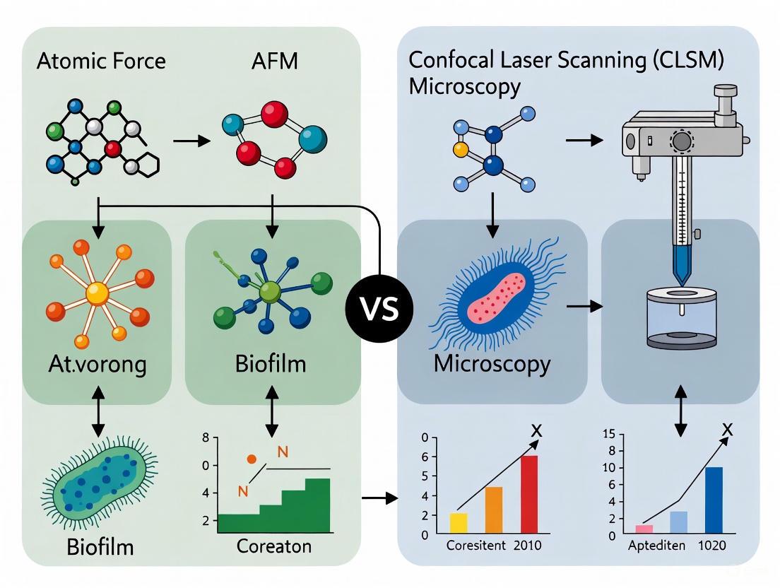
AFM vs. CLSM for Biofilm Thickness: A Correlative Microscopy Guide for Quantitative Research
Accurately measuring biofilm thickness is critical for understanding microbial resistance, mass transfer limitations, and the efficacy of antimicrobial treatments.
AFM vs. CLSM for Biofilm Thickness: A Correlative Microscopy Guide for Quantitative Research
Abstract
Accurately measuring biofilm thickness is critical for understanding microbial resistance, mass transfer limitations, and the efficacy of antimicrobial treatments. This article provides a comprehensive comparison of Atomic Force Microscopy (AFM) and Confocal Laser Scanning Microscopy (CLSM) for the quantitative analysis of biofilm thickness, tailored for researchers and drug development professionals. We explore the foundational principles of each technique, detail methodological workflows for accurate measurement, address common troubleshooting and optimization challenges, and present a framework for validating and correlating data between these powerful tools. By synthesizing current methodologies, this guide aims to empower scientists to select and implement the most appropriate imaging strategy for their specific biofilm research applications, from basic science to clinical investigation.
Understanding Biofilm Architecture and the Principles of AFM and CLSM
The three-dimensional architecture and thickness of biofilms are critical determinants of their function, pathogenicity, and resistance to treatment. This comparison guide examines how Atomic Force Microscopy (AFM) and Confocal Laser Scanning Microscopy (CLSM) measure these key parameters, providing researchers with objective data to inform their methodological choices for biofilm thickness correlation studies. While CLSM excels at non-destructively quantifying 3D architecture and live microbial distributions, AFM delivers superior nanoscale mechanical property data and surface topography measurements under physiologically relevant conditions.
Biofilm Thickness: A Critical Parameter in Research
Biofilm thickness is not merely a structural characteristic but a fundamental property influencing microbial community composition, metabolic activity, and resistance mechanisms. Thicker biofilms develop steep chemical gradients of oxygen, nutrients, and pH, creating distinct microenvironments that drive microbial stratification and functional specialization [1]. Studies demonstrate that thickness alone can determine community structure, with 400μm nitrifying biofilm communities significantly differing from 50μm communities grown under identical conditions, exhibiting both increased species richness and altered nitrogen transformation rates [1].
The clinical relevance of thickness is particularly evident in oral biofilms, where mature 3-week-old biofilms show significantly higher resistance to antimicrobial treatments compared to 1-week-old biofilms, attributed to increased extracellular polymeric substances (EPS) volume and enhanced cell-cell adhesion forces that develop over time [2]. Furthermore, biofilm thickness directly impacts disease pathogenesis, as demonstrated in dental caries where acidic microenvironments (pH<5.5) persist within the interior of microcolonies, protected from salivary buffering and leading to localized enamel demineralization [3].
AFM vs. CLSM: Technical Comparison for Thickness Measurement
Fundamental Operating Principles
Table 1: Core Principle Comparison Between AFM and CLSM
| Feature | Atomic Force Microscopy (AFM) | Confocal Laser Scanning Microscopy (CLSM) |
|---|---|---|
| Physical Basis | Mechanical probing via physical cantilever tip | Optical sectioning using focused laser light |
| Resolution Range | Nanoscale (sub-nm vertical, nm lateral) | Subcellular (≈200 nm lateral, ≈500 nm axial) |
| Measurement Type | Surface topography and mechanical properties | 3D fluorescence imaging and volume reconstruction |
| Key Thickness Output | Height profile from substrate to biofilm surface | 3D volume reconstruction from optical sections |
| Sample Environment | Liquid, air, or controlled atmospheres | Typically requires immersion for high-resolution objectives |
Performance Comparison for Biofilm Applications
Table 2: Experimental Performance Metrics for Biofilm Characterization
| Parameter | AFM Performance | CLSM Performance | Experimental Evidence |
|---|---|---|---|
| Thickness Measurement | Provides precise height data but limited by tip access to substratum | Direct 3D reconstruction capability through optical sectioning | CLSM enabled quantification of biofilm volume and spatial organization [2] |
| Live Monitoring | Limited by scan speed; possible for surface topography changes | Excellent for real-time 4D imaging of developing biofilms | CLSM allows observation of biofilm formation processes in real-time [4] |
| EPS Characterization | Indirect via mechanical properties; direct adhesion force measurement | Direct visualization with specific fluorescent probes (e.g., Alexa Fluor 647-dextran) | CLSM with fluorescent dextran conjugates quantified EPS matrix volume [2] [3] |
| Surface Roughness | Quantitative nanoscale roughness measurements (RMS) | Limited surface roughness quantification capability | AFM showed 1-week-old biofilms had significantly higher surface roughness than 3-week-old biofilms [2] |
| Mechanical Properties | Direct measurement of adhesion forces, elasticity, and Young's modulus | Indirect inference from architecture | AFM measured cell-cell and cell-surface adhesion forces in oral biofilms [2] |
Experimental Workflow: AFM and CLSM Integration for Comprehensive Biofilm Analysis
Experimental Protocols for Thickness Measurement Correlation
CLSM Protocol for 3D Architecture Quantification
Sample Preparation and Staining:
- Grow biofilms on appropriate substrates (e.g., hydroxyapatite discs for oral biofilms) [2]
- Incorporate 1 mM Alexa Fluor 647-labeled dextran (10 kDa) into growth medium for EPS matrix visualization [2] [3]
- Stain live bacteria with SYTO 9 green-fluorescent nucleic acid stain (485/498 nm excitation/emission) [3]
- Rinse specimens briefly with 0.85% physiological saline to remove unbound dye [2]
Image Acquisition and 3D Reconstruction:
- Use confocal laser scanning microscope with appropriate objectives (e.g., 10×/0.45 NA or 25×/1.05 NA water immersion) [3]
- Collect optical sections at 5-μm step size from top to bottom of biofilm [2]
- Set resolution to 512 × 512 pixels for optimal detail and file size balance
- Reconstruct 3D volume stacks using software such as Imaris 7.2 or Amira 5.0.2 [2] [3]
Thickness and Volume Quantification:
- Measure biofilm thickness from Z-stack dimensional data
- Calculate biovolume and EPS volume using 3D reconstruction software algorithms
- Perform statistical analysis with appropriate sample sizes (e.g., 10+ scanning areas per group) [2]
AFM Protocol for Nanoscale Topography and Mechanics
Sample Preparation:
- Fix biofilms in 2% glutaraldehyde at 4°C for 3 minutes for structural preservation [2]
- Rinse twice with phosphate-buffered saline (PBS) to remove fixative residues
- Air-dry samples overnight in a desiccator if operating in air [2]
- For liquid operation, maintain hydration in appropriate buffer solution
Surface Topography and Roughness Analysis:
- Use contact mode AFM with sharpened silicon nitride cantilevers (nominal tip radius <20 nm) [2]
- Set scan size to 8 × 8 μm for representative area analysis
- Maintain relative humidity between 50-60% when operating in air
- Calculate root mean square (RMS) average of height deviations for surface roughness quantification [2]
Adhesion Force Measurement:
- Perform force-distance measurements at 15 Hz scanning rate in z-direction [2]
- Conduct force mapping over 64 × 64 grid points for statistical significance
- Measure both tip-cell and cell-cell interface adhesion forces
- Repeat experiments three times at three different locations per disc for reproducibility [2]
Research Reagent Solutions for Biofilm Architecture Studies
Table 3: Essential Research Materials for Biofilm Thickness and Architecture Studies
| Reagent/Equipment | Function in Biofilm Research | Specific Examples |
|---|---|---|
| Hydroxyapatite Discs | Mimics tooth enamel for oral biofilm studies; standardized substrate | Collagen-coated HA discs (0.38-inch diameter) [2] |
| Fluorescent Dextran Conjugates | EPS matrix labeling through incorporation during synthesis | Alexa Fluor 647-labeled dextran (10 kDa) [2] [3] |
| Nucleic Acid Stains | Bacterial viability assessment and biomass quantification | SYTO 9 green-fluorescent stain [2] [3] |
| AFM Cantilevers | Surface probing for topography and mechanical measurements | Silicon nitride cantilevers with nominal tip radius <20 nm [2] |
| Specialized Growth Media | Supports multispecies biofilm development under controlled conditions | Brain Heart Infusion (BHI) broth with mucin [2] [5] |
| pH-Sensitive Fluorophores | Microenvironment pH mapping within biofilm architecture | Lysosensor yellow/blue dextran conjugate (pKa~4.2) [3] |
Key Experimental Findings: Structural and Mechanical Properties
Thickness-Dependent Architectural Changes
Research demonstrates clear structural maturation in developing biofilms. In oral multispecies biofilms, the volume of both live bacteria and EPS matrix significantly increases from 1-week to 3-week maturation points [2]. Concurrently, surface roughness undergoes a significant decrease as biofilms mature, with 1-week-old biofilms showing markedly higher roughness values compared to 3-week-old biofilms [2]. This structural consolidation correlates with increased resistance to antimicrobial treatments.
Mechanical Property Development
AFM studies reveal that adhesion forces at cell-cell interfaces are significantly more attractive than those at bacterial cell surfaces in both early and mature biofilms [2]. Importantly, 3-week-old mature biofilms exhibit stronger adhesion forces at bacterial cells compared to younger biofilms [2]. These mechanical property changes contribute substantially to biofilm resilience and removal resistance.
Thickness Impact: How Biofilm Depth Influences Structure and Function
Methodological Recommendations for Drug Development Applications
For antimicrobial efficacy testing, CLSM provides critical data on penetration depth and spatial distribution of treatment effects through live-dead staining and 3D reconstruction of treated biofilms [6] [4]. When evaluating mechanical removal strategies, AFM offers unique insights into adhesion force modifications and matrix stiffness changes in response to treatment [2] [5].
The most comprehensive approach integrates both technologies, using CLSM to identify critical architectural features followed by AFM to characterize their mechanical properties at the nanoscale [5]. This combined methodology is particularly valuable for evaluating anti-biofilm agents that target matrix integrity or cellular adhesion mechanisms.
Core Principles of Atomic Force Microscopy (AFM) for Surface Topography
Atomic Force Microscopy (AFM) is a very-high-resolution type of scanning probe microscopy (SPM) with demonstrated resolution on the order of fractions of a nanometer, representing a significant advancement over the optical diffraction limit that constrains traditional microscopy techniques [7]. Unlike optical or electron microscopy, AFM does not use lenses or beam irradiation but instead gathers information by "feeling" or "touching" the surface with a mechanical probe, thereby avoiding limitations related to diffraction and aberration [7]. This unique operating principle allows AFM to provide three-dimensional topographic imaging and a wide range of surface metrology with Ångström-level height accuracy, making it uniquely versatile for nanoscale analysis across diverse scientific disciplines [8].
The technique was invented in 1986 by Gerd Binnig, Calvin Quate, and Christoph Gerber, building upon the earlier development of the scanning tunneling microscope (STM) [7] [9]. Since its invention, AFM has evolved into one of the foremost tools for imaging, measuring, and manipulating matter at the nanoscale, with applications spanning solid-state physics, semiconductor science and technology, molecular engineering, polymer chemistry and physics, surface chemistry, molecular biology, cell biology, and medicine [7]. In the specific context of biofilm research, which is crucial for medical, industrial, and environmental applications, AFM offers critically important high-resolution insights on structural and functional properties at the cellular and even sub-cellular level [10].
Core Operating Principles of AFM
Fundamental Components and Mechanism
The fundamental principle of AFM involves scanning a sharp probe mounted on a flexible cantilever across a sample surface while monitoring the interaction forces between the tip and sample [8] [11]. The core components of a typical AFM system include [7] [12]:
- A small spring-like cantilever
- A support structure carrying the cantilever
- A sharp tip (typically with a radius of curvature on the order of nanometers) fixed to the free end of the cantilever
- A detector system that records the deflection and motion of the cantilever
- A piezoelectric element that enables precise scanning movements
- A sample stage with an x-y-z drive for precise positioning
The detection system most commonly employs a "beam bounce" method where a laser beam is focused on the back of the cantilever and reflected onto a position-sensitive photodetector (PSPD) [8] [11]. As the cantilever deflects due to tip-sample interactions, the position of the laser spot on the detector changes, providing a sensitive measure of cantilever motion with nanoscale resolution [12]. This deflection is converted into an electrical signal that is processed by a control system, which adjusts the tip-sample separation to maintain a constant interaction force during scanning [7].
The following diagram illustrates the fundamental workflow of AFM operation from initial setup to final image generation:
Force Interactions and Detection
At the most fundamental level, AFM operates by measuring the forces between the probe tip and the sample surface. As the tip approaches the sample surface, several types of forces come into play [8] [11]:
- Repulsive forces: Dominant when the tip is in direct contact with the sample surface, arising from Pauli exclusion principles
- Attractive forces: Include van der Waals, electrostatic, and capillary forces that act at longer ranges
- Chemical forces: Specific interactions that can be measured with functionalized tips
The force between the probe and sample is dependent on the spring constant (stiffness) of the cantilever and the distance between the probe and the sample surface, following Hooke's Law: F = -k·x, where F is the force, k is the spring constant, and x is the cantilever deflection [11]. This typically results in measured forces ranging from nanonewtons (10⁻⁹ N) to micronewtons (10⁻⁶ N) in air [11].
The AFM detector measures the cantilever deflection and converts it into an electrical signal proportional to the displacement [7]. By maintaining a constant cantilever deflection through a feedback loop that continuously adjusts the probe-sample distance, the system can precisely map surface topography as the tip is raster-scanned across the sample [7].
Primary AFM Imaging Modes
AFM offers several imaging modes optimized for different sample types and measurement requirements. The three primary modes are contact mode, tapping mode, and non-contact mode, each with distinct advantages and limitations for specific applications.
Contact Mode AFM
Contact mode is the most fundamental AFM imaging mode where the tip remains in constant contact with the sample surface during scanning [8] [11]. In this mode:
- The cantilever scans while applying a constant force onto the sample surface [8]
- As the tip passes over surface features, the cantilever bends and deflects [8]
- The feedback loop responds by moving the Z scanner to restore the initial cantilever deflection, thereby maintaining constant applied force [8]
- By tracking the displacement of the Z scanner, the surface topography is determined [8]
Advantages: Fast scanning speed, good for rough samples, used in friction analysis [11] Disadvantages: Lateral forces can damage or deform soft samples [11]
Tapping Mode AFM
Tapping mode (also called intermittent contact mode) represents a hybrid approach that minimizes sample damage while maintaining high resolution [8] [13]. In this mode:
- The cantilever oscillates at or near its resonance frequency [11] [13]
- The tip intermittently "taps" the sample surface at the bottom of its swing [11]
- Changes in oscillation amplitude due to tip-sample interactions are used as feedback signals [8]
- By maintaining constant oscillation amplitude, a constant tip-sample interaction is preserved [11]
Advantages: Minimizes lateral forces, suitable for soft or easily damaged samples, maintains high resolution [13] Disadvantages: Slower scan speeds compared to contact mode, more challenging to implement in liquids [11]
Non-Contact Mode AFM
Non-contact mode AFM operates with the tip oscillating above the sample surface without making direct contact [8] [13]. In this configuration:
- The cantilever oscillates just above the sample surface, typically at a frequency just above its resonance frequency [8] [11]
- Long-range forces such as van der Waals forces and electrostatic forces cause changes in the oscillation amplitude, frequency, or phase [11] [13]
- These changes are used as feedback signals to map the surface topography [8]
- A precise, high-speed feedback loop prevents the tip from crashing into the surface [8]
Advantages: Minimal sample damage, suitable for soft or easily deformable samples [13] Disadvantages: Lower resolution compared to contact and tapping modes due to weaker tip-sample interactions [13]
Table 1: Comparison of Primary AFM Imaging Modes
| Operating Mode | Tip-Sample Interaction | Best For | Resolution | Risk of Sample Damage |
|---|---|---|---|---|
| Contact Mode | Constant physical contact | Hard samples, rough surfaces, friction analysis | High | High for soft samples |
| Tapping Mode | Intermittent contact | Soft materials, biological samples, polymers | High | Low |
| Non-Contact Mode | No contact, attractive forces | Soft or easily deformable samples, liquid droplets | Moderate | Very Low |
Advanced AFM Capabilities
Beyond basic topographic imaging, AFM can measure numerous other sample properties through specialized modes and techniques, making it an exceptionally versatile characterization tool.
Force-Distance Spectroscopy
Force-distance curves provide quantitative measurements of interaction forces between the AFM tip and sample surface as a function of separation distance [13]. This technique involves:
- Moving the tip toward the sample until contact is made, then retracting while recording cantilever deflection [13]
- Converting deflection into force using the cantilever's spring constant [13]
- Extracting information about adhesion, elasticity, and surface charge from the resulting curves [13]
The adhesion force is determined from the "pull-off" force required to separate the tip from the sample during retraction, while the sample's elasticity (Young's modulus) can be estimated from the slope of the force-distance curve in the contact region [13]. By mapping force-distance curves across a sample surface, spatially resolved maps of mechanical properties can be created through "force-volume imaging" [13].
Specialized Property Mapping
AFM can be extended to characterize numerous material properties through specialized modes:
- Electrical Properties: Conductive AFM (C-AFM) measures current flow between a conductive tip and electrically-biased sample [8]; Kelvin Probe Force Microscopy (KPFM) measures surface potential and work function [8]; Scanning Capacitance Microscopy (SCM) maps dopant concentrations in semiconductors [8]
- Magnetic Properties: Magnetic Force Microscopy (MFM) uses a magnetized tip to probe magnetic domain structures [8]
- Mechanical Properties: Force Modulation Microscopy (FMM) measures local hardness by monitoring changes in oscillation amplitude as the tip scans [8]; Lateral Force Microscopy (LFM) measures frictional forces by detecting torsional cantilever bending [8]; Nanoindentation determines hardness and elasticity by analyzing loading-unloading curves [8]
- Chemical Properties: Chemical force microscopy uses functionalized tips to measure specific molecular interactions [11]
Experimental Protocols for AFM Biofilm Characterization
Sample Preparation Methodology
Proper sample preparation is critical for successful AFM imaging of biofilms. The following protocol has been adapted from established methodologies in biofilm research [10]:
Substrate Selection and Treatment: Select appropriate substrates (e.g., glass coverslips, silicon wafers, or medical device materials). For controlled experiments, treat surfaces with specific coatings such as PFOTS to modify surface properties and study their effect on bacterial adhesion [10]
Biofilm Growth: Inoculate substrates with bacterial suspension in appropriate growth medium. For Pantoea sp. YR343 studies, use liquid growth medium in petri dishes containing treated coverslips [10]
Incubation: Incubate under appropriate conditions for selected time points (e.g., 30 minutes for initial attachment studies, 6-8 hours for cluster formation) [10]
Sample Rinsing: Gently rinse coverslips to remove unattached cells while preserving biofilm architecture [10]
Immobilization: For liquid imaging, securely mount samples in appropriate fluid cells. For air imaging, samples may be dried, though this may alter native biofilm structure
Large-Area AFM Imaging Protocol
Recent advances in AFM technology enable automated large-area imaging, which is particularly valuable for capturing the spatial heterogeneity of biofilms [10]:
System Setup: Implement an automated large-area AFM system capable of capturing high-resolution images over millimeter-scale areas [10]
Image Stitching: Utilize machine learning algorithms for seamless stitching of multiple high-resolution images with minimal overlap between scans [10]
Data Acquisition: Perform sequential imaging of adjacent regions with precise positioning to create comprehensive maps of biofilm organization [10]
Automated Analysis: Implement machine learning-based image segmentation and analysis methods for automated extraction of parameters such as cell count, confluency, cell shape, and orientation [10]
Key Research Reagents and Materials
Table 2: Essential Research Reagents and Materials for AFM Biofilm Studies
| Item | Specification/Function | Application Example |
|---|---|---|
| AFM Probes | Si₃N₄ or Si cantilevers with sharp tips (tip radius <10 nm); specific spring constants (typically 0.1-1 N/m) for different modes [11] | Contact mode: softer cantilevers (∼0.1 N/m); Tapping mode: resonant frequency ∼100-300 kHz [11] |
| Substrate Materials | Glass coverslips, silicon wafers, titanium discs, hydroxyapatite discs [10] [14] | PFOTS-treated glass for studying surface modification effects on bacterial adhesion [10] |
| Surface Treatment Reagents | PFOTS (perfluorooctyltrichlorosilane) and other surface modifiers to control surface properties [10] | Creating gradient-structured surfaces to study how varying surface properties influence bacterial attachment [10] |
| Immobilization Agents | Poly-L-lysine, glutaraldehyde, or other crosslinkers for sample fixation | Securing samples to substrates while preserving native structure |
| Buffer Solutions | Phosphate-buffered saline (PBS) or appropriate physiological buffers | Maintaining hydrated conditions for biological imaging |
| Calibration Samples | Gratings with known pitch and height, reference samples with characterized topography | Verifying instrument calibration and tip condition |
AFM vs. CLSM for Biofilm Thickness Measurement Correlation
Technical Comparison for Biofilm Analysis
When evaluating AFM against Confocal Laser Scanning Microscopy (CLSM) for biofilm thickness measurement, each technique offers distinct advantages and limitations that make them complementary rather than directly competitive:
AFM Strengths for Biofilm Analysis:
- Superior resolution at the nanoscale, capable of visualizing individual bacterial cell structures (∼2 μm length, ∼1 μm diameter), flagella (∼20-50 nm height), and extracellular polymeric substances [10]
- Quantitative height measurement with Ångström-level accuracy [8]
- Ability to operate under physiological conditions (liquid environment, controlled temperature) [10] [15]
- Simultaneous topographic and mechanical property mapping [8] [10]
- No requirement for fluorescent staining, avoiding potential artifacts from sample labeling [10]
AFM Limitations for Biofilm Analysis:
- Limited maximum scan size (typically <100 μm) restricted by piezoelectric actuator constraints [10]
- Slow imaging speed compared to optical techniques, limiting temporal resolution for dynamic processes [13]
- Difficulty imaging steep or overhanging features due to tip geometry constraints [13]
- Potential tip-sample convolution effects that can distort feature dimensions [13]
CLSM Strengths for Biofilm Analysis:
- Rapid 3D imaging of larger areas (millimeter scale) [10] [16]
- Non-invasive optical sectioning capability for true 3D reconstruction [16]
- Ability to use multiple fluorescent labels for chemical specificity [16]
- Compatibility with live-cell imaging over extended time periods [16]
CLSM Limitations for Biofilm Analysis:
- Resolution limited by diffraction (∼200 nm lateral, ∼500 nm axial) [7]
- Requires fluorescent staining of cells or biomolecules, which may alter biofilm properties [10]
- Cannot directly measure mechanical properties [10]
Correlation Study Methodology
For correlative AFM-CLSM biofilm thickness measurements, the following integrated workflow provides optimal results:
Sample Preparation: Grow biofilms on transparent substrates suitable for both techniques. For multispecies biofilms, use appropriate fluorescent labels compatible with CLSM
CLSM Imaging: First perform CLSM imaging to identify regions of interest across larger areas and obtain 3D structural information
AFM Imaging: Select representative regions identified by CLSM for high-resolution AFM topography mapping
Data Correlation: Register and correlate height measurements from both techniques, accounting for differences in resolution and sampling areas
Multimodal Analysis: Combine chemical information from CLSM with nanomechanical data from AFM to develop comprehensive structure-function relationships
The following diagram illustrates this integrated approach to correlative microscopy for biofilm analysis:
Table 3: Quantitative Comparison of AFM and CLSM for Biofilm Thickness Measurement
| Parameter | AFM | CLSM |
|---|---|---|
| Lateral Resolution | 1-10 nm [13] | ~200 nm (diffraction-limited) [7] |
| Vertical Resolution | 0.1 nm [13] | ~500 nm [16] |
| Maximum Field of View | ~100 μm [10] | Millimeter scale [10] |
| Height Measurement Accuracy | Ångström-level [8] | Limited by optical sectioning thickness |
| Sample Preparation Requirements | Minimal; possible in native state [10] | Fluorescent staining typically required [10] |
| Imaging Environment | Air, liquid, controlled conditions [9] | Typically limited to transparent media |
| Measurement Type | Surface topography only | Bulk volume imaging |
| Additional Information | Mechanical, electrical properties [8] | Chemical specificity with staining [16] |
| Best Application | Nanoscale surface features, mechanical properties | Large-scale architecture, chemical composition |
Atomic Force Microscopy provides unparalleled capabilities for high-resolution surface topography characterization, with particular relevance to biofilm research where nanoscale features and mechanical properties play crucial functional roles. While AFM offers exceptional resolution and the ability to measure multiple physical properties simultaneously, its limitations in imaging speed and field of view make it complementary to techniques like CLSM rather than a direct replacement.
The integration of AFM with complementary imaging technologies, combined with recent advancements in automation and machine learning, is paving the way for more comprehensive biofilm characterization [10] [16]. These correlative approaches leverage the respective strengths of each technique - using CLSM to identify regions of interest across large areas and AFM to provide detailed nanoscale analysis of selected regions - ultimately providing more complete understanding of biofilm structure-function relationships.
For researchers investigating biofilm thickness and organization, the optimal approach involves strategic use of both AFM and CLSM in a correlative framework, acknowledging that each technique provides different but complementary information about these complex microbial communities. This multimodal methodology enables researchers to connect nanoscale surface properties and mechanical behaviors with larger-scale architectural organization, ultimately advancing our understanding of biofilm development, resilience, and function in clinical, industrial, and environmental contexts.
Core Principles of Confocal Laser Scanning Microscopy (CLSM) for 3D Optical Sectioning
Confocal Laser Scanning Microscopy (CLSM) represents a pivotal advancement in optical imaging, enabling high-resolution three-dimensional analysis of biological specimens. The core principle of confocal microscopy is the use of spatial filtering to eliminate out-of-focus light, providing a significant improvement over conventional widefield fluorescence microscopy [17]. This technique allows researchers to generate sharp, optical sections from thick, scattering tissues, which can then be reconstructed into detailed 3D models [17]. For biofilm thickness measurement correlation research, CLSM provides non-destructive, in-situ analysis capabilities that are essential for understanding the complex architecture of microbial communities without the need for physical sectioning or extensive sample preparation that might alter native structures.
The fundamental advantage of CLSM in biofilm studies lies in its ability to perform optical sectioning while maintaining sample integrity. Unlike atomic force microscopy (AFM), which provides exquisite surface detail and nanomechanical properties but lacks penetration depth, CLSM enables researchers to visualize the complete three-dimensional organization of biofilms, from the attachment surface to the biofilm-liquid interface [5]. This capability is particularly valuable for investigating structure-property relationships in complex oral biofilms and other microbial systems where the vertical dimension critically influences function and pathogenicity [5].
Core Principles of CLSM
Optical Sectioning and Out-of-Focus Light Rejection
The defining feature of confocal microscopy is its ability to eliminate blur from out-of-focus planes, a limitation inherent in conventional widefield fluorescence microscopy. In thick samples where the objective lens lacks sufficient depth of focus, light from sample planes above and below the focal plane contributes haze and reduces resolution in widefield systems [17]. CLSM addresses this fundamental challenge through a clever optical arrangement that ensures only light from the focal plane is detected.
The confocal principle employs "double focusing" with two pinholes positioned in conjugate image planes, making them "confocal" [17]. The first pinhole is placed in front of the light source to create a point source, which is focused onto the specimen by the objective lens. The second pinhole is positioned in front of the detector, precisely at the image plane of the illuminated spot. This configuration ensures that out-of-focus rays from the illuminated sample are rejected before they reach the detector [17]. The result is dramatically improved image contrast and effective optical sectioning capability, allowing researchers to collect data from precisely defined focal planes within thick specimens.
Figure 1: CLSM optical pathway showing confocal principle
Resolution and Contrast Fundamentals
In confocal microscopy, resolution is intrinsically linked to contrast, with both determining the ability to distinguish fine specimen details. The relationship between contrast and resolution is particularly important when imaging biofilms, which often exhibit subtle variations in refractive index and fluorophore distribution. Resolution is formally defined as the minimum separation between two points that results in a certain level of contrast between them [18].
The theoretical resolution limits of CLSM are governed by the same fundamental principles as conventional microscopy but are enhanced by the confocal effect. The lateral resolution is described by the equation:
R_lateral = 0.4λ/NA [17]
Where λ represents the emission light wavelength and NA is the objective's numerical aperture. In practice, the best lateral resolution achievable is approximately 0.2 μm [17].
Axial resolution, critical for optical sectioning and 3D reconstruction, follows the relationship:
R_axial = 1.4λη/(NA)² [17]
Where η is the refractive index of the mounting medium. The best achievable axial resolution is approximately 0.6 μm [17]. It's important to note that axial resolution in confocal microscopy remains inferior to lateral resolution, as in widefield fluorescence microscopy [17].
The point spread function (PSF) is a critical concept for understanding resolution in CLSM. The PSF describes the three-dimensional intensity distribution of light from a point source after passing through the optical system [18]. In confocal fluorescence configurations, pointwise illumination scanning and pointwise detection mean only fluorophores in the shared volume of the illumination and detection point spread functions are detected [18]. The confocal intensity point spread function is therefore the product of the independent illumination intensity and detection intensity point spread functions, resulting in improved resolution compared to widefield microscopy.
CLSM System Components and Performance
Essential Hardware Components
Modern confocal microscopy systems integrate several critical components that work in concert to achieve optical sectioning:
Laser Illumination Sources: Contemporary CLSM systems typically employ diode lasers, fiber lasers, and solid-state lasers, which offer greater stability, more uniform output, less heat production, and a broader range of visible wavelengths compared to traditional gas lasers (argon and helium-neon) [17].
Scanning Mechanisms: Most systems use scanning galvanometer mirrors to sweep the laser beam across the sample in x and y directions [17]. An acousto-optic tunable filter (AOTF) rapidly turns lasers on and off, attenuates laser power, and selects wavelength during imaging [17].
Detection System: Highly sensitive photomultiplier tubes (PMTs) remain the primary detectors in most CLSM systems due to their amplification capabilities and compatibility with the light-rejecting nature of confocal microscopy [17]. System sensitivity can vary by wavelength, with reported maximum sensitivity values of approximately 4% for 488 nm, 2.5% for 568 nm, 20% for 647 nm, and 19% for 365 nm laser light when measured using 10-μm Spherotech beads [19].
Pinhole Apertures: The confocal pinhole is arguably the most critical component, typically positioned in a conjugate image plane to exclude out-of-focus light. The size of the pinhole represents a tradeoff between light collection efficiency and resolution – for dim samples, the pinhole may be opened to improve contrast at the cost of resolution, while closing the pinhole improves resolution at the cost of signal-to-noise [17].
Performance Considerations and Quality Assurance
Several factors influence CLSM performance in practical applications, particularly for quantitative measurements like biofilm thickness:
Laser Stability: Laser power stability can vary between 3% and 30% due to factors including incompatibility of fiber-optic polarization with laser polarization, thermal instability of the AOTF, and inherent laser noise [19].
System Sensitivity and Noise: The signal-to-noise ratio directly impacts image quality and measurement accuracy. PMT performance can be assessed using the coefficient of variation (CV) concept to quantify image noise [19].
Spectral Registration: Accurate alignment of different fluorescence channels is essential for multi-color imaging, requiring regular calibration to ensure proper overlay of signals from different fluorophores [19].
Comprehensive quality assurance tests are recommended to ensure CLSM systems deliver reproducible intensity measurements with optimal image quality. These tests should evaluate dichroic reflectivity, field illumination, lens performance, laser power output, spectral registration, axial resolution, laser stability, PMT reliability, and system noise [19].
Comparative Analysis: CLSM vs. AFM for Biofilm Characterization
The selection between CLSM and AFM for biofilm research depends heavily on the specific research questions and the type of information required. Each technique offers distinct advantages and limitations that make them suitable for complementary aspects of biofilm analysis.
Table 1: Comparison of CLSM and AFM for biofilm characterization
| Parameter | Confocal Laser Scanning Microscopy (CLSM) | Atomic Force Microscopy (AFM) |
|---|---|---|
| Resolution | Lateral: ~0.2 μmAxial: ~0.6 μm [17] | Sub-nanometer lateral resolution [5] |
| Penetration Depth | Up to several hundred microns depending on tissue scattering [17] | Surface topology only (nanometers) [5] |
| Measurement Type | Non-contact optical sectioning [17] | Physical contact with surface [5] |
| Key Outputs | 3D architecture, thickness, volume, live-dead distribution [20] | Young's modulus, adhesion forces, surface morphology [5] |
| Sample Environment | Hydrated, physiological conditions possible [20] | Typically submerged in liquid [5] |
| Multiplexing Capability | Simultaneous multi-channel fluorescence [21] | Limited to mechanical and topographical data |
| Throughput | Moderate - requires scanning of optical sections [17] | Low - point-by-point surface mapping [5] |
| Biofilm Thickness Measurement | Direct measurement from z-stacks [20] | Indirect, requires destructive cross-sectioning |
Table 2: Performance characteristics of CLSM for biofilm imaging
| Performance Metric | Typical Value | Impact on Biofilm Imaging |
|---|---|---|
| Lateral Resolution | 0.2 μm [17] | Resolves individual bacterial cells and microcolonies |
| Axial Resolution | 0.6 μm [17] | Determines optical section thickness for 3D reconstruction |
| System Sensitivity | 2.5-20% (wavelength-dependent) [19] | Affects signal from dim fluorophores in biofilm matrix |
| Laser Stability | 3-30% variation [19] | Impacts quantitative intensity measurements over time |
| Multi-color Registration | Requires calibration [19] | Essential for co-localization studies in mixed-species biofilms |
Correlation Between CLSM and AFM Data
A multi-scale approach combining OCT and AFM has revealed new structure-property relationships in oral biofilms that were unattainable using either technique independently [5]. This integrated methodology demonstrates how CLSM and AFM provide complementary data:
CLSM excels at visualizing the mesoscale architecture of biofilms, including the distribution of extracellular polymeric substances (EPS), voids, channels, and microcolonies in 3D space [5]. This structural information is essential for interpreting AFM mechanical measurements, as the mechanical properties of biofilms vary significantly between regions of high and low EPS density [5].
AFM provides nanomechanical properties such as Young's modulus and adhesion forces, which are influenced by biofilm composition and organization [5]. For instance, studies have shown that increasing sucrose concentration decreases Young's modulus and increases cantilever adhesion relative to the biofilm, while increasing biofilm age decreases adhesion [5].
For thickness measurement correlation studies, CLSM provides direct, non-destructive measurement of biofilm height through z-stack imaging, while AFM typically provides higher resolution surface topography but limited depth penetration. The combination of both techniques enables researchers to correlate mechanical properties with 3D structural features throughout the biofilm depth.
Experimental Protocols for Biofilm Imaging
Sample Preparation for CLSM Biofilm Analysis
Proper sample preparation is critical for accurate biofilm characterization using CLSM:
Substrate Selection: Biofilms can be grown on various substrates relevant to research questions, including hydroxyapatite (HAP) discs for oral biofilms [5], glass coverslips, or medical device materials.
Fluorescence Labeling: Multiple staining strategies enable visualization of different biofilm components:
- Viability Stains: SYTO stains for total cells combined with propidium iodide for dead cells provide live-dead discrimination [20].
- EPS Components: Specific polysaccharide stains (e.g., concanavalin A conjugates) or general nucleic acid stains (e.g., TOTO-3, YOYO-1) highlight matrix components [21].
- Immunofluorescence: Antibody labeling targets specific bacterial species or biofilm matrix proteins [21].
Mounting Media: Use appropriate mounting media with refractive index matching objective lens specifications (typically η ≈ 1.33 for aqueous samples or 1.51 for oil immersion) [17].
CLSM Imaging Protocol for Biofilm Thickness Measurement
A standardized acquisition protocol ensures reproducible thickness measurements:
System Calibration: Verify laser power stability, spectral registration, and axial calibration using reference beads [19].
Objective Selection: Choose high numerical aperture objectives (typically NA ≥ 1.2) for optimal resolution and light collection [17].
Z-stack Acquisition:
- Set initial focal plane below biofilm attachment surface
- Set final focal plane above biofilm-liquid interface
- Use step size ≤ 0.5 × axial resolution (typically 0.3 μm or smaller) [17]
- Maintain constant laser power and detector settings throughout stack
Multi-channel Imaging: For simultaneous multi-parameter imaging, ensure minimal cross-talk between channels and sequential scanning when fluorophore emission spectra overlap [21].
Controls: Include unstained controls for autofluorescence assessment and single-stained controls for spectral cross-talk correction.
3D Reconstruction and Thickness Analysis
Post-processing transforms z-stacks into quantitative thickness measurements:
Deconvolution: Apply iterative deconvolution algorithms to improve resolution and contrast using measured or theoretical point spread functions [18].
Segmentation: Use intensity thresholding or machine learning approaches to distinguish biofilm from background.
Height Measurement: Calculate biofilm thickness as the vertical distance between the attachment surface and the biofilm-liquid interface for each (x,y) position.
Statistical Analysis: Generate thickness distribution maps and extract parameters such as average thickness, maximum thickness, and roughness coefficient.
Research Reagent Solutions for CLSM Biofilm Imaging
Table 3: Essential reagents and materials for CLSM biofilm analysis
| Reagent/Material | Function | Application Notes |
|---|---|---|
| SYTO 9 Green Fluorescent Nucleic Acid Stain | Labels all bacterial cells | Often combined with propidium iodide for viability assessment [20] |
| Propidium Iodide | Labels membrane-compromised cells | Penetrates only dead cells; used with SYTO 9 for live-dead staining [20] |
| Concanavalin A Tetramethylrhodamine Conjugate | Binds α-mannopyranosyl and α-glucopyranosyl residues in EPS | Specific polysaccharide labeling in biofilm matrix [20] |
| TOTO-3 Iodide | Far-red fluorescent nucleic acid stain | Excitation/emission: 642/661 nm; useful for triple-labeling [21] |
| YOYO-1 Iodide | Green fluorescent nucleic acid stain | Excitation/emission: 491/509 nm; high DNA affinity [21] |
| Hydroxyapatite Discs | Mineralized substrate for biofilm growth | Mimics tooth enamel for oral biofilm studies [5] |
| Brain Heart Infusion (BHI) Medium | Nutrient-rich growth medium | Supports robust biofilm formation with 5% sucrose [5] |
| Artificial Saliva Medium | Nutrient-poor growth medium | Maintains oral biofilms with 0.1% sucrose [5] |
Advanced Applications and Multi-Technique Approaches
The integration of CLSM with complementary techniques like AFM provides a more comprehensive understanding of biofilm systems than either approach alone. This multi-scale methodology enables researchers to correlate structural features with mechanical properties and biochemical composition.
CLSM's capability for long-term live-cell imaging makes it ideal for investigating biofilm development dynamics, including initial attachment, microcolony formation, and maturation. When combined with AFM's capacity to map nanomechanical properties at different stages of biofilm development, researchers can establish critical structure-function relationships [5].
For biofilm thickness correlation studies specifically, the combination of techniques validates measurements across different scales. CLSM provides the overall architectural context, while AFM offers ultra-high resolution surface details and mechanical properties at specific locations. This correlation is particularly valuable for understanding how local mechanical properties vary with position in the biofilm and how these relate to overall 3D organization.
Future developments in correlative microscopy will likely focus on improving spatial registration between CLSM and AFM datasets, enabling more precise matching of structural features with mechanical properties. Additionally, the integration of spectroscopic techniques with CLSM could provide simultaneous chemical and structural information, further enhancing our understanding of biofilm organization and function.
The study of biofilms, structured communities of microorganisms encased in an extracellular polymeric matrix, is crucial across clinical and industrial domains due to their remarkable resistance to antimicrobial treatments [20]. Accurately measuring biofilm architecture, particularly thickness, is essential for understanding their resilience and developing effective eradication strategies. Among the most powerful tools for this purpose are Atomic Force Microscopy (AFM) and Confocal Laser Scanning Microscopy (CLSM). This guide provides a comparative analysis of these two techniques, framing them within biofilm thickness measurement correlation research. It is designed to help researchers, scientists, and drug development professionals select the appropriate method based on their specific experimental needs, whether for nanoscale topographic mapping or for dynamic, three-dimensional visualization of live biofilms.
Core Principles and Technical Specifications
Atomic Force Microscopy (AFM) is a scanning probe microscopy technique that provides topographical imaging by physically scanning a nanometric tip over a sample surface. It measures the interaction force between the tip and the sample, achieving a horizontal resolution of 0.1 nm and a vertical resolution of 0.01 nm [22]. AFM can be operated in various modes, including contact mode and tapping mode, the latter being particularly suited for soft biological samples as it minimizes sample damage by oscillating the cantilever [23] [22]. A key strength of AFM is its ability to perform in liquid conditions, allowing for the investigation of biofilms in a hydrated state [23].
Confocal Laser Scanning Microscopy (CLSM), in contrast, is an optical imaging technique. It uses a spatial pinhole to block out-of-focus light, enabling the reconstruction of high-resolution three-dimensional images from optical sections taken at different depths within a sample [6] [24]. CLSM allows for the quantitative evaluation of structural parameters like biovolume, thickness, and roughness, and is capable of real-time 4-D (time-lapse) imaging [6]. Its resolution is limited by the diffraction of light, but it excels at visualizing the spatial arrangement of different components within a biofilm through the use of specific fluorescent stains [6] [25].
Table 1: Comparative Technical Specifications of AFM and CLSM for Biofilm Research
| Feature | Atomic Force Microscopy (AFM) | Confocal Laser Scanning Microscopy (CLSM) |
|---|---|---|
| Primary Measurement | Topography, mechanical properties | 3D fluorescence architecture, cell viability |
| Resolution (Lateral) | ~0.1 nanometers (nm) [22] | Diffraction-limited (~200 nanometers) [6] |
| Resolution (Vertical) | ~0.01 nanometers [22] | Lower than lateral resolution [25] |
| Imaging Environment | Air or liquid (physiological conditions) [23] [22] | Typically liquid (physiological conditions) |
| Key Outputs | Height, roughness, adhesion, stiffness/elasticity [22] | Biovolume, thickness, roughness, live/dead cell distribution [6] |
| Sample Preparation | Can be minimal for live cells in liquid [23]; may require fixation for high-resolution | Often requires fluorescent staining or labeling [6] |
| Throughput | Low (small scan area, slow scan speed) [6] | Moderate to high (can image larger areas faster) |
Experimental Protocols for Biofilm Analysis
AFM Protocol for Topography and Mechanical Properties
The following protocol outlines the key steps for assessing biofilm topography and stiffness using AFM, which provides critical data on surface morphology and mechanical integrity [26] [22].
- Biofilm Growth: Grow biofilms on a suitable substrate (e.g., glass, titanium) under relevant conditions (static or dynamic flow) [14]. Fluid shear during growth significantly impacts biofilm physical characteristics; biofilms grown under high shear are typically thinner and stiffer [26].
- Sample Mounting: For live-cell imaging in liquid, stabilize hydrated biofilms. One effective method is using a thin layer of agarose and lean media on a glass cover slip to prevent dehydration and lateral movement during scanning [23].
- AFM Imaging and Force Spectroscopy:
- Mode Selection: Use tapping mode for high-resolution topographic imaging of soft biofilms to minimize shear forces and sample damage [23] [22].
- Data Acquisition: Raster-scan the probe over the biofilm surface. The vertical (z) position of the probe is adjusted to maintain a constant interaction force, generating a topographic image [22].
- Mechanical Property Measurement: Employ force spectroscopy mode. Approach the AFM tip to the biofilm surface until contact, indent the sample to a set force, and then retract. The resulting force-distance (FD) curve is analyzed using models (e.g., Hertz model) to quantify local mechanical properties such as elastic modulus (stiffness) and adhesion forces [22]. Creep compliance, a measure of how a material deforms under constant stress, can also be calculated from these curves to characterize viscoelasticity [26].
- Data Analysis: Use dedicated software to extract quantitative parameters from topographic images (e.g., root-mean-square roughness, thickness) and from FD curves (e.g., stiffness, adhesion force) [6] [22].
CLSM Protocol for 3D Architecture and Viability
This protocol describes the standard procedure for visualizing the 3D structure and assessing cell viability within biofilms using CLSM [6] [25].
- Biofilm Growth: Grow biofilms in a system compatible with CLSM, such as flow cells, microtiter plates, or glass-bottom dishes [25] [14]. These models allow biofilms to develop under controlled hydrodynamic conditions that mimic in vivo environments [14].
- Staining (Fixation or Live):
- For Fixed Samples: Fix biofilms with glutaraldehyde or paraformaldehyde. Subsequently, stain with appropriate fluorescent dyes. Common stains include DAPI (for nucleic acids, labeling all cells) and Crystal Violet (for negatively charged cell membranes and matrix components) [25].
- For Live-Cell Imaging: Use fluorescent proteins (e.g., GFP) expressed by the bacteria or viability stains (e.g., SYTO 9/propidium iodide in a LIVE/DEAD BacLight assay) to discriminate between live and dead cells in real-time without fixation [6].
- Image Acquisition: Place the sample on the CLSM stage. Use appropriate laser wavelengths to excite the fluorescent probes. Collect a Z-stack—a series of images taken at sequential focal planes through the depth of the biofilm. The step size between planes (e.g., 0.13 µm) determines the Z-resolution [25].
- 3D Reconstruction and Quantification: Use CLSM software to reconstruct a 3D model from the Z-stack. The software can then calculate critical parameters such as biovolume (total volume of biomass), average thickness (mean distance from the substrate to the biofilm surface), and surface roughness (a measure of biofilm heterogeneity) [6].
Research Reagent Solutions
The table below lists key reagents and materials essential for conducting the AFM and CLSM experiments described in this guide.
Table 2: Essential Research Reagents and Materials for AFM and CLSM Biofilm Studies
| Item Name | Function/Application | Relevant Technique |
|---|---|---|
| Glass-Bottom Dish | Provides a transparent, flat substrate for biofilm growth and high-resolution imaging. | CLSM, AFM [25] |
| Flow Cell System | Enables dynamic growth of biofilms under hydrodynamic shear forces, promoting maturation. | CLSM, AFM [14] |
| Crystal Violet (CV) | Fluorescent stain for negatively charged cell membranes and extracellular matrix components. | CLSM [25] |
| DAPI | Fluorescent stain that binds strongly to DNA, labeling cell nuclei in bacteria. | CLSM [25] |
| Fluorescent Proteins (e.g., GFP, YFP) | Genetic tags for labeling specific bacterial strains or proteins in live-cell imaging. | CLSM [27] [23] |
| Agarose | Used to create a hydrated gel matrix for immobilizing live bacterial cells during in-air AFM imaging. | AFM [23] |
| Polystyrene Microspheres | Micron-sized particles used as tracers for microrheology measurements within the biofilm matrix. | CLSM [26] |
| LIVE/DEAD BacLight Viability Kit | Contains two nucleic acid stains (SYTO 9 and PI) to distinguish between live and dead bacterial cells. | CLSM [6] |
Integrated Workflow for Correlative Microscopy
The strengths of AFM and CLSM are highly complementary. A correlative workflow that integrates both techniques on the same sample provides a more comprehensive understanding of biofilm structure and function than either method could alone [25]. This approach allows researchers to correlate the nanoscale topography and mechanical properties measured by AFM with the internal 3D structure and cellular arrangement revealed by CLSM.
Correlative AFM-CLSM Workflow for Biofilm Analysis
This integrated methodology was effectively demonstrated in a study on marine bacterial biofilms, where the same area of a biofilm on glass was observed with both SICM (a technique analogous to AFM) and CLSM. This simultaneous measurement clarified the three-dimensional morphology, the arrangement of bacteria, and differences in local ion conductivity, achievements that were not possible with a single microscope [25].
AFM and CLSM are not competing but complementary pillars in modern biofilm research. The choice between them is not a matter of which is superior, but which is more appropriate for the specific research question at hand. AFM is unparalleled for investigations requiring nanoscale resolution of surface topography and quantitative measurements of mechanical properties like stiffness and adhesion. CLSM is the definitive tool for visualizing the internal 3D architecture of biofilms, monitoring dynamic processes in real-time, and locating specific cellular components through fluorescence. For the most holistic insights, a correlative approach that sequentially uses both techniques on the same sample provides a powerful strategy to bridge the gap between nanoscale structure and biological function, ultimately accelerating the development of novel anti-biofilm therapies.
Practical Protocols: Measuring Biofilm Thickness with AFM and CLSM
Atomic Force Microscopy (AFM) is a powerful nanoscale characterization tool that has become dominant for measuring mechanical properties and topography in soft matter, including microbial biofilms [28]. In the context of biofilm thickness measurement correlation research, AFM provides exceptional vertical resolution capable of sub-nanometer accuracy, complementing the volumetric imaging capabilities of Confocal Laser Scanning Microscopy (CLSM) [29]. Unlike optical techniques such as CLSM, which rely on fluorescence principles and have resolution limits constrained by light diffraction [29], AFM operates as a mechanical microscope that transforms tip-sample interaction forces into precise topographical maps [28]. This technical guide presents a standardized workflow for AFM operation, from initial probe selection to final height data acquisition, specifically contextualized for biofilm analysis where delicate structures and heterogeneous composition demand specialized approaches.
AFM Fundamental Principles and Comparison Framework
Core Operating Principle
AFM functions by raster-scanning a sharp tip mounted on a flexible cantilever across a sample surface. The interaction forces between tip and sample cause cantilever deflection, which is tracked via a laser beam reflected onto a position-sensitive photodiode [30]. This deflection information is transformed into three-dimensional topographical data with exceptional Z-axis resolution, making it particularly valuable for accurate biofilm thickness measurements [28].
Comparative Advantages for Biofilm Analysis
Mechanical Property Correlation: While CLSM provides excellent volumetric visualization of hydrated biofilms through optical sectioning [29], AFM uniquely generates simultaneous nanomechanical property maps alongside topography. This allows researchers to correlate biofilm thickness with mechanical properties such as elasticity and adhesion, providing insights into how extracellular polymeric substance (EPS) distribution influences structural integrity [5] [28].
Unmatched Vertical Resolution: AFM's Z-axis resolution surpasses optical diffraction limits that constrain CLSM [29], enabling detection of sub-nanometer height variations critical for analyzing thin early-stage biofilms and EPS layer organization [28].
AFM Operating Principle: The core feedback loop maintains constant tip-sample interaction while scanning, generating high-resolution topography data.
Detailed AFM Workflow for Biofilm Characterization
Probe Selection and Calibration
Cantilever Selection Criteria: For soft, hydrated biofilms, cantilevers with appropriate spring constants are essential to prevent sample damage. Typical biofilms require cantilevers with spring constants of 0.01-1 N/m for optimal force sensitivity without excessive indentation [31]. Sharp tips (radius <10 nm) provide high spatial resolution for mapping individual matrix components, while spherical tips (radius 1-10 μm) are preferred for quantitative nanomechanical mapping as they minimize local strain and provide more reliable mechanical property data [5] [31].
Comprehensive Calibration Protocol: Accurate calibration is fundamental for quantitative measurements. The calibration workflow involves:
- Spring Constant Calibration: Determine the exact cantilever stiffness using thermal tune or reference sample methods [31].
- Photodetector Sensitivity: Measure the deflection sensitivity on a rigid reference sample (e.g., silicon wafer) to convert voltage to distance [32].
- Scanner Calibration: Verify X, Y, and Z piezos using calibration gratings with known dimensions. For height measurements critical to biofilm thickness studies, Z-axis calibration requires particular attention using standards with characterized step heights (e.g., 100 nm for general topography, 0.75-1.5 nm for 2D materials or molecular-scale features) [32].
- Probe Functionalization (Optional): For specific applications, tips may be modified with colloidal particles or chemical functionalization. For biofilm studies, 10 μm borosilicate spheres are often attached using UV-curing resin to create colloidal probes for nanomechanical mapping [5].
Sample Preparation and Mounting
Biofilm samples for AFM analysis are typically grown on flat substrates such as hydroxyapatite (mimicking tooth enamel), glass, or medical device-relevant materials [5] [33]. Key considerations include:
- Substrate Flatness: The substrate should be significantly smoother than the biofilm features of interest to avoid topography artifacts.
- Hydration Maintenance: Biofilms must remain hydrated throughout analysis. Samples are typically immersed in phosphate-buffered saline (PBS) or growth medium during measurement [5].
- Fixation Considerations: While live biofilms can be analyzed, mild chemical fixation (e.g., 0.5-2% glutaraldehyde) may be used to preserve structure, though this can alter mechanical properties [34].
Measurement Parameter Optimization
Imaging Mode Selection: The optimal imaging mode depends on biofilm properties and research objectives:
| Imaging Mode | Principle | Biofilm Applications | Advantages | Limitations |
|---|---|---|---|---|
| Contact Mode | Maintains constant tip-sample contact | General topography on robust biofilms | Fast scanning, simple operation | High lateral forces may damage soft samples |
| Intermittent Contact | Tip oscillates at resonance with intermittent surface contact | High-resolution imaging of delicate biofilm structures | Reduced lateral forces, excellent for soft samples | Lower scanning speed, potential for feedback instability |
| Force Volume | Acquires force-distance curves at each pixel | Nanomechanical property mapping alongside topography | Quantitative Young's modulus and adhesion data | Very slow acquisition, complex data analysis |
Key Parameter Optimization:
- Setpoint Force/Amplitude: Use the minimum necessary for stable feedback (typically 0.5-2 nN for soft biofilms) to prevent sample deformation [31].
- Scan Rate: Balance between acquisition speed and image quality. Typical rates of 0.5-2 Hz are suitable for most biofilm applications [28].
- Resolution: 256×256 to 512×512 pixels provide sufficient detail while maintaining reasonable acquisition times [5].
- Environmental Control: Maintain constant temperature and minimize acoustic vibrations which significantly affect nanoscale measurements [31].
Data Acquisition and Processing
Height Data Acquisition: The primary data consists of height values at each pixel position, generated by recording the Z-piezo motion required to maintain constant tip-sample interaction [28]. For biofilms, this produces a three-dimensional representation of surface topography from which thickness can be determined by measuring height differences between the substrate and biofilm surface.
Artifact-Free Processing: Raw AFM data requires careful processing to reveal true surface features:
- Plane Subtraction: Removes sample tilt using first or second-order polynomial fits [30].
- Flattening: Corrects for scanner bow and other low-frequency artifacts [30].
- Filtering: Application of low-pass filters to reduce high-frequency noise while preserving relevant features [30].
Critical processing choices significantly impact interpretation. As demonstrated in bitumen studies (a material with biofilm-like complexity), different background subtraction algorithms can yield substantially different morphological interpretations [30]. Researchers should apply minimal processing and document all procedures thoroughly.
Experimental Protocols for Biofilm Analysis
Force Volume Nanomechanical Mapping
The force volume mode is particularly valuable for biofilm research as it provides simultaneous topographical and mechanical property data, revealing relationships between biofilm structure and function [5] [28].
Protocol:
- Cantilever Preparation: Select a soft cantilever (0.01-0.5 N/m) and calibrate as described in section 3.1. For quantitative mechanical mapping, spherical colloid probes are preferred [5].
- Parameter Setup: Define a 16×16 to 64×64 point grid covering the region of interest. Set maximum indentation force to 1-10 nN to avoid biofilm damage [5].
- Approach/Retract Settings: Program tip approach and retraction velocities of 0.5-2 μm/s to minimize hydrodynamic effects while maintaining reasonable acquisition times [28].
- Data Acquisition: Acquire force-distance curves at each grid point. Total acquisition time typically ranges from 10-60 minutes depending on grid density [5].
- Data Analysis: Fit retraction curves with appropriate contact mechanics models (Hertz, Sneddon, or JKR for adhesive samples) to extract Young's modulus values [5] [28].
This approach was successfully applied to oral biofilms, revealing that increasing sucrose concentration decreased Young's modulus from approximately 20 kPa to 5 kPa while increasing adhesion, demonstrating how environmental factors alter biofilm mechanical properties [5].
Correlation with CLSM Measurements
Multi-Technique Validation Protocol:
- Substrate Selection: Use transparent substrates (e.g., glass coverslips) compatible with both AFM and CLSM [29].
- Sequential Imaging: First, perform CLSM analysis using appropriate fluorescent stains (e.g., FITC-ConA for polysaccharides, propidium iodide for cells) to obtain volumetric biofilm data and overall architecture [29].
- Region Matching: Identify specific regions of interest in CLSM data for subsequent high-resolution AFM analysis.
- AFM Correlation: Perform AFM scanning on matched regions to obtain high-resolution topography and mechanical properties.
- Data Integration: Correlate CLSM fluorescence intensity profiles with AFM height and mechanical property maps to establish structure-function relationships.
This multi-modal approach leverages the strengths of both techniques: CLSM provides non-invasive three-dimensional visualization of internal biofilm architecture and component distribution, while AFM delivers superior vertical resolution and nanomechanical property data [29] [28].
Comparative Technical Analysis: AFM vs. CLSM for Biofilm Thickness
| Parameter | Atomic Force Microscopy (AFM) | Confocal Laser Scanning Microscopy (CLSM) |
|---|---|---|
| Height Resolution | Sub-nanometer vertical resolution [32] [28] | Limited by optical diffraction (~200 nm axial resolution) [29] |
| Lateral Resolution | 1-10 nm under optimal conditions [28] | ~180 nm (diffraction-limited) [29] |
| Measurement Principle | Mechanical tip-sample interaction [30] | Fluorescence emission and optical sectioning [29] |
| Sample Requirements | Requires accessible surface; minimal sample prep [31] | Requires fluorescence (intrinsic or staining) [29] |
| Environment | Liquid, air, controlled environments [5] | Typically liquid for hydrated biofilms [29] |
| Depth Capability | Surface and near-surface features only [28] | Tens to hundreds of micrometers [29] |
| Measurement Type | Direct physical measurement [28] | Indirect optical inference [29] |
| Additional Data | Simultaneous nanomechanical properties [5] [28] | Chemical specificity, viability assessment [29] |
| Throughput | Slow (minutes to hours) [28] | Fast (seconds to minutes) [29] |
| Sample Risk | Potential for tip-induced damage [31] | Photobleaching, phototoxicity [29] |
Comparative Workflows: AFM and CLSM follow different pathways for biofilm thickness assessment, with potential for correlation analysis.
Essential Research Reagent Solutions
| Reagent/Equipment | Function in AFM Biofilm Research | Technical Specifications |
|---|---|---|
| Soft Cantilevers | Force-sensitive detection on delicate biofilms | Spring constant: 0.01-0.5 N/m; Tip radius: <10 nm (sharp), 1-10 μm (colloidal) [5] [31] |
| Calibration Gratings | Scanner calibration for accurate dimensional measurements | Pitch: 1-10 μm (XY); Step height: 0.75-1.5 nm to 100+ nm (Z) [32] |
| Hydroxyapatite Substrates | Biologically relevant surface for oral biofilm growth | 5 mm diameter discs pressed from <75 μm HAP powder [5] |
| Phosphate Buffered Saline | Hydration medium maintaining physiological conditions | 1X concentration, isotonic for biological samples [5] |
| UV-Curing Resin | Colloidal probe attachment for nanomechanical mapping | Used to attach 10 μm borosilicate spheres to tipless cantilevers [5] |
| SiC Step Height Standards | Nanometer-scale Z-axis calibration | 0.75 nm or 1.5 nm step heights for high-accuracy height calibration [32] |
AFM provides unparalleled vertical resolution and nanomechanical property mapping capabilities for biofilm thickness correlation studies, complementing CLSM's volumetric imaging strengths. The step-by-step workflow presented—from careful probe selection through optimized data acquisition and processing—enables researchers to obtain quantitative, nanoscale topographical data from delicate biofilm structures. When correlated with CLSM data, AFM measurements provide a comprehensive understanding of biofilm structure-function relationships, advancing research in antimicrobial development, biofilm physiology, and material-associated infections. For optimal results, researchers should implement rigorous calibration protocols, select appropriate imaging modes based on specific research questions, and apply careful data processing to avoid artifacts that could compromise thickness measurements.
Confocal Laser Scanning Microscopy (CLSM) has emerged as a powerful non-invasive technique for generating high-resolution, three-dimensional images of biological specimens, including bacterial biofilms. Unlike conventional wide-field microscopy, CLSM uses a laser beam to create very thin focal planes, enabling optical sectioning through samples from the top to the bottom [35]. This capability is particularly valuable for examining the complex architecture of biofilms, which are highly structured microbial colonies embedded in a hydrated extracellular polymeric substance (EPS) matrix [2]. When applied to biofilm research, CLSM allows researchers to quantitatively study bacterial viability, visualize 3D structure, and investigate EPS production without the need for extensive sample preparation that alters the biofilm's physiological state [20] [36].
The application of CLSM in biofilm research represents a significant advancement over classical techniques such as crystal violet staining, colony-forming unit (CFU) counts, and scanning electron microscopy (SEM). These conventional methods often require fixation, dehydration, and coating with conductive materials, processes that can distort the native biofilm structure [20] [36]. In contrast, CLSM enables the examination of fully hydrated, living biofilms, providing insights into their authentic architecture and functional organization. This article provides a comprehensive workflow from fluorophore staining to 3D reconstruction, specifically framed within research comparing CLSM with Atomic Force Microscopy (AFM) for measuring biofilm thickness and properties.
Experimental Protocols for CLSM Biofilm Imaging
Sample Preparation and Staining Procedures
Proper sample preparation is fundamental to successful CLSM imaging. For biofilm analysis, samples are typically grown on substrates relevant to the research context, such as hydroxyapatite discs coated with collagen to mimic tooth surfaces in oral biofilm studies [2].
Biofilm Growth Protocol:
- Inoculate sterile substrates with bacterial suspension (e.g., subgingival plaque suspended in brain-heart infusion broth) [2].
- Incubate anaerobically at 37°C with weekly medium changes to mature biofilms [2].
- Define biofilm age based on research objectives (1-week for young biofilms, 3-weeks for mature biofilms) [2].
Staining Protocol for EPS and Live Bacteria [2]:
- EPS Staining: Incorporate 1 mM Alexa Fluor 647-labelled dextran into the growth medium before and during biofilm formation. This fluorescent marker integrates into the EPS matrix as it's synthesized.
- Live Bacteria Staining: Label live bacteria using SYTO 9 green-fluorescent nucleic acid stain according to manufacturer specifications.
- Staining Process: Apply stains to biofilm samples, then rinse with 0.85% physiological saline for 1 minute to remove excess dye.
- Preservation: For delayed analysis, fixed samples can be stored in fixation buffer at 2-8°C in the dark for up to 3 days [37].
Table 1: Essential Staining Reagents for CLSM Biofilm Analysis
| Reagent Name | Function | Target | Excitation/Emission |
|---|---|---|---|
| SYTO 9 | Nucleic acid stain | Live bacteria | 483/503 nm [2] |
| Alexa Fluor 647-labelled dextran | Polysaccharide binding | EPS matrix | 650/668 nm [2] |
| YOYO-1 iodide | DNA fluorescent probe | Nuclear DNA/chromatin | 491/509 nm [38] |
| Acridine Orange | Fluorescent dye | General staining of structures | 500/526 nm (DNA), 460/650 nm (RNA) [35] |
| LIVE/DEAD Fixable Dead Cell Stain | Viability staining | Distinguish live/dead cells | Varies by dye [37] |
CLSM Imaging Parameters and Configuration
Optimal CLSM imaging requires careful configuration of instrument parameters to maximize signal detection while minimizing background noise and photobleaching.
Instrument Setup:
- Laser Selection: Choose appropriate lasers matching fluorophore excitation spectra (e.g., 647 nm laser for Alexa Fluor 647) [2].
- Detection Channels: Configure simultaneous dual-channel imaging for multi-fluorescence experiments (e.g., green channel for SYTO 9, red channel for Alexa Fluor 647) [2].
- Spatial Resolution: Set image resolution to 512 × 512 pixels or higher for detailed structural analysis [2].
- Z-stack Sectioning: Define optical sectioning parameters with 5-μm step size from top to bottom of biofilm to ensure complete 3D reconstruction [2].
- Scanning Settings: Adjust scanning speed, laser power, and detector gain to optimize signal-to-noise ratio without causing fluorophore photobleaching.
Minimizing Technical Limitations:
- To address dye bleaching during extended imaging, limit laser exposure and use antifade reagents when possible [36].
- For deep biofilm imaging, ensure sufficient pinhole alignment and use objectives with long working distances.
- Maintain consistent imaging parameters across comparative samples to enable quantitative analyses.
3D Reconstruction and Data Analysis
Image Processing and Volume Reconstruction
Following image acquisition, the Z-stack series undergoes processing to generate three-dimensional representations of biofilm structure.
Reconstruction Workflow:
- Image Stack Import: Transfer optical sections to 3D reconstruction software (e.g., Imaris, microVoxel) [38] [2].
- Volume Rendering: Process optical section stacks with volume render and surface display parameters to create 3D models [38].
- Spatial Analysis: Examine 3D morphologic characteristics in various orientations through angular image rotation [38].
- Quantification: Calculate volumes of EPS and live bacteria components from reconstructed 3D models [2].
Morphometric Parameters for Biofilm Characterization:
- Biovolume: Total volume occupied by bacterial cells and EPS matrix [2].
- Surface Roughness: Quantitative measurement of biofilm surface heterogeneity [2].
- Thickness: Vertical measurement from substrate to biofilm surface [2].
- Porosity: Analysis of void spaces within biofilm architecture.
The following diagram illustrates the complete CLSM workflow from sample preparation to 3D reconstruction:
Comparative Analysis: CLSM vs. AFM for Biofilm Thickness Measurement
Methodological Comparison and Complementary Data
CLSM and AFM provide complementary approaches for analyzing biofilm properties, each with distinct advantages and limitations for thickness measurement and structural characterization.
Table 2: CLSM vs. AFM for Biofilm Thickness Measurement
| Parameter | CLSM | AFM |
|---|---|---|
| Measurement Principle | Optical sectioning with fluorescent probes [35] | Mechanical profiling with physical probe [2] |
| Resolution Range | ~200 nm lateral, ~500 nm axial [35] | Sub-nanometer vertical resolution [2] |
| Sample Preparation | Minimal for live imaging; staining required [2] | Often requires fixation, dehydration [36] |
| Imaging Environment | Fully hydrated, living biofilms [2] [36] | Typically air or liquid, but fixation often needed [36] |
| Depth Penetration | Up to tens of microns [35] | Surface topography only [2] |
| Thickness Measurement | Direct 3D volume measurement [2] | Surface profiling, indirect thickness estimation [2] |
| Complementary Data | Internal architecture, live/dead distribution, EPS volume [2] | Surface roughness, adhesion forces, mechanical properties [2] |
Correlation Data from Comparative Studies
Recent research directly comparing CLSM and AFM for biofilm analysis reveals both correlations and discrepancies between the two techniques:
Structural Properties:
- CLSM measurements show 3-week-old mature biofilms have significantly higher live bacteria and EPS volumes compared to 1-week-old young biofilms [2].
- AFM confirms structural changes, showing young biofilms have significantly higher surface roughness than mature biofilms [2].
- CLSM provides better visualization of internal biofilm architecture, while AFM offers superior nanoscale surface detail [2].
Quantitative Correlation:
- AFM force-distance measurements reveal cell-cell interface adhesion forces are significantly stronger than bacterial cell surface adhesion in both young and mature biofilms [2].
- Mature biofilms show increased EPS volume via CLSM and stronger adhesion forces via AFM, demonstrating complementary data on biofilm maturation [2].
- Surface roughness decreased as biofilms matured, correlating CLSM data on structural consolidation with AFM mechanical measurements [2].
Technical Considerations and Methodological Limitations
Addressing CLSM Limitations in Biofilm Research
While CLSM provides significant advantages for biofilm visualization, researchers should consider several technical limitations:
Penetration Depth Constraints: CLSM can typically only image to depths of several tens of microns, potentially limiting analysis of very thick biofilms [35]. This necessitates careful interpretation of surface effects and complementary use of other methodologies for complete structural analysis.
Fluorophore Limitations: Photobleaching can occur during extended imaging sessions, particularly for real-time monitoring over several days [36]. This makes CLSM less suitable for continuous long-term biofilm development studies compared to non-invasive methods like impedance monitoring.
Spatial Resolution Boundaries: While superior to conventional light microscopy, CLSM resolution remains limited compared to electron microscopy techniques, potentially missing finer ultrastructural details of biofilm organization.
Quantification Challenges: Accurate volume measurements require appropriate threshold settings during image processing, which can introduce subjectivity. Standardized protocols and consistent parameter settings are essential for comparative studies.
Protocol Optimization Strategies
- Fluorophore Selection: Use fluorophores with minimal bleaching characteristics and non-overlapping emission spectra for multiplex experiments.
- Control Experiments: Include appropriate controls for autofluorescence and nonspecific staining.
- Multi-Method Approach: Combine CLSM with complementary techniques like AFM for comprehensive biofilm characterization [2].
- Standardized Imaging: Maintain consistent laser power, gain settings, and processing parameters across experimental groups.
CLSM provides an exceptionally powerful methodology for analyzing biofilm architecture in three dimensions, from initial fluorophore staining through to quantitative 3D reconstruction. The step-by-step workflow outlined in this guide enables researchers to obtain detailed information about biofilm thickness, spatial organization, and compositional elements including live bacteria distribution and EPS matrix volume. When correlated with AFM data, CLSM delivers complementary insights that enrich our understanding of biofilm structural-mechanical relationships, particularly through different developmental stages. This integrated approach offers a more comprehensive framework for evaluating biofilm characteristics than either technique alone, supporting advanced research in antimicrobial development, biofilm management, and microbial ecology.
Biofilms are complex microbial communities encased in a self-produced matrix of extracellular polymeric substances (EPS), playing critical roles in environments ranging from medical implants to water systems. [16] [39] Their inherent heterogeneity in chemical composition, spatial structure, and microbial complexity makes accurate characterization challenging. [16] Atomic Force Microscopy (AFM) and Confocal Laser Scanning Microscopy (CLSM) have emerged as two powerful techniques for quantifying key biofilm properties, yet they operate at different scales and provide complementary data. AFM excels at mapping nanoscale surface topography and mechanical properties, offering direct measurements of surface roughness that influence bacterial adhesion. [40] CLSM, through optical sectioning, enables non-destructive, three-dimensional visualization of hydrated biofilms, allowing precise quantification of biovolume and thickness. [39] [41] Understanding the specific quantitative outputs, methodological requirements, and correlation between these techniques is essential for researchers selecting the appropriate tool for their biofilm analysis needs, particularly in pharmaceutical and biomedical applications where surface colonization impacts drug efficacy and medical device safety.
Atomic Force Microscopy (AFM) for Surface Roughness
Atomic Force Microscopy operates by scanning a sharp probe across a sample surface while measuring forces between the probe and the surface to generate topographical images with nanometer-scale resolution. [10] This technique provides three-dimensional topography data without extensive sample preparation and can be performed under physiological conditions. [10] [5] AFM's exceptional resolution enables visualization of individual bacterial cells, membrane protrusions, surface proteins, and the fine structures of the EPS matrix. [10] Beyond topographical imaging, AFM can simultaneously map nanomechanical properties such as stiffness, adhesion, and viscoelasticity through force-volume imaging (FVI), generating force-displacement curves at specified array points. [5] Recent advancements include automated large-area AFM approaches capable of capturing high-resolution images over millimeter-scale areas, addressing the traditional limitation of small imaging areas (<100 µm) that restricted representation of biofilm heterogeneity. [10]
Experimental Protocols for Surface Roughness Measurement
Standard AFM protocol for biofilm roughness analysis involves several critical steps. Biofilm samples are typically grown on relevant substrates (e.g., hydroxyapatite for oral biofilms, [5] or gold electrodes for pathogen biofilms [42]) under controlled conditions. For imaging, samples may be gently rinsed to remove unattached cells and can be measured either hydrated under physiological buffer or in air after drying. [10] [42] Tapping mode AFM is commonly employed to minimize sample damage, where the cantilever oscillates at its resonance frequency while scanning. [40]
Probe selection is crucial; standard silicon nitride probes with nominal spring constants of ~10 N/m and resonance frequencies of ~250 kHz are suitable for most biofilm applications. [40] For mechanical property measurements, cantilevers may be functionalized with borosilicate spheres (e.g., 10 µm diameter) to ensure well-defined contact geometry during indentation experiments. [5] Multiple scans at different locations (typically ≥3) are recommended to account for biofilm heterogeneity, with scan sizes adjusted according to features of interest (e.g., 30×30 µm² for bacterial adhesion assessment, [40] or smaller areas for cellular-level features).
Surface roughness is most commonly quantified as the root mean square roughness (Rq), calculated from the distribution of heights in AFM topographical images. [40] Statistical analysis of Rq values across multiple samples and locations should be performed using ANOVA with post-hoc testing to assess significance between treatment groups. [40]
Key Quantitative Outputs and Representative Data
AFM provides direct, quantitative measurements of surface topography at high resolution. The table below summarizes key quantitative outputs from representative AFM studies on biofilms and relevant surfaces:
Table 1: Key Quantitative Outputs from AFM Biofilm Studies
| Sample Type | Measurement | Value | Scan Size | Significance | Source |
|---|---|---|---|---|---|
| Dental composite after air-polishing | RMS Roughness (Rq) | ~300 nm (glycine, 5s) | 30×30 μm² | Minimal bacterial adhesion | [40] |
| Dental composite after air-polishing | RMS Roughness (Rq) | 400-750 nm (bicarbonate) | 30×30 μm² | Increased plaque retention risk | [40] |
| S. aureus biofilm on electrode | Surface Roughness | Higher than S. epidermidis | N/A | Forms rougher, irregular biofilm | [42] |
| S. epidermidis biofilm on electrode | Surface Roughness | Lower than S. aureus | N/A | Forms smoother, compact biofilm | [42] |
| Pantoea sp. YR343 cells | Cellular Dimensions | ~2 μm length, ~1 μm diameter | N/A | Individual cell morphology | [10] |
| Pantoea sp. YR343 flagella | Appendage Height | ~20-50 nm | N/A | Early attachment structures | [10] |
These quantitative outputs demonstrate AFM's capability to detect statistically significant differences in surface roughness resulting from various treatments. [40] The technology can also characterize nanoscale features critical to initial bacterial attachment and biofilm development, such as flagellar structures. [10]
Confocal Laser Scanning Microscopy (CLSM) for Biovolume and Thickness
Confocal Laser Scanning Microscopy is a fluorescence-based optical imaging technique that provides non-destructive, three-dimensional visualization of biofilms through optical sectioning. [39] By using a spatial pinhole to eliminate out-of-focus light, CLSM acquires high-resolution images at specific depths within a sample, which can be reconstructed into three-dimensional representations of biofilm architecture. [39] [43] A significant advantage of CLSM is its ability to image fully hydrated biofilms under physiological conditions, preserving their native structure and enabling real-time monitoring of biofilm development. [39] The technique readily accommodates multiplex fluorescent staining, allowing simultaneous visualization of different biofilm components, including live/dead bacteria, extracellular DNA, and specific exopolysaccharides. [39] When combined with computational analysis, CLSM provides robust quantification of structural parameters such as biovolume, thickness, surface coverage, and roughness coefficients, making it invaluable for evaluating antimicrobial efficacy and biofilm development. [39] [43]
Experimental Protocols for Biovolume and Thickness Measurement
Standard CLSM protocol begins with appropriate fluorescent staining of biofilm components. The most common approach uses viability stains like the FilmTracer LIVE/DEAD kit, where SYTO 9 labels all bacteria (green fluorescence) and propidium iodide penetrates only membrane-compromised cells (red fluorescence). [39] Alternative staining protocols target specific EPS components, proteins, or nucleic acids. After staining, biofilms are gently rinsed to remove non-adherent cells and excess dye, then immersed in appropriate buffer to maintain hydration during imaging. [39] [41]
Imaging parameters must be optimized for each application. For Geobacter sulfurreducens biofilms, researchers used a 63× magnification dip-in objective (numerical aperture 0.9) with OPSL 488 laser set to 5% intensity, detecting emission at 500-545 nm (SYTO 9) and 615-788 nm (propidium iodide). [41] Z-stack images are acquired with step sizes typically between 0.5-1.0 µm to adequately resolve biofilm structure in three dimensions. To ensure statistical significance, imaging multiple (e.g., ≥10) randomly selected regions per sample is recommended. [41]
For quantitative analysis, automated image processing pipelines have been developed to eliminate subjective manual segmentation. [39] The "Biofilm Viability Checker" tool incorporates image pre-processing and automated thresholding using Fiji/ImageJ, specifically addressing challenges like channel crosstalk in live/dead stains. [39] For thickness measurements, R-based scripts can process large z-stack datasets by analyzing fluorescence intensity profiles to determine biofilm presence and calculate thickness distribution across multiple regions of interest. [41]
Key Quantitative Outputs and Representative Data
CLSM provides comprehensive three-dimensional structural data essential for understanding biofilm development and response to treatments. The table below summarizes key quantitative outputs from representative CLSM studies:
Table 2: Key Quantitative Outputs from CLSM Biofilm Studies
| Sample Type | Measurement | Value | Method | Significance | Source |
|---|---|---|---|---|---|
| Geobacter sulfurreducens on ITO anodes | Biofilm thickness | Variable with potential | R-based z-stack analysis | Correlation with current density | [41] |
| Multi-species oral biofilms | Biovolume, thickness, roughness | Sucrose-dependent | OCT/CLSM combination | EPS content affects mechanical properties | [5] |
| Cleaning efficacy assessment | Biomass removal, residual thickness | Protocol-dependent | Structural parameter quantification | Evaluation of cleaning efficiency | [43] |
| Streptococcus sanguinis biofilm over time | Viability ratio | Decreased with age | Automated live/dead analysis | Superior to CFU counting (4-11% CV vs 17-78%) | [39] |
These quantitative outputs demonstrate CLSM's utility in tracking dynamic changes in biofilm architecture and composition. The technique shows significantly lower coefficients of variation (4-11%) compared to traditional microbiological methods like CFU counting (17-78%), highlighting its precision and reliability for quantitative studies. [39]
Comparative Analysis: AFM vs. CLSM
Technical Specifications and Data Output Comparison
AFM and CLSM offer complementary capabilities for biofilm analysis, with distinct advantages and limitations. The following table provides a direct comparison of their key technical specifications and data outputs:
Table 3: AFM vs. CLSM Technical Comparison for Biofilm Analysis
| Parameter | Atomic Force Microscopy (AFM) | Confocal Laser Scanning Microscopy (CLSM) |
|---|---|---|
| Resolution | Nanoscale (sub-cellular features, flagella) [10] | Diffraction-limited (~200 nm lateral) [16] |
| Field of View | Typically <100 µm (extended with automation) [10] | Millimeter-scale possible [41] |
| Depth Penetration | Surface topography only | Up to several hundred micrometers [39] |
| Sample Environment | Liquid, air, or physiological conditions [10] [5] | Typically hydrated, physiological conditions [39] |
| Sample Preparation | Minimal; may require drying for certain modes | Fluorescent staining typically required [39] |
| Primary Outputs | Surface roughness (Rq), mechanical properties, adhesion [5] [40] | Biovolume, thickness, viability ratios, 3D structure [39] [41] |
| Measurement Type | Direct physical measurement | Optical inference from fluorescence |
| Key Strengths | Nanoscale resolution, mechanical property quantification | True 3D visualization, live imaging, physiological conditions |
| Limitations | Limited field of view, slow scanning, potential tip artifacts | Fluorescence staining required, lower resolution than AFM |
Correlation Between Surface Roughness and Biofilm Development
The relationship between surface topography and biofilm formation represents a critical interface where AFM and CLSM data converge. AFM measurements demonstrate that surface roughness significantly influences initial bacterial attachment, with rougher surfaces (Rq = 400-750 nm) promoting greater bacterial retention compared to smoother surfaces (Rq = ~300 nm). [40] This occurs because surface irregularities provide protected niches that enhance bacterial adhesion and shield against shear forces. [40]
CLSM studies complement these findings by revealing how initial attachment develops into mature biofilm structures. Surface roughness parameters measured by AFM correlate with subsequent biofilm thickness and biovolume quantified by CLSM. [5] [40] This relationship is particularly evident in dental studies, where air-polishing treatments creating different surface roughness values lead to predictable differences in plaque re-growth. [40]
The combination of AFM and CLSM in a multi-scale approach provides comprehensive insights unattainable with either technique alone. [5] For example, AFM can identify nanoscale surface features conducive to bacterial attachment, while CLSM can track how these initial attachment sites develop into complex three-dimensional biofilm architectures over time. This integrated approach enables researchers to establish robust structure-property relationships in biofilms, connecting surface characteristics with developmental outcomes. [5]
Essential Research Reagent Solutions
Successful biofilm characterization requires appropriate research reagents and materials tailored to each technique. The following table outlines essential solutions for AFM and CLSM studies:
Table 4: Essential Research Reagents for Biofilm Characterization
| Reagent/Material | Function | Application | Examples/Specifications |
|---|---|---|---|
| AFM Probes | Surface scanning and force measurement | AFM imaging and mechanical testing | MSNL-10 cantilevers (Bruker); NPO-10 tipless for functionalization [5] |
| Functionalized Probes | Defined contact geometry | AFM nanomechanics | Borosilicate spheres (10 µm) attached to cantilevers [5] |
| Fluorescent Stains | Cell viability and structure labeling | CLSM visualization | SYTO 9 (all cells), propidium iodide (dead cells) [39] [41] |
| Biofilm Substrates | Surface for biofilm growth | Both techniques | Hydroxyapatite discs (dental), [5] ITO electrodes, [41] gold electrodes [42] |
| Growth Media | Biofilm cultivation | Both techniques | Nutrient-rich (NR) vs. nutrient-poor (NP) for oral biofilms [5] |
| Poly-L-lysine | Surface modification for enhanced attachment | Electrode functionalization | 10 µg/mL solution for coating gold electrodes [42] |
AFM and CLSM provide distinct yet complementary quantitative data for comprehensive biofilm analysis. AFM delivers precise nanoscale topography and mechanical properties, with surface roughness (Rq) values ranging from ~300-750 nm for dental composites being a key parameter for predicting bacterial adhesion. [40] CLSM excels at three-dimensional architectural analysis, providing critical measurements of biofilm thickness, biovolume, and viability distribution under physiological conditions. [39] [41] The correlation between AFM-measured surface roughness and subsequent biofilm development quantified by CLSM establishes an important structure-function relationship in biofilm studies. [5] [40] For researchers investigating biofilm-related challenges in drug development and medical devices, the selection between these techniques depends on specific research questions: AFM for surface properties and nanoscale interactions, CLSM for three-dimensional architecture and temporal dynamics. An integrated approach combining both techniques offers the most comprehensive solution for developing effective anti-biofilm strategies, leveraging their complementary strengths to bridge nanoscale surface characterization with microscale structural analysis.
Biofilms, structured communities of microorganisms encased in an extracellular polymeric substance (EPS) matrix, represent a significant challenge in healthcare, industrial systems, and environmental contexts. Their complex architecture confers remarkable resistance to antimicrobial treatments and environmental stresses [44]. Accurate characterization of biofilm properties—including their mechanical strength, three-dimensional structure, and dynamic development—is crucial for developing effective control strategies. No single technique can fully capture the multifaceted nature of biofilms, often necessitating a combined analytical approach. Atomic Force Microscopy (AFM) and Confocal Laser Scanning Microscopy (CLSM) have emerged as two powerful, yet fundamentally different, techniques for biofilm analysis. This guide provides a structured comparison of their respective capabilities, optimal application scenarios, and methodologies to inform appropriate selection based on specific research objectives.
Fundamental Principles and Comparison
AFM and CLSM operate on different physical principles, which directly dictates the type of information they yield and their appropriate application scenarios.
CLSM is an optical imaging technique that uses a laser beam focused to a specific depth within a sample. A pinhole aperture eliminates out-of-focus light, enabling high-resolution optical sectioning. By sequentially scanning at different depths, CLSM constructs high-resolution 3D images of intact, hydrated biofilms, typically stained with fluorescent probes [2] [4]. It is primarily used to visualize spatial architecture and quantify biomasses in a non-destructive manner.
AFM, in contrast, is a scanning probe technique that does not rely on light. A sharp tip on a flexible cantilever physically probes the sample surface. The tip-sample interactions are measured by monitoring cantilever deflection, providing topographical data with nanometer-scale resolution. Furthermore, by analyzing the force-distance curves during indentation, AFM can quantitatively map nanomechanical properties such as Young's modulus and adhesion forces [5] [45] [46].
Table 1: Core Principle Comparison of AFM and CLSM
| Feature | Atomic Force Microscopy (AFM) | Confocal Laser Scanning Microscopy (CLSM) |
|---|---|---|
| Fundamental Principle | Scanning probe microscopy based on tip-surface interaction | Optical microscopy with laser scanning and pinhole detection |
| Key Measurable Parameters | Topography, Young's modulus, adhesion forces, surface roughness | 3D architecture, biofilm thickness, live/dead cell distribution, EPS volume |
| Typical Resolution | Nanometer-scale (topography and mechanics) | Sub-micrometer scale (lateral and axial) |
| Sample Environment | Can operate in liquid (PBS), air, or controlled environments | Typically requires immersion in liquid, often with fluorescent dyes |
| Destructive/Non-destructive | Can be non-destructive (imaging); mechanical mapping involves contact | Largely non-destructive to biofilm structure |
Application Scenarios: When to Choose AFM vs. CLSM
The choice between AFM and CLSM is primarily guided by the nature of the research question—specifically, whether the focus is on mechanical properties and nanoscale surface interactions or on 3D architecture, spatial distribution, and dynamic biological processes.
Choose AFM for Quantifying Nanomechanical Properties
AFM is the unequivocal tool for investigating the mechanical behavior of biofilms at the micro- and nanoscale. Its ability to perform force-volume imaging allows for the generation of spatial maps of mechanical properties, correlating local structure with function.
- Probing Structure-Property Relationships: A multi-scale biophysical study used AFM to demonstrate that increasing sucrose concentration in oral biofilms decreased their Young's modulus and increased cantilever adhesion, linking changes in EPS composition directly to altered mechanical behavior [5].
- Assessing Biofilm Cohesion and Adhesion: AFM can quantitatively measure the cohesive strength between cells and the adhesive forces between the biofilm and a substrate. Studies have shown that cell-cell adhesion forces in mature biofilms are significantly stronger than the adhesion between the AFM tip and an individual cell surface [2] [47].
- Evaluating the Impact of EPS Modifiers: Research has successfully employed AFM to measure how enzymatic treatments (e.g., proteases, DNases, Dispersin B) that target specific EPS components alter the mechanical strength and cohesiveness of biofilms, providing insights for biofilm removal strategies [47].
Choose CLSM for Visualizing Dynamic Processes and 3D Architecture
CLSM excels in providing a holistic view of biofilm architecture and quantifying biological processes over time and space, especially when used with vital fluorescent stains.
- Quantifying Biomass and Viability: CLSM, combined with fluorescent markers like SYTO 9 for live cells and Alexa Fluor-conjugated dextran for EPS, can precisely quantify the volume of live bacteria and EPS matrix in biofilms at different stages of maturation [2].
- Monitoring Spatial Organization and Development: CLSM enables non-invasive, time-lapse imaging of biofilm formation, from initial attachment to maturation and dispersion, under controlled flow conditions in flow cells [4] [44].
- Visualizing Microenvironment Heterogeneity: CLSM reveals the heterogeneous structure of biofilms, including the formation of water channels, voids, and stratified layers of different bacterial populations and EPS components [44].
Table 2: Optimal Application Scenarios for AFM and CLSM
| Research Objective | Recommended Technique | Key Experimental Metrics |
|---|---|---|
| Measure Elasticity/Stiffness | AFM | Young's Modulus (from force-distance curves) |
| Map Local Adhesion Forces | AFM | Adhesion Force (from force-distance curve retraction) |
| Quantify EPS & Live Cell Volume | CLSM | Biovolume (µm³) from 3D image stacks |
| Analyze 3D Spatial Distribution | CLSM | Thickness, Surface Coverage, Biomass Distribution |
| Study Biofilm Development Over Time | CLSM | Time-series of 3D architecture and biomass |
| Link Sucrose/Nutrient Impact to Mechanics | AFM | Changes in Young's Modulus and Adhesion |
| Test Efficacy of EPS-Targeting Enzymes | AFM & CLSM | AFM: Mechanical strength; CLSM: Structural integrity |
Experimental Protocols and Methodologies
AFM Protocol for Nanomechanical Mapping
The following protocol, adapted from biofilm research, details the steps for quantifying the mechanical properties of oral microcosm biofilms [5].
1. Substrate Preparation:
- Fabricate hydroxyapatite (HAP) discs to mimic mineralized surfaces like teeth.
- Sterilize HAP discs and place them in a multi-well plate.
2. Biofilm Growth:
- Inoculate HAP discs with pooled human saliva to form microcosm biofilms.
- Grow biofilms in nutrient-rich (e.g., 5% w/v sucrose) and nutrient-poor (e.g., 0.1% w/v sucrose) media.
- Incubate at 37°C in 5% CO₂ for defined periods (e.g., 3 and 5 days), replacing growth media every 24 hours.
3. AFM Probe Preparation and Calibration:
- Functionalize tipless AFM cantilevers (e.g., NPO-10) with 10 µm borosilicate glass spheres using UV-curing resin. A spherical probe is preferred for quantitative mechanical property measurement as it simplifies data analysis compared to a sharp tip.
- Calibrate the spring constant of the modified cantilever (e.g., 0.36 ± 0.18 N/m) using thermal tuning or another suitable method.
4. Force-Volume Imaging:
- Submerge the biofilm sample in phosphate-buffered saline (PBS) to maintain hydration.
- Perform force-volume imaging by recording an array of force-distance curves (e.g., 64x64 curves) over a selected scan area (e.g., 50x50 µm).
- Fit the approaching segment of each force-distance curve with an appropriate contact mechanics model (e.g., Hertz, Sneddon, or JKR models) to extract local Young's modulus.
- Analyze the retraction segment to quantify adhesion forces.
CLSM Protocol for 3D Architecture and EPS Quantification
This protocol, derived from a study on mature oral biofilms, outlines the procedure for quantifying EPS and live bacteria volumes [2].
1. Biofilm Growth and Staining:
- Grow multi-species biofilms anaerobically on collagen-coated HAP discs in brain heart infusion (BHI) broth for one and three weeks to represent young and mature stages.
- For EPS labeling, incorporate 1 mM Alexa Fluor 647-labelled dextran into the BHI broth during biofilm formation. Dextran is incorporated by live cells into the developing EPS matrix.
- After incubation, stain the biofilms with SYTO 9 green-fluorescent nucleic acid stain to label all live bacteria.
- Rinse the stained specimens gently with 0.85% physiological saline for 1 minute to remove excess dye.
2. CLSM Imaging:
- Mount the stained biofilm specimen on the CLSM stage.
- Using simultaneous dual-channel imaging, collect Z-stack images from the top to the bottom of the biofilm. Typical settings include a 512x512 pixel resolution and a 5-µm step size between optical sections.
- Ensure consistent laser power, gain, and pinhole settings across all compared samples.
3. 3D Reconstruction and Quantification:
- Import the Z-stack image series into 3D image analysis software (e.g., Imaris).
- Reconstruct a 3D volume stack from the individual optical sections.
- Use the software's volume calculation module to quantify the total volume (in µm³) of the EPS (red channel) and live bacteria (green channel) for each biofilm.
Research Reagent Solutions and Essential Materials
Successful biofilm analysis relies on specific reagents and materials tailored to each technique.
Table 3: Essential Research Reagents and Materials
| Item | Function/Role | Example in Context |
|---|---|---|
| Hydroxyapatite (HAP) Discs | Mimics tooth enamel or mineralized surfaces; substrate for biofilm growth. | Used as a standard substrate for growing oral microcosm biofilms in both AFM and CLSM studies [5] [2]. |
| Functionalized AFM Cantilevers | Probes for measuring force interactions; spherical tips are ideal for mechanical property mapping. | Tipless cantilevers modified with 10 µm borosilicate glass spheres for nanoindentation [5]. |
| Alexa Fluor 647-labelled Dextran | A fluorescent probe incorporated into newly synthesized EPS during biofilm growth for CLSM visualization. | Used to stain and quantify the EPS matrix volume in multi-species oral biofilms [2]. |
| SYTO 9 Green Stain | A fluorescent nucleic acid stain that penetrates live bacterial cells, labeling them for CLSM viability assessment. | Used in conjunction with EPS stain to quantify the volume of live bacteria in 3D [2]. |
| EPS-Degrading Enzymes | Agents used to perturb specific EPS components to study their role in biofilm mechanics and structure. | Proteinase K, DNase, Dispersin B; used to weaken biofilm matrix and study mechanical property changes [47]. |
Integrated Workflow for AFM and CLSM Correlation
For a comprehensive thesis investigating the correlation between biofilm thickness and mechanical properties, an integrated workflow that leverages both techniques on similar biofilm samples is most powerful. CLSM provides the macroscopic architectural context (thickness, overall biomass), while AFM probes the resulting local nanomechanical properties. This combined approach can reveal, for instance, whether thicker biofilms are consistently stiffer or if local mechanical properties are more strongly influenced by EPS composition than by overall thickness.
AFM and CLSM are not competing but complementary techniques in the biofilm researcher's toolkit. The decision to use one or the other—or ideally, both in tandem—rests entirely on the specific research question.
- To quantify nanomechanical properties like elasticity, stiffness, and adhesion forces to understand a biofilm's physical resilience and response to mechanical or chemical stress, AFM is the definitive choice.
- To visualize and quantify 3D architecture, spatial distribution of components, and dynamic processes over time to understand biofilm development, heterogeneity, and overall structure, CLSM is the superior technique.
For a holistic understanding that links structure and function, a correlative approach, using CLSM to identify regions of interest and AFM to probe their mechanical properties, provides the most profound insights into the complex world of biofilms.
Overcoming Challenges: Optimizing AFM and CLSM for Accurate Thickness Data
Atomic force microscopy (AFM) provides unparalleled nanoscale resolution for probing the structural and mechanical properties of bacterial biofilms, yet researchers frequently encounter technical pitfalls that can compromise data integrity. This review objectively compares AFM performance against confocal laser scanning microscopy (CLSM) for correlative biofilm thickness measurements, supporting the broader thesis that integrating AFM and CLSM delivers superior insights than either technique alone. We detail common experimental challenges—tip contamination, sample deformation, and setpoint optimization—with supporting quantitative data and standardized protocols to guide researchers in obtaining reliable, reproducible nanomechanical and topographical data for biofilm characterization in pharmaceutical and microbiological research.
The study of biofilms, which are structured communities of microorganisms encased in an extracellular polymeric substance (EPS), is critical across infectious disease research and drug development [20] [44]. These complex biostructures exhibit viscoelastic properties and heterogeneous architecture that demand advanced analytical techniques. AFM excels in providing high-resolution topographical imaging and quantitative nanomechanical property mapping (e.g., Young's modulus, adhesion forces) under physiologically relevant conditions [48] [5] [49]. In contrast, CLSM is a powerful non-destructive optical imaging technique that generates three-dimensional reconstructions of biofilm morphology, allowing for the visualization of biofilm depth, volume, and spatial organization of different components via fluorescence staining [50] [4] [14].
The central thesis of correlative microscopy posits that the combination of AFM and CLSM compensates for the limitations inherent in each standalone technique. While AFM operates at a superior spatial resolution for surface analysis, CLSM provides crucial sub-surface structural context. This multi-scale approach is exemplified in a study of oral microcosm biofilms, where OCT (a related optical technique) identified distinct mesoscale features, and AFM subsequently revealed that increasing sucrose concentration decreased the biofilm's Young's modulus and increased cantilever adhesion [5]. This synergy enables researchers to build comprehensive structure-property relationships unattainable with a single methodology.
Technical Comparison: AFM vs. CLSM for Biofilm Analysis
The following table summarizes the core capabilities and limitations of AFM and CLSM, providing a framework for technique selection in biofilm studies.
Table 1: Technical Comparison of AFM and CLSM for Biofilm Characterization
| Characteristic | Atomic Force Microscopy (AFM) | Confocal Laser Scanning Microscopy (CLSM) |
|---|---|---|
| Resolution | Sub-nanometer lateral/vertical resolution [48] | Diffraction-limited (lateral ~200 nm, axial ~500 nm) [4] |
| Key Measurables | Topography, Young's modulus, adhesion forces, surface roughness [5] [49] | 3D architecture, biofilm thickness, biovolume, spatial distribution of labeled components [50] [14] |
| Sample Environment | Native liquid and ambient conditions possible [48] [5] | Typically requires fluorescent staining; can image in liquid [4] |
| Throughput | Low to medium (slow image acquisition) [4] | Medium to high (rapid 3D scanning) [4] |
| Destructive to Sample? | Potentially (contact mode can deform soft samples) [49] | Non-destructive [14] |
| Information Depth | Surface and near-surface mechanical properties | Full biofilm depth (hundreds of micrometers) [4] |
| Ideal Application | Nanomechanical profiling, single-cell/ EPS property analysis | Quantifying biofilm development, monitoring dynamic processes in real-time [14] |
Navigating Common AFM Pitfalls in Biofilm Research
Pitfall 1: Tip Contamination and Probe Selection
Challenge Description: The AFM probe is in direct physical contact with the hydrated, adhesive biofilm matrix. The EPS, comprised of polysaccharides, proteins, and extracellular DNA (eDNA), readily adheres to the probe tip, altering its geometry and interaction properties [50] [44]. This contamination causes artifactual imaging, such as duplicated features, and leads to inaccurate force measurements.
Supporting Experimental Data: The choice of probe significantly influences the measured mechanical properties of soft biological samples.
Table 2: Impact of AFM Probe Selection on Measured Biofilm Mechanics
| Probe Type | Typical Tip Radius | Reported Young's Modulus Range on Biofilms | Key Considerations |
|---|---|---|---|
| Sharp Cantilever | < 10 nm | Wider range, often higher values [49] | High stress concentration, prone to sample penetration and tip contamination. |
| Colloidal Probe | 1-10 µm | Lower, more consistent values [5] [49] | Larger contact area reduces pressure, provides more reliable mechanics on soft, heterogeneous biofilms. |
Experimental Protocol for Mitigation:
- Probe Functionalization: For force spectroscopy, modify tipless cantilevers with colloidal probes (e.g., 10 µm borosilicate spheres) using a UV-curing resin to create a well-defined contact geometry [5].
- Probe Cleaning: In liquid imaging, employ gentle, non-contact tapping mode to minimize adhesive forces. If contamination is suspected, clean probes using a validated protocol (e.g., UV-ozone treatment or solvent rinse).
- In-Situ Validation: Frequently acquire high-resolution images of a standard sample with known topography to check for tip shape degradation. Compare force curves from the same location for consistency.
Pitfall 2: Sample Deformation in Soft Biofilms
Challenge Description: Biofilms are quintessential soft materials, with Young's modulus values often in the kPa range [5] [49]. Applying excessive force during contact mode imaging or force indentation can compress and deform the native structure, leading to grossly inaccurate height and mechanical measurements.
Supporting Experimental Data: The mechanical properties of biofilms are not intrinsic but are highly dependent on experimental parameters.
- A study on oral biofilms showed that their elastic modulus is significantly influenced by environmental factors like sucrose concentration, with values decreasing as sucrose content increases [5].
- Biofilms exhibit viscoelastic stress relaxation, meaning the measured modulus depends on the loading rate and indentation depth [49].
Experimental Protocol for Mitigation:
- Force Calibration: Pre-calibrate the cantilever's spring constant accurately using the thermal tuning method or a reference cantilever.
- Setpoint Adjustment in Imaging: In tapping mode, use the lowest possible setpoint that maintains stable feedback. A high setpoint increases the median tapping force, leading to sample damage and deformation.
- Indentation Control: For force-volume imaging, keep the maximum indentation depth less than 10-20% of the sample's thickness to avoid substrate effects. Use the Hertzian contact model or other appropriate models (Sneddon, JKR) for soft, adhesive samples to fit the force curves and extract Young's modulus.
Pitfall 3: Setpoint Optimization for Reliable Feedback
Challenge Description: The setpoint is a critical feedback parameter that controls the tip-sample interaction force. Incorrect setpoint selection is a primary source of artifact in AFM imaging. An excessively low setpoint can cause the tip to lose contact with the sample, while a high setpoint induces deformation, as described above.
Supporting Experimental Data: Setpoint optimization is particularly crucial for heterogeneous samples like biofilms, where rigidity can vary dramatically between EPS-rich regions and bacterial cell surfaces.
- Research indicates that the interaction forces between the AFM tip and the biofilm surface directly influence the measured adhesion and mechanical properties [5] [4].
Experimental Protocol for Mitigation:
- Initial Approach: Engage the tip at a conservative (low) amplitude setpoint (e.g., 80-90% of the free air amplitude).
- Iterative Optimization: After engagement, gradually lower the setpoint while monitoring the topography and phase images. The optimal setpoint is the lowest value before the image appears "noisy" or the feedback loop loses track.
- Real-Time Diagnostics: Use the "trace" and "retrace" channels. A well-optimized setpoint will produce overlapping trace and retrace profiles. Significant discrepancies between them indicate inappropriate feedback settings or excessive force.
Diagram Title: AFM Setpoint Optimization Workflow
The Scientist's Toolkit: Essential Research Reagents and Materials
Successful AFM analysis of biofilms relies on a suite of specialized materials and reagents. The following table details key items for a typical experiment.
Table 3: Essential Research Reagent Solutions for AFM Biofilm Studies
| Item Name | Function/Application | Experimental Context |
|---|---|---|
| Hydroxyapatite (HAP) Discs | Biomimetic substrate for growing oral and orthopaedic implant-related biofilms. | Used as a standard surface to replicate tooth/enamel mineralogy for in vitro biofilm formation [5] [14]. |
| Brain Heart Infusion (BHI) Broth | Nutrient-rich growth medium for cultivating a wide range of bacterial biofilms. | Serves as a base for nutrient-rich media to promote robust biofilm growth in models simulating rich environments [5]. |
| Polyvinylidene Fluoride (PVDF) Membrane | A hydrophobic microporous membrane used in biofilm reactors. | Studied as a substrate with high microbial affinity and resistance to pore blocking in membrane-aerated biofilm reactors (MABRs) [51]. |
| Crystal Violet Stain | A simple colorimetric dye for quantifying total biofilm biomass. | A classical, high-throughput method for initial biofilm assessment, though it does not distinguish live/dead cells [20] [50]. |
| Fluorescent Stains (e.g., SYTO 9, ConA) | Labels specific biofilm components (nucleic acids, polysaccharides) for CLSM. | Enables 3D visualization of biofilm structure and composition, facilitating correlation with AFM surface data [50] [4]. |
| Borosilicate Microspheres | Colloidal probes for AFM force spectroscopy. | Functionalized onto tipless cantilevers to provide a defined spherical tip for reliable nanomechanical measurements on soft biofilms [5]. |
| UV-Curing Resin | Adhesive for attaching colloidal probes to AFM cantilevers. | Ensures a stable and permanent bond between the microsphere and the cantilever during functionalization [5]. |
Integrated Workflow for AFM-CLSM Correlation
To effectively correlate AFM-measured biofilm thickness with CLSM data, a systematic workflow is essential. The following diagram outlines a robust experimental pathway that accounts for common pitfalls.
Diagram Title: AFM-CLSM Correlative Workflow
AFM is an indispensable tool for dissecting the nanoscale world of biofilms, but its power is unlocked only through meticulous attention to technique. Pitfalls like tip contamination, sample deformation, and poor setpoint control are pervasive, yet they can be systematically managed with the protocols and comparisons outlined herein. The future of biofilm research lies not in relying on a single methodology but in the strategic integration of AFM and CLSM. This correlative approach leverages the nanomechanical precision of AFM with the deep, multi-component visualization of CLSM, providing a holistic and quantitatively robust understanding of biofilm architecture and function that is critical for advancing antimicrobial drug development and material science.
Confocal Laser Scanning Microscopy (CLSM) is a powerful tool for biofilm research, enabling the creation of high-resolution, three-dimensional images of these complex microbial communities. However, researchers frequently face significant challenges, including photobleaching, limited signal penetration, and the critical task of fluorophore selection. This guide objectively compares CLSM's performance with alternative and complementary techniques, with a specific focus on its role in correlating biofilm thickness measurements.
CLSM in the Analytical Workflow: A Comparative Look
While CLSM is a cornerstone of biofilm visualization, its performance is best understood in the context of other available technologies. The table below summarizes its key advantages and disadvantages.
| Technique | Core Principle | Key Advantages for Biofilms | Key Limitations for Biofilms |
|---|---|---|---|
| CLSM | Fluorescence emission with a scanning laser and pinhole [52]. | High-resolution optical sectioning; 3D reconstruction; ability to use multiple fluorescent labels simultaneously [5] [14] [52]. | Photobleaching degrades signal over time [52]; limited penetration depth in thick samples [52]; requires staining, which may alter biology [52]. |
| Atomic Force Microscopy (AFM) | Physical probing of surface with a sharp tip [5]. | Provides nanoscale mechanical data (Young's modulus, adhesion) in addition to topography; does not require staining; operates in liquid [5]. | Only images surface topology; cannot visualize internal biofilm structure; slow imaging speed; soft samples may be deformed by the probe [5] [25]. |
| Optical Coherence Tomography (OCT) | Depth-resolved analysis of backscattered light via interferometry [5]. | Label-free and non-invasive; can image biofilm structures several millimeters deep in real-time [5]. | Lower resolution (~micrometers) compared to CLSM; lacks molecular specificity without labeling [5]. |
| Scanning Ion Conductance Microscopy (SICM) | Measures ion current flow through a micro-pipette probe [25]. | Non-contact imaging prevents sample deformation; suitable for soft, hydrated samples in liquid; can map local ion conductivity [25]. | Only images surface topology; cannot distinguish biological components without correlation with another technique like CLSM [25]. |
| Structured Illumination Microscopy (SIM) | Uses patterned light to computationally reconstruct super-resolution images [53]. | Higher resolution (~100 nm) than CLSM; lower phototoxicity and faster imaging speed than other super-resolution methods [53]. | Relatively lower resolution improvement compared to other super-res techniques; high requirements for fluorescence signals [53]. |
Experimental Data: Correlating CLSM with AFM for Biofilm Analysis
A multi-scale biophysical study highlights the power of combining CLSM with other techniques. Researchers grew oral microcosm biofilms on hydroxyapatite discs with varying sucrose concentrations to create structurally different biofilms [5].
Experimental Protocol:
- Biofilm Growth: Microcosm biofilms were cultivated from pooled human saliva in nutrient-rich (5% w/v sucrose) and nutrient-poor (0.1% w/v sucrose) media for 3 and 5 days [5].
- CLSM Imaging: Biofilms were stained with appropriate fluorescent dyes to visualize overall structure and biomass. 3D image stacks (Z-stacks) were acquired for morphological analysis [5].
- AFM Mechanical Testing: The same biofilms were analyzed using AFM with borosilicate sphere-modified cantilevers. Force-volume imaging (FVI) was performed in phosphate-buffered saline (PBS) to generate nano-mechanical maps, measuring properties like Young's modulus and adhesion [5].
Key Correlation Findings:
| Sucrose Concentration | CLSM Observation (Morphology) | AFM Measurement (Mechanics) |
|---|---|---|
| High (5% w/v) | Distinct regions of high EPS content were identified [5]. | Young's modulus decreased; cantilever adhesion increased due to higher EPS content [5]. |
| Low (0.1% w/v) | - | Corresponding higher Young's modulus and lower adhesion [5]. |
This data demonstrates that CLSM's morphological observations of EPS distribution directly correlate with AFM-measured mechanical properties. CLSM can identify areas of interest, which AFM can then probe to quantify functional properties like stiffness and adhesion, creating a comprehensive structure-property relationship for the biofilm [5].
Detailed Experimental Protocols for CLSM in Biofilm Research
Protocol for Biofilm Thickness Measurement via CLSM
This protocol is adapted from methods used to study biofilms on various surfaces, including glass and hydroxyapatite [25] [14].
Workflow for Biofilm Thickness Measurement
Key Research Reagent Solutions:
| Reagent/Material | Function | Example Protocol Details |
|---|---|---|
| Glass-bottom Dish / Hydroxyapatite Disc | Provides a transparent or clinically relevant substrate for biofilm growth and imaging [5] [14]. | Biofilms are grown directly on the substrate for several hours to days under suitable conditions [25]. |
| Phosphate Buffered Saline (PBS) | Maintains osmotic balance and is used for rinsing and as an imaging medium to preserve biofilm structure [5] [25]. | Used for all rinsing steps and to submerge the sample during imaging to prevent drying [5]. |
| DAPI (4',6-Diamidino-2-Phenylindole) | A fluorescent dye that stains bacterial DNA, labeling individual cells within the biofilm [25]. | Stain fixed biofilms for 5-10 minutes, then rinse. Excitation: 405 nm, Emission: 461 nm [25]. |
| Crystal Violet (CV) | A fluorescent dye that stains negatively charged surfaces like cell membranes and some EPS components [25]. | Stain fixed biofilms for 1 hour, then rinse. Excitation: 561 nm, Emission: ~580 nm [25]. |
| Mounting Medium | A solution to preserve the sample under a coverslip, often with anti-fading agents to reduce photobleaching. | Applied after staining and rinsing before sealing with a coverslip for imaging. |
Protocol for Correlative CLSM-AFM Analysis
This protocol outlines the steps for sequentially using CLSM and AFM on the same biofilm sample to correlate structure with mechanics [5].
Workflow for CLSM-AFM Correlation
Strategies to Overcome Common CLSM Challenges
Managing Photobleaching and Signal Penetration
The core challenges of CLSM can be mitigated through both technical and methodological approaches.
| Challenge | Underlying Cause | Mitigation Strategies |
|---|---|---|
| Photobleaching [52] | Degradation of fluorophores due to prolonged or intense laser exposure. | • Use anti-fading mounting media.• Reduce laser power and use the lowest necessary gain.• Optimize scan speed and use signal averaging where possible.• Employ more photostable fluorophores (e.g., Alexa Fluor dyes). |
| Limited Signal Penetration [52] | Scattering and absorption of light in thick, dense samples like mature biofilms. | • Use longer wavelength excitation (e.g., red and near-infrared dyes) which scatter less.• Optimize pinhole size to balance sectioning capability and signal strength.• Consider clearing techniques (if compatible with the sample) to reduce scattering. |
Rational Fluorophore Selection for Biofilm Imaging
Choosing the right fluorophore is critical for successful CLSM imaging. The design of probes for super-resolution SIM imaging offers relevant principles for CLSM, emphasizing high quantum yield, stable optical properties, and organelle-targeting tags [53]. For biofilms, this translates to probes that target specific components.
| Target | Fluorophore Examples | Key Considerations |
|---|---|---|
| General Biomass / Cells | DAPI (DNA), SYTO dyes (nucleic acids) | DAPI is cost-effective but requires UV laser; SYTO dyes offer broader laser compatibility [25]. |
| Extracellular Polymeric Substances (EPS) | ConA conjugates (polysaccharides), FITC (proteins), PI or TOTO dyes (eDNA) | Requires specific binding; ConA binds to α-mannopyranosyl/glucopyranosyl residues [5]. |
| Live/Dead Staining | SYBR Green / Propidium Iodide (PI) | PI only penetrates cells with compromised membranes, allowing viability assessment [14]. |
For advanced applications, new technologies like Paired-Objectives Photon Enhancement (POPE) microscopy address the fundamental limitation of photon collection efficiency in fluorescence microscopy. By using dual objectives to redirect otherwise lost photons, POPE can improve the signal-to-noise ratio and spatial resolution, which directly helps mitigate challenges like photobleaching by allowing researchers to use lower laser powers [54].
CLSM remains an indispensable technique for visualizing biofilm architecture and measuring parameters like thickness in three dimensions. Its primary strength lies in its ability to non-destructively resolve the internal structure of biofilms when combined with specific fluorescent markers. However, its limitations in penetration depth, susceptibility to photobleaching, and reliance on staining are real constraints.
The most powerful approach for comprehensive biofilm analysis, particularly in correlating structure with function, involves the integration of CLSM with other technologies. As demonstrated, combining CLSM's morphological mapping with AFM's nanomechanical profiling creates a much more complete picture of the biofilm's properties. This correlative methodology provides researchers with a robust toolkit for advanced biofilm characterization in drug development and fundamental microbiological research.
For researchers investigating biofilm architecture, correlating data from Atomic Force Microscopy (AFM) and Confocal Laser Scanning Microscopy (CLSM) is a powerful approach. AFM provides high-resolution topographical and nanomechanical data, while CLSM excels at non-destructively visualizing three-dimensional internal structures and locating specific components via fluorescence. The reliability of this correlative study hinges on sample preparation. Optimizing fixation, hydration, and substrate selection is critical to preserving the native biofilm structure, ensuring mechanical integrity for AFM probing, and maintaining optical clarity for CLSM imaging. This guide details protocols to prepare a single biofilm sample that is optimally compatible for both techniques.
Key Reagent Solutions for Biofilm Preparation
The table below lists essential reagents and materials used in the preparation of biofilm samples for correlative AFM and CLSM analysis.
Table 1: Key Research Reagents and Materials for Biofilm Sample Preparation
| Reagent/Material | Primary Function in Preparation | Example from Literature |
|---|---|---|
| Glutaraldehyde | Chemical fixative; cross-links proteins to preserve 3D biofilm structure. | Used for fixing Aliivibrio fischeri biofilms on glass prior to CLSM imaging [25]. |
| Phosphate-Buffered Saline (PBS) | Hydration medium; maintains physiological conditions and prevents dehydration. | Medium for AFM indentation and CLSM observation of oral biofilms [5]. Served as an electrolyte and hydration buffer for SICM/CLSM of biofilms [25]. |
| Glass-bottom Dish | Transparent substrate; ideal for high-resolution CLSM and allows for same-location imaging with SPM techniques. | Used as a substrate for growing and observing Aliivibrio fischeri biofilms [25]. |
| Hydroxyapatite (HAP) Discs | Biologically relevant substrate; models mineral surfaces like teeth or bone for in vitro biofilm growth. | Substrate for growing oral microcosm biofilms from pooled human saliva [5]. |
| Titanium (Ti) Discs | Biologically relevant substrate; models medical implant surfaces for in vitro biofilm growth. | Substrate for growing Streptococcus mutans biofilms in peri-implantitis research [55]. |
| Crystal Violet (CV) | Fluorescent stain; binds to negatively charged surfaces like cell membranes and EPS for visualization in CLSM. | Used alongside DAPI to stain fixed Aliivibrio fischeri biofilms [25]. |
| DAPI | Fluorescent stain; binds to nucleic acids (DNA) for identifying bacterial cells within the biofilm matrix in CLSM. | Used to stain bacterial nucleic acids in fixed biofilms for CLSM [25]. |
Comparative Analysis: AFM vs. CLSM in Biofilm Studies
Understanding the fundamental differences between AFM and CLSM is essential for designing a correlative study and correctly interpreting the data. The following table summarizes their key characteristics and outputs.
Table 2: Technical comparison of AFM and CLSM for biofilm analysis
| Parameter | Atomic Force Microscopy (AFM) | Confocal Laser Scanning Microscopy (CLSM) |
|---|---|---|
| Principle | Mechanical probing with a sharp tip. | Detection of laser-induced fluorescence. |
| Resolution | Sub-nanometer (XYZ). | Diffraction-limited (~200 nm lateral, ~500 nm axial). |
| Key Outputs | Topography, roughness, nanomechanical properties (Young's modulus, adhesion). | 3D architecture, biofilm thickness, volume, biovolume, spatial distribution of labeled components. |
| Sample Environment | Can operate in liquid (PBS), preserving native hydrated state. | Requires immersion with water or mounting medium; fully hydrated imaging is possible. |
| Sample Preparation | Can be minimal for imaging in liquid. Fixation often used for mechanical tests. | Often requires chemical fixation and fluorescent staining for structural analysis. |
| Destructive? | Potentially, if forces are too high, but can be non-destructive. | Non-destructive to the sample structure. |
| Throughput | Low (single FV maps can take tens of minutes). | Moderate to high (3D stack acquisition in minutes). |
| Data Complexity | Force-distance curves, topographical maps. | 3D fluorescence image stacks (z-stacks). |
| Ideal Use Case | Quantifying nanoscale surface features and mechanical properties of the biofilm matrix. | Visualizing the 3D arrangement of different bacterial species and EPS components in a hydrated biofilm. |
Optimized Experimental Protocols for Correlative Studies
The following workflow and detailed protocols are designed to prepare a single biofilm sample for sequential analysis with CLSM followed by AFM, ensuring structural preservation and data correlation.
Protocol 1: Biofilm Growth and Fixation
This initial stage focuses on preserving the delicate, hydrated architecture of the biofilm with minimal disturbance.
Biofilm Growth:
- Substrate Selection: Choose substrates compatible with both techniques. Glass-bottom dishes are ideal as they are transparent for CLSM and provide a smooth, rigid surface for AFM. For clinically relevant models, hydroxyapatite (HAP) or titanium (Ti) discs can be used [5] [55].
- Culture Conditions: Grow biofilms under controlled conditions relevant to your research question (e.g., using a drip-flow reactor for shear stress or static incubation) [20]. For oral biofilms, a microcosm model from pooled human saliva can be grown in nutrient-rich (5% sucrose) or nutrient-poor (0.1% sucrose) media for 3-5 days [5].
Fixation:
- Procedure: Gently rinse the mature biofilm with Phosphate-Buffered Saline (PBS) to remove non-adherent planktonic cells. Subsequently, immerse the biofilm in a solution of 4% glutaraldehyde in PBS for a minimum of 1 hour at room temperature [25].
- Rationale: Fixation cross-links proteins and other cellular components, preserving the 3D structure of the extracellular polymeric substances (EPS). This hardening is crucial to withstand the lateral forces from the AFM probe tip during scanning and prevents structural degradation during handling and imaging [44].
Protocol 2: Staining for CLSM Visualization
After fixation, specific components of the biofilm can be labeled for visualization.
- Procedure: Following fixation, stain the biofilm with appropriate fluorescent dyes. A common dual-staining protocol involves using Crystal Violet (CV) to highlight negatively charged cell membranes and EPS, followed by DAPI to stain bacterial nucleic acids [25]. Incubation times can vary (e.g., 1 hour for CV, 5-10 minutes for DAPI).
- Rationale: Staining provides contrast for CLSM. It allows for the differentiation between live/dead cells, the quantification of biomass, and the spatial localization of different biofilm elements (e.g., bacteria vs. EPS). This step is non-destructive and does not significantly alter the sample's topography for subsequent AFM analysis.
Protocol 3: Correlative Imaging and Data Acquisition
This stage involves sequentially using both microscopes on the same sample location.
CLSM Imaging First:
- Protocol: Place the stained, hydrated sample on the CLSM stage. Submerge the biofilm in PBS to prevent drying and refractive index mismatches. Acquire 3D z-stacks with appropriate laser lines and emission filters for the chosen dyes. For thickness measurement, ensure z-steps are small enough (e.g., 0.13 µm) for accurate reconstruction [25].
- Outputs: High-resolution 3D images providing data on biofilm thickness, biovolume, and the distribution of stained components.
AFM Imaging Second:
- Protocol: Carefully transfer the same sample to the AFM liquid cell, ensuring the area of interest remains submerged in PBS. Use sharp, high-resolution probes (e.g., MSNL-10 from Bruker). Initially, perform contact mode imaging to identify the region previously analyzed by CLSM. For mechanical characterization, switch to force-volume (FV) mode, using a spherical probe (e.g., a 10 µm borosilicate sphere glued to a tipless cantilever) to perform an array of nanoindentations across the biofilm surface [5].
- Outputs: Topographical maps (surface roughness) and thousands of force-distance curves. These curves can be processed to create spatial maps of nanomechanical properties like Young's modulus and adhesion forces.
Data Integration and Correlation Workflow
The ultimate goal is to merge the datasets from CLSM and AFM to create a comprehensive picture of the biofilm's structure and function.
The correlation of data involves several key analyses:
- Structure-Property Relationships: Overlaying CLSM images (showing dense EPS regions) with AFM Young's modulus maps can reveal how the chemical composition of the matrix influences its local mechanical strength [5]. Studies have shown that higher EPS content, often resulting from increased sucrose concentration, leads to a decrease in biofilm stiffness [5].
- Topography and Thickness: The overall biofilm thickness measured by CLSM can be correlated with the average surface roughness (Sa, Sq) measured by AFM across the same region.
- Validation: AFM provides ground-truth topographical data that can help validate the structural details and thickness measurements obtained from CLSM, especially at the biofilm-substrate interface where CLSM resolution may be lower [25].
Successful correlation of AFM and CLSM data for biofilm thickness and property measurement is critically dependent on a robust and optimized sample preparation workflow. By implementing the outlined protocols for fixation, staining, and sequential imaging on compatible substrates, researchers can preserve the native biofilm state and generate multi-faceted, correlative data. This integrated approach provides unparalleled insights into the complex structure-property relationships of biofilms, advancing research in antimicrobial development, materials science, and environmental engineering.
The quantitative analysis of biofilm architecture, particularly thickness, is a cornerstone for understanding biofilm-mediated processes in medical, environmental, and industrial contexts. Atomic Force Microscopy (AFM) and Confocal Laser Scanning Microscopy (CLSM) have emerged as two leading techniques for obtaining high-resolution, three-dimensional topological data. However, the operational principles of these methods are fundamentally different; AFM provides a physical probe of surface topography and nanomechanical properties, while CLSM uses optical sectioning to visualize fluorescently labelled components within a biofilm. This guide provides a objective, data-driven comparison of these technologies, focusing on the advanced operational modes that enhance their utility for biofilm thickness correlation: Tapping Mode AFM for imaging delicate structures and spectral unmixing in CLSM for deconvoluting complex fluorescent signals. Understanding their correlated performance and limitations is essential for researchers and drug development professionals to select the optimal tool for specific biofilm challenges, from designing anti-fouling surfaces to screening novel antimicrobial agents.
Technical Face-Off: Tapping Mode AFM vs. Spectral Unmixing CLSM
At the heart of this comparison are two sophisticated techniques designed to mitigate the primary limitations of their respective base technologies.
Tapping Mode AFM is a key advancement for imaging soft, adhesive, or fragile biological samples like biofilms [56]. In this mode, the AFM cantilever oscillates at a high frequency (typically 50-500 kHz), making intermittent contact with the sample surface [56]. This minimizes lateral (shear) forces that could otherwise distort or damage the delicate biofilm structure during scanning. The system maintains a constant oscillation amplitude, and changes in this amplitude due to topography are used to generate a high-resolution 3D surface map [56]. This mode is particularly suited for measuring the topographical thickness of a biofilm and simultaneously probing its nanomechanical properties.
Spectral Unmixing in CLSM addresses the challenge of fluorescence bleed-through (crosstalk), where the emission of one fluorophore is detected in the channel of another, leading to inaccurate colocalization and quantification [34]. This computational technique acquires the entire fluorescence spectrum at each image pixel and then decomposes the mixed signal into its constituent fluorophores based on their known reference spectra. This allows for the precise separation of multiple fluorescent labels, for instance, distinguishing SYTO 9-stained live bacteria from Alexa Fluor 647-labelled extracellular polymeric substances (EPS) within a biofilm, leading to a more accurate volumetric and thickness measurement of individual biofilm components [2].
Table 1: Core Principle Comparison of Advanced Modes.
| Feature | Tapping Mode AFM | Spectral Unmixing CLSM |
|---|---|---|
| Fundamental Principle | Physical probe sensing surface topology | Optical sectioning with fluorescence detection |
| Advanced Mode Purpose | Reduce shear forces & sample damage | Resolve overlapping fluorescence signals (crosstalk) |
| Primary Data Output | 3D surface topography, Nanomechanical properties | 3D volumetric renderings, Co-localization maps |
| Key Measurable | Topographical thickness, Surface roughness, Elasticity (Young's modulus) | Biovolume, EPS volume, Structural thickness, Component distribution |
| Sample Interaction | Intermittent physical contact | Non-contact (photon excitation) |
Performance and Data Correlation: A Quantitative Review
When correlating biofilm thickness measurements between AFM and CLSM, it is critical to recognize that they measure different properties. AFM typically reports the physical height from the substrate to the top of the biofilm structure, while CLSM measures the depth through which optical sections can be obtained from fluorescent labels. Despite this, studies using both techniques on the same samples reveal insightful trends.
A study on oral multispecies biofilms used CLSM to quantitatively measure the volume of live bacteria and EPS matrix in 1-week-old (young) and 3-week-old (mature) biofilms [2]. The same study employed AFM in contact mode to measure surface roughness, finding that the younger, 1-week-old biofilms had a significantly higher surface roughness value than the mature 3-week-old biofilms [2]. This demonstrates a clear relationship between the biofilm's developmental stage (which CLSM captures through biovolume) and its physical surface properties (quantified by AFM). The maturation process leads to a smoother, more cohesive biofilm surface alongside an increase in EPS and bacterial volume [2].
The efficacy of antibiotics against biofilms has been shown to be closely related to the degree of biofilm development, a parameter that can be tracked via thickness and roughness. For instance, biofilms grown on submicron-textured polyurethane surfaces showed reduced biofilm formation and improved antibiotic efficacy compared to those on smooth surfaces [57]. As biofilms matured, the efficacy of antibiotics dropped dramatically on smooth surfaces, with lesser decreases seen for the textured surfaces [57]. This highlights how surface topography, measurable by AFM, influences biofilm architecture and clinical treatment outcomes.
Table 2: Correlation of Biofilm Properties Measured by AFM and CLSM.
| Biofilm Property | AFM Measurement | CLSM Measurement | Correlated Outcome |
|---|---|---|---|
| Maturation State | Decreased surface roughness in mature biofilms [2] | Increased live bacteria & EPS volume in mature biofilms [2] | Maturation leads to smoother surfaces and larger, denser biovolume. |
| Response to Surface | Quantifies surface roughness and topography of substrate [57] | Measures resulting biofilm thickness and architecture [57] | Submicron-textured surfaces reduce biofilm formation and alter structure. |
| Mechanical Strength | High adhesion forces at cell-cell interfaces in mature biofilms [2] | Dense, consolidated EPS matrix in mature biofilms [2] | Increased cell-cell adhesion correlates with a denser EPS matrix, enhancing cohesion. |
Diagram 1: Workflow for correlating biofilm data from Tapping Mode AFM and Spectral Unmixing CLSM.
Essential Research Reagent Solutions
The experimental protocols for these advanced techniques rely on specific reagents and materials. The following table details key solutions for a correlated AFM-CLSM biofilm study.
Table 3: Key Research Reagents and Materials for AFM-CLSM Biofilm Studies.
| Item | Function in Experiment | Application Context |
|---|---|---|
| Silicon Nitride AFM Cantilevers | Scanning probe with a nominal tip radius of <20 nm for high-resolution imaging. | AFM topography and force measurement [2]. |
| SYTO 9 Green Stain | Green-fluorescent nucleic acid stain for labelling live bacterial cells. | CLSM visualization and volume quantification of live bacteria in biofilms [2]. |
| Alexa Fluor 647-labelled Dextran | Fluorescent probe incorporated into the biofilm matrix to label EPS. | CLSM visualization and volume quantification of EPS [2]. |
| Glutaraldehyde (2.5%) | Cross-linking fixative for preserving biofilm structure for AFM. | Sample preparation for AFM force measurements and roughness analysis [2]. |
| Formaldehyde (4%) | Fixative for preserving general biofilm architecture. | Sample preparation for both CLSM and AFM studies [34]. |
| Collagen-Coated Hydroxyapatite Discs | Biologically relevant substrate for growing oral biofilms. | Provides a standardized surface for biofilm growth in correlation studies [2]. |
| Brain Heart Infusion (BHI) Broth | Rich growth medium for cultivating multispecies plaque biofilms. | Supports the development of complex, mature biofilms for analysis [2]. |
Experimental Protocols for Correlation Studies
Protocol for Tapping Mode AFM Thickness & Nanomechanics
This protocol is adapted from studies on oral biofilms and polyurethane surfaces [57] [2].
- Sample Preparation: Grow biofilm on a relevant substrate (e.g., collagen-coated hydroxyapatite, polyurethane). For fixed samples, treat with 2% glutaraldehyde at 4°C for 3 minutes, then rinse twice in phosphate-buffered saline (PBS) and air-dry overnight in a desiccator [2]. Live biofilms can be imaged in liquid media.
- Microscope Setup: Mount the sample on the AFM stage. Install a sharp silicon nitride cantilever. Engage the Tapping Mode (also known as intermittent contact mode) in air or liquid.
- Imaging: Set the optimal drive frequency and amplitude for the cantilever. Define the scan size (e.g., 8 × 8 μm) and resolution (e.g., 512 × 512 pixels) [2]. Initiate the scan, allowing the feedback loop to maintain constant amplitude.
- Data Acquisition:
- Thickness/Roughness: The height data from the scan provides the topographic thickness. Surface roughness is calculated as the root mean square average of height deviations [2].
- Nanomechanics: Use force-distance curves obtained at multiple points. The retraction curve is analyzed to quantify adhesion forces between the tip and the sample surface [2]. Elasticity (Young's modulus) can be derived from the approach curve.
Protocol for Spectral Unmixing CLSM for Biofilm Volumetrics
This protocol is based on methods used for quantifying EPS and bacteria in multispecies biofilms [2] [34].
- Biofilm Staining: Incubate the biofilm with fluorescent probes. For live bacteria and EPS, stain with SYTO 9 (e.g., 5 μM) and incorporate Alexa Fluor 647-labelled dextran (e.g., 1 mM) into the growth medium to label the EPS matrix during formation [2].
- Microscope Setup: Place the sample on the CLSM stage. Set the excitation/emission wavelengths for each fluorophore (e.g., 488/500-550 nm for SYTO 9, 640/660-720 nm for Alexa Fluor 647).
- Spectral Unmixing Acquisition:
- Use the "Lambda" or spectral scanning mode to acquire the full emission spectrum (e.g., from 500-720 nm) for each pixel within a Z-stack.
- Define the optical sectioning parameters (e.g., 5 μm step size) from the top to the bottom of the biofilm to create a Z-stack [2].
- Data Processing:
- Using the CLSM software, generate reference spectra from single-stained control samples.
- Execute the spectral unmixing algorithm to decompose the mixed signal in each pixel of the experimental images into the contributions from each fluorophore.
- Quantification: Use 3D rendering software (e.g., Imaris) on the unmixed image stacks to calculate the biovolume (μm³) of live bacteria and EPS, and to determine the maximum and average thickness of the biofilm and its components [2].
Diagram 2: A direct comparison of the core principles, advanced modes, and primary outputs of AFM and CLSM technologies.
The correlation between Tapping Mode AFM and Spectral Unmixing CLSM provides a more comprehensive picture of biofilm architecture than either technique could alone. AFM excels in providing nanoscale topographical and mechanical data critical for understanding biofilm-surface interactions and material resistance. In contrast, CLSM is unparalleled in its ability to non-destructively resolve the three-dimensional volumetric structure and composition of hydrated, living biofilms. The future of biofilm analysis lies in the intelligent integration of these complementary datasets. Interdisciplinary approaches, potentially guided by artificial intelligence for data fusion and modeling, will deepen our understanding of structure-function relationships in biofilms [20] [58]. This synergistic use of AFM and CLSM will be instrumental in paving the way for more effective biofilm management strategies and antimicrobial interventions.
Bridging the Scales: Correlating AFM and CLSM Thickness Measurements
For researchers, scientists, and drug development professionals working with microbial communities, accurate biofilm thickness measurement is critical for evaluating antimicrobial efficacy, understanding structural resilience, and developing anti-fouling strategies. Atomic Force Microscopy (AFM) and Confocal Laser Scanning Microscopy (CLSM) represent two cornerstone techniques in this domain, yet they frequently yield different thickness values for the same biofilm samples. This discrepancy arises from their fundamentally different measurement principles: AFM probes surface topography and mechanical properties through physical tip-sample interaction, while CLSM constructs 3D images using optical sectioning of fluorescently labeled samples [5] [2] [43].
Understanding the correlation between AFM and CLSM thickness measurements requires acknowledging that these techniques provide complementary rather than directly equivalent data. AFM excels at mapping surface topology and nanomechanical properties under physiologically relevant conditions, offering exceptional resolution of surface features and extracellular polymeric substance (EPS) distribution [5] [10]. Conversely, CLSM visualizes the entire hydrated biofilm volume through fluorescent staining, capturing the full extent of biomass including voids and internal structures that AFM cannot physically access [39] [2] [43]. This methodological divergence means thickness values obtained reflect different biofilm properties, explaining why AFM typically reports lower values corresponding to more compact surface layers, while CLSM measures the maximum vertical extent of the hydrated biofilm structure [5] [2].
Quantitative Data Comparison: AFM vs. CLSM Thickness Measurements
The table below summarizes representative thickness values obtained from comparable biofilm samples using AFM and CLSM methodologies, illustrating the consistent discrepancies between techniques and their relationship to biofilm composition.
Table 1: Comparative Biofilm Thickness Measurements Using AFM and CLSM
| Biofilm Type | Growth Conditions | AFM Thickness (μm) | CLSM Thickness (μm) | Reported Discrepancy | Key Influencing Factors |
|---|---|---|---|---|---|
| Oral microcosm biofilms [5] | Hydroxyapatite discs, 3-5 days, low (0.1%) vs. high (5%) sucrose | Surface topography only (not full thickness) | Not specified | OCT* measured 50 μm; AFM measured surface features 5-20 μm | Sucrose concentration significantly increased EPS production, altering mechanical properties measured by AFM |
| Oral multispecies biofilms [2] | Hydroxyapatite discs, 1 week vs. 3 weeks | Not specified (used for roughness/adhesion) | 50-200 μm (estimated from volume data) | N/A | Mature (3-week) biofilms showed increased EPS volume and decreased surface roughness |
| Membrane biofilms [43] | PVC surfaces, water system | Not specified | ~20 μm (after cleaning) | N/A | Cleaning efficiency reduced biofilm thickness; CLSM visualized residual biofilm patches |
*Optical Coherence Tomography was used as a reference method in this study [5].
The quantitative comparison reveals that CLSM typically measures greater biofilm thickness because it captures the complete vertical dimension of hydrated structures, including porous regions and internal cavities. AFM, in contrast, primarily profiles surface topology and may compress more compliant upper layers during measurement, particularly when dealing with highly hydrated EPS-rich regions [5] [2]. The discrepancy is most pronounced in mature biofilms with developed EPS matrices, where the difference between physical surface extent (AFM) and biological volume (CLSM) becomes more substantial.
Table 2: Technical Capabilities and Measurement Principles of AFM and CLSM
| Parameter | Atomic Force Microscopy (AFM) | Confocal Laser Scanning Microscopy (CLSM) |
|---|---|---|
| Measurement Principle | Physical tip-sample interaction force mapping | Optical sectioning with fluorescent probes |
| Resolution Range | Nanoscale (sub-cellular) to microscale [10] | Microscale (diffraction-limited) [39] |
| Sample Requirements | Requires fixation for some modes; can measure hydrated samples with specialized systems [5] | Typically requires fluorescent staining (e.g., SYTO9, LIVE/DEAD) [39] [2] |
| Key Measurable Parameters | Topography, Young's modulus, adhesion forces, surface roughness [5] [2] | Biovolume, thickness, viability, EPS distribution, substratum coverage [39] [43] |
| Primary Thickness Interpretation | Surface topography and mechanical properties | 3D architectural volume including internal voids |
Experimental Protocols for Correlative AFM-CLSM Analysis
Integrated Multi-Scale Biofilm Characterization Protocol
The following protocol, adapted from multi-scale studies, enables direct correlation between AFM and CLSM measurements from the same biofilm sample [5] [2]:
Sample Preparation:
- Grow biofilms on appropriate substrates (e.g., hydroxyapatite discs for oral biofilms, titanium for implant-related biofilms, or PVC for membrane systems) under controlled conditions [5] [43].
- For CLSM imaging: stain with appropriate fluorescent probes (e.g., SYTO9 for live cells, propidium iodide for dead cells, Alexa Fluor-conjugated dextran for EPS visualization) [39] [2].
- For AFM measurement: fix a subset of samples with 2% glutaraldehyde at 4°C for 3 minutes followed by phosphate-buffered saline rinse, or measure hydrated in PBS depending on experimental goals [2].
CLSM Imaging Protocol:
- Acquire z-stack images through the entire biofilm depth using appropriate laser wavelengths and emission filters for each fluorescent probe [39] [2].
- Set optimal resolution (typically 512 × 512 pixels) with z-step size of 0.5-5 μm depending on required precision [2] [43].
- Use at least 5 random locations per sample to account for biofilm heterogeneity [43].
- Reconstruct 3D volume stacks using imaging software (e.g., Imaris, BiofilmQ) to determine maximum thickness, biovolume, and EPS distribution [39] [2].
AFM Measurement Protocol:
- Calibrate cantilevers using thermal tune method to determine spring constants (typically 0.1-0.5 N/m for biofilms) [5] [10].
- For topography mapping: use contact or tapping mode in liquid or air, depending on sample preparation.
- For mechanical properties: perform force-volume imaging with spherical tips (10 μm borosilicate spheres) to map Young's modulus and adhesion forces [5].
- Set appropriate scanning parameters (64×64 force curves over 10×10 μm areas, 15 Hz z-scan rate) to minimize sample damage while maintaining resolution [2].
- Analyze force-distance curves using appropriate contact models (Hertz, Sneddon, or JKR) to extract mechanical properties [5].
Data Correlation:
- Register AFM and CLSM images from adjacent regions of the same sample using surface features as landmarks.
- Correlate local mechanical properties (AFM) with cellular viability and EPS density (CLSM) [5] [2].
- Compare AFM surface topography with CLSM vertical extent to establish structure-function relationships.
Automated Image Analysis for CLSM Thickness Quantification
To address variability in CLSM analysis, recent advancements have introduced automated image processing approaches:
- Image Pre-processing: Apply background subtraction and flat-field correction to minimize illumination artifacts [39].
- Automated Thresholding: Use robust algorithms (e.g., Otsu's method, IsoData) to separate biofilm from background in each z-slice [39].
- Viability Analysis: Process red and green channels separately to avoid erroneous yellow signal from channel superposition [39].
- 3D Thickness Calculation: Determine maximum vertical distance where biofilm signal exceeds threshold in minimum of 3 adjacent z-positions [43].
- Validation: Compare automated results with manual segmentation for accuracy assessment; report sensitivity and specificity metrics [39].
This automated approach reduces user subjectivity, with reported coefficients of variation (4.24-11.5%) significantly lower than traditional microbiological methods (17.0-78.1%) [39].
The Scientist's Toolkit: Essential Research Reagents and Materials
Successful correlation of AFM and CLSM thickness measurements requires specific research reagents and materials optimized for biofilm studies.
Table 3: Essential Research Reagents and Materials for AFM-CLSM Biofilm Studies
| Category | Specific Product/Model | Research Function | Application Notes |
|---|---|---|---|
| Microscopy Substrates | Hydroxyapatite discs [5] [2] | Mimics mineral surfaces in oral and medical implant environments | Standardize biofilm growth on relevant surfaces |
| Titanium discs (machined or grit-blasted) [55] | Models medical implant surfaces | Surface roughness significantly affects bacterial attachment | |
| CLSM Reagents | FilmTracer LIVE/DEAD Biofilm Viability Kit [39] | Differentiates viable vs. non-viable bacteria in biofilms | Process channels separately to avoid false yellows |
| SYTO 9 green fluorescent nucleic acid stain [39] [2] | Labels all bacteria in biofilms | Membrane-permeant stain for total cell counts | |
| Alexa Fluor-conjugated dextran [2] | EPS matrix visualization | Incorporates during synthesis for matrix labeling | |
| AFM Consumables | MSNL-10 cantilevers (Bruker) [5] | Standard bioimaging in liquid | Low spring constant for soft samples |
| NPO-10 tip-less cantilevers (Bruker) [5] | Functionalization with spherical tips | Enable mechanical mapping without sample damage | |
| Borosilicate spheres (10 μm) [5] | Cantilever functionalization for force mapping | Larger contact area for accurate mechanical properties | |
| Software Solutions | Imaris (Bitplane) [39] [2] | 3D reconstruction and volume analysis | Advanced biofilm architecture quantification |
| BiofilmQ [39] | Automated image analysis pipeline | Specialized for biofilm complexity | |
| JPK SPM software [5] | AFM data acquisition and force curve analysis | Nanomechanical property extraction |
The discrepancy between AFM and CLSM thickness measurements is not a methodological failure but rather a reflection of the complex, heterogeneous nature of biofilms themselves. AFM provides exceptional resolution of surface topology and nanomechanical properties, effectively mapping the "physical footprint" of biofilms, while CLSM captures the full volumetric extent of hydrated structures, revealing the "biological space" occupied by microbial communities. For researchers and drug development professionals, this complementarity is actually advantageous—by employing both techniques in a correlative approach, one can develop comprehensive structure-function relationships unattainable with either method alone. The future of biofilm characterization lies not in seeking a single "true" thickness value, but in understanding how the different structural aspects measured by these techniques collectively determine biofilm behavior, antimicrobial resistance, and physiological impact.
The accurate analysis of biofilm structure is paramount in microbiology and drug development, as the three-dimensional architecture of these microbial communities directly influences their pathogenicity and resistance to antimicrobial agents. Confocal Laser Scanning Microscopy (CLSM) and Atomic Force Microscopy (AFM) have emerged as two powerful techniques for biofilm characterization, yet they operate at different scales and provide complementary data types. CLSM excels at non-destructively visualizing 3D biofilm architecture and quantifying biofilm biomass and volume through optical sectioning, making it ideal for observing large-scale structures. However, its resolution is fundamentally limited by the diffraction of light, preventing the detailed examination of surface topography at the nanoscale. AFM overcomes this limitation by providing sub-nanometer resolution through physical probing of surfaces, enabling detailed topographic imaging and nanomechanical property measurement, but typically over smaller scan areas.
This case study investigates the correlation between CLSM-derived 3D models and AFM-validated nanoscale topography within the context of biofilm research. We present a structured methodology for cross-validating measurements from these techniques, providing researchers with a framework to enhance the reliability of their structural analyses. By integrating the mesoscale volumetric data from CLSM with the nanoscale resolution of AFM, scientists can develop more comprehensive structure-property relationships in biofilm systems, ultimately advancing antimicrobial development and surface science research.
Technical Comparison: CLSM versus AFM
Table 1: Fundamental Characteristics of CLSM and AFM
| Feature | Confocal Laser Scanning Microscopy (CLSM) | Atomic Force Microscopy (AFM) |
|---|---|---|
| Fundamental Principle | Optical sectioning using point illumination and a pinhole [59] | Physical probing of surface with a nanoscale tip [22] |
| Resolution (Lateral) | Diffraction-limited (~200 nm) | Sub-nanometer (0.1 nm) [22] |
| Resolution (Axial) | ~500 nm | 0.01 nm [22] |
| Scanning Environment | Air or liquid | Air or liquid (including physiological buffers) [22] [5] |
| Sample Preparation | Often requires fluorescent staining | Can be performed with minimal preparation on native samples [5] |
| Measurement Type | Non-contact; optical | Contact or dynamic contact; physical force |
| Primary Data | 3D fluorescence intensity stacks for volume/thickness | 3D surface topography maps and force-distance curves [22] |
| Key Strengths | Non-destructive, live-cell imaging, deep penetration (~500 μm), 3D volume rendering | Nanoscale resolution, quantitative mechanical properties (elasticity, adhesion) [22] [5] |
| Limitations | Diffraction-limited resolution, potential photobleaching | Smaller scan areas, potential sample deformation, slower for large areas |
Table 2: Quantitative Performance in Biofilm Analysis
| Parameter | CLSM Performance | AFM Performance |
|---|---|---|
| Typical Field of View | 140 μm × 90 μm (at 50×) [59] | 10 μm × 10 μm to 100 μm × 100 μm |
| Thickness Measurement | Indirect via optical sectioning; correlates with AFM [5] | Direct surface profiling; used for validation |
| Young's Modulus Measurement | Not possible | 0.1 kPa to several MPa [5] |
| Imaging Speed | 80 frames/second for 2D [59] | Slow (minutes to hours for full 3D maps) |
| Adhesion Force Measurement | Not possible | Yes, via force spectroscopy [22] [5] |
Experimental Protocol for Correlative CLSM-AFM Validation
Integrated Workflow for Cross-Validation
The correlative imaging workflow ensures that data from both techniques can be directly compared, maximizing the structural and mechanical information obtained from a single biofilm sample.
Sample Preparation Protocol
- Substrate Selection: Use relevant substrates such as hydroxyapatite (HAP) discs to mimic mineralized surfaces for oral biofilms, or titanium–aluminum–vanadium discs for prosthetic joint infection research [60] [5].
- Biofilm Growth: Grow microcosm biofilms from pooled human saliva or specific bacterial strains (e.g., Staphylococcus aureus) using a feed-batch culture approach [5]. Incubate at 37°C in 5% CO₂ for defined periods (e.g., 3-7 days) to form mature biofilms [60] [5].
- Media Formulation: Utilize different growth media to study the effect of nutrients:
- Sample Stabilization: For correlative studies, stabilize specimens for both techniques. Attach biofilm-grown substrates to a petri dish using perfluoropolyether lubricant and submerge in phosphate-buffered saline (PBS) for at least 1 hour before analysis to maintain physiological conditions [5].
CLSM 3D Imaging and Thickness Measurement
- Instrument Setup: Use a confocal system with appropriate laser wavelengths (e.g., 520 nm) and objectives (e.g., 50×, NA 0.42) [59].
- Fluorescence Staining: If applicable, stain biofilms with fluorescent dyes targeting cellular components (e.g., SYTO 9) and/or EPS (e.g., Con A) to distinguish structural features.
- Data Acquisition: Perform z-stack imaging through the entire biofilm thickness with optimal step size (e.g., 0.5-1 μm). Set the pinhole to 1 Airy unit for optimal optical sectioning [59].
- 3D Reconstruction and Thickness Measurement: Use instrument software or image analysis tools (e.g., ImageJ, Imaris) to reconstruct 3D models from z-stacks. Measure biofilm thickness by determining the distance from the substrate to the topmost detectable signal in the z-dimension at multiple random locations.
AFM Nanoscale Topography and Validation
- Instrument Setup: Use a JPK Nanowizard or similar AFM system. Calibrate cantilevers before analysis to determine the precise spring constant (e.g., ~0.36 N/m) [5].
- Probe Selection: For topographic imaging, use sharp silicon nitride tips (MSNL-10). For mechanical property mapping, use cantilevers functionalized with borosilicate spheres (e.g., 10 μm diameter) to avoid sample damage [5].
- Imaging Mode Selection:
- Contact Mode: Provides high-resolution topography but higher lateral forces [22].
- Tapping Mode: Oscillates the tip to minimize lateral forces, ideal for soft, biological samples like biofilms [22].
- Force Volume Imaging (FVI): Captains an array of force-distance curves to simultaneously generate topographic and mechanical property maps (Young's modulus, adhesion) [22] [5].
- Data Acquisition: Perform scans in liquid (PBS) to maintain biofilm integrity. Identify and scan regions previously analyzed by CLSM. Acquire multiple images at different scan sizes (e.g., 50×50 μm, 10×10 μm) to bridge the scale gap [5].
- Topographic Validation: Precisely measure the height profile from the substrate to the biofilm surface using AFM cross-sectional analysis. Compare these direct height measurements with the optical thickness values obtained from CLSM to establish a correlation factor and validate the CLSM model's accuracy.
Data Correlation and Pathway Analysis
Data Integration Logic
The relationship between data obtained from CLSM and AFM is synergistic, with each technique informing and validating the other to create a comprehensive biophysical profile.
Key Research Reagents and Materials
Table 3: Essential Research Reagents and Solutions for CLSM-AFM Biofilm Studies
| Item | Function/Application | Example Specification |
|---|---|---|
| Hydroxyapatite (HAP) Discs | Mimics mineralized surfaces for relevant biofilm growth studies [5] | 5 mm diameter, pressed from <75 μm particle size HAP [5] |
| Titanium-Aluminum-Vanadium Discs | Represents biomedical implant surfaces for PJI biofilm research [60] | Commercial grade, polished |
| Brain Heart Infusion (BHI) Broth | Nutrient-rich growth medium for biofilm cultivation [60] [5] | 37 g/L, with added mucin and sucrose [5] |
| Phosphate Buffered Saline (PBS) | Maintenance of physiological conditions during AFM/CLSM imaging [5] | pH 7.4, for submerging samples [5] |
| AFM Cantilevers | Physical probing of biofilm surface topography and mechanics | MSNL-10 for imaging; NPO-10 tipless for sphere functionalization [5] |
| Borosilicate Spheres | Functionalization of AFM cantilevers for nanoindentation | 10 μm diameter, attached with UV-curing resin [5] |
| Clindamycin / Rifampicin | Antibiotics for studying treatment effects on biofilm structure & mechanics [60] | Clinical grade, used in defined exposure strategies [60] |
Discussion: Interpreting Correlative Data in Biofilm Research
Establishing Structure-Property Relationships
The combination of CLSM and AFM enables researchers to move beyond simple morphology and establish critical structure-property relationships. For instance, studies on oral biofilms have revealed that increasing sucrose concentration in growth media (from 0.1% to 5% w/v) leads to increased EPS production, which is observable as distinct mesoscale features in CLSM. AFM correlation demonstrates that this increase in EPS directly decreases the biofilm's Young's modulus (softer material) and increases cantilever adhesion forces [5]. This inverse relationship between EPS content and mechanical stiffness is a key structural-property insight that could not be definitively established by either technique alone.
Implications for Drug Development
For drug development professionals, this correlative approach provides a powerful tool for evaluating the efficacy of antimicrobial treatments. CLSM can track overall biofilm reduction in 3D, while AFM can detect nanoscale changes in biofilm mechanical integrity and surface adhesion that may precede bulk removal [60]. Furthermore, AFM force spectroscopy can quantify the interaction forces between drug molecules and biofilm components, offering insights into mechanisms of action at the molecular level. This multi-scale analysis is crucial for developing strategies against persistent biofilm-related infections, such as those involving prosthetic joints, where biofilm morphology and mechanical stability directly influence treatment outcomes [60].
The validation of CLSM 3D models with AFM nanoscale topography represents a robust biophysical approach for comprehensive biofilm analysis. While CLSM provides invaluable mesoscale volumetric data, its thickness measurements benefit greatly from nanoscale validation via AFM, which also delivers unique mechanical property data. The experimental protocols outlined here provide a framework for researchers to implement this correlative methodology, leveraging the strengths of each technique to develop definitive structure-property relationships. For researchers and drug development professionals, this integrated toolkit offers a more complete picture of biofilm architecture and response to interventions, ultimately accelerating the development of effective anti-biofilm strategies.
Defining a Correlative Microscopy Workflow for Comprehensive Biofilm Characterization
Thesis Context: This guide is framed within broader research comparing Atomic Force Microscopy (AFM) and Confocal Laser Scanning Microscopy (CLSM) for correlating biofilm thickness measurements. It objectively compares the performance of these techniques and details a workflow for their integrated use.
Biofilms are complex, three-dimensional communities of microorganisms embedded in a self-produced matrix of extracellular polymeric substances (EPS). Their structural heterogeneity and dynamic nature present a significant challenge for characterization, as no single microscopic technique can fully elucidate their architecture, composition, and mechanical properties simultaneously [4] [61]. This limitation has driven the adoption of correlative microscopy, an approach that combines multiple complementary techniques to provide a holistic view of biofilm structure and function. Within the specific context of comparing AFM and CLSM for thickness measurement correlation, a well-defined workflow is essential to ensure that data from each instrument can be accurately aligned and interpreted, thereby validating the measurements and extracting complementary data types [62].
CLSM excels at providing three-dimensional structural information and quantifying biofilm architecture, including thickness, biovolume, and surface coverage, in a hydrated, near-native state [41] [63]. AFM, conversely, provides nanoscale topographical mapping and quantitative nanomechanical properties, such as adhesion forces and elastic modulus, which are crucial for understanding biofilm cohesion and stability [2] [49]. The synergy of these techniques allows researchers to not only measure biofilm thickness with high confidence but also to understand the mechanical implications of the observed structural features. This guide outlines a standardized correlative workflow, provides experimental protocols, and presents comparative data to benchmark the performance of AFM and CLSM within an integrated framework.
Comparative Analysis of AFM and CLSM
Technical Principles and Data Outputs
AFM and CLSM operate on fundamentally different physical principles, which dictates their respective strengths and outputs in biofilm characterization.
Atomic Force Microscopy (AFM) is a surface-sensitive technique that uses a physical probe to scan the sample surface. It does not rely on optical imaging but rather on the mechanical interaction between a sharp tip and the biofilm. AFM operates in various modes, including contact mode for topographical imaging and PeakForce Tapping for quantitative nanomechanical mapping [64]. Its key outputs include high-resolution three-dimensional surface topography, surface roughness parameters, and nanomechanical properties such as adhesion force and Young's modulus [2] [49]. AFM can measure adhesion forces at both the tip-cell and cell-cell interfaces, providing insights into the cohesive forces that hold the biofilm together [2].
Confocal Laser Scanning Microscopy (CLSM) is an optical imaging technique that uses a focused laser beam to scan the sample. A key feature is the use of a pinhole to eliminate out-of-focus light, enabling the acquisition of sharp optical sections from different depths within a thick specimen [4] [65]. These sections can be reconstructed into a high-fidelity 3D volume of the biofilm. When combined with fluorescent stains (e.g., for live/dead differentiation or EPS labeling), CLSM provides information on the spatial distribution of different biofilm components, microbial viability, and the 3D architecture of the entire community [2] [41]. It is the primary tool for non-destructively measuring total biofilm thickness in a hydrated state.
Performance Comparison for Thickness and Topography
The following table summarizes the quantitative performance of AFM and CLSM in key metrics relevant to biofilm characterization, synthesized from multiple studies.
Table 1: Performance Comparison of AFM and CLSM in Biofilm Analysis
| Characteristic | Atomic Force Microscopy (AFM) | Confocal Laser Scanning Microscopy (CLSM) |
|---|---|---|
| Resolution | Sub-nanometer vertical; molecular-scale lateral [49] | Diffraction-limited (~200 nm lateral; ~500 nm axial) [4] |
| Key Measured Parameters | Topography, roughness, adhesion force, elastic modulus | 3D architecture, thickness, biovolume, surface coverage |
| Sample Environment | Can operate in liquid, but often requires fixation for soft samples [64] | Ideal for hydrated, living samples; can be used in vivo |
| Throughput | Low (slow scan speeds, small scan areas) | Medium to High (can rapidly image large areas) |
| Biofilm Thickness Measurement | Indirect, from surface topography; limited by tip access | Direct, from optical sectioning; the gold standard |
| Nanomechanical Data | Yes, direct measurement (e.g., Young's modulus) [64] | No, inferred from structure |
| Chemical Specificity | No, unless functionalized tips are used | Yes, via fluorescent labeling |
Data from comparative studies highlight the practical differences in measurements. For instance, a study on oral biofilm maturation showed that while CLSM directly measured increasing thickness and EPS volume, AFM revealed that the surface roughness of 1-week-old biofilms was significantly higher than that of 3-week-old mature biofilms, which possessed a denser EPS matrix [2]. Another study on orthodontic materials used CLSM to determine that initial biofilm height was 4.0 ± 7.3 μm on stainless steel and 6.0 ± 6.6 μm on gold after 48 hours [63]. AFM cannot easily measure this total height but would excel at mapping the surface topography of the biofilm on these different materials with nanoscale precision.
A Standardized Correlative Workflow
A successful correlative workflow requires meticulous planning to ensure that the same biofilm regions can be reliably located and analyzed with both techniques. The following diagram and protocol outline a robust, generalized workflow.
Diagram 1: Correlative AFM-CLSM Workflow for Biofilm Analysis
Experimental Protocol for Correlative AFM and CLSM
Step 1: Sample Preparation and Substrate Selection
- Substrate: Use substrates suitable for both techniques. Optically clear materials (e.g., glass coverslips, indium-tin oxide (ITO) coated glass) are essential for high-quality CLSM. These surfaces can also be used for AFM. For electroactive biofilms, ITO serves as a transparent electrode [41].
- Biofilm Growth: Grow biofilms under controlled conditions. For the "biofilm-in-capillary" method used with Soft X-ray Tomography, biofilms are grown directly inside thin glass capillaries, a method adaptable for locating the same region in CLSM and AFM [66].
- Staining (for CLSM): After AFM-compatible fixation (if necessary), stain the biofilm with appropriate fluorescent dyes. Common stains include:
Step 2: CLSM Imaging and Analysis
- Imaging: Use a confocal microscope with a motorized stage to record the precise coordinates of regions of interest (ROIs). Acquire z-stacks with a step size of 0.5-1.0 μm from the substrate surface to the top of the biofilm [41] [63].
- Thickness Analysis: Use instrument software or automated scripts (e.g., in R or Python) to determine the maximum and average biofilm thickness from the z-stacks. An R-based script can process large volumes of CLSM images to output statistical data on thickness and homogeneity [41].
- 3D Reconstruction: Reconstruct the z-stacks into a 3D volume to visualize biofilm architecture and quantify biovolume and surface coverage [2].
Step 3: Sample Processing and Region Relocation for AFM
- Fixation: If the biofilm is too soft for AFM in liquid, fix the sample with a gentle fixative like 2% glutaraldehyde for a short duration (e.g., 3 minutes) to preserve structure without excessive hardening, followed by rinsing in phosphate-buffered saline [2] [64].
- Relocation: Transfer the sample to the AFM. Use the coordinate system from the CLSM or fiduciary markers on the substrate to relocate the exact same ROIs imaged with CLSM.
Step 4: AFM Imaging and Force Measurement
- Imaging: Use contact mode or, preferably, a gentle mode like PeakForce Tapping for imaging in liquid. This mode provides topographical data and simultaneous nanomechanical property mapping [64].
- Force Measurements: Perform force-distance curve measurements on a grid (e.g., 64x64 points) across the biofilm surface. This yields quantitative maps of:
Step 5: Data Correlation and Integration
- Overlay: Use correlative software platforms (e.g., ZEISS ZEN Connect) to overlay the CLSM 3D architecture and the AFM surface topography/mechanics data onto a single coordinate system [62].
- Interpretation: Correlate the biofilm thickness from CLSM with the surface roughness and mechanical properties from AFM. For example, a thick biofilm observed in CLSM might be correlated with a lower elastic modulus measured by AFM, indicating a softer, more hydrated structure [2].
The Scientist's Toolkit: Essential Research Reagents and Materials
The following table details key reagents and materials essential for implementing the correlative AFM-CLSM workflow.
Table 2: Essential Research Reagents and Materials for Correlative Biofilm Analysis
| Item | Function/Application | Example from Literature |
|---|---|---|
| Transparent Conductive Substrates (e.g., ITO glass) | Serves as a growth substrate for electroactive biofilms and allows for CLSM imaging and electrochemical analysis. | Used for growing Geobacter sulfurreducens biofilms for CLSM thickness analysis [41]. |
| Fluorescent Nucleic Acid Stains (e.g., SYTO 9, PI) | Labels bacterial cells for viability assessment and 3D architectural analysis in CLSM. | Used in LIVE/DEAD staining to analyze initial biofilm formation on orthodontic materials [63] and for G. sulfurreducens [41]. |
| EPS-Specific Fluorescent Probes (e.g., Alexa Fluor-dextran) | Incorporated into the EPS matrix during biofilm growth to enable visualization and volumetric quantification via CLSM. | Used to measure the EPS volume of young and mature oral multispecies biofilms [2]. |
| Gentle Fixatives (e.g., low-concentration Glutaraldehyde) | Stabilizes the delicate biofilm structure for AFM analysis without causing significant structural artifacts, preserving nanomechanical properties. | Used to fix oral biofilms prior to AFM roughness and adhesion force measurements [2]. |
| Culture Media for Biofilm Growth | Provides nutrients for specific biofilm models. The choice of medium significantly impacts biofilm structure and mechanics. | Media like Marine Broth (MB) and Supplemented Artificial Seawater (SASW) used to grow Shewanella algae biofilms, resulting in different mechanical properties [64]. |
The correlative workflow integrating AFM and CLSM provides a powerful, multi-parameter framework for biofilm characterization that is far greater than the sum of its parts. While CLSM stands as the unequivocal method for direct, accurate measurement of biofilm thickness in a hydrated state, AFM provides the critical nanoscale context of surface topography and the mechanical properties that govern biofilm stability and response to stressors [2] [49]. The correlation of data from these two techniques moves beyond simple thickness validation; it enables researchers to ask and answer more complex questions. For instance, how does the local nanomechanical stiffness at the biofilm surface influence the diffusion of antimicrobials through the structure? Or, how do different cultivation conditions alter the relationship between biofilm thickness and its cohesive strength? [61] [64].
Standardizing this workflow is paramount for generating comparable and reproducible data across different laboratories, a known challenge in biofilm mechanics [61]. The protocols and materials outlined here provide a foundation for such standardization. Future developments in this field will likely include more advanced software for automated data fusion and the integration of additional techniques, such as the "biofilm-in-capillary" method for Soft X-ray Tomography, which offers label-free, quantitative 3D imaging of both cells and the ECM at high resolution [66]. By adopting a disciplined correlative approach, researchers in drug development and environmental science can gain a deeper, more predictive understanding of biofilm behavior, ultimately leading to more effective strategies for their control or exploitation.
Biofilms are complex, three-dimensional microbial communities encased in a self-produced matrix of extracellular polymeric substances (EPS) that demonstrate remarkable resilience to environmental stresses and antimicrobial treatments [47] [6]. Understanding the intricate structure-function relationships within biofilms is crucial for both combating problematic biofilms and harnessing beneficial ones. This complexity creates a significant imaging challenge: no single microscopy technique can fully capture the hierarchical nature of biofilms, which ranges from single molecular EPS constituents to bulbous micro-colonies and fully formed mesoscale surface coverings [5]. Atomic Force Microscopy (AFM) and Confocal Laser Scanning Microscopy (CLSM) have emerged as powerful, complementary tools that, when integrated, provide a more complete understanding of biofilm organization, composition, and mechanical properties than either technique could achieve alone.
AFM provides high-resolution topographical imaging and quantitative nanomechanical property mapping of biofilm surfaces under physiological conditions, but traditionally suffers from a small scan area that makes it difficult to contextualize findings within the broader biofilm architecture [10] [6]. Conversely, CLSM excels at non-destructively visualizing the 3D spatial organization of biofilms, identifying different microbial species through fluorescent tagging, and differentiating between live and dead cells across larger areas, though it lacks the resolution to examine finer structural details and cannot directly measure mechanical properties [2] [6]. By strategically combining these techniques, researchers can correlate nanoscale surface features and mechanical properties with the larger-scale structural and compositional organization of biofilms, thereby establishing critical structure-function relationships essential for advancing both fundamental biofilm research and applied therapeutic development.
Principles and Capabilities of AFM and CLSM
Atomic Force Microscopy (AFM): Nanoscale Topography and Mechanics
AFM operates by scanning a sharp probe across a sample surface while measuring the forces between the probe and the sample, generating topographical images with nanometer-scale resolution without requiring extensive sample preparation [10]. Unlike optical or electron microscopy techniques, AFM can be performed under physiological conditions (in liquids), preserving the native state of biofilms and enabling the study of living samples [6]. A key advantage of AFM is its ability to quantify mechanical properties such as Young's modulus (stiffness), adhesion, and viscoelasticity through force-distance curves, providing crucial insights into biofilm stability and resilience [10] [5]. AFM can reveal structural features including individual bacterial cells, membrane protrusions, surface proteins, fine EPS components (polysaccharides, proteins, and nucleic acids), and even appendages like flagella and pili [10] [6]. However, conventional AFM is limited by its small imaging area (typically <100 μm), restricted by piezoelectric actuator constraints, making it difficult to capture the full spatial complexity of biofilms and raising questions about data representativeness [10].
Confocal Laser Scanning Microscopy (CLSM): 3D Architecture and Composition
CLSM utilizes a spatial pinhole to eliminate out-of-focus light, enabling the optical sectioning of thick biological specimens and the reconstruction of high-resolution three-dimensional images [6]. When applied to biofilm research, CLSM is typically combined with fluorescent staining techniques to differentiate various biofilm components, such as labeling live/dead bacteria with nucleic acid stains (e.g., SYTO 9) and specifically targeting EPS components with conjugated fluorescent markers (e.g., Alexa Fluor 647-labelled dextran) [2]. This allows for quantitative evaluation of structural parameters including biovolume, thickness, and roughness, as well as the visualization of biofilm 3D architecture and its temporal variations (4D imaging) [2] [6]. CLSM can also identify specific bacterial species in multispecies biofilms using pathogen-specific fluorescent probes (FISH-CLSM), enabling researchers to analyze interspecies competition and interaction within the biofilm consortium [6]. Limitations of CLSM include potential interference from intrinsic biofilm fluorescence, photobleaching of fluorophores, and resolution constraints that prevent imaging of ultrafine structures like individual pili or flagella [6].
Comparative Analysis: Technical Specifications and Applications
Table 1: Fundamental characteristics of AFM and CLSM for biofilm analysis.
| Feature | Atomic Force Microscopy (AFM) | Confocal Laser Scanning Microscopy (CLSM) |
|---|---|---|
| Resolution | Nanoscale (sub-cellular) [10] | Diffraction-limited (single-cell) [6] |
| Imaging Mode | Surface topography and mechanical mapping | 3D optical sectioning with fluorescence |
| Key Measurable Parameters | Young's modulus, adhesion forces, surface roughness [10] [5] | Biovolume, thickness, roughness, live/dead distribution [2] [6] |
| Sample Environment | Can operate in liquid under physiological conditions [6] | Typically requires immersion with specific mounting media |
| Sample Preparation | Minimal; no labeling required | Often requires fluorescent staining or labeling |
| Throughput | Low to moderate (improved with automation) [10] | Moderate to high |
| Scan Area | Small (traditional: <100 μm; large-area: mm-scale) [10] | Larger areas possible (mm-scale) |
| Strengths | Nanomechanical property quantification, no photobleaching | 3D structural visualization, live-cell imaging, specific labeling |
Table 2: Quantitative data obtained from AFM and CLSM studies of oral biofilms at different maturation stages [2].
| Parameter | 1-Week-Old Biofilms | 3-Week-Old Biofilms | Measurement Technique |
|---|---|---|---|
| Live Bacteria Volume | Lower | Significantly higher (P<0.01) | CLSM with SYTO 9 staining |
| EPS Volume | Lower | Significantly higher (P<0.01) | CLSM with Alexa Fluor 647-dextran |
| Surface Roughness | Significantly higher (P<0.01) | Lower | AFM topography imaging |
| Cell-Cell Adhesion Forces | Less attractive | Significantly more attractive (P<0.01) | AFM force-distance curves |
Integrated AFM/CLSM Workflows for Biofilm Analysis
Experimental Design and Protocol Integration
Combining AFM and CLSM typically involves a correlated microscopy approach where the same biofilm sample is analyzed using both instruments, either sequentially or in an integrated system. A standard workflow begins with CLSM analysis to identify regions of interest based on structural features or compositional heterogeneity, followed by targeted AFM investigation of these specific regions to obtain nanomechanical data [5] [2]. For example, in studies of oral microcosm biofilms, researchers first used CLSM to identify distinct mesoscale features such as regions of low and high EPS content, then performed AFM indentation measurements on these predefined regions to correlate mechanical properties with EPS distribution [5]. This approach revealed that increasing sucrose concentration in growth media decreased Young's modulus and increased cantilever adhesion relative to the biofilm, establishing a direct structure-property relationship [5].
Sample preparation for correlated AFM/CLSM studies requires careful consideration. Biofilms are typically grown on substrates compatible with both techniques, such as glass coverslips, hydroxyapatite discs (to mimic tooth enamel), or specially treated surfaces [5] [2]. For CLSM imaging, biofilms may be stained with fluorescent markers specific to different biofilm components—SYTO 9 for live bacteria, Alexa Fluor-conjugated dextran for EPS visualization, or other pathogen-specific probes for multispecies biofilms [2] [6]. After CLSM imaging, samples can be gently rinsed to remove excess stain before AFM analysis, though controls must be performed to ensure staining procedures do not significantly alter biofilm mechanical properties. When investigating biofilm response to treatment, some researchers prefer to perform AFM first on native biofilms to obtain mechanical measurements unaffected by staining, followed by CLSM for compositional analysis.
Diagram 1: Correlated AFM-CLSM workflow (46 characters)
Data Integration and Analysis Methods
The power of combined AFM/CLSM approaches lies in the ability to integrate diverse datasets to establish meaningful structure-function relationships in biofilms. This typically involves spatial registration of AFM mechanical maps with CLSM optical sections, enabling direct correlation of local mechanical properties with specific structural features or compositional variations. Advanced image analysis software facilitates this integration by allowing researchers to overlay datasets and extract quantitative parameters from corresponding regions [5] [6].
For instance, in a study examining oral biofilms at different maturation stages, researchers combined CLSM measurements of EPS and live bacteria volume with AFM measurements of surface roughness and adhesion forces [2]. This integrated analysis revealed that as biofilms mature, EPS volume and cell-cell adhesion forces increase while surface roughness decreases, providing mechanistic insights into why mature biofilms exhibit greater resistance to antimicrobial agents [2]. Similarly, another multi-scale study combined Optical Coherence Tomography (OCT) with AFM to link mesoscale biofilm morphology with nanomechanical properties, finding that regions with higher EPS content demonstrated different mechanical responses compared to bacterial cell-rich regions [5].
Machine learning algorithms are increasingly being employed to enhance the integration and analysis of multimodal biofilm imaging data [16] [10]. These AI-driven approaches can automatically segment biofilm structures, classify features across scales, and identify patterns that might be missed through manual analysis [16]. For example, convolutional neural networks (CNNs) have been successfully applied to delineate biofilm boundaries in CLSM datasets, while generative adversarial networks have been used to reconstruct high-resolution structural visualizations from lower-quality inputs [16].
Key Research Applications and Findings
Establishing Structure-Property Relationships in Oral Biofilms
The integrated AFM/CLSM approach has yielded significant insights into structure-property relationships in oral biofilms, with important implications for dental caries research and treatment development. A seminal study investigating the role of sucrose concentration and biofilm age demonstrated that high sucrose exposure (5% w/v) resulted in biofilms with significantly lower Young's modulus and higher adhesion compared to low sucrose (0.1% w/v) conditions, as measured by AFM [5]. CLSM imaging of these same biofilms revealed that the high sucrose group developed denser EPS matrices, directly linking matrix composition to mechanical behavior [5]. This finding explains why sugar-rich environments promote biofilm adherence and persistence on tooth surfaces.
Further research comparing young (1-week-old) and mature (3-week-old) oral biofilms using combined AFM/CLSM revealed distinctive structural and mechanical changes during biofilm maturation [2]. CLSM quantification showed significantly higher volumes of both live bacteria and EPS in mature biofilms, while AFM measurements demonstrated that cell-cell interface adhesion forces became significantly more attractive in mature biofilms compared to younger ones [2]. This increased cohesion, coupled with the finding that mature biofilms exhibit lower surface roughness, provides a mechanical basis for their enhanced resistance to removal by mechanical cleaning or shear forces.
Evaluating Biofilm Matrix Components and Their Mechanical Roles
Integrated AFM/CLSM approaches have been instrumental in deciphering the mechanical contributions of specific EPS components to overall biofilm integrity. Research has shown that the biofilm matrix consists of polysaccharides, proteins, lipids, and extracellular DNA (eDNA), with each component contributing differently to biofilm mechanical properties [47]. By using enzymatic treatments to selectively degrade specific matrix components and then evaluating the mechanical consequences with AFM, while simultaneously monitoring structural changes with CLSM, researchers have established that eDNA plays a particularly crucial role in maintaining biofilm structural stability, especially in early-stage biofilms [50] [47].
A striking example of this approach comes from studies of Clostridioides difficile biofilms, where CLSM imaging revealed for the first time the presence of eDNA filaments connecting bacterial cells in a spider's web-like organization [50]. When these biofilms were treated with DNase I (an enzyme that degrades DNA), biofilm cohesion was dramatically disrupted, demonstrating the critical scaffolding role of eDNA in the biofilm matrix [50]. Combined with AFM measurements, this approach could quantitatively link the presence of specific matrix components to macroscopic mechanical behavior, providing potential targets for anti-biofilm strategies.
Table 3: Essential research reagents and materials for combined AFM/CLSM biofilm studies.
| Reagent/Material | Function/Application | Examples/Specifications |
|---|---|---|
| SYTO 9 Green Stain | Labels live bacterial cells for CLSM visualization | ThermoFisher Scientific; excitation/emission: 483/503 nm [2] |
| Alexa Fluor 647-dextran | Labels EPS matrix for CLSM visualization | ThermoFisher Scientific; 10 kDa molecular weight [2] |
| Hydroxyapatite Discs | Biofilm growth substrate mimicking tooth enamel | 5 mm diameter, collagen-coated [5] [2] |
| DNase I | Selective degradation of eDNA matrix component | Used to probe eDNA's mechanical role [50] [47] |
| Proteases (Proteinase K, Trypsin) | Selective degradation of protein matrix components | Used to probe proteins' mechanical role [47] |
| Dispersin B | Selective degradation of polysaccharide matrix components | Targets PNAG polysaccharide [47] |
| Functionalized AFM Cantilevers | Nanomechanical property measurement | Borosilicate sphere-modified tips for force mapping [5] |
Diagram 2: EPS component functional analysis (45 characters)
Advanced Technological Developments
Large-Area and Automated AFM Solutions
A significant limitation of traditional AFM in biofilm research has been the limited scan area, which restricted the ability to link nanoscale features with macroscale biofilm organization. Recent advancements in automated large-area AFM have begun to address this limitation by enabling high-resolution imaging over millimeter-scale areas with minimal user intervention [10]. These systems utilize machine learning algorithms for seamless image stitching, cell detection, and classification, effectively bridging the scale gap between AFM and CLSM [10]. For example, one study used large-area AFM to reveal a preferred cellular orientation and distinctive honeycomb pattern during early attachment of Pantoea sp. YR343, findings that would have been obscured by conventional AFM's limited field of view [10].
The integration of machine learning and artificial intelligence has transformed AFM operation and data analysis, with applications in sample region selection, scanning process optimization, and automated feature recognition [10]. These advancements significantly enhance the efficiency and statistical power of correlated AFM/CLSM studies by allowing comprehensive analysis of biofilm heterogeneity across multiple scales. AI-driven models can optimize scanning site selection based on preliminary CLSM data, reduce human intervention, and accelerate data acquisition, making large-scale correlated studies more feasible [10].
Future Perspectives in Multi-Modal Biofilm Imaging
The future of integrated AFM/CLSM for biofilm analysis points toward increasingly sophisticated correlation approaches, with real-time or simultaneous data acquisition from both techniques. Emerging technologies such as super-resolution microscopy (SRM) are pushing the resolution limits of optical microscopy beyond the diffraction barrier, potentially enabling direct correlation of nanoscale AFM topographic data with similarly-resolved fluorescence images [20]. Additionally, the integration of other complementary techniques, such as Raman spectroscopy and Fourier-transform infrared (FTIR) spectroscopy, with AFM/CLSM workflows provides chemical composition data that further enriches understanding of structure-function relationships in biofilms [16] [47].
Artificial intelligence is poised to play an increasingly important role in multi-modal biofilm imaging, not only in data acquisition but also in extracting biologically meaningful patterns from complex, multi-dimensional datasets [16]. Deep learning algorithms can integrate information from multiple imaging modalities to infer functional interactions, map spatiotemporal dynamics, and quantify structural heterogeneity in ways that were previously unattainable with traditional manual methods [16]. These advancements facilitate the development of targeted interventions and promise to deepen our understanding of biofilm-related processes across medical, industrial, and environmental contexts.
The integration of AFM and CLSM has proven to be a powerful approach for elucidating the complex structure-function relationships in biofilms, providing complementary data across multiple spatial scales that neither technique could deliver independently. AFM contributes nanoscale topographic imaging and quantitative mechanical property mapping, while CLSM provides three-dimensional structural visualization and component-specific identification across larger areas. Together, they enable researchers to establish critical connections between biofilm composition, architecture, and mechanical behavior—insights that are essential for developing effective strategies to control problematic biofilms or enhance beneficial ones.
The continued advancement of both technologies, particularly through automation, machine learning integration, and expanded imaging capabilities, promises to further enhance their synergistic application in biofilm research. As these methodologies become more accessible and standardized, correlated AFM/CLSM is likely to become an increasingly central approach in both fundamental studies of biofilm biology and applied research aimed at developing novel anti-biofilm therapeutics and management strategies.
Conclusion
AFM and CLSM are not mutually exclusive but are profoundly complementary techniques for biofilm thickness analysis. AFM provides unparalleled nanoscale resolution of surface topography and mechanical properties, while CLSM excels at non-destructively visualizing the 3D architecture and dynamics of live biofilms. A correlative approach, where data from both techniques are integrated, offers the most powerful strategy for a holistic understanding. This synergy is crucial for advancing biomedical research, particularly in designing effective anti-biofilm strategies and antimicrobial coatings. Future directions will be shaped by increased automation, the development of more sophisticated correlative platforms, and the application of artificial intelligence to fuse multi-modal datasets, ultimately leading to more predictive models of biofilm behavior in clinical and industrial settings.
References
- 1. Thickness determines microbial community structure and ... [https://www.nature.com/articles/s41598-019-41542-1]
- 2. Evaluation of extracellular polymeric substances matrix ... [https://pmc.ncbi.nlm.nih.gov/articles/PMC10547949/]
- 3. Biofilm three-dimensional architecture influences in situ pH ... [https://pmc.ncbi.nlm.nih.gov/articles/PMC5518976/]
- 4. Methods to probe the formation of biofilms: applications in ... [https://pubs.rsc.org/en/content/articlehtml/2020/ay/c9ay02214g]
- 5. A Multi-scale Biophysical Approach to Develop Structure ... [https://www.nature.com/articles/s41598-018-23798-1]
- 6. Microscopy Methods for Biofilm Imaging: Focus on SEM ... [https://pmc.ncbi.nlm.nih.gov/articles/PMC7828176/]
- 7. Atomic force microscopy [https://en.wikipedia.org/wiki/Atomic_force_microscopy]
- 8. How AFM Works - Atomic Force Microscopy [https://www.parksystems.com/en/learning-center/how-afm-works]
- 9. Discovering Atomic Force Microscopy: Principles and ... [https://www.neware.net/news/discovering-atomic-force-microscopy-principles-and-applications/230/111.html]
- 10. Analysis of biofilm assembly by large area automated AFM [https://www.nature.com/articles/s41522-025-00704-y]
- 11. Atomic Force Microscopy (AFM) [https://chem.libretexts.org/Bookshelves/Analytical_Chemistry/Supplemental_Modules_(Analytical_Chemistry)/Microscopy/Scanning_Probe_Microscopy/03_Basic_Theory/02_Atomic_Force_Microscopy_(AFM)]
- 12. Atomic force microscopy [https://anufrievroman.com/method/afm/]
- 13. 7.2 Atomic force microscopy (AFM) techniques and modes [https://fiveable.me/surface-science/unit-7/atomic-force-microscopy-afm-techniques-modes/study-guide/JqZKNhf1PYV9RHzn]
- 14. How to study biofilms: technological advancements in ... [https://www.frontiersin.org/journals/cellular-and-infection-microbiology/articles/10.3389/fcimb.2023.1335389/full]
- 15. Principle of AFM [https://a5-science.com/afm-principle/]
- 16. Artificial intelligence-empowered imaging technologies for ... [https://www.sciencedirect.com/science/article/abs/pii/S1385894725084311]
- 17. Confocal Microscopy: Principles and Modern Practices - PMC [https://pmc.ncbi.nlm.nih.gov/articles/PMC6961134/]
- 18. Resolution and Contrast in Confocal Microscopy [https://evidentscientific.com/en/microscope-resource/knowledge-hub/techniques/confocal/resolutionintro]
- 19. Evaluation of confocal microscopy system performance [https://pubmed.ncbi.nlm.nih.gov/11500845/]
- 20. Biofilms Exposed: Innovative Imaging and Therapeutic ... [https://pmc.ncbi.nlm.nih.gov/articles/PMC12466655/]
- 21. Three-Color Confocal Imaging [https://www.microscopyu.com/techniques/confocal/three-color-confical-imaging]
- 22. Atomic Force Microscopy: A Versatile Tool in Cancer Research [https://pmc.ncbi.nlm.nih.gov/articles/PMC11899184/]
- 23. Correlated atomic force microscopy and fluorescence ... [https://www.sciencedirect.com/science/article/abs/pii/S0927776503002789]
- 24. Characterization and morphological methods for oral ... [https://www.aimspress.com/article/doi/10.3934/microbiol.2024020?viewType=HTML]
- 25. Three-Dimensional Structure of Biofilm Formed on Glass ... [https://pmc.ncbi.nlm.nih.gov/articles/PMC12388386/]
- 26. Effect of biofilm physical characteristics on their ... [https://www.nature.com/articles/s41522-024-00544-2]
- 27. Live-Cell SOFI Correlation with SMLM and AFM Imaging [https://pmc.ncbi.nlm.nih.gov/articles/PMC10288496/]
- 28. Advances in nanomechanical property mapping by atomic ... [https://pubs.rsc.org/en/content/articlehtml/2025/na/d5na00702j]
- 29. Biofilm Analysis by Confocal Microscopy-Basics and ... [https://pubmed.ncbi.nlm.nih.gov/40927881/]
- 30. Atomic-Force Microscopy (AFM) data: Are these images real? [https://www.openaccessgovernment.org/article/atomic-force-microscopy-afm-data-are-these-images-real/200998/]
- 31. A guide for nanomechanical characterization of soft matter ... [https://www.sciencedirect.com/science/article/pii/S2666166725002151]
- 32. AFM Calibration for 2D Materials [https://www.afmworkshop.com/newsletter/afm-calibration-for-2d-materials?srsltid=AfmBOopKskZMRUX8WMhg_dKCPE75xx6Wm1fb8VoqtOwRYXdy0scJJvAI]
- 33. Assessment of the biofilm formation capacities ... [https://www.nature.com/articles/s41598-025-00521-5]
- 34. A novel dual-staining method for cost-effective visualization ... [https://www.nature.com/articles/s41598-024-80644-3]
- 35. Confocal Scanning Laser Microscopy (CSLM) - Consistence [https://consistence.nl/confocal-scanning-laser-microscopy-cslm/]
- 36. Real-time monitoring of biofilm formation using a ... [https://www.sciencedirect.com/science/article/abs/pii/S092540052201677X]
- 37. Staining Cell Surface Targets for Flow Cytometry [https://www.thermofisher.com/us/en/home/references/protocols/cell-and-tissue-analysis/protocols/staining-cell-surface-targets-flow-cytometry.html]
- 38. Three-dimensional reconstruction by confocal laser ... [https://pubmed.ncbi.nlm.nih.gov/9151109/]
- 39. Biofilm viability checker: An open-source tool for automated ... [https://www.nature.com/articles/s41522-021-00214-7]
- 40. Atomic force microscopy in vitro study of surface roughness ... [https://biomedical-engineering-online.biomedcentral.com/articles/10.1186/1475-925X-9-59]
- 41. R‐based method for quantitative analysis of biofilm thickness ... [https://pmc.ncbi.nlm.nih.gov/articles/PMC9162930/]
- 42. Pathogen-on-a-Chip: Impedance-Based Detection of Biofilm ... [https://pmc.ncbi.nlm.nih.gov/articles/PMC12467558/]
- 43. Confocal laser scanning microscopy as a tool to validate ... [https://www.sciencedirect.com/science/article/abs/pii/S1383586613006874]
- 44. Insights into biogenesis and architecture of bacterial biofilms [https://pmc.ncbi.nlm.nih.gov/articles/PMC11750278/]
- 45. Advances in nanomechanical property mapping by atomic ... [https://pmc.ncbi.nlm.nih.gov/articles/PMC12379807/]
- 46. AFM Characterization of Mechanical Properties [https://www.spmtips.com/library-materials-research-mechanical-properties]
- 47. Matrix matters: How extracellular substances shape biofilm ... [https://www.sciencedirect.com/science/article/abs/pii/S0927776524006003]
- 48. Atomic Force Microscopy - an overview [https://www.sciencedirect.com/topics/nursing-and-health-professions/atomic-force-microscopy]
- 49. Advances in biofilm characterization: utilizing rheology and ... [https://link.springer.com/article/10.1007/s42114-024-00950-2]
- 50. Extracellular DNA filaments associated with surface ... [https://www.nature.com/articles/s41522-025-00751-5]
- 51. Comparison study on the performance of two different gas ... [https://www.sciencedirect.com/science/article/abs/pii/S0048969718349660]
- 52. The Differences Between CLM, CLSM, and RCM in ... [https://www.vivascope.com/knowledge-base/the-differences-between-clm-clsm-and-rcm-in-confocal-microscopy/]
- 53. Advancements and perspectives on organelle-targeted ... [https://pubs.rsc.org/en/content/articlehtml/2025/sc/d5sc04640h]
- 54. Paired-objectives photon enhancement (POPE) microscopy [https://www.nature.com/articles/s44172-025-00491-6]
- 55. High-intensity focused ultrasound for biofilm debridement, an ... [https://pmc.ncbi.nlm.nih.gov/articles/PMC12401828/]
- 56. Applying the Atomic Force Microscopy Technique in ... [https://www.mdpi.com/2227-9059/12/9/2012]
- 57. The Effects of Submicron Textured Surface Topography ... [https://pmc.ncbi.nlm.nih.gov/articles/PMC11239140/]
- 58. Key issues in biofilm reactors and AI-driven performance ... [https://www.sciencedirect.com/science/article/pii/S2666498424000942]
- 59. Dynamic 3D profiling of microscale surface using a MEMS ... [https://www.nature.com/articles/s41598-025-94711-w]
- 60. Effectiveness of clindamycin-based exposure strategies in ... [https://pmc.ncbi.nlm.nih.gov/articles/PMC11878082/]
- 61. analysis and critical review | npj Biofilms and Microbiomes [https://www.nature.com/articles/s41522-018-0062-5]
- 62. Correlative Microscopy: a tool for understanding soil ... [https://www.nature.com/articles/s41598-021-92184-1]
- 63. three-dimensional analysis of initial Comparative formation on... biofilm [https://head-face-med.biomedcentral.com/articles/10.1186/s13005-015-0062-0]
- 64. Shewanella algae, a case study | BMC Microbiology [https://bmcmicrobiol.biomedcentral.com/articles/10.1186/1471-2180-14-102]
- 65. Characterization and morphological methods for oral ... [https://pmc.ncbi.nlm.nih.gov/articles/PMC11194622/]
- 66. Soft X-ray tomography reveals variations in B. subtilis ... [https://pmc.ncbi.nlm.nih.gov/articles/PMC11788442/]