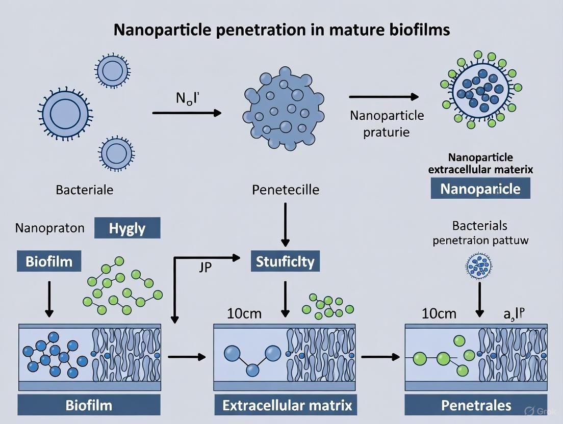
Breaking the Barrier: Advanced Nanoparticle Strategies for Penetrating Mature Biofilms
Mature biofilms present a formidable challenge in treating persistent infections due to their dense extracellular matrix that severely limits the penetration and efficacy of conventional antimicrobials.
Breaking the Barrier: Advanced Nanoparticle Strategies for Penetrating Mature Biofilms
Abstract
Mature biofilms present a formidable challenge in treating persistent infections due to their dense extracellular matrix that severely limits the penetration and efficacy of conventional antimicrobials. This article synthesizes the latest research and technological advances in nanotechnology designed to overcome these penetration barriers. We explore the fundamental properties of biofilms that confer resistance, detail the design principles of next-generation nanoparticle (NP) delivery systems—including lipid-based, polymeric, and inorganic NPs—and evaluate strategies for optimizing their size, surface charge, and functionalization for enhanced biofilm penetration. The content further provides a critical analysis of current in vitro and ex vivo models for validating NP efficacy and discusses the translational challenges and future directions for clinical application. This resource is tailored for researchers, scientists, and drug development professionals seeking to develop novel anti-biofilm therapeutics.
Understanding the Fortress: The Structural and Functional Barriers of Mature Biofilms
Frequently Asked Questions (FAQs)
FAQ 1: What are the primary architectural components of the EPS that hinder nanoparticle (NP) penetration? The EPS is a highly hydrated, three-dimensional matrix that acts as a dynamic filter and trapping network. Its primary components are polysaccharides (PS), proteins (PN), and humic acids (HA), which are cross-linked through hydrogen bonding, electrostatic interactions, ionic bridging, and van der Waals forces [1] [2]. This complex, heterogeneous structure creates a physico-chemical barrier that can sequester NPs, reduce their diffusion, and shield bacterial cells [3] [4].
FAQ 2: How does the stratified nature of EPS influence its barrier function? EPS exhibits a stratified structure from the cell surface outward: Tightly-Bound EPS (TB-EPS), Loosely-Bound EPS (LB-EPS), and Soluble EPS (S-EPS) [5] [1]. Each layer has a distinct composition and function. TB-EPS, which adheres directly to the cell, has a dense, complex architecture with strong ternary interactions among PS, PN, and HA, making it a particularly resilient barrier [5] [1]. The outer layers, like S-EPS, can act as an initial sequestration site for NPs [1].
FAQ 3: Why are biofilms with intact EPS significantly more resistant to nanoparticles than planktonic cells or cells with EPS removed? Direct experimental evidence shows that the EPS matrix is a key determinant of resistance. One study found that the survival rate of pristine E. coli (with intact EPS) exposed to 500 mg/L of ZnO NPs was 65%, whereas for cells that had their EPS removed via sonication/centrifugation, the survival rate plummeted to 11% [3]. This demonstrates that the EPS matrix acts as a protective shield that actively reduces the particle-specific bactericidal activity of NPs [3].
FAQ 4: What are the key NP characteristics that affect their interaction with and penetration through the EPS? The interaction is modulated by a complex interplay of NP characteristics and EPS properties. Key NP factors include:
- Size: Diffusion coefficients decrease with increasing size; penetration becomes severely limited for NPs larger than 50 nm in dense biofilms [4].
- Surface Charge: Electrostatic forces between the NP and charged EPS components greatly influence attachment and transport [4] [2].
- Surface Chemistry/Functionalization: NPs can be "decorated" by biomolecules in the environment, forming a "protein corona" that alters their surface properties and subsequent interaction with the biofilm [4]. For instance, polyethylene glycol-conjugated quantum dots penetrated biofilms more effectively than carboxylated ones [4].
Troubleshooting Guides
Problem: Inconsistent or Poor Nanoparticle Penetration into Mature Biofilms
Potential Cause 1: Strong Sequestration of NPs by the Outer EPS Layers. The soluble and loosely-bound EPS layers can act as a "sponge," binding and retaining NPs before they reach the deeper, cell-dense regions of the biofilm [3] [1].
- Solution: Pre-treat biofilms with EPS matrix-disrupting agents.
- Experimental Protocol: Utilize enzymes to target specific EPS components. For example, use DNase I to degrade extracellular DNA (eDNA) or proteinase K to digest protein components within the matrix. Incubate mature biofilms with a non-bactericidal concentration of the enzyme (e.g., 100 µg/mL DNase I in a suitable buffer for 1 hour at 37°C) prior to the introduction of NPs. This can weaken the matrix structure and enhance NP diffusion [2].
Potential Cause 2: Mismatch between NP Physicochemical Properties and the Biofilm's Microenvironment. The dense, negatively charged EPS matrix can filter out NPs based on their size, charge, and hydrophobicity [4] [2].
- Solution: Systematically engineer NP properties to overcome electrostatic and steric hindrance.
- Experimental Protocol:
- Size Optimization: Synthesize and test a library of NPs with a range of diameters (e.g., 10 nm, 30 nm, 50 nm, 100 nm) while keeping other properties constant.
- Surface Charge Tuning: Functionalize NPs of the optimal size with different surface chemistries (e.g., cationic with amine groups, anionic with carboxyl groups, or neutral with polyethylene glycol (PEG)).
- Evaluation: Use confocal laser scanning microscopy (CLSM) with fluorescently tagged NPs to quantitatively compare penetration depth and distribution within the biofilm architecture.
Potential Cause 3: Dynamic Nature and Heterogeneity of the Biofilm Matrix. Biofilms are not static; their composition and structure can change over time and vary spatially, leading to inconsistent experimental results [2].
- Solution: Characterize the specific biofilm model's EPS composition and structure at the time of experimentation.
- Experimental Protocol: Employ spectroscopic fingerprinting techniques on biofilm samples harvested in parallel with NP treatment experiments.
- Fluorescence Excitation-Emission Matrix (FEEM) Spectroscopy: Can fingerprint the PS/PN/HA architecture and reveal inter-component interactions [5] [1].
- Fourier Transform Infrared (FTIR) Spectroscopy: Identifies functional groups (e.g., amide II C-N, carbonyl C=O) involved in NP sequestration [3]. This data provides a correlative understanding between the EPS chemical makeup and observed NP penetration efficacy.
Data Tables
Table 1: Stratified EPS Composition and Key Characteristics in Activated Sludge (as a Model System)
| EPS Layer | Approx. Organic Matter (TOC) | Dominant Fluorophores (from FEEM) | Key Component Interactions | Proposed Primary Barrier Function |
|---|---|---|---|---|
| S-EPS(Soluble) | ~1.3 mgTOC/gSS [1] | Low-Stokes shift region(Em - Ex < 25 nm) [5] | Binary (PS×HA, PS×PN) [5] | Initial NP sequestration; source of membrane foulants [1] |
| LB-EPS(Loosely-Bound) | ~0.6 mgTOC/gSS [1] | Emission > 400 nm [5] | Binary (PN×HA, PS×PN) [5] | Connects flocs; affects flocculation and settling; moderate filtration barrier [1] |
| TB-EPS(Tightly-Bound) | ~14 mgTOC/gSS [1] | Emission = 350-400 nm [5] | Ternary (PS×PN×HA) [5] | Critical protective barrier; dense, cross-linked architecture affecting cell aggregation [5] [1] |
TOC: Total Organic Carbon; SS: Suspended Solids; Em: Emission Wavelength; Ex: Excitation Wavelength.
Table 2: Impact of Nanoparticle Properties on Biofilm Interaction and Penetration
| Nanoparticle Property | Impact on Biofilm Interaction | Key Experimental Findings |
|---|---|---|
| Size | Determines diffusion capability through EPS pore spaces. | Self-diffusion coefficients decrease with increasing size; severe limitation for sizes >50 nm in dense biofilms [4]. |
| Surface Charge | Governs electrostatic interactions with charged EPS components (e.g., carboxyl, amino groups). | Sulfate-functionalized (negatively charged) polystyrene NPs had greater sorption to biofilms than amine- (positive) or carboxyl- (negative) functionalized ones, indicating charge-specific interactions [4]. |
| Surface Corona/Chemistry | Defines the "biological identity" of the NP and its affinity for EPS molecules. | PEG-conjugated Quantum Dots penetrated better than carboxylated ones [4]. Pre-exposure to Natural Organic Matter (NOM) reduced AgNP toxicity, suggesting a mitigating effect from the corona [4]. |
| Composition | Dictates the mechanism of action (e.g., ROS generation, ion release). | Metal/Metal Oxide NPs (e.g., Ag, ZnO) can generate ROS and disrupt matrix integrity [6]. Particle-specific toxicity is a dominant mechanism for ZnONPs and SiO2NPs, independent of ion release [3]. |
Experimental Protocols & Workflows
Protocol 1: Assessing the Protective Role of EPS via EPS Manipulation and NP Toxicity Testing
This protocol is designed to directly quantify the contribution of the EPS matrix in mitigating NP toxicity, based on the methodology described in [3].
Principle: By comparing the survival rates of bacteria with intact EPS to those with EPS removed after exposure to NPs, the protective role of the EPS can be isolated and measured.
Workflow Diagram: EPS Removal and NP Toxicity Assay
Materials:
- Mature biofilm culture (e.g., E. coli, P. aeruginosa)
- Nanoparticle suspension (e.g., ZnONPs, SiO2NPs)
- Phosphate Buffered Saline (PBS)
- Sonicator (with microtip)
- Centrifuge
- Culture medium and plates for viability counts
Step-by-Step Procedure:
- Biofilm Harvest & Split: Grow a mature biofilm. Gently scrape and harvest the biomass. Resuspend in a suitable buffer (e.g., PBS) and split into two equal aliquots.
- EPS Manipulation:
- Pristine EPS Sample (Control): Subject one aliquot to a mild centrifugation (e.g., 5000×g for 10 minutes) to remove the bulk medium without aggressively stripping EPS. Resuspend the pellet in fresh medium.
- Low EPS Sample (Test): Subject the second aliquot to a rigorous EPS removal procedure. This typically involves sonication on ice (e.g., 5-10 seconds pulses at a low power setting) followed by high-speed centrifugation (e.g., 15,000×g for 20 minutes). Carefully discard the supernatant (which contains the removed EPS) and resuspend the pellet in fresh medium [3].
- NP Exposure: Add identical concentrations of the nanoparticle suspension to both the "Pristine EPS" and "Low EPS" samples. Include a no-NP control for both.
- Incubation: Incubate the samples under appropriate conditions (e.g., 37°C with shaking) for a predetermined period (e.g., 4-16 hours).
- Viability Assessment: After incubation, perform serial dilutions and plate on agar to determine the colony-forming units (CFU/mL). Alternatively, use a metabolic assay like AlamarBlue to assess cell viability.
- Data Analysis: Calculate the percentage survival for each sample relative to its no-NP control. Compare the survival rate of the "Pristine EPS" sample versus the "Low EPS" sample to quantify the protective effect of the EPS.
Protocol 2: Spectroscopic Fingerprinting of EPS Architecture
This protocol outlines the use of UV-Vis and Fluorescence spectroscopy to characterize the complex structure of stratified EPS, as detailed in [5] [1].
Principle: Spectroscopic techniques provide a sensitive, non-destructive way to fingerprint the chemical composition and molecular interactions within different EPS layers based on their light absorption and emission properties.
Workflow Diagram: EPS Stratification and Spectral Analysis
Materials:
- Activated sludge or biofilm sample
- Centrifuge
- Suitable buffer (e.g., NaCl solution, phosphate buffer)
- Water bath or heating block
- UV-Vis Spectrophotometer
- Fluorescence Spectrophotometer
Step-by-Step Procedure:
- Stratified EPS Extraction:
- S-EPS: Centrifuge the sludge/biofilm sample at 4,000×g for 10 minutes. The resulting supernatant is the S-EPS fraction [1].
- LB-EPS: Resuspend the pellet from step 1 in a pre-warmed buffer. Vortex and then centrifuge at 12,000×g for 20 minutes. The resulting supernatant is the LB-EPS fraction [1].
- TB-EPS: Resuspend the remaining pellet in buffer and heat at 60°C for 30 minutes. Centrifuge at 15,000×g for 30 minutes. The supernatant is the TB-EPS fraction [1].
- UV-Vis Spectroscopy: Scan all three EPS fractions across the 200-800 nm wavelength range. Analyze parameters like specific UV absorbances (SUVA) to infer aromaticity and the ratio of absorbances at different wavelengths (e.g., A250/A365) to understand molecular weight distribution [1].
- Fluorescence EEM Spectroscopy: For each EPS fraction, acquire an excitation-emission matrix (EEM). Typical settings are an excitation range of 200-500 nm and an emission range of 250-600 nm [5] [1].
- Data Processing and Analysis:
- Fluorescence Quotient (FQ) Analysis: Calculate FQs by dividing the emission intensity at two different wavelengths for a given excitation. This helps identify dominant fluorophore regions for each EPS layer (e.g., TB-EPS dominates in Em=350-400 nm) [5].
- Multiple Linear Regression & Variance Partitioning Analysis (MLR-VPA): Model the FEEM intensity as a function of PS, PN, and HA content. Use VPA to quantify the individual and joint contributions of these components to the overall fluorescence, revealing key interactions (e.g., ternary PS×PN×HA interaction in TB-EPS) [5] [1].
The Scientist's Toolkit: Research Reagent Solutions
| Item | Function/Benefit | Example Application / Note |
|---|---|---|
| DNase I | Degrades extracellular DNA (eDNA), a key structural component of many biofilms, weakening the matrix integrity. | Used in pre-treatment protocols to enhance NP penetration [2]. |
| Proteinase K | A broad-spectrum serine protease that digests proteins in the EPS matrix. | Effective for disrupting protein-rich biofilms; used similarly to DNase I in pre-treatment [2]. |
| Cationic Functionalized NPs | NPs with positive surface charge (e.g., amine-modified) can improve interaction with negatively charged EPS components. | Can increase initial adhesion but may also lead to agglomeration in the outer EPS layers; requires optimization [4]. |
| PEGylated NPs | Coating with polyethylene glycol (PEG) creates a "stealth" effect, reducing non-specific interactions with EPS biomolecules. | Can enhance diffusion through the biofilm matrix by avoiding sequestration [4]. |
| Fluorescently-Labeled NPs (e.g., Cy5, FITC) | Enable direct visualization and quantification of NP penetration and distribution within the biofilm using microscopy (e.g., CLSM). | Essential for validating the efficacy of NP design or pre-treatment strategies [4]. |
| ZnO Nanoparticles | Exhibit particle-specific toxicity; generate Reactive Oxygen Species (ROS); useful as a model antimicrobial NP. | Dissolved zinc ions show negligible toxicity at typical dissolution concentrations; toxicity is primarily particle-specific [3]. |
| Spectroscopic Standards (BSA, Alginate, etc.) | Pure protein (Bovine Serum Albumin) and polysaccharide (Alginate) standards for calibrating spectroscopic assays and validating EPS quantification methods. | Used to create calibration curves for colorimetric assays and as references in FEEM and FTIR analysis [5] [1]. |
FAQ: Troubleshooting Common Experimental Challenges
FAQ 1: Why are my nanoparticles accumulating on the biofilm surface without significant penetration?
This is a common issue often caused by electrostatic repulsion. Most bacterial biofilms possess a negatively charged surface due to anionic substances in their extracellular polymeric substance (EPS) matrix, such as extracellular DNA (eDNA) and certain polysaccharides [7] [8]. If your nanoparticles are also negatively charged, strong repulsive forces will prevent entry.
- Troubleshooting Steps:
- Characterize Surface Charge: Measure the zeta potential of both your nanoparticles and the target biofilm. This confirms if negative-negative repulsion is the issue [7].
- Modify Nanoparticle Chemistry: Functionalize nanoparticles with positively charged ligands (e.g., amine groups) to create attractive electrostatic forces. Studies show that cationic MSNs exhibit different binding and efficacy profiles compared to anionic ones [7].
- Use Electrolyte Screening: Co-administer cationic electrolytes (e.g., Tris buffer) to shield the negative charges on the EPS, thereby reducing repulsion and enhancing penetration [7].
FAQ 2: My nanoparticles are designed to be cationic, yet they are not penetrating deeply. What could be wrong?
While a positive charge can aid initial adhesion, it can also lead to surface fouling. The high density of binding sites on the biofilm's surface can cause nanoparticles to bind so strongly that they form a clogging layer, preventing further diffusion inward [7] [9].
- Troubleshooting Steps:
- Optimize Surface Charge Density: Avoid an excessively high density of cationic charges. A moderately positive or even neutral zeta potential might facilitate deeper penetration after initial adhesion.
- Introduce Hydrophilic Coatings: Modify the nanoparticle surface with hydrophilic polymers like polyethylene glycol (PEG). PEGylation creates a hydration layer that reduces non-specific binding ("mucoadhesion"), allowing nanoparticles to diffuse more freely through the biofilm matrix [10] [8].
- Verify Nanoparticle Size: Ensure the size is appropriate for the biofilm's pore structure (see FAQ 3).
FAQ 3: How do I determine if the physical pore structure of the biofilm is blocking my nanoparticles?
The EPS matrix acts as a physical sieve. The pore sizes in mature biofilms are typically in the range of 10s to 100s of nanometers, but this can vary significantly between species and growth conditions [10].
- Troubleshooting Steps:
- Size Characterization: Use techniques like electron microscopy or diffusion studies with fluorescent dextrans of known sizes to estimate the effective pore size of your specific biofilm model.
- Downsize Nanoparticles: Design nanoparticles smaller than the characterized pore size. Research indicates that nanoparticles with a diameter of ~50 nm or less often show significantly enhanced penetration and antimicrobial effects [11].
- Utilize Biofilm-Disrupting Strategies: Combine nanoparticles with agents that degrade the EPS. Enzymes such as DNase I (targets eDNA) or dispersin B (targets polysaccharides) can enlarge pore sizes, facilitating nanoparticle access [7] [12].
FAQ 4: How can I overcome the hydrophobic barrier of the biofilm matrix?
The outer layer of biofilms often contains lipids and modified polysaccharides, creating a hydrophobic zone that can repel hydrophilic therapeutics [8].
- Troubleshooting Steps:
- Tune Hydrophobicity: Engineer nanoparticles with balanced surface hydrophobicity. While extreme hydrophobicity can cause non-specific binding, a degree of hydrophobicity can promote interaction with and disruption of the lipid-rich biofilm components [8] [13].
- Use Surfactants: Incorporate biocompatible surfactants or co-deliver biosurfactants (e.g., rhamnolipids) that can solubilize hydrophobic barriers and improve wetting and diffusion [13].
- Employ Lipid-Based Nanocarriers: Utilize liposomes or nanoemulsions that naturally fuse with hydrophobic domains, facilitating delivery of encapsulated antibiotics into the biofilm [8].
The following tables consolidate key quantitative findings from the literature to guide your experimental design.
Table 1: Impact of Nanoparticle Surface Charge on Antibacterial Efficacy
| Surface Functionalization | Example Ligand | Net Charge | Vancomycin Loading Efficiency | Key Finding / Mechanism |
|---|---|---|---|---|
| Carboxyl | Succinic anhydride | Negative | High | Higher drug loading; cellular binding reduces biofilm viability [7]. |
| Bare Silica | - | Negative | High | Good loading; penetration can be influenced by electrostatic screening [7]. |
| Amine | DETA | Positive | Lower | Enhanced initial adhesion; potential for surface fouling [7]. |
| Aromatic | Benzoic acid | Positive | Lower | Potential for π-π interactions with biofilm components [7]. |
Table 2: Influence of Nanoparticle Size on Biofilm Interaction and Antimicrobial Activity
| Nanoparticle Type / Core | Size Range | Key Finding / Antimicrobial Effect |
|---|---|---|
| Silica-Polymer NPPBs | ~7 nm - 270 nm | A critical threshold was identified at ~50 nm. Particles below this size demonstrated potent antimicrobial activity by remodeling bacterial membranes, while larger particles were less effective [11]. |
| LTP Polymer Nanoparticles | 1000 - 5000 nm | This size range is considered optimal for inhalation and deposition in the deep passages of the lungs for treating respiratory biofilm infections [10]. |
| LTP Polymer Nanoparticles | (Not specified) | Distributions consistent with diffusive transport; uniform distribution through biofilm thickness achieved in about four hours [10]. |
Experimental Protocol: Evaluating NP-Biofilm Interactions
This protocol provides a methodology to systematically evaluate how nanoparticle surface properties affect penetration and efficacy in a standard biofilm model, based on established research approaches [7].
Objective: To assess the binding, penetration, and antibacterial efficacy of nanoparticles with different surface functionalizations against Staphylococcus aureus biofilms.
Materials:
- Bacterial Strain: e.g., Methicillin-resistant S. aureus (MRSA) or Methicillin-susceptible S. aureus (MSSA).
- Nanoparticles: Mesoporous silica nanoparticles (MSNs) functionalized with amine (MSN-D), carboxyl (MSN-C), and aromatic (MSN-A) groups, plus bare MSNs (MSN-B) as control [7].
- Fluorescent Dye: Rhodamine B isothiocyanate (RITC) for labeling nanoparticles.
- Culture Media: Tryptic Soy Broth (TSB).
- Equipment: Confocal Laser Scanning Microscope (CLSM), microplate reader, sterile 96-well plates.
Methodology:
- Biofilm Cultivation:
- Grow biofilms in 96-well plates (for viability assays) or on glass-bottom dishes (for microscopy) for 24-48 hours in TSB at 37°C.
- Gently wash mature biofilms with saline to remove non-adherent planktonic cells.
Nanoparticle Exposure:
- Prepare suspensions of the different fluorescently-labeled MSNs in an appropriate buffer (e.g., PBS or Tris) at a standardized concentration (e.g., 0.25 mg mL⁻¹) [7].
- Apply the nanoparticle suspensions to the pre-formed biofilms and incubate for a set period (e.g., 2-4 hours).
Analysis:
- Binding and Penetration (CLSM): Image the biofilms using z-stacking to create cross-sectional views. Analyze the fluorescence intensity profile from the top to the bottom of the biofilm to quantify penetration depth.
- Antibacterial Efficacy (MTT Assay): After exposure and washing, treat biofilms with MTT solution. Measure the absorbance of the dissolved formazan product. Reduced absorbance indicates decreased metabolic activity and higher antibacterial efficacy [7].
- Viability Assessment (BacLight Staining): Use a live/dead bacterial viability kit (e.g., SYTO 9 and propidium iodide) after nanoparticle treatment to visualize the proportion of live vs. dead cells within the biofilm structure.
Schematic Diagram: Strategy to Overcome Biofilm Penetration Barriers
The following diagram illustrates the multi-faceted strategy for engineering nanoparticles to overcome key biofilm barriers.
The Scientist's Toolkit: Key Research Reagents
Table 3: Essential Materials for Nanoparticle-Biofilm Penetration Studies
| Reagent / Material | Function / Rationale | Example Use in Experiment |
|---|---|---|
| Mesoporous Silica Nanoparticles (MSNs) | Versatile, inorganic platform with high drug loading capacity and easily tunable surface chemistry [7]. | Core material for synthesizing functionalized nanoparticles (e.g., MSN-B, -C, -D, -A). |
| Functionalization Agents (e.g., APTES, Succinic Anhydride) | Used to covalently attach specific chemical groups (amine, carboxyl) to the nanoparticle surface to modify charge and hydrophobicity [7]. | Synthesis of MSN-D (aminated) and MSN-C (carboxylated) as described in the experimental protocol. |
| Rhodamine B Isothiocyanate (RITC) | Fluorescent dye used to label nanoparticles for tracking and visualization under a confocal microscope [7]. | Fluorescently tagging nanoparticles to quantify binding and visualize penetration depth in biofilms via CLSM. |
| DNase I Enzyme | Degrades extracellular DNA (eDNA), a key structural component of many biofilms (especially MRSA), reducing matrix integrity and increasing porosity [7] [12]. | Co-incubated with biofilms to disrupt the EPS matrix and test if it enhances nanoparticle penetration. |
| PEG (Polyethylene Glycol) | A hydrophilic polymer used to create a "stealth" coating on nanoparticles, reducing non-specific binding and aggregation [10] [8]. | Grafted onto nanoparticle surfaces to improve diffusion through the biofilm matrix by minimizing mucoadhesion. |
| Zetasizer Instrument | Measures the zeta potential (surface charge) and hydrodynamic size of nanoparticles in suspension, which are critical physicochemical parameters [7]. | Characterizing the synthesized nanoparticles to confirm successful functionalization and determine colloidal stability. |
| Confocal Laser Scanning Microscope (CLSM) | Allows for non-invasive, high-resolution optical sectioning of thick samples, enabling 3D visualization of nanoparticle distribution within a biofilm [7] [14]. | Acquiring z-stack images of biofilms after treatment with fluorescent nanoparticles to create penetration depth profiles. |
Biofilms are structured communities of microbial cells enclosed in a self-produced extracellular polymeric substance (EPS) matrix and adherent to either biotic or abiotic surfaces [15]. For researchers investigating antimicrobial agents, understanding the biofilm lifecycle is not merely academic; it is a critical practical necessity. Biofilms can exhibit resistance to antimicrobial agents that is up to 1,000 times greater than their planktonic counterparts, presenting a formidable challenge in fields from clinical drug development to industrial microbiology [16]. The central theme of this technical guide is framed within the ongoing research to overcome a significant barrier: enabling therapeutic nanoparticles (NPs) to penetrate and disrupt the mature biofilm EPS, a major hurdle in translating nanotherapeutics into effective applications [6] [17].
Detailed Lifecycle Stages and Key Experimental Checkpoints
The classic model of biofilm development is a cyclic process comprising distinct, regulated stages. The diagram below illustrates the key phases researchers must account for in their experimental designs.
Stage 1: Initial Reversible Attachment
- Process: Free-floating (planktonic) cells loosely attach to a conditioned surface via weak physical forces like van der Waals forces, electrostatic, and hydrophobic interactions [16].
- Experimental Checkpoint: Attachment can be disrupted by mild washing. Quantify baseline attachment using crystal violet staining or phase-contrast microscopy cell counts.
Stage 2: Irreversible Attachment and EPS Production
- Process: Attachment becomes permanent via microbial surface adhesins and the beginning of EPS production. The second messenger cyclic di-GMP (c-di-GMP) is a key regulator of this switch [18] [16].
- Experimental Checkpoint: Cells resist gentle washing. Confocal Laser Scanning Microscopy (CLSM) with fluorescent stains (e.g., ConA for polysaccharides) can visualize initial matrix production.
Stage 3: Microcolony Formation
- Process: Attached cells proliferate, forming clustered microcolonies. Quorum Sensing (QS) cell-to-cell communication becomes increasingly active, coordinating group behavior [15] [16].
- Experimental Checkpoint: Monitor the expression of QS-related genes (e.g., lasR in P. aeruginosa or icaADBC in S. aureus) via qPCR or reporter strains.
Stage 4: Maturation
- Process: Microcolonies develop into a complex, three-dimensional structure with water channels that facilitate nutrient inflow and waste removal. The EPS matrix, containing exopolysaccharides, proteins, and extracellular DNA (eDNA), is fully established [19] [17] [15].
- Experimental Checkpoint: The mature biofilm exhibits characteristic structures (e.g., mushroom-shaped in P. aeruginosa). Analyze the 3D architecture using CLSM and characterize EPS composition using specific enzymatic treatments (e.g., DNase for eDNA).
Stage 5: Dispersion
- Process: A dedicated phase where a sub-population of cells actively breaks free from the biofilm to colonize new surfaces. This involves the production of matrix-degrading enzymes (e.g., glycosidases, nucleases) and a shift in gene expression [19] [18].
- Experimental Checkpoint: Collect and plate effluent to quantify dispersed cells. Use transcriptomics to identify genes and pathways (e.g., nuclease or phospholipase genes in V. cholerae) essential for dispersal [18].
The Nanoparticle Penetration Barrier in Mature Biofilms
The mature biofilm matrix presents a formidable diffusion barrier that significantly hinders the penetration of therapeutic nanoparticles, a central challenge in the field.
Primary Barrier Mechanisms:
- Matrix Density: The dense network of EPS polymers acts as a physical sieve, sterically hindering the movement of NPs, particularly those larger than the matrix pore size [17] [20].
- Charge Interactions: The EPS components often carry negative charges (e.g., from eDNA and some polysaccharides) that can bind to and retain charged NPs, preventing further penetration [17].
- Hydrophobicity: Hydrophobic domains within the matrix can similarly interact with and trap hydrophobic NPs [17].
- Matrix-Degrading Enzyme Retention: The EPS retains host and bacterial enzymes that could potentially degrade the NP coating or payload [19].
Troubleshooting FAQs for Biofilm & Nanoparticle Research
Q1: Our anti-biofilm nanoparticles show good efficacy in planktonic assays but consistently fail against mature (24-48h) biofilms. What could be the issue? A: This is a classic symptom of the penetration barrier.
- Hypothesis 1: NP Size/Charge is Suboptimal. The NPs may be too large to diffuse through the matrix or have a surface charge that causes them to get trapped.
- Troubleshooting Experiment: Characterize the zeta potential and hydrodynamic diameter of your NPs. Perform a penetration assay by incubating NPs with mature biofilms and using time-lapse CLSM to track their movement and final distribution within the biofilm structure.
- Hypothesis 2: The EPS Matrix is Sequestering or Inactivating the NPs.
- Troubleshooting Experiment: Pre-treat biofilms with matrix-disrupting agents (e.g., DNase I to degrade eDNA, Dispersin B to hydrolyze polysaccharides, or metallic NPs known to degrade matrix [6]) before applying your therapeutic NPs. A significant increase in efficacy after pre-treatment confirms matrix involvement.
Q2: How can we accurately quantify nanoparticle penetration into a biofilm? A: Use a combination of direct and indirect methods.
- Direct Method: Confocal Microscopy. Label NPs with a bright, stable fluorophore (e.g., FITC, Cy5). Create Z-stacks of the biofilm and use image analysis software to plot the fluorescence intensity as a function of biofilm depth. This provides a visual and quantitative penetration profile.
- Indirect Method: Biofilm Viability Analysis. After NP treatment, use a viability stain (e.g., LIVE/DEAD BacLight) in conjunction with CLSM. If NPs are penetrating effectively, you will observe a gradient of dead cells (red) from the top to the interior of the biofilm. If only surface cells are dead, penetration is poor.
Q3: Our biofilm formation is highly variable between experimental replicates, skewing our NP efficacy data. How can we improve consistency? A: Standardize every aspect of the biofilm growth protocol.
- Inoculum Preparation: Always use cells from the same growth phase (typically mid-log phase) and standardize the optical density of the inoculum precisely.
- Surface Conditioning: Ensure the substrate (e.g., peg lid, glass bottom dish) is identical and cleaned in a standardized way, as surface properties drastically affect initial attachment [16].
- Growth Medium & Flow Conditions: Use the same medium batch and maintain strict control over temperature, incubation time, and agitation or flow rate. For flow-cell systems, calibrate pumps regularly.
- Normalization: Always include a internal control (e.g., total protein content or crystal violet staining of biomass) to normalize your anti-biofilm readouts against.
Key Experimental Protocols
Protocol 1: Standardized Static Biofilm Formation for Anti-biofilm Screening
This protocol is adapted for a 96-well plate model, ideal for high-throughput screening of NPs [19].
- Inoculum Prep: Grow the test organism (e.g., P. aeruginosa, S. aureus) to mid-log phase. Dilute in fresh medium to a standardized OD600 (e.g., 0.05).
- Seeding: Aliquot 200 µL of the cell suspension into designated wells of a sterile, flat-bottom 96-well plate. Include medium-only wells as negative controls.
- Adhesion Phase: Incubate the plate without agitation for 2-4 hours at the appropriate temperature to allow initial attachment.
- Growth Phase: Carefully remove the supernatant containing non-adherent cells by pipetting. Replace with 200 µL of fresh, pre-warmed medium.
- Maturation: Incubate for desired time (e.g., 24h for maturation) with or without gentle agitation. Refresh medium every 24h for biofilms grown longer than a day.
- Analysis: Proceed with NP treatment and subsequent analysis (e.g., viability assay, staining).
Protocol 2: Evaluating NP Penetration via Confocal Microscopy
This protocol details how to visualize NP action within a mature biofilm.
- Grow Biofilm: Form a mature (24-48h) biofilm on a suitable surface for microscopy (e.g., glass-bottom dish, flow cell).
- NP Treatment: Apply your fluorescently-labeled NPs at the desired sub-inhibitory or treatment concentration. Incubate for a set time.
- Staining: If needed, counterstain the biofilm. A common combination is:
- SYTO 9 (green fluorescent nucleic acid stain, labels all cells).
- Propidium Iodide (PI) (red fluorescent stain, penetrates only cells with compromised membranes).
- Concanavalin A conjugated to Tetramethylrhodamine (ConA-TRITC) (binds to polysaccharides in the matrix).
- Washing: Gently wash the biofilm with a buffer (e.g., PBS) to remove non-adherent NPs and stains.
- Imaging: Image immediately using a Confocal Laser Scanning Microscope. Acquire Z-stacks through the entire biofilm depth.
- Image Analysis: Use software (e.g., ImageJ, IMARIS) to create 3D reconstructions and depth-intensity profiles for the NP fluorescence.
Research Reagent Solutions
Table 1: Essential Reagents for Biofilm and Nanoparticle Penetration Research
| Reagent Category | Specific Examples | Function in Research | Key Considerations |
|---|---|---|---|
| Metal/Metal Oxide NPs | Silver (Ag), Zinc Oxide (ZnO), Iron Oxide (Fe₃O₄) [6] [17] | Intrinsic anti-biofilm agents; can generate ROS, degrade matrix, inhibit QS. | Size, shape, and surface coating critically affect penetration and toxicity. |
| Matrix Targeting Enzymes | DNase I, Dispersin B, Proteinase K [17] | Degrade specific EPS components (eDNA, polysaccharides, proteins) to enhance NP penetration. | Use as pre-treatment or co-treatment with NPs. Optimize concentration to avoid complete biofilm disintegration. |
| Quorum Sensing Inhibitors (QSIs) | Furanones, halogenated furanones [17] | Attenuate virulence and biofilm formation by disrupting bacterial communication. | Can be loaded into NP carriers for targeted delivery. May not disrupt pre-formed matrix. |
| Viability Stains | LIVE/DEAD BacLight (SYTO9/PI), CTC/DAPI [20] | Differentiate live/dead cells and metabolic activity within the biofilm post-NP treatment. | CLSM-compatible. Confirm stain compatibility with NP fluorescence. |
| EPS-Specific Stains | ConA (polysaccharides), FilmTracer SYPRO Ruby (proteins) [17] | Visualize and quantify the EPS matrix components before and after NP treatment. | Essential for confirming matrix disruption. |
Targeting Biofilm-Related Genes with Nanoparticles
A advanced strategy involves using NPs to target the genetic regulation of biofilm formation. The following diagram outlines how NPs can disrupt key genetic pathways.
Research indicates that metal and metal oxide NPs can interfere with the expression of critical biofilm-related genes [16].
- Adhesion Genes: NPs can downregulate genes like atlE in S. aureus and fim cluster in E. coli, which are crucial for the initial attachment phase [16].
- Quorum Sensing Genes: NPs can disrupt the expression of central QS regulators like lasR and rhlI/rhlR in P. aeruginosa, preventing the cell-density-dependent coordination required for maturation [16].
- EPS Synthesis Genes: The expression of genes responsible for producing polysaccharides (e.g., pelA, psl in P. aeruginosa) can be inhibited by NPs, directly weakening the structural integrity of the matrix [16].
Experimental Approach: To investigate this, researchers can treat developing or mature biofilms with sub-inhibitory concentrations of NPs and use quantitative RT-PCR (qPCR) to measure the changes in expression levels of these target genes compared to untreated controls.
Bacterial biofilms are three-dimensional aggregates of microorganisms encased in a self-produced protective matrix that adhere to surfaces [21] [22]. This structured community represents a predominant form of microbial life and is a significant virulence factor in human infections [22] [23]. Unlike free-floating (planktonic) bacteria, cells within a biofilm can exhibit 10 to 1,000-fold increase in antibiotic resistance, making associated infections notoriously difficult to treat [22] [23].
Biofilms impact all human organ systems and are implicated in 65% of all bacterial infections and nearly 80% of chronic wounds [21] [24]. They are a leading cause of persistent medical device infections—found on catheters, prosthetic joints, and pacemakers—often requiring device removal for resolution [21] [24]. The global economic impact is staggering, estimated at over $280 billion annually, with biofilms implicated in over 500,000 deaths per year in the United States alone [21] [24].
FAQ: Understanding Biofilm-Associated Resistance
What makes biofilms so resistant to antibiotics and nanoparticles? Biofilms employ multiple mechanisms for resistance. The extracellular polymeric substance (EPS) matrix acts as a physical barrier, hindering drug penetration [21] [23]. Within the biofilm, metabolic heterogeneity leads to dormant "persister" cells that survive antibiotic treatment [21] [24]. Additionally, efflux pumps actively remove antimicrobial agents, and the exchange of resistance genes is facilitated within the structured community [21] [24] [23].
Why is the Minimum Inhibitory Concentration (MIC) for biofilms much higher than for planktonic cells? The MIC for a biofilm can be 100-800 times greater than for planktonic cells due to combined factors: reduced antibiotic penetration through the matrix, enzymatic inactivation of drugs by matrix components, the presence of metabolically inactive cells, and increased expression of efflux pumps [21] [24].
How does the biofilm microenvironment contribute to antibiotic tolerance? The biofilm structure creates nutrient and oxygen gradients, leading to heterogeneous bacterial subpopulations [21] [24]. Cells in deeper layers experience nutrient depletion and hypoxia, slowing their metabolism and growth. This slow growth rate increases tolerance to many antibiotics that target actively dividing cells [21] [24] [23].
What is the role of quorum sensing in biofilm resistance? Quorum sensing (QS) is a cell-cell communication system that bacteria use to coordinate gene expression based on population density [24]. QS directly regulates biofilm formation, maturation, and the expression of various virulence factors and efflux pumps, thereby influencing antibiotic penetration and resistance [24].
Troubleshooting Guides for Biofilm Experiments
Common Challenges in Growing Robust Biofilms
Problem: Inconsistent or weak biofilm formation across experimental replicates.
- Solution: Standardize your inoculum concentration to 10⁵-10⁶ CFU/mL [25].
- Solution: Ensure proper humidity (75%-90%) in the incubator to prevent wells from drying during incubation [25].
- Solution: Use a shaker at 110 rpm for 96-well plates to ensure adequate aeration and nutrient mixing [25].
- Solution: For strains forming weak biofilms, consider using hydroxyapatite-coated pegs to simulate bone/teeth surfaces, which typically enhance biofilm growth [25].
Problem: Biofilm assays not accurately predicting in vivo efficacy.
- Solution: Consider using more physiologically relevant models. Biofilms grown on human plasma-conditioned surfaces under shear flow show significantly different antibiotic susceptibility profiles compared to those grown on standard polystyrene [23].
Challenges in Evaluating Anti-Biofilm Nanoparticle Penetration
Problem: Nanoparticles fail to penetrate mature biofilms.
- Solution: Optimize nanoparticle size. The EPS matrix pore size limits penetration; aim for particles below 100-200 nm for better diffusion [26].
- Solution: Modify surface charge. The biofilm matrix contains negatively charged components like eDNA that can bind positively charged nanoparticles, trapping them at the surface [23].
- Solution: Utilize enzyme-functionalized nanoparticles. Glycoside hydrolases or DNases can degrade EPS components (polysaccharides, eDNA), creating channels for improved penetration [23].
Key Resistance Mechanisms and Experimental Data
Table 1: Primary Mechanisms of Antimicrobial Resistance in Biofilms
| Resistance Mechanism | Key Components | Impact on Treatment | Experimental Measurement |
|---|---|---|---|
| Physical Barrier & Reduced Penetration | EPS matrix: polysaccharides, proteins, eDNA [21] [23] | Traps antimicrobials; slows diffusion; requires 100-800× higher MIC [21] [24] | Confocal microscopy with fluorescently tagged antibiotics [23] |
| Metabolic Heterogeneity & Persister Cells | Nutrient/O₂ gradients; dormant subpopulations [21] [24] [23] | Dormant cells survive antibiotic courses; lead to relapse [21] [24] [23] | Time-kill assays; post-antibiotic regrowth monitoring [23] |
| Efflux Pump Upregulation | Membrane proteins (e.g., in P. aeruginosa, S. aureus) [21] [24] | Active export of antibiotics from bacterial cells [21] [24] | RT-PCR for efflux pump gene expression; assays with efflux pump inhibitors [21] |
| Quorum Sensing (QS) Regulation | Autoinducer molecules (AHLs in gram-negative, oligopeptides in gram-positive) [24] | Coordinates biofilm development and virulence factor expression [24] | HPLC/MS for signal molecules; gene reporter assays for QS activity [24] |
Table 2: Quantitative Comparison of Planktonic vs. Biofilm Antibiotic Resistance
| Bacterial Species | Antibiotic | Planktonic MIC (μg/mL) | Biofilm MIC (μg/mL) | Fold Increase |
|---|---|---|---|---|
| Staphylococcus epidermidis | Vancomycin | Susceptible (100% isolates) | Resistant (75% isolates) | >1,000× (functional) [22] |
| Klebsiella pneumoniae | Various | Susceptible in solution | Highly resistant in biofilm | Varies [22] |
| Pseudomonas aeruginosa | Tobramycin/Ciprofloxacin | Low MIC in standard test | High MIC in biofilm | Significant (O₂ dependent) [22] [23] |
Experimental Protocols for Biofilm Research
Standardized Biofilm Cultivation using the MBEC Assay
Principle: The Minimum Biofilm Eradication Concentration (MBEC) assay uses a peg lid to grow biofilms in a high-throughput manner, allowing simultaneous testing of multiple antimicrobial conditions [25].
Procedure:
- Inoculation: Dilute overnight bacterial culture to 10⁵-10⁶ CFU/mL in appropriate growth medium. Pipette 150-200 μL into each well of a 96-well plate.
- Assembly: Attach the peg lid to the base plate, ensuring pegs are submerged in the inoculated media.
- Incubation: Incubate the assembled plate for 24-48 hours at 37°C on a shaker at 110 rpm. Maintain humidity at 75-90%.
- Biofilm Maturation: After incubation, biofilms will have formed on the pegs.
- Treatment: Transfer the peg lid with mature biofilms to a new challenge plate containing serial dilutions of antimicrobial agents or nanoparticles.
- Exposure: Incubate for 24 hours to determine the MBEC.
- Recovery: To quantify viable cells, transfer the peg lid to a recovery plate containing neutralization buffer. Sonicate for 30 minutes to dislodge biofilm cells.
- Viability Assessment: Plate serial dilutions of the recovery solution for colony counting or measure optical density [25].
Assessing Nanoparticle Penetration into Biofilms
Principle: Fluorescently labeled nanoparticles are used with confocal laser scanning microscopy (CLSM) to visualize and quantify penetration depth and distribution within the biofilm matrix.
Procedure:
- Biofilm Preparation: Grow biofilms on appropriate surfaces (e.g., glass-bottom dishes, MBEC pegs) until mature (typically 48-72 hours).
- Nanoparticle Application: Apply fluorescent nanoparticles suspended in relevant buffer to the biofilm surface.
- Incubation: Allow particles to penetrate for a defined period (e.g., 1-24 hours) under physiologically relevant conditions.
- Washing: Gently wash the biofilm to remove non-adherent nanoparticles.
- Imaging: Use CLSM to capture Z-stack images through the entire biofilm depth.
- Image Analysis: Use software to quantify fluorescence intensity as a function of depth, calculating penetration coefficients and distribution profiles.
Research Reagent Solutions for Biofilm Studies
Table 3: Essential Materials for Biofilm and Nanoparticle Penetration Research
| Reagent / Material | Function / Application | Example Use Cases |
|---|---|---|
| MBEC Assay System | High-throughput biofilm cultivation and antimicrobial susceptibility testing [25] | Standardized screening of anti-biofilm compounds and nanoparticles against various bacterial species [25] |
| Hydroxyapatite-Coated Pegs | Simulate bone/tooth surfaces to enhance biofilm growth for relevant models [25] | Studying biofilms of dental pathogens (S. mutans) or orthopedic implant infections [25] |
| Enzymes (DNase I, Dispersin B, Glycoside Hydrolases) | Degrade specific EPS components (eDNA, polysaccharides) to facilitate nanoparticle penetration [23] | Pre-treatment to enhance antimicrobial penetration; component of enzyme-functionalized nanoparticles [23] |
| Efflux Pump Inhibitors | Block active antibiotic export from bacterial cells [21] | Used in combination therapies to restore susceptibility; studying efflux pump contributions to resistance [21] |
| Quorum Sensing Inhibitors | Interfere with bacterial cell-cell communication and biofilm regulation [24] | Anti-virulence agents to prevent biofilm maturation and enhance susceptibility [24] |
Visualizing Biofilm Resistance and Nanoparticle Penetration Barriers
Biofilm Resistance Mechanisms Diagram
Nanoparticle Penetration Strategy
Engineering the Key: Nanoparticle Design for Enhanced Biofilm Penetration and Targeting
Technical Support Center
Troubleshooting Guides
Issue 1: Poor Nanoparticle Penetration into Mature Biofilms
- Problem: Lipid-based nanoparticles (LNP) fail to diffuse beyond the superficial layers of a mature biofilm, leading to inadequate therapeutic delivery.
- Diagnosis: The dense, anionic extracellular polymeric substance (EPS) matrix of mature biofilms acts as a formidable physical and chemical barrier, filtering out nanoparticles through steric hindrance and electrostatic repulsion [14] [27].
- Solution:
- Surface Functionalization: Modify the liposome surface with polyethylene glycol (PEG) to create a "stealth" effect, reducing non-specific binding to the EPS and enhancing diffusion [10] [28]. For targeted delivery, conjugate surface with cationic lipids or specific ligands to promote adhesion to bacterial cells or EPS components [28].
- Size Optimization: Formulate nanoparticles with a diameter below 200 nm to facilitate passage through the water channels and pores within the biofilm architecture [10].
- Co-delivery with Matrix-Disrupting Agents: Design LNPs to co-encapsulate and deliver antibiotics or biofilm-dispersing enzymes (e.g., DNase, dispersin B) alongside the primary antimicrobial agent to disrupt the matrix and enhance penetration [14].
Issue 2: Low Encapsulation Efficiency and Payload Instability
- Problem: The therapeutic agent (e.g., antibiotic, CRISPR-Cas9 components) leaks from the liposome before it reaches the target within the biofilm, or the encapsulation efficiency during formulation is low.
- Diagnosis: Instability can arise from inappropriate lipid composition, which fails to form a stable bilayer, or from harsh preparation methods that degrade the payload [28].
- Solution:
- Optimized Lipid Composition: Incorporate cholesterol and saturated phospholipids into the lipid bilayer to increase membrane rigidity and reduce permeability, thereby improving payload retention [28].
- Advanced Formulation Techniques: Utilize techniques like remote loading for ionizable drugs or double-emulsion methods for hydrophilic/hydrophobic cargo to achieve high encapsulation efficiency (>80%) [28].
- Stimuli-Responsive Formulations: Develop "smart" liposomes that release their payload only in response to specific biofilm microenvironment triggers, such as low pH or elevated enzyme activity, minimizing premature release [28].
Issue 3: Inconsistent Experimental Results in Flow-Cell Systems
- Problem: Reproducibility issues when evaluating LNP deposition and penetration in biofilm flow-cell models.
- Diagnosis: Variations in biofilm growth conditions, fluid flow dynamics, and nanoparticle sticking coefficients lead to inconsistent deposition profiles [10].
- Solution:
- Standardize Biofilm Growth: Use defined media and control growth time to establish consistent biofilm thickness and density. Techniques like confocal laser scanning microscopy (CLSM) can validate biofilm architecture before experiments [27].
- Control Flow Parameters: For flow-cell experiments, use a mathematical model to inform flow rates. Simulations suggest that low, steady flow rates (e.g., mimicking pulmonary fluid dynamics) are sufficient for deposition, and increasing flow rate may not significantly enhance nanoparticle concentration in the biofilm [10].
- Quantify Key Parameters: Estimate the nanoparticle sticking coefficient (a measure of adhesion efficiency) and biofilm diffusion coefficient through controlled experiments to refine your model and experimental setup [10].
Experimental Protocol: Evaluating LNP Penetration into Biofilms
This protocol outlines a methodology for quantifying the penetration and distribution of lipid nanoparticles within a mature biofilm using a flow-cell system.
1. Objective To visualize and quantify the deposition and diffusion of fluorescently labeled liposomes into a Pseudomonas aeruginosa biofilm over time.
2. Materials
- Bacterial Strain: P. aeruginosa PAO1 (or other relevant strain).
- Growth Medium: Tryptic Soy Broth (TSB) or similar.
- Flow Cell: Parallel-plate flow cell apparatus [10].
- Lipid Nanoparticles: Fluorescently labeled (e.g., with Cy5) PEGylated liposomes, diameter ~100-200 nm [10] [28].
- Imaging Equipment: Confocal Laser Scanning Microscope (CLSM) [27].
- Software: Image analysis software (e.g., ImageJ, IMARIS).
3. Procedure Step 1: Biofilm Cultivation
- Grow P. aeruginosa to mid-log phase in TSB.
- Inject the bacterial suspension into the flow cell and allow cells to attach under no-flow conditions for 2 hours.
- Initiate a continuous, low flow of fresh, diluted medium (e.g., 10% TSB) through the cell. Grow the biofilm for 48-72 hours to ensure maturation [10].
Step 2: Nanoparticle Administration
- Prepare a suspension of fluorescent liposomes in buffer at a defined concentration.
- Stop the nutrient flow and carefully inject the liposome suspension into the flow cell.
- Allow the nanoparticles to deposit and diffuse under static or very low flow conditions for a set period (e.g., 1-4 hours) [10].
Step 3: Sample Processing and Imaging
- Gently rinse the flow cell with buffer to remove non-adherent nanoparticles.
- Use CLSM to capture Z-stack images (cross-sectional slices) of the biofilm at various time points. Image from the biofilm surface down to the substratum.
Step 4: Data Analysis
- Penetration Depth: Measure the distance from the biofilm surface to the deepest point where fluorescence is detectable above background.
- Relative Concentration: Quantify the fluorescence intensity at different depths (e.g., every 5 µm) to generate a concentration profile. Normalize the intensity to the maximum value observed.
- Uniformity of Distribution: Calculate the coefficient of variation (CV) of fluorescence intensity across the biofilm depth. A lower CV indicates a more uniform distribution.
The experimental workflow for evaluating lipid nanoparticle penetration into biofilms is as follows:
Table 1: Key Parameters from LNP-Biofilm Interaction Studies
| Parameter | Typical Range/Value | Experimental Context | Significance |
|---|---|---|---|
| Optimal LNP Size for Inhalation/Deposition [10] | 1000 - 5000 nm | Lung delivery for cystic fibrosis treatment. | Maximizes amount of drug delivered to terminal bronchioles and alveoli. |
| Target LNP Size for Biofilm Penetration [10] | < 200 nm | Diffusion through biofilm EPS matrix. | Facilitates passage through water-filled pores in the biofilm. |
| Time to Uniform Distribution in Biofilm [10] | ~ 4 hours | In vitro flow-cell model with polymer nanoparticles. | Indicates time required for nanoparticles to diffuse through the entire biofilm thickness. |
| Liposomal CRISPR-Cas9 Biofilm Reduction [14] | > 90% biomass reduction | In vitro against Pseudomonas aeruginosa biofilm. | Demonstrates potency of combining LNPs with precision genetic tools. |
| Gene-Editing Enhancement with Gold NPs [14] | 3.5-fold increase | Comparison of gold nanoparticle carriers vs. non-carrier systems. | Highlights the efficacy of nanoparticles in delivering functional genetic payloads. |
Table 2: The Scientist's Toolkit - Key Research Reagent Solutions
| Reagent / Material | Function in Experiment | Key Considerations |
|---|---|---|
| PEGylated Liposomes [28] | The core delivery vehicle; "stealth" function reduces clearance and improves biofilm penetration. | PEG density and chain length are critical for balancing stability and diffusion. |
| Cationic Lipids [28] | Confer a positive charge to LNPs, promoting electrostatic interaction with anionic biofilm components. | Can increase cytotoxicity; optimization of charge density is required. |
| L-Tyrosine Polyphosphate (LTP) Nanoparticles [10] | Biodegradable polymer nanoparticle for sustained drug release; compatible with lung delivery. | Degradation products are non-cytotoxic and do not alter local pH. |
| Stimuli-Responsive Lipids [28] | Enable payload release in response to biofilm-specific triggers (e.g., low pH, enzymes). | Enhances specificity and reduces off-target effects. |
| Fluorescent Dyes (e.g., Cy5) [10] | Tagging LNPs for visualization and quantification using confocal microscopy. | Must not alter the physicochemical properties of the LNP. |
Frequently Asked Questions (FAQs)
Q1: What is the primary mechanism preventing lipid nanoparticles from penetrating mature biofilms? The major barrier is the extracellular polymeric substance (EPS), a dense, gel-like matrix of polysaccharides, proteins, and DNA [27]. This matrix creates both a physical barrier through steric hindrance and a chemical barrier via its overall negative charge, which can repel anionic or neutral particles. Furthermore, physiological gradients within the biofilm lead to zones of reduced metabolic activity, which can hinder the active uptake of nanoparticles [14] [29].
Q2: How can I improve the targeting efficiency of my liposomes to a specific biofilm? Functionalizing the liposome surface is key. This can be achieved by:
- Active Targeting: Conjugating antibodies (creating immunoliposomes), peptides, or other ligands that recognize specific surface antigens on the bacterial species within the biofilm [28].
- Cationic Charge: Formulating LNPs with cationic lipids to exploit electrostatic attraction with the negatively charged EPS [28]. However, monitor for increased cytotoxicity.
- Magnetic Guidance: For in vitro or localized applications, embedding magnetic nanoparticles allows for precise, external control over liposome localization [28].
Q3: Our therapeutic DNA/RNA is degrading before reaching the biofilm. What formulation should we use? Cationic liposomes are ideally suited for nucleic acid delivery. Their positive surface charge electrostatically complexes with negatively charged DNA or RNA, forming stable, compact lipoplexes that protect the genetic material from enzymatic degradation during delivery [14] [28]. This approach is foundational for CRISPR-Cas9 antimicrobial strategies [14].
Q4: Are there ways to make liposomes release their payload only inside the biofilm? Yes, develop stimuli-responsive liposomes. These "smart" systems can be engineered to release their cargo in response to unique environmental cues found in the biofilm microenvironment, such as:
- Low pH: Common in hypoxic regions of a mature biofilm.
- Elevated Enzyme Activity: Such as overexpression of lipases or phosphatases.
- Redox Potential: Differences between the external milieu and the biofilm interior [28].
The following diagram illustrates the mechanisms of lipid nanoparticle interaction with and penetration through the biofilm matrix:
Troubleshooting Guides
Premature Drug Release Before reaching mature biofilms
| Problem Description | Possible Causes | Suggested Solutions & Experimental Verification |
|---|---|---|
| Rapid, uncontrolled drug release during circulation [30] | • Low stability of nanocarrier under physiological conditions (e.g., low CMC for micelles) [31] [32]• Insufficient shell density or thickness in layer-by-layer (LbL) systems [30]• Degradation kinetics too fast for the polymer used (e.g., PLGA, PLA) [33] | • For micelles: Use block copolymers with lower Critical Micelle Concentration (CMC) (e.g., 10⁻⁶ to 10⁻⁷ M) to enhance stability against dilution in blood [31] [32].• For LbL NPs: Increase the number of layers or use stronger polyelectrolytes. Optimize coating parameters (salt concentration, pH) to create a denser shell, as demonstrated with heparin coatings [30].• For polyesters: Select polymers with a higher molecular weight or more crystalline structure (e.g., PCL vs. PLA) to slow hydrolysis [31] [33]. |
| Burst release upon administration [30] | • Drug adsorbed on or near the nanoparticle surface [30]• Inefficient encapsulation | • For LbL NPs: Incorporate a protective outer layer (e.g., Hyaluronate or Polystyrene Sulfonate) to reduce initial burst release. Studies show HA coatings can extend release from days to months [30].• For nanogels/dendrimers: Optimize cross-linking density or core-shell design to improve encapsulation efficiency [31] [34]. |
Poor Penetration through Mature Biofilm Matrix
| Problem Description | Possible Causes | Suggested Solutions & Experimental Verification |
|---|---|---|
| Nanocarriers accumulate at biofilm surface [8] [14] | • Large hydrodynamic size [8]• Non-optimal surface charge leading to interaction with the anionic, hydrophobic EPS [8] [35] | • Size Tuning: Aim for a hydrodynamic diameter below 100 nm, ideally between 10-50 nm, to facilitate diffusion through biofilm pores [8].• Surface Charge (Zeta Potential) Modulation: For the anionic biofilm matrix, use nanocarriers with a neutral or slightly positive surface charge to reduce electrostatic repulsion/adhesion. However, high positive charge may cause non-specific binding [8] [35].• Surface Functionalization: Coat with PEG or other hydrophilic polymers to create a "stealth" effect and reduce hydrophobic interactions with the EPS [31] [33]. |
| Inability to disrupt biofilm integrity | • Nanocarrier is biologically inert• Lacks biofilm-disrupting mechanisms | • Intrinsic Activity: Use metallic nanoparticles (e.g., Ag, La) known to disrupt biofilm matrices via lipid peroxidation or enzyme inhibition [35].• Stimuli-Responsive Design: Engineer carriers that release biofilm-degrading enzymes (e.g., DNase, proteases) in response to biofilm-specific stimuli like low pH or high enzyme concentration [8] [14]. |
Inefficient Degradation and Clearance
| Problem Description | Possible Causes | Suggested Solutions & Experimental Verification |
|---|---|---|
| Nanocarrier persistence leading to potential long-term toxicity [33] | • Polymer is non-biodegradable or has very slow degradation kinetics.• Degradation products are cytotoxic. | • Polymer Selection: Prioritize biodegradable polymers like PLGA, PLA, PCL, and chitosan [31] [33].• Monitor Degradation: Conduct in vitro degradation studies in relevant buffers (e.g., PBS at pH 7.4) and monitor molecular weight loss, mass loss, and particle size change over time. Correlation with drug release profile is crucial [33]. |
| Inconsistent degradation between batches | • Poor control over polymer synthesis (molecular weight, dispersity).• Variable nanoparticle fabrication conditions. | • Polymer Characterization: Use Gel Permeation Chromatography (GPC) to ensure consistent molecular weight and low dispersity (Ð) of polymers before formulation [33].• Process Control: Standardize critical formulation parameters such as solvent evaporation rate, sonication energy, and temperature [31] [33]. |
Frequently Asked Questions (FAQs)
Q1: What are the key polymer characteristics that most significantly impact drug release kinetics from these nanocarriers? [30] [33]
The most critical characteristics are:
- Polymer Hydrophobicity/Crystallinity: More hydrophobic and crystalline polymers (e.g., PCL) typically lead to slower drug release compared to more hydrophilic ones (e.g., PLA) [31] [33].
- Molecular Weight & Dispersity: Higher molecular weight polymers generally degrade slower, prolonging release. A low dispersity (Ð) ensures consistent release profiles across a batch [33].
- Functional Groups & Cross-linking Density: For dendrimers and nanogels, the surface chemistry and internal cross-linking density are paramount. Higher cross-linking leads to slower degradation and a more sustained release [31] [34].
Q2: How can I experimentally tune the release profile of a Layer-by-Layer (LbL) system for my specific application? [30]
The release profile from LbL nanoparticles is highly tunable by modifying the shell architecture:
- Number of Layers: Increasing the number of layers generally creates a thicker, more robust diffusion barrier, slowing release.
- Polyelectrolyte Ratio: The ratio of positively to negatively charged polymers in the layers can control shell compactness. For example, varying the ratio of carboxymethyl starch (CMS) to spermine-modified starch (SS) from 1:2 to 1:8 significantly reduced premature insulin release in the upper GI tract from 60% to 12% [30].
- Layer Composition: Using different polyelectrolytes (e.g., Hyaluronate vs. Polystyrene Sulfonate) can drastically alter release kinetics. One study showed HA coatings provided a more prolonged release than PSS coatings [30].
Q3: My dendritic nanocarrier shows cellular toxicity in vitro. What are the primary strategies to mitigate this? [34]
Dendrimer toxicity, common with cationic surfaces like unmodified PAMAM, can be reduced through:
- Surface Engineering: PEGylation (attaching Polyethylene Glycol chains) is a highly effective method to shield the cationic charge and improve biocompatibility [34].
- Surface Functionalization: Modifying terminal groups with neutral or anionic moieties (e.g., acetyl groups, carboxylates) can significantly reduce cytotoxicity [34].
- Biomimetic Coatings: Using cell membrane fragments or other natural coatings can create a "self" disguise, reducing immune recognition and toxicity [34].
Q4: What are the main barriers that mature biofilms present against nanoparticle penetration, and how can carriers be designed to overcome them? [8] [14]
Mature biofilms present three major barriers:
- Physical Barrier: The dense, gel-like extracellular polymeric substance (EPS) matrix limits diffusion [8] [14].
- Design Strategy: Use small (<100 nm), rigid nanoparticles and engineer hydrophilic, neutral surfaces to minimize adhesion to the EPS [8].
- Chemical Barrier: The biofilm microenvironment is often acidic, hypoxic, and enzyme-rich [8].
- Biological Barrier: Bacteria in biofilms have a slow metabolic rate and can upregulate efflux pumps [14].
Experimental Protocols for Key Characterization
Objective: To quantify and modulate the drug release profile of LbL nanoparticles in simulated biofilm conditions.
Materials:
- Prepared LbL nanoparticle suspension (e.g., IN/CMS/SS system [30])
- Release medium (e.g., PBS at pH 7.4, or an acidic buffer at pH 5.5 to simulate biofilm microenvironment [8])
- Dialysis tubes or Float-A-Lyzer devices with appropriate molecular weight cut-off (MWCO)
- Spectrophotometer, HPLC, or other analytical instrument for drug quantification.
Method:
- Preparation: Pre-hydrate the dialysis membrane. Pre-warm the release medium to 37°C.
- Loading: Place a precise volume of the nanoparticle suspension (containing a known amount of drug) into the dialysis device. Seal it securely.
- Incubation: Immerse the dialysis device in a large volume of release medium (sink conditions) under constant agitation in a 37°C incubator.
- Sampling: At predetermined time intervals, withdraw a small aliquot (e.g., 1 mL) from the external release medium for analysis. Replace with an equal volume of fresh, pre-warmed medium to maintain sink conditions.
- Analysis: Quantify the drug concentration in each sample using your calibrated analytical method.
- Data Processing: Calculate the cumulative drug release (%) over time. Plot the release profile.
Tuning Variable: Repeat the experiment with LbL nanoparticles fabricated using different polyelectrolyte ratios (e.g., CMS:SS at 1:2, 1:4, 1:8) or with different outer coating layers (e.g., PSS vs. HA). Compare the release profiles to identify the optimal formulation for your desired release duration.
Objective: To visualize and quantify the depth of penetration of fluorescently labeled nanocarriers into a mature biofilm.
Materials:
- Fluorescently labeled nanocarriers (e.g., with Cy5.5, FITC [30])
- Mature biofilm (e.g., P. aeruginosa or S. aureus grown in a flow cell or on a coverslip for 48-72 hours)
- Confocal Laser Scanning Microscope (CLSM)
- Image analysis software (e.g., ImageJ, Imaris)
Method:
- Biofilm Incubation: Incubate the mature biofilm with the fluorescent nanocarriers for a specified time (e.g., 1-4 hours) at 37°C.
- Washing: Gently wash the biofilm 2-3 times with a buffer (e.g., PBS) to remove non-adherent nanoparticles.
- Imaging: Mount the biofilm and acquire Z-stack images using CLSM from the top to the bottom of the biofilm.
- Analysis:
- Visual Inspection: Examine the Z-stack images to see if fluorescence is distributed evenly throughout the biofilm depth or only on the surface.
- Quantification: Use software to plot the fluorescence intensity as a function of biofilm depth (from 0% at the top to 100% at the bottom). Calculate the penetration efficiency, for example, as the depth at which the fluorescence intensity drops to 50% of its maximum value.
Tuning Variable: Compare the penetration profiles of nanocarriers of different sizes (e.g., 50 nm vs. 200 nm) or surface charges (e.g., cationic vs. PEGylated neutral). This directly informs the optimal design for overcoming the biofilm physical barrier.
Signaling Pathways and Workflow Visualizations
Nanoparticle-Biofilm Penetration Mechanism
Title: NP Penetration through Biofilm Barriers
LbL Nanoparticle Assembly and Release Workflow
Title: LbL Nanoparticle Assembly Workflow
Research Reagent Solutions
Table: Essential Materials for Developing Polymeric & Dendritic Nanocarriers
| Reagent / Material | Function / Role | Examples & Key Characteristics |
|---|---|---|
| Hydrophilic Polymers | Forms the hydrophilic shell of micelles; provides "stealth" properties and colloidal stability [31] [32]. | • PEG (Polyethylene Glycol): Gold standard for stealth coating, improves circulation time.• Chitosan: Bioadhesive, mucopenetrating, promotes cellular uptake.• Hyaluronic Acid: Targetable to CD44 receptors, biodegradable. |
| Hydrophobic/Biodegradable Polymers | Forms the core of nanoparticles for drug encapsulation; determines degradation rate and release kinetics [31] [33]. | • PLGA (Poly(lactic-co-glycolic acid)): Erosion-controlled degradation, tunable copolymer ratio.• PCL (Poly(ε-caprolactone)): Slower degrading than PLGA, for sustained release.• PLA (Polylactic acid): Degrades to lactic acid, good biocompatibility. |
| Dendrimer Cores | The foundational building block for precise, branched dendritic structures [34]. | • PAMAM (Polyamidoamine): Most widely studied, amine-terminated surface.• PPI (Polypropylene imine): Contains tertiary amine interiors.• Phosphorus-based: Offers alternative chemical functionality. |
| Cross-linkers | Creates 3D networks in nanogels, controlling swelling, stability, and degradation. | • Disulfide-containing cross-linkers: Enable reduction-responsive degradation in intracellular environments.• Enzyme-sensitive peptides: Degrade in the presence of specific biofilm-associated enzymes. |
| Targeting Ligands | Enables active targeting to specific sites on bacterial cells or within the biofilm matrix [33]. | • Peptides: Small size, high affinity.• Aptamers: Nucleic acid-based, high specificity.• Antibodies/fragments: High specificity, but larger size. |
| Stimuli-Responsive Polymers | Enables "smart" drug release triggered by the unique biofilm microenvironment [8] [14]. | • pH-responsive (e.g., poly(histidine)): Ionizes at low pH, causing structural change.• Redox-responsive (e.g., polymers with disulfide bonds): Degrades in high glutathione environments.• Enzyme-responsive: Contains sequences cleavable by biofilm-specific enzymes (e.g., matrix metalloproteinases). |
This technical support center is designed within the context of a broader thesis focused on overcoming the significant challenge of nanoparticle penetration in mature biofilms. Biofilms are aggregates of microorganisms encased in a protective extracellular polymeric substance (EPS) matrix, which poses a formidable barrier to conventional antimicrobial agents [8]. This EPS matrix, composed of exopolysaccharides, extracellular DNA (eDNA), and proteins, acts as a physical and chemical shield, making bacteria within biofilms up to 1000 times more resistant than their free-floating, planktonic counterparts [20] [8]. For researchers and drug development professionals, the inability to effectively deliver therapeutic agents through this matrix is a critical roadblock.
Inorganic nanoparticles, particularly those made from metals and metal oxides, offer a promising strategy to breach these defenses. Their mechanisms primarily involve the generation of reactive oxygen species (ROS) and direct physical or chemical disruption of the biofilm matrix. This guide addresses the specific, recurring issues encountered in experimental work with these nanoparticles, providing troubleshooting advice and detailed protocols to enhance the efficacy and reproducibility of your research.
Troubleshooting FAQs & Guides
FAQ 1: My nanoparticles are ineffective at penetrating the mature biofilm. What could be going wrong?
This is a common issue often stemming from a mismatch between nanoparticle properties and the biofilm's physical barrier.
- Potential Cause: Incorrect Nanoparticle Size and/or Shape. The dense, mesh-like structure of the EPS can physically block nanoparticles that are too large or have an unfavorable shape for diffusion.
- Troubleshooting Steps:
- Characterize the Biofilm Porosity: Use techniques like multiple particle tracking with fluorescent nanospheres of varying sizes to estimate the average pore size of your specific biofilm model [26].
- Optimize Nanoparticle Dimensions: Synthesize or source nanoparticles with dimensions smaller than the characterized pore size. Tip: Elongated, rod-shaped nanoparticles have been shown to achieve higher diffusion rates through biological gels compared to spherical particles of similar volume, especially when their length exceeds the average hydrogel network mesh size [36].
- Modify Surface Charge: The biofilm matrix is typically negatively charged. Using positively charged nanoparticles can increase electrostatic attraction and initial adhesion to the biofilm surface, but may also lead to aggregation. A near-neutral or slightly negative charge can sometimes improve deeper penetration by reducing non-specific binding [4] [8].
FAQ 2: The antibacterial efficacy of my metal oxide nanoparticles is inconsistent between experimental replicates. How can I improve reliability?
Inconsistency often originates from variations in the nanoparticle synthesis or changes in the nanoparticle state in biological media.
- Potential Cause: Uncontrolled Aggregation of Nanoparticles. Nanoparticles can aggregate in the high-ionic-strength environment of growth media or biofilms, changing their effective size, surface area, and reactivity.
Troubleshooting Steps:
- Characterize Stability: Use Dynamic Light Scattering (DLS) to measure the hydrodynamic diameter and polydispersity index (PDI) of your nanoparticles after dispersing them in the experimental medium (e.g., PBS, growth media) over time. An increase in size indicates aggregation.
- Use Appropriate Stabilizers: Incorporate biocompatible stabilizers or surfactants (e.g., polyethylene glycol (PEG), polysorbates) during synthesis or dispersion to prevent aggregation.
- Standardize Dispersion Protocols: Ensure a consistent and rigorous nanoparticle dispersion protocol (e.g., sonication energy and time, vortexing speed) across all experiments [37].
Potential Cause: The "Protein Corona" Effect. When nanoparticles enter a biological fluid, proteins and other biomolecules rapidly adsorb onto their surface, forming a "corona" that alters the nanoparticle's original surface properties, identity, and biological interactions [4] [37].
- Troubleshooting Steps:
- Pre-condition Nanoparticles: Pre-incubate nanoparticles in the relevant biological fluid (e.g., serum, saliva, sputum) for a standardized period before applying them to biofilms. This allows for a more consistent and physiologically relevant corona to form.
- Account for Corona in Design: Actively engineer nanoparticle surfaces to attract a specific corona that may facilitate, rather than hinder, biofilm targeting and penetration.
FAQ 3: I am concerned about the potential toxicity of my nanoparticles to host cells. What are the key factors to control?
The properties that make nanoparticles effective antimicrobials can also cause collateral damage to host tissues.
- Potential Cause: Non-specific ROS Generation and Ion Release. The primary mechanism of toxicity for many metal oxide nanoparticles (e.g., ZnO, TiO₂, CuO) is the generation of ROS, which can oxidatively damage both bacterial and host cell components [37] [38]. Similarly, the release of metal ions (e.g., Ag⁺, Zn²⁺, Cd²⁺) can disrupt host cell functions.
- Troubleshooting Steps:
- Leverage the Biofilm Microenvironment: Design "smart" nanoparticles that are activated by specific conditions within the biofilm, such as low pH or high enzyme concentrations. For example, CeO₂ nanoparticles can act as antioxidants at neutral pH but become pro-oxidants in the acidic microenvironment of a biofilm, potentially offering a selective effect [38].
- Prioritize Biocompatible Materials: When possible, select metal oxides with known better biocompatibility profiles for therapeutic applications, such as iron oxide (Fe₂O₃) or zinc oxide (ZnO), over those with higher inherent toxicity like cadmium oxide (CdO) [38].
- Perform Co-culture Assays: Always include relevant host cell lines (e.g., epithelial cells, fibroblasts) in your efficacy models to directly assess selectivity and cytotoxicity at your working concentrations.
Quantitative Data for Experimental Design
Table 1: Optimization of Nanoparticle Physical Properties for Biofilm Penetration
| Property | Target Range for Penetration | Rationale & Experimental Considerations |
|---|---|---|
| Size | < 100 nm (ideally < 50 nm) | Must be smaller than the average biofilm pore size (often 100-300 nm, but highly variable). Smaller size facilitates deeper diffusion but requires careful control of aggregation [26] [36]. |
| Shape | Rods, elongated particles | Rod-shaped particles demonstrate enhanced diffusion through hydrogel networks like the EPS when their length exceeds the mesh size, compared to spherical particles [36]. |
| Surface Charge (Zeta Potential) | Near-neutral or slightly negative | Reduces non-specific electrostatic binding to anionic components of the EPS, allowing deeper penetration. Positive charge increases surface adhesion but may limit penetration and promote aggregation [4] [8]. |
| Surface Hydrophobicity | Moderate to Hydrophilic | Hydrophobic surfaces tend to interact more strongly with organic matter, leading to fouling and reduced penetration. Hydrophilic coatings (e.g., PEG) can create a "stealth" effect [39]. |
Table 2: ROS Generation and Toxicity Profiles of Common Metal/Metal Oxide NPs
| Nanoparticle | Primary Antibiofilm Mechanism | Key Considerations for Experimental Use |
|---|---|---|
| ZnO NPs | ROS generation, Zn²⁺ ion release [38]. | FDA-approved for some applications. Exhibits selective toxicity to prokaryotic cells but can be cytotoxic at higher doses. Efficacy is highly dependent on size and concentration [38]. |
| TiO₂ NPs | ROS generation (especially under UV light) [38]. | Excellent for photocatalytic disruption. For biofilm studies, ensure experimental setup includes appropriate light activation. Also used in bone tissue engineering [38]. |
| CeO₂ NPs | Antioxidant (at neutral pH) / Pro-oxidant (at low pH) [38]. | Unique pH-dependent activity can be exploited to target acidic biofilm microenvironments selectively. Behavior is highly dependent on synthesis method [38]. |
| Ag NPs / AgO NPs | Ag⁺ ion release, ROS generation, membrane disruption [37]. | Very potent but concerns over cytotoxicity and environmental persistence. Ion release rate is a critical parameter to monitor and control [37]. |
| Fe₂O₃ NPs | Drug delivery, hyperthermia (under magnetic field), moderate ROS [38]. | High biocompatibility and functionalizability. Often used as a drug delivery platform rather than a direct antimicrobial. Magnetic targeting can enhance localization [38]. |
Detailed Experimental Protocols
Protocol 1: Assessing Nanoparticle Diffusion through a Biofilm Matrix
Objective: To quantitatively measure the penetration efficiency and diffusion coefficient of nanoparticles within a mature biofilm.
Materials:
- Mature biofilm (e.g., P. aeruginosa, S. aureus)
- Fluorescently-labeled nanoparticles
- Confocal Laser Scanning Microscope (CLSM)
- Glass-bottom culture dishes
- Time-lapse imaging system
Method:
- Biofilm Growth: Grow a mature biofilm (typically 3-5 days old) on a glass-bottom dish to a desired thickness (e.g., 50-100 µm).
- NP Application: Gently introduce the fluorescent nanoparticles suspended in the relevant buffer to the biofilm surface. Avoid mechanical disturbance.
- Z-stack Imaging: Using CLSM, capture Z-stack images (cross-sectional slices) of the biofilm at the application site at predetermined time intervals (e.g., 0, 15, 30, 60, 120 minutes).
- Image Analysis:
- Use image analysis software (e.g., ImageJ/FIJI) to quantify the fluorescence intensity at different depths within the biofilm over time.
- Calculate the penetration profile by plotting normalized intensity vs. depth.
- For diffusion coefficients, use Multiple Particle Tracking (MPT) software to track the mean squared displacement (MSD) of individual nanoparticles within the biofilm matrix [36].
Protocol 2: Quantifying ROS Generation within Biofilms
Objective: To visually confirm and measure the spatial distribution of nanoparticle-induced ROS inside a biofilm.
Materials:
- Mature biofilm
- Nanoparticle suspension
- Cell-permeant ROS-sensitive fluorescent probe (e.g., H₂DCFDA, CellROX)
- Confocal Laser Scanning Microscope (CLSM)
- Appropriate negative and positive controls (e.g., buffer, H₂O₂)
Method:
- Staining: Incubate the mature biofilm with the ROS-sensitive fluorescent probe according to the manufacturer's protocol.
- Washing: Gently wash the biofilm to remove excess, unincorporated dye.
- NP Treatment & Imaging: Apply the nanoparticle suspension to the stained biofilm. Immediately begin time-lapse imaging with CLSM to monitor the increase in fluorescence, which corresponds to ROS generation.
- Analysis: Co-localize the ROS signal (green) with biofilm stains (e.g., red for bacteria, blue for EPS) to determine if ROS is generated primarily on the surface or deep within the biofilm structure and whether it is associated with bacterial cells.
Visualization of Mechanisms and Workflows
ROS-Mediated Biofilm Disruption Pathway
Experimental Workflow for Evaluating Anti-Biofilm NPs
The Scientist's Toolkit: Research Reagent Solutions
Table 3: Essential Materials for Anti-Biofilm Nanoparticle Research
| Category | Item / Reagent | Function in Experiment | Key Considerations |
|---|---|---|---|
| Nanoparticle Synthesis | Metal Salts (e.g., Zn(NO₃)₂, AgNO₃) | Precursors for bottom-up synthesis of MONPs. | Purity is critical for reproducible NP properties. Follow safety protocols [38]. |
| Stabilizers (e.g., PEG, PVP, Citrate) | Coat NPs to prevent aggregation and control surface properties. | Choice of stabilizer directly impacts NP stability, charge, and bio-interactions [37]. | |
| Biofilm Culture & Staining | Specific Bacterial Strains (e.g., ESKAPE pathogens) | Relevant models for biofilm formation. | Choose strains relevant to your research context (e.g., cystic fibrosis, implant infections) [20]. |
| Live/Dead BacLight Kit (SYTO9/PI) | Fluorescently labels live (green) and dead (red) bacteria for viability quantification via CLSM. | Industry standard for assessing bactericidal activity within biofilms. | |
| ConA, WGA, or other EPS-binding dyes | Stains specific components (e.g., polysaccharides) of the biofilm matrix. | Crucial for visualizing NP interaction with the EPS and measuring matrix disruption. | |
| Detection & Analysis | ROS-sensitive probes (H₂DCFDA, CellROX) | Detects and quantifies intracellular ROS generation. | Confirm cell permeability and specificity for the ROS type you wish to measure. |
| Confocal Laser Scanning Microscope (CLSM) | Enables 3D, non-destructive imaging of biofilm structure and NP penetration/co-localization. | Essential for high-quality penetration and efficacy studies. | |
| Dynamic Light Scattering (DLS) Instrument | Measures hydrodynamic size, size distribution (PDI), and zeta potential of NPs in suspension. | Critical for quality control and confirming NP stability in experimental media. |
Bacterial biofilms represent a significant hurdle in treating persistent infections. Their complex extracellular polymeric substance (EPS) matrix acts as a formidable barrier, reducing the efficacy of conventional antibiotics by up to 1000-fold [6] [12]. This EPS, a hydrated mix of exopolysaccharides, extracellular DNA, proteins, and lipids, restricts the diffusion of therapeutic agents, creates heterogeneous microenvironments with nutrient and oxygen gradients, and harbors metabolically dormant "persister" cells [8] [20]. This combination of physical and physiological factors makes eradicating mature biofilms exceptionally difficult.
To overcome these barriers, nanotechnology offers a promising arsenal of "smart" and responsive nanoparticles. These advanced systems are engineered to penetrate the biofilm matrix and release their antimicrobial payloads in a controlled manner, triggered by specific stimuli unique to the infection site [8] [40]. This technical resource center provides troubleshooting guides and detailed protocols to support researchers in developing and applying these pH, enzyme, and light-activated nanoparticles to advance the fight against resilient biofilms.
FAQ: Troubleshooting Smart Nanoparticle Experiments
Q1: Our pH-sensitive nanoparticles are releasing their payload prematurely in circulation before reaching the acidic biofilm microenvironment. What could be the issue?
- A: This is a common challenge related to the sensitivity threshold of your pH-responsive material.
- Check the Trigger pH: The transition pH of your nanoparticle material must be carefully tuned. Biofilm interiors are mildly acidic (pH ~5.5-6.5), not highly acidic. Using polymers that degrade at a pH of 5.0 might be too low. Consider materials that undergo a structural change or charge reversal in the 6.0-6.8 range [8].
- Improve Stability: Incorporate a stabilizing coating, such as polyethylene glycol (PEG), to shield the nanoparticles during circulation. This helps prevent non-specific interactions and premature degradation, ensuring the payload remains protected until the target pH is encountered [40] [41].
- Validate with Assays: Use in vitro assays to simulate the physiological (pH 7.4) and biofilm (pH ~6.0) conditions to accurately characterize the release profile of your specific formulation.
Q2: The efficacy of our light-activated nanoparticles drops significantly when treating biofilms in a tissue-like model. How can we improve penetration and efficacy in deeper layers?
- A: This likely indicates an issue with the penetration depth of the light source and potential shielding by the tissue or biofilm matrix.
- Shift to NIR-II Window: Move from visible light or near-infrared-I (NIR-I, 650-950 nm) to the second biological window (NIR-II, 950-1450 nm). Biological tissues exhibit significantly less scattering and absorption of NIR-II light, allowing for deeper penetration and more effective activation of nanoparticles located within or behind the biofilm [42].
- Optimize Photothermal Conversion Efficiency (PCE): Select or engineer nanoparticles with high PCE in the NIR-II window. This ensures sufficient heat is generated from the available light energy to disrupt the biofilm matrix and kill bacteria, even at lower power densities that are safe for surrounding tissues [43] [42].
- Combine Therapies: Implement a dual-mode photothermal and photodynamic therapy (PTT/PDT) approach. The photothermal effect can disrupt the biofilm structure, enhancing the penetration of ROS generated by the photodynamic effect, leading to a synergistic antibacterial action [43].
Q3: Our enzyme-responsive nanoparticles show inconsistent performance across different bacterial biofilm species. What factors should we investigate?
- A: Inconsistency often stems from variations in the type and concentration of enzymes secreted by different bacterial species.
- Profile the Enzyme Secretion: Characterize the specific enzyme profile (e.g., matrix metalloproteinases, lipases, hyaluronidases) of your target biofilm. An enzyme-responsive system designed for a protease-rich P. aeruginosa biofilm may not be effective against a different species that secretes predominantly other enzymes [8] [12].
- Broaden Specificity: Design nanoparticles that respond to a broader class of enzymes (e.g., esterases commonly found in EPS) or create a multi-responsive system that is activated by both enzymes and a second stimulus like pH, ensuring a more robust and universal response [8].
- Quantify Enzyme Kinetics: Study the enzyme kinetics and degradation profile of your nanoparticle's responsive material in vitro using conditioned media from the target biofilm to ensure efficient cleavage and drug release.
Experimental Protocols for Key Methodologies
Protocol: Evaluating pH-Sensitive Drug Release Kinetics
This protocol measures the triggered release of a payload from pH-responsive nanoparticles under conditions mimicking physiological and biofilm microenvironments.
- Objective: To quantify and compare the drug release profile of nanoparticles at pH 7.4 (physiological) and pH 6.0 (biofilm).
- Materials:
- pH-responsive nanoparticle formulation
- Dialysis bags (appropriate MWCO)
- Phosphate Buffered Saline (PBS), pH 7.4
- Acetate buffer or MES buffer, pH 6.0
- Water bath or shaking incubator maintained at 37°C
- UV-Vis spectrophotometer or HPLC system for quantification
- Method:
- Preparation: Place a known concentration of drug-loaded nanoparticles into separate dialysis bags and seal them.
- Incubation: Immerse the bags in two separate vessels containing release medium (PBS pH 7.4 and acetate buffer pH 6.0). Maintain under gentle agitation at 37°C.
- Sampling: At predetermined time intervals (e.g., 0.5, 1, 2, 4, 8, 12, 24, 48 hours), withdraw a small aliquot (e.g., 1 mL) from the release medium outside the dialysis bag.
- Replenishment: Immediately replace with an equal volume of fresh, pre-warmed buffer to maintain sink conditions.
- Analysis: Quantify the drug concentration in each sample using a pre-calibrated standard curve (UV-Vis or HPLC).
- Data Calculation: Calculate the cumulative drug release percentage over time and plot the release profiles for both pH conditions.
- Troubleshooting Tip: If release is too slow even at low pH, investigate the composition and degradation rate of your polymer matrix. Incorporating more hydrolysable linkers or adjusting the copolymer ratio can accelerate response.
Protocol: Assessing Anti-Biofilm Efficacy via Crystal Violet Assay
This standard method quantifies the total biofilm biomass, providing a direct measure of a treatment's ability to inhibit biofilm formation or disrupt pre-formed biofilms.
- Objective: To quantify the inhibitory effect of smart nanoparticles on bacterial biofilm formation.
- Materials:
- Bacterial culture (e.g., S. aureus or E. coli)
- Tryptic Soy Broth (TSB) or other appropriate growth medium
- 96-well flat-bottom polystyrene microtiter plate
- Test nanoparticles and controls (free drug, blank nanoparticles)
- Crystal violet solution (0.1% w/v)
- Acetic acid (30% v/v)
- Microplate reader
- Method:
- Biofilm Formation: Grow bacteria in medium to mid-log phase. Dilute the culture and add 100 µL per well to the 96-well plate. Incubate for 1-2 hours to allow initial adhesion.
- Treatment: After adhesion, carefully remove the non-adherent cells by washing with PBS. Add fresh medium containing varying concentrations of your test nanoparticles. Include wells with medium only (negative control) and untreated bacteria (positive control). Incubate for 24 hours.
- Staining: Gently wash the wells twice with PBS to remove planktonic bacteria. Air-dry the plate. Add 125 µL of 0.1% crystal violet solution to each well and stain for 15 minutes.
- Washing and Elution: Rinse the plate thoroughly under running tap water to remove excess stain. Invert the plate to dry. Add 125 µL of 30% acetic acid to each well to solubilize the crystal violet bound to the biofilm. Incubate for 15 minutes with shaking.
- Quantification: Transfer 100 µL of the eluted dye from each well to a new microtiter plate. Measure the optical density (OD) at 595 nm using a microplate reader.
- Data Analysis: The OD value is proportional to the biofilm biomass. Calculate the percentage of biofilm inhibition relative to the untreated positive control. This protocol can be adapted for anti-biofilm activity against pre-formed mature biofilms by adding treatments after 24 hours of initial biofilm growth [43].
Data Presentation: Quantitative Efficacy of Smart Nanoparticles
Table 1: Comparative Efficacy of Different Stimuli-Responsive Nanoparticle Platforms Against Bacterial Biofilms
| Nanoparticle Type | Stimulus | Target Bacteria | Key Outcome | Reference Model |
|---|---|---|---|---|
| Selenium-Tellurium doped CuO (SeTe-CuO) | NIR Light (PTT/PDT) | S. aureus, E. coli | >99% bacterial eradication; ~80% biofilm inhibition at 100 µg/mL | In vitro & in vivo wound healing [43] |
| Polymeric NPs (e.g., PLGA) | pH, Enzymes | ESKAPE pathogens | Enhanced biofilm penetration; controlled antibiotic release in acidic milieu | In vitro biofilm models [8] [41] |
| Liposomes | pH, Fusion | Various | Fusion with bacterial membranes; co-delivery of hydrophilic/hydrophobic drugs | In vitro biofilm models [8] [40] |
| Dendrimers | Intrinsic Activity | Multidrug-resistant bacteria | Membrane disruption; inhibition of biofilm formation; drug solubilization | In vitro studies [41] [12] |
| Metal/Metal Oxide NPs (e.g., Ag, CuO) | Intrinsic ROS/ Ion Release | Broad-spectrum | ROS generation; mechanical disruption; synergistic effects with antibiotics | In vitro & surface coating studies [6] [39] |
Table 2: Key Properties and Comparisons for Light-Based Activation Strategies
| Property | NIR-I (650-950 nm) | NIR-II (950-1450 nm) | Considerations for Experimental Design |
|---|---|---|---|
| Tissue Penetration Depth | Moderate (mm range) | Higher (up to cm range) | NIR-II is superior for targeting deep-seated or tissue-embedded biofilms. [42] |
| Photothermal Conversion Efficiency (PCE) | Moderate | Can be very high | High PCE is critical for effective PTT and can reduce the required laser power and exposure time. [43] [42] |
| Tissue Scattering & Absorption | Higher | Significantly Lower | Lower scattering in NIR-II allows for more precise energy delivery and clearer imaging for theranostics. [42] |
| Maximum Permissible Exposure (MPE) | 0.33 W/cm² | 1.0 W/cm² (e.g., at 1064 nm) | The higher ANSI limit for NIR-II allows for the use of higher power densities, improving efficacy. [42] |
Visualization: Signaling Pathways and Workflows
Smart Nanoparticle Activation
Biofilm Penetration Strategy
The Scientist's Toolkit: Essential Research Reagents
Table 3: Key Reagents and Materials for Developing Smart Anti-Biofilm Nanoparticles
| Reagent / Material | Function / Application | Key Considerations |
|---|---|---|
| Poly(Lactic-co-Glycolic Acid) (PLGA) | Biodegradable polymer for controlled drug release; can be engineered for pH-sensitivity. | Tunable degradation rate by adjusting lactide:glycolide ratio; excellent biocompatibility. [40] [41] |
| Polyethylene Glycol (PEG) | "Stealth" coating to reduce protein adsorption, improve circulation time, and enhance stability. | Critical for preventing opsonization and premature clearance by the immune system. [40] [41] |
| Gold Nanorods / Selenium Nanoparticles | Photothermal agents for NIR light-activated therapy (PTT). | High photothermal conversion efficiency; tunable absorption in NIR-I and NIR-II windows. [43] [42] |
| Cetyltrimethylammonium Bromide (CTAB) | Surfactant and stabilizing agent in the synthesis of various nanoparticles (e.g., metallic NPs). | Requires careful removal post-synthesis due to potential cytotoxicity. [43] |
| Dendrimers (PAMAM, PPI) | Highly branched nanoparticles for drug conjugation; intrinsic membrane-disrupting activity. | Precise control over size and surface functional groups for multi-targeting. [41] [12] |
| Reactive Oxygen Species (ROS) Probes (e.g., DCFH-DA, TEMP, DMPO) | Detection and quantification of ROS generation in PDT and metal oxide NP treatments. | EPR spectroscopy with spin traps like TEMP and DMPO provides direct evidence of ROS type. [43] [42] |
| Extracellular Polymeric Substances (EPS) | Isolated from biofilms for in vitro testing of nanoparticle diffusion and binding. | Used to create more realistic models to study penetration kinetics. [8] [20] |
Troubleshooting Guide: CRISPR-Cas9 and Nanoparticle Delivery
Problem: Low Gene-Editing Efficiency in Biofilms
Possible Causes and Solutions:
- Cause 1: Inefficient cellular uptake of CRISPR-Cas9 components.
- Solution: Optimize nanoparticle surface functionalization. Use cationic polymers or cell-penetrating peptides to enhance interaction with negatively charged bacterial cell walls. Ensure the nanoparticle size is optimized for penetration (typically < 200 nm).
- Cause 2: Degradation of gRNA or Cas9 nuclease before reaching target cells.
- Solution: Utilize nanoparticles with high encapsulation efficiency, such as lipid-based or solid lipid nanoparticles, to protect the genetic payload. Perform gel electrophoresis to verify the integrity of encapsulated components after release tests.
- Cause 3: The biofilm's extracellular polymeric substance (EPS) matrix is preventing deep penetration.
- Solution: Pre-treat or co-deliver biofilm-disrupting agents (e.g., DNase I to degrade eDNA, or EDTA to disrupt metal-ion-dependent matrix stability). Utilize nanoparticles with biofilm-degrading enzymes conjugated on their surface.
Problem: Inconsistent Biofilm Disruption
Possible Causes and Solutions:
- Cause 1: Sub-therapeutic release of payload at the biofilm site.
- Solution: Develop stimuli-responsive nanoparticles that release their cargo in response to specific biofilm microenvironments (e.g., low pH, specific enzymes, or hypoxia). Perform in vitro release kinetics studies under conditions that mimic the biofilm environment.
- Cause 2: The chosen gRNA does not effectively target critical biofilm integrity or resistance genes.
- Solution: Re-design gRNAs to target essential genes involved in quorum sensing (e.g., lasI or rhlI in P. aeruginosa), efflux pumps, or antibiotic resistance genes (e.g., bla, mecA). Use computational tools to predict gRNA efficiency and minimize off-target effects.
Problem: High Cytotoxicity or Off-Target Effects
Possible Causes and Solutions:
- Cause 1: Non-specific bacterial targeting leads to damage of commensal flora.
- Solution: Incorporate species-specific targeting ligands, such as antibodies or aptamers, onto the nanoparticle surface to ensure precise delivery to the pathogenic bacteria.
- Cause 2: The intrinsic properties of the nanoparticle material are cytotoxic.
- Solution: Screen different nanoparticle formulations (e.g., polymeric vs. lipid vs. metallic) and concentrations for cytotoxicity. Gold nanoparticles are often favored for their biocompatibility and surface functionalization ease.
Frequently Asked Questions (FAQs)
Q1: What are the key advantages of using nanoparticles for CRISPR-Cas9 delivery over other methods in biofilm treatment? Nanoparticles offer several key advantages: they protect CRISPR-Cas9 components from enzymatic degradation, enhance cellular uptake, and can be engineered to penetrate the tough EPS of biofilms. Furthermore, they allow for controlled and targeted release, and can facilitate the co-delivery of multiple therapeutic agents (e.g., CRISPR and antibiotics), creating a synergistic effect. For example, liposomal Cas9 formulations have been shown to reduce P. aeruginosa biofilm biomass by over 90% in vitro [14].
Q2: Which nanoparticle type shows the most promise for this application? Different nanoparticles offer distinct benefits. Lipid nanoparticles are excellent for encapsulating biomolecules and have shown high biofilm disruption efficacy. Gold nanoparticles (AuNPs) are highly biocompatible, easily functionalized, and have been reported to enhance gene-editing efficiency up to 3.5-fold compared to non-carrier systems [14]. The choice depends on the specific bacterial target, the desired release profile, and the nature of the CRISPR payload.
Q3: How can I quantify the success of my CRISPR-NP treatment against a mature biofilm? Success can be measured using a combination of methods:
- Biomass Reduction: Use crystal violet staining to quantify total biofilm biomass.
- Viability Assessment: Perform colony-forming unit (CFU) counts to determine the reduction in viable bacteria.
- Confocal Microscopy: Use live/dead staining (e.g., SYTO9/propidium iodide) to visualize biofilm architecture and cell viability in 3D.
- PCR and Sequencing: Confirm the disruption of the target gene at the DNA level.
Q4: What are the primary regulatory hurdles for translating this technology to the clinic? The main hurdles include ensuring the long-term safety and biocompatibility of the nanoparticles, conducting comprehensive studies to rule out off-target genetic edits, and scaling up the production of CRISPR-NP complexes under Good Manufacturing Practice (GMP) conditions. Regulatory bodies will require extensive in vivo efficacy and toxicology data.
The table below summarizes key quantitative findings from recent studies on CRISPR-Nanoparticle hybrids for combating biofilms.
| Nanoparticle Type | Target Bacteria / System | Key Outcome Metric | Result | Citation |
|---|---|---|---|---|
| Liposomal CRISPR-Cas9 | Pseudomonas aeruginosa | Reduction in biofilm biomass | >90% reduction in vitro [14] | |
| Gold Nanoparticle (CRISPR-Gold) | Model bacterial system | Gene-editing efficiency | 3.5-fold increase vs. non-carrier systems [14] | |
| Hybrid NP Platform (Co-delivery) | Antibiotic-resistant infections | Synergistic antibacterial effect | Superior biofilm disruption compared to mono-therapies [14] |
Experimental Protocols
Protocol 1: Formulating Liposomal CRISPR-Cas9 Complexes
Objective: To encapsulate CRISPR-Cas9 ribonucleoproteins (RNPs) within lipid nanoparticles for targeted biofilm delivery.
Materials:
- Cationic lipids (e.g., DOTAP, DOPE), cholesterol, PEG-lipid.
- CRISPR-Cas9 RNP (Cas9 nuclease complexed with target-specific gRNA).
- Microfluidics device or thin-film hydration apparatus.
- Phosphate-buffered saline (PBS), dialysis tubing.
Method:
- Lipid Film Formation: Dissolve cationic lipid, helper lipid, cholesterol, and PEG-lipid in an organic solvent in a molar ratio (e.g., 50:25:20:5). Evaporate the solvent under nitrogen gas to form a thin lipid film.
- Hydration: Hydrate the dried lipid film with a buffer containing the pre-assembled CRISPR-Cas9 RNP complex. Vortex and heat above the lipid phase transition temperature to form multilamellar vesicles.
- Size Reduction: Down-size the liposomes by extruding the suspension through polycarbonate membranes (e.g., 100 nm pore size) using an extruder.
- Purification: Purify the formed liposomal CRISPR-Cas9 complexes from unencapsulated RNPs using dialysis or size-exclusion chromatography.
- Characterization: Determine the particle size, polydispersity index (PDI), and zeta potential using dynamic light scattering (DLS). Measure encapsulation efficiency via a fluorescence-based assay.
Protocol 2: Assessing Biofilm Disruption via Confocal Microscopy
Objective: To visually quantify the reduction in biofilm biomass and bacterial viability after treatment with CRISPR-NP complexes.
Materials:
- Mature bacterial biofilm (e.g., 48-72 hour culture).
- CRISPR-NP formulation and untreated control.
- LIVE/DEAD BacLight Bacterial Viability Kit (SYTO9 and propidium iodide).
- Confocal Laser Scanning Microscope (CLSM).
- Image analysis software (e.g., ImageJ, COMSTAT).
Method:
- Biofilm Treatment: Grow a mature biofilm on a suitable surface (e.g., glass-bottom dish). Treat the biofilm with the CRISPR-NP complex for a predetermined time (e.g., 4-24 hours).
- Staining: Gently wash the biofilm with PBS to remove non-adherent cells. Incubate with the LIVE/DEAD stain mixture according to the manufacturer's instructions.
- Imaging: Image the stained biofilm using a CLSM. Collect Z-stacks to capture the entire 3D structure of the biofilm.
- Analysis: Use image analysis software to quantify the total biofilm biovolume, average thickness, and the ratio of live (green) to dead (red) cells. Compare these parameters between treated and untreated samples to determine efficacy.
The Scientist's Toolkit: Research Reagent Solutions
| Item | Function / Explanation |
|---|---|
| CRISPR-Cas9 Ribonucleoprotein (RNP) | The core gene-editing machinery. Using pre-assembled RNP complexes, rather than plasmid DNA, reduces off-target effects and allows for faster action. |
| Cationic Lipid Nanoparticles (LNPs) | Serve as a delivery vehicle. Their positive charge facilitates interaction with the negative charge of both the bacterial cell membrane and the biofilm EPS, improving adhesion and uptake. |
| Gold Nanoparticles (AuNPs) | A highly versatile delivery platform. Their surface can be easily modified with thiolated linkers to conjugate CRISPR components and targeting ligands. They are also biocompatible and can be tuned for specific release profiles. |
| DNase I | A biofilm-disrupting enzyme. Degrades extracellular DNA (eDNA), a critical component of the biofilm matrix that contributes to structural integrity and antibiotic tolerance. |
| Quorum Sensing Inhibitors (QSIs) | Small molecules that disrupt bacterial cell-to-cell communication. Using QSIs in combination with CRISPR-NPs can prevent the upregulation of biofilm-forming genes and increase susceptibility to treatment. |
| Anti-CRISPR Proteins (Acrs) | Used as safety controls. These bacteriophage-derived proteins can inhibit Cas9 activity. They can be delivered after the primary treatment to halt further editing, providing a control mechanism to minimize potential off-target effects [44]. |
Workflow and Pathway Diagrams
CRISPR-NP Biofilm Combat Workflow
Mechanism of Biofilm Resistance
CRISPR-NP Synergistic Action
Fine-Tuning the Approach: Optimizing Physicochemical Properties and Functionalization
Frequently Asked Questions
What is the most critical nanoparticle (NP) property governing penetration into a biofilm? NP size is a primary determining factor. Quantitative studies indicate that the self-diffusion coefficients of NPs within a biofilm decrease as their size increases. This effect is particularly pronounced in dense biofilms, where NP self-diffusion can become severely limited for particles larger than approximately 50 nm [4]. The biofilm matrix acts as a dense, 3D filter, and its pore spaces physically restrict the movement of larger particles [4].
Why do my nanoparticles fail to penetrate the bottom layers of a mature biofilm? This is a common observation attributed to the spatial heterogeneity of the biofilm matrix. The effective diffusion coefficient is not constant; it varies with location and generally decreases from the top of the biofilm to the bottom [45]. As the biofilm matures, the matrix in deeper layers becomes denser, and the EPS composition may change, creating a more significant physical and chemical barrier that hinders nanoparticle penetration [45] [4].
How does the surface chemistry of my nanoparticles affect their interaction with the biofilm? The surface charge and functionalization of NPs critically mediate their interaction with the biofilm's extracellular polymeric substances (EPS). For instance:
- Charge: Sulfate-functionalized (negatively charged) fluorescent polystyrene NPs showed greater sorption to biofilms than those with amine or carboxyl groups [4].
- Coating: An organic "corona" (formed from proteins or Natural Organic Matter) can form on the NP surface. This corona can mitigate toxicity or, in some cases, enhance penetration. For example, quantum dots conjugated with polyethylene glycol penetrated biofilms more easily than those with carboxyl groups [4].
My experimental results from static well plates do not match my flow cell data. Why? This discrepancy arises because most natural biofilms grow under fluid flow. Static systems do not replicate the shear forces present in vivo, which profoundly influence biofilm structure, density, and EPS composition [46]. Biofilms grown under static conditions often have a different morphology and may not form the same protective barriers as those grown under relevant shear flow, leading to poor translation of results [46].
What are the best methods for directly measuring nanoparticle diffusion within a biofilm in situ? The gold-standard methods are non-invasive and allow for real-time measurement within living biofilms. Key techniques include:
- Pulsed-Field Gradient Nuclear Magnetic Resonance (PFG-NMR): This method can measure 2D effective diffusion coefficient maps within live, metabolically active biofilms without damaging their structure [45].
- Fluorescence Correlation Spectroscopy (FCS): This technique, under two-photon excitation, can characterize the diffusion properties of fluorescently-labeled particles, like viruses or latex beads, within the biofilm matrix [47].
Troubleshooting Guides
Problem: Inconsistent Nanoparticle Penetration Across Biofilm Replicates
| Potential Cause | Investigation Steps | Proposed Solution |
|---|---|---|
| Variable Biofilm Maturity | Measure biofilm thickness and age; correlate with NP penetration depth. | Standardize a precise biofilm growth protocol, including exact growth time and nutrient conditions [45]. |
| Uncontrolled NP Aggregation | Characterize NP size and zeta potential in the experimental medium before application. | Functionalize NPs to improve stability; use dispersing agents compatible with the biofilm environment [4]. |
| Heterogeneous Biofilm Structure | Use microscopy (e.g., CLSM) to visualize the 3D structure of the biofilm before NP application. | Grow biofilms under controlled shear flow (e.g., using a BioFlux system or parallel plate flow chamber) to promote uniform and reproducible structures [46]. |
Problem: Low Overall Penetration Efficiency, Regardless of Biofilm Age
| Potential Cause | Investigation Steps | Proposed Solution |
|---|---|---|
| NP Size Too Large | Perform a screening experiment with NPs of varying sizes (e.g., 20 nm, 50 nm, 100 nm). | Redesign the synthesis to produce NPs with a core size below 50 nm to navigate the biofilm pore network [4]. |
| Non-optimized Surface Charge | Measure the zeta potential of your NPs and test NPs with different surface chemistries. | Systematically test different surface functionalizations (e.g., PEGylation, carboxyl, amine) to find the optimal charge for your specific biofilm [4]. |
| Insufficient Experiment Duration | Perform time-course experiments to track penetration depth over time. | Increase the exposure time of NPs to the biofilm, allowing for slower diffusion processes to reach equilibrium [4]. |
Experimental Data & Protocols
Table 1: Quantitative Data on Nanoparticle Characteristics and Their Diffusion in Biofilms
| NP Type / Model Particle | Size | Surface Charge / Functionalization | Key Finding (Diffusion/Penetration) | Reference |
|---|---|---|---|---|
| Silver NPs | Varied | Varied | Self-diffusion coefficients decreased with increasing size and negative charge. Restricted if >50 nm in dense biofilms. | [4] |
| Polystyrene NPs | ~28 nm | Sulfate, Amine, Carboxyl | Sulfate-functionalized NPs had greater sorption to biofilms than amine or carboxylated ones. | [4] |
| Cadmium Selenide Quantum Dots | - | Polyethylene Glycol vs. Carboxyl | PEG-conjugated QDs penetrated more easily into P. aeruginosa biofilms than carboxylated QDs. | [4] |
| Fluorescent Latex Beads | ~28 nm | Positive & Negative | Electrostatic forces controlled diffusion in biofilms with low and high EPS content. | [4] |
| Bacteriophages / Latex Beads | - | - | Viral particles and reference beads can penetrate the exopolymeric matrix of mucoid biofilms. | [47] |
Protocol: Measuring Effective Diffusion Coefficients using PFG-NMR
This protocol allows for non-invasive, in-situ measurement of diffusion profiles in live biofilms [45].
- Biofilm Growth: Grow biofilms in a suitable reactor, such as a constant depth film fermenter (CDFF), on planar substrates compatible with the NMR equipment. For Shewanella oneidensis MR-1, use a lysogeny broth medium and grow under controlled conditions for a defined period [45].
- Sample Preparation: Carefully transfer the biofilm substrate to an NMR-compatible tube without disturbing or damaging the biofilm structure. Ensure the biofilm remains hydrated in life-sustaining medium during transfer and measurement.
- Data Acquisition: Place the sample in the NMR spectrometer. Use a pulsed-field gradient NMR (PFG-NMR) imaging sequence to acquire two-dimensional effective diffusion coefficient maps. The pulse sequence uses magnetic field gradients to encode the spatial displacement of water molecules (acting as a tracer) over a defined diffusion time [45].
- Data Analysis: Process the acquired NMR data to generate 2D maps of the effective diffusion coefficient. From these maps, calculate surface-averaged relative effective diffusion coefficient (Drs) profiles to correlate diffusion with depth in the biofilm [45].
Protocol: Assessing Penetration using Fluorescence Correlation Spectroscopy (FCS)
This technique is useful for measuring the mobility of fluorescent nanoparticles within a biofilm [47].
- Labeling: Use fluorescently labeled nanoparticles, such as bacteriophages or latex beads, as model systems.
- Sample Setup: Grow a biofilm on a glass-bottom dish or coverslip suitable for microscopy. Incubate the biofilm with the fluorescent NPs for a set time.
- Measurement: Use a confocal microscope equipped with FCS capability and two-photon excitation. Focus the laser on a specific spot within the biofilm matrix (not on cells). The two-photon excitation helps limit photobleaching in the thick biofilm sample.
- Analysis: The FCS system measures fluctuations in fluorescence intensity as particles diffuse in and out of the small excitation volume. Analyze the autocorrelation function of these fluctuations to determine the diffusion coefficients of the particles within that specific region of the biofilm [47].
The Scientist's Toolkit
Table 2: Essential Research Reagent Solutions
| Item | Function/Application in Biofilm NP Research |
|---|---|
| Pulsed-Field Gradient NMR (PFG-NMR) | A non-invasive method to measure 2D effective diffusion coefficient maps and depth profiles of water or solutes in live, active biofilms [45]. |
| Microfluidic Shear Flow System (e.g., BioFlux) | Provides a high-throughput platform to grow, assay, and image biofilms under biologically relevant shear flow conditions, improving the translational value of data [46]. |
| Fluorescent Latex Beads | Act as inert, model nanoparticles to study the fundamental transport and diffusion limitations imposed by the biofilm matrix without complicating biological interactions [4] [47]. |
| Extracellular Polymeric Substances (EPS) Analysis Kits | Used to characterize the chemical composition (e.g., polysaccharides, proteins, eDNA) of the biofilm matrix, which is the primary barrier to NP penetration [4]. |
Experimental Workflow and NP-Biofilm Interaction Pathways
Diagram 1: Core experimental workflow for studying nanoparticle penetration into biofilms.
Diagram 2: The three-step process of nanoparticle-biofilm interactions, influenced by multiple factors [4].
Troubleshooting Guides
Common Experimental Challenges and Solutions
Problem: Cationic nanoparticles exhibit high cytotoxicity despite good antibiofilm performance.
- Potential Cause #1: The surface positive charge density is too high, leading to non-specific damage to mammalian cell membranes [48].
- Solution: Modulate the surface charge by reducing the ratio of cationic ligands or incorporating neutral or anionic co-ligands. Studies on gold nanoparticles (AuNPs) have shown that reducing the extent of positive charge can prevent epithelial cell toxicity while retaining some bactericidal activity [48].
- Solution: Consider using a mixed-charge system. Brush polymers with both cationic and anionic groups have demonstrated reduced cytotoxicity while maintaining the ability to penetrate and disrupt biofilms [49].
- Potential Cause #2: The chosen cationic polymer or lipid is inherently toxic (e.g., some polyethylenimine derivatives) [50].
- Solution: Switch to cationic molecules with known better biocompatibility, such as the cell-penetrating peptide-inspired polymers or specific cationic lipids like DOTAP used in lipid-coated hybrid nanoparticles (LCHNPs) [12] [51].
Problem: Insufficient nanoparticle penetration into mature biofilms.
- Potential Cause #1: The nanoparticle's surface charge is not optimized to overcome electrostatic repulsion from the negatively charged biofilm matrix [8] [12].
- Solution: Increase the positive surface charge (zeta potential). Research on LCHNPs showed that a higher positive zeta potential (e.g., +36 mV) correlated with significantly enhanced biofilm penetration and nearly complete eradication of biofilm cells [51].
- Solution: Employ a "shield" mechanism. Zwitterionic or mixed-charge coatings can provide antifouling properties, reducing non-specific interactions and allowing deeper penetration into the biofilm before the cationic groups engage with bacterial targets [49].
- Potential Cause #2: The nanoparticle size is too large for effective diffusion through the dense extracellular polymeric substance (EPS) [8].
- Solution: Optimize the synthesis to produce smaller nanoparticles, typically in the sub-100 nm range, to facilitate deeper penetration [8] [6].
Problem: Inconsistent batch-to-batch performance of cationic nanoparticles.
- Potential Cause: Poor control over the synthesis process, leading to variations in size, charge, and ligand density [51].
- Solution: Strictly control reaction parameters such as monomer concentration, reaction time, and temperature. For lipid-coated nanoparticles, precisely define the mass ratio of polymer core to cationic lipid, as this directly determines the final zeta potential and encapsulation efficiency [51].
- Solution: Implement robust characterization for every batch. Essential quality control measures include Dynamic Light Scattering (DLS) for size and polydispersity, and zeta potential measurement for surface charge [51] [52].
Quantitative Data for Cationic Nanoparticle Formulations
Table 1: Optimization of Vancomycin-Loaded Lipid-Coated Hybrid Nanoparticles (LCHNPs) [51]
| Sample Name | PLGA Mass (mg) | DOTAP Mass (mg) | Encapsulation Efficiency (%) | Loading Efficiency (µg/mg) | Zeta Potential (mV) |
|---|---|---|---|---|---|
| LCHNP60:10 | 60 | 10 | 26.12 ± 2.21 | 49.34 ± 3.78 | -9.58 ± 1.22 |
| LCHNP60:20 | 60 | 20 | 39.72 ± 2.47 | 59.58 ± 3.70 | +14.60 ± 1.54 |
| LCHNP45:20 | 45 | 20 | 48.66 ± 0.42 | 72.99 ± 0.63 | +36.13 ± 0.31 |
Table 2: Impact of Cationic Gold Nanoparticles (AuNPs) on Bacteria vs. Mammalian Cells [48]
| AuNP Surface Charge | Bactericidal Efficacy | Cytotoxicity to Bronchial Epithelial Cells | Therapeutic Window |
|---|---|---|---|
| 100% Positive | High | High (Toxic) | Very Limited |
| Reduced Positive Charge | Moderate | Lower (Better tolerated) | Improved, but may be limited |
| Low Positive at High Concentration | High | Lower | More Favorable |
Frequently Asked Questions (FAQs)
Q1: Why is the negative charge of biofilms a primary target for cationic coatings? Biofilms are inherently negatively charged due to the composition of their extracellular polymeric substance (EPS), which contains anionic molecules like polysaccharides, proteins, and nucleic acids [8] [12]. This creates a formidable electrostatic barrier that repels many conventional antibiotics and drug carriers. Cationic coatings are designed to exploit this very property, using electrostatic attraction to facilitate initial binding, enhance concentration at the biofilm surface, and promote penetration into the depth of the biofilm [8] [50].
Q2: How can I balance high antibacterial activity with low cytotoxicity? The key is to fine-tune the surface charge density rather than simply maximizing it [48]. A very high positive charge is uniformly toxic to both bacterial and mammalian cells. Strategies include:
- Charge Modulation: Using a lower density of cationic groups or creating mixed-charge systems where cationic moieties are presented alongside neutral or anionic groups [48] [49].
- Smart Design: Incorporating materials that leverage additional interactions. For example, adjacent cationic–aromatic sequences in polymers have been shown to enhance electrostatic adhesion to negative surfaces even in high-ionic-strength environments, potentially allowing for effective targeting at a lower net charge [53].
Q3: What are the primary mechanisms by which cationic nanoparticles disrupt biofilms? Cationic nanoparticles act through multiple, often synergistic, mechanisms:
- Membrane Disruption: The primary mechanism. Positively charged surfaces interact with and disrupt the integrity of negatively charged bacterial cell membranes, leading to cell lysis and death [12] [49].
- Biofilm Matrix Penetration: Their small size and engineered surface allow them to physically penetrate the EPS barrier, reaching dormant bacteria that are tolerant to antibiotics [8] [51].
- EPS Disruption: Some cationic polymers can directly interact with and destabilize the structural components of the EPS, aiding in biofilm dispersion [49].
- Enhanced Drug Delivery: When used as carriers, they can deliver encapsulated antibiotics directly into the biofilm, increasing the local drug concentration and bypassing efflux pumps [8] [51].
Q4: Are there alternatives to purely cationic coatings for biofilm penetration? Yes, recent research highlights promising alternatives:
- Zwitterionic/Mixed-Charge Coatings: These materials possess both positive and negative charges, resulting in an overall neutral surface. This gives them "stealth" or antifouling properties, allowing them to evade non-specific interactions with the EPS and penetrate deeply into the biofilm. Once inside, the localized cationic groups can then interact with bacterial membranes [49].
- Enzyme-Based Dispersal: While not a coating, co-administering nanoparticles with biofilm-degrading enzymes (e.g., DNase, dispersin B) can break down the EPS matrix, thereby improving the penetration of various therapeutic agents [12].
Experimental Protocols
Objective: To fabricate and characterize vancomycin-loaded LCHNPs with a cationic DOTAP shell for enhanced antibiofilm efficacy.
Materials:
- Polymer Core: Poly(lactic-co-glycolic acid) (PLGA)
- Cationic Lipid: Dioleoyl-3-trimethylammonium propane (DOTAP)
- Drug: Vancomycin hydrochloride
- Solvents: Dichloromethane (DCM), Dimethyl Sulfoxide (DMSO)
- Equipment: Probe sonicator, magnetic stirrer, centrifuge, lyophilizer, dynamic light scattering (DLS) zeta potential analyzer.
Methodology:
- Double Emulsion Preparation: Dissolve PLGA and DOTAP in DCM. Add an aqueous solution of vancomycin to the organic phase and emulsify using a probe sonicator to form the primary water-in-oil (w/o) emulsion.
- Secondary Emulsion: This primary emulsion is then added to a larger volume of an aqueous solution (e.g., containing a stabilizer like polyvinyl alcohol) and sonicated again to form a double (w/o/w) emulsion.
- Solvent Evaporation: Stir the double emulsion for several hours at room temperature to allow the organic solvent to evaporate, solidifying the nanoparticle core.
- Purification: Collect the nanoparticles by ultracentrifugation. Wash the pellet with pure water to remove unencapsulated drug and stabilizers.
- Lyophilization: Freeze-dry the purified nanoparticle suspension to obtain a stable powder for storage.
Characterization:
- Size and Polydispersity Index (PDI): Measure via Dynamic Light Scattering (DLS). Aim for a size below 200 nm and a low PDI (<0.2) for uniform properties.
- Surface Charge: Measure the zeta potential. A highly positive value (e.g., >+30 mV) indicates a robust DOTAP shell and predicts good stability and biofilm interaction [51].
- Drug Loading: Determine encapsulation efficiency (EE) and loading efficiency (LE) using a method like HPLC after dissolving a known amount of nanoparticles in DMSO.
Synthesis of cationic LCHNPs.
Protocol: Assessing Anti-biofilm Efficacy In Vitro
Objective: To evaluate the ability of cationic nanoparticles to eradicate pre-formed bacterial biofilms.
Materials:
- Biofilm-forming bacterial strain (e.g., Staphylococcus aureus, Pseudomonas aeruginosa).
- Culture media (e.g., Tryptic Soy Broth with 1% glucose for S. aureus [48]).
- 96-well or 8-well chambered plates.
- LIVE/DEAD BacLight Bacterial Viability Kit (or similar containing SYTO9 and Propidium Iodide).
- Confocal Laser Scanning Microscope (CLSM).
- Crystal Violet stain (for biomass assessment).
Methodology:
- Biofilm Formation: Grow biofilms in wells under static or gentle agitation conditions for 24-48 hours to allow mature biofilm development.
- Treatment: Gently wash the pre-formed biofilms to remove planktonic bacteria. Treat the biofilms with the cationic nanoparticle formulation, free drug, and untreated control (media only) for a specified period (e.g., 24 hours).
- Viability Staining: After treatment, wash the biofilms and stain with the LIVE/DEAD kit according to the manufacturer's protocol. SYTO9 stains all cells (green), while Propidium Iodide (PI) only penetrates cells with compromised membranes (red).
- Imaging and Analysis: Visualize the biofilms using CLSM. Take Z-stack images to assess biofilm thickness and quantify the ratio of dead (red) to live (green) cells throughout the biofilm structure.
- Biomass Quantification (Optional): Use Crystal Violet staining to measure the total remaining biofilm biomass after treatment.
Workflow for anti-biofilm efficacy assay.
The Scientist's Toolkit: Research Reagent Solutions
Table 3: Essential Materials for Cationic Nanoparticle Research
| Reagent / Material | Function / Application | Key Considerations |
|---|---|---|
| DOTAP Lipid | Forms a cationic shell on nanoparticles, promoting fusion with bacterial membranes and biofilm penetration [51]. | The ratio of DOTAP to core polymer (e.g., PLGA) is critical for controlling zeta potential and efficacy [51]. |
| Cationic Peptides (e.g., AMPs) | Serves as bioactive coating or drug; disrupts bacterial membranes and inhibits biofilm formation [12]. | Can be encapsulated in or conjugated to nanoparticles to enhance stability and delivery [12]. |
| PLGA Polymer | Forms the biodegradable core of hybrid nanoparticles for controlled drug release [51]. | Molecular weight and lactide:glycolide ratio determine degradation rate and drug release kinetics. |
| Gold Nanoparticle (AuNP) Cores | Provides an inert, tunable platform for surface charge engineering and functionalization [48] [52]. | Core size and shape influence cellular uptake and optical properties. |
| Mixed-Charge Brush Polymers | Provides "stealth" penetration through biofilm matrix via antifouling properties, with localized membrane disruption by cationic groups [49]. | The balance and density of cationic/anionic groups are key to balancing penetration and killing. |
| LIVE/DEAD BacLight Kit | Standard fluorescence-based assay for quantifying bacterial cell viability within a biofilm after treatment [48]. | Distinguishes between live (intact membrane) and dead/compromised cells. Requires CLSM for 3D biofilm analysis. |
| Quartz Crystal Microbalance (QCM) | Measures real-time adsorption of cationic polymers/nanoparticles onto negatively charged surfaces in liquid [53]. | Useful for quantifying binding strength and kinetics under different ionic strength conditions. |
Troubleshooting Guides
Guide 1: Troubleshooting Insufficient Nanoparticle Penetration into Mature Biofilms
Problem: Nanoparticles (NPs) fail to penetrate deeply into the biofilm matrix, leading to poor therapeutic outcomes.
| Problem & Observation | Potential Root Cause | Recommended Solution | Key Performance Indicator for Validation |
|---|---|---|---|
| Rapid NP aggregation at the biofilm periphery. | High ionic strength of the biofilm environment screens surface charges, reducing NP stability [6]. | Modify NP surface with non-ionic surfactants (e.g., polysorbates) or PEGylation to improve colloidal stability [54]. | Measure NP hydrodynamic diameter via DLS after incubation in simulated biofilm fluid. |
| Low NP accumulation in biofilm core despite surface functionalization. | Ligand-receptor mismatch; target receptor absent or inaccessible in the specific biofilm strain/species [55]. | Perform receptor profiling (e.g., via fluorescence-labeled ligands) on the target biofilm prior to ligand selection [55]. | Quantify binding affinity (e.g., KD) of the ligand to target cells isolated from the biofilm. |
| Enzyme-coated NPs lose activity before reaching the matrix core. | Enzyme leaching or denaturation due to proteolysis, shear stress, or interaction with NP surface [54]. | Employ covalent conjugation techniques and introduce protective stabilizers (e.g., trehalose, sucrose) in the formulation [54]. | Measure residual enzymatic activity after subjecting NPs to simulated biofilm conditions over time. |
| Successful penetration but no enhancement in antibiotic efficacy. | Dispersed cells are not effectively killed by the co-administered antibiotic due to persister cell phenotypes [56]. | Combine NP treatment with agents that target metabolically dormant persister cells [56]. | Perform colony-forming unit (CFU) counts post-treatment to assess viability of dispersed cells. |
Guide 2: Troubleshooting Enzyme Coating Efficiency and Stability
Problem: Enzymes intended for matrix degradation are inactive, unstable, or fail to function as expected when coated onto nanoparticles.
| Problem & Observation | Potential Root Cause | Recommended Solution | Key Performance Indicator for Validation |
|---|---|---|---|
| Low enzyme loading efficiency on NP carrier. | Incompatible conjugation chemistry or insufficient surface functional groups on the NP [57]. | Optimize the crosslinker-to-enzyme ratio and employ spacers (e.g., PEG chains) to improve accessibility [57]. | Quantify protein concentration in the supernatant pre- and post-conjugation using a Bradford or BCA assay. |
| Rapid loss of enzymatic activity during storage (liquid formulation). | Physical instability (aggregation) or chemical instability (oxidation, deamidation) [54]. | Reformulate into a lyophilized powder or optimize liquid formulation with stabilizers (sucrose, amino acids) and antioxidants [54]. | Monitor enzymatic activity and soluble aggregate formation (via SEC-HPLC) over time under accelerated stability conditions. |
| Enzyme-coated NPs are ineffective at dispersing a known susceptible biofilm. | The enzyme does not target the dominant polymer in the specific biofilm's extracellular polymeric substance (EPS) [58] [56]. | Characterize the EPS composition (e.g., via spectroscopy, specific dye binding) and select a targeted enzyme (e.g., DNase I for eDNA-rich matrices) [58] [56]. | Conduct a biofilm dispersion assay comparing the efficacy of your enzyme against a positive control enzyme known to be effective. |
| Synergistic treatment with antibiotic shows no improvement. | The enzyme disrupts the matrix but the antibiotic cannot penetrate the NP carrier or is inactivated by it. | Use a sequential treatment strategy: apply enzyme-coated NPs first, then administer the antibiotic after a predetermined dispersal period [56]. | Measure the minimum biofilm eradication concentration (MBEC) of the antibiotic with and without NP pre-treatment. |
Frequently Asked Questions (FAQs)
Q1: What are the primary classes of enzymes used for biofilm matrix degradation, and what do they target?
The three primary enzyme classes and their specific targets within the biofilm EPS are:
- Glycoside Hydrolases: Target exopolysaccharides. Examples include Dispersin B (degrades poly-N-acetylglucosamine/PNAG) [56], amylases (target glucans), and cellulases (degrade cellulose) [58].
- Proteases: Target protein components of the matrix. Examples include Proteinase K (broad-spectrum) and specific enzymes that degrade amyloid-like fibers such as curli [58] [56].
- Deoxyribonucleases (DNases): Target extracellular DNA (eDNA), which is a crucial structural component in many bacterial biofilms, providing structural integrity and contributing to antibiotic resistance [58] [56].
Q2: Why is my ligand-targeted nanoparticle still not binding effectively, even with a high-affinity ligand?
Even with high affinity in vitro, several factors can impede binding in a mature biofilm:
- The EPS Diffusion Barrier: The dense, negatively charged matrix can physically block NPs from reaching bacterial cell surfaces [6] [59]. A dual-functionalization strategy, combining a targeting ligand with an EPS-degrading enzyme (e.g., a broad-spect protease), can be more effective.
- Heterogeneity in Target Expression: Not all cells within a biofilm may express the target receptor uniformly, especially dormant cells in the biofilm core [56]. Using a cocktail of ligands targeting different receptors may improve coverage.
- Conditioning Film Interference: Proteins and other biomolecules from the environment can adsorb onto the NP surface, forming a "corona" that masks the targeting ligand [59]. Using dense PEG brushes or optimizing the ligand surface density can mitigate this.
Q3: Can I switch from a lyophilized to a ready-to-use liquid formulation for my enzyme-coated nanoparticles later in development?
Yes, but this is a non-trivial formulation change that requires significant re-development. While lyophilization provides long-term stability, a liquid formulation offers greater convenience [54]. The transition requires a comprehensive stability study to identify the right combination of buffers, stabilizers (e.g., sugars, polyols), and surfactants to protect the enzyme from denaturation and aggregation in an aqueous solution over the intended shelf-life [54]. This process is molecule-specific and benefits from a data-driven formulation platform.
Q4: How do I select the right enzyme for a biofilm of unknown composition?
For a biofilm of unknown composition, a pragmatic approach is recommended:
- Use a Broad-Spectrum Enzyme First: Start with a non-specific protease (e.g., Proteinase K) or DNase I, as proteins and eDNA are ubiquitous in many biofilms [58] [56].
- Perform an EPS Composition Analysis: If broad-spectrum enzymes are ineffective, characterize the EPS. Techniques include:
- Employ an Enzyme Cocktail: Given that biofilms often contain multiple EPS types, a combination of glycoside hydrolases, proteases, and DNases can be highly effective [58] [56].
Experimental Protocols
Protocol 1: Evaluating Nanoparticle Penetration in a 3D Biofilm Model
Objective: To quantitatively assess the depth and distribution of functionalized NPs within an established in vitro biofilm.
Materials:
- Mature biofilm (e.g., 72-hour P. aeruginosa or S. aureus biofilm grown in a flow cell or on a coverslip)
- Fluorescently labeled nanoparticles (e.g., with Cy5 or FITC)
- Confocal Laser Scanning Microscopy (CLSM) system
- Image analysis software (e.g., ImageJ, Imaris)
Methodology:
- Biofilm Growth: Grow a relevant biofilm strain to maturity (typically 3-5 days) in a suitable medium under static or flow conditions on a glass-bottom dish or coverslip [59].
- NP Application: Gently replace the growth medium with a suspension of your fluorescent NPs in a buffered solution (e.g., PBS) at the desired concentration. Avoid introducing air bubbles.
- Incubation: Incubate the biofilm with NPs for a predetermined time (e.g., 1-4 hours) at the relevant temperature (e.g., 37°C) under static conditions.
- Washing: Gently wash the biofilm three times with buffer to remove non-adherent or non-penetrated NPs.
- Imaging: Acquire Z-stack images of the biofilm using CLSM. Use consistent laser power and gain settings for comparative studies.
- Image Analysis:
- Use the software to create a depth-intensity profile.
- Calculate the Penetration Efficiency (PE) as the ratio of fluorescence intensity at the base of the biofilm (e.g., 90% depth) to the intensity at the top (e.g., 10% depth) [55].
- Generate 3D reconstructions to visualize NP distribution.
Protocol 2: Assessing Biofilm Dispersal Efficacy via Enzyme-Coated Nanoparticles
Objective: To measure the reduction in biofilm biomass and the release of viable cells after treatment with enzyme-coated NPs.
Materials:
- 96-well plate with mature biofilm
- Enzyme-coated NPs (test article)
- Free enzyme solution (control)
- Buffer (negative control)
- Crystal violet stain or SYTO 9 green fluorescent nucleic acid stain
- Microplate reader or fluorometer
Methodology:
- Biofilm Formation: Grow biofilms in a 96-well plate for the desired time.
- Treatment: Carefully aspirate the medium from the wells. Add treatment solutions: enzyme-coated NPs, free enzyme at an equivalent activity, and buffer alone.
- Incubation: Incubate the plate for 1-2 hours under conditions optimal for the enzyme (e.g., 37°C, with shaking).
- Dispersal Quantification:
- Crystal Violet (CV) Assay: Fix the remaining adherent biofilm with methanol, stain with 0.1% CV, solubilize in acetic acid, and measure absorbance at 595 nm. The percentage dispersal is calculated as:
[1 - (OD<sub>595</sub>(test) / OD<sub>595</sub>(control))] * 100[56]. - Viable Count Assay: After treatment, gently scrape the well and vortex the suspension to disaggregate clusters. Serially dilute and plate on agar to enumerate CFUs of dispersed cells.
- Crystal Violet (CV) Assay: Fix the remaining adherent biofilm with methanol, stain with 0.1% CV, solubilize in acetic acid, and measure absorbance at 595 nm. The percentage dispersal is calculated as:
- Data Analysis: Compare the reduction in CV absorbance or the increase in planktonic CFUs for the test article against controls. Statistical significance is typically determined using a t-test or ANOVA.
Signaling Pathways and Workflows
Biofilm EPS Degradation Pathways
Experimental Workflow for NP Functionalization
The Scientist's Toolkit: Research Reagent Solutions
| Item | Function & Application | Key Considerations |
|---|---|---|
| Dispersin B | Glycoside hydrolase that specifically degrades poly-N-acetylglucosamine (PNAG), a key polysaccharide in biofilms of Staphylococci, E. coli, and others [56]. | Check susceptibility of your target strain, as PNAG is not universally present. Effective against early-stage and mature biofilms. |
| Proteinase K | A broad-spectrum serine protease that degrades protein components of the EPS, including amyloid fibers like curli [58]. | Can be too aggressive, potentially damaging some NP coatings or triggering a stress response in bacteria if not controlled. |
| DNase I | Degrades extracellular DNA (eDNA), a structural scaffold in many Gram-positive and Gram-negative biofilms [58] [56]. | Activity is dependent on divalent cations (e.g., Mg²⁺). Check buffer composition. Efficacy varies with biofilm species and growth conditions. |
| Carbodiimide Crosslinkers (e.g., EDC) | Facilitates covalent conjugation between carboxylic acid and amine groups, used for attaching enzymes or ligands to NP surfaces [57]. | Reaction conditions (pH, time) must be optimized to maximize conjugation efficiency while preserving biological activity of the protein. |
| PEG-Based Spacers (e.g., NHS-PEG-Maleimide) | Creates a hydrophilic layer on NPs, reducing non-specific binding and providing a flexible tether for attaching ligands/enzymes, improving their accessibility [54] [57]. | The length of the PEG chain can impact binding efficiency and penetration; longer chains may provide better steric freedom. |
| Polysorbate 80 (Tween 80) | A non-ionic surfactant used in NP formulations to prevent aggregation and reduce interfacial stress, thereby improving stability during storage and in biological fluids [54]. | Critical for stabilizing liquid formulations. Concentration must be optimized to prevent micelle formation that could sequester APIs. |
For researchers aiming to overcome the formidable penetration barriers of mature biofilms, nanoparticle-mediated co-delivery represents a promising frontier. Biofilms, which are structured communities of microbial cells encased in a self-produced extracellular polymeric matrix, can increase bacterial resistance to antimicrobial agents by up to 1000-fold [6] [12]. This technical support center is designed to assist scientists in developing and troubleshooting advanced nanoplatforms that simultaneously deliver antibiotic agents, DNase, and Quorum Sensing Inhibitors (QSIs). This multi-pronged strategy attacks the biofilm's physical integrity, cellular vitality, and communication networks, offering a potent solution to a persistent challenge in treating chronic infections.
The Scientist's Toolkit: Research Reagent Solutions
The table below summarizes key reagents used in the development of anti-biofilm combination nanotherapies.
Table 1: Essential Research Reagents for Anti-Biofilm Nanoparticle Development
| Reagent Category | Specific Examples | Primary Function in Anti-biofilm Therapy |
|---|---|---|
| Nanoparticle Matrices | Polymeric NPs (e.g., PLGA), Lipid NPs (LNPs), Metal/Metal Oxide NPs (e.g., Silver, ZnO) | Serves as the delivery vehicle, enabling encapsulation of therapeutics and penetration of the biofilm matrix [6] [60] [12]. |
| Quorum Sensing Inhibitors (QSIs) | Curcumin, Hamamelitannin, Furanones, Patulin, Penicillic acid | Disrupts bacterial cell-to-cell communication, reducing virulence factor production and biofilm stability without inducing bacterial death [61] [62]. |
| Matrix-Disrupting Enzymes | DNase (Deoxyribonuclease) | Degrades extracellular DNA (eDNA) within the biofilm's extracellular polymeric substance (EPS), weakening the structural integrity and facilitating NP penetration [63] [12]. |
| Cationic Antimicrobial Peptides | NZ2114, Natural or synthetic AMPs | Disrupts microbial membranes, inhibits QS, and exhibits broad-spectrum activity against pathogens with a lower likelihood of resistance development [12]. |
| Signal Molecules (for Assays) | AHLs (e.g., C6-HSL, 3-oxo-C12-HSL), Autoinducing Peptides (AIPs), AI-2 | Used in biosensor assays to detect AI production and screen for potential QSIs [62]. |
Troubleshooting Guides & FAQs
FAQ 1: Why is a combination therapy approach necessary for penetrating mature biofilms, rather than using antibiotics alone?
Biofilms possess multiple, concurrent mechanisms of resistance that render single-agent therapies ineffective.
- The EPS Barrier: The extracellular polymeric substance (EPS) acts as a physical diffusion barrier, trapping antibiotics and preventing them from reaching embedded cells. The EPS is a complex mixture of exopolysaccharides, proteins, lipids, and extracellular DNA (eDNA) [6] [64] [65].
- Metabolic Heterogeneity: Cells within a biofilm exhibit varied metabolic activity. Those in a slow-growing or dormant state are less susceptible to antibiotics that target active cellular processes [65] [12].
- Elevated Resistance Gene Transfer: The close proximity of cells in a biofilm facilitates the horizontal transfer of resistance genes, promoting the spread of antibiotic resistance [6] [64].
- Quorum Sensing (QS)-Mediated Resilience: QS systems regulate the production of the EPS matrix and other virulence factors, making the biofilm community more adaptive and robust [61] [62].
A combination strategy using nanoparticles addresses these issues simultaneously: DNase degrades the eDNA in the EPS, facilitating penetration; antibiotics kill susceptible cells; and QSIs disrupt the coordinated behavior that maintains the biofilm lifestyle, making the population more vulnerable [63] [65].
FAQ 2: Our nanoparticle formulation shows excellent drug loading, but its efficacy against mature biofilms is low. What could be the issue?
This common problem often relates to the inability of nanoparticles to penetrate the dense biofilm matrix. Below is a workflow to diagnose and resolve this issue.
Diagnostic Steps & Solutions:
Nanoparticle Size and Surface Charge:
- Problem: Nanoparticles larger than 200 nm may be physically filtered by the biofilm matrix. A highly negative surface charge (strongly negative zeta potential) can lead to repulsion by the generally anionic components of the EPS [66].
- Solution: Optimize formulation to achieve a small size (ideally < 200 nm) and a neutral or slightly positive surface charge to improve penetration [60] [66]. Use dynamic light scattering (DLS) for size and zeta potential analysis.
Lack of Matrix-Disruption:
- Problem: The nanoparticles are trying to push through an intact, dense matrix.
- Solution: Co-encapsulate or co-deliver a matrix-disrupting agent like DNase. DNase hydrolyzes eDNA, a critical structural component, creating channels for nanoparticle diffusion [63] [12]. A QSI like hamamelitannin can be incorporated to inhibit the QS system, down-regulating EPS production [65] [62].
Sub-optimal Drug Release Profile:
- Problem: The drug is released in a rapid "burst" before the particles have deeply penetrated, or too slowly to achieve a therapeutic concentration.
- Solution: Tune the nanoparticle material (e.g., polymer molecular weight in PLGA, lipid composition in LNPs) to achieve sustained release kinetics that match the penetration and treatment timeline [60] [66]. Perform in vitro release studies in a biofilm-relevant medium.
FAQ 3: How can we quantitatively evaluate the synergy between the different agents (Antibiotic, DNase, QSI) in our nanoparticle formulation?
Demonstrating synergy requires a combination of standardized biofilm assays and quantitative assessment. The following protocol provides a robust methodology.
Protocol: Evaluating Synergy in Anti-Biofilm Nanoparticles
Objective: To quantify the individual and combined efficacy of antibiotics, DNase, and QSIs delivered via nanoparticles against mature biofilms.
Materials:
- Standard biofilm-forming strain (e.g., Pseudomonas aeruginosa, Staphylococcus aureus)
- Tryptic Soy Broth (TSB) or other suitable growth media
- 96-well flat-bottom polystyrene plates
- PBS (Phosphate Buffered Saline), pH 7.4
- Crystal Violet (0.1% w/v)
- Acetic Acid (33% v/v)
- AlamarBlue or Resazurin cell viability reagent
- Microplate reader
Experimental Groups:
- Untreated control (media only)
- Empty nanoparticles (vehicle control)
- NPs with Antibiotic alone
- NPs with DNase alone
- NPs with QSI alone
- NPs with Antibiotic + DNase
- NPs with Antibiotic + QSI
- NPs with DNase + QSI
- NPs with Antibiotic + DNase + QSI (combination therapy)
Procedure:
- Biofilm Formation: Grow a standardized bacterial inoculum in 96-well plates for 24-48 hours to form a mature biofilm. Gently wash wells with PBS to remove non-adherent planktonic cells.
- Treatment: Apply the different nanoparticle formulations to the pre-formed biofilms and incubate for a predetermined time (e.g., 24 hours).
- Assessment:
- A. Biomass Quantification (Crystal Violet Assay): After treatment, wash the biofilms, stain with 0.1% crystal violet for 15 minutes, wash again, and solubilize the bound dye with 33% acetic acid. Measure the absorbance at 570 nm. This correlates with the total biofilm biomass [65].
- B. Metabolic Activity (AlamarBlue Assay): After the CV assay or in a separate plate, incubate the treated biofilms with a resazurin solution. Measure the fluorescence (Ex/Em ~560/590 nm). The reduction of resazurin (blue, non-fluorescent) to resorufin (pink, fluorescent) is proportional to the number of metabolically active cells in the biofilm [65].
Data Analysis:
- Calculate the percentage reduction in biofilm biomass and metabolic activity for each treatment group compared to the untreated control.
- Use software like Combenefit or a calculated Fractional Inhibitory Concentration (FIC) index to determine if the combination therapy shows additive or synergistic effects. A key indicator of synergy for the triple combination is a significantly greater reduction in both biomass and viability than any single or double agent formulation.
FAQ 4: What are the primary challenges in scaling up the manufacturing of these multi-component lipid nanoparticles (LNPs) for clinical application?
Scaling up LNP production for complex, multi-agent formulations presents distinct hurdles in quality control and process reproducibility.
Table 2: Key Scale-Up Challenges and Potential Mitigation Strategies in LNP Manufacturing
| Challenge | Impact on Product | Potential Mitigation Strategy |
|---|---|---|
| Particle Size and Dispersity Control | Inconsistent biodistribution, biofilm penetration, and therapeutic efficacy [66]. | Implement scalable mixing techniques like microfluidics with precise control over flow rate ratios. Use high-pressure homogenization for final size reduction and uniformity [60]. |
| Encapsulation Efficiency (EE) of Multiple Agents | Low EE leads to wasted API, incorrect dosing, and potential toxicity. Co-encapsulation of agents with different hydrophilicity is difficult [60] [66]. | Optimize lipid-to-drug ratio and aqueous/organic phase compositions. Use specialized techniques like ion complexation for nucleic acids (e.g., DNase). Employ continuous manufacturing for better reproducibility [60]. |
| Reproducibility and Sterility | Batch-to-batch variability compromises experimental and clinical outcomes. Traditional sterilization methods can damage LNPs [66]. | Adopt single-use, closed-system bioprocessing equipment (e.g., from Single Use Support). This reduces cross-contamination, eliminates cleaning validation, and facilitates aseptic processing [60]. |
| Stability and Storage | Aggregation, drug leakage, and loss of activity during storage [66]. | Incorporate stabilizing excipients (e.g., cryoprotectants like sucrose). Use controlled, rapid plate freezing instead of manual freezer storage to ensure uniform freezing and improve long-term stability [60]. |
FAQ 5: How do we screen for and validate the activity of natural Quorum Sensing Inhibitors in our lab?
Screening for QSIs requires specific biosensor strains that produce a detectable output in response to QS signals.
Protocol: Screening for QSIs Using a Biosensor Strain
Objective: To identify potential QSIs from natural extracts or compound libraries that inhibit QS without affecting bacterial growth.
Materials:
- Biosensor Strain: Chromobacterium violaceum (CV026 is a common mini-Tn5 mutant that produces violacein pigment in response to short-chain AHLs) [62].
- Positive Control: A known AHL (e.g., C6-HSL).
- Test Compounds: Library of natural extracts or synthetic compounds.
- LB Agar and Broth
- Sterile filter paper discs
Procedure (T-Streak and Disc Diffusion Method):
- Preparation: Grow the CV026 biosensor strain in LB broth to mid-log phase.
- Lawn Preparation: Mix a small volume of the culture with molten, cooled soft LB agar and pour over a base LB agar plate to create a bacterial lawn.
- Application:
- Place a filter paper disc in the center of the plate.
- Apply the positive control (AHL) to the disc.
- Streak the test compounds in lines radiating from the disc, or place separate discs containing test compounds nearby.
- Incubation and Analysis: Incubate the plate for 24-48 hours at 30°C.
- A positive QSI result is indicated by a clear, colorless zone (inhibition of violacein production) around the test compound in an otherwise purple lawn. The absence of a zone of growth inhibition confirms that the effect is specifically on QS and not on bacterial growth (bactericidal effect) [62].
Validation:
- Active hits from the initial screen should be further validated using quantitative assays, such as measuring the dose-dependent reduction in violacein production (extracted with DMSO and measured at A585) or using other biosensor strains (e.g., E. coli JM109) for different types of AIs [62].
- Thin-Layer Chromatography (TLC) can be used with biosensor overlays to separate and identify the specific type of AHL being inhibited [62].
Addressing Nanotoxicity and Biocompatibility for Clinical Translation
Troubleshooting Guides
FAQ: How can I improve the biocompatibility of metal nanoparticles?
Issue: My metal nanoparticle formulations (e.g., silver, gold) are showing high cytotoxicity in vitro, jeopardizing their therapeutic potential.
Solution:
- Surface Functionalization: Coat nanoparticles with biocompatible polymers like polyethylene glycol (PEG). PEGylation creates a hydrophilic layer that reduces opsonization and recognition by the immune system, leading to decreased toxicity and prolonged circulation time [67].
- Modify Size and Shape: Toxicity of metal nanoparticles is often size-dependent, with smaller particles generally being more toxic. Optimize synthesis to control these parameters [67].
- Create an Organic Corona: Pre-expose nanoparticles to natural organic matter (NOM) or specific proteins to form a "corona." This corona can mitigate toxicity; for example, silver NPs pre-exposed to NOM showed greater bacterial cell viability compared to bare NPs [4].
FAQ: Why are my nanoparticles failing to penetrate mature biofilms?
Issue: Nanoparticles aggregate or are trapped in the extracellular polymeric substance (EPS), preventing them from reaching target cells within a mature biofilm.
Solution:
- Optimize Surface Charge: NPs with a positive surface charge may interact more effectively with the generally negatively charged components of the EPS matrix. However, this must be balanced with biocompatibility concerns [4].
- Control Size and Hydrophobicity: The self-diffusion coefficient of NPs within a biofilm decreases with increasing size, particularly in dense biofilms. Using smaller nanoparticles (e.g., < 50 nm) can enhance penetration. Surface modification with hydrophilic polymers like PEG can also improve diffusion through the hydrogel-like biofilm [4].
- Use Enzymatic Pretreatment: Combine nanoparticles with EPS-degrading enzymes (e.g., DNase, dispersin B) that disrupt the biofilm matrix, creating channels for improved nanoparticle penetration. This can be part of a combination therapy strategy.
FAQ: How do I accurately assess nanotoxicity in a biofilm context?
Issue: Standard cytotoxicity assays give inconsistent results when testing nanoparticles against biofilm-grown bacteria.
Solution:
- Employ Advanced In Vitro Models: Use standardized in vitro biofilm models that more closely mimic the in vivo environment instead of testing on planktonic cells alone.
- Combine Multiple Assays: Do not rely on a single viability assay. Use a combination of methods:
- Metabolic Assays: (e.g., AlamarBlue, MTT) to measure metabolic activity.
- Bactericidal Monitoring: (e.g., colony-forming unit counts) to quantify viable bacteria.
- Anti-adhesion Monitoring: Assess the ability of nanoparticles to prevent initial biofilm formation [39].
- Characterize NP-Biofilm Interactions: Use microscopy (e.g., confocal laser scanning microscopy) to visually confirm nanoparticle localization and penetration within the biofilm structure.
Experimental Protocols
Protocol: Assessing Nanoparticle Penetration into a Mature Biofilm
Objective: To visually confirm and quantify the penetration of nanoparticles into a pre-formed biofilm using confocal laser scanning microscopy (CLSM).
Materials:
- Biofilm-forming strain (e.g., Pseudomonas aeruginosa)
- Fluorescently-labeled nanoparticles
- Confocal laser scanning microscope
- 6-well or 24-well cell culture plates
- Appropriate growth medium
Method:
- Biofilm Growth: Grow a mature biofilm on a suitable surface (e.g., a glass-bottom dish) for 48-72 hours under optimal conditions for the chosen strain, with regular medium changes.
- NP Exposure: Gently wash the mature biofilm with a buffer to remove non-adherent cells. Introduce the fluorescently-labeled nanoparticle suspension in fresh medium at the desired sub-inhibitory concentration.
- Incubation: Incubate the biofilm with NPs for a predetermined time (e.g., 2-24 hours) under static or flow conditions, as required by the experimental design.
- Washing and Fixation: Gently wash the biofilm multiple times to remove any non-adhered or loosely-bound nanoparticles. Fix the biofilm with a suitable fixative (e.g., 4% paraformaldehyde).
- Imaging: Observe the biofilm under CLSM. Acquire Z-stack images from the top to the bottom of the biofilm.
- Analysis: Use image analysis software to determine the fluorescence intensity profile across the Z-axis. This profile indicates the depth of nanoparticle penetration into the biofilm.
Protocol: Evaluating Immunological Responses to Polymeric Nanoparticles
Objective: To determine the inflammatory response (e.g., cytokine release) triggered by polymeric nanoparticles in immune cells.
Materials:
- Polymeric NPs (e.g., PLGA, Chitosan)
- Immune cells (e.g., THP-1 cell line differentiated into macrophages, or primary human peripheral blood mononuclear cells - PBMCs)
- Cell culture plates and medium
- ELISA kits for target cytokines (e.g., TNF-α, IL-6, IL-1β)
Method:
- Cell Culture: Differentiate THP-1 cells into macrophages using PMA (phorbol 12-myristate 13-acetate) or isolate PBMCs from whole blood. Seed cells in a 96-well plate at a density of 1x10^5 cells/well.
- NP Exposure: After cells have adhered, expose them to a range of nanoparticle concentrations. Include a negative control (medium only) and a positive control (e.g., Lipopolysaccharides - LPS).
- Incubation: Incubate cells with NPs for 6-24 hours at 37°C and 5% CO₂.
- Sample Collection: Centrifuge the plate to pellet cells and debris. Carefully collect the supernatant.
- Cytokine Analysis: Use the collected supernatant in a commercial ELISA kit according to the manufacturer's instructions to quantify the levels of secreted pro-inflammatory cytokines.
- Data Interpretation: Compare cytokine levels from NP-treated groups to controls. A significant increase indicates an immunomodulatory or inflammatory effect of the nanoparticles [67].
Data Presentation
Table 1: Common Nanomaterial Classes, Associated Toxicity Concerns, and Mitigation Strategies
| Nanomaterial Class | Common Compositions | Key Toxicity Concerns | Proven Mitigation Strategies |
|---|---|---|---|
| Polymetric | PLGA, PLA, PEG, Chitosan | Varying toxicity based on polymer; non-degradable polymers can cause chronic inflammation [67]. | Use biodegradable polymers (PLGA, PLA); functionalize with PEG (PEGylation) to reduce immune recognition [67]. |
| Metal & Metal Oxide | Gold, Silver, Iron Oxide, TiO₂ | Generation of reactive oxygen species (ROS); inflammation; size-dependent toxicity; ion leaching [67]. | Control size and shape; surface coating with inert materials; modification of surface charge [67]. |
| Carbon-based | Carbon Nanotubes (CNTs) | Fiber-like shape can cause inflammation and granuloma formation; persistence in tissues [67]. | Functionalize with hydrophilic groups; use short, debundled tubes to improve clearance [67]. |
| Liposomes | Phospholipids, Cholesterol | Generally low toxicity, but composition can influence stability and immune recognition. | Use saturated phospholipids for stability; incorporate cholesterol to reduce leakage; PEGylate for stealth properties [67]. |
Table 2: Key Characterization Methods for Nanotoxicity and Biofilm Interaction Studies
| Characterization Goal | Method Category | Specific Techniques | Key Measurable Outputs |
|---|---|---|---|
| Biocompatibility & Toxicity | In Vitro Cytotoxicity | MTT/XTT assay, LDH release, Live/Dead staining [68]. | Cell viability (%), Metabolic activity, Membrane integrity. |
| Immunological Response | ELISA, Flow Cytometry [67]. | Cytokine secretion profile (e.g., TNF-α, IL-6), Immune cell activation markers. | |
| Oxidative Stress | DCFDA assay, GSH/GSSG ratio [68]. | Intracellular ROS levels, Antioxidant capacity. | |
| Biofilm Interaction | Anti-biofilm Efficacy | Colony Forming Unit (CFU) counts, Crystal Violet staining [39]. | Log reduction in viable cells, Total biofilm biomass. |
| Anti-adhesion Monitoring | Surface wettability measurement, Microfluidic adhesion assays [39]. | Contact angle, Number of adherent cells after exposure. | |
| NP Penetration & Localization | Confocal Laser Scanning Microscopy (CLSM), Scanning Electron Microscopy (SEM) [4]. | Depth of penetration (µm), Spatial distribution within biofilm. |
Mandatory Visualization
Nanoparticle-Biofilm Interaction
Nanotoxicity Assessment Workflow
The Scientist's Toolkit
Table 3: Essential Research Reagents for Nanotoxicity and Biofilm Studies
| Reagent / Material | Function & Application | Key Considerations |
|---|---|---|
| Polyethylene Glycol (PEG) | Surface functionalization agent to improve nanoparticle stability, reduce protein adsorption, and enhance biocompatibility [67]. | Molecular weight and chain density impact "stealth" properties and circulation time. |
| PLGA (Poly(lactic-co-glycolic acid)) | A biodegradable and FDA-approved polymer for creating polymeric nanoparticles for drug delivery [67]. | Ratio of lactic to glycolic acid and molecular weight control degradation rate and drug release kinetics. |
| AlamarBlue / MTT Reagents | Cell viability and metabolic activity assays for initial in vitro nanotoxicity screening [39]. | Can interfere with certain nanomaterials; always include NP-only controls. |
| Crystal Violet | Dye used to stain and quantify total biofilm biomass in anti-adhesion studies [39]. | Measures attached biomass but does not distinguish between live and dead cells. |
| DNase I | Enzyme that degrades extracellular DNA (eDNA) in the biofilm EPS matrix, used to disrupt biofilms and enhance NP penetration [69]. | Effective for combination therapy; activity is buffer and cation-dependent. |
| Reactive Oxygen Species (ROS) Kits | (e.g., DCFDA) fluorescent probes to detect and quantify oxidative stress induced by nanoparticles in cells [68]. | A key mechanism of NP toxicity; can be measured in both mammalian and bacterial cells. |
| Cytokine ELISA Kits | (e.g., for TNF-α, IL-6) to quantitatively measure the immunomodulatory effects and inflammatory potential of nanoparticles [67]. | Essential for evaluating the Foreign Body Response (FBR) to nanomaterials. |
Proving Efficacy: Models, Metrics, and Comparative Analysis of Nano-Strategies
FAQs & Troubleshooting Guides
Frequently Asked Questions
Q1: What are the key advantages of using nanoparticles (NPs) in anti-biofilm testing compared to conventional antibiotics? NPs offer multiple mechanistic advantages against biofilms. Their small size and high surface area enable deeper penetration into the dense extracellular polymeric substance (EPS) matrix [69]. They can overcome biofilm-associated resistance by generating reactive oxygen species (ROS), inhibiting quorum sensing, and degrading the biofilm structure [6]. Furthermore, NPs act as carriers for enhanced antibiotic delivery, increasing drug concentration at the infection site and potentially overcoming tolerance mechanisms [70].
Q2: Why is tryptic soy broth supplemented with 1% glucose (TSG) often recommended for biofilm growth in Gram-positive bacteria? Systematic studies have identified TSG as an optimal medium for achieving maximum biofilm growth for clinical reference strains of Staphylococci and Enterococci [71]. The supplemented glucose enhances biofilm formation. Note that incubation times may need extension for Enterococci (e.g., 48 hours) compared to Staphylococci (e.g., 24 hours) for robust biofilm production [71].
Q3: Our resazurin-based biofilm viability assays show inconsistent results. What critical parameters should we optimize? The resazurin assay requires careful optimization for reliable results. Key parameters include:
- Dye Concentration: Test concentrations between 2-8 μg/mL [71].
- Incubation Temperature: Room temperature (25°C) is often preferable to 37°C to slow the reaction and prevent saturation [71].
- Incubation Time: Optimize for each strain; for instance, 20 minutes for Staphylococci and 40 minutes for Enterococci have been used successfully [71]. Always validate the assay using quality parameters like Z' factor (>0.5 indicates an excellent assay), signal-to-background, and signal window to ensure robustness [71].
Q4: Can the resazurin assay alone reliably determine the Minimum Biofilm Eradication Concentration (MBEC)? No, caution is advised. The resazurin assay's detection limit can be a constraint for MBEC determination, as it may not accurately reflect complete eradication [71]. It is recommended to combine resazurin assay data with a complementary method, such as colony counting, to confirm the absence of viable cells and precisely determine the MBEC [71].
Q5: How does the surface charge of nanoparticles influence their interaction with biofilms? Surface charge is a critical factor. Research on liposomal formulations shows that positively charged nanoparticles often exhibit electrostatic interaction with the biofilm surface but may not internalize effectively [70]. In contrast, nanoparticles with a neutral surface charge can demonstrate high internalization within the biofilm, potentially leading to more effective eradication [70].
Troubleshooting Common Issues
Problem: High variability in biofilm biomass measurements (e.g., using Crystal Violet staining).
| Possible Cause | Solution |
|---|---|
| Inconsistent washing | Manual washing can introduce variability. Ensure washing steps are consistent and careful not to disturb the biofilm. Consider using an automated plate washer [71]. |
| Inhomogeneous bacterial suspension | Prior to seeding, vortex the bacterial suspension in saline thoroughly to ensure a homogeneous inoculum [71]. |
| Incorrect media | Use a medium optimized for strong biofilm growth, such as Tryptic Soy Broth supplemented with 1% glucose, for relevant strains [71]. |
Problem: Weak or no color development in a resazurin assay after the expected incubation time.
| Possible Cause | Solution |
|---|---|
| Low metabolic activity | Ensure biofilms are healthy and actively metabolizing. Check incubation times and conditions for biofilm growth [71]. |
| Sub-optimal resazurin concentration | Increase the concentration of resazurin (e.g., from 2 μg/mL to 4 or 8 μg/mL) and re-optimize the assay [71]. |
| Improper storage of resazurin | Prepare a fresh stock solution or ensure the existing stock (often 1 mg/mL in PBS) has been stored correctly at 4°C in the dark [71]. |
Problem: Nanoparticle formulations are not achieving expected MBIC or MBEC values.
| Possible Cause | Solution |
|---|---|
| Poor biofilm penetration | Re-evaluate the nanoparticle's physicochemical properties, such as size and surface charge. Neutral charge may improve penetration [70]. |
| Lack of targeting | Consider functionalizing nanoparticles with targeting moieties that bind to specific biofilm components. |
| Stability issues | Test the stability of the nanoparticle formulation in biologically relevant media (e.g., human plasma). A stable formulation should have >80% of the drug associated after 24h at 37°C [70]. |
Experimental Protocols & Data Analysis
Standardized Protocol for MBIC Determination using Resazurin
This protocol is optimized for Gram-positive bacteria like Staphylococci and Enterococci [71].
Biofilm Growth:
- Prepare a bacterial suspension of 1 x 10^6 CFU/mL in Tryptic Soy Broth supplemented with 1% glucose (TSG) [71].
- Dispense 200 μL per well into a 96-well flat-bottom microplate.
- Incubate at 37°C for 24 hours (Staphylococci) or 48 hours (Enterococci) without shaking.
Treatment with Antimicrobials:
- Carefully remove the spent media from the wells.
- Wash the formed biofilm once gently with 200 μL of phosphate-buffered saline (PBS).
- Add serial dilutions of the antimicrobial agent (e.g., antibiotic or nanoparticle formulation) in fresh TSG to the wells.
Incubation and Viability Assessment:
- Incubate the plate for an additional 24 hours at 37°C.
- Remove the treatment media and wash once with PBS.
- Add 100 μL of a freshly prepared resazurin solution (4-8 μg/mL in PBS) to each well [71].
- Incubate in the dark at 25°C for 20 minutes (Staphylococci) or 40 minutes (Enterococci).
- Measure the relative fluorescence units (RFU) using a microplate reader (λEx 530 nm / λEm 590 nm).
Data Analysis:
- The MBIC50 is defined as the lowest concentration that reduces the metabolic activity of the biofilm by 50% compared to the untreated control.
Workflow for Biofilm Inhibition and Eradication Assays
The following diagram illustrates the logical workflow for conducting and interpreting these assays.
Mechanisms of Nanoparticle Action Against Biofilms
Nanoparticles combat biofilms through several key mechanisms, as visualized below.
Quantitative Data from Key Studies
Table 1: Comparative Efficacy of Anti-Biofilm Agents [71] [70]
| Anti-Biofilm Agent | Target Strain | Assay Type | Key Metric | Result Value | Commentary |
|---|---|---|---|---|---|
| Ciprofloxacin (Free) | S. aureus ATCC 29213 | MBIC | MBIC₅₀ | >800 μg/mL | Highlights high resistance in biofilms [71]. |
| Linezolid (Free) | S. aureus ATCC 29213 | MBIC | MBIC₅₀ | >800 μg/mL | Similar high resistance observed [71]. |
| Rifabutin (Free) | MRSA ATCC 33592 | MBIC | MBIC₅₀ | 103 μg/mL | More effective than Vancomycin against biofilm [70]. |
| Rifabutin (Liposomal) | MRSA ATCC 33592 | MBIC | MBIC₅₀ | Lipid-dependent | Neutral charge showed high biofilm internalization [70]. |
| Vancomycin (Free) | MRSA ATCC 33592 | MBIC | MBIC₅₀ | >800 μg/mL | Gold standard shows limited efficacy [70]. |
Table 2: Optimized Conditions for Resazurin Assay on Biofilms [71]
| Bacterial Strain | Optimal Resazurin Concentration | Optimal Incubation Temperature | Optimal Incubation Time | Key Quality Parameter (Z' factor) |
|---|---|---|---|---|
| Staphylococci (e.g., S. aureus, MRSA) | 4 μg/mL | 25 °C | 20 min | > 0.5 |
| Enterococci (e.g., E. faecalis, E. faecium) | 8 μg/mL | 25 °C | 40 min | > 0.5 |
The Scientist's Toolkit: Research Reagent Solutions
Table 3: Essential Reagents and Materials for Anti-Biofilm Assays
| Item | Function & Application | Key Considerations |
|---|---|---|
| Resazurin Sodium Salt | A redox dye used to measure the metabolic activity of biofilm cells for MBIC determination [71]. | Prepare a stock solution (1 mg/mL in PBS), filter sterilize, store at 4°C in the dark. Optimize concentration and incubation time per strain [71]. |
| Crystal Violet | Stains total biofilm biomass (living and dead cells) adhering to an abiotic surface [71]. | Use an aqueous solution (e.g., 0.01%). After staining, solubilize bound dye with ethanol for absorbance reading at 570 nm [71]. |
| Tryptic Soy Broth (TSB) + 1% Glucose | Optimal growth medium for maximum biofilm production in Staphylococci and Enterococci [71]. | Supplementation with 1% glucose is critical for robust biofilm growth. Incubation time varies (24h for Staphylococci, 48h for Enterococci) [71]. |
| Cation-Adjusted Mueller Hinton Broth (MHB) | Standard medium for preparing bacterial stocks and for determining Minimum Inhibitory Concentration (MIC) for planktonic cells [71]. | Serves as a baseline for comparing planktonic vs. biofilm susceptibility. |
| Metal & Metal Oxide Nanoparticles | Anti-biofilm agents (e.g., Ag, ZnO) that can penetrate biofilm matrix and generate ROS [6] [69]. | Can be used alone or in combination with antibiotics. Surface charge and size are critical for penetration efficiency [70]. |
| Liposomal Nanoparticles | Nanocarrier system for targeted antibiotic delivery to biofilms [69] [70]. | Lipid composition dictates drug loading, stability, and interaction with biofilm (e.g., neutral charge for internalization) [70]. |
Frequently Asked Questions (FAQs)
Q1: Our nanoparticles show good efficacy in vitro but fail to penetrate intraoral biofilms. What could be the cause? This is a common challenge often related to the biofilm acting as a diffusion barrier. A recent study using 20 nm gold nanoparticles with a low-charge polymer outer layer demonstrated that nanoparticles adsorbed strongly to the outer surface of 24-hour intraorally formed biofilms but showed limited penetration into deeper layers, as confirmed by Transmission Electron Microscopy (TEM) [72]. This occurred despite increased adsorption time and remained detectable even after 20 washes with water [72]. The cause is likely the combined effect of the biofilm's extracellular polymeric substance (EPS) matrix and the physicochemical properties of the nanoparticles (size, surface charge) [72] [73].
Q2: How can we better model the penetration resistance of mature dental biofilms in our experiments? Incorporating intraoral exposure is key. Models that grow biofilms on enamel specimens mounted on customized maxillary splints worn by subjects for 24 hours produce biofilms with a globular-structured pellicle and bacterial adhesion that more accurately replicate the in vivo diffusion barrier [72]. Furthermore, consider using a dysbiotic biofilm model grown from a complex saliva inoculum on substrates like bovine root slabs or collagen-coated devices, which are cultured for 7-10 days with sucrose cycles to mimic a cariogenic environment and enhance ecological complexity [74].
Q3: What are the primary advantages of using ex vivo and in situ models over traditional in vitro models for biofilm research? Traditional in vitro models often fail to recapitulate the complex microenvironment, including host-bacteria interactions, fluid flow, and the presence of physiological proteins [75]. Ex vivo and in situ models address these limitations by:
- Preserving Microbial Complexity: They support a diverse, metabolically active microbiome, including bacteria, archaea, and viruses, leading to more clinically relevant community structures and functions [74].
- Incorporating a Native Pellicle: Biofilms form on a physiologically relevant pellicle layer, which significantly influences initial bacterial adhesion and nanoparticle interaction [72].
- Improving Translational Predictability: They bridge the gap between simple in vitro assays and expensive in vivo models, potentially reducing the high failure rates associated with translating in vitro findings to clinical applications [75] [76].
Troubleshooting Guides
Guide: Overcoming Limited Nanoparticle Penetration in Mature Biofilms
| Problem Step | Possible Cause | Solution | Key References |
|---|---|---|---|
| Adsorption | Nanoparticle surface charge repelled by negatively charged EPS. | Utilize charge-reversible nanocarriers that are negatively charged in physiological conditions but become positively charged in the slightly acidic biofilm microenvironment to enhance adsorption. | [73] |
| Penetration & Diffusion | Biofilm matrix acts as a physical barrier; pore size limits diffusion. | Optimize nanoparticle size. A study found 20 nm nanoparticles had limited penetration; investigate smaller sizes. Functionalize nanoparticles with biofilm matrix-degrading enzymes (e.g., DNase, dispersin B) or antimicrobial peptides to disrupt the EPS. | [72] [73] [77] |
| Experimental Model | In vitro biofilm model lacks the structural complexity and EPS composition of mature native biofilms. | Adopt an in situ biofilm model (e.g., intraoral splints) or a complex ex vivo model using saliva inoculum and extended culture times to grow biofilms with a more representative, penetration-limiting matrix. | [72] [74] |
Guide: Addressing Variable Results in Complex Biofilm Models
| Symptom | Underlying Issue | Corrective Action |
|---|---|---|
| High variability in microbial composition between replicates. | Inconsistent saliva inoculum from a single donor. | Pool saliva from multiple screened donors (e.g., 4 donors) based on specific inclusion criteria (e.g., caries experience) to create a standardized, diverse inoculum. [74] |
| Failure to form cariogenic lesions or biofilms in ex vivo settings. | Lack of a dynamic cariogenic challenge. | Incorporate sucrose cycles into the growth medium to mimic dietary intake and select for acidogenic/aciduric bacteria, driving the biofilm towards a dysbiotic state. [74] |
| Inconsistent nanoparticle-biofilm interaction between experimental runs. | Substrate for biofilm growth does not mimic the dental surface. | Use relevant substrates such as bovine enamel specimens or collagen-coated hydroxyapatite pegs to better replicate the surface properties of teeth and roots. [72] [74] |
Detailed Experimental Protocols
Protocol: Intraoral Biofilm Formation on Customized Splints
This protocol is adapted from a study investigating nanoparticle interaction with intraorally formed biofilms [72].
1. Specimen Preparation:
- Material: Bovine enamel specimens (5x5x1.5 mm) are prepared from lower incisors.
- Polishing: Wet-ground and polished to 4000-grit using silicon carbide abrasive paper.
- Cleaning and Disinfection: Treat with 3% sodium hypochlorite (3 min) to remove the smear layer, rinse, and disinfect in 70% isopropyl alcohol (15 min). Rehydrate in water for 24 hours.
2. Splint Customization and Intraoral Exposure:
- Mounting: Fix enamel specimens to buccal sites on customized methacrylate splints in the region of the maxillary premolars and first molars using a silicone impression material.
- Biofilm Formation: Subjects wear the splints intraorally for 24 hours, removing them only during food intake. Oral hygiene is omitted during this period.
- Controls: Specimens not exposed to the oral cavity serve as controls.
3. Post-Exposure Processing:
- Fixation: After 24 hours, biofilms are fixed in 2% glutaraldehyde in 0.1 M cacodylate buffer for 1 hour.
- Post-fixation: Wash and post-fix with 2% osmium tetroxide for 1 hour.
4. Nanoparticle Application and Analysis:
- Incubation: Incubate specimens with nanoparticle solution (e.g., 50 µl of 20 nm gold nanoparticles) for 10-30 minutes.
- Washing/Storage: Subject specimens to a single wash, 20 washes with distilled water, or storage in water for 24 hours to test retention.
- Imaging: Analyze the outer surface and basal layer using Scanning Electron Microscopy (SEM). Examine cross-sections using Transmission Electron Microscopy (TEM) to confirm penetration depth.
The workflow for this protocol is outlined below.
Protocol: Engineering a Dysbiotic Ex Vivo Root Caries Biofilm
This protocol is for creating complex, cariogenic biofilms for testing microbial modulation strategies [74].
1. Saliva Inoculum Preparation:
- Donors: Recruit multiple donors (e.g., n=4) based on inclusion criteria (e.g., caries experience). Exclude users of antibiotics or specific mouthwashes.
- Collection: Collect non-stimulated saliva via passive drooling and keep it cool. Pool and filter-sterilize a portion to create a saliva coating solution for pellicle formation.
2. Substrate Preparation ("Pre-treatment Strategy"):
- Material: Use bovine root slabs. Coat them with filter-sterilized saliva for 1 hour at 37°C to form a pellicle.
- Inoculation: Immerse slabs in a growth medium (e.g., SHI medium) containing 10% of the fresh, non-sterile saliva inoculum.
3. Biofilm Growth and Cariogenic Challenge:
- Conditions: Incubate in anaerobiosis at 37°C for an extended period (e.g., 10 days).
- Sucrose Cycles: Introduce sucrose cycles into the medium to mimic a cariogenic environment and select for a dysbiotic, cariogenic microbiome.
4. Intervention and Analysis:
- Testing: Apply test compounds (e.g., natural substances like cranberry extract) either as a pre-treatment to the slab before biofilm growth or as a post-treatment to mature biofilms.
- Outcome Measures:
- Metatranscriptomics: Analyze the composition and functional gene expression of the active microbiome.
- Imaging: Use Confocal Laser Scanning Microscopy (CLSM) and SEM to visualize biofilm structure and EPS.
- Lesion Analysis: Confirm mineral loss and lesion formation using micro-computed tomography (μ-CT).
- Enzyme Activity: Assess collagenase and gelatinase activity.
The Scientist's Toolkit: Research Reagent Solutions
| Item | Function/Description | Application in Biofilm Models |
|---|---|---|
| Customized Maxillary Splints | Methacrylate splints used to hold substrate specimens (e.g., enamel) intraorally for in situ biofilm growth. | Enables the formation of biofilms with a native pellicle and complex architecture directly in the human oral environment. [72] |
| Bovine Enamel/Root Slabs | A common substrate that provides a surface similar to human tooth structure for biofilm adhesion and growth. | Used as a standardized surface for growing biofilms in both in situ and ex vivo models. Root slabs are particularly relevant for root caries studies. [72] [74] |
| SHI Medium | A complex growth medium containing peptones, mucin, blood, and other supplements designed to sustain a diverse oral microbiota. | Supports the growth of complex, multi-species biofilms from a saliva inoculum in ex vivo systems, maintaining microbial diversity. [74] |
| Gold Nanoparticles (20 nm) | Electron-dense nanoparticles useful as tracers in penetration studies due to their easy detection via electron microscopy. | Used to experimentally study the adsorption and diffusion behavior of nanoparticles within the biofilm matrix. [72] |
| Charge-Reversible Nanocarriers | Smart delivery systems that change surface charge in response to biofilm microenvironment stimuli (e.g., pH). | Enhances nanoparticle adsorption and initial penetration into the negatively charged biofilm matrix by switching to a positive charge. [73] |
| Calgary Biofilm Device (CBD) | A high-throughput platform using a lid with pegs that is submerged in medium to grow reproducible biofilms. | Adapted for oral biofilms by coating pegs with collagen/hydroxyapatite; used for growing standardized mature biofilms for screening interventions. [74] |
Frequently Asked Questions
FAQ: What is the core difference between biologically and chemically synthesized nanoparticles? The core difference lies in the synthesis method and the resulting properties. Biological synthesis uses natural reducing agents from plants, bacteria, fungi, or algae to convert metal ions into nanoparticles. This method is generally more eco-friendly, sustainable, and often results in nanoparticles with biocompatible organic capping layers. Chemical synthesis relies on chemical reagents and reducing agents in a controlled environment, allowing for precise control over size, shape, and surface properties, but often involves harsher conditions and potentially toxic by-products [78] [79].
FAQ: Which synthesis method creates more effective nanoparticles for biofilm penetration? Chemically synthesized nanoparticles have shown superior efficacy in some direct comparative studies. For instance, chemically synthesized ZnO nanoparticles (ZnO-NP-C1) exhibited significantly higher antifungal activity against Candidozyma auris planktonic cells (MIC50 = 61.9 ± 3.3 µg/ml) compared to biologically synthesized ones (ZnO-NP-B, MIC50 = 1 mg/ml). The chemical nanoparticles were also more effective at preventing biofilm formation, reducing bacterial adhesion by 67.9% at 150 µg/ml, while the biological ones showed negligible inhibition [79]. This enhanced activity is often attributed to the superior control over physicochemical properties achievable with chemical methods.
FAQ: My biologically synthesized nanoparticles are aggregating. How can I improve stability? Aggregation is a common challenge due to the high surface energy of nanoparticles. To improve stability:
- Optimize Capping Agents: During biological synthesis, ensure the biological extract (e.g., plant, microbial) contains sufficient natural capping agents like terpenoids, flavonoids, or proteins. These agents adsorb onto the nanoparticle surface, providing electrostatic or steric repulsion to prevent clumping [78].
- Purify Carefully: Post-synthesis, wash nanoparticles thoroughly with distilled water and ethanol to remove residual impurities that can destabilize the suspension [80].
- Control Storage Conditions: Store nanoparticles in a dry form or in a stable buffer at optimal pH to maintain dispersion.
FAQ: I'm not achieving consistent results with my bio-synthesis protocol. What key parameters should I control? Inconsistency in biogenic synthesis is often due to variability in the biological source. To improve reproducibility:
- Standardize the Biological Source: Use the same part of the plant (e.g., only leaves) harvested at the same growth stage. For microbes, use the same strain and culture conditions (medium, temperature, incubation time) [80] [78].
- Control Reaction Parameters: Precisely regulate the temperature, pH, concentration of the metal salt, and the ratio of biological extract to salt solution [78]. Even small variations can significantly impact nanoparticle size and morphology.
FAQ: For a new research project aiming to disrupt mature biofilms, which synthesis method should I start with and why? For initial experiments targeting mature biofilms, starting with chemically synthesized nanoparticles is advisable. They consistently demonstrate higher penetration and anti-biofilm efficacy in comparative studies [79]. Their intrinsic properties, such as the generation of reactive oxygen species (ROS), can directly damage the biofilm matrix and embedded cells [6] [79]. Furthermore, their surface can be more easily and predictably functionalized with targeting moieties or enzymes to enhance penetration and disruption of the extracellular polymeric substance (EPS) [8] [14].
Troubleshooting Guides
Problem: Low Catalytic or Antimicrobial Activity
Potential Cause #1: Inefficient Bio-reduction.
- Explanation: The biological extract may lack sufficient reducing power or the right enzymes to properly reduce the metal salt, leading to incomplete or slow nanoparticle formation.
- Solution: Screen different biological sources or optimize the extraction process (e.g., use a different solvent, change the temperature of extraction). Increase the concentration of the biological extract relative to the metal salt [78].
Potential Cause #2: Large Size or Irregular Shape.
- Explanation: Larger, irregularly shaped nanoparticles have a lower surface-area-to-volume ratio, which can reduce their reactivity, catalytic activity, and ability to penetrate biofilms [81].
- Solution:
Potential Cause #3: Contamination from Synthesis By-products.
- Explanation: Residual chemical precursors or biological macromolecules can adsorb to the nanoparticle surface, blocking active sites and reducing functionality [81].
- Solution: Implement rigorous post-synthesis purification steps, including multiple cycles of centrifugation, washing with appropriate solvents (e.g., ethanol, distilled water), and dialysis or filtration [80] [79].
Problem: Poor Penetration into Mature Biofilms
Potential Cause #1: Incorrect Surface Charge.
- Explanation: Mature biofilms are often negatively charged. Nanoparticles with a negative surface charge (zeta potential) will be electrostatically repelled, preventing deep penetration [8].
- Solution: Modify the synthesis protocol to yield nanoparticles with a positive surface charge. This can be achieved in chemical synthesis by using positively charged capping agents. In biological synthesis, this is more challenging but may be influenced by the type of biological extract used [80].
Potential Cause #2: Excessive Aggregation.
- Explanation: Nanoparticles that aggregate into large clumps behave like bulk material and cannot navigate the porous structure of the biofilm matrix [81].
- Solution: As per the aggregation troubleshooting guide, improve stabilization. For biofilm studies, monodisperse (uniformly dispersed) suspensions are critical. Use sonication to re-disperse nanoparticles immediately before application.
Potential Cause #3: Inability to Degrade the Biofilm Matrix.
- Explanation: The biofilm's extracellular polymeric substance (EPS) is a physical barrier. Nanoparticles need to disrupt this matrix to reach persistent cells.
- Solution: Design hybrid nanoparticles. Chemically synthesized nanoparticles can be functionalized with biofilm matrix-degrading enzymes, such as DNase (to target extracellular DNA) or dispersin B (to target polysaccharides) [8] [14].
Comparative Experimental Data
Table 1: Quantitative Comparison of Synthesized Nanoparticles in Biofilm and Dye Degradation Studies
| Nanoparticle Type | Synthesis Method | Key Result (Anti-Biofilm) | Key Result (Dye Degradation) | Target Organism / Pollutant |
|---|---|---|---|---|
| ZnO-NP-C1 [79] | Chemical (Dry-Wet) | MIC50: 61.9 ± 3.3 µg/ml; Biofilm adhesion reduced by 67.9% [79] | Not Reported | Candidozyma auris |
| ZnO-NP-C2 [79] | Chemical (Sol-Gel) | MIC50: 151 ± 7.83 µg/ml [79] | Not Reported | Candidozyma auris |
| ZnO-NP-B [79] | Biological (L. gasseri) | MIC50: 1 mg/ml; Negligible biofilm inhibition [79] | Not Reported | Candidozyma auris |
| NiO-NPs-B [80] | Biological (Pseudochrobactrum sp.) | Not Reported | 90% MB decolorization in 1 min; 84.8% RB5 removal from wastewater [80] | Methylene Blue, Reactive Black-5 |
| NiO-NPs-C [80] | Chemical (Reduction) | Not Reported | 90% MB decolorization in 5 min; 67.2% RB5 removal from wastewater [80] | Methylene Blue, Reactive Black-5 |
Table 2: Essential Research Reagent Solutions
| Reagent / Material | Function in Synthesis / Application | Key Consideration for Biofilm Research |
|---|---|---|
| Metal Salts (e.g., NiCl₂, ZnSO₄, AgNO₃) [80] [79] | Precursor ions for nanoparticle formation. | Purity is critical to avoid unintended doping or toxic effects in biological assays [81]. |
| Sodium Borohydride (NaBH₄) [80] [82] | Strong chemical reducing agent. | Allows for rapid nucleation, enabling control over small particle size—beneficial for penetration [82]. |
| Plant/Microbial Extracts [78] | Act as reducing and capping agents in green synthesis. | Source variability is a major challenge; requires strict standardization for reproducible results [78]. |
| Capping/Stabilizing Agents (e.g., CTAB, Citrate) [82] | Control growth and prevent agglomeration of nanoparticles. | The choice of agent directly influences final surface charge and functionalization potential [82] [8]. |
| Biofilm Matrix Enzymes (e.g., DNase, Dispersin B) [14] | Functionalization agents to degrade specific EPS components. | Conjugating these to nanoparticles can dramatically enhance penetration and efficacy against mature biofilms [14]. |
Detailed Experimental Protocols
Objective: To synthesize nickel oxide nanoparticles intracellularly using the bacterial strain Pseudochrobactrum sp. C5.
Materials:
- Nutrient Broth (NB) medium
- Culture of Pseudochrobactrum sp. C5
- Nickel chloride (NiCl₂) salt solution (0.003 M)
- Centrifuge
- Oven and Muffle Furnace
Methodology:
- Inoculation and Culture: Inoculate 50 mL of sterile NB medium with the bacterial strain and incubate overnight at 28°C with shaking at 150 rpm.
- Metal Salt Addition: After 72 hours of growth, add 2 mL of 0.003 M NiCl₂ solution to the 50 mL culture.
- Incubation for Synthesis: Shake the mixture at 150 rpm for 2 hours at 28°C. Observe a color change from light green to intense green, indicating nanoparticle formation.
- Harvesting and Washing: Centrifuge the culture at 13,000 rpm for 20 minutes to collect the precipitate (which contains the nanoparticles). Wash the pellet thrice with double-distilled water and ethanol.
- Calcination: Dry the washed precipitate in an oven at 85°C. Transfer the dried powder to a muffle furnace and calcine at 700°C for 7 hours to obtain the final NiO-NPs in powder form.
Objective: To synthesize nickel oxide nanoparticles via a chemical reduction method.
Materials:
- Nickel chloride (NiCl₂) solution (0.5 M)
- Sodium hydroxide (NaOH) solution (0.5 M)
- Magnetic hotplate stirrer
- Centrifuge
- Oven and Muffle Furnace
Methodology:
- Reaction Setup: Place the reaction vessel on a magnetic hotplate stirrer set to 70°C and 500 rpm.
- Reduction Reaction: Add the 0.5 M NaOH solution to the 0.5 M NiCl₂ solution. A green precipitate will form immediately.
- Harvesting and Washing: Centrifuge the reaction mixture at 13,000 rpm for 20 minutes to collect the green precipitate. Wash the precipitate thoroughly with double-distilled water and ethanol to remove residual reactants.
- Calcination: Dry the washed precipitate in an oven at 85°C. Calcine the dried material in a muffle furnace at 700°C for 7 hours to produce the final NiO-NP powder.
Experimental Workflows and Mechanisms
Nanoparticle Synthesis and Biofilm Penetration Workflow
Diagram 1: A comparative workflow for the synthesis and application of nanoparticles against biofilms.
Mechanisms of Nanoparticle Action Against Biofilms
Diagram 2: Multi-faceted mechanisms through which nanoparticles disrupt and eradicate mature biofilms.
Troubleshooting Guides and FAQs
FAQ: Nanoparticle Penetration and Biofilm Barriers
Q1: Why do nanoparticles (NPs) fail to penetrate mature biofilms effectively? The mature biofilm matrix presents a formidable physical and chemical barrier. The dense, negatively charged network of extracellular polymeric substances (EPS)—comprising exopolysaccharides, proteins, and extracellular DNA (eDNA)—severely limits nanoparticle diffusion [83] [20]. Furthermore, charge interactions can cause non-specific binding; positively charged NPs may bind to the outer layers of the negatively charged biofilm, while highly hydrophobic NPs can become trapped in the hydrophobic regions of the EPS [84].
Q2: How can I modify the physicochemical properties of NPs to enhance biofilm penetration? Optimizing size, surface charge, and hydrophobicity is crucial. The table below summarizes the key design parameters for enhanced penetration.
Table 1: Nanoparticle Design for Enhanced Biofilm Penetration
| Property | Optimal Characteristic for Penetration | Rationale | Example from Literature |
|---|---|---|---|
| Size | Small (typically < 100 nm) | Facilitates deeper diffusion through the dense EPS matrix [46]. | Engineered nanoparticles typically range from 1-100 nm [46]. |
| Surface Charge | Neutral or slight negative charge | Reduces electrostatic interaction with negatively charged components of the EPS, preventing aggregation at the biofilm surface [84]. | Cationic particles often bind the outer biofilm layers, limiting depth [84]. |
| Surface Chemistry | Hydrophilic or tuned hydrophobicity | Prevents excessive hydrophobic trapping within the biofilm matrix [84]. | PEGylation is a common strategy to improve hydrophilicity and stability [85]. |
| Functionalization | EPS-degrading enzymes (e.g., DNase, dispersin B) | Actively degrades specific components of the biofilm matrix (e.g., eDNA), creating paths for deeper NP infiltration [83] [84]. | NPs equipped with DNase I degrade eDNA, disrupting the EPS structure [84]. |
Q3: What are the primary mechanisms by which N-PDT overcomes biofilm resistance? N-PDT employs a multi-pronged attack strategy. The key mechanisms are outlined in the following table.
Table 2: Mechanisms of N-PDT Action Against Biofilms
| Mechanism | Description | Key Advantage |
|---|---|---|
| Enhanced PS Delivery | Nanocarriers protect hydrophobic PSs, improve their water solubility, and facilitate their accumulation within the biofilm via the EPR effect and targeted delivery [85] [86]. | Increases the effective concentration of the PS at the target site, overcoming poor penetration of free PS. |
| Reactive Oxygen Species (ROS) Generation | Upon light activation, the PS generates cytotoxic ROS (e.g., singlet oxygen, superoxide anions). These molecules cause oxidative damage to bacterial membranes, proteins, and DNA [85] [86]. | ROS attack multiple bacterial targets simultaneously, making it difficult for bacteria to develop resistance. |
| EPS Disruption | Certain nanomaterials, particularly metal and metal oxide NPs, can themselves generate ROS or physically disrupt the EPS structure, facilitating deeper penetration [6] [83]. | Weakens the biofilm's structural integrity, making bacteria more vulnerable to antimicrobial agents. |
| Quorum Sensing Interference | Some NPs can inhibit bacterial quorum sensing (QS), a cell-cell communication system that regulates biofilm formation and virulence [6]. | Prevents the coordination of bacterial behavior, potentially reducing biofilm viability and pathogenicity. |
Q4: My PS is not generating sufficient ROS upon light activation. What could be wrong? Several factors in your experimental setup could be responsible:
- PS Aggregation: Hydrophobic PSs can aggregate in aqueous environments, which causes self-quenching and reduces ROS quantum yield. Ensure your nanocarrier effectively disperses and individualizes the PS molecules [86].
- Oxygen Depletion: PDT is an oxygen-dependent process. Within dense biofilms, oxygen gradients can lead to hypoxic conditions in the deeper layers, severely limiting ROS production [85]. Consider fractionated light dosing to allow oxygen diffusion.
- Light Parameters: Verify the wavelength of your light source matches the absorption peak of your PS. Also, ensure the light fluence (dose) and fluence rate are sufficient to activate the PS without causing excessive photobleaching [85] [86].
Experimental Protocol: Evaluating N-PDT Efficacy Against Mature Biofilms
This protocol provides a detailed methodology for assessing the penetration and bactericidal efficacy of nanoparticle-based photosensitizers against mature bacterial biofilms in a controlled shear flow environment.
Methodology: Biofilm Culture under Shear Flow and N-PDT Treatment
1. Materials and Reagents
- Bacterial Strain: e.g., Pseudomonas aeruginosa or Staphylococcus aureus.
- Growth Medium: Appropriate broth (e.g., Tryptic Soy Broth, Lysogeny Broth).
- Nanoparticle Formulation: PS-loaded NPs (e.g., Chlorin e6-loaded polymeric NPs [85]).
- Control Groups: Free PS solution, blank NPs, untreated control.
- Equipment: BioFlux shear flow system or equivalent microfluidic flow chamber [46], inverted confocal laser scanning microscope (CLSM), light source for PDT (Laser or LED at appropriate wavelength), colony counting equipment.
2. Procedure
Step 1: Biofilm Establishment under Shear Flow
- Prime the microfluidic channels of the BioFlux plate with growth medium.
- Load a mid-logarithmic phase bacterial suspension (OD600 ~ 0.5) into the designated channels.
- Allow bacteria to adhere to the substrate under a low shear stress (e.g., 0.5 dyn/cm²) for 1-2 hours.
- Initiate a continuous flow of fresh, diluted medium at a defined shear stress (e.g., 1-2 dyn/cm²) to promote the growth of a heterogeneous, physiologically relevant biofilm. Culture for 24-48 hours to obtain a mature biofilm [46].
Step 2: Application of Nanoparticles
- Stop the medium flow.
- Carefully introduce the N-PDT formulation, free PS, or blank NPs into the channels containing the mature biofilms.
- Incubate in the dark for a predetermined period (e.g., 1-4 hours) to allow for NP penetration and PS uptake.
Step 3: Photodynamic Therapy
- After the incubation period, flush the channels with PBS or saline to remove non-internalized NPs.
- Expose the biofilm to light from a calibrated laser or LED source at the PS-specific wavelength (e.g., 660 nm for Ce6). Apply a precise light fluence (e.g., 10-100 J/cm²).
- Maintain a control biofilm that is treated with NPs but kept in the dark to assess dark toxicity.
Step 4: Assessment of Efficacy
- Viability Staining and CLSM: Stain the biofilm with a live/dead bacterial viability kit (e.g., SYTO 9 and propidium iodide). Use CLSM to obtain z-stacks and 3D reconstructions of the biofilm. Quantify the ratio of dead to live cells throughout the biofilm depth to visualize penetration and killing efficacy [83].
- Biofilm Dispersion and CFU Counting: Aspirate the content from the channel, and disaggregate the biofilm via sonication and vortexing. Serially dilute the suspension, plate on agar, and incubate overnight. Count the colony-forming units (CFU) to quantify the reduction in viable bacteria [20].
- ROS Detection: Use a fluorescent ROS probe (e.g., Singlet Oxygen Sensor Green) during light irradiation to detect and visualize the spatial distribution of ROS generation within the biofilm using CLSM.
The Scientist's Toolkit: Research Reagent Solutions
Table 3: Essential Materials for N-PDT Biofilm Research
| Item / Reagent | Function / Application in Experiment |
|---|---|
| Microfluidic Shear Flow System (e.g., BioFlux) | Provides a biologically relevant environment for growing biofilms under controlled fluid shear stress, mimicking in vivo conditions far better than static well plates [46]. |
| Chlorin e6 (Ce6) | A commonly used second-generation photosensitizer. It can be incorporated into various nanocarriers and activated with red light (~660 nm) to produce ROS [85]. |
| Polymeric Nanoparticles (e.g., PLGA) | Biocompatible and biodegradable nanocarriers used to encapsulate and deliver hydrophobic photosensitizers, improving their solubility, stability, and biofilm accumulation [85]. |
| Metal Nanoparticles (e.g., Gold NPs) | Act as efficient carriers for PSs and can themselves be functionalized with targeting ligands (e.g., peptides). They also exhibit properties that can enhance light absorption and ROS production [85]. |
| Live/Dead Bacterial Viability Kit | A two-color fluorescence assay (typically green for live, red for dead cells) used with Confocal Laser Scanning Microscopy (CLSM) to visualize and quantify bactericidal effects and penetration depth in a 3D biofilm [83]. |
| DNase I | An enzyme that degrades extracellular DNA (eDNA), a key component of the biofilm EPS matrix. It can be co-delivered with NPs to disrupt the biofilm structure and enhance penetration [83] [84]. |
| Quorum Sensing Inhibitors | Signal molecules or their analogues that can disrupt bacterial cell-to-cell communication. They can be loaded into NPs to inhibit biofilm formation and virulence, acting synergistically with PDT [6]. |
Advanced Strategies: Overcoming Penetration Barriers
To address the core challenge of nanoparticle penetration in mature biofilms, researchers are developing sophisticated strategies that move beyond simple passive diffusion. The diagram above visualizes four key advanced approaches, which can also be used in combination for a synergistic effect:
Physicochemical Tuning: This is the foundational passive strategy. It involves engineering nanoparticles with a small diameter (typically < 100 nm), a neutral or slightly negative surface charge to minimize electrostatic interaction with the anionic EPS, and optimized hydrophobicity to prevent trapping within the matrix [84]. This creates a "stealth" nanoparticle designed for improved diffusion.
Active EPS Disruption: This active approach involves "digging" through the biofilm. Nanoparticles can be co-functionalized with matrix-degrading enzymes such as DNase I, which breaks down the eDNA scaffold, or dispersin B, which targets polysaccharides. This enzymatic activity creates temporary channels within the EPS, allowing for significantly deeper nanoparticle penetration [83] [84].
Targeted Delivery: This strategy uses "molecular addresses" to enhance retention. Nanoparticle surfaces are decorated with targeting ligands, such as specific peptides, that bind to receptors on the bacterial surface itself. This not only improves specificity but also helps concentrate the NPs at the site of action, overcoming some limitations of passive diffusion [85] [87].
Stimuli-Responsive Release: These "smart" nanoparticles are designed to release their payload in response to unique stimuli found in the biofilm microenvironment. For example, NPs can be engineered to degrade or change conformation in response to the slightly acidic pH within biofilms or the presence of specific bacterial enzymes. This ensures the PS is released predominantly where it is needed, maximizing therapeutic efficacy and minimizing premature release [86].
Fungal pathogens represent a critical and escalating global public health threat, particularly due to their capacity to form resilient biofilms. These structured microbial communities, encased in a self-produced extracellular polymeric substance (EPS), serve as a primary defense mechanism, shielding fungi from antimicrobial agents and host immune responses [88]. The World Health Organization has classified Candida auris as a "critical priority" fungal pathogen, notable for its multidrug resistance and persistence in healthcare settings [89]. The extracellular matrix of fungal biofilms complicates antifungal therapeutics by creating physical and physiological barriers that restrict drug penetration [90] [88]. This technical support center provides targeted strategies and methodologies to overcome these barriers, with a specific focus on advancing nanoparticle-based solutions for eradicating fungal biofilms.
Frequently Asked Questions (FAQs)
Q1: What makes fungal biofilms, particularly those of Candida auris, so resistant to conventional antifungal treatments?
Fungal biofilms possess multiple mechanisms that confer resistance to conventional therapies. The extracellular polymeric substance (EPS) matrix forms a physical barrier that impedes drug penetration and creates heterogeneous microenvironments within the biofilm structure [88]. This matrix contains polysaccharides, proteins, lipids, and extracellular DNA (eDNA) that interact to provide structural integrity and protection [88]. Additionally, biofilm-residing fungi exhibit altered physiological states, including slow-growing and persister subpopulations that demonstrate heightened tolerance to antifungal agents [20]. In the case of Candida auris, this intrinsic resistance is compounded by its increasing development of pan-resistance to all three major antifungal drug classes: azoles, polyenes, and echinocandins [91] [89].
Q2: What are the current first-line clinical recommendations for treating Candida auris infections?
The Centers for Disease Control and Prevention (CDC) provides specific treatment guidelines for Candida auris infections. For adults and children over two months of age, echinocandins (such as anidulafungin, caspofungin, or micafungin) are the recommended initial therapy [91]. For neonates and infants under two months, the initial recommended treatment is amphotericin B deoxycholate (1 mg/kg daily) [91]. Importantly, treatment is only recommended for patients with clinical signs and symptoms of infection, not for those with mere colonization or detection in non-invasive sites [91]. For echinocandin-resistant cases, liposomal amphotericin B (5 mg/kg daily) is recommended, while pan-resistant infections may require investigational drugs accessed through expanded access programs [91].
Q3: How do nanoparticles overcome the penetration barriers presented by mature fungal biofilms?
Nanoparticles employ several unique mechanisms to overcome biofilm penetration barriers. Their small size and tunable surface properties enable them to penetrate the dense EPS matrix more effectively than conventional antifungal drugs [6]. Certain nanoparticles, particularly metal and metal oxide varieties, can generate reactive oxygen species (ROS) that degrade biofilm components and directly damage fungal cells [6]. They can also be functionalized to target specific biofilm components or fungal cell receptors, enhancing localized drug delivery [6] [92]. Some nanoparticles disrupt quorum sensing signaling, interfering with cell-to-cell communication that maintains biofilm integrity [6]. Furthermore, nanoparticles can be designed for triggered drug release in response to specific biofilm microenvironment conditions (e.g., pH, enzymes) [92].
Q4: What are the key stages of fungal biofilm development that can be targeted for therapeutic intervention?
Fungal biofilm development occurs through distinct, targetable stages, which have been refined based on recent research [88]:
Table: Targetable Stages of Fungal Biofilm Development
| Developmental Stage | Key Characteristics | Potential Intervention Points |
|---|---|---|
| Initial Adhesion/Aggregation | Fungal cells adhere to surfaces or aggregate in fluids; synthesis of adhesion factors | Anti-adhesion coatings; surface modifiers |
| Growth & Expansion | Cellular proliferation; early EPS production; microcolony formation | Growth inhibitors; EPS synthesis disruptors |
| Maturation | Complex 3D structure with water channels; maximal EPS production; metabolic heterogeneity | Matrix-degrading enzymes; penetration enhancers |
| Dispersion | Active or passive release of cells to initiate new biofilm formation | Dispersion inhibitors; anti-seeding agents |
Troubleshooting Guides
Addressing Nanoparticle Penetration Limitations in Mature Biofilms
Problem: Inadequate penetration of nanoparticles into mature fungal biofilms, resulting in subtherapeutic drug concentrations at the core.
Solutions:
- Matrix Disruption Pre-treatment: Incorporate EPS-degrading enzymes (e.g., DNase to target eDNA, polysaccharidases) or chelating agents to loosen the biofilm structure before nanoparticle application [6] [88].
- Size and Surface Optimization: Design nanoparticles <50 nm with cationic surface charges to enhance penetration through the negatively charged biofilm matrix, but balance with potential toxicity concerns [6].
- External Energy Assistance: Utilize ultrasound stimulation to physically disrupt biofilm integrity and simultaneously activate therapeutic nanoparticles, as demonstrated in ultrasound-assisted photodynamic therapy [92].
- Dual-functionality Design: Develop nanoparticles that combine biofilm matrix-degrading capabilities with antifungal drug delivery in a sequential or simultaneous manner [6].
Problem: Limited efficacy of nanoparticle-based therapies against persister cells and dormant fungal populations within biofilms.
Solutions:
- Pro-drug Activation Strategies: Design nanoparticle systems that carry pro-drugs activated specifically by biofilm microenvironment conditions (e.g., specific pH, enzyme activities) [6].
- Combination Therapy Approaches: Implement nanoparticle systems that co-deliver conventional antifungals with persister-cell activating compounds or metabolism disruptors [90] [89].
- Extended Release Formulations: Develop nanoparticles with sustained release profiles that maintain effective drug concentrations throughout the prolonged treatment periods needed against dormant populations [6].
Overcoming Technical Challenges in Anti-biofilm Experiments
Problem: High variability in biofilm formation assays and assessment methods across experiments.
Solutions:
- Standardized Growth Conditions: Control for key variables including incubation time (typically 24-48h for initial maturation), temperature, nutrient availability, and surface characteristics [88].
- Multiple Assessment Methods: Combine quantitative methods (e.g., crystal violet staining, ATP assays) with qualitative imaging approaches (e.g., confocal microscopy with live/dead staining) for comprehensive evaluation [88] [92].
- Include Appropriate Controls: Always include planktonic cell controls, biofilm-positive controls, and treatment controls to properly contextualize results [88].
Experimental Protocols
Protocol: Assessing Nanoparticle Penetration into Candida auris Biofilms
Principle: This protocol evaluates the penetration efficiency and distribution of nanoparticles within mature Candida auris biofilms using confocal microscopy and quantitative analysis.
Materials:
- Candida auris clinical isolate (ensure proper biosafety level 2 containment)
- Fluorescently-labeled nanoparticles (e.g., FITC-labeled polymeric NPs, quantum dots)
- Confocal laser scanning microscope equipped with appropriate filters
- 96-well glass-bottom plates for biofilm growth and imaging
- Standard biofilm culture media (RPMI-1640 or Yeast Nitrogen Base)
- Image analysis software (e.g., ImageJ, IMARIS)
Procedure:
- Biofilm Formation: Grow C. auris biofilms in 96-well glass-bottom plates for 48 hours at 37°C with gentle agitation (75-100 rpm) to promote uniform biofilm development.
- Nanoparticle Application: Apply fluorescent nanoparticles at relevant therapeutic concentrations (typically 0.1-1 mg/mL) to mature biofilms and incubate for predetermined time points (1-24 hours).
- Staining and Fixation: Carefully wash biofilms with PBS to remove non-adhered nanoparticles. Stain biofilm biomass with a compatible counterstain (e.g., ConA-Texas Red for matrix polysaccharides) and fix if necessary.
- Confocal Imaging: Acquire Z-stack images through the entire biofilm thickness (typically 20-100 μm) at multiple random locations per sample.
- Image Analysis: Quantify nanoparticle penetration using:
- Penetration Depth: Measure the distance from the biofilm surface to the point where nanoparticle fluorescence drops to 50% of maximum intensity.
- Distribution Uniformity: Calculate the coefficient of variation of fluorescence intensity across the biofilm depth.
- Colocalization Analysis: Assess spatial relationship between nanoparticles and specific biofilm components.
Expected Results: Effective nanoparticle formulations should demonstrate >60% penetration depth relative to total biofilm thickness and relatively uniform distribution (CV < 0.5) throughout the biofilm architecture.
Protocol: Ultrasound-Enhanced Nanoparticle-Mediated Biofilm Eradication
Principle: This protocol utilizes ultrasound stimulation to enhance nanoparticle penetration and activation within fungal biofilms, based on recent advances in semiconductor-sensitized upconversion photodynamic therapy [92].
Table: Research Reagent Solutions for Ultrasound-Enhanced Biofilm Eradication
| Reagent/Material | Function/Application | Specifications/Considerations |
|---|---|---|
| UCNP@g-C3N4-Ru Nanohybrids | Core therapeutic agent; generates ROS under ultrasound/NIR light | Synthesized per published methods [92]; characterize size, zeta potential, and ROS generation capacity |
| Ultrasound Apparatus | Applies mechanical energy to disrupt biofilms and activate nanoparticles | Low-frequency (1-3 MHz), low-intensity (0.5-2.0 W/cm²) settings; calibrate for consistent output |
| NIR Light Source | Activates upconversion nanoparticles for enhanced ROS production | 980 nm wavelength; control power density and exposure duration to prevent thermal effects |
| ROS Detection Probe | Quantifies reactive oxygen species generation within biofilms | Cell-permeable dyes (e.g., DCFH-DA, SOSG); validate specificity for different ROS types |
| Candida albicans/biofilm Model | Standardized fungal biofilm for efficacy testing | Use validated reference strains; characterize baseline biofilm formation capacity |
Procedure:
- Biofilm Preparation: Grow standardized Candida biofilms (≥48 hours maturation) on appropriate substrates relevant to your experimental goals.
- Nanoparticle Application: Incubate biofilms with UCNP@CR nanohybrids (or comparable nanoparticles) at optimized concentration for 1-2 hours to allow initial association.
- Ultrasound Treatment: Apply low-frequency ultrasound (1-3 MHz) at therapeutic intensities (0.5-2.0 W/cm²) for 1-5 minutes to disrupt biofilm structure and simultaneously activate ROS generation.
- NIR Irradiation: Following ultrasound treatment, expose biofilms to NIR light (980 nm) to further enhance ROS production through the upconversion mechanism.
- Viability Assessment: Quantify biofilm eradication using multiple methods:
- Metabolic Activity: ATP assays or resazurin reduction
- Colony Forming Units: Mechanical disruption and plating
- Live/Dead Staining: Confocal microscopy with SYTO9/propidium iodide
- Matrix Integrity Analysis: Assess EPS disruption through polysaccharide quantification, eDNA measurement, and scanning electron microscopy.
Pathway and Workflow Visualizations
Biofilm Defense and Nanoparticle Counterstrategies
Ultrasound-Nanoparticle Anti-Biofilm Workflow
Table: Current Antifungal Therapies and Limitations Against Candida auris Biofilms
| Antifungal Class | Representative Agents | Recommended Use | Efficacy Concerns | Resistance Mechanisms |
|---|---|---|---|---|
| Echinocandins | Micafungin, Caspofungin, Anidulafungin | First-line for adults and children >2 months [91] | Limited biofilm penetration; increasing resistance reports [91] [93] | FKS gene mutations altering drug target (β-1,3-glucan synthase) [89] |
| Polyenes | Amphotericin B deoxycholate, Liposomal Amphotericin B | First-line for infants <2 months; second-line for echinocandin resistance [91] | Significant toxicity; poor biofilm activity [89] | Altered membrane sterol composition; oxidative damage resistance [89] |
| Azoles | Fluconazole, Voriconazole, Posaconazole | Not recommended for C. auris due to high resistance [91] [93] | High-level resistance in most isolates [93] | Upregulation of efflux pumps; ERG11 gene mutations [89] |
Table: Emerging Nanoparticle Strategies Against Fungal Biofilms
| Nanoparticle Type | Key Mechanisms | Experimental Efficacy | Advantages | Translation Challenges |
|---|---|---|---|---|
| Metal/Metal Oxide NPs | ROS generation; EPS disruption; direct membrane damage [6] | Up to 1000x increased susceptibility vs. free drugs [20] | Broad-spectrum activity; multiple targeting mechanisms | Potential cytotoxicity; environmental concerns [6] |
| Polymeric Nanocarriers | Enhanced drug delivery; controlled release; surface functionalization [6] | Improved biofilm penetration (>60% depth) [6] | Tunable properties; biocompatible materials | Manufacturing complexity; scalability issues |
| Lipid-Based Systems | Membrane fusion; improved drug solubility; biofilm matrix interaction [6] | Significant reduction in fungal burden in vivo [6] | High biocompatibility; clinical experience | Limited drug loading; stability concerns |
| Hybrid/Composite NPs | Combined mechanisms; multimodal therapy (e.g., UCNP@CR) [92] | Complete biofilm eradication in murine models [92] | Synergistic effects; multiple functionality | Complex characterization; regulatory hurdles |
The escalating challenge of fungal biofilm-based infections, particularly those caused by multidrug-resistant pathogens like Candida auris, demands innovative approaches that move beyond conventional antifungal strategies. The integration of nanoparticle technology with physical enhancement methods such as ultrasound and photodynamic therapy represents a promising frontier in biofilm eradication [6] [92]. Success in this field requires multidisciplinary collaboration that combines materials science, microbiology, and clinical medicine to develop solutions that effectively overcome the unique penetration barriers presented by fungal biofilms. As research advances, focus must remain on translating laboratory efficacy into clinically viable treatments that can address the urgent and growing threat of antifungal resistance in healthcare settings worldwide.
Conclusion
The fight against biofilm-mediated infections is entering a transformative phase with the advent of sophisticated nanoparticle technologies. The synthesis of knowledge across the four intents confirms that overcoming penetration barriers requires a multi-faceted strategy: a deep understanding of biofilm fundamentals, intelligent design of nanocarriers, meticulous optimization of their physicochemical properties, and rigorous validation in biologically relevant models. Key takeaways include the critical importance of sub-100 nm size and positive surface charge for diffusion, the superior efficacy of smart, responsive systems that target the biofilm microenvironment, and the promising potential of hybrid approaches combining NPs with gene-editing tools or physical therapies. Future research must pivot towards standardizing efficacy metrics, developing more complex in vivo models that reflect chronic infection sites, and comprehensively addressing the long-term safety and scalable manufacturing of these nano-formulations. The successful clinical translation of these strategies promises a new arsenal against some of the most persistent and drug-resistant infections, fundamentally shifting the paradigm from conventional antibiotic therapy to precision nanomedicine.
References
- 1. Spectroscopic fingerprints profiling the polysaccharide ... [https://www.sciencedirect.com/science/article/abs/pii/S0043135423003019]
- 2. Nanoparticle–Biofilm Interactions: The Role of the EPS Matrix [https://www.sciencedirect.com/science/article/abs/pii/S0966842X19301854]
- 3. Sequestration of nanoparticles by an EPS matrix reduces ... [https://www.nature.com/articles/srep21379]
- 4. When nanoparticles meet biofilms—interactions guiding ... [https://pmc.ncbi.nlm.nih.gov/articles/PMC4468922/]
- 5. Spectroscopic fingerprints profiling the polysaccharide/ ... [https://pubmed.ncbi.nlm.nih.gov/36934542/]
- 6. Nanoparticles targeting biofilms: A new era in combating ... [https://www.sciencedirect.com/science/article/pii/S2590097825000370]
- 7. Tailoring Nanoparticle-Biofilm Interactions to Increase the ... [https://pmc.ncbi.nlm.nih.gov/articles/PMC7354952/]
- 8. Nanomaterial-enabled anti-biofilm strategies - RSC Publishing [https://pubs.rsc.org/en/content/articlehtml/2025/nr/d4nr04774e]
- 9. How Microbial Biofilms Control the Environmental Fate of ... [https://www.frontiersin.org/journals/environmental-science/articles/10.3389/fenvs.2020.00082/full]
- 10. Nanoparticle Deposition onto Biofilms - PMC [https://pmc.ncbi.nlm.nih.gov/articles/PMC3524401/]
- 11. Hydrophilic nanoparticles that kill bacteria while sparing ... [https://www.nature.com/articles/s41467-021-27193-9]
- 12. Advancing Nanotechnology: Targeting Biofilm-Forming ... [https://pmc.ncbi.nlm.nih.gov/articles/PMC11876546/]
- 13. Antimicrobial nanoparticles: current landscape and future ... [https://pubs.rsc.org/en/content/articlehtml/2024/pm/d4pm00032c]
- 14. integrating CRISPR/Cas9 and nanoparticles against biofilm ... [https://bmcmedicine.biomedcentral.com/articles/10.1186/s12916-025-04323-4]
- 15. Biofilms and their role in pathogenesis [https://www.immunology.org/public-information/bitesized-immunology/pathogens-disease/biofilms-and-their-role-pathogenesis]
- 16. Targeting bacterial biofilm-related genes with nanoparticle ... [https://www.frontiersin.org/journals/microbiology/articles/10.3389/fmicb.2024.1387114/full]
- 17. Therapeutic Strategies against Biofilm Infections - PMC [https://pmc.ncbi.nlm.nih.gov/articles/PMC9866932/]
- 18. Biofilm Dispersal | Bonnie L. Bassler - Princeton University [https://basslerlab.scholar.princeton.edu/biofilm-dispersal]
- 19. The biofilm life cycle– expanding the conceptual model of ... [https://pmc.ncbi.nlm.nih.gov/articles/PMC9841534/]
- 20. Recent advances in nanotechnology for eradicating bacterial ... [https://pmc.ncbi.nlm.nih.gov/articles/PMC8899562/]
- 21. Frontiers | Strategies for combating antibiotic resistance in bacterial biofilms [https://www.frontiersin.org/journals/cellular-and-infection-microbiology/articles/10.3389/fcimb.2024.1352273/full]
- 22. The Role of Bacterial Biofilms in Antimicrobial Resistance | ASM.org [https://asm.org/articles/2023/march/the-role-of-bacterial-biofilms-in-antimicrobial-re]
- 23. Mechanisms of antimicrobial resistance in biofilms [https://www.nature.com/articles/s44259-024-00046-3]
- 24. Strategies for combating antibiotic resistance in bacterial ... [https://pmc.ncbi.nlm.nih.gov/articles/PMC10846525/]
- 25. MBEC Assay® Kits Product FAQ [https://innovotech.ca/biofilm-products/mbec-faq/]
- 26. Challenges associated with Penetration of Nanoparticles ... [https://pmc.ncbi.nlm.nih.gov/articles/PMC4129396/]
- 27. Challenges of intervention, treatment, and antibiotic resistance ... [https://pmc.ncbi.nlm.nih.gov/articles/PMC6709409/]
- 28. Lipid-based Functionalized Delivery System Development ... [https://www.creative-biolabs.com/lipid-based-delivery/functional-liposome-development-service.htm]
- 29. Biofilms: Understanding the structure and contribution ... [https://www.sciencedirect.com/science/article/pii/S2590097823000095]
- 30. Advanced rational design of polymeric nanoparticles for ... [https://www.sciencedirect.com/science/article/abs/pii/S0168365925005607]
- 31. Polymeric nanocarriers in drug delivery, bioimaging, and ... [https://pmc.ncbi.nlm.nih.gov/articles/PMC12547661/]
- 32. Polymeric nanocarriers in drug delivery, bioimaging, and ... [https://pubs.rsc.org/en/content/articlehtml/2025/ra/d5ra05399d?page=search]
- 33. Polymeric Nanoparticles in Targeted Drug Delivery: Unveiling ... [https://pmc.ncbi.nlm.nih.gov/articles/PMC11991310/]
- 34. Crossing the blood–brain barrier: advances in dendrimer ... [https://pubs.rsc.org/en/content/articlehtml/2025/nr/d5nr02548f?page=search]
- 35. Strategies for Overcoming Bacterial Resistance to ... [https://pmc.ncbi.nlm.nih.gov/articles/PMC11864092/]
- 36. Shaping nanoparticle diffusion through biological barriers ... [https://www.sciencedirect.com/science/article/pii/S2666934X21000246]
- 37. Metal/metal oxide nanoparticles: Toxicity concerns ... [https://pmc.ncbi.nlm.nih.gov/articles/PMC8654697/]
- 38. Metal Oxide Nanoparticles: Review of Synthesis ... [https://www.mdpi.com/2079-4983/13/4/274]
- 39. A review of advances & potential of applying nanomaterials ... [https://www.nature.com/articles/s41545-024-00423-5]
- 40. Current approaches in smart nano‐inspired drug delivery [https://pmc.ncbi.nlm.nih.gov/articles/PMC11040566/]
- 41. Smart nanoparticles for cancer therapy [https://www.nature.com/articles/s41392-023-01642-x]
- 42. Review of light activated antibacterial nanomaterials in the ... [https://pmc.ncbi.nlm.nih.gov/articles/PMC11998224/]
- 43. NIR light-activated nanocomposites combat biofilm ... [https://www.nature.com/articles/s42004-024-01215-1]
- 44. Role of CRISPR-Cas systems and anti-CRISPR proteins in ... [https://www.sciencedirect.com/science/article/pii/S2405844024107232]
- 45. In situ effective diffusion coefficient profiles in live biofilms ... [https://pmc.ncbi.nlm.nih.gov/articles/PMC2898744/]
- 46. How the experts are overcoming obstacles in biofilm ... [https://cellmicrosystems.com/blog/experts-overcoming-obstacles-in-biofilms/]
- 47. In situ measurements of viral particles diffusion inside ... [https://www.sciencedirect.com/science/article/pii/S1631069105001782]
- 48. Targeting bacterial biofilms via surface engineering of gold ... [https://pmc.ncbi.nlm.nih.gov/articles/PMC4748853/]
- 49. Biofilm penetrating and disrupting polymers to effectively ... [https://www.sciencedirect.com/science/article/abs/pii/S1742706125004738]
- 50. Overcoming negatively charged tissue barriers: Drug delivery ... [https://pmc.ncbi.nlm.nih.gov/articles/PMC7425807/]
- 51. Lipid-Coated Hybrid Nanoparticles for Enhanced Bacterial ... [https://pmc.ncbi.nlm.nih.gov/articles/PMC9558607/]
- 52. Engineering bioactive surfaces on nanoparticles and their ... [https://www.nature.com/articles/s41598-020-75465-z]
- 53. Adjacent cationic–aromatic sequences yield strong ... [https://www.nature.com/articles/s41467-019-13171-9]
- 54. Why Is Enzyme Formulation So Difficult? Unpack Stability [https://www.leukocare.com/blog/why-is-enzyme-formulation-so-difficult]
- 55. Nanoparticle-Based Strategies for Managing Biofilm Infections ... [https://pmc.ncbi.nlm.nih.gov/articles/PMC11223148/]
- 56. Strategy to combat biofilms: a focus on biofilm dispersal ... [https://www.nature.com/articles/s41522-023-00427-y]
- 57. Collagenase-mediated extracellular matrix targeting for ... [https://jnanobiotechnology.biomedcentral.com/articles/10.1186/s12951-025-03815-y]
- 58. Extracellular matrix-degrading enzymes as a biofilm control ... [https://pmc.ncbi.nlm.nih.gov/articles/PMC10533455/]
- 59. Nanotechnology-based drug delivery systems for control of ... [https://pmc.ncbi.nlm.nih.gov/articles/PMC5834171/]
- 60. Lipid nanoparticle (LNP) manufacturing: Challenges & ... [https://www.susupport.com/blogs/knowledge/lipid-nanoparticle-lnp-manufacturing-challenges-solutions]
- 61. Quorum sensing systems in biofilm formation and the ... [https://www.sciencedirect.com/science/article/abs/pii/S0963996925019611]
- 62. It is the time for quorum sensing inhibition as alternative ... [https://biosignaling.biomedcentral.com/articles/10.1186/s12964-023-01154-9]
- 63. Novel Nanoplatforms for Antimicrobial Photodynamic ... [https://pubmed.ncbi.nlm.nih.gov/41276197/?utm_source=FeedFetcher&utm_medium=rss&utm_campaign=None&utm_content=1VMHU6Rqfkbtp2WB6k1vECZN_409KQzT1zO-DE8y7TXR1bfEg9&fc=None&ff=20251124114422&v=2.18.0.post22+67771e2]
- 64. biofilms, quorum sensing, formation and prevention [https://pmc.ncbi.nlm.nih.gov/articles/PMC7086079/]
- 65. Strategies for combating bacterial biofilms: A focus on anti ... [https://pmc.ncbi.nlm.nih.gov/articles/PMC5955472/]
- 66. Challenges in Development of Nanoparticle-Based ... [https://pmc.ncbi.nlm.nih.gov/articles/PMC3326161/]
- 67. Biocompatibility of nanomaterials and their immunological ... [https://pmc.ncbi.nlm.nih.gov/articles/PMC8357854/]
- 68. Recent approaches in nanotoxicity assessment for drug ... [https://www.sciencedirect.com/science/article/pii/S2590098625000016]
- 69. Nanotechnology in combating biofilm: A smart and promising ... [https://pmc.ncbi.nlm.nih.gov/articles/PMC10020670/]
- 70. A step forward on the in vitro and in vivo validation of ... [https://www.sciencedirect.com/science/article/pii/S0168365925000938]
- 71. Defining conditions for biofilm inhibition and eradication ... [https://pmc.ncbi.nlm.nih.gov/articles/PMC6215609/]
- 72. Exploratory study of nanoparticle interaction with intraorally ... [https://pmc.ncbi.nlm.nih.gov/articles/PMC12374447/]
- 73. Drug delivery approaches for enhanced antibiofilm therapy [https://www.sciencedirect.com/science/article/abs/pii/S0168365922008161]
- 74. Engineering a dysbiotic biofilm model for testing root caries ... [https://microbiomejournal.biomedcentral.com/articles/10.1186/s40168-024-01862-5]
- 75. In vitro and ex vivo systems at the forefront of infection ... [https://pmc.ncbi.nlm.nih.gov/articles/PMC7172914/]
- 76. Recent progress in in vitro and in vivo biofilm models [https://www.sciencedirect.com/science/article/pii/S2589004221004119]
- 77. Biofilms Exposed: Innovative Imaging and Therapeutic ... [https://pmc.ncbi.nlm.nih.gov/articles/PMC12466655/]
- 78. Biofabrication of nanoparticles: sources, synthesis, and ... [https://pmc.ncbi.nlm.nih.gov/articles/PMC10185809/]
- 79. Comparative evaluation of biologically and chemically ... [https://pmc.ncbi.nlm.nih.gov/articles/PMC12119736/]
- 80. Comparative efficacy of biologically and chemically ... [https://www.nature.com/articles/s41598-025-92845-5]
- 81. What Are The Key Issues In The Synthesis Of ... [https://kindle-tech.com/faqs/what-are-the-key-issues-in-the-synthesis-of-nanomaterials?srsltid=AfmBOoojhASbFYT2UlUMPBcUHVUq-QENUa3T9KEdm6w8tSBCaBqxA3uF]
- 82. Nanoparticles: synthesis and applications - PMC [https://pmc.ncbi.nlm.nih.gov/articles/PMC7151836/]
- 83. How Nanoparticles Help in Combating Chronic Wound ... [https://pmc.ncbi.nlm.nih.gov/articles/PMC11575445/]
- 84. Recent Nanotechnologies to Overcome the Bacterial ... [https://pubmed.ncbi.nlm.nih.gov/36470671/]
- 85. Photodynamic therapy meets nanotechnology [https://pmc.ncbi.nlm.nih.gov/articles/PMC12554145/]
- 86. Advances in smart nanotechnology-supported ... [https://www.nature.com/articles/s41420-024-02236-4]
- 87. Google Logo 4 Colors #4285f4, #ea4335, #fbbc05, #34a853 [https://colorswall.com/palette/170959]
- 88. Fungal biofilm formation and its regulatory mechanism - PMC [https://pmc.ncbi.nlm.nih.gov/articles/PMC11233959/]
- 89. New antifungal strategies and drug development against ... [https://www.frontiersin.org/journals/cellular-and-infection-microbiology/articles/10.3389/fcimb.2025.1662442/full]
- 90. Current and emerging therapies for fungal biofilms ... [https://pubmed.ncbi.nlm.nih.gov/41070964/]
- 91. Clinical Treatment of C. auris infections [https://www.cdc.gov/candida-auris/hcp/clinical-care/index.html]
- 92. Eradicating fungal biofilm-based infections by ultrasound ... [https://www.nature.com/articles/s41467-025-61519-1]
- 93. Management of Patients with Candida auris Fungemia at ... [https://pmc.ncbi.nlm.nih.gov/articles/PMC6390749/]