CRISPR Against Biofilms: Precision Gene Editing to Decipher and Disrupt Resistance Mechanisms
This article comprehensively reviews the transformative role of CRISPR-Cas systems in biofilm research and therapeutic development.
CRISPR Against Biofilms: Precision Gene Editing to Decipher and Disrupt Resistance Mechanisms
Abstract
This article comprehensively reviews the transformative role of CRISPR-Cas systems in biofilm research and therapeutic development. It covers foundational mechanisms of biofilm-mediated resistance and explores how CRISPR-based tools are used to dissect essential gene functions. The article details advanced methodological applications, including CRISPRi/a for gene regulation and nanoparticle-enhanced delivery for biofilm eradication. It critically addresses key challenges such as off-target effects and delivery inefficiencies, while evaluating CRISPR's performance against conventional and emerging therapies. Aimed at researchers and drug development professionals, this review synthesizes current evidence and future directions for translating CRISPR precision into clinical biofilm interventions.
Understanding Biofilm Resistance and CRISPR's Mechanistic Foundation
The Global Challenge of Biofilm-Associated Infections and Antibiotic Resistance
Biofilm-associated infections represent a critical front in the global battle against antimicrobial resistance (AMR). These structured communities of microorganisms, embedded in a self-produced extracellular polymeric substance (EPS), are responsible for up to 80% of microbial infections and exhibit up to 1,000-fold greater tolerance to antibiotics compared to their planktonic counterparts [1] [2]. The protective EPS matrix limits antibiotic penetration, creates heterogeneous microenvironments, and harbors metabolically dormant persister cells, making conventional therapies largely ineffective [1].
The escalating crisis of antibiotic resistance, causing an estimated 700,000 deaths annually, underscores the urgent need for novel therapeutic strategies [1] [3]. The World Health Organization has emphasized the critical threat of resistant pathogens like carbapenem-resistant Acinetobacter baumannii and methicillin-resistant Staphylococcus aureus (MRSA), which account for 13-74% of S. aureus infections worldwide [3]. Within this challenging landscape, CRISPR-Cas systems have emerged as revolutionary tools for precision antimicrobial therapy, offering targeted approaches to disrupt biofilm formation and resensitize resistant pathogens to conventional antibiotics [1] [3] [4].
Global Impact of Biofilm-Associated Infections
Table 1: Economic and Clinical Burden of Biofilm-Associated Resistance
| Area of Impact | Specific Context | Quantitative Burden |
|---|---|---|
| Economic Impact | Global agrifood sector (annual) | ~$324 billion [4] |
| U.S. foodborne illnesses (annual) | $17.6 billion [4] | |
| Average recall direct expenses | ~$10 million [4] | |
| Healthcare Impact | Annual global AMR deaths | 700,000 [1] |
| U.S. antibiotic-resistant infections (annual) | 2.8 million [3] | |
| U.S. AMR annual deaths | >35,000 [3] | |
| Biofilm Resistance | Increased antibiotic tolerance | Up to 1,000-fold [1] |
| Percentage of microbial infections | 60-80% [2] |
Efficacy of CRISPR-Based Interventions Against Biofilms
Table 2: Documented Efficacy of CRISPR-Based Anti-Biofilm Strategies
| CRISPR Intervention Strategy | Pathogen/Model System | Efficacy Metrics |
|---|---|---|
| Liposomal Cas9 Formulations | Pseudomonas aeruginosa (in vitro) | >90% reduction in biofilm biomass [1] |
| Gold Nanoparticle Carriers | Multiple bacterial species | 3.5-fold increase in editing efficiency [1] |
| CRISPR-Cas9 targeting MCR-1 | E. coli and Klebsiella pneumoniae | Restored susceptibility to carbapenems [3] |
| CRISPRi targeting c-di-GMP | P. fluorescens SBW25 | Significant reduction in biofilm mass & altered architecture [5] |
| CRISPR-Cas9 targeting virulence | Acinetobacter baumannii Δcas3 | Significant reduction in biofilm formation & virulence [6] |
CRISPR-Based Mechanistic Approaches to Biofilm Control
Targeting Essential Biofilm Regulatory Pathways
The CRISPR-Cas system can be programmed to disrupt key genetic networks that control biofilm development:
- Quorum Sensing Disruption: Targeting autoinducer synthase genes (e.g., lasI, rhlI in Pseudomonas) to disrupt cell-to-cell communication essential for biofilm maturation [4].
- Extracellular Polymeric Substance (EPS) Inhibition: Knocking out polysaccharide synthesis genes (e.g., pel, psl in Pseudomonas, ica in Staphylococcus) to prevent matrix formation [1] [4].
- Cyclic di-GMP Signaling Modulation: Targeting diguanylate cyclases (DGCs) and phosphodiesterases (PDEs) to manipulate intracellular c-di-GMP levels, a central secondary messenger that governs the transition from motile to sessile lifestyles [5].
- Virulence Factor Silencing: Disrupting genes encoding adhesins, toxins, and secretion systems to reduce pathogenicity [3] [6].
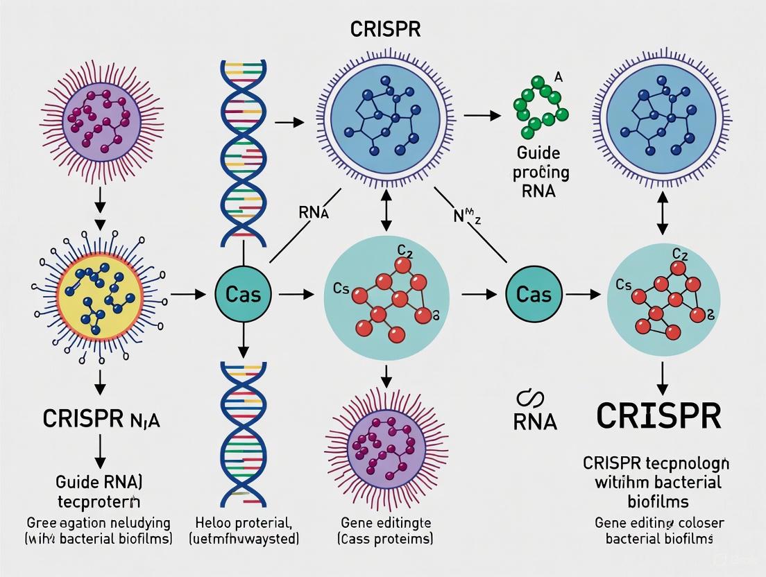
The following diagram illustrates the core mechanism of the CRISPR-Cas9 system and its application in targeting biofilm-related genes:
(CRISPR-Cas9 Mechanism for Biofilm Gene Targeting)
Advanced CRISPR Toolbox for Biofilm Research
Beyond standard CRISPR-Cas9 knockout approaches, several specialized systems enable more precise biofilm manipulation:
- CRISPR Interference (CRISPRi): Utilizing catalytically dead Cas9 (dCas9) fused to repressive domains for reversible gene silencing without permanent DNA damage [4] [5].
- CRISPR Activation (CRISPRa): Employing dCas9-activator fusions to enhance expression of biofilm-suppressing genes [4].
- Base and Prime Editing: Making precise single-nucleotide changes to study specific residues in biofilm regulatory proteins without creating double-strand breaks [3].
- Multiplexed Genome Engineering: Simultaneously targeting multiple biofilm-related genes using arrays of guide RNAs [7].
Application Notes & Experimental Protocols
Protocol 1: CRISPRi-Mediated Biofilm Gene Silencing in Pseudomonas fluorescens
Application: This protocol enables reversible gene silencing to study essential biofilm genes without creating irreversible knockouts, adapted from [5].
Materials:
- Bacterial strains of interest
- Two compatible plasmids: dCas9 expression vector and gRNA expression vector
- anhydrotetracycline (aTc) for induction
- Biofilm culture vessels (e.g., 96-well plates, flow cells)
- Confocal laser scanning microscopy (CLSM) system
Procedure:
sgRNA Design: Design 20-nt guide sequences targeting the promoter or 5' coding region of your biofilm gene of interest. For transcription initiation blockade, target the -35 to +1 region relative to transcription start site.
Plasmid Construction:
- Clone sgRNA into expression plasmid with constitutive promoter
- Transform dCas9 and sgRNA plasmids sequentially into target strain
- Verify constructs by sequencing
Induction Conditions:
- Grow overnight cultures with appropriate antibiotics
- Dilute 1:100 in fresh medium with 0-200 ng/mL aTc inducer
- Incubate with shaking at optimal growth temperature
Biofilm Phenotyping:
- For quantitative assessment: Use 96-well plate crystal violet assay
- For structural analysis: Grow biofilms in flow cells for CLSM
- Image 24-72 hour biofilms using appropriate stains (SYTO9 for cells, dextran-conjugates for EPS)
Validation:
- Quantify mRNA reduction via RT-qPCR
- Confirm protein level reduction if antibodies available
- Correlate knockdown efficiency with biofilm phenotype strength
Troubleshooting:
- High basal dCas9 activity: Titrate aTc concentration downward
- Weak phenotype: Test multiple sgRNAs targeting different regions
- Structural defects: Use CLSM to characterize 3D architecture changes
Protocol 2: Nanoparticle-Mediated CRISPR Delivery for Biofilm Eradication
Application: Overcoming limited penetration of CRISPR components through dense biofilm matrices using nanoparticle carriers, adapted from [1].
Materials:
- Cas9 protein or expression plasmid
- sgRNA targeting biofilm-specific resistance genes
- Liposomal or gold nanoparticle formulations
- Mature biofilms grown in relevant model systems
- Appropriate antibiotics for synergy testing
Procedure:
Nanoparticle Formulation:
- For liposomal preparations: Mix Cas9-sgRNA RNP complexes with lipid mixtures using thin-film hydration or ethanol injection methods
- For gold nanoparticles: Conjugate RNP complexes via thiol chemistry
- Characterize size distribution (DLR or NTA) and encapsulation efficiency
Biofilm Treatment:
- Grow 24-72 hour mature biofilms in relevant medium
- Apply nanoparticle formulations at optimized concentrations
- Include controls: free CRISPR, nanoparticles alone, untreated
- Incubate for 4-24 hours depending on biofilm maturity
Efficacy Assessment:
- Quantify biofilm biomass via crystal violet staining
- Assess viability by colony forming units (CFU) counts
- Evaluate structural disruption by scanning electron microscopy (SEM)
- Test antibiotic resensitization by combining with sub-MIC antibiotics
Optimization:
- Test different nanoparticle surface modifications for enhanced penetration
- Optimize charge characteristics for EPS interaction
- Consider stimuli-responsive release (pH, enzyme-triggered)
Validation Metrics:
- ≥80% reduction in biofilm biomass compared to control
- 2-4 log reduction in viable counts
- 3.5-fold improvement in editing efficiency over non-carrier systems
- Restored antibiotic susceptibility demonstrated by reduced MIC values
The Scientist's Toolkit: Essential Research Reagents
Table 3: Key Reagents for CRISPR-Based Biofilm Research
| Reagent Category | Specific Examples | Function & Application |
|---|---|---|
| CRISPR Nucleases | SpCas9, dCas9, Cas12a, Cas13 | DNA/RNA targeting for gene knockout, knockdown, or editing [7] |
| Delivery Systems | Liposomal nanoparticles, Gold nanoparticles, Bacteriophages | Enhanced delivery through biofilm matrix [1] [8] |
| Biofilm Assay Kits | Crystal violet, ATP bioluminescence, Live/Dead staining | Biofilm quantification and viability assessment [2] |
| Imaging Tools | Confocal Laser Scanning Microscopy (CLSM), Scanning Electron Microscopy (SEM) | 3D structural analysis of biofilm architecture [2] [5] |
| Vector Systems | dCas9 expression plasmids, gRNA cloning vectors, Reporter constructs | CRISPR component expression and tracking [5] [9] |
Integrated Workflow for CRISPR-Based Biofilm Analysis
The following diagram outlines a comprehensive experimental workflow for applying CRISPR technologies to study biofilm-related genes:
(Comprehensive Workflow for CRISPR-Biofilm Research)
The integration of CRISPR-based technologies with traditional antimicrobial approaches represents a paradigm shift in combating biofilm-associated infections. The precision offered by these systems enables targeted disruption of resistance mechanisms while potentially preserving the natural microbiome—a significant advantage over broad-spectrum antibiotics [1] [4]. Current research demonstrates remarkable efficacy, with liposomal Cas9 formulations achieving over 90% reduction in P. aeruginosa biofilm biomass and nanoparticle carriers enhancing editing efficiency 3.5-fold [1].
Future directions include the development of smart delivery systems with enhanced biofilm-penetrating capabilities, integration of CRISPR diagnostics for rapid pathogen detection, and combination therapies that leverage synergy between genetic editing and conventional antibiotics [4] [8]. The recent success of lipid nanoparticle (LNP) delivery in clinical trials for genetic diseases suggests potential translation pathways for biofilm-targeted CRISPR therapies [8]. However, challenges remain in optimizing delivery efficiency, minimizing off-target effects, and navigating regulatory pathways for these innovative approaches [1] [3] [4].
As the field advances, the convergence of CRISPR technologies with artificial intelligence for target prediction and nanoparticle engineering for precision delivery will likely yield increasingly sophisticated anti-biofilm strategies, potentially revolutionizing our approach to some of the most challenging infections in clinical practice.
Biofilm-associated infections represent a significant hurdle in modern medicine, contributing substantially to persistent chronic infections and the global antimicrobial resistance (AMR) crisis. Biofilms are structured microbial communities embedded in a self-produced extracellular polymeric substance (EPS) that adheres to biological or inert surfaces [1] [10]. This architectural complexity confers remarkable resistance to conventional antibiotics, with biofilm-embedded bacteria exhibiting up to 1,000-fold greater tolerance compared to their planktonic counterparts [1]. The resilience of biofilms stems from an integrated multilayer defense system combining physical diffusion barriers, metabolic heterogeneity, and sophisticated cell-to-cell communication networks. Understanding this intricate architecture is paramount for developing advanced therapeutic strategies, including those leveraging CRISPR-based precision technologies [10].
Structural and Ultrastructural Organization of Biofilms
Biofilm formation follows a developmental cascade beginning with initial surface attachment and culminating in complex three-dimensional communities. The process initiates when planktonic bacteria reversibly attach to conditioned surfaces, followed by irreversible adhesion mediated by surface adhesins and pili [1] [10]. Attached cells then proliferate into microcolonies and begin synthesizing the extracellular matrix that defines the biofilm phenotype. The final maturation stage involves development of a complex architectural structure with characteristic water channels that facilitate nutrient distribution and waste removal [1]. The cycle completes with active dispersal of planktonic cells from the biofilm to colonize new niches [10].
The biofilm matrix is a composite material primarily consisting of polysaccharides, proteins, lipids, and extracellular DNA (eDNA) [10]. This matrix is not merely a passive scaffold but a dynamic functional component that provides structural stability, mediates adhesion to surfaces, and retains water to prevent desiccation [1]. At the ultrastructural level, biofilms display remarkable heterogeneity, with stratified organization into basal layers of densely packed cells firmly anchored to the substrate, and upper layers exhibiting more heterogeneous distribution of cells within the EPS matrix [1]. Advanced imaging techniques like confocal laser scanning microscopy (CLSM) and scanning electron microscopy (SEM) have revealed this architectural complexity, showing variations in bacterial density, matrix composition, and void spaces across the biofilm depth [1].
Diagram: Biofilm Developmental Cycle and Key Resistance Mechanisms
Physiological Barriers to Antimicrobial Penetration and Efficacy
The therapeutic recalcitrance of biofilms emerges from interconnected physiological mechanisms that collectively undermine antimicrobial efficacy:
Matrix-Imposed Diffusion Limitation: The anionic EPS matrix acts as a molecular sieve, physically restricting antibiotic penetration through charge-based interactions and molecular binding. This filtration effect creates concentration gradients that prevent adequate drug accumulation in the deeper biofilm layers [1] [10].
Metabolic Heterogeneity and Persister Cells: Biofilms develop microscale gradients of nutrients, oxygen, and metabolic waste products, resulting in heterogeneous metabolic activity throughout the structure. Subpopulations of bacteria, particularly in the deeper layers, enter a slow-growing or dormant state characterized by reduced metabolic activity. These persister cells are tolerant to conventional antibiotics that typically target active cellular processes, enabling biofilm regeneration after antibiotic pressure is removed [10].
Enhanced Horizontal Gene Transfer: The dense aggregation of bacterial cells within the EPS matrix facilitates efficient intercellular transfer of mobile genetic elements, including plasmids and transposons carrying antibiotic resistance genes. This proximity significantly accelerates the dissemination of resistance determinants throughout the microbial community [1] [10].
Adaptive Stress Responses: Biofilm-embedded bacteria exhibit upregulated expression of efflux pumps and other stress response systems that enhance their ability to expel or neutralize antimicrobial compounds. These adaptive responses are further modulated by cell-cell communication systems [1].
CRISPR-Based Approaches for Biofilm Functional Analysis
The emergence of CRISPR-Cas systems has revolutionized our ability to dissect the genetic determinants underlying biofilm formation and resistance. Both nuclease-active Cas9 for targeted gene knockout and catalytically dead Cas9 (dCas9) for gene modulation (CRISPRi/CRISPRa) enable precise functional genomics in diverse bacterial pathogens [4] [5].
CRISPR Interference (CRISPRi) for Targeted Gene Silencing
CRISPRi employs dCas9 fused to transcriptional repressors to achieve reversible, tunable gene silencing without permanent genetic alteration. This approach is particularly valuable for studying essential genes and regulatory networks controlling biofilm development [4] [5].
Protocol: CRISPRi-Mediated Biofilm Gene Silencing in Pseudomonas fluorescens [5]
Step 1: CRISPRi Plasmid System Construction
- Utilize a two-plasmid system with compatible replication origins and selection markers.
- Plasmid 1: Expresses dCas9 under the control of an inducible promoter (e.g., PtetA induced by anhydrotetracycline, aTc).
- Plasmid 2: Constitutively expresses gene-specific guide RNA (gRNA) targeting the promoter or coding region of biofilm-related genes.
Step 2: gRNA Design and Validation
- Design gRNAs complementary to the non-template strand targeting regions within 50 base pairs downstream of the transcription start site for optimal repression.
- For genes involved in c-di-GMP signaling (e.g., GacA/S two-component system), design multiple gRNAs and validate silencing efficiency via qRT-PCR.
Step 3: Bacterial Transformation and Induction
- Introduce both plasmids into target bacterial strain via electroporation or conjugation.
- Culture transformants in medium containing appropriate antibiotics and induce dCas9 expression with aTc (e.g., 100 ng/mL).
Step 4: Phenotypic Assessment of Biofilm Formation
- Biomass Quantification: Perform crystal violet staining after 24-48 hours of biofilm growth. Measure solubilized dye absorbance at 570 nm.
- Architectural Analysis: Use confocal laser scanning microscopy (CLSM) of live-dead stained biofilms to assess 3D structure and viability.
- Motility Assays: Evaluate swarming and swimming motility on semi-solid agar to assess impact on virulence-related behaviors.
Quantitative Data on CRISPR Efficacy Against Biofilm Formation
Table 1: Efficacy of CRISPR-Based Interventions Against Biofilm Formation
| Target Pathway | CRISPR System | Target Bacteria | Efficacy Metric | Quantitative Outcome | Reference |
|---|---|---|---|---|---|
| Quorum Sensing & EPS Production | CRISPRi (dCas9) | Pseudomonas fluorescens | Biofilm biomass reduction | >70% reduction compared to control | [5] |
| Antibiotic Resistance Genes | CRISPR-Cas9 | Escherichia coli | Re-sensitization to antibiotics | 3.5-fold increase in antibiotic efficacy | [1] |
| Biofilm Matrix Genes | Liposomal CRISPR-Cas9 | Pseudomonas aeruginosa | Biofilm biomass reduction | >90% reduction in vitro | [1] |
| c-di-GMP Regulatory Genes | CRISPRi (dCas9) | Pseudomonas fluorescens | Swarming motility inhibition | Significant impairment in swarm expansion | [5] |
CRISPR-Enhanced Nanoparticle Delivery Systems
A significant challenge in therapeutic CRISPR application is efficient delivery through the biofilm matrix. Nanoparticle (NP)-based carriers have emerged as promising vehicles that enhance stability, penetration, and targeted delivery of CRISPR components [1].
Step 1: Cas9-sgRNA Ribonucleoprotein (RNP) Complex Preparation
- Formulate RNP complexes by incubating purified Cas9 protein with synthetic sgRNA targeting essential biofilm genes (e.g., quorum sensing regulators, antibiotic resistance genes) at a 1:2 molar ratio for 15 minutes at room temperature.
Step 2: Lipid Nanoparticle (LNP) Encapsulation
- Prepare lipid mixture of ionizable cationic lipid, phospholipid, cholesterol, and PEG-lipid in ethanol at specific molar ratios.
- Mix aqueous RNP solution with lipid solution using microfluidic device to form uniform LNPs.
- Dialyze LNP formulation against PBS to remove ethanol and concentrate using centrifugal filters.
Step 3: Characterization and Quality Control
- Determine particle size and polydispersity index via dynamic light scattering (target diameter: 80-120 nm).
- Measure zeta potential to confirm surface charge.
- Quantify encapsulation efficiency using RiboGreen assay.
Step 4: Application and Efficacy Assessment
- Apply LNP-formulated RNP complexes to pre-established biofilms and quantify biomass reduction via crystal violet staining.
- Assess bacterial viability through colony-forming unit (CFU) counts and live-dead staining.
- Visualize biofilm structural integrity using scanning electron microscopy.
Research Reagent Solutions for CRISPR-Biofilm Studies
Table 2: Essential Research Reagents for CRISPR-Mediated Biofilm Analysis
| Reagent/Category | Specific Examples | Function/Application | Experimental Considerations | |
|---|---|---|---|---|
| CRISPR Systems | dCas9 for CRISPRi, Cas9 nuclease, Cas12a | Targeted gene knockout or transcriptional modulation in biofilm-forming bacteria | Catalytically dead dCas9 enables reversible gene silencing without DNA cleavage | [4] [5] |
| Delivery Vehicles | Lipid nanoparticles, Gold nanoparticles, Bacteriophages | Enhance delivery of CRISPR components through protective biofilm matrix | Gold nanoparticles shown to increase editing efficiency by 3.5-fold compared to non-carrier systems | [1] |
| Biofilm Assay Kits | Crystal violet staining kits, Live-dead viability kits, EPS extraction kits | Quantify total biomass, assess viability distribution, analyze matrix composition | Crystal violet measures total biomass but cannot distinguish between live and dead cells | [11] |
| Molecular Tools | sgRNA synthesis kits, Plasmid vectors with inducible promoters, qRT-PCR reagents | Construct CRISPR systems and validate gene expression changes | Inducible promoters enable temporal control of CRISPR system activity | [5] |
| Imaging Reagents | CLSM-compatible stains, SEM preparation kits, Fluorescent protein reporters | Visualize 3D biofilm architecture and spatial gene expression patterns | CLSM enables non-destructive imaging of biofilm depth and structure | [1] [5] |
Diagram: CRISPR-Nanoparticle Synergy in Biofilm Eradication
The structural and physiological barriers posed by biofilm architecture represent a formidable challenge in clinical management of persistent infections. The integration of CRISPR-based technologies provides unprecedented precision in dissecting and targeting the genetic foundations of biofilm-mediated resistance. When combined with advanced delivery platforms like engineered nanoparticles, CRISPR systems can effectively penetrate the biofilm matrix and disrupt key resistance mechanisms. As these technologies continue to evolve, they hold exceptional promise for developing next-generation anti-biofilm strategies that transcend the limitations of conventional antibiotics, potentially revolutionizing the treatment of chronic infections in the post-antibiotic era.
CRISPR-Cas systems represent a remarkable evolutionary adaptation in prokaryotes, originating as adaptive immune mechanisms that protect bacteria and archaea from viral infections and other invasive genetic elements. These systems consist of clustered regularly interspaced short palindromic repeats (CRISPR) and CRISPR-associated (Cas) proteins that provide sequence-specific defense through a molecular memory of past infections [12]. The fundamental mechanism involves three distinct stages: adaptation, where new spacers are acquired from foreign DNA; expression, where CRISPR arrays are transcribed and processed into CRISPR RNAs (crRNAs); and interference, where crRNA-guided Cas complexes identify and cleave complementary nucleic acids [12]. This sophisticated immune system has been identified in approximately 40% of sequenced bacteria and over 80% of archaea, demonstrating its widespread biological significance [12].
The transformative potential of CRISPR-Cas systems was realized when researchers repurposed the Type II CRISPR-Cas9 system from Streptococcus into a programmable gene-editing tool, creating a two-component system comprising the Cas9 enzyme and a synthetic single-guide RNA [12]. This breakthrough initiated a new era in genetic engineering, enabling precise genome manipulation across diverse organisms and cell types. The subsequent discovery and adaptation of additional Cas enzymes, including Cas12 (Type V) for DNA editing and Cas13 (Type VI) for RNA targeting, further expanded the CRISPR toolkit beyond simple DNA cleavage [12]. This evolution from bacterial immunity to programmable gene editing has revolutionized biological research, therapeutic development, and biotechnology applications.
Application Notes: CRISPR-Based Biofilm Research
Quantitative Analysis of CRISPR Applications in Biofilm Disruption
Table 1: Efficacy of CRISPR-Based Approaches Against Bacterial Biofilms
| Application Approach | Target System | Reported Efficacy | Delivery Method | Key Findings |
|---|---|---|---|---|
| CRISPR-Cas9 with nanoparticle delivery | Pseudomonas aeruginosa | >90% reduction in biofilm biomass [1] | Liposomal nanoparticles | Enhanced cellular uptake and controlled release within biofilm environments |
| CRISPR-Cas9 with gold nanoparticles | Various biofilm-forming bacteria | 3.5-fold increase in editing efficiency [1] | Gold nanoparticle carriers | Superior biofilm penetration and synergistic action with antibiotics |
| CRISPR interference (CRISPRi) | Pseudomonas fluorescens | Effective gene silencing confirmed [5] | Plasmid-based dCas9 | Reliable phenotyping of complex traits including swarming motility and biofilm formation |
| CRISPR-based antimicrobials | Foodborne pathogens | ~3-log reduction of target pathogens [4] | Engineered phages or nanoparticles | Programmable, sequence-specific suppression of pathogens and resistance genes |
Key Signaling Pathways in Biofilm Formation
The development of bacterial biofilms is regulated through sophisticated signaling pathways that control the transition from motile to sessile lifestyles. Two primary regulatory systems govern this process: the GacA/S two-component system and cyclic di-GMP (c-di-GMP) signaling networks [5]. The GacA/S system comprises a membrane-bound sensor histidine kinase (GacS) and its cognate response regulator (GacA) that coordinates genetic programs in response to environmental stimuli [5]. This system regulates approximately 700 genes involved in diverse functions including biofilm formation, oxidative stress response, and secondary metabolite production through the Gac/Rsm signaling cascade involving non-coding small regulatory RNAs RsmZ and RsmY [5].
Simultaneously, c-di-GMP serves as a near-universal intracellular signaling messenger that regulates bacterial motility, virulence, and the transition to biofilm formation [5]. Intracellular c-di-GMP levels are modulated through the balanced activities of diguanylate cyclases (DGCs), which synthesize c-di-GMP from GTP, and phosphodiesterases (PDEs), which break down c-di-GMP [5]. Elevated intracellular concentrations of c-di-GMP correlate with increased propensity for biofilm formation, influencing multiple stages of biofilm development from initial cell adhesion to matrix production and eventual dispersion [5].
Diagram 1: Signaling pathways regulating bacterial biofilm formation. The GacA/S two-component system and c-di-GMP signaling network integrate environmental cues to control biofilm development through regulation of EPS production and bacterial lifestyle transitions.
Advanced CRISPR Tools for Biofilm Research
The expanding CRISPR toolkit now includes several specialized technologies for biofilm research. CRISPR interference (CRISPRi) utilizes a catalytically inactive Cas9 (dCas9) to block transcription initiation or elongation without altering DNA sequence, enabling reversible gene silencing essential for studying essential biofilm genes [4] [5]. Conversely, CRISPR activation (CRISPRa) employs modified dCas9 complexes to enhance gene transcription, facilitating gain-of-function studies [4]. More recently, CRISPR-based diagnostics platforms including SHERLOCK (Cas13-based) and DETECTR (Cas12-based) exploit collateral cleavage activity to achieve attomolar sensitivity for detecting biofilm-forming pathogens [4]. These tools provide unprecedented precision for dissecting biofilm regulatory networks and developing targeted interventions.
Experimental Protocols
Protocol: CRISPRi-Mediated Gene Silencing in Pseudomonas fluorescens for Biofilm Analysis
Principle
CRISPR interference (CRISPRi) enables targeted gene silencing without permanent genetic modifications through the action of a catalytically inactive Cas9 (dCas9) that sterically hinders transcription when directed to specific genomic locations by guide RNAs [5]. This protocol describes the application of CRISPRi to investigate genes controlling biofilm formation in P. fluorescens, a model organism for studying complex bacterial phenotypes including cell morphology, motility, and biofilm architecture [5].
Materials and Reagents
Table 2: Research Reagent Solutions for CRISPRi Biofilm Experiments
| Reagent/Category | Specific Examples | Function/Application | Experimental Notes |
|---|---|---|---|
| CRISPRi Plasmid System | dCas9 expression plasmid (PtetA promoter), gRNA expression plasmid [5] | Provides inducible dCas9 and target-specific gRNA | Use compatible plasmids with appropriate antibiotic resistance |
| Inducer | Anhydrotetracycline (aTc) [5] | Induces dCas9 expression from PtetA promoter | Dose-dependent response; optimize for each strain |
| Bacterial Strains | P. fluorescens SBW25, WH6, Pf0-1 [5] | Model organisms for biofilm research | Chromosomal reporter constructs recommended for validation |
| Imaging & Analysis | Confocal laser scanning microscopy (CLSM) [1] | High-resolution biofilm architecture analysis | Enables quantification of biomass, thickness, and EPS distribution |
| Delivery Vehicles | Liposomal nanoparticles, Gold nanoparticles [1] | Enhances CRISPR component delivery | Improves biofilm penetration and editing efficiency |
Procedure
Step 1: gRNA Design and Validation
- Design gRNAs targeting either the template (T) or non-template (NT) DNA strand of the promoter region or early coding sequence of target genes [5].
- For transcription initiation blockade: Design gRNAs complementary to regions within 50 bp downstream of the transcription start site [5].
- For transcription elongation blockade: Design gRNAs targeting sites overlapping the start of the open reading frame [5].
- Validate gRNA specificity using appropriate bioinformatics tools to minimize off-target effects.
Step 2: Plasmid Construction
- Clone the selected gRNA sequence into the gRNA expression plasmid under a constitutive promoter [5].
- Transform both dCas9 expression plasmid and gRNA plasmid into the target P. fluorescens strain using standard electroporation or chemical transformation methods [5].
- Select transformants using appropriate antibiotics and verify plasmid incorporation through colony PCR and sequencing.
Step 3: Induction of Gene Silencing
- Inoculate transformed bacteria in appropriate liquid medium and grow to mid-exponential phase (OD600 ≈ 0.5).
- Add anhydrotetracycline (aTc) inducer to a final concentration of 100 ng/mL (optimize concentration based on experimental conditions) [5].
- Incubate for 6-24 hours to allow dCas9 expression and target gene silencing.
Step 4: Biofilm Formation Assay
- Transfer induced cultures to biofilm-compatible surfaces (e.g., glass coverslips, microtiter plates, flow cells).
- Allow biofilms to develop for 24-72 hours under conditions appropriate for the specific P. fluorescens strain being studied.
- For architectural analysis, use confocal laser scanning microscopy (CLSM) to image biofilms at various time points [1].
Step 5: Phenotypic Analysis
- Quantify biofilm biomass using crystal violet staining or direct microscopic measurement.
- Analyze biofilm architecture using COMSTAT or similar software for CLSM image analysis.
- Assess related phenotypes including swarming motility, EPS production, and cellular morphology [5].
Diagram 2: Experimental workflow for CRISPRi-mediated analysis of biofilm formation. The protocol progresses from genetic construct preparation through bacterial transformation, induction of gene silencing, biofilm development, and quantitative phenotypic analysis.
Troubleshooting and Optimization
- Low silencing efficiency: Optimize aTc concentration (test range: 50-200 ng/mL); verify gRNA binding site accessibility; check dCas9 expression levels [5].
- Variable phenotypes between replicates: Standardize growth conditions; ensure consistent induction timing; use freshly prepared aTc stock solutions.
- Poor biofilm formation: Verify nutrient composition; optimize surface material; control environmental conditions (temperature, humidity).
- High basal silencing without inducer: Use lower-copy-number plasmids; include tighter regulatory elements; reduce basal dCas9 expression [5].
Protocol: Nanoparticle-Mediated CRISPR Delivery for Biofilm Eradication
Principle
Nanoparticles enhance the delivery of CRISPR components to bacterial cells within biofilms by improving cellular uptake, protecting genetic material from degradation, and facilitating penetration through the extracellular polymeric matrix [1]. This protocol describes the formulation of CRISPR-functionalized nanoparticles and their application for targeted biofilm disruption.
Procedure
Step 1: Nanoparticle Formulation
- For liposomal nanoparticles: Prepare lipid film using DOTAP, DOPE, and cholesterol in molar ratio 1:0.7:0.3 [1].
- Hydrate lipid film with CRISPR-Cas9 components (purified Cas9 protein and sgRNA complex or encoding plasmids) in appropriate buffer.
- Size reduction through extrusion or sonication to achieve particles of 100-200 nm diameter.
- For gold nanoparticles: Functionalize citrate-capped AuNPs (15-20 nm) with thiolated oligonucleotides and CRISPR complexes [1].
Step 2: Biofilm Treatment and Assessment
- Apply nanoparticle formulations to pre-established biofilms (24-48 hours old) at multiplicities of infection (MOI) optimized for the specific system.
- Incubate for 4-24 hours to allow penetration and editing.
- Assess biofilm reduction through biomass staining, viability counts, and microscopy.
- Quantify gene editing efficiency through PCR-based methods or sequencing.
CRISPR-Cas systems have undergone a remarkable transformation from fundamental bacterial immune mechanisms to versatile tools for precision biofilm research and intervention. The integration of CRISPR technologies with advanced delivery systems such as nanoparticles has created powerful platforms for dissecting the complex regulatory networks that control biofilm formation and for developing targeted approaches to combat biofilm-associated challenges [1] [4]. The ongoing discovery of novel Cas variants including CasΦ and CasX/Cas12f, coupled with precision editing technologies like base editing and prime editing, continues to expand the CRISPR toolbox for biofilm research [13].
Future developments in CRISPR-based biofilm control will likely focus on enhancing delivery efficiency through engineered nanoparticles with improved biofilm penetration capabilities, developing multiplexed approaches that simultaneously target multiple genetic pathways, and creating integrated systems that combine CRISPR-mediated gene editing with conventional antimicrobial strategies [1] [4]. Furthermore, the convergence of CRISPR technologies with artificial intelligence for predictive modeling of optimal gene targets and guide RNA designs represents a promising frontier for precision biofilm control [4]. As these technologies mature, they hold significant potential for addressing the persistent challenges posed by bacterial biofilms in clinical, industrial, and environmental settings.
Bacterial biofilms represent a significant challenge in clinical and industrial settings due to their inherent resistance to antimicrobial treatments and immune responses. The architecture and resilience of biofilms are governed by complex genetic networks that regulate key processes such as quorum sensing (QS), extracellular polymeric substance (EPS) production, and stress response [14]. The emergence of CRISPR technology has revolutionized functional genomics, providing researchers with precise tools to interrogate these biofilm-regulating genes. This Application Note details protocols for using CRISPR interference (CRISPRi) to investigate the genetic determinants of biofilm formation, offering methodologies for gene silencing, phenotypic analysis, and mechanistic studies. The guidance is framed within a broader research context focused on leveraging CRISPR-based approaches for antibacterial drug development and biofilm control strategies.
Key Genetic Determinants of Biofilm Formation
The transition from planktonic to sessile biofilm lifestyle is coordinated by specific genetic systems. Understanding these systems is crucial for designing targeted CRISPR-based studies.
Quorum Sensing (QS) Genes
Quorum sensing is a cell-density dependent communication system that coordinates collective behaviors, including biofilm formation. Key QS genes serve as prime targets for CRISPRi silencing studies.
- LuxR/LuxI-type systems: These acyl-homoserine lactone (AHL)-based systems are prevalent in Gram-negative bacteria. Silencing the synthase (e.g., lasI, rhlI) or receptor (e.g., lasR, rhlR) genes disrupts the production of signal molecules and the expression of virulence and biofilm genes [15].
- Accessory regulator genes: Genes such as gacA and gacS, which form a two-component system, regulate the QS cascade and control the expression of downstream biofilm-related operons. In Pseudomonas fluorescens, CRISPRi-mediated silencing of the gacA/S system produces clear swarming and biofilm phenotypes [5].
Extracellular Polymeric Substance (EPS) Production Genes
The EPS matrix forms the structural backbone of the biofilm, providing mechanical stability and protection. Its production is regulated by complex genetic networks.
- Polysaccharide biosynthesis operons: These include genes for the synthesis of polysaccharides like alginate, cellulose, and Pel. For example, the PFLU1114 operon in P. fluorescens SBW25 has been shown to powerfully inhibit biofilm formation when disrupted [5].
- c-di-GMP regulatory network genes: The intracellular secondary messenger cyclic di-GMP (c-di-GMP) is a central regulator of the motile-sessile switch. Genes encoding diguanylate cyclases (DGCs) with GGDEF domains (e.g., gcbA) synthesize c-di-GMP, promoting biofilm formation. Conversely, genes encoding phosphodiesterases (PDEs) with EAL or HD-GYP domains (e.g., bifA, dipA) degrade c-di-GMP, promoting dispersal [5]. CRISPRi silencing allows for the specific functional analysis of these numerous and often redundant enzymes.
Stress Response Genes
Biofilm environments create heterogeneous microenvironments with nutrient and oxygen gradients, necessitating robust stress response pathways.
- General stress response regulators: Genes like rpoS (encoding the sigma factor σ^S^) control the expression of a large regulon in response to starvation, oxidative, and osmotic stress, enhancing the survival of biofilm-embedded cells.
- Persister cell formation genes: Toxin-antitoxin (TA) systems (e.g., hipA, relBE) are implicated in the formation of dormant, highly tolerant persister cells within biofilms. Targeting these genes with CRISPRi can help elucidate mechanisms of recalcitrance to antibiotic treatment.
Table 1: Key Biofilm-Regulating Gene Targets for CRISPR-Based Studies
| Gene Category | Example Genes | Function | Phenotype upon Silencing |
|---|---|---|---|
| Quorum Sensing | gacA, gacS, lasI, lasR | Two-component system; AHL synthase/receptor [5] [15] | Disrupted cell-cell signaling, reduced virulence factor production, impaired biofilm maturation |
| EPS Production | alg44, PFLU1114 operon | Alginate copolymerase; putative EPS biosynthesis [5] | Altered biofilm architecture, reduced biomass, increased susceptibility to antimicrobials |
| c-di-GMP Metabolism | DGCs (e.g., gcbA), PDEs (e.g., bifA, dipA) | Synthesis and degradation of c-di-GMP [5] | Altered motility, defective initial attachment, or impaired biofilm dispersion |
CRISPRi Experimental Protocol for Biofilm Gene Silencing
This section provides a detailed methodology for implementing a CRISPRi system to study biofilm genes in a model bacterium like Pseudomonas fluorescens, based on the system adapted by [5].
Research Reagent Solutions
The following reagents are essential for establishing the CRISPRi system.
Table 2: Essential Research Reagents for CRISPRi Biofilm Studies
| Reagent/Solution | Function/Description | Example/Specification |
|---|---|---|
| dCas9 Expression Plasmid | Constitutively or inducibly expresses catalytically "dead" Cas9 (dCas9), which binds DNA without cutting it. | Plasmid with PtetA promoter driving dCas9 expression, origin of replication for Pseudomonas [5]. |
| Guide RNA (gRNA) Expression Plasmid | Constitutively expresses the target-specific gRNA. Contains a scaffold sequence that binds dCas9. | Compatible plasmid with constitutive promoter (e.g., P~J~23119), target-specific 20nt spacer sequence [5]. |
| Electrocompetent Cells | Bacterial cells prepared for transformation via electroporation. | P. fluorescens strain (e.g., SBW25, WH6, Pf0-1) made electrocompetent [5]. |
| Inducer Molecule | Chemical to induce dCas9 expression in inducible systems. | Anhydrotetracycline (aTc), used at optimized concentrations (e.g., 100 ng/mL) [5]. |
| Biofilm Growth Media | Culture medium supporting robust biofilm formation. | Standard Lysogeny Broth (LB) or specific minimal media; 96-well polystyrene plates for microtiter assays. |
Protocol Workflow
The following diagram outlines the complete experimental workflow for a CRISPRi biofilm study.
Step 1: gRNA Design and Plasmid Construction
- Target Selection: Identify the 20-nucleotide DNA sequence immediately 5' to a 5'-NGG-3' Protospacer Adjacent Motif (PAM) within the promoter or coding region of your target gene. For maximal silencing, design gRNAs to target the non-template (NT) strand within the promoter or the early coding region to block transcription initiation or elongation, respectively [5].
- Plasmid Assembly: Synthesize and clone the spacer oligonucleotide into the BsaI site of the gRNA expression plasmid. Verify the sequence of the final construct by Sanger sequencing.
Step 2: Transformation
- Co-transform the verified gRNA plasmid and the dCas9 expression plasmid into electrocompetent P. fluorescens cells via electroporation.
- Plate the transformation mixture on selective media containing the appropriate antibiotics for both plasmids. Incubate at 28-30°C for 24-48 hours until colonies appear.
Step 3: Gene Silencing Induction
- Inoculate a single colony into liquid media with antibiotics and the inducer (e.g., 100 ng/mL aTc). Incubate with shaking for 16-24 hours.
- For biofilm assays, dilute the induced culture to an OD~600~ of ~0.05 in fresh media with inducer and antibiotics.
Step 4: Biofilm Phenotypic Assessment
Crystal Violet Biofilm Assay [5]:
- Transfer 150 µL of diluted culture to wells of a 96-well polystyrene plate. Include controls (non-targeting gRNA, no gRNA).
- Incubate statically for 24-48 hours at 30°C.
- Carefully remove planktonic cells and rinse the biofilm with water.
- Stain the adherent biofilm with 0.1% crystal violet for 15 minutes.
- Rinse thoroughly and solubilize the bound dye with 30% acetic acid.
- Measure the absorbance at 550 nm to quantify total biofilm biomass.
Swarming Motility Assay:
- Prepare 0.3-0.5% agar plates containing appropriate media, antibiotics, and inducer.
- Spot 2-3 µL of induced culture in the center of the agar surface.
- Incubate the plates right-side up for 16-24 hours.
- Measure the diameter of the swarming colony to quantify motility, which is often inversely correlated with biofilm formation.
Step 5: Biofilm Architecture Imaging
- Grow biofilms on a suitable surface (e.g., glass coverslip) placed in a culture well.
- Stain the mature biofilm with fluorescent dyes (e.g., SYTO 9 for cells, Concanavalin A-Texas Red for polysaccharides).
- Image the biofilm using a Confocal Laser Scanning Microscope (CLSM). Acquire Z-stacks at multiple random locations.
- Use image analysis software (e.g., ImageJ, COMSTAT) to quantify architectural parameters such as biomass volume, average thickness, and substratum coverage [5].
Step 6: Data Analysis and Validation
- Statistical Analysis: Perform triplicate biological repeats. Use Student's t-test or ANOVA to determine the statistical significance of phenotypic differences between the target-gRNA strain and control strains.
- Validation: Confirm gene knockdown efficiency by quantifying mRNA levels using reverse transcription quantitative PCR (RT-qPCR).
Signaling Pathway and Genetic Network Analysis
The genes regulating biofilm formation do not operate in isolation but are part of an integrated signaling network. The following diagram synthesizes the key interactions between the quorum sensing, c-di-GMP, and EPS production systems.
Quantitative Data and Expected Outcomes
Successful implementation of the CRISPRi protocol should yield quantifiable changes in biofilm phenotypes and gene expression. The table below summarizes expected outcomes based on published studies.
Table 3: Expected Quantitative Outcomes from CRISPRi Silencing of Biofilm Genes
| Target Gene/Pathway | Assay Type | Control Value | Expected Outcome with CRISPRi | Reported Efficiency/Notes |
|---|---|---|---|---|
| gacA/S (QS) | Swarming Motility | Swarm diameter: ~20 mm | >50% reduction in diameter [5] | CRISPRi phenocopied gene inactivation mutants [5] |
| PFLU1114 operon (EPS) | Biofilm Biomass (Crystal Violet) | OD~550~: ~2.0 | >70% reduction in OD~550~ [5] | Potent inhibition of biofilm formation observed [5] |
| Multiple c-di-GMP genes | Biofilm Architecture (CLSM) | Biomass: ~25 µm³/µm² | Variable (Increase or decrease\nbased on target) [5] | ~1/3 of mutants show strong phenotypes across conditions [5] |
| General | Gene Expression (qPCR) | 100% mRNA expression | 50-90% knockdown of target mRNA [5] | Efficiency depends on gRNA target location (promoter vs. coding) [5] |
Bacterial biofilms are structured communities of microorganisms embedded in a self-produced extracellular polymeric substance (EPS) matrix, which confers inherent resistance to conventional antimicrobial treatments [1]. The complex architecture of biofilms limits antibiotic penetration and creates heterogeneous microenvironments, leading to persistent infections that are challenging to eradicate [1]. Understanding the genetic pathways controlling biofilm formation and maintenance is therefore crucial for developing effective anti-biofilm strategies. CRISPR-based technologies have emerged as powerful discovery tools for functional genomics, enabling researchers to systematically interrogate genes involved in biofilm formation pathways with unprecedented precision and scalability.
Traditional gene inactivation methods are often labor-intensive, limiting systematic genetic surveys in many bacterial hosts [5]. The adaptation of CRISPR interference (CRISPRi) for biofilm research has revolutionized this field by enabling reversible, sequence-specific gene silencing without permanent genetic alterations [5]. This approach utilizes a catalytically inactive Cas9 (dCas9) protein that binds to target DNA sequences under the guidance of a specific single-guide RNA (sgRNA), sterically hindering transcription initiation or elongation [16]. The precision of CRISPRi allows researchers to dissect complex genetic networks governing biofilm development, including quorum sensing pathways, cyclic di-GMP (c-di-GMP) signaling systems, and EPS production mechanisms [5] [16].
Key Signaling Pathways in Biofilm Formation
Quorum Sensing and Biofilm Regulation
Quorum sensing (QS) represents a cell-density dependent chemical signaling mechanism that bacteria utilize to coordinate collective behaviors including biofilm formation and pathogenesis [16]. The luxS gene, which encodes a synthase involved in the production of Autoinducer-2 (AI-2), plays a pivotal role in guiding the initial stages of biofilm formation across numerous bacterial species [16]. CRISPRi-mediated silencing of luxS has been demonstrated to significantly inhibit biofilm formation in E. coli, confirming its essential function in QS-mediated biofilm development [16].
Cyclic di-GMP Signaling Network
The near-universal bacterial second messenger c-di-GMP represents a key signaling system that regulates the transition between motile and sessile lifestyles [5]. Intracellular c-di-GMP levels are modulated through the balanced activities of diguanylate cyclases (DGCs) containing GGDEF domains that synthesize c-di-GMP, and phosphodiesterases (PDEs) containing EAL or HD-GYP domains that break down c-di-GMP [5]. Elevated intracellular concentrations of c-di-GMP typically correlate with enhanced biofilm formation through the regulation of EPS production, flagellar function, and cell adhesion mechanisms [5]. Bacterial genomes often encode numerous proteins with c-di-GMP-binding signatures, creating a complex regulatory network that controls various stages of biofilm development from initial attachment to maturation and dispersion [5].
Two-Component Systems
Two-component systems (TCS) serve as fundamental environmental sensing mechanisms that translate external stimuli into coordinated cellular responses through regulated genetic programs [5]. In Pseudomonas species, the GacA/S two-component system regulates approximately 700 genes involved in diverse biological functions including biofilm formation, oxidative stress response, and secondary metabolite production [5]. This system consists of a membrane-bound sensor histidine kinase (GacS) and its cognate response regulator (GacA), which collectively influence EPS production through the Gac/Rsm signaling cascade involving non-coding small regulatory RNAs RsmZ and RsmY [5].
The following diagram illustrates the core CRISPRi mechanism for interrogating these biofilm formation pathways:
Experimental Protocols for CRISPR-Based Biofilm Analysis
CRISPRi System Design and Implementation
The following protocol outlines the implementation of a CRISPRi system for interrogating biofilm-related genes in diverse bacterial species, adapted from established methodologies in Pseudomonas fluorescens and E. coli [5] [16].
Materials Required:
- pdCas9 plasmid (constitutively or inducibly expresses dCas9)
- pgRNA plasmid backbone for sgRNA expression
- Target bacterial strain(s)
- Appropriate antibiotics for selection
- Inducer molecules (e.g., anhydrotetracycline/aTc for Ptet systems)
- Primers for sgRNA template cloning
- Molecular biology reagents for cloning and transformation
Procedure:
sgRNA Design and Cloning:
- Identify 20-nucleotide target sequences adjacent to PAM (NGG for S. pyogenes Cas9) sites within promoter regions or early coding sequences of target biofilm genes
- Synthesize complementary oligonucleotides containing the target sequence with appropriate overhangs for cloning into the pgRNA backbone
- Clone annealed oligonucleotides into the BsaI restriction site of the pgRNA plasmid using Golden Gate assembly
- Transform ligation products into E. Top 10 competent cells and select on appropriate antibiotic plates
- Verify successful cloning by colony PCR and Sanger sequencing using specific primers flanking the insertion site
Dual Plasmid Transformation:
- Co-transform the verified pgRNA construct (carrying biofilm gene-specific sgRNA) and pdCas9 plasmid into the target bacterial strain
- Select transformants on plates containing both antibiotics (e.g., ampicillin 100 μg/mL and chloramphenicol 25 μg/mL)
- Verify dual plasmid maintenance by replica plating and colony PCR
Induction of CRISPRi System:
- Inoculate single colonies of the knockdown strain in liquid medium with appropriate antibiotics
- Grow cultures to exponential phase (OD600 ≈ 0.4-0.6)
- Induce dCas9 expression with 2 μM anhydrotetracycline (aTc) or other appropriate inducer
- Continue incubation for 3-4 hours to allow gene silencing before downstream assays
Quantitative Assessment of Biofilm Phenotypes
Crystal Violet Biofilm Assay:
- Grow CRISPRi knockdown strains in appropriate media with inducer for 24-48 hours in static conditions
- Remove planktonic cells and gently wash adhered cells with phosphate-buffered saline (PBS)
- Fix biofilms with methanol or ethanol for 15 minutes
- Stain with 0.1% crystal violet solution for 15-20 minutes
- Wash extensively to remove unbound dye
- Elute bound dye with 33% acetic acid or ethanol-acetone mixture (80:20)
- Measure absorbance at 570-600 nm to quantify biofilm biomass [6] [16]
Metabolic Activity Assay (XTT Reduction):
- Prepare XTT-menadione solution fresh: 1 mg/mL XTT with 4 μM menadione in PBS
- Add XTT solution to biofilms and incubate in dark for 2-3 hours at 37°C
- Measure supernatant absorbance at 490 nm to assess metabolic activity of biofilm cells [16]
Confocal Laser Scanning Microscopy (CLSM):
- Grow biofilms on appropriate surfaces (e.g., glass coverslips, flow cells)
- Stain with SYTO9 green fluorescent nucleic acid stain (emission: 498 nm) for bacterial cells
- Counterstain extracellular polysaccharide (EPS) matrix with Alexa Fluor 647-conjugated dextran (emission: 668 nm) or other EPS-specific probes
- Image using appropriate laser settings and filter configurations
- Acquire z-stacks at regular intervals (e.g., 1 μm) through the entire biofilm depth
- Reconstruct 3D architecture and quantify biomass, thickness, and spatial distribution using image analysis software (e.g., ImageJ, COMSTAT) [6]
Gene Expression Validation:
- Extract total RNA from CRISPRi strains using Trizol method
- Treat with DNase I to remove genomic DNA contamination
- Synthesize cDNA using reverse transcriptase and random hexamers
- Perform quantitative RT-PCR with gene-specific primers
- Normalize expression to housekeeping genes (e.g., rpoB, gyrA)
- Calculate fold-change using comparative Ct method (2-ΔΔCt) [16]
The experimental workflow for implementing CRISPRi-based biofilm analysis is systematically outlined below:
Quantitative Data from CRISPR-Based Biofilm Studies
Table 1: Efficacy of CRISPRi in Targeting Biofilm-Associated Genes Across Bacterial Species
| Target Gene | Bacterial Species | Gene Function | CRISPRi Efficiency | Biofilm Reduction | Reference |
|---|---|---|---|---|---|
| luxS | E. coli AK-117 | Quorum sensing (AI-2 synthesis) | ~70% gene expression knockdown | ~60% biomass reduction | [16] |
| GacA/S | P. fluorescens SBW25 | Two-component system regulator | Strong downregulation | Significant alteration in biofilm architecture | [5] |
| PFLU1114 operon | P. fluorescens SBW25 | Biofilm inhibition mediator | Effective silencing | Potent inhibition of biofilm formation | [5] |
| Genes encoding DGCs/PDEs | P. fluorescens SBW25 | c-di-GMP metabolism | Successful repression | Altered biofilm mass and structure | [5] |
| cas3 (Type I-Fa) | A. baumannii ATCC19606 | CRISPR-Cas system component | Gene knockout | Significant reduction in biofilm formation | [6] |
Table 2: Advanced Nanoparticle-Mediated CRISPR Delivery Systems for Enhanced Biofilm Penetration
| Nanoparticle Type | CRISPR Payload | Target Bacterium | Editing Efficiency | Biofilm Reduction | Key Advantages |
|---|---|---|---|---|---|
| Liposomal nanoparticles | Cas9/sgRNA complexes | P. aeruginosa | High efficiency | >90% biofilm biomass reduction in vitro | Enhanced cellular uptake, controlled release [1] |
| Gold nanoparticles | CRISPR-Cas9 components | P. aeruginosa | 3.5-fold increase compared to non-carrier systems | Significant biofilm disruption | Improved target specificity, intrinsic antibacterial properties [1] |
| Hybrid nanoparticle systems | Cas9/sgRNA + antibiotics | Multi-drug resistant pathogens | Enhanced editing efficiency | Superior biofilm disruption | Synergistic antibacterial effects, co-delivery capability [1] |
The Scientist's Toolkit: Essential Research Reagents
Table 3: Key Research Reagent Solutions for CRISPR-Based Biofilm Studies
| Reagent / Material | Function | Application Notes | Commercial Sources |
|---|---|---|---|
| pdCas9 plasmids | Expresses catalytically inactive Cas9 protein | Available with inducible (Ptet) or constitutive promoters; includes antibiotic resistance markers | Addgene (#44249) |
| pgRNA backbone | sgRNA expression vector | Contains scaffold sequence compatible with S. pyogenes dCas9; ampicillin resistance | Addgene (#44251) |
| dCas9 protein | CRISPRi effector molecule | Can be used directly in ribonucleoprotein complexes for transient silencing | Commercial protein suppliers |
| Anhydrotetracycline (aTc) | Inducer for Ptet promoter systems | Typical working concentration: 2-100 nM; water-soluble | Sigma-Aldrich, Thermo Fisher |
| SYTO9 green fluorescent stain | Bacterial cell staining for CLSM | Excitation/Emission: 483/498 nm; penetrates intact cell membranes | Thermo Fisher, Bio-Rad |
| Alexa Fluor 647-dextran | EPS matrix staining for CLSM | Excitation/Emission: 647/668 nm; labels extracellular polysaccharides | Thermo Fisher |
| XTT sodium salt | Metabolic activity assay in biofilms | Used with electron-coupling agent menadione; measures cellular respiration | Sigma-Aldrich, Abcam |
| Crystal violet | Biofilm biomass quantification | Stains cells and matrix components; requires solubilization for quantification | Standard laboratory suppliers |
Discussion and Technical Considerations
The application of CRISPR-based technologies for interrogating biofilm formation pathways presents both unprecedented opportunities and technical challenges that researchers must address for successful experimental outcomes.
Optimization Strategies for Enhanced Efficiency
The efficiency of CRISPRi-mediated gene silencing in biofilm studies is influenced by multiple factors including sgRNA design, target accessibility, and bacterial strain characteristics. For optimal results, sgRNAs should be designed to target the non-template DNA strand in promoter regions or early coding sequences, as this approach has demonstrated superior repression efficiency [5]. The positioning of sgRNAs targeting transcription initiation sites (e.g., Pc4 and Pc5 in P. fluorescens) has been shown to produce more effective gene silencing compared to those targeting elongation regions [5]. Additionally, researchers should note that dCas9 may exhibit basal expression even in the absence of induction, which can be mitigated through careful control of induction parameters and the use of inducible promoter systems with low background activity [5].
Advanced Delivery Mechanisms for Biofilm Penetration
A significant challenge in CRISPR-based biofilm research involves efficient delivery of CRISPR components through the protective EPS matrix. Nanoparticle-based delivery systems have emerged as promising solutions to this limitation, offering enhanced penetration and targeted release within biofilm environments [1]. Liposomal Cas9 formulations have demonstrated remarkable efficacy, reducing P. aeruginosa biofilm biomass by over 90% in vitro [1]. Similarly, gold nanoparticle carriers have shown a 3.5-fold increase in editing efficiency compared to non-carrier systems, while also exhibiting intrinsic antibacterial properties that synergize with CRISPR-mediated genetic disruption [1]. These advanced delivery platforms can be further engineered for co-delivery of antibiotics or antimicrobial peptides alongside CRISPR components, creating multifaceted approaches that attack bacterial biofilms through both genetic and traditional antimicrobial mechanisms [1].
Interpretation of Phenotypic Outcomes
When employing CRISPRi for functional genomics in biofilm pathways, researchers should implement appropriate controls and validation methods to accurately interpret phenotypic outcomes. The inclusion of non-targeting sgRNA controls is essential to distinguish sequence-specific effects from non-specific responses to the CRISPRi system itself [5]. Additionally, complementation strains can help verify that observed phenotypes directly result from targeted gene silencing rather than off-target effects [6]. For comprehensive pathway analysis, researchers should consider implementing multiplexed sgRNA approaches to simultaneously target multiple components of biofilm regulatory networks, enabling the dissection of complex genetic interactions and functional redundancies within these systems [5].
The integration of CRISPR-based functional genomics with advanced analytical techniques such as confocal laser scanning microscopy, RNA sequencing, and proteomic profiling provides a powerful framework for elucidating the complex genetic pathways that control biofilm formation and maintenance. This multidisciplinary approach offers unprecedented insights into bacterial community behaviors and opens new avenues for developing targeted anti-biofilm strategies with translational potential in clinical, industrial, and environmental settings.
CRISPR Toolbox: From Functional Screening to Precision Antimicrobials
CRISPR Knockout Libraries for Genome-Wide Biofilm Gene Screening
CRISPR knockout (CRISPRko) screening represents a powerful high-throughput approach for functional genomics, enabling the systematic identification of genes essential for biofilm formation and regulation. This technology leverages the CRISPR-Cas9 system to generate permanent, targeted knockouts across the entire genome, allowing researchers to investigate genotype-phenotype relationships on an unprecedented scale [17]. In biofilm research, this is particularly valuable for unraveling the complex genetic networks that control bacterial adhesion, matrix production, community structure, and antibiotic tolerance [1] [18].
Compared to traditional genetic screening methods like RNA interference (RNAi), CRISPRko screening offers significantly higher specificity, more efficient gene knockout, and lower off-target effects [17]. The permanent nature of CRISPR-induced gene knockouts enables studies of long-term biofilm development and stability, making it particularly suited for investigating chronic biofilm-associated infections and industrial biofilm applications [19] [1]. The technology has been successfully applied to diverse microbial species, including Staphylococcus aureus, Pseudomonas aeruginosa, Escherichia coli, and various yeast species, providing insights into conserved and species-specific genetic determinants of biofilm formation [19] [18].
CRISPR Screening Approaches and Library Design
Types of CRISPR Screening Systems
CRISPRko is the most widely used approach for genome-wide knockout screens, utilizing the native Cas9 nuclease to create double-strand breaks that are repaired by non-homologous end joining (NHEJ), leading to frameshift mutations and gene knockouts [17]. However, modified CRISPR systems offer alternative screening strategies for biofilm research, each with distinct advantages as detailed in the table below.
Table 1: Comparison of CRISPR Screening Approaches for Biofilm Research
| Screening Type | Working Principle | Advantages | Disadvantages | Biofilm Applications |
|---|---|---|---|---|
| CRISPRko (Knockout) | Generates irreversible gene knockouts via NHEJ, followed by phenotypic screening [17]. | Low noise; single-vector system possible; relatively easy to operate [17]. | Potential off-target effects; may produce heterozygous cells; certain genes may have low editing efficiency [17]. | Identifying essential adhesion genes, matrix producers, and regulatory pathways [18]. |
| CRISPRi (Interference) | Uses dCas9 fused to transcriptional repressors (e.g., KRAB) for reversible gene suppression [17]. | No genome breakage; can target regulatory regions and non-coding genes [17]. | Affected by chromatin accessibility; larger complex; requires stable dCas9 cell line [17]. | Studying essential genes whose knockout is lethal; investigating non-coding RNAs in biofilm regulation. |
| CRISPRa (Activation) | Uses dCas9 fused to transcriptional activators (e.g., VP64) for gene upregulation [17]. | No genome breakage; can activate silent gene clusters [17]. | Affected by chromatin accessibility; larger complex; requires stable dCas9 cell line [17]. | Identifying genes that suppress biofilm formation when overexpressed. |
Library Design and Selection
The design of the guide RNA (gRNA) library is a critical factor determining screen success. For genome-wide screens in bacteria, libraries typically include 4-6 gRNAs per gene to ensure effective perturbation and control for off-target effects [20]. The number of gRNAs required per gene depends on the desired screen coverage, with 250x coverage per gRNA being the gold standard for hit identification, though lower coverage may be sufficient for strong phenotypes [20].
Table 2: CRISPR Library Selection Based on Screening Goals
| Library Coverage | Description | Recommended Use |
|---|---|---|
| Whole Genome Library | Designs gRNAs for all coding genes of a species; large coverage [17]. | Unbiased discovery of novel biofilm genes; foundational research. |
| Sub-library/Focused Library | Combination of a specific class of genes (e.g., kinase family, signaling pathway, transcription factors) [17]. | Targeted studies; validating specific hypotheses; follow-up screens. |
| Custom Library | User-defined gene set based on prior omics data or specific interests. | Investigating specific pathways or gene families with known biofilm relevance. |
For biofilm research, focused libraries targeting specific gene families such as kinases, two-component systems, transcription factors, or known adhesion factors (e.g., icaADBC, fnbA, clfA) can provide deeper coverage with reduced screening workload [17] [18]. The choice between genome-wide and focused libraries depends on the research objectives, available resources, and prior knowledge of the microbial system under investigation.
Experimental Workflow for Biofilm CRISPR Screening
The following diagram illustrates the comprehensive workflow for performing a CRISPR knockout screen to identify genes involved in biofilm formation:
Diagram Title: CRISPR Biofilm Screening Workflow
Library Delivery and Transduction
Efficient delivery of the CRISPR library to the target microbial cells is crucial for screen success. For bacterial systems, electroporation and conjugation are commonly used, while lentiviral transduction is preferred for eukaryotic microbes like yeast [17]. The library is typically delivered at a low multiplicity of infection (MOI < 0.3) to ensure most cells receive only a single gRNA, enabling clear genotype-phenotype associations [17]. Following transduction, cells are selected with appropriate antibiotics to eliminate untransduced cells, and a sample is collected as the "pre-selection" reference point for subsequent comparison.
For in vivo biofilm models, delivery optimization becomes more complex. Recent advances include engineered lentiviral vectors with specific tropisms, adeno-associated viral (AAV) vectors coupled with transposon systems for stable integration, and nanoparticle-based delivery systems that can enhance editing efficiency up to 3.5-fold compared to non-carrier systems [1] [20].
Biofilm Phenotypic Assay and Selection
The core of the screen involves applying a selective pressure that enriches or depletes specific gRNAs based on their effect on biofilm formation. Both positive and negative selection strategies can be employed:
- Negative Selection: Cells are grown under biofilm-forming conditions, and gRNAs that disrupt genes essential for biofilm formation will be depleted from the population. This approach identifies genes required for biofilm development [17].
- Positive Selection: Cells that cannot form biofilms are selectively enriched, potentially identifying genes that suppress biofilm formation when knocked out [17].
Biofilm quantification can be performed using various methods, including crystal violet staining for total biomass, colony forming unit (CFU) counts for viable cells, or advanced imaging techniques like confocal microscopy coupled with quantitative image analysis tools such as BiofilmQ [2] [21]. BiofilmQ enables comprehensive 3D quantification of biofilm properties, including biovolume, thickness, surface area, and spatial distribution of fluorescent reporters, providing rich phenotypic data beyond simple biomass measurements [21].
Sequencing and Bioinformatics Analysis
Following phenotypic selection, genomic DNA is extracted from both the selected population and the pre-selection reference, and the gRNA regions are amplified and sequenced using next-generation sequencing (NGS) [17]. The abundance of each gRNA in the selected versus reference population is compared to identify significantly enriched or depleted gRNAs.
Several bioinformatics tools are available for analysis, including MAGeCK, CERES, and CRISPRcleanR, which normalize read counts, calculate fold-changes, and perform statistical testing to identify significant hits [17]. Genes targeted by multiple significantly enriched or depleted gRNAs are considered high-confidence hits. For biofilm screens, hits are typically categorized into functional groups: adhesion factors, matrix producers, regulatory genes, metabolic enzymes, and unknown function genes.
Essential Research Reagents and Solutions
Table 3: Key Research Reagent Solutions for CRISPR Biofilm Screening
| Reagent/Category | Function | Examples & Specifications |
|---|---|---|
| CRISPRko Library | Contains sgRNAs targeting genes of interest for knockout screening [17]. | Genome-wide (e.g., 4-6 sgRNAs/gene) or sub-library (e.g., kinase-focused); human, mouse, microbial species-specific. |
| Cas9 Expression System | Provides the Cas9 nuclease for genome editing [17]. | Constitutive or inducible expression; plasmid, viral, or stable cell line formats; species-specific codon optimization. |
| Delivery Vehicles | Facilitates introduction of CRISPR components into target cells [20]. | Lentiviral vectors (VSVG-pseudotyped), AAV vectors, lipid nanoparticles, electroporation systems. |
| Selection Antibiotics | Enriches for successfully transduced cells [17]. | Puromycin, blasticidin, gentamicin; concentration optimized for host cell type. |
| Biofilm Assay Kits | Enables quantification of biofilm formation [2] [18]. | Crystal violet staining kits, microtiter plate assays, metabolic activity assays (e.g., resazurin). |
| gRNA Amplification Primers | Allows amplification of gRNA regions for NGS library preparation [17]. | Universal primers compatible with library design; including sequencing adapters and barcodes. |
| Biofilm Imaging Tools | Enables spatial analysis of biofilm structure and composition [21]. | BiofilmQ software, fluorescent dyes (SYTO, propidium iodide), antibody labels for matrix components. |
Applications in Biofilm Gene Discovery
CRISPR knockout screening has identified numerous genes critical for biofilm formation across microbial species. In Staphylococcus aureus, screening has validated known adhesion genes (icaA, icaD, fnbA, clfA) and discovered novel regulators [18]. The technology enables systematic mapping of genetic networks controlling biofilm maturation, dispersion, and antibiotic tolerance.
In Pseudomonas aeruginosa, CRISPR screens have identified genes involved in quorum sensing, cyclic-di-GMP signaling, and matrix production as essential for biofilm formation [1]. Combining CRISPR screening with nanoparticle delivery has shown promise for targeted disruption of biofilm-related genes, with liposomal Cas9 formulations reducing P. aeruginosa biofilm biomass by over 90% in vitro [1].
The applications extend to industrial and environmental biofilms, including engineering microbial strains for improved biofilm formation in bioremediation or bioproduction settings [19]. CRISPR screening can identify genetic targets for enhancing biofilm stability, substrate utilization, or product yield in industrial fermentation processes [19].
Protocol: Genome-Wide CRISPRko Screen for Biofilm-Defective Mutants
Library Amplification and Delivery
- Day 1: Transform the CRISPRko library plasmid into competent E. coli and plate on large-scale LB agar with appropriate antibiotic. Incubate overnight at 32°C.
- Day 2: Harvest the bacterial lawn and perform maxiprep plasmid isolation. Verify library representation by NGS if using a pre-made library.
- Day 3: For lentiviral production, transfect HEK293T cells with the library plasmid and packaging plasmids using PEI transfection reagent. For bacterial delivery, prepare electrocompetent target cells.
- Day 4: Harvest viral supernatant or perform electroporation. For lentiviral delivery, concentrate virus by PEG precipitation or ultracentrifugation.
- Day 5: Transduce target cells at MOI < 0.3 to ensure single gRNA integration. Add polybrene (8 μg/mL) for mammalian cells to enhance transduction.
Selection and Biofilm Assay
- Day 6: Begin antibiotic selection (e.g., puromycin 2 μg/mL for mammalian cells) to eliminate untransduced cells. Continue selection for 3-7 days until >90% of control non-transduced cells are dead.
- Day 10: Harvest a reference sample (10^7 cells) for genomic DNA extraction as "pre-selection" timepoint.
- Day 11: Seed remaining cells in biofilm-forming conditions. For microtiter plate assays, seed 2×10^5 cells per well in 200 μL appropriate medium with 5-8 technical replicates. Incubate under static conditions for 24-48 hours to allow biofilm formation.
- Day 13: Quantify biofilm formation using crystal violet staining: (1) Aspirate medium; (2) Wash gently with PBS; (3) Fix with 200 μL 99% methanol for 15 minutes; (4) Air dry; (5) Stain with 200 μL 0.1% crystal violet for 15 minutes; (6) Wash thoroughly with water; (7) Elute bound dye with 200 μL 33% acetic acid; (8) Measure OD570 of eluent [2] [18].
- Alternative Methods: For viability-based assays, disrupt biofilms by sonication or enzymatic treatment, then perform serial dilution and CFU counting on agar plates [2].
gRNA Recovery and Sequencing
- Extract genomic DNA from both pre-selection reference and biofilm-selected populations using a maxiprep kit. Aim for >20 μg DNA per sample to ensure sufficient representation.
- Amplify gRNA regions in a two-step PCR process: (1) Primary PCR using library-specific primers; (2) Secondary PCR to add Illumina adapters and barcodes. Use high-fidelity polymerase and minimize PCR cycles (typically 12-16 cycles) to maintain representation.
- Purify PCR products using SPRI beads and quantify by fluorometry. Pool samples equimolarly and sequence on Illumina platform (minimum 50-100 reads per gRNA recommended).
Data Analysis and Hit Calling
- Process sequencing data through standard CRISPR screen analysis pipelines (e.g., MAGeCK):
- Demultiplex samples and count gRNA reads
- Normalize read counts using median ratio method
- Calculate log2 fold-change for each gRNA between selected and reference populations
- Perform robust rank aggregation (RRA) or similar statistical test to identify significantly depleted gRNAs
- Aggregate gRNA scores to gene-level scores
- Define hits as genes with FDR < 0.1 and log2 fold-change < -1 (for negative selection) or > 1 (for positive selection).
- Validate top hits using individual sgRNAs and orthogonal biofilm assays.
CRISPRi and CRISPRa for Reversible Gene Regulation Without DNA Cleavage
CRISPR interference (CRISPRi) and CRISPR activation (CRISPRa) represent advanced biotechnology tools derived from the CRISPR-Cas9 system that enable reversible transcriptional regulation without permanent DNA cleavage. These techniques utilize a catalytically inactive or "dead" Cas9 (dCas9) protein, which retains its DNA-binding capability but lacks endonuclease activity due to point mutations (D10A and H840A) in its RuvC and HNH nuclease domains [22] [23]. When fused with transcriptional effector domains, dCas9 can be precisely targeted to specific genomic loci to either repress (CRISPRi) or activate (CRISPRa) gene expression [22]. This reversible control makes CRISPRi/a particularly valuable for investigating gene function in dynamic biological processes such as biofilm formation, where understanding temporal gene regulation is essential for deciphering pathogenesis and developing therapeutic interventions.
Unlike traditional CRISPR knockout approaches that permanently disrupt gene function, CRISPRi and CRISPRa offer titratable and reversible control over gene expression, allowing researchers to mimic partial loss-of-function or gain-of-function scenarios that better reflect physiological conditions and drug actions [22] [24]. These systems have demonstrated significant utility in biofilm research, enabling precise dissection of genetic networks controlling quorum sensing, extracellular polymeric substance (EPS) production, and adhesion mechanisms in pathogenic bacteria [16] [4]. The programmable nature of CRISPRi/a allows for multiplexed regulation of multiple genes simultaneously, facilitating comprehensive studies of complex genetic pathways underlying biofilm-associated antibiotic resistance and persistence [4].
Molecular Mechanisms and System Components
Core Architecture and Transcriptional Control
The fundamental CRISPRi and CRISPRa platforms share a common architecture consisting of two primary components: the dCas9 protein fused to transcriptional effector domains and a single-guide RNA (sgRNA) that directs the complex to specific DNA sequences [22] [23]. For CRISPRi, the most common configuration involves dCas9 fused to the Krüppel-associated box (KRAB) repressor domain, which recruits chromatin remodeling factors that promote a transcriptionally silent state [22]. This system employs dual mechanisms of transcriptional repression: (1) steric hindrance that physically blocks RNA polymerase binding or transcriptional elongation, and (2) epigenetic silencing mediated by KRAB-induced heterochromatin formation [22].
CRISPRa systems employ more complex architectures to achieve robust transcriptional activation. First-generation CRISPRa utilized dCas9-VP64 (a tetrameric VP16 activation domain), which showed modest activation efficiency [25]. Enhanced second-generation systems include:
- dCas9-SAM (Synergistic Activation Mediator): Combines dCas9-VP64 with modified sgRNAs containing MS2 RNA aptamers that recruit additional activation domains (p65 and HSF1) [26] [25]
- dCas9-VPR: Incorporates a tripartite activation fusion (VP64-p65-Rta) for potent transcriptional stimulation [25]
- dCas9-SunTag: Utilizes a peptide array that recruits multiple copies of VP64 activation domains [25]
Recent advancements include specialized systems like SAM-TET1, which combines transcriptional activation with TET1-mediated DNA demethylation to activate genes with highly methylated promoters [26].
Guide RNA Design Considerations
The effectiveness of CRISPRi/a systems critically depends on sgRNA design, with optimal targeting positions differing between repression and activation [23]. The following table summarizes key design parameters:
Table 1: Guide RNA Design Specifications for CRISPRi and CRISPRa
| Parameter | CRISPRi | CRISPRa |
|---|---|---|
| Optimal targeting window relative to TSS | -50 to +300 bp (best: +1 to +100 bp) | -400 to -50 bp |
| Protospacer length | ≤21 bp | ≤21 bp |
| Sequence considerations | Avoid homopolymers (e.g., AAAA, GGGG) | Avoid homopolymers (e.g., AAAA, GGGG) |
| Chromatin accessibility | Critical factor affecting efficacy | Critical factor affecting efficacy |
| Library design | 3-10 gRNAs per gene for genome-scale screens | 3-10 gRNAs per gene for genome-scale screens |
The targeting windows were established through comprehensive screens of sgRNAs targeting transcriptional start sites (TSS) of multiple genes, revealing that CRISPRi functions most effectively when targeting regions immediately downstream of the TSS, while CRISPRa requires upstream promoter targeting [23]. Chromatin accessibility significantly influences sgRNA efficacy, as nucleosome occupancy can block dCas9 binding regardless of sgRNA design [24].
Diagram 1: Molecular mechanisms of CRISPRi and CRISPRa systems showing core components and transcriptional outcomes.
Applications in Biofilm Gene Function Research
Targeting Quorum Sensing and Biofilm Formation
CRISPRi has demonstrated significant efficacy in disrupting quorum sensing pathways essential for biofilm formation. In a foundational study targeting E. coli biofilms, researchers employed CRISPRi to knock down the luxS gene, which encodes a synthase involved in autoinducer-2 (AI-2) production [16]. The experimental approach involved:
- Designing three specific sgRNAs complementary to different regions of the luxS gene adjacent to PAM sequences
- Co-expressing these sgRNAs with dCas9 in clinical E. coli strain AK-117
- Inducing repression with 2μM anhydrotetracycline (aTc)
- Achieving approximately 70% reduction in biofilm formation confirmed through crystal violet assay, XTT reduction assay, and scanning electron microscopy [16]
This precision targeting of quorum sensing genes illustrates how CRISPRi can selectively disrupt cell-to-cell communication without affecting bacterial viability, offering a potential alternative to conventional antibiotics that exert broad selective pressure [16] [4].
Multiplexed Regulation of Biofilm-Related Pathways
CRISPRi/a enables systematic dissection of complex genetic networks controlling biofilm development through multiplexed targeting. Research in food safety applications has demonstrated simultaneous repression of multiple genes involved in different stages of biofilm formation, including:
- Adhesion genes: fimB, csgD [16] [4]
- EPS production genes: bcsA, pgaC [16] [4]
- Quorum sensing genes: luxS, mqsR, qseB [16] [4]
- Stress response genes: rpoS, csrA [4]
This approach has revealed functional redundancies and synergies between different genetic pathways, providing insights for developing multi-target anti-biofilm strategies [4]. The capability to titrate repression levels using CRISPRi further allows researchers to determine threshold effects in gene expression required for biofilm initiation and maintenance.
Comparative Analysis of CRISPRi/a Performance in Biofilm Studies
Table 2: Quantitative Outcomes of CRISPRi/a in Biofilm Research
| Application | Target Gene | Organism | Regulation Type | Efficiency | Validation Methods |
|---|---|---|---|---|---|
| Quorum sensing inhibition | luxS | E. coli AK-117 | CRISPRi knockdown | ~70% biofilm reduction | qRT-PCR, Crystal violet assay, XTT assay, SEM [16] |
| Drug resistance screening | MED12, MED23 | A375 melanoma | CRISPRi/CRISPRa | Enhanced sensitivity vs. CRISPRko | NGS, MAGeCK analysis [24] |
| Inducible system | Multiple | HEK293T, B16, C2C12 | 4OHT-inducible iCRISPRa/i | Low leakage, fast drug response | Flow cytometry, phenotypic assays [27] |
| Therapeutic activation | Scn1a, Kcn1a | Mouse models | CRISPRa rescue | Phenotypic rescue | Behavioral tests, electrophysiology [25] |
Experimental Protocols
Protocol 1: CRISPRi-Mediated Biofilm Inhibition in E. coli
This protocol details the methodology for inhibiting biofilm formation via luxS gene repression in E. coli,adapted from established procedures [16]:
Phase 1: sgRNA Cloning and Vector Preparation
- sgRNA Design: Design 20bp protospacers complementary to the luxS promoter region immediately preceding 5'-CCN-3' PAM sequences using computational tools like CRISPOR [16].
- Primer Synthesis: Synthesize forward and reverse primers containing the 20bp target sequence flanked by 35nt homology arms for the dCas9 handle.
- Inverse PCR: Perform inverse PCR on the pgRNA plasmid using phosphorylated primers under the following conditions:
- Initial denaturation: 98°C for 30 seconds
- 25 cycles: 98°C for 10 seconds, 60°C for 30 seconds, 72°C for 4 minutes
- Final extension: 72°C for 10 minutes
- Template Removal and Ligation: Digest PCR products with DpnI to remove template DNA, then purify using gel extraction. Perform blunt-end ligation to generate circular plasmids (pgRNA-LV1, pgRNA-LV2, etc.).
- Transformation and Verification: Transform ligated products into E. coli Top10 competent cells, then verify positive clones by colony PCR and Sanger sequencing.
Phase 2: Bacterial Transformation and Biofilm Assay
- Co-transformation: Isolate verified plasmids and co-transform with pdCas9 plasmid into E. coli AK-117 strain using standard heat-shock methods.
- Gene Repression Induction: Grow transformed bacteria in LB broth supplemented with 2μM aTc and appropriate antibiotics (ampicillin 100μg/mL, chloramphenicol 25μg/mL) to mid-log phase.
- RNA Extraction and Validation: Extract total RNA using Trizol method, treat with DNase, and perform qRT-PCR to quantify luxS knockdown efficiency.
- Biofilm Assessment:
- Crystal Violet Assay: Incubate bacteria in 96-well plates for 24-48h, stain with 0.1% crystal violet, and measure absorbance at 570nm after destaining with ethanol:acetone (80:20).
- XTT Reduction Assay: Measure metabolic activity of biofilms using XTT menadione solution (0.5mg/mL XTT, 1μM menadione) with incubation at 37°C for 4h.
- SEM Imaging: Fix biofilms with 2.5% glutaraldehyde, dehydrate through ethanol series, critical point dry, sputter-coat with gold, and image using scanning electron microscopy.
Diagram 2: Experimental workflow for CRISPRi-mediated biofilm inhibition showing sequential phases from sgRNA cloning to functional validation.
Protocol 2: Inducible CRISPRa/i System for Mammalian Cells
The iCRISPRa/i system enables precise temporal control over gene regulation using 4-hydroxy-tamoxifen (4OHT)-inducible nuclear translocation [27]:
Phase 1: Plasmid Construction and Cell Line Preparation
Vector Assembly:
- Construct ERT2-ERT2-dCas9-effector-ERT2 fusion variants using Gibson assembly or similar methods
- For CRISPRa: Clone NLS-dCas9-VPR-NLS-NLS into pcDNA3.1/myc-His A vector
- For CRISPRi: Clone KRAB-dCas9-NLS-NLS into same backbone
- Incorporate additional ERT2 domains at N- and C-termini for enhanced cytoplasmic sequestration
sgRNA Vector Preparation:
- Design sgRNAs targeting promoter regions of interest using computational prediction tools
- Clone into pU6-sgRNA EF1Alpha-puro-T2A-BFP vector with modified scaffold
- For multiplexing, construct sgRNA arrays by cloning multiple U6-sgRNA cassettes
Stable Cell Line Generation:
- Transfect HEK293T, NIH/3T3, B16, or C2C12 cells with CRISPRa/i plasmids using appropriate methods (lipofection, electroporation)
- Select with appropriate antibiotics (puromycin, blasticidin) for 5-7 days
- Sort for BFP-positive cells using FACS if using T2A-BFP reporter system
Phase 2: Induction and Validation
Drug Induction:
- Treat cells with 100-500nM 4OHT for 24-72 hours
- Include vehicle control (ethanol) for comparison
- For time-course studies, collect samples at 0, 6, 12, 24, 48, and 72 hours
Nuclear Translocation Verification:
- Fix cells with 4% paraformaldehyde at various time points post-induction
- Perform immunocytochemistry using anti-myc or anti-Cas9 antibodies
- Counterstain with DAPI and image using confocal microscopy
- Quantify nuclear-to-cytoplasmic fluorescence ratio
Transcriptional Response Assessment:
- Extract total RNA using column-based methods
- Perform qRT-PCR with gene-specific primers to measure target gene expression
- Analyze phenotypic changes (e.g., differentiation, proliferation) using appropriate functional assays
Research Reagent Solutions
Table 3: Essential Reagents for CRISPRi/a Biofilm Research
| Reagent Category | Specific Examples | Function/Application | Source/Reference |
|---|---|---|---|
| dCas9 Expression Plasmids | pdCas9 (Addgene #44249), pHR-SFFV-KRAB-dCas9-P2A-mCherry (Addgene #60954) | Expresses catalytically dead Cas9 for transcriptional regulation | [16] [27] |
| CRISPRa Activation Systems | dCas9-VPR, dCas9-SAM (MS2-p65-HSF1), SAM-TET1 (Addgene #235593-235600) | Enhanced transcriptional activation; SAM-TET1 combines activation with demethylation | [26] [25] |
| sgRNA Cloning Vectors | pU6-sgRNA EF1Alpha-puro-T2A-BFP (Addgene #60955), LsgRNA-MS2 (Addgene #235597) | sgRNA expression with selection markers; MS2-modified for SAM system | [27] [26] |
| Inducible Systems | iCRISPRa/i (ERT2 domains), TRE-dCas9-KRAB (Addgene #50917) | Drug-responsive regulation; 4OHT-inducible nuclear translocation | [27] |
| Delivery Tools | Lentiviral packaging systems, electroporation kits, lipofection reagents | Efficient transduction of CRISPR components into target cells | [27] [26] |
| Validation Reagents | qPCR primers, antibody labels (anti-myc, anti-Cas9), biofilm assays (crystal violet, XTT) | Assessment of transcriptional changes and phenotypic effects | [16] [27] |
Technical Considerations and Optimization
Delivery Challenges and Solutions
Effective delivery of CRISPRi/a components remains a significant challenge, particularly for biofilm-related applications. Current approaches include:
- Viral Vectors: Lentiviral and adeno-associated viral (AAV) vectors enable efficient transduction but face packaging limitations due to the large size of CRISPRa systems [25]. Dual-AAV approaches using split-intein systems can circumvent size constraints [25].
- Non-viral Methods: Electroporation and lipid nanoparticles offer transient delivery with reduced immunogenicity but typically lower efficiency in primary cells [28].
- Bacterial Conjugation: For prokaryotic biofilm studies, plasmid transfer via conjugation provides efficient delivery without extraction procedures [4].
Recent innovations include engineered nanoparticles that penetrate biofilm matrices more effectively than conventional delivery methods, potentially improving CRISPR component access to embedded bacterial cells [4].
Specificity and Off-Target Effects
CRISPRi/a systems demonstrate high specificity compared to alternative technologies, with minimal off-target effects reported in multiple studies [28] [24]. However, several factors require consideration:
- Guide RNA Specificity: Mismatch tolerance varies between dCas9 effctors, with KRAB-dCas9 showing higher specificity than activation systems [22].
- Chromatin Effects: Local chromatin environment significantly influences binding efficiency, with heterochromatin regions exhibiting reduced dCas9 accessibility [24].
- Duration Effects: Prolonged dCas9 binding may induce epigenetic changes or local chromatin remodeling that persists beyond initial targeting [27].
Control experiments should include non-targeting sgRNAs, multiple sgRNAs per gene, and rescue experiments to confirm phenotype specificity. For inducible systems, verification of reversal after inducer withdrawal provides additional confirmation of specificity [27].
System Selection Guidelines
Choosing the appropriate CRISPRi/a system depends on specific experimental requirements:
- For Strong Repression: dCas9-KRAB provides most effective silencing across diverse cell types [22].
- For Robust Activation: SAM or VPR systems generally outperform VP64-alone constructs [26] [25].
- For Methylated Targets: SAM-TET1 enables activation of genes with repressed promoters [26].
- For Temporal Control: 4OHT-inducible iCRISPRa/i systems offer rapid, reversible regulation with minimal leakage [27].
- For Multiplexing: Modular sgRNA arrays with Pol III promoters enable simultaneous regulation of multiple targets [27].
Optimizing expression levels through promoter selection and vector choice significantly impacts system performance, with moderate expression often yielding better results than maximal expression due to reduced cellular stress [23].
The escalating global health crisis of antimicrobial resistance (AMR) demands innovative therapeutic strategies that move beyond conventional antibiotic discovery. The World Health Organization has classified antimicrobial resistance as a critical threat, with bacterial AMR directly causing an estimated 1.27 million deaths in 2019 alone [29]. Among the most challenging pathogens are the ESKAPE pathogens (Enterococcus faecium, Staphylococcus aureus, Klebsiella pneumoniae, Acinetobacter baumannii, Pseudomonas aeruginosa, and Enterobacter species), which are notorious for their multidrug resistance profiles and role in hospital-acquired infections [29].
Programmable CRISPR antimicrobials represent a paradigm shift in precision medicine, leveraging bacterial adaptive immune systems to selectively target virulence and resistance genes. Unlike broad-spectrum antibiotics that disrupt commensal microbiota and promote resistance, CRISPR-based systems can be engineered to specifically eliminate resistance determinants or directly kill resistant pathogens through targeted DNA cleavage [30] [31]. This approach offers unprecedented precision in combating multidrug-resistant Gram-negative pathogens, which pose particular challenges due to their impermeable outer membranes and efflux pump systems [30].
The integration of CRISPR technologies with advanced delivery platforms such as engineered bacteriophages and nanoparticles has demonstrated remarkable efficacy in both in vitro and in vivo models. For instance, conjugative CRISPR-Cas9 systems targeting mcr-1 and tet(X4) successfully re-sensitized E. coli to colistin and tigecycline, reducing resistant bacterial populations to less than 1% [29]. Similarly, liposomal CRISPR-Cas9 formulations have achieved over 90% reduction in P. aeruginosa biofilm biomass in vitro [1].
CRISPR-Cas Mechanisms and System Selection
Molecular Mechanisms of CRISPR-Cas Systems
CRISPR-Cas systems function through a coordinated, three-stage immune process in prokaryotes: adaptation, expression, and interference. During the adaptation phase, short fragments of foreign DNA (spacers) are integrated into the host's CRISPR array. In the expression phase, the CRISPR array is transcribed and processed into mature CRISPR RNAs (crRNAs). Finally, in the interference phase, RNA-guided Cas nucleases surveil cellular nucleic acids and cleave complementary target sequences [30].
These natural defense mechanisms have been repurposed into programmable genetic tools that can be directed against bacterial resistance genes. The systems are broadly classified into Class 1 (multi-protein effector complexes) and Class 2 (single-protein effectors), with Class 2 systems being predominantly utilized for antimicrobial applications due to their simplicity and efficiency [30].
Selection of Appropriate CRISPR Systems
Different Cas effectors offer distinct advantages for specific antimicrobial applications. The selection criteria should consider target specificity, PAM requirements, cleavage mechanism, and delivery constraints.
Table 1: Comparison of Class 2 CRISPR-Cas Systems for Antimicrobial Applications
| System Type | Signature Effector | Target Nucleic Acid | Cleavage Mechanism | PAM Requirement | Best Suited Applications |
|---|---|---|---|---|---|
| Type II | Cas9 | DNA | Blunt double-strand breaks | 5'-NGG-3' (SpCas9) | Plasmid elimination, chromosomal gene disruption |
| Type V | Cas12a | DNA | Staggered double-strand breaks | 5'-TTTN-3' | Multiplexed targeting, biofilm disruption |
| Type VI | Cas13 | RNA | RNA cleavage | Protospacer Flanking Site | Gene expression knockdown, viral inhibition |
The compact size of certain Cas variants (e.g., Cas12f) is particularly advantageous for delivery via viral vectors, with recent engineered versions such as Cas12f1Super and TnpBSuper demonstrating up to 11-fold better DNA editing efficiency while maintaining compatibility with therapeutic delivery vehicles [32].
Targeting Strategies and Experimental Design
Strategic Approaches to CRISPR Antimicrobials
Two primary strategies have emerged for deploying CRISPR-based antimicrobials: selective bactericidal and resgene disruption approaches. The selective bactericidal approach designs CRISPR systems to target essential genes or sequences unique to resistant pathogens, resulting in lethal DNA cleavage and specific elimination of target bacteria. The resgene disruption approach targets antibiotic resistance genes to disrupt their function without killing the bacteria, resensitizing the pathogen to conventional antibiotics while minimizing ecological disruption [30] [31].
For ESKAPE pathogens, successful targeting has been demonstrated against numerous critical resistance determinants. In K. pneumoniae, a native CRISPR-Cas3 system achieved approximately 100% elimination of resistance plasmids in vivo, effectively reversing drug resistance [29]. Similarly, in P. aeruginosa, targeting the OprM efflux pump component compromised the bacterial efflux system and increased antibiotic susceptibility [29].
Guide RNA Design and AI Optimization
The efficacy of CRISPR antimicrobials is heavily dependent on optimal guide RNA design. Key considerations include on-target efficiency, minimizing off-target effects, and accessibility to target sequences within chromatin or biofilm structures. Advances in artificial intelligence have significantly enhanced gRNA design capabilities, with models like DeepSpCas9 and CRISPRon demonstrating improved prediction accuracy across diverse cell types and species [33].
AI-driven tools analyze large datasets from high-throughput screens to identify sequence features correlated with high activity, enabling researchers to select optimal gRNAs before experimental validation. The Rule Set 2 and Rule Set 3 models incorporate features such as sequence composition, chromatin accessibility, and tracrRNA variations to predict gRNA efficacy with increasing accuracy [33].
The following diagram illustrates the workflow for designing and implementing programmable CRISPR antimicrobials:
Delivery Platforms for CRISPR Antimicrobials
Engineered Bacteriophages
Bacteriophages represent nature's delivery system for bacterial genetic material and have been successfully engineered to transport CRISPR-Cas components. Lytic phages are preferred over temperate phages for therapeutic applications due to their direct bactericidal activity and reduced potential for lysogenic integration [29]. Engineering approaches include:
- Modular phage platforms with swappable guide RNA cassettes for rapid reprogramming against emerging resistance genes
- Phagemid systems that leverage phage packaging signals while utilizing host bacterial machinery for replication
- Phage genome engineering to incorporate CRISPR expression cassettes while maintaining infectivity
Phage delivery has demonstrated remarkable success in animal infection models, with modular phage platforms enabling rapid reprogramming to address newly emerging resistance genes [30]. The natural specificity of phages ensures targeted delivery while minimizing impact on commensal microbiota.
Synthetic Nanoparticles
Nanoparticles overcome limitations of biological delivery vectors by offering enhanced stability, reduced immunogenicity, and tunable physicochemical properties. Different nanoparticle platforms offer distinct advantages:
Table 2: Nanoparticle Platforms for CRISPR-Cas Delivery
| Nanoparticle Type | Key Advantages | CRISPR Payload | Targeting Efficiency | Representative Applications |
|---|---|---|---|---|
| Lipid Nanoparticles (LNPs) | Excellent biocompatibility, clinical validation | mRNA encoding Cas9 + sgRNA | High for liver tissues | In vivo editing, 90% biofilm reduction [1] |
| Gold Nanoparticles | Conjugation versatility, photothermal properties | Cas9 RNP complexes | 3.5-fold enhancement over non-carrier systems [1] | Biofilm disruption, synergistic antibiotic delivery |
| Polymeric Nanoparticles | Controlled release, surface functionalization | DNA plasmids or RNPs | Enhanced biofilm penetration | Food safety surface coatings [4] |
| Outer Membrane Vesicles | Natural bacterial origin, self-adjuvating | Preassembled Cas9 RNP | Native tropism for bacterial uptake | Intraspecies delivery in Gram-negative bacteria |
The intrinsic antibacterial properties of certain nanoparticles, such as silver nanoparticles that disrupt bacterial membranes and generate reactive oxygen species, can synergize with CRISPR-mediated gene targeting for enhanced efficacy [29].
Quantitative Efficacy Data
Recent advances in CRISPR antimicrobial development have yielded promising quantitative results across various pathogens and resistance mechanisms.
Table 3: Quantitative Efficacy of CRISPR Antimicrobial Strategies
| Target Pathogen | CRISPR System | Target Gene(s) | Delivery Method | Efficacy | Reference |
|---|---|---|---|---|---|
| Klebsiella pneumoniae | CRISPR-Cas3 | Resistance plasmids | Conjugative system | ~100% plasmid elimination | [29] |
| Escherichia coli | CRISPR-Cas9 | mcr-1, tet(X4) | Conjugative plasmid | <1% resistant bacteria | [29] |
| Pseudomonas aeruginosa | CRISPR-Cas9 | Biofilm genes | Liposomal nanoparticles | >90% biofilm reduction | [1] |
| E. coli (UPEC) | CRISPR-Cas9 | Adhesion genes | Phagemid | 3-log reduction in biofilm | [4] |
| Multiple Gram-negatives | CRISPR-Cas9 | β-lactamase genes | Gold nanoparticles | 3.5× editing efficiency | [1] |
Experimental Protocols
Protocol 1: Bacteriophage-Mediated CRISPR Delivery for Resensitization
This protocol describes the engineering of bacteriophages to deliver CRISPR-Cas systems targeting specific antibiotic resistance genes in Gram-negative pathogens, effectively resensitizing them to conventional antibiotics.
Materials & Reagents:
- Lytic bacteriophage with known host range
- CRISPR-Cas9 plasmid with phage homology arms
- Bacterial strains: Target pathogen and propagation strain
- Antibiotics for selection
- PCR reagents for verification
- Packaging extracts (if using phagemid system)
Procedure:
- Guide RNA Design: Design sgRNAs targeting specific resistance genes (e.g., ndm-1, ctx-m, vim). Verify specificity using AI prediction tools (e.g., DeepCRISPR) to minimize off-target effects.
- Vector Construction: Clone selected sgRNA expression cassette alongside Cas9 into a temperature-sensitive plasmid containing homology arms flanking a non-essential region of the phage genome.
- Phage Engineering: Introduce the CRISPR construct into the phage genome via homologous recombination during phage propagation in a permissive host. Use plaque purification to isolate recombinant phages.
- Validation: Verify CRISPR insertion through PCR amplification of the modified phage region and sequencing. Confirm functional Cas9 expression and guide RNA processing in the target pathogen.
- Efficacy Assessment: Infect exponentially growing target bacteria at MOI 10 with engineered phage. After 4 hours, plate on antibiotic-containing media to quantify resensitization efficiency.
Troubleshooting:
- Low recombination efficiency: Optimize homology arm length (500-1000 bp) and use RecA-deficient strains for cleaner recombination.
- Phage instability: Include stability assays through serial passage to detect potential CRISPR cassette loss.
Protocol 2: Nanoparticle-Enabled CRISPR Delivery for Biofilm Disruption
This protocol details the formulation of lipid nanoparticles (LNPs) for delivery of CRISPR-Cas9 components to disrupt pre-established bacterial biofilms, with particular utility for device-associated infections.
Materials & Reagents:
- Cationic lipid (e.g., DOTAP, DLin-MC3-DMA)
- Helper lipids (DSPC, cholesterol, PEG-lipid)
- Cas9 mRNA or preassembled RNP complexes
- In vitro transcribed sgRNA
- Microfluidics device for LNP formation
- Biofilm cultivation surfaces (e.g., peg lids, flow cells)
Procedure:
- sgRNA Preparation: Transcribe sgRNAs in vitro using T7 RNA polymerase, followed by purification and quality assessment. For multiplexing, pool sgRNAs targeting quorum sensing (e.g., lasI, rhlI) and biofilm matrix genes (e.g., pelA, pslD).
- LNP Formulation: Prepare lipid mixture in ethanol at molar ratio 50:10:38.5:1.5 (cationic lipid:DSPC:cholesterol:PEG-lipid). Combine with aqueous phase containing CRISPR payload using microfluidics device with total flow rate 12 mL/min and 3:1 aqueous:organic flow rate ratio.
- LNP Characterization: Determine particle size (target 80-100 nm) by dynamic light scattering, measure encapsulation efficiency (>90% target) using RiboGreen assay, and confirm surface charge (0 to +10 mV) by zeta potential.
- Biofilm Treatment: Grow biofilms for 48-72 hours, then treat with LNPs at 1-100 μg/mL lipid concentration. Include control LNPs with non-targeting sgRNA.
- Assessment: Quantify biofilm biomass (crystal violet), viability (CFU counts), and matrix composition (EPS extraction) at 24-hour post-treatment.
Troubleshooting:
- Poor encapsulation: Optimize N/P ratio (molar ratio of nitrogen in cationic lipid to phosphate in nucleic acid), typically 6-10 for mRNA.
- Inefficient biofilm penetration: Incorporate matrix-degrading enzymes (e.g., DNase I, dispersin B) in formulation.
The Scientist's Toolkit: Essential Research Reagents
Table 4: Essential Research Reagents for CRISPR Antimicrobial Development
| Reagent Category | Specific Examples | Function | Key Considerations |
|---|---|---|---|
| Cas Effectors | SpCas9, AsCas12a, LwaCas13a | DNA/RNA cleavage | PAM requirements, size constraints for delivery |
| Guide RNA Scaffolds | sgRNA, crRNA+tracrRNA | Target recognition | Chemical modifications enhance stability |
| Delivery Vectors | Phagemids, Conjugative plasmids, LNPs | Component delivery | Host range, payload capacity, immunogenicity |
| Selection Markers | Antibiotic resistance, Fluorescent proteins | Tracking and enrichment | May require removal for therapeutic use |
| Assembly Systems | Golden Gate, Gibson Assembly | Vector construction | Modularity for guide RNA swapping |
| Biofilm Models | Calgary device, Flow cells, Microtiter plates | Efficacy assessment | Recapitulate in vivo biofilm architecture |
Integration with Broader Research Context
The development of programmable CRISPR antimicrobials represents a convergence of multiple disciplines, with particular significance for biofilm gene function research. CRISPR tools enable functional genomics at unprecedented resolution within complex biofilm communities, allowing researchers to dissect the genetic determinants of biofilm formation, persistence, and antibiotic tolerance [4].
The integration of artificial intelligence with CRISPR technology has accelerated the design process, with machine learning algorithms enhancing guide RNA design, predicting off-target effects, and improving editing efficiency through analysis of large datasets from diverse experiments [33]. AI-driven models like DeepCRISPR simultaneously predict on-target efficiency and genome-wide off-target effects, addressing critical safety considerations for therapeutic development [33].
Furthermore, CRISPR-based diagnostics such as SHERLOCK (Cas13-based) and DETECTR (Cas12-based) enable rapid, on-site detection of antibiotic resistance genes with attomolar sensitivity, creating opportunities for personalized antimicrobial therapy guided by real-time resistance profiling [4].
The following diagram illustrates the strategic integration of programmable CRISPR antimicrobials within the broader context of biofilm research and therapeutic development:
Programmable CRISPR antimicrobials represent a transformative approach to combating antimicrobial resistance by directly targeting the genetic basis of resistance and virulence. The precision offered by these systems enables selective elimination of resistant pathogens or resensitization to conventional antibiotics, addressing a critical limitation of broad-spectrum antimicrobials. While challenges remain in delivery optimization and safety assessment, the integration of CRISPR technology with advanced delivery platforms and AI-driven design tools has positioned this approach at the forefront of next-generation antimicrobial development.
The continued refinement of CRISPR-based antimicrobials, particularly when integrated with comprehensive biofilm research, holds significant promise for addressing some of the most challenging aspects of antimicrobial resistance in both clinical and industrial settings. As delivery systems become more sophisticated and our understanding of resistance mechanisms deepens, programmable CRISPR antimicrobials are poised to become essential tools in the global effort to preserve antibiotic efficacy for future generations.
Nanoparticle-CRISPR Hybrid Systems for Enhanced Biofilm Penetration
Biofilm-associated infections represent a significant challenge in healthcare due to their inherent resistance to conventional antimicrobial therapies. The extracellular polymeric substance (EPS) matrix of biofilms limits antibiotic penetration and enhances horizontal gene transfer, facilitating bacterial persistence [34] [1]. CRISPR-Cas systems have emerged as revolutionary tools for precision targeting of biofilm-forming mechanisms, including quorum sensing pathways, antibiotic resistance genes, and biofilm-regulating factors [34] [4]. However, the clinical application of CRISPR-based antimicrobials faces significant delivery challenges, particularly efficient transport through biofilm matrices and bacterial cell membranes [34] [35].
Nanoparticle (NP)-CRISPR hybrid systems represent an innovative solution that synergistically combines the gene-editing precision of CRISPR with the enhanced delivery capabilities of nanotechnology [34] [1]. These hybrid platforms address critical barriers in biofilm targeting by improving cellular uptake, increasing editing efficiency, and ensuring controlled release within the complex biofilm environment [34] [36]. This Application Note provides detailed methodologies and experimental protocols for developing and validating these hybrid systems within the broader context of CRISPR technology for biofilm gene function research.
Key Performance Data of Nanoparticle-CRISPR Systems
Table 1: Efficacy metrics of selected nanoparticle-CRISPR systems against bacterial biofilms
| Nanoparticle Type | CRISPR Payload | Target Bacteria | Key Efficacy Metrics | Reference |
|---|---|---|---|---|
| Liposomal nanoparticles | Cas9/sgRNA targeting biofilm genes | Pseudomonas aeruginosa | >90% reduction in biofilm biomass in vitro [34] [1] | |
| Gold nanoparticles | Cas9/sgRNA complexes | Model bacterial species | 3.5-fold increase in gene-editing efficiency compared to non-carrier systems [34] [1] | |
| Engineered phagemids | dCas9 for CRISPRi | P. fluorescens | Significant reduction in swarming motility and biofilm formation [5] | |
| Cationic polymer NPs | CRISPRi system | Foodborne pathogens | Up to ~3-log reduction of target pathogens in multispecies biofilms [4] |
Table 2: Functional targeting outcomes of nanoparticle-CRISPR systems
| Target Mechanism | Target Genes | Functional Outcome | Reference |
|---|---|---|---|
| Quorum sensing inhibition | rhlI-rhlR, lasR, pqsR-pqsA | Disruption of cell-to-cell communication and biofilm maturation [4] [36] | |
| EPS matrix disruption | pelA, alg44, icaADBC | Inhibition of extracellular matrix production and biofilm structural integrity [36] [5] | |
| Bacterial adhesion | fimABCDEFGH, eno, ebps, fib | Prevention of initial surface attachment and microcolony formation [36] | |
| c-di-GMP signaling | DGCs and PDEs encoding genes | Alteration of intracellular c-di-GMP levels, affecting motile-to-sessile transition [5] | |
| Antibiotic resistance | bla, mecA, ndm-1 | Resensitization of biofilm-associated bacteria to conventional antibiotics [34] [1] |
Experimental Protocols
Protocol 1: Synthesis and Characterization of Liposomal CRISPR-Cas9 Formulations
Principle: Cationic liposomes self-assemble with negatively charged CRISPR-Cas9 components (sgRNA and Cas9 protein or mRNA) through electrostatic interactions, facilitating efficient encapsulation and protection of genetic material [34] [35].
Materials:
- 1,2-dioleoyl-3-trimethylammonium-propane (DOTAP)
- Cholesterol
- 1,2-dioleoyl-sn-glycero-3-phosphoethanolamine (DOPE)
- Cas9 protein or mRNA
- sgRNA targeting specific biofilm genes
- Phosphate-buffered saline (PBS), pH 7.4
- Extrusion apparatus with 100 nm polycarbonate membranes
- Dialysis tubing (MWCO 300 kDa)
Procedure:
- Lipid Film Formation: Dissolve DOTAP, cholesterol, and DOPE in chloroform at a molar ratio of 50:45:5 in a round-bottom flask. Evaporate solvent using a rotary evaporator at 40°C to form a thin lipid film.
- Hydration: Hydrate the lipid film with nuclease-free PBS containing CRISPR-Cas9 components (20 µg Cas9 protein and 10 µg sgRNA per 1 mg total lipid) at 60°C for 1 hour with gentle agitation.
- Size Reduction: Subject the multilamellar vesicle suspension to 5 freeze-thaw cycles (liquid nitrogen/60°C water bath). Extrude through polycarbonate membranes (400 nm, 200 nm, then 100 nm) using a mini-extruder.
- Purification: Remove unencapsulated CRISPR components by dialysis against PBS for 4 hours at 4°C with three buffer changes.
- Characterization: Measure particle size and zeta potential using dynamic light scattering. Determine encapsulation efficiency via fluorescence quantification of labeled sgRNA.
Quality Control:
- Target size: 100-150 nm with PDI <0.2
- Zeta potential: +20 to +30 mV
- Encapsulation efficiency: >80%
- Sterility: Pass sterility test according to USP <71>
Protocol 2: Gold Nanoparticle Functionalization for CRISPR Delivery
Principle: Gold nanoparticles (AuNPs) provide a biocompatible platform for covalent attachment of CRISPR components through gold-thiol chemistry, enabling precise control over complex assembly and enhanced editing efficiency [34] [37].
Materials:
- 20 nm colloidal gold nanoparticles
- Thiol-modified sgRNA (5'-thiol modification)
- Polyethylene glycol (PEG) spacer (MW 2000 Da) with thiol and carboxylic acid termini
- 1-ethyl-3-(3-dimethylaminopropyl)carbodiimide (EDC)
- N-hydroxysuccinimide (NHS)
- Recombinant Cas9 protein with nuclear localization signal
- Ultrapure water (RNase-free)
Procedure:
- PEG Spacer Attachment: Incubate AuNPs (1 nM) with thiol-PEG-COOH (100 µM) in RNase-free water for 16 hours at room temperature with gentle shaking.
- Purification: Remove excess PEG by centrifugation at 14,000 × g for 20 minutes and resuspend in MES buffer (0.1 M, pH 6.0).
- Carboxyl Activation: Add EDC (10 mM) and NHS (5 mM) to PEGylated AuNPs and incubate for 30 minutes at room temperature.
- Cas9 Conjugation: Incubate activated AuNPs with Cas9 protein (50 µg/mL) for 2 hours at 4°C with gentle mixing.
- sgRNA Attachment: Add thiol-modified sgRNA (25 µM) to the AuNP-Cas9 conjugates and incubate for 4 hours at room temperature.
- Blocking: Add mercaptohexanol (1 mM) to block any remaining reactive sites on the gold surface.
- Purification: Remove unbound components by centrifugation at 10,000 × g for 15 minutes and resuspend in sterile PBS.
Quality Control:
- UV-Vis spectroscopy: Surface plasmon resonance peak at ~525 nm
- Dynamic light scattering: Hydrodynamic diameter increase <30 nm after functionalization
- Agarose gel electrophoresis: Confirm sgRNA binding via mobility shift
- Functional validation: Gene editing efficiency assay in reporter cells
Protocol 3: Biofilm Penetration and Gene Editing Assessment
Principle: This protocol evaluates the ability of nanoparticle-CRISPR systems to penetrate established biofilms and achieve targeted gene editing, quantifying both physical penetration and functional outcomes [34] [5].
Materials:
- 24-well polystyrene plates with non-treated surfaces
- Specific bacterial strain (e.g., P. aeruginosa PAO1)
- Tryptic soy broth (TSB) or appropriate growth medium
- Fluorescently labeled nanoparticle-CRISPR complexes
- Con focal laser scanning microscope (CLSM)
- RNA extraction kit and RT-qPCR reagents
- Crystal violet staining solution
- Synergy HTX multi-mode microplate reader
Procedure:
- Biofilm Formation: Grow biofilms by inoculating 1 mL of bacterial suspension (1×10^6 CFU/mL) in 24-well plates and incubating for 48 hours at 37°C with medium refreshment at 24 hours.
- Treatment Application: Add nanoparticle-CRISPR formulations (containing 100 nM CRISPR components) to established biofilms and incubate for 4-24 hours depending on experimental design.
- Penetration Analysis (CLSM):
- Gently wash biofilms with PBS to remove non-adherent particles
- Fix with 4% paraformaldehyde for 30 minutes
- Image using CLSM with appropriate laser settings for fluorescence detection
- Analyze Z-stack images to determine penetration depth using ImageJ software
- Gene Expression Analysis:
- Harvest biofilm cells by scraping and vortexing with glass beads
- Extract total RNA using commercial kit with DNase treatment
- Perform RT-qPCR to quantify expression of target genes (e.g., quorum sensing genes)
- Normalize to housekeeping genes and calculate fold-change compared to untreated controls
- Biofilm Biomass Quantification:
- Fix biofilms with methanol for 15 minutes
- Stain with 0.1% crystal violet for 20 minutes
- Wash gently to remove excess stain
- Elute stain with 30% acetic acid and measure absorbance at 590 nm
Data Analysis:
- Calculate percentage reduction in biofilm biomass compared to untreated controls
- Determine correlation between target gene knockdown and biofilm reduction
- Establish penetration efficiency as function of nanoparticle size and surface charge
Signaling Pathways and Experimental Workflows
Diagram 1: Biofilm genetic regulation network and NP-CRISPR intervention points. The diagram illustrates key genetic pathways controlling biofilm development and potential targets for NP-CRISPR hybrid systems.
Diagram 2: NP-CRISPR hybrid system workflow. The diagram outlines the key stages in developing and evaluating nanoparticle-CRISPR hybrid systems for biofilm control.
The Scientist's Toolkit: Essential Research Reagents
Table 3: Essential research reagents for nanoparticle-CRISPR biofilm studies
| Reagent Category | Specific Examples | Function/Application | Key Considerations |
|---|---|---|---|
| CRISPR Components | Cas9 protein, sgRNA targeting lasR/rhlR, dCas9 for CRISPRi | Precision targeting of biofilm-related genes | sgRNA design should avoid off-target effects; validate specificity [4] [5] |
| Nanoparticle Systems | Cationic liposomes, gold nanoparticles, polymeric NPs | CRISPR component delivery and biofilm penetration | Size (<200 nm), surface charge (+20-30 mV) crucial for penetration [34] [35] |
| Biofilm Assay Reagents | Crystal violet, concanavalin-A conjugates, SYTO stains | Biofilm quantification and visualization | Combine multiple staining methods for EPS composition analysis [36] [5] |
| Gene Expression Analysis | RT-qPCR primers for pelA, alg44, rhlI, lasB | Quantification of gene expression changes | Normalize to stable housekeeping genes; use multiple reference genes [36] [5] |
| Bacterial Strains | P. aeruginosa PAO1, S. aureus biofilm-forming strains | Model systems for biofilm studies | Select strains with well-characterized biofilm genetics [34] [36] |
| Cell Culture Materials | MBEC assay system, flow cells for CLSM, poly-styrene plates | Standardized biofilm growth and treatment | Use consistent surface materials for reproducible biofilm formation [5] |
Technical Considerations and Optimization Strategies
Nanoparticle Design Parameters for Enhanced Biofilm Penetration
Size Optimization: Nanoparticles in the 50-150 nm range demonstrate optimal biofilm penetration characteristics, balancing diffusion capabilities with payload capacity [34] [35]. Size reduction below 50 nm may compromise CRISPR payload capacity, while particles exceeding 200 nm exhibit limited penetration into deeper biofilm layers.
Surface Charge Engineering: Cationic surface charges (+20 to +30 mV) promote interaction with negatively charged bacterial membranes and EPS components, but excessive positive charge (>+35 mV) may lead to non-specific binding and reduced penetration depth. Surface PEGylation can balance stability and interaction parameters [34] [37].
Stimuli-Responsive Release: Incorporate pH-sensitive or enzyme-responsive elements that trigger CRISPR payload release specifically in the biofilm microenvironment. For instance, matrix metalloproteinase-cleavable linkers can exploit the elevated protease activity in mature biofilms [35].
CRISPR System Selection for Biofilm Applications
CRISPRi vs. CRISPRa: For functional gene studies, CRISPR interference (CRISPRi) using dCas9 provides reversible, titratable gene silencing without permanent genetic alterations, ideal for studying essential biofilm genes [4] [5]. CRISPR activation (CRISPRa) can be employed to overexpress biofilm dispersal genes.
Multiplexing Strategies: Design sgRNA arrays targeting multiple biofilm-related genes simultaneously (e.g., quorum sensing regulators combined with EPS biosynthesis genes) to address functional redundancy in biofilm regulatory networks [4].
Delivery Format Optimization: Evaluate ribonucleoprotein (RNP) complexes versus plasmid-based delivery. RNP complexes offer rapid activity and reduced off-target effects, while plasmid systems enable sustained expression but may face delivery challenges in biofilm environments [34] [4].
Integrating CRISPR with Biosensors for Real-Time Pathogen Detection
The persistent challenge of biofilm-associated infections, which exhibit heightened tolerance to conventional antibiotics, necessitates the development of advanced diagnostic tools capable of precise and timely pathogen detection [1]. CRISPR (Clustered Regularly Interspaced Short Palindromic Repeats) technology, originally identified as a bacterial immune mechanism, has emerged as a revolutionary tool for molecular diagnostics due to its exceptional programmability and specificity [38] [39]. When integrated with biosensors, CRISPR systems enable real-time, sensitive detection of pathogen-specific nucleic acids, offering transformative potential for biofilm research and therapeutic development [40].
This application note provides detailed methodologies for leveraging CRISPR-integrated biosensors to detect pathogens and analyze virulence genes within biofilms. The protocols are designed to support researchers and drug development professionals in implementing these advanced diagnostic platforms to accelerate the study of biofilm gene function and the screening of anti-biofilm compounds.
The core principle of CRISPR-based detection involves the programmable recognition of specific nucleic acid sequences by Cas proteins guided by CRISPR RNA (crRNA), followed by the activation of their enzymatic activity [39]. Upon target recognition, certain Cas proteins exhibit trans-cleavage activity, indiscriminately degrading surrounding single-stranded DNA or RNA molecules, which serves as the primary signal generation mechanism for biosensing [39] [41].
Key CRISPR-Cas Proteins for Pathogen Detection
Table 1: Characteristics of Major Cas Proteins Used in Pathogen Detection Biosensors
| Cas Protein | Target Type | Trans-Cleavage Substrate | PAM Sequence | Key Applications |
|---|---|---|---|---|
| Cas9 | dsDNA | None (requires engineered reporters) | 3'-NGG | Gene editing, DNA detection with modified reporters |
| Cas12a (Cpf1) | dsDNA | ssDNA | 5'-TTTV | DETECTR, bacterial DNA detection, viral pathogens |
| Cas13a | ssRNA | ssRNA | None (protospacer flanking sequence) | SHERLOCK, RNA viruses, gene expression monitoring |
| Cas14 (Cas12f) | ss/dsDNA | ssDNA | 5'-TTT | Small-size applications, point-of-care diagnostics |
Biosensor Integration Modalities
CRISPR-biosensor platforms typically employ one of two primary integration strategies:
Amplification-based CRISPR: Combines isothermal amplification techniques (e.g., RPA, LAMP) with CRISPR detection to achieve ultra-sensitive detection, capable of identifying as few as 1 copy of target DNA [41]. This approach is ideal for detecting low-abundance pathogens in early biofilm formation.
Amplification-free CRISPR: Utilizes direct target recognition without pre-amplification, reducing operational complexity and enabling true real-time monitoring of pathogen dynamics [42] [41]. This method is particularly valuable for quantifying gene expression changes in response to anti-biofilm agents.
Experimental Protocols
Protocol 1: Detection of Biofilm-Associated Genes Using Cas12a-Based Biosensors
This protocol describes a method for detecting bacterial pathogens carrying biofilm-associated genes (e.g., virulence factors in Pseudomonas aeruginosa or Salmonella enterica) by integrating recombinase polymerase amplification (RPA) with a Cas12a fluorescence-based biosensor [1] [41].
Materials and Reagents
Table 2: Essential Research Reagent Solutions
| Item | Function | Example Specifications |
|---|---|---|
| Cas12a Nuclease | Target recognition and trans-cleavage activity | 50µM stock in storage buffer |
| crRNA | Guides Cas12a to specific target sequence | Custom-designed, 20-25nt spacer sequence |
| ssDNA-FQ Reporter | Fluorescent signal generation | FAM-TTATT-BHQ1, 100µM in TE buffer |
| RPA Kit | Isothermal amplification | Commercial kit with rehydration buffer, enzymes, magnesium acetate |
| Target DNA | Pathogen nucleic acid | Bacterial genomic DNA or synthetic target |
| Microfluidic Chip | Reaction compartmentalization | 3D-printed with multiple reaction chambers |
Procedure
crRNA Design and Preparation:
RPA Amplification (30 minutes):
- Prepare RPA reaction mix:
- 29.5µL Rehydration Buffer
- 2.4µL Forward Primer (10µM)
- 2.4µL Reverse Primer (10µM)
- 5µL Template DNA
- 10.2µL Nuclease-Free Water
- Add 2.5µL Magnesium Acetate (280mM) to initiate reaction.
- Incubate at 37-42°C for 20-30 minutes.
- Prepare RPA reaction mix:
CRISPR-Cas12a Detection (20 minutes):
- Prepare CRISPR detection mix:
- 5µL Nuclease-Free Water
- 2µL 10X Reaction Buffer
- 1µL Cas12a (50µM)
- 1µL crRNA (50µM)
- 1µL ssDNA-FQ Reporter (100µM)
- 5µL RPA Amplification Product
- Incubate at 37°C for 20 minutes.
- Measure fluorescence using a plate reader (excitation/emission: 485/535nm) in real-time or at endpoint.
- Prepare CRISPR detection mix:
Data Analysis
- Calculate fluorescence growth rate:
(Final RFU - Initial RFU) / Time - Compare samples to positive and negative controls.
- Generate standard curve using known target concentrations for quantification.
Figure 1: CRISPR-Cas12a Workflow for Pathogen Detection
Protocol 2: Amplification-Free CRISPR-Cas13a for RNA Virulence Factor Detection
This protocol enables direct detection of RNA virulence factors expressed by biofilm-forming pathogens without nucleic acid amplification, allowing for real-time monitoring of gene expression [41].
Materials and Reagents
- Cas13a Nuclease (50µM stock)
- Target-specific crRNA (50µM)
- RNA-FQ Reporter (FAM-UUUUUU-BHQ1, 100µM)
- Bacterial RNA extracts
- Nuclease-Free Buffer System
Procedure
crRNA Design:
- Design crRNAs targeting expressed virulence factor mRNA (e.g., quorum-sensing genes).
- Ensure appropriate protospacer flanking sequences for Cas13a activation.
Direct Detection Setup:
- Prepare reaction mixture:
- 5µL Cas13a (50µM)
- 3µL crRNA (50µM)
- 2µL RNA-FQ Reporter (100µM)
- 8µL Nuclease-Free Water
- 2µL Bacterial RNA Extract
- Incubate at 37°C with continuous fluorescence monitoring.
- Prepare reaction mixture:
Real-Time Monitoring:
- Measure fluorescence every 30 seconds for 60-90 minutes.
- Calculate fluorescence growth rates as indicator of target abundance.
Data Interpretation
- Higher fluorescence growth rates correlate with increased target RNA abundance.
- Compare expression levels between treated and untreated biofilm samples.
Advanced Applications in Biofilm Research
Nanoparticle-Enhanced CRISPR Delivery and Detection
The integration of nanotechnology with CRISPR systems significantly enhances detection capabilities for biofilm applications:
Table 3: Nanomaterial-Enhanced CRISPR Platforms for Biofilm Research
| Nanomaterial | Function | Performance Enhancement | Application in Biofilms |
|---|---|---|---|
| Gold Nanoparticles | Signal amplification, carrier | 3.5× increase in editing efficiency [1] | Penetration of biofilm matrix |
| Quantum Dots | Fluorescence resonance energy transfer (FRET) donors | Enhanced signal-to-noise ratio | Multiplexed detection of virulence genes |
| Upconversion Nanoparticles | Background-free luminescence | Improved sensitivity in complex samples | In situ monitoring of biofilm development |
| Lipid Nanoparticles | CRISPR component protection | Stable delivery in biofilm environments | Long-term monitoring applications |
Multiplexed Detection of Antibiotic Resistance Genes
CRISPR-biosensor platforms can be engineered for simultaneous detection of multiple antibiotic resistance genes prevalent in biofilms:
- Design crRNAs targeting specific resistance genes (e.g., bla, mecA, ndm-1) [1].
- Utilize orthogonal Cas proteins (e.g., Cas12a and Cas13a) with different reporter specificities.
- Implement in microfluidic systems with spatial separation for parallel analysis.
Performance Metrics and Validation
Quantitative Assessment of CRISPR-Biosensor Platforms
Table 4: Performance Comparison of CRISPR-Biosensor Detection Methods
| Parameter | Amplification-Based CRISPR | Amplification-Free CRISPR | Traditional PCR |
|---|---|---|---|
| Sensitivity | ~1 copy/reaction [41] | 470 aM [41] | 10-100 copies |
| Detection Time | 45-60 minutes | 20-30 minutes | 2-4 hours |
| Equipment Needs | Basic heater, fluorescence reader | Fluorescence reader | Thermal cycler, sophisticated instrumentation |
| Multiplexing Capability | Moderate | High with microfluidics | Limited |
| Suitability for Point-of-Care Testing | Good | Excellent | Poor |
Troubleshooting Guide
- Low Signal Intensity: Optimize crRNA design; increase target amplification time; verify Cas protein activity.
- High Background Noise: Purify nucleic acid extracts; optimize reporter concentration; include additional negative controls.
- False Positive Results: Include mismatch controls; verify crRNA specificity; ensure proper laboratory practices to prevent contamination.
The integration of CRISPR technologies with advanced biosensors provides researchers with powerful tools for real-time pathogen detection and biofilm gene function analysis. These platforms offer unprecedented sensitivity, specificity, and flexibility for monitoring microbial communities and their response to therapeutic interventions. The protocols outlined in this application note establish a foundation for implementing these cutting-edge technologies in biofilm research and antimicrobial development workflows.
Figure 2: CRISPR-Biosensor Integration for Pathogen Detection
Overcoming Technical Hurdles: Delivery, Specificity, and Safety
Delivery Challenges in EPS-Rich Matrices and Strategies for Improvement
The application of Clustered Regularly Interspaced Short Palindromic Repeats (CRISPR) technology for investigating biofilm gene function represents a frontier in microbial research. Biofilms, which are structured microbial communities embedded in a self-produced matrix of extracellular polymeric substances (EPS), present a formidable barrier to conventional genetic manipulation tools [14]. The EPS matrix, composed of polysaccharides, proteins, nucleic acids, and lipids, forms a protective, gel-like barrier that significantly limits the efficiency of CRISPR delivery systems [4] [14]. This application note details the specific challenges associated with delivering CRISPR components through EPS-rich matrices and provides validated strategies and protocols to overcome these barriers, enabling precise genetic manipulation of biofilm-embedded microorganisms for functional genomics studies.
The EPS Barrier: Composition and Delivery Challenges
The extracellular polymeric substance is not merely a physical barrier; it is a dynamic, functional component of biofilms that actively interacts with foreign molecules, including CRISPR delivery vectors. Its anionic nature, high viscosity, and molecular sieve-like properties constitute the primary obstacles to efficient delivery.
Key Challenges in CRISPR Delivery to Biofilms
- Limited Diffusion and Penetration: The dense, cross-linked architecture of the EPS acts as a molecular sieve, severely restricting the diffusion of CRISPR-Cas complexes, plasmid DNA, and even viral vectors like recombinant adeno-associated viruses (rAAVs) [4] [43]. The matrix pore size is often smaller than the hydrodynamic diameter of these macromolecular complexes.
- Polyanionic Entrapment: The EPS is rich in anionic components such as uronic acids, extracellular DNA (eDNA), and other acidic polysaccharides [14] [44]. These negatively charged polymers can electrostatically bind and sequerve positively charged delivery vehicles, including lipid nanoparticles (LNPs), preventing them from reaching their cellular targets.
- Enzymatic Degradation: Biofilm matrices contain various extracellular nucleases and proteases that can degrade naked CRISPR nucleic acids (gRNA, repair templates) and protein components if not adequately protected [1].
- Cellular Heterogeneity: The metabolic and physiological heterogeneity of cells within a biofilm means that even if delivery vectors penetrate the matrix, they may not be taken up efficiently by all target cells, particularly dormant persister cells [1] [14].
Table 1: Quantitative Analysis of Delivery Challenges in EPS-Rich Environments
| Challenge | Impact on Delivery Efficiency | Evidence from Literature |
|---|---|---|
| Reduced Diffusion | Up to 100-fold reduction in macromolecule mobility compared to aqueous solution [4]. | Studies show nanoparticles >20 nm are effectively excluded from inner biofilm layers [4]. |
| Electrostatic Sequestration | Cationic lipid nanoparticles (LNPs) can be >80% trapped by the EPS before reaching cells [1]. | Anionic EPS components (e.g., alginate, eDNA) bind cationic carriers, neutralizing charge and halting mobility [14]. |
| Enzymatic Degradation | Unprotected nucleic acids can be degraded within minutes in certain biofilm environments [1]. | Presence of extracellular DNases and RNases in EPS directly degrades plasmid DNA and gRNA [1]. |
| Cellular Uptake Variability | Editing efficiency can vary by over 90% between surface and deep-layer cells in a mature biofilm [4]. | Metabolic activity gradients from the biofilm surface to the interior result in heterogeneous vector uptake and editing [14]. |
Strategic Approaches to Enhance Delivery
Overcoming the EPS barrier requires a multi-faceted strategy that combines advanced material science with microbial physiology. The core principles involve evading, disrupting, or navigating the matrix to ensure CRISPR components reach their intracellular targets.
Nanoparticle-Based Delivery Systems
Nanoparticles (NPs) are at the forefront of solving CRISPR delivery challenges in biofilms. Their tunable size, surface charge, and functionalizability make them ideal for navigating the EPS.
- Lipid Nanoparticles (LNPs): Recent advances have shown that engineering the surface properties of LNPs can dramatically improve their performance. Coating LNPs with a dense shell of DNA to create Spherical Nucleic Acids (LNP-SNAs) has been shown to triple gene-editing efficiency and reduce toxicity compared to standard LNPs [45]. The SNA architecture is recognized by cell surface receptors, promoting active cellular uptake and bypassing endosomal entrapment.
- Inorganic Nanoparticles: Gold nanoparticles (AuNPs) and other metallic carriers have been successfully used due to their low toxicity and ease of surface modification. CRISPR-gold nanoparticle hybrids have demonstrated a 3.5-fold increase in gene-editing efficiency compared to non-carrier systems in biofilm models [1].
- Polymeric Nanoparticles: Biodegradable polymers like PLGA (poly(lactic-co-glycolic acid)) can encapsulate CRISPR components, protecting them from enzymatic degradation and allowing for sustained release within the biofilm matrix.
Table 2: Performance Comparison of Nanoparticle Delivery Systems in Biofilm Models
| Delivery System | Key Feature | Reported Efficacy | Key Advantage |
|---|---|---|---|
| LNP-SNA [45] | DNA-coated surface for enhanced cellular recognition | 3x higher editing efficiency; >60% improvement in HDR | Reduced toxicity; improved endosomal escape |
| CRISPR-AuNP Hybrid [1] | Gold core for complex stability | 3.5x higher editing efficiency | Enhanced stability; synergistic effect with antibiotics |
| Liposomal Cas9 [1] | Phospholipid bilayer encapsulating Cas9 ribonucleoprotein (RNP) | >90% reduction in P. aeruginosa biofilm biomass in vitro | High payload capacity; biocompatibility |
| Cationic Polymer NP | Condenses nucleic acids via electrostatic interaction | Up to ~3-log reduction of target pathogens in vitro [4] | Tunable charge and size; cost-effective synthesis |
Biochemical and Physical Disruption Strategies
Weakening the EPS structure prior to or during delivery can significantly enhance vector penetration.
- EPS Matrix Disruption: The use of chelatants like EDTA, which sequesters divalent cations (Ca²⁺, Mg²⁺) that cross-link EPS polymers, can loosen the matrix structure [14]. Furthermore, enzymatic disruption using DNase I to degrade eDNA or dispersin B to hydrolyze polysaccharide adhesins can create temporary channels for vector diffusion [14].
- Physical Methods: Applying mild ultrasound (sonication) creates cavitation bubbles that can mechanically disrupt the EPS without killing the embedded cells [14]. This pre-treatment has been shown to enhance antibiotic penetration and can be similarly applied to improve CRISPR delivery.
Biological and Phage-Assisted Delivery
Bacteriophages are nature's solution to infecting bacteria within biofilms and can be engineered as precision delivery vectors.
- Engineered Phage Vectors: CRISPR components can be packaged into engineered bacteriophages, which have evolved to infect and penetrate biofilms efficiently. Phage capsids can be modified to deliver Cas9/sgRNA complexes directly into the bacterial cytoplasm [4] [8].
- Conjugative Plasmids: For some bacterial species, conjugative plasmids can be engineered to carry CRISPR machinery. This "bacterial mating" approach transfers genetic material directly from a donor to a recipient cell, effectively bypassing the EPS barrier altogether [4].
Application Notes & Experimental Protocols
Protocol: LNP-SNA Mediated CRISPR Delivery to a Pseudomonas aeruginosa Biofilm
This protocol details the synthesis of LNP-SNAs loaded with Cas9 ribonucleoprotein (RNP) and their application to disrupt a mature P. aeruginosa biofilm by targeting the pelA gene, critical for EPS production [45].
I. Synthesis of LNP-SNAs Loaded with Cas9 RNP
Reagents & Materials:
- Cas9 protein (commercially available)
- pelA-targeting sgRNA (custom synthesized)
- Cationic lipid (e.g., DOTAP, DLin-MC3-DMA)
- Neutral helper lipid (e.g., DOPE, Cholesterol)
- PEG-lipid (e.g., DMG-PEG2000)
- Thiolated DNA strands (for surface functionalization)
- Microfluidic mixer (e.g., NanoAssemblr)
- 100 nm polycarbonate extrusion membranes
Procedure:
- Formulate LNPs: Use a microfluidic mixer to combine an aqueous phase containing Cas9 RNP (pre-complexed at a 1:1.2 molar ratio) with an ethanol phase containing the lipid mixture (cationic:helper:PEG lipid at 50:40:10 molar ratio). Use a total flow rate of 12 mL/min and an aqueous-to-organic flow rate ratio of 3:1.
- Dialyze: Dialyze the resulting LNP suspension against a 1000x volume of PBS (pH 7.4) for 4 hours at 4°C to remove ethanol.
- Extrude: Extrude the dialyzed LNPs through a 100 nm membrane 5-7 times to achieve a homogeneous size distribution.
- Functionalize with DNA: Incubate the LNPs with a 100-fold molar excess of thiolated DNA strands (5'-TTG TCT GTC TTT T-3', as an example) in PBS for 16 hours at room temperature with gentle agitation. Remove unbound DNA via size-exclusion chromatography.
- Characterize: Use dynamic light scattering (DLS) to confirm a final hydrodynamic diameter of 80-100 nm and a polydispersity index (PDI) <0.2. Measure zeta potential, which should be slightly negative for the SNA formulation.
II. Biofilm Treatment and Assessment
Reagents & Materials:
- 48-hour mature P. aeruginosa PAO1 biofilm grown in a flow cell or 96-well plate
- DNase I solution (10 U/mL in PBS)
- Confocal Laser Scanning Microscope (CLSM)
- SYTO9 and Propidium Iodide stains for viability
- PCR reagents for amplification of the pelA locus
Procedure:
- Pre-treatment: Gently wash the mature biofilm twice with PBS. Apply 1 mL of DNase I solution (10 U/mL) per well and incubate for 30 minutes at 37°C to partially disrupt the eDNA network within the EPS.
- LNP-SNA Application: Remove the DNase I solution and apply the LNP-SNA formulation (at a final Cas9 concentration of 200 nM) in fresh medium. Incubate for 6 hours at 37°C.
- Post-incubation and Analysis:
- Efficiency: Harvest biofilm cells and extract genomic DNA. Perform T7 Endonuclease I assay or Sanger sequencing on the PCR-amplified pelA target site to quantify indel formation.
- Phenotypic Effect: Assess biofilm integrity and biomass using crystal violet staining or CLSM after SYTO9/PI staining. A successful edit should result in a significant reduction in overall biofilm biomass and altered architecture.
- Viability: Quantify colony-forming units (CFUs) from homogenized biofilm samples to determine bacterial killing, which is expected due to CRISPR-induced double-strand breaks in the essential EPS gene.
Protocol: Enhancing Penetration via Enzymatic Matrix Disruption
This protocol outlines a pre-treatment strategy to degrade specific EPS components, facilitating the delivery of smaller CRISPR systems like the compact Staphylococcus aureus Cas9 (SaCas9) via gold nanoparticles [1] [14].
I. EPS Disruption and Nanoparticle Delivery
Reagents & Materials:
- Mature target biofilm (e.g., Staphylococcus aureus)
- Dispersin B (100 µg/mL in PBS)
- DNase I (10 U/mL in PBS)
- AuNP-SaCas9-sgRNA conjugates (commercially prepared or synthesized as in [1])
- shaking incubator
Procedure:
- Dual Enzymatic Pre-treatment: Wash the biofilm and incubate with a cocktail of Dispersin B (100 µg/mL) and DNase I (10 U/mL) for 1 hour at 37°C with gentle shaking (50 rpm). This targets polysaccharide (PNAG) and eDNA components simultaneously.
- Nanoparticle Delivery: Remove the enzyme cocktail and immediately apply the AuNP-SaCas9-sgRNA conjugates (at an OD520 of 5) in fresh growth medium. Incubate for 4-8 hours.
- Validation:
- Assess editing efficiency via targeted deep sequencing.
- Compare the penetration depth of fluorescently labelled AuNPs in pre-treated vs. untreated biofilms using CLSM z-stack imaging. A significant increase in fluorescence signal in the deeper layers of the pre-treated biofilm should be observed.
The Scientist's Toolkit: Research Reagent Solutions
Table 3: Essential Reagents for CRISPR Delivery in Biofilm Research
| Item/Category | Function/Application | Example Product/Specification |
|---|---|---|
| Compact Cas Proteins | Enables packaging into size-limited vectors (e.g., AAVs, some NPs) for improved diffusion. | SaCas9, CjCas9, Cas12f [43]. |
| Cationic Lipids | Formulates LNPs that complex with negatively charged nucleic acids (plasmid, RNA). | DOTAP, DLin-MC3-DMA, MC3 [45]. |
| PEGylated Lipids | Imparts "stealth" properties to nanoparticles, reducing non-specific binding to EPS. | DMG-PEG2000, DSG-PEG2000 [45]. |
| Enzymatic Dispersants | Pre-treatment to degrade specific EPS components and enhance nanoparticle penetration. | Dispersin B (hydrolyzes PNAG), DNase I (degrades eDNA) [14]. |
| Gold Nanoparticles | Inorganic core for stable conjugation of Cas9 RNP; can be tuned for photothermal release. | 10-20 nm colloidal gold, functionalized with thiol-linked sgRNA [1]. |
| Microfluidic Mixers | Enables reproducible, scalable synthesis of monodisperse LNPs with high encapsulation efficiency. | NanoAssemblr Ignite or Benchtop systems [45]. |
| Engineered Phage Capsids | Biological vectors evolved for natural biofilm penetration and bacterial infection. | CRISPR-ready, replication-deficient phage particles [4] [8]. |
In CRISPR-Cas-based biofilm gene function research, precision is paramount. Off-target effects—unintended genetic modifications at sites other than the intended target—can compromise experimental validity and therapeutic safety [46]. These inaccuracies are particularly problematic in biofilm studies, where targeting specific quorum-sensing pathways, antibiotic resistance genes, or biofilm-regulating factors requires exceptional specificity [1] [47]. The complex, polyclonal nature of biofilms further amplifies the consequences of off-target editing, potentially altering population dynamics and gene transfer patterns without researcher detection.
The primary mechanisms driving off-target effects include non-specific Cas nuclease activity at genomic sites with sequence similarity to the target protospacer and imperfect guide RNA (gRNA) complementarity [46] [48]. Addressing these challenges requires a dual approach: selecting high-fidelity Cas variants and employing optimized gRNA design strategies. This application note details practical methodologies to achieve this precision in biofilm research contexts, enabling reliable genetic dissection of biofilm formation, persistence, and resistance mechanisms.
High-Fidelity Cas Variants: Mechanisms and Selection
The development of high-fidelity Cas variants represents a cornerstone achievement in reducing CRISPR off-target effects. These engineered nucleases retain robust on-target activity while dramatically minimizing non-specific DNA cleavage.
Mechanism of Action: How High-Fidelity Variants Work
High-fidelity mutations typically function by altering the energy landscape of Cas nuclease-DNA interactions. Wild-type Cas nucleases often possess more binding energy than necessary for on-target recognition, facilitating tolerance to mismatched off-target sites [48]. Strategic mutations rebalance this equilibrium.
For instance, SpCas9-HF1 (High-Fidelity 1) incorporates four alanine substitutions (N497A, R661A, Q695A, and Q926A) designed to disrupt hydrogen bonding with the DNA phosphate backbone [48]. This reduces non-specific interactions while preserving key contacts essential for recognizing perfectly matched target sites. The result is a nuclease that requires more exact complementarity for cleavage, effectively rejecting mismatched off-targets.
Quantitative Comparison of High-Fidelity Cas Variants
Table 1: Performance Characteristics of Selected High-Fidelity Cas Variants
| Cas Variant | Parent Nuclease | Key Mutations/Features | PAM Requirement | On-Target Efficiency* | Off-Target Reduction | Primary Applications |
|---|---|---|---|---|---|---|
| SpCas9-HF1 [48] | SpCas9 | N497A, R661A, Q695A, Q926A | 5'-NGG-3' | >85% of sgRNAs tested (comparable to WT) | Undetectable for most sgRNAs in GUIDE-seq | General biofilm gene knockout |
| eSpCas9(1.1) [49] | SpCas9 | K848A, K1003A, R1060A (alter positive charge) | 5'-NGG-3' | High (minimal reduction) | Significant reduction | Therapeutic development |
| SaCas9-HF [49] | SaCas9 | High-fidelity mutations | 5'-NNGRRT-3' | High with optimized sgRNAs | Improved specificity | AAV delivery for in vivo models |
| OpenCRISPR-1 [50] | AI-generated | AI-designed de novo | Custom programmable | Comparable/improved vs. SpCas9 | Enhanced specificity | Broad research & therapeutics |
| hfCas12Max [49] | Cas12i | Engineered fidelity | 5'-TN-3' | Enhanced editing | High-fidelity, reduced off-targets | Therapeutic development (e.g., Duchenne MD) |
Relative to wild-type (WT) parent nuclease in human cells under optimal conditions.
The selection criteria for biofilm research should consider delivery constraints, PAM requirements for targeting specific biofilm genes, and the need for complex editing approaches like multiplexing. For example, the compact size of SaCas9-HF facilitates AAV delivery for in vivo biofilm models, while the broad PAM recognition of hfCas12Max enables targeting of genomic regions inaccessible to SpCas9-derived editors [49].
gRNA Design Principles for Enhanced Specificity
gRNA design critically influences off-target potential. The following principles and tools enable design of highly specific gRNAs for biofilm studies.
Key Design Parameters
- Seed Sequence Optimization: The 10-12 PAM-proximal nucleotides require perfect complementarity. Mismatches in this region dramatically reduce off-target editing [48].
- GC Content Management: Maintain 40-60% GC content. Excessive GC increases non-specific binding, while insufficient GC reduces on-target efficiency.
- Specificity Scoring: Utilize algorithms that quantify potential off-target sites across the reference genome, prioritizing gRNAs with minimal homologous sequences.
- Poly-T Tracts Avoidance: Avoid four or more consecutive T nucleotides, which can terminate RNA Polymerase III transcription.
- Secondary Structure Consideration: Ensure the gRNA spacer avoids internal folding that could impede Cas binding, particularly for targets in GC-rich biofilm genes.
Biofilm-Specific Design Considerations
When targeting biofilm-associated genes (e.g., quorum-sensing regulators, adhesion proteins, or antibiotic resistance determinants), consider:
- Sequence Homology: In species with multiple similar operons (e.g., Pseudomonas aeruginosa secretion systems), verify unique targeting to avoid simultaneous editing of non-targeted family members.
- Epigenetic Context: For eukaryotic biofilm models (e.g., fungal biofilms), consider chromatin accessibility data to improve editing efficiency in targeted regions.
- Multiplexing Design: For studies targeting multiple biofilm pathway components simultaneously, ensure minimal cross-homology between different gRNAs to prevent unintended interactions.
Integrated Experimental Protocol for Off-Target Assessment
This section provides a detailed methodology for evaluating off-target effects in biofilm gene editing experiments, incorporating high-fidelity variants and careful gRNA design.
Protocol: Off-Target Assessment Using GUIDE-seq
Principle: Genome-wide unbiased identification of double-stranded breaks enabled by sequencing (GUIDE-seq) detects nuclease-induced double-strand breaks genome-wide by capturing integrated double-stranded oligodeoxynucleotide (dsODN) tags [48].
Table 2: Key Reagents for GUIDE-seq Protocol
| Reagent | Function | Specifications |
|---|---|---|
| High-fidelity Cas nuclease | Target DNA cleavage | SpCas9-HF1 or similar [48] |
| Validated sgRNA | Target sequence recognition | Designed per Section 3 guidelines |
| dsODN tag | Marking cleavage sites | 5'-phosphorylated, HPLC-purified, 34 bp |
| Transfection reagent | Delivery of RNP complexes | Lipofectamine-based or electroporation |
| PCR reagents | Amplification of tag-integrated sites | High-fidelity polymerase |
| Sequencing platform | NGS analysis | Illumina-compatible for high sensitivity |
Procedure:
Complex Formation: Form ribonucleoprotein (RNP) complexes by pre-incubating 2 µg high-fidelity Cas9 protein with 1 µg synthetic sgRNA for 20 minutes at room temperature.
Cell Transfection:
- For bacterial biofilms: Utilize electroporation or conjugation for delivery.
- For eukaryotic systems: Transfect cultured biofilm-relevant cells (e.g., HT-29 epithelial) with RNP complexes plus 100 pmol dsODN tag using appropriate transfection methods.
Genomic DNA Extraction: Harvest cells 72 hours post-transfection. Extract genomic DNA using silica-column methods, ensuring high molecular weight and purity (A260/280 > 1.8).
Tag-Specific PCR:
- Set up 50 µL reactions with 100 ng genomic DNA, tag-specific primers, and high-fidelity polymerase.
- Cycling conditions: 98°C/30s; 20-25 cycles of (98°C/10s, 60°C/30s, 72°C/30s); 72°C/5min.
Library Preparation and Sequencing:
- Purify PCR products (AMPure beads) and prepare sequencing libraries with dual indexing.
- Sequence on an Illumina platform (minimum 2 million 150bp paired-end reads per sample).
Data Analysis:
- Align sequences to the reference genome using BWA or Bowtie2.
- Identify dsODN integration sites as evidence of cleavage events.
- Compare off-target profiles between wild-type and high-fidelity Cas variants.
Expected Outcomes: SpCas9-HF1 typically reduces off-target sites to undetectable levels for standard non-repetitive targets compared to wild-type SpCas9, which may generate 2-25 off-target sites per sgRNA [48].
Protocol: NGS-Based Off-Target Verification
For orthogonal confirmation, targeted amplicon sequencing provides quantitative indel measurements at suspected off-target loci.
Procedure:
Amplicon Design: Design 200-300 bp PCR amplicons covering the on-target site and all potential off-target loci identified by GUIDE-seq or computational prediction.
Library Preparation:
- Amplify target regions from transfected cell genomic DNA (without dsODN tag).
- Attach Illumina sequencing adapters via a second PCR.
- Pool equimolar amounts and sequence with sufficient coverage (>100,000x).
Analysis:
- Use CRISPResso2 or similar tools to quantify indel frequencies.
- Compare off-target indel rates between wild-type and high-fidelity Cas nucleases.
Validation: High-fidelity variants should maintain high on-target indel rates (>70% of wild-type) while reducing off-target indels to near-background levels (<0.1%) [48].
The Scientist's Toolkit: Essential Research Reagents
Table 3: Key Research Reagent Solutions for Off-Target Mitigation
| Reagent/Category | Specific Examples | Function in Off-Target Assessment |
|---|---|---|
| High-fidelity Cas nucleases | SpCas9-HF1 [48], eSpCas9(1.1) [49], SaCas9-HF [49] | Core editing machinery with reduced non-specific cleavage |
| gRNA design tools | CRISPRscan, Cas-OFFinder, CHOPCHOP | In silico specificity prediction and off-target site identification |
| Off-target detection kits | GUIDE-seq kit [48], CIRCLE-seq | Experimental identification of nuclease cleavage sites |
| Analysis software | Synthego ICE [51], CRISPResso2, CrisprVi [52] | Quantification of editing efficiency and indel spectra |
| Nuclease delivery systems | Lipofectamine CRISPRMAX, AAVpro (for SaCas9) [49] | Efficient RNP or plasmid delivery without cytotoxicity |
| Control reagents | Wild-type SpCas9, non-targeting sgRNAs | Experimental controls for specificity comparisons |
The integration of high-fidelity Cas variants with optimized gRNA design represents a robust strategy for mitigating off-target effects in biofilm research. The approaches detailed herein enable precise genetic dissection of biofilm mechanisms with confidence in results validity. Emerging technologies, including AI-designed nucleases like OpenCRISPR-1 [50] and advanced computational prediction tools, promise further improvements in specificity while expanding the targetable genomic landscape. As CRISPR applications in biofilm research evolve toward therapeutic interventions for biofilm-associated infections, these precision engineering approaches will be essential for developing safe, effective treatments.
CRISPR Off-Target Mitigation Workflow
Off-Target Causes and Mitigation Strategies
Addressing On-Target Structural Variations and Genomic Instability
The application of CRISPR-based gene editing has revolutionized the functional analysis of biofilm formation in bacteria, enabling precise dissection of genes regulating extracellular polymeric substance (EPS) production, quorum sensing, and the motile-to-sessile transition. However, recent advances reveal that on-target structural variations (SVs) and genomic instability present significant challenges for accurate phenotypic interpretation in biofilm studies [53]. These unintended genomic alterations—including kilobase to megabase-scale deletions, chromosomal translocations, and chromothripsis—can confound experimental results and lead to erroneous conclusions about gene function in biofilm regulation [53]. This Application Note provides a standardized framework for detecting, quantifying, and mitigating these genotoxic effects specifically within biofilm functional genomics research, ensuring more reliable and reproducible characterization of biofilm-associated genes.
Quantitative Landscape of On-Target Structural Variations
Understanding the frequency and scale of CRISPR-induced structural variations is paramount for experimental design and data interpretation in biofilm studies. The table below summarizes key quantitative findings from recent investigations.
Table 1: Quantified Frequencies and Scales of On-Target Structural Variations
| Variation Type | Experimental System | Frequency / Scale | Detection Method | Key Influencing Factors |
|---|---|---|---|---|
| Kilobase-scale deletions | Human hematopoietic stem cells (HSCs) at BCL11A locus [53] | Frequent occurrence [53] | Long-read sequencing (e.g., PacBio) | Target site selection; DNA repair pathway modulation |
| Megabase-scale deletions | Multiple human cell types with DNA-PKcs inhibitors [53] | Significantly increased [53] | CAST-Seq, LAM-HTGTS | Use of DNA-PKcs inhibitors (e.g., AZD7648) |
| Chromosomal arm losses | Multiple human cell types with DNA-PKcs inhibitors [53] | Significantly increased [53] | CAST-Seq, LAM-HTGTS | Use of DNA-PKcs inhibitors; p53 status |
| Chromosomal translocations (Off-target mediated) | Cells with simultaneous on- and off-target cleavage [53] | Thousand-fold increase with DNA-PKcs inhibitors [53] | CAST-Seq, LAM-HTGTS | Use of DNA-PKcs inhibitors; off-target activity |
| Toxic editing sites | Computational analysis of a popular human CRISPR library [54] | 3,300 targeted spots; ~15% of human genes [54] | In silico prediction & functional assay | Underlying DNA sequence; epigenetic context; p53 activity |
The use of DNA-PKcs inhibitors to enhance Homology-Directed Repair (HDR) has been shown to markedly exacerbate these issues, not only increasing the frequency of large deletions but also qualitatively aggravating the off-target profile and leading to a dramatic rise in chromosomal translocations [53]. Furthermore, traditional short-read amplicon sequencing often fails to detect these large-scale alterations, especially when primer binding sites are deleted, leading to an overestimation of precise editing outcomes and an underestimation of indels and structural variations [53].
Experimental Protocols for SV Detection in Biofilm Models
Robust detection of structural variations is critical for validating findings in CRISPR-based biofilm research. The following protocols are adapted for common biofilm-forming organisms like Pseudomonas fluorescens and Pseudomonas aeruginosa.
Protocol: Long-Range PCR and Long-Read Sequencing for Major Deletion Screening
This protocol identifies large deletions at the on-target site that disrupt biofilm-related phenotypes.
- Application: Rapid screening for large on-target deletions (>1 kb) in biofilm-associated genes (e.g., gacA, alg44, pel operon).
- Principle: Uses primers flanking the CRISPR target site to amplify across large regions. Amplicon size shifts on a gel indicate major deletions; subsequent long-read sequencing characterizes the exact breakpoints.
- Materials:
- Reagents: Long-range PCR enzyme mix (e.g., Q5 Hot Start High-Fidelity 2X Master Mix), genomic DNA extraction kit, agarose gel supplies, long-read sequencing library prep kit (e.g., Oxford Nanopore Ligation Sequencing Kit).
- Equipment: Thermal cycler, agarose gel electrophoresis system, long-read sequencer (e.g., PacBio or Oxford Nanopore).
- Procedure:
- Genomic DNA Extraction: Extract high-molecular-weight genomic DNA from edited and control biofilm cells 48-72 hours post-editing.
- Primer Design: Design primers 2-10 kb upstream and downstream of the CRISPR target site. Ensure amplicon size is within the capability of your long-range polymerase.
- Long-Range PCR: Set up 50 μL reactions per manufacturer's instructions. Use touchdown PCR cycling conditions to enhance specificity for long products.
- Agarose Gel Electrophoresis: Resolve PCR products on a low-percentage (0.6-0.8%) agarose gel at low voltage for several hours. Compare edited sample banding patterns to the control wild-type band.
- Library Preparation and Sequencing: Purify PCR products showing size variations and prepare libraries for long-read sequencing according to the platform-specific protocol.
- Data Analysis: Map sequences to the reference genome to identify the precise genomic coordinates of deletion breakpoints.
Protocol: CAST-Seq for Genome-Wide Translocation Detection
This protocol detects chromosomal rearrangements and translocations, which is crucial for assessing the genomic safety of CRISPR tools.
- Application: Sensitive, genome-wide detection of chromosomal rearrangements and translocations resulting from multiple DSBs [53].
- Principle: Captures and sequences chimeric DNA fragments formed by illegitimate joining of distant genomic breaks.
- Materials:
- Reagents: CAST-Seq kit (commercial or custom), fragmentation enzymes, ligation reagents, NGS library prep kit, bioinformatics analysis software.
- Equipment: Covaris sonicator, thermal cycler, NGS sequencer.
- Procedure:
- Cell Lysis and DNA Extraction: Harvest edited cells and extract genomic DNA.
- DNA Fragmentation and Ligation: Fragment DNA and perform ligation under conditions that favor the circularization of small fragments.
- Nested PCR: Perform PCR using target-specific and universal primers to amplify translocation events involving the on-target site.
- NGS Library Preparation and Sequencing: Construct sequencing libraries from PCR products and sequence on an Illumina platform.
- Bioinformatic Analysis: Map chimeric reads to the reference genome to identify translocation partners and breakpoint junctions.
Pathway Diagrams for SV Formation and Mitigation
The following diagrams illustrate the molecular pathways leading to structural variations and strategic approaches for their mitigation.
Diagram 1: Pathways to Structural Variations. This map outlines the cellular repair routes after a CRISPR-induced double-strand break (DSB) that lead to either precise edits or detrimental structural variations. It also highlights the impact of common chemical modulators.
Diagram 2: Mitigation Strategy Map. A strategic workflow for biofilm researchers to prevent and account for structural variations, from careful initial design to final genotyping.
The Scientist's Toolkit: Essential Reagents and Materials
The table below catalogues key reagents and their functions for conducting CRISPR biofilm studies with awareness of genomic instability.
Table 2: Research Reagent Solutions for CRISPR Biofilm Genomics
| Reagent / Material | Function / Application | Example Product / System | Safety & Genomic Integrity Notes |
|---|---|---|---|
| High-Fidelity Cas9 | Reduces off-target effects, mitigating translocation risk [53]. | HiFi Cas9 [53] | Maintains on-target efficiency while lowering overall genomic burden. |
| Catalytically Dead Cas9 (dCas9) | CRISPR interference (CRISPRi) for transient gene knockdown without DSBs [5]. | dCas9 under Ptet promoter [5] | Eliminates risk of SVs; ideal for essential gene analysis in biofilms. |
| DNA-PKcs Inhibitor | Enhances HDR efficiency by suppressing NHEJ [53]. | AZD7648 [53] | Use with caution: Known to drastically increase SVs and translocations [53]. |
| POLQ Inhibitor | Suppresses Microhomology-Mediated End Joining (MMEJ) [53]. | (Research compounds) | Can protect against kilobase-scale deletions when co-inhibited with DNA-PKcs [53]. |
| p53 Inhibitor | Improves viability of edited cells by dampening DNA damage response [53]. | Pifithrin-α [53] | Risk of selection: May favor p53-deficient clones with genomic instability; use transiently [53]. |
| Liposomal Nanoparticles | Enhances delivery of CRISPR components for biofilm disruption [1]. | Liposomal Cas9-sgRNA complexes [1] | Achieved >90% reduction of P. aeruginosa biofilm biomass in vitro [1]. |
| Gold Nanoparticles | Carrier for CRISPR components; improves editing efficiency and biofilm penetration [1]. | CRISPR-gold nanoparticle hybrids [1] | Shown 3.5x higher editing efficiency vs. non-carrier systems [1]. |
| CAST-Seq Kit | Genome-wide detection of translocations and rearrangements [53]. | Commercial CAST-Seq assay [53] | Critical for safety assessment in pre-clinical therapeutic development. |
Concluding Remarks
Acknowledging and systematically addressing on-target structural variations is no longer optional for rigorous CRISPR-based biofilm research. By integrating the detailed protocols, mitigation strategies, and reagent guidelines outlined in this document, researchers can significantly improve the accuracy and reliability of their functional assignments for biofilm-related genes. A proactive approach to genomic instability ensures that observed phenotypic changes—from altered EPS production to modified biofilm architecture—are correctly attributed to the targeted gene perturbation rather than to confounding, large-scale genomic damage.
The efficacy of Clustered Regularly Interspaced Short Palindromic Repeats (CRISPR)-based gene editing is fundamentally constrained by the ability to deliver its molecular components—the Cas nuclease and guide RNA (gRNA)—into target cells. The selection of an appropriate delivery vector is therefore a critical determinant of experimental success, influencing editing efficiency, specificity, and safety. Vectors are broadly categorized into viral and non-viral systems, each with distinct mechanisms of cellular entry, cargo delivery, and pharmacokinetic profiles. Within the specific research context of biofilm gene function, delivery challenges are exacerbated by the protective extracellular polymeric substance (EPS) matrix, which acts as a formidable physical and biochemical barrier to transduction. This application note provides a structured comparison of viral and non-viral delivery vectors, detailing their properties, applications, and optimized protocols for use in CRISPR-mediated biofilm research.
The CRISPR cargo can be delivered in three primary forms, each with implications for kinetics and off-target effects: as a DNA plasmid encoding both Cas9 and gRNA, which requires transcription and translation leading to delayed but persistent activity; as Cas9 mRNA and gRNA, which bypasses transcription for faster onset but is less stable; and as a preassembled ribonucleoprotein (RNP) complex of Cas9 protein and gRNA, which enables immediate editing activity, offers high efficiency, and minimizes off-target effects due to its rapid degradation [55] [56] [57]. The choice of cargo is interdependent with the selection of the delivery vehicle, as different vectors have varying capacities and compatibilities.
Vector Comparison and Selection Guidelines
Quantitative Comparison of Delivery Vectors
The table below summarizes the key characteristics of major viral and non-viral delivery vectors to inform selection.
Table 1: Quantitative and Qualitative Comparison of CRISPR Delivery Vectors
| Delivery Vector | Cargo Capacity | Editing Efficiency | Stability & Storage | Key Advantages | Key Disadvantages | Ideal Research Context |
|---|---|---|---|---|---|---|
| Adeno-Associated Virus (AAV) | ~4.7 kb [55] | Moderate [56] | Moderate (-80°C, ~1 year) [56] | Low immunogenicity; High clinical safety profile; Tissue-specific targeting via serotypes [55] [56] | Limited cargo capacity requires small Cas variants (e.g., SaCas9) or dual-vector systems [55] | In vivo delivery for long-term expression; precision biofilm targeting in specific tissues |
| Lentivirus (LV) | Up to ~8 kb [55] | High [56] | Low (-80°C, ~6 months) [56] | High transduction efficiency; Stable, long-term expression; Infects dividing and non-dividing cells [55] [56] | Integration into host genome raises insertional mutagenesis risk; Persistent Cas9 expression increases off-target potential [55] [56] | In vitro and ex vivo applications; large-scale CRISPR library screens; hard-to-transfect cells |
| Adenovirus (AdV) | Up to ~36 kb [55] | Moderate [56] | Moderate (-80°C, ~1 year) [56] | Very large cargo capacity; Non-integrating; High transduction efficiency in vivo [55] | Can trigger strong immune responses; Higher immunogenicity than AAV [55] [56] | In vivo delivery of large cargoes (e.g., Cas9 with multiple gRNAs); biofilm models in immunodeficient hosts |
| Lipid Nanoparticles (LNPs) | Varies with formulation | Low to Moderate [56] | Varies; generally good | Excellent safety profile with no viral components; Suitable for DNA, mRNA, and RNP delivery; Amenable to surface functionalization [55] [57] | Lower efficiency than viral vectors; Can be trapped in endosomes; Requires optimization for different cell types [55] [56] | In vivo therapeutic delivery (e.g., FDA-approved mRNA vaccines); Co-delivery of CRISPR and antibiotics into biofilms [1] |
| Electroporation | N/A (direct delivery) | Highly efficient for RNP [56] | N/A (performed live) | Highly efficient for a broad range of cell types; Works with DNA, mRNA, and RNP cargo [56] | Highly damaging to cells; Not suitable for in vivo application [56] | Ex vivo gene editing in primary cells or cell lines; Clinical application (e.g., Casgevy for sickle cell anemia [56]) |
Vector Selection Workflow for Biofilm Research
The following diagram outlines a logical decision pathway for selecting the most appropriate CRISPR delivery vector based on key experimental parameters, particularly in the context of biofilm research.
Diagram Title: CRISPR Vector Selection Workflow for Biofilm Research
Application in Biofilm Gene Function Research
Biofilms present a unique delivery environment characterized by a dense EPS matrix that limits vector penetration and creates heterogeneous metabolic conditions. Research aimed at elucidating biofilm gene function—such as genes involved in quorum sensing, adhesion, EPS production, and antibiotic resistance—requires vectors that can navigate this complex milieu. Combining CRISPR with nanoparticle delivery has shown significant promise in this area. For instance, liposomal Cas9 formulations have been demonstrated to reduce Pseudomonas aeruginosa biofilm biomass by over 90% in vitro, while CRISPR-gold nanoparticle hybrids can enhance editing efficiency up to 3.5-fold compared to non-carrier systems and facilitate synergistic effects with co-delivered antibiotics [1]. This synergy is crucial for targeting biofilm-specific resistance mechanisms, which include both genetic determinants (e.g., antibiotic resistance genes) and phenotypic tolerance driven by the biofilm matrix [1] [4].
Beyond editing, catalytically inactive Cas9 (dCas9) systems, known as CRISPR interference (CRISPRi) and CRISPR activation (CRISPRa), allow for precise, reversible gene knockdown or upregulation without permanently altering the bacterial genome. This is particularly valuable for functional genetic screens in biofilm-forming bacteria to identify essential genes for biofilm integrity [4]. The delivery of dCas9 systems for prokaryotic biofilms often relies on optimized plasmids or nanoparticles, as many viral vectors are designed for mammalian cells.
Detailed Experimental Protocols
Protocol 1: Lentiviral Transduction for Generating Stable eGFP-Expressing Cell Lines
This protocol is adapted from a 2025 study detailing the generation of reporter cell lines for CRISPR screening [58]. A stable eGFP-expressing cell line provides a versatile platform for rapid, high-throughput assessment of CRISPR-Cas9 editing outcomes via FACS analysis.
Table 2: Research Reagent Solutions for Lentiviral Transduction
| Reagent/Material | Function/Role | Source/Example |
|---|---|---|
| HEK293T Cells | Producer cell line for lentivirus packaging | ATCC CRL-3216 [58] |
| pMD2.G (VSV-G) | Plasmid encoding envelope glycoprotein for pseudotyping | Addgene #12259 [58] |
| pRSV-Rev | Plasmid providing Rev protein for viral RNA nuclear export | Addgene #12253 [58] |
| pMDLg/pRRE | Plasmid providing Gag/Pol polyprotein for viral structure | Addgene #12251 [58] |
| pHAGE2-Ef1a-eGFP-IRES-PuroR | Transfer plasmid with gene of interest (eGFP) and selection marker | [58] |
| Polyethylenimine (PEI) | Cationic polymer for transient plasmid transfection | Polysciences 23966 [58] |
| Puromycin | Antibiotic for selecting successfully transduced cells | InvivoGen Ant-pr-1 [58] |
Procedure:
- Cell Culture Preparation: Thaw and culture HEK293T cells in complete DMEM medium (high glucose, supplemented with 10% FBS). Maintain cells in a T75 flask, passaging them every 3-4 days at ~80% confluency using Trypsin-EDTA to ensure robust growth for transfection. Incubate at 37°C with 5% CO₂ [58].
- Lentivirus Production (Day 0): On day 0, seed HEK293T cells in a 10 cm culture dish to reach 70-80% confluency within 24 hours.
- Lentivirus Production (Day 1): Prepare two sterile tubes.
- Tube A (DNA Mix): Dilute the following plasmids in Opti-MEM reduced serum medium to a total DNA mass of 20 µg per dish: 10 µg transfer plasmid (e.g., pHAGE2-Ef1a-eGFP), 5 µg pMDLg/pRRE, 2.5 µg pRSV-Rev, and 2.5 µg pMD2.G.
- Tube B (PEI Mix): Dilute PEI at a 3:1 ratio (PEI:Total DNA) in Opti-MEM. Incubate both tubes at room temperature for 5 minutes.
- Combine the contents of Tube A and Tube B, mix gently, and incubate for 20-30 minutes at room temperature to form DNA-PEI complexes.
- Add the complex mixture dropwise to the HEK293T cells in the 10 cm dish. Gently rock the dish to ensure even distribution.
- Virus Harvest (Day 2 & 3): Approximately 12-16 hours post-transfection, replace the transfection medium with 10 mL of fresh, pre-warmed complete medium. After 48 hours post-transfection, carefully collect the virus-containing supernatant. Centrifuge at 500 x g for 10 minutes to remove cellular debris. Filter the supernatant through a 0.45 µm PVDF filter. Aliquot and store at -80°C, or proceed immediately to transduction.
- Target Cell Transduction (Day 4): Seed the target cells (e.g., HEK293T, HepG2) in a 12-well plate. When cells reach 30-50% confluency, add the filtered viral supernatant along with polybrene (at a final concentration of 6-8 µg/mL) to enhance transduction efficiency. Centrifuge the plate at 800 x g for 30-60 minutes (optional "spinoculation") to increase infection.
- Selection and Expansion (Day 5 Onwards): 24-48 hours post-transduction, replace the medium with a selection medium containing puromycin (e.g., 2 µg/mL). Maintain the selection pressure for 3-7 days, replacing the medium with fresh selection medium every 2-3 days until all non-transduced control cells have died. The surviving, stably transduced eGFP-positive cells can be expanded for future experiments [58].
Protocol 2: Lipid Nanoparticle (LNP)-Mediated RNP Delivery for Biofilm-Associated Bacteria
This protocol outlines a strategy for delivering CRISPR RNP complexes into bacteria within a biofilm using lipid nanoparticles, a method inspired by recent advances in nano-carriers for biofilm eradication [1] [57].
Procedure:
- Biofilm Cultivation: Grow the target bacterial strain (e.g., Pseudomonas aeruginosa) in a suitable medium in a flow cell or on a surface (e.g., peg lid of a Calgary biofilm device) for 48-72 hours to allow for mature biofilm formation. Confirm biofilm density and architecture via microscopy (e.g., Confocal Laser Scanning Microscopy) [1].
- RNP Complex Formation: Reconstitute the CRISPR-Cas9 RNP complex by combining purified Cas9 protein with synthesized sgRNA at a molar ratio of 1:1.2 (Cas9:sgRNA) in nuclease-free duplex buffer. Incubate the mixture at room temperature for 10-20 minutes to allow for complex formation [58] [56].
- LNP Encapsulation:
- Prepare an aqueous phase containing the preassembled RNP complexes.
- Prepare an organic phase containing ionizable cationic lipids, phospholipids, cholesterol, and PEG-lipid dissolved in ethanol. The specific lipid composition can be optimized for bacterial uptake and biofilm penetration.
- Rapidly mix the aqueous and organic phases under vigorous stirring using a microfluidic device or rapid pipetting to form LNPs encapsulating the RNP cargo.
- Dialyze the resulting LNP suspension against a large volume of buffer (e.g., PBS) to remove residual ethanol and achieve a neutral pH.
- Characterize the final LNPs for size (e.g., 80-120 nm), polydispersity index, and encapsulation efficiency [1] [57].
- Biofilm Treatment and Incubation: Gently wash the pre-formed biofilms with a saline solution or fresh medium to remove non-adherent cells. Apply the LNP-RNP suspension directly to the biofilm and incubate under optimal growth conditions for the specific bacterium for 4-24 hours. A control group should be treated with empty LNPs.
- Efficiency Assessment: Following incubation, disaggregate the biofilm by sonication or vigorous vortexing. Plate the bacterial suspensions on solid agar media to determine the colony-forming unit (CFU) count, assessing bacterial viability. To quantify gene editing efficiency, extract genomic DNA from the treated biofilm and perform targeted next-generation sequencing (NGS) or T7 Endonuclease I (T7E1) assays around the CRISPR target site [58] [1]. Biofilm biomass can be quantified using crystal violet staining before and after treatment.
The strategic selection of a CRISPR delivery vector is a cornerstone of successful gene function research in biofilms. Viral vectors like lentivirus offer high efficiency for ex vivo work, while AAVs provide a safer profile for specific in vivo applications. Non-viral methods, particularly LNPs delivering RNPs, present a highly promising avenue due to their superior safety, adaptability for co-delivery, and demonstrated efficacy in disrupting biofilm matrices. The protocols provided for lentiviral transduction and LNP-mediated RNP delivery offer actionable methodologies that can be directly implemented or adapted to probe the genetic underpinnings of biofilm formation and persistence, accelerating the development of novel anti-biofilm strategies.
Biosafety and Regulatory Considerations for Clinical Translation
The clinical translation of CRISPR-based technologies, particularly from foundational research on biofilm gene functions, necessitates rigorous biosafety and regulatory oversight. As these advanced therapies move from laboratory research to clinical trials, researchers and drug development professionals must navigate an evolving landscape of guidelines and safety requirements. The recent expansion of the NIH Guidelines to include certain genome-edited cellular therapies, effective September 2024, exemplifies the dynamic nature of this regulatory environment [59]. This document outlines critical biosafety considerations, quantitative efficacy data, and standardized experimental protocols to facilitate compliant translation of CRISPR biofilm research into clinical applications.
Current Regulatory Framework and Updates
Key Regulatory Changes
Recent amendments to the NIH Guidelines for Research Involving Recombinant or Synthetic Nucleic Acid Molecules have significantly expanded biosafety oversight requirements for clinical trials involving intentional human genome modification [59].
- Effective Date: September 30, 2024 [59]
- Scope Expansion: The definition of Human Gene Transfer (HGT) research now broadly includes gene editing and genome-modified products, closing a previous oversight gap. Before this change, trials using non-viral delivery methods (e.g., some LNP-based CRISPR therapies) could bypass certain IBC review requirements [59].
- Impact: Sponsors and investigators must now secure IBC approval for a wider range of CRISPR clinical trials, including those involving genome-edited cell therapies created without viral vectors [59].
Table 1: Key Elements of the Updated NIH Guidelines (2024)
| Feature | Previous Scope | Updated Scope (Post-Sept 2024) |
|---|---|---|
| HGT Definition | Primarily covered viral vector-delivered rsNA [59] | Includes genome-edited products/cells, regardless of delivery method [59] |
| IBC Review | Required for trials using viral vectors or integrating rsNA [59] | Required for a broader set of CRISPR clinical trials, including those with non-viral delivery [59] |
| Compliance Focus | Biosafety of delivery vectors [59] | Biosafety of the final genome-edited product and risk to clinical staff [59] |
Biosafety Challenges in CRISPR Clinical Translation
Synthetic biology tools present unique biosafety challenges that must be addressed in risk assessments and IBC protocols [60].
- Dual-Use Potential: CRISPR/Cas systems like DETECTR (Cas12a) and SHERLOCK (Cas13a), used for sensitive pathogen detection, could be misused to enhance pathogen virulence or create biological weapons [60].
- Horizontal Gene Transfer: Engineered probiotics and living therapeutics, designed to detect pathogens or modulate immune responses, carry the potential risk of horizontal gene transfer to endogenous microbiota, which could disrupt natural flora or inadvertently transfer engineered traits [60].
- Delivery System Toxicity: While lipid nanoparticles (LNPs) have emerged as a promising delivery vehicle for in vivo CRISPR therapies, they—along with other synthetic delivery systems—can present challenges related to potential toxicity and immune reactogenicity [60].
- Off-Target Effects: Unintended, off-target genomic modifications remain a primary biosafety concern for any CRISPR-based therapeutic application [61].
Quantitative Evaluation of CRISPR Editing Efficiency
Accurate, quantitative measurement of CRISPR-Cas9 editing efficiency is critical for both preclinical safety assessment and meeting regulatory standards for clinical trial applications. The qEva-CRISPR method provides a robust, quantitative approach that overcomes limitations of earlier techniques like T7E1 assay or Sanger sequencing decomposition [61].
Table 2: Quantitative Methods for Evaluating CRISPR Genome Editing
| Method | Principle | Key Advantages | Key Limitations |
|---|---|---|---|
| qEva-CRISPR | Multiplex Ligation-dependent Probe Amplification (MLPA) [61] | Quantifies all mutation types (INDELs, point mutations, large deletions); multiplex capability; high sensitivity; works in difficult genomic regions [61] | Requires specialized probe design [61] |
| T7 Endonuclease 1 (T7E1) | Mismatch cleavage of heteroduplex DNA [61] | Simple, low-cost, uses basic lab equipment [61] | Misses single-nucleotide changes and large deletions; cannot detect homozygous mutations; sensitive to polymorphisms; not truly quantitative [61] |
| TIDE/CRISPR-GA | Decomposition of Sanger sequencing traces [61] | Detects homozygous mutations; provides sequence information [61] | Limited sensitivity; confounded by common SNPs; limited multiplex capability [61] |
qEva-CRISPR Protocol for Editing Efficiency and Off-Target Analysis
This protocol enables quantitative evaluation of on-target editing efficiency and simultaneous analysis of selected off-target sites [61].
I. Probe Design and Synthesis
- Design two oligonucleotide probes for each target locus (on-target and predicted off-targets). Each pair consists of:
- A left probe containing a 5' universal primer sequence and a target-specific sequence.
- A right probe containing a 3' universal primer sequence and a target-specific sequence.
- When hybridized to the target DNA, the two probes must be adjacent for ligation [61].
- Synthesize probes with standard phosphoramidite chemistry. The right probe must be phosphorylated at its 5' end [61].
II. DNA Sample Preparation
- Extract genomic DNA from CRISPR-Cas9 treated and untreated control cells (e.g., HCT116, HEK293T, K562) using a standard phenol-chloroform or column-based method [61].
- Quantify DNA concentration and dilute to a working concentration of 10-20 ng/μL [61].
III. Multiplex Ligation Reaction
- Hybridization: In a PCR tube, combine:
- 100-200 ng genomic DNA
- Probe mix (0.2-2 fmol of each probe)
- Denature at 98°C for 5 minutes, then hybridize at 60°C for 16 hours [61].
- Ligation:
- Cool samples to 54°C and add a ligation master mix containing ligation buffer and DNA ligase.
- Incubate at 54°C for 15 minutes [61].
- Reaction Stop: Heat-inactivate the ligase at 98°C for 5 minutes [61].
IV. Quantitative PCR (qPCR) Amplification
- Prepare a qPCR reaction using:
- 2-4 μL of the ligation product
- Universal forward and reverse primers (complementary to the primer sequences on the probes)
- SYBR Green or TaqMan qPCR master mix [61].
- Run qPCR with standard cycling conditions:
- 95°C for 10 minutes
- 35-40 cycles of (95°C for 15 sec, 60°C for 1 minute) [61].
V. Data Analysis
- Use the ΔΔCt method for relative quantification, comparing treated samples to untreated controls [61].
- Editing efficiency is calculated as the relative reduction in the signal for the wild-type allele in treated samples versus control [61].
- For off-target analysis, the signal increase at a predicted off-target site indicates the frequency of off-target editing [61].
Clinical Trial Biosafety Protocols: In Vivo LNP Delivery
The success of recent in vivo CRISPR clinical trials, such as those for hATTR amyloidosis and hereditary angioedema (HAE), relies on rigorous biosafety protocols for Lipid Nanoparticle (LNP) delivery [8].
LNP Formulation and Quality Control Protocol
I. LNP Formulation
- Prepare an aqueous phase containing CRISPR-Cas9 ribonucleoprotein (RNP) or mRNA/sgRNA in sodium acetate buffer (pH 4.0) [8].
- Prepare an organic phase containing ionizable lipid (e.g., DLin-MC3-DMA), phospholipid, cholesterol, and PEG-lipid dissolved in ethanol [8].
- Mix the two phases using a microfluidic mixer at a controlled flow rate (1:3 aqueous:organic ratio) to form LNPs via rapid nanoprecipitation [8].
- Dialyze the formed LNPs against PBS (pH 7.4) for 24 hours to remove ethanol and establish a neutral pH [8].
II. LNP Characterization and QC
- Particle Size and PDI: Measure by dynamic light scattering (DLS). Acceptable specification: 70-100 nm, PDI < 0.2 [8].
- Zeta Potential: Measure by electrophoretic light scattering. Acceptable specification: -5 to +5 mV [8].
- Encapsulation Efficiency: Quantify using Ribogreen assay. Acceptable specification: >90% [8].
- Sterility: Test according to USP <71> [8].
- Endotoxin: Test using LAL assay. Acceptable specification: <5 EU/kg body weight [8].
In Vivo Administration and Safety Monitoring Protocol
I. Dosing Strategy
- Based on Intellia Therapeutics' trials for hATTR, administer a single intravenous infusion over 2-4 hours [8].
- Dose is calculated based on patient body weight (e.g., 0.3 mg/kg or 0.7 mg/kg in hATTR trial) [8].
- Pre-medicate with intravenous corticosteroid (e.g., dexamethasone 10 mg) and antihistamine (e.g., diphenhydramine 25-50 mg) 30-60 minutes before LNP infusion to mitigate infusion-related reactions [8].
II. Clinical Safety Monitoring
- Monitor vital signs (blood pressure, heart rate, temperature, respiratory rate) every 30 minutes during infusion and for 2 hours post-infusion [8].
- Monitor for infusion-related reactions (e.g., flushing, fever, hypotension, dyspnea). Manage per protocol with temporary infusion interruption, additional corticosteroids, or antihistamines as needed [8].
- Schedule follow-up assessments at 24 hours, 1 week, 1 month, 3 months, 6 months, and annually thereafter [8].
III. Laboratory Safety Monitoring
- Clinical Pathology: Assess complete blood count, comprehensive metabolic panel (liver and renal function), and coagulation panel at baseline and each follow-up visit [8].
- Liver Function Focus: Given LNP tropism for the liver, closely monitor ALT, AST, ALP, and total bilirubin. In trials, mild-moderate transient elevations have been observed [8].
- Immunogenicity: Test for anti-Cas9 antibodies at baseline and periodically post-treatment [8].
Table 3: Clinical Efficacy and Safety Data from Key In Vivo CRISPR Trials
| Trial / Indication | Delivery Method | Key Efficacy Result | Key Safety Findings |
|---|---|---|---|
| Intellia: hATTR Amyloidosis [8] | LNP, systemic IV | ~90% sustained reduction in serum TTR protein at 2 years [8] | Mild-moderate infusion-related reactions; transient liver enzyme elevations [8] |
| Intellia: Hereditary Angioedema (HAE) [8] | LNP, systemic IV | 86% reduction in plasma kallikrein; 8/11 patients attack-free at 16 weeks [8] | Similar mild-moderate infusion reactions; no serious adverse events [8] |
| IGI: CPS1 Deficiency (Infant) [8] | LNP, systemic IV | Symptom improvement with multiple doses; no serious side effects [8] | Tolerated multiple re-dosing [8] |
The Scientist's Toolkit: Research Reagent Solutions
Table 4: Essential Reagents for CRISPR-Cas9 Biosafety and Efficacy Assessment
| Reagent / Material | Function / Application | Example Use Case |
|---|---|---|
| qEva-CRISPR Probe Set [61] | Quantitative, multiplex analysis of on-target and off-target editing efficiency | Assessing specificity and efficiency of a novel sgRNA targeting a biofilm-related gene [61] |
| Lipid Nanoparticles (LNPs) [8] | In vivo delivery of CRISPR components; liver-tropic | Formulating RNP for systemic administration in animal models [8] |
| Ionizable Cationic Lipid | Core component of LNP formulation for nucleic acid encapsulation | DLin-MC3-DMA for efficient RNP encapsulation and delivery [8] |
| Anti-Cas9 Antibody Assay | Detects host immune response to bacterial-derived Cas9 protein | Monitoring immunogenicity in pre-clinical and clinical studies [8] |
| Next-Generation Sequencing (NGS) | Unbiased genome-wide off-target screening | Comprehensive safety profiling of a therapeutic sgRNA [61] |
| Ribonucleoprotein (RNP) | Pre-complexed Cas9 protein and sgRNA; reduces off-target effects | Direct delivery for improved specificity in primary cell editing [8] |
Evaluating Efficacy: Benchmarks, Synergies, and Clinical Outlook
Within the context of a broader thesis on CRISPR technology for biofilm gene function research, this document provides detailed Application Notes and Protocols for assessing the efficacy of CRISPR-based antimicrobial strategies. The rise of multidrug-resistant bacterial infections, largely driven by biofilm-associated communities, represents a significant global health challenge. Biofilms, which are structured microbial communities encased in an extracellular polymeric substance (EPS), can exhibit up to 1000-fold greater tolerance to antibiotics compared to their planktonic counterparts [62] [63]. CRISPR-Cas systems have emerged as revolutionary tools not only for precise genome editing but also for interrogating the genetic determinants of biofilm formation and virulence [6] [5]. This document standardizes the key quantitative metrics—biofilm biomass and bacterial load reduction—for evaluating these novel interventions in both laboratory and pre-clinical settings, providing a critical framework for researchers and drug development professionals.
Quantitative Efficacy Data of CRISPR-Based Strategies
The following tables summarize key quantitative findings from recent studies employing CRISPR-based technologies to combat biofilm-forming pathogens.
Table 1: In Vitro Efficacy of CRISPR-Based Interventions Against Biofilms
| Target Pathogen | Intervention Strategy | Key Efficacy Metric | Result | Citation |
|---|---|---|---|---|
| Pseudomonas aeruginosa | Liposomal CRISPR-Cas9 Formulation | Reduction in Biofilm Biomass | >90% reduction in vitro [62] | |
| Pseudomonas aeruginosa | CRISPR-Cas9 with Gold Nanoparticle Carrier | Gene-Editing Efficiency | 3.5-fold increase vs. non-carrier systems [62] | |
| Acinetobacter baumannii | Deletion of cas3 (Type I-Fa CRISPR-Cas system) | Biofilm Formation | Significantly reduced [6] | |
| Various (P. fluorescens) | CRISPR Interference (CRISPRi) | Functional Gene Silencing | Enabled quantitative phenotyping of biofilm mass and structure [5] |
Table 2: In Vivo Efficacy and Virulence Assessment
| Pathogen / Model | Intervention | Key Efficacy / Virulence Metric | Result | Citation |
|---|---|---|---|---|
| Acinetobacter baumannii (Mouse Infection Model) | Deletion of cas3 | Organ Bacterial Load | Significantly reduced [6] | |
| Acinetobacter baumannii (Galleria mellonella Model) | Deletion of cas3 | Larval Survival Rate | 50% survival with ∆cas3 vs. 100% mortality with wild-type at 24h [6] | |
| Acinetobacter baumannii (A549 Cell Model) | Deletion of cas3 | Adhesion and Invasion Rates | Significantly reduced [6] |
Experimental Protocols
Protocol: In Vitro Assessment of Biofilm Biomass via Crystal Violet Staining
This protocol is used to quantify total biofilm biomass after CRISPR-based treatment, such as for assessing the impact of cas3 gene knockout in Acinetobacter baumannii [6].
Research Reagent Solutions:
- CRISPR-Cas9 System: For targeted gene editing (e.g., plasmid-encoded Cas9 and gRNA).
- Liposomal or Gold Nanoparticle Carriers: To enhance delivery and editing efficiency [62].
- Crystal Violet Solution (0.1% w/v): For staining adhered biofilm biomass.
- Tryptic Soy Broth (TSB) or LB Medium: Standard nutrient media for biofilm growth.
- Acetic Acid (33% v/v): For solubilizing crystal violet stain for quantification.
- 96-Well Polystyrene Microtiter Plates: For standardized, high-throughput biofilm growth.
Methodology:
- Preparation and Treatment: Grow the bacterial strain of interest (e.g., wild-type, CRISPR-edited mutant, complement strain) to mid-log phase. For delivery-dependent studies, formulate CRISPR-Cas9 components with nanoparticles per optimization protocols [62].
- Biofilm Cultivation: Dispense 200 µL of bacterial suspension (diluted to ~10^6 CFU/mL in fresh medium) into wells of a 96-well plate. Include wells with medium only as a blank. Incubate under static conditions for 24-48 hours at the optimal temperature for the pathogen (e.g., 37°C).
- Washing and Staining: Gently remove the planktonic cells and culture medium from the wells. Wash the adhered biofilms twice with 200 µL of phosphate-buffered saline (PBS) to remove non-adherent cells. Air-dry the plate for 45-60 minutes. Add 200 µL of 0.1% crystal violet solution to each well and incubate for 15-20 minutes at room temperature.
- Destaining and Quantification: Carefully remove the stain and rinse the wells thoroughly under running tap water until the runoff is clear. Invert the plate and air-dry. Add 200 µL of 33% acetic acid to each well to solubilize the crystal violet bound to the biofilm. Incubate for 10-15 minutes with gentle shaking.
- Measurement: Transfer 125 µL of the solubilized dye from each well to a new microtiter plate or measure directly. Measure the optical density (OD) at 570-600 nm using a microplate reader. The OD value is proportional to the total biofilm biomass.
Protocol: In Vivo Bacterial Load Quantification in a Murine Infection Model
This protocol outlines the steps for determining organ bacterial load in a mouse model of Acinetobacter baumannii infection, a key metric for assessing the virulence of CRISPR-generated mutants [6].
Research Reagent Solutions:
- CRISPR-Modified Bacterial Strain: The strain with targeted gene deletion (e.g., ∆cas3).
- Wild-Type and Complemented Strains: Essential controls for the experiment.
- Phosphate-Buffered Saline (PBS): Sterile, for homogenization and serial dilution.
- Tryptic Soy Agar (TSA) or LB Agar Plates: For enumerating bacterial colonies.
- Animal Model: Specific pathogen-free (SPF) mice of defined age and sex (e.g., 6-8 week old female mice).
Methodology:
- Infection Model Establishment: Grow the test and control bacterial strains to the desired growth phase. Harvest cells, wash, and resuspend in an appropriate vehicle like PBS. Determine the concentration (CFU/mL) spectrophotometrically and confirm by plating. Anesthetize mice and infect via the chosen route (e.g., intraperitoneal injection for a systemic model) with a standardized inoculum (e.g., 1x10^8 CFU in 100 µL).
- Monitoring and Euthanasia: Monitor mice closely for signs of morbidity. At a predetermined endpoint post-infection (e.g., 24 or 48 hours), euthanize the animals humanely according to approved animal ethics protocols.
- Organ Harvesting and Homogenization: Aseptically harvest target organs (e.g., spleen, liver, lungs). Weigh each organ and place it in a tube containing a known volume of sterile PBS (e.g., 1 mL). Homogenize the tissues thoroughly using a mechanical homogenizer to create a uniform suspension.
- Serial Dilution and Plating: Prepare a series of 10-fold serial dilutions of the organ homogenates in sterile PBS. Plate 100 µL aliquots of appropriate dilutions onto TSA plates, in duplicate. Spread the inoculum evenly and allow it to be absorbed.
- Incubation and Enumeration: Invert the plates and incubate at 37°C for 18-24 hours. Count the colonies on plates that have between 30-300 colonies. Calculate the bacterial load in each organ and express it as Log~10~ CFU per gram of tissue.
Visualization of Workflows and Pathways
Experimental Workflow for CRISPR-Based Biofilm Research
This diagram illustrates the logical flow of a complete study, from genetic manipulation to efficacy assessment in vitro and in vivo.
Signaling Pathways in Biofilm Formation and CRISPR Targeting
This diagram outlines key regulatory pathways controlling biofilm formation, highlighting potential targets for CRISPR-based interrogation and intervention.
Abstract The rise of antimicrobial resistance (AMR) represents a critical global health challenge, driving the exploration of novel therapeutic strategies beyond conventional antibiotics. Bacteriophage (phage) therapy and CRISPR-based antimicrobials have emerged as promising alternatives with distinct mechanisms of action. This application note provides a structured comparison of these modalities, emphasizing their efficacy against biofilm-associated infections—a key area of CRISPR biofilm gene function research. We summarize quantitative performance data, detail essential experimental protocols for evaluating anti-biofilm activity, and visualize core mechanisms and workflows to support research and therapeutic development.
Biofilms are structured bacterial communities responsible for approximately 70% of microbial infections, exhibiting dramatically enhanced tolerance to antimicrobial agents [64]. Within the context of biofilm gene function research, understanding how emerging technologies overcome this tolerance is critical. Conventional antibiotics, while foundational to medicine, face severe limitations due to the rapid evolution of resistance and poor biofilm penetration. Phage therapy uses viruses that specifically infect and lyse bacteria, offering a self-amplifying, targeted approach. CRISPR-based antimicrobials employ programmable nucleases to precisely disrupt bacterial DNA, targeting essential genes, antibiotic resistance determinants, or biofilm regulatory circuits. This note compares these strategies head-to-head, providing researchers with the data and methods needed to inform experimental design.
Quantitative Performance Comparison
Table 1: Comparative Analysis of Key Performance Metrics
| Performance Metric | Conventional Antibiotics | Phage Therapy | CRISPR Antimicrobials |
|---|---|---|---|
| Mechanism of Action | Biochemical inhibition of cellular processes (e.g., cell wall synthesis, protein translation) [64] | Bacterial cell lysis via viral infection cycle; some produce biofilm-degrading depolymerases [64] [65] | Sequence-specific cleavage of bacterial DNA, targeting essential genes, virulence factors, or antibiotic resistance genes [66] |
| Specificity | Broad-spectrum; disrupts host microbiota | High specificity for target bacteria; preserves commensals [64] [67] | Programmable for high sequence specificity; potential for strain-level targeting |
| Activity Against Planktonic Cells | Strong, but declining due to resistance | Effective; rapid lysis of susceptible strains [64] | Highly effective in inducing bacterial cell death or gene silencing |
| Activity Against Biofilms | Limited penetration; reduced efficacy (up to 1000-fold increase in resistance reported) [64] | Can penetrate and replicate within biofilms; reported biofilm biomass reductions of 50-60% in 24h [64] | Demonstrated efficacy in disrupting biofilm integrity; efficiency depends on delivery system |
| Propensity for Resistance Development | High; rapid selection of resistant mutants | Moderate; bacterial resistance can emerge but may incur fitness costs (e.g., restored antibiotic susceptibility) [68] [65] | Theoretically low, as target sites are essential; resistance possible via genomic mutations in target sequences or delivery failure |
| Therapeutic Delivery | Standard pharmacological formulations | Can be administered systemically; shows good tissue distribution [64] | Requires sophisticated delivery systems (e.g., engineered phages, lipid nanoparticles) [66] |
| Key Advantage | Well-established protocols and broad availability | Self-replicating, targeted, and can evolve alongside bacteria [68] [67] | Highly programmable and precise; can target genetic determinants of resistance and virulence |
Table 2: Summary of In Vitro Anti-Biofilm Efficacy from Select Studies
| Therapeutic Agent / Strategy | Target Bacterium | Biofilm Model | Reported Efficacy | Source/Reference |
|---|---|---|---|---|
| Single Lytic Phage (vB_EcoP-EG1) | Uropathogenic E. coli | 24-hour biofilm in vitro | ~50-60% reduction in biofilm biomass after 24h treatment | [64] |
| Phage Cocktail | Pseudomonas aeruginosa | Hydrogel-coated catheter | Significant reduction in viable biofilm counts | [64] |
| Biofilm-Adapted Phage (PE1-5) | P. aeruginosa (CF isolate) | 24-hour biofilm in synthetic sputum medium (SCFM2) | Significant reduction in culturable cells; superior to wild-type phage | [69] |
| CRISPR-Phage Engineering | Various (e.g., E. coli, S. aureus) | Conceptual/theoretical | Proposed strategy to enhance lytic activity and suppress resistance mechanisms | [66] |
Mechanisms and Workflows
Core Mechanistic Pathways
The following diagram illustrates the fundamental operational pathways for phage therapy and CRISPR antimicrobials in combating a bacterial biofilm.
Diagram 1: Anti-Biofilm Mechanistic Pathways. Phage therapy relies on a cyclical lytic process and biofilm matrix degradation, while CRISPR antimicrobials function by inducing lethal DNA cuts or gene knockout within the bacterial cell.
Protocol: Directed Phage Evolution Against Biofilms
This protocol, adapted from a 2025 Nature Communications study, details the experimental evolution of phages to enhance their efficacy against bacterial biofilms [69].
3.2.1 Workflow Visualization
Diagram 2: Directed Phage Evolution Workflow. This iterative process selects for phage mutants with enhanced abilities to absorb to, replicate within, and disrupt bacterial biofilms.
3.2.2 Detailed Methodology
Materials Required:
- Target bacterial strain (e.g., a clinical biofilm-forming isolate like P. aeruginosa M800).
- Ancestral phage stock (e.g., Pbunavirus PE1).
- Growth medium (e.g., LB; for CF pathogens, use Synthetic Cystic Fibrosis Sputum Medium (SCFM2) for physiological relevance).
- 24-well polystyrene plates for biofilm formation.
- Sterile phosphate-buffered saline (PBS).
- 0.22 μm syringe filters.
- Soft agar for plaque assays.
Procedure:
- Biofilm Cultivation: In a 24-well plate, incubate the target bacterium in an appropriate medium for 24 hours at 37°C under static conditions to allow for robust biofilm formation.
- Initial Infection: Carefully remove the planktonic culture and gently wash the established biofilm with PBS. Add a high-titer aliquot (e.g., 1 × 10^7 PFU/mL) of the ancestral phage library to the biofilm and incubate for 24 hours.
- Lysate Harvesting: After incubation, add a known volume of sterile medium to each well and vigorously pipette or vortex to dislodge the biofilm and release progeny phages. Centrifuge the suspension to pellet bacterial debris, then filter the supernatant through a 0.22 μm filter to obtain a clean phage lysate.
- Iterative Evolution: Use a portion of this filtered lysate to infect a fresh, 24-hour biofilm of the same bacterium. Repeat this harvesting-and-reinfection process for 5 to 10 serial passages to impose strong selective pressure for phages with enhanced biofilm-infecting capabilities.
- Isolation and Screening: Following the final passage, perform a plaque assay to isolate individual phage clones. Screen these candidate evolved phages for improved anti-biofilm efficacy using quantitative methods like colony forming unit (CFU) counts on treated biofilms or crystal violet staining for total biomass.
Expected Outcomes: Genomic sequencing of evolved, high-performing phages often reveals mutations in tail fiber and baseplate proteins (e.g., gp78, gp76) that improve recognition of variable bacterial surface receptors like lipopolysaccharide (LPS), leading to enhanced adsorption and biofilm control [69].
The Scientist's Toolkit
Table 3: Essential Research Reagents and Solutions
| Item | Function/Application | Example/Notes |
|---|---|---|
| Synthetic Cystic Fibrosis Sputum Medium (SCFM2) | Mimics the nutrient and ion composition of the CF lung environment for physiologically relevant biofilm studies [69]. | Critical for in vitro experiments targeting pathogens like P. aeruginosa in a clinically meaningful context. |
| Lipid Nanoparticles (LNPs) | A delivery vehicle for in vivo CRISPR-Cas genome editing components, naturally accumulating in the liver [8]. | Enables systemic administration of CRISPR therapeutics; allows for potential re-dosing. |
| Receptor-Binding Proteins (RBPs) | Phage-encoded proteins (e.g., tail fibers, baseplate components) that mediate specific recognition of and binding to bacterial surface receptors [68] [69]. | Primary targets for engineering to expand phage host range via directed evolution or genetic modification. |
| Appelmans Protocol | A classic method for the experimental evolution of phages by serially passaging them on a mixture of bacterial strains, including resistant variants [68]. | Used to generate phages with broadened host ranges and enhanced lytic activity against evolving bacterial populations. |
| Droplet Digital PCR (ddPCR) | A highly precise and quantitative method for measuring genome editing efficiency and allelic modification frequencies [70]. | Useful for verifying the success and efficiency of CRISPR-based genetic manipulations in bacterial targets. |
Conventional antibiotics, phage therapy, and CRISPR antimicrobials represent three distinct generations of antibacterial strategies, each with unique strengths and limitations. Phage therapy offers a biologically dynamic, self-amplifying option capable of penetrating biofilms and co-evolving with its target. CRISPR-based approaches provide unparalleled precision to directly target the genetic basis of antibiotic resistance and virulence. The future of anti-biofilm research and therapy likely lies in synergistic combinations of these modalities—for instance, using CRISPR-engineered phages to simultaneously lyse bacteria and deliver genetic payloads that resensitize surviving populations to traditional antibiotics. The protocols and data summarized here provide a foundation for researchers to rigorously evaluate and advance these next-generation solutions.
Within the context of CRISPR technology biofilm gene function research, combating biofilm-associated infections represents a significant challenge. Biofilms are structured communities of microorganisms embedded in a self-produced extracellular polymeric substance (EPS) matrix, which can exhibit up to 1000-fold greater tolerance to antibiotics compared to their planktonic counterparts [1] [36]. This resilience leads to persistent infections and increased morbidity and mortality. The synergy of CRISPR-based gene editing with traditional antibiotics and nanoparticle delivery systems presents a novel, precision approach to disrupt biofilm integrity and resensitize resistant bacteria. This Application Note details the protocols and methodologies for implementing these synergistic strategies, providing researchers with practical frameworks for investigating biofilm-related gene functions and developing advanced antibacterial therapies [71] [1].
The integration of CRISPR/Cas9 with nanoparticles and antibiotics has demonstrated significant efficacy in disrupting biofilms and resensitizing resistant bacteria. The table below summarizes key quantitative findings from recent studies.
Table 1: Efficacy Metrics of Combined CRISPR-Nanoparticle-Antibiotic Approaches
| Combination Approach | Experimental Model | Key Efficacy Metrics | Reported Mechanism of Action |
|---|---|---|---|
| Liposomal CRISPR-Cas9 + Antibiotics [1] [34] | Pseudomonas aeruginosa biofilm (in vitro) | >90% reduction in biofilm biomass; Synergistic antibacterial effect | Targeted disruption of antibiotic resistance genes (e.g., bla, mecA) and quorum-sensing pathways, enhanced by improved biofilm penetration. |
| CRISPR/Gold Nanoparticle Hybrids + Antibiotics [1] [34] | Bacterial biofilms (in vitro) | 3.5-fold increase in gene-editing efficiency compared to non-carrier systems; Superior biofilm disruption | Gold nanoparticles enhance cellular uptake and delivery of CRISPR components, enabling precise gene disruption alongside antibiotic action. |
| Metal Oxide Nanoparticles + CRISPR [36] | Various bacterial biofilms (in vitro) | Significant inhibition of biofilm formation and development | Nanoparticles (e.g., Ag, ZnO) penetrate biofilm and interfere with expression of key genes (e.g., rhlI/R, lasR for quorum sensing; icaADBC for adhesion). |
Conceptual Framework for Synergistic Action
The following diagram illustrates the core hypothesis that combining CRISPR-Cas9 gene editing with nanoparticles and antibiotics produces a synergistic effect against bacterial biofilms, overcoming the limitations of any single approach.
Diagram 1: Core concept of synergistic anti-biofilm therapy.
Experimental Protocols
Protocol: Preparation of CRISPR-Loaded Lipid Nanoparticles (LNPs) for Anti-Biofilm Research
This protocol describes the formulation of LNPs for the delivery of CRISPR-Cas9 ribonucleoprotein (RNP) complexes, based on established methods with modifications for biofilm applications [1] [72].
- Objective: To prepare and characterize LNPs encapsulating CRISPR-Cas9 RNP for targeted delivery to bacterial biofilms.
- Principle: Ionizable lipids electrostatically bind to negatively charged CRISPR RNP, forming stable nanoparticles that protect the payload and facilitate its delivery into bacterial cells within the biofilm [72].
Materials & Reagents:
- Ionizable lipid (e.g., DLin-MC3-DMA)
- Phospholipid (e.g., DSPC)
- Cholesterol
- PEGylated lipid (e.g., DMG-PEG2000)
- CRISPR-Cas9 RNP complex (pre-complexed Cas9 protein and sgRNA)
- Ethanol (absolute)
- 10 mM Citrate buffer, pH 4.0
- Dialysis cassettes (e.g., 10K MWCO)
- PBS, pH 7.4
Procedure:
- Lipid Mixture Preparation: In ethanol, combine the ionizable lipid, DSPC, cholesterol, and PEGylated lipid at a molar ratio of 50:10:38.5:1.5 [72].
- Aqueous Phase Preparation: Dilute the CRISPR-Cas9 RNP complex in 10 mM Citrate Buffer (pH 4.0).
- Nanoparticle Formation: Rapidly mix the ethanolic lipid solution with the aqueous RNP solution at a 1:3 volume ratio using a microfluidic mixer or turbulent mixing.
- Dialyze: Dialyze the resulting LNP suspension against a large volume of PBS (pH 7.4) for 4-6 hours at 4°C to remove ethanol and buffer exchange.
- Characterize: Measure the particle size (target: 80-120 nm), polydispersity index (PDI), and encapsulation efficiency using dynamic light scattering (DLS) and Ribogreen assay.
Protocol: Anti-Biofilm Efficacy Assay of CRISPR-Nanoparticle-Antibiotic Combinations
This protocol assesses the synergistic effect of CRISPR-NP formulations with antibiotics on pre-established biofilms.
- Objective: To quantify the reduction in biofilm biomass and bacterial viability following combination treatment.
- Principle: CRISPR-NPs target and disrupt key bacterial genes, weakening the biofilm and resensitizing bacteria to the co-delivered antibiotic, leading to enhanced biofilm disruption [71] [1].
Materials & Reagents:
- Bacterial strain (e.g., Pseudomonas aeruginosa PAO1)
- Growth medium (e.g., Tryptic Soy Broth - TSB)
- 96-well flat-bottom polystyrene plates
- CRISPR-NP formulation (from Protocol 4.1)
- Antibiotic (e.g., Tobramycin)
- Crystal Violet stain (0.1%)
- Acetic acid (33%)
- PBS
Procedure:
- Biofilm Formation: Grow biofilms in a 96-well plate by inoculating with mid-log phase bacteria and incubating for 24-48 hours at 37°C under static conditions.
- Treatment: Carefully remove the planktonic cells and medium. Add treatments to the pre-formed biofilms:
- Group A: PBS (Negative Control)
- Group B: Antibiotic alone (e.g., 2x MIC of Tobramycin)
- Group C: CRISPR-NP formulation alone
- Group D: CRISPR-NP formulation + Antibiotic
- Incubate: Incubate the plate for an additional 18-24 hours.
- Biomass Quantification (Crystal Violet Assay):
- Wash the wells gently with PBS to remove non-adherent cells.
- Fix the biofilms with methanol for 15 minutes, then air dry.
- Stain with 0.1% crystal violet for 20 minutes.
- Wash thoroughly to remove excess stain.
- Solubilize the bound stain with 33% acetic acid.
- Measure the absorbance at 570-600 nm. Lower absorbance indicates reduced biofilm biomass.
- Data Analysis: Calculate the percentage reduction in biofilm biomass for each treatment group compared to the negative control. Statistical analysis (e.g., one-way ANOVA) should confirm that the combination treatment (Group D) results in a significantly higher reduction than either agent alone.
Experimental Workflow Visualization
The complete experimental pipeline, from reagent preparation to data analysis, is outlined in the workflow below.
Diagram 2: Anti-biofilm combination therapy workflow.
The Scientist's Toolkit: Research Reagent Solutions
The following table catalogues essential materials and their functions for conducting research on CRISPR-nanoparticle-antibiotic synergistic approaches.
Table 2: Essential Research Reagents for CRISPR-Nanoparticle Anti-Biofilm Studies
| Reagent / Material | Function / Application | Examples / Specifications |
|---|---|---|
| CRISPR-Cas9 Ribonucleoprotein (RNP) [1] [72] | The core gene-editing machinery. Pre-complexing the Cas9 protein with sgRNA allows for rapid activity and reduced off-target effects compared to plasmid DNA delivery. | Purified Cas9 nuclease; target-specific sgRNA (e.g., against ndm-1, lasI, pelA). |
| Ionizable Lipids [72] | The critical component of LNPs; positively charged at low pH to bind nucleic acids/proteins and neutrally charged at physiological pH to reduce toxicity. Enables endosomal escape. | DLin-MC3-DMA, SM-102, proprietary ionizable lipids. |
| Selective Organ Targeting (SORT) Molecules [72] | Specialized lipids incorporated into LNP formulations to direct the nanoparticles to specific tissues, such as the lungs or spleen, beyond the default liver tropism. | Quaternary ammonium lipids, permanently cationic lipids. |
| Gold Nanoparticles [1] [34] | Used as a non-viral carrier for CRISPR components. Offer high stability and ease of surface functionalization. Can enhance gene-editing efficiency. | Spherical, 10-50 nm diameter, functionalized with thiol-linked sgRNA and Cas9 protein. |
| sgRNA and HDR Templates [72] | Key nucleic acid components. sgRNA directs Cas9 to the target genomic locus. HDR templates are required for precise gene correction via homology-directed repair. | Chemically modified sgRNA for stability; single-stranded or closed-end double-stranded DNA donor templates. |
| Biofilm-Associated Gene Targets [71] [1] [36] | Specific bacterial genes targeted by CRISPR-Cas9 to disrupt biofilm integrity and antibiotic resistance. | Antibiotic resistance genes (bla, mecA); Quorum Sensing genes (lasR, rhlI); EPS genes (pelA, psl); Adhesion genes (icaADBC). |
Analysis of Current Preclinical Models and Early-Phase Clinical Trials
The application of Clustered Regularly Interspaced Short Palindromic Repeats (CRISPR) technology has revolutionized biomedical research, particularly in the challenging field of biofilm-associated infections. Biofilms, structured communities of microorganisms encased in an extracellular polymeric matrix, play a significant role in bacterial persistence and antibiotic resistance [1]. The intrinsic resistance of biofilms to conventional antimicrobial therapies necessitates novel therapeutic strategies. CRISPR-Cas systems, derived from bacterial adaptive immunity, have emerged as transformative tools for precision genome modification, offering targeted disruption of antibiotic resistance genes, quorum sensing pathways, and biofilm-regulating factors [73]. This application note analyzes current preclinical models and early-phase clinical trials, providing researchers with structured data, detailed protocols, and visualization tools to advance CRISPR-based biofilm research and therapeutic development.
Quantitative Analysis of Preclinical CRISPR-Biofilm Models
Recent advances in preclinical research demonstrate the efficacy of CRISPR-based systems against biofilm-forming pathogens. The integration of nanoparticle delivery platforms has significantly enhanced editing efficiency and therapeutic outcomes, as summarized in Table 1.
Table 1: Efficacy of CRISPR-Nanoparticle Systems Against Biofilms in Preclinical Models
| Pathogen | CRISPR System | Nanoparticle Type | Efficacy Metrics | Reference Model |
|---|---|---|---|---|
| Pseudomonas aeruginosa | CRISPR-Cas9 | Liposomal nanoparticles | >90% reduction in biofilm biomass [1] | In vitro biofilm model |
| Acinetobacter baumannii | Type I-Fa Cas3 | N/A (gene knockout) | Significant reduction in biofilm formation and virulence [6] | Murine infection model |
| Bacterial biofilms | CRISPR-Cas9 | Gold nanoparticles | 3.5-fold increase in editing efficiency compared to non-carrier systems [1] | In vitro biofilm model |
| Staphylococcus aureus | CRISPR-Cas12a | N/A (detection) | Detection sensitivity of 10 copies per reaction [74] | Diagnostic assay development |
The quantitative data reveals that lipid-based nanoparticles demonstrate particular efficacy for therapeutic delivery, while metallic nanoparticles like gold significantly enhance editing efficiency. The 3.5-fold increase in editing efficiency achieved with gold nanoparticles highlights the critical role of delivery systems in CRISPR efficacy [1]. Furthermore, research on Acinetobacter baumannii demonstrates that CRISPR can target not only external threats but also regulate bacterial virulence through endogenous genes, with cas3 deletion mutants showing significantly reduced biofilm formation and pathogenicity in murine models [6].
Early-Phase Clinical Trial Landscape
The transition from preclinical models to human trials represents a critical milestone for CRISPR-based therapies. Table 2 summarizes key ongoing early-phase clinical trials with relevance to antimicrobial applications, though the current landscape is predominantly focused on genetic and metabolic diseases.
Table 2: Select Early-Phase Clinical Trials of CRISPR-Based Therapies (2024-2025)
| Therapy/Sponsor | Target | Delivery Method | Phase | Key Efficacy Findings | Safety Profile |
|---|---|---|---|---|---|
| CTX310 (CRISPR Therapeutics) | ANGPTL3 (cardiovascular disease) | Lipid nanoparticles (LNP) | Phase 1 | -73% mean reduction in ANGPTL3; -55% triglycerides; -49% LDL at highest dose [75] | Well tolerated; no treatment-related serious adverse events |
| Nexiguran ziclumeran (Intellia Therapeutics) | Transthyretin (hATTR) | Lipid nanoparticles (LNP) | Phase 1 | ~90% reduction in disease-causing TTR protein sustained over 2 years [8] | Generally mild or moderate infusion-related events |
| PBGENE-HBV (Precision BioSciences) | Hepatitis B virus | Meganuclease delivery | Phase 1 | Active trial; efficacy data pending [74] | No serious adverse events or dose-limiting toxicities in initial cohorts |
The clinical data demonstrates remarkable durability of effect, with Intellia's hATTR treatment maintaining approximately 90% protein reduction over two years of follow-up [8]. The safety profiles across these trials have been generally favorable, with no treatment-related serious adverse events reported for CTX310 and PBGENE-HBV, supporting further clinical development [75] [74]. Lipid nanoparticles (LNPs) have emerged as the dominant delivery platform for in vivo CRISPR therapies, with clinical data confirming their safety profile allows for potential redosing, as demonstrated in the personalized treatment for CPS1 deficiency where an infant safely received three LNP-delivered doses [8].
Experimental Protocols for CRISPR-Biofilm Research
Protocol: CRISPR-Cas9 Delivery via Lipid Nanoparticles for Biofilm Disruption
This protocol details the methodology for utilizing LNP-formulated CRISPR-Cas9 to target biofilm formation genes in bacterial pathogens, based on successful approaches that achieved over 90% biofilm reduction [1].
Materials and Reagents
- CRISPR-Cas9 plasmid targeting biofilm-related genes (e.g., quorum sensing regulators)
- Lipid nanoparticle formulation components (ionizable lipid, phospholipid, cholesterol, PEG-lipid)
- Bacterial strains with biofilm-forming capability (e.g., P. aeruginosa)
- Culture media appropriate for target bacteria
- Biofilm assessment reagents (crystal violet, fluorescent stains)
- Confocal laser scanning microscopy equipment
Procedure
- gRNA Design and Vector Preparation: Design gRNAs to target essential biofilm formation genes (e.g., quorum sensing pathways, matrix production genes). Clone into appropriate CRISPR-Cas9 expression vector with bacterial promoter systems.
- LNP Formulation: Prepare CRISPR-LNP complexes using microfluidic mixing technology. Combine aqueous phase containing CRISPR-Cas9 plasmid with ethanol phase containing lipid mixture at precise ratios.
- Characterization: Measure particle size (target 80-100 nm), polydispersity index (<0.2), and encapsulation efficiency (>90%) using dynamic light scattering and fluorescence-based assays.
- Biofilm Treatment: Grow biofilms for 48-72 hours until mature. Treat with CRISPR-LNPs at optimized concentrations (typically 10-100 μg/mL). Include appropriate controls (empty LNPs, scrambled gRNAs).
- Efficacy Assessment:
- Quantify biofilm biomass at 24-48 hours post-treatment using crystal violet staining or fluorescent viability stains.
- Assess structural changes via confocal laser scanning microscopy with appropriate fluorescent staining (e.g., SYTO9 for cells, dextran conjugates for extracellular matrix).
- Evaluate genetic modification efficiency through sequencing of target loci.
- Validation: Measure downstream effects including gene expression changes of targeted pathways and susceptibility to conventional antibiotics.
Protocol: Validation of CRISPR Editing in Biofilm Models
Rigorous validation is essential to confirm CRISPR-mediated genetic changes and their phenotypic consequences in biofilm systems.
Controls and Validation Steps
- Transfection Controls: Use fluorescent reporter plasmids (e.g., GFP) in parallel to confirm efficient delivery of genetic material into biofilm structures [76].
- Editing Controls:
- Positive editing control: Validated gRNA with known high editing efficiency
- Negative editing controls:
- Scramble gRNA with no genomic target
- Cas9 nuclease only (no gRNA)
- gRNA only (no Cas9) [76]
- Genotypic Validation:
- Amplify target regions from treated biofilms and sequence using next-generation sequencing
- Analyze insertion/deletion (indel) frequencies using computational tools like ICE (Inference of CRISPR Edits)
- Assess off-target effects by sequencing predicted off-target sites
- Phenotypic Validation:
- Measure changes in biofilm architecture and thickness via microscopy
- Evaluate alterations in antibiotic susceptibility profiles
- Assess virulence changes in appropriate infection models
Visualization of Workflows and Pathways
The following diagrams illustrate key experimental workflows and mechanistic pathways in CRISPR-based biofilm research.
Diagram 1: CRISPR-Biofilm Experimental Workflow. This flowchart outlines the key steps in conducting CRISPR-based biofilm experiments, from initial design to final validation.
Diagram 2: CRISPR Anti-Biofilm Mechanism. This diagram illustrates the molecular mechanism of CRISPR-based biofilm disruption, from cellular uptake to phenotypic effects.
The Scientist's Toolkit: Essential Research Reagents
Table 3: Essential Research Reagents for CRISPR-Biofilm Studies
| Reagent/Category | Function | Examples/Specifications |
|---|---|---|
| CRISPR Nucleases | Catalyzes DNA cleavage | SpCas9, Cas12a, Fanzor2 systems [74] [7] |
| Guide RNAs | Targets nuclease to specific genomic loci | Synthego INDe gRNAs (for IND-enabling studies) [77] |
| Nanoparticle Delivery Systems | Enhances cellular uptake and stability | Lipid nanoparticles (LNPs), gold nanoparticles, polymeric NPs [1] |
| Validation Controls | Confirms specific editing and phenotype | Positive editing controls (TRAC, RELA), scramble gRNAs, transfection controls [76] |
| Biofilm Assessment Tools | Quantifies biofilm formation and structure | Crystal violet staining, CLSM with SYTO9/dextran conjugates [6] |
| Cell Culture Models | Provides relevant biological context | Primary cells, immortalized lines, bacterial strains with biofilm-forming capability [77] |
The selection of appropriate research reagents is critical for robust CRISPR-biofilm experiments. High-quality gRNAs with documented purity, such as Synthego's INDe gRNAs that comply with Good Laboratory Practice standards, are essential for reproducible results in preclinical studies [77]. For delivery, lipid nanoparticles have demonstrated particular efficacy for liver-focused targets, accumulating naturally in hepatic tissue after systemic administration, while research continues on LNPs with affinity for other organs [8]. Validation controls should include both transfection controls to assess delivery efficiency and editing controls to confirm specific genetic modifications, as the cellular stress response to transfection alone can produce confounding phenotypes [76].
The integration of CRISPR technology with advanced delivery systems represents a promising frontier in combating biofilm-mediated resistance. Current preclinical models demonstrate impressive efficacy, with CRISPR-nanoparticle combinations achieving greater than 90% reduction in biofilm biomass and significantly enhanced editing efficiency. The ongoing early-phase clinical trials, while primarily focused on genetic and metabolic diseases, provide valuable safety and delivery insights applicable to antimicrobial development. As research progresses, the combination of robust experimental protocols, appropriate controls, and rigorous validation will be essential for translating CRISPR-based biofilm strategies from bench to bedside. Researchers should prioritize delivery optimization, thorough mechanistic studies, and appropriate model systems to advance this promising therapeutic approach.
Economic and Practical Viability for Industrial and Clinical Settings
The persistent challenge of biofilm-associated infections and contamination imposes significant economic and clinical burdens globally. In industrial settings, particularly food production, biofilm-related losses are estimated at approximately $324 billion annually in the agrifood sector, with a single recall costing around $10 million in direct expenses [4]. In clinical medicine, biofilms contribute substantially to antibiotic-resistant infections, which cause an estimated 700,000 deaths annually worldwide [62]. CRISPR-Cas systems have emerged as transformative tools for precision biofilm control, offering targeted approaches that surpass the limitations of conventional broad-spectrum antimicrobials. This application note provides a comprehensive economic and technical framework for implementing CRISPR-based biofilm control strategies across industrial and clinical contexts, with detailed protocols for evaluating viability in specific applications.
Economic Viability Analysis
Cost-Benefit Framework for CRISPR Biofilm Interventions
The economic viability of CRISPR-based biofilm control strategies must be evaluated against conventional approaches across multiple dimensions. The table below summarizes key economic considerations for implementation in industrial versus clinical settings.
Table 1: Economic Comparison of CRISPR-Based vs. Conventional Biofilm Control Strategies
| Economic Factor | Industrial/Food Processing Setting | Clinical/Healthcare Setting |
|---|---|---|
| Current Annual Costs | ~$324 billion in global agrifood sector [4] | $17.6 billion for U.S. foodborne illnesses alone [4]; 700,000 global deaths annually from antibiotic-resistant infections [62] |
| Conventional Approach Costs | Chemical disinfectants, repeated sanitation, product recalls (~$10 million/recall) [4] | Prolonged hospital stays, additional tests, expensive antibiotics, extended treatments [62] |
| CRISPR Implementation Costs | Guide RNA synthesis, delivery system development, regulatory compliance, personnel training | CRISPR component production, nanoparticle delivery systems, clinical trials, regulatory approvals |
| Potential Savings/Benefits | Targeted pathogen reduction, reduced batch failures, extended product shelf-life, minimized recall frequency | Precision antimicrobial effects, reduced treatment duration, resensitization to conventional antibiotics, lower recurrence rates |
| Key Economic Metrics | Return on investment through reduced recalls and extended equipment lifespans | Cost per quality-adjusted life year (QALY), reduced hospitalization costs, treatment efficacy rates |
Quantitative Performance Indicators
Recent advances demonstrate the compelling economic potential of CRISPR-based approaches. In food safety applications, CRISPR-guided antimicrobials and CRISPR interference/activation (CRISPRi/a) systems achieve up to ~3-log target pathogen reductions in vitro while sparing beneficial microbes [4]. This precision contrasts with broad-spectrum disinfectants that disrupt entire microbial communities and can accelerate resistance development. For clinical applications, nanoparticle-enhanced CRISPR delivery systems have demonstrated remarkable efficacy, with liposomal Cas9 formulations reducing Pseudomonas aeruginosa biofilm biomass by over 90% in vitro, while gold nanoparticle carriers enhance editing efficiency up to 3.5-fold compared to non-carrier systems [62].
Experimental Protocols for Viability Assessment
Protocol 1: Industrial Surface Biofilm Prevention Assay
Objective: Evaluate the efficacy of CRISPR-based surface treatments for preventing biofilm formation on food-contact surfaces.
Materials:
- Stainless steel, plastic, or silicone coupons (standard food-contact surfaces)
- Bacterial strains: Relevant biofilm-forming pathogens (e.g., Listeria monocytogenes, Escherichia coli O157:H7)
- CRISPR components: dCas9-effector fusion proteins, guide RNAs targeting adhesion or quorum-sensing genes
- Delivery vehicle: Phage sprays or nanocarrier formulations [4]
- Growth media: Appropriate broth and agar for target pathogens
- Analysis equipment: Confocal laser scanning microscopy (CLSM), crystal violet staining kits, RNA extraction kits
Procedure:
- Surface Preparation: Clean and sterilize surface coupons according to industry standards.
- CRISPR Formulation: Prepare CRISPR-nanocarrier conjugates according to Table 4 specifications.
- Surface Treatment: Apply CRISPR formulations using spray-coating or immersion methods appropriate to industrial settings.
- Biofilm Challenge: Inoculate treated surfaces with bacterial suspension (∼10⁶ CFU/mL) and incubate under relevant conditions (temperature, humidity).
- Assessment:
- At 24, 48, and 72 hours, quantify biofilm formation via crystal violet assay.
- Extract RNA for RT-qPCR analysis of target gene expression (e.g., adhesion genes, quorum-sensing regulators).
- Visualize biofilm architecture using CLSM.
- Economic Calculation: Compare efficacy and cost with conventional sanitizers (chlorine-based, quaternary ammonium compounds).
Protocol 2: Clinical Biofilm Eradication with CRISPR-Nanoparticle Formulations
Objective: Assess the therapeutic potential of CRISPR-nanoparticle hybrids for eradicating established biofilms in clinically relevant models.
Materials:
- Bacterial strains: Antibiotic-resistant clinical isolates (e.g., methicillin-resistant Staphylococcus aureus, P. aeruginosa)
- CRISPR components: Cas9 nuclease with guide RNAs targeting antibiotic resistance genes (e.g., bla, mecA, ndm-1) or biofilm-regulating factors [62]
- Nanoparticles: Lipid-based, polymeric, or metallic nanoparticles (e.g., gold, ∼20-50 nm)
- Biofilm model: In vitro biofilm reactor or catheter segment model; in vivo infection model where appropriate
- Assessment tools: Colony forming unit (CFU) quantification, minimum biofilm eradication concentration (MBEC) assay, scanning electron microscopy (SEM)
Procedure:
- Biofilm Establishment: Grow biofilms for 48-72 hours in appropriate models to achieve mature structures.
- CRISPR-NP Formulation: Complex CRISPR components with nanoparticles at optimized ratios (refer to Section 5.1).
- Treatment Application:
- In vitro: Apply CRISPR-NP formulations at varying concentrations; include nanoparticle-only and conventional antibiotic controls.
- In vivo: Administer via appropriate route (e.g., intravenous, topical) in established infection models.
- Efficacy Assessment:
- Quantify viable cells via CFU counting after treatment.
- Evaluate MBEC values compared to conventional antibiotics.
- Analyze biofilm ultrastructure via SEM pre- and post-treatment.
- Assess resensitization to conventional antibiotics where relevant.
- Economic Analysis: Calculate cost per treatment and compare with standard antibiotic regimens, considering efficacy and treatment duration improvements.
Technical Workflows and Signaling Pathways
Workflow for CRISPR-Based Biofilm Control Implementation
The following diagram illustrates the comprehensive workflow for developing and implementing CRISPR-based biofilm control strategies, integrating both industrial and clinical applications.
Diagram 1: CRISPR Biofilm Control Implementation Workflow
Molecular Targeting Strategies for Biofilm Control
The diagram below illustrates the key molecular targets in bacterial biofilms for CRISPR-based interventions and their functional relationships in biofilm development and persistence.
Diagram 2: Biofilm Molecular Targets for CRISPR
The Scientist's Toolkit: Research Reagent Solutions
Essential Materials for CRISPR Biofilm Research
Table 2: Key Research Reagents for CRISPR Biofilm Studies
| Reagent/Category | Specific Examples | Function & Application Notes |
|---|---|---|
| CRISPR Nucleases | Cas9, dCas9, Cas12a, Cas13 | DNA cleavage (Cas9, Cas12a) or gene regulation (dCas9); RNA targeting (Cas13) [4] [78] |
| Delivery Vehicles | Lipid nanoparticles (LNPs), Gold nanoparticles, Phage vectors, Polymeric nanocarriers | Enhance cellular uptake, protect genetic material, enable targeted delivery to biofilm communities [62] [8] |
| Guide RNA Design Tools | CRISPR design software (e.g., CHOPCHOP, CRISPRscan), Synthetic sgRNAs | Target specificity optimization, off-effect minimization, experimental efficiency maximization [78] |
| Biofilm Assessment Tools | Crystal violet assay kits, Confocal microscopy with viability stains, PCR primers for biofilm genes | Quantify biofilm biomass, visualize 3D structure, analyze gene expression changes post-intervention |
| Resistance Assessment | Antibiotic sensitivity test strips, Plasmid curing assays, Gene expression analysis kits | Evaluate resensitization to conventional antibiotics, confirm resistance gene disruption |
| Industrial Surface Models | Stainless steel coupons, Plastic polymers, Silicone substrates | Reproduce real-world conditions for food processing and medical device applications [4] |
CRISPR-Nanoparticle Formulation Specifications
Table 3: Optimization Parameters for CRISPR Delivery Systems
| Formulation Parameter | Industrial Setting Optimization | Clinical Setting Optimization |
|---|---|---|
| Nanoparticle Type | Phage-based sprays, Cost-effective polymeric NPs | Biocompatible lipid NPs, Targeted gold NPs [62] |
| Size Range | 20-200 nm (surface penetration) | <100 nm (tissue penetration) [62] |
| Surface Functionalization | EPS-degrading enzymes, Surface adhesion promoters | Tissue-specific ligands, Immune evasion polymers |
| CRISPR Payload | CRISPRi/a for gene regulation, Multi-target gRNAs | Cas9 for resistance gene disruption, Combination approaches |
| Stability Requirements | Extended shelf-life, Temperature tolerance | Physiological stability, Controlled release kinetics |
| Scalability | High-volume production feasibility | GMP-compliant manufacturing processes |
| Cost Constraints | <$100/kg production cost target | <$1000/treatment cost target |
Integrated Data Analysis and Decision Framework
Multi-Parameter Viability Assessment
Successful implementation requires integrated analysis across technical, economic, and regulatory dimensions. The table below provides a structured framework for viability assessment.
Table 4: CRISPR Biofilm Intervention Viability Assessment Matrix
| Assessment Dimension | High Viability Indicators | Low Viability Indicators | Data Sources |
|---|---|---|---|
| Technical Efficacy | >90% biofilm reduction [62], >3-log pathogen decrease [4], Resensitization to conventional antibiotics | <50% biofilm reduction, Limited pathogen specificity, No synergy with conventional treatments | In vitro biofilm models, Gene expression analysis, MIC/MBC assays |
| Economic Feasibility | Favorable ROI <2 years, Recall reduction >70%, Lower cost than conventional treatments | High implementation costs, Limited scale-up potential, Specialized equipment needs | Cost-benefit analysis, Production scaling models, Lifecycle cost assessment |
| Regulatory Pathway | Clear classification as antimicrobial or processing aid, Established safety profile for components | Uncertain regulatory status, Complex approval requirements, Significant safety concerns | Regulatory agency consultations, Pre-submission meetings, Existing product approvals |
| Implementation Practicality | Compatible with existing processes, Minimal staff training requirements, Straightforward quality control | Complex handling procedures, Specialized storage requirements, Difficult monitoring protocols | Process integration studies, Staff training trials, Quality system assessments |
| Risk Profile | Minimal off-target effects, Limited resistance development, Established escape mechanisms | Significant off-target activity, Rapid resistance emergence, Unknown environmental persistence | Genomic analysis, Resistance development studies, Environmental fate testing |
CRISPR-based biofilm control strategies present compelling economic and practical advantages for both industrial and clinical settings, with demonstrated efficacy in reducing biofilm biomass by over 90% and targeted pathogen loads by approximately 3-log units. The integration of nanoparticle delivery systems further enhances potential by improving editing efficiency up to 3.5-fold and enabling synergistic effects with conventional antimicrobials. Successful implementation requires careful consideration of technical design parameters, delivery optimization, and economic factors specific to each application context. The protocols and frameworks provided herein offer researchers comprehensive tools for evaluating and implementing these innovative approaches to address the significant global challenge of biofilm-associated infections and contamination.
Conclusion
CRISPR technology represents a paradigm shift in biofilm research and therapeutic development, offering unprecedented precision in dissecting and targeting the genetic foundations of biofilm resilience. The integration of CRISPR with advanced delivery systems like nanoparticles and its combination with conventional antibiotics creates powerful multimodal strategies against persistent infections. However, clinical translation requires overcoming critical hurdles in delivery efficiency, safety profiling of on- and off-target effects, and navigating regulatory pathways. Future progress hinges on developing smarter delivery platforms, advancing in vivo validation models, and exploring emerging synergies with artificial intelligence for target prediction. As these challenges are addressed, CRISPR-based approaches are poised to move from sophisticated research tools to transformative clinical solutions for biofilm-driven infections, fundamentally altering our approach to antimicrobial resistance.
References
- 1. integrating CRISPR/Cas9 and nanoparticles against biofilm ... [https://pmc.ncbi.nlm.nih.gov/articles/PMC12366227/]
- 2. Quantitative and Qualitative Assessment Methods for Biofilm ... [https://pmc.ncbi.nlm.nih.gov/articles/PMC6133255/]
- 3. CRISPR-Cas Systems in the Fight Against Antimicrobial ... [https://pmc.ncbi.nlm.nih.gov/articles/PMC11608035/]
- 4. CRISPR–Cas systems as emerging tools for precision ... [https://www.sciencedirect.com/science/article/abs/pii/S0963996925021416]
- 5. CRISPR interference to interrogate genes that control ... [https://www.nature.com/articles/s41598-019-52400-5]
- 6. Cas3 of type I-Fa CRISPR-Cas system upregulates ... [https://www.nature.com/articles/s42003-025-08124-6]
- 7. CRISPR Guide [https://www.addgene.org/guides/crispr/]
- 8. CRISPR Clinical Trials: A 2025 Update [https://innovativegenomics.org/news/crispr-clinical-trials-2025/]
- 9. Comprehensive protocols for CRISPR/Cas9-based gene ... [https://pmc.ncbi.nlm.nih.gov/articles/PMC4988528/]
- 10. Beyond antibiotics: CRISPR/Cas9 triumph over biofilm- ... [https://www.frontiersin.org/journals/cellular-and-infection-microbiology/articles/10.3389/fcimb.2024.1408569/full]
- 11. Biofilms Exposed: Innovative Imaging and Therapeutic ... [https://www.mdpi.com/2079-6382/14/9/865]
- 12. CRISPR-Cas Systems: Bridging Bacterial Immunity and ... [https://www.mdpi.com/2673-8007/5/4/118]
- 13. CRISPR Cas systems: From bacterial defense mechanisms ... [https://www.sciencedirect.com/science/article/pii/S2950194625001128]
- 14. Biofilms: structure, resistance mechanism, emerging control ... [https://pubs.rsc.org/en/content/articlehtml/2025/pm/d5pm00094g]
- 15. Biofilm, resistance, and quorum sensing: The triple threat in ... [https://www.sciencedirect.com/science/article/pii/S2950194625003462]
- 16. CRISPR Interference (CRISPRi) Inhibition of luxS Gene ... [https://pmc.ncbi.nlm.nih.gov/articles/PMC5445563/]
- 17. CRISPR Screen Explained: Methods, Applications & Design [https://www.ubigene.us/application/crispr-library.html]
- 18. Screening of genes encoding adhesion factors and biofilm ... [https://bmcgenomics.biomedcentral.com/articles/10.1186/s12864-019-5929-1]
- 19. Microbial Genome Editing with CRISPR–Cas9 [https://www.mdpi.com/2311-5637/11/7/410]
- 20. A roadmap toward genome-wide CRISPR screening ... [https://pmc.ncbi.nlm.nih.gov/articles/PMC11960495/]
- 21. Quantitative image analysis of microbial communities with ... [https://www.nature.com/articles/s41564-020-00817-4]
- 22. CRISPRa and CRISPRi [https://www.synthego.com/guide/crispr-methods/crispri-crispra/]
- 23. CRISPRa and CRISPRi: Why You Need These Powerful ... [https://bitesizebio.com/47575/crispra-and-crispri/]
- 24. Dual direction CRISPR transcriptional regulation screening ... [https://www.nature.com/articles/s41598-017-18172-6]
- 25. Maybe you can turn me on: CRISPRa-based strategies for ... [https://pmc.ncbi.nlm.nih.gov/articles/PMC8840918/]
- 26. Protocol to rapidly verify silent gene reporter in human ... [https://pmc.ncbi.nlm.nih.gov/articles/PMC12596206/]
- 27. Novel drug-inducible CRISPRa/i systems for rapid and ... [https://pmc.ncbi.nlm.nih.gov/articles/PMC12185803/]
- 28. The Potential and Pitfalls of CRISPRa [https://frontlinegenomics.com/crispra-therapeutic-applications/]
- 29. CRISPR-Cas mediated targeting of resistance genes for ... [https://www.sciencedirect.com/science/article/pii/S0141813025097375]
- 30. CRISPR-Cas Systems: Transformative Precision Tools to ... [https://www.genesispub.org/crispr-cas-systems-transformative-precision-tools-to-combat-antimicrobial-resistance-in-multidrug-resistant-gram-negative-pathogens]
- 31. Current Knowledge on CRISPR Strategies Against ... [https://pmc.ncbi.nlm.nih.gov/articles/PMC11672446/]
- 32. CMN Weekly (31 October 2025) [https://crisprmedicinenews.com/news/cmn-weekly-31-october-2025-your-weekly-crispr-medicine-news/]
- 33. Revolutionizing CRISPR technology with artificial intelligence [https://www.nature.com/articles/s12276-025-01462-9]
- 34. Innovative approaches to combat antibiotic resistance: integrating... [https://pubmed.ncbi.nlm.nih.gov/40830872/]
- 35. Intelligent nanotherapeutic strategies for the delivery of ... [https://www.sciencedirect.com/science/article/pii/S2211383522005226]
- 36. Targeting bacterial biofilm-related genes with nanoparticle ... [https://www.frontiersin.org/journals/microbiology/articles/10.3389/fmicb.2024.1387114/full]
- 37. Nanotechnology's Role in Gene and Editing Delivery CRISPR [https://www.azonano.com/article.aspx?ArticleID=5278]
- 38. CRISPR revolution: Unleashing precision pathogen ... [https://www.sciencedirect.com/science/article/pii/S104620232500115X]
- 39. CRISPR‐driven diagnostics: Molecular mechanisms, clinical ... [https://pmc.ncbi.nlm.nih.gov/articles/PMC12464570/]
- 40. Intracellular Biosensors by Functional Nanomaterial-integrated... [https://pubs.rsc.org/en/content/articlelanding/2025/cc/d5cc05016b]
- 41. Advances in the application of CRISPR technology ... [https://www.frontiersin.org/journals/cellular-and-infection-microbiology/articles/10.3389/fcimb.2025.1645699/full]
- 42. /Cas-powered nucleic acid amplification and amplification-free... CRISPR [https://pubmed.ncbi.nlm.nih.gov/40409480/]
- 43. innovations and challenges in rAAV vector-based CRISPR ... [https://www.nature.com/articles/s41434-025-00573-2]
- 44. Bioactivity of microbial biofilms in extreme environments [https://www.frontiersin.org/journals/microbiology/articles/10.3389/fmicb.2025.1602583/full]
- 45. Scientists just made CRISPR three times more effective [https://www.sciencedaily.com/releases/2025/09/250907024543.htm]
- 46. Off-target effects in CRISPR-Cas genome editing for ... [https://pubmed.ncbi.nlm.nih.gov/40777742/]
- 47. Precision targeting of food biofilm-forming genes by microbial ... [https://pmc.ncbi.nlm.nih.gov/articles/PMC9396135/]
- 48. High-fidelity CRISPR-Cas9 variants with undetectable ... [https://pmc.ncbi.nlm.nih.gov/articles/PMC4851738/]
- 49. How to Choose the Right Cas Variant for Every CRISPR ... [https://www.synthego.com/guide/how-to-use-crispr/cas-nuclease-variants/]
- 50. Design of highly functional genome editors by modelling ... [https://www.nature.com/articles/s41586-025-09298-z]
- 51. How To Use ICE: A Detailed Guide for Analyzing CRISPR ... [https://www.synthego.com/guide/how-to-use-crispr/ice-analysis-guide/]
- 52. CrisprVi: a software for visualizing and analyzing CRISPR ... [https://bmcbioinformatics.biomedcentral.com/articles/10.1186/s12859-022-04716-9]
- 53. The hidden risks of CRISPR/Cas: structural variations and ... [https://www.nature.com/articles/s41467-025-62606-z]
- 54. CRISPR can give rise to cell toxicity and genomic instability [https://www.drugtargetreview.com/news/104493/researchers-find-that-crispr-gene-editing-can-give-rise-to-cell-toxicity-and-genomic-instability/]
- 55. CRISPR Delivery Methods: Cargo, Vehicles, and Challenges [https://www.synthego.com/blog/delivery-crispr-cas9/]
- 56. CRISPR This Way: Choosing Between Delivery System [https://en.vectorbuilder.com/resources/vector-academy/lessons/crispr-delivery-systems.html]
- 57. CRISPR/Cas9 gene editing strategy for cancer therapy [https://www.sciencedirect.com/science/article/abs/pii/S0141813025089469]
- 58. Protocol to rapidly screen CRISPR-Cas9 gene editing ... [https://pmc.ncbi.nlm.nih.gov/articles/PMC12282252/]
- 59. How New Changes to the NIH Guidelines Will Impact IBC ... [https://www.wcgclinical.com/insights/how-new-changes-to-the-nih-guidelines-will-impact-ibc-review/]
- 60. Application progress and biosafety challenges of gene ... [https://pmc.ncbi.nlm.nih.gov/articles/PMC12624554/]
- 61. qEva-CRISPR: a method for quantitative evaluation of ... [https://pmc.ncbi.nlm.nih.gov/articles/PMC6158505/]
- 62. integrating CRISPR/Cas9 and nanoparticles against biofilm ... [https://bmcmedicine.biomedcentral.com/articles/10.1186/s12916-025-04323-4]
- 63. Biofilms and multidrug resistance: an emerging crisis ... [https://www.frontiersin.org/journals/bioengineering-and-biotechnology/articles/10.3389/fbioe.2025.1625356/full]
- 64. Efficacy and Experience of Bacteriophages in Biofilm-Related ... [https://pmc.ncbi.nlm.nih.gov/articles/PMC10886175/]
- 65. Phage therapy as a revitalized weapon for treating clinical ... [https://pmc.ncbi.nlm.nih.gov/articles/PMC12540056/]
- 66. CRISPR-Cas-assisted phage engineering for personalized ... [https://www.sciencedirect.com/science/article/abs/pii/S0255085724002469]
- 67. Phage therapy: A targeted approach to overcoming ... [https://www.sciencedirect.com/science/article/pii/S0882401024005552]
- 68. Phage Therapy as a Novel Alternative to Antibiotics Through ... [https://pmc.ncbi.nlm.nih.gov/articles/PMC12562026/]
- 69. Directed evolution of phages in biofilms enhances ... [https://www.nature.com/articles/s41467-025-65014-5]
- 70. Comparative Analysis of Methods for Assessing On-Target ... [https://pmc.ncbi.nlm.nih.gov/articles/PMC11932317/]
- 71. Beyond antibiotics: CRISPR/Cas9 triumph over biofilm ... - PMC [https://pmc.ncbi.nlm.nih.gov/articles/PMC11257871/]
- 72. Lipid Nanoparticles: A Breakthrough in CRISPR Delivery ... [https://www.genscript.com/lipid-nanoparticles-the-vanguard-of-crispr-delivery-systems.html]
- 73. Harnessing bacterial immunity: CRISPR-Cas system as a ... [https://www.frontiersin.org/journals/cellular-and-infection-microbiology/articles/10.3389/fcimb.2025.1588446/full]
- 74. CMN Weekly (23 May 2025) - Your ... [https://crisprmedicinenews.com/news/cmn-weekly-23-may-2025-your-weekly-crispr-medicine-news/]
- 75. Press Release [https://ir.crisprtx.com/news-releases/news-release-details/crispr-therapeutics-announces-positive-phase-1-clinical-data]
- 76. Ensure Proper Controls in Your CRISPR Experiments [https://www.synthego.com/blog/controls-crispr-experiments/]
- 77. Understanding CRISPR Clinical Trials [https://www.synthego.com/blog/understanding-crispr-clinical-trials/]
- 78. How To Use CRISPR: Your Guide to Successful Genome ... [https://www.synthego.com/guide/how-to-use-crispr/]