CRISPR Knockdown vs. Mutant Strains: A Modern Framework for Validating Biofilm Gene Function
This article provides a comprehensive guide for researchers and drug development professionals on employing CRISPR-based knockdown and traditional mutant strains for validating biofilm gene function.
CRISPR Knockdown vs. Mutant Strains: A Modern Framework for Validating Biofilm Gene Function
Abstract
This article provides a comprehensive guide for researchers and drug development professionals on employing CRISPR-based knockdown and traditional mutant strains for validating biofilm gene function. It covers the foundational principles of biofilm-mediated antibiotic resistance and the limitations of conventional genetics. The piece delves into detailed methodologies for CRISPR-Cas9 and CRISPRi experimental design, including gRNA selection and delivery systems like nanoparticles. It further addresses common troubleshooting scenarios and optimization strategies for both techniques. Finally, it establishes a rigorous comparative framework for data validation, discussing the complementary strengths and specific applications of knockdown versus knockout approaches in advancing antimicrobial development and combating persistent biofilm-associated infections.
Biofilm Resistance and the Genetic Toolbox: From Classical Mutants to CRISPR Precision
The Global Challenge of Biofilm-Associated Antibiotic Resistance
Biofilm-associated antibiotic resistance represents one of the most urgent threats to modern healthcare, contributing significantly to the global challenge of antimicrobial resistance (AMR). Biofilms are structured communities of microbial cells enclosed in a self-produced extracellular polymeric substance (EPS) matrix that adhere to biological or inert surfaces [1] [2]. This complex architecture provides physical and physiological protection for embedded bacteria, making them remarkably resilient to antimicrobial agents and host immune responses [3] [4]. The World Health Organization estimates that antibiotic resistance causes approximately 700,000 deaths annually, with biofilm-associated infections representing a substantial proportion of these cases [3]. The ESKAPE pathogens (Enterococcus faecium, Staphylococcus aureus, Klebsiella pneumoniae, Acinetobacter baumannii, Pseudomonas aeruginosa, and Enterobacter species) are of particular concern due to their propensity for biofilm formation and multidrug resistance profiles [2].
The clinical impact of biofilms is profound, leading to persistent infections, treatment failures, and increased morbidity and mortality. Biofilm-associated infections can exhibit up to 1,000-fold greater tolerance to antibiotics compared to their planktonic (free-floating) counterparts [3]. This resistance crisis has necessitated a paradigm shift from conventional antibiotic therapies toward innovative approaches that target the fundamental biology of biofilms, including their genetic regulation and structural components.
Biofilm Architecture and Resistance Mechanisms
Structural Organization and Development
Biofilm formation follows a well-defined developmental sequence that begins with initial reversible attachment to surfaces, proceeds through irreversible attachment and maturation, and culminates in active dispersal [1] [2]. The initial attachment phase is mediated by weak interactions such as van der Waals forces and electrostatic interactions between microbial cells and conditioned surfaces [2]. Surface characteristics, particularly roughness, significantly influence this process, with rough surfaces promoting better microbial adhesion [2].
As biofilms mature, they develop complex three-dimensional architectures characterized by microcolonies interspersed with water channels that facilitate nutrient distribution and waste removal [3]. The mature biofilm matrix consists primarily of extracellular polymeric substances (EPS) including polysaccharides, proteins, extracellular DNA (eDNA), and lipids, which collectively form a protective barrier that limits antibiotic penetration and provides structural stability [3] [1]. This heterogeneous structure creates diverse microenvironments with varying nutrient availability, pH, oxygen concentration, and metabolic activity, contributing to the phenotypic diversity of embedded bacterial cells [3].
Mechanisms of Antibiotic Resistance
Biofilms employ multifaceted resistance mechanisms that can be categorized into physical barriers, physiological adaptations, and genetic plasticity:
Physical Barrier Function: The dense EPS matrix acts as a diffusion barrier, impeding antibiotic penetration through binding or sequestration of antimicrobial molecules [3] [2]. This physical obstruction prevents adequate antibiotic concentrations from reaching bacteria in the deeper layers of the biofilm.
Metabolic Heterogeneity: Biofilms contain subpopulations of metabolically dormant persister cells that exhibit exceptional tolerance to antibiotics targeting active cellular processes [3] [4]. These persister cells are not genetically resistant but can repopulate the biofilm after antibiotic exposure is discontinued.
Enhanced Horizontal Gene Transfer (HGT): The dense cellular arrangement within biofilms facilitates the exchange of mobile genetic elements carrying antibiotic resistance genes through conjugation, transformation, and transduction [3] [5]. This accelerates the dissemination of resistance determinants among bacterial populations.
Altered Microenvironment: Chemical gradients within biofilms (oxygen, pH, nutrients) create niches where local conditions can neutralize antibiotic activity or induce stress responses that enhance bacterial survival [4].
The following diagram illustrates the structural components and resistance mechanisms of bacterial biofilms:
Methodological Approaches: CRISPR Knockdown vs. Traditional Mutants
Understanding gene function in biofilm formation requires precise genetic manipulation tools. The following table compares the two primary approaches for validating biofilm gene function:
Table 1: Methodological Comparison for Validating Biofilm Gene Function
| Parameter | CRISPR Knockdown/Interference | Traditional Mutant Strains |
|---|---|---|
| Genetic Precision | High; targets specific sequences with guide RNA [3] | Variable; may affect large genomic regions or have polar effects |
| Mechanism of Action | Guided nucleases (Cas9, Cas3) cleave target DNA or RNA [5] | Complete gene deletion or insertion mutagenesis |
| Reversibility | Potentially reversible with inducible systems [1] | Permanent; requires complementation strains |
| Temporal Control | High with inducible promoters [1] | Limited; constitutive gene absence |
| Pleiotropic Effects | Minimal when properly targeted [3] | Common due to downstream effects |
| Experimental Duration | Relatively fast (days to weeks) [6] | Longer; requires stable strain generation |
| Application in Biofilm Studies | Target essential genes, resistance genes, and virulence factors [3] [1] | Study complete gene loss-of-function |
| Technical Challenges | Delivery efficiency, off-target effects [3] [5] | Complementation, secondary mutations |
CRISPR-Cas Systems for Genetic Analysis
CRISPR-Cas systems function as adaptive immune mechanisms in prokaryotes, but have been repurposed as powerful genetic tools for biofilm research [5]. These systems are categorized into Class 1 (multi-protein effector complexes) and Class 2 (single-protein effectors), with Type II CRISPR-Cas9 being the most widely utilized for genetic manipulation [5]. The system consists of two key components: the Cas nuclease that introduces double-strand breaks in DNA, and a guide RNA (gRNA) that directs the nuclease to specific genomic sequences [3].
In biofilm research, CRISPR-Cas systems enable precise disruption of antibiotic resistance genes, quorum sensing pathways, and biofilm-regulating factors [3]. For instance, studies targeting the Cas3 gene in Acinetobacter baumannii demonstrated its critical role in enhancing biofilm formation and virulence, with deletion mutants showing significantly reduced biofilm formation and pathogenicity in mouse models [6]. This precision allows researchers to dissect the contribution of individual genes to the complex multicellular behavior of biofilms without the compensatory adaptations that often complicate traditional mutagenesis approaches.
Traditional Mutant Approaches
Conventional genetic approaches for studying biofilm formation involve creating knockout mutations through homologous recombination or transposon mutagenesis [6]. While these methods have historically provided valuable insights into biofilm genetics, they suffer from several limitations including polar effects on downstream genes, potential for secondary mutations, and inability to target essential genes [6]. The construction of mutant strains typically involves generating marked deletions, verifying by PCR and sequencing, and often creating complementary strains to confirm phenotype restoration [6].
The experimental workflow below illustrates the process of using CRISPR-Cas systems to study biofilm gene function:
Experimental Data and Comparative Efficacy
Recent advances in biofilm research have generated substantial quantitative data on the efficacy of different intervention strategies. The following table summarizes key experimental findings from recent studies:
Table 2: Quantitative Efficacy of Anti-Biofilm Strategies
| Intervention Strategy | Experimental Model | Key Efficacy Metrics | Reference Results |
|---|---|---|---|
| CRISPR-Cas9 + Liposomal NPs | Pseudomonas aeruginosa biofilm | Biofilm biomass reduction | >90% reduction in vitro [3] |
| CRISPR-Cas9 + Gold NPs | Bacterial biofilms | Gene editing efficiency | 3.5-fold increase vs. non-carrier systems [3] |
| Cas3 Deletion Mutant | Acinetobacter baumannii | Biofilm formation capacity | Significant reduction [6] |
| CRISPR-Plasmid System | Carbapenem-resistant Enterobacteriaceae | Plasmid clearance efficiency | Successful removal of blaNDM, blaKPC [5] |
| Conjugative CRISPR System | E. coli (mcr-1 positive) | Resensitization to colistin | Elimination of mcr-1 plasmid [5] |
Detailed Experimental Protocols
CRISPR-Cas9 Biofilm Disruption Assay
Objective: To assess the efficacy of CRISPR-Cas9 systems in disrupting pre-formed biofilms and targeting specific resistance genes.
Methodology:
- gRNA Design: Design guide RNAs complementary to target genes (e.g., antibiotic resistance genes like blaNDM, virulence factors, or quorum-sensing regulators) [5].
- Delivery System Preparation: Formulate CRISPR-Cas9 components using appropriate carriers:
- Biofilm Cultivation: Grow biofilms in flow cells or 96-well plates for 24-48 hours to allow maturation [3] [6].
- Treatment Application: Apply CRISPR formulations at varying concentrations to pre-formed biofilms.
- Assessment Metrics:
- Biofilm biomass: Quantify using crystal violet staining [6].
- Bacterial viability: Measure via colony-forming unit (CFU) counts [3].
- Gene editing efficiency: Assess through sequencing of target loci [3].
- Structural integrity: Analyze using confocal laser scanning microscopy (CLSM) with fluorescent stains (e.g., SYTO9 for cells, dextran conjugates for EPS) [6].
Genetic Knockout Validation for Biofilm Phenotyping
Objective: To compare biofilm formation capabilities between wild-type and genetically modified strains.
Methodology:
- Strain Construction:
- Growth Curve Analysis: Culture wild-type and mutant strains in liquid media with shaking, monitoring OD600 for 24 hours to ensure comparable growth kinetics [6].
- Biofilm Quantification:
- Virulence Assessment:
The Scientist's Toolkit: Essential Research Reagents
Table 3: Essential Research Reagents for Biofilm and CRISPR Studies
| Reagent Category | Specific Examples | Research Application | Key Considerations |
|---|---|---|---|
| CRISPR Components | Cas9 nuclease, guide RNA templates, Cas3 protein [5] | Targeted gene disruption in biofilm-forming bacteria | Specificity, efficiency, PAM requirements |
| Delivery Systems | Liposomal nanoparticles, gold nanoparticles, bacteriophage vectors, conjugative plasmids [3] [5] | Delivery of CRISPR components across biofilm barriers | Stability, cellular uptake, biofilm penetration |
| Biofilm Assay Reagents | Crystal violet, SYTO9/propidium iodide, dextran conjugates, Calcofluor white [6] [2] | Biofilm quantification and visualization | Compatibility with imaging systems, staining specificity |
| Cell Culture Models | A549 alveolar epithelial cells, HUVECs, Caco-2 intestinal cells [6] | Host-pathogen interaction studies | Relevance to infection model, reproducibility |
| Gene Expression Analysis | RNA extraction kits, RT-PCR reagents, RNA sequencing libraries [6] | Transcriptomic profiling of biofilm communities | RNA stability from biofilm samples, normalization methods |
| Animal Models | Galleria mellonella, mouse bacteremia models [6] | In vivo virulence assessment | Ethical considerations, physiological relevance |
The integration of CRISPR-based technologies with traditional genetic approaches provides a powerful framework for dissecting the molecular mechanisms underlying biofilm-associated antibiotic resistance. While traditional mutant strains offer established methodology for complete gene deletion, CRISPR systems enable unprecedented precision in targeting specific genetic elements with temporal control and minimal pleiotropic effects [3] [6]. The combination of these approaches allows for robust validation of gene function in biofilm formation and antibiotic resistance.
Future directions in biofilm research will likely focus on multimodal strategies that combine CRISPR-mediated gene editing with nanoparticle delivery systems to enhance targeting efficiency and penetration through biofilm matrices [3] [4]. Additionally, the development of CRISPR-based diagnostics for rapid detection of biofilm-associated infections and the engineering of phage-CRISPR synergistic systems represent promising avenues for clinical translation [4] [5]. As these technologies mature, they hold immense potential to address the global challenge of biofilm-associated antibiotic resistance and pave the way for next-generation antimicrobial therapies.
Bacterial biofilms are structured communities of microorganisms embedded in a self-produced extracellular polymeric substance (EPS) that adhere to biological or abiotic surfaces [3] [7]. This structured existence represents the dominant form of bacterial life in most environments, playing crucial roles in both beneficial applications and problematic contexts [8]. From a clinical perspective, biofilms pose significant therapeutic challenges due to their inherent tolerance to antimicrobial agents and host immune responses [3] [9]. The global economic impact of biofilm-associated problems is estimated to cost USD 5000 billion annually, with nosocomial infections representing a substantial portion of this burden [8]. Understanding the precise mechanisms underlying biofilm protection—including the physical barrier provided by the matrix, the presence of dormant persister cells, and facilitated horizontal gene transfer—is essential for developing effective anti-biofilm strategies. This guide compares two key methodological approaches for validating biofilm gene function: CRISPR-based knockdown systems and traditional mutant strains, providing researchers with experimental data and protocols to inform their study designs.
The Protective Mechanisms of Biofilms
The Extracellular Matrix as a Physical and Chemical Barrier
The biofilm matrix is a complex, dynamic assemblage of extracellular polymeric substances that provides structural integrity and protection to the embedded microbial community [10]. This matrix consists primarily of polysaccharides, proteins, extracellular DNA (eDNA), and lipids that together create a formidable barrier against antimicrobial agents [3] [7]. The matrix functions through multiple mechanisms: it physically limits antibiotic penetration by creating a diffusion barrier, chemically neutralizes antimicrobial compounds through binding interactions, and establishes heterogeneous microenvironments with gradients of nutrients, oxygen, and metabolic activity [3] [7]. In Pseudomonas aeruginosa, a model organism for biofilm studies, the exopolysaccharides Psl, Pel, and alginate serve as central structural components, with Psl facilitating surface attachment and early biofilm development, while alginate contributes to the structural stability of mature biofilms [7]. The matrix also contains substantial amounts of eDNA, which contributes to biofilm stability through electrostatic interactions and provides a reservoir for genetic exchange [7].
Persister Cells: Dormant Phenotypic Variants
Persisters are non-growing or slow-growing bacterial cells that exhibit multidrug tolerance without genetic resistance mechanisms [9] [11]. These phenotypic variants exist in a transient, dormant state that allows them to survive antibiotic exposure that kills their metabolically active counterparts [9]. When antibiotic pressure is removed, persisters can resume growth and regenerate the population, leading to chronic and recurrent infections [9]. The formation of persister cells is regulated by complex biological networks including toxin-antitoxin modules, stringent response, trans-translation, protein degradation, and epigenetic modifications [9]. In biofilm populations, persisters are enriched and contribute significantly to the recalcitrance of biofilm-associated infections to conventional antibiotic therapies [9] [11]. Their metabolic diversity spans a continuum from completely dormant (Type I persisters) to slow-growing (Type II persisters), with varying levels of persistence ability from "shallow" to "deep" persistence [9].
Horizontal Gene Transfer: Accelerating Resistance Dissemination
Biofilms provide an ideal environment for horizontal gene transfer (HGT), facilitating the dissemination of antibiotic resistance genes (ARGs) among bacterial cells [8]. The close proximity of cells within the EPS matrix, combined with longer retention times and the presence of extracellular DNA, significantly enhances the efficiency of conjugative transfer, transformation, and transduction compared to planktonic cultures [8]. Biofilms are considered hotspots for ARG propagation, with studies demonstrating that HGT occurs up to 1000 times more frequently in biofilms than in planktonic conditions [8]. This genetic exchange is particularly problematic in clinical settings where biofilms form on medical devices and in chronic wounds, as well as in environmental contexts such as wastewater treatment plants, which serve as reservoirs for multidrug resistance dissemination [8].
Table 1: Key Components of Biofilm Protection Mechanisms
| Protective Mechanism | Key Components | Function in Biofilm Protection | Representative Pathogens |
|---|---|---|---|
| Extracellular Matrix | Psl, Pel, alginate polysaccharides | Structural scaffolding, attachment, mechanical stability | Pseudomonas aeruginosa |
| Extracellular DNA (eDNA) | Matrix stability, genetic information reservoir | Staphylococcus aureus, P. aeruginosa | |
| Matrix proteins (CdrA) | Cross-linking exopolysaccharides, maintaining integrity | P. aeruginosa | |
| Persister Cells | Toxin-antitoxin modules (HipA) | Inducing dormancy, stress response | Escherichia coli, Mycobacterium tuberculosis |
| (p)ppGpp-mediated stringent response | Regulating growth arrest under nutrient limitation | S. aureus, P. aeruginosa | |
| SOS response DNA repair | Managing stress-induced DNA damage | Multiple pathogens | |
| Gene Transfer | Conjugative plasmids | Direct cell-to-cell transfer of resistance genes | Enterococci, Staphylococci |
| Integrative conjugative elements (ICEs) | Chromosomal transfer of resistance determinants | Multiple Gram-positive and Gram-negative bacteria | |
| Membrane vesicles | Package and transfer of genetic material | Acinetobacter baumannii, P. aeruginosa |
Methodological Comparison: CRISPR Knockdown vs. Mutant Strains
CRISPR-Based Gene Editing Systems
CRISPR (Clustered Regularly Interspaced Short Palindromic Repeats) systems have emerged as powerful, programmable tools for precise genome editing in bacterial systems [3] [12]. The CRISPR-Cas9 system, derived from Streptococcus pyogenes, consists of two key components: the Cas9 nuclease that introduces double-strand breaks in DNA, and a guide RNA (gRNA) that directs Cas9 to specific genomic sequences [3]. This system can be deployed in multiple formats, including plasmid-based expression systems and preassembled Cas9 ribonucleoproteins (RNPs) [13]. More recently, alternative systems such as CRISPR-MAD7 (a Type V Cas nuclease from Eubacterium rectale) have been developed as royalty-free alternatives for academic and industrial research [12]. The editing efficiency of CRISPR systems varies significantly based on the specific nuclease, delivery method, and target organism. In comparative studies with the yeast Komagataella phaffii, CRISPR-Cas9 demonstrated approximately 65% editing efficiency across 259 kinase genes, while CRISPR-MAD7 showed about 23% efficiency under similar conditions [12].
Traditional Mutant Strain Generation
Conventional approaches for generating mutant strains include homologous recombination (HR), zinc-finger nucleases (ZFNs), and transcription activator-like effector nucleases (TALENs) [13]. These methods typically involve the integration of knockout cassettes via homologous recombination, which can be inefficient in organisms with low natural recombination rates [13]. In many microalgae and bacterial species, non-homologous end joining (NHEJ) predominates over HR, leading to random insertions and deletions rather than precise gene replacements [13]. The deletion of NHEJ pathway components (such as ku70) can improve HR efficiency but often results in pleiotropic effects including increased DNA damage sensitivity and reduced growth capacity [12]. Transposon mutagenesis represents another established approach for generating mutant libraries, though it lacks the precision of targeted gene editing methods [14].
Comparative Performance Data
Table 2: Experimental Comparison of CRISPR Knockdown vs. Mutant Strains
| Performance Metric | CRISPR-Cas9 System | CRISPR-MAD7 System | Traditional Mutant Strains |
|---|---|---|---|
| Editing Efficiency | Up to 95% in K. phaffii [12]; 90% biofilm reduction with liposomal delivery [3] | ~23% in K. phaffii [12] | Variable; often <10% without selection [13] |
| Off-target Effects | Minimal with RNP delivery [13] | Not fully characterized | Significant with random mutagenesis |
| Tool Development Time | Rapid (weeks); guide RNA design only | Rapid (weeks); guide RNA design only | Protracted (months); construct design & validation |
| Multiplexing Capacity | High (multiple gRNAs) | High (multiple gRNAs) | Low (sequential modifications) |
| Temporal Control | Possible with inducible systems | Possible with inducible systems | Limited to constitutive knockout |
| Applicability to Essential Genes | Conditional knockdown possible | Conditional knockdown possible | Typically lethal |
Experimental Protocols for Biofilm Gene Validation
CRISPR-Cas9 Ribonucleoprotein Protocol
The delivery of preassembled Cas9 ribonucleoproteins (RNPs) represents the most efficient approach for CRISPR genome editing in challenging systems, as demonstrated in Chlamydomonas reinhardtii [13]. The following protocol details RNP delivery for targeted gene disruption in biofilm studies:
Materials Required:
- Purified Cas9 nuclease (commercially available)
- Target-specific sgRNA (synthesized in vitro)
- Electroporation system or nanoparticle transfection reagents
- Appropriate bacterial strains and growth media
- Selection agents if using co-transformation (e.g., hygromycin) [13]
Methodology:
- sgRNA Design: Design sgRNAs to target specific biofilm-related genes (e.g., quorum sensing regulators, matrix biosynthesis genes, cyclic di-GMP metabolic enzymes). Select target sites with appropriate PAM sequences (5'-NGG-3' for SpCas9) and minimize potential off-target effects using available prediction tools.
- RNP Complex Assembly: Combine purified Cas9 protein and synthetic sgRNA at a mass ratio of 3:4 (Cas9:sgRNA) in nuclease-free buffer. Incubate at 25°C for 10-30 minutes to allow RNP complex formation [13].
- Delivery to Bacterial Cells: For electroporation, mix RNP complexes with competent bacterial cells and transfer to pre-chilled electroporation cuvettes. Apply appropriate electrical parameters (typically 1-2 kV, 200-400Ω, 25μF). For nanoparticle-mediated delivery, formulate RNPs with lipid or polymer nanoparticles following established protocols [3].
- Recovery and Selection: Transfer electroporated cells to rich recovery medium and incubate with shaking for 2-24 hours to allow expression of resistance markers. Plate on selective media containing appropriate antibiotics if co-transformation with selection plasmids was employed [13].
- Mutant Screening: Screen for successful gene editing using phenotype-based selection (e.g., 5-fluoroindole for MAA7 mutations) or molecular confirmation through PCR amplification and sequencing of the target locus [13].
Traditional Mutant Strain Construction Protocol
Materials Required:
- Plasmid vectors with antibiotic resistance markers
- PCR amplification system
- Homologous recombination machinery
- Appropriate bacterial strains and growth media
Methodology:
- Knockout Cassette Design: Design a linear DNA cassette consisting of an antibiotic resistance marker flanked by 500-1000 bp homology regions corresponding to the sequences upstream and downstream of the target gene.
- Cassette Amplification: Amplify the knockout cassette using PCR with high-fidelity DNA polymerase to minimize mutations.
- Transformation: Introduce the knockout cassette into competent bacterial cells via electroporation or chemical transformation.
- Selection and Screening: Plate transformed cells on selective media containing appropriate antibiotics. Screen resistant colonies for successful gene replacement via PCR using primers that bind outside the homology regions and within the resistance marker.
- Confirmation: Verify the mutation by sequencing the modified genomic locus and conducting phenotypic assays to confirm the loss of target gene function.
Signaling Pathways in Biofilm Formation
The formation and maturation of bacterial biofilms are regulated by complex signaling networks, with cyclic di-GMP serving as a central regulator that controls the transition between motile and sessile lifestyles [7] [14]. The diagram below illustrates the key pathways and their components:
Diagram 1: Biofilm Regulation via c-di-GMP Signaling. This pathway illustrates how environmental cues are integrated through sensory systems to modulate intracellular c-di-GMP levels, ultimately determining bacterial lifestyle choices between motility and biofilm formation. Mutations in regulatory components (e.g., wspF, yfiB, PFLU0185/bmo) lead to altered c-di-GMP levels and consequent biofilm phenotypes [7] [14].
Experimental Workflow for Biofilm Gene Validation
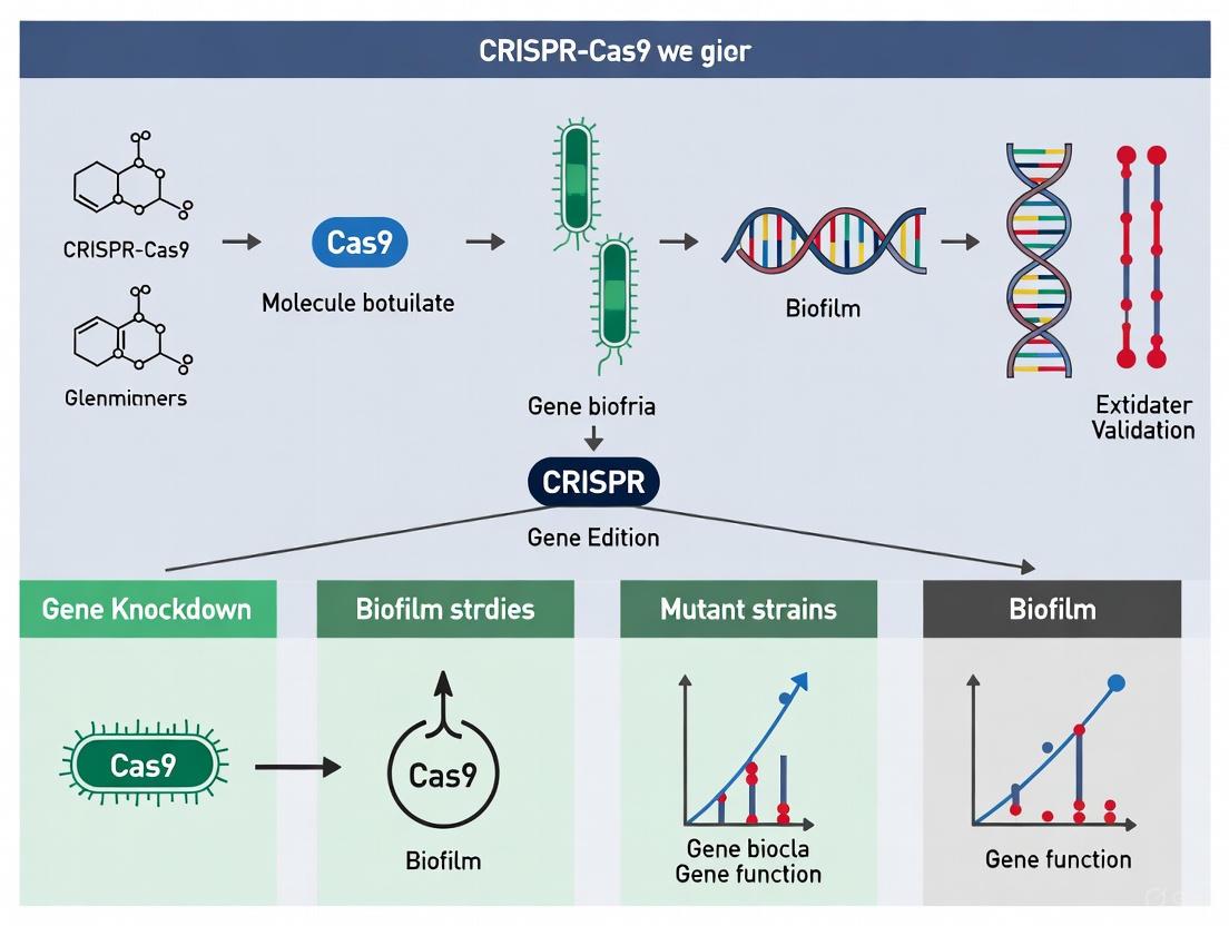
The following diagram outlines a comprehensive experimental approach for validating biofilm gene function using both CRISPR and traditional mutagenesis methods:
Diagram 2: Experimental Workflow for Biofilm Gene Validation. This comprehensive workflow compares parallel approaches using CRISPR-based knockdown and traditional mutant generation, followed by standardized biofilm phenotyping assays to assess the functional consequences of genetic perturbations.
The Scientist's Toolkit: Essential Research Reagents
Table 3: Key Research Reagents for Biofilm Gene Function Studies
| Reagent Category | Specific Examples | Function in Biofilm Research | Application Notes |
|---|---|---|---|
| CRISPR Components | Cas9 nuclease (SpCas9) | RNA-guided DNA endonuclease for targeted gene disruption | Commercial sources available; human-codon optimized versions improve expression [12] |
| MAD7 nuclease | Royalty-free alternative to Cas9 for gene editing | Editing efficiency ~23% in K. phaffii; 5'-YTTN-3' PAM site [12] | |
| sgRNA/single-guide RNA | Target-specific guide RNA for nuclease direction | Can be chemically synthesized or in vitro transcribed [13] | |
| Delivery Systems | Lipid nanoparticles | Encapsulation and delivery of CRISPR components | Enhances cellular uptake and protects genetic material [3] |
| Gold nanoparticles | Non-viral vector for RNP delivery | 3.5x enhanced editing efficiency in some systems [3] | |
| Electroporation systems | Physical method for intracellular delivery | Parameters must be optimized for each bacterial strain [13] | |
| Selection Agents | Zeocin | Antibiotic selection marker | Used for stable transformant selection [12] |
| Hygromycin B | Antibiotic for selective pressure | Common selection agent in bacterial and fungal systems [12] | |
| 5-Fluoroindole (5-FI) | Counter-selection for tryptophan auxotrophs | Identifies mutations in MAA7 (tryptophan synthase) [13] | |
| Biofilm Assay Reagents | Crystal violet | Biomass staining and quantification | Standard method for biofilm quantification [7] |
| Polystyrene beads | Surface for biofilm growth in experimental evolution | Used in bead transfer models of biofilm adaptation [14] | |
| Extracellular matrix dyes | EPS visualization (e.g., lectin conjugates) | Fluorescent labeling of matrix components [7] |
The strategic validation of gene function in biofilm research requires careful consideration of methodological approaches, balancing precision, efficiency, and biological relevance. CRISPR-based knockdown systems offer unprecedented precision and flexibility for targeted gene disruption, particularly when deployed as ribonucleoprotein complexes to minimize off-target effects and cytotoxicity [13]. The integration of nanoparticle delivery platforms further enhances the potential of CRISPR technologies by improving stability and cellular uptake of editing components [3]. Traditional mutant strains continue to provide value for certain applications, particularly when complete gene knockout is desired rather than partial knockdown. The choice between these approaches should be guided by specific research objectives, the model system being employed, and the required throughput. As biofilm research continues to evolve, the integration of advanced gene editing with sophisticated phenotyping assays will be essential for deciphering the complex molecular mechanisms underlying biofilm-mediated protection and identifying novel therapeutic targets for combating persistent infections.
In the investigation of bacterial biofilm formation and function, researchers primarily rely on two foundational genetic approaches to establish gene function: classical genetic knockouts and CRISPR knockdowns. Classical knockouts involve the permanent deletion or disruption of a target gene, creating a mutant strain that completely lacks the gene's function. In contrast, CRISPR knockdowns (particularly using CRISPR interference or CRISPRi) employ a catalytically inactive Cas9 (dCas9) to temporarily block gene transcription without altering the underlying DNA sequence. Within biofilm research, where processes like adhesion, extracellular polymeric substance (EPS) production, and quorum sensing are governed by complex genetic networks, the choice between these methods significantly impacts the interpretation of gene function and physiological relevance [15] [16].
This guide objectively compares the performance of these established methodologies, focusing on their application in validating biofilm gene function. We present experimental data, detailed protocols, and analytical frameworks to help researchers select the appropriate technique for their specific investigation of bacterial virulence and biofilm dynamics.
Performance Comparison: Key Experimental Data
The table below summarizes quantitative findings from studies that have applied both classical knockout and CRISPR-based approaches to investigate genes essential for biofilm formation in various bacterial species.
Table 1: Comparative Performance of Knockout and Knockdown Methods in Biofilm Research
| Target Gene / Organism | Method Used | Impact on Biofilm Formation | Key Experimental Findings | Reference |
|---|---|---|---|---|
| cas3 in Acinetobacter baumannii (Type I-Fa system) | Classical Knockout | Significant reduction | ∆cas3 mutant showed significantly reduced biofilm formation; complemented strain restored this ability. Demonstrated cas3 is a key virulence factor. [6] | |
| cas3 in Streptococcus mutans UA159 | Classical Knockout (∆cas3) | Impaired formation | Mutant showed impaired biofilm formation and weakened competition against S. sanguinis in the presence of fluoride. [15] | |
| Quorum Sensing (luxS, fimH, bola) in Escherichia coli | CRISPR/Cas9-HDR Knockout | Significant reduction | Targeted disruption of quorum sensing and adhesion genes led to reduced biofilm formation on urinary catheters. [17] | |
| Virulence genes (e.g., gtfB, gtfC) in Streptococcus mutans | Self-targeting CRISPR (Knockdown) | Decreased EPS, disrupted formation | Targeting bacterial virulence genes led to a decrease in extracellular polymeric substances (EPS) and disrupted biofilm formation. [15] | |
| Various biofilm-regulating factors | CRISPRi (dCas9) | Programmable precision | Allows reversible, titratable suppression of gene expression without permanent DNA alteration, useful for essential genes. [16] |
Table 2: Inherent Limitations and Technical Challenges of Each Method
| Aspect | Classical Genetic Knockouts | CRISPR Knockdowns |
|---|---|---|
| Genetic Permanence | Permanent gene deletion or disruption. | Reversible, transient gene silencing. |
| Impact on Essential Genes | Not suitable; lethal to the organism. | Highly suitable; enables study of essential gene function. |
| Compensatory Adaptation | High risk; genomic rearrangements or unlinked suppressor mutations can occur over time. [18] | Lower risk due to transient nature, but long-term cultures can still adapt. |
| Pleiotropic Effects | Common; can disrupt regulatory networks and downstream genes, confounding phenotypic analysis. [15] | Reduced; offers more direct correlation between gene suppression and observed phenotype. |
| Experimental Timeline | Longer; requires creation and validation of stable mutant strains. | Faster; single plasmid delivery enables rapid phenotypic assessment. |
| Off-Target Effects | Not applicable (target is specific). | Possible with dCas9, though high-fidelity systems mitigate this. [16] [19] |
| Delivery Efficiency | High in transformable strains. | Can be challenging in wild or poorly transformable strains; requires efficient delivery system. [19] |
Experimental Protocols for Key Workflows
Protocol for Classical Knockout via Homologous Recombination
This protocol is adapted from studies constructing cas3 deletion mutants in Acinetobacter baumannii to investigate its role in biofilm formation [6].
- Design of Knockout Construct: Design a linear DNA fragment containing an antibiotic resistance cassette (e.g., kanamycin) flanked by ~500-1000 bp sequences that are homologous to the regions directly upstream and downstream of the target gene (cas3). This construct will be used to replace the target gene via homologous recombination.
- Strain Transformation: Introduce the knockout construct into the wild-type A. baumannii strain (e.g., ATCC19606) via electroporation or natural transformation.
- Selection and Screening: Plate transformed cells onto agar containing the relevant antibiotic (e.g., kanamycin). Select colonies that have integrated the resistance cassette.
- Mutant Validation (PCR & Sequencing): Verify successful gene deletion using colony PCR with primers that bind outside the homologous recombination regions. Confirm the absence of the target gene and the correct integration of the cassette by sequencing the amplified product. [6]
- Phenotypic Assay - Biofilm Formation (Crystal Violet): a. Grow the wild-type, mutant (19606Δcas3), and complemented strains in appropriate media for 24-48 hours. b. Stain adherent biofilms with 0.1% crystal violet for 15 minutes. c. Destain with ethanol-acetate solution and measure the absorbance of the solution at 570-600 nm to quantify biofilm biomass. [6]
- Phenotypic Assay - Biofilm Architecture (CLSM) a. Grow biofilms on suitable surfaces (e.g., glass coverslips). b. Stain with SYTO9 green fluorescent nucleic acid stain for bacterial cells and Alexa Fluor 647-conjugated dextran for extracellular polysaccharides (EPS). c. Image using Confocal Laser Scanning Microscopy (CLSM) to analyze 3D architecture and thickness. [6]
Protocol for CRISPR Knockdown via CRISPR Interference (CRISPRi)
This protocol leverages a catalytically inactive dCas9 to block transcription, as applied in precision biofilm control studies [16].
- CRISPRi Plasmid Assembly: Clone a guide RNA (gRNA) sequence specific to the promoter or coding region of the target biofilm gene (e.g., a quorum sensing gene like luxS) into a plasmid expressing dCas9. The dCas9 protein, often derived from Streptococcus pyogenes, is mutated (e.g., D10A and H840A) to lack nuclease activity.
- Transformation: Introduce the constructed plasmid into the target bacterial strain.
- Induction of Gene Repression: Induce the expression of dCas9 and the gRNA using a regulated promoter (e.g., anhydrotetracycline-inducible promoter) to initiate targeted gene repression.
- Validation of Knockdown: Quantify the repression efficiency by measuring mRNA levels of the target gene using quantitative RT-PCR (qRT-PCR).
- Phenotypic Assay - Biofilm Disruption: Assess the impact of gene knockdown on biofilm formation using the crystal violet method or by measuring the reduction in EPS components, comparing to a strain containing a non-targeting gRNA control.
The Scientist's Toolkit: Essential Research Reagents
Table 3: Key Reagent Solutions for Knockout and Knockdown Experiments
| Reagent / Material | Function in Experiment | Specific Example / Note |
|---|---|---|
| dCas9 Plasmid System | Core component for CRISPRi; provides the programmable, non-cutting protein that binds DNA to block transcription. | Often uses a S. pyogenes dCas9 backbone with an inducible promoter for controlled expression. [16] |
| Guide RNA (gRNA) Oligos | Determines the specificity of the CRISPRi system by guiding dCas9 to the target DNA sequence. | Designed to target the non-template strand within the promoter or early coding sequence for optimal repression. |
| Antibiotic Resistance Cassettes | Selectable marker for isolating successful transformants or knockout mutants. | Kanamycin (KanR) or Ampicillin (AmpR) cassettes are commonly used in construct design. [6] |
| Electroporator | Instrument for introducing DNA constructs into bacterial cells via electrical pulses. | Critical for transforming strains with low natural competence. |
| Crystal Violet Stain | Dye used to quantify total biofilm biomass in standard microtiter plate assays. | A 0.1% solution is standard; the dissolved stain is measured spectrophotometrically. [6] |
| SYTO9 & EPS-Specific Probes | Fluorescent stains for visualizing live bacteria and extracellular matrix components under confocal microscopy. | SYTO9 labels cells (green); dextran conjugates or other lectin probes label polysaccharides (red). [6] |
| Lipid Nanoparticles (LNPs) | Advanced delivery vehicle for in vivo or hard-to-transform systems, co-delivering CRISPR components. | Enhances cellular uptake and protects genetic material; shown to reduce P. aeruginosa biofilm by >90% in vitro. [19] |
Conceptual Workflows and Signaling Pathways
The following diagrams illustrate the logical flow of the two genetic approaches and their impact on a biofilm-related signaling pathway.
Figure 1: Experimental Workflow Comparison. This diagram contrasts the sequential steps and key outcomes of creating a classical knockout versus implementing a CRISPR knockdown.
Figure 2: Targeting a Biofilm Gene in a Signaling Pathway. This diagram shows how CRISPRi and classical knockout differently intervene in a genetic pathway, such as quorum sensing, to disrupt biofilm formation.
In the field of functional genomics, the ability to precisely disrupt gene function has become fundamental to understanding complex biological systems, from bacterial development to human disease. The CRISPR-Cas9 system has emerged as a revolutionary technology for genetic manipulation, offering two distinct approaches for gene suppression: complete gene knockout (CRISPRn) using the catalytically active Cas9 nuclease, and transcriptional knockdown (CRISPRi) using a catalytically dead Cas9 (dCas9) [20]. While both systems utilize a guide RNA (gRNA) for target specificity, their mechanisms and applications differ significantly. CRISPRn creates permanent, physical changes to the DNA sequence through double-strand breaks, while CRISPRi temporarily blocks transcription without altering the genetic code. For researchers investigating complex processes like biofilm formation—where bacterial communities exhibit both structural robustness and cellular differentiation—understanding the distinction between these technologies is crucial for designing appropriate experiments and interpreting results. This guide provides a comprehensive comparison of Cas9 knockout and dCas9 knockdown methodologies, with specific application to validating biofilm gene function.
Molecular Mechanisms: How CRISPRn and CRISPRi Work
CRISPR Nuclease (CRISPRn): Permanent Gene Knockout
The CRISPR-Cas9 nuclease system creates permanent genetic alterations through a well-defined molecular process. The active Cas9 protein complexes with a single guide RNA (sgRNA) and induces double-strand breaks (DSBs) at specific genomic locations complementary to the sgRNA sequence and adjacent to a protospacer adjacent motif (PAM) [20]. Cellular repair of these breaks typically occurs through the error-prone non-homologous end joining (NHEJ) pathway, resulting in small insertions or deletions (indels) at the target site [21] [22]. When these indels occur within a protein-coding region and are not multiples of three nucleotides, they cause frameshift mutations that lead to premature stop codons and effectively knock out the target gene [22]. For larger deletions, researchers can employ two sgRNAs targeting flanking regions of a gene, resulting in the excision of the intervening sequence [22].
CRISPR Interference (CRISPRi): Reversible Gene Knockdown
In contrast, the CRISPR interference (CRISPRi) system utilizes a catalytically dead Cas9 (dCas9) that lacks nuclease activity but retains its ability to bind DNA based on sgRNA guidance [20]. When directed to a target gene's promoter region or early coding sequence, the dCas9-sgRNA complex physically obstructs the binding or elongation of RNA polymerase, effectively repressing transcription without altering the DNA sequence itself [20] [23]. This approach enables reversible, titratable control over gene expression—increasing dCas9 expression or using multiple sgRNAs can enhance repression efficiency [23]. The CRISPRi system can be further enhanced by fusing dCas9 to transcriptional repressor domains (e.g., KRAB) for stronger suppression, or to activator domains (CRISPRa) for gene activation [20].
The diagram below illustrates the key differences in the mechanisms of CRISPRn and CRISPRi:
Comparative Analysis: Knockout vs. Knockdown for Biofilm Research
The choice between CRISPRn and CRISPRi depends heavily on research goals, gene characteristics, and experimental constraints. The table below provides a systematic comparison of both technologies:
Table 1: Comparative Analysis of CRISPRn and CRISPRi Technologies
| Parameter | CRISPRn (Cas9 Knockout) | CRISPRi (dCas9 Knockdown) |
|---|---|---|
| Mechanism | DNA cleavage → NHEJ repair → indels | Steric hindrance of transcription |
| Genetic Outcome | Permanent DNA sequence alteration | Reversible, no DNA alteration |
| Efficiency | Varies by cell type (higher in mouse zygotes than MEFs) [21] | Titratable with inducer concentration [23] |
| Applications | Complete gene inactivation; study of essential gene null phenotypes | Study of essential genes; temporal control; fine-tuning expression |
| Screening Utility | Effective for identifying essential genes [24] | Enables knockdown of essential genes lethal if fully knocked out [23] |
| Technical Considerations | Potential for off-target effects; cytotoxicity from DSBs | Minimal off-target effects; no DNA damage response |
| Ideal for Biofilm Studies | Structural genes, non-essential pathways | Essential genes, fatty acid synthesis targets, temporal processes |
For biofilm research specifically, CRISPRi has demonstrated particular utility in investigating essential genes that cannot be studied with traditional knockouts. For instance, knockdown of fatty acid synthesis genes in Bacillus subtilis biofilms significantly enhanced colony wrinkling and reduced sporulation efficiency—phenotypes that would be impossible to study with lethal complete knockouts [23]. Similarly, CRISPRi enabled high-throughput screening of essential genes in biofilm colonies over 48 hours, revealing genes critical for biofilm architecture and development [23].
Experimental Protocols for Biofilm Gene Validation
CRISPR-Cas9 Knockout Protocol for Biofilm-Associated Genes
This protocol outlines the creation of knockout mutants for genes encoding structural biofilm components or regulatory elements:
sgRNA Design and Validation: Design sgRNAs with high on-target activity scores (e.g., using VBC scores) [25] targeting early exons of the target gene. For larger deletions, design two sgRNAs flanking the domain of interest. Validate cutting efficiency in vitro using Cas9 ribonucleoproteins (RNPs) and genomic DNA fragments [13].
Delivery System Selection: For bacterial systems, electroporate Cas9/sgRNA RNPs to avoid cytotoxicity and off-target effects [13]. For mammalian cells, use lentiviral delivery of Cas9 and sgRNA constructs.
Screening and Validation: Select mutants using:
Confirmation: Sequence the target region to confirm indel mutations and verify protein loss via Western blotting.
CRISPRi Knockdown Protocol for Essential Biofilm Genes
This protocol enables titratable knockdown of essential genes involved in biofilm formation, such as those in fatty acid synthesis pathways:
dCas9 and sgRNA System: Express dCas9 constitutively or inducibly from a plasmid. For B. subtilis, use a xylose-inducible dCas9 system [23]. Design sgRNAs targeting the promoter or 5' coding region of essential genes (e.g., fabI, accD in fatty acid synthesis).
Library Construction: For high-throughput screening, create a CRISPRi library targeting all essential genes with multiple sgRNAs per gene (e.g., 4-6 sgRNAs/gene) [23] [25].
Biofilm Culturing and Induction: Grow biofilm colonies on appropriate media (LB or MSgg for B. subtilis). Induce dCas9 expression with xylose (0-1% concentration) for titratable knockdown [23].
Phenotypic Assessment: After 48 hours of growth, quantify:
The experimental workflow for implementing these technologies in biofilm research is illustrated below:
Performance Data: Efficacy in Functional Genomics
Multiple studies have systematically compared the performance of CRISPRn and CRISPRi in various biological contexts. The data reveal both complementary and distinct capabilities:
Table 2: Performance Comparison in Genetic Screens
| Metric | CRISPRn | CRISPRi | Combined Approach |
|---|---|---|---|
| Essential Gene Detection | 60% of gold standard essentials at 1% FPR [24] | Effective for essential gene study [23] | 85% of gold standard essentials at 1% FPR [24] |
| Specific Biological Processes Identified | Electron transport chain genes [24] | Fatty acid synthesis, DNA gyrase genes [23] | Both chaperonin complexes and electron transport [24] |
| Correlation Between Technologies | Low correlation with RNAi/CRISPRi screens [24] | Low correlation with CRISPRn screens [24] | Complementary information [24] |
| Screen Size Optimization | Top 3 VBC-scored sgRNAs perform optimally [25] | Effective with inducible systems [23] | Dual-targeting libraries enhance efficiency [25] |
Notably, a benchmark study comparing CRISPR-Cas9 and RNAi screens found that they identified distinct biological processes despite similar precision in detecting essential genes [24]. CRISPRn screens effectively identified genes involved in the electron transport chain, while RNAi/CRISPRi screens better detected components of the chaperonin-containing T-complex [24]. This suggests these technologies provide complementary information, with combination approaches yielding the most comprehensive functional insights.
The Scientist's Toolkit: Essential Research Reagents
Successful implementation of CRISPR technologies requires specific molecular tools and delivery systems. The table below outlines key reagents for both CRISPRn and CRISPRi approaches:
Table 3: Essential Research Reagents for CRISPR-Based Studies
| Reagent Category | Specific Examples | Function and Application |
|---|---|---|
| CRISPRn Components | Active Cas9 nuclease | Induces double-strand breaks at target sites [22] |
| Single-guide RNA (sgRNA) | Targets Cas9 to specific genomic loci [21] | |
| Repair templates (ssODNs) | For homology-directed repair with point mutations [21] | |
| CRISPRi Components | dCas9 (catalytically dead) | Binds DNA without cutting; transcriptional blockade [20] [23] |
| dCas9-repressor fusions | Enhanced repression (e.g., dCas9-KRAB) [20] | |
| Inducible dCas9 systems | Xylose-inducible for titratable knockdown [23] | |
| Delivery Systems | Cas9 ribonucleoproteins (RNPs) | Direct delivery of protein-RNA complexes; reduced off-target effects [13] |
| Lentiviral vectors | Stable delivery of CRISPR components [24] | |
| Electroporation | Efficient RNP delivery into bacterial cells [13] | |
| Screening Tools | Genome-wide sgRNA libraries | Brunello, Yusa, Vienna libraries for high-throughput screens [25] |
| Dual-targeting sgRNA libraries | Two sgRNAs per gene for enhanced knockout efficiency [25] | |
| Selection Markers | Antibiotic resistance genes | Hygromycin, puromycin for stable integrant selection [13] |
| Fluorescent reporters | GFP, RFP for FACS sorting and efficiency monitoring [21] [23] |
CRISPRn and CRISPRi represent complementary rather than competing technologies in the molecular biologist's toolkit. For biofilm research, CRISPRn knockout is ideal for studying non-essential structural genes where complete inactivation is desired, while CRISPRi knockdown excels in investigating essential genes and temporal processes where titratable suppression is necessary. The emerging approach of combining both technologies—using CRISPRi for initial screening of essential genes followed by CRISPRn for detailed analysis of candidate genes—provides a powerful strategy for comprehensive gene function validation.
As CRISPR technologies continue to evolve, improvements in sgRNA design algorithms, delivery methods, and specificity will further enhance their utility in studying complex biological systems like biofilms. For researchers embarking on biofilm gene validation projects, the strategic selection between CRISPRn and CRISPRi based on gene essentiality, desired permanence of suppression, and experimental throughput requirements will be critical for obtaining meaningful biological insights.
Core Concepts: Knockdown and Knockout in Gene Editing
In genetic research, knockdown and knockout are two fundamental techniques for studying gene function, each with distinct mechanisms and outcomes. Knockdown refers to a partial reduction of gene expression, typically at the mRNA level, leading to decreased protein production without altering the underlying DNA sequence [26] [27]. In contrast, knockout involves complete and permanent inactivation of a gene at the DNA level, resulting in a complete loss of protein function [28] [29].
The choice between these methods is critical in biofilm research, as it can significantly influence the interpretation of a gene's role in complex processes like biofilm formation, maintenance, and dispersal.
Comparison of Fundamental Characteristics
| Feature | Gene Knockdown | Gene Knockout |
|---|---|---|
| Molecular Target | mRNA (post-transcriptional)[ccitation:4] [27] | DNA (genomic sequence) [28] [29] |
| Mechanism | RNA interference (RNAi) using siRNA or shRNA; CRISPR interference (CRISPRi) [29] [30] | CRISPR-Cas9 nuclease creating double-strand breaks [28] |
| Effect on Gene | Reduced expression (knockdown) [26] [27] | Complete disruption (knockout) [28] [29] |
| Permanence | Temporary and reversible [27] | Permanent and heritable [27] |
| Key Repair Pathway | Not applicable (no DNA damage) | Non-Homologous End Joining (NHEJ) [28] |
| Typical Outcome | Incomplete silencing; dose-titratable [27] | Frameshift mutations and premature stop codons [28] |
Technical and Practical Considerations for Experimental Design
Selecting between knockdown and knockout extends beyond the molecular goal to encompass practical experimental factors, including the gene's essentiality, required throughput, and need for specificity.
Comparison of Practical Application
| Consideration | Gene Knockdown | Gene Knockout |
|---|---|---|
| Study of Essential Genes | Suitable (allows partial silencing) [27] | Not suitable (lethal if completely knocked out) [27] |
| Experimental Workflow | Often faster; uses transient transfection of siRNAs [27] | Can be more complex; may require stable cell line generation [27] |
| Off-Target Effects | Higher risk (sequence-dependent and independent) [29] | Generally lower risk with optimized gRNA design [29] [27] |
| Phenotype Interpretation | Confounded by incomplete knockdown [27] | Confounded by potential compensatory mechanisms [26] |
| Therapeutic Recapitulation | Better models pharmacological inhibition [27] | Models complete loss-of-function genetic diseases [28] |
Application in Biofilm Research: A CRISPR-Centric View
In biofilm research, both knockdown and knockout approaches are valuable, with CRISPR technologies offering tools for both. CRISPR knockout is ideal for modeling the complete loss of a gene, such as creating mutant strains lacking a biofilm-related transcription factor. Alternatively, CRISPR interference (CRISPRi)—a knockdown technique using a catalytically "dead" Cas9 (dCas9) to block transcription—is excellent for studying essential genes or conducting tunable, reversible loss-of-function studies [30] [27].
A key application of CRISPRi in biofilm research is the systematic interrogation of genes involved in cyclic di-GMP (c-di-GMP) signaling, a central regulatory network controlling the transition from planktonic to biofilm lifestyle in bacteria [30]. Silencing specific diguanylate cyclases (DGCs) or phosphodiesterases (PDEs) via CRISPRi allows researchers to dissect their individual contributions to biofilm formation without the pleiotropic effects that can arise from traditional knockouts [30].
Experimental Workflow for Biofilm Gene Validation
The following diagram illustrates a generalized workflow for validating biofilm gene function using both CRISPR knockdown and knockout approaches, from design to phenotypic analysis.
Detailed Experimental Protocols for Biofilm Studies
CRISPR Knockout for Generating Mutant Strains
This protocol is used to create permanent, heritable gene disruptions in bacterial strains to study the effect of a gene's complete absence on biofilm formation.
- Step 1: gRNA Design: Design a guide RNA (gRNA) of 20 nucleotides that is complementary to the early coding sequence of the target biofilm gene (e.g., a diguanylate cyclase). Use established design tools to minimize off-target effects [29].
- Step 2: Delivery System Preparation: Clone the gRNA sequence and the Cas9 nuclease gene into a plasmid vector suitable for the target bacterium (e.g., Pseudomonas fluorescens). Alternatively, for higher efficiency and reduced off-target effects, complex the purified Cas9 protein with in vitro transcribed gRNA to form a Ribonucleoprotein (RNP) complex [29].
- Step 3: Transformation and Selection: Introduce the CRISPR plasmid or RNP complex into the bacterial cells via electroporation or conjugation. Select for transformed cells using appropriate antibiotics (if using a plasmid) or through direct screening [30].
- Step 4: Mutant Validation: Isolate genomic DNA from potential knockout clones. Amplify the target region by PCR and sequence it to confirm the presence of insertions or deletions (indels) that cause frameshift mutations [28] [29].
- Step 5: Phenotypic Analysis: Quantify biofilm formation of the mutant strain compared to the wild-type using a crystal violet assay. For architectural analysis, use confocal laser scanning microscopy (CLSM) to visualize the 3D structure of live biofilms, often using fluorescent stains for cells and extracellular polymeric substances (EPS) [30].
CRISPR Interference (CRISPRi) for Gene Knockdown
This protocol uses a catalytically dead Cas9 (dCas9) to block transcription, allowing for reversible, titratable gene silencing—ideal for studying essential genes or fine-tuning expression.
- Step 1: gRNA Design for Repression: Design gRNAs to target the non-template strand within the promoter region or the early part of the open reading frame to sterically hinder RNA polymerase. gRNAs targeting the promoter region (e.g., -35 to -10 boxes) often yield the strongest repression [30].
- Step 2: Two-Plasmid System Delivery: Use a dual-plasmid system in the target bacterium. One plasmid constitutively or inducibly expresses the dCas9 protein (e.g., under a PtetA promoter induced by anhydrotetracycline, aTc). The second plasmid constitutively expresses the sequence-specific gRNA [30].
- Step 3: Knockdown Induction and Validation: Co-transform both plasmids into the bacterial strain. Induce dCas9 expression with a defined concentration of aTc to control the level of gene repression. Validate knockdown efficiency by measuring mRNA transcript levels using quantitative RT-PCR (qRT-PCR) [30].
- Step 4: Functional Phenotyping: Assess the functional consequences of gene knockdown on biofilm formation and other related phenotypes. As demonstrated in P. fluorescens, this can include:
- Swarming Motility Assays: To assess changes in flagellar-mediated movement.
- Biofilm Mass Quantification: Using microtiter plate assays.
- Detailed Architectural Analysis: Using CLSM to measure biomass, thickness, and EPS matrix production of biofilms grown in flow cells or on relevant surfaces [30].
Signaling Pathways in Biofilm Formation and Intervention Points
Biofilm formation is a tightly regulated process. Understanding its key pathways is essential for rationally selecting target genes for knockdown or knockout studies. The diagram below outlines a simplified core pathway and the points where genetic perturbation takes effect.
Quantitative Data from Key Studies
Efficacy of CRISPR-Based Strategies in Biofilm and Bacterial Research
The table below summarizes key quantitative findings from recent studies utilizing CRISPR technologies in bacterial and biofilm research, highlighting their potential efficacy.
| Application / System | Target / Model | Key Quantitative Result | Citation |
|---|---|---|---|
| CRISPR-Cas9 + Nanoparticles | Pseudomonas aeruginosa biofilm | >90% reduction in biofilm biomass in vitro with liposomal Cas9 formulation [3] [31] | |
| CRISPR-Cas9 + Nanoparticles | Bacterial gene editing | 3.5-fold increase in editing efficiency with gold nanoparticle carriers vs. non-carrier systems [3] [31] | |
| CRISPR Interference (CRISPRi) | P. fluorescens (SBW25) | Effective downregulation of the GacA/S system and c-di-GMP genes, recapitulating knockout swarming and biofilm phenotypes [30] | |
| CRISPR Knockdown | Human macrophages (CHIP genes) | Significant increase in pro-inflammatory cytokines (IL-6, MCP-1, IL-1β) after knockdown of DNMT3A, TET2, ASXL1 [32] |
Research Reagent Solutions for CRISPR-Based Validation
A successful CRISPR experiment relies on a suite of specialized reagents and tools. The table below details essential materials and their functions for both knockout and knockdown workflows in a biofilm research context.
| Reagent / Tool | Function in Experiment | Application Context |
|---|---|---|
| Cas9 Nuclease | Creates double-strand breaks in DNA for knockout generation. | CRISPR Knockout [29] |
| dCas9 (dead Cas9) | Binds DNA without cutting; platform for transcriptional repression in CRISPRi. | CRISPR Knockdown (CRISPRi) [30] [27] |
| Guide RNA (gRNA) | A ~20 nt RNA that directs Cas9/dCas9 to the specific genomic target site. | Knockout & Knockdown [29] [30] |
| RNP Complex (Ribonucleoprotein) | Pre-assembled complex of Cas9 protein and gRNA; increases editing efficiency and reduces off-target effects. | Preferred for Knockout [29] |
| Dual-Plasmid System (dCas9 + gRNA) | Separate, compatible plasmids for inducible dCas9 expression and constitutive gRNA expression. | CRISPRi Knockdown [30] |
| Anhydrotetracycline (aTc) | Small molecule inducer for systems using the PtetA promoter to control dCas9 expression. | Titratable CRISPRi Knockdown [30] |
| Confocal Laser Scanning Microscope (CLSM) | High-resolution 3D imaging of biofilm architecture, biomass, and matrix composition. | Phenotypic Analysis (Both) [30] |
The decision to use knockdown or knockout for validating biofilm gene function is not a matter of which is universally better, but which is more appropriate for the specific biological question. The following strategic summary can guide this choice:
- Use CRISPR Knockout when the goal is to model the complete, permanent loss of a gene's function, such as identifying the core set of genes essential for biofilm initiation or to create stable mutant strains for long-term study. It is the preferred choice for non-essential genes where complete loss is informative and non-lethal [28] [29].
- Use CRISPR Knockdown (CRISPRi) when studying essential genes, when a titratable or reversible phenotype is desired, or when aiming to dissect the role of specific genes within a complex, redundant network like the c-di-GMP signaling system. Its ability to fine-tune gene expression makes it powerful for establishing dose-responsive relationships between gene expression and phenotypic output [30] [27].
Ultimately, leveraging both approaches in a complementary manner can provide the most robust validation. A phenotype observed in both a complete knockout and a specific knockdown is less likely to be an artifact and more likely to represent the true function of the gene within the intricate process of biofilm formation.
Bench Protocols: Designing CRISPR and Mutant Strain Experiments for Biofilm Genes
Step-by-Step Guide to Constructing Traditional Gene Deletion Mutants
In the field of microbial genetics, constructing gene deletion mutants is a fundamental technique for elucidating gene function. Within biofilm research, two primary strategies are employed: the creation of traditional deletion mutants and the use of transient CRISPR knockdowns. Traditional gene deletion involves the permanent removal or disruption of a target gene from the genome, providing a complete and stable knockout. In contrast, CRISPR interference (CRISPRi) offers a reversible method to knock down gene expression without altering the DNA sequence itself, using a catalytically inactive Cas9 (dCas9) to block transcription [30]. While newer methods like CRISPRi are valuable for studying essential genes or achieving temporal control, the construction of traditional mutants remains a critical skill for generating definitive, permanent loss-of-function models. This guide provides a detailed protocol for creating these mutants, framed within the context of validating biofilm-associated genes.
Part 1: Core Principles and a Comparative Framework
Traditional Gene Deletion vs. Modern CRISPR Approaches
The choice between traditional gene deletion and CRISPR-based methods depends heavily on the research question. The table below summarizes the key characteristics of each approach.
| Feature | Traditional Gene Deletion (Homologous Recombination) | CRISPR Knockdown (CRISPRi) | CRISPR Knockout (CRISPR-Cas9 NHEJ) |
|---|---|---|---|
| Genetic Outcome | Permanent gene removal or replacement [33] | Reversible repression of gene expression [30] | Permanent gene disruption via small insertions/deletions (indels) [34] |
| Mechanism | Homologous recombination between genomic DNA and an engineered targeting construct [33] | dCas9-guided steric blockage of RNA polymerase during transcription [30] | Cas9-induced double-strand break repaired via error-prone Non-Homologous End Joining (NHEJ) [34] |
| Best For | Creating stable, defined mutants; large gene deletions; pre-clinical models | Studying essential genes; temporal control of gene expression; rapid, titratable knockdowns [30] [26] | High-efficiency knockout in cell lines; multiplexed gene targeting; pooled genetic screens [23] |
| Typical Workflow Duration | Several weeks to months | Several days to a week | 1-2 weeks |
| Key Limitation | Low efficiency in some systems; labor-intensive [33] | Knockdown is incomplete and transient; potential for incomplete phenotype [26] | Potential for off-target effects; mosaic edits in organisms; not all indels create a knockout [35] |
For biofilm research, this distinction is critical. For instance, a 2022 study on Bacillus subtilis biofilms used a CRISPRi library to systematically knock down essential genes, revealing that inhibiting fatty acid synthesis led to enhanced biofilm wrinkling and reduced sporulation efficiency [23]. This type of high-throughput, essential-gene study would be challenging with traditional deletion methods. Conversely, traditional deletion is ideal for creating a clean, stable mutant to definitively confirm the role of a non-essential matrix protein.
Part 2: Step-by-Step Guide to Traditional Gene Deletion
This protocol details the construction of a gene deletion mutant via homologous recombination in bacteria, a method that ensures precise gene replacement and is widely applicable.
The following diagram illustrates the key stages of constructing a traditional gene deletion mutant.
Step 1: Design and Clone the Deletion Construct
The goal is to create a DNA molecule that the cell can use to replace its target gene via homologous recombination.
- Identify Target Gene Sequence: Obtain the full DNA sequence of the gene to be deleted, including flanking regions.
- Design Homology Arms: Amplify two sequences (typically 500–2000 base pairs) that are identical to the regions immediately upstream (5') and downstream (3') of the target gene. These "homology arms" guide the construct to the correct genomic location.
- Select a Selectable Marker: Choose a gene for selection, such as an antibiotic resistance cassette (e.g., kanamycin resistance). This marker will replace the target gene in the genome.
- Assemble the Construct: Clone the 5' homology arm, the selectable marker, and the 3' homology arm into a suitable plasmid vector. The final construct should have the structure: 5' Homology Arm - Antibiotic Resistance Cassette - 3' Homology Arm.
Step 2: Deliver the Construct into Target Cells
Introduce the deletion construct into the bacterial cells.
- Preparation: Make the target cells "competent" for DNA uptake, often using chemical treatments like calcium chloride.
- Transformation: Incubate the competent cells with the deletion construct plasmid. A brief heat shock (e.g., 42°C for 30–90 seconds) facilitates DNA uptake into the cells.
- Recovery: Add a nutrient broth and incubate to allow the cells to recover and express the antibiotic resistance gene from the plasmid.
Step 3: Select for Successful Integration
Isolate the rare cells where the deletion construct has integrated into the genome.
- Plate Transformed Cells: Spread the recovered cells onto agar plates containing the appropriate antibiotic.
- Initial Selection: Only cells that have stably integrated the antibiotic resistance cassette into their genome via homologous recombination will grow into colonies. These are potential deletion mutants.
Step 4: Screen and Isolate Putative Mutants
Confirm the loss of the target gene and the correct integration of the marker.
- Colony PCR: Use PCR with one primer that binds outside the homology arm used in the construct and another that binds within the resistance marker. A successful PCR product of the expected size indicates that the resistance cassette has replaced the target gene at the correct locus.
- Purify Clones: Streak PCR-positive colonies onto fresh antibiotic plates to isolate pure clones.
Step 5: Verify the Mutant Genotype
Perform rigorous validation to ensure the mutant is correct.
- Diagnostic PCR: Use multiple PCR reactions with different primer sets to confirm both the absence of the wild-type gene and the presence of the resistance cassette in the correct location.
- Southern Blotting (Optional but Definitive): This technique provides definitive proof of a single, correct recombination event and the absence of random plasmid integrations.
- Sequence Analysis: Sequence the PCR products spanning the recombination junctions to confirm perfect fusion between the homology arms and the resistance cassette.
Part 3: Application in Biofilm Research: Protocols and Reagents
Experimental Protocol: Validating a Biofilm Gene
To validate the function of a putative biofilm-related gene (e.g., an EPS biosynthesis gene), a researcher would:
- Construct a Deletion Mutant: Follow the step-by-step guide above to create a clean deletion of the target gene.
- Generate a CRISPRi Knockdown Strain: For comparison, introduce a plasmid expressing dCas9 and a gene-specific guide RNA (gRNA) into the wild-type strain [30].
- Quantify Biofilm Phenotypes: Culture both the deletion mutant and the CRISPRi strain under biofilm-promoting conditions and assess key metrics.
- Biomass Assay: Use crystal violet staining to quantify total adhered biofilm biomass.
- Matrix Production: Quantify specific exopolysaccharides (e.g., via lectin staining or chemical assays).
- Morphology: Use confocal microscopy to image the 3D structure of the biofilm, as was done in a Pseudomonas fluorescens study using CRISPRi [30].
Key Research Reagent Solutions
The table below lists essential materials required for these experiments.
| Reagent / Solution | Function / Purpose | Example Specifications |
|---|---|---|
| Deletion Construct Plasmid | Carries the engineered DNA for homologous recombination; typically contains a selectable marker (e.g., AmpR) and the gene replacement cassette. | pKO Vector or similar, with KanR cassette flanked by 1kb homology arms. |
| Antibiotics | Selective pressure to isolate transformed cells and mutants. | Kanamycin (50-100 µg/mL), Ampicillin (100 µg/mL). |
| Thermostable DNA Polymerase | Enzyme for PCR during construct verification, colony screening, and genotyping. | Q5 High-Fidelity DNA Polymerase for accurate amplification of homology arms. |
| Agarose Gel Electrophoresis System | To separate and visualize DNA fragments by size for PCR product analysis. | 0.8-1.2% agarose gel in TAE buffer. |
| Crystal Violet Stain | Dye that binds to biomass, used for basic quantification of biofilm formation. | 0.1% aqueous crystal violet solution. |
| Confocal Microscopy | High-resolution imaging to analyze the 3D architecture and composition of biofilms. | System capable of imaging GFP/RFP and performing Z-stack analysis. |
Advanced Method: CRISPR-del for Large Deletions
For complete, unambiguous knockout—especially to remove large genomic regions or cis-regulatory elements—an advanced CRISPR method called CRISPR-deletion (CRISPR-del) is highly effective [35] [36]. This method uses two gRNAs and Cas9 to delete the entire intervening sequence, ensuring the target is removed and cannot be bypassed by alternative splicing or translation.
Protocol for CRISPR-del [35]:
- Design two sgRNAs targeting sequences flanking the region to be deleted.
- Form Ribonucleoprotein (RNP) complexes by mixing recombinant Cas9 protein with the two synthesized sgRNAs.
- Deliver RNPs into cells via electroporation (highly efficient with minimal off-target effects).
- Screen clones by genomic PCR using primers that bind outside the targeted region. A smaller PCR product indicates a successful large deletion.
Both traditional gene deletion and modern CRISPR-based techniques are indispensable for validating gene function in biofilm research. The construction of traditional gene deletion mutants, while more time-consuming, provides a stable, permanent, and genetically defined model that is the gold standard for confirming a gene's role. CRISPRi knockdowns offer unparalleled speed and flexibility for screening and studying essential genes. The emerging CRISPR-del method combines the precision of targeting with the definitiveness of a complete deletion. A robust validation strategy will often leverage the strengths of both traditional and CRISPR methods, using knockdowns for initial discovery and high-throughput screening, and following up with traditional or CRISPR-del mutants for conclusive phenotypic confirmation.
Designing High-Efficiency gRNAs for Knockout (Cas9) and Knockdown (dCas9)
In functional genomics research, particularly in the study of biofilm formation, a critical decision is whether to use permanent gene knockout or transient gene knockdown to validate gene function. CRISPR-Cas9 technology provides powerful tools for both approaches through the use of nuclease-active Cas9 for complete gene knockout and catalytically dead Cas9 (dCas9) for targeted gene knockdown (CRISPRi). The efficacy of both systems depends critically on the selection and design of the guide RNA (gRNA). This guide provides a comprehensive comparison of high-efficiency gRNA design principles for both knockout and knockdown applications, focusing specifically on the context of biofilm research. We present objective performance data and detailed methodologies to enable researchers to make informed decisions for their experimental designs.
Table 1: Core Components of CRISPR Knockout and Knockdown Systems
| Component | CRISPR-Cas9 (Knockout) | CRISPR-dCas9 (Knockdown) |
|---|---|---|
| Cas9 Form | Catalytically active | Catalytically dead (mutated RuvC & HNH nuclease domains) |
| Primary Mechanism | Creates double-strand breaks, leading to indels and frameshifts via NHEJ | Binds DNA without cutting, blocking transcription |
| gRNA Length | Typically 20 nt | Typically 20 nt (full-length) or 14-16 nt (truncated for tgCRISPRi) |
| Persistence of Effect | Permanent gene disruption | Reversible transcription repression |
| Ideal Target Region | Early exons for frameshifts | Transcriptional Start Site (TSS) -50 to +300 bp |
Fundamental gRNA Design Principles for Genome Editing
The fundamental goal of gRNA design is to maximize on-target efficiency while minimizing off-target effects. The gRNA consists of a 20-nucleotide guiding sequence (spacer or crRNA) that recognizes the target DNA via Watson-Crick base pairing, and a structural scaffold (tracrRNA) that binds to the Cas9 protein. The target sequence must be immediately upstream of a Protospacer Adjacent Motif (PAM) - for SpCas9, this is 5'-NGG-3' [37] [38].
Key Parameters for Optimal gRNA Design
On-Target Efficiency: Various algorithms have been developed to predict gRNA on-target efficiency based on large-scale experimental datasets. The most advanced scoring methods include [37]:
- Rule Set 3 (2022): Considers both the target sequence and tracrRNA sequence, trained on 47,000 gRNAs using gradient boosting framework. Recommended for any tracrRNA with a 'T' in the 5th position.
- CRISPRscan (2015): Predictive model based on activity data of 1,280 gRNAs validated in vivo in zebrafish.
- Lindel (2019): Predicts frameshift ratio using a 60 bp sequence centered at the cleavage site, generally more accurate for predicting indel patterns.
Off-Target Risk Assessment: Specificity is crucial to avoid unintended consequences. Key evaluation methods include [37]:
- Homology Analysis: Sequences with only one mismatch to the gRNA imply high off-target potential, while sequences with zero mismatches should be completely avoided.
- Cutting Frequency Determination (CFD): Based on activity of 28,000 gRNAs with single variations; scores below 0.05 indicate low off-target risk.
- MIT Specificity Score: Developed from studying indel mutation levels of 700+ gRNA variants with 1-3 mismatches.
Table 2: Comparison of gRNA On-Target Efficiency Prediction Algorithms
| Algorithm | Year | Basis | Strengths | Application |
|---|---|---|---|---|
| Rule Set 3 | 2022 | 47,000 gRNAs; considers tracrRNA variant | Most updated logic; fast training | GenScript, CRISPick |
| Rule Set 2 | 2016 | 4,390 gRNAs | Improved over original Rule Set | CHOPCHOP, CRISPOR |
| CRISPRscan | 2015 | 1,280 gRNAs in zebrafish | In vivo validation focus | CHOPCHOP, CRISPOR |
| Lindel | 2019 | ~1.16 million mutation events | Accurate indel and frameshift prediction | CRISPOR |
| VBC Score | 2025 | Genome-wide scores for coding sequences | Strong performance in benchmark studies | Custom library design |
Recent benchmarking studies demonstrate that Vienna Bioactivity CRISPR (VBC) scores show excellent performance in predicting gRNA efficacy, with top3-VBC guides exhibiting the strongest depletion curves in essentiality screens [25]. Rule Set 3 scores also correlate negatively with log fold changes, confirming their predictive value for gRNA efficiency [25].
Comparative Analysis: gRNA Design for Knockout vs. Knockdown
gRNA Design for Cas9-Mediated Gene Knockout
For traditional CRISPR-Cas9 knockout experiments, gRNAs should target early exons of the gene of interest to maximize the probability of generating frameshift mutations that disrupt the entire coding sequence. The cutting site is located between positions 17 and 18 of the 20-nt gRNA sequence [38]. Key considerations include:
- GC Content: Optimal between 40-60%; avoid extremes (<20% or >80%) [38]
- Position-Specific Nucleotides: Guanine in position 20, avoid cytosine in position 20 and uracil in positions 17-20 [38]
- Avoiding Poly-N Sequences: Especially GGGG repeats which reduce efficiency [38]
Experimental validation in hexaploid wheat demonstrated that gRNA on-target activities varied substantially (0% to ~20% indel frequency), with no obvious correlation between experimentally determined and in silico predicted efficiency, highlighting the importance of empirical testing [39].
gRNA Design for dCas9-Mediated Gene Knockdown
CRISPR interference (CRISPRi) using dCas9 requires targeting the transcriptional start site (TSS) to effectively block transcription initiation or elongation. Optimal targeting occurs within a window from -50 to +300 bp relative to the TSS [40]. Recent advances include:
tgCRISPRi: An alternative strategy using catalytically active Cas9 complexed with truncated gRNAs (tgRNAs) of 14-15 nt. These shorter guides direct Cas9 binding without DNA cleavage, efficiently repressing gene expression when targeted near TSSs [40].
In Drosophila studies, tgRNAs of 14-15 nt targeting the TSS of yellow and ebony genes produced strong, uniform loss-of-function phenotypes without detectable target site mutations, demonstrating efficient transcriptional repression [40].
Diagram 1: gRNA Design Workflow: Knockout vs. Knockdown
Experimental Protocols for gRNA Validation
Protoplast-Based gRNA Validation Protocol
For preliminary testing of gRNA efficiency, a protoplast validation system provides rapid results before committing to stable transformation. This method is particularly valuable for polyploid species or challenging genetic systems [39]:
- Design 5-7 gRNAs targeting your gene of interest, considering homoeoallele specificity if working with polyploid organisms
- Clone gRNAs into appropriate CRISPR vectors containing Cas9
- Isolate protoplasts from target tissue (e.g., wheat mesophyll) and dilute to 3.0 × 10^5 cells/mL
- Transform protoplasts with CRISPR constructs using PEG-mediated transformation
- Incubate for 48-72 hours before extracting genomic DNA
- Amplify target region using homoeoallele-specific primers
- Analyze editing efficiency via TIDE (Tracking of Indels by DEcomposition) or CRISPResso analysis of Sanger or amplicon sequencing data
This protocol achieved transformation efficiencies of 64-72% in wheat protoplasts and could detect indels at frequencies as low as 1% [39].
tgCRISPRi Validation in Animal Models
For validating tgCRISPRi systems for gene knockdown [40]:
- Design paired tgRNAs (14-15 nt) targeting TSS regions of target genes
- Generate transgenic lines expressing tgRNAs under appropriate promoters (e.g., U6:3 and U6:1 in Drosophila)
- Cross with Cas9-expressing lines (e.g., vasa-Cas9 for ubiquitous expression)
- Analyze phenotypic penetrance and uniformity across tissue types
- Verify absence of mutations at target sites via sequencing
- Quantify gene expression reduction using qRT-PCR
This approach achieved highly efficient biallelic loss of gene function in somatic cells with strong uniform phenotypes, without detectable target site mutations [40].
Performance Comparison and Benchmarking Data
Recent benchmarking studies provide critical insights into gRNA library performance. A 2025 study comparing genome-wide CRISPR sgRNA libraries found that libraries with fewer constructs per gene, when chosen according to principled criteria, can perform as well or better than larger libraries [25].
Table 3: Benchmark Performance of gRNA Selection Strategies in Essentiality Screens
| Library/Strategy | Guides per Gene | Depletion Performance | Notes |
|---|---|---|---|
| Top3-VBC | 3 | Strongest depletion | Best performance in essentiality screens |
| Yusa v3 | 6 (average) | Strong | One of best performing pre-existing libraries |
| Croatan | 10 (average) | Strong | Good performance but larger size |
| Bottom3-VBC | 3 | Weakest depletion | Demonstrates importance of selection criteria |
| Vienna-dual | 6 (paired) | Strongest in dual-targeting | Enhanced effect size in drug-gene interaction |
Dual-targeting strategies, where two sgRNAs target the same gene, showed stronger depletion of essential genes but also exhibited a modest fitness reduction even in non-essential genes, possibly due to increased DNA damage response [25]. This suggests caution when using dual-targeting in certain screening contexts.
Diagram 2: gRNA Design Evaluation Logic
Advanced AI-Based gRNA and Editor Design
The field of gRNA design is rapidly evolving with artificial intelligence approaches. Recent work demonstrates that large language models can generate novel CRISPR-Cas proteins with optimal properties. One study created OpenCRISPR-1, an AI-generated gene editor that shows comparable or improved activity and specificity relative to SpCas9 while being 400 mutations away in sequence [41].
These AI-designed editors:
- Show compatibility with base editing systems
- Represent a 4.8-fold expansion of protein clusters across CRISPR-Cas families found in nature
- Can be tailored with specific guide RNA sequences for Cas9-like effector proteins
- Demonstrate the potential to bypass evolutionary constraints of natural systems [41]
The Scientist's Toolkit: Essential Research Reagents
Table 4: Key Research Reagent Solutions for gRNA Validation
| Reagent/Resource | Function | Example Applications |
|---|---|---|
| CRISPick (Broad Institute) | gRNA design with Rule Set 3 and CFD scores | Genome-wide library design |
| CHOPCHOP | Versatile tool supporting various CRISPR-Cas systems | Visualizing potential off-target sites |
| CRISPOR | Detailed off-target analysis with position-specific mismatch scoring | Restriction enzyme site identification for cloning |
| GenScript sgRNA Design Tool | Uses Rule Set 3 and CFD scoring with overall balanced scores | Supports SpCas9 and AsCas12a design |
| TIDE Analysis | Tracking Indels by Decomposition from Sanger sequencing | Rapid efficiency validation without deep sequencing |
| CRISPResso | Analysis of amplicon sequencing data for editing efficiency | Comprehensive indel characterization |
| Wheat Protoplast System | Transient validation in polyploid species | Pre-screening gRNAs before stable transformation |
| tgRNA Vectors | Truncated gRNA expression for CRISPRi | Specific gene knockdown without cleavage |
Application to Biofilm Gene Function Validation
When applying these gRNA design principles to biofilm research, consider the following specific recommendations:
For Essential Gene Analysis: Use dCas9 knockdown rather than knockout to study essential biofilm genes where complete disruption would be lethal. The tgCRISPRi system with 14-15 nt guides provides particularly efficient repression without DNA damage [40].
For Redundant Gene Families: In polyploid organisms or organisms with redundant gene families, design homoeoallele-specific gRNAs with SNPs in the seed region to target specific family members [39].
For Temporal Studies: Employ dCas9 knockdown to study temporal requirements of biofilm genes at different development stages, as effects are reversible unlike permanent knockouts.
Validation Workflow: Implement a two-stage validation process starting with protoplast or cell culture screening followed by whole-organism testing to conserve resources.
The choice between CRISPR knockout and knockdown for biofilm studies should be guided by the biological question, essentiality of the target gene, and desired permanence of the effect. Both approaches benefit from careful gRNA design using the principles and tools outlined in this guide.
The application of CRISPR technology for validating biofilm gene function, whether through CRISPR knockdown or comparison with mutant strains, hinges on one critical factor: the efficient delivery of editing machinery to the target cells. The choice of delivery system directly impacts editing efficiency, specificity, and experimental outcomes. This guide provides an objective comparison of the primary physical and nanoparticle-based delivery methods, equipping researchers with the data and protocols needed to select the optimal system for their biofilm research.
At a Glance: Delivery System Comparison
The table below summarizes the key characteristics of major delivery systems to aid in initial selection.
Table 1: Comparative Overview of CRISPR-Cas9 Delivery Systems
| Delivery Method | Typical Cargo | Key Advantages | Key Limitations | Reported Editing Efficiency (Sample Data) |
|---|---|---|---|---|
| Electroporation | RNP, mRNA | High efficiency in amenable cells; direct cytosolic delivery [42] [43] | High cytotoxicity; optimization required for different cell types [42] | Up to 95% in SaB-1 cells; ~30% in DLB-1 cells [42] |
| Lipid Nanoparticles (LNPs) | mRNA, RNP | Low immunogenicity; suitable for in vivo delivery; clinically validated [44] [43] [45] | Endosomal entrapment can limit efficiency; requires optimization for escape [42] [43] | ~25% in DLB-1 cells; minimal editing in SaB-1 cells [42] |
| Magnetofection (SPIONs) | RNP | Efficient cellular uptake guided by magnetic field; high biocompatibility [42] | May face post-entry barriers (e.g., no detectable editing despite uptake) [42] | Efficient uptake, but editing often minimal without further optimization [42] |
| Adeno-Associated Virus (AAV) | DNA | High transduction efficiency; cell-type specific tropism [43] [46] | Limited cargo capacity (<4.7 kb); potential for immune responses [43] [46] | N/A for large Cas9; requires dual-AAV systems or smaller Cas variants |
| Lentivirus (LV) | DNA | Large cargo capacity; stable genomic integration (for long-term expression) [43] | Insertional mutagenesis risk; sustained Cas9 expression raises off-target concerns [42] [43] | High efficiency, but biosafety concerns limit use for transient editing [42] |
Detailed Methodologies and Performance Data
Electroporation
Electroporation uses electrical pulses to create transient pores in the cell membrane, allowing CRISPR cargo to enter the cytoplasm directly.
- Protocol Summary: DLB-1 and SaB-1 cells were resuspended in electroporation buffer with Cas9-sgRNA RNP complexes (2-3 µM). Using a Neon Transfection System (Thermo Fisher), optimal conditions for SaB-1 cells were 1800 V, 20 ms, and 2 pulses, while DLB-1 required 1700 V, 20 ms, and 2 pulses. Cell viability and transfection efficiency were assessed 1-hour post-electroporation using trypan blue exclusion and Cas9-Cy3 fluorescence, respectively. Editing efficiency was quantified via NGS of the target locus [42].
- Key Data: Under optimized conditions, editing efficiency reached up to 95% in SaB-1 cells but only ~30% in DLB-1 cells, which also exhibited locus-specific genomic rearrangements. Chemically synthesized sgRNAs (Synthego) consistently outperformed in vitro transcribed (IVT) sgRNAs [42].
Lipid Nanoparticles (LNPs)
LNPs are lipid-based vesicles that encapsulate and protect CRISPR cargo, fusing with cell membranes to release their contents intracellularly.
- Protocol Summary: For Diversa LNPs, sgRNAs were loaded into particles, and Cas9 protein was internalized separately. The LNP-sgRNA complexes were then added to DLB-1 and SaB-1 cell cultures. Confocal microscopy using FLUOGREEN-labeled Cas9 confirmed cellular uptake and subcellular localization [42].
- Key Data: This method achieved moderate editing efficiency (~25%) in DLB-1 cells but was largely ineffective in SaB-1 cells. A significant barrier was identified as endosomal retention and insufficient nuclear import of the Cas9 protein [42].
Superparamagnetic Iron Oxide Nanoparticles (SPIONs)
Magnetofection uses magnetic nanoparticles to concentrate CRISPR cargo onto target cells, enhancing uptake.
- Protocol Summary: Cas9-sgRNA RNPs were conjugated to fluorescent, gelatin-coated SPIONs (SPIONs@Gelatin). These complexes were added to cell culture media, and a magnet was placed beneath the culture plate for 15-30 minutes to drive uptake. Efficiency was analyzed via fluorescence for uptake and sequencing for editing [42].
- Key Data: While SPIONs facilitated efficient cellular uptake observed via fluorescence, this did not translate into detectable gene editing, highlighting significant post-internalization barriers [42].
The workflow and key decision points for selecting and implementing these systems are summarized in the diagram below.
Diagram: A workflow for selecting and optimizing a CRISPR delivery system, from cargo choice to outcome validation.
Special Considerations for Biofilm Research
Using CRISPR to validate biofilm gene function presents unique challenges. Biofilms are structured communities encased in an extracellular polymeric substance (EPS) that limits penetration of antimicrobials and molecular tools [3].
- Synergy with Nanoparticles: Nanoparticles can enhance CRISPR delivery against biofilm-associated bacteria. They improve penetration through the EPS matrix and facilitate co-delivery of CRISPR components with antibiotics for a synergistic effect [3] [47].
- Demonstrated Efficacy: Liposomal CRISPR-Cas9 formulations have been shown to reduce Pseudomonas aeruginosa biofilm biomass by over 90% in vitro. Similarly, gold nanoparticle-CRISPR hybrids demonstrated a 3.5-fold increase in editing efficiency compared to non-carrier systems [3].
The Scientist's Toolkit: Essential Reagents and Materials
Table 2: Key Research Reagent Solutions for CRISPR Delivery Experiments
| Reagent / Material | Function / Description | Example Use Case |
|---|---|---|
| Cas9 Protein (hfCas9) | High-fidelity nuclease for RNP complex formation; reduces off-target effects. | Formation of RNP complexes for electroporation or nanoparticle loading [42] [43]. |
| Chemically Modified sgRNA | Enhanced stability and binding affinity compared to IVT sgRNA. | Used in RNP complexes to significantly boost editing efficiency [42]. |
| Ionizable Lipids | Key component of LNPs; enables encapsulation and release of nucleic acids. | Formulating LNPs for in vivo mRNA or RNP delivery [44] [43]. |
| Superparamagnetic Iron Oxide Nanoparticles (SPIONs) | Magnetic core for guided delivery; often coated (e.g., gelatin) for biocompatibility. | Magnetofection to enhance cellular uptake in the presence of a magnetic field [42]. |
| DNA-PKcs Inhibitors (e.g., AZD7648) | Small molecule inhibitor of NHEJ pathway; used to enhance HDR efficiency. | Shifting repair toward precise HDR; Note: Recent studies link it to increased structural variations [48]. |
| Selective Organ Targeting (SORT) Molecules | Engineered lipids that confer tissue-specific targeting to LNPs. | Creating LNPs that target organs beyond the liver (e.g., lungs, spleen) [43]. |
Navigating Safety and Genomic Integrity
A critical, often overlooked aspect of CRISPR delivery is the spectrum of on-target genomic alterations beyond small indels.
- Structural Variations: Efficient CRISPR editing can lead to large, unintended on-target structural variations (SVs), including kilobase- to megabase-scale deletions and chromosomal rearrangements [48].
- Impact of HDR Enhancement Strategies: The use of DNA-PKcs inhibitors to boost HDR rates can dramatically increase the frequency of these SVs and chromosomal translocations. This can also lead to an overestimation of HDR efficiency in standard assays that fail to detect large deletions [48].
- Recommendation: For functional genomics, strategies that favor a clean knockout (NHEJ) without HDR-enhancing chemicals may be preferable. The safety profile of high-fidelity Cas9 variants (e.g., HiFi Cas9) and the choice of repair pathway modulators must be carefully evaluated [48].
Diagram: DNA repair pathways activated by a CRISPR-induced break can lead to both intended edits and genotoxic structural variations.
Biofilms, structured communities of microbial cells embedded in a self-produced extracellular polymeric matrix, represent the predominant mode of bacterial growth in nature and a significant clinical challenge [49] [50]. The study of biofilm formation is crucial for understanding bacterial pathogenesis, antimicrobial resistance, and persistence in industrial settings. For researchers validating gene function—particularly with modern techniques like CRISPR knockdown versus traditional mutant strains—selecting appropriate phenotypic assays is paramount. Biofilm development progresses through defined stages: initial reversible attachment, irreversible adhesion, microcolony formation, maturation, and eventual dispersion [50]. Each stage exhibits distinct phenotypic characteristics that require specific quantification methods to elucidate gene function accurately.
This guide provides a comparative analysis of established biofilm assay methodologies, focusing on their application in validating genetic manipulations. We present standardized protocols, quantitative performance data, and experimental considerations to enable researchers to select optimal assessment strategies for their specific research questions, particularly within the context of genetic validation studies using CRISPR interference and mutant strains.
Essential Biofilm Assay Methodologies
Microtiter Plate-Based Quantification Assays
The Tissue Culture Plate Method (TCPM) is widely considered the gold standard for quantitative biofilm assessment due to its high throughput and excellent reproducibility [50] [51]. This method involves growing biofilms in 96-well plates, followed by staining and destaining with crystal violet to quantify adhered biomass. The fixed and stained biofilms are typically dissolved in organic solvents or acetic acid, and the optical density (OD) is measured at 570 nm [50]. The degree of biofilm formation is classified based on OD values, which can be correlated with bacterial density. This method effectively detects both live and dead cells along with the biofilm matrix components, providing a robust measure of total biomass accumulation [51].
The ATP Bioluminescence (BLM) Assay offers a complementary approach that quantifies metabolically active cells within the biofilm by measuring adenosine triphosphate (ATP) levels. The assay utilizes the luciferin-luciferase reaction, where light emission is proportional to the ATP concentration present in viable cells [51]. This method is particularly valuable for assessing biofilm viability after antimicrobial treatments or genetic manipulations that might affect metabolic activity rather than physical adherence. A significant advantage is the rapid results generation, typically within minutes, though the dynamic range can be limited compared to other methods [51].
The Resazurin (RES) Assay provides another measure of metabolic activity by exploiting the reduction of the non-fluorescent resazurin to the highly fluorescent resorufin by metabolically active cells [51]. This assay is time-sensitive, as resorufin can be further reduced to non-fluorescent hydroresorufin, potentially diminishing the signal intensity even in dense bacterial populations. For optimal results with facultative anaerobes like Staphylococcus aureus, adding fresh growth medium containing RES to established biofilms has been shown to improve the dynamic range [51].
Phenotypic Screening Methods
The Congo Red Agar (CRA) Method serves as a qualitative screening tool for identifying slime-producing bacterial isolates. This technique relies on the inspection of colony coloration on Congo Red-containing agar plates, where black colonies indicate biofilm-producing strains, while red colonies suggest non-producers [50]. The interpretation time varies by species; for Staphylococcus aureus, inspection after up to 72 hours of incubation provides better distinction than the 24-hour reading used for Staphylococcus epidermidis [51]. While useful for initial screening, the CRA method cannot reliably discriminate between weak and negative biofilm producers and shows variable performance compared to quantitative microtiter plate methods [50].
The Tube Method offers a simple qualitative approach where biofilms form on the walls and bottom of culture tubes, visualized after staining with crystal violet. Although less quantitative than microplate assays, it provides a rapid screening mechanism, particularly in resource-limited settings [50]. Studies comparing these methods have demonstrated that the Tube method generally shows lower sensitivity and specificity compared to the CRA method when using the TCPM as a reference standard [50].
Table 1: Comparison of Common Biofilm Quantification Methods
| Method | Principle | Target Measurement | Throughput | Key Advantages | Key Limitations |
|---|---|---|---|---|---|
| Tissue Culture Plate (TCPM) | Crystal violet staining of adhered biomass | Total biofilm biomass (live/dead cells + matrix) | High | Quantitative, reproducible, high-throughput | Does not distinguish viable cells |
| ATP Bioluminescence (BLM) | Luciferin-luciferase reaction with ATP | Metabolically active cells | High | Rapid results, measures viability | Transient signal, requires specialized equipment |
| Resazurin (RES) | Reduction of resazurin to resorufin | Metabolic activity | High | Sensitive to metabolic state | Time-sensitive, small dynamic range |
| Congo Red Agar (CRA) | Polysaccharide-dye interaction | Slime production | Medium | Simple, no specialized equipment | Qualitative, species-dependent interpretation |
| Tube Method | Visual assessment of adhered biofilm | Biofilm formation | Low | Simple, low cost | Subjective, semi-quantitative at best |
Advanced Model Systems for Specialized Applications
Reactor-Based Systems for Medical Device Materials
For studying biofilm formation on medical device materials, reactor-based systems provide sophisticated platforms that simulate relevant hydrodynamic conditions. The CDC Biofilm Reactor (CDC-BR) and Drip Flow Reactor (DFR) represent two established models for growing biofilms on material surrogates like stainless steel coupons [52]. These systems enable researchers to investigate biofilm development under different shear stress conditions relevant to clinical environments.
Recent studies with Pseudomonas aeruginosa biofilms have demonstrated that both CDC-BR and DFR models produce reproducible biofilms with similar colony forming units (CFU) despite their different hydrodynamic profiles [52]. The research revealed that both protein concentration and total organic carbon (TOC) serve as potential analytes for quantifying biofilm biomass, with each assay displaying low variability across multiple replicates [52]. Interestingly, while CFU measurements were similar between models, protein and TOC levels differed significantly, highlighting how different quantification methods capture distinct aspects of the biofilm phenotype—a crucial consideration when interpreting genetic manipulation effects.
Advanced Physical Characterization Methods
Force Spectroscopy using atomic force microscopy (AFM) provides unique insights into the adhesive and viscoelastic properties of bacterial biofilms at different developmental stages. This technique quantifies adhesive pressure and viscoelastic moduli by performing force measurements with microbeads on native biofilms [53]. Studies with P. aeruginosa have revealed significant differences in these physical properties between wild-type and lipopolysaccharide mutant (wapR) strains, as well as prominent changes during biofilm maturation [53]. For example, the adhesive pressure of PAO1 early biofilms was measured at 34 ± 15 Pa, decreasing to 19 ± 7 Pa in mature biofilms, while wapR mutant early biofilms showed markedly higher adhesion (332 ± 47 Pa) that also decreased with maturation (80 ± 22 Pa) [53]. Such precise physical measurements complement traditional biomass quantification, especially when investigating genes potentially involved in matrix composition or surface adhesion.
Confocal Laser Scanning Microscopy (CLSM) enables detailed three-dimensional visualization of biofilm architecture when combined with appropriate fluorescent staining. This method typically employs SYTO9 green fluorescent nucleic acid stain for bacterial cells and Alexa Fluor 647-conjugated dextran for extracellular polysaccharide (EPS) matrix components, allowing clear spatial differentiation between cellular and matrix constituents [6]. CLSM has proven particularly valuable for documenting structural defects in biofilms formed by genetically modified strains, such as the significant reduction in both biofilm thickness and fluorescence intensity observed in Acinetobacter baumannii Δcas3 mutants compared to wild-type strains [6].
Biofilm Assays in Genetic Validation Studies
CRISPRi Applications in Biofilm Research
CRISPR interference (CRISPRi) has emerged as a powerful tool for investigating essential gene function in biofilm formation, enabling targeted gene knockdown without permanent mutation. In Bacillus subtilis strain 3610, CRISPRi libraries targeting essential genes have revealed unexpected connections between core cellular processes and biofilm development [23]. For instance, knockdown of fatty acid synthesis genes and DNA gyrase unexpectedly enhanced biofilm wrinkling without increasing matrix gene expression, suggesting previously unrecognized roles for these essential processes in biofilm architecture [23].
The effectiveness of CRISPRi in biofilm studies was demonstrated through titratable repression of target genes across relevant timescales. In B. subtilis biofilm colonies, RFP reporter repression reached approximately 20-fold in LB medium and 100-fold in biofilm-promoting MSgg medium over 48 hours of development [23]. This persistent, tunable knockdown capability makes CRISPRi particularly valuable for studying essential genes whose complete inactivation would prevent biofilm formation entirely.
Table 2: Genetic Approaches for Biofilm Gene Validation
| Approach | Mechanism | Best Applications | Advantages | Considerations for Biofilm Studies |
|---|---|---|---|---|
| CRISPR Interference (CRISPRi) | dCas9-mediated gene repression | Essential gene function, titratable knockdown | Tunable, reversible, enables essential gene study | Repression efficiency varies in 3D biofilms |
| Traditional Gene Knockout | Complete gene deletion | Non-essential gene function | Complete loss-of-function, stable phenotype | May mask essential roles in subpopulations |
| Complementation Strains | Restoration of gene function | Confirming genotype-phenotype linkage | Controls for polar effects, validates specificity | Expression levels may not match native |
Comparative Analysis of Genetic Approaches
Studies comparing CRISPRi knockdowns with traditional mutants have revealed both conserved and distinct phenotypic outcomes in biofilm formation. Research with B. subtilis demonstrated that gene essentiality is largely conserved between planktonic and biofilm growth conditions, with most genes showing similar requirement patterns in liquid culture and surface-grown colonies [23]. However, certain genes exhibited medium-dependent phenotypes, such as patA and asnB (involved in amino acid synthesis) being essential in LB but not in MSgg medium, while glyA and folD (involved in glycine and folate synthesis) showed the opposite pattern [23].
In Acinetobacter baumannii, deletion of the cas3 gene from the type I-Fa CRISPR-Cas system significantly reduced biofilm formation without affecting growth rates, highlighting how genes not traditionally associated with biofilm development can play crucial regulatory roles [6]. The Δcas3 mutant showed reduced expression of virulence factors including biofilm formation-related genes and outer membrane protein A (ompA), accompanied by altered carbon metabolism and oxidative phosphorylation pathway regulation [6]. These findings illustrate how comprehensive phenotypic assessment across multiple biofilm parameters can reveal unexpected gene functions.
Experimental Protocols for Key Assays
Standardized Tissue Culture Plate Method (TCPM) Protocol
Materials Required:
- Sterile 96-well flat-bottom polystyrene tissue culture plates
- Trypticase soy broth (TSB) supplemented with 1% glucose
- Phosphate buffered saline (PBS)
- 2% sodium acetate solution
- 0.1% crystal violet solution
- Microplate reader capable of measuring OD~570nm~
Procedure:
- Prepare bacterial suspensions in TSB with 1% glucose from fresh agar plate cultures.
- Dispense 180µL of sterile TSB into each well of the tissue culture plate.
- Inoculate with 20µL of bacterial suspension (creating 1:10 dilution).
- Seal plates with Parafilm to prevent evaporation and incubate at 37°C for 24 hours.
- Gently shake plates and discard contents to remove planktonic cells.
- Wash each well 3-4 times with sterile distilled water, inverting to dry between washes.
- Fix biofilms with 200µL of 2% sodium acetate for 30 minutes.
- Repeat washing step (3-4 times with sterile distilled water).
- Stain with 200µL of 0.1% crystal violet for 15 minutes.
- Repeat washing step thoroughly to remove unbound dye.
- Air dry inverted plates completely.
- Measure optical density at 570nm using microplate reader.
- Include appropriate controls (uninoculated medium, known positive and negative strains).
Data Interpretation: Classify biofilm formation based on OD values relative to negative control. One common approach categorizes strains as: non-biofilm producer (OD ≤ ODC), weak producer (ODC < OD ≤ 2×ODC), moderate producer (2×ODC < OD ≤ 4×ODC), and strong producer (4×ODC < OD) where ODC is the OD of the negative control [50].
ATP Bioluminescence Assay Protocol
Materials Required:
- BacTiter-Glo Microbial Cell Viability Assay reagent or equivalent
- White-walled 96-well plates
- Luminometer
- Trypticase soy broth (TSB)
Procedure:
- Grow biofilms in 96-well plates as described in TCPM protocol steps 1-6.
- Prepare BacTiter-Glo reagent according to manufacturer instructions.
- Add equal volume of reagent to each well containing washed biofilms.
- Mix contents thoroughly using orbital shaker for 2 minutes to induce cell lysis.
- Incubate at room temperature for 10 minutes to stabilize luminescent signal.
- Measure luminescence using luminometer with integration time of 0.5-1 second per well.
Data Interpretation: Relative Light Units (RLU) are proportional to the amount of ATP present, which correlates with the number of metabolically active cells in the biofilm. Normalize values to positive and negative controls included in each assay run [51].
Research Reagent Solutions for Biofilm Studies
Table 3: Essential Research Reagents for Biofilm Assays
| Reagent/Chemical | Application | Function | Considerations |
|---|---|---|---|
| Crystal Violet | TCPM, Tube Method | Stains total biomass (cells + matrix) | Requires ethanol or acetic acid for destaining |
| BacTiter-Glo Reagent | ATP Bioluminescence Assay | Lyses cells and generates luminescent signal | Time-sensitive detection |
| Resazurin Sodium Salt | RES Assay | Indicators of metabolic activity | Phot sensitive, requires fresh preparation |
| Congo Red Dye | CRA Method | Binds extracellular polysaccharides | Interpretation varies by bacterial species |
| SYTO9 Green Stain | CLSM Imaging | Labels nucleic acids in all cells | Often used with propidium iodide for viability |
| Alexa Fluor 647-Dextran | CLSM Imaging | Labels extracellular polysaccharide matrix | Requires confocal microscopy with appropriate filters |
| Polystyrene Microplates | TCPM, BLM, RES | Substrate for biofilm growth | Surface properties affect attachment |
Visualizing Biofilm Experimental Workflows
Biofilm Assay Methodology Selection
The selection of biofilm assessment methods should align with specific research questions and the particular aspects of biofilm biology under investigation. For genetic validation studies comparing CRISPR knockdowns to mutant strains, employing multiple complementary assays provides the most comprehensive phenotypic characterization. Biomass quantification methods like TCPM offer robust high-throughput screening, while metabolic activity assays (BLM, RES) provide insights into physiological states within biofilms. Advanced techniques like CLSM and force spectroscopy reveal structural and mechanical properties that may be uniquely affected by specific genetic manipulations.
The integration of these methodological approaches enables researchers to dissect complex genotype-phenotype relationships in biofilm formation, from initial adhesion to mature dispersion. As genetic tools continue to evolve, particularly with CRISPR-based technologies, parallel advancement in phenotypic assessment capabilities will be essential for unraveling the complex regulatory networks governing bacterial biofilm development.
In bacterial biology, the second messenger bis-(3'→5')-cyclic dimeric guanosine monophosphate (c-di-GMP) represents a central regulatory node that controls the transition between motile and sessile lifestyles, directly influencing biofilm formation [30] [54]. This universal bacterial signaling molecule promotes biofilm formation through complex networks of diguanylate cyclases (DGCs) that synthesize c-di-GMP and phosphodiesterases (PDEs) that degrade it [54]. The inherent redundancy in these networks—where a single bacterium may encode dozens of DGCs and PDEs with overlapping functions—has long hampered traditional genetic approaches to deciphering individual contributions to biofilm regulation.
This case study objectively compares how CRISPR interference (CRISPRi) technology performs against traditional mutant strains in validating biofilm gene function, with specific focus on c-di-GMP signaling networks. We present experimental data demonstrating that CRISPRi provides superior resolution for dissecting complex genetic networks while overcoming the limitations of conventional mutagenesis, offering researchers a more precise toolkit for investigating bacterial biofilm pathogenesis and identifying potential therapeutic targets.
Methodology Comparison: CRISPRi vs. Traditional Mutagenesis
Fundamental Technological Differences
Traditional gene inactivation relies on the physical disruption of a target gene through insertion or deletion, creating a permanent knockout strain. This approach is labor-intensive for systematic surveys and impossible for essential genes [30] [55]. In contrast, CRISPRi (CRISPR interference) utilizes a catalytically inactive dCas9 protein guided by a short RNA sequence to bind specific genomic regions, sterically hindering transcription initiation or elongation without altering the DNA sequence itself [30]. This results in reversible, titratable gene silencing rather than permanent disruption.
Experimental Implementation
CRISPRi system design typically involves two compatible plasmids: one carrying the dCas9 gene under an inducible promoter (e.g., PtetA inducible by anhydrotetracyclin/aTc), and another constitutively expressing the guide RNA (gRNA) [30]. The gRNA can be designed to target either the template or non-template DNA strand, with the most effective repression typically achieved by targeting transcription initiation regions near the promoter [30]. This system has been successfully adapted for diverse bacterial species including Pseudomonas fluorescens, Streptococcus mutans, and Bacillus subtilis [30] [55] [56].
Table 1: Core Methodology Comparison
| Feature | Traditional Mutagenesis | CRISPRi |
|---|---|---|
| Genetic Outcome | Permanent gene disruption | Reversible gene silencing |
| Titratable Control | Not typically available | Dose-dependent with inducer [30] |
| Essential Gene Study | Not feasible | Possible with controlled knockdown [55] |
| Throughput Potential | Low to moderate | High (arrayed libraries) [55] |
| Polar Effects | Common in operons | Minimal with careful gRNA design |
| Development Timeline | Months for multiple mutants | Weeks for library construction |
Experimental Data: Direct Performance Comparison
c-di-GMP Network Dissection in Pseudomonas Species
In P. fluorescens SBW25, CRISPRi-mediated silencing of genes encoding the GacA/S two-component system and regulatory proteins associated with c-di-GMP signaling produced swarming and biofilm phenotypes indistinguishable from those obtained with traditional gene inactivation [30]. However, CRISPRi enabled systematic surveys of multiple genes across different strain backgrounds (SBW25, WH6, and Pf0-1) with significantly reduced labor requirements. Quantitative phenotyping confirmed that CRISPRi could reliably assess complex traits including cell morphology, motility, and biofilm formation over extended periods [30].
A landmark study in Pseudomonas aeruginosa PA14 employed multiplexed CRISPR/Cas9 genome editing to disrupt all 32 GGDEF domain-containing proteins implicated in c-di-GMP signaling [54]. The resulting mutant (PA14Δ32) was unable to form biofilms and exhibited attenuated virulence, while residual c-di-GMP levels indicated remarkable network robustness. This comprehensive approach overcame native redundancy and established a platform for studying individual DGC functions without compensatory effects [54].
Arrayed Library Applications
The development of arrayed CRISPRi libraries has enabled high-throughput functional genomics. The SNAP library (Streptococcus mutans Arrayed CRISPRi) contains strains targeting >250 essential and growth-supporting genes, allowing reproducible, community-wide investigation of gene function [55]. In biofilm formation assays, this library demonstrated that repression specificity could be controlled through inducer (xylose) concentration, with 0.5-1% xylose producing significant biofilm reduction when targeting gtfB, essential for sucrose-mediated biofilm formation [55].
Table 2: Quantitative Phenotypic Outcomes
| Organism | Target Gene/Pathway | CRISPRi Phenotype | Traditional Mutant Phenotype | Key Advantage |
|---|---|---|---|---|
| P. fluorescens SBW25 | GacA/S two-component system | Defective swarming & biofilm [30] | Similar defects [30] | Multi-strain application |
| P. aeruginosa PA14 | All 32 DGCs (GGDEF domains) | No biofilm formation [54] | Not achievable (lethality) | Comprehensive network dissection |
| S. mutans UA159 | gtfB (exopolysaccharide) | Dose-dependent biofilm reduction [55] | Constitutive defect | Titratable control |
| B. subtilis 3610 | Fatty acid synthesis | Increased biofilm wrinkling [56] | Not reported | Essential gene study |
| A. baumannii ATCC17978 | smpB (ribosome rescue) | Reduced biofilm & virulence [57] | Not reported | Novel target identification |
Protocol: CRISPRi Biofilm Assay for c-di-GMP Genes
Day 1: Strain Preparation
- Inoculate CRISPRi strains targeting c-di-GMP-associated genes (e.g., DGCs, PDEs, PilZ-domain effectors) in LB medium with appropriate antibiotics.
- Add inducer (aTc at 0-200 ng/mL) for dCas9 expression and incubate overnight at 30°C with shaking [30].
Day 2: Biofilm Setup
- Dilute cultures to OD600 of 0.05 in fresh medium with inducer.
- Transfer 200 μL to 96-well polystyrene plates (8 replicates per strain).
- Include control strains without gRNA and with non-targeting gRNA.
Day 4-5: Biofilm Quantification
- Carefully remove planktonic cells and rinse attached cells with PBS.
- Fix with 200 μL 99% methanol for 15 minutes, then air dry.
- Stain with 200 μL 0.1% crystal violet for 15 minutes.
- Wash thoroughly with water and destain with 200 μL 33% acetic acid.
- Measure absorbance at 595 nm for biomass quantification [30] [57].
Day 5: Confocal Microscopy (Optional)
- Grow biofilms on glass-bottom dishes instead of plates.
- Stain with LIVE/DEAD BacLight Bacterial Viability Kit following manufacturer's protocol.
- Image using 20× or 63× objective on confocal microscope.
- Analyze 3D architecture using Imaris or similar software [30].
Signaling Pathway Visualization
Diagram 1: c-di-GMP signaling network with CRISPRi knockdown sites.
Experimental Workflow
Diagram 2: CRISPRi experimental workflow for biofilm studies.
Research Reagent Solutions
Table 3: Essential Research Materials for CRISPRi Biofilm Studies
| Reagent/Category | Specific Examples | Function & Application Notes |
|---|---|---|
| dCas9 Expression System | pnpthet-dCas9 [30] | Catalytically inactive Cas9 for transcriptional repression |
| Guide RNA Vector | pgRNAabc [30] | Expresses target-specific gRNA with customizable spacer |
| Inducer Compounds | Anhydrotetracycline (aTc) [30], Xylose [55] | Titratable control of dCas9/gRNA expression |
| Selection Antibiotics | Apramycin [57], Kanamycin | Maintain plasmid selection pressure |
| Biofilm Assay Kits | Crystal violet staining [57], LIVE/DEAD BacLight | Quantify biofilm biomass and viability |
| Validation Tools | RNA extraction kits, qPCR reagents | Confirm gene knockdown efficiency |
| Imaging Systems | Confocal microscopy [30] | Analyze 3D biofilm architecture |
Discussion: Advantages and Limitations in Practice
Performance Advantages of CRISPRi
The experimental data consistently demonstrates that CRISPRi outperforms traditional mutagenesis in key metrics relevant to biofilm research:
Functional Redundancy Resolution: The ability to target multiple genes simultaneously proved invaluable for dissecting c-di-GMP networks where compensatory mechanisms often mask phenotypes in single mutants [54]. The multiplex editing capability of CRISPR systems enabled complete dissection of all 32 DGCs in P. aeruginosa, revealing network properties impossible to discern with sequential knockouts [54].
Essential Gene Analysis: CRISPRi enabled the first systematic study of essential genes in biofilm formation and structure. In B. subtilis, knockdown of fatty acid synthesis genes revealed unexpected connections to biofilm wrinkling and sporulation, identifying multifaceted roles in bacterial development [56].
Temporal Control: The inducible nature of CRISPRi allows researchers to time gene repression to specific biofilm developmental stages. This temporal precision revealed that repression of gtfB in S. mutans during early biofilm formation had more dramatic effects than later repression [55].
Persistent Challenges and Considerations
Despite its advantages, CRISPRi implementation faces several technical challenges:
Variable Knockdown Efficiency: gRNA effectiveness varies based on target location, with transcription initiation regions typically yielding superior repression compared to coding regions [30]. This necessitates testing multiple gRNAs per target.
Delivery Limitations: Efficient CRISPRi delivery remains challenging in certain bacterial species, particularly those with poor transformation efficiency or restriction-modification systems that target foreign DNA [16].
Polar Effects Considerations: While less problematic than in operon-wide knockouts, CRISPRi can still exhibit polar effects on downstream genes, requiring careful gRNA design and validation [55].
CRISPRi technology represents a paradigm shift in how researchers investigate complex bacterial signaling networks, particularly the redundant c-di-GMP system that controls biofilm formation. The experimental data presented demonstrates clear advantages over traditional mutagenesis, including superior scalability, temporal control, and the unique ability to study essential genes. While methodological challenges remain, CRISPRi has proven indispensable for dissecting the genetic architecture of biofilms, providing insights that could lead to novel anti-biofilm therapeutic strategies targeting c-di-GMP signaling networks. As the technology continues to evolve, particularly with improvements in delivery efficiency and multiplexing capabilities, CRISPRi is positioned to become the standard approach for functional genomics in bacterial biofilm research.
Solving Experimental Hurdles: Off-Target Effects, Delivery, and Phenotype Confirmation
The precision of CRISPR-based genome editing has revolutionized biological research and therapeutic development, yet achieving high rates of Homology-Directed Repair (HDR) remains a significant challenge, especially when using Ribonucleoprotein (RNP) complexes for gene knock-in. In the context of biofilm research—where understanding gene function often requires precise gene modification rather than complete knockout—the efficiency of HDR and delivery of RNPs is paramount. This guide objectively compares current strategies and products designed to overcome these limitations, providing researchers with experimental data to inform their methodological choices for validating biofilm gene function.
The HDR and RNP Delivery Challenge in Biofilm Research
CRISPR-Cas9 enables precise genome editing by creating double-strand breaks in DNA, which the cell repairs either via the error-prone Non-Homologous End Joining (NHEJ) pathway or the precise Homology-Directed Repair (HDR) pathway [58] [59]. While NHEJ is efficient and suitable for gene knockouts, HDR is essential for inserting or correcting DNA sequences using a donor template, making it indispensable for studying specific gene functions in complex processes like biofilm formation [60] [30].
However, HDR efficiency is inherently low in mammalian cells and is influenced by multiple factors, including cell cycle stage and the competition from the more dominant NHEJ pathway [58] [61]. This challenge is compounded when using pre-assembled Cas9 protein and guide RNA RNP complexes, which, despite offering advantages like reduced off-target effects and no risk of genomic integration of foreign DNA, present delivery difficulties due to their large size and charge [60] [58].
In biofilm research, where scientists like Stephen Dolan use CRISPR to "break genes" in pathogenic fungi such as Aspergillus fumigatus to understand their role in infection and antifungal resistance, the precision and efficiency of editing are critical [62]. Similarly, CRISPR interference (CRISPRi) with a catalytically inactive dCas9 allows for gene knockdown without permanent mutation, enabling the study of essential genes in biofilm formation in species like Pseudomonas fluorescens [30]. The choice between knockdown and knockout approaches hinges on the ability to efficiently deliver editing components and achieve the desired repair outcome.
Comparative Analysis of Delivery Systems for HDR and RNP
The delivery of CRISPR components is a critical determinant of editing success. The table below compares the key delivery modalities, highlighting their applicability for HDR and RNP delivery.
Table 1: Comparison of CRISPR Delivery Systems for HDR and RNP Applications
| Delivery System | Mechanism | Best For | HDR Efficiency | Key Advantages | Major Limitations |
|---|---|---|---|---|---|
| Cationic Polymers (e.g., Ppoly) [60] | Electrostatic complexation with RNPs/DNA to form nanoparticles | RNP + dsDNA donor co-delivery | ~50% (with TILD method in CHO-K1 cells) [60] | High encapsulation (>90%), low cytotoxicity ( >80% viability) [60] | Requires optimization for different cell types |
| Lipid Nanoparticles (LNPs) [44] [45] [61] | Lipid vesicles encapsulating mRNA/protein | In vivo delivery, mRNA-based editing | High (in clinical liver targets) [44] | Targeted delivery (e.g., liver), suitable for redosing [44] | Efficiency can vary by cell type; requires extensive optimization [61] |
| Electroporation [58] [61] | Electrical pulses to create transient pores in cell membrane | Hard-to-transfect cells (e.g., T cells, stem cells) | High (with optimized protocols) [61] | High efficiency for RNP delivery; direct cytoplasmic delivery | Can cause significant cell stress and mortality |
| Viral Vectors (e.g., AAV, Lentivirus) [58] [59] [45] | Engineered viruses transduce cells | Stable cell line generation, in vivo delivery | Moderate | High transduction efficiency; sustained expression | Limited packaging capacity; immune response risks; persistent expression increases off-target risk |
Performance Data for Cyclodextrin-Based Polymer (Ppoly) vs. Commercial Reagent
A 2025 study provides a direct, quantitative comparison of a novel cationic hyper-branched cyclodextrin-based polymer (Ppoly) against the commercial reagent CRISPRMAX for RNP and donor DNA delivery [60]. The experimental data offers a clear performance benchmark.
Table 2: Experimental Performance Comparison: Ppoly vs. CRISPRMAX
| Parameter | Ppoly (Cyclodextrin Polymer) | CRISPRMAX (Commercial Standard) |
|---|---|---|
| Knock-in Efficiency | ~50% (GFP gene integration in CHO-K1 cells) [60] | ~14% (GFP gene integration in CHO-K1 cells) [60] |
| Cytotoxicity | >80% cell viability [60] | Not explicitly stated, but implied to be higher than Ppoly |
| Encapsulation Efficiency | >90% for RNP complexes [60] | Not specified |
| Key Innovation | TILD-CRISPR method with linearized dsDNA donor and 1000-bp homology arms [60] | Standard commercial lipid-based formulation |
The significantly higher knock-in efficiency and low cytotoxicity of Ppoly highlight the potential of advanced polymer-based nanocarriers to outperform standard commercial reagents for precise genome engineering tasks [60].
Experimental Protocols for Enhanced HDR and RNP Delivery
Protocol: TILD-CRISPR with Cyclodextrin Polymer (Ppoly) Delivery
This protocol, adapted from a 2025 study, details the steps for achieving high-efficiency gene integration using linearized donor DNA and RNP delivery via a cyclodextrin-based polymer [60].
- Donor DNA Preparation: Linearize a double-stranded DNA (dsDNA) plasmid donor template containing the gene of interest (e.g., GFP) flanked by 1000-base-pair homology arms. Linearization can be performed using restriction enzymes or PCR amplification [60].
- RNP Complex Formation: Pre-assemble the Cas9 protein with in vitro-transcribed sgRNA targeting the genomic locus of interest. Incubate at room temperature for 10-20 minutes to form the RNP complex [60].
- Polyplex Formation (Ppoly/RNP): Mix the pre-formed RNP complexes and the linearized dsDNA donor with the cationic hyper-branched cyclodextrin-based polymer (Ppoly) in an optimized ratio. Incubate for 30 minutes at room temperature to allow for the self-assembly of stable nanoparticles (polyplexes) [60].
- Cell Transfection: Add the Ppoly/RNP/donor polyplexes to the culture medium of the target cells (e.g., CHO-K1). Incubate for 48-72 hours to allow for cellular uptake, genome cleavage, and HDR-mediated integration [60].
- Validation and Cloning: After transfection, perform antibiotic selection to enrich for edited cells. Isolve single-cell clones and use junction PCR to confirm the precise integration of the transgene at the target locus [60].
Protocol: CRISPRi for Biofilm Gene Knockdown
For biofilm research where gene knockout is lethal or undesirable, CRISPR interference (CRISPRi) provides a powerful alternative. This protocol is adapted from studies in P. fluorescens [30].
- System Design: Utilize a two-plasmid system. The first plasmid carries the gene for the catalytically inactive dCas9 protein under an inducible promoter (e.g., Ptet, induced by anhydrotetracycline/aTc). The second plasmid constitutively expresses the guide RNA (gRNA) designed to target the promoter or the 5' end of the open reading frame of the biofilm-related gene of interest [30].
- Strain Transformation: Introduce both plasmids into the target bacterial strain (e.g., P. fluorescens SBW25, WH6, or Pf0-1) via conjugation or electroporation.
- Gene Silencing Induction: Grow the transformed bacteria in media containing the inducer (aTc) to express dCas9. The dCas9-gRNA complex will bind to the target DNA site and sterically block transcription initiation or elongation [30].
- Phenotypic Assay: Assess the biofilm phenotype using confocal laser scanning microscopy (CLSM) to analyze biofilm architecture, mass, and composition. Compare to controls without inducer or with a non-targeting gRNA [30].
Visualization of Strategies and Workflows
The following diagrams illustrate the core strategies and experimental workflows for overcoming low HDR efficiency with RNP delivery.
Diagram 1: A flowchart summarizing the multi-faceted strategies for improving HDR efficiency, highlighting optimized delivery vehicles and donor template design as key approaches.
Diagram 2: A sequential workflow for high-efficiency gene knock-in using the TILD-CRISPR method with RNP complexes delivered via a cationic cyclodextrin polymer.
The Scientist's Toolkit: Essential Reagents for HDR and RNP Experiments
Successful execution of these advanced protocols requires a specific set of high-quality reagents. The following table details the essential components of the research toolkit.
Table 3: Research Reagent Solutions for HDR and RNP Experiments
| Reagent / Material | Function | Specific Example / Note |
|---|---|---|
| Cationic Hyper-branched Polymer [60] | Forms nanoparticles with negatively charged RNPs and DNA, facilitating cellular uptake. | Cyclodextrin-based polymer (Ppoly); offers high encapsulation and low toxicity [60]. |
| Purified Cas9 Protein [60] [58] | The nuclease component of the RNP complex. | Using purified protein instead of plasmid DNA reduces off-target effects and avoids DNA integration [60]. |
| Linearized dsDNA Donor Template [60] | Provides the homology-directed repair template for precise gene insertion. | TILD-CRISPR method uses dsDNA with long homology arms (e.g., 1000 bp) for high efficiency [60]. |
| NHEJ Inhibitors [63] | Chemical compounds that suppress the NHEJ repair pathway, thereby favoring HDR. | Can be added to cell culture media post-transfection to enhance knock-in rates [63]. |
| dCas9 Expression Plasmid [30] | For CRISPRi experiments; produces the catalytically dead Cas9 that blocks transcription. | Essential for gene knockdown studies in biofilm formation without introducing double-strand breaks [30]. |
| Lipid Nanoparticles (LNPs) [44] [61] | A delivery vehicle for in vivo applications or for difficult-to-transfect cell types. | Particularly effective for liver-targeted delivery and allows for re-dosing [44] [61]. |
The journey to overcome low HDR efficiency in CRISPR research is multifaceted, relying on the synergistic optimization of delivery vehicles, donor templates, and cellular conditions. For biofilm researchers validating gene function, the choice between knockout and knockdown strategies must be informed by these advancements. As the data demonstrates, novel nanocarriers like cyclodextrin-based polymers can significantly outperform standard commercial reagents for precise RNP delivery and knock-in. By adopting the robust protocols and reagents outlined in this guide, scientists can more reliably engineer microbial genomes, accelerating the discovery of gene functions critical to biofilm formation and resistance.
Identifying and Mitigating CRISPR Off-Target Effects
The CRISPR/Cas9 system has revolutionized genetic engineering, offering an unprecedented ability to perform precise genomic modifications. Its simplicity, efficiency, and cost-effectiveness have made it an indispensable tool across biomedical research and therapeutic development [64]. However, a significant challenge that persists is the occurrence of off-target effects—unintended edits at genomic locations similar but not identical to the target site. These off-target mutations can compromise experimental accuracy, confound phenotypic interpretations, and raise substantial safety concerns for clinical applications [65] [66]. In biofilm research, where elucidating gene function often involves comparing CRISPR knockdowns with traditional mutant strains, identifying and controlling for these off-target effects is paramount for validating genuine gene function. This guide provides a comprehensive comparison of methodologies for detecting and mitigating CRISPR off-target effects, equipping researchers with the tools necessary to enhance the reliability of their functional genomics studies.
Understanding CRISPR Off-Target Effects
Mechanisms of Off-Target Activity
Off-target effects in CRISPR/Cas9 systems primarily occur due to the enzyme's tolerance for imperfect complementarity between the guide RNA (gRNA) and genomic DNA. The wild-type Streptococcus pyogenes Cas9 (SpCas9) can tolerate between three and five base pair mismatches, particularly if these mismatches are located in the distal region from the Protospacer Adjacent Motif (PAM) sequence [66] [67]. Several key factors influence off-target susceptibility:
- PAM Recognition: While SpCas9 primarily recognizes the canonical 'NGG' PAM, it can also tolerate non-canonical variants like 'NAG' and 'NGA', albeit with reduced efficiency. The recent development of PAM-free or less restrictive systems (e.g., SpRY) has expanded targeting range but potentially increased off-target risks [66].
- Seed Sequence Imperfections: The PAM-proximal 10–12 nucleotide "seed" region is crucial for specific target recognition. Mismatches in this region typically prevent efficient cleavage, while those in the distal region are more tolerated [66].
- DNA/RNA Bulges: Imperfect complementarity that results in extra nucleotide insertions can still lead to Cas9 cleavage at these mismatched sites [66].
- Genetic Diversity: Single nucleotide polymorphisms (SNPs), insertions, deletions, and copy number variations can create novel off-target sites or reduce editing efficiency at intended targets [66].
Implications for Biofilm Research
In studies aimed at validating biofilm-forming genes, off-target effects present particular challenges. When employing CRISPR knockdowns versus traditional mutant strains, undetected off-target edits can:
- Lead to misinterpretation of gene function by introducing confounding phenotypic effects
- Complicate comparative analyses between CRISPR-generated and conventional mutants
- Reduce reproducibility across experiments and between research groups
- Undermine conclusions about specific gene contributions to biofilm formation, quorum sensing, or antimicrobial resistance mechanisms [1] [68]
Detection Methods: A Comparative Analysis
Multiple approaches have been developed to identify and quantify off-target effects, each with distinct strengths, limitations, and applications.
In Silico Prediction Tools
Computational methods provide an initial assessment of potential off-target sites by analyzing gRNA sequence similarity across the genome.
Table 1: Comparison of In Silico Off-Target Prediction Tools
| Tool Name | Algorithm Type | Key Features | Advantages | Limitations |
|---|---|---|---|---|
| CasOT [65] | Alignment-based | Allows custom adjustment of PAM sequence and mismatch number (up to 6) | First exhaustive tool for off-target prediction in user-provided genomes | Biased toward sgRNA-dependent effects |
| Cas-OFFinder [65] | Alignment-based | High tolerance for sgRNA length, PAM types, and number of mismatches or bulges | Widely applicable with flexible parameters | Does not fully account for epigenetic factors |
| FlashFry [65] | Alignment-based | High-throughput analysis of thousands of target sequences; provides GC content data | Rapid processing of large datasets | Limited by reference genome completeness |
| CCTop [65] | Scoring-based | Considers distances of mismatches to PAM | User-friendly web interface | Validation required for reliable results |
| DeepCRISPR [65] | Scoring-based | Incorporates both sequence and epigenetic features in predictions | More comprehensive by including chromatin accessibility | Requires computational expertise for implementation |
Experimental Detection Methods
Experimental approaches provide empirical data on off-target activity, offering varying levels of sensitivity, scalability, and biological relevance.
Table 2: Experimental Methods for Off-Target Detection
| Method | Principle | Sensitivity | Throughput | Key Applications | Advantages | Limitations |
|---|---|---|---|---|---|---|
| Digenome-seq [65] [66] | In vitro digestion of genomic DNA with Cas9/sgRNA RNP followed by whole-genome sequencing | High | Genome-wide | Initial screening of potential off-target sites | Does not require reference genome; sensitive | Expensive; requires high sequencing depth |
| GUIDE-seq [65] | Integration of double-stranded oligodeoxynucleotides (dsODNs) into DSBs followed by sequencing | High | Genome-wide | Comprehensive identification of off-target sites in living cells | Highly sensitive; low false positive rate | Limited by transfection efficiency |
| CIRCLE-seq [65] | Circularization of sheared genomic DNA, incubation with Cas9/sgRNA, then linearization and sequencing | Very High | Genome-wide | Sensitive detection of rare off-target sites | Extremely sensitive; works with low DNA input | In vitro system may not reflect cellular context |
| BLESS/BLISS [65] [66] | Direct in situ capture of DSBs using biotinylated adaptors or dsODNs with T7 promoter | Medium | Genome-wide | Snapshot of DSBs at time of detection | Captures breaks in native chromatin context | Only identifies DSBs present at detection time |
| DISCOVER-seq [65] | Utilizes DNA repair protein MRE11 as bait for ChIP-seq | High | Genome-wide | In vivo detection of off-target sites | Works in living cells and tissues | Potential for false positives |
| Whole Genome Sequencing [65] [67] | Sequencing entire genome before and after editing | Comprehensive | Genome-wide | Gold standard for comprehensive off-target analysis | Identifies all genomic changes including structural variations | Very expensive; computationally intensive |
Experimental Workflow Visualization
The following diagram illustrates the logical relationships and workflows between major off-target detection methods:
CRISPR Off-Target Detection Method Workflow
Mitigation Strategies: Enhancing Editing Precision
Several effective strategies have been developed to minimize off-target effects while maintaining efficient on-target editing.
CRISPR System Engineering
Table 3: Engineered CRISPR Systems for Reduced Off-Target Effects
| Strategy | Mechanism | Off-Target Reduction | On-Target Efficiency | Best Applications |
|---|---|---|---|---|
| High-Fidelity Cas9 Variants (eSpCas9, SpCas9-HF1) [69] [66] | Mutations that destabilize non-specific DNA binding | 10-100 fold reduction | Slightly reduced to comparable | Therapeutic applications where safety is paramount |
| Cas9 Nickase [65] [66] | Creates single-strand breaks instead of DSBs; requires paired gRNAs | >100 fold reduction | Moderate (requires two guides) | Applications where precision is critical |
| dCas9-FokI Fusions [66] | Catalytically dead Cas9 fused to FokI nuclease; requires dimerization | >100 fold reduction | Moderate (requires two guides) | High-precision editing in complex genomes |
| Alternative Cas Nucleases (Cas12a, SaCas9) [69] [67] | Different PAM requirements and structural specificity | Varies by system | Varies by system | Targets with specific sequence contexts |
| Base and Prime Editing [69] [67] | Uses catalytically impaired Cas9 fused to deaminases; no DSBs | Significant reduction | Moderate to high | Specific point mutations without DSBs |
| Anti-CRISPR Proteins [69] | Natural inhibitors that temporally control Cas9 activity | Controlled reduction | Temporally regulated | Applications requiring precise timing of editing |
Experimental Design Optimization
Beyond enzyme engineering, strategic experimental design significantly reduces off-target risks:
gRNA Optimization: Careful gRNA selection using design tools that calculate on-target to off-target activity ratios is crucial. Guides with higher GC content in the seed region and minimal homology to other genomic sites are preferable [67]. Truncated gRNAs (17-18 nt instead of 20 nt) can also reduce off-target activity while maintaining on-target efficiency [66].
Chemical Modifications: Adding 2'-O-methyl analogs (2'-O-Me) and 3' phosphorothioate bonds (PS) to synthetic gRNAs reduces off-target editing while potentially increasing on-target efficiency [67].
Delivery Optimization: Using ribonucleoprotein (RNP) complexes instead of plasmid DNA reduces the duration of Cas9 activity, thereby limiting off-target effects. The transient nature of RNP delivery narrows the window for off-target editing [67].
Dosage Considerations: Using the minimum effective concentration of Cas9-gRNA complex reduces off-target effects while maintaining on-target editing [66].
Research Reagent Solutions
Table 4: Essential Reagents for Off-Target Assessment in Biofilm Research
| Reagent Category | Specific Examples | Function | Considerations for Biofilm Research |
|---|---|---|---|
| CRISPR Nucleases | SpCas9, SpCas9-HF1, eSpCas9, SaCas9, Cas12a | Catalytic component for DNA cleavage | High-fidelity variants recommended for functional genetics |
| gRNA Design Tools | CRISPOR, CHOPCHOP, Benchling | Computational prediction of optimal gRNAs with minimal off-target potential | Essential for designing specific guides against biofilm genes |
| Detection Kits | GUIDE-seq, CIRCLE-seq, Digenome-seq kits | Experimental identification of off-target sites | Selection depends on required sensitivity and throughput |
| Control Elements | Non-targeting gRNAs, Off-target positive controls | Experimental controls for validation | Critical for distinguishing specific from non-specific effects |
| Delivery Systems | RNP complexes, AAV vectors, Lentiviral vectors | Introduction of CRISPR components into cells | RNP preferred for transient expression; viral for stable |
| Validation Reagents | PCR primers for candidate sites, NGS libraries | Confirmation of suspected off-target sites | Necessary for validating in silico predictions |
Experimental Protocols for Off-Target Assessment
GUIDE-seq Protocol for Comprehensive Off-Target Detection
GUIDE-seq (Genome-wide, Unbiased Identification of DSBs Enabled by Sequencing) is one of the most sensitive methods for detecting off-target events in living cells [65].
Step-by-Step Protocol:
- Transfection: Co-transfect cells with Cas9-gRNA RNP complexes and 100-500 nM of dsODN (double-stranded oligodeoxynucleotide) tag using an appropriate transfection method.
- Incubation: Allow cells to grow for 48-72 hours to enable DSB formation and tag integration.
- Genomic DNA Extraction: Harvest cells and extract genomic DNA using standard methods.
- Library Preparation:
- Fragment DNA by sonication to ~300-500 bp
- End-repair and A-tail fragments
- Ligate Illumina sequencing adapters
- Perform PCR amplification with primers specific to the dsODN tag
- Sequencing: Conduct high-throughput sequencing (Illumina platform recommended)
- Data Analysis:
- Align sequences to the reference genome
- Identify dsODN integration sites
- Compare with in silico predictions
- Validate top candidate sites by amplicon sequencing
Key Considerations for Biofilm Research:
- Optimal for both planktonic and biofilm-grown bacterial cells
- Requires efficient delivery of dsODN tag, which may need optimization for different bacterial species
- Particularly useful for assessing off-target effects when studying essential biofilm genes
Digenome-seq Protocol for In Vitro Off-Target Screening
Digenome-seq provides a sensitive, genome-wide method for identifying off-target sites in vitro [65] [66].
Step-by-Step Protocol:
- Genomic DNA Extraction: Isolate high molecular weight genomic DNA from target cells.
- In Vitro Cleavage:
- Incubate 1-5 μg of genomic DNA with Cas9-gRNA RNP complex in appropriate reaction buffer
- Include controls without RNP for background subtraction
- Sequencing Library Preparation:
- Fragment DNA (if not already fragmented by cleavage)
- Prepare sequencing library using standard whole-genome sequencing protocols
- High-Throughput Sequencing: Perform WGS at sufficient coverage (typically 30-50x)
- Bioinformatic Analysis:
- Map sequencing reads to reference genome
- Identify cleavage sites by detecting clustered reads with identical 5' ends
- Compare with in silico predictions
- Annotate off-target sites with genomic features
Applications in Biofilm Studies:
- Useful for preliminary screening of gRNAs before cellular experiments
- Can be applied to any bacterial species with a sequenced genome
- Particularly valuable when working with slow-growing biofilm-forming bacteria
When comparing CRISPR knockdowns with traditional mutant strains for validating biofilm gene function, implementing a systematic approach to off-target assessment is critical. Based on the methodologies compared in this guide, we recommend:
Begin with rigorous in silico prediction using multiple tools (e.g., Cas-OFFinder and DeepCRISPR) to select gRNAs with minimal off-target potential.
Employ at least one empirical detection method (preferably GUIDE-seq or CIRCLE-seq) to identify actual off-target sites in your experimental system.
Utilize high-fidelity Cas9 variants and optimized delivery methods (particularly RNP complexes) to minimize off-target editing during experimental validation.
Include appropriate controls in all experiments, including non-targeting gRNAs and, when possible, rescue experiments to confirm phenotype specificity.
Validate findings through multiple approaches, including orthogonal genetic methods and complementation assays.
The evolving landscape of CRISPR technologies continues to offer improved specificity and novel detection methods. By implementing these comprehensive off-target assessment strategies, researchers can enhance the reliability of their functional genomics studies in biofilm research and advance our understanding of the genetic determinants of biofilm formation and resistance.
Addressing Toxicity and Basal Expression in CRISPRi Systems
A key challenge in functional genomics is achieving precise gene repression without confounding cellular toxicity, a consideration especially critical when comparing CRISPR knockdown to traditional mutant strains in biofilm research. This guide objectively compares the performance of different CRISPR interference (CRISPRi) systems by examining their toxicity profiles, efficacy across varying basal expression levels, and practical experimental data.
Comparative Analysis of CRISPRi System Performance
The table below summarizes key performance characteristics of different CRISPRi repressor domains and systems based on experimental data.
| System / Characteristic | Reported Toxicity | Impact of Basal Expression | Key Experimental Evidence |
|---|---|---|---|
| dCas9-SALL1-SDS3 | Minimal reported; high specificity in RNA-seq [70] | Effective repression independent of basal expression levels [70] | RT-qPCR shows consistent knockdown (to ~20% expression) across genes with high (e.g., PPIB) and low (e.g., SOX2) basal expression [70]. |
| dCas9-KRAB | Minimal reported; lower toxicity than CRISPRa [71] [72] | Not explicitly quantified in results | RNA-seq demonstrates high specificity, though slightly less potent repression than dCas9-SALL1-SDS3 [70]. Widely used in iPSCs without cytotoxicity [71]. |
| CRISPRa (SAM System) | Pronounced cytotoxicity; low viral titers and cell death [72] | Not a focus of studies | Growth curve analyses show severe toxicity in BC-3 and A375 cells; Western blot reveals surviving cells have ~5-fold reduced activator expression [72]. |
| High dCas9 (E. coli) | Sequence-specific "bad seed" toxicity killing cells [73] | Not applicable (prokaryotic system) | Machine learning on fitness defects revealed 5-nt seed sequences causing death; toxicity alleviated by tuning dCas9 expression [73]. |
Guide to Key Research Reagent Solutions
Successful implementation of CRISPRi requires specific, optimized reagents. The following table details essential components and their functions.
| Reagent / Component | Function & Importance | Examples & Notes |
|---|---|---|
| Repressor Domain Fusion | The effector that silences transcription; determines potency and potential toxicity. | dCas9-SALL1-SDS3: Proprietary fusion, more potent than dCas9-KRAB in head-to-head tests [70].dCas9-KRAB: Classic, well-characterized repressor [71] [70]. |
| Guide RNA (sgRNA) | Provides targeting specificity; design is critical for efficiency. | Must target 0-300 bp downstream of the transcriptional start site (TSS). Use algorithmically designed guides (e.g., CRISPRi v2.1) for best results [70]. |
| Delivery Vector | Introduces CRISPRi machinery into cells. | Lentiviral vectors: For stable, long-term expression [71] [74].Transient (mRNA/sgRNA): For fast, short-term repression (effects seen in 24-72 hours) [70]. |
| Validated Control sgRNAs | Essential for confirming system functionality and normalizing data. | Non-targeting control (NTC) sgRNAs are mandatory. A pool of sgRNAs targeting a highly expressed gene like PPIB serves as a positive control [70]. |
Experimental Protocols for Validation and Application
Protocol 1: Validating Repression and Assessing Toxicity
This workflow is critical before embarking on large-scale screens.
- Cell Line Generation: Generate a stable cell line expressing dCas9-repressor (e.g., dCas9-SALL1-SDS3) or use transient co-delivery methods [71] [70].
- sgRNA Transfection: Introduce synthetic sgRNAs (25 nM final concentration recommended) targeting your gene of interest and a positive control (e.g., PPIB) using appropriate transfection reagents [70].
- Toxicity Monitoring: Monitor cell density and morphology daily for 4-7 days post-transduction/transfection. Compare growth rates to cells treated with non-targeting control sgRNAs. Severe toxicity suggests issues with the repressor or guide design [72].
- Efficiency Confirmation: Harvest cells 72 hours post-transfection. Isolate total RNA and perform RT-qPCR to measure relative gene expression using the ∆∆Cq method, normalizing to a housekeeping gene and non-targeting control [70].
Protocol 2: CRISPRi in Biofilm Gene Function Studies (Mycobacterium tuberculosis)
This approach from M. tuberculosis research can be adapted for other biofilm-forming pathogens [75].
- CRISPRi Library Screening: A genome-scale CRISPRi library is used to titrate the expression of nearly all genes in the presence of sub-inhibitory concentrations of antibiotics or biofilm-disrupting compounds [75].
- Phenotypic Analysis: Bacterial fitness is quantified by deep sequencing of sgRNA abundance after outgrowth under selective pressure. Genes whose knockdown sensitizes or resists the treatment are identified as "hits" [75].
- Hit Validation: Individual hypomorphic strains for hit genes (e.g., mtrA) are constructed. Biofilm-relevant phenotypes are then assayed, including drug susceptibility (IC50), cell envelope permeability (e.g., ethidium bromide uptake), and transcriptomic analysis (RNA-seq) to define the regulon of the hit gene [75].
Diagram 1: Experimental decision workflow highlighting the critical step of monitoring toxicity.
Key Takeaways for Researchers
- Toxicity is a System-Specific Risk: While CRISPRi systems like dCas9-KRAB and dCas9-SALL1-SDS3 show minimal toxicity, CRISPRa systems are prone to cytotoxicity, and even dCas9 can be toxic in bacteria at high levels. Always include controls to monitor cell health [73] [72].
- Repression is Largely Independent of Basal Expression: A key advantage of CRISPRi is its ability to repress both high and lowly expressed genes effectively, making it suitable for studying a wide array of biofilm-related genes [70].
- Validation is Non-Negotiable: The success of a CRISPRi experiment hinges on validating both repression efficiency (via RT-qPCR) and the absence of significant toxicity. Pools of sgRNAs can enhance knockdown efficacy and provide more consistent results [70].
- CRISPRi Complements Mutant Studies: CRISPRi knockdown is ideal for probing essential genes in bulk populations and studying acute gene function without the adaptive compensations that can occur in traditional mutant strains [75] [74].
Diagram 2: Mechanism of dCas9-SALL1-SDS3 repression at the target gene.
In the study of bacterial biofilms, deciphering the role of individual genes through techniques like CRISPR interference (CRISPRi) requires robust and multi-layered validation. Establishing a direct chain of evidence from successful transcript knockdown to consequent protein reduction and finally to expected phenotypic outcome is paramount for drawing meaningful biological conclusions. This guide objectively compares the performance of key methodological alternatives for each validation step, providing a structured framework for researchers aiming to reliably characterize gene function in biofilm formation and other complex bacterial behaviors.
The integration of CRISPRi into biofilm studies, as demonstrated in Pseudomonas fluorescens and Bacillus subtilis, allows for scalable gene silencing without permanent mutation [23] [30]. However, the inherent complexity of biofilm development—regulated by intricate signaling pathways like c-di-GMP and two-component systems (TCS)—demands validation strategies that confirm not only the molecular efficacy of the knockdown but also its functional consequence within a multicellular context [30]. The following sections provide a comparative analysis of the tools and methods available to build this comprehensive validation profile.
Methodological Comparison: mRNA Quantification Technologies
Accurately quantifying the reduction in target mRNA levels is the first critical evidence of successful CRISPRi-mediated silencing. The choice of technology involves a trade-off between throughput, resolution, and cost.
The two primary approaches for transcriptome analysis are Whole Transcriptome Sequencing (WTS) and 3' mRNA-Seq. A comparative study of murine liver samples found that while WTS detected more differentially expressed genes, 3' mRNA-Seq robustly captured the majority of key differentially expressed genes and provided highly similar biological conclusions at the level of pathway analysis [76].
Table 1: Comparison of mRNA Quantification Methods
| Feature | Whole Transcriptome (WTS) | 3' mRNA-Seq (e.g., QuantSeq) |
|---|---|---|
| Primary Application | Global view of all RNA types (coding, non-coding); isoform-level resolution [76] | Accurate, cost-effective gene expression quantification [76] |
| Data Type | Qualitative & Quantitative (splicing, novel isoforms, fusions) [76] | Primarily Quantitative (gene expression levels) [76] |
| Typical Read Depth | High (≥30 million reads/sample) [76] | Low (1-5 million reads/sample) [76] |
| Workflow | Requires rRNA depletion or poly(A) selection prior to library prep; longer workflow [76] | Uses internal poly(A) selection; streamlined, faster workflow [76] |
| Ideal for CRISPRi Validation | Investigating splice variants or complex transcriptional units | High-throughput screening of many knockdowns; degraded RNA (e.g., from biofilms) [76] |
Supporting Analytical Techniques
Beyond sequencing, other analytical techniques are crucial for characterizing the mRNA therapeutic itself or ensuring the quality of in vitro transcribed (IVT) mRNA used in downstream assays. Capillary gel electrophoresis (CGE) and ion-pair reversed-phase liquid chromatography (IP-RP LC) are standard for assessing mRNA integrity and purity, ensuring that the silencing reagent is of high quality [77]. For confirming the sequence identity of IVT mRNAs, techniques like oligonucleotide mapping by LC-MS/MS and direct RNA sequencing are employed [77].
From Transcript to Function: Protein-Level Analysis
Confirming that a reduction in mRNA translates to a corresponding decrease in protein levels is a non-negotiable step in validation. Protein-level analysis closes the loop between genetic perturbation and functional impact.
Core Protein Analysis Techniques
Table 2: Comparison of Protein Analysis Methods
| Method | Application in Validation | Throughput | Sensitivity / Key Advantage |
|---|---|---|---|
| Western Blotting [77] [30] | Semi-quantitative measurement of specific target protein reduction. | Low | Confirms protein size and identity; widely accessible. |
| In Vitro Translation Assay [77] | Measuring functional output and translation efficiency of mRNA extracts. | Medium | Directly links mRNA quality to protein production capability. |
| Cell-Based Assays [77] | Assessing biological activity in a relevant cellular context. | Variable (can be high) | Measures functional protein activity, not just abundance. |
| Mass Spectrometry | Global proteomic profiling to assess off-target effects. | Low to Medium (for discovery) | Detects unintended protein-level changes system-wide. |
In practice, Western blotting is often the go-to method for direct confirmation of protein knockdown. The use of in vitro translation assays and specific cell-based assays further strengthens the validation by connecting the presence of the mRNA to the production of a functional protein product [77]. In biofilm research, these assays can be tailored to measure enzymes involved in matrix production or regulatory proteins central to signaling pathways.
A Practical Workflow for Validating Biofilm Gene Silencing
The following diagram illustrates the integrated experimental workflow for validating a CRISPRi-mediated gene knockdown in a biofilm study, from initial silencing to multi-level confirmation.
Experimental Protocol for CRISPRi and Validation in Biofilm Studies
The protocol below, adapted from studies in P. fluorescens and B. subtilis, outlines the key steps for implementing and validating CRISPRi in a biofilm context [23] [30].
CRISPRi System Design and Delivery:
- Plasmid System: Utilize a two-plasmid system where one plasmid carries the anhydrotetracycline (aTc)-inducible dCas9 and a second, compatible plasmid constitutively expresses the gene-specific gRNA [30].
- gRNA Design: Design gRNAs to target the promoter or the initial coding sequence of the gene of interest for optimal silencing. For essential genes, an inducible system is crucial.
- Delivery: Introduce the plasmids into the target bacterial strain (e.g., P. fluorescens SBW25, B. subtilis 3610) via conjugation or electroporation.
Induction and Sample Collection:
- Induction: Grow the CRISPRi strain in appropriate medium (e.g., LB or MSgg) supplemented with aTc to induce dCas9 expression. Include controls without aTc and empty gRNA vectors.
- Sampling: Harvest cells for molecular analysis (RNA, protein) at mid-log phase and again during early biofilm formation (e.g., 24-48 hours).
Molecular Validation (mRNA & Protein):
- mRNA Extraction & Analysis: Extract total RNA. Perform RT-qPCR for a rapid, quantitative assessment of transcript knockdown. For a more global view or to check for off-target transcriptional effects, use 3' mRNA-Seq or WTS as compared in Table 1.
- Protein Analysis: Prepare cell lysates. Use Western blotting with antibodies against the target protein to confirm reduction at the protein level. If an antibody is unavailable, a specific enzymatic or functional assay can serve as a proxy.
Phenotypic Validation (Biofilm Assays):
- Macroscopic Phenotyping: Spot the induced and control cultures on agar plates or grow in microtiter plates. After 48-72 hours, image the colony architecture to assess wrinkling (a proxy for matrix production) [23].
- Quantitative Biofilm Assays: Use crystal violet staining in 96-well plates to quantify total biofilm biomass.
- High-Resolution Imaging: Analyze biofilm 3D structure using confocal laser scanning microscopy (CLSM). This can reveal changes in thickness, biovolume, and matrix distribution that are not apparent in macroscopic assays [30].
Visualizing the Core Pathway: c-di-GMP Signaling in Biofilm Regulation
A common target for CRISPRi in biofilm research is the complex cyclic di-GMP (c-di-GMP) signaling network. The diagram below outlines this key pathway to illustrate how silencing a specific gene (e.g., a diguanylate cyclase or phosphodiesterase) can be positioned within a broader regulatory context.
The Scientist's Toolkit: Essential Research Reagents
Successful execution of the validation workflow requires a suite of reliable reagents and tools. The following table details key solutions for CRISPRi and validation experiments in biofilm research.
Table 3: Research Reagent Solutions for CRISPRi Validation
| Reagent / Tool | Function | Example Application / Note |
|---|---|---|
| Inducible dCas9 Plasmid [30] | Provides titratable expression of catalytically dead Cas9 for tunable gene silencing. | Ptet-aTc inducible system allows control over knockdown strength and timing. |
| gRNA Expression Vector [30] | Constitutively expresses the single guide RNA (sgRNA) that targets dCas9 to the gene of interest. | Compatible plasmid with dCas9 vector; contains scaffold for gRNA. |
| CRISPRi Bacterial Strains [23] [30] | Model biofilm-forming strains with established genetic tools. | Bacillus subtilis 3610, Pseudomonas fluorescens SBW25. |
| RNA Extraction Kit | Isolves high-quality, intact total RNA from bacterial cells and biofilms. | Critical for accurate downstream RNA quantification. |
| RT-qPCR Master Mix | Enables reverse transcription and quantitative PCR for precise mRNA level measurement. | Used with gene-specific primers to quantify transcript knockdown. |
| 3' mRNA-Seq Library Prep Kit [76] | Streamlined library preparation for cost-effective, high-throughput gene expression profiling. | Ideal for screening multiple gRNAs or conditions. |
| Antibodies for Western Blot | Detects specific target protein abundance to confirm knockdown at the protein level. | Validation requires target-specific, validated antibodies. |
| Crystal Violet Stain | Quantifies total adherent biofilm biomass in a high-throughput microtiter plate assay. | Standard, inexpensive method for biofilm phenotyping. |
| Confocal Microscopy | Provides high-resolution 3D imaging of biofilm architecture and matrix composition. | Allows phenotypic analysis beyond simple biomass quantification. |
Troubleshooting Atypical Biofilm Phenotypes and Data Interpretation
Biofilm research is crucial for addressing persistent bacterial infections and antimicrobial resistance. A significant challenge in this field is the consistent generation and interpretation of data concerning atypical biofilm phenotypes, such as those with weak or hyper-biofilm formation. This guide provides a structured comparison of two primary genetic tools—CRISPR knockdown and traditional mutant strains—for validating biofilm-related gene function, equipping researchers with strategies for effective troubleshooting.
Methodological Comparison: CRISPR Knockdown vs. Mutant Strains
The choice between creating traditional gene knockouts and utilizing CRISPR-based knockdowns is fundamental. The table below summarizes the core characteristics of each method.
Table 1: Key Methodological Comparison of Genetic Tools for Biofilm Research
| Feature | CRISPR Knockdown/Editing | Traditional Mutant Strains (e.g., Transposon Mutagenesis) |
|---|---|---|
| Primary Mechanism | RNA-guided DNA cleavage (e.g., by Cas9); can be used for gene knockout via NHEJ or knock-in via HDR. [78] [13] | Random (e.g., transposon) or homologous recombination-based gene disruption. [79] |
| Target Specificity | High (sequence-specific). [13] | Variable; can be lower in random mutagenesis. [79] |
| Mutational Precision | Can produce specific indels or precise edits. [13] | Often results in complete gene disruption or deletion. |
| Development Workflow | Requires design and synthesis of guide RNAs (sgRNAs). [13] | Relies on library construction and screening. [79] |
| Key Technical Hurdles | Delivery efficiency (e.g., via RNP electroporation), potential off-target effects. [13] | Random insertion can cause polar effects; screening is laborious. [79] |
| Phenotype Strength | Can produce partial to complete knockdown, allowing study of essential genes. [13] | Typically results in a complete null phenotype. [79] |
| Typical Experimental Readouts | Biofilm quantification (crystal violet, viable counts), DNA sequencing of target locus, CLSM visualization. [13] [80] | Biofilm quantification, PCR mapping of insertion site, complementation assays. [79] |
Experimental Protocols for Validation
Protocol 1: CRISPR/Cas9-Mediated Gene Knockout via RNP Delivery
This protocol, adapted from successful work in Chlamydomonas, uses pre-assembled Cas9 ribonucleoproteins (RNPs) to minimize cytotoxicity and off-target effects. [13]
- sgRNA Design and Validation: Design ~20 nt sgRNAs targeting the gene of interest using standard tools. Critically, validate sgRNA efficacy in vitro by incubating the Cas9 RNP complex with a purified PCR-amplified genomic DNA fragment containing the target site and analyzing cleavage via gel electrophoresis. [13]
- RNP Complex Assembly: Combine synthetic sgRNA and purified Cas9 protein in a mass ratio of approximately 4:3 in an appropriate electroporation buffer. [13]
- Bacterial Transformation: Deliver the RNP complex into competent bacterial cells via electroporation. [13]
- Mutant Screening: Screen transformed cells for the desired phenotype (e.g., altered biofilm formation, auxotrophy, or colony morphology).
- Genotypic Confirmation: Isolate genomic DNA from candidate mutants. Amplify the target region by PCR and sequence the products to confirm the presence of indel mutations at the Cas9 cut site. [13]
Protocol 2: Transposon Mutagenesis for Mutant Generation
This classic approach is useful for unbiased discovery of genes involved in biofilm formation. [79]
- Library Generation: Introduce a transposon (e.g., EZ::TN
) into a strong biofilm-forming wild-type strain via conjugation or electroporation to create a large library of random mutants. [79] - Phenotypic Screening: Plate the mutant library on solid media and screen for clones exhibiting altered biofilm formation or colony morphology (e.g., loss of pellicle formation, reduced adhesion).
- Mutant Identification: Isolate genomic DNA from phenotypically deficient mutants. Use PCR with transposon-specific and arbitrary genomic primers to amplify the flanking regions of the insertion site.
- Sequence Analysis: Sequence the PCR products to identify the interrupted gene.
- Complementation Assay: Clone the wild-type gene into a plasmid and introduce it back into the mutant to confirm phenotype restoration, verifying the gene's role.
Visualizing Biofilm Experimental Workflows and Key Pathways
The following diagrams illustrate a generalized experimental workflow and a key regulatory pathway frequently implicated in biofilm phenotypes.
Diagram 1: A generalized workflow for troubleshooting atypical biofilm phenotypes using genetic tools, highlighting the iterative nature of the process.
Diagram 2: The GacS/GacA two-component system, a key regulatory pathway for biofilm formation in Pseudomonas aeruginosa that can be targeted by both genetic and chemical tools. [81]
The Scientist's Toolkit: Essential Research Reagents and Materials
Table 2: Key Reagents and Materials for Biofilm Gene Validation Studies
| Reagent/Material | Function/Application | Example Use-Case |
|---|---|---|
| Purified Cas9 Protein | Core nuclease for CRISPR RNP assembly. | Direct delivery for gene knockout without foreign DNA integration. [13] |
| Synthetic sgRNA | Guides Cas9 to specific genomic DNA target. | Targeting biofilm regulator genes like gacS or csgA (curli). [15] [13] |
| Transposon System | For random insertion mutagenesis. | Generating mutant libraries to discover novel biofilm-related genes. [79] |
| CRISPR-Compatible Plasmids | For stable, inducible expression of Cas9/sgRNA. | Creating permanent knockout or knockdown strains. |
| 96-well Polystyrene Plps | Standardized substrate for biofilm growth. | High-throughput quantification of biofilm formation. [80] |
| Crystal Violet (CV) Dye | Stains biomass for biofilm quantification. | Measuring total adhered biofilm in a microtiter plate assay. [80] |
| SYTO9 & Propidium Iodide | Fluorescent live/dead cell stains. | Confocal microscopy to visualize biofilm architecture and cell viability. [80] |
| Acinetobacter Agar | Selective growth medium. | Enumerating viable Acinetobacter cells from dislodged biofilms. [80] |
| Cell Scrapers & Glass Beads | Mechanical biofilm disruption. | Harvesting and homogenizing biofilms for viable cell counts. [80] |
Data Interpretation and Phenotype Resolution
When faced with atypical or conflicting biofilm data, a systematic approach is essential.
- Confirm Genotypic Changes: Always verify genetic alterations by sequencing the modified locus in CRISPR mutants or mapping the transposon insertion site. In CRISPR studies, an in vitro cleavage assay can pre-emptively rule out non-functional sgRNAs, a common pitfall. [13]
- Triangulate Phenotypic Data: Rely on multiple, orthogonal biofilm assays. A crystal violet assay showing reduced biomass should be corroborated with viable cell counts and imaging (e.g., CLSM) to distinguish between reduced cell attachment and a change in the extracellular matrix. [80]
- Investigate Metabolic and Regulatory Networks: Recognize that biofilm formation is regulated by complex networks. For instance, a knockout in a metabolic gene could indirectly affect biofilm by altering intracellular cAMP levels or the stringent response, which are master regulators of the biofilm lifecycle. [9] Transcriptomics can reveal these compensatory pathways. [82] [83]
- Context-Dependent Phenotypes: Be aware that biofilm phenotypes can be temperature-dependent. Some strains form robust biofilms at ambient temperatures (e.g., 26°C) but not at 37°C, often linked to the expression of adhesins like curli. [79] Always test under multiple environmental conditions.
Successfully troubleshooting atypical biofilm phenotypes hinges on a rigorous, multi-faceted approach. By understanding the distinct advantages and limitations of CRISPR versus traditional mutagenesis, implementing robust and validated protocols, and critically interpreting data from multiple sources, researchers can confidently validate gene function and advance our understanding of biofilm-related biology for therapeutic development.
Data Integrity and Strategic Choice: Critically Comparing Genetic Validation Tools
In the study of bacterial biofilms, establishing robust causal links between genetic alterations and observed phenotypes is a fundamental challenge. Biofilms, which are structured communities of microorganisms encased in an extracellular polymeric substance, demonstrate enhanced resistance to antimicrobials and environmental stresses [16] [3]. Two primary methodologies for probing gene function in biofilms are CRISPR-based knockdown approaches and the use of traditional mutant strains. Each strategy offers distinct advantages and limitations, necessitating a comprehensive validation workflow to ensure experimental reliability and biological relevance. This guide provides an objective comparison of these approaches, detailing protocols and data interpretation for researchers and drug development professionals.
The complex nature of biofilm architecture and its associated resistance mechanisms means that gene function observed in planktonic cultures may not accurately reflect its role in a biofilm context [16]. Furthermore, the genetic redundancy and intricate regulatory networks within biofilms, such as those controlled by quorum sensing, can lead to compensatory mechanisms that mask true phenotypic outcomes [84]. A rigorous validation workflow—spanning from initial genotypic confirmation to phenotypic corroboration in relevant models—is therefore indispensable for drawing meaningful conclusions about gene function and for translating these findings into therapeutic applications.
CRISPR Knockdown vs. Traditional Mutant Strains
Table 1: Comparison of Gene Perturbation Strategies for Biofilm Research
| Feature | CRISPR Knockdown (CRISPRi/a) | Traditional Mutant Strains |
|---|---|---|
| Genetic Precision | High; targets specific DNA sequences with guide RNA [85] | Variable; can be precise (defined deletions) or random (EMS mutagenesis) [85] |
| Temporal Control | High with inducible systems (e.g., dCas9 fused to regulatory domains); allows reversible, tunable expression [85] [16] | Typically permanent and constitutive |
| Mechanism of Action | CRISPRi: dCas9 blocks transcription. CRISPRa: dCas9-activator fusions enhance transcription [85] [16] | Complete gene deletion (knockout) or point mutation introduction |
| Phenotypic Influence | Avoids potential polar effects on downstream genes; can model hypomorphs [16] | Knockouts may cause polar effects; complete loss-of-function can be lethal for essential genes |
| Development Workflow | Relatively fast once system is established; requires gRNA design and validation [86] | Can be time-consuming, especially for complex mutants or in non-model organisms |
| Key Advantage | Reversible, quantitative control without altering DNA sequence; ideal for essential genes and dynamic studies [85] | Genetically stable, permanent alteration; no requirement for maintained effector expression |
Key Decision Factors for Method Selection
Choosing between CRISPR knockdown and traditional mutants depends on several experimental factors. CRISPR interference (CRISPRi) is particularly suited for studying essential genes whose complete knockout would be lethal, as it allows for partial transcript depletion [16]. It is also the preferred method when investigating redundant gene families, where CRISPR activation (CRISPRa) can be used to perform gain-of-function studies and overcome compensatory mechanisms that might obscure phenotypes in single knockouts [85]. Furthermore, CRISPRi/a is ideal for experiments requiring temporal control of gene expression to study dynamic processes like biofilm development and dispersal [16].
Conversely, traditional mutant strains remain valuable for creating stable, permanent genetic alterations. They are often used to confirm phenotypes observed in initial CRISPR screens and for long-term studies where maintaining consistent selective pressure for CRISPR constructs is impractical. However, methods like random mutagenesis with ethyl methane sulfonate (EMS) can introduce unintended background mutations, requiring extensive backcrossing or whole-genome sequencing to confirm strain purity [85].
Establishing the Validation Workflow
A robust validation pipeline must progress from confirming the intended genetic change to demonstrating a meaningful phenotypic outcome. The workflow below outlines the key stages from genotypic confirmation to phenotypic corroboration.
Stage 1: Genotypic Confirmation
The first critical step is to verify that the intended genetic modification has been successfully introduced.
3.1.1 Protocol: Sanger Sequencing for Mutation Verification
This protocol confirms the presence of small indels or specific point mutations in mutant strains or at the CRISPR target site.
- DNA Extraction: Isolate genomic DNA from bacterial colonies using a standard kit or phenol-chloroform extraction.
- PCR Amplification: Design primers flanking the target region (amplicon size: 400-800 bp). Perform PCR using a high-fidelity polymerase.
- Purification: Clean the PCR product with magnetic beads or a column-based kit.
- Sequencing Reaction: Set up the Sanger sequencing reaction with ~10 ng of purified PCR product per 100 bp of amplicon length and 3.2 pmol of a single sequencing primer.
- Analysis: Analyze the chromatogram files using alignment software (e.g., Geneious, SnapGene) against the reference sequence to identify mutations [86].
3.1.2 Protocol: RNA-seq for Transcriptional Validation
For CRISPRi/a strains, RNA-seq validates changes in gene expression and identifies potential off-target effects.
- RNA Extraction: Harvest cells under appropriate conditions. Extract total RNA using a kit with DNase I treatment to remove genomic DNA contamination.
- Library Preparation: Assess RNA integrity (RIN > 8). Deplete ribosomal RNA and construct sequencing libraries using a stranded kit.
- Sequencing: Perform sequencing on an Illumina platform to a depth of 20-30 million reads per sample.
- Bioinformatic Analysis:
- Quality Control: Use FastQC to assess read quality.
- Alignment: Map reads to the reference genome with STAR or HISAT2.
- Quantification: Calculate gene counts with featureCounts.
- Differential Expression: Identify significantly differentially expressed genes using packages like DESeq2 or edgeR [87]. This analysis confirms on-target knockdown/activation and screens for unanticipated transcriptional changes, such as exon skipping or gene fusions, that might confound phenotypic results [87].
Stage 2: Phenotypic Corroboration
After genotypic confirmation, assays are performed to link the genetic change to a biofilm-related phenotype.
3.2.1 Protocol: Crystal Violet Biofilm Biomass Assay
This colorimetric assay quantifies total adhered biomass.
- Growth and Staining: Grow bacterial cultures in a suitable medium in 96-well polystyrene plates for the desired time (e.g., 24-48 h). Carefully remove planktonic cells and gently wash the adhered biofilm with water or PBS. Air-dry the plate.
- Fixing and Staining: Add 0.1% (w/v) crystal violet solution to cover the well bottom. Incubate for 15 minutes at room temperature.
- Washing and Solubilization: Wash the plate thoroughly with water to remove unbound dye. Solubilize the bound crystal violet in 30% acetic acid or 95% ethanol for 15 minutes.
- Quantification: Transfer the solubilized dye to a new plate and measure the absorbance at 595 nm. Higher absorbance correlates with greater biofilm biomass [3].
3.2.2 Protocol: Confocal Laser Scanning Microscopy (CLSM) for 3D Architecture
This technique visualizes the three-dimensional structure of the biofilm.
- Biofilm Growth: Grow biofilms on suitable surfaces, such as glass-bottom dishes or flow cells.
- Staining: Use fluorescent stains to differentiate biofilm components. Common stains include:
- SYTO 9 or DAPI: For labeling live-cell nucleic acids.
- Concanavalin A conjugated to a fluorophore (e.g., Alexa Fluor 647): For staining matrix polysaccharides.
- Propidium Iodide: For labeling dead cells or extracellular DNA.
- Image Acquisition: Use a confocal microscope with appropriate laser lines and filters. Acquire Z-stacks at regular intervals (e.g., 1 µm) through the entire biofilm depth.
- Image Analysis: Use software like ImageJ (with BiofilmQ plugin) or IMARIS to quantify parameters such as biovolume (µm³), average thickness (µm), substratum coverage (%), and surface area to biovolume ratio [3]. This reveals structural changes, such as a transition from a dense, uniform biofilm to a sparse, patchy one, that are invisible to bulk assays.
Stage 3: Functional Validation
The final stage tests the functional consequences of the biofilm alteration in biologically relevant contexts.
3.3.1 Protocol: Biofilm-Specific Antibiotic Tolerance Assay
This assay tests if the genetic perturbation affects the biofilm's inherent resistance.
- Biofilm Formation: Form mature biofilms in 96-well plates as described in the crystal violet protocol.
- Antibiotic Challenge: Expose the biofilms to a range of concentrations of a relevant antibiotic (e.g., tobramycin for P. aeruginosa) for a specified period (e.g., 24 h). Include planktonic cultures of the same strain as a control.
- Viability Assessment:
- For Biofilms: Carefully aspirate the antibiotic, wash the biofilm, and disaggregate the cells by sonication or vigorous pipetting.
- For Planktonic Cells: Harvest cells directly.
- Serially dilute and spot-plate the suspensions on non-selective agar to determine the Colony Forming Units (CFU/mL).
- Analysis: Calculate the minimum biofilm eradication concentration (MBEC) or the log reduction in viable cells at a given antibiotic concentration. A significant change in MBEC between mutant and wild-type indicates the target gene's role in biofilm-mediated tolerance [3].
3.3.2 Protocol: Genetic Complementation
This gold-standard experiment confirms that the observed phenotype is directly caused by the targeted gene and not by secondary mutations.
- Clone the Target Gene: Amplify the wild-type version of the gene, including its native promoter, and clone it into a neutral site in the genome or a plasmid with a different antibiotic marker.
- Introduce the Complement: Introduce the complement construct into the mutant strain.
- Phenotypic Re-testing: Repeat the key phenotypic assays (e.g., crystal violet, antibiotic tolerance). Successful complementation is demonstrated by the restoration of the wild-type phenotype, thereby confirming the gene-phenotype link [88].
Quantitative Data Comparison
Performance Data from Key Studies
Table 2: Experimental Outcomes of CRISPR vs. Mutant Strategies in Biofilm Studies
| Study System / Target Gene | Perturbation Method | Key Genotypic Result | Key Phenotypic Result | Functional Outcome |
|---|---|---|---|---|
| Phaseolus vulgaris (Defense genes) [85] | CRISPRa (dCas9-TV) | 6.97-fold upregulation of Pv-lectin (qRT-PCR) | Enhanced defense response | Significant increase in pathogen resistance |
| Tomato / SlPR-1 [85] | CRISPRa | Upregulation of pathogen-related gene 1 | Enhanced plant defense | Increased resistance to Clavibacter michiganensis |
| Pseudomonas aeruginosa (Biofilm resistance genes) [3] | CRISPR-KO + Nanoparticles | Targeted gene disruption confirmed by sequencing | >90% reduction in biofilm biomass in vitro | Increased antibiotic sensitivity; synergistic effect with antibiotics |
| E. coli (Quorum sensing & adhesion genes) [16] | CRISPR-KO (HDR) | Gene replacement confirmed by PCR & sequencing | Significant reduction in biofilm formation on urinary catheter | Clinical application potential for reducing device-related infections |
| Vibrio cholerae (Matrix protein RbmC) [88] | Traditional Mutant (Deletion) | Gene deletion confirmed by sequencing and proteomics | Altered biofilm matrix integrity and structure | Reduced adhesion to host surfaces; changed ecological resilience |
The Scientist's Toolkit: Essential Research Reagents
Table 3: Key Reagent Solutions for Biofilm Gene Validation
| Item | Function in Workflow | Example Application / Note |
|---|---|---|
| dCas9 Variants (e.g., dCas9-KRAB, dCas9-VPR) | Core effector for CRISPRi (repression) or CRISPRa (activation) without DNA cleavage [85] | Fused to transcriptional regulatory domains for tunable gene expression control. |
| High-Fidelity Cas9 | Reduces off-target editing for knockout generation; critical for clean genotype-phenotype mapping [89] | HiFi Cas9 demonstrated high specificity in primary human cell editing [89]. |
| Electroporation System | Efficient delivery of CRISPR RNP complexes or plasmid DNA into bacterial cells [86] | Used for rapid, transient delivery with high efficiency. |
| T7 Endonuclease I Assay | Initial, low-cost screening tool for detecting CRISPR-induced indels at the target site [86] | Cheaper alternative to NGS for initial clone screening, though less comprehensive. |
| Unique Molecular Identifiers (UMIs) | Tags individual progenitor cells to track clonal origin and control for heterogeneity in complex screens [90] | Essential for in vivo pooled screens (e.g., CRISPR-StAR) to account for bottleneck effects. |
| Fluorescently Labeled gRNA | Visualizes successful RNP delivery into target cells and can indicate nuclear localization [86] | Useful for optimizing delivery protocols and confirming transfection efficiency. |
| Programmable Transcriptional Activators (PTAs) | Plant-specific activator systems optimized for CRISPRa in crop species [85] | Crucial for achieving strong gene activation in agriculturally relevant organisms. |
A rigorous, multi-stage validation workflow is non-negotiable for establishing definitive gene function in biofilm research. Both CRISPR-based strategies and traditional mutant strains are powerful, yet each comes with distinct strengths. CRISPRi/a offers unparalleled temporal control and is ideal for probing essential genes and redundant pathways, while traditional mutants provide stable, permanent models for long-term studies. The choice of method should be guided by the specific biological question.
The most compelling studies will not stop at genotypic confirmation but will proceed through phenotypic assays and, crucially, functional validation in relevant models. The integration of advanced tools—from RNA-seq for comprehensive transcriptomic analysis to confocal microscopy for structural insight and genetic complementation for causal verification—ensures that conclusions are robust and reproducible. As the field moves forward, combining the precision of CRISPR with the power of nanoparticle delivery and sophisticated in vivo models [90] [3] will continue to deepen our understanding of biofilm biology and unlock new therapeutic avenues.
In functional genomics, researchers commonly employ two distinct strategies to investigate gene function: gene knockdown and gene knockout. These approaches differ fundamentally in their mechanisms, temporal duration, and phenotypic outcomes. Gene knockdown refers to the partial reduction of gene expression at the RNA level, typically achieved through RNA interference (RNAi) technologies such as siRNA (small interfering RNA) or shRNA (short hairpin RNA) [26] [91]. In contrast, gene knockout involves complete, permanent disruption of the gene sequence in the DNA, most commonly accomplished using CRISPR/Cas9 genome editing technology [26] [91]. The choice between these methods significantly impacts the penetrance (the proportion of individuals showing the phenotype) and expressivity (the severity of the phenotypic manifestation) of the resulting traits, making this comparative analysis particularly relevant for studies aiming to validate biofilm gene function.
This distinction is crucial in biofilm research, where determining essential gene functions can inform novel antibacterial strategies. The temporary nature of knockdown makes it suitable for studying essential genes whose complete inactivation might be lethal, while knockout provides a permanent solution for creating null alleles to establish gene necessity [26] [91]. As we explore the penetrance and expressivity differences between these approaches, researchers can make more informed decisions about which methodology best suits their specific experimental needs in biofilm studies and therapeutic development.
Fundamental Mechanisms: How Knockdown and Knockout Work
RNA Interference (RNAi) - The Knockdown Mechanism
The gene knockdown process utilizes the cell's natural RNA interference pathway to reduce gene expression post-transcriptionally. This mechanism involves introducing small RNA molecules—typically siRNA (small interfering RNA) or shRNA (short hairpin RNA)—that are complementary to the target messenger RNA (mRNA) sequence [26] [91]. These small RNA molecules integrate into the RNA-induced silencing complex (RISC), which guides them to their complementary mRNA targets. Upon binding, the complex either cleaves and degrades the target mRNA or inhibits its translation into protein [91]. The resulting effect is a temporary reduction in the target protein level without altering the underlying DNA sequence, making knockdown an approach for transient gene suppression studies.
The key characteristic of knockdown is its partial and reversible nature. Since the technique operates at the mRNA level and doesn't modify the genomic DNA, the effect diminishes as the siRNA is degraded or diluted through cell divisions [91]. This transient suppression makes knockdown particularly valuable for studying essential genes whose complete and permanent disruption would lead to cell death, allowing researchers to observe partial loss-of-function phenotypes that might reveal important gene functions in processes like biofilm formation.
CRISPR/Cas9 - The Knockout Mechanism
In stark contrast to knockdown, CRISPR/Cas9-mediated gene knockout creates permanent genetic changes at the DNA level. The system consists of two key components: the Cas9 nuclease enzyme, which creates double-strand breaks in DNA, and a guide RNA (gRNA) that directs Cas9 to a specific genomic location [26] [92]. When the cell repairs these breaks through its error-prone non-homologous end joining (NHEJ) pathway, small insertions or deletions (indels) are frequently introduced at the repair site [22]. If these indels disrupt the coding sequence and cause a frameshift mutation, they can lead to premature stop codons or completely altered amino acid sequences, effectively abolishing the function of the resulting protein [22].
For larger deletions, researchers can employ two gRNAs targeting separate regions of the same gene. When both sites are cut simultaneously, the intervening sequence can be deleted during repair, allowing for precise removal of specific protein domains or entire exons [22]. This precision makes knockout particularly valuable for studying structure-function relationships in biofilm-associated proteins. The permanent nature of CRISPR editing means that once a gene is knocked out, all descendant cells will carry the same modification, enabling long-term studies of gene function in clonal populations—a significant advantage for establishing stable biofilm models.
Comparative Performance: Penetrance and Expressivity
Quantitative Comparison of Phenotypic Strength
The fundamental mechanistic differences between knockdown and knockout approaches translate directly to significant variations in phenotypic penetrance and expressivity. Knockdown techniques typically achieve 70-90% reduction in target mRNA and protein levels, but this incomplete suppression often results in variable phenotypes with lower penetrance [26] [93]. In contrast, CRISPR/Cas9-mediated knockout aims for complete gene disruption, with studies demonstrating 85-95% phenotypic penetrance in optimized F0 zebrafish models using 1-2 carefully selected guide RNAs per gene [94]. This high penetrance is crucial for consistent observation of phenotypes across biological replicates.
In terms of expressivity—the severity of the observed phenotype—knockout generally produces more profound effects due to complete gene disruption. However, several factors can influence expressivity in knockout experiments: the location of the mutation within the gene, the potential for alternative splicing to bypass the disruption, and the presence of genetic compensation mechanisms [95]. For paralogous gene families where functional redundancy exists, knockout of single genes may show minimal phenotypic expressivity, requiring simultaneous disruption of multiple family members to observe strong effects—a approach now feasible with advanced multiplex CRISPR systems [92].
Table 1: Performance Comparison of Knockdown vs. Knockout Methods
| Parameter | Knockdown (RNAi) | Knockout (CRISPR/Cas9) |
|---|---|---|
| Mechanism of Action | mRNA degradation or translational inhibition [26] [91] | DNA cleavage with permanent sequence alteration [26] [22] |
| Level of Effect | Post-transcriptional (RNA level) [91] | Genomic (DNA level) [91] |
| Typical Efficiency | 70-90% reduction in expression [26] [93] | 85-95% penetrance with optimized gRNAs [94] |
| Duration of Effect | Temporary and reversible [91] | Permanent and heritable [91] |
| Phenotypic Penetrance | Variable due to incomplete suppression [26] | High with proper validation [94] |
| Phenotypic Expressivity | Partial loss-of-function [91] | Complete loss-of-function (null) [91] |
| Key Advantages | Suitable for essential genes; reversible; faster results [26] [91] | Complete gene disruption; permanent; more reproducible [26] [93] |
| Major Limitations | Off-target effects; incomplete silencing; transient nature [26] | Potential embryonic lethality for essential genes; compensatory mechanisms [26] [95] |
Experimental Factors Influencing Phenotypic Outcomes
Multiple technical considerations significantly impact the penetrance and expressivity observed in both knockdown and knockout experiments. For RNAi knockdown, the design of siRNA/shRNA sequences profoundly affects efficiency and specificity, with poor designs leading to substantial off-target effects that complicate phenotypic interpretation [26]. Delivery efficiency and stability of the RNAi reagents also contribute to variable penetrance, as inconsistent uptake or rapid degradation can create mosaic populations with differing expression levels [26].
In CRISPR knockout, guide RNA selection is arguably the most critical factor determining penetrance. Research demonstrates that optimal gRNA design targeting functional protein domains can increase phenotypic penetrance to over 90% using just 1-2 guides per gene, whereas poorly designed guides may produce inconsistent editing and lower penetrance [94]. The position of the CRISPR-induced mutation within the gene also affects expressivity—mutations early in the coding sequence are more likely to cause complete loss of function, while those closer to the 3' end may permit production of partially functional protein fragments [22]. Additionally, the efficiency of DNA repair mechanisms in different cell types can influence the spectrum of indels generated, potentially affecting phenotypic consistency [22].
For both methods, genetic redundancy represents a significant challenge for phenotypic expressivity. In biofilm studies, where multiple genes may contribute to similar functions, single gene perturbations might show surprisingly mild phenotypes due to compensatory mechanisms [92]. This has driven the development of multiplex CRISPR approaches that simultaneously target multiple genes or entire paralog families to uncover synthetic lethal interactions and achieve more complete phenotypic expressivity [92].
Applications in Biofilm Research: Methodological Considerations
Experimental Workflows for Biofilm Gene Validation
The choice between knockdown and knockout approaches follows distinct experimental workflows with different timelines and validation requirements. A typical CRISPR knockout workflow begins with careful gRNA design targeting critical exons or functional domains, followed by delivery of CRISPR components into bacterial or eukaryotic cells [94] [92]. After selection, edited clones are screened for indels through sequencing, with successful knockouts confirmed by tracking of indels by decomposition (TIDE) or inference of CRISPR edits (ICE) analysis [94]. Protein-level validation via Western blot is essential, though researchers should note that frameshift mutations don't always guarantee complete protein absence due to alternative splicing or genetic compensation [95]. The entire process typically requires several weeks but produces permanent, clonally pure knockout lines.
In comparison, RNAi knockdown workflows involve designing siRNA sequences or shRNA constructs targeting the mRNA of interest, followed by transfection or viral transduction [91]. Validation occurs 48-72 hours post-transfection, assessing mRNA reduction via qPCR and protein reduction via Western blot [91]. While significantly faster than knockout approaches (requiring days rather than weeks), the effects are transient, often necessitating repeated transfections for longer-term studies. This temporal limitation makes knockdown less suitable for extended biofilm experiments but ideal for rapid initial assessment of gene function.
Advancing Biofilm Research with CRISPR-Nanoparticle Combinations
Recent innovations have combined CRISPR/Cas9 with nanoparticle delivery systems to overcome long-standing challenges in biofilm research. Nanoparticles—including lipid-based, polymeric, and metallic varieties—enhance CRISPR delivery by protecting genetic material from degradation, improving cellular uptake, and enabling targeted release within the biofilm microenvironment [3]. This combination approach has demonstrated remarkable efficacy, with liposomal Cas9 formulations reducing Pseudomonas aeruginosa biofilm biomass by over 90% in vitro, while gold nanoparticle-CRISPR hybrids have shown a 3.5-fold increase in gene-editing efficiency compared to non-carrier systems [3].
These advanced delivery systems address the fundamental problem of biofilm permeability, which traditionally limits conventional antimicrobial efficacy. The extracellular polymeric substance (EPS) matrix of biofilms creates a protective barrier that restricts antibiotic penetration and creates heterogeneous microenvironments where bacterial cells exhibit reduced metabolic activity and enhanced tolerance [3]. Nanoparticles can be engineered with surface modifications that enhance their interaction with biofilm components, facilitating penetration and targeted delivery of CRISPR components directly to bacterial cells within the biofilm [3]. This targeted approach enables precise disruption of antibiotic resistance genes, quorum-sensing pathways, and biofilm-regulating factors without affecting non-targeted bacteria.
The therapeutic potential of CRISPR-nanoparticle systems extends beyond single-gene disruption to combination approaches. These platforms can co-deliver CRISPR components with antibiotics or antimicrobial peptides, creating synergistic antibacterial effects that enhance biofilm disruption [3]. For example, simultaneously targeting a resistance gene with CRISPR while delivering a previously ineffective antibiotic can resensitize biofilm-embedded bacteria to conventional treatments. This multifaceted approach represents a promising strategy against chronic biofilm-associated infections where current mono-therapies often fail.
Research Reagent Solutions for Genetic Perturbation Studies
Table 2: Essential Research Reagents for Knockdown and Knockout Studies
| Reagent Category | Specific Examples | Function & Application | Considerations for Biofilm Research |
|---|---|---|---|
| RNAi Reagents | siRNA, shRNA constructs [91] | Induce transient gene silencing by targeting specific mRNA sequences [91] | Optimal for rapid screening of multiple biofilm-related genes; enables assessment of essential genes [91] |
| CRISPR Nucleases | Cas9 protein, Cas12a [92] | Create double-strand breaks at specific DNA sequences guided by RNA [26] [92] | Cas12a enables multiplexed targeting of paralogous genes in biofilm pathways [92] |
| Guide RNA Design Tools | CRISPOR, CRISPRscan [94] | Computational tools for selecting optimal gRNA sequences with high efficiency [94] | Critical for maximizing penetrance; designs should target functional domains in biofilm-associated genes [94] |
| Delivery Vehicles | Lentiviral vectors, nanoparticles [3] [93] | Introduce genetic material into target cells; nanoparticles enhance biofilm penetration [3] | Nanoparticles enable efficient delivery through biofilm matrix; essential for in vivo biofilm models [3] |
| Validation Tools | TIDE, ICE analysis, Western blot [94] [95] | Confirm editing efficiency and protein-level knockout [94] [95] | Frameshift mutations don't guarantee protein loss; multiple validation methods recommended [95] |
| Multiplex Systems | in4mer Cas12a platform [92] | Enable simultaneous targeting of multiple genes with single arrays [92] | Ideal for addressing genetic redundancy in biofilm pathways; identifies synthetic lethal combinations [92] |
The comparative analysis of penetrance and expressivity in knockdown versus knockout phenotypes reveals a clear strategic framework for biofilm researchers. Knockdown approaches (RNAi) offer advantages for preliminary screening of essential genes where complete inactivation would be lethal, providing faster results and reversible effects that enable researchers to study temporal requirements for gene function in biofilm development [26] [91]. However, the variable penetrance and incomplete expressivity associated with knockdown can lead to false negatives, particularly for genes with high functional threshold requirements.
In contrast, CRISPR/Cas9 knockout delivers more consistent and complete phenotypic effects with higher penetrance, making it the preferred method for definitive validation of gene function in biofilm formation and maintenance [26] [94]. The permanent nature of knockout enables long-term studies and creates stable models for therapeutic screening. Emerging technologies that combine CRISPR with nanoparticle delivery systems further enhance this approach by addressing the fundamental challenges of biofilm permeability and bacterial heterogeneity [3].
For comprehensive biofilm gene validation, researchers should consider a sequential approach: beginning with RNAi knockdown for rapid screening of multiple candidate genes, followed by CRISPR/Cas9 knockout for definitive validation of hits. When addressing genetically redundant pathways, multiplex CRISPR systems like the in4mer Cas12a platform that simultaneously target multiple paralogs provide unprecedented ability to achieve complete phenotypic expressivity [92]. As both technologies continue to evolve, their strategic application will accelerate the identification of novel targets for disrupting problematic biofilms in both clinical and industrial settings.
The study of essential genes—those critical for an organism's survival—has long been a formidable challenge in molecular biology and functional genomics. Traditional gene knockout approaches, which completely disrupt gene function, are inherently unsuitable for investigating essential genes because their ablation typically results in cell death. This fundamental limitation has obstructed comprehensive understanding of vital cellular processes, including biofilm formation, antibiotic resistance, and bacterial persistence. The emergence of CRISPR interference (CRISPRi) technology represents a paradigm shift, enabling precise, tunable gene silencing without permanent genetic alteration. This capability is particularly transformative for investigating bacterial biofilm formation, a complex, multifactorial process governed by networks of essential and non-essential genes that coordinate attachment, matrix production, and community persistence.
Biofilms, which are structured communities of microorganisms encapsulated within a self-produced polymeric matrix, constitute a significant clinical challenge. They are implicated in approximately 80% of persistent bacterial infections and exhibit up to 1,000-fold greater tolerance to antibiotics compared to their planktonic counterparts. Understanding the genetic underpinnings of biofilm development requires functional assessment of conditionally essential genes—those indispensable during specific stages of biofilm formation or under particular environmental conditions. While transposon mutagenesis has successfully identified roles for non-essential genes in these pathways, essential gene contributions remained largely uncharacterized until CRISPRi provided a mechanism for their controlled investigation. This guide objectively compares CRISPRi against traditional genetic methods, highlighting its superior capabilities for non-lethal functional assessment of essential genes in biofilm and other critical biological contexts.
Technical Comparison: CRISPRi Versus Traditional Genetic Tools
The fundamental distinction between CRISPRi and traditional gene disruption methods lies in their mechanism of action and consequent phenotypic outcomes. CRISPRi utilizes a catalytically dead Cas9 (dCas9) protein that binds target DNA sequences under the guidance of a specific RNA without cleaving the DNA backbone. This binding sterically hinders transcription initiation or elongation, resulting in reversible gene silencing rather than permanent disruption. In contrast, conventional CRISPR-Cas9 and transposon mutagenesis create double-strand DNA breaks that lead to permanent gene knockout through error-prone repair mechanisms. This key mechanistic difference enables CRISPRi to modulate expression of essential genes to sub-lethal levels, facilitating study of their function without causing cell death.
Table 1: Core Mechanism Comparison of Genetic Perturbation Tools
| Feature | CRISPRi | Traditional CRISPR-KO | Transposon Mutagenesis |
|---|---|---|---|
| Molecular Mechanism | dCas9 sterically blocks transcription | Cas9 creates double-strand breaks followed by NHEJ repair | Transposon insertion disrupts gene sequence |
| Genetic Outcome | Reversible gene knockdown | Permanent gene knockout | Permanent gene disruption |
| Effect on Essential Genes | Tunable silencing enabling viability studies | Lethal, preventing functional analysis | Lethal, preventing functional analysis |
| Tunability | Inducible and titratable with IPTG/aTc | Binary (on/off) | Binary (present/absent) |
| Temporal Control | High (hours) | Limited | None |
| Applicability | Essential and non-essential genes | Non-essential genes only | Non-essential genes only |
The versatility of CRISPRi extends beyond simple repression. CRISPR activation (CRISPRa) systems, employing dCas9 fused to transcriptional activators, enable targeted gene upregulation, providing a complementary approach for studying gene dosage effects. Furthermore, the development of dCas9 fusion proteins with transcriptional repressors or activators has enhanced the dynamic range and efficacy of gene modulation. These advanced configurations allow researchers to emulate the subtle expression variations that occur in natural biological systems, rather than the all-or-nothing approach of traditional knockouts. This capability is particularly valuable for investigating genetic networks governing biofilm formation, where gene expression requires precise temporal and quantitative regulation to transition through attachment, maturation, and dispersal phases.
Experimental Evidence: CRISPRi Applications in Biofilm Research
CRISPRi has demonstrated exceptional utility in deciphering the genetic networks controlling biofilm formation across diverse bacterial species. In Pseudomonas fluorescens SBW25, a model soil bacterium with plant growth-promoting properties, CRISPRi enabled systematic functional analysis of genes encoding diguanylate cyclases (DGCs) and phosphodiesterases (PDEs)—enzymes that synthesize and degrade the key biofilm signaling molecule cyclic di-GMP (c-di-GMP). The technology successfully silenced components of the GacA/S two-component system, a global regulatory pathway controlling biofilm matrix production, yielding swarming and biofilm phenotypes indistinguishable from traditional knockout mutants while maintaining cell viability. This validation confirmed CRISPRi's reliability for investigating complex phenotypes over extended experimental durations, a critical requirement for biofilm studies that often span days rather than hours.
The application of CRISPRi in Mycobacterium smegmatis and Escherichia coli has further illuminated its capabilities for functional genomics. A groundbreaking study combining CRISPRi with metabolomics created a comprehensive reference map of metabolic changes following silencing of 352 genes, including many essential for central metabolism. This "perturbomics" approach established that CRISPRi knockdowns of essential enzymes elicit metabolic changes in proximal network metabolites, mirroring patterns observed with non-essential gene knockouts. The research demonstrated that metabolic signatures from genetic interference successfully recapitulated those from chemical inhibition of the same gene products, validating CRISPRi as a predictive tool for antibiotic mechanism-of-action studies. Notably, the platform achieved 63% coverage of essential biological processes with minimal growth defects, highlighting CRISPRi's capability to probe essential gene function without compromising cellular viability.
Table 2: Quantitative Performance of CRISPRi in Biofilm and Essential Gene Studies
| Organism | Target Genes | Repression Efficiency | Key Phenotypic Outcomes | Reference |
|---|---|---|---|---|
| P. fluorescens | GacA/S two-component system | >80% mRNA reduction | Swarming deficiency, altered biofilm architecture | [30] |
| E. coli | 352 essential genes | ~10-fold repression | Metabolic rewiring without growth defect in 63% of strains | [96] |
| P. aeruginosa | PA0715 | Not specified | Impaired biofilm formation and antibiotic resistance | [97] |
| A. baumannii | ftsI, murA (cell division) | Not specified | Resensitization to β-lactam antibiotics | [97] |
| Enterococcus faecalis | ebpABC operon | Not specified | Reduced biofilm formation | [97] |
Methodological Protocols: Implementing CRISPRi for Biofilm Gene Analysis
Implementing CRISPRi requires careful consideration of experimental design across several key parameters. A standard CRISPRi system for bacterial biofilm studies typically employs two compatible plasmids: one carrying the dCas9 gene under control of an inducible promoter (e.g., PtetA activated by anhydrotetracycline), and another constitutively expressing the guide RNA (gRNA) targeting the gene of interest. This dual-plasmid approach enables independent optimization of dCas9 expression and targeting specificity. Guide RNA design represents a critical determinant of silencing efficacy; guides targeting transcription initiation within promoter regions typically achieve stronger repression than those targeting elongation within open reading frames. In P. fluorescens, guides directed toward the promoter region of a fluorescent reporter gene achieved the most substantial reduction in fluorescence intensity (>80%), demonstrating the importance of target site selection.
For comprehensive functional assessment, researchers should implement a time-course experimental design with sampling at multiple points during mid-log growth phase (typically 3-7 hours post-induction). This approach captures dynamic transcriptional and phenotypic consequences as protein levels gradually decline. When studying biofilm formation specifically, complement traditional growth assays with advanced imaging techniques such as confocal laser scanning microscopy (CLSM) to quantify architectural parameters including biomass, thickness, and extracellular matrix distribution. The integration of CRISPRi with metabolomic profiling via flow-injection time-of-flight mass spectrometry (FIA-TOFMS) enables high-dimensional phenotyping, detecting nearly 1,000 metabolites to generate comprehensive functional signatures. This multi-omics integration provides unprecedented resolution for deconvoluting complex genetic networks, moving beyond binary viability readouts to capture subtle functional perturbations essential for understanding processes like biofilm development.
The Researcher's Toolkit: Essential Reagents for CRISPRi Experiments
Successful implementation of CRISPRi technology requires specific molecular tools and reagents optimized for the bacterial system under investigation. The core components include dCas9 expression vectors with appropriate bacterial origins of replication and selection markers, guide RNA scaffolds compatible with the chosen dCas9 variant, and induction systems with well-characterized dynamics for the target organism. For biofilm studies specifically, specialized staining protocols and imaging platforms are necessary to quantify structural phenotypes beyond simple biomass accumulation.
Table 3: Essential Research Reagents for CRISPRi Biofilm Studies
| Reagent Category | Specific Examples | Function & Application |
|---|---|---|
| dCas9 Expression Systems | pRH2501 (PtetA-dCas9), pInducer | Provides regulated dCas9 expression; tunable with aTc or other inducers |
| Guide RNA Vectors | pRH2522, pCRISPR | Expresses target-specific gRNA; determines silencing specificity |
| Induction Compounds | Anhydrotetracycline (aTc), IPTG | Chemical inducers for precise temporal control of dCas9 expression |
| Validation Reagents | qPCR primers, RNA extraction kits | Confirms mRNA knockdown efficiency following dCas9 induction |
| Biofilm Assay Tools | Crystal violet, concanavalin A, SYTO dyes | Quantifies total biomass, matrix polysaccharides, and cellular content |
| Advanced Imaging | Confocal microscopy, SEM protocols | Resolves 3D biofilm architecture and ultrastructural features |
| Metabolomic Profiling | FIA-TOFMS platforms | Measures hundreds of metabolites for functional signature analysis |
For researchers investigating biofilm-essential genes, we recommend beginning with validated CRISPRi systems previously demonstrated in related organisms, then adapting guide RNA designs for specific targets. The availability of arrayed CRISPRi libraries targeting entire functional gene categories enables medium-throughput screening for biofilm defects. When designing gRNAs for essential genes, include multiple independent guides per target to control for potential off-target effects and confirm phenotype reproducibility. For biofilm quantification, implement complementary assays measuring different matrix components (e.g., polysaccharides, proteins, eDNA) to obtain comprehensive functional insights beyond simple attachment metrics.
CRISPRi technology represents a significant advancement in functional genomics, specifically enabling the investigation of essential genes that have historically been intractable to traditional genetic approaches. Its non-lethal, tunable silencing mechanism permits detailed functional dissection of genes critical for biofilm formation, antibiotic resistance, and bacterial persistence—addressing fundamental microbiological questions with clinical relevance. The integration of CRISPRi with multi-omics readouts, particularly metabolomics, provides unprecedented resolution for mapping gene function within complex biological networks. As the field progresses, emerging delivery systems including lipid nanoparticles and conjugated CRISPR systems promise to further expand CRISPRi applications in diverse bacterial species and clinical contexts. For researchers investigating essential gene function in biofilms and beyond, CRISPRi offers a superior approach for non-lethal functional assessment that transcends the limitations of conventional genetic tools.
In the study of bacterial biofilms, a critical challenge persists: how can researchers definitively link a specific gene to an observed phenotypic trait? Traditional methods often rely on a single approach, either using laboratory-evolved mutant strains or modern CRISPR-based gene editing. While powerful, each method alone can introduce confounding variables, from compensatory mutations in conventionally derived mutants to off-target effects in CRISPR models. This guide objectively compares the performance of these two methodologies, framing the analysis within the broader thesis that using mutants and CRISPR in tandem provides a more robust, unambiguous validation of biofilm gene function. For researchers and drug development professionals, this comparative data is essential for designing experimental strategies that yield conclusive results.
Methodological Comparison: Mutants vs. CRISPR
The table below summarizes the core characteristics, advantages, and limitations of using mutant strains versus CRISPR-Cas9 for investigating gene function in biofilm formation.
Table 1: Comparison of Mutant Strains and CRISPR-Cas9 for Biofilm Research
| Feature | Mutant Strains (Laboratory-Evolved) | CRISPR-Cas9-Targeted Mutants |
|---|---|---|
| Mutagenesis Mechanism | Ultraviolet (UV) radiation causing large, random genome deletions and rearrangements [98]. | Targeted DNA double-strand break via Cas9 nuclease, repaired to introduce a specific nucleotide substitution [57] [99]. |
| Genetic Specificity | Low; large-scale genomic changes, often with multiple undefined mutations [98]. | High; introduces a specific, known point mutation (e.g., C212T in smpB) [57] [99]. |
| Phenotypic Clarity | Can be confounded by multiple unknown mutations; used to study complex, emergent traits like hyper-biofilm formation [98]. | Directly links a specific genetic alteration to a phenotypic change, enabling clear causal inference [57]. |
| Key Applications | Ideal for discovering novel phenotypes and mechanisms through non-targeted genomic stress [98] [100]. | Perfect for validating the function of a specific gene and investigating its precise molecular role [57] [1]. |
| Primary Limitation | Difficult to pinpoint which genetic change caused the phenotype; secondary mutations can obscure results [98]. | Potential for off-target editing at genomic sites with sequence similarity to the guide RNA [67]. |
Experimental Data and Workflows
Investigating Biofilm Formation with Mutant Strains
Protocol Overview: Wild-type Deinococcus metallilatus MA1002 was subjected to UV irradiation to generate random mutants. Strains were then screened for enhanced biofilm production, and selected hyper-biofilm formers (e.g., mutant nos. 5 and 6) were subjected to genomic and transcriptomic analysis [98].
Table 2: Phenotypic and Genomic Data from UV-Generated Mutant Strains
| Strain | Biofilm Formation (vs. WT) | Key Genomic Alteration | Transcriptomic Findings |
|---|---|---|---|
| Wild-Type (WT) | Baseline | N/A | Baseline gene expression [98]. |
| Mutant No. 5 | Increased | Large genome deletions and fragmentation in a secondary chromosome [98]. | Upregulation of genes for exopolysaccharide (EPS) synthesis and stress response [98]. |
| Mutant No. 6 | Increased | Large genome deletions and fragmentation in a secondary chromosome [98]. | Upregulation of genes for exopolysaccharide (EPS) synthesis and stress response [98]. |
Validating Gene Function with CRISPR-Cas9
Protocol Overview: A targeted mutation in the smpB gene of Acinetobacter baumannii was created using a CRISPR-Cas9 plasmid system (pBECAb-apr). The mutant strain was then phenotypically characterized for growth, biofilm formation, motility, antibiotic susceptibility, and virulence in a Galleria mellonella model. Proteomic analysis identified downstream expression changes [57] [99].
Table 3: Phenotypic Consequences of CRISPR-Mediated smpB Mutation in A. baumannii
| Trait Analyzed | Finding in smpB Mutant | Statistical Significance |
|---|---|---|
| Bacterial Growth | No significant difference from wild-type [57]. | p > 0.05 |
| Biofilm Formation | Significant reduction [57]. | p = 0.0079 |
| Twitching Motility | Impaired [57]. | Not specified |
| Antibiotic Susceptibility | Increased sensitivity to Ceftizoxime, Piperacillin/Tazobactam, Gentamicin; Decreased susceptibility to Cefepime, Tetracycline, Spectinomycin [57]. | Not specified |
| Virulence (Larval Survival) | 84% survival (mutant) vs. 72% (wild-type) [57]. | p = 0.4183 |
Diagram 1: Tandem experimental workflow for validating biofilm gene function.
Research Reagent Solutions
The following table details key reagents and their functions essential for executing the experiments cited in this guide.
Table 4: Essential Research Reagents and Materials
| Reagent / Material | Function in Experiment | Example Use Case |
|---|---|---|
| pBECAb-apr Plasmid | CRISPR-Cas9 delivery vector for gene editing in A. baumannii [57]. | Introducing a targeted point mutation in the smpB gene [57] [99]. |
| Synthetic sgRNA | Guides the Cas9 nuclease to a specific DNA sequence for cleavage [57]. | Designed to target the smpB gene locus in A. baumannii [57]. |
| Crystal Violet (CV) Stain | Quantitative dye that binds to biomass for biofilm quantification [57] [100]. | Measuring biofilm formation in A. baumannii smpB mutant and B. subtilis regulatory mutants [57] [100]. |
| Confocal Laser Scanning Microscopy (CLSM) | High-resolution 3D imaging of biofilm structure and matrix components [98]. | Visualizing extracellular matrix architecture in Deinococcus mutant biofilms [98]. |
| Congo Red Dye | Binds to amyloid-like proteins in the biofilm matrix for detection and quantification [100]. | Assessing relative amyloid fibril content in B. subtilis mutant biofilms [100]. |
The tandem use of mutant strains and CRISPR-Cas9 creates a powerful, iterative cycle for discovery and validation. The non-targeted, broad genomic alterations in UV-evolved mutants are excellent for phenotype discovery, as seen in the Deinococcus study that revealed a link between genome deletions, metabolic toxicity, and hyper-biofilm formation [98]. Similarly, studies on Bacillus subtilis with mutations in regulatory genes like degU and spo0A reveal how transcription factors control biofilm development under various stresses [100]. These findings generate strong hypotheses about which genes and pathways are involved.
Subsequently, CRISPR-Cas9 enables precise hypothesis testing. The research on A. baumannii definitively showed that mutating the smpB gene—a component of the trans-translation system—directly impairs biofilm formation, twitching motility, and antibiotic susceptibility [57] [99]. The proteomic data further revealed the underlying molecular drama: the mutant showed downregulation of key stress response proteins (GroEL, DnaK) and virulence factors, while proteins for ribosome maturation were upregulated [57]. This provides a mechanistic explanation for the observed phenotype, moving from correlation to causation.
This complementary strategy mitigates the weaknesses of each method used in isolation. The random secondary mutations in traditional mutants are clarified by the clean, targeted approach of CRISPR. Conversely, potential off-target effects of CRISPR [67] are less concerning when its results confirm pathways first identified in conventional mutant screens. For researchers aiming to make unambiguous conclusions in biofilm gene function and identify novel therapeutic targets, this integrated toolkit is indispensable.
Biofilms, which are structured communities of bacteria encased in an extracellular polymeric matrix, are a principal factor in the persistence and antibiotic resistance of chronic infections [2]. A critical challenge in biomedical research is accurately determining the function of specific genes that drive biofilm formation and drug resistance. Two primary gene perturbation methods are widely used for this purpose: CRISPR-mediated knockdown and the use of traditional mutant strains (e.g., transposon mutants). The choice between these models has profound implications for the validity, reproducibility, and ultimately, the translational potential of research findings. This guide provides an objective comparison of these approaches, underpinned by experimental data and structured within a workflow for functionally characterizing hypothetical genes, to aid researchers in selecting the most appropriate model for bridging discovery to clinical applications.
Objective Comparison of Gene Perturbation Methodologies
The two core techniques for investigating gene function—CRISPR knockdown and classical mutant generation—operate on distinct principles and present a clear trade-off between mechanistic insight and phenotypic robustness. The table below provides a direct comparison of their key performance characteristics.
Table 1: Performance Comparison of CRISPR Knockdown vs. Classical Mutant Strains in Biofilm Gene Research
| Feature | CRISPR-Cas9 Knockdown/Knockout | Classical Mutant Strains (e.g., Transposon) |
|---|---|---|
| Mechanism of Action | Induces double-strand breaks repaired by error-prone NHEJ, leading to frameshift indels and premature stop codons [101]. | Typically involves the physical disruption of a target gene by insertion of an exogenous DNA sequence [102]. |
| Key Advantage | High target specificity and programmability; enables study of essential genes via partial knockdown or exploiting "knockout escaping" isoforms [103]. | Provides a stable, permanent, and complete gene disruption; phenotype is less likely to be confounded by functional residual protein [102]. |
| Primary Limitation | Knockout Escaping: Functional truncated proteins can be produced via alternative splicing or translation reinitiation, potentially masking true knockout phenotypes [103]. | Less precise targeting; potential for polar effects on downstream genes in an operon. |
| Phenotype Reliability | Can be variable; requires rigorous validation of protein null status due to risk of residual function from in-frame transcripts or reinitiation [103]. | Generally high for the disrupted gene; the phenotype is more likely to represent a true complete loss-of-function. |
| Experimental Workflow | Can be rapid in cell culture; however, clonal isolation and expansion are necessary, which may not be feasible for all cell types [104]. | Relies on pre-screened library mutants, allowing for rapid phenotypic screening once a library is established [102]. |
| Typical Mutation Efficiency | Highly variable; HDR efficiency in MEFs was an order of magnitude lower than in mouse zygotes in one study, yet still sufficient for clonal isolation [104]. | Efficiency is determined by library construction; the screening process identifies successfully generated mutants. |
| Translational Application | Promising for gene therapy but confounded by escape mechanisms; useful for modeling specific hypomorphic alleles found in diseases [103] [101]. | Ideal for validating high-confidence drug targets by confirming that a clean genetic disruption leads to a therapeutically beneficial phenotype [81] [102]. |
Experimental Workflow for Functional Characterization of Biofilm Genes
A robust, phenotype-driven workflow is essential for moving from genetic perturbation to validated function, particularly for hypothetical genes. The following diagram and detailed protocol outline this process, integrating both CRISPR and mutant approaches.
Diagram 1: A workflow for characterizing biofilm genes.
Detailed Experimental Protocols
1. Phenotypic Screening Using an Arrayed Mutant Library
- Objective: To identify uncharacterized genes required for early biofilm formation.
- Methodology:
- Utilize an arrayed transposon (Tn) mutant library, with each well containing a single, unique mutant strain [102].
- Culture mutants and the parental strain (e.g., Enterococcus faecalis OG1RF) in a suitable medium like TSB without dextrose (TSB-D) in microtiter plates.
- After a defined incubation period (e.g., 6 hours for early biofilm assessment), quantify biofilm biomass using a crystal violet stain or similar method. Simultaneously measure planktonic cell density (OD600) to normalize for growth effects.
- Calculate a Biofilm Index for each mutant as: (Biofilm Biomass / Planktonic Density)mutant / (Biofilm Biomass / Planktonic Density)parent. Mutants with a significant reduction (e.g., <25% of parental strain) are considered primary hits [102].
2. Validation of Genetic Perturbation and Phenotype
- Objective: To confirm that the observed phenotype is directly linked to the disruption of the target gene.
- Methodology:
- For CRISPR-generated knockouts: Sequence the target locus to confirm the presence of frameshift indels. Crucially, use western blotting or functional assays to verify the absence of the full-length target protein, as transcripts bypassing the knockout can occur [103].
- For both CRISPR and Tn mutants: Complement the mutation by expressing the wild-type gene from a plasmid in the mutant background and reassess the biofilm phenotype. Restoration of wild-type biofilm levels confirms the gene's role [102].
- Employ additional assays to dissect the phenotypic defect, such as surface attachment assays (1-hour incubation with stained biomass) or fluorescence microscopy of biofilms grown on Aclar discs [102].
3. Functional Analysis of a Hypothetical Protein
- Objective: To determine the biochemical function of a gene product identified in the screen.
- Methodology:
- Clone and express the gene in a heterologous system (e.g., E. coli) and purify the recombinant protein.
- Based on protein domain predictions (e.g., DUF402), perform relevant biochemical assays. For instance, if a phosphatase domain is suspected, test enzymatic activity using a colorimetric substrate like p-nitrophenyl phosphate (pNPP) [102].
- To understand the global impact of the gene deletion, perform proteomic analysis (e.g., LC-MS/MS) on the mutant versus parent strain to identify differentially expressed proteins. This can reveal connections to specific metabolic pathways, such as arginine catabolism [102].
The Scientist's Toolkit: Essential Research Reagents
Successful execution of the above workflows depends on key reagents and tools. The following table details essential components for biofilm and genetic research.
Table 2: Key Research Reagent Solutions for Biofilm Gene Validation
| Reagent / Tool | Function & Application | Example Use-Case |
|---|---|---|
| Arrayed Transposon Mutant Library | Enables high-throughput, systematic phenotypic screening of thousands of individual mutants in a predefined format [102]. | Primary screen to identify hypothetical genes essential for early biofilm formation in E. faecalis [102]. |
| CRISPR-Cas9 System (Plasmid or RNP) | Provides targeted gene knockout capability; can be used in various models, from cell lines to whole organisms [104]. | Generating point mutations in clock genes (Per1, Per2) in MEFs and mice using sgRNA and ssODN repair templates [104]. |
| Single-Stranded Oligodeoxynucleotide (ssODN) | Serves as a donor template for HDR to introduce specific point mutations or tags during CRISPR editing [104]. | Introducing a tryptophan-to-glutamate point mutation (W448E) and a novel restriction site into the mPer1 gene in mouse embryos [104]. |
| Homology-Directed Repair (HDR) Enhancers | Chemical compounds that inhibit the NHEJ pathway or activate the HDR pathway to improve the efficiency of precise genome editing [104]. | Increasing the proportion of MEFs or zygotes with the desired precise knock-in allele rather than random indels [104]. |
| Microtiter Plate Biofilm Assay | A standard, quantitative method to measure biofilm formation capacity in a high-throughput format [102]. | Comparing biofilm biomass of Tn mutants versus the parental strain to calculate a Biofilm Index and identify defective mutants [102]. |
| Pheromone-Inducible Complementation Vector | Allows for controlled, plasmid-based expression of a gene in trans to confirm that complementation restores wild-type function [102]. | Restoring biofilm formation in a Δbph mutant to confirm that the phenotype was due to the loss of the bph gene [102]. |
The journey from discovering a gene's association with biofilm formation to validating it as a therapeutic target is fraught with technical challenges. CRISPR-knockdown models offer unparalleled precision and flexibility, making them powerful tools for functional genomics and modeling specific genetic lesions. However, the phenomenon of "knockout escaping" demands rigorous protein-level validation to prevent misinterpretation [103]. In contrast, well-characterized classical mutant strains often provide a more reliable and straightforward path to establishing a clear genotype-phenotype link for target validation [102].
The translational path forward lies in leveraging the strengths of each model within a systematic workflow. Initial, high-throughput screens with arrayed mutant libraries are exceptionally efficient for identifying candidate genes. The most promising hits can then be further characterized using CRISPR to create clean, markerless deletions or to model specific patient-relevant mutations. This combined approach ensures that resources are invested in targets with a genetically validated and therapeutically relevant role in biofilm formation, ultimately bridging the gap between laboratory discovery and clinical application for novel anti-biofilm strategies.
Conclusion
The synergistic use of CRISPR knockdown and classical mutant strains provides a powerful, multi-faceted approach for unequivocally validating biofilm gene function. While traditional knockouts offer complete and permanent gene disruption, CRISPR knockdown (CRISPRi) excels in studying essential genes, achieving tunable suppression, and enabling high-throughput functional screens. The choice between these tools should be guided by the specific biological question, the essentiality of the target gene, and the required phenotypic resolution. Future directions will be shaped by advancements in nanoparticle-based delivery for complex biofilm environments, the integration of AI for gRNA design and phenotype prediction, and the application of these validated findings to develop next-generation, precision anti-biofilm therapies and diagnostics that overcome antibiotic resistance. Embracing this dual-strategy framework will accelerate the translation of basic biofilm research into impactful clinical and industrial solutions.
References
- 1. Beyond antibiotics: CRISPR/Cas9 triumph over biofilm ... [https://pmc.ncbi.nlm.nih.gov/articles/PMC11257871/]
- 2. Biofilms: structure, resistance mechanism, emerging control ... [https://pubs.rsc.org/en/content/articlehtml/2025/pm/d5pm00094g]
- 3. integrating CRISPR/Cas9 and nanoparticles against biofilm ... [https://pmc.ncbi.nlm.nih.gov/articles/PMC12366227/]
- 4. Biofilms and multidrug resistance: an emerging crisis ... [https://www.frontiersin.org/journals/bioengineering-and-biotechnology/articles/10.3389/fbioe.2025.1625356/full]
- 5. The Application of the CRISPR-Cas System in Antibiotic ... [https://pmc.ncbi.nlm.nih.gov/articles/PMC9356603/]
- 6. Cas3 of type I-Fa CRISPR-Cas system upregulates ... [https://www.nature.com/articles/s42003-025-08124-6]
- 7. Biofilm formation: mechanistic insights and therapeutic targets [https://pmc.ncbi.nlm.nih.gov/articles/PMC10721784/]
- 8. Horizontal Gene Transfer of Antibiotic Resistance Genes in ... [https://pmc.ncbi.nlm.nih.gov/articles/PMC9952180/]
- 9. Bacterial persisters: molecular mechanisms and ... [https://www.nature.com/articles/s41392-024-01866-5]
- 10. Biofilm Matrixome: Extracellular Components in Structured ... [https://www.sciencedirect.com/science/article/pii/S0966842X20300871]
- 11. 13.6D: Biofilms, Persisters, and Antibiotic Tolerance [https://bio.libretexts.org/Bookshelves/Microbiology/Microbiology_(Boundless)/13%3A_Antimicrobial_Drugs/13.06%3A_Drug_Resistance/13.6D%3A_Biofilms_Persisters_and_Antibiotic_Tolerance]
- 12. Comparison of CRISPR-MAD7 and CRISPR-Cas9 for Gene ... [https://pmc.ncbi.nlm.nih.gov/articles/PMC10971144/]
- 13. CRISPR/Cas9-induced knockout and knock-in mutations ... [https://www.nature.com/articles/srep27810]
- 14. Student-led experimental evolution reveals novel biofilm ... [https://pmc.ncbi.nlm.nih.gov/articles/PMC12157681/]
- 15. CRISPR-Cas systems target endogenous genes to impact ... [https://pmc.ncbi.nlm.nih.gov/articles/PMC9296731/]
- 16. CRISPR–Cas systems as emerging tools for precision ... [https://www.sciencedirect.com/science/article/abs/pii/S0963996925021416]
- 17. Reduction of biofilm formation of Escherichia coli by ... [https://www.sciencedirect.com/science/article/pii/S1876034123001818]
- 18. Genetic controllers for enhancing the evolutionary ... [https://www.nature.com/articles/s41467-025-63627-4]
- 19. integrating CRISPR/Cas9 and nanoparticles against biofilm ... [https://bmcmedicine.biomedcentral.com/articles/10.1186/s12916-025-04323-4]
- 20. CRISPR/dCas9-Based Systems: Mechanisms and ... [https://pmc.ncbi.nlm.nih.gov/articles/PMC8540305/]
- 21. Generation of CRISPR-Cas9-mediated knockin mutant ... [https://pmc.ncbi.nlm.nih.gov/articles/PMC10198968/]
- 22. Two CRISPR/Cas9 Gene Knockout Methods [https://www.zeclinics.com/blog/two-crispr-gene-knockout-methods]
- 23. Fatty Acid Synthesis Knockdown Promotes Biofilm ... [https://pmc.ncbi.nlm.nih.gov/articles/PMC9600695/]
- 24. Systematic comparison of CRISPR-Cas9 and RNAi ... [https://pmc.ncbi.nlm.nih.gov/articles/PMC4900911/]
- 25. A benchmark comparison of CRISPRn guide-RNA design ... [https://bmcgenomics.biomedcentral.com/articles/10.1186/s12864-025-11386-3]
- 26. Gene Knockdown vs. Knockout: RNAi vs. CRISPR ... [https://synapse.patsnap.com/article/gene-knockdown-vs-knockout-rnai-vs-crispr-approaches]
- 27. Gene Silencing – RNAi or CRISPR ? - Nordic Biosite [https://nordicbiosite.com/blog/gene-silencing-rnai-or-crispr]
- 28. CRISPR Knockouts vs. Knockins: Key Differences [https://www.zeclinics.com/blog/what-are-knockouts-and-knockins]
- 29. RNAi vs. CRISPR: Guide to Selecting the Best Gene ... [https://www.synthego.com/blog/rnai-vs-crispr-guide/]
- 30. CRISPR interference to interrogate genes that control ... [https://www.nature.com/articles/s41598-019-52400-5]
- 31. Innovative approaches to combat antibiotic resistance: integrating... [https://pubmed.ncbi.nlm.nih.gov/40830872/]
- 32. CRISPR knockdown of CHIP genes in macrophages drives ... [https://www.sciencedirect.com/science/article/pii/S2666636725013971]
- 33. Gene knockout [https://en.wikipedia.org/wiki/Gene_knockout]
- 34. CRISPR Guide [https://www.addgene.org/guides/crispr/]
- 35. A CRISPR-del-based pipeline for complete gene knockout ... [https://pmc.ncbi.nlm.nih.gov/articles/PMC10038147/]
- 36. Protocol for CRISPR-mediated deletion of cis-regulatory ... [https://www.sciencedirect.com/science/article/pii/S2666166725002370]
- 37. A Guide to Efficient CRISPR gRNA Design: Principles and ... [https://www.genscript.com/a-guide-to-efficient-crispr-grna-design-principles-and-design-tools.html]
- 38. CRISPR–Cas9 gRNA efficiency prediction: an overview of ... [https://pmc.ncbi.nlm.nih.gov/articles/PMC9023298/]
- 39. gRNA validation for wheat genome editing with the ... [https://pmc.ncbi.nlm.nih.gov/articles/PMC6829922/]
- 40. tgCRISPRi: efficient gene knock-down using truncated ... [https://www.nature.com/articles/s41467-023-40836-3]
- 41. Design of highly functional genome editors by modelling ... [https://www.nature.com/articles/s41586-025-09298-z]
- 42. Comparative Analysis of CRISPR/Cas9 Delivery Methods in ... [https://pmc.ncbi.nlm.nih.gov/articles/PMC12610623/]
- 43. CRISPR Delivery Methods: Cargo, Vehicles, and Challenges [https://www.synthego.com/blog/delivery-crispr-cas9/]
- 44. CRISPR Clinical Trials: A 2025 Update [https://innovativegenomics.org/news/crispr-clinical-trials-2025/]
- 45. Advancing gene editing therapeutics: Clinical trials and ... [https://www.sciencedirect.com/science/article/pii/S2162253125002203]
- 46. CRISPR/Cas9 therapeutics: progress and prospects [https://www.nature.com/articles/s41392-023-01309-7]
- 47. Theragnostic application of nanoparticle and CRISPR ... [https://www.sciencedirect.com/science/article/pii/S2590006422000898]
- 48. The hidden risks of CRISPR/Cas: structural variations and ... [https://pmc.ncbi.nlm.nih.gov/articles/PMC12325979/]
- 49. Quantitative and Qualitative Assessment Methods for Biofilm ... [https://pmc.ncbi.nlm.nih.gov/articles/PMC6133255/]
- 50. analysis of Comparative detection methods and antibiotic... biofilm [https://bmcmicrobiol.biomedcentral.com/articles/10.1186/s12866-025-04506-2]
- 51. Quantitative Assessment of Staphylococcus Aureus Biofilms - Page 4 [https://www.medscape.com/viewarticle/829736_4]
- 52. of Two Comparison of Models Formation on Reusable... Biofilm [https://pubmed.ncbi.nlm.nih.gov/41265798/]
- 53. Article Absolute Quantitation of Bacterial Biofilm Adhesion ... [https://www.sciencedirect.com/science/article/pii/S0006349509004226]
- 54. Genetic Dissection of Cyclic di‐GMP Signalling in ... [https://pmc.ncbi.nlm.nih.gov/articles/PMC11963287/]
- 55. Construction of an arrayed CRISPRi library as a resource ... [https://pmc.ncbi.nlm.nih.gov/articles/PMC10783072/]
- 56. Fatty Acid Synthesis Knockdown Promotes Biofilm Wrinkling and... [https://pubmed.ncbi.nlm.nih.gov/36069446/]
- 57. CRISPR/Cas9-targeted smpB mutation revealing roles in ... [https://pmc.ncbi.nlm.nih.gov/articles/PMC12321068/]
- 58. Delivery Strategies of the CRISPR-Cas9 Gene-Editing ... [https://pmc.ncbi.nlm.nih.gov/articles/PMC5723556/]
- 59. Overcoming the Delivery Challenges in CRISPR/Cas9 ... [https://www.medsci.org/v22p3625.htm]
- 60. Efficient Delivery of CRISPR-Cas9 RNP Complexes with ... [https://pmc.ncbi.nlm.nih.gov/articles/PMC12608112/]
- 61. Optimizing CRISPR delivery for drug discovery [https://www.drugdiscoverynews.com/optimizing-crispr-delivery-for-drug-discovery-16487]
- 62. How three Clemson scientists are using the revolutionary ... [https://news.clemson.edu/how-three-clemson-scientists-are-using-the-revolutionary-gene-editing-technology-crispr-to-advance-human-health/]
- 63. CMN Weekly (17 October 2025) [https://crisprmedicinenews.com/news/cmn-weekly-17-october-2025-your-weekly-crispr-medicine-news/]
- 64. Methods for detecting off-target effects of CRISPR/Cas9 [https://pubmed.ncbi.nlm.nih.gov/41213415/]
- 65. Off-target effects in CRISPR/Cas9 gene editing - PMC [https://pmc.ncbi.nlm.nih.gov/articles/PMC10034092/]
- 66. Methods for detecting off-target effects of CRISPR/Cas9 [https://www.sciencedirect.com/science/article/abs/pii/S0734975025002368]
- 67. CRISPR Off-Target Editing: Prediction, Analysis, and More [https://www.synthego.com/blog/crispr-off-target-editing/]
- 68. Precision targeting of food biofilm-forming genes by microbial ... [https://pmc.ncbi.nlm.nih.gov/articles/PMC9396135/]
- 69. Guarding the genome: strategies for off-target detection ... [https://pubmed.ncbi.nlm.nih.gov/41241768/?utm_source=FeedFetcher&utm_medium=rss&utm_campaign=None&utm_content=0GaYCevddEDAfC5HA0wlElkFqaKqOUgRmbLrsDcYiaD&fc=None&ff=20251116125141&v=2.18.0.post22+67771e2]
- 70. CRISPR interference [https://horizondiscovery.com/en/applications/crisprmod/crispri]
- 71. CRISPR Interference Efficiently Induces Specific and ... [https://pmc.ncbi.nlm.nih.gov/articles/PMC4830697/]
- 72. Cytotoxicity of activator expression in CRISPR-based ... [https://pmc.ncbi.nlm.nih.gov/articles/PMC12394647/]
- 73. A CRISPRi screen in E. coli reveals sequence-specific ... [https://www.nature.com/articles/s41467-018-04209-5]
- 74. CRISPRi Versus CRISPR-Cut [https://oxfordglobal.com/nextgen-biomed/resources/when-to-cut-and-when-to-inhibit-a-low-throughput-hands-on-investigation-of-crispr-cut-and-crispri-efficiencies-in-ipsc-and-neural-cells]
- 75. CRISPRi chemical genetics and comparative genomics ... [https://www.nature.com/articles/s41564-022-01130-y]
- 76. Whole Transcriptome vs 3' mRNA-Seq: How to Choose ... [https://www.lexogen.com/blog/choosing-between-whole-transcriptome-total-rna-seq-and-3-mrna-seq/]
- 77. Current Analytical Strategies for mRNA-Based Therapeutics [https://pmc.ncbi.nlm.nih.gov/articles/PMC11990077/]
- 78. CRISPR/Cas System and Factors Affecting Its Precision ... [https://pmc.ncbi.nlm.nih.gov/articles/PMC8652214/]
- 79. Phenotypic and genotypic characteristics associated with ... [https://bmcmicrobiol.biomedcentral.com/articles/10.1186/1471-2180-14-184]
- 80. Quantification, Viability Assessment, and Visualization Strategies for... [https://www.jove.com/t/65517/quantification-viability-assessment-visualization-strategies-for]
- 81. Identification and Validation of Promising Targets and ... [https://pmc.ncbi.nlm.nih.gov/articles/PMC12472822/]
- 82. Comprehensive phenotyping combined with multi-omics of ... [https://www.sciencedirect.com/science/article/pii/S0740002025000243]
- 83. Application to Bacterial Biofilm Matrix Transcriptomics [https://link.springer.com/article/10.1007/s00284-025-04435-3]
- 84. A Recently Evolved Transcriptional Network Controls ... [https://www.sciencedirect.com/science/article/pii/S0092867411013614]
- 85. CRISPR activation: identifying and using novel genes for ... [https://pmc.ncbi.nlm.nih.gov/articles/PMC12450905/]
- 86. A simple validation and screening method for CRISPR ... [https://pmc.ncbi.nlm.nih.gov/articles/PMC11949363/]
- 87. Techniques for Validating CRISPR Changes Using RNA- ... [https://www.mdpi.com/2073-4425/16/4/369]
- 88. Comprehensive genomic and evolutionary analysis of ... [https://pmc.ncbi.nlm.nih.gov/articles/PMC12090793/]
- 89. Comparative analysis of CRISPR off-target discovery tools ... [https://pmc.ncbi.nlm.nih.gov/articles/PMC10124080/]
- 90. CRISPR-StAR enables high-resolution genetic screening ... [https://www.nature.com/articles/s41587-024-02512-9]
- 91. Knockout by TALEN or CRISPR VS by shRNA/siRNA [https://www.genecopoeia.com/resource/knockout-by-talen-or-crispr/]
- 92. Efficient gene knockout and genetic interaction screening ... [https://www.nature.com/articles/s41467-024-47795-3]
- 93. CRISPR Screen Explained: Methods, Applications & Design [https://www.ubigene.us/application/crispr-library.html]
- 94. Optimizing gRNA selection for high-penetrance F0 ... [https://pmc.ncbi.nlm.nih.gov/articles/PMC11915512/]
- 95. Why is there protein expression after the CRISPR/Cas9 ... [https://www.genuinbiotech.com/post/why-is-there-protein-expression-after-the-crispr-cas9-gene-editing-experiment-produces-a-transcode-m]
- 96. Combining CRISPRi and metabolomics for functional ... [https://pmc.ncbi.nlm.nih.gov/articles/PMC7612681/]
- 97. Blunted blades: new CRISPR-derived technologies to ... [https://pmc.ncbi.nlm.nih.gov/articles/PMC11036814/]
- 98. Comparative genomics of wild-type and laboratory-evolved ... [https://pmc.ncbi.nlm.nih.gov/articles/PMC8116681/]
- 99. CRISPR/Cas9-targeted smpB mutation revealing roles in ... [https://journals.plos.org/plosone/article?id=10.1371/journal.pone.0329638]
- 100. Biofilm Formation by Mutant Strains of Bacilli under ... [https://www.mdpi.com/2076-2607/11/6/1486]
- 101. CRISPR Gene Therapy: Applications, Limitations, and ... [https://pmc.ncbi.nlm.nih.gov/articles/PMC7427626/]
- 102. Exploiting biofilm phenotypes for functional ... [https://www.nature.com/articles/s41522-019-0099-0]
- 103. Escaping from CRISPR–Cas-mediated knockout [https://pmc.ncbi.nlm.nih.gov/articles/PMC11003099/]
- 104. Generation of CRISPR-Cas9-mediated knockin mutant ... [https://www.nature.com/articles/s41598-023-35203-7]