CRISPRi vs. Gene Knockout: A Strategic Guide for Validating Essential Biofilm Genes
This article provides a comprehensive comparison of CRISPR interference (CRISPRi) and traditional gene knockout for validating essential genes in bacterial biofilms.
CRISPRi vs. Gene Knockout: A Strategic Guide for Validating Essential Biofilm Genes
Abstract
This article provides a comprehensive comparison of CRISPR interference (CRISPRi) and traditional gene knockout for validating essential genes in bacterial biofilms. Aimed at researchers and drug development professionals, it explores the foundational principles of biofilm genetics, details practical methodological workflows, and offers troubleshooting strategies for complex phenotypes. By synthesizing validation protocols and comparative analyses, this guide empowers scientists to select the optimal genetic tool for probing biofilm-associated antibiotic resistance and identifying novel therapeutic targets, ultimately accelerating the development of anti-biofilm strategies.
The Genetic Blueprint of Biofilms: From Essential Genes to Complex Regulation
The functional analysis of biofilm-forming genes is fundamental to understanding bacterial persistence and antimicrobial resistance. While conventional gene knockout has been a longstanding methodological cornerstone, it falls short for investigating genes essential for bacterial viability or biofilm integrity. This guide compares the experimental capabilities of CRISPR interference (CRISPRi) against traditional gene knockout for validating essential biofilm genes, presenting quantitative data demonstrating how CRISPRi enables mechanistic studies of previously intractable targets. We provide detailed protocols, reagent specifications, and visual frameworks to equip researchers with practical tools for implementing these complementary genetic approaches in their investigation of bacterial biofilm genetics.
Bacterial biofilms represent structured microbial communities embedded within an extracellular polymeric matrix that confer significant protection against antimicrobial treatments and host immune responses [1] [2]. The genetic dissection of biofilm formation is crucial for understanding bacterial pathogenesis and developing novel therapeutic strategies. However, conventional gene knockout techniques encounter a fundamental limitation: they cannot distinguish between genes that are essential for cellular viability and those that are essential specifically for biofilm formation [1]. This distinction creates a critical methodological gap in functional genomics research.
Table 1: Fundamental Methodological Distinctions Between Genetic Approaches
| Feature | Conventional Gene Knockout | CRISPR Interference (CRISPRi) |
|---|---|---|
| Genetic Outcome | Permanent gene deletion | Reversible gene repression |
| Mechanism | Homologous recombination replacing target gene with selectable marker | dCas9-sgRNA complex blocks transcription |
| Applicability to Essential Genes | Lethal, not feasible | Tunable repression enables study |
| Temporal Control | None (constitutive) | Inducible systems (e.g., aTc) |
| Resolution | Complete gene removal | Graded repression (knockdown) |
| Key Advantage | Creates stable mutant strains | Enables study of essential gene functions |
CRISPRi technology addresses this limitation through its reversible, titratable gene repression capability. By utilizing a catalytically inactive "dead" Cas9 (dCas9) protein directed by sequence-specific guide RNAs to block transcription, CRISPRi enables partial to near-complete gene knockdown without altering the underlying DNA sequence [1] [3]. This methodological advancement has created new avenues for investigating genes that control critical biofilm processes, including quorum sensing, extracellular polymeric substance (EPS) production, and cyclic-di-GMP signaling networks [1] [3].
Methodological Comparison: Experimental Approaches and Workflows
Conventional Gene Knockout Protocol
Traditional gene knockout relies on homologous recombination to replace target genes with selectable markers, creating stable mutant strains. The standard protocol involves:
Vector Construction: Flanking regions (typically 500-1000 bp) of the target gene are cloned into a suicide vector containing a selectable marker (e.g., antibiotic resistance cassette) and counter-selection marker [4].
Bacterial Transformation: The constructed vector is introduced into recipient cells via electroporation or conjugation.
Selection and Counter-Selection: Primary selection identifies single-crossover integrants. Counter-selection (e.g., sacB-based) promotes second crossover events, yielding gene replacement mutants.
Mutant Verification: PCR amplification and sequencing confirm successful gene deletion and absence of secondary mutations.
While effective for non-essential genes, this approach fails when investigating genes whose complete absence is lethal to the bacterium, creating a significant blind spot in functional genomic studies of biofilm formation [1].
CRISPR Interference Experimental Workflow
CRISPRi provides a complementary approach for gene repression studies with the following detailed methodology:
sgRNA Design and Cloning:
- Design 20-nucleotide complementary sequences targeting the template or non-template strand of the gene promoter or coding region [1] [3].
- Sequences must be adjacent to a 5'-NGG-3' Protospacer Adjacent Motif (PAM) for Streptococcus pyogenes dCas9 [1].
- Synthesize oligonucleotides, anneal, and clone into sgRNA expression vectors (e.g., pgRNA/pdCas9 system) [1].
Strain Engineering:
- Co-transform or sequentially transform with dCas9 and sgRNA plasmids.
- Select transformants using appropriate antibiotics (e.g., ampicillin 100 μg/mL, chloramphenicol 25 μg/mL) [1].
Gene Repression Induction:
Efficiency Validation:
CRISPRi Experimental Workflow: This diagram outlines the key steps in implementing CRISPRi for biofilm gene studies, from target identification to phenotypic assessment.
Experimental Data: Quantitative Comparison of Genetic Approaches
Case Study: luxS Gene in E. coli Biofilm Formation
The luxS gene, which encodes a synthase involved in Autoinducer-2 (AI-2) production for quorum sensing, presents an illustrative case study comparing both methodologies.
Table 2: Quantitative Comparison of luxS Gene Targeting Approaches
| Experimental Metric | Conventional Knockout | CRISPRi Approach | Experimental Details |
|---|---|---|---|
| Genetic Modification | Complete gene deletion | ~90% transcription repression | qRT-PCR validation [1] |
| Biofilm Reduction | Not quantitatively reported | 65-75% inhibition | Crystal violet assay [1] |
| Metabolic Activity | Not applicable | ~60% reduction | XTT assay [1] |
| Structural Changes | Not quantitatively reported | Significant architectural disruption | Scanning electron microscopy [1] |
| Bacterial Viability | Maintained if non-essential | Maintained with growth delay | Viable cell counts [1] |
CRISPRi-mediated luxS repression demonstrated that this gene significantly influences early biofilm formation stages in E. coli, with structural analysis revealing compromised biofilm architecture and reduced extracellular matrix production [1]. This level of mechanistic insight would be impossible to achieve with conventional knockout if luxS were essential for viability.
Case Study: epsA Gene in Lactic Acid Bacteria Exopolysaccharide Production
The epsA gene case highlights how both methods can be strategically employed for comprehensive gene function analysis:
Conventional Knockout Findings:
- CRISPR/Cas9-mediated epsA knockout in Lactiplantibacillus plantarum F179 reduced EPS yield by 21.99% [4].
- Biofilm formation decreased by 30.02% in knockout mutants [4].
- This established epsA as a key regulator of EPS biosynthesis in lactic acid bacteria.
CRISPRi Potential Application:
- While not directly applied to epsA in the available literature, CRISPRi could theoretically be used to titrate epsA expression levels to determine threshold requirements for EPS production.
- This approach would allow researchers to investigate whether complete epsA knockout would be lethal while partial repression is tolerable.
Research Reagent Solutions: Essential Materials for Implementation
Table 3: Key Research Reagents for CRISPRi and Knockout Studies
| Reagent Category | Specific Examples | Function and Application | Key Considerations |
|---|---|---|---|
| Vector Systems | pdCas9, pgRNA (ADDGENE #44249, #44251) [1] | dCas9 and sgRNA expression | Ensure compatibility with bacterial host |
| Induction Systems | Anhydrotetracycline (aTc) [1] [3] | Titratable dCas9 expression | Optimize concentration (typically 2μM-100ng/mL) |
| Selection Markers | Ampicillin (100μg/mL), Chloramphenicol (25μg/mL) [1] | Plasmid maintenance and selection | Use appropriate concentrations for specific strains |
| Validation Reagents | RT-qPCR kits, Crystal violet, XTT assay reagents [1] | Assess repression efficiency and phenotypic effects | Include proper controls for normalization |
| Imaging Tools | Confocal laser scanning microscopy, SEM preparation kits [6] | Visualize biofilm architecture and thickness | SYTO9 and dextran conjugates for EPS staining |
Signaling Pathways in Biofilm Formation: Key Regulatory Networks
Biofilm gene essentiality must be understood within the context of complex regulatory networks that control the transition from planktonic to sessile lifestyles. CRISPRi studies have been particularly valuable for dissecting these pathways:
Biofilm Regulation Network: This diagram illustrates the key signaling pathways controlling biofilm formation, including nodes where gene essentiality is frequently encountered.
The critical nodes in these regulatory networks often involve:
- Quorum Sensing Systems: luxS-mediated AI-2 production that coordinates population-level behaviors [1]
- Cyclic-di-GMP Signaling: Multiple diguanylate cyclases and phosphodiesterases that control the intracellular c-di-GMP levels, with high concentrations promoting biofilm formation [3]
- Two-Component Systems: GacA/S and other systems that transduce environmental signals into genetic programs controlling biofilm development [3]
Discussion: Strategic Application of Complementary Genetic Approaches
The comparative analysis presented in this guide demonstrates that CRISPRi and conventional gene knockout represent complementary rather than competing methodologies in biofilm genetics research. Each approach offers distinct advantages for specific research questions:
CRISPRi is optimal for:
- Studying genes with essential cellular functions
- Titration studies to determine expression thresholds for phenotypic effects
- Investigating temporal requirements for gene function during different biofilm development stages
- High-throughput screening of gene networks using genome-scale libraries [7]
Conventional knockout is preferable for:
- Creating stable mutant strains for long-term studies
- Complete elimination of gene function in non-essential genes
- Industrial applications requiring genetically stable strains
- Studies where partial gene function could confound interpretation
The emerging paradigm in bacterial genetics employs CRISPRi as a primary screening tool to identify critical regulatory nodes, followed by traditional knockout for non-essential targets or combinatorial approaches for comprehensive pathway analysis. This strategic integration of methodologies enables researchers to overcome the historical limitation imposed by essential genes and provides a more complete understanding of the genetic architecture controlling biofilm formation.
As CRISPRi technology continues to evolve with improved efficiency, specificity, and delivery systems [2], its application in conjunction with established gene knockout methods will undoubtedly accelerate the discovery of novel therapeutic targets for combating biofilm-associated infections and optimizing industrial processes involving beneficial bacterial biofilms.
This guide compares the performance of two primary genetic perturbation techniques—CRISPR interference (CRISPRi) and conventional gene knockout—for validating essential genes within bacterial core regulatory networks governing quorum sensing (QS), cyclic di-GMP (c-di-GMP) signaling, and extracellular polymeric substance (EPS) production. Based on current literature, CRISPRi provides significant advantages for studying these essential, phenotype-defining pathways due to its precision in controlling gene expression and ability to target essential genes without causing cell death. This review synthesizes experimental data and protocols to inform researchers and drug development professionals in selecting appropriate methodologies for investigating biofilm-associated gene networks.
Bacterial biofilms represent a protected mode of growth that enhances resistance to antimicrobials and environmental stresses. The formation and maintenance of biofilms are coordinated through interconnected regulatory networks, primarily quorum sensing (QS) and cyclic di-GMP (c-di-GMP) signaling, which collectively control the production of extracellular polymeric substances (EPS) that form the biofilm matrix. QS enables bacterial populations to synchronize behavior based on cell density through the exchange of small signaling molecules called autoinducers [8]. C-di-GMP functions as a ubiquitous intracellular second messenger that regulates the transition between planktonic and biofilm lifestyles, with high c-di-GMP levels generally promoting biofilm formation and repressing motility [8].
These signaling pathways converge to regulate EPS production, which provides the structural scaffold for microbial aggregates. EPS, composed primarily of polysaccharides, proteins, humic acids, and protein-like substances, possesses redox properties that facilitate electron transfer for electroactive bacteria while protecting microbial communities from external environmental shocks [9]. Understanding these core regulatory networks is essential for developing anti-biofilm strategies, particularly against multi-drug-resistant pathogens where biofilms significantly complicate treatment [10].
Methodological Comparison: CRISPRi vs. Gene Knockout
Conventional gene knockout techniques permanently disrupt target genes through methods like transposon mutagenesis, making them suitable for studying non-essential genes but unable to address essential gene functions. In contrast, CRISPR interference (CRISPRi) utilizes a catalytically dead Cas9 (dCas9) that binds target DNA without causing cleavage, sterically hindering transcription initiation or elongation for tunable gene knockdown [10]. This fundamental difference enables researchers to study essential genes whose complete disruption would be lethal.
Table 1: Methodological Comparison of Genetic Perturbation Techniques
| Feature | CRISPRi | Traditional Gene Knockout |
|---|---|---|
| Mechanism of Action | dCas9-sgRNA complex blocks transcription | Permanent DNA cleavage and disruption |
| Applicability to Essential Genes | Yes, enables titratable knockdown | No, causes lethal phenotypes |
| Temporal Control | Tunable repression with inducible promoters | Binary (on/off) disruption |
| Genetic Compensation | Minimal, maintains gene locus | Possible compensatory mutations |
| Throughput Potential | Genome-wide libraries available | Limited for essential genes |
| Technical Complexity | Moderate (requires sgRNA design) | Low to moderate |
Performance Comparison for Biofilm Gene Validation
CRISPRi has demonstrated superior performance for validating essential biofilm genes across multiple bacterial pathogens. In E. coli, CRISPRi-mediated knockdown of the acrA, acrB, and tolC genes significantly affected both drug resistance and biofilm formation, revealing these essential genes' roles in biofilm-associated antibiotic tolerance [10]. Similarly, in Pseudomonas aeruginosa, CRISPRi targeting of PA0715 successfully altered both biofilm formation capacity and antibiotic resistance profiles, confirming this essential gene's dual functionality [10].
Traditional knockout approaches have historically identified non-essential biofilm genes but failed to address essential pathway components. For instance, transposon mutagenesis studies in Enterococcus faecalis identified non-essential genes like ebpA and the ebpABC operon that influence biofilm formation, but could not elucidate the role of essential cell wall synthesis genes in the same process [10].
Table 2: Experimental Validation of Biofilm-Related Genes Using CRISPRi
| Pathogen | Target Gene | Phenotypic Effect | Methodology |
|---|---|---|---|
| E. coli | acrA, acrB, tolC | Impaired drug resistance and biofilm formation | Type II CRISPRi with inducible promoter [10] |
| P. aeruginosa | PA0715 | Altered biofilm formation and drug resistance | Type II CRISPRi system [10] |
| K. pneumoniae | folA | Reduced drug resistance | Type II CRISPRi with integrative plasmid [10] |
| Mycobacterium spp. | mmpSL5, rv0678 | Increased drug susceptibility | Type II CRISPRi, integrative, inducible [10] |
| Enterococcus faecalis | croR, ebpA, ebpABC operon | Impaired drug resistance and biofilm formation | Type II CRISPRi with inducible promoter [10] |
Experimental Protocols for Core Network Analysis
CRISPRi Workflow for Essential Biofilm Genes
The following protocol outlines the key steps for implementing CRISPRi to validate essential genes in QS, c-di-GMP, and EPS regulatory networks:
sgRNA Design: Design 20-nt sgRNA sequences complementary to the template strand of target gene promoters or early coding regions. For QS genes, target the transcriptional start sites of autoinducer synthase genes (e.g., lasI in P. aeruginosa). For c-di-GMP metabolic genes, target diguanylate cyclase (GGDEF domain) or phosphodiesterase (EAL/HD-GYP domain) genes.
Vector Construction: Clone sgRNA sequences into a CRISPRi plasmid containing dCas9 under control of an inducible promoter (e.g., pTet, pBad). For biofilm applications, use integrative plasmids that maintain system stability during long-term biofilm growth.
Transformation and Induction: Introduce the constructed plasmid into target bacterial strains. For induction, use sub-inhibitory concentrations of inducer (e.g., 10-100 ng/mL anhydrotetracycline) to achieve titratable gene repression.
Phenotypic Assessment:
- Biofilm Quantification: Measure biofilm formation using crystal violet staining or confocal microscopy after 24-48 hours of growth.
- EPS Analysis: Extract and quantify EPS components using the thermal extraction method [9], measuring polysaccharides (phenol-sulfuric acid method) and proteins (Lowry method).
- QS Activity: Monitor QS signaling molecule production using HPLC-MS or reporter strains.
- c-di-GMP Quantification: Measure intracellular c-di-GMP levels using LC-MS/MS or immunoassays.
Validation: Confirm target gene knockdown using RT-qPCR and correlate expression levels with phenotypic effects.
Network Analysis with SCORPION Algorithm
For comprehensive analysis of regulatory networks, the SCORPION algorithm reconstructs gene regulatory networks from single-cell RNA-seq data through five iterative steps [11]:
Data Coarse-Graining: Reduce sparsity by collapsing similar cells into "SuperCells" or "MetaCells" to improve correlation detection.
Initial Network Construction: Build three preliminary networks:
- Co-regulatory network (gene co-expression patterns)
- Cooperativity network (protein-protein interactions from STRING database)
- Regulatory network (TF-target relationships from motif data)
Message Passing: Calculate availability and responsibility networks using modified Tanimoto similarity.
Network Update: Integrate information from all networks using a default weighting factor (α=0.1).
Iteration: Repeat steps 3-4 until network convergence (Hamming distance ≤0.001).
SCORPION has demonstrated superior performance compared to 12 existing network reconstruction methods, showing 18.75% higher precision and sensitivity in recovering gene regulatory relationships [11].
Signaling Pathways and Molecular Mechanisms
Integrated QS and c-di-GMP Regulatory Network
The core regulatory network connecting QS and c-di-GMP signaling coordinates EPS production and biofilm formation through multiple molecular mechanisms. The following diagram illustrates these key regulatory connections:
Diagram 1: Integrated QS-c-di-GMP Regulatory Network
The molecular mechanisms underlying this regulatory network include:
QS Regulation of c-di-GMP Metabolism: In Xanthomonas campestris, the QS signal Diffusible Signal Factor (DSF) activates the sensor kinase RpfC, which phosphorylates the HD-GYP domain protein RpfG, stimulating its phosphodiesterase activity and decreasing cellular c-di-GMP levels [8]. This reduction in c-di-GMP activates the transcription factor Clp, which induces virulence factor production.
Protein Complex Formation: RpfG interacts with multiple GGDEF domain-containing proteins via its HD-GYP domain, creating localized complexes that coordinate c-di-GMP signaling specificity [8]. This sequestration mechanism represents another layer of integration between QS and c-di-GMP pathways.
Direct Transcriptional Control: C-di-GMP binds to transcription factors like Clp in X. campestris, which contains a cNMP-binding domain and directly regulates EPS biosynthesis genes [8]. This provides a direct mechanistic link between second messenger signaling and EPS production.
EPS Biosynthesis Regulation
EPS synthesis is fundamentally regulated through QS and c-di-GMP at both transcriptional and post-transcriptional levels. QS signaling molecules, particularly acyl-homoserine lactones (AHLs) in Gram-negative bacteria, directly influence EPS production by modulating the expression of genes in ATP synthesis and carbon metabolism pathways, providing essential energy and precursor metabolites for EPS biosynthesis [9].
The following experimental workflow outlines the process for validating EPS regulatory genes using CRISPRi:
Diagram 2: CRISPRi Experimental Workflow for EPS Genes
The Scientist's Toolkit: Research Reagent Solutions
Table 3: Essential Research Reagents for Core Network Studies
| Reagent/Category | Specific Examples | Research Application | Technical Notes |
|---|---|---|---|
| CRISPRi Systems | dCas9 plasmids, sgRNA libraries | Targeted gene repression | Use integrative plasmids for biofilm studies; inducible promoters for essential genes |
| QS Signal Molecules | AHLs (C4-HSL, 3OC12-HSL), AI-2 | QS stimulation/inhibition | Dose-dependent effects; use synthetic analogs for specific receptor targeting |
| c-di-GMP Modulators | GTP analogs, H-NS proteins | c-di-GMP level manipulation | Consider compartmentalized signaling; use specific DGC/PDE inhibitors |
| EPS Analysis Kits | EPS extraction kits, polysaccharide/protein assays | EPS quantification and characterization | Distinguish between LB-EPS and TB-EPS fractions; include DNase treatment |
| Network Analysis Tools | SCORPION, PANDA, BEELINE | GRN reconstruction and comparison | SCORPION outperforms 12 other methods on synthetic data [11] |
| Biofilm Assays | Crystal violet, confocal microscopy, Calgary device | Biofilm quantification and visualization | Combine multiple methods for comprehensive assessment |
Applications in Wastewater Treatment and Biofilm Mitigation
Research on QS-regulated EPS has significant applications in wastewater treatment systems, where precise control of microbial aggregates is essential for process efficiency. Key applications include:
Enhanced Microbial Colonization: QS-based regulation of EPS improves initial microbial attachment and granule formation in aerobic granular sludge systems, reducing startup time by approximately 30% in experimental studies [9].
Membrane Biofouling Control: Strategic inhibition of QS pathways (quorum quenching) reduces EPS production and mitigates membrane biofouling in membrane bioreactors, decreasing cleaning frequency by 40-60% while maintaining treatment efficiency [9].
System Resilience: QS-enhanced EPS synthesis improves microbial resistance to environmental shocks, including toxic compound exposure and organic loading variations, by providing protective matrix barriers and maintaining redox homeostasis [9].
Electron Transfer Enhancement: EPS with optimized composition through QS regulation facilitates extracellular electron transfer in bioelectrochemical systems, increasing current density by 25-40% in microbial fuel cells [9].
CRISPRi technology represents a superior approach for validating essential genes within the core regulatory networks controlling QS, c-di-GMP signaling, and EPS production. Its precise, titratable control of gene expression enables researchers to study essential pathway components that are inaccessible to traditional knockout methods. The integration of CRISPRi with advanced network analysis tools like SCORPION provides a powerful framework for deciphering the complex regulatory hierarchies governing biofilm formation.
For drug development professionals targeting multi-drug-resistant pathogens, understanding these core networks offers promising avenues for anti-biofilm strategies. Future research should focus on developing more specific c-di-GMP modulators, optimizing CRISPRi delivery systems for clinical isolates, and exploring combination therapies that simultaneously target multiple nodes within these interconnected regulatory networks.
Initially characterized as an adaptive immune system in prokaryotes, CRISPR-Cas systems have transcended their canonical role to emerge as potent regulators of endogenous bacterial gene expression. This paradigm shift reveals their significant involvement in modulating crucial physiological processes, including biofilm formation, virulence, and antibiotic resistance. This guide objectively compares two primary technologies derived from these systems—CRISPR interference (CRISPRi) and CRISPR-based gene knockout (KO)—for validating essential biofilm genes, providing supporting experimental data, standardized protocols, and key reagent solutions for research and drug development applications.
The Clustered Regularly Interspaced Short Palindromic Repeats (CRISPR) and CRISPR-associated (Cas) proteins function as an adaptive immune system in bacteria and archaea, protecting against invasive genetic elements like bacteriophages [12] [13]. The system incorporates spacers from foreign DNA into the host genome, which are then transcribed into CRISPR RNAs (crRNAs) that guide Cas proteins to cleave complementary invading nucleic sequences [10].
However, growing evidence indicates that the functional repertoire of CRISPR-Cas systems extends far beyond immune defense. Genomic studies in Escherichia coli suggest its Type I-E CRISPR-Cas system may primarily function in endogenous gene regulation rather than adaptive immunity, as it shows limited active adaptation against viruses [12]. Computational analyses reveal that CRISPR spacers in E. coli have a statistically significant tendency to target the host genome itself, particularly transcriptionally active regions and cis-regulatory elements [12]. This non-canonical function represents a paradigm shift in our understanding of CRISPR biology.
In pathogenic bacteria, this regulatory function directly impacts host-pathogen interactions. Francisella novicida utilizes its CRISPR-Cas system to repress an endogenous bacterial lipoprotein, thereby dampening the host proinflammatory immune response and enhancing virulence [14]. This endogenous gene regulation represents a paradigm shift in our understanding of CRISPR biology, revealing its potential as a powerful tool for dissecting bacterial physiology.
Comparative Analysis: CRISPRi vs. Gene Knockout for Biofilm Gene Validation
Biofilm formation represents a critical virulence factor for many pathogenic bacteria, contributing significantly to antibiotic tolerance and chronic infections [2] [13]. Understanding the genetic determinants of biofilm formation, especially essential genes that cannot be studied through conventional knockout approaches, requires sophisticated genetic tools. The table below compares two primary CRISPR-based approaches for this validation.
Table 1: Performance Comparison of CRISPRi vs. Gene Knockout for Essential Biofilm Gene Validation
| Feature | CRISPR Interference (CRISPRi) | CRISPR Gene Knockout |
|---|---|---|
| Mechanism of Action | Catalytically dead Cas9 (dCas9) binds DNA and sterically blocks transcription; can be fused to repressors (KRAB) for enhanced silencing [10] [15] | Wild-type Cas9 creates double-strand breaks repaired by error-prone NHEJ, introducing frameshift mutations that disrupt the reading frame [16] |
| Gene Expression Impact | Tunable, reversible knockdown (can be titrated via inducible promoters) [10] [15] | Complete, permanent knockout [16] |
| Suitability for Essential Genes | High - allows study of essential genes through partial repression without cell death [10] | Low - complete disruption of essential genes is lethal [10] |
| Multiplexing Capability | High - easier to target multiple genes simultaneously without creating multiple DNA breaks [15] | Limited - multiple double-strand breaks can cause genomic instability [15] |
| Experimental Workflow | Faster readout; cloning required for guide RNA vectors; often uses lentiviral delivery [15] | Requires high editing efficiency; can use all-in-one lentiviral constructs [15] [16] |
| Key Applications in Biofilm Research | Studying essential genes in resistance pathways (e.g., acrA, acrB, tolC in E. coli) [10]; dissecting complex regulatory networks (e.g., c-di-GMP signaling in P. fluorescens) [3] | Creating complete loss-of-function mutants for non-essential biofilm genes (e.g., smpB in A. baumannii) [17]; deleting specific protein domains [16] |
| Quantitative Efficacy in Biofilm Reduction | Up to 95% knockdown efficiency in IPS cells; stable, long-term repression [15] | High efficiency when successful, but vector toxicity can impair cell survival [15] |
Visualizing the Core Differences
The fundamental mechanistic differences between CRISPRi and CRISPR knockout technologies are illustrated below.
Experimental Protocols for Biofilm Gene Validation
CRISPRi Protocol for Essential Gene Knockdown in Biofilm Studies
Background: Based on established protocols for studying essential genes in multidrug-resistant pathogens [10] and Pseudomonas fluorescens biofilm formation [3].
Reagents Required:
- dCas9 expression plasmid (with inducible promoter, e.g., Ptet)
- Guide RNA (gRNA) expression plasmid (constitutive promoter)
- Appropriate bacterial strains (P. fluorescens, E. coli, etc.)
- Inducer (e.g., anhydrotetracycline/aTc for Ptet)
- Biofilm growth media (e.g., LB, M63 minimal media)
- Crystal violet stain, acetic acid (for biofilm quantification)
Methodology:
- gRNA Design: Design 2-3 gRNAs targeting the promoter region or early coding sequence of the target essential biofilm gene. For transcription initiation blockade, target the template strand near the transcription start site [3].
- Plasmid Construction: Clone selected gRNA sequences into the gRNA expression plasmid using appropriate restriction enzymes or Gibson assembly.
- Transformation: Co-transform dCas9 and gRNA plasmids into the target bacterial strain. Include empty gRNA vector as control.
- Induction Optimization: Perform dose-response curve with inducer (e.g., 0-100 ng/mL aTc) to determine optimal knockdown conditions with minimal basal expression [3].
- Biofilm Assay:
- Grow cultures with and without inducer in appropriate biofilm-forming conditions (e.g., 96-well plates, 24-72 hours).
- Fix biofilms with methanol or ethanol.
- Stain with 0.1% crystal violet for 15 minutes.
- Wash to remove unbound stain.
- Elute bound stain with 30% acetic acid.
- Measure absorbance at 550-600 nm.
- Validation:
- Quantify gene expression knockdown via RT-qPCR.
- Assess biofilm architecture changes via confocal laser scanning microscopy [3].
Expected Results: CRISPRi-mediated silencing of gacA in P. fluorescens produces swarming and biofilm phenotypes similar to gene inactivation mutants [3]. Typical biofilm mass reduction ranges from 40-70% for essential genes.
CRISPR Knockout Protocol for Non-Essential Biofilm Genes
Background: Adapted from methods used to investigate smpB roles in Acinetobacter baumannii biofilm formation [17].
Reagents Required:
- Cas9 expression plasmid
- sgRNA expression cassette targeting specific gene
- Donor DNA template (for homology-directed repair if needed)
- Appropriate bacterial strains
- Antibiotics for selection
- Materials for motility assays (swimming, swarming, twitching)
Methodology:
- sgRNA Design: Design two sgRNAs flanking the target region if creating large deletions, or one sgRNA targeting an early exon for gene disruption [16] [17].
- Vector Preparation: Construct plasmid expressing both Cas9 and sgRNA, or use separate plasmids.
- Transformation: Introduce CRISPR components into target bacteria via electroporation or conjugation.
- Mutant Selection: Screen for successful knockout mutants via antibiotic selection and/or colony PCR.
- Phenotypic Characterization:
- Validation: Confirm gene disruption via DNA sequencing and Western blot (if antibodies available).
Expected Results: smpB mutation in A. baumannii significantly reduces biofilm formation (p = 0.0079) and impairs twitching motility while increasing sensitivity to specific antibiotics like ceftizoxime and gentamicin [17].
The Scientist's Toolkit: Essential Research Reagents
Table 2: Key Research Reagent Solutions for CRISPR-Based Biofilm Studies
| Reagent/Category | Specific Examples | Function & Application Notes |
|---|---|---|
| CRISPR Plasmids | dCas9 expression vectors (with Ptet, Pbad promoters); gRNA cloning plasmids | Backbone for constructing CRISPRi/KO systems; inducible systems allow titratable control [10] [3] |
| Delivery Systems | Lentiviral vectors; conjugative plasmids; nanoparticle carriers (gold, lipid-based) | Enable efficient delivery of CRISPR components; nanoparticles enhance biofilm penetration [15] [2] |
| Inducers | Anhydrotetracycline (aTc); Arabinose; IPTG | Regulate dCas9 or Cas9 expression in inducible systems; concentration optimization critical [3] |
| Biofilm Assay Kits | Crystal violet staining kits; Calgary biofilm device; Microtiter plate assays | Standardized quantification of biofilm formation; high-throughput screening compatible |
| Validation Tools | RT-qPCR primers/probes; Antibodies for target proteins; Confocal microscopy | Confirm gene knockdown/knockout and assess biofilm structural changes [3] |
| Bacterial Strains | P. fluorescens SBW25, WH6, Pf0-1; E. coli; A. baumannii; Pathogenic clinical isolates | Common model organisms for biofilm studies; clinical isolates enhance translational relevance [3] [17] |
Advanced Applications and Integration with Novel Technologies
Nanoparticle-Enhanced CRISPR Delivery for Biofilm Eradication
The integration of nanoparticle systems with CRISPR technologies represents a cutting-edge approach to overcoming biofilm-specific delivery challenges. Nanoparticles can significantly enhance the efficacy of CRISPR-based antimicrobials through:
- Improved Penetration: Nanoparticles engineered with surface modifications can penetrate the extracellular polymeric substance (EPS) matrix of biofilms, which typically limits conventional antibiotic diffusion [2].
- Controlled Release: Lipid-based and polymeric nanoparticles provide sustained release of CRISPR components within the biofilm microenvironment [2].
- Synergistic Effects: Co-delivery of CRISPR constructs with antibiotics or antimicrobial peptides creates multifaceted anti-biofilm strategies [2].
Table 3: Efficacy of Nanoparticle-CRISPR Hybrid Systems Against Biofilms
| Nanoparticle Type | CRISPR Payload | Target Bacteria | Efficacy Results |
|---|---|---|---|
| Liposomal nanoparticles | Cas9/sgRNA targeting resistance genes | Pseudomonas aeruginosa | >90% reduction in biofilm biomass in vitro [2] |
| Gold nanoparticles | CRISPRi components | Multiple gram-negative species | 3.5-fold increase in editing efficiency compared to non-carrier systems [2] |
| Polymeric nanoparticles | dCas9-effector fusions | Foodborne pathogens (Listeria, Salmonella) | Up to ∼3-log reduction in target pathogens within multispecies biofilms [18] |
Integration with Artificial Intelligence and Multi-Omics Approaches
The future of CRISPR-based biofilm research lies in integration with complementary technologies:
- AI-Guided Target Identification: Machine learning algorithms can predict optimal gRNA sequences and identify key regulatory nodes in biofilm networks for targeted intervention [18].
- Multi-Omics Validation: Transcriptomics, proteomics, and metabolomics provide systems-level understanding of cellular responses to CRISPR-mediated gene perturbation in biofilms [18] [17].
- CRISPR Diagnostics: Cas12/Cas13-based biosensors enable rapid detection of biofilm-forming pathogens and resistance genes directly from clinical or environmental samples [18].
The evolution of CRISPR-Cas systems from simple bacterial immunity components to sophisticated gene regulation platforms has revolutionized our approach to studying bacterial physiology, particularly biofilm formation. CRISPRi technology offers distinct advantages for investigating essential genes through its tunable, reversible repression, while CRISPR knockout remains valuable for complete disruption of non-essential genes. The integration of these approaches with nanoparticle delivery systems and artificial intelligence represents the next frontier in developing targeted anti-biofilm strategies with therapeutic potential against multidrug-resistant pathogens.
In bacterial functional genomics, a significant challenge arises when investigating essential genes—those critical for survival—and multi-domain regulators that control complex processes like biofilm formation. Traditional gene knockout (KO) approaches, which completely disrupt a gene's sequence, are ineffective for studying essential genes because their complete removal is lethal, precluding the observation of subsequent phenotypic consequences [19] [20]. Furthermore, for global regulatory systems such as those involving cyclic di-GMP (c-di-GMP), which can involve dozens of functionally redundant proteins, knocking out a single gene may yield no observable phenotype, obscuring its biological role [21]. This methodological gap has driven the adoption of CRISPR interference (CRISPRi), a gene silencing technique that allows for tunable, reversible suppression of gene expression without altering the underlying DNA sequence [22] [21]. This guide objectively compares the performance of CRISPRi and gene knockout technologies, providing supporting experimental data to outline their respective applications in essential biofilm gene validation research.
Technology Comparison: Mechanisms and Applicability
Core Mechanistic Differences
The fundamental difference between these techniques lies in their permanence and impact on the genome.
- Gene Knockout (KO): This approach uses the CRISPR/Cas9 system to create a double-strand break in the DNA, which is then repaired by the error-prone non-homologous end joining (NHEJ) pathway. This repair often results in insertions or deletions (indels) that disrupt the original DNA sequence, leading to a permanent and complete loss of gene function [19].
- CRISPR Interference (CRISPRi): This method utilizes a catalytically "dead" Cas9 (dCas9) that lacks nuclease activity. The dCas9 protein is guided to a specific DNA sequence—typically the promoter region or the beginning of the open reading frame—where it sterically hinders the initiation or elongation of transcription by RNA polymerase, resulting in a tunable and reversible reduction in gene expression [21].
Applicability for Different Gene Classes
The choice between KO and CRISPRi is primarily dictated by the nature of the gene under investigation. The table below summarizes their suitability for different gene classes.
Table 1: Technology Applicability by Gene Class
| Gene Class | CRISPR Knockout | CRISPR Interference | Rationale |
|---|---|---|---|
| Non-Essential Genes | Ideal | Applicable | Complete KO allows for clear phenotypic study of gene absence [19]. |
| Essential Genes | Not suitable | Ideal | Silencing enables study of essential gene function without causing cell death [22] [20]. |
| Multi-Domain Regulators | Limited utility | Highly suitable | Tunable knockdown can reveal the function of redundant genes; KO often shows no phenotype [21]. |
| Genes in Synthetic Lethality Studies | Challenging | Highly suitable | Enables simultaneous, multiplexed silencing of multiple genes to uncover genetic interactions [20]. |
Experimental Performance and Workflow Data
Quantitative Performance Metrics
Data from published studies and industry surveys highlight key differences in the performance and practicality of these methods.
Table 2: Experimental Performance and Workflow Comparison
| Performance Metric | CRISPR Knockout | CRISPR Interference | Supporting Data / Context |
|---|---|---|---|
| Typical Workflow Duration | ~3 months (median) [23] | Varies by scope | Knockins require ~6 months, highlighting that CRISPRi for silencing is often faster than precise editing [23]. |
| Clonal Isolation Repetitions | 3 times (median) [23] | Not typically required | CRISPRi often uses pooled libraries for screening, bypassing the need for laborious clonal isolation [20]. |
| Editing Efficiency | ~60% (average) [23] | High (>90% silencing shown) [21] | Efficiency for KO is measured as successful indel formation; CRISPRi efficiency is measured as transcript reduction. |
| Multiplexing Capacity | Moderate | High (Inherently suited) | CRISPRi–TnSeq enabled screening of ~24,000 gene pairs in one study [20]. |
| Key Application | Generating stable, null mutants [19] | Studying essential genes, genetic interactions, and functional redundancy [22] [20] |
Experimental Outcomes in Biofilm Research
Direct experimental comparisons and case studies demonstrate how the choice of technology shapes research outcomes.
- Studying Essential Genes in Streptococcus pneumoniae: Traditional KO is impossible for essential genes. CRISPRi–TnSeq, which combines CRISPRi knockdown of an essential gene with transposon sequencing of non-essential genes, was used to map genome-wide genetic interactions. This approach identified 1,334 interactions between essential and non-essential genes, revealing hidden redundancies and pathways that compensate for essential gene loss [20].
- Functional Analysis of Biofilm Regulators in Pseudomonas fluorescens: Researchers compared CRISPRi silencing to traditional KO for genes in the GacA/S two-component system and c-di-GMP network. They found that CRISPRi "produced swarming and biofilm phenotypes similar to those obtained after gene inactivation." More importantly, CRISPRi enabled the study of essential genes and complex phenotypes over extended periods, which is infeasible with KO [21].
- Targeting Biofilm Formation in Enterococcus faecalis: A novel CRISPRi system was developed to silence genes involved in biofilm formation and antibiotic resistance. A key advantage cited was the ability to use CRISPRi for "essential genes, genes involved in antimicrobial resistance, and genes involved in biofilm formation and persistence," and to quickly repurpose the system for multiplexing or stage-specific studies of biofilm development [22].
Detailed Experimental Protocols
Protocol for CRISPRi inP. fluorescensto Study Biofilm Genes
This protocol, adapted from [21], details the steps for implementing a dual-vector CRISPRi system in bacteria.
System Design:
- Utilize two compatible plasmids. The first plasmid carries the dCas9 gene from S. pyogenes under the control of an inducible promoter (e.g., PtetA, induced by anhydrotetracycline, aTc). The second plasmid constitutively expresses the guide RNA (gRNA) [21].
- Design gRNAs to target the promoter region (for blocking transcription initiation) or the early coding sequence (for blocking transcription elongation). In P. fluorescens, gRNAs targeting the promoter showed the highest efficacy [21].
Strain Transformation:
- Introduce both plasmids into the target P. fluorescens strain (e.g., SBW25, WH6, Pf0-1) via electroporation or conjugation.
- Select for transformants using the appropriate antibiotics for each plasmid.
Induction and Phenotyping:
- Grow the transformed bacteria in medium with and without the inducer (aTc) to activate dCas9 expression.
- Monitor gene silencing efficacy at the mRNA level using qRT-PCR or at the protein level if a fluorescent reporter is used [21].
- Quantify biofilm phenotypes using confocal microscopy to analyze architecture and standard assays to measure biofilm mass [21].
Protocol for CRISPRi–TnSeq Genetic Interaction Mapping
This advanced protocol, based on [20], is used for genome-wide studies of interactions between essential and non-essential genes.
CRISPRi Strain Generation:
- Develop a library of CRISPRi strains, each engineered to knockdown a specific essential gene (e.g., involved in cell wall synthesis or DNA replication).
Transposon Mutagenesis:
- In each CRISPRi strain, generate a comprehensive random transposon (Tn) insertion mutant library. This library represents knockouts of nearly all non-essential genes in the genome.
Dual Screening:
- Divide each Tn-mutant library and grow it under two conditions: with inducer (to activate CRISPRi and knockdown the essential gene) and without inducer (control condition).
- Use next-generation sequencing (Tn-Seq) to quantify the abundance of each Tn mutant in both conditions. A mutant's fitness is calculated based on its representation in the population.
Identification of Genetic Interactions:
- A genetic interaction is identified when the observed fitness of a double perturbation (essential gene knockdown + non-essential gene knockout) significantly deviates from the expected multiplicative fitness of the two single perturbations.
- A significantly lower fitness indicates a negative (synthetic sick/lethal) interaction, suggesting the genes function in parallel pathways or processes. A higher fitness indicates a positive (suppressor) interaction [20].
Signaling Pathways and Workflows
The following diagrams illustrate the core mechanisms of CRISPRi and a key integrated screening workflow.
Diagram 1: CRISPRi silences genes by blocking transcription. The dCas9 protein (yellow) and guide RNA (red) form a complex that binds to a specific DNA sequence. This complex physically obstructs the RNA polymerase (blue), preventing it from initiating or elongating transcription of the target gene, thereby reducing mRNA production [21].
Diagram 2: The CRISPRi-TnSeq workflow maps genetic interactions. A transposon (Tn) library is built in a CRISPRi strain. Screening the library with and without CRISPRi induction reveals genetic interactions when the combined effect of essential gene knockdown and non-essential gene knockout on mutant fitness deviates from expectation [20].
The Scientist's Toolkit: Key Research Reagents
Successful implementation of these gene editing technologies requires a specific set of reagents and materials.
Table 3: Essential Research Reagents for CRISPRi and Knockout Studies
| Reagent / Material | Function | Example in Context |
|---|---|---|
| dCas9 Expression Plasmid | Expresses the catalytically inactive Cas9 protein; often inducible for controlled silencing. | Plasmid with dCas9 under PtetA promoter in P. fluorescens [21]. |
| Guide RNA (gRNA) Expression Plasmid | Expresses the RNA molecule that targets dCas9 or Cas9 to a specific genomic locus. | A compatible plasmid constitutively expressing a target-specific gRNA [21]. |
| Inducer Molecule | Activates expression of dCas9 in inducible systems, allowing temporal control over silencing. | Anhydrotetracycline (aTc) used to induce PtetA in P. fluorescens [21]. |
| CRISPRi Strain Library | A collection of strains, each with a unique gRNA for knocking down different essential genes. | Library of 13 CRISPRi strains targeting essential genes in S. pneumoniae for CRISPRi–TnSeq [20]. |
| Electroporation Apparatus | Enables efficient transformation of plasmids into bacterial cells. | Used to introduce CRISPR plasmids into E. coli or other bacterial hosts [24]. |
| Next-Generation Sequencer | For high-throughput analysis of transposon insertion sites (Tn-Seq) or verifying edits. | Used to sequence and quantify Tn-mutant abundance in CRISPRi–TnSeq screens [20]. |
Hands-On Protocols: Implementing CRISPRi and Knockout for Biofilm Analysis
In the study of bacterial pathogenesis, essential biofilm genes represent high-priority targets for therapeutic intervention. However, validating their function presents a significant challenge: conventional CRISPR-Cas9 knockout is lethal for essential genes, preventing phenotypic analysis. CRISPR interference (CRISPRi) has emerged as a powerful alternative that enables targeted, reversible gene silencing without DNA cleavage [25] [5]. This technology utilizes a catalytically dead Cas9 (dCas9) that binds DNA target sites guided by a single guide RNA (sgRNA) without introducing double-strand breaks, thereby blocking transcription and enabling the functional analysis of essential genes [26] [5].
For researchers investigating essential biofilm genes, CRISPRi offers distinct advantages over permanent knockout approaches. It allows for dose-dependent gene silencing, temporal control during biofilm development stages, and the study of essential genes whose complete knockout would be lethal [25]. This technical guide provides a comprehensive overview of CRISPRi system design, focusing on dCas9 engineering, gRNA selection strategies, and inducible control systems, with specific application to essential biofilm gene validation in bacterial pathogens.
Core Components of a CRISPRi System
dCas9 Engineering and Repressor Domain Optimization
The foundation of any CRISPRi system is dCas9, generated through point mutations (D10A and H840A for S. pyogenes Cas9) that abolish nuclease activity while preserving DNA-binding capability [25]. Beyond this basic construct, fusion of repressor domains to dCas9 dramatically enhances transcriptional repression efficiency. Recent systematic engineering efforts have identified highly effective repressor configurations:
Table 1: Comparison of CRISPRi Repressor Domain Performance
| Repressor Configuration | Relative Repression Efficiency | Key Characteristics | Optimal Applications |
|---|---|---|---|
| dCas9-ZIM3(KRAB)-MeCP2(t) | 20-30% improvement over standards | High efficacy, lower variability across targets | Genome-wide screens, essential gene studies |
| dCas9-KOX1(KRAB) (Standard) | Baseline | Well-characterized, reliable performance | General laboratory use |
| dCas9-KRBOX1(KRAB)-MAX | Significant improvement over baseline | Novel bipartite design | Specialized applications requiring maximal repression |
| dCas9 alone (no repressor domain) | Limited steric repression only | Minimal background effects | Proof-of-concept studies |
The novel repressor dCas9-ZIM3(KRAB)-MeCP2(t) demonstrates particularly robust performance, showing improved gene repression at both transcript and protein levels across multiple cell lines with reduced dependence on guide RNA sequences [26]. This consistency is valuable for biofilm studies where reliable knockdown across multiple genetic targets is essential for phenotypic validation.
gRNA Design and Selection Strategies
Effective guide RNA design is critical for successful CRISPRi implementation. The optimal targeting strategy differs between prokaryotic and eukaryotic systems due to fundamental differences in transcriptional machinery:
Bacterial Systems: For essential gene repression in bacteria, sgRNAs should target the template DNA strand within the coding region or the non-template strand near the 5' end of the target gene to effectively block transcription elongation [25] [5]. In Mycobacterium smegmatis, sgRNAs targeting the inhA gene achieved over 90% downregulation when positioned effectively within the coding sequence [5].
Eukaryotic Systems: Maximum repression typically occurs when sgRNAs target regions near the transcription start site (TSS), ideally within -50 to +300 bp relative to the TSS [27].
Genome-wide optimized libraries such as Dolcetto demonstrate that careful sgRNA selection enables highly effective CRISPRi screens that rival CRISPR knockout performance in detecting essential genes [28]. For biofilm research, sgRNAs should be designed to target genes encoding critical structural components, regulatory elements, or resistance factors within biofilm matrices.
Inducible Control Systems for Temporal Regulation
Temporal control of CRISPRi activity is essential for studying essential biofilm genes at specific developmental stages. Multiple inducible systems have been successfully implemented:
4OHT-Inducible Systems: Novel drug-responsive CRISPRi systems utilize mutated human estrogen receptor (ERT2) domains fused to CRISPRi components. Upon 4-hydroxy-tamoxifen (4OHT) treatment, rapid protein translocation from cytoplasm to nucleus enables transcriptional manipulation that can be restored to baseline upon 4OHT withdrawal [29]. These systems exhibit lower leakage and faster drug response compared to other inducible systems.
Tet-Based Systems: Anhydrotetracycline (aTc)-inducible systems provide dose-dependent control of dCas9 expression. In mycobacterial studies targeting inhA, 100 ng/ml aTc achieved over 90% gene downregulation [5]. This titratability allows for fine-tuning repression levels to study dose-dependent phenotypes.
Nisin-Inducible Systems: In Gram-positive bacteria like Enterococcus faecalis, a dual-vector nisin-inducible system enables efficient single and multiplex gene silencing. The nisA promoter shows dose-dependent activity peaking at 25 ng/ml nisin [25].
Figure 1: CRISPRi Workflow for Essential Biofilm Gene Analysis. This diagram illustrates the core mechanism of inducible CRISPRi systems for studying essential biofilm genes, showing how inducer molecules activate the formation of the repressive complex that leads to gene silencing and phenotypic changes.
Experimental Design & Methodology
Protocol: Implementing a CRISPRi System for Biofilm Gene Validation
Step 1: System Selection and Vector Construction
- Select dCas9 repressor configuration based on required repression strength (see Table 1). For essential gene studies where maximal knockdown is critical, dCas9-ZIM3(KRAB)-MeCP2(t) is recommended [26].
- Clone selected dCas9-repressor fusion into an appropriate inducible expression vector. For bacterial systems, the nisin-inducible pMSP3545 backbone has proven effective for Enterococcus faecalis biofilm studies [25].
- Design and clone sgRNA expression cassette targeting essential biofilm gene into compatible vector. For multiplexed targeting, construct sgRNA arrays with individual expression cassettes for each target [25].
Step 2: Delivery and Integration
- Transform constructs into target organism. For recalcitrant strains, consider conjugative transfer as implemented in Pseudoalteromonas fuliginea editing systems [30].
- Validate dCas9 expression and nuclear localization (in eukaryotes) via Western blot or fluorescence imaging. For 4OHT-inducible systems, confirm nuclear translocation upon inducer addition [29].
Step 3: Induction and Knockdown Validation
- Optimize inducer concentration and timing for specific biofilm development stage. For nisin-inducible systems, test 0-25 ng/ml nisin; for Tet-based systems, test 50-200 ng/ml aTc [25] [5].
- Quantify knockdown efficiency 24-48 hours post-induction using RT-qPCR to measure transcript levels [5].
- Confirm functional protein depletion via Western blot or specific activity assays when antibodies are available.
Step 4: Phenotypic Assessment in Biofilm Models
- Implement standardized biofilm assays (crystal violet, confocal microscopy, COMSTAT analysis) following gene knockdown.
- Compare biofilm formation between induced and non-induced conditions across multiple developmental timepoints.
- Assess reversibility of phenotype by washing out inducer and monitoring biofilm recovery [25].
Research Reagent Solutions
Table 2: Essential Reagents for CRISPRi Biofilm Studies
| Reagent Category | Specific Examples | Function & Application Notes |
|---|---|---|
| dCas9 Expression Systems | dCas9-ZIM3(KRAB)-MeCP2(t) [26] | High-efficacy repressor for essential genes; reduces guide-dependent variability |
| Inducible Backbones | pMSP3545 (nisA-inducible) [25], PLJR962 (Tet-on) [5] | Provide temporal control; selection based on host compatibility |
| sgRNA Cloning Vectors | pGCP123-sgRNA [25], pU6-sgRNA EF1Alpha-puro-T2A-BFP [29] | Enable guide RNA expression; contain selection markers |
| Optimized sgRNA Libraries | Dolcetto (CRISPRi) [28] | Pre-validated designs for improved performance in essential gene screens |
| Inducer Compounds | 4-hydroxy-tamoxifen (4OHT) [29], anhydrotetracycline (aTc) [5], nisin [25] | Activate inducible systems; concentration must be optimized for each system |
Performance Comparison: CRISPRi vs. Alternative Technologies
Quantitative Comparison of Genetic Perturbation Methods
When designing essential gene validation studies, researchers must select the most appropriate genetic perturbation technology. The table below summarizes key performance characteristics based on recent comparative studies:
Table 3: Technology Comparison for Essential Gene Studies
| Parameter | CRISPRi | CRISPR Knockout | RNAi |
|---|---|---|---|
| Mechanism of Action | Transcriptional repression (dCas9+repressor) | DNA cleavage (Cas9) | mRNA degradation or translational blockade |
| Reversibility | Reversible upon inducer withdrawal [29] | Permanent | Partially reversible |
| Knockdown Efficiency | 70-90% for optimized systems [5] | Complete knockout | Highly variable (30-90%) |
| Off-Target Effects | Lower sequence-based off-targets [31] | Sequence-specific with potential off-target cleavage | High rates of sequence-dependent and independent off-targets [31] |
| Essential Gene Studies | Compatible (enables partial knockdown) | Lethal for essential genes | Compatible but variable efficacy |
| Screening Performance | Superior to RNAi in identifying essential genes [27] | Gold standard for non-essential genes | Higher false negatives in essential gene identification [27] |
| Application in Biofilms | Enables stage-specific perturbation during biofilm development | Limited to non-essential biofilm genes | Possible but confounded by off-target effects |
Case Study: CRISPRi for Biofilm Gene Validation
In Enterococcus faecalis, a nisin-inducible CRISPRi system successfully silenced genes involved in biofilm formation, including those encoding Ebp pili, and was uniquely capable of perturbing preformed biofilms through inducible gene knockdown [25]. This demonstrates CRISPRi's particular advantage for studying mature biofilm communities where essential gene function must be disrupted after community establishment.
The system achieved efficient silencing via both nontemplate and template strand targeting, with the ability to be quickly repurposed for multiplexing or combinatorial targeting [25]. This flexibility enables researchers to dissect complex genetic networks in biofilm development where multiple genes may contribute to a single phenotype.
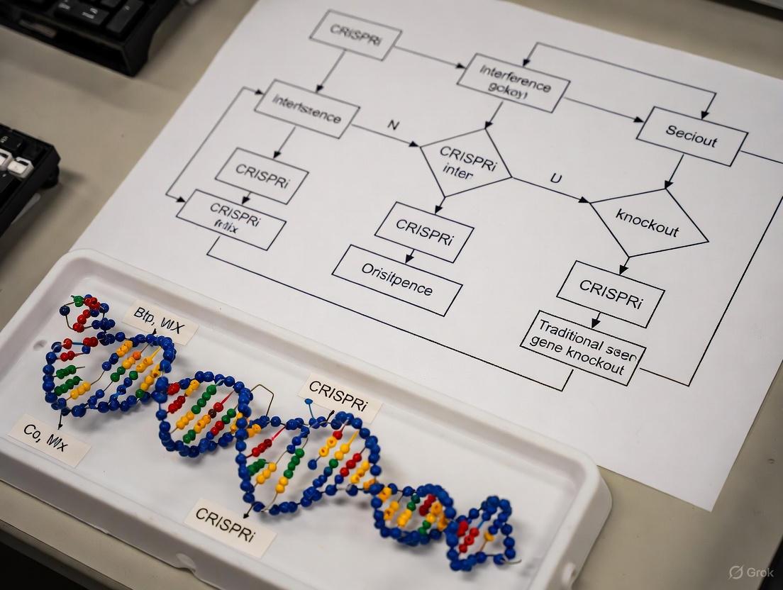
Figure 2: Decision Framework for CRISPRi Experimental Design. This flowchart guides researchers in selecting the appropriate CRISPR-based approach based on their specific research questions about essential biofilm genes, highlighting key decision points between constitutive and inducible systems.
Advanced Applications: Multiplexing and Genetic Interaction Mapping
CRISPRi technology enables sophisticated experimental designs beyond single gene perturbation. For biofilm research, where complex genetic networks govern community behavior, these advanced applications are particularly valuable:
Multiplexed Gene Silencing: CRISPRi systems can be engineered to simultaneously target multiple genes by incorporating several sgRNA expression cassettes [25]. This approach is invaluable for studying functionally redundant genes or complete pathway analysis in biofilm formation. For example, targeting multiple genes in enterococcal biofilm formation revealed compensatory mechanisms that would be missed in single-gene studies.
CRISPRi-TnSeq Genetic Interaction Mapping: A powerful method combining CRISPRi knockdown of essential genes with transposon mutagenesis of non-essential genes enables systematic mapping of genetic interactions [32]. This approach identified 1,334 genetic interactions in Streptococcus pneumoniae, including 754 negative and 580 positive interactions, revealing functional connections between essential and non-essential genes [32]. For biofilm researchers, this technology can identify backup systems that compensate for essential gene knockdown, informing combination therapy strategies.
Essential Gene Function Mapping: CRISPRi enables systematic analysis of essential gene function during biofilm development. By titrating repression levels through inducer concentration modulation, researchers can establish dose-response relationships between gene expression and biofilm phenotypes [5]. This quantitative approach moves beyond binary assessments of gene necessity to reveal quantitative contributions to biofilm traits.
CRISPRi technology represents a sophisticated and versatile approach for validating essential biofilm genes that overcomes the fundamental limitation of lethal phenotypes in conventional knockout studies. Through strategic selection of dCas9 repressor domains, optimized gRNA design, and implementation of inducible control systems, researchers can achieve precise, temporal gene silencing at specific stages of biofilm development. The performance advantages of CRISPRi over RNAi and its complementary nature with CRISPR knockout make it an indispensable tool in the molecular microbiology toolkit. As CRISPRi systems continue to evolve with enhanced repressor domains and more sophisticated control mechanisms, their application in biofilm research will undoubtedly yield new insights into the genetic underpinnings of this complex microbial community behavior.
Step-by-Step Workflow for Generating a Complete Gene Knockout
In the field of functional genomics, generating a complete gene knockout is a fundamental technique for investigating gene function. Within research areas such as essential biofilm gene validation, scientists often choose between two primary CRISPR-based approaches: permanent gene knockout and reversible gene knockdown via CRISPR interference (CRISPRi). A complete gene knockout uses the active Cas9 nuclease to create permanent, irreversible loss-of-function mutations by disrupting the DNA sequence itself. In contrast, CRISPRi employs a deactivated Cas9 (dCas9) fused to repressor domains to block transcription without altering the genetic code, offering reversible and tunable suppression [33] [34]. This guide provides a detailed, step-by-step workflow for generating a complete gene knockout, objectively compares its performance to CRISPRi, and presents supporting experimental data to inform researchers and drug development professionals.
Core Principles: Knockout vs. Interference
Before detailing the protocol, it is crucial to understand the mechanistic differences between the two main approaches.
- CRISPR Knockout (CRISPRn): This method uses the wild-type Cas9 nuclease, which creates double-strand breaks (DSBs) in the DNA at a location specified by a single-guide RNA (sgRNA). The cell's primary repair mechanism, non-homologous end joining (NHEJ), is error-prone. The insertion or deletion of base pairs (indels) often results in frameshift mutations and premature stop codons, leading to a truncated and non-functional protein [19] [31].
- CRISPR Interference (CRISPRi): This method uses a deactivated Cas9 (dCas9) that lacks nuclease activity. The dCas9-sgRNA complex binds to the target DNA without cutting it, physically obstructing the transcription machinery. When fused to a repressor domain like KRAB, it provides enhanced, reversible gene repression [33] [34] [10].
The diagram below illustrates the fundamental mechanisms of both approaches.
Step-by-Step Knockout Workflow
The following section outlines a robust, optimized protocol for generating a complete gene knockout in human pluripotent stem cells (hPSCs), a model system relevant for disease research [35].
Step 1: sgRNA Design and Preparation
The first and most critical step is designing a highly efficient sgRNA.
- Design: Use bioinformatics algorithms like Benchling or CCTop to design sgRNAs targeting early exons of the gene of interest to maximize the likelihood of disrupting the protein's functional domain. Select guides with high on-target efficiency scores and minimal off-target potential [35].
- Preparation: For highest editing efficiency, use chemically synthesized and modified sgRNAs (CSM-sgRNA). Chemical modifications at the 5' and 3' ends enhance stability within cells compared to in vitro transcribed (IVT) sgRNAs [35].
Step 2: Delivery of CRISPR Components
The chosen delivery method significantly impacts efficiency and toxicity.
- Recommended Method: Use ribonucleoprotein (RNP) complexes. Pre-complex the purified SpCas9 protein with your synthesized sgRNA and deliver it via nucleofection. RNP delivery offers high efficiency, rapid action, and reduced off-target effects compared to plasmid-based methods [36] [31].
- Optimization Parameters: Systematically optimize cell tolerance, nucleofection frequency, and the cell-to-sgRNA ratio. For hPSCs, using 5 µg of sgRNA with 8 × 10^5 cells in a single nucleofection can achieve INDEL efficiencies over 80% [35].
Step 3: Validation of Knockout Efficiency
After delivery, confirm the success of the editing.
- Initial Assessment: Harvest genomic DNA 72 hours post-nucleofection. Amplify the target region by PCR and analyze the INDEL spectrum using tools like ICE (Inference of CRISPR Edits) or TIDE on Sanger sequencing data [35] [37].
- Functional Validation: A high INDEL rate does not always guarantee loss of protein. Perform Western blotting 7-14 days post-editing to confirm the absence of the target protein. This step is crucial to identify "ineffective sgRNAs" that cause indels but do not ablate protein expression [35].
Step 4: Clonal Isolation and Expansion
For a pure population of knockout cells, single-cell cloning is necessary.
- Method: After confirming high editing efficiency in the bulk population, use fluorescence-activated cell sorting (FACS) to isolate single cells into 96-well plates.
- Challenge: This step is time-consuming and can take 3-6 months. Researchers often need to repeat the clonal isolation process a median of 3 times before obtaining a desired clonal line [23]. Clonally expanded cells also risk genetic instability [15].
The entire workflow, from design to a validated clonal line, is summarized below.
Performance Comparison: Knockout vs. CRISPRi
Selecting between knockout and interference depends on the experimental goals. The table below summarizes key performance differences, supported by recent data.
Table 1: Objective comparison of CRISPR Knockout vs. CRISPRi
| Feature | CRISPR Knockout (CRISPRn) | CRISPR Interference (CRISPRi) |
|---|---|---|
| Mechanism | Permanent DNA cleavage; error-prone NHEJ repair [19] [31] | Reversible dCas9 binding; blocks transcription [33] [34] |
| Genetic Alteration | Permanent; irreversible sequence change [19] | Reversible; no sequence alteration [33] |
| Efficiency (in iPSCs) | Highly variable (20-93%); requires optimization. Protein knockout can fail despite high INDELs [35] | Highly efficient and homogeneous; >95% repression in bulk populations [34] |
| Multiplexing | Difficult; multiple DSBs can cause genomic complexity and toxicity [15] | Easier; no DNA damage allows simultaneous knockdown of multiple genes [15] |
| Tunability | All-or-nothing effect (knockout or not) [19] | Tunable repression via inducible systems or varying sgRNA dose [33] [10] |
| Ideal Use Case | Studying complete loss-of-function; creating stable disease models [19] | Studying essential genes; pathway analysis; reversible phenotype studies [34] [10] |
A pivotal study directly comparing both technologies in induced pluripotent stem cells (iPSCs) highlights a key advantage of CRISPRi. When targeting the pluripotency gene OCT4, CRISPRi achieved >95% knockdown efficiency in the bulk cell population. In contrast, CRISPRn left 30-40% of the bulk population as OCT4-positive, demonstrating less homogeneous and efficient gene disruption [34]. This makes CRISPRi superior for applications where working with a bulk population is preferred over isolating single-cell clones.
Application in Biofilm Gene Validation
In the context of validating essential biofilm genes, the choice of tool is critical:
- CRISPR Knockout is suitable for non-essential biofilm genes. However, if the gene is essential for cell survival, a complete knockout will be lethal, preventing study [10].
- CRISPRi allows for the knockdown of essential genes to study their role in biofilm formation and antimicrobial resistance without killing the cell, enabling genome-wide screens to identify and validate new targets [10].
The Scientist's Toolkit: Essential Research Reagents
The following table lists key reagents required for executing the knockout workflow described above.
Table 2: Essential Reagents for CRISPR Knockout Workflow
| Reagent / Solution | Function / Description | Recommendation |
|---|---|---|
| Chemically Modified sgRNA (CSM-sgRNA) | Guide RNA with stability enhancements for increased half-life and editing efficiency [35] | Prefer over IVT-sgRNA or plasmid-based guides for higher efficiency and lower off-target effects [35] [31]. |
| SpCas9 Nuclease | Wild-type Cas9 protein from S. pyogenes that creates double-strand breaks in DNA [35] | Use high-quality, purified protein to form RNP complexes. |
| RNP Complex | Pre-formed complex of Cas9 protein and sgRNA; the gold standard for delivery [36] [31] | Direct delivery of RNPs via nucleofection results in highest editing efficiency and fastest activity. |
| Nucleofector System | Electroporation device optimized for hard-to-transfect cells like stem cells and primary cells [35] | Essential for efficient RNP delivery into hPSCs. |
| ICE / TIDE Analysis Tool | Bioinformatics software for analyzing Sanger sequencing data to quantify INDEL efficiency [35] [37] | Use for rapid, initial assessment of editing success in bulk populations. |
| AAVS1 Targeting System | Safe-harbor locus control for gRNA design; disruption has no adverse effects [36] [35] | An essential negative control for fitness and viability assays. |
Generating a complete gene knockout via CRISPR-Cas9 is a powerful but meticulous process. Its success hinges on optimized sgRNA design, efficient RNP delivery, and rigorous validation that includes confirmation of protein loss. For studies requiring a permanent, complete loss-of-function—such as creating stable knockout disease models—it remains the definitive method.
However, the comparative data clearly shows that CRISPRi offers significant advantages in efficiency, reversibility, tunability, and multiplexing. For the validation of essential genes, particularly in complex processes like biofilm formation where essential gene pathways are a key focus, CRISPRi provides a more robust and flexible platform. Researchers should therefore select the technology based on their specific biological question: CRISPR knockout for irreversible ablation and CRISPRi for precise, reversible modulation of gene expression.
The validation of essential genes involved in biofilm formation presents a significant challenge in microbiology research. Traditional gene knockout methods are ineffective for essential genes, as their complete deletion is lethal to the cell. This limitation has propelled the adoption of CRISPR interference (CRISPRi) as a powerful alternative for probing gene function in biofilm studies. CRISPRi employs a catalytically inactive Cas9 (dCas9) that binds to target DNA without causing double-strand breaks, thereby enabling tunable gene repression rather than permanent deletion [38] [1]. This technical comparison guide objectively assesses the performance of CRISPRi against traditional knockout methods specifically for validating essential biofilm genes, focusing on phenotypic outputs including biomass quantification, architectural analysis, and virulence profiling.
The fundamental distinction lies in their applicability to essential genes. As one study notes, "gene essentiality is largely conserved between liquid and surface growth," meaning genes critical for planktonic growth are often also vital for biofilm development [38]. While knockout strains cannot be constructed for these essential genes, CRISPRi knockdown strains remain viable, allowing researchers to study their roles in biofilm phenotypes through titratable repression [38].
Technical Comparison: CRISPRi vs. Gene Knockout
Table 1: Core Methodological Comparison Between CRISPRi and Gene Knockout
| Feature | CRISPRi | Traditional Gene Knockout |
|---|---|---|
| Mechanism of Action | dCas9-sgRNA complex blocks transcription [1] | Complete physical deletion or disruption of the target gene |
| Applicability to Essential Genes | Yes; enables partial knockdown without cell death [38] | No; lethal to the cell |
| Reversibility | Tunable and reversible repression [38] | Permanent and irreversible |
| Titratable Control | Yes; repression levels can be modulated with inducer concentration [38] [3] | No; typically all-or-nothing |
| Development Timeline | Relatively fast; requires only sgRNA design and cloning [39] | Time-consuming; requires homologous recombination and selection |
| Multiplexing Potential | High; multiple sgRNAs can target several genes simultaneously [39] | Possible but labor-intensive |
Table 2: Performance in Biofilm Phenotype Assessment
| Performance Metric | CRISPRi | Traditional Gene Knockout |
|---|---|---|
| Biomass Quantification (Crystal Violet) | Reveals dose-dependent biofilm reduction [1] | Only reveals phenotypes for non-essential genes |
| Architectural Analysis (CLSM) | Enables study of essential gene impact on 3D structure [3] | Not applicable for essential genes |
| Genetic Compensation | Minimal; partial knockdown reduces adaptive mutations [38] | Common; can lead to suppressor mutations |
| Phenotypic Penetrance | Homogeneous population response [38] | Can be heterogeneous due to suppressor development |
| Off-Target Effects | Possible, but design improvements minimize risk [39] | Minimal with proper controls |
Experimental Data and Case Studies
Quantitative Phenotypic Data from Published Studies
Table 3: Representative Experimental Data from CRISPRi Biofilm Studies
| Target Gene / Pathway | Organism | Biofilm Phenotype | Quantitative Impact | Citation |
|---|---|---|---|---|
| luxS (Quorum Sensing) | E. coli | Biofilm inhibition | Significant reduction in biofilm mass confirmed by CV and SEM [1] | |
| Fatty Acid Synthesis | B. subtilis | Enhanced biofilm wrinkling | Increased wrinkling independent of matrix gene expression [38] | |
| GacA/S Two-Component System | P. fluorescens | Altered biofilm architecture | Defects in biofilm mass and structure similar to knockout [3] | |
| Bacterial Gyrase | B. subtilis | Enhanced biofilm wrinkling | Distinct wrinkling pattern phenotype observed [38] |
Detailed Experimental Protocols
CRISPRi Strain Construction and Validation
The following protocol is adapted from multiple high-impact studies [38] [1] [3]:
- sgRNA Design: Design 20-nucleotide complementary sequences targeting the non-template strand of the essential gene's promoter region or early coding sequence. The target site must be immediately adjacent to a 5'-NGG-3' PAM sequence [1].
- Vector Assembly: Clone the synthesized sgRNA sequence into an appropriate expression plasmid (e.g., pgRNA) using inverse PCR and blunt-end ligation. Co-transform with a second plasmid expressing dCas9 (e.g., pdCas9) under an inducible promoter (e.g., Ptet, Pxyl) into the target bacterial strain [38] [1].
- Knockdown Validation: Quantify repression efficiency via qRT-PCR on total RNA extracted from induced and non-induced cultures. Assess protein-level knockdown if antibodies are available [1].
Biofilm Phenotyping Assays
Crystal Violet Biomass Assay:
- Grow CRISPRi strains in 96-well polyvinyl chloride (PVC) microtiter plates for 48 hours at 37°C with and without inducer [40] [1].
- Remove planktonic cells, stain adherent biomass with 1% crystal violet (25 μL/well) for 15 minutes [40].
- Wash wells three times with sterile distilled water, solubilize bound dye with 95% ethanol, and measure absorbance at 630 nm [40].
Architectural Analysis via Confocal Laser Scanning Microscopy (CLSM):
- Grow biofilms in flow cells or on coverslips under continuous medium flow (e.g., 0.33 mL/min) for 1-7 days to allow maturation [40] [3].
- Stain with LIVE/DEAD BacLight stain (or similar viability stain) for 15 minutes in the dark [40].
- Image using a 20x objective, collecting z-stacks at random positions at 4-μm intervals through the biofilm [40].
- Quantify architectural parameters (biomass, thickness, roughness, substratum coverage) using image analysis software such as COMSTAT [40].
The Scientist's Toolkit: Key Research Reagent Solutions
Table 4: Essential Reagents and Tools for CRISPRi Biofilm Research
| Reagent / Tool | Function | Example Application |
|---|---|---|
| dCas9 Expression Plasmid | Provides the catalytically inactive Cas9 protein for targeted gene repression. | Constitutively or inducibly expressed in the target strain [38] [3]. |
| sgRNA Expression Vector | Expresses the target-specific guide RNA that directs dCas9 to the gene of interest. | Cloned with complementary sequences to the essential biofilm gene [1]. |
| Inducer Molecules | Controls the timing and level of dCas9 expression, enabling titratable knockdown. | Anhydrotetracycline (aTc) for Ptet, Xylose for Pxyl [38] [1]. |
| Polyvinyl Chloride (PVC) Microplates | Provides a standardized abiotic surface for high-throughput biofilm cultivation. | Used for crystal violet biofilm assays [40]. |
| Flow Cell Systems | Enables continuous culture and high-resolution imaging of mature, structured biofilms. | Used for CLSM analysis of 3D biofilm architecture [40] [3]. |
| COMSTAT Software | Quantifies 3D parameters from CLSM image stacks, providing objective metrics. | Analysis of biofilm biomass, thickness, and roughness [40]. |
Methodological Workflows
The following diagrams illustrate the core workflows for implementing CRISPRi and analyzing the resulting biofilm phenotypes.
CRISPRi Workflow
Phenotype Comparison Logic
The choice between CRISPRi and traditional knockout methods for biofilm phenotype assessment depends heavily on the nature of the target gene and the research question. For non-essential genes, knockout strains remain a valuable, permanent, and well-characterized tool. However, for essential genes—which comprise a significant portion of biofilm regulators—CRISPRi provides an unparalleled ability to study their function through titratable knockdown without causing cell death [38].
The experimental data consistently demonstrate that CRISPRi produces biologically relevant biofilm phenotypes, from the inhibition of E. coli biofilm via luxS repression to the distinct wrinkling patterns in B. subtilis following fatty acid synthesis knockdown [1] [38]. Furthermore, CRISPRi phenotypes for non-essential genes, such as those in the GacA/S system, often mirror their knockout counterparts, validating its reliability [3].
Future developments in CRISPRi technology, including improved base editing and prime editing systems, along with more sophisticated delivery mechanisms such as nanoparticle carriers, promise to further enhance its specificity and application in biofilm research [2] [39]. These advancements will solidify CRISPRi's role as an indispensable method for comprehensive biofilm phenotyping, ultimately accelerating the discovery of novel anti-biofilm therapeutic strategies.
The opportunistic pathogen Pseudomonas aeruginosa presents a formidable challenge in healthcare settings due to its robust capacity for biofilm formation, a key contributor to its antibiotic resistance and persistence in chronic infections. The second messenger bis-(3'-5')-cyclic dimeric guanosine monophosphate (c-di-GMP) stands as a central regulator of the transition from motile to sessile biofilm lifestyles in bacteria. However, dissecting the individual contributions of enzymes involved in c-di-GMP metabolism has been hampered by extensive genetic redundancy; P. aeruginosa encodes up to 40 proteins with GGDEF, EAL, or HD-GYP domains, many with overlapping functions [41]. This case study examines how CRISPR interference (CRISPRi) has emerged as a powerful alternative to conventional gene knockouts, enabling precise functional analysis of this complex signaling network and validating essential genes that control biofilm formation.
Methodological Comparison: CRISPRi vs. Conventional Gene Knockout
Traditional genetic approaches for studying gene function, particularly gene knockout via homologous recombination or transposon mutagenesis, have significant limitations when investigating redundant or essential genes. These methods create permanent, complete disruptions of target genes, which can be lethal for essential genes and may fail to reveal phenotypes in systems with parallel signaling pathways due to compensatory mechanisms [41] [10].
CRISPRi technology overcomes these limitations by utilizing a catalytically dead Cas9 (dCas9) protein that binds to specific DNA sequences under the guidance of a programmable single-guide RNA (sgRNA) without cleaving the DNA. This binding sterically hinders transcription initiation or elongation, resulting in tunable and reversible gene silencing rather than permanent disruption [10] [1]. This capability is particularly valuable for:
- Studying essential genes without causing cell death
- Investigating gene families with functional redundancy by enabling simultaneous multiplexed silencing
- Achieving titratable levels of gene repression through inducible promoter systems
- Enabling functional analysis in systems where traditional knockout is technically challenging
Table 1: Comparison of CRISPRi and Gene Knockout for Biofilm Gene Validation
| Feature | CRISPRi | Conventional Knockout |
|---|---|---|
| Mechanism | dCas9-mediated transcriptional repression | Physical gene deletion or disruption |
| Reversibility | Reversible | Permanent |
| Applicability to Essential Genes | Yes (titratable repression) | No (lethal) |
| Tunability | Inducible and tunable | Binary (present/absent) |
| Multiplexing Capacity | High (multiple sgRNAs) | Technically challenging |
| Phenotypic Compensation | Reduced due to rapid knockdown | Common in redundant systems |
| Technical Implementation | Relatively fast, suitable for high-throughput | Often labor-intensive |
Case Study: Dissecting c-di-GMP Signaling inPseudomonas aeruginosa
Experimental Approach and Workflow
A landmark 2025 study demonstrated the power of CRISPR-based genome editing to overcome native redundancy in c-di-GMP synthesis [41]. Researchers employed a multiplexed cytosine base-editor to simultaneously disrupt all 32 GGDEF domain-containing proteins (GCPs) implicated in c-di-GMP signaling in P. aeruginosa PA14. The experimental workflow involved:
- Target Identification: Bioinformatic identification of all 32 GCP-encoding genes in P. aeruginosa PA14
- Spacer Design: CRISPy web service was used to design 20-nt spacer sequences targeting each GCP gene
- Vector Construction: pBEC/pMBEC plasmids containing spacer combinations were assembled via Golden Gate assembly
- Genome Editing: P. aeruginosa PA14 was electroporated with the base-editor plasmids
- Strain Validation: Introduction of premature STOP codons confirmed through Sanger sequencing
- Phenotypic Characterization: Comprehensive analysis of biofilm formation, virulence, motility, and antibiotic resistance
The resulting mutant strain (PA14Δ32) showed impaired c-di-GMP synthesis and served as a clean genetic background for functional complementation studies with individual diguanylate cyclases (DGCs) to dissect their specific roles [41].
Key Findings and Phenotypic Outcomes
The systematic disruption of all 32 GCPs revealed profound phenotypic consequences:
- Biofilm Formation: The PA14Δ32 strain was unable to form biofilms, confirming the collective essentiality of GCPs in the transition to sessile lifestyle [41]
- Virulence Attenuation: Significant reduction in pathogenicity, highlighting the role of c-di-GMP in virulence regulation
- Residual c-di-GMP: Despite comprehensive GCP disruption, low c-di-GMP levels persisted, suggesting exceptional robustness in this regulatory network
- Genetic Complementation: Reintroduction of selected DGCs into the PA14Δ32 background restored specific c-di-GMP-related traits, enabling functional dissection of individual enzymes
Table 2: Phenotypic Characterization of P. aeruginosa GCP-Deficient Strain
| Phenotypic Trait | Observation in PA14Δ32 Mutant | Functional Complementation Outcome |
|---|---|---|
| Biofilm Formation | Abolished | Partially restored by individual DGCs |
| c-di-GMP Levels | Severely reduced but detectable | Restored to varying degrees |
| Virulence | Significantly attenuated | Dependent on specific DGC reintroduced |
| Motility | Altered swimming and swarming | Pattern-specific restoration |
| Antibiotic Resistance | Modified susceptibility | Variable based on DGC identity |
| Growth Rate | Minimal impact | Not significantly affected |
Experimental Protocols for CRISPRi inPseudomonas
CRISPRi System Implementation
The adapted CRISPRi system for Pseudomonas species typically consists of two compatible plasmids [3]:
- dCas9 Expression Plasmid: Contains the dCas9 gene from S. pyogenes under control of the PtetA promoter, inducible by anhydrotetracyclin (aTc)
- sgRNA Expression Plasmid: Constitutively expresses gene-specific sgRNAs targeting genes of interest
Protocol for CRISPRi-Mediated Gene Silencing:
sgRNA Design: Design 20-nt sequences complementary to the target region adjacent to PAM (5'-NGG-3') sites
- For maximal repression: target transcription initiation near promoter regions
- For partial repression: target transcription elongation within open reading frames
Strain Transformation:
- Electroporate P. aeruginosa with both plasmids
- Recover cells in lysogeny broth for 3 hours
- Plate on selective media containing appropriate antibiotics
Gene Silencing Induction:
- Grow transformed strains in media supplemented with aTc (typically 2-1000 nM)
- Induce during exponential growth phase for optimal repression
Repression Validation:
- Quantify mRNA levels using qRT-PCR to confirm target gene downregulation
- Assess protein levels when possible via Western blot or functional assays
Phenotypic Assessment Methods
Biofilm Quantification:
- Crystal Violet Assay: Standard method for total biofilm biomass measurement
- Confocal Laser Scanning Microscopy: Enables 3D visualization of biofilm architecture and extracellular matrix distribution
- Scanning Electron Microscopy: Provides ultrastructural details of biofilm morphology
Motility Assays:
- Swarming Motility: Assessed on low-percentage agar plates (0.5-0.8%)
- Swimming Motility: Evaluated on intermediate-percentage agar plates (0.3%)
- Twitching Motility: Measured at the interface between agar and petri dish
Virulence Factor Production:
- Quantification of pyocyanin, elastase, and rhamnolipid production using established spectrophotometric methods
The Scientist's Toolkit: Essential Research Reagents
Table 3: Key Research Reagents for CRISPRi Experiments in Pseudomonas
| Reagent/Resource | Function/Application | Source/Example |
|---|---|---|
| dCas9 Expression Plasmid | Provides catalytically dead Cas9 for transcriptional repression | pdCas9 (Addgene #44249) |
| sgRNA Expression Vector | Enables programmable targeting of specific genes | pgRNA (Addgene #44251) |
| Anhydrotetracycline (aTc) | Inducer for PtetA-controlled dCas9 expression | Chemical suppliers |
| CRISPy Web Service | Bioinformatics tool for spacer sequence design | Blin et al. 2016 |
| Golden Gate Assembly | Modular cloning system for multiplexed sgRNA constructs | Bird et al. 2022 |
| P. aeruginosa PA14 Strain | Model opportunistic pathogen for biofilm studies | Rahme et al. 1995 |
| Pseudomonas.com Database | Genomic resource for locus tag identification | Winsor et al. 2016 |
Signaling Pathways and Regulatory Networks
The c-di-GMP signaling network in P. aeruginosa represents a complex regulatory hierarchy that controls the transition between planktonic and biofilm states. This system integrates multiple environmental signals through various DGCs and phosphodiesterases (PDEs) to fine-tune intracellular c-di-GMP levels, which subsequently bind to effector molecules and regulate downstream cellular processes [41] [3].
Discussion: Advantages and Limitations in Biofilm Research
CRISPRi as a Superior Approach for Signaling Network Dissection
The application of CRISPRi technology to study c-di-GMP signaling in Pseudomonas demonstrates significant advantages over traditional gene knockout methods:
Overcoming Redundancy: The multiplex capability of CRISPRi enables simultaneous targeting of multiple DGCs and PDEs, bypassing the compensatory mechanisms that frequently obscure phenotypes in single knockout studies [41]. This was powerfully demonstrated in the PA14Δ32 strain, which exhibited dramatic biofilm deficiency only when all GCPs were disrupted.
Precise Functional Mapping: The ability to reintroduce individual DGCs into a clean genetic background (free of other GCPs) allows for unambiguous determination of specific enzyme functions without interference from native redundancy [41].
Studying Essential Functions: CRISPRi enables partial silencing of genes that would be lethal if completely knocked out, making it possible to study the roles of essential biofilm-related genes in bacterial physiology [10].
Technical Considerations and Future Directions
While CRISPRi presents a powerful approach, several considerations must be addressed:
Variable Repression Efficiency: Silencing efficiency can vary depending on sgRNA target location, with maximal repression typically achieved when targeting transcription initiation regions [3].
Off-Target Effects: Careful bioinformatic design and appropriate controls are necessary to minimize potential off-target silencing.
Delivery Optimization: Efficient delivery of CRISPR components remains challenging in some bacterial systems, though nanoparticle-CRISPR hybrids show promise for enhancing delivery efficiency [42].
The integration of CRISPRi with emerging technologies such as CRISPR activation (CRISPRa) for gene upregulation and nanoparticle-mediated delivery systems will further expand our ability to manipulate and study complex bacterial behaviors [10] [42]. These advanced tools promise to accelerate the identification of novel therapeutic targets for combating biofilm-associated infections.
This case study demonstrates that CRISPRi technology provides a superior methodological approach for dissecting complex bacterial signaling networks compared to conventional gene knockout strategies. By enabling targeted, tunable gene silencing without permanent genetic disruption, CRISPRi has uncovered the collective essentiality of diguanylate cyclases in P. aeruginosa biofilm formation while allowing functional characterization of individual enzymes. The application of CRISPRi to overcome genetic redundancy in c-di-GMP signaling represents a paradigm shift in bacterial genetics, offering unprecedented precision for validating essential genes and identifying potential therapeutic targets against biofilm-mediated antimicrobial resistance. As CRISPR-based technologies continue to evolve, they will undoubtedly yield deeper insights into the complex regulatory hierarchies that control bacterial lifestyle transitions and pathogenesis.
Studying essential genes in bacteria presents a significant methodological challenge for researchers investigating virulence and biofilm formation. Essential genes are those required for an organism's survival; their complete disruption via knockout is lethal, preventing the study of their function in later developmental stages. This is particularly relevant in Burkholderia cepacia complex (Bcc) species, such as Burkholderia multivorans, which are opportunistic pathogens in cystic fibrosis patients and rely on complex regulatory networks for biofilm formation and virulence [43].
The RpfR regulator in B. multivorans represents a paradigm of this challenge. RpfR is a key regulator within the diffusible signal factor (DSF) signaling system that modulates biofilm development, virulence, and antibiotic resistance. As research has demonstrated, RpfR is essential for bacterial viability, making traditional knockout approaches unsuitable for functional studies [43]. This case study compares the experimental limitations of CRISPR/Cas9-mediated knockout versus CRISPR interference (CRISPRi) for validating RpfR function, providing a framework for researchers studying essential bacterial regulators.
Methodological Comparison: Knockout vs. Interference
CRISPR/Cas9 Knockout Approach
CRISPR/Cas9 knockout utilizes the Cas9 nuclease to create double-strand breaks in DNA at specific sites guided by RNA molecules. The cell repairs these breaks primarily through two pathways: non-homologous end joining (NHEJ), which often results in insertions or deletions (indels) that disrupt gene function, or homology-directed repair (HDR), which can be used to incorporate specific genetic modifications [31] [44].
The fundamental limitation for essential genes like RpfR is that successful knockout leads to cell death, preventing the study of gene function in subsequent phenotypic assays. While CRISPR/Cas9 has been successfully used to generate unmarked deletions in non-essential Burkholderia genes (including rpfR, bceF, cepR, and bcsB) using a two-plasmid system (pCasPA and pACRISPR), this approach is only viable when gene disruption does not affect bacterial survival [43].
CRISPR Interference (CRISPRi) Approach
CRISPR interference (CRISPRi) employs a catalytically dead Cas9 (dCas9) variant that lacks nuclease activity but retains DNA-binding capability. When guided to specific genomic locations, dCas9 functions as a transcriptional blockade, physically preventing RNA polymerase from initiating or elongating transcription without altering the underlying DNA sequence [45] [46].
For essential genes like RpfR, CRISPRi enables tunable gene silencing rather than permanent disruption. This allows researchers to study partial loss-of-function effects without triggering cell death. In Burkholderia species, CRISPRi systems have been optimized using a rhamnose-inducible dCas9 that can be chromosomally integrated via a mini-CTX system, enabling titratable control of gene expression [45].
Experimental Comparison & Performance Data
Quantitative Comparison of Methodological Outcomes
Table 1: Comparative Performance of CRISPR Knockout vs. CRISPRi for Studying Essential Genes
| Parameter | CRISPR/Cas9 Knockout | CRISPR Interference |
|---|---|---|
| Genetic Outcome | Permanent gene disruption | Reversible transcriptional silencing |
| Cell Viability with Essential Genes | Lethal | Maintained with tunable knockdown |
| Experimental Applications | Non-essential gene function studies | Essential gene function studies, titratable knockdowns |
| Titratable Control | Not available | Rhamnose-inducible (0.01%-1%) for controlled silencing [45] |
| Methodological Limitations | Cannot study essential genes; potential for off-target mutations | Incomplete silencing; potential for residual gene expression |
| Implementation in Burkholderia | Successful for non-essential genes (rpfR, bceF, cepR, bcsB) [43] | Optimized for chromosomal integration with mini-CTX system [45] |
Experimental Workflow for RpfR Functional Analysis
Detailed Methodologies
CRISPR/Cas9 Knockout Protocol for Burkholderia
The CRISPR/Cas9 system for Burkholderia employs a two-plasmid approach adapted from Pseudomonas aeruginosa tools [43]:
- Plasmid System: pCasPA (expressing Cas9 and λ-Red system proteins) and pACRISPR (expressing sgRNA and repair template)
- Selection Markers: Tetracycline resistance for pCasPA; sacB as a counter-selectable marker for plasmid curing
- Induction Conditions: Cas9 and λ-Red expression induced with 2% L-arabinose
- Delivery Method: Triparental conjugation from E. coli to Burkholderia
- Validation: PCR amplification of internal cas9 fragment to confirm plasmid presence
This approach has demonstrated efficiency in creating precise, unmarked deletions of non-essential genes in B. multivorans, with successful conjugation frequencies varying across strains [43].
CRISPRi Silencing Protocol for Burkholderia
The optimized CRISPRi system for Burkholderia species enables targeted gene silencing [45]:
- dCas9 Expression: Chromosomally integrated rhamnose-inducible dCas9 via mini-CTX system
- Guide RNA Delivery: Broad-host plasmid pSCB2-gRNA for sgRNA expression
- Induction Conditions: dCas9 expression induced with L-rhamnose (0.01%-1% concentration)
- Experimental Controls: Essential to include induction controls to distinguish CRISPRi effects from rhamnose toxicity
- Efficiency Assessment: Proteomic and phenotypic analysis to quantify silencing efficiency
This system has demonstrated reductions in target protein levels by up to 90%, although complete abolition of gene expression is rarely achieved, which paradoxically provides an advantage for studying essential genes [45].
The Scientist's Toolkit: Essential Research Reagents
Table 2: Key Research Reagents for Burkholderia Genetic Manipulation
| Reagent/Component | Function | Application Notes |
|---|---|---|
| pCasPA Plasmid | Expresses Cas9 and λ-Red system proteins | Tetracycline selection; induced with 2% L-arabinose [43] |
| pACRISPR Plasmid | Expresses sgRNA and repair template | Contains sacB counter-selectable marker for curing [43] |
| CTX:rha-dCas9 System | Chromosomally integrated dCas9 | Rhamnose-inducible; enables stable dCas9 expression [45] |
| pSCB2-gRNA Plasmid | sgRNA expression vector | Broad-host range; compatible with diverse Burkholderia species [45] |
| L-Rhamnose | Inducer for dCas9 expression | Titratable (0.01%-1%); high concentrations may cause proteomic changes [45] |
| L-Arabinose | Inducer for Cas9/λ-Red expression | Used at 2% concentration; no detectable toxicity observed [43] |
Discussion & Research Implications
Advantages of CRISPRi for Essential Gene Functional Studies
CRISPRi provides several critical advantages for studying essential regulators like RpfR:
- Titratable Silencing: Rhamnose induction levels (0.01%-1%) enable fine-tuning of gene expression, allowing researchers to study dose-dependent effects of essential gene reduction [45]
- Functional Phenotyping: Partial silencing enables assessment of RpfR's role in biofilm formation, virulence factor expression, and antibiotic resistance without triggering lethality
- Multiplexing Capability: CRISPRi enables simultaneous silencing of multiple genes, facilitating study of genetic interactions and redundant pathways [46]
- Temporal Control: Inducible systems allow researchers to control the timing of gene silencing, enabling study of stage-specific essentiality during biofilm development
Technical Considerations for Burkholderia Genetic Studies
When implementing these approaches in Burkholderia species, researchers should consider:
- Strain Variability: Conjugation efficiency of genetic tools varies significantly across Burkholderia strains, with clinical isolates often showing higher transformation frequency [43]
- Incomplete Silencing: CRISPRi typically achieves 80-90% reduction in target expression, which may be insufficient for studying some essential genes [45]
- Genetic Stability: CRISPRi systems with chromosomal dCas9 integration demonstrate long-term stability, maintaining silencing efficiency through multiple generations [45]
- Phenotypic Validation: Essential gene silencing requires comprehensive phenotypic validation through proteomics, transcriptomics, and functional assays to correlate expression changes with physiological effects
The case of RpfR in Burkholderia illustrates a fundamental principle in bacterial genetics: methodological selection must align with biological constraints. While CRISPR/Cas9 knockout provides definitive evidence for gene function through complete disruption, its application is limited to non-essential genes. CRISPR interference emerges as the superior approach for studying essential regulators, enabling titratable silencing that preserves cell viability while allowing functional assessment.
For researchers investigating essential biofilm regulators in Burkholderia and related pathogens, CRISPRi provides a versatile platform for mechanistic studies, though appropriate controls must be implemented to distinguish specific silencing effects from potential induction-related artifacts. The continued refinement of these genetic tools will enhance our understanding of essential gene networks in bacterial pathogenesis and biofilm development.
Solving Experimental Pitfalls: From Off-Target Effects to Phenotypic Complexity
In the study of essential genes, particularly those controlling complex processes like biofilm formation in pathogens, CRISPR interference (CRISPRi) has emerged as a powerful alternative to conventional gene knockouts. While traditional CRISPR-Cas9 knockout strategies permanently disrupt gene function by creating double-strand breaks and relying on error-prone non-homologous end joining (NHEJ) repair, this approach is unsuitable for essential genes whose complete disruption would be lethal to the cell [19] [16] [47]. CRISPRi overcomes this limitation by utilizing a catalytically dead Cas9 (dCas9) that binds to target DNA without causing cleavage, thereby sterically hindering transcription initiation or elongation and achieving reversible, titratable gene repression [48] [10]. This capacity for fine-tuned gene silencing makes CRISPRi indispensable for validating the function of essential genes involved in multi-drug resistance and biofilm formation, as it allows researchers to probe gene function without causing cell death [10]. The efficacy of this technology, however, hinges on two critical optimization parameters: the strategic design of guide RNAs (gRNAs) and the precise titration of the induction system controlling dCas9 expression.
Optimizing gRNA Design for Maximum Silencing Efficiency
The design of the single guide RNA (sgRNA) is the primary determinant of CRISPRi efficiency and specificity. A well-designed sgRNA ensures high on-target activity while minimizing off-target effects.
Fundamental Principles and Positioning
The basic sgRNA structure consists of a 20-nucleotide spacer sequence that guides the dCas9 protein to a specific genomic locus immediately upstream of a Protospacer Adjacent Motif (PAM) sequence [49]. For the commonly used S. pyogenes Cas9, the PAM sequence is 5'-NGG-3' [49]. Strategic positioning of the sgRNA within the target gene is crucial. Table 1 summarizes the performance of sgRNAs based on their target location.
Table 1: sgRNA Performance Based on Target Location and Strand
| Target Region | Strand | Efficiency Rationale | Validated Efficacy |
|---|---|---|---|
| Promoter/Transcription Start Site | Non-template (NT) | Blocks transcription initiation by physically preventing RNA polymerase binding [48] [3]. | Highest reported knockdown in P. fluorescens (Pc5 gRNA) [3]. |
| Early Coding Sequence (5' end) | Non-template (NT) | Hinders transcription elongation by the RNA polymerase [48]. | Effective repression, but can be less potent than promoter-targeting gRNAs [3]. |
| Early Coding Sequence | Template (T) | Can interfere with transcription, but mechanism may be less efficient [3]. | Variable and often lower efficiency compared to NT strand targeting [3]. |
Advanced gRNA Design Strategies and Tools
Beyond basic positioning, sophisticated design strategies have been developed to enhance performance. Dual-sgRNA libraries, where a single construct expresses two highly active sgRNAs targeting the same gene, have been shown to produce stronger and more consistent knockdown than single sgRNAs. A 2023 study demonstrated that such dual-sgRNA constructs enabled the creation of an ultra-compact, genome-wide library without sacrificing performance [50]. Furthermore, the fusion of dCas9 to optimized repressor domains like Zim3 has been empirically shown to provide an superior balance of strong on-target knockdown and minimal non-specific effects on cell growth or the transcriptome [50].
To translate these principles into practice, researchers rely on computational tools that incorporate algorithm-based scoring for on-target efficiency and off-target risk. Key scoring methods include:
- On-target Efficiency: Rule Set 3 (2022) is a modern algorithm that considers the tracrRNA sequence for improved prediction accuracy [49].
- Off-target Risk: Cutting Frequency Determination (CFD) scoring is widely used to predict and minimize off-target effects [49].
Popular design platforms that implement these algorithms include CRISPick (Broad Institute), CHOPCHOP, and CRISPOR [49].
Titrating Induction for Precise Gene Silencing
Titratable control over gene repression is a key advantage of CRISPRi, allowing researchers to fine-tune gene expression levels and study essential genes without inducing lethality.
Inducer Titration and Tunable Repression
A foundational method for titration involves varying the concentration of the molecule that induces dCas9 expression. In a CRISPRi system engineered for Pseudomonas fluorescens, which used an anhydrotetracycline (aTc)-inducible promoter (PtetA) to control dCas9, a clear dose-dependent response was observed. Higher concentrations of aTc led to increased dCas9 expression and a correspondingly stronger repression of a target fluorescent reporter gene [3]. This precise control enables researchers to dial in the desired level of gene silencing for their specific experimental needs.
Engineering Titration with Modified sgRNAs
An even more refined strategy for titration involves using a series of mutated sgRNAs. A 2020 study in E. coli systematically designed sgRNAs with mismatches in their 20-nucleotide homology region to titrate knockdown strength [51]. The study found that a "compounding mutational" strategy—incrementally adding mutations from the distal end (far from the PAM) toward the seed region (close to the PAM)—produced a near-monotonic, gradated relationship between the number of mutations and the resulting growth rate effect [51]. This provides a powerful, scalable method to generate a spectrum of phenotypic severities from a single gene target.
Table 2: Experimental Protocols for Titration and Efficiency Validation
| Method | Key Procedure | Measurement & Validation | Key Finding/Application |
|---|---|---|---|
| Inducer Titration [3] | Grow bacterial cultures (e.g., P. fluorescens) with varying concentrations of anhydrotetracycline (aTc). | Measure target gene repression via fluorescence (flow cytometry) or mRNA quantification (qRT-PCR). | Dose-dependent repression of a fluorescent reporter, enabling fine-tuning of silencing levels. |
| sgRNA Mutation Strategy [51] | Clone a library of sgRNAs with compounding mutations (e.g., at positions -20, -19, -18, etc.) into a low-copy plasmid. | Use CRISPRiSeq (sgRNA amplicon sequencing from genomic DNA) to track population growth rates in a turbidostat. | Revealed 37% more gene-by-environment interactions at intermediate knockdowns vs. maximal knockdown. |
| Escape Mutation Correction [51] | Incorporate a random 6 bp molecular barcode downstream of each sgRNA expression cassette. | Sequence the barcode to track and computationally correct for sub-populations that evade CRISPRi repression. | Unmasked deleterious growth effects that were otherwise obscured by the expansion of "escaper" mutants. |
Application in Biofilm Research: Validating Essential Genes
The optimized strategies for gRNA design and induction titration are critical for dissecting the genetic pathways of biofilm formation, a key virulence factor in many multi-drug resistant pathogens.
CRISPRi has been successfully deployed to investigate genes controlling biofilm in diverse bacterial species, including Pseudomonas aeruginosa, Escherichia coli, and Mycobacterium species [10]. For instance, in P. fluorescens SBW25, CRISPRi-mediated silencing of central regulatory genes like gacA (part of the GacA/S two-component system) and key enzymes involved in cyclic di-GMP (c-di-GMP) signaling (e.g., bifA, dipA, rimA) produced clear, quantifiable phenotypes in swarming motility and biofilm architecture that were consistent with known knockout mutants [3]. This validates CRISPRi as a reliable tool for functional genetics in this space.
A major advantage of CRISPRi is its ability to target essential genes that are intractable to knockout. A CRISPRi screen in mycobacteria targeting the clp protease system, which is essential for survival, revealed its critical role in drug resistance, a finding that would be impossible to obtain with a conventional knockout approach [10]. The titratable nature of CRISPRi allows for the silencing of such essential genes to a sub-lethal threshold, enabling the study of their function in processes like biofilm formation and antibiotic tolerance.
Diagram 1: A workflow for optimizing CRISPRi experiments to validate essential biofilm genes, covering system selection, gRNA design, and induction titration.
The Scientist's Toolkit: Essential Reagents for CRISPRi Experimentation
The following table lists key reagents and resources required to establish and execute a CRISPRi experiment for biofilm gene validation, based on protocols from the cited literature.
Table 3: Research Reagent Solutions for CRISPRi Experiments
| Reagent / Resource | Function / Description | Example Sources / Components |
|---|---|---|
| dCas9 Effector Plasmid | Constitutively or inducibly expresses the catalytically dead Cas9 protein, often fused to a repressor domain. | Zim3-dCas9 [50]; dCas9-KRAB; System with PtetA promoter for aTc induction [3]. |
| sgRNA Expression Plasmid | A vector that constitutively expresses the single guide RNA; must be compatible with the dCas9 plasmid. | Mobile-CRISPRi Tn7 vector (for chromosomal integration) [48]; Multiplexed sgRNA arrays [48]. |
| sgRNA Design Tools | Online bioinformatics platforms for designing and scoring highly efficient, specific sgRNAs. | CRISPick, CHOPCHOP, CRISPOR (employ Rule Set 3, CFD scoring) [49]. |
| Chemical Inducers | Small molecules used to titrate the expression level of dCas9, enabling precise control over silencing. | Anhydrotetracycline (aTc) [3]; Isopropyl β-d-1-thiogalactopyranoside (IPTG) [48]. |
| Validated Positive Control gRNAs | gRNAs targeting genes with known, strong phenotypes (e.g., growth defect, loss of fluorescence) to validate system functionality. | gRNAs targeting essential genes (e.g., rpoB) [48] or reporter genes (e.g., mRFP, mNG) [48] [3]. |
| Barcoded sgRNA Library | A pooled library of sgRNAs, each with a unique DNA barcode, enabling tracking via high-throughput sequencing (CRISPRiSeq). | Library of mutated sgRNAs with a 6 bp molecular barcode for tracking "escapers" and quantifying growth [51]. |
The strategic optimization of gRNA design and induction parameters is paramount to harnessing the full potential of CRISPRi technology. As demonstrated in numerous studies, employing advanced design rules—such as promoter-proximal targeting, dual-sgRNA constructs, and modern scoring algorithms—significantly boosts silencing efficacy [50] [49] [3]. Concurrently, precise titration through inducer concentration or engineered sgRNAs enables the fine-grained control necessary to probe the function of essential genes without causing lethality [51] [3]. For researchers focused on combating multi-drug resistant pathogens, these optimized CRISPRi protocols provide a robust and scalable framework for systematically validating essential genes that govern critical virulence mechanisms like biofilm formation, thereby opening new avenues for the development of novel therapeutic strategies.
Addressing Off-Target Effects in CRISPRi and Polar Effects in Knockout
This guide objectively compares the performance of CRISPR interference (CRISPRi) and traditional gene knockout for validating essential biofilm genes, providing researchers with data-driven insights for selecting the appropriate genetic perturbation method.
Core Technology Comparison: CRISPRi vs. Gene Knockout
The choice between CRISPRi (using catalytically dead Cas9 for gene knockdown) and gene knockout (creating permanent gene disruptions) involves fundamental trade-offs between precision and permanence.
Table 1: Fundamental Characteristics and Applications
| Feature | CRISPRi (Interference) | Gene Knockout |
|---|---|---|
| Mechanism | dCas9 blocks transcription; reversible gene knockdown [18] | Cas9 nuclease creates double-strand breaks; permanent gene disruption via indels [52] |
| Genetic Alteration | Epigenetic silencing; no DNA sequence change [18] | Permanent mutation (insertions/deletions) in DNA sequence [52] |
| Reversibility | Reversible upon removal of dCas9/sgRNA [18] | Irreversible and heritable [52] |
| Ideal Use Case | Studying essential genes, functional genomics, tunable repression [53] [18] | Complete gene ablation, creating stable mutant strains [54] |
Quantitative Performance in Biofilm Gene Validation
Recent studies directly comparing these technologies reveal critical performance differences in practical biofilm research.
Table 2: Experimental Performance in Biofilm Studies
| Study Focus | CRISPRi Performance | Gene Knockout Performance | Citation |
|---|---|---|---|
| Target Specificity | CRISPRi-ART avoids polar effects, accurately assigning essentiality [55] | dCas12a-based CRISPRi misclassified non-essential nin genes as essential due to polar effects [55] | [55] |
| Biofilm Disruption | Liposomal Cas9 formulations reduced Pseudomonas aeruginosa biofilm biomass by over 90% in vitro [2] | CRISPR/Cas9-mediated epsA knockout in L. plantarum reduced EPS yield by 21.99% and biofilm formation by 30.02% [54] | [2] [54] |
| Functional Genomics | CRISPRi–TnSeq enabled genome-wide interaction mapping between essential and non-essential genes [53] | Tn-Seq alone cannot sample essential genes, limiting functional analysis [53] | [53] |
| Regulation of Virulence | Study of native Type I-F Cas3 showed its deletion reduced biofilm formation and virulence in A. baumannii [56] | Native CRISPR-Cas systems can regulate bacterial virulence, but their knockout can have complex phenotypic consequences [56] | [56] |
Overcoming Technical Challenges: Off-Target and Polar Effects
CRISPRi Off-Target Effects
Off-target effects occur when the CRISPR system acts on untargeted genomic sites with sequence similarity to the guide RNA, potentially confounding experimental results. [57] [52] The wild-type Cas9 can tolerate 3-5 base pair mismatches, making careful gRNA design critical. [57]
Experimental Solutions:
- gRNA Design: Use design tools (e.g., CRISPOR) to select guides with high on-target/off-target ratios and higher GC content for improved specificity. [57]
- Nuclease Selection: Employ high-fidelity Cas variants (e.g., eSpCas9) or alternative systems like Cas13d for RNA-targeting (CRISPRi-ART), which demonstrated broad effectiveness across diverse phages without major off-target issues. [55] [57]
- Delivery Optimization: Use short-term expression systems (RNA or ribonucleoprotein complexes) instead of DNA plasmids to limit nuclease activity duration and reduce off-target potential. [57]
Knockout Polar Effects
Polar effects in gene knockouts occur when a disruption in one gene negatively impacts the expression of downstream genes in the same operon, leading to misclassification of gene essentiality. [55]
Experimental Solutions:
- CRISPRi Advantage: CRISPRi effectively avoids polar effects. In one study, targeting essential gene O in the Lambda PR transcript with CRISPRi did not prohibit expression of downstream essential gene Q, whereas DNA-targeting dCas12a-based CRISPRi caused misclassification of intervening non-essential genes as essential. [55]
- Genetic Complementation: For knockout studies, always complement the mutated gene in trans to confirm the phenotype is due to the specific gene disruption and not polar effects on downstream genes. [55] [54]
Experimental Protocols for Essential Biofilm Gene Validation
Protocol 1: CRISPRi Knockdown with dCas9 for Biofilm Genes
- gRNA Design: Design 2-3 sgRNAs targeting the ribosome-binding site (RBS) or early coding sequence of the essential biofilm gene (e.g., epsA). [55] [18]
- Delivery System: Clone sgRNAs into a CRISPRi vector expressing dCas9. For enhanced biofilm penetration, consider liposomal or gold nanoparticle delivery systems. [2] [18]
- Induction: Transform into your bacterial strain and induce dCas9/sgRNA expression with a tunable inducer (e.g., IPTG or aTc). [55] [53]
- Validation: Quantify knockdown efficiency via qPCR and assess biofilm reduction through crystal violet staining or confocal microscopy. [54] [56]
Protocol 2: CRISPR/Cas9 Knockout for Validation Studies
- Knockout Strategy: Design sgRNAs flanking the critical domain of the target gene and a homologous repair template if using HDR. [54] [52]
- Mutant Screening: Transfert with Cas9/sgRNA ribonucleoprotein complexes and screen for mutants via antibiotic selection or PCR genotyping. [54]
- Phenotypic Analysis: Compare biofilm formation between wild-type and mutant strains using standardized biofilm assays. [54]
- Complementation: Clone the wild-type gene into an expression vector and reintroduce it into the mutant to confirm phenotype restoration. [54]
Visualizing Experimental Workflows
The Scientist's Toolkit: Essential Research Reagents
Table 3: Key Reagents for CRISPRi and Knockout Studies
| Reagent / Solution | Function | Application Notes |
|---|---|---|
| dCas9 Vector Systems | Enables transcriptional repression without DNA cleavage | Use tunable promoters (e.g., pTet, pJEx) for controlled knockdown [55] [53] |
| High-Fidelity Cas9 | Reduces off-target editing in knockout studies | Engineered variants (e.g., eSpCas9) maintain on-target efficiency while minimizing off-targets [57] |
| Liposomal/Nanoparticle Formulations | Enhances delivery through protective biofilm matrices | Gold nanoparticles increased editing efficiency 3.5-fold in biofilm environments [2] |
| Chemical-Modified sgRNAs | Improves stability and reduces off-target effects | 2'-O-methyl and phosphorothioate modifications enhance performance [57] |
| CRISPRi-TnSeq Library | Enables genome-wide genetic interaction mapping | Identifies synthetic lethal relationships with essential biofilm genes [53] |
For essential biofilm gene validation, CRISPRi provides a superior approach for studying gene function with minimal polar effects and reversible modulation, while knockout strategies remain valuable for creating stable mutant lines and complete gene ablation. The most rigorous experimental designs should consider an integrated approach: using CRISPRi for initial functional screening and tunable repression, followed by targeted knockout with comprehensive complementation studies to control for polar effects. This combined methodology provides the most robust validation framework for essential genes in biofilm formation and other complex bacterial phenotypes.
Biofilms are structured microbial communities embedded within a self-produced matrix of extracellular polymeric substances (EPS), which provides mechanical stability, facilitates nutrient absorption, and confers significant resistance to antimicrobial agents and host immune responses [58]. The biofilm matrix is a complex mixture of polysaccharides, proteins, lipids, and extracellular DNA that determines the distinctive structural and functional properties of biofilms [59] [58]. Analyzing this intricate architecture presents substantial challenges for researchers, as biofilms are dynamic, heterogeneous ecosystems that require multifaceted analytical approaches to fully understand their three-dimensional organization and composition.
The transition from planktonic to sessile biofilm lifestyles represents a well-coordinated process involving initiation, maturation, and dispersion stages, each characterized by distinct structural and molecular profiles [58]. This life cycle is regulated by sophisticated signaling pathways, including quorum sensing (QS) and nucleotide-based secondary messengers such as cyclic diguanosine monophosphate (c-di-GMP), which control gene expression patterns governing biofilm development and resilience [58] [3]. The complexity of these regulatory networks, combined with the structural heterogeneity of biofilms, necessitates integrated analytical strategies that combine visualization and quantification techniques to overcome the limitations inherent in any single methodology.
Within the specific research context of comparing CRISPR interference (CRISPRi) with traditional gene knockout approaches for validating essential biofilm genes, the selection of appropriate architectural analysis methods becomes particularly critical. CRISPRi technology, which utilizes a catalytically dead Cas9 (dCas9) to sterically hinder transcription without altering DNA sequence, enables tunable gene knockdown—including essential genes that cannot be completely knocked out [1] [10]. This approach allows researchers to investigate genes involved in c-di-GMP signaling, EPS production, and other pathways crucial for biofilm formation and architecture [3]. The analytical challenges in this domain require techniques capable of detecting subtle phenotypic changes resulting from partial gene suppression, demanding highly sensitive and quantitative assessment methods.
Established Techniques for Biofilm Architecture Analysis
Confocal Laser Scanning Microscopy (CLSM)
Confocal Laser Scanning Microscopy (CLSM) has emerged as a powerful tool for visualizing the three-dimensional structure of living biofilms without the need for destructive sample preparation. This technology enables researchers to analyze biofilm thickness, cell distribution, and the spatial arrangement of microbial cells within the EPS matrix through optical sectioning of fluorescently-labeled specimens [59]. CLSM provides exceptional capabilities for monitoring biofilm development in real-time, allowing researchers to observe dynamic processes such as initial attachment, microcolony formation, and mature biofilm architecture under conditions that mimic natural environments [58].
The application of CLSM in biofilm research typically involves using fluorescent stains such as 4',6-diamidino-2-phenylindole (DAPI) for nucleic acid visualization or viability markers that distinguish between live and dead bacterial populations [60]. For CRISPRi studies focusing on essential gene validation, CLSM offers the significant advantage of quantifying structural changes in biofilms resulting from gene suppression, including alterations in biomass volume, surface coverage, and three-dimensional complexity [3]. When integrated with computational image analysis software such as ImageJ, CLSM data can be processed to extract quantitative parameters including biovolume, surface area-to-biovolume ratios, and roughness coefficients that objectively describe biofilm architecture [61].
Recent methodological advances have further enhanced CLSM applications in biofilm research. The technique can be effectively combined with microfluidic flow cells to study biofilm development under controlled hydrodynamic conditions that more accurately mimic natural environments [62]. Additionally, CLSM has been successfully employed in ex vivo models to evaluate the efficacy of biofilm removal strategies, demonstrating its utility in translational research applications [63]. In one such study, CLSM analysis revealed that a hydro-dynamic cavitation system with physiological saline achieved 98% biofilm removal efficiency, performing as effectively as conventional chemical treatments using 3% NaOCl [63].
Scanning Electron Microscopy (SEM)
Scanning Electron Microscopy (SEM) provides high-resolution, topographical images of biofilm surfaces, revealing intricate details of biofilm morphology and the interactions between bacterial cells and contact surfaces [59]. Unlike CLSM, SEM requires extensive sample preparation including dehydration, fixation, and metal coating, processes that can introduce artifacts but yield exceptional resolution at the nanometer scale [59] [58]. Field Emission Scanning Electron Microscopy (FE-SEM), an advanced variant of conventional SEM, offers even greater resolution and has been successfully used to capture the progression from early bacterial attachment to mature, dense biofilm structures [59].
The superior resolving power of SEM makes it particularly valuable for visualizing the ultrastructural details of biofilm matrix components and cellular arrangements that are beyond the detection limits of light microscopy. Researchers have employed SEM to examine biofilms formed by various pathogens, including Pseudomonas aeruginosa and Escherichia coli, on different material surfaces relevant to food processing and medical devices [59]. These investigations have revealed how surface characteristics influence biofilm architecture and how genetic manipulations alter biofilm formation at the cellular level.
In the context of CRISPRi studies, SEM has proven instrumental in validating phenotypic changes resulting from targeted gene suppression. For example, when CRISPRi was used to inhibit the luxS gene in E. coli—a gene involved in quorum sensing and biofilm formation—SEM imaging provided visual confirmation of reduced biofilm formation compared to wild-type strains [1]. Similarly, SEM has been utilized to assess the efficacy of biofilm removal techniques, with one recent study combining SEM analysis with CLSM to evaluate a hydrodynamic cavitation system's ability to eliminate biofilms and debris from dentin surfaces [63].
Spectroscopic and Spectrometric Techniques
Fourier-Transform Infrared Spectroscopy (FTIR) and Nuclear Magnetic Resonance (NMR) represent complementary approaches for analyzing the biochemical composition of biofilm matrices. These spectroscopic techniques identify functional groups, chemical bonds, and monomeric composition within EPS, providing insights into the molecular basis of biofilm properties and resilience [59]. FTIR spectroscopy has been particularly valuable for tracking the evolution of EPS composition during biofilm maturation, with studies demonstrating increasing lipid accumulation in mature biofilms that correlates with enhanced structural stability [59].
The application of these techniques in biofilm research has revealed important structural insights, including the evolving amphiphilic nature of EPS, with stable polysaccharide frameworks and accumulating lipid components that contribute significantly to biofilm resilience [59]. Proton NMR (1H NMR) further complements FTIR data by providing detailed information about the molecular structure and dynamics of EPS components, creating a comprehensive picture of the chemical environment within biofilms [59].
For CRISPRi-based investigations, these spectroscopic methods offer the potential to correlate genetic manipulations with biochemical changes in the biofilm matrix. While traditional gene knockout studies might reveal complete abolition of specific matrix components, CRISPRi-mediated partial suppression could yield more subtle compositional shifts that nonetheless significantly impact biofilm architecture and function. This capability is particularly valuable when studying essential genes involved in EPS synthesis, where complete knockout would be lethal but partial suppression through CRISPRi allows researchers to study graded phenotypic effects [10].
Table 1: Comparison of Major Biofilm Architecture Analysis Techniques
| Technique | Key Applications | Resolution | Sample Preparation | Quantitative Capabilities | Key Limitations |
|---|---|---|---|---|---|
| CLSM | 3D architecture, live cell imaging, viability assessment, spatial organization | ~200 nm | Minimal (living samples possible) | High with image analysis software | Limited penetration depth, requires fluorescent labeling |
| SEM | Surface topography, ultrastructural details, cell-surface interactions | ~1-10 nm | Extensive (dehydration, fixation, coating) | Limited (primarily qualitative) | Sample artifacts from preparation, non-viable samples |
| FTIR | EPS biochemical composition, functional groups, molecular bonds | Molecular level | Minimal to moderate | High for chemical quantification | Limited spatial resolution, complex data interpretation |
| NMR | Molecular structure, monomer composition, metabolic profiling | Atomic level | Extraction of EPS components | High for structural analysis | Requires specialized expertise, expensive instrumentation |
Quantitative Assessment Methods for Biofilm Analysis
Biomass and Cell Viability Quantification
Accurate quantification of biofilm biomass and cell viability represents a fundamental aspect of biofilm research, particularly when assessing the functional consequences of genetic manipulations. The crystal violet (CV) assay stands as one of the most widely employed methods for quantifying total biofilm biomass, staining both microbial cells and extracellular matrix components to provide a colorimetric measurement of adherent material [59] [61]. This assay offers advantages of simplicity, cost-effectiveness, and compatibility with high-throughput screening formats, making it suitable for initial assessments of biofilm formation capacity under different experimental conditions [62]. However, a significant limitation of the CV assay is its inability to distinguish between viable and non-viable cells, providing information about total biomass but not metabolic status [62].
Colony Forming Unit (CFU) enumeration remains the gold standard for quantifying viable, culturable bacteria within biofilms, offering direct assessment of microbial viability after biofilm disruption [59] [61]. The CFU method involves suspending and homogenizing biofilms through scraping, vortexing, or sonication, followed by serial dilution, plating on appropriate agar media, and counting resultant colonies after incubation [61]. While this approach provides crucial viability data, it is labor-intensive, time-consuming, and subject to potential errors from bacterial clumping or antimicrobial carryover effects [61]. Additionally, CFU counts may underestimate total cell numbers due to the presence of viable but non-culturable (VBNC) cells or biofilm-associated dormant subpopulations with reduced metabolic activity [62].
The MTT assay addresses certain limitations of CV and CFU methods by measuring metabolic activity through the reduction of the yellow tetrazolium salt MTT to purple formazan crystals by metabolically active cells [59]. This colorimetric method provides an indication of viable cell activity rather than just presence, offering complementary data to CFU counts. However, interpretation requires caution as metabolic activity can vary independently of cell numbers, and formazan crystals may exhibit limited diffusion in dense biofilm structures [59].
Table 2: Quantitative Methods for Biofilm Assessment
| Method | Measured Parameter | Throughput | Key Advantages | Major Limitations |
|---|---|---|---|---|
| Crystal Violet Assay | Total biomass (cells + matrix) | High | Simple, cost-effective, high-throughput compatible | Does not distinguish live/dead cells, no structural information |
| CFU Enumeration | Viable, culturable cells | Low | Direct viability assessment, quantitative | Labor-intensive, slow, affected by clumping, misses VBNC cells |
| MTT Assay | Metabolic activity | Medium | Measures cell viability/activity, relatively simple | Activity not always proportional to cell number, diffusion limitations |
| ATP Bioluminescence | Metabolically active cells | High | Rapid, highly sensitive | Requires specialized equipment, affected by quenching |
| Quartz Crystal Microbalance | Mass accumulation | Medium | Real-time monitoring, highly sensitive | Cannot distinguish cell types, requires specific instrumentation |
Advanced Quantitative Approaches
Beyond conventional quantification methods, several advanced techniques offer enhanced capabilities for specific biofilm research applications. ATP bioluminescence measures metabolically active cells through the detection of adenosine triphosphate (ATP), the universal energy currency in living cells, providing rapid assessment of viability with greater sensitivity than traditional CFU counting [61]. This method is particularly valuable for high-throughput screening scenarios where rapid results are prioritized, though it requires specialized instrumentation and may be affected by matrix-related quenching effects [61].
Quartz crystal microbalance (QCM) represents a label-free technique that measures mass accumulation in real-time by detecting changes in the resonance frequency of a quartz crystal oscillator as biofilm material adheres to its surface [61]. This approach provides exceptional sensitivity for monitoring the initial stages of biofilm formation and assessing the kinetics of attachment under different experimental conditions. However, QCM cannot distinguish between different cell types or matrix components, providing information about total mass deposition rather than specific compositional details [61].
Flow cytometry and automated cell counting systems offer automated, high-throughput approaches for quantifying cell numbers in suspended biofilm samples, with flow cytometry providing additional capabilities for multiparameter analysis using fluorescent markers [61]. These flow-based methods significantly reduce operator time and subjective bias associated with manual counting, but require complete biofilm disruption into single-cell suspensions for accurate quantification, potentially losing spatial information in the process [61].
In the context of CRISPRi studies for essential gene validation, these quantitative methods enable researchers to precisely measure the functional consequences of targeted gene suppression on biofilm formation capacity, metabolic activity, and overall biomass accumulation. The combination of multiple quantitative approaches provides a more comprehensive understanding of phenotypic changes than any single method alone, allowing researchers to distinguish between effects on initial attachment, biofilm maturation, and overall architectural integrity [1] [3].
Experimental Protocols for Integrated Biofilm Analysis
CLSM Protocol for Biofilm Architecture Assessment
Confocal Laser Scanning Microscopy requires meticulous sample preparation and imaging parameters to generate reliable, quantifiable data on biofilm architecture. The following protocol outlines key steps for effective CLSM analysis:
Biofilm Growth and Staining: Grow biofilms on appropriate substrates (e.g., glass coverslips, plastic surfaces) under conditions relevant to the research question. For CRISPRi studies, include appropriate inducers (e.g., anhydrotetracycline for dCas9 expression) during biofilm development [1] [3]. After incubation, carefully rinse samples with phosphate-buffered saline (PBS, pH 7.4) to remove non-adherent planktonic cells. Stain with appropriate fluorescent markers—DAPI for total cells (1-5 μg/mL for 10-15 minutes) or viability stains such as SYTO 9/propidium iodide for live/dead differentiation according to manufacturer protocols [60] [63].
Fixation (Optional): For samples requiring preservation before imaging, fix with 5% formaldehyde solution for 30-60 minutes at room temperature, followed by PBS rinsing to remove excess fixative [60]. Note that fixation may alter biofilm structure and preclude subsequent viability assessment.
Microscopy Imaging: Mount samples on glass slides using appropriate mounting medium if necessary. Seal coverslips with nail varnish to prevent dehydration during extended imaging sessions [60]. Acquire Z-stack images through the entire biofilm thickness with optimal step sizes (typically 0.5-1 μm) using appropriate laser wavelengths and detection filters for each fluorescent probe. Maintain consistent imaging parameters (laser power, gain, resolution) across all experimental conditions to enable valid comparisons.
Image Analysis: Process Z-stack images using specialized software such as ImageJ with biofilm analysis plugins (e.g., COMSTAT, BiofilmQ) or commercial packages [61]. Extract quantitative parameters including total biovolume (μm³/μm²), average thickness (μm), surface coverage (%), and roughness coefficient (dimensionless) to objectively compare biofilm architecture under different experimental conditions [63] [61].
SEM Protocol for Biofilm Ultrastructure
Scanning Electron Microscopy provides high-resolution visualization of biofilm surface topography through a multi-step preparation process:
Primary Fixation: Rinse biofilm samples gently with appropriate buffer (e.g., phosphate or cacodylate buffer) to preserve ultrastructure. Fix with 2.5-4% glutaraldehyde in buffer for 2-4 hours at 4°C to cross-link and stabilize cellular components [59].
Secondary Fixation and Dehydration: Post-rinsing, apply secondary fixation with 1-2% osmium tetroxide for 1-2 hours to enhance membrane contrast. Dehydrate samples through a graded ethanol series (30%, 50%, 70%, 80%, 90%, 100%) with 10-15 minute incubations at each concentration, followed by critical point drying to minimize structural collapse [59] [63].
Mounting and Coating: Mount dehydrated samples on aluminum stubs using conductive adhesive. Coat with a thin layer (10-20 nm) of gold-palladium or other conductive material using a sputter coater to prevent charging effects during electron imaging [59] [63].
Imaging and Analysis: Image samples using appropriate accelerating voltage (typically 5-15 kV) and working distance to optimize resolution and contrast. Capture representative images from multiple areas and replicates to account for biofilm heterogeneity. While primarily qualitative, SEM images can be analyzed for morphological features including surface coverage, cell arrangement, and presence of extracellular matrix fibers [59].
Crystal Violet Biofilm Quantification Protocol
The crystal violet assay provides a straightforward method for quantifying adherent biofilm biomass:
Biofilm Growth: Inoculate test strains in appropriate media in 96-well or 24-well plates. For CRISPRi applications, include appropriate inducers for dCas9 expression and gene-specific sgRNAs [1]. Incubate under static conditions for desired duration to allow biofilm formation.
Staining and Solubilization: Carefully remove planktonic cells by inverting plates and rinsing gently with distilled water or PBS. Air-dry plates for 10-15 minutes. Add 0.1% crystal violet solution (125 μL for 96-well plates, 300 μL for 24-well plates) to each well and incubate for 10-15 minutes at room temperature [60]. Remove unbound dye by thorough rinsing with water until runoff appears clear. Solubilize bound crystal violet with modified biofilm dissolving solution (MBDS: 10% sodium dodecyl sulfate in 80% ethanol) or 33% acetic acid [60].
Quantification: Transfer 125-200 μL of solubilized dye solution to a fresh flat-bottomed plate. Measure optical density at 570-600 nm using a plate reader. Subtract blank well measurements (MBDS only) from all values. Normalize data to control conditions as appropriate [60].
Signaling Pathways in Biofilm Formation and Analysis Implications
Biofilm development is regulated by sophisticated signaling networks that control the transition from planktonic to sessile lifestyles. Understanding these pathways is essential for designing appropriate analytical strategies, particularly when investigating genetic manipulations through CRISPRi or knockout approaches.
Diagram 1: Key Signaling Pathways in Biofilm Formation. This diagram illustrates the major regulatory networks controlling biofilm development, highlighting potential targets for CRISPRi and gene knockout interventions. The dashed lines indicate where genetic manipulations can be applied to investigate specific pathway components.
The cyclic diguanylate monophosphate (c-di-GMP) signaling system represents a central regulatory pathway that controls the transition between motile and sessile bacterial lifestyles [58] [3]. Elevated intracellular c-di-GMP levels promote biofilm formation through enhanced production of EPS components and suppression of motility genes [3]. This pathway involves diguanylate cyclases (DGCs) that synthesize c-di-GMP and phosphodiesterases (PDEs) that degrade it, creating a complex regulatory network that researchers can manipulate through CRISPRi to study essential genes in this pathway [10] [3].
Quorum sensing (QS) enables cell-density-dependent coordination of gene expression through the accumulation of small diffusible signaling molecules called autoinducers [58]. Gram-negative bacteria typically use acyl-homoserine lactones (AHLs), while Gram-positive bacteria employ autoinducing peptides, with both systems converging on the regulation of biofilm-related genes [58]. The luxS gene, involved in autoinducer-2 (AI-2) production, has been successfully targeted using CRISPRi to inhibit biofilm formation in E. coli, demonstrating the potential of this approach for manipulating QS pathways [1].
Two-component systems (TCS), such as GacA/S, serve as environmental sensors that translate external stimuli into cellular responses through coordinated genetic programs [3]. These systems regulate diverse aspects of biofilm formation including EPS production, motility, and stress response mechanisms [3]. When investigating these essential signaling pathways, CRISPRi offers distinct advantages over traditional knockout methods by enabling tunable suppression of gene expression, allowing researchers to study genes that would be lethal if completely inactivated [1] [10].
Research Reagent Solutions for Biofilm Architecture Studies
Table 3: Essential Research Reagents for Biofilm Architecture Analysis
| Reagent/Category | Specific Examples | Research Applications | Technical Considerations |
|---|---|---|---|
| Fluorescent Stains | DAPI, SYTO 9, propidium iodide, ConA, FITC | Cell viability assessment, EPS labeling, CLSM imaging | Photostability, staining specificity, compatibility with other probes |
| Molecular Biology Tools | dCas9 plasmids, sgRNA vectors, inducers (aTc), antibiotics | CRISPRi implementation, genetic manipulation | Inducer concentration, sgRNA design, transformation efficiency |
| Microscopy Supplies | Coverslips, mounting media, multi-well plates | Sample preparation for CLSM, SEM, light microscopy | Optical quality, surface properties, compatibility with stains |
| Culture Media & Components | Mueller-Hinton broth, Luria-Bertani broth, specific additives | Biofilm growth under standardized conditions | Nutrient composition, osmolarity, cation concentration |
| Fixation & Preparation Reagents | Glutaraldehyde, formaldehyde, osmium tetroxide, ethanol series | Sample preservation for SEM, histology | Concentration, buffer compatibility, fixation time |
| Biofilm Quantification Kits | Crystal violet, MTT assay kits, ATP bioluminescence assays | High-throughput screening, metabolic activity assessment | Standardization, sensitivity, dynamic range |
Comparative Analysis: CRISPRi vs. Gene Knockout in Biofilm Gene Validation
The investigation of genes controlling biofilm architecture employs two principal genetic approaches: traditional gene knockout and the more recent CRISPR interference (CRISPRi) technology. Each method offers distinct advantages and limitations that influence experimental design and interpretation in biofilm studies.
CRISPRi utilizes a catalytically dead Cas9 (dCas9) protein directed by sequence-specific single-guide RNA (sgRNA) to bind target DNA and sterically hinder transcription without causing permanent genetic changes [1] [10]. This approach enables reversible, tunable gene suppression that is particularly valuable for studying essential genes involved in biofilm formation, where complete knockout would be lethal [10]. For example, CRISPRi-mediated suppression of the luxS gene in E. coli resulted in significant inhibition of biofilm formation, confirming this gene's role in quorum sensing and biofilm development without eliminating bacterial viability [1]. The titratable nature of CRISPRi (often using inducers like anhydrotetracycline) allows researchers to study dose-dependent effects of gene suppression on biofilm architecture, potentially revealing subtle phenotypic changes that would be missed in all-or-nothing knockout scenarios [1] [3].
Traditional gene knockout approaches, through homologous recombination or transposon mutagenesis, completely disrupt target genes, resulting in permanent genetic deletion [10]. While this method provides unambiguous evidence for gene function when viable mutants can be obtained, it is unsuitable for studying essential genes required for bacterial growth or basic cellular processes [10]. In biofilm research, this limitation is significant as many genes involved in fundamental metabolic pathways, cell wall synthesis, and central signaling networks also play crucial roles in biofilm development. Additionally, compensatory adaptations in knockout strains may complicate the interpretation of biofilm phenotypes, as bacteria may activate alternative pathways to mitigate the effects of permanent gene loss.
The analytical methods discussed in this review—CLSM, SEM, and quantitative assays—play crucial roles in characterizing the phenotypic outcomes of both CRISPRi and knockout approaches. CLSM enables detailed comparison of biofilm architecture changes following genetic manipulation, revealing alterations in three-dimensional structure, thickness, and spatial organization [3]. SEM provides high-resolution visualization of ultrastructural changes at the cellular level, while quantitative methods like crystal violet staining and CFU enumeration offer objective measurements of biomass and viability impacts [59] [1]. FTIR and NMR spectroscopy can further detect compositional changes in the EPS matrix resulting from genetic manipulations, linking specific genes to biochemical aspects of biofilm formation [59].
Diagram 2: Experimental Workflow for Biofilm Gene Validation. This diagram outlines the integrated approach for investigating genes involved in biofilm formation using genetic manipulation combined with architectural and quantitative analysis methods.
For researchers studying essential biofilm genes, CRISPRi offers significant advantages by enabling partial suppression that mimics natural variations in gene expression while maintaining cell viability [10]. This approach has been successfully applied to study essential genes in E. coli and Pseudomonas species, revealing new insights into biofilm regulation without the confounding effects of compensatory mutations that may arise in knockout strains [1] [3]. However, incomplete suppression with CRISPRi may sometimes yield subtle phenotypes that require more sensitive analytical methods for detection, emphasizing the importance of selecting appropriate architectural and quantitative assessment techniques with sufficient resolution and statistical power [1] [10].
The comprehensive analysis of biofilm architecture represents a multifaceted challenge that requires integrated methodological approaches combining visualization, quantification, and genetic manipulation techniques. CLSM provides unparalleled capabilities for three-dimensional analysis of living biofilms, while SEM offers exceptional resolution for visualizing ultrastructural details. Quantitative methods including crystal violet staining, CFU enumeration, and metabolic assays provide complementary data on different aspects of biofilm formation and physiology.
The emerging application of CRISPRi technology in biofilm research has created new opportunities for investigating essential genes that control biofilm architecture, overcoming fundamental limitations of traditional knockout approaches. When combined with the analytical methods discussed in this review, CRISPRi enables researchers to establish precise links between specific genes and structural aspects of biofilms through tunable, reversible gene suppression. This integrated approach promises to advance our understanding of biofilm biology and facilitate the development of novel strategies for controlling biofilm-related challenges in medical, industrial, and environmental contexts.
As biofilm research continues to evolve, further technical developments in imaging technology, spectroscopic analysis, and genetic manipulation will undoubtedly enhance our ability to decipher the complex architecture and regulation of these sophisticated microbial communities. The optimal selection and integration of these analytical methods, tailored to specific research questions and biological systems, remains essential for generating robust, reproducible insights into biofilm formation and function.
Strategies for Multigenic Pathways and Functional Redundancy
Validating the function of essential genes in multigenic pathways, such as those governing biofilm formation, presents a significant challenge in microbial research. Traditional gene knockout (KO) strategies, which aim to completely disrupt a gene's function, are often unsuitable for studying essential genes because their complete loss can be lethal to the cell, precluding functional analysis [64]. This limitation is particularly acute when investigating functional redundancy within polycistronic operons or gene families in bacterial biofilms, where multiple genes may perform overlapping functions. In this context, CRISPR interference (CRISPRi) has emerged as a powerful alternative for gene validation, enabling programmable gene knockdown without permanent genetic alteration [33] [64]. This guide objectively compares the performance of CRISPRi versus conventional CRISPR-Cas9 gene knockout, providing experimental data and protocols to inform their application in essential biofilm gene research.
Technical Mechanisms: How KO and CRISPRi Function
Understanding the fundamental mechanisms of CRISPR-Cas9 knockout and CRISPRi is essential for selecting the appropriate strategy.
CRISPR-Cas9 Gene Knockout (KO)
CRISPR-Cas9 knockout utilizes the catalytic activity of the Cas9 nuclease. A guide RNA (gRNA) directs Cas9 to a specific genomic locus, where it creates a double-strand break (DSB) [19] [65]. The cell primarily repairs this break via the error-prone non-homologous end joining (NHEJ) pathway. NHEJ often results in small insertions or deletions (indels) at the break site [19]. If these indels are not multiples of three, they cause a frameshift mutation, leading to premature STOP codons and the production of a truncated, non-functional protein [19]. This process results in a permanent, complete loss-of-function mutation.
CRISPR Interference (CRISPRi)
CRISPRi employs a catalytically "dead" Cas9 (dCas9) that lacks nuclease activity but retains the ability to bind DNA based on gRNA guidance [33] [64]. When targeted to a gene's promoter or transcriptional start site, the dCas9 complex physically obstructs the RNA polymerase, thereby inhibiting transcription [64]. For enhanced repression, dCas9 is typically fused to a transcriptional repressor domain, such as the Kruppel associated box (KRAB) [33] [64]. This system results in a reversible knockdown of gene expression without altering the underlying DNA sequence [64].
The diagram below illustrates the core mechanistic differences between these two approaches.
Performance Comparison: Experimental Data and Applications
Direct experimental comparisons and functional data help delineate the strengths and optimal use cases for each technology.
Table 1: Comparative Performance of CRISPR Knockout vs. CRISPRi
| Feature | CRISPR Knockout (KO) | CRISPR Interference (CRISPRi) |
|---|---|---|
| Molecular Outcome | Permanent gene disruption via indels [19] | Reversible transcriptional repression [64] |
| Targeting Mechanism | Cuts coding sequence, exploits NHEJ repair [19] [65] | Binds promoter/transcriptional start site, blocks RNA polymerase [33] [64] |
| Efficacy (Knockdown/Knockout) | High efficiency in generating complete loss-of-function [66] | Highly efficient repression, but level can be guide- and dose-dependent [15] |
| Best for Studying | Non-essential genes; creating stable disease models [19] | Essential genes; mimicking pharmacotherapy; functional redundancy [64] |
| Multiplexing Capacity | Possible but multiple DSBs can cause genomic complexity and toxicity [15] [65] | Easier to multiplex; no DNA breaks, simultaneous knockdown of multiple genes [15] |
| Temporal Control | Permanent, no temporal control | Reversible; allows for precise temporal studies of gene function [64] |
| Reported Performance in Screens | Lower noise, minimal off-target effects, consistent activity in identifying essential genes [66] | Robust repression, but screening can be more complex; requires post-screen validation [33] [66] |
A 2016 study in Nature Biotechnology directly compared screening technologies and found that CRISPR knockout consistently performed best, with low noise and minimal off-target effects, for identifying essential genes [66]. However, a critical application for CRISPRi is the study of essential genes, where complete knockout is lethal [64]. Furthermore, because CRISPRi reduces gene expression rather than eliminating it, it can better mimic the action of drugs that inhibit protein function, making it highly relevant for drug development research [64].
For investigating functional redundancy in multigenic pathways, such as those in biofilms, CRISPRi's superior multiplexing capability is a key advantage. Researchers can co-express multiple gRNAs to simultaneously repress several genes in a pathway or operon, an approach that is riskier with KO due to the toxicity associated with multiple double-strand breaks [15].
Experimental Protocols for Biofilm Gene Validation
This section outlines detailed protocols for implementing both strategies in a biofilm research context.
CRISPR-Cas9 Knockout Protocol for Non-Essential Biofilm Genes
This protocol is adapted from established methods for generating knockouts in human iPSCs, tailored for microbial systems [67].
Step 1: gRNA Design and Cloning
- Design gRNAs targeting early exons of the target biofilm gene to maximize the probability of a frameshift. Use online design tools to minimize off-target effects [65]. The target must be adjacent to a Protospacer Adjacent Motif (PAM), typically NGG for S. pyogenes Cas9 [65].
- Clone the selected gRNA sequence(s) into an appropriate plasmid vector, such as a lentiviral vector for delivery [67].
Step 2: Delivery and Electroporation
- For microbial systems, the CRISPR-Cas9 machinery is often delivered via plasmid electroporation. The ribonucleoprotein complex (RNP) of Cas9 protein and in vitro transcribed gRNA can be electroporated for higher efficiency and reduced off-target effects [67].
- Alternatively, use an all-in-one vector expressing both Cas9 and the gRNA [15].
Step 3: Validation of Editing
- After delivery, extract genomic DNA from the target cells.
- Use the T7 Endonuclease I assay to detect indels at the target site. This enzyme cleaves heteroduplex DNA formed by annealing wild-type and mutant DNA strands [67].
- Sequence the target locus to confirm the exact nature of the mutations.
Step 4: Phenotypic Screening
- Assess the impact on biofilm formation using assays like crystal violet staining for biomass, confocal microscopy for 3D structure, or metabolic activity assays within the biofilm.
CRISPRi Knockdown Protocol for Essential Biofilm Genes
This protocol is based on successful implementations of CRISPRi in prokaryotic and eukaryotic cells [33] [64].
Step 1: dCas9-KRAB Stable Cell Line Generation
Step 2: gRNA Design and Library Delivery for Multiplexing
- Design gRNAs targeting the promoter region or transcriptional start site of the essential biofilm gene(s). Designs from published resources, such as Horlbeck et al., have been shown to be more effective [33].
- To address functional redundancy, design a library of gRNAs targeting multiple genes within a suspected redundant pathway or operon.
- Clone these gRNAs into a lentiviral vector and transduce the stable dCas9-KRAB cell line at a low MOI to ensure single-copy integration [33].
Step 3: Efficiency Validation
- Measure knockdown efficiency 72+ hours post-transduction using RT-qPCR to quantify mRNA levels of the target gene(s).
- For multiplexed knockdown, use RNA-Seq to profile transcriptome-wide changes and confirm specific repression of target pathways.
Step 4: Functional and Temporal Assays
- Perform biofilm assays as described in the KO protocol.
- To exploit reversibility, measure biofilm recovery after the repression of dCas9-KRAB (e.g., by removing an inducer) [64]. This allows for precise determination of when a gene's function is critical during the biofilm lifecycle.
The logical workflow for deciding between these strategies and implementing them is summarized below.
The Scientist's Toolkit: Key Research Reagents
Successful implementation of these CRISPR strategies relies on a core set of validated reagents and tools.
Table 2: Essential Research Reagents for CRISPR Workflows
| Reagent | Function | Key Considerations |
|---|---|---|
| Cas9 Nuclease | Creates double-strand breaks for knockout edits [65]. | High-fidelity variants (e.g., eSpCas9, SpCas9-HF1) reduce off-target effects [65]. |
| dCas9-KRAB Fusion | Catalytic core of CRISPRi; binds DNA and recruits repressive complexes [33] [64]. | Can be expressed constitutively in stable cell lines for consistent repression [15]. |
| Guide RNA (gRNA) | Determines targeting specificity for both KO and CRISPRi [65]. | Design is critical. For KO, target the coding sequence. For CRISPRi, target the promoter/TSS. Synthetic gRNAs can increase efficiency [64]. |
| Lentiviral Vectors | Enable efficient delivery of CRISPR components, especially in hard-to-transfect cells [15]. | Allows for generation of stable cell lines; cloning and production require specialized facilities [15]. |
| NHEJ Repair Machinery | Endogenous cellular pathway that repairs Cas9-induced breaks, generating indels [19]. | An error-prone process; its efficiency is key to successful knockout generation [19]. |
| T7 Endonuclease I | Enzyme used to detect successful gene editing by cleaving mismatched heteroduplex DNA [67]. | A simple and rapid method for initial validation of indel formation before sequencing [67]. |
Navigating Variable Penetration and Efficacy in Dense Biofilm Structures
Bacterial biofilms represent a significant challenge in therapeutic development due to their complex architecture and enhanced resistance mechanisms. These structured communities of microorganisms embed themselves in a self-produced matrix of extracellular polymeric substances (EPS), creating a formidable barrier that limits antibiotic penetration and efficacy [2]. The biofilm matrix provides a protective environment that reduces antibiotic penetration by up to 1000-fold compared to planktonic cells, primarily through physical barrier formation, altered chemical microenvironments, and phenotypic heterogeneity [2] [68]. This protective architecture is characterized by gradients of oxygen and nutrients from the top to bottom layers, resulting in bacterial cells with lower metabolic activity and longer doubling rates—creating quiescent "persister" cells responsible for much of the observed antibiotic tolerance [68].
Within this context, genetic approaches to disrupt biofilm formation and maintenance have emerged as promising therapeutic strategies. Both CRISPR interference (CRISPRi) and conventional gene knockout techniques enable researchers to validate essential biofilm genes, but these methodologies differ significantly in their implementation requirements and experimental outcomes. Understanding how these techniques navigate the complex biofilm architecture is crucial for developing effective anti-biofilm strategies. This guide provides an objective comparison of these technologies, focusing on their efficacy in dense biofilm structures and their application in identifying key genetic targets for therapeutic intervention.
Technical Comparison: CRISPRi versus Gene Knockout for Biofilm Research
The selection between CRISPR interference (CRISPRi) and traditional gene knockout methods involves balancing precision, efficiency, and practical experimental constraints. The table below summarizes the key technical differences between these approaches for biofilm gene validation:
Table 1: Technical Comparison of CRISPRi versus Gene Knockout for Biofilm Gene Validation
| Parameter | CRISPR Interference (CRISPRi) | Traditional Gene Knockout |
|---|---|---|
| Mechanism of Action | Transcriptional repression via dCas9 binding to DNA | Complete gene removal via double-strand breaks and repair |
| Reversibility | Reversible knockdown | Irreversible deletion |
| Essential Gene Study | Suitable for essential genes | Lethal for essential genes |
| Implementation Time | Rapid (days to weeks) | Slower (weeks to months) |
| Throughput Potential | High (library scale) | Low to medium (individual constructs) |
| Off-Target Effects | Moderate (dependent on gRNA specificity) | Low (precise targeting) |
| Phenotypic Penetration | Gradual, dose-dependent | Immediate, complete |
| Biofilm Applications | Genome-wide screening, essential gene validation, partial disruption studies | Complete gene function elucidation, strain construction |
CRISPRi employs a catalytically inactive "dead" Cas9 (dCas9) protein that binds to target DNA sequences without causing cleavage, thereby sterically hindering transcription initiation or elongation [21]. This approach enables tunable, reversible gene repression rather than permanent mutation, making it particularly valuable for studying essential genes whose complete knockout would be lethal to bacterial cells. In practice, CRISPRi has demonstrated efficient gene silencing in diverse bacterial species, including Pseudomonas fluorescens strains, where it successfully downregulated genes encoding the GacA/S two-component system and c-di-GMP regulatory proteins with resulting biofilm phenotypes equivalent to traditional knockout mutants [21].
Traditional gene knockout approaches, utilizing CRISPR/Cas9 or other methods, create permanent genetic deletions through double-strand breaks followed by cellular repair mechanisms. For instance, CRISPR/Cas9-mediated knockout of the epsA gene in Lactiplantibacillus plantarum F179 resulted in a 21.99% reduction in exopolysaccharide yield and a 30.02% decrease in biofilm formation, conclusively validating its key role in EPS production [54]. Similarly, successful knockout of efflux pump and biofilm genes in Escherichia coli using plasmid BPK764 demonstrated editing efficiencies between 88.6% and 100% [24].
The choice between these methods depends heavily on research objectives. CRISPRi excels in high-throughput functional genomics and essential gene studies, while traditional knockouts provide definitive evidence of gene function through complete ablation.
Quantitative Data: Efficacy Comparison in Biofilm Models
The performance of CRISPRi and gene knockout technologies varies significantly across different biofilm models and bacterial species. The following table compiles experimental data from published studies evaluating both approaches:
Table 2: Experimental Efficacy Data of CRISPRi and Gene Knockout in Biofilm Models
| Study System | Target Gene | Technology | Biofilm Reduction | Additional Effects |
|---|---|---|---|---|
| Lactiplantibacillus plantarum F179 | epsA | CRISPR/Cas9 knockout | 30.02% reduction | 21.99% reduction in EPS yield [54] |
| Pseudomonas fluorescens SBW25 | GacA/S system | CRISPRi | Equivalent to knockout | Similar swarming and biofilm phenotypes [21] |
| Pseudomonas fluorescens SBW25 | PFLU1114 operon | CRISPRi | Potent inhibition | Novel phenotype discovery [21] |
| Escherichia coli | LuxS | CRISPR/Cas9 knockout | Sequencing suggested editing | Required further validation [24] |
| Shewanella oneidensis MR-1 | Genome-wide | CRISPRi library | Identification of essential genes | Expanded substrate spectrum for electricity generation [7] |
The data reveal several important trends. First, both technologies effectively identify and validate genes critical for biofilm formation, though the magnitude of effect depends on the specific target. The epsA knockout in lactic acid bacteria demonstrates how single genes can significantly impact biofilm architecture through EPS modulation [54]. Second, CRISPRi recapitulates knockout phenotypes for key regulatory systems, as evidenced by the equivalent biofilm and swarming deficiencies observed when targeting the GacA/S two-component system in Pseudomonas fluorescens [21]. Third, CRISPRi enables discovery of novel biofilm determinants, such as the potent inhibition mediated by the PFLU1114 operon in SBW25, which might be challenging to identify through traditional approaches [21].
The efficiency of genetic perturbation does not always correlate directly with biofilm reduction, as the extracellular matrix provides compensatory mechanisms and functional redundancy. This is particularly relevant when considering the physical characteristics of biofilms that influence intervention efficacy. Biofilms grown under high shear stress develop denser, more compact architectures with higher protein-to-polysaccharide ratios in their EPS, creating stiffer structures that are more resistant to penetration [69]. Low-shear biofilms, in contrast, exhibit greater thickness, roughness, and porosity with lower density, making them more susceptible to antimicrobial penetration and genetic disruption [69]. These physical parameters significantly influence how effectively genetic interventions can penetrate and produce phenotypic effects throughout the biofilm depth.
Experimental Protocols: Methodologies for Biofilm Gene Validation
CRISPRi Protocol for Biofilm Gene Silencing
The implementation of CRISPRi for biofilm studies requires careful design and validation. The following protocol outlines the key steps for successful gene silencing in biofilm models:
Strain Engineering: Introduce a two-plasmid system containing (1) dCas9 under inducible control (e.g., PtetA promoter induced by anhydrotetracyclin) and (2) a guide RNA (gRNA) expression plasmid [21].
gRNA Design: Design gRNAs to target either transcription initiation (promoter regions) or transcription elongation (overlapping start of open reading frame). gRNAs targeting the non-template strand generally show higher efficacy [21].
Validation of Silencing: Confirm gene knockdown at transcriptional (RT-qPCR) and/or translational (fluorescence, western blot) levels before biofilm assays [21].
Biofilm Cultivation: Grow biofilms under conditions relevant to the research question, considering that fluid shear significantly impacts biofilm architecture and mechanical properties [69].
Phenotypic Assessment: Quantify biofilm formation using standardized methods (crystal violet, confocal microscopy, OCT imaging) and relate to gene silencing efficiency [21].
This protocol successfully identified genes controlling biofilm formation in Pseudomonas fluorescens, including components of the GacA/S two-component system and c-di-GMP signaling pathway [21]. The approach allows systematic investigation of gene networks over extended periods, enabling observation of complex phenotypes that might be missed in acute knockout studies.
CRISPR/Cas9-Mediated Gene Knockout Protocol
For complete gene ablation, the following protocol details CRISPR/Cas9-mediated knockout specific to biofilm genes:
sgRNA Design: Design sgRNA oligos targeting specific genes of interest (e.g., efflux pumps, biofilm-associated genes like luxS) [24].
Plasmid Construction: Ligate sgRNA oligos into CRISPR/Cas9 plasmid vectors (e.g., BPK764) via linearization, gel extraction, and annealing [24].
Transformation: Introduce engineered plasmids into target bacterial cells (e.g., E. coli) via electroporation and select on antibiotic-containing media [24].
Editing Validation: Screen colonies for successful editing through phenotypic assays and sequencing of target loci [24].
Biofilm Characterization: Quantitatively compare biofilm formation between wild-type and mutant strains using standardized biomass assays and microscopic analysis [54].
This approach successfully generated epsA knockout mutants in Lactiplantibacillus plantarum F179, with subsequent characterization revealing significant reductions in both EPS production and biofilm formation capacity [54]. The protocol produces stable, heritable mutations suitable for definitive gene function analysis.
Signaling Pathways and Regulatory Networks in Biofilm Formation
The genetic targets for CRISPRi and knockout approaches function within complex regulatory networks that control biofilm development. The following diagram illustrates key pathways and their connections:
Biofilm Regulatory Network
This network highlights several critical targets for genetic intervention. The GacA/S two-component system senses environmental stimuli and initiates signaling cascades that ultimately influence biofilm development through regulation of cyclic di-GMP (c-di-GMP) levels [21]. The c-di-GMP signaling pathway represents a central hub, with diguanylate cyclases (DGCs) synthesizing c-di-GMP and phosphodiesterases (PDEs) breaking it down [21]. Elevated c-di-GMP levels promote biofilm formation through increased production of extracellular polymeric substances (EPS) and alginate while repressing motility [21]. These pathways can be systematically interrogated using both CRISPRi and knockout approaches to identify key regulatory nodes.
Experimental Workflow for Comparative Validation
The systematic comparison of CRISPRi and gene knockout technologies requires standardized workflows to ensure valid conclusions. The following diagram outlines a robust experimental approach:
Comparative Validation Workflow
This workflow emphasizes parallel implementation of both technologies with standardized validation steps. The initial phase involves careful design of genetic tools, followed by implementation in relevant biofilm models. Critical validation steps confirm successful genetic perturbation before phenotypic assessment. Comprehensive biofilm characterization includes both biomass quantification and architectural analysis, as biofilm physical characteristics significantly influence intervention efficacy [69]. The final comparative analysis should evaluate not only phenotypic outcomes but also practical implementation considerations.
Research Reagent Solutions for Biofilm Genetic Studies
The implementation of CRISPRi and gene knockout technologies requires specific research reagents optimized for biofilm studies. The following table details essential materials and their applications:
Table 3: Essential Research Reagents for Biofilm Genetic Studies
| Reagent Category | Specific Examples | Function and Application |
|---|---|---|
| CRISPRi Systems | dCas9 expression plasmids (PtetA promoter), gRNA cloning vectors | Tunable gene silencing in diverse bacterial species [21] |
| Knockout Systems | CRISPR/Cas9 plasmids (e.g., BPK764), homologous recombination templates | Permanent gene deletion and functional validation [24] |
| Delivery Vehicles | Electroporation equipment, conjugation systems, nanoparticle carriers | Efficient delivery of genetic tools through biofilm barriers [2] |
| Biofilm Growth Materials | Flow cells, constant depth film fermenters, specific substrata | Controlled biofilm cultivation under defined shear conditions [70] [69] |
| Analytical Tools | Optical coherence tomography, confocal microscopy, RT-qPCR systems | Biofilm architecture analysis and genetic validation [70] [21] |
| EPS Characterization Kits | Polysaccharide quantification assays, protein assays, eDNA extraction kits | Matrix composition analysis following genetic perturbation [54] [69] |
The selection of appropriate reagents significantly impacts experimental outcomes. For CRISPRi, inducible dCas9 systems provide control over timing and extent of gene silencing, which is particularly valuable for studying essential genes [21]. For knockout approaches, high-efficiency plasmid systems like BPK764 enable successful editing in challenging bacterial species [24]. Delivery remains a critical consideration, with nanoparticle-based carriers showing promise for enhancing penetration through dense biofilm structures [2].
Biofilm cultivation materials should enable control over physical parameters, particularly fluid shear, which significantly impacts biofilm architecture and mechanical properties [69]. Analytical tools must capture both structural and compositional changes following genetic intervention, with optical coherence tomography providing valuable non-destructive assessment of biofilm physical characteristics [70]. EPS characterization reagents enable quantitative analysis of matrix components, which often change following genetic perturbation of key regulatory genes [54] [69].
CRISPRi and traditional gene knockout technologies offer complementary approaches for validating essential biofilm genes, each with distinct advantages and limitations. CRISPRi provides tunable, reversible gene repression suitable for high-throughput screening and essential gene studies, while traditional knockouts deliver definitive functional validation through complete gene ablation. Both technologies must overcome the significant penetration barriers presented by dense biofilm structures, with efficacy influenced by biofilm physical characteristics such as thickness, density, and matrix composition.
The strategic selection between these approaches depends on research objectives, technical constraints, and the specific biofilm model under investigation. CRISPRi excels in systematic functional genomics and dynamic studies, while knockout approaches provide unambiguous evidence of gene function. As biofilm research advances, integration of both methodologies within standardized workflows will accelerate the identification of novel therapeutic targets to combat biofilm-associated infections and persistence.
Head-to-Head Comparison: Validating Findings and Choosing the Right Tool
In functional genomics, deciphering the role of essential genes—particularly those involved in processes like biofilm formation—requires precise tools for gene perturbation. Gene ablation (or knockout) using CRISPR-Cas9 introduces double-strand breaks in DNA, leading to permanent disruption of the target gene via error-prone non-homologous end joining (NHEJ) repair. This often results in frameshift mutations and complete loss of functional protein [31] [71]. In contrast, gene silencing via CRISPR interference (CRISPRi) employs a catalytically inactive Cas9 (dCas9) that binds to DNA without cutting it, creating a steric block that represses transcription [72]. This guide provides a direct phenotypic comparison of these two methods, focusing on their application in validating essential biofilm genes, to help researchers select the optimal strategy for their experimental goals.
Methodological Comparison: Mechanisms and Workflows
Fundamental Mechanisms of Action
The core difference lies in the permanence and level of intervention.
- CRISPR Ablation (KO): The functional Cas9 nuclease creates a double-strand break (DSB) in the genomic DNA. The cell's primary repair mechanism, NHEJ, often introduces small insertions or deletions (indels). If these indels disrupt the open reading frame, they lead to a frameshift and premature termination codon, effectively ablating the gene [71] [47].
- CRISPR Silencing (CRISPRi): The dCas9 protein, lacking nuclease activity, is guided to a specific location on the DNA, typically within the promoter or early coding region. This dCas9-sgRNA complex physically obstructs the progression of RNA polymerase, thereby silencing gene transcription without altering the underlying DNA sequence [72].
Detailed Experimental Protocols
Protocol for CRISPR-Cas9 Gene Ablation [73] [74]:
- gRNA Design: Design one or two single-guide RNAs (sgRNAs) targeting early exons of the gene of interest. For large deletions or specific domain excisions, two sgRNAs flanking the target region are required.
- Component Delivery: Transfect the target cells (e.g., primary human keratinocytes) with a plasmid encoding Cas9 and the sgRNA(s), or deliver pre-assembled Cas9-sgRNA ribonucleoprotein (RNP) complexes via nucleofection. RNP delivery offers higher efficiency and reduced off-target effects.
- Screening and Validation: Culture transfected cells and screen for indels. For bulk populations, editing efficiency can be assessed via PCR (looking for size variations) and sequencing (e.g., T7E1 assay or next-generation sequencing). To obtain a homogeneous knockout population, single-cell cloning is essential, followed by genotyping via PCR and Sanger sequencing to confirm the presence of frameshift mutations.
- Phenotypic Analysis: Validate successful ablation at the protein level using western blot or immunofluorescence. Subsequently, conduct functional phenotypic assays.
Protocol for CRISPRi Gene Silencing [72]:
- System Establishment: Generate a stable cell line or bacterial strain expressing the dCas9 protein under a tightly regulated, inducible promoter (e.g., anhydrotetracycline-inducible). Chromosomal integration of dCas9 is preferred to minimize basal expression.
- sgRNA Design and Delivery: Design sgRNAs targeting the non-template strand within the promoter or the 5' end of the coding sequence to maximize transcriptional interference. Clone the sgRNA sequence into an expression vector with a constitutive promoter.
- Induction of Silencing: Introduce the sgRNA plasmid into the dCas9-expressing cell line. Induce dCas9 expression by adding the relevant inducer to the culture medium.
- Efficiency Validation: Quantify knockdown efficiency 48-72 hours post-induction by measuring mRNA levels (using qRT-PCR) and protein levels (using western blot).
- Phenotypic Analysis: Conduct functional assays during the period of maximal gene silencing.
Table 1: Core Methodological Differences Between Ablation and Silencing
| Feature | CRISPR Ablation (KO) | CRISPR Silencing (CRISPRi) |
|---|---|---|
| Molecular Target | Genomic DNA | Transcription (DNA-RNA polymerase complex) |
| Permanence | Permanent, heritable | Transient, reversible |
| Cas Protein | Active Cas9 nuclease | Catalytically dead Cas9 (dCas9) |
| Primary Repair | Non-Homologous End Joining (NHEJ) | Not applicable |
| Typical Outcome | Frameshift mutations, gene disruption | Transcriptional block, reduced mRNA levels |
| Reversibility | Irreversible | Reversible upon dCas9 repression |
Key Research Reagent Solutions
The following table outlines essential reagents for implementing these techniques, as derived from the cited experimental workflows.
Table 2: Essential Research Reagents for Ablation and Silencing Studies
| Reagent / Solution | Function | Example Application |
|---|---|---|
| Ribonucleoprotein (RNP) Complexes | Pre-complexed Cas9 and sgRNA; increases editing efficiency and reduces off-target effects in ablation. | Efficient knockout in primary human keratinocytes [73]. |
| Arrayed sgRNA Libraries | Collection of individual sgRNAs targeting genes one-by-one; enables screening of non-selectable phenotypes. | Genome-wide ablation and silencing screens [75]. |
| Inducible dCas9 System | Allows precise temporal control over dCas9 expression for silencing; crucial for studying essential genes. | Tightly controlled knockdown of essential genes in S. aureus [72]. |
| Quadruple-guide RNA (qgRNA) Vectors | A single vector expressing four distinct sgRNAs against one target; significantly enhances perturbation efficacy. | Robust gene activation, silencing, and ablation [75]. |
Phenotypic Outcomes in Biofilm Gene Validation
Case Study: CRISPR-Cas System in Salmonella Biofilm Formation
A direct investigation into the role of the endogenous CRISPR-Cas system in Salmonella enterica serovar Typhimurium provides a compelling case for comparing knockout and knockdown-like phenotypes. Researchers generated knockout strains (ΔcrisprI, ΔcrisprII, Δcas op), effectively ablating components of the system [76].
Key Phenotypic Findings:
- Surface-Attached Biofilm: The knockout strains showed a significant reduction in surface biofilm formation at 96 hours under gallstone-mimicking conditions compared to the wild type [76].
- Pellicle Biofilm Architecture: Scanning electron microscopy (SEM) revealed that the pellicle biofilm of knockout strains had altered architecture, with denser extracellular matrix and enhanced cellulose content, resulting in a structurally weaker pellicle [76].
- Mechanistic Insights: The ablation of CRISPR-Cas components led to the dysregulation of key biofilm genes: downregulation of flagellar (fliC, flgK) and curli (csgA) genes, explaining the reduced surface attachment, and upregulation of the cellulose export gene (bcsC), explaining the altered pellicle structure [76].
This study demonstrates that complete ablation of the CRISPR-Cas system causes a clear, complex, and temporally variable biofilm phenotype, highlighting its role as a differential regulator of biofilm formation.
The Challenge of Knockout Escaping in Ablation Studies
A critical consideration when interpreting knockout phenotypes is the phenomenon of "knockout escaping." Evidence from systematic studies shows that in about one-third of CRISPR-Cas-generated knockout cell lines, residual protein fragments are detected. These truncated proteins can retain partial or even full function, potentially rescuing the loss-of-function phenotype and leading to misinterpretation [77].
Mechanisms of Escape:
- Translation Reinitiation: Internal start codons can produce N-terminally truncated, but still functional, proteins [77].
- Alternative Splicing: Mutations can induce skipping of the targeted exon. If the splice event maintains the reading frame, a functional protein with an internal deletion can be produced [77].
This is exemplified in studies of the mitotic checkpoint protein Bub1, where knockout cells were found to produce 2-8% of the wild-type Bub1 level from alternatively spliced transcripts. This small amount was sufficient to maintain a functional mitotic checkpoint, which initially led to the incorrect conclusion that Bub1 was dispensable for this process [77]. For essential biofilm genes, such escape mechanisms could mask a gene's true importance, making silencing a more reliable tool for these specific cases.
Discussion: Strategic Application and Selection Guide
Integrated Workflow for Method Selection
The diagram below outlines a decision-making workflow for choosing between gene ablation and silencing, incorporating the risk of knockout escape.
Synergistic Use in Essential Gene Validation
For comprehensive validation of essential biofilm genes, a combined approach is most powerful.
- CRISPRi for Initial Screening: Use CRISPRi to conduct knockdown screens of essential genes. Its reversibility allows for the study of genes whose permanent loss is lethal, and it circumvents the issue of knockout escape [72].
- Ablation for Confirmatory Studies: Once a gene is identified as critical via CRISPRi, targeted ablation can be pursued. The resulting clonal lines provide a stable model for in-depth phenotypic characterization. However, findings must be rigorously validated using multiple sgRNAs and antibodies against different protein domains to rule out functional truncated proteins [77] [74].
- Advanced Mapping with CRISPRi–TnSeq: For microbial systems, CRISPRi–TnSeq is a powerful method that maps genetic interactions by combining CRISPRi knockdown of an essential gene with genome-wide transposon (Tn) mutagenesis of non-essential genes. This approach can reveal functional connections, compensatory pathways, and hidden redundancies within biofilm-related processes, offering a systems-level view of gene essentiality [53].
The direct comparison between gene silencing and ablation reveals that the choice is not a matter of which technique is superior, but which is most appropriate for the specific biological question and gene under investigation. CRISPR ablation is unmatched for creating stable, permanent knockout models but carries the risk of knockout escape and is unsuitable for vital essential genes. CRISPR silencing offers a reversible, tunable, and rapid approach ideal for studying essential genes and conducting high-throughput screens without altering the genome.
For the validation of essential biofilm genes, the evidence suggests a synergistic strategy: beginning with CRISPRi screens to identify key players without the confounding effects of viability or escape, followed by careful ablation and validation in non-essential cases or for creating stable models. This combined methodology provides the most robust framework for accurately defining gene function in complex processes like biofilm formation.
In the field of functional genomics, particularly in studies investigating essential genes for biofilm formation and drug resistance, CRISPR interference (CRISPRi) has emerged as a powerful alternative to conventional gene knockout techniques. While traditional knockout methods completely disrupt gene function by creating permanent DNA breaks, CRISPRi utilizes a catalytically dead Cas9 (dCas9) to sterically block transcription, enabling tunable gene knockdown without permanent genetic damage [10]. This approach is especially valuable for studying essential genes, whose complete knockout would be lethal to cells. Transcriptional validation through quantitative reverse transcription PCR (qRT-PCR) serves as a critical methodological bridge, providing researchers with precise measurements of gene expression reduction following CRISPRi-mediated targeting. This verification step is essential for correlating observed phenotypic changes with specific molecular interventions, particularly in complex processes like biofilm formation and antimicrobial resistance in pathogenic species [10] [3].
The comparison between CRISPRi and traditional knockout methods extends beyond mere technical execution to fundamental differences in their applications for gene function studies. This article provides a comprehensive comparison of these methodologies, focusing on their application in validating essential biofilm genes, with particular emphasis on qRT-PCR protocols for confirming transcriptional knockdown.
CRISPRi vs. Traditional Knockout: Key Technical Distinctions
Table 1: Fundamental Comparison Between CRISPRi and Traditional Gene Knockout
| Feature | CRISPRi (Interference) | Traditional Knockout (CRISPR-Cas9) |
|---|---|---|
| Mechanism | dCas9 blocks transcription initiation or elongation [10] | Cas9 creates double-strand breaks repaired by error-prone NHEJ [10] |
| Genetic Outcome | Reversible gene suppression | Permanent gene disruption |
| Essential Gene Study | Possible via tunable knockdown [10] | Limited due to lethality |
| Tunability | Graded repression possible [10] | Binary (on/off) outcome |
| Off-Target Effects | Reduced (no DNA damage) [78] | Higher risk due to DNA breakage and repair |
| Validation Approach | qRT-PCR for knockdown confirmation | DNA sequencing for mutation verification |
Advantages of CRISPRi for Essential Gene Validation
CRISPRi technology provides several distinct advantages for studying essential genes involved in biofilm formation and antimicrobial resistance. First, by targeting essential genes without causing cell death, CRISPRi enables researchers to investigate gene function in pathways that would be inaccessible with conventional knockout methods [10]. This is particularly relevant for studying multi-drug resistant (MDR) pathogens, where essential genes often contribute significantly to resistance mechanisms and biofilm formation [10].
Second, the tunable nature of CRISPRi allows for titratable levels of gene repression when combined with inducible promoters, moving beyond the binary "present or completely knocked out" phenotype of traditional knockouts [10]. This feature was demonstrated in mycobacterial studies where essential genes involved in drug resistance were successfully knocked down using inducible CRISPRi systems [10].
Furthermore, CRISPRi demonstrates improved cell type specificity compared to recombination-based systems like Cre-loxP. A 2023 study directly comparing both systems found that "CRISPRi is effective for cell type-specific LOF and that it provides improved cell type-specificity compared to the Cre-loxP system" [78]. This enhanced specificity reduces off-target effects in heterogeneous cell populations, a valuable characteristic when studying complex biofilm communities containing multiple bacterial subpopulations.
qRT-PCR Experimental Design for Knockdown Validation
Sample Preparation and CRISPRi Implementation
The validation of CRISPRi-mediated knockdown begins with proper experimental design and sample preparation. For biofilm studies, as demonstrated in Pseudomonas fluorescens research, CRISPRi systems typically employ two compatible plasmids: one carrying the dCas9 gene under an inducible promoter (e.g., PtetA induced by anhydrotetracyclin), and another constitutively expressing the gene-specific guide RNA (gRNA) [3].
Key steps in sample preparation:
- Strain Selection: Choose appropriate bacterial strains relevant to your research question. Studies have successfully implemented CRISPRi in diverse species including E. coli, Pseudomonas spp., Mycobacterium, and fungal pathogens [10].
- gRNA Design: Design gRNAs to target transcription initiation (near promoter regions) or elongation (within the open reading frame). Research in P. fluorescens showed that gRNAs targeting transcription initiation generally produced stronger knockdown [3].
- Induction Conditions: Optimize inducer concentration and duration. In P. fluorescens, knockdown efficiency was dose-dependent with the aTc inducer [3].
- Biofilm Cultivation: Grow biofilms under conditions appropriate for your experimental goals, ensuring sufficient biomass for RNA extraction.
RNA Extraction and Quality Control
Proper RNA handling is critical for reliable qRT-PCR results. The extraction process should prioritize RNA integrity and purity:
- Use appropriate extraction methods that effectively lyse bacterial cells while maintaining RNA integrity
- Include DNase treatment to eliminate genomic DNA contamination
- Assess RNA quality using spectrophotometry (A260/A280 ratio ~2.0) and electrophoresis (clear ribosomal RNA bands)
- Standardize RNA concentrations across samples to ensure comparable cDNA synthesis efficiency
qRT-PCR Normalization and Reference Gene Selection
Accurate normalization is arguably the most critical aspect of reliable qRT-PCR data interpretation. The selection of appropriate reference genes must be empirically validated for each experimental system, as commonly used housekeeping genes can exhibit significant expression variation across different conditions [79] [80].
Table 2: Reference Gene Validation in Different Biological Systems
| Organism/System | Most Stable Reference Genes | Least Stable Reference Genes | Citation |
|---|---|---|---|
| Sweet Potato (Various tissues) | IbACT, IbARF, IbCYC | IbGAP, IbRPL, IbCOX | [79] |
| Tomato (Various organs & development stages) | Combination of multiple genes outperforms single references | Individual "stable" genes show condition-dependent variation | [80] |
| General Recommendation | Validate stability for each experimental condition | Avoid presumed "housekeeping" genes without validation | [80] |
Recent methodological advances suggest that "a stable combination of non-stable genes outperforms standard reference genes for RT-qPCR data normalization" [80]. This approach identifies a fixed set of genes whose individual expressions balance each other across experimental conditions, providing more robust normalization than single reference genes.
The MIQE (Minimum Information for Publication of Quantitative Real-Time PCR Experiments) guidelines emphasize that reference gene utility must be experimentally validated for specific tissues and experimental designs [80]. Statistical algorithms such as GeNorm, NormFinder, and BestKeeper can rank candidate reference genes based on their expression stability [79] [80].
Data Analysis and Interpretation
For CRISPRi validation experiments, analyze qRT-PCR data using the comparative Cq (ΔΔCq) method:
- Calculate ΔCq values (Cq target gene - Cq reference gene) for each sample
- Determine ΔΔCq values (ΔCq test sample - ΔCq control sample)
- Calculate fold-change as 2^(-ΔΔCq)
Interpret results in the context of functional outcomes. For example, in lactic acid bacteria, CRISPR/Cas9-mediated knockout of epsA reduced EPS yield by 21.99% and decreased biofilm formation by 30.02% [54]. Such correlations between transcriptional knockdown and phenotypic changes strengthen the biological relevance of your findings.
Case Study: Validating Biofilm Gene Knockdown in Pseudomonas fluorescens
A 2019 study provides an exemplary model for qRT-PCR validation of CRISPRi-mediated gene knockdown in biofilm-forming bacteria [3]. The researchers investigated genes controlling biofilm formation in P. fluorescens, focusing on the cyclic di-GMP (c-di-GMP) signaling network that regulates the transition from motile to sessile lifestyle.
Experimental Workflow and Validation
The study implemented a dual-plasmid CRISPRi system with dCas9 expression induced by anhydrotetracycline. They targeted genes encoding key regulatory proteins including:
- GacA/S two-component system
- Diguanylate cyclases (DGCs) and phosphodiesterases (PDEs) involved in c-di-GMP homeostasis
- Alg44 involved in alginate synthesis
qRT-PCR validation confirmed significant transcriptional knockdown of target genes, which correlated with:
- Altered swarming motility patterns
- Modified biofilm architecture visualized by confocal microscopy
- Changes in extracellular polymeric substance (EPS) production
This comprehensive approach demonstrated that "CRISPRi is a reliable and scalable approach to investigate gene networks" in complex bacterial phenotypes like biofilm formation [3].
Figure 1: Experimental workflow for qRT-PCR validation of CRISPRi-mediated gene knockdown in biofilm studies. The normalization step is particularly critical for reliable results.
Research Reagent Solutions for Knockdown Validation
Table 3: Essential Reagents and Their Applications in CRISPRi Validation
| Reagent Category | Specific Examples | Function & Importance |
|---|---|---|
| CRISPRi Vectors | dCas9 expression plasmids, gRNA cloning vectors | Enables targeted transcriptional repression [3] |
| Inducers | Anhydrotetracycline (aTc), other tetracycline derivatives | Controls dCas9 expression in inducible systems [3] |
| RNA Extraction Kits | Commercial kits with DNase treatment | Ensures high-quality, DNA-free RNA for accurate quantification |
| Reverse Transcription Kits | cDNA synthesis kits with random hexamers/oligo-dT | Converts RNA to cDNA for qPCR amplification |
| qPCR Master Mixes | SYBR Green or probe-based chemistries | Enables quantitative detection of transcript levels |
| Reference Genes | Validated for specific organism/conditions | Critical for data normalization; must be empirically confirmed [79] |
| Positive Controls | Known expression standards | Verifies assay performance and efficiency |
qRT-PCR remains an indispensable tool for validating CRISPRi-mediated gene knockdown in biofilm and antimicrobial resistance research. When properly implemented with rigorous normalization strategies and appropriate controls, it provides the critical link between genetic intervention and phenotypic outcomes. While traditional knockout methods continue to have value for non-essential genes, CRISPRi coupled with qRT-PCR validation offers a superior approach for studying essential genes in their native genetic context. As CRISPRi technology continues to evolve, with developments like CRISPR activation (CRISPRa) and novel Cas variants expanding the toolbox, transcriptional validation through qRT-PCR will maintain its central role in ensuring accurate interpretation of functional genomics data [10].
In genetic research, establishing a direct causal link between a specific gene and an observed phenotype is a fundamental challenge. While techniques like CRISPR knockout (CRISPRko) and CRISPR interference (CRISPRi) are powerful for disrupting gene function, the observed effects can sometimes be misleading due to potential off-target impacts or compensatory cellular mechanisms. Functional complementation is a gold-standard genetic technique used to confirm that a phenotypic change is indeed a direct result of manipulating a specific gene. This process involves reintroducing a functional copy of the target gene into a mutant organism and assessing whether the wild-type phenotype is restored.
Within the specific context of essential biofilm gene validation, functional complementation is indispensable. Biofilm formation is a complex, regulated process critical to bacterial persistence and antibiotic resistance. When studying essential genes in these pathways, complete knockout is often lethal to the cell, making alternative strategies like CRISPRi essential. Functional complementation provides the confirmatory evidence needed to validate the specificity of these CRISPR-based tools, ensuring that research findings on novel drug targets or genetic pathways are robust and reliable [64] [81] [2]. This guide objectively compares the application of CRISPRi and CRISPRko for validating biofilm-associated genes, with a focus on the role of functional complementation in confirming experimental results.
Traditional and CRISPR-Aided Complementation
The core principle of functional complementation is to restore the wild-type phenotype by reintroducing a functional gene, thereby verifying the causal link between the gene and the observed trait of the mutant. Two primary methodological approaches are employed:
- Traditional Complementation: This method involves introducing the wild-type gene into the mutant strain on a plasmid. The gene is expressed from its own or a constitutive promoter, and the restoration of function is assessed. While straightforward, this method can lead to artifacts due to variable plasmid copy numbers and potential disruption of native genomic context.
- CRISPR-Aided "Bookmark" Complementation: A more advanced strategy leverages CRISPR-Cas9 to precisely restore the wild-type allele directly back to its native genomic locus. This "gold standard" method involves a clever trick: the initial mutant allele is engineered to include a unique 24-nucleotide "bookmark" sequence. A CRISPR-Cas9 system, programmed to target this bookmark, is then used to catalyze the replacement of the mutant sequence with the wild-type version via homology-directed repair (HDR). The resulting complemented strain is genetically identical to the original wild-type progenitor, eliminating concerns over ectopic expression or plasmid-related artifacts [82].
Complementation Workflow
The following diagram illustrates the logical workflow for designing and implementing a functional complementation experiment, from the initial observation to final validation.
CRISPRi vs. Gene Knockout: A Side-by-Side Comparison for Biofilm Research
Choosing between CRISPRi and gene knockout requires careful consideration of the gene's essentiality and the desired experimental outcome. The table below summarizes their core characteristics, supported by experimental data from biofilm studies.
Table 1: Comparison of CRISPRi and Gene Knockout for Biofilm Gene Validation
| Feature | CRISPR Interference (CRISPRi) | CRISPR Knockout (CRISPRko) |
|---|---|---|
| Mechanism of Action | dCas9 fused to repressor domains (e.g., KRAB) binds to promoter/enhancer regions, causing transcriptional repression without altering the DNA sequence [64]. | Cas9 nuclease creates double-strand breaks, leading to frameshift mutations and permanent gene disruption via NHEJ repair [64] [47]. |
| Level of Gene Modulation | Knockdown: Partial, reversible reduction of gene expression [64] [47]. | Knockout: Complete, permanent loss of gene function [64] [47]. |
| Applicability to Essential Genes | Ideal. Allows for the study of essential genes by enabling transient knockdown without causing cell death [64]. | Not suitable. Disruption of essential genes leads to cell lethality, precluding functional study [64]. |
| Phenotype Mimic | Better mimics the partial reduction of gene expression often achieved by pharmacotherapy [64]. | Mimics a complete loss-of-function mutation, which is rarely achieved by drugs [64]. |
| Experimental Data (Biofilm Context) | CRISPRi screens in human iPSC-derived neurons successfully identified genes essential for neuronal growth but not for the viability of the progenitor cells [64]. | Example: Knockout of the epsA gene in Lactiplantibacillus plantarum via CRISPR/Cas9 resulted in a 21.99% reduction in EPS yield and a 30.02% decrease in biofilm formation, confirming its key role [54]. Example: A mmar_2190 deficient M. marinum strain showed a significant defect in biofilm formation, essentiating the gene for this process [83]. |
| Key Advantage for Complementation | Reversibility of the knockdown phenotype upon removal of the CRISPRi system provides an internal control, complementing restoration experiments. | Creates a stable, non-reverting genetic background, providing a clear baseline for functional complementation to restore the wild-type phenotype. |
Experimental Data and Protocols
Quantitative Outcomes in Biofilm Studies
Functional complementation experiments in biofilm research generate concrete, quantifiable data. The table below summarizes key metrics from published studies, highlighting the phenotypic changes upon gene disruption and the subsequent restoration after complementation.
Table 2: Experimental Data from Biofilm Gene Validation Studies
| Gene / System | Organism | Modification | Key Phenotypic Change (vs. Wild-Type) | Outcome After Functional Complementation |
|---|---|---|---|---|
| epsA [54] | Lactiplantibacillus plantarum | CRISPR/Cas9 Knockout | - 21.99% reduction in Exopolysaccharide (EPS) yield- 30.02% decrease in biofilm formation | (Implied restoration of high EPS yield and biofilm formation, as the study validated epsA as a key gene) |
| MMAR_2190 [83] | Mycobacterium marinum | Genetic Mutation | Significant defect in biofilm formation | Complementation restored the biofilm formation capability, essentiating the gene for this process. |
| csgD [81] | Salmonella enterica sv. Typhi | Introduction of functional csgD from S. Typhimurium | Partial restoration of curli and cellulose production, but no significant change in overall biofilm formation. | Phenotype not restored, suggesting the presence of an alternative biofilm regulatory process in this pathogen. |
| CRISPR-Nanoparticle Hybrids [2] | Pseudomonas aeruginosa | Liposomal CRISPR-Cas9 targeting resistance genes | >90% reduction in biofilm biomass in vitro. | (Therapy-focused study; complementation not performed) |
Detailed Experimental Protocols
Protocol 1: CRISPR/Cas9-Mediated Gene Knockout and Complementation
This protocol is adapted from a study that validated the role of the epsA gene in exopolysaccharide production in lactic acid bacteria [54].
Step 1: sgRNA Design and Vector Construction
- Design sgRNAs complementary to the promoter region or early exons of the target gene (e.g., epsA).
- Clone the sgRNA sequence and a homologous repair template (containing desired mutations or the "bookmark" for future complementation) into a CRISPR/Cas9 plasmid vector.
Step 2: Mutant Strain Construction
- Transform the CRISPR/Cas9 plasmid into the host strain (e.g., L. plantarum).
- Induce Cas9 expression to create a double-strand break. The cell's repair via NHEJ introduces indels, disrupting the gene.
- Screen and validate knockout mutants using PCR and sequencing. Measure the resulting phenotype (e.g., reduced EPS yield and biofilm formation).
Step 3: Functional Complementation
- Traditional Method: Clone the wild-type epsA gene, along with its native promoter, into a plasmid vector. Introduce this plasmid into the mutant strain and measure for restoration of EPS production and biofilm formation [54].
- "Bookmark" Method: For a gold-standard approach, use a CRISPR vector with an sgRNA targeting the previously inserted "bookmark" and a repair template containing the wild-type epsA sequence. This catalyzes HDR to precisely replace the mutant allele, restoring the wild-type genotype and phenotype [82].
Protocol 2: CRISPRi for Knockdown of Essential Biofilm Genes
This protocol leverages CRISPRi for transient gene repression, ideal for studying essential genes [64] [84].
Step 1: dCas9-effector and sgRNA Delivery
- Construct a plasmid expressing a catalytically dead Cas9 (dCas9) fused to a potent repressor domain, such as KRAB or the more recently enhanced ZIM3(KRAB)-MeCP2(t) fusion [84].
- Design and clone sgRNAs targeting the promoter or transcriptional start site of the essential biofilm gene.
- Co-deliver the dCas9-repressor and sgRNA constructs to the target cells via lentiviral transduction or electroporation.
Step 2: Phenotypic Analysis
- Quantify the knockdown efficiency using qRT-PCR to measure mRNA levels.
- Assess the impact on biofilm formation using assays like crystal violet staining (total biomass), confocal microscopy (3D structure), or specific EPS production assays.
Step 3: Specificity Confirmation via Complementation
- The reversible nature of CRISPRi provides a built-in control. Cessation of sgRNA expression should lead to a gradual return to the wild-type phenotype.
- For formal complementation, the target gene can be expressed from an inducible promoter on a plasmid in the presence of the CRISPRi system. Restoration of the wild-type phenotype despite the presence of the CRISPRi machinery confirms the finding's specificity.
The Scientist's Toolkit: Key Research Reagents
Successful implementation of the described protocols relies on a set of core reagents. The following table details these essential tools and their functions.
Table 3: Essential Research Reagents for Functional Complementation Studies
| Reagent | Function and Importance |
|---|---|
| dCas9-KRAB Fusion Protein | The core effector for CRISPRi. dCas9 provides DNA targeting, while the Kruppel-associated box (KRAB) domain recruits repressive complexes to silence gene transcription [64] [84]. |
| Synthetic sgRNA | Guides the Cas9/dCas9 protein to the specific DNA target sequence. Synthetic production is noted for being faster and more accurate than traditional plasmid-based expression, leading to higher editing efficiencies [64]. |
| Homology-Directed Repair (HDR) Template | A DNA template containing the wild-type gene sequence or a "watermark" flanked by homology arms. It is used by the cell's repair machinery to precisely edit the genome, enabling both "bookmark" complementation and precise knock-ins [82]. |
| Lipid Nanoparticles (LNPs) | Delivery vehicles for in vivo CRISPR component delivery. They are particularly efficient at targeting liver cells and, unlike viral vectors, can allow for re-dosing of CRISPR therapies [85] [2]. |
| Bookmark Sequence | A unique 24-nucleotide sequence incorporated into a mutant allele. It serves as a target for a specific sgRNA during subsequent CRISPR-Cas9-mediated complementation, enabling precise replacement of the mutant with the wild-type allele [82]. |
| Enhanced Repressor Domains (e.g., ZIM3) | Next-generation repressor fusions that demonstrate improved gene repression with reduced performance variability across different sgRNAs, enhancing the reproducibility of CRISPRi screens [84]. |
The strategic choice between CRISPRi and gene knockout is fundamental to validating biofilm-associated genes. CRISPR knockout provides a definitive, permanent solution for studying non-essential genes, as demonstrated by the direct phenotypic impact of epsA disruption on EPS production. In contrast, CRISPRi offers a powerful, reversible approach for probing the function of essential genes, whose complete knockout would be lethal. In both cases, functional complementation is the critical, final step that moves beyond correlation to causation. By restoring gene function and observing a concomitant restoration of phenotype, researchers can confirm the specificity of their initial findings, ensuring the integrity of their research and providing a solid foundation for downstream applications, including the development of novel anti-biofilm therapeutics.
In the field of bacterial genetics, particularly for validating genes essential in complex processes like biofilm formation, researchers must choose between modern gene-silencing and traditional gene-disruption techniques. CRISPR interference (CRISPRi) and CRISPR-mediated knockout are two such powerful approaches. While both leverage the CRISPR-Cas system, their mechanisms and applications differ significantly. CRISPRi uses a catalytically inactive Cas9 (dCas9) to block transcription, resulting in a reversible gene knockdown. In contrast, CRISPR knockout employs an active Cas9 nuclease to create double-strand DNA breaks, leading to permanent gene disruption via error-prone repair. This guide provides an objective comparison of these technologies to inform their use in essential biofilm gene validation research.
Comparative Analysis at a Glance
The table below summarizes the core technical and practical differences between CRISPRi and CRISPR knockout methodologies.
Table 1: Key Parameter Comparison between CRISPRi and Gene Knockout
| Parameter | CRISPR Interference (CRISPRi) | CRISPR Knockout |
|---|---|---|
| Core Mechanism | Transcriptional repression using dCas9 bound to DNA, blocking RNA polymerase [3] [86]. | DNA cleavage by active Cas9, leading to permanent mutations via Non-Homologous End Joining (NHEJ) [31] [19]. |
| Genetic Outcome | Knockdown (Reversible silencing) | Knockout (Permanent disruption) |
| Target Level | DNA (transcription level) [3] | DNA (genomic sequence level) [19] |
| Reversibility | Yes (silencing is reversible upon removal of the dCas9-gRNA complex) [86] | No (genomic changes are permanent and heritable) [86] |
| Suitability for Essential Gene Study | Excellent. Allows for the study of genes essential for viability by creating hypomorphic phenotypes [86] [87]. | Poor. Complete disruption of essential genes is typically lethal, preventing study of their function [86]. |
| Phenotypic Onset | Rapid, can be tuned with inducible systems [87] | Fixed, dependent on protein degradation and cell division |
| Primary Application in Biofilm Research | Ideal for functional dissection of dynamic processes; studying essential genes in adhesion, matrix production, and regulation [3] [18]. | Ideal for validating the necessity of non-essential genes; creating stable mutant strains for phenotypic analysis [54] [6]. |
| Typical Workflow | Inducible system requiring delivery of dCas9 and gene-specific sgRNA, followed by induction and phenotypic monitoring over time [3] [87]. | One-time transfection of active Cas9 and sgRNA, followed by screening for clones with successful gene edits [54] [19]. |
Experimental Protocols for Biofilm Research
CRISPRi Workflow for Silencing Biofilm Genes
The following protocol is adapted from studies investigating biofilm formation in Pseudomonas fluorescens and Acinetobacter baumannii [3] [87].
- System Design: Employ a two-plasmid system. One plasmid constitutively expresses a gene-specific guide RNA (gRNA). The second plasmid carries a codon-optimized dCas9 gene under a tightly regulated, inducible promoter (e.g., tetracycline/doxycycline-inducible Ptet) [3] [87].
- Strain Transformation: Introduce both plasmids into the target bacterial strain.
- Gene Silencing Induction: Grow the transformed bacteria in a medium suitable for biofilm formation (e.g., in microtiter plates or on submerged surfaces). Add the inducer (e.g., doxycycline) to initiate dCas9 expression and gene silencing. The concentration of the inducer can be titrated to control the level of gene repression [87].
- Phenotypic Assay:
- Biofilm Biomass: After a suitable incubation period (e.g., 24-48 hours), quantify biofilm formation using crystal violet staining. Measure the absorbance of the dissolved dye to determine the relative biomass [6] [3].
- Biofilm Architecture: Use confocal laser scanning microscopy (CLSM) to visualize the 3D structure of the biofilm. Stain cells with SYTO9 (green) and extracellular polysaccharides (EPS) with specific dyes (e.g., dextran-conjugated red fluorescent dye) to assess changes in biofilm thickness and matrix composition [6].
- Efficiency Validation: Measure the knockdown efficiency by quantifying the reduction in target mRNA levels using quantitative RT-PCR (qRT-PCR) [3].
CRISPR Knockout Workflow for Biofilm Gene Validation
This protocol is based on studies that have knocked out genes like epsA in lactobacilli or cas3 in A. baumannii to study their role in EPS production and biofilm formation [54] [6].
- gRNA Design and Complex Formation: Design a gRNA to target an early exon of the essential biofilm gene. Complex the gRNA with the Cas9 nuclease to form a Ribonucleoprotein (RNP) complex, which increases editing efficiency and reduces off-target effects [54] [31].
- Delivery: Introduce the RNP complex into the bacterial cells via electroporation or other transformation methods.
- Screening and Selection: Allow the cells to recover and then screen for successful knockout mutants. This can involve selecting for a co-delivered antibiotic resistance marker and then performing PCR on individual colonies to identify those with insertions or deletions (indels) at the target site [54].
- Genotypic Validation: Sequence the PCR products of the putative mutant colonies to confirm the presence of frameshift mutations that disrupt the gene's open reading frame [54] [19].
- Phenotypic Validation:
- Compare the biofilm-forming capability of the wild-type and knockout mutant strains using the crystal violet assay and CLSM, as described in the CRISPRi protocol [54] [6].
- For the epsA knockout in L. plantarum, researchers observed a 21.99% reduction in EPS yield and a 30.02% decrease in biofilm formation, quantitatively validating the gene's role [54].
Mechanism and Workflow Visualization
CRISPRi Mechanism for Transcriptional Knockdown
The diagram below illustrates how CRISPRi blocks gene transcription without altering the DNA sequence.
CRISPR Knockout Mechanism for Gene Disruption
This diagram shows the process of permanent gene knockout via DNA cleavage and error-prone repair.
The Scientist's Toolkit: Key Research Reagents
Successful implementation of CRISPRi or knockout experiments relies on a set of core reagents. The table below lists essential solutions for setting up these genetic systems.
Table 2: Essential Reagents for CRISPRi and Knockout Experiments
| Reagent / Solution | Function | Example Use Case |
|---|---|---|
| dCas9 Expression Plasmid | Encodes the catalytically "dead" Cas9 protein for CRISPRi; often under inducible control (e.g., Ptet) [3] [87]. | Creating a repressible system for titratable gene silencing in P. aeruginosa [87]. |
| Cas9 Nuclease (Wild-Type) | The active form of the Cas9 protein that creates double-strand breaks in DNA for knockout strategies [31] [19]. | Forming RNP complexes for highly efficient and specific gene editing in lactobacilli [54]. |
| Guide RNA (gRNA) Expression Vector | A plasmid or DNA template for expressing the sequence-specific RNA that directs (d)Cas9 to the target genomic locus [3] [87]. | Targeting the epsA gene for knockout or the phzM promoter for silencing [54] [87]. |
| Inducer Molecules | Small molecules that control the expression of inducible systems (e.g., Doxycycline for Ptet, Anhydrotetracycline for PtetA) [3] [87]. | Titrating the level of gene knockdown in a time-dependent manner in CRISPRi experiments [87]. |
| Ribonucleoprotein (RNP) Complex | A pre-assembled complex of purified Cas9 protein and synthetic gRNA, ready for delivery into cells [31]. | Achieving high knockout efficiency with minimal off-target effects in various bacterial models [54]. |
| Homology-Directed Repair (HDR) Template | A DNA template containing the desired edit (e.g., a point mutation or reporter gene) flanked by homology arms for precise knock-in [19]. | Inserting a fluorescent protein gene to create a reporter strain for studying biofilm gene expression. |
The choice between CRISPRi and CRISPR knockout is not a matter of which is superior, but which is the most appropriate tool for the specific biological question. For the systematic discovery and functional analysis of essential genes in biofilm pathways—where gene loss is lethal—CRISPRi is the indispensable tool of choice due to its reversibility and titratability. Its application in genome-wide screens can efficiently identify novel genetic determinants of biofilm formation and antibiotic tolerance [86] [87]. Conversely, for definitively validating the role of non-essential genes and creating stable, non-reverting mutant strains, CRISPR knockout provides a permanent and conclusive solution. As demonstrated in studies on epsA and cas3, knockout mutants provide clear phenotypic evidence of a gene's necessity [54] [6]. Ultimately, many research programs will benefit from employing both technologies in a complementary strategy, using CRISPRi for initial discovery and functional dissection and knockout for final validation.
For researchers investigating bacterial biofilms, choosing the right genetic tool to validate gene function is a critical decision that directly impacts experimental outcomes. Biofilms—structured communities of microorganisms encased in an extracellular polymeric matrix—play crucial roles in chronic infections, antimicrobial resistance, and industrial applications. Two powerful genetic approaches, CRISPR interference (CRISPRi) and complete gene knockout, offer distinct advantages and limitations for probing biofilm-forming genes. This guide provides an objective comparison of these technologies, supported by experimental data, to help you select the optimal strategy for your essential biofilm gene validation research.
CRISPR Interference (CRISPRi)
CRISPRi utilizes a catalytically dead Cas9 (dCas9) protein that binds to specific DNA sequences without cleaving the target, acting as a steric blockade to transcription initiation or elongation [10]. This technology enables tunable gene knockdown rather than complete elimination of gene function, making it particularly valuable for studying essential genes whose complete deletion would be lethal [10].
The system consists of two key components: the dCas9 protein and a guide RNA (gRNA) that directs dCas9 to the target genomic location. When combined with inducible promoters, CRISPRi allows for titratable levels of gene repression, enabling researchers to study dose-dependent effects on biofilm formation [10].
Complete Gene Knockout
Traditional gene knockout approaches completely remove or disrupt a target gene, resulting in permanent elimination of gene function. Homologous recombination is commonly used to replace the target gene with a selectable marker, creating a null mutant that allows researchers to observe phenotypic consequences of total gene loss [56] [54].
While powerful for studying non-essential genes, this approach cannot be applied to essential genes required for bacterial viability. Recent advancements have improved the efficiency of gene knockout through technologies like CRISPR/Cas9, which creates double-strand breaks at target sites [54].
Comparative Analysis: Key Technical Differences
Table 1: Fundamental Characteristics of CRISPRi vs. Gene Knockout
| Feature | CRISPRi | Gene Knockout |
|---|---|---|
| Genetic Alteration | Reversible transcriptional repression | Permanent DNA deletion/modification |
| Effect on Gene | Partial to near-complete knockdown | Complete elimination of function |
| Suitable for Essential Genes | Yes, enables study of essential genes | No, lethal if target is essential |
| Reversibility | Tunable and reversible | Permanent and irreversible |
| Temporal Control | High (with inducible systems) | Limited (constitutive) |
| Multiplexing Capacity | High, multiple gRNAs possible | Limited, typically single gene |
| Off-Target Effects | Moderate, sequence-dependent | Lower with precise methods |
Table 2: Experimental Considerations for Biofilm Studies
| Parameter | CRISPRi | Gene Knockout |
|---|---|---|
| Study Duration | Ideal for short to medium-term | Suitable for long-term studies |
| Phenotype Detection | May reveal partial phenotypes | Clear, unambiguous phenotypes |
| Essential Gene Analysis | Enables study of essential biofilm genes | Limited to non-essential genes |
| Regulatory Network Mapping | Excellent for probing networks | Challenging for complex networks |
| Technical Complexity | Moderate to high | Low to moderate |
| Validation Requirements | Requires knockdown efficiency checks | Requires sequencing confirmation |
Quantitative Performance Data from Biofilm Studies
Table 3: Experimental Outcomes in Biofilm Research
| Study System | Genetic Tool | Target Gene | Biofilm Phenotype | Efficiency/Impact |
|---|---|---|---|---|
| Acinetobacter baumannii [56] | Cas3 knockout | cas3 (type I-Fa) | Significant reduction in biofilm formation | ~30% decrease in biofilm thickness |
| Lactiplantibacillus plantarum [54] | CRISPR/Cas9 knockout | epsA | Reduced EPS yield and biofilm formation | 21.99% reduction in EPS; 30.02% decrease in biofilm |
| Pseudomonas fluorescens [3] | CRISPRi | GacA/S two-component system | Altered swarming and biofilm phenotypes | Successful downregulation with detailed confocal imaging |
| Pseudomonas fluorescens [3] | CRISPRi | c-di-GMP signaling genes | Modified biofilm architecture | Identified novel matrix biosynthesis phenotypes |
Experimental Protocols and Methodologies
CRISPRi Workflow for Biofilm Genes
The following diagram illustrates the typical experimental workflow for implementing CRISPRi in biofilm studies:
Key Protocol Steps:
- gRNA Design: Design 20-nucleotide guide sequences complementary to the target region near the promoter or start codon of biofilm-related genes. Tools like CHOPCHOP or Benchling can help optimize gRNA selection and minimize off-target effects [3].
Vector Construction: Clone the gRNA into an appropriate expression plasmid. For Pseudomonas fluorescens studies, researchers used a two-plasmid system with dCas9 under PtetA promoter control and constitutive gRNA expression [3].
Transformation and Induction: Co-transform target bacteria with both dCas9 and gRNA plasmids. Induce dCas9 expression using anhydrotetracycline (aTc) or other appropriate inducers. Titrate inducer concentration for optimal repression levels [3].
Validation: Measure knockdown efficiency using qPCR to quantify transcript levels and confirm reduction of target gene expression before phenotypic assessment.
Phenotypic Analysis: Evaluate biofilm formation using crystal violet staining for biomass quantification, confocal laser scanning microscopy (CLSM) for 3D architecture, and other relevant assays for your specific research questions.
Gene Knockout Protocol for Biofilm Studies
Key Protocol Steps:
- Knockout Construct Design: For CRISPR/Cas9-mediated knockout, design gRNAs flanking the target gene. For homologous recombination, create a construct with 500-1000 bp homology arms surrounding a selectable marker [54].
Transformation and Selection: Introduce the knockout construct into target bacteria via electroporation or chemical transformation. Select for successful recombinants using appropriate antibiotics.
Genotypic Validation: Verify complete gene deletion using PCR amplification across the target locus and DNA sequencing to confirm precise editing.
Phenotypic Characterization: Compare biofilm formation between wild-type and knockout strains using standardized assays. In Acinetobacter baumannii cas3 knockout studies, researchers observed significant reductions in biofilm formation and thickness [56].
Complementation: Reintroduce the wild-type gene on a plasmid to confirm that observed phenotypes result from specific gene deletion rather than secondary mutations.
Signaling Pathways in Biofilm Formation
Understanding the genetic regulation of biofilms is essential for effective experimental design. The following diagram illustrates key pathways and potential targets for genetic manipulation:
Pathway Notes:
- Two-component systems like GacA/S sense environmental stimuli and regulate genetic programs for biofilm formation [3].
- c-di-GMP signaling is a key second messenger that controls the transition from motile to sessile lifestyle, with intracellular levels regulated by diguanylate cyclases (DGCs) and phosphodiesterases (PDEs) [3].
- eps gene clusters direct exopolysaccharide production, with genes like epsA identified as critical for EPS yield and biofilm formation in lactic acid bacteria [54].
- Quorum sensing coordinates bacterial behavior through population-density dependent gene regulation [88].
Research Reagent Solutions
Table 4: Essential Materials for Genetic Biofilm Studies
| Reagent Category | Specific Examples | Function/Application |
|---|---|---|
| CRISPRi Systems | dCas9 expression plasmids (PtetA-dCas9), gRNA cloning vectors | Targeted gene repression without DNA cleavage |
| Knockout Tools | CRISPR/Cas9 plasmids, homologous recombination vectors | Complete gene deletion or disruption |
| Selection Markers | Chloramphenicol, Kanamycin, Gentamicin resistance genes | Selection of successfully transformed bacteria |
| Inducers | Anhydrotetracycline (aTc), IPTG, Arabinose | Titratable control of CRISPRi or recombinase expression |
| Validation Reagents | qPCR primers and probes, Western blot antibodies | Confirmation of gene knockdown or knockout efficiency |
| Biofilm Assays | Crystal violet, SYTO9/eDNA stains, Congo red | Quantification and visualization of biofilm formation |
| Bacterial Strains | A. baumannii ATCC19606, P. fluorescens SBW25, L. plantarum F179 | Model organisms for biofilm studies [56] [54] [3] |
Decision Framework Application
When to Choose CRISPRi
Select CRISPRi when:
- Studying essential genes involved in biofilm formation that cannot be completely knocked out [10]
- Investigating gene dosage effects on biofilm architecture and maturation
- Examining regulatory networks where multiple genes may need simultaneous partial repression
- Working with slow-growing bacteria where rapid phenotype assessment is valuable
- Seeking to avoid compensatory adaptations that may occur with permanent gene deletion
When to Choose Gene Knockout
Opt for gene knockout when:
- Investigating non-essential genes with clear null phenotypes
- Requiring unambiguous results without potential incomplete repression
- Conducting long-term studies where stable genetic modification is preferable
- Working with genetically tractable systems with established protocols
- Studying redundant genes where single knockouts may not reveal function
CRISPRi and gene knockout offer complementary approaches for validating essential biofilm genes, each with distinct advantages for specific research scenarios. CRISPRi provides unparalleled flexibility for studying essential genes and regulatory networks through tunable knockdown, while traditional knockout methods deliver definitive results for non-essential genes through complete elimination of function. By aligning your experimental goals with the strengths of each technology as outlined in this framework, you can optimize your genetic approach to advance our understanding of biofilm biology and develop novel strategies for biofilm control.
Conclusion
The strategic choice between CRISPRi and gene knockout is pivotal for advancing biofilm research. CRISPRi emerges as the superior tool for interrogating essential genes, multi-domain regulators, and for conducting high-throughput functional genomics screens due to its reversibility and tunability. Traditional knockouts remain valuable for studying non-essential genes and achieving complete, stable gene ablation. Future directions include integrating these genetic tools with nanoparticle delivery systems for enhanced biofilm penetration, applying base-editing for precise mutagenesis, and leveraging single-cell RNA sequencing to unravel cell-to-cell heterogeneity within biofilms. This synergistic approach will ultimately unlock new precision antimicrobial therapies targeting the core genetic circuits of biofilm-mediated resistance.
References
- 1. CRISPR Interference (CRISPRi) Inhibition of luxS Gene ... [https://pmc.ncbi.nlm.nih.gov/articles/PMC5445563/]
- 2. integrating CRISPR/Cas9 and nanoparticles against biofilm ... [https://pmc.ncbi.nlm.nih.gov/articles/PMC12366227/]
- 3. CRISPR interference to interrogate genes that control ... [https://www.nature.com/articles/s41598-019-52400-5]
- 4. CRISPR/Cas9-mediated knockout of epsA validates its key ... [https://www.sciencedirect.com/science/article/abs/pii/S2212429225021789]
- 5. The CRISPR-dCas9 interference system suppresses inhA ... [https://www.nature.com/articles/s41598-024-77442-2]
- 6. Cas3 of type I-Fa CRISPR-Cas system upregulates ... [https://www.nature.com/articles/s42003-025-08124-6]
- 7. Genome-scale CRISPRi and base-editing libraries for ... [https://www.sciencedirect.com/science/article/abs/pii/S0167779925004123]
- 8. Regulatory Connections between Quorum Sensing and Cyclic ... [https://pmc.ncbi.nlm.nih.gov/articles/PMC3415487/]
- 9. Review Regulation of extracellular polymers based on ... [https://www.sciencedirect.com/science/article/abs/pii/S0043135423014975]
- 10. Blunted blades: new CRISPR-derived technologies to ... [https://pmc.ncbi.nlm.nih.gov/articles/PMC11036814/]
- 11. Population-level comparisons of gene regulatory networks ... [https://www.nature.com/articles/s43588-024-00597-5]
- 12. as a Predicted Main Function of Type... Endogenous Gene Regulation [https://pubmed.ncbi.nlm.nih.gov/30795631/]
- 13. - CRISPR target Cas to impact systems ... endogenous genes bacterial [https://link.springer.com/article/10.1186/s43556-022-00084-1]
- 14. A CRISPR / Cas mediates system innate bacterial ... | Nature immune [https://www.nature.com/articles/nature12048?error=cookies_not_supported&code=fd3ec3a7-0b9c-44de-ba0d-ec2b64ef80de]
- 15. CRISPRi Versus CRISPR-Cut [https://oxfordglobal.com/nextgen-biomed/resources/when-to-cut-and-when-to-inhibit-a-low-throughput-hands-on-investigation-of-crispr-cut-and-crispri-efficiencies-in-ipsc-and-neural-cells]
- 16. Two CRISPR/Cas9 Gene Knockout Methods [https://www.zeclinics.com/blog/two-crispr-gene-knockout-methods]
- 17. CRISPR/Cas9-targeted smpB mutation revealing roles in ... [https://journals.plos.org/plosone/article?id=10.1371/journal.pone.0329638]
- 18. CRISPR–Cas systems as emerging tools for precision ... [https://www.sciencedirect.com/science/article/abs/pii/S0963996925021416]
- 19. CRISPR Knockouts vs. Knockins: Key Differences [https://www.zeclinics.com/blog/what-are-knockouts-and-knockins]
- 20. –TnSeq maps CRISPRi -wide interactions between genome ... essential [https://www.nature.com/articles/s41564-024-01759-x?error=cookies_not_supported]
- 21. CRISPR interference to interrogate genes that control ... [https://pmc.ncbi.nlm.nih.gov/articles/PMC6828691/]
- 22. Multiplex CRISPRi System Enables the Study of Stage-Specific... [https://pubmed.ncbi.nlm.nih.gov/33082254/]
- 23. State of CRISPR Gene Editing in the Drug Discovery Sector [https://www.synthego.com/blog/survey-report-crispr-therapeutics/]
- 24. Utilizing CRISPR/Cas9 Gene Editing Techniques for ... [https://journals.flvc.org/UFJUR/article/view/138804]
- 25. Multiplex CRISPRi System Enables the Study of Stage ... [https://pmc.ncbi.nlm.nih.gov/articles/PMC7587440/]
- 26. Engineering novel CRISPRi repressors for highly efficient ... [https://genomebiology.biomedcentral.com/articles/10.1186/s13059-025-03640-4]
- 27. CRISPR knockout screening outperforms shRNA and CRISPRi in... [https://www.nature.com/articles/nbt.3536?error=cookies_not_supported]
- 28. Optimized libraries for CRISPR-Cas9 genetic screens with ... [https://www.nature.com/articles/s41467-018-07901-8]
- 29. Novel drug-inducible CRISPRa/i systems for rapid and ... [https://pmc.ncbi.nlm.nih.gov/articles/PMC12185803/]
- 30. Development of a CRISPR/Cas9-induced gene editing system ... [https://pmc.ncbi.nlm.nih.gov/articles/PMC12628799/]
- 31. RNAi vs. CRISPR: Guide to Selecting the Best Gene Silencing Method [https://www.synthego.com/blog/rnai-vs-crispr-guide/]
- 32. –TnSeq maps CRISPRi -wide interactions between genome ... essential [https://pmc.ncbi.nlm.nih.gov/articles/PMC11371651/]
- 33. CRISPRi and CRISPRa: Beyond Gene Knockout - Cellecta, Inc. [https://cellecta.com/pages/crispri-and-crispra-beyond-gene-knockout]
- 34. CRISPR Interference Efficiently Induces Specific and ... [https://pmc.ncbi.nlm.nih.gov/articles/PMC4830697/]
- 35. Optimization of gene knockout approaches and sgRNA ... [https://www.nature.com/articles/s41598-025-24184-4]
- 36. Rapid and robust validation of pooled CRISPR knockout ... [https://www.nature.com/articles/s41598-025-96095-3]
- 37. Rapid and robust validation of pooled CRISPR knockout ... [https://pubmed.ncbi.nlm.nih.gov/40247031/]
- 38. Fatty Acid Synthesis Knockdown Promotes Biofilm Wrinkling and... [https://pmc.ncbi.nlm.nih.gov/articles/PMC9600695/]
- 39. Comparing Gene Editing Platforms: CRISPR vs. Traditional ... [https://blog.crownbio.com/comparing-gene-editing-platforms-crispr-vs-traditional-methods]
- 40. Characterization and Comparison of Biofilm Development ... [https://pmc.ncbi.nlm.nih.gov/articles/PMC2168709/]
- 41. Genetic Dissection of Cyclic di‐GMP Signalling in ... [https://pmc.ncbi.nlm.nih.gov/articles/PMC11963287/]
- 42. integrating CRISPR/Cas9 and nanoparticles against biofilm ... [https://bmcmedicine.biomedcentral.com/articles/10.1186/s12916-025-04323-4]
- 43. Genomic editing in Burkholderia multivorans by CRISPR/Cas9 - PMC [https://pmc.ncbi.nlm.nih.gov/articles/PMC10880607/]
- 44. CRISPR Gene Therapy: Applications, Limitations, and ... [https://pmc.ncbi.nlm.nih.gov/articles/PMC7427626/]
- 45. CRISPRi-Mediated Silencing of Burkholderia O-Linked ... [https://pmc.ncbi.nlm.nih.gov/articles/PMC10243306/]
- 46. Pooled CRISPR interference screening enables genome ... [https://www.nature.com/articles/s41467-018-04899-x]
- 47. Gene Knockdown vs. Knockout: RNAi vs. CRISPR ... [https://synapse.patsnap.com/article/gene-knockdown-vs-knockout-rnai-vs-crispr-approaches]
- 48. Generation and validation of a versatile inducible multiplex ... [https://pmc.ncbi.nlm.nih.gov/articles/PMC12085384/]
- 49. A Guide to Efficient CRISPR gRNA Design: Principles and ... [https://www.genscript.com/a-guide-to-efficient-crispr-grna-design-principles-and-design-tools.html]
- 50. Maximizing CRISPRi efficacy and accessibility with dual- ... [https://elifesciences.org/articles/81856]
- 51. A simplified strategy for titrating gene expression reveals new ... [https://pmc.ncbi.nlm.nih.gov/articles/PMC7797047/]
- 52. Off-target effects in CRISPR/Cas9 gene editing - PMC [https://pmc.ncbi.nlm.nih.gov/articles/PMC10034092/]
- 53. CRISPRi–TnSeq maps genome-wide interactions between ... [https://www.nature.com/articles/s41564-024-01759-x]
- 54. CRISPR/Cas9-mediated knockout of epsA validates its key ... [https://www.sciencedirect.com/science/article/pii/S2212429225021789]
- 55. CRISPRi-ART enables functional genomics of diverse ... [https://pmc.ncbi.nlm.nih.gov/articles/PMC11879866/]
- 56. Cas3 of type I-Fa CRISPR-Cas system upregulates ... [https://pmc.ncbi.nlm.nih.gov/articles/PMC12078494/]
- 57. CRISPR Off-Target Editing: Prediction, Analysis, and More [https://www.synthego.com/blog/crispr-off-target-editing/]
- 58. Insights into biogenesis and architecture of bacterial biofilms [https://pmc.ncbi.nlm.nih.gov/articles/PMC11750278/]
- 59. Biofilm formation and analysis of EPS architecture ... [https://www.sciencedirect.com/science/article/abs/pii/S0963996925006118]
- 60. A Guideline for Assessment and Characterization of ... [https://pmc.ncbi.nlm.nih.gov/articles/PMC10632153/]
- 61. Quantitative and Qualitative Assessment Methods for Biofilm ... [https://pmc.ncbi.nlm.nih.gov/articles/PMC6133255/]
- 62. Biofilms Exposed: Innovative Imaging and Therapeutic ... [https://www.mdpi.com/2079-6382/14/9/865]
- 63. Evaluation of bacterial biofilm, smear layer, and debris ... [https://pubmed.ncbi.nlm.nih.gov/39827354/]
- 64. CRISPRa and CRISPRi [https://www.synthego.com/guide/crispr-methods/crispri-crispra/]
- 65. CRISPR Guide [https://www.addgene.org/guides/crispr/]
- 66. CRISPR knockout screening outperforms shRNA and ... [https://www.nature.com/articles/nbt.3536]
- 67. CRISPR/Cas9-Mediated Gene Knockout and Knockin ... [https://pubmed.ncbi.nlm.nih.gov/33190185/]
- 68. Biofilms: Understanding the structure and contribution ... [https://www.sciencedirect.com/science/article/pii/S2590097823000095]
- 69. Effect of biofilm physical characteristics on their ... [https://www.nature.com/articles/s41522-024-00544-2]
- 70. Bacterial Density and Biofilm Structure Determined by ... [https://pmc.ncbi.nlm.nih.gov/articles/PMC6611762/]
- 71. CRISPR Knockouts vs. Knockins: Key Differences | ZeClinics CRO [https://www.zeclinics.com/blog/what-are-knockouts-and-knockins/]
- 72. A CRISPRi-based genetic resource to study essential ... [https://pmc.ncbi.nlm.nih.gov/articles/PMC10870820/]
- 73. Efficient CRISPR-Cas9-Mediated Gene Ablation in Human ... [https://pmc.ncbi.nlm.nih.gov/articles/PMC7329935/]
- 74. Comparing Two CRISPR/Cas9 Gene Knockout Methods | ZeClinics CRO [https://www.zeclinics.com/blog/two-crispr-gene-knockout-methods/]
- 75. Arrayed CRISPR libraries for the genome-wide activation ... [https://www.nature.com/articles/s41551-024-01278-4]
- 76. The CRISPR-Cas System Differentially Regulates Surface ... [https://pmc.ncbi.nlm.nih.gov/articles/PMC9241790/]
- 77. Escaping from CRISPR–Cas-mediated knockout: the facts, mechanisms, and applications | Cellular & Molecular Biology Letters [https://link.springer.com/article/10.1186/s11658-024-00565-x]
- 78. CRISPR interference provides increased cell type-specificity compared ... [https://pmc.ncbi.nlm.nih.gov/articles/PMC10415806/]
- 79. Evaluation and validation of reference genes for RT-qPCR ... [https://www.nature.com/articles/s41598-025-22650-7]
- 80. A stable combination of non-stable genes outperforms ... [https://www.nature.com/articles/s41598-024-82651-w]
- 81. Functional role of the biofilm regulator CsgD in Salmonella ... [https://pmc.ncbi.nlm.nih.gov/articles/PMC11668344/]
- 82. A Gold Standard, CRISPR/Cas9-Based Complementation ... [https://pmc.ncbi.nlm.nih.gov/articles/PMC7230483/]
- 83. Article Functional flexibility of a type III polyketide synthase ... [https://www.sciencedirect.com/science/article/pii/S2589004225013902]
- 84. CMN Weekly (13 June 2025) - Your ... [https://crisprmedicinenews.com/news/cmn-weekly-13-june-2025-your-weekly-crispr-medicine-news/]
- 85. CRISPR Clinical Trials: A 2025 Update [https://innovativegenomics.org/news/crispr-clinical-trials-2025/]
- 86. The rise and future of CRISPR-based approaches for high- ... [https://pmc.ncbi.nlm.nih.gov/articles/PMC11409895/]
- 87. CRISPRi screen identifies FprB as a synergistic target for ... [https://www.nature.com/articles/s41467-025-61208-z]
- 88. Precision targeting of food biofilm-forming genes by microbial ... [https://pmc.ncbi.nlm.nih.gov/articles/PMC9396135/]