Culture-Based Viability PCR: A Comprehensive Protocol for Accurate Environmental Pathogen Monitoring
This article provides a detailed guide to culture-based viability PCR, an advanced molecular method that combines the sensitivity of qPCR with the ability to distinguish viable pathogens.
Culture-Based Viability PCR: A Comprehensive Protocol for Accurate Environmental Pathogen Monitoring
Abstract
This article provides a detailed guide to culture-based viability PCR, an advanced molecular method that combines the sensitivity of qPCR with the ability to distinguish viable pathogens. Tailored for researchers and scientists in environmental health and drug development, the content covers foundational principles, step-by-step protocols, optimization strategies, and comparative validation against traditional methods. It addresses critical challenges in environmental monitoring, such as detecting viable but non-culturable cells and overcoming PCR inhibition in complex matrices, offering a robust framework for improving accuracy in contamination risk assessment.
The Science of Viability PCR: Bridging Culture and Molecular Detection for Environmental Surveillance
Limitations of Traditional Culture and Standard qPCR in Environmental Monitoring
Environmental monitoring is a critical component of public health, particularly in healthcare settings where surfaces can act as reservoirs for pathogens leading to healthcare-associated infections (HAIs) [1]. The accurate detection of viable microorganisms is essential for effective infection prevention and control. Traditionally, two primary methods have been employed for microbial detection: culture-based methods and quantitative polymerase chain reaction (qPCR). While each offers distinct advantages, they also possess significant limitations that can compromise their effectiveness in environmental monitoring. Culture methods, long considered the gold standard, can confirm viable organisms but suffer from being slow, labor-intensive, and having a high detection threshold [1]. Conversely, qPCR offers a faster, more sensitive alternative but cannot distinguish between live and dead cells, as it detects persistent genetic material that may remain after cell death [1]. This application note details these limitations and positions culture-based viability PCR as an integrated solution, providing researchers with a detailed protocol to overcome these challenges.
Limitations of Traditional Culture-Based Methods
Despite being the historical reference standard, traditional culture methods present several constraints for modern environmental monitoring applications.
Prolonged Time-to-Result: Culture-based methods are slow, often requiring 24 hours to several days for microorganisms to grow and become visible. In some cases, such as with certain difficult-to-culture pathogens, this process can take weeks [2]. This delay impedes timely decision-making and immediate corrective actions in response to contamination.
Low Sensitivity and High Detection Threshold: These methods have a high detection threshold, meaning they may fail to detect low levels of contamination [1]. Furthermore, many microorganisms are viable but non-culturable (VBNC) under standard laboratory conditions, leading to false-negative results [3].
Technical and Resource Constraints: Culture methods require specialized personnel for operation and interpretation [1]. They also necessitate strict conditions for sample transport and storage to maintain microorganism viability. The process is labor-intensive, involving significant hands-on time for plating, sub-culturing, and identification [2].
Viability Compromise from Sample Handling: The reliability of culture can be affected by sample freezing or antibiotic treatment [4]. For instance, one study noted that when fresh samples that tested culture-positive were re-tested after a freeze-thaw cycle, less than 50% remained culture-positive [4].
Limitations of Standard Quantitative PCR (qPCR)
While qPCR addresses several shortcomings of culture, it introduces its own set of limitations.
Inability to Distinguish Viability: The most significant limitation of standard qPCR is its inability to differentiate between live and dead cells. The technique detects target DNA sequences regardless of whether they originate from a viable organism or from genetic material persisting in the environment after cell death [1] [3]. This can lead to false-positive results and an overestimation of active contamination risk.
Susceptibility to PCR Inhibitors: Environmental samples often contain substances that can inhibit the PCR reaction, potentially leading to false-negative results if not properly controlled during nucleic acid extraction and amplification [3].
Lack of Standardization: There is a recognized need to standardize qPCR protocols across laboratories for it to be widely adopted as an analytical diagnostic tool for routine monitoring [3]. Variations in reagents, equipment, and data analysis can affect result comparability.
Quantification and Viability Gaps: While qPCR can be quantitative, the correlation between gene copy number and the actual number of viable, infectious units is often unclear, limiting its predictive power for risk assessment [1].
Table 1: Comparative Limitations of Traditional Culture and Standard qPCR
| Parameter | Traditional Culture | Standard qPCR |
|---|---|---|
| Time to Result | Days to weeks [2] | Hours [2] |
| Sensitivity | Low detection threshold [1] | Highly sensitive [1] |
| Viability Assessment | Confirms viable organisms [1] | Cannot distinguish live from dead cells [1] |
| Technical Expertise | Requires specialized microbiological training [1] | Requires molecular biology training; easier to automate [2] |
| Impact of Sample Viability | Compromised by freeze-thaw, antibiotics [4] | Results unaffected by sample viability status [2] |
| Key Limitation | Slow speed, misses VBNC state | Overestimates risk due to DNA from dead cells |
Culture-Based Viability PCR: An Integrated Solution
Culture-based viability PCR is a hybrid method that combines the sensitivity of qPCR with the ability to confirm organism viability. The core principle involves running species-specific qPCR on a sample both before and after a period of incubation in growth media. A decrease in the quantification cycle (Ct) value after incubation indicates that detected organisms were viable and capable of proliferating [1].
Experimental Protocol: Culture-Based Viability PCR for Environmental Surface Monitoring
The following protocol, adapted from a prospective microbiological analysis of patient bed footboards, provides a template for detecting viable bacteria on environmental surfaces [1].
Research Reagent Solutions and Essential Materials
Table 2: Essential Research Reagent Solutions
| Item | Function |
|---|---|
| Foam sponges premoistened in neutralizing buffer | Sample collection from environmental surfaces. |
| Trypticase Soy Broth (TSB) | A general-purpose growth medium for enriching viable bacteria. |
| Species-specific qPCR primers & probes | For the selective amplification and detection of target pathogen DNA (e.g., E. coli, S. aureus, C. difficile). |
| PowerUp SYBR Green Master Mix | A ready-to-use mix for qPCR containing SYBR Green dye for DNA detection [1]. |
| DNA extraction kit | For purifying microbial DNA from sample homogenates and broth cultures. |
| Sodium hypochlorite (8.25%) | Used in the growth negative control to kill microorganisms and confirm the absence of viable cells. |
| PBS (Phosphate Buffered Saline) | For washing pellets and diluting samples. |
Detailed Methodology
Sample Collection:
- Collect samples from the environmental surface (e.g., bed footboard) using a foam sponge premoistened in a neutralizing buffer.
- Transport samples to the laboratory under appropriate conditions.
Sample Processing (Stomacher Method):
- Process the sponge using a stomacher to create a 5 mL homogenate.
Sample Aliquot for Viability PCR:
- Split the homogenate into three distinct paths:
- T0 (Direct qPCR): Immediately add 500 µL of homogenate to 4.5 mL of TSB. From this mixture, subject 500 µL to DNA extraction and subsequent qPCR.
- T1 (Post-Incubation qPCR): Add 500 µL of homogenate to 4.5 mL of TSB and incubate under species-specific conditions (e.g., 24 hours at 37°C aerobically for E. coli and S. aureus; 48 hours anaerobically for C. difficile). After incubation, subject 500 µL to DNA extraction and qPCR.
- Growth Negative Control (GNC): Add 500 µL of homogenate to 4.5 mL of 8.25% sodium hypochlorite. Leave at room temperature for 10 minutes, then centrifuge for 15 minutes. Decant the supernatant, wash the pellet twice with PBS, and then add it to 5 mL of TSB. After incubation, subject 500 µL to DNA extraction and qPCR.
- Split the homogenate into three distinct paths:
Parallel Culture (Optional Validation):
- Culture 200 µL from the T0, T1, and GNC samples on Trypticase Soy Agar (TSA) or other relevant media to parallel the molecular analysis with traditional culture.
qPCR Analysis:
- Perform DNA extraction on all samples (T0, T1, GNC) using a standardized kit.
- Run qPCR assays using species-specific primers and a SYBR Green master mix, following manufacturer guidelines. Perform all reactions in triplicate.
Interpretation of Viability: A sample is considered viable for a target species if one of the following criteria is met [1]:
- It is detected at T0, and the Ct value decreases by at least 1.0 at T1 compared to the GNC.
- It is undetected at T0, detected at T1, and undetected for the GNC.
- It grows on the standard culture agar (if parallel culture is performed).
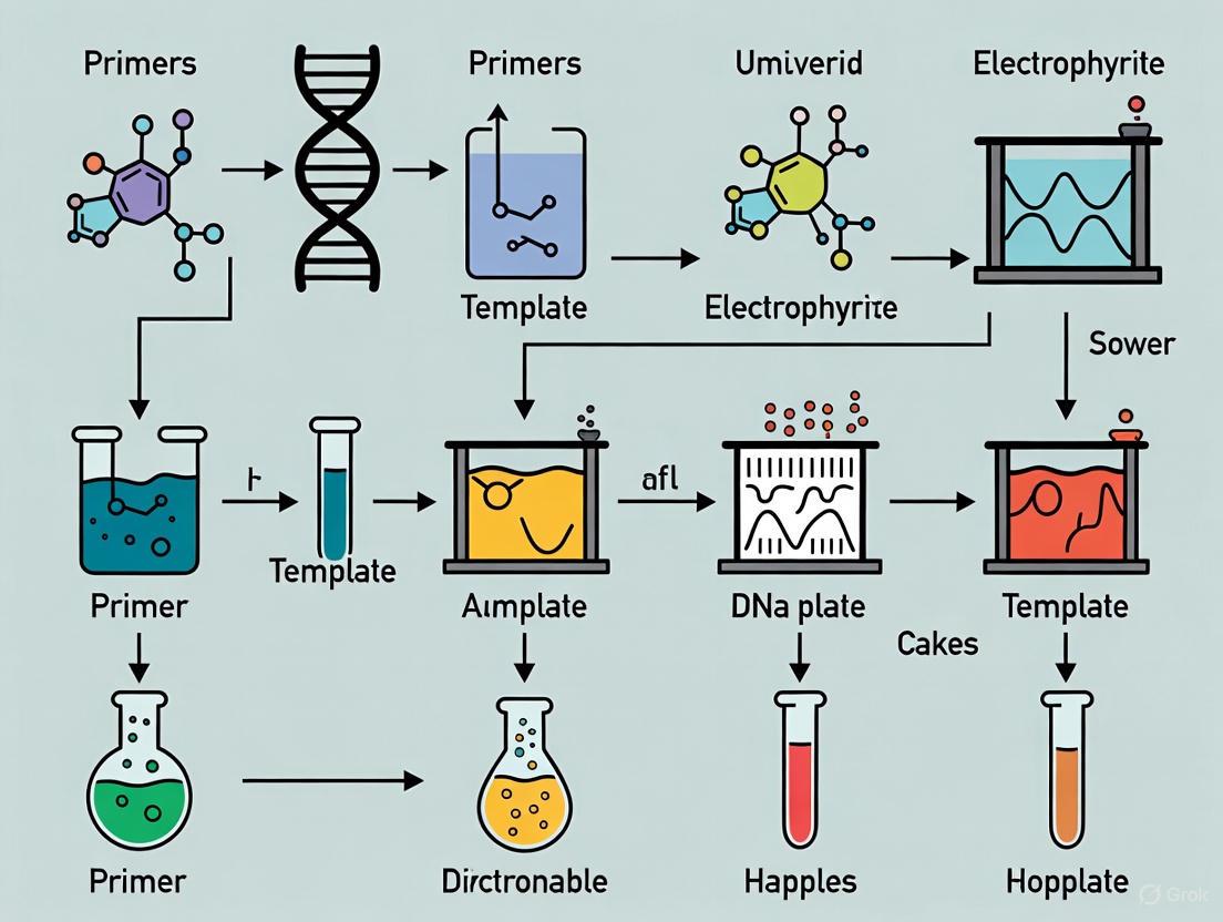
Workflow and Performance Diagram
The following diagram illustrates the experimental workflow and summarizes the performance outcomes of the culture-based viability PCR protocol compared to traditional methods.
Traditional culture and standard qPCR, while useful, present significant and complementary limitations for environmental monitoring. The integration of these methods into culture-based viability PCR offers a powerful alternative, combining the speed and sensitivity of qPCR with a reliable assessment of microbial viability. The provided protocol and experimental framework empower researchers to implement this robust strategy, thereby generating more accurate data for risk assessment and intervention in healthcare and other critical environments.
The accurate detection of viable pathogens is a cornerstone of effective environmental monitoring in public health and pharmaceutical development. Traditional culture methods, while reliable for confirming viability, are time-consuming and have a high detection threshold [1]. Quantitative PCR (qPCR) offers rapid and sensitive detection of pathogen DNA but cannot distinguish between live and dead cells, potentially leading to false-positive results [5] [6]. Culture-based viability PCR emerges as a powerful solution, harmonizing the sensitivity of qPCR with a reliable assessment of cellular viability. This protocol details the core principle of combining a broth enrichment step with quantitative PCR to determine the viability of pathogens, a method recently shown to outperform traditional culture techniques in hospital environmental monitoring [1]. The method is grounded in a simple principle: if a pathogen detected by qPCR at Time Zero (T0) is viable, it will proliferate during a subsequent broth enrichment phase, leading to a significant increase in nucleic acid concentration and a consequently lower qPCR cycle threshold (Ct) value at Time One (T1) [1].
Principle and Workflow
The core principle of this method is to use a broth enrichment phase to amplify viable target cells, enabling qPCR to not only detect their presence but also confirm their metabolic activity and ability to proliferate. A sample is first tested via qPCR to establish a baseline (T0). It is then incubated in an enrichment broth under conditions that support the growth of the target organism. Following incubation, the sample is tested again by qPCR (T1). A sample is confirmed viable if the target is undetected at T0 but detected at T1, or if the Ct value decreases significantly (e.g., by at least 1.0 cycle) from T0 to T1, indicating replication during enrichment [1]. This approach effectively circumvents the limitation of standard qPCR, which can detect persistent DNA from dead cells.
The workflow diagram below outlines this process:
Figure 1: Core workflow for culture-based viability PCR, integrating broth enrichment with qPCR analysis.
Key Research Reagent Solutions
The successful implementation of this protocol relies on several key reagents. The table below summarizes their critical functions.
Table 1: Essential research reagents for broth enrichment viability PCR.
| Item | Function/Description | Application Example |
|---|---|---|
| Non-Selective Broth (e.g., BPW) | Pre-enrichment medium for recovery of stressed/injured cells [7] [8]. | Pre-enrichment for Salmonella detection from food/environmental samples [7]. |
| Selective Enrichment Broth | Suppresses background flora, allowing target pathogen proliferation [7] [9]. | Selective enrichment in RV or MKTTn for Salmonella; Bolton Broth for Campylobacter [7] [10] [9]. |
| Viability Dyes (e.g., PMA, PMAxx) | Membrane-impermeant dyes that penetrate dead cells, bind DNA, and inhibit PCR amplification [5] [6]. | Differentiation of viable/non-viable Salmonella in poultry environmental samples [6]. |
| DNA Extraction Kits | Efficient isolation of high-quality DNA template for PCR. Silica-column-based methods are common [7] [6]. | DNeasy Tissue Kit for Salmonella; QIAamp DNA Mini Kit for soil samples [7] [6]. |
| qPCR Master Mix | Contains DNA polymerase, dNTPs, buffers, and salts. SYBR Green or TaqMan probe chemistry can be used [1] [11]. | Multiplex detection of C. jejuni and C. coli using probe-based chemistry [10] [9]. |
Experimental Protocols
Core Protocol: Culture-Based Viability PCR
This protocol is adapted from a recent hospital environmental study that detected viable E. coli, S. aureus, and C. difficile from patient room samples [1].
Sample Collection and Homogenization:
- Collect environmental samples (e.g., using swabs or sponges pre-moistened with a neutralizing buffer).
- Process samples via stomacher or vortexing to create a homogenate.
Baseline (T₀) Measurement:
- Transfer 500 µL of the sample homogenate into 4.5 mL of a non-selective broth like Trypticase Soy Broth (TSB).
- From this mixture, take a 500 µL aliquot for immediate DNA extraction.
- Perform DNA extraction using a validated kit (e.g., silica-column based).
- Run qPCR with species-specific primers and record the Cycle Threshold (Ct) value. This is the T₀ value.
Broth Enrichment:
- Transfer another 500 µL of the original sample homogenate into 4.5 mL of an appropriate enrichment broth (TSB).
- Incubate the broth under optimal conditions for the target pathogen (e.g., 24-48 hours at 37°C, aerobically for E. coli and S. aureus, anaerobically for C. difficile).
Post-Enrichment (T₁) Measurement:
- After incubation, take a 500 µL aliquot from the enriched culture.
- Perform DNA extraction and qPCR identically to the T₀ step. Record the Ct value as T₁.
Viability Assessment: A sample is considered viable for the target species if one of the following criteria is met:
- The pathogen is detected at T₁ but was undetected at T₀.
- The pathogen is detected at both time points, and the Ct value decreases by at least 1.0 at T₁ compared to T₀ [1].
Supporting Protocol: PMAxx-Based Viability qPCR
For samples with a high proportion of dead cells, combining enrichment with a viability dye can further reduce false positives. This protocol is optimized for Salmonella in soil [6].
Sample Preparation: Resuspend 1 g of soil in 9 mL of phosphate-buffered saline (PBS) and mix thoroughly.
PMAxx Treatment:
- Add 10 µL of PMAxx dye (from a 5-10 mM stock) to 1 mL of sample suspension.
- Incubate in the dark for 10 minutes with gentle mixing.
- Place the tube on ice and expose it to a 500-watt halogen light source at a 20 cm distance for 15 minutes with occasional mixing to photo-activate the dye.
DNA Extraction: Centrifuge the sample and extract DNA from the pellet using an optimized kit protocol (e.g., QIAamp DNA Mini Kit with a bead-beating step for soil samples) [6].
qPCR Quantification: Perform qPCR using validated primers (e.g., targeting the invA gene for Salmonella). The PMAxx dye will have suppressed DNA amplification from dead cells, allowing for quantification of the viable load.
Data Presentation and Analysis
Quantitative Results from Environmental Monitoring
A recent study applying the core culture-based viability PCR protocol demonstrated its superior sensitivity compared to traditional culture methods.
Table 2: Comparison of culture-based viability PCR and traditional culture for detecting pathogens from hospital footboards [1].
| Pathogen | Samples Detected via qPCR (T₀ or T₁) | Samples Confirmed Viable via Culture-Based Viability PCR | Samples Detected via Traditional Culture (Post-Enrichment) |
|---|---|---|---|
| E. coli | 24/26 (92%) | 3/26 (13%) | 0/26 (0%) |
| S. aureus | 11/26 (42%) | 8/26 (31%) | 5/26 (19%) |
| C. difficile | 2/26 (8%) | 0/26 (0%) | 0/26 (0%) |
Impact of Broth Type on PCR Efficiency
The choice of enrichment broth is critical, as some selective media can inhibit PCR amplification. The following table summarizes key findings from a study on Salmonella detection.
Table 3: Effect of enrichment broth type on real-time PCR detection of Salmonella [7].
| Enrichment Broth | Effect on Real-Time PCR (vs. BPW) | Recommended DNA Extraction Method to Overcome Inhibition |
|---|---|---|
| Buffered Peptone Water (BPW) | Minimal to no inhibition | Standard PrepMan Ultra Reagent method |
| Rappaport-Vassiliadis (RV) | Statistically significant (p<0.05) inhibition | PrepMan Ultra with additional wash step or DNeasy Tissue Kit |
| Muller-Kauffmann Tetrathionate Novobiocin (MKTTn) | Statistically significant (p<0.05) inhibition | PrepMan Ultra with additional wash step or DNeasy Tissue Kit |
Technical Optimization and Troubleshooting
Enhancing Viability Dye Efficiency
The efficiency of viability dyes like PMA can be improved using membrane enhancers. Lactic acid (LA) pre-treatment has been shown to improve PMA penetration into dead Gram-negative cells without compromising live cell viability. A protocol for this involves incubating cell aliquots with 400 µL of 10 mM LA (pH 5–5.5) for 30 minutes before adding PMA, which significantly improves the selective detection of live E. coli in milk samples [5].
Ensuring qPCR Precision and Accuracy
High precision in qPCR is essential for reliably discriminating the Ct value changes that indicate viability.
- Replicates: Run technical triplicates for each sample to measure system precision and allow for outlier detection [12].
- Controls: Always include a negative control (no template) and a positive control (template of known concentration) [13] [11].
- Inhibition Monitoring: Use an Internal Amplification Control (IAC) within the qPCR reaction to detect the presence of PCR inhibitors, which is common in complex sample matrices [10].
The relationship between experimental components and data interpretation is summarized below:
Figure 2: Logical relationship between sample content, enrichment outcome, qPCR data, and final viability interpretation.
Key Applications in Healthcare, Food Safety, and Water Quality Monitoring
The accurate detection of viable pathogens is a critical challenge across environmental monitoring, food safety, and clinical diagnostics. Traditional culture methods, while specific, are slow and may miss viable but non-culturable (VBNC) organisms. Standard quantitative PCR (qPCR) offers speed and sensitivity but cannot distinguish between live and dead cells, as it detects persistent genetic material from both [1] [14]. Culture-based viability PCR and viability dye-PCR have emerged as powerful molecular solutions to this problem, bridging the gap between traditional microbiology and modern molecular diagnostics.
Culture-based viability PCR involves running species-specific qPCR before and after a sample incubation period to determine if detected organisms can proliferate, thereby confirming viability [1]. Alternatively, viability dye-PCR (vPCR) uses photo-reactive dyes like propidium monoazide (PMA) that penetrate membrane-compromised (dead) cells and covalently bind DNA upon light exposure, preventing its amplification in subsequent PCR [15] [14]. This technical note details the protocols and applications of these methods across key sectors, providing a framework for their implementation in environmental monitoring research.
Performance Data and Comparative Analysis
The tables below summarize key performance metrics of viability PCR from recent studies, highlighting its advantages over traditional methods.
Table 1: Comparative Performance of Culture-Based Viability PCR vs. Traditional Culture in Healthcare Environmental Monitoring [1]
| Target Pathogen | Samples with Detectable DNA (qPCR) | Samples with Viable Pathogens (Culture-Based Viability PCR) | Samples with Viable Pathogens (Traditional Culture) |
|---|---|---|---|
| E. coli | 24/26 (92%) | 3/26 (13%) | 0/26 (0%) |
| S. aureus | 11/26 (42%) | 8/26 (73%) | 5/26 (19%)* |
| C. difficile | 2/26 (8%) | 0/26 (0%) | 0/26 (0%) |
| Note: *All culture-positive samples were also correctly identified as viable by culture-based viability PCR. |
Table 2: Efficacy of Optimized Viability PCR (vPCR) for *S. aureus in Food Matrices* [14]
| Food Matrix | Result with Optimized vPCR (Low Live/High Dead Cells) | Key Challenge |
|---|---|---|
| Ground Pepper, Oregano, Infant Milk Powder | Complete PCR signal suppression from dead cells; only live cells detected. | N/A |
| Ground Paprika, Allspice, Pork | PCR signals from dead cells reduced to near the detection limit. | Complete signal suppression in some matrices remains difficult. |
| General vPCR | Effectively detects low levels of live cells even with high background of dead cells. | Differentiation based solely on membrane integrity; not effective for UV-inactivated cells. |
Detailed Experimental Protocols
Protocol 1: Culture-Based Viability PCR for Healthcare Environments
This protocol is designed for monitoring bacterial pathogens on high-touch hospital surfaces [1].
Workflow Overview:
Materials & Reagents:
- Sampling: Foam sponges premoistened with neutralizing buffer.
- Growth Media: Trypticase Soy Broth (TSB).
- Viability Control: 8.25% sodium hypochlorite (bleach), Phosphate-Buffered Saline (PBS).
- DNA Extraction & Detection: Commercial DNA extraction kit, SYBR Green-based qPCR master mix, species-specific primers/probes for target pathogens.
Step-by-Step Procedure:
- Sample Collection & Processing: Collect surface samples using premoistened neutralizing sponges. Process using a stomacher to create a 5 mL homogenate [1].
- Sample Split: Aseptically divide the homogenate into three aliquots:
- T0 Sample: Combine 500 µL of homogenate with 4.5 mL of TSB. Immediately proceed to DNA extraction and qPCR.
- T1 Sample: Combine 500 µL of homogenate with 4.5 mL of TSB.
- Growth Negative Control (GNC): Combine 500 µL of homogenate with 4.5 mL of 8.25% sodium hypochlorite. Incubate at room temperature for 10 minutes to kill cells. Centrifuge at 3,100 RPM for 15 minutes, decant supernatant, and wash the pellet twice with PBS. Resuspend in 5 mL of TSB [1].
- Incubation: Incubate T1 and GNC samples under species-specific conditions (e.g., 24 hours at 37°C aerobically for E. coli and S. aureus; 48 hours anaerobically for C. difficile) [1].
- Post-Incubation Processing: After incubation, take 500 µL from T1 and GNC samples for DNA extraction and qPCR.
- Viability Analysis: A sample is considered viable for a species if one of the following is met:
- It is detected at T0, and the CT value decreases by at least 1.0 at T1 compared to the GNC.
- It is undetected at T0, detected at T1, and undetected for the GNC.
- It grows on standard culture agar (run in parallel) [1].
Protocol 2: Viability Dye-PCR (vPCR) for Foodborne Pathogens
This optimized protocol for Staphylococcus aureus detection in food minimizes false positives from dead cells [14].
Workflow Overview:
Materials & Reagents:
- Viability Dye: PMAxx (50 µM final concentration determined as optimal).
- Equipment: Microcentrifuge tubes, bright LED light source for photoactivation (e.g., PMA-Lite LED device).
- DNA Extraction & Detection: Commercial DNA extraction kit (e.g., NucleoSpin Food Kit), qPCR instrumentation, strain-specific primers and hydrolysis probes.
Step-by-Step Procedure:
- Sample Preparation: Prepare sample homogenate from artificially contaminated food matrix.
- Initial Dye Treatment: Add PMAxx to the sample for a final concentration of 50 µM. Mix thoroughly and incubate in the dark for 15-20 minutes [15] [14].
- Tube Change: Critical Step. Transfer the sample to a new, clean microcentrifuge tube. This minimizes the risk of amplifying DNA from dead cells that may be adsorbed to the walls of the original tube [14].
- Photoactivation: Place the tube on a cooled ice bath or a dedicated PMA device tray. Expose to bright visible light for 15-20 minutes to activate the dye.
- Double Treatment: Repeat steps 2-4 (dye treatment, tube change, photoactivation) to ensure complete suppression of DNA from dead cells [14].
- DNA Extraction and qPCR: Proceed with standard DNA extraction. Perform qPCR using strain-specific assays. The resulting signal originates exclusively from viable cells with intact membranes.
Application in Water Quality Monitoring
While the core principles remain consistent, water quality monitoring presents unique challenges in quantification across different methods and laboratories.
Key Consideration - Data Standardization: A major challenge in wastewater surveillance is the lack of comparability of quantitative viral RNA results obtained through different concentration and extraction methods. A proposed solution is the Data Standardization Test [16].
- Procedure: Identical, non-spiked, field-collected wastewater samples are distributed to multiple labs for analysis. Each lab uses its own standard operating procedures (SOPs). The quantification results (e.g., gene copies/L) are compared to identify systematic biases between methods [16].
- Outcome: This allows for the development of correction factors, enabling direct comparison of data generated by different laboratories and methods, which is crucial for large-scale public health monitoring [16].
Quantitative Metagenomics: For non-targeted monitoring of antibiotic resistance genes (ARGs) in wastewater, quantitative metagenomics can be used. This involves:
- Spiking Internal Standards: Adding known quantities of synthetic DNA standards (e.g., "meta sequins") to wastewater DNA extracts prior to shotgun sequencing [17].
- Absolute Quantification: The internal standards allow for the conversion of relative sequence read counts into absolute gene copy numbers per volume of wastewater, enabling the calculation of gene removal rates across treatment processes [17].
The Scientist's Toolkit: Research Reagent Solutions
Table 3: Essential Reagents and Kits for Viability PCR Applications
| Reagent / Kit | Function / Application | Examples / Specifications |
|---|---|---|
| Viability Dyes (PMA/PMAxx) | Selective DNA intercalation in dead cells; crucial for vPCR. | PMAxx; final concentration 50 µM for S. aureus [14]; 50 µM for Bifidobacterium [15]. |
| DNA Extraction Kits | High-quality DNA extraction from complex matrices. | NucleoSpin Food Kit (food samples) [15]; FastDNA Spin Kit for Soil (environmental/wastewater samples) [17]. |
| qPCR Master Mix | Sensitive DNA amplification and detection. | SYBR Green [1] or TaqMan probe-based mixes (e.g., SensiFast Probes Master Mix) [15]. |
| Species-Specific Primers/Probes | Ensures specific detection of target organism. | Designed for unique genomic regions; validated for specificity and efficiency [15]. |
| Growth Media | Enriches viable cells in culture-based viability PCR. | Trypticase Soy Broth (TSB) for bacterial enrichment [1]. |
| Synthetic DNA Standards | Enables absolute quantification in complex matrices. | "Meta sequins" for quantitative metagenomics in wastewater [17]. |
The integration of viability assessment with PCR detection provides researchers and quality control professionals with powerful tools for accurate pathogen monitoring. Culture-based viability PCR offers a robust method for confirming proliferating organisms in environmental samples, while viability dye-PCR allows for rapid and specific detection of membrane-intact cells, even in the presence of a high background of dead cells. The protocols outlined herein for healthcare, food safety, and water quality monitoring are adaptable to a wide range of target organisms and matrices, providing a solid foundation for enhancing the accuracy and relevance of environmental surveillance data.
The detection of viable but non-culturable (VBNC) cells represents a critical challenge in environmental monitoring, clinical microbiology, and food safety. The VBNC state is a survival strategy adopted by bacteria in response to sublethal environmental stresses, such as nutrient starvation, extreme temperatures, or exposure to antimicrobial agents and disinfectants [18] [19]. In this state, cells maintain metabolic activity and membrane integrity but lose the ability to form colonies on conventional growth media, the gold standard for microbial viability assessment [18]. This leads to a significant underestimation of viable pathogen counts, posing a hidden risk to public health as these cells retain virulence potential and can resuscitate when conditions become favorable [20] [21].
Overcoming the limitations of culture-based methods has driven the development of numerous advanced detection techniques. This document provides Application Notes and Protocols for these methods, contextualized within a broader research thesis. It is designed to equip researchers and drug development professionals with the tools to accurately detect and quantify VBNC cells, thereby enhancing the reliability of environmental monitoring data.
Comparative Analysis of VBNC Detection Methods
A variety of methods have been developed to detect VBNC cells, each with distinct principles, advantages, and limitations. The table below provides a structured comparison of these key techniques to aid in method selection.
Table 1: Comparison of Key Methodologies for VBNC Cell Detection
| Method Category | Specific Technique | Principle | Key Advantage | Primary Limitation |
|---|---|---|---|---|
| Nucleic Acid Staining & Flow Cytometry | Live/Dead Staining (e.g., SYTO 9/PI) | Differential membrane integrity; viable cells stain green, dead cells stain red [22]. | Rapid, provides cell count and viability status. | Can overestimate viable cells in complex matrices; cannot confirm potential for resuscitation [22]. |
| Viability Molecular Methods | DyeTox13 + EMA v-qPCR/v-ddPCR | DyeTox13 indicates metabolic activity (esterase); EMA indicates membrane integrity; both inhibit DNA amplification from non-viable sources [20]. | Rapidly quantifies viable cells; higher sensitivity and resistance to inhibitors than qPCR [20]. | Complex optimization for dye concentration and sample matrix is required [20] [22]. |
| PMAxx-qPCR | PMAxx penetrates cells with compromised membranes, binding DNA and preventing its amplification in PCR [22] [21]. | Effectively excludes signal from dead cells and free DNA; more reliable than EMA alone. | May fail to detect VBNC cells with minor membrane damage; requires photoactivation step [21]. | |
| Advanced Imaging & AI | AI-Enabled Hyperspectral Imaging | Captures unique spectral profiles of VBNC cells; AI (e.g., EfficientNetV2) classifies these profiles with high accuracy [23]. | Label-free, rapid, and highly accurate (e.g., 97.1%); provides spatial and chemical data. | Requires expensive, specialized equipment and complex data analysis models [23]. |
| Culture-Based with Supplementation | Ferrioxamine E Supplementation | A siderophore that provides essential iron (III), reactivating sub-lethally damaged cells and reducing lag phase [18]. | Can resuscitate VBNC cells, making them culturable; integrates with standard plating. | Not all species respond; may only recover a subpopulation of VBNC cells [18]. |
Detailed Experimental Protocols
This section provides step-by-step protocols for two of the most impactful and contemporary methods for VBNC detection and analysis.
Protocol: Viability Droplet Digital PCR (v-ddPCR) with DNA-Intercalating Dyes
This protocol, adapted from recent research, uses DyeTox13 and EMA in conjunction with ddPCR to quantitatively distinguish viable Salmonella cells, including those in the VBNC state, in a complex matrix like flour [20].
Table 2: Key Research Reagent Solutions for v-ddPCR
| Reagent/Material | Function/Explanation |
|---|---|
| DyeTox13 Green C-2 Azide | A cell-permeant dye converted by active esterases in viable cells, becoming DNA-binding and inhibiting PCR amplification in dead cells after photoactivation [20]. |
| Ethidium Monoazide (EMA) | A membrane-impermeant dye that enters only dead cells with compromised membranes, binding to DNA and suppressing its PCR amplification [20]. |
| Propidium Monoazide (PMAxx) | An improved version of PMA; more effective at penetrating dead cells with compromised membranes and covalently binding to DNA upon light exposure, preventing its amplification [22]. |
| Droplet Digital PCR (ddPCR) System | Partitions a PCR reaction into thousands of nanoliter-sized droplets, allowing for absolute quantification of target DNA molecules without a standard curve and with high resistance to inhibitors [20]. |
| PMA-Lite LED Photolysis Device | A high-power LED light source used to photoactivate PMAxx, EMA, and DyeTox13, causing them to bind covalently to DNA. |
Workflow Overview:
Procedure:
- Sample Preparation and Stress Induction: Prepare a pure culture of the target bacterium (e.g., Salmonella Typhimurium ATCC 14028) and subject it to a sublethal stressor. For example, expose cells in PBS to UV light (e.g., 33-99 mJ/cm²) or pasteurization (63°C for 30 min) to induce the VBNC state [20].
- Confirm Loss of Culturability: Plate the stressed suspension on non-selective rich media (e.g., Tryptic Soy Agar). The successful induction of the VBNC state is confirmed by a significant reduction (>99.9%) or absence of colony-forming units (CFU), while control cells grow normally [20].
- Dye Treatment: a. To 500 µL of sample, add DyeTox13 to a final concentration of 50 µM and EMA to a final concentration of 25 µM [20]. b. Vortex thoroughly and incubate the mixture in the dark at room temperature for 10 minutes.
- Photoactivation: Transfer the tubes to a PMA-Lite LED Photolysis device and expose for 15 minutes to crosslink the dyes to DNA from non-viable cells. Keep samples on ice during this step to prevent DNA degradation.
- DNA Extraction: Pellet the cells by centrifugation (e.g., 5,000 × g for 10 min). Proceed with genomic DNA extraction from the pellet using a commercial kit suitable for bacterial cells.
- ddPCR Setup and Run: Prepare the ddPCR reaction mix according to the manufacturer's instructions using primers and probes specific to your target organism. Generate droplets, perform PCR amplification, and read the droplets on the ddPCR droplet reader.
- Data Analysis: The concentration (copies/µL) provided by the ddPCR software represents the quantity of viable cells (with intact membranes and metabolic activity). The difference between this number and the near-zero CFU count quantifies the VBNC population.
Protocol: AI-Enabled Hyperspectral Microscopy for VBNC Classification
This protocol outlines the use of hyperspectral imaging combined with deep learning to identify VBNC cells based on their unique spectral fingerprints, without the need for dyes [23].
Workflow Overview:
Procedure:
- VBNC Induction: Induce the VBNC state in your target bacterium (e.g., Escherichia coli K-12) by exposure to low-level antimicrobial stressors. An example is incubation with 0.01% hydrogen peroxide or 0.001% peracetic acid for 3 days. Confirm the VBNC state using live/dead staining (e.g., BacLight) alongside the absence of growth on culture plates [23].
- Hyperspectral Image Acquisition: Place a sample of the cell suspension on a microscope slide. Use a hyperspectral microscope to capture images, collecting data across a range of wavelengths for each pixel. This creates a detailed spectral profile for individual cells [23].
- Data Preprocessing and Pseudo-RGB Generation: Extract spatial and spectral data from the images. To simplify data for the neural network, create "pseudo-RGB" images by selecting and combining three characteristic spectral wavelengths that best differentiate VBNC cells from their culturable counterparts [23].
- Deep Learning Model Training: Train a convolutional neural network (CNN), such as EfficientNetV2, on a dataset of labeled pseudo-RGB images. The dataset should contain known "Normal" and "VBNC" cells. The model learns to recognize the subtle spectral patterns associated with the VBNC state [23].
- Classification of Unknown Cells: Apply the trained model to classify new, unlabeled hyperspectral image data. The model outputs a classification (e.g., Normal or VBNC) for each cell in the field of view, achieving high accuracy (e.g., 97.1%) [23].
Application in Environmental Monitoring & Discussion
The protocols described herein are particularly vital for assessing the true efficacy of disinfection processes in water treatment and food production. Traditional culture methods can be misleading. For instance, one study demonstrated that UV radiation, sodium hypochlorite (NaClO), and peracetic acid (PAA) disinfection, while reducing culturability of Pseudomonas aeruginosa by >99.9%, primarily induced the VBNC state rather than achieving true killing [21]. These VBNC cells retained metabolic activity, as indicated by high intracellular ATP levels, and could resuscitate, with UV-induced cells resuscitating faster than those induced by NaClO [21]. This underscores a significant "hidden" risk in systems declared safe by culture-based standards.
The choice of method depends on the research goal. For rapid, quantitative detection of viable cells in complex environmental samples, v-ddPCR with DNA-intercalating dyes offers high sensitivity and robustness [20]. Conversely, AI-enabled hyperspectral microscopy provides a powerful, label-free alternative for fundamental studies of VBNC physiology and morphology, albeit with higher equipment costs [23]. Integrating these advanced methods into environmental monitoring protocols is crucial for a more accurate risk assessment and for developing strategies to effectively eliminate or control this resilient subpopulation of bacteria.
Step-by-Step Protocol: Implementing Culture-Based Viability PCR for Environmental Samples
Sample Collection and Processing from Healthcare and Environmental Matrices
Within environmental monitoring research, the accuracy of data is fundamentally dependent on the integrity of the initial sample collection and processing phases. This document provides detailed application notes and protocols for the collection of samples from key healthcare and environmental matrices, framed specifically for use in advanced molecular techniques such as culture-based viability PCR. This method addresses a critical gap in environmental monitoring by combining the sensitivity of quantitative PCR (qPCR) with the ability to confirm cellular viability, which is crucial for accurate risk assessment [1]. The procedures outlined herein are designed to enable researchers to reliably capture and process samples that reflect the true state of microbial contamination in a given environment.
The Rationale for Matrix Selection
A well-informed choice of sample matrix is critical, as different matrices offer unique advantages for studying various aspects of environmental contamination [24]. The selection criteria often involve a balance between ethical considerations, analytical goals, and practical constraints.
Advantages of Non-Invasively Collected Matrices
The use of non-invasively collected matrices is strongly promoted as an ethically appropriate and cost-efficient alternative for many biomarkers [24]. Key advantages include:
- Ethical Compliance: Sampling is preferable for vulnerable groups (e.g., children, pregnant women) and facilitates higher participation rates in studies [24].
- Cost-Efficiency: Less specialized personnel are required for collection, significantly reducing costs for large-scale surveys [24].
- Toxicological Relevance: Certain matrices, like urine, are a main excretory pathway and are preferred for monitoring non-bioaccumulating and rapidly metabolized compounds [24].
- Opportunity for Repeated Sampling: Matrices such as urine and hair allow for repeated or routine sampling, which is essential for monitoring substances with short biological half-lives or for evaluating the efficacy of risk management interventions over time [24].
The table below summarizes the primary applications and considerations for common matrices.
Table 1: Key Matrices for Biomonitoring and Environmental Sampling
| Matrix | Primary Applications | Key Advantages | Key Limitations |
|---|---|---|---|
| Urine [24] | Biomarkers for non-persistent chemicals, metals, and metabolites. | Large volumes can be collected; suitable for repeated sampling; no associated risk. | Requires standardization (e.g., via creatinine); spot sample variability. |
| Hair [24] | Historical exposure to metals (e.g., mercury), organic pollutants. | Provides a long-term record of exposure (approx. 1 cm/month). | Difficult to distinguish internal from external contamination; affected by cosmetic treatments. |
| Surface Wipes (e.g., Foam Sponges) [1] | Detection of surface contamination in healthcare (e.g., S. aureus, E. coli) and other built environments. | Directly measures exposure risk from fomites; practical for large-scale environmental sampling. | Recovery efficiency can vary; may not represent the entire microbial community. |
| Exhaled Breath [24] | Volatile organic compounds (VOCs), biomarkers of respiratory exposure. | Direct measurement at the target organ (lungs); non-invasive. | Requires specialized collection systems; concentration can be influenced by various factors. |
| Human Milk [24] | Monitoring persistent, bio-accumulating toxicants (PBTs). | Represents a major exposure pathway for infants; enriched in lipophilic compounds. | Only applicable to a specific sub-population (lactating women). |
Sample Collection Protocols
Standardized protocols are essential to ensure that sample collection yields high-quality, representative data. The following protocols are adapted from established environmental and biomedical sampling methodologies.
Protocol A: Surface Sampling in Healthcare Environments
This protocol is designed for the collection of microorganisms from high-touch surfaces in healthcare settings for subsequent culture-based viability PCR analysis [1].
1. Materials and Equipment
- Foam sponges pre-moistened with a neutralizing buffer (e.g., Dey-Engley broth)
- Sterile template (e.g., 10x10 cm) to define sampling area
- Sterile gloves
- Cooler with ice packs or dry ice for sample transport
- Permanent marker for labeling
2. Step-by-Step Procedure 1. Don sterile gloves to prevent cross-contamination. 2. Define the sampling area by placing the sterile template on the surface to be sampled (e.g., patient bed footboard). 3. Vigorously wipe the entire area inside the template with the pre-moistened sponge, using a consistent back-and-forth motion. Rotate the sponge to use all sides. 4. Aseptically place the used sponge into a sterile, labeled whirl-pack or stomacher bag. 5. Store samples immediately on ice or dry ice and transport to the laboratory for processing within 24 hours.
Protocol B: Collection and Preservation of Urine Samples
Urine is a preferred matrix for biomarkers of short-term exposure [24].
1. Materials and Equipment
- Sterile, wide-mouth polypropylene containers
- Cold-resistant sample transport kits
- Permanent marker for labeling
2. Step-by-Step Procedure 1. Provide participant with a sterile collection container. 2. Collect a first-morning void spot urine sample, as this typically has the highest concentration of analytes. 3. Label the container immediately with a unique sample ID, date, and time of collection. 4. Preserve samples by freezing at -20°C or -80°C if analysis is not performed immediately. For metabolically active organisms, process immediately for viability testing. 5. Standardize measurements during analysis by correcting for dilution using creatinine concentration or specific gravity [24].
Sample Processing for Culture-Based Viability PCR
Culture-based viability PCR is a two-step method that involves incubating a sample in growth media before using species-specific qPCR to determine if detected genetic material originates from viable, proliferating cells [1]. The workflow below outlines the core process.
Detailed Experimental Protocol
This protocol is adapted from a prospective microbiological analysis of healthcare surface samples [1].
1. Sample Homogenization and Splitting
- Process the collected sample (e.g., a sponge from surface sampling) via a stomacher or pulsifier method to create a 5 mL homogenate [1].
- Aseptically split the homogenate into three distinct processing paths:
- T0 Sample: 500 µL is added to 4.5 mL of Trypticase Soy Broth (TSB). 500 µL of this mixture immediately undergoes DNA extraction and qPCR.
- T1 Sample: 500 µL is added to 4.5 mL of TSB and incubated under species-specific conditions (e.g., 24 hours at 37°C aerobically for E. coli and S. aureus; 48 hours anaerobically for C. difficile).
- Growth Negative Control (GNC): 500 µL is added to 4.5 mL of 8.25% sodium hypochlorite, left at room temperature for 10 minutes, centrifuged, decanted, and washed with PBS before being resuspended in 5 mL of TSB. This step ensures no viable cells remain, controlling for non-viable DNA signal [1].
2. DNA Extraction and Quantitative PCR 1. Extract DNA from 500 µL of the T1 and GNC samples after incubation, using a commercial DNA extraction kit suitable for bacterial cells and spores. 2. Perform qPCR using species-specific primers and probes (e.g., for E. coli, S. aureus, C. difficile) [1]. 3. Run all qPCR assays in triplicate using a master mix like SYBR Green or TaqMan, following manufacturer guidelines [1]. 4. Record the average cycle threshold (CT) values for each sample path.
3. Criteria for Determining Viability A sample is considered viable for a target species if it meets any of the following criteria [1]: 1. It is detected at T0, and the CT value decreases by at least 1.0 at T1 compared to the GNC (indicating growth during incubation). 2. It is undetected at T0 but is detected at T1 and is undetected for the GNC (indicating growth of organisms below the initial detection limit). 3. It yields growth on standard culture agar, though this method is less sensitive.
Table 2: Example qPCR Results from a Healthcare Environment Study (n=26 rooms) [1]
| Target Organism | Detected via qPCR (T0 or T1) | Determined Viable via \nCulture-Based Viability PCR | Detected via Traditional Culture |
|---|---|---|---|
| E. coli | 24 (92%) | 3 (13%) | 0 (0%) |
| S. aureus | 11 (42%) | 8 (73%) | 5 (19%) |
| C. difficile | 2 (8%) | 0 (0%) | 0 (0%) |
This data highlights the superior sensitivity of culture-based viability PCR, which can detect viable pathogens that traditional culture methods often miss [1].
The Scientist's Toolkit: Essential Research Reagents and Materials
The following table details key reagents and their critical functions in the culture-based viability PCR workflow.
Table 3: Essential Research Reagent Solutions for Culture-Based Viability PCR
| Reagent/Material | Function/Application |
|---|---|
| Neutralizing Buffer | Inactivates disinfectants and antimicrobial agents present on sampled surfaces, ensuring accurate microbial recovery [1]. |
| Trypticase Soy Broth (TSB) | A general-purpose liquid growth medium used to enrich and resuscitate viable microorganisms from the sample during the incubation (T1) step [1]. |
| Species-Specific Primers/Probes | Short, single-stranded DNA sequences designed to bind to unique genetic regions of the target organism, enabling specific detection and quantification in qPCR [1]. |
| SYBR Green or TaqMan Master Mix | Fluorescent dyes or probes used in qPCR to monitor the amplification of target DNA in real-time, allowing for quantification of the initial amount of genetic material [1]. |
| Sodium Hypochlorite Solution | Used in the Growth Negative Control (GNC) to kill all viable cells, ensuring that any qPCR signal from the GNC is from non-viable DNA and not from cell proliferation [1]. |
Within environmental monitoring research, the efficacy of culture-based viability PCR is fundamentally dependent on the initial broth enrichment phase. This protocol details optimized pre-analytical strategies for the selective enrichment of key bacterial pathogens (Salmonella, Listeria monocytogenes, and Campylobacter) and the fungus Candida auris. By integrating selective media, precise incubation conditions, and neutralization strategies, this guide ensures the robust proliferation of target organisms while suppressing background flora, thereby providing high-quality template for subsequent molecular detection. This application note provides a standardized framework for researchers and drug development professionals to enhance the sensitivity and specificity of their viability PCR assays.
The detection of viable pathogens in environmental samples is a critical challenge in pharmaceutical and food manufacturing environments. Culture-based viability PCR combines the selectivity of enrichment culture with the speed and specificity of molecular detection. The success of this method hinges on the broth enrichment strategy, which must accomplish two primary objectives: first, to resuscitate and promote the growth of stressed or low numbers of target cells, and second, to inhibit competing non-target microorganisms that can cause false-negative results or inhibit downstream PCR. This document synthesizes current research to provide detailed protocols for enriching major pathogens, with a focus on practical application within a research and development context.
Pathogen-Specific Enrichment Conditions
Optimized enrichment conditions vary significantly by pathogen due to differences in growth kinetics, stress tolerance, and susceptibility to selective agents. The following section and table summarize the key media and incubation parameters for major targets.
Table 1: Optimized Enrichment Conditions for Key Pathogens
| Pathogen | Recommended Media | Incubation Temperature | Incubation Time | Atmosphere | Key Enhancements |
|---|---|---|---|---|---|
| Salmonella | Tetrathionate Broth, Selenite Broth [25] | 43°C [25] | 24-48 hours [25] | Aerobic or Anaerobic [25] | Secondary enrichment in a different medium (e.g., Tetrathionate) is statistically advantageous [25]. |
| Campylobacter | Hunt Broth, Double Strength Blood-Free Bolton's Enrichment Broth (2x BF-BEB) [26] | 37-42°C (Broth-specific) | 24-48 hours (Broth-specific) | Microaerophilic | Bolton broth supplemented with 12.5 mg/L rifampin (R-Bolton) inhibits non-Campylobacter flora [27]. |
| Listeria monocytogenes | Listeria Enrichment Broth (e.g., Fraser, UVM) [28] [29] | 35-37°C [28] | 24-48 hours [28] | Aerobic | Formulations often include acriflavine and nalidixic acid as selective inhibitors [28]. |
| Candida auris | CABroth (Modified Sabouraud) [30] | 35-37°C (Ambient storage) [30] | Up to 48 hours [30] | Aerobic | Selectively inhibits other Candida species and commensal bacteria [30]. |
Special Considerations for Inhibitory Matrices
Environmental samples, particularly low-water-activity spices, contain antimicrobial compounds that can induce a viable but non-culturable (VBNC) state in pathogens, eluding culture-based detection [31]. For such matrices, standard enrichment protocols require modification.
- Neutralizer-Aided Enrichment: For detecting Salmonella in garlic granules, a modified Buffered Peptone Water (mBPW) containing DL-dithiothreitol (DTT) and corn oil has proven effective in neutralizing antimicrobial substances, enabling detection at levels as low as 5 CFU/g [31].
- Enhanced Buffering and Recovery: The same mBPW formulation uses 3× buffered peptone water for increased buffering capacity, plus magnesium sulfate and sodium pyruvate to promote bacterial self-repair [31].
Integrated Experimental Protocols
General Workflow for Culture-Based Viability PCR
The following diagram illustrates the overarching workflow integrating broth enrichment with subsequent viability PCR, crucial for environmental monitoring research.
Detailed Protocol: Neutralizer-Aided Enrichment for Salmonella in Inhibitory Matrices
This protocol, adapted from a 2025 study on garlic granules, is designed for detecting low levels of viable Salmonella in challenging matrices and is compatible with PMAxx-qPCR [31].
Objective: To enrich viable Salmonella cells from a dry, antimicrobial environmental sample (e.g., a spice or powder residue) for subsequent culture-based viability PCR. Materials:
- Modified Buffered Peptone Water (mBPW): 3× concentration of buffered peptone water, supplemented with MgSO₄, sodium pyruvate, DL-dithiothreitol (DTT), and corn oil [31].
- Sample: 25g of environmental material (e.g., collected via a sterile sponge).
- Incubator (35-37°C).
Procedure:
- Sample Inoculation: Aseptically transfer the 25g environmental sample into 225 mL of sterile mBPW containing neutralizers. This creates a 1:10 dilution.
- Pre-enrichment Incubation: Incubate the inoculated broth at 35-37°C for 18-24 hours. This critical step allows for the resuscitation of stressed or injured Salmonella cells.
- Secondary Selective Enrichment (Optional): For increased selectivity, transfer 1 mL of the pre-enriched culture into 9 mL of secondary enrichment broth, such as Tetrathionate Broth. Incubate at 43°C for 24 hours [25].
- Downstream Processing: Proceed to the viability PCR workflow. For PMAxx-qPCR, the entire process from enrichment to result can be completed in approximately 26 hours [31].
Table 2: Research Reagent Solutions for Viability PCR Enrichment
| Reagent / Solution | Function / Application | Example Formulations / Notes |
|---|---|---|
| Tetrathionate Broth | Selective enrichment of Salmonella; inhibits Gram-positive and many Gram-negative bacteria. | Statistically superior to selenite broth for secondary enrichment [25]. |
| Hunt Broth / 2x BF-BEB | Selective enrichment of Campylobacter spp. from complex samples. | Demonstrates high sensitivity (97%) and specificity (96.8%) on poultry products [26]. |
| R-Bolton Broth | Bolton broth with rifampin; suppresses non-Campylobacter flora. | Supplementation with 12.5 mg/L rifampin markedly inhibits competing microbes [27]. |
| Listeria Enrichment Broth | Selective for Listeria monocytogenes; used in dairy, meat, and RTE food environments. | Often contains acriflavine and nalidixic acid. Vital for environmental monitoring in facilities [28] [29]. |
| CABroth | Selective enrichment for Candida auris from clinical or environmental specimens. | Modified Sabouraud broth; inhibits other Candida species and bacteria [30]. |
| Modified BPW with Neutralizers | Enrichment for Salmonella in inhibitory matrices (e.g., spices). | Contains DTT and corn oil to neutralize antimicrobials, and additives to promote bacterial repair [31]. |
| PMAxx Dye | Viability PCR reagent; penetrates only dead cells, binding DNA and preventing its amplification in qPCR. | Critical for distinguishing viable cells in culture-based viability PCR [31]. |
Environmental Sampling and Zone Management
Effective enrichment begins with proper sample collection. A structured environmental monitoring program (EMP) using a zone concept is essential for identifying sampling sites and interpreting results [32].
- Zone 1: Direct product contact surfaces (e.g., conveyor belts, filler nozzles). Testing here is high-risk but crucial.
- Zone 2: Non-product contact surfaces close to Zone 1 (e.g., equipment frames, control panels). Key for detecting potential contamination vectors.
- Zone 3: Non-product contact surfaces further from the product area (e.g., floors, walls, drains). Often a reservoir for pathogens.
- Zone 4: Support areas outside the processing area (e.g., locker rooms, warehouses) [32].
Samples from these zones, collected with aseptic techniques using sponges or swabs with neutralizing transport buffers (e.g., Letheen broth, D/E broth), provide the input material for the enrichment protocols described herein [32].
A meticulously designed broth enrichment strategy is the cornerstone of a sensitive and reliable culture-based viability PCR assay for environmental monitoring. The pathogen-specific and matrix-specific protocols outlined in this document provide a critical foundation for researchers to obtain meaningful and actionable results. By carefully selecting enrichment media, fine-tuning incubation conditions, and employing neutralizers for challenging samples, scientists can ensure that the template entering the molecular workflow truly represents the viable pathogen load in the environment, thereby strengthening the overall validity of their research and drug development processes.
In environmental monitoring research, the accuracy of culture-based viability PCR—a method that combines the sensitivity of qPCR with the ability to confirm cell viability through pre-incubation [1]—is critically dependent on the quality of the extracted nucleic acids. Environmental samples, from healthcare settings to wastewater, contain a complex mixture of substances that can inhibit downstream PCR amplification, leading to false-negative results or an underestimation of microbial loads [33] [34]. These inhibitors, including humic acids, polysaccharides, and proteins, interfere with polymerase activity, primer binding, and fluorescent signal detection [33]. Overcoming these inhibitors through optimized nucleic acid extraction and purification is therefore not merely a preliminary step but a foundational requirement for generating reliable data in environmental surveillance and diagnostic protocols.
Common PCR Inhibitors and Their Effects
The highly heterogeneous nature of environmental samples introduces a wide array of substances that can compromise PCR efficiency. The table below catalyses common inhibitors, their sources, and their specific mechanisms of action.
Table 1: Common PCR Inhibitors Found in Environmental Samples
| Source | Example Inhibitors | Primary Effect on PCR |
|---|---|---|
| Biological Samples | Hemoglobin, heparin, immunoglobulins, polysaccharides [33] [34] | Inhibition of DNA polymerase activity; chelation of essential co-factors like Mg²⁺ [34]. |
| Environmental Matrices | Humic and fulvic acids (soil, water), phenols, tannins [33] [34] | Binding to nucleic acids, making them inaccessible; degradation of DNA; interference with fluorescent signaling [33]. |
| Laboratory Reagents | Ethanol, SDS (Sodium Dodecyl Sulfate), salts from extraction kits [34] | Disruption of primer binding to the template; precipitation of nucleic acids [34]. |
| Complex Polysaccharides | Collagen, glycogen, complex carbs from plants/foods [33] | Can interact with templates or chelate metal ions essential for amplification [33]. |
The effects of these inhibitors manifest in several ways during qPCR. Key indicators of inhibition include:
- Delayed Cq Values: A general increase in quantification cycle (Cq) values across samples and controls suggests a systemic reduction in amplification efficiency [34].
- Poor Amplification Efficiency: Assay efficiency falling outside the optimal 90–110% range (corresponding to a standard curve slope between -3.1 and -3.6) indicates polymerase function or primer binding is compromised [34].
- Abnormal Amplification Curves: Flattened curves, a lack of clear exponential growth phase, or a failure to cross the detection threshold are visual hallmarks of inhibition [34].
Strategies for Inhibitor Removal and Mitigation
A multi-faceted approach is essential for reliable nucleic acid purification from challenging environmental samples. The following strategies can be employed individually or in combination.
Sample Purification and Pre-Treatment
Enhancing the sample preparation phase is the first line of defense against PCR inhibitors.
- High-Quality Extraction Kits: Use kits specifically validated for complex environmental samples like soil, wastewater, or stool, as they are designed to remove common inhibitors [34].
- Alternative Purification Methods: A novel precipitation-based method utilizes chaotropic salts combined with isopropanol or polyethylene glycol (PEG) to simultaneously denature proteins and precipitate nucleic acids in a single step. This method is fast, versatile, and avoids the issue of unidentified inhibitory substances that can be eluted from some commercial silica columns [35].
- Dilution: A simple 10-fold dilution of the extracted nucleic acid can dilute inhibitor concentrations to sub-critical levels. However, this also dilutes the target DNA, potentially reducing sensitivity below the limit of detection, especially for low-abundance targets [33].
PCR Reaction Optimization
When inhibitors persist despite purification, optimizing the reaction mixture itself can rescue the assay.
- PCR Enhancers: Adding compounds that counteract specific inhibitors can dramatically improve results. Their effectiveness is concentration-dependent, as summarized below.
Table 2: Evaluation of Common PCR Enhancers for Overcoming Inhibition
| Enhancer | Reported Mechanism of Action | Effect on Cq Value (Example) |
|---|---|---|
| Bovine Serum Albumin (BSA) | Binds to inhibitors like humic acids, preventing them from interfering with the polymerase [33]. | Reduction of ~2.5 cycles [33]. |
| T4 Gene 32 Protein (gp32) | Stabilizes single-stranded DNA and binds humic acids [33]. | Reduction of ~1 cycle [33]. |
| Dimethyl Sulfoxide (DMSO) | Lowers the melting temperature (Tm) of DNA and destabilizes secondary structures [33]. | Reduction of ~0.5 cycles [33]. |
| Formamide | Acts as a helix destabilizer, similar to DMSO [33]. | Reduction of ~0.5 cycles [33]. |
| TWEEN 20 | A non-ionic detergent that counteracts inhibitory effects on Taq DNA polymerase [33]. | Reduction of ~2 cycles [33]. |
| Glycerol | Protects enzymes from degradation and denaturation, improving efficiency [33]. | Reduction of ~1 cycle [33]. |
- Inhibitor-Resistant Master Mixes: Formulations like GoTaq Endure qPCR Master Mix are specifically engineered with inhibitor-tolerant polymerases and optimized buffers to deliver consistent performance in the presence of common inhibitors found in blood, soil, and plant materials [34].
- Magnesium Adjustment: Increasing the concentration of MgCl₂ in the reaction can counteract the effect of chelators like heparin or EDTA that sequester Mg²⁺, an ion essential for polymerase activity [34].
Alternative Detection Technologies
- Digital PCR (dPCR): Techniques like droplet digital PCR (ddPCR) partition a single reaction into thousands of nanoreactions. This effectively dilutes inhibitors, making the assay more tolerant to interfering substances compared to qPCR, albeit at a higher cost and with longer processing times [33].
Integrated Experimental Protocol for Inhibitor-Free Culture-Based Viability PCR
This protocol is designed for processing environmental surface samples (e.g., from healthcare settings) to detect viable bacterial pathogens (E. coli, S. aureus, C. difficile) while mitigating PCR inhibition [1].
Sample Collection and Initial Processing
- Collection: Sample surfaces using foam sponges pre-moistened in a neutralizing buffer [1].
- Homogenization: Process sponges using a stomacher, resulting in a 5 mL homogenate [1].
- Aliquoting: Split the homogenate into three paths:
- T0 Sample: 500 µL added to 4.5 mL of Trypticase Soy Broth (TSB). This aliquot undergoes immediate DNA extraction and qPCR to establish a baseline [1].
- T1 Sample: 500 µL added to 4.5 mL of TSB for incubation [1].
- Growth Negative Control (GNC): 500 µL added to 4.5 mL of 8.25% sodium hypochlorite. After 10 minutes at room temperature, centrifuge for 15 minutes, decant, wash the pellet with PBS, and resuspend in 5 mL TSB [1].
Incubation and Viability Assessment
- Incubation: Incubate T1 and GNC samples at species-specific conditions (e.g., 24-48 hours at 37°C, aerobically for E. coli and S. aureus, anaerobically for C. difficile) [1].
- Post-Incubation Processing: After incubation, take 500 µL from T1 and GNC samples for DNA extraction and qPCR analysis [1].
- Viability Determination: A sample is considered viable for a target species if one of the following is met [1]:
- It is detected at T0, and the Cq value decreases by at least 1.0 at T1 compared to the GNC.
- It is undetected at T0 but detected at T1, and is undetected in the GNC.
- It grows on standard culture agar.
Inhibitor-Resistant Nucleic Acid Purification and qPCR
This critical step can be performed using either a commercial kit or a novel precipitation method.
Option A: Silica Column-Based Purification with Enhanced Wash
- Lysis: Add a lysis buffer containing a high concentration of chaotropic salts to the 500 µL sample.
- Binding: Bind nucleic acids to a silica column.
- Enhanced Washing: Perform two wash steps. The second wash should include a reagent like 1-undecanol to more thoroughly remove residual inhibitors, though this may not eliminate all inhibition [35].
- Elution: Elute DNA in a low-EDTA TE buffer or nuclease-free water. Note: Eluents from silica columns may contain unidentified inhibitory substances; therefore, testing different dilutions of the eluted DNA in the downstream qPCR is recommended [35].
Option B: Novel Single-Step Precipitation Protocol [35]
- Simultaneous Protein Denaturation & DNA Precipitation: To the 500 µL sample, add a volume of a solution containing a optimized concentration of a chaotropic salt (e.g., Guanidine Hydrochloride) mixed with a precipitating agent (Isopropanol or PEG). Mix gently. This single step inactivates proteins and precipitates nucleic acids.
- Protein Dilution: Add a protein dilution solution to further eliminate any residual proteinase activity.
- Centrifugation: Centrifuge at 4°C to pellet DNA. Gently mixing and cooling facilitates DNA renaturation and improves recovery [35].
- Wash and Resuspension: Wash the pellet with 70% ethanol to remove chaotropic salts. Briefly air-dry the pellet and resuspend in nuclease-free water or TE buffer.
qPCR Setup with Enhancers
- Master Mix Preparation: Prepare a qPCR master mix using an inhibitor-resistant formulation. For standard master mixes, supplement with a PCR enhancer. For instance, adding 0.1-0.5 µg/µL BSA or 0.1-0.5% TWEEN 20 can be highly effective [33].
- Amplification: Run qPCR using species-specific primers and probes under optimized cycling conditions [1].
- Inhibition Check: Include an internal PCR control (IPC) in each reaction to distinguish between true target absence and PCR inhibition.
The Scientist's Toolkit: Essential Research Reagents
Table 3: Key Reagents for Inhibitor-Free Nucleic Acid Purification and Viability PCR
| Reagent / Kit | Function |
|---|---|
| Neutralizing Buffer Sponges | For environmental sample collection, inactivating disinfectants to allow for microbial growth. |
| Inhibitor-Resistant qPCR Master Mix (e.g., GoTaq Endure) | A robust reaction mix designed to maintain high amplification efficiency in the presence of common inhibitors [34]. |
| Chaotropic Salt / Alcohol Precipitation Solution | For novel single-step purification; denatures proteins and precipitates nucleic acids simultaneously [35]. |
| PCR Enhancers (BSA, TWEEN 20) | Additives that bind to or neutralize inhibitory compounds in the PCR reaction [33]. |
| Species-Specific Growth Media (e.g., TSB) | Enables the proliferation of viable target cells during the incubation step of culture-based viability PCR [1]. |
| Silica Column Purification Kits | Standard method for binding, washing, and eluting nucleic acids, though may require optimization for complete inhibitor removal. |
Workflow Diagram
The following diagram illustrates the integrated workflow for culture-based viability PCR, highlighting the critical points for inhibitor mitigation.
Viability PCR and Inhibitor Mitigation Workflow
The success of culture-based viability PCR in environmental monitoring hinges on the effective management of PCR inhibitors. By integrating rigorous sample purification methods, such as the novel single-step precipitation or optimized column-based protocols, with the strategic use of PCR enhancers and inhibitor-resistant chemistries, researchers can ensure the reliability and accuracy of their results. The protocols and strategies outlined here provide a robust framework for obtaining high-quality nucleic acids from even the most challenging environmental samples, thereby strengthening the conclusions drawn from vital environmental surveillance research.
The accurate detection and quantification of specific microorganisms are fundamental to environmental monitoring research. Species-specific quantitative PCR (qPCR) assays provide a powerful tool for this purpose, offering high sensitivity and specificity by targeting unique genetic regions of an organism. When integrated with culture-based methods, these assays can distinguish viable cells, providing critical data for risk assessment and microbial management in various environments. This protocol details the systematic design of species-specific primers and probes, the establishment of robust cycling conditions, and the rigorous validation required to develop a reliable qPCR assay for environmental applications, framed within the context of culture-based viability PCR.
Primer and Probe Design
The foundation of a successful species-specific qPCR assay is the careful design of oligonucleotides.
Core Design Principles
Primers and probes must be designed to meet specific thermodynamic and structural criteria to ensure efficient and specific amplification [36] [37].
- Primer Length: Optimal length is between 18 and 30 bases. Primers in this range balance specificity with efficient hybridization [37].
- Melting Temperature (Tm): Primers should have a Tm between 60–64°C, with an ideal of 62°C. The Tm for both primers in a pair should not differ by more than 2°C [37].
- GC Content: Aim for a GC content of 35–65%, with 50% being ideal. Avoid regions of 4 or more consecutive G residues [37].
- 3' End Stability: The 3' ends should be stabilized with one or two G or C bases (GC clamp) to enhance the initiation of polymerization [36].
- Secondary Structures: Designs must be screened to avoid self-dimers, hairpins, and cross-dimers. The free energy (ΔG) of any stable secondary structure should be weaker (more positive) than -9.0 kcal/mol [37].
Probe Design for Hydrolysis Probes (TaqMan)
For probe-based qPCR assays, additional considerations are critical [37]:
- Location: The probe should bind near the forward or reverse primer but must not overlap with the primer-binding site.
- Melting Temperature (Tm): The probe should have a Tm 5–10°C higher than the primers to ensure it is fully bound before primer extension begins.
- Quencher Type: Double-quenched probes (e.g., incorporating ZEN or TAO internal quenchers) are recommended over single-quenched probes as they provide lower background and higher signal-to-noise ratios.
- 5' End Base: Avoid a guanine (G) base at the 5' end, as it can quench the fluorophore reporter.
Ensuring Specificity
Specificity is paramount for a species-specific assay. The following steps are essential:
- In Silico Specificity Check: Use tools like NCBI Primer-BLAST to ensure primers are unique to the target species. Primer-BLAST checks candidate primers against a selected database to return pairs that amplify only the intended template [38].
- Target Gene Selection: Identify a unique genetic marker for the target species. This can be achieved by comparing available genome sequences to find a conserved and specific gene region [39].
- Exon Spanning (for eukaryotic targets): When targeting mRNA to assess viability or gene expression, design assays to span an exon-exon junction. This prevents amplification of contaminating genomic DNA [37] [38].
Assay Validation
Before deployment, the assay must be rigorously validated against established guidelines such as the Minimum Information for Publication of Quantitative Real-Time PCR Experiments (MIQE) [40] [41]. The key validation parameters are summarized in the table below.
Table 1: Key Analytical Performance Parameters for qPCR Assay Validation
| Parameter | Definition | Acceptable Range / Value | Experimental Approach |
|---|---|---|---|
| Amplification Efficiency | The rate of PCR product doubling per cycle. | 90–110% (Ideal: 100%) [40] | A 7-point, 10-fold serial dilution of template; calculated from standard curve slope. |
| Linear Dynamic Range | The range of template concentrations over which the Ct is linearly related to the log of the starting quantity. | 6–8 orders of magnitude [40] | A 7-point, 10-fold serial dilution of template; R² ≥ 0.980 [40]. |
| Limit of Detection (LOD) | The lowest concentration of target that can be reliably detected. | Defined as the concentration detected in ≥95% of replicates [42]. | Testing a dilution series of target DNA; statistical determination (e.g., probit analysis). |
| Limit of Quantification (LOQ) | The lowest concentration of target that can be reliably quantified with acceptable precision. | Typically a higher concentration than LOD; defined with a CV < 35% [42]. | Testing a dilution series of target DNA; determining the concentration with a coefficient of variation below a set threshold. |
| Inclusivity | The ability of the assay to detect all target strains/isolates. | Detection of all intended genetic variants [40]. | In silico analysis followed by in vitro testing against a panel of well-defined target strains (e.g., up to 50 strains) [40]. |
| Exclusivity (Specificity) | The ability of the assay to avoid amplification of non-target species. | No amplification from non-target species [40]. | In silico analysis (BLAST) followed by in vitro testing against DNA from genetically related non-target taxa [42] [40]. |
An example of a successfully validated assay is found in a study detecting the ascidian Ascidiella aspersa, which demonstrated high specificity against 128 non-target taxa, an efficiency of 110.1%, and an LOD of 12 copies per reaction [42].
Experimental Protocol
Reagent Setup and qPCR Workflow
The following diagram illustrates the key stages in setting up and running a validated qPCR assay.
qPCR Cycling Conditions
A standard two-step cycling protocol is used after initial enzyme activation [42]. The conditions must be optimized for the specific primer Tm.
Table 2: Standard Two-Step qPCR Cycling Protocol
| Step | Cycles | Temperature | Time | Purpose |
|---|---|---|---|---|
| Initial Denaturation | 1 | 95°C | 3–10 min | Activates the DNA polymerase and denatures the template. |
| Denaturation | 40–45 | 95°C | 5–15 s | Separates DNA strands. |
| Annealing/Extension | 40–45 | 60°C* | 30–60 s | Primers and probe anneal; polymerase extends the strand and cleaves the probe. |
| Final Extension | 1 | 72°C | 5–7 min | (Optional) Final extension for any incomplete products. |
*The annealing temperature is typically set 5°C below the primer Tm and may require optimization.
The Scientist's Toolkit
Essential reagents and tools for developing and running a species-specific qPCR assay are listed below.
Table 3: Essential Research Reagent Solutions for qPCR Assay Development
| Item | Function / Description | Example |
|---|---|---|
| Primer/Probe Design Tools | Free online software for designing and analyzing oligonucleotides for specificity, secondary structures, and Tm. | NCBI Primer-BLAST [38], IDT OligoAnalyzer [37], Primer3 [43] |
| Automated Design Toolkit | Open-source software that automates the entire workflow from sequence retrieval to specificity testing. | PrimeSpecPCR (Python toolkit) [43] |
| qPCR Probe Master Mix | A ready-to-use buffered solution containing DNA polymerase, dNTPs, and optimized salts (Mg²⁺, K⁺) for probe-based qPCR. | qPCRBIO Probe Mix HiROX [42] |
| DNA Polymerase | Thermostable enzyme for PCR amplification. Hot-start enzymes are preferred to minimize non-specific amplification. | AmpliTaq Gold DNA Polymerase [44] |
| Nucleic Acid Extraction Kit | For isolating high-quality DNA from complex environmental or cultured samples. | DNeasy Blood and Tissue Kit [42] |
| Synthetic DNA Standard | A gBlock or oligo with the target sequence, used to generate a standard curve for absolute quantification. | Integrated DNA Technologies synthetic fragments [44] |
| Positive Control | A well-characterized sample containing the target sequence, essential for validating each run. | Genomic DNA from a certified target strain [45] |
Troubleshooting and Quality Control
Adherence to MIQE guidelines is critical for ensuring the reproducibility and credibility of qPCR results [41]. The following diagram outlines the core validation workflow.
- No Amplification: Verify primer and probe integrity, check template quality, and ensure the Tm is appropriately set.
- High Background or Non-Specific Amplification: Increase the annealing temperature, re-check primers for dimers and secondary structures, or titrate primer concentrations.
- Poor Amplification Efficiency: Check for PCR inhibitors in the template, verify primer design, and ensure accurate pipetting when creating dilution series.
- Inconsistent Replicates: Ensure thorough mixing of reagents, check pipette calibration, and confirm that the template is homogeneously distributed.
Culture-based viability PCR represents a significant advancement in environmental monitoring for healthcare-associated infections, overcoming critical limitations of traditional methods [1]. While standard quantitative PCR (qPCR) offers high sensitivity but cannot distinguish between live and dead cells, and culture methods confirm viability but are slow and have a high detection threshold, culture-based viability PCR bridges this gap [1]. This methodology involves performing species-specific qPCR both before and after a sample incubation period in growth media to assess whether detected organisms can proliferate [1]. The accurate interpretation of the quantitative data generated—particularly the cycle threshold (CT) values—is paramount for correctly determining microbial viability. This application note provides detailed protocols and data interpretation frameworks for establishing robust viability thresholds within environmental monitoring research.
Key Principles and Definitions
The fundamental principle of culture-based viability PCR relies on detecting genetic amplification after a culture enrichment step. A decrease in the CT value following incubation indicates microbial growth, and thus, the presence of initially viable cells [1]. The core components of the data interpretation logic are outlined below.
Figure 1: Viability Determination Logic. This decision tree outlines the criteria for determining microbial viability based on qPCR results at different time points and controls.
Critical Terminology
- T0: The cycle threshold (CT) value obtained from qPCR performed directly on the sample after collection, prior to incubation.
- T1: The CT value obtained from qPCR performed on the sample after a defined period of incubation in growth media.
- Growth Negative Control (GNC): A sample treated with a sterilizing agent (e.g., sodium hypochlorite) to kill all cells, then processed identically to T1 samples. This controls for the persistence of DNA from non-viable cells [1] [46].
- ΔCT: The calculated difference in CT values, crucial for viability determination. A lower CT value corresponds to a higher quantity of target DNA.
Experimental Protocol for Culture-Based Viability PCR
The following section provides a detailed methodology for conducting culture-based viability PCR for environmental monitoring, as adapted from recent studies [1] [46].
Sample Collection and Processing
- Collection: Sample environmental surfaces (e.g., patient bed footboards) using foam sponges pre-moistened with a neutralizing buffer [1].
- Homogenization: Process samples using a stomacher method to create a 5 mL homogenate [1].
- Aliquoting: Split the homogenate into three separate paths for T0, T1, and GNC analysis.
Sample Incubation and Analysis Paths
Path 1: T0 Analysis
- Transfer 500 µL of homogenate into 4.5 mL of trypticase soy broth (TSB) or another suitable enrichment broth.
- From this mixture, extract DNA and perform immediate qPCR with species-specific primers to establish the baseline (T0) CT value [1].
Path 2: T1 Analysis
- Transfer 500 µL of homogenate into 4.5 mL of TSB.
- Incubate the broth under species-specific conditions (e.g., 24 hours at 37°C aerobically for E. coli and S. aureus; 48 hours anaerobically for C. difficile) [1].
- After incubation, transfer 500 µL from the T1 broth for DNA extraction and qPCR to obtain the T1 CT value.
Path 3: Growth Negative Control (GNC)
- Transfer 500 µL of homogenate into 4.5 mL of 8.25% sodium hypochlorite.
- Incubate at room temperature for 10 minutes to ensure cell death.
- Centrifuge for 15 minutes at 3,100 RPM, decant the supernatant, and wash the pellet twice with PBS.
- Resuspend the pellet in 5 mL of TSB after the final wash.
- Incubate the GNC under the same conditions as the T1 sample.
- After incubation, transfer 500 µL for DNA extraction and qPCR to obtain the GNC CT value [1].
Parallel Culture Confirmation
- For methodological validation, culture 200 µL from the T0, T1, and GNC paths on appropriate agar plates (e.g., TSA agar) in parallel with molecular analyses [1].
Data Interpretation and Threshold Establishment
The core of viability determination lies in the comparative analysis of the CT values obtained from T0, T1, and GNC pathways.
Viability Criteria
A sample is considered positive for viable cells if it meets any of the following conditions [1] [46]:
- Condition A: The target is detected at T0, and the CT value at T1 decreases by at least 1.0 cycle compared to the GNC (i.e., ΔCT (T1 - GNC) ≤ -1.0).
- Condition B: The target is undetected at T0, but is detected at T1, and remains undetected in the GNC.
- Condition C: The target grows on standard culture agar after incubation.
Quantitative Data from Environmental Monitoring
The following table summarizes quantitative results from a study analyzing 468 samples from 26 patient rooms, demonstrating the application of these viability criteria [1] [46].
Table 1: Viability Detection of Pathogens via Culture-Based Viability PCR vs. Traditional Culture
| Pathogen | Samples with Detectable DNA (T0 or T1) | Viable via Culture-Based Viability PCR (%) | Viable via Traditional Culture (%) | P-Value |
|---|---|---|---|---|
| E. coli (N=26) | 24/26 (92%) | 3/24 (13%) | 0/26 (0%) | < 0.01 |
| S. aureus (N=26) | 11/26 (42%) | 8/11 (73%) | 5/26 (19%)* | < 0.01 |
| C. difficile (N=26) | 2/26 (8%) | 0/2 (0%) | 0/26 (0%) | < 0.01 |
Table note: The 5 culture-positive S. aureus samples were also correctly identified as viable by the qPCR method [1] [46].
Key Interpretation Guidelines
- Statistical Significance: The ≥1.0 CT reduction threshold provides a statistically significant indicator of DNA amplification resulting from bacterial proliferation during incubation [1].
- GNC Utility: The GNC is essential for controlling for background DNA from dead cells that persists through incubation. The T1 CT must be interpreted in the context of the GNC CT [1].
- Broth Enrichment: The data shows that broth enrichment (T1) can enhance the sensitivity of traditional culture, but culture-based viability PCR demonstrates superior sensitivity and speed for detecting viable pathogens [1] [46].
The Scientist's Toolkit: Research Reagent Solutions
Table 2: Essential Reagents and Materials for Culture-Based Viability PCR
| Item | Function / Application | Example / Specification |
|---|---|---|
| Neutralizing Buffer | Inactivates disinfectants on sampled surfaces to allow for microbial recovery. | Used to pre-moisten foam sponges for environmental sampling [1]. |
| Enrichment Broth | Supports the growth and proliferation of viable target cells during incubation. | Trypticase Soy Broth (TSB), or other species-specific broths [1] [46]. |
| Sterilizing Agent | Creates the Growth Negative Control (GNC) by killing cells while leaving DNA intact. | 8.25% Sodium Hypochlorite [1]. |
| DNA Binding Dye (for vPCR variant) | Penetrates dead cells with compromised membranes and binds DNA, preventing its amplification in qPCR. | Propidium Monoazide (PMA) [47] [14]. |
| DNA Extraction Kit | Isolates high-quality DNA from samples for subsequent qPCR analysis. | Commercial kits (e.g., QIAamp DNA Mini Kit) [47]. |
| qPCR Master Mix | Provides enzymes, nucleotides, and buffers necessary for the quantitative PCR reaction. | SYBR Green-based master mixes [1]. |
| Species-Specific Primers | Ensures the specific amplification of the target pathogen's DNA during qPCR. | Primers targeting species-specific genes (e.g., uidA for E. coli) [1] [47]. |
Comparative Methodologies and Optimization
While culture-based viability PCR uses pre- and post-incubation qPCR, an alternative method, viability PCR (vPCR), uses DNA-intercalating dyes like propidium monoazide (PMA). The workflow for this optimized method, particularly for challenging matrices, is detailed below.
Figure 2: Optimized PMA-vPCR Workflow. This protocol, using double PMA treatment and a tube change, enhances suppression of dead cell signals in complex samples [14].
Performance of Optimized vPCR
Optimized vPCR protocols have demonstrated high efficacy in detecting viable cells while suppressing signals from large populations of dead cells.
Table 3: Performance Metrics of Optimized Viability PCR Protocols
| Parameter | Culture-Based Viability PCR (Environmental) | Optimized PMA-vPCR (Spiked Food Samples) |
|---|---|---|
| Lower Limit of Detection | Not explicitly stated, but more sensitive than culture [1]. | 10² to 10³ CFU/mL [47] [14]. |
| Linear Range of Quantification | Not explicitly stated. | 10² to 10⁸ CFU/mL [47]. |
| Signal Suppression from Dead Cells | Controlled via GNC comparison [1]. | Complete suppression from 5.0 × 10⁷ dead cells in pure culture; near-complete in complex matrices [14]. |
| Key Advantage | Confirms viability through proven proliferation. | Rapid results (hours vs. days); detects VBNC cells [14]. |
Establishing clear, pre-defined thresholds is the cornerstone of reliable data interpretation in viability testing. The culture-based viability PCR protocol, utilizing a ΔCT of ≥1.0 between T1 and GNC as a primary viability criterion, provides a robust framework that outperforms traditional culture methods by combining the sensitivity of qPCR with a confirmation of viability [1] [46]. For researchers, adhering to the detailed protocols for T0, T1, and GNC sample processing is critical. Furthermore, alternative methods like optimized PMA-vPCR offer powerful, complementary approaches for specific applications requiring rapid results or detection of VBNC cells. The choice of method and the rigorous application of its corresponding interpretation thresholds will significantly enhance the accuracy of environmental monitoring and risk assessment in healthcare and other critical settings.
Troubleshooting and Advanced Optimization for Robust Viability PCR Results
In environmental monitoring and food safety research, accurately distinguishing viable bacterial cells from non-viable ones is crucial for risk assessment. While quantitative Polymerase Chain Reaction (qPCR) offers rapid and sensitive detection of pathogens, a significant limitation is its inability to differentiate between DNA from live cells and DNA from dead cells or extracellular sources, leading to potential false positives [48] [1]. This overestimation of viable pathogen load can trigger unnecessary product recalls, distort epidemiological data, and impede accurate evaluation of disinfection efficacy [48] [49].
Viability PCR (vPCR) has emerged as a molecular solution to this challenge. By using photoactive DNA-intercalating dyes like propidium monoazide (PMA), the method aims to selectively suppress DNA amplification from dead cells with compromised membranes [50]. However, achieving complete signal suppression from dead cells, particularly in samples with high microbial loads or complex matrices, remains a significant technical hurdle [48] [51]. This application note details optimized strategies within a culture-based viability PCR framework to effectively eliminate false-positive signals, enabling more reliable environmental monitoring.
Key Experimental Data and Performance Comparison
The following table summarizes quantitative data from recent studies that successfully enhanced signal suppression from dead cells using optimized protocols.
Table 1: Quantitative Performance of Optimized Viability PCR Protocols
| Target Organism | Sample Matrix | Key Optimization Strategy | Level of Dead Cell Suppression Achieved | Reference |
|---|---|---|---|---|
| Staphylococcus aureus | Pure culture | Double PMA treatment, low PMA concentration, tube change before light exposure | Complete DNA signal suppression from 5.0 × 10⁷ dead cells | [50] |
| Staphylococcus aureus | Ground pepper, oregano, infant milk powder | Double PMA treatment, low PMA concentration, tube change | Complete PCR signal reduction with high dead cell count | [50] [14] |
| Staphylococcus aureus | Ground paprika, allspice, pork | Double PMA treatment, low PMA concentration, tube change | PCR signals close to the detection limit with high dead cell count | [50] [14] |
| Erwinia amylovora | Pure culture | Targeting larger DNA amplicon (966 bp), reduced PCR thermal cycles | Significant improvement in false-positive suppression compared to 74 bp amplicon | [51] |
| Staphylococcus aureus | Artificially contaminated food | Detection of ~1.9 CFU/ml viable cells in presence of ~4.8 × 10⁶/ml dead cells | Effective detection of only live cells despite high background of dead cells | [50] [14] |
Core Methodologies for Enhanced Signal Suppression
Protocol 1: Culture-Based Viability PCR for Environmental Monitoring
This protocol, adapted from healthcare environment research, combines the sensitivity of qPCR with a culture enrichment step to confirm viability [1].
Workflow Overview
Detailed Experimental Procedure
- Sample Collection and Processing: Collect environmental samples (e.g., from surfaces) using sponges pre-moistened in a neutralizing buffer. Process samples using a stomacher to create a 5 mL homogenate [1].
- Sample Split and Enrichment:
- T0 (Baseline): Combine 500 µL of homogenate with 4.5 mL of Trypticase Soy Broth (TSB). Immediately perform DNA extraction and species-specific qPCR on 500 µL of this mixture.
- T1 (Enrichment): Combine 500 µL of homogenate with 4.5 mL of TSB. Incubate under species-specific conditions (e.g., 24 hours at 37°C aerobically for E. coli and S. aureus; 48 hours anaerobically for C. difficile) [1].
- Growth Negative Control (GNC): Treat 500 µL of homogenate with sodium hypochlorite, wash, and resuspend in TSB to create a dead-cell control. Incubate alongside T1 samples [1].
- Post-Enrichment Analysis: After incubation, extract DNA from 500 µL of the T1 and GNC samples and perform species-specific qPCR.
- Viability Interpretation: A sample is considered positive for viable pathogens if [1]:
- It is detected at T0, and the quantification cycle (Cq) value at T1 decreases by at least 1.0 compared to the GNC; or
- It is undetected at T0 but detected at T1 and undetected in the GNC.
Protocol 2: Optimized PMA Treatment for Direct vPCR
This protocol is designed for direct viability testing without enrichment, focusing on maximizing PMA dye efficacy for complete dead cell DNA suppression [50] [14].
Workflow Overview
Detailed Experimental Procedure
- Sample Preparation: Prepare a pure bacterial suspension or food homogenate. For artificial contamination studies, mix a low number of viable cells with a high background of heat-inactivated dead cells (e.g., ~2 CFU/ml viable vs. ~10^6 cells/ml dead) [50] [14].
- PMA Treatment Optimization:
- Dye Concentration: Use a low, optimized concentration of PMA. High concentrations can permeate live cells, causing false negatives, while low concentrations may not fully suppress dead cell signals [48] [50].
- Double PMA Treatment: Apply PMA to the sample and incubate in the dark. Change the reaction tube before photoactivation with visible light to eliminate dye binding to the tube walls. Then, perform a second PMA treatment followed by another photoactivation [50] [14].
- Critical Factor - Tube Change: The physical transfer of the sample to a new tube between the final dark incubation and light exposure is a key step for reducing background signal and improving suppression efficiency [50].
- DNA Extraction and PCR: Following the final photoactivation, proceed with standard DNA extraction and PCR amplification.
- PCR Amplification Adjustments:
- Amplicon Length: Design primers to target longer amplicons (>900 bp). The probability of PMA binding and inhibiting amplification increases with longer DNA sequences [51].
- Cycle Number: Reduce the number of PCR thermal cycles to minimize the chance of amplifying small, dye-free DNA fragments from dead cells [51].
The Scientist's Toolkit: Essential Research Reagents
Table 2: Key Reagents and Materials for Viability PCR
| Item | Function in the Protocol | Application Notes |
|---|---|---|
| Propidium Monoazide (PMA) | DNA intercalating dye that penetrates dead cells with compromised membranes; upon light exposure, it covalently binds DNA and inhibits PCR amplification. | Concentration must be optimized for specific bacteria and matrix. Low concentrations are often more effective for complete suppression [50] [52]. |
| Photoactivation Device | Provides high-intensity visible light to activate PMA, converting its azide group into a reactive nitrene radical for covalent DNA binding. | LED light sources are commonly used. Ensure uniform sample illumination [50]. |
| Neutralizing Buffer | Used in sample collection sponges to neutralize disinfectants and preservatives from environmental surfaces, ensuring accurate microbial recovery. | Critical for healthcare environment monitoring to prevent false negatives [1]. |
| Species-Specific Primers/Probes | For qPCR detection and quantification of target pathogens (e.g., E. coli, S. aureus, L. monocytogenes). | Specificity is paramount to avoid cross-reaction with non-target DNA [1]. |
| Enrichment Broth (e.g., TSB) | Allows for the proliferation of viable cells present in the sample, increasing their DNA signal relative to non-viable background. | The culture-based step in viability PCR confirms metabolic activity and viability [1]. |
The strategies outlined here provide a robust framework for overcoming the critical challenge of false positives in molecular viability testing. The culture-based viability PCR protocol offers a reliable, though longer, method for confirming the presence of metabolically active pathogens by combining molecular sensitivity with culture confirmation [1]. For faster results, the optimized PMA-based vPCR protocol, incorporating a double treatment with tube change and adjusted PCR parameters, can achieve near-complete suppression of signals from high concentrations of dead cells, even in complex food matrices [50] [14].
The success of any vPCR assay is highly dependent on the specific bacterial strain, sample matrix, and the methods used to inactivate cells [48] [51]. Therefore, rigorous in-house validation of any adopted protocol is essential. By implementing these refined methods, researchers and drug development professionals can significantly improve the accuracy of their environmental monitoring data, leading to better risk assessments and more effective contamination control strategies.
Optimizing PMA Treatment for Membrane Integrity Assessment
The molecular detection of microorganisms in environmental and clinical samples often relies on PCR-based methods, which typically cannot differentiate live from dead microbes. This limitation can significantly hinder the accurate understanding of microbial community functions and the efficacy of antimicrobial treatments [52]. Propidium monoazide (PMA) is a viability dye that addresses this challenge by selectively penetrating membrane-compromised (dead) cells and intercalating into their DNA upon photoactivation, thereby inhibiting its amplification in subsequent PCR reactions [53] [54]. This process allows researchers to selectively quantify intact, and thus potentially viable, cells, including those in a viable-but-nonculturable (VBNC) state that conventional culture methods would miss [54] [55].
Optimizing PMA treatment is critical because its performance is influenced by multiple interconnected factors, including microbial community characteristics, dye concentration, light exposure conditions, and sample matrix properties [52] [55]. Non-standardized practices and a lack of understanding of the PMA mechanism can lead to variability and errors, deterring the broader application of this promising technique [52]. This protocol provides a detailed framework for optimizing PMA treatment for reliable membrane integrity assessment within environmental monitoring research.
Key Optimization Parameters for PMA Treatment
Interrelated Factors Affecting PMA Efficacy
The effectiveness of PMA in suppressing DNA amplification from dead cells is not absolute and can vary considerably across different experimental conditions [54]. An analysis of previous literature indicates that optimization often focuses on single parameters, but all steps are interrelated and should be considered together to mitigate unwanted variability [52]. The following parameters are most critical:
- PMA Concentration: The optimal dye concentration must be sufficient to inhibit PCR amplification of DNA from membrane-compromised cells while avoiding potential cytotoxic effects on membrane-intact cells [55].
- Sample Matrix: Components such as turbidity and salt content can influence PMA performance. For instance, isolation buffers for intricate samples have been optimized with 72 mM salt mixture, 1 mM phosphate buffer, and 1% glycerol to minimize unwanted interactions with treatment and PMA chemistries [53].
- Photoactivation Conditions: The light source, wavelength, exposure time, and sample handling during activation are crucial for consistent and complete DNA cross-linking.
Quantitative Optimization Data
The table below summarizes key optimization data from recent studies on different sample types, illustrating the range of effective conditions.
Table 1: Optimization of PMA Treatment Parameters Across Different Sample Types
| Sample Type | Optimal PMA Concentration | Incubation & Activation | Key Findings | Source |
|---|---|---|---|---|
| Natural Seawater | 2.5 – 15 µM | 10 min dark incubation, 30 min light activation (464 nm LED) | Effective concentration range reduced 16S RNA gene copies by 24–44% relative to untreated samples. | [55] |
| Campylobacter coli (Pure Culture & Meat) | Not Specified | Standard PMA-qPCR enhanced with spheroplast formation (lysozyme & EDTA). | Spheroplast formation significantly elevated PMA-qPCR signal suppression of dead cells. | [54] |
| CLas in Psyllid Homogenate | Specific concentration not stated. | Light activation conditions optimized to prevent PCR interference. | 0.1% Triton-X 100 served as a suitable positive control for nearly complete CLas disruption. | [53] |
| General Workflows | Variable | A critical review highlighted that variability arises from pre-analysis, PMA activation, DNA extraction, and PCR steps. | All steps are interrelated; optimizing one in isolation is insufficient for assay reliability. | [52] |
Detailed Experimental Protocols
Protocol 1: PMA Treatment for Low-Biomass Water Samples
This protocol is adapted for quantifying intact cells in natural seawater, a low-biomass environment, and can be adjusted for other water matrices [55].
Key Materials:
- PMAxx Dye (Biotium, Fremont, CA, USA)
- Phosphate Buffered Saline (PBS, adjusted to sample salinity, e.g., 33 ppt, pH 8.0)
- 464 nm light LED Transilluminator (120 V, General Electric, USA) or equivalent PMA-Lite device
- Sterivex filter units (0.22 µm pore size, polyethersulfone membrane, Millipore Merck, Australia)
Procedure:
- Sample Preparation and Filtration: Filter a known volume of water sample (e.g., 500 mL - 2 L) onto a Sterivex filter unit using a peristaltic pump or vacuum system.
- PMA Application: Prepare a PMA working stock in PBS. Expel residual water from the filter unit and add 2 mL of PMA solution at the desired final concentration (e.g., 2.5 µM). Ensure the filter membrane is fully covered.
- Dark Incubation: Incub the filter unit in the dark for 10 minutes at room temperature, rolling horizontally at ~25 rpm to ensure even contact.
- Photoactivation: Place the filter unit randomly on the LED transilluminator and expose for 30 minutes, maintaining horizontal rotation. Keep the unit cool if possible to prevent heat buildup.
- PMA Removal and Storage: Expel the PMA solution using a syringe. Snap-freeze the entire filter unit in liquid nitrogen and store at -80°C until DNA extraction.
Protocol 2: Enhanced PMA Treatment with Spheroplast Formation
This protocol, developed for Campylobacter coli, enhances PMA penetration in Gram-negative bacteria via spheroplast formation and is applicable to other resilient organisms [54].
Key Materials:
- Lysozyme
- EDTA
- PMA dye
Procedure:
- Cell Harvesting: Harvest bacterial cells by centrifugation from a liquid culture or homogenate.
- Spheroplast Formation: Resuspend the cell pellet in a solution containing lysozyme and EDTA to destabilize the outer membrane and cell wall synergistically. Optimize enzyme concentration and incubation time for the target bacterium.
- Osmotic Shock: Indce a mild osmotic shock to further enhance the selective penetration of PMA into dead cells without compromising viable cells.
- PMA Treatment: Add PMA to the spheroplast suspension at the optimized concentration. Incubate in the dark for the required time (typically 5-15 minutes).
- Photoactivation & DNA Extraction: Perform photoactivation under cool, controlled conditions. Proceed with DNA extraction and qPCR.
Protocol 3: Internal Sample Process Control (ISPC)
An ISPC is crucial for monitoring variations in PMA performance and identifying residual false-positive signals from dead cells within each sample [54].
Procedure:
- Sample Splitting: Split each sample into two aliquots after initial preparation.
- Main Aliquot: The main aliquot undergoes the standard PMA treatment protocol.
- ISPC Aliquot: The ISPC aliquot is subjected to a cell inactivation method (e.g., heat treatment or hydrogen peroxide exposure) designed to create a known population of dead cells. Hydrogen peroxide inactivation has been shown to result in higher qPCR signal suppression than heat inactivation [54].
- Parallel Processing: The ISPC aliquot is then treated with PMA identically to the main aliquot.
- Data Analysis: The qPCR result from the ISPC aliquot indicates the efficiency of PMA in suppressing DNA from dead cells in that specific sample matrix. This value can be used to qualify data from the main aliquot or for normalization.
The Scientist's Toolkit: Research Reagent Solutions
Table 2: Essential Materials for PMA Optimization Experiments
| Item | Function / Application | Exemplary Product / Note |
|---|---|---|
| PMA / PMAxx | Viability dye; selectively binds DNA in dead, membrane-compromised cells. | PMAxx (Biotium) offers improved performance. |
| LED Photoactivation Device | Activates PMA by cross-linking it to DNA upon exposure to intense light. | 464 nm LED transilluminator [55]. |
| Lysozyme & EDTA | Used for spheroplast formation to enhance PMA penetration in Gram-negative bacteria. | For enhancing dead-cell signal suppression [54]. |
| Triton-X 100 | A detergent used as a positive control for complete cell disruption. | 0.1% solution effective for CLas [53]. |
| Isolation Buffer Components | Provides a stable chemical environment that minimizes interference with PMA. | 72 mM salt, 1 mM phosphate buffer, 1% glycerol [53]. |
| Hydrogen Peroxide | Used for preparing Internal Sample Process Control (ISPC) via cell inactivation. | Creates a known dead-cell population for process control [54]. |
| Sterivex Filter Units | For processing and PMA-treating large volume liquid samples like seawater. | 0.22 µm pore size, PES membrane [55]. |
Workflow Diagram for PMA Optimization
The following diagram illustrates the logical workflow for developing and validating an optimized PMA protocol.
Within environmental monitoring research, the application of culture-based viability PCR presents a powerful methodology for distinguishing living microorganisms, which are of primary concern for public health and ecological risk assessment. This technique couples initial enrichment cultures to detect viable organisms with subsequent molecular detection, overcoming the limitation of PCR in differentiating between viable and non-viable cells [56]. The success of this multi-step process, however, is critically dependent on the rigorous optimization of the PCR step, particularly when analyzing complex environmental samples that may contain potent PCR inhibitors. This application note provides detailed protocols for optimizing key PCR components—Mg2+ concentration, DNA polymerase selection, and reaction additives—to ensure robust, sensitive, and specific amplification from challenging sample matrices encountered in environmental monitoring.
Core Component Optimization
Magnesium Ion (Mg2+) Concentration
Magnesium chloride (MgCl2) is an essential cofactor for DNA polymerase activity. It stabilizes the double-stranded DNA structure and facilitates primer-template binding [57]. Its concentration is one of the most crucial parameters affecting reaction success, influencing both specificity and efficiency [58].
Table 1: Optimization Guidelines for Magnesium Chloride Concentration
| Parameter | Standard Optimal Range | Effect of Low Concentration | Effect of High Concentration | Optimization Strategy |
|---|---|---|---|---|
| MgCl₂ Concentration | 1.5 – 2.0 mM for Taq Polymerase [59] | No PCR product formation; reaction failure [59] | Spurious, non-specific amplification products [59] | Titrate in 0.5 mM increments from 1.0 mM to 4.0 mM [59] |
| Interaction with dNTPs | Mg2+ chelates dNTPs; [dNTP] affects free [Mg2+] [57] | Reduced polymerase processivity and yield | Increased misincorporation, reduced fidelity | Balance concentrations; free Mg2+ should be >0.5 mM |
| Template-Specific Effects | GC-rich templates may require higher [Mg2+] [58] | Poor denaturation and low yield of GC-rich amplicons | General increase in non-specific background | Increase Mg2+ by 0.5-1.0 mM for GC-rich targets (>60% GC) |
Advanced mathematical modeling, integrating factors such as melting temperature (Tm), GC content, amplicon length, and dNTP concentration, can predict optimal MgCl2 levels with high accuracy (R² = 0.9942) [60]. The following equation derived from such a model can serve as a starting point for optimization:
Predictive Equation for MgCl2 Concentration: (MgCl2) ≈ 1.5625 + (-0.0073 × Tm) + (-0.0629 × GC%) + (0.0273 × L) + (0.0013 × [dNTP]) + ... [60]
DNA Polymerase Selection
The choice of DNA polymerase is critical, especially for samples containing inhibitors. While standard Taq polymerase is sufficient for clean templates, complex samples often require engineered enzymes.
Table 2: Selection Guide for DNA Polymerases
| Polymerase Type | Key Characteristics | Recommended Use Case | Typical Concentration |
|---|---|---|---|
| Standard Taq | Thermostable, no proofreading (3'→5' exonuclease), standard processivity [59] [57] | Routine amplification of clean DNA templates (e.g., from purified cultures) | 0.5 – 2.0 units/50 µL reaction [59] |
| Inhibitor-Resistant Mutants (e.g., OmniTaq) | Engineered to withstand potent PCR inhibitors found in blood, soil, and complex matrices [61] | Direct amplification from crude samples (soil, water, biofluids) without extensive DNA purification | 2 units/50 µL reaction [61] |
| High-Fidelity Polymerases | Possess proofreading activity (3'→5' exonuclease), higher fidelity [57] | Applications requiring low error rates, such as cloning or sequencing of amplicons | Manufacturer's recommendation |
Reaction Additives and Enhancers
Additives can significantly improve PCR performance by increasing specificity, yield, and tolerance to inhibitors or difficult template structures (e.g., high GC content).
Table 3: Common PCR Additives and Their Applications
| Additive | Common Working Concentration | Mechanism of Action | Application in Complex Samples |
|---|---|---|---|
| Betaine | 0.5 – 1.5 M | Reduces DNA melting temperature differential; equalizes Tm across sequence | Amplification of GC-rich templates; prevents secondary structure [61] |
| Dimethyl Sulfoxide (DMSO) | 1 – 10% (v/v) | Disrupts base pairing; prevents secondary structure in DNA | Amplification of templates with strong secondary structures |
| d-(+)-Trehalose | 0.5 – 1.0 M | Stabilizes enzymes, enhances thermal stability of DNA polymerases | Used in PCR enhancer cocktails for direct amplification from crude samples [61] |
| l-Carnitine | 10 – 50 mM | Stabilizes DNA polymerase, improves resistance to inhibitors | Component of novel PCR enhancer cocktails for blood and soil samples [61] |
| Non-Ionic Detergents (e.g., NP-40) | 0.1 – 1% (v/v) | Prevents polymerase adhesion to tubes, denatures contaminants | Improves consistency and robustness with crude lysates |
| Heparin | 0.5 – 1 U/reaction | Binds to and inhibits contaminating nucleases | Protects nucleic acids in samples with high nuclease activity [61] |
PCR Enhancer Cocktail (PEC) for Crude Samples: A novel PEC containing 0.1% NP-40, 20 mM l-carnitine, 0.5 M d-(+)-trehalose, and 0.5 U/reaction heparin, when combined with inhibitor-resistant Taq mutants, enabled direct amplification from samples containing up to 25% whole blood, serum, or plasma without DNA purification [61].
Practical Application & Experimental Protocol
Viability PCR for Environmental Monitoring
The following workflow integrates culture-based viability assessment with an optimized PCR protocol for detecting viable pathogens in environmental samples, such as water or soil.
Optimized PCR Protocol for Complex Samples
This protocol is designed for a 50 µL reaction and is suitable for use with DNA extracted from enrichment cultures or for direct amplification from crude lysates.
Protocol: qPCR Detection from Complex Matrices
Step 1: Reaction Setup
- Assemble all reaction components on ice.
- Use thin-walled, optical-grade PCR plates or tubes.
- Prepare a master mix for all reactions to minimize pipetting error.
Step 2: Master Mix Composition
- Water, PCR-grade: To 50 µL final volume
- 10X Reaction Buffer: 1X final concentration (as supplied with polymerase)
- MgCl₂ (25 mM stock): 2.0 µL for a 2.0 mM final concentration (Titrate from 1.5 mM to 3.0 mM as needed)
- dNTP Mix (10 mM each): 1.0 µL for 200 µM each dNTP [59] [57]
- Forward Primer (10 µM): 1.0 µL for 0.2 µM final
- Reverse Primer (10 µM): 1.0 µL for 0.2 µM final
- Hydrolysis Probe (10 µM): 0.5 µL for 0.1 µM final (e.g., TaqMan)
- PCR Enhancer Cocktail (5X): 10 µL (1X final). For crude lysates, use a cocktail containing NP-40, l-carnitine, and trehalose [61].
- Inhibitor-Resistant DNA Polymerase (e.g., OmniTaq, 5 U/µL): 0.4 µL for 2.0 units [61]
- DNA Template: 2–5 µL (10–100 ng of purified DNA or 1–5 µL of crude lysate)
Step 3: Thermal Cycling Conditions
- Initial Denaturation: 95°C for 2–5 minutes (Activates hot-start polymerases, fully denatures complex DNA)
- Amplification (40–45 cycles):
- Denaturation: 95°C for 15–30 seconds
- Annealing: 55–65°C for 30–45 seconds (Set 5°C below the lowest primer Tm) [59]
- Extension: 68°C for 30–60 seconds per 1 kb (For products >3 kb, extend time)
- Final Extension: 68°C for 5–10 minutes
Step 4: Post-Amplification Analysis
- Analyze qPCR results based on Cycle Threshold (Ct) values and amplification curve morphology.
- For endpoint PCR, run products on an agarose gel (e.g., 2%) for size verification.
The Scientist's Toolkit: Research Reagent Solutions
Table 4: Essential Reagents for Viability PCR in Environmental Monitoring
| Reagent / Kit | Function / Application | Example Product |
|---|---|---|
| Inhibitor-Resistant DNA Polymerase | Essential for amplifying targets directly from inhibitor-rich crude samples without purification. | OmniTaq, Omni Klentaq [61] |
| Soil/DNA Extraction Kit | Standardized method for obtaining high-quality, inhibitor-free DNA from complex environmental matrices. | PowerSoil Pro Kit [56] |
| PCR Enhancer Cocktail | Pre-mixed solution of additives to overcome inhibition and stabilize the reaction for crude samples. | Custom blend (NP-40, l-carnitine, trehalose, heparin) [61] |
| Commercial PCR Kits (Validated) | Pre-optimized, validated assays for specific pathogens; ensure consistency and regulatory compliance. | R-Biopharm SureFast PLUS, Biopremier dtec-rt-PCR kits [56] |
| Enrichment Broth | Culture medium for promoting the growth of viable target microorganisms from samples. | Eugon broth, Selective broths [56] |
The reliable detection of viable microorganisms in complex environmental samples through culture-based viability PCR is achievable with systematic optimization of the PCR step. The interplay between Mg2+ concentration, the selection of an appropriate inhibitor-resistant DNA polymerase, and the use of specialized enhancer cocktails forms the cornerstone of a robust assay. The protocols and data summarized in this application note provide a clear roadmap for researchers to develop highly sensitive and specific molecular detection methods that are fit-for-purpose in environmental monitoring and regulatory decision-making.
Thermal Cycler Condition Refinement for Enhanced Specificity and Yield
For researchers in environmental monitoring and drug development, accurately identifying and quantifying viable microbial pathogens is crucial for risk assessment. Traditional culture methods, while essential, are time-consuming and can miss viable but non-culturable (VBNC) cells. Conventional quantitative PCR (qPCR) offers speed but fails to distinguish between live and dead cells, potentially leading to an overestimation of active risk [62]. Viability PCR (v-PCR) presents a culture-independent molecular technique that bridges this gap. By employing selective DNA intercalating dyes, such as propidium monoazide (PMA) or ethidium monoazide bromide (EMA), v-PCR allows for the selective amplification of DNA exclusively from viable cells with intact membranes [62] [63]. This application note details the refinement of thermal cycler conditions and protocols to optimize the specificity and yield of v-PCR, framed within the context of environmental monitoring for pathogens like Legionella pneumophila [62].
The fundamental principle of v-PCR relies on the use of membrane-impermeant dyes. These dyes, such as PMAxx, penetrate only the compromised membranes of dead cells. Upon photoactivation, the dye covalently binds to DNA, effectively inhibiting its amplification in subsequent PCR reactions [63]. Consequently, the resulting amplification signal originates predominantly from live cells. However, the efficiency of this process is highly dependent on precise experimental conditions, including thermal cycler parameters, which directly impact the assay's specificity and yield [51] [64].
Core Principles and Optimization Strategies
The Critical Role of Thermal Cycler Conditions
The performance of any PCR, including v-PCR, is profoundly influenced by thermal cycler conditions. Key parameters such as annealing temperature (Ta) and denaturation temperature (Td) require meticulous optimization to maximize specificity and yield [65]. Specificity ensures that the amplification signal is derived solely from the intended target, minimizing false positives from non-specific binding or dead cell DNA. Yield, or amplification efficiency, determines the sensitivity of the assay, enabling the detection of low levels of viable pathogens [66].
An inappropriately low annealing temperature is a common cause of non-specific amplification, as it permits primers to bind to off-target sequences with partial complementarity. Conversely, a Ta that is too high can prevent primer binding altogether, leading to amplification failure and low yield [66]. Similarly, an optimized denaturation temperature is vital, especially for GC-rich templates or complex sample matrices common in environmental samples, as it ensures complete strand separation without compromising polymerase activity over many cycles [65].
Key Factors for v-PCR Optimization
- Amplicon Length: The length of the target DNA amplicon significantly affects v-PCR specificity. Longer amplicons increase the probability that PMA will bind to and inhibit the amplification of DNA from dead cells. One study found that increasing the amplicon size from 74 bp to 966 bp dramatically improved the suppression of false-positive signals from dead cells [51].
- Polymerase and Buffer Chemistry: The choice of DNA polymerase is critical. For applications requiring high fidelity, such as downstream sequencing, high-fidelity polymerases (e.g., Pfu, KOD) with proofreading capabilities are recommended. Standard Taq polymerase is suitable for rapid diagnostic assays. Buffer additives like DMSO (2-10%) or betaine (1-2 M) can be essential for amplifying difficult templates, such as those with high GC content, by resolving secondary structures and homogenizing DNA stability [66].
- Mg2+ Concentration: Magnesium ions (Mg2+) are an essential cofactor for DNA polymerase. Its concentration must be carefully titrated, typically between 1.5 and 2.5 mM. Suboptimal Mg2+ can lead to reduced enzyme activity (low yield) or a loss of specificity and fidelity, promoting non-specific amplification [66].
Quantitative Data for Thermal Cycler Optimization
The following tables consolidate key optimization data to guide the refinement of thermal cycler conditions for v-PCR.
Table 1: Optimization of Annealing Temperature (Ta) using Gradient PCR
| Factor | Optimal Range | Impact of Low Value | Impact of High Value |
|---|---|---|---|
| Primer Length | 18-24 bases | Reduced specificity | Reduced annealing efficiency [66] |
| Primer Melting Temperature (Tm) | 55°C - 65°C | N/A | N/A |
| GC Content | 40% - 60% | Reduced binding stability | Secondary structure formation [66] |
| Optimal Ta | Tm -5°C to Tm -3°C | Non-specific amplification, "smearing" on gel | Reduced or failed amplification [66] |
Table 2: Key Thermal and Chemical Parameters for PCR Optimization
| Parameter | Standard / Starting Condition | Optimization Range | Effect on Specificity and Yield |
|---|---|---|---|
| Denaturation (Td) | 94-98°C | Varies by template | Ensures complete strand separation; critical for GC-rich templates [65] [67] |
| Annealing (Ta) | Calculated from Tm | Gradient: Tm ± 10°C | Most critical for specificity; requires empirical testing [66] |
| Extension | 72°C | 68-72°C | Standard for Taq polymerase [67] |
| Mg2+ Concentration | 1.5 mM | 1.0 - 2.5 mM titration | Essential cofactor; high concentration reduces fidelity [66] |
| Cycle Number | 40 | 30-45 | Fewer cycles (e.g., 30) can reduce false positives from dead cells in v-PCR [51] |
| Additive (DMSO) | 0% | 2% - 10% | Improves yield and specificity for GC-rich templates [66] |
Experimental Protocols
Viability PCR Workflow for Environmental Water Samples
This protocol is adapted from methods used for the detection of viable Legionella pneumophila in water [62], a key concern in environmental monitoring.
I. Sample Preparation and Viability Dye Treatment
- Collect and Concentrate: Collect environmental water samples (e.g., from cooling towers or domestic water systems). Concentrate bacterial cells by filtration or centrifugation.
- Split Sample: Divide the sample for culture (if desired), conventional qPCR, and v-PCR analysis.
- Add Viability Dye: Add PMAxx or EMA to the v-PCR sample at a final concentration of 10-100 µM. The optimal concentration must be determined empirically; 25 µM is a common starting point [62] [64].
- Incubate and Photoactivate: Incubate the sample in the dark for 5-15 minutes with occasional mixing. Place the sample on ice and expose it to a high-intensity halogen light source for 5-20 minutes to activate the dye. Use a dedicated photolysis device for consistent results [62] [63].
II. DNA Extraction and Purification
- Wash Cells: Centrifuge the photoactivated samples to remove excess dye. Wash the pellet with a suitable buffer like PBS or 0.85% NaCl [62].
- Extract DNA: Extract genomic DNA using a commercial kit (e.g., Qiagen DNeasy PowerSoil Pro Kit) following the manufacturer's instructions. This step is critical for removing PCR inhibitors common in environmental samples [56] [51].
III. Quantitative Real-Time PCR (qPCR)
- Prepare Reaction Mix: Use a commercial qPCR master mix. A typical 20 µL reaction contains:
- Thermal Cycling: Program the real-time thermal cycler using the following conditions, optimized via a 2D-gradient as described in Section 4.2:
- Data Analysis: Quantify the genomic units (GU) by comparing the cycle threshold (Ct) values to a standard curve. The concentration of viable cells is derived from the v-PCR results [62].
2D-Gradient PCR for Simultaneous Taand TdOptimization
This protocol uses a thermal cycler with a 2D-gradient function to efficiently optimize both annealing and denaturation temperatures [65].
- Prepare Reaction Plate: Prepare a master mix containing all qPCR reagents and template DNA. Aliquot the mix into a 96-well plate.
- Set 2D-Gradient: In the thermal cycler software, define a 2D-gradient for the annealing and denaturation steps. For example, set a gradient along the x-axis for annealing temperature (e.g., 50°C to 65°C) and a gradient along the y-axis for denaturation temperature (e.g., 90°C to 98°C).
- Run 2D-Gradient PCR: Initiate the qPCR run with the defined gradient parameters.
- Analyze Results: After the run, analyze the amplification curves and endpoint fluorescence. The optimal combination of Ta and Td will be identified by the well that produces the lowest Ct value (highest yield) and the highest fluorescence intensity with no non-specific amplification, as verified by melt curve analysis [65].
Workflow Visualization
The following diagram illustrates the core v-PCR workflow and the critical optimization points for thermal cycling.
The Scientist's Toolkit: Research Reagent Solutions
Table 3: Essential Reagents and Kits for Viability PCR
| Item | Function / Description | Example Product / Specification |
|---|---|---|
| Viability Dye | Selective DNA intercalating dye for dead cells; membrane-impermeant. | PMAxx (improved live/dead discrimination) or EMA [63] |
| Photoactivation Device | Provides uniform, high-intensity light for consistent dye activation. | PMA-Lite 2.0 LED Photolysis Device [63] |
| DNA Extraction Kit | Purifies high-quality DNA while removing environmental PCR inhibitors. | PowerSoil Pro Kit (Qiagen) [56] [51] |
| qPCR Master Mix | Contains buffer, dNTPs, polymerase, and fluorescent dye for detection. | Forget-Me-Not EvaGreen qPCR Master Mix [63] |
| PMA Enhancer | Improves dye penetration for Gram-negative bacteria, enhancing dead-cell signal suppression. | PMA Enhancer for Gram-Negative Bacteria [63] [64] |
| Strain-Specific v-PCR Kits | Pre-optimized reagents and primers for specific pathogens. | PMA Real-Time PCR Bacterial Viability Kits (e.g., for Legionella pneumophila, E. coli) [63] |
The refinement of thermal cycler conditions is a decisive factor in developing a robust and reliable viability PCR assay for environmental monitoring. By systematically optimizing annealing and denaturation temperatures, amplicon length, and reaction chemistry using gradient and 2D-gradient PCR, researchers can achieve superior specificity and yield. This optimized v-PCR protocol provides a rapid and powerful tool for the accurate quantification of viable pathogens, enabling more effective risk assessment and management in environmental and pharmaceutical research.
Molecular analysis of environmental samples provides powerful insights into microbial communities, but the complex composition of soil, water, and other matrices presents significant challenges for PCR-based detection [68]. These matrices often contain PCR inhibitors that reduce amplification efficiency and lead to false-negative results [69]. Additionally, distinguishing between viable and non-viable microorganisms remains particularly difficult in environmental samples where non-viable cells may persist and release detectable DNA [1]. This application note details optimized protocols for culture-based viability PCR specifically validated for challenging environmental matrices, enabling researchers to accurately detect and quantify viable pathogens and microorganisms in soil, water, and high-microbial load samples. The protocols address matrix-specific interference while providing a reliable methodology for viability assessment in environmental monitoring research.
Matrix-Specific Challenges and Solutions
Environmental samples present unique challenges for molecular detection methods. The table below summarizes the primary challenges and corresponding solutions for different sample matrices.
Table 1: Matrix-Specific Challenges and Solutions for PCR-Based Detection
| Sample Matrix | Primary Challenges | Recommended Solutions |
|---|---|---|
| Soil | High inhibitor content (humic acids, heavy metals), heterogeneous microbial distribution, background prokaryotic DNA [69] | CTAB-based DNA extraction, sample dilution, inhibitor removal kits, increased sampling replicates [68] [69] |
| Water | Low microbial biomass, diverse chemical compositions (e.g., chlorine, minerals), filtration concentrates inhibitors | Large volume filtration, sample pre-filtration, internal amplification controls, culture enrichment [1] |
| High-Microbial Load | High background prokaryotic DNA, non-target amplification, competition for reagents [68] | Selective enrichment, species-specific primers/probes, high-specificity multiplex PCR, sequencing verification [69] |
A significant challenge across all matrices is the potential for false positive results. Studies have shown that in environmental samples, a high percentage of amplifications (9.3% in one study) from broad-range PCR assays can be false positives resulting from non-target prokaryotic organisms [69]. This underscores the critical need for method validation and confirmatory testing, such as sequencing, to verify results.
Culture-Based Viability PCR Workflow
Culture-based viability PCR overcomes the limitation of standard PCR, which cannot differentiate between DNA from live and dead cells [1]. This method combines the sensitivity of qPCR with a culture enrichment step to confirm cellular viability.
Figure 1: Culture-based viability PCR workflow for assessing microbial viability in environmental samples.
Workflow Description
The core principle of culture-based viability PCR involves running species-specific qPCR both before and after a sample is incubated in growth media [1]. A sample is considered viable if one of the following criteria is met:
- The target is detected at T0 (pre-enrichment) and the quantification cycle (Cq) value decreases by at least 1.0 at T1 (post-enrichment) compared to a growth-negative control.
- The target is undetected at T0 but is detected at T1, and remains undetected in the growth-negative control [1].
This protocol has demonstrated superior performance compared to traditional culture methods, successfully detecting viable pathogens in environmental samples where standard culture methods failed [1].
Detailed Experimental Protocols
Protocol A: Soil Sample Processing and DNA Extraction
This protocol is optimized for soil samples, which contain high levels of PCR inhibitors like humic acids [69].
Materials:
- Homogenization bags and Homex 6 homogenizer (Bioreba) or similar
- PBS extraction buffer
- CTAB (Cetyl Trimethyl Ammonium Bromide) extraction buffer
- Chloroform-isoamyl alcohol (24:1)
- Isopropanol
- 70% Ethanol
- Nuclease-free water
Procedure:
- Sample Homogenization: Place 1 g of soil sample into a homogenization bag with 5-10 mL of PBS extraction buffer [69]. Homogenize thoroughly using a mechanical homogenizer.
- Crude Extract Collection: Transfer 1.5 mL of the crude homogenate to a microcentrifuge tube. Centrifuge briefly to pellet large debris.
- CTAB DNA Extraction: Transfer 200 μL of supernatant to a new tube and add 200 μL of CTAB extraction buffer. Mix thoroughly and incubate at 65°C for 20-30 minutes [69].
- Chloroform Extraction: Add an equal volume of chloroform-isoamyl alcohol, mix vigorously, and centrifuge at 12,000 × g for 15 minutes.
- DNA Precipitation: Transfer the upper aqueous phase to a new tube. Add 0.7 volumes of isopropanol, mix, and incubate at -20°C for 30 minutes to precipitate DNA.
- DNA Washing: Centrifuge at 12,000 × g for 15 minutes to pellet DNA. Wash the pellet with 500 μL of 70% ethanol, centrifuge again, and air-dry.
- DNA Resuspension: Resuspend the purified DNA in 50-100 μL of nuclease-free water. Analyze immediately or store at -20°C.
Protocol B: Culture-Based Viability PCR for Water Samples
This protocol is adapted for water samples, which may have low microbial biomass, and is based on the method validated in healthcare environments [1].
Materials:
- Sterile filtration units (0.22 μm or 0.45 μm)
- Trypticase Soy Broth (TSB) or other appropriate enrichment media
- Species-specific qPCR primers and probes
- DNA extraction kit
- qPCR master mix (e.g., SYBR Green or TaqMan)
- Sodium hypochlorite (for growth-negative control)
Procedure:
- Sample Concentration: Filter a large volume of water (e.g., 100 mL to 1 L) through a sterile membrane filter.
- Initial qPCR (T0): Cut a segment of the filter membrane and process it for DNA extraction using a commercial kit or CTAB method. Perform qPCR with species-specific primers/probes [1].
- Culture Enrichment:
- Test Sample (T1): Transfer the remaining filter membrane to a tube containing 5 mL of TSB. Incubate under species-specific conditions (e.g., 24-48 hours at 37°C) [1].
- Growth-Negative Control (GNC): Treat a segment of the filter with 8.25% sodium hypochlorite for 10 minutes, wash twice with PBS, and then add to TSB [1].
- Post-Enrichment qPCR (T1): After incubation, extract DNA from 500 μL of the T1 and GNC cultures. Perform qPCR using the same primers/probes as in T0.
- Viability Determination: Apply the viability criteria outlined in Section 3.1 to interpret results.
The Scientist's Toolkit: Essential Research Reagents
Table 2: Key Research Reagents and Materials for Viability PCR
| Reagent/Material | Function | Example Use Case |
|---|---|---|
| CTAB Extraction Buffer | Lysis of cells and removal of polysaccharides and polyphenols (e.g., humic acids) that inhibit PCR [69]. | DNA extraction from inhibitor-rich matrices like soil and compost. |
| Neutralizing Buffer | Inactivates disinfectants and other inhibitory agents present on environmental swabs or in water samples [1]. | Sampling of hospital surfaces or chlorinated water. |
| Trypticase Soy Broth (TSB) | General-purpose liquid enrichment medium that supports the growth of a wide range of bacteria, allowing viable cells to proliferate [1]. | Culture enrichment step in viability PCR for organisms like E. coli and S. aureus. |
| Species-Specific Primers/Probes | Oligonucleotides designed to target a unique genetic sequence of the microorganism of interest, ensuring detection specificity [69]. | qPCR detection and differentiation of target pathogens from background flora. |
| SYBR Green or TaqMan Master Mix | Fluorescent dyes or probes used to detect and quantify PCR amplification in real-time during qPCR cycles. | Detection and quantification of target DNA in environmental extracts. |
Data Interpretation and Troubleshooting
Quantitative Data Interpretation
The following table provides a framework for interpreting qPCR results from the culture-based viability protocol.
Table 3: Interpretation of Culture-Based Viability PCR Results
| T0 qPCR | T1 qPCR (Post-Enrichment) | GNC qPCR | Interpretation |
|---|---|---|---|
| Detected | Cq value decreases by ≥1.0 | Detected (or Cq not decreased) | Viable cells present. Proliferation during enrichment led to increased DNA [1]. |
| Not Detected | Detected | Not Detected | Viable cells present. Initial biomass was below detection limit, but enrichment allowed detection [1]. |
| Detected | Cq value unchanged or increases | Detected | Non-viable cells present. DNA is detected but no proliferation occurred. |
| Detected | Not Detected | Not Applicable | Inhibition or sampling error. Target was lost or inhibited during enrichment. |
Troubleshooting Common Issues
- PCR Inhibition: If amplification fails in samples with known positives, dilute the DNA template 1:10 or use a commercial inhibitor removal kit [69].
- False Positives: If non-specific amplification is suspected, verify results by designing primers external to the original target region and sequence the amplicon to confirm its identity [69].
- Low Sensitivity: For water samples with low biomass, increase the volume filtered. For all matrices, ensure the DNA extraction method is efficient for the target microbe and matrix [68].
Validation and Comparative Analysis: Viability PCR vs. Traditional and Alternative Methods
The accurate detection and quantification of viable pathogens is a critical requirement in environmental monitoring, food safety, and clinical diagnostics. For decades, standard culture methods have been the gold standard for this purpose, providing confirmation of viable organisms through growth on selective media [70]. However, these methods are time-consuming, have a high detection threshold, and require specialized personnel [1].
Molecular techniques such as quantitative Polymerase Chain Reaction (qPCR) offer a faster alternative and are generally more sensitive than direct plating methods by detecting small amounts of DNA. A significant limitation of conventional qPCR, however, is its inability to distinguish between live and dead cells, as it detects genetic material that may persist after cell death [1].
Culture-based viability PCR (v-PCR) has emerged as a hybrid methodology that combines the sensitivity of qPCR with the ability to assess cell viability. This approach involves running species-specific qPCR before and after incubation in growth media to determine whether detected organisms can proliferate [1]. This application note provides a detailed performance comparison and experimental protocols for implementing culture-based viability PCR in environmental monitoring research.
Performance Comparison
Comparative Analysis of Detection Methods
The table below summarizes the key characteristics of standard culture, conventional PCR, and culture-based viability PCR methods based on current research findings:
Table 1: Comparison of Pathogen Detection Methodologies
| Method Parameter | Standard Culture | Conventional PCR | Culture-Based Viability PCR |
|---|---|---|---|
| Time to Results | 2-10 days [62] | Several hours [71] | 24-48 hours incubation + PCR processing [1] |
| Viability Detection | Yes, confirms viable organisms [1] | No, detects DNA regardless of viability [1] | Yes, through proliferation in culture [1] |
| Sensitivity | Lower detection threshold; may miss low levels or non-viable pathogens [70] | Highly sensitive; can detect low levels of DNA [71] [70] | High sensitivity combined with viability assessment [1] |
| Specificity | High, but false negatives possible due to competing microflora [71] | High with specific primers [71] | High specificity with viability confirmation [1] |
| Key Advantages | Confirms viability, gold standard [1] | Rapid, sensitive, specific [71] [70] | Combines sensitivity with viability assessment [1] |
| Key Limitations | Time-consuming, may not detect VBNC states [62] | Cannot distinguish live/dead cells [1] [62] | Requires specialized equipment and personnel [70] |
Quantitative Performance Data
Recent studies have provided quantitative comparisons of these methodologies in practical applications:
Table 2: Experimental Detection Rates of Pathogens in Environmental Samples
| Pathogen | Standard Culture Detection Rate | qPCR Detection Rate (T0/T1) | Culture-Based Viability PCR Detection Rate | Study Context |
|---|---|---|---|---|
| E. coli (EC) | 0% (0/26) [1] | 92% (24/26) [1] | 13% (3/24) of qPCR-positive samples [1] | Healthcare environmental monitoring [1] |
| S. aureus (SA) | 19% (5/26) after enrichment [1] | 42% (11/26) [1] | 73% (8/11) of qPCR-positive samples [1] | Healthcare environmental monitoring [1] |
| C. difficile (CD) | 0% (0/26) [1] | 8% (2/26) [1] | 0% (0/2) of qPCR-positive samples [1] | Healthcare environmental monitoring [1] |
| L. monocytogenes | Varies with background microflora [71] | Statistically excellent sensitivity (p<0.05) [71] | Not specifically tested in these studies | Food matrix analysis [71] |
| L. pneumophila | 6-10 days for results [62] | Rapid detection but overestimates viable cells [62] | Correlates with culture while detecting VBNC cells [62] | Water system monitoring [62] |
Experimental Protocols
Protocol 1: Culture-Based Viability PCR for Bacterial Pathogens
This protocol adapts the methodology validated for healthcare environmental monitoring [1] and is suitable for a wide range of bacterial pathogens.
Sample Collection and Processing
- Collection: Sample environmental surfaces using foam sponges premoistened in neutralizing buffer.
- Processing: Process samples via the stomacher method, resulting in a 5 mL homogenate.
- Aliquoting: Split the homogenate into three paths:
- T0 Analysis: 500 μL added to 4.5 mL of Trypticase Soy Broth (TSB)
- T1 Incubation: 500 μL added to 4.5 mL of TSB
- Growth Negative Control (GNC): 500 μL added to 4.5 mL of 8.25% sodium hypochlorite, left at room temperature for 10 minutes, centrifuged (15 minutes at 3100 RPM), decanted, and resuspended in 5 mL TSB after two PBS washes [1]
Incubation Conditions
- Incubate T1 and GNC samples at species-specific conditions:
- E. coli and S. aureus: 24 hours at 37°C aerobically
- C. difficile: 48 hours anaerobically
- Adjust conditions based on target pathogen requirements [1]
DNA Extraction and qPCR Analysis
- DNA Extraction: Extract DNA from 500 μL of T0, T1, and GNC samples using a commercial DNA extraction kit.
- qPCR Setup: Perform qPCR with species-specific primers using SYBR Green chemistry following manufacturer's guidelines.
- Reaction Conditions:
- All qPCR assays performed in triplicate
- Results averaged for final analysis [1]
Viability Assessment
A sample is considered viable for each species if:
- It is detected at T0, and the CT value decreases by at least 1.0 at T1 compared to GNC, OR
- It is undetected at T0, detected at T1, and undetected for GNC, OR
- It grows on standard culture agar [1]
Protocol 2: EMA-Based Viability PCR for Legionella pneumophila
This protocol utilizes ethidium monoazide bromide (EMA) to differentiate between viable and non-viable cells, specifically optimized for Legionella pneumophila in water systems [62].
EMA Treatment Optimization
- EMA Preparation: Dissolve solid EMA in dimethyl sulfoxide to create a 5 mg/mL stock solution. Store aliquots at -20°C in light-resistant tubes.
- EMA Concentration Testing: Test concentrations of 2.5, 5, 10, and 100 μg/mL of EMA for 10 minutes.
- Light Exposure Optimization: Expose EMA-treated samples to a halogen light source for 1 or 15 minutes on ice to photo-cross-link EMA to DNA.
- Washing: Centrifuge EMA-treated samples (16,000 × g, 5 min) and wash with 0.85% NaCl [62].
DNA Isolation and qPCR
- Lysis: Add InstaGene matrix lysis buffer to washed samples.
- DNA Extraction: Heat tubes in boiling water bath for 15 min, store at -20°C for 30 min, then return to 98°C for 15 min.
- qPCR Analysis: Use 5 μL of supernatant with iQ-Check Quanti L. pneumophila or Legionella species kits according to manufacturer's instructions [62].
Workflow Visualization
Culture-Based Viability PCR Workflow
Figure 1: Culture-Based Viability PCR Workflow. This diagram illustrates the parallel processing of samples for T0 analysis, T1 incubation, and growth negative control, culminating in viability assessment.
Sensitivity and Specificity Analysis Against Direct qPCR and Viability Dye Approaches
Accurately detecting viable pathogens is a critical requirement in environmental monitoring, food safety, and clinical diagnostics. While traditional culture methods are considered the gold standard for viability, they are time-consuming and can miss viable but non-culturable (VBNC) cells [14]. Molecular methods like quantitative PCR (qPCR) offer speed and sensitivity but cannot distinguish between live and dead cells, as they detect persistent DNA from non-viable organisms [1] [14]. This limitation can lead to false-positive results and an overestimation of contamination risk.
This application note provides a comparative analysis of three advanced molecular approaches for viability detection: culture-based viability PCR, viability dye qPCR (vPCR), and direct qPCR. We present quantitative data on their sensitivity and specificity, alongside detailed protocols, to guide researchers in selecting and implementing the optimal method for environmental monitoring applications.
Comparative Performance Analysis
The following table summarizes the key performance characteristics of the three viability detection methods as reported in recent studies.
Table 1: Comparative Performance of Viability Detection Methods
| Method | Reported Sensitivity (Limit of Detection) | Key Advantages | Key Limitations | Reported Specificity for Viable Cells |
|---|---|---|---|---|
| Culture-Based Viability PCR | Outperformed culture, detecting viable S. aureus in 73% of PCR-positive samples vs. 0% by culture alone [1] [46]. | Confirms metabolic activity and ability to proliferate; high specificity [1]. | Longer turnaround time (~24-48 hrs) due to enrichment step [1]. | High; combines DNA detection with growth confirmation [1]. |
| Viability Dye qPCR (PMA-based) | 30 CFU/g live Salmonella in spiked spinach after 4h enrichment [72]; 7.30 × 10³ CFU/mL for L. paracasei [73]. | Rapid (hours); detects VBNC cells based on membrane integrity [14]. | Performance varies with matrix, amplicon length, and dye protocol; can overestimate if membrane is intact but cells are dead [72] [14]. | Up to 128,000-fold reduction in dead cell signal; complete suppression achieved in optimized protocols for S. aureus [72] [14]. |
| Direct qPCR | More sensitive than direct plating [1]; can detect 1.62 × 10¹ copies of a target DNA fragment [74]. | Fastest method; highest analytical sensitivity for target DNA [74]. | Cannot differentiate live and dead cells; high risk of false positives [1] [14]. | None; amplifies DNA from both live and dead cells equally [1]. |
Detailed Experimental Protocols
Protocol for Culture-Based Viability PCR
This protocol, adapted from healthcare environmental monitoring research, is designed to detect viable E. coli, S. aureus, and C. difficile on surface samples [1] [46].
1. Sample Collection and Processing:
- Collect environmental samples (e.g., from patient bed footboards) using foam sponges pre-moistened in neutralizing buffer.
- Process samples via a stomacher method to obtain a 5 mL homogenate.
2. Sample Split and Enrichment: Split the homogenate into three parallel paths:
- T0 Sample: Combine 500 µL of homogenate with 4.5 mL of Trypticase Soy Broth (TSB). Immediately perform DNA extraction and qPCR on 500 µL of this mixture.
- T1 Sample: Combine 500 µL of homogenate with 4.5 mL of TSB and incubate under species-specific conditions (24h at 37°C aerobically for E. coli and S. aureus; 48h anaerobically for C. difficile).
- Growth Negative Control (GNC): Add 500 µL of homogenate to 4.5 mL of 8.25% sodium hypochlorite. Incubate for 10 minutes at room temperature to kill all cells. Centrifuge, wash twice with PBS, and resuspend in 5 mL of TSB.
3. Post-Incubation Analysis:
- After incubation, extract DNA from 500 µL of the T1 and GNC samples and perform species-specific qPCR.
- In parallel, culture 200 µL from T0, T1, and GNC samples on TSA agar for traditional culture confirmation.
4. Viability Criteria: A sample is considered viable for a target species if any of the following are met:
- It is detected at T0, and the qPCR CT value decreases by at least 1.0 at T1 compared to the GNC.
- It is undetected at T0 but detected at T1, and is undetected in the GNC.
- It grows on standard culture agar [1] [46].
Protocol for Viability Dye qPCR (PMA-qPCR)
This protocol outlines the optimized use of Propidium Monoazide (PMA) for the selective detection of viable Salmonella [72].
1. PMA Treatment:
- To 1 mL of sample, add PMA to a final concentration of 50 µM. Incubate in the dark for 5-10 minutes with occasional mixing.
- Place the sample on ice and expose it to bright visible light (e.g., a 650 W halogen lamp or dedicated PMA lamp) for 5-15 minutes. This step cross-links the DNA in dead cells.
2. DNA Extraction:
- Proceed with standard genomic DNA extraction from the PMA-treated sample. The cross-linked DNA from dead cells will not be extracted efficiently.
3. Quantitative PCR:
- Perform qPCR using species-specific primers. For Salmonella, target a ~130 bp fragment of the invA gene for optimal PMA performance [72].
- Use the following cycling conditions (optimize as needed):
- Initial Denaturation: 95°C for 2 min
- 40 cycles of: 95°C for 15 sec, 60°C for 30 sec (with fluorescence acquisition)
- Generate a standard curve using known concentrations of viable cells for quantification.
Key Optimization Parameters for vPCR
- Amplicon Length: Shorter amplicons (100-130 bp) are more effectively suppressed by viability dyes in dead cells, improving live/dead discrimination [72].
- Dye Concentration and Incubation: Concentration must be optimized for each organism and matrix. A double PMA treatment with a low dye concentration can improve suppression of dead-cell signals in complex samples like food [14].
- Matrix Effects: Food components (e.g., in ground paprika or pork) can interfere with dye penetration and photoactivation, requiring protocol adjustments [14].
The Scientist's Toolkit: Research Reagent Solutions
Table 2: Essential Reagents for Viability Testing
| Reagent / Kit | Function in Viability Testing |
|---|---|
| Propidium Monoazide (PMA) | DNA-intercalating dye that penetrates dead cells with compromised membranes; upon light exposure, it covalently binds DNA and inhibits its amplification in qPCR [72] [14] [73]. |
| MycoLight vPCR 350 | A psoralen-based, UV-activated dye as an alternative to PMA; reported to offer large dead-cell signal suppression with minimal impact on viable templates [75]. |
| MycoLight vPCR Star | A light-independent, minor-groove-targeted reagent that blocks DNA amplification from dead cells without requiring a photoactivation step, simplifying the workflow [75]. |
| PowerUp SYBR Green Master Mix | A ready-to-use qPCR master mix for SYBR Green-based detection, used in conjunction with species-specific primers for DNA quantification [1]. |
| Photoactivation Device (e.g., PhotoLyst X100) | Provides standardized, high-intensity light at specific wavelengths (465 nm for PMA, 365 nm for psoralen-based dyes) crucial for consistent dye activation and results [75]. |
| DNeasy Blood & Tissue Kit | A standardized, column-based kit for efficient and consistent genomic DNA extraction from complex biological samples prior to qPCR [74] [73]. |
Method Selection Workflow
The following diagram illustrates a decision pathway for selecting the most appropriate viability testing method based on key research requirements.
The choice between culture-based viability PCR, viability dye qPCR, and direct qPCR involves a direct trade-off between speed, specificity, and practicality. For environmental monitoring research where accurately confirming the presence of proliferating pathogens is paramount, culture-based viability PCR offers a robust solution that marries the sensitivity of qPCR with the certainty of culture. When faster results are required and the protocol can be rigorously optimized for the target and matrix, viability dye qPCR presents a powerful alternative. Direct qPCR remains useful for initial screening where the presence of DNA, regardless of viability, is a useful indicator. Understanding the performance characteristics and requirements of each method, as detailed in this application note, enables researchers to make informed decisions that enhance the accuracy and reliability of their environmental monitoring data.
Environmental monitoring for pathogenic bacteria is a critical component of infection prevention in healthcare and public health. Staphylococcus aureus, Escherichia coli, and Clostridioides difficile represent significant challenges due to their persistence on environmental surfaces and varying detection requirements. This case study evaluates traditional culture-based methods and introduces culture-based viability PCR as an innovative approach that combines the sensitivity of molecular methods with the ability to confirm organism viability [1]. The accurate detection of these pathogens on environmental surfaces is essential for breaking chains of transmission in healthcare settings, yet each organism presents unique challenges that require optimized detection strategies [76] [77] [78].
Comparative Performance of Detection Methods
Method Performance Across Surface Types
Environmental sampling efficiency varies significantly based on both the detection method employed and the surface material being sampled. The table below summarizes the performance of various sampling methods for detecting S. aureus on different surface types based on quantitative recovery data:
Table 1: Detection limits for S. aureus on environmental surfaces by sampling method (CFU/mL) [76]
| Sampling Method | Non-porous Surface | Porous Surface | Moisturizing Bar Soap |
|---|---|---|---|
| Baird Parker Contact Plate | 10² | 10³ | 10⁵ |
| RODAC Contact Plate | 10² | 10¹ | 10⁴ |
| Enviroswab | 10³ | 10⁵ | 10⁴ |
| Eswab (without enrichment) | 10² | ND* | ND |
| Eswab (with broth enrichment) | 10² | 10¹ | 10³ |
*ND: Not Detected at any inoculum level
The data reveal important patterns in detection efficiency. The RODAC contact plate and Eswab with broth enrichment demonstrated superior sensitivity on porous surfaces, detecting inoculum levels as low as 10 CFU/mL [76]. Notably, surface characteristics significantly impacted recovery rates, with porous surfaces generally showing higher detection limits across most methods. Interestingly, antibacterial and deodorant soaps completely inhibited S. aureus recovery across all methods, while a standard moisturizing bar soap permitted detection at higher inoculum levels [76] [79].
The table below provides a comparative analysis of the fundamental detection methodologies for the target pathogens:
Table 2: Comparison of pathogen detection methodologies for environmental monitoring
| Methodology | Time to Result | Viability Assessment | Key Advantages | Key Limitations |
|---|---|---|---|---|
| Traditional Culture | 2-7 days [78] | Yes | Gold standard for viability; no specialized equipment required [77] | Slow; high detection threshold; labor-intensive [1] |
| qPCR Alone | < 1 day | No | Rapid; highly sensitive [1] | Cannot distinguish live vs. dead cells [1] [78] |
| Culture-Based Viability PCR | 22-28 hours [1] [78] | Yes | Combines PCR sensitivity with viability assessment [1] | Requires specialized equipment and expertise |
Culture-based viability PCR addresses critical limitations of both traditional culture and standalone qPCR methods. This approach utilizes the change in cycle threshold (CT) after incubation to confirm the presence of live organisms, providing a more reliable assessment of infection risk than DNA detection alone [1] [78].
Detailed Experimental Protocols
Traditional Culture Methods for S. aureus
Surface Sampling Procedure [76] [79]:
Sample Collection: For flat surfaces, use RODAC (Replicate Organism Detection and Counting) contact plates with Trypticase Soy Agar + lecithin and polysorbate 80. Press onto surface for 5-second intervals at six non-overlapping locations. For irregular surfaces, use swabs (Eswab or Enviroswab) wiped across the surface in perpendicular directions.
Transport and Processing: Transport samples to laboratory under appropriate conditions. For swabs, elute in suitable transport media.
Culture Conditions: Incubate contact plates overnight at 35°C in ambient air. For swabs, plate 100μL of eluate onto Blood Agar Plates (BAP) and/or inoculate into Tryticase Soy Broth (TSB) with 6.5% NaCl for enrichment.
Confirmation: Subculture growth to BAPs for isolation. Confirm S. aureus with catalase and Staphaurex tests. Typical detection limits range from 10-100 CFU/mL depending on surface type [76].
Culture-Based Viability PCR Protocol
Sample Processing Workflow [1]:
Sample Collection: Collect surface samples using foam sponges pre-moistened in neutralizing buffer.
Initial Processing: Process samples via stomacher method to create a 5mL homogenate.
Initial DNA Extraction (T0): Aliquot 500μL of homogenate into 4.5mL of Trypticase Soy Broth (TSB). Perform DNA extraction on 500μL of this mixture followed by species-specific qPCR analysis.
Incubation Phase: Aliquot another 500μL of original homogenate into 4.5mL TSB. Incubate at species-specific conditions (24 hours at 37°C aerobically for E. coli and S. aureus; 48 hours anaerobically for C. difficile).
Post-Incubation Analysis (T1): After incubation, extract DNA from 500μL of the incubated sample and perform qPCR analysis.
Viability Determination: A sample is considered viable if: (1) detected at T0 and the CT value decreases by at least 1.0 at T1 compared to growth negative control; OR (2) undetected at T0 but detected at T1; OR (3) growth occurs on standard culture agar [1].
Specialized Protocol for C. difficile Spore Detection
Rapid-Viability PCR for Spores [78]:
Spore Preparation: Generate spore stocks by spreading vegetative cells on anaerobic blood agar and incubating for up to 10 days anaerobically at 36°C.
Spore Purification: Wash spore suspensions with phosphate buffered saline with 0.02% Tween 80 (PBST), heat-treat at 65±2°C for 10 minutes, and purify by centrifugation in 50% HistoDenz solution.
Sample Inoculation: Inoculate sterile pre-moistened sponge-wipes with 1mL of spore suspension (10¹–10⁴ spores/mL).
Incubation and Detection: Incubate samples in Cycloserine Cefoxitin Fructose Broth (CCFB) for 22 hours anaerobically. Proceed with DNA extraction using bead beating combined with Maxwell 16 Casework kit and real-time duplex PCR assay for toxin B and cdd3 genes.
Limit of Detection: The direct LOD from spiked environmental sponge-wipe samples is 10⁴ spores/mL, but after incubation, initial spore levels of 10¹ spores/mL can be detected [78].
The Scientist's Toolkit
Table 3: Essential research reagents and materials for environmental pathogen detection
| Reagent/Material | Application | Function |
|---|---|---|
| RODAC Contact Plates (TSA + lecithin & polysorbate 80) | Surface sampling for S. aureus | Direct impression culture method for flat surfaces [76] |
| Neutralizing Buffer Sponges | Sample collection from surfaces | Neutralizes disinfectants; improves recovery of viable organisms [1] |
| Trypticase Soy Broth (TSB) | Culture-based viability PCR | Enrichment medium to support pathogen growth during incubation [1] |
| Cycloserine Cefoxitin Fructose Broth (CCFB) | C. difficile detection | Selective enrichment medium for C. difficile spores [78] |
| Species-Specific qPCR Primers | Molecular detection | Targets genetic markers for specific pathogen identification [1] |
| Blood Agar Plates (BAP) | Culture confirmation | General purpose medium for isolation and preliminary identification [76] |
Workflow Visualization
Comparative Method Workflow
Culture-Based Viability PCR Process
Discussion
The data presented demonstrate that method selection must be tailored to both the target pathogen and the specific environmental context. Traditional culture methods remain valuable for viability confirmation but show limitations in detection sensitivity and time-to-result [76] [77]. Culture-based viability PCR represents a significant advancement, particularly for assessing healthcare environment contamination risks, as it combines the sensitivity of molecular methods with the ability to distinguish viable pathogens [1].
For S. aureus detection on environmental surfaces, RODAC contact plates and Eswab with broth enrichment provide the most sensitive options, particularly on porous surfaces where detection is more challenging [76]. The complete inhibition of S. aureus recovery from antibacterial and deodorant soaps suggests these products may effectively reduce transmission risk in clinical settings [79].
For C. difficile spore detection, the rapid-viability PCR method dramatically reduces detection time from up to 7 days for traditional culture to approximately 28 hours while maintaining the ability to detect viable spores [78]. This is particularly valuable in healthcare outbreak investigations where timely intervention is critical.
The culture-based viability PCR protocol offers a balanced approach for broad pathogen detection, successfully identifying viable S. aureus in environmental samples where traditional culture methods failed [1]. This method's ability to detect pathogen viability with greater sensitivity than culture alone makes it particularly valuable for environmental monitoring in high-risk healthcare settings.
This case study demonstrates that effective environmental monitoring for bacterial pathogens requires careful method selection based on the specific objectives and constraints of the surveillance program. While traditional culture methods provide the fundamental basis for viability assessment, emerging methodologies like culture-based viability PCR offer enhanced sensitivity and reduced time-to-result. The protocols detailed herein provide researchers with robust frameworks for detecting S. aureus, E. coli, and C. difficile across diverse environmental samples. As environmental monitoring continues to evolve, methods that balance sensitivity, specificity, and speed will be essential for effective infection prevention strategies in healthcare and community settings.
Comparative Analysis with Digital PCR (ddPCR) for Low-Abundance Targets
The accurate detection and quantification of low-abundance nucleic acid targets represents a significant challenge in environmental monitoring research. Digital PCR (dPCR) has emerged as a powerful third-generation PCR technology that enables absolute quantification of nucleic acids without requiring a standard curve [80]. This technique operates by partitioning a single PCR sample into thousands to millions of individual reactions, with each partition functioning as an independent PCR microreactor [81] [80]. Through this compartmentalization, dPCR achieves exceptional sensitivity for detecting rare targets and demonstrates greater robustness to PCR inhibitors compared to quantitative real-time PCR (qPCR) [82] [83]. These characteristics make dPCR particularly valuable for environmental applications where target organisms may be present in low concentrations and sample matrices often contain substances that inhibit amplification [82].
The fundamental principle underlying dPCR is limiting dilution and Poisson statistics [81] [80]. After partitioning the sample, each compartment undergoes end-point PCR amplification. Partitions containing the target sequence (positive) are distinguished from those without (negative) based on fluorescence signals [80]. The ratio of positive to total partitions allows for calculation of the absolute target concentration in the original sample using Poisson distribution models [81] [84]. For environmental monitoring, this capability provides significant advantages for applications such as pathogen detection in water systems, tracking of invasive species, and monitoring of microbial communities in complex matrices where sensitivity and inhibitor tolerance are critical [82] [83].
Comparative Performance of ddPCR with Other Molecular Detection Methods
Sensitivity and Specificity Comparisons
Multiple studies have demonstrated the superior sensitivity of ddPCR for detecting low-abundance targets compared to established molecular methods. A 2025 comparative study analyzing SARS-CoV-2 on environmental surfaces found that ddPCR detected 36% of positive samples compared to 23% for RT-qPCR and 26% for RT-LAMP when using the same sample set [85]. Although ddPCR showed higher sensitivity in detection rate, the study reported a sensitivity (SE) of 41% and specificity (SP) of 66% for ddPCR compared to RT-qPCR as the reference method [85]. This apparent discrepancy highlights the complex performance relationships between these techniques in environmental applications.
The same study revealed that RT-LAMP, while faster, achieved only 30% sensitivity and 75% specificity compared to RT-qPCR [85]. The relatively lower sensitivity values for both ddPCR and RT-LAMP were attributed to the use of kits designed for clinical rather than environmental samples, emphasizing the importance of method optimization for specific sample matrices [85]. When comparing the faster RT-LAMP directly with the more sensitive ddPCR, the study reported 26% sensitivity and 74% specificity for RT-LAMP [85], suggesting that ddPCR provides superior detection capability for low-abundance targets in environmental monitoring.
Tolerance to PCR Inhibitors
A significant advantage of ddPCR for environmental samples is its enhanced tolerance to PCR inhibitors commonly found in complex matrices. Chemical and biological complexity in environmental samples (e.g., soils, wastewater) often introduces substances that inhibit amplification, such as humic acids [82]. While qPCR is highly susceptible to these inhibitors, ddPCR's partitioning approach naturally dilutes inhibitory substances across thousands of droplets, reducing their impact on amplification [83].
The endpoint quantification nature of ddPCR further contributes to its robustness against inhibitors [80] [83]. Unlike qPCR, which relies on the efficiency of amplification during early cycles to determine quantity, ddPCR simply records positive or negative amplification at the endpoint [80]. This means that delayed amplification due to inhibitors has less impact on the final quantification in ddPCR compared to qPCR, where inhibition directly affects the critical threshold (Ct) value [83]. Research has confirmed that ddPCR maintains better accuracy and precision than qPCR when analyzing samples containing common environmental inhibitors [82].
Table 1: Comparison of Molecular Detection Methods for Environmental Targets
| Parameter | ddPCR | RT-qPCR | RT-LAMP |
|---|---|---|---|
| Detection Rate (SARS-CoV-2 surfaces) | 36% [85] | 23% [85] | 26% [85] |
| Sensitivity (vs. RT-qPCR) | 41% [85] | Reference Method | 30% [85] |
| Specificity (vs. RT-qPCR) | 66% [85] | Reference Method | 75% [85] |
| Inhibitor Tolerance | High [83] | Moderate to Low [83] | Variable [85] |
| Quantification Type | Absolute [80] | Relative [80] | Primarily Qualitative [81] |
| Throughput | Moderate [80] | High [80] | High [85] |
| Best Application | Low-abundance targets in complex matrices [82] [83] | High-throughput screening [80] | Rapid field deployment [85] |
Precision and Accuracy in Quantification
The precision and accuracy of ddPCR for quantifying low-abundance targets have been systematically evaluated across platforms and applications. A 2025 study comparing the QX200 droplet digital PCR (Bio-Rad) and QIAcuity One nanoplate digital PCR (QIAGEN) demonstrated that both platforms achieve high precision across most analyses, with coefficients of variation (CV) typically below 10% for synthetic oligonucleotides [84]. The study established a limit of detection (LOD) of approximately 0.17 copies/μL for the QX200 system and 0.39 copies/μL for the QIAcuity One system, with limits of quantification (LOQ) of 4.26 copies/μL and 1.35 copies/μL, respectively [84].
When evaluating accuracy through comparison of expected versus measured gene copy numbers, both platforms showed strong correlation (R²adj > 0.98) but consistently measured slightly lower than expected values [84]. This systematic underestimation highlights the importance of platform-specific validation and the potential need for correction factors in absolute quantification applications. For environmental monitoring, these findings underscore the exceptional precision of ddPCR while emphasizing the need for careful method validation to ensure accurate quantification [84] [82].
Experimental Protocols for Environmental Sample Analysis Using ddPCR
Sample Collection and DNA Extraction
Proper sample collection and processing are critical for successful ddPCR analysis of environmental targets. For surface monitoring studies, such as SARS-CoV-2 detection on hospital surfaces, samples should be collected using specialized collection kits like the SRK kit (Copan Italia) with appropriate transport media [85]. Consistent sampling technique and surface area coverage are essential for reproducible results across sampling events and locations.
DNA extraction represents a crucial step significantly impacting ddPCR performance. Comparative studies have evaluated different extraction methods, with findings indicating that the QIAmp Viral Mini kit (QIAGEN) demonstrates superior performance for surface samples compared to TRIzol LS extraction [85]. This superiority is particularly evident in the improved stability and detection of specific targets such as the SARS-CoV-2 N2 gene target in environmental monitoring [85]. For complex environmental matrices like soil and roots, the PowerSoil DNA Isolation Kit (Qiagen) has proven effective, though additional purification steps may be necessary to remove co-extracted inhibitors that can affect amplification efficiency [82].
Table 2: Key Research Reagent Solutions for ddPCR Environmental Analysis
| Reagent/Kit | Function | Application Note |
|---|---|---|
| SRK Sampling Kit (Copan Italia) | Surface sample collection | Maintains nucleic acid integrity during transport [85] |
| QIAmp Viral Mini Kit (QIAGEN) | Nucleic acid extraction | Superior performance for surface samples [85] |
| PowerSoil DNA Isolation Kit (Qiagen) | DNA extraction from complex matrices | Effective for soil and root samples [82] |
| Supermix for Probes (Bio-Rad) | PCR reaction mixture | Provides optimized environment for amplification [82] |
| PrimeTime qPCR Assay (IDT) | Primers/probe system | Target-specific detection [82] |
| Restriction Enzymes (e.g., HaeIII) | DNA digestion | Improves precision, especially for tandem repeats [84] |
ddPCR Reaction Setup and Partitioning
The ddPCR reaction setup requires precise formulation to ensure optimal partitioning and amplification. A standardized reaction mixture per sample typically includes: 10 μL of 1× Supermix for Probes (Bio-Rad), 1 μL of primers/probe mixture (e.g., PrimeTime Std qPCR Assay) with final concentrations of 500 nM primers and 250 nM probe in the reaction, 7 μL of DNase-free water, and 2 μL of sample DNA [82]. This 20μL total reaction volume is then partitioned into approximately 20,000 nanodroplets using a droplet generator [82].
Partitioning quality directly impacts quantification accuracy. Droplet coagulation and variation in droplet size can significantly affect fluorescence readings and lead to ambiguous results [82]. To minimize these issues, ensure proper functioning of droplet generation equipment and use fresh reagents. For environmental samples, which often exhibit intermediate fluorescence ("rain"), optimal primer and probe concentrations may require empirical determination beyond manufacturer recommendations [82]. Additionally, the use of restriction enzymes (e.g., HaeIII) can improve precision, particularly for targets with tandem repeats, by enhancing DNA accessibility [84].
Thermal Cycling and Signal Detection
Thermal cycling conditions must be optimized specifically for environmental samples to maximize target amplification and minimize non-specific signals. A standardized cycling protocol begins with enzyme activation at 95°C for 10 minutes, followed by 40-45 cycles of denaturation at 94°C for 30-60 seconds and annealing/extension at target-specific temperatures (e.g., 59°C for specific fungal assays) for 60 seconds [82]. The process concludes with enzyme deactivation at 98°C for 10 minutes followed by holding at 4°C [82].
Optimization experiments demonstrate that simultaneously manipulating multiple cycling parameters can have additive benefits for assay quality [82]. Key optimizations include:
- Annealing temperature gradient testing (e.g., 57°C to 67°C) to determine optimal specificity [82]
- Cycle number adjustment (40-45 cycles) to enhance sensitivity for low-abundance targets [82]
- Extension time modification to improve amplification efficiency for longer targets [82]
Following thermal cycling, droplets are analyzed using a droplet reader that detects fluorescence in each partition. Threshold determination represents a critical step in data analysis, particularly for environmental samples where "rain" (droplets with intermediate fluorescence) is common [82]. Both automated and manual threshold-setting approaches should be evaluated, with multiple controls (positive, negative, and environmental negative) informing the final threshold decision [82].
Critical Assessment and Troubleshooting of ddPCR for Environmental Applications
Challenges in Environmental Sample Analysis
Despite its advantages, ddPCR implementation for environmental monitoring presents several specific challenges that require consideration. Sample complexity represents a primary obstacle, as environmental samples often contain diverse inhibitory substances that can affect amplification efficiency despite ddPCR's relative tolerance [82]. This complexity frequently manifests as "rain" - droplets exhibiting intermediate fluorescence that do not clearly cluster with positive or negative populations [82]. The ambiguous classification of these droplets complicates threshold determination and can introduce quantification errors.
The physical state of target DNA in environmental samples presents another challenge. Template DNA may be fragmented during extraction or storage, or originate from dead organisms, leading to partial amplification and contributing to intermediate fluorescence [82]. Additionally, environmental samples often contain low biomass, pushing detection to the very limits of the technology where limit of detection (LOD) and limit of quantification (LOQ) become critical considerations [84]. A 2025 study established an LOD of approximately 0.17 copies/μL for the QX200 ddPCR system, but achieving this sensitivity in complex environmental matrices requires rigorous optimization [84].
Optimization Strategies for Enhanced Performance
Several optimization strategies can significantly improve ddPCR performance with environmental samples. Thermal cycling optimization through systematic manipulation of multiple parameters simultaneously has demonstrated additive benefits for assay quality [82]. This includes testing annealing temperature gradients, extending denaturation times, and potentially increasing cycle numbers beyond manufacturer recommendations (up to 45 cycles) for very low-abundance targets [82].
The implementation of a comprehensive control scheme is particularly crucial for environmental applications. This should include:
- Organismal positive controls (DNA extracted from pure cultures) to demonstrate optimal amplification without inhibitor effects [82]
- Environmental positive controls (inoculated field samples) to assess performance in realistic matrices [82]
- Environmental negative controls (field samples without target) to establish background signals [82]
- No-template controls (PCR reagents only) to detect contamination [82]
Restriction enzyme digestion represents another valuable optimization, with studies showing that enzyme selection (e.g., HaeIII versus EcoRI) can significantly impact precision, especially for targets with tandem repeats [84]. This approach improves DNA accessibility and reduces variability in copy number estimation [84].
Threshold Determination and Data Analysis
Appropriate threshold determination is arguably the most critical aspect of ddPCR data analysis for environmental samples. The presence of "rain" necessitates careful approach selection between automated and manual threshold-setting methods [82]. Research indicates that for environmental samples, manual threshold determination informed by multiple controls often provides more reliable results than fully automated approaches [82].
The application of corrected cut-off values may enhance detection sensitivity. A 2025 SARS-CoV-2 environmental monitoring study demonstrated that reducing the manufacturer's cut-off for positive droplets from 3 to 1, while establishing a new target-specific cut-off of 2.82 viral RNA copies/μL, improved detection capability without substantially increasing false positives [85]. This approach highlights the potential for method-specific optimization to enhance performance for particular applications.
When analyzing results, researchers should apply Poisson correction to account for partitions containing multiple target copies, particularly in samples with higher concentrations [81] [84]. Additionally, consideration of partition volume and dilution factors is essential for accurate absolute quantification [81]. The implementation of rigorous quality control metrics, including assessment of droplet number, overall fluorescence intensity, and separation between positive and negative clusters, ensures reliable data interpretation [84] [82].
Digital PCR represents a transformative technology for detecting and quantifying low-abundance targets in environmental monitoring research. Its exceptional sensitivity, absolute quantification capability, and tolerance to inhibitors provide significant advantages over traditional PCR methods for complex sample matrices. The protocols and applications detailed in this document establish a framework for implementing ddPCR in environmental monitoring contexts, with particular relevance to culture-based viability PCR approaches. Through appropriate sample processing, reaction optimization, and data analysis techniques, researchers can leverage the full potential of this technology to address challenging detection and quantification problems in environmental research.
ASSESSING QUANTITATIVE ACCURACY AND LIMIT OF DETECTION IN COMPLEX MATRICES
Quantitative accuracy and robust limits of detection (LOD) are foundational to reliable environmental monitoring in pharmaceutical research. Molecular techniques like quantitative polymerase chain reaction (qPCR) offer rapid pathogen detection but face significant challenges in complex sample matrices, where components can inhibit reactions or interfere with viability assessment. This application note provides a standardized framework for evaluating quantitative accuracy and LOD specifically for culture-based viability PCR protocols, enabling researchers to generate defensible data for regulatory submissions and contamination control strategies.
Theoretical Foundations and Key Concepts
Defining Analytical Figures of Merit
In univariate calibration, several key parameters characterize method performance. Accuracy represents the closeness of measured values to the true value, while precision indicates the reproducibility of repeated measurements under unchanged conditions [86] [87]. The limit of detection (LOD) is the lowest concentration that can reliably be distinguished from zero, whereas the limit of quantification (LOQ) is the lowest concentration that can be quantified with acceptable precision [88].
For microbial quantification in complex matrices, accuracy is compromised by efficiency losses at multiple stages: DNA extraction from cells (ηDNA) and extraction of whole cells from sampling filters (ηfilter). These must be accounted for to prevent underestimation of true concentrations [86].
The Viability Challenge in Molecular Methods
A fundamental limitation of standard qPCR is its inability to differentiate between DNA from viable cells and persistent DNA from non-viable cells. Culture-based viability PCR addresses this by incorporating a brief cultural enrichment step prior to molecular analysis, ensuring only viable organisms proliferate and are detected. This approach bridges the gap between traditional culture methods and direct molecular detection.
Table 1: Common Approaches for Handling Data Near the Detection Limit
| Method | Description | Application Context | Advantages/Limitations |
|---|---|---|---|
| Substitution with DL | Non-detects assigned the value of the DL [88]. | Highly conservative screening; identifies potential maximum risk. | Advantage: Simple, health-protective.Limitation: Consistently overestimates concentration; not recommended for final risk assessment [88]. |
| Substitution with Zero | Non-detects assumed absent [88]. | Chemicals determined unlikely to be present based on scientific judgment [88]. | Advantage: Simple, best-case scenario.Limitation: Can significantly underestimate risk if contaminants are present. |
| Substitution with DL/2 | Non-detects assigned half the DL value [88]. | Default for chemicals that may be present below the DL [88]. | Advantage: Simple, reasonable default.Limitation: May not reflect true distribution of data. |
| Statistical Estimation | Uses statistical models to predict concentrations below DL [88]. | Compounds significantly impacting risk; requires >50% detects [88]. | Advantage: Technically superior, more accurate.Limitation: Requires expertise, effort, and sufficient data. |
Experimental Protocol: Assessing qPCR Accuracy and LOD for Viability Testing
This protocol outlines the procedure for determining the LOD and quantitative accuracy of a viability qPCR method for detecting Escherichia coli in complex environmental matrices, such as process water.
Research Reagent Solutions
Table 2: Essential Materials and Reagents
| Item | Function/Description | Application Note |
|---|---|---|
| Ethidium Monoazide Bromide (EMA) | Viability dye; penetrates cells with compromised membranes and covalently binds DNA upon photoactivation, inhibiting PCR amplification [89]. | Use at optimized concentrations (e.g., 1.0-7.5 µg/ml); effectiveness is matrix-dependent [89]. |
| Selective Enrichment Broth | Culture medium allowing proliferation of target viable cells during pre-enrichment step. | Critical for culture-based viability PCR; enables detection of viable but non-culturable (VBNC) cells. |
| Neutralizing Transport Buffer | Liquid transport medium for environmental samples (e.g., Letheen, D/E, or Neutralizing Buffer) [32]. | Inactivates common sanitizers residual on collected samples, preventing microbial die-off and ensuring accurate quantification [32]. |
| DNA Extraction Kit | For lysing cells and purifying total genomic DNA. | Optimize for Gram-positive/-negative organisms; include bead-beating for difficult-to-lyse spores [86]. |
| qPCR Master Mix | Pre-mixed solution containing DNA polymerase, dNTPs, buffers, and salts for qPCR. | Must be compatible with dual-labeled probes (e.g., TaqMan) for specific quantification. |
| Species-Specific Primers/Probes | Oligonucleotides targeting a unique genetic sequence of the organism of interest. | Design for high specificity and efficiency; validate against non-target species. |
Sample Preparation and Viability Staining
- Sample Collection: Aseptically collect environmental samples (e.g., water, surface swabs) using appropriate tools like pre-moistened sponges in neutralizing buffer [32].
- Pre-enrichment: Incubate samples in selective broth to resuscitate and amplify viable target cells.
- Cell Harvesting: Centrifuge 1 ml of pre-enriched culture at 13,000 × g for 10 min. Discard supernatant.
- EMA Treatment: Resuspend pellet in 1 ml of EMA solution at varying concentrations (e.g., 0, 1, 2, 3, 5, 7.5 µg/ml). Incubate in dark for 5 min [89].
- Photoactivation: Expose the suspension to a 150-W incandescent bulb at 20 cm distance for 1-10 min to cross-link EMA to DNA from dead cells [89].
- Washing: Centrifuge sample, discard supernatant, wash pellet with sterile HPLC-grade H2O, and centrifuge again to remove trace EMA [89].
DNA Extraction and qPCR Analysis
- DNA Extraction: Extract genomic DNA from the washed pellet using a commercial kit. Boiling cells for 10 min is an alternative for pure cultures [89]. For complex environmental matrices, a bead-beating protocol with phenol/chloroform/isoamyl alcohol may be necessary [89].
- qPCR Setup: Prepare 25 µl reactions containing: 5 µl template DNA, 3.5 mM MgCl2, 1× PCR buffer, 200 nM dNTPs, 1.0 U DNA polymerase, 100 nM dual-labeled probe, 150 nM forward primer, and 150 nM reverse primer [89].
- Amplification: Run qPCR with cycling conditions optimized for primer/probe set.
- Standard Curve: Include a standard curve of known cell concentrations (determined by microscopy) with each run to convert Ct values to cell equivalents [86].
Data Analysis and LOD/LOQ Calculation
- Calculate Efficiencies: Determine DNA extraction efficiency (ηDNA) by comparing measured DNA mass (via PicoGreen assay) to theoretical DNA mass per cell. Determine filter extraction efficiency (ηfilter) by comparing DNA recovered from filters spiked with known cells to DNA recovered from cells in PBS [86].
- Compute LOD/LOQ: Follow a multi-criteria approach. Use the signal-to-noise (S/N) ratio to estimate the range, then employ calibration curve statistics for final calculation [87]. LOD can be calculated as 3.3*σ/S, where σ is the standard deviation of the response and S is the slope of the calibration curve [87]. For a binary method like qPCR, a probabilistic model can also be used, where the LOD is the concentration yielding a positive signal with 99% confidence (e.g., 7/7 positive replicates) [86].
The workflow below illustrates the complete analytical procedure and the established decision path for interpreting results near the detection limit.
Workflow for Viability qPCR and Data Interpretation - This diagram outlines the complete analytical process for culture-based viability PCR and the recommended decision path for handling results near the detection limit, based on EPA guidance [88].
Results and Data Interpretation
Assessing Method Performance
Table 3: Example qPCR Performance Data for E. coli O157:H7 in Different Matrices
| Sample Matrix | Turbidity (NTU) | Optimal EMA (µg/ml) | DNA Extraction Efficiency (ηDNA) | Filter Extraction Efficiency (ηfilter) | LOD (Cells per Filter) | Precision (COV) |
|---|---|---|---|---|---|---|
| Creek Water | <10 | 7.5 | 85% | 15% | 25 | 28% |
| Activated Sludge Effluent | <10 | 7.5 | 82% | 12% | 30 | 35% |
| Trickling Filter Effluent | ≥10 | N/A (Ineffective) | 80% | 4% | >2000 | 79% |
| Pure Culture (5-h) | N/A | 7.5 | 95% | 98%* | 5 | 22% |
Note: COV = Coefficient of Variation; N/A = Not Applicable. Data adapted from studies on EMA-qPCR viability testing [89] and qPCR accuracy metrics [86]. *Assumes direct processing without a filter.*
Application of this protocol reveals critical matrix effects. For instance, EMA treatment effectively enabled viability discrimination in low-turbidity samples (<10 NTU) but failed in high-turbidity matrices (≥10 NTU), where sample processing efficiencies plummeted and LOD values increased significantly [89]. Precision also degrades near the detection limit, with coefficients of variation (COV) ranging from 28% to 79% [86].
Robust assessment of quantitative accuracy and LOD is non-negotiable for implementing culture-based viability PCR in environmental monitoring. This application note demonstrates that these parameters are highly dependent on sample matrix, necessitating rigorous matrix-specific validation. Accounting for sample processing efficiencies (ηDNA and ηfilter) is critical to avoid underestimating true microbial concentrations [86]. Furthermore, a standardized decision path, rather than arbitrary rules, must be applied to the treatment of non-detects to ensure accurate risk assessment [88]. By adopting the detailed protocols and frameworks outlined herein, researchers can generate reliable, defensible data crucial for maintaining contamination control in pharmaceutical manufacturing and safeguarding public health.
Conclusion
Culture-based viability PCR represents a significant advancement in environmental monitoring, successfully merging the viability assurance of culture methods with the speed and sensitivity of molecular detection. This synthesis confirms its superior performance over traditional culture and standard qPCR, particularly in detecting low levels of viable pathogens and VBNC cells critical for accurate risk assessment. Future directions should focus on standardizing protocols for broader pathogen targets, automating high-throughput applications for clinical settings, and further integrating with advanced detection platforms like ddPCR to enhance quantification precision. This methodology holds strong potential to reshape contamination control strategies in biomedical research and clinical environments, leading to more effective infection prevention and public health protection.
References
- 1. Culture-based viability PCR: strategies to harness ... [https://pmc.ncbi.nlm.nih.gov/articles/PMC12281229/]
- 2. Advantages of PCR Testing over Culture Based Testing! [https://www.ammagenomics.com/advantages-of-pcr-testing-over-culture-based-testing/]
- 3. Application of quantitative PCR for the detection of ... [https://pmc.ncbi.nlm.nih.gov/articles/PMC7079929/]
- 4. Real-time PCR has advantages over culture-based ... [https://www.frontiersin.org/journals/microbiology/articles/10.3389/fmicb.2022.1098133/full]
- 5. Improving the Efficiency of Viability-qPCR with Lactic Acid ... [https://www.mdpi.com/2304-8158/13/7/1021?]
- 6. Based qPCR for the Quantification of Viable and Non- ... [https://www.frontiersin.org/journals/microbiology/articles/10.3389/fmicb.2020.581201/full]
- 7. Evaluation of PCR inhibitory effect of enrichment broths ... [https://pmc.ncbi.nlm.nih.gov/articles/PMC2873814/]
- 8. Enrichment Media for PCR Detection [https://www.hygiena.com/food-safety/enrichment-media/enrichment-media-pcr-detection]
- 9. Combining enrichment with multiplex real-time PCR leads ... [https://www.sciencedirect.com/science/article/pii/S0740002022001411]
- 10. Enrichment Followed by Quantitative PCR both for Rapid ... [https://pmc.ncbi.nlm.nih.gov/articles/PMC427791/]
- 11. PCR Protocols & Primer Design Guide | Boster Bio [https://www.bosterbio.com/protocol-and-troubleshooting/pcr-protocol?srsltid=AfmBOoqwh_HCQg3NpSHomPXpgprbIodLyqnqIP58uJa8kpHRnG2MXwYt]
- 12. Quantitative PCR (qPCR) Data Analysis [https://www.thermofisher.com/us/en/home/life-science/pcr/real-time-pcr/real-time-pcr-learning-center/gene-expression-analysis-real-time-pcr-information/precision-qpcr.html]
- 13. PCR Protocol - PCR Steps - PCR Design Tool [https://www.genscript.com/pcr-protocol-pcr-steps.html]
- 14. Optimization of the viability PCR for accurate detection of ... [https://journals.plos.org/plosone/article?id=10.1371/journal.pone.0324819]
- 15. Real-time PCR methods for identification and stability ... [https://www.frontiersin.org/journals/microbiology/articles/10.3389/fmicb.2024.1360241/full]
- 16. Enabling quantitative comparison of wastewater ... [https://www.sciencedirect.com/science/article/abs/pii/S0048969724062296]
- 17. Evaluating Quantitative Metagenomics for Environmental ... [https://pmc.ncbi.nlm.nih.gov/articles/PMC11966778/]
- 18. Viable but Nonculturable (VBNC) Bacteria [https://www.sigmaaldrich.com/US/en/technical-documents/technical-article/microbiological-testing/microbial-culture-media-preparation/viable-but-nonculturable-bacteria?srsltid=AfmBOopSCzmUyzzKAufauibbRpBNTcfIGgyhQ-vqia2v7-WGKVW4hHGG]
- 19. Viable but non-cultivable state in oral microbiota [https://www.frontiersin.org/journals/cellular-and-infection-microbiology/articles/10.3389/fcimb.2025.1533768/full]
- 20. Quantitative detection and survival analysis of VBNC ... [https://pmc.ncbi.nlm.nih.gov/articles/PMC11302299/]
- 21. Quantitative Detection of VBNC State Pseudomonas ... [https://www.mdpi.com/2073-4441/16/2/236]
- 22. Detection and Quantification Methods for Viable but Non- ... [https://pmc.ncbi.nlm.nih.gov/articles/PMC7214806/]
- 23. Detection of Viable but Nonculturable E. coli Induced by ... [https://www.sciencedirect.com/science/article/pii/S0362028X2400214X]
- 24. Applicability of non-invasively collected for human... matrices [https://ehjournal.biomedcentral.com/articles/10.1186/1476-069X-8-8]
- 25. Effects of enrichment media and incubation conditions on ... [https://pmc.ncbi.nlm.nih.gov/articles/PMC242644/]
- 26. Improving Enrichment of Campylobacter on Poultry ... [https://www.foodprotection.org/publications/food-protection-trends/archive/2025-07-improving-enrichment-of-campylobacter-on-poultry-for-enhanced-laboratory-system-performance/]
- 27. Enrichment Broth for the Detection of Campylobacter jejuni ... [https://www.sciencedirect.com/science/article/pii/S0362028X22095680]
- 28. Regular Article A contribution of Listeria enrichment ... [https://www.sciencedirect.com/science/article/abs/pii/S0740002098902520]
- 29. Listeria Enrichment Broth in the Real World: 5 Uses You'll ... [https://www.linkedin.com/pulse/listeria-enrichment-broth-real-world-5-uses-youll-fmmye]
- 30. Introducing CABroth – Optimized Enrichment for Candida aur [https://www.rapidmicrobiology.com/news/introducing-cabroth-optimized-enrichment-for-candida-auris]
- 31. Optimization of Salmonella Detection in Garlic Granules ... [https://pubmed.ncbi.nlm.nih.gov/40296833/]
- 32. A Guide to Environmental Monitoring [https://www.eurofinsus.com/food-testing/resources/a-guide-to-environmental-monitoring/]
- 33. Evaluation of PCR-enhancing approaches to reduce ... [https://www.sciencedirect.com/science/article/abs/pii/S0048969724069250]
- 34. Overcoming qPCR Inhibitors: Strategies for Reliable ... [https://www.promegaconnections.com/overcoming-qpcr-inhibitors-strategies-for-reliable-quantification/]
- 35. An inhibitor-free, versatile, fast, and cheap precipitation-based ... [https://pmc.ncbi.nlm.nih.gov/articles/PMC11978010/]
- 36. Protocol - How to Design Primers [https://www.addgene.org/protocols/primer-design/]
- 37. Rules and Tips for PCR & qPCR Primer Design | IDT [https://www.idtdna.com/page/support-and-education/decoded-plus/how-to-design-primers-and-probes-for-pcr-and-qpcr/]
- 38. Primer designing tool - NCBI - NIH [https://www.ncbi.nlm.nih.gov/tools/primer-blast/]
- 39. Novel Species-Specific Primers Enable Accurate Detection ... [https://www.sciencedirect.com/science/article/pii/S0362028X25000195]
- 40. Quantitative PCR validation for research scientists: Part 1 of 2 [https://virologyresearchservices.com/2023/01/13/quantitative-pcr-validation-part-1-of-2/]
- 41. MIQE Guidelines | Thermo Fisher Scientific - US [https://www.thermofisher.com/us/en/home/life-science/pcr/real-time-pcr/real-time-pcr-assays/why-choose-taqman-assays/publish-real-time-pcr-results.html]
- 42. Development and Application of a Species–Specific eDNA ... [https://pmc.ncbi.nlm.nih.gov/articles/PMC12600031/]
- 43. PrimeSpecPCR: Python toolkit for species-specific DNA ... [https://www.sciencedirect.com/science/article/pii/S235271102500216X]
- 44. Using a species-specific assay to improve detection of an ... [https://link.springer.com/article/10.1007/s12686-025-01387-5]
- 45. Validating Real-Time Polymerase Chain Reaction (PCR) Assays [https://pmc.ncbi.nlm.nih.gov/articles/PMC7834634/]
- 46. P-252. Culture-based Viability PCR: Strategies to Harness ... [https://pmc.ncbi.nlm.nih.gov/articles/PMC11776743/]
- 47. Optimization and Testing of a Commercial Viability PCR ... [https://pmc.ncbi.nlm.nih.gov/articles/PMC11052410/]
- 48. Purdue researchers show common bacterial assay is ... [https://ag.purdue.edu/news/2025/04/purdue-researchers-show-common-bacterial-assay-is-unreliable.html]
- 49. False Positive Results With SARS-CoV-2 RT-PCR Tests ... [https://pmc.ncbi.nlm.nih.gov/articles/PMC7934325/]
- 50. Optimization of the viability PCR for accurate detection ... [https://pmc.ncbi.nlm.nih.gov/articles/PMC12101660/]
- 51. Development of a viability digital PCR protocol for the ... [https://www.nature.com/articles/s41598-019-47976-x]
- 52. Sources of variability for viability PCR using propidium ... [https://www.sciencedirect.com/science/article/pii/S0944501325001806]
- 53. An in vitro protocol for rapidly assessing the effects of ... [https://pmc.ncbi.nlm.nih.gov/articles/PMC6668101/]
- 54. Viability Quantitative PCR Utilizing Propidium Monoazide ... [https://pmc.ncbi.nlm.nih.gov/articles/PMC6805072/]
- 55. Development of a quantitative PMA-16S rRNA gene ... [https://environmentalmicrobiome.biomedcentral.com/articles/10.1186/s40793-025-00741-2]
- 56. Development of real-time PCR methods for quality control ... [https://pmc.ncbi.nlm.nih.gov/articles/PMC12058689/]
- 57. PCR Setup—Six Critical Components to Consider [https://www.thermofisher.com/us/en/home/life-science/cloning/cloning-learning-center/invitrogen-school-of-molecular-biology/pcr-education/pcr-reagents-enzymes/pcr-component-considerations.html]
- 58. Investigating concentration effects on reaction efficiency ... [https://www.sciencedirect.com/science/article/abs/pii/S0003269725001472]
- 59. Guidelines for PCR Optimization with Taq DNA Polymerase [https://www.neb.com/en-us/tools-and-resources/usage-guidelines/guidelines-for-pcr-optimization-with-taq-dna-polymerase?srsltid=AfmBOoquOVtbmgyLr6NmcwVRE22C_3x9p3V66d_UzkexNEMMnp0Uh6iW]
- 60. Mathematical Modeling and Thermodynamic Integration for ... [https://pmc.ncbi.nlm.nih.gov/articles/PMC12620865/]
- 61. Direct DNA Amplification from Crude Clinical Samples ... [https://pmc.ncbi.nlm.nih.gov/articles/PMC2871721/]
- 62. Viability PCR, a Culture-Independent Method for Rapid ... [https://pmc.ncbi.nlm.nih.gov/articles/PMC2687295/]
- 63. Viability PCR [https://biotium.com/technology/pma-for-viability-pcr/]
- 64. Optimization and Testing of a Commercial Viability PCR ... [https://www.mdpi.com/2076-2607/12/4/765]
- 65. Improve Yield and Specificity of Your PCR Using the 2D- ... [https://www.eppendorf.com/it-en/lab-academy/life-science/molecular-biology/improve-yield-and-specificity-of-your-pcr-using-the-2d-gradient-function/]
- 66. Optimizing PCR Reaction Conditions for High Fidelity and ... [https://www.labx.com/resources/optimizing-pcr-reaction-conditions-for-high-fidelity-and-yield/5579]
- 67. Real-time Thermal Cycler (qPCR) [https://www.analytik-jena.com/products/life-science/pcr-qpcr-thermal-cycler/real-time-thermal-cycler-qpcr/]
- 68. Current challenges and best-practice protocols for ... [https://pmc.ncbi.nlm.nih.gov/articles/PMC7820839/]
- 69. The Challenge of Environmental Samples for PCR ... [https://www.mdpi.com/2073-4395/11/1/10]
- 70. Which is a Better Lab Test, PCR or Culture? [https://americanpharmacogenomicsassociation.com/li-newsletter/pcr-or-culture/]
- 71. Comparison of Culture, Conventional and Real-time PCR ... [https://pmc.ncbi.nlm.nih.gov/articles/PMC4662229/]
- 72. Development of a sensitive and specific qPCR assay in ... [https://pmc.ncbi.nlm.nih.gov/articles/PMC4219372/]
- 73. Development and validation of a PMA-qPCR method for ... [https://www.frontiersin.org/journals/microbiology/articles/10.3389/fmicb.2024.1456274/full]
- 74. Compared with conventional PCR assay, qPCR ... [https://pmc.ncbi.nlm.nih.gov/articles/PMC7391540/]
- 75. Alternative Chemistries for Improved Live-Dead Discrimination [https://www.aatbio.com/resources/assaywise/2025-14-1/next-generation-viability-pcr-alternative-chemistries-for-improved-live-dead-discrimination]
- 76. Evaluation of Environmental Sampling Methods for ... [https://pmc.ncbi.nlm.nih.gov/articles/PMC4399720/]
- 77. Clinical Testing and Diagnosis for C. diff Infection [https://www.cdc.gov/c-diff/hcp/diagnosis-testing/index.html]
- 78. Development of a Rapid-Viability PCR Method for ... [https://pmc.ncbi.nlm.nih.gov/articles/PMC7869797/]
- 79. Evaluation of Environmental Sampling Methods for ... [https://www.jscimedcentral.com/jounal-article-info/Annals-of-Public-Health-and-Research/Evaluation-of-Environmental--Sampling-Methods-for-Detection--of-Staphylococcus-aureus-on--Fomites-8155]
- 80. The Role of Digital PCR in Enhancing Diagnostic Power ... [https://pmc.ncbi.nlm.nih.gov/articles/PMC11311727/]
- 81. Critical assessment of digital PCR for the detection and ... [https://pmc.ncbi.nlm.nih.gov/articles/PMC6010488/]
- 82. Challenges Using Droplet Digital PCR for Environmental ... [https://www.mdpi.com/2673-8007/1/1/7]
- 83. Digital PCR Applications: Environmental Pathogen ... [https://www.thermofisher.com/us/en/home/life-science/pcr/digital-pcr/education/basic-research/environmental-pathogen-quantification.html]
- 84. Comparing the precision of two digital PCR applications for ... [https://www.nature.com/articles/s41598-025-13143-8]
- 85. Surfaces environmental monitoring of SARS-CoV-2 [https://pubmed.ncbi.nlm.nih.gov/39899502/]
- 86. Accuracy, Precision, and Method Detection Limits of Quantitative PCR for Airborne Bacteria and Fungi - PMC [https://pmc.ncbi.nlm.nih.gov/articles/PMC2976253/]
- 87. A tutorial for computing limits of detection and ... [https://www.sciencedirect.com/science/article/abs/pii/S0003267021011685]
- 88. Regional Guidance on Handling Chemical Concentration ... [https://www.epa.gov/risk/regional-guidance-handling-chemical-concentration-data-near-detection-limit-risk-assessments]
- 89. Development of a quantitative PCR method to differentiate between viable and nonviable bacteria in environmental water samples | Applied Microbiology and Biotechnology [https://link.springer.com/article/10.1007/s00253-008-1846-y]