Digital Plating Platform: A Revolutionary Approach for High-Throughput Microbial Analysis and Antibiotic Discovery
This article explores the digital plating (DP) platform, a transformative technology that integrates traditional microbiology with cutting-edge digital bioassay and microfluidic principles.
Digital Plating Platform: A Revolutionary Approach for High-Throughput Microbial Analysis and Antibiotic Discovery
Abstract
This article explores the digital plating (DP) platform, a transformative technology that integrates traditional microbiology with cutting-edge digital bioassay and microfluidic principles. Tailored for researchers, scientists, and drug development professionals, we detail how this platform overcomes the limitations of conventional culturing by enabling rapid, single-cell-resolution analysis. The scope covers foundational principles, diverse methodological applications from antibiotic susceptibility testing to single-cell isolation, strategies for troubleshooting and optimization, and rigorous validation against established techniques. By synthesizing the latest research, this article serves as a comprehensive guide to leveraging the DP platform for accelerated microbial detection, phenotypic characterization, and high-throughput screening in biomedical and clinical research.
Beyond the Petri Dish: Understanding the Core Principles of Digital Plating Technology
Traditional plate culturing remains the “gold standard” in microbiology laboratories, providing a reliable and well-established framework for isolating, identifying, and quantifying microorganisms from various environmental, clinical, and industrial samples [1]. Despite its foundational role in microbiology, this method faces significant critical limitations that hinder modern diagnostic and research workflows. These constraints primarily include prolonged incubation times ranging from 18 to 72 hours, labor-intensive serial dilutions and manual spreading techniques that limit scalability, and the obscuring of rare or slow-growing taxa due to interspecies competition in mixed samples [1]. Furthermore, traditional methods rely predominantly on colony morphology and population-level metabolic profiling, failing to differentiate intercellular genetic and phenotypic variations [2]. This inherent limitation potentially disregards microbial heterogeneity and obscures crucial biological insights, particularly for rare cell subtypes or slow-growing organisms that are often outcompeted by faster-growing species [2].
The emergence of innovative technologies, particularly digital plating platforms and microfluidic systems, addresses these constraints by integrating the principles of traditional culturing with cutting-edge digital bioassay technology [1]. These advanced systems enable rapid isolation, quantification, and phenotypic characterization of microorganisms at single-cell resolution, significantly accelerating microbial detection and analysis while providing unprecedented resolution for studying cellular heterogeneity [1] [3]. The following sections detail the specific limitations of traditional methodologies and present quantitative comparisons with emerging technologies, along with detailed protocols for implementing these advanced platforms in microbiological research.
Quantitative Comparison: Traditional vs. Digital Methods
Table 1: Comparative Analysis of Microbial Cultivation and Screening Platforms
| Platform Characteristic | Traditional Plate Culturing | Digital Plating (DP) Platform | AI-Powered Digital Colony Picker | Droplet Microfluidics |
|---|---|---|---|---|
| Time to Detection | 16-24 hours (E. coli) [1] | 6-7 hours (E. coli) [1] | Not specified | 5 hours (Salmonella detection) [2] |
| Single-Cell Resolution | Limited [1] | Yes [1] | Yes [3] | Yes [2] |
| Throughput Capacity | Low, limited by manual processes [1] | 113,137 microwells per chip [1] | 16,000 microchambers per chip [3] | 28,000 droplets per hour [4] |
| Antibiotic Susceptibility Testing Time | Typically 16-24 hours [1] | <6 hours [1] | Not specified | Not specified |
| Isolation of Rare Taxa | Limited by interspecies competition [1] | Enabled via compartmentalization [1] | Enabled via AI-driven identification [3] | Enabled via single-cell encapsulation [2] |
| Automation Potential | Low, labor-intensive [1] | Moderate [1] | High, fully automated [3] | High [2] |
Table 2: Applications and Technical Specifications of Advanced Microbiological Platforms
| Platform | Key Applications | Technical Basis | Detection Method | Sorting/Recovery Mechanism |
|---|---|---|---|---|
| Digital Plating (DP) | Single-cell isolation, AST, microbial interactions [1] | High-density picoliter microwell array with replaceable agar sheets [1] | Microscopic imaging | Agar sheet replacement for flexible microenvironment control [1] |
| AI-Powered Digital Colony Picker | Multi-modal phenotyping, strain sorting [3] | Addressable picoliter microchambers [3] | AI-driven image analysis | Laser-induced bubble technique [3] |
| Droplet Microfluidics | Enzyme screening, uncultivated microbe isolation [2] | Water-in-oil emulsion droplets [2] | Fluorescence detection | Dielectrophoretic sorting [4] |
| 3D-Printed Replica Plate | Antibacterial compound screening [5] | Mechanical colony transfer | Inhibition zone formation | Manual picking [5] |
The quantitative advantages of emerging platforms are particularly evident in time-sensitive applications such as clinical diagnostics and antibiotic susceptibility testing. The digital plating platform reduces the typical incubation time for Escherichia coli from 16-24 hours to just 6-7 hours, while enabling antibiotic susceptibility testing in under 6 hours [1]. This significant acceleration is attributed to microconfinement-enhanced metabolite accumulation and single-cell resolution analysis, which allow for earlier detection of growth and metabolic activity [1]. Similarly, droplet-based microfluidic systems have demonstrated pathogen detection within 5 hours with a detection limit of 50 CFU/mL, far surpassing traditional methods in speed and sensitivity [2].
Experimental Protocols for Advanced Microbial Analysis
Protocol 1: Digital Plating Platform for Single-Cell Isolation and AST
Principle: The digital plating (DP) platform integrates a high-density picoliter microwell array chip with a replaceable agar sheet, enabling digital quantification and phenotypic characterization at single-cell resolution [1].
Materials:
- PDMS PicoArray device (113,137 hexagonal microwells, 70 μm diagonal, 40 μm height) [1]
- Sylgard 184 silicone elastomer and curing agent (10:1, w/w) [1]
- LB broth powder and agar powder [1]
- Bacterial suspension (optimized concentration ~1×10⁶ cells/mL) [3]
- Appropriate reagents (antibiotics, metabolic indicators, dyes) [1]
Procedure:
- Device Preparation: Fabricate PDMS PicoArray device using conventional soft lithography with SU-8 3010 and 3050 negative photoresists to create channel and microwell layers [1].
- Agar Sheet Preparation: Dissolve LB broth (2.5 g) and agar (1.5 g) in 100 mL water and autoclave. Cool to 60°C and add appropriate reagents (antibiotics, dyes). Pour into sterilized PDMS chamber mold (76 mm × 26 mm × 1 mm), cover with plastic sheet, and solidify at room temperature [1].
- Bacterial Loading: Pre-degas the PicoArray device to create a vacuum. Introduce bacterial suspension (concentration optimized per Poisson distribution statistics) into the main channel, allowing partitioning into microwells via self-pumping mechanism [1] [3].
- Incubation: Cover the loaded microwell array with the prepared agar sheet. Incubate at appropriate temperature (e.g., 37°C for E. coli) for 6-7 hours [1].
- Analysis and Replacement: Image microwells using microscopy for quantification. For dynamic assays, replace agar sheet with different formulations (e.g., antibiotic-containing for AST) to flexibly regulate growth conditions [1].
Applications: This protocol enables single-cell isolation from mixed communities, rapid antibiotic susceptibility testing (<6 hours), and quantitative assessment of microbial interactions [1].
Protocol 2: AI-Powered Digital Colony Picking for Phenotypic Screening
Principle: The Digital Colony Picker (DCP) uses an addressable microfluidic chip with AI-driven image analysis to screen and export microbial clones based on multi-modal phenotypes at single-cell resolution [3].
Materials:
- Microfluidic chip (16,000 picoliter-scale microchambers) with PDMS mold layer, ITO metal film, and glass layer [3]
- Bacterial suspension (optimized for single-cell loading, ~1×10⁶ cells/mL) [3]
- Oil phase for droplet collection
- Collection plate (96-well format)
Procedure:
- Chip Loading: Pre-vacuum the microfluidic chip. Introduce bacterial suspension, allowing single cells to load into microchambers via vacuum-assisted distribution. Optimize cell concentration using Poisson distribution calculations (λ = 0.3) to maximize single-cell occupancy [3].
- Incubation: Place the loaded chip in a water-filled centrifuge tube to maintain humidity and prevent evaporation. Incubate at appropriate temperature until microscopic monoclones form [3].
- AI-Powered Identification: Inject oil phase into the chip. Use automated microscopy with AI-driven image recognition to identify microchambers containing monoclonal colonies with desired phenotypes [3].
- Laser-Induced Export: Position laser focus at the base of identified microchambers. Generate microbubbles via laser-induced bubble technique to propel single-clone droplets toward the outlet [3].
- Collection: Transfer exported clones to a 96-well collection plate using cross-surface microfluidic printing method [3].
Applications: High-throughput screening of microbial cell factories, identification of strains with improved metabolite production (e.g., lactate in Zymomonas mobilis), and functional gene discovery [3].
Protocol 3: Droplet Microfluidics for Filamentous Fungi Gene Knockout Screening
Principle: This method utilizes water-in-oil emulsion droplets to compartmentalize single fungal cells, enabling high-throughput screening based on growth phenotypes before visible colony formation [4].
Materials:
- Microfluidic droplet generation chip (T-junction or flow-focusing design) [2]
- Fusarium graminearum protoplast suspension
- Fungal culture medium (Tryptic Soy Broth)
- Surfactant-containing oil phase
- Calcofluor white fluorescent stain [4]
Procedure:
- Droplet Generation: Mix fungal protoplast suspension with culture medium and fluorescent stain. Inject through microfluidic droplet generator to create monodisperse water-in-oil emulsion droplets following Poisson distribution for single-cell encapsulation [4] [2].
- Incubation: Collect droplets and incubate at appropriate temperature for 24-48 hours. During incubation, mutant strains display distinct growth phenotypes (no growth vs. hyphae formation) detectable via fluorescent staining [4].
- Sorting: Analyze droplets using fluorescence detection to identify target phenotypes. Sort positive droplets using dielectrophoretic sorting or other microfluidic sorting techniques at throughput up to 28,000 droplets per hour [4].
- Recovery and Validation: Break sorted droplets to recover fungal cells. Plate on solid media for expansion and confirm gene knockout via PCR analysis [4].
Applications: High-throughput screening of filamentous fungi transformants, identification of gene knockout mutants, and isolation of strains with desired enzymatic activities [4].
The Scientist's Toolkit: Essential Research Reagent Solutions
Table 3: Key Research Reagents and Materials for Advanced Microbial Analysis
| Item | Function/Application | Example Specifications |
|---|---|---|
| PDMS PicoArray Device | High-density microwell array for single-cell compartmentalization | 113,137 hexagonal microwells; 70 μm diagonal, 40 μm height [1] |
| Replaceable Agar Sheets | Flexible microenvironment control for microbial growth | LB broth (2.5 g/100 mL) with agar (1.5 g/100 mL); customizable with nutrients/chemicals [1] |
| Microfluidic Chips with ITO Coating | Laser-induced bubble export of selected clones | 16,000 picoliter microchambers; ITO film transparency >86% [3] |
| Double-Ended Barcoded Primers | Multiplexed Nanopore sequencing for species identification | 40-bp barcodes flanking 16S rDNA primers (27F/1492R); enables pooling of thousands of samples [6] |
| Dual-Plasmid Biosensor System | Functional screening for metabolite production | Sensor plasmid + reporter plasmid; modular design for diverse metabolites [6] |
| TeSR 3D Media | hPSC expansion in 3D suspension culture | Fed-batch workflow; animal-origin free options available [7] |
The limitations of traditional plate culturing—particularly its labor-intensive workflows, prolonged incubation times, and limited single-cell resolution—present significant constraints for modern microbiology research and diagnostics. Digital plating platforms, microfluidic systems, and AI-powered technologies effectively address these challenges by enabling rapid, high-throughput microbial analysis with single-cell resolution. The protocols and methodologies detailed herein provide researchers with practical frameworks for implementing these advanced platforms, facilitating accelerated microbial detection, functional screening, and phenotypic characterization that surpass the capabilities of traditional approaches. As these technologies continue to evolve, they promise to further transform microbiological research, clinical diagnostics, and biotechnological applications through enhanced resolution, efficiency, and throughput.
The Digital Plating (DP) platform represents a significant advancement in microbial analysis, engineered to bridge the critical technological gap between conventional agar-based methods and modern high-throughput microfluidic systems. Traditional plate culturing, despite being the gold standard in microbiology laboratories, is notably hindered by labor-intensive workflows, prolonged incubation times (typically 18–72 hours), and limited single-cell resolution [1]. While robotic automation systems like the QPix Microbial Colony Pickers improve throughput for specific tasks, they do not fundamentally address the issue of long incubation times [8]. Conversely, droplet microfluidic technologies offer powerful single-cell analysis capabilities but introduce challenges such as droplet coalescence, difficulties in substance exchange, and complex operational setups that often require specialized expertise [1] [9].
The DP platform directly addresses these limitations by integrating the simplicity and practicality of traditional agar culturing with the precision and speed of digital bioassay technology [1]. Its core innovation lies in a high-density picoliter microwell array chip, combined with a unique replaceable agar sheet. This design allows bacterial suspensions to be partitioned into hundreds of thousands of micro-compartments via a self-pumping mechanism, followed by incubation under a nutrient- and chemical-laden agar cover [1] [10]. This hybrid architecture enables rapid microbial quantification within hours—demonstrated for Escherichia coli with a 6-7 hour detection time versus the 16-24 hours required by traditional methods—while also providing unparalleled flexibility for dynamic phenotypic screening and single-cell isolation from complex communities [1] [11]. By combining digital quantification with familiar agar-based workflows, the DP platform offers a scalable and cost-effective solution adaptable to clinical diagnostics, environmental microbiology, and synthetic biology research [1].
Key Components and Operational Principles
Core Hardware: The PicoArray Device
The foundation of the DP platform is the PicoArray device, a microfabricated chip containing a high-density array of picoliter-sized microwells. Typical devices feature 113,137 hexagonal microwells with specific dimensions of 70 μm (diagonal) by 40 μm in height [1]. The device is fabricated using conventional soft lithography with PDMS (polydysiloxane), a material chosen for its gas permeability, which is essential for the self-pumping mechanism. The design includes a main channel (52 mm length × 80 μm width × 60 μm height) and loading microchannels (17.9 mm length × 30 μm width × 20 μm height) that facilitate efficient sample distribution [1].
The Replaceable Agar Sheet
A defining feature of the DP platform is the replaceable agar sheet that serves as both the nutrient source and the physical cover for the microwell array. These sheets are typically prepared in standardized dimensions (76 mm × 26 mm × 1 mm) using a sterilized PDMS chamber mold [1]. The agar medium (e.g., LB broth with 1.5% agar) can be supplemented with various reagents—including antibiotics, specific metabolic indicators, or dyes—depending on experimental requirements. This replaceability enables researchers to flexibly alter the microenvironment within the picowells during an experiment, a capability not available in traditional solid-phase culturing or most microfluidic systems [1].
Self-Pumping Sample Loading Mechanism
The platform operates through a vacuum-assisted self-pumping mechanism that eliminates the need for external tubing or bulky pumping equipment. Prior to sample introduction, the PDMS PicoArray device undergoes pre-degassing to create a vacuum [1]. When a bacterial suspension is introduced at the inlet, this pre-established vacuum spontaneously draws the sample through the microchannel network, efficiently partitioning it into the individual microwells. This passive loading mechanism makes the system accessible to users without specialized microfluidics expertise [1].
Table: Core Components of the Digital Plating Platform
| Component | Description | Key Features |
|---|---|---|
| PicoArray Chip | PDMS-based microfluidic device with high-density microwell array | 113,137 hexagonal microwells; 70 μm diagonal × 40 μm height; fabricated via soft lithography [1] |
| Replaceable Agar Sheet | Thin, nutrient-infused solid medium sheet | Standardized dimensions (76 × 26 × 1 mm); customizable with antibiotics, indicators, or dyes [1] |
| Self-Pumping Mechanism | Vacuum-driven sample loading system | Pre-degassing induced vacuum; no external pumps or tubing required [1] |
The fundamental operational workflow begins with the introduction of a bacterial suspension into the pre-degassed PicoArray device. The vacuum-driven flow partitions individual bacterial cells into the microwells through statistical confinement. The device is then covered with the prepared agar sheet, which serves as a nutrient source while preventing evaporation. During incubation, metabolically active cells grow and form microcolonies within their individual compartments. The platform enables time-lapse monitoring of growth dynamics and phenotypic characteristics at single-cell resolution, followed by potential recovery of specific microcolonies for downstream analysis [1].
Application Protocols
Protocol 1: Rapid Bacterial Quantification and Viability Assessment
Purpose: To accurately quantify viable bacteria in a suspension within significantly reduced timeframes compared to traditional colony-forming unit (CFU) counts.
Materials and Reagents:
- PicoArray device (113,137 microwells)
- PDMS chamber mold for agar sheets
- LB broth powder (e.g., CM158)
- Agar powder (Biowest, Spain)
- Bacterial suspension (e.g., E. coli JM109)
- Normal saline for dilution
- Fluorescent viability stain (e.g., resazurin-based dyes), optional [1]
Procedure:
- Agar Sheet Preparation: Dissolve 2.5 g LB broth and 1.5 g agar in 100 mL distilled water. Autoclave the mixture and cool to 60°C. Pour into a sterilized PDMS chamber mold (76 mm × 26 mm × 1 mm), cover with a sterilized plastic sheet, and allow to solidify at room temperature [1].
- Sample Preparation: Dilute the stabilized bacterial culture in normal saline to achieve a target concentration of approximately 10⁵–10⁶ cells/mL. This dilution ensures optimal statistical distribution for single-cell occupancy in the microwells [1].
- Device Loading: Introduce 20–50 μL of the diluted bacterial suspension to the inlet of the pre-degassed PicoArray device. The self-pumping mechanism will spontaneously distribute the sample throughout the microwell array within minutes [1].
- Incubation and Imaging: Carefully place the prepared agar sheet onto the loaded PicoArray device. Incubate at 37°C while monitoring microcolony formation. For E. coli, positive microwells typically show detectable growth within 6–7 hours using brightfield or fluorescence microscopy [1].
- Quantification: Count the number of microwells containing microcolonies and calculate the original viable cell concentration using Poisson distribution statistics, adjusting for the number of wells and dilution factor [1].
Troubleshooting Tips:
- If loading is incomplete, ensure proper pre-degassing of the PDMS device.
- If evaporation occurs, verify the agar sheet is making complete contact with the chip surface.
- For slow-growing organisms, incubation times may be extended, though they typically remain shorter than conventional methods due to microconfinement effects [1].
Protocol 2: Rapid Antibiotic Susceptibility Testing (AST)
Purpose: To determine antibiotic susceptibility profiles of bacterial isolates in less than 6 hours, significantly faster than conventional AST methods.
Materials and Reagents:
- PicoArray device
- Agar sheets with and without antibiotics
- Mueller-Hinton broth or appropriate culture medium
- Antibiotic stock solutions (e.g., ampicillin sodium salt at 100 mg/mL)
- Test bacterial strain (e.g., Staphylococcus aureus ATCC 43300)
- Sterile 0.22-μm filters for antibiotic sterilization [1]
Procedure:
- Preparation of Antibiotic Agar Sheets: Prepare agar sheets as described in Protocol 1. Supplement with appropriate antibiotics at clinically relevant concentrations after cooling the autoclaved agar to 60°C. For example, prepare ampicillin-containing sheets at 32 μg/mL for Gram-negative bacteria [1].
- Sample Preparation: Dilute a fresh bacterial culture to approximately 10⁵ cells/mL in normal saline or appropriate buffer.
- Baseline Loading and Imaging: Load the bacterial suspension into a PicoArray device and cover with a non-supplemented agar sheet. Acquire baseline images to confirm initial cell distribution and viability [1].
- Antibiotic Exposure: After initial imaging (approximately 1-2 hours), carefully replace the non-supplemented agar sheet with an antibiotic-containing agar sheet. This replaceable agar feature enables dynamic alteration of growth conditions during the experiment [1].
- Monitoring and Analysis: Continue incubation at 35°C ± 2°C and monitor microcolony formation every 30-60 minutes. Compare growth rates between antibiotic-containing and control conditions. Susceptible strains will show significantly inhibited growth in antibiotic-containing wells within 3-4 hours [1].
- Interpretation: Determine the minimum inhibitory concentration (MIC) by testing a range of antibiotic concentrations and identifying the lowest concentration that prevents microcolony formation within the test period [1].
Protocol 3: Single-Cell Isolation from Mixed Microbial Communities
Purpose: To isolate and recover individual cells from complex microbial communities for pure culture establishment or rare cell screening.
Materials and Reagents:
- PicoArray device
- Selective and non-selective agar sheets
- Mixed microbial community sample (environmental, clinical, or synthetic)
- Differential media or metabolic indicators for target phenotypes
- Sterile recovery tools (micromanipulator or fine gauge needles) [1]
Procedure:
- Sample Preparation: Process the mixed community sample to achieve a single-cell suspension using appropriate dissociation techniques. Minimal dilution is required as the partitioning is statistical [1].
- Device Loading and Incubation: Load the sample into the PicoArray device and cover with a non-selective agar sheet to support the growth of diverse community members. Incubate for 6-12 hours while monitoring microcolony formation [1].
- Identification of Target Microcolonies: Using time-lapse imaging, identify microwells containing pure microcolonies derived from single cells. For specific phenotypes, the agar sheet can be replaced with one containing differential media or metabolic indicators (e.g., chromogenic substrates) to highlight specific metabolic capabilities [1].
- Recovery of Target Cells: Using a sterile micromanipulator or fine gauge needle, carefully excise the specific agar region above the target microwell and transfer to appropriate recovery media. Alternatively, the entire agar sheet can be carefully removed and the target microwell contents accessed directly from the array [1].
- Confirmation and Expansion: Streak the recovered cells onto conventional agar plates to confirm purity and establish stable cultures for downstream analysis [1].
Table: Performance Comparison: Digital Plating vs. Traditional Methods
| Parameter | Digital Plating Platform | Traditional Plate Culturing | Droplet Microfluidics |
|---|---|---|---|
| Incubation Time (E. coli) | 6–7 hours [1] | 16–24 hours [1] | Variable |
| Single-Cell Resolution | Yes [1] | Limited | Yes [1] |
| Environmental Control | High (replaceable agar) [1] | Low | Moderate |
| Throughput | ~100,000 microwells [1] | 100–300 CFU/plate | Very High [9] |
| Equipment Complexity | Moderate | Low | High [1] |
| Cell Recovery | Direct physical access [1] | Easy | Technically challenging [1] |
Essential Research Reagent Solutions
The successful implementation of Digital Plating protocols requires specific research reagents and materials optimized for the platform's unique architecture.
Table: Essential Research Reagent Solutions for Digital Plating
| Reagent/Material | Function | Application Notes |
|---|---|---|
| PDMS PicoArray Device | Microfabricated chip providing picoliter isolation chambers | 113,137 hexagonal wells; requires pre-degassing for self-pumping [1] |
| Agar Sheet Formulations | Nutrient delivery and environmental control | Customizable with antibiotics, indicators, dyes; 1.5% agar concentration optimal [1] |
| Viability Stains (Resazurin) | Fluorescent detection of metabolic activity | Enables rapid viability assessment; amplifies detection signal [10] |
| Antibiotic Stock Solutions | AST and selective enrichment | Prepare at high concentration (e.g., 100 mg/mL); filter sterilize [1] |
| Differential Media Components | Phenotypic screening and identification | Chromogenic substrates, pH indicators, specific metabolic supplements |
| Biosensor Strains | Detection of specific metabolites or interactions | Engineered reporter strains for monitoring microbial interactions [9] |
The Digital Plating platform establishes a new paradigm in microbial analysis by successfully integrating the practical benefits of traditional agar-based methodologies with the precision and throughput of digital single-cell analysis. Its capacity for rapid quantification (6-7 hours for E. coli), flexible phenotypic screening through replaceable agar sheets, and efficient single-cell isolation addresses multiple limitations inherent in both conventional culture methods and existing microfluidic systems [1]. The platform's demonstrated applications in antibiotic susceptibility testing, microbial interaction studies, and selective enrichment from complex communities position it as a versatile tool for advancing research in clinical diagnostics, drug discovery, environmental microbiology, and synthetic biology [1] [9].
As the field continues to confront challenges such as antimicrobial resistance and the need to access microbial dark matter, the DP platform's unique combination of familiarity, flexibility, and high-resolution capabilities offers a practical pathway for enhancing research efficiency and expanding our fundamental understanding of microbial life. Its compatibility with standard laboratory workflows and minimal requirement for specialized expertise further lower adoption barriers, suggesting strong potential for widespread implementation across diverse microbiological research contexts.
The digital plating (DP) platform represents a transformative approach in microbial analysis, integrating the principles of traditional plate culturing with cutting-edge digital bioassay technology [1]. This platform addresses critical limitations of conventional methods, which are often hindered by labor-intensive workflows, prolonged incubation times (typically 18-72 hours), and limited single-cell resolution [1] [11]. At the heart of this system are two core components: a high-density picoliter microwell array chip and a replaceable agar sheet [1]. This combination enables rapid isolation, quantification, and phenotypic characterization of microorganisms, achieving precise bacterial quantification within hours—significantly faster than conventional plate culturing (e.g., 6-7 hours for Escherichia coli versus 16-24 hours with traditional methods) [1]. The platform's versatility extends to single-cell isolation from mixed communities, selective enrichment using differential media, rapid antibiotic susceptibility testing (<6 hours), and quantitative assessment of microbial interactions [1], making it particularly valuable for researchers, scientists, and drug development professionals engaged in high-throughput microbial analysis.
Technical Specifications of Core Components
High-Density Picoliter Microwell Array
The microwell array serves as the foundational element of the DP platform, enabling digital single-cell compartmentalization. The specific PicoArray device documented in recent literature contains an array of 113,137 hexagonal microwells fabricated using conventional soft lithography with polydimethylsiloxane (PDMS) [1].
Table 1: Physical Specifications of the PicoArray Microwell Chip
| Parameter | Specification | Functional Significance |
|---|---|---|
| Total Microwell Count | 113,137 wells | Enables high-throughput single-cell analysis |
| Microwell Geometry | Hexagonal | Optimal space-filling arrangement for efficient partitioning |
| Microwell Dimensions | 70 μm (diagonal) × 40 μm (height) | Confines bacterial growth within picoliter volumes |
| Channel System | Main channel: 52 mm × 80 μm × 60 μm; Loading microchannel: 17.9 mm × 30 μm × 20 μm | Facilitates sample loading via self-pumping mechanism |
This high-density configuration allows the partitioning of bacterial suspensions into numerous picoliter-scale compartments via a pre-degassing-induced vacuum [1]. The small volume of each microwell enhances metabolite accumulation, significantly accelerating microbial detection to ≤8 hours—a key advantage over traditional culture methods [1].
Replaceable Agar Sheets
The second revolutionary component is the replaceable agar-based solid medium sheet that covers the microwell array. These sheets are prepared by dissolving LB broth powder and agar powder in water, autoclaving the mixture, adding specific reagents depending on experimental purposes (e.g., dyes, antibiotics, metabolic indicators), and pouring it into a sterilized PDMS chamber mold (76 mm × 26 mm × 1 mm) [1]. After solidification at room temperature, the resulting agar sheet serves as a replaceable nutrient source and chemical delivery system [1].
The replaceability of the agar cover is the platform's most innovative feature, allowing dynamic control and flexible regulation of microbial growth conditions through agar replacement [1]. This functionality enables researchers to perform complex experimental sequences without disturbing the partitioned cells in the microwell array, facilitating applications from precise selection of individuals with desired properties to rapid antibiotic susceptibility testing [1].
Operational Workflow
The integrated operation of these core components follows a systematic workflow that transforms traditional microbial cultivation into a digital assay format.
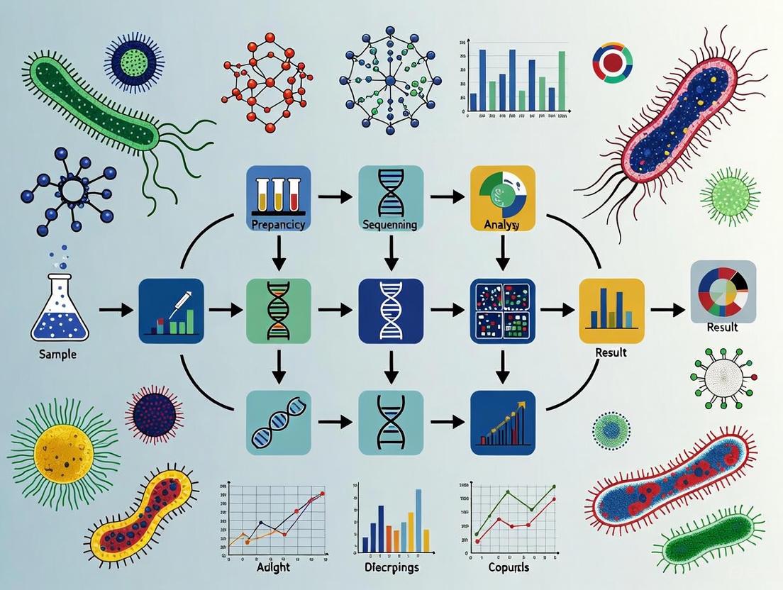
Diagram 1: Digital plating workflow showing core process with key innovations.
The process begins with introducing a bacterial suspension into the PicoArray device, where it partitions into individual microwells via a self-priming mechanism that requires no external pumps [1]. The partitioned sample is then covered with a specific nutrient- or chemical-laden agar sheet for incubation [1]. Thanks to the replaceability of the agar cover, the microenvironment within the picowells can be flexibly altered for culturing or screening microbes, significantly extending the application range beyond static cultivation systems [1]. This capability enables complex multi-stage experiments where different growth conditions, selective pressures, or chemical stimuli can be introduced sequentially without disrupting the individually compartmentalized cells.
Research Reagent Solutions
Table 2: Essential Research Reagents for Digital Plating Platform
| Reagent/Material | Function | Example Specifications |
|---|---|---|
| PDMS Prepolymer | Device fabrication | Sylgard 184 silicone elastomer with curing agent (10:1 w/w) |
| Agar Powder | Solid matrix for replaceable sheets | Biowest, Spain |
| LB Broth Powder | Nutrient base for microbial growth | CM158, Beijing Land Bridge Technology, China |
| Antibiotics | Selective pressure for phenotypic screening | Ampicillin sodium salt (100 mg/mL stock solution) |
| Fluorescent Dyes | Cell viability assessment and metabolic indicators | Concentration varies by experimental design |
| Bacterial Strains | Experimental subjects | E. coli JM109, GFP-tagged E. coli BL21, S. aureus ATCC 43300, Salmonella enterica 14028 |
Detailed Experimental Protocols
Protocol 1: Platform Setup and Basic Microbial Quantification
Objective: To isolate and quantify bacterial populations at single-cell resolution using the digital plating platform.
Materials:
- PicoArray device (113,137 hexagonal microwells)
- Sterilized PDMS chamber mold (76 mm × 26 mm × 1 mm)
- LB broth powder (2.5 g/1000 mL) and agar powder (1.5 g/1000 mL)
- Bacterial suspension (e.g., E. coli JM109) in normal saline
- Autoclave, sterile containers, and weighing instruments
Procedure:
- Agar Sheet Preparation:
- Dissolve 2.5 g LB broth powder and 1.5 g agar powder in 1000 mL distilled water
- Autoclave the mixture and cool to 60°C
- Pour into sterilized PDMS chamber mold and cover with sterilized plastic sheet
- Place a glass slide and weight on the plastic sheet for even thickness
- After solidification at room temperature, remove the mold to obtain agar sheet [1]
Sample Loading:
- Introduce bacterial suspension into the PicoArray device
- Allow self-priming vacuum to partition sample into microwells [1]
Incubation and Analysis:
- Cover the partitioned sample with the prepared agar sheet
- Incubate at appropriate temperature (37°C for E. coli)
- Monitor microbial growth for 6-7 hours [1]
- Image the array using fluorescence microscopy or scanner
- Quantify positive wells based on metabolic activity or fluorescence
Protocol 2: Rapid Antibiotic Susceptibility Testing (AST)
Objective: To determine antibiotic susceptibility in less than 6 hours using replaceable agar sheets.
Materials:
- Prepared PicoArray with partitioned bacterial suspension
- Agar sheets containing gradient concentrations of antibiotics (e.g., ampicillin)
- Control agar sheets without antibiotics
Procedure:
- Initial Cultivation:
- Cover partitioned bacterial sample with nutrient agar sheet (without antibiotics)
- Incubate for 2-3 hours to establish initial growth [1]
Antibiotic Exposure:
- Replace initial agar sheet with antibiotic-laden agar sheet
- Ensure complete contact with microwell array
- Incubate for 3 hours [1]
Assessment and Analysis:
- Compare growth in antibiotic-containing wells to control wells
- Determine minimum inhibitory concentration (MIC) based on growth inhibition
- Calculate susceptibility based on the fraction of wells showing continued growth
The replaceable agar sheet technology enables this multi-stage assay without disturbing the partitioned cells, providing susceptibility results in <6 hours compared to 16-24 hours with traditional methods [1].
Diagram 2: Rapid antibiotic susceptibility testing workflow comparison.
Applications in High-Throughput Microbial Analysis
The integration of high-density microwell arrays with replaceable agar sheets enables diverse applications that leverage their unique capabilities:
Single-Cell Isolation from Mixed Communities: The platform's partitioning efficiency enables isolation of individual cells from complex samples without prior dilution, overcoming interspecies competition that plagues traditional plating [1].
Selective Enrichment Using Differential Media: The replaceable agar system allows sequential application of different selective media to the same partitioned population, enabling sophisticated screening protocols [1].
Quantitative Microbial Interaction Studies: The ability to control microenvironment conditions while monitoring individual wells facilitates investigation of microbial interactions through metabolite exchange or signaling molecules [1].
Cultivation of Previously Unculturable Microbes: By recreating specific microenvironments through agar composition and avoiding competition, the platform shows promise for accessing microbial "dark matter" [12].
The synergistic combination of high-density picoliter microwell arrays and replaceable agar sheets in the digital plating platform represents a significant advancement in microbial analysis methodology. This integration bridges the gap between high-throughput microfluidics and practical laboratory routines, offering researchers a scalable, cost-effective solution that maintains the familiarity of agar-based culturing while providing digital single-cell resolution [1]. The platform's flexibility, accelerated timeline, and compatibility with diverse experimental designs position it as a powerful tool for advancing research in clinical diagnostics, environmental microbiology, synthetic biology, and drug development. By transforming traditional plating into a digital, programmable format, these core components enable a new generation of microbial studies that leverage single-cell resolution and dynamic environmental control.
The self-pumping mechanism represents a pivotal innovation within the digital plating (DP) platform, enabling the automated partitioning of bacterial suspensions into high-density picoliter microwell arrays without the need for external tubing, connections, or bulky pumping equipment [1]. This mechanism leverages a pre-degassing-induced vacuum to drive the rapid and spontaneous partitioning of samples, making the platform particularly accessible to non-microfluidics experts and smaller laboratories [1]. By replacing complex fluidic operations with a simple, self-contained process, this technology bridges the gap between high-throughput microfluidics and practical laboratory routines, facilitating robust and versatile microbial detection and analysis [1].
Principle of Operation
The core principle underlying the self-pumping mechanism is the creation of a vacuum via the pre-degassing of the polydimethylsiloxane (PDMS) material used to fabric the PicoArray device [1]. When the PDMS device is degassed prior to use, air is evacuated from its porous microstructure. Upon contact with a liquid bacterial suspension, the stored vacuum energy creates a pressure differential that spontaneously draws the sample into the device's network of microchannels and microwells. This capillary-driven flow continues until all microwells are filled, achieving complete partitioning of the sample into discrete picoliter-volume compartments without any external power source or fluidic controls.
Key Specifications and Performance Metrics
The following table summarizes the critical specifications of the PicoArray device and the performance characteristics of its self-pumping mechanism:
Table 1: Technical Specifications of the PicoArray Device and Self-Pumping Mechanism
| Parameter | Specification | Experimental Context |
|---|---|---|
| Microwell Array Density | 113,137 microwells per chip | Fabricated using conventional soft lithography [1] |
| Individual Microwell Volume | Picoliter scale | Enables single-cell confinement [1] |
| Microwell Geometry | Hexagonal | Diagonal: 70 μm; Height: 40 μm [1] |
| Microchannel Dimensions | Loading channel: 30 μm width, 20 μm height [1] | Facilitates fluid distribution |
| Quantification Time | 6-7 hours for E. coli [1] | Significant reduction from conventional methods (16-24 hours) [1] |
| Antibiotic Susceptibility Testing (AST) Time | <6 hours [1] | Enabled by rapid metabolite accumulation in microconfinement |
Detailed Experimental Protocol
This section provides a step-by-step protocol for utilizing the self-pumping mechanism for microbial analysis.
Materials and Reagents
Table 2: Essential Research Reagent Solutions and Materials
| Item | Function/Description | Example/Specification |
|---|---|---|
| PDMS PicoArray Device | Self-pumping chip containing the microwell array | Fabricated from Sylgard 184 silicone elastomer [1] |
| Bacterial Suspension | Sample for analysis | Prepared in normal saline to desired concentration [1] |
| Agar Solid Medium Sheet | Nutrient source for microbial growth | Contains LB broth (2.5 g/L) and agar (1.5 g/L) [1] |
| LB Broth Powder | Base nutrient component for culture medium | Example: CM158 from Beijing Land Bridge Technology [1] |
| Agar Powder | Gelling agent for solid medium sheet | Example: Biowest, Spain [1] |
| Specific Reagents | For selective enrichment or screening | Dyes, antibiotics, or metabolic indicators added to cooled agar [1] |
Step-by-Step Procedure
Step 1: Device Preparation and Pre-degassing
- Fabricate the PicoArray device using conventional soft lithography with SU-8 negative photoresists on silicon wafers to create molds for the channel and microwell layers [1].
- Prepare the PDMS prepolymer by thoroughly mixing the silicone elastomer and curing agent (10:1, w/w), then pour onto the SU-8 molds [1].
- Cure the PDMS at 90°C for 1 hour, then peel the slabs from the molds [1].
- Prior to the experiment, degas the PDMS PicoArray device to create the internal vacuum that will power the self-pumping action [1].
Step 2: Preparation of Covering Agar Sheet
- Prepare a sterile agar solution by dissolving LB broth powder (2.5 g) and agar powder (1.5 g) in 100 mL of water, followed by autoclaving [1].
- Cool the autoclaved agar solution to approximately 60°C [1].
- As needed, add specific reagents such as antibiotics, dyes, or metabolic indicators to the agar solution and mix thoroughly [1].
- Pour the mixture into a sterilized PDMS chamber mold (76 mm × 26 mm × 1 mm), cover with a sterilized plastic sheet, and place a glass slide with a weight on top [1].
- Allow the agar to solidify at room temperature, then remove the mold to obtain a uniform agar solid media sheet [1].
Step 3: Sample Loading and Partitioning
- Dilute the bacterial subculture with normal saline to achieve the desired concentration for analysis [1].
- Introduce the prepared bacterial suspension to the inlet port of the pre-degassed PicoArray device [1].
- Observe as the self-pumping mechanism spontaneously draws the suspension into the microchannel network and partitions it into the high-density picoliter microwell array via the pre-degassing-induced vacuum [1].
- Ensure complete filling by verifying that all target microwells contain the suspension.
Step 4: Incubation and Analysis
- Carefully cover the filled PicoArray with the prepared agar solid medium sheet, ensuring full contact to facilitate nutrient diffusion into the microwells [1].
- Place the assembled platform in an appropriate incubator (e.g., 37°C for E. coli) for the required duration [1].
- Monitor microbial growth, quantification, or phenotypic responses using appropriate microscopy or imaging systems at regular intervals.
- For multi-stage assays, leverage the replaceability of the agar cover to dynamically alter growth conditions by replacing with a new sheet containing different chemical inducers or antibiotics [1].
Workflow Visualization
Figure 1: Workflow of the self-pumping digital plating platform, illustrating the automated partitioning process and flexible assay design enabled by the replaceable agar sheet.
Applications Enabled by the Self-Pumping Mechanism
The efficiency and simplicity of the self-pumping mechanism facilitate several advanced applications within the digital plating platform [1]:
- Single-Cell Isolation from Mixed Communities: The precise partitioning enables the isolation and clonal cultivation of individual bacteria from complex samples without the need for prior dilution.
- Rapid Antibiotic Susceptibility Testing (AST): The microconfinement effect accelerates bacterial responses, allowing for AST results in less than 6 hours.
- Selective Enrichment and Screening: The replaceable agar sheet system permits dynamic changes to the chemical microenvironment, enabling flexible phenotypic screening.
- Quantitative Assessment of Microbial Interactions: The platform supports the study of interactions between different microbial species at the single-cell level.
The digital plating (DP) platform represents a transformative advancement in microbial analysis, integrating the established principles of traditional plate culturing with cutting-edge digital bioassay technology [1]. This hybrid system addresses critical limitations of conventional methods by enabling rapid isolation, precise quantification, and comprehensive phenotypic characterization of microorganisms at the single-cell level [1]. The core of the DP platform consists of a high-density picoliter microwell array chip that partitions bacterial suspensions via a self-pumping mechanism, after which the chip is covered with a replaceable nutrient- or chemical-laden agar sheet for incubation and analysis [1]. This innovative approach bridges the gap between high-throughput microfluidics and practical laboratory workflows, offering a scalable and cost-effective solution for clinical diagnostics, environmental microbiology, and synthetic biology applications where single-cell resolution is critical [1].
Table 1: Performance Comparison: Digital Plating vs. Traditional Methods
| Parameter | Digital Plating Platform | Traditional Plate Culturing |
|---|---|---|
| Incubation Time (E. coli) | 6-7 hours [1] | 16-24 hours [1] |
| Antibiotic Susceptibility Testing | <6 hours [1] | Typically 18-24 hours or longer |
| Single-Cell Isolation | Precise isolation from mixed communities [1] | Limited by dilution and spreading techniques |
| Quantification Resolution | Digital quantification at single-cell level [1] | Colony-forming unit counting |
| Environmental Flexibility | Dynamic microenvironment control via replaceable agar sheets [1] | Fixed medium conditions |
| Throughput Capability | High-density picoliter microwell arrays (e.g., 113,137 wells) [1] | Limited by plate surface area |
Speed and Efficiency Advantages
Accelerated Microbial Detection and Analysis
The DP platform significantly reduces microbial detection times through microconfinement-enhanced metabolite accumulation within picoliter-scale microwells [1]. This confined environment accelerates microbial growth and metabolic activity, enabling precise bacterial quantification within hours rather than days [1]. For Escherichia coli, the DP platform reduces detection time to 6-7 hours compared to 16-24 hours required for traditional plate culturing methods [1]. This remarkable acceleration stems from the physical partitioning of individual cells into microwells, where localized accumulation of metabolic products creates favorable growth conditions that support rapid proliferation detectable within significantly shortened timeframes [1].
Rapid Antibiotic Susceptibility Testing
The platform enables particularly rapid antibiotic susceptibility testing (AST), completing analyses in less than 6 hours compared to the 18-24 hours typically required by conventional methods [1]. This expedited timeline is achieved through single-cell resolution monitoring of phenotypic responses to antimicrobial agents within the microwell array [1]. The replaceable agar sheet functionality allows introduction of antibiotics at precise concentrations after initial cell loading and partitioning, enabling researchers to observe bacterial responses to antimicrobial challenge in real-time without the need for subculturing or manual intervention [1]. This capability has profound implications for clinical diagnostics, where rapid AST results can directly impact patient treatment outcomes and antimicrobial stewardship efforts.
Advanced Quantification Capabilities
Digital Quantification Methodology
The DP platform employs digital quantification principles similar to digital PCR, where statistical analysis of positive microwells enables precise enumeration of viable microorganisms [1]. Each microwell in the high-density array (typically containing 113,137 hexagonal microwells) functions as a miniature cultivation chamber [1]. Following partitioning and incubation, microwells containing proliferated microorganisms are identified and counted, allowing for absolute quantification of the initial inoculum through Poisson distribution statistics [1]. This approach provides superior accuracy compared to traditional colony-forming unit (CFU) counting, especially at low bacterial concentrations where conventional methods suffer from significant statistical uncertainty.
Enhanced Resolution for Complex Communities
Unlike traditional plating that obscures rare or slow-growing taxa through interspecies competition, the physical separation afforded by the microwell array enables resolution of individual cells from mixed microbial communities without prior dilution [1]. This capability allows researchers to identify and characterize minority populations that would otherwise be overwhelmed by dominant species in conventional culture systems [1]. The platform's compartmentalization prevents competitive exclusion and enables quantitative assessment of microbial interactions, including synergism and antagonism between different species or strains [1]. This makes the DP platform particularly valuable for studying complex environmental samples or clinical specimens containing polymicrobial communities.
Phenotypic Characterization at Single-Cell Resolution
High-Content Screening Capabilities
The DP platform enables comprehensive phenotypic characterization through its unique ability to monitor individual cells across multiple parameters and conditions [1]. The replaceable agar sheet functionality allows dynamic modification of the microenvironment during experiments, facilitating flexible phenotypic screening approaches [1]. Researchers can initially culture microorganisms under standard conditions, then replace the agar sheet with media containing specific compounds, antibiotics, or metabolic indicators to assess phenotypic responses at the single-cell level [1]. This capability enables high-content screening for traits including antibiotic susceptibility, metabolic capabilities, and stress responses while maintaining spatial registration of individual cells throughout sequential manipulations.
Single-Cell Isolation and Clonal Cultivation
A key advantage of the DP platform is its ability to precisely isolate individual cells from complex samples for subsequent clonal cultivation and analysis [1]. The microwell array effectively partitions single cells from mixed microbial communities without the need for prior dilution or sophisticated instrumentation [1]. Once identified, specific cells of interest can be recovered from individual microwells for downstream applications including whole-genome sequencing, further phenotypic characterization, or strain development [1]. This functionality bridges the gap between cultivation-based and molecular approaches, enabling researchers to correlate genomic information with phenotypic traits observed at the single-cell level.
Table 2: Application Range of the Digital Plating Platform
| Application | Methodology | Key Advantage |
|---|---|---|
| Single-Cell Isolation | Partitioning of mixed communities into picoliter wells | Isolation without competitive exclusion [1] |
| Selective Enrichment | Use of differential media in agar sheets | Flexible medium switching during experiments [1] |
| Antibiotic Susceptibility Testing | Monitoring response to antibiotics in agar cover | Results in <6 hours [1] |
| Microbial Interaction Studies | Co-partitioning of multiple species in wells | Quantitative assessment of interactions [1] |
| Phenotypic Heterogeneity | Time-lapse monitoring of individual wells | Resolution of subpopulation differences [1] |
Experimental Protocols
Protocol 1: Fabrication of PicoArray Devices
Materials: PDMS prepolymer (Sylgard 184), curing agent, SU-8 3010 and 3050 negative photoresists, silicon wafers, punching tool.
Methodology:
- Create SU-8 molds for channel layer (52mm length × 80μm width × 60μm height) and microwell layer using conventional soft lithography [1].
- Prepare PDMS prepolymer by thoroughly mixing silicone elastomer and curing agent (10:1, w/w) and degassing.
- Pour degassed PDMS onto SU-8 molds and cure at 90°C for 1 hour.
- Carefully peel cured PDMS slabs from molds and create inlet port on channel layer with punching tool.
- Align PDMS channel layer and microwell layer face-to-face to form reversible seal [1].
Technical Notes: Typical microwell dimensions are 70μm (diagonal) × 40μm (height), with arrays containing up to 113,137 hexagonal microwells [1]. The reversible sealing allows retrieval of specific cells after analysis.
Protocol 2: Preparation of Covering Agar Solid Media Sheets
Materials: LB broth powder, agar powder, autoclave, PDMS chamber mold (76mm × 26mm × 1mm), sterile plastic sheet, reagents for experimental purposes (dyes, antibiotics, metabolic indicators).
Methodology:
- Dissolve 2.5g LB broth powder and 1.5g agar powder in 1000mL water and autoclave.
- Cool agar solution to 60°C and add appropriate reagents (e.g., antibiotics, metabolic indicators) depending on experimental goals.
- Pour mixture into sterilized PDMS chamber mold and cover with sterilized plastic sheet.
- Place glass slide and weight onto plastic sheet to ensure uniform thickness.
- Allow solidification at room temperature before removing PDMS chamber mold [1].
Technical Notes: The replaceable agar sheet is fundamental to the platform's flexibility. Multiple sheets with different compositions can be sequentially applied to the same microwell array to study microbial responses to changing conditions.
Protocol 3: Rapid Antibiotic Susceptibility Testing
Materials: Prepared PicoArray device, bacterial suspension, antibiotic-containing agar sheets, ampicillin sodium salt (or other antibiotics), normal saline.
Methodology:
- Prepare bacterial suspension by diluting subculture solution with normal saline to desired concentration.
- Load bacterial suspension into PicoArray device via self-pumping mechanism driven by pre-degassing-induced vacuum [1].
- Cover loaded device with antibiotic-containing agar sheet (e.g., ampicillin at appropriate concentration).
- Incubate and monitor periodically for growth inhibition.
- Analyze results within 6 hours by quantifying viable cells in antibiotic-containing versus control conditions [1].
Technical Notes: For ampicillin testing, prepare stock solution at 100mg/mL in distilled water and sterilize by filtration through 0.22μm sterile filter [1]. The platform's single-cell resolution enables detection of heteroresistance and persistence at frequencies undetectable by conventional methods.
Research Reagent Solutions
Table 3: Essential Research Reagents for Digital Plating Applications
| Reagent/Material | Function | Example Specifications |
|---|---|---|
| PDMS (Sylgard 184) | Device fabrication | Silicone elastomer basecuring agent (10:1 w/w) [1] |
| SU-8 Photoresist | Mold creation | SU-8 3010, 3050 for features [1] |
| Agar Powder | Solid matrix for covers | Biowest or equivalent, 1.5g/L [1] |
| LB Broth | Nutrient base | 2.5g/L in agar sheets [1] |
| Ampicillin Sodium | Antibiotic testing | Stock: 100mg/mL, 0.22μm filtered [1] |
| Fluorescent Dyes | Cell labeling & viability | SYTOX Green, Hoechst 33342 [13] |
| OPP (O-propargyl-puromycin) | Translation monitoring | Puromycin analog for biosynthesis tracking [13] |
Figure 1: Digital Plating Experimental Workflow
Figure 2: Technology Comparison Overview
From Theory to Bench: Practical Applications and Workflows in Drug Discovery and Diagnostics
The digital plating (DP) platform represents a significant advancement in microbial analysis, merging the principles of traditional agar-based culturing with the precision of digital microfluidics. This platform centers on a high-density picoliter microwell array chip (PicoArray) used in conjunction with a replaceable agar sheet [1] [11]. The core innovation lies in its ability to partition a bacterial suspension into thousands of isolated picoliter-scale compartments via a self-pumping mechanism, followed by incubation under a solid agar medium sheet. This system enables rapid microbial quantification and phenotypic characterization within hours, a substantial improvement over the 16-24 hours required for traditional methods [1]. The replaceable agar sheet provides remarkable flexibility, allowing researchers to dynamically alter the microbial growth microenvironment for applications ranging from antibiotic susceptibility testing to the isolation of specific microbes from complex communities [1]. This protocol details the fabrication of the PicoArray device and the preparation of the essential agar sheets, providing researchers with the tools to implement this cutting-edge technology in high-throughput microbial analysis and drug development workflows.
Fabrication of PicoArray Devices
The PicoArray device serves as the foundation of the digital plating platform, providing the microstructure for isolating and analyzing individual microbial cells. Below are detailed protocols for fabricating devices using two different material systems.
Silicon-Glass Based PicoArray Fabrication
This method produces a highly durable device through silicon-glass anodic bonding [14].
- Materials: Silicon wafer (Si(100)), Corning 7740 glass wafer, photoresist (PR 1813, PR 1827, AZ9260), buffered HF, reactive ion etching mask (thermal oxide layer).
- Equipment: Deep reactive ion etching system (STS), spin coater, photolithography setup, wafer bonder (EV500, EV Group), RCA cleaning setup.
Procedure:
- Thermal Oxidation: Grow a 0.6 μm thermal oxide layer on a silicon wafer to serve as a reactive ion etching mask.
- Pico-Reaction Chamber Patterning:
- Spin-coat PR 1813 photoresist onto the oxidized silicon wafer.
- Transfer the pico-reaction chamber pattern via photolithography and etch the thermal oxide layer with buffered HF.
- Remove photoresist with a stripper (PRS 2000).
- Microchannel Patterning:
- Apply a new layer of PR 1827 photoresist.
- Transfer the microchannel pattern via photolithography and reactive ion etching.
- Perform deep reactive ion etching to obtain ~135 μm deep microchannels.
- Strip the photoresist.
- Final Etching and Through-Hole Creation:
- Re-etch the wafer using the deep RIE system to achieve the final depths: ~15 μm for reaction chambers and ~150 μm for microchannels.
- Apply AZ9260 photoresist to the wafer's backside.
- Transfer the inlet/outlet hole pattern via photolithography and etch through-holes.
- Surface Preparation and Bonding:
- Clean the wafer using RCA treatment.
- Grow a 0.2 μm thermal silicon oxide layer on the surface to enable subsequent functionalization for chemical reactions.
- Anodically bond the microfabricated silicon wafer to the glass wafer at 400°C and 1000 V.
The final device contains an array of 1,278 individual pico-reaction chambers [14]. Each three-dimensional chamber measures 90 μm wide, 200 μm long, and 15 μm deep, yielding an internal volume of 270 picoliters and a reactive interior surface area of 0.045 mm² [14]. The fluid channels are designed with a tapered shape based on fluid mechanical modeling to ensure a uniform flow rate across all reaction chambers [14].
PDMS-Based PicoArray Fabrication
This method utilizes soft lithography for rapid prototyping and produces a device containing 113,137 hexagonal microwells [1].
- Materials: PDMS prepolymer (Sylgard 184 silicone elastomer and curing agent), SU-8 3010 and SU-8 3050 negative photoresists, silicon wafers.
- Equipment: Plasma cleaner, oven, photolithography setup, punching tool.
Procedure:
- Mold Fabrication:
- Pattern SU-8 3010 and SU-8 3050 negative photoresists on separate silicon wafers to create two molds: one for the channel layer (main channel: 52 mm L × 80 μm W × 60 μm H) and one for the microwell layer (microwell: 70 μm diagonal × 40 μm H; loading microchannel: 17.9 mm L × 30 μm W × 20 μm H) [1].
- PDMS Molding:
- Thoroughly degas a PDMS prepolymer mixture (elastomer:curing agent = 10:1 w/w).
- Pour the prepolymer onto the SU-8 molds.
- Cure at 90°C for 1 hour.
- Carefully peel off the molded PDMS slabs from the molds.
- Device Assembly:
- Create an inlet port on the PDMS channel layer with a punching tool.
- Align the PDMS channel layer and the PDMS microwell layer face-to-face and bring them into conformal contact to form a reversible seal.
Table 1: Specifications and Comparison of PicoArray Fabrication Methods
| Parameter | Silicon-Glass Device [14] | PDMS Device [1] |
|---|---|---|
| Base Material | Silicon substrate anodically bonded to glass | Polydimethylsiloxane (PDMS) |
| Fabrication Method | Standard microelectronic fabrication & anodic bonding | Soft lithography & reversible bonding |
| Total Number of Wells | 1,278 | 113,137 |
| Individual Well Volume | 270 picoliters | Picoliter-scale (precise volume not specified) |
| Well Geometry | Rectangular (90 μm wide × 200 μm long × 15 μm deep) | Hexagonal (70 μm diagonal × 40 μm height) |
| Key Advantage | High durability; integrated fluidic channels | Rapid prototyping; higher well density; lower cost |
PicoArray Fabrication Paths
Preparation of Covering Agar Solid Media Sheets
The agar sheet is a critical component of the DP platform, acting as a replaceable nutrient source and enabling dynamic manipulation of the microbial growth environment.
- Materials: LB broth powder, agar powder, sterile distilled water, antibiotic stocks (e.g., Ampicillin sodium salt), specific metabolic indicators or dyes as needed.
- Equipment: Autoclave, water bath, sterile PDMS chamber mold (76 mm × 26 mm × 1 mm), sterilized plastic sheet, glass slide, weight.
Procedure:
- Prepare Agar Solution:
- Dissolve 2.5 g of LB broth powder and 1.5 g of agar powder in 100 mL of distilled water [1]. This creates a standard LB agar mixture at 1.5% agar.
- For antibiotic plates, refer to Table 2 for recommended stock and working concentrations.
- Sterilize:
- Cool and Additives:
- Cool the sterile molten agar to approximately 60°C in a water bath [15] [1]. This temperature keeps the agar liquid but prevents thermal degradation of most antibiotics.
- If adding antibiotics, dyes, or other chemical inducers, mix them thoroughly into the agar solution at this stage. Ensure antibiotic stocks are filter-sterilized.
- Cast Agar Sheets:
- Pour the mixture into a sterilized PDMS chamber mold with internal dimensions of 76 mm × 26 mm × 1 mm [1].
- Cover the mold with a sterilized plastic sheet.
- Place a glass slide and a weight on top of the plastic sheet to ensure a uniform thickness and flat surface.
- Solidify:
- Allow the agar to solidify at room temperature.
- Once solid, carefully remove the PDMS chamber mold to obtain the final agar solid media sheet, ready for use.
Table 2: Common Antibiotic Stock and Working Concentrations for Agar Plates [15]
| Antibiotic | Recommended Stock Concentration | Recommended Working Concentration | Solvent |
|---|---|---|---|
| Ampicillin | 100 mg/mL | 100 µg/mL | Water |
| Carbenicillin | 100 mg/mL | 100 µg/mL | Water |
| Chloramphenicol | 25 mg/mL | 25 µg/mL | Ethanol |
| Kanamycin | 50 mg/mL | 50 µg/mL | Water |
| Spectinomycin | 50 mg/mL | 50 µg/mL | Water |
| Tetracycline | 10 mg/mL | 10 µg/mL | Water |
The Scientist's Toolkit: Research Reagent Solutions
Table 3: Essential Materials for PicoArray Fabrication and Agar Preparation
| Item | Function/Description | Example/Specification |
|---|---|---|
| SU-8 Negative Photoresist | Used to create high-resolution masters (molds) for PDMS-based PicoArrays via photolithography. | SU-8 3010, SU-8 3050 [1] |
| PDMS Elastomer Kit | The base material for soft lithography. Provides optical clarity, gas permeability, and flexibility. | Sylgard 184 [1] |
| LB Broth Powder | A rich, complex growth medium providing essential nutrients for a wide range of bacteria. | Composition: 5.0 g Yeast Extract, 10.0 g Peptone, 10.0 g NaCl per 37g pre-mixed powder [15] |
| Agar Powder | A polysaccharide derived from seaweed that forms a gel matrix, providing a solid support for microbial growth. | 12 g per L for standard plates [15]; 1.5% for agar sheets [1] |
| Antibiotic Stocks | Selective agents added to agar to isolate or select for microorganisms carrying specific resistance genes. | See Table 2 for concentrations. Prepare as 1000x stocks [15]. |
| Sterile PDMS Chamber Mold | A custom mold used to cast the replaceable agar sheets to the precise dimensions required by the PicoArray chip. | Internal dimensions: 76 mm × 26 mm × 1 mm [1] |
Integrated Experimental Workflow for Digital Plating
The power of the digital plating platform is realized when the fabricated PicoArray device and prepared agar sheets are used together in a seamless workflow.
Digital Plating Workflow
- Bacterial Suspension Preparation: Grow bacteria to the desired growth phase and dilute to the target concentration in a suitable buffer like normal saline [1].
- Sample Loading and Partitioning: Introduce the bacterial suspension into the inlet port of the PicoArray device. A pre-degassing-induced vacuum (self-pumping mechanism) drives the partitioning of the sample, distributing individual bacterial cells into the thousands of picoliter-scale microwells [1] [11].
- Agar Sheet Application: Carefully place the pre-prepared, sterile agar sheet onto the surface of the loaded PicoArray chip, ensuring full and uniform contact. This creates a "cover" that supplies nutrients and creates a microconfined environment for each trapped cell [1].
- Incubation and Imaging: Place the assembled unit in an appropriate incubator. The microconfinement within the picowells leads to accelerated metabolite accumulation, enabling detection and quantification in as little as 6-7 hours for E. coli, compared to 16-24 hours with traditional plating [1]. The platform is compatible with real-time, single-cell resolution imaging.
- Dynamic Assays via Agar Replacement: A key feature of the platform is the replaceability of the agar cover. To perform antibiotic susceptibility testing (AST) or change growth conditions, the initial agar sheet can be carefully peeled off and replaced with a new sheet containing the desired compounds (e.g., antibiotics). This enables rapid phenotypic screening (e.g., AST in <6 hours) without disturbing the spatially fixed microcolonies that have grown in the picowells [1].
High-Throughput Single-Cell Isolation from Complex Microbial Communities
Within the broader research on digital plating platforms for high-throughput microbial analysis, the precise isolation of individual microbial cells from complex communities represents a critical foundational step. Traditional culture-based methods, while considered the "gold standard," are hindered by prolonged incubation times, labor-intensive workflows, and an inherent inability to resolve cellular heterogeneity [1]. This document details integrated application notes and protocols for two advanced, complementary technologies enabling high-throughput single-cell isolation: a Digital Plating (DP) Platform and Single-Cell Sequencing via Semi-Permeable Capsules (SPCs). The Digital Plating platform bridges the gap between conventional microbiology and modern microfluidics, allowing for phenotypic screening and cultivation [1] [11]. In parallel, single-cell sequencing using SPCs provides a powerful tool for genotypic analysis, overcoming limitations of droplet-based systems by enabling full reagent exchange and multi-step workflows [16]. Together, these methods empower researchers to dissect microbial communities with unprecedented resolution.
Technology Comparison and Selection
Selecting the appropriate high-throughput isolation method depends on the experimental goals, whether they lean towards rapid phenotypic screening and cultivation or deep genotypic characterization. The table below summarizes the key characteristics of the two primary technologies discussed in this document.
Table 1: Quantitative Comparison of High-Throughput Single-Cell Isolation Technologies
| Feature | Digital Plating (DP) Platform | Single-Cell Sequencing (SPCs) |
|---|---|---|
| Core Principle | Microfabricated picoliter microwell array with replaceable agar sheets [1] | Encapsulation of single cells in semi-permeable capsules for DNA processing [16] |
| Throughput | 113,137 microwells per array [1] | ~100,000 cells per run (with lambda=0.1) [16] |
| Isolation Resolution | Single-cell isolation from mixed communities [1] | Single-cell encapsulation for genomics [16] |
| Key Applications | Rapid quantification, AST (<6h), microbial interactions, cultivation [1] | Linking antimicrobial resistance genes to host species, uncovering microbial diversity [16] |
| Typical Duration | E. coli quantification: 6-7 hours [1] | Multi-day process including lysis, amplification, and sequencing [16] |
| Cell Recovery | Facilitates recovery of cells for further analysis [1] | Not designed for cell recovery; focus on genetic material |
| Data Output | Phenotypic (growth, inhibition, susceptibility) | Genomic (taxonomy, ARGs, phylogenetic relationships) |
Application Notes
Digital Plating for Phenotypic Screening
The Digital Plating (DP) platform is a hybrid system that integrates the simplicity of traditional agar plating with the precision of digital microfluidics. Its core component is a high-density array of picoliter-sized microwells fabricated in PDMS. A bacterial suspension is loaded and partitioned into these wells via a self-pumping mechanism driven by a pre-degassing-induced vacuum. A critical innovation is the use of a replaceable, nutrient- or chemical-laden agar sheet that covers the array, creating a microconfined environment for growth [1]. The replaceability of this agar sheet allows for dynamic manipulation of the microenvironment during an experiment. For instance, an initial non-selective medium can be swapped for an antibiotic-laden one to perform rapid Antibiotic Susceptibility Testing (AST) in less than 6 hours. This platform dramatically accelerates microbial quantification—reducing the time from 16-24 hours to 6-7 hours for E. coli—by enhancing metabolite accumulation within picoliter wells, enabling faster detection of microcolonies [1]. Its applications extend to single-cell isolation from mixtures, selective enrichment using differential media, and quantitative studies of microbial interactions [1].
Single-Cell Genomic Analysis with Semi-Permeable Capsules
For comprehensive genotypic insights, single-cell sequencing using Semi-Permeable Capsules (SPCs) is a powerful complementary technique. This method addresses key limitations of traditional droplet microfluidics, where reagent exchange is difficult and workflows are constrained by reaction compatibility [16]. The SPC technology involves encapsulating single bacterial cells from a complex sample (e.g., sewage or feces) within permeable hydrogel capsules. A major advantage of SPCs is their semi-permeable membrane, which allows for complete exchange of reagents and waste products through simple washing steps, while retaining large genomic DNA fragments inside [16]. This capability is crucial for the multi-step workflow required for single-cell genomics, which includes cell lysis, whole-genome amplification, and combinatorial barcoding. The process enables the sequencing of genetic material from tens of thousands of individual bacterial cells in parallel. This high-throughput approach has been successfully applied to link antimicrobial resistance genes (ARGs) to their specific bacterial hosts within complex samples, thereby unraveling true microbial diversity and functional potential at the single-cell level [16].
Detailed Experimental Protocols
Protocol 1: Single-Cell Isolation and Analysis Using the Digital Plating Platform
This protocol describes the procedure for isolating and phenotypically characterizing single bacterial cells from a mixed community using the DP platform.
Table 2: Key Reagent Solutions for the Digital Plating Protocol
| Reagent/Material | Function | Example/Specification |
|---|---|---|
| PDMS PicoArray Device | High-density microwell array for single-cell partitioning | 113,137 hexagonal wells (70 μm diagonal, 40 μm height) [1] |
| Agar Sheet with Nutrients | Solid growth medium for microbial cultivation | LB broth with 1.5% agar; can be supplemented with antibiotics or indicators [1] |
| Liquid Growth Medium | For bacterial subculture and suspension preparation | e.g., LB broth [1] |
| Normal Saline | Diluent for adjusting bacterial concentration | 0.85-0.9% NaCl solution [1] |
| Antibiotic Stock Solution | For AST or selective enrichment | e.g., Ampicillin sodium salt, 100 mg/mL in distilled water, filter sterilized [1] |
Procedure:
- Device Preparation: Fabricate the PicoArray device using conventional soft lithography with SU-8 photoresist molds and PDMS (silicone elastomer: curing agent, 10:1 w/w). Cure at 90°C for 1 hour [1].
- Agar Sheet Preparation: Prepare a liquid agar medium (e.g., LB with 1.5% agar), autoclave, and cool to 60°C. Add any required reagents (e.g., antibiotics, dyes). Pour the mixture into a sterile chamber mold (76 mm × 26 mm × 1 mm), cover with a plastic sheet, and allow it to solidify at room temperature [1].
- Sample Preparation: Grow bacterial strains (e.g., E. coli, S. aureus) from frozen stocks in liquid medium. Harvest cells in the late exponential phase and dilute the suspension in normal saline to the desired concentration for single-cell loading [1].
- Device Loading: Introduce the prepared bacterial suspension into the main channel of the PicoArray device. The pre-degassed PDMS will create a vacuum, autonomously pumping and partitioning the suspension into the microwells via a network of loading microchannels [1].
- Incubation and Analysis: Carefully cover the loaded microwell array with the prepared agar sheet to create the cultivation microenvironment. Incubate the entire assembly at the appropriate temperature (e.g., 37°C). Monitor microcolony formation over time using microscopy. For dynamic assays like AST, the initial agar sheet can be replaced with a new one containing the antibiotic after initial growth is observed [1].
Protocol 2: Single-Cell Genomic Sequencing via Semi-Permeable Capsules (SPCs)
This protocol outlines the steps for preparing single microbial cells from complex communities for high-throughput genomic sequencing using SPCs.
Table 3: Key Reagent Solutions for the SPC Sequencing Protocol
| Reagent/Material | Function | Example/Specification |
|---|---|---|
| Detergent Mix | Cell detachment from complex samples | 100 mM EDTA, 100 mM sodium pyrophosphate, 1% (v/v) Tween 80 [16] |
| Lysis Enzyme Cocktail | Degrades cell walls for DNA release | Lysozyme, Zymolyase, Lysostaphin, Mutanolysin in PBS [16] |
| Proteinase K | Protein digestion for comprehensive lysis | 1 mg/mL in PBS [16] |
| Alkaline Lysis Solution | Chemical lysis to complement enzymatic treatment | 0.4 M KOH, 10 mM EDTA, 100 mM DTT [16] |
| Whole Genome Amplification (WGA) Mix | Amplifies genomic DNA within SPCs | Custom Single-Microbe DNA Barcoding Kit [16] |
| SPC Innovator Kit | Core reagents for capsule generation | Includes core/shell solutions, emulsion breaker [16] |
Procedure:
- Sample Preparation and Cell Detachment: Suspend 0.1 g of environmental sample (e.g., sewage, feces) in 150 μL of 2.5% NaCl solution. Add 50 μL of detergent mix and 50 μL of methanol. Shake vigorously for 60 minutes at 500 r.p.m. Sonicate the slurry three times for 1 minute each in a water bath. Filter the mixture through an 8 μm syringe filter, centrifuge the filtrate at 15,000 × g for 10 min, and wash the cell pellet twice with 1x PBS [16].
- Single-Cell Encapsulation in SPCs: Use impedance flow cytometry to count cells and dilute the sample to achieve a target occupancy (lambda) of 0.1 cells/SPC. Produce SPCs on the ONYX platform using the SPCs Innovator Kit according to the manufacturer's instructions. Collect the emulsion and cross-link the capsule shells using a Light Exposure Device. Break the emulsion and recover the SPCs in an aqueous buffer with 0.1% Triton X-100 [16].
- Cell Lysis and DNA Release: Perform a two-step lysis within the SPCs.
- Enzymatic Lysis: Incubate SPCs in a lysis solution containing lysozyme, zymolyase, lysostaphin, and mutanolysin at 37°C overnight. Wash with PBS.
- Proteinase K Treatment: Incubate SPCs with Proteinase K at 40°C overnight. Wash with PBS.
- Alkaline Lysis: Resuspend SPCs in an alkaline lysis solution for 15 minutes at room temperature. Neutralize with Tris-HCl buffer and wash thoroughly [16].
- Whole Genome Amplification and Barcoding: Incubate SPCs with the WGA mix at 45°C for 1 hour to amplify the genomic DNA. Confirm amplification by staining with SYBR Green and checking for fluorescence. Perform combinatorial split-and-pool barcoding in four steps to label DNA fragments from each SPC with a unique barcode combination [16].
- Library Preparation and Sequencing: Pool the barcoded SPCs, prepare sequencing libraries, and sequence using an Illumina platform. The resulting data can be de-multiplexed, assigning sequences back to individual cells based on their unique barcode combinations for downstream analysis [16].
The rapid global increase in antimicrobial resistance (AMR) necessitates a paradigm shift in diagnostic methodologies. Conventional antimicrobial susceptibility testing (AST), while reliable, often requires a minimum of 72 hours from specimen collection to result, impeding timely targeted therapy [17]. This delay fuels the overuse of broad-spectrum antibiotics, exacerbating the AMR crisis [18]. For critically ill patients, such as those with sepsis, every hour of delay in effective antibiotic administration increases mortality risk by 3-7% [17]. Rapid phenotypic AST technologies, particularly those yielding results within six hours, are therefore crucial for improving patient outcomes and advancing antimicrobial stewardship. This application note details the implementation and performance of cutting-edge platforms that achieve this goal, with a specific focus on the Digital Plating (DP) platform within the context of high-throughput microbial analysis research.
The Digital Plating (DP) platform represents a hybrid technological innovation that merges the robustness of traditional agar-based culturing with the precision and speed of digital single-cell analysis [1] [11]. Its core principle involves partitioning a bacterial suspension into hundreds of thousands of picoliter-volume microwells, creating a "digital" environment for microbial growth and analysis.
Key Components and Workflow
The system is centered on a high-density microwell array chip fabricated from PDMS using standard soft lithography. A single chip contains over 113,000 hexagonal microwells, each with dimensions of 70 μm (diagonal) by 40 μm (height) [1]. The platform's unique innovation is the use of a replaceable agar sheet, which is pre-prepared with specific nutrients, indicators, or antibiotics and serves as a cover for the microwell array. This replaceability allows for dynamic modulation of the microbial microenvironment during an experiment, enabling complex assay protocols such as sequential antibiotic exposure or differential staining without disrupting the partitioned cells [1].
The partitioning process is driven by a self-pumping mechanism (pre-degassing-induced vacuum), which loads the bacterial suspension into the microwells without requiring external tubing or pumps, thus simplifying operation and reducing equipment costs [1] [11]. Once partitioned and covered with the agar sheet, the platform is incubated, and bacterial growth is monitored.
Experimental Protocol: Rapid AST using the Digital Plating Platform
This protocol describes the procedure for performing rapid antibiotic susceptibility testing directly from a bacterial suspension, enabling results in under 6 hours.
Materials and Reagents
Research Reagent Solutions and Essential Materials
| Item | Function/Description |
|---|---|
| PDMS PicoArray Device | A microwell array chip for partitioning bacterial samples [1]. |
| Agar Solid Media Sheets | Replaceable covers that provide nutrients and antibiotics to the microwells [1]. |
| Mueller-Hinton (MH) Broth | Standardized growth medium for AST, used for bacterial dilution [1]. |
| Antibiotic Stock Solutions | Prepared at high concentration (e.g., 100 mg/mL) and sterilized by 0.22-µm filtration [1]. |
| LB Broth & Agar | Used for preparation of base agar sheets and routine culturing [1]. |
| Bacterial Strains | Pure cultures, e.g., E. coli JM109, S. aureus ATCC 43300 [1]. |
Step-by-Step Procedure
- Chip Preparation: Ensure the PDMS PicoArray device is clean and sterile. Place it within the experimental setup.
- Bacterial Sample Preparation:
- Inoculate bacteria from a frozen glycerol stock or a single colony into liquid media (e.g., LB broth) and incubate with shaking overnight at 37°C.
- Dilute the subcultured bacteria in a standardized broth like Mueller-Hinton to a target concentration of approximately 10^5 CFU/mL [1].
- Loading the Digital Plate: Introduce the prepared bacterial suspension into the main channel of the PicoArray device. The self-pumping mechanism will automatically partition the suspension, isolating individual bacterial cells into the microwells within approximately one minute [1].
- Application of Antibiotic Conditions:
- Prepare agar sheets containing a defined concentration of the antibiotic of interest (e.g., ampicillin) and control sheets without antibiotics.
- Carefully cover the loaded microwell array with the prepared agar sheet, ensuring full and even contact.
- Incubation and Imaging: Place the assembled platform in an incubator at 37°C. Monitor bacterial growth directly within the microwells using time-lapse microscopy (e.g., 100x phase-contrast images captured every 2 minutes) [19].
- Data Analysis and Result Interpretation:
- Image Analysis: Use advanced cell segmentation and tracking software (e.g., based on Omnipose or U-net algorithms) to analyze the time-lapse images. Calculate the growth rate of individual bacterial cells in 10-minute sliding windows [19].
- AST Call: A bacterium is classified as susceptible if its growth rate is significantly inhibited in the antibiotic-containing environment compared to the control. A resistant classification is made if growth continues unabated in the presence of the antibiotic. For E. coli, this analysis can be completed in 6-7 hours, a significant acceleration over the 16-24 hours required by traditional plate culturing [1] [11].
Performance Data and Comparative Analysis
The drive for ultra-rapid AST has spurred the development of multiple technologies. The table below summarizes the performance of the Digital Plating platform alongside other established and emerging rapid AST systems.
Performance Comparison of Rapid AST Systems
| Technology / Platform | Principle | Time-to-Result | Key Performance Metrics |
|---|---|---|---|
| Digital Plating (DP) | Picoliter microwell array + replaceable agar sheets | <6 hours [11] | Precise single-cell quantification; demonstrated for E. coli [1]. |
| QuickMIC | Not specified in detail | ~3 hours (analysis time) [20] | Overall EA/CA >95% with routine systems; TAT of 10-11.5h from sample processing [20]. |
| Nanomotion (Resistell) | Measures nanoscale vibrations of bacteria | ~4.1 hours (AST run time) [21] | 97.6% sensitivity vs. reference; 15.7h TAT from blood culture positivity [21]. |
| Microfluidic FISH-AST | Single-cell growth tracking + fluorescence in situ hybridization | ~2 hours [19] | Enables AST and species identification in mixed samples [19]. |
| Conventional Systems | Automated broth microdilution | 9-19 hours [20] | The reference standard against which new platforms are validated. |
These platforms employ different strategies to reduce the time-to-result. The following diagram illustrates the primary acceleration strategies used by rapid AST technologies compared to the conventional workflow.
Discussion and Research Implications
The validation of rapid AST platforms like the Digital Plating system marks a significant advancement for clinical microbiology and pharmaceutical research. The ability to obtain reliable phenotypic susceptibility results in under six hours can fundamentally change the management of bacterial infections [17].
From a research perspective, the DP platform's versatility extends beyond clinical diagnostics. Its capacity for single-cell isolation from mixed communities and quantitative assessment of microbial interactions makes it a powerful tool for synthetic biology and fundamental microbiology studies [1] [11]. The replaceable agar sheet feature introduces a dynamic dimension to screening assays, allowing researchers to challenge microbial populations with sequential changes in environmental conditions—a capability highly relevant for studying adaptive resistance and antibiotic persistence.
While the clinical impact of rapid AST on ultimate outcomes like mortality is still being defined by ongoing studies [22], the theoretical benefits for antimicrobial stewardship are substantial. A reduction in time-to-effective therapy and a decrease in the unnecessary use of broad-spectrum antibiotics are critical steps in combating the global AMR crisis [18] [17]. Future developments, including integration with machine learning for automated analysis and the creation of multi-channel devices for high-throughput screening, will further solidify the role of these technologies in the future of microbiology and drug discovery [23] [21].
Selective Enrichment and Screening Using Differential and Chemical-Laden Media
Selective enrichment and screening are foundational techniques in microbiology, enabling the isolation and phenotypic characterization of specific microorganisms from complex communities. The advent of digital plating platforms represents a significant evolution of these methods, merging the principles of traditional agar-based culture with cutting-edge digital bioassay technology. This synergy facilitates high-throughput, single-cell-resolution analysis with dramatically reduced incubation times. The core of this advancement lies in the use of a replaceable agar sheet system, which allows for dynamic and flexible manipulation of the microbial microenvironment directly on a picoliter-scale microwell array. This document provides detailed application notes and protocols for implementing these techniques within a modern research framework, supported by quantitative data and actionable workflows.
Table 1: Comparison of Microbial Cultivation and Screening Platforms
| Platform Feature | Traditional Plate Culturing | Robotic Automation | Droplet Microfluidics | Digital Plating (DP) Platform |
|---|---|---|---|---|
| Incubation Time | 16-72 hours [11] [1] | 16-72 hours [1] | Variable | ≤ 8 hours [11] [1] |
| Single-Cell Isolation | Limited resolution | Limited resolution | Yes [1] | Yes [11] [1] |
| Environmental Flexibility | Low (fixed media) | Low (fixed media) | Low (difficult substance exchange) [1] | High (replaceable agar sheets) [11] [1] |
| Throughput | Low | Medium | High [1] | High [11] [1] |
| Key Advantage | Gold standard, simple | Reduces labor | High-throughput encapsulation | Rapid, digital quantification with phenotypic flexibility |
Detailed Experimental Protocols
Protocol 1: Rapid Antibiotic Susceptibility Testing (AST) on a Digital Plating Platform
This protocol leverages the digital plating (DP) platform to perform phenotypic antibiotic susceptibility testing in less than 6 hours, a process that traditionally takes 16-24 hours [11] [1].
I. Materials and Reagents
- Digital Plating Setup: PicoArray device (high-density picoliter microwell array chip), degassing chamber [1].
- Bacterial Suspension: Target bacterium (e.g., Escherichia coli), cultured in appropriate liquid medium and diluted to the desired concentration [1].
- Agar Sheets:
- Equipment: Microplate spectrophotometer (for high-throughput colorimetric reading) [24].
II. Methodology
Sample Partitioning:
- Introduce the bacterial suspension into the main channel of the PicoArray device.
- A self-pumping mechanism, driven by a pre-degassing-induced vacuum, partitions the suspension into the high-density picoliter microwells, achieving single-cell isolation [1].
Initial Cultivation:
- Cover the microwell array with the control nutrient agar sheet (e.g., LB).
- Incubate the platform for a short period (e.g., 2-3 hours) to initiate microcolony growth from single cells [11].
Antibiotic Exposure via Agar Replacement:
Incubation and Digital Quantification:
- Continue incubation for a total of 4-6 hours.
- Use integrated imaging or a microplate reader to quantify growth in each microwell. A susceptible strain will show inhibited growth, while a resistant strain will proliferate [11]. The minimum inhibitory concentration (MIC) can be determined by testing a series of antibiotic concentrations.
Protocol 2: Selective Enrichment and Isolation of Pharmaceutical-Degrading Bacteria
This protocol outlines a strategy for isolating specialized bacteria capable of degrading micropollutants, using a combination of traditional enrichment and the DP platform for high-resolution isolation [25].
I. Materials and Reagents
- Enrichment Culture: Mineral salt solution (MSS) with a target pharmaceutical (e.g., Diclofenac, Ibuprofen, Carbamazepine) as the sole carbon source (e.g., 1.5 mg/L) [25].
- Inoculum: Environmental sample (e.g., groundwater biofilm, soil) [25].
- Selective Agar Sheets: MSS agar containing the same pharmaceutical compound.
II. Methodology
Bulk Enrichment:
- Inoculate the pharmaceutical-containing MSS with the environmental sample.
- Incubate with shaking. Monitor population dynamics via metagenomic sequencing. Successive transfers into fresh MSS are performed to enrich for degraders. Genera like Sphingopyxis (Diclofenac), Nocardioides (Ibuprofen), and Pseudonocardia (Carbamazepine) have been shown to increase in relative abundance by up to three orders of magnitude through this process [25].
High-Resolution Isolation on DP Platform:
- After enrichment, partition the culture onto the DP platform.
- Cover with a selective agar sheet containing the pharmaceutical.
- Incubate. Only viable, compound-degrading cells (e.g., Stenotrophomonas humi, Rhizobium daejeonense) will form microcolonies, enabling their direct identification and isolation from the mixed community for further characterization [11] [25].
Workflow Visualization
The following diagram illustrates the core logical workflow of the digital plating platform for selective screening applications.
Digital Plating Screening Workflow
The Scientist's Toolkit: Research Reagent Solutions
Table 2: Essential Reagents and Materials for Digital Selective Screening
| Item | Function/Application | Example & Notes |
|---|---|---|
| PicoArray Device | High-density microwell array for single-cell partitioning and digital analysis. | Comprises a channel layer and a microwell layer fabricated from PDMS via soft lithography [1]. |
| Replaceable Agar Sheets | Provides flexible and dynamic microbial microenvironments for cultivation and screening. | Prepared with LB broth and agar, supplemented with specific compounds (antibiotics, indicators, sole carbon sources) [1]. |
| Selective Agents | Enriches for or identifies microorganisms with specific metabolic traits or resistance profiles. | Antibiotics (e.g., Ampicillin for AST) [1]; Pharmaceuticals (e.g., Diclofenac as a sole carbon source for degraders) [25]. |
| Differential Media Components | Allows visual distinction of microbial phenotypes based on metabolism. | Metabolic indicators (e.g., tetrazolium dyes), chromogenic enzyme substrates [11] [1]. |
| High-Throughput Reader | Enables rapid, quantitative analysis of growth and metabolic activity across the platform. | Microplate spectrophotometer for monitoring optical density (OD) at 660nm [24]. |
Quantitative Data Analysis
Table 3: Performance Metrics of the Digital Plating Platform
| Parameter | Traditional Method Performance | Digital Plating Platform Performance | Application Context |
|---|---|---|---|
| Quantification Time | 16-24 hours (for E. coli) [11] | 6-7 hours (for E. coli) [11] | General bacterial enumeration [11]. |
| AST Result Time | 16-24 hours [26] | < 6 hours [11] | Antibiotic susceptibility testing [11]. |
| Single-Cell Isolation Efficiency | Limited by manual picking and crowding | High-efficiency isolation from mixed communities [11] | Isolation of rare or uncultivated taxa [11] [1]. |
| Enrichment Selectivity | Monitored via serial sub-culturing | Digital tracking of specific genotype increase (e.g., 1000-fold) [25] | Selective enrichment for degraders [25]. |
Quantitative Assessment of Microbial Interactions and Antagonistic Activity
Within microbial communities, antagonistic interactions are a primeval form of competition, where bacteria employ antibacterial weapons like exotoxins, bacteriocins, and antibiotics to outcompete rivals for limited space and resources [27]. Accurately quantifying these interactions is crucial for understanding microbial community dynamics and for applications in drug development and biocontrol agent discovery [28] [29]. Traditional methods, such as selection plates after co-culture, are often time-consuming, low-throughput, and can be problematic when bacteria have similar antibiotic resistance profiles [28].
This Application Note details how a digital plating (DP) platform integrates with advanced assays to overcome these limitations, enabling robust, high-throughput quantitative assessment of microbial antagonism. The DP platform bridges the gap between conventional plate culturing and modern microfluidics, offering scalable and cost-effective solutions for clinical and environmental microbiology [1].
Quantitative Data on Microbial Antagonism
The following tables summarize key quantitative findings from recent studies on microbial antagonism, highlighting the efficiency of different methods and the relationships between microbial traits.
Table 1: Comparison of Microbial Analysis Platforms
| Platform/Method | Key Feature | Quantitative Performance | Key Advantage |
|---|---|---|---|
| Digital Plating (DP) Platform [1] | Microfluidic picoliter wells with replaceable agar sheets | Quantification in 6-7 hours (for E. coli); Traditional methods: 16-24 hours [1]. | Rapid, single-cell isolation, flexible microenvironment. |
| Fluorescence-Based Protocol [28] | Uses RFP-tagged reporter strains | Fluorescence reduction correlates with CFU reduction; Semi-high-throughput [28]. | Fast, avoids issues with similar resistance patterns. |
| Traditional Plate Culturing [1] [28] | Selective media and colony counting | Incubation times of 18-72 hours [1] [28]. | Inexpensive and reliable, but slow and labor-intensive. |
Table 2: Antagonistic Activity and Resistance in Environmental Isolates
| Study System / Organism | Quantitative Finding on Antagonism | Correlated Trait |
|---|---|---|
| Antarctic Flavobacteria (50 isolates from microbial mats) [27] | 29 strains (58%) produced inhibitory substances; 21 strains (42%) were sensitive [27]. | Strong correlation between antagonistic potential and multidrug resistance (MDR); 34 strains (68%) were MDR [27]. |
| Streptomyces Biocontrol Strains (50 strains) [29] | Inhibition zones against Ralstonia solanacearum ranged from 0.99 to 6.59 cm on agar [29]. | No significant association (R² < 0.01) between in vitro inhibition zone size and in planta biocontrol efficiency [29]. |
| Native Keystone Taxa (Paenibacillus cellulositrophicus CSC13) [29] | Metabolites from CSC13 enhanced the inhibition efficiency of Streptomyces R02 against a pathogen. | Induced production of Erythromycin E in Streptomyces R02, a key antibiotic for pathogen suppression [29]. |
Experimental Protocols
Protocol 1: Fluorescence-Based Antagonism Screening
This protocol uses fluorescence as a fast, semi-quantitative proxy for bacterial viability during co-culture [28].
- Principle: A fluorescently tagged target bacterium (e.g., Staphylococcus aureus JE2 or Escherichia coli DH5α expressing Red Fluorescent Protein, RFP) is competed against a non-fluorescent antagonist. A reduction in relative fluorescence units (RFU) correlates with a reduction in colony-forming units (CFU), indicating killing [28].
- Applications: Rapid screening for antibacterial activity during bacterial co-cultures, especially when selective agents are unknown or similar between strains [28].
Materials:
- Reporter strain: Fluorescently tagged bacterium (e.g., RFP-expressing S. aureus JE2).
- Antagonist strain: Non-fluorescent competitor (e.g., Pseudomonas aeruginosa).
- Appropriate liquid and solid growth media.
- Fluorescence plate reader.
Procedure:
- Culture Preparation: Grow the fluorescent reporter strain and the antagonist strain separately to the desired growth phase in appropriate media.
- Co-culture Setup: Combine the reporter and antagonist strains in a defined ratio in a liquid co-culture medium. Include a control with the reporter strain alone.
- Incubation and Monitoring: Incubate the co-cultures under suitable conditions. At designated time points, measure the RFU of the cultures using a fluorescence plate reader.
- Data Analysis: Plot RFU over time. A significant reduction in RFU in the co-culture compared to the reporter-alone control indicates antagonistic activity. This can be semi-quantified by comparing the degree of reduction.
- Validation (Optional): The correlation between RFU and CFU can be validated for the specific reporter strain by performing parallel CFU counts via serial dilution and plating.
Protocol 2: Cross-Streak Antagonism Assay for Community Profiling
This classic method, adapted for high-throughput analysis, is ideal for profiling antagonistic interactions within diverse microbial communities, such as those isolated from environmental samples [27].
- Principle: Two microbial strains are streaked on a solid medium in close proximity. After incubation, the growth inhibition of one strain by diffusible inhibitory compounds produced by the other is assessed [27].
- Applications: Mapping antagonistic networks within complex microbial isolates, screening for antimicrobial producers from environmental libraries [27].
Materials:
- Tested bacterial strains (e.g., purified isolates from microbial mats).
- Solid growth medium (e.g., R2A agar).
- Inoculation loops or sterile toothpicks.
Procedure:
- Strain Preparation: Grow all test strains in liquid medium to achieve moderate growth.
- Agar Plating: Pour a suitable solid medium into Petri dishes and allow it to solidify.
- Streaking Antagonist: Using a sterile loop, streak a single line of the putative antagonist strain in the center of the agar plate.
- Incubation (Optional): Incubate the plates for 1-2 days to allow the antagonist to establish and start producing metabolites.
- Cross-Streaking Indicators: Perpendicular to the first streak, streak the indicator strains on both sides of the antagonist streak. Multiple indicator strains can be tested on a single plate.
- Incubation: Incubate the plates under optimal conditions for the strains until growth is visible.
- Analysis: Examine the plates for zones of inhibited growth at the intersection between the indicator and antagonist streaks. Measure the width of the inhibition zone.
The Scientist's Toolkit: Research Reagent Solutions
Table 3: Essential Materials for Antagonism Studies
| Item | Function/Description | Example Application |
|---|---|---|
| Digital Plating (DP) Chip [1] | A high-density picoliter microwell array chip for single-cell compartmentalization and rapid quantification. | High-throughput isolation and antibiotic susceptibility testing of microbes from mixed communities [1]. |
| Fluorescent Reporter Strains [28] | Bacteria engineered to express fluorescent proteins (e.g., RFP); serve as a target for antagonism. | Fast, semi-quantitative screening of antibacterial activity in co-culture assays [28]. |
| R2A Agar [27] | A low-nutrient solid growth medium suitable for cultivating environmental bacteria, including flavobacteria. | Culturing strains isolated from oligotrophic environments like polar microbial mats for cross-streak assays [27]. |
| Antibiotic Discs [27] | Paper discs impregnated with specific antibiotics for phenotypic resistance testing via disc diffusion. | Determining the antibiotic resistance index (ARI) and multi-drug resistance (MDR) profiles of isolates [27]. |
| Replaceable Agar Sheets [1] | Solid medium sheets laden with nutrients or chemicals that cover the DP chip's microwells. | Enables dynamic change of the microbial microenvironment for selection or phenotypic screening on the DP platform [1]. |
| Selective Media [28] | Growth media containing antibiotics or other agents that selectively inhibit certain species. | Traditional method for quantifying recovery of individual species after co-culture competition [28]. |
Maximizing Performance: Overcoming Challenges and Integrating with AI and Automation
Addressing Common Pitfalls in Microwell Loading and Agar Sheet Replacement
The digital plating (DP) platform represents a significant advancement in microbial analysis, integrating the principles of traditional agar culturing with the precision of digital bioassay technology [1] [11]. This platform utilizes a high-density picoliter microwell array chip covered with a replaceable agar sheet, enabling rapid isolation, quantification, and phenotypic characterization of microorganisms at single-cell resolution [1]. The core innovation lies in its replaceable agar cover system, which allows flexible modification of the microenvironment in picowells for culturing or screening microbes, significantly extending application range beyond conventional methods [1].
For researchers in high-throughput microbial analysis and drug development, the DP platform addresses critical limitations of traditional techniques by providing precise bacterial quantification within hours instead of days—for Escherichia coli, 6-7 hours for DP versus 16-24 hours for traditional methods [1]. This acceleration is particularly valuable in pharmaceutical quality control and antibiotic discovery workflows where time-sensitive results are critical [30] [5]. The platform's versatility has been demonstrated through single-cell isolation from mixed microbial communities, selective enrichment using differential media, rapid antibiotic susceptibility testing (<6 hours), and quantitative assessment of microbial interactions [1].
Experimental Protocols for Digital Plating
Fabrication of PicoArray Devices
The foundation of the digital plating platform is the PicoArray device, fabricated using conventional soft lithography processes [1]. The device consists of an array of 113,137 hexagonal microwells created with specific dimensions to optimize microbial confinement and analysis.
Detailed Fabrication Methodology:
- Photoresist Patterning: SU-8 3010 and 3050 negative photoresists are patterned onto separate silicon wafers to create molds for the channel layer and microwell layer [1].
- Dimension Specifications: Typical mold dimensions include: main channel = 52 mm (length) × 80 μm (width) × 60 μm (height); loading microchannel = 17.9 mm (length) × 30 μm (width) × 20 μm (height); gap between neighboring channels = 48 μm; microwell = 70 μm (diagonal) × 40 μm (height) [1].
- PDMS Molding: A thoroughly degassed PDMS prepolymer, consisting of silicone elastomer and curing agent (10:1, w/w), is poured onto the prepared SU-8 molds and cured at 90°C for 1 hour [1].
- Device Assembly: The molded PDMS slabs are carefully peeled from molds, and the channel layer and microwell layer are face-to-face aligned and conformally contacted to form a reversible seal [1].
Preparation of Covering Agar Solid Media Sheets
The replaceable agar sheet system provides nutritional support and enables dynamic environmental changes during experimentation.
Protocol for Agar Sheet Preparation:
- Medium Preparation: 2.5 g LB broth powder and 1.5 g agar powder are dissolved in 100 mL water and autoclaved [1].
- Supplement Addition: After cooling to 60°C, appropriate reagents (dyes, antibiotics, specific metabolic indicators) are mixed thoroughly into the agar solution depending on experimental purposes [1].
- Sheet Casting: The mixture is poured into a sterilized PDMS chamber mold with dimensions of 76 mm × 26 mm × 1 mm and covered with a sterilized plastic sheet [1].
- Solidification: A glass slide and weight are placed onto the plastic sheet, and the agar is solidified at room temperature before removing the PDMS chamber mold [1].
Quantitative Performance Data
Table 1: Comparison of Digital Plating Platform vs. Traditional Methods
| Parameter | Digital Plating Platform | Traditional Plate Culturing |
|---|---|---|
| Quantification time for E. coli | 6-7 hours [1] | 16-24 hours [1] |
| Antibiotic susceptibility testing | <6 hours [1] | 18-24 hours [1] |
| Microwell density | 113,137 microwells per array [1] | N/A |
| Microwell volume | Picoliter scale [1] | Milliliter scale |
| Single-cell isolation efficiency | High (enables isolation from mixed communities) [1] | Limited |
Table 2: Troubleshooting Common Pitfalls in Microwell Loading
| Problem | Potential Cause | Solution |
|---|---|---|
| Inconsistent cell distribution | Improper degassing of PDMS | Ensure thorough degassing of PDMS prepolymer before molding [1] |
| Low cell occupancy | Suboptimal cell density | Calculate optimal cell density based on Poisson distribution statistics [31] |
| Cross-contamination between wells | Improper sealing between layers | Verify alignment and conformal contact between PDMS channel and microwell layers [1] |
| Evaporation during incubation | Inadequate humidity control | Use hydration systems or maintain proper environmental controls [1] |
Optimized Microwell Loading Protocol
Bacterial Suspension Preparation
Proper preparation of bacterial suspensions is fundamental to successful microwell loading and achieving optimal single-cell occupancy.
Step-by-Step Methodology:
- Culture Standardization: Transfer 3-5 well-isolated colonies from an agar plate into a tube containing 4-5 mL of tryptic soy broth and incubate at 35±2°C until achieving turbidity equivalent to a 0.5 McFarland standard [30].
- Turbidity Adjustment: Use a spectrophotometer to adjust transmittance at 580 nm for accurate standardization [30].
- Dilution Calculation: Dilute the standardized culture with normal saline to achieve a concentration that maximizes single-cell occupancy based on Poisson distribution statistics [1].
Microwell Loading Technique
The digital plating platform utilizes a self-pumping mechanism for efficient bacterial suspension partitioning into microwells [1].
Optimized Loading Workflow:
- Device Priming: Ensure the PicoArray device is properly degassed to create the necessary vacuum for autonomous partitioning [1].
- Loading Volume: Introduce sufficient bacterial suspension to cover the entire array surface without overflow.
- Partitioning Time: Allow 5-10 minutes for complete partitioning via the self-pumping mechanism [1].
- Excess Removal: Carefully remove excess suspension from the main channels to prevent cross-contamination.
Achieving Optimal Single-Cell Occupancy
Based on nanowell-in-microwell research, optimal cell seeding density should correspond to approximately 30% of the total number of nanowells in each microwell to maximize single-cell occupancy according to Poisson statistics [31]. For the DP platform's 113,137-microwell array, this translates to approximately 33,941 cells per array for ideal single-cell distribution.
Agar Sheet Replacement Methodology
Replacement Technique
The replaceable agar sheet system enables dynamic modification of microbial growth conditions during experimentation.
Step-by-Step Replacement Protocol:
- Careful Removal: Gently lift the existing agar sheet using fine-tipped forceps, avoiding disruption to the partitioned bacterial cells in microwells.
- Surface Inspection: Examine the microwell array for any residual agar or debris that might impede new sheet contact.
- Precise Alignment: Lower the new nutrient- or chemical-laden agar sheet onto the array, ensuring even contact across the entire surface.
- Bubble Elimination: Use gentle pressure from the center outward to remove air bubbles that could affect nutrient diffusion.
Applications of Sequential Agar Replacement
The unique replaceable agar system enables sophisticated experimental designs:
- Antibiotic Susceptibility Testing: Initial culture on nutrient agar followed by replacement with antibiotic-containing agar for rapid AST (<6 hours) [1].
- Selective Enrichment: Sequential use of different selective media to isolate specific microorganisms from mixed communities [1].
- Metabolic Induction: Replacement with indicator media to detect specific metabolic activities after initial growth.
Research Reagent Solutions
Table 3: Essential Materials for Digital Plating Platform
| Reagent/Material | Function | Specifications |
|---|---|---|
| PDMS Sylgard 184 | Device fabrication | Silicone elastomer basecuring agent (10:1, w/w) [1] |
| SU-8 3010/3050 photoresist | Mold creation | Negative photoresist for microfabrication [1] |
| LB broth powder | Nutrient source | 2.5 g in 100 mL agar solution [1] |
| Agar powder | Solidifying agent | 1.5 g in 100 mL solution [1] |
| Buffered sodium chloride peptone solution | Bacterial suspension | For inoculum preparation [30] |
| Tween 80 | Neutralizing agent | 1-5% for method suitability testing [30] |
| Lecithin | Neutralizing agent | 0.7% for method suitability testing [30] |
Workflow Visualization
Digital Plating Workflow and Pitfall Mitigation
Agar Sheet Replacement Protocol
The digital plating platform with its optimized microwell loading and agar sheet replacement protocols represents a transformative approach for high-throughput microbial analysis in pharmaceutical research and development. By addressing the common pitfalls through standardized methodologies, researchers can achieve reproducible and reliable results for applications ranging from antibiotic susceptibility testing to isolation of novel microbial species. The replaceable agar sheet system particularly offers unprecedented flexibility for dynamic experimental designs that were previously impossible with traditional culturing methods.
Integrating Hierarchical AI (e.g., DeepColony) for Automated Colony Interpretation
The advent of Full Laboratory Automation (FLA) in clinical microbiology has generated massive streams of digital images of culture plates, creating a critical need for advanced interpretation systems [32]. Hierarchical artificial intelligence represents a paradigm shift in how computational systems can assist with the complex visual task of bacterial culture interpretation. Unlike single-network approaches, hierarchical AI decomposes the interpretation process into a stratified structure of subtasks, mirroring the sophisticated decision-making process of expert microbiologists [32] [33]. This approach is particularly valuable in the context of high-throughput microbial analysis, where it enables rapid, accurate, and standardized interpretation of culture plates while maintaining alignment with human expertise.
DeepColony exemplifies this hierarchical approach, specifically designed to operate within modern laboratory automation ecosystems [32]. Developed initially for urinary tract infection (UTI) diagnostics—a high-volume testing area in clinical microbiology—this system demonstrates how hierarchical AI can achieve expert-level accuracy across a comprehensive panel of pathogens. The system's architecture enables presumptive pathogen identification, quantitation, and clinical significance assessment directly from digital images of culture plates, providing decision support for downstream processing such as MALDI-TOF identification and antimicrobial susceptibility testing (AST) [32] [33].
DeepColony Architecture and Workflow
Hierarchical Analysis Levels
The DeepColony framework employs a multi-network architecture that processes culture plate images through five distinct analytical levels, each handling progressively complex interpretation tasks [32] [33]. This hierarchical decomposition allows the system to address the inherent complexity of culture interpretation by breaking it down into manageable subtasks with specialized neural networks at each level.
Figure 1: Hierarchical AI Workflow for Colony Interpretation. This diagram illustrates the five-level analytical process from digital image input to clinical significance assessment.
Level 0-2: Colony Detection and Identification
At Level 0, DeepColony performs colony enumeration using a deep learning-based counting method that generates an "enumeration map" identifying all potential bacterial colonies on the plate [32] [33]. This initial detection phase is crucial for determining the extent of bacterial growth and guiding subsequent analysis.
Level 1 focuses on selecting "good colonies"—those isolated from confluent groupings and well-developed among all single colonies on the plate [32]. This selection process accounts for species polymorphism and colony maturity, ensuring that only the most reliable colonies progress to identification. The system's ability to discriminate between suitable and unsuitable colonies for analysis mirrors the expert technologist's selection process.
Level 2 performs presumptive species-level identification for each selected bacterial colony [32]. A specialized convolutional neural network (CNN) architecture operates on colony image segments to generate a "presumptive identification vector" (pIDv), which provides a confidence-based ranking of the most probable bacterial species from among the 32 possible pathogens in its database. This level operates in a "pathogen-aware, similarity-agnostic" mode, focusing solely on visual characteristics without considering contextual relationships between colonies on the plate [32].
Level 3-4: Contextual Analysis and Clinical Interpretation
Level 3 introduces a "similarity-aware, pathogen-agnostic" refinement step that examines the global plate context [32]. Using Siamese CNNs trained on 200,000 image pairs, this level determines whether observed colonies represent pure or mixed cultures by identifying morphological similarities and variants of the same strain [33]. This contextual analysis significantly improves identification accuracy by reducing misclassification of morphologically similar organisms.
At Level 4, the system assesses the clinical significance of the entire plate, classifying it as "positive" (significant bacterial growth), "no significant growth" (negative), or "contaminated" (three or more different colony morphologies without a prevalent pathogen) [33]. This final interpretation step incorporates laboratory-specific guidelines and enables appropriate routing for downstream processing, such as MALDI-TOF confirmation or antimicrobial susceptibility testing.
Performance Validation and Quantitative Assessment
Analytical Performance Metrics
DeepColony was trained and validated on an extensive dataset comprising 26,213 isolated colony images representing 32 bacterial and fungal species commonly associated with urinary tract infections [32]. This comprehensive dataset captured the clinical variability of these pathogens and was validated against MALDI-TOF identification as ground truth.
Table 1: Colony-Level Identification Accuracy of Hierarchical AI
| Performance Metric | Accuracy Rate | Notes |
|---|---|---|
| Top-1 Accuracy | 83.4% | Correct species as first prediction |
| Top-2 Accuracy | 92.3% | Correct species in top two predictions |
| Top-3 Accuracy | 95.5% | Correct species in top three predictions |
| Phylogenetic Group Accuracy | 88.4% | Accuracy when species grouped by phylogenetic relation |
| Misclassification Pattern | Primarily within phylogenetic groups | Most errors occur between morphologically similar organisms |
The system demonstrates particularly strong performance in distinguishing clinically relevant categories when interpreting complete culture plates [33]. In validation testing on over 5,000 urine cultures, DeepColony achieved remarkable agreement with human technologists:
Table 2: Plate Interpretation Agreement with Human Technologists
| Interpretation Category | Agreement Rate | Discrepancy Notes |
|---|---|---|
| No-growth cultures | 99.2% | Near-perfect agreement |
| Positive cultures | 95.6% | Strong alignment on significant growth |
| Contaminated/Mixed growth | 77.1% | Precautionary bias toward false positives |
The lower agreement for contaminated cultures reflects deliberate "safety by design" criteria, where the system errs on the side of caution by potentially classifying some contaminated plates as positive to ensure critical cases receive appropriate attention [33].
Integration with Digital Plating Platforms
The hierarchical AI approach aligns seamlessly with emerging digital plating technologies that enhance throughput and resolution. The Digital Plating (DP) platform represents one such innovation, utilizing a high-density picoliter microwell array chip covered with a replaceable agar sheet [1] [11]. This system partitions bacterial suspensions into numerous picoliter compartments via a self-pumping mechanism, enabling rapid quantification and characterization within hours rather than the days required for conventional methods [1].
When integrated with such platforms, hierarchical AI can further accelerate analysis by providing:
- Rapid single-cell isolation from mixed microbial communities
- Selective enrichment using differential media
- Antibiotic susceptibility testing (< 6 hours) through replaceable agar sheets containing antibiotics [1]
- Quantitative assessment of microbial interactions
This integration creates a powerful synergy where the digital plating platform provides high-resolution data and the hierarchical AI extracts clinically actionable information, dramatically reducing turnaround times while maintaining analytical precision.
Experimental Protocols
Protocol 1: Hierarchical AI Implementation for Urine Culture Interpretation
Purpose: To implement DeepColony hierarchical AI for automated interpretation of urine culture plates in a high-throughput clinical microbiology setting.
Materials:
- Full Laboratory Automation (FLA) system with integrated digital imaging
- DeepColony software architecture [32]
- Sheep blood agar plates (non-selective, non-differential media)
- Quality control strains: American Type Culture Collection (ATCC) reference strains
- MALDI-TOF mass spectrometer for confirmation
Procedure:
- Sample Processing and Plate Imaging
- Inoculate clinical urine samples onto sheep blood agar plates using FLA system
- Incubate plates at 35±2°C for 18-24 hours under appropriate atmospheric conditions
- Capture high-resolution digital images of culture plates using integrated imaging system
AI-Based Analysis
- Transfer digital images to DeepColony hierarchical AI system
- Execute Level 0 analysis: Generate enumeration map of all bacterial colonies
- Execute Level 1 analysis: Select isolated, well-developed colonies for identification
- Execute Level 2 analysis: Generate presumptive identification vectors for selected colonies
- Execute Level 3 analysis: Assess plate context for pure vs. mixed culture determination
- Execute Level 4 analysis: Assign clinical significance (positive, negative, or contaminated)
Result Verification and Downstream Processing
- For plates interpreted as "positive": Proceed with MALDI-TOF identification from selected colonies
- For plates with significant pathogens: Perform antimicrobial susceptibility testing (AST)
- Compare AI interpretations with technologist readings for quality assurance
- Resolve discrepancies through expert microbiologist review
Validation:
- Perform parallel reading by certified medical technologists on 500 consecutive clinical samples
- Calculate agreement rates for each interpretation category
- Monitor system performance quarterly using ATCC reference strains
Protocol 2: High-Throughput Screening with Integrated Digital Plating and AI
Purpose: To combine digital plating technology with hierarchical AI for rapid antibacterial compound screening.
Materials:
- PicoArray device (113,137 hexagonal microwells) [1]
- Replaceable agar sheets with specialized media
- Bacterial suspension (E. coli JM109, GFP-tagged E. coli BL21, Staphylococcus aureus ATCC 43300)
- 3D-printed replica plate device for high-throughput inoculation [5]
- Microplate reader for absorbance measurement
Procedure:
- Digital Plating Setup
- Partition bacterial suspension into PicoArray via self-pumping mechanism [1]
- Cover array with nutrient-laden agar sheet for standard growth assessment
- Prepare antibiotic-containing agar sheets for susceptibility testing
Rapid Antibiotic Susceptibility Testing
- Incubate partitioned bacteria for 4-6 hours at 37°C
- Replace initial agar sheet with antibiotic-containing agar sheet
- Monitor growth inhibition every 30 minutes for 6 hours
- Capture time-lapse images of microcolony development
AI-Enhanced Analysis
- Process time-lapse images with hierarchical AI for growth kinetics assessment
- Determine minimum inhibitory concentrations (MICs) based on digital growth quantification
- Identify morphological changes indicative of stress responses
- Classify isolates as susceptible, intermediate, or resistant based on growth patterns
Applications:
- High-throughput screening of novel antimicrobial compounds [5]
- Rapid phenotypic antibiotic susceptibility testing
- Microbial interaction studies in controlled microenvironments
The Scientist's Toolkit: Essential Research Reagents and Materials
Table 3: Key Research Reagent Solutions for Hierarchical AI Integration
| Item | Function | Application Notes |
|---|---|---|
| Sheep Blood Agar Plates | Non-selective culture medium | Supports growth of diverse UTI pathogens; used for DeepColony training [32] |
| PicoArray Device | High-density microwell array | Enables digital partitioning for single-cell analysis [1] |
| Replaceable Agar Sheets | Flexible microenvironment control | Allows dynamic changing of growth conditions; contains nutrients or antibiotics [1] |
| 3D-Printed Replica Plate Device | High-throughput colony replication | Facilitates simultaneous screening of thousands of colonies [5] |
| MCount Software | Automated colony counting | Specifically handles merged colonies in high-density plating [34] |
| American Type Culture Collection Strains | Reference quality control organisms | Validates AI interpretation accuracy [33] |
| Siamese CNN Architecture | Similarity assessment between colonies | Enables Level 3 contextual refinement in hierarchical AI [32] |
Decision Support and Interpretation Logic
The clinical implementation of hierarchical AI requires transparent decision logic that aligns with laboratory guidelines and regulatory standards. The following diagram illustrates the interpretive decision pathway that integrates analytical results with clinical significance assessment:
Figure 2: Colony Interpretation Logic for Clinical Decision Support. This diagram outlines the decision pathway from analytical results to clinical reporting and downstream processing.
Hierarchical AI systems like DeepColony represent a transformative advancement in microbial culture interpretation, offering a structured approach to tackling the complexity of visual analysis. By decomposing the interpretation process into discrete analytical levels, these systems achieve expert-level accuracy while providing standardized, reproducible results across high-throughput workflows. The integration of hierarchical AI with emerging digital plating platforms further enhances capabilities, enabling rapid screening, susceptibility testing, and microbial interaction studies at unprecedented speeds.
The validation data demonstrates robust performance, particularly for common clinical scenarios like UTI diagnosis, with agreement rates exceeding 95% for positive cultures and 99% for negative cultures compared to human technologists [33]. The slight precautionary bias in contaminated culture interpretation reflects appropriate "safety by design" principles for clinical decision support systems.
As digital microbiology continues to evolve, hierarchical AI architectures provide a flexible framework for incorporating additional analytical capabilities, from antibiotic resistance detection to novel pathogen identification. This technology promises to extend expert-level diagnostic capabilities to broader healthcare settings while enhancing efficiency in high-volume reference laboratories, ultimately advancing both patient care and microbiological research.
In the pursuit of advanced microbial analysis, a fundamental challenge persists: balancing the competing demands of throughput (the number of samples or cells processed) and resolution (the quality and depth of information obtained per sample). Traditional plate culturing, while robust, provides population-level data that obscures crucial single-cell heterogeneity and requires prolonged incubation times of 18-72 hours [1] [12]. Conversely, many advanced microfluidic technologies offer exquisite single-cell resolution but introduce complexity, stability issues, and high costs that limit their scalable application in routine laboratory and industrial settings [1] [3].
The emergence of digital plating platforms represents a technological evolution designed to navigate this trade-off. By integrating the practical principles of conventional microbiology with cutting-edge digital bioassay technology, these systems create a bridge between high-throughput capabilities and single-cell resolution [1]. This application note examines the operational strategies and technical implementations that enable these platforms to achieve scalable, cost-effective microbial analysis, providing detailed protocols for researchers and drug development professionals.
Table: Comparison of Microbial Analysis Platforms
| Platform Type | Theoretical Throughput | Resolution | Incubation Time (E. coli) | Key Limitations |
|---|---|---|---|---|
| Traditional Plate Culturing | 10s-100s of colonies | Population-level | 16-24 hours [1] | Labor-intensive, masks cellular heterogeneity [1] [12] |
| Droplet Microfluidics | 10,000+ droplets/second [12] | Single-cell | Variable | Droplet fusion, complex workflows, limited temporal monitoring [1] [3] |
| Digital Plating (DP) Platform | 113,137 microwells/chip [1] | Single-cell | 6-7 hours [1] | Limited by chip size, requires imaging capabilities |
| AI Digital Colony Picker | 16,000 microchambers/chip [3] | Single-cell + multi-modal phenotyping | Protocol-dependent | High initial equipment investment |
Platform Architecture and Operational Principles
Core Technological Framework
Digital plating platforms achieve their performance characteristics through innovative architectural designs that compartmentalize microbial samples into picoliter-scale environments. The high-density microwell array forms the foundation of this technology, with implementations varying in specific configuration but sharing common operational principles. The PicoArray device contains 113,137 hexagonal microwells with typical dimensions of 70 μm (diagonal) × 40 μm (height), fabricated using conventional soft lithography with PDMS [1]. Similarly, the AI-powered Digital Colony Picker employs a chip with 16,000 addressable picoliter-scale microchambers, featuring a three-layer structure with PDMS mold, metal film, and glass components [3].
These platforms utilize a self-pumping mechanism for sample loading, where a pre-degassed PDMS layer creates a vacuum that spontaneously partitions bacterial suspensions into the microwell array without external equipment [1]. This elegant fluid handling approach significantly reduces operational complexity compared to traditional microfluidic systems that require bulky pumping apparatus [1]. The incorporation of a replaceable agar sheet covering the microwell array introduces unprecedented flexibility, allowing dynamic modification of the microbial microenvironment during experiments by simply replacing the nutrient- or chemical-laden agar cover [1].
Resolution Enhancement Mechanisms
The single-cell resolution afforded by digital plating platforms stems from both physical compartmentalization and advanced detection methodologies. Physical isolation in picoliter wells (300 pL in the DCP platform) eliminates inter-species competition, enabling the study of slow-growing or rare taxa that would be obscured in bulk cultures [1] [3]. This compartmentalization also accelerates metabolite accumulation within the microconfinement, reducing detection times for bacterial quantification from 16-24 hours with traditional methods to 6-7 hours for Escherichia coli [1].
Advanced detection and monitoring capabilities further enhance resolution. The integration of AI-driven image analysis enables dynamic, multi-modal phenotyping at single-cell resolution, tracking morphology, proliferation, and metabolic activities with spatiotemporal precision [3]. For the DP platform, the replaceable agar cover allows sequential phenotypic characterization under different conditions, such as performing rapid antibiotic susceptibility testing (<6 hours) by changing the chemical composition of the agar sheet [1].
Strategic Implementation for Balanced Operation
Throughput Optimization Strategies
Achieving scalable operation requires meticulous attention to throughput optimization without compromising data quality. Single-cell loading efficiency represents a critical operational parameter that directly impacts throughput. Statistical optimization using Poisson distribution calculations (λ = 0.3) indicates that a cell concentration of approximately 1×10⁶ cells/mL is ideal for loading single cells into 300 pL microchambers, resulting in approximately 30% of chambers containing a single cell while minimizing multi-cell occupancy to around 5% [3]. This optimization balances the competing needs of maximizing usable data points while minimizing confounding multi-cell events.
Evaporation mitigation constitutes another essential strategy for maintaining throughput reliability. The minute volumes in picoliter-scale chambers are particularly susceptible to evaporation, which can alter nutrient and metabolite concentrations. Placing the entire chip within a humidity-controlled environment (e.g., a 50 mL centrifuge tube 10% filled with water) creates a saturated vapor environment that maintains stable liquid volumes throughout incubation [3]. This simple but effective approach prevents sample loss and concentration artifacts that would otherwise compromise experimental integrity and throughput.
Operational streamlining through workflow integration further enhances practical throughput. The self-pumping loading mechanism of the DP platform eliminates the need for external tubing, connectors, and pumps, reducing setup time and complexity [1]. Similarly, the AI-powered Digital Colony Picker automates the identification and export processes, with the system automatically identifying the chip's zero point and using AI-powered image recognition to detect microchambers containing monoclonal colonies [3].
Resolution Preservation Techniques
Maintaining high resolution at scale demands specialized approaches to information extraction and analysis. Multi-modal phenotyping represents a powerful strategy for maximizing information yield per sample. By simultaneously monitoring multiple phenotypic parameters—including cellular morphology, proliferation kinetics, and metabolic activities—researchers can extract substantially more information from each experimental run [3]. This approach effectively increases resolution without additional throughput costs.
Dynamic microenvironment control through the replaceable agar sheet in the DP platform enables sequential phenotypic characterization under different conditions without disturbing the physically isolated microcolonies [1]. This capability allows researchers to perform complex experimental sequences, such as initial cultivation followed by antibiotic challenge or metabolic induction, all while maintaining single-cell resolution throughout the process.
Temporal monitoring capabilities provide another dimension of resolution enhancement. Unlike endpoint assays, the ability to continuously monitor microbial growth and response dynamics enables the detection of subtle phenotypic heterogeneities and transient states that would be missed in bulk measurements [1] [3]. For the AI Digital Colony Picker, this is achieved through automated time-lapse imaging, while the DP platform facilitates monitoring through microscopic examination of the fixed microchambers [1] [3].
Table: Quantitative Performance Metrics of Digital Plating Platforms
| Performance Parameter | Digital Plating Platform | AI Digital Colony Picker | Traditional Methods |
|---|---|---|---|
| Analysis Time (E. coli) | 6-7 hours [1] | Protocol-dependent | 16-24 hours [1] |
| Single-Cell Isolation Efficiency | ~30% at optimal concentration [3] | ~30% at optimal concentration [3] | Not applicable |
| Antibiotic Susceptibility Testing | <6 hours [1] | Not explicitly reported | 16-24 hours |
| Microchamber/Microwell Density | 113,137 per chip [1] | 16,000 per chip [3] | Not applicable |
| Liquid Handling | Self-pumping, no external equipment [1] | Vacuum-assisted [3] | Manual or robotic automation |
Application Protocols
Protocol: Rapid Antibiotic Susceptibility Testing (AST)
Principle: This protocol leverages the digital plating platform to perform rapid antibiotic susceptibility testing by combining single-cell compartmentalization with replaceable agar sheets containing antibiotics [1].
Materials:
- PicoArray device (113,137 microwells) [1]
- Bacterial suspension (optimized to 1×10⁶ cells/mL) [3]
- Nutrient agar sheets
- Antibiotic-containing agar sheets (e.g., ampicillin at desired concentrations)
- Sterile phosphate-buffered saline (PBS)
Procedure:
- Device Preparation: Ensure the PDMS PicoArray device is properly degassed to create the vacuum for self-pumping loading [1].
- Sample Loading: Introduce the bacterial suspension into the main channel of the PicoArray device. The vacuum will spontaneously partition the sample into the microwells within minutes [1].
- Initial Cultivation: Cover the loaded device with a nutrient agar sheet and incubate at 37°C for 2-3 hours to initiate bacterial growth [1].
- Antibiotic Exposure: Carefully replace the nutrient agar sheet with an antibiotic-containing agar sheet. Ensure complete contact with the microwell array [1].
- Incubation and Monitoring: Continue incubation at 37°C while monitoring growth inhibition using time-lapse microscopy. Microcolonies from antibiotic-susceptible strains will show growth arrest [1].
- Analysis: Determine susceptibility by comparing growth kinetics in antibiotic-containing versus control conditions. The platform enables AST completion in <6 hours [1].
Troubleshooting:
- If loading efficiency is suboptimal, verify bacterial concentration and ensure proper device degassing [1] [3].
- For inconsistent results, confirm uniform contact between the agar sheet and microwell array.
- If evaporation occurs, implement humidity control by placing the device in a humidified chamber [3].
Protocol: Single-Cell Isolation from Mixed Microbial Communities
Principle: This protocol enables isolation and phenotypic characterization of individual bacterial cells from complex mixtures using physical compartmentalization to eliminate interspecies competition [1].
Materials:
- Microfluidic chip with 16,000 picoliter-scale microchambers [3]
- Mixed microbial community sample
- Appropriate growth medium
- Oil phase for droplet stabilization
- Collection plates (96-well)
Procedure:
- Sample Preparation: Dilute the mixed microbial community sample to approximately 1×10⁶ cells/mL in appropriate growth medium [3].
- Chip Loading: Pre-vacuum the microfluidic chip, then introduce the sample suspension. Residual air in microchambers will be absorbed by the PDMS layer, facilitating complete filling [3].
- Incubation: Place the loaded chip in a humidity-controlled environment (e.g., water-filled centrifuge tube) and incubate at appropriate temperature until microcolonies form [3].
- Identification and Sorting:
- Collection: Transfer exported clones to a 96-well collection plate using cross-surface microfluidic printing method [3].
Troubleshooting:
- If multi-cell occupancy is high, optimize cell concentration using Poisson distribution calculations [3].
- For poor colony growth, verify medium composition and environmental conditions.
- If sorting efficiency is low, calibrate laser parameters for optimal bubble generation.
Essential Research Reagent Solutions
The successful implementation of digital plating platforms requires specific reagents and materials optimized for their unique operational parameters. The following table details key research reagent solutions and their functional roles in ensuring robust platform performance.
Table: Essential Research Reagent Solutions for Digital Plating Platforms
| Reagent/Material | Function | Specifications | Performance Considerations |
|---|---|---|---|
| PDMS PicoArray Device | Microwell array for single-cell compartmentalization | 113,137 hexagonal microwells; 70 μm diagonal × 40 μm height [1] | Self-pumping capability eliminates need for external fluidic equipment [1] |
| Replaceable Agar Sheets | Dynamic microenvironment control | 1.5% agar in growth medium; can be supplemented with nutrients, chemicals, antibiotics [1] | Enable flexible experimental design without disturbing isolated microcolonies [1] |
| Indium Tin Oxide (ITO) Coating | Photoresponsive layer for laser-induced export | Sputter-coated on glass; >86% transparency [3] | Facilitates bubble generation for contact-free clone export without compromising visualization [3] |
| Cell Suspension Buffer | Sample preparation and dilution | Normal saline or appropriate buffer; optimized to 1×10⁶ cells/mL [1] [3] | Critical for achieving optimal single-cell loading efficiency [3] |
| Oil Phase Stabilizer | Prevents droplet fusion and evaporation | Specific composition not detailed; compatible with microbial viability [3] | Maintains compartment integrity during extended incubation and sorting operations [3] |
Digital plating platforms represent a significant advancement in microbial analysis technology by explicitly addressing the fundamental trade-off between throughput and resolution. Through strategic implementation of self-pumping loading mechanisms, replaceable microenvironment systems, and AI-enhanced analytics, these platforms achieve operational balance that eludes both traditional methods and earlier microfluidic approaches.
The future development of digital plating technologies will likely focus on further enhancing this balance through increased miniaturization, enhanced automation, and improved data integration capabilities. As these platforms continue to evolve, they offer the promise of making high-resolution microbial analysis increasingly accessible and scalable, potentially transforming capabilities in drug development, clinical diagnostics, and fundamental microbiological research.
Long-term microbial cultivation is essential for advanced studies in synthetic biology, antibiotic discovery, and the development of microbial cell factories. The shift from traditional macroscale methods to innovative microfluidic platforms, such as the Digital Plating (DP) platform and the AI-powered Digital Colony Picker (DCP), has introduced new challenges and opportunities for maintaining culture viability and enhancing metabolite yield over extended periods. The DP platform utilizes a high-density picoliter microwell array chip covered by a replaceable, nutrient-infused agar sheet, creating a versatile microenvironment for microbial growth [1]. This system bridges the gap between high-throughput microfluidics and practical laboratory routines, enabling precise single-cell isolation, phenotypic characterization, and flexible modulation of growth conditions—key factors for sustaining long-term, robust cultivation [1] [10].
A primary challenge in prolonged microscale cultivation is managing the accumulation of metabolites within confined volumes. In picoliter-scale environments, metabolic by-products can rapidly reach cytotoxic concentrations, leading to culture collapse [1] [35]. Furthermore, ensuring population stability in co-cultures and mitigating evaporation in microchambers are critical for reproducible results. This Application Note details actionable protocols to overcome these hurdles, leveraging the unique advantages of digital plating and microchamber technologies to ensure consistent performance in high-throughput microbial analysis and metabolite production.
Key Challenges in Long-Term Cultivation
Managing microbial cultures over extended periods in microscale systems presents several distinct challenges that, if unaddressed, can compromise experimental integrity and metabolite yield.
- Metabolite Toxicity and Accumulation: In confined picoliter microwells, metabolic waste products accumulate rapidly due to the small volume, creating a toxic microenvironment that can inhibit growth and lead to culture death [1]. This is particularly critical in long-term cultivation where sustained metabolic activity is required.
- Evaporation Control: The small volumes inherent to microchamber-based systems are highly susceptible to evaporation, which alters nutrient and metabolite concentrations, directly impacting microbial physiology and data reliability [35].
- Population Heterogeneity and Co-culture Stability: In mixed microbial communities, uncontrolled interactions can lead to the dominance of one species over another, destabilizing the consortium [36]. Maintaining a balanced co-culture is essential for studying microbial interactions or for industrial processes that rely on multi-strain consortia for efficient biosynthesis.
- Limited Nutrient Availability: Without replenishment, the finite nutrient supply in microscale cultures can become exhausted, truncating the growth cycle and limiting metabolite production [35].
Strategic Framework for Robust Cultivation
A successful long-term cultivation strategy hinges on the integrated management of the microenvironment, microbial interactions, and metabolic processes. The following framework outlines the core pillars for ensuring robustness.
Table 1: Strategic Framework for Robust Long-Term Cultivation
| Strategic Pillar | Key Principle | Primary Benefit |
|---|---|---|
| Dynamic Environment Control | Replace or refresh the growth medium to remove waste and replenish nutrients [1] [35]. | Prevents metabolite toxicity and nutrient depletion. |
| Co-culture Engineering | Leverage synergistic microbial interactions to enhance metabolic output and stability [36]. | Improves ecosystem functioning and metabolite yield. |
| Single-Cell Resolution Analysis | Utilize microconfined growth to monitor and analyze heterogeneity at the single-cell level [1] [35]. | Enables detection of rare phenotypes and precise isolation. |
| Physical Parameter Optimization | Control evaporation and gas exchange to maintain a stable physico-chemical environment [35]. | Ensures experimental reproducibility and culture viability. |
These pillars are interconnected. For instance, dynamic environment control is a prerequisite for maintaining stable co-cultures, and single-cell resolution analysis provides the data needed to fine-tune all other parameters.
Diagram 1: Strategic framework for robust cultivation, showing the interconnectivity and feedback between core pillars.
Core Experimental Protocols
Protocol 1: Dynamic Medium Exchange in a Digital Plating Platform
This protocol is designed for the periodic refreshment of nutrients and removal of accumulated waste products in a DP platform, enabling sustained cultivation [1].
Materials & Reagents
- PicoArray Device: PDMS device with a high-density microwell array (e.g., 113,137 hexagonal wells of 70 μm diagonal) [1].
- Agar Solid Media Sheets: Sterile, nutrient- or chemical-laden agar sheets (e.g., 1.5% agar in LB broth), prepared in a chamber mold [1].
- Fresh Medium: Sterile liquid culture medium appropriate for the microorganism.
- Vacuum Source or Degassing Chamber: For initial self-pumping loading of the bacterial suspension [1].
Step-by-Step Procedure
- Initial Loading and Cultivation:
- Introduce the bacterial suspension into the inlet of the pre-degassed PicoArray device. The pre-induced vacuum will drive the self-pumping mechanism, partitioning the sample into the microwells [1].
- Carefully cover the filled chip with a prepared agar sheet containing the required nutrients. Incubate to initiate growth.
Medium Exchange via Agar Sheet Replacement:
- After the initial cultivation period (e.g., 6-8 hours for E. coli), gently peel off the used agar sheet. This action removes a significant portion of the waste metabolites that have diffused into the agar [1].
- Replace it with a fresh agar sheet, which can be infused with the same or a different medium to alter the growth conditions or apply a chemical stimulus (e.g., for antibiotic susceptibility testing). This step replenishes nutrients and continues the cultivation cycle [1].
Monitoring and Termination:
- Use time-lapse microscopy to monitor growth and metabolite accumulation, if applicable.
- To terminate the experiment, simply remove the final agar sheet and wash the chip as needed.
Protocol 2: Establishing and Monitoring Stabilized Co-cultures
This protocol outlines the procedure for cultivating multiple microbial strains together to enhance metabolite production through synergistic interactions, with stability ensured by the compartmentalization offered by microchamber systems [36].
Materials & Reagents
- Microbial Strains: Genetically and phenotypically characterized pure cultures.
- Co-culture Medium: A balanced medium that supports the growth of all strains without favoring one excessively.
- DCP or DP Platform: For high-resolution, compartmentalized cultivation [35] [1].
- Selective Agents or Reporters: As needed for tracking individual strain populations.
Step-by-Step Procedure
- Strain Preparation and Mixing:
- Grow pure cultures of each strain to mid-log phase.
- Mix the strains in an optimal ratio. This ratio may require prior experimentation; a 1:1 ratio is a common starting point [36].
Compartmentalized Loading and Incubation:
- Load the mixed cell suspension into a DCP or DP chip. The system will stochastically compartmentalize single or multiple cells into picoliter chambers [35].
- Incubate the chip under suitable environmental conditions (e.g., temperature, gas composition).
Stability Monitoring and Intervention:
- Use the platform's imaging capabilities (e.g., AI-driven analysis in the DCP) to track the growth and spatial organization of different strains over time, if they are visually distinguishable or tagged with fluorescent markers [35].
- If one strain begins to dominate, the system's ability to replace the agar cover (DP) or liquid medium (DCP) can be used to introduce a selective pressure that restores balance, such as a nutrient that only the subordinate strain can efficiently utilize [1] [36].
Protocol 3: Evaporation Mitigation during Microchamber Cultivation
This protocol details a simple yet effective method to minimize evaporation, a critical factor for the success of long-term, small-volume cultivation [35].
Materials & Reagents
- Microfluidic Chip (DCP or similar).
- Sealed Chamber or Container: A 50 mL centrifuge tube is effective for smaller chips [35].
- Humidifying Agent: Sterile, deionized water.
Step-by-Step Procedure
- Post-Loading Enclosure:
- After loading the cell suspension into the microfluidic chip, place the entire chip into a 50 mL centrifuge tube.
- Add a small reservoir of sterile deionized water (e.g., 1-2 mL) to the bottom of the tube, ensuring the liquid does not contact the chip itself.
- Securely close the lid of the tube, creating a humidified and saturated environment.
- Incubation and Verification:
- Place the sealed tube into a standard laboratory incubator set to the desired temperature.
- Periodically check the water reservoir to ensure it does not dry out over extended incubation times. This setup drastically reduces the vapor pressure deficit, thereby minimizing evaporation from the microchambers [35].
Quantitative Management of Metabolite Accumulation
Effective management requires monitoring key parameters that directly correlate with metabolic activity and culture health. The table below summarizes critical quantitative metrics and their management strategies.
Table 2: Key Parameters for Monitoring and Managing Metabolite Accumulation
| Parameter | Target/Healthy Range | Monitoring Method | Intervention Strategy |
|---|---|---|---|
| Microcolony Density | Occupancy of ~30% of microwells for single-cell isolation [35]. | Bright-field or fluorescence microscopy imaging. | Adjust initial cell loading concentration (~1x10⁶ cells/mL for 300 pL chambers) [35]. |
| Medium Refreshment Interval | 6-8 hours for fast-growing bacteria (e.g., E. coli) [1]. | Observation of growth curve plateau or pH indicator color change. | Replace agar cover sheet or perfuse liquid medium [1] [35]. |
| Evaporation Rate | <5% volume loss over 24h [35]. | Measure volume change in control chambers or chip weight. | Use saturated humidity chambers (e.g., water-filled centrifuge tube) [35]. |
| Co-culture Stability Ratio | Strain ratio maintained within 40:60 to 60:40 [36]. | Fluorescent tagging and quantification or strain-specific PCR. | Apply dynamic medium modulation to re-balance nutrient availability [36]. |
The Scientist's Toolkit: Essential Research Reagents and Materials
Successful implementation of the protocols depends on the use of specific, high-quality materials.
Table 3: Essential Research Reagent Solutions for Digital Plating Cultivation
| Item Name | Function/Application | Key Specifications |
|---|---|---|
| PicoArray Device | High-density microwell array for single-cell compartmentalization and digital analysis [1]. | 113,137 hexagonal wells; 70 μm diagonal, 40 μm height; PDMS material [1]. |
| Replaceable Agar Sheets | Provides a solid growth medium and allows for dynamic alteration of the chemical microenvironment [1]. | 1.5% agar in LB; can be infused with nutrients, antibiotics, or indicators [1]. |
| Resazurin-Based Viability Dye (e.g., AlamarBlue) | Fluorescent indicator of metabolic activity for rapid viability assessment and early growth detection [1]. | Used to accelerate the detection of "microcolonies" within picochambers [1]. |
| AI-Powered Digital Colony Picker (DCP) | Automated platform for screening, monitoring, and contactless export of clones based on single-cell phenotypes [35]. | 16,000 addressable picoliter microchambers; integrated with LIB for export [35]. |
| Humidified Incubation Chamber | A sealed, humidified environment to prevent evaporation from micro-scale cultures during incubation [35]. | A 50 mL centrifuge tube with a small water reservoir is a simple and effective implementation [35]. |
The transition to digital microfluidic platforms for high-throughput microbial analysis demands a refined approach to cultivation. Robustness in long-term experiments is not a single parameter but the result of a holistic strategy that integrates dynamic environmental control, engineered microbial interactions, and vigilant physical parameter management. The protocols and frameworks detailed herein provide a concrete foundation for researchers to harness the full potential of platforms like the Digital Plater and the AI-powered Digital Colony Picker. By systematically addressing the challenges of metabolite toxicity, population stability, and evaporation, scientists can achieve unprecedented reliability and insight in their pursuit of advanced microbial research, from functional gene discovery to the development of next-generation microbial cell factories.
The escalating crisis of antibiotic resistance necessitates the development of novel antimicrobial compounds at an unprecedented pace and scale [37]. This urgent demand compels clinical and research microbiology laboratories to transition from traditional, low-throughput methods to advanced, automated systems capable of rapid, large-scale screening. Within this paradigm shift, two complementary technologies have emerged as transformative: custom 3D-printed replica devices and integrated Full Laboratory Automation (FLA) systems [37] [38]. When deployed within a modern digital plating platform, these technologies synergize to create a powerful, future-proofed framework for high-throughput microbial analysis, drastically accelerating the drug discovery pipeline [1] [39].
This application note details the implementation of 3D-printed tools and FLA, providing structured protocols and quantitative data to guide researchers in modernizing laboratory workflows for enhanced productivity and discovery.
The integration of 3D printing for custom tooling and FLA for core laboratory processes delivers measurable improvements in throughput, efficiency, and cost-effectiveness.
Table 1: Quantitative Performance Comparison of Microbial Analysis Platforms
| Platform / Metric | Traditional Plate Culturing | 3D-Printed Replica Platform | Full Laboratory Automation (FLA) |
|---|---|---|---|
| Throughput (Colonies screened) | Low (Individual handling) | High (≥ 7400 colonies in one study) [37] | Very High (e.g., 143,000 cultures/year at one site) [38] |
| Process Time | Bacterial Quantification: 16-24 hours [1] | Bacterial Quantification: 6-7 hours [1] | Turnaround Time (TAT): Significant reduction reported [38] |
| Labor Efficiency (Productivity Gain) | Baseline | Not explicitly quantified | Up to 131% increase in productivity post-FLA [38] |
| Primary Application | Low-throughput isolation & culture | Target-based antimicrobial screening [37] | High-volume clinical specimen processing [38] |
Table 2: Economic and Operational Impact of FLA Implementation Data derived from a multicenter study on Full Laboratory Automation [38].
| Metric | Pre-FLA Implementation | Post-FLA Implementation | Change |
|---|---|---|---|
| Labor Cost per Specimen | Site-specific baseline | Decrease of $0.66 to $3.48 | Reduction [38] |
| Full-Time Equivalent (FTE) Allocation | Site-specific baseline | Direct FTE savings of 0.8 to 3.0 | Reduction [38] |
| Specimen Processing | Limited hours; often off-site | 24/7 on-site processing | Major Improvement [38] |
Experimental Protocols
Protocol 1: High-Throughput Screening with a 3D-Printed Replica Device
This protocol describes the use of a custom 3D-printed Petri plate replicator for the primary screening of antimicrobial compound-producing bacteria from complex environmental samples [37].
Principle: A 3D-printed replicator deposits multiple bacterial colonies onto an assay plate in a grid-like pattern, which is subsequently overlaid with an indicator pathogen to detect inhibitory activity [37].
Materials:
- 3D-Printed Replica Device: Designed with pins for simultaneous colony transfer. Files in STL format can be fabricated using a stereolithography (SLA) printer with hydrophilic, autoclavable resin [37] [40].
- Sample Plates: Agar plates with isolated environmental colonies (e.g., from halophilic bacteria).
- Assay Plates: Fresh agar plates for the replication assay.
- Overlay Agar: Soft agar containing a safe relative of an ESKAPE pathogen as an indicator strain.
Procedure:
- Preparation: Arrange the sample plates with up to several hundred isolated colonies.
- Replication: Sterilize the 3D-printed replica device and use it to transfer colonies from the sample plate to the assay plate, replicating their spatial configuration.
- Incubation: Incubate the assay plate to allow the transferred colonies to grow and secrete metabolites.
- Overlay: Prepare a lawn of the indicator pathogen by mixing an overnight culture with soft, cooled agar and pour it over the assay plate.
- Secondary Incubation: Incubate the overlaid plate until a confluent lawn of the indicator strain appears.
- Hit Identification: Identify potential antimicrobial producers by looking for clear zones of inhibition surrounding the replicated colonies.
- Strain Retrieval: Retrieve the active producers from the original sample plate for secondary screening and characterization.
Protocol 2: Automated Specimen Processing with FLA
This protocol outlines the automated workflow for processing clinical culture specimens using a Full Laboratory Automation system [38].
Principle: Automated systems integrate specimen processing, plating, smart incubation, and digital imaging to standardize workflows and reduce manual labor.
Materials:
- FLA System: Comprising an automated specimen processor (e.g., WASP), a track line, smart incubators, and digital imaging station [38].
- Clinical Specimens: Urine, swabs, respiratory samples, etc.
- Barcoded Culture Plates.
Procedure:
- Specimen Loading: Place barcoded specimens into the FLA system's input module.
- Automated Processing: The system automatically:
- Decaps and vortexes specimens.
- Inoculates and spreads samples onto appropriate, barcoded agar plates.
- Transports plates via a track line to a smart incubator.
- Incubation & Digital Imaging: Plates are incubated under optimal conditions. The system periodically captures high-resolution digital images of the plates.
- Remote Digital Reading: Microbiologists review plate images from a computer workstation without physically handling the plates. Artificial intelligence algorithms can pre-sort negative cultures and highlight specimens with growth [38].
- Downstream Processing: The system can automatically route plates requiring further workup (e.g., for identification or susceptibility testing) to the appropriate station.
Workflow Visualization
The following diagrams illustrate the core workflows enabled by 3D-printed devices and Full Laboratory Automation, highlighting the streamlined path from sample to result.
Diagram 1: High-Throughput Screening Workflow Using a 3D-Printed Replica Device. This workflow demonstrates the automated colony transfer for primary antimicrobial screening [37].
Diagram 2: Fully Automated Clinical Specimen Processing Workflow. This workflow showcases the end-to-end automation from specimen loading to result generation in an FLA system [38].
Research Reagent Solutions
Table 3: Essential Materials for Advanced Microbial Screening Platforms
| Item | Function / Description | Application Context |
|---|---|---|
| 3D-Printed Replica Device | Custom tool for parallel transfer of microbial colonies; designed in CAD and printed with autoclavable resin [37] [40]. | High-throughput primary screening of antimicrobial producers [37]. |
| Hydrophilic 3D-Printing Resin | Material for fabricating microfluidic and replica devices; ensures proper wicking and formation of liquid films for microbial growth [40]. | Creating abiotic controls for dispersal studies or custom micro-environments [40]. |
| Digital Plating (DP) Chip | A high-density picoliter microwell array chip for partitioning bacterial suspensions at a single-cell level [1] [11]. | Rapid quantification, isolation, and phenotypic characterization in digital plating platforms [1]. |
| Replaceable Agar Sheets | Solid nutrient medium sheets that cover the DP chip, allowing flexible changes of the microbial microenvironment [1]. | Performing multi-step assays like antibiotic susceptibility testing on a single chip [1]. |
| AI/IA Segregation Software | Artificial intelligence and interpretive algorithms that automatically analyze digital plate images [38]. | Triage of negative cultures and prioritization of positive results in FLA systems [38]. |
Discussion and Future Outlook
The confluence of 3D printing and full laboratory automation is fundamentally restructuring microbial research and diagnostics. The strategic value of 3D printing lies in its agility, enabling the rapid, low-cost prototyping of application-specific devices that address unique experimental needs not met by commercial suppliers [37] [41]. Conversely, FLA provides a robust, integrated infrastructure for standardizing and scaling high-volume, repetitive processes, yielding significant and quantifiable gains in efficiency and cost-effectiveness [38].
The future of laboratory automation is intelligent and interconnected. The market is evolving from standalone hardware to software-first, orchestrated ecosystems powered by AI and real-time data analytics [39]. Key trends shaping the future include the rise of vendor-neutral platforms for better interoperability, an increased focus on sustainability through energy-efficient equipment and miniaturized assays, and the expansion of Point-of-Care Testing (POCT) driven by the same technological advances [42] [39]. For researchers, this means that investing in a strategy that combines customizable 3D-printed solutions with scalable, data-driven automation platforms is not merely an upgrade but an essential step for future-proofing laboratory operations and accelerating the pace of discovery.
Proving Efficacy: Benchmarking Digital Plating Against Gold Standards and Alternative Technologies
Within microbial research and clinical diagnostics, the ability to rapidly and accurately quantify and characterize microorganisms is foundational. For over a century, conventional plate culturing has served as the undisputed "gold standard," providing a reliable but often slow and labor-intensive method [43] [11]. The pressing need for higher throughput and faster results in fields like clinical diagnostics and drug development has catalyzed the emergence of innovative platforms, among which digital plating (DP) represents a significant technological advance [43].
This Application Note provides a structured comparison between the novel Digital Plating platform and conventional culture methods, focusing on two critical performance parameters: quantitative accuracy and turnaround time. We present definitive quantitative data and detailed protocols to guide researchers and scientists in evaluating these methods for high-throughput microbial analysis.
Conventional Plate Culturing
Conventional culture relies on a simple principle: spreading a serially diluted sample onto a nutrient agar surface and incubating it to allow the growth of visible colonies. Each colony is assumed to originate from a single colony-forming unit (CFU), enabling quantification through backward calculation [43]. Despite its robustness, this method is hampered by its macroscopic scale, requiring extended incubation times—often 16 to 72 hours—for colonies to become visible [43] [44]. Furthermore, its utility for isolating individual cells from complex communities or for performing single-cell phenotypic analysis is limited.
Digital Plating (DP) Platform
The Digital Plating platform is a hybrid system that integrates the core principle of agar-based growth with the precision of digital bioassay technology [43] [11]. Its core component is a high-density picoliter microwell array chip fabricated from PDMS. A bacterial suspension is partitioned into these microwells via a self-pumping mechanism driven by a pre-degassing-induced vacuum. The chip is then covered with a replaceable, nutrient-infused agar sheet for incubation [43].
This design confines bacterial growth to microscopic volumes, leading to the rapid formation of microcolonies. Detection and quantification are achieved statistically, akin to digital PCR, by counting the number of positive wells (containing growth) against negative wells, providing single-cell resolution [43]. A key innovation is the replaceable agar sheet, which allows for dynamic alteration of the microbial microenvironment during an experiment, enabling complex phenotypic screens such as rapid antibiotic susceptibility testing (AST) [43].
Quantitative Performance Comparison
The following tables summarize the head-to-head performance of digital plating versus conventional culture methods based on recent studies.
Table 1: Overall Performance Metrics for Microbial Detection
| Performance Metric | Digital Plating | Conventional Culture | Source/Model |
|---|---|---|---|
| Time to Quantification (E. coli) | 6-7 hours [43] | 16-24 hours [43] | DP Platform |
| Rapid Antibiotic Susceptibility Test (AST) | < 6 hours [43] | Typically 16-24 hours | DP Platform |
| Pathogen Detection Rate (Clinical NCNSIs) | ~86.6% (mNGS) [44] | ~59.1% [44] | mNGS vs. Culture |
| Time to Final Result (Clinical NCNSIs) | ~16.8 hours (mNGS) [44] | ~22.6 hours [44] | mNGS vs. Culture |
| Single-Cell Resolution | Yes [43] | No | DP Platform |
| Isolation from Mixed Communities | Excellent [43] | Challenging, requires prior dilution [43] | DP Platform |
Table 2: Comparative Analysis of Method Characteristics
| Characteristic | Digital Plating | Conventional Culture |
|---|---|---|
| Principle | Single-cell compartmentalization in picoliter wells & microcolony growth | Serial dilution & macroscopic colony growth on agar plates |
| Quantification Basis | Statistical digital counting of positive wells | Manual counting of visible colonies (CFU) |
| Throughput | High (analyzes >100,000 microwells per chip) [43] | Low (limited by plate size and dilutions) |
| Flexibility | High (agar sheet replaceable for dynamic assays) [43] | Low (fixed medium per plate) |
| Labor Intensity | Lower (minimal manual processing post-loading) | High (labor-intensive serial dilutions and plating) [43] |
| Key Application | Rapid AST, single-cell analysis, microbial interactions, cultivation of uncultivated microbes [43] | Isolation, clonal cultivation, phenotypic observation |
Detailed Experimental Protocols
Protocol: Microbial Quantification via Digital Plating
4.1.1 Principle A bacterial suspension is digitally partitioned into a high-density array of picoliter wells. After incubation, the number of wells containing microcolonies is counted, enabling precise, single-cell-resolution quantification within hours [43].
4.1.2 Materials
- PicoArray device (PDMS microwell array chip) [43]
- Agar medium sheets (e.g., LB agar) [43]
- Bacterial suspension in saline
- Incubator (37°C)
4.1.3 Procedure
- Chip Preparation: Ensure the PDMS PicoArray device is clean and dry.
- Sample Loading: Apply the bacterial suspension to the chip's inlet. The self-pumping mechanism, driven by a pre-degassing-induced vacuum, will automatically partition the sample into the microwells within minutes [43].
- Agar Sheet Covering: Place a pre-prepared, sterile agar sheet onto the chip, ensuring complete contact to seal the microwells and provide nutrients.
- Incubation: Incubate the assembled chip at the appropriate temperature (e.g., 37°C for E. coli) for the required time (e.g., 6-7 hours).
- Imaging and Analysis: Image the chip using a microscope or automated scanner. Quantify the bacterial concentration using the formula:
Concentration = -ln(1 - p) / V, wherepis the proportion of positive wells andVis the volume of a single microwell.
Protocol: Microbial Quantification via Conventional Culture
4.2.1 Principle A sample is serially diluted and spread on an agar plate. After incubation, visible colonies are counted, and the original concentration is calculated based on the dilution factor [43].
4.2.2 Materials
- Solid agar plates (e.g., LB agar)
- Sterile saline or buffered solution
- Sterile dilution tubes
- Spreader
4.2.3 Procedure
- Serial Dilution:
- Label a series of sterile tubes.
- Aseptically add 900 µL of diluent to each tube.
- Add 100 µL of the sample to the first tube (10⁻¹ dilution) and mix thoroughly.
- Transfer 100 µL from the first tube to the second (10⁻² dilution) and repeat to achieve a desired dilution series.
- Plating:
- Pipette 100 µL from selected dilution tubes onto the center of separate agar plates.
- Use a sterile spreader to evenly distribute the liquid over the agar surface.
- Incubation: Allow the plates to dry, then invert and incubate at the appropriate temperature for 16-48 hours.
- Counting and Calculation: Count the number of colonies on plates with 30-300 colonies. Calculate the CFU/mL using the formula:
CFU/mL = (Number of colonies) / (Dilution factor × Volume plated in mL).
Workflow Visualization
The following diagram illustrates the key procedural steps and decisive performance differences between the two methods.
The Scientist's Toolkit: Essential Research Reagents & Materials
Table 3: Key Research Reagent Solutions for Digital Plating Experiments
| Item | Function/Description | Example/Specification |
|---|---|---|
| PicoArray Chip | High-density microwell array for single-cell partitioning. | PDMS device with >100,000 picoliter wells [43]. |
| Agar Medium Sheets | Provides nutrients for microbial growth; replaceable for dynamic assays. | Prepared with LB broth, specific antibiotics, or metabolic indicators [43]. |
| Bacterial Suspension | Sample for analysis, typically in a saline solution. | Diluted to an optimal concentration for digital partitioning. |
| Rumen Fluid & Sheep Blood | Growth enhancers for complex microbiota, used in culturomics. | Filtered rumen fluid (10% v/v) and defibrinated sheep blood (10% v/v) [45]. |
| Selective Agents | For phenotypic screening and selection. | Antibiotics (e.g., Ampicillin) or specific metabolic indicators added to the agar sheet [43]. |
| MALDI-TOF MS | For rapid identification of isolated microorganisms. | Biotyper system for bacterial identification [45]. |
| 16S rRNA Gene Sequencing | Identification of novel or hard-to-identify species. | Used for isolates not identifiable by MALDI-TOF MS [45]. |
Discussion and Application Context
The data and protocols presented confirm that the Digital Plating platform offers a paradigm shift in microbial analysis, primarily through a dramatic reduction in turnaround time and the provision of unprecedented single-cell resolution [43].
For researchers and drug development professionals, the implications are substantial. The ability to obtain quantitative results and perform antibiotic susceptibility tests within a single working day (<6-7 hours) can significantly accelerate diagnostic workflows and therapeutic decision-making [43] [44]. Furthermore, the platform's capacity for flexible medium switching via the replaceable agar sheet opens new avenues for investigating microbial responses to environmental changes, drug treatments, and for isolating rare or previously uncultivated species from complex communities [43].
While conventional culturing remains a vital tool for obtaining bulk biomass and for certain types of phenotypic observation, Digital Plating effectively bridges the gap between high-throughput but closed microfluidic systems and the practical, adaptable needs of microbiology laboratories [43]. Its integration into research pipelines promises to enhance the scope and speed of microbial analysis in clinical diagnostics, environmental microbiology, and synthetic biology.
Comparative Analysis with Digital PCR (dPCR) and Blood Culture for Pathogen Detection
The rapid and accurate identification of bloodstream pathogens is a critical determinant in the management of infectious diseases, with mortality rates reaching up to 50% [46] [47]. For decades, blood culture (BC) has been the unchallenged gold standard for pathogen detection, enabling direct observation of microbial growth. However, its clinical utility is constrained by prolonged turnaround times (often exceeding 72 hours), low sensitivity particularly in patients pre-treated with antibiotics, and the inability to cultivate fastidious organisms [46] [48].
The emergence of molecular diagnostics, particularly Digital PCR (dPCR), presents a paradigm shift. As a third-generation nucleic acid amplification technology, dPCR offers absolute quantification of target DNA without relying on standard curves, exhibiting superior sensitivity and a much shorter detection window [46] [49]. This application note provides a comparative analysis of dPCR versus BC, framing the findings within the context of developing high-throughput, precision-based microbial analysis platforms. We summarize quantitative performance data and provide detailed protocols to facilitate the adoption of dPCR in research and clinical development settings aimed at overcoming the bottlenecks of traditional phenotypic methods.
Comparative Performance Data
A retrospective study involving 149 patients with suspected bloodstream infections directly compared a multiplex dPCR assay against routine blood culture [46]. The results demonstrate a marked advantage for dPCR in key performance metrics, as summarized in the table below.
Table 1: Comparative Performance of dPCR vs. Blood Culture in Pathogen Detection
| Performance Metric | Digital PCR (dPCR) | Blood Culture (BC) |
|---|---|---|
| Positive Specimens | 42/149 (28.2%) | 6/149 (4.0%) |
| Total Pathogen Strains Detected | 63 | 6 |
| Typical Detection Time | 4.8 ± 1.3 hours [46] | 94.7 ± 23.5 hours [46] |
| Detection Range | 8 Bacterial, 2 Fungal, 3 Viral species [46] | Primarily cultivable bacteria and fungi |
| Impact of Prior Antibiotics | Minimal impact on detection rate [48] | Significantly reduces positivity rate [46] [48] |
| Quantification Capability | Absolute quantification (e.g., 25.5 to 439,900 copies/mL) [46] | Semi-quantitative (CFU/mL) |
| Polymicrobial Infection Detection | Yes (14 cases in study) [46] | Limited |
The data underscores the higher clinical sensitivity of dPCR, which detected a seven-fold greater number of positive specimens and a ten-fold greater number of pathogen strains than BC [46]. Furthermore, dPCR identified polymicrobial infections in 14 cases, including double, triple, and even quintuple infections, a scenario frequently missed by BC [46]. The ability of dPCR to deliver results within hours, compared to the days required for BC, is a decisive advantage for early therapeutic intervention.
Experimental Protocols
Digital PCR Assay for Blood Pathogen Detection
The following protocol is adapted from a clinical study comparing dPCR and BC [46].
Sample Collection and Plasma Separation
- Collect whole blood using standard aseptic phlebotomy into tubes containing EDTA.
- Centrifuge samples at 1,600 × g for 10 minutes to separate plasma from blood cells.
- Carefully transfer the plasma supernatant to a fresh tube, avoiding the cellular pellet.
Nucleic Acid Extraction
- Extract plasma DNA using a commercial nucleic acid extraction or purification kit (e.g., from Pilot Gene Technology) and an automated purification system (e.g., Auto-Pure10B).
- Elute the purified DNA in a volume of 100 μL.
dPCR Reaction Setup
- Utilize a commercial droplet dPCR system (e.g., from Pilot Gene).
- Prepare the dPCR reaction mix according to the manufacturer's instructions. A typical 20-25 μL reaction contains:
- dPCR Supermix (dry powder form often includes fluorescent probes and primers)
- 15 μL of extracted DNA template
- Vortex and centrifuge the reaction mixture to ensure homogeneity.
Droplet Generation and PCR Amplification
- Load the reaction mixture into a droplet generator to create thousands of nanoliter-sized water-in-oil emulsion droplets.
- Transfer the emulsion droplet cartridge to a thermal cycler and run the PCR amplification with the recommended cycling conditions.
Droplet Reading and Data Analysis
- Following amplification, place the cartridge into a droplet reader that scans each droplet sequentially.
- Use six fluorescence channels (e.g., FAM, VIC, ROX, CY5, CY5.5, A425) to identify different pathogen targets in a multiplexed panel.
- Analyze the data using the instrument's software (e.g., Gene PMS). The software applies Poisson statistics to the count of positive and negative droplets to provide an absolute quantification of the target DNA concentration, expressed as copies per mL of plasma.
Blood Culture Protocol
Sample Collection
Culture and Incubation
- Inoculate blood into culture bottles containing broth medium.
- Load the bottles into an automated continuous monitoring system (e.g., BacT/ALERT 3D) and incubate at 37°C.
Pathogen Identification
- When the system flags a bottle as positive, perform Gram staining on a sample of the broth.
- Sub-culture the positive broth onto solid agar plates (e.g., Columbia blood agar) and incubate for 18-24 hours to obtain isolated colonies.
- Identify the isolated pathogens using an automated system (e.g., Vitek 2 Compact) or MALDI-TOF mass spectrometry.
Workflow Visualization
The following diagram illustrates the key procedural steps and comparative timelines of the dPCR and BC methods.
The Scientist's Toolkit: Essential Research Reagents & Materials
Successful implementation of a dPCR-based pathogen detection assay requires the following key reagents and instruments.
Table 2: Key Research Reagent Solutions for dPCR-based Pathogen Detection
| Item | Function/Description | Example |
|---|---|---|
| Nucleic Acid Extraction Kit | Purifies pathogen DNA from complex clinical samples like plasma, removing PCR inhibitors. | Pilot Gene Technology kits [46] |
| Droplet Digital PCR System | Instrument platform for droplet generation, thermal cycling, and droplet fluorescence reading. | Pilot Gene Technology system [46] |
| Multiplex dPCR Assay Panel | Pre-designed primer and probe sets for simultaneous detection of multiple high-priority pathogens. | Custom panels for bacteria, fungi, viruses [46] |
| dPCR Supermix | Optimized buffer containing DNA polymerase, dNTPs, and necessary chemicals for amplification. | Commercial dPCR supermix |
| Automated Nucleic Acid Purifier | Standardizes and accelerates the DNA extraction process, improving reproducibility and throughput. | Auto-Pure10B System [46] |
This application note delineates the transformative potential of digital PCR in the landscape of microbial diagnostics. The compelling data demonstrates that dPCR outperforms the conventional gold standard of blood culture in sensitivity, speed, and breadth of detection. Its capacity for absolute quantification and identification of polymicrobial infections provides a deeper, more nuanced understanding of bloodstream infections. For researchers and drug development professionals, integrating dPCR into high-throughput microbial analysis platforms represents a robust strategy to accelerate strain characterization, functional gene discovery, and the development of novel therapeutic interventions. Future work will focus on expanding pathogen panels to include resistance markers and standardizing protocols for direct clinical translation.
Microfluidic technologies have revolutionized high-throughput microbial analysis by enabling precise manipulation of fluids and cells at the microscale. Among these, droplet-based and microchamber-based systems represent two leading approaches for single-cell analysis and cultivation, each with distinct advantages and limitations. This application note provides a systematic performance evaluation of these platforms within the context of a broader thesis on digital plating for high-throughput microbial analysis. We present structured quantitative comparisons, detailed experimental protocols, and analytical frameworks to guide researchers in selecting and implementing the appropriate microfluidic strategy for their specific applications in drug development and microbial research.
The fundamental distinction between these platforms lies in their compartmentalization strategy. Droplet microfluidics encapsulates samples in picoliter to nanoliter aqueous droplets within an immiscible carrier oil, creating numerous isolated bioreactors [51] [52]. In contrast, microchamber-based systems (including digital plating platforms) employ fixed arrays of physical microwells, often integrated with replaceable agar sheets or nutrient sources to create stable cultivation environments [1] [35]. This structural difference drives significant variations in performance parameters including throughput, stability, and operational complexity.
Key Characteristics and Performance Metrics
Table 1: Comprehensive comparison of droplet-based and microchamber-based microfluidic platforms
| Performance Parameter | Droplet-Based Systems | Microchamber-Based Systems |
|---|---|---|
| Throughput | High (thousands to millions of droplets per second) [53] | Medium to High (thousands to hundreds of thousands of chambers) [1] [35] |
| Compartment Volume | 10 µL [51] | Picoliter to nanoliter scale [1] [35] |
| Single-Cell Encapsulation Efficiency | ~80% with advanced inertial focusing [52] | >90% with optimized loading [35] |
| Temporal Monitoring Capability | Limited without specialized equipment | Excellent (fixed position enables time-lapse imaging) [1] [35] |
| Environmental Control | Limited substance exchange in droplets | High (flexible medium exchange via replaceable agar sheets) [1] |
| Risk of Compartment Fusion/Cross-Contamination | Moderate (requires surfactant optimization) [1] [53] | Low (physically isolated chambers) [1] [35] |
| Suitability for Adherent Cells | Poor | Excellent [1] |
| Operational Complexity | High (requires precise flow control, pumps) [1] [53] | Low to Medium (self-priming or vacuum-driven loading) [1] [35] |
| Downstream Recovery/Sorting | Advanced methods available (FADS, acoustic) [53] | Emerging techniques (laser-induced ejection) [35] |
| Integration with Automation | Moderate (complex fluid handling) | High (simplified workflows) [35] |
Analysis of Performance Differences
The quantitative comparison reveals fundamental trade-offs between these platforms. Droplet systems achieve superior absolute throughput, generating up to millions of compartments rapidly through continuous flow operation [53]. This makes them ideal for applications requiring massive screening campaigns, such as directed evolution or rare mutant isolation. However, this advantage is counterbalanced by higher operational complexity, requiring sophisticated flow control systems and careful surfactant optimization to prevent droplet coalescence [1].
Microchamber platforms excel in experimental flexibility and stability, particularly for longitudinal studies. The fixed spatial arrangement of cells in microchambers enables continuous temporal monitoring of growth and phenotypic dynamics at single-cell resolution [1] [35]. The digital plating approach, which incorporates replaceable agar sheets, further enhances flexibility by allowing dynamic modification of the chemical environment during experiments [1]. This feature is particularly valuable for antibiotic susceptibility testing or studying microbial responses to changing environmental conditions.
Experimental Protocols
Protocol 1: Microbial Conjugation in Droplet Microfluidics
This protocol adapts traditional bacterial conjugation for high-throughput implementation in droplet microfluidics, enabling efficient genetic transfer between bacterial strains through enhanced cell-to-cell contact in microcompartments [54].
Research Reagent Solutions
Table 2: Essential reagents for droplet-based microbial conjugation
| Reagent/Material | Function/Application |
|---|---|
| PDMS Microfluidic Chip | Flow-focusing droplet generator design |
| Immiscible Carrier Oil | Creates continuous phase for droplet formation |
| Fluorinated Surfactant | Stabilizes droplets against coalescence |
| Donor and Recipient Bacterial Strains | Genetically distinct strains for conjugation |
| Selective Media | Selects for successful transconjugants |
| Lysis Buffer | Releases genetic material for analysis |
Step-by-Step Procedure
Chip Preparation: Fabricate a flow-focusing PDMS microfluidic device using standard soft lithography techniques. Treat the channels with fluorophilic coating to ensure proper wettability.
Bacterial Preparation: Grow donor and recipient strains to mid-log phase (OD600 ≈ 0.5-0.6). Centrifuge and resuspend in fresh medium at a donor-to-recipient ratio of 1:10 to 1:1, optimizing for specific bacterial pairs [54].
Droplet Generation: Co-inject the mixed cell suspension and carrier oil (containing 2-5% fluorosurfactant) into the microfluidic device. Adjust flow rates to achieve monodisperse droplets of 50-100 µm diameter (approximately 100-500 pL).
Incubation: Collect emulsions in PCR tubes or syringes. Incubate at appropriate temperature (typically 30-37°C) for conjugation, with incubation time optimized for specific bacterial pairs (typically 2-24 hours).
Droplet Breaking and Analysis: Add droplet-breaking solution (perfluoro-octanol) to the emulsion. Plate the aqueous phase on selective media to isolate transconjugants. Compare conjugation efficiency to bulk methods through colony counting.
Critical Parameters and Optimization
- Flow Rate Ratio: Maintain aqueous-to-oil flow rate ratio of 1:3 to 1:5 for stable droplet formation
- Cell Density: Optimize cell concentration to achieve 0.5-1 cells/droplet for maximum conjugation efficiency while minimizing multi-cell occupancy
- Surfactant Concentration: Balance droplet stability with biocompatibility (typically 0.5-2% surfactant in carrier oil)
Protocol 2: Single-Cell Phenotypic Screening in Microchamber Arrays
This protocol employs a digital plating platform for high-resolution phenotypic screening of microbial populations, enabling identification of rare variants based on growth and metabolic characteristics [1] [35].
Research Reagent Solutions
Table 3: Essential reagents for microchamber-based phenotypic screening
| Reagent/Material | Function/Application |
|---|---|
| PicoArray Device | High-density microwell array (e.g., 113,137 wells) |
| Agarose Sheets | Nutrient delivery and microenvironment control |
| Fluorescent Metabolic Probes | Report on metabolic activity (e.g., resazurin) |
| Selective Chemical Compounds | Antibiotics, stressors for phenotypic profiling |
| Cell Staining Dyes | Viability assessment and morphological analysis |
Step-by-Step Procedure
Device Preparation: Fabricate PDMS PicoArray devices containing high-density microwell arrays (e.g., 113,137 hexagonal wells of 70 µm diagonal, 40 µm height) using soft lithography [1].
Agar Sheet Preparation: Prepare specific nutrient- or chemical-laden agar sheets by autoclaving agar solution (1.5% w/v), cooling to 60°C, adding appropriate reagents (antibiotics, metabolic indicators), and casting in sterilized PDMS molds (76 mm × 26 mm × 1 mm).
Vacuum-Assisted Cell Loading: Apply vacuum to the PicoArray device for 15-30 minutes. Introduce bacterial suspension (optimized to ~1×10⁶ cells/mL for single-cell occupancy) at the inlet, allowing capillary action and residual vacuum to distribute cells into microwells [35].
Agar Sheet Integration: Carefully place the prepared agar sheet over the filled PicoArray device, ensuring complete contact without bubble entrapment. The agar sheet provides nutrients and chemical environment to the microwells.
Incubation and Monitoring: Place the assembled device in a humidified chamber at appropriate temperature. Monitor single-cell growth and metabolic activity using time-lapse microscopy at 30-60 minute intervals for 6-24 hours.
Image Analysis and Target Export: Apply AI-driven image analysis to identify microchambers containing clones with desired phenotypes. For microchamber systems with export capability, employ laser-induced bubble technique to selectively eject target clones for downstream analysis [35].
Critical Parameters and Optimization
- Cell Concentration: Optimize using Poisson distribution calculations (λ = 0.3-0.5) to maximize single-cell occupancy while minimizing empty chambers
- Evaporation Control: Maintain high humidity environment or use hydration chambers to prevent volume reduction in microchambers
- Agar Composition: Tailor to specific application with appropriate nutrients, antibiotics, or metabolic indicators
Application Workflows and Decision Framework
Technology Selection Guide
Integrated Workflow for Microbial Analysis
Droplet and microchamber microfluidic platforms offer complementary capabilities for high-throughput microbial analysis. The optimal choice depends critically on specific application requirements: droplet systems for maximum throughput and microchamber platforms for temporal monitoring and environmental control. The emerging digital plating approach represents a significant advancement in microchamber technology, bridging the gap between conventional plate culturing and modern microfluidic precision.
Future developments will likely focus on increased integration of AI-driven analysis, as demonstrated in the Digital Colony Picker platform [35], and hybrid approaches that combine the throughput of droplet systems with the stability and monitoring capabilities of microchamber arrays. Additionally, standardization of device fabrication and operational protocols will be crucial for broader adoption across microbiology laboratories. These advanced microfluidic platforms continue to transform microbial analysis, enabling unprecedented resolution and scale in single-cell studies for drug development and fundamental research.
This application note details the experimental validation of high-throughput microbial analysis platforms in two distinct settings: the clinical diagnosis of Urinary Tract Infections (UTIs) and the environmental screening of halophilic bacteria for antimicrobial compounds. The protocols demonstrate how digital plating and automated reading technologies enhance diagnostic accuracy, accelerate screening processes, and support antimicrobial discovery efforts.
Case Study 1: Validation of UTI Diagnostic Methods
Background and Significance
Urinary Tract Infections represent one of the most common bacterial infections globally, with an estimated 30.9 million UTI events reported in 2019 from US households alone, resulting in healthcare expenditures of $11.45 billion [55]. The diagnostic benchmark for UTI relies on the standard urine culture (SUC), but microbial thresholds considered clinically relevant remain in dispute, with guidelines varying significantly based on specimen collection method, patient sex, and UTI category [55]. This variability necessitates robust validation of diagnostic methods.
Quantitative Analysis of UTI Diagnostic Thresholds
Table 1: Microbial Threshold Guidelines for Symptomatic UTI Diagnosis Based on Standard Urine Culture
| Patient Population | Specimen Collection | Microbial Threshold (CFU/mL) | Key Considerations |
|---|---|---|---|
| Women (average risk) | Midstream clean-catch | 10⁵ | Most common threshold for uncomplicated UTI [55] |
| Men | Midstream clean-catch | 10² to 10⁵ | All UTIs in men are considered complicated [55] |
| High-risk patients (complicated UTI) | Various | 10² to 10⁴ | Includes patients with structural abnormalities, diabetes, or immunocompromised status [55] |
| All patients | Suprapubic needle aspiration | Any growth | Consistent agreement across guidelines [55] |
Table 2: Performance Metrics of a Novel Point-of-Care UTI Diagnostic Kit (Rapidogram) [56]
| Parameter | Result | 95% Confidence Interval |
|---|---|---|
| Sensitivity | 90.6% | 74.9–98.0% |
| Specificity | 99.6% | 97.9–99.9% |
| Positive Predictive Value (PPV) | >96% | Not specified |
| Negative Predictive Value (NPV) | >96% | Not specified |
| Test Accuracy | >96% | Not specified |
| Diagnostic Odds Ratio | 2581 | Not specified |
Experimental Protocol: Validation of UTI Diagnostic Methods
Objective: To validate diagnostic methods for UTI detection against standard urine culture.
Materials:
- Urine collection containers (sterile)
- Calibrated inoculating loops (1 μL)
- UriSelect4 chromogenic medium or blood agar plates
- Incubator (36 ± 1 °C)
- MALDI-TOF mass spectrometry or MicroScan WalkAway system for identification
- Rapidogram kit (where applicable) [56]
Procedure:
Sample Collection and Processing
- Collect urine specimens via clean-catch method or appropriate clinical technique [57].
- Process samples within 2 hours of collection or refrigerate at 4°C until processing.
Standard Urine Culture Protocol
- Using a calibrated 1-μL inoculating loop, streak urine samples onto UriSelect4 chromogenic medium [58].
- For nephrology patients, additionally streak onto blood agar plates [58].
- Incubate plates at 36 ± 1 °C for 24 hours [58].
- Count colony-forming units (CFU) after incubation and identify significant growth based on established thresholds (Table 1).
Microorganism Identification
- Subculture significant isolates for pure growth.
- Identify microorganisms using MALDI-TOF mass spectrometry (Biotyper, Bruker Daltonics) or the MicroScan WalkAway system [58].
Point-of-Care Test Validation
- For Rapidogram validation, add 12 mL sterile water to the sample preparation vial [56].
- Add 3 mL of urine sample and adjust color with "Reagent 1" if required [56].
- Add 1 mL of the solution to the control vial and each of the 11 antibiotic test vials [56].
- Incubate at 37°C for 3-6 hours and interpret by color change from green to yellow [56].
Data Interpretation
- Compare results from novel methods against standard urine culture as reference.
- Calculate sensitivity, specificity, PPV, NPV, and accuracy using standard formulas.
Research Reagent Solutions for UTI Diagnosis
Table 3: Essential Research Reagents for UTI Diagnostic Validation
| Reagent/Material | Function | Application Note |
|---|---|---|
| UriSelect4 Chromogenic Medium | Differential growth and preliminary identification of uropathogens | Allows rapid visual distinction of common uropathogens based on colony color [58] |
| Blood Agar Plates | Supports growth of fastidious microorganisms | Essential for samples from nephrology patients [58] |
| MALDI-TOF Mass Spectrometry | Rapid microbial identification | Provides species-level identification within minutes compared to traditional methods [58] |
| Rapidogram Kit | Point-of-care detection and antibiotic sensitivity testing | Provides results within 3-6 hours, includes 11 antibiotic panels [56] |
| Calibrated 1μL Inoculating Loops | Standardized sample inoculation | Critical for accurate quantitative culture results [58] |
Case Study 2: High-Throughput Screening of Halophilic Bacteria
Background and Significance
The escalating crisis of antimicrobial resistance (AMR), responsible for approximately 33,000 deaths annually in the European Union and 700,000 deaths globally, has necessitated the discovery of novel antimicrobial compounds [37]. Halophilic bacteria from extreme environments represent promising sources of novel antimicrobial compounds, as they produce unique secondary metabolites adapted to survive in high-salt conditions [59]. Traditional screening methods are labor-intensive and time-consuming, creating a bottleneck in antimicrobial discovery pipelines.
Quantitative Analysis of Screening Outcomes
Table 4: High-Throughput Screening Outcomes of Halophilic Bacteria for Antimicrobial Compounds [37]
| Screening Parameter | Result | Notes |
|---|---|---|
| Total colonies screened | >7,400 | From hypersaline environments in Bulgaria |
| Primary hits | 54 | Potential antimicrobial compound producers |
| Success rate | 0.7% | Percentage of total colonies showing activity |
| Confirmed active strains after secondary screening | 22 | 40% of primary hits lost activity |
| Most potent strain | Virgibacillus salarius POTR191 | Showed activity against ESKAPE relatives |
| MIC values (V. salarius) | 128-512 μg/mL | Against E. faecalis, A. baumanii, S. epidermidis |
Experimental Protocol: High-Throughput Antimicrobial Screening of Halophilic Bacteria
Objective: To implement a high-throughput screening platform for identification of antimicrobial compound-producing halophilic bacteria.
Materials:
- 3D-printed replica plate device [37]
- 96-well microplates
- Halophilic bacterial colonies from saline environments (e.g., salt mines, salterns, saline soils)
- Appropriate growth media (e.g., MRS broth for lactobacilli, LB broth for E. coli) [60]
- Safe relatives of ESKAPE pathogens as indicator strains
- Microplate spectrophotometer/reader
Procedure:
Sample Collection and Isolation
- Collect environmental samples from hypersaline environments (salt pans, salt lakes, lagoons) [59].
- Serially dilute samples and spread on appropriate high-salt media.
- Incubate at appropriate temperatures (e.g., 30-37°C) until colony formation.
High-Throughput Screening with 3D-Printed Replica Plate
- Use the 3D-printed replica plate device to transfer multiple single-cell-derived colonies to new agar plates while retaining spatial organization [37].
- Incubate transferred plates to allow growth and metabolite production.
Agar Overlay Assay for Antimicrobial Activity
- Prepare an overlay of soft agar inoculated with safe relatives of ESKAPE pathogens [37].
- Pour the overlay onto plates with grown halophilic colonies.
- Incubate and look for zones of inhibition around producer colonies.
Secondary Screening and Compound Extraction
- Retrieve potential producer strains from the original master plate based on coordinates.
- Culture promising isolates in liquid medium for compound production.
- Extract antimicrobial compounds using ethyl acetate [37].
Minimum Inhibitory Concentration (MIC) Determination
- Prepare serial dilutions of extracted compounds.
- Inoculate with indicator strains and incubate.
- Determine MIC as the lowest concentration showing no visible growth.
Advanced Quantitative Analysis (Optional)
Research Reagent Solutions for Halophilic Bacteria Screening
Table 5: Essential Research Reagents for High-Throughput Screening of Halophilic Bacteria
| Reagent/Material | Function | Application Note |
|---|---|---|
| 3D-Printed Replica Plate Device | High-throughput colony transfer | Enables screening of thousands of colonies while maintaining spatial organization for easy retrieval [37] |
| Ethyl Acetate | Solvent for antimicrobial compound extraction | Used for extraction of bioactive compounds from potent strains [37] |
| ESKAPE Relative Indicator Strains | Targets for antimicrobial activity screening | Safe relatives of pathogenic strains used for initial screening [37] |
| High-Salt Media | Supports growth of halophilic bacteria | Formulated with appropriate NaCl concentrations for extreme halophiles [59] |
| 96-Well Microplates | High-throughput culturing and analysis | Compatible with automated screening systems and microplate readers [60] |
| Microplate Spectrophotometer | Automated growth monitoring | Measures OD at 660nm every 5min for 24h to generate growth curves [60] |
These case studies demonstrate robust validation methodologies for microbial analysis in both clinical and environmental settings. The UTI diagnostic validation highlights the importance of standardized thresholds and rapid testing methods, while the halophilic bacteria screening showcases an efficient pipeline for antimicrobial discovery. Both applications benefit significantly from high-throughput technologies including replica plating, microplate readers, and automated imaging systems. Integration of these validated methods with emerging digital plating platforms will further accelerate microbial analysis, enhance diagnostic precision, and streamline the discovery of novel antimicrobial compounds in the face of growing antimicrobial resistance threats.
Assessing Cost-Benefit and Operational Efficiency for Laboratory Implementation
The digital plating (DP) platform represents a transformative technology in microbiology, integrating the established principles of traditional agar-based culture with cutting-edge digital bioassay capabilities [1] [11]. This hybrid system addresses critical limitations of conventional methods, including prolonged incubation times, labor-intensive workflows, and limited single-cell resolution [1]. For research and drug development laboratories, the DP platform enables rapid isolation, quantification, and phenotypic characterization of microorganisms with significant improvements in operational efficiency [11]. The core innovation lies in its high-density picoliter microwell array chip coupled with a replaceable agar sheet, creating a versatile system for microbial analysis that bridges the gap between high-throughput microfluidics and practical laboratory routines [1].
Operational Efficiency Analysis
Comparative Performance Metrics
Traditional plate culturing remains the "gold standard" in microbiology laboratories but imposes significant operational constraints due to prolonged incubation periods (typically 16-72 hours) and labor-intensive manual processes [1]. The digital plating platform demonstrates substantial advantages across multiple efficiency parameters, most notably through drastically reduced incubation times and automated digital quantification [1] [11].
Table 1: Time Efficiency Comparison for Escherichia coli Analysis
| Method | Incubation Time | Quantification Method | Total Process Time |
|---|---|---|---|
| Traditional Plate Culturing | 16-24 hours | Manual colony counting | 18-26 hours (including counting) |
| Automated Colony Counting Systems | 16-24 hours | Algorithmic analysis (~6 seconds) | 16-24 hours (minimal reduction) |
| Digital Plating Platform | 6-7 hours | Automated digital detection | 6-7 hours (including quantification) |
The DP platform reduces the microbial detection timeline by approximately 65-75% for standard bacterial species such as Escherichia coli, decreasing incubation from 16-24 hours to just 6-7 hours [1]. This acceleration stems from microconfinement-enhanced metabolite accumulation within picoliter-scale wells, which promotes faster microbial growth and detectable signal generation [1].
Workflow Efficiency and Labor Optimization
The implementation of digital plating technology transforms laboratory workflows through substantial reductions in manual processes and increased throughput capacity. Traditional microbial analysis requires serial dilutions, manual spreading, and visual colony counting—all labor-intensive steps prone to human error and variability [1] [61].
Table 2: Workflow Efficiency Comparison
| Process Step | Traditional Method | Digital Plating Platform | Efficiency Gain |
|---|---|---|---|
| Sample Preparation | Manual serial dilutions | Direct partitioning via self-pumping mechanism | ~80% time reduction |
| Incubation Period | 16-72 hours | 6-7 hours for common bacteria | ~65-75% time reduction |
| Data Collection | Manual colony counting | Automated digital quantification | ~90% time reduction |
| Data Interpretation | Technician-dependent | Standardized algorithmic analysis | Significant consistency improvement |
| Antimicrobial Testing | 24+ hours | <6 hours for AST | ~75% time reduction |
Automated systems substantially improve counting consistency, with advanced systems like the Neogen Petrifilm Plate Reader Advanced processing results in 6 seconds or less per plate [61]. However, these systems still depend on traditional incubation timelines. The DP platform achieves efficiency gains at both the incubation and analysis stages, providing end-to-end workflow optimization [1].
Experimental Applications and Protocols
Core Methodology: Digital Plating Workflow
Principle: The DP platform partitions bacterial suspensions into high-density picoliter wells via a self-pumping mechanism, followed by incubation under a replaceable nutrient- or chemical-laden agar sheet [1]. This creates thousands of nanoscale culture environments suitable for single-cell analysis and high-throughput screening.
Materials:
- PDMS PicoArray device (113,137 hexagonal microwells, 70μm diagonal, 40μm height) [1]
- Sylgard 184 silicone elastomer and curing agent (10:1 w/w) [1]
- LB broth powder (e.g., CM158, Beijing Land Bridge Technology) [1]
- Agar powder (e.g., Biowest, Spain) [1]
- Sterilized PDMS chamber mold (76mm × 26mm × 1mm) [1]
- Bacterial suspensions (e.g., E. coli, S. aureus, Salmonella enterica) [1]
Protocol:
Device Fabrication:
- Create SU-8 3010 and 3050 negative photoresist molds using conventional soft lithography [1]
- Pour degassed PDMS prepolymer onto molds and cure at 90°C for 1 hour [1]
- Peel PDMS slabs from molds and create inlet port on channel layer [1]
- Align PDMS channel layer and microwell layer to form reversible seal [1]
Agar Sheet Preparation:
- Dissolve 2.5g LB broth powder and 1.5g agar powder in 1000mL water [1]
- Autoclave mixture and cool to 60°C [1]
- Add experimental reagents (dyes, antibiotics, metabolic indicators) as needed [1]
- Pour into sterilized PDMS chamber mold and cover with plastic sheet [1]
- Place glass slide and weight on sheet, solidify at room temperature [1]
Sample Processing:
- Inoculate bacterial suspension from frozen stocks and stabilize in shaking incubator at 37°C [1]
- Dilute subculture solution with normal saline to desired concentration [1]
- Load bacterial suspension into PicoArray device via self-pumping mechanism [1]
- Cover partitioned sample with prepared agar sheet [1]
- Incubate at appropriate temperature (typically 37°C for common bacteria) [1]
Data Collection and Analysis:
Application 1: Rapid Antibiotic Susceptibility Testing
Principle: The DP platform enables rapid antibiotic susceptibility testing (AST) by leveraging the replaceable agar sheet system to introduce antibiotics after initial bacterial confinement, reducing testing time to under 6 hours compared to 24+ hours with conventional methods [1].
Materials:
- Digital Plating Platform with PicoArray device [1]
- Antibiotic stock solutions (e.g., ampicillin sodium salt at 100mg/mL) [1]
- Nutrient agar sheets with and without antibiotics [1]
- Bacterial suspensions at appropriate concentrations [1]
Protocol:
- Prepare bacterial suspension and partition into PicoArray device as described in core methodology [1]
- Incubate with nutrient agar sheet for 2-3 hours to initiate growth [1]
- Carefully replace initial agar sheet with antibiotic-containing agar sheet [1]
- Continue incubation for additional 3 hours [1]
- Analyze growth patterns in individual microwells using digital imaging [1]
- Classify isolates as susceptible or resistant based on growth inhibition metrics [1]
Application 2: Single-Cell Isolation from Mixed Communities
Principle: The high-density microwell array (113,137 wells per device) statistically ensures individual well occupancy by single cells, enabling precise isolation of individual microorganisms from complex mixed communities without prior dilution [1].
Materials:
- Digital Plating Platform with PicoArray device [1]
- Mixed microbial community samples [1]
- Selective and differential agar media sheets [1]
Protocol:
- Prepare mixed microbial suspension from environmental or clinical samples [1]
- Load undiluted sample into PicoArray device [1]
- Cover with appropriate selective agar sheet [1]
- Incubate for 6-7 hours to allow microcolony formation [1]
- Identify wells containing single cells or specific microbial types via digital imaging [1]
- For recovery of specific isolates, replace agar sheet with recovery media and continue incubation [1]
- Retrieve target microorganisms from identified wells for further analysis [1]
Application 3: Quantitative Microbial Interaction Studies
Principle: The DP platform enables spatial organization of different microbial species in adjacent microwells, allowing controlled study of metabolic interactions, quorum sensing, and competitive or synergistic relationships through diffusible signaling molecules [1].
Materials:
- Digital Plating Platform with PicoArray device [1]
- Fluorescently tagged microbial strains (e.g., GFP-tagged E. coli) [1]
- Specialized agar sheets for metabolite diffusion studies [1]
Protocol:
- Prepare separate suspensions of interacting microbial species [1]
- Sequentially or simultaneously load different species into device [1]
- Cover with appropriate agar medium sheet [1]
- Incubate for 6-8 hours with periodic imaging [1]
- Quantify growth patterns, spatial organization, and metabolic interactions [1]
- Analyze data to determine interaction mechanisms and kinetics [1]
Cost-Benefit Analysis
Implementation Cost Structure
Laboratories considering adoption of the digital plating platform must evaluate both capital investment and operational expenditures against potential efficiency gains and throughput improvements.
Table 3: Cost-Benefit Analysis of Digital Plating Implementation
| Cost Category | Traditional Methods | Digital Plating Platform | Comparative Impact |
|---|---|---|---|
| Equipment Costs | Basic incubators, manual counting tools (~$5,000-$10,000) | PicoArray devices, imaging systems, analysis software (~$50,000-$100,000) | Significant initial investment required |
| Consumables Cost Per Test | Petri dishes, media, pipettes (~$2-$5) | PicoArray chips, specialized agar sheets (~$10-$20) | 2-4x increase per test |
| Labor Costs | High (extensive manual processing) | Low (automated processes) | ~60-70% reduction |
| Time to Results | 16-72 hours | 6-7 hours | ~65-75% reduction |
| Throughput Capacity | Limited by manual processes | High (113,137 wells/device) | 5-10x improvement |
| Training Requirements | Standard microbiology skills | Specialized microfluidics training | Moderate increase initially |
Return on Investment Considerations
The business case for digital plating implementation demonstrates strongest value proposition for high-volume diagnostic laboratories, pharmaceutical screening facilities, and research institutions with substantial microbial analysis requirements. Key financial considerations include:
Labor Cost Savings: Reduction in technician time required for processing and analysis can offset higher consumables costs within 6-12 months for laboratories processing >50 samples daily [1] [61]
Value of Accelerated Results: Earlier availability of antimicrobial susceptibility data can improve patient outcomes in clinical settings and accelerate decision-making in drug discovery pipelines [1] [11]
Throughput Advantages: The ability to process 113,137 individual cultures simultaneously enables research applications impractical with conventional methods, potentially generating new research capabilities and funding opportunities [1]
Quality and Consistency Benefits: Automated digital quantification eliminates inter-operator variability, improving data reliability and reproducibility [61]
The Scientist's Toolkit: Essential Research Reagents
Successful implementation of the digital plating platform requires specific materials and reagents optimized for the system's unique architecture.
Table 4: Essential Research Reagent Solutions for Digital Plating
| Item | Specification | Function | Application Notes |
|---|---|---|---|
| PicoArray Device | PDMS, 113,137 hexagonal microwells (70μm diagonal, 40μm height) | Microscale partitioning of bacterial samples | Reusable with proper sterilization protocols |
| Agar Sheets | 1.5% agar in appropriate media, 1mm thickness | Nutrient delivery and chemical exposure | Customizable with antibiotics, indicators, or nutrients |
| Culture Media | LB broth, HM medium, specialized formulations | Microbial growth support | Optimize for target microorganisms |
| Antibiotic Stocks | Filter-sterilized concentrates (e.g., 100mg/mL ampicillin) | AST and selective pressure applications | Incorporate into agar sheets as needed |
| Bacterial Strains | GFP-tagged variants, reference strains | Method validation and experimental use | Enable fluorescent monitoring where applicable |
| Detection Reagents | Metabolic indicators, fluorescent dyes | Visualization and quantification | Compatible with agar sheet formulation |
| Sterilization Solutions | Ethanol, UV light sources | Device and workspace preparation | Maintain aseptic conditions |
Implementation Protocol
Laboratory Integration Strategy
Successful deployment of digital plating technology requires systematic implementation with attention to workflow integration, personnel training, and quality assurance.
Phase 1: Pre-Implementation (Weeks 1-2)
- Conduct needs assessment and application prioritization
- Secure equipment and initial consumable inventory
- Designate and prepare dedicated workspace
- Develop standard operating procedures for specific applications
Phase 2: Personnel Training (Weeks 3-4)
- Train key personnel in device operation and maintenance
- Practice core protocols with control microorganisms
- Establish proficiency testing benchmarks
- Cross-train staff for operational continuity
Phase 3: Limited Deployment (Weeks 5-8)
- Implement parallel testing with established methods
- Validate performance for priority applications
- Refine protocols based on operational experience
- Conduct preliminary cost-benefit assessment
Phase 4: Full Integration (Weeks 9-12)
- Expand to full operational capacity
- Implement electronic data management systems
- Establish ongoing quality control measures
- Develop advanced application protocols
Quality Assurance and Validation
Maintaining analytical quality requires systematic validation procedures:
- Perform daily quality control with reference strains
- Document lot-to-lot performance of consumables
- Participate in proficiency testing programs where available
- Maintain calibration records for imaging and analysis systems
- Establish criteria for result validation and repeat testing
The digital plating platform represents a significant advancement in microbial analysis technology, offering substantial improvements in speed, throughput, and analytical precision compared to traditional culture methods. While implementation requires considerable initial investment and specialized training, the operational efficiency gains and expanded research capabilities provide compelling value for diagnostic, pharmaceutical, and research laboratories. The technology's unique ability to combine digital single-cell analysis with the flexibility of replaceable agar sheets enables diverse applications from rapid antibiotic susceptibility testing to sophisticated microbial interaction studies. As microbial analysis continues to evolve toward more rapid and precise methodologies, the digital plating platform offers a viable pathway for laboratories to enhance their capabilities while maintaining connections to established culture-based techniques.
Conclusion
The digital plating platform represents a paradigm shift in microbial analysis, effectively bridging the gap between high-throughput microfluidics and practical laboratory workflows. By synthesizing the key takeaways, it is evident that this technology delivers unprecedented speed—reducing detection times from days to hours—while providing single-cell resolution and unparalleled flexibility through its unique replaceable agar system. Its proven applications in rapid AST, high-throughput screening for antibiotic discovery, and precise analysis of microbial interactions position it as a cornerstone for the future of clinical diagnostics, environmental microbiology, and synthetic biology. Future directions should focus on the deeper integration of AI for global plate interpretation, expanding the range of culturable organisms, and streamlining the technology for seamless adoption in routine laboratory practice, ultimately accelerating the path from discovery to clinical application in the fight against antimicrobial resistance.
References
- 1. A digital plating platform for robust and versatile microbial ... [https://www.nature.com/articles/s41598-025-11525-6]
- 2. Droplet-Based Microfluidics in Single-Bacterium Analysis [https://pmc.ncbi.nlm.nih.gov/articles/PMC12384171/]
- 3. AI-powered high-throughput digital colony picker platform ... [https://www.nature.com/articles/s41467-025-63929-7]
- 4. Rapid filamentous fungi gene knockout identification ... [https://www.sciencedirect.com/science/article/abs/pii/S000326702500488X]
- 5. Novel 3D-Printed Replica Plate Device Ensures High ... [https://www.mdpi.com/1660-3397/23/8/295]
- 6. High throughput construction of species characterized ... [https://www.frontiersin.org/journals/microbiology/articles/10.3389/fmicb.2025.1545877/full]
- 7. Scaling Up and Differentiating hPSCs in 3D Suspension ... [https://www.isctglobal.org/telegrafthub/blogs/ken-ip1/2025/03/12/scaling-up-and-differentiating-hpscs-in-3d-suspens]
- 8. QPix Microbial Colony Pickers [https://www.moleculardevices.com/products/clone-screening/microbial-screening/qpix-400-series-microbial-colony-pickers]
- 9. Microfluidics to Meet Antibiotic Resistance: A Growing ... [https://www.preprints.org/manuscript/202511.0470]
- 10. A digital plating platform for robust and versatile microbial ... [https://www.semanticscholar.org/paper/A-digital-plating-platform-for-robust-and-versatile-Hu-Han/4158571529fa1a039b54c3e4d0ebc1baa3e11e50]
- 11. A digital plating platform for robust and versatile microbial ... [https://pubmed.ncbi.nlm.nih.gov/40653544/]
- 12. Droplet-Based Microfluidics in Single-Bacterium Analysis [https://www.mdpi.com/2079-6374/15/8/535]
- 13. Single-Cell Technologies to Study Phenotypic ... [https://www.mdpi.com/2076-2607/9/11/2277]
- 14. Microfluidic PicoArray synthesis of oligodeoxynucleotides ... [https://pmc.ncbi.nlm.nih.gov/articles/PMC524290/]
- 15. Pouring LB Agar Plates [https://www.addgene.org/protocols/pouring-lb-agar-plates/]
- 16. High throughput single cell metagenomic sequencing with ... [https://www.frontiersin.org/journals/microbiology/articles/10.3389/fmicb.2024.1516656/full]
- 17. The Future of AST | Thermo Fisher Scientific - TW [https://www.thermofisher.com/tw/zt/home/industrial/microbiology/microbiology-learning-center/susceptibility-testing-empowered/future-of-AST.html]
- 18. Next-generation rapid phenotypic antimicrobial susceptibility testing | Nature Communications [https://www.nature.com/articles/s41467-024-53930-x]
- 19. Rapid antibiotic susceptibility testing and species identification for mixed samples | Nature Communications [https://www.nature.com/articles/s41467-022-33659-1]
- 20. impact on turnaround time compared to routine AST systems [https://pubmed.ncbi.nlm.nih.gov/41081986/]
- 21. Nanomotion-based technology for rapid antibiotic susceptibility testing among adult patients admitted to a tertiary-care hospital with Gram-negative bacteremia: A prospective, single arm, comparative diagnostic accuracy study. - ScienceDirect [https://www.sciencedirect.com/science/article/pii/S1198743X25005178]
- 22. Rapid versus standard antimicrobial susceptibility testing to ... [https://pmc.ncbi.nlm.nih.gov/articles/PMC8561756/]
- 23. High-throughput microbial culturomics using automation ... [https://www.nature.com/articles/s41587-023-01674-2]
- 24. A Simple High-Throughput Technology for Microorganism ... [https://pmc.ncbi.nlm.nih.gov/articles/PMC11431717/]
- 25. Selective enrichment, identification, and isolation of ... [https://link.springer.com/article/10.1007/s11356-022-24975-6]
- 26. A simple, low-cost, and highly efficient protocol for rapid ... [https://www.frontiersin.org/journals/microbiology/articles/10.3389/fmicb.2025.1637776/full]
- 27. High antagonistic activity and antibiotic resistance of ... [https://www.nature.com/articles/s41598-025-97205-x]
- 28. A fluorescence-based protocol to monitor bacterial antagonism [https://pubmed.ncbi.nlm.nih.gov/40503833/]
- 29. Interactions with native microbial keystone taxa enhance the ... [https://microbiomejournal.biomedcentral.com/articles/10.1186/s40168-025-02120-y]
- 30. Optimizing neutralization strategies for microbial testing of non ... [https://pmc.ncbi.nlm.nih.gov/articles/PMC12619157/]
- 31. High-throughput single cell motility analysis using nanowell-in ... [https://pubs.rsc.org/en/content/articlehtml/2025/lc/d5lc00478k]
- 32. Hierarchical AI enables global interpretation of culture ... [https://www.nature.com/articles/s41467-023-42563-1]
- 33. Scientists in Italy Develop Hierarchical Artificial Intelligence ... [https://www.darkdaily.com/2024/02/21/scientists-in-italy-develop-hierarchical-artificial-intelligence-system-to-analyze-bacterial-species-in-culture-plates/]
- 34. MCount: An automated colony counting tool for high ... [https://journals.plos.org/plosone/article?id=10.1371/journal.pone.0311242]
- 35. AI-powered high-throughput digital colony picker platform ... [https://pmc.ncbi.nlm.nih.gov/articles/PMC12514280/]
- 36. Strategies to enhance production of metabolites in ... [https://www.sciencedirect.com/science/article/abs/pii/S0960852424007533]
- 37. Novel 3D-Printed Replica Plate Device Ensures High ... [https://pmc.ncbi.nlm.nih.gov/articles/PMC12387310/]
- 38. Benefits Derived from Full Laboratory Automation in ... [https://pmc.ncbi.nlm.nih.gov/articles/PMC8106725/]
- 39. From Manual to Intelligent: The Evolution of Lab Automation [https://www.frost.com/growth-opportunity-news/healthcare/precision-health/from-manual-to-intelligent-the-evolution-of-lab-automation-hlsad_wb-go_pftg_jul25_webinarkt_cim-sg/]
- 40. Design and construction of 3D printed devices to investigate ... [https://bmcbiol.biomedcentral.com/articles/10.1186/s12915-022-01406-z]
- 41. 3D Printing in the Laboratory: Maximize Time and Funds with ... [https://pmc.ncbi.nlm.nih.gov/articles/PMC5380887/]
- 42. Top lab trends for 2025 | LabLeaders [https://diagnostics.roche.com/global/en/article-listing/lab-leaders/article/top-lab-trends-2025.html]
- 43. A digital platform for robust and versatile microbial detection... plating [https://www.nature.com/articles/s41598-025-11525-6?error=cookies_not_supported&code=cacaaf19-35b1-4734-a563-256e26586299]
- 44. Comparison of microbial culture, metagenomic next ... [https://pmc.ncbi.nlm.nih.gov/articles/PMC12343261/]
- 45. A streamlined culturomics case study for the human gut ... [https://www.nature.com/articles/s41598-024-71370-x]
- 46. Comparative analysis between digital PCR and blood ... [https://www.frontiersin.org/journals/medicine/articles/10.3389/fmed.2025.1615409/full]
- 47. Comparative analysis between digital PCR and blood ... [https://pmc.ncbi.nlm.nih.gov/articles/PMC12515817/]
- 48. PCR-based Sepsis@Quick test is superior in comparison ... [https://www.nature.com/articles/s41598-019-50150-y]
- 49. Digital PCR as an Emerging Tool for Monitoring of Microbial ... [https://pmc.ncbi.nlm.nih.gov/articles/PMC7037809/]
- 50. Practical Guidance for Clinical Microbiology Laboratories [https://pmc.ncbi.nlm.nih.gov/articles/PMC6822992/]
- 51. Recent advances in droplet microfluidic technology and its ... [https://www.sciencedirect.com/science/article/abs/pii/S0924224425002523]
- 52. Development of Droplet Microfluidics Enabling High ... [https://pmc.ncbi.nlm.nih.gov/articles/PMC6272933/]
- 53. Droplet microfluidic technologies for next-generation high- ... [https://www.frontiersin.org/journals/lab-on-a-chip-technologies/articles/10.3389/frlct.2023.1230791/full]
- 54. A High-Throughput Droplet-based Method to Facilitate ... [https://pubmed.ncbi.nlm.nih.gov/39677019/]
- 55. Microbial Threshold Guidelines for UTI Diagnosis | PLMI [https://www.dovepress.com/microbial-threshold-guidelines-for-uti-diagnosis-a-scoping-systematic--peer-reviewed-fulltext-article-PLMI]
- 56. Diagnostic validation study of rapid urinary tract infection ... [https://www.nature.com/articles/s41598-023-49489-0]
- 57. Microbiological diagnosis of urinary tract infection by NHS and ... [https://www.ncbi.nlm.nih.gov/books/NBK373515/]
- 58. Application of Machine Learning Algorithms in Urinary ... [https://pmc.ncbi.nlm.nih.gov/articles/PMC12566670/]
- 59. Bioprospecting of unexplored halophilic actinobacteria ... [https://pmc.ncbi.nlm.nih.gov/articles/PMC10645811/]
- 60. A Simple High-Throughput Technology for Microorganism ... [https://www.mdpi.com/2304-8158/13/18/2954]
- 61. Automation in Colony Counting | Modern Microbial ... [https://www.neogen.com/en/usac/neocenter/blog/colony-counting-how-automation-is-transforming-microbial-enumeration/]