Droplet Digital PCR for Low Biomass Bacterial Quantification: A Comprehensive Guide for Researchers and Drug Developers
This article provides a comprehensive overview of droplet digital PCR (ddPCR) for the precise and absolute quantification of bacterial species in low-biomass samples.
Droplet Digital PCR for Low Biomass Bacterial Quantification: A Comprehensive Guide for Researchers and Drug Developers
Abstract
This article provides a comprehensive overview of droplet digital PCR (ddPCR) for the precise and absolute quantification of bacterial species in low-biomass samples. Aimed at researchers, scientists, and drug development professionals, it explores the foundational principles that give ddPCR its superior sensitivity and precision over traditional qPCR. The scope covers methodological workflows, from DNA extraction to data analysis, for diverse applications including synthetic microbial consortia, gut microbiome studies, and environmental monitoring. It further delves into critical troubleshooting and optimization strategies to overcome practical challenges, and presents a rigorous validation framework comparing ddPCR performance with other technologies. By synthesizing the latest research, this guide serves as an essential resource for implementing ddPCR in demanding biotechnological and clinical contexts where accurate quantification of scarce bacterial targets is paramount.
The Power of Single-Molecule Counting: Why ddPCR Excels in Low Biomass Detection
High-throughput sequencing has revolutionized microbial ecology, yet most data analysis remains constrained by relative abundance quantification, which ignores total bacterial load and can lead to misleading interpretations [1]. The limitation of relative abundance data becomes evident when considering that identical relative proportions can result from completely different biological scenarios: a doubling of bacteria A produces the same relative abundance (67%/33%) as a halving of bacteria B, despite representing fundamentally opposite treatment effects [1]. This fundamental limitation of relative quantification has driven the evolution toward absolute quantification methods that preserve information about total microbial loads.
The transition from quantitative PCR (qPCR) to droplet digital PCR (ddPCR) represents a paradigm shift in molecular quantification, particularly for challenging applications such as low-biomass bacterial quantification research. While qPCR has served as the workhorse for nucleic acid quantification for decades, its dependence on external calibrators and reference genes introduces variability that becomes particularly problematic when quantifying minimal target sequences against a background of host DNA [2] [3]. Digital PCR, through partitioning of reactions into thousands of nanodroplets, enables absolute target quantification without standard curves, providing enhanced precision, sensitivity, and robustness to inhibitors that are critical advantages for low-biomass applications [2] [4].
Fundamental Technical Comparisons: qPCR versus ddPCR
Core Principles and Quantification Approaches
The fundamental difference between these technologies lies in their quantification methodologies. qPCR relies on measuring the cycle threshold (Ct) at which amplification fluorescence crosses a detection threshold, requiring comparison to a standard curve of known concentrations for quantification [3]. This relative quantification must typically be normalized to reference genes, introducing potential bias if reference gene expression varies under experimental conditions [3]. In contrast, ddPCR partitions samples into approximately 20,000 nanodroplets, performs endpoint amplification, and directly counts positive and negative droplets to provide absolute quantification based on Poisson statistics without needing standard curves or reference genes [2] [3].
Performance Characteristics in Bacterial Quantification
dot code for "Figure 1. qPCR and ddPCR Workflow Comparison"
Table 1: Fundamental Differences Between qPCR and ddPCR
| Parameter | qPCR | ddPCR |
|---|---|---|
| Quantification Basis | Cycle threshold (Ct) relative to standard curve | Direct counting of positive/negative partitions |
| Standard Curve Requirement | Essential | Not required |
| Reference Genes | Required for relative quantification | Not needed |
| Dynamic Range | 5-6 logs | 4-5 logs [3] |
| Precision at Low Target Concentration | Lower (higher CV) [2] | Higher (lower CV) [2] [5] |
| Effect of PCR Inhibitors | Sensitive | More tolerant [6] [4] |
| Throughput | Moderate | Higher potential [5] |
| Absolute Quantification Capability | Indirect, requires standards | Direct, absolute counting |
Table 2: Performance Comparison for Bacterial Quantification
| Application Context | qPCR Performance | ddPCR Performance | Reference |
|---|---|---|---|
| 16S rRNA in lung tissue | CV: 0.70 (negative controls) | CV: 0.28 (negative controls) | [2] |
| Circulating miRNAs in serum | Higher CV for let-7a | Significantly lower CV for let-7a (p=0.028) | [5] |
| Low biomass samples | Often requires replicate reactions | Reliable with minimal replicates [6] | [6] [2] |
| Bloodstream infection diagnosis | Limited sensitivity for direct detection | 84.9% sensitivity, 92.5% specificity when combined with clinical evidence | [4] |
| Multiplexing capability | Limited by fluorescence channels | Enhanced via digital melting curve analysis | [7] |
Advantages of ddPCR for Low-Biomass Bacterial Quantification
Enhanced Sensitivity and Precision
The partitioning nature of ddPCR provides exceptional sensitivity for rare targets and precision in low-biomass applications. By dividing a single sample into thousands of nanodroplets, ddPCR dramatically increases the signal-to-noise ratio, allowing detection of target sequences present at extremely low concentrations against complex backgrounds [2]. This partitioning effect enables precise quantification of bacterial 16S rRNA in human lung tissue at concentrations as low as 1-10 copies/μL, near the detection limit of qPCR [2]. The precision advantage of ddPCR becomes particularly pronounced at these low concentrations, as demonstrated by significantly lower coefficients of variation compared to qPCR for targets like let-7a in serum analysis (p=0.028) [5].
Absolute Quantification Without Reference Standards
A fundamental advantage of ddPCR for low-biomass research is its capacity for absolute quantification without external calibrators. This eliminates concerns about reference gene stability that plague qPCR experiments, particularly important in bacterial quantification studies where conventional housekeeping genes may vary under different experimental conditions [3]. The absolute counting capability directly provides information about the actual copy numbers of target genes, enabling more accurate comparisons across samples and studies [3]. This feature is particularly valuable in longitudinal studies tracking bacterial load fluctuations, where relative abundance measurements can obscure true population dynamics.
Robustness to PCR Inhibitors
The partitioning of ddPCR reactions confers superior tolerance to PCR inhibitors compared to qPCR. By separating the reaction mixture into discrete compartments, inhibitors become diluted in most droplets, allowing amplification to proceed unimpeded in inhibitor-free partitions containing target sequences [6] [4]. This robustness is particularly advantageous for complex sample matrices common in low-biomass research, such as soil, feces, and clinical specimens, which often contain substances that interfere with PCR efficiency [6].
Application Notes: ddPCR Protocols for Low-Biomass Bacterial Research
Protocol 1: 16S rRNA Gene Amplicon Sequencing from Low DNA Input
This protocol enables reliable 16S rRNA gene sequencing from DNA amounts below the detection limit of standard fluorometric methods, crucial for low-biomass samples [6].
dot code for "Figure 2. Low DNA Input 16S rRNA Sequencing Workflow"
Materials and Reagents:
- Template DNA: Extracted from low-biomass samples (e.g., stool, lung tissue, milk)
- 16S rRNA Primers: Targeting appropriate variable regions (V1-V2, V3-V4, or V7-V9)
- Barcoding Primers: Containing Illumina sequencing adapters (P5 and P7)
- ddPCR Master Mix: Including EvaGreen fluorescent dye
- Droplet Generation Oil: Suitable for water-in-oil emulsions
- Q5U Polymerase: For emergency re-amplification if needed
Procedure:
- DNA Extraction: Extract total DNA using appropriate methods for sample type. Note that different extraction methods influence bacterial community composition [6].
- 1st-Step PCR: Amplify 16S rRNA gene regions using as little as 0.01-1 ng total DNA input with region-specific primers.
- 2nd-Step PCR: Add barcodes and Illumina adapters using primers containing P5 and P7 sequences.
- Purification: Clean amplicons after each PCR step to remove primers and enzymes.
- ddPCR Setup: Dilute amplicons to approximately one molecule per droplet according to Poisson distribution principles.
- Droplet Generation: Partition samples into ~20,000 droplets using appropriate droplet generators.
- Endpoint Amplification: Perform PCR amplification with the following cycling conditions: 1 cycle at 95°C for 5 minutes, 40 cycles at 95°C for 15 seconds and 60°C for 1 minute, 1 cycle at 4°C for 5 minutes, and 1 cycle at 90°C for 5 minutes [2].
- Droplet Reading: Count positive and negative droplets using a droplet reader.
- Sequencing Preparation: Extract and quantify ddPCR amplicons. If concentrations remain too low (<50 pg initial DNA), implement the "emergency plan" with additional re-amplification using Q5U polymerase with P5 and P7 primers [6].
Critical Considerations:
- Include negative controls to detect contamination, which significantly impacts low-biomass samples [6].
- The detection limit for reliable bacterial quantification is approximately 10¹ cells/ml; below this threshold, contaminating Operational Taxonomic Units may dominate [6].
- Different 16S rRNA primer pairs have greater effects on taxonomic profiles than variations in initial DNA amount [6].
Protocol 2: Rapid Diagnosis of Bloodstream Infections
This clinical protocol demonstrates ddPCR application for detecting low levels of bacterial pathogens in blood, with results in 2.5 hours compared to 2.63 days for culture methods [4] [8].
Materials and Reagents:
- Whole Blood Samples: Collected in EDTA tubes (3 mL minimum)
- Multiplex ddPCR Panels: Targeting common BSI pathogens (ESKAPE pathogens) and antifungal resistance genes
- Plasma Separation Tubes: For processing blood samples
- Droplet Generator: DG32 micro-channel cartridge
- Chip Scanner: CS5 or equivalent for droplet counting
- Positive Controls: Synthesized DNA fragments for target pathogens
- Negative Controls: DNase-free water or blood from healthy subjects
Procedure:
- Sample Collection: Obtain paired blood cultures (aerobic and anaerobic bottles) and whole blood samples (EDTA tubes) synchronously upon clinical suspicion of BSI.
- Plasma Separation: Centrifuge whole blood at 1,600 r.c.f. for 15 minutes to separate plasma.
- Reaction Mixture Preparation: Combine plasma sample with ddPCR master mix containing primers and probes for target pathogens and resistance genes.
- Droplet Generation: Pass reaction mixture through micro-channel droplet generator (DG32) under pressure to create tens of thousands of water-in-oil emulsion droplets within 20 minutes.
- PCR Amplification: Amplify target sequences using a thermal cycler with optimized cycling conditions for 60 minutes.
- Endpoint Analysis: Scan droplets using chip scanner CS5 and analyze data with appropriate software (e.g., GenePMS v2.0.01.20011) within 30 minutes.
- Result Interpretation: Report copies of each targeted pathogen or resistance gene based on positive droplet counts and Poisson statistics.
Performance Characteristics:
- Turnaround Time: 2.5 hours versus 2.63 days for culture methods [8]
- Sensitivity: 50 copies/mL for most microbial species, 80 copies/mL for blaKPC [4]
- Clinical Validation: 84.9% sensitivity and 92.5% specificity when combined with clinical evidence [4]
Table 3: Essential Research Reagent Solutions for ddPCR in Bacterial Quantification
| Reagent/Category | Specific Examples | Function/Application | Technical Notes |
|---|---|---|---|
| Fluorescent Dyes | EvaGreen, SYBR Green | DNA binding for detection | EvaGreen provides accurate melting profiles with minimal PCR interference [7] |
| Probe Systems | FAM-labeled, HEX-labeled probes | Target-specific detection | Enable multiplexing; different fluorophores for different targets [9] |
| Droplet Generation Oil | Bio-Rad Droplet Generation Oil | Creates water-in-oil emulsion | Critical for partition formation and stability |
| DNA Polymerases | Q5U Polymerase | Emergency re-amplification | Used for samples with <50 pg initial DNA [6] |
| Positive Controls | Synthesized DNA fragments | Assay validation | Verify detection efficiency for each target [4] |
| Reference Dyes | ROX | Signal normalization | Compensate for fluorescence variations between droplets [7] |
| Sample Preservation | EDTA blood collection tubes | Prevent DNA degradation | Maintain nucleic acid integrity before processing [4] |
Advanced Applications and Multiplexing Strategies
Digital Melting Curve Analysis for Enhanced Multiplexing
A recent innovation in ddPCR technology combines digital melting curve analysis (MCA) with endpoint detection to overcome multiplexing limitations. Traditional ddPCR is constrained by the number of available fluorescence channels, typically limiting multiplexing to 4-6 targets. The digital MCA approach enables differentiation of multiple targets within a single fluorescence channel by analyzing their distinct melting temperatures after amplification [7].
Implementation Protocol:
- Amplification: Perform ddPCR with intercalating dyes (EvaGreen) that bind double-stranded DNA non-specifically.
- Endpoint Imaging: Capture fluorescence image of droplet array after amplification.
- Temperature Ramping: Gradually increase temperature while monitoring fluorescence decay.
- Droplet Tracking: Use convolutional neighborhood search algorithms to correct for droplet displacement during heating.
- Melting Curve Analysis: Generate melting peaks for positive droplets and classify targets based on characteristic melting temperatures (Tm).
- Quantification: Count positive droplets within specific Tm ranges for each target and apply Poisson statistics for absolute quantification.
This approach has been successfully demonstrated for simultaneous quantification of six respiratory bacterial pathogens (Staphylococcus aureus, Escherichia coli, Klebsiella pneumoniae, Acinetobacter baumannii, Haemophilus influenzae, and Streptococcus pneumoniae) using a single fluorescence channel with an average accuracy of 85% [7].
Food Safety Applications: Multiplex Pathogen Detection
ddPCR enables highly sensitive simultaneous detection of multiple foodborne pathogens, as demonstrated by a quadruplex assay for Salmonella enterica, Staphylococcus aureus, Listeria monocytogenes, and Bacillus cereus [9].
Table 4: Performance Characteristics of Quadruplex Foodborne Pathogen Detection
| Pathogen | Target Gene | Linear Range (copies/20μL) | Lower Detection Limit (copies/20μL) | Correlation Coefficient (r²) |
|---|---|---|---|---|
| Salmonella Typhi | ttrA/ttrC | 33-21,500 | 8 | >0.999 |
| Staphylococcus aureus | GltS FMN-binding domain | 28-18,400 | 7 | >0.999 |
| Listeria monocytogenes | Invasion-associated endopeptidase | 25-27,000 | 9 | >0.999 |
| Bacillus cereus | essC (type VII secretion protein) | 15-15,600 | 7 | >0.999 |
This application demonstrates excellent correlation with traditional plate counting methods while offering significantly shorter turnaround times, lower detection limits, and improved reproducibility [9]. The absolute quantification capability provides distinct advantages for food safety monitoring where threshold-based regulations require precise bacterial load determinations.
The evolution from qPCR to ddPCR represents a significant advancement in molecular quantification, particularly for low-biomass bacterial research where precision, sensitivity, and absolute quantification are paramount. The partitioning principle underlying ddPCR technology provides fundamental advantages for detecting and quantifying rare targets in complex sample matrices, while eliminating dependencies on external standards and reference genes that introduce variability in qPCR assays.
As ddPCR technology continues to evolve with innovations such as digital melting curve analysis and expanded multiplexing capabilities, its applications in microbial research, clinical diagnostics, and food safety monitoring will continue to expand. The protocols and applications detailed in this article provide researchers with practical frameworks for implementing ddPCR in their low-biomass quantification workflows, enabling more accurate and reliable absolute quantification of bacterial targets across diverse sample types and concentrations.
Droplet Digital PCR (ddPCR) represents a significant evolution in nucleic acid quantification, combining microfluidic partitioning with end-point PCR and Poisson statistics to achieve absolute quantification without the need for standard curves [10]. This core principle makes it particularly powerful for applications in low-biomass bacterial quantification, where traditional methods like qPCR face limitations in sensitivity and precision [6] [11]. The partitioning of PCR reactions into thousands of nanoliter-sized droplets enables the detection of rare targets and provides high sensitivity even when minimal template DNA is available, making it ideal for challenging samples such as those encountered in microbiome studies of low-biomass environments [6] [1].
The fundamental innovation of ddPCR lies in its conversion of analog molecular measurements into digital counting operations. By dividing a single PCR reaction into 20,000 individual droplets, the method effectively creates 20,000 parallel PCR reactions [10]. This partitioning allows for the detection of single molecules with exceptional precision, overcoming the limitations of relative quantification that often plague microbiome studies based solely on relative abundance data [1]. When applied to low-biomass bacterial research, this capability becomes crucial for obtaining accurate quantitative data that reflects true biological changes rather than compositional artifacts.
Core Technical Principles
Partitioning and Microfluidics
The ddPCR workflow begins with sample partitioning, where each PCR reaction mixture is divided into thousands of nanoliter-sized droplets using microfluidic technology [10]. The Bio-Rad QX100 system, for instance, typically generates approximately 20,000 droplets per sample with a volume of about 1 nL each [10]. This massive partitioning creates an emulsion where each droplet functions as an individual PCR reactor. The reaction mixture consists of DNA template, buffer, dNTPs, primers, DNA polymerase, and fluorescently labeled probes (typically TaqMan probes) for target detection [10]. The partitioning process is facilitated by specialized cartridges and generators that ensure uniform droplet formation, with the oil-surfactant mixture maintaining droplet integrity throughout thermal cycling.
The microfluidic partitioning provides several critical advantages for low-biomass applications. First, it effectively dilutes potential PCR inhibitors that may be concentrated in low-biomass samples, thereby reducing their impact on amplification efficiency [6]. Second, by separating individual DNA molecules into discrete compartments, it enables the detection of rare targets that would be obscured in bulk PCR reactions. This is particularly valuable when analyzing complex microbial communities where target organisms may be present in low abundance but have significant biological relevance [1].
End-Point PCR Amplification
Following partitioning, the droplets undergo standard thermal cycling for PCR amplification. Unlike qPCR, which monitors fluorescence in real-time, ddPCR utilizes end-point detection, measuring fluorescence only after amplification is complete [10]. This approach eliminates the dependence on amplification efficiency and cycle threshold (Ct) values that can vary between samples and assays. During thermal cycling, targets present in individual droplets are amplified exponentially. Positive droplets, containing at least one copy of the target sequence, generate elevated fluorescence signals due to probe cleavage, while negative droplets, containing no target, maintain baseline fluorescence.
The end-point detection strategy provides particular benefits for low-biomass bacterial quantification. It demonstrates reduced sensitivity to PCR inhibitors, which is frequently problematic when working with samples containing minimal bacterial DNA [6]. Additionally, by measuring the final amplified product rather than the kinetics of amplification, ddPCR achieves more consistent results across different sample types and extraction methods. This robustness is essential when analyzing challenging sample matrices often associated with low-biomass environments, such as respiratory samples, tissue biopsies, or environmental samples with low bacterial load [6] [11].
Poisson Statistics and Absolute Quantification
Following amplification and fluorescence reading, ddPCR applies Poisson statistical analysis to determine target concentration. The Poisson distribution models the random distribution of target molecules across droplets, accounting for the fact that some droplets may contain multiple copies [10]. The fundamental equation used is:
λ = -ln(1-p)
Where λ represents the average number of target molecules per droplet, and p is the ratio of positive droplets to the total number of droplets analyzed [10]. This calculation converts the digital readout (positive vs. negative droplets) into an absolute concentration measurement, expressed as copies per microliter of the original sample.
For low-biomass applications, the statistical foundation of ddPCR provides critical advantages. The method enables direct quantification without external standards, eliminating uncertainties associated with standard curve construction that can be particularly problematic when quantifying rare targets [11]. The confidence intervals derived from Poisson statistics also offer transparent assessment of measurement precision, which is crucial when working with samples containing limited target copies. This statistical framework allows researchers to distinguish true signal from background noise with greater confidence, a frequent challenge in low-biomass bacterial research [6] [1].
Table 1: Key Advantages of ddPCR for Low-Biomass Bacterial Quantification
| Feature | Technical Basis | Benefit for Low-Biomass Samples |
|---|---|---|
| Partitioning | Division into 20,000 droplets | Reduces inhibitor effects; enables rare target detection |
| Absolute Quantification | Poisson statistics without standard curves | Eliminates need for reference standards; provides copy numbers |
| Sensitivity | Single-molecule detection | Detects low-abundance taxa in microbial communities |
| Precision | Statistical confidence intervals | Reliable quantification even with limited template |
| Tolerance to Inhibitors | Compartmentalization in droplets | Robust performance with complex sample matrices |
Experimental Protocols for Bacterial Quantification
16S rRNA Gene Quantification in Low-Biomass Samples
The application of ddPCR for 16S rRNA gene quantification in low-biomass samples requires specific protocol adaptations to ensure reliable detection. A established approach involves using 1-12 ng of total DNA as input for the initial amplification step, significantly lower than typical qPCR requirements [6]. For samples with extremely low bacterial biomass, protocols have been successfully validated with inputs as low as 0.01-0.05 ng total DNA [6]. The amplification typically targets hypervariable regions of the 16S rRNA gene (e.g., V1-V2, V3-V4, or V7-V9), with primer selection significantly influencing the resulting taxonomic profiles [6].
Following initial amplification, a critical ddPCR step is incorporated to enable processing of low DNA concentrations. The amplified products are diluted according to a specific formula to achieve approximately one amplicon molecule per droplet [6]. The ddPCR reaction then proceeds using primers complementary to the adapter sequences (e.g., P5 and P7 for Illumina platforms). For samples with concentrations remaining too low after ddPCR (typically <50 pg initial DNA), an additional "emergency plan" amplification step using a high-fidelity polymerase can be implemented to rescue samples that would otherwise fail sequencing [6]. This comprehensive approach has demonstrated faithful amplification of very low template amounts, making samples with low bacterial biomass comparable to those with high bacterial loads.
Absolute Quantification of Prokaryotes in Stool Samples
For absolute quantification of prokaryotes in human gut microbiome samples, a detailed protocol enables measurement of 16S rRNA copies per gram of stool [11]. The process begins with sample homogenization and DNA extraction, incorporating steps to measure stool moisture content for normalization. Between 1-10 ng of extracted DNA is recommended as input for the ddPCR reaction [11]. The assay utilizes broad-range (universal) 16S rRNA gene primers and TaqMan probes, with careful attention to avoiding regions that might amplify eukaryotic DNA or contaminants.
The reaction setup follows standard ddPCR configurations: 12.5 μL of 2× ddPCR master mix, 1.25 μL of 20× primer/probe mix, and 10 μL of template DNA in a total volume of 25 μL [11]. After droplet generation, thermal cycling proceeds with an optimized protocol: 10 minutes at 95°C, followed by 40 cycles of 30 seconds at 94°C and 60 seconds at 60°C, with a final enzyme deactivation at 98°C for 10 minutes [11]. Following droplet reading, the concentration of 16S rRNA genes is calculated using Poisson statistics and normalized to stool mass, accounting for moisture content. This protocol enables quantification of 80 samples within approximately four days, providing absolute prokaryotic concentrations that correct potential misinterpretations arising from relative abundance data alone [11].
Table 2: Key Protocol Parameters for Bacterial ddPCR Applications
| Parameter | 16S rRNA Gene Sequencing | Absolute Prokaryotic Quantification |
|---|---|---|
| Sample Input | 0.01-12 ng DNA [6] | 1-10 ng DNA [11] |
| Target Region | Hypervariable regions (V1-V2, V3-V4, V7-V9) [6] | Conserved 16S rRNA regions [11] |
| Critical Step | ddPCR after initial amplification [6] | Direct quantification without pre-amplification [11] |
| Primer Design | Region-specific with adapter sequences [6] | Universal prokaryotic primers [11] |
| Data Output | Taxonomic profiles [6] | 16S rRNA copies/gram stool [11] |
Research Reagent Solutions
Successful implementation of ddPCR for low-biomass bacterial quantification requires specific reagent systems optimized for partitioning and amplification. The following table details essential materials and their functions in the experimental workflow.
Table 3: Essential Research Reagents for Bacterial ddPCR
| Reagent/Material | Function | Application Notes |
|---|---|---|
| ddPCR Master Mix | Contains buffer, DNA polymerase, dNTPs including dUTP [10] | Optimized for droplet generation; substitution may compromise droplet formation |
| TaqMan Probes | Sequence-specific detection with fluorescent reporters [10] | Typically FAM and VIC labels for duplex assays; Tm 8-10°C higher than primers |
| Droplet Generation Oil | Creates water-oil emulsion for partitioning [10] | Formulated for stable droplet formation throughout thermal cycling |
| Restriction Enzymes (e.g., AluI) | Digests genomic DNA to reduce viscosity [10] | Not always essential but improves partitioning efficiency for some sample types |
| Primer/Probe Mixes | Target-specific amplification and detection [10] | 20× concentrates; designed for 60-150 bp amplicons to ensure efficient amplification |
| Digital PCR Plates/Cartridges | Microfluidic chambers for droplet generation [10] | Single-use consumables specific to ddPCR platform |
Application Notes and Performance Assessment
Sensitivity and Detection Limits
ddPCR demonstrates exceptional sensitivity for low-biomass bacterial quantification, reliably detecting 16S rRNA genes even when input DNA concentrations fall below the detection limit of fluorescence-based quantification methods like Qubit [6]. In comparative studies, ddPCR has generated PCR products from DNA inputs as low as 0.01 ng, whereas standard protocols typically require 1-100 ng for reliable amplification [6]. This enhanced sensitivity is particularly valuable when analyzing clinical samples with low bacterial load, such as respiratory specimens from COPD patients or blood samples from individuals with suspected bloodstream infections [1].
The limit of detection for bacterial quantification via ddPCR depends on both the sample type and the specific target. For 16S rRNA gene sequencing approaches, detection of specific bacterial taxa becomes unreliable when bacterial input falls below 10¹ cells/mL, at which point contaminating operational taxonomic units (OTUs) may significantly distort results [6]. However, the absolute quantification capability of ddPCR enables researchers to establish and validate detection limits for specific applications, providing crucial guidance for experimental design in low-biomass environments. This transparency in detection limits represents a significant advantage over relative abundance methods, where the distinction between technical artifacts and true biological signals can be challenging [1].
Comparison with Alternative Quantification Methods
When compared to qPCR, the current gold standard for molecular quantification, ddPCR offers distinct advantages for low-biomass applications. Unlike qPCR, ddPCR does not require standard curves for absolute quantification, eliminating a potential source of variability and standard preparation challenges [10] [11]. ddPCR also demonstrates greater resilience to PCR inhibitors, which is particularly beneficial when analyzing complex sample matrices that may contain substances interfering with amplification [6]. Additionally, ddPCR provides superior precision at low target concentrations, where qPCR quantification can become unreliable due to the high cycle threshold values [1].
However, method selection should consider specific research questions and resource constraints. While ddPCR excels at low target quantification, qPCR maintains advantages for high dynamic range applications and may be more cost-effective for large-scale screening studies [11]. Flow cytometry represents an alternative cell-based approach that provides rapid single-cell enumeration without DNA extraction but may lack the specificity to distinguish particular bacterial taxa [1]. Spike-in methods incorporating internal references offer another alternative that can be integrated with high-throughput sequencing but require careful optimization of spiking amount and timing to ensure accuracy [1].
Data Analysis and Interpretation
The analysis of ddPCR data for bacterial quantification requires specialized approaches to account for the statistical nature of the measurements. The primary output includes the absolute count of target molecules, calculated based on the fraction of positive droplets and application of Poisson statistics [10]. For 16S rRNA gene quantification, results are typically expressed as copies per unit volume or mass, which can then be converted to bacterial cell equivalents using estimates of 16S rRNA copy number per genome [11].
Several computational tools have been developed specifically for ddPCR data analysis. The Cloudy algorithm, available as an R script, processes raw fluorescence data to estimate target numbers with confidence intervals, calculate performance parameters (resolution, rain percentage, compartmentalization efficiency), and set optimal thresholds for distinguishing positive and negative droplets [12]. More advanced versions of this algorithm include capabilities for cross-talk correction between fluorescence channels and implementation of different threshold-setting approaches, such as the Generalized Extreme Value method [12]. These analytical tools are essential for maximizing the value of ddPCR data, particularly when working with low-biomass samples where signal separation may be challenging.
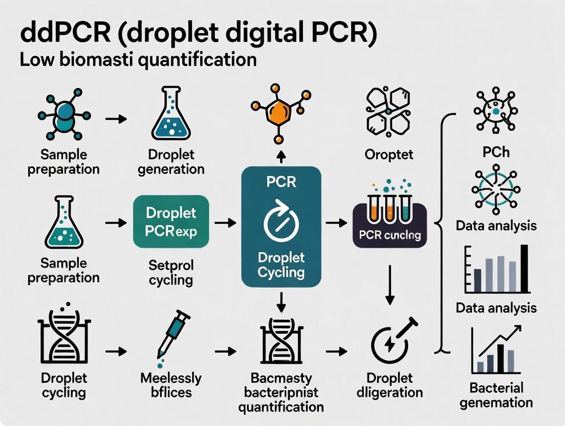
Workflow Visualization
Quantifying bacterial load in low-biomass environments presents significant challenges for molecular detection methods. Samples such as lung tissue, soil, and other complex matrices contain minimal bacterial DNA that often resides at the very limit of detection for conventional quantitative PCR (qPCR). Furthermore, these samples frequently contain PCR inhibitors that can compromise amplification efficiency and quantification accuracy. Droplet Digital PCR (ddPCR) technology addresses these limitations through a fundamentally different approach to nucleic acid quantification, providing unmatched sensitivity and robust tolerance to inhibitors that is revolutionizing low-biomass bacterial research.
Fundamental ddPCR Advantages for Low-Biomass Applications
Principle of Operation and Key Technological Differentiators
Droplet Digital PCR operates on the principle of sample partitioning, where a single PCR reaction is divided into thousands to millions of nanoliter-sized water-in-oil droplets [13] [14]. This partitioning effectively creates a massive array of independent micro-reactors, each containing zero, one, or a few target DNA molecules. Following end-point PCR amplification, each droplet is analyzed individually for fluorescence, with positive (target-present) and negative (target-absent) droplets counted digitally [13]. The absolute concentration of the target nucleic acid in the original sample is then calculated using Poisson statistics based on the ratio of positive to total droplets, eliminating the need for standard curves [13] [14].
The digital nature of this readout, combined with massive sample partitioning, confers two critical advantages for low-biomass applications. First, it dramatically increases the signal-to-noise ratio by separating rare target sequences from a complex background of non-target DNA [2]. Second, it inherently dilutes PCR inhibitors across thousands of partitions, minimizing their impact in any individual droplet and maintaining amplification efficiency where qPCR would fail [15] [13].
Quantitative Performance Comparison: ddPCR vs. qPCR
Table 1: Comparative Analytical Performance of ddPCR and qPCR in Low-Biomass Applications
| Performance Metric | ddPCR Performance | qPCR Performance | Experimental Context |
|---|---|---|---|
| Lower Limit of Quantification | 1.6 copies of qnrB target [15] | 15 copies of qnrB target [15] | Bacterial genomic DNA [15] |
| Detection in Negative Controls | 0.55 ± 0.28 16S/μL [16] | 1.00 ± 0.70 16S copies [16] | Lung tissue analysis [16] |
| Coefficient of Variation (CV) | 0.18 ± 0.14 [16] | 0.62 ± 0.29 [16] | 16S rRNA quantification in lung tissue [16] |
| Sensitivity in Complex Samples | 96.4% positive detection rate [17] | 83.9% positive detection rate [17] | Phytophthora nicotianae in soil samples [17] |
| Tolerance to PCR Inhibitors | Accurate quantification without facilitators [15] | Overestimation of targets, high sensitivity loss [15] | Soil and organic residue samples [15] |
Table 2: Protocol Selection Guide for Low-Biomass 16S rRNA Gene Sequencing
| Protocol Step | Standard Protocol | ddPCR-Enhanced Protocol | Key Advantage |
|---|---|---|---|
| DNA Input | 1–100 ng (commonly 12 ng) [6] | As low as 0.01 ng [6] | Enables work with vanishingly small samples |
| Amplicon Generation | Standard 1st-step PCR [6] | Standard 1st-step PCR [6] | Maintains compatibility with existing workflows |
| Library Preparation | Standard 2nd-step PCR [6] | Standard 2nd-step PCR + ddPCR re-amplification [6] | Generes sufficient product from minimal input |
| Detection Limit | Bands visible for DNA inputs ≥5 ng [6] | Successful sequencing with 0.01 ng input [6] | Pushes detection boundaries by orders of magnitude |
Experimental Evidence and Validation Studies
Bacterial Load Quantification in Lung Tissue
A direct comparison between ddPCR and qPCR for analyzing bacterial 16S load in lung tissue samples from control and COPD patients demonstrated ddPCR's superior performance in low-biomass settings. While both methods detected similar average bacterial loads in samples (ddPCR: 2.80 ± 1.80 16S/μL; qPCR: 2.32 ± 0.67 16S copies), ddPCR exhibited significantly lower background noise in negative controls (0.55 ± 0.28 16S/μL vs 1.00 ± 0.70 16S copies for qPCR) [2] [16]. This reduced background provides a cleaner baseline for detecting true signal in low-biomass samples. Additionally, the coefficient of variation was substantially lower for ddPCR (0.18 ± 0.14) versus qPCR (0.62 ± 0.29), indicating superior precision and reproducibility critical for research and diagnostic applications [16].
Pathogen Detection in Complex Environmental Samples
In agricultural research, a ddPCR assay developed for detecting Phytophthora nicotianae demonstrated greater sensitivity compared to qPCR when testing infectious tobacco root and soil samples. ddPCR achieved a 96.4% positive detection rate versus 83.9% for qPCR [17]. Receiver operating characteristic (ROC) analysis confirmed better diagnostic performance, with an area under the curve (AUC) of 0.913 for ddPCR compared to 0.885 for qPCR [17]. The study also highlighted ddPCR's better quantification accuracy for low pathogen concentrations in soil, attributing this advantage to better tolerance to potential PCR inhibitors present in complex environmental matrices [17].
16S rRNA Gene Sequencing from Minimal DNA Input
For 16S rRNA gene sequencing of low-biomass samples, standard protocols requiring 1-100 ng of DNA input present a major limitation. A novel protocol incorporating ddPCR enabled faithful amplification and sequencing with DNA inputs as low as 0.01 ng—concentrations often undetectable by standard fluorometric methods like Qubit [18] [6]. This approach demonstrated that with a ddPCR-enhanced workflow, samples of low bacterial biomass become directly comparable to those with high bacterial amounts, as the critical initial amplification steps remain identical, minimizing technical bias [6].
Detailed Experimental Protocols
ddPCR Protocol for Absolute Quantification of Bacterial 16S rRNA Genes
Principle: This protocol adapts established 16S rRNA qPCR assays to ddPCR for absolute quantification of bacterial load in low-biomass samples like lung tissue [2] [16].
Reagent Setup:
- Primers: Use previously published 16S rRNA assay primers spanning the V2 region [2] [16]
- Probe: (For probe-based assays) FAM-labeled TaqMan probe with appropriate quencher
- Reaction Mix: 10 μL of 2× ddPCR Supermix for Probes, 1 μL of each primer (final concentration 500 nM), 0.5 μL of probe (final concentration 250 nM), 2 μL of template DNA, nuclease-free water to 20 μL [17]
Procedure:
- Droplet Generation: Transfer 20 μL reaction mix to the DG8 cartridge. Add 70 μL of droplet generation oil. Place in QX200 Droplet Generator to generate approximately 20,000 droplets [17].
- PCR Amplification: Transfer droplets to a 96-well PCR plate. Seal with pierceable foil. Perform thermal cycling: 1 cycle at 95°C for 5-10 minutes; 40-45 cycles of 94-95°C for 30 seconds and 58-60°C for 1 minute; 1 cycle at 98°C for 10 minutes; hold at 4°C [2] [17].
- Droplet Reading: Place plate in QX200 Droplet Reader. Analyze using manufacturer's software.
- Data Analysis: Set threshold between positive and negative populations using negative controls. Use Poisson statistics to calculate absolute copy concentration (copies/μL) [13].
Troubleshooting Note: For samples with extremely low concentration (<0.05 ng/μL), an additional "emergency plan" re-amplification step using a high-fidelity polymerase may be required to generate sufficient product for sequencing [6].
ddPCR-Enhanced 16S rRNA Gene Sequencing Protocol for Low-Biomass Samples
Principle: This protocol enables 16S rRNA gene sequencing from DNA amounts insufficient for standard protocols by incorporating a ddPCR step after initial library preparation [6].
Procedure:
- Initial Library Preparation:
- Perform 1st-step PCR with primers targeting desired hypervariable regions (e.g., V1-V2, V3-V4, V7-V9) using even minimal DNA input (0.01-1 ng) [6].
- Clean PCR products using standard methods.
- Perform 2nd-step PCR with barcoded primers containing Illumina sequencing adapters (P5 and P7) [6].
- Clean amplicons again.
ddPCR Re-amplification:
- Dilute 2nd-step PCR amplicons to approximately one molecule per droplet [6].
- Prepare ddPCR mix: 10 μL of 2× ddPCR Supermix, plain P5 and P7 primers (no barcodes), diluted template, nuclease-free water to 20 μL.
- Generate droplets and perform PCR as in Protocol 4.1.
- Recover amplified products from droplets.
Sequencing:
Critical Considerations: Always include negative controls to detect contamination. Note that primer selection has a greater effect on taxonomic profiles than the use of high or low DNA input amounts [6].
The Scientist's Toolkit: Essential Research Reagent Solutions
Table 3: Essential Research Reagents for ddPCR in Low-Biomass Research
| Reagent/Material | Function | Application Notes |
|---|---|---|
| ddPCR Supermix for Probes | Provides optimized buffer, dNTPs, polymerase, and stabilizers for droplet PCR reactions | Essential for maintaining reaction stability within droplets; choose probe-based or EvaGreen formats based on assay design |
| Droplet Generation Oil | Creates stable water-in-oil emulsion for partitioning samples into nanoliter droplets | Critical for consistent droplet formation; must be compatible with droplet generator |
| DG8 Cartridges and Gaskets | Microfluidic chips for generating uniform droplets | Single-use consumables essential for the droplet generation process |
| TaqMan Probes | Sequence-specific fluorescent probes for target detection | Provide enhanced specificity over intercalating dyes; FAM-labeled most common |
| DNA Extraction Kits (DNeasy) | Isolation of high-quality DNA from complex matrices | Critical first step; efficiency varies by sample type (soil, tissue, etc.) [2] [17] |
| Nuclease-Free Water | Diluent and control substance | Must be certified nuclease-free to prevent false positives |
| Primer Sets (16S rRNA) | Target-specific amplification | V region selection (V1-V2, V3-V4, etc.) significantly impacts taxonomic profiles [6] |
Workflow Visualization
Diagram 1: ddPCR workflow for low-biomass samples with key advantages highlighted. The partitioning step confers both exceptional sensitivity and inhibitor tolerance.
Droplet Digital PCR represents a paradigm shift in low-biomass bacterial quantification, addressing fundamental limitations of qPCR through its digital partitioning approach. The technology's ability to provide absolute quantification without standard curves, coupled with unparalleled sensitivity down to single-copy detection and robust tolerance to PCR inhibitors, makes it particularly suited for challenging sample matrices like lung tissue, soil, and other low-biomass environments. As research continues to explore the microbial components of low-biomass systems, ddPCR stands as an essential tool for generating reliable, reproducible data that advances our understanding of microbial communities in these challenging environments.
Droplet Digital PCR (ddPCR) represents a significant advancement in nucleic acid quantification, operating on the principle of sample partitioning and end-point detection to achieve absolute quantification without the need for standard curves [13] [2]. This technology partitions a single PCR reaction into thousands of nanoliter-sized water-in-oil droplets, effectively creating individual microreactors [13]. The core principle of the "digital range" encompasses the limits of detection (LOD) and quantification (LOQ), which define the lowest concentrations at which a target can be reliably detected and precisely measured [19]. Within the context of low-biomass bacterial quantification, defining this range is paramount for generating reliable data, particularly when analyzing samples with minimal bacterial load, such as human lung tissue, sterile body sites, or environmental samples where total bacterial 16S rRNA gene concentrations may be as low as 1-10 copies/μL [2]. The digital nature of ddPCR, where target molecules are randomly distributed into droplets following a Poisson distribution, provides a statistical framework that confers exceptional sensitivity and precision at these low concentrations, overcoming key limitations of quantitative PCR (qPCR) [13] [2].
Technical Advantages for Low-Biomass Applications
The application of ddPCR for low-biomass bacterial research is driven by several key technical advantages that make it uniquely suited for challenging samples where target nucleic acids are scarce.
- Absolute Quantification Without Standard Curves: Unlike qPCR, ddPCR does not rely on external standard curves for quantification, which is particularly beneficial for low-biomass samples where creating accurate standards is challenging [13] [2]. This approach eliminates inaccuracies that can arise from differences in amplification efficiency between standard and sample reactions [13].
- Enhanced Sensitivity and Lower Detection Limits: The partitioning process significantly increases the signal-to-noise ratio by separating target molecules from background DNA [2]. This enables detection of rare targets and low-abundance sequences that would be obscured in a bulk PCR reaction [13] [20]. Studies have demonstrated successful detection of bacterial DNA at concentrations below the detection limit of fluorometric methods like Qubit [6].
- Superior Tolerance to PCR Inhibitors: Inhibitors present in complex sample matrices (e.g., stool, blood, soil) are diluted during partitioning, minimizing their impact on amplification efficiency [13]. This robustness is crucial for low-biomass samples where extensive purification can lead to complete loss of target material [6] [13].
- Improved Precision at Low Concentrations: The digital counting of molecules provides a direct measure of target concentration with high precision, even near the detection limit [2] [21]. This reduces the need for technical replicates, conserving precious sample material [2].
Table 1: Comparative Performance of ddPCR vs. qPCR for Low-Biomass Applications
| Parameter | ddPCR | Traditional qPCR |
|---|---|---|
| Quantification Method | Absolute (copies/μL) via Poisson statistics | Relative (requires standard curve) |
| Detection Limit | Can detect single copies [13] | Limited by standard curve and efficiency |
| Precision at Low Copy Number | High precision with 95% confidence intervals [2] | Lower precision, requires more replicates [2] |
| Effect of Inhibitors | Reduced impact due to partitioning [13] | Significant impact on amplification efficiency |
| Dynamic Range | Linear across a wide concentration range [13] | Limited by standard curve quality |
Establishing Limits of Detection and Quantification
Defining LOD and LOQ in Digital PCR
The Limit of Detection (LOD) represents the lowest concentration of target molecules that can be reliably distinguished from blank samples, while the Limit of Quantification (LOQ) defines the lowest concentration at which precise quantitative measurements can be made [19]. In ddPCR, these parameters are influenced by multiple factors including total number of droplets generated, background contamination levels, template partitioning efficiency, and reaction specificity [19].
For bacterial quantification in low-biomass environments, the LOD can be remarkably sensitive. In studies quantifying 16S rRNA genes in human lung tissue, ddPCR demonstrated reliable detection at concentrations as low as 1-10 copies/μL, where qPCR performance was suboptimal [2]. Similarly, for viral targets in clinical diagnostics, ddPCR assays have achieved LODs of 4 copies/mL for Hepatitis B Virus (HBV) DNA in serum, enabling detection in samples classified as undetectable by classical real-time PCR assays [22].
Experimental Determination of LOD and LOQ
The establishment of LOD and LOQ follows standardized experimental approaches:
- Limit of Blank (LOB) Determination: Multiple negative controls (no-template controls or blank samples) are analyzed to establish the background signal. The LOB is typically calculated as the 95th percentile of the blank measurement distribution [19].
- LOD Calculation: The LOD is determined by testing samples with known low concentrations of the target. Probit analysis is often employed, with the LOD defined as the concentration detected with 95% confidence [22] [19]. For example, one study established an LOD of 0.0001 TRECs/cell for a rare DNA target using probit analysis [19].
- LOQ Establishment: The LOQ is set as the lowest concentration where acceptable precision (typically <25% coefficient of variation) is maintained across replicates [19].
Table 2: Experimentally Determined Detection Limits in Various Sample Types
| Target | Sample Matrix | LOD | LOQ | Reference Technique |
|---|---|---|---|---|
| 16S rRNA genes | Human lung tissue | 1-10 copies/μL | Not specified | qPCR [2] |
| HBV DNA | Human serum | 4 copies/mL (<1 IU/mL) | Not specified | Real-time PCR [22] |
| TRECs | PBMCs from blood | 0.0001 copies/cell | ~0.0003 copies/cell | Standard ddPCR [19] |
| Bacterial DNA | Stool samples | Below Qubit detection limit | Not specified | Standard 16S rRNA sequencing [6] |
| HPV16 DNA | Liquid biopsies (plasma, serum) | Significantly lower than qPCR | Not specified | Standard ddPCR with purified cfDNA [20] |
Detailed Protocols for Low-Biomass Bacterial Quantification
Standard ddPCR Protocol for 16S rRNA Gene Quantification
This protocol enables absolute quantification of bacterial load in low-biomass samples such as tissue, sterile body fluids, or environmental samples with limited microbial content.
Sample Preparation and DNA Extraction
- Extract genomic DNA using kits designed for low-biomass samples (e.g., Qiagen DNeasy). Include negative extraction controls to monitor contamination [2].
- Quantify DNA using fluorometric methods. For samples with concentrations below detection limits, proceed with maximum available volume [6].
- Critical Consideration: For low-biomass samples, the DNA extraction method significantly impacts results. Consistency in extraction methodology is essential for comparative studies [6].
Reaction Setup
- Prepare ddPCR reaction mix containing:
- 10-11 μL of ddPCR Supermix for Probes (Bio-Rad)
- 0.9-1.8 μL each of forward and reverse primer (final concentration 900 nM)
- 0.25 μL of probe (final concentration 250 nM)
- 1-5 μL of template DNA (adjust volume based on expected concentration)
- Nuclease-free water to total volume of 20-22 μL [2]
- Primers should target conservative regions of the 16S rRNA gene. Common targets include V1-V2, V3-V4, or V7-V9 hypervariable regions [6].
Droplet Generation and PCR Amplification
- Transfer reaction mix to DG8 cartridges for droplet generation using the QX200 Droplet Generator [2].
- Carefully transfer generated droplets to a 96-well PCR plate and seal with foil using a pierceable heat seal [2].
- Perform PCR amplification with the following cycling conditions:
- 1 cycle: 95°C for 5-10 minutes (enzyme activation)
- 40 cycles: 95°C for 15-30 seconds (denaturation) and 60°C for 1 minute (annealing/extension)
- 1 cycle: 4°C for 5 minutes (hold)
- 1 cycle: 90°C for 5 minutes (enzyme deactivation) [2]
- Ramp rate should be set at 2°C/second [2].
Droplet Reading and Data Analysis
- Load plate into the QX200 Droplet Reader for automated counting of positive and negative droplets [2].
- Analyze data using manufacturer's software (e.g., QuantaSoft). Set threshold between positive and negative droplets based on fluorescence amplitude of negative controls [2].
- Calculate absolute concentration (copies/μL) applying Poisson statistics to account for multiple targets per droplet [13].
Crude Lysate Protocol for Minimal Sample Input
For extremely limited samples where DNA extraction would result in complete loss of material, a crude lysate approach eliminates the extraction step, enabling quantification from as few as 200 cells [19].
Cell Lysis Preparation
- Collect cells (200-10,000 cells) by centrifugation and resuspend in PBS [19].
- Prepare lysis buffer using commercial kits (e.g., SuperScript IV CellsDirect cDNA Synthesis Kit lysis buffer) [19].
- Incubate cell suspension with lysis buffer to release DNA.
Viscosity Reduction
- Critical Step: Implement viscosity breakdown protocol to reduce interference from cellular debris and intact oligonucleotides [19].
- Process lysed samples prior to droplet formation to ensure proper droplet generation [19].
ddPCR Reaction Setup
- Use processed lysate directly in ddPCR reaction mix, replacing template DNA volume with equivalent lysate volume [19].
- Adjust droplet generation parameters if necessary to accommodate residual cellular components.
Data Analysis Considerations
- Account for potential background fluorescence from cellular components when setting thresholds [19].
- Validate against standard DNA extraction methods for accuracy verification [19].
The Scientist's Toolkit: Essential Research Reagents
Successful implementation of ddPCR for low-biomass bacterial quantification requires specific reagents and materials optimized for sensitive detection.
Table 3: Essential Reagents for ddPCR-Based Low-Biomass Bacterial Quantification
| Reagent/Material | Function | Application Notes |
|---|---|---|
| ddPCR Supermix for Probes | Provides optimized reaction components including polymerase, dNTPs, and buffer | Essential for consistent droplet formation and amplification efficiency [2] |
| 16S rRNA-Targeted Primers & Probes | Specific amplification of bacterial DNA | Design to target appropriate variable regions (V1-V2, V3-V4, V7-V9); choice significantly impacts taxonomic profiles [6] |
| DG8 Cartridges & Droplet Generation Oil | Creates uniform water-in-oil emulsion | Critical for partitioning efficiency; requires proper storage and handling [2] |
| QX200 Droplet Generator | Partitions samples into nanoliter droplets | Consistent droplet generation is key to quantitative accuracy [2] |
| QX200 Droplet Reader | Detects fluorescence in individual droplets | Must be properly calibrated for threshold setting [2] |
| Low-Binding Tubes and Tips | Sample handling and preparation | Minimizes adsorption of low-concentration nucleic acids [6] |
| Negative Control Reagents | Contamination monitoring | Nuclease-free water and extraction blanks essential for establishing LOB [2] |
| Commercial Lysis Buffers | Cell disruption for crude lysate protocols | Enables analysis of minimal samples without DNA extraction [19] |
Troubleshooting Common Challenges in Low-Biomass ddPCR
- High Background Signal in Negative Controls: Implement rigorous contamination controls, including extraction blanks and no-template controls. Use separate work areas for pre- and post-PCR steps and consider UV irradiation of workspaces [6].
- Poor Droplet Generation: Ensure samples are free of particulates that can clog microfluidic channels. For crude lysate protocols, the viscosity breakdown step is essential [19].
- Inconsistent Amplification Between Replicates: This is common at very low target concentrations due to Poisson sampling statistics. Increase sample input volume and number of technical replicates to improve precision [2].
- Inhibition Despite Partitioning: While ddPCR is more tolerant to inhibitors than qPCR, strong inhibition can still occur. Dilute samples or implement additional purification steps if necessary [13].
Defining the digital range through rigorous determination of limits of detection and quantification is fundamental to applying ddPCR technology to low-biomass bacterial research. The exceptional sensitivity, absolute quantification capability, and inhibitor tolerance of ddPCR make it uniquely suited for challenging samples where traditional methods fail. The protocols outlined herein provide a framework for reliable implementation, while the troubleshooting guidance addresses common pitfalls. As research continues to explore microbial communities in low-biomass environments, the precision of ddPCR will play an increasingly critical role in generating accurate, reproducible data that advances our understanding of microbial ecology and pathogenesis.
From Theory to Bench: A Step-by-Step ddPCR Workflow for Bacterial Quantification
The analysis of complex, low-biomass samples presents unique challenges in microbial ecology, clinical diagnostics, and drug development. In the context of droplet digital PCR (ddPCR) for bacterial quantification, the extraction step is not merely a preliminary procedure but a critical determinant of experimental success. Low-biomass environments—such as respiratory samples, tissue biopsies, water filtration systems, and clinical specimens from treated patients—contain limited microbial material, where inefficient DNA recovery can lead to complete experimental failure or significant data bias [6]. The integration of ddPCR into this workflow represents a transformative approach, as its superior sensitivity allows for the absolute quantification of microbial loads even when template DNA is undetectable by conventional fluorometric methods [6].
The fundamental challenge in low-biomass research lies in the inefficient lysis of tough-to-lyse microorganisms (such as Gram-positive bacteria) and the co-extraction of inhibitors that can downstream molecular applications. These factors disproportionately affect low-biomass samples where the signal-to-noise ratio is already compromised. Consequently, optimization of DNA extraction is not a one-size-fits-all protocol but rather a sample-specific strategy that must account for community composition, sample matrix, and intended analytical outcomes [6]. Within a broader thesis on ddPCR for low-biomass bacterial quantification, this application note establishes the foundational principles and practical methodologies for maximizing DNA yield, quality, and representativeness—thereby ensuring the reliability of subsequent digital PCR quantification.
Key Principles for Extraction Optimization
Addressing Fundamental Challenges
Optimizing DNA extraction from low-biomass environments requires addressing several interconnected challenges that can compromise data integrity:
Inefficient Cell Lysis: Microbial communities in environmental and clinical samples often contain mixtures of Gram-positive and Gram-negative bacteria with differing cell wall structures. Gram-positive bacteria, with their thick peptidoglycan layers, are frequently underrepresented in standard extraction protocols due to incomplete lysis [6]. This leads to biased community representation that cannot be corrected in downstream analysis, regardless of the quantification method employed.
Inhibitor Co-extraction: Complex sample matrices (e.g., soil, feces, clinical specimens) contain substances that inhibit enzymatic reactions in subsequent PCR steps. Common inhibitors include humic acids in environmental samples, bile salts in fecal matter, and hemoglobin in blood samples [13]. These compounds can be co-extracted with nucleic acids and significantly reduce amplification efficiency, particularly problematic in low-biomass scenarios where template DNA is already limited.
DNA Loss and Fragmentation: During extraction and purification, DNA molecules can be lost through adsorption to tube surfaces, incomplete precipitation, or failure to bind to purification matrices. This issue is exacerbated in low-biomass samples where the starting material is minimal. Additionally, DNA fragmentation becomes a critical concern, especially with formalin-fixed paraffin-embedded (FFPE) clinical specimens, where fixation and storage conditions can severely degrade DNA [23].
Background Contamination: Reagents, kits, and laboratory environments contribute exogenous bacterial DNA that becomes significantly problematic when analyzing samples with minimal endogenous biomass. This contamination can dominate sequencing libraries or quantitative assays, leading to erroneous conclusions about sample composition [6].
The Case for Integration with ddPCR
Droplet Digital PCR offers distinct advantages for quantifying microbial abundance in low-biomass samples following extraction. Unlike quantitative PCR (qPCR), ddPCR provides absolute quantification without requiring standard curves, achieving higher precision through sample partitioning into thousands of nanoliter-sized droplets [13]. This partitioning also confers greater resistance to PCR inhibitors, as inhibitors are diluted into individual droplets, minimizing their impact on amplification [13]. The exceptional sensitivity of ddPCR enables detection of target sequences in samples where DNA concentrations fall below the detection limit of conventional fluorometers [6], making it ideally suited for low-biomass applications where material is precious and limited.
Table 1: Comparison of Quantification Methods for Low-Biomass Samples
| Method | Sensitivity | Precision | Inhibitor Resistance | Quantification Type | Best Application |
|---|---|---|---|---|---|
| ddPCR | High (detects single copies) | High (low CV) | High (inhibitors partitioned) | Absolute (copies/μL) | Low-biomass samples, rare targets [13] |
| qPCR | Moderate | Moderate | Low to Moderate | Relative (requires standard curve) | Samples with sufficient DNA [13] |
| Fluorescence Spectroscopy | Moderate | Low to Moderate | Not Applicable | Indirect (cell counts) | Simple matrices, rapid assessment [1] |
| Flow Cytometry | Moderate | Moderate | Not Applicable | Direct (cell counts) | Samples with intact cells [1] |
| Spike-in Standards | High | High | Varies with method | Absolute (with normalization) | Sequencing-based approaches [1] |
Recommended Extraction Optimization Protocol
Comprehensive Workflow for Low-Biomass Samples
The following protocol has been specifically adapted for low-biomass samples intended for downstream ddPCR analysis, with particular emphasis on maximizing yield and reducing bias.
Sample Preparation and Pre-treatment
- For solid samples (soil, stool, tissue), use lyophilization followed by mechanical disruption (e.g., bead beating) to homogenize the matrix before extraction.
- For liquid samples, concentrate biomass via filtration (0.22 μm filters) or centrifugation (14,000 × g for 30 minutes).
- Implement pre-washing steps with inhibitor-specific solutions: phosphate buffer for humic acids, ethanol washes for bile salts [1].
- Incorporate process controls by spiking with a known quantity of exogenous cells (e.g., Pseudomonas aeruginosa) not expected in the sample to assess extraction efficiency [6].
Cell Lysis Optimization
- Employ a combination of mechanical and enzymatic lysis for comprehensive community representation.
- For mechanical lysis: Use bead beating with a mixture of bead sizes (e.g., 0.1 mm and 0.5 mm glass beads) for 3-5 minutes at maximum speed.
- For enzymatic lysis: Implement a sequential approach with lysozyme (for Gram-positives), proteinase K, and mutanolysin where appropriate.
- Include a heating step (70°C for 10-20 minutes) to enhance lysis efficiency, particularly for difficult-to-lyse organisms.
DNA Purification and Inhibition Removal
- Use inhibitor removal columns specifically designed for the sample type (e.g., OneStep PCR Inhibitor Removal Kit for stool samples).
- Implement silica-based membrane columns with multiple wash steps to ensure complete inhibitor removal while minimizing DNA loss.
- For particularly challenging samples, consider gel electrophoresis with excision and extraction of high-molecular-weight DNA to remove co-extracted contaminants.
- Elute DNA in low-EDTA TE buffer or molecular grade water (never in nuclease-free water alone) to prevent chelation of magnesium ions required for PCR.
Quality Assessment and Quantification
- Assess DNA quality via agarose gel electrophoresis to confirm high molecular weight and absence of significant degradation.
- Quantify DNA using fluorometric methods (e.g., Qubit) rather than spectrophotometry, as the latter is inaccurate for low-concentration samples and cannot detect common contaminants.
- Test DNA extractability using ddPCR targeting universal 16S rRNA genes, which provides more sensitive quantification than fluorometry for low-biomass samples [6].
Research Reagent Solutions
Table 2: Essential Reagents for Optimized DNA Extraction from Low-Biomass Samples
| Reagent/Category | Specific Examples | Function & Application Notes |
|---|---|---|
| Inhibitor Removal Resins | OneStep PCR Inhibitor Removal Kit, PVPP | Binds to and removes humic acids, polyphenols, and other common inhibitors from environmental and clinical samples [1] |
| Mechanical Disruption Aids | 0.1 mm & 0.5 mm silica/zirconia beads | Enhances lysis of tough microbial cell walls through bead beating; combination of sizes improves efficiency [6] |
| Enzymatic Lysis Cocktail | Lysozyme, Proteinase K, Mutanolysin | Enzymatically digests peptidoglycan layers (Gram-positives) and protein components; sequential application recommended [6] |
| DNA Binding Matrices | Silica membrane columns, magnetic beads | Selective binding of DNA while removing contaminants; magnetic beads preferred for automated high-throughput applications [11] |
| Process Controls | Exogenous microbial cells (e.g., P. aeruginosa), synthetic DNA spikes | Monitors extraction efficiency and identifies potential contamination; essential for normalization in quantitative studies [6] |
| Digital PCR Reagents | ddPCR Supermix for Probes, EvaGreen Supermix | Optimized reaction chemistry for partitioned PCR; probe-based methods offer higher specificity for targeted applications [24] |
Quantitative Assessment of Extraction Efficiency
Rigorous assessment of extraction efficiency is paramount for low-biomass studies. The following approaches provide quantitative metrics for protocol optimization and inter-study comparisons.
Spike-In Controls for Efficiency Calculation
- Incorporate a known quantity of an exogenous microbe (e.g., P. aeruginosa ATCC 27853) or synthetic DNA construct at the beginning of extraction.
- After extraction, quantify the recovery using ddPCR with specific primers/probes for the spike-in target.
- Calculate extraction efficiency: (Recovered spike-in copies / Initial spike-in copies) × 100%.
- Acceptable efficiency thresholds: >50% for complex matrices (e.g., soil, stool), >70% for simpler matrices (water, clinical swabs).
Comparative Analysis of Lysis Efficiency
- Apply different lysis methods (enzymatic only, mechanical only, combined) to aliquots of the same sample.
- Quantify total 16S rRNA gene copies using ddPCR with universal primers (e.g., 341F/534R targeting the V3-V4 region).
- The method yielding the highest copy number represents the most effective lysis protocol for that sample type.
Inhibition Assessment via Dilution Series
- Perform ddPCR on neat and diluted (1:2, 1:4, 1:8) DNA extracts.
- Calculate the observed/expected ratio of copies/μL across dilutions.
- A consistent ratio indicates minimal inhibition, while improving ratios with dilution suggests presence of PCR inhibitors.
Table 3: Troubleshooting Common Extraction Issues in Low-Biomass Samples
| Problem | Potential Causes | Solutions | Expected Outcome |
|---|---|---|---|
| Low DNA Yield | Incomplete lysis, DNA loss during purification, insufficient starting material | Optimize bead beating duration/speed, implement carrier RNA during precipitation, increase sample input volume | 2-5x increase in quantifiable 16S rRNA copies by ddPCR [6] |
| Incomplete Community Representation | Bias against difficult-to-lyse organisms (Gram-positives, spores) | Combine mechanical and enzymatic lysis, extend incubation times, use specialized lysis buffers | Improved detection of Gram-positive taxa (Firmicutes, Actinobacteria) [6] |
| PCR Inhibition | Co-purification of humic substances, bile salts, hemoglobin | Implement inhibitor removal columns, dilute templates, add BSA to PCR reactions | Lower Ct values in qPCR, increased droplet amplitude in ddPCR [13] |
| High Background Contamination | Reagent contaminants, environmental DNA | Use UV-irradiated reagents, dedicated workspace, include extraction controls | Reduction in negative control signals to <10% of sample signal [6] |
| DNA Fragmentation | Overly aggressive bead beating, nuclease activity, sample degradation | Optimize mechanical disruption time, use nuclease inhibitors, gentle pipetting | Presence of high molecular weight DNA on agarose gels |
Downstream ddPCR Analysis of Extracted DNA
Following optimized extraction, ddPCR provides the most sensitive approach for absolute quantification of microbial targets in low-biomass samples. The partitioning of reactions into thousands of nanoliter-sized droplets not only enables precise quantification but also confers exceptional resilience to inhibitors that may remain despite optimized extraction [13].
16S rRNA Gene Quantification for Total Bacterial Load
- Target conservative regions of the 16S rRNA gene with universal primers (e.g., 341F/534R for V3-V4 region).
- Use probe-based detection (e.g., FAM-labeled TaqMan probes) for enhanced specificity over intercalating dyes.
- Calculate total bacterial density: (Concentration in copies/μL × Elution Volume) / Sample Input Mass or Volume.
- Report as 16S rRNA gene copies per gram (solid samples) or per milliliter (liquid samples).
Taxon-Specific Absolute Quantification
- Design species- or group-specific primers and probes for targets of interest (e.g., pathogens, keystone taxa).
- Apply the same ddPCR conditions as for universal quantification to enable direct comparison.
- Calculate absolute abundance of specific taxa: (Target concentration / Total 16S concentration) × Total bacterial load.
Data Normalization and Interpretation
- Normalize data using process control recovery rates to account for extraction efficiency variations.
- For comparative studies, report both absolute abundances (copies/gram or mL) and relative abundances to provide comprehensive community insights.
- Apply Poisson correction to ddPCR data to account for template partitioning statistics, as implemented in manufacturer software (e.g., Bio-Rad's QuantaSoft) [13].
Optimization of DNA extraction from complex, low-biomass samples represents a foundational step in ensuring the reliability of downstream ddPCR analysis for bacterial quantification. The implementation of a rigorous, sample-specific extraction protocol that addresses the key challenges of inefficient lysis, inhibitor co-extraction, and DNA loss is essential for generating meaningful quantitative data. When coupled with the exceptional sensitivity and precision of ddPCR, researchers can achieve unprecedented insights into microbial communities in challenging environments where traditional approaches fail. This application note provides a comprehensive framework for maximizing DNA yield, quality, and representativeness—establishing a robust foundation for accurate absolute quantification in low-biomass research.
In the field of low biomass bacterial quantification, the absolute quantification of specific bacterial strains provides a critical advantage over relative abundance measurements, which can often be misleading [25]. The ability to detect and quantify a specific bacterial strain within a complex microbial community, such as the gut microbiome or environmental samples, is paramount for applications in probiotics, live biotherapeutics, and pathogen tracking [26] [27]. Droplet Digital PCR (ddPCR) emerges as a particularly powerful tool for this purpose, offering superior sensitivity and absolute quantification without the need for a standard curve, making it exceptionally suited for low abundance targets often encountered in low biomass research [28] [29].
While next-generation sequencing (NGS) can identify strains, it is semi-quantitative and suffers from high detection limits and compositional data limitations [26]. PCR-based methods, conversely, can provide the highly sensitive and quantitative data needed. The foundational step for achieving this specificity lies not in the platform itself, but in the initial design of primers and probes that target unique, strain-specific genetic markers. This application note details a comprehensive protocol for designing these critical reagents and their subsequent validation using ddPCR, framed within a research context focused on challenging low biomass samples.
The Scientist's Toolkit: Research Reagent Solutions
The following table outlines the essential materials and reagents required for the development and execution of a strain-specific ddPCR assay.
Table 1: Key Research Reagent Solutions for Strain-Specific ddPCR
| Reagent / Material | Function / Explanation |
|---|---|
| ddPCR Supermix (No dUTP) | A core PCR master mix optimized for the generation of water-in-oil droplets, ensuring consistent amplification across thousands of partitions [28]. |
| Strain-Specific Primers & Probes | Custom-designed oligonucleotides that bind to unique genomic regions (e.g., single-copy genes) of the target strain, forming the basis of assay specificity [26]. |
| FRET Cassette (e.g., for KASP) | A universal fluorescence resonance energy transfer reporter system used in some allele-specific protocols, eliminating the need for custom-labeled probes and increasing design flexibility [30]. |
| Droplet Generation Oil | Specialized oil used to partition the aqueous PCR reaction into thousands of nanoliter-sized droplets, the core of the ddPCR technology [28]. |
| DNA Extraction Kit (e.g., QIAamp Fast DNA Stool Mini Kit) | For isolating high-quality DNA from complex sample matrices like fecal samples or environmental swabs. Kit-based methods are recommended for consistency and inhibitor removal [26]. |
| Nuclease-Free Water | Used to reconstitute primers and probes and as a negative control to monitor for contamination, which is critical in low-biomass applications [28]. |
Core Principles of Strain-Specific Assay Design
Target Selection and In Silico Design
The foremost requirement for ultimate specificity is the identification of a unique genomic signature exclusive to the target strain.
- Identifying Marker Genes: Begin with a comparative genomic analysis. Whole genome sequences of the target strain and closely related strains (from public databases like NCBI) are compared to identify genes or intergenic regions that are unique to the target. Ideal targets are single-copy, essential genes that provide a stable and reliable quantification marker [26].
- Primer and Probe Design Software: Utilize bioinformatic tools to design the actual oligonucleotides. Software like the one described by [30] allows for the design of allele-specific primers where the 3'-end nucleotide is positioned directly on the single nucleotide variant (SNV) responsible for the strain's uniqueness. This "penultimate" base positioning maximizes discriminatory power by ensuring inefficient amplification in non-target strains with a mismatch at this critical location [30].
- Probe Selection: For TaqMan-based ddPCR assays, design a dual-labeled hydrolysis probe that binds within the amplicon defined by your strain-specific primers. For alternative chemistries like KASP (Kompetitive Allele Specific PCR), the design involves primers with unique 5' tail sequences that are complementary to universal FRET cassettes, eliminating the need for custom-labeled probes and reducing costs [30].
Comparative Analysis of Quantitative PCR Platforms
Choosing the right quantification platform is crucial. The table below summarizes the key differences between ddPCR and qPCR in the context of strain-specific quantification, particularly for low-biomass scenarios.
Table 2: Platform Comparison: qPCR vs. ddPCR for Strain-Specific Quantification
| Parameter | Quantitative PCR (qPCR) | Droplet Digital PCR (ddPCR) |
|---|---|---|
| Principle | Relies on amplification kinetics and a standard curve for relative quantification. | Partitions sample into droplets for end-point, digital counting of target molecules [31]. |
| Quantification | Relative (requires a standard curve). | Absolute quantification without a standard curve [26] [31]. |
| Susceptibility to Inhibitors | More susceptible, as inhibitors affect amplification efficiency, altering Ct values [28] [26]. | More resilient, as partitioning dilutes inhibitors and the endpoint measurement is less affected by efficiency variations [28] [32] [31]. |
| Precision & Sensitivity | Good, but cannot reliably detect small-fold changes (<2x). Lower limit of detection around 10⁴ cells/g feces for strain quantification [26]. | Higher precision and sensitivity; can detect differences <30%, rare alleles (<0.1%), and has a lower limit of detection (~10³ cells/g feces) [26] [33] [31]. |
| Dynamic Range | Wider dynamic range [26]. | Dynamic range is extended through Poisson correction, but can be limited at very high target concentrations [29]. |
| Best Use-Case | High-abundance targets, cost-sensitive projects where relative quantification is sufficient. | Low-biomass/low-abundance targets, absolute quantification required, and complex samples with PCR inhibitors [28] [26] [33]. |
The following workflow diagram illustrates the complete process from genomic analysis to final absolute quantification using ddPCR.
Experimental Protocol: A Step-by-Step Guide
Step 1: DNA Extraction from Complex Samples
Consistent and unbiased DNA extraction is critical. For fecal samples, kit-based methods (e.g., QIAamp Fast DNA Stool Mini Kit) are recommended over phenol-chloroform protocols due to better reproducibility and lower inhibitor co-extraction [26].
- Procedure: Follow the manufacturer's instructions with an added bead-beating step for robust cell lysis of hard-to-lyse Gram-positive bacteria [26]. Include a sample pre-wash with ice-cold PBS to remove soluble PCR inhibitors commonly found in feces [26]. Elute DNA in nuclease-free water or TE buffer and quantify using a spectrophotometer (e.g., NanoDrop). Note that a low 260/230 ratio may indicate residual inhibitors [28].
Step 2: Optimization of the ddPCR Assay
Once DNA is extracted, the ddPCR conditions must be meticulously optimized.
- Reaction Setup: Prepare a 22 µL reaction mixture containing 11 µL of 2x ddPCR Supermix for Probes (No dUTP), 0.9 µM of each strain-specific primer, 0.25 µM of the probe (or equivalent for EvaGreen assays), and 2 µL of template DNA [28].
- Droplet Generation: Load 20 µL of the reaction mix into a DG8 cartridge along with 70 µL of droplet generation oil. Use the QX200 Droplet Generator to create the water-in-oil emulsion [28].
- Thermal Cycling: Transfer the droplets to a 96-well PCR plate and run the following protocol on a thermal cycler:
- *Annealing Temperature Optimization: This is a critical step. Perform a gradient PCR (e.g., from 55°C to 65°C) using a standard PCR machine and analyze products on an agarose gel. The optimal temperature is the one that produces a single, bright band of the expected size. Confirm in the ddPCR system by selecting the temperature that best separates positive and negative droplet populations [28] [29].
Step 3: Specificity and Sensitivity Validation
- Specificity Testing: Test the optimized assay against DNA from a panel of non-target strains, especially the most phylogenetically close relatives. The assay should yield a strong positive signal only for the target strain and no amplification for non-targets [26].
- Sensitivity and Limit of Detection (LOD): Create a standard curve by spiking a known number of target bacterial cells (determined by plate counting) into a negative background matrix (e.g., sterile fecal slurry or buffer). Perform a dilution series and run each dilution in the ddPCR assay. The LOD is the lowest concentration where amplification is consistently detected with 100% reproducibility [26] [29]. Studies have shown that well-optimized, strain-specific qPCR/ddPCR can achieve an LOD of around 10³ cells/g feces, far superior to NGS approaches [26].
The mechanism of a TaqMan probe-based ddPCR assay for strain-specific detection is illustrated below.
The integration of meticulously designed, strain-specific primers and probes with the robust quantification power of ddPCR creates a formidable toolkit for researchers tackling the challenges of low biomass bacterial quantification. This protocol provides a roadmap from in silico design to a fully validated assay, enabling the precise absolute quantification necessary to move beyond compositional data and uncover true biological dynamics in complex microbial ecosystems. This approach is indispensable for the accurate tracking of probiotics, pathogens, and live biotherapeutics in both clinical and environmental settings.
In the field of low biomass bacterial quantification, such as studies of the lung microbiome or environmental samples, the accuracy of droplet digital PCR (ddPCR) is highly dependent on robust assay optimization [32]. Unlike quantitative real-time PCR (qPCR), ddPCR partitions a sample into thousands of nanoliter-sized droplets, allowing for absolute quantification of target DNA without the need for a standard curve [34] [28]. This partitioning reduces the impact of PCR inhibitors and increases the effective concentration of the target, making it particularly suited for detecting low-abundance targets [35] [28]. However, suboptimal assay conditions can lead to poor separation between positive and negative droplet populations, a phenomenon known as "rain," which complicates data analysis and can compromise quantification accuracy [36] [37]. This application note details a systematic protocol for optimizing two of the most critical parameters in ddPCR assay development: annealing temperature and reagent concentration, with a specific focus on applications in low biomass bacterial research.
The Critical Role of Optimization in Low Biomass Quantification
The quantification of bacterial load in low biomass samples, such as lung tissue or filtered environmental water, presents unique challenges [32] [28]. In these samples, the target DNA concentration is often very low (e.g., 1–10 copies/µL), pushing detection to the limits of the technology [32]. Imperfect assay conditions can cause a significant proportion of droplets to exhibit intermediate fluorescence, or "rain," which obscures the clear discrimination needed for precise Poisson-based quantification [36] [37]. This is especially critical when quantifying total bacterial load via the 16S rRNA gene, as required for calculating the fungi-to-bacteria ratio or absolute prokaryotic abundance in complex samples [35] [38]. A well-optimized assay minimizes rain by maximizing the fluorescence amplitude and the separation between negative and positive droplet clusters. The "droplet separation value," an objective metric that considers both the absolute fluorescence signal distance and the variation within the positive and negative populations, is a key indicator of a robust assay [36] [37]. Furthermore, optimization enhances tolerance to PCR inhibitors commonly found in environmental and clinical samples, such as humic substances or heme, ensuring more accurate results even in complex matrices [17] [34] [28].
Key Parameters for ddPCR Optimization
Annealing/Extension Temperature
The annealing temperature is a primary factor influencing assay specificity and efficiency. An inappropriate temperature can lead to non-specific amplification or reduced yield, directly contributing to rain and poor droplet separation [37].
- Optimization Strategy: A temperature gradient experiment is the most effective method for identifying the optimal annealing/extension temperature. The recommended range is typically ±5 °C from the calculated primer Tm [28].
- Experimental Protocol:
- Prepare a master mix containing the ddPCR supermix, primers, probe, and a template with a known, moderate concentration of the target (e.g., genomic DNA from a positive control sample).
- Aliquot the master mix into a 96-well plate suitable for the droplet generator.
- Run the ddPCR protocol using a thermal cycler with a gradient function across the desired temperature range (e.g., from 55.5 °C to 60.5 °C in 0.5 °C steps) [28].
- After amplification, analyze the droplets using the droplet reader and associated software.
- Data Analysis: The optimal temperature is identified by the clearest separation between positive and negative droplet clusters, the highest fluorescence amplitude of the positive cluster, and the minimal amount of rain [37]. This can be quantified using the droplet separation value if supported by the software [36].
Oligonucleotide Concentration
The concentrations of primers and probes significantly impact the reaction's dynamics, affecting both fluorescence amplitude and the appearance of rain [36] [37].
- Standard vs. High Concentrations: While "normal" concentrations from established qPCR protocols can be used, increasing them to "high" concentrations (e.g., 900 nM for primers and 250 nM for probes) has been shown to improve ddPCR performance for certain targets [37].
- Experimental Protocol:
- Design a matrix of reactions testing different combinations of primer and probe concentrations. A typical matrix might include primer concentrations of 500 nM and 900 nM, cross-tested with probe concentrations of 125 nM and 250 nM [37] [17].
- Use a single, optimally diluted positive control sample and a consistent, optimized annealing temperature across all reactions.
- Process the reactions through droplet generation, PCR amplification, and droplet reading.
- Data Analysis: Evaluate the results based on the endpoint fluorescence, the droplet separation value, and the measured concentration of the target. The combination that yields the highest target concentration with the clearest droplet separation and lowest coefficient of variation is considered optimal [36] [37].
Table 1: Summary of Key Optimization Parameters and Their Effects
| Parameter | Typical Range Tested | Impact of Insufficient/Excessive Value | Optimal Outcome Indicator |
|---|---|---|---|
| Annealing Temperature | Primer Tm ±5 °C [28] | Low: Non-specific binding, high rain.High: Reduced efficiency, low fluorescence. | Maximal fluorescence amplitude and droplet separation value [36]. |
| Primer Concentration | 500 - 900 nM [37] [17] | Low: Reduced fluorescence, poor sensitivity.High: Increased background, potential primer-dimer. | High target concentration with low CV and clear cluster separation [37]. |
| Probe Concentration | 125 - 250 nM [37] [17] | Low: Weak fluorescent signal.High: Increased background fluorescence. | Strong positive cluster signal with minimal rain [37]. |
Integrated Workflow for Comprehensive Assay Optimization
The following workflow integrates the optimization of annealing temperature and reagent concentrations into a logical sequence. This systematic approach ensures that interactions between parameters are accounted for, leading to a robust and reliable ddPCR assay.
Detailed Experimental Protocols
Protocol 1: Annealing Temperature Optimization
This protocol is designed to identify the optimal annealing/extension temperature for your ddPCR assay using a thermal gradient.
Materials:
- QX200 ddPCR Supermix for Probes (Bio-Rad) [17]
- Primers and hydrolysis probe (e.g., FAM-labeled) [17]
- Template DNA (from target organism or positive control)
- Nuclease-free water
- QX200 Droplet Generator and DG8 Cartridges (Bio-Rad) [37]
- Thermal cycler with gradient functionality (e.g., Bio-Rad T100) [37] [28]
- QX200 Droplet Reader (Bio-Rad)
Procedure:
- Prepare Master Mix: On ice, prepare a sufficient master mix for all reactions and a no-template control (NTC). Per 20-22 µL reaction:
- Generate Droplets: Pipet 20 µL of each reaction mix and 70 µL of droplet generation oil into the wells of a DG8 cartridge. Place the cartridge in the droplet generator. Once generated, carefully transfer the emulsified samples (~40 µL) to a semi-skirted 96-well PCR plate. Seal the plate with a pierceable foil heat seal.
- Amplify with Gradient: Place the sealed plate in the thermal cycler and run the following PCR protocol with a gradient across the annealing/extension step:
- Read Droplets: Transfer the plate to the droplet reader for automatic counting of positive and negative droplets.
- Analyze Data: Using the analysis software (e.g., QuantaSoft), assess the data from each temperature. The optimal temperature will produce two distinct, tight clusters of positive and negative droplets with maximal fluorescence difference and minimal rain.
Protocol 2: Oligonucleotide Concentration Titration
This protocol should be performed after determining the optimal annealing temperature to fine-tune primer and probe concentrations.
Procedure:
- Design Concentration Matrix: Prepare a series of master mixes that vary the concentrations of primers and probes. A typical 3x2 matrix might include:
- Primer Concentrations: 500 nM, 700 nM, 900 nM
- Probe Concentrations: 125 nM, 250 nM [37]
- Run ddPCR Reactions: For each concentration combination, prepare reactions as described in Protocol 1, using the optimal annealing temperature identified from the gradient experiment. Include an NTC for each primer/probe combination.
- Generate and Read Droplets: Follow the same droplet generation, PCR amplification, and reading steps as in Protocol 1.
- Evaluate Performance: For each condition, record:
Table 2: Example Results from an Oligonucleotide Concentration Matrix for a 16S rRNA Gene Assay
| Primer (nM) | Probe (nM) | Measured Conc. (copies/µL) | CV (%) | Droplet Separation | Rain Assessment |
|---|---|---|---|---|---|
| 500 | 125 | 25.5 | 8.5 | Good | Moderate |
| 500 | 250 | 26.8 | 7.1 | Good | Low |
| 700 | 125 | 28.1 | 6.5 | Very Good | Low |
| 700 | 250 | 29.5 | 5.2 | Excellent | Very Low |
| 900 | 125 | 28.9 | 6.0 | Excellent | Low |
| 900 | 250 | 30.1 | 4.8 | Excellent | Minimal |
The Scientist's Toolkit: Research Reagent Solutions
Table 3: Essential Reagents and Equipment for ddPCR Assay Optimization
| Item | Function/Application | Example Product/Catalog Number |
|---|---|---|
| ddPCR Supermix for Probes | Optimized reaction buffer for probe-based assays in droplet digital PCR. | Bio-Rad ddPCR Supermix for Probes (186-3010) [37] |
| Droplet Generation Cartridges | Consumable for partitioning samples into nanoliter-sized droplets. | Bio-Rad DG8 Cartridges (186-3008 or 186-4008) [37] |
| Droplet Generation Oil | Oil for creating water-in-oil emulsion during droplet generation. | Bio-Rad Droplet Generation Oil for Probes (186-3005) [28] |
| Hydrolysis Probes | Sequence-specific probes (e.g., FAM, HEX/VIC) for target detection. | Synthesized by providers (TIB Molbiol, Eurofins-MWG) [37] |
| Thermal Cycler with Gradient | Instrument for PCR amplification with precise temperature control across wells. | Bio-Rad T100 Thermal Cycler [37] [28] |
| Droplet Reader | Instrument for analyzing droplets post-PCR to count positive/negative events. | Bio-Rad QX200 Droplet Reader [17] |
A methodical approach to optimizing annealing temperature and reagent concentrations is fundamental to developing a precise and robust ddPCR assay, particularly for the critical task of low biomass bacterial quantification. By systematically evaluating these parameters and employing objective metrics like the droplet separation value, researchers can significantly minimize analytical artifacts like rain and enhance the sensitivity and reliability of their results. This optimized protocol ensures that ddPCR can fully leverage its advantages of absolute quantification and high tolerance to inhibitors, making it an indispensable tool for exploring microbial communities in challenging low-biomass environments.
The engineering of synthetic microbial consortia represents a frontier in biotechnology, enabling complex tasks in metabolic engineering, biosensing, and biocomputing through distributed labor across multiple microbial populations [39]. However, a significant challenge in utilizing these consortia lies in the precise quantification of individual species, particularly in low-biomass environments or when dealing with rare populations. Droplet Digital PCR (ddPCR) has emerged as a powerful solution to this challenge, offering absolute quantification without the need for standard curves and demonstrating superior sensitivity compared to traditional quantitative PCR (qPCR) [40] [11].
For researchers investigating low-biomass bacterial systems, ddPCR provides the precision required to monitor population dynamics in synthetic communities. This technology partitions samples into thousands of nanoliter-sized droplets, allowing for target amplification within each individual droplet and enabling absolute quantification of target genes through Poisson statistics [40]. This application note details the methodology and protocols for applying ddPCR to quantify bacterial species within synthetic microbial consortia, with a specific focus on addressing the challenges of low-biomass quantification.
Quantitative Analysis of ddPCR Performance
Comparative Sensitivity of ddPCR vs. qPCR
Table 1: Performance Characteristics of ddPCR for Gene Quantification
| Parameter | ddPCR Performance | qPCR Performance | Application Significance |
|---|---|---|---|
| Limit of Detection (LOD) | 3.98 - 6.16 copies/reaction for sul genes [40]; 12-22 copies for clinical assays [41] | Higher LOD, requires standard curves | Essential for detecting low-abundance species in consortia |
| Quantification Method | Absolute quantification without standard curves [40] | Relative quantification requiring standard curves | Enables direct cross-sample comparison without reference standards |
| Precision & Reproducibility | Coefficient of Variation <25% [40] | Varies with standard curve accuracy | Provides reliable data for tracking consortium stability |
| Tolerance to Inhibitors | High (due to sample partitioning) [40] | Moderate to Low | Crucial for complex culture media or environmental samples |
| Multiplexing Capacity | Quadruplex detection in a single reaction [40] | Typically limited to duplex | Allows simultaneous quantification of multiple consortium members |
The data reveal that ddPCR achieves exceptional sensitivity with detection limits as low as 3.98 copies per reaction for specific target genes, making it particularly suited for monitoring minor populations within synthetic consortia [40]. This sensitivity is further enhanced by the technology's ability to tolerate PCR inhibitors common in complex media through sample partitioning into approximately 20,000 independent reaction units [40].
Performance in Complex Matrices
Table 2: Application of ddPCR Across Diverse Sample Types
| Sample Type | Target Genes | Quantification Range | Positive Detection Rate |
|---|---|---|---|
| Human Feces | sul1, sul2, sul3, sul4 [40] | Up to 2.14 × 10⁹ copies/g [40] | sul1: 100%; sul2: 99.13%; sul3: 93.91%; sul4: 68.70% [40] |
| Sewage & Wastewater | sul1, sul2 [40] | 1.2 × 10⁵ copies/mL [40] | Consistently high (>95%) [40] |
| Animal-Derived Foods | sul1, sul2, sul3, sul4 [40] | Non-detection to 2.14 × 10⁹ copies/g [40] | Varies by gene and sample source [40] |
| Surface Water | sul1, sul2, sul3, sul4 [40] | Non-detection to 2.14 × 10⁹ copies/g [40] | Varies by gene and sample source [40] |
| Stool (Microbiome) | 16S rRNA [11] | 462-6,169 copies/mL (wild-type) [41] | High reproducibility for absolute abundance [11] |
The robust performance of ddPCR across these diverse sample types demonstrates its utility for monitoring synthetic consortia in various experimental and applied settings. The technology consistently delivers high detection rates even for challenging targets like the sul4 gene, which showed a 68.70% positive detection rate across 115 diverse samples [40].
Experimental Protocols for Consortium Quantification
Sample Preparation and DNA Extraction
Proper sample preparation is critical for accurate quantification of species within microbial consortia:
Sample Collection and Preservation: Collect consortium samples in sterile containers and process within 2 hours of collection or preserve at -80°C for long-term storage [40]. For stool samples, measure moisture content by weighing samples before and after lyophilization [11].
Homogenization: For solid or semi-solid consortia samples (e.g., from biofilms), homogenize 25g of sample with 225mL of physiological saline using a mechanical homogenizer [40].
Centrifugation: Transfer homogenized liquid to centrifuge tubes and centrifuge at 8,000 rpm for 10 minutes to pellet microbial cells [40].
DNA Extraction: Extract genomic DNA from cell pellets using commercial kits optimized for microbial communities [11]. Validate DNA extraction efficiency and potential bias using appropriate controls [11].
DNA Quantification and Quality Assessment: Measure DNA concentration using fluorometric methods and assess purity via spectrophotometric ratios (A260/A280 and A260/A230). Store DNA at -20°C until ddPCR analysis [11].
Primer and Probe Design and Validation
The development of a multiplex ddPCR assay requires meticulous primer and probe design:
Target Selection: Identify unique genetic markers for each species in the consortium. For broad-range quantification of prokaryotes, target the 16S rRNA gene [11]. For specific strains, identify unique genomic regions.
Primer Design: Design primers with similar melting temperatures (60-65°C) and lengths (18-22 bases). Avoid primer-dimer formation and secondary structures [40].
Probe Design: Design dual-labeled hydrolysis probes (e.g., FAM, HEX) with reporter dyes on the 5' end and quenchers on the 3' end. For quadruplex assays, use a proportion-based probe mixing strategy where targets with significant disparity in probe concentrations can be distinguished by fluorescence amplitude [40].
Specificity Validation: Test primer and probe specificity against individual consortium members using control samples. Verify amplification efficiency and absence of cross-reactivity [40].
ddPCR Reaction Setup and Optimization
The following protocol is adapted from established methods for quadruple ddPCR [40]:
Diagram 1: ddPCR Workflow
Reaction Mixture Preparation:
- Prepare a 20-22μL reaction mixture containing:
- ddPCR Supermix for Probes (No dUTP)
- Primers (optimized concentration, typically 400-900nM each)
- Probes (optimized concentration and ratio for multiplexing)
- DNA template (2-5μL)
- Nuclease-free water to volume [40]
- Prepare a 20-22μL reaction mixture containing:
Droplet Generation:
- Load reaction mixture into a DG8 cartridge with Droplet Generation Oil
- Generate droplets using the QX200 Droplet Generator
- Carefully transfer generated droplets to a 96-well PCR plate [40]
PCR Amplification:
- Seal the plate with a foil heat seal
- Run PCR with the following optimized cycling conditions:
Droplet Reading and Analysis:
- Place plate in the QX200 Droplet Reader
- Analyze droplets using the proprietary software
- Set appropriate fluorescence amplitude thresholds for each channel to distinguish positive and negative droplets [40]
Data Analysis and Interpretation
Absolute Quantification Calculation:
- The software calculates the initial concentration of the target gene in copies/μL of reaction mix based on the fraction of positive droplets using Poisson statistics [40].
- Convert to copies/gram or copies/mL of original sample using the formula:
Concentration = (C × V_d) / (V_x × W)Where C is measured copies/μL, Vd is total reaction volume (μL), Vx is DNA template volume (μL), and W is original sample mass or volume [11].
Quality Control:
- Include no-template controls (NTC) to detect contamination
- Include positive controls with known target concentrations to verify assay performance
- Ensure the number of total droplets meets minimum requirements (typically >10,000 valid droplets) [41]
Signaling Pathways and Ecological Interactions in Consortia
Synthetic microbial consortia are engineered with specific ecological interactions to maintain stability and function. ddPCR enables precise monitoring of these population dynamics.
Diagram 2: Ecological Interactions in Consortia
Quorum Sensing Pathways
Diagram 3: Quorum Sensing Pathway
The diagrams above illustrate the programmed ecological interactions that can be engineered into synthetic consortia. These include:
Mutualistic Interactions: Strains exchange essential metabolites, such as in the co-culture of E. coli and S. cerevisiae where E. coli excretes acetate that inhibits its own growth, while yeast consumes the acetate as a carbon source, benefiting both populations [39].
Predator-Prey Relationships: Engineered systems where a predator strain expresses a killing mechanism (e.g., bacteriocin) targeting a prey strain, while the prey produces a beneficial factor that supports predator survival [39].
Competition Mitigation: Implementation of negative feedback loops using synchronized lysis circuits (SLC) where populations self-limit upon reaching high densities, preventing competitive exclusion [39].
These ecological dynamics create complex population behaviors that can be precisely monitored using ddPCR quantification of strain-specific genetic markers.
Research Reagent Solutions
Table 3: Essential Research Reagents for ddPCR-based Consortium Quantification
| Reagent/Category | Specific Examples | Function & Application Notes |
|---|---|---|
| ddPCR Systems | QX200 Droplet Digital PCR System (Bio-Rad) [40] | Integrated droplet generator and reader for absolute quantification; compatible with multiplex probe-based assays |
| PCR Reagents | ddPCR Supermix for Probes [40] | Optimized reaction mix for droplet-based digital PCR; provides consistent partitioning and amplification |
| Sample Prep Kits | Commercial DNA extraction kits [11] | Critical for efficient lysis of diverse microbial species; minimizes bias in community representation |
| Reference Materials | Horizon reference materials [41] | Validated controls for assay development; essential for determining LOB, LOD, and LOQ |
| Primers & Probes | Custom-designed primers and dual-labeled hydrolysis probes [40] | Target species-specific genetic markers; optimized concentrations and ratios enable multiplex quantification |
Droplet Digital PCR provides researchers with a powerful methodology for the absolute quantification of species within synthetic microbial consortia, particularly in challenging low-biomass scenarios. The technology's sensitivity, precision, and ability to multiplex make it ideally suited for monitoring the complex population dynamics of engineered microbial communities. The protocols and analytical frameworks presented here offer researchers a comprehensive pathway to implement ddPCR in their consortium studies, enabling deeper insights into population stability, interaction dynamics, and functional outcomes in synthetic ecosystems. As the field of synthetic biology continues to advance toward more complex multi-strain systems, ddPCR will play an increasingly critical role in quantifying and understanding these sophisticated microbial communities.
The precise quantification of specific bacterial strains, such as probiotics, within complex microbial communities like the human gut is a significant challenge in microbiome research. This challenge is particularly acute in the context of low-biomass samples, where target organisms may be present in very low abundances amid a high background of non-target DNA. Next-generation sequencing (NGS) approaches, while valuable for community profiling, are inherently semi-quantitative and suffer from high detection limits, making them unsuitable for absolute quantification of specific strains [26]. Digital droplet PCR (ddPCR) has emerged as a powerful solution to these limitations, enabling absolute quantification of nucleic acids without the need for standard curves and with exceptional sensitivity and specificity [10]. This application note details the use of ddPCR for tracking probiotic strains in human fecal samples, providing a framework for researchers requiring precise measurement of bacterial strains in low-biomass environments, a critical capability for probiotic studies, live biotherapeutic development, and microbial ecology research.
Comparative Analysis of Quantitative Methods
ddPCR vs. qPCR: Technical Performance
When selecting a method for absolute quantification of bacterial strains, researchers typically choose between ddPCR and quantitative PCR (qPCR). The table below summarizes their key technical characteristics:
Table 1: Comparison of ddPCR and qPCR for bacterial strain quantification
| Parameter | ddPCR | qPCR |
|---|---|---|
| Quantification Basis | Absolute quantification without standard curves [10] [31] | Relative quantification requiring standard curve [10] |
| Detection Principle | Endpoint measurement with binary readout (positive/negative droplets) [10] [31] | Real-time fluorescence monitoring during amplification cycles [31] |
| Sensitivity | High sensitivity, capable of detecting rare variants and low-abundance targets [10] [31] | Slightly lower sensitivity, limited by amplification efficiency and inhibitors [26] |
| Tolerance to Inhibitors | Higher tolerance to PCR inhibitors due to sample partitioning [10] [31] | More susceptible to inhibition affecting amplification efficiency [26] |
| Precision and Reproducibility | Excellent reproducibility with smaller standard deviations in negative controls [2] [26] | Good reproducibility but potentially more variable, especially near detection limits [2] |
| Dynamic Range | Narrower dynamic range [27] [26] | Wider dynamic range [27] [26] |
| Throughput and Cost | Lower throughput, higher cost per sample [27] [26] | Higher throughput, lower cost per sample, faster [27] [26] |
Performance in Bacterial Strain Quantification
Recent studies have directly compared the performance of these methods for quantifying bacterial strains in human fecal samples:
Table 2: Performance metrics for bacterial strain quantification in fecal samples
| Performance Metric | ddPCR | qPCR | Notes |
|---|---|---|---|
| Limit of Detection (LOD) | ~104 cells/g feces [26] | ~103-104 cells/g feces [26] | Kit-based DNA isolation methods |
| Linearity (R2) | >0.98 [26] | >0.98 [26] | Both methods show excellent linearity |
| Reproducibility | Slightly better reproducibility [26] | Almost as reproducible [26] | Both show acceptable reproducibility for most applications |
| Accuracy in Complex Samples | High due to partitioning and resistance to inhibitors [10] [6] | Good, but potentially affected by sample composition [26] | ddPCR's partitioning helps overcome background DNA effects |
For most applications in probiotic strain quantification, qPCR may be preferable due to its wider dynamic range, lower cost, and faster turnaround time [27] [26]. However, ddPCR excels in situations requiring maximal sensitivity, detection of rare variants, or when working with samples containing PCR inhibitors [10] [31].
Experimental Workflow for Probiotic Strain Tracking
The following diagram illustrates the complete workflow for absolute quantification of probiotic strains in fecal samples using ddPCR:
Workflow Description
The ddPCR workflow for probiotic strain tracking begins with careful sample collection and preservation to maintain DNA integrity. DNA is then extracted using optimized kit-based methods, which have been shown to provide the best balance of yield, purity, and quantitative accuracy [26]. The critical step of strain-specific primer/probe design ensures precise targeting of the probiotic strain of interest. The reaction setup incorporates microfluidic partitioning, creating thousands of nanoliter-sized droplets where individual PCR reactions occur in isolation. Following endpoint PCR amplification, droplets are analyzed for fluorescence, and the data undergoes Poisson statistical analysis to determine the absolute concentration of the target strain in the original sample [10].
Detailed Protocol for Absolute Quantification of Probiotic Strains
DNA Extraction and Purification
Proper DNA extraction is critical for accurate quantification. Based on comparative studies, kit-based methods outperform traditional phenol-chloroform extraction for fecal samples:
- Sample Preparation: Homogenize 0.1-0.2 g of fecal material in appropriate buffer. For low-biomass samples, consider increasing starting material while monitoring inhibitor introduction.
- Cell Lysis: Use a combination of mechanical, chemical, and enzymatic lysis to ensure efficient disruption of both Gram-positive and Gram-negative bacteria.
- Inhibitor Removal: Employ specific steps to remove PCR inhibitors common in fecal samples (e.g., bilirubin, complex polysaccharides).
- DNA Purification and Elution: Purify DNA using silica membrane columns or magnetic beads. Elute in nuclease-free water or low-EDTA TE buffer to ensure compatibility with downstream applications.
- Quality Assessment: Quantify DNA using fluorometric methods and assess purity via spectrophotometric ratios (A260/A280 and A260/A230).
Kit-based methods such as the QIAamp Fast DNA Stool Mini Kit (Qiagen) with modifications have demonstrated excellent performance for quantitative applications [26].
Strain-Specific Primer and Probe Design
The specificity of quantification depends entirely on careful primer and probe design:
- Target Identification: Identify unique genomic regions specific to the probiotic strain of interest through comparative genomics against closely related strains and common gut microbiota.
- Amplicon Design: Design amplicons of 60-150 bp for optimal amplification efficiency [10]. Smaller products are preferred as longer amplicons amplify less efficiently.
- Primer Parameters: Design primers with melting temperature (Tm) of approximately 60°C. Avoid homopolymer runs (>3 bases) and secondary structures.
- Probe Design: Use dual-labeled hydrolysis probes (TaqMan) with FAM or HEX/VIC labels. The probe Tm should be 8-10°C higher than primer Tm. Avoid designing probes with a 5' guanine, as this may partially quench fluorescence [10].
- Specificity Validation: Verify specificity in silico using BLAST against relevant genome databases and empirically test against non-target strains, including closely related organisms.
ddPCR Reaction Setup and Optimization
The following diagram details the droplet generation and amplification process:
Follow this optimized protocol for ddPCR setup:
Reaction Assembly: Prepare 20-25 μL reactions containing:
- 1× ddPCR Supermix (commercial formulations optimized for droplet generation)
- 900 nM forward and reverse primers
- 250 nM fluorescent probe
- 1-100 ng fecal DNA template
- Nuclease-free water to volume
Droplet Generation: Transfer reaction mix to DG8 cartridges with appropriate oil. Generate droplets using the QX200 droplet generator (Bio-Rad) or equivalent system. Aim for 15,000-20,000 droplets per sample.
PCR Amplification: Transfer droplets to a 96-well plate, seal securely, and perform amplification with the following cycling conditions:
- 95°C for 5-10 minutes (enzyme activation)
- 40-45 cycles of:
- 95°C for 30 seconds (denaturation)
- 55-60°C for 60 seconds (annealing/extension; optimize based on primer Tm)
- 4°C for 5 minutes (hold)
- 90°C for 5 minutes (enzyme inactivation; optional for probe-based assays)
Maintain a ramp rate of 2°C/second for all steps.
Droplet Reading: Place plate in droplet reader and measure fluorescence in appropriate channels (FAM for target, HEX/VIC for reference controls).
Data Analysis: Use manufacturer's software (e.g., QuantaSoft) to identify positive and negative droplets. Apply Poisson correction to calculate absolute concentration:
λ = -ln(1 - p)
where λ is the average number of target molecules per droplet and p is the fraction of positive droplets [10].
The Scientist's Toolkit: Essential Research Reagent Solutions
Table 3: Essential reagents and materials for ddPCR-based probiotic tracking
| Category | Specific Products/Solutions | Function and Application Notes |
|---|---|---|
| DNA Extraction Kits | QIAamp Fast DNA Stool Mini Kit (Qiagen), Protocol Q-based methods [26] | Isolation of high-quality, PCR-amplifiable DNA from complex fecal samples while removing inhibitors |
| ddPCR Master Mixes | ddPCR Supermix for Probes (Bio-Rad) | Optimized reaction mixtures containing DNA polymerase, dNTPs, buffers, and stabilizers formulated specifically for droplet generation and stability |
| Strain-Specific Assays | Custom TaqMan assays [10] | Primer-probe sets designed to uniquely target the probiotic strain of interest; dual-labeling enables multiplexing with reference assays |
| Reference Assays | RPP30 assay for human DNA quantification [10], universal 16S rRNA assays [11] | Reference targets for normalization and quality control; RPP30 assesses human contamination, while 16S rRNA assesses total bacterial load |
| Droplet Generation Consumables | DG8 Cartridges, DG8 Gaskets, ddPCR Oil (Bio-Rad) | Microfluidic components for partitioning samples into nanoliter-sized droplets; proper selection critical for consistent droplet formation |
| Detection Equipment | QX200 Droplet Reader (Bio-Rad), compatible thermal cyclers | Instrumentation for droplet fluorescence measurement and data collection; requires proper calibration and maintenance for optimal performance |
| Analysis Software | QuantaSoft (Bio-Rad) | Specialized software for droplet classification, threshold setting, and Poisson statistical analysis providing absolute quantification |
Applications in Probiotic Research and Development
The exceptional sensitivity and absolute quantification capabilities of ddPCR make it particularly valuable for several key applications in probiotic research:
Pharmacokinetic Studies of Probiotics: Precisely track colonization and persistence of probiotic strains following administration. The low limit of detection enables accurate measurement of strain abundance even at early timepoints or in low-colonizing individuals [26].
Strain-Level Discrimination in Complex Communities: Distinguish between closely related bacterial strains in the gut microbiome, enabling researchers to study niche competition, synergistic relationships, and functional redundancy at unprecedented resolution.
Quality Control for Probiotic Formulations: Quantify the abundance of specific strains in probiotic products, ensuring consistent dosing and verifying label claims, particularly for multi-strain formulations where competitive interactions may affect viability.
Assessment of Strain Engraftment: Evaluate successful colonization of probiotic strains following intervention, distinguishing between transient passage and stable engraftment in the gut ecosystem.
Microbial Translocation Studies: Detect low levels of bacterial translocation from the gut to peripheral tissues, a phenomenon increasingly recognized in various disease states but challenging to quantify with conventional methods.
When implementing ddPCR for these applications, researchers should consider that while ddPCR shows slightly better reproducibility, qPCR offers comparable sensitivity with a wider dynamic range at lower cost and faster processing times [26]. The choice between these methods should be guided by specific research requirements, sample characteristics, and available resources.
Droplet Digital PCR (ddPCR) has established itself as a powerful tool for absolute nucleic acid quantification, offering distinct advantages for applications in low-biomass bacterial research. This technology partitions samples into thousands of nanoliter-sized droplets, allowing for precise target quantification without the need for standard curves and with reduced susceptibility to PCR inhibitors [4]. Within the specific context of low-biomass analysis—such as studying minimal bacterial communities, environmental samples with scarce targets, or clinical specimens with low pathogen loads—ddPCR provides the sensitivity and accuracy that traditional qPCR struggles to deliver [6] [1].
This application note focuses on two advanced methodologies that significantly expand the utility of ddPCR in microbial research: the multiplexed detection of antibiotic resistance genes and the precise quantification of plasmid copy number (PCN). The ability to monitor antimicrobial resistance (AMR) genes across diverse matrices is critical for understanding the dissemination of resistance in microbial communities, while accurate PCN determination is fundamental for optimizing recombinant protein production and understanding plasmid biology [40] [42]. The protocols and data presented herein are designed to provide researchers with robust frameworks for implementing these sophisticated applications within their investigative workflows.
Multiplexed Detection of Sulfonamide Resistance Genes
Principles and Workflow of Quadruple ddPCR
The dissemination of antibiotic resistance genes (ARGs) represents a significant public health challenge. A novel quadruple ddPCR method has been developed for the simultaneous quantification of four key sulfonamide resistance genes (sul1, sul2, sul3, and sul4) in a single reaction [40]. This approach overcomes the limitations of traditional methods that are incapable of simultaneously quantifying all four genes, thereby enhancing monitoring efficiency for these highly prevalent and transmissible resistance determinants [40].
The method integrates ddPCR with a ratio-based probe-mixing strategy, allowing for the discrimination of multiple targets within the limited fluorescence channels available. In this system, two target genes with a significant disparity in probe concentrations coexist in a single channel, with the concentration difference creating a noticeable fluorescence amplitude disparity that enables clear distinction between the two targets [40]. This strategic probe allocation enables the detection of up to four distinct genetic targets using a standard two-channel ddPCR system.
Table 1: Performance Characteristics of Quadruple ddPCR for Sul Gene Detection
| Parameter | sul1 | sul2 | sul3 | sul4 |
|---|---|---|---|---|
| Limit of Detection (copies/reaction) | 4.67 | 3.98 | 6.16 | 5.32 |
| Linear Range (copies/μL) | 2.5 × 10^0–2.5 × 10^5 | 2.5 × 10^0–2.5 × 10^5 | 2.5 × 10^0–2.5 × 10^5 | 2.5 × 10^0–2.5 × 10^5 |
| Coefficient of Variation (%) | <25% | <25% | <25% | <25% |
| Positive Rate in Environmental Samples* | 100% | 99.13% | 93.91% | 68.70% |
Applied to 115 diverse samples including human feces, animal-derived foods, sewage, and surface water [40].
Detailed Experimental Protocol
Sample Collection and DNA Extraction:
- Collect samples (e.g., 25g of fecal material or 10mL of water) in sterile containers.
- Homogenize samples in 225mL of physiological saline using a homogenizer.
- Centrifuge homogenized liquid at 8,000 r/min for 10 minutes to pellet solid materials.
- Extract DNA from the pellet using a commercial DNA extraction kit following manufacturer's protocols.
- Quantify extracted DNA using a UV-Vis spectrophotometer and adjust concentrations to working levels [40].
Primer and Probe Design:
- Design primers and probes targeting conserved regions of
sul1,sul2,sul3, andsul4genes. - Label probes with FAM or HEX reporter dyes, with concentration ratios optimized for amplitude separation (e.g., 1:4 or 1:8 ratios for targets sharing a channel).
- Validate specificity through in silico analysis and experimental testing against negative controls [40].
ddPCR Reaction Setup and Optimization:
- Prepare reaction mix containing ddPCR supermix, optimized concentrations of primers and probes (typically 0.3-0.5 μM for primers, 0.2-0.3 μM for probes), and DNA template.
- Systematically optimize critical parameters including annealing temperature (typically 58-62°C) and primer/probe concentrations using a matrix approach.
- Partition the reaction mixture using a droplet generator (e.g., Bio-Rad QX200 or similar system) to create approximately 20,000 droplets [40].
Thermal Cycling and Data Analysis:
- Perform PCR amplification with the following profile: initial denaturation at 95°C for 5-10 minutes; 40 cycles of denaturation at 95°C for 15-30 seconds and combined annealing/extension at optimized temperature (e.g., 60°C) for 30-60 seconds; final enzyme deactivation at 98°C for 10 minutes.
- Read droplets using a droplet reader and analyze data with accompanying software (e.g., QuantaSoft).
- Determine target concentrations using Poisson statistics to calculate copies/μL in the original sample [40].
Figure 1: Workflow for multiplexed detection of sulfonamide resistance genes using quadruple ddPCR. The process enables simultaneous quantification of four sul genes from diverse sample types using a ratio-based probe strategy [40].
Absolute Quantification of Plasmid Copy Number
Principles of PCN Quantification
Plasmid copy number (PCN) is a critical parameter in molecular biology and biotechnology that defines the number of plasmid units contained within a single bacterial cell. This parameter directly influences gene dosage and consequently affects the production yield of recombinant proteins [42]. Accurate PCN determination is therefore essential for optimizing expression systems, particularly when balancing the production of target proteins with host cell viability [42].
ddPCR offers significant advantages for PCN quantification compared to traditional methods. Unlike relative quantification approaches that depend on external standards and calibration curves, ddPCR provides absolute quantification by directly counting target molecules partitioned across thousands of droplets [42]. This method requires amplification of two single-copy genes: one located on the bacterial chromosome (reference gene) and one on the plasmid (target gene). The calculated copy ratio of the target gene to the reference gene determines the PCN [42].
Table 2: Comparison of Methods for Plasmid Copy Number Determination
| Method | Principle | Advantages | Limitations |
|---|---|---|---|
| ddPCR | Absolute quantification by partitioning and endpoint detection | No standard curve needed; high precision; less affected by inhibitors | Requires optimization of partition volume; specialized equipment |
| qPCR | Relative quantification based on amplification curves | Widely accessible; high sensitivity | Requires standard curves; prone to amplification efficiency variations |
| Agarose Gel Electrophoresis | Densitometry of plasmid vs. chromosomal DNA bands | Low cost; simple procedure | Poor reproducibility; low sensitivity; semi-quantitative |
| Capillary Electrophoresis | Separation and quantification of DNA fragments | Better resolution than gel electrophoresis | Requires specialized equipment; moderate throughput |
| Enzyme/Protein Reporter Systems | Correlation of PCN with reporter protein activity | Can monitor in live cells | Indirect measurement; affected by translation efficiency |
Detailed Experimental Protocol for PCN Determination
Bacterial Culture and Growth Conditions:
- Inoculate E. coli cells harboring the plasmid of interest in appropriate medium (e.g., LB, TB, or M9 minimal medium) supplemented with selective antibiotics.
- Grow cultures at 37°C with shaking until reaching the desired growth phase (typically mid-logarithmic phase at OD600 of 0.5 for consistent PCN measurements).
- Harvest 1-mL aliquots by centrifugation for DNA extraction [42].
Critical Step: DNA Extraction Method Selection The choice of DNA extraction method significantly impacts PCN results. Studies have demonstrated that different isolation methods can yield substantially different PCN values:
- Silica-membrane-based columns (e.g., QIAamp DNA Mini Kit): May yield higher apparent PCN due to more efficient recovery of plasmid DNA (87.4 ± 5.5%) compared to chromosomal DNA (46.5 ± 1.9%).
- Mechanical disruption (e.g., bead-beating method): Provides more balanced recovery but may cause more DNA shearing.
- Consistency in DNA extraction method is critical for comparative studies [42].
ddPCR Assay Setup:
- Design primers for the plasmid target gene (e.g., antibiotic resistance gene) and chromosomal reference gene (e.g., housekeeping gene).
- For EvaGreen-based ddPCR, ensure amplicon sizes differ sufficiently (recommended >50 bp difference) to distinguish fluorescence signals.
- Prepare reaction mixture containing ddPCR supermix, primers, and template DNA.
- Generate droplets using an automated droplet generator.
- Perform PCR amplification with the following profile: initial denaturation at 95°C for 5-10 minutes; 40-50 cycles of denaturation at 95°C for 15-30 seconds and annealing/extension at optimized temperature for 30-60 seconds; final signal stabilization at 4°C or 98°C depending on detection chemistry [42].
Data Analysis and PCN Calculation:
- Analyze droplets using droplet reader to determine positive and negative droplets for both target and reference genes.
- Calculate concentration for each target (copies/μL) using Poisson statistics.
- Determine PCN using the formula: PCN = (Plasmid target concentration) / (Chromosomal reference concentration)
- Include appropriate controls: wild-type strain without plasmid (negative control), and known PCN reference if available [42].
Figure 2: Workflow for absolute plasmid copy number determination using ddPCR. The method requires amplification of both plasmid and chromosomal targets, with consistent DNA extraction being particularly critical for accurate results [42].
The Scientist's Toolkit: Essential Research Reagents and Materials
Table 3: Key Research Reagent Solutions for ddPCR Applications
| Reagent/Kit | Function | Application Notes |
|---|---|---|
| ddPCR Supermix | Provides optimized reaction components for droplet formation and PCR amplification | Select probe-based or EvaGreen chemistry based on application; varies by manufacturer |
| Magnetic Serum/Plasma DNA Kit | Extraction of high-quality DNA from clinical samples (e.g., plasma, serum) | Essential for clinical applications; enables detection of bacterial DNA in blood [43] |
| QIAamp DNA Mini Kit | Silica-membrane-based DNA extraction from bacterial cultures | Efficient plasmid DNA recovery; may affect PCN results compared to mechanical methods [42] |
| Automated Droplet Generator | Partitions reactions into uniform nanoliter-sized droplets | Critical for reproducible partition volume; manual methods introduce variability |
| TaqMan Probes (FAM/HEX) | Sequence-specific fluorescence detection in multiplex assays | Ratio-based mixing enables quadruplex detection in dual-channel systems [40] |
| EvaGreen dye | dsDNA-binding dye for amplicon detection in single-plex assays | Cost-effective for single-plex reactions; requires amplicon size differentiation for multiplexing [42] |
| Certified Reference Materials | Absolute quantification standards for assay validation | Plasmid DNA with certified copy number concentration essential for validation [44] [45] |
| Droplet Reader Oil | Specific oil formulation for stable droplet reading | Formulation affects droplet stability and signal clarity during detection |
Troubleshooting and Technical Considerations
Partition Volume Accuracy: The accuracy of absolute quantification in ddPCR is highly dependent on precise partition volume determination. Studies have demonstrated that using manufacturer-preset droplet volumes without verification can introduce significant bias. For example, optical microscopy measurements revealed an average droplet volume 8% smaller than the predefined parameter in earlier versions of QuantaSoft software, leading to systematically lower copy number concentrations [44]. To address this:
- Verify droplet volumes using optical methods or gravimetric analysis when high accuracy is required
- Use calibrated pipettes and LoBind tubes to minimize adsorption errors
- Be aware that different ddPCR platforms (chip-based vs. droplet-based) have distinct partition characteristics and uncertainties [45]
Multiplexing Optimization Challenges: Successful multiplexed detection, particularly for resistance gene panels, requires careful optimization of several parameters:
- Primer and probe concentrations must be balanced to prevent competitive inhibition
- Annealing temperature optimization is critical for specific amplification of all targets
- Fluorescence amplitude separation must be sufficient for clear cluster identification in two-dimensional plots
- Validation with control templates containing all targets is essential before running clinical or environmental samples [40]
Inhibition and Sample Effects: While ddPCR is generally more tolerant to inhibitors than qPCR, sample-specific effects can still impact results:
- The partitioning process dilutes inhibitors across thousands of droplets, reducing their local concentration
- Different sample types (e.g., feces, sewage, soil) may require customized extraction and purification protocols
- Internal controls can help identify inhibition issues in difficult samples [1]
The advanced applications of ddPCR presented in this document—multiplexed resistance gene detection and absolute plasmid copy number quantification—demonstrate the technology's versatility and precision in addressing complex challenges in microbial research. The quadruple ddPCR approach for sulfonamide resistance genes provides an efficient monitoring tool with sensitivity down to 3.98-6.16 copies/reaction, enabling comprehensive surveillance of these critical resistance determinants across diverse sample matrices [40]. Similarly, the ddPCR method for PCN determination offers unprecedented accuracy in quantifying plasmid abundance, with particular attention to the critical impact of DNA extraction methodology on results [42].
For researchers investigating low-biomass bacterial systems, these protocols provide robust frameworks that can be adapted to specific experimental needs. The exceptional sensitivity of ddPCR, capable of detecting targets at concentrations as low as 50 copies/mL in clinical applications [4], makes it particularly valuable for studying minimal microbial communities or scarce targets in complex environments. As the field continues to advance, these methodologies are poised to contribute significantly to our understanding of microbial dynamics, resistance dissemination, and genetic regulation in diverse research and clinical contexts.
Maximizing Precision and Accuracy: Troubleshooting Common ddPCR Challenges
Digital droplet PCR (ddPCR) provides absolute quantification of nucleic acid targets by partitioning samples into thousands of individual reactions, enabling precise microbial quantification in low-biomass environments where traditional methods fail. However, achieving optimal partitioning density is critical for maintaining quantification accuracy and avoiding saturation, which occurs when excessive template molecules compromise the binary endpoint detection fundamental to ddPCR technology. This application note provides detailed protocols and frameworks for identifying and preventing partitioning saturation, specifically tailored for 16S rRNA gene-based quantification of bacterial load in low-biomass samples. We present systematic experimental approaches to establish saturation thresholds, optimize template input, and implement validation controls to ensure data reliability in microbiome and drug development research.
Droplet digital PCR technology enables absolute quantification of nucleic acid sequences by dividing a PCR reaction into thousands to millions of nanoliter-sized water-in-oil droplets, effectively creating individual reaction chambers where amplification occurs independently [46] [47]. This partitioning process allows for binary endpoint detection – droplets containing at least one target molecule fluoresce positively (positive droplets), while those without target remain non-fluorescent (negative droplets). The fundamental principle of ddPCR quantification relies on Poisson statistics to calculate the original target concentration from the ratio of positive to negative droplets, without requiring standard curves [47].
Partitioning saturation occurs when the number of target molecules approaches or exceeds the number of available partitions, violating the Poisson distribution assumptions necessary for accurate quantification [6]. In saturated partitions, multiple target molecules frequently co-localize in single droplets, leading to underestimation of true target concentration as the relationship between positive droplets and actual template concentration becomes non-linear. This phenomenon particularly challenges low-biomass bacterial quantification studies, where target concentrations may vary dramatically between samples and fall outside optimal quantification ranges [6] [11]. For 16S rRNA gene-based quantification of prokaryotes in complex samples like stool, soil, or clinical specimens, saturation can significantly distort absolute abundance measurements and subsequent biological interpretations [11].
Establishing Saturation Thresholds: Experimental Approaches
Determining Optimal Template Input Range
A systematic dilution series experiment establishes the dynamic range and identifies saturation points for specific ddPCR assays. This protocol determines the maximum template input before saturation occurs while maintaining detection sensitivity for low-biomass applications.
Materials:
- Purified genomic DNA from bacterial cultures or extracted samples
- ddPCR supermix suitable for probe-based assays
- 16S rRNA gene primers and dual-labeled hydrolysis probes (e.g., FAM/HEX)
- Droplet generator and cartridge system
- Droplet reader capable of detecting specified fluorophores
- PCR plates and sealers compatible with ddPCR system
Experimental Procedure:
- Prepare DNA Dilution Series: Create a 8-point serial dilution of purified DNA template spanning four orders of magnitude (e.g., from 100 ng/µL to 0.001 ng/µL) using molecular-grade TE buffer or nuclease-free water.
- Reaction Assembly: For each dilution, prepare 20 µL reactions containing:
- 10 µL of 2× ddPCR supermix
- 1 µL of 20× 16S rRNA primer-probe mix (final concentration: 900 nM primers, 250 nM probe)
- X µL of DNA template (variable volume to achieve desired mass input)
- Y µL of nuclease-free water to adjust final volume
- Partitioning: Transfer 20 µL of each reaction mixture to appropriate droplet generator cartridges, add droplet generation oil, and process according to manufacturer instructions.
- Thermal Cycling: Perform PCR amplification on a thermal cycler using optimized conditions for 16S rRNA targets:
- Initial denaturation: 95°C for 10 minutes
- 40 cycles of: 94°C for 30 seconds, 55-60°C (assay-specific) for 60 seconds
- Enzyme deactivation: 98°C for 10 minutes
- Hold at 4°C until droplet reading
- Droplet Reading: Transfer plates to droplet reader and quantify fluorescence in each droplet for target and reference channels.
Table 1: Template Input Range Determination
| DNA Input (ng) | Theoretical Partitions (n) | Positive Partitions (%) | Calculated Concentration (copies/µL) | Saturation Indicator |
|---|---|---|---|---|
| 0.01 | 18,500 | 5.2% | 12.1 | Undersampled |
| 0.05 | 19,200 | 18.7% | 48.5 | Optimal |
| 0.1 | 18,800 | 32.4% | 98.7 | Optimal |
| 0.5 | 19,500 | 78.9% | 285.4 | Approaching saturation |
| 1.0 | 18,900 | 94.2% | 412.6 | Saturated |
| 5.0 | 19,100 | 98.7% | 498.3 | Severely saturated |
Saturation Identification Through Data Analysis
Saturation manifests in specific patterns within ddPCR data analysis outputs. Monitor these key indicators to identify saturation:
- Positive Droplet Percentage: The fraction of positive droplets should ideally remain below 70-80% for accurate Poisson correction. Values exceeding 90% indicate severe saturation [6].
- Confidence Intervals: Saturated samples display disproportionately wide confidence intervals around concentration estimates despite high positive droplet counts.
- Linearity Deviation: Plot calculated concentration against template input across the dilution series. Deviation from linearity at higher inputs indicates saturation onset.
- Amplitude Separation: Reduced separation between positive and negative droplet clusters on amplitude plots suggests heterogeneous amplification efficiency due to template overcrowding.
Protocols for Avoiding Saturation in Low-Biomass Bacterial Quantification
Template Normalization and Input Optimization
Accurate quantification of bacterial load in low-biomass samples requires careful template normalization to avoid saturation while maintaining detection sensitivity. This protocol adapts ddPCR for 16S rRNA gene quantification from samples with limited bacterial content.
Materials:
- Sample DNA extracted from low-biomass environments (e.g., sterile site swabs, filtered air, clean water)
- Qubit fluorometer or similar DNA quantification system
- 16S rRNA gene standard (e.g., gBlock, purified genomic DNA from control bacteria)
- ddPCR reagents as listed in Section 2.1
Procedure:
- Quantify Total DNA: Measure DNA concentration using fluorometric methods (e.g., Qubit) rather than spectrophotometry for improved accuracy with low-concentration samples.
- Pilot ddPCR Run: Perform initial ddPCR analysis with undiluted sample DNA to estimate 16S rRNA gene concentration. If positive droplet percentage exceeds 80%, proceed with optimization.
- Calculate Optimal Dilution: Apply the formula below to determine appropriate dilution factor: The target positive droplet percentage is approximately 70% to avoid saturation while maintaining precision.
- Validate Dilution: Repeat ddPCR with calculated dilution and adjust as needed to achieve 50-80% positive droplets.
- Include Controls: Run negative controls (no-template) and positive controls with known concentration in parallel to monitor contamination and assay performance.
Table 2: Recommended Input Ranges for Low-Biomass Samples
| Sample Type | Expected 16S Concentration | Recommended DNA Input | Optimal Dilution Factor | Partitions for Reliable Detection |
|---|---|---|---|---|
| Sterile site swabs | 10-100 copies/µL | 1-5 µL | 1-2× | >15,000 |
| Preterm infant stool | 10²-10⁴ copies/µL | 0.5-2 µL | 5-10× | >12,000 |
| Bronchoalveolar lavage | 10¹-10³ copies/µL | 2-5 µL | 2-5× | >10,000 |
| Laboratory blanks | 0-10 copies/µL | 5-10 µL | 1× | >18,000 |
Multiplexed Detection with Reference Assays
Implementing reference assays alongside target detection provides internal indicators of saturation and normalizes technical variations. This approach is particularly valuable for low-biomass samples where inhibitor concentrations may vary significantly.
Procedure:
- Assay Design: Design 16S rRNA target assay with FAM-labeled probe and reference assay (e.g., synthetic oligonucleotide, exogenous spike-in, or conserved host gene) with HEX/VIC-labeled probe.
- Concentration Optimization: Titrate reference assay concentration to achieve 20-40% positive droplets when run alone.
- Multiplex Validation: Validate multiplex conditions to ensure no cross-reactivity or signal bleed-between channels.
- Saturation Monitoring: During data analysis, simultaneous saturation in both target and reference channels indicates partitioning overload rather than biological variation.
Research Reagent Solutions for ddPCR Saturation Management
Table 3: Essential Reagents for Partitioning Optimization
| Reagent/Category | Specific Examples | Function in Saturation Management |
|---|---|---|
| ddPCR Supermixes | Bio-Rad ddPCR Supermix, QX200 Droplet Digital PCR Supermix | Provides optimal chemical environment for partitioned amplification with reduced well-to-well variability |
| Quantification Standards | Synthetic 16S rRNA gBlocks, Genomic DNA Standards | Enables absolute quantification and standardization across experiments and sample batches |
| Droplet Generation Oil | DG Oil, Droplet Generation Oil | Creates stable, uniform partitions with minimal coalescence for accurate Poisson distribution |
| Evaporation Preventions | PCR Plate Seals, Pierceable Foil Heat Seals | Prevents reaction volume loss that artificially increases template concentration |
| Nucleic Acid Diluents | TE Buffer, Molecular Grade Water | Provides consistent, inhibitor-free medium for precise template dilution |
| Contamination Controls | UDG Treatment, dUTP Incorporation | Reduces carryover contamination that contributes to false positive partitions |
Data Analysis and Interpretation Framework
Correcting for Saturated Measurements
When saturation is detected, apply mathematical corrections or re-analyze with appropriate dilution. The Poisson correction formula provides the foundation for concentration calculations:
Where λ represents the average number of targets per partition and p is the proportion of positive partitions. For saturated samples (p > 0.9), this equation becomes increasingly sensitive to small measurement errors, requiring dilution and re-analysis rather than mathematical correction.
Quality Control Metrics Implementation
Establish comprehensive quality control metrics to flag potential saturation artifacts:
- Partition Count Thresholds: Reject analyses with fewer than 10,000 accepted droplets as insufficient for reliable quantification in low-biomass samples.
- Dynamic Range Verification: Ensure that negative control samples show <0.1% positive droplets for target channel while positive controls fall within expected concentration ranges.
- Amplitude Gap Monitoring: Maintain amplitude separation >5,000 RFU between positive and negative droplet populations for clear cluster identification.
Effective management of partitioning saturation is fundamental to deriving accurate absolute quantification data from ddPCR experiments, particularly when working with low-biomass bacterial samples where template concentrations approach detection limits. The protocols and analytical frameworks presented here provide researchers with systematic approaches to identify, avoid, and correct for saturation artifacts in 16S rRNA gene-based bacterial quantification. By implementing template normalization strategies, saturation monitoring protocols, and rigorous quality control metrics, scientists can ensure the reliability of ddPCR data for critical applications in microbiome research, clinical diagnostics, and therapeutic development. As ddPCR technology continues to evolve with increased partition densities, these fundamental principles will remain essential for maximizing data quality and biological insights from partitioning-based digital assays.
In the evolving field of microbial ecology, precise genetic analysis is paramount, particularly when investigating low-biomass environments where bacterial DNA is scarce. The selection of molecular tools exerts a profound influence on data accuracy and reproducibility. While restriction enzymes have long been foundational for cloning and genetic engineering, their traditional use involves challenges such as time-consuming optimization and sensitivity to reaction conditions that can limit precision [48] [49]. Contemporary strategies are increasingly integrating these classical tools with advanced methods like digital droplet PCR (ddPCR), which offers absolute quantification of target genes without the need for standard curves. This integration is especially critical in 16S rRNA gene-based studies of low-biomass samples, where minimizing bias and overcoming detection limits are essential for obtaining biologically meaningful results [18] [32] [28]. This application note details protocols and data demonstrating how strategic restriction enzyme selection and method integration enhance analytical precision in demanding research contexts.
Background
The quantification of bacterial abundance in low-biomass environments, such as lung tissue or filtered water samples, presents significant analytical challenges. Conventional quantitative PCR (qPCR) struggles with accuracy at very low target concentrations and is susceptible to inhibition from environmental contaminants [32] [28]. Digital PCR (dPCR), particularly droplet digital PCR (ddPCR), addresses these limitations by partitioning samples into thousands of nanoliter-sized reactions, allowing for absolute quantification of DNA targets based on Poisson statistics without external calibration curves [38] [28].
Simultaneously, molecular cloning techniques are being refined. The I-PREFR (Inverse PCR-Based Restriction Enzyme FRee) strategy exemplifies this progress, eliminating the need for multiple restriction enzymes and their optimized buffers—a traditional source of variability and inefficiency. This restriction-free approach using long-range PCR, kinase, and ligase streamlines the creation of markerless genetic modifications in bacteria [48]. Together, these advancements provide researchers with a more precise and robust toolkit for probing complex microbial systems.
Experimental Protocols
Protocol 1: Absolute Quantification of Bacterial 16S rRNA Gene Using ddPCR
This protocol is adapted from established methods for quantifying prokaryotic concentration in microbiome studies [38] and environmental samples [28].
1. Sample Preparation and DNA Extraction:
- Samples: Process samples (e.g., stool, lung tissue, activated sludge, filtered water) according to source-specific requirements. For liquid samples, filter a sufficient volume (e.g., 250-650 mL) through 0.22 μm polycarbonate filters. For tissues, use cryotome sections [32] [28].
- DNA Extraction: Extract genomic DNA using a commercial kit (e.g., DNeasy PowerSoil Pro Kit, Qiagen). Elute DNA in a minimal volume (e.g., 50 μL) of nuclease-free water. Assess DNA purity and concentration using a spectrophotometer (e.g., NanoDrop). Note that a low 260/230 ratio may indicate persistent inhibitors [28].
2. Primer and Probe Design:
- Target: For total bacterial quantification, use broad-range (universal) primers targeting the 16S rRNA gene. Common primer sets include 341F/518R or 515F-Y/806R [50] [38].
- Validation: Confirm primer specificity in silico and empirically. For probe-based ddPCR, use a hydrolysis probe (e.g., TaqMan) labeled with a fluorophore like FAM [28].
3. ddPCR Reaction Setup (TaqMan-based):
- Prepare a reaction mix in a final volume of 22 μL:
- 11 μL of 2x ddPCR Supermix for Probes (No dUTP)
- Forward and Reverse Primers (e.g., 0.9 μM each, final concentration)
- Probe (e.g., 0.25 μM, final concentration)
- 2 μL of DNA template
- Nuclease-free water to 22 μL
- Include a no-template control (NTC) by replacing DNA template with water.
4. Droplet Generation and PCR Amplification:
- Load 20 μL of the reaction mix into a droplet generation cartridge alongside 70 μL of droplet generation oil.
- Generate droplets using a commercial droplet generator (e.g., QX200 Droplet Generator, Bio-Rad).
- Transfer the emulsion to a 96-well PCR plate and seal.
- Amplify on a thermal cycler using a protocol such as: 95°C for 5-10 min (enzyme activation); 40 cycles of 95°C for 30 sec (denaturation) and an optimized annealing temperature (e.g., 55-60°C) for 1 min (annealing/extension); and signal stabilization steps at 4°C and 90°C [32] [28].
5. Droplet Reading and Data Analysis:
- Read the plate on a droplet reader (e.g., QX200 Droplet Reader, Bio-Rad).
- Analyze data using the instrument's software (e.g., QuantaSoft, Bio-Rad). Set a fluorescence amplitude threshold to distinguish positive from negative droplets based on the NTC.
- The software calculates the absolute concentration of the target gene in copies per microliter of the original reaction mix using Poisson statistics [38] [28].
Protocol 2: Restriction-Free Cloning Using the I-PREFR Strategy
This protocol outlines the key steps for markerless chromosomal gene deletion without restriction enzymes, as described in the I-PREFR method [48].
1. Vector Linearization and Insert Amplification:
- Vector Linearization: Perform a long-range PCR (using a proofreading enzyme like Phusion polymerase) to amplify the entire suicide vector (e.g., pK18mobsacB), using primers designed to introduce ends homologous to the target genomic region. This PCR product is the linearized vector.
- Insert Amplification: Amplify the target genomic gene (2-3 kb) for complementation using primers with ends homologous to the vector. Alternatively, for deletion construct creation, this step is part of the inverse PCR.
2. Phosphorylation and Ligation:
- Phosphorylation: Treat the PCR-amplified linearized vector and insert (if applicable) with T4 Polynucleotide Kinase to ensure the 5' ends are phosphorylated, a prerequisite for ligation.
- Ligation: Incubate the phosphorylated products with T4 DNA Ligase to create the recombinant plasmid (pRM1 for the complementation vector).
3. Creation of Deletion Construct via Inverse PCR:
- Use the recombinant plasmid (pRM1) as a template for an inverse PCR with primers designed to flank and point outwards from the region targeted for deletion.
- This amplification linearizes the plasmid and creates a deleted version of the gene. This product is then phosphorylated and ligated as in Step 2 to form the final deletion plasmid (pRM4).
4. Genomic Integration and Mutant Selection:
- Transform the deletion plasmid (pRM4) into electrocompetent cells of the target bacterium (e.g., Xanthomonas sontii).
- Use the suicide vector's selection markers (e.g., sacB for sucrose counter-selection) to select for clones where the plasmid has integrated into the chromosome and subsequently been excised, leaving behind the markerless in-frame deletion mutant.
- Confirm the mutation and subsequent gene reconstitution for complementation studies via whole-genome sequencing [48].
Data Presentation and Analysis
The following table summarizes key performance metrics from comparative studies of qPCR and ddPCR for 16S rRNA gene quantification in low-biomass and complex samples.
Table 1: Comparative Performance of qPCR and ddPCR for 16S rRNA Gene Quantification in Challenging Samples
| Sample Type | Metric | qPCR Performance | ddPCR Performance | Key Finding | Source |
|---|---|---|---|---|---|
| Lung Tissue (Control & COPD) | Coefficient of Variation (CV) | 0.62 ± 0.29 | 0.18 ± 0.14 | ddPCR showed significantly higher precision (lower CV). | [32] |
| Lung Tissue (Control & COPD) | Negative Control (16S copies/μL) | 1.00 ± 0.70 | 0.55 ± 0.28 | ddPCR demonstrated reduced background signal. | [32] |
| Environmental & Engineered Samples (AOB target) | Sensitivity (Low target/High inhibitor) | Variable; performance decreased with inhibitors. | Precise & reproducible results in all samples. | ddPCR was more robust in complex samples. | [28] |
| General Low-Biomass Samples | Quantification Basis | Relies on external standard curve. | Absolute quantification without standard curve. | ddPCR eliminates calibration bias and variability. | [38] [28] |
| General Low-Biomass Samples | Inhibitor Susceptibility | Susceptible to common inhibitors (e.g., humic acids). | Less susceptible to some inhibitors (e.g., ethanol, humic acids), but may be more affected by others (e.g., tannic acid). | ddPCR generally offers improved resistance to inhibition. | [50] |
The Scientist's Toolkit: Research Reagent Solutions
Table 2: Essential Reagents and Kits for ddPCR and Restriction-Free Cloning
| Item | Function/Description | Example Product/Catalog |
|---|---|---|
| ddPCR Supermix | A master mix optimized for digital PCR applications, available for both EvaGreen and TaqMan probe-based chemistries. | QX200 ddPCR EvaGreen Supermix; ddPCR Supermix for Probes (Bio-Rad) [28] |
| Broad-Range 16S Primers | Primer sets targeting conserved regions of the bacterial 16S rRNA gene for total prokaryotic quantification. | 341F (CCTACGGGAGGCAGCAG) / 518R (ATTACCGCGGCTGCTGG) [50] |
| DNA Extraction Kit | For purification of high-quality genomic DNA from complex, inhibitor-rich samples like soil, stool, or sludge. | DNeasy PowerSoil Pro Kit (QIAGEN) [28] |
| Proofreading Polymerase | High-fidelity DNA polymerase for accurate amplification during long-range PCR steps in restriction-free cloning. | Phusion Polymerase [48] |
| T4 Polynucleotide Kinase | Enzymatically phosphorylates the 5' ends of DNA fragments, essential for ligation in restriction-free cloning protocols. | T4 PNK [48] |
| T4 DNA Ligase | Joins DNA fragments with compatible ends during the ligation step of the I-PREFR method. | T4 DNA Ligase [48] |
| Suicide Vector | A plasmid that can replicate in a cloning host (e.g., E. coli) but not in the target bacterium, facilitating allelic exchange. Contains positive (e.g., Kanamycin) and negative (e.g., sacB) selection markers. | pK18mobsacB [48] |
Workflow Visualization
Strategic selection and application of molecular tools are fundamental to enhancing precision in microbiological research. The integration of ddPCR for absolute quantification provides a robust solution to the limitations of qPCR, particularly for low-biomass samples, by offering superior precision, reduced background noise, and greater resilience to inhibitors. Concurrently, the adoption of restriction enzyme-free cloning methods, such as the I-PREFR strategy, streamlines genetic manipulation, saving time and resources while increasing experimental flexibility. Together, these advanced protocols empower researchers in drug development and microbial ecology to generate more accurate, reproducible, and reliable data, thereby pushing the boundaries of what is possible in the study of complex bacterial systems.
In microbial ecology, particularly in low-biomass environments, data interpretation based solely on relative abundance from high-throughput sequencing can be profoundly misleading [1]. Relative abundance measurements ignore total bacterial load, meaning that a treatment that doubles the absolute abundance of one bacterium will show the same relative pattern as a treatment that halves the absolute abundance of another [1]. This limitation becomes critically important in low-biomass research contexts such as studies of the lung microbiome, preterm infant microbiomes, sterile tissue sites, and clinical samples from patients treated with antibiotics [6] [2].
Droplet Digital PCR (ddPCR) represents a transformative technology for absolute quantification of bacterial load in low-biomass samples by providing direct counting of target DNA molecules without requiring standard curves [2] [11]. Unlike quantitative PCR (qPCR), which relies on extrapolation from standard curves and is susceptible to efficiency variations, ddPCR partitions samples into thousands of nanoliter-sized droplets, allowing for absolute quantification based on Poisson statistics [2]. This approach demonstrates particular advantages for quantifying 16S rRNA genes in samples with extremely low bacterial concentrations (1-10 copies/μL), where it provides greater precision and more reproducible findings compared to qPCR [2] [51]. The implementation of rigorous thresholds and appropriate data interpretation frameworks is therefore essential for generating reliable, biologically meaningful results in ddPCR-based low-biomass bacterial quantification.
Key Principles of ddPCR Quantification
The fundamental advantage of ddPCR lies in its partitioning approach, which transforms the continuous measurement challenge of traditional qPCR into a discrete counting process [2]. By dividing each sample into approximately 20,000 droplets, the technology effectively creates tens of thousands of parallel PCR reactions, with each droplet functioning as an individual reaction vessel [2]. This partitioning provides two critical benefits for low-biomass applications: it dramatically increases the signal-to-noise ratio by separating target molecules from background DNA, and it enables absolute quantification without reference to external standards [2].
For bacterial quantification, this approach typically targets the 16S rRNA gene, a ubiquitous bacterial marker with conserved regions suitable for primer binding and variable regions that enable phylogenetic differentiation [6]. In the ddPCR workflow, after partitioning and amplification, each droplet is analyzed for fluorescence, and droplets are scored as positive or negative based on their fluorescence exceeding a predetermined threshold [2]. The fraction of positive droplets then allows calculation of the absolute copy number concentration of the target gene in the original sample using Poisson statistics [2]. This binary readout makes ddPCR inherently more robust to variations in amplification efficiency that commonly affect qPCR accuracy, particularly when analyzing complex sample matrices that may contain PCR inhibitors [2].
Experimental Protocol: Absolute Quantification of Bacterial 16S rRNA Genes in Low-Biomass Samples
Research Reagent Solutions
Table 1: Essential reagents and materials for ddPCR-based bacterial quantification
| Item | Function | Specifications/Notes |
|---|---|---|
| ddPCR Supermix for Probes | Reaction mixture for droplet generation | No dUTP formulation; provides optimized environment for droplet PCR [52] |
| 16S rRNA Primers/Probes | Target-specific amplification | Designed for conserved regions of 16S rRNA gene; sequence-specific detection [2] |
| Droplet Generation Oil | Creates partitioned reaction vessels | Specific oil formulation required for stable droplet generation [52] |
| DG8 Cartridges and Gaskets | Microfluidic droplet generation | Compatible with Bio-Rad QX200 system [52] |
| Positive Control | Assay validation | Known concentration of target DNA (e.g., E. coli DNA) [52] |
| Negative Controls | Contamination monitoring | DNase/RNase-free water; non-template controls [52] [2] |
Sample Preparation and DNA Extraction
Begin with sample collection appropriate to your low-biomass system (lung tissue, sterile body fluids, environmental samples, etc.) [2]. Extract DNA using a standardized kit-based approach (e.g., Qiagen DNeasy Extraction kit) with modifications to maximize yield from low-biomass samples [2]. Include negative extraction controls to monitor potential contamination throughout the process [6]. Quantify DNA concentration using fluorometric methods (e.g., Qubit), recognizing that for very low-biomass samples, DNA may be undetectable by standard methods yet still amplifiable by ddPCR [6]. If sample volume permits, determine moisture content by weighing before and after lyophilization to enable future normalization to wet or dry mass [11].
ddPCR Reaction Setup and Droplet Generation
Prepare the reaction mixture according to the following formulation for each sample:
- 11.0 μL of 2× ddPCR Supermix for Probes (no dUTP)
- 1.1 μL of 20× 16S rRNA primer/probe mix
- 4.4 μL of nuclease-free water
- 5.5 μL of template DNA (typically 100 ng/μL) [52]
For low-biomass samples with very low DNA concentration, the reaction composition can be modified to accommodate larger template volumes (up to 8 μL) while correspondingly reducing the water volume [6]. After mixing and brief centrifugation, transfer 20 μL of the reaction mixture to a DG8 cartridge well. Add 70 μL of droplet generation oil to the adjacent oil well, place the gasket, and generate droplets using the droplet generator [52]. Carefully transfer 40 μL of generated droplets to a 96-well PCR plate and seal with a pierceable foil heat seal using a plate sealer [52].
Thermal Cycling and Data Acquisition
Perform PCR amplification using the following thermal cycling protocol:
- 1 cycle: 95°C for 5-10 minutes (enzyme activation)
- 40 cycles: 94°C for 30 seconds (denaturation) and 60°C for 1 minute (annealing/extension)
- 1 cycle: 98°C for 10 minutes (enzyme inactivation)
- 4°C hold [52] [2]
Note that the annealing temperature may require optimization based on the specific 16S rRNA primer set employed. Following amplification, transfer the plate to the droplet reader for automated counting of positive and negative droplets. The reader measures the fluorescence of each droplet and classifies it as positive or negative based on the fluorescence threshold set by the operator [2].
Data Analysis and Normalization
The ddPCR software automatically calculates the copy number concentration (copies/μL) based on the fraction of positive droplets and application of Poisson statistics [2]. Convert these raw concentrations to biologically meaningful units by normalizing to the original sample characteristics (e.g., copies per gram of stool, copies per mg tissue, or copies per mL fluid) [11]. When matched metagenomic sequencing data are available, these absolute 16S rRNA gene counts can be used to convert relative taxonomic abundances from sequencing into taxon-specific absolute abundances [11].
Figure 1: Experimental workflow for absolute bacterial quantification using ddPCR
Establishing Appropriate Thresholds and Quality Control Metrics
Threshold Determination for Positive Droplet Classification
Establishing the fluorescence threshold that distinguishes positive from negative droplets represents a critical analytical decision that directly impacts quantification accuracy. For 16S rRNA gene quantification, set the threshold using the following approach: Run negative controls (no-template water) and low-concentration positive controls in the same experiment [2]. Examine the amplitude plot of fluorescence intensity for all droplets, and set the threshold clearly above the cluster of negative droplets while ensuring it captures the positive droplet population [2]. For the Bio-Rad QX200 system, a threshold cutoff of 20,000 fluorescence units has been successfully employed for bacterial 16S rRNA detection, but this should be validated for each specific assay and instrument [2]. Consistent threshold application across all samples within an experiment is essential for comparative analyses.
Quality Control Parameters and Acceptance Criteria
Implement rigorous quality control measures to ensure data reliability:
- Negative Controls: Non-template controls (NTCs) should contain ≤3 positive droplets [52]. Higher counts indicate contamination requiring investigation.
- Positive Controls: Known concentration controls should yield measurements within expected confidence intervals.
- Droplet Count: Each sample should generate ≥10,000 total droplets for reliable quantification [2].
- Reference Gene: For normalization, include a reference gene (e.g., albumin for human-associated samples) in parallel reactions to account for DNA input variations [52].
- Replicates: Process follow-up samples in triplicate; two replicates are acceptable only with insufficient DNA [52].
Contamination Mitigation in Low-Biomass Studies
Low-biomass samples are particularly vulnerable to contamination artifacts. Implement a multi-layered strategy:
- Include multiple negative controls at each stage (extraction, amplification)
- Use dedicated equipment and workspace for pre-amplification steps
- Employ ultraviolet irradiation and bleach decontamination of worksurfaces
- Utilize barrier tips and aliquoted reagents
- Monitor control results across experiments to identify systematic contamination issues [6]
Data Interpretation and Analytical Considerations
Comparing ddPCR with qPCR for Bacterial Quantification
Table 2: Performance comparison between ddPCR and qPCR for bacterial 16S rRNA quantification
| Parameter | ddPCR | Traditional qPCR |
|---|---|---|
| Quantification Method | Absolute counting via Poisson statistics | Relative to standard curve |
| Standard Curve Requirement | Not required | Essential for quantification |
| Precision at Low Concentration | High precision (low CV) at 1-10 copies/μL [2] | Higher variability near detection limit [2] |
| Effect of PCR Inhibitors | Less susceptible due to partitioning [2] | More significantly affected [2] |
| Dynamic Range | Limited by droplet count (typically ~5 orders) | Broader dynamic range (~7 orders) |
| Throughput Considerations | Faster setup without standard curve [2] | Slower due to standard curve requirements [2] |
| Data Output | Direct absolute copy number [2] | Relative quantification requiring computation |
Interpreting Absolute vs. Relative Abundance Data
The integration of absolute quantification with relative abundance data from sequencing can reveal biologically significant patterns obscured by relative abundance alone. For example, in soil microbiome studies, 40.58% of bacterial genera showed opposite abundance trends when comparing relative abundance interpretation versus absolute quantification [1]. Similarly, in gut microbiome studies, absolute quantification revealed that patients with inflammatory bowel disease had higher overall mucosal bacterial loads than healthy controls, a finding that could not be discerned from relative abundance data alone [1]. When reporting results, always provide both absolute abundances (e.g., 16S rRNA copies per gram) and, when available, taxon-specific absolute abundances derived from integrating with sequencing data [11].
Addressing Technical and Biological Variability
Technical variability in ddPCR arises primarily from partitioning statistics, pipetting inaccuracies, and droplet generation consistency. Biological variability in bacterial density can be substantial - healthy adult human fecal samples show up to tenfold variation (10^10-11 cells/g) with daily fluctuations of 3.8 × 10^10 cells/g [1]. Address these sources of variability through:
- Adequate technical replication (minimum duplicate, ideally triplicate measurements)
- Incorporation of batch controls across multiple experimental runs
- Appropriate sample size accounting for expected effect sizes and variability
- Consistency in sample collection, processing, and storage conditions
Figure 2: Data analysis workflow from raw ddPCR data to biological interpretation
Troubleshooting Common Issues in ddPCR Quantification
Low Droplet Count
Insufficient droplet formation (<10,000 droplets) compromises quantification accuracy. This typically results from improper cartridge loading, bubble formation in sample wells, or issues with droplet generation oil. Ensure proper loading technique, remove bubbles carefully, and verify oil storage conditions.
Poor Separation Between Positive and Negative Populations
When positive and negative droplet clusters show insufficient separation, consider:
- Redesigning primers/probes for improved specificity
- Optimizing annealing temperature
- Checking for probe degradation
- Verifying reaction component concentrations
Unexpected Positive Signals in Negative Controls
Contamination in negative controls necessitates:
- Replacing all reagent aliquots
- Decontaminating workspaces and equipment
- Using fresh bleach solutions for surface cleaning
- Implementing more stringent physical separation of pre- and post-amplification areas
High Coefficient of Variation Between Replicates
Excessive variability between technical replicates often stems from:
- Inconsistent pipetting technique
- Incomplete mixing of reaction components
- Partial clogging during droplet generation
- Template DNA heterogeneity
Address through improved technical practices, verification of pipette calibration, and ensuring thorough mixing before droplet generation.
ddPCR provides a powerful approach for absolute quantification of bacterial abundance in low-biomass environments where traditional methods face significant limitations. The technology's partitioning principle, independence from standard curves, and resistance to amplification inhibitors make it particularly valuable for challenging sample types. By implementing rigorous thresholds, comprehensive quality control measures, and appropriate data interpretation frameworks, researchers can generate robust, reproducible absolute quantification data that complements relative abundance information from sequencing approaches. This integrated understanding of microbial ecosystems, encompassing both relative composition and absolute abundance, enables more biologically accurate insights into microbial dynamics in health, disease, and environmental contexts.
Droplet Digital PCR (ddPCR) has revolutionized the detection and absolute quantification of nucleic acids, offering unparalleled sensitivity and precision for low biomass bacterial quantification research [6]. This technology partitions samples into thousands of nanoliter-sized droplets, serving as individual microreactors for PCR amplification, enabling absolute quantification without external calibration curves [53]. While this partitioning enhances sensitivity for rare targets and reduces the impact of PCR inhibitors, it also introduces unique challenges in interpreting results, particularly concerning false positive signals [54] [55].
In low-biomass bacterial research—such as studies of the gut microbiome, environmental samples, or circulating tumor DNA—the accurate discrimination between true biological signals and false positives becomes paramount [6] [54]. False positives can stem from various sources, including polymerase errors, sample contamination, nonspecific amplification, and background DNA, potentially leading to erroneous conclusions about microbial presence or abundance [56] [57]. This application note examines the sources of discordant results in ddPCR assays and provides structured experimental protocols to enhance the specificity and reliability of findings in complex matrices relevant to drug development and clinical research.
Understanding the origins and quantification of false positives is fundamental to developing robust ddPCR assays. The unique partitioning nature of ddPCR, while beneficial for sensitivity, creates specific vulnerabilities that researchers must address systematically.
| Source Type | Description | Impact on Results | Common Affected Targets |
|---|---|---|---|
| Polymerase-Induced Errors (PIFs) | DNA polymerase incorporation errors during early amplification cycles, fixed and amplified in subsequent cycles [56]. | Mismeasurement of low-frequency mutations; false positive mutation detection. | Point mutations, rare alleles [54] [56]. |
| Background DNA Contamination | Non-target DNA present in reagents, samples, or introduced during handling [6]. | Overestimation of target concentration; false detection in negative samples. | 16S rRNA genes, human genomic DNA [6] [11]. |
| Nonspecific Amplification | Off-target amplification due to imperfect primer/probe specificity, especially in multiplex assays [54]. | Reduced assay specificity; inaccurate quantification in multiplex panels. | Homologous sequences, related species [54] [4]. |
| Partitioning Artifacts | Rain effect (intermediate fluorescence), droplet merging, or improper thresholding [54]. | Inaccurate absolute quantification; miscategorization of positive/negative droplets. | All targets, especially low concentration ones [54]. |
The false positive rate is not static but varies with input DNA amount, sequence context, and assay optimization [56]. In one comprehensive study, false positive rates were found to range from 0.001% to 0.1% across different assays, with higher rates observed when detecting single-base mutations compared to larger indels [54] [57]. This variation underscores the necessity of characterizing each assay individually rather than applying universal thresholds.
Table 2: Quantitative Assessment of False Positive Rates Across Applications
| Application Context | Reported False Positive Rate | Key Contributing Factors | Reference |
|---|---|---|---|
| Circulating Tumor DNA Detection | 0.01% - 0.1% | Polymerase errors, sequence context, input DNA quality [54] [56]. | [54] |
| 16S rRNA Gene Sequencing | Variable; increases with lower biomass | Background contamination, primer specificity [6]. | [6] |
| Bloodstream Infection Pathogen Detection | ~27% (ddPCR+/BC-) but 87% clinically relevant | Inhibitors, prior antibiotic treatment, low pathogen load [4]. | [4] |
| SARS-CoV-2 Detection | Significantly lower than RT-PCR | Partitioning reduces inhibitor effects [55]. | [55] |
Diagram: False Positive Sources and Mitigation Pathways in ddPCR. This workflow illustrates the primary sources of false positives in ddPCR experiments and their corresponding mitigation strategies, highlighting the interconnected nature of these challenges and solutions.
Mitigation Strategies and Experimental Protocols
Algorithmic Correction of False Positives
The ALPACA (Adaptive LoB and PIFs: An Automated Correction Algorithm) framework represents a significant advancement in handling false positives systematically. This approach combines two critical components: adaptive Limit of Blank (LoB) determination and Polymerase-Induced False positive (PIF) correction [56].
Protocol: ALPACA Implementation
- Determine Assay-Specific False Positive Rate: Run multiple replicates (n≥8) of no-template controls (NTCs) and wild-type DNA controls across the expected range of input DNA concentrations (1-100 ng/μL) [56].
- Calculate Adaptive Limit of Blank: Using the formula: LoB = meanFP(NTC) + 1.645√(σ²FP(NTC) + σ²FP(wild-type))*, where FP represents false positive counts [56].
- Apply PIF Correction: Model the relationship between input DNA and false positive rates using Probit regression analysis to establish a concentration-dependent correction factor [56].
- Validate Algorithm Performance: Test against known positive and negative controls before applying to experimental samples. In validation studies, ALPACA improved specificity from 88% to 98% in healthy cfDNA and from 93% to 99% in stage IV NSCLC patient cfDNA without compromising sensitivity [56].
Optimized ddPCR Assay Design and Validation
Robust assay design is the foundation for minimizing false positives in ddPCR applications, particularly for low-biomass bacterial quantification.
Protocol: ddPCR Assay Optimization for 16S rRNA Gene Sequencing
- Primer and Probe Design:
Reaction Optimization:
Partitioning and Amplification:
- Generate 15,000-20,000 droplets per sample using appropriate droplet generators [6] [54].
- Use the following cycling conditions: 95°C for 10 min (enzyme activation), 40 cycles of 94°C for 30 s (denaturation) and assay-specific annealing temperature for 1 min (extension), followed by 98°C for 10 min (enzyme deactivation) and infinite hold at 4°C [55] [57].
- Include a droplet stabilization step by incubating plates at 12°C for a minimum of 4 hours before reading [54].
Systematic Negative Control Strategies
Comprehensive negative controls are essential for distinguishing true signals from background in low-biomass applications.
Protocol: Control Implementation for Bacterial Quantification
- Extraction Controls: Include extraction blanks with each batch of samples processed [6] [11].
- Reagent Controls: Test all PCR reagents (water, buffers, enzymes) without template DNA [6].
- Environmental Controls: Monitor laboratory surfaces and equipment for contamination using swab tests [6].
- Bioinformatic Subtraction: For 16S rRNA sequencing, create a contamination profile from controls and subtract operational taxonomic units (OTUs) present in controls from experimental samples [6] [11].
Diagram: Comprehensive ddPCR False Positive Mitigation Workflow. This protocol visualization outlines the key stages in minimizing false positives, from initial sample handling through final validation, emphasizing the multi-layered approach required for reliable results.
Essential Research Reagent Solutions
The reliability of ddPCR assays depends heavily on the quality and appropriate selection of research reagents. The following table outlines critical components for establishing robust ddPCR protocols in low-biomass bacterial research.
Table 3: Essential Research Reagent Solutions for ddPCR False Positive Mitigation
| Reagent Category | Specific Examples | Function & Importance | Optimization Notes |
|---|---|---|---|
| Polymerases | ddPCR SuperMix for Probes (no dUTP) [54]; Q5U High-Fidelity Polymerase [57] | High-fidelity enzymes reduce polymerase-induced errors; dUTP exclusion prevents carryover contamination | Consider high-fidelity polymerases for mutation detection; standard polymerases for quantification [54] [57] |
| Specificity Enhancers | LNA-containing probes [54]; DMSO [57] | LNA probes increase Tm and improve mismatch discrimination; DMSO reduces secondary structure | Titrate LNA content (1-3 bases per probe); optimize DMSO concentration (1-5%) [54] |
| Reference Assays | RPP30 assay [54]; XenT gBlock spike-in [54] | Controls for total DNA input; quantifies extraction efficiency | Use non-homologous sequences (XenT) for extraction efficiency monitoring [54] |
| Control Templates | gBlock Gene Fragments [54]; Horizon Reference Standards [54] | Synthetic DNA for standards; characterized reference materials for assay validation | Use at multiple concentrations (including near-LoD) for assay characterization [54] |
| Partitioning Reagents | Droplet Generation Oil; DG32 Cartridges [4] | Create stable water-in-oil emulsions; consistent droplet formation | Ensure proper storage and handling to maintain droplet integrity [4] |
The navigation of discordant results in ddPCR applications requires a comprehensive understanding of false positive origins and systematic implementation of mitigation strategies. Through optimized assay design, rigorous control strategies, and advanced computational correction methods such as the ALPACA algorithm, researchers can significantly enhance the reliability of ddPCR data, particularly in challenging low-biomass applications. The protocols and frameworks presented herein provide a structured approach to managing false positives, enabling more confident interpretation of results in drug development and clinical research settings. As ddPCR technology continues to evolve, maintaining rigorous standards for assay validation and false positive management will remain essential for generating meaningful scientific insights in complex biological matrices.
Benchmarking Performance: How ddPCR Stacks Up Against Other Technologies
In the field of low biomass bacterial quantification, the accuracy and sensitivity of molecular detection methods are paramount. The challenge of precisely quantifying minimal amounts of bacterial DNA in complex environments—such as water systems, clinical samples, or industrial settings—drives the need for robust analytical techniques. Digital droplet PCR (ddPCR) and quantitative real-time PCR (qPCR) represent two pivotal technologies in this domain. While qPCR has been the established workhorse for nucleic acid quantification for decades, ddPCR emerges as a promising third-generation technology that partitions samples into thousands of nanoreactions for absolute quantification. This application note provides a detailed, evidence-based comparison of these technologies, focusing on their performance characteristics for sensitive bacterial detection and quantification, complete with implementable protocols for researchers engaged in low biomass analysis.
Technology Comparison: Performance Metrics and Applications
Core Principles and Quantitative Comparison
The fundamental difference between these techniques lies in their approach to quantification. qPCR is a relative method that monitors amplification in real-time during the exponential phase, requiring a standard curve for quantification [58]. In contrast, ddPCR is an absolute method that partitions a sample into thousands of droplets, performs end-point PCR amplification, and applies Poisson statistics to count positive and negative reactions without needing external standards [58] [59].
Table 1: Head-to-Head Comparison of qPCR and ddPCR Performance Characteristics
| Performance Parameter | qPCR | ddPCR | Key Implications for Low Biomass Research |
|---|---|---|---|
| Quantification Principle | Relative (requires standard curve) | Absolute (no standard curve) | Eliminates standard curve variability; improves lab-to-lab reproducibility [60] |
| Dynamic Range | Up to 108 [61] | ~105 [61] | qPCR better for high-concentration targets; care needed diluting samples for ddPCR [61] |
| Limit of Detection (LOD) | Varies by assay; generally higher | As low as 0.01% mutant alleles [62] | Superior for rare variant detection in complex backgrounds [58] [62] |
| Precision (Variability) | 2.4–6.2 fold between replicates [60] | 1.5–1.7 fold between replicates [60] | Higher precision for absolute quantification; critical for subtle fold-change differences [58] [60] |
| Sensitivity in Clinical Samples | Can be higher in some contexts [63] | Superior for low viral loads [64] [60] | ddPCR more sensitive for P. falciparum; equal for P. vivax vs qPCR [60] |
| Tolerance to Inhibitors | Lower | Higher due to sample partitioning [58] [59] | More robust performance with complex environmental samples containing PCR inhibitors |
| Mutation Detection | >1% [58] | ≥0.1% [58] | ~10x more sensitive for single-nucleotide variants |
Figure 1: Comparative Workflows of qPCR and ddPCR. The fundamental divergence occurs immediately after sample preparation, with qPCR utilizing bulk reaction monitoring and ddPCR employing sample partitioning for absolute quantification.
Application-Based Selection Guide
The choice between ddPCR and qPCR is highly application-dependent. The following section outlines optimal use cases for each technology in the context of low biomass research.
qPCR is optimal for:
- Gene expression analysis where high dynamic range is crucial [58]
- Pathogen detection in samples with moderate to high target abundance [58] [65]
- Routine screening where cost-effectiveness and high-throughput are priorities [61]
- Preclinical biodistribution studies with highly variable target concentrations [61]
ddPCR excels in:
- Low biomass bacterial quantification where maximum sensitivity is required [59] [60]
- Copy number variation analysis requiring high precision [58] [66]
- Rare allele detection (<1% mutation frequency) [58] [62]
- Absolute quantification without standard curves [59] [60]
- Analyzing inhibitor-rich samples where robust performance is critical [58] [59]
For extrapulmonary tuberculosis diagnosis, a meta-analysis demonstrated that ddPCR had a significantly higher area under the ROC curve (0.97) compared to qPCR (0.94), highlighting its superior diagnostic performance for paucibacillary samples [65]. This makes it particularly valuable for low bacterial load scenarios.
Experimental Protocols for Low Biomass Quantification
ddPCR Protocol for Bacterial Gene Copy Number Quantification
This protocol adapts methodologies from recent comparative platform studies for quantifying bacterial gene copies in low biomass samples [59].
Sample Preparation and Digestion:
- Extract genomic DNA from environmental or clinical samples using a kit suitable for low biomass recovery (e.g., DNeasy PowerSoil Pro Kit).
- Determine DNA concentration using a fluorometer (e.g., Qubit dsDNA HS Assay).
- Perform restriction digestion to disrupt DNA secondary structure and improve access to tandemly repeated genes:
- 1 µg genomic DNA
- 5 µL 10X restriction enzyme buffer
- 1 µL HaeIII restriction enzyme (10 U/µL) [59]
- Nuclease-free water to 50 µL
- Incubate at 37°C for 1 hour, followed by enzyme inactivation at 65°C for 20 minutes.
ddPCR Reaction Setup:
- Prepare reaction mix (per sample):
- 10 µL 2X ddPCR Supermix (no dUTP)
- 1.8 µL Forward primer (10 µM)
- 1.8 µL Reverse primer (10 µM)
- 0.5 µL Probe (10 µM)
- 1 µL Restriction-digested DNA template (diluted 1:10)
- 4.9 µL Nuclease-free water
- Total volume: 20 µL
- Generate droplets using the QX200 Droplet Generator according to manufacturer instructions.
- Transfer droplets to a 96-well PCR plate, seal with foil, and perform PCR amplification.
Thermal Cycling Conditions:
- Enzyme activation: 95°C for 10 minutes
- 40 cycles of:
- Denaturation: 94°C for 30 seconds
- Annealing/Extension: 57°C for 60 seconds
- Enzyme deactivation: 98°C for 10 minutes
- Hold: 12°C ∞
Droplet Reading and Analysis:
- Read plate using QX200 Droplet Reader.
- Analyze data with QuantaSoft software:
- Set appropriate threshold between positive and negative droplets
- Apply Poisson correction to calculate absolute copy number concentration (copies/µL)
- Calculate coefficient of variation (CV) across replicates to assess precision
qPCR Protocol for Parallel Quantification
For direct comparison, implement this qPCR protocol using the same samples and targets.
Standard Curve Preparation:
- Prepare serial dilutions (10-fold) of a quantified DNA standard (e.g., gBlock gene fragment) from 107 to 101 copies/µL.
qPCR Reaction Setup:
- Prepare reaction mix (per sample):
- 10 µL 2X TaqMan Universal PCR Master Mix
- 1.8 µL Forward primer (10 µM)
- 1.8 µL Reverse primer (10 µM)
- 0.5 µL Probe (10 µM)
- 1 µL DNA template
- 5.9 µL Nuclease-free water
- Total volume: 20 µL
- Run samples in triplicate on a real-time PCR instrument.
Thermal Cycling Conditions:
- Enzyme activation: 50°C for 2 minutes, 95°C for 10 minutes
- 50 cycles of:
- Denaturation: 95°C for 15 seconds
- Annealing/Extension: 60°C for 60 seconds
Data Analysis:
- Generate standard curve from dilution series, ensuring efficiency between 90-110% and R2 > 0.99.
- Calculate target concentration in unknown samples using the standard curve equation.
- Determine limit of detection (LOD) and limit of quantification (LOQ) through statistical analysis of dilution series.
Research Reagent Solutions for ddPCR
Table 2: Essential Reagents and Materials for ddPCR-Based Low Biomass Quantification
| Reagent/Material | Function/Application | Example Products/Considerations |
|---|---|---|
| Restriction Enzymes | Improve DNA accessibility, especially for targets with tandem repeats | HaeIII demonstrated higher precision than EcoRI in protist studies [59] |
| ddPCR Supermix | Optimized reaction chemistry for partitioned PCR | Bio-Rad ddPCR Supermix for Probes (no dUTP) [59] |
| Target-Specific Probes | Sequence-specific detection with fluorescent reporters | FAM/HEX dual-labeled probes for multiplex detection [62] |
| Droplet Generation Oil | Creates stable water-in-oil emulsions for partitioning | DG Droplet Generation Oil for Probes [63] |
| Quantitative Standards | Validation of assay performance and quantification accuracy | Synthetic oligonucleotides (gBlocks) with known copy numbers [59] [66] |
| Microbial DNA Kits | Optimized extraction from low biomass samples | DNeasy PowerSoil Pro Kit; critical for inhibitor removal [59] |
Figure 2: Technology Selection Decision Tree for Low Biomass Applications. This flowchart guides researchers in selecting the appropriate PCR technology based on specific application requirements and sample characteristics.
For low biomass bacterial quantification research, ddPCR demonstrates distinct advantages in sensitivity, accuracy, and precision, particularly for rare target detection and absolute quantification without standard curves. The technology's partitioning approach provides superior tolerance to PCR inhibitors commonly found in environmental samples and enables precise measurement of subtle fold-changes. However, qPCR maintains utility for high-throughput applications with broader dynamic range requirements and more established protocols. The decision between these technologies should be guided by specific application needs, sample characteristics, and resource constraints, with ddPCR emerging as the preferred method for the most challenging low biomass quantification scenarios where maximum sensitivity and precision are required.
Digital PCR (dPCR) represents a significant advancement in nucleic acid quantification by enabling absolute target quantification without the need for a standard curve. The core principle involves partitioning a PCR reaction into thousands of individual reactions, allowing for the detection and counting of single DNA molecules [2]. This technique has become indispensable for applications requiring high sensitivity and precision, such as the quantification of bacterial biomass in low-biomass environments [6] [38]. Among the various dPCR platforms, droplet-based (ddPCR) and nanoplate-based (ndPCR) systems have emerged as the two leading technologies. The fundamental difference between them lies in their method of partition generation: ddPCR systems use an immiscible fluid, typically oil, to generate tens of thousands of nanoliter-sized droplets, while ndPCR systems perform partitioning in microfabricated plates containing fixed nanoliter-volume wells [67]. This article provides a detailed comparison of these two platforms, with a specific focus on their application in low-biomass bacterial quantification research.
Technology Comparison: ddPCR vs. ndPCR
Core Technical Specifications
The performance of dPCR systems is largely determined by their partitioning characteristics. The following table summarizes the key technical parameters of representative ddPCR and ndPCR systems.
Table 1: Comparison of Core Technical Specifications for Representative dPCR Platforms
| Parameter | Droplet Digital PCR (ddPCR) | Nanoplate Digital PCR (ndPCR) |
|---|---|---|
| Partitioning Method | Oil-water emulsion droplets [67] | Microfluidic plate with etched wells [67] |
| Number of Partitions | 20,000 (QX200) [2]; Up to 80 million (RainDrop Plus) [67] | 8,500 or 26,000 per well (QIACuity) [67] |
| Partition Volume | 10 - 100 picoliters (pL) [67] | ~10 nanoliters (nL) [67] |
| Typical Workflow | Multiple instruments: droplet generator, thermocycler, droplet reader [67] | Integrated instrument: partitioning, thermocycling, imaging [67] |
| Multiplexing Capability | Up to 4 dyes (QX200) [68] | Up to 6-8 dyes (QIACuity) [67] |
| Sample Turnaround Time (for max throughput) | 21 hours for 480 samples (QX One) [67] | 8 hours for 1248 samples (QIACuity) [67] |
| Throughput (reactions/run) | 480 (5 plates for QX One) [67] | 312 to 1,248 (24-well or 96-well plate) [67] |
Performance in Low-Biomass Applications
For low-biomass bacterial research, such as analyzing respiratory tissue, sterile sites, or other samples with minimal bacterial load, sensitivity and precision are paramount. A direct comparative study of the Bio-Rad QX200 (ddPCR) and QIAGEN QIAcuity One (ndPCR) platforms provides critical performance data [69].
Table 2: Performance Metrics for Low-Biomass and Quantitative Analysis
| Performance Metric | QX200 ddPCR | QIAcuity One ndPCR |
|---|---|---|
| Limit of Detection (LOD) | ~0.17 copies/µL input [69] | ~0.39 copies/µL input [69] |
| Limit of Quantification (LOQ) | ~4.26 copies/µL input [69] | ~1.35 copies/µL input [69] |
| Precision (with HaeIII enzyme) | High (CV <5%) [69] | High (CV 1.6%-14.6%) [69] |
| Dynamic Range (from HAdV study) | Effective at low copy numbers [70] | 0.9476 to 770.4 cp/µL [70] |
| Key Advantage for Low Biomass | High sensitivity for rare targets [68] | High precision and closed-tube workflow minimize contamination risk [67] [70] |
Workflow and Practical Considerations
The user experience and practical implementation of these technologies differ significantly, impacting laboratory efficiency and reproducibility.
Droplet Digital PCR (ddPCR) Workflow: The ddPCR workflow is typically multi-step and involves several instruments. First, the reaction mix is prepared in a tube. It is then transferred to a droplet generator, which uses an immiscible fluid to create an emulsion of tens of thousands of droplets. This emulsion is manually transferred to a PCR plate for endpoint thermocycling. Finally, the cycled plate is transferred to a droplet reader, which reads the fluorescence of each droplet one by one as they flow past a laser [67]. This process is time-consuming and presents multiple opportunities for pipetting errors and cross-contamination. Furthermore, data quality can be adversely affected by "rain" (droplets with intermediate fluorescence), irregular droplet size, or droplet shearing and coalescence during thermocycling [67].
Nanoplate Digital PCR (ndPCR) Workflow: The ndPCR workflow is notably more streamlined and similar to a standard qPCR protocol. The reaction mix is pipetted directly into the wells of a dedicated digital PCR plate. The entire plate is then placed into a single, integrated instrument that automatically performs partitioning, thermocycling, and imaging [67]. This closed-system approach reduces hands-on time, minimizes the risk of contamination, and eliminates variability associated with droplet stability [67] [70]. The workflow is faster, with a total sample turnaround time as short as two hours [67].
Application Note: 16S rRNA Gene Quantification in Low-Biomass Samples
Accurate quantification of bacterial load in low-biomass samples (e.g., lung tissue, sterile body fluids) is challenging with standard 16S rRNA amplicon sequencing due to insufficient DNA. Both ddPCR and ndPCR offer solutions, with ddPCR having a more established protocol history.
Detailed Protocol: ddPCR for 16S rRNA Amplicon Sequencing from Low DNA Inputs
This protocol allows for the reliable generation of 16S sequencing libraries from DNA amounts that are undetectable by standard fluorometers [6].
Principle: A standard 16S rRNA library preparation is performed, followed by a ddPCR step to amplify the purified amplicons. This enables the generation of sufficient product for sequencing from very low initial template concentrations [6].
Materials and Reagents:
- Template DNA: Extracted DNA from low-biomass samples.
- 16S rRNA Primers: Targeting the desired variable region (e.g., V1-V2, V3-V4).
- PCR Master Mix: Contains DNA polymerase, dNTPs, and buffer.
- Barcoding Primers: Illumina P5 and P7 primers with unique dual indices.
- ddPCR Reagents: ddPCR Supermix, droplet generation oil (for Bio-Rad QX200 or similar).
- Purification Kits: Magnetic beads (e.g., AMPure XP).
- Equipment: Droplet generator, thermal cycler, droplet reader.
Procedure:
- First-Step PCR (Amplification): Perform the initial 16S rRNA gene amplification using sample DNA and region-specific primers. The reaction volume and cycling conditions should be optimized for the chosen primers.
- Purification: Clean the first-step PCR amplicons using magnetic beads to remove primers, enzymes, and salts.
- Second-Step PCR (Barcoding): Perform a second PCR using the purified amplicons as template and primers containing the Illumina P5 and P7 flow cell adapters and sample-specific barcodes.
- Purification: Clean the final barcoded amplicons with magnetic beads.
- Dilution: Dilute the purified amplicons according to the formula provided in the original study to achieve approximately one amplicon molecule per droplet [6].
- ddPCR Amplification:
- Prepare the ddPCR reaction mix containing the diluted amplicons, ddPCR Supermix, and plain P5 and P7 primers.
- Generate droplets using an Automated Droplet Generator (AutoDG).
- Perform endpoint PCR on the droplet emulsion using the following cycling conditions, optimized for the QX200 system:
- Read the droplets on a droplet reader.
- Product Recovery and Sequencing: Break the emulsion and extract the amplified DNA from the positive droplets. Quantify the product and normalize for sequencing on an Illumina MiSeq or similar platform. If the concentration is too low (common for <50 pg initial DNA input), a "rescue" PCR with a high-fidelity polymerase and P5/P7 primers can be performed [6].
Adaptation for Nanoplate dPCR
The same experimental principle can be applied to nanoplate-based systems with modifications to the workflow.
- After the second-step PCR and purification, the amplicon is quantified and loaded directly into the nanoplate.
- The entire process of partitioning, thermocycling, and imaging is performed in the integrated ndPCR instrument according to the manufacturer's protocol.
- The resulting data provides absolute quantification of the 16S rRNA gene copies. For sequencing, the material from the positive partitions can be recovered from the plate, though the specific protocol for this would depend on the manufacturer's recommendations.
The Scientist's Toolkit: Essential Reagents for dPCR in Microbiome Research
Table 3: Key Research Reagent Solutions for dPCR-Based Bacterial Quantification
| Reagent / Material | Function | Application Note |
|---|---|---|
| Digital PCR Supermix | Provides optimized buffer, enzymes, and dNTPs for the partitioning environment. | Use a probe-based supermix for multiplexed pathogen detection [71] or an EvaGreen-based mix for 16S rRNA quantification [69]. |
| Target-Specific Primers & Probes | Enables specific amplification and detection of the 16S rRNA gene or a specific pathogen target. | For broad detection of all human adenovirus serotypes, use a multi-probe system as described in [70]. |
| Magnetic Bead Purification Kits | Purifies PCR amplicons prior to the dPCR step or sequencing library preparation. | Critical for removing contaminants and enzymes that could interfere with partitioning or sequencing [6]. |
| Restriction Enzymes (e.g., HaeIII) | Digests genomic DNA to improve access to the target gene, especially in organisms with high gene copy numbers. | Significantly improves precision and data quality in ddPCR and ndPCR for ciliate quantification [69]. |
| Droplet Generation Oil / Nanoplates | The physical medium for creating partitions. Platform-specific consumables. | Droplet oil must be matched to the ddPCR system. Nanoplates are specific to the instrument model (e.g., 96-well or 24-well formats) [67]. |
The choice between droplet-based and nanoplate-based dPCR systems depends heavily on the specific requirements of the research project, particularly in the context of low-biomass bacterial studies.
Select Droplet Digital PCR (ddPCR) if:
- Your primary need is the highest possible sensitivity for detecting extremely rare targets, as it offers a lower Limit of Detection in some comparisons [69].
- Your workflow requires a very high number of partitions (in the millions) for ultra-rare variant detection [67].
- The laboratory has established expertise and can manage a more complex, multi-instrument workflow.
Select Nanoplate Digital PCR (ndPCR) if:
- Workflow efficiency, speed, and minimal contamination risk are top priorities. The integrated, qPCR-like workflow is significantly faster and simpler [67] [70].
- Your research requires higher-order multiplexing (more than 4 targets) [67].
- You are processing large sample batches and require high throughput with a shorter total turnaround time [67].
- The application demands high precision and robustness, with less concern about variables like droplet "rain" [67] [69].
For the quantification of low-biomass bacterial communities via 16S rRNA gene sequencing, both platforms are capable. ddPCR has a longer track record with published, detailed protocols [6], while ndPCR offers a streamlined and potentially more robust path to the same result, making it an excellent choice for labs adopting dPCR for high-throughput microbiome studies.
In the field of low biomass bacterial quantification research, accurate measurement of specific genetic targets is paramount. Data interpretation based solely on relative abundance can be misleading, making absolute quantification essential for understanding true microbial dynamics [1]. Copy number variation (CNV) analysis provides critical insights in this context, but requires methodologies that are both precise and applicable to samples with limited DNA. This application note details the rigorous validation of droplet digital PCR (ddPCR) for CNV enumeration against the established gold standard, pulsed-field gel electrophoresis (PFGE). We demonstrate that ddPCR is a highly accurate, high-throughput, and cost-effective method ideal for advancing research in low bacterial biomass environments.
Comparative Performance: ddPCR vs. PFGE and qPCR
To validate digital droplet PCR (ddPCR) for copy number variation (CNV) analysis, its performance was systematically compared against pulsed-field gel electrophoresis (PFGE) as a gold standard and quantitative PCR (qPCR) as a common alternative. A study utilizing 40 genomic samples to assess the CNV of the DEFA1A3 gene found a high degree of concordance between ddPCR and PFGE [72].
Table 1: Method Comparison for CNV Enumeration
| Method | Concordance with PFGE | Correlation with PFGE (Spearman's r) | Average Difference from PFGE | Key Limitations |
|---|---|---|---|---|
| ddPCR | 95% (38/40 samples) [72] | 0.90 (p < 0.0001) [72] | 5% [72] | May require dilution for high-concentration templates [1] |
| qPCR | 60% (24/40 samples) [72] | 0.57 (p < 0.0001) [72] | 22% [72] | Prone to error at high copy numbers; requires standard curve [72] |
| PFGE (Gold Standard) | - | - | - | Low-throughput, time-intensive, requires special equipment and high-quality DNA [72] |
The data underscore that ddPCR provides a level of accuracy and precision that is highly concordant with the PFGE gold standard, while overcoming the major throughput and cost limitations of PFGE. Furthermore, ddPCR significantly outperforms qPCR, particularly in avoiding the systematic underestimation of copy number often observed with qPCR at higher CNV values [72].
Experimental Protocol: CNV Validation using the DEFA1A3 Locus
The following detailed protocol is adapted from a study that validated ddPCR against PFGE for the quantification of DEFA1A3 gene copies, a multiallelic gene with a typical range of 2 to 12 copies per diploid genome [72].
Sample Preparation and DNA Extraction
- DNA Source: Obtain genomic DNA from the target samples. The validation study used 40 DNA samples from a clinical cohort [72].
- Extraction and Quality Control: Extract high-quality DNA using a standardized commercial kit. DNA purity and concentration should be assessed via spectrophotometry (e.g., NanoDrop) [73].
ddPCR Assay Setup and Execution
- Reaction Composition:
- Prepare a 20-22 µL ddPCR reaction mixture per sample using a commercial ddPCR supermix for probes.
- Incorporate target-specific primers and a fluorescent probe (e.g., FAM-labeled) for the DEFA1A3 gene.
- Include a reference gene assay (e.g., HEX-labeled) for a stable, two-copy-per-genome gene for normalization.
- Add approximately 1-100 ng of template genomic DNA [72].
- Droplet Generation: Transfer the reaction mixture to a droplet generator cartridge. Using the appropriate oil, generate approximately 20,000 nanoliter-sized water-in-oil droplets per sample using a QX200 Droplet Generator (Bio-Rad) [72] [74].
- PCR Amplification: Transfer the emulsified samples to a 96-well plate, seal it, and perform endpoint PCR amplification in a thermal cycler using optimized cycling conditions for the assay.
- Droplet Reading and Analysis: After amplification, place the plate in a droplet reader (e.g., QX200 Droplet Reader, Bio-Rad) which automatically counts the positive and negative droplets for each channel. Use the instrument's software (e.g., QuantaSoft) to assign copy numbers based on the fraction of positive droplets, applying Poisson correction for absolute quantification [72] [74].
Data Analysis and Copy Number Calculation
- Absolute Quantification: The ddPCR software provides the absolute concentration (in copies/µL) for both the target (DEFA1A3) and the reference gene.
- Copy Number Determination: Calculate the copy number per diploid genome using the formula:
CNV = (Concentration of Target Gene / Concentration of Reference Gene) × 2[72].
Workflow Diagram: PFGE and ddPCR for CNV Analysis
The following workflow contrasts the procedural steps of the PFGE gold standard with the validated ddPCR protocol, highlighting the significant efficiency gains offered by ddPCR.
The Scientist's Toolkit: Research Reagent Solutions
Table 2: Essential Reagents and Equipment for ddPCR CNV Analysis
| Item | Function in Protocol | Example & Note |
|---|---|---|
| ddPCR Supermix | Provides optimized reagents for PCR in droplets. | Bio-Rat ddPCR Supermix for Probes (no dUTP). Must be compatible with probe-based assays [74]. |
| FAM-labeled Assay | Targets the gene of interest (e.g., DEFA1A3) for quantification. | Primer/Probe set for specific CNV locus. Validated design is critical for accuracy [72]. |
| HEX-labeled Assay | Targets the reference gene for normalization of DNA input. | Primer/Probe set for RNase P (RPP30) or a similar stable, 2-copy reference gene [73]. |
| DG8 Cartridges & Oil | Microfluidic consumables for generating nanoliter-sized droplets. | QX200 Droplet Generation Cartridge and Droplet Generation Oil for Probes [74]. |
| QX200 System | Instrumentation for droplet generation, thermal cycling, and droplet reading. | QX200 Droplet Generator, Thermocycler, and QX200 Droplet Reader (Bio-Rad) [74]. |
| DNA Quality Control Tools | To assess the purity and integrity of input genomic DNA. | Spectrophotometer (NanoDrop) and/or fluorometer (Qubit) [73]. |
Application in Low Biomass Bacterial Quantification
The exceptional accuracy and sensitivity of ddPCR are particularly advantageous for low biomass bacterial research, where total bacterial load is low and target DNA is limited. In such samples, standard 16S rRNA gene sequencing can fail due to insufficient DNA in the amplicon generation step [6]. ddPCR has been shown to enable reliable 16S rRNA gene sequencing even from very low DNA input amounts that are undetectable by standard fluorometric methods, thus preventing biased taxonomical profiles that can occur with standard protocols in low-biomass contexts [6]. Furthermore, the high sensitivity of ddPCR allows for the accurate quantification of specific bacterial taxa in complex samples, providing absolute counts that are essential for meaningful ecological interpretation and for revealing host-microbe interactions that can be obscured by relative abundance data alone [1].
Next-generation sequencing (NGS) has revolutionized microbial ecology by providing unparalleled insight into complex community structures. However, a fundamental limitation inherent to NGS data is its compositional nature, meaning it can only describe the relative proportions of microbial taxa within a sample rather than their absolute abundances [75]. This characteristic can lead to misleading interpretations, as an observed increase in the relative abundance of one taxon might actually result from a decrease in other community members, not a true expansion of the taxon itself [1]. In low-biomass environments, such as lung tissue or other sterile sites, this limitation is particularly pronounced, where accurate quantification of low bacterial loads is essential for understanding colonization, infection, and host-microbe interactions [2].
Digital droplet PCR (ddPCR) presents a powerful alternative that provides absolute quantification of target nucleic acids without relying on the standard curves required by quantitative PCR (qPCR) [10] [76]. By partitioning a sample into thousands of nanoliter-sized droplets and performing an end-point PCR amplification, ddPCR allows for precise counting of target DNA molecules, making it exceptionally suited for quantifying low-abundance targets in complex backgrounds [76] [2]. This application note details protocols and compares the performance of ddPCR against NGS for sensitive and accurate bacterial quantification in low-biomass research contexts.
Technical Comparison: ddPCR vs. NGS and qPCR
Fundamental Limitations of NGS Data
NGS techniques, including 16S rRNA gene sequencing and whole metagenome sequencing (WMS), generate data that is fundamentally semi-quantitative and compositional [26] [75]. The core issue is that the total number of sequences obtained per sample (the library size) is arbitrary and fixed. Consequently, the abundance of any single nucleotide fragment is only interpretable relative to others in the same sample [75]. This introduces a dependency between microbial features, where an increase in one taxon's abundance necessarily forces a decrease in the observed proportions of all others. This compositional nature violates the assumptions of many standard statistical tests and can lead to incorrect conclusions about microbial dynamics [1] [75].
Advantages of ddPCR for Absolute Quantification
Droplet digital PCR addresses several key limitations of both NGS and qPCR, particularly for low-biomass applications:
- Absolute Quantification without Standards: ddPCR does not require a standard curve, as quantification is achieved directly by counting positive and negative droplets post-amplulation and applying Poisson statistics [10] [76]. This eliminates inaccuracies arising from imperfect standard curves.
- Enhanced Sensitivity and Precision: Partitioning the sample into thousands of droplets effectively concentrates the target and background, significantly improving the signal-to-noise ratio. This makes ddPCR particularly effective for detecting rare targets or quantifying genes present at very low copy numbers (e.g., 1–10 copies/µL) [2].
- Resistance to PCR Inhibitors: The massive sample partitioning in ddPCR also dilutes PCR inhibitors present in complex sample matrices (like stool or tissue), making the reaction less susceptible to inhibition compared to qPCR [76].
Table 1: Comparative Analysis of NGS, qPCR, and ddPCR for Bacterial Quantification.
| Feature | NGS (16S/WMS) | Quantitative PCR (qPCR) | Droplet Digital PCR (ddPCR) |
|---|---|---|---|
| Quantification Type | Semi-quantitative (Relative) | Relative or Absolute (requires standard curve) | Absolute (no standard curve) |
| Detection Limit | High (Limited dynamic range) | ~104 cells/g feces [26] | ~103 cells/g feces [26]; 1-10 copies/µL [2] |
| Data Output | Compositional (Proportions) | Copy Number (if calibrated) | Absolute Copy Number |
| Effect of PCR Inhibitors | High | High [26] | Lower [76] |
| Precision at Low Concentration | Low | Moderate | High [2] |
| Multiplexing Capability | High (All taxa in one run) | Low to Moderate | Moderate (2-4 targets per well) [76] |
| Suitability for Low Biomass | Poor | Moderate | Excellent [1] [2] |
Experimental Evidence Highlighting Performance Gaps
Direct comparisons between these methodologies underscore the practical advantages of ddPCR. One study systematically comparing strain-specific quantification of Limosilactobacillus reuteri in human fecal samples found that while qPCR and ddPCR showed comparable sensitivity and linearity, a specially optimized strain-specific qPCR assay had a detection limit of around 103 cells/g feces. This was notably lower than the detection limits achieved by NGS approaches, demonstrating a much lower LOD and broader dynamic range for PCR-based methods [26].
Another critical study on low-biomass lung tissue samples from COPD patients and controls found that ddPCR demonstrated a much smaller standard deviation in negative controls compared to qPCR (0.28 versus 0.70), indicating superior precision and accuracy for quantifying very low bacterial 16S rRNA concentrations (1-10 copies/µL) [2]. The authors concluded that ddPCR's partitioning technology increases the signal-to-background ratio, allowing primers and probes to locate target sequences from a less concentrated background, thereby enhancing detection accuracy [2].
Essential Reagents and Materials for ddPCR
Table 2: Research Reagent Solutions for ddPCR Workflow.
| Item | Function / Application | Example / Note |
|---|---|---|
| ddPCR System | Instrument platform for droplet generation and reading | Bio-Rad QX200 or similar [77]. |
| 2x ddPCR Supermix | Provides optimized buffer, DNA polymerase, and dNTPs for droplet generation and PCR. | Bio-Rad ddPCR Supermix for Probes [10] [77]. |
| Droplet Generation Oil | Creates the oil-phase emulsion necessary for partitioning the sample. | Bio-Rad Droplet Generation Oil [77]. |
| DG8 Cartridges & Gaskets | Single-use consumables for generating droplets in the QX200 system. | Bio-Rad 8-Channel Cartridges (186-3008) and DG8 Gaskets (186-3009) [77]. |
| Primers & TaqMan Probes | For specific amplification and detection of the target DNA. | Designed for 60-150 bp amplicons; FAM/VIC labeled for multiplexing [10]. |
| Restriction Enzyme (e.g., AluI) | Digests genomic DNA to reduce viscosity and prevent biased partitioning. | Use an enzyme that does not cut within the target amplicon [10]. |
| Pierceable Foil Heat Seal | Seals the 96-well PCR plate to prevent cross-contamination and droplet loss. | Bio-Rad Pierceable Foil Heat Seal (181-4040) [77]. |
Detailed ddPCR Protocol for Low Biomass Bacterial Quantification
Protocol Workflow
The following diagram illustrates the complete ddPCR workflow for absolute quantification of bacterial targets:
Step-by-Step Experimental Procedure
Step 1: DNA Sample Preparation and Digestion
- Extract DNA from low-biomass samples (e.g., tissue, filters) using a kit optimized for maximal yield and minimal inhibitor carryover (e.g., Qiagen DNeasy kit) [2].
- To ensure proper template partitioning and reduce sample viscosity, digest 1 µg of genomic DNA with a restriction enzyme (e.g., AluI) that does not have a cleavage site within your target amplicon.
- Reaction Setup: 200 ng DNA, 1 µL 10x restriction buffer, 0.1 µL AluI (10,000 U/mL), made up to 10 µL with nuclease-free water.
- Incubate at 37°C for at least 1 hour, then heat-inactivate at 65°C for 20 minutes [10].
- After inactivation, dilute the digested DNA at least 1:2 (e.g., add 10 µL nuclease-free water) to dilute salts that may interfere with PCR [10].
Step 2: Assembling the ddPCR Reaction
- Prepare a 20x primer/probe mix containing your target-specific primers and TaqMan probe (e.g., for a bacterial 16S rRNA gene) and a reference assay if performing duplex PCR [10].
- Assemble the reaction on ice in a 96-well plate as follows. Note that the total volume is larger than what will be used for partitioning to avoid air bubbles [10]:
- 12.5 µL of 2x ddPCR Supermix
- 1.25 µL of 20x target Primer/Probe mix
- 1.25 µL of 20x reference Primer/Probe mix (if used)
- 10 µL of diluted, digested DNA (from Step 1)
- Total Volume: 25 µL. Mix well by pipetting 15-20 times to create a homogeneous mixture. Centrifuge briefly to collect contents at the bottom of the well [10].
Step 3: Droplet Generation and PCR Amplification
- Load 20 µL of the prepared reaction into the middle well of an 8-channel droplet generation cartridge. Add 70 µL of Droplet Generation Oil to the bottom well for each sample [77].
- Place a clean DG8 gasket over the cartridge and load it into the QX200 Droplet Generator. This instrument will create approximately 20,000 nanoliter-sized droplets per sample.
- Using a 50 µL multi-channel pipette, carefully transfer 40 µL of the generated emulsion (droplets) to a clean, semi-skirted 96-well PCR plate [77].
- Seal the plate with a pierceable foil heat seal using a plate sealer.
- Perform PCR amplification on a thermal cycler using optimized conditions. A standard 2-step protocol is:
- After amplification, the plate can be stored in the dark until droplet reading.
Step 4: Droplet Reading and Data Analysis
- Load the PCR plate into the QX200 Droplet Reader.
- The reader will aspirate each sample, stream the droplets single-file past a two-color (FAM/HEX) optical detection system, and classify each droplet as positive or negative for each fluorescent channel.
- Use the instrument's accompanying software (e.g., QuantaSoft) to analyze the results.
- The software automatically applies Poisson correction to the counts of positive and negative droplets to calculate the absolute concentration of the target DNA in copies/µL of the original reaction [10] [76].
For researchers investigating low-biomass bacterial communities, moving beyond the semi-quantitative constraints of NGS is critical for accurate ecological and clinical insights. Droplet digital PCR provides a robust, precise, and absolute quantification method that is uniquely suited for this challenging application. Its ability to deliver sensitive and inhibitor-resistant quantification without standard curves makes it an indispensable tool in the modern microbial researcher's toolkit, enabling a more complete and accurate understanding of microbial abundance in environments where every molecule counts.
Conclusion
Droplet digital PCR represents a paradigm shift in the quantification of low-abundance bacteria, offering unparalleled sensitivity, absolute quantification without standard curves, and remarkable resilience to PCR inhibitors. For researchers and drug developers, this translates to the ability to reliably monitor low-biomass targets—from engineered consortia in industrial biotechnology to probiotic strains and pathogens in clinical samples—with a precision that was previously unattainable with qPCR or NGS. As the technology continues to evolve, future directions will likely see its expanded integration into clinical diagnostics for infectious diseases, enhanced multiplexing capabilities for comprehensive microbial profiling, and standardized protocols for regulatory acceptance. Embracing ddPCR is key to unlocking a deeper, more quantitative understanding of microbial dynamics in even the most challenging samples.
References
- 1. Current Applications of Absolute Bacterial in... Quantification [https://pmc.ncbi.nlm.nih.gov/articles/PMC8467167/]
- 2. A Comparison between Droplet Digital and Quantitative... | PLOS One [https://journals.plos.org/plosone/article?id=10.1371/journal.pone.0110351]
- 3. Relative versus RNA absolute : a quantification ... comparative [https://biologicalproceduresonline.biomedcentral.com/articles/10.1186/s12575-021-00144-w]
- 4. Clinical validation of a multiplex droplet digital PCR for diagnosing... [https://ccforum.biomedcentral.com/articles/10.1186/s13054-022-04116-8]
- 5. A comparison between quantitative and droplet digital PCR... PCR [https://pubmed.ncbi.nlm.nih.gov/27538962/]
- 6. allows 16S rRNA gene amplicon sequencing of very small DNA... ddPCR [https://bmcmicrobiol.biomedcentral.com/articles/10.1186/s12866-021-02391-z]
- 7. Digital Melting Curve Analysis for Multiplex Quantification of Nucleic Acids on Droplet Digital PCR - PMC [https://pmc.ncbi.nlm.nih.gov/articles/PMC11764372/]
- 8. Plasma ddPCR for etiological diagnosis of focal bacterial infections [https://pubmed.ncbi.nlm.nih.gov/40776906/]
- 9. A ddPCR method for simultaneous detection and quantification of... [https://www.nature.com/articles/s41598-025-17272-y?error=cookies_not_supported&code=655aac97-c78a-4f0f-9607-4b4d18ba0b1f]
- 10. Digital Droplet PCR: CNV Analysis and Other Applications - PMC [https://pmc.ncbi.nlm.nih.gov/articles/PMC4355013/]
- 11. Absolute quantification of prokaryotes in the... | Nature Protocols [https://www.nature.com/articles/s41596-025-01165-5?error=cookies_not_supported&code=b99561c0-aedf-419b-835a-32160fc656cb]
- 12. GitHub - Gromgorgel/ddPCR: Digital PCR Analysis Algorithms [https://github.com/Gromgorgel/ddPCR]
- 13. Droplet Digital PCR: Advantages and Applications in Molecular... [https://www.labx.com/resources/droplet-digital-pcr-advantages-and-applications-in-molecular-diagnostics/5581]
- 14. Principles of digital PCR and its applications in current obstetrical and gynecological diseases - PMC [https://pmc.ncbi.nlm.nih.gov/articles/PMC6943456/]
- 15. Efficiency and sensitivity of the digital droplet PCR for the... [https://pubmed.ncbi.nlm.nih.gov/27844142/]
- 16. A comparison between droplet digital and quantitative in the... PCR [https://pubmed.ncbi.nlm.nih.gov/25329701/]
- 17. Frontiers | Development and validation of a droplet digital PCR assay for sensitive detection and quantification of Phytophthora nicotianae [https://www.frontiersin.org/journals/plant-science/articles/10.3389/fpls.2025.1573949/full]
- 18. allows 16S rRNA gene amplicon sequencing of very small DNA... ddPCR [https://pubmed.ncbi.nlm.nih.gov/34922460/]
- 19. Absolute quantification of rare gene targets in limited samples using... [https://www.nature.com/articles/s41598-025-94115-w?error=cookies_not_supported&code=1571729f-8ccb-42d8-968e-e3cf16d2564d]
- 20. Rapid and Sensitive Digital Droplet PCR Assays for Detecting HPV16 DNA in Liquid Biopsies - PMC [https://pmc.ncbi.nlm.nih.gov/articles/PMC11683180/]
- 21. Leveraging ddPCR for Accurate Quantification of Amplifiable DNA | IDT [https://www.idtdna.com/page/support-and-education/decoded-plus/leveraging-digital-droplet-pcr-ddpcr-for-accurate-quantification-of-amplifiable-dna/]
- 22. Droplet digital PCR assay as an innovative and promising highly sensitive assay to unveil residual and cryptic HBV replication in peripheral compartment - PubMed [https://pubmed.ncbi.nlm.nih.gov/34000391/]
- 23. A Robust Protocol for Using Multiplexed Droplet Digital PCR to Quantify Somatic Copy Number Alterations in Clinical Tissue Specimens | PLOS One [https://journals.plos.org/plosone/article?id=10.1371/journal.pone.0161274]
- 24. Bio-Rad Droplet Digital PCR | University of Minnesota Genomics Center [https://genomics.umn.edu/service/bio-rad-droplet-digital-pcr]
- 25. Current Applications of Absolute Bacterial Quantification in Microbiome Studies and Decision-Making Regarding Different Biological Questions [https://www.mdpi.com/2076-2607/9/9/1797]
- 26. Highly accurate and sensitive absolute quantification of bacterial... [https://microbiomejournal.biomedcentral.com/articles/10.1186/s40168-024-01881-2]
- 27. Development of an optimized protocol for bacterial strain ... [https://www.researchsquare.com/article/rs-6022025/v1]
- 28. Quantitative Estimation of Low-Abundance Targets in Engineered Systems and Environmental Samples: Comparative Study Between Droplet Digital PCR and Real-Time PCR [https://www.mdpi.com/2076-2607/13/11/2426]
- 29. Development of Droplet Digital PCR-Based Assays to Quantify HIV Proviral and Integrated DNA in Brain Tissues from Viremic Individuals with Encephalitis and Virally Suppressed Aviremic Individuals - PMC [https://pmc.ncbi.nlm.nih.gov/articles/PMC8754137/]
- 30. Allele- Designing Competitive-Extension PCR-Based Assays... Specific [https://pmc.ncbi.nlm.nih.gov/articles/PMC8921500/]
- 31. Digital polymerase chain reaction - Wikipedia [https://en.wikipedia.org/wiki/Digital_polymerase_chain_reaction]
- 32. A Comparison between Droplet Digital and Quantitative PCR in the Analysis of Bacterial 16S Load in Lung Tissue Samples from Control and COPD GOLD 2 - PMC [https://pmc.ncbi.nlm.nih.gov/articles/PMC4199711/]
- 33. Comparison of qualitative and quantitative analyses of COVID-19 clinical samples - PMC [https://pmc.ncbi.nlm.nih.gov/articles/PMC7446654/]
- 34. Comparison of Droplet Digital PCR and Quantitative PCR Assays for... [https://journals.plos.org/plosone/article?id=10.1371/journal.pone.0159004]
- 35. ddPCR surpasses classical qPCR technology in quantitating bacteria and fungi in the environment - PubMed [https://pubmed.ncbi.nlm.nih.gov/35587727/]
- 36. of digital droplet polymerase chain reaction for... Optimization [https://pubmed.ncbi.nlm.nih.gov/27077048/]
- 37. Optimization of digital droplet polymerase chain reaction for quantification of genetically modified organisms - PMC [https://pmc.ncbi.nlm.nih.gov/articles/PMC4827695/]
- 38. Absolute quantification of prokaryotes in the microbiome by 16S rRNA qPCR or ddPCR | Nature Protocols [https://www.nature.com/articles/s41596-025-01165-5]
- 39. Engineered microbial : strategies and consortia applications [https://microbialcellfactories.biomedcentral.com/articles/10.1186/s12934-021-01699-9]
- 40. Frontiers | Development and application of a highly sensitive... [https://www.frontiersin.org/journals/microbiology/articles/10.3389/fmicb.2025.1612740/full]
- 41. Applying Standard Clinical Chemistry Assay Validation to Droplet... [https://pubmed.ncbi.nlm.nih.gov/30237149/]
- 42. Quantification of Plasmid Copy Number with Single Colour Droplet Digital PCR - PMC [https://pmc.ncbi.nlm.nih.gov/articles/PMC5234771/]
- 43. Frontiers | Plasma ddPCR for etiological diagnosis of focal bacterial ... [https://www.frontiersin.org/journals/medicine/articles/10.3389/fmed.2025.1613077/full]
- 44. DNA copy number concentration measured by digital and droplet digital quantitative PCR using certified reference materials - PMC [https://pmc.ncbi.nlm.nih.gov/articles/PMC4336415/]
- 45. Comparison of four digital PCR platforms for accurate quantification of DNA copy number of a certified plasmid DNA reference material | Scientific Reports [https://www.nature.com/articles/srep13174]
- 46. Reliable and robust droplet digital PCR (ddPCR) and RT-ddPCR protocols for mouse studies - PubMed [https://pubmed.ncbi.nlm.nih.gov/32721466/]
- 47. Reliable and robust droplet digital PCR (ddPCR) and RT-ddPCR protocols for mouse studies - ScienceDirect [https://www.sciencedirect.com/science/article/abs/pii/S1046202319303196]
- 48. I-PREFR: Inverse PCR-Based Restriction FRee... Enzyme [https://pubmed.ncbi.nlm.nih.gov/40432750/]
- 49. Reaction Conditions for Conventional Restriction Enzymes [https://www.thermofisher.com/us/en/home/brands/thermo-scientific/molecular-biology/thermo-scientific-restriction-modifying-enzymes/restriction-enzymes-thermo-scientific/conventional-restriction-enzymes-thermo-scientific/reaction-conditions-for-restriction-enzymes.html]
- 50. Validating digital polymerase chain reaction for 16S rRNA gene amplification from low biomass environmental samples - PMC [https://pmc.ncbi.nlm.nih.gov/articles/PMC12342375/]
- 51. A survey of tools for the analysis of quantitative PCR (qPCR) data - PMC [https://pmc.ncbi.nlm.nih.gov/articles/PMC5129434/]
- 52. Minimal Residual Disease Analysis by Monitoring... | SpringerLink [https://link.springer.com/protocol/10.1007/978-1-0716-2115-8_5]
- 53. Comparison of Droplet Digital PCR and Quantitative PCR Assays for Quantitative Detection of Xanthomonas citri Subsp. citri - PMC [https://pmc.ncbi.nlm.nih.gov/articles/PMC4948846/]
- 54. Optimisation of robust singleplex and multiplex droplet digital PCR assays for high confidence mutation detection in circulating tumour DNA | Scientific Reports [https://www.nature.com/articles/s41598-019-49043-x]
- 55. ddPCR: a more accurate tool for SARS-CoV-2 detection in low viral load specimens - PMC [https://pmc.ncbi.nlm.nih.gov/articles/PMC7448897/]
- 56. An Automated Correction Algorithm (ALPACA) for ddPCR Data Using... [https://pubmed.ncbi.nlm.nih.gov/33842952/]
- 57. A Digital PCR-Based Method for Efficient and Highly Specific Screening of Genome Edited Cells - PMC [https://www.ncbi.nlm.nih.gov/pmc/articles/PMC4835065/]
- 58. dPCR vs – QIAGEN qPCR [https://www.qiagen.com/us/applications/digital-pcr/beginners/dpcr-vs-qpcr]
- 59. Comparing the precision of two digital PCR applications for copy number comparisons in protists | Scientific Reports [https://www.nature.com/articles/s41598-025-13143-8]
- 60. Sensitive and accurate quantification of human malaria parasites using droplet digital PCR (ddPCR) | Scientific Reports [https://www.nature.com/articles/srep39183]
- 61. Cell & Gene Therapy Answers: Addressing Complex... | Labcorp [https://www.labcorp.com/education-events/articles/cell-gene-therapy-answers-addressing-complex-analytical-needs-ddpcrqpcr]
- 62. A comparison of qPCR and ddPCR used for quantification of the... [https://link.springer.com/article/10.1007/s00277-018-3451-1]
- 63. Comparison of Droplet Digital PCR to Real-Time PCR for Quantitative Detection of Cytomegalovirus - PMC [https://pmc.ncbi.nlm.nih.gov/articles/PMC3553899/]
- 64. Increased sensitivity using real-time dPCR for detection of SARS-CoV-2 - PubMed [https://pubmed.ncbi.nlm.nih.gov/33222514/]
- 65. Frontiers | Droplet digital PCR vs. quantitative real time-PCR for diagnosis of pulmonary and extrapulmonary tuberculosis: systematic review and meta-analysis [https://www.frontiersin.org/journals/medicine/articles/10.3389/fmed.2023.1248842/full]
- 66. Assessing reliability and accuracy of qPCR , dPCR and ddPCR for... [https://pmc.ncbi.nlm.nih.gov/articles/PMC11995889/]
- 67. of Comparison methods digital PCR [https://www.qiagen.com/us/knowledge-and-support/knowledge-hub/bench-guide/pcr/digital-pcr/comparison-of-digital-pcr-methods]
- 68. Best PCR Systems of 2025 : Top Picks for Research, Clinical & Industry... [https://www.labx.com/resources/best-pcr-systems-of-2025-top-picks-for-research-clinical-industry-labs/4817]
- 69. the precision of two Comparing applications for copy... digital PCR [https://www.nature.com/articles/s41598-025-13143-8?error=cookies_not_supported&code=65bb69cc-66c5-45ae-bb59-6cd842ccfdfa]
- 70. Development of a Nanoplate-Based Digital PCR Test Method for Quantitative Detection of Human Adenovirus DNA - PMC [https://pmc.ncbi.nlm.nih.gov/articles/PMC10986707/]
- 71. A ddPCR method for simultaneous detection and quantification of Salmonella enterica, Staphylococcus aureus, Listeria monocytogenes, and Bacillus cereus in foods | Scientific Reports [https://www.nature.com/articles/s41598-025-17272-y]
- 72. Digital droplet PCR is an accurate and precise method to measure... [https://www.nature.com/articles/s41598-025-20944-4?error=cookies_not_supported&code=6bd4510a-1d36-4b43-b218-e6669b6153f4]
- 73. Quantitation of Integrated HIV Provirus by Pulsed-Field Gel Electrophoresis and Droplet Digital PCR - PMC [https://pmc.ncbi.nlm.nih.gov/articles/PMC6258838/]
- 74. Digital droplet PCR accurately quantifies SARS-CoV-2 viral load from crude lysate without nucleic acid purification | Scientific Reports [https://www.nature.com/articles/s41598-020-80715-1]
- 75. A field guide for the compositional analysis of any-omics data - PMC [https://pmc.ncbi.nlm.nih.gov/articles/PMC6755255/]
- 76. Droplet-based digital PCR (ddPCR) and its applications - ScienceDirect [https://www.sciencedirect.com/science/article/abs/pii/S0165993622003806]
- 77. dPCR Protocol [https://www.sigmaaldrich.com/US/en/technical-documents/protocol/genomics/qpcr/dpcr]