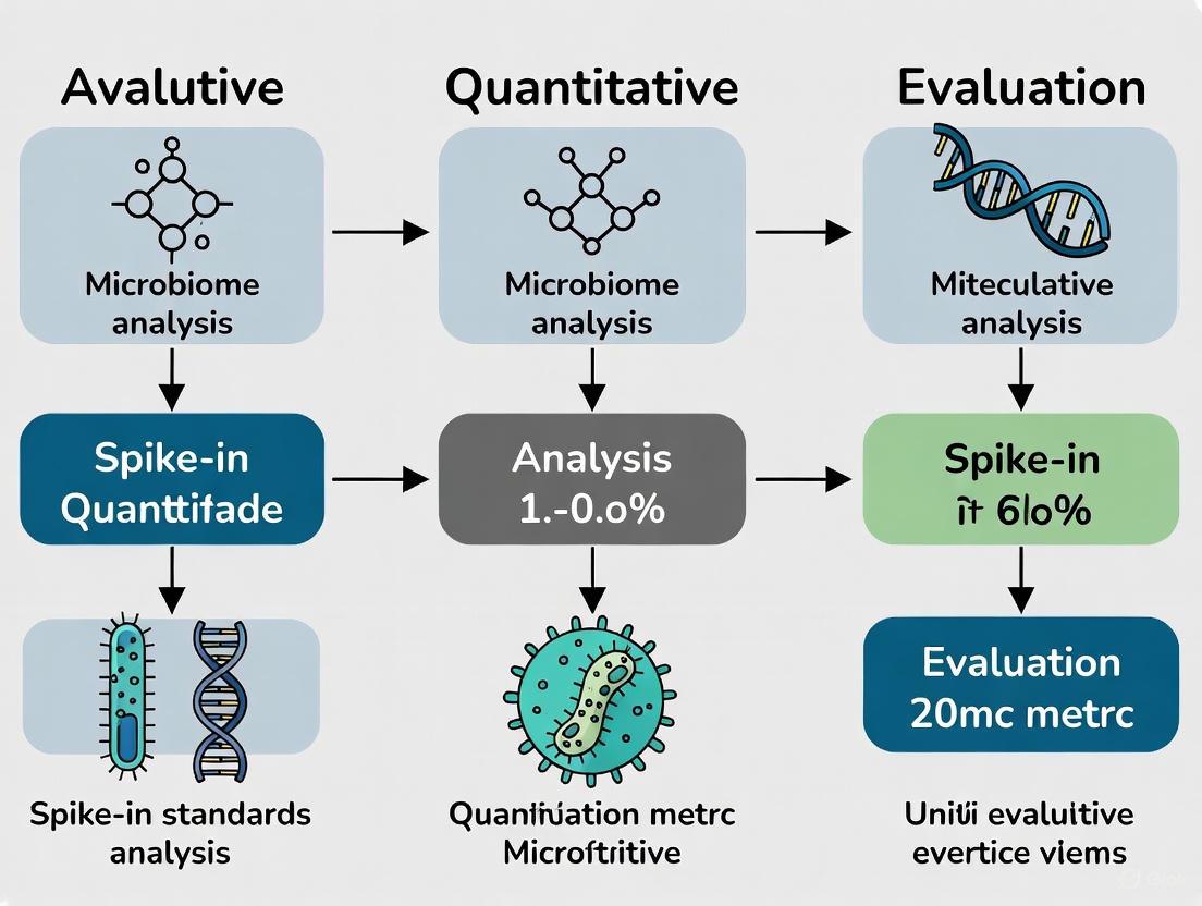
Evaluating Spike-In Standards for Robust and Quantitative Microbiome Analysis
Moving from relative to absolute quantification is a critical frontier in microbiome research, essential for understanding true microbial dynamics in health and disease.
Evaluating Spike-In Standards for Robust and Quantitative Microbiome Analysis
Abstract
Moving from relative to absolute quantification is a critical frontier in microbiome research, essential for understanding true microbial dynamics in health and disease. This article provides a comprehensive guide for researchers and drug development professionals on the implementation and evaluation of spike-in standards. We cover the foundational principles of these controls, detail best practices for their application in both 16S rRNA and shotgun metagenomic sequencing, and present strategies for troubleshooting common pitfalls. Furthermore, we establish a framework for the analytical validation of spike-in protocols and their comparative assessment against alternative quantification methods, empowering robust and reproducible microbiome science.
Why Spike-In Standards? Overcoming the Limitations of Relative Abundance Data
In microbiome research, the standard output of high-throughput sequencing is relative abundance, where the abundance of each taxon is expressed as a proportion of the total sequenced community. While these relative measurements have powered thousands of microbiome studies, they come with fundamental mathematical constraints that can dramatically mislead biological interpretation. The core issue is compositionality—because all proportions must sum to 1, an increase in one taxon's relative abundance forces an apparent decrease in all others, regardless of their actual behavior [1]. This review examines the specific pitfalls of relative abundance data, demonstrates how these limitations manifest in experimental contexts, and evaluates spike-in standards as a solution for achieving absolute quantification.
Why Relative Abundance Misleads: Core Conceptual Pitfalls
The Compositional Data Problem
Relative abundance measurements artificially constrain taxonomic relationships within a sample. When analyzing relative data, researchers cannot distinguish between an actual increase in one taxon versus a decrease in all others—both scenarios produce identical relative abundance patterns [2]. This limitation becomes particularly problematic when comparing samples with different total microbial loads, as relative abundance completely obscures changes in community density.
Mathematical Constraints and Spurious Correlations
The compositional nature of relative abundance data introduces negative correlation bias, where taxa appear to be negatively correlated even when no biological relationship exists [3] [1]. This occurs because the proportional space forces an inverse relationship between taxa—as one increases, others must decrease to maintain the constant sum. These mathematical artifacts can be misinterpreted as genuine biological interactions or treatment effects.
Quantitative Comparisons: Relative vs. Absolute Abundance
Interpreting Differential Abundance Scenarios
Table 1: Comparison of Relative vs. Absolute Interpretation in a Two-Taxon Community
| Scenario | Relative Abundance Pattern | Possible Absolute Realities |
|---|---|---|
| Taxon A increases | Taxon A ↑, Taxon B ↓ | (1) Taxon A actually increased(2) Taxon B actually decreased(3) Both changed but Taxon A increased more(4) Both changed but Taxon B decreased more(5) Combination of increases/decreases |
| No change in ratios | Taxon A stable, Taxon B stable | Total community size could have increased or decreased dramatically |
| All taxa change | Complex pattern shifts | Cannot distinguish overall dilution/concentration from compositional shifts |
This table illustrates how a single relative abundance pattern can correspond to multiple, biologically distinct realities in absolute terms [2]. Without absolute quantification, researchers cannot determine the direction or magnitude of changes for individual taxa between experimental conditions.
Impact on Heritability Estimates
Relative abundance data substantially distorts heritability estimates for microbial taxa. The heritability estimate derived from relative abundances (φ²) differs systematically from true heritability (h²) due to interdependencies between taxa [3]. This problem is most severe for dominant taxa, where spurious heritability signals can emerge for non-heritable microbes simply because they coexist with heritable ones. With large sample sizes, these artifacts lead to inflated false discovery rates and overestimation of the proportion of heritable taxa in a community.
Experimental Evidence: Case Studies Revealing the Discrepancy
Ketogenic Diet Study Using Digital PCR Anchoring
A rigorous absolute quantification framework using digital PCR (dPCR) demonstrated how relative abundance analyses can produce misleading conclusions [2]. In a murine ketogenic diet study, absolute quantification revealed that the diet actually decreased total microbial loads—information completely absent from relative abundance data. While relative abundances suggested simple taxonomic shifts, absolute measurements showed that some taxa maintained stable populations while others dramatically decreased, revealing a more nuanced biological response to dietary intervention.
Antibiotic Perturbation in Transplant Patients
Research on patients undergoing allogeneic stem cell transplantation demonstrated the critical importance of absolute quantification for understanding clinical outcomes [4]. When Enterococcus relative abundance increased from undetectable to 94% after transplantation, relative data alone couldn't determine whether this represented an absolute expansion of Enterococcus or collapse of the background community. Using spike-in bacteria for absolute quantification revealed this was primarily a collapse of other taxa, with important implications for understanding gastrointestinal graft-versus-host disease risk.
Spike-In Standards: Methodological Solutions for Absolute Quantification
Synthetic DNA Spike-ins for Metagenomics
The synDNA method utilizes 10 synthetic DNA sequences (2,000-bp length) with variable GC content (26-66%) and negligible identity to natural sequences in NCBI databases [5]. These synDNAs are spiked into samples at known concentrations before DNA extraction and sequencing, creating an internal calibration curve that enables absolute quantification of bacterial cells in complex communities.
Whole Cell Spike-in Calibration for Microbial Load
An alternative approach uses entire bacterial cells as spike-in controls, such as Salinibacter ruber, Rhizobium radiobacter, and Alicyclobacillus acidiphilus [4]. These species are absent from mammalian guts under physiological conditions and are added to samples before DNA extraction. The resulting Spike-in-based Calibration to Microbial Load (SCML) uses the known spike-in abundances to rescale read counts and estimate ratios of absolute endogenous bacterial abundances.
Table 2: Comparison of Spike-in Methodologies for Absolute Quantification
| Method | Spike-in Type | Added At | Key Advantages | Limitations |
|---|---|---|---|---|
| synDNA [5] | Synthetic DNA sequences | DNA extraction | Negligible database identity; customizable GC content | Doesn't control for extraction efficiency variations |
| Whole Cell [4] | Non-native bacterial cells | Sample processing | Controls for entire workflow including cell lysis | Potential interaction with sample matrix |
| Marine-sourced DNA [6] | Marine bacterial DNA | DNA extraction | Phylogenetically distant from host microbiome; cost-effective | Requires characterization of new bacterial strains |
Performance Benchmarking of Quantitative Approaches
Comparative analyses demonstrate that quantitative approaches using spike-in standards significantly outperform computational normalization methods [1]. In benchmarking studies, quantitative methods improved precision in identifying true positive taxon-taxon associations while reducing false positive detection. When analyzing simulated dysbiosis scenarios with low microbial loads—similar to those observed in inflammatory diseases—quantitative methods correcting for sampling depth showed substantially higher accuracy compared to relative abundance approaches.
The Researcher's Toolkit: Essential Reagents and Protocols
Key Research Reagent Solutions
Table 3: Essential Materials for Implementing Spike-in Quantitative Methods
| Reagent/Material | Function | Implementation Considerations |
|---|---|---|
| Synthetic DNA (synDNA) [5] | DNA spike-in for metagenomic sequencing | Design with variable GC content (26-66%); ensure minimal database identity |
| Exogenous bacterial cells [4] | Whole cell spike-in for 16S sequencing | Select species absent from study ecosystem; account for 16S copy number variation |
| Marine-sourced bacterial DNA [6] | Cost-effective DNA spike-in alternative | Use phylogenetically distant species (e.g., Pseudoalteromonas, Planococcus) |
| Digital PCR system [2] | Absolute quantification of target genes | Provides precise quantification without standard curves; higher sensitivity than qPCR |
| Linearized plasmid standards [7] | Precise quantification for 16S sequencing | Enables copy number determination; facilitates inter-laboratory reproducibility |
Implementation Workflow for Synthetic DNA Spike-ins
The evidence overwhelmingly demonstrates that relative abundance data presents significant interpretation pitfalls that can lead to incorrect biological conclusions. Compositional constraints inherently distort taxonomic relationships, obscure changes in total microbial load, and can generate spurious correlations [3] [1]. Spike-in standards for absolute quantification—whether synthetic DNA, whole cells, or marine-sourced DNA—provide effective solutions to these limitations [5] [4] [6]. As the field moves toward more rigorous and reproducible microbiome science, adopting absolute quantification methods will be essential for accurately understanding microbial dynamics in health, disease, and response to interventions.
Next-generation sequencing of microbial communities, such as 16S rRNA gene amplicon sequencing, has revolutionized microbiome research by enabling detailed profiling of complex microbial ecosystems. However, a fundamental limitation of standard sequencing approaches is that they generate only relative abundance data, where the abundance of each taxon is expressed as a proportion of the total sequenced reads [7]. This compositional nature of microbiome data makes biological interpretation challenging, as an increase in the relative abundance of one taxon inevitably leads to the apparent decrease of others, regardless of their actual absolute abundances [8]. This limitation becomes critical in clinical and experimental settings where the true microbial load matters, such as when tracking pathogen expansion or understanding ecosystem responses to perturbations like antibiotic treatments [9] [4].
The integration of exogenous spike-in controls addresses this fundamental limitation by providing an internal reference for absolute quantification. These controls consist of known quantities of synthetic or foreign biological materials added to samples prior to DNA extraction. By measuring the sequencing recovery rate of these spikes, researchers can rescale relative abundance data to absolute quantities, transforming microbiome measurements from proportional to quantitative data [10] [4]. This approach enables the detection of biologically meaningful changes that would be obscured in relative abundance data alone, such as distinguishing whether an increase in a taxon's proportion reflects its actual expansion or merely the decline of other community members.
Core Principles and Mechanisms of Spike-In Controls
Fundamental Working Principle
The core principle of using exogenous controls rests on a simple but powerful concept: adding a known quantity of a distinguishable standard to an unknown sample before processing. The fundamental relationship for absolute quantification is derived from the consistent known input ((C{spike-in})) and its measured output ((R{spike-in})) in sequencing reads, which calibrates the measurement for endogenous taxa:
[ Absolute\ Abundance{taxon\ X} = \frac{R{taxon\ X}}{R{spike-in}} \times C{spike-in} ]
where (R{taxon\ X}) and (R{spike-in}) represent read counts for the endogenous taxon and spike-in control, respectively, and (C_{spike-in}) represents the known absolute abundance of the spike-in [4]. This formula enables rescaling of relative sequencing data to absolute values. The following diagram illustrates the conceptual workflow and this mathematical relationship:
Key Design Characteristics of Effective Spike-In Controls
To function effectively as internal standards, exogenous controls must possess specific characteristics that ensure they can be reliably distinguished from endogenous microbiota and provide accurate quantification across diverse experimental conditions.
Non-interference with endogenous microbiome: Ideal spike-in organisms or sequences should be completely absent from the native samples being studied to prevent confusion with endogenous taxa [11] [4]. For example, species like Salinibacter ruber (from hypersaline environments) and Alicyclobacillus acidiphilus (from acidic thermal soils) are ideal for human gut microbiome studies because they do not naturally occur in this environment [4].
Distinguishability from native sequences: Spike-in sequences must contain unique identifier regions that allow unambiguous bioinformatic separation from endogenous sequences in the sample. This can be achieved through synthetic 16S rRNA variable regions with negligible identity to known natural sequences [7] or whole genomes of foreign microbial species [11].
Controlled and known abundance: The spike-in standard must be precisely quantified before addition to the sample, with concentration values traceable to standardized measurements [10]. Both whole cell and genomic DNA formats are used, with the former providing control over the entire workflow including cell lysis efficiency [12] [10].
Compatibility with experimental workflows: The physical and genetic properties of spike-in controls should be compatible with the sample processing and analysis methods. This includes considerations of cell wall structure (Gram-positive vs. Gram-negative for cellular standards) and GC content for DNA-based standards to account for potential amplification biases [7] [12].
Types of Spike-In Standards and Commercial Solutions
The growing recognition of the importance of absolute quantification in microbiome research has led to the development of diverse spike-in standards with different properties and applications. The following table compares major categories and representative commercial solutions:
| Standard Type | Key Features | Representative Products | Primary Applications |
|---|---|---|---|
| Whole Cell Spike-Ins | Intact microbial cells with varying cell wall structures; control for DNA extraction efficiency bias | ZymoBIOMICS Spike-in Controls (I: High Microbial Load; II: Low Microbial Load) [11]; ATCC MSA-2014 [10] | Absolute quantification accounting for lysis efficiency differences; quality control for entire workflow |
| Genomic DNA Spike-Ins | Purified DNA from unique strains; eliminate extraction variability | ATCC MSA-1014 [10] | Library preparation and sequencing normalization; bioinformatics pipeline validation |
| Synthetic Sequence Spike-Ins | Artificial 16S rRNA genes with designed variable regions [7] | Custom synthetic constructs [7] | Universal application across diverse sample types; bioinformatic ground truth |
| Multi-Species Spike-Ins | Mixtures of different microbial species with known proportions | ATCC Spike-in Standards (3 engineered strains) [10]; Combination of S. ruber, R. radiobacter, A. acidiphilus [4] | Assessing amplification biases across taxa; evaluating quantitative accuracy |
The ZymoBIOMICS Spike-in Controls exemplify the whole cell approach, comprising equal cell numbers of Imtechella halotolerans (Gram-negative) and Allobacillus halotolerans (Gram-positive) that represent different cell recalcitrance and can expose potential bias during DNA extraction [11]. Meanwhile, the ATCC standards utilize an innovative approach with three genetically engineered bacterial strains (Escherichia coli, Staphylococcus aureus, and Clostridium perfringens) that each contain a unique synthetic DNA tag integrated into their genomes, enabling precise detection and quantification in both 16S rRNA gene amplicon and shotgun sequencing assays [10].
Experimental Protocols and Implementation
Sample Processing Workflow with Spike-In Controls
Implementing spike-in controls requires careful integration into the standard microbiome analysis workflow at specific critical points. The following detailed protocol outlines the key steps for incorporating these standards effectively:
Critical Step: Spike-in Addition - Spike-in controls must be added at the beginning of the processing workflow, immediately after sample collection and before any processing steps [10] [4]. For cellular standards, this typically involves adding a precise volume of the standardized cell suspension to the sample. For DNA standards, a defined number of genome copies is added. The amount should be calibrated to the expected microbial load of the sample—for instance, ZymoBIOMICS recommends Spike-in Control I for high microbial load samples (e.g., stool) and Spike-in Control II for low microbial load samples (e.g., water, swabs) [11].
DNA Extraction Considerations - When using whole cell spike-ins, the extraction method must be efficient for both the spike-in organisms and the endogenous microbiota. The inclusion of species with different cell wall structures (Gram-positive vs. Gram-negative) helps monitor extraction efficiency biases [12] [11]. Validation experiments should confirm that the spike-ins are not present in the native samples, as demonstrated by qPCR verification in the SCML approach [4].
Library Preparation and Sequencing - Standard protocols for 16S rRNA gene amplification or shotgun metagenomic library preparation are used. Primer selection is important for synthetic 16S rRNA spike-ins, as different variable regions (V1V2, V3V4, V4) can exhibit varying amplification efficiencies for artificial sequences [10].
Bioinformatic Analysis and Data Normalization
The bioinformatic pipeline for processing spike-in controlled samples requires specific steps to identify and quantify the control sequences:
Spike-in Read Identification: Sequence reads must be classified as originating from spike-in controls versus endogenous microbiota. For synthetic 16S rRNA tags, this involves mapping to the artificial reference sequences using tools like Bowtie2 [10]. For foreign species, taxonomic classification or specific marker gene identification can separate spike-in reads.
Absolute Abundance Calculation: The read counts for each endogenous taxon are rescaled using the spike-in measurements. If (R{taxon\ X}) is the read count for a taxon, (R{spike-in}) is the read count for the spike-in, and (C_{spike-in}) is the known absolute abundance of the spike-in, then:
[ Absolute\ Abundance{taxon\ X} = \frac{R{taxon\ X}}{R{spike-in}} \times C{spike-in} ]
This calculation transforms the data from relative proportions to absolute quantities [4].
Validation of Quantification Accuracy: Experimental validation should include dilution series of known communities to verify linearity and accuracy of quantification across the dynamic range expected in experimental samples [4].
Performance Comparison and Experimental Data
Quantitative Assessment of Different Standards
Rigorous experimental validation is essential to demonstrate the performance of spike-in standards in actual research applications. The following data from key studies illustrates how these controls perform in controlled experiments:
| Study & Standard Type | Experimental Design | Key Quantitative Results | Limitations Identified |
|---|---|---|---|
| Stämmler et al. [4](Three foreign species: S. ruber, R. radiobacter, A. acidiphilus) | Serial dilutions of pooled murine stool with defined spike-in concentrations; 36 aliquots total | SCML reduced systematic error in ratio estimation; variability cut nearly in half compared to relative abundance analysis | Different spike-in species showed notably different read yields (S. ruber highest) despite adjustment for 16S copy number |
| Tkacz et al. [7](Synthetic 16S rRNA genes with artificial variable regions) | Defined mock communities and environmental microbiota; staggered spike-in mixtures | Enabled absolute abundance estimation suitable for comparative analysis; identified template-specific Illumina sequencing artifacts | Technical biases from sequencing platform remained despite spike-in normalization |
| ATCC Engineered Strains [10](Three tagged strains: E. coli, S. aureus, C. perfringens) | 16S amplicon sequencing with different primer sets (V1V2, V3V4, V4) compared to ddPCR quantification | V3V4 and V4 regions showed minimal bias; V1V2 region showed significant divergence from expected abundance | Primer selection critical - V1V2 region amplification showed substantial bias for synthetic tags |
| ZymoBIOMICS [11](I. halotolerans & A. halotolerans) | Equal cell numbers of Gram-negative and Gram-positive species | Enabled absolute cell number quantification; exposed potential bias during DNA extraction due to differential cell lysis | Limited to two species; may not capture full range of extraction biases |
In the SCML approach developed by Stämmler et al., the method was specifically tested using dilution experiments where the "ground truth" was known by experimental design [4]. When comparing ratios of absolute abundances between samples, the standard relative abundance approach showed systematically overestimated ratios in both directions, while the spike-in calibrated data significantly reduced this bias and decreased variability in estimated ratios by nearly half [4].
The ATCC engineered strains demonstrated how amplification biases can affect different spike-in standards. When evaluating the performance of their tagged strains with different 16S rRNA gene primer sets, researchers found that while V3V4 and V4 regions showed minimal bias compared to digital PCR quantification, the V1V2 region exhibited significant divergence from expected abundance [10]. This highlights the importance of primer selection and validation for specific spike-in standards.
Comparison of Quantification Methods
The search results also reveal important considerations regarding the method used for microbial load quantification in conjunction with spike-in controls. A direct comparison between flow cytometry-based quantification (the original QMP approach) and qPCR-based quantification found that while both methods provided accurate and correlated results when quantifying a mock community of bacterial cells, they produced "highly divergent quantitative microbial profiles" when applied to human fecal samples [8]. This discrepancy could not be attributed to extracellular DNA or lack of qPCR precision, suggesting that the choice of quantification method itself can introduce substantial additional bias in quantitative microbiome profiling.
Essential Research Reagent Solutions
Successful implementation of exogenous controls for absolute quantification requires specific reagent systems designed to address the technical challenges of quantitative microbiome analysis:
| Reagent Category | Specific Examples | Function in Workflow |
|---|---|---|
| Quantified Spike-in Standards | ZymoBIOMICS Spike-in Controls I & II [11]; ATCC MSA-1014 & MSA-2014 [10] | Provide known reference materials for absolute quantification across different sample types |
| Digital PCR Systems | ddPCR with tag-specific probes [10] | Enable precise absolute quantification of spike-in standards independent of sequencing |
| Viability Dyes for Cell Sorting | Propidium Monoazide (PMAxx) [8] | Differentiate between intact cells and free DNA in quantitative assessments |
| Reference Materials | ZymoBIOMICS Microbial Community Standards [12]; ATCC Mock Microbial Communities [13] | Validate overall workflow performance alongside spike-in controls |
| Bioinformatic Tools | Custom mapping pipelines (Bowtie2) [10]; Specialized normalization algorithms [4] | Identify spike-in reads and perform absolute abundance calculations |
The integration of digital PCR (ddPCR) systems deserves special emphasis, as this technology provides orthogonal validation of spike-in concentrations. In the ATCC validation studies, ddPCR with tag-specific primers and probes was used to confirm the absolute abundance of each engineered strain in the spike-in mixture, providing a reference measurement against which sequencing-based quantification could be compared [10]. This highlights the importance of multi-platform validation in establishing reliable quantitative workflows.
The adoption of exogenous controls as internal standards represents a fundamental advancement in microbiome research methodology, enabling the transition from relative to absolute quantification. The experimental data comprehensively demonstrate that spike-in calibrated approaches significantly improve accuracy in estimating ratios of absolute abundances compared to standard relative abundance analysis [4]. While technical challenges remain, including amplification biases [10] and choice of quantification method [8], the commercial availability of standardized spike-in solutions [11] [10] has made this powerful approach accessible to a broad research community.
As the field moves toward more quantitative and translational applications, the implementation of spike-in controls will be essential for generating reproducible, biologically meaningful measurements that can be compared across studies and laboratories. The continuing development of innovative spike-in technologies, including multi-species standards [10] and synthetic sequences [7], promises to further enhance the accuracy and applicability of absolute quantification in microbiome research.
In the field of quantitative microbiome analysis, the transition from relative to absolute abundance measurements is critical for accurate ecological interpretation and risk assessment. Spike-and-recovery and linearity of dilution are two fundamental experimental metrics used to validate the accuracy of quantitative molecular methods, including the use of spike-in standards. This guide details the principles, protocols, and interpretation of these assays, providing a framework for researchers to rigorously evaluate and compare the performance of different quantitative approaches, thereby ensuring data reliability in microbiome research.
Next-generation sequencing (NGS) techniques, such as 16S rRNA gene sequencing, have revolutionized microbiome research but inherently produce relative abundance data [14]. This compositional nature means that the reported proportion of one microbe is mathematically constrained by the proportions of all others, making it impossible to determine if a change in relative abundance represents an actual increase in one taxon or a decrease in others [14] [4]. For many applications, understanding the absolute abundance—the true number of microbial cells or gene copies per unit of sample—is biologically crucial.
The limitations of relative data can lead to spurious correlations and misinterpretations [14]. To overcome this, researchers employ spike-in standards, which are known quantities of exogenous cells or DNA added to a sample before DNA extraction [15] [4]. These standards act as internal controls, enabling the calculation of absolute abundances for endogenous microbes. However, the accuracy of this quantification hinges on validating that the assay performs consistently between the spike-in material and the native sample matrix. This is precisely where spike-and-recovery and linearity-of-dilution experiments become indispensable, providing critical validation for quantitative methods in microbiome research [16].
Defining the Key Metrics
Spike-and-Recovery
Spike-and-recovery assesses whether the detection of an analyte is affected by differences between the standard diluent and the biological sample matrix [16]. In essence, it tests if the assay "sees" a known amount of material equally well in a clean buffer versus in a complex, heterogeneous sample like stool or soil.
The experiment involves adding a known amount of a reference analyte (the "spike") into both the standard diluent and the natural sample matrix. The assay is then run, and the measured concentration (the "recovery") is compared between the two. An ideal assay shows identical recovery, indicating the sample matrix does not interfere with detection [16] [17]. Poor recovery suggests the presence of matrix effects, such as inhibitors or enhancers, that compromise accuracy and must be addressed before reliable quantification is possible [16].
Linearity of Dilution
Linearity of dilution evaluates the precision of results for samples tested at different levels of dilution in a chosen sample diluent [16]. It determines whether the relationship between the measured concentration and the dilution factor is linear and proportional, which is a key assumption for extrapolating quantifications from diluted samples back to their original, neat concentration.
This is distinct from parallelism, another validation parameter. While linearity of dilution typically involves a sample spiked with a known analyte, parallelism uses a sample with a high endogenous concentration of the analyte, which is then serially diluted to confirm that the native analyte and the standard curve analyte behave similarly [17]. Good linearity indicates that the sample diluent successfully neutralizes matrix effects across a range of concentrations, providing flexibility to assay samples with high microbial loads by bringing them within the dynamic range of the standard curve [16].
Experimental Protocols and Methodologies
Protocol for Spike-and-Recovery Assessment
A standard spike-and-recovery experiment follows a systematic workflow to identify matrix effects.
Workflow: Spike-and-Recovery Experiment
Step-by-Step Protocol:
- Sample Preparation: Aliquot the natural sample matrix (e.g., a homogenized fecal sample) and an appropriate standard diluent (e.g., phosphate-buffered saline) into separate tubes [16].
- Spike Addition: Add a known, physiologically relevant concentration of the spike analyte (e.g., a synthetic DNA standard or known bacterial cells not found in the sample) to both the sample matrix and the standard diluent [16] [15] [4]. The spike concentration should be within the dynamic range of the assay.
- Assay Execution: Process both the spiked sample matrix and the spiked standard diluent through the entire quantitative workflow (DNA extraction, library preparation, and sequencing or qPCR) using identical protocols [4].
- Recovery Calculation: For both sets, measure the concentration of the spike from the standard curve. Calculate the percent recovery using the formula:
- Interpretation: A recovery of 100% indicates no matrix interference. Recoveries of 80–120% are generally considered acceptable, though the specific threshold should be defined based on the required precision of the study [17]. Values outside this range indicate significant matrix effects.
Protocol for Linearity of Dilution Assessment
The linearity-of-dilution experiment tests how well a sample can be diluted while maintaining accurate quantification.
Workflow: Linearity of Dilution Experiment
Step-by-Step Protocol:
- Sample Preparation: Create a sample with a high concentration of the target analyte. This can be achieved by using a sample with high endogenous levels or by spiking a sample matrix with a known amount of analyte to a concentration above the assay's upper limit of quantification [16] [17].
- Serial Dilution: Perform a series of dilutions (e.g., 1:2, 1:4, 1:8) of this high-concentration sample using the chosen sample diluent. Continue until the predicted concentration falls below the assay's lower limit of quantification [16] [17].
- Assay Execution: Run the neat (undiluted) and all diluted samples through the quantitative assay.
- Data Analysis: For each dilution, calculate the expected concentration based on the dilution factor and the measured concentration of the neat sample (or the known spike amount). Then, calculate the percent recovery for each dilution point: (Observed Concentration / Expected Concentration) × 100.
- Interpretation: Plot the observed concentrations against the expected concentrations. The dilution series is considered linear if the recovery values for all dilutions fall within a pre-defined acceptance range (e.g., 80–120%) and the data points on the plot fall along the line of identity [16] [17].
Data Presentation and Comparison
The results from spike-and-recovery and linearity experiments are best summarized in tables for clear interpretation and cross-comparison of different methods or sample types.
Representative Spike-and-Recovery Data
The following table summarizes data from a spike-and-recovery experiment for recombinant human IL-1 beta in human urine samples, demonstrating acceptable recovery across a range of spike concentrations [16].
Table 1: ELISA Spike-and-Recovery of Recombinant Human IL-1β in Human Urine [16]
| Sample (n) | Spike Level (pg/mL) | Expected (pg/mL) | Observed (pg/mL) | Recovery % |
|---|---|---|---|---|
| Urine (9) | Low (15) | 17.0 | 14.7 | 86.3 |
| Urine (9) | Medium (40) | 44.1 | 37.8 | 85.8 |
| Urine (9) | High (80) | 81.6 | 69.0 | 84.6 |
Representative Linearity of Dilution Data
This table shows linearity-of-dilution results for human IL-1 beta in different sample types. The deviation from 100% recovery at higher dilutions can indicate matrix effects becoming more pronounced [16].
Table 2: ELISA Linearity-of-Dilution for Human IL-1β Samples [16]
| Sample | Dilution Factor | Observed (pg/mL) × DF | Expected (pg/mL) | Recovery % |
|---|---|---|---|---|
| ConA-stimulated cell culture supernatant | Neat | 131.5 | 131.5 | 100 |
| 1:2 | 149.9 | 114 | ||
| 1:4 | 162.2 | 123 | ||
| 1:8 | 165.4 | 126 | ||
| High-level serum sample | Neat | 128.7 | 128.7 | 100 |
| 1:2 | 142.6 | 111 | ||
| 1:4 | 139.2 | 108 | ||
| 1:8 | 171.5 | 133 |
Application in Quantitative Microbiome Analysis
In microbiome research, spike-and-recovery principles are directly applied using whole cells or synthetic DNA as spikes to convert relative sequencing data into absolute abundances, an approach known as Spike-in-based Calibration to Microbial Load (SCML) or Quantitative Microbiome Profiling (QMP) [14] [4].
- Spike-in Standards: Researchers add a known quantity of non-native bacteria (e.g., Salinibacter ruber, Alicyclobacillus acidiphilus) [4] or synthetic DNA sequences [15] to stool or environmental samples prior to DNA extraction.
- Correcting for Load: The fundamental calculation involves using the recovery of the spike to adjust the counts of native microbes. The formula used in QMP is: Absolute Abundance = (Relative Abundance of Taxon × Total Cell Count Estimated from Spike-in) [14].
- Overcoming Compositionality: This correction allows researchers to distinguish between a situation where a taxon's relative increase is due to its actual growth versus the decline of other taxa, which is impossible with relative data alone [14] [4]. For example, an overgrowth of Enterococcus during antibiotic treatment can be confirmed as an absolute increase rather than a relative artifact [4].
The Scientist's Toolkit: Essential Research Reagents
Successful implementation of these quantitative metrics relies on specific reagents and controls.
Table 3: Key Research Reagent Solutions for Quantitative Microbiome Analysis
| Item | Function & Application | Examples |
|---|---|---|
| Synthetic DNA Spike-ins | Defined, quantifiable DNA sequences added to sample lysis buffer to monitor DNA recovery yield and calculate absolute abundances [15]. | Custom-designed 733bp standard [15]; ZymoBIOMICS Spike-in Control [18]. |
| Whole Cell Spike-ins | Intact, non-native bacterial cells added to the sample to control for variability from cell lysis to sequencing [4]. | Salinibacter ruber, Rhizobium radiobacter, Alicyclobacillus acidiphilus [4]. |
| Mock Microbial Communities | DNA or cell-based standards with known, fixed compositions used to validate assay accuracy, precision, and limit of detection [18] [19]. | ZymoBIOMICS Microbial Community Standards (D6300, D6305, D6331) [18]. |
| Standard Diluent | A well-characterized buffer used to prepare the standard curve, aiming to match the final sample matrix as closely as possible [16]. | Phosphate-Buffered Saline (PBS), sometimes with added carrier protein like 1% BSA [16]. |
| Sample Diluent | The solution used to dilute neat biological samples; optimized to minimize matrix effects and bring analyte concentrations within the assay range [16]. | PBS, often without added protein for serum samples [16]. |
Spike-and-recovery and linearity of dilution are not merely procedural checkboxes but are fundamental to establishing confidence in quantitative data. As microbiome research progresses toward clinical diagnostics and therapeutic development [18] [19], the demand for accurate, absolute quantification will only intensify. By rigorously applying these validation metrics, researchers can ensure their spike-in standards perform reliably, enabling them to move beyond relative shifts and answer the biologically critical question: "How many are there?" This rigorous framework is essential for building reproducible, translatable, and impactful science in quantitative microbiome analysis.
The Pervasive Challenge of Contamination in Low Biomass Samples
In microbiome research, low microbial biomass samples—those containing small amounts of microbial DNA—present a formidable analytical challenge. These samples, which include tissue, blood, urine, skin swabs, and other sterile site specimens, are particularly vulnerable to contamination from exogenous DNA sources [20] [21].
The fundamental issue lies in the signal-to-noise ratio: when the actual microbial signal is low, contaminants from DNA extraction kits, laboratory reagents, plastic consumables, researchers, and the sampling environment can constitute a substantial portion, or even the majority, of the detected microbial community [21]. This contamination can easily generate signals that are misinterpreted as biological findings, potentially leading to invalid conclusions. For instance, studies of putative placental and blood microbiomes have been heavily criticized when follow-up investigations revealed that reported microbial signals were indistinguishable from those found in blank control samples [21].
The composition of contaminant DNA is not random; it often originates from specific bacterial taxa commonly found in reagents and laboratory environments. Without appropriate controls, these contaminants can be mistakenly interpreted as genuine biological signals, particularly in studies investigating disease-associated microbiomes where accurate microbial profiling is critical for diagnostic and therapeutic applications [20] [21].
The Limitations of Relative Abundance in Microbial Ecology
Traditional microbiome sequencing data are compositional in nature, meaning they provide only relative abundances rather than absolute quantities [4] [1]. This fundamental characteristic poses significant interpretative challenges:
Masked Biological Truth: Compositional data cannot distinguish between an absolute increase in one taxon versus a decrease in all others. For example, an observed relative increase in Enterococcus from undetectable levels to 94% in stool specimens from transplant patients could represent either a true bloom of this bacterium or a catastrophic collapse of the rest of the community [4].
Negative Correlation Bias: In relative abundance data, any increase in one taxon must be compensated by decreases in others, creating artificial negative correlations that may not exist in reality [1].
Sampling Depth Variability: Metagenomic analyses typically survey only a tiny fraction (as low as 0.0045%) of the total microbial community, with sampling depth varying more than 40-fold across samples. This variability means that detection of specific microbiome features may reflect technical artifacts rather than true biological differences [1].
These limitations are particularly problematic when studying low biomass samples, where small absolute changes can produce dramatic shifts in relative abundance profiles that poorly reflect biological reality.
Spike-In Controls as a Quantitative Solution
Spike-in controls provide a methodological solution to the problems of contamination and compositionality by adding known quantities of exogenous biological materials to samples prior to DNA extraction [10] [22] [4]. These controls serve as internal standards that enable absolute quantification and help distinguish true biological signals from contamination.
Diverse Spike-In Formats and Applications
Table 1: Comparison of Major Spike-In Control Approaches
| Control Type | Composition | Key Applications | Advantages | Limitations |
|---|---|---|---|---|
| Whole Cell Spike-Ins [10] [4] | Genetically engineered bacterial cells (e.g., E. coli, S. aureus, C. perfringens) with synthetic 16S rRNA tags | Accounting for DNA extraction efficiency; absolute quantification | Control for entire workflow from cell lysis onward; most comprehensive | Requires careful matching of cell wall properties to sample type |
| Genomic DNA Spike-Ins [10] [22] | Purified DNA from engineered strains or synthetic constructs | Normalization for amplification and sequencing biases; absolute quantification | More stable than whole cells; easier to standardize | Does not control for DNA extraction efficiency |
| Synthetic Gene Constructs [22] [23] | Artificial rRNA operons with natural conserved regions and artificial variable regions | Cross-domain quantification; sample tracking; contamination detection | Highly customizable; minimal risk of natural occurrence | May not fully capture extraction biases affecting native DNA |
Mechanisms of Action
Spike-in controls function through several complementary mechanisms:
Absolute Quantification: By adding a known number of spike-in cells or genome copies, researchers can convert relative sequencing read counts into absolute abundances using the formula: Absolute Abundance = (Native Taxon Reads / Spike-in Reads) × Known Spike-in Quantity [4].
Microbial Load Estimation: Spike-ins enable the calculation of total microbial load in a sample, which is particularly valuable when comparing communities with different overall densities, such as in dysbiotic states where microbial loads may be dramatically reduced [1].
Process Efficiency Monitoring: By tracking the recovery of spike-in controls throughout the experimental workflow, researchers can identify and correct for technical variations in DNA extraction, amplification, and sequencing efficiency [10] [4].
The following diagram illustrates how spike-in controls integrate into the microbiome analysis workflow to enable absolute quantification:
Experimental Protocols and Implementation
ATCC Spike-in Standard Protocol
The ATCC spike-in standards (MSA-1014 for genomic DNA, MSA-2014 for whole cells) utilize three genetically engineered bacterial strains containing unique synthetic 16S rRNA tags. The recommended protocol involves [10]:
Spike-in Addition: Add the spike-in control (either whole cells or genomic DNA) to the sample immediately upon collection or at the beginning of DNA extraction. The typical specification is 6 × 10⁷ genome copies or cells per vial, with lot-specific quantification provided.
DNA Extraction: Process samples using standard extraction kits such as the DNeasy PowerLyzer Microbial Kit. For whole cell spike-ins, this step will lyse both native and spike-in cells.
Library Preparation and Sequencing: Perform either 16S rRNA gene amplicon sequencing (targeting V3V4 or V4 regions recommended over V1V2 due to better amplification characteristics) or shotgun metagenomic sequencing using standard Illumina platforms.
Bioinformatic Analysis: Map reads to the unique synthetic tag sequences using alignment tools such as Bowtie2. Precisely quantify spike-in reads to establish normalization factors.
Data Normalization: Calculate absolute abundances of native taxa using the formula: Absolute Abundance = (Native Taxon Reads / Spike-in Reads) × Known Spike-in Quantity.
Synthetic rDNA-Mimic Protocol
For cross-domain quantification encompassing both bacteria and fungi, the rDNA-mimic approach employs 12 synthetic rRNA operon sequences. The protocol includes [22]:
Spike-in Design: Construct artificial sequences with conserved regions matching natural rRNA genes (for PCR primer binding) and unique variable regions for robust identification.
Spike-in Preparation: Clone full-length rDNA-mimics into plasmid vectors, transform into E. coli, extract plasmid DNA, linearize using restriction enzymes (BsaI or BpmI), and purify.
Quantity Verification: Precisely quantify DNA concentrations using high-sensitivity assays such as Quant-iT dsDNA Assay Kit with Qubit Fluorometer.
Sample Processing: Add rDNA-mimics directly to samples prior to DNA extraction or to extracted DNA, then process through standard amplicon sequencing workflows targeting multiple rRNA regions (SSU-V9, ITS1, ITS2, LSU-D1D2 for fungi; SSU-V4 for bacteria).
SCML (Spike-in Calibration to Microbial Load) Protocol
The SCML method uses whole cell spike-ins of non-mammalian bacteria (Salinibacter ruber, Rhizobium radiobacter, Alicyclobacillus acidiphilus) and involves [4]:
Spike-in Selection: Choose exogenous bacteria not found in the sample type of interest, with varying 16S rRNA gene copy numbers (1, 4, and 6 copies per genome for the three species mentioned).
Standard Curve Generation: Spike samples with known concentrations of spike-in bacteria across a dilution series to establish quantitative relationships.
qPCR Validation: Verify spike-in concentrations and specificity using quantitative PCR with unique primer/probe sets.
Data Transformation: Use spike-in read counts to rescale native microbial abundances, making profiles sensitive to true microbial load differences rather than just compositional shifts.
Comparative Performance of Spike-in Methods
Table 2: Performance Characteristics of Different Quantitative Approaches
| Method | Quantification Accuracy | Contamination Resistance | Workflow Complexity | Best Application Context |
|---|---|---|---|---|
| Whole Cell Spike-Ins [10] [4] | High (controls for extraction efficiency) | High when using engineered tags | Moderate to high | Clinical samples; low biomass environments |
| Genomic DNA Spike-Ins [10] [22] | Moderate (post-extraction only) | Moderate | Low to moderate | Well-characterized sample types; high biomass |
| Synthetic rDNA-Mimics [22] | High for cross-domain studies | High due to unique sequences | Moderate | Multi-kingdom community profiling |
| qPCR-Based AMP [19] [24] | High for specific targets | Variable | Low | Targeted quantification of specific taxa |
| Relative Abundance Only [4] [1] | Low (compositional bias) | Low | Low | Exploratory studies; limited sample availability |
Benchmarking studies have demonstrated that quantitative approaches incorporating spike-in controls significantly outperform computational normalization methods in accurately recovering true biological relationships. Specifically, quantitative methods show higher precision in identifying taxon-taxon associations and taxon-metadata correlations while reducing false positive detection rates [1].
In scenarios simulating low microbial load dysbiosis (as observed in inflammatory diseases), quantitative methods correcting for sampling depth show superior performance compared to uncorrected scaling approaches. They more accurately detect true positive associations and reduce identification of spurious relationships that plague traditional relative abundance analyses [1].
The Scientist's Toolkit: Essential Research Reagents
Table 3: Key Research Reagents for Spike-in Implementation
| Reagent/Kit | Function | Application Notes |
|---|---|---|
| ATCC Spike-in Standards (MSA-1014, MSA-2014) [10] | Whole cell and gDNA quantitative standards | Contains three engineered bacteria with unique synthetic 16S rRNA tags |
| rDNA-Mimic Constructs [22] | Cross-domain quantification standards | 12 synthetic operons covering fungal and bacterial target regions |
| DNeasy PowerLyzer Microbial Kit [10] | DNA extraction from mixed samples | Validated for use with whole cell spike-in standards |
| QIAamp Fast DNA Stool Mini Kit [19] | Fecal DNA extraction | Compatible with spike-in approaches; includes inhibitor removal |
| Nextera XT DNA Library Prep Kit [10] | Sequencing library preparation | Compatible with spike-in enabled samples |
| Strain-Specific qPCR Assays [19] | Absolute quantification of target strains | Limit of detection ~10³ cells/g feces; requires careful primer design |
| Bowtie2 [10] | Read mapping to spike-in tags | Identifies synthetic sequences in sequencing data |
| Digital PCR (ddPCR) [10] [19] | Absolute quantification of spike-ins | Higher reproducibility than qPCR; more expensive |
The problem of low microbial biomass represents a critical challenge in microbiome research, where contamination can easily distort results and lead to invalid conclusions. Spike-in controls provide a powerful solution to this problem, enabling researchers to distinguish true biological signals from technical artifacts and transform relative microbiome data into absolute quantities.
While implementation requires careful experimental design and validation, the benefits of spike-in approaches are substantial: they provide internal controls for technical variability, enable absolute quantification, facilitate detection of cross-contamination, and ultimately yield more biologically meaningful data. As the field moves toward more clinically relevant applications, the adoption of these robust quantitative methods will be essential for generating reliable, reproducible results that can inform diagnostic and therapeutic development.
For researchers working with low biomass samples, the integration of appropriate spike-in controls—whether whole cells, genomic DNA, or synthetic constructs—represents a best practice that significantly enhances the validity and interpretability of microbiome studies.
High-throughput sequencing of microbial communities has revolutionized our understanding of ecosystems ranging from the human gut to environmental habitats. However, standard sequencing approaches generate only relative abundance data, which poses significant limitations for both basic ecology and clinical applications. The inherent compositionality of microbiome data means that changes in the abundance of one taxon can artificially alter the perceived relative abundances of all others, potentially leading to spurious associations [4] [25] [22]. This fundamental limitation has driven the development and adoption of spike-in standards to enable absolute quantification, transforming microbiome research from purely observational to truly quantitative science.
Spike-in standards are known quantities of exogenous biological materials added to samples prior to DNA extraction. These standards serve as internal references, allowing researchers to convert relative sequencing read counts into absolute abundances by accounting for technical variations throughout the experimental workflow [4] [22]. The integration of spike-in controls represents a paradigm shift in how we approach quantitative microbiome analysis, providing critical tools to distinguish between true biological changes and methodological artifacts across diverse research applications.
Comparative Analysis of Spike-In Standard Approaches
Types of Spike-In Standards and Their Applications
Spike-in standards for microbiome research primarily fall into two categories: whole cell microbes and synthetic DNA constructs. Each approach offers distinct advantages and limitations, making them suitable for different research scenarios and applications. The choice between these standards depends on multiple factors, including the study objectives, required precision, and available resources.
Table 1: Comparison of Major Spike-In Standard Types for Microbiome Research
| Standard Type | Examples | Key Applications | Advantages | Limitations |
|---|---|---|---|---|
| Whole Cell Microbes | Salinibacter ruber, Rhizobium radiobacter, Alicyclobacillus acidiphilus [4] | Microbial load calibration, absolute abundance estimation, clinical biomarker studies [4] [25] | Controls for DNA extraction efficiency, mimics natural community processing, biologically relevant [4] | Limited to culturable organisms, potential batch variability, storage stability concerns |
| Synthetic DNA Constructs | rDNA-mimics (12 synthetic rRNA operons) [22], full-length synthetic 16S rRNA genes [22] | Cross-domain quantification, standardized workflows, multi-laboratory studies [22] | Highly reproducible, customizable sequences, stable long-term storage, compatible with multiple primer sets [22] | Does not control for DNA extraction efficiency, requires careful quantification during preparation |
Performance Metrics and Technical Validation
Robust validation is essential for establishing the reliability of spike-in standards. Multiple studies have demonstrated the technical performance of different spike-in approaches through rigorous experimental designs.
Whole cell spike-in standards have shown excellent linearity between spiked 16S rDNA copies and resulting read counts across dilution series, with correlation coefficients ranging from r = -0.725 to -0.834 for different spike-in bacteria [4]. In validation experiments using serial dilutions of pooled murine stool samples spiked with defined amounts of exogenous bacteria, this approach demonstrated a significant reduction in systematic error compared to relative abundance analysis alone. The variability of estimated ratios was almost cut in half when using spike-in based calibration to microbial load (SCML) compared to standard relative abundance data [4].
Synthetic DNA standards offer complementary advantages. The recently developed rDNA-mimics, consisting of 12 synthetic rRNA operons, were experimentally validated using defined mock communities and environmental samples [22]. These constructs demonstrated precise quantification of total fungal and bacterial rRNA genes when added to extracted DNA or directly to samples prior to DNA extraction. The rDNA-mimics cover multiple rRNA operon regions commonly targeted in fungal/eukaryotic microbiome studies (SSU-V9, ITS1, ITS2, and LSU-D1D2), with two constructs also including an artificial segment of the bacterial 16S rRNA gene (SSU-V4) for cross-domain applications [22].
Table 2: Quantitative Performance Characteristics of Spike-In Standards
| Performance Metric | Whole Cell Standards | Synthetic DNA Standards |
|---|---|---|
| Dynamic Range | 4+ orders of magnitude [4] | 6+ orders of magnitude [22] |
| Linearity | R = -0.725 to -0.834 [4] | Pearson's r > 0.96 on log-transformed counts [26] |
| Limit of Detection | Not explicitly reported | 0.1 to 1.0 pg/μL for genomic DNA [27] |
| Cross-Domain Compatibility | Limited to bacterial targets | Full cross-domain (bacterial, fungal, eukaryotic) [22] |
| Inter-laboratory Reproducibility | Requires careful standardization | High reproducibility demonstrated [22] [26] |
Experimental Protocols for Spike-In Implementation
Spike-In-Based Calibration to Microbial Load (SCML) Protocol
The SCML protocol using whole cell spike-ins involves several critical steps that must be carefully controlled to ensure accurate quantification [4]:
Spike-in Selection and Preparation: Select exogenous bacteria that do not exist in the study ecosystem under physiological conditions. In gut microbiome studies, this includes species such as Salinibacter ruber (extreme halophile), Rhizobium radiobacter (soil bacterium), and Alicyclobacillus acidiphilus (thermo-acidophilic soil bacterium) [4]. These organisms belong to different phyla than those typically found in mammalian fecal microbiomes and are well distinguishable using 16S rRNA gene sequencing.
Cell Culture and Quantification: Grow spike-in bacteria under optimal conditions and quantify using flow cytometry or quantitative PCR. Note that 16S rRNA gene copy numbers per genome vary between species (1, 4, and 6 rRNA gene copies per genome for S. ruber, R. radiobacter, and A. acidiphilus, respectively) [4]. Quantification should be based on 16S rRNA copy numbers rather than cell counts.
Sample Spiking: Add defined amounts of spike-in bacteria to each specimen. Keep one spike-in species (e.g., S. ruber) constant across all samples to measure microbial loads, while others can vary for validation purposes [4].
DNA Extraction and Sequencing: Process samples through standard DNA extraction and library preparation protocols. The spike-in bacteria will be co-extracted and co-amplified with endogenous bacteria.
Bioinformatic Analysis and Normalization: Identify spike-in reads using specific sequence signatures. Use the read counts of the constant spike-in (S. ruber) to normalize endogenous bacterial read counts according to the formula: Normalized Countsendogenous = (Raw Countsendogenous / Raw Countsspike-in) × Known Concentrationspike-in.
Validation: Compare calibrated ratios of observed reads with expected ratios defined by experimental design. This includes intra-OTU comparisons, inter-OTU comparisons, and background microbiome OTU analyses [4].
Synthetic DNA Spike-In Protocol
The protocol for using synthetic DNA spike-ins follows a similar workflow but with key differences in preparation [22]:
rDNA-Mimic Design: Design synthetic rRNA operons by substituting variable regions in natural rRNA operons with unique artificial sequences distinct from known natural sequences. Assemble sequences from randomly generated 20-mers with controlled GC content and without homopolymers >3 bp [22].
Vector Construction and Linearization: Clone full-length rDNA-mimics into plasmid vectors (e.g., pUC19) and transform into competent E. coli cells. Extract plasmid DNA and linearize using appropriate restriction enzymes (e.g., BsaI or BpmI) [22].
Quantification and Pooling: Precisely quantify linearized plasmid DNA using high-sensitivity assays (e.g., Quant-iT dsDNA Assay Kit). Dilute to working concentrations (e.g., 10 ng/μL) in Tris-EDTA buffer and store in single-use aliquots at -80°C [22].
Sample Spiking: Add known quantities of the rDNA-mimic pool to each sample either before DNA extraction (for total process control) or to extracted DNA (for sequencing normalization only).
Library Preparation and Sequencing: Process samples using standard amplicon sequencing protocols with primers targeting the appropriate regions.
Data Normalization: Normalize endogenous read counts using the formula: Absolute Abundance = (Relative Abundance of Taxon × Total Spike-in Reads) / Known Spike-in Concentration.
Diagram: Comparative workflows for whole cell versus synthetic DNA spike-in standards highlighting key procedural differences.
Applications Across Research Domains
Clinical Biomarker Discovery and Validation
The transition from relative to absolute quantification has profound implications for clinical microbiome research. In colorectal cancer (CRC) studies, quantitative microbiome profiling (QMP) combined with rigorous confounder control has revealed that previously established microbiome CRC targets, such as Fusobacterium nucleatum, did not significantly associate with CRC diagnostic groups when controlling for covariates like transit time, fecal calprotectin, and BMI [25]. Instead, QMP identified robust associations with Anaerococcus vaginalis, Dialister pneumosintes, Parvimonas micra, Peptostreptococcus anaerobius, Porphyromonas asaccharolytica, and Prevotella intermedia, highlighting their potential as future therapeutic targets [25].
In stem cell transplantation patients, spike-in approaches have enabled differentiation between absolute increases in Enterococcus versus decreases in other bacteria when relative abundance shifts were observed [4]. This distinction is critical for clinical decision-making, as these different scenarios may require distinct therapeutic interventions. Without absolute quantification through spike-in standards, such differentiation would be impossible from sequencing data alone.
Drug Development and Pharmacomicrobiomics
The human microbiome significantly influences drug metabolism and therapeutic outcomes, creating the emerging field of pharmacomicrobiomics [28]. At least 50 drugs are known to be metabolized by bacteria, though in most cases neither the responsible microbial species nor the genetic determinants have been identified [28]. Spike-in standards enable absolute quantification of drug-metabolizing bacteria, providing critical insights for personalized medicine approaches.
Research tools for studying microbiome-drug interactions include [28]:
- Culture collection screens: Identifies culturable active isolates but requires front-ended effort for collection curation
- Ex vivo fecal incubations: Samples large genetic diversity but may experience interstrain antagonism and culture bias
- Fecalase preparations: Cell-free extracts of feces containing microbial enzymes that allow culture-independent metabolism studies
- Gnotobiotic models: Isolates in vivo effects of specific microbes but shows differences in regulation/metabolism between host species
Spike-in standards are particularly valuable for standardizing these diverse methodological approaches, enabling cross-study comparisons and accelerating the identification of microbiome-derived biomarkers for drug response prediction.
Cancer Immunotherapy and Microbiome Modulation
The gut microbiome plays a crucial role in shaping immune responses and influencing the efficacy of anticancer immunotherapy [29]. Emerging evidence suggests that modulating the gut microbiome through interventions such as faecal microbiota transplantation (FMT), probiotics, and prebiotics may enhance therapeutic outcomes. Specific gut microbiota strains have been found to enhance the effectiveness of immune checkpoint inhibitors, leading to improved patient outcomes [30] [29].
Spike-in standards provide the quantitative framework necessary to precisely monitor microbial engraftment after FMT and quantify absolute abundances of therapeutic bacteria in probiotic formulations. This quantitative approach is essential for dose optimization and understanding the relationship between bacterial abundance and treatment efficacy. The integration of microbiome profiling into precision oncology enables personalized treatment plans tailored to individual patients' microbial compositions [30].
The Scientist's Toolkit: Essential Research Reagents and Materials
Table 3: Essential Research Reagents for Spike-In Based Microbiome Research
| Reagent/Material | Function | Examples/Specifications |
|---|---|---|
| Whole Cell Spike-ins | Internal standards for absolute quantification | Salinibacter ruber (GenBank ID: CP000159), Rhizobium radiobacter (ASXY01000000), Alicyclobacillus acidiphilus (PRJDB697) [4] |
| Synthetic DNA Constructs | Customizable internal standards | rDNA-mimics (12 synthetic operons), full-length synthetic 16S rRNA genes [22] |
| Quantification Standards | Precise DNA quantification for spike-in preparation | High-sensitivity dsDNA assay kits (e.g., Quant-iT dsDNA Assay Kit) [22] |
| Linearization Enzymes | Preparation of linear DNA standards | Restriction enzymes (BsaI, BpmI, ScaI) for specific cutting sites [22] |
| Cloning Vectors | Propagation of synthetic DNA constructs | pUC19 plasmid vectors for synthetic rRNA operon cloning [22] |
| Reference Materials | Method validation and standardization | ERCC RNA controls (NIST Standard Reference Material 2374) [26] |
| DNA Extraction Kits | Standardized nucleic acid isolation | QIAamp DNA Mini Kit, with spike-ins added pre-extraction [27] |
| Quantitative PCR Assays | Independent validation of spike-in quantification | Species-specific qPCR assays for 45 gut core microbes [27] |
Spike-in standards represent a transformative methodological advancement in microbiome research, enabling the transition from relative to absolute quantification across diverse applications. The complementary strengths of whole cell and synthetic DNA spike-ins provide researchers with flexible options tailored to specific study needs, whether investigating basic ecological principles, discovering clinical biomarkers, or developing microbiome-based therapeutics.
As the field advances, future developments will likely focus on expanding cross-domain quantification capabilities, standardizing spike-in implementations across laboratories, and integrating absolute quantification with multi-omics approaches. The incorporation of spike-in standards into routine microbiome workflows will enhance reproducibility, enable true cross-study comparisons, and accelerate the translation of microbiome research into clinical applications and therapeutic interventions.
The broad applications of spike-in standards—from basic ecology to clinical biomarker discovery and drug development—highlight their fundamental role in advancing microbiome science as a quantitative discipline. By providing a rigorous framework for absolute microbial quantification, these standards support the continued evolution of microbiome research from descriptive analysis to predictive science and therapeutic innovation.
A Practical Guide to Implementing Spike-Ins in Your Microbiome Workflow
In quantitative microbiome research, the choice of internal controls is not merely a technical detail but a fundamental decision that determines the biological validity of study conclusions. High-throughput sequencing techniques, particularly 16S rRNA gene amplicon sequencing, generate data that are inherently compositional in nature. This means that the reported abundances of microbial taxa are expressed as relative proportions within each sample rather than as absolute cell counts [31]. Consequently, an observed increase in one taxon's relative abundance might result from a true expansion of that population or, alternatively, from the decline of other community members—a critical distinction that relative abundance data alone cannot resolve [31].
To overcome this limitation and achieve true quantitative profiling, researchers employ spike-in controls that serve as internal standards. These controls fall into three primary categories: whole-cell standards, genomic DNA (gDNA) standards, and synthetic DNA (synDNA) standards. Each approach offers distinct advantages and challenges for converting relative sequencing reads into absolute microbial abundances. Whole-cell standards involve adding known quantities of microbial cells to samples prior to DNA extraction, thereby controlling for variations in extraction efficiency and providing a direct link to cell counts. Genomic DNA standards consist of purified DNA from organisms not expected in the samples, added either before or after extraction. Synthetic DNA standards are artificially designed DNA sequences that mimic target genes but contain unique identifiers to prevent confusion with biological sequences [32] [33].
This comparison guide objectively evaluates these three spike-in approaches within the broader thesis that optimal spike-in selection must align with specific research questions, experimental systems, and technical constraints. By synthesizing current experimental data and methodologies, we provide a framework for researchers to make informed decisions about quantitative controls that enhance the biological relevance of their microbiome studies.
Technical Comparison of Spike-In Standards
The selection of an appropriate spike-in standard requires careful consideration of multiple technical parameters, each impacting the accuracy, precision, and practical implementation of quantitative microbiome profiling. The table below provides a systematic comparison of the three primary spike-in classes across critical experimental dimensions.
Table 1: Technical Comparison of Spike-In Standards for Quantitative Microbiome Analysis
| Parameter | Whole-Cell Standards | Genomic DNA (gDNA) Standards | Synthetic DNA (synDNA) Standards |
|---|---|---|---|
| Quantification Basis | Flow cytometry or cell counting | Spectrophotometry (Qubit, Nanodrop) | Digital PCR or spectrophotometry |
| Controls for Extraction Efficiency | Yes | No (if added post-extraction) | No (if added post-extraction) |
| Handling & Storage | Requires viable culture maintenance; sensitive to freeze-thaw | Stable at -20°C or -80°C; less handling sensitivity | Highly stable; synthesized on demand |
| Experimental Flexibility | Limited to cultivable organisms; may interact biologically with sample | Limited by source organism's GC content and genome structure | Highly customizable sequence composition and length |
| Cross-Domain Application | Possible with mixed microbial cultures | Limited to specific taxa included | Designed to span multiple domains (bacterial, fungal, eukaryotic) [32] |
| Risk of Biological Interactions | High (may grow, die, or interact with native microbiota) | None | None |
| Cost Considerations | Moderate (cultivation costs) | Low to moderate | High initial synthesis, low per-use cost |
| Implementation in High-Throughput Settings | Labor-intensive for large sample numbers | Moderate throughput | Highly amenable to automation and high-throughput workflows |
As evidenced in recent studies, the choice of standardization method directly influences experimental outcomes. In veterinary microbiome research investigating antibiotic effects, flow cytometry-based whole-cell quantification identified significant decreases in eight bacterial genera following tulathromycin treatment, while standard relative abundance analysis detected only two reduced genera [31]. This demonstrates the superior sensitivity of whole-cell approaches for detecting true biological effects that may be obscured in compositional data.
Alternatively, synthetic DNA spike-ins offer unique advantages for complex microbial communities where whole-cell standards might introduce biological confounding. A recently developed set of 12 unique synthetic rRNA operons (rDNA-mimics) enables cross-domain absolute quantification spanning bacterial, fungal, and eukaryotic microbiota [32]. These constructs contain conserved sequence regions for universal PCR primer binding alongside bioinformatically designed variable regions that permit unambiguous identification in mixed samples.
Experimental Performance Data
Quantitative Comparisons Across Standards
Recent comparative studies provide compelling experimental data on the performance characteristics of different spike-in standards. The table below summarizes key findings from controlled experiments evaluating each standard type.
Table 2: Experimental Performance Metrics of Spike-In Standards in Microbiome Studies
| Standard Type | Experimental Context | Key Performance Findings | Limitations Identified |
|---|---|---|---|
| Whole-Cell (Flow Cytometry) | Piglet model with tylosin/tulathromycin antibiotics [31] | • Detected 8 significantly reduced genera vs. 2 with relative abundance• Superior for identifying antibiotic-induced dysbiosis• Direct cell count correlation | • Labor-intensive protocol• Large interindividual variability in cell counts• Requires fresh or properly preserved samples |
| Whole-Cell (Spike-in Method) | Piglet model with tulathromycin [31] | • Identified 4 significantly reduced genera• Comparable to flow cytometry at phylum level• Less technical expertise required than flow cytometry | • Inferior to flow cytometry for genus-level resolution• Dependent on accurate initial quantification |
| Synthetic DNA (rDNA-mimics) | Defined mock communities and environmental samples [32] | • Accurate estimation of microbial load differences• Suitable for absolute quantitative analysis• Validated for cross-domain application (bacteria & fungi) | • Does not control for DNA extraction efficiency• Requires precise initial quantification• Patent restrictions may apply |
| Synthetic DNA (SDSIs) | SARS-CoV-2 sequencing workflows [33] | • Effective contamination detection and sample tracking• No impact on viral genome recovery or accuracy• Compatible with amplicon-based sequencing | • Designed specifically for amplicon sequencing• Limited validation in microbiome contexts |
Case Study: Antibiotic Perturbation in Veterinary Medicine
A direct comparison within the same research program demonstrated how standardization approaches affect conclusions in veterinary antibiotic studies. When evaluating tylosin effects on piglet microbiota, flow cytometry-based whole-cell quantification revealed decreased absolute abundances of five bacterial families and ten genera that were undetectable by standard relative abundance analysis [31]. Furthermore, correction for 16S rRNA gene copy number (GCN) bias—an additional confounding factor in quantitative profiling—uncovered significant decreases in Lactobacillus and Faecalibacterium that would otherwise remain masked [31].
In a parallel experiment with tulathromycin, methodological differences emerged between quantification approaches. While both flow cytometry and a spike-in method detected antibiotic-induced changes, flow cytometry proved superior in resolution, identifying eight significantly reduced genera including Prevotella and Paraprevotella, compared to only four genera detected with the spike-in approach [31]. This performance advantage must be balanced against the considerably greater technical demands of flow cytometry-based bacterial enumeration.
Detailed Experimental Protocols
Whole-Cell Spike-In Protocol with Flow Cytometry Validation
The most technically demanding but comprehensive approach combines whole-cell standards with flow cytometric validation, as implemented in recent veterinary microbiome studies [31]:
- Step 1: Standard Preparation - Grow reference bacterial strains (e.g., E. coli or other non-target organisms) to mid-log phase. Establish accurate cell density using optical density measurements validated with quantitative culture plating.
- Step 2: Cell Enumeration - Dilute bacterial culture to approximately 10^6 cells/mL in phosphate-buffered saline. Analyze using flow cytometer with nucleic acid staining (e.g., SYBR Green I) to obtain precise cell count. Alternative: use automated cell counter with viability staining.
- Step 3: Sample Spiking - Add known volume of standardized cell suspension (typically 10^4-10^5 cells) to each experimental sample immediately prior to DNA extraction. Include unspiked controls to assess background.
- Step 4: DNA Extraction and Sequencing - Process samples through standard DNA extraction protocol. Perform 16S rRNA gene amplicon sequencing using established primers and conditions.
- Step 5: Data Normalization - Calculate absolute abundances using the formula: Absolute Abundance = (Sample Read Count / Spike-in Read Count) × Known Spike-in Cells Added Apply 16S rRNA gene copy number correction using databases like rrnDB to account for phylogenetic variation in ribosomal operons.
Synthetic DNA Spike-In Protocol for Cross-Domain Quantification
For researchers prioritizing convenience, cross-domain compatibility, and avoidance of biological interactions, synthetic DNA standards offer a streamlined alternative [32]:
- Step 1: Standard Design - Design rDNA-mimics containing conserved regions complementary to universal PCR primers (e.g., 16S V4 for bacteria, ITS1 for fungi) and unique variable regions for bioinformatic identification. Alternatively, purchase commercially available synthetic standards.
- Step 2: Quantification and Dilution - Quantify synthetic DNA standards using digital PCR for maximum accuracy. Create working aliquots at predetermined concentrations (typically 10^2-10^4 copies/μL) to span expected microbial loads in experimental samples.
- Step 3: Sample Spiking - Add known quantity of synthetic DNA standards (by copy number) to each sample. For DNA extraction efficiency control, add standards prior to extraction. For sequencing normalization only, add post-extraction.
- Step 4: Library Preparation and Sequencing - Proceed with standard amplicon sequencing protocols. The synthetic standards will co-amplify with native microbial targets due to shared primer binding sites.
- Step 5: Bioinformatic Processing - Demultiplex sequences then identify synthetic standards by their unique variable regions. Calculate absolute abundances using the formula: Absolute Abundance = (Sample Read Count / Standard Read Count) × Known Spike-in Molecules Added
Genomic DNA Spike-In Protocol
Genomic DNA standards represent a middle ground between whole-cell and synthetic approaches, balancing practicality with biological relevance:
- Step 1: Standard Selection - Select source organisms phylogenetically distinct from expected sample microbiota but with similar GC content to minimize amplification bias. Pseudomonas aeruginosa and Bacillus subtilis are common choices for bacterial studies.
- Step 2: DNA Preparation - Extract high-quality genomic DNA from pure cultures. Quantify using fluorometric methods (e.g., Qubit) with standards to ensure accuracy. Avoid spectrophotometric methods due to impurity interference.
- Step 3: Standardization - Determine copy number concentration based on genome size: Copies/μL = (DNA concentration [g/μL] / (genome size [bp] × 660 g/mol/bp)) × 6.022 × 10^23 molecules/mol
- Step 4: Sample Spiking - Add calculated volume of gDNA standard to each sample. Pre-extraction addition controls for extraction efficiency, while post-extraction addition only normalizes for sequencing variation.
- Step 5: Data Analysis - Process sequencing data with standard pipelines, then normalize sample abundances relative to gDNA standard recovery rates.
Figure 1: Experimental workflow for spike-in standardization approaches in microbiome sequencing. Different spike-in types control for distinct technical variations throughout the sequencing process.
The Scientist's Toolkit: Essential Research Reagents
Successful implementation of quantitative microbiome profiling requires specific reagents and tools tailored to each standardization approach. The following table catalogues essential research reagents for implementing spike-in controls in microbiome studies.
Table 3: Essential Research Reagents for Spike-In Standard Implementation
| Reagent Category | Specific Examples | Application Purpose | Technical Notes |
|---|---|---|---|
| Whole-Cell Standards | • Culturable reference strains (e.g., Aliivibrio fischeri, Bacillus thuringiensis)• SYBR Green I nucleic acid stain• Flow cytometry counting beads | Provides biological internal standard for absolute cell counting | Select organisms phylogenetically distant from sample microbiota to avoid amplification overlap |
| Genomic DNA Standards | • Purified gDNA from uncommon species (e.g., Methylobacterium extorquens, Deinococcus radiodurans)• Digital PCR reagents• Fluorometric quantification kits | DNA-based internal standard for normalization | Verify absence in experimental samples through preliminary sequencing |
| Synthetic DNA Standards | • Custom rDNA-mimic oligonucleotides• Commercial spike-in kits (e.g., ZymoBIOMICS Spike-in Control)• Synthetic microbial communities | Highly customizable standard for cross-domain quantification | Design unique barcode regions to prevent misidentification with biological sequences [32] |
| Quantification Tools | • Flow cytometer with small-particle detection• Digital PCR system• Microplate spectrophotometer | Accurate pre-qualification of standard concentrations | Digital PCR provides most accurate copy number quantification for DNA standards |
| Bioinformatics Tools | • 16S rRNA gene copy number databases (rrnDB)• Custom classification databases• Absolute abundance calculation scripts | Correct for phylogenetic bias in gene copy number | Implement 16S copy number correction to account for overrepresentation of high-copy taxa [31] |
The comparative analysis of whole-cell, genomic DNA, and synthetic DNA spike-in standards reveals a clear continuum of technical complexity and informational yield. Whole-cell standards coupled with flow cytometric validation provide the most comprehensive biological normalization, controlling for DNA extraction efficiency and directly linking sequencing data to cellular abundance. This approach proves particularly valuable in intervention studies where changes in total microbial load are expected, such as antibiotic trials in veterinary medicine [31].
Synthetic DNA standards offer distinct advantages in experimental flexibility, cross-domain application, and contamination detection. Their bioinformatically-designed sequences eliminate concerns about biological interactions or growth, while enabling precise sample tracking in large sequencing batches [32] [33]. These characteristics make synthetic standards ideal for large-scale epidemiological studies, clinical diagnostics, and projects requiring fungal-bacterial co-quantification.
Genomic DNA standards represent a pragmatic middle ground, balancing practical implementation with reasonable biological relevance. While lacking the extraction efficiency control of whole-cell standards, they provide robust normalization for amplification and sequencing variations without the biological complexity of viable cells.
The broader thesis of spike-in standard evaluation affirms that method selection must align with experimental priorities. Studies demanding true cellular quantification should prioritize whole-cell standards despite technical challenges, while investigations focusing on community dynamics may benefit from the practicality of synthetic DNA approaches. Future methodological developments will likely continue to bridge the gap between biological relevance and practical implementation, further enhancing the accuracy and interpretability of quantitative microbiome research.
Synthetic spike-in standards have become indispensable tools for converting relative sequencing data into absolute quantitative measurements, thereby addressing the compositional nature of next-generation sequencing (NGS) data [22] [34]. Their design, however, is paramount to their performance. This guide objectively compares design considerations by analyzing key parameters—GC content, sequence length, and homology—across different synthetic spike-in strategies, providing a framework for selecting and implementing these critical reagents in quantitative microbiome analysis.
Fundamental Design Parameters for Synthetic Spike-Ins
The core challenge in spike-in design is creating sequences that behave identically to native DNA/RNA during wet-lab procedures, thereby providing an unbiased internal standard. The following parameters are critical for achieving this.
GC Content
GC content must be carefully engineered to mimic that of the target natural sequences to avoid biases during amplification and sequencing.
- Bacterial/Fungal rRNA Gene Mimics: One design strategy uses synthetic rRNA operons (rDNA-mimics) with GC content that is uniformly balanced within a narrow margin (e.g., within 2.5% of the overall target GC content) [22].
- Plant Pathogen Diagnostics: For a Fusarium spike-in assay based on the TEF1 gene, the GC content variability of the target region across species is a key consideration. A successful design targeted a locus with low GC content variability (≤ 11.1%), which contributed to more reliable quantification compared to more variable loci like ITS [34].
- Marine-Sourced DNA Spike-ins: In a gut microbiome study, the exogenous bacterial DNA from marine sources (Pseudoalteromonas and Planococcus) was selected partly because their 16S rRNA gene sequences are effectively amplified by standard primers, implying a compatible GC content with the endogenous gut microbial community [35].
Sequence Length
Amplicon length is a major source of bias and must be controlled.
- rDNA-mimics: The length is designed to match the intended amplicon for the specific primer set used (e.g., spanning the V9, ITS1, ITS2, or LSU-D1D2 regions for fungal communities) [22].
- Plant Pathogen Diagnostics: The TEF1 gene was selected over ITS because it has low length variation (≤ 113 bp) across the Fusarium genus. This uniformity prevents amplification biases that can occur when spike-ins and native templates have dramatically different lengths [34].
Avoiding Homology with Natural Genomes
To be uniquely identifiable, spike-in sequences must be absent from the sample's natural microbiome.
- Sequence Inversion: A common and robust method involves keeping the primer-binding sites identical to the natural target but inverting the internal sequence between them. This preserves the original GC content and length while creating a completely novel sequence [34].
- De Novo Sequence Design: Another approach involves generating random sequences with desired properties and rigorously screening them. This includes checking for:
- Sequence similarity: Using BLAST against NCBI databases to ensure no significant alignments (>16 bp) exist [22].
- Prohibited k-mers: Excluding sequences that contain 8-mers matching the intended PCR primers [22].
- Direct and inverted repeats: Removing sequences with repeats of >8 bp to prevent secondary structure issues [22] [34].
- Phylogenetic Distance: Using DNA from microorganisms that are evolutionarily distant and absent from the host microbiome under study (e.g., marine bacteria for human gut studies) is a practical strategy to avoid homology [35].
Table 1: Comparison of Synthetic Spike-in Design Strategies and Performance
| Strategy / Model | Target Application | Key Design Features | Reported Quantitative Performance |
|---|---|---|---|
| rDNA-mimics [22] | Cross-domain (bacterial & fungal) microbiome profiling | • Full-length synthetic rRNA operons• Balanced GC content• Screened for repeats & homology | Suitable for absolute quantification of differential microbial abundances; validated on mock communities. |
| TEF1 Gene Inversion [34] | Quantifying Fusarium plant pathogens | • Inverted internal sequence• Low length & GC variability• Single-copy gene target | Precise (R² > 0.93) and proportional (slope ~1) quantification across a wide dynamic range. |
| Marine-Sourced Genomic DNA [35] | Human gut microbiome (mother-infant pairs) | • Phylogenetically distant source• Cultured, well-characterized isolates | Accurate estimation of microbial loads; results consistent with qPCR and total DNA quantification. |
Experimental Protocols for Validation
Once designed, spike-ins must be rigorously validated to confirm their quantitative accuracy. The following protocols outline benchmark experiments.
Protocol 1: Validation of Quantitative Accuracy using Mock Communities
This is the gold-standard method for assessing spike-in performance [22] [34].
- Preparation: Create a mock community by mixing genomic DNA or cloned rRNA genes from known microbial strains at defined, absolute concentrations.
- Spike-in Addition: Add a known quantity (e.g., gene copy number) of the synthetic spike-in to the mock community DNA.
- Processing: Process the spiked sample through the entire NGS workflow (library prep, sequencing).
- Analysis: For each microbial strain in the mock community, calculate the absolute abundance by normalizing its read count to the spike-in read count and the known spike-in concentration.
- Formula: Absolute Abundance (copies) = (Strain Read Count / Spike-in Read Count) × Known Spike-in Concentration (copies)
- Validation: Compare the NGS-measured absolute abundances against the known input concentrations. High precision (R²) and a proportional relationship (slope near 1) indicate a successful assay [34].
Protocol 2: "Signal Implantation" for Benchmarking in Real Data
This method tests the entire differential abundance (DA) pipeline, including statistical tests, under realistic conditions [36].
- Baseline Data: Start with a real microbiome dataset from a homogeneous control group.
- Implantation: Artificially create a "case" group by modifying the original data. This involves:
- Abundance Scaling: Multiplying the counts of specific taxa in a randomly selected subset of samples by a constant factor (e.g., 2x, 10x).
- Prevalence Shift: Shuffling a percentage of non-zero counts for a taxon between the control and case groups.
- DA Testing: Run various differential abundance testing methods on the simulated case-control dataset.
- Evaluation: Assess the performance of each DA method based on its ability to correctly identify the implanted "true positive" features while controlling false discoveries [36].
The Scientist's Toolkit
Table 2: Essential Research Reagent Solutions for Spike-in Workflows
| Reagent / Material | Function in Workflow | Key Considerations |
|---|---|---|
| Synthetic Spike-in DNA | Serves as an internal quantitative standard. | Can be linearized plasmid DNA or synthesized fragments; must be accurately quantified (e.g., via fluorometry) [22]. |
| Universal PCR Primers | Amplifies a marker gene from both the sample and the spike-in. | Primers must bind with equal efficiency to all targets; binding sites are conserved in spike-in design [22] [34]. |
| Mock Community DNA | A ground-truth standard for validating spike-in accuracy. | Composed of DNA from known species at defined ratios; essential for benchmarking [22]. |
| Restriction Enzymes | Linearizes plasmid DNA containing the cloned spike-in. | Prevents differential amplification of supercoiled vs. linear DNA; critical for accurate quantification [22]. |
Experimental Workflow Visualization
The following diagram illustrates the complete process of designing, validating, and applying synthetic spike-ins in a quantitative microbiome study.
Synthetic Spike-in Development Workflow
The move from relative to absolute quantification in microbiome science is crucial for reproducible and accurate results. The design of synthetic spike-ins is a foundational element in this transition. As evidenced by the experimental data, controlling for GC content, length, and homology is not merely a theoretical exercise but a practical necessity for achieving precise and proportional quantification. Researchers must select or design spike-ins based on the specific characteristics of their target microbiome and rigorously validate them using mock communities and realistic benchmarking frameworks. By adhering to these design considerations, the scientific community can better leverage spike-ins to uncover true biological signals in complex microbial ecosystems.
In microbiome research, standard sequencing techniques generate data that is compositional in nature; results are expressed as relative abundances, where an increase in one taxon inevitably leads to the decrease of others [25] [8]. This limitation obscures true biological changes, particularly shifts in total microbial load, which are critical for understanding dysbiosis in disease states [4] [25]. Spike-in controls provide a powerful solution to this problem by serving as an internal reference, enabling the conversion of relative sequencing data into absolute quantitative abundances [22] [4].
These controls are synthetic or foreign biological materials added to samples in known quantities at various stages of the workflow. Their core function is to track technical variation and provide a scaling factor for normalizing read counts, thereby allowing researchers to distinguish between true changes in absolute abundance and apparent changes caused by shifts in the overall community structure [4] [6]. This protocol details the strategic introduction of different classes of spike-ins, from sample collection to sequencing, within the context of a rigorous quantitative microbiome analysis.
Strategic Planning: Selecting the Appropriate Spike-In
The first and most critical step is selecting a spike-in standard that aligns with the experimental goals. The choice hinges on the research question, the sample type, and the desired quantitative output. The table below compares the three principal categories of spike-ins used in microbiome studies.
Table 1: Comparison of Major Spike-In Types for Microbiome Research
| Spike-In Type | Composition | Primary Application | Key Advantages | Key Limitations |
|---|---|---|---|---|
| Whole Cells [4] [6] | Intact, viable microbial cells (e.g., Salinibacter ruber, Rhizobium radiobacter). | Absolute quantification of microbial load; controls for DNA extraction efficiency. | Controls for entire workflow, from cell lysis to sequencing; biologically relevant. | Requires cultivation; cell lysis efficiency may vary; storage and viability concerns. |
| Genomic DNA (gDNA) [22] [37] | Purified genomic DNA from non-native microbes (e.g., engineered E. coli, synthetic rDNA-mimics). | Absolute quantification; controls for library preparation and sequencing biases. | More stable than whole cells; sequences are distinct from natural microbiota. | Does not control for variations in cell lysis and DNA extraction efficiency. |
| Synthetic Oligos [38] | Short, custom-designed DNA fragments (e.g., SASI-Seq tags). | Sample assurance, tracking cross-contamination, and detecting sample mix-ups. | Inexpensive, highly customizable, minimal sequence homology to any genome. | Limited utility for absolute microbial load quantification; used primarily for identification. |
The following decision diagram outlines the process for selecting the most appropriate spike-in strategy based on your experimental objectives.
Step-by-Step Experimental Protocol
Pre-sequencing: Sample Collection and Preparation
Step 1: Calculate and Aliquot the Spike-In
The foundation of accurate quantification is adding a precise, known amount of spike-in. For whole cells, concentration should be based on 16S rRNA gene copy number, not just cell count, as this is the target of subsequent amplification [4]. For gDNA spike-ins, use fluorometric methods (e.g., Qubit HS assay) for accurate quantification [6]. The spike-in should be added at a concentration that is within the dynamic range of the native microbiome to be quantified but does not dominate the library [39].
- Experimental Data: In a validation study using serial dilutions of a mock community, the spike-in bacteria Salinibacter ruber and Rhizobium radiobacter showed a linear relationship between spiked-in 16S rDNA copies and resulting read counts (log2-log2 scale), demonstrating their utility for quantifying absolute abundance ratios [4].
Step 2: Introduce Spike-Ins to the Sample
The timing of spike-in addition is crucial and depends on the type chosen. The following workflow integrates the key decision points and steps for spike-in addition.
- Whole Cells: Add to the raw sample immediately after collection (e.g., to fecal or soil samples) [4]. This allows the spike-in to co-process with the native microbes through every step, including cell lysis during DNA extraction, thereby controlling for the efficiency of the entire workflow.
- gDNA Standards: Add a precise aliquot to the isolated genomic DNA after extraction [22] [6]. This controls for technical variation from library preparation onward but does not account for differences in DNA extraction efficiency.
- Synthetic Oligos: Add during the library preparation stage, just before or during the PCR amplification [38]. This is primarily for tracking samples through the sequencing process and detecting cross-contamination.
Sequencing and Data Analysis
Step 3: In-Run Sequencing Control with PhiX
For the sequencing run itself, the PhiX bacteriophage genome is a critical control. It is often spiked into the final library pool (typically at 1-50%) [40].
- Function: PhiX provides a balanced genome (~45% GC) to increase diversity for low-diversity libraries, calibrate base calling, and monitor sequencing quality metrics (e.g., Q30 scores) [40].
- Protocol: Follow the manufacturer's (e.g., Illumina) recommendations for dilution and mixing with your final pooled library.
Step 4: Bioinformatics and Normalization for Absolute Quantification
After sequencing, spike-in reads must be identified and used to normalize the data.
- Bioinformatic Identification: Demultiplex samples using their unique barcodes. Identify spike-in reads by mapping them to a custom reference file containing the spike-in sequences (e.g., the rDNA-mimic sequences or the PhiX genome) [22] [38].
- Calculate Scaling Factors: For absolute quantification, the known input amount of the spike-in is compared to its read output. The scaling factor for each sample is derived from the deviation of the observed spike-in read count from the expected count.
Normalize Data: Convert relative abundances to absolute abundances. The formula for this conversion is generally:
Absolute Abundance (Taxon A) = (Relative Abundance of Taxon A) * (Known Spike-In Amount / Observed Spike-In Read Count) * (Total Sequencing Depth)This correction ties the relative proportions to a fixed absolute value, allowing for meaningful cross-sample comparisons of microbial loads [4] [6].
Key Research Reagent Solutions
The table below catalogues essential reagents and materials featured in the cited spike-in protocols, providing researchers with a concrete starting point for experimental design.
Table 2: Essential Reagents and Materials for Spike-In Experiments
| Reagent/Material | Function | Example Protocols & Notes |
|---|---|---|
| Whole Cell Spike-Ins | Internal standard for absolute quantification and full workflow control. | Salinibacter ruber, Rhizobium radiobacter, Alicyclobacillus acidiphilus [4]; Marine-sourced Pseudoalteromonas sp. and Planococcus sp. [6]. |
| Genomic DNA (gDNA) Spike-Ins | Internal standard for quantification from DNA extraction onward. | Synthetic rDNA-mimics [22]; Recombinant bacteria with unique 16S tags (e.g., from ATCC) [37]. |
| Synthetic DNA Spike-Ins (SASI-Seq) | Sample assurance and tracking; contamination detection. | Custom-designed PhiX-based amplicons with unique 11-nt barcodes [38]. |
| PhiX Control v3 | In-run sequencing control for low-diversity libraries and quality monitoring. | Used on Illumina platforms to improve cluster detection and base calling [40]. |
| Quantitative PCR (qPCR) Assay | Independent validation of spike-in concentration and microbial load. | Used with universal 16S rRNA primers or taxon-specific primers [8] [6]. |
| Flow Cytometry | Direct measurement of total bacterial load for QMP validation. | Requires instruments like BD FACSCanto II and viability staining kits [8]. |
| High-Sensitivity DNA Assay | Accurate quantification of spike-in DNA before addition. | Qubit dsDNA HS Assay Kit is commonly used [22] [6]. |
The integration of spike-in controls represents a paradigm shift from qualitative to quantitative microbiome profiling (QMP). Evidence from recent studies underscores the critical importance of this approach. For instance, a 2024 colorectal cancer study demonstrated that after controlling for confounders like transit time and inflammation using quantitative methods, several previously reported microbial biomarkers, including Fusobacterium nucleatum, were no longer significantly associated with the disease [25]. This highlights how reliance on relative abundance alone can generate spurious associations.
The choice between whole cells and gDNA standards is a trade-off between comprehensiveness and practicality. Whole cell spike-ins, while controlling for the entire workflow, introduce challenges related to consistent cultivation and lysis efficiency [4] [6]. Conversely, gDNA spike-ins like the synthetic rDNA-mimics are highly stable and reproducible but only control for steps from DNA extraction forward [22]. Validation with an orthogonal method, such as qPCR or flow cytometry, is strongly recommended to confirm the accuracy of spike-in-based quantitation [8] [6].
In conclusion, employing a step-by-step, spike-in-based protocol is no longer a niche exercise but a fundamental requirement for robust and interpretable microbiome science. By carefully selecting the appropriate standard and introducing it at the correct point in the workflow, researchers can unlock true absolute quantification, thereby generating more reliable, reproducible, and biologically meaningful data.
16S ribosomal RNA (rRNA) gene sequencing has become an indispensable tool in microbial ecology, enabling researchers to identify and characterize bacterial and archaeal communities from diverse environments, including the human body, soil, and water [41]. This amplicon-based approach targets the 16S rRNA gene, which contains highly conserved regions interspersed with variable regions (V1-V9) that provide taxonomic signatures for differentiating microorganisms [42]. The choice of sequencing technology, target variable regions, and bioinformatic processing pipelines significantly influences the resolution, accuracy, and quantitative potential of microbial community analyses.
The field has evolved from traditional Sanger sequencing to next-generation sequencing (NGS) platforms, with third-generation long-read technologies such as Oxford Nanopore Technologies (ONT) and PacBio now offering the ability to sequence the entire ~1,500 bp 16S rRNA gene [42] [43]. This comprehensive guide compares the performance of available sequencing technologies and methodologies for 16S rRNA gene sequencing, with particular emphasis on quantitative approaches incorporating spike-in standards for absolute microbial quantification, providing researchers with a framework for selecting appropriate protocols for their specific experimental needs.
Comparative Analysis of Sequencing Technologies
The selection of sequencing technology represents a critical decision point in 16S rRNA amplicon study design, with implications for read length, taxonomic resolution, error rates, and cost-effectiveness. Table 1 provides a direct comparison of the primary sequencing platforms used in 16S rRNA gene sequencing.
Table 1: Performance Comparison of 16S rRNA Gene Sequencing Technologies
| Technology | Read Length | Error Rate | Key Strengths | Key Limitations | Best Applications |
|---|---|---|---|---|---|
| Sanger | ~800-1000 bp | Very Low (~0.1%) | High accuracy, established method | Poor for polymicrobial samples | Single isolate identification |
| Illumina MiSeq | 2×300 bp | Low (0.1-1%) | High throughput, low cost | Short reads limit taxonomic resolution | High-density sample screening |
| Oxford Nanopore (ONT) | ~15 kb average | Moderate (<2%) | Long reads, real-time analysis, portability | Higher error rate than Illumina | Full-length 16S, in-field sequencing |
| PacBio | ~10-20 kb | Moderate (~1%) | Long reads, high accuracy | High cost, complex workflow | Full-length 16S for reference databases |
Sanger vs. Next-Generation Sequencing
Traditional Sanger sequencing, while highly accurate for identifying pure bacterial isolates, demonstrates significant limitations when applied to complex microbial communities. In clinical samples from sterile sites, Sanger sequencing produced uninterpretable chromatograms for polymicrobial samples, with a positivity rate of only 59% compared to 72% for ONT [44]. Furthermore, ONT detected more than twice as many samples with polymicrobial presence (13 vs. 5) and identified pathogens missed by Sanger sequencing, such as Borrelia bissettiiae in a joint fluid sample [44]. This enhanced sensitivity for mixed bacterial populations makes NGS particularly valuable for analyzing clinical samples from non-sterile sites or complex environmental samples.
Short-Read vs. Long-Read Sequencing
The choice between short-read (e.g., Illumina) and long-read (e.g., ONT, PacBio) technologies involves important trade-offs between sequencing accuracy, read length, and cost. Short-read technologies typically target one or several hypervariable regions (e.g., V3-V4, V4), which limits taxonomic resolution to the genus level at best [42]. In contrast, long-read technologies can sequence the entire 16S rRNA gene, providing higher taxonomic resolution that can extend to the species level [42] [43]. Despite historically higher error rates, continuous improvements in nanopore chemistry have reduced ONT's error rate to well below 2% with the Q20+ chemistry and R10.4 flow cells, making it increasingly competitive for microbiome applications [42].
Quantitative Approaches with Spike-In Standards
Traditional 16S rRNA gene sequencing generates compositional data (relative abundances), where the increase of one taxon necessarily leads to the apparent decrease of others [15] [45]. This compositionality effect can obscure true biological relationships and limit the interpretation of microbial dynamics. To overcome this limitation, researchers have developed spike-in methods that enable absolute quantification of microbial loads.
Spike-In Methodologies and Experimental Designs
Table 2 compares the main approaches for absolute quantification in 16S rRNA sequencing studies.
Table 2: Comparison of Absolute Quantification Methods in 16S rRNA Sequencing
| Method | Principle | Measures | Limitations | References |
|---|---|---|---|---|
| Synthetic DNA Spike-Ins | Add known quantities of artificial DNA sequences before extraction | Initial density/extraction efficiency to obtain OTU/ASV counts per mg | Requires precise quantification; may need dedicated qPCR | [15] |
| Whole Cell Spike-Ins | Add known numbers of microbial cells not found in samples | OTU abundance relative to spike-in organisms | Spike-in species must be absent from native samples | [15] |
| Flow Cytometry | Direct cell counting combined with sequencing | Cell number per mg; initial density | Requires fresh samples; potential bias if cells cannot be extracted/amplified | [15] |
| qPCR without Internal Standard | Quantify 16S rRNA genes without measuring recovery | 16S rRNA copies per mg | Does not account for DNA recovery yield variations | [15] |
The most advanced spike-in approaches use synthetic DNA internal standards added to the lysis buffer before DNA extraction in minute amounts (100 ppm to 1% of the environmental 16S rRNA genes) [15]. This method accounts for DNA recovery yield, which can vary substantially between 40% and 84%, crucially affecting quantification accuracy [15]. The synthetic standard is designed with identifiable patterns that allow its quantification alongside native sequences, enabling calculation of absolute microbial concentrations per gram of sample.
Implementation and Validation
Successful implementation of spike-in protocols requires careful optimization of the spike-in proportion relative to the environmental 16S rRNA genes. While adding the internal standard at 30% of the environmental 16S rRNA genes minimizes PCR bias associated with rare phylotypes, much lower proportions (as little as 0.01%) can be used when quantifying the standard with qPCR rather than sequencing [15]. This approach preserves sequencing effort for the target microorganisms while still enabling absolute quantification.
Validation studies using full-length 16S rRNA gene sequencing with nanopore technology demonstrated that spike-ins provide robust quantification across varying DNA inputs and sample types [45]. The method showed high concordance with culture-based quantification across human samples with varying microbial loads (stool, saliva, nose, and skin), supporting its potential use in clinical diagnostics where bacterial identification and load estimation are both critical [45].
Experimental Protocols and Workflows
DNA/RNA Extraction and Sample Preparation
The DNA extraction method represents a potential source of bias in 16S rRNA sequencing studies. A comparative study evaluating four DNA extraction methodologies highlighted the importance of using well-characterized reference materials to validate extraction efficiency and bias [43]. For samples with very low microbial biomass, such as uterine cytobrush samples, an optimized protocol using peptide nucleic acid (PNA) clamps to block host DNA amplification can significantly improve bacterial detection sensitivity [46].
For RNA-based 16S rRNA analyses, which target active members of the community, DNA and RNA are typically co-extracted using commercial kits such as the AllPrep DNA/RNA/miRNA Universal Kit [46]. The RNA-based approach demonstrates at least 10-fold higher sensitivity compared to DNA-based analysis, making it particularly valuable for low-biomass environments [46]. However, it should be noted that RNA-based analysis introduces its own biases due to differences in ribosome content between bacterial species with varying growth rates [46].
Diagram 1: 16S rRNA sequencing workflow with quantitative options. The green node highlights the spike-in addition step that enables absolute quantification.
Primer Selection and Amplification
Primer choice significantly influences the detected microbial community composition. A comparative analysis of two primer sets for full-length 16S rRNA sequencing with ONT revealed striking differences in both taxonomic diversity and relative abundance [42]. The conventional 27F primer (27F-I) included in ONT's 16S Barcoding Kit showed significantly lower biodiversity and a dominance of Firmicutes and Proteobacteria compared to a more degenerate primer set (27F-II) [42]. The 27F-II primer set better reflected the composition and diversity of the fecal microbiome commonly reported in large sequencing projects like the American Gut Project [42].
For amplification, protocols typically use 25-35 PCR cycles, with lower cycle numbers preferred to reduce amplification biases [45]. The template DNA amount can vary from 0.1 ng to 50 ng depending on sample type and microbial load [45] [42]. In clinical practice, 16S rRNA gene PCR and subsequent sequencing are performed on culture-negative samples from anatomically sterile sites, with careful limitation of PCR cycles to prevent non-specific over-amplification of low-abundance environmental microorganisms [43].
Library Preparation and Sequencing
Library preparation for ONT sequencing follows the manufacturer's PCR barcoding protocol (SQK-LSK109 or similar) with additional reagents from New England Biolabs [44] [45]. After barcoding PCR, DNA content is quantified and adjusted to equal concentrations before pooling. Typically, 1 μg of pooled amplicons is used for library preparation [42]. Sequencing run settings typically include super-accurate basecalling, read filtering with minimum Q-score of 10, and length filtering (e.g., 200-500 bases for partial gene sequencing or 1,000-1,800 bases for full-length sequencing) [44] [45].
Bioinformatic Processing and Analysis
Clustering and Denoising Algorithms
The processing of 16S rRNA sequencing data involves either clustering reads into Operational Taxonomic Units (OTUs) based on sequence similarity (typically 97%) or resolving exact biological sequences as Amplicon Sequence Variants (ASVs). Table 3 summarizes the performance characteristics of major bioinformatic pipelines based on benchmarking studies using complex mock communities.
Table 3: Performance Comparison of Bioinformatic Pipelines for 16S rRNA Data
| Pipeline | Type | Sensitivity | Specificity | Error Rate | Key Characteristics |
|---|---|---|---|---|---|
| DADA2 | ASV | Highest | Moderate | Low | Consistent output; suffers from over-splitting |
| USEARCH-UNOISE3 | ASV | High | Highest | Low | Best balance between resolution and specificity |
| Qiime2-Deblur | ASV | Moderate | High | Low | Good performance with default parameters |
| USEARCH-UPARSE | OTU | Moderate | Moderate | Low | Lower specificity than ASV methods |
| MOTHUR | OTU | Moderate | Moderate | Low | Performs well with different clustering algorithms |
| QIIME-uclust | OTU | Low | Low | High | Produces spurious OTUs; inflates diversity |
A comprehensive benchmarking analysis using the most complex mock community to date (227 bacterial strains) revealed that ASV algorithms—particularly DADA2—produce more consistent outputs but tend to over-split biological sequences into multiple variants [47]. In contrast, OTU algorithms—led by UPARSE—achieve clusters with lower errors but with more over-merging of distinct sequences [47]. In independent comparisons, USEARCH-UNOISE3 showed the best balance between resolution and specificity, while DADA2 offered the highest sensitivity at the expense of slightly decreased specificity [48].
Taxonomic Classification and Diversity Analysis
For taxonomic classification of full-length 16S rRNA sequences, the Emu algorithm has demonstrated good performance at genus and species-level resolution [45]. The analysis of alpha (within-sample) and beta (between-sample) diversity follows standard ecological metrics, with the choice of distance measures (e.g., Bray-Curtis, unweighted UniFrac) depending on the research question [49].
The bioinformatic processing significantly influences downstream diversity analyses. In comparative studies, QIIME-uclust produced inflated alpha-diversity measures and should be avoided in future studies [48]. Differences between pipelines can substantially affect biological interpretations, highlighting the importance of pipeline selection and consistent application within studies.
The Scientist's Toolkit: Essential Research Reagents
Table 4: Key Research Reagent Solutions for 16S rRNA Amplicon Studies
| Reagent/Kit | Function | Application Notes | References |
|---|---|---|---|
| ZymoBIOMICS Microbial Community Standards | Mock community controls | Validate sequencing accuracy and quantification; available as DNA or whole cells | [46] [45] |
| ZymoBIOMICS Spike-in Control I | Internal quantification standard | Enables absolute quantification; added before DNA extraction | [45] |
| AllPrep DNA/RNA/miRNA Universal Kit | Co-extraction of DNA and RNA | Enables both DNA-based and RNA-based microbiome analyses | [46] |
| Quick-DNA HMW MagBead Kit | DNA extraction from complex samples | Suitable for fecal samples and other challenging matrices | [42] |
| Oxford Nanopore 16S Barcoding Kit | Library preparation for ONT | Includes primers for full-length 16S amplification; may require optimization | [42] |
| Molzym Micro-Dx Kit | Pathogen DNA enrichment | Selectively enriches microbial DNA from clinical samples | [44] |
Diagram 2: Spike-in workflow for absolute quantification. The green nodes highlight the spike-in components that enable conversion of relative to absolute abundances.
The advancing landscape of 16S rRNA gene sequencing technologies offers researchers multiple pathways for microbial community analysis, each with distinct advantages and limitations. Short-read platforms like Illumina provide high accuracy and throughput for large-scale screening studies, while long-read technologies such as Oxford Nanopore enable full-length 16S sequencing with improved taxonomic resolution. The integration of spike-in standards addresses the critical limitation of compositionality in traditional relative abundance data, transforming 16S rRNA sequencing from a purely descriptive tool to a quantitative method capable of measuring absolute microbial abundances.
For optimal experimental design, researchers should carefully consider primer selection, which significantly impacts detected community composition, and choose bioinformatic pipelines aligned with their study objectives—with ASV-based methods generally providing higher resolution than traditional OTU approaches. As the field moves toward standardized quantitative methodologies, the incorporation of internal controls and validated reference materials will be essential for generating comparable, reproducible results across studies and laboratories. These advancements in 16S rRNA sequencing protocols continue to expand our understanding of microbial communities in diverse environments, from the human body to ecosystems.
Shotgun metagenomics has revolutionized the study of microbial communities, yet traditional relative abundance profiling presents fundamental limitations for cross-study comparisons. Relative abundance measurements, calculated by normalizing sequence counts to the total reads per sample, produce compositional data where an increase in one taxon artificially decreases the proportions of all others [5]. This constraint makes it impossible to determine whether a taxon is truly more or less abundant in absolute terms between samples or studies, potentially leading to spurious correlations and erroneous biological interpretations [5] [50].
Spike-in standards provide a powerful solution to this problem by enabling conversion of relative sequence counts into absolute microbial cell numbers. These synthetic controls, added to samples in known quantities before DNA extraction and sequencing, serve as internal calibrators that account for technical variations across entire workflows [5] [51]. The growing recognition of absolute quantitative metagenomic analysis is reflected in recent studies demonstrating its superior accuracy for evaluating drug-microbiome interactions [51] [52] and microbial succession patterns [50] compared to relative quantification methods.
Spike-In Methodologies: Design and Implementation
Synthetic DNA (synDNA) Spike-In Workflow
The synDNA method exemplifies a rigorously designed spike-in approach for shotgun metagenomics. This system employs ten synthetic DNA sequences (~2,000 bp) computationally designed with negligible identity to natural sequences in the NCBI database to prevent false alignments. These synDNAs span a wide GC content range (26-66%) to minimize PCR amplification bias and are cloned into plasmids for easy distribution among laboratories [5].
Table 1: Key Characteristics of synDNA Spike-Ins
| Feature | Specification | Purpose |
|---|---|---|
| Number of sequences | 10 | Statistical robustness |
| Length | 2,000 bp | Similar to natural genes |
| GC content range | 26-66% | Minimize PCR amplification bias |
| Sequence identity | Negligible to NCBI database | Prevent false alignments |
| Format | pUC57 plasmid | Easy maintenance and distribution |
The experimental workflow involves spiking a defined quantity of the synDNA pool into each sample prior to DNA extraction. After shotgun sequencing, the ratio of observed-to-expected synDNA reads enables the generation of linear models that convert relative read counts into absolute abundances, accurately predicting bacterial cell numbers in complex communities [5].
16S rRNA Gene Spike-Ins and Alternative Quantification Methods
For 16S rRNA sequencing, spike-in methods like Accu16S utilize synthetic genes with natural conserved regions and artificial variable regions. These are spiked into samples at known concentrations, enabling absolute quantification of bacterial taxa [51]. However, shotgun metagenomics requires more carefully designed synthetic sequences with minimal similarity to natural genomes to prevent misalignment [5].
Alternative absolute quantification approaches include:
- Flow cytometry: Direct bacterial cell counting using fluorescent staining [31]
- Quantitative PCR (qPCR): Targeting taxonomic marker genes with standard curves [51]
- Whole-cell spike-ins: Adding known quantities of exogenous bacteria [5]
Each method presents distinct advantages and limitations for specific applications and resource availability.
Comparative Performance: Spike-Ins vs. Relative Quantification
Revealing Hidden Microbial Dynamics
Multiple studies demonstrate that quantitative profiling with spike-ins reveals microbial dynamics obscured by relative analysis. In carcass decomposition studies, quantitative microbiome profiling (QMP) with spike-ins showed markedly different, sometimes opposite, successional patterns compared to relative microbiome profiling (RMP). For instance, Pseudomonadota displayed decreasing trends in tissue samples based on RMP but actually increased in absolute abundance according to QMP [50].
Antibiotic intervention studies in piglets further highlight these disparities. When tylosin was administered, flow cytometry-based absolute quantification identified decreased abundances in five bacterial families and ten genera that remained undetected by standard relative abundance analysis [31]. This demonstrates how relative methods can mask true biological effects.
Table 2: Performance Comparison of Quantification Methods in Antibiotic Studies
| Method | Tylosin Study Results | Tulathromycin Study Results | Advantages | Limitations |
|---|---|---|---|---|
| Relative Abundance | Detected 0 significantly affected families | Detected 2 significantly decreased taxa | Simple workflow; Low cost | Masks true abundance changes |
| Spike-In QMP | Not reported for this study | Detected 4 significantly decreased genera | Accounts for technical variation; High precision | Requires optimized synthetic standards |
| Flow Cytometry QMP | Detected 5 significantly decreased families and 10 genera | Detected 8 significantly decreased genera | Direct cell counting; No sequence bias | Laborious; Requires specialized equipment |
Drug Microbiome Studies
Absolute quantification provides more accurate assessment of pharmaceutical effects on gut microbiota. In metabolic disorder models, absolute quantification revealed that berberine (BBR) and metformin (MET) differentially regulated gut microbiota, with absolute sequencing providing results more consistent with actual microbial community changes [51]. Similarly, in ulcerative colitis models, absolute quantification more accurately captured the regulatory effects of berberine and sodium butyrate on gut microbiota [52].
These findings underscore that relative abundance measurements alone may misrepresent drug-microbiome interactions, potentially leading to incorrect conclusions about therapeutic mechanisms.
Normalization Methods for Cross-Study Comparability
Benchmarking Normalization Techniques
Effective cross-study normalization must address both technical variability and biological heterogeneity. A comprehensive evaluation of normalization methods for metagenomic prediction tasks revealed that batch correction methods (BMC, Limma) consistently outperformed other approaches when applied to heterogeneous populations from different studies [53].
Among scaling methods, TMM and RLE showed superior performance in maintaining prediction accuracy across datasets with different background distributions [53] [54]. Methods assuming a consistent data distribution across studies (e.g., Quantile Normalization) often performed poorly as they distorted true biological variation between case and control samples [53].
Integration with Bioinformatics Pipelines
The effectiveness of spike-in normalization depends on compatibility with downstream bioinformatics tools. Recent pipeline development like Meteor2 enables comprehensive taxonomic, functional, and strain-level profiling using environment-specific microbial gene catalogs [55]. When using such tools, absolute abundance values derived from spike-ins can replace relative counts as input, potentially enhancing detection sensitivity for low-abundance species by at least 45% compared to marker-based tools like MetaPhlAn4 [55].
For pathogen detection, Kraken2/Bracken has demonstrated superior performance in identifying low-abundance pathogens down to 0.01% abundance, making it particularly suitable for absolute quantification applications in clinical and food safety settings [56].
Experimental Protocol: Implementing Spike-In Normalization
synDNA Spike-In Protocol
Materials Required:
- synDNA pool (10 sequences with varying GC content in pUC57 plasmid)
- Qubit fluorometer or similar DNA quantification system
- Shotgun metagenomic library preparation kit
- High-throughput sequencer
Step-by-Step Procedure:
synDNA Pool Preparation: Mix the 10 synDNA plasmids at different concentrations to create a dilution series. Verify concentrations using fluorometric methods [5].
Sample Spiking: Add a fixed volume of the synDNA pool to each sample prior to DNA extraction. The absolute amount should be calibrated to approximate the expected microbial DNA concentration [5].
DNA Extraction and Sequencing: Process samples through standard metagenomic workflows including DNA extraction, library preparation, and shotgun sequencing [5].
Bioinformatic Processing:
- Trim sequencing adapters and quality filter reads
- Align reads to a combined reference containing both natural genomes and synDNA sequences
- Count reads aligned to each synDNA sequence
- Calculate the linear relationship between expected and observed synDNA read counts
Absolute Abundance Calculation: Apply the derived linear model to convert relative read counts for biological taxa into absolute abundances [5].
Quality Control Considerations
- Linearity Validation: Ensure synDNA read counts show high correlation (r ≥ 0.96) with expected concentrations across the dilution series [5].
- GC Bias Assessment: Monitor potential GC-based amplification biases, particularly for sequences with extreme GC content (<40% or >65%) [5].
- Limit of Detection: Establish the minimum number of spike-in reads required for reliable quantification, typically requiring coverage of multiple synDNA sequences.
Research Reagent Solutions
Table 3: Essential Research Reagents for Spike-In Normalization
| Reagent/Resource | Function | Example Specifications |
|---|---|---|
| synDNA Plasmids | Absolute quantification standard | 10 sequences, 2,000 bp, varying GC content (26-66%); Available from AddGene [5] |
| 16S rRNA Spike-Ins | 16S sequencing quantification | Synthetic genes with conserved regions and artificial variable regions; ~40% GC content [51] |
| DNA Quantification Kit | Precise DNA concentration measurement | Fluorometer-based system (e.g., Qubit) for accurate spike-in pool preparation [5] |
| Metagenomic Library Prep Kit | Sequencing library construction | Compatible with low-input DNA and capable of capturing diverse GC content [5] |
| Reference Databases | Taxonomic classification | Curated databases (GTDB, RefSeq) for comprehensive microbial profiling [55] |
Spike-in standards represent a transformative methodology for shotgun metagenomics, enabling absolute microbial quantification and reliable cross-study normalization. The synDNA approach, with its carefully engineered synthetic sequences, provides a robust framework for converting relative sequence counts into absolute cell numbers, effectively addressing the compositionality problem inherent in traditional metagenomic analysis.
As the field moves toward more quantitative and reproducible microbiome research, spike-in methods will play an increasingly vital role in pharmaceutical development, clinical diagnostics, and fundamental microbial ecology. Future methodology development should focus on standardizing spike-in protocols across laboratories and optimizing their integration with rapidly evolving bioinformatic pipelines for taxonomic and functional profiling.
In the field of quantitative microbiome research, the choice between 16S rRNA gene sequencing and shotgun metagenomic sequencing presents a significant dilemma. While 16S sequencing offers a cost-effective method for taxonomic profiling, it is largely limited to bacteria and archaea and provides limited functional information [57]. Shotgun sequencing, in contrast, enables cross-domain microbial identification and functional gene profiling but at a higher cost and with greater computational demands [58]. This case study evaluates an innovative solution: using engineered bacteria with unique synthetic 16S tags as spike-in standards to bridge the methodological gap, allowing for concurrent validation and integration of data from both sequencing approaches.
The fundamental challenge in microbiome research lies in the inherent limitations of each method. 16S sequencing targets specific hypervariable regions of the bacterial 16S rRNA gene via PCR amplification, but its resolution is typically restricted to genus level, with some approaches reaching species-level classification [59]. Shotgun sequencing randomly fragments all DNA in a sample, potentially identifying bacteria, fungi, viruses, and other microorganisms while also providing insights into functional genetic elements [57] [58]. However, its effectiveness heavily depends on the completeness of reference databases, potentially leading to false positives or missing novel microbes not represented in databases [59].
Methodological Comparison: 16S vs. Shotgun Sequencing
Technical Foundations and Workflows
16S rRNA Gene Sequencing is a targeted amplicon sequencing approach that leverages PCR to amplify specific hypervariable regions (V1-V9) of the 16S rRNA gene, which is unique to bacteria and archaea [57] [58]. The process begins with DNA extraction, followed by PCR amplification using primers targeting selected variable regions, with simultaneous attachment of molecular barcodes to enable sample multiplexing [58] [59]. After cleanup and size selection, amplified DNA from multiple samples is pooled in equal proportions and sequenced [58]. Bioinformatic processing then involves trimming, error correction, and comparison to 16S reference databases to generate taxonomic profiles [58].
Shotgun Metagenomic Sequencing takes a comprehensive, untargeted approach by sequencing all genomic DNA present in a sample [59]. The workflow involves DNA extraction followed by random fragmentation, typically through mechanical shearing or tagmentation, which cleaves and tags DNA with adapter sequences [57] [58]. After cleanup, PCR amplification adds molecular barcodes, followed by another cleanup step and size selection before pooling samples for sequencing [58]. Bioinformatic analysis is more complex, involving quality filtering followed by either assembly-based approaches (reconstructing partial or full microbial genomes) or alignment to databases of microbial marker genes or whole genomes [58].
Figure 1: Comparative workflows of 16S rRNA gene sequencing and shotgun metagenomic sequencing approaches.
Performance Comparison and Limitations
Table 1: Comprehensive comparison of 16S rRNA gene sequencing versus shotgun metagenomic sequencing
| Factor | 16S rRNA Sequencing | Shotgun Metagenomic Sequencing |
|---|---|---|
| Cost per Sample | ~$50-$80 USD [58] [59] | Starting at ~$150-$200 (deep shotgun); ~$120 (shallow shotgun) [58] [59] |
| Taxonomic Resolution | Genus-level (sometimes species) [58] | Species-level (sometimes strains) [58] [59] |
| Taxonomic Coverage | Bacteria and Archaea only [57] | All domains: Bacteria, Archaea, Fungi, Viruses [57] [58] |
| Functional Profiling | No direct profiling (predicted only via tools like PICRUSt) [58] [59] | Yes (functional gene content and metabolic pathways) [58] [59] |
| Reference Database Dependence | Established, well-curated 16S databases [58] | Growing but incomplete whole-genome databases [57] [59] |
| False Positive Risk | Lower (with error-correction tools like DADA2) [59] | Higher (due to database limitations and horizontal gene transfer) [59] |
| Host DNA Interference | Low (specific amplification) [59] | High (sequences all DNA) [59] |
| Minimum DNA Input | Very low (as low as 10 copies of 16S gene) [59] | Higher (minimum 1 ng) [59] |
| Bioinformatics Requirements | Beginner to intermediate [58] | Intermediate to advanced [58] |
| Recommended Sample Types | All sample types, including low microbial biomass [59] | High microbial biomass samples (e.g., human feces); host DNA depletion needed for other samples [59] |
Comparative studies consistently demonstrate that 16S sequencing detects only part of the microbial community revealed by shotgun sequencing, particularly missing less abundant taxa [60] [61]. In a study comparing chicken gut microbiota, shotgun sequencing identified a statistically significant higher number of taxa when sufficient sequencing depth was achieved (>500,000 reads) [60]. Similarly, in human colorectal cancer research, 16S data was sparser and exhibited lower alpha diversity compared to shotgun sequencing [61].
A critical limitation of 16S sequencing is primer bias, where the choice of primers targeting different hypervariable regions significantly impacts taxonomic representation and abundance estimates [62] [63]. Research has shown that while beta-diversity metrics are surprisingly robust to both primer and sequencing platform biases, community structures and biomarker identification can vary considerably based on these technical choices [62] [63].
Engineered Spike-In Standards: Design and Implementation
Synthetic rRNA Operons as Quantitative Standards
Recent innovations in quantitative microbiome research have focused on developing synthetic spike-in standards that enable absolute quantification and methodological integration. One advanced approach involves designing synthetic rRNA operons, termed "rDNA-mimics," which serve as spike-in controls for cross-domain absolute quantification [22].
These engineered standards are created by substituting variable regions in natural rRNA operons with unique artificial sequences that are distinct from known natural sequences [22]. The design strategy involves:
- Conserved Region Preservation: Maintaining conserved sequence regions from natural rRNA genes that serve as binding sites for universal PCR primers [22]
- Artificial Variable Regions: Incorporating bioinformatically designed variable regions that allow robust identification in any microbiome sample [22]
- Cross-Domain Coverage: Designing constructs that cover multiple rRNA operon regions targeted in fungal/eukaryotic studies (SSU-V9, ITS1, ITS2, LSU-D1D2) while some also include an artificial segment of the bacterial 16S rRNA gene (SSU-V4) [22]
- Sequence Optimization: Ensuring balanced base composition, avoidance of homopolymers, direct and inverted repeats, and prohibited k-mers matching PCR primers [22]
This design enables the same spike-in standards to be used across different sequencing platforms and methodologies, providing a bridge between 16S and shotgun sequencing data.
Table 2: Synthetic rDNA-mimic spike-in specifications and applications
| Characteristic | Specification | Application in Sequencing |
|---|---|---|
| Number of Constructs | 12 unique synthetic rRNA operons [22] | Enables multiplexed absolute quantification |
| Covered Regions | SSU-V9, ITS1, ITS2, LSU-D1D2 (eukaryotic); SSU-V4 (bacterial) [22] | Cross-domain quantification in single experiment |
| Sequence Design | Artificial variable regions flanked by natural conserved regions [22] | Compatibility with universal PCR primers |
| Quantification Performance | Precisely reflects total microbial load when added prior to DNA extraction [22] | Absolute quantification of differential microbial abundances |
| Validation | Defined mock communities and environmental samples [22] | Confirmed accurate estimation of microbial load differences |
Experimental Protocol for Concurrent Analysis
The implementation of engineered spike-in standards for concurrent 16S and shotgun analysis follows a standardized protocol:
Step 1: Spike-in Addition
- Add known quantities of synthetic rDNA-mimics directly to samples prior to DNA extraction [22]
- Use precisely quantified controls (e.g., ZymoBIOMICS Spike-in Control I) containing equal cell numbers of distinct bacterial species (Imtechella halotolerans and Allobacillus halotolerans) [64]
Step 2: DNA Extraction
- Perform standardized DNA extraction across all samples
- Include controls for extraction efficiency monitoring [64]
Step 3: Parallel Library Preparation
- Split each sample for 16S and shotgun library preparation
- For 16S: Amplify target hypervariable regions using universal primers [58]
- For shotgun: Fragment DNA via mechanical shearing or tagmentation [58]
Step 4: Sequencing and Data Processing
- Sequence libraries on appropriate platforms (Illumina for 16S; Illumina or others for shotgun) [58]
- Process data through appropriate bioinformatic pipelines (QIIME2, MOTHUR for 16S; MetaPhlAn, HUMAnN for shotgun) [58]
Step 5: Cross-Method Validation
- Identify synthetic spike-in sequences in both datasets
- Normalize abundance data using spike-in recovery rates [22]
- Compare taxonomic profiles and resolve discrepancies using spike-in calibrated abundances
Comparative Experimental Data
Taxonomic Profiling Accuracy
Table 3: Comparative performance in taxonomic profiling across multiple studies
| Study Model | 16S-Specific Limitations | Shotgun Advantages | Spike-In Enhanced Resolution |
|---|---|---|---|
| Chicken Gut Microbiota [60] | Detected only 108 significant differences between GI tract compartments | Identified 256 significant differences between compartments | N/A |
| Human Colorectal Cancer [61] | Sparse data, lower alpha diversity, partial community profile | More detailed snapshot in depth and breadth | Enables absolute quantification |
| Mock Communities [59] | High accuracy with error-correction (DADA2) | Risk of false positives due to database gaps | Identifies extraction and amplification biases |
| Environmental Samples [22] | Limited to bacteria and archaea | Cross-domain identification | Quantifies absolute abundances across domains |
Experimental data demonstrates that shotgun sequencing provides superior resolution for less abundant taxa. In the chicken gut microbiota study, shotgun sequencing identified 152 statistically significant changes in genera abundance between caeca and crop that 16S sequencing failed to detect, while 16S found only 4 changes that shotgun sequencing did not identify [60]. The genera detected exclusively by shotgun sequencing were biologically meaningful and able to discriminate between experimental conditions as effectively as the more abundant genera detected by both sequencing strategies [60].
Quantitative Accuracy with Spike-In Controls
The integration of synthetic rDNA-mimics enables absolute quantification that addresses fundamental limitations of relative abundance data in microbiome studies [22]. When spike-in controls are added prior to DNA extraction, they precisely reflect the total microbial load in samples, allowing for accurate estimation of absolute abundances rather than relative proportions [22].
This approach is particularly valuable for cross-domain comparisons, where different microbial kingdoms require different PCR primers for amplicon sequencing, making relative abundances incomparable across domains [22]. Synthetic spike-ins covering multiple rRNA operon regions enable true quantitative comparisons across bacteria, fungi, and other eukaryotes in complex communities [22].
Figure 2: Workflow for concurrent 16S and shotgun analysis using engineered spike-in standards, enabling data integration and cross-validation.
The Scientist's Toolkit: Essential Research Reagents
Table 4: Key research reagents and solutions for spike-in enhanced microbiome studies
| Reagent/Solution | Function | Application Context |
|---|---|---|
| Synthetic rDNA-mimics [22] | Cross-domain absolute quantification | Designed with artificial variable regions for robust identification in both 16S and shotgun data |
| ZymoBIOMICS Spike-in Control I [64] | In situ quality control for high microbial load samples | Contains precisely quantified Imtechella halotolerans and Allobacillus halotolerans cells |
| Host Depletion Kits | Reduce host DNA contamination in shotgun sequencing | Critical for non-fecal samples with high host DNA content [59] |
| Mock Community Standards (e.g., ZymoBIOMICS Microbial Community Standard) [59] | Method validation and benchmarking | Assess accuracy and false positive rates across sequencing methods |
| High-Fidelity DNA Polymerases | Minimize PCR errors during amplification | Essential for both 16S amplicon and shotgun library preparation [62] |
| Standardized DNA Extraction Kits | Consistent microbial lysis and DNA recovery | Enable reproducible results across experiments and laboratories |
Engineered bacteria with unique 16S tags represent a transformative approach in quantitative microbiome research, effectively bridging the methodological gap between 16S rRNA gene sequencing and shotgun metagenomics. These synthetic spike-in standards enable absolute quantification, cross-method validation, and data integration that address fundamental limitations of both techniques when used in isolation.
The experimental data presented demonstrates that while shotgun sequencing provides greater resolution for less abundant taxa and functional potential, and 16S sequencing offers cost-effective taxonomic profiling, the combination of both methods with engineered standards creates a synergistic approach that exceeds the capabilities of either method alone. This integrated framework is particularly valuable for longitudinal studies, clinical applications, and any research requiring precise quantification of microbial abundances across domains.
As reference databases continue to improve and sequencing costs decrease, the implementation of engineered standards for concurrent 16S and shotgun analysis will likely become the gold standard for rigorous microbiome research, enabling more reproducible, quantitative, and comprehensive understanding of microbial communities in health, disease, and environmental applications.
Solving Common Challenges and Optimizing Spike-In Performance
Suboptimal recovery of microbial DNA, characterized by low yield, poor purity, or biased community representation, presents a significant challenge in quantitative microbiome research. Accurate interpretation of whether these issues originate from DNA extraction or PCR amplification is crucial for generating reliable, reproducible data. Within the framework of evaluating spike-in standards for quantitative microbiome analysis, this guide systematically compares diagnostic approaches and performance metrics across common methodological choices. We present objective experimental data to help researchers identify failure points and select optimal protocols for their specific applications, ultimately strengthening the validity of quantitative microbiome findings.
Diagnosing the Source of Suboptimal Recovery
Determining whether suboptimal recovery stems from DNA extraction or PCR amplification requires systematic investigation. The following diagnostic workflow provides a structured approach to identify the most likely source of problems, enabling more effective troubleshooting.
Diagnostic Criteria for DNA Extraction Problems
Suboptimal DNA extraction typically manifests through specific quality metrics and technical performance issues:
- Low DNA yield: Quantification values below expected ranges based on sample type and mass [65]
- Poor DNA purity: A260/A280 ratios outside the optimal 1.8-2.0 range, indicating protein or reagent contamination [66] [65]
- DNA degradation: Smearing on agarose gel electrophoresis instead of discrete high-molecular-weight bands [67] [66]
- Incomplete cell lysis: Under-representation of Gram-positive bacteria in microbial community profiles [68] [65]
- Inhibitor carryover: Presence of PCR inhibitors such as phenols, heparin, or hemoglobin derivatives that affect downstream amplification [67] [19]
Diagnostic Criteria for PCR Amplification Problems
PCR-specific issues often produce distinctive patterns in amplification outcomes:
- No amplification: Complete absence of product despite adequate template DNA [67]
- Non-specific amplification: Multiple bands or smearing on agarose gels instead of a single discrete amplicon band [67] [69]
- Primer-dimer formation: Low molecular weight bands indicating primer self-annealing [69]
- Reduced amplification efficiency: Poor yield despite optimal template DNA, often related to suboptimal reaction components or cycling conditions [67]
- Sequence fidelity issues: Unintentional mutations introduced during amplification, particularly problematic for long targets [67]
Comparative Performance of DNA Extraction Methods
DNA extraction methodology significantly impacts yield, purity, and community representation. Recent comparative studies have quantified these performance differences across common extraction approaches.
Table 1: Comparison of DNA Extraction Method Performance for Gut Microbiome Studies
| Extraction Method | Average DNA Yield (ng/μL) | A260/A280 Purity Ratio | Alpha Diversity (Observed ASVs) | Gram-positive Bacteria Recovery | Reference |
|---|---|---|---|---|---|
| S-DQ (SPD + DNeasy PowerLyzer) | 45.2 | 1.8 | 175 | High | [65] |
| DQ (DNeasy PowerLyzer) | 43.1 | 1.7 | 168 | Moderate | [65] |
| S-Z (SPD + ZymoBIOMICS) | 38.5 | 1.7 | 170 | High | [65] |
| Z (ZymoBIOMICS) | 25.3 | 1.6 | 155 | Moderate | [65] |
| S-QQ (SPD + QIAamp Fast) | 35.8 | 2.0 | 165 | Moderate-High | [65] |
| QQ (QIAamp Fast) | 22.4 | 2.0 | 152 | Moderate | [65] |
| MoBio PowerSoil | 30.5* | 1.7* | N/R | Lower for Actinobacteria | [68] |
| QIAamp DNA Stool | 35.2* | 1.8* | N/R | Higher for Firmicutes | [68] |
*Estimated from relative abundance data; N/R = Not Reported
The experimental data reveal that protocols incorporating a stool preprocessing device (SPD) generally improve DNA yield and sample diversity compared to standard protocols [65]. The S-DQ protocol (SPD combined with DNeasy PowerLyzer PowerSoil) demonstrated superior overall performance in terms of DNA yield, purity, and diversity metrics [65]. Significant differences in Gram-positive bacterial recovery were observed between methods, with bead-beating protocols generally providing better lysis of these difficult-to-lyse organisms [68] [65].
Comparative Performance of PCR Amplification Methods
PCR amplification efficiency and specificity vary considerably based on polymerase selection, primer design, and cycling parameters.
Table 2: Impact of PCR Amplification Conditions on Microbial Community Profiles
| Amplification Parameter | Conditions Compared | Effect on Alpha Diversity | Effect on Community Composition | Taxonomic Biases | Reference |
|---|---|---|---|---|---|
| Polymerase Enzyme | MyTaq HS Red Mix vs. Accustart II PCR ToughMix | Significant differences observed | Significant effect | Variation in Firmicutes abundance | [68] |
| Primer Set | V3–V4 vs. V4–V5 regions | Significant differences observed | Significant effect | Differential recovery of bacterial taxa | [68] |
| Amplification Method | Standard two-step PCR vs. Fluidigm Access Array | Significant differences observed | Significant effect | Reduced Actinobacteria with Fluidigm | [68] |
| DNA Extraction Method | QIAamp vs. MoBio PowerSoil | No significant difference | Significant effect | Reduced Actinobacteria with MoBio | [68] |
The experimental data demonstrate that PCR amplification method, primer selection, and polymerase choice significantly impact both alpha diversity and community composition estimates [68]. Notably, microfluidic PCR technologies like the Fluidigm Access Array system produced microbial community profiles that were not directly comparable to those generated with more commonly used methods, highlighting the significant biases introduced by amplification approach [68].
Optimized Experimental Protocols
DNA Extraction Protocol with Stool Preprocessing Device
Based on comparative performance data, the S-DQ protocol (SPD + DNeasy PowerLyzer PowerSoil) provides optimal results for gut microbiome studies [65]:
- Sample Preparation: Homogenize stool samples using the stool preprocessing device (SPD) to standardize sample input and improve reproducibility [65]
- Cell Lysis: Transfer processed sample to PowerBead Tubes containing a mixture of ceramic and silica beads for mechanical lysis
- Chemical Lysis: Add Solution CD1 and incubate at 65°C for 10 minutes to enhance chemical lysis, particularly for Gram-positive bacteria [65]
- Inhibition Removal: Apply supernatant to a silica membrane in the presence of high-salt conditions to bind DNA while allowing inhibitors to pass through
- Wash Steps: Perform two wash steps with Solution CD2 and Solution CD3 to remove residual contaminants
- Elution: Elute purified DNA in Solution CD4 or TE buffer (pre-warmed to 70°C) and incubate for 20 minutes at room temperature to maximize yield [68]
PCR Optimization Protocol
For amplification of 16S rRNA genes from complex microbial communities:
Reaction Setup:
Thermal Cycling Conditions:
- Initial denaturation: 3 min at 95°C
- 28 cycles of:
- Denaturation: 30 s at 95°C
- Annealing: 45 s at 55°C
- Extension: 45 s at 72°C
- Final extension: 1 min at 72°C
- Hold at 4°C indefinitely [68]
Indexing PCR (for Illumina sequencing):
- 1 μL primary PCR product
- 10 μL 2× polymerase mix
- 4 μL 0.4 μM barcoded primers
- 5 μL molecular grade H₂O
- Total reaction volume: 20 μL
- Cycling: 5 min at 95°C; 8 cycles of 30 s at 95°C, 30 s at 60°C, 45 s at 72°C; hold at 4°C [68]
Spike-in Standards for Quality Assessment
The integration of synthetic spike-in standards provides a critical quality control mechanism for diagnosing suboptimal recovery throughout the experimental workflow.
Synthetic rRNA operon sequences (rDNA-mimics) serve as competitive internal controls that are added directly to samples prior to DNA extraction [22]. These spike-ins:
- Cover multiple regions of the rRNA operon commonly targeted in bacterial and fungal microbiome studies [22]
- Are added in known quantities to enable absolute quantification by normalizing sequence counts to microbial load [22]
- Act as competitive controls processed alongside native DNA throughout the entire workflow [22]
- Enable identification of whether suboptimal recovery originates from extraction or amplification steps based on their consistent or differential recovery [22]
The implementation of spike-in standards allows researchers to distinguish between technical artifacts and biological signals, particularly when comparing samples with differing microbial loads [22].
The Scientist's Toolkit: Essential Research Reagents
Table 3: Key Research Reagent Solutions for DNA Extraction and PCR Amplification
| Reagent/Category | Specific Examples | Function & Application | Performance Notes |
|---|---|---|---|
| DNA Extraction Kits | DNeasy PowerLyzer PowerSoil (QIAGEN), NucleoSpin Soil (Macherey-Nagel), ZymoBIOMICS DNA Mini Kit | Standardized protocols for microbial DNA isolation from complex samples | Bead-beating methods improve Gram-positive bacterial recovery [68] [65] |
| Polymerase Enzymes | MyTaq HS Red Mix, Accustart II PCR ToughMix, Hot-start DNA polymerases | DNA amplification with varying fidelity, speed, and inhibitor tolerance | Significant effects on microbial community profiles observed [68] [67] |
| Spike-in Standards | Synthetic rDNA-mimics, Whole cell standards, Genomic DNA standards | Absolute quantification and quality control | Enable normalization to microbial load; identify technical biases [22] |
| Sample Preprocessing | Stool Preprocessing Device (SPD) | Standardization of sample input and homogenization | Improves DNA yield and diversity representation [65] |
| Primer Sets | V3-V4 (357f/926r), V4-V5 (515fa/926r) primers | Target specific hypervariable regions of 16S rRNA gene | Significant impact on diversity estimates and taxonomic recovery [68] |
Diagnosing suboptimal recovery in microbiome studies requires systematic evaluation of both DNA extraction and PCR amplification components. Comparative data reveal that methodological choices at each step significantly impact yield, diversity estimates, and community composition. The integration of spike-in standards provides a powerful approach for quality control and absolute quantification, enabling researchers to distinguish technical artifacts from biological signals. Based on current evidence, protocols that incorporate bead-beating mechanical lysis (e.g., S-DQ method) and carefully optimized PCR conditions provide the most comprehensive representation of complex microbial communities. As quantitative microbiome research advances, standardized implementation of controls and systematic troubleshooting approaches will be essential for generating reliable, reproducible data in both research and clinical applications.
Mitigating GC Content and Amplification Biases in Multi-Template PCR
Multi-template polymerase chain reaction (PCR) serves as a foundational technology in diverse fields ranging from quantitative molecular biology and microbial ecology to DNA data storage systems. This technique enables the parallel amplification of diverse DNA molecules from complex mixtures, making it indispensable for modern sequencing workflows and molecular diagnostics [70]. However, this powerful amplification method possesses an inherent vulnerability: non-homogeneous amplification efficiency across different template sequences. Even minor, sequence-specific differences in amplification efficiency can become dramatically exaggerated through PCR's exponential amplification process, ultimately compromising the accuracy and sensitivity of downstream quantitative analyses [70] [71].
The exponential nature of PCR means that a template with an amplification efficiency just 5% below the average will be underrepresented by a factor of approximately two after only 12 cycles—a common cycle number in library preparation for Illumina sequencing [70]. This bias poses a particularly significant challenge in quantitative microbiome analysis, where preserving the true relative abundances of different microbial taxa is essential for obtaining biologically meaningful results. As such, mitigating these biases is not merely an optimization concern but a fundamental prerequisite for generating reliable quantitative data in multi-template PCR applications [72].
Molecular Mechanisms Driving Amplification Bias
The skewing of template-to-product ratios in multi-template PCR arises from multiple interrelated molecular mechanisms. Understanding these sources is crucial for developing effective mitigation strategies.
Sequence-Specific Amplification Efficiency: Fundamental sequence characteristics beyond GC content—including specific motifs adjacent to primer binding sites—significantly influence amplification efficiency. Deep learning models have identified that adapter-mediated self-priming constitutes a major mechanism causing poor amplification, challenging long-standing PCR design assumptions [70].
Primer-Template Interactions: The binding energy between primers and their template binding sites varies significantly based on sequence complementarity. Research demonstrates that GC-rich permutations of degenerate primers consistently amplify with higher efficiency compared to their AT-rich counterparts, directly skewing product ratios [73].
Secondary Structure Formation: Templates with propensity to form stable secondary structures through intra-molecular base pairing demonstrate reduced amplification efficiency, as these structures impede polymerase progression and primer binding [72].
Compositional Effects: The amplification efficiency for a specific template is not an absolute value but varies non-linearly based on its proportion within the complex mixture of templates. This composition-dependent amplification means that low-abundance taxa in microbial communities are particularly susceptible to under-representation [72].
Stochastic Effects (PCR Drift): In the initial cycles of amplification, when template copies are limited, stochastic variations in amplification efficiency can occur purely by chance. While this effect diminishes with increasing template abundance, it contributes to non-reproducible bias between technical replicates [73].
The Compounding Problem of GC Content Bias
GC content has long been recognized as a significant factor in amplification bias, though its influence is complex. While extreme GC content can affect template denaturation efficiency and polymerase processivity, recent evidence suggests that GC content alone does not fully explain observed amplification biases [70]. Experimental analyses comparing completely random sequences (GCall) with sequences constrained to 50% GC content (GCfix) revealed that the progressive skewing of coverage distributions with increased PCR cycles persisted even in GC-balanced pools [70]. This indicates that while GC content contributes to bias, other sequence-specific factors play equally important roles and must be addressed in comprehensive bias mitigation strategies.
Comparative Analysis of Bias Mitigation Approaches
The scientific community has developed multiple strategies to address amplification biases, each with distinct mechanisms, advantages, and limitations. The following table provides a systematic comparison of these approaches.
Table 1: Comparative Analysis of PCR Bias Mitigation Strategies
| Mitigation Approach | Mechanism of Action | Key Advantages | Documented Limitations |
|---|---|---|---|
| Spike-In Standards | Addition of known quantities of exogenous DNA standards to normalize quantification | Enables absolute quantification; Corrects for both technical and biological variation [51] [50] | Requires careful standard selection; Adds cost and complexity to workflow |
| PCR Protocol Optimization | Adjustment of cycle number, template concentration, and reaction conditions | Practically implementable with standard laboratory equipment [73] | Provides partial mitigation only; Cannot eliminate sequence-specific effects |
| Computational Correction (Deep Learning) | Prediction of sequence-specific efficiency from sequence features alone | High predictive performance (AUROC: 0.88); Enables proactive sequence design [70] | Requires specialized bioinformatics expertise; Model training demands large datasets |
| Constrained Coding | Design of template sequences to avoid problematic motifs and GC extremes | Prevents bias at source; Particularly valuable for DNA data storage [70] | Not applicable to natural sequences (e.g., microbiome studies) |
Performance Metrics of Different Approaches
The effectiveness of these bias mitigation strategies can be quantified through specific performance metrics derived from experimental studies.
Table 2: Performance Metrics of Bias Mitigation Techniques
| Approach | Quantitative Improvement Documented | Experimental Validation Method | Impact on Quantitative Accuracy |
|---|---|---|---|
| Spike-In Standards (Absolute Quantification) | Revealed opposing trends for major phyla compared to relative methods [50] | Quantitative Microbiome Profiling (QMP) during carcass decomposition | Provides actual microbial counts rather than proportions [51] |
| PCR Protocol Optimization | 4-fold reduction in required sequencing depth to recover 99% of amplicon sequences [70] | Serial amplification of synthetic DNA pools over 90 cycles | Improved detection of low-abundance templates [70] |
| Computational Correction | High predictive performance (AUROC: 0.88, AUPRC: 0.44) for identifying poor amplifiers [70] | 1D-CNN trained on annotated datasets from synthetic DNA pools | Enables design of inherently homogeneous amplicon libraries [70] |
Experimental Protocols for Bias Evaluation and Mitigation
Protocol 1: Evaluating Sequence-Specific Amplification Efficiency
This protocol systematically quantifies amplification biases across templates, adapted from rigorous experimental designs used in recent studies [70].
Materials Required:
- Synthetic DNA pool with known sequence composition (e.g., 12,000 random sequences with common terminal adapters)
- High-fidelity DNA polymerase with GC buffer system
- Quantitative PCR instrument
- Next-generation sequencing platform
Procedure:
- Serial Amplification Setup: Perform six consecutive PCR reactions with 15 cycles each, collecting samples for sequencing after each iteration.
- Sequencing Library Preparation: Prepare sequencing libraries from each time point using a PCR-free workflow to avoid additional bias introduction.
- Coverage Quantification: Map sequencing reads back to reference sequences and calculate coverage for each template at each cycle point.
- Efficiency Calculation: Fit coverage data to an exponential PCR amplification model to estimate initial coverage bias and sequence-specific amplification efficiency (εi) for each template.
- Validation: Categorize sequences by amplification efficiency and validate extreme performers using single-template qPCR with dilution curves.
Expected Outcomes: The experiment typically reveals a small subset (approximately 2%) of sequences with very poor amplification efficiency (as low as 80% relative to population mean), which become dramatically underrepresented after 60 cycles [70].
Protocol 2: Absolute Quantification Using Spike-In Standards
This protocol implements spike-in standards for absolute quantification, based on methodologies known as Accu16S or Quantitative Microbiome Profiling (QMP) [51] [50].
Materials Required:
- Multiple spike-in standards with identical conserved regions to natural 16S rRNA genes but variable regions replaced by random sequence with ~40% GC content
- FastDNA SPIN Kit for Soil or equivalent DNA extraction kit
- Platform-specific sequencing library preparation reagents
- Qubit fluorometer or equivalent DNA quantification system
Procedure:
- Spike-In Mixture Preparation: Combine spike-in standards in known gradient copy numbers, precisely quantifying the concentration of each component.
- Sample Processing: Add a fixed volume of spike-in mixture to experimental samples prior to DNA extraction.
- DNA Extraction: Co-extract DNA from both sample and spike-in standards using standardized protocols.
- Library Preparation and Sequencing: Amplify target regions (e.g., V3-V4 for 16S) using standard primers and prepare sequencing libraries.
- Data Normalization: Calculate absolute abundance of native templates using the formula: Absolute Abundance = (Native Read Count / Spike-in Read Count) × Known Spike-in Copies.
Expected Outcomes: Studies implementing this approach have demonstrated that absolute quantification can reveal strikingly different, even opposing successional trends for major phyla compared to relative abundance profiling [50].
Integration of Approaches: A Strategic Workflow for Bias Mitigation
Successfully mitigating GC content and amplification biases requires an integrated approach that combines experimental and computational strategies. The following workflow visualization illustrates a comprehensive framework for addressing these challenges in quantitative microbiome research.
Diagram 1: Comprehensive Bias Mitigation Workflow for Multi-Template PCR
Implementation of the Integrated Workflow
The synergistic workflow combines the most effective elements of various mitigation strategies:
- Pre-Analytical Phase: Incorporates spike-in standards immediately after DNA extraction to control for both technical variation in sample processing and amplification biases [51] [50].
- Amplification Phase: Utilizes optimized PCR conditions with reduced cycle numbers and high template concentrations to minimize the exponential accumulation of bias [73].
- Post-Sequencing Phase: Applies computational correction using deep learning models trained on amplification efficiency data to identify and correct for remaining sequence-specific biases [70].
This integrated approach addresses biases at multiple points in the experimental pipeline rather than relying on a single silver bullet solution, acknowledging the multifactorial nature of amplification bias in multi-template PCR.
The Scientist's Toolkit: Essential Reagents and Materials
Successful implementation of bias mitigation strategies requires specific reagents and tools. The following table details key solutions for robust quantitative multi-template PCR studies.
Table 3: Essential Research Reagent Solutions for PCR Bias Mitigation
| Reagent/Material | Specific Function | Application Context |
|---|---|---|
| Synthetic Spike-In Standards | Normalization for absolute quantification by accounting for technical variation | Quantitative microbiome profiling [51] [50] |
| High-Fidelity DNA Polymerase with GC Buffer | Improved amplification efficiency across diverse template sequences | All multi-template PCR applications, especially with varied GC content |
| Pre-Designed Homogeneous Amplicon Libraries | Templates engineered for uniform amplification efficiency | DNA data storage and synthetic biology applications [70] |
| One-Dimensional Convolutional Neural Network (1D-CNN) Models | Prediction of sequence-specific amplification efficiencies from sequence data | In silico assessment and design of PCR templates [70] |
| Standardized Reference Microbial Communities | Controls for evaluating bias in amplification of complex mixtures | Method validation and cross-laboratory standardization |
Addressing GC content and amplification biases in multi-template PCR represents a critical frontier in molecular biology with profound implications for basic research and applied diagnostics. The evidence clearly demonstrates that a single-method approach provides incomplete protection against these complex, multifactorial biases. Rather, the integration of spike-in standards for absolute quantification with computational correction using deep learning models and wet-lab protocol optimization offers the most promising path forward.
This comprehensive strategy enables researchers to transition from relative, semi-quantitative data to truly robust quantitative measurements—a crucial advancement for fields including microbiome research, molecular ecology, and DNA data storage systems. As these methodologies continue to mature and become more accessible, they promise to unlock new levels of accuracy and reliability in our understanding of complex biological systems through multi-template PCR analysis.
In the field of quantitative microbiome research, spike-in standards have emerged as a powerful tool for transforming relative abundance data into absolute microbial counts. Unlike relative abundance measurements, which can distort true biological changes due to their compositional nature, absolute quantification reveals whether a taxon genuinely increases or decreases in abundance and accurately determines the magnitude of change [35] [2]. However, a critical challenge persists: determining the optimal spike-in concentration that ensures precise detection while minimizing disruption to the native microbial community. This balance is essential for generating accurate, reproducible data across diverse sample types, from high-microbial-load stool samples to low-biomass mucosal specimens [45] [2]. This guide systematically compares experimental approaches and provides evidence-based recommendations for spike-in concentration optimization.
Methodological Frameworks for Spike-In Quantification
Spike-in methodologies generally follow two main strategies: adding known quantities of exogenous microbial cells prior to DNA extraction, or incorporating synthetic DNA sequences during the library preparation phase. Cell-based spike-ins control for the entire workflow from extraction onward, while DNA-based spike-ins primarily normalize for variations in amplification and sequencing efficiency [35] [5]. Both approaches utilize organisms absent from the target microbiome under normal conditions, such as marine bacteria (Pseudoalteromonas and Planococcus) [35], genetically engineered constructs [5], or other non-native species [45].
The fundamental calculation for absolute quantification follows this principle: Absolute Abundance (taxon A) = (Spike-in DNA mass × Read count taxon A) / (Spike-in read count × Sample DNA mass)
This formula highlights the critical relationship between spike-in concentration, sequencing reads, and the resulting absolute abundance calculations [35].
Quantitative Comparison of Spike-In Strategies
Table 1: Comparison of Spike-In Methodologies for Microbiome Quantification
| Method | Recommended Concentration | Key Advantages | Limitations | Supported Sample Types |
|---|---|---|---|---|
| Marine Bacterial DNA Spike-in [35] | Not explicitly specified ( calibrated to sample DNA) | Phylogenetically distinct from gut microbes; applicable to various sample types; cost-effective | Requires cultivation of marine bacteria; potential batch-to-batch variation | Stool (mother-infant pairs) |
| Synthetic DNA (synDNA) Spike-in [5] | Pools with varying concentrations covering 5 orders of magnitude | Minimal sequence similarity to known genomes; customizable GC content; highly reproducible | Does not control for DNA extraction efficiency; requires precise quantification | Synthetic communities; human gut microbiome |
| Commercial Spike-in Controls (ZymoBIOMICS) [45] | 10% of total DNA input | Standardized formulation; quality-controlled; compatible with standard protocols | Fixed composition; potentially costly for large studies | Mock communities; human stool, saliva, skin, nasal samples |
| Digital PCR (dPCR) Anchoring [2] | Not applicable (uses dPCR for absolute quantification) | Ultrasensitive quantification; does not require standard curves; precise for low-biomass samples | Requires specialized equipment; higher cost per sample; complex workflow | Mucosal and lumenal samples throughout GI tract |
Experimental Optimization: Concentration Effects and Performance
Systematic Assessment of Spike-In Percentage
Research directly testing spike-in concentration gradients reveals that the optimal percentage depends on several factors, including sample microbial load, sequencing depth, and the specific research question. A comprehensive study utilizing full-length 16S rRNA gene sequencing with nanopore technology systematically evaluated different spike-in proportions and determined that 10% spike-in relative to total DNA input provided robust quantification across varying DNA inputs and sample types while maintaining community representation [45]. This concentration demonstrated high correlation with expected values (R² ≥ 0.94) while preserving the detection of low-abundance taxa in complex communities.
For low-biomass samples, such as mucosal swabs or skin samples, higher relative spike-in percentages may be necessary to maintain detection sensitivity. However, exceeding 20% spike-in significantly alters community composition and can interfere with the detection of rare taxa [2]. The lower limit of quantification (LLOQ) for accurate microbial quantification has been established at approximately 4.2 × 10⁵ 16S rRNA gene copies per gram for stool samples and 1 × 10⁷ copies per gram for mucosal samples, providing guidance for minimum acceptable spike-in levels in different matrices [2].
Impact on Diversity Metrics and Community Structure
The effect of spike-in concentration on alpha and beta diversity measures represents a critical consideration for experimental design. Studies indicate that proper spike-in normalization does not alter alpha diversity measures but can slightly affect beta diversity analysis by providing more precise inter-group comparisons [35]. When spike-in concentrations are optimized, they enhance the detection of true biological differences rather than introducing technical artifacts.
However, excessive spike-in proportions can artificially compress diversity measures, particularly in low-biomass samples where high spike-in percentages may dominate the sequencing library. Research comparing quantitative approaches to relative abundance methods demonstrates that absolute quantification with appropriate spike-in levels improves correlation detection between taxa and enhances the identification of true positive associations while reducing false positives [1].
Research Reagent Solutions
Table 2: Essential Research Reagents for Spike-In Experiments
| Reagent Category | Specific Examples | Function & Application Notes |
|---|---|---|
| Exogenous DNA Spike-ins | Marine bacterial DNA (Pseudoalteromonas sp. APC 3896, Planococcus sp. APC 3900) [35] | Phylogenetically distant from mammalian gut microbes; easily distinguishable via 16S sequencing |
| Synthetic DNA Constructs | synDNA (2,000-bp length, variable GC content: 26-66%) [5] | Negligible identity to NCBI database sequences; minimizes PCR amplification bias |
| Commercial Spike-in Kits | ZymoBIOMICS Spike-in Control I (High Microbial Load) [45] | Contains Allobacillus halotolerans and Imtechella halotolerans at fixed 16S copy ratio (7:3) |
| DNA Quantification Kits | Qubit dsDNA High Sensitivity (HS) Assay Kit [35] | Accurate DNA concentration measurement critical for spike-in normalization |
| DNA Extraction Kits | QIAamp PowerFecal Pro DNA Kit [45] | Standardized microbial DNA extraction; compatible with spike-in protocols |
| qPCR Master Mixes | PowerUp SYBR Green Master Mix [35] | Verification of spike-in concentrations and quality control |
Integrated Experimental Workflow for Spike-In Optimization
Experimental Workflow for Spike-In Optimization
Detailed Protocol for Concentration Optimization
Sample Characterization: Determine baseline microbial load using flow cytometry or qPCR for initial spike-in calculation [35] [2]. Categorize samples as high-biomass (stool, ~10¹¹ cells/g) or low-biomass (mucosa, skin; ~10⁷ cells/g).
Spike-in Preparation: Prepare serial dilutions of spike-in material covering a 100-10,000x concentration range. For synthetic DNA pools, ensure equal molarity of individual constructs [5].
Experimental Testing: Add different spike-in percentages (1%, 5%, 10%, 20%) to aliquots of the same sample. Include replicates at each concentration to assess technical variability.
Library Preparation and Sequencing: Process all samples identically using controlled PCR cycles (25-35 cycles) to minimize amplification bias [45]. Monitor reactions with qPCR and stop in late exponential phase.
Data Analysis: Calculate absolute abundances using the spike-in normalization formula. Assess recovery rates, precision (%CV), and linearity (R²) across the concentration series.
Validation: Compare quantitative results with culture-based methods (CFU counts) or flow cytometry where feasible to confirm accuracy [45] [19].
Based on current evidence, the optimal spike-in concentration represents a balance between precise detection and minimal community disturbance. For most applications with moderate to high microbial loads, 5-10% spike-in relative to total DNA provides the optimal balance, offering robust quantification without significantly distorting community profiles [35] [45]. For low-biomass samples, consider increasing to 10-15% while acknowledging the potential impact on rare taxon detection. Regardless of the specific percentage chosen, consistency within an experiment is paramount, and validation against orthogonal quantification methods (e.g., qPCR, flow cytometry) strengthens experimental conclusions. As spike-in methodologies continue to evolve, particularly with synthetic DNA constructs and improved computational normalization, researchers gain increasingly powerful tools to advance from relative observations to absolute quantification in microbiome research.
Correcting for Batch Effects and Reagent Contamination Using Spike-In Data
High-throughput sequencing of microbial communities has revolutionized microbiome research, but it generates data that is inherently relative and compositional [22] [1]. This compositionality means that an increase in the relative abundance of one taxon necessitates an apparent decrease in others, which can lead to spurious correlations and misinterpretations of microbial dynamics [1] [2]. Furthermore, technical variations introduced during sample processing—known as batch effects—combined with potential reagent contamination pose significant challenges for data reproducibility and accuracy, particularly in low-biomass samples [74] [75].
Spike-in controls provide a powerful experimental solution to these problems. These are known quantities of exogenous biological materials—such as synthetic DNA sequences, whole cells from non-native microbes, or genomic DNA from uncommon species—added to samples prior to DNA extraction [22] [4] [6]. By providing an internal reference, spike-ins enable researchers to convert relative sequence counts into absolute abundances, account for technical variations across batches, and identify contamination introduced during laboratory workflows [4] [10]. This guide compares the major categories of spike-in standards available, evaluates their performance characteristics, and provides detailed protocols for their application in correcting batch effects and reagent contamination.
Types of Spike-In Standards and Their Mechanisms
Spike-in standards differ in their composition, preparation, and specific applications. The table below compares the three primary types of spike-in controls used in microbiome research.
Table 1: Comparison of Major Spike-In Standard Types for Microbiome Research
| Standard Type | Composition | Key Features | Primary Applications | Considerations |
|---|---|---|---|---|
| Synthetic DNA (rDNA-mimics) [22] | Chemically synthesized plasmid DNA containing artificial variable regions flanked by natural conserved primer binding sites. | - Highly customizable sequences- Compatible with multiple primer sets (e.g., SSU-V9, ITS1, ITS2, LSU-D1D2 for fungi; SSU-V4 for bacteria)- Free from biological contaminants | - Absolute quantification across microbial domains- Cross-domain microbiome profiling | - Requires precise linearization and quantification- Does not control for cell lysis efficiency |
| Whole Cell Standards [4] [10] | Intact, non-pathogenic bacterial cells (e.g., Salinibacter ruber, Rhizobium radiobacter, engineered E. coli, S. aureus) | - Controls for entire workflow from cell lysis onward- Available as commercial formulations (e.g., ATCC MSA-2014)- Mimics natural sample processing | - Monitoring DNA extraction efficiency- Absolute quantification accounting for lysis bias | - Cell wall structure affects lysis efficiency and DNA yield- Requires careful storage to maintain viability |
| Genomic DNA Standards [6] [10] | Purified genomic DNA from marine or engineered bacteria (e.g., Pseudoalteromonas, Planococcus, ATCC MSA-1014) | - Bypasses cell lysis step- Marine sources ensure phylogenetic distinction from host-associated microbes- Stable and easy to quantify | - Normalization after cell lysis- Absolute quantification when lysis efficiency is not a concern | - Does not control for lysis variability- Marine bacterial DNA may have different GC content affecting sequencing |
Mechanisms of Action for Batch Effect Correction and Absolute Quantification
Spike-in standards function as internal references that experience the same technical variability as the native microbial community throughout the experimental workflow. For batch effect correction, the consistent known quantity of spike-in material added to each sample allows for the identification and statistical adjustment of technical variations arising from different DNA extraction kits, sequencing runs, or laboratory personnel [75] [76]. The measured variation in spike-in recovery across batches quantifies the technical noise, which can be modeled and removed computationally.
For absolute quantification, spike-ins enable the conversion of relative sequencing reads to absolute counts (e.g., cells per gram) using a simple formula based on the known added quantity of the spike-in and its measured read count [4]. The absolute abundance of a native taxon can be calculated as:
Absolute Abundance_taxon = (Reads_taxon / Reads_spike-in) × Known_spike-in_quantity.
This approach effectively anchors the relative proportions to a fixed reference point, overcoming the compositionality problem [1] [2].
Experimental Design and Protocols
Incorporating Spike-In Standards into Microbiome Workflows
The timing and method of spike-in addition significantly impact the types of technical variations they can control for. The following workflow diagram illustrates a comprehensive approach integrating spike-in standards at multiple critical points:
Workflow Diagram Title: Comprehensive Spike-In Integration in Microbiome Sequencing
Detailed Protocol for Absolute Quantification with Spike-In Standards
Step 1: Selection and Preparation of Spike-In Material
- For whole cell spike-ins: Use a combination of phylogenetically distinct bacteria not found in your sample type. For gut microbiome studies, appropriate choices include Salinibacter ruber (halophilic), Rhizobium radiobacter (soil bacterium), and Alicyclobacillus acidiphilus (thermo-acidophilic) [4]. Culture each strain separately, harvest at mid-log phase, and quantify using flow cytometry or plate counts. Create a master mix with equal 16S rRNA gene copy numbers based on each strain's rRNA operon count (e.g., S. ruber: 1 copy, R. radiobacter: 4 copies, A. acidiphilus: 6 copies) [4].
- For synthetic DNA spike-ins: Use linearized plasmid DNA containing artificial rRNA gene sequences (rDNA-mimics) with conserved primer binding sites and unique variable regions [22]. Precisely quantify DNA using fluorometric methods (e.g., Qubit HS dsDNA assay) and dilute to working concentrations in TE buffer. Aliquot and store at -80°C to prevent degradation.
Step 2: Sample Processing and Spike-In Addition
- For whole cell controls: Add a consistent volume of the whole cell master mix directly to the sample material (e.g., stool, soil, water) before DNA extraction. The added spike-in cells should experience the same lysis conditions as native microbes [4]. Record the exact number of cells or 16S rRNA copies added.
- For DNA controls: Add a known quantity of genomic or synthetic DNA spike-ins to the sample after DNA extraction but before PCR amplification [6]. This approach controls for variations in amplification and library preparation but not DNA extraction efficiency.
Step 3: DNA Extraction and Library Preparation
- Extract DNA using your standard protocol. For low-biomass samples, implement stringent contamination controls as outlined in Section 5 [74].
- Proceed with 16S rRNA gene amplification or shotgun metagenomic library preparation using established protocols. Monitor amplification cycles with qPCR and stop reactions in late exponential phase to reduce chimera formation [2].
Step 4: Sequencing and Data Normalization
- Sequence libraries on your preferred platform (Illumina, Ion Torrent, etc.).
- For bioinformatic processing: Identify spike-in sequences using mapping to reference sequences (for whole cell and genomic DNA standards) or exact matching to artificial sequences (for synthetic DNA standards) [22] [10].
- Calculate absolute abundances using the formula:
Absolute Abundance_taxon = (Reads_taxon / Reads_spike-in) × Known_spike-in_quantity[4]. - For batch effect correction, use the spike-in counts as covariates in statistical models like negative binomial regression or quantile normalization methods [75] [76].
Performance Comparison and Experimental Validation
Quantitative Performance Across Spike-In Types
Multiple studies have systematically evaluated the performance of different spike-in standards for absolute quantification and batch effect correction. The table below summarizes key quantitative findings from experimental validations:
Table 2: Experimental Performance Metrics of Spike-In Standards
| Spike-In Type | Accuracy (Deviation from Expected) | Precision (Coefficient of Variation) | Dynamic Range | Key Validation Findings |
|---|---|---|---|---|
| Synthetic DNA (rDNA-mimics) [22] | >95% correlation with expected abundances in mock communities | <10% CV across technical replicates | 4 orders of magnitude | - Robust identification via unique artificial sequences- Compatible with multiple primer sets targeting different rRNA regions |
| Whole Cell Standards [4] | 87-92% recovery of expected ratios | 15-20% CV for low abundance taxa | 5 orders of magnitude | - Effectively normalizes for microbial load variations- Different read yields between spike-in species due to lysis efficiency and GC content |
| Genomic DNA Standards [6] | 94% agreement with qPCR and flow cytometry | <12% CV across sample types | 3 orders of magnitude | - Marine-sourced DNA (Pseudoalteromonas, Planococcus) provides phylogenetic distinction from gut microbes- Consistent performance in mother-infant gut microbiome study |
| Engineered Tagged Strains [10] | Varies by 16S region: V3V4/V4 (95%), V1V2 (80%) | <8% CV for ddPCR quantification | 5 orders of magnitude | - Unique synthetic tags enable precise identification- Primer selection critical for accurate quantification- Minimal impact on native community profile when spiked at ≤1% |
Case Study: Correcting Microbial Load Variations in Stool Samples
A landmark study demonstrated the critical importance of spike-in normalization in a clinical context. Researchers monitored gut microbiome changes in patients undergoing allogeneic stem cell transplantation (ASCT) [4]. Using relative abundance analysis alone, the genus Enterococcus appeared to increase dramatically after ASCT (from undetectable to 94% of community). However, with whole cell spike-in calibration (SCML), researchers discovered this "bloom" was actually due to a collapse of non-Enterococcus taxa while absolute Enterococcus abundance remained stable [4]. This finding fundamentally altered the biological interpretation and highlighted how relative abundance data alone can be misleading when total microbial load varies significantly.
Benchmarking Against Computational Approaches
A comprehensive benchmarking study compared experimental spike-in approaches against computational transformations for addressing compositionality [1]. The study simulated three ecological scenarios with varying microbial loads and found that quantitative spike-in methods significantly outperformed computational strategies (relative, rarefaction, and compositional transformations) in identifying true positive taxon-taxon associations while reducing false positives. Specifically, quantitative approaches improved precision in detecting true associations by 30-50% compared to the best compositional methods in scenarios involving dysbiosis with low microbial loads [1].
Controlling Reagent and Laboratory Contamination
In low-biomass microbiome studies, contamination from reagents, kits, and laboratory environments can drastically impact results [74]. Common contamination sources include:
- DNA extraction kits: Often contain trace bacterial DNA from manufacturing processes
- Laboratory reagents: PCR master mixes, water, and buffers may contain microbial DNA
- Cross-contamination: Between samples during processing, especially in 96-well formats
- Personnel and environment: Human-associated microbes and environmental bacteria
Spike-in standards aid in contamination identification by providing an internal metric for total DNA input. Unexpected variations in spike-in recovery can indicate the presence of contaminating DNA that dilutes the spike-in signal [74] [2].
Best Practices for Contamination Prevention
Recent consensus guidelines for low-biomass microbiome studies recommend [74]:
- Include multiple negative controls: Process blank extraction controls (containing no sample) alongside experimental samples through entire workflow
- Use DNA-free reagents: Select kits that undergo DNA removal processes or treat reagents with DNA-degrading enzymes
- Implement physical barriers: Use personal protective equipment (PPE) including gloves, masks, and clean suits to reduce human-derived contamination
- Decontaminate surfaces and equipment: Treat work surfaces and equipment with 80% ethanol followed by DNA-degrading solutions (e.g., bleach, UV irradiation)
- Sequence controls deeply: Allocate sufficient sequencing depth to negative controls to adequately characterize contamination profile
When contamination is detected, spike-in normalized data can be used to distinguish true signal from background contamination by setting abundance thresholds based on negative controls [74] [2].
The Scientist's Toolkit: Essential Research Reagents
Table 3: Key Research Reagents and Resources for Spike-In Experiments
| Reagent/Resource | Function | Example Products/Suppliers | Application Notes |
|---|---|---|---|
| Synthetic DNA Spike-Ins | Absolute quantification across bacterial and fungal communities | Custom rDNA-mimics [22] | - Design artificial variable regions with balanced GC content- Include multiple primer binding sites for cross-domain applications |
| Whole Cell Standards | Controls for DNA extraction efficiency and cell lysis bias | ATCC MSA-2014 [10], S. ruber DSM 13855 [4] | - Combine Gram-positive and Gram-negative species- Quantify by 16S rRNA copy number, not just cell count |
| Genomic DNA Standards | Normalization after DNA extraction | ATCC MSA-1014 [10], Marine bacterial DNA [6] | - Marine-sourced DNA minimizes overlap with host-associated microbiomes- Verify concentration with fluorometry, not spectrophotometry |
| Nucleic Acid Quantitation Kits | Precise DNA quantification for spike-in preparation | Qubit dsDNA HS Assay [22] [6] | - Fluorometric methods more accurate than A260 for dilute samples- Create standard curves for precise quantification |
| DNA Removal Reagents | Reduce background contamination in reagents | DNA-ExitusPlus, DNAaway [74] | - Treat work surfaces and equipment- Use DNA-free water and buffers |
| Restriction Enzymes | Linearize plasmid DNA for synthetic standards | BsaI, BpmI [22] | - Linearization ensures proper amplification efficiency- Verify complete digestion by electrophoresis |
| Digital PCR Systems | Absolute quantification of spike-ins without standard curves | Bio-Rad ddPCR [10], QuantStudio 3D | - Use for initial spike-in quantification- Develop target-specific assays for engineered spike-ins |
Spike-in standards represent a transformative approach for correcting batch effects and contamination in microbiome studies. The experimental evidence demonstrates that quantitative spike-in methods outperform computational alternatives in accurately capturing true biological signals, particularly in studies where microbial load varies substantially between samples [4] [1].
The choice between synthetic DNA, whole cell, and genomic DNA standards depends on study objectives, sample type, and which technical variations require control. Whole cell standards provide the most comprehensive control throughout the entire workflow, while synthetic DNA offers unparalleled flexibility for cross-domain studies [22] [4]. For researchers working with low-biomass samples, implementing spike-in controls alongside rigorous contamination monitoring is no longer optional but essential for producing reliable, interpretable results [74].
As microbiome research progresses toward more quantitative and translational applications, the adoption of spike-in standards will be crucial for enabling accurate cross-study comparisons, improving reproducibility, and generating biologically meaningful insights into microbial community dynamics.
In quantitative microbiome research, spike-in controls have become indispensable tools for data normalization, quality control, and absolute quantification. However, a critical challenge persists: ensuring that these artificially introduced sequences remain uniquely identifiable without cross-reacting with biological material in samples. Cross-reactivity can lead to false positives, miscalibrated quantifications, and compromised data integrity. This guide objectively compares the primary strategies researchers have developed to ensure the unique identifiability of spike-in sequences, supported by experimental data and detailed methodologies.
Design Strategies for Unique Spike-In Sequences
Researchers have developed three principal design strategies to create spike-in sequences that minimize the risk of cross-reactivity while remaining compatible with standard laboratory workflows. The table below summarizes the core design philosophies, their applications, and key differentiators.
Table 1: Comparison of Design Strategies for Unique Spike-In Sequences
| Design Strategy | Core Principle | Key Differentiators | Primary Application |
|---|---|---|---|
| Foreign Taxonomic Origin [33] | Sourcing sequences from evolutionary distant organisms (e.g., Archaea) with negligible homology to sample material. | Maximizes evolutionary distance; uses entire, functional gene segments. | Viral (SARS-CoV-2) and clinical sample sequencing. |
| Combinatorial Barcoding [23] [38] | Using short, synthetic barcodes attached to a common carrier sequence, combined in mixtures for unique sample tagging. | High multiplexing capacity; minimal impact on primary assay; flexible and inexpensive. | Tracking sample identity in large-scale sequencing projects. |
| De Novo Synthetic Design [22] | Bioinformatically designing artificial sequences from scratch, preserving only primer-binding sites. | Maximum control over sequence properties (GC content, repeats); avoids all known biological sequences. | Cross-domain (bacterial/fungal) absolute quantification. |
Strategy 1: Sourcing from Foreign Taxa
One established approach is to use sequences from taxonomic groups not expected in the sample. The Synthetic DNA Spike-Ins (SDSIs) for SARS-CoV-2 sequencing, for instance, utilize 96 distinct DNA sequences derived from genomes of uncommon Archaea [33]. This design capitalizes on the vast evolutionary distance between extremophilic Archaea and common human pathogens or commensals.
Experimental Validation: To confirm uniqueness, researchers performed a permissive BLASTn search against the entire NCBI database. The results confirmed that the SDSI core sequences had limited homology outside the domain Archaea, and specifically shared no significant homology (defined as >90% identity over 50 base pairs) with Homo sapiens or known viral genomes [33]. This ensures that the spike-ins will not be misidentified as sample-derived genetic material.
Strategy 2:De NovoSynthetic Sequence Design
A more comprehensive strategy involves designing sequences de novo.
Design Workflow: The process for creating rDNA-mimics is methodical [22]:
- Artificial Variable Regions: Sequences are created by starting with randomly generated 20-mers, which are assembled into longer sequences.
- Stringent In Silico Filtering: At each assembly step, sequences are screened for balanced base composition, absence of homopolymers (>3 bp), direct/inverted repeats (>8 bp), and prohibited k-mers (e.g., those matching PCR primers).
- Database Query: The final designed sequences are queried against NCBI's nt/nr database using BLAST to verify no significant similarity to any known natural sequences [22].
- Structure Prediction: Tools like the M-fold server assess sequences for secondary structure formation, which could affect amplification efficiency [22].
This multi-layered bioinformatic design ensures that the resulting spike-ins are truly novel and uniquely identifiable.
Strategy 3: Combinatorial Barcoding on a Carrier
A highly multiplexed approach for sample tracking, rather than quantification, involves adding short, unique barcodes to a common carrier sequence. The SASI-Seq method, for example, uses three different-sized amplicons from the PhiX174 genome, each carrying the same unique barcode on the forward primer [38].
Barcode Design for Fidelity: To ensure each barcode is uniquely identifiable even with sequencing errors, these systems use barcodes with a large edit distance (the number of substitutions required to change one barcode into another). The SASI-Seq barcode set has an edit distance of 5, meaning any two barcodes differ by at least 5 base substitutions [38]. This design allows for single-error correction and requires at least 4 sequencing errors before a barcode could be mistaken for another, a statistically improbable event given typical Illumina error rates of less than 1% [38].
Experimental Protocols for Validation
After in silico design, rigorous wet-lab validation is crucial. The following protocols are commonly used to confirm that spike-ins are uniquely identifiable and free from cross-reactivity.
Protocol 1: Specificity and Cross-Mapping Assessment
This protocol tests whether spike-ins amplify cleanly and can be uniquely identified in a complex sample.
- Method: A set of samples (e.g., 14 clinical samples spanning a range of viral loads) is processed with both a standard sequencing workflow and the spike-in-enhanced workflow [33]. After sequencing, reads are mapped to both the target (e.g., SARS-CoV-2) and all spike-in reference sequences.
- Data Analysis: Researchers assess the "purity" of each sample by calculating the proportion of spike-in reads that map to the expected (added) spike-in versus any other spike-in in the set. In one study, this purity had a median of 99.997% across 96 samples, indicating extremely low cross-mapping and misidentification [23].
- Outcome: A successful experiment shows no significant non-specific amplification and minimal misassignment of reads between different spike-in identifiers.
Protocol 2: Limit of Detection for Cross-Contamination
This experiment quantifies how sensitive the spike-in system is at detecting low-level contamination.
- Method: Individually tagged samples are artificially mixed at known ratios. For example, a sample containing one Sample Tracking Mix (STM) is admixed with a sample containing a different STM at a level of 1% [23].
- Data Analysis: Sequencing data from the mixed sample is analyzed to determine the ratio of the contaminating STM reads to the primary STM reads. This measured ratio is compared to the expected ratio based on the experimental design.
- Outcome: The method has been shown to reliably detect and quantify cross-contamination down to a level of approximately 1% [23]. This establishes a sensitivity threshold for the technique.
Protocol 3: Impact on Primary Assay Performance
A critical validation step is confirming that the spike-ins do not interfere with the main assay.
- Method: The same set of samples is run with and without spike-ins. Key performance metrics are compared, including:
- Coverage Uniformity: The evenness of sequencing coverage across the target genome (e.g., measured by Gini coefficient) [33].
- Genome Recovery: The percentage of the target genome that is successfully sequenced and assembled [33].
- Variant Calling Accuracy: The concordance of single nucleotide variants (SNVs) called with and without spike-ins, and when compared to an alternative sequencing method like unbiased metagenomics [33].
- Outcome: Successful implementation, as in the SDSI + AmpSeq protocol, shows no significant difference in amplicon coverage, 100% genome concordance, and minimal SNV discordance, proving the spike-ins do not detrimentally alter the primary assay [33].
The Scientist's Toolkit
Table 2: Essential Research Reagents and Materials for Spike-In Workflows
| Item | Function | Example from Research |
|---|---|---|
| Synthetic DNA Constructs | The core spike-in material, either as linear DNA fragments or cloned into plasmids. | Near-full-length 16S rRNA genes from Archaea [33] or plasmid-based rDNA-mimics [22]. |
| Universal Primer Pairs | Primer sequences embedded within the spike-in to co-amplify it alongside the target during multiplex PCR. | Designed with constant priming regions flanking the unique core [33]. |
| Barcoded Primers | For combinatorial methods, primers with unique barcode sequences attached for sample multiplexing and tracking. | 9-mer or 11-mer barcodes with a high edit distance [38]. |
| Quantification Standards | Precisely quantified DNA standards (e.g., via digital PCR or fluorometry) to create accurate spike-in stock solutions. | Linearized plasmid DNA quantified with a high-sensitivity dsDNA assay kit [22]. |
| Negative Control Samples | Samples known to be free of the target organism (e.g., water or buffer) to test for spurious amplification of spike-ins. | Used to confirm spike-in primers do not produce non-specific products [33]. |
| Bioinformatics Pipelines | Customized software scripts for demultiplexing and assigning reads to the correct spike-in reference sequence. | USEARCH's uparse_ref command for annotating spike-in reads [23]. |
Visualizing Workflows and Design Logic
The following diagrams illustrate the core experimental workflow for using spike-ins and the logical process for designing unique sequences.
Diagram 1: Spike-In Sample Assurance Workflow. This diagram outlines the process of adding a uniquely identifiable spike-in to a sample and how sequencing data can be used to confirm sample identity or detect contamination.
Diagram 2: Sequence Design and Validation Logic. This chart illustrates the strategic choices and multi-step validation process required to create uniquely identifiable spike-in sequences.
Ensuring the unique identifiability of spike-in sequences is a foundational requirement for reliable quantitative microbiome analysis. The choice between using sequences from foreign taxa, de novo synthetic designs, or combinatorial barcodes depends on the specific application—whether the priority is absolute quantification, sample tracking, or high-level multiplexing. Robust experimental validation, including specificity testing, sensitivity analysis, and interference checks, is non-negotiable. By adhering to these design principles and validation protocols, researchers can confidently employ spike-in standards to generate accurate, trustworthy, and biologically meaningful data.
Benchmarking Spike-In Protocols and Comparing Quantitative Methodologies
The advancement of next-generation sequencing has fundamentally transformed microbiome research, enabling unprecedented insights into complex microbial communities. However, standard sequencing outputs are compositional, reporting only relative abundances, which poses significant challenges for quantitative analyses and cross-study comparisons [2] [77]. To address these limitations, spike-in standards have emerged as powerful tools for absolute quantification, allowing researchers to convert relative sequencing data into absolute counts of microbial taxa. The validation of these spike-in standards requires rigorous assessment of three fundamental analytical performance metrics: linearity, dynamic range, and limit of detection (LOD). These parameters are essential for establishing reliable, reproducible, and accurate quantitative microbiome analyses, particularly in regulated environments such as clinical diagnostics and drug development [78] [79].
Linearity measures the ability of an assay to produce results that are directly proportional to the analyte concentration across a specified range, while dynamic range defines the interval between the upper and lower concentration levels over which this linear relationship holds true. The LOD represents the lowest concentration of an analyte that can be reliably distinguished from zero, a critical parameter for detecting low-abundance taxa in complex samples [78]. Together, these metrics form the foundation of any robust analytical framework, providing researchers with the confidence that their quantitative measurements accurately reflect biological reality rather than technical artifacts.
This guide provides a comprehensive comparison of experimental approaches for establishing these key validation parameters, with a specific focus on spike-in standards for quantitative microbiome analysis. We present structured experimental data, detailed methodologies, and practical recommendations to assist researchers in building rigorous validation frameworks for their microbiome quantification workflows.
Comparative Performance of Microbiome Quantification Methods
Key Analytical Performance Metrics Across Platforms
Table 1: Performance comparison of major quantification methods for microbiome analysis
| Method | Linearity (R²) | Dynamic Range | Limit of Detection | Quantification Type | Key Applications |
|---|---|---|---|---|---|
| Spike-in with 16S Sequencing | 0.96-0.99 [78] | 5-6 orders of magnitude [2] | 100-300 copies/mL (CSF); 10-221 kcopies/mL (stool) [78] | Absolute (with calibration) | General microbiome profiling, low-biomass samples |
| qPCR with Strain-Specific Primers | >0.98 [19] | ~4 orders of magnitude [19] | 10³-10⁴ cells/g feces [19] | Absolute | Specific strain quantification, probiotic studies |
| ddPCR with Strain-Specific Primers | N/R | ~4 orders of magnitude [19] | ~10⁴ cells/g feces [19] | Absolute | Specific strain quantification, low-abundance targets |
| Standard 16S Sequencing (Relative) | Not applicable | Not applicable | Not applicable | Relative (compositional) | Community composition, diversity studies |
| Whole Metagenome Sequencing | N/R | N/R | Varies by protocol | Relative (compositional) | Functional potential, strain-level resolution |
N/R = Not routinely reported in standardized manner
Performance Across Sample Types and Complexities
The analytical performance of spike-in standards varies significantly across different sample matrices, reflecting the unique challenges presented by diverse microbial ecosystems. Recent assessments using NIST Reference Material (RM) 8376 demonstrate that limits of detection for taxa spiked into cerebrospinal fluid (CSF) range from approximately 100 to 300 copies/mL, with excellent linearity (R² = 0.96 to 0.99). In contrast, the same taxa spiked into stool samples showed LODs ranging from 10 to 221 kcopies/mL, despite maintaining strong linearity (R² = 0.99 to 1.01) [78]. This >100-fold difference in LOD highlights the substantial impact of sample matrix on analytical sensitivity, yet the preservation of linear response suggests that detection remains sample-agnostic within a given workflow.
Interestingly, when comparing the linear response of the same taxa across different sample types, research has demonstrated that these functions remain consistent despite large differences in absolute LOD values [78]. This finding supports the "agnostic diagnostic" theory for metagenomics, where DNA serves as a universal measurand independent of the sample background. This characteristic is particularly valuable for diagnostic applications where the same analytical workflow may be applied to diverse clinical sample types.
Table 2: Impact of sample type on analytical performance of spike-in standards
| Sample Type | Microbial Complexity | Typical LOD Range | Linearity (R²) | Key Technical Challenges |
|---|---|---|---|---|
| Sterile Fluids (CSF, Blood) | Low (near-sterile) | 100-300 copies/mL [78] | 0.96-0.99 [78] | Host DNA contamination, low biomass limitations |
| Stool | High (100s-1000s of species) | 10-221 kcopies/mL [78] | 0.99-1.01 [78] | Sample heterogeneity, PCR inhibitors, diverse genome sizes |
| Mucosal Samples | Medium-High | ~10⁷ copies/gram [2] | N/R | High host-to-microbial DNA ratio, extraction efficiency |
| Environmental Samples | Variable (Low-High) | Method-dependent [79] | Method-dependent [79] | Matrix effects, inhibitors, extreme diversity |
N/R = Not routinely reported in standardized manner
Experimental Protocols for Validation Metrics
Establishing Limit of Detection (LOD) for Spike-in Standards
Protocol Overview: This protocol describes a systematic approach for determining the LOD of specific taxa in a spike-in standard across different sample matrices, adapted from validated methodologies using NIST RM 8376 [78].
Step-by-Step Procedure:
- Background Signal Assessment: First, estimate the background signal for each target taxon by analyzing multiple replicates of the sample matrix without spike-ins. For CSF samples, this typically yields 0-1 absolute reads for each taxon, confirming the near-sterile nature of the matrix [78].
- Spike-in Dilution Series: Prepare a series of spike-in concentrations covering the expected detection range. For CSF applications, concentrations from 100-10,000 copies/mL are appropriate, while stool samples may require 1,000-1,000,000 copies/mL to account for higher background [78].
- Minimum Signal Determination: Calculate the minimum signal above background for each taxon. Include only samples with spike-in abundances below 10⁴ copies/μL, as these more closely resemble typical clinical samples and prevent signal saturation [78].
- Linear Regression Modeling: Perform linear regression on data points above the minimum signal. Plot the measured signal against the expected concentration. For validated workflows, slopes of approximately 1.0 with 1-5% uncertainty indicate no signal saturation across the tested range [78].
- LOD Calculation: Use the linear model with the minimum signal value to estimate the LOD for each taxon. The LOD is typically defined as the concentration corresponding to a signal that is statistically different from background with 95% confidence [78].
Technical Considerations: Sequencing depth significantly impacts LOD. Increasing read depth by 10-100× can improve LOD, though this increases per-sample costs. The LODs for different taxa in the same matrix may vary statistically while remaining within the same order of magnitude [78].
Assessing Linearity and Dynamic Range
Protocol Overview: This protocol describes the evaluation of linearity and dynamic range using spike-in standards, based on the SCML (Spike-in-based Calibration to Microbial Load) approach [4] and dPCR-anchored quantification [2].
Step-by-Step Procedure:
- Staggered Spike-in Design: Prepare a mixture of spike-in organisms at defined, staggered concentrations spanning the expected dynamic range. Use organisms with negligible identity to naturally occurring sequences in the sample type being tested [7] [4].
- Sample Processing: Spike the standards into sample matrices at the point of DNA extraction to control for technical variability throughout the entire workflow [4].
- DNA Extraction and Library Preparation: Extract DNA using validated methods, with careful attention to equal recovery of DNA across different microbial taxa. Monitor amplification reactions with real-time qPCR and stop reactions in the late exponential phase to limit overamplification and chimera formation [2].
- Sequencing and Data Analysis: Sequence samples and process data using standardized bioinformatics pipelines. Calculate the linear regression between expected and observed abundances for each spike-in organism across the concentration series [78] [4].
- Linearity Assessment: Determine the coefficient of determination (R²) for each taxon. Well-performing workflows typically achieve R² values of 0.96-0.99 for CSF samples and 0.99-1.01 for stool samples [78].
- Dynamic Range Establishment: Identify the range over which the linear response maintains acceptable linearity (typically R² > 0.95). The upper limit is defined by concentration where signal saturation occurs, while the lower limit is determined by the LOD [78] [2].
Validation: For inter-species comparisons, validate that calibrated ratios of observed reads align with expected ratios defined by experimental design. SCML has been shown to reduce systematic error in ratio estimation compared to standard relative abundance analysis [4].
Absolute Quantification Framework Using dPCR Anchoring
Protocol Overview: This protocol describes a rigorous absolute quantification framework that combines the precision of digital PCR with the high-throughput nature of 16S rRNA gene amplicon sequencing [2].
Step-by-Step Procedure:
- Efficiency Optimization: Assess DNA extraction efficiency across different sample types (mucosa, cecum contents, stool) by spiking a defined microbial community into samples from germ-free mice. Perform dilution series from 1.4 × 10⁹ CFU/mL to 1.4 × 10⁵ CFU/mL to establish quantitative limits [2].
- Extraction Capacity Determination: Measure total DNA and microbial DNA load across sample types to estimate the maximum sample quantity that can be extracted without exceeding column capacity (typically 20-μg for standard columns) [2].
- dPCR Quantification: Use digital PCR in a microfluidic format to count single molecules of 16S rRNA gene DNA. This provides absolute quantification without a standard curve and minimizes biases from uneven amplification of microbial DNA or non-specific amplification of host DNA [2].
- Lower Limit of Quantification (LLOQ) Establishment: Normalize sample input to maximum extraction mass to determine LLOQ. Typical values are approximately 4.2 × 10⁵ 16S rRNA gene copies per gram for stool/cecum contents and 1 × 10⁷ copies per gram for mucosal samples [2].
- Sequencing Variability Assessment: Sequence replicates of DNA extractions with different input amounts (e.g., 1.2 × 10⁷ vs. 1.2 × 10⁴ 16S rRNA gene copies) to determine the impact of starting DNA amount on sequencing variability [2].
Technical Considerations: Extraction efficiency is approximately equal across Gram-negative and Gram-positive microbes when total 16S rRNA gene input exceeds 8.3 × 10⁴ copies. Samples with total microbial loads below 1 × 10⁴ 16S rRNA gene copies may show contamination and should be interpreted with caution [2].
Visualizing Experimental Workflows
Diagram 1: Comprehensive workflow for validating spike-in standards in microbiome analysis
Comparative Analysis of Methodologies
Method Selection Guide for Different Research Applications
The selection of an appropriate quantification method depends on the specific research question, sample type, and required performance characteristics. Spike-in standards with high-throughput sequencing offer the broadest application for community-wide absolute quantification, particularly when analyzing diverse sample types with varying microbial loads [78] [4]. The demonstrated linearity across concentration ranges and sample matrices makes this approach particularly valuable for comprehensive microbiome studies where both high-abundance and low-abundance taxa are of interest.
For targeted quantification of specific strains, such as in probiotic studies or pathogen tracking, qPCR with strain-specific primers provides superior sensitivity with LODs as low as 10³ cells/g feces [19]. The wider dynamic range and lower cost compared to ddPCR make qPCR particularly suitable for routine quantification of specific targets. However, this approach requires a priori knowledge of target sequences and does not provide the community-wide perspective offered by sequencing-based methods.
When analyzing samples with extreme differences in microbial load, such as comparing sterile fluids to high-biomass stool samples, the consistent linear response of spike-in standards across matrices provides a significant advantage [78]. This characteristic enables direct comparison across sample types using the same analytical framework, though the absolute LOD will vary substantially between matrices.
Impact of Bioinformatics and Data Processing
The validation of analytical performance does not end with wet lab procedures; bioinformatics processing significantly impacts all validation metrics. Different differential abundance testing methods applied to the same dataset can identify drastically different numbers and sets of significant taxa [77]. This variability underscores the importance of standardizing bioinformatics pipelines when establishing validation frameworks.
Compositional data analysis methods, such as those implemented in ALDEx2 and ANCOM, explicitly account for the compositional nature of sequencing data and generally produce more consistent results across studies compared to methods that assume absolute abundances [77]. These tools have been shown to agree best with the intersect of results from different approaches, making them valuable for robust differential abundance analysis.
Recent evaluations across 38 datasets demonstrate that the percentage of significant features identified by different differential abundance methods varies widely, with means ranging from 0.8% to 40.5% depending on the method and filtering approach [77]. This dramatic variation highlights the critical importance of selecting and reporting bioinformatics methods when establishing and applying validation frameworks.
The Scientist's Toolkit: Essential Research Reagents
Table 3: Key research reagents and standards for spike-in validation studies
| Reagent Category | Specific Examples | Primary Function | Key Considerations |
|---|---|---|---|
| Reference Materials | NIST RM 8376 [78] | Provides quantified genome copy numbers for analytical performance assessment | Enables standardized comparison across laboratories and studies |
| Cellular Mock Communities | ZymoBIOMICS Microbial Community Standard [80] | Acts as ground truth for known composition and abundance; assesses lysis efficiency | Contains species with varying cell wall strength; evaluates extraction bias |
| DNA Mock Communities | ZymoBIOMICS Microbial Community DNA Standard [80] | Controls for biases in library preparation and bioinformatics | Used after DNA extraction step; evaluates amplification and sequencing bias |
| Spike-in Controls | ZymoBIOMICS Spike-in Controls I & II [80] | Enables absolute quantification and per-sample quality control | Unique species not found in samples; different formulations for high/low biomass |
| True Diversity References | ZymoBIOMICS Fecal Reference with TruMatrix [80] | Provides realistic microbial diversity as quality control | Natural source with stabilized composition; challenges bioinformatic pipelines |
| Strain-Specific Controls | Limosilactobacillus reuteri strains [19] | Validates detection and quantification of specific strains | Essential for probiotic and pathogen tracking studies |
The establishment of rigorous validation frameworks for spike-in standards represents a critical advancement in quantitative microbiome research. Through systematic assessment of linearity, dynamic range, and limit of detection, researchers can ensure that their quantitative measurements accurately reflect biological reality rather than technical artifacts. The experimental data and protocols presented here provide a foundation for standardizing these assessments across laboratories and study designs.
The performance comparisons reveal that while absolute sensitivity varies substantially across sample types—with sterile fluids like CSF offering 100-300 copies/mL LOD compared to 10-221 kcopies/mL in stool—the preserved linearity across matrices enables consistent quantitative relationships within a workflow [78]. This characteristic supports the "agnostic diagnostic" potential of metagenomic approaches, where DNA serves as a universal measurand independent of sample background.
As the field moves toward increased standardization and clinical application, the validation framework outlined here—incorporating appropriate spike-in standards, controlled experimental protocols, and validated bioinformatics pipelines—will be essential for generating reproducible, reliable quantitative data. By adopting these practices, researchers across basic, translational, and clinical domains can advance toward truly quantitative microbiome analysis with well-characterized analytical performance.
In the field of quantitative biology, the accuracy of data normalization is paramount. Spike-in controls are known as external standards added to biological samples to account for technical variability introduced during sample processing. This guide provides a comparative analysis of how spike-in-based quantification performs against two widely used analytical platforms: quantitative polymerase chain reaction (qPCR) and flow cytometry (FCM). The focus is placed within the context of microbiome and cellular analysis, which is critical for research and drug development. The performance is evaluated based on key metrics including accuracy, sensitivity, and suitability for different sample formats, drawing on direct experimental evidence.
A direct comparative study investigating the quantification of tumorigenic cells (Mewo-Luc) spiked into human mesenchymal stem cell (hMSC) products found that the choice of quantification method significantly impacts the reported cell numbers, with the discrepancy being more pronounced at lower spike-in levels [81].
The table below summarizes the core findings from this comparative analysis:
Table 1: Comparative Performance of qPCR vs. Flow Cytometry for Spiked-Cell Quantification
| Spiked Cell Number | Sample Format | Reported Cell Count (qPCR) | Reported Cell Count (Flow Cytometry) | Statistical Significance (p-value) |
|---|---|---|---|---|
| 100 cells | hMSC suspension | 59 ± 25 | 232 ± 35 | 0.022 |
| 10 cells | hMSC suspension | 21 ± 7 | 114 ± 27 | 0.030 |
| 1000 cells | hMSC sheet | 110 ± 18 | 973 ± 232 | 0.012 |
| 100 cells | hMSC sheet | 1723 ± 258 | 5810 ± 878 | 0.012 |
| 10 cells | hMSC sheet | 20 ± 6 | 141 ± 36 | 0.030 |
The data reveals a consistent trend where flow cytometry calculated significantly higher cell numbers than qPCR, especially when the spiked cells were incorporated into more complex 3D structures like cell sheets [81]. This highlights that methodological accuracy is not absolute but is influenced by the sample format and the target quantity, urging researchers to carefully consider their experimental model.
Detailed Experimental Protocols
Experimental Model and Spiking Procedure
The following workflow was used to generate the comparative data in the key study [81]. It involved spiking genetically and fluorescently labeled tumorigenic cells into different formats of stem cell products.
Key Materials and Reagents:
- hMSCs: Sourced from commercial providers (e.g., Lonza) and cultured in specialized media [81].
- Mewo-Luc Cells: A melanoma cell line constitutively expressing luciferase, used as the model tumorigenic cell [81].
- PKH26 Red Fluorescent Cell Linker Kit: Used for fluorescent labeling of Mewo-Luc cells for subsequent flow cytometric detection [81].
- Temperature-Responsive Culture Plates (UpCell): Used for the fabrication of intact hMSC sheets that can be harvested by reducing temperature without enzymatic digestion [81].
Quantification Protocols
Flow Cytometry (FCM) Protocol
This protocol is designed to detect fluorescently labeled spiked cells within a complex sample [81].
- Sample Preparation: After spiking, process the cell suspension or dissociated cell sheet into a single-cell suspension.
- Analysis: Analyze the cell suspension using a flow cytometer equipped with appropriate lasers and filters for the fluorescent label (e.g., PKH26).
- Gating and Quantification: Identify the target cell population based on their fluorescent signal. The quantity is expressed as the number of fluorescent-positive events.
Quantitative PCR (qPCR) Protocol
This protocol quantifies spiked cells based on the presence of a specific genetic marker [81].
- Nucleic Acid Extraction: Isolate total DNA or RNA from the spiked samples.
- Reverse Transcription: If targeting RNA, perform reverse transcription to generate cDNA.
- qPCR Amplification: Perform qPCR using primers and probes specific to a unique sequence in the spiked cells (e.g., the luciferase gene in Mewo-Luc cells).
- Quantification: Determine the absolute or relative quantity of the target gene using a standard curve generated from known quantities of the spiked cells.
The Scientist's Toolkit: Essential Research Reagents
Successful execution of spike-in experiments requires specific reagents and tools. The following table details the essential components of the research toolkit based on the cited studies.
Table 2: Key Research Reagent Solutions for Spike-in Experiments
| Reagent / Tool | Function in Experiment | Specific Examples & Notes |
|---|---|---|
| Fluorescent Cell Linkers | Tags spiked cells with a fluorescent marker for detection by flow cytometry. | PKH26 (red fluorescent) linker kit [81]. |
| Genetically Engineered Cells | Provides a unique genetic barcode (e.g., luciferase) for qPCR detection of spiked cells. | Mewo-Luc melanoma cells [81]. |
| Exogenous Reference RNA | Serves as a spike-in control for normalizing RT-qPCR data, circumventing instability of internal host genes. | Total mouse RNA (e.g., Qiagen Mouse XpressRef Universal Total RNA) [82]. |
| Cell Counter / Analyzer | Validates the accuracy of serial dilutions of the spike-in stock solution. | Coulter Counter (e.g., Beckman Coulter Multisizer 4e) [81]. Vi-CELL XR for cell diameter analysis [81]. |
| Specialized Cultureware | Enables the creation of complex biological formats, like cell sheets, for more physiologically relevant testing. | Temperature-responsive culture plates (e.g., UpCell by CellSeed) [81]. |
Discussion and Research Implications
The observed discrepancy between qPCR and flow cytometry can be attributed to their fundamental principles. qPCR measures a specific nucleic acid sequence and is highly sensitive, but its accuracy can be influenced by nucleic acid extraction efficiency, amplification inhibitors in the sample, and the choice of reference genes for normalization [81] [82]. In contrast, flow cytometry directly counts fluorescently labeled cells, but its accuracy can be affected by cell loss during processing, the stability and intensity of the fluorescent label, and the gating strategy used to define the positive population [81]. In complex structures like cell sheets, inefficient dissociation into a single-cell suspension may lead to an underestimation of cell numbers in flow cytometry, which makes the overestimation seen in the study a notable finding that may be linked to these technical factors [81].
Broader Context and Complementary Data
The critical importance of appropriate controls and validated methods extends beyond this specific model.
- Spike-ins for qPCR Normalization: The use of exogenous spike-in RNA (e.g., total mouse RNA) has been demonstrated to improve the accuracy of gene expression data in barley by providing a stable external reference, outperforming commonly used internal reference genes which can exhibit significant variability [82].
- Flow Cytometry Assay Development: The versatility of flow cytometry is shown in its adaptation for sensitive serological testing, such as detecting SARS-CoV-2 antibodies by using cells transfected with the viral spike protein [83]. This demonstrates the platform's utility beyond cell counting, into protein-level detection.
Recommendations for Experimental Design
Thoughtful experimental design is the foundation of robust science. When planning a study involving spike-ins, consider the following established principles [84]:
- Adequate Replication: Ensure sufficient biological replicates (the number of independent biological units) rather than just technical replicates to account for biological variability.
- Appropriate Controls: Always include positive controls (e.g., known quantity of spiked cells) and negative controls (samples without spike-ins) to validate the assay's performance.
- Noise Reduction: Employ strategies like blocking (grouping similar experimental units) to account for known sources of variability (e.g., different processing days) [84].
- Randomization: Randomly assign treatments to experimental units to prevent confounding effects from unplanned factors.
The choice between qPCR and flow cytometry for quantifying spike-ins is not trivial. The experimental evidence demonstrates that method-specific biases can lead to significantly different quantitative results, influenced by the target amount and sample complexity. qPCR offers high sensitivity for genetic targets, while flow cytometry provides direct, cell-based data. Researchers must validate their chosen method within the specific experimental system, using spike-in controls where appropriate, to ensure the generation of accurate and reliable quantitative data. This rigorous approach is essential for advancing research in microbiome analysis, stem cell therapy, and drug development.
Synthetic DNA spike-in controls are artificially engineered DNA sequences that are added to biological samples prior to DNA extraction or library preparation in microbiome sequencing studies. These controls serve as internal reference standards to address critical challenges in metagenomic analysis, including technical variation in sequencing depth, batch effects across multiple experiments, and the compositional nature of relative abundance data [85]. Without such controls, microbiome studies face significant limitations in distinguishing genuine biological changes from technical artifacts, particularly when comparing communities across different environments or experimental conditions.
The fundamental principle behind synthetic DNA controls involves adding known quantities of artificial DNA sequences to samples, enabling researchers to track efficiency throughout the processing workflow and convert relative abundance measurements to absolute quantification [86]. This approach has become increasingly important as researchers recognize that relative abundance measurements can be misleading—microbial taxa that appear to increase proportionally may actually remain constant or even decrease in absolute abundance when total microbial load changes [87]. Two prominent designs have emerged: the sequins (sequencing spike-ins) platform, and the synDNA method, each with distinct characteristics and applications in quantitative microbiome research.
Key Synthetic DNA Control Platforms
synDNA Design and Specifications
The synDNA method employs a versatile spike-in approach that enables absolute quantification of bacterial species in complex microbial communities. This system is notable for its adaptability to various genomic features beyond whole bacterial species, including specific genes and operons of interest [86]. The synDNA platform is characterized by its cost-effectiveness and ease of implementation, making it readily adaptable by other research groups with minimal barrier to entry. According to the research, synDNA spike-ins facilitate "accurate and reproducible measurements of absolute amount and fold changes of bacterial species in complex microbial communities" [86], addressing a critical need in microbial ecology and clinical microbiome studies where quantitative changes rather than just presence/absence determine functional outcomes.
Sequin Design and Specifications
The sequins platform represents a more comprehensive synthetic community approach, consisting of 86 artificial DNA sequences designed to emulate the complexity of natural microbial communities [85]. These sequences are derived from inverted subsequences of diverse microbial genomes, spanning a wide range of GC content (20-71%) and phylogenetic origins, while maintaining no significant homology to natural sequences to prevent cross-alignment issues. The total sequin community spans approximately 227 kb of synthetic DNA, with individual fragments ranging from 1-10 kb in length [85].
Sequins are formulated into defined mixtures with staggered concentrations. The primary mixture (Mix A) spans a ~3.2 × 10^4-fold concentration range, enabling quantitative assessment across different abundance ranges. An alternative mixture (Mix B) contains the same 86 DNA standards but with a subset (n=50) undergoing known fold changes between mixtures and a subset (n=36) remaining at equimolar concentrations, providing both positive controls for fold change assessment and negative controls for inter-sample normalization [85].
Table 1: Comparison of Key Platform Specifications
| Feature | synDNA | Sequins |
|---|---|---|
| Number of artificial sequences | Not specified | 86 sequences |
| Sequence length | Not specified | 1-10 kb fragments |
| Total community size | Not specified | ~227 kb |
| Dynamic range | Designed for absolute quantification | ~3.2 × 10^4-fold concentration range |
| Key application | Absolute quantification of bacterial species | Measuring fold changes, normalization, benchmarking |
| Implementation cost | Low cost | Not specified |
| Homology to natural sequences | Not specified | No significant homology (E value <0.01) |
Experimental Protocols and Validation Data
synDNA Experimental Workflow and Validation
The synDNA method employs a straightforward workflow centered on adding known quantities of synthetic DNA controls to samples before DNA extraction. The experimental protocol involves: (1) spike-in addition of synDNA controls to the microbial sample at a predetermined ratio; (2) concurrent processing of both sample DNA and synDNA through DNA extraction, library preparation, and sequencing; (3) computational separation of sample-derived reads from synDNA-derived reads based on reference alignment; and (4) absolute quantification of microbial taxa by scaling relative abundances using the recovery rate of synDNA controls [86].
Validation studies demonstrated that synDNA enables "accurate and reproducible measurements of absolute amount and fold changes of bacterial species in complex microbial communities" [86]. The method has been applied to various microbial community types and has shown particular utility in clinical contexts where bacterial load determination is critical for diagnostic applications.
Sequin Experimental Workflow and Validation
The sequins workflow incorporates spike-ins at the DNA extraction stage, with the synthetic community undergoing concurrent processing with the natural sample throughout library preparation and sequencing. The detailed methodology includes:
- Sample Preparation: Addition of sequin mixture (Mix A or B) to environmental DNA samples prior to library preparation, typically at approximately 1% of total sequencing material.
- Library Construction & Sequencing: Concurrent processing of sample and sequin DNA through standard NGS protocols.
- Bioinformatic Analysis: Alignment of reads to a combined reference index containing both natural genomes and artificial sequin sequences.
- Data Normalization: Using the known concentrations of sequins to normalize between samples and estimate absolute abundances.
Validation against mock microbial communities (MBARC-26) demonstrated minimal cross-alignment between sequins and natural sequences (>99.7% specificity), high quantitative accuracy (R² = 0.979), and minimal technical variation between replicates [85]. The sequins also effectively identified GC-content bias, showing increased mismatch error rates with increasing GC content, similar to patterns observed with natural microbial genomes [85].
Full-Length 16S rRNA Sequencing with Spike-Ins
Recent advances in long-read sequencing have enabled the implementation of spike-in controls for full-length 16S rRNA gene sequencing. This approach, optimized using nanopore technology, incorporates internal controls at varying proportions (typically 10% of total DNA) and across different PCR cycle numbers (25-35 cycles) to account for amplification bias [45]. Validation across diverse human microbiome samples (stool, saliva, nasal, skin) demonstrated high concordance between sequencing estimates and culture methods, supporting its potential for clinical diagnostics where bacterial load quantification is critical [45].
Table 2: Performance Metrics Across Validation Studies
| Performance Metric | Sequins (Neat Mixture) | Sequins (with MBARC-26) | Full-length 16S with Spike-ins |
|---|---|---|---|
| Quantitative accuracy (R²) | 0.979 | Not specified | High concordance with culture methods |
| Technical variation | Minimal between replicates | Not specified | Robust across DNA inputs |
| Cross-alignment rate | 0.26% to natural genomes | 0.26% to natural genomes | Not specified |
| Alignment rate | 89.7% concordant pair alignment | 98.1% to MBARC-26 genomes | Varies by sample type |
| GC bias detection | Effective identification of increasing errors with GC content | Similar patterns in natural genomes | Not specified |
| Application evidence | Mock communities, real metagenomes | Mock communities | Human microbiome samples |
Comparative Analysis of Functional Applications
Normalization and Fold Change Measurement
Both sequins and synDNA enable critical normalization procedures that address fundamental limitations of relative abundance data. The sequins platform specifically facilitates fold change measurements between samples through its alternative mixture design (Mix B), which contains known fold changes for a subset of standards [85]. This allows researchers to distinguish genuine biological changes from technical artifacts—a crucial capability when studying microbial community dynamics in response to environmental perturbations or therapeutic interventions.
The importance of such normalization is highlighted by research showing that standard normalizations like Total Sum Scaling (TSS) make implicit assumptions about constant microbial load between samples, which when violated can lead to both false positives and false negatives in differential abundance analysis [88]. By incorporating spike-in controls, researchers can move beyond these limitations and achieve more biologically accurate interpretations.
Absolute Quantification in Microbial Communities
The transition from relative to absolute quantification represents a paradigm shift in microbiome research, with significant implications for interpreting biological meaning. Research comparing relative and absolute quantitative sequencing in ulcerative colitis models demonstrated that "absolute quantitative analysis [can] accurately represent the true microbial counts in a sample," whereas relative abundance measurements could be opposite to actual changes in absolute abundance [87]. This distinction is particularly important when evaluating therapeutic interventions, where relative abundance measurements might misleadingly suggest beneficial effects while absolute counts reveal no actual change or even detrimental outcomes.
The synDNA method specifically addresses this need by enabling "absolute quantification of shotgun metagenomic sequencing" [86], providing researchers with a direct measurement of microbial loads rather than proportional data that can obscure true biological changes.
Technical Benchmarking and Quality Control
Beyond quantitative applications, both platforms serve as valuable tools for technical benchmarking and quality control. Sequins have been specifically validated for benchmarking new methodologies, including emerging technologies like nanopore long-read sequencing [85]. The platform's design across diverse GC content ranges enables identification of sequence-dependent biases that can significantly impact microbial community profiles.
Similarly, full-length 16S rRNA sequencing with spike-in controls has demonstrated utility in detecting and correcting for variations introduced by different DNA input amounts and PCR cycle numbers—critical sources of technical variation in amplicon-based studies [45]. This quality control function ensures that observed differences reflect genuine biological variation rather than technical artifacts.
The Scientist's Toolkit: Essential Research Reagents
Table 3: Key Research Reagent Solutions for Synthetic DNA Control Studies
| Reagent/Resource | Function | Example Sources/Platforms |
|---|---|---|
| Artificial DNA standards | Internal controls for quantification | Sequins, synDNA, ZymoBIOMICS Spike-in Control |
| Mock microbial communities | Validation of accuracy and precision | ZymoBIOMICS Microbial Community Standards, MBARC-26 |
| DNA extraction kits | Standardized nucleic acid isolation | QIAamp PowerFecal Pro DNA Kit |
| 16S rRNA amplification primers | Target amplification for sequencing | 27F/1492R for full-length 16S |
| Sequencing platforms | DNA sequence determination | Illumina, Oxford Nanopore |
| Bioinformatic tools | Data processing and analysis | Bowtie2, Emu, Centrifuge |
| Reference databases | Taxonomic classification | BLAST nt database, custom indices |
Synthetic DNA spike-in controls represent a transformative advancement in quantitative microbiome research, addressing fundamental limitations of relative abundance data through internal standardization. The sequins and synDNA platforms, while differing in specific implementation details, both enable absolute quantification, technical normalization, and quality control across diverse experimental contexts. Validation studies demonstrate that these approaches provide more accurate representations of microbial community dynamics than relative abundance measurements alone, with particular importance for clinical translation where quantitative changes inform diagnostic and therapeutic decisions.
As microbiome research continues to evolve toward more quantitative frameworks, synthetic DNA standards will play an increasingly critical role in ensuring analytical rigor and biological relevance. Future developments will likely focus on expanding the complexity of synthetic communities, optimizing implementation for emerging sequencing technologies, and validating applications across increasingly diverse sample types and research questions.
High-throughput sequencing has revolutionized microbiome research, yet the inherent technical variability introduced during sample processing across different batches and laboratories remains a significant challenge to data reproducibility. Technical variations from library preparation, handling, and measurement can introduce biases that obscure true biological signals and compromise cross-study comparisons. Spike-in controls—known quantities of exogenous molecules added to samples prior to processing—have emerged as a powerful strategy to monitor and correct for this technical noise. This guide objectively compares the performance of major spike-in standards and methodologies for quantitative microbiome analysis, providing researchers with experimental data and protocols to enhance reproducibility in their microbial community studies.
The Critical Need for Standardization in Microbiome Research
Microbiome studies traditionally rely on relative abundance measurements derived from sequencing data, which are inherently compositional. This compositionality means that an increase in one taxon's relative abundance necessarily forces a decrease in others, making it impossible to determine whether changes represent actual microbial growth/decline or merely proportional shifts [5] [2]. This limitation fundamentally constrains biological interpretation and cross-study comparisons.
Batch effects—technical variations introduced due to differences in experimental conditions, reagents, personnel, or instrumentation over time—can profoundly impact microbiome data quality. When batch effects are confounded with biological variables of interest, they can lead to irreproducible results and incorrect conclusions [89]. The problem is particularly acute in large-scale multi-center studies where technical variability across laboratories can easily obscure true biological signals. Spike-in standards address these challenges by providing internal reference points that experience the same technical processing as the native samples, enabling precise quantification of technical noise and facilitating its correction during data analysis [90].
Comparative Analysis of Spike-in Standards and Methodologies
Synthetic DNA Spike-ins for Metagenomic Studies
The synDNA method utilizes ten synthetic DNA sequences (2,000-bp length) with variable GC content (26-66%) designed to have negligible identity to natural sequences in the NCBI database. These synDNAs are cloned into plasmids for distribution and added to samples in defined pools at different concentrations. In validation experiments, shotgun metagenomic sequencing demonstrated the synDNA pools efficiently reproduced serial dilutions with high correlation (r = 0.96; R² ≥ 0.94) and statistical significance (P < 0.01) between expected and observed read counts [5].
Performance analysis revealed that synDNAs generated no false-positive alignments when tested against 436 shotgun metagenomic datasets from diverse environments (ocean, soil, gut, saliva, skin), confirming their specificity. While sequencing errors followed a quadratic polynomial model with higher error rates at GC extremes (<40%), the method maintained high quantitative accuracy across the GC spectrum [5].
rDNA-Mimics for Cross-Domain Amplicon Sequencing
The rDNA-mimics approach employs twelve synthetic rRNA operons containing conserved regions for PCR primer binding and artificial variable regions for identification. These constructs span multiple taxonomic domains, with two mimics incorporating bacterial 16S rRNA V4 regions alongside fungal regions (SSU-V9, ITS1, ITS2, LSU-D1D2) for cross-domain quantification [22].
In validation experiments using defined mock communities and environmental samples, rDNA-mimics added to extracted DNA or directly to samples prior to DNA extraction accurately reflected total microbial loads. When applied to absolute quantitative analyses of differential microbial abundances, the method demonstrated precise quantification of fold-changes in microbial loads, confirming its utility for both bacterial and fungal community profiling [22].
Comparative Performance Data
Table 1: Performance Metrics of Major Spike-in Methodologies
| Method | Application | Linear Range | Accuracy (R²) | Specificity | Key Advantage |
|---|---|---|---|---|---|
| synDNA [5] | Shotgun metagenomics | 5 orders of magnitude | ≥0.94 | No false alignments across 436 samples | Cost-effective, versatile for various genomic features |
| rDNA-mimics [22] | Amplicon sequencing (cross-domain) | Validated for microbial load variation | Precise fold-change quantification | Unique artificial variable regions | Compatible with multiple primer sets across domains |
| Whole-cell spike-ins [5] | Various microbiome applications | Dependent on specific organism | Varies by implementation | Potential cross-reactivity | Benchmarks entire process from sample storage to analysis |
Table 2: Technical Considerations for Spike-in Implementation
| Parameter | Synthetic DNA | Whole-Cell Standards | Reference RNA |
|---|---|---|---|
| Ease of distribution | High (plasmid DNA) | Medium (live cells require viability) | Medium (RNA stability challenges) |
| Process coverage | DNA extraction through sequencing | Sample storage through sequencing | Library preparation through sequencing |
| Risk of interference | Low (with proper design) | Medium (potential cross-reactivity) | Low (with proper design) |
| Cost effectiveness | High | Medium | Medium |
| Normalization approach | Linear modeling based on expected vs. observed | Ratio of absolute abundances | Technical noise estimation for allele-specific expression |
Experimental Protocols for Spike-in Implementation
synDNA Protocol for Absolute Quantification in Metagenomics
Materials and Reagents:
- synDNA plasmid set (10 synDNAs in pUC57 backbone)
- Qubit Fluorometer with dsDNA HS Assay Kit
- Restriction enzymes for plasmid linearization
- Shotgun metagenomic library preparation kit
- Sequencing platform (Illumina recommended)
Methodology:
- synDNA Pool Preparation: Linearize synDNA plasmids and quantify using fluorometric methods. Mix the 10 synDNAs at different concentrations to create a dilution pool covering 5 orders of magnitude (10⁻⁴ ng/μL to 10⁰ ng/μL).
- Sample Spiking: Add defined amounts of synDNA pool to microbial community samples prior to DNA extraction. The optimal spike-in amount should be determined empirically based on sample microbial load.
- DNA Extraction and Sequencing: Process samples through standard metagenomic DNA extraction, library preparation, and sequencing protocols.
- Computational Analysis: Map sequencing reads to a combined reference containing natural genomes and synDNA sequences. Calculate observed synDNA read counts and compare to expected values based on spike-in amounts.
- Absolute Quantification: Generate linear models from synDNA recovery rates to predict absolute abundance of native bacterial taxa in the sample.
Validation Metrics:
- Linearity: R² ≥ 0.94 for expected vs. observed synDNA reads
- Correlation: Pearson r ≥ 0.96 across dilution series
- Specificity: Zero alignment to natural sequences in target samples [5]
rDNA-Mimics Protocol for Amplicon Sequencing
Materials and Reagents:
- rDNA-mimic linearized plasmid DNA (12 constructs)
- Domain-specific PCR primers (e.g., ITS1/2 for fungi, V4 for bacteria)
- High-fidelity PCR master mix
- Amplicon purification beads
- Qubit Fluorometer with dsDNA HS Assay Kit
Methodology:
- rDNA-mimic Standard Curve: Prepare serial dilutions of rDNA-mimic plasmids to generate a standard curve covering expected microbial load range.
- Sample Processing: Spike known quantities of rDNA-mimics into samples either before or after DNA extraction. For maximum process control, add mimics prior to DNA extraction.
- Library Preparation: Amplify target regions using domain-specific primers with PCR cycle optimization to prevent overamplification.
- Sequencing and Analysis: Sequence amplicon libraries and demultiplex reads. Identify rDNA-mimic sequences based on their artificial variable regions.
- Quantitative Normalization: Use rDNA-mimic recovery rates to normalize native microbial abundances to absolute units based on the standard curve.
Validation Metrics:
- Accurate quantification of absolute fungal and bacterial rRNA gene copies
- Precise estimation of microbial load differences between samples
- Cross-domain compatibility for integrated bacterial-fungal analyses [22]
Visualizing Spike-in Workflows and Applications
Figure 1: Generalized Spike-in Workflow for Microbiome Studies. Synthetic DNA or RNA spike-ins are added to samples early in the processing pipeline (before or after DNA extraction) and experience the same technical variations as native molecules throughout subsequent steps.
Figure 2: Spike-in Applications for Addressing Technical Challenges. Spike-in controls help monitor and correct for multiple sources of technical variation that compromise reproducibility in microbiome studies.
The Scientist's Toolkit: Essential Research Reagent Solutions
Table 3: Key Research Reagents for Spike-in Experiments
| Reagent/Resource | Function | Example Implementation |
|---|---|---|
| Synthetic DNA Plasmids | Source of sequence-defined spike-ins | synDNA clones in pUC57 vector; rDNA-mimics in pUC19 [5] [22] |
| Linearization Enzymes | Convert circular plasmids to insert-ready fragments | BsaI, BpmI restriction enzymes for rDNA-mimics [22] |
| Fluorometric Quantification Kits | Precisely measure DNA concentrations for standard curves | Quant-iT dsDNA HS Assay Kit with Qubit Fluorometer [22] |
| High-Fidelity PCR Master Mix | Amplify target regions with minimal bias | KAPA HiFi HotStart ReadyMix for mock community amplification [22] |
| Unique Molecular Identifiers (UMIs) | Account for amplification bias in sequencing | Random barcodes added during library preparation [91] |
| Bioinformatic Pipelines | Process spike-in data and perform normalization | controlFreq for allele-specific expression; custom scripts for synDNA [92] |
Spike-in controls represent a transformative approach for enhancing reproducibility and enabling true quantitative biology in microbiome research. The comparative data presented herein demonstrates that both synDNA (for metagenomics) and rDNA-mimics (for amplicon sequencing) provide robust solutions for absolute quantification and technical variation monitoring. While implementation specifics vary by methodology, the fundamental principle remains consistent: adding known standards to samples enables researchers to disentangle technical noise from biological signal, facilitates cross-batch and cross-laboratory comparisons, and moves beyond the limitations of relative abundance data. As microbiome research progresses toward clinical and regulatory applications, standardized spike-in methodologies will play an increasingly vital role in ensuring data quality, reproducibility, and translational impact.
The advancement of microbiome research hinges on the ability to generate reproducible and quantitatively accurate data. While mock communities with known compositions provide a benchmark for validating metagenomic workflows, their utility is significantly enhanced when combined with spike-in controls. These controls are exogenous materials added to samples in known quantities to correct for technical biases introduced during DNA extraction, amplification, and sequencing [10]. This case study evaluates the application of different spike-in standards—whole cells, genomic DNA, and synthetic constructs—to a defined mock community, assessing their performance in restoring absolute microbial abundance and improving cross-study comparability.
The fundamental need for spike-in controls arises because high-throughput sequencing data are inherently compositional. Normalizing to total read counts, a standard practice, relies on the flawed assumption that the total microbial load is constant across samples [93]. When this is not true, relative abundance data can lead to spurious conclusions. Spike-in controls serve as an internal standard, enabling a shift from relative to absolute quantification and revealing true biological changes that may otherwise be obscured [93] [25].
Experimental Protocols for Spike-In Validation
Preparation of Spike-in Controls and Mock Communities
This validation utilized three primary types of spike-in standards, each with a distinct preparation protocol:
Recombinant Whole Cell and gDNA Standards (ATCC): Three engineered bacterial strains (Escherichia coli, Staphylococcus aureus, and Clostridium perfringens) were each tagged with a unique synthetic 16S rRNA gene sequence. These tagged sequences mimic native genes but contain four artificial variable regions (V1-V4) flanked by conserved regions for PCR amplification [10]. The strains were mixed in even proportions to create whole-cell (ATCC MSA-2014) or genomic DNA (ATCC MSA-1014) spike-in standards, each containing approximately 6 x 10^7 cells or genome copies per vial [10].
Synthetic rRNA Operon Mimics (rDNA-mimics): A set of 12 unique synthetic rRNA operons was designed for cross-domain (bacterial and fungal/eukaryotic) quantification. These rDNA-mimics contain conserved regions that serve as binding sites for universal PCR primers and artificial variable regions that allow for robust identification. The full-length constructs were chemically synthesized, cloned into plasmid vectors (pUC19), linearized, and purified. DNA concentration was rigorously quantified using a high-sensitivity dsDNA assay kit [22].
Commercial Gut Microbiome Standard with Spike-ins (ZymoBIOMICS): The validation also employed the ZymoBIOMICS Gut Microbiome Standard (D6331), a cell-based mock community of 15 human gut bacterial strains. This was used in conjunction with the ZymoBIOMICS Spike-in Control I (D6320), which consists of two bacterial strains (Allobacillus halotolerans and Imtechella halotolerans) at a fixed 16S copy number ratio of 7:3 [18].
Sample Processing and Sequencing Workflow
The experimental workflow for validating spike-in controls is systematic, as illustrated below.
Diagram 1: Experimental workflow for spike-in validation.
The process begins by spiking the control into the defined mock community at the start of the workflow. Subsequent steps include:
- DNA Extraction: Total DNA is co-extracted from the mock community and spike-in cells using kits such as the QIAamp PowerFecal Pro DNA Kit [18].
- Library Preparation: 16S rRNA genes are amplified using primers targeting various hypervariable regions (e.g., V1V2, V3V4, V4). The number of PCR cycles (e.g., 25 vs. 35) is varied to test for amplification bias [10] [18].
- Sequencing: Libraries are sequenced on platforms such as Illumina MiSeq/NextSeq for short-read or Oxford Nanopore MinION for full-length 16S sequencing [10] [18].
- Bioinformatic Analysis: Sequencing reads are processed using tools like Bowtie2 for read mapping, Emu for taxonomic classification of long-read data, or MetaPhlAn for shotgun metagenomics [10] [18] [94]. Spike-in reads are identified via their unique synthetic sequences.
- Data Normalization: The known input quantity of spike-ins is used to normalize the observed abundance of mock community members, converting relative counts into absolute abundances [10].
Performance Comparison of Spike-in Standards
Quantitative Accuracy and Bias Across Platforms
The performance of different spike-in standards was evaluated by their ability to accurately reflect expected abundances in the mock community after normalization. Key findings are summarized in the table below.
Table 1: Performance Comparison of Spike-in Controls in Mock Community Studies
| Spike-in Standard Type | Key Performance Findings | Identified Limitations |
|---|---|---|
| Recombinant Tagged Cells (ATCC MSA-2014) [10] | Shotgun sequencing revealed a greater distortion from expected abundance vs. gDNA standard. Useful for normalizing data and benchmarking entire workflow from cell lysis. | Discrepancy likely due to cell counting inaccuracy or variable gDNA extraction efficiency from different cell wall types. |
| Recombinant Tagged gDNA (ATCC MSA-1014) [10] | Relative abundance from shotgun sequencing and ddPCR was highly similar. Effective for 16S and shotgun assay verification and data normalization. | N/A |
| Synthetic rDNA-mimics (Plasmid DNA) [22] | Precisely reflected total fungal/bacterial rRNA genes in samples. Enabled accurate estimation of differences in microbial loads between samples. | Requires careful linearization and purification of plasmid DNA. |
| Commercial Spike-in Control I (Zymo) [18] | Provided robust quantification across varying DNA inputs (0.1-5 ng) and sample types (stool, saliva, skin, nose). High concordance with culture-based estimates. | Challenges in detecting very low-abundance taxa persist even with normalization. |
The choice of 16S rRNA gene amplification primers introduced significant bias. One study found that while the V3V4 and V4 regions produced relative abundances of spike-in tags similar to ddPCR quantification, the V1V2 region showed higher divergence, indicating lower amplification efficiency for those specific tags [10]. Furthermore, the number of PCR cycles can impact the final abundance profile. Testing with the Zymo standard showed that increasing cycles from 25 to 35 can distort community structure, a bias that spike-in normalization can help identify and correct [18].
Impact on Microbiome Data Interpretation
The application of spike-in controls and quantitative profiling has a profound impact on the biological interpretation of microbiome data, particularly in disease studies.
Revealing True Covariates: A large-scale colorectal cancer (CRC) study demonstrated that after implementing quantitative microbiome profiling (QMP) with spike-in controls, well-established microbial targets like Fusobacterium nucleatum were no longer significantly associated with CRC diagnostic groups when covariates like intestinal inflammation (fecal calprotectin), transit time, and BMI were controlled for. In contrast, the associations of other species like Parvimonas micra and Peptostreptococcus anaerobius remained robust, refining the list of true CRC-associated taxa [25].
Correcting Global Changes: Without spike-in normalization, global changes in microbial load or chromatin content can be completely misinterpreted. For example, in aging yeast cells, standard MNase-seq (which measures nucleosome occupancy) showed no change, while spike-in controlled data revealed a 50% global reduction in nucleosomes—a critical biological finding [93]. Similarly, normalized RNA-seq data showed that virtually all genes were transcriptionally induced during aging, contrary to previous studies that reported only hundreds of changed genes [93].
Essential Research Reagent Solutions
The successful implementation of a spike-in control strategy requires a toolkit of well-characterized reagents and standards. The following table details key materials used in the featured experiments.
Table 2: Key Reagent Solutions for Spike-in Experiments
| Reagent / Standard | Function in Experiment | Specific Examples |
|---|---|---|
| Defined Mock Communities | Provides a "ground truth" community with known composition to validate accuracy and specificity. | ZymoBIOMICS Microbial Community Standard (D6300) & Gut Microbiome Standard (D6331); ATCC MSA-1000 [10] [18]. |
| Spike-in Control Standards | Exogenous internal controls added to samples for absolute quantification and bias detection. | ATCC Spike-in Standards (MSA-1014, MSA-2014); ZymoBIOMICS Spike-in Control I (D6320); Synthetic rDNA-mimics [10] [22] [18]. |
| DNA Extraction Kits | Co-extracts nucleic acids from both the sample microbiota and the spike-in cells. | DNeasy PowerLyzer Microbial Kit; QIAamp PowerFecal Pro DNA Kit [10] [18]. |
| DNA Quantification Kits | Precisely measures DNA concentration for standardizing input amounts for library prep. | Quant-iT dsDNA Assay Kit (High-Sensitivity); Qubit dsDNA BR Assay Kit [22] [18]. |
| Bioinformatic Tools | Processes sequencing data, assigns taxonomy, and maps reads to spike-in unique tags. | Bowtie2, Emu, MetaPhlAn4, Woltka, Kraken2 [10] [18] [94]. |
Discussion & Concluding Remarks
This case study validates that spike-in controls are no longer an optional luxury but a fundamental component of rigorous quantitative microbiome research. Their application to mock communities with known composition provides an unambiguous method to benchmark performance across DNA extraction protocols, primer sets, sequencing platforms, and bioinformatic pipelines [10] [18] [94].
The data clearly show that while all spike-in types (whole cells, gDNA, synthetic DNA) are effective, they serve slightly different purposes. Whole-cell standards account for biases from cell lysis and DNA extraction, while purified gDNA and synthetic standards are optimal for normalizing biases from amplification and sequencing. The emergence of complex synthetic standards like the rDNA-mimics, which cover multiple genomic regions and even cross into fungal taxonomy, points to a future of highly specific and customizable normalization tools [22].
The most critical conclusion is that without spike-in controls, researchers risk building hypotheses on technical artifacts. The reassessment of CRC markers and the reinterpretation of global transcription and chromatin changes in aging are powerful testaments to this fact [93] [25]. As the field moves towards clinical application and multi-study integration, the adoption of spike-in standards will be paramount for generating reliable, comparable, and truly quantitative microbiome data.
Conclusion
The integration of spike-in standards represents a paradigm shift towards more rigorous and quantitative microbiome analysis. By providing a direct path to absolute abundance measurements, these controls mitigate the inherent limitations of relative data, enhance reproducibility, and enable valid cross-study comparisons. Future directions will focus on the standardization and widespread adoption of these methods, particularly the use of versatile, non-interfering synthetic DNA controls. As the field advances towards clinical translation in areas like drug development and personalized medicine, the robust quantitative framework offered by spike-in standards will be indispensable for discovering reliable microbial biomarkers and developing targeted microbiome-based therapeutics.
References
- 1. Benchmarking microbiome transformations favors ... [https://www.nature.com/articles/s41467-021-23821-6]
- 2. A quantitative sequencing framework for absolute ... [https://www.nature.com/articles/s41467-020-16224-6]
- 3. Relative abundance data can misrepresent heritability of the ... [https://microbiomejournal.biomedcentral.com/articles/10.1186/s40168-023-01669-w]
- 4. Adjusting microbiome profiles for differences in microbial load ... [https://microbiomejournal.biomedcentral.com/articles/10.1186/s40168-016-0175-0]
- 5. synDNA—a Synthetic DNA Spike-in Method for Absolute ... [https://pmc.ncbi.nlm.nih.gov/articles/PMC9765022/]
- 6. Assessment of absolute abundance in mother-infant gut ... [https://www.oaepublish.com/articles/mrr.2024.94]
- 7. Synthetic spike-in standards for high-throughput 16S rRNA ... [https://pmc.ncbi.nlm.nih.gov/articles/PMC5389483/]
- 8. How to Count Our Microbes? The Effect of Different ... [https://www.frontiersin.org/journals/cellular-and-infection-microbiology/articles/10.3389/fcimb.2020.00403/full]
- 9. A quantitative approach to measure and predict ... [https://pubmed.ncbi.nlm.nih.gov/39230261/]
- 10. Use of Recombinant Bacteria with Unique Tags as Spike-in ... [https://www.atcc.org/resources/application-notes/use-of-recombinant-bacteria-with-unique-tags-as-spike-in-controls-for-microbiome-studies]
- 11. ZymoBIOMICS Spike-in Control I (High Microbial Load) [https://www.zymoresearch.com/products/zymobiomics-spike-in-control-i-high-microbial-load?srsltid=AfmBOoqbMlwSOoGtqJv6K9oNA61dt3mIJ2d0NLBM_frfUHr1V14tTqJo]
- 12. How to Choose a Microbiome Standard [https://www.zymoresearch.com/blogs/blog/how-to-choose-a-microbiome-standard?srsltid=AfmBOopqMd9zgKKfTPBtVBxRw-1x1VUIl68ju4KGKWJbLBUgWFd5vYJs]
- 13. Microbiome Research [https://www.atcc.org/microbe-products/applications/microbiome-research]
- 14. Improved quantitative microbiome profiling for ... [https://pmc.ncbi.nlm.nih.gov/articles/PMC8600772/]
- 15. Absolute quantitation of microbes using 16S rRNA gene ... [https://pmc.ncbi.nlm.nih.gov/articles/PMC7066463/]
- 16. Spike and Recovery and Linearity of Dilution Assessment [https://www.thermofisher.com/us/en/home/life-science/protein-biology/protein-biology-learning-center/protein-biology-resource-library/pierce-protein-methods/overview-elisa/spike-recovery-linearity-assessment.html]
- 17. Dilutional Linearity, Parallelism, Spike-and-Recovery in ... [https://www.enzo.com/note/dilutional-linearity-parallelism-spike-and-recovery-in-elisa-how-to-qc-your-results/]
- 18. Quantitative profiling of bacterial communities via full ... [https://pmc.ncbi.nlm.nih.gov/articles/PMC12581304/]
- 19. Highly accurate and sensitive absolute quantification of ... [https://microbiomejournal.biomedcentral.com/articles/10.1186/s40168-024-01881-2]
- 20. Microbiome applications for pathology: challenges of low ... [https://pmc.ncbi.nlm.nih.gov/articles/PMC7164373/]
- 21. Contamination in Low Microbial Biomass Microbiome Studies [https://www.sciencedirect.com/science/article/abs/pii/S0966842X18302531]
- 22. Synthetic DNA spike-in standards for cross-domain absolute ... [https://pmc.ncbi.nlm.nih.gov/articles/PMC11912825/]
- 23. Sample tracking in microbiome community profiling assays ... [https://www.nature.com/articles/s41598-018-27314-3]
- 24. Assessment of spike-AMP and qPCR-AMP in soil ... [https://www.sciencedirect.com/science/article/abs/pii/S003807172200027X]
- 25. Microbiome confounders and quantitative profiling ... [https://www.nature.com/articles/s41591-024-02963-2]
- 26. Assessing technical performance in differential gene ... [https://www.nature.com/articles/ncomms6125]
- 27. Quantitative and dynamic profiling of human gut core ... [https://pmc.ncbi.nlm.nih.gov/articles/PMC11208268/]
- 28. How to Determine the Role of the Microbiome in Drug ... [https://pmc.ncbi.nlm.nih.gov/articles/PMC7333656/]
- 29. The gut microbiome as a target in cancer immunotherapy [https://www.nature.com/articles/s41573-025-01211-7]
- 30. Human Microbiome Research Report 2025: Market ... [https://finance.yahoo.com/news/human-microbiome-research-report-2025-093500315.html]
- 31. Absolute abundance calculation enhances the significance ... [https://www.frontiersin.org/journals/microbiology/articles/10.3389/fmicb.2025.1481197/full]
- 32. Synthetic DNA spike-in standards for cross-domain ... [https://pubmed.ncbi.nlm.nih.gov/40099159/]
- 33. Synthetic DNA spike-ins (SDSIs) enable sample tracking ... [https://www.nature.com/articles/s41564-021-01019-2]
- 34. Synthetic spike-in metabarcoding for plant pathogen ... [https://pmc.ncbi.nlm.nih.gov/articles/PMC12342935/]
- 35. Assessment of absolute abundance in mother-infant gut ... [https://pmc.ncbi.nlm.nih.gov/articles/PMC12367464/]
- 36. A realistic benchmark for differential abundance testing and ... [https://genomebiology.biomedcentral.com/articles/10.1186/s13059-024-03390-9]
- 37. Spike-In Controls for Microbiome Studies [https://www.atcc.org/resources/posters/2019-posters/utility-of-recombinant-bacteria-with-unique-tags-as-spike-in-controls-for-microbiome-studies]
- 38. SASI-Seq: sample assurance Spike-Ins, and highly ... [https://bmcgenomics.biomedcentral.com/articles/10.1186/1471-2164-15-110]
- 39. Synthetic spike-in standards for RNA-seq experiments - PMC [https://pmc.ncbi.nlm.nih.gov/articles/PMC3166838/]
- 40. Single Cell Experiments: Spike-in [https://fatmab-dincaslan.medium.com/single-cell-experiments-spike-in-7998fdf3edff]
- 41. 16S rRNA Sequencing Guide [https://blog.microbiomeinsights.com/16s-rrna-sequencing-guide]
- 42. Comparative analysis of full-length 16s ribosomal RNA ... [https://pmc.ncbi.nlm.nih.gov/articles/PMC10411958/]
- 43. Standardization of 16S rRNA gene sequencing using ... [https://www.frontiersin.org/journals/cellular-and-infection-microbiology/articles/10.3389/fcimb.2025.1517208/full]
- 44. Next Generation Sequencing Improves Diagnostic 16S ... [https://pmc.ncbi.nlm.nih.gov/articles/PMC12418051/]
- 45. Quantitative profiling of bacterial communities via full length ... [https://bmcmicrobiol.biomedcentral.com/articles/10.1186/s12866-025-04399-1]
- 46. Comparison of RNA- and DNA-based 16S amplicon ... [https://www.nature.com/articles/s41598-025-00969-5]
- 47. The unresolved struggle of 16S rRNA amplicon sequencing [https://environmentalmicrobiome.biomedcentral.com/articles/10.1186/s40793-025-00705-6]
- 48. Comparing bioinformatic pipelines for microbial 16S rRNA ... [https://journals.plos.org/plosone/article?id=10.1371/journal.pone.0227434]
- 49. 16S rRNA gene amplicon sequencing for microbiota ... [https://www.sciencedirect.com/science/article/pii/S0022030225004631]
- 50. Quantifying the relative contributions of bacterial and ... [https://www.nature.com/articles/s41522-025-00842-3]
- 51. Absolute quantitative metagenomic analysis reveals ... [https://pmc.ncbi.nlm.nih.gov/articles/PMC12403899/]
- 52. Absolute Quantitative Metagenomic Analysis Provides ... [https://www.mdpi.com/2218-273X/15/3/400]
- 53. Comparison of the effectiveness of different normalization ... [https://www.nature.com/articles/s41598-024-57670-2]
- 54. Comparison of normalization methods for the analysis of ... [https://bmcgenomics.biomedcentral.com/articles/10.1186/s12864-018-4637-6]
- 55. Accurate profiling of microbial communities for shotgun ... [https://pmc.ncbi.nlm.nih.gov/articles/PMC12590682/]
- 56. Benchmarking Metagenomic Pipelines for the Detection of ... [https://www.sciencedirect.com/science/article/pii/S0362028X25001358]
- 57. Unravel Your Metagenomic Sample - Shotgun Sequencing ... [https://cegat.com/unravel-your-metagenomic-sample-shotgun-sequencing-vs-16s-sequencing/]
- 58. 16S rRNA Gene Sequencing vs. Shotgun Metagenomic ... [https://blog.microbiomeinsights.com/16s-rrna-sequencing-vs-shotgun-metagenomic-sequencing]
- 59. 16S Sequencing vs Shotgun Metagenomic Sequencing [https://www.zymoresearch.com/blogs/blog/16s-sequencing-vs-shotgun-metagenomic-sequencing?srsltid=AfmBOornWQPq1P88KT3phHnAaDpUizQ5gFqUBd9fuXARHm7t8y0qM2pB]
- 60. Comparison between 16S rRNA and shotgun sequencing ... [https://www.nature.com/articles/s41598-021-82726-y]
- 61. Comparison between 16S rRNA and shotgun sequencing ... [https://pubmed.ncbi.nlm.nih.gov/39075388/]
- 62. of microbial diversity determined with the same variable... Comparison [https://bmcmicrobiol.biomedcentral.com/articles/10.1186/1471-2180-13-208]
- 63. Frontiers | Primer and platform effects on 16 rRNA S sequencing tag [https://www.frontiersin.org/journals/microbiology/articles/10.3389/fmicb.2015.00771/full]
- 64. ZymoBIOMICS Spike-in Control I (High Microbial Load) [https://www.zymoresearch.com/products/zymobiomics-spike-in-control-i-high-microbial-load?srsltid=AfmBOoqcgnZgEfTc16vbg6uHZ-6LPx01eOI5ac_venmaqY-DYCZEbLrQ]
- 65. Comparison of DNA extraction methods for 16S rRNA gene ... [https://www.nature.com/articles/s41598-023-33959-6]
- 66. Troubleshooting Guide for Genomic DNA Extraction & ... [https://www.neb.com/en-us/tools-and-resources/troubleshooting-guides/troubleshooting-guide-for-t3010?srsltid=AfmBOoprsrDm9VNTSCbGKjGBU47lcgWmWvZkbpBleS-nKZaIynlrLWNI]
- 67. PCR Troubleshooting Guide | Thermo Fisher Scientific - US [https://www.thermofisher.com/us/en/home/life-science/cloning/cloning-learning-center/invitrogen-school-of-molecular-biology/pcr-education/pcr-reagents-enzymes/pcr-troubleshooting.html]
- 68. Assessing the comparability of different DNA extraction and ... [https://pmc.ncbi.nlm.nih.gov/articles/PMC7481737/]
- 69. Polymerase Chain Reaction: Basic Protocol Plus ... [https://pmc.ncbi.nlm.nih.gov/articles/PMC4846334/]
- 70. Predicting sequence-specific amplification efficiency in ... [https://www.nature.com/articles/s41467-025-64221-4]
- 71. Review Article Multi-template polymerase chain reaction [https://www.sciencedirect.com/science/article/pii/S2214753514000102]
- 72. Be aware of the allele-specific bias and compositional ... [https://peerj.com/articles/13888/]
- 73. Bias in Template-to-Product Ratios in Multitemplate PCR [https://pmc.ncbi.nlm.nih.gov/articles/PMC106531/]
- 74. Guidelines for preventing and reporting contamination in ... [https://www.nature.com/articles/s41564-025-02035-2]
- 75. Composite quantile regression approach to batch effect ... [https://www.frontiersin.org/journals/microbiology/articles/10.3389/fmicb.2025.1484183/full]
- 76. Composite quantile regression approach to batch effect ... [https://pmc.ncbi.nlm.nih.gov/articles/PMC11893821/]
- 77. Microbiome differential abundance methods produce ... [https://www.nature.com/articles/s41467-022-28034-z]
- 78. Analytical assessment of metagenomic workflows for ... [https://pmc.ncbi.nlm.nih.gov/articles/PMC11960059/]
- 79. The quest for environmental analytical microbiology [https://pmc.ncbi.nlm.nih.gov/articles/PMC11773863/]
- 80. How to Choose a Microbiome Standard [https://www.zymoresearch.com/blogs/blog/how-to-choose-a-microbiome-standard?srsltid=AfmBOopnPSlYKy6jhrY87rCyXiMU2lk5Aowu6obFrlpy5uSnTRm0n-Nw]
- 81. Accuracy of spiked cell counting methods for designing a ... [https://pmc.ncbi.nlm.nih.gov/articles/PMC7358391/]
- 82. Exogenous spike-in mouse RNAs for accurate differential ... [https://pmc.ncbi.nlm.nih.gov/articles/PMC10728042/]
- 83. A simple, sensitive, and low‐cost FACS assay for detecting ... [https://pmc.ncbi.nlm.nih.gov/articles/PMC8239943/]
- 84. How thoughtful experimental design can empower biologists ... [https://pmc.ncbi.nlm.nih.gov/articles/PMC12328601/]
- 85. Synthetic microbe communities provide internal reference ... [https://www.nature.com/articles/s41467-018-05555-0]
- 86. synDNA—a Synthetic DNA Spike-in Method for Absolute ... [https://www.osti.gov/pages/biblio/1896520]
- 87. Absolute Quantitative Metagenomic Analysis Provides More ... [https://pmc.ncbi.nlm.nih.gov/articles/PMC11940175/]
- 88. Incorporating scale uncertainty in microbiome and gene ... [https://genomebiology.biomedcentral.com/articles/10.1186/s13059-025-03609-3]
- 89. Assessing and mitigating batch effects in large-scale omics ... [https://genomebiology.biomedcentral.com/articles/10.1186/s13059-024-03401-9]
- 90. Spike-in controls [https://en.wikipedia.org/wiki/Spike-in_controls]
- 91. Batch effects and the effective design of single-cell gene ... [https://www.nature.com/articles/srep39921]
- 92. Foreign RNA spike-ins enable accurate allele-specific ... [https://pmc.ncbi.nlm.nih.gov/articles/PMC10311301/]
- 93. The Overlooked Fact: Fundamental Need for Spike-In ... [https://pmc.ncbi.nlm.nih.gov/articles/PMC4760223/]
- 94. Mock community taxonomic classification performance of ... [https://www.nature.com/articles/s41597-023-02877-7]