FDA vs. 2-NBDG: A Comprehensive Guide to Choosing Fluorescent Viability Dyes
This article provides a detailed comparative analysis of two prominent fluorescent dyes used for cell viability assessment: Fluorescein Diacetate (FDA) and 2-NBDG.
FDA vs. 2-NBDG: A Comprehensive Guide to Choosing Fluorescent Viability Dyes
Abstract
This article provides a detailed comparative analysis of two prominent fluorescent dyes used for cell viability assessment: Fluorescein Diacetate (FDA) and 2-NBDG. Tailored for researchers, scientists, and drug development professionals, it explores the foundational principles, mechanisms of action, and specific applications of each dye. The content covers methodological protocols, common troubleshooting scenarios, and optimization strategies. By synthesizing current research and validation data, this guide aims to equip scientists with the knowledge to select the appropriate dye based on their experimental model, from bacterial pathogens and yeast to mammalian cancer cells, and to understand the future trajectory of viability staining technologies.
Understanding the Mechanisms: How FDA and 2-NBDG Report on Cell Viability
Evaluating cell viability is a fundamental requirement across biological research, toxicology, and drug development. The assessment primarily rests on three established and widespread criteria: culturability, metabolic activity, and membrane integrity [1]. Each criterion probes a different aspect of cellular health, and the choice of assay dictates the specific physiological state being measured. Culturability, the historical gold standard, determines a cell's ability to reproduce and form colonies. Metabolic activity assays measure the biochemical processes essential for life, such as enzyme function or nutrient uptake. Membrane integrity assessments distinguish viable cells by their ability to maintain an intact plasma membrane, which excludes certain dyes [1] [2].
Within this framework, fluorescent dyes offer rapid, sensitive, and often quantitative means of viability assessment. This guide objectively compares two such dyes based on different viability pillars: Fluorescein Diacetate (FDA), which assesses metabolic enzyme activity, and 2-[N-(7-nitrobenz-2-oxa-1,3-diazol-4-yl)amino]-2-deoxy-D-glucose (2-NBDG), a fluorescent glucose analog used to probe metabolic uptake activity. Understanding their distinct mechanisms, performance characteristics, and optimal applications is critical for selecting the appropriate tool in scientific and industrial workflows.
Fundamental Principles and Mechanisms of Action
The dyes FDA and 2-NBDG operate on different principles, targeting two distinct aspects of the metabolic activity pillar.
Fluorescein Diacetate (FDA): Probing Esterase Activity
FDA is a non-fluorescent, cell-permeant compound that serves as a substrate for intracellular esterases. Once it diffuses across the membrane, nonspecific intracellular esterases hydrolyze FDA, releasing the fluorescent product fluorescein [1] [3]. Because fluorescein is a charged molecule, it is retained within cells that possess an intact plasma membrane. Therefore, the accumulation of green fluorescence signals the combined presence of metabolic activity (esterase function) and membrane integrity [3] [2]. This dual requirement makes it a strong indicator of cell vitality.
2-NBDG: A Proxy for Glucose Uptake
2-NBDG is a fluorescent derivative of glucose where a nitrobenzoxadiazole (NBD) group replaces the 2-hydroxy group on the molecule [4]. It is designed to be transported into cells via glucose transporters (GLUTs). Once inside the cell, it is phosphorylated by hexokinase to 2-NBDG-6-phosphate, which is then trapped intracellularly [5]. The accumulation of fluorescence is thus interpreted as a measure of glucose uptake activity, a key metabolic function [6]. However, it is crucial to note that recent studies on mammalian cells, such as L929 fibroblasts, suggest that 2-NBDG uptake may occur through transporter-independent mechanisms, calling into question its universal validity as a direct proxy for specific glucose transport activity [4].
Table 1: Core Mechanistic Principles of FDA and 2-NBDG
| Feature | Fluorescein Diacetate (FDA) | 2-NBDG |
|---|---|---|
| Viability Pillar | Metabolic Activity & Membrane Integrity | Metabolic Activity |
| Mechanism | Diffusion into cells; hydrolysis by intracellular esterases to fluorescent fluorescein. | Transport via glucose transporters and phosphorylation. |
| Fluorescent Product | Fluorescein | 2-NBDG-6-phosphate |
| Primary Signal Readout | Intracellular fluorescence retention indicates live cells. | Intracellular fluorescence accumulation indicates glucose uptake. |
| Key Consideration | Signal depends on both enzyme activity and membrane integrity. | Uptake mechanism may not be specific to glucose transporters in all cell types [4]. |
Visualizing the Mechanisms
The diagrams below illustrate the fundamental working principles of each dye inside a viable cell.
Direct Performance Comparison and Experimental Data
When selecting a dye, researchers must consider how their performance aligns with the experimental needs. The following table provides a side-by-side comparison of key characteristics for FDA and 2-NBDG.
Table 2: Performance Comparison of FDA and 2-NBDG in Viability Assessment
| Parameter | Fluorescein Diacetate (FDA) | 2-NBDG |
|---|---|---|
| Assay Readiness | Requires preparation of stock solution in DMSO and dilution into buffer/medium [3]. | Available as ready-made kits or can be prepared in water or buffer [5] [6]. |
| Typical Working Concentration | 1 – 25 µM [3] | 10 – 200 µM [6] |
| Incubation Time | 5 – 60 minutes [3] [2] | 5 – 60 minutes [6] [7] |
| Excitation/Emission | ~490/~514 nm [3] | ~488/~542 nm [6] |
| Toxicity / Stability | Low cytotoxicity; suitable for longer-term assays [2]. | Reported as non-toxic for short-term incubations [7]. |
| Key Advantages | - Signal requires both metabolism and membrane integrity.\n- Low background (non-fluorescent substrate).\n- Well-established for yeast and various cell types [2]. | - Directly targets a central metabolic pathway (glucose uptake).\n- Allows real-time monitoring of uptake kinetics. |
| Key Limitations | - Hydrolysis product (acetic acid) can lower intracellular pH, affecting signal [1].\n- Fluorescein can leak from cells over time [3]. | - Uptake mechanism may not be specific to glucose transporters in all cell types [4].\n- Not all bacteria can take up 2-NBDG [1]. |
| Ideal Application | Viability and vitality assessments where combined esterase activity and membrane integrity is the target. | Research focused on glucose uptake rates and metabolic status, especially in screening settings. |
Detailed Experimental Protocols
To ensure reproducibility, below are generalized protocols for using FDA and 2-NBDG in cell-based assays.
Generalized Protocol for FDA/PI Viability Staining
This protocol is commonly used with automated cell counters and fluorescence microscopy to distinguish live from dead cells [2].
- Sample Preparation: For suspension cells, pellet by centrifugation (e.g., 1,000 g for 5 minutes) and resuspend in an appropriate buffer. For adherent cells, detach using a standard trypsinization procedure, pellet, and resuspend.
- Staining Solution: Prepare a working solution containing FDA (typical final concentration 1-25 µM) and Propidium Iodide (PI). PI is a red-fluorescent dead cell stain that enters cells with compromised membranes [2].
- Staining: Mix the cell suspension with the FDA/PI working solution. A common ratio is 1:1 (v/v).
- Incubation: Incubate the mixture at room temperature for 5-10 minutes, protected from light.
- Analysis: Analyze the sample immediately using a fluorescence microscope, fluorometer, flow cytometer, or an automated fluorescence cell counter. Live cells will display green fluorescence (from FDA hydrolysis), while dead cells will display red fluorescence (from PI DNA binding).
Generalized Protocol for 2-NBDG Uptake Assay
This protocol outlines the steps for assessing glucose uptake activity in cells [6].
- Preparation of 2-NBDG Solution: Prepare a 1-10 mM stock solution of 2-NBDG in water or DMSO. Dilute this stock in a serum-free, low-glucose buffer or culture medium to create a working solution (typically 10-200 µM).
- Cell Preparation and Staining: Wash cells (adherent or suspension) with PBS. For suspension cells, pellet and resuspend in the 2-NBDG working solution. For adherent cells, replace the culture medium with the 2-NBDG working solution.
- Uptake Incubation: Incubate cells with 2-NBDG for a defined period (e.g., 30-60 minutes) at 37°C, protected from light. Note: Including a control group treated with a glucose transporter inhibitor (e.g., phloretin) is recommended to confirm the specificity of the uptake signal [5] [4].
- Termination and Washing: After incubation, remove the 2-NBDG solution and wash the cells 2-3 times with ice-cold PBS to stop the reaction and remove excess extracellular dye.
- Analysis: Resuspend the cells in cold PBS and analyze immediately using a flow cytometer or fluorescence plate reader with settings for FITC/GFP (Ex/Em ~488/520 nm). For microscopy, cells can be analyzed directly after washing.
The Scientist's Toolkit: Essential Reagents for Viability Assays
Successful execution of viability assays requires a set of core reagents and instruments. The following table details essential items for workflows involving FDA and 2-NBDG.
Table 3: Key Research Reagent Solutions for Fluorescent Viability Assays
| Reagent / Instrument | Function / Description | Example Use Case |
|---|---|---|
| Fluorescein Diacetate (FDA) | Cell-permeant substrate for intracellular esterases; converted to green-fluorescent fluorescein in live cells [1] [2]. | Live/Dead staining, often combined with PI. |
| 2-NBDG | Fluorescent D-glucose analog used to monitor glucose uptake activity in living cells [5] [6]. | Measuring cellular metabolic activity via glucose transport. |
| Propidium Iodide (PI) | Cell-impermeant red-fluorescent nucleic acid stain. It only enters cells with damaged membranes, labeling dead cells [2]. | Counterstain in FDA assays to distinguish dead cells. |
| Calcein-AM | A cell-permeant esterase substrate often considered superior to FDA due to better cellular retention and less sensitivity to pH [3]. | Long-term viability tracking and cell adhesion assays. |
| Phloretin | A potent inhibitor of glucose transporters (e.g., GLUT1) [5]. | Used as a positive control to inhibit specific 2-NBDG uptake. |
| Dimethyl Sulfoxide (DMSO) | A polar aprotic solvent used to prepare stock solutions of many hydrophobic dyes, including FDA. | Creating 1-10 mM stock solutions of fluorescent dyes. |
| Flow Cytometer / Fluorescence Microplate Reader | Instruments for quantifying fluorescence signals from individual cells or bulk samples, respectively. | Providing quantitative data on viability or uptake rates. |
| Automated Fluorescence Cell Counter | Instrument that combines cell counting with fluorescence-based viability assessment [2]. | Rapid and accurate viability measurement for routine lab workflows. |
The comparison between FDA and 2-NBDG underscores a central theme in viability assessment: the tool must match the specific biological question. FDA provides a robust, general-purpose measure of cell vitality by reporting on core enzymatic activity and membrane health. In contrast, 2-NBDG offers a more specific window into the critical metabolic function of glucose uptake, though its interpretation requires caution due to potential transporter-independent uptake in some systems [4].
For researchers, the choice is clear. FDA is the dye of choice for general viability and cytotoxicity screening, where the goal is a straightforward count of live versus dead cells, particularly when paired with a dead cell stain like PI. 2-NBDG is the preferred tool for metabolic phenotyping studies, especially those investigating cellular response to drugs, nutrients, or disease states that alter glucose metabolism. A thorough understanding of their distinct mechanisms, strengths, and limitations, as outlined in this guide, empowers scientists to make informed decisions, ensuring that their viability data is both accurate and biologically relevant.
In cellular research, accurately assessing viability and metabolic activity is paramount. Fluorescein Diacetate (FDA) and 2-NBDG are two prominent fluorescent dyes used for this purpose, each functioning through a distinct biological mechanism. FDA serves as a direct probe for esterase activity and membrane integrity, while 2-NBDG acts as a fluorescent glucose analog to monitor cellular glucose uptake. This guide provides a objective comparison of their performance, applications, and experimental protocols to inform researchers and drug development professionals.
Principle of Action: A Tale of Two Mechanisms
The core difference between these probes lies in their metabolic targets, which dictates their application and the biological information they yield.
Fluorescein Diacetate (FDA) - The Esterase Activity Probe
FDA is a non-fluorescent, cell-permeant compound. Once inside a viable cell with an intact membrane, intracellular esterases hydrolyze it, releasing the highly fluorescent product fluorescein. This fluorescein accumulates in cells that possess both enzymatic activity and intact membranes, making it a classic marker for cell viability. [8] [9] [10]
2-NBDG - The Glucose Uptake Probe
2-NBDG is a fluorescent derivative of glucose, where a fluorophore is attached to the glucose molecule. It is taken up by cells through glucose transporters. Its accumulation inside the cell provides a measure of the cell's glucose uptake activity, which is often correlated with metabolic activity and viability. [11] [12] [13]
The following diagram illustrates these distinct pathways:
Comparative Performance Data
The choice between FDA and 2-NBDG depends on the specific research question. The table below summarizes their core characteristics and performance based on experimental data.
| Feature | Fluorescein Diacetate (FDA) | 2-NBDG |
|---|---|---|
| Primary Mechanism | Esterase hydrolysis & membrane integrity [8] [14] | Glucose uptake via transporters [11] [12] |
| Fluorescence Activation | From non-fluorescent to green fluorescent [9] [10] | Constitutively fluorescent [12] |
| Viability Correlation | Direct marker of enzymatic viability [8] [15] | Indirect, via metabolic activity [11] |
| Key Applications | - Standard viability stain (plants, protoplasts) [9]- Microbial enzyme activity in soil [10]- Eukaryotic viability with PI (e.g., islet transplantation) [10] | - Yeast viability assessment [11]- Cancer cell detection (Warburg effect) [16] [13]- Glucose uptake studies in single cells [12] |
| Experimental Evidence | - Distinguishes viable/nonviable fungal spores [15]- Human islet viability >70% for transplantation [10] | - r=0.98 with CFU for yeast viability [11]- 6-fold higher uptake in oral cancer vs. normal [16]- Detects MCF-7 in PBMCs at 1:10,000 ratio [13] |
| Limitations / Caveats | May not correlate directly with growth if esterase activity is low/variable [10] | Uptake may not always be GLUT-dependent; can enter cells via transporter-independent mechanisms [4] |
Detailed Experimental Protocols
Protocol 1: FDA Staining for Eukaryotic Cell Viability (with PI)
This protocol is adapted from the widely used method for assessing human pancreatic islet viability. [10]
- FDA Stock Solution Preparation: Dissolve FDA in acetone to create a 5 mg/mL stock solution. This stock must be protected from light and stored at 4°C, as it will spoil otherwise. [10]
- Staining Solution: Dilute the FDA stock in an appropriate buffer (e.g., PBS) to the working concentration. For dual-color staining with Propidium Iodide (PI), add PI to the final working solution.
- Staining: Add the staining solution to the cell suspension (e.g., islets, yeast, or other eukaryotic cells) and incubate for a short, optimized period (typically 5-15 minutes).
- Analysis: Analyze the cells using fluorescence microscopy or flow cytometry.
- Viable cells will hydrolyze FDA and display green fluorescence (FITC filter).
- Non-viable cells with compromised membranes will take up PI and display red fluorescence (TRITC/RFP filter).
- A viability score is calculated, with a common benchmark for human islets being above 70% for transplantation. [10]
Protocol 2: 2-NBDG Uptake Assay for Yeast Viability
This method provides a rapid alternative to colony-forming unit (CFU) counts. [11]
- Sample Preparation: Suspend yeast cells (e.g., C. albicans) in an appropriate medium like YPD. For antifungal susceptibility testing, expose the cells to the agent of interest (e.g., amphotericin B, miconazole) prior to staining. [11]
- Staining: Treat the yeast cells with 2-NBDG dissolved in sterile saline. The incorporation is both time- and concentration-dependent, so these parameters must be optimized. [11]
- Incubation & Uptake: Incubate the cells to allow for the uptake of 2-NBDG. Live yeast cells that are metabolically active will take up the glucose analog and emit a light green fluorescence.
- Quantification: Measure fluorescence intensity using a fluorescence spectrophotometer or flow cytometer. A strong correlation (e.g., r=0.98) has been demonstrated between fluorescence intensity from 2-NBDG uptake and the number of CFUs, providing a rapid and sensitive viability assessment. [11]
Protocol 3: 2-NBDG for Cancer Cell Detection under Hyperoxia
This optimized protocol maximizes the difference in 2-NBDG signal between tumor and normal cells, leveraging the Warburg effect. [13]
- Cell Preparation: Prepare suspensions of target tumor cells (e.g., MCF-7) and normal cells (e.g., Peripheral Blood Mononuclear Cells - PBMCs). For spiking experiments, dilute tumor cells into PBMCs at ratios as low as 1:10,000 to simulate circulating tumor cell (CTC) detection. [13]
- Optimized Staining: Incubate the cell suspension with 300 µM 2-NBDG in phosphate-buffered saline (PBS) for 30 minutes under hyperoxia (high oxygen) conditions. Hyperoxia has been shown to maximize the fluorescence signal in tumor cells without causing death within the 30-minute window. [13]
- Counterstaining (Optional): To positively identify leukocytes, co-stain with an antibody against CD45 labeled with a different fluorophore (e.g., CD45-APC). [13]
- Analysis by Flow Cytometry: After incubation, wash the cells and analyze by flow cytometry. Tumor cells are identified as events showing high green fluorescence (2-NBDG positive) but negative for the leukocyte marker (CD45-APC negative). This method allows for single-event recognition of tumor cells even at high dilution. [13]
The Scientist's Toolkit: Essential Research Reagents
The table below lists key materials and their functions for implementing the experiments described in this guide.
| Reagent / Material | Function in Experiment |
|---|---|
| Fluorescein Diacetate (FDA) | Non-fluorescent precursor; substrate for intracellular esterases to indicate viability. [8] [9] |
| 2-NBDG | Fluorescent glucose analog; taken up by cells to indicate glucose uptake activity and metabolic viability. [11] [12] |
| Propidium Iodide (PI) | Red fluorescent dye; excluded by intact membranes, staining only dead cells. Used for dual-color viability assays with FDA. [10] [14] |
| Dimethyl Sulfoxide (DMSO) | Common solvent for dissolving and preparing stock solutions of hydrophobic dyes and drugs (e.g., antifungal agents). [11] |
| BAY-876 / WZB-117 | Selective pharmacological inhibitors of the GLUT1 glucose transporter; used to investigate the mechanism of glucose analog uptake. [4] |
| CD45-APC Antibody | Fluorophore-conjugated antibody against a common leukocyte antigen; used to label and identify PBMCs, excluding them from tumor cell analysis in 2-NBDG assays. [13] |
FDA and 2-NBDG are powerful yet distinct tools in the cell biologist's arsenal. Fluorescein Diacetate remains the probe of choice for direct, enzymatic-based viability assessment, particularly where membrane integrity and ubiquitous esterase activity are key indicators. In contrast, 2-NBDG offers a window into cellular metabolism through glucose uptake, making it invaluable for studies on cancer metabolism, rapid microbial viability, and other processes where metabolic activity is a primary readout. The decision between them must be guided by the biological question, with a clear understanding that they report on different, albeit sometimes correlated, aspects of cell physiology.
The Mechanism of FDA Hydrolysis and Fluorescein Accumulation
The assessment of cell viability and metabolic activity is a cornerstone of biological research and drug development. Fluorescent dyes that serve as indicators of cellular enzymology and metabolism provide powerful tools for this purpose. Among these, Fluorescein Diacetate (FDA) and 2-[N-(7-nitrobenz-2-oxa-1,3-diazol-4-yl)amino]-2-deoxy-D-glucose (2-NBDG) represent two distinct classes of viability probes with different mechanisms and applications. FDA functions as a marker for nonspecific esterase activity and has recently been proposed as a rapid pre-screening tool for evaluating compost microbial suitability in biodegradation testing [17]. In contrast, 2-NBDG is a fluorescent glucose analog that exploits the enhanced glucose uptake characteristic of highly metabolic cells, including neoplastic tissues [16] [13]. This guide provides an objective comparison of these two dyes, detailing their mechanisms, experimental applications, and performance characteristics to inform researchers in selecting the appropriate probe for specific viability assessment scenarios.
Fundamental Mechanisms of Action
FDA Hydrolysis and Fluorescein Accumulation
The mechanism of FDA hydrolysis involves a sequence of passive transport and enzymatic conversion steps that culminate in fluorescent signal generation:
Passive Diffusion: FDA, a nonpolar and nonfluorescent dye, passively crosses intact lipid bilayer membranes due to its lipophilic properties [18]. The uptake rate increases in direct proportion to FDA concentration and is not saturable, confirming passive diffusion as the primary transport mechanism [19].
Intracellular Hydrolysis: Once inside the cell, FDA undergoes hydrolysis by intracellular esterases, lipases, and proteases [18]. This enzymatic cleavage removes the acetate groups, converting the nonfluorescent FDA into the highly fluorescent compound fluorescein [18].
Fluorescein Accumulation: The polar fluorescein molecules cannot diffuse back across the lipid membrane and thus accumulate within viable cells with intact membranes and active enzyme systems [18]. The fluorescence intensity therefore correlates directly with cellular enzymatic activity and viability.
Recent research has highlighted FDA hydrolysis activity (FDA-H) as an effective biological indicator, with studies demonstrating a strong correlation (r = 0.93) between FDA-H and early CO2 evolution, reflecting initial microbial metabolic potential [17].
2-NBDG Uptake and Metabolic Incorporation
2-NBDG operates on a fundamentally different principle, exploiting cellular glucose metabolism pathways:
Active Transport: 2-NBDG enters cells primarily through glucose transporters (GLUTs), particularly GLUT1 and GLUT3, which are often overexpressed in highly metabolic cells like cancer cells [13] [20].
Metabolic Trapping: Unlike FDA, 2-NBDG is not hydrolyzed but rather becomes metabolically trapped within cells after phosphorylation by hexokinase, the first enzyme in the glycolytic pathway [13]. This trapping mechanism mirrors that of the clinical imaging agent 18F-FDG used in PET scans [16].
Warburg Effect Exploitation: 2-NBDG specifically highlights cells exhibiting the Warburg effect - a metabolic phenomenon where cancer cells preferentially utilize aerobic glycolysis, leading to dramatically enhanced glucose uptake compared to normal cells [13]. Research on oral squamous cell carcinoma models demonstrates that 2-NBDG fluorescence intensity following topical application was 6-fold higher in OSCC and 4-fold higher in oral epithelial dysplasia compared to normal mucosa [16].
Table 1: Fundamental Mechanisms and Properties of FDA and 2-NBDG
| Characteristic | FDA | 2-NBDG |
|---|---|---|
| Primary Mechanism | Passive diffusion followed by enzymatic hydrolysis | Active transport via GLUTs followed by metabolic trapping |
| Key Enzymes/Transporters | Nonspecific esterases, lipases, proteases | Glucose transporters (GLUT1, GLUT3), hexokinase |
| Fluorophore Released | Fluorescein | 2-NBDG itself (no structural change) |
| Cellular Process Measured | Esterase activity & membrane integrity | Glucose uptake & metabolic activity |
| Signal Localization | Cytoplasmic | Cytoplasmic |
| Relationship to Viability | Correlates with enzymatic activity & membrane integrity | Correlates with metabolic activity, particularly glycolysis |
Experimental Protocols and Methodologies
Standardized FDA Hydrolysis Assay Protocol
The following protocol has been optimized for microbial viability assessment, particularly in environmental samples:
Sample Preparation: Suspend cells in appropriate buffer (e.g., PBS, pH 7.4) at a concentration of 10^6-10^7 cells/mL [19]. For compost samples as described in recent studies, standardize inocula to 1-10% (w/v) in mineral medium [17].
FDA Staining: Prepare FDA working solution at 1-10 μg/mL in buffer [18]. Add equal volume of FDA solution to sample and mix thoroughly.
Incubation Conditions: Incubate at 25-37°C for 15-60 minutes depending on cell type and metabolic activity [17] [19]. Protect from light during incubation.
Termination and Measurement: Centrifuge samples (if necessary) and resuspend in fresh buffer for fluorescence measurement. Analyze using flow cytometry, fluorescence microscopy, or plate readers with excitation/emission settings of 490/520 nm [18].
Critical Considerations: FDA hydrolysis is highly sensitive to pH variations, as acidic environments can enhance protonation of fluorescein, leading to efflux and reduced signal [18]. The hydrolysis product (acetic acid) may decrease intracellular pH, potentially affecting enzyme activity [18].
Optimized 2-NBDG Uptake Assay Protocol
For cancer cell detection and metabolic profiling, the following protocol has been demonstrated effective:
Cell Preparation: Culture cells in glucose-free media for 30-60 minutes prior to assay to enhance glucose transporter expression [13]. For in vivo applications in oral cancer models, topical application of 1 mL 2-NBDG (1 mg/mL) for 30 minutes has been successfully used [16].
2-NBDG Staining: Prepare 2-NBDG working solution at 100-300 μM in PBS or glucose-free medium [13]. Replace culture medium with 2-NBDG solution.
Optimized Incubation: Incubate at 37°C for 30 minutes under hyperoxia conditions (high oxygen), which dramatically enhances signal differentiation between tumor and normal cells [13]. Protect from light throughout the procedure.
Washing and Analysis: Remove 2-NBDG solution and wash cells 2-3 times with PBS. Analyze immediately using flow cytometry or fluorescence microscopy with excitation/emission settings of 465/540 nm [13] [21].
Signal Maximization: Under hyperoxia conditions, the ratiometric difference in 2-NBDG fluorescence emission between normal and cancer cells can be maximized, with MCF-7 breast cancer cells showing significantly enhanced uptake compared to normal peripheral blood mononuclear cells [13].
Table 2: Optimized Experimental Conditions for FDA and 2-NBDG Assays
| Parameter | FDA Assay | 2-NBDG Assay |
|---|---|---|
| Working Concentration | 1-10 μg/mL | 100-300 μM |
| Incubation Time | 15-60 minutes | 30 minutes (optimal) |
| Incubation Temperature | 25-37°C | 37°C |
| Optimal pH | Neutral (7.0-7.4) | Physiological (7.4) |
| Special Conditions | Standard atmosphere | Hyperoxia enhances cancer cell detection |
| Excitation/Emission | 490/520 nm | 465/540 nm |
| Sample Types | Microbial cells, eukaryotic cells | Primarily mammalian cells, especially cancer cells |
| Key Limitations | pH sensitivity, fluorescein efflux at low pH | Variable uptake in different cell types |
Quantitative Performance Comparison
Sensitivity and Specificity Profiles
Both dyes demonstrate distinct performance characteristics across different experimental models:
FDA Performance Metrics: In compost biodegradation assessments, FDA hydrolysis activity showed a strong correlation with early CO2 evolution (r = 0.93), reflecting its sensitivity to initial microbial metabolic potential [17]. Moderate correlations were observed with final biodegradation rates of cellulose and polybutylene succinate, suggesting that microbial community shifts influence long-term outcomes [17].
2-NBDG Diagnostic Performance: In a preclinical model of oral epithelial neoplasia, 2-NBDG fluorescence intensity following 30-minute topical application was 6-fold higher in oral squamous cell carcinoma and 4-fold higher in oral epithelial dysplasia compared to normal mucosa [16]. Receiver operator characteristic analysis demonstrated 83% sensitivity and 73% specificity for detection of neoplasia versus benign conditions (normal and inflammation) [16].
Temporal Dynamics: FDA fluorescence accumulation is typically limited by esterase activity rather than transport, as FDA transport occurs faster than hydrolysis in most cell types [19]. In contrast, 2-NBDG shows faster fluorescence temporal decay in neoplasia, indicating higher uptake and glucose metabolic rate than normal mucosa [16].
Applications in Specific Research Areas
The distinct mechanisms of FDA and 2-NBDG make them suitable for different research applications:
Microbial Viability and Environmental Applications: FDA hydrolysis serves as an effective indicator of overall microbial activity in environmental samples, with recent research proposing it as a rapid pre-screening tool for optimizing compost selection in standardized plastic biodegradation testing [17]. Unlike respiration and dehydrogenase activity assays, FDA-H captures extracellular hydrolytic enzyme activity relevant to polymer breakdown and offers advantages in speed and operational simplicity [17].
Cancer Research and Detection: 2-NBDG has emerged as a valuable tool for cancer detection and metabolic profiling. Research demonstrates its effectiveness in delineating neoplasia from normal tissue in oral cancer models [16], detecting circulating tumor cells in blood samples [13], and exploiting the Warburg effect for tumor cell identification. The ability to use 2-NBDG with topical application in vivo provides significant advantages for preclinical cancer studies [16].
Drug Development Applications: In glioblastoma research, GLUT3 (a primary transporter for 2-NBDG) expression correlates with chemosensitivity to temozolomide and capecitabine, suggesting applications in predicting drug response [20]. The role of glucose transporters in chemotherapeutic agent uptake indicates potential for 2-NBDG in drug development screening assays.
Research Reagent Solutions
Table 3: Essential Materials and Reagents for Fluorescent Viability Assays
| Reagent/Equipment | Function/Purpose | Specifications/Notes |
|---|---|---|
| Fluorescein Diacetate (FDA) | Substrate for esterase activity | Stock solution: 1-10 mg/mL in acetone or DMSO; Working concentration: 1-10 μg/mL |
| 2-NBDG | Fluorescent glucose analog for uptake studies | Stock solution: 10-100 mM in DMSO or water; Working concentration: 100-300 μM |
| Phosphate Buffered Saline (PBS) | Buffer for dye preparation and washing | pH 7.4, isotonic |
| DMSO/Acetone | Solvent for stock solutions | Use anhydrous grade; final concentration <1% to avoid cytotoxicity |
| Flow Cytometer | Quantitative analysis of cell populations | Requires appropriate laser/filter combinations (488 nm laser for both FDA and 2-NBDG) |
| Fluorescence Microscope | Spatial localization of fluorescence | Requires FITC/GFP filter set for FDA; specific filter for 2-NBDG (465/540 nm) |
| Microplate Reader | High-throughput fluorescence quantification | Capable of kinetic measurements for temporal studies |
| Hyperoxia Chamber | Oxygen enrichment for 2-NBDG assays | Enhances signal differentiation in cancer cells |
| Centrifuge | Cell washing and concentration | Standard benchtop model with appropriate speed ranges |
FDA and 2-NBDG represent complementary tools for viability assessment with distinct mechanisms and applications. FDA hydrolysis serves as a broad indicator of enzymatic activity and membrane integrity, particularly valuable in environmental and microbial research. Recent studies have validated its utility as a rapid pre-screening tool for compost microbial activity assessment [17]. In contrast, 2-NBDG provides a specific measure of glucose uptake activity, making it particularly valuable in cancer research where enhanced glycolysis is a hallmark of malignant transformation [16] [13].
The selection between these probes should be guided by specific research questions: FDA is recommended for general viability assessment and enzymatic activity profiling, especially in environmental and industrial applications. 2-NBDG is the preferred choice for metabolic studies, cancer research, and investigations of glucose transporter function. Recent advances in 2-NBDG application, including topical delivery for in vivo imaging and use under hyperoxia conditions to enhance signal differentiation, have expanded its utility in preclinical research [16] [13].
Future methodological developments will likely focus on standardized protocols for specific applications, combination with other probes for multiparameter assessment, and translation of these methods to point-of-care diagnostic applications. The continued validation of these assays against established viability standards will further solidify their role in research and diagnostic applications.
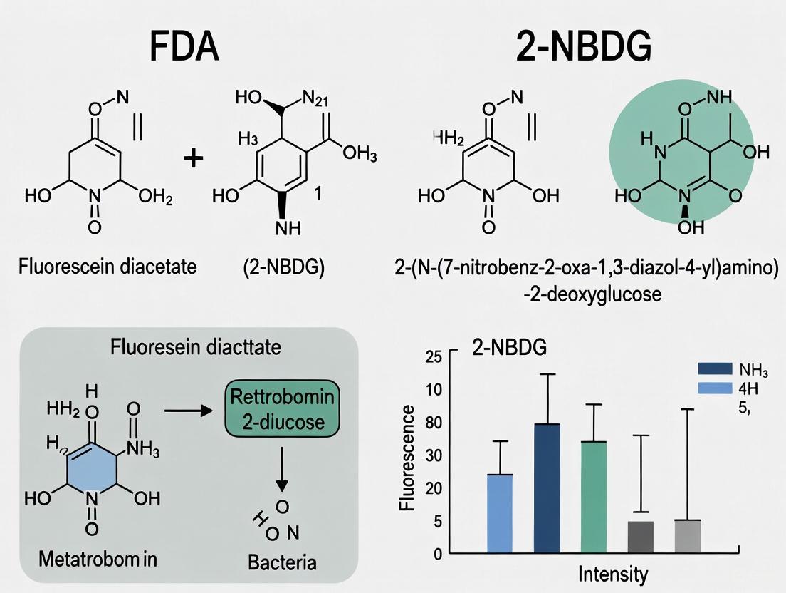
In the study of cellular metabolism, few tools have garnered as much widespread use—and subsequent scrutiny—as fluorescent glucose analogs. 2-NBDG (2-(N-(7-Nitrobenz-2-oxa-1,3-diazol-4-yl)Amino)-2-deoxyglucose) has emerged as a prominent probe for visualizing glucose uptake in living cells across diverse fields including diabetes research, cancer biology, and microbiology [22] [12]. This fluorescent derivative, created by replacing the 2-hydroxyl group of D-glucose with a 7-nitrobenzofurazan fluorophore, offers researchers a non-radioactive method to monitor glucose transport at single-cell resolution using techniques like flow cytometry and fluorescence microscopy [23] [12]. As the scientific community increasingly frames cellular viability research within the context of metabolic activity, understanding the precise capabilities and limitations of 2-NBDG becomes paramount for researchers, scientists, and drug development professionals who rely on these tools for critical experimental outcomes.
The fundamental premise of using 2-NBDG rests on its structural similarity to glucose, theoretically allowing it to be transported into cells via the same mechanisms as natural glucose and subsequently phosphorylated by hexokinase—the first step in glycolysis [22]. This intracellular trapping enables detection of cells actively taking up glucose. However, recent rigorous investigations have revealed crucial distinctions between 2-NBDG's behavior and that of native glucose, raising important questions about its appropriate application in research settings, particularly when compared to more established methods including FDA-approved approaches for metabolic assessment.
Mechanism of Action and Technical Specifications
Structural Properties and Detection
2-NBDG possesses distinct photophysical properties that enable its experimental utility. The compound exhibits excitation and emission maxima at approximately 465/540 nm, allowing detection using standard fluorescein filter sets [24]. With a molecular weight of 342.26 g/mol, 2-NBDG is significantly larger than native glucose due to the bulky 7-nitrobenzofurazan fluorophore attached to the glucosamine backbone [4] [12]. This size difference contributes to key functional differences in transport kinetics compared to natural glucose.
The probe is typically prepared as a stock solution in water, PBS, or ethanol at concentrations up to 10-20 mg/mL [25] [24]. For experimental applications, working concentrations generally range from 10-200 μM for mammalian cells and up to 600 μM for microbial systems [24]. Incubation times vary significantly by cell type and experimental conditions, typically ranging from several minutes to an hour at physiological temperatures to allow sufficient cellular uptake and detection [22] [24].
Cellular Uptake and Metabolism
The purported mechanism of 2-NBDG action involves transport into cells via glucose transporters, particularly those belonging to the SLC2A (GLUT) family, followed by phosphorylation via hexokinase [22]. This phosphorylation theoretically traps the compound intracellularly, allowing accumulation and detection. In bacterial systems like Escherichia coli, evidence suggests 2-NBDG is predominantly transported by the mannose phosphotransferase system and is subsequently metabolized to a non-fluorescent derivative [12]. However, the intracellular fate in mammalian cells remains less clearly defined, with significant questions emerging about whether the compound accurately mimics endogenous glucose transport pathways.
Table 1: Key Technical Specifications of 2-NBDG
| Parameter | Specification | Experimental Notes |
|---|---|---|
| Molecular Weight | 342.26 g/mol | Larger than native glucose due to fluorophore [25] |
| Excitation/Emission | ~465/540 nm | Compatible with FITC filter sets [24] |
| Stock Concentration | Up to 10-20 mg/mL | Prepare in water, PBS, or ethanol [25] [24] |
| Working Concentration | 10-200 μM (mammalian cells); up to 600 μM (microbes) | Must be optimized for cell type [24] |
| Incubation Time | 5 minutes to 1 hour | Temperature-dependent; longer incubations may increase background [22] [24] |
| Cellular Retention | Phosphorylation by hexokinase | Theoretical trapping mechanism [22] |
Comparative Performance Analysis
Advantages Over Traditional Methods
2-NBDG offers several distinct advantages that have contributed to its widespread adoption. A significant benefit is its non-radioactive nature, eliminating safety concerns, regulatory hurdles, and specialized disposal requirements associated with traditional radiolabeled glucose analogs like 2-deoxy-D-[3H]glucose or 2-deoxy-D-[14C]glucose [23]. This characteristic makes the probe particularly valuable for educational settings or facilities with limited radiation safety infrastructure.
The fluorescent signature of 2-NBDG enables single-cell resolution in glucose uptake studies, allowing researchers to investigate heterogeneity within cell populations that would be masked by bulk measurement techniques [23] [26]. This capability has proven valuable in identifying metabolic subpopulations in cancer cells, immune cells, and microbial communities. When applied to rumen bacteria, the method successfully identified over 40 different bacterial variants that uptake glucose, nearly half of which represented previously uncultured bacteria [27].
Furthermore, 2-NBDG demonstrates reduced susceptibility to cross-feeding artifacts compared to stable isotope probing methods. In coculture experiments, the fluorescent label of 2-NBDG was not transferred to nontarget bacteria through metabolic cross-feeding, a significant advantage over isotope-based methods that often lead to off-target labeling [27].
Limitations and Validation Challenges
Despite its theoretical advantages, substantial evidence questions whether 2-NBDG accurately reports glucose transport in mammalian systems. A critical study using L929 fibroblasts—which rely exclusively on Glut1 for glucose uptake—demonstrated that neither pharmacological inhibition of Glut1 nor genetic manipulation of its expression significantly impacted 2-NBDG uptake, despite both approaches dramatically reducing [3H]-2-deoxyglucose uptake [4]. This suggests 2-NBDG may enter cells through transporter-independent mechanisms.
Genetic evidence further complicates the interpretation of 2-NBDG signals. CRISPR-Cas9 ablation of Slc2a1 (GLUT1) in 5TGM1 myeloma cells abrogated radioactive glucose uptake but had no effect on the magnitude or kinetics of 2-NBDG import [28]. The study also found that ablation of other hexose transporters or members of the Slc29 and Slc35 families of nucleoside transporters similarly failed to impact 2-NBDG uptake, indicating the compound enters cells through an unknown mechanism distinct from canonical glucose transport pathways [28].
Additional technical limitations include 2-NBDG's lower photostability compared to rhodamine-based fluorescent probes and potential issues with self-quenching at higher concentrations (>0.25 mM) [25]. The compound also exhibits different transport kinetics than glucose, with a lower Vmax (maximum rate), resulting in generally slower transport compared to native glucose [12].
Table 2: Performance Comparison of Glucose Uptake Probes
| Characteristic | 2-NBDG | Radiolabeled 2-DG | Genetically Encoded Sensors (e.g., iGlucoSnFR2) |
|---|---|---|---|
| Spatial Resolution | Single-cell | Population average | Subcellular to single-cell [29] |
| Temporal Resolution | Minutes to hours | Minutes to hours | Seconds to minutes [29] |
| Transport Mechanism | Questionable fidelity to glucose transporters [4] [28] | High fidelity | Reports concentration, not uptake |
| Technical Complexity | Moderate | High (radiation safety) | High (genetic manipulation) |
| Throughput | High (flow cytometry compatible) | Moderate | Low to moderate |
| Quantitative Accuracy | Limited by transport mechanisms | High | High for concentration measurements [29] |
Experimental Protocols and Methodologies
Standard Glucose Uptake Assay in Mammalian Cells
The following protocol describes a typical 2-NBDG uptake assay in mammalian cells, adapted from multiple sources [22] [23] [25]:
Cell Preparation:
- Culture cells in appropriate medium until 70-90% confluent.
- For suspension cells: Centrifuge at 1,000 × g for 3-5 minutes and wash twice with PBS.
- For adherent cells: Dissociate with trypsin, centrifuge, and wash twice with PBS.
- Resuspend cells in serum-free medium or PBS at approximately 1×10^6 cells/mL.
2-NBDG Staining:
- Prepare working solution of 2-NBDG in serum-free medium or PBS (typically 10-200 μM).
- Add 1 mL of 2-NBDG working solution to cell pellet.
- Incubate at 37°C for 5-60 minutes (time must be optimized for cell type).
- Centrifuge at 400 × g for 3-4 minutes and discard supernatant.
- Wash cells twice with PBS to remove extracellular 2-NBDG.
- Resuspend in serum-free medium or PBS for immediate analysis.
Detection and Analysis:
- Analyze by flow cytometry using 488 nm excitation and 530/30 nm emission filter.
- Alternatively, image using fluorescence microscopy with FITC filter sets.
- Include controls without 2-NBDG for autofluorescence correction.
- For quantitative comparisons, maintain consistent incubation times and concentrations.
Microfluidic Single-Cell Analysis in RBCs
A recent advanced application of 2-NBDG enables quantitative measurement of glucose uptake in individual red blood cells using microfluidics and confocal microscopy [26]:
Sample Preparation:
- Isolate RBCs from whole blood via centrifugation at 2,000 RPM for 5 minutes.
- Remove supernatant and buffy coat, then wash three times with KCl solution.
- Incubate packed RBCs with biotinylated-α-glycophorin A+B antibodies for 1 hour at 37°C.
Microfluidic Setup:
- Functionalize microfluidic chamber surface with streptavidin.
- Perfuse antibody-labeled RBCs into chamber for surface attachment.
- Establish continuous perfusion with 5 mM 2-NBDG in modified KCl buffer.
Image Acquisition and Analysis:
- Acquire time-lapse confocal images using 488 nm excitation.
- Measure intracellular and extracellular fluorescence intensities.
- Calculate intracellular glucose analog tracer percentage as ratio of intra- to extracellular 2-NBDG intensity.
- Analyze multiple single cells to assess population heterogeneity.
This method revealed significant cell-to-cell and donor-to-donor variability in 2-NBDG uptake in human RBCs, demonstrating the importance of single-cell approaches for understanding glucose transport heterogeneity [26].
Figure 1: Standard 2-NBDG Uptake Assay Workflow
Emerging Alternatives and Future Directions
Genetically Encoded Glucose Sensors
The development of genetically encoded fluorescent sensors represents a promising alternative to chemical probes like 2-NBDG. The second-generation intensity-based glucose-sensing fluorescent reporter (iGlucoSnFR2) demonstrates significant advantages for certain applications [29]. This sensor, developed from a glucose-binding protein of Thermus thermophilus and circularly permuted SuperFolder GFP, reports intracellular glucose concentration with high temporal resolution and specificity.
Unlike 2-NBDG, iGlucoSnFR2 can be targeted to specific subcellular compartments, enabling researchers to monitor glucose dynamics in specific organelles. The sensor has been successfully deployed in vivo using fiber photometry in mouse brains, reporting transient increases in glucose concentration following noradrenaline stimulation or electrical activity [29]. For research questions focused on glucose concentration rather than uptake mechanisms, genetically encoded sensors offer superior specificity and temporal resolution.
Advanced Chemical Probes
While 2-NBDG remains the most common fluorescent glucose analog, other chemical probes continue to be developed and characterized. 6-NBDG represents a structural isomer with the fluorophore at a different position on the glucose molecule, though it shares many of the same limitations regarding transport mechanism fidelity [4]. Silicon-rhodamine glucose conjugates (e.g., D-glu-SiR) offer improved photostability and longer wavelength emission, potentially enabling multiplexed experiments [26].
Each emerging technology presents distinct advantages and limitations, requiring researchers to carefully match their tool selection to specific experimental questions rather than relying on a single standardized approach.
Figure 2: Probe Selection Decision Framework
The Scientist's Toolkit: Essential Research Reagents
Table 3: Key Research Reagent Solutions for Glucose Uptake Studies
| Reagent/Category | Specific Examples | Function/Application | Considerations |
|---|---|---|---|
| Fluorescent Glucose Analogs | 2-NBDG, 6-NBDG | Direct visualization of glucose analog uptake | Questionable transport mechanism fidelity; concentration-dependent self-quenching [4] [12] |
| GLUT Transporter Inhibitors | Cytochalasin B, WZB117, BAY-876 | Pharmacological inhibition of glucose transporters | Limited impact on 2-NBDG uptake suggests alternative entry mechanisms [4] [28] |
| Genetically Encoded Sensors | iGlucoSnFR2, FLIP-glu | Real-time monitoring of glucose concentration | Requires genetic manipulation; superior temporal resolution [29] |
| Microfluidic Systems | Commercial perfusion chambers | Single-cell analysis under homeostasis | Enables precise control of extracellular conditions [26] |
| Detection Instruments | Flow cytometers, confocal microscopes | Signal detection and quantification | Standard FITC filters suitable for 2-NBDG detection [24] |
2-NBDG represents a valuable but imperfect tool for investigating glucose metabolism in living cells. Its non-radioactive nature and compatibility with single-cell analysis technologies make it attractive for screening applications and heterogeneous cell population studies. However, substantial evidence questions its fidelity to endogenous glucose transport mechanisms, particularly in mammalian systems [4] [28]. Researchers must therefore exercise caution when interpreting 2-NBDG uptake as a direct measure of glucose transport activity.
For applications requiring definitive glucose uptake measurement, radiolabeled 2-deoxyglucose remains the gold standard despite its technical and safety challenges. When fluorescent methods are preferred, controls verifying transport mechanisms are essential for appropriate interpretation of 2-NBDG data. Emerging technologies, particularly genetically encoded sensors like iGlucoSnFR2, offer promising alternatives for specific applications focused on glucose concentration rather than uptake mechanisms [29].
The appropriate selection of glucose monitoring tools depends critically on the specific research question, with different technologies offering complementary strengths and limitations. As the field advances, continued rigorous validation of these essential research tools remains fundamental to progress in understanding cellular metabolism.
Fluorescent glucose analogs have revolutionized the assessment of cellular glucose uptake and metabolic activity in live cells at single-cell resolution. Among these, 2-NBDG and Fluorescein Diacetate (FDA) represent two distinct approaches for monitoring cellular metabolic status in viability research. While both serve as fluorescent indicators, their mechanisms of uptake, metabolic processing, and reliability as metabolic proxies differ significantly. This comprehensive comparison examines the journey of 2-NBDG from cellular uptake to intracellular metabolism, contrasting it with FDA's mechanism of action. We synthesize recent genetic and pharmacological evidence challenging long-held assumptions about 2-NBDG transport mechanisms and provide experimental data supporting informed probe selection for metabolic studies in drug development and basic research.
The measurement of cellular metabolic activity is fundamental to understanding physiological and pathological processes across biomedical research. Glucose uptake represents a critical parameter in metabolic phenotyping, particularly in cancer biology, immunology, and toxicology. While traditional methods utilizing radiolabeled glucose analogs provide reliable quantification of glucose transport, they lack single-cell resolution and require specialized handling facilities [4].
Fluorescent alternatives have emerged to overcome these limitations, with 2-NBDG and FDA representing two distinct classes of metabolic probes. 2-NBDG was developed as a direct fluorescent analog of glucose, designed to report on glucose transporter activity and cellular glucose uptake [12]. In contrast, FDA serves as a substrate for intracellular esterases, providing a general measure of enzymatic activity and membrane integrity [18]. Despite their widespread use, the precise mechanisms underlying their cellular uptake and metabolism require careful examination, as recent evidence challenges conventional understanding of these processes.
Table 1: Fundamental Properties of Fluorescent Metabolic Probes
| Property | 2-NBDG | FDA |
|---|---|---|
| Chemical nature | Fluorescent glucose derivative (2-deoxy-2-[(7-nitro-2,1,3-benzoxadiazol-4-yl)amino]-D-glucose) | Non-fluorescent diacetate ester of fluorescein |
| Molecular weight | 342.26 g/mol [12] | 416.38 g/mol |
| Primary mechanism of uptake | Originally thought to be glucose transporters, but recent evidence suggests unknown, transporter-independent mechanisms [30] [4] | Passive diffusion across lipid membranes [18] |
| Intracellular processing | Converted to non-fluorescent derivative [12] | Hydrolysis by nonspecific intracellular esterases [18] |
| Fluorescent signal | Green fluorescence (emission ~540 nm) [12] | Green fluorescence (emission ~520 nm) [18] |
| Primary application | Originally designed for monitoring glucose uptake | Assessment of esterase activity and membrane integrity |
Mechanisms of Cellular Uptake
The Controversial Journey of 2-NBDG into Cells
The conventional understanding posits that 2-NBDG enters cells through glucose transporters (GLUTs), similar to native glucose. This analog contains a bulky 7-nitro-2,1,3-benzoxadiazol-4-yl-amino moiety in place of the 2-hydroxyl group on D-glucose, significantly altering both the size and shape of the molecule compared to glucose [4]. Early characterization studies in E. coli suggested competition with D-glucose for import via mannose or glucose/mannose transporter systems [30], leading to the widespread assumption that similar mechanisms operated in mammalian cells.
However, recent genetic evidence fundamentally challenges this paradigm. Multiple independent studies using CRISPR-Cas9 gene editing demonstrate that ablation of the primary glucose transporter gene Slc2a1 (encoding GLUT1) abrogates radioactive glucose uptake but has no effect on the magnitude or kinetics of 2NBDG import [30] [28]. Neither excess glucose nor pharmacological inhibition of GLUT1 using multiple inhibitors (cytochalasin B, BAY-876, WZB-117) impacted 2NBDG uptake in myeloma cells or primary splenocytes [30] [4]. Genetic ablation of other expressed hexose transporters individually or in combination similarly failed to impact 2NBDG uptake, as did ablation of genes in the Slc29 and Slc35 families of nucleoside and nucleoside sugar transporters [30].
These findings collectively indicate that 2NBDG uptake occurs independently of known glucose transporters and is promoted by an unknown mechanism specific to this compound. The transport mechanism appears distinct even from chemically similar compounds, as extracellular 2NBDG, but not NBD-fructose, was transported by primary plasma cells into the cytoplasm [30].
Diagram 1: 2-NBDG uptake occurs independently of known glucose transporters
FDA Uptake and Activation Mechanism
In contrast to 2-NBDG, FDA uptake occurs through passive diffusion across lipid bilayer membranes due to its lipophilic properties imparted by two acetate groups [18]. This non-polar, non-fluorescent dye readily traverses cell membranes without requiring specific transport proteins or energy expenditure. Once inside viable cells, FDA undergoes hydrolysis by nonspecific intracellular enzymes (esterases, lipases, and proteases) to release fluorescein, which is polar and therefore trapped inside cells with intact membranes [18]. The fluorescent signal accumulation thus serves as an indicator of both enzymatic activity and membrane integrity.
The fundamental difference in uptake mechanisms between these probes highlights their distinct applications: 2-NBDG was designed to report specifically on sugar transport activity, while FDA serves as a general viability marker through assessment of esterase activity and membrane competence.
Table 2: Comparison of Inhibitory Effects on Probe Uptake
| Experimental Condition | Effect on 2-NBDG Uptake | Effect on FDA Uptake | Effect on Radiolabeled Glucose Uptake |
|---|---|---|---|
| GLUT1 genetic ablation | No significant effect [30] [4] | Not tested | Abrogated [30] [4] |
| GLUT1 pharmacological inhibition | No significant effect [30] [4] | Not tested | Significantly reduced [4] |
| Excess D-glucose competition | Minimal to no inhibition [30] [4] | Not applicable | Significantly inhibited [4] |
| Temperature reduction | Uptake blocked [31] | Expected to slow diffusion | Transport blocked |
Intracellular Metabolism and Fate
Metabolic Processing of 2-NBDG
Once inside cells, 2-NBDG undergoes metabolic processing that differs from both native glucose and its radiolabeled counterparts. Early work in E. coli demonstrated that 2-NBDG is metabolized to a non-fluorescent derivative, though the exact identity and subsequent metabolic fate of this derivative remain unestablished [12]. Unlike 2-deoxy-D-glucose (2-DG), which is phosphorylated by hexokinase and accumulates as 2-DG-6-phosphate, the metabolic pathway of 2-NBDG has not been fully elucidated.
In mammalian cells, the intracellular fluorescence pattern of 2-NBDG typically appears diffuse throughout the cytoplasm, suggesting that it may not be sequestered in specific organelles or undergo the same metabolic trapping as 2-DG-6-phosphate [31]. This distribution pattern, combined with evidence of non-specific transport, complicates interpretation of 2-NBDG fluorescence as a direct measure of glucose metabolic capacity.
FDA Activation and Signal Retention
FDA's metabolic processing is more straightforward: intracellular esterases cleave the acetate groups, converting the non-fluorescent FDA to highly fluorescent fluorescein [18]. This hydrolyzed product accumulates within cells with intact membranes, creating a fluorescent signal proportional to esterase activity. However, several factors can affect signal intensity, including pH-dependent fluorescence quenching and potential efflux of fluorescein from cells, particularly under acidic conditions [18]. The product of FDA hydrolysis (acetic acid) may itself decrease intracellular pH, potentially creating a negative feedback loop that limits signal accumulation.
Experimental Protocols and Methodologies
Standard 2-NBDG Uptake Assay Protocol
The following protocol for measuring 2-NBDG uptake has been adapted from multiple methodological approaches described in the literature [30] [16]:
Cell Preparation: Harvest and wash cells with PBS. Count and adjust cell density to 1-2 × 10^6 cells/mL in appropriate glucose-free buffer or culture medium.
2-NBDG Incubation: Resuspend cells in pre-warmed medium containing 20μg/mL (~60μM) 2-NBDG. Incubate for 1 hour at 37°C protected from light.
Control Preparations: Include the following controls:
- Non-2-NBDG-treated cells for autofluorescence
- Cells incubated at 4°C to assess temperature dependence
- Cells pre-treated with excess D-glucose (100mM) to assess GLUT-specificity
Termination and Washing: Stop the reaction by placing tubes on ice. Wash cells twice with ice-cold PBS to remove extracellular 2-NBDG.
Analysis: Analyze cells by flow cytometry (excitation/emission: ~465/540 nm) or fluorescence microscopy. For microscopy, cells may be fixed with 4% paraformaldehyde if necessary.
Diagram 2: Standard 2-NBDG uptake assay workflow
FDA Viability Staining Protocol
The standard protocol for FDA viability assessment is as follows [18]:
Stock Solution Preparation: Prepare FDA stock solution at 1-5 mg/mL in acetone or DMSO. Store at -20°C protected from light.
Cell Preparation: Harvest cells and wash with appropriate buffer. Adjust cell density to 1 × 10^6 cells/mL.
Staining: Add FDA to cell suspension at final concentration of 1-10 μg/mL. Incubate for 5-15 minutes at room temperature or 37°C protected from light.
Analysis: Analyze immediately by fluorescence microscopy or flow cytometry (excitation/emission: ~490/520 nm). Do not wash cells after staining, as this may remove the fluorescent product.
Quantitative Comparison and Experimental Data
Performance in Metabolic Inhibition Studies
Recent direct comparisons between 2-NBDG and established glucose uptake assays reveal significant discrepancies in response to metabolic inhibition. When evaluated alongside the enzymatic bulk 2DG uptake assay (considered the gold standard for glucose transport measurement), 2-NBDG showed markedly different inhibition profiles:
Table 3: Quantitative Comparison of Inhibition Effects on Glucose Uptake Probes
| Inhibition Condition | Effect on 2DG Uptake (Gold Standard) | Effect on 2-NBDG Uptake | Effect on 6AzGal Uptake (Novel Probe) |
|---|---|---|---|
| Cytochalasin B (GLUT inhibitor) | >70% signal reduction [31] | 20-40% signal reduction [31] | >70% signal reduction [31] |
| WZB-117 (GLUT inhibitor) | >70% signal reduction [31] | 20-40% signal reduction [31] | >70% signal reduction [31] |
| D-glucose competition | Significant dose-dependent suppression [31] | Minimal to no effect [30] [4] | Significant dose-dependent suppression [31] |
| Temperature dependence | Complete inhibition at low temperature [31] | Complete inhibition at low temperature [31] | Complete inhibition at low temperature [31] |
These data demonstrate that 2-NBDG uptake responds differently to established GLUT inhibitors compared to radiolabeled 2DG, supporting the conclusion that it enters cells through mechanisms distinct from canonical glucose transporters.
Applications in Biological Systems
Despite limitations in specificity, 2-NBDG has been successfully employed in various experimental models. In a preclinical model of oral epithelial neoplasia, topical application of 2-NBDG enabled delineation of neoplastic tissue, with fluorescence intensity 4-6 fold higher in oral squamous cell carcinoma and oral epithelial dysplasia compared to normal mucosa [16]. The probe showed 83% sensitivity and 73% specificity for detecting neoplasia versus benign tissue, demonstrating its utility in detecting increased metabolic activity associated with transformation, even if the exact transport mechanism differs from native glucose.
Emerging Alternatives and Methodological Advances
Click Chemistry-Based Approaches
Recent methodological advances address the limitations of direct fluorescent glucose analogs. A novel click chemistry-based post-labeling method utilizes GLUT-mediated uptake of azide-tagged sugars (such as 6-azido-6-deoxy-D-galactose, 6AzGal) followed by intracellular labeling with a cell-permeable fluorescent reagent via copper-free click reaction [31].
This approach offers several advantages:
- Minimal modification of sugar structure (molecular weight: 205 Da vs 180 Da for glucose)
- Maintains GLUT specificity as demonstrated by competition with D-glucose and inhibition by GLUT inhibitors
- Enables highly accurate single-cell measurements with low background adsorption
- Compatible with multi-parametric immunophenotyping for resolving heterogeneous metabolic landscapes in complex tissues [31]
The 6AzGal method accurately reproduces in vivo dynamics similar to 18F-FDG and shows appropriate inhibition profiles that match the gold standard 2DG uptake assay, addressing the key limitations of 2-NBDG [31].
The Scientist's Toolkit: Essential Research Reagents
Table 4: Key Research Reagents for Glucose Uptake and Viability Studies
| Reagent | Function/Application | Key Considerations |
|---|---|---|
| 2-NBDG | Fluorescent glucose analog for monitoring uptake | Use with caution for GLUT activity; transport mechanism not fully defined; appropriate for metabolic activity assessment but not specific glucose transport [30] [4] |
| FDA (Fluorescein Diacetate) | Viability staining through esterase activity | General viability marker; affected by intracellular pH; may leak from cells [18] |
| 6AzGal | Click chemistry-based glucose uptake probe | High specificity for GLUTs; requires post-labeling with BDP-DBCO; more accurate than 2-NBDG for glucose transport [31] |
| WZB-117 | GLUT1 inhibitor | Useful for validating GLUT-specific uptake; inhibits 6AzGal and 2DG but not 2-NBDG uptake [30] [31] |
| Cytochalasin B | Broad-spectrum GLUT inhibitor | Endofacial inhibitor; inhibits 6AzGal and 2DG uptake effectively but only partially inhibits 2-NBDG [4] [31] |
| BAY-876 | Potent GLUT1-specific inhibitor | Useful for dissecting GLUT1-specific effects; does not inhibit 2-NBDG uptake [30] [4] |
The journey of 2-NBDG from extracellular space to intracellular compartments represents a more complex pathway than originally anticipated. While widely used as a fluorescent glucose analog, substantial genetic and pharmacological evidence now indicates that its uptake occurs independently of known glucose transporters through an unidentified mechanism. This fundamental limitation necessitates careful interpretation of 2-NBDG data, particularly in studies aiming to make specific conclusions about glucose transporter activity or regulation.
In contrast, FDA serves as a reliable general viability probe through its conversion by intracellular esterases, though it provides no specific information about glucose metabolism. For researchers requiring accurate assessment of glucose transport, emerging alternatives such as click chemistry-based approaches with 6AzGal offer GLUT-specific quantification with minimal background, addressing the key limitations of both 2-NBDG and FDA.
The appropriate choice between these probes depends critically on the specific research question: FDA for general viability assessment, 2-NBDG for detecting broad metabolic activation, and newer click chemistry approaches for specific quantification of glucose transporter activity. As our understanding of these tools evolves, so too must our experimental designs and interpretations in metabolic research.
This guide objectively compares the fluorescent glucose analog 2-NBDG against alternative methods for assessing cell viability and metabolic activity, providing structured experimental data and protocols for researchers in drug development.
Performance Comparison of Glucose Uptake and Viability Assays
The table below summarizes the core characteristics, advantages, and limitations of 2-NBDG and other common methods for measuring glucose uptake and viability.
| Assay Method | Principle of Detection | Key Advantages | Key Limitations / Discrepancies | Best-Suited Application |
|---|---|---|---|---|
| 2-NBDG (Fluorescent Glucose Analog) | Intracellular accumulation of a fluorescent glucose analog [32]. | Rapid; non-radioactive; works well for imaging (microscopy/flow cytometry) [7] [32] [33]. | Uptake may not fully replicate native glucose transport; larger molecular size raises questions about transporter fidelity [32] [4]. | Real-time imaging of glucose uptake; rapid viability screening in yeast and mammalian cells [7] [13] [16]. |
| Radioactive (³H-2DG) | Intracellular accumulation of radiolabeled 2-deoxyglucose-6-phosphate (2DG6P) [32]. | Considered the gold standard; high sensitivity [32] [4]. | Requires handling and disposal of radioactive materials; multiple wash steps [32]. | High-sensitivity, definitive measurement of glucose transporter activity. |
| Luminescence (2DG6P Detection) | Enzymatic detection of accumulated 2DG6P, generating a luminescent signal [32]. | Non-radioactive; highly sensitive; large signal window; amenable to high-throughput screening [32]. | Not applicable for cell imaging [32]. | High-throughput screening in multiwell plate formats. |
| Absorbance/Fluorescence (2DG6P Detection) | Enzymatic detection of accumulated 2DG6P, generating a colored or fluorescent product [32]. | Non-radioactive [32]. | Multiple processing steps; narrow detection window [32]. | Lower-throughput plate-based assays. |
Experimental Data and Validation
The following table consolidates key experimental findings that validate and contextualize the performance of 2-NBDG across different biological systems.
| Cell System | Experimental Finding | Correlation / Validation | Citation |
|---|---|---|---|
| Yeast | Fluorescence intensity after 2-NBDG staining showed a strong correlation with viability measured by Colony Forming Units (CFU) after exposure to antifungal agents. | Correlation constant: r = 0.98 with CFU count [7]. | [7] |
| Mammalian Cells (MCF-7 & PBMCs) | Under hyperoxia, the difference in 2-NBDG fluorescence between tumor cells (MCF-7) and healthy cells (PBMCs) was maximized, allowing for discrimination and quantification by flow cytometry. | Successful detection of spiked MCF-7 cells in PBMCs at ratios as low as 1:10,000 [13]. | [13] |
| Mammalian Cells (L929 Fibroblasts) | Pharmacological inhibition or genetic knockdown of GLUT1 transporter significantly reduced uptake of radioactive 2-DG but had no significant impact on the uptake of 2-NBDG. | Indicates 2-NBDG may enter cells via transporter-independent mechanisms in some cell lines [4]. | [4] |
| Oral Mucosa (In Vivo Hamster Model) | Topical application of 2-NBDG resulted in a 4-fold and 6-fold higher fluorescence in dysplastic (OED) and cancerous (OSCC) tissues, respectively, compared to normal mucosa. | Enabled delineation of neoplasia from normal tissue with 83% sensitivity and 73% specificity [16]. | [16] |
Detailed Experimental Protocols
Protocol 1: Rapid Viability Assessment in Yeast using 2-NBDG
This protocol is adapted from a study demonstrating a high correlation between 2-NBDG fluorescence and colony-forming units (CFUs) [7].
- 1. Staining Solution Preparation: Prepare a working solution of 2-NBDG in a suitable buffer or low-glucose medium. A concentration of 10-200 μM is recommended, though this should be optimized [33].
- 2. Cell Staining: Harvest yeast cells and wash with PBS. Resuspend the cell pellet in the 2-NBDG working solution.
- 3. Incubation: Incubate the cell suspension for 30-60 minutes at room temperature, protected from light [7] [33].
- 4. Washing and Analysis: Centrifuge the cells to remove the excess dye and wash the pellet with PBS. Resuspend in a clean buffer and analyze fluorescence intensity immediately using a fluorescence spectrophotometer or flow cytometer [7].
Protocol 2: Discriminating Tumor Cells in PBMCs using 2-NBDG under Hyperoxia
This optimized protocol from a study on circulating tumor cells (CTCs) maximizes the signal difference between tumor and healthy cells [13].
- 1. Cell Preparation: Isolate Peripheral Blood Mononuclear Cells (PBMCs) from human blood via Ficoll gradient centrifugation. Culture and harvest the tumor cell line of interest (e.g., MCF-7).
- 2. Spiking and Staining: Spike a known number of tumor cells into the PBMC suspension. Stain the cell mixture with 2-NBDG at a final concentration of 300 μM in PBS.
- 3. Hyperoxia Incubation: Incubate the stained cell suspension for 30 minutes under hyperoxia (high oxygen) conditions [13].
- 4. Flow Cytometry Analysis: After incubation, wash the cells and analyze using a flow cytometer equipped with a 488 nm laser for 2-NBDG excitation. Positive events for tumor cells are identified by high green fluorescence in the absence of leukocyte markers (e.g., CD45) [13].
The Scientist's Toolkit: Key Research Reagent Solutions
| Reagent / Material | Function in Experiment | Specific Example / Note |
|---|---|---|
| 2-NBDG (2-(N-(7-Nitrobenz-2-oxa-1,3-diazol-4-yl)Amino)-2-Deoxyglucose) | Fluorescent glucose analog used to monitor and quantify cellular glucose uptake and viability [33]. | Excitation/Emission: ~488/542 nm; suitable for confocal microscopy and flow cytometry [33]. |
| GLUT1 Inhibitors (e.g., BAY-876, WZB117) | Pharmacological tools to inhibit the major glucose transporter GLUT1, used to validate the transport mechanism of a probe [4]. | Critical for experiments determining if 2-NBDG uptake is GLUT1-dependent [4]. |
| Microfluidic Perfusion System | Provides high-precision control of the cellular environment, enabling steady-state imaging and homeostatic condition maintenance [26]. | Essential for single-cell glucose uptake studies in RBCs using confocal microscopy [26]. |
| Turbid Phantoms (Intralipid & Hemoglobin) | Tissue-simulating models used to validate and correct fluorescence measurements in a controlled, scattering environment [34]. | Used to develop empirical methods for correcting spectral distortions in vivo [34]. |
| Glucose Uptake-Glo Assay | A luminescent, non-radioactive assay that detects accumulated 2DG6P, ideal for high-throughput screening in multiwell plates [32]. | Offers a simple, sensitive alternative without wash steps [32]. |
Mechanistic Pathways and Experimental Workflows
2-NBDG Uptake and the Warburg Effect in Cancer Cells
Experimental Workflow for Single-Cell Glucose Uptake Analysis
From Theory to Bench: Standard Protocols and Advanced Applications for FDA and 2-NBDG
In cellular and microbiological research, assessing cell viability and metabolic activity is fundamental. Fluorescent dyes that serve as metabolic indicators provide a powerful, often non-disruptive, means to evaluate these parameters in real-time. Among these, Fluorescein Diacetate (FDA) and 2-[N-(7-nitrobenz-2-oxa-1,3-diazol-4-yl)amino]-2-deoxy-D-glucose (2-NBDG) are two prominent probes. FDA is a marker of general enzymatic activity, while 2-NBDG is a direct probe for glucose uptake, a key metabolic process. This guide provides a detailed, objective comparison of these two dyes, focusing on their standardized staining protocols, performance characteristics, and experimental applications to aid researchers in selecting the appropriate tool for their viability research.
Dye Mechanisms and Metabolic Pathways
Understanding the distinct metabolic pathways exploited by FDA and 2-NBDG is crucial for interpreting staining results and selecting the appropriate dye for a given research question. Their fundamental mechanisms of action are compared below.
Mechanism of Fluorescein Diacetate (FDA)
Figure 1: FDA Mechanism: Enzymatic Hydrolysis for Viability
FDA itself is a non-fluorescent, lipophilic compound that passively diffuses across the cell membrane of viable cells [1]. Once inside the cell, it serves as a substrate for ubiquitous intracellular enzymes, primarily nonspecific esterases, but also lipases and proteases [1]. These active enzyme systems hydrolyze FDA, cleaving the acetate groups to release fluorescein. Fluorescein is a hydrophilic and intensely green-fluorescent molecule. Because of its charged, hydrophilic nature, it cannot easily diffuse back out through the intact cell membrane, leading to its accumulation within the cytoplasm. Therefore, the detected green fluorescence (emission ~520 nm) is directly correlated with the combined activity of these enzymatic systems and the integrity of the cell membrane, which are hallmarks of cell viability [1].
Mechanism of 2-NBDG
Figure 2: 2-NBDG Mechanism: Glucose Transporter Uptake
2-NBDG operates on a different principle, functioning as a direct reporter of glucose uptake metabolism. It is a fluorescently labeled glucose analogue, where the nitrobenzoxadiazole (NBD) fluorophore is conjugated to the 2-deoxyglucose molecule [13]. Cells take up 2-NBDG primarily through active transport via their glucose transporter (GLUT) proteins on the membrane [13] [16]. Once inside the cell, similar to native glucose, 2-NBDG is phosphorylated by hexokinase, the first enzyme in the glycolytic pathway. However, being 2-deoxyglucose, it cannot be further metabolized in the glycolysis cycle. This phosphorylation traps the molecule inside the cell, leading to the accumulation of fluorescent 2-NBDG-phosphate [13]. The resulting green fluorescence (emission ~540 nm) is thus a direct indicator of the cell's glucose transporter activity and hexokinase activity. This mechanism is particularly useful for identifying cells with elevated glucose consumption, such as cancer cells exhibiting the Warburg effect (aerobic glycolysis) [13] [16].
Standard Staining Protocols and Performance Data
Direct comparison of standardized protocols and quantitative performance data is essential for experimental planning. The following sections provide detailed methodologies and a consolidated comparison of key parameters for both dyes.
Standard FDA Staining Protocol
The FDA staining protocol is designed to maximize dye uptake and hydrolysis while minimizing background signal and potential cytotoxicity.
Workflow: FDA Staining and Detection
- Dye Preparation: Prepare a stock solution of FDA in high-grade dimethyl sulfoxide (DMSO) at a concentration of 1-10 mM. Aliquot and store at -20°C, protected from light and moisture. Immediately before use, dilute the stock solution in a suitable, serum-free buffer (e.g., PBS or Hanks' Balanced Salt Solution) to create a working concentration typically between 1 and 10 µM [1]. Serum must be avoided in the working solution, as esterases present in serum can hydrolyze the dye extracellularly, creating high background fluorescence.
- Cell Staining: Aspirate the culture medium from cells and gently wash with PBS. Add the pre-warmed FDA working solution to completely cover the cells. The optimal incubation time is determined empirically but generally ranges from 15 to 45 minutes at 37°C [1]. The incubation must be performed in the dark to prevent photobleaching.
- Washing and Detection: After incubation, carefully remove the FDA working solution and wash the cells 2-3 times with a sufficient volume of PBS to ensure complete removal of any extracellular, non-hydrolyzed dye. The fluorescent signal from intracellular fluorescein must be detected immediately after washing. Detection is typically performed using a fluorescence microscope or flow cytometer equipped with a standard FITC/GFP filter set (Excitation ~492 nm, Emission ~520 nm).
Standard 2-NBDG Staining Protocol
The 2-NBDG protocol is optimized to highlight differences in glucose uptake, particularly between normal and hypermetabolic cells.
Workflow: 2-NBDG Staining and Detection
- Dye Preparation: Reconstitute 2-NBDG according to the manufacturer's instructions. The working solution is typically prepared in PBS or a low-glucose culture medium at concentrations ranging from 50 to 300 µM [13] [16]. A critical optimization step involves the oxygen content during incubation. Research indicates that performing the incubation under hyperoxia (high oxygen) conditions can significantly maximize the fluorescence signal difference between tumor cells and normal cells, thereby enhancing detection sensitivity [13].
- Cell Staining: Cells are incubated with the 2-NBDG working solution for a defined period. A common and effective incubation time, as used in cancer cell discrimination studies, is 30 minutes at 37°C, protected from light [13] [16]. For some cell types, especially bacterial cells, uptake may vary, and not all organisms transport 2-NBDG efficiently [1].
- Washing and Detection: Following incubation, cells are washed twice with PBS to remove any non-internalized 2-NBDG. The fluorescence is detected immediately using instrumentation equipped with a blue laser/excitation source (~465 nm) and a green emission filter (~540 nm) [13]. Flow cytometry is particularly well-suited for quantifying 2-NBDG uptake in heterogeneous cell populations.
Quantitative Performance Comparison
The table below summarizes the core staining parameters and performance characteristics of FDA and 2-NBDG based on experimental data.
Table 1: Direct Comparison of FDA and 2-NBDG Staining Protocols and Performance
| Parameter | Fluorescein Diacetate (FDA) | 2-NBDG |
|---|---|---|
| Working Concentration | 1 - 10 µM [1] | 50 - 300 µM [13] [16] |
| Standard Incubation Time | 15 - 45 minutes [1] | ~30 minutes [13] [16] |
| Optimal Incubation Conditions | 37°C, serum-free buffer [1] | 37°C, hyperoxia can enhance signal contrast [13] |
| Excitation/Emission | ~492 nm / ~520 nm [1] | ~465 nm / ~540 nm [13] |
| Primary Mechanism | Passive diffusion & enzymatic hydrolysis [1] | Active transport via glucose transporters [13] |
| What It Measures | Broad enzymatic activity (esterases, lipases) & membrane integrity [1] | Glucose uptake and hexokinase activity (glycolytic flux) [13] [16] |
| Key Advantages | - Simple, cost-effective- High sensitivity to general viability- Passive uptake is universal | - Direct measure of metabolic activity (glycolysis)- Can exploit Warburg effect in cancer cells- Useful for functional metabolic phenotyping |
| Key Limitations | - Sensitive to extracellular esterases (requires serum-free)- Signal is pH-sensitive (efflux in acidic conditions) [1]- Does not specify metabolic pathway | - Not all cells take up 2-NBDG (e.g., some bacteria) [1]- Requires active transport mechanism- Generally more expensive than FDA |
The Scientist's Toolkit: Essential Research Reagents
Successful experimentation requires a set of well-defined reagents and materials. The following table lists key solutions and their functions for experiments utilizing these fluorescent dyes.
Table 2: Essential Reagent Solutions for Fluorescent Viability Staining
| Reagent/Solution | Function & Role in Staining Protocol |
|---|---|
| FDA Stock Solution (1-10 mM in DMSO) | Stable, concentrated stock for preparing working solutions; DMSO ensures cell permeability of the probe. |
| 2-NBDG Stock Solution | Reconstituted fluorescent glucose analog; the core probe for measuring glucose uptake. |
| Phosphate-Buffered Saline (PBS) | Isotonic buffer used for washing cells, diluting dyes, and maintaining physiological pH during staining steps. |
| Serum-Free Culture Medium | Critical for preparing FDA working solution to prevent extracellular hydrolysis by serum esterases, which cause high background [1]. |
| Dimethyl Sulfoxide (DMSO) | High-quality solvent for preparing stock solutions of lipophilic dyes like FDA; ensures dye solubility and cell penetration. |
| Trypan Blue Solution | Vital dye used to assess overall cell viability and count cells prior to staining, ensuring consistent seeding density. |
FDA and 2-NBDG are both powerful fluorescent dyes for assessing cell viability, but they report on fundamentally different biological processes. FDA serves as a broad indicator of enzymatic activity and membrane integrity, making it a good general-purpose viability stain. In contrast, 2-NBDG provides a more specific functional readout of glucose uptake and glycolytic metabolism, making it indispensable for studies focusing on metabolic phenotyping, especially in cancer research and immunology. The choice between them is not a matter of superiority but of application. Researchers must align their selection with their specific biological question, considering the distinct mechanisms, optimized protocols, and inherent limitations of each dye to ensure accurate and interpretable results.
In cell-based research, accurately assessing viability and metabolic activity is fundamental. Fluorescent probes such as Fluorescein Diacetate (FDA) and 2-[N-(7-nitrobenz-2-oxa-1,3-diazol-4-yl)amino]-2-deoxy-D-glucose (2-NBDG) are powerful tools for this purpose, yet they operate on distinct biological principles. FDA is a non-fluorescent compound that passively diffuses into cells and is hydrolyzed by intracellular esterases into fluorescent fluorescein, marking cells with enzymatic activity as viable [2]. In contrast, 2-NBDG is a fluorescent glucose analog that is actively transported into cells via glucose transporters (GLUTs) and subsequently phosphorylated, serving as a direct indicator of cellular glucose uptake activity [13] [35]. This guide provides a standardized protocol for optimizing 2-NBDG staining, directly compares its performance with FDA, and presents supporting experimental data to inform researchers and drug development professionals.
Principles of 2-NBDG Uptake and Mechanism
The utility of 2-NBDG stems from its ability to mimic natural glucose metabolism. Its uptake is primarily mediated by glucose transporters, most notably GLUT1, which is often overexpressed in various tumor cells [35]. Once inside the cell, 2-NBDG is phosphorylated by hexokinase, the first committed step of glycolysis. This phosphorylation traps the molecule intracellularly, allowing for the accumulation of a fluorescent signal that is proportional to the cell's glucose uptake activity [13] [26]. This mechanism is particularly valuable for exploiting the Warburg effect—the phenomenon where many cancer cells exhibit elevated aerobic glycolysis and consume glucose at a much higher rate than normal cells [13]. Consequently, 2-NBDG uptake can discriminate between tumor cells and normal cells based on their intrinsic metabolic properties rather than relying solely on surface markers that may be downregulated during processes like epithelial-mesenchymal transition (EMT) [13].
The following diagram illustrates the distinct metabolic pathways exploited by FDA and 2-NBDG for cell labeling.
Direct Comparison: FDA vs. 2-NBDG
The choice between FDA and 2-NBDG depends heavily on the specific biological question. The table below summarizes the core characteristics of each probe to guide this decision.
Table 1: Characteristic Comparison Between FDA and 2-NBDG
| Feature | Fluorescein Diacetate (FDA) | 2-NBDG |
|---|---|---|
| Primary Mechanism | Passive diffusion and hydrolysis by intracellular esterases [2] | Active transport via glucose transporters (GLUTs) [13] [35] |
| What it Measures | General enzymatic activity and membrane integrity [2] | Cellular glucose uptake and metabolic flux [13] [18] |
| Key Applications | General cell viability assays [2]; often combined with PI for live/dead staining [36] [2] | Identifying metabolically active cells (e.g., cancer cells with Warburg effect) [13]; glucose transport studies [26] |
| Critical Limitations | Sensitivity to intracellular pH; fluorescein efflux can reduce signal [18] [2] | Uptake is cell-type dependent (not all cells use GLUTs equally) [18]; signal can be weak without optimization [13] |
| Optimal Signal | Fluorescein (Green) [2] | 2-NBDG (Green) [13] |
| Excitation/Emission | ~490/~514 nm [2] | ~465/~540 nm [13] |
Optimized Staining Protocol for 2-NBDG
Critical Experimental Parameters
A study designed to detect circulating tumor cells (CTCs) provides robust, data-driven optimization for 2-NBDG staining. The researchers systematically evaluated parameters like incubation time and oxygen content to maximize the fluorescence difference between breast cancer cells (MCF-7) and normal peripheral blood mononuclear cells (PBMCs) [13].
Table 2: Optimized 2-NBDG Staining Parameters from Experimental Data
| Parameter | Optimized Condition | Experimental Rationale and Effect |
|---|---|---|
| Working Concentration | 300 μM in PBS [13] | Standard concentration used in flow cytometry assays for reliable cell detection. |
| Incubation Time | 30 minutes [13] | Fluorescence intensity increases with time up to 30 minutes. Longer incubations risk inducing apoptosis/autophagy due to nutrient depletion [13]. |
| Oxygen Condition | Hyperoxia (High O₂) [13] | Key Optimizer: Maximizes signal in tumor cells without affecting normal cells. Under hyperoxia, MCF-7 fluorescence increased significantly compared to normoxia or hypoxia, greatly enhancing the signal-to-background ratio [13]. |
| Temperature | 37°C [13] | Standard physiological temperature for active cellular metabolism and transport. |
| Buffer | Phosphate Buffered Saline (PBS) [13] | Provides a defined ionic environment without competing carbon sources like glucose. |
Step-by-Step Workflow
The following diagram outlines the general workflow for a 2-NBDG uptake assay, incorporating the critical optimization steps.
Experimental Data and Performance Comparison
Quantitative Performance of 2-NBDG
In the CTC detection study, under optimized hyperoxia conditions, 2-NBDG enabled clear discrimination and quantification of MCF-7 tumor cells spiked into PBMCs at ratios as low as 1:10,000, effectively simulating the rarity of CTCs in patient samples [13]. The assay showed a very good agreement between the detected and expected number of tumor cells across all tested dilution ratios. Furthermore, the average fluorescence intensity of tumor cells was "considerably larger than that of normal cells, allowing for single events recognition even at highly diluted regimes" [13].
Viability Assessment Showdown: Data Comparison
While 2-NBDG excels in measuring metabolic uptake, other stains are better suited for general viability counts. A 2025 study directly compared Fluorescence Microscopy (FM) using FDA/PI staining against Flow Cytometry (FCM) using multiparametric apoptosis stains for assessing biomaterial cytotoxicity [36]. Both techniques confirmed a clear, dose-dependent cytotoxic trend. However, FCM demonstrated superior precision, especially under high cytotoxic stress where viability dropped below 10% [36]. This highlights that the choice of detection method (microscopy vs. flow cytometry) can be as critical as the choice of stain itself for accurate quantification.
Table 3: Viability Stain Comparison for Automated Cell Counting
| Viability Stain | Principle | Pros | Cons | Best For |
|---|---|---|---|---|
| 2-NBDG | Glucose uptake via GLUT transporters [13] | Targets metabolic activity; identifies cells with Warburg effect [13] | Cell-type specific; requires optimization of O₂ [13] [18] | Metabolic phenotyping, cancer cell detection |
| FDA/PI | Esterase activity (live, green) / membrane integrity (dead, red) [2] | Low toxicity; enzyme-specific live cell signal [2] | Signal is pH-sensitive; background fluorescence risks [18] | General viability for mammalian cells, long assays |
| AO/PI | Nucleic acid staining (all cells, green / dead cells, red) [2] | High sensitivity and contrast; robust and widely adopted [2] | AO can be cytotoxic over time [2] | High-throughput screening, flow cytometry |
| Trypan Blue (TB) | Membrane integrity (dead cells are blue) [2] | Accessible, works with brightfield microscopy [2] | High cytotoxicity; short counting window (~15 min) [2] | Quick checks on robust cell lines only |
The Scientist's Toolkit: Essential Research Reagent Solutions
Table 4: Key Reagents and Tools for 2-NBDG and FDA Assays
| Item | Function in the Assay | Example / Note |
|---|---|---|
| 2-NBDG | Fluorescent glucose analog to track uptake and metabolism [13] [26] | Available from suppliers like Thermo Fisher [35]. |
| FDA & PI | Used in combination for live/dead staining based on enzymatic activity and membrane integrity [36] [2]. | FDA/PI kits are commercially available (e.g., Cat.#F23214 from Logos Biosystems) [2]. |
| GLUT1 Inhibitors (e.g., WZB117) | Pharmacological tools to validate the specificity of 2-NBDG uptake through GLUT1 [35] [26]. | Critical for control experiments [35]. |
| Microfluidic Perfusion System | Maintains precise homeostasis for real-time imaging of 2-NBDG uptake in single cells [26]. | Enables steady-state equilibrium measurements [26]. |
| Anti-Glycophorin A+B Antibody | Used to anchor red blood cells to surfaces for single-cell glucose uptake studies [26]. | Example from ABCAM plc. (Cat No. ab15009) [26]. |
| LC-MS/MS | Provides highly accurate, sensitive, and specific quantification of intracellular 2-NBDG, avoiding fluorescence background issues [35]. | Method described by [35] for inhibitor screening. |
The selection between FDA and 2-NBDG must be guided by the biological question. FDA remains a strong choice for robust, general viability assessment based on enzymatic activity and membrane integrity. In contrast, 2-NBDG is a specialized tool for investigating cellular metabolism, particularly for identifying metabolically active cells like cancer cells that exhibit the Warburg effect. The critical optimization of its staining protocol—specifically, the use of 300 µM 2-NBDG with a 30-minute incubation under hyperoxia conditions—can dramatically enhance its signal-to-background ratio and detection sensitivity. By leveraging the standardized protocols and performance data presented here, researchers can effectively apply these fluorescent probes to advance their work in drug discovery and cell biology.
For researchers, scientists, and drug development professionals, selecting the optimal detection modality is critical for accurate cell viability and metabolic activity assessment. Fluorescence microscopy (FM) and flow cytometry (FCM) represent two foundational techniques in biological research, each with distinct advantages and limitations. This guide provides an objective comparison of these modalities, framed within the context of evaluating two important fluorescent dyes: FDA (Fluorescein Diacetate) for direct viability staining and 2-NBDG, a fluorescent glucose derivative, for metabolic uptake studies. Understanding the capabilities of FM and FCM enables more informed methodological choices, experimental design, and data interpretation in biomedical research.
Technical Principles and Comparative Strengths
The core distinction between these techniques lies in their fundamental operation: FM provides spatial imaging of cells on a substrate, while FCM analyzes cells in suspension as they pass single-file through a laser beam.
The table below summarizes the fundamental operational characteristics and comparative strengths of each technique.
| Feature | Fluorescence Microscopy (FM) | Flow Cytometry (FCM) |
|---|---|---|
| Basic Principle | Widefield illumination of stationary cells on a substrate; imaging of emitted light [36] | Hydrodynamic focusing of cells in suspension; single-cell laser interrogation [36] [37] |
| Primary Output | Spatial and morphological information from images [36] | Quantitative, high-throughput multiparameter data for thousands of cells [36] [37] |
| Key Strength | Direct visualization, context of cell adhesion, and subcellular localization | Statistical power, objective quantification, and ability to distinguish complex subpopulations [36] |
| Cell Throughput | Low to medium (limited to fields of view) [36] | Very high (thousands of cells per second) [36] |
| Spatial Context | Preserved | Lost during analysis |
Experimental Protocols for Viability and Metabolic Assessment
Protocol for Viability Assessment Using FDA/PI with FM and FCM
A comparative study of bioactive glass cytotoxicity provides a clear protocol for using FDA (for live cells) and Propidium Iodide (PI, for dead cells) in both FM and FCM [36].
- Cell Preparation and Treatment: SAOS-2 osteoblast-like cells are treated with the test material (e.g., Bioglass 45S5 particles of varying sizes and concentrations) for set durations (e.g., 3 and 72 hours) [36].
- Staining: Cells are stained with a solution containing FDA and PI.
- FDA is a non-fluorescent, cell-permeant compound. In live cells with active esterases, it is hydrolyzed to fluorescein, which emits green fluorescence and is retained in cells with intact membranes.
- PI is a DNA-binding dye that is impermeant to live cells. It only enters cells with compromised membranes (dead cells), emitting red fluorescence upon binding to nucleic acids [36].
- Analysis:
- FM Analysis: Stained cells on a substrate are imaged using a fluorescence microscope with appropriate filter sets. Viable (green) and non-viable (red) cells are counted manually or via image analysis software [36].
- FCM Analysis: After staining, cells are detached (if adherent) and resuspended. The suspension is run through the flow cytometer, which quantifies the fluorescence of each cell. A biparametric plot (FDA vs. PI) is used to automatically quantify the percentages of viable (FDA+/PI-), apoptotic (often FDA-dim/PI-), and necrotic (PI+) populations [36].
Protocol for Glucose Uptake Assessment Using 2-NBDG with FM and FCM
The fluorescent glucose derivative 2-NBDG is used to assess metabolic activity, a key indicator of cell viability and activation.
- Cell Preparation: Cells can be analyzed in culture, in suspension (e.g., lymphocytes), or in live tissue slices [38].
- Starvation and Stimulation: To enhance assay sensitivity, cells are often starved in a low-glucose or glucose-free buffer. They may also be stimulated (e.g., T-cells activated with anti-CD3/CD28 antibodies) to induce metabolic reprogramming [38].
- 2-NBDG Incubation: Cells are incubated with 2-NBDG (typical concentrations range from 25-200 μM) for 15-45 minutes at 37°C. Viable, metabolically active cells import 2-NBDG via glucose transporters (GLUTs). Inside the cell, it is phosphorylated and trapped, accumulating in the cytoplasm [7] [38].
- Analysis:
- FM Analysis: Cells are imaged directly. High 2-NBDG green fluorescence intensity indicates high glucose uptake, which can be resolved at the tissue regional level or cellular level using confocal microscopy [38].
- FCM Analysis: Cells in suspension are analyzed by flow cytometry. The median fluorescence intensity (MFI) of the population in the green channel is a quantitative measure of collective glucose uptake. This can be combined with immunophenotyping using cell surface marker antibodies to analyze uptake in specific cell subtypes [38].
Performance Comparison and Experimental Data
A direct comparative study on bioactive glass cytotoxicity provides robust quantitative data on the performance of FM and FCM.
Quantitative Comparison of Viability Measurements
The table below summarizes viability results from the same experiment analyzed by both FM and FCM, showing a strong correlation but also key differences in sensitivity and resolution [36].
| Particle Size & Concentration | Time Point | Viability by FM (FDA/PI) | Viability by FCM (Multiparametric Stain) |
|---|---|---|---|
| Control | 3 h | >97% | >97% |
| < 38 μm at 100 mg/mL | 3 h | 9% | 0.2% |
| < 38 μm at 100 mg/mL | 72 h | 10% | 0.7% |
| Statistical Correlation | Strong correlation reported: r = 0.94, R² = 0.8879, p < 0.0001 [36] |
Qualitative and Operational Comparison
Beyond raw numbers, the techniques differ significantly in their operational attributes and informational output.
| Aspect | Fluorescence Microscopy (FM) | Flow Cytometry (FCM) |
|---|---|---|
| Sensitivity & Resolution | Lower sensitivity; may overestimate viability in high-stress conditions (e.g., 9-10% viability detected) [36] | Higher sensitivity and precision; better at detecting rare populations and near-total cytotoxicity (e.g., 0.2-0.7% viability detected) [36] |
| Subpopulation Discrimination | Limited ability to distinguish apoptosis from necrosis with basic live/dead stains [36] | Superior; can use multiparametric staining (e.g., Annexin V, PI) to classify cells as viable, early apoptotic, late apoptotic, and necrotic [36] |
| Throughput & Objectivity | Lower throughput; manual or semi-automated analysis is time-consuming and can introduce observer bias [36] | High-throughput; automated analysis of thousands of cells provides objective, statistical data [36] |
| Impact of Complex Samples | Imaging can be inhibited by particulate biomaterials due to autofluorescence and light scattering [36] | More robust for analyzing cells in the presence of particulate matter, as cells can be gated based on light scatter to exclude debris [36] |
The Scientist's Toolkit: Key Research Reagent Solutions
The table below details essential reagents and materials used in the featured experiments for viability and metabolic assessment.
| Reagent / Material | Function in Research |
|---|---|
| FDA (Fluorescein Diacetate) | A cell-permeant viability probe. Converted to green-fluorescent fluorescein by intracellular esterases in live cells [36] [1]. |
| PI (Propidium Iodide) | A cell-impermeant DNA intercalator. Used as a dead cell stain, emitting red fluorescence upon entering cells with compromised membranes [36]. |
| 2-NBDG | A fluorescent D-glucose analog. Taken up by viable cells via glucose transporters (GLUTs), serving as a reporter for metabolic activity and glucose uptake [7] [38]. |
| Annexin V-FITC | Binds to phosphatidylserine (PS), which is externalized on the outer leaflet of the plasma membrane during early apoptosis. Used in FCM to distinguish apoptotic cells [36]. |
| Hoechst Stains | Cell-permeant blue fluorescent dyes that bind to DNA in live and fixed cells, often used in FCM for cell cycle analysis or as a viability dye in combination with others [36]. |
| Antibody Capture Beads | Used for generating high-quality compensation controls in FCM, ensuring consistent autofluorescence and accurate spectral overlap calculations [39]. |
| Click Chemistry Reagents (6AzGal & BDP-DBCO) | An advanced method for glucose uptake assessment. 6AzGal (an azide-tagged sugar) is taken up by cells and later labeled with a fluorescent DBCO reagent, offering lower background than 2-NBDG [31]. |
Advanced Techniques and Future Directions
Technological advancements are continuously pushing the boundaries of both FM and FCM. In flow cytometry, spectral flow cytometry reduces the impact of spectral overlap through full-spectrum fingerprinting and unmixing, while imaging flow cytometry combines the high-throughput statistical power of FCM with spatial and morphological information from microscopy [37]. A promising development in metabolic assessment is the "post-click labeling" method, which uses a small, azide-tagged sugar (6AzGal) that is metabolically taken up by cells before a subsequent, highly specific fluorescent labeling step via copper-free click chemistry. This method demonstrates more accurate GLUT-dependent measurement and lower non-specific background compared to 2-NBDG [31].
The choice between fluorescence microscopy and flow cytometry is not a matter of which is universally superior, but which is most appropriate for the specific research question. Fluorescence microscopy is indispensable when spatial context, cellular morphology, and the interaction of cells with a substrate or biomaterial are the primary endpoints. In contrast, flow cytometry excels in applications requiring high-throughput, quantitative, and multiparametric analysis of cell populations at the single-cell level, especially when discerning fine subpopulations like apoptotic cells or when working with complex samples like particulate biomaterials. The selection of a dye—be it the direct viability stain FDA or the metabolic activity probe 2-NBDG (or its advanced successor 6AzGal)—is deeply intertwined with this choice, as each modality extracts different information from the fluorescent signal. A robust experimental design may often leverage the complementary strengths of both techniques to provide a comprehensive understanding of cellular responses.
Fluorescent dyes are indispensable tools in modern microbiology for assessing bacterial viability, particularly when investigating the viable but non-culturable (VBNC) state [1]. When bacteria encounter stressful environmental conditions, such as nutrient deprivation, extreme temperatures, or exposure to disinfectants and antibiotics, they can enter a dormant state known as the VBNC state [40] [41]. In this state, cells lose the ability to form colonies on conventional growth media—the gold standard for viability assessment—yet remain alive with metabolic activity and intact membranes, retaining potential pathogenicity [1] [42]. This phenomenon poses significant challenges for public health, food safety, and clinical diagnostics, as standard culture-based methods fail to detect these viable pathogens [40].
To address this limitation, scientists rely on fluorescence-based methods that probe different physiological aspects of bacterial cells. Two prominent dyes used for this purpose are fluorescein diacetate (FDA) and 2-[N-(7-nitrobenz-2-oxa-1,3-diazol-4-yl)amino]-2-deoxy-D-glucose (2-NBDG), which operate on distinct metabolic principles [1]. This article provides a comprehensive comparative analysis of FDA and 2-NBDG, evaluating their mechanisms, applications, and performance in detecting bacterial viability, with special emphasis on their efficacy for identifying pathogens in the VBNC state.
Methodological Principles and Metabolic Pathways
Fluorescein Diacetate (FDA): Esterase Activity Probe
FDA is a non-fluorescent, lipophilic compound that passively diffuses across bacterial membranes. Once inside the cell, nonspecific intracellular enzymes—primarily esterases, lipases, and proteases—hydrolyze FDA, removing its acetate groups and converting it to fluorescein [1]. This hydrolyzed product is polar and becomes trapped within cells with intact membranes, emitting a measurable green fluorescent signal that indicates enzymatic activity and, by extension, cellular viability.
Figure 1: FDA Mechanism - Hydrolysis-based Viability Detection
2-NBDG: Glucose Uptake Activity Probe
2-NBDG is a fluorescently labeled glucose analog that enters bacterial cells primarily through active transport via the phosphotransferase system (PTS) and other glucose transporters [1] [43]. Its uptake indicates active glucose metabolism, a fundamental process in viable cells. Once inside the cell, 2-NBDG is phosphorylated but not significantly metabolized further, leading to its accumulation and enabling detection of fluorescence primarily using 488 nm excitation [43] [44].
Figure 2: 2-NBDG Mechanism - Transport-based Viability Detection
Comparative Performance Analysis
Quantitative Comparison of Key Characteristics
Table 1: Direct Comparison of FDA and 2-NBDG for Bacterial Viability Assessment
| Parameter | FDA | 2-NBDG |
|---|---|---|
| Primary Mechanism | Passive diffusion followed by enzymatic hydrolysis [1] | Active transport via glucose transporters [1] [43] |
| Target Activity | Non-specific esterase/lipase activity [1] | Glucose uptake metabolism [43] [44] |
| Fluorescence Signal Change | Non-fluorescent → Fluorescent (activation) [1] | Fluorescent → May be degraded (variable) [1] |
| Excitation/Emission | ~490/~514 nm [1] | 488/542 nm [43] |
| Detection of VBNC Cells | Limited for dormant cells with low enzyme activity [1] | Effective for VBNC cells maintaining glucose uptake [42] [44] |
| pH Sensitivity | High - affected by intracellular pH [1] | Moderate - less affected by pH fluctuations [1] |
| Key Advantages | Passive uptake independent of specific transporters; Low background signal [1] | Direct measurement of central energy metabolism; Effective for VBNC detection [42] [44] |
| Major Limitations | Signal quenching at high concentrations; Efflux at low pH; Limited for dormant cells [1] | Not transported by all bacterial species; Variable degradation patterns [1] |
| Optimal Application | Detection of metabolically active cells with robust enzyme activity | Detection of VBNC cells and monitoring glucose metabolism |
Experimental Evidence in VBNC State Detection
2-NBDG for VBNC Pathogen Detection: Research demonstrates 2-NBDG's particular utility in detecting VBNC pathogens. A seminal study on Listeria monocytogenes exposed to benzalkonium chloride (a common disinfectant) revealed that adapted cells entered the VBNC state, maintaining metabolic activity detectable via 2-NBDG uptake despite losing culturability [42]. Flow cytometry analysis showed the number of metabolically active cells with intact membranes was significantly higher than the number of culturable cells, confirming VBNC subpopulation induction [42].
Similarly, in oxidative stress-induced VBNC Salmonella Enteritidis, 2-NBDG uptake remained notably high when other metabolic activities declined. Glucose-uptake activity was maintained at the highest level among measured activities, while respiratory activity was intermediate, and DNA synthesis activity decreased most significantly [44]. This metabolic hierarchy demonstrates that glucose uptake is a resilient indicator of viability under stress conditions and effectively identifies VBNC cells.
FDA Limitations with Dormant Cells: While FDA detects metabolically active cells, its effectiveness diminishes with dormant VBNC cells that significantly reduce their metabolic rates [1]. The hydrolysis reaction requires sufficient activity of nonspecific intracellular enzymes, which becomes limited in deeply dormant cells. Furthermore, the product fluorescein is subject to efflux when intracellular pH decreases, potentially leading to false negative results for stressed cells [1].
Detailed Experimental Protocols
Standardized FDA Staining Protocol for Viability Assessment
Reagents and Equipment:
- FDA stock solution (prepared in acetone or DMSO, typically 1-10 mM)
- Appropriate buffer (e.g., phosphate-buffered saline, PBS, pH 7.4)
- Bacterial suspension in logarithmic or stationary growth phase
- Fluorescence microscope or flow cytometer with FITC filter set
- Incubator shaker
- Centrifuge
Procedure:
- Preparation of FDA Working Solution: Dilute FDA stock solution in buffer to achieve a final concentration of 10-100 μM. Protect from light during preparation and use.
- Cell Harvesting and Washing: Harvest bacterial cells by gentle centrifugation (3,000-5,000 × g for 5-10 minutes). Wash twice with buffer to remove residual medium components.
- Staining Incubation: Resuspend bacterial pellet in FDA working solution at a density of approximately 10^6-10^7 cells/mL. Incubate at optimal growth temperature (typically 25-37°C) for 15-60 minutes with gentle agitation. Protect from light throughout incubation.
- Termination and Washing: Centrifuge stained cells and discard supernatant. Resuspend in fresh buffer to remove extracellular fluorescence.
- Signal Detection: Analyze by fluorescence microscopy (immediately) or flow cytometry (within 30-60 minutes). For microscopy, use FITC filter set (excitation ~490 nm, emission ~514 nm). For flow cytometry, use FL1 channel (530/30 nm bandpass filter).
- Controls: Include unstained cells (autofluorescence control), heat-killed cells (non-viable control), and culture-positive cells (viable control).
Critical Considerations:
- pH optimization is crucial as fluorescence intensity is pH-sensitive [1]
- Avoid excessive staining concentrations that may cause quenching
- Include membrane integrity stains (e.g., propidium iodide) for counterstaining dead cells
Standardized 2-NBDG Uptake Assay for VBNC Detection
Reagents and Equipment:
- 2-NBDG stock solution (prepared in distilled water, typically 10-100 mM)
- Glucose-free buffer or minimal medium
- Bacterial suspension, stress-induced if studying VBNC
- Fluorescence microscope or flow cytometer with FITC/GFP filter set
- Temperature-controlled incubator or water bath
- Centrifuge
Procedure:
- Preparation of 2-NBDG Working Solution: Dilute 2-NBDG stock in glucose-free buffer to achieve 10-200 μM working concentration. Protect from light.
- Cell Preparation: Harvest bacterial cells by centrifugation (3,000-5,000 × g for 5-10 minutes). Wash twice with glucose-free buffer to deplete endogenous carbon sources.
- Staining Incubation: Resuspend bacterial pellet in 2-NBDG working solution at 10^6-10^7 cells/mL. Incubate at appropriate temperature (typically 37°C) for 5-60 minutes. Shorter incubations (5-30 minutes) often suffice for uptake detection.
- Uptake Termination: Place samples on ice and immediately centrifuge to stop uptake. Wash once with ice-cold buffer.
- Signal Detection: Analyze by flow cytometry (preferred for quantification) or fluorescence microscopy. Use standard FITC settings (excitation 488 nm, emission 520-540 nm).
- Viability Correlation: Compare 2-NBDG positive cells with culturability (CFU counts) and membrane integrity stains to confirm VBNC state.
Critical Considerations:
- Use glucose-free conditions during assay to maximize uptake detection
- Include species-specific controls as not all bacteria transport 2-NBDG efficiently [1]
- Temperature control is critical as transport is temperature-dependent
- Combine with counterstains for membrane integrity (e.g., propidium iodide) for comprehensive viability assessment
The Scientist's Toolkit: Essential Research Reagents
Table 2: Key Reagents for Bacterial Viability and VBNC State Research
| Reagent/Category | Specific Examples | Function and Application |
|---|---|---|
| Viability Dyes | FDA, 2-NBDG, CTC, Resazurin | Metabolic activity detection; VBNC state identification [1] [44] |
| Membrane Integrity Dyes | SYTO9, Propidium Iodide, SYTOX Green | Differentiation between intact/compromised membranes [42] [44] |
| VBNC Induction Agents | Hydrogen peroxide, Benzalkonium chloride, Low-nutrient media | Experimental induction of VBNC state for mechanistic studies [42] [44] |
| Detection Instruments | Flow cytometer, Fluorescence microscope, Plate reader | Quantification and visualization of fluorescent signals [42] [44] |
| Culture Media Components | LB broth, Agar, Minimal media, Supplementation cocktails | Assessment of culturability; Resuscitation studies [44] |
Integrated Workflow for Comprehensive Viability Assessment
Figure 3: Integrated Workflow for VBNC Detection Using Multiple Parameters
The comparative analysis of FDA and 2-NBDG reveals distinct yet complementary roles in bacterial viability assessment, particularly for detecting pathogens in the VBNC state. FDA serves as an excellent indicator for cells with robust enzymatic activity, operating through passive diffusion and enzymatic conversion—advantageous for broad application across species with active metabolism. However, its limitations with deeply dormant cells and pH sensitivity constrain its utility for comprehensive VBNC detection.
2-NBDG emerges as particularly valuable for VBNC state research, as demonstrated by its effectiveness in detecting viable Listeria monocytogenes and Salmonella cells that maintain glucose uptake capacity despite losing culturability following disinfectant or oxidative stress [42] [44]. The resilience of glucose uptake metabolism under stress conditions makes 2-NBDG a crucial tool for identifying this elusive bacterial population.
For comprehensive viability assessment in scenarios where VBNC cells are suspected, researchers should implement an integrated approach combining multiple dyes targeting different physiological aspects: 2-NBDG for glucose uptake, FDA for enzymatic activity, and membrane integrity stains. This multi-parameter strategy provides a more accurate representation of bacterial viability status, addressing the significant public health challenge posed by undetected VBNC pathogens in clinical, industrial, and environmental samples.
Rapid Viability Assessment in Yeast and Fungi
Rapid Viability Assessment in Yeast and Fungi is a critical requirement in numerous fields, including food safety, pharmaceutical development, and clinical diagnostics. Traditional culture-based methods, while reliable for culturable cells, require several days to yield results and fail to detect metabolically active cells that have entered a viable but non-culturable (VBNC) state. [1]. To address these limitations, fluorescence-based assays utilizing probes such as Fluorescein Diacetate (FDA) and 2-[N-(7-nitrobenz-2-oxa-1,3-diazol-4-yl)amino]-2-deoxy-D-glucose (2-NBDG) have been developed. This guide provides an objective comparison of the performance of these two dyes, synthesizing experimental data to inform their application in research and development. [7] [45]
Dye Profiles and Mechanistic Comparison
The fundamental difference between FDA and 2-NBDG lies in their mechanisms for indicating cell viability, which directly influences their application and reliability.
Mechanism of Action
Fluorescein Diacetate (FDA): FDA is a non-fluorescent, lipophilic compound that passively diffuses across intact cell membranes. Inside viable cells, non-specific intracellular esterases hydrolyze FDA, releasing the fluorescent product fluorescein. Because fluorescein is charged, it accumulates within cells with intact membranes, generating a green fluorescent signal. [1] The FDA assay is often combined with Ethidium Bromide (EB), which only enters cells with compromised membranes, binding to DNA and producing a red fluorescence, thereby providing a clear viability index. [45]
2-NBDG: This molecule is a fluorescently labeled glucose analog. It is designed to be taken up by cells via active glucose transport systems. Once inside the cell, it is metabolized, leading to the accumulation of fluorescent products, which is interpreted as a sign of metabolic activity and viability. [7] [1]
The table below summarizes the core characteristics of each dye.
Table 1: Fundamental Characteristics of FDA and 2-NBDG
| Characteristic | Fluorescein Diacetate (FDA) | 2-NBDG |
|---|---|---|
| Primary Target | Enzymatic (esterase) activity | Glucose uptake and metabolism |
| Viability Indicator | General metabolic enzyme activity | Specific sugar transporter activity |
| Cellular Uptake | Passive diffusion | Purportedly active transport (debated) |
| Excitation/Emission | Not specified in search results | ~465 nm / ~540 nm [46] |
| Key Advantage | Broad applicability across yeast/fungi | Direct link to central energy metabolism |
The following diagram illustrates the distinct pathways these dyes take to indicate viability.
Comparative Performance Data from Experimental Studies
A direct analysis of experimental data reveals critical differences in the performance and reliability of FDA and 2-NBDG for viability staining.
Correlation with Gold-Standard Methods
A pivotal study on yeast demonstrated that the fluorescence intensity from 2-NBDG accumulation showed an excellent correlation with viability determined by Colony Forming Units (CFU), with a correlation constant of r=0.98. This method was successfully used for antifungal susceptibility testing against amphotericin B and miconazole. [7]
For FDA, when combined with ethidium bromide (EB), the test showed a good correlation with the colony count technique for yeast and yeast-like cells. The distinct color differentiation (green for live, red for dead) allows for clear viability assessment. [45]
Critical Limitation of 2-NBDG: Transporter Specificity
A significant caveat for 2-NBDG emerged from a rigorous study using L929 fibroblasts, which rely exclusively on the Glut1 glucose transporter. The research demonstrated that neither pharmacological inhibition of Glut1 nor genetic manipulation of its expression significantly impacted the uptake of 2-NBDG. In contrast, these manipulations profoundly affected the uptake of the classical glucose analog, [3H]-2-deoxyglucose. [4]
This key finding indicates that 2-NBDG can enter mammalian cells via transporter-independent mechanisms, calling its utility as an accurate proxy for specific glucose transport into question. [4] While this study was in mammalian cells, it highlights a fundamental property of the dye that researchers must consider when interpreting results in any cell type.
Furthermore, the uptake of 2-NBDG is not universal among microbes. Several bacterial species, including Vibrio mimicus, Bacillus cereus, Plesiomonas shigelloides, Aeromonas hydrophila, and E. coli W539, have been reported as unable to take up 2-NBDG, limiting its broad application. [1]
The table below consolidates the key performance metrics.
Table 2: Experimental Performance Comparison
| Performance Metric | Fluorescein Diacetate (FDA) | 2-NBDG |
|---|---|---|
| Correlation with CFU | Good correlation for yeast and fungi [45] | Excellent correlation (r=0.98) for yeast [7] |
| Specificity for Intended Pathway | High (non-specific esterase activity) [1] | Low (may use transporter-independent uptake) [4] |
| Application in Susceptibility Testing | Demonstrated [45] | Demonstrated for antifungals [7] |
| Range of Applicable Species | Broad spectrum of yeast and fungi [45] | Limited; several bacterial species cannot uptake it [1] |
Detailed Experimental Protocols
To ensure reproducibility, below are detailed methodologies for conducting viability assays with each dye.
FDA-Ethidium Bromide (FDA-EB) Assay for Fungi
This protocol is adapted from the foundational study by Calich et al. (1979). [45]
- Dye Solution Preparation: Prepare a working solution containing both Fluorescein Diacetate (FDA) and Ethidium Bromide (EB) in a suitable buffer, such as phosphate-buffered saline (PBS). Specific concentrations from the original study were not provided in the search results, but common laboratory practices use FDA in the microgram per milliliter range.
- Cell Staining: Mix the fungal or yeast cell suspension with an equal volume of the FDA-EB dye solution.
- Incubation: Allow the mixture to incubate at room temperature for a short period (typically 5-15 minutes), protected from light.
- Microscopy and Analysis: Place a drop of the stained cell suspension on a microscope slide and observe immediately under an ultraviolet (UV) microscope.
- Viable Cells: Will display a green fluorescence (fluorochromasia) due to the intracellular accumulation of fluorescein.
- Non-viable Cells: Will display a red fluorescence due to the penetration and DNA-binding of ethidium bromide.
2-NBDG Uptake Assay for Yeast
This protocol is based on the work of Oh et al. (2002) for rapid viability assessment in yeast. [7]
- Staining Procedure: Expose the yeast population to 2-NBDG. The specific concentration and incubation time should be optimized for the strain and conditions, but the original study successfully used this step for antifungal testing.
- Removal of Excess Dye (Critical): After incubation, the excess 2-NBDG must be removed from the incubation solution prior to analysis. This is a crucial step to minimize background fluorescence. [46]
- Signal Measurement: Measure the fluorescence intensities of the yeast population. This can be accomplished using:
- Data Interpretation: Higher fluorescence accumulation is correlated with higher metabolic activity and viability. The signal can be compared to a standard curve generated from CFU counts for quantitative assessment. [7]
The Scientist's Toolkit: Essential Research Reagents
The following table lists key materials required for performing these fluorescent viability assays.
Table 3: Essential Reagents for Fluorescent Viability Assays
| Reagent / Material | Function in the Assay | Examples / Notes |
|---|---|---|
| Fluorescein Diacetate (FDA) | Non-fluorescent precursor; substrate for intracellular esterases. | Available from various biochemical suppliers. Stable stock solution can be prepared in acetone or DMSO. |
| 2-NBDG | Fluorescent glucose analog; substrate for glucose uptake and metabolism. | Available from suppliers like Cayman Chemical [4]; typically dissolved in ethanol or DMSO. [4] |
| Ethidium Bromide (EB) | Membrane-impermeant DNA stain; labels cells with compromised membranes. | Used as a counterstain in the FDA-EB method. [45] Handle with care (mutagen). |
| Catalase | Enzyme used to resuscify VBNC cells for method validation. | Used at 2500 U/plate to recover culturability of VBNC yeast. [47] |
| Cytochalasin B / BAY-876 | Pharmacological inhibitors of glucose transporters (e.g., Glut1). | Used as experimental controls to validate the transport mechanism of 2-NBDG. [4] |
| Fluorescence Microscope | Essential equipment for visualizing dye uptake at the single-cell level. | Must have appropriate filter sets: e.g., ~525/50 nm for 2-NBDG. [46] |
| Fluorescence Spectrophotometer | Equipment for quantifying fluorescence intensity in cell populations. | Used for high-throughput analysis and generating quantitative data. [7] |
Probing Metabolic Phenotypes in Cancer Cell Research
Fluorescent dyes are indispensable tools in live-cell imaging, enabling researchers to visualize and quantify critical metabolic processes in real-time. In cancer research, understanding the metabolic phenotypes of cells—such as viability, proliferative status, and nutrient uptake—is crucial for unraveling tumor biology and developing therapeutic strategies. Among the array of available probes, Fluorescein Diacetate (FDA) and 2-[N-(7-nitrobenz-2-oxa-1,3-diazol-4-yl)amino]-2-deoxy-D-glucose (2-NBDG) serve distinct but complementary roles. FDA functions primarily as a viability probe that reveals intracellular esterase activity and membrane integrity [8] [48] [14]. In contrast, 2-NBDG is a fluorescent glucose analog used to investigate glucose uptake, a process frequently upregulated in cancer cells due to the Warburg effect [4] [13]. This guide provides a direct comparison of these two probes, detailing their mechanisms, applications, and experimental protocols to inform their appropriate use in cancer cell research.
Probe Mechanisms and Properties
The fundamental difference between FDA and 2-NBDG lies in their metabolic targets and the biological information they provide. The table below summarizes their core characteristics.
Table 1: Fundamental Properties of FDA and 2-NBDG
| Property | Fluorescein Diacetate (FDA) | 2-NBDG |
|---|---|---|
| Primary Application | Cell viability staining [48] | Glucose uptake probe [4] [13] |
| Mechanism of Action | Passive diffusion into cells; hydrolysis by intracellular esterases to fluorescent fluorescein [8] [14] | Transport into cells and phosphorylation; often used as a proxy for glucose transport [4] [13] |
| Molecular Target / Process | Intracellular esterase activity & membrane integrity [8] | Glucose uptake metabolism [13] |
| Excitation/Emission | ~498 nm / ~517 nm [14] | ~465 nm / ~540 nm [13] |
| Key Limitation | Fluorescein leakage from cells over time due to hydrophilicity [48] | May enter cells via transporter-independent mechanisms, not always a faithful proxy for glucose transport [4] |
Mechanism of Action Visualization
The following diagrams illustrate the distinct cellular pathways and fates of FDA and 2-NBDG.
Figure 1: FDA Mechanism for Cell Viability Staining. FDA passively enters cells and is hydrolyzed by esterases to fluorescent fluorescein, which is retained only in viable cells with intact membranes [8] [48] [14].
Figure 2: 2-NBDG Mechanism for Glucose Uptake. 2-NBDG is taken up by cells and phosphorylated, leading to its accumulation. The fluorescence intensity is used as an indicator of glucose uptake activity [4] [13].
Experimental Data and Performance Comparison
In practice, the choice between FDA and 2-NBDG depends on the specific research question, as each probe illuminates a different aspect of cell metabolism.
Quantitative Performance Metrics
The following table consolidates key experimental findings and performance characteristics for both probes from the literature.
Table 2: Experimental Performance and Key Research Findings
| Aspect | Fluorescein Diacetate (FDA) | 2-NBDG |
|---|---|---|
| Key Research Context | Standard cell viability assay for HeLa cells [48] | Discrimination of MCF-7 tumor cells from PBMCs under hyperoxia [13] |
| Signal-to-Background | High in viable cells due to enzymatic amplification and retention [48] | Can be low under standard conditions; optimized in hyperoxia to maximize tumor cell signal [13] |
| Cellular Retention | Moderate; fluorescein can leak from cells over time [48] | Good after phosphorylation and trapping as 2-NBDG-6-phosphate [4] |
| Critical Note | A marker of viability (esterase activity + membrane integrity), not a direct metabolic probe. | Uptake mechanism may not faithfully replicate native glucose transport in all cell types [4]. Optimization of conditions (e.g., O₂) is critical [13]. |
Illustrative Experimental Workflow
A typical workflow for using these probes, particularly 2-NBDG in metabolic phenotyping, involves specific incubation and environmental control.
Figure 3: Generic Experimental Workflow for FDA and 2-NBDG Staining. The process involves probe incubation followed by washing and detection. Key optimization, like hyperoxia for 2-NBDG, can be applied [13] [48].
Detailed Experimental Protocols
To ensure reproducible results, follow these established laboratory protocols for each probe.
This protocol is designed for staining and observing adherent cells (e.g., HeLa) on a glass slide.
- Stock Solution Preparation: Prepare a 0.5 mg/mL stock solution of FDA in DMSO. This stock can be aliquoted and stored at -20°C, protected from light.
- Working Solution Preparation: Dilute 10 μL of the FDA stock solution in 5 mL of phosphate-buffered saline without calcium and magnesium (PBS(-)). Protect from light and use promptly.
- Cell Preparation: Wash the cell sample (a suspension or adherent cells) with PBS(-) to remove growth media. Prepare a cell suspension at a density of 1x10^5 to 1x10^6 cells/mL.
- Staining: Add 15 μL of the FDA working solution to 30 μL of the cell suspension. Mix gently.
- Incubation: Incubate the cell-probe mixture at 37°C for 15 to 30 minutes. Protect the sample from light during incubation.
- Microscopy: Place 10 μL of the stained cell suspension onto a glass slide and cover with a coverslip. Observe immediately under a fluorescence microscope equipped with standard FITC filters (excitation ~488 nm, emission ~530 nm).
This protocol is optimized for maximizing the difference in glucose uptake between tumor cells (e.g., MCF-7) and normal cells (e.g., PBMCs) using flow cytometry.
- Cell Preparation: Prepare cell suspensions (e.g., PBMCs and MCF-7 cells, separately or in co-culture at desired ratios) in a suitable buffer like PBS.
- Probe Incubation: Add 2-NBDG to the cell suspension at a final concentration of 300 μM. Incubate for 30 minutes at 37°C.
- Hyperoxia Application: Maintain the sample under hyperoxia conditions (high oxygen concentration) throughout the 30-minute incubation period. This step is critical for significantly enhancing the 2-NBDG signal in tumor cells compared to normal cells.
- Washing: After incubation, wash the cells twice with PBS to remove any extracellular 2-NBDG.
- Flow Cytometry: Resuspend the cells in an appropriate buffer and analyze immediately using a flow cytometer equipped with a blue laser (488 nm). Collect the fluorescence emission using a 530/30 nm band-pass filter.
The Scientist's Toolkit: Essential Research Reagents
Successful experimentation requires a set of well-defined reagents and materials. The following table lists key components for experiments utilizing FDA and 2-NBDG.
Table 3: Essential Reagents and Materials for Probe-Based Assays
| Item Name | Function / Description | Example Use Case |
|---|---|---|
| Fluorescein Diacetate (FDA) | Cell-permeant esterase substrate; viability probe [8] [48] | Distinguishing live/dead cells in a culture [48] |
| 2-NBDG | Fluorescent D-glucose analog; proxy for glucose uptake [4] [13] | Measuring glycolytic activity in cancer cells vs. normal cells [13] |
| Propidium Iodide (PI) | Cell-impermeant DNA dye; stains dead cells [14] | Counterstain in FDA assays for two-color viability/dead assay [14] |
| BAY-876 | Selective pharmacological inhibitor of GLUT1 [4] | Tool for investigating the dependence of glucose uptake on the GLUT1 transporter [4] |
| CD45-APC Antibody | Fluorescent antibody against leukocyte common antigen [13] | Labeling PBMCs (white blood cells) to distinguish them from tumor cells in mixed populations [13] |
| Dimethyl Sulfoxide (DMSO) | Organic solvent for preparing stock solutions of hydrophobic probes [48] [14] | Dissolving FDA or 2-NBDG to create concentrated, frozen stocks [48] |
| Phosphate Buffered Saline (PBS) | Isotonic buffer for washing cells and diluting reagents [13] [48] | Washing cells to remove serum esterases or extracellular dye [13] [48] |
FDA and 2-NBDG are powerful yet fundamentally different tools for probing cancer cell phenotypes. FDA is the probe of choice for straightforward, reliable assessment of cell viability based on esterase activity and membrane integrity. 2-NBDG, while a valuable indicator of glucose uptake, requires careful experimental design and interpretation. Researchers must be aware that its uptake may not always be mediated by canonical glucose transporters [4]. Furthermore, environmental conditions like oxygen concentration can dramatically influence the resulting signal and its biological interpretation, necessitating careful optimization to exploit its full potential for discriminating metabolic phenotypes in cancer research [13]. The choice between them is not a matter of superiority, but of aligning the tool with the specific biological question at hand.
The development of fluorescent dyes for biological imaging has revolutionized the field of viability research and cancer detection. These probes enable real-time visualization of cellular processes, metabolic activities, and pathological changes in living systems. Among these tools, fluorescent glucose analogs have emerged as powerful reagents for exploiting the heightened metabolic activity characteristic of neoplastic tissues—a phenomenon known as the Warburg effect. This guide provides a comprehensive comparison of 2-NBDG (2-(N-(7-Nitrobenz-2-oxa-1,3-diazol-4-yl)Amino)-2-Deoxyglucose), a fluorescent deoxyglucose analog, with other imaging approaches, focusing on its application via topical delivery for in-vivo tumor delineation. We present structured experimental data, detailed protocols, and analytical frameworks to assist researchers in selecting appropriate imaging strategies for their specific investigative needs.
The fundamental principle underlying 2-NBDG utilization stems from the consistent observation that neoplastic tissues exhibit significantly greater glucose demand than healthy tissues. This increased glucose consumption fuels highly proliferating neoplastic cells that favor aerobic glycolysis, resulting in overexpression of glycolytic enzymes and glucose transporters (GluTs) [16]. While this metabolic dysregulation forms the basis for tumor detection via 18FDG-PET in clinical oncology, the relatively poor spatial resolution of PET (approximately 4 mm) limits its effectiveness for detecting precancers confined to the epithelium or early cancers on mucosal surfaces [16]. Optical imaging with fluorescent glucose analogs like 2-NBDG provides micron-level resolution, enabling detailed visualization of metabolic changes at cellular levels across several millimeters-to-centimeters of tissue surface.
2-NBDG: Mechanism and Comparative Advantages
Chemical and Pharmacological Profile
2-NBDG is a fluorescently-labeled deoxyglucose analog (Molecular Weight: 342.26 g/mol, CAS No.: 186689-07-6) that is used primarily to directly monitor glucose uptake by living cells and tissues [49]. Its chemical structure consists of a glucose molecule conjugated to a nitrobenzoxadiazole (NBD) fluorophore, creating a probe that mimics natural glucose in transport processes while emitting detectable fluorescence signals. The probe can be excited by the Argon laser at 488 nm, with maximum emission at 542 nm, making it compatible with standard fluorescence microscopy systems including confocal, high-resolution, and wide-field microscopy, as well as flow cytometry applications [49].
The working principle of 2-NBDG centers on its ability to enter cells through the same glucose transport mechanisms as natural glucose, primarily via glucose transporters (GluTs). Once inside cells, it undergoes phosphorylation by hexokinase—the first and rate-limiting enzyme in glycolysis—which traps the molecule intracellularly since phosphorylated 2-NBDG cannot readily cross cell membranes. This accumulation mechanism parallels that of 18FDG used in PET imaging, making 2-NBDG an optical equivalent for monitoring glucose metabolism [16]. However, unlike 18FDG, 2-NBDG does not require radioactive labeling and can be imaged with readily available optical equipment, offering significant practical and safety advantages for research applications.
Topical Application Advantages
The topical application of 2-NBDG represents a significant methodological advancement for in-vivo imaging studies. Traditional approaches required systemic injection of the probe or immersion of ex-vivo resected tissue, both of which present limitations for clinical translation and repeated measurements [16]. Topical mucosal delivery provides several distinct advantages: (1) selective application to specific imaging sites; (2) reduced fluctuations in drug levels that occur through systemic administration; (3) avoidance or reduction of potential systemic reactions or toxicity; and (4) simplified application procedures that facilitate preclinical studies requiring metabolic monitoring of mucosal surfaces [16].
Research has demonstrated that in-vivo uptake of 2-NBDG occurs successfully across intact mucosal surfaces in normal, inflamed, and neoplastic mucosa without injection or epithelial disruption [16]. Following a 30-minute topical application, fluorescence intensity was markedly higher in oral squamous cell carcinoma (OSCC, 6-fold increase) and oral epithelial dysplasia (OED, 4-fold increase) compared to normal mucosa, indicating preferential accumulation in neoplastic tissues [16]. This differential accumulation provides the contrast necessary to delineate neoplasia from surrounding normal tissue.
Figure 1: Mechanism of 2-NBDG Uptake and Retention in Neoplastic Cells Following Topical Application
Comparative Performance Data
Quantitative Comparison of Imaging Agents
Table 1: Comparative Performance of Fluorescent Agents for Tumor Delineation
| Imaging Agent | Target/Mechanism | Application Method | Tumor-to-Background Ratio | Key Advantages | Key Limitations |
|---|---|---|---|---|---|
| 2-NBDG (Topical) | Glucose metabolism/GLUT transporters | Topical mucosal application | 1.42 (glioma) [50] | Non-invasive, high resolution, monitors metabolism | Lower contrast in some models, limited depth penetration |
| 2-NBDG (IV) | Glucose metabolism/GLUT transporters | Intravenous injection | 2.76 (glioma) [50] | Higher contrast, systemic delivery | Requires injection, potential systemic effects |
| 5-ALA (PpIX) | Heme biosynthesis pathway | Oral or topical administration | Significantly higher than 2-NBDG [50] | Superior tumor margin delineation, clinical approval for glioma | Photosensitivity risk, different metabolic target |
| FDA-based Viability Assays | Esterase enzyme activity | In vitro incubation | Varies by cell type | Distinguishes viable/non-viable cells, works with diverse bacteria | pH sensitivity, fluorescein efflux issues [1] |
Diagnostic Performance of Topical 2-NBDG in Oral Neoplasia
Table 2: Diagnostic Performance of Topical 2-NBDG for Oral Neoplasia Detection [16]
| Parameter | Normal Mucosa | Oral Epithelial Dysplasia (OED) | Oral Squamous Cell Carcinoma (OSCC) |
|---|---|---|---|
| Fluorescence Intensity | Baseline | 4-fold higher than normal | 6-fold higher than normal |
| Temporal Decay Rate | Standard | Faster than normal | Fastest decay rate |
| Sensitivity for Neoplasia | - | 83% (for detection of neoplasia vs benign) | - |
| Specificity for Neoplasia | - | 73% (for detection of neoplasia vs benign) | - |
Experimental Protocols for Topical 2-NBDG Application
Standardized Protocol for In-Vivo Topical Mucosal Delivery
The following protocol has been successfully implemented for in-vivo topical mucosal delivery of 2-NBDG in a preclinical hamster model of oral epithelial neoplasia [16]:
Reagent Preparation:
- Prepare a 1 mg/mL solution of 2-NBDG in sterile PBS (pH 7.4)
- Protect from light during preparation and storage
- The solution can be used at room temperature
Animal Preparation:
- Anesthetize animals with intraperitoneal injection of 100 mg/kg ketamine and 2.5 mg/kg xylazine
- Gently pull out the buccal pouch and stretch it onto a flat sample holder
- Secure the tissue in place using pins
- Rinse the mucosal surface with PBS to remove debris
Topical Application and Imaging:
- Apply 1 mL of 2-NBDG solution (1 mg/mL) to the mucosal surface
- Ensure the entire surface is covered with the solution
- Incubate for 30 minutes at room temperature in the dark
- After topical exposure, rinse tissues for 2 minutes with PBS
- Wipe excess 2-NBDG from the tissue surface using sterile gauze
- Perform a final rinse with PBS for 1 minute before imaging
- Begin imaging at 5 minutes post-application and continue at 5-minute intervals for up to 120 minutes
Imaging System Specifications:
- Excitation light source: Collimated 470 nm LED
- Emission filter: 550 nm (45 nm bandpass)
- Dichroic filter: Long pass 510 nm
- Detection: Scientific camera with 5-megapixel CCD sensor
- Field of view: 4 × 3 cm
- System resolution: 24.8 μm
In-Vitro Cell Staining Protocol
For in-vitro applications, the following protocol is recommended [49]:
Stock Solution Preparation:
- Dissolve 1 mg of 2-NBDG in 2.92 mL of purified water to obtain 1 mM stock solution
- Store stock solution at -20°C or -80°C protected from light
- Avoid repetitive freeze-thaw cycles
Working Solution Preparation:
- Dilute the stock solution in serum-free cell culture medium or PBS to obtain 10-200 μM 2-NBDG working solution
- The optimal concentration should be determined empirically for each cell type
Cell Staining Procedure:
- For suspension cells: Centrifuge at 1,000 × g at 4°C for 3-5 minutes and discard supernatant
- For adherent cells: Dissociate with trypsin to create a single-cell suspension, then centrifuge
- Wash cells twice with PBS, 5 minutes each time
- Add 1 mL of 2-NBDG working solution and incubate at room temperature for 5-60 minutes
- Centrifuge at 400 × g at 4°C for 3-4 minutes and discard supernatant
- Wash twice with PBS, 5 minutes each time
- Resuspend cells in serum-free culture medium or PBS for imaging or flow cytometry
Research Reagent Solutions Toolkit
Table 3: Essential Research Reagents for 2-NBDG Imaging Studies
| Reagent/Equipment | Specification/Recommended Use | Function in Experimental Workflow |
|---|---|---|
| 2-NBDG | 1 mM stock solution in water; working concentration 10-200 μM [49] | Fluorescent glucose analog for monitoring cellular uptake and metabolism |
| Serum-Free Cell Culture Medium | DMEM or RPMI without supplements | Diluent for 2-NBDG working solution to prevent serum interference |
| Phosphate-Buffered Saline (PBS) | Sterile, pH 7.4 | Washing buffer to remove unincorporated dye |
| Fluorescence Microscope | 488 nm excitation, 525/550 nm emission filters [49] | Detection and visualization of 2-NBDG fluorescence |
| Flow Cytometer | 488 nm laser with FITC/GFP filter set | Quantitative analysis of 2-NBDG uptake in cell populations |
| Confocal Laser Endomicroscope | Compatible with 488 nm excitation [50] | High-resolution cellular imaging in vivo |
| LED Light Source | Collimated 470 nm LED [16] | Uniform illumination for widefield fluorescence imaging |
Figure 2: Comprehensive Experimental Workflow for Topical 2-NBDG Imaging Studies
Applications in Preclinical Tumor Models
Oral Epithelial Neoplasia Detection
The most well-documented application of topical 2-NBDG is in the detection of oral epithelial neoplasia in a preclinical hamster model. In this model, OSCC and OED were induced by topical treatment of 0.5% 9,10-dimethyl-1,2-benzanthracene (DMBA) on the left buccal pouch of hamsters three times per week for 8-12 weeks [16]. This model demonstrates histological and molecular similarities to human OED and OSCC, making it highly relevant for translational research.
In this application, 2-NBDG fluorescence intensity following 30-minute topical application showed statistically significant differences between pathological and normal tissues. The 6-fold higher fluorescence in OSCC and 4-fold higher fluorescence in OED compared to normal mucosa provided sufficient contrast for delineation of neoplasia [16]. Receiver operator characteristic analysis demonstrated that 2-NBDG fluorescence could discriminate neoplasia from benign tissue (normal and inflammation) with 83% sensitivity and 73% specificity [16]. Additionally, the temporal decay of 2-NBDG fluorescence was faster in neoplasia, indicating higher uptake and glucose metabolic rate than normal mucosa, providing an additional parameter for discrimination.
Glioma Imaging and Intraoperative Guidance
While topical application of 2-NBDG has shown limitations in glioma imaging, intravenous administration has demonstrated utility for intraoperative fluorescence guidance during brain tumor surgery. In murine glioma models, intravenously administered 2-NBDG was detectable in brain tumors and absent in contralateral normal brain parenchyma on wide-field operating microscope imaging [50]. Intraoperative and benchtop confocal laser endomicroscopy showed preferential 2-NBDG accumulation in the cytoplasm of glioma cells, with a mean tumor-to-background ratio of 2.76 [50].
In comparative studies with 5-aminolevulinic acid-induced protoporphyrin IX (5-ALA/PpIX), the latter demonstrated superior specificity in outlining tumor margins and significantly higher tumor-to-normal-brain fluorescence intensity ratios [50]. However, 2-NBDG retained value for cellular-level imaging, as topical application created sufficient contrast to evaluate cellular tissue architecture and differentiate tumor cells from normal brain parenchyma using confocal endomicroscopy [50]. This suggests a potential niche application for 2-NBDG as a topical molecular marker for noninvasive in vivo intraoperative microscopy, particularly given its favorable safety profile.
Advantages and Limitations in Viability Research
Comparative Advantages Over Alternative Methods
2-NBDG offers several distinct advantages for viability research and tumor delineation compared to alternative approaches:
Metabolic Functional Imaging: Unlike structural or anatomical imaging techniques, 2-NBDG provides information about cellular metabolic activity, specifically glucose uptake and utilization. This functional data can reveal pathological changes before structural manifestations become apparent. The ability to monitor glucose metabolism in real-time offers insights into dynamic biological processes that static imaging cannot capture [16].
Topical Application Benefits: The capacity for topical application represents a significant advantage over many other imaging agents that require intravenous administration. Topical delivery enables selective application to specific sites, reduces systemic exposure and potential toxicity, simplifies administration procedures, and facilitates repeated measurements in longitudinal studies [16]. This feature is particularly valuable for imaging epithelial surfaces accessible for topical treatment, such as oral mucosa, skin, and cervical epithelium.
Research Versatility: 2-NBDG is compatible with multiple imaging platforms, including widefield fluorescence microscopy, confocal microscopy, and flow cytometry. This flexibility allows researchers to apply the same probe for different experimental needs, from high-resolution cellular imaging to population-level quantitative analysis [49]. The fluorescent properties (488 nm excitation/542 nm emission) align with standard filter sets available on most fluorescence microscopes, enhancing accessibility.
Limitations and Considerations
Despite its advantages, 2-NBDG has several limitations that researchers must consider:
Contrast Limitations: In some applications, particularly with topical administration, 2-NBDG may provide lower tumor-to-background contrast compared to other specialized agents. For glioma surgery guidance, 5-ALA-induced PpIX fluorescence provided significantly higher tumor-to-normal-brain fluorescence intensity ratios than 2-NBDG [50]. This limited contrast can restrict utility in situations where clear margin delineation is critical.
Bacterial Specificity Limitations: While glucose uptake assays generally work for viability assessment of many bacterial species, certain bacteria including Vibrio mimicus 10393, Bacillus cereus JCM 2152, Plesiomonas shigelloides NP321, Aeromonas hydrophila JCM 1027, and Escherichia coli W539 cannot take up 2-NBDG [1]. This limits its universal application in microbiological viability studies.
Photostability Issues: 2-NBDG has lower photostability than rhodamine-based fluorescent probes, which can limit imaging duration and require careful optimization of imaging parameters to prevent photobleaching [49]. This characteristic may complicate quantitative comparisons across multiple time points or extended imaging sessions.
Depth Penetration Constraints: As with all optical imaging techniques, 2-NBDG fluorescence imaging is limited by light penetration depth in tissues. This restricts its utility to superficial lesions or requires the use of specialized endoscopic or intraoperative imaging systems for deeper structures.
2-NBDG represents a valuable tool in the repertoire of fluorescent dyes for viability research and tumor delineation, particularly when applied via topical administration for imaging epithelial surfaces. Its ability to monitor glucose metabolism non-invasively provides functional insights that complement structural imaging approaches. While contrast limitations may restrict some applications, particularly compared to specialized agents like 5-ALA for glioma margin detection, 2-NBDG offers unique advantages for metabolic imaging of superficial tissues with high cellular resolution.
The future development of 2-NBDG and similar metabolic probes will likely focus on improving photostability, enhancing tumor-specific contrast, and expanding applications to different tissue types and disease states. As optical imaging technologies continue to advance, particularly in endoscopic and intraoperative platforms, the utility of topical 2-NBDG for real-time metabolic imaging is expected to grow, potentially finding niches in clinical detection of early epithelial neoplasias and monitoring of therapeutic responses in accessible tissues.
Solving Common Challenges: A Troubleshooting Guide for FDA and 2-NBDG Assays
Fluorescein diacetate (FDA) is a fluorogenic esterase substrate widely used for assessing bacterial and cell viability based on metabolic activity. A significant and documented limitation of this assay is its inherent sensitivity to pH fluctuations, which can critically impact the accuracy and reliability of the fluorescence signal. This article objectively compares the performance of FDA with 2-[N-(7-nitrobenz-2-oxa-1,3-diazol-4-yl)amino]-2-deoxy-D-glucose (2-NBDG), a fluorescent glucose analog, focusing on their respective responses to pH variations. For researchers, scientists, and drug development professionals, understanding this limitation is essential for selecting the appropriate viability stain and for designing robust experimental protocols that yield dependable results.
The Mechanism of FDA Hydrolysis and Its pH Dependence
The principle of the FDA viability assay relies on two sequential steps. First, the non-polar, non-fluorescent FDA molecule passively diffuses across the intact membranes of viable cells [18]. Second, once inside the cell, nonspecific intracellular esterases hydrolyze FDA, cleaving its acetate groups to produce fluorescein [18] [3]. This hydrolyzed product is a polar, green-fluorescent molecule that is typically retained within cells with intact plasma membranes, thereby emitting a measurable fluorescent signal that indicates viability [3].
The core of FDA's limitation lies in the properties of its hydrolyzed product. The fluorescence intensity of fluorescein is highly dependent on the pH of its surrounding environment [18] [3]. In an acidic environment, fluorescein molecules undergo protonation. This protonated form can passively diffuse out of the cell across the lipid membrane, leading to a reduction in the intracellular fluorescence signal [18]. Furthermore, the hydrolysis reaction of FDA itself produces acetic acid, which can lower the intracellular pH, creating a negative feedback loop that further quenches the fluorescence signal and potentially inhibits the activity of the intracellular esterases required for the reaction [18]. This dual susceptibility of the product and the enzymatic process to pH changes makes the FDA assay inherently unstable under non-optimal conditions.
Table 1: Key Characteristics of Fluorescein Diacetate (FDA)
| Characteristic | Description | Impact on Assay |
|---|---|---|
| Chemical Form | Non-polar, non-fluorescent substrate (diacetate ester) | Allows passive diffusion into cells [18] |
| Activation Mechanism | Hydrolysis by intracellular esterases (e.g., lipases, proteases) | Converts to fluorescent fluorescein; indicates metabolic activity [18] |
| Fluorescent Product | Polar, green-fluorescent fluorescein molecule | Retained in cells with intact membranes [3] |
| Primary pH Limitation | Fluorescein fluorescence is highly pH-sensitive; protonates in acidic conditions | Protonated form leaks from cells, reducing signal; acidic product can lower intracellular pH [18] |
Direct Comparison: Quantitative Data on pH Sensitivity
Experimental data and technical notes consistently highlight the challenges associated with FDA's pH sensitivity. In direct comparison, other viability stains like calcein AM are noted for their superior performance, particularly their relative insensitivity to pH changes within the physiological range [3]. The following table summarizes the comparative performance of FDA and 2-NBDG based on published literature.
Table 2: Comparative Analysis of FDA and 2-NBDG Fluorescent Probes
| Assay Parameter | Fluorescein Diacetate (FDA) | 2-NBDG |
|---|---|---|
| Viability Criterion | Metabolic Activity (Esterase) | Metabolic Activity (Glucose Uptake) |
| Signal Mechanism | Enzymatic hydrolysis to fluorescent fluorescein | Accumulation of fluorescent glucose analog [32] |
| pH Sensitivity | High; signal is quenched and product leaks in acidic pH [18] [3] | Not specifically reported as a primary limitation in the available search results |
| Key Advantage | Passive uptake; no specific transport pathway needed [18] | Targets Warburg effect (high glucose uptake in cancer cells) [16] [21] |
| Key Disadvantage | Signal quenching at low pH; enzymatic reaction pH-dependent [18] | May not be transported identically to natural glucose; not all bacteria consume it [18] [32] |
| Optimal Use Case | Viability assessment under tightly controlled, neutral pH conditions | Detecting neoplastic cells and monitoring glucose transporter activity [32] [16] |
Experimental Protocol for Assessing pH Impact on FDA
To empirically demonstrate the pH limitation of FDA, researchers can implement the following protocol designed to quantify signal variation across a pH gradient.
1. Reagent Preparation:
- Prepare a 1-10 mM stock solution of FDA in high-quality dimethyl sulfoxide (DMSO) [3].
- Create a series of cell suspension buffers (e.g., phosphate-buffered saline) with pH values ranging from 5.5 to 8.0, adjusted using HCl or NaOH.
- Use a standardized cell type, such as a laboratory strain of E. coli or a mammalian cell line like HCT116, cultured according to standard conditions.
2. Staining and Incubation:
- Harvest and wash the cells to remove growth media.
- Resuspend equal aliquots of cells in the different pH buffers.
- Load the cells with FDA by adding the stock solution to a final concentration of 1-25 µM [3].
- Incubate the samples for 30 minutes at 37°C, protecting them from light.
3. Signal Measurement and Analysis:
- Following incubation, measure the fluorescence intensity using a fluorometer, fluorescence microplate reader, or flow cytometer (excitation ~490 nm, emission ~515 nm).
- Compare the mean fluorescence intensity (MFI) of samples across the pH range against a negative control (no dye) and a baseline sample at physiological pH (7.4).
- Expected Outcome: A significant, quantifiable decrease in fluorescence intensity will be observed as the pH becomes more acidic, illustrating the signal instability.
The Scientist's Toolkit: Key Research Reagent Solutions
Selecting the right reagents is fundamental for successful viability assays. The table below lists essential materials and their functions for experiments investigating dye performance and pH effects.
Table 3: Essential Reagents for Fluorescent Viability Assays
| Reagent / Material | Function in the Experiment |
|---|---|
| Fluorescein Diacetate (FDA) | Fluorogenic substrate for assessing esterase-based metabolic activity and membrane integrity [18] [3] |
| 2-NBDG | Fluorescent D-glucose analog for measuring glucose uptake activity; used as a comparative dye [32] [16] |
| Calcein AM | Superior esterase substrate serving as a control; provides a pH-insensitive green fluorescent signal in viable cells [3] |
| Dimethyl Sulfoxide (DMSO) | High-grade solvent for preparing stable, concentrated stock solutions of fluorescent dyes [3] |
| pH Buffer Series | Range of calibrated buffers (pH 5.5-8.0) to create the environmental conditions for testing dye sensitivity |
| Fluorometer / Microplate Reader | Instrument for quantifying fluorescence intensity from cell populations under different experimental conditions |
Visualizing the Signaling Pathways and Experimental Workflow
The diagram below illustrates the comparative mechanisms of FDA and 2-NBDG, highlighting the critical pathway where pH impacts the FDA signal.
The pH sensitivity of FDA represents a significant limitation for its use in viability research, particularly in environments where pH is not tightly regulated or is expected to shift, such as in bacterial cultures or diseased tissues. The propensity of fluorescein to leak from cells under acidic conditions can lead to an underestimation of viable cell counts and compromise data integrity. In contrast, while 2-NBDG has its own limitations—including potential deviations in transport kinetics compared to native glucose—it serves as a valuable alternative for assessing viability through glucose metabolism, especially in contexts like cancer research where upregulated glucose uptake is a hallmark. Researchers must weigh these factors carefully. For general purposes where pH can be rigorously controlled, FDA remains a useful tool. However, for applications involving potential acidic microenvironments or when studying metabolic phenotypes, alternative dyes like 2-NBDG or pH-insensitive probes like calcein AM provide more reliable and robust solutions [3].
Comparative Analysis of Fluorescence Quenching in FDA and 2-NBDG
Table 1: Direct Comparison of FDA and 2-NBDG Quenching Characteristics
| Characteristic | Fluorescein Diacetate (FDA) | 2-NBDG |
|---|---|---|
| Core Mechanism | Intracellular esterase hydrolysis converts non-fluorescent FDA to fluorescent fluorescein [51] [52] [1] | Direct uptake as a fluorescent glucose analog; does not require enzymatic conversion for fluorescence [16] [53] [1] |
| Primary Cause of Quenching | Concentration-dependent self-quenching of fluorescein product at high intracellular concentrations [52] [1] | Not typically reported as a primary limitation in the literature surveyed |
| Impact of Assay Medium | Significant quenching of fluorescence by various microbiological media components [52] | Uptake and fluorescence can be tissue and cell-type dependent [16] [54] [1] |
| Key Experimental Evidence | Elevated intracellular fluorescein concentration leads to a non-linear and even decrease in detectable fluorescence [52] [1] | Used for quantitative fluorescence imaging (e.g., 6-fold higher intensity in OSCC vs. normal mucosa) without noted quenching [16] |
Detailed Experimental Protocols for Assessing Quenching
Protocol: Demonstrating FDA Quenching in Different Media
This protocol is adapted from studies investigating the abiotic hydrolysis and quenching effects of FDA [52].
- Key Reagents: Fluorescein diacetate (FDA), various culture media (e.g., LB, TYG), buffers (e.g., Tris-HCl, Sodium Phosphate), and purified fluorescein.
- Procedure:
- Prepare a series of dilutions of fluorescein in different media and buffers to simulate the product of FDA hydrolysis.
- Measure the fluorescence intensity of each sample using a fluorescence spectrophotometer or plate reader (excitation ~490 nm, emission ~514 nm).
- Observe the loss of linearity between fluorescein concentration and fluorescence signal, particularly in richer media, which indicates quenching.
- Expected Outcome: The measured fluorescence will be significantly lower than expected in media containing components like tryptone, peptone, and yeast extract, confirming the quenching effect of the assay environment [52].
Protocol: Assessing 2-NBDG Uptake as an Alternative
This protocol is based on methods used for in-vivo topical mucosal delivery and imaging of 2-NBDG [16].
- Key Reagents: 2-NBDG, sterile phosphate-buffered saline (PBS).
- Procedure:
- Apply a 1 mg/mL solution of 2-NBDG in PBS to the target cells or tissue surface.
- Incubate for 30 minutes at room temperature, protected from light.
- Rinse thoroughly with PBS to remove excess, non-specific probe.
- Image immediately using a fluorescence imaging system equipped with a ~470 nm excitation source and a ~550 nm emission filter [16].
- Expected Outcome: Viable, metabolically active cells will display clear fluorescence. The signal intensity can be quantified and correlates with metabolic activity, as shown by a 6-fold higher intensity in oral squamous cell carcinoma compared to normal mucosa [16].
Visualization of Mechanisms and Workflows
Diagram 1: Mechanism of FDA Activation and Quenching
Diagram 2: Experimental Workflow for Viability Assessment
The Scientist's Toolkit: Key Research Reagent Solutions
Table 2: Essential Reagents for Fluorescent Viability Assays
| Reagent | Function & Rationale |
|---|---|
| Fluorescein Diacetate (FDA) | A cell-permeable esterase substrate used to assess viability based on enzymatic activity. Requires caution in media selection and data interpretation due to quenching [51] [52] [1]. |
| 2-NBDG | A fluorescent glucose analog used to monitor glucose uptake and metabolic activity. Serves as an alternative to avoid FDA's quenching limitation [16] [7] [53]. |
| Propidium Iodide (PI) | A cell-impermeant nucleic acid stain used in dual-staining assays with FDA to label dead cells with compromised membranes [51]. |
| Cytochalasin B | A glucose transporter inhibitor. Used in control experiments to validate the specificity of glucose uptake mechanisms, though its effectiveness varies for 2-NBDG [54] [4]. |
| Specific Media/Buffers | The choice of assay medium is critical. Simple buffers like PBS may reduce FDA quenching, while complex media (e.g., with tryptone) can exacerbate it [52]. |
{start of main content}
2-NBDG Limitation 1: Variable Uptake Across Different Bacterial Species
A critical challenge in microbial viability assessment is accurately distinguishing metabolically active cells, particularly when investigating the viable but nonculturable (VBNC) state. Fluorescent dyes that probe metabolic activity are essential tools for this purpose. Among them, 2-NBDG, a fluorescent glucose analog, and Fluorescein Diacetate (FDA) are widely employed. This guide objectively compares the performance of 2-NBDG and FDA, with a specific focus on the significant limitation of 2-NBDG: its variable and unpredictable uptake across different bacterial species. Experimental data confirm that while 2-NBDG is a powerful tool for tracking glucose consumption in specific contexts, its effectiveness is not universal. In contrast, FDA uptake, which relies on passive diffusion and nonspecific esterase activity, offers broader applicability for general metabolic viability assessment in diverse bacterial communities [1] [27].
Comparative Uptake Mechanisms
The fundamental difference in how 2-NBDG and FDA enter cells and generate a signal underpins their varying reliability across species.
2-NBDG Uptake Mechanism
2-NBDG is designed to mimic glucose. Its uptake was traditionally assumed to be mediated by specific glucose or mannose transport systems in bacteria. Once inside the cell, it is degraded by intracellular enzymes to a non-fluorescent compound, with the loss of fluorescence serving as a proxy for metabolic activity [1] [27]. However, a growing body of evidence challenges this simplified view. The molecule's bulky fluorophore significantly alters its size and shape compared to glucose, raising questions about its true transport mechanism. Studies on mammalian cells have demonstrated that 2-NBDG uptake can occur through transporter-independent pathways, bypassing classic glucose transporters like Glut1 [4] [55] [54]. This suggests that in bacteria, its entry may not reliably reflect the activity of native glucose uptake systems, leading to the observed species-specific variability.
FDA Uptake Mechanism
FDA, in contrast, is a non-polar, non-fluorescent molecule that passively diffuses across the lipid bilayer of cell membranes, a process that does not require a specific transport system. Once inside a viable cell, nonspecific intracellular esterases hydrolyze FDA, releasing the fluorescent product fluorescein. Because fluorescein is polar, it is trapped inside cells with intact membranes, accumulating as a bright green signal. This mechanism depends on two key criteria of viability: metabolic activity (esterase function) and membrane integrity. The passive uptake of FDA makes it less susceptible to the species-specific transport variations that affect 2-NBDG [1] [2].
The diagram below illustrates these divergent pathways.
Diagram 1: Comparative Uptake and Signaling Pathways of 2-NBDG and FDA. The 2-NBDG pathway is characterized by variable initial uptake, while the FDA pathway is consistent across most species due to passive diffusion.
Experimental Evidence of 2-NBDG's Variable Uptake
Direct experimental findings from the literature underscore the limitation framed in this article. A key study explicitly names several bacterial species that demonstrate a complete inability to take up 2-NBDG, preventing its use for viability assessment in these contexts [1].
Table 1: Bacterial Species with Documented Inability to Take Up 2-NBDG
| Bacterial Species | Documented Uptake of 2-NBDG | Key Finding |
|---|---|---|
| Vibrio mimicus 10393 | No | Cannot take in 2-NBDG [1] |
| Bacillus cereus JCM 2152 | No | Cannot take in 2-NBDG [1] |
| Plesiomonas shigelloides NP321 | No | Cannot take in 2-NBDG [1] |
| Aeromonas hydrophila JCM 1027 | No | Cannot take in 2-NBDG [1] |
| Escherichia coli W539 | No | Cannot take in 2-NBDG [1] |
Further evidence from complex microbial communities confirms that 2-NBDG uptake is a selective marker rather than a universal indicator of metabolic activity. Research on bovine rumen bacteria revealed that only a specific subset of the community takes up 2-NBDG. The method identified over 40 different amplicon sequence variants (ASVs) as glucose consumers, but this represented only a fraction of the total microbial diversity. Nearly half of the 2-NBDG-positive ASVs were from previously uncultured groups, and about two-thirds of them resisted subsequent culture attempts, highlighting that 2-NBDG labels a specific, limited physiological group rather than all viable cells [27].
Performance Comparison & Experimental Protocols
To aid researchers in selecting the appropriate dye, the following table provides a direct comparison of key performance characteristics, followed by standard experimental protocols.
Table 2: Performance Comparison: 2-NBDG vs. FDA for Bacterial Metabolic Viability
| Parameter | 2-NBDG | Fluorescein Diacetate (FDA) |
|---|---|---|
| Primary Mechanism | Uptake and catabolism via glucose pathways | Passive diffusion & hydrolysis by nonspecific esterases |
| Signal Change | Fluorescence loss upon consumption | Fluorescence generation upon activation |
| Dependence on Specific Transporters | High (but may use incorrect transporters) | None (passive diffusion) |
| Applicability Across Species | Variable and unpredictable | Broad |
| Key Limitation | Species-dependent uptake; not all bacteria consume it | Signal can be pH-sensitive; fluorescein may efflux if membrane is compromised [1] |
| Ideal Use Case | Identifying specific glucose-consuming populations in a community [27] | General assessment of esterase-based metabolic activity and membrane integrity in diverse samples [1] [2] |
Detailed Protocol: 2-NBDG Uptake Assay for Bacterial Identification
This protocol, adapted from a study on rumen bacteria, is used to identify specific glucose-consuming taxa within a mixed community using fluorescence-activated cell sorting (FACS) and 16S rRNA sequencing [27].
- Sample Preparation: Suspend the bacterial sample (e.g., from rumen fluid, environmental sample) in an appropriate buffer or minimal medium.
- Dye Incubation: Add 2-NBDG to the sample at a final concentration of ~30-100 µM. Incubate in the dark for a defined period (e.g., 15-60 minutes) to allow for uptake and metabolism.
- Fixation Control (Optional): Include a parallel sample fixed with formalin (e.g., 1-4% formaldehyde) to confirm that uptake is specific to live cells and not due to surface binding to dead cells.
- Flow Cytometry & Cell Sorting (FACS):
- Analyze the sample using a flow cytometer to detect the 2-NBDG-negative population (fluorescence loss indicates consumption).
- To isolate the glucose-consuming bacteria, use FACS to sort the 2-NBDG-negative cells. The cited study achieved high purity (>94%) through two successive rounds of sorting.
- DNA Extraction and Sequencing: Extract genomic DNA from the sorted cell population.
- Microbial Identification: Perform 16S rRNA gene sequencing (e.g., of the V4 region) and analyze the data to identify the amplicon sequence variants (ASVs) that were present in the sorted, glucose-consuming fraction.
Detailed Protocol: FDA Staining for Metabolic Viability
This standard protocol is used for assessing general metabolic activity via esterase activity in a bacterial population [1] [2].
- Dye Solution Preparation: Prepare an FDA stock solution in a suitable solvent (e.g., acetone or DMSO) and dilute to a working concentration in buffer. A common working concentration is in the range of 0.5-10 µg/mL.
- Staining: Mix the bacterial sample with the FDA working solution. Incubate for 15-30 minutes in the dark at the appropriate temperature.
- Signal Detection: Analyze the sample using a fluorescence microscope, flow cytometer, or microplate reader. Viable, metabolically active cells will display intracellular green fluorescence (excitation ~490 nm, emission ~525 nm).
- Critical Considerations:
- pH Sensitivity: The fluorescence intensity of fluorescein is highly sensitive to intracellular pH. An acidic environment can quench the signal and lead to underestimation of viability [1].
- Incubation Time: Avoid over-incubation, as a high concentration of fluorescein can lead to quenching, and acetic acid (a hydrolysis byproduct) can lower the intracellular pH [1].
The Scientist's Toolkit: Essential Research Reagents
The following table lists key materials and reagents required to perform the experiments discussed in this guide.
Table 3: Essential Reagents for Bacterial Viability Staining
| Reagent / Tool | Function / Description | Example Use Case |
|---|---|---|
| 2-NBDG (2-(N-(7-Nitrobenz-2-oxa-1,3-diazol-4-yl)Amino)-2-Deoxyglucose) | Fluorescent D-Glucose Analog. Used to track glucose uptake and metabolism. | Identifying glucose-consuming subpopulations in a complex microbial community [27]. |
| Fluorescein Diacetate (FDA) | Cell-Permeant Esterase Substrate. A non-fluorescent probe converted to green-fluorescent fluorescein in live cells. | General assessment of metabolic viability based on nonspecific esterase activity [1] [2]. |
| Propidium Iodide (PI) | Cell-Impermeant Nucleic Acid Stain. Labels cells with compromised membranes. | Often used in combination with FDA in a live/dead staining kit (FDA/PI) to simultaneously assess metabolism and membrane integrity [2]. |
| Flow Cytometer / Cell Sorter | Analytical Instrument. Detects fluorescence per cell and can sort cells based on fluorescence. | Quantifying 2-NBDG uptake or FDA staining and isolating specific populations for further analysis [27]. |
| Formalin / Formaldehyde | Fixative. Cross-links proteins and inactivates enzymes. | Used as a negative control to confirm that fluorescent signals are from active cellular uptake/processes, not passive binding [27]. |
The choice between 2-NBDG and FDA for viability research is highly application-dependent. 2-NBDG serves as a specialized tool for identifying and studying specific subsets of bacteria that actively consume glucose, providing valuable insights into the functional structure of microbial communities. However, its variable uptake across different bacterial species is a major limitation that precludes its use as a universal viability stain. For a broader, more general assessment of metabolic activity that is less dependent on specific nutrient transport pathways, FDA offers a more robust and widely applicable solution. Researchers must therefore align their choice of fluorescent dye with their experimental question: 2-NBDG for probing glucose metabolism in specific taxa, and FDA for a more general snapshot of esterase-mediated metabolic health.
{end of main content}
The fluorescent glucose analog 2-NBDG is a valuable tool for monitoring glucose uptake at the single-cell level. However, its utility is constrained by significant limitations in signal detection, including weak fluorescence intensity, rapid signal leakage, and incompatibility with standard plate readers. This comparison guide objectively analyzes 2-NBDG's instrumentation requirements against alternative methods, presenting experimental data that reveals its performance shortcomings. Within the context of viability research, these limitations affect assay reliability and throughput, necessitating careful consideration by researchers and drug development professionals when selecting glucose uptake probes.
2-(N-(7-Nitrobenz-2-oxa-1,3-diazol-4-yl)Amino)-2-Deoxyglucose (2-NBDG) is a fluorescent derivative of glucose widely used for monitoring glucose transport activity in living cells [12]. Structurally, it consists of a glucosamine molecule substituted with a 7-nitrobenzofurazan fluorophore at its amine group [12]. Unlike radiolabeled glucose analogs, 2-NBDG enables researchers to visualize glucose uptake in individual cells using fluorescence-based techniques, providing spatial information unavailable with population-level measurement methods [12] [26].
Table 1: Fundamental Properties of 2-NBDG
| Property | Specification | Research Implication |
|---|---|---|
| Chemical Formula | C₁₂H₁₄N₄O₈ [12] | Standard molecular weight (342.26 g/mol) for cell permeability |
| Excitation Peak | 467 nm [56] | Requires specific laser/filter sets compatible with blue excitation |
| Emission Peak | 538 nm [56] | Green fluorescence detectable with FITC/GFP filter sets |
| Transport Mechanism | Glucose transporters (e.g., GLUT1) [12] [35] | Subject to same regulatory mechanisms as natural glucose |
| Primary Applications | Single-cell imaging, flow cytometry [12] [57] | Not optimal for plate-based screening assays |
Instrumentation Limitations of 2-NBDG
Weak Fluorescence Intensity and Detection Challenges
The fundamental limitation of 2-NBDG lies in its poor photophysical properties, which create substantial instrumentation challenges. Compared to newer fluorescent glucose analogs, 2-NBDG exhibits low fluorescence intensity and rapid signal leakage from cells after uptake [57]. This weak signal intensity makes 2-NBDG particularly unsuitable for detection with standard plate readers, a workhorse instrument in high-throughput screening laboratories [32]. The commercial literature explicitly notes that 2-NBDG "doesn't work well in plate formats" [32], severely limiting its application in drug discovery workflows where rapid screening of multiple compounds is essential.
Signal Retention Issues and Measurement Constraints
After cellular uptake, 2-NBDG exhibits poor retention within cells, with significant signal loss occurring within 30 minutes or less in A549 cells [57]. This rapid signal decline necessitates immediate measurement after staining and complicates experimental workflows, especially when processing multiple samples. To address this limitation, specialized washing solutions (such as the WI Solution included in commercial kits) have been developed to enhance cellular retention of fluorescent probes [57]. The transient nature of the 2-NBDG signal demands precise timing and rapid detection capabilities, constraining experimental design and potentially introducing variability.
Comparative Performance Data
Direct Comparison with Alternative Fluorescent Probes
Recent technological advancements have produced improved fluorescent glucose uptake probes that address several of 2-NBDG's limitations. The table below compares the performance characteristics of 2-NBDG with next-generation alternatives:
Table 2: Performance Comparison of Fluorescent Glucose Uptake Probes
| Probe Characteristic | 2-NBDG | Glucose Uptake Probe-Green | Research Advantage |
|---|---|---|---|
| Fluorescence Intensity | Low [57] | High [57] | Improved signal-to-noise ratio |
| Plate Reader Compatibility | Poor [32] | Excellent [57] | Enables high-throughput screening |
| Signal Retention | <30 minutes [57] | ~1 hour [57] | Flexible experimental timing |
| Microscopy Application | Suitable [57] | Suitable [57] | Maintains single-cell resolution |
| Flow Cytometry Application | Suitable [57] | Suitable [57] | Maintains population heterogeneity analysis |
Detection Method Comparison Across Glucose Uptake Assays
2-NBDG represents one of several methodological approaches for measuring glucose uptake, each with distinct instrumentation requirements and limitations:
Table 3: Comparison of Glucose Uptake Assay Methodologies
| Assay Method | Detection Principle | Instrumentation Requirements | Key Limitations |
|---|---|---|---|
| 2-NBDG | Fluorescent analog accumulation [32] | Fluorescence microscope or flow cytometer [57] | Weak fluorescence, poor plate reader compatibility [32] |
| Radioactive (³H-2DG) | Radiolabeled analog accumulation [54] | Scintillation counter, radioactive handling facilities [32] | Radioactive hazards, specialized disposal [32] |
| Enzymatic (Absorbance/Fluorescence) | 2DG6P detection via enzyme cycling [32] | Absorbance/fluorescence plate reader [32] | Multiple processing steps, narrow detection window [32] |
| Luminescence | 2DG6P detection via bioluminescent reaction [32] | Luminescence plate reader [32] | Not applicable for cell imaging [32] |
Experimental Evidence and Case Studies
Experimental Workflow for 2-NBDG Glucose Uptake Assay
Diagram 1: Experimental workflow for 2-NBDG assays with detection compatibility.
Instrumentation-Specific Validation Data
Research studies have demonstrated how instrumentation choices affect 2-NBDG detection reliability. A 2025 study published in Scientific Reports utilized confocal microscopy with microfluidic devices to quantify 2-NBDG uptake in human red blood cells at single-cell resolution [26]. This sophisticated imaging approach was necessary to overcome 2-NBDG's fluorescence limitations and capture the significant cell-to-cell variability in glucose uptake that would be masked in population-level measurements [26].
In contrast, commercial providers explicitly note 2-NBDG's limitations for plate-based assays. Dojindo's technical data shows that while their Glucose Uptake Probe-Green produces robust signals in plate readers with a clear response to insulin stimulation and inhibitor treatment in adipocytes and HepG2 cells, 2-NBDG delivers suboptimal performance under the same conditions [57]. This performance gap highlights the critical instrumentation constraints of 2-NBDG for automated screening applications.
Detection Specificity Concerns Across Cell Types
Beyond sensitivity limitations, research indicates that 2-NBDG detection may not accurately reflect glucose transporter activity in all cell types. A 2020 study in Immunometabolism revealed a concerning discordance between 2-NBDG fluorescence and well-validated radiolabeled glucose transport assays in T lymphocytes [54]. The research found that 2-NBDG uptake in murine T cells was not inhibited by competitive substrates or facilitative glucose transporter inhibitors, nor could 2-NBDG competitively block glucose uptake [54]. This suggests that 2-NBDG detection in these immune cells may not specifically reflect glucose transporter activity, raising questions about the validity of data obtained through fluorescence detection methods for certain applications.
Essential Research Reagent Solutions
The following table outlines key reagents required for implementing 2-NBDG assays and their specific functions in overcoming detection challenges:
Table 4: Essential Reagents for 2-NBDG Glucose Uptake Assays
| Reagent | Function | Considerations for Detection |
|---|---|---|
| 2-NBDG Probe | Fluorescent glucose analog for uptake tracking | Limited brightness affects sensitivity [57] |
| GLUT Inhibitors (e.g., Phloretin, Cytochalasin B) | Positive controls for assay validation [5] | Essential due to potential non-specific uptake [54] |
| Glucose-Free Buffer | Creates dependence on exogenous 2-NBDG | Critical for reducing background signal |
| WI Solution/Enhanced Wash Buffers | Improves cellular retention of probe [57] | Mitigates rapid signal loss post-staining |
| Cell Type-Specific Adhesion Reagents | Anchors cells for microscopy | Necessary for precise imaging of weak signals |
2-NBDG provides unique capabilities for visualizing glucose uptake at single-cell resolution but imposes significant instrumentation constraints that affect its utility in research and drug development. Its weak fluorescence intensity, incompatibility with standard plate readers, and rapid signal decay necessitate specialized detection approaches and limit throughput. When selecting probes for viability research, scientists must balance 2-NBDG's spatial resolution advantages against these detection limitations, considering alternative fluorescent probes or methodological approaches that may better align with their instrumentation capabilities and research objectives.
In viability research, the choice of a fluorescent dye is paramount, and this often involves a fundamental choice between FDA-approved color additives for general staining and specialized research probes like 2-NBDG. These two categories operate under vastly different regulatory frameworks and are designed for distinct primary purposes. FDA-approved color additives are dyes and pigments that have undergone a rigorous pre-market review for safety in foods, drugs, cosmetics, and some medical devices [58] [59]. They are deemed safe for their intended uses but are not necessarily optimized for specific, sensitive research applications like cellular viability assays. In contrast, 2-NBDG (2-(N-(7-Nitrobenz-2-oxa-1,3-diazol-4-yl)Amino)-2-Deoxyglucose) is a fluorescent glucose analog sold explicitly "For research use only" and is not intended for diagnostic or therapeutic use in humans [60] [61]. Its design and application are focused on probing cellular metabolism, making it a powerful tool for modern life science research. This guide provides an objective comparison of their performance in viability research, with a focus on optimizing the critical signal-to-noise ratio.
Direct Comparison: FDA-Approved Color Additives vs. 2-NBDG
The table below summarizes the core characteristics of each dye category, highlighting their differences in application, regulation, and experimental performance.
| Feature | FDA-Approved Color Additives | 2-NBDG |
|---|---|---|
| Primary Application | Coloring consumer products (food, drugs, cosmetics) [59]. | Monitoring glucose uptake and metabolism in living cells [60] [16] [7]. |
| Regulatory Status | Pre-approved for specific uses; subject to batch certification or exemption [58] [62]. | For research use only; not for human diagnostic/therapeutic use [60] [61]. |
| Mechanism of Action | Imparts color through physical/chemical properties [58]. | Competes with glucose for cellular uptake via glucose transporters; accumulates in metabolically active cells [16]. |
| Key Performance Data | Regulated for identity and purity (e.g., ≥90% pure dye for some certified colors) [58]. | Excitation/Emission: ~465-488 nm / ~538-542 nm [60] [46] [56]; 6-fold higher fluorescence in oral neoplasia vs. normal mucosa [16]. |
| Typical Use in Viability | General staining (often requires conversion to a fluorescent form, like FDA-fluorescein). | Direct indicator of metabolic activity; used for rapid viability assessment in yeast and mammalian cells [7]. |
| Signal-to-Noise Challenge | Non-specific background staining; requires careful washing. | Lower photostability than rhodamine-based probes; excess probe must be washed off to reduce background [60] [46]. |
Experimental Protocols for Key Applications
Protocol 1: Assessing Cellular Glucose Uptake with 2-NBDG
This protocol is adapted from multiple studies that used 2-NBDG to monitor glycolytic activity in living cells [60] [16] [63].
- Cell Preparation and Fasting: Plate cells and allow them to adhere. Prior to the assay, fast the cells in a glucose-free medium (e.g., DMEM without glucose) to upregulate glucose transporters. Studies on murine breast cancer cells suggest that including 10% serum in the fasting medium can help maintain cell viability and enhance 2-NBDG uptake during longer fasting periods [63].
- Staining with 2-NBDG: Prepare a working solution of 2-NBDG in serum-free medium or PBS. The optimal concentration is cell-type dependent; literature shows effective use in the range of 10 μM to 400 μM [60] [63]. Incubate cells with the working solution for 20-30 minutes at 37°C, protected from light [16].
- Washing and Imaging: After incubation, remove the 2-NBDG solution and wash the cells thoroughly with PBS to remove excess, non-internalized probe. This step is critical for maximizing the signal-to-noise ratio [46]. Image immediately using a fluorescence microscope or analyze by flow cytometry. 2-NBDG is excited by the 488 nm laser line and its emission is typically collected around 525/50 nm or 540 nm [46] [61].
Protocol 2: Topical Mucosal Delivery for Preclinical Imaging
This methodology demonstrates the in-vivo application of 2-NBDG for detecting neoplasia, showcasing its utility beyond in-vitro cultures [16].
- Animal Model and Preparation: Use an appropriate preclinical model, such as a hamster model of oral carcinogenesis. Anesthetize the animal prior to the procedure.
- Topical Application: Apply a 1 mg/mL solution of 2-NBDG in PBS (pH 7.4) directly to the target mucosal surface (e.g., the buccal pouch). Ensure the entire area of interest is covered and incubate for 30 minutes at room temperature in the dark.
- Rinsing and Image Acquisition: After topical exposure, rinse the tissue with PBS for 1-2 minutes and gently wipe with sterile gauze to remove any unabsorbed dye. Acquire widefield fluorescence images using a system equipped with a ~470 nm LED excitation source and a ~550/45 nm emission filter. The study reported a 4 to 6-fold higher fluorescence intensity in neoplastic tissues compared to normal mucosa [16].
Visualizing 2-NBDG Uptake and Metabolism
The following diagram illustrates the cellular pathway by which 2-NBDG is taken up and trapped within metabolically active cells, which forms the basis for its use in viability research.
The Scientist's Toolkit: Essential Research Reagent Solutions
A successful experiment relies on having the right tools. The following table lists key reagents and their functions for experiments utilizing 2-NBDG.
| Reagent | Function in the Experiment |
|---|---|
| 2-NBDG | Fluorescent glucose analog; serves as the direct reporter for glucose transporter activity and hexokinase phosphorylation [60] [7]. |
| Glucose-Free Medium | Fasting medium used to upregulate cellular glucose transporters, thereby enhancing 2-NBDG uptake during the assay [63]. |
| Dimethyl Sulfoxide (DMSO) | Solvent for preparing high-concentration stock solutions of 2-NBDG; ensure aliquoting to avoid repeated freeze-thaw cycles [61]. |
| Phosphate-Buffered Saline (PBS) | Isotonic buffer used for washing cells to remove excess, non-internalized dye, which is critical for improving the signal-to-noise ratio [60] [16]. |
| Serum (e.g., FBS) | Addition of serum (e.g., 10%) to the fasting medium can help maintain cell viability during the pre-incubation period and has been shown to improve 2-NBDG uptake [63]. |
Choosing between FDA-approved color additives and 2-NBDG for viability research is not a matter of which is "better," but which is appropriate for the specific biological question. FDA-approved dyes are essential for product safety and identification but are not specialized metabolic indicators. For researchers investigating cellular metabolism, viability, and glycolytic demand, 2-NBDG provides a direct, quantifiable, and functionally relevant signal. Optimizing its use requires careful attention to fasting conditions, probe concentration, and thorough washing to ensure that the fluorescence signal accurately reflects the underlying biological activity.
Critical Controls for Validating Your Viability Assay
In the realm of cell biology and drug discovery, accurately measuring cell viability and metabolic activity is paramount. Among the various tools available, fluorescent dyes offer powerful means to probe cellular functions, yet each comes with distinct advantages and limitations that must be understood through rigorous validation. This guide focuses on a critical comparison between U.S. Food and Drug Administration (FDA)-approved fluorescent agents and the glucose analog probe 2-NBDG for viability and metabolic research. While FDA-approved dyes like indocyanine green (ICG) and 5-aminolevulinate (5-ALA) provide established benchmarks for clinical imaging, research-focused probes like 2-NBDG enable detailed investigation of metabolic pathways at the cellular level [64] [65] [35]. The core thesis of this article contends that effective assay validation requires dye-specific control strategies that address their unique mechanisms of action, potential artifacts, and analytical limitations. We will objectively compare these dye classes through experimental data, methodological protocols, and pathway visualizations to equip researchers with practical frameworks for rigorous assay design.
Dye Profiles: Mechanisms and Approved Applications
FDA-Approved Fluorescent Agents
FDA-approved fluorescent dyes represent a class of agents with validated safety and efficacy profiles for human use, primarily in surgical guidance and diagnostic imaging. Their development followed stringent regulatory pathways involving stability testing, toxicity assessments, and clinical trials [64] [66].
- Indocyanine Green (ICG): A near-infrared fluorescent dye (excitation: 750-810 nm, emission: ~840 nm) with a blood half-life of approximately 4 minutes and hepatic clearance. ICG has been used clinically for over 50 years for angiography, blood flow assessment, and lymph node mapping. Its mechanism involves non-specific protein binding rather than targeting specific cellular pathways [64].
- 5-Aminolevulinate (5-ALA): A metabolic precursor in the heme synthesis pathway that accumulates as fluorescent protoporphyrin IX (PpIX) in high-metabolic activity cells. It exhibits absorption at 405 nm and emission at 635 nm (red visible light), enabling surface-level tumor imaging up to 2 mm depth. Approved for glioblastoma and bladder cancer visualization [64].
- Pafolacianine (Cytalux/OTL38): A targeted fluorescent agent combining a folate analog with a near-infrared dye (excitation: 776 nm, emission: 796 nm). Approved for ovarian and lung cancer surgery, it specifically binds to folate receptors overexpressed on cancer cells. In Phase III trials, it identified additional malignant lesions in 26.9% of patients that were missed by standard visual or tactile evaluation [64].
Table 1: Characteristics of FDA-Approved Fluorescent Dyes
| Dye Name | Excitation/Emission (nm) | Primary Applications | Cellular Mechanism | Imaging Depth |
|---|---|---|---|---|
| Indocyanine Green (ICG) | 750-810/~840 | Angiography, lymph node mapping | Non-specific protein binding | 0.5-1.0 cm |
| 5-Aminolevulinate (5-ALA) | 405/635 | Glioblastoma, bladder cancer visualization | Heme precursor metabolism | ~2 mm |
| Pafolacianine (OTL38) | 776/796 | Ovarian cancer, lung cancer surgery | Folate receptor targeting | N/A |
2-NBDG Research Probe
2-NBDG (2-(N-(7-Nitrobenz-2-oxa-1,3-diazol-4-yl)Amino)-2-Deoxyglucose) is a fluorescent glucose analog widely used in research settings to monitor glucose uptake activity in living cells [65] [35]. Its core mechanism involves:
- GLUT1-Mediated Transport: 2-NBDG primarily enters cells through glucose transporter 1 (GLUT1), the major facilitative glucose transporter responsible for basal glucose uptake in many cell types, including cancer cells [35].
- Metabolic Trapping: Unlike glucose, 2-NBDG is not significantly metabolized beyond phosphorylation by hexokinase, causing it to accumulate intracellularly, making it useful for monitoring early uptake stages [65].
- Detection Versatility: With excitation at approximately 488 nm and emission at 530/30 nm, 2-NBDG is compatible with standard FITC filters on fluorescence microscopes and flow cytometers [65].
2-NBDG has become a valuable non-radioactive alternative to traditional glucose uptake probes like [³H]-2-deoxy-D-glucose or [¹⁸F]FDG, facilitating research into metabolic phenotypes, particularly the enhanced glycolysis (Warburg effect) observed in many cancers [35].
Comparative Performance Analysis
Quantitative Comparison of Key Metrics
Table 2: Performance Comparison Between 2-NBDG and FDA-Approved Dyes
| Performance Metric | 2-NBDG | ICG | 5-ALA |
|---|---|---|---|
| Sensitivity | Susceptible to background interference [35] | High (picomolar sensitivity) [64] | High in target cells [64] |
| Specificity | GLUT1-mediated transport [35] | Low (non-specific binding) [64] | Moderate (accumulates in metabolically active cells) [64] |
| Spatial Resolution | Cellular/subcellular (microscopy) [26] | Macroscopic (tissue level) [64] | Cellular/tissue (up to 2mm depth) [64] |
| Temporal Resolution | Minutes (rapid uptake) [65] | Minutes (rapid clearance) [64] | Hours (requires metabolic conversion) [64] |
| Quantification Method | Fluorescence, LC-MS/MS [35] | Fluorescence intensity [64] | Fluorescence intensity [64] |
| Regulatory Status | Research Use Only (RUO) [65] | FDA-Approved (Clinical) [64] | FDA-Approved (Clinical) [64] |
Key Advantages and Limitations
FDA-Approved Dyes:
- Advantages: Established clinical safety profiles, standardized dosing protocols, regulatory validation, and compatibility with approved imaging systems [64].
- Limitations: Primarily designed for macroscopic tissue imaging rather than cellular resolution, limited mechanistic information about specific cellular pathways, and relatively high cost due to manufacturing standards [64].
2-NBDG:
- Advantages: Enables direct investigation of glucose transport activity at single-cell resolution, non-radioactive, compatible with high-throughput screening applications, and provides mechanistic insights into metabolic pathways [26] [65].
- Limitations: Fluorescence detection can be inaccurate with low sensitivity and susceptibility to background signal interference, potential non-specific transport, and limited clinical translation potential due to RUO status [35].
Essential Experimental Controls and Validation Strategies
Controls for 2-NBDG Glucose Uptake Assays
Validating 2-NBDG assays requires specific controls that account for its mechanism of action as a GLUT1 substrate:
- GLUT Inhibition Controls: Pre-treatment with established GLUT inhibitors such as WZB117 (10-100 µM), phloretin (100 µM), or cytochalasin B provides essential verification that observed uptake is GLUT-mediated rather than passive diffusion [65] [35]. Typical protocols involve 1-hour pre-incubation with inhibitors before 2-NBDG addition.
- Competition with Native Glucose: Co-incubation with high concentrations of non-fluorescent D-glucose (e.g., 20 mM) should competitively reduce 2-NBDG uptake, confirming specificity for glucose transport pathways [65].
- Temperature Dependence: Performing parallel uptake experiments at 4°C helps distinguish active, transporter-mediated uptake from passive diffusion, as GLUT activity is significantly reduced at low temperatures [26].
- GLUT1 Expression Correlation: Validating 2-NBDG uptake measurements against independent measures of GLUT1 expression (e.g., Western blotting) in the same cell model strengthens mechanistic interpretation [67].
Advanced Quantification Methods
Traditional fluorescence detection of 2-NBDG has limitations in accuracy and sensitivity due to background interference [35]. Implementing advanced quantification methods significantly enhances assay validation:
- LC-MS/MS Quantification: Liquid chromatography with tandem mass spectrometry provides superior specificity and sensitivity for 2-NBDG quantification by separating the analyte from background interferents. Established methods use hydrophilic interaction liquid chromatography (HILIC) with amide columns (e.g., XBridge Amide) and mass detection in parallel reaction monitoring mode, achieving linear quantification from 5–500 ng/mL with precision RSD <10% [35].
- Single-Cell Analysis: Microfluidics combined with confocal microscopy enables quantification of intracellular 2-NBDG at single-cell resolution, revealing significant cell-to-cell variability in glucose uptake that bulk measurements obscure [26]. This approach demonstrates that intracellular 2-NBDG percentages can differ statistically based on donor demographics, supporting more personalized diagnostic strategies.
Controls for FDA-Approved Dyes
While FDA-approved dyes have established clinical protocols, research applications still require appropriate controls:
- Dose-Response Characterization: Establishing linear range and saturation curves for fluorescence intensity versus dye concentration.
- Specificity Controls: For targeted agents like OTL38, demonstrating blocked uptake with excess unlabeled targeting ligand (folate for OTL38) confirms receptor-specific binding [64].
- Background Autofluorescence: Accounting for tissue or cellular autofluorescence through appropriate unstained controls.
- Pharmacokinetic Timing: Optimizing imaging timepoints based on the dye's pharmacokinetic profile (e.g., rapid clearance of ICG requires imaging within minutes of administration) [64].
Experimental Protocols for Key Applications
Protocol: 2-NBDG Uptake Assay in Adherent Cells
This protocol outlines a standardized approach for measuring glucose uptake using 2-NBDG in cultured cells, adaptable for high-throughput screening of GLUT inhibitors [65] [35].
Materials:
- 2-NBDG Glucose Uptake Assay Kit (such as Abnova KA6077) [65]
- Cell culture plates (black-walled, clear bottom for microscopy)
- Glucose-free assay buffer
- GLUT inhibitors (e.g., WZB117, phloretin)
- Fluorescence microscope or flow cytometer with FITC capabilities
Procedure:
- Cell Preparation: Seed adherent cells (e.g., Huh7, HEK293T, or CHO-K1) at 40,000 cells/well in a 96-well plate and culture overnight to reach 70-80% confluence.
- Starvation and Inhibition: Replace growth media with glucose-free buffer and pre-incubate with or without GLUT inhibitors (e.g., 100 µM phloretin) for 60 minutes at 37°C.
- 2-NBDG Loading: Add 100 µM 2-NBDG staining solution and incubate for 20 minutes at 37°C protected from light.
- Washing: Remove staining solution and wash cells twice with ice-cold PBS to terminate uptake and remove extracellular 2-NBDG.
- Detection:
- For microscopy: Image immediately using FITC filter sets.
- For flow cytometry: Detach cells gently using EDTA-based dissociation, resuspend in cold PBS, and analyze using 488 nm excitation with 530/30 nm emission collection.
- Data Analysis: Normalize fluorescence intensities to inhibitor-free controls and express as percentage inhibition of glucose uptake.
Protocol: LC-MS/MS Quantification of 2-NBDG Uptake
For enhanced accuracy in quantifying GLUT1-mediated transport, this LC-MS/MS method provides superior specificity over fluorescence detection alone [35].
Materials:
- LC-MS/MS system with HILIC capability
- XBridge Amide column (3.5 µm, 2.1 × 150 mm)
- 2-NBDG standard (Thermo Fisher)
- Internal standard (e.g., D-glucose-C-d7)
- Acetonitrile and water (HPLC grade)
- Ammonium acetate
Chromatographic Conditions:
- Mobile Phase: Acetonitrile-water containing 2 µM ammonium acetate (80:20, v/v)
- Flow Rate: 0.25 mL/min
- Column Temperature: 30°C
- Injection Volume: 5 µL
- Run Time: 10 minutes
Sample Preparation:
- After 2-NBDG uptake, wash cells with cold PBS and lyse with RIPA buffer.
- Precipitate proteins with acetonitrile containing internal standard.
- Centrifuge at 14,000 × g for 10 minutes and collect supernatant for analysis.
- Quantify against a calibration curve (5-500 ng/mL) using parallel reaction monitoring.
Signaling Pathways and Molecular Mechanisms
Glucose Uptake Pathway in Cellular Metabolism
The following diagram illustrates the molecular pathway of glucose uptake mediated by GLUT1 transporters and the points where 2-NBDG and various inhibitors interact with this pathway:
This pathway highlights several critical control points for assay validation:
- YULINK Regulation: Recent research identifies YULINK as a regulatory protein that interacts with GLUT1, influencing its translocation to the cell membrane. YULINK deficiency disrupts this process, reducing glucose uptake and increasing cellular sensitivity to glucose restriction [67].
- Inhibition Mechanisms: WZB117 and phloretin inhibit GLUT1 through distinct mechanisms—WZB117 directly blocks transport activity, while phloretin acts as a competitive inhibitor [35].
- Metabolic Fate: Unlike natural glucose, 2-NBDG undergoes minimal metabolism after phosphorylation, causing it to accumulate intracellularly and serve as a indicator of transport activity rather than full metabolic utilization [65].
Experimental Workflow for Validated Glucose Uptake Measurement
The following diagram outlines a comprehensive experimental workflow for validating glucose uptake measurements using 2-NBDG with appropriate controls and quantification methods:
The Scientist's Toolkit: Essential Research Reagent Solutions
Table 3: Key Research Reagents for Fluorescent Viability and Uptake Assays
| Reagent/Category | Specific Examples | Research Application | Function in Validation |
|---|---|---|---|
| GLUT1 Inhibitors | WZB117, Phloretin, Cytochalasin B, BAY-876 [35] [26] | GLUT1-mediated uptake confirmation | Specificity controls for glucose transport mechanisms |
| Fluorescent Glucose Analogs | 2-NBDG, 6-NBDG, 2-NBDG Glucose Uptake Assay Kit (Abnova KA6077) [65] | Glucose uptake measurement | Primary probe for monitoring glucose transport activity |
| LC-MS/MS Components | XBridge Amide column, ammonium acetate, acetonitrile [35] | High-accuracy 2-NBDG quantification | Gold-standard quantification method overcoming fluorescence limitations |
| Cell Line Models | GLUT1-HEK293T, Huh7, CHO-K1 [67] [35] [65] | Cellular glucose uptake studies | Validated models with known GLUT1 expression and activity |
| Microfluidics & Imaging | Confocal microscopy systems, microfluidic perfusion devices [26] | Single-cell uptake variability studies | Enable precise temporal control and single-cell resolution |
| Metabolic Assay Kits | MTT assay kits, glycolytic function assays [67] | Complementary viability assessment | Correlative measurements of metabolic activity |
The comparison between FDA-approved fluorescent dyes and 2-NBDG reveals distinct profiles that dictate their appropriate applications in viability and metabolic research. FDA-approved agents provide clinically translatable benchmarks with established safety profiles but offer limited cellular resolution. In contrast, 2-NBDG enables detailed investigation of glucose transport mechanisms at single-cell resolution, though it requires more sophisticated validation approaches to overcome analytical limitations. The critical differentiator in assay quality rests not merely in dye selection but in implementing dye-specific control strategies that address their unique mechanisms and limitations. For 2-NBDG, this means employing GLUT inhibition controls, temperature dependence studies, and advanced quantification methods like LC-MS/MS to verify specificity and accuracy. For FDA-approved dyes, appropriate validation requires pharmacokinetic timing optimization, specificity blocking studies, and background subtraction protocols. By adopting these comprehensive validation frameworks, researchers can generate more reliable, interpretable data that advances our understanding of cellular metabolism and viability in both basic research and drug development contexts.
Pitfalls in Data Interpretation and How to Avoid Them
In biomedical research, accurately assessing cell viability and metabolic activity is fundamental. Fluorescent dyes serve as powerful tools for this purpose, enabling real-time, non-invasive monitoring of living cells. Among the numerous available agents, FDA-approved fluorescent compounds and the glucose analog 2-NBDG represent two distinct approaches to viability assessment. FDA-approved dyes, such as 5-aminolevulinic acid (5-ALA), benefit from established safety profiles and regulatory validation for specific clinical applications [68]. In contrast, 2-NBDG is a research-grade fluorescent glucose analog that has gained popularity for monitoring glucose uptake as a indicator of metabolic activity in living cells and tissues [69]. While both dye categories enable critical observations of biological processes, they differ significantly in their mechanisms, validation status, and appropriate applications.
Understanding the strengths, limitations, and proper implementation of these dyes is crucial for generating reliable, interpretable data. This guide provides a comprehensive comparison of FDA-approved fluorescent dyes and 2-NBDG, highlighting common pitfalls in experimental design and data interpretation, with the goal of empowering researchers to select the most appropriate tools for their specific viability research applications.
Fundamental Properties and Mechanisms of Action
FDA-Approved Fluorescent Dyes
FDA-approved fluorescent dyes encompass a range of compounds with established safety and efficacy profiles for specific clinical applications. These agents typically function through targeted mechanisms that exploit biological pathways dysregulated in disease states. A prime example is 5-aminolevulinic acid (5-ALA), an endogenous metabolite that leverages the heme biosynthesis pathway for tumor visualization [68]. When administered, 5-ALA is converted to fluorescent protoporphyrin IX (PpIX), which accumulates preferentially in glioma cells due to reduced ferrochelatase expression and increased pathway activity in these cells [68]. This metabolic trapping mechanism provides excellent contrast for visualizing high-grade gliomas during surgical resection, with the fluorescent PpIX emitting at 635 nm when excited at 405 nm [68].
Other FDA-approved agents include EMI-137, a Cy5-labeled peptide that targets the c-MET receptor overexpressed in colorectal polyps and cancers, and OTL38, a folate receptor-alpha-targeted near-infrared probe [68]. These targeted agents enable specific molecular visualization rather than general metabolic assessment, potentially offering higher specificity for certain applications but requiring knowledge of appropriate biomarker expression in the target system.
2-NBDG: Mechanism and Controversies
2-NBDG (2-[N-(7-nitrobenz-2-oxa-1,3-diazol-4-yl)amino]-2-deoxy-D-glucose) is a fluorescent glucose analog in which the hydroxyl group at the C2 position of glucose is replaced by a nitrobenzoxadiazole fluorophore [4]. This modification creates a molecule that is structurally similar to glucose yet fluorescent, allowing direct visualization of cellular uptake. Traditionally, 2-NBDG has been employed as an indicator of glucose uptake activity, based on the premise that it enters cells through the same glucose transporters (GLUT family proteins) as native glucose [69].
However, recent evidence challenges this fundamental assumption. Studies in L929 murine fibroblasts, which rely exclusively on the Glut1/Slc2a1 membrane transporter for glucose uptake, demonstrated that neither pharmacological inhibition of Glut1 nor genetic manipulation of its expression significantly impacted 2-NBDG uptake, despite substantially reducing [3H]-2-deoxyglucose uptake [4]. This indicates that 2-NBDG may enter cells through transporter-independent mechanisms, calling into question its utility as an accurate proxy for physiological glucose transport [4].
Table 1: Fundamental Properties of Fluorescent Dyes for Viability Research
| Property | FDA-Approved Dyes (e.g., 5-ALA) | 2-NBDG |
|---|---|---|
| Regulatory Status | Approved for specific clinical applications (e.g., glioma visualization) [68] | Research use only [69] |
| Primary Mechanism | Metabolic conversion (5-ALA) or receptor targeting (EMI-137, OTL38) [68] | Glucose analog uptake [69] |
| Excitation/Emission | Varies by agent (e.g., 5-ALA-derived PpIX: Ex 405 nm/Em 635 nm) [68] | ~465-495 nm/~515-555 nm [69] |
| Target Specificity | High for specific molecular targets (e.g., folate receptor, c-MET) [68] | Controversial; may involve non-specific uptake [4] |
| Validation Level | Extensive clinical validation for approved indications [68] | Preclinical research with mechanistic controversies [4] |
Visualizing Uptake Mechanisms
The following diagrams illustrate the fundamental uptake and activation mechanisms for both dye categories, highlighting key differences in their cellular processing.
Diagram 1: 5-ALA undergoes metabolic conversion in the heme biosynthesis pathway, leading to preferential PpIX accumulation in tumor cells due to reduced ferrochelatase activity [68].
Diagram 2: 2-NBDG cellular uptake may occur through both proposed GLUT-mediated pathways and transporter-independent mechanisms, creating interpretation challenges [4].
Quantitative Performance Comparison
Sensitivity and Specificity Profiles
The diagnostic performance of fluorescent dyes varies significantly based on their mechanism and application context. FDA-approved agents like 5-ALA demonstrate high clinical utility in their approved indications, with studies showing that 5-ALA administration results in fluorescence in more than 90% of high-grade gliomas, but less than 25% of low-grade gliomas [68]. This differential performance highlights how dye effectiveness depends on the specific biological characteristics of the target system.
For 2-NBDG, performance metrics have been established primarily in preclinical models. In a hamster model of oral epithelial neoplasia, 2-NBDG fluorescence intensity following 30-minute topical application was 6-fold higher in oral squamous cell carcinoma and 4-fold higher in oral epithelial dysplasia compared to normal mucosa [16]. Receiver operator characteristic analysis demonstrated 83% sensitivity and 73% specificity for detection of neoplasia versus benign tissue (normal and inflammation) [16]. However, these performance metrics should be interpreted with caution given the emerging evidence about transporter-independent uptake mechanisms [4].
Table 2: Performance Characteristics in Specific Applications
| Application Context | Dye Category | Performance Metrics | Limitations & Considerations |
|---|---|---|---|
| Glioma Visualization | 5-ALA (FDA-approved) | >90% fluorescence in high-grade gliomas; <25% in low-grade gliomas [68] | Specific to approved indications; requires metabolic activity |
| Oral Neoplasia Detection | 2-NBDG | 6-fold higher fluorescence in OSCC vs normal; 83% sensitivity, 73% specificity [16] | Preclinical data only; potential nonspecific uptake |
| Yeast Viability Assessment | 2-NBDG | Strong correlation with colony forming units (r=0.98) [7] | Species-dependent applicability |
| Colorectal Polyp Detection | EMI-137 (FDA-investigational) | c-MET receptor targeting; Phase IIb trials [68] | Requires biomarker expression; investigational status |
Technical Considerations and Experimental Pitfalls
Multiple technical factors can significantly impact fluorescence-based viability measurements, potentially leading to erroneous conclusions if not properly controlled. A primary concern across all fluorescent techniques is the assumption that higher fluorescence directly correlates with increased target engagement or metabolic activity. However, fluorescence readouts can be influenced by numerous factors beyond the biological phenomenon of interest, including solvent interactions, fluorophore dissociation from conjugates, environmental pH, quenching effects, and autofluorescence [1] [70].
For 2-NBDG specifically, several methodological considerations are critical. The dye's uptake is competitively inhibited by D-glucose but not L-glucose or sucrose in E. coli, suggesting some specificity in certain systems [69]. However, in mammalian cells, evidence indicates that 2-NBDG uptake may not reliably reflect glucose transporter activity [4]. Additional technical challenges include the pH sensitivity of fluorescence measurements, as acidic environments can enhance protonation and efflux of fluorescent products [1], and potential quenching effects at high intracellular concentrations [1].
For FDA-approved dyes, limitations include their specific indication restrictions and potential variability in target expression. For instance, 5-ALA effectiveness depends on the differential expression of enzymes in the heme biosynthesis pathway between normal and tumor cells [68], which may vary across cancer types and individuals.
Experimental Protocols and Methodological Guidelines
Standardized 2-NBDG Uptake Assay Protocol
The following protocol for 2-NBDG uptake assessment incorporates necessary controls to address the identified pitfalls and interpretation challenges:
Cell Preparation: Seed log-phase cells in appropriate culture plates and pre-incubate with glucose-free medium for 30 minutes to deplete endogenous glucose [69].
Dye Solution Preparation: Prepare 2-NBDG working solution at 10-200 μM concentration in preheated serum-free cell culture medium or PBS from a 1 mM stock solution (prepared by dissolving 1 mg of 2-NBDG in 2.92 mL purified water) [69].
Experimental Conditions:
- Test Condition: Incubate cells with 2-NBDG working solution (typically 20-50 μM) for 5-60 minutes at 37°C [69].
- Specificity Control: Include excess unlabeled D-glucose (50 mM) to competitively inhibit specific uptake [69].
- Transporter Inhibition Control: Use glucose transport inhibitors (e.g., phlorizin) to assess transporter dependence [4].
- Background Control: Include cells without 2-NBDG incubation to account for autofluorescence.
Termination and Washing: After incubation, immediately place cells on ice and wash three times with ice-cold PBS to remove extracellular dye [69].
Fluorescence Detection:
Validation: Correlate 2-NBDG fluorescence with a gold standard measure of glucose uptake (e.g., 3H-2-deoxyglucose) in parallel experiments to confirm assay validity for your specific cell system [4].
FDA-Approved Dye Implementation Protocol
While specific protocols vary by agent, general guidelines for implementing FDA-approved fluorescent dyes include:
Indication Verification: Confirm that the intended use aligns with the dye's approved indications or established mechanistic basis.
Dosing and Administration: Follow recommended dosing guidelines (e.g., 20 mg/kg body weight for 5-ALA [68]) and administration routes established in clinical literature.
Incubation Timing: Adhere to validated incubation periods (e.g., 3-6 hours for 5-ALA to allow for metabolic conversion to PpIX [68]).
Imaging Parameters: Use established excitation/emission settings specific to each agent (e.g., 405 nm excitation for 5-ALA-induced PpIX detection [68]).
Validation Controls: Include appropriate positive and negative control samples to confirm target expression and assay specificity.
Experimental Workflow Visualization
The following diagram outlines a comprehensive experimental workflow that incorporates essential validation steps to address common pitfalls in fluorescence-based viability assessment.
Diagram 3: A robust experimental workflow incorporating essential controls and validation steps to ensure reliable viability assessment using fluorescent dyes.
The Scientist's Toolkit: Essential Research Reagent Solutions
Table 3: Essential Reagents and Resources for Fluorescence-Based Viability Research
| Reagent/Resource | Function/Purpose | Implementation Notes |
|---|---|---|
| 2-NBDG | Fluorescent glucose analog for uptake studies [69] | Use at 10-200 μM; prepare fresh stock solutions; protect from light [69] |
| Viability Dyes | Distinguish live/dead cells to prevent skewed results [71] | Select dyes compatible with instrument lasers and filter sets [71] |
| FMO Controls | Determine proper gating boundaries in multicolor panels [71] | Include all fluorophores except one being measured [71] |
| GLUT Inhibitors (e.g., BAY-876, WZB-117) | Assess glucose transporter dependence of 2-NBDG uptake [4] | Use appropriate solvents (DMSO) and concentrations based on cell type [4] |
| Spectra Viewer Tools | Assess potential spectral overlap before panel design [71] | Critical for multicolor experiments to minimize spillover [71] |
| Automated Compensation Algorithms | Correct for spectral spillover during analysis [71] | Prefer instrument-based algorithms over manual compensation [71] |
| Reference Standards | System calibration and quantification [16] | Use fluorescence standard slides for system calibration [16] |
The selection between FDA-approved fluorescent dyes and research compounds like 2-NBDG requires careful consideration of the specific research question, required validation level, and mechanistic understanding. FDA-approved dyes offer validated, targeted mechanisms for specific applications but may have limited flexibility for novel research questions. 2-NBDG provides a versatile tool for assessing metabolic activity but requires careful interpretation due to emerging evidence about its potentially non-specific uptake mechanisms.
Robust experimental design that incorporates appropriate controls, validation against gold standard methods where possible, and cautious interpretation within the limitations of each dye's mechanism are essential for generating reliable data. By understanding both the capabilities and constraints of these fluorescent tools, researchers can more effectively leverage their strengths while avoiding common pitfalls in data interpretation, ultimately advancing more reproducible and physiologically relevant viability research.
Head-to-Head Comparison: Validating Performance Across Experimental Models
Fluorescent dyes are indispensable tools in life science research for assessing cell viability and metabolic activity. Among the most utilized dyes are Fluorescein Diacetate (FDA) and 2-(N-(7-Nitrobenz-2-oxa-1,3-diazol-4-yl)Amino)-2-Deoxyglucose (2-NBDG). While both provide fluorescent readouts, their underlying mechanisms and the biological processes they report on are fundamentally different. This guide provides an objective, data-driven comparison of FDA and 2-NBDG to assist researchers in selecting the appropriate dye for their specific experimental needs.
Experimental Protocols for Key Applications
Protocol for Viability Staining using FDA
The following protocol is adapted from methods used for islet of Langerhans viability assessment [51]:
- Sample Preparation: Prepare a single-cell suspension. For tissue samples, generate a suspension using standard dissociation techniques.
- Staining Solution: Prepare an FDA working solution in an appropriate buffer (e.g., D-PBS, RPMI-1640) at a final concentration of 0.67 µM to 150 µM. Note that stock solutions are often prepared in acetone at concentrations around 12 mM [51].
- Staining Incubation: Incubate cells with the FDA working solution for 2 to 105 minutes at room temperature. Protection from light is essential.
- Analysis: Analyze samples immediately via fluorescence microscopy or flow cytometry. Use standard FITC/Fluorescein filter sets (Excitation ~490 nm, Emission ~514 nm).
Protocol for Glucose Uptake Measurement using 2-NBDG
The following protocol is derived from preclinical models of oral epithelial neoplasia and uptake in bacterial communities [16] [27]:
- Sample Preparation: For cell cultures, a fasting step in low-glucose media may be beneficial. For ex vivo tissues, the sample is rinsed with PBS and secured for imaging [16].
- Staining Solution: Prepare a 2-NBDG working solution in sterile PBS or culture media at a final concentration of 60 µM to 200 µM [16] [72] [28].
- Staining Incubation: Apply the solution topically or incubate cells for 30 minutes to 1 hour at 37°C, protected from light [16] [27].
- Rinsing: After incubation, rinse the tissue or cells with PBS for 1-2 minutes to remove excess, non-specific 2-NBDG [16].
- Analysis: Analyze samples via fluorescence microscopy or flow cytometry. Use filter sets appropriate for FITC/GFP (Excitation ~485 nm, Emission ~528 nm) [72].
Comparative Analysis of FDA and 2-NBDG
The table below summarizes the core characteristics, advantages, and limitations of FDA and 2-NBDG based on current experimental data.
| Feature | Fluorescein Diacetate (FDA) | 2-NBDG |
|---|---|---|
| Core Mechanism | Passive diffusion into cells; hydrolysis by intracellular esterases to fluorescent fluorescein [51] [1]. | Transported into the cytoplasm; reported uptake via glucose transporters or other unknown, specific mechanisms [4] [28] [1]. |
| Primary Target / Readout | Viability based on enzymatic activity and membrane integrity. Fluorescein accumulation indicates nonspecific esterase activity and an intact membrane [51] [1]. | Glucose uptake activity. Intracellular fluorescence indicates the capacity to consume glucose [1] [27]. |
| Key Advantages | 1. Simple, rapid protocol.2. Does not require specific transport pathways (passive diffusion) [1].3. Non-fluorescent extracellularly (low background) [1]. | 1. Directly probes a key metabolic pathway.2. Allows identification of specific glucose-consuming subpopulations in complex communities [27].3. Avoids cross-feeding artifacts common in isotope-based methods [27]. |
| Key Limitations | 1. Signal is pH-sensitive; acidic conditions can cause fluorescein efflux [1].2. Can suffer from fluorescence quenching at high intracellular concentrations [1].3. Does not directly measure a specific metabolic pathway. | 1. Uptake mechanism is debated. Genetic studies show ablation of GLUT1 and other transporters does not consistently affect uptake, questioning its fidelity as a glucose uptake proxy [4] [28].2. Not all organisms take up 2-NBDG, limiting its universality [1].3. Requires specialized equipment (fluorometer, flow cytometer). |
| Quantitative Data | Used in islet viability: Final conc. 0.67-150 µM; Incubation 2-105 min at RT [51]. | Used in oral neoplasia: 1 mg/mL (~3.3 mM) topical application for 30 min showed 4-6x higher fluorescence in neoplasia vs. normal [16]. In bacteria, ~60 µM incubation labeled ~10% of a rumen community [27]. |
Mechanism and Workflow Visualization
The diagrams below illustrate the fundamental mechanisms of action for each dye and a generalized experimental workflow.
FDA Mechanism of Action
2-NBDG Mechanism of Action
General Experimental Workflow
The Scientist's Toolkit: Essential Research Reagents
The table below details key reagents and their functions for experiments utilizing FDA and 2-NBDG.
| Reagent / Material | Function in the Experiment |
|---|---|
| Fluorescein Diacetate (FDA) | The non-fluorescent, cell-permeant substrate that is hydrolyzed to fluorescent fluorescein by intracellular esterases [51] [1]. |
| 2-NBDG | The fluorescent glucose analog used to track and quantify glucose uptake activity in cells and microorganisms [16] [27]. |
| Propidium Iodide (PI) | A cell-impermeant nucleic acid stain commonly used in a dual-staining protocol with FDA to differentiate dead cells (PI-positive) from live cells (FDA-positive) [51]. |
| Dimethyl Sulfoxide (DMSO) / Acetone | Common solvents for preparing stock solutions of fluorescent dyes like FDA [51]. |
| Phlorizin | A specific inhibitor of sodium-glucose linked transporters (SGLTs), used in control experiments to characterize 2-NBDG uptake mechanisms [72]. |
| Cytochalasin B | An inhibitor of facilitative glucose transporters (GLUTs), used in control experiments to probe the mechanism of 2-NBDG uptake [4] [72]. |
| HEPES-Buffered Saline Solutions | Provide a stable pH during staining and washing steps, which is critical for maintaining fluorescein fluorescence in FDA-based assays [1]. |
In the field of cell biology and drug development, the accuracy of in vitro assays is paramount. The term "gold standard" refers to the most reliable and accepted method against which new technologies are validated. In cellular viability and function analysis, traditional plate culture methods, often coupled with colony-forming unit (CFU) counts or metabolic assays like MTT, have long served this role due to their direct measurement of cellular replication and metabolic activity over time. However, the emergence of fluorescent assays offers compelling advantages in speed, sensitivity, and the ability to provide single-cell resolution. Fluorescent techniques, such as those using fluorescent glucose analogs (e.g., 2-NBDG) or viability dyes, enable real-time monitoring of cellular processes without the need for lengthy culture periods. This guide provides an objective, data-driven comparison of these two methodological approaches, focusing on their correlation and respective performances in quantitative cell analysis. The context is framed within viability research, contrasting established methods with modern fluorescent dyes like 2-NBDG and clinically translated, FDA-approved agents.
Performance Comparison: Quantitative Data Analysis
The correlation between traditional methods and fluorescent assays has been extensively studied. The data below summarizes key comparative performance metrics from controlled experiments.
Table 1: Comparative Performance of Fluorescent Assays and Plate Readers in Cell-Based Screening
| Method/Instrument | Detection Limit (Fluorescent Cells/Well) | Key Performance Metric | Value | Reference Assay |
|---|---|---|---|---|
| IN Cell 1000 Analyzer (Imager) | 280 | Sensitivity (Z' factor for antagonists) | 0.41 | VCAM-1 Primary Screen [73] |
| PerkinElmer EnVision (Plate Reader) | 560 | Sensitivity (Z' factor for antagonists) | 0.16 | VCAM-1 Primary Screen [73] |
| Beckman Coulter DTX (Plate Reader) | 2,250 | Not Reported | - | Test Plate Analysis [73] |
| Flow Cytometry (FCM) | N/A | Correlation with Fluorescence Microscopy (FM) | r = 0.94, R² = 0.89 | Cell Viability (Bioglass) [36] |
| Fluorescence Microscopy (FM) | N/A | Viability Assessment (Most cytotoxic condition) | 9-10% Viability | Cell Viability (Bioglass) [36] |
| Flow Cytometry (FCM) | N/A | Viability Assessment (Same condition as FM) | 0.2-0.7% Viability | Cell Viability (Bioglass) [36] |
The data demonstrates that imaging-based fluorescent assays consistently offer superior sensitivity compared to plate readers, as evidenced by lower detection limits and more robust statistical factors (Z') in primary screens [73]. Furthermore, while fluorescence microscopy and flow cytometry show a strong overall correlation, flow cytometry can provide greater precision and accuracy, particularly under high-cytotoxicity conditions where it may identify late-stage apoptotic and necrotic cells that microscopy cannot easily distinguish [36].
Table 2: Characteristics of Representative Fluorescent Agents in Research and Clinics
| Fluorescent Agent | Type / Target | Excitation/Emission (nm) | Status / Key Application | Key Characteristic |
|---|---|---|---|---|
| 2-NBDG | Glucose analog / Uptake | 465 / 540 [46] | Research Use Only | Monitors glucose uptake in live cells [12] |
| ICG (Indocyanine Green) | Non-specific / EPR effect | ~780 / ~820 [74] | FDA-Approved | NIR dye; used in sentinel lymph node mapping [74] |
| Methylene Blue (MB) | Non-specific / Accumulation | ~665 / ~685 [74] | FDA-Approved | Identifies neuroendocrine tumors [74] |
| 5-ALA (PpIX precursor) | Metabolic / Enzyme activity | ~635 (red) [74] | FDA-Approved | Used for visualizing glioma tissue [74] |
| OTL-38 | Targeted / Folate receptor-α | NIR | Phase 2 Clinical Trial | Used for ovarian cancer visualization [74] |
Experimental Protocols for Method Comparison
To ensure the reliability and reproducibility of comparative studies, standardized protocols are essential. The following sections detail common methodologies for comparing fluorescent techniques and for using a specific fluorescent dye.
Protocol 1: Comparative Analysis of Fluorescence-Based Uptake and Viability Methods
This protocol is adapted from studies designed to directly compare plate readers, flow cytometry, and image analysis for quantifying cellular uptake of nanoparticles or assessing cell viability [36] [75].
1. Cell Culture and Seeding: - Use relevant cell lines, such as murine macrophage lines (e.g., DMBM-2) for phagocytes and human alveolar epithelial cells (e.g., A549) for non-phagocytic cells [75]. - Seed cells in appropriate vessels 24 hours before treatment: 96- or 384-well black-walled plates with clear bottoms for plate reader and imaging, and culture dishes for flow cytometry. Ensure sub-confluent monolayers at the time of treatment [75].
2. Particle/Test Material Exposure: - Apply fluorescence-labeled test particles (e.g., polystyrene beads) or cytotoxic materials (e.g., Bioglass particles) to the cells in serum-containing or serum-free medium. Include untreated controls. - For uptake studies, use a range of particle concentrations (e.g., 20 μg/ml) and incubate for a set period (e.g., 24 hours) [75]. For viability, use a gradient of cytotoxic stress (e.g., particle size and concentration) and incubate for different durations (e.g., 3 and 72 hours) [36].
3. Staining and Sample Preparation: - For Viability (FCM/FM): Stain cells with a combination of fluorescent probes. A common multiparametric set includes Hoechst (nuclear stain), DiIC1 (membrane potential), Annexin V-FITC (apoptosis), and Propidium Iodide (PI, necrosis) for flow cytometry. For fluorescence microscopy, FDA (fluorescein diacetate, for live cells) and PI (for dead cells) can be used [36]. - For Uptake: After incubation, remove the exposure medium, wash cells thoroughly with PBS to remove non-internalized particles, and trypsinize for flow cytometry analysis [75].
4. Data Acquisition on Multiple Platforms: - Plate Reader: Read fluorescence in the wells using appropriate excitation/emission filters. Use integrated intensity as the readout [73] [75]. - Flow Cytometry: Analyze single-cell suspensions. Acquire a high number of events (e.g., 10,000 per sample) and use forward/side scatter to gate on live cells. Quantify median fluorescence intensity or the percentage of positive cells in the relevant channel [36] [75]. - Image Analysis (High-Content Imaging): Acquire multiple images per well (e.g., 4 images/well for 384-well plates) using a 10x or 20x objective. Use segmentation algorithms to identify cells and quantify integrated fluorescence intensity or the number of fluorescent objects per well [73].
5. Data Analysis and Correlation: - Normalize data from all platforms to control groups (e.g., percent of control). - Perform statistical analysis (e.g., Pearson correlation) to compare the results from flow cytometry and imaging with those from the plate reader or between FCM and FM viability counts [36].
Protocol 2: Monitoring Glucose Uptake with 2-NBDG
This protocol outlines the use of 2-NBDG, a fluorescent glucose analog, to monitor glucose uptake in live cells as an alternative to radiolabeled glucose [12].
1. Cell Preparation: - Culture and seed cells of interest (e.g., mammalian, plant, or microbial) in a suitable clear-bottom black-walled microplate.
2. Stimulation and Staining: - Prior to the assay, replace the culture medium with a balanced salt solution or medium without glucose. - Add 2-NBDG at a final concentration of typically 50-200 μM to the cells and incubate for a defined period (e.g., 30 minutes) at 37°C. Note: The uptake kinetics of 2-NBDG are slower than glucose, and the specific transporter can vary by cell type [12]. - Include control wells without 2-NBDG to account for cellular autofluorescence.
3. Washing and Analysis: - After incubation, carefully remove the 2-NBDG solution and wash the cells several times with PBS to remove excess, extracellular fluorescent probe. This step is critical because 2-NBDG is always fluorescent [46]. - Add fresh PBS or medium to the wells and immediately proceed with analysis. - Analysis: Read the plate using a fluorescence microplate reader, microscope, or flow cytometer. The optimal settings are excitation at ~465 nm and emission detection at ~540 nm [46].
Signaling Pathways and Experimental Workflows
The following diagrams illustrate the logical workflow for a comparative assay and the cellular pathway of a fluorescent dye like 2-NBDG.
Diagram 1: Workflow for a comparative assay using different detection methods.
Diagram 2: Cellular pathway of a fluorescent dye like 2-NBDG.
Essential Research Reagent Solutions
Successful execution of these comparative assays relies on a suite of essential reagents and materials. The following table details key components and their functions.
Table 3: Essential Research Reagents and Materials for Fluorescent Cell Assays
| Reagent / Material | Function & Description | Example Use-Case |
|---|---|---|
| 2-NBDG | A fluorescent glucose analog (Ex/Em: ~465/540 nm) used to monitor glucose uptake in live, metabolically active cells [46] [12]. | Studying cellular metabolism and glucose transport activity in various cell types [12]. |
| FDA & Propidium Iodide (PI) | A classic live/dead stain combination. FDA is cleaved to green fluorescein in live cells; PI enters and stains DNA red in dead cells with compromised membranes [36]. | Fluorescence microscopy assessment of cell viability in response to biomaterials or drugs [36]. |
| Annexin V-FITC & PI | Used to distinguish viable (Annexin-/PI-), early apoptotic (Annexin+/PI-), late apoptotic (Annexin+/PI+), and necrotic (Annexin-/PI+) cells [36]. | Flow cytometry-based detailed analysis of cell death pathways [36]. |
| Black-Walled Microplates | Microplates with black walls absorb stray light, minimizing crosstalk between wells and background fluorescence, which is critical for fluorescence assays [76]. | All fluorescence intensity readings in a microplate reader format to ensure data quality. |
| White-Walled Microplates | Microplates with white walls reflect light, directing more emitted photons toward the detector, maximizing signal capture for luminescence assays [76]. | Luminescence-based reporter assays (e.g., luciferase) or viability assays (e.g., CellTiter-Glo). |
| Fluorescence-Labeled Nanoparticles | Model particles (e.g., polystyrene) of various sizes and surface functionalities (carboxyl, amine) used to study cellular uptake mechanisms [75]. | Quantifying particle uptake by phagocytic vs. non-phagocytic cells using plate readers, FCM, or imaging. |
The assessment of cell viability is a cornerstone of in vitro biomaterial testing and cancer research. For osteoblast-like cells, such as the human osteosarcoma cell line SAOS-2, reliable viability assays are crucial for evaluating the cytotoxic effects of materials, drugs, or therapeutic compounds. This case study objectively compares two distinct approaches to fluorescent viability assessment: the use of U.S. Food and Drug Administration (FDA)-approved dyes and the fluorescent glucose derivative 2-NBDG. Within the context of preclinical evaluation, understanding the performance characteristics, mechanisms, and appropriate applications of these dyes enables researchers to select the optimal tool for their specific experimental needs, particularly when working with SAOS-2 cells, which exhibit a mature osteoblast-like phenotype [36].
Dye Mechanisms and Technical Profiles
Mechanism of Action
The fundamental difference between these dye classes lies in their mechanism for distinguishing viable cells.
- FDA-Approved Dyes (Live/Dead Staining): This group includes dyes like calcein-AM and propidium iodide (PI), which function based on membrane integrity. Calcein-AM is a non-fluorescent, cell-permeant compound that is hydrolyzed by intracellular esterases in live cells to produce intensely green-fluorescent calcein, which is well-retained in cells with intact membranes [77]. Conversely, PI is a red fluorescent dye that is impermeant to live cells but readily enters dead cells through compromised membranes and intercalates with DNA [36].
- 2-NBDG: This dye is a fluorescent glucose analog (2-[N-(7-nitrobenz-2-oxa-1,3-diazol-4-yl)amino]-2-deoxy-D-glucose). Its uptake was historically thought to be mediated by glucose transporters and subsequent phosphorylation, trapping it within metabolically active cells [7]. However, emerging evidence indicates that its entry into certain mammalian cells, including fibroblasts, may occur through transporter-independent mechanisms, calling into question its direct correlation with glucose uptake rates [4].
The diagram below illustrates the distinct cellular pathways these dyes follow.
Technical and Experimental Comparison
The choice between dye classes has significant implications for experimental design, detection, and data interpretation. The table below summarizes the key characteristics.
Table 1: Technical Profile and Experimental Comparison of Viability Dyes
| Feature | FDA-Approved Dyes (e.g., Calcein-AM/PI) | 2-NBDG |
|---|---|---|
| Viability Indicator | Membrane integrity & Esterase activity [77] | Metabolic activity (debated) [4] [7] |
| Detection Method | Fluorescence microscopy, Flow cytometry [36] | Fluorescence spectrophotometry, Flow cytometry, Fluorescence microscopy |
| Key Advantage | Clear compartmentalization (live vs. dead); well-established protocols [36] [77] | Can potentially indicate metabolic activity rapidly [7] |
| Key Limitation | Cannot distinguish early apoptotic stages; photobleaching risk [36] | Uptake mechanism may not reflect specific glucose transport; potential for non-specific binding [4] |
| Best Suited For | Quantitative viability counts; high-throughput flow cytometry; imaging where membrane integrity is the key endpoint [36] | Rapid screening of metabolic activity in populations; research where classical transporters are not the focus (e.g., yeast) [7] |
Experimental Data and Performance Comparison
Quantitative Performance in Cytotoxicity Assessment
A direct comparison of fluorescence microscopy (using FDA/PI staining) and flow cytometry (using a multiparametric panel including Hoechst, DiIC1, Annexin V-FITC, and PI) in SAOS-2 cells treated with cytotoxic Bioglass 45S5 particles revealed a strong correlation between the methods (r = 0.94) [36]. However, flow cytometry, which allows for the analysis of a larger number of cells, demonstrated superior precision under high cytotoxic stress [36]. The data below illustrate the performance of the viability assays under different levels of cytotoxic stress.
Table 2: Viability Assessment of SAOS-2 Cells Under Cytotoxic Stress [36]
| Experimental Condition | Viability (Fluorescence Microscopy - FDA/PI) | Viability (Flow Cytometry - Multiparametric) |
|---|---|---|
| Control (Untreated) | > 97% | > 97% |
| < 38 µm BG, 100 mg/mL, 3h | 9% | 0.2% |
| < 38 µm BG, 100 mg/mL, 72h | 10% | 0.7% |
Note: BG = Bioglass 45S5 particles.
Experimental Protocols
This protocol is adapted for a microassay format, suitable for limited cell numbers.
- Cell Preparation: Culture SAOS-2 cells, which grow adherently. Detach cells from the monolayer using trypsin-EDTA, wash twice, and resuspend in complete medium.
- Dye Loading: Resuspend the SAOS-2 target cells at a final concentration of 1 x 10⁶ cells/mL. Incubate with 15 µM Calcein-AM for 30 minutes at 37°C with occasional shaking.
- Washing: After incubation, wash the cells twice with complete medium to remove excess dye.
- Assay Setup: Adjust labeled target cells to 1 x 10⁴ cells/mL. Co-culture effector and target cells in V-bottom 96-well microtiter plates at various Effector-to-Target (E:T) ratios. Include triplicate wells for test, spontaneous release (target cells in medium alone), and maximum release (target cells in medium with 2% Triton X-100).
- Incubation and Measurement: Incubate the plate for 4 hours at 37°C in 5% CO₂. After incubation, harvest 75 µL of supernatant from each well. Measure the fluorescence of the supernatant using a fluorimeter (excitation ~485 nm, emission ~530 nm).
- Data Analysis: Calculate specific lysis using the formula:
[(Test Release - Spontaneous Release) / (Maximum Release - Spontaneous Release)] x 100.
This protocol outlines a general method for using 2-NBDG to assess cell population viability.
- Cell Preparation: Seed and culture cells in an appropriate growth medium.
- Staining: Incubate the cell population with 2-NBDG (specific concentration to be optimized for the cell type, e.g., 10-100 µM) for a defined period (e.g., 30-60 minutes) at 37°C.
- Washing (Optional): Depending on the detection method, cells may be washed with buffer to remove excess extracellular dye.
- Measurement: Measure the fluorescence intensity of the cell population. This can be done using:
- Fluorescence Spectrophotometry: For a bulk population reading.
- Flow Cytometry: For single-cell analysis of fluorescence intensity.
- Data Interpretation: A higher mean fluorescence intensity of the population correlates with a greater accumulation of 2-NBDG, which is interpreted as a higher proportion of viable, metabolically active cells.
The workflow for these two protocols is visualized below.
The Scientist's Toolkit: Essential Research Reagents
Table 3: Key Reagent Solutions for Viability Assays with SAOS-2 Cells
| Reagent | Function in the Assay |
|---|---|
| SAOS-2 Cells | Human osteosarcoma cell line with a mature osteoblast-like phenotype; the standard cellular model for bone biology and biomaterial cytotoxicity studies [36] [77]. |
| Calcein-AM | Cell-permeant fluorescent viability dye. Converted to green-fluorescent calcein by intracellular esterases in live cells [77]. |
| Propidium Iodide (PI) | Cell-impermeant fluorescent dye that binds to nucleic acids. Used to identify dead cells with compromised plasma membranes [36]. |
| 2-NBDG | A fluorescent glucose analog used as a marker for metabolic activity and, by extension, cell viability in some cell systems [7]. |
| Trypsin-EDTA | Solution used to detach adherent SAOS-2 cells from culture surfaces for passaging or preparing for assays [77]. |
| BAY-876 & WZB-117 | Selective pharmacological inhibitors of the GLUT1 glucose transporter. Used in control experiments to validate the transport mechanism of fluorescent dyes [4]. |
Both FDA-approved dyes like calcein-AM/PI and the fluorescent glucose analog 2-NBDG provide valuable, yet distinct, insights into the status of SAOS-2 cells. The choice between them is not a matter of superiority, but of appropriateness for the specific research question.
- For definitive, high-precision viability counts where membrane integrity is the primary endpoint, particularly under conditions of high cytotoxic stress, FDA-approved dyes combined with flow cytometry offer a robust and quantitative solution [36].
- For rapid, bulk assessment of metabolic activity in a population, and in research contexts where the limitations of its uptake mechanism are accounted for, 2-NBDG serves as a useful tool [7].
Researchers must base their selection on the specific biological question, the required level of precision, and a clear understanding of the mechanistic basis of each dye's signal.
A significant challenge in managing oral cancer is the high rate of positive surgical margins, which has remained stagnant for decades and contributes to poor patient survival rates [78]. Early and accurate detection of oral neoplasia is therefore critical for improving oncological outcomes. While techniques like autofluorescence imaging (e.g., VELscope) are used clinically, they can lack specificity, often leading to false positives from benign inflammation [79].
This case study focuses on the fluorescent glucose analog 2-NBDG (2-[N-(7-nitrobenz-2-oxa-1,3-diazol-4-yl)amino]-2-deoxy-D-glucose) as a potential tool for overcoming this diagnostic hurdle. We objectively compare its performance for distinguishing neoplastic oral mucosa against established methods, framing the analysis within a broader thesis on fluorescent dyes for viability research, with particular attention to the regulatory status of dyes versus research reagents.
Performance Comparison of Fluorescent Probes
The following table summarizes a direct comparison between 2-NBDG and a next-generation click chemistry-based probe, 6AzGal, based on recent experimental findings.
Table 1: Performance Comparison of Fluorescent Glucose Uptake Probes
| Feature | 2-NBDG | 6AzGal (with BDP-DBCO post-labeling) |
|---|---|---|
| Molecular Weight | 342 Da [31] | ~205 Da (azide sugar + separate dye) [31] |
| Molecular Size vs. Glucose | Larger (may hinder GLUT interaction) [31] | Comparable to glucose (180 Da) [31] |
| Hydrophilicity (cLogP) | Not specified | Similar to glucose (cLogP: -2 to -1) [31] |
| Uptake Mechanism | Direct GLUT-mediated uptake [13] | GLUT-mediated uptake of azide sugar, followed by intracellular click reaction [31] |
| Background Staining | Higher, non-specific cellular adsorption [31] | Low, minimal non-specific background [31] |
| Inhibitor Response | Weak effect (20-40% signal reduction with Cytochalasin B) [31] | Potent effect (>70% signal reduction with Cytochalasin B) [31] |
| Regulatory Status | Research reagent | Research reagent |
A key distinction is that neither 2-NBDG nor 6AzGal are FDA-approved color additives for use in humans. The FDA's color additive regulations (21 CFR Parts 70-82) strictly govern substances used to impart color to foods, drugs, cosmetics, and some medical devices [62]. These probes are, instead, critical research reagents for pre-clinical and experimental investigations.
Experimental Data & Protocol for 2-NBDG
Optimized Protocol for Distinguishing Tumor Cells
A seminal study optimized a flow-cytometry assay using 2-NBDG to discriminate breast cancer cells (MCF-7) from healthy peripheral blood mononuclear cells (PBMCs) [13]. The detailed protocol is as follows:
- Cell Preparation: Spike MCF-7 tumor cells into a suspension of PBMCs at ratios simulating post-enrichment patient samples (e.g., from 1:1 down to 1:10,000) [13].
- Staining:
- Critical Incubation Conditions: Incubate for 30 minutes under hyperoxia (high oxygen conditions) [13]. This step was identified as crucial for maximizing the fluorescence difference between tumor and normal cells.
- Analysis: Wash the samples and analyze via flow cytometry. Use a blue laser (488 nm) to detect 2-NBDG (emission ~540 nm) and a red laser (640 nm) to detect CD45-APC. Cells showing only green fluorescence are considered positive for tumor phenotype [13].
Key Experimental Findings
The application of this protocol yielded the following quantitative results, summarized in the table below.
Table 2: Key Experimental Findings from 2-NBDG Study [13]
| Experimental Variable | Effect on PBMC Fluorescence | Effect on MCF-7 Fluorescence | Key Outcome |
|---|---|---|---|
| Incubation Time (0-30 min) | Slight increase over time | General increase over time | No plateau within 30 min; longer times risk apoptosis. |
| Hyperoxia (vs. Normoxia) | No relevant effect | Significantly larger increase | Maximized ratiometric difference (MCF-7 signal / PBMC signal). |
| Tumor Cell Dilution (1:10,000) | Consistent low signal | High, detectable signal | Enabled single-event recognition of tumor cells even at high dilution. |
Metabolic Pathway and Workflow
The scientific premise for using 2-NBDG is the Warburg effect—the propensity of cancer cells to undergo elevated aerobic glycolysis and glucose uptake compared to normal cells [13]. The following diagram illustrates this metabolic reprogramming and the subsequent mechanism of 2-NBDG detection.
Diagram 1: Metabolic Basis of 2-NBDG Signal in Cancer Cells
The experimental workflow for applying 2-NBDG in an orthotopic oral cancer model, as demonstrated by a portable multi-parametric microscopy study, is outlined below [80].
Diagram 2: Workflow for In Vivo 2-NBDG Imaging
The Scientist's Toolkit: Essential Research Reagents
The following table details key reagents and their functions for conducting 2-NBDG-based experiments in oral neoplasia research.
Table 3: Essential Research Reagents for 2-NBDG Experiments
| Reagent / Material | Function in the Experiment |
|---|---|
| 2-NBDG | Fluorescent glucose analog taken up by cells via glucose transporters (GLUTs); serves as a direct reporter of glucose uptake activity [13] [80]. |
| CD45-APC Antibody | Fluorescently-labelled antibody against a common leukocyte antigen; used to identify and exclude hematopoietic cells (like PBMCs) from analysis, improving tumor cell specificity [13]. |
| TMRE (Tetramethylrhodamine, Ethyl Ester) | Cationic, fluorescent dye that accumulates in active mitochondria based on membrane potential; used alongside 2-NBDG for multi-parametric metabolic imaging [80]. |
| Cytochalasin B / WZB-117 | Pharmacological inhibitors of GLUT transporters; used in control experiments to confirm that cellular 2-NBDG uptake is specifically mediated by glucose transporters [31]. |
| Portable Multi-Parametric Microscope | Custom or commercial imaging system capable of fluorescence and dark-field diffuse reflectance microscopy. Enables simultaneous measurement of 2-NBDG uptake, mitochondrial potential, and vascular parameters (oxygenation, hemoglobin) in vivo [80]. |
This comparison demonstrates that 2-NBDG is a valuable research reagent for distinguishing neoplastic tissue based on metabolic activity. Its primary advantage is the direct reporting of glucose uptake, a hallmark of cancer metabolism. The establishment of optimized protocols, particularly the use of hyperoxia, significantly enhances its signal-to-noise ratio for detecting rare tumor cells [13].
However, the performance data reveals a key limitation: 2-NBDG's relatively large molecular size compared to glucose can lead to non-specific background staining and reduced accuracy in reporting true GLUT-mediated transport, as evidenced by its weak response to GLUT inhibitors [31]. The emerging click chemistry-based method using 6AzGal addresses this limitation by decoupling the small, glucose-like azido-sugar uptake from the subsequent fluorescent labeling, resulting in higher specificity and accuracy [31].
In the context of a broader thesis on fluorescent dyes, it is critical to distinguish between FDA-approved color additives for use in products and research reagents like 2-NBDG and 6AzGal, which are indispensable tools for pre-clinical investigation but are not approved for human diagnostic use [62]. For research aimed specifically at understanding GLUT-dependent glucose influx in oral neoplasia, the 6AzGal method offers a technically superior alternative. Nevertheless, 2-NBDG remains a well-validated and effective probe, especially in optimized imaging setups that correlate glucose uptake with other metabolic and vascular parameters to provide a systems-level view of the tumor microenvironment [80].
Sensitivity and Specificity in Detecting Early Apoptosis
The accurate detection of early apoptosis is paramount in cell biology research and drug development, as it allows researchers to discern the initial stages of programmed cell death before irreversible membrane damage occurs. This early detection is crucial for assessing the efficacy and mechanisms of potential therapeutic compounds. Among the various techniques available, flow cytometry-based methods utilizing fluorescent dyes represent a powerful approach for quantifying apoptotic cells within a population. Two distinct classes of dyes—metabolic indicators like 2-NBDG and membrane integrity probes such as fixable viability dyes (FVDs)—offer different advantages and limitations in these assays. Understanding the sensitivity and specificity of these dyes is essential for selecting the appropriate tool for specific experimental contexts, particularly when differentiating early apoptotic cells from viable or necrotic populations.
The critical difference between these dye classes lies in their mechanisms of action. Metabolic probes report on cellular physiology, while FVDs leverage the characteristic phospholipid redistribution that occurs during early apoptosis. This phospholipid redistribution, specifically the externalization of phosphatidylserine (PS) from the inner to the outer leaflet of the plasma membrane, serves as a definitive early marker of apoptosis. It precedes the loss of membrane integrity, which is a hallmark of later apoptotic stages and necrosis. Therefore, assays that detect PS exposure, like Annexin V staining, provide a specific method for identifying cells in the early phase of apoptosis, offering a window into the initial cellular response to toxic insults or therapeutic agents.
Comparative Analysis of Fluorescent Dyes
Key Dye Categories and Their Characteristics
The following table summarizes the core properties, strengths, and limitations of the primary dye types used in viability and apoptosis assays.
| Dye Type | Example Dyes | Mechanism of Action | Primary Application | Key Advantages | Key Limitations |
|---|---|---|---|---|---|
| Metabolic Glucose Probe | 2-NBDG | GLUT-mediated uptake and intracellular phosphorylation [31] | Measurement of glucose uptake as a proxy for metabolic activity [31] | Reports on cellular metabolic state | High non-specific background staining; poor reproducibility of natural GLUT influx [31] |
| Fixable Viability Dye (FVD) | Phantom Dyes, Zombie Dyes | Covalent binding to amine groups of intracellular proteins in cells with compromised membranes [81] | Distinguishing live/dead cells; compatible with cell fixation [81] | Can be used in fixed samples; available in many fluorophores; excludes dead cells with high autofluorescence [81] | Does not specifically identify early apoptosis on its own |
| DNA Binding Dye | Propidium Iodide (PI), 7-AAD, DAPI | Intercalation into double-stranded DNA of cells with permeable membranes [81] | Identifying dead cells (late apoptosis/necrosis) | Simple, straightforward protocols [81] | Cannot be used with fixed cells; only stains cells with total membrane rupture [81] |
| Early Apoptosis Marker | Annexin V (often FITC-conjugated) | Binds to phosphatidylserine (PS) exposed on the outer membrane leaflet [81] | Specific detection of early apoptotic cells | High specificity for an early apoptotic event [81] | Requires careful interpretation with a viability dye (e.g., PI) to exclude late apoptotic/necrotic cells [81] |
| Enzyme Activity Probe | Calcein AM | Conversion by intracellular esterases to fluorescent calcein in live cells [81] | Identifying viable cells | Positive stain for live cells | Low esterase activity in dead cells leads to no fluorescence; not for use in fixed samples [81] |
Quantitative Performance Data
A comparative study evaluating different viability assessment techniques provides insight into the precision of various methods. The data below show the coefficient of variation (CV) for different technologies across a range of viability levels, with lower CV indicating higher precision [82].
| Expected Viability (%) | Manual Trypan Blue (CV%) | Automated Microscopic Cell Counter (CV%) | Flow Cytometry with PI (CV%) |
|---|---|---|---|
| 100 | 4.3 | 2.0 | 2.5 |
| 75 | 10.3 | 3.2 | 2.6 |
| 50 | 5.6 | 4.0 | 5.7 |
| 25 | 16.6 | 5.9 | 3.8 |
| 1 | 37.2 | 22.6 | 26.6 |
Furthermore, the accuracy of metabolic probes can be compromised by non-specific effects. Research on glucose uptake probes demonstrates that while the gold-standard 2-deoxy-D-glucose (2DG) assay shows a >70% signal reduction upon treatment with GLUT inhibitors, the 2NBDG assay shows a rather weak effect (only 20–40% signal reduction). This indicates significant non-specific cellular binding of 2NBDG, which reduces the specificity and accuracy of measurements based on its signal [31].
Experimental Protocols for Key Assays
Annexin V/PI Apoptosis Detection by Flow Cytometry
This protocol is the gold standard for distinguishing between viable, early apoptotic, and late apoptotic/necrotic cell populations [83] [81].
- Cell Preparation and Staining: Harvest cells (e.g., by gentle trypsinization followed by inactivation with serum-containing medium) and wash with cold phosphate-buffered saline (PBS). Resuspend the cell pellet (approximately 0.5-1 x 10^6 cells) in 100 µL of Annexin V Binding Buffer.
- Fluorophore Addition: Add a fluorophore-conjugated Annexin V (e.g., CoraLite488-Annexin V) and Propidium Iodide (PI) to the cell suspension. Gently vortex the tubes to mix.
- Incubation: Incubate the cells for 15 minutes at room temperature (25°C) in the dark. This allows Annexin V to bind to externalized phosphatidylserine and PI to enter cells with compromised membranes.
- Analysis: After incubation, add 400 µL of Annexin V Binding Buffer to each tube. Analyze the cells by flow cytometry within 1 hour. Use a dot plot to display Annexin V fluorescence against PI fluorescence for population discrimination.
Viability/Cytotoxicity Assay Using Calcein AM and PI
This assay simultaneously labels live and dead cells for analysis by flow cytometry or fluorescence microscopy [81].
- Sample Preparation: Prepare a cell suspension and wash cells with PBS. For adherent cells, they can be analyzed directly in the culture vessel.
- Staining Solution Preparation: Prepare a working solution containing both Calcein AM (typically 1 µM final concentration) and Propidium Iodide (PI, typically 1-2 µg/mL final concentration) in PBS or culture medium.
- Staining and Incubation: Add the staining solution to the cells and incubate for 15-30 minutes at 37°C protected from light.
- Immediate Analysis: Analyze the samples immediately. For flow cytometry, use a dot plot to distinguish Calcein AM-positive (viable) and PI-positive (dead) cells. For microscopy, use standard FITC and TRITC filter sets to visualize green (live) and red (dead) cells.
Signaling Pathways in Programmed Cell Death
Apoptosis can be triggered through multiple biochemical pathways, which converge on the activation of caspases. Understanding these pathways is key to developing assays that detect specific events in the death cascade. The intrinsic (mitochondrial) pathway is initiated by internal stressors like DNA damage or oxidative stress, leading to mitochondrial outer membrane permeabilization and the release of cytochrome c. The extrinsic pathway is activated by the binding of death ligands (e.g., FasL, TRAIL) to cell surface death receptors. Both pathways lead to the activation of executioner caspases (e.g., caspase-3), which cleave key cellular substrates, resulting in the morphological hallmarks of apoptosis. A critical early event in this process is the loss of phospholipid asymmetry and the externalization of phosphatidylserine (PS), which is the molecular target for Annexin V staining. It is important to note that cells can also undergo other forms of programmed cell death, such as necroptosis, which is mediated by a defined molecular pathway involving RIPK1, RIPK3, and MLKL, and shares features with both apoptosis and necrosis [83] [84].
The Scientist's Toolkit: Essential Reagents for Apoptosis Research
| Research Reagent | Core Function in Apoptosis/Viability Assays |
|---|---|
| Annexin V (conjugated) | Specifically binds to phosphatidylserine (PS) for detecting early apoptosis [81]. |
| Propidium Iodide (PI) | DNA intercalating dye that stains cells with compromised plasma membranes (late apoptosis/necrosis) [81]. |
| 7-AAD | DNA binding dye alternative to PI, excitable by 488 nm laser, for dead cell exclusion [81]. |
| Fixable Viability Dyes (FVDs) | Amine-reactive dyes that covalently label dead cells; compatible with subsequent cell fixation and intracellular staining [81]. |
| Calcein AM | Cell-permeant dye converted to green-fluorescent calcein by intracellular esterases in live cells [81]. |
| CellTiter-Glo Assay | Luminescent assay to measure ATP levels as a indicator of metabolically active cells [84]. |
| Caspase Inhibitors (e.g., Q-VD-OPh) | Pan-caspase inhibitor used to confirm the apoptotic mechanism of cell death [83]. |
| GLUT Inhibitors (e.g., Cytochalasin B) | Inhibits glucose transporters; used to validate the specificity of glucose uptake probes like 2-NBDG [31]. |
Accurately determining cell viability is a cornerstone of biological research, pre-clinical testing, and drug development. The choice of fluorescent dye for these assessments is not merely a technical detail but a critical decision that can define the outcome and interpretation of an experiment. Researchers are often faced with a choice between dyes based on fundamentally different principles of cellular activity. This guide provides a detailed, evidence-based comparison between two such dyes: Fluorescein Diacetate (FDA), a long-established marker of enzymatic activity, and 2-NBDG, a fluorescent glucose analog used to monitor metabolic uptake. The performance of these dyes varies significantly across different research scenarios, influenced by factors such as the target microorganism, the physiological state of the cells, and the specific biological question being asked. By synthesizing current scientific data, this article offers a structured decision matrix to empower scientists in selecting the optimal dye for their specific research context, thereby enhancing the reliability and relevance of their viability data.
Dye Mechanism and Scientific Principle
Understanding the distinct mechanisms by which FDA and 2-NBDG operate is the first step in selecting the appropriate tool for an experiment. Their pathways into the cell and the specific cellular functions they probe are fundamentally different, which directly influences their applications and limitations.
Fluorescein Diacetate (FDA)
- Enzyme-Based Hydrolysis: FDA is a nonpolar, nonfluorescent compound that passively diffuses across the lipid bilayer membrane of cells [18].
- Signal Generation: Once inside the cell, FDA is hydrolyzed by intracellular nonspecific esterases, lipases, and proteases. This enzymatic cleavage converts it into fluorescein, a polar and highly fluorescent molecule [18].
- Signal Retention: Because fluorescein is charged, it is trapped within the cytoplasm of cells with intact membranes, leading to an accumulation of a detectable fluorescent signal. This process makes FDA a marker for both enzymatic activity and membrane integrity [18].
2-NBDG
- Uptake Via Transport Systems: 2-NBDG (2-Deoxy-2-[(7-nitro-2,1,3-benzoxadiazol-4-yl)amino]-D-glucose) is a fluorescent derivative of glucose. It was historically thought to be transported into the cell through glucose transporters (GLUTs) in a manner similar to natural glucose [11].
- Debated Mechanism: Recent genetic studies have challenged this long-held assumption. Research using CRISPR-Cas9 to ablate key glucose transporter genes (such as Slc2a1/GLUT1) demonstrated that while radioactive glucose uptake was abrogated, the import of 2-NBDG remained unaffected [30] [28]. This indicates that 2-NBDG uptake in mammalian cells occurs through an unknown mechanism independent of classic glucose transporters [30] [28].
- Signal Interpretation: Despite the unresolved transport mechanism, its uptake is still linked to cellular metabolic activity. In microbial systems, its incorporation correlates well with viability, whereas in mammalian cells, it may report on a general import activity rather than specific glucose transport [30] [11] [28].
The following diagram illustrates the distinct uptake and activation pathways for FDA and 2-NBDG:
Comparative Performance Data
Theoretical mechanisms must be validated with empirical performance data. The following tables summarize the core characteristics and quantitative performance of FDA and 2-NBDG across key experimental parameters, providing a clear, side-by-side comparison.
Table 1: Core Characteristics and Experimental Considerations of FDA and 2-NBDG
| Parameter | Fluorescein Diacetate (FDA) | 2-NBDG |
|---|---|---|
| Primary Mechanism | Passive diffusion & enzymatic hydrolysis [18] | Cellular import via unknown mechanism [30] [28] |
| Cellular Process Reported | Esterase activity & membrane integrity [18] | Metabolic uptake activity [11] |
| Initial State | Non-fluorescent [18] | Fluorescent [11] |
| Final State | Fluorescent (upon hydrolysis) [18] | Fluorescent (may be metabolized to non-fluorescent product in viable cells) [11] |
| Key Limitations | Sensitive to intracellular pH; potential fluorescein efflux; enzyme activity is pH and temperature dependent [18] | Not a faithful indicator of glucose transport in mammalian cells; not taken up by all bacterial species [18] [30] |
Table 2: Quantitative Experimental Performance of FDA and 2-NBDG
| Performance Metric | Fluorescein Diacetate (FDA) | 2-NBDG |
|---|---|---|
| Correlation with Gold Standard (CFU) | Information not available in search results | r = 0.98 (correlation with yeast viability) [11] |
| Detection Sensitivity | Can be limited by quenching effect and pH-dependent efflux [18] | Demonstrated sensitivity for detecting neoplasia; 4-6 fold higher fluorescence in oral neoplasia vs. normal tissue [16] |
| Time to Result | Rapid (minutes to 1 hour) [36] | Rapid (30-minute topical application in vivo) [16] |
| Key Experimental Conditions | Requires optimization of pH for enzyme activity [18] | Uptake is concentration- and time-dependent [11] |
Decision Matrix for Dye Selection
With a clear understanding of the dyes' mechanisms and performance, this decision matrix translates the data into an actionable guide for selection, tailored to specific research scenarios.
Recommended Workflow for Dye Selection
The following diagram outlines a logical workflow to guide the choice of dye based on the primary research objective and the biological model being used.
Scenario-Based Recommendations
Scenario 1: General Viability and Membrane Integrity Assessment
- Recommended Dye: FDA is the optimal choice for a broad, well-established assessment of viability, particularly when combined with a dead-cell stain like propidium iodide (PI) in a live/dead assay [36]. Its reliance on ubiquitous esterase activity makes it a good generalist tool.
Scenario 2: Metabolic Activity in Microbial Systems (e.g., Yeast)
- Recommended Dye: 2-NBDG has been validated in yeast, showing an excellent correlation (r=0.98) with colony-forming units (CFU) after antifungal treatment [11]. It serves as a rapid and sensitive method for assessing yeast cell viability based on glucose uptake activity.
Scenario 3: Metabolic Imaging in Mammalian Systems (e.g., Preclinical Cancer Models)
- Recommended Dye: 2-NBDG can be highly effective, as demonstrated by its ability to delineate neoplasia from normal oral mucosa in a hamster model following topical application [16]. However, researchers must be cautious in interpreting results, as the signal reflects uptake via an unknown mechanism and not necessarily GLUT-mediated glucose transport [30] [28].
Scenario 4: Studies Requiring Specific Glucose Transporter Reporting
Detailed Experimental Protocols
To ensure reproducibility and high-quality results, standardized protocols for using these dyes are essential. The following sections detail specific methodologies for two key applications.
FDA/PI Staining for Fluorescence Microscopy
This protocol is adapted from a study comparing fluorescence microscopy and flow cytometry for assessing biomaterial cytotoxicity [36].
- Primary Application: Qualitative and semi-quantitative assessment of cell viability and distribution in adherent cultures, particularly in the presence of particulate matter.
- Reagent Preparation:
- FDA Stock Solution: Prepare a stock solution of Fluorescein Diacetate in high-quality dimethyl sulfoxide (DMSO). Aliquot and store at -20°C.
- Propidium Iodide (PI) Stock Solution: Prepare a stock solution of PI in water or PBS. Shield from light and store at 4°C.
- Working Stain Solution: Dilute both FDA and PI in a suitable buffer (e.g., PBS) to their final working concentrations immediately before use. The final DMSO concentration should be ≤ 0.1% to avoid cellular toxicity.
- Staining Procedure:
- Culture cells on the substrate of interest (e.g., glass coverslips, biomaterial surfaces).
- At the end of the treatment period, carefully aspirate the culture medium and gently rinse the cells with pre-warmed PBS to remove any non-adherent cells and serum esterases.
- Add the prepared FDA/PI working stain solution to completely cover the cells.
- Incubate the cells in the dark at room temperature or 37°C for a defined period, typically 5-20 minutes.
- Gently rinse the cells with PBS to remove excess, non-specific dye.
- Immediately image the cells using a fluorescence microscope with appropriate filter sets: FITC/GFP for FDA (green fluorescence for live cells) and TRITC/DS-Red for PI (red fluorescence for dead cells).
- Critical Considerations:
- Photostability: FDA and PI are susceptible to photobleaching. Minimize light exposure during staining and imaging.
- pH Sensitivity: FDA hydrolysis and fluorescein fluorescence are highly sensitive to pH. Maintain a consistent, physiological pH throughout the procedure [18].
- Sampling Bias: Fluorescence microscopy may only sample a few fields of view, potentially introducing sampling bias, especially in heterogeneous samples [36].
2-NBDG Uptake Assay for Metabolic Activity
This protocol synthesizes methods from yeast viability assessment and in vivo topical application studies [11] [16].
- Primary Application: Quantifying metabolic uptake activity in microbial cultures or for topical metabolic imaging in preclinical models.
- Reagent Preparation:
- 2-NBDG Stock Solution: Reconstitute 2-NBDG in sterile phosphate-buffered saline (PBS) to a concentration of 1-10 mg/mL. Aliquot and store protected from light and moisture at -20°C.
- Staining Procedure for Microbial Cells:
- Harvest microbial cells (e.g., yeast) from the culture and wash them in an appropriate buffer.
- Resuspend the cell pellet in a solution containing 2-NBDG at the desired working concentration (e.g., 60 µM).
- Incubate the cell suspension for a defined time (e.g., 30-60 minutes) at the optimal growth temperature, protecting the tube from light.
- Terminate the incubation by centrifugation and wash the cells with buffer to remove extracellular 2-NBDG.
- Analyze fluorescence immediately using a fluorescence spectrophotometer, flow cytometer, or fluorescence microscope with a FITC/GFP filter set.
- Topical Application for Preclinical Imaging:
- In an anesthetized animal model (e.g., hamster cheek pouch), gently expose and secure the tissue.
- Apply 2-NBDG solution (e.g., 1 mg/mL in PBS) directly onto the mucosal surface, ensuring complete coverage.
- Allow the agent to remain in contact with the tissue for the uptake period (e.g., 30 minutes) in the dark.
- Rinse the tissue thoroughly with PBS to remove any unincorporated dye.
- Acquire widefield fluorescence images at specific time points post-application to monitor uptake and retention [16].
- Critical Considerations:
- Concentration and Time: 2-NBDG uptake is both concentration- and time-dependent. These parameters must be optimized for each cell type and experimental setup [11].
- Specificity: Verify the ability of your target cell type to import 2-NBDG, as some bacterial species cannot take it up [18]. In mammalian cells, do not equate uptake with GLUT activity [30].
- Data Interpretation: In viability assays, a decrease in fluorescence over time in viable cells may indicate metabolic conversion of 2-NBDG to a non-fluorescent product, which contrasts with the signal accumulation seen with FDA [11].
The Scientist's Toolkit: Essential Research Reagent Solutions
A successful experiment relies on more than just the primary dye. The following table details key reagents and materials essential for performing high-quality viability assays with FDA and 2-NBDG.
Table 3: Essential Reagents and Materials for Viability Staining
| Item | Function/Description | Example Application |
|---|---|---|
| Fluorescein Diacetate (FDA) | Non-fluorescent precursor hydrolyzed by intracellular esterases to produce green fluorescent signal in viable cells [18]. | General viability staining, often combined with PI. |
| 2-NBDG | Fluorescent glucose analog used to monitor cellular metabolic uptake activity [11] [16]. | Assessing metabolic activity in yeast or preclinical cancer models. |
| Propidium Iodide (PI) | Red fluorescent nucleic acid stain that is excluded by cells with intact membranes; labels dead cells [36]. | Standard counterstain in FDA-based live/dead assays. |
| Dimethyl Sulfoxide (DMSO) | High-quality solvent for preparing stock solutions of lipophilic dyes like FDA. | Creating FDA stock solutions for long-term storage. |
| Phosphate-Buffered Saline (PBS) | Isotonic, pH-balanced buffer used for washing cells, reconstituting dyes, and as a diluent. | Washing cells to remove serum esterases before FDA staining; preparing 2-NBDG working solutions. |
| Fluorescence Microscope | Imaging system equipped with specific filter sets (e.g., FITC, TRITC) to visualize fluorescent dyes. | Visualizing spatial distribution of live (FDA-green) and dead (PI-red) cells. |
| Flow Cytometer | Instrument for quantitative, single-cell analysis of fluorescence intensity in a cell population. | Generating precise viability percentages and detecting subpopulations with differing metabolic activity (2-NBDG) [36]. |
Fluorescent dyes like Fluorescein Diacetate (FDA) and 2-[N-(7-nitrobenz-2-oxa-1,3-diazol-4-yl)amino]-2-deoxy-D-glucose (2-NBDG) have become cornerstone tools in cell biology for assessing cell viability. While their fundamental role in distinguishing live from dead cells is well-established, their utility extends far beyond basic viability assessment into more sophisticated applications. FDA functions as a marker for general enzymatic activity, whereas 2-NBDG serves as a direct probe for glucose uptake, a key metabolic process. This article provides a comprehensive comparison of these dyes, focusing on their expanding applications in modern cell biology research and drug development. We present structured experimental data, detailed protocols, and analytical frameworks to guide researchers in selecting and applying these reagents for advanced cellular investigations, from metabolic profiling to single-cell analysis.
The distinct chemical properties and mechanisms of action of FDA and 2-NBDG dictate their specific applications in cell biology.
FDA is a non-polar, non-fluorescent compound that readily crosses intact cell membranes via passive transport. Once inside a viable cell, it is hydrolyzed by non-specific intracellular esterases, lipases, and proteases into fluorescein, a polar, fluorescent product that is trapped within the cell, generating a measurable fluorescent signal. This process serves as a general marker of enzymatic activity and membrane integrity [1].
2-NBDG, a fluorescent derivative of glucose, is primarily taken up by cells through specific glucose transporters (GLUTs). In viable, metabolically active cells, it is phosphorylated and incorporated into the glycolytic pathway. Unlike FDA, which reports on general enzymatic activity, 2-NBDG directly measures glucose uptake activity, a critical parameter in metabolic studies. Its accumulation provides a quantifiable readout of cellular metabolic status [1] [7].
The table below summarizes the core characteristics and mechanisms of these two dyes.
Table 1: Fundamental Characteristics of FDA and 2-NBDG
| Feature | Fluorescein Diacetate (FDA) | 2-NBDG |
|---|---|---|
| Chemical Nature | Non-fluorescent precursor (diacetate ester of fluorescein) | Fluorescent glucose analog |
| Primary Uptake Mechanism | Passive diffusion across lipid bilayer | Active transport via glucose transporters (e.g., GLUT1) [26] |
| Intracellular Conversion | Hydrolysis by non-specific esterases to fluorescent fluorescein | Phosphorylation and metabolic incorporation [1] |
| Key Parameter Measured | General enzymatic activity & membrane integrity | Specific glucose uptake & metabolic activity |
| Signal Localization | Cytoplasmic | Cytoplasmic, reports on metabolic flux |
Applications and Performance Data in Research
The application scope of FDA and 2-NBDG extends into diverse areas of cell biology, each leveraging the unique properties of the dyes.
FDA is widely used for high-throughput viability screening, cytotoxicity assays, and apoptosis studies. Its strength lies in its broad applicability across different cell types due to the ubiquitous presence of esterases. However, a significant limitation is its sensitivity to intracellular pH; the fluorescence of fluorescein is quenched in acidic environments, which can occur as a byproduct of its own hydrolysis (production of acetic acid) or in pathological conditions like tumor hypoxia [1].
2-NBDG has become indispensable in metabolic research. It is extensively used to study metabolic reprogramming in cancer, where tumors often exhibit elevated glucose uptake (the Warburg effect) [80]. Furthermore, it enables the assessment of cellular responses to pharmacological agents, such as GLUT inhibitors [26]. A key advantage is its utility in detecting viable but non-culturable (VBNC) and dormant cells, which are metabolically active but cannot be detected by traditional plate culture methods [1]. Its application in single-cell analysis via confocal microscopy and flow cytometry also reveals population heterogeneity in metabolic activity, which is masked in bulk assays [26].
The following table compares the performance of both dyes in key experimental scenarios, synthesizing data from multiple studies.
Table 2: Experimental Performance Comparison of FDA and 2-NBDG
| Application Scenario | FDA Performance | 2-NBDG Performance | Supporting Data |
|---|---|---|---|
| Viability Assessment (Yeast) | Not Applicable (Correlation data not available in search results) | Excellent correlation with CFU count (r = 0.98) [7] | Colony Forming Units (CFU) vs. fluorescence intensity |
| Metabolic Imaging (Tumors) | Limited due to pH sensitivity | High contrast; identifies tumor regions with increased uptake [80] | In vivo fluorescence imaging of orthotopic tongue tumors |
| Single-Cell Heterogeneity | Limited due to broad mechanism | Reveals significant cell-to-cell variability in uptake [26] | Confocal microscopy of single RBCs; distribution analysis |
| Detection of VBNC State | Limited (relies on active enzymes) | Effective (probes fundamental metabolic function) [1] | Comparison with culture-based methods |
| Pharmacological Inhibition | Indirect effect via cytotoxicity | Direct measure of transporter inhibition (e.g., by WZB117) [26] | Dose-dependent reduction in fluorescence intensity |
Experimental Protocols for Key Applications
Protocol 1: Assessing Glucose Uptake in Tumor Cells Using 2-NBDG
This protocol is adapted from in vivo tumor metabolism studies and can be adapted for in vitro use [80].
Materials:
- 2-NBDG Stock Solution: 5 mM 2-NBDG in KCl-based homeostasis buffer (100 mM KCl, 5 mM HEPES, 4 mM EGTA, 5 mM MgCl2) [26].
- Cell Lines: Adherent or suspension tumor cells (e.g., from an orthotopic tongue tumor model).
- Imaging Setup: Confocal microscope or portable multi-parametric fluorescence microscope with a 488-nm excitation laser/LED and a ~520-nm emission filter [80].
Procedure:
- Cell Preparation: Culture cells on imaging-compatible dishes (e.g., glass-bottom dishes). For in vivo imaging, prepare an orthotopic tumor model.
- Staining: Replace the culture medium with a solution containing 5 mM 2-NBDG. For in vivo studies, administer 2-NBDG via intravenous or intraperitoneal injection.
- Incubation: Incubate cells at 37°C for 30-60 minutes. For in vivo studies, allow a circulation time of approximately 30-50 minutes.
- Washing: Gently wash the cells three times with PBS to remove excess, non-specific extracellular dye. In vivo, no wash step is performed, but background is accounted for.
- Image Acquisition: Acquire fluorescence images using a confocal microscope (e.g., 488 nm excitation). The portable system described in search results uses an illumination power of ~8.9 mW for 2-NBDG [80].
- Data Analysis: Quantify the mean fluorescence intensity (MFI) of regions of interest (ROI) over tumor and normal tissue. Calculate a tumor-to-normal ratio to assess increased glucose uptake.
Protocol 2: Single-Cell Glucose Uptake Variability in RBCs Using 2-NBDG
This protocol details a microfluidics-confocal microscopy approach for quantifying uptake at the single-cell level [26].
Materials:
- Microfluidic Perfusion System: Commercial system (e.g., from Ibidi) for precise fluid control.
- Biotinylated-α-glycophorin A+B Antibodies: For anchoring RBCs to the microfluidic channel surface.
- 2-NBDG Solution: 5 mM in modified KCl buffer.
- Confocal Microscope: Equipped with a environmental chamber.
Procedure:
- RBC Isolation: Draw 200 µL of human whole blood and isolate packed RBCs via centrifugation (2000 RPM for 5 min) in 125 mM KCl solution. Repeat washing three times to diminish intracellular glucose [26].
- Cell Anchoring: Incubate packed RBCs with biotinylated-α-glycophorin A+B antibodies (1:40 dilution) for one hour at 37°C on a shaker. This step ensures cells are immobilized for imaging during perfusion.
- Microfluidic Loading: Load the antibody-treated RBCs into a microfluidic channel coated with an appropriate ligand (e.g., streptavidin) to anchor the cells.
- Perfusion & Imaging: Perfuse the channel with the 5 mM 2-NBDG solution at a constant flow rate to maintain steady-state conditions. Simultaneously, acquire time-lapse confocal images.
- Quantification: Measure the fluorescence intensity of individual RBCs. The intracellular glucose analog tracer percentage is calculated as the ratio of intracellular to extracellular tracer intensity for each cell, revealing the distribution of uptake across the population [26].
Visualizing Workflows and Mechanisms
The following diagrams illustrate the core mechanisms and experimental workflows for FDA and 2-NBDG, providing a clear visual reference for the protocols and concepts described.
Figure 1: FDA Activation Mechanism in Viable Cells
Figure 2: 2-NBDG Uptake and Measurement Pathway
Figure 3: Generic Experimental Workflow for Fluorescent Dye Assays
The Scientist's Toolkit: Essential Research Reagents and Materials
Successful execution of experiments with FDA and 2-NBDG requires a set of key reagents and tools. The following table details these essential components.
Table 3: Essential Research Reagent Solutions for Fluorescent Dye assays
| Item | Function/Description | Example Use Case |
|---|---|---|
| 2-NBDG | Fluorescent glucose analog for probing glucose uptake and metabolism. | Measuring metabolic activity in cancer cells or yeast viability [7] [80]. |
| Fluorescein Diacetate (FDA) | Esterase-activated viability dye for general metabolic activity and membrane integrity. | High-throughput cytotoxicity and viability screening [1]. |
| GLUT1 Inhibitors (e.g., WZB117) | Pharmacological agents to specifically block GLUT1-mediated glucose transport. | Validation of 2-NBDG uptake mechanism and studying glucose dependency [26]. |
| Microfluidic Perfusion System | Device for precise control of fluid flow and cellular environment during live-cell imaging. | Maintaining steady-state conditions for single-cell 2-NBDG uptake kinetics in RBCs [26]. |
| Confocal Microscope | High-resolution imaging system for optical sectioning and reduced background fluorescence. | Quantifying intracellular 2-NBDG or FDA fluorescence at the single-cell level [26] [80]. |
| Homeostasis Buffer (KCl Solution) | Physiological buffer for maintaining cell integrity and ion balance during experiments. | Washing RBCs and preparing 2-NBDG staining solutions [26]. |
| Biotinylated-α-glycophorin A+B Antibodies | Surface anchors for immobilizing specific cell types (e.g., RBCs) in a flow chamber. | Tethering RBCs to microfluidic channel surfaces for single-cell imaging under flow [26]. |
Conclusion
The choice between FDA and 2-NBDG is not a matter of superiority but of strategic application. FDA serves as a robust, general-purpose probe for esterase activity and membrane integrity, making it widely applicable for basic viability counts. In contrast, 2-NBDG provides a more specific window into active glucose metabolism, proving invaluable for studying metabolic phenotypes in cancer research and for detecting viable but non-culturable (VBNC) cells that other methods miss. The key takeaway is to align the selected dye with the biological question: use FDA for broad viability screening and 2-NBDG when metabolic activity is the primary focus. Future directions point toward the development of novel bio-orthogonal probes, increased compatibility with microfluidic platforms for rapid testing, and the combined use of multiple dyes for a multi-parameter assessment of cell physiology, ultimately driving more precise and informative outcomes in biomedical and clinical research.
References
- 1. Recent Methods for the Viability Assessment of Bacterial ... [https://www.mdpi.com/2076-0817/11/9/1057]
- 2. Viability Stain Automated Cell Counting [https://logosbio.com/how-to-choose-the-right-viability-stain-for-automated-cell-counting-tb-eb-ao-pi-and-fda-pi-compared/]
- 3. Viability and Cytotoxicity Assay Reagents—Section 15.2 [https://www.thermofisher.com/us/en/home/references/molecular-probes-the-handbook/assays-for-cell-viability-proliferation-and-function/viability-and-cytotoxicity-assay-reagents.html]
- 4. Cellular binding and uptake of fluorescent glucose analogs 2 ... [https://pmc.ncbi.nlm.nih.gov/articles/PMC8546778/]
- 5. 2-NBDG Glucose Uptake Assay Kit [https://www.apexbt.com/2-nbdg-glucose-uptake-assay-kit.html]
- 6. 2-NBDG | Fluorescent Indicator [https://www.medchemexpress.com/2-NBDG.html?srsltid=AfmBOorcspqbiDiPqnvb0eIEIQ2qbCb0fk7dMo-ZguYfst5LjgUOggBw]
- 7. Rapid viability assessment of yeast cells using vital ... [https://pubmed.ncbi.nlm.nih.gov/12038577/]
- 8. Fluorescein Diacetate (FDA) 1 g | Buy Online | Invitrogen™ [https://www.thermofisher.com/order/catalog/product/F1303]
- 9. Fluorescein diacetate (FDA) (CAS 596-09-8) [https://www.abcam.com/en-us/products/biochemicals/fluorescein-diacetate-fda-ab275087]
- 10. Fluorescein diacetate hydrolysis [https://en.wikipedia.org/wiki/Fluorescein_diacetate_hydrolysis]
- 11. Rapid viability assessment of yeast cells using vital ... [https://www.sciencedirect.com/science/article/abs/pii/S0168160501007504]
- 12. 2-NBDG [https://en.wikipedia.org/wiki/2-NBDG]
- 13. Metabolic pathway for the universal fluorescent recognition ... [https://pmc.ncbi.nlm.nih.gov/articles/PMC5652690/]
- 14. FDA [Fluorescein diacetate] *CAS 596-09-8 [https://www.aatbio.com/products/fda-fluorescein-diacetate-cas-596-09-8]
- 15. Rapid viability assessment of spores of several fungi by an ... [https://www.semanticscholar.org/paper/Rapid-viability-assessment-of-spores-of-several-by-Yang-Nemoto/205983abc22a5f8344f8b97ccf70c303d02dd36f]
- 16. In-vivo topical mucosal delivery of a fluorescent deoxy ... [https://www.nature.com/articles/s41598-018-28014-8]
- 17. FDA-hydrolysis activity: A pre-screening tool for optimizing ... [https://www.sciencedirect.com/science/article/pii/S0956053X25003186]
- 18. Recent Methods for the Viability Assessment of Bacterial ... [https://pmc.ncbi.nlm.nih.gov/articles/PMC9500772/]
- 19. Characterization of uptake and hydrolysis of fluorescein ... [https://pmc.ncbi.nlm.nih.gov/articles/PMC167417/]
- 20. GLUT3 enhances chemosensitivity in glioblastoma by ... [https://www.nature.com/articles/s41420-025-02664-w]
- 21. Metabolic pathway for the universal fluorescent recognition ... [https://www.oncotarget.com/article/18551/text/]
- 22. 2-NBDG - Fluorescent Glucose Uptake Probe [https://www.apexbt.com/2-nbdg.html]
- 23. 2-NBDG as a fluorescent indicator for direct glucose uptake ... [https://www.sciencedirect.com/science/article/abs/pii/S0165022X05001260]
- 24. 2-NBDG (2-(N-(7-Nitrobenz-2-oxa-1,3-diazol-4-yl)Amino) [https://www.thermofisher.com/order/catalog/product/N13195]
- 25. 2-NBDG | Fluorescent Indicator [https://www.medchemexpress.com/2-NBDG.html?srsltid=AfmBOorXva7LhytClgkvcIsy9NkEzpIGdDsq0xEEh9eD96f9841iXA7T]
- 26. Quantifying glucose uptake at the single cell level with ... [https://www.nature.com/articles/s41598-024-74574-3]
- 27. Use of a Fluorescent Analog of Glucose (2-NBDG) To ... [https://pmc.ncbi.nlm.nih.gov/articles/PMC6585498/]
- 28. Genetic evidence that uptake of the fluorescent analog ... [https://journals.plos.org/plosone/article?id=10.1371/journal.pone.0261801]
- 29. iGlucoSnFR2: A genetically encoded fluorescent sensor for ... [https://pmc.ncbi.nlm.nih.gov/articles/PMC12609159/]
- 30. Genetic evidence that uptake of the fluorescent analog ... [https://pmc.ncbi.nlm.nih.gov/articles/PMC9401136/]
- 31. Post-click labeling enables highly accurate single cell ... [https://www.nature.com/articles/s42003-024-06164-y]
- 32. Comparison of Glucose Uptake Assay Methods [https://www.promega.com/resources/pubhub/comparison-of-glucose-uptake-assay-methods/]
- 33. 2-NBDG | Fluorescent Indicator [https://www.medchemexpress.com/2-NBDG.html?srsltid=AfmBOopL162EVmFFVD0WlfF3-D9oF_pwYGQN839J--MLgmIGPrFaezP9]
- 34. Empirical method for rapid quantification of intrinsic ... [https://pmc.ncbi.nlm.nih.gov/articles/PMC8062794/]
- 35. Quantification of 2-NBDG, a probe for glucose uptake, in ... [https://www.sciencedirect.com/science/article/abs/pii/S000326972100258X]
- 36. Evaluating cell viability assessment techniques [https://pmc.ncbi.nlm.nih.gov/articles/PMC12492793/]
- 37. Applications of Flow Cytometry in Drug Discovery and ... [https://pmc.ncbi.nlm.nih.gov/articles/PMC11011675/]
- 38. Spatially Resolved Measurement of Dynamic Glucose Uptake ... [https://pmc.ncbi.nlm.nih.gov/articles/PMC7701360/]
- 39. 3 Guidelines For Setting Compensation Controls In Flow ... [https://expertcytometry.com/guidelines-for-setting-compensation-controls-in-flow-cytometry-experiments/]
- 40. Frontiers | Viable but non-cultivable state in oral microbiota: a critical... [https://www.frontiersin.org/journals/cellular-and-infection-microbiology/articles/10.3389/fcimb.2025.1533768/full]
- 41. Viable but non-cultivable state in oral microbiota [https://pmc.ncbi.nlm.nih.gov/articles/PMC11959090/]
- 42. Benzalkonium Chloride Induces a VBNC in Listeria... State [https://pubmed.ncbi.nlm.nih.gov/32012868/]
- 43. 2-NBDG | Fluorescent Indicator [https://www.medchemexpress.com/2-NBDG.html?srsltid=AfmBOop4jAChu18_y6waR6kk7idcuPpQ5tjfwK2Q-qDH65eavJkmJezu]
- 44. Use of Flow Cytometry for Quantitative Analysis of Metabolism of... [https://www.jstage.jst.go.jp/article/bpb/38/9/38_b15-00005/_html/-char/en]
- 45. A new fluorescent viability test for fungi cells [https://pubmed.ncbi.nlm.nih.gov/375098/]
- 46. 2-NBDG Dye Profile [https://app.fluorofinder.com/dyes/702-2-nbdg-ex-max-465-nm-em-max-540-nm]
- 47. Characterization and formation mechanisms of viable, but ... [https://www.sciencedirect.com/science/article/pii/S0023643821021277]
- 48. Live Cell Staining -Cellstain- FDA | CAS 596-09-8 ... [https://www.dojindo.com/products/F209/]
- 49. 2-NBDG | Fluorescent Indicator [https://www.medchemexpress.com/2-NBDG.html?srsltid=AfmBOor3sxz1MggEM01qJUeEB3evd5lZa-4bbcjBLHcjEJPgAtt3L08R]
- 50. Molecular Imaging of Glucose Metabolism for ... [https://pubmed.ncbi.nlm.nih.gov/33544308/]
- 51. Limitations in the Use of Fluorescein Diacetate/Propidium ... [https://pmc.ncbi.nlm.nih.gov/articles/PMC2931281/]
- 52. Potential problems with fluorescein diacetate assays of cell ... [https://www.sciencedirect.com/science/article/abs/pii/S0167701201002858]
- 53. 2-NBDG | MedChemExpress (MCE) Life Science Reagents [https://www.medchemexpress.com/search.html?q=2-NBDG&ft=&fa=&fp=&srsltid=AfmBOoqDu_ASISY43Ou9o6kuFNA342_mcB1JA9batehr48W0F1KFxeXZ]
- 54. Single Cell Glucose Uptake Assays: A Cautionary Tale - PMC [https://pmc.ncbi.nlm.nih.gov/articles/PMC7116014/]
- 55. Cellular binding and uptake of fluorescent glucose analogs ... [https://www.sciencedirect.com/science/article/abs/pii/S0300908421001711]
- 56. Spectrum [2-NBDG] [https://www.aatbio.com/fluorescence-excitation-emission-spectrum-graph-viewer/2_nbdg]
- 57. Glucose uptake capacity assay Glucose Uptake Assay Kit-Green Dojindo [https://www.dojindo.com/products/UP02/]
- 58. Guidance for Industry: Color Additive Petitions [https://www.fda.gov/regulatory-information/search-fda-guidance-documents/guidance-industry-color-additive-petitions-fda-recommendations-submission-chemical-and-technological]
- 59. Color Additives in Foods [https://www.fda.gov/food/color-additives-information-consumers/color-additives-foods]
- 60. 2-NBDG | Fluorescent Indicator [https://www.medchemexpress.com/2-NBDG.html?srsltid=AfmBOoqSFEgeik0oK867V5UX66raOLDGhwCgoTaxElvCvf3zGM_IG_vK]
- 61. 2-NBDG | Fluorescent Transporter Probes [https://www.tocris.com/products/2-nbdg_6065]
- 62. Color Additives [https://www.fda.gov/industry/color-additives]
- 63. "Optimized Protocol for Measuring 2-NBDG Uptake as a ... [https://scholarworks.uark.edu/bmeguht/31/]
- 64. 【技术】让肿瘤无处遁形——靶向荧光造影剂 [https://www.clin-nov.com.cn/xwzx/html/598.html]
- 65. 2-NBDG Glucose Uptake Assay Kit(KA6077) [https://www.abnova.com/en-global/product/detail/KA6077]
- 66. Top Compliance Issues in the Pharmaceutical Industry [https://ioni.ai/post/top-compliance-issues-in-the-pharmaceutical-industry-fda-483-reasons-examples]
- 67. YULINK deficiency promotes cell death under glucose ... [https://pmc.ncbi.nlm.nih.gov/articles/PMC12465497/]
- 68. Approved and investigational fluorescent optical imaging ... [https://pmc.ncbi.nlm.nih.gov/articles/PMC10442106/]
- 69. - 2 | Dye reagent | glucose... | buy NBDG - 2 from InvivoChem NBDG [https://www.invivochem.com/2-nbdg.html]
- 70. Pitfalls and opportunities in quantitative fluorescence ... [https://www.sciencedirect.com/science/article/pii/S0168365921002753]
- 71. Newsletter: Common Mistakes [https://fluorofinder.com/newsletter-common-mistakes/]
- 72. A Fluorescence Method for Measurement of Glucose ... [https://pmc.ncbi.nlm.nih.gov/articles/PMC3118926/]
- 73. A Comparative Analysis of Standard Microtiter Plate ... [https://pmc.ncbi.nlm.nih.gov/articles/PMC2956680/]
- 74. Fluorescent Probes for Imaging in Humans - PubMed Central [https://pmc.ncbi.nlm.nih.gov/articles/PMC10604082/]
- 75. Comparison of fluorescence-based methods to determine ... [https://pmc.ncbi.nlm.nih.gov/articles/PMC5410379/]
- 76. Which Plates Should I Choose for Fluorescence and ... [https://www.promega.com/resources/pubhub/which-plates-to-choose-for-fluorescence-and-luminescence-measurements/]
- 77. Calcein-Acetyoxymethyl Cytotoxicity Assay [https://pmc.ncbi.nlm.nih.gov/articles/PMC96238/]
- 78. Fluorescence-Guided Surgery for Assessing Margins in ... [https://pubmed.ncbi.nlm.nih.gov/40531539/]
- 79. Advances in fluorescence imaging techniques to detect oral ... [https://pmc.ncbi.nlm.nih.gov/articles/PMC2929485/]
- 80. Portable multi-parametric microscopy for noninvasive ... [https://pmc.ncbi.nlm.nih.gov/articles/PMC12017805/]
- 81. To dye or not to dye? Understanding the different viability ... [https://www.ptglab.com/news/blog/to-dye-or-not-to-dye-understanding-the-different-viability-dyes-available-for-flow-cytometry/?srsltid=AfmBOorr6ArXGKpI3z08T2z2LGpX4EoNdugkdAKzwkzcedzY6BF-ul8y]
- 82. Comparison of the automated fluorescence microscopic ... [https://pmc.ncbi.nlm.nih.gov/articles/PMC6647696/]
- 83. Drug toxicity assessment: cell proliferation versus cell death [https://www.nature.com/articles/s41420-022-01207-x]
- 84. Screening of an FDA-Approved Drug Library: Menadione ... [https://www.mdpi.com/1424-8247/18/8/1145]