MALDI-TOF MS for Bacterial Identification in Raw Milk: A Comprehensive Guide for Biomedical Research and Quality Control
This article provides a comprehensive overview of the application of Matrix-Assisted Laser Desorption/Ionization Time-of-Flight Mass Spectrometry (MALDI-TOF MS) for bacterial identification in raw milk, a critical matrix for both food...
MALDI-TOF MS for Bacterial Identification in Raw Milk: A Comprehensive Guide for Biomedical Research and Quality Control
Abstract
This article provides a comprehensive overview of the application of Matrix-Assisted Laser Desorption/Ionization Time-of-Flight Mass Spectrometry (MALDI-TOF MS) for bacterial identification in raw milk, a critical matrix for both food safety and clinical diagnostics. It explores the foundational principles of the technology, detailing its workflow from sample preparation to spectral analysis. The scope extends to methodological applications for pathogen detection, quality indicator monitoring, and milk adulteration analysis. It further addresses troubleshooting common challenges and offers optimization strategies for complex matrices like milk. Finally, the article presents a rigorous validation and comparative analysis of leading MALDI-TOF MS systems, evaluating their performance against traditional and molecular methods. This resource is tailored for researchers, scientists, and drug development professionals seeking to implement or optimize this rapid, high-throughput technology in their workflows.
Understanding MALDI-TOF MS Fundamentals and Its Role in Raw Milk Microbiome Analysis
Matrix-Assisted Laser Desorption/Ionization Time-of-Flight Mass Spectrometry (MALDI-TOF MS) has revolutionized the field of microbial identification, particularly in applied food safety research such as the analysis of raw milk. This analytical technique provides a rapid, accurate, and cost-effective method for identifying microorganisms based on their unique protein fingerprints. The fundamental principle of MALDI-TOF MS lies in its ability to generate mass spectral profiles predominantly from highly abundant bacterial ribosomal proteins, creating a characteristic "fingerprint" that can be used for species-level identification [1] [2].
The application of MALDI-TOF MS in bacterial identification has transformed laboratory workflows, reducing the time required for microbiological diagnosis by approximately 24 hours compared to conventional biochemical and automatic systems [3]. In the context of raw milk research—a complex matrix with significant public health implications—this technology has proven invaluable for the rapid detection and identification of pathogenic and spoilage microorganisms, enabling faster response times in quality control and food safety assurance [1] [2].
Fundamental Principles and Instrumentation
The MALDI Process
The MALDI technique is a soft ionization method that enables the vaporization and ionization of large, non-volatile biomolecules such as proteins with minimal fragmentation. The process begins with the preparation of a sample-matrix mixture, where the analytical sample is combined with a large excess of low-molecular-weight organic acid matrix compounds. The most commonly used matrices include α-cyano-4-hydroxycinnamic acid (CHCA) for peptides and smaller proteins, 2,5-dihydroxybenzoic acid (DHB) for proteins and oligosaccharides, and 3,5-dimethoxy-4-hydroxycinnamic acid (sinapinic acid) for larger proteins [4].
The matrix serves two critical functions: first, it isolates analyte molecules from each other during solvent evaporation and solid solution formation; second, it acts as a mediator for energy absorption during laser irradiation [4]. When a short laser pulse (typically from a nitrogen laser at 337 nm) strikes the co-crystallized sample-matrix mixture, the matrix absorbs the laser energy and transfers it to the analyte molecules, facilitating their desorption and ionization into the gas phase with minimal decomposition [5] [4]. The ionization process typically generates singly-charged ions ([M+H]⁺ or [M-H]⁻), making spectral interpretation relatively straightforward compared to other ionization techniques.
Time-of-Flight Mass Analysis
Following ionization, the generated ions are accelerated into the time-of-flight (TOF) mass analyzer through an applied electric field (typically 20 kV). The fundamental principle of TOF analysis is that ions of different mass-to-charge (m/z) ratios are dispersed in time as they travel along a field-free drift path of known length [3] [4]. According to the basic physical principles, all ions are given the same kinetic energy during acceleration, meaning lighter ions will achieve higher velocities and reach the detector sooner than heavier ions.
The time taken for an ion to travel through the flight tube is measured precisely and converted to mass-to-charge ratio (m/z) using the relationship derived from the equation for kinetic energy: KE = ½mv² = eV, where m is mass, v is velocity, e is charge, and V is applied voltage. Since the distance (d) traveled is fixed and the time of flight (t) is measured, the mass-to-charge ratio can be calculated using the relationship: t = C₁(m/z)⁰·⁵ + C₂, where C₁ and C₂ are instrumental constants [5].
Two primary configurations exist for TOF analyzers: linear TOF and reflectron TOF. Linear TOF analyzers provide a straightforward flight path from ion source to detector. Reflectron TOF systems incorporate an electrostatic mirror that reflects ions toward the detector, effectively increasing the flight path length and correcting for small variations in kinetic energy among ions of the same m/z, thereby significantly improving mass resolution and accuracy [4].
MALDI-TOF MS in Bacterial Identification
The Protein Fingerprinting Approach
The application of MALDI-TOF MS for bacterial identification relies on the analysis of highly abundant, conserved proteins that serve as characteristic biomarkers for different microbial species. The most significant of these are ribosomal proteins, which are ideal for several reasons: they are abundantly expressed in all bacterial cells (constituting up to 30% of total bacterial protein), they are moderately hydrophobic which facilitates effective ionization, and they exhibit both conserved regions (for phylogenetic relationships) and variable regions (for species differentiation) [1].
When analyzed by MALDI-TOF MS, these proteins generate profile spectra consisting of a series of peaks in the mass range of 2,000 to 20,000 Da, creating a unique "fingerprint" that is predominantly derived from ribosomal proteins [1] [2]. The mass signals corresponding to these proteins are highly reproducible within a species while demonstrating sufficient variation between species to allow for discrimination. The resulting mass spectrum serves as a proteomic signature that can be compared against reference databases for identification.
The identification process involves comparing the acquired mass spectrum from an unknown bacterium against a library of reference spectra in a database. Commercial systems such as the Bruker Biotyper and VITEK MS utilize sophisticated algorithms to calculate similarity scores between the unknown spectrum and reference entries. According to Bruker's criteria, a score ≥ 2.000 indicates reliable species-level identification, a score between 1.700-1.999 indicates secure genus-level identification, and a score < 1.700 is considered unreliable identification [6] [7] [2].
Comparative Advantages Over Traditional Methods
MALDI-TOF MS offers significant advantages over conventional phenotypic and molecular identification methods. Traditional biochemical identification methods require extensive hands-on time, numerous reagents, and incubation periods of 24-48 hours or longer. In contrast, MALDI-TOF MS can provide identification within minutes after colony isolation, dramatically reducing turnaround time [3].
Compared to genotypic methods such as 16S rRNA gene sequencing, MALDI-TOF MS provides comparable reliability at a lower cost per sample and with significantly faster analysis time. Research has demonstrated that MALDI-TOF MS fingerprinting is "effective enough as 16S rRNA gene sequencing identification, allowing faster and more reliable analysis than biochemical/physiological methods" [1]. Furthermore, while 16S rRNA sequencing may struggle to differentiate between closely related subspecies, MALDI-TOF MS can provide additional intraspecific information based on variations in protein profiles [1].
Table 1: Comparison of Bacterial Identification Methods
| Parameter | Conventional Biochemical | 16S rRNA Sequencing | MALDI-TOF MS |
|---|---|---|---|
| Time to Result | 24-48 hours | 4-24 hours | 10-30 minutes |
| Cost per Sample | Moderate | High | Low |
| Hands-on Time | High | Moderate | Low |
| Species-Level ID | Variable | Excellent | Excellent |
| Subspecies Discrimination | Limited | Limited | Possible |
| Throughput | Low to Moderate | Low | High |
Application Notes: Raw Milk Research
Experimental Design for Raw Milk Analysis
The application of MALDI-TOF MS for bacterial identification in raw milk requires careful experimental design to ensure accurate and reproducible results. Raw milk represents a complex biological matrix with diverse microbial communities, including potential pathogens such as Listeria monocytogenes, Salmonella spp., and Staphylococcus aureus, as well as spoilage organisms and beneficial bacteria [1] [2].
The general experimental workflow begins with sample collection and appropriate storage conditions. Raw milk samples should be collected aseptically in sterile containers and maintained at 4-6°C during transport to the laboratory, with analysis ideally commencing within 12-30 hours of collection [1]. For microbial analysis, samples are typically enriched in selective or non-selective media depending on the target microorganisms. For instance, detection of Listeria species employs a two-step enrichment process using University of Vermont (UVM) broths, followed by streaking onto selective agar media such as PALCAM (Polymyxin-Acriflavin-Lithium chloride Ceftazidime Aesculin-Mannitol) agar [2].
Following incubation, isolated colonies are subjected to MALDI-TOF MS analysis. The sample preparation can be performed using either the direct transfer method (on-plate formic acid extraction) or the full protein extraction method, with the latter providing higher quality spectra and more reliable identification for difficult-to-lyse microorganisms [2].
Prevalence Studies in Raw Milk
MALDI-TOF MS has been successfully employed in multiple studies investigating the prevalence of pathogenic bacteria in raw milk and dairy products. Recent research has demonstrated its effectiveness in detecting Listeria monocytogenes in raw milk samples with high reliability and correlation with conventional phenotypic identification methods [2].
Table 2: Prevalence of Listeria monocytogenes in Raw Milk Detected by MALDI-TOF MS
| Study | Sample Size (n) | Positive Isolates | Prevalence Rate | Identification Method Correlation |
|---|---|---|---|---|
| Suryawanshi et al. (2023) | 360 | 3 | 0.83% | Excellent correlation between conventional tests and MALDI-TOF MS |
| Kalorey et al. (2008) | 2060 | 2 | 0.1% | Not specified |
| Aurora et al. (2006) | Not specified | Not specified | 1.69% | Not specified |
| Gebretsadik et al. (2011) | 100 | 22 | 22% | Not specified |
Beyond pathogen detection, MALDI-TOF MS has proven valuable for identifying diverse microbial populations in raw camel milk, with studies identifying 83 strains of Leuconostoc mesenteroides isolated from Algerian raw camel milk, with seven strains showing remarkable antagonistic and probiotic characteristics [1]. The technology enabled reliable subspecies identification (Ln. mesenteroides subsp. mesenteroides), demonstrating its utility in both safety and functional characterization of raw milk microbiota.
Detailed Experimental Protocols
Standard Operating Procedure: Bacterial Identification from Raw Milk
5.1.1 Sample Collection and Preparation
- Aseptically collect raw milk samples in sterile containers (e.g., 35ml sterile milk sampling bottles)
- Store samples at 4-6°C and process within 12-30 hours of collection
- For general microbial analysis, serially dilute samples in buffered peptone water and spread on appropriate agar media (e.g., MRS agar for lactic acid bacteria, PCA for total viable count)
- For specific pathogen detection, enrich samples in selective broths (e.g., UVM broth for Listeria, Bolton broth for Campylobacter)
- Incubate plates/broths at appropriate temperatures and durations based on target microorganisms
5.1.2 MALDI-TOF MS Sample Preparation (Full Protein Extraction Method)
- Transfer 1-3 colonies from a pure culture to a microcentrifuge tube containing 300 μL of ultrapure water
- Add 900 μL of absolute ethanol and vortex thoroughly
- Centrifuge at 13,000-15,000 rpm for 2 minutes and discard supernatant
- Resuspend the pellet in 10-50 μL of formic acid (70% v/v) and mix thoroughly
- Add an equal volume of acetonitrile and mix by pipetting or vortexing
- Centrifuge at 13,000-15,000 rpm for 2 minutes
- Transfer 1 μL of the supernatant to a polished steel MALDI target plate
- Allow to air dry completely at room temperature
- Overlay with 1 μL of MALDI matrix (saturated solution of α-cyano-4-hydroxycinnamic acid in 50% acetonitrile/2.5% trifluoroacetic acid)
- Allow to air dry completely before analysis [1] [2]
5.1.3 MALDI-TOF MS Analysis Parameters
- Instrument calibration: Perform daily using bacterial test standard (Bruker Daltonics) or appropriate calibrants
- Mass range: 2,000-20,000 Da
- Laser frequency: Adjust to achieve optimal signal intensity (typically 60-100 Hz)
- Shot number: 240-400 shots per spectrum, acquired from multiple random positions per spot
- Acquisition mode: Linear positive mode for bacterial identification
- Detection voltage: 2.2-2.5 kV
5.1.4 Data Analysis and Interpretation
- Process raw spectra using the instrument software (e.g., Bruker Biotyper, VITEK MS SARAMIS)
- Compare acquired spectra against reference database
- Interpret identification scores according to manufacturer guidelines:
- Score ≥ 2.000: Reliable species-level identification
- Score 1.700-1.999: Reliable genus-level identification
- Score < 1.700: Unreliable identification [2]
Protocol for Strain Typing and Subspecies Discrimination
For applications requiring higher discrimination power, such as tracking contamination sources or differentiating between closely related subspecies, modified protocols can be employed:
- Cultivate isolates under standardized conditions (medium, temperature, incubation time)
- Perform protein extraction as described in section 5.1.2
- Acquire spectra in both linear and reflection modes for improved mass accuracy
- Analyze multiple technical replicates (minimum of 4 spots per extract)
- Process spectra with specialized software for biomarker detection and analysis
- Generate reference dendrograms based on mass profile comparisons [1]
This approach has been successfully used to differentiate between subspecies of Leuconostoc mesenteroides isolated from raw camel milk, providing the same identification as 16S rRNA gene sequencing with additional intraspecific information [1].
Essential Research Reagent Solutions
The successful application of MALDI-TOF MS for bacterial identification from raw milk requires specific research reagents and materials. The following table details essential components and their functions in the analytical process.
Table 3: Essential Research Reagents for MALDI-TOF MS Bacterial Identification
| Reagent/Material | Function | Application Notes |
|---|---|---|
| α-cyano-4-hydroxycinnamic acid (CHCA) | MALDI matrix | Optimal for peptide/protein analysis in the 2-20 kDa range; prepare saturated solution in 50% ACN/2.5% TFA |
| Formic Acid (70%) | Protein extraction | Facilitates cell lysis and protein extraction; use high-purity grade for consistent results |
| Acetonitrile (HPLC grade) | Protein extraction and matrix solvent | Promotes protein co-crystallization with matrix; essential for spectrum quality |
| Trifluoroacetic Acid (TFA) | Matrix additive | Improves crystal formation and analyte incorporation; typically used at 0.1-2.5% concentration |
| Ethanol (Absolute) | Protein precipitation and washing | Removes interfering salts and metabolites from bacterial extracts |
| Bacterial Test Standard | Instrument calibration | Provides known mass references for accurate mass assignment |
| Selective Culture Media | Target pathogen isolation | Examples: PALCAM for Listeria, MRS with vancomycin for Leuconostoc |
| Polished Steel Target Plots | Sample presentation | Provides conductive surface for sample application and laser irradiation |
Quality Control and Validation
Implementing robust quality control measures is essential for reliable MALDI-TOF MS identification in raw milk research. Daily calibration using bacterial test standards ensures mass accuracy throughout analyses. Additionally, including control strains with known identification in each batch verifies system performance and procedure effectiveness [2].
For regulatory and research applications, method validation should include:
- Determination of limit of detection for target pathogens
- Assessment of reproducibility through repeated measurements
- Comparison with reference methods (e.g., biochemical tests, 16S sequencing)
- Evaluation of database completeness for target microorganisms
Studies have demonstrated "excellent correlation between identification of Listeria species using conventional phenotypic tests and advanced molecular tool Matrix Assisted Laser Desorption Ionization Time of Flight Mass Spectrometry (MALDI-TOF MS) technique" [2], validating its application in food safety research.
MALDI-TOF MS represents a transformative technology for bacterial identification in raw milk research, combining rapid analysis, cost-effectiveness, and high accuracy. Its core principle of generating species-specific protein fingerprints enables reliable identification of pathogens, spoilage organisms, and beneficial microbiota. The detailed protocols provided in this application note offer researchers a comprehensive framework for implementing this technology in raw milk safety and quality studies. As reference databases continue to expand and methodologies refine, MALDI-TOF MS is poised to become an increasingly indispensable tool in the dairy research landscape.
Why Raw Milk? The Critical Need for Rapid and Accurate Pathogen Detection
Raw milk, consumed without the pathogen-eliminating step of pasteurization, presents a significant public health challenge. It is an ideal medium for a wide range of pathogenic and spoilage microorganisms due to its rich nutrient composition. The consumption of raw milk is associated with foodborne disease outbreaks, with contaminated dairy products accounting for approximately 4% of global foodborne illnesses [8]. A recent 2024 study in Ardabil province, Iran, highlighted this risk, revealing a high frequency of major foodborne pathogens in unpasteurized bulk milk samples, including Bacillus cereus (12.8%), Brucella spp. (11.3%), and Coxiella burnetii (9.2%) [8]. This prevalence of pathogens underscores the non-negotiable need for robust, rapid, and accurate microbial identification systems within the raw milk production and safety monitoring chain.
Matrix-Assisted Laser Desorption/Ionization Time-of-Flight Mass Spectrometry (MALDI-TOF MS) has emerged as a revolutionary technology that meets this need. Unlike traditional, time-consuming cultural and biochemical methods, which can take several days, MALDI-TOF MS enables identification within minutes directly from bacterial colonies, and increasingly, from complex samples like raw milk [9] [10] [11]. This application note details the critical role of MALDI-TOF MS in safeguarding raw milk by providing structured data, detailed protocols, and analytical workflows tailored for researchers and food safety professionals.
Systematic surveillance is vital for understanding the microbial hazards associated with raw milk. The following table synthesizes quantitative data on pathogen prevalence from recent international studies, illustrating the scope of the challenge and the critical importance of ongoing monitoring.
Table 1: Prevalence of Foodborne Pathogens in Raw Milk from Recent Studies
| Pathogen | Prevalence (%) | Region | Sample Type | Detection Method | Citation |
|---|---|---|---|---|---|
| Bacillus cereus | 12.8 | Ardabil Province, Iran | Bulk Tank Milk (n=281) | PCR / Nested PCR | [8] |
| Brucella spp. | 11.3 | Ardabil Province, Iran | Bulk Tank Milk (n=281) | PCR / Nested PCR | [8] |
| Coxiella burnetii | 9.2 | Ardabil Province, Iran | Bulk Tank Milk (n=281) | PCR / Nested PCR | [8] |
| Mycobacterium tuberculosis complex | 8.1 | Ardabil Province, Iran | Bulk Tank Milk (n=281) | PCR / Nested PCR | [8] |
| Campylobacter jejuni | 7.8 | Ardabil Province, Iran | Bulk Tank Milk (n=281) | PCR / Nested PCR | [8] |
| Salmonella enterica | 6.4 | Ardabil Province, Iran | Bulk Tank Milk (n=281) | PCR / Nested PCR | [8] |
| Staphylococcus aureus | 3.9 | Ardabil Province, Iran | Bulk Tank Milk (n=281) | PCR / Nested PCR | [8] |
| Escherichia coli | 3.2 | Ardabil Province, Iran | Bulk Tank Milk (n=281) | PCR / Nested PCR | [8] |
| Listeria monocytogenes | 1.0 | Ardabil Province, Iran | Bulk Tank Milk (n=281) | PCR / Nested PCR | [8] |
| Coagulase-Negative Staphylococci (CNS) | 26.6 | Veneto Region, Italy | Quarter Milk (Sterile Protocol, n=3239) | qPCR | [12] |
| Streptococcus uberis | 16.5 | Veneto Region, Italy | Composite Cow Milk (n=5464) | qPCR | [12] |
| Streptococcus uberis | 9.6 | Veneto Region, Italy | Quarter Milk (Sterile Protocol, n=3239) | qPCR | [12] |
| Coagulase-Negative Staphylococci (CNS) | 13.9 | Veneto Region, Italy | Composite Cow Milk (n=5464) | qPCR | [12] |
MALDI-TOF MS: A Paradigm Shift in Microbial Identification
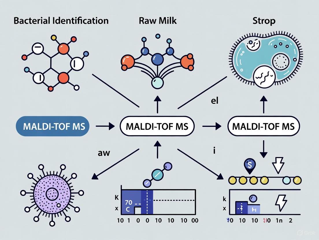
MALDI-TOF MS has transformed microbiological diagnostics by providing a rapid, high-throughput, and cost-effective method for identifying microorganisms. The technique involves several key steps which are outlined in the workflow below.
The principle of operation is based on ionizing microbial proteins using a laser beam, which generates a unique protein profile for each microorganism. This profile, or mass spectral fingerprint, is then compared against a reference database by the MALDI-TOF MS software to identify the microorganism at the genus and species levels [13]. Compared to other identification methods such as biochemical assays or 16S rDNA sequencing, mass spectrometry is less time-consuming, less labour-intensive, and its basic operation is relatively straightforward [9].
Performance Comparison of Leading MALDI-TOF MS Systems
With multiple commercial systems available, it is essential for laboratories to understand their comparative performance. The following table summarizes the identification efficacy of two prominent systems, the Bruker Biotyper and the Zybio EXS2600, as evaluated in recent studies.
Table 2: Comparison of MALDI-TOF MS System Performance for Bacterial Identification
| System | Species-Level ID Rate | Genus-Level ID Rate | Failed Identification | Study Context | Citation |
|---|---|---|---|---|---|
| Bruker Biotyper | 73.63% - 94.6%* | 21.0%* | 5.4%* | Raw milk isolates (n=1130) | [9] |
| Zybio EXS2600 | 74.43% - 91.3%* | 16.9%* | 8.7%* | Raw milk isolates (n=1130) | [9] |
| Bruker Biotyper | 66.8% | 32.7% | 0.5% | Dairy samples (n=196 isolates) | [10] |
| Zybio EXS2600 | 76.0% | 23.0% | 1.0% | Dairy samples (n=196 isolates) | [10] |
| MALDI-TOF MS (General) | 74.0% | 19.9% | 6.1% | Bovine mastitis milk (n=481) | [11] |
| Note: The range for the Bruker and Zybio systems in [9] reflects different calculation bases (see Section 3.2). |
Inter-System Comparative Analysis
A 2025 comparative analysis of 1,130 raw milk isolates provided a direct, head-to-head comparison of the Bruker Biotyper and Zybio EXS2600 systems [9]. The study found a high level of agreement at the species level, with approximately 75% of Bruker identifications matching those from the Zybio system [9]. The Bruker system demonstrated a statistically significant higher percentage of identifications to the genus-only level (Bruker: 21.0%, Zybio: 16.9%), while the Zybio system had a significantly higher rate of unidentified isolates (Zybio: 8.7%, Bruker: 5.4%) [9]. The diagram below visualizes this comparative performance.
Performance variations are often attributable to the comprehensiveness and focus of the proprietary spectral databases maintained by each manufacturer. The Bruker system used a database with 10,830 entries, while the Zybio system's database was larger, containing approximately 15,000 entries [9]. However, it was noted that the Zybio database maintains a strong focus on clinical applications, which may impact its performance with certain environmental or dairy-specific strains [10].
Application Notes & Experimental Protocols
Protocol: Identification of Aerobic Mesophilic Bacteria from Raw Milk
This standard protocol is adapted for the identification of microbial flora from raw milk samples using MALDI-TOF MS [14].
1. Sample Collection and Preparation:
- Aseptically collect raw milk into a sterile container. Transport and store at 4°C until analysis, commencing within 12 hours of collection.
- Serially dilute the milk sample in peptone water (e.g., to 10⁻²). Spread 100 µL of each dilution onto Plate Count Agar (PCA) or other non-selective media suitable for aerobic mesophilic bacteria.
- Incubate plates at 30°C or 37°C for 24-48 hours under aerobic conditions.
2. Colony Selection and Isolation:
- After incubation, enumerate colonies to determine the Total Viable Count.
- Select morphologically distinct colonies and sub-culture them onto fresh agar plates (e.g., Tryptic Soy Agar - TSA) to obtain pure cultures.
- Incubate the pure cultures again at 37°C for 18-24 hours.
3. MALDI-TOF MS Sample Spotting (Direct Transfer Method):
- Gently touch the center of a single, well-isolated bacterial colony with a sterile loop or toothpick.
- Smear the biomass as a thin film directly onto a polished steel MALDI-TOF MS target plate.
- Overlay the smear with 1 µL of the matrix solution, which is α-Cyano-4-hydroxycinnamic acid (HCCA) dissolved in a standard solvent (e.g., 50% acetonitrile, 47.5% water, 2.5% trifluoroacetic acid).
- Allow the spot to air dry completely at room temperature before analysis.
4. Instrumental Analysis and Identification:
- Insert the target plate into the MALDI-TOF MS instrument.
- Acquire mass spectra in positive linear mode within a mass range of 2,000 to 20,000 m/z.
- Analyze the resulting protein spectrum using the system's software, which compares it against the reference database.
- Identifications are provided with a confidence score. Typically, a score ≥ 2.000 indicates reliable species-level identification, a score between 1.700 and 1.999 indicates genus-level identification, and a score < 1.700 is considered unreliable [9].
Protocol: Direct Identification from Positive Blood Cultures for Sepsis Diagnostics
While not specific to milk, this protocol demonstrates the adaptability of MALDI-TOF MS for rapid diagnosis in clinical settings linked to systemic infections, using a simplified centrifugation method [13].
1. Sample Processing:
- Take 4.0 mL from a positive blood culture bottle (e.g., BD BACTEC or BacT/ALERT).
- Transfer it to a tube containing a plasma separation gel and centrifuge at 3000 x g for 10 minutes.
- Carefully discard the supernatant.
- Resuspend the pellet (containing the microorganisms) in 1.0 mL of deionized water.
2. MALDI-TOF MS Spotting and Analysis:
- Spot 1 µL of the resuspended pellet onto the target plate in triplicate.
- Add 1 µL of HCCA matrix solution to each spot and allow it to dry.
- For yeasts, a preliminary step of adding 0.5 µL of formic acid to the spot and allowing it to evaporate before matrix application can improve spectral quality.
- Perform MALDI-TOF MS analysis as described in the previous protocol.
3. Performance Note:
- This direct method shows high sensitivity for Gram-negative bacteria (~90% species-level ID) but lower performance for Gram-positive bacteria (~69%) and yeasts (~33%) [13]. This highlights the potential need for protocol optimization based on the target microorganism.
The Scientist's Toolkit: Essential Research Reagents & Materials
Table 3: Key Reagents and Materials for MALDI-TOF MS-Based Milk Analysis
| Item | Function / Application | Example / Specification |
|---|---|---|
| MALDI-TOF MS System | Core analytical instrument for generating and analyzing protein mass spectra. | Bruker Microflex LT, Zybio EXS2600, bioMérieux VITEK MS |
| Reference Spectral Database | Software library for matching unknown sample spectra to identify microorganisms. | MBT Compass Library (Bruker), VITEK MS Knowledge Base, EXS2600 Database (Zybio) |
| MALDI Target Plate | Platform for loading and analyzing multiple samples. | 96-spot polished steel target plate |
| Matrix Solution (CHCA) | Energy-absorbing compound that enables soft laser desorption/ionization of proteins. | α-Cyano-4-hydroxycinnamic acid in 50% ACN/47.5% H₂O/2.5% TFA |
| Culture Media | For the growth and isolation of bacteria from raw milk samples. | Tryptic Soy Agar (TSA), Plate Count Agar (PCA), Man-Rogosa-Sharpe (MRS) Agar |
| Calibration Standard | For mass accuracy calibration of the instrument. | Bruker Bacterial Test Standard (BTS), Zybio Microbiology Calibrator |
| Formic Acid | Pre-treatment agent to improve protein extraction and spectral quality, especially for Gram-positive bacteria and yeasts. | 70-100% concentration for on-target formic acid overlay |
| Deionized Water | For washing steps and resuspension of pellets in sample preparation protocols. | Molecular biology grade, sterile |
| Centrifuge Tubes with Gel | For rapid separation of microorganisms from complex liquid samples like milk or blood. | Tubes containing a plasma separation gel |
The compelling data on pathogen prevalence in raw milk leaves no doubt that rigorous and continuous safety monitoring is a scientific and public health imperative. MALDI-TOF MS technology stands as a powerful tool to meet this demand, enabling a shift from slow, traditional methods to a new standard of rapid, accurate, and cost-effective microbial identification. As the technology evolves and databases expand to include more environmental and food-borne isolates, its value in ensuring the safety of raw milk and other agricultural products will only increase. For researchers and the dairy industry, the adoption and refinement of these protocols are critical steps toward mitigating public health risks and building a scientifically-grounded framework for the production of safe raw milk.
Matrix-Assisted Laser Desorption/Ionization Time-of-Flight Mass Spectrometry (MALDI-TOF MS) has revolutionized microbial identification in clinical and food microbiology, including raw milk research [15]. This application note details the standard workflow for identifying bacteria from raw milk samples, from initial colony picking to final spectral database matching. The protocol emphasizes the specific considerations required for the diverse microbiota found in raw milk, which includes a wide variety of Gram-positive and Gram-negative bacteria, many of which were previously difficult to identify using traditional biochemical methods [9] [10]. The robustness of this workflow supports milk quality control, mastitis pathogen screening, and dairy product safety assurance.
The following diagram illustrates the complete MALDI-TOF MS identification workflow for bacterial isolates from raw milk.
Detailed Experimental Protocols
Sample Collection and Cultivation
Purpose: To isolate pure bacterial cultures from raw milk samples for subsequent MALDI-TOF MS analysis.
Materials:
- Raw milk samples collected aseptically
- Peptone water for serial dilution
- Tryptic Soya Agar (TSA) or other appropriate media
- Incubator (37°C)
Procedure:
- Perform serial dilutions of raw milk samples in peptone water (typically to 10⁻²) [9].
- Spread 100 µL of each dilution onto TSA plates.
- Incubate plates at 37°C for 24–48 hours under aerobic conditions or CO₂-enriched atmosphere (5%) as required [9].
- After incubation, select morphologically distinct colonies and subculture onto fresh media to obtain pure cultures.
- Store isolates at -80°C using appropriate preservation systems (e.g., Microbank) for future analysis [9].
Colony Picking and Sample Preparation
Purpose: To transfer and prepare bacterial samples for MALDI-TOF MS analysis.
Materials:
- Pure bacterial cultures (18-24 hour growth)
- Formic acid (70%)
- Acetonitrile (HPLC grade)
- α-cyano-4-hydroxycinnamic acid (HCCA) matrix
- Trifluoroacetic acid (TFA)
- MALDI-TOF MS target plate
Procedure:
- Direct Smear Method: For a quick preparation, directly smear a small amount of bacterial biomass from a single colony onto a target plate spot [16].
- Formic Acid/Acetonitrile Extraction Method (Recommended): a. Transfer 1-3 colonies to a microcentrifuge tube containing 20 µL of sterile water. b. Add 80 µL of pure ethanol and mix thoroughly. c. Centrifuge briefly and discard the supernatant. d. Add 20-50 µL of 70% formic acid and mix thoroughly. e. Add an equal volume of acetonitrile and mix. f. Centrifuge at high speed for 2 minutes. g. Spot 1 µL of the supernatant onto a MALDI target plate and allow to dry at room temperature [9].
Matrix Application and MALDI-TOF MS Analysis
Purpose: To co-crystallize samples with matrix and acquire mass spectral data.
Materials:
- α-cyano-4-hydroxycinnamic acid (HCCA) matrix solution (10 mg/mL in 50% acetonitrile, 47.5% water, and 2.5% trifluoroacetic acid) [9]
- MALDI-TOF MS instrument (e.g., Bruker Microflex LT, Zybio EXS2600)
Procedure:
- Prepare fresh HCCA matrix solution.
- Overlay each dried sample spot with 1 µL of matrix solution and allow to dry completely [9].
- Calibrate the MALDI-TOF MS instrument using the manufacturer's recommended standards (e.g., Bacterial Test Standard for Bruker systems) [9].
- Acquire mass spectra in positive linear mode within the mass range of 2,000-20,000 Da [9] [16].
- For each sample, accumulate spectra from multiple laser positions (typically 500 shots per spectrum) to ensure representative sampling [17].
Spectral Analysis and Database Matching
Purpose: To identify bacterial isolates by comparing acquired spectra to reference databases.
Procedure:
- Process raw spectra by applying smoothing, baseline subtraction, and peak detection algorithms using the instrument's software [17].
- Compare the processed spectrum against the reference database.
- Interpret identification scores based on the manufacturer's recommendations:
Table 1: MALDI-TOF MS Identification Score Interpretation
| Score Value | Identification Level | Confidence |
|---|---|---|
| ≥ 2.000 | Species level | High confidence |
| 1.700 - 1.999 | Genus level | Low confidence |
| < 1.700 | No reliable identification | Unacceptable |
- For problematic identifications, consider repeating the analysis with a fresh sample or using an alternative sample preparation method.
Performance Data in Raw Milk Analysis
The following table summarizes performance metrics of MALDI-TOF MS systems for identifying raw milk isolates based on recent comparative studies.
Table 2: Performance Comparison of MALDI-TOF MS Systems for Raw Milk Bacterial Identification
| Parameter | Bruker Biotyper System | Zybio EXS2600 System |
|---|---|---|
| Species-level ID rate | 73.63% | 74.43% |
| Genus-level ID rate | 20.97% | 16.87% |
| No identification | 5.4% | 8.7% |
| Mean score value | 2.064 | 2.098 |
| Database entries | ~10,830 | ~15,000 |
| Best performance with | Pseudomonas spp., Actinomycetia, Gammaproteobacteria | Yeasts, H. alvei, Alphaproteobacteria, Bacilli |
Research Reagent Solutions
Table 3: Essential Reagents and Materials for MALDI-TOF MS Bacterial Identification
| Item | Function | Example Specification |
|---|---|---|
| HCCA Matrix | Absorbs laser energy, facilitates analyte ionization and desorption | α-cyano-4-hydroxycinnamic acid, 10 mg/mL in 50% ACN, 47.5% water, 2.5% TFA [9] |
| Formic Acid | Protein extraction and denaturation | 70% solution in water [9] |
| Acetonitrile | Solubilizes hydrophobic proteins, enhances extraction efficiency | HPLC grade [9] |
| Trifluoroacetic Acid | Ion-pairing agent, improves crystal formation and spectral quality | 0.1-2.5% in matrix solution [9] [17] |
| Bacterial Test Standard | Instrument calibration | Contains characterized bacterial extracts with known mass peaks [9] |
| Target Plate | Sample presentation | Polished steel with 96-spot format [9] |
Critical Considerations for Raw Milk Research
Database Selection and Limitations
The identification accuracy heavily depends on the comprehensiveness of the reference database. Commercial databases historically focused on clinical isolates may lack specific dairy-related strains [15]. A study comparing two MALDI-TOF MS systems found that although both systems performed comparably for most raw milk isolates, differences emerged in identifying specific bacterial classes [9]. Supplementing commercial databases with custom entries for dairy-specific strains improves identification accuracy for raw milk microbiota [18] [15].
Special Considerations for Raw Milk Isolates
- Gram-positive bacteria: Some Gram-positive bacteria with robust cell walls (e.g., Actinomycetota) may require more extensive extraction procedures for optimal protein recovery [18].
- Diverse microbiota: Raw milk contains a wide variety of bacteria, including non-aureus Staphylococci, Streptococci, and environmental contaminants, which can now be identified to species level using MALDI-TOF MS [15].
- Sample preparation variability: Cultivation conditions (different media, incubation times) generally have minor impacts on identification, but extraction efficiency remains critical for reliable results [18].
The standard MALDI-TOF MS workflow from colony picking to spectral database matching provides a robust, rapid, and accurate method for identifying bacteria in raw milk samples. This approach enables species-level identification of many microorganisms that were previously grouped generically using traditional microbiological methods. Following the detailed protocols outlined in this application note will help researchers obtain reliable identifications for diverse raw milk isolates, supporting advanced research into milk quality, animal health, and dairy product safety.
This application note provides a detailed framework for the identification of key bacterial targets in raw milk using Matrix-Assisted Laser Desorption/Ionization Time-of-Flight Mass Spectrometry (MALDI-TOF MS). The protocol outlines the specific pathogens, spoilage organisms, and indicator bacteria critical to dairy product quality and safety, and presents optimized methodologies for their rapid and reliable identification. Designed within the broader context of a thesis on MALDI-TOF MS applications in dairy microbiology, this document provides researchers and industry professionals with a standardized approach to microbial analysis, enabling enhanced control over raw milk quality and shelf-life prediction.
Raw milk is a complex ecosystem harboring a diverse microbiota, the composition of which directly influences the safety, quality, and shelf-life of final dairy products. Controlling this microbiome requires the rapid and accurate identification of specific bacterial groups: pathogens that pose public health risks, spoilage organisms that cause product deterioration, and indicator organisms that reflect hygiene practices. For decades, the identification of these microbes relied on conventional culture-based and biochemical methods, which are often time-consuming, labor-intensive, and limited in discriminatory power.
MALDI-TOF MS has revolutionized microbial diagnostics by utilizing proteomic fingerprints for identification. This technique analyzes highly abundant proteins, primarily ribosomal proteins, to generate a unique spectral profile for each microorganism, which is then matched against a reference database. The technique is rapid, cost-effective, and provides high throughput, making it exceptionally suitable for the demanding environment of food microbiology laboratories. This document details the application of MALDI-TOF MS for targeting and identifying the most relevant bacteria in raw milk, providing a robust protocol validated by recent scientific research.
Key Bacterial Targets in Raw Milk
The bacterial targets in raw milk can be categorized based on their impact. The table below summarizes the primary organisms of concern, their categorization, and their significance in the dairy industry.
Table 1: Key Bacterial Targets in Raw Milk and Their Significance
| Bacterial Genus/Species | Category | Significance in Raw Milk |
|---|---|---|
| Listeria monocytogenes | Pathogen | Causes the severe foodborne illness listeriosis; a major public health concern [19]. |
| Escherichia coli (pathogenic strains) | Pathogen | Indicator of fecal contamination; some strains can cause serious food poisoning [9]. |
| Staphylococcus aureus | Pathogen | Can produce heat-stable enterotoxins leading to food intoxication [9]. |
| Pseudomonas spp. (e.g., P. fluorescens) | Spoilage | Psychrotrophic; produces heat-resistant extracellular proteases and lipases, leading to spoilage of pasteurized and UHT milk [20] [21]. |
| Bacillus spp. (e.g., B. cereus, B. licheniformis) | Spoilage | Spore-forming; can survive pasteurization and cause defects like "sweet curdling" and off-flavors [20]. |
| Hafnia alvei | Spoilage | Psychrotrophic; implicated in spoilage and off-flavor development [20] [10]. |
| Lactic Acid Bacteria (e.g., Lactococcus, Lactobacillus) | Starter Culture / Indicator | Used in fermentation; their unexpected presence can indicate spoilage or cross-contamination [22] [19]. |
| Coliforms | Indicator | Group of bacteria used as a general indicator of sanitation and fecal contamination [19]. |
Performance of Identification Methods
The accuracy of bacterial identification is paramount. A comparative study of different identification systems highlighted the relative performance of genetic, proteomic, and biochemical methods.
Table 2: Comparison of Bacterial Identification Systems for Raw Milk Isolates
| Identification System | Type | Accuracy for Gram-Negative Bacilli | Accuracy for Gram-Positive Bacilli | Simpson's Index of Diversity | Relative Speed |
|---|---|---|---|---|---|
| 16S rRNA Gene Sequencing | Genetic | 100.0% | 100.0% | 0.966 | High [20] |
| MALDI-TOF MS | Proteomic | 63.2%* | 95.0% | 0.496 | Very High [20] |
| Biolog System | Biochemical | 86.8% | 85.0% | 0.711 | Medium [20] |
| API | Biochemical | 60.5% | 90.0% | 0.472 | Low [20] |
| Microbact | Biochemical | 57.9% | N/R | 0.140 | Medium [20] |
Note: Accuracy can be significantly improved with protocol optimization and database expansion [21]. N/R = Not Reported.
Recent studies have also compared newer MALDI-TOF MS platforms. Research comparing the Bruker Biotyper and the Zybio EXS2600 systems on 1,130 raw milk isolates found them to be highly comparable for routine use. The Bruker system identified 94.6% of isolates to the genus level or beyond, while the Zybio system identified 91.3%. At the species level, the identification rates were 73.63% (Bruker) and 74.43% (Zybio), demonstrating equivalent performance for species-level assignment [9].
Experimental Protocol for MALDI-TOF MS Analysis of Raw Milk
The following protocol is adapted from established methodologies for the analysis of dairy products [22] [19]. The entire process, from colony picking to identification, can be completed within a single working day.
Isolation of Dairy-Related Bacteria
- Sample Plating: Serially dilute raw milk samples in peptone water and spread 100 µL of each dilution onto appropriate agar plates.
- Media Selection: Use a combination of general and selective media to maximize microbial recovery.
- Tryptic Soy Agar (TSA): For general total viable counts.
- Milk Plate Count Agar (MPCA): Standard for milk microbiology.
- CBL Agar (China Blue Lactose): For Gram-negative bacteria.
- MRS Agar: For Lactic Acid Bacteria (LAB).
- Incubation: Incubate plates at 37°C for 24–48 hours under both aerobic and CO₂-enriched (5%) atmospheres to recover a diverse microbiota [9].
- Media Selection: Use a combination of general and selective media to maximize microbial recovery.
- Pure Culture Preparation: After incubation, select morphologically distinct colonies and subculture them onto fresh TSA plates to obtain pure cultures. Incubate again at 37°C for 24 hours under aerobic conditions.
Sample Preparation for MALDI-TOF MS
The ethanol-formic acid-acetonitrile extraction method is recommended for robust and reproducible identification, particularly for difficult-to-lyse Gram-positive bacteria [19].
Reagents and Equipment:
- Matrix: α-Cyano-4-hydroxycinnamic acid (HCCA)
- Solvents: Absolute ethanol, HPLC-grade water, Trifluoroacetic acid (TFA), Acetonitrile (ACN), Formic acid (70%)
- Equipment: Centrifuge, Micropipettes, Vortex mixer, MALDI-TOF MS target plate
Procedure:
- Cell Harvesting: Transfer 1-3 loops of bacterial biomass from a pure culture plate into a 1.5 mL microcentrifuge tube containing 300 µL of HPLC-grade water.
- Ethanol Inactivation and Washing: Add 900 µL of absolute ethanol to the suspension. Vortex thoroughly. Centrifuge at maximum speed (e.g., 13,000-16,000 × g) for 2 minutes.
- Pellet Drying: Carefully decant the supernatant. Allow the pellet to air-dry completely at room temperature.
- Protein Extraction:
- Resuspend the pellet in 25-50 µL of 70% formic acid.
- Add an equal volume of 100% acetonitrile. Vortex vigorously.
- Centrifuge at maximum speed for 2 minutes.
- Target Spotting: Transfer 1 µL of the clear supernatant onto a polished steel MALDI target plate. Allow the spot to dry completely at room temperature.
- Matrix Overlay: Overlay the dried spot with 1 µL of matrix solution (saturated HCCA in 50% ACN and 2.5% TFA). Allow to dry completely.
MALDI-TOF MS Measurement and Data Analysis
- Instrument Calibration: Calibrate the MALDI-TOF MS instrument using a commercial Bacterial Test Standard (BTS) as per the manufacturer's instructions.
- Spectral Acquisition: Insert the target plate into the spectrometer. Acquire protein spectra in the linear positive ion mode within a mass range of 2,000 to 20,000 m/z. Each spectrum should be an accumulation of multiple laser shots across the spot.
- Database Matching and Identification: Automatically compare the acquired protein fingerprint against the installed reference database (e.g., Bruker MBT Compass Library or Zybio Ex-Accuspec Library).
- Interpretation of Results: Use the manufacturer-recommended score values for identification:
Workflow and Data Interpretation
The following diagram illustrates the logical workflow for the MALDI-TOF MS analysis of raw milk, from sample preparation to final identification.
The Scientist's Toolkit: Essential Research Reagents and Materials
The following table lists the key reagents, materials, and equipment required to successfully implement the described MALDI-TOF MS protocol for raw milk analysis.
Table 3: Essential Research Reagents and Materials for MALDI-TOF MS Analysis
| Category | Item | Function / Application | Example Catalog Number |
|---|---|---|---|
| Culture Media | Tryptic Soy Agar (TSA) | General growth medium for bacterial isolation and subculturing [22]. | Sigma-Aldrich 22091 |
| MRS Agar | Selective isolation of Lactic Acid Bacteria (LAB) [22]. | Merck KGaA 1.10660.0500 | |
| Milk Plate Count Agar (MPCA) | Standard medium for enumeration of milk microbiota [22]. | Oxoid CM0681 | |
| Extraction Reagents | Absolute Ethanol | Inactivates and washes cells; part of the extraction protocol [19]. | - |
| Formic Acid (70%) | Disrupts bacterial cells to release proteins for analysis [19]. | Chempur 115676307 | |
| Acetonitrile (ACN) | Organic solvent that co-crystallizes with the matrix and analyte [19]. | Merck KGaA 1.00014.1011 | |
| MALDI Matrix & Calibration | α-Cyano-4-hydroxycinnamic acid (HCCA) | Matrix that absorbs laser energy and facilitates soft ionization of proteins [19]. | Bruker Daltonik GmbH 8201344 |
| Bacterial Test Standard (BTS) | Standardized protein extract for instrument calibration [9]. | Bruker Daltonik GmbH 8255343 | |
| Equipment & Consumables | MALDI-TOF MS System | Instrument for generating and analyzing protein spectral fingerprints. | E.g., Bruker Microflex LT, Zybio EXS2600 |
| Polished Steel Target Plate | Platform for holding sample spots for analysis [22]. | Bruker Daltonik GmbH 8280800 | |
| Centrifuge | For pelleting cells during the extraction process. | - |
Practical Applications: From Routine Pathogen Screening to Advanced Profiling
In the context of MALDI-TOF MS bacterial identification in raw milk research, appropriate sample preparation is paramount for both accurate results and laboratory safety. This document details validated protocols for the safe handling, inactivation, and formic acid-based extraction of bacterial isolates from raw milk. Proper sample preparation ensures reliable spectral quality for microorganism identification while mitigating biohazard risks associated with pathogenic bacteria such as Staphylococcus aureus and Prototheca spp., common etiological agents in bovine mastitis [23] [24]. The following sections provide step-by-step application notes for creating safe, reproducible, and high-quality MALDI-TOF MS samples.
Safety and Inactivation Protocol
The initial step in any sample preparation protocol must be the complete inactivation of pathogens to ensure researcher safety. The following procedure effectively inactivates bacterial cells while preserving protein integrity for mass spectrometric analysis [23] [24].
Reagents and Equipment
- Fresh bacterial colonies from pure culture (18-24 hours growth)
- Absolute ethanol ( molecular biology grade)
- Sterile molecular-grade water
- Microcentrifuge tubes (1.5 - 2 mL capacity)
- Benchtop centrifuge (capable of ≥ 13,000 × g)
- Vortex mixer
- Sterile inoculating loops or needles
Step-by-Step Inactivation Procedure
- Harvest Biomass: Using a sterile loop or needle, transfer approximately 10 colonies (or 10-30 μL of cell pellet) from a fresh pure culture to a microcentrifuge tube containing 300 μL of sterile molecular-grade water [23] [24].
- Suspend Cells: Vortex the mixture thoroughly for 15-30 seconds until a homogeneous suspension is formed.
- Add Inactivation Reagent: Add 900 μL of absolute ethanol to the suspension, resulting in a final ethanol concentration of approximately 75% [23]. This concentration is critical for effective pathogen inactivation.
- Mix Thoroughly: Vortex the ethanol-water mixture for 1 minute to ensure complete contact with all cellular material [23].
- Pellet Cells: Centrifuge the mixture at 11,700 - 13,000 × g for 2 minutes to form a firm pellet [23] [24].
- Remove Supernatant: Carefully aspirate and discard the supernatant without disturbing the pellet.
- Complete Drying: Allow the pellet to air-dry at room temperature for approximately 2 minutes with the tube cap open to evaporate residual ethanol [24].
Table 1: Troubleshooting Inactivation Steps
| Step | Potential Issue | Solution |
|---|---|---|
| Biomass harvesting | Insufficient material for analysis | Ensure adequate colony growth (18-24 hours); use 10-30 colonies |
| Ethanol addition | Precipitated proteins not forming firm pellet | Verify ethanol concentration; ensure proper vortexing |
| Supernatant removal | Pellet dislodging during aspiration | Leave small volume of supernatant above pellet; use fine-tip pipette |
| Drying | Over-drying making resuspension difficult | Monitor pellet consistency; do not exceed 5 minutes drying time |
Formic Acid Extraction Protocol
Following inactivation, formic acid extraction is performed to disrupt cell walls and release ribosomal proteins which generate the characteristic mass spectral fingerprints used for bacterial identification [23] [24].
Reagents and Equipment
- 70% Formic acid ( analytical grade)
- 100% Acetonitrile (HPLC grade)
- Sterile zirconium or glass beads (0.1 mm diameter)
- MALDI-TOF steel target plate
- Matrix solution: Saturated α-cyano-4-hydroxycinnamic acid (HCCA) in 50% acetonitrile/2.5% trifluoroacetic acid
- Bead beater or similar mechanical disruption device
Step-by-Step Extraction Procedure
- Resuspend Pellet: Add 50 μL of 70% formic acid to the dried pellet and pipette mix thoroughly until the pellet is completely resuspended [23].
- Add Organic Solvent: Add 50 μL of 100% acetonitrile to the formic acid-cell mixture. Vortex briefly to combine. This step enhances protein extraction and precipitation [23].
- Mechanical Disruption (Optional but Recommended): Transfer the entire suspension to a tube containing 20-30 mg of sterile zirconium beads. Process in a bead beater at 4,000 rpm for 5 cycles of 1 minute each, with brief cooling intervals between cycles to prevent overheating [23]. For less robust organisms, this step may be omitted.
- Clarify Extract: Centrifuge the processed suspension at 11,700 × g for 2 minutes to pellet cell debris [23] [24].
- Prepare Target Plate: Transfer 1 μL of the clear supernatant onto a polished steel MALDI target plate. Allow to air-dry completely at room temperature [23] [24].
- Overlay with Matrix: Once the sample spot is dry, overlay it with 1 μL of saturated HCCA matrix solution and allow it to crystallize completely at room temperature [23].
Table 2: Centrifugation Parameters for Sample Preparation
| Step | Speed | Time | Temperature | Purpose |
|---|---|---|---|---|
| Initial inactivation | 11,700 - 13,000 × g | 2 min | Room temperature | Pellet cells after ethanol inactivation |
| Post-extraction clarification | 11,700 × g | 2 min | Room temperature | Pellet cell debris after formic acid/acetonitrile treatment |
Experimental Workflow and Signaling Pathways
The complete workflow from sample collection to MALDI-TOF MS analysis integrates both safety and analytical considerations, as diagrammed below.
Research Reagent Solutions
The following table details essential reagents and materials required for implementing these sample preparation protocols.
Table 3: Essential Research Reagents and Materials
| Reagent/Material | Specifications | Function in Protocol |
|---|---|---|
| Absolute Ethanol | Molecular biology grade, ≥99.8% purity | Primary inactivation agent; denatures pathogens and fixes cellular proteins [23] [24] |
| Formic Acid | Analytical grade, 70% concentration | Cell wall disruption and protein solubilization; enhances ionization efficiency [23] |
| Acetonitrile | HPLC grade, 100% concentration | Protein precipitation and co-crystallization with matrix; enhances spectral quality [23] |
| α-Cyano-4-hydroxycinnamic Acid (HCCA) | Saturated solution in 50% ACN/2.5% TFA | MALDI matrix; facilitates laser desorption/ionization of protein analytes [23] |
| Zirconium Beads | 0.1 mm diameter, acid-washed | Mechanical cell disruption; enhances protein yield from robust microorganisms [23] |
| Sterile Molecular Water | Nuclease-free, molecular grade | Initial cell suspension without interfering contaminants [23] [24] |
Protocol Validation and Performance Metrics
These protocols have been validated in multiple studies focused on raw milk pathogens. When applied to Prototheca isolates from bovine mastitis, the formic acid extraction method enabled species-level identification of 22 out of 27 P. bovis isolates and 3 out of 4 P. blaschkeae isolates with high confidence scores (>2.0) using MALDI-TOF MS systems [23]. Similarly, the protocol achieved 100% agreement with conventional methods for identifying Staphylococcus aureus from bovine mastitis cases [24].
Recent comparative studies of MALDI-TOF MS systems demonstrate that optimized sample preparation is critical for performance. The EXS 2600 system showed a 76.0% species identification rate compared to 66.8% for the Bruker Biotyper when analyzing dairy isolates, highlighting how extraction quality impacts platform performance [10]. These standardized protocols ensure maximum identification rates across different MALDI-TOF MS systems by providing high-quality protein extracts.
Bovine mastitis, an inflammatory condition of the udder, remains one of the most prevalent and economically devastating diseases in dairy cattle worldwide. The disease not only causes significant production losses and discarded milk but also represents an important animal welfare concern. Mastitis pathogens are broadly classified as contagious (spreading from cow to cow during milking) or environmental (originating from the cow's surroundings) [25]. Accurate and rapid identification of the causative microorganisms is fundamental for implementing effective control strategies, guiding antimicrobial treatment, and reducing economic impact.
Matrix-Assisted Laser Desorption/Ionization Time-of-Flight Mass Spectrometry (MALDI-TOF MS) has revolutionized microbial diagnostics in clinical and veterinary settings. This technology provides a rapid, reliable, and cost-effective method for identifying microorganisms based on their unique protein fingerprints [9] [24]. Within the context of raw milk research, MALDI-TOF MS enables precise identification of mastitis pathogens, facilitating studies on microbial ecology, antibiotic resistance patterns, and pathogen transmission dynamics. This application note details the identification profiles of the three primary bacterial groups associated with bovine mastitis—Staphylococcus, Streptococcus, and coliforms—and provides standardized protocols for their detection using MALDI-TOF MS.
Pathogen Profiles and Key Data
The following sections delineate the primary mastitis-causing pathogens, their prevalence, and key characteristics relevant to identification and resistance profiling.
1Staphylococcusspp.
Staphylococci are among the most frequently isolated pathogens from bovine mastitis cases. They are Gram-positive, catalase-positive cocci and can be divided into coagulase-positive (e.g., S. aureus) and coagulase-negative staphylococci (CoNS) based on their ability to produce coagulase [26].
- Identification Profile: S. aureus is a classic contagious mastitis pathogen. However, CoNS are increasingly recognized as significant causes of subclinical and clinical intramammary infections [26]. A study on CoNS from bovine milk in Ethiopia identified S. epidermidis (11%) as the most prevalent species, followed by S. sciuri (5.2%), S. warneri (3.4%), S. haemolyticus (3.1%), and S. simulans (3.1%) [26].
- Antimicrobial Resistance: Methicillin resistance, mediated by the mecA or mecC genes, is a critical concern. Phenotypic resistance to oxacillin has been reported in 37.5% of CoNS isolates, with a high prevalence of multidrug-resistance (54.2%) [26]. Some studies have noted an absence of mecA/mecC genes in phenotypically resistant isolates, suggesting alternative resistance mechanisms [27] [28].
- Virulence Factors: CoNS can harbor various virulence genes, including the intracellular adhesion gene icaD (26.5%), Panton-Valentine leukocidin pvl (22.1%), and the methicillin resistance gene mecA (21.7%) [26].
Table 1: Prevalence and Virulence Gene Profile of Coagulase-Negative Staphylococci (CoNS) from Bovine Mastitis
| CoNS Species | Prevalence (%) | Key Virulence Genes (% Prevalence in CoNS) |
|---|---|---|
| S. epidermidis | 11.0 | icaD (26.5), pvl (22.1), mecA (21.7) |
| S. sciuri | 5.2 | icaD (26.5), pvl (22.1), mecA (21.7) |
| S. warneri | 3.4 | icaD (26.5), pvl (22.1), mecA (21.7) |
| S. haemolyticus | 3.1 | icaD (26.5), pvl (22.1), mecA (21.7) |
| S. simulans | 3.1 | icaD (26.5), pvl (22.1), mecA (21.7) |
2Streptococcusspp.
Streptococci are Gram-positive, catalase-negative cocci and represent a major group of environmental mastitis pathogens, though some exhibit contagious characteristics [25].
- Identification Profile: The most relevant streptococcal species in bovine mastitis are Streptococcus uberis, Streptococcus dysgalactiae, and Streptococcus agalactiae [25]. In small ruminants, S. uberis is the predominant species (89.5%), followed by S. dysgalactiae (3.5%) and S. parauberis (3.5%) [29].
- Epidemiology: S. agalactiae is a contagious pathogen adapted to survive in the bovine mammary gland, while S. uberis is primarily environmental, residing in bedding, soil, and other cow surroundings. S. dysgalactiae is considered an intermediate pathogen [25].
- Antimicrobial Resistance: Streptococci isolated from mastitis show high or moderate resistance to erythromycin (68.7%), benzylpenicillin (63.7%), and ampicillin (51.5%) [30]. A significant number of streptococcal isolates are multidrug-resistant [30].
Table 2: Key Streptococcal Pathogens in Bovine Mastitis
| Species | Lancefield Group | Classification | Primary Reservoir |
|---|---|---|---|
| S. uberis | E, G, P, U | Environmental (can be contagious) | Bedding, environment |
| S. dysgalactiae | C | Intermediate | Environment, udder |
| S. agalactiae | B | Contagious | Udder, gastrointestinal tract |
Coliforms
Coliforms are a method-defined group of Gram-negative, non-sporeforming rods capable of fermenting lactose to acid and gas within 48 hours at 32-35°C [31]. They are classic indicators of environmental contamination.
- Identification Profile: This group includes genera within the Enterobacteriaceae family (e.g., Escherichia, Klebsiella, Enterobacter, Serratia, Citrobacter) and sometimes Aeromonas [31]. They are common contaminants of raw milk, with mean counts ranging from 31 CFU/mL to 2,570 CFU/mL in US studies [31].
- Significance in Mastitis: Certain coliforms, notably Escherichia coli and Klebsiella pneumoniae, are known to cause severe clinical mastitis [31]. High levels of coliforms in raw milk may indicate unsanitary production practices, inadequate refrigeration, or the presence of coliform mastitis within the herd [31].
- Classification: Coliforms can be categorized as:
- Thermophilic (e.g., E. coli), primarily of fecal origin.
- Psychrotrophic, which are environmental and can grow at refrigeration temperatures.
- Ubiquitous, originating from various natural environments like soil, water, and vegetation [31].
Experimental Protocols for MALDI-TOF MS Identification
The following protocol is adapted from standardized methods used for identifying mastitis pathogens from milk samples [9] [24] [29].
Sample Preparation and Bacterial Isolation
- Milk Sample Collection: Aseptically collect raw milk samples after discarding the first streams and disinfecting the teat ends. Store and transport samples under refrigeration.
- Culture and Isolation: Inoculate 10 µL of milk onto 5% sheep blood agar and other selective agars (e.g., Mannitol Salt Agar for staphylococci). Incubate aerobically at 37°C for 24-48 hours.
- Selection of Colonies: After incubation, select morphologically distinct colonies and subculture them onto fresh media (e.g., Tryptic Soya Agar) to obtain pure cultures for analysis.
Protein Extraction for MALDI-TOF MS Analysis
The standard ethanol-formic acid extraction method is recommended for optimal spectral quality [9] [24].
- Harvest approximately 10 colonies from a fresh pure culture (18-24 hours old) and transfer to a microfuge tube containing 300 µL of sterile distilled water.
- Vortex thoroughly to create a homogeneous bacterial suspension.
- Add 900 µL of absolute ethanol to the tube and vortex again.
- Centrifuge the mixture at 13,000-16,000 × g for 2 minutes.
- Carefully decant or pipette off the supernatant.
- Allow the pellet to air-dry for a few minutes at room temperature to evaporate residual ethanol.
- Resuspend the pellet in 25-50 µL of 70% formic acid.
- Add an equal volume of acetonitrile (25-50 µL) and mix thoroughly by pipetting.
- Centrifuge again at 13,000-16,000 × g for 2 minutes.
- The resulting supernatant, which contains the bacterial proteins, is used for MALDI-TOF MS analysis.
Target Spotting and Measurement
- Apply 1 µL of the protein extract supernatant to a polished steel MALDI target plate and allow it to dry completely at room temperature.
- Overlay the spot with 1 µL of matrix solution (saturated α-cyano-4-hydroxycinnamic acid [HCCA] in 50% acetonitrile and 2.5% trifluoroacetic acid).
- Allow the spot to dry completely before inserting the target plate into the mass spectrometer.
- Calibrate the instrument using a certified bacterial test standard (e.g., E. coli extract).
- Acquire mass spectra in positive linear mode within a mass range of 2,000 to 20,000 Da. Each spectrum should be generated from a minimum of 240 accumulated laser shots.
Data Interpretation and Identification
- Process the acquired spectra using the instrument's software (e.g., Bruker Biotyper or Zybio Ex-Accuspec).
- Compare the sample spectrum against the reference spectral database.
- Interpret the identification results based on the manufacturer's recommended log score values:
- Score ≥ 2.000: Reliable identification at the species level.
- Score 1.700 - 1.999: Reliable identification at the genus level.
- Score < 1.700: Unreliable identification.
Figure 1: MALDI-TOF MS Workflow for Mastitis Pathogen Identification
Technical Considerations and Validation
Comparison of MALDI-TOF MS Systems
Different MALDI-TOF MS systems demonstrate high, comparable performance in identifying mastitis pathogens. A comparative study of 1,130 raw milk isolates showed that the Bruker Biotyper and Zybio EXS2600 systems identified isolates to the species level in 73.63% and 74.43% of cases, respectively [9]. While both systems are effective for routine diagnostics, minor differences in performance can occur with specific bacterial classes like Actinomycetia and Bacilli [9].
Agreement with Conventional and Molecular Methods
MALDI-TOF MS shows a high level of agreement with molecular methods like gap PCR-RFLP for identifying the most prevalent staphylococcal and streptococcal species [29]. When compared to conventional microbiological methods, the agreement is substantial at the genus level (kappa = 0.80) and moderate to substantial at the species level (kappa = 0.64) [32]. This confirms that MALDI-TOF MS is a robust and accurate tool for mastitis pathogen identification, though some caution is warranted when comparing species-level identifications of gram-negative bacteria from historical data [32].
Detection of Antimicrobial Resistance
While MALDI-TOF MS is primarily an identification tool, it can be applied to detect specific antibiotic resistance mechanisms, such as methicillin resistance in staphylococci, by analyzing resistance-associated biomarker peaks or through specialized software modules [27]. However, phenotypic antimicrobial susceptibility testing (AST) remains the standard for determining resistance profiles. Studies using MALDI BioTyper Compass Explorer and ClinProTools software have demonstrated the potential for rapid detection of biomarkers associated with resistance [27].
The Scientist's Toolkit
Table 3: Essential Research Reagents and Materials for MALDI-TOF MS-Based Mastitis Pathogen Identification
| Item | Function/Application | Example |
|---|---|---|
| Selective Culture Media | Primary isolation and differentiation of bacterial groups from milk samples. | Mannitol Salt Agar (Staphylococci), Blood Agar (general) [24] [26] |
| Protein Extraction Reagents | Preparation of bacterial protein extracts for high-quality mass spectra. | Ethanol, Formic Acid, Acetonitrile [9] [24] |
| MALDI Matrix | Enables soft ionization of bacterial proteins for TOF analysis. | α-cyano-4-hydroxycinnamic acid (HCCA) [9] [29] |
| Calibration Standard | Ensures mass accuracy and reproducibility of the mass spectrometer. | Bacterial Test Standard (BTS) [9] [32] |
| Reference Spectral Database | Library of reference spectra for microorganism identification. | MBT Compass Library (Bruker), Ex-Accuspec Database (Zybio) [9] [29] |
Maintaining the microbial quality of raw milk is a paramount concern for the dairy industry, as spoilage microorganisms directly impact product safety, shelf life, and economic value. Psychrotrophic bacteria, capable of growing at refrigeration temperatures (0-7°C), represent a particular challenge. During cold storage, these bacteria can proliferate and produce heat-stable extracellular enzymes (proteases and lipases) that survive pasteurization and subsequently degrade milk components, leading to off-flavors, texture defects, and premature spoilage of dairy products [33] [34]. Effective monitoring and identification of this spoilage microflora are therefore essential for implementing targeted control measures.
Within the broader scope of thesis research on MALDI-TOF MS bacterial identification in raw milk, this document provides detailed application notes and protocols. Matrix-Assisted Laser Desorption/Ionization Time-of-Flight Mass Spectrometry (MALDI-TOF MS) has emerged as a powerful tool for the rapid and accurate identification of microorganisms, offering a significant advantage over traditional, time-consuming methods [35]. These protocols are designed to enable researchers and scientists to reliably identify psychrotrophic and spoilage bacteria, facilitating a deeper understanding of raw milk spoilage dynamics and contributing to improved quality assurance throughout the dairy production chain.
Comparative Analysis of Bacterial Identification Systems
Selecting an appropriate identification method is crucial for the accurate characterization of raw milk microflora. Different systems offer varying levels of accuracy, discrimination power, and speed. The following table summarizes the performance of several commercial identification systems when applied to spoilage bacteria isolated from raw bovine milk, with 16S rRNA gene sequencing serving as the reference genetic method [20].
Table 1: Performance comparison of bacterial identification systems for raw milk spoilage bacteria
| Identification System | Accuracy for Gram-Negative Bacilli (%) | Accuracy for Gram-Positive Bacilli (%) | Simpson's Index of Diversity | Reproducibility | Rapidity |
|---|---|---|---|---|---|
| 16S rRNA Gene Sequencing | 100.0 | 100.0 | 0.966 | 1st | 2nd |
| MALDI-TOF MS | 63.2 | 95.0 | 0.496 | 4th | 1st |
| Biolog System | 86.8 | 85.0 | 0.711 | 5th | 3rd |
| API System | 60.5 | 90.0 | 0.472 | 2nd | 5th |
| Microbact System | 57.9 | N/R | 0.140 | 3rd | 4th |
Key Insights from Comparative Data:
- 16S rRNA gene sequencing is the most reliable and robust method, providing the highest accuracy and discrimination power, making it ideal for definitive confirmation and research purposes [20].
- MALDI-TOF MS is the fastest method and shows excellent performance in identifying Gram-positive bacilli. Its lower accuracy for Gram-negative bacilli in this study was attributed to limitations in the database's coverage of environmental species commonly found in raw milk [20].
- The Biolog system offers a balanced performance but has lower reproducibility and speed compared to molecular and proteomic methods [20].
- Phenotypic kits (API, Microbact), while reproducible, are generally less accurate and slower than modern automated systems [20].
For high-throughput, routine screening of raw milk where speed is critical, MALDI-TOF MS is the superior choice, provided its database is adequately populated with relevant spectral profiles of dairy spoilage organisms [35] [36].
MALDI-TOF MS Principle and Workflow for Microbial Identification
MALDI-TOF MS is a proteomic technique that enables microbial identification by analyzing the unique protein fingerprint, primarily of highly abundant ribosomal proteins, from intact bacterial cells or cell extracts [35].
Principle of Operation
The process involves several key steps:
- Sample-Matrix Mixing: The microbial sample is mixed with a chemical matrix (e.g., cyano-4-hydroxycinnamic acid).
- Co-crystallization: The mixture is spotted on a target plate and allowed to dry, resulting in the sample being embedded within matrix crystals.
- Laser Desorption/Ionization: A pulsed laser beam irradiates the crystals, causing the matrix to absorb the energy and transfer it to the analyte, leading to its vaporization and ionization into singly charged, protonated ions.
- Time-of-Flight Separation: The ionized molecules are accelerated by an electric field into a flight tube. They separate based on their mass-to-charge ratio (m/z), with lighter ions reaching the detector first.
- Spectral Acquisition and Analysis: The detector records the time-of-flight, which is converted into an m/z spectrum. This resulting Peptide Mass Fingerprint (PMF), typically in the 2,000-20,000 Da range, is then compared against a reference spectral database for identification [35].
Experimental Workflow: From Raw Milk to Bacterial Identification
The following diagram illustrates the end-to-end protocol for identifying spoilage bacteria from a raw milk sample using MALDI-TOF MS.
Detailed Experimental Protocols
Protocol A: Isolation and Cultivation of Psychrotrophic Bacteria from Raw Milk
Objective: To selectively isolate and enumerate psychrotrophic bacteria from raw milk samples.
- Sample Collection: Aseptically collect raw milk in sterile containers. Transport and store at 4°C until analysis, preferably within 24-30 hours [36].
- Serial Dilution: Serially dilute the milk sample in 0.85% sterile saline solution [33].
- Plating and Incubation:
- Spread plate appropriate dilutions onto non-selective media like Mueller-Hinton Agar or selective media like MRS Agar supplemented with vancomycin for certain Gram-positive bacteria [36] [33].
- For psychrotroph enumeration, incubate plates at 7°C for 10 days [33].
- For total mesophilic counts, incubate plates at 30°C for 2-3 days [33].
- Colony Selection: After incubation, count colonies to determine CFU/mL. Select distinct colonies based on morphology for purification. Sub-culture selected colonies to obtain pure isolates for identification [36].
Protocol B: Protein Extraction and MALDI-TOF MS Target Preparation
Objective: To prepare bacterial protein extracts for MALDI-TOF MS analysis.
- Bacterial Pellet Preparation:
- Transfer 1-3 loops of bacterial biomass from a pure culture plate to a 1.5 mL microcentrifuge tube.
- Alternatively, inoculate a liquid broth (e.g., MRS), incubate, and centrifuge 1-3 mL of culture to obtain a pellet [36].
- Protein Extraction:
- Add 300 µL of ultrapure water to the pellet and vortex thoroughly.
- Add 900 µL of absolute ethanol and vortex again.
- Centrifuge at high speed (e.g., 13,000-15,000 rpm) for 2 minutes. Carefully discard the supernatant.
- Air-dry the pellet for a few minutes to evaporate residual ethanol.
- Resuspend the pellet in 10-50 µL of 70% formic acid by pipetting up and down.
- Add an equal volume of acetonitrile (ACN) and vortex mix.
- Centrifuge again for 2 minutes to pellet debris [36].
- Spotting and Analysis:
- Transfer 1 µL of the resulting supernatant to a well on a clean MALDI target plate.
- Allow the spot to air-dry completely at room temperature.
- Overlay each spot with 1 µL of α-cyano-4-hydroxycinnamic acid (HCCA) matrix solution saturated in a standard solvent (e.g., 50% ACN, 2.5% TFA).
- Calibrate the MALDI-TOF MS instrument using a standard calibration mixture per manufacturer's instructions.
- Acquire mass spectra in the linear positive ion mode, typically over a range of 2,000 to 20,000 Da, by accumulating spectra from multiple laser shots [35] [36].
- Compare the acquired spectrum against the integrated reference database for identification.
The Scientist's Toolkit: Research Reagent Solutions
Table 2: Essential reagents and materials for MALDI-TOF MS-based identification of milk spoilage bacteria
| Reagent/Material | Function/Application | Examples & Notes |
|---|---|---|
| Selective Culture Media | Enrichment and isolation of target microbial groups. | MRS Agar (for LAB), Mueller-Hinton Agar, Cetrimide Agar (for Pseudomonas) [20] [36]. |
| Organic Solvents | Protein extraction and matrix preparation. | Acetonitrile (ACN), Ethanol, Trifluoroacetic Acid (TFA) - HPLC grade purity is recommended [36]. |
| MALDI Matrix | Energy absorption for laser desorption/ionization. | α-cyano-4-hydroxycinnamic acid (HCCA/CHCA) - saturated solution in 50% ACN/2.5% TFA [36]. |
| Calibration Standards | Instrument mass accuracy calibration. | Peptide/Protein standards (e.g., oxidized insulin B-chain, bovine insulin) [36]. |
| Reference Databases | Spectral matching for species identification. | Commercial (e.g., Bruker Biotyper, VITEK MS) and custom databases. Database completeness is critical for environmental isolates [35] [20]. |
Data Interpretation and Integration with Broader Research
Spectral Analysis and Identification Criteria
- Database Matching: The software compares the unknown spectrum against the reference database and assigns a confidence score.
- Interpretation of Scores:
- ≥ 2.000: High-confidence identification at the species level.
- 1.700 - 1.999: Confident identification to the genus level.
- < 1.700: Unreliable identification; the isolate should be re-analyzed or identified by an alternative method like 16S rRNA sequencing [37].
- A meta-analysis confirmed that MALDI-TOF MS correctly identifies 92% of anaerobic bacteria at the genus level and 84% at the species level, with performance varying by genus [37].
Integration with Thesis Research on Raw Milk
The application of this standardized MALDI-TOF MS protocol within a thesis framework enables:
- Microbiota Mapping: Rapid profiling of the diversity and succession of spoilage microorganisms, including psychrotrophs like Pseudomonas spp., in raw milk during cold storage [34].
- Correlation with Spoilage Phenotypes: Linking identified microbial communities with spoilage markers (e.g., protease activity, off-flavors) to pinpoint key spoilage agents.
- Antibiotic Resistance (AMR) Tracking: Isolates identified via MALDI-TOF MS can be downstream processed for AMR profiling, investigating the prevalence of resistance in spoilage populations, which is an emerging concern [33].
This integrated approach, combining rapid identification with phenotypic and genotypic analyses, significantly advances the understanding of raw milk spoilage microbiology and provides actionable data for improving dairy quality and safety.
Application Notes
Expanding the Utility of MALDI-TOF MS in Dairy Science
Matrix-Assisted Laser Desorption/Ionization Time-of-Flight Mass Spectrometry (MALDI-TOF MS) has revolutionized bacterial identification in clinical microbiology. Its application has now powerfully expanded into dairy science, providing robust solutions for two critical areas: detecting milk adulteration and conducting high-resolution strain typing of raw milk microbiota. This technology offers a rapid, accurate, and high-throughput alternative to traditional, more laborious methods.
Detection of Milk Adulteration
Milk adulteration, whether by the addition of milk from a non-declared species or the use of powdered milk instead of fresh, is a significant economic fraud. MALDI-TOF MS enables rapid detection by analyzing the unique protein and peptide profiles of different milk types.
An integrated MALDI-TOF-MS platform allows for the combined peptidomic and proteomic profiling of milk samples. This approach can rapidly detect the illegal addition of bovine milk to water buffalo, goat, and ovine milks, or the fraud of adding powdered bovine milk to fresh milk. The method identifies specific peptide and protein markers unique to each animal's milk after direct analysis of diluted skimmed milk filtrates. Furthermore, markers indicative of thermal treatment (e.g., pasteurization) can be characterized in commercial milk. By subjecting spectral data to statistical methods like partial least-squares regression, the method provides a fast and accurate estimate of the extent of adulteration [38].
Strain Typing and Profiling of Raw Milk Microbiota
Beyond fraud detection, MALDI-TOF MS is highly effective for identifying and characterizing the bacterial communities in raw milk and associated dairy products, which is crucial for quality control, safety, and understanding product typicity.
Studies have successfully used MALDI-TOF MS to profile the Lactic Acid Bacteria (LAB) in raw milk and artisanal cheeses. For instance, one study found Lactococcus lactis to be the predominant LAB in a Brazilian artisanal cheese, followed by Lactococcus garvieae, with other species like Leuconostoc mesenteroides and Enterococcus faecium identified sporadically [39]. This precise identification is vital for managing fermentation processes and ensuring product consistency.
The technology has also proven effective for the specific identification of subspecies. In research on Leuconostoc mesenteroides strains isolated from Algerian raw camel milk, MALDI-TOF MS was found to be as effective as 16S rRNA gene sequencing, providing the same identification with additional intraspecific information in a faster and more reliable manner than classical biochemical methods [1]. This demonstrates its power for detailed strain-level analysis.
For pathogen detection, a MALDI-TOF MS-based phyloproteomic approach using Principal Component Analysis (PCA) has been used to efficiently characterize and cluster Staphylococcus aureus isolates from raw milk and traditional dairy foods. The method identified a common protein peak (m/z 5305 ± 2 Da) across all strains, including a standard reference strain, confirming its reliability for monitoring foodborne pathogens [40].
Table 1: Key Research Reagent Solutions for MALDI-TOF MS in Milk Analysis
| Reagent/Material | Function in Protocol |
|---|---|
| Trifluoroacetic Acid (TFA) | Cell lysis and protein extraction; component of matrix solvent for improved ionization [1] [16]. |
| Acetonitrile (ACN) | Organic solvent used in combination with TFA for protein extraction and in matrix solutions [1] [41]. |
| α-Cyano-4-hydroxycinnamic Acid (HCCA) | Matrix compound that absorbs laser energy, co-crystallizes with the analyte, and facilitates analyte ionization [42] [16]. |
| Formic Acid | Used in some sample preparation protocols (e.g., ethanol-formic acid extraction) to enhance protein extraction, particularly from Gram-positive bacteria [16]. |
| Bacterial Test Strains | Reference strains (e.g., Lactococcus lactis CECT 4432) used for method validation and as quality controls [1]. |
Detection of Bovine Mastitis
MALDI-TOF MS profiling also shows promise for the early detection of bovine subclinical mastitis, a major issue in dairy farming. Research has demonstrated that the polypeptide/protein profiles of skim milk from healthy and mastitic cows (differentiated by Somatic Cell Count) show significant differences. Specific mass peaks, such as 4,218.2 and 4,342.98 m/z, have been identified as highly discriminant, with area under the curve (AUC) values greater than 0.8 in receiver operating characteristic (ROC) analysis. Classification algorithms can then use these spectral profiles to classify new samples accurately, offering a rapid and low-cost complementary method for early diagnosis [43].
Table 2: Quantitative Data from Key MALDI-TOF MS Applications in Milk Analysis
| Application | Key Quantitative Findings | Source |
|---|---|---|
| Milk Adulteration Detection | Method allows for accurate estimation of fraud extent (e.g., % of bovine milk in buffalo milk) using partial least-squares regression on protein/peptide spectral data. | [38] |
| Pathogen Detection (S. aureus) | 26 out of 285 samples (9.12%) were contaminated with Staphylococci; 15 (5.26%) were contaminated with S. aureus. A common protein peak at m/z 5305 ± 2 Da was significant. | [40] |
| Strain Identification Agreement | MALDI-TOF MS showed 100% agreement with 16S rRNA gene sequencing for identifying Ln. mesenteroides subsp. mesenteroides, with faster analysis times. | [1] |
| Mastitis Detection | Two discriminant peaks (4,218.2 and 4,342.98 m/z) for mastitic milk showed ROC curve AUC values > 0.8. Classification models (e.g., neural network) enabled sample classification. | [43] |
Experimental Protocols
Protocol 1: Integrated Proteomic and Peptidomic Profiling for Milk Adulteration Detection
This protocol is designed for the detection of milk adulteration through the simultaneous analysis of protein and peptide markers [38].
Sample Preparation:
- Milk Fractionation: Gently centrifuge raw milk to separate the fat layer. Collect the skim milk fraction.
- Sample Dilution: Dilute the skim milk with a suitable aqueous buffer (e.g., ammonium bicarbonate).
- Filtration: Use centrifugal filter devices with an appropriate molecular weight cutoff (e.g., 10 kDa) to separate high-molecular-weight proteins (retentate) from lower-molecular-weight peptides (filtrate).
- Protein Enrichment (optional): For the proteomic fraction, the retentate can be further purified and enriched. The peptidomic fraction (filtrate) is ready for direct analysis.
MALDI-TOF MS Analysis:
- Matrix Application: For both fractions, use the dried droplet method. Spot 0.5-1 µL of the sample onto a MALDI target plate.
- Matrix Overlay: Immediately overlay the sample spot with 0.5-1 µL of a saturated solution of α-cyano-4-hydroxycinnamic acid (HCCA) matrix in a solvent comprising 50% acetonitrile and 0.1% trifluoroacetic acid (TFA). Allow the spot to air-dry completely, forming a homogeneous co-crystal layer [41].
- Data Acquisition: Acquire mass spectra in the positive linear mode, typically within a mass range of 2,000 to 20,000 Da. Use a laser intensity sufficient to achieve good signal-to-noise ratio without causing excessive fragmentation.
- Calibration: Calibrate the instrument externally using a standard protein/peptide mixture of known molecular weights.
Data Processing and Analysis:
- Spectral Pre-processing: Perform baseline subtraction and smoothing on all acquired spectra.
- Peak Picking: Identify the mass-to-charge (m/z) values and intensities of prominent peaks in the spectra.
- Statistical Analysis: Subject the peak data to multivariate statistical analysis, such as Principal Component Analysis (PCA), to visualize natural clustering of different milk types (e.g., bovine vs. buffalo).
- Regression Modeling: Use Partial Least-Squares Regression (PLSR) on the spectral data from calibration samples with known adulteration levels to build a model. This model can then predict the extent of adulteration in unknown test samples.
Diagram 1: Integrated Milk Adulteration Analysis Workflow
Protocol 2: Bacterial Strain Identification from Raw Milk
This protocol details the identification of bacteria isolated from raw milk using a universal sample preparation method, which is effective for both Gram-positive and Gram-negative species [1] [44].
Bacterial Isolation and Culture:
- Culture: Isolate bacteria from raw milk on appropriate agar media (e.g., MRS for lactic acid bacteria). Incubate under suitable conditions (temperature, atmosphere, duration) to obtain single, well-isolated colonies.
- Harvesting: Using a sterile loop or toothpick, transfer a small quantity of bacterial biomass (approximately 1 mg or 1-2 full loops) from a single colony to a microcentrifuge tube.
Sample Preparation (Solvent Treatment Method):
- Washing: Wash the bacterial cells by adding 200 µL of 0.1% Trifluoroacetic Acid (TFA) to the pellet. Vortex thoroughly and centrifuge at high speed (e.g., 13,000 rpm) for 2 minutes. Carefully discard the supernatant.
- Protein Extraction: Resuspend the washed pellet in 30-50 µL of 0.1% TFA. Vortex for 1 minute to lyse the cells and extract proteins.
- On-target spotting: Spot 0.5-1 µL of the protein extract onto a MALDI target plate. Allow it to air-dry completely at ambient temperature.
- Matrix Application: Overlay the dried sample spot with 0.5-1 µL of the HCCA matrix solution (saturated in 50% ACN / 0.1% TFA) and allow it to co-crystallize by air-drying.
MALDI-TOF MS Analysis and Identification:
- Measurement: Insert the target plate into the mass spectrometer. Acquire mass spectra in the positive linear mode across a mass range of 2,000 to 20,000 Da.
- Identification: Compare the obtained mass spectral fingerprint (peak pattern) against a validated reference spectral database (e.g., the publicly available RKI database [16]) using the instrument's software.
- Result Interpretation: Identification is based on the software's matching score. Scores ≥ 2.000 indicate high-confidence identification to the species level, while scores between 1.700 and 1.999 indicate confident genus-level identification [42].
Diagram 2: Bacterial Strain Identification Workflow
Overcoming Challenges: Optimization Strategies for Complex Matrices
Matrix-Assisted Laser Desorption/Ionization Time-of-Flight Mass Spectrometry (MALDI-TOF MS) has emerged as a transformative technology for the rapid identification of microorganisms in dairy microbiology. For bacterial identification from raw milk samples, the technique offers the compelling advantages of speed, minimal sample consumption, and reduced operational costs compared to conventional biochemical or molecular methods [9]. However, the reliable application of MALDI-TOF MS in this matrix is significantly hampered by the inherent complexity of milk, a biofluid rich in proteins, lipids, lactose, and salts. These components induce severe spectral interference, suppress the ionization of bacterial biomarkers, and ultimately compromise identification accuracy and sensitivity [45] [46].
The core of the problem lies in the competition for ionization during the MALDI process. The abundant milk proteins (particularly caseins and whey proteins) and lipids co-crystallize with the matrix and readily ionize, generating intense, confounding signals that can obscure the characteristic ribosomal protein "fingerprints" (2,000–20,000 m/z) used for bacterial identification [46]. Furthermore, the presence of these interferents can quench the ionization of bacterial proteins and lead to poor crystal formation, resulting in suppressed microbial signals and failed identifications [45]. Therefore, sophisticated sample clean-up and preparation protocols are not merely beneficial but essential for successful bacterial profiling in raw milk.
This application note provides a detailed, practical guide to refining sample preparation workflows specifically for MALDI-TOF MS-based bacterial identification in raw milk. Designed within the context of a broader thesis on this subject, it synthesizes current methodologies to empower researchers in obtaining high-quality, reproducible mass spectra, thereby enabling accurate pathogen detection, spoilage monitoring, and microbiota analysis.
Technical Challenges and Theoretical Basis
Major Interfering Components in Milk
The primary sources of spectral interference in milk analysis are well-characterized. Proteins such as caseins (α-, β-, κ-) and whey proteins (α-lactalbumin, β-lactoglobulin) dominate the mass range below 25 kDa, directly overlapping with the crucial bacterial biomarker region [46]. Lipids, including triacylglycerols (TAGs) and phospholipids like phosphatidylcholine (PC) and phosphatidylethanolamine (PE), can cause signal suppression and generate clusters of peaks in various mass ranges, complicating the spectral baseline [47] [48]. Lactose and other sugars can form crystalline adducts with ions, leading to peak broadening and reduced resolution [46].
Impact on Bacterial Identification
The consequences of inadequate sample clean-up are quantifiable. One study on the direct identification of bovine mastitis pathogens reported that without a pre-culture step, the direct MALDI-TOF MS method correctly identified isolates of coagulase-negative Staphylococci, Streptococcus agalactiae, Staphylococcus aureus, and Streptococcus uberis at rates of only 27.2%, 21.8%, 14.2%, and 5.2%, respectively [45]. This low sensitivity was attributed to the high background interference and insufficient bacterial concentration. The total bacterial count is a critical success factor, with studies on other biological fluids suggesting that counts ≥ 10^6 CFU/mL are often necessary for reliable direct identification [45].
Refined Sample Preparation Workflows
To overcome these challenges, we present two refined protocols: a standard protocol for isolates from pre-culture and an advanced protocol for direct analysis from raw milk.
Standard Protocol: Bacterial Protein Extraction from Isolates
This protocol is recommended for high-confidence species-level identification from bacterial colonies isolated on agar plates and is widely used in comparative studies [9].
Workflow Diagram: Bacterial Protein Extraction
Detailed Procedure:
- Biomass Harvesting: Using a sterile loop, transfer 1-3 colonies of a pure culture (18-24 hours old) into a 1.5 mL microcentrifuge tube containing 300 µL of ultrapure water.
- Inactivation and Washing: Add 900 µL of absolute ethanol to the suspension, vortex thoroughly for 30 seconds, and centrifuge at 13,000 × g for 2 minutes. Carefully decant the supernatant. This step inactivates the bacteria and removes residual culture media.
- Protein Extraction: Air-dry the pellet for 5-10 minutes to remove residual ethanol. Resuspend the pellet thoroughly in 25-50 µL of 70% formic acid. Immediately add an equal volume of 100% acetonitrile, vortex mix, and centrifuge at 13,000 × g for 2 minutes. The formic acid lyses the cells, while acetonitrile precipitates proteins and other macromolecules; the supernatant contains the acid-soluble ribosomal proteins.
- Target Spotting: Spot 1 µL of the clear supernatant onto a polished steel MALDI target plate and allow it to air-dry at room temperature.
- Matrix Application: Overlay the dried spot with 1 µL of saturated α-cyano-4-hydroxycinnamic acid (HCCA) matrix solution (10 mg/mL in 50% acetonitrile, 2.5% trifluoroacetic acid) and allow it to crystallize completely.
- MS Analysis: Introduce the target into the mass spectrometer for acquisition in linear positive mode across the 2,000–20,000 m/z range.
Advanced Protocol: Direct Analysis from Raw Milk
This protocol is designed for situations requiring rapid results without culture, such as high-throughput screening. It involves steps to physically separate bacteria from the soluble milk matrix [45] [47].
Workflow Diagram: Direct Analysis from Raw Milk
Detailed Procedure:
- Defatting: Transfer 1 mL of raw milk to a 1.5 mL microcentrifuge tube. Centrifuge at 10,000 × g for 10 minutes. Carefully remove and discard the upper fat layer using a pipette.
- Pre-incubation (Optional): To increase the bacterial load, the skimmed milk can be pre-incubated at 37°C for 12 hours in a water bath with agitation [45].
- Bacterial Pelletting: Centrifuge the skimmed milk at 13,000 × g for 10 minutes to pellet the bacterial cells. Discard the supernatant, which contains soluble milk proteins and lactose.
- Washing: Resuspend the pellet in 1 mL of phosphate-buffered saline (PBS) or sterile water. Centrifuge again at 13,000 × g for 2 minutes and discard the supernatant. This step is critical for removing residual soluble interferents.
- Cell Lysis and Clean-up (Two Options):
- Protocol A (In-House): Proceed with the protein extraction steps (Steps 3-5) as described in the Standard Protocol above.
- Protocol B (Commercial Kit): For higher reproducibility, use a dedicated kit like the MALDI Sepsityper (Bruker Daltonik). Follow the manufacturer's instructions, which typically involve a series of lysis and wash buffers to purify the bacterial proteins from the complex matrix [45].
- Target Spotting and Analysis: Spot the final extract and matrix as described in the standard protocol.
Performance Data and Comparison
The effectiveness of different preparation strategies can be evaluated based on key performance metrics, including identification rates and spectral quality.
Table 1: Comparative Performance of Sample Preparation Methods for Bacterial ID from Milk
| Preparation Method | Key Procedural Steps | Reported Identification Rate (Species Level) | Key Advantages | Key Limitations |
|---|---|---|---|---|
| Direct from Milk (No Clean-up) | Spot milk directly with matrix. | Very Low (<5-30%) [45] | Fastest; minimal processing. | Severe spectral interference; unreliable. |
| Direct with Centrifugation/Washing | Defatting, pelleting, PBS wash, then lysis. | 14-27% for major mastitis pathogens [45] | No culture required; faster than culture-based methods. | Sensitivity depends on initial bacterial load; requires optimization. |
| Bacterial Protein Extraction (from isolates) | Ethanol inactivation, formic acid/acetonitrile extraction. | High (73-94% from pure culture) [9] | Gold standard for reliability; high-quality spectra. | Requires 24-48h pre-culture; not for rapid diagnosis. |
| Pre-incubation (12h) + Direct ID | 12h enrichment, then direct protocol. | Did not significantly increase ID vs. non-incubated [45] | May increase bacterial biomass. | Adds delay; may alter microbial profile. |
Table 2: Impact of Matrix Selection on Lipid and Protein Analysis in Milk Components
| Analyte Class | Recommended Matrix | Key Characteristics | Application Note |
|---|---|---|---|
| Proteins & Peptides (Bacterial & Milk) | α-Cyano-4-hydroxycinnamic Acid (HCCA) | Standard for protein ID; fine crystals; good for <20 kDa [49] [9]. | Ideal for bacterial ribosomal protein fingerprints. The most common choice for microbiological ID. |
| Proteins & Peptides (Bacterial & Milk) | Sinapinic Acid (SA) | Better for higher MW proteins; larger crystals [50]. | Can be useful for larger milk proteins but may be less optimal for standard bacterial ID databases. |
| Lipids (TAGs, Phospholipids) | 2,5-Dihydroxybenzoic Acid (DHB) | Good for lipids and carbohydrates; promotes homogeneous crystallization [46] [48]. | Useful for simultaneous analysis of milk lipid profiles and bacterial features, though with compromise. |
| Lipids (for enhanced sensitivity) | 9-Aminoacridine (9-AA) | Non-acidic, works in negative ion mode; reduces background [48]. | Superior for acidic phospholipids; reduces interference from proteins. |
| Free Fatty Acids | 1,6-Diphenyl-1,3,5-hexatriene (DPH) | Very low background in low MW range [48]. | Excellent for detecting small molecules without matrix interference. |
The Scientist's Toolkit: Essential Research Reagents
The following reagents and materials are critical for implementing the protocols described in this note.
Table 3: Essential Reagents and Materials for Sample Preparation
| Item | Specification / Example | Critical Function in Workflow |
|---|---|---|
| MALDI-TOF MS System | e.g., Bruker Microflex LT/SH, Zybio EXS2600 [9] | Platform for spectral acquisition and database matching. |
| Matrix Compound | α-Cyano-4-hydroxycinnamic Acid (HCCA) [49] [9] | Absorbs laser energy and facilitates soft ionization of analytes. |
| Organic Solvents | HPLC-grade Ethanol, Acetonitrile, Formic Acid [45] [9] | Cell inactivation, protein extraction, and precipitation. |
| Centrifuge | Capable of >13,000 × g [45] | Pelletting bacteria and separating supernatants during clean-up. |
| Commercial Kit | MALDI Sepsityper Kit (Bruker Daltonik) [45] | Standardized reagents and protocol for direct analysis from complex samples. |
| Target Plate | Polished Steel 96-spot Plate | Sample presentation platform for the mass spectrometer. |
Troubleshooting and Best Practices
- Low Spectral Scores/No Peaks: This is often due to insufficient bacterial load or incomplete removal of interferents. Ensure the bacterial pellet is visible after centrifugation. Increase the starting volume of milk or include a pre-culture step. Verify that washing steps are not too vigorous to avoid losing the pellet.
- High Background Noise: This indicates persistent matrix interference. Repeat the PBS washing step or increase the centrifugation speed/duration during bacterial pelleting. Ensure the fat layer is completely removed after the initial skimming step.
- Poor Spot Homogeneity: This leads to "sweet spot" shooting and unreproducible spectra. Ensure the matrix and analyte are mixed thoroughly on the target or use the dried droplet method. Switching to a matrix like DHB or using a binary matrix (e.g., DHB/CHCA) can sometimes improve homogeneity, though HCCA remains the standard for microbial ID [47] [48].
- Validation: For critical applications, always corroborate results from direct methods with those from the standard protein extraction from pure cultures, which remains the most reliable reference method [45] [9].
Matrix-Assisted Laser Desorption/Ionization Time-of-Flight Mass Spectrometry (MALDI-TOF MS) has revolutionized microbial identification in clinical diagnostics, yet its application in environmental and food microbiology—particularly in raw milk research—faces significant limitations. The primary constraint lies in the restricted scope of commercial spectral libraries, which are heavily biased toward clinically significant microorganisms and frequently lack representatives for environmental and spoilage organisms [19] [20]. This gap impedes the rapid detection and control of spoilage bacteria in raw milk, which produce heat-resistant extracellular proteases that reduce the shelf-life of dairy products [20]. Overcoming this limitation requires strategic expansion of spectral libraries using genomic and metagenomic data, coupled with optimized sample preparation protocols that together enhance identification accuracy for diverse bacterial isolates encountered in dairy processing chains.
Current Limitations in Dairy Microbiology
Traditional MALDI-TOF MS systems rely on reference spectral libraries generated from cultured isolates. When analyzing raw milk isolates, this approach frequently fails because many environmental taxa are poorly represented. A comparative study of identification systems for psychrotrophic bacteria from raw milk demonstrated this limitation clearly, revealing that MALDI-TOF MS achieved only 63.2% accuracy for Gram-negative bacilli at the species level compared to 16S rRNA gene sequencing (100% accuracy) [20]. The performance discrepancy was attributed to "limited reference profiles in the databases" [20]. Similarly, for Gram-positive bacilli, while MALDI-TOF MS performance was better (95.0% accuracy), the need for enhanced library coverage remained evident [20].
The identification of uncultured microorganisms presents an even greater challenge. Metagenomic studies have revealed that most microorganisms in most ecosystems remain uncultured, with roughly half of bacterial and archaeal taxa identified at the species level in the Genome Taxonomy Database represented solely by uncultured microorganisms [18]. In the highly studied human gut ecosystem, more than 70% of species-level taxa lack cultured representatives [18]. This "culture gap" fundamentally limits the comprehensiveness of conventional MALDI-TOF MS libraries when applied to complex microbial communities like those found in raw milk.
Table 1: Comparison of Identification Systems for Raw Milk Spoilage Bacteria
| Identification System | Accuracy for Gram-Negative Bacilli | Accuracy for Gram-Positive Bacilli | Simpson's Index of Diversity | Speed of Analysis |
|---|---|---|---|---|
| 16S rRNA gene sequencing | 100.0% | 100.0% | 0.966 | Intermediate |
| MALDI-TOF MS | 63.2% | 95.0% | 0.496 | Fastest |
| Biolog system | 86.8% | 85.0% | 0.711 | Slow |
| API system | 60.5% | 90.0% | 0.472 | Slowest |
Strategies for Library Expansion
Genomically Predicted Theoretical Protein Mass Database
A transformative approach to library expansion involves creating theoretical mass peak lists from genomic sequences rather than relying solely on experimentally acquired spectra. Researchers have developed a genomically predicted theoretical protein mass database (GPMsDB) containing approximately 163 million protein mass entries predicted from nearly 200,000 publicly available bacterial and archaeal genomes [18]. This database includes protein masses in the range of 2000 to 15,000 Da, accounting for posttranslational cleavage of N-terminal methionine and signal peptides, which corresponds to the expected mass-to-charge ratio (m/z) detected in MALDI-TOF MS measurements [18].
The methodology for constructing such a database involves several key steps. First, genomes are downloaded from public databases like NCBI's RefSeq and GenBank, followed by quality filtering to remove lower-quality sequences [18]. Protein sequences are then predicted from the retained genomes, and theoretical molecular masses are calculated for the relevant mass range [18]. The resulting database serves as a collection of theoretical mass peak lists for matching experimentally measured peak lists obtained by MALDI-TOF MS, effectively bypassing the need for cultured reference strains [18].
Validation of this approach using pure cultures of 94 strains (84 bacteria and 10 archaea) across 15 phyla demonstrated that 94.5% of measured spectra were correctly identified at the species level, and 94.8% at the genus level [18]. This represents a significant improvement over conventional MALDI-TOF MS identification for environmental isolates and demonstrates the utility of genomic data for expanding identification capabilities.
Integration of Metagenome-Assembled Genomes (MAGs)
For environments where a substantial portion of microorganisms resist cultivation, metagenome-assembled genomes (MAGs) offer an alternative source of genomic information for expanding spectral libraries. This approach involves extracting and sequencing DNA directly from environmental samples (e.g., raw milk), assembling sequences into genomes, and predicting protein masses from these MAGs for inclusion in the identification database [18].
The utility of this strategy was demonstrated through the successful identification of 103 cultured strains from mouse feces by matching them against protein masses predicted from MAGs obtained from the same samples [18]. For raw milk research, this approach enables the creation of customized databases that reflect the specific microbial community of the dairy ecosystem, including previously uncultured lineages that may play roles in spoilage or fermentation processes.
Machine Learning for Phenotype Prediction
Recent advances in machine learning offer complementary strategies for enhancing microbial identification and characterization. By leveraging high-quality, curated datasets such as the BacDive database—which contains phenotypic data for over 99,000 strains—researchers can train models to predict phenotypic traits from genomic data [51]. These approaches utilize protein family annotations (e.g., from the Pfam database) as features for Random Forest models to predict traits such as oxygen requirements, temperature tolerance, and metabolic capabilities [51].
Although not a direct method for spectral library expansion, this machine learning approach provides functional annotations that complement spectral identification, offering a more comprehensive characterization of raw milk isolates. The predictions generated by these models can guide experimental design and help prioritize isolates for further investigation based on their predicted functional traits.
Experimental Protocols for Method Implementation
Database Construction from Genomic Sequences
Objective: Create a custom spectral database for raw milk isolates using genomic and metagenomic data.
Materials:
- High-quality bacterial and archaeal genomes from public repositories (NCBI RefSeq, GenBank)
- Metagenome-assembled genomes (MAGs) from raw milk samples
- Computational resources for protein prediction and mass calculation
- GPMsDB-tk toolkit or custom scripts for database construction
Procedure:
- Genome Acquisition and Quality Filtering:
- Download all available bacterial and archaeal genomes from NCBI RefSeq and GenBank
- Apply quality filters to remove genomes with potential sequencing errors or incomplete sequences
- For MAGs, apply minimum quality thresholds (e.g., completeness >90%, contamination <5%)
Protein Prediction and Mass Calculation:
- Predict protein sequences from retained genomes using gene prediction tools (e.g., Prodigal)
- Calculate theoretical molecular masses for all predicted proteins
- Account for posttranslational modifications, particularly cleavage of N-terminal methionine and signal peptides
- Filter protein masses to retain only those in the 2000-15,000 Da range (relevant for MALDI-TOF MS)
Database Construction and Indexing:
- Compile theoretical mass peak lists into a searchable database format
- Implement scoring schemes for matching experimental spectra to database entries
- Optimize database structure for rapid searching and retrieval
Validation and Benchmarking:
- Test database performance using reference strains with known identities
- Compare identification accuracy against conventional methods (16S sequencing, phenotypic tests)
- Refine scoring thresholds to maximize specificity and sensitivity
Sample Preparation and MALDI-TOF MS Analysis for Raw Milk Isolates
Objective: Generate high-quality mass spectra from raw milk bacterial isolates for identification using expanded databases.
Materials:
- Raw milk samples
- Appropriate culture media (e.g., Columbia sheep blood agar, chocolate agar, brucella agar)
- Ethanol (absolute and 70%)
- Formic acid (70% and 25%)
- Acetonitrile
- Alpha-cyano-4-hydroxycinnamic acid (HCCA) matrix solution
- MALDI-TOF MS target plate
Procedure:
- Isolate Cultivation:
- Spread raw milk samples on appropriate culture media
- Incubate at optimal temperatures (e.g., 30°C for psychrotrophs, 35°C for mesophiles) for 24-48 hours
- Select well-isolated colonies for analysis
Sample Preparation - Direct Smear Method:
- Transfer a small amount of bacterial cells directly from colony to target plate
- Spread as a thin film using a sterile loop
- Overlay with 1 µl of 25% formic acid (for enhanced protein extraction)
- Allow to air dry completely
- Cover with 1.75 µl of HCCA matrix solution and air dry
Sample Preparation - Ethanol-Formic Acid Extraction (for difficult-to-lyse organisms):
- Suspend bacterial cells in 300 µl distilled water
- Add 900 µl absolute ethanol, mix by inversion, and centrifuge (16,000 × g, 2 min)
- Remove supernatant completely and air-dry pellet
- Resuspend pellet in 50 µl of 70% formic acid, vortex for 1 minute
- Add 50 µl pure acetonitrile, mix thoroughly, and centrifuge (16,000 × g, 2 min)
- Transfer 1.25 µl of supernatant to target plate, air dry
- Overlay with 1.75 µl of HCCA matrix solution and air dry
MALDI-TOF MS Analysis:
- Insert target plate into mass spectrometer
- Acquire spectra in linear positive ionization mode between 2,000-20,000 m/z
- Sum 500 laser shots per spectrum collected in increments of 100
- Calibrate instrument using standard calibration mixtures
Data Analysis and Identification:
- Compare acquired spectra against custom expanded database
- Apply appropriate scoring thresholds (e.g., ≥1.9 for species-level, ≥1.7 for genus-level identification)
- Interpret results considering multiple matches and score differentials
Table 2: Research Reagent Solutions for MALDI-TOF MS Analysis of Raw Milk Isolates
| Reagent/Material | Function | Application Notes |
|---|---|---|
| HCCA Matrix (α-cyano-4-hydroxycinnamic acid) | Energy-absorbent compound that facilitates soft ionization of proteins | Prepare saturated solution in 50% acetonitrile-2.5% trifluoroacetic acid; protects proteins from laser-induced fragmentation [19] [52] |
| Formic Acid (70% and 25%) | Protein extraction solvent; enhances cell wall disruption | Critical for difficult-to-lyse Gram-positive bacteria; improves protein extraction and peak intensities [19] [52] |
| Acetonitrile | Organic solvent that enhances protein extraction efficiency | Used in combination with formic acid for complete protein extraction [19] |
| Ethanol (absolute and 70%) | Purification and preservation of bacterial samples; removes interfering substances | Used in ethanol-formic acid extraction method; 70% ethanol used for sample storage [19] [52] |
| Columbia Sheep Blood Agar | Culture medium for isolation of diverse bacteria from raw milk | Supports growth of fastidious organisms; incubation at 35°C under appropriate atmosphere [52] |
Workflow Integration and Data Interpretation
The integration of expanded spectral libraries into the routine analysis of raw milk isolates requires a systematic workflow that combines traditional microbiology with bioinformatics and mass spectrometry. The following diagram illustrates this integrated approach:
Integrated Workflow for Enhanced Bacterial Identification
For data interpretation, established scoring thresholds should be applied, with species-level identification typically requiring scores ≥1.9 and genus-level identification requiring scores ≥1.7 [52]. When multiple species matches occur with similar scores, the "10% rule" can be applied, where any species scoring >10% below the top-scoring match may be excluded [52]. For results that remain ambiguous after database searching, complementary methods such as 16S rRNA gene sequencing should be employed to resolve discrepancies and potentially add new entries to the custom database.
The strategic expansion of MALDI-TOF MS spectral libraries through genomic and metagenomic data represents a paradigm shift for identifying environmental and foodborne isolates in raw milk research. By moving beyond dependence on cultured reference strains, this approach dramatically improves identification accuracy for diverse microbial communities, including previously uncultured lineages. The implementation of customized databases, coupled with optimized sample preparation protocols, enables comprehensive monitoring of spoilage and pathogenic bacteria throughout the dairy production chain. As genomic databases continue to grow and machine learning approaches mature, the integration of these complementary technologies will further enhance our ability to characterize complex microbial ecosystems and ensure dairy product quality and safety.
Strategies for Reliable Identification of Difficult-to-Lyse Gram-Positive Bacteria
Within the context of MALDI-TOF MS bacterial identification in raw milk research, a significant challenge emerges: the reliable disruption of difficult-to-lyse Gram-positive bacteria. The robust peptidoglycan layer in the cell walls of organisms common in milk, such as Bacillus, Staphylococcus, and Enterococcus species, severely hinders protein extraction, a prerequisite for effective Mass Spectrometry profiling [50] [53]. While MALDI-TOF MS has revolutionized clinical diagnostic microbiology by enabling rapid, high-throughput, and cost-effective identification of microorganisms, its performance is contingent on effective sample preparation [54]. This application note details optimized protocols to overcome the lysis barrier, ensuring reliable and reproducible identification of Gram-positive bacteria from complex matrices like raw milk.
Experimental Protocols and Workflows
Optimized Sample Preparation Protocol for Gram-Positive Bacteria
The following in-house (IH) protocol has been developed and modified from methods used for direct identification from blood cultures, adapted specifically for the raw milk matrix [55] [13]. It incorporates a mechanical disruption step critical for Gram-positive cells.
Materials:
- Trifluoroacetic acid (TFA)
- Acetonitrile (ACN)
- Sinapinic Acid (SA) matrix
- Glass beads (acid-washed, 106μm diameter or similar)
- Ethanol (70-100%)
- Serum separating tubes (SST) with a gel plug
- Centrifuge
- Vortex mixer
Procedure:
- Biomass Collection: From a pure culture plate, harvest 1-3 loops of biomass (approximately 20 μL) and transfer to a microcentrifuge tube. For direct analysis from raw milk, first centrifuge 1-2 mL of milk and use the pellet.
- Washing Step: Add 1 mL of distilled water to the pellet, vortex thoroughly, and centrifuge at 13,000 x g for 3 minutes. Carefully discard the supernatant. Repeat this wash step once more [50].
- Protein Extraction:
- Resuspend the clean pellet in 1 mL of 70% ethanol.
- Add approximately 0.3 g of acid-washed glass beads.
- Vortex the mixture vigorously for 1-2 minutes to mechanically disrupt the cell walls.
- Centrifuge at 13,000 x g for 3 minutes and discard the supernatant.
- Formic Acid Extraction:
- Resuspend the pellet in 50 μL of 70% formic acid.
- Pipette the mixture up and down to ensure complete resuspension.
- Add 50 μL of 100% acetonitrile and vortex briefly.
- Centrifuge at 13,000 x g for 3 minutes.
- Spotting and Analysis:
- Transfer 1 μL of the resulting supernatant onto a polished steel MALDI target plate.
- Allow the spot to air-dry completely at room temperature.
- Overlay the spot with 1 μL of Sinapinic Acid (SA) matrix solution and allow it to co-crystallize.
- Proceed with MALDI-TOF MS analysis using the manufacturer's guidelines.
Workflow Visualization
The following diagram illustrates the logical flow of the optimized protocol, highlighting the critical steps that differentiate it from standard procedures.
Performance Data and Comparative Analysis
The challenge of identifying Gram-positive bacteria is evident when comparing performance data across different sample types. The following table summarizes the species-level identification success rates for Gram-positive organisms using direct MALDI-TOF MS methods, which face the same lysis barriers encountered in milk isolates.
Table 1: Comparison of Direct MALDI-TOF MS Identification Success for Gram-Positive Bacteria
| Sample Source | Gram-Positive Genera/Species Encountered | Species-Level ID Success Rate | Key Challenges |
|---|---|---|---|
| Positive Blood Cultures [13] | Staphylococcus, Streptococcus, Enterococcus | 69.1% (38/55 isolates) | Inadequate lysis leading to low protein yield; misidentification (e.g., S. epidermidis as S. aureus) |
| Smear-Positive CSF [56] | Various Gram-positive cocci | 9.1% (1/11 samples) | Particularly poor performance in direct analysis from clinical samples without enrichment |
| Spiked Blood Cultures [55] | Not Specified | 82.4% (overall for Gram-positives) | Performance improved with a specialized in-house extraction protocol |
The data underscores a consistent theme: standard preparation methods are insufficient for Gram-positive bacteria. The optimized protocol presented here, which includes mechanical disruption with glass beads, is designed to address this performance gap directly.
The Scientist's Toolkit: Essential Research Reagents
Successful implementation of this strategy requires specific reagents. The table below lists key materials and their functions in the workflow.
Table 2: Essential Research Reagents for Gram-Positive Bacterial Lysis and MALDI-TOF MS Analysis
| Reagent/Material | Function in the Protocol | Critical Consideration |
|---|---|---|
| Sinapinic Acid (SA) Matrix | Primary matrix for desorption/ionization of larger proteins and protein profiles [50] [57]. | Superior to CHCA for generating high-quality spectra from bacterial protein extracts; matched polarity to analytes enhances ionization [57]. |
| Formic Acid | A strong organic acid that denatures proteins and, crucially, helps to break down the rigid Gram-positive cell wall [55] [13]. | Concentration (typically 70%) and exposure time must be optimized to balance efficient lysis with avoiding excessive protein degradation. |
| Acetonitrile | Organic solvent used in combination with formic acid. It further disrupts cellular membranes and promotes co-crystallization with the matrix [55]. | The 1:1 ratio with formic acid is standard for effective protein extraction. |
| Glass Beads (106μm) | Provides mechanical shearing force to physically disrupt the thick peptidoglycan layer during vortexing [55]. | Acid-washed beads are recommended to prevent contamination of the sample with environmental residues. |
| Ethanol (70-100%) | Used for washing and as a suspension medium for mechanical lysis. It helps to remove residual lipids and inactivate cells [50]. | The washing step is critical for removing interfering substances from the raw milk matrix. |
Concluding Remarks
The integration of a dedicated lysis step involving mechanical disruption with glass beads and chemical treatment with formic acid is paramount for unlocking the full potential of MALDI-TOF MS in identifying Gram-positive bacteria in raw milk. This tailored approach directly addresses the core methodological limitation, transforming a major challenge into a manageable routine procedure. By adopting this optimized protocol, researchers can significantly improve the accuracy and reliability of their microbiological surveys and diagnostic workflows, leading to a more profound understanding of the Gram-positive microbiota in raw milk and its implications for spoilage and safety.
Within the context of MALDI-TOF MS bacterial identification for raw milk research, the reproducibility of results is critically dependent on the standardization of pre-analytical procedures. Matrix-assisted laser desorption/ionization time-of-flight mass spectrometry (MALDI-TOF MS) has revolutionized microbial diagnostics in dairy industry settings due to its speed, cost-effectiveness, and high throughput capabilities [11] [9]. However, its performance is significantly influenced by several variables, including sample preparation, culture media, incubation conditions, and the matrix application process itself [58]. This application note details standardized protocols to ensure analytical reproducibility when identifying bacterial pathogens from raw milk and dairy products, providing a critical framework for supporting food safety and quality control programs.
The Impact of Culture Conditions on MALDI-TOF MS Identification
The initial culturing of bacteria isolated from raw milk introduces multiple variables that can affect the resulting protein profiles and subsequent MALDI-TOF MS identification scores.
Key Factors Affecting Spectral Quality
- Culture Media Composition: The choice of culture medium can significantly alter the protein spectra obtained. Studies have demonstrated that bacteria cultured on different media may yield varying identification scores, as nutrient composition influences protein expression profiles [58].
- Incubation Time and Growth Stage: The incubation period must be optimized to harvest bacteria in the appropriate growth phase, typically the late-logarithmic phase, to ensure consistent and high-quality spectral acquisition [58].
- Incubation Temperature and Atmosphere: Both temperature and atmospheric conditions (aerobic vs. anaerobic) must be rigorously controlled, as they directly impact microbial metabolism and, consequently, the protein fingerprint [58] [59].
Comparative Performance of MALDI-TOF MS Systems
Recent studies on raw milk isolates provide quantitative data on the performance of different MALDI-TOF MS systems, underscoring the need for system-specific standardization.
Table 1: Comparison of MALDI-TOF MS System Performance for Raw Milk Isolates [9]
| System Component | Bruker Microflex LT Biotyper | Zybio EXS2600 Ex-Accuspec |
|---|---|---|
| Total Isolates Analyzed | 1,130 | 1,130 |
| Species-Level ID Rate | 73.63% | 74.43% |
| Genus-Level ID Rate | 21.00% | 16.87% |
| Unidentified | 5.37% | 8.70% |
| Mean Identification Score | 2.064 | 2.098 |
| Database Entries | ~10,830 | ~15,000 |
The data in Table 1 show that while both systems demonstrate high performance, there are statistically significant differences in genus-level identification rates and the number of unidentified isolates. This highlights that protocol standardization may need to be adjusted for the specific platform in use.
Standardized Experimental Protocols
Protocol 1: Culture and Sample Preparation from Raw Milk
This protocol is optimized for the isolation and preparation of bacterial cultures from raw milk for MALDI-TOF MS analysis.
Materials:
- Raw milk sample
- Peptone water (e.g., 0.5%)
- Tryptic Soya Agar (TSA) plates
- Sterile Falcon tubes
- Incubator (37°C), with aerobic and CO₂-enriched (5%) options
Procedure:
- Sample Dilution: Aseptically perform a serial dilution of the raw milk sample in sterile peptone water, typically to a 10⁻² dilution [9].
- Plating: Spread 100 µL of each dilution onto the surface of TSA plates.
- Incubation: Incubate plates at 37°C for 24–48 hours under both aerobic and CO₂-enriched atmospheres to support the growth of diverse bacterial species [9].
- Sub-culturing: After incubation, select morphologically distinct colonies and subculture them onto fresh TSA plates to obtain pure cultures.
- Harvesting: Harvest bacterial cells from pure cultures after 24 hours of incubation at 37°C for consistent spectral quality [9].
Protocol 2: Protein Extraction and Matrix Spotting
The standardized formic acid/acetonitrile extraction method is critical for generating high-quality protein spectra.
Materials:
- High-purity water (HPLC-grade)
- Ethanol (70%)
- Formic acid (70%)
- Acetonitrile (HPLC-grade)
- Matrix solution: Saturated α-cyano-4-hydroxycinnamic acid (HCCA) in a standard solvent (50% acetonitrile, 47.5% water, 2.5% trifluoroacetic acid) [9]
- Steel 96-spot MALDI target plate
- Calibration standard (e.g., Bruker Bacterial Test Standard)
Procedure:
- Spot Application: Apply 1 µL of the prepared bacterial extract to a spot on the steel target plate and allow it to air-dry completely at room temperature [9].
- Matrix Overlay: Overlay the dried sample spot with 1 µL of the saturated HCCA matrix solution and allow it to dry again at room temperature [9].
- Calibration: Calibrate the MALDI-TOF MS instrument using the manufacturer's recommended standard, such as the Bruker Bacterial Test Standard (BTS) or Zybio Microbiology Calibrator, prior to sample analysis [9].
Diagram 1: Workflow for standardized MALDI-TOF MS analysis of raw milk bacteria.
Optimizing Culture Conditions for Enhanced Reproducibility
Systematic Optimization Approach
A systematic approach to optimizing culture conditions can dramatically improve both biomass yield and the quality of MALDI-TOF MS identification.
- One-Factor-at-a-Time (OFAT) Initial Screening: Begin by systematically varying individual factors such as carbon source, nitrogen source, pH, and temperature to determine their preliminary effect on growth and reduction activity [60].
- Response Surface Methodology (RSM): For advanced optimization, employ RSM to explore interactions between critical factors identified from OFAT results and to model the optimal culture condition landscape [60].
Table 2: Optimized Culture Conditions for High Biomass and Activity [60]
| Culture Parameter | Basic Medium (Suboptimal) | Optimized Condition | Impact on Yield/Activity |
|---|---|---|---|
| Carbon Source | Glucose (10 g/L) | Glucose (8.26 g/L) + Fructose (2.50 g/L) | Balanced high biomass and high enzyme activity |
| Nitrogen Source | Peptone & Yeast Extract | Soy Peptone (83.92 g/L) | Significant increase in biomass production |
| pH | Uncontrolled | Controlled at pH 5.70 | Two-fold increase in target product (bacteriocin) |
| Inoculum Size | Not specified | 10% (v/v) | Improved growth consistency and final cell density |
| Final Biomass | 0.11 g/L (dw) | 1.10 g/L (dw) | 9.5-fold increase in biomass yield |
As shown in Table 2, the optimization of culture components and controlled parameters like pH can lead to substantial improvements. The application of this systematic approach is directly relevant to producing robust and active bacterial cells for reliable MALDI-TOF MS analysis.
Culturomics for Maximum Diversity Capture
In studies aiming to capture the broadest possible microbial diversity from a sample, a "culturomics" approach—using multiple culture conditions—is recommended. Research has identified a core set of the most profitable culture conditions.
- Most Profitable Conditions: For anaerobic bacteria, the most profitable single condition is the use of a blood culture bottle with rumen fluid and sheep blood, incubated anaerobically at 37°C, which can capture a vast number of species [59].
- Condition Reduction: Analysis has shown that a carefully selected set of 16 culture conditions can capture 98% of the total bacterial diversity isolated from a much larger set of 58 conditions, providing a streamlined and reproducible workflow for complex samples like raw milk [59].
The Scientist's Toolkit: Essential Research Reagents
The following table details key reagents and materials essential for implementing the standardized protocols described in this application note.
Table 3: Key Research Reagent Solutions for MALDI-TOF MS Preparation
| Reagent/Material | Function/Application | Example Specification |
|---|---|---|
| Tryptic Soya Agar (TSA) | General-purpose medium for initial isolation and pure culture of bacteria from raw milk. | [9] |
| α-cyano-4-hydroxycinnamic acid (HCCA) | Matrix solution for co-crystallization with sample, enabling laser desorption/ionization. | Saturated solution in 50% ACN, 47.5% H₂O, 2.5% TFA [9] |
| Formic Acid (70%) | Protein extraction solvent; disrupts cell walls and solubilizes proteins for analysis. | High Purity [9] |
| Acetonitrile (HPLC-grade) | Organic solvent used in extraction and matrix solution; aids in protein denaturation and co-crystallization. | HPLC-grade [9] |
| Bruker Bacterial Test Standard (BTS) | Calibration standard for MALDI-TOF MS ensuring mass accuracy and instrument performance. | [9] |
| Gas-Permeable Membrane Seals | Used to seal 96-well culture plates during automated incubation, preventing evaporation while allowing aeration. | [61] |
| Soy Peptone | Complex nitrogen source proven to significantly enhance biomass yield in bacterial cultures. | [60] |
Diagram 2: Logical relationship between standardization factors and analytical outcomes in MALDI-TOF MS.
Performance Validation: Benchmarking MALDI-TOF MS Against Gold Standards
Matrix-Assisted Laser Desorption/Ionization Time-of-Flight Mass Spectrometry (MALDI-TOF MS) has revolutionized microbiological diagnostics, providing rapid, accurate identification of microorganisms compared to traditional biochemical methods. Within the specific context of raw milk research—where identifying spoilage organisms and pathogens is crucial for quality and safety—selecting an appropriate MALDI-TOF MS system is paramount. This application note provides a comparative analysis of three commercial MALDI-TOF MS systems—the established Bruker Biotyper, the emerging Zybio EXS2600, and the Smart MS 5020 from Zhuhai DL Biotech—focusing on their performance in identifying bacterial isolates from raw milk. We present summarized quantitative data, detailed experimental protocols for raw milk analysis, and essential tools for researchers in dairy microbiology.
System Performance Comparison
The following tables summarize the key performance metrics and database characteristics of the three systems based on recent comparative studies.
Table 1: Overall Identification Performance for Bacterial Isolates
| System | Species-Level ID Rate | Genus-Level ID Rate | Unidentified | Sample Type (Isolates) | Citation |
|---|---|---|---|---|---|
| Bruker Biotyper | 73.63% | 20.97% (94.6% total to genus) | 5.4% | Raw milk (1,130) | [62] [9] |
| Zybio EXS2600 | 74.43% | 16.87% (91.3% total to genus) | 8.7% | Raw milk (1,130) | [62] [9] |
| Bruker Biotyper | 96.6% (Correct ID) | - | - | Clinical (612) | [63] |
| Smart MS 5020 | 96.9% (Correct ID) | - | - | Clinical (612) | [63] |
| Bruker Biotyper | 66.8% | >99% | <1% | Dairy samples (196) | [10] [64] |
| Zybio EXS2600 | 76.0% | >99% | <1% | Dairy samples (196) | [10] [64] |
Table 2: System Specifications and Database Information
| Feature | Bruker Biotyper | Zybio EXS2600 | Smart MS 5020 |
|---|---|---|---|
| Representative Model | Microflex LT | EXS2600 | Smart MS 5020 |
| Reference Library | MBT Compass (e.g., >4,200 species in 2023) [65] | ~15,000 entries [9] | >5,000 microorganisms [66] |
| Library Focus | Food & clinical pathogens; Filamentous fungi library available [65] | Clinical & food; Special fungi database [67] | Gram+/Gram- bacteria, yeast, fungi, mycobacterium [66] |
| Notable Strengths | Excellent for Pseudomonas; Certified for food pathogens (ISO 16140-6) [65] [10] | Better for certain yeasts, H. alvei, and Fusarium species [67] [10] | High concordance with Bruker (97.2% species level) [63] |
| Key Software | MBT Compass, Subtyping HT Module, MBT Explorer | Ex-Accuspec | Not Specified |
Experimental Protocols for Raw Milk Analysis
The standardized protocol below, adapted from recent comparative studies, ensures a direct and fair comparison between systems when identifying bacteria from raw milk samples [9] [68].
Sample Collection and Strain Isolation
- Raw Milk Sampling: Collect raw milk samples aseptically directly from animals into sterile containers. It is recommended to sample before the morning milking and to pre-clean the udders thoroughly [9].
- Dilution and Plating: Serially dilute the milk samples (e.g., to 10⁻²) in a neutral diluent like peptone water. Plate 100 µL of each dilution onto appropriate agar media such as Tryptic Soya Agar (TSA) or Milk Plate Count Agar (MPCA) [9] [68].
- Incubation and Isolation: Incubate plates at 37°C for 24–48 hours under aerobic or CO₂-enriched conditions (e.g., 5% CO₂). After incubation, select morphologically distinct colonies and subculture them onto fresh agar media to obtain pure cultures [9].
- Culture Storage: Preserve pure isolates at -80°C using commercial preservation systems like Microbank (Pro-Lab Diagnostics) for long-term storage and future analysis [9].
Sample Preparation for MALDI-TOF MS Analysis
The in-tube protein extraction method is recommended for optimal spectral quality and identification performance [62] [9].
Protein Extraction:
- Transfer a single bacterial colony to a microcentrifuge tube containing 300 µL of ultrapure water.
- Add 900 µL of absolute ethanol and vortex thoroughly.
- Centrifuge the mixture at high speed (e.g., 13,000-16,000 × g) for 2 minutes.
- Carefully decant the supernatant and allow the pellet to air-dry.
- Resuspend the pellet in 20-50 µL of 70% formic acid by vigorous vortexing.
- Add an equal volume of pure acetonitrile, mix well, and centrifuge again for 2 minutes.
Target Spotting:
- Apply 1 µL of the clear supernatant to a polished steel 96-spot MALDI target plate.
- Allow the spot to air-dry completely at room temperature.
Matrix Application:
- Overlay each dried sample spot with 1 µL of matrix solution—saturated α-cyano-4-hydroxycinnamic acid (HCCA) in a solvent containing 50% acetonitrile and 2.5% trifluoroacetic acid.
- Allow the target plate to dry completely before loading it into the mass spectrometer.
Mass Spectrometry Analysis
This protocol assumes the use of a single target plate spotted with identical samples for parallel analysis on different instruments.
Bruker Biotyper Analysis:
- Calibration: Calibrate the Microflex LT system using the Bruker Bacterial Test Standard (BTS).
- Acquisition: Acquire spectra in positive linear mode with a laser frequency of 60 Hz, scanning a mass range of 2,000–20,000 m/z.
- Software: Use FlexControl software for data acquisition and MBT Compass software for identification against the reference library (e.g., version 4.1 or newer) [9].
Zybio EXS2600 Analysis:
- Calibration: Use the Zybio Microbiology Calibrator.
- Acquisition: Acquire spectra using the same parameters (positive linear mode, 60 Hz, 2,000–20,000 m/z).
- Software: Perform automated spectral acquisition and identification using the Ex-Accuspec software (e.g., V1.0.21.7) and its integrated database [9] [68].
Smart MS 5020 Analysis:
- While specific protocols were not detailed in the searched literature, the general principle of using the manufacturer's recommended calibrants and following the established extraction method would apply. Refer to the manufacturer's guidelines for specific instrument settings [63].
Data and Statistical Analysis
Identification results are typically interpreted based on the manufacturer's recommended score thresholds. For Bruker and Zybio, a score ≥ 2.000 indicates species-level identification, a score between 1.700 and 1.999 indicates genus-level identification, and a score < 1.700 is considered an identification failure [9]. Use statistical tests such as the Z-test for comparing identification proportions and the Kruskal-Wallis test for analyzing differences in mean score values across different bacterial classes [9].
Workflow Visualization
The following diagram illustrates the logical sequence of the comparative analysis protocol for raw milk bacterial identification using multiple MALDI-TOF MS systems.
The Scientist's Toolkit: Research Reagent Solutions
Table 3: Essential Reagents and Materials for MALDI-TOF MS Analysis of Raw Milk Bacteria
| Item | Function/Application | Example & Specification |
|---|---|---|
| MALDI Target Plate | Platform for sample spotting and laser irradiation. | 96-spot polished steel BC target plate (Bruker Daltonics) [9]. |
| Chemical Matrix (HCCA) | Enables soft desorption/ionization of microbial proteins. | α-cyano-4-hydroxycinnamic acid (HCCA), saturated solution in 50% ACN/2.5% TFA [9]. |
| Protein Extraction Solvents | Disrupts cell walls to release ribosomal proteins for analysis. | 70% Formic Acid and 100% Acetonitrile (HPLC grade) [9] [68]. |
| Culture Media | Supports the growth of diverse microorganisms from raw milk. | Tryptic Soya Agar (TSA), Milk Plate Count Agar (MPCA) [9] [68]. |
| System Calibrant | Ensures mass accuracy and instrument performance. | Bruker Bacterial Test Standard (BTS) or Zybio Microbiology Calibrator [9]. |
The Bruker Biotyper, Zybio EXS2600, and Smart MS 5020 systems all demonstrate high efficacy in identifying raw milk bacteria, making them suitable for routine use in dairy microbiology research. The choice between them depends on specific research priorities. The established Bruker system offers a robust, certified platform with a strong track record in food microbiology. The newer Zybio EXS2600 shows comparable, and in some cases superior, species-level identification rates for certain microorganisms relevant to dairy. The Smart MS 5020, while requiring further validation in food matrices, presents itself as a highly accurate alternative based on clinical data. This comparative analysis provides the necessary protocols and data to inform this critical decision.
Matrix-Assisted Laser Desorption/Ionization Time-of-Flight Mass Spectrometry (MALDI-TOF MS) has revolutionized microbial identification in clinical and food microbiology laboratories. This technology offers rapid, cost-effective, and high-throughput identification of microorganisms compared to conventional phenotypic and molecular methods. Within the specific context of raw milk research—where microbiological quality directly impacts public health and dairy product quality—accurate bacterial identification is paramount for troubleshooting contamination issues, monitoring spoilage organisms, and identifying pathogenic hazards [69]. This application note provides a statistical evaluation of MALDI-TOF MS identification accuracy against reference methods, specifically analyzing concordance rates at the genus and species levels for bacteria isolated from raw milk and other relevant matrices. The data presented herein supports the broader thesis research on optimizing MALDI-TOF MS protocols for dairy microbiology.
Statistical Evaluation of Identification Accuracy
The accuracy of MALDI-TOF MS systems is typically reported based on concordance with reference methods, most commonly 16S rRNA gene sequencing. The identification reliability is often categorized using a scoring system where a score of ≥2.000 indicates species-level identification, a score of 1.700-1.999 indicates genus-level identification, and a score of <1.700 represents unreliable identification [37] [2].
A 2025 comparative study analyzing 1,130 bacterial isolates from raw milk reported that the Bruker Microflex LT Biotyper system correctly identified 94.6% of isolates to at least the genus level, while the newer Zybio EXS2600 EXS Accuspec system identified 91.3% [62]. At the species level, these two systems showed approximately 75% agreement in their identifications, with discrepancies observed in the remaining 25% of cases [62].
A separate 2024 study focusing on microbial communities in various dairy samples (including milk, whey, buttermilk, and cream) found similar performance, with 99% genus-level concordance and 74% species-level consistent identification between the EXS 2600 and MALDI Biotyper systems [10]. Interestingly, this study reported that the species-level identification rate was higher for the Zybio system (76.0%) than for the Bruker system (66.8%) [10].
Table 1: Summary of MALDI-TOF MS Identification Accuracy Across Studies
| Study Context | Number of Isolates | Genus-Level Concordance | Species-Level Concordance | Reference Method |
|---|---|---|---|---|
| Raw milk bacteria (2025) | 1,130 | 94.6% (Bruker), 91.3% (Zybio) | ~75% agreement between systems | 16S rRNA sequencing |
| Dairy samples (2024) | 196 | 99% | 74% (76.0% Zybio, 66.8% Bruker) | 16S rRNA sequencing |
| Psychrotrophic bacteria from raw milk (2014) | Not specified | Not reported | 63.2% (Gram-negative), 95.0% (Gram-positive) | 16S rRNA sequencing |
| Listeria monocytogenes in raw milk (2023) | 3 | 100% | 100% | Conventional biochemistry |
Performance with Specific Bacterial Groups
Identification accuracy varies considerably among different bacterial groups relevant to raw milk microbiology:
Anaerobic Bacteria
A comprehensive meta-analysis of 28 studies encompassing 6,685 anaerobic bacterial strains reported that MALDI-TOF MS correctly identified 92% of isolates to the genus level and 84% to the species level [37]. Considerable variation was observed between different genera:
Table 2: Identification Accuracy for Selected Anaerobic Genera
| Bacterial Genus | Species-Level Identification Accuracy |
|---|---|
| Bacteroides | 96% |
| Lactobacillus | >90% |
| Clostridium | >90% |
| Prevotella | >90% |
| Veillonella | >90% |
| Peptostreptococcus | >90% |
| Bifidobacterium | ~80% |
| Fusobacterium | >70% |
| Actinobaculum | ~60% |
The meta-analysis also revealed differences between the two major MALDI-TOF MS systems, with the VITEK MS showing a 90% identification accuracy compared to 86% for the MALDI Biotyper system for anaerobic bacteria [37].
Mycobacteria
For mycobacterial identification, a systematic review and meta-analysis of 19 studies involving 2,593 isolates found that MALDI-TOF MS correctly identified 85% to the genus level and 71% to the species level [70]. Significant differences were observed between the Mycobacterium tuberculosis complex (* 92%* species-level accuracy) and non-tuberculous mycobacteria (74% species-level accuracy) [70].
Psychrotrophic Bacteria
Psychrotrophic bacteria in raw milk are particularly important as they produce heat-resistant proteases that can survive pasteurization and cause spoilage in dairy products. A 2014 study comparing identification systems found that MALDI-TOF MS correctly identified 63.2% of Gram-negative psychrotrophs and 95.0% of Gram-positive psychrotrophs to the species level [20]. This disparity highlights how bacterial taxonomy and cell structure can significantly impact identification performance.
Factors Influencing Identification Accuracy
Multiple experimental and biological factors contribute to the variation in MALDI-TOF MS identification accuracy observed across studies.
Culture Conditions and Sample Preparation
The culture media, incubation conditions (temperature and time), and sample preparation methods significantly affect identification rates to the species level [21]. The development of customized spectral libraries using tools like SPECLUST, particularly for isolates grown on different media, can significantly enhance the correct assignment of bacteria to species [21].
Database Completeness
A primary limitation affecting species-level discrimination is the incompleteness of reference spectral databases [20] [69]. Most commercial systems were originally developed for clinical isolates, leading to underrepresentation of environmental and dairy-specific strains [20] [10]. A 2024 study noted that despite the high genus-level concordance (99%), only 74% species-level agreement was achieved between two major systems, attributed to database limitations [10].
Biological Characteristics
The intrinsic biological characteristics of certain bacteria present identification challenges. Some species pairs are notoriously difficult to differentiate, including M. abscessus and M. massiliense, M. fortuitum and M. septicum, and M. parascrofulaceum and M. scrofulaceum [70]. These limitations highlight the need for complementary methods like 16S rRNA gene sequencing for resolving such closely related taxa.
Experimental Protocols
Standard MALDI-TOF MS Identification Protocol for Raw Milk Isolates
Sample Preparation and Bacterial Isolation
- Raw Milk Collection: Aseptically collect raw milk samples in sterile containers and transport under refrigeration (4°C) for immediate processing [71] [72].
- Bacterial Isolation: Prepare serial dilutions of milk samples in buffered peptone water. Plate appropriate dilutions on non-selective (e.g., skim milk agar) and selective media depending on target bacteria. Incubate plates at appropriate temperatures (e.g., 30°C for mesophilic counts, 6.5°C for psychrotrophs) for 24-72 hours [71] [20].
- Colony Selection: Select representative colonies based on morphological characteristics for subculture on appropriate media to obtain pure isolates [20].
MALDI-TOF MS Sample Processing
- Protein Extraction: Transfer 1-3 bacterial colonies to a microfuge tube containing 300 μL of deionized water. Add 900 μL of absolute ethanol and vortex thoroughly. Centrifuge at 13,000-15,000 × g for 2 minutes and discard supernatant [62] [2].
- Air-Dry the pellet completely at room temperature.
- Formic Acid Extraction: Resuspend the pellet in 10-50 μL of 70% formic acid followed by equal volume of acetonitrile. Mix thoroughly by pipetting. Centrifuge at 13,000-15,000 × g for 2 minutes [2].
- Target Spotting: Apply 1 μL of the supernatant to a polished steel MALDI target plate. Allow to air dry completely at room temperature.
- Matrix Application: Overlay each sample spot with 1 μL of α-cyano-4-hydroxycinnamic acid (HCCA) matrix solution in 50% acetonitrile/2.5% trifluoroacetic acid. Allow to dry completely [10].
Instrument Analysis and Identification
- Calibration: Calibrate the MALDI-TOF MS instrument using the manufacturer's bacterial test standard (e.g., E. coli extract) [2].
- Spectral Acquisition: Acquire mass spectra in linear positive mode within the mass range of 2,000-20,000 Da. Accumulate spectra from multiple laser shots (typically 240-480 shots) across different positions of each sample spot [62] [10].
- Database Matching: Compare acquired protein mass fingerprints against reference spectral libraries using the manufacturer's software algorithms.
- Interpretation: Use manufacturer-recommended score thresholds: ≥2.000 for species-level identification; 1.700-1.999 for genus-level identification; and <1.700 for unreliable identification [37] [2].
Reference Method: 16S rRNA Gene Sequencing
- DNA Extraction: Purify genomic DNA from pure bacterial cultures using commercial extraction kits.
- PCR Amplification: Amplify nearly full-length 16S rRNA gene using universal primers 27F (5'-AGAGTTTGATCMTGGCTCAG-3') and 1492R (5'-GGTTACCTTGTTACGACTT-3').
- Sequencing and Analysis: Purify PCR products and perform Sanger sequencing. Compare resulting sequences against curated databases (e.g., EzBioCloud, SILVA) using BLAST algorithm for species assignment [20].
Figure 1: Experimental workflow for evaluating MALDI-TOF MS identification accuracy against reference methods
Research Reagent Solutions
Table 3: Essential Research Reagents and Materials for MALDI-TOF MS Bacterial Identification
| Item | Function/Application | Examples/Specifications |
|---|---|---|
| MALDI-TOF MS Systems | Bacterial identification through protein mass fingerprinting | Bruker Microflex LT Biotyper; bioMérieux VITEK MS; Zybio EXS2600 EXS Accuspec [62] [10] |
| Culture Media | Isolation and propagation of raw milk bacteria | Skim milk agar; Selective media (PALCAM for Listeria); Brain Heart Infusion agar [71] [2] |
| Extraction Reagents | Protein preparation for MALDI-TOF MS analysis | Ethanol (absolute); Formic acid (70%); Acetonitrile; Deionized water [2] |
| MALDI Matrix | Facilitates laser desorption/ionization | α-cyano-4-hydroxycinnamic acid (HCCA) in 50% acetonitrile/2.5% trifluoroacetic acid [10] |
| Reference Databases | Spectral libraries for bacterial identification | MBT Compass Library; VITEK MS Database; Custom dairy-specific spectral libraries [10] |
| Calibration Standards | Instrument mass accuracy calibration | Bacterial Test Standard (Bruker); E. coli reference strains [2] |
This statistical evaluation demonstrates that MALDI-TOF MS provides highly reliable genus-level identification (typically >90% concordance) for raw milk bacteria, supporting its use for routine screening and quality control in dairy microbiology. However, species-level discrimination remains challenging, with concordance rates varying substantially (60-95%) depending on the bacterial group, culture conditions, and database completeness. For critical applications requiring precise species-level identification—particularly for spoilage organisms, pathogens, and taxonomic studies—complementary confirmation with 16S rRNA gene sequencing is recommended. Future efforts should focus on expanding MALDI-TOF MS spectral libraries to include dairy-specific isolates and optimizing sample preparation protocols for challenging bacterial groups to enhance species-level discrimination in raw milk research.
In the field of microbial diagnostics and research, accurate and rapid bacterial identification is paramount. For studies focusing on raw milk—a complex ecosystem with a diverse microbial community crucial for both product quality and safety—selecting the right identification method is a fundamental decision. Two powerful technologies dominate this landscape: Matrix-Assisted Laser Desorption/Ionization Time-of-Flight Mass Spectrometry (MALDI-TOF MS) and 16S ribosomal RNA (rRNA) gene sequencing. MALDI-TOF MS rapidly analyzes the protein profile of a bacterial isolate, while 16S rRNA sequencing provides a genotypic identification based on a conserved genetic target. This Application Note provides a detailed, evidence-based comparison of these two methodologies, framing them within the specific context of raw milk research. It includes summarized quantitative data, detailed experimental protocols for both techniques, and a curated list of essential research reagents to guide scientists in their experimental design.
The following tables synthesize key performance metrics and technical characteristics for MALDI-TOF MS and 16S rRNA sequencing, drawing from recent studies involving food, environmental, and clinical isolates.
Table 1: Performance Metrics and Operational Characteristics
| Parameter | MALDI-TOF MS | 16S rRNA Gene Sequencing |
|---|---|---|
| Identification Accuracy | 87.3% - 98.78% at species level [73] [74] | High, but limited for closely related species (e.g., Bacillus and related genera) [75] |
| Typical Turnaround Time | Minutes to a few hours after pure culture is obtained [1] [73] | 24 - 48 hours (includes PCR and sequencing steps) [1] [76] |
| Approximate Cost per Sample | Low after initial instrument investment [1] [73] | High, due to reagents and sequencing costs [1] [75] |
| Throughput | High-throughput, automated analysis possible [77] | Low to medium throughput, more hands-on time |
| Ease of Use | Simple protocol, minimal specialized training required [73] | Sophisticated, requires trained personnel and dedicated facilities [76] |
| Primary Analytical Target | Ribosomal proteins (abundant, stable) [1] [77] | 16S ribosomal RNA gene (highly conserved) [1] [76] |
Table 2: Technical Specifications and Application Scope
| Aspect | MALDI-TOF MS | 16S rRNA Gene Sequencing |
|---|---|---|
| Database Dependency | High; requires comprehensive, updated spectral databases [75] [74] | Relies on public (e.g., NCBI, EzBioCloud) or curated reference databases [1] [75] |
| Ability to Detect New Species | Limited; may not identify species absent from the database [75] | High; sequence data can reveal potential new species (<98.7% similarity) [75] |
| Strain-Level Differentiation | Possible with advanced analysis, not standard [1] | Generally not possible due to high gene conservation |
| Key Limitation | Database gaps for environmental and rare species [75] [77] | Cannot differentiate between some species with nearly identical 16S genes [75] |
| Ideal Use Case | Rapid, cost-effective identification of known pathogens and contaminants in raw milk [1] [73] | Identification of rare/atypical isolates, phylogenetic studies, and discovering novel organisms [75] [76] |
Experimental Protocols for Raw Milk Isolates
The following protocols are adapted from methods successfully used to identify lactic acid bacteria and other isolates from raw milk and dairy environments [1] [78] [76].
Protocol 1: Bacterial Identification by MALDI-TOF MS
Principle: Intact bacterial cells are irradiated with a laser, causing the ionization of highly abundant proteins (primarily ribosomal proteins). The resulting mass-to-charge (m/z) spectrum serves as a unique fingerprint for identification against a reference database [1] [73].
Materials:
- Pure culture of the bacterial isolate (e.g., from raw milk)
- MALDI-TOF MS target plate
- Matrix solution: α-cyano-4-hydroxycinnamic acid (HCCA) in 50% acetonitrile and 2.5% trifluoroacetic acid [1] [78]
- Ethanol (absolute)
- Formic acid (70% v/v)
- MALDI-TOF MS instrument (e.g., Bruker MALDI Biotyper, bioMérieux VITEK MS)
Procedure:
- Sample Preparation (Direct Transfer/Ethanol-Formic Acid Extraction):
- For a robust protein profile, use the extraction method.
- Harvest 1-3 colonies from a fresh culture plate and suspend in 300 μL of sterile water.
- Add 900 μL of absolute ethanol and mix thoroughly.
- Centrifuge (e.g., 13,000 x g for 2 minutes) and discard the supernatant.
- Air-dry the pellet to evaporate residual ethanol.
- Resuspend the pellet in 25-50 μL of 70% formic acid.
- Add an equal volume of acetonitrile and mix. Centrifuge again [1] [76].
Target Spotting:
Data Acquisition and Analysis:
- Insert the target plate into the MALDI-TOF MS instrument.
- Acquire mass spectra in the linear positive ion mode, typically over a mass range of 2,000 to 20,000 Da.
- Compare the acquired spectrum against the instrument's reference database (e.g., MBT Compass for Bruker, Saramis for VITEK MS).
- An identification score of ≥2.000 indicates high-confidence species-level identification; a score of 1.700-1.999 indicates genus-level identification [1] [75] [73].
Protocol 2: Bacterial Identification by 16S rRNA Gene Sequencing
Principle: The 16S rRNA gene, which contains both highly conserved and variable regions, is amplified via polymerase chain reaction (PCR) and sequenced. The resulting sequence is compared to large public databases to determine the closest phylogenetic relatives [1] [76].
Materials:
- Pure culture of the bacterial isolate
- DNA extraction kit (e.g., DNeasy Kit, Invitrogen) or materials for mechanical/thermal lysis
- PCR reagents: primers (e.g., p8FPL 5'-AGTTTGATCCTGGCTCAG-3' and p806R 5'-GGACTACCAGGGTATCTAAT-3'), dNTPs, Taq polymerase, PCR buffer [1]
- Thermo-cycler
- Agarose gel electrophoresis equipment
- PCR purification kit
- Sanger sequencing services
Procedure:
- Genomic DNA Extraction:
16S rRNA Gene Amplification (PCR):
- Prepare a 50 μL PCR reaction mix containing:
- 1x PCR buffer (with MgCl₂)
- 0.2 mM of each dNTP
- 0.5 μM of each forward and reverse primer
- 1.25 U of Taq DNA polymerase
- 2-5 μL of template DNA
- Run the PCR with the following cycling conditions:
- Initial denaturation: 94°C for 5 minutes
- 35 cycles of:
- Denaturation: 94°C for 1 minute
- Annealing: 55°C for 1 minute
- Extension: 72°C for 1.5 minutes
- Final extension: 72°C for 10 minutes
- Hold at 4°C [1].
- Prepare a 50 μL PCR reaction mix containing:
PCR Product Analysis and Purification:
- Confirm successful amplification by running 5 μL of the PCR product on a 1.5% agarose gel. A single band of ~1500 bp should be visible.
- Purify the remaining PCR product using a commercial kit to remove excess primers and dNTPs.
Sequencing and Data Analysis:
- Submit the purified PCR product for Sanger sequencing using both the forward and reverse primers.
- Analyze the resulting chromatograms with software (e.g., Chromas) to ensure sequence quality.
- Assemble the forward and reverse sequences and perform a BLAST search against the NCBI or EzBioCloud databases.
- Identifications with ≥99% sequence homology are considered excellent at the species level, though a threshold of ≥98.7% is also commonly used [1] [75].
Decision Workflow for Method Selection
This diagram illustrates the logical process for choosing between MALDI-TOF MS and 16S rRNA sequencing in a raw milk research context.
The Scientist's Toolkit: Research Reagent Solutions
Table 3: Essential Materials for Bacterial Identification Experiments
| Item | Function/Application | Example Products/Notes |
|---|---|---|
| Selective Culture Media | Isolation of target bacteria (e.g., LAB) from raw milk. | MRS Agar (supplemented with vancomycin for Leuconostoc) [1]; PCA for general aerobic counts [77]. |
| MALDI Matrix Solution | Enables soft ionization of bacterial proteins for MS analysis. | α-cyano-4-hydroxycinnamic acid (HCCA) [1] [78]. |
| Protein Extraction Solvents | Cell lysis and protein extraction for improved MALDI-TOF MS spectra. | Ethanol, Formic Acid (70%), Acetonitrile [1] [76]. |
| Universal 16S rRNA Primers | Amplification of the ~1500 bp 16S rRNA gene for sequencing. | p8FPL (F) / p806R (R) [1]; 27F (F) / 1492R (R). |
| DNA Purification Kit | Isolation of high-quality genomic DNA for PCR. | DNeasy Kit (Invitrogen) [1]; other commercial kits. |
| PCR Reagents | Enzymatic amplification of the target gene. | Taq Polymerase, dNTP mix, 10x PCR Buffer [1]. |
| Sequence Database | Reference for comparing obtained sequences or spectra. | EzBioCloud [75]; NCBI BLAST [1]; MALDI Biotyper Library [75]. |
For raw milk research, MALDI-TOF MS and 16S rRNA gene sequencing are not mutually exclusive but are powerful complementary tools. MALDI-TOF MS is the undisputed champion for high-throughput, low-cost routine identification of known bacterial contaminants, such as monitoring for Leuconostoc or Lactobacillus species [1]. Its speed facilitates near-real-time quality control. However, its performance is intrinsically linked to the comprehensiveness of its database. When faced with an unidentified isolate, a rare contaminant, or the need for phylogenetic resolution beyond MALDI-TOF's capabilities, 16S rRNA sequencing is the definitive next step [75] [74]. It is also indispensable for discovering novel species, as indicated by sequence similarities below 98.7% [75].
The most robust strategy for comprehensive raw milk microbiome analysis is a tiered approach: use MALDI-TOF MS for primary, rapid screening of all isolates, and then employ 16S rRNA gene sequencing to resolve any ambiguous identifications or to characterize isolates of particular scientific interest. This synergistic combination ensures both efficiency and accuracy, providing a complete picture of the microbial landscape in raw milk.
Within the context of raw milk research, the comprehensive characterization of lactic acid bacteria (LAB) extends beyond mere taxonomic identification to include critical phenotypic profiling, particularly antibiotic resistance screening. Matrix-Assisted Laser Desorption/Ionization Time-of-Flight Mass Spectrometry (MALDI-TOF MS) has revolutionized microbial identification in clinical and food microbiology laboratories, offering rapid, cost-effective species-level identification [79] [80]. However, the growing public health concern regarding antibiotic resistance in food-production bacteria, including LAB, necessitates techniques capable of detecting functional phenotypic traits [49].
Fourier Transform Infrared (FT-IR) spectroscopy emerges as a powerful complementary technique that probes the metabolic state of microorganisms by analyzing vibrational bonds in cellular components, providing a window into phenotypic adaptations [81]. This application note details a validated, integrated workflow that synergizes the rapid identification power of MALDI-TOF MS with the phenotypic discrimination capacity of FT-IR spectroscopy, specifically optimized for bacterial isolates derived from raw milk. This combined approach facilitates not only species-level identification but also strain-level differentiation and correlation with antibiotic resistance profiles, thereby enhancing the safety assessment of indigenous microflora for potential probiotic or starter culture applications [81] [49] [82].
The complementary nature of MALDI-TOF MS and FT-IR spectroscopy stems from their distinct analytical principles. MALDI-TOF MS primarily analyzes high-abundance proteins, generating a mass spectral fingerprint that is highly conserved for microbial species identification [79]. It provides limited direct information on metabolic function or resistance mechanisms. FT-IR spectroscopy, in contrast, measures the absorption of infrared light by specific molecular bonds, creating a holistic snapshot of the cellular composition, including fatty acids, proteins, and polysaccharides [81]. Shifts in these spectral regions reflect phenotypic changes, some of which are linked to antibiotic resistance mechanisms.
The integrated workflow capitalizes on these strengths sequentially. Initial isolation and culture provide a pure biomass that is split for parallel analysis. MALDI-TOF MS delivers rapid species identification, the result of which can inform the subsequent FT-IR analysis. FT-IR detects subtle metabolic variations among strains of the same species, enabling clustering based on phenotypic traits. Finally, these spectral clusters can be correlated with standard antibiotic susceptibility testing results, establishing FT-IR as a rapid, non-invasive predictive tool for resistance screening [81] [49]. This workflow transforms a traditional microbiological pipeline from mere identification to functional characterization.
Application Note: Phenotypic Profiling of Raw Milk Lactic Acid Bacteria
A recent study demonstrated the efficacy of combining MALDI-TOF MS and FT-IR for the analysis of lactic acid bacteria isolated from commercial yoghurts, a methodology directly transferable to raw milk research [81] [49]. The primary objective was to achieve species-level identification and strain-level differentiation, with a focus on correlating phenotypic profiles with antibiotic resistance.
MALDI-TOF MS successfully provided rapid species-level identification for all isolates. Subsequent FT-IR spectroscopy, analyzing spectral regions corresponding to key cellular components, revealed significant metabolic variations between strains that were not discernible via mass spectrometry [81]. Linear Discriminant Analysis (LDA) of the FT-IR spectral data produced distinct clusters that showed a statistically significant correlation (Chi² test, p < 0.05) with resistance profiles to specific antibiotics, namely oxacillin, clindamycin, and tetracycline [81] [49]. This finding underscores FT-IR's utility in the early detection of resistant strains directly from a bacterial colony, facilitating real-time monitoring during fermentation processes.
Key Findings and Data
The following table summarizes the quantitative results and correlations established in the applied study, which serves as a model for raw milk microbiota analysis.
Table 1: Key experimental findings from the combined MALDI-TOF MS and FT-IR approach
| Analytical Technique | Primary Output | Key Quantitative Findings | Significance |
|---|---|---|---|
| MALDI-TOF MS | Species Identification | Achieved high-confidence identification for >94% of isolates [62]. | Provides a rapid, reliable foundation for all subsequent analysis. |
| FT-IR Spectroscopy | Phenotypic Clustering | Identified metabolic variations in fatty acids (3000-2800 cm⁻¹), proteins (1800-1500 cm⁻¹), and polysaccharides (1200-900 cm⁻¹) [81]. | Enables strain-level differentiation based on overall biochemical composition. |
| FT-IR LDA Analysis | Correlation with Antibiotic Resistance | Strong correlation (p < 0.05) between spectral clusters and resistance to oxacillin, clindamycin, and tetracycline [49]. | Establishes FT-IR as a rapid, non-invasive tool for predicting phenotypic resistance. |
Detailed Experimental Protocols
Sample Preparation and Bacterial Isolation
Principle: Obtain pure bacterial cultures from raw milk for subsequent analysis.
- Materials: Raw milk sample, MRS agar, M17 agar [82], sterile peptone-saline diluent (1 g/L peptone, 8.5 g/L NaCl), Stomacher bags, anaerobic incubation system.
- Procedure:
- Homogenization: Aseptically mix 1 g of raw milk with 9 mL of sterile peptone-saline diluent in a Stomacher bag and homogenize thoroughly [49].
- Serial Dilution: Perform ten-fold serial dilutions of the homogenate in the peptone-saline diluent.
- Plating: Spread-plate appropriate dilutions (e.g., 10⁻⁵ to 10⁻⁹) onto both MRS and M17 agar plates to isolate a broad spectrum of LAB [82].
- Incubation: Incubate plates under anaerobic conditions at 37°C for 48 hours [49].
- Colony Selection: Pick 3-5 distinct, well-separated colonies per sample and sub-culture on fresh agar plates to ensure purity. Preserve pure isolates at -80°C in an appropriate broth with 20-30% glycerol for long-term storage [82].
Identification by MALDI-TOF MS
Principle: Generate a protein mass fingerprint for identification by comparing against a reference database.
- Materials: Bruker Microflex LT/SH or equivalent MALDI-TOF MS system, ground steel target plate, 70% formic acid, HPLC-grade water, α-cyano-4-hydroxycinnamic acid (HCCA) matrix solution, MALDI Biotyper database [62] [49] [10].
- Procedure:
- Sample Spotting: Smear a thin layer of a single bacterial colony directly onto a spot on the target plate.
- Overlay with Matrix: Immediately overlay the sample with 1 µL of 70% formic acid and allow to dry completely at room temperature.
- Matrix Addition: Add 1 µL of saturated HCCA matrix solution in 50% acetonitrile and 2.5% trifluoroacetic acid on top of the dried spot and allow to crystallize [49].
- Data Acquisition: Insert the target plate into the mass spectrometer. Acquire mass spectra in linear positive ion mode over a mass range of 2,000 to 20,000 Da, accumulating spectra from multiple laser shots per spot.
- Data Analysis: Compare the acquired mass spectral fingerprints against the reference database (e.g., Bruker Biotyper). A score ≥ 2.000 indicates confident species-level identification, while a score between 1.700 and 1.999 indicates confident genus-level identification [10] [82].
Phenotypic Typing by FT-IR Spectroscopy
Principle: Acquire an infrared absorption spectrum that reflects the total biochemical composition of the bacterial cell.
- Materials: FT-IR spectrometer with reflectance accessory, aluminum-coated glass slides, sterile loops, dedicated analysis software.
- Procedure:
- Biomass Preparation: Harvest bacterial biomass from a fresh, pure culture (e.g., from an MRS agar plate incubated 24-48h).
- Sample Spotting: Uniformly smear the biomass onto an aluminum-coated glass slide to create a thin, translucent film. Prepare multiple spots per isolate for technical replicates.
- Drying: Dry the spotted samples in a desiccator to minimize interference from water vapor.
- Spectral Acquisition: Place the slide in the FT-IR spectrometer. Acquire spectra in the reflectance mode across the wavenumber range of 4000-500 cm⁻¹, with a resolution of 4 cm⁻¹. Co-add multiple scans to improve the signal-to-noise ratio.
- Data Pre-processing: Process the raw spectra by applying vector normalization and calculating the second derivative to enhance spectral resolution and minimize baseline effects. Focus subsequent analysis on the key biologically relevant regions: 3000-2800 cm⁻¹ (fatty acids), 1800-1500 cm⁻¹ (protein amide I and II), and 1200-900 cm⁻¹ (polysaccharides) [81] [49].
Data Integration and Correlation with Antibiotic Resistance
Principle: Statistically link the phenotypic clusters from FT-IR with standard antibiotic susceptibility test results.
- Materials: FT-IR spectral data, antibiotic discs, Mueller-Hinton agar, standard disc diffusion assay materials.
- Procedure:
- Multivariate Analysis: Subject the pre-processed FT-IR spectral data to multivariate analysis, such as Linear Discriminant Analysis, to cluster isolates based on their spectral similarities and differences.
- Antibiotic Susceptibility Testing: Perform a standard Kirby-Bauer disc diffusion assay on all isolates using a panel of relevant antibiotics [81] [82]. Interpret results based on CLSI guidelines.
- Statistical Correlation: Use statistical tests (e.g., Chi-square test) to determine if the FT-IR-derived clusters correlate significantly with the observed antibiotic resistance profiles [49].
Workflow and Data Analysis Visualization
Diagram 1: Integrated MALDI-TOF MS and FT-IR workflow for bacterial analysis. The flowchart illustrates the parallel analytical paths for identification and phenotypic profiling, which converge for statistical correlation with resistance data.
Diagram 2: FT-IR spectral data analysis pathway. The process shows how different biochemical regions of the FT-IR spectrum are analyzed to generate phenotypic clusters that can correlate with antibiotic resistance.
The Scientist's Toolkit: Essential Research Reagents and Materials
Table 2: Key research reagents and solutions for the integrated workflow
| Item | Function/Application | Example/Specification |
|---|---|---|
| MRS & M17 Agar | Selective isolation and cultivation of lactic acid bacteria from raw milk. | Commercially available dehydrated powder, prepared according to manufacturer instructions [82]. |
| MALDI Target Plate | Platform for presenting samples to the mass spectrometer. | Ground steel target plate for Bruker systems [49]. |
| HCCA Matrix | Energy-absorbing matrix for co-crystallization with the analyte in MALDI-TOF MS. | α-cyano-4-hydroxycinnamic acid in 50% acetonitrile/2.5% TFA [49]. |
| Formic Acid | Protein extraction solvent for on-target preparation in MALDI-TOF MS. | 70% solution in HPLC-grade water [49]. |
| FT-IR Substrate | Surface for depositing bacterial biomass for infrared analysis. | Aluminum-coated glass slides for reflectance measurements. |
| Antibiotic Discs | Performing standard antimicrobial susceptibility testing by disc diffusion. | Oxacillin (1 µg), Clindamycin (2 µg), Tetracycline (30 µg) for correlation studies [81] [82]. |
| Mueller-Hinton Agar | Standardized medium for antibiotic susceptibility testing. | Prepared according to CLSI guidelines for disc diffusion assays [82]. |
Conclusion
MALDI-TOF MS has unequivocally established itself as a rapid, reliable, and cost-effective cornerstone technology for bacterial identification in raw milk. It demonstrates high performance in pathogen detection, quality control, and authenticity testing, with recent studies confirming the comparable efficacy of newer systems to established platforms. The key to its successful application lies in robust sample preparation, continuous database expansion, and a clear understanding of its strengths and limitations relative to genomic methods. Future directions point toward the integration of machine learning for enhanced data analysis, the development of portable systems for on-site testing, and the expansion of applications into real-time monitoring of microbial dynamics during production and storage. For biomedical research, these advancements promise not only safer food supplies but also new insights into the complex interplay between milk microbiota and human health.
References
- 1. 16S rRNA gene sequencing and MALDI mass spectroscopy... TOF [https://pmc.ncbi.nlm.nih.gov/articles/PMC10151297/]
- 2. of Application - MALDI Mass Spectrometry for the Assessment of... TOF [https://www.arccjournals.com/journal/asian-journal-of-dairy-and-food-research/DR-2095]
- 3. - MALDI : A Reliable Tool in the Real Life of the Clinical... TOF MS [https://pmc.ncbi.nlm.nih.gov/articles/PMC10892259/]
- 4. - MALDI Mass Spectrometry - Creative Proteomics TOF [https://www.creative-proteomics.com/technology/maldi-tof-mass-spectrometry.htm]
- 5. MALDI-TOF - Chemistry LibreTexts [https://chem.libretexts.org/Courses/BethuneCookman_University/BCU:_CH-346_Instrumental_Analysis/Mass_Spectrometry/MALDI-TOF]
- 6. Decision criteria for MALDI-TOF MS-based identification of filamentous fungi using commercial and in-house reference databases - PMC [https://pmc.ncbi.nlm.nih.gov/articles/PMC5282874/]
- 7. Decision criteria for MALDI-TOF MS-based identification of filamentous fungi using commercial and in-house reference databases | BMC Microbiology | Full Text [https://bmcmicrobiol.biomedcentral.com/articles/10.1186/s12866-017-0937-2]
- 8. Molecular detection of foodborne pathogens in Ardabil’s milk supply [https://bmcvetres.biomedcentral.com/articles/10.1186/s12917-025-05018-x]
- 9. Comparative analysis of milk bacteria by two MALDI-MS systems | Scientific Reports [https://www.nature.com/articles/s41598-025-21019-0]
- 10. The usefulness of the MALDI – TOF technique in the determination... MS [https://link.springer.com/article/10.1007/s00203-024-03885-w]
- 11. Evaluation of the matrix - assisted time of... laser desorption ionization [https://pubmed.ncbi.nlm.nih.gov/33600875/]
- 12. Pathogen Detection via Quantitative PCR in Milk of Healthy Cows Collected Using Different Sampling Protocols - PMC [https://pmc.ncbi.nlm.nih.gov/articles/PMC10383812/]
- 13. Rapid and cost-effective identification of microorganisms from positive... [https://bmcinfectdis.biomedcentral.com/articles/10.1186/s12879-025-11129-5]
- 14. MALDI-TOF Mass Spectrometry-Based Identification of Aerobic Mesophilic Bacteria in Raw Unpreserved and Preserved Milk [https://www.mdpi.com/2227-9717/12/4/731]
- 15. Milk Bacteriological Analysis Using MALDI-TOF Technology - Dairy Producer [https://www.dairyproducer.com/milk-bacteriological-analysis-using-maldi-tof-technology/]
- 16. A MALDI - ToF mass spectrometry database for identification and... [https://www.nature.com/articles/s41597-025-04504-z?error=cookies_not_supported&code=fc736e71-059c-4e27-bc7e-ed8f50fcb70d]
- 17. A MALDI-TOF MS Approach for Mammalian, Human, and Formula Milks’ Profiling - PMC [https://pmc.ncbi.nlm.nih.gov/articles/PMC6163840/]
- 18. A large-scale genomically predicted protein mass database enables... [https://genomebiology.biomedcentral.com/articles/10.1186/s13059-023-03096-4]
- 19. Application of MALDI-TOF MS for the Identification of Food Borne Bacteria - PMC [https://pmc.ncbi.nlm.nih.gov/articles/PMC3866695/]
- 20. Comparison of identification systems for psychrotrophic bacteria ... [https://pubmed.ncbi.nlm.nih.gov/25113043/]
- 21. Species-Level Discrimination of Psychrotrophic Pathogenic and... [https://pubmed.ncbi.nlm.nih.gov/28418253/]
- 22. Evaluation of Microbial Safety of Dairies using Bacterial Proteomic... [https://app.jove.com/t/68993/evaluation-microbial-safety-dairies-using-bacterial-proteomic]
- 23. - MALDI identification of Prototheca algae associated with... TOF MS [https://pmc.ncbi.nlm.nih.gov/articles/PMC8546474/]
- 24. Identification of Staphylococcus aureus Causing Bovine Mastitis using... [https://scialert.net/fulltext/?doi=ijds.2017.105.113]
- 25. The Role of Streptococcus spp. in Bovine Mastitis - PMC [https://pmc.ncbi.nlm.nih.gov/articles/PMC8305581/]
- 26. Coagulase-negative staphylococci from bovine milk : Antibiogram... [https://bmcmicrobiol.biomedcentral.com/articles/10.1186/s12866-024-03415-0]
- 27. - MALDI bacterial subtyping for rapid TOF of biomarkers in... detection [https://pubmed.ncbi.nlm.nih.gov/37930722/]
- 28. Prevalence and Antimicrobial Susceptibility Profile of Staphylococcus ... [https://pubmed.ncbi.nlm.nih.gov/34327229/]
- 29. Species identification by MALDI - TOF and gap PCR–RFLP of... MS [https://veterinaryresearch.biomedcentral.com/articles/10.1186/s13567-022-01102-4]
- 30. Identification of Emerging Human Mastitis Pathogens by MALDI-TOF and Assessment of Their Antibiotic Resistance Patterns - PubMed [https://pubmed.ncbi.nlm.nih.gov/28747897/]
- 31. The Evolving Role of Coliforms As Indicators of Unhygienic Processing Conditions in Dairy Foods - PMC [https://pmc.ncbi.nlm.nih.gov/articles/PMC5043024/]
- 32. Use of MALDI - TOF to identify cryopreserved mastitis pathogens... [https://pmc.ncbi.nlm.nih.gov/articles/PMC12126761/]
- 33. Trends of Antibiotic Resistance in Mesophilic and Psychrotrophic... [https://pmc.ncbi.nlm.nih.gov/articles/PMC3658802/]
- 34. Microbial quality and safety of milk and milk products in the 21st century - PubMed [https://pubmed.ncbi.nlm.nih.gov/33337106/]
- 35. MALDI-TOF mass spectrometry: an emerging technology for microbial identification and diagnosis - PMC [https://pmc.ncbi.nlm.nih.gov/articles/PMC4525378/]
- 36. 16S rRNA gene sequencing and MALDI mass spectroscopy... TOF [https://jgeb.springeropen.com/articles/10.1186/s43141-023-00500-1]
- 37. Application of MALDI-TOF MS to rapid identification of anaerobic bacteria | BMC Infectious Diseases | Full Text [https://bmcinfectdis.biomedcentral.com/articles/10.1186/s12879-019-4584-0]
- 38. - MALDI - TOF Platform for Integrated Proteomic and Peptidomic... MS [https://pubmed.ncbi.nlm.nih.gov/26098723/]
- 39. Profile of lactic acid bacteria (MALDI-TOF-MS) and physico-chemical and microbiological characteristics of the raw milk and fresh artisanal cheese from Serra Geral, Minas Gerais, Brazil - PubMed [https://pubmed.ncbi.nlm.nih.gov/38163729/]
- 40. (PDF) The efficiency of MALDI - TOF method in detecting... MS [https://www.academia.edu/74561892/The_efficiency_of_MALDI_TOF_MS_method_in_detecting_Staphylococcus_aureus_isolated_from_raw_milk_and_artisanal_dairy_foods]
- 41. Protocols for Identification of Proteins by MALDI-TOF MS - Creative Proteomics [https://www.creative-proteomics.com/resource/protocols-for-identification-of-proteins-by-maldi-tof-ms.htm]
- 42. An improved protocol for bacteria by identification - MALDI ... TOF MS [https://link.springer.com/article/10.1007/s10096-023-04725-3]
- 43. MALDI-TOF mass spectrometry profiling of bovine skim milk for subclinical mastitis detection - PubMed [https://pubmed.ncbi.nlm.nih.gov/36532342/]
- 44. Universal Sample Preparation Method for Characterization of Bacteria by Matrix-Assisted Laser Desorption Ionization-Time of Flight Mass Spectrometry - PMC [https://pmc.ncbi.nlm.nih.gov/articles/PMC1828838/]
- 45. Direct identification of bovine mastitis pathogens by matrix - assisted ... [https://www.elsevier.es/en-revista-brazilian-journal-microbiology-490-articulo-direct-identification-bovine-mastitis-pathogens-S1517838217307670]
- 46. Biomolecular Profiling by MALDI - TOF Mass Spectrometry in Food and... [https://pmc.ncbi.nlm.nih.gov/articles/PMC9654121/]
- 47. Enhanced authentication of organic milk using MALDI-TOF MS with combined lipid-peptide fingerprinting and machine learning integration - ScienceDirect [https://www.sciencedirect.com/science/article/abs/pii/S0308814625005977]
- 48. A Five-Year Update on Matrix Compounds for MALDI-MS Analysis of Lipids - PMC [https://pmc.ncbi.nlm.nih.gov/articles/PMC10046246/]
- 49. Application of MALDI - TOF and FT-IR spectroscopy in identification... MS [https://link.springer.com/article/10.1007/s00253-025-13550-x]
- 50. Optimization of matrix assisted desorption/ionization time of flight mass spectrometry (MALDI-TOF-MS) for the characterization of Bacillus and Brevibacillus species - PMC [https://pmc.ncbi.nlm.nih.gov/articles/PMC4223412/]
- 51. Predicting bacterial phenotypic traits through improved machine learning using high-quality, curated datasets | Communications Biology [https://www.nature.com/articles/s42003-025-08313-3]
- 52. Optimization of Matrix-Assisted Laser Desorption Ionization–Time of Flight Mass Spectrometry Analysis for Bacterial Identification - PMC [https://pmc.ncbi.nlm.nih.gov/articles/PMC3502975/]
- 53. Identification, proteolytic activity quantification and biofilm-forming characterization of Gram-positive, proteolytic, psychrotrophic bacteria isolated from cold raw milk - PMC [https://pmc.ncbi.nlm.nih.gov/articles/PMC10499245/]
- 54. Applications of MALDI - TOF mass spectrometry in clinical diagnostic... [https://pubmed.ncbi.nlm.nih.gov/22092265/]
- 55. Frontiers | An Improved In-house MALDI-TOF MS Protocol for Direct Cost-Effective Identification of Pathogens from Blood Cultures [https://www.frontiersin.org/journals/microbiology/articles/10.3389/fmicb.2017.01824/full]
- 56. The use of matrix - assisted / laser time-of-flight... desorption ionization [https://pubmed.ncbi.nlm.nih.gov/28529026/]
- 57. (#MS2) An Optimization Strategy for Successful MALDI-TOF MS Analysis of Synthetic Polymers - UNC-SMCL [https://smcore.unc.edu/2024/08/14/ms2/]
- 58. Sample preparation and culture condition effects on MALDI-TOF MS... [https://pubmed.ncbi.nlm.nih.gov/34642960/]
- 59. Optimization and standardization of the culturomics technique for human microbiome exploration | Scientific Reports [https://www.nature.com/articles/s41598-020-66738-8]
- 60. Optimization of culture conditions to produce high yields of active Acetobactersp. CCTCC M209061 cells for anti-Prelog reduction of prochiral ketones | BMC Biotechnology | Full Text [https://bmcbiotechnol.biomedcentral.com/articles/10.1186/1472-6750-11-110]
- 61. Method for reproducible automated bacterial cell culture and measurement - PMC [https://pmc.ncbi.nlm.nih.gov/articles/PMC9462466/]
- 62. Comparative analysis of milk by two bacteria - MALDI systems MS [https://pubmed.ncbi.nlm.nih.gov/41136405/]
- 63. Evaluation of the identification performance of MALDI-TOF MS Smart MS 5020 from Zhuhai DL Biotech - PubMed [https://pubmed.ncbi.nlm.nih.gov/40503826/]
- 64. The usefulness of the MALDI-TOF MS technique in the determination of dairy samples' microbial composition: comparison of the new EXS 2600 system with MALDI Biotyper platform - PubMed [https://pubmed.ncbi.nlm.nih.gov/38492038/]
- 65. MALDI Biotyper® for Food Microbiology | Bruker [https://www.bruker.com/en/applications/microbiology-and-diagnostics/food-beverage-microbiology/maldi-biotyper-for-food-microbiology.html]
- 66. Auto microbial rapid identification mass spectrometer system-ZHUHAI DL BIOTECH Co., Ltd. [https://en.medicaldl.com/product/11.html]
- 67. Formic acid sandwich method is well-suited for filamentous fungi... [https://bmcmicrobiol.biomedcentral.com/articles/10.1186/s12866-024-03394-2]
- 68. The usefulness of the MALDI TOF MS technique in... | Research Square [https://www.researchsquare.com/article/rs-3687314/v1]
- 69. High quality raw needs 'rapid and accessible' bacterial testing... milk [https://www.foodnavigator.com/Article/2024/02/12/high-quality-raw-milk-needs-rapid-and-accessible-bacterial-testing-methods/]
- 70. Accuracy of Matrix-Assisted Laser Desorption Ionization–Time of Flight Mass Spectrometry for Identification of Mycobacteria: a systematic review and meta-analysis | Scientific Reports [https://www.nature.com/articles/s41598-018-22642-w]
- 71. Rapid Microbiological Assessment in Raw Milk: Validation of a Rapid Alternative Method for the Assessment of Microbiological Quality in Raw Milk - PMC [https://pmc.ncbi.nlm.nih.gov/articles/PMC7555333/]
- 72. Bacterial hazard identification and exposure assessment of raw milk consumption in Jimma zone, South West Ethiopia - PMC [https://pmc.ncbi.nlm.nih.gov/articles/PMC10262534/]
- 73. - MALDI mass spectrometry proteomic based TOF of... identification [https://pubmed.ncbi.nlm.nih.gov/25758576/]
- 74. Comparative evaluation of MALDI-TOF MS and 16S rRNA gene sequencing for the identification of clinically relevant anaerobic bacteria: critical evaluation of discrepant results - PubMed [https://pubmed.ncbi.nlm.nih.gov/37321445/]
- 75. Characterization by MALDI-TOF MS and 16S rRNA Gene Sequencing of Aerobic Endospore-Forming Bacteria Isolated from Pharmaceutical Facility in Rio de Janeiro, Brazil [https://www.mdpi.com/2076-2607/12/4/724]
- 76. Identification of Rare Bacterial Pathogens by 16 Gene... S rRNA [https://www.jove.com/t/53176/identification-rare-bacterial-pathogens-16s-rrna-gene-sequencing]
- 77. Whole-Cell MALDI-TOF MS Versus 16S rRNA Gene Analysis for Identification and Dereplication of Recurrent Bacterial Isolates - PMC [https://pmc.ncbi.nlm.nih.gov/articles/PMC6018384/]
- 78. of Comparison profiling and MALDI gene... | ASJP TOF MS 16 S rRNA [https://asjp.cerist.dz/en/article/154903]
- 79. Frontiers | Detection of antimicrobial resistance via state-of-the-art technologies versus conventional methods [https://www.frontiersin.org/journals/microbiology/articles/10.3389/fmicb.2025.1549044/full]
- 80. MALDI-TOF MS Is an Effective Technique To Classify Specific Microbiota - PubMed [https://pubmed.ncbi.nlm.nih.gov/37140390/]
- 81. Application of MALDI - TOF and MS - FT spectroscopy in identification... IR [https://pubmed.ncbi.nlm.nih.gov/40637798/]
- 82. Assessment of the Antibacterial Potential and Safety Profile of Lactic... [https://www.arccjournals.com/journal/asian-journal-of-dairy-and-food-research/DRF-462]