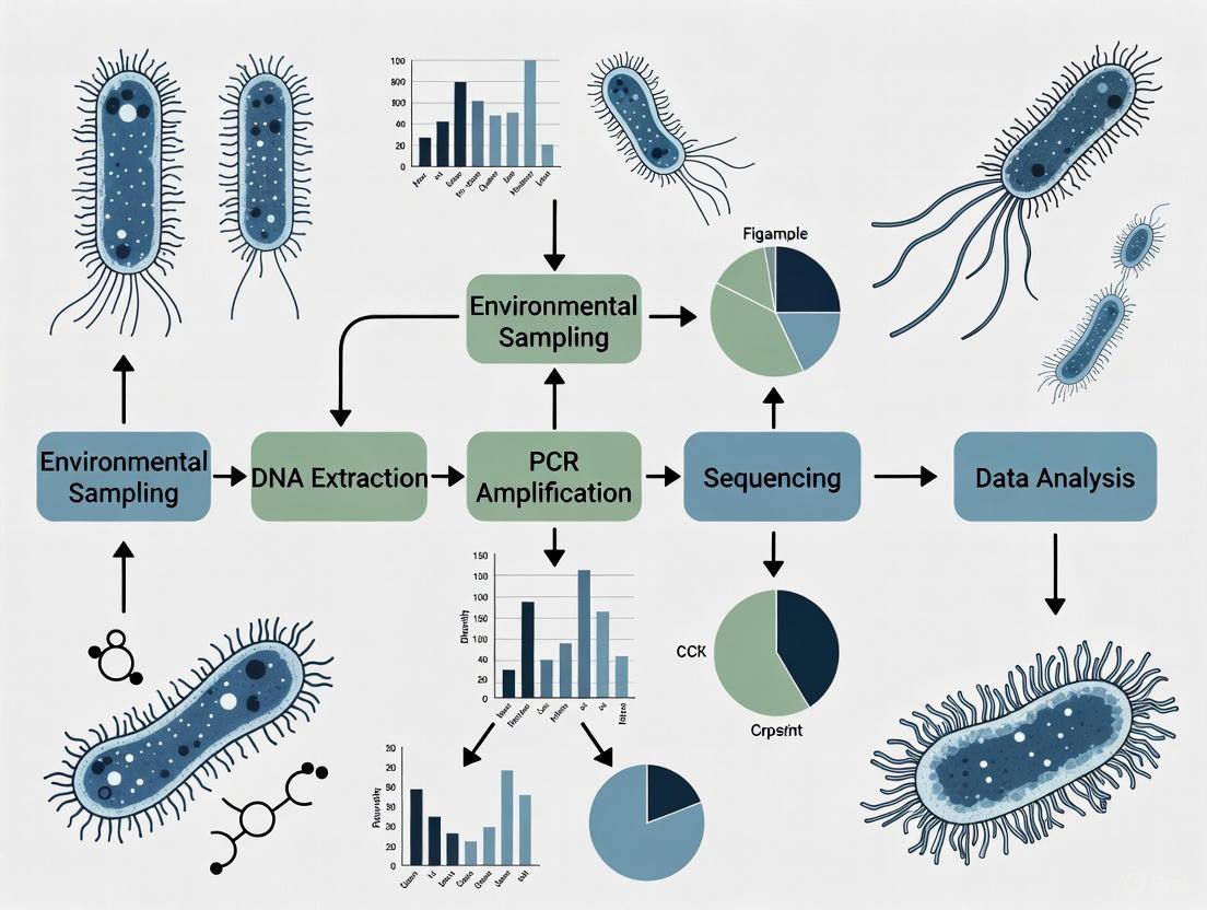
Maximizing Microbial Diversity in Low-Biomass Samples: A Comprehensive Guide to Sampling, Analysis, and Validation
Profiling microbial communities in low-biomass environments—such as human tissues, cleanrooms, and extreme ecosystems—presents unique challenges that can compromise data integrity and biological conclusions.
Maximizing Microbial Diversity in Low-Biomass Samples: A Comprehensive Guide to Sampling, Analysis, and Validation
Abstract
Profiling microbial communities in low-biomass environments—such as human tissues, cleanrooms, and extreme ecosystems—presents unique challenges that can compromise data integrity and biological conclusions. This article provides researchers and drug development professionals with a comprehensive framework for designing robust low-biomass microbiome studies. Covering foundational principles, advanced methodological protocols, rigorous troubleshooting strategies, and comparative validation of analytical techniques, this guide synthesizes current best practices to maximize bacterial diversity recovery while minimizing contamination and bias, thereby enabling reliable discoveries in biomedical and clinical research.
Understanding the Low-Biomass Challenge: Key Concepts and Contamination Risks
Low-biomass environments harbor minimal microbial life, often operating at the very limits of detection for standard DNA-based sequencing methods [1]. In these habitats, the target microbial DNA signal is exceptionally faint, making it disproportionately vulnerable to being obscured by contaminating DNA "noise" introduced during sampling, laboratory processing, or from reagents [1]. This characteristic poses a significant challenge for microbiome research, as even trace contamination can lead to spurious results and incorrect conclusions. Proper identification and handling of these environments are therefore fundamental to studying the true microbial diversity in these niches, which range from internal human tissues to extreme planetary habitats [1].
Defining Characteristics and Examples
Low-biomass environments are defined not just by the low absolute number of microbial cells but also by the high risk of contamination overshadowing genuine biological signals. The table below categorizes exemplary low-biomass environments and their key challenges.
Table 1: Examples and Challenges of Low-Biomass Environments
| Environment Category | Specific Examples | Key Characteristics & Research Challenges |
|---|---|---|
| Human Tissues | Fetal tissues, placenta, blood, brain, lower respiratory tract, breast milk, cancerous tumours [1] [2] | Often disputed findings due to contamination; high consequence for medical interpretations [1]. |
| Extreme Terrestrial & Subsurface | Hyper-arid soils, dry permafrost, deep subsurface, hypersaline brines, ice cores [1] | Approach detection limits; potential for false positives in astrobiology-relevant models [1]. |
| Atmosphere & Built Environments | Atmosphere, snow, treated drinking water, cleanrooms, hospital operating rooms, metal surfaces [1] [2] | Ultra-low biomass requires high-efficiency collection and multiple controls to discern signal from noise [2]. |
| Food Production & Fermentation | Specific layers of Daqu fermentation chambers for sesame-flavored liquor [3] | Microbial diversity varies significantly with physical parameters like temperature (e.g., 35-65°C) [3]. |
Critical Experimental Protocols for Low-Biomass Research
Contamination-Aware Sampling Protocol
A contamination-informed sampling design is critical for obtaining meaningful data [1].
- Pre-Sampling Decontamination: All equipment, tools, vessels, and gloves must be decontaminated. Use single-use, DNA-free items where possible. For re-usable equipment, a two-step process is recommended: decontamination with 80% ethanol to kill organisms, followed by a nucleic acid degrading solution (e.g., sodium hypochlorite, UV-C light, hydrogen peroxide) to remove residual DNA [1].
- Use of Personal Protective Equipment (PPE): Researchers should wear appropriate PPE (gloves, goggles, coveralls, masks, shoe covers) to limit sample exposure to human-associated contaminants from skin, hair, or aerosols generated by breathing [1].
- Collection of In-Line Controls: It is essential to collect and process various control samples alongside actual samples. These include [1]:
- Blank Reagent Controls: Aliquots of sterile water or preservation solutions carried through the entire DNA extraction and sequencing process.
- Equipment/Environmental Controls: Swabs of empty collection vessels, sampling equipment, air in the sampling environment, or researcher PPE.
- Process Controls (for surface sampling): Sampling fluid aspirated without touching a surface to control for the kitome and reagent contamination [2].
Protocol for Surface Sampling with the SALSA Device
This protocol details a method for rapid, on-site microbiome analysis of ultra-low biomass surfaces, such as cleanrooms [2].
- Sampling Device: Use the Squeegee-Aspirator for Large Sampling Area (SALSA), a handheld device that combines a vacuum and squeegee to collect liquid from a large surface area (~1 m²) directly into a collection tube, bypassing the inefficient elution step of swabs [2].
- Sample Collection:
- Pre-wet the target surface area (e.g., 12" x 12") with 2 mL of sterile, DNA-free PCR-grade water from a UV-treated spray bottle.
- Using a new, sterile collection tip for each sample, use the SALSA aspirator to completely collect the liquid from the target area, depositing it into a 5 mL microcentrifuge tube.
- Collect process control samples by aspirating 2 mL of sprayer water using the SALSA device without active surface sampling [2].
- Sample Concentration: Immediately concentrate the 2 mL sample using a device like the InnovaPrep CP-150 with a 0.2 µm polysulfone hollow fiber concentrating pipette tip. Elute the concentrated sample into 150 µL of phosphate-buffered saline (PBS) [2].
- DNA Extraction and Sequencing: Extract DNA from a 100 µL aliquot of the concentrated sample using a commercial kit (e.g., Maxwell RSC). For sequencing, employ a modified version of Oxford Nanopore's Rapid PCR Barcoding Kit, which may require additional PCR cycles or the use of carrier DNA to successfully amplify the ultra-low input DNA [2].
Diagram 1: SALSA Surface Sampling Workflow
The Scientist's Toolkit: Essential Research Reagents and Materials
Successful low-biomass research relies on specialized reagents and materials to minimize and monitor contamination.
Table 2: Essential Research Reagent Solutions for Low-Biomass Studies
| Item | Function | Key Considerations |
|---|---|---|
| DNA-Free Water | Solvent for wetting surfaces during sampling and for preparing molecular biology reagents. | Must be certified DNA-free and sterile; used for pre-wetting in protocols like SALSA sampling [2]. |
| DNA Decontamination Solutions | To remove contaminating DNA from sampling equipment and work surfaces. | Sodium hypochlorite (bleach), UV-C light, hydrogen peroxide, or commercial DNA removal solutions are effective [1]. |
| Personal Protective Equipment (PPE) | To create a barrier between the human operator and the sample, reducing contamination from skin, hair, and aerosols. | Should include gloves, goggles, coveralls/cleansuits, and masks. Extensive PPE is crucial in ultra-clean labs [1]. |
| DNA Extraction Kits | To isolate total genomic DNA from samples. | Kits must be used in tandem with extraction blank controls. The "kitome" (reagent-associated microbiome) must be characterized [2]. |
| PCR Reagents & Carrier DNA | To amplify the minimal DNA obtained from low-biomass samples for downstream sequencing. | Modified PCR protocols with increased cycles or nonspecific carrier DNA (e.g., from mussels) may be necessary for ultra-low inputs [2]. |
| Hollow Fiber Concentrators | To concentrate large-volume liquid samples (e.g., from SALSA) into a smaller volume suitable for DNA extraction. | Devices like the InnovaPrep CP use a 0.2 µm polysulfone pipette tip to concentrate samples from mL to µL volumes [2]. |
Data Analysis and Bioinformatics Workflow
The analysis of sequencing data from low-biomass environments requires rigorous bioinformatics to distinguish true signal from contamination and noise.
- Incorporating Control Data: The data from negative controls and process blanks must be used to inform the filtering process. Contaminant sequences identified in these controls should be removed from the actual samples [1] [2].
- Taxonomic Classification and Filtering: Use bioinformatics platforms like QIIME 2 for processing sequence data [4]. Specialized computational processing is critical, as sequencing can produce high-quality "noise reads" that do not map to any known cellular life [2].
- Analysis Approach Selection: The choice of analysis (e.g., OTU-based vs. phylogenetic approaches) depends on sequencing depth. For datasets with low sequence coverage, phylogenetic approaches can be more robust than OTU-based methods [5].
Diagram 2: Bioinformatics Analysis Workflow
The accurate study of low-biomass environments demands an integrated approach that combines meticulous, contamination-aware sampling, the use of appropriate controls at every stage, and robust bioinformatics. By adhering to the protocols and guidelines outlined in this document—from employing DNA-free reagents and specialized sampling devices like SALSA to implementing rigorous data analysis workflows—researchers can significantly improve the reliability and reproducibility of their findings in these challenging but critical ecosystems.
In low-biomass microbiome research, where microbial DNA yields approach the limits of detection, contamination control transitions from a routine practice to a fundamental determinant of scientific validity. The proportional nature of sequence-based data means that even minute introductions of exogenous DNA can severely distort community profiles, leading to spurious ecological conclusions and erroneous claims about the presence of microbes in potentially sterile environments [1]. The critical contamination sources in these sensitive studies consistently cluster into three primary categories: reagents, personnel, and cross-contamination between samples [1] [6]. Without rigorous mitigation, contaminants from these sources can overwhelm the true biological signal, compromising everything from clinical diagnostics to assessments of environmental ecosystems. This application note delineates evidence-based protocols to identify, mitigate, and monitor these critical contamination sources, providing a structured framework to safeguard data integrity in low-biomass research.
Source 1: Reagents and Laboratory Materials
Laboratory reagents, including DNA extraction kits, PCR master mixes, and water, are well-documented vectors of microbial contaminants. Their impact is magnified in low-biomass studies because the contaminant DNA can constitute a significant proportion, or even the majority, of the final sequencing library [1]. Contaminants originate from the manufacturing processes of these reagents and can include bacterial, archaeal, and fungal DNA. The table below summarizes common reagent-associated contaminants and recommended mitigation strategies.
Table 1: Common Reagent-Derived Contaminants and Control Measures
| Contaminant Source | Typical Contaminants Identified | Recommended Mitigation Strategy |
|---|---|---|
| DNA Extraction Kits | Bacterial genera such as Pseudomonas, Acinetobacter, Burkholderia | Use low-biomass-specific kits; employ multiple kit lots for comparison [1] |
| PCR Reagents | Same as above, plus fungal DNA | Use high-fidelity, DNA-free enzymes; include multiple negative controls [1] |
| Water & Buffers | Various aquatic bacteria | Filter-sterilize using 0.1-0.22 µm filters; validate sterility via qPCR [6] |
| Plasticware/Glassware | Environmental microbes, human skin flora | Autoclave and UV-irradiate; use single-use, DNA-free disposables [1] |
Source 2: Personnel
Researchers are a significant source of contamination, shedding millions of skin cells and aerosolized droplets daily. Human DNA and commensal microorganisms from skin (e.g., Staphylococcus, Cutibacterium) and the oral cavity can be inadvertently introduced during sample collection and handling [1] [6]. One study emphasized that personnel-related contamination is a critical risk factor that can be minimized through stringent personal protective equipment (PPE) protocols, such as those used in cleanrooms and ancient DNA laboratories [1]. These protocols require covering all exposed body parts with cleansuits, gloves, masks, and shoe covers to reduce the introduction of human-associated microbes.
Source 3: Cross-Contamination
Cross-contamination, the transfer of DNA between samples during processing, presents a particularly insidious challenge. This can occur via aerosolization, contaminated equipment, or well-to-well leakage during plate-based workflows [1]. In metagenomic analyses, such cross-contamination can create artificial community structures and lead to false conclusions about microbial transmission or ecology [1]. Mitigation requires both physical barriers, such as separated workstations and closed-system processing, and methodological controls, including randomized plate placement and the use of blank extraction controls to trace contamination pathways.
The following workflow diagram illustrates the primary contamination sources and corresponding mitigation checkpoints throughout a typical low-biomass study pipeline.
Experimental Protocols for Contamination Mitigation
Protocol 1: Rigorous Sample Collection with Controls
This protocol is designed to minimize contamination during the initial sampling of low-biomass environments, such as host tissues or oligotrophic environments [1].
- Step 1: Pre-sampling Decontamination. Decontaminate all non-disposable tools and surfaces with 80% ethanol to kill contaminating organisms, followed by a nucleic acid degrading solution (e.g., 0.5-1% sodium hypochlorite) to remove residual DNA. For disposable items, use only certified DNA-free plasticware that has been pre-treated by autoclaving or UV-C light sterilization [1].
- Step 2: Personnel Preparation. Personnel should wear appropriate PPE, including a cleanroom suit, face mask, safety goggles, and multiple layers of gloves. Rigorous handwashing should be performed before donning gloves, and gloves should be decontaminated with ethanol and DNA degradation solution before sample handling [1] [6].
- Step 3: Control Sample Collection. Concurrently with experimental samples, collect a comprehensive set of control samples to profile the contaminant background. These should include:
- Process Blanks: An empty, sterile collection vessel subjected to the same environment.
- Swab Blanks: A swab exposed to the air in the sampling environment.
- Reagent Controls: Aliquots of preservation or sampling fluids [1].
- Step 4: Sample Storage. Immediately place samples in sterile, DNA-free containers and freeze at -80°C or preserve in a DNA-stabilizing solution to prevent microbial growth or degradation.
Protocol 2: Low-Biomass Optimized Amplicon-PCR
For low-biomass samples where microbial DNA input is insufficient for standard library preparation, an alternative amplicon-PCR protocol can be employed to maximize target amplification without biasing community diversity profiles [7].
- Step 1: Initial Amplification (PCR1). Perform the first PCR reaction using primers targeting the 16S-rDNA V4 region or ITS region with overhang Illumina adapters. The reaction should use 25-35 cycles to maximize yield from low-input templates [7].
- Step 2: Secondary Amplification (PCR2). Use 1 µL of the unpurified PCR1 product as the template for a second PCR. This reaction uses primers that attach full Illumina sequencing adapters and dual-index barcodes. A lower cycle number (e.g., 10 cycles) is typically sufficient [7].
- Step 3: Purification and Normalization. Purify the final PCR2 product using magnetic beads. Quantify the amplicon concentration using a fluorometric method and normalize samples to an equimolar concentration for pooling [7].
- Validation: This protocol has been validated using mock communities and clinical samples, showing no significant differences in microbial diversity compared to standard protocols when the same DNA input is used, while successfully enabling sequencing of otherwise problematic samples [7].
Protocol 3: Quantitative Profiling with Internal Controls
This protocol utilizes spike-in controls for absolute quantification in full-length 16S rRNA gene sequencing, allowing for estimation of microbial load—a critical factor in low-biomass studies [8].
- Step 1: Spike-in Addition. Prior to DNA extraction, add a known quantity of a synthetic microbial community (e.g., ZymoBIOMICS Spike-in Control I) to the sample. The spike-in should comprise a fixed proportion (e.g., 10%) of the estimated total DNA [8].
- Step 2: DNA Extraction and Sequencing. Extract DNA using a kit validated for low-biomass samples (e.g., QIAamp PowerFecal Pro DNA Kit). Perform full-length 16S rRNA gene amplification (using primers 27F and 1492R) with optimized PCR cycles (e.g., 25-35 cycles) for nanopore or other long-read sequencing platforms [8].
- Step 3: Bioinformatic Analysis and Quantification. Process sequencing data with a taxonomy assignment tool like Emu. Calculate absolute abundance of sample taxa using the formula: Absolute Abundance (Sample Taxa) = (Read Count Sample Taxa / Read Count Spike-in Taxa) × Known Spike-in Cell Count [8].
- Application: This method provides robust quantification across varying DNA inputs and has shown high concordance with culture-based estimates in diverse human microbiome samples [8].
The Scientist's Toolkit: Essential Reagents and Controls
Successful low-biomass research requires not only standard laboratory equipment but also specialized reagents and controls designed to identify and mitigate contamination. The following table details key solutions.
Table 2: Essential Research Reagent Solutions for Low-Biomass Studies
| Item Name | Function/Application | Key Features & Usage Notes |
|---|---|---|
| DNA Degradation Solution | Destroys contaminating free DNA on surfaces and equipment. | Typically sodium hypochlorite (bleach) or commercial DNA-away type solutions; used after ethanol cleaning [1]. |
| Certified DNA-free Water | Serves as a base for reagents and negative control preparation. | Filter-sterilized through 0.1 µm membranes and tested via qPCR to be free of microbial DNA [6]. |
| Mock Community Standards | Validates entire wet-lab and bioinformatics workflow accuracy. | Commercially available (e.g., ZymoBIOMICS); contains known, sequenced microbes at defined ratios [8]. |
| Spike-in Controls | Enables absolute quantification of microbial load in samples. | Added to sample lysis buffer; used to convert relative sequencing abundances to absolute counts [8]. |
| UV Chamber | Sterilizes surfaces and tools by degrading nucleic acids. | Effective for plasticware, glassware, and solutions that cannot be autoclaved [1]. |
Mitigating contamination from reagents, personnel, and cross-contamination is not merely a quality control step but the foundation of scientifically valid low-biomass microbiome research. The protocols and guidelines presented here—spanning rigorous sample collection with extensive controls, optimized molecular methods for low-input samples, and quantitative frameworks with internal standards—provide a actionable roadmap for researchers. By systematically implementing these practices, scientists can significantly reduce contaminant noise, thereby enhancing the fidelity of the biological signal. This rigorous approach is indispensable for producing reliable, reproducible data that can accurately characterize microbial communities in the most challenging low-biomass environments, from human tissues to extreme ecosystems.
The Proportional Impact of Contaminant DNA in Sparse Microbial Communities
In microbial ecology, low-biomass environments—such as certain human tissues, air, drinking water, and deep subsurface environments—present a unique set of challenges for accurate characterization. The fundamental issue is that in samples with minimal resident microbial DNA, the relative proportion of contaminant DNA from reagents, sampling equipment, and the laboratory environment can be overwhelmingly high. This contamination can severely distort the apparent microbial community structure, leading to false positives and incorrect ecological conclusions [1]. The proportional impact of this contamination is inversely related to the biomass of the target sample; the lower the starting biomass, the greater the influence of contaminating sequences on the final dataset [1]. This Application Note, framed within a broader thesis on optimizing sampling for bacterial diversity, outlines the sources and impacts of contamination and provides detailed protocols for mitigating its effects in low-biomass research, which is critical for robust scientific discovery and drug development.
The Contamination Challenge in Low-Biomass Research
Contaminant DNA can be introduced at virtually every stage of the research workflow, from sample collection to data analysis. The major sources include:
- Human operators (from skin, hair, or breath)
- Sampling equipment and collection vessels
- Laboratory reagents and DNA extraction kits
- Laboratory environments and cross-contamination between samples [1]
The core of the problem lies in the proportional nature of sequence-based datasets. In a high-biomass sample (e.g., stool or soil), the signal from the true resident microbes dwarfs the contaminant noise. However, in a low-biomass sample (e.g., from the upper respiratory tract [9], fish gills [10], or blood [1]), the contaminant DNA can constitute the majority of the sequenced DNA, making the true signal difficult or impossible to distinguish [1]. This has led to debates about the very existence of microbiomes in certain environments, such as the human placenta [1].
Quantitative Data on Contamination Effects
Table 1: Quantitative Effects of Low Microbial Load on Sequencing Data
| Sequencing Input (16S rRNA gene copies) | Effect on Taxonomic Profile | Source of Artefact |
|---|---|---|
| High input (e.g., 1.2 x 10^7 copies) | Stable, representative profile | N/A |
| Low input (e.g., 1.2 x 10^4 copies) | "Dropout" of lowest abundance taxa | Stochastic sampling |
| Low input (e.g., 1.2 x 10^4 copies) | Appearance of contaminant taxa | Contaminant DNA dominates |
Data from a dilution series experiment showed that sequencing samples with low total microbial loads (below 1 x 10^4 16S rRNA gene copies) resulted in the presence of contaminants, which were confirmed by sequencing negative control extractions [11]. Furthermore, the quantitative limits of 16S rRNA gene amplicon sequencing mean that low DNA input leads to an increase in the coefficient of variation for each taxon's relative abundance and can cause the dropout of the rarest taxa [11].
Methodological Strategies for Contamination Mitigation
A multi-layered approach is essential to minimize contamination and its effects. The following workflow diagram outlines the key stages for reliable low-biomass microbiome research.
Pre-Sampling and Collection Protocols
Personal Protective Equipment (PPE) and Decontamination:
- Researchers should cover exposed body parts with PPE, including gloves, goggles, coveralls or cleansuits, and shoe covers to protect samples from human aerosol droplets and cells shed from clothing, skin, and hair [1].
- Single-use DNA-free collection vessels are ideal. Reusable equipment must be decontaminated with 80% ethanol (to kill organisms) followed by a nucleic acid degrading solution (e.g., sodium hypochlorite, UV-C light, hydrogen peroxide) to remove traces of DNA [1].
Collection of Controls:
- The inclusion of field and sampling controls is non-negotiable for identifying contaminants. These may include:
- An empty collection vessel.
- A swab exposed to the air in the sampling environment.
- Swabs of PPE or surfaces the sample may contact.
- An aliquot of the sample preservation solution [1].
- These controls must be processed alongside actual samples through all downstream steps.
DNA Extraction and Sequencing Optimization
Maximizing Microbial DNA Yield:
- For low-biomass samples like fish gills, developing a quantitative PCR (qPCR) assay for both host and 16S rRNA genes can optimize collection methods to minimize host DNA contamination and facilitate the creation of equicopy libraries based on 16S rRNA gene copies, which significantly increases captured diversity [10].
- Digital PCR (dPCR) can be used as an ultrasensitive anchoring method for absolute quantification, overcoming the limitations of relative abundance analyses [11]. dPCR partitions a PCR reaction into thousands of nanoliter droplets to count single DNA molecules, providing absolute quantification without a standard curve [11].
Alternative Amplification Protocols:
- When standard amplicon-PCR protocols fail due to low microbial DNA input, an alternative amplicon-PCR protocol (effectively a nested PCR with two sequential reactions) can be used to maximize target amplicon yield for 16S rDNA and ITS regions without biasing diversity assessments [7].
Table 2: Essential Research Reagent Solutions for Low-Biomass Studies
| Reagent / Tool | Function | Application Notes |
|---|---|---|
| Sodium Hypochlorite (Bleach) | Degrades contaminant DNA on surfaces | More effective than autoclaving or ethanol alone for creating DNA-free surfaces [1] |
| Digital PCR (dPCR) | Absolute quantification of 16S rRNA gene copies | Overcomes compositionality issue; provides copy number per gram of sample [11] |
| Quantitative PCR (qPCR) | Quantifies total bacterial and host DNA load | Screens samples prior to sequencing; enables normalization for equicopy libraries [10] |
| DNeasy PowerSoil Pro Kit | DNA extraction from complex samples | Effective for soil and other inhibitor-rich, complex matrices [12] |
| Peptide Nucleic Acids (PNAs) | Blocks host DNA amplification (e.g., chloroplast, mitochondrial) | Increases sequencing depth on target microbial DNA in host-contaminated samples [7] |
Data Analysis and Validation
Absolute vs. Relative Abundance Analysis
Relying solely on relative abundance data can be misleading. A change in the relative abundance of a taxon can be caused by an actual change in its absolute abundance or by changes in the abundances of all other taxa in the community [11]. Absolute quantification is necessary to determine the true direction and magnitude of change for individual taxa.
The use of a dPCR anchoring framework allows for the conversion of relative sequencing data to absolute abundances, providing a more accurate picture. In a murine ketogenic-diet study, for example, quantitative measurements of absolute (but not relative) abundances revealed a true decrease in total microbial loads on the diet [11].
Bioinformatic Contamination Removal
Post-sequencing, bioinformatic tools can be used to identify and remove contaminants by leveraging the data from negative controls. The specific sequences and taxa that appear prominently in negative controls should be treated as potential contaminants and removed from all samples [1]. However, these approaches can struggle to distinguish signal from noise in extensively contaminated datasets, underscoring the importance of rigorous wet-lab practices to minimize contamination from the start.
The proportional impact of contaminant DNA in sparse microbial communities is a fundamental challenge that can compromise the validity of research findings. Mitigating this impact requires a vigilant, multi-stage approach that integrates rigorous decontamination during sampling, the systematic use of controls throughout the workflow, specialized molecular protocols to enhance microbial signal and enable absolute quantification, and bioinformatic tools designed to identify residual contamination. By adopting the comprehensive strategies outlined in this Application Note, researchers can significantly improve the accuracy and reliability of microbial community profiles from low-biomass environments, thereby advancing discoveries in human health, environmental science, and drug development.
The investigation of microbial communities in low-biomass environments—those containing minimal microbial DNA—represents a frontier in microbiome science with profound implications for understanding human health and disease. Research on environments traditionally considered sterile, including internal organs like the placenta and tumors, has generated both excitement and intense controversy. These debates center on a fundamental challenge: distinguishing true biological signals from contamination introduced during sampling and processing. In low-biomass samples, contaminating DNA from reagents, laboratory environments, or personnel can constitute a substantial proportion, or even the majority, of the detected microbial signal [13] [14]. Failure to address these concerns adequately has led to high-profile controversies and contradictory findings, risking misdirected scientific resources and flawed biological interpretations [13] [15]. This application note examines the key case studies of the placental and tumor microbiomes, extracting critical lessons and providing validated protocols to ensure rigor in future low-biomass investigations aimed at maximizing the accurate capture of bacterial diversity.
The Placental Microbiome Controversy
Conflicting Evidence and the Sterility Debate
The long-standing paradigm of uterine sterility has been vigorously challenged in the past decade, leading to a significant scientific dispute. Early high-throughput sequencing studies reported a unique, low-abundance placental microbiota composed primarily of non-pathogenic commensals from phyla such as Firmicutes, Tenericutes, Proteobacteria, Bacteroidetes, and Fusobacteria, with compositions allegedly linked to pregnancy complications like preterm birth and preeclampsia [16] [17]. These findings supported the "in utero colonization" hypothesis, suggesting that fetal immune development begins before birth via interaction with placental microorganisms [13] [16].
However, subsequent rigorously controlled studies failed to detect a distinct placental microbial community. Critical re-evaluations demonstrated that the microbial signals observed in placental samples were often indistinguishable from those found in negative controls [13] [17]. One study quantifying absolute bacterial 16S rRNA gene sequences found levels as low as those in negative controls [17]. A comprehensive analysis of 537 placental samples concluded that the extractable bacterial sequence biomass was extremely low, with no evidence of a specific microbiome, though pathogenic infections like Streptococcus agalactiae were detectable in about 5% of samples [13]. The table below summarizes the conflicting findings from key studies in this debate.
Table 1: Conflicting Evidence in the Placental Microbiome Debate
| Study Conclusion | Reported Microbial Composition | Key Evidence | Critiques & Contradictory Findings |
|---|---|---|---|
| Presence of a Distinct Microbiota [16] | Non-pathogenic Escherichia coli, Tannerella forsythia, Fusobacterium nucleatum; Phyla: Firmicutes, Tenericutes, Proteobacteria, Bacteroidetes, Fusobacteria | 16S rRNA and metagenomic sequencing | Signals likely from contamination; no distinct profile found when compared with rigorous controls [13] [14] |
| Placental Microbiota Association with Adverse Outcomes [17] | Higher abundances of Bifidobacterium, Duncaniella, Ruminococcus (GDM); Bacteroides, Paraprevotella, Ruminococcus (PROM) | 16S rRNA gene sequencing with exogenous DNA spike-in | 88.9% of sequences came from added exogenous DNA, indicating extremely low native bacterial biomass [17] |
| Evidence of Sterility [13] | No distinct microbiota found; only specific pathogens (e.g., S. agalactiae) in some cases. | Analysis of 537 placentas; comparison with extensive controls | Extractable bacterial sequence biomass was extremely low; detected signals matched contaminants [13] |
Methodological Lessons and Pitfalls
The core issue confounding placental microbiome research is the failure to adequately control for and distinguish contamination from true signal. Common pitfalls include:
- Inadequate Controls: Early studies often failed to include sufficient negative controls (e.g., reagent blanks, sterile swabs) processed concurrently with samples [13] [14]. Without these, the contaminant DNA inherent in DNA extraction kits and laboratory reagents is misinterpreted as sample-derived [18] [14].
- Batch Effects: When sample processing batches are confounded with experimental groups (e.g., all case samples processed together and all controls processed separately), technical artifacts can create false biological signals [18].
- Misinterpretation of Low Biomass: The placental microbial biomass is so low that it approaches the detection limit of standard sequencing protocols. One study had to artificially add exogenous bacterial DNA to even obtain a visible electrophoresis band for sequencing, revealing that over 88% of their final sequences came from this contaminant [17].
The Tumor Microbiome Controversy
From Pan-Cancer Microbiomes to Rigorous Validation
The tumor microbiome field has followed a similar trajectory of initial excitement followed by rigorous reassessment. Early studies, including a large analysis of The Cancer Genome Atlas (TCGA) data, claimed the existence of tumor-type-specific intracellular microbiomes [19] [20]. However, these findings were later disputed when re-analysis suggested that many reported microbial signals were attributable to contamination of microbial databases with human and vector sequences, or to environmental contaminants not typically associated with humans [20] [15].
This controversy underscored the critical need for orthogonal validation—using non-sequencing-based methods to confirm sequencing results. A seminal study on brain tumors (gliomas and brain metastases) exemplifies this rigorous approach [19]. While standard culture methods did not yield cultivable microbiota, the researchers used fluorescence in situ hybridization (FISH), immunohistochemistry (IHC), and high-resolution spatial imaging to detect intracellular bacterial 16S rRNA and lipopolysaccharides within tumor cells and immune cells [19]. This confirmed the presence of microbial elements but not necessarily a replicating microbiota. The study further found that these intratumoral bacterial signals correlated with antimicrobial and immunometabolic signatures, suggesting a potential functional role in the tumor microenvironment [19].
Table 2: Key Analytical Challenges in Low-Biomass Tumor Microbiome Studies
| Challenge | Description | Impact on Data & Interpretation |
|---|---|---|
| Host DNA Misclassification | Host (e.g., human) DNA sequences are misclassified as microbial in bioinformatic pipelines [18]. | Generates noise and false-positive microbial signals; a significant portion of putative microbial reads can map to the human genome upon re-analysis [20]. |
| External Contamination | DNA introduced from reagents, kits, laboratory environments, and personnel during sample collection and processing [14] [1]. | Can constitute the majority of the microbial signal in low-biomass samples, leading to spurious conclusions about microbial community composition [13] [18]. |
| Well-to-Well Leakage | Cross-contamination between samples processed concurrently, e.g., in adjacent wells on a 96-well plate [18] [1]. | Can cause the transfer of microbial signals between samples, distorting the inferred microbial composition of all affected samples [18]. |
| Computational Artifacts | Errors in taxonomic classifiers and reference databases that have not been benchmarked on real-world, low-biomass datasets [20]. | Leads to false-positive taxa identification; databases contaminated with human/vector sequences perpetuate these errors [20]. |
Advanced Computational Decontamination
In response to these challenges, new computational tools have been developed. PRISM (Precise Identification of Species of the Microbiome) is one such method designed for low-biomass sequencing data [20]. Its two-step process involves:
- Eliminating False-Positive Artifacts: Using full-length read alignment to remove reads that were misclassified as microbial but actually originate from the host human genome.
- Identifying Truly-Present Taxa: Using a machine learning classifier trained on known true and false positives to distinguish tissue-resident microbes from contaminants [20]. This approach dramatically reduces the number of putative microbial taxa per sample, moving from a median of 160 taxa/sample identified by standard k-mer-based classifiers to a median of 14 high-confidence, uniquely identifiable taxa/sample [20].
Essential Protocols for Rigorous Low-Biomass Research
Integrated Experimental Workflow for Low-Biomass Sampling and Analysis
The following diagram outlines a comprehensive and rigorous workflow for low-biomass microbiome studies, integrating experimental and computational best practices.
The Scientist's Toolkit: Research Reagent Solutions
Table 3: Essential Reagents and Kits for Low-Biomass Microbiome Research
| Item | Function & Application | Key Considerations |
|---|---|---|
| DNA Decontamination Solutions (e.g., Bleach, DNA-ExitusPlus) | To remove contaminating DNA from work surfaces and non-disposable equipment [1]. | Sodium hypochlorite (bleach) and commercial DNA degradation solutions are effective. Autoclaving and ethanol alone do not remove persistent DNA [1]. |
| Ultra-clean DNA Extraction Kits (e.g., QIAamp PowerFecal Pro DNA Kit) | To extract microbial DNA from low-biomass samples while co-purifying inhibitors [17]. | Select kits validated for low-biomass samples. Be aware that all kits contain their own background microbial DNA [14] [21]. |
| Preservative Buffers (e.g., AssayAssure, OMNIgene•GUT) | To stabilize microbial community DNA at room temperature when immediate freezing is not possible [21]. | Effectiveness varies; some buffers may influence the detection of specific bacterial taxa. Cold storage is generally preferred if possible [21]. |
| Personal Protective Equipment (PPE) (Gloves, Masks, Coveralls) | To act as a barrier, reducing contamination from researchers' skin, hair, and breath [1]. | PPE is a simple and critical contamination control. Gloves should be changed frequently and not touch anything before sample collection [1]. |
| Sterile, Single-Use Consumables (Collection tubes, swabs, pipette tips) | To avoid introducing contaminants from previous uses or the manufacturing environment [1]. | Pre-sterilized, DNA-free consumables are essential. Swabs from different manufacturing batches can have different contaminant profiles [18] [1]. |
The controversies surrounding the placental and tumor microbiomes provide a critical learning opportunity for the entire field of low-biomass microbiome research. The primary lesson is unequivocal: rigorous, contamination-aware methodologies are not merely best practice but are fundamental to generating biologically valid data. Reliance on sequencing data alone, without robust experimental controls and orthogonal validation, is insufficient to claim the existence of a resident microbiome in low-biomass environments. Future research must adopt a trans-disciplinary framework that integrates classical microbiology, immunology, and microbial ecology with carefully controlled sequencing and advanced computational decontamination. By implementing the protocols and guidelines outlined in this document, researchers can navigate the pitfalls of low-biomass studies, maximize the accurate assessment of bacterial diversity, and ensure that the pursuit of novel microbial discoveries is built upon a foundation of methodological rigor and scientific reproducibility.
Core Principles for Reliable Low-Biomass Research
Low-biomass microbial environments, which include human tissues (e.g., respiratory tract, placenta, blood), natural ecosystems (e.g., deep aquifers, hyper-arid soils), and built environments, present unique challenges for DNA-based microbiome studies [1] [18]. The defining feature of these environments is that the target microbial DNA signal is minimal, making results disproportionately vulnerable to contamination from external DNA, cross-contamination between samples, and other analytical pitfalls [1] [18]. High-profile controversies, such as those surrounding the purported placental microbiome and the tumor microbiome, underscore how these challenges can compromise biological conclusions and even lead to retractions [18]. This application note outlines the core principles and detailed protocols essential for conducting reliable low-biomass research, framed within the broader thesis that rigorous sampling methods are paramount for maximizing the accurate detection of bacterial diversity.
Core Principles and Experimental Design
A robust low-biomass study must be designed to minimize the introduction of contaminants and to maximize the ability to detect and account for those that are inevitable. The following principles are critical [1] [18].
- Contamination Mitigation: Contamination can originate from human operators, reagents, sampling equipment, and the laboratory environment. Its impact is proportional; the lower the native biomass, the greater the influence of contaminating DNA on the final dataset [1].
- Batch Confounding Avoidance: Perhaps the most critical experimental design consideration is ensuring that the groups being compared (e.g., case vs. control) are processed in completely randomized, non-confounded batches. If all samples from one group are processed in a single batch, and the other group in another, any batch-specific effects (contamination, reagent lot, personnel) can create artifactual signals that are indistinguishable from true biological differences [18].
- Comprehensive Control Strategy: The use of various process controls is non-negotiable. These controls help identify the nature and sources of contamination introduced throughout the experimental workflow [1] [18].
Table 1: Essential Process Controls for Low-Biomass Studies
| Control Type | Description | Purpose |
|---|---|---|
| Negative Extraction Control | A blank sample (e.g., water) carried through the DNA extraction process [18]. | Identifies contamination originating from DNA extraction kits and reagents [18]. |
| No-Template PCR Control | A PCR reaction containing all reagents except for sample DNA [18]. | Detects contamination in PCR master mixes and other amplification reagents. |
| Sampling/Equipment Blank | A sterile swab or an empty collection vessel exposed to the sampling environment [1]. | Reveals contaminants from collection kits, vessels, and the ambient air during sampling [1]. |
| Mock Community | A sample containing known quantities of DNA from specific microorganisms [18]. | Assesses sequencing accuracy, PCR bias, and bioinformatic performance. |
Detailed Experimental Protocols
Sample Collection and Preservation
This protocol is adapted for collecting upper respiratory tract (URT) swabs, a common low-biomass application, but the principles are widely applicable [1] [9].
Materials:
- Sterile, DNA-free swabs
- Personal Protective Equipment (PPE): gloves, mask, hair net, clean suit [1]
- DNA decontamination solution (e.g., 10% bleach, commercially available DNA removal solutions) [1]
- Sterile collection tubes
- Portable cooler or dry ice for flash-freezing
Procedure:
- Pre-Sampling Decontamination: Prior to sampling, decontaminate all surfaces and equipment. Wipe down surfaces with 80% ethanol followed by a DNA-degrading solution like sodium hypochlorite (bleach) to remove both viable cells and free DNA [1].
- Investigator Preparation: Don appropriate PPE (gloves, mask, and if possible, a clean suit) to limit contamination from the investigator's skin, hair, and breath [1].
- Sample Collection: Using a sterile swab, collect the sample according to the specific procedure (e.g., nasal swab). Avoid touching the swab to any surface other than the target tissue.
- Sample Storage: Immediately place the swab into a sterile, DNA-free tube. Flash-freeze the sample on dry ice or in a liquid nitrogen dry shipper for transport. Store at -80°C until DNA extraction [9].
DNA Extraction from Low-Biomass Samples
This protocol is crucial as the efficiency of DNA recovery from low-biomass samples directly impacts downstream diversity assessments [9] [22].
Materials:
- DNeasy PowerSoil Kit (Qiagen) or similar [23]
- Mechanical lysis equipment (e.g., bead beater)
- Refrigerated microcentrifuge
- Molecular grade water (DNA-free)
Procedure:
- Cell Lysis: For robust lysis of diverse bacterial cell walls, use a combination of mechanical and chemical methods. Add the sample (e.g., swab tip or filter membrane) to a bead-beating tube containing a lysis buffer and perform mechanical disruption in a bead beater for 2-5 minutes [9] [22].
- DNA Extraction: Follow the manufacturer's instructions for the DNA extraction kit. Include the negative extraction controls and mock community controls in the same batch as the samples [18] [23].
- DNA Elution: Elute the purified DNA in a small volume (e.g., 30-50 µL) of molecular grade water or the provided elution buffer to maximize DNA concentration.
- DNA Quantification: Quantify DNA yield using a fluorescence-based method (e.g., Qubit) which is more accurate for low-concentration samples than spectrophotometry. Expect very low yields (e.g., 0.3-6.4 ng/µL) [23].
Library Preparation and Sequencing
Materials:
- Illumina MiSeq platform [9]
- Primers for the 16S rRNA gene V4 region (e.g., 515F/806R) or for metagenomic shotgun sequencing [9]
- No-template PCR controls [18]
Procedure:
- Amplification: Amplify the target region (e.g., 16S rRNA V4) using a high-fidelity polymerase in a minimal number of PCR cycles to reduce bias. Include no-template PCR controls.
- Library Preparation and Sequencing: Construct sequencing libraries following standard Illumina protocols. Sequence on an appropriate platform (e.g., MiSeq with 2x250 bp chemistry for 16S rRNA gene studies) [9].
The Scientist's Toolkit: Key Research Reagent Solutions
Table 2: Essential Materials for Low-Biomass Microbiome Research
| Item | Function/Justification |
|---|---|
| DNeasy PowerSoil Kit (Qiagen) | Optimized for efficient DNA extraction from difficult, low-biomass samples and soils; includes inhibitors removal steps [23]. |
| Sterile FloqSwabs | Designed for efficient sample collection and release of biological material; pre-sterilized to prevent contamination [23]. |
| Sodium Hypochlorite (Bleach) | Used for surface and equipment decontamination; degrades environmental DNA that survives autoclaving [1]. |
| Personal Protective Equipment (PPE) | Minimizes the introduction of human-associated microbial contaminants (from skin, hair, breath) into samples during collection [1]. |
| Mock Microbial Communities | Comprised of known microbes; used as a positive control to assess bias and accuracy in DNA extraction, PCR, and sequencing [18]. |
Workflow and Data Analysis Diagram
The following diagram illustrates the integrated workflow for a reliable low-biomass microbiome study, from experimental design to data interpretation, highlighting critical control points.
Advanced Sampling and Extraction Protocols for Diverse Low-Biomass Environments
The accurate characterization of microbial communities in low-biomass environments presents significant methodological challenges, as the limited microbial signal can be easily obscured by contamination or inefficient sampling. The selection of an appropriate sample collection strategy is therefore paramount to achieving meaningful results in studies of low-biomass microbiomes, such as those found on human skin, in cleanrooms, or on various clinical surfaces. This application note provides a detailed comparison of three prominent collection methods—swabbing, SALSA aspiration, and tape stripping—to guide researchers in selecting and implementing optimal protocols for maximizing bacterial diversity recovery. Within the context of a broader thesis on sampling methodologies, we demonstrate that method selection directly dictates the upper limit of microbial diversity that can be captured, with significant implications for downstream ecological interpretation and analytical outcomes.
Performance Comparison of Sampling Methods
The quantitative and qualitative performance of swabbing, SALSA aspiration, and tape stripping varies considerably across different experimental contexts and surface types. The following table summarizes key comparative data from recent studies to inform methodological selection.
Table 1: Performance Metrics of Low-Biomass Sampling Methods
| Method | Reported Sampling Efficiency | Typical DNA Yield | Dominant Taxa Recovered | Best Suited Surfaces |
|---|---|---|---|---|
| Swabbing | 19% - 75% [24] [25] | Often undetectable in very low-biomass scenarios [25] | Variable; highly technique-dependent | Large, even surfaces (e.g., floors, skin) [26] |
| SALSA Aspiration | ≈60% or higher [2] | 1-2 orders of magnitude above process controls [2] | Paracoccus, Acinetobacter [2] | Large, smooth, non-porous surfaces (e.g., cleanroom floors) [2] |
| Tape Stripping | ≈50% to >99% [24] | ~2-3 fold less bacterial DNA than swabbing [26] | Staphylococcus, Cutibacterium [27] [25] | Skin, especially stratum corneum [28] [27] |
| Advanced Tape (PVA-based) | >96.6% for DNA materials [24] | Not Specified | S. aureus, E. coli, S. cerevisiae [24] | Glass, stainless steel [24] |
| Skin Scraping | Not Quantified | 0.065 to 13.2 ng/µL (bacteria); 0.104 to 30.0 ng/µL (fungi) [25] | S. aureus group, C. acnes group, Malassezia [25] | Sensitive facial skin [25] |
Detailed Experimental Protocols
Swabbing Protocol for Low-Biomass Surfaces
Principle: Mechanical removal of microbes via friction using a pre-moistened, sterile swab.
- Step 1: Preparation. Moisten a sterile swab (e.g., cotton or flocked) with a sterile solution such as phosphate-buffered saline (PBS). For skin sampling, instruct participants to avoid washing the area with soap or bathing for at least 24 hours prior to collection [26].
- Step 2: Collection. Apply moderate, consistent pressure to the target surface (e.g., a defined area of skin or a 10x10 cm surface area). Use circular motions while simultaneously rotating the swab to ensure all sides of the tip contact the surface. Perform approximately 20 strokes per subregion [25].
- Step 3: Elution. Aseptically cut the swab head and transfer it to a sterile tube containing an appropriate elution buffer (e.g., PBS or a commercial elution solution). Vortex or shake the tube vigorously to disperse collected material into the solution [25].
- Step 4: Processing. Concentrate the eluent if necessary using methods such as centrifugal filtration (e.g., InnovaPrep CP with a 0.2µm polysulfone hollow fiber tip) [2]. Proceed to DNA extraction.
SALSA Aspiration Protocol for Large Surface Areas
Principle: Combines squeegee action and aspiration to efficiently recover microbes from large surface areas into a liquid sample, bypassing adsorption to swab fibers [2].
- Step 1: Surface Pre-wetting. Spray a defined large surface area (up to 1 m²) with a sterile, DNA-free wetting buffer (e.g., PCR-grade water) using a UV-treated spray bottle [2].
- Step 2: Aspiration. Use the sterile, disposable squeegee head of the SALSA device to scrub the pre-wetted surface. Activate the battery-operated aspirator to draw the liquid sample directly into a sterile collection tube. The device uses a vacuum and squeegee function for efficient recovery [2].
- Step 3: Concentration. Concentrate the collected liquid sample (often a large volume) using a high-efficiency method. The InnovaPrep CP-150 device with a 0.2-µm polysulfone hollow fiber concentrating pipette tip is recommended, eluting into a final volume of 150 µL of PBS [2].
- Step 4: Storage. Aliquot the concentrated sample for downstream DNA extraction and analysis.
Advanced Tape Stripping Protocol
Principle: An adhesive tape or a film-forming solution is applied to the skin to remove layers of the stratum corneum and associated microbes [28] [24].
A. Conventional Adhesive Tape Method
- Step 1: Application. Firmly press a piece of sterile adhesive tape (e.g., D-Squame, Sebutape) onto the target skin area.
- Step 2: Removal. Peel the tape off the skin in one swift motion. The stripped stratum corneum and microbes will be embedded in the adhesive.
- Step 3: Elution. Place the tape adhesive-side down in a sterile tube containing elution buffer. Agitate to release the collected material, or analyze the tape directly [26] [28].
B. Polyvinyl Alcohol (PVA)-Based Advanced Tape Stripping [24]
- Step 1: Solution Application. Apply a film-forming PVA solution (containing PVA, ethanol, glycerol, and nuclease-free water) to the target surface. The solution engulfs surface deposits as it is applied.
- Step 2: Membrane Formation. Allow the solution to solidify into an elastic membrane upon exposure to air.
- Step 3: Collection. Strip off the resulting membrane from the surface.
- Step 4: Re-dissolution. Place the membrane in a tube and re-dissolve it in nuclease-free water to release the collected material for downstream analysis.
Workflow Integration and Contamination Control
A standardized workflow is critical for ensuring sample integrity from collection to analysis, particularly for low-biomass samples which are highly susceptible to contamination.
Figure 1: Integrated workflow for low-biomass microbiome sampling, highlighting critical contamination control points at each stage.
Essential Contamination Control Practices
- Negative Controls: Include multiple negative controls at each stage, including DNA extraction controls (to detect kit reagent contaminants) and template-free PCR blanks (to identify contamination during downstream processing) [26] [1]. The proportion of contaminant DNA is significantly higher in low-biomass samples.
- Sample Handling: Minimize handling and exposure to the laboratory environment. Use personal protective equipment (PPE) such as gloves, masks, and coveralls to limit contamination from operators [1].
- Reagent Verification: Check that sampling reagents and preservation solutions are DNA-free. Decontaminate reusable equipment with 80% ethanol followed by a nucleic acid degrading solution (e.g., bleach) [1].
- Data Normalization: For challenging samples like fish gills, quantify 16S rRNA gene copies via qPCR prior to library construction. Creating equicopy libraries based on this quantification can significantly improve the resolution of true microbial community structure [10].
Research Reagent Solutions
The following reagents and tools are critical for implementing the described sampling protocols effectively.
Table 2: Essential Reagents and Tools for Low-Biomass Sampling
| Item | Function/Description | Example Use Case |
|---|---|---|
| Flocked Swabs | Swabs with perpendicular fibers for superior sample release | Standardized skin or surface swabbing [25] |
| Sterile Surgical Blades | For gentle skin scraping to recover stratum corneum | Sampling sensitive facial skin [25] |
| D-Squame / Sebutape | Standardized adhesive tapes for consistent tape stripping | Sampling skin stratum corneum [26] |
| PVA Film-Forming Solution | Advanced tape-stripping solution that solidifies and is re-dissolvable | High-efficiency sampling from non-absorbent surfaces [24] |
| SALSA Device | Handheld squeegee-aspirator for large surface area sampling | Sampling cleanroom floors [2] |
| InnovaPrep CP Concentrator | Hollow fiber concentration device for liquid samples | Concentrating SALSA aspirates or swab eluates [2] |
| HostZERO Microbial DNA Kit | DNA extraction kit with host DNA depletion | Extracting microbial DNA from samples rich in host cells [25] |
| Femto Bacterial DNA Quantification Kit | qPCR kit for accurate quantification of low-abundance bacterial DNA | Quantifying 16S rRNA genes in low-biomass extracts [2] [10] |
Method Selection Guidelines
Choosing the optimal sampling method requires careful consideration of the surface type, research objectives, and practical constraints.
Figure 2: Decision tree for selecting the optimal sampling method based on surface characteristics, research objectives, and biomass level.
Application-Specific Recommendations
- Large Surface Areas in Controlled Environments: For sampling large, smooth surfaces such as cleanroom floors where efficiency and scale are priorities, SALSA aspiration is the superior choice due to its high recovery efficiency (≥60%) and ability to process areas up to 1 m² [2].
- Standard Skin Microbiome Studies: For general skin sampling where patient comfort and stratification corneum resolution are important, traditional tape stripping provides a robust, minimally invasive option that yields comparable microbial composition to swabbing with superior recovery of viable bacteria in culture studies [27].
- Challenging Low-Biomass Skin: For sensitive facial skin or other very low-biomass scenarios where swabbing fails to recover detectable DNA, gentle skin scraping with a sterile surgical blade has proven effective, yielding sufficient DNA for both bacterial and fungal sequencing [25].
- Maximized Efficiency on Non-Absorbent Surfaces: When the highest possible sampling efficiency is required from smooth, non-absorbent surfaces like glass or stainless steel, the PVA-based advanced tape stripping method demonstrates exceptional performance (>96.6% efficiency) for DNA-based targets [24].
The selection of an appropriate sample collection method fundamentally shapes the fidelity of microbial community analysis in low-biomass research. As demonstrated in this application note, no single method is universally superior; rather, the optimal choice depends on a careful balance of surface characteristics, research objectives, and practical constraints. Swabbing remains a versatile, though sometimes inefficient, option for general use. SALSA aspiration offers unprecedented efficiency for large surface areas, while tape stripping and its advanced derivatives provide depth-resolved sampling ideal for skin and other complex surfaces. Crucially, all low-biomass studies must incorporate rigorous contamination controls throughout the workflow, from sample collection through DNA sequencing, to distinguish true signal from artifact. By implementing these detailed protocols and selection guidelines, researchers can significantly enhance the reliability and reproducibility of their low-biomass microbiome studies, thereby enabling more accurate ecological inference and mechanistic insight.
In low-biomass research, such as studies of specific skin sites, the female upper reproductive tract, or the lower respiratory tract, the efficient extraction of microbial DNA is a foundational challenge [7] [29]. The low ratio of microbial to host DNA in these samples makes them highly susceptible to confounding factors like contamination, and the limited starting material makes maximizing yield and preserving diversity paramount [7] [29]. The initial cell lysis step is critical, as it directly influences DNA yield, integrity, and the faithful representation of the microbial community in downstream sequencing [30] [31]. This application note provides a comparative analysis of mechanical and chemical lysis strategies, offering structured protocols and data to guide researchers in optimizing methods for challenging, low-biomass samples.
Comparative Analysis of Lysis Methods
The choice between mechanical and chemical lysis involves a fundamental trade-off between DNA yield and DNA integrity, which must be carefully balanced based on the sample type and downstream application.
Table 1: Characteristics of Mechanical vs. Chemical Lysis Methods
| Feature | Mechanical Lysis | Chemical Lysis |
|---|---|---|
| Principle | Physical force to shear cells [32] [33] | Chemicals (e.g., detergents) to dissolve membranes [32] [33] |
| Key Techniques | Bead beating, sonication, high-pressure homogenization [32] [33] | Detergent-based lysis, osmotic shock, enzymatic treatment [32] [33] |
| Typical DNA Yield | Higher, especially from robust cells [30] | Variable, can be lower for gram-positive bacteria [30] |
| DNA Integrity | Lower; can cause fragmentation [31] | Higher; gentler on nucleic acids [30] |
| Impact on Community Profile | Better for resistant cells (e.g., gram-positive); may bias against fragile cells [30] | May underrepresent tough cells; better for fragile protists [30] |
| Scalability | Easily scalable for high-throughput [34] | Highly scalable [34] |
| Cost & Ease | Requires equipment investment [32] | Lower initial cost; often simpler [32] |
Quantitative data highlights the practical impact of this choice. A study on rumen microbiota found that including a bead-beating (mechanical) step increased total DNA yield significantly (P=0.001) but was not recommended for protozoal community analysis due to reduced length polymorphism of protozoal amplicons [30]. Conversely, the chemical lysis provided by the RBB+C protocol was more effective at harvesting DNA from fibrolytic bacteria like Ruminococcaceae compared to the QIAamp kit's chemistry [30].
For long-read sequencing, which requires high-molecular-weight DNA, the intensity of mechanical lysis is a key variable. A 2024 study using a Design of Experiments (DoE) approach found that low-intensity mechanical lysis (4 m s⁻¹ for 10 s) increased DNA fragment length by 70% compared to the manufacturer's standard protocol, without introducing significant bias in microbial community composition [31]. This demonstrates that mechanical lysis parameters can be finely tuned to maximize downstream success.
Application Notes for Low-Biomass Samples
Samples with low microbial load present unique challenges, where standard protocols often fail.
- Challenge of Host DNA Contamination: In low-biomass samples, host DNA can dominate, drastically reducing the sequencing depth of the microbial community [7] [29]. While swabbing can improve the microbial-to-human DNA ratio [29], the lysis and analysis methods must be selected to maximize microbial signal.
- Lysis Choice Affects Diversity Recovery: The optimal lysis method can depend on the analytical technique. One study on low-biomass skin sites found that while 16S rRNA amplicon sequencing exhibited extreme bias toward the most abundant taxon, metagenomic sequencing and qPCR revealed concordant, diverse microbial communities [29]. This suggests that for low-biomass samples analyzed via amplicon sequencing, a lysis method that effectively recovers a broader range of taxa is even more critical.
- Alternative Amplification Protocols: When DNA input is insufficient for library preparation, an alternative amplicon-PCR protocol, similar to a nested-PCR with two sequential reactions, can be used to maximize target amplification without biasing bacterial or fungal diversity data [7].
Detailed Experimental Protocols
Protocol: Optimized Mechanical Lysis for Soil DNA
This protocol, adapted from a 2024 Scientific Reports paper, is designed to maximize DNA fragment length for long-read sequencing from soil and other complex samples [31].
Research Reagent Solutions:
- Lysis Buffer: Typically contains salts (e.g., NaCl), a buffer (e.g., Tris-HCl), and detergents [33].
- Homogenization Beads: Ceramic or silica beads (0.1-2.0 mm diameter) for efficient cell disruption [31].
- Inhibition Removal Solution: Additives like EDTA to chelate metal ions and inhibit nucleases [33].
- Proteinase K: An enzyme that degrades proteins and aids in cell lysis [35].
Workflow:
- Preparation: Place approximately 0.25 g of soil sample into a pre-chilled lysing tube containing homogenization beads.
- Lysis Buffer Addition: Add the appropriate volume (e.g., 800 µL) of pre-chilled lysis buffer to the tube. Include proteinase K if required by the protocol.
- Homogenization: Securely load the tubes into a benchtop homogenizer (e.g., FastPrep-24).
- Parameter Optimization: Process samples at a speed of 4 m s⁻¹ for a duration of 10 seconds. This low-energy input is key to preserving DNA integrity [31].
- Centrifugation: Centrifuge the lysate at high speed (e.g., 14,000 x g) for 5-10 minutes to pellet debris and beads.
- Supernatant Collection: Carefully transfer the supernatant containing the DNA to a new, clean tube for subsequent purification.
Protocol: Chemical Lysis for Rumen Microbiota
This protocol is derived from a comparison of the RBB+C and QIAamp Fast DNA Stool Mini Kit methodologies for studying bacterial and protozoal communities [30].
Research Reagent Solutions:
- RBB+C Lysis Buffer: A detergent-based buffer designed to dissolve cell membranes [30].
- Silica Matrix Columns: For binding and purifying DNA after lysis [30].
- Protease Inhibitors: Added to lysis buffer to protect proteins from degradation [33].
- Enhanced RIPA Buffer: Contains strong detergents for efficient membrane protein extraction [33].
Workflow:
- Sample Input: Use 200 µL of rumen fluid or an equivalent sample volume.
- Chemical Lysis:
- Optional Mechanical Lysis: To enhance yield from tough cells, add ~200 mg of beating material (beads or sand) to the tube and vortex vigorously [30].
- Incubation: Incubate the mixture according to the chosen protocol's specifications (often at elevated temperature, e.g., 70°C or 95°C).
- Purification: Bind and wash the DNA using a silica membrane column.
- Elution: Elute the purified DNA in a low-salt buffer or nuclease-free water.
Table 2: Impact of Lysis Method on Downstream Analysis
| Lysis Method | Total DNA Yield | DNA Purity (OD260/280) | Impact on Bacterial Diversity | Impact on Protozoal Community |
|---|---|---|---|---|
| RBB+C (YM) Chemical Lysis | Greater yield from fibrolytic bacteria [30] | Lower [30] | Better for gram-positive lineages [30] | Lower length polymorphism [30] |
| QIAamp (QIA) Chemical Lysis | Lower yield from some bacteria [30] | Higher [30] | Standard diversity profile [30] | Preferred method [30] |
| With Bead Beating (BB) | Increased (P=0.001) [30] | No significant effect [30] | No significant effect on richness [30] | Decreased richness and polymorphism [30] |
| Sand Beating (SB) | No difference vs. BB [30] | No difference vs. BB [30] | No difference vs. BB [30] | No difference vs. BB [30] |
The Scientist's Toolkit
Table 3: Essential Reagents and Materials for Cell Lysis
| Item | Function & Application |
|---|---|
| Ceramic/Silica Beads | Provide abrasive action for mechanical disruption of tough cell walls in microbial and soil samples [30] [31]. |
| Enhanced RIPA Lysis Buffer | A detergent-based chemical lysis buffer fortified for efficient extraction of membrane proteins and insoluble proteins [33]. |
| Proteinase K | Enzyme that digests proteins and inactivates nucleases, aiding in cell lysis and protecting nucleic acids [35]. |
| EDTA (Chelating Agent) | Binds metal ions to inhibit metal-dependent proteases and nucleases, protecting DNA during extraction [33] [35]. |
| Protease & Phosphatase Inhibitors | Added to lysis buffers to prevent co-extracted proteases/phosphatases from degrading target proteins or modifying phosphorylation states [33]. |
| Silica Matrix Columns | Used for post-lysis purification to bind, wash, and elute DNA, removing contaminants and inhibitors [30]. |
Maximizing yield and diversity from challenging, low-biomass samples requires a strategic and often customized approach to cell lysis. As the data demonstrates, there is no single superior method; the optimal choice is application-dependent. Researchers should select chemical lysis for preserving DNA integrity and fragile protist communities, while mechanical lysis is more effective for maximizing yield from tough, gram-positive bacteria. For long-read sequencing, tuning mechanical lysis to lower energy inputs is critical. By understanding these trade-offs and implementing the optimized protocols outlined here, scientists can significantly enhance the quality and reliability of their downstream molecular analyses in low-biomass research.
DNA Extraction Kits and Modifications for Ultra-Low Input Samples
In microbial ecology and clinical diagnostics, the study of low-biomass environments—those containing minimal microbial loads—presents unique challenges that demand specialized approaches. Samples such as fish gills, fetal tissues, plant seeds, liquid biopsies, and preserved museum specimens share the common characteristic of yielding very limited amounts of microbial DNA [10] [1]. In these contexts, standard DNA extraction methods frequently fail to capture the true microbial diversity, as contaminants from reagents, equipment, or handling can dramatically outweigh the authentic signal from the sample itself [1]. The proportional impact of contamination increases exponentially as biomass decreases, potentially leading to spurious conclusions about microbial community structure and function [1].
The fidelity of downstream analyses, including 16S rRNA gene sequencing and metagenomics, is fundamentally dependent on the initial sample collection and DNA extraction phases. Research has demonstrated that microbial community composition exerts a strong influence on ecosystem function, rivaling the importance of substrate chemistry in processes like litter decay [36]. Therefore, optimizing DNA extraction for low-biomass samples is not merely a technical concern but a foundational requirement for generating meaningful biological insights. This application note outlines practical strategies and modified protocols to maximize bacterial diversity recovery while minimizing contamination in low-biomass research, framed within the broader context of a thesis on sampling methods for microbial ecology.
Key Challenges in Ultra-Low Input Sample Processing
Working with ultra-low input samples presents three primary challenges that complicate accurate microbial profiling. First, the limited target DNA necessitates specialized methods to maximize yield while preventing loss during extraction. Second, the high susceptibility to contamination requires rigorous controls and decontamination protocols, as contaminant DNA can constitute most of the final sequencing data [1]. Third, the inherent inhibitor-rich nature of many sample matrices requires additional purification steps.
The challenges are particularly pronounced in specific sample types. Fish gills represent inhibitor-rich, low-biomass tissues directly exposed to aquatic environments [10]. Museum specimens contain highly fragmented DNA due to degradation over time [37]. Clinical samples like liquid biopsies, fetal tissues, and certain human microbiomes approach the detection limits of standard molecular methods [1]. Built environments including metal surfaces, drinking water, and hyper-arid soils also present significant challenges for microbial DNA studies [1].
Commercial Kits and Protocols for Low-Input DNA Extraction
Available Commercial Solutions
Recent advancements have yielded several specialized kits designed to address the challenges of low-input DNA extraction. The following table summarizes key commercial options:
Table 1: Commercial DNA Extraction and Library Preparation Kits for Low-Input Samples
| Product Name | Input Range | Key Technology | Primary Applications | Special Advantages |
|---|---|---|---|---|
| Quick-DNA HMW MagBead Kit (Zymo Research) | Not specified | Magnetic beads for HMW DNA purification | Nanopore sequencing, metagenomics | Best yield of pure HMW DNA; accurate detection in mock communities [38] |
| Ovation Ultralow System V2 (Tecan) | 10 pg – 100 ng | DimerFree technology | WGS, DNA-seq, ChIP-seq, targeted sequencing | Virtually no adapter dimers; single workflow for any input [39] |
| Ampli-Fi Protocol (PacBio) | 1 – 50 ng | KOD Xtreme Hot Start DNA Polymerase | HiFi sequencing of ultra-limited samples | Reduced PCR bias; better assembly of difficult genomes [40] |
| Monarch PCR & DNA Cleanup Kit (NEB) | Not specified | Spin column purification | DNA clean-up | Effective in comparative studies [37] |
Specialized Library Preparation Methods
Beyond DNA extraction, library preparation methods significantly impact success with low-input samples. The Santa Cruz Reaction (SCR) method has demonstrated particular effectiveness for degraded DNA from museum specimens, outperforming several commercial kits in cost-effectiveness and retrieval efficiency [37]. This do-it-yourself approach enables high-throughput processing while maintaining sensitivity for challenging samples.
For long-read sequencing technologies, the Ampli-Fi protocol supports HiFi sequencing from inputs as low as 1 ng and is compatible with genomes up to 3 Gb. This protocol employs KOD Xtreme Hot Start DNA Polymerase, which reduces amplification bias in high-GC regions and generates more contiguous genome assemblies compared to alternatives [40].
Methodological Comparisons and Performance Assessment
DNA Extraction Method Comparisons
A comprehensive evaluation of six DNA extraction methods for long-read sequencing revealed significant performance variations [38]. The study assessed different cell lysis and purification techniques, including bead-beating, lysis buffers, lytic enzymes, phenol-chloroform extraction, spin columns, and magnetic beads. The Quick-DNA HMW MagBead Kit (Zymo Research) emerged as superior, producing the best yield of pure high molecular weight DNA and enabling accurate detection of almost all bacterial species in a complex mock community [38].
Another systematic comparison focused on museum specimens tested two DNA extraction methods alongside three library build methods [37]. The extraction methods included: (1) a modified Rohland (R) protocol using binding buffer D with silica beads, and (2) a modified Patzold (P) protocol using the Monarch PCR & DNA Cleanup Kit (NEB). When combined with the SCR library build method, both extraction approaches effectively recovered DNA from degraded samples, though the specific extraction method showed less impact than library preparation choice [37].
Quantitative Assessment of Microbial Community Structure
The critical importance of extraction and quantification methods is highlighted by research demonstrating that quantitative PCR-based titration of 16S rRNA genes prior to library construction significantly improves microbial community resolution in low-biomass samples [10]. By creating equicopy libraries based on 16S rRNA gene copies rather than standardizing by mass, researchers achieved a substantial increase in captured bacterial diversity, providing more accurate representations of true microbial community structure [10].
Furthermore, the choice between qualitative and quantitative β diversity measures can dramatically influence conclusions about factors structuring microbial diversity. Qualitative measures (e.g., unweighted UniFrac) better detect effects of different founding populations and restrictive growth factors like temperature, while quantitative measures (e.g., weighted UniFrac) more effectively reveal impacts of transient factors like nutrient availability [41].
Essential Contamination Control Practices
Contamination represents perhaps the most significant challenge in low-biomass microbiome studies. Even minimal contamination can completely obscure true signals, leading to erroneous conclusions [1]. The following practices are essential:
- Implement rigorous decontamination: Treat equipment, tools, and surfaces with 80% ethanol followed by nucleic acid degrading solutions (e.g., bleach, UV-C light). Note that autoclaving alone removes viable cells but not persistent DNA [1].
- Use appropriate controls: Include extraction blanks (reagents only), sampling controls (empty collection vessels, air swabs), and positive controls with known microbial communities to identify contamination sources [1].
- Employ personal protective equipment (PPE): Use gloves, masks, clean suits, and hair covers to minimize human-derived contamination from skin, breath, or clothing [1].
- Leverage specialized laboratory environments: When possible, use clean rooms, dedicated low-biomass laboratories, or UV-equipped hoods to process samples [1].
Integrated Experimental Workflows
Comprehensive Workflow for Low-Biomass Sample Processing
The following diagram illustrates a complete, optimized workflow from sample collection to data analysis, incorporating critical contamination control measures:
DNA Extraction and Library Preparation Decision Framework
For selecting appropriate methods based on sample characteristics and research goals, follow this decision pathway:
The Scientist's Toolkit: Essential Research Reagents
Table 2: Key Research Reagent Solutions for Low-Biomass Studies
| Reagent/Kit | Function | Application Context |
|---|---|---|
| DNA/RNA Shield (Zymo Research) | Stabilizes nucleic acids at collection | Field sampling; prevents degradation during transport [38] |
| Binding Buffer D (Rohland method) | Silica-based DNA binding | Modified extraction protocols for historical specimens [37] |
| SPRI/AMPure Beads | Size-selective DNA purification | Library clean-up; fragment selection [39] |
| KOD Xtreme Hot Start DNA Polymerase | High-fidelity PCR amplification | Ampli-Fi protocol; reduces bias in GC-rich regions [40] |
| Lytic Enzymes (e.g., lysozyme) | Gentle cell lysis | Alternative to bead-beating for HMW DNA preservation [38] |
| Unique Dual Index (UDI) Adaptors | Sample multiplexing | Prevents index hopping; enables sample pooling [39] |
Optimizing DNA extraction for ultra-low input samples requires integrated strategies addressing sample collection, processing, and analysis. The most successful approaches combine specialized commercial kits with modified protocols tailored to specific sample characteristics. The Quick-DNA HMW MagBead Kit, Santa Cruz Reaction library method, and Ampli-Fi protocol represent significant advancements for challenging samples.
Future progress will likely focus on improved amplification methods with reduced bias, more effective contamination removal, and integrated workflows maintaining DNA integrity throughout processing. As research continues to explore increasingly low-biomass environments, these refined molecular approaches will be essential for generating accurate, reproducible insights into previously inaccessible microbial communities.
Sample Preservation and Storage to Maintain Microbial Integrity
Within the context of low biomass research, such as studies of specific human tissues (e.g., placenta, respiratory tract), atmospheric environments, or deep subsurface soils, the initial sampling and preservation steps are critically important for accurately assessing the true microbial diversity. When the target microbial signal is low, the influence of external contaminants and changes introduced during sample handling is magnified, making proper techniques essential to avoid misleading results [42]. This application note provides detailed protocols and frameworks for preserving and storing microbial samples to maintain their integrity from the point of collection to downstream molecular analysis, thereby maximizing the fidelity of bacterial diversity data.
Fundamentals of Microbial Sample Integrity
The core challenge in low biomass research is that the microbial DNA from the target environment is present in quantities near the detection limit of standard sequencing methods. Consequently, even minor contamination or shifts in the microbial community post-sampling can drastically alter the perceived diversity and composition [42]. The "gold standard" is to extract DNA or RNA immediately from fresh samples. When immediate processing is impossible, the best practice is to rapidly freeze samples at −80°C [43]. Microbial communities in samples like feces can begin to shift at room temperature within one to two days, and other samples like soil are similarly temperature-sensitive [43].
Key principles to preserve integrity include:
- Preventing Microbial Growth and Death: The goal is to "freeze" the microbial community in its in-situ state, halting all metabolic activity and preventing changes in relative abundances.
- Minimizing Nucleic Acid Degradation: DNA and RNA must be protected from enzymatic and chemical degradation that can occur post-sampling.
- Controlling for Contamination: A proactive and systematic approach is required to identify, minimize, and account for potential contaminant DNA introduced during the sampling and handling process [42].
Sample Preservation Methodologies
The choice of preservation method depends on the sample type, the intended downstream analysis (e.g., DNA sequencing, RNA sequencing, cultivation), and logistical constraints such as the availability of freezing equipment.
Temperature-Based Preservation
The simplest and most direct preservation method is the application of cold.
- Ultra-Low Temperature Freezing (−80°C): This is the preferred method for long-term storage of most sample types, including soil, feces, and water [43] [44]. It effectively halts most biological activity. For optimal results, samples should be snap-frozen and maintained without freeze-thaw cycles, as repeated thawing can degrade DNA and allow for microbial growth during the thaw phase [43].
- −20°C Freezing: A practical alternative if −80°C freezers are not available, particularly for shorter-term storage [43]. However, it is less effective at preserving RNA and some more labile microbial communities.
- Flash Freezing in Liquid Nitrogen: This is an excellent method for field collection of certain environmental samples. It enables extremely rapid freezing, which is critical for preserving the in-vivo state for transcriptomic or proteomic analyses. One protocol for feces involves grinding samples in liquid nitrogen with a mortar and pestle before −80°C storage to reduce variability [43].
Considerations for Soil Samples: Soil presents a particular challenge due to its complex matrix. It is generally recommended to freeze soil samples directly without any additives. Gilbert suggests that for very humid soil samples, immediate freezing is crucial to prevent microbial proliferation [43]. Storing and transporting soil samples on ice or in a frozen state is advised [44].
Chemical Preservation Solutions
When immediate freezing is not feasible, such as during sample transportation from remote collection sites, chemical preservation solutions offer a valuable alternative.
Table 1: Common Chemical Preservation Agents for Microbial Samples
| Preservative | Mechanism of Action | Best For | Key Advantages | Key Limitations |
|---|---|---|---|---|
| RNAlater | Stabilizes and protects nucleic acids from degradation by rapidly permeating tissues and cells [43]. | DNA and RNA studies from various sample types; useful for room-temperature transport. | Non-flammable; suitable for shipment; can preserve samples at 25°C for ~1 week [43]. | High salt content can interfere with downstream processing; may alter perceived abundance of certain bacterial taxa (e.g., Bacteroidetes) [43]. |
| Ethanol | Dehydrates and fixes cells; generally kills microbes but can preserve DNA structure [43]. | DNA analysis of fecal and other specific sample types. | Inexpensive; proven to stabilize fecal microbial community structure at room temperature for over 8 weeks [43]. | Flammable, complicating transport; can cause over-representation of certain taxa like Cyanobacteria; not suitable for soil DNA preservation [43] [44]. |
| Glycerol-based Solutions | Prevents formation of damaging ice crystals during freezing, thereby preserving cell viability [43]. | Cultivation-based studies or assays requiring live microorganisms (e.g., fecal microbiota transplant). | Enables survival of microbes during frozen storage (e.g., -80°C for 2 years) [43]. | Generally not suitable for direct DNA/RNA analysis if viability is not required. |
| DMSO-Trehalose-TSB Mix | A cryopreservant mix where DMSO penetrates cells, trehalose prevents cellular drying, and TSB provides nutrients [43]. | Complex microbial communities where conserving both diversity and functionality is key. | Helps maintain conserved functionality and diversity across different microbial systems [43]. | Requires pre-cooling to 4°C before adding to the sample [43]. |
| Commercial DNA/RNA Shield | Lyses cells and inactivates nucleases upon contact, stabilizing nucleic acids. | Fecal, swab, and other samples for DNA/RNA work; room-temperature transport. | Effective at room temperature; often compatible with downstream automated extraction. | For lysing-type solutions, subsequent extraction must process the entire lysate, not just solids, to avoid bias [44]. |
Sample Homogenization and Aliquoting
A critical, often-overlooked step is sample homogenization. Many sample types, particularly feces and soil, are not inherently homogenous. To ensure that a small sub-sample used for DNA extraction is representative of the whole, homogenization is essential [43].
- Feces: The outer layer, which may contain environmental contaminants, can be removed. The remainder is then manually mashed and mixed in a bag. Increasing the number of sub-sampling repetitions can improve homogeneity [43].
- Soil: Can be homogenized using a blender or grinder to break up large particles, though complete homogenization is challenging [43].
After homogenization, aliquoting the sample into multiple portions is strongly recommended. This prevents repeated freeze-thaw cycles of the original sample, which degrades nucleic acids and can alter microbial composition [43].
Contamination Control in Low Biomass Samples
For low biomass research, a rigorous contamination control strategy is non-negotiable. Contaminating DNA from reagents, sampling equipment, laboratory personnel, and the environment can constitute a significant portion of the final sequencing data, leading to false positives and erroneous conclusions [42].
Pre-Sampling and Sampling Controls
- Decontamination of Equipment: All sampling tools, containers, and gloves should be treated to remove microbial cells and, crucially, trace DNA. This can involve cleaning with 80% ethanol to kill cells, followed by a nucleic acid degradation solution (e.g., dilute bleach or commercial DNA-removing solutions) to remove residual DNA [42].
- Use of Personal Protective Equipment (PPE): Operators should wear gloves, masks, and clean lab coats or coveralls to minimize the introduction of human-associated contaminants [42].
- Collection of Control Samples: It is essential to collect various control samples to identify the "noise" of contamination. These should be processed alongside the actual samples. Recommended controls include [42]:
- Field Blanks: Empty collection tubes opened and closed at the sampling site.
- Equipment Blanks: Swabs of sampling equipment after decontamination.
- Reagent Blanks: Aliquots of the preservation solutions used.
- Environmental Blanks: Swabs of the air or surfaces in the sampling environment.
Laboratory Processing Controls
- Laboratory Space Management: A clean, dedicated pre-PCR space for sample preparation and DNA extraction should be maintained, physically separated from post-PCR areas where amplified DNA is handled. Work should be performed in a biological safety cabinet using dedicated equipment and filtered pipette tips [42].
- Reagent Quality Control: Use reagents certified to be DNA-free. Aliquot bulk reagents to minimize contamination events. Pre-treat plasticware with UV-C irradiation to degrade potential contaminating DNA [42].
- Process Control Setting: Each batch of DNA extraction and library preparation should include:
- Extraction Blanks: Tubes containing no sample that undergo the entire DNA extraction process.
- PCR Blanks: Tubes containing no template DNA that undergo the entire amplification process.
- Positive Controls: Defined mock microbial communities to monitor technical performance [42].
Experimental Protocols
Protocol: Preservation of Fecal Samples for DNA Analysis
Application: This protocol is designed for the collection and preservation of fecal samples from humans or animals for downstream 16S rRNA gene sequencing or metagenomic analysis, with a focus on maintaining community structure.
Materials:
- Sterile spatula or disposable biopsy punch
- Sample collection tube (e.g., 50 mL conical tube)
- Homogenization bag (e.g., Stomacher bag)
- Chemical preservative (e.g., RNAlater, 70% Ethanol, or commercial fecal DNA stabilizer)
- Permanent marker for labeling
- Cooler with ice packs or dry ice for transport
- -80°C Freezer for long-term storage
Procedure:
- Labeling: Clearly label the collection tube with all relevant sample information.
- Homogenization: a. Transfer the entire fecal sample or a representative portion (e.g., ~200 mg) into a sterile homogenization bag. b. Manually knead and massage the bag from the outside until the sample appears visually consistent. c. Alternatively, for a more rigorous approach, flash-freeze the sample in liquid nitrogen and pulverize it using a mortar and pestle [43].
- Preservation: a. Option A (Chemical Preservation): Using a sterile technique, transfer multiple small aliquots (100-200 mg each) of the homogenized sample into cryovials. Add a volume of your chosen preservative (e.g., 1 mL of RNAlater) that is at least 5 times the volume of the sample. Invert to mix. This is now suitable for transport at room temperature for a defined period (consult preservative specifications) [43]. b. Option B (Direct Freezing): Transfer aliquots of the homogenized sample into cryovials without any additive. Immediately place the tubes on dry ice or directly into a -80°C freezer.
- Transport and Storage: Keep samples frozen on dry ice during transport. For long-term storage, maintain samples at -80°C. Record the number of freeze-thaw cycles for each aliquot.
Notes: If a lysing-type preservative is used, subsequent DNA extraction must process the entire lysate mixture, as bacterial DNA will be present in the supernatant. Failure to do so will result in bias and loss of data [44].
Protocol: Collection and Storage of Low Biom量 Soil
Application: Preserving the native microbial community of low biomass soils, such as arid, deep subsurface, or frozen soils, for diversity studies.
Materials:
- Sterile corer or trowel (decontaminated with bleach and DNA-removing solutions)
- Sterile sample bags or 50 mL conical tubes
- Cooler with ice packs or dry ice
- -80°C Freezer
Procedure:
- Decontaminate Tools: Before sampling, thoroughly clean tools with a DNA-degrading solution (e.g., 10% bleach) and rinse with DNA-free water if possible. Use sterile gloves [42].
- Sample Collection: Collect the soil sample using the decontaminated tool. Place the sample into a sterile bag or tube. If studying a profile, collect controls from each layer or depth.
- Homogenization (Optional): For highly heterogeneous soils, homogenize gently by hand-kneading the sealed bag. Avoid cross-contamination between samples. For some studies, homogenization using a sterile blender may be appropriate [43].
- Aliquoting: Split the homogenized sample into multiple cryovials to avoid future freeze-thaw cycles.
- Immediate Freezing: Place aliquots immediately on dry ice or in liquid nitrogen in the field. Transfer to a -80°C freezer as soon as possible. Do not add any liquid preservatives like ethanol, as this is ineffective for soil DNA preservation and can hinder extraction [44].
- Controls: Collect field blanks (e.g., opening and closing an empty sample bag at the site) and equipment blanks (swabs of the corer) to control for contamination [42].
Workflow and Contamination Control Diagrams
The following diagram illustrates the integrated workflow for sample handling, from collection to analysis, highlighting key decision points and quality control steps.
Diagram 1: Integrated workflow for microbial sample preservation, highlighting critical steps and decision points.
Understanding and mitigating contamination sources is paramount. The following diagram conceptualizes how contamination and cross-contamination can occur throughout the research process and outlines corresponding mitigation strategies.
Diagram 2: Conceptual map of contamination risks and corresponding mitigation strategies across the research workflow.
Research Reagent Solutions
Table 2: Essential Materials for Microbial Sample Preservation
| Item | Function/Application | Key Considerations |
|---|---|---|
| RNAlater | Stabilizes and protects RNA and DNA in diverse sample types; ideal for room-temperature transport [43]. | High salt content may require washing steps before extraction; can bias against certain bacterial groups [43]. |
| Commercial Fecal DNA Stabilizer | Specifically formulated to stabilize microbial community DNA in feces at ambient temperatures. | Check compatibility with downstream DNA extraction kits; some are lysing agents requiring full lysate processing [44]. |
| Glycerol (e.g., 12.5% Solution) | Cryoprotectant that prevents ice crystal formation, preserving cell viability for cultivation or FMT [43]. | Suitable for -20°C or -80°C storage; not ideal for direct DNA extraction from non-viable cells. |
| DMSO-Trehalose-TSB Mix | A cryopreservant cocktail for maintaining microbial diversity and metabolic functionality during frozen storage [43]. | Should be pre-cooled before adding to the sample to avoid thermal shock [43]. |
| DNA/RNA Decontamination Solution | Removes contaminating nucleic acids from surfaces of sampling equipment and laboratory workstations [42]. | Critical for low biomass studies to reduce background contamination from reagents and environment [42]. |
| Sterile Disposable Swabs & Tubes | For sample collection and transport; ensures no cross-contamination from reusable equipment. | Use certified DNA-free consumables to minimize introduction of contaminating DNA [42]. |
Low-biomass environments, characterized by relatively few microbial cells, present unique challenges for microbiome research. Successful characterization of bacterial communities in these settings is highly dependent on the sampling and processing methods chosen, as these decisions directly impact DNA yield, diversity capture, and the authenticity of biological conclusions [18]. This protocol deep dive explores optimized, evidence-based workflows for three critical low-biomass environments: human skin, the respiratory tract, and built environments. Framed within the broader thesis of maximizing bacterial diversity, this document provides detailed application notes and protocols to guide researchers in making informed methodological choices.
Skin Microbiome Sampling
The skin is a low-biomass, heterogeneous organ where microbial load is significantly influenced by skin site physiology (e.g., dry, moist, or sebaceous) [26] [45]. Selecting an appropriate and standardized sampling method is therefore vital for comparative studies.
Comparative Analysis of Skin Sampling Methods
The following table summarizes key findings from comparative studies of common skin sampling techniques.
Table 1: Comparison of Skin Microbiome Sampling Methods
| Sampling Method | Reported DNA Yield | Reported Microbial Diversity | Key Advantages | Key Limitations |
|---|---|---|---|---|
| Flocked Swab | Significantly higher bacterial load compared to skin scrapings [45]. | Very similar compositional profiles to skin scrapings; effective for surface microbiota [45]. | Non-invasive; well-tolerated; superior specimen collection and release; easier protocol [45]. | Technique variations (pressure, frequency) can alter output [45]. |
| Skin Scraping | Lower total bacterial load compared to flocked swabs [45]. | Very similar compositional profiles to flocked swabs [45]. | Non-invasive; potentially accesses deeper skin layers. | More complex protocol; may require clinical expertise [45]. |
| Tape Stripping | Recovers ~2 to 3-fold less bacterial DNA than swabbing [26]. | Under evaluation; may sample different epidermal layers [26]. | Accesses stratum corneum and sebum; potentially standardized application. | Lower DNA yield; effect on microbial profiles requires further validation [26]. |
| Punch Biopsy | High yield, accesses full skin depth. | Captures follicular and dermal microbiota [45]. | Gold standard for deep skin layers and follicles. | Invasive; requires suturing; can leave scars; not suitable for all body sites or longitudinal studies [26] [45]. |
Detailed Protocol: Flocked Swab Sampling
This protocol is optimized for sampling the human skin microbiome overlying the cubital fossa, cheek, and axilla, based on the work of Smith et al. [45].
Pre-sampling Participant Instructions
- Instruct participants to avoid using moisturizers, sunscreens, or creams on the skin areas to be sampled on the morning of collection.
- Participants should avoid swimming in chlorinated pools or the ocean for 24 hours prior to sampling.
- Participants must not have used any antibiotic, antifungal, or antiviral treatment (IV, oral, or topical) within the past two weeks.
- Participants must not have used any antiseptic or antimicrobial topical preparations on the skin sites for sampling within the past 48 hours [45].
Sample Collection Workflow
- Don Personal Protective Equipment (PPE): Wear sterile gloves and a lab coat.
- Prepare Materials: Obtain a single sterile flocked swab per skin site.
- Define Sampling Area: Mark a consistent surface area (e.g., 5 cm²) using a sterile template.
- Collect Sample: Firmly rub the flocked swab over the defined skin area. Systematically rotate the swab to use all surfaces of the tip. Use a back-and-forth motion for approximately 30 seconds, applying consistent pressure.
- Store Sample: Aseptically place the swab head into a sterile, pre-labeled cryovial. Immediately place the tube on dry ice or in a -80°C freezer for long-term storage [45].
Workflow Diagram: Skin Microbiome Analysis
The following diagram illustrates the complete workflow from participant preparation to data analysis.
Respiratory Tract Microbiome Sampling
The upper and lower respiratory tract constitute a low-biomass system with multiple ecologically distinct niches [9]. Credible sample selection is critical to accurately evaluate the community composition and avoid confounding results.
Comparative Analysis of Respiratory Sampling Methods
Different sample types reflect microbial communities from different anatomical regions, as summarized below.
Table 2: Comparison of Respiratory Tract Microbiome Sampling Methods
| Sample Type | Reported Community Richness & Diversity | Key Advantages | Key Limitations |
|---|---|---|---|
| Bronchoalveolar Lavage Fluid (BALF) | Significantly higher community richness (Chao, Ace indices) and diversity (Shannon, Simpson indices) compared to sputum or oral wash [46]. | Considered a representative sample of the lower airway; high diversity capture. | Invasive procedure; requires specialized clinical expertise and equipment; not suitable for healthy controls or large studies. |
| Induced Sputum | Community richness and diversity lower than BALF but higher than oral wash fluid [46]. | Non-invasive proxy for lower airways; convenient to obtain; causes less patient discomfort [46]. | Requires patient cooperation; potential for contamination from upper respiratory tract; lacks homogeneity [46]. |
| Oropharyngeal Swab | Community structure is similar to sputum and clusters separately from nasal samples [47]. | Non-invasive; well-tolerated; reasonable proxy for lung microbiota in health and disease (e.g., tuberculosis) [47]. | Represents upper respiratory tract; may not fully capture lower airway signals in all disease states. |
| Nasal Swab | Least diverse community compared to oropharynx and sputum; clusters separately from both [47]. | Simple and non-invasive. | Represents a distinct niche (anterior nares) and is not a proxy for lower airways. |
Detailed Protocol: Oropharyngeal Swab Collection
This protocol is designed for microbial profiling of the low-biomass upper respiratory tract [9] [47].
Pre-collection Considerations
- Exclude participants with a history of upper respiratory tract infection within 4 weeks and antibiotic use within 3 months [46].
- The procedure should be explained to the participant to minimize anxiety and ensure cooperation.
Sample Collection Workflow
- Prepare Materials: Obtain a sterile synthetic tip (flocked or rayon) swab with a plastic shaft. Avoid calcium alginate swabs or swabs with wooden shafts, as they may inhibit PCR.
- Position Participant: Ask the participant to sit with their head tilted slightly back.
- Collect Sample: Ask the participant to open their mouth and say "ah." Depress the tongue gently with a tongue depressor. Without touching the tonsils, palate, or tongue, swab the posterior wall of the oropharynx using a rolling motion [47].
- Store Sample: Immediately place the swab into a sterile tube containing a stabilizing solution (e.g., DNA/RNA Shield). Break the swab shaft at the score line. Store samples on ice or at 4°C for short-term storage (up to 24 hours) and transfer to -80°C for long-term preservation.
Built Environment Microbiome Sampling
Built environments like hospitals and offices are typically low-biomass and contain a mixture of bacteria, fungi, and viruses from various sources, including humans. Efficient sampling is challenged by low bioaerosol concentrations and the variable susceptibility of microbes to sampling stress [48].
Comparative Analysis of Bioaerosol Samplers
The choice of air sampler significantly impacts the results of microbial community analysis.
Table 3: Comparison of Bioaerosol Samplers for Built Environments
| Sampler Type | Reported Performance | Key Advantages | Key Limitations |
|---|---|---|---|
| Two-Stage Dry Cyclone Sampler | Collected significantly more DNA and 16S rRNA gene copies than the PEM sampler (p<0.001) [48]. | High concentration of airborne microbes; suitable for long-term sampling. | Captured a significantly different (lower diversity) bacterial community compared to the PEM sampler [48]. |
| Personal Environmental Monitor (PEM) | Collected less DNA than the cyclone sampler but captured a higher diversity of Gram-negative bacteria [48]. | Standardized method; size-selective sampling; captures a broader diversity of bacteria types [48]. | Lower overall DNA yield, which may impact downstream sequencing. |
Detailed Protocol: Surface Swab Sampling for Healthcare Environmental Surveillance
This protocol is optimized for metagenomics-based surveillance of hospital surfaces, focusing on overcoming low-biomass challenges [49].
Pre-sampling Considerations
- Develop a sampling plan that includes representative high-touch surfaces (e.g., bed rails, door handles, infusion pumps).
- Include process controls: for each batch of samples, prepare at least two types of negative controls: (1) DNA extraction controls (kit reagents only) and (2) template-free PCR blanks [26] [18].
Sample Collection Workflow
- Don PPE: Wear sterile gloves and a mask to prevent sample contamination.
- Define Sampling Area: Use a sterile template to define a consistent surface area (e.g., 10 cm²). For irregular surfaces, standardize the sampling time and swabbing pattern instead.
- Moisten Swab: Moisten a sterile flocked swab with a sterile molecular biology-grade buffer (e.g., PBS or 0.15 M NaCl with 0.1% Tween 20).
- Collect Sample: Thoroughly swab the defined area, rotating the swab to use all surfaces. Use a back-and-forth motion, applying consistent pressure. For porous surfaces, increase swabbing time.
- Store Sample: Place the swab head into a sterile cryovial or a tube containing a lysis buffer. Store on dry ice or at -80°C.
The Scientist's Toolkit: Essential Reagents & Materials
Success in low-biomass microbiome research relies on the selection of specific reagents and kits validated for these challenging samples.
Table 4: Essential Research Reagent Solutions for Low-Biomass Microbiome Studies
| Item | Function | Example Products & Notes |
|---|---|---|
| Flocked Swabs | Sample collection from skin, oropharynx, and surfaces. Superior for cell collection and release due to brush-like tip [45]. | Copan FLOQSwabs. Preferred over cotton swabs for higher DNA yield. |
| DNA Extraction Kit (High-Yield) | Lyses difficult cell walls (e.g., Gram-positive bacteria) and recovers maximal DNA. | MoBio PowerSoil Kit (with modified, extended bead-beating) [50]. Liquid-liquid extraction methods also recommended over column-based kits for very low biomass [49]. |
| Propidium Monoazide (PMA) | Viability indicator. Penetrates membranes of dead cells, binding DNA and preventing its amplification [49]. | PMA dye (Biotium). Helps distinguish between viable and non-viable organisms, reducing background noise from dead cells. |
| 16S rRNA Primers (V3-V4) | Amplifies hypervariable regions for bacterial community profiling via sequencing. | Primers 347F and 803R. This region provides good capture of skin microbiota diversity and is robust to human DNA contamination [50]. |
| Mock Microbial Community | Positive control for DNA extraction, amplification, and sequencing. Assesses bias and technical variation. | ZymoBIOMICS Microbial Community Standard. A defined mix of bacterial cells to validate the entire workflow [49]. |
| qPCR Master Mix | Quantifies total bacterial load (16S rRNA gene copies) and host DNA. | Useful for screening samples prior to sequencing and creating equicopy libraries to improve diversity capture [51]. |
Maximizing bacterial diversity in low-biomass research is a multifaceted challenge that begins at the moment of sample collection. The evidence presented herein underscores that there is no universal "best" method; rather, the optimal sampling strategy is highly dependent on the specific research question and environment. Key takeaways include the superiority of flocked swabs for skin studies, the utility of oropharyngeal swabs and induced sputum as proxies for lower respiratory tract communities, and the significant impact of sampler choice in built environment studies. Throughout all low-biomass workflows, the consistent implementation of stringent negative and positive controls, high-yield DNA extraction methods, and DNA quantification prior to sequencing are non-negotiable practices for ensuring data robustness and authentic biological discovery.
Mitigating Contamination and Bias: A Troubleshooting Framework
In low-biomass microbiome research, where microbial DNA levels approach the limits of detection, the inevitability of contamination from external sources becomes a critical concern [1]. The DNA signal from the actual sample can be easily overwhelmed by contaminant "noise" introduced during sampling or laboratory processing [1]. Process controls—specifically extraction blanks, kit controls, and environmental blanks—are therefore not merely optional but essential components for distinguishing true microbial signals from contamination [2]. These controls provide critical data on background contamination, including the "kitome," which refers to the microbial contamination associated with sampling and DNA extraction reagents themselves [2]. The implementation of these controls is fundamental to any rigorous sampling methodology aimed at maximizing the accuracy of bacterial diversity assessments in low-biomass environments such as cleanrooms, certain human tissues, treated drinking water, and hyper-arid soils [1].
Types of Essential Process Controls
Definitions and Purposes
Table 1: Essential Process Controls for Low-Biomass Microbiome Research
| Control Type | Definition | Primary Purpose | Key Contaminants Identified |
|---|---|---|---|
| Extraction Blank | A control consisting of sterile, DNA-free water or buffer that is carried through the entire DNA extraction process alongside actual samples [2]. | To identify contamination introduced from DNA extraction kits and reagents, including the "kitome" [2]. | Reagent-associated microbial DNA (e.g., Cutibacterium acnes has been identified as a common reagent contaminant) [2]. |
| Kit Control | Often synonymous with an extraction blank; specifically assesses the contaminant profile of the laboratory kits and reagents used [1]. | To determine the baseline contaminant DNA signature of the molecular biology kits and enzymes used in library preparation and amplification [1]. | Microbial DNA present in polymerases, buffers, and other kit components [1]. |
| Environmental Blank | A control collected during sampling to capture contaminating DNA from the sampling environment, equipment, or personnel [1]. | To account for contaminants introduced from the air, sampling equipment, personal protective equipment (PPE), or surfaces during sample collection [1]. | Airborne microbes, human-associated bacteria from skin or aerosols, and contaminants on sampling tools [1]. |
Implementation in the Research Workflow
The strategic placement of these controls throughout the experimental workflow is crucial for accurate contamination tracking. They must be included alongside true samples from the moment of collection through all downstream processing steps, including DNA extraction, amplification, and sequencing [1]. This parallel processing ensures that any contamination introduced at any stage is recorded in the controls, providing a representative profile of the contaminant "noise" that can be bioinformatically subtracted from the sample data. For studies in ultra-low biomass environments, such as spacecraft cleanrooms, the inclusion of multiple negative controls is considered a mandatory practice to enable a proper assessment of the sample's true microbiome [2]. The following workflow diagram illustrates how these controls are integrated into a typical low-biomass study.
Experimental Protocols for Control Implementation
Protocol for Environmental Blank Collection
Purpose: To capture microbial DNA contamination introduced from the sampling environment, equipment, or personnel [1]. Materials: Sterile swabs, DNA-free water, sterile collection tubes, personal protective equipment (PPE). Procedure:
- Preparation: Prior to sampling, decontaminate all reusable equipment (e.g., the SALSA device for large surfaces) with 80% ethanol to kill contaminating organisms, followed by a nucleic acid degrading solution (e.g., dilute sodium hypochlorite) to remove residual DNA [1]. Use single-use, DNA-free swabs and collection vessels where possible.
- Air Exposure Control: Open a sterile swab in the sampling environment and expose it to the air for the duration of a typical sample collection. Then, place it in a sterile tube [1].
- Surface Swab Control: Moisten a separate sterile swab with DNA-free water and swab a representative area of the sampling equipment or the PPE (e.g., gloves) of the personnel collecting the samples [1].
- Blank Liquid Control: If using a liquid-based sampler like the SALSA device, aspirate an aliquot of the sterile PCR-grade water used for sampling directly into a collection tube without contacting any surface to serve as a process control [2].
- Storage: Immediately store all environmental blanks at -20°C or below until DNA extraction, ensuring they are processed in parallel with the true samples [2].
Protocol for Extraction Blank and Kit Control
Purpose: To identify contamination originating from DNA extraction kits, reagents, and laboratory processing [2]. Materials: DNA extraction kit (e.g., Maxwell RSC from Promega), sterile DNA-free water (e.g., PCR-grade), and all standard reagents for library preparation (e.g., Oxford Nanopore's Rapid PCR Barcoding Kit). Procedure:
- Extraction Blank Setup: For every batch of samples processed, include a tube containing only the elution buffer (e.g., 50 µL of 10-mM Tris buffer) or sterile DNA-free water [2]. This is the extraction blank.
- Parallel Processing: Subject the extraction blank to the exact same DNA extraction protocol as the true samples. For example, when using a Maxwell RSC device, load the blank and process it simultaneously with the sample extracts [2].
- Kit Control Assessment: The kit control is effectively represented by the extraction blank. Its resulting DNA profile after sequencing reveals the "kitome"—the collective microbiome of the extraction and purification reagents [2].
- Downstream Application: Use the extracted "DNA" from the blank controls in all subsequent steps, including PCR amplification and library preparation for sequencing. This is critical for identifying contaminants introduced during these stages [1] [2].
- Amplification Note: For ultra-low biomass studies, a higher number of PCR cycles may be required to amplify the minimal DNA in samples; however, this will also amplify the contaminating DNA in the blanks, making their profiles essential for downstream data correction [2].
Data Presentation and Interpretation from Controls
Quantitative Data from Control Samples
The data derived from process controls can be both quantitative and qualitative. Quantitatively, control samples help establish detection thresholds and provide context for the signal observed in true samples.
Table 2: Example Quantitative Data from a Cleanroom Surface Sampling Study
| Sample Type | 16S rRNA qPCR (Gene Copies/µL) | Post-Amplification DNA Concentration (ng/µL) | Dominant Taxa Identified (by relative abundance) |
|---|---|---|---|
| True Sample (Cleanroom Floor) | 1.5 x 10³ | 15.5 | Paracoccus spp. (45%), Acinetobacter spp. (30%) |
| Environmental Blank (Air) | 5.0 x 10¹ | 0.8 | Staphylococcus spp., Micrococcus spp. |
| Extraction Blank (Sterile Water) | 2.0 x 10¹ | 0.5 | Cutibacterium acnes (85%) |
| Kit Control (Reagents only) | 1.8 x 10¹ | 0.5 | Cutibacterium acnes (80%), Pseudomonas spp. (10%) |
Note: The data in Table 2 is a synthesis from methodologies and findings described in [2]. It demonstrates that true samples showed DNA concentrations 1-2 orders of magnitude higher than controls, and that the contaminant profile (e.g., Cutibacterium acnes) is distinct from the environmental signal.
Interpreting Control Data for Maximizing Diversity
The interpretation of data from process controls is a critical step in validating the results of a low-biomass study. True microbial signals from the environment should be significantly more abundant than those found in the controls [2]. The quantitative data, such as that from 16S rRNA qPCR, should show that true samples have DNA concentrations that are substantially higher (e.g., 1 to 2 orders of magnitude) than the extraction and kit controls [2]. Qualitatively, the taxonomic profile of the true sample should be distinct from the profiles of the controls. While some overlap is possible due to ubiquitous contaminants, the dominant taxa in the true sample should be taxonomically distinct and ecologically plausible for the sampled environment. For instance, a cleanroom sample might be dominated by Paracoccus and Acinetobacter, while controls are dominated by reagent-associated Cutibacterium acnes [2]. This comparative analysis prevents the misinterpretation of common contaminants as novel diversity, thereby ensuring that the reported bacterial diversity is accurate and representative of the sampled environment rather than the laboratory or reagents.
The Scientist's Toolkit: Research Reagent Solutions
Table 3: Essential Materials and Reagents for Low-Biomass Control Procedures
| Item | Function/Description | Key Consideration for Low-Biomass Work |
|---|---|---|
| Sterile, DNA-Free Water | Used for sample resuspension, creating extraction blanks, and as a wetting agent for surface sampling [2]. | Must be certified DNA-free and nuclease-free to prevent introducing target analyte or degrading sample DNA. |
| DNA Decontamination Solution | A solution, such as dilute sodium hypochlorite (bleach), used to remove trace DNA from reusable equipment [1]. | Critical for decontaminating surfaces and tools; ethanol alone kills cells but may not remove persistent DNA. |
| Hollow Fiber Concentrator | A device (e.g., InnovaPrep CP) used to concentrate dilute samples from large surface areas into small volumes [2]. | Increases analyte concentration for downstream molecular applications, improving detection limits. |
| Rapid PCR Barcoding Kit | A library preparation kit (e.g., from Oxford Nanopore) optimized for low DNA input, allowing for rapid on-site sequencing [2]. | Requires modification for ultra-low input (<10 pg); enables quick turnaround for contamination assessment. |
| UV-C Light Source | Used to sterilize plasticware, glassware, and work surfaces by degrading nucleic acids [1]. | Effective for creating a DNA-free work area before and during sample processing. |
| SALSA Sampler | A handheld device that uses squeegee and aspiration to sample large surface areas with high efficiency (~60%) [2]. | Bypasses the low recovery efficiency of swabs (~10%), directly depositing sample into a collection tube. |
| Maxwell RSC Instrument | An automated nucleic acid extraction system that provides reproducible DNA purification from samples and controls [2]. | Standardizes the extraction process, reducing human error and potential for cross-contamination. |
In low-biomass microbiome research, where the target microbial signal is minimal, contaminating DNA from reagents, equipment, and laboratory surfaces can overwhelmingly distort results and lead to erroneous conclusions [1] [52]. Effective decontamination is therefore not merely a matter of cleanliness but a fundamental methodological requirement for ensuring data integrity. This application note synthesizes current evidence to provide validated protocols for decontaminating equipment and surfaces, specifically tailored for researchers working in low-biomass environments such as cleanrooms, forensic labs, and studies of host-associated microbiomes. The recommendations are framed within the broader thesis of implementing comprehensive sampling methods to accurately capture true bacterial diversity by first minimizing the confounding factor of contamination.
Comparative Efficiency of Decontamination Strategies
The choice of decontamination agent is critical, as efficiency varies significantly between chemicals and application contexts. The table below summarizes the DNA removal efficiencies of various cleaning strategies tested on different surfaces, providing a quantitative basis for selection.
Table 1: Efficiency of Cleaning Strategies for Removing Cell-Free DNA
| Cleaning Agent | Plastic (% DNA Recovered) | Metal (% DNA Recovered) | Wood (% DNA Recovered) | Key Characteristics |
|---|---|---|---|---|
| Sodium Hypochlorite (Bleach) | ≤ 0.3% | ≤ 0.3% | ≤ 0.3% | Oxidizing agent; corrosive to metals; requires fresh preparation [53] [54]. |
| Trigene | ≤ 0.3% | ≤ 0.3% | ≤ 0.3% | Commercial disinfectant cleaner [53]. |
| Virkon | 0.8% | Information Missing | Information Missing | Strong oxidizing agent; less corrosive than bleach; environmentally less toxic [54]. |
| DNA Remover | 1.6% | 0.9% | 1.2% | Commercial DNA-degrading solution [53]. |
| 70% Ethanol | 29.6% | 22.8% | 12.8% | Common disinfectant; poor DNA removal efficiency [53]. |
| UV Radiation (254 nm) | 26.6% | 21.0% | 17.0% | Causes DNA strand breaks; efficiency depends on exposure and distance [53]. |
| Ethanol + UV | 28.9% | 21.5% | 16.2% | Combination does not show synergistic effect [53]. |
| Isopropanol | Information Missing | Information Missing | Information Missing | Disinfectant; does not remove all amplifiable DNA [54]. |
For cell-contained DNA, such as in blood, Virkon was highly effective, allowing a maximum recovery of only 0.8% of deposited DNA [53]. It is important to note that standard disinfectants like ethanol and isopropanol, while effective against viable organisms, are poor at removing contaminating DNA molecules and should not be relied upon as the sole decontamination agent in low-biomass workflows [53] [54].
Experimental Protocols for Validation and Application
Protocol: Evaluating Decontamination Efficiency on Laboratory Surfaces
This protocol is adapted from a study evaluating DNA removal and can be used to validate decontamination procedures in your own laboratory context [53].
- Contamination: Deposit 10 µL of a standardized DNA solution (e.g., 6 ng/µL) or whole blood within a marked 25-mm diameter circle on the test surfaces (e.g., plastic, metal, painted wood).
- Drying: Allow the sample to air-dry for two hours at room temperature.
- Decontamination:
- Apply the liquid cleaning agent (e.g., 1% fresh sodium hypochlorite) using a calibrated spray bottle to ensure consistent coverage.
- Wipe the area thoroughly with a dust-free paper wipe using three circular motions.
- Allow the surface to dry completely (approximately 30-120 minutes, depending on the agent).
- Sampling Residual DNA: Swab the entire marked area with a cotton swab moistened with 0.9% sodium chloride.
- DNA Extraction and Quantification: Extract DNA from the swab using a commercial kit (e.g., DNeasy Blood and Tissue Kit, Qiagen). Quantify the recovered DNA via a sensitive real-time PCR assay, such as one targeting mitochondrial DNA for high sensitivity.
- Analysis: Compare the quantity of DNA recovered from cleaned surfaces to the quantity recovered from non-cleaned (positive control) surfaces to calculate the percentage of DNA removal.
Protocol: Integrated Decontamination for Low-Biomass Sample Processing
This protocol outlines a comprehensive decontamination strategy for handling equipment and surfaces prior to and during processing of low-biomass samples [1] [52].
- Mechanical Cleaning: Physically remove debris and dust from surfaces and equipment using a lint-free wipe.
- Chemical DNA Removal:
- Apply a DNA-degrading agent, such as a freshly diluted sodium hypochlorite solution (0.4% - 1.0%) or Virkon (1%), to all work surfaces, equipment, and non-disposable tools.
- Ensure a minimum 5-minute contact time for the chemical to act.
- Wipe the surfaces clean. If using corrosive agents like bleach on metal surfaces, a subsequent wipe with 70% ethanol or DNA-free water is recommended to prevent corrosion [54].
- UV Irradiation: After chemical cleaning, irradiate the work area, such as inside a dead-air cabinet or fume hood, with UV-C light (254 nm) for at least 20-30 minutes to degrade any residual nucleic acids [52]. Note that UV light should be used as a supplementary measure, not a primary one.
- Verification with Controls: Regularly include extraction blank controls (EBCs)—empty tubes processed alongside samples—to monitor the effectiveness of decontamination and identify contaminating DNA introduced during laboratory workflows [1] [52].
Workflow Visualization for Decontamination Strategy Selection
The following diagram illustrates a decision-making workflow for selecting an appropriate decontamination strategy based on the specific application and surface type.
The Scientist's Toolkit: Essential Reagents and Equipment
A robust decontamination strategy relies on the correct selection of reagents and tools. The following table details key items for a low-biomass research toolkit.
Table 2: Essential Reagents and Equipment for Low-Biomass Decontamination
| Tool/Reagent | Function/Description | Application Note |
|---|---|---|
| Sodium Hypochlorite (Bleach) | Powerful oxidizing agent that degrades DNA. | Use freshly diluted (0.4% - 1.0%). Corrosive to metals; check compatibility [53] [54]. |
| Virkon | Peroxygen-based disinfectant with strong DNA removal properties. | Effective on blood; less corrosive than bleach; preferred for sensitive equipment [53] [54]. |
| Trigene / DNA Remover | Commercial solutions specifically formulated to degrade DNA. | Ready-to-use alternatives; follow manufacturer's instructions for concentration and contact time [53]. |
| UV-C Lamp (254 nm) | Generates ultraviolet light that causes thymine dimers and strand breaks in DNA. | Used as a supplementary decontamination method for surfaces and air in cabinets; ensure direct line-of-sight [52] [53]. |
| HEPA-Filtered Enclosure / Cleanroom | Provides a controlled, low-particulate environment for sample processing. | Critical for handling ultra-low biomass samples; prevents introduction of environmental contaminants [52] [2]. |
| Personal Protective Equipment (PPE) | Full-body suits, masks, gloves, and shoe covers. | Acts as a physical barrier to prevent contamination of samples from researchers [1] [52]. |
| Extraction Blank Controls (EBCs) | Tubes containing no sample that are processed alongside experimental samples. | Non-negotiable for identifying contaminating DNA derived from reagents and the laboratory environment [1] [52]. |
| Squeegee-Aspirator (SALSA) | Handheld device for efficient liquid sample collection from large surface areas. | Bypasses inefficiency of swabs; improves recovery for surface microbiome studies [2]. |
In the critical context of low-biomass research, where the authenticity of a microbial signal is paramount, a rigorous and evidence-based decontamination regimen is a cornerstone of reliable science. The data and protocols presented here demonstrate that not all cleaning strategies are equal. Agents like freshly diluted sodium hypochlorite and Virkon are highly effective for DNA removal, whereas common disinfectants like ethanol are not. By integrating these validated chemical and physical methods—supported by appropriate controls and specialized equipment—researchers can significantly reduce contaminant noise, thereby maximizing the accurate detection of true bacterial diversity from some of the most challenging samples.
Personal Protective Equipment (PPE) and Clean Technique to Minimize Human Contamination
In low-biomass microbiome research, where target microbial DNA is minimal, contamination from human operators and the environment presents a critical challenge that can compromise data integrity and lead to spurious conclusions [1]. The primary objective of applying rigorous personal protective equipment (PPE) and clean techniques is to protect the sample from the researcher, thereby minimizing the introduction of contaminating DNA that can distort the true microbial signal [1]. This protocol outlines evidence-based procedures for contamination control, essential for studying environments such as certain human tissues (e.g., upper respiratory tract, fetal tissues), deep subsurface aquifers, and drinking water [1] [7] [22]. The principles described herein are foundational to a broader thesis on sampling methods designed to maximize the accuracy and reliability of bacterial diversity assessments in low-biomass contexts.
Quantitative Data on Contamination and PPE Efficacy
The implementation of structured protocols is quantitatively demonstrated to reduce contamination. The following table summarizes key experimental findings on contamination rates and the effectiveness of enhanced PPE procedures.
Table 1: Quantitative Data on Doffing Contamination and Protocol Efficacy
| Study Focus | Initial Contamination Rate (Adapted Practice) | Contamination Rate with Enhanced Protocol | Key Contaminated Areas Noted | Statistical Significance |
|---|---|---|---|---|
| PPE Doffing Contamination (Simple, Level D, Level C Kits) [55] | 72.7% - 77.8% | 22.7% - 27.8% | "Hands-fingers" and "shirt" were most frequent. | ( P = .0009 ) (Level C & D)( P = .0027 ) (Level D) |
| General Doffing Contamination [55] | 52.5% of simulations had at least one contamination. | Not Applicable | N/A | Not Applicable |
| Contamination Level Distribution [55] | "Noticeable" level (40%) was most frequent. | Not Applicable | N/A | Not Applicable |
The data unequivocally shows that enhanced, structured protocols can significantly reduce contamination incidents during the critical doffing process [55].
PPE Classification and Selection for Low-Biomass Research
Selecting the appropriate level of protection is the first step in contamination control. The following table adapts standard PPE classifications for the specific needs of low-biomass sampling environments.
Table 2: PPE Classification and Application in Low-Biomass Research
| OSHA/EPA Level | Protection Provided | Indications for Use in Low-Biomass Research | Example Research Context |
|---|---|---|---|
| Level A [56] | Highest level of skin, eye, and respiratory protection. | Identified or suspected high-hazard environments; working in confined, uncharacterized areas. | Not typically the first choice for most microbiological fieldwork. |
| Level B [56] | Highest level of respiratory protection; lower level of skin protection. | Atmospheres containing less than 19.5% oxygen; maximal respiratory protection needed. | Sampling in confined subterranean spaces with potential for low oxygen [22]. |
| Level C [56] | Lower level of respiratory and skin protection; air-purifying respirator (APR). | Hazards have been identified and will not adversely affect exposed skin; all APR criteria are met. | Recommended for most low-biomass sample collection where chemical hazards are low but human contamination is a major concern [1]. |
| Level D [56] | Lowest level of respiratory and skin protection; work clothes. | No known atmospheric hazards; very low potential for unexpected skin contact. | General laboratory work not involving sensitive sample handling. |
For most low-biomass fieldwork, Level C PPE is recommended as it provides a robust barrier against human-borne contaminants while maintaining practicality for the researcher [1].
Experimental Protocols for Minimizing Contamination
Protocol: Enhanced PPE Doffing Procedure to Minimize Self-Contamination
This protocol is validated to significantly reduce contamination during PPE removal compared to adapted, personal practices [55].
1. Principle: To remove contaminated PPE in a sequence that minimizes the transfer of pathogens or contaminants to skin, clothing, or the environment, concluding with hand hygiene.
2. Reagents and Equipment:
- Personal Protective Equipment (as selected from Table 2)
- Touch-free, automatic-spray hand sanitizer dispenser [55]
- Large, uncovered hazard trash box [55]
- Full-length floor mirror to aid self-observation [55]
3. Step-by-Step Procedure: - Step 1: Remove Gloves. - Grasp the outside of one glove near the wrist and peel it away from the hand, without touching the skin. - Hold the removed glove in the gloved hand. - Slide fingers under the edge of the second glove and peel it off so that the first glove is contained inside the second. - Step 2: Gown Removal. - Unfasten all ties. - Pull the gown away from the neck and shoulders, turning it inside out as it is removed. - Roll the gown into a compact bundle to minimize contact with the exposed surface. - Step 3: Remove Eye Protection (Goggles or Face Shield). - Handle only by the earpieces or the back of the headband. - Place in a designated area for cleaning or disposal. - Step 4: Remove Mask or Respirator Last. - Do not touch the front of the mask. - Untie the bottom strings first, then the top strings, or remove the straps from behind the head. - Step 5: Perform Hand Hygiene. - Immediately after all PPE is removed, sanitize hands with an alcohol-based rub or wash thoroughly with soap and water [57].
Protocol: Pre-Sampling Decontamination and Control Strategies
This protocol outlines steps for decontaminating equipment and implementing controls to identify contamination sources.
1. Principle: To eliminate contaminating DNA from all sources that will contact the sample and to collect controls that will inform the bioinformatic filtering of contaminants.
2. Reagents and Equipment:
- 80% ethanol solution
- Nucleic acid degrading solution (e.g., fresh bleach, commercial DNA removal solutions) [1]
- UV-C light sterilization cabinet
- Single-use, DNA-free swabs and collection vessels
- Sterile, pre-treated plasticware or glassware
3. Step-by-Step Procedure: - Step 1: Equipment Decontamination. - For reusable equipment, decontaminate with 80% ethanol to kill microorganisms, followed by a DNA removal solution (e.g., bleach) to degrade residual DNA [1]. - Use single-use, DNA-free collection vessels (e.g., swabs, tubes) whenever possible. - Pre-treat other plasticware or glassware by autoclaving and/or UV-C light sterilization, keeping them sealed until the moment of use [1]. - Step 2: Operator Preparation. - Don the selected PPE (see Table 2) prior to entering the sampling area. - Decontaminate gloved hands with ethanol and a DNA removal solution before handling any sampling equipment [1]. - Step 3: Collection of Field and Process Controls. - Field Controls: Collect sampling controls, such as an empty collection vessel, a swab exposed to the air, or a swab of the PPE itself [1]. - Process Controls: Include "blank" extraction controls (e.g., an aliquot of sterile preservation solution) that undergo the entire DNA extraction and sequencing process alongside the true samples. - These controls are crucial for identifying the "contamination signature" that must be bioinformatically subtracted from the final datasets [1].
Workflow Visualization for Low-Biomass Sampling
The following diagram illustrates the integrated workflow, from preparation to analysis, for obtaining reliable microbial data from low-biomass environments.
The Scientist's Toolkit: Essential Research Reagent Solutions
The following table details key materials and reagents required for implementing the contamination control protocols described in this document.
Table 3: Essential Reagents and Materials for Contamination Control
| Item | Function/Application | Key Considerations |
|---|---|---|
| Nucleic Acid Degrading Solution (e.g., fresh bleach, commercial products) [1] | To remove cell-free DNA from surfaces and equipment after initial decontamination. | Sterility is not the same as DNA-free; this step is critical after ethanol wiping or autoclaving. |
| Touch-Free Hand Sanitizer Dispenser [55] | To perform hand hygiene without introducing contamination from pump handles. | Eliminates a noted source of cross-contamination during the doffing process. |
| Fluorescent Powder (e.g., Glo Germ) [55] | To visually track contamination routes and validate PPE doffing protocols during training. | Allows for immediate visual feedback under UV light in a simulation setting. |
| Single-Use, DNA-Free Collection Vessels [1] | To collect samples without introducing contaminants from the container itself. | Pre-treatment (e.g., UV sterilization) is still recommended for pre-packaged sterile items. |
| High-Efficiency Particulate Air (HEPA) Filter [55] | To clean the air in the sampling or processing environment, reducing airborne contaminants. | Used in the study to minimize flying fluorescent powder during simulation. |
| 0.2 μm Pore Size Sterile Filters [22] | To concentrate microbial cells from large volumes of ultra-oligotrophic water for DNA extraction. | Essential for processing very low-biomass water samples (e.g., deep aquifer water). |
| Alternative Amplicon-PCR Reagents [7] | To maximize target amplification from samples with insufficient microbial DNA for standard library prep. | A two-step PCR protocol that does not bias diversity results, for use when standard protocols fail. |
Preventing Well-to-Well Leakage and Sample Cross-Contamination
In low-biomass microbiome research, the accurate characterization of microbial communities is critically dependent on the integrity of the sample throughout the processing workflow. Well-to-well leakage and sample cross-contamination represent pervasive challenges that can disproportionately impact low-biomass samples, where contaminant DNA can constitute a substantial fraction of the final sequencing data [1] [58]. This contamination arises from the transfer of DNA or sequence reads between adjacent samples, primarily during DNA extraction and, to a lesser extent, library preparation [58]. The resulting distortion can lead to spurious ecological interpretations, false attribution of pathogen exposure pathways, and ultimately, inaccurate scientific conclusions [1] [59]. Within the broader thesis of optimizing sampling methods to maximize bacterial diversity in low-biomass environments, implementing robust safeguards against cross-contamination is not merely a procedural refinement but a fundamental necessity for data validity.
Understanding the Challenge
Cross-contamination in microbiome studies is a multi-faceted problem involving various sources and pathways. The primary mechanisms include:
- Well-to-Well Contamination: This occurs during DNA extraction or library preparation in multi-well formats, where genetic material physically transfers between neighboring wells [58]. This phenomenon is distinct from background reagent contamination and is characterized by a strong distance-decay relationship, with the highest frequency of transfer occurring between immediately adjacent wells [58].
- Background Contamination: This involves microbial DNA introduced from reagents, kits, consumables, and laboratory environments [1] [59]. While prevalent, this type of contamination is often more consistent and can be partially characterized through negative controls.
- Cross-Contamination During Sampling: In contexts like food safety, contaminants can be transferred between samples and their environment. For instance, pathogenic bacteria like Staphylococcus aureus and Vibrio parahaemolyticus can transfer between contaminated ice and food products during preservation [60].
The impact of these contaminants is magnified in low-biomass systems—such as certain human tissues, groundwater aquifers, and food-contact surfaces—because the contaminant "noise" can overwhelm the true biological "signal" [1] [22]. This can artificially inflate perceived diversity, introduce taxa not native to the environment, and fundamentally alter conclusions about community structure and function.
Key Experimental Evidence
Empirical studies have quantified the nature and extent of well-to-well contamination. One pivotal investigation demonstrated that well-to-well contamination is highest with plate-based extraction methods compared to manual single-tube methods, and occurs more frequently in low-biomass samples [58]. The study employed a designed 96-well plate containing unique bacterial "source" isolates in high biomass, alternating with low-biomass "sink" wells and blank wells, to track transfer events.
Table 1: Key Findings on Well-to-Well Contamination from Empirical Studies
| Experimental Factor | Finding | Implication for Protocol |
|---|---|---|
| Primary Occurrence | Primarily during DNA extraction rather than PCR [58]. | Focus contamination control efforts on the extraction phase. |
| Extraction Method | Plate-based methods had more well-to-well contamination; single-tube methods had higher background contaminants [58]. | Choice of extraction method represents a trade-off; consider manual single-tube or hybrid plate-based cleanups for critical low-biomass samples. |
| Distance Decay | Highest contamination rates in immediately proximate wells, with rare events up to 10 wells apart [58]. | Physical spacing of samples on a plate can reduce cross-talk. |
| Biomass Effect | Effect is greatest in samples with lower biomass [58]. | Process samples of similar biomasses together and be extra vigilant with ultra-low biomass samples. |
| Barcode Leakage | Negligible when using 12-bp Golay error-correcting barcodes [58]. | Using robust, sufficiently long barcodes can mitigate this potential source of sample misassignment. |
Further research into contamination patterns has revealed that a few bacterial species (e.g., Ralstonia pickettii and Cutibacterium acnes) consistently dominate negative controls, while a long tail of low-abundance contaminants shows high variability between PCR replicates [59]. This pattern informs data filtering strategies, suggesting that the abundance of the most dominant contaminant species can be used to establish a sample-specific threshold for reliable identifications [59].
Application Notes & Protocols
Pre-Sampling and Sample Collection
The foundation of contamination prevention is laid during sample collection. A contamination-informed sampling design is critical for minimizing and identifying contamination from the outset [1].
- Decontaminate Equipment and Surfaces: All sampling equipment, tools, vessels, and gloves should be rigorously decontaminated. Use single-use, DNA-free items where possible. For re-usable equipment, a two-step decontamination is recommended: treatment with 80% ethanol to kill contaminating organisms, followed by a nucleic acid degrading solution (e.g., sodium hypochlorite/bleach, UV-C light, hydrogen peroxide) to remove residual DNA [1]. Note that autoclaving eliminates viable cells but does not necessarily remove persistent DNA [1].
- Use Personal Protective Equipment (PPE): Operators should wear appropriate PPE—including gloves, masks, goggles, and cleansuits or coveralls—to limit the introduction of human-associated contaminants from skin, hair, and aerosols generated by breathing [1].
- Collect Sampling Controls: To identify contaminants introduced during collection, include controls such as an empty collection vessel, a swab exposed to the sampling environment air, swabs of PPE, or an aliquot of the preservation solution. These controls must be processed alongside actual samples through all downstream steps [1].
Laboratory Processing: DNA Extraction and Library Preparation
The laboratory phase, particularly DNA extraction in a plate-based format, represents the highest risk for well-to-well contamination.
- Randomize Sample Placement: Do not group low-biomass samples together or place them adjacent to high-biomass samples on extraction plates. Randomize sample positions across the plate to prevent systematic bias from cross-contamination [58].
- Incorporate Extensive Controls: Each extraction batch should include:
- Negative Extraction Controls: Reagent-only blanks to identify background contamination from kits and reagents.
- Positive Controls: Mock communities with known composition to assess extraction efficiency and potential bias.
- Blank Wells: Leave empty spaces or water blanks between samples, particularly those of high value or extremely low biomass, to act as physical buffers and monitors for cross-contamination [58].
- Consider Extraction Methodology: Be aware of the trade-off between plate-based and single-tube extraction methods. For the most critical low-biomass applications, a manual single-tube extraction or a hybrid plate-based cleanup method may be preferable to fully automated plate-based protocols to minimize well-to-well transfer [58].
- Quantify Bacterial Load: Prior to library construction, use quantitative PCR (qPCR) to assess the 16S rRNA gene copy number in each sample. This allows for the screening of samples with insufficient biomass and facilitates the creation of equicopy libraries, which has been shown to improve the resolution of true microbial diversity [10].
Data Analysis and Contamination Filtering
Post-sequencing bioinformatics should incorporate transparent and reasoned contaminant filtering strategies rather than simplistic removal of taxa found in negative controls.
- Establish Sample-Specific Cutoffs: Based on the observation that dominant contaminants are consistent, one can use the abundance of the most dominant contaminant species (as determined from negative controls) to set a frequency threshold in each clinical sample. Identifications with abundances below this threshold (e.g., below 20% of the top contaminant) may be treated with caution or filtered out [59].
- Avoid Naive Filtering: Simply removing all Operational Taxonomic Units (OTUs) found in negative controls is not recommended, as this approach fails to account for well-to-well contamination where a true signal from one sample can appear as a contaminant in a neighboring well's control [58]. This can lead to the erroneous removal of legitimate taxa.
The Scientist's Toolkit
The following table details essential reagents and materials for implementing the protocols described above, specifically tailored for low-biomass research.
Table 2: Research Reagent Solutions for Low-Biomass Contamination Control
| Item | Function/Application | Key Considerations |
|---|---|---|
| DNA Decontamination Solution (e.g., 10% bleach, commercial DNA removal kits) | To degrade contaminating DNA on surfaces of sampling equipment and laboratory workstations [1]. | Sodium hypochlorite is effective but corrosive; ensure compatibility with equipment. UV-C light sterilization is a non-contact alternative. |
| DNA-Free Collection Vessels | Pre-sterilized, disposable tubes, swabs, and containers for sample collection and storage. | Verify "DNA-free" and "PCR-clean" certifications from the manufacturer. Pre-treat with UV-C light if necessary. |
| Personal Protective Equipment (PPE) | Gloves, masks, and cleansuits to minimize contamination from the researcher [1]. | Change gloves frequently. Use cleanroom-grade PPE for the most sensitive applications. |
| qPCR Assay Kits | For quantification of total 16S rRNA gene copies and host DNA (if applicable) in sample lysates [10]. | Enables normalization of input biomass and creation of equicopy libraries, improving diversity capture. |
| Mock Community Standards | Defined mixes of microbial cells or DNA from known species, used as positive controls [59]. | Essential for benchmarking extraction efficiency, sequencing performance, and bioinformatic pipelines. |
| High-Purity Extraction Kits & Reagents | Kits specifically validated for low-biomass samples or microbiome studies. | Different kits have varying contaminant profiles. Include negative extraction controls with every batch. |
Workflow Visualization
The following diagram summarizes the integrated workflow for preventing and monitoring contamination from sample collection through data analysis, highlighting critical control points.
Preventing well-to-well leakage and cross-contamination is an indispensable component of any rigorous strategy to maximize the accurate resolution of bacterial diversity in low-biomass research. This requires a holistic approach that integrates meticulous pre-sampling planning, contamination-aware laboratory practices—including strategic use of controls and sample randomization—and informed bioinformatic filtering. By adopting these detailed protocols, researchers can significantly reduce the risk of contamination-driven artifacts, thereby ensuring that the microbial communities they describe are a true reflection of the environment under study, rather than a product of procedural confounding. The consistent application of these practices is fundamental to advancing reliable science in the challenging realm of low-biomass microbiome research.
The analysis of low-biomass microbial environments—including human tissues, atmospheric samples, and treated drinking water—presents unique methodological challenges that distinguish it from higher-biomass microbiome research. In these environments, where microbial signals approach the limits of detection, contamination from external sources becomes a critical concern that can fundamentally compromise study conclusions [1]. The inherent difficulty lies in distinguishing true biological signal from contaminant noise, which can originate from reagents, sampling equipment, laboratory environments, and even cross-contamination between samples during processing [1] [18]. These contaminants disproportionately impact low-biomass samples because they constitute a much larger proportion of the total sequenced DNA compared to high-biomass samples where the target DNA "signal" far exceeds the contaminant "noise" [1].
Failure to adequately address contamination has led to several high-profile controversies and retractions in the field. For instance, early claims about the placental microbiome were subsequently challenged by studies demonstrating that observed signals were likely attributable to contamination [18]. Similarly, studies of blood, tumors, and other low-biomass environments have faced skepticism when appropriate contamination controls were not implemented [1] [18]. These examples underscore the critical importance of robust computational decontamination strategies, which must be integrated with careful experimental design to generate reliable results in low-biomass microbiome research.
Core Principles of Contamination Identification
Effective computational decontamination begins with recognizing the diverse sources and types of contamination that affect low-biomass studies. Contamination can be broadly categorized into three main types, each with distinct characteristics and implications for analysis:
External contamination: This encompasses DNA introduced from sources outside the sample itself, including human operators, sampling equipment, laboratory reagents, and the environment [1] [61]. Reagent-derived contamination is particularly problematic as it introduces consistent microbial signatures across samples processed with the same kits or solutions [61].
Cross-contamination (well-to-well leakage): This occurs when DNA transfers between samples during processing, often between adjacent wells on plates [1] [18]. Also termed the "splashome," this form of contamination can cause genuine sample DNA to appear in negative controls, complicating the identification of true contaminants [18].
Host DNA misclassification: In host-associated low-biomass samples, the overwhelming majority of sequenced DNA may originate from the host rather than microbes [18]. When this host DNA is incorrectly classified as microbial during analysis, it generates false signals that obscure the true microbial profile.
Statistical Patterns of Contamination
Computational decontamination approaches leverage two reproducible statistical patterns that distinguish contaminants from true biological signals in low-biomass studies:
Inverse frequency-concentration relationship: Contaminant sequences typically appear at higher relative frequencies in samples with lower total DNA concentration [61]. This pattern arises because the absolute amount of contaminant DNA remains relatively constant across samples, while the amount of true sample DNA varies. Thus, in low-concentration samples, contaminants constitute a larger proportion of the sequenced material [61].
Differential prevalence in controls: Contaminant sequences demonstrate higher prevalence in negative controls compared to true samples [61]. This occurs because negative controls lack competing sample DNA, increasing the probability of detecting contaminant sequences during sequencing [61].
These statistical patterns provide the foundation for algorithmic contaminant identification, allowing researchers to distinguish contaminants from true sequences even without prior knowledge of potential contaminant taxa.
Establishing Abundance Thresholds: Quantitative Frameworks
The establishment of abundance thresholds represents a fundamental approach to computational decontamination, though the specific implementation must be tailored to the study context. The table below summarizes key considerations and recommendations for setting appropriate thresholds:
Table 1: Framework for Establishing Abundance Thresholds in Low-Biomass Studies
| Threshold Type | Typical Range | Application Context | Advantages | Limitations |
|---|---|---|---|---|
| Relative Abundance | 0.001% - 0.01% | Initial filtering of rare sequences; studies with consistent biomass across samples | Simple implementation; computationally efficient | Removes rare legitimate taxa; ineffective against abundant contaminants |
| Absolute Read Count | 10 - 100 reads | Removing low-count features potentially arising from sequencing errors | Reduces noise from technical artifacts; preserves relative abundance patterns | Does not address proportional impact of contamination |
| Sample DNA Concentration-Based | Variable based on quantitation standards | Studies with measured DNA concentration across samples | Targets the inverse frequency-concentration relationship of true contaminants | Requires auxiliary DNA quantitation data; performs poorly when C ≈ S or C > S |
| Cross-Study Validation | Determined by agreement across datasets | When multiple datasets processed with different protocols are available | Identifies contaminants persistent across methodologies | Limited to well-studied environments; may miss study-specific contaminants |
While simple relative abundance thresholds (e.g., removing taxa below 0.001%) were once common practice, contemporary approaches recognize the limitations of this method [61]. Abundance thresholds indiscriminately remove rare sequences regardless of origin, potentially eliminating true low-abundance community members while failing to address abundant contaminants that most significantly distort community profiles [61]. More sophisticated approaches now leverage DNA concentration data and negative control profiles to establish dynamic, study-specific thresholds that more accurately distinguish contaminants from true signals.
Computational Tools and Methodologies
The Decontam Algorithm and Implementation
The decontam R package represents a widely adopted computational approach that implements statistical classification to identify contaminant sequences in marker-gene and metagenomic sequencing data [61]. Decontam operates through two primary modes, each leveraging different statistical patterns of contamination:
Frequency-based method: This approach identifies contaminants by detecting sequences whose frequencies inversely correlate with sample DNA concentration [61]. The method fits two linear models to the log-transformed frequencies of each sequence feature: a contaminant model with slope -1 and a non-contaminant model with slope 0. A score statistic is computed as the ratio between the sums-of-squared-residuals of these models, with low scores indicating better fit to the contaminant model [61].
Prevalence-based method: This method identifies contaminants by detecting sequences with significantly higher prevalence in negative controls compared to true samples [61]. For each sequence feature, a chi-square statistic or Fisher's exact test is computed on the presence-absence table comparing true samples and negative controls, with low p-values indicating contaminants [61].
Table 2: Comparison of Decontam Operational Modes
| Characteristic | Frequency-Based Method | Prevalence-Based Method |
|---|---|---|
| Required data | Sample DNA concentration measurements | Sequenced negative controls |
| Statistical foundation | Inverse relationship between contaminant frequency and sample DNA concentration | Higher prevalence of contaminants in negative controls |
| Optimal use case | Studies with varying sample biomass and DNA quantitation data | Studies with multiple sequenced negative controls |
| Limitations | Performs poorly in extremely low-biomass samples (where C ≈ S or C > S) | Vulnerable to cross-contamination that introduces true sequences into controls |
| Implementation | Linear modeling with fixed slopes; F-distribution derived score | Contingency table analysis with chi-square or Fisher's exact test |
Integrated Decontamination Workflow
A robust computational decontamination workflow integrates multiple complementary approaches to maximize contaminant detection while preserving true biological signals. The following diagram illustrates a recommended workflow that combines experimental design considerations with computational filtering:
Workflow for Computational Decontamination in Low-Biomass Studies
This integrated approach emphasizes the importance of connecting experimental design with computational analysis. The effectiveness of computational decontamination is heavily dependent on the quality and comprehensiveness of control samples and metadata collected during the experimental phase [1] [18].
Experimental Protocols for Contamination Control
Sample Collection and Handling Procedures
Proper sample collection and handling procedures form the foundation for effective contamination control in low-biomass studies. The following protocols are essential for minimizing contamination introduction during sampling:
Equipment decontamination: All sampling equipment, tools, and collection vessels should be thoroughly decontaminated using a two-step process: (1) treatment with 80% ethanol to kill contaminating organisms, followed by (2) application of a nucleic acid degrading solution (e.g., sodium hypochlorite, UV-C exposure, or commercial DNA removal solutions) to remove residual DNA [1]. Single-use DNA-free consumables are preferred when practical.
Personal protective equipment (PPE): Researchers should utilize appropriate PPE including gloves, cleansuits, face masks, and shoe covers to limit contact between samples and contamination sources [1]. PPE protects samples from human aerosol droplets and cells shed from skin, hair, and clothing.
Environmental controls: During sampling, collect control samples that represent potential contamination sources, including empty collection vessels, air exposure samples, swabs of PPE and sampling surfaces, and aliquots of preservation solutions [1]. These controls enable identification of contamination sources introduced during sample collection.
Laboratory Processing Protocols
Contamination control during laboratory processing requires meticulous technique and appropriate controls at each step:
DNA extraction controls: Include blank extraction controls containing no sample material alongside each batch of extractions [18]. These controls identify contamination introduced through extraction reagents and procedures.
Library preparation controls: Incorporate no-template controls during library preparation to detect contamination introduced through amplification reagents and processes [18].
Batch processing design: Process cases and controls together in randomized batches to avoid confounding biological groups with processing batches [18]. When processing samples on multi-well plates, distribute sample types across the plate to minimize the impact of well-to-well leakage.
Comprehensive metadata collection: Document all reagents (including lot numbers), equipment, and processing steps for each sample to enable investigation of batch-specific contamination patterns [1].
The Scientist's Toolkit: Essential Research Reagents and Materials
Table 3: Essential Research Reagents and Materials for Low-Biomass Microbiome Studies
| Item Category | Specific Examples | Function/Purpose | Contamination Considerations |
|---|---|---|---|
| Nucleic Acid Removal Reagents | DNA-ExitusPlus, DNA-Zap, sodium hypochlorite (bleach) | Degrade contaminating DNA on surfaces and equipment | Effective against cell-free DNA; required after ethanol decontamination |
| DNA-Free Collection Supplies | Sterile swabs, DNA-free collection tubes | Sample collection without introducing contaminants | Verify DNA-free status; pre-treat with UV sterilization when possible |
| Extraction Kits with Low Biomass Protocols | DNeasy PowerSoil Pro Kit, QIAamp DNA Microbiome Kit | Efficient lysis and DNA extraction from low-biomass samples | Select kits with demonstrated low contamination; track reagent lot variations |
| Negative Control Materials | Molecular grade water, sterile saline, empty collection kits | Process controls to identify contamination sources | Include multiple types; process alongside true samples through entire workflow |
| DNA Quantitation Reagents | Qubit dsDNA HS Assay, qPCR assays | Accurate measurement of total and bacterial DNA concentration | Essential for frequency-based decontamination; more accurate than spectrophotometry |
| Library Preparation Reagents | Low-Input Library Prep Kits, Unique Dual Indexes | Preparation of sequencing libraries with minimal contamination | Reduce cross-contamination; kits optimized for low DNA input |
| Personal Protective Equipment | Cleanroom suits, face masks, multiple glove layers | Create barrier between researcher and sample | Prevent introduction of human-associated contaminants |
Data Analysis and Interpretation Framework
Post-Decontamination Validation Procedures
Following computational decontamination, rigorous validation is essential to ensure that true biological signals have been preserved while contaminants have been removed:
Control sample verification: Verify that negative controls contain minimal sequences following decontamination. However, recognize that some cross-contamination may persist, particularly when true sequences have leaked into controls [18].
Biological plausibility assessment: Evaluate whether the remaining microbial community profiles align with biological expectations for the sampled environment. This includes consideration of known environment-specific taxa and their relative abundances [61].
Batch effect examination: Test for residual batch effects using principal coordinate analysis or PERMANOVA to ensure that technical artifacts do not dominate biological signals [18].
Signal consistency: For longitudinal studies or those with replicates, verify that similar community profiles are observed across technical and biological replicates following decontamination.
Reporting Standards and Documentation
Comprehensive reporting of decontamination procedures is essential for interpretation and reproducibility of low-biomass microbiome studies. The following elements should be documented:
Control sample details: Report the types, numbers, and processing of all control samples, including extraction blanks, no-template controls, and sampling controls [1].
Decontamination parameters: Specify the software tools, algorithms, and parameter settings used for computational decontamination, including threshold values and statistical cutoffs [1] [61].
Sequence removal metrics: Report the number and relative abundance of sequences removed during decontamination, and how these were distributed across samples and taxonomic groups.
Validation results: Include results of post-decontamination validation analyses demonstrating the effectiveness of the procedures and the preservation of biological signals.
The following diagram illustrates the decision-making process for interpreting and validating decontamination results:
Validation and Interpretation of Decontamination Results
Computational decontamination represents an essential component of the analytical workflow for low-biomass microbiome studies, but it must be grounded in appropriate experimental design and comprehensive control strategies. The implementation of abundance thresholds requires careful consideration of study-specific factors, with statistical approaches like those implemented in the decontam package providing more sophisticated alternatives to simple fixed thresholds. When properly implemented, these computational approaches significantly enhance the reliability and interpretability of low-biomass microbiome data, enabling researchers to distinguish true biological signals from technical artifacts with greater confidence.
Future advancements in computational decontamination will likely focus on integrating multiple contamination indicators, developing study-specific contaminant databases, and creating more adaptive thresholding approaches that respond to varying sample characteristics. Regardless of methodological improvements, the fundamental principle remains that computational decontamination is most effective when paired with rigorous experimental controls and transparent reporting practices.
Validating Your Findings: Comparative Analysis of Profiling Techniques
16S ribosomal RNA (rRNA) gene sequencing has revolutionized microbial ecology, providing unprecedented insights into the composition of bacterial communities across diverse environments. However, its application to low-biomass samples—those containing fewer than 10^6 bacterial cells—presents substantial technical and interpretive challenges that threaten data validity and reproducibility [62] [63]. Samples from sites such as skin, human milk, the upper genital tract, and body fluids like ascites contain limited microbial material against a high background of host DNA and potential contaminants [29] [64] [65]. Without appropriate methodological adjustments and stringent controls, 16S rRNA sequencing of these samples can yield biased, irreproducible, or entirely spurious results, potentially misdirecting scientific conclusions [63] [29].
This Application Note details the specific limitations and biases of 16S rRNA sequencing in low-biomass contexts and provides optimized experimental protocols designed to maximize bacterial diversity assessment. Framed within a broader thesis on sampling methods for low-biomass research, we present evidence-based solutions for generating robust, reliable, and interpretable microbiome data from challenging sample types.
The analysis of low-biomass samples using 16S rRNA sequencing is confounded by several interconnected technical challenges that can drastically alter the perceived microbial community.
Contamination and Background Signal
In low-biomass samples, the signal from genuine microbial residents can be overwhelmed by contaminating DNA introduced from reagents, kits, and the laboratory environment [63] [66].
- Reagent-Derived Contaminants: DNA extraction kits, PCR master mixes, and even nuclease-free water can contain trace bacterial DNA that is co-amplified and sequenced [66]. This "kitome" becomes a significant component of the sequencing library when the true biological signal is low.
- Cross-Contamination: Amplicon "spill-over" between samples during library preparation (well-to-well contamination) can transfer sequences from high-biomass samples to adjacent low-biomass samples [63].
- Ineffective Negative Control Filtering: Common skin bacteria (e.g., Cutibacterium, Staphylococcus) are also frequent contaminants. Filtering sequences based solely on their presence in negative controls can thus remove true biological signals, complicating decontamination [29].
PCR Amplification Bias and Stochastic Effects
The PCR amplification step, essential for 16S rRNA library preparation, introduces significant biases that are exacerbated in low-biomass samples due to the limited starting template [62] [29].
- Drift in Low-Template PCR: With few template molecules, stochastic effects during early PCR cycles can lead to the dramatic over-amplification of specific taxa and the under-representation of others. This PCR drift reduces reproducibility between technical replicates [66].
- Differential Lysis Efficiency: Bacterial cells with tough cell walls (e.g., Gram-positive bacteria) may lyse less efficiently than others, leading to their under-representation in the final sequencing data unless mechanical lysis is optimized [62].
- Altered Community Representation: Studies have demonstrated that 16S sequencing consistently underrepresents microbial diversity in low-biomass samples compared to metagenomics or species-specific qPCR, with a strong bias toward the most abundant taxon [29]. In dilution series of mock communities, 16S sequencing showed increasing bias toward Cutibacterium as biomass decreased, while metagenomics and qPCR maintained consistent profiles [29].
Limited Taxonomic Resolution
Short-read sequencing of variable regions (V-regions) of the 16S rRNA gene often lacks the resolution to distinguish closely related bacterial species or strains, a limitation that is particularly problematic when dealing with simplified communities.
- Insufficient Discriminatory Power: Different variable regions perform differently, with the V4 region performing the worst, failing to confidently classify 56% of sequences to the species level in an in-silico experiment. Sequencing the full-length (~1500 bp) gene dramatically improves taxonomic resolution [67].
- Inability to Resolve Lactobacillus Species: In studies of the genital tract microbiome, 16S rRNA sequencing (V5-V8 regions) could not reliably distinguish between key Lactobacillus species (L. crispatus, L. gasseri, L. jensenii, etc.), which have critical differences in their functional roles in health and disease [68].
- Intragenomic Variation: Many bacterial genomes contain multiple copies of the 16S rRNA gene, which can have sequence variations. While full-length sequencing can resolve these variants, which may reflect strain-level differences, short-read approaches typically cannot [67].
Biomass Thresholds and Loss of Sample Identity
Perhaps the most fundamental limitation is the existence of a lower biomass threshold below which 16S rRNA sequencing fails to accurately represent the original microbial community.
Table 1: Impact of Sample Biomass on 16S rRNA Sequencing Outcomes
| Bacterial Load (Cells per Sample) | Cluster Analysis Outcome | Alpha Diversity Trend | Recommended PCR Protocol |
|---|---|---|---|
| 10^8 - 10^7 | Maintains sample identity | Representative diversity | Standard or Semi-nested |
| 10^6 | Critical threshold for maintaining sample identity [62] | Maximum diversity reached [62] | Semi-nested preferred |
| 10^5 - 10^4 | Loss of sample identity; clusters separately from origin [62] | Artificially low/unreliable diversity | Semi-nested required [62] |
Research has demonstrated that bacterial densities below 10^6 cells result in a loss of sample identity based on cluster analysis, regardless of the DNA extraction or standard PCR protocol used [62]. At these low levels, the community profile no longer clusters with higher biomass replicates of the same origin, indicating a fundamental breakdown in analytical robustness.
Diagram 1: Sources of bias in low-biomass 16S rRNA sequencing. Multiple factors converge during sample processing to distort the final microbial community profile.
Optimized Experimental Protocols for Low Biomass
To overcome the limitations described, a modified 16S rRNA gene sequencing protocol is required. The following optimized workflow is based on systematic comparisons and has been validated for samples with bacterial densities as low as 10^6 cells.
Sample Collection and DNA Extraction
Critical Step: DNA Extraction Kit Selection The choice of DNA extraction method significantly impacts yield and community representation in low-biomass samples.
Table 2: Evaluation of DNA Extraction Kits for Low-Biomass Samples
| Extraction Kit | Extraction Principle | Performance in Low Biomass | Key Advantages / Disadvantages |
|---|---|---|---|
| DNeasy PowerSoil Pro (Qiagen) | Silica column with mechanical lysis | Consistent 16S rRNA results; low contamination [29] [65] | High efficiency; effective inhibitor removal |
| MagMAX Total Nucleic Acid Kit | Magnetic beads | Consistent 16S rRNA results; low contamination [65] | Suitable for automation |
| ZymoBIOMICS Miniprep | Silica column | Better yield for low biomass than bead absorption or chemical precipitation [62] | Represents composition well with prolonged lysis |
| Milk Bacterial DNA Kit (Norgen) | Chemical precipitation | Lower and more variable yield; higher contamination [65] | Not recommended for very low biomass |
Protocol: Enhanced Mechanical Lysis for DNA Extraction
- Sample Concentration: For liquid samples (e.g., milk, ascites), centrifuge 3.5-25 mL at 13,000-16,000 × g for 15 minutes at 4°C. Carefully remove the supernatant and fat layer [64] [65].
- Mechanical Lysis: Resuspend the pellet in kit-specific lysis buffer and transfer to a bead-beating tube.
- Optimized Bead-Beating: Use a homogenizer for bead-beating. Increase mechanical lysis time and repetition compared to standard protocols (e.g., 2 cycles of 90 seconds at 6800 rpm) to ensure efficient rupture of tough bacterial cell walls [62] [65].
- DNA Purification: Complete the extraction according to the manufacturer's instructions for the DNeasy PowerSoil Pro or ZymoBIOMICS Miniprep kit. Elute DNA in a small volume (50-100 µL) to maximize concentration.
16S rRNA Gene Amplification and Sequencing
Critical Step: PCR Protocol Selection To overcome the challenge of amplifying low quantities of template DNA, a semi-nested PCR approach is recommended over classical single-step PCR.
Semi-Nested PCR Protocol [62]:
- First PCR Round (5-10 Cycles):
- Use a low cycle count to minimize early-cycle bias.
- Target a large portion of the 16S rRNA gene (e.g., V1-V3 or V5-V8 regions).
- Second PCR Round (25-30 Cycles):
- Use a small aliquot (1-2 µL) of the first PCR product as template.
- Amplify the target region for sequencing (e.g., V3-V4) with primers containing Illumina adapters and barcodes.
- This two-step approach has been shown to correctly represent samples with tenfold lower microbial biomass compared to standard PCR [62].
Critical Step: Library Preparation Efficiency
- PCR Replication and Pooling: Evidence suggests that performing PCR in multiple replicates (e.g., duplicate or triplicate) with subsequent pooling of products does not significantly improve outcomes for low-biomass nasal samples or mock communities. A single, larger-volume PCR reaction is sufficient, reducing handling and potential for contamination [66].
- Mastermix Choice: The use of a premixed, commercial mastermix (e.g., Q5 Hot Start High-Fidelity 2× Mastermix) does not introduce significant bias compared to manually prepared mastermix and reduces liquid handling, enabling scaling and automation [66].
Essential Controls and Contaminant Management
Rigorous controls are non-negotiable for interpreting low-biomass sequencing data.
- Negative Controls: Include extraction controls (only lysis buffer) and PCR no-template controls (water) processed alongside biological samples in every batch [63] [66].
- Positive Controls: Use a commercial mock microbial community (e.g., ZymoBIOMICS Microbial Community Standard) diluted to a biomass level similar to your samples to assess accuracy, sensitivity, and reproducibility [63] [29] [65].
- In Silico Decontamination: Employ statistical packages like
decontam(R) to identify and remove contaminant sequences prevalent in negative controls. A prevalence-based method is generally more effective than frequency-based methods for low-biomass studies [63]. - Abundance Thresholding: Leverage your mock community dilution series to set a relative abundance threshold. For each dataset, choose the threshold that retains all input species while removing the most non-input taxa from the lowest reliable dilution [29].
Diagram 2: Essential control strategy for reliable low-biomass analysis. Multiple control types are processed alongside samples and inform computational decontamination.
The Scientist's Toolkit: Research Reagent Solutions
Table 3: Essential Reagents and Kits for Low-Biomass 16S rRNA Studies
| Reagent / Kit | Specific Product Examples | Function & Rationale |
|---|---|---|
| DNA Extraction Kit | DNeasy PowerSoil Pro (Qiagen); ZymoBIOMICS DNA Miniprep | Silica-membrane based purification provides consistent yield and low contamination in low-biomass contexts [62] [29] [65]. |
| PCR Mastermix | Q5 Hot Start High-Fidelity 2× Mastermix (NEB) | Premixed solution reduces handling, minimizes contamination risk, and shows no significant bias vs. manual prep [66]. |
| Positive Control | ZymoBIOMICS Microbial Community Standard (for cells) or DNA Standard (for DNA) | Validates entire workflow; use a dilution series to match sample biomass and set filtering thresholds [29] [65]. |
| Bead-Beating Tubes | Lysing Matrix E (MP Biomedicals) or PowerBead Pro Tubes (Qiagen) | Ensures efficient mechanical lysis of diverse bacterial cell walls, critical for representative extraction [62] [66]. |
| Library Prep Kit | NEXTflex 16S V4 Amplicon-Seq Kit (BioO Scientific) | Tailored for 16S sequencing; however, consider full-length 16S kits (e.g., PacBio) for superior resolution [64] [67]. |
The accurate characterization of microbial communities in low-biomass samples using 16S rRNA sequencing is fraught with technical pitfalls, including pervasive contamination, severe PCR biases, and a fundamental lower limit of detection. Success in this challenging domain is contingent upon a rigorous, optimized workflow. This entails selecting a high-performance silica-column-based DNA extraction kit, incorporating a semi-nested PCR protocol to enhance sensitivity, and implementing an exhaustive control strategy that includes both negative controls and diluted mock communities. By adhering to these detailed protocols and acknowledging the inherent limitations discussed, researchers can reliably profile microbial communities in low-biomass environments, thereby advancing our understanding of microbiome structure and function in these elusive niches.
Shotgun metagenomic sequencing represents a transformative approach for studying microbial communities in low biomass environments, where traditional methods like 16S rRNA amplicon sequencing often fail to accurately capture true diversity. Unlike targeted amplicon sequencing that amplifies specific genomic regions, shotgun metagenomics sequences all DNA fragments in a sample, providing comprehensive access to the entire genetic material. This capability is particularly valuable for low biomass research, where maximizing information recovery from limited starting material is crucial. In these challenging samples, which contain minimal microbial DNA relative to host or environmental DNA, shotgun metagenomics enables species and strain-level differentiation that can reveal functionally distinct microbial populations despite their low abundance [29]. The method's ability to provide high-resolution insights into microbial community structure and functional potential makes it indispensable for studies investigating previously unexplored niches where microbial biomass is naturally limited [29].
Technical Advantages for Species and Strain-Level Resolution
Enhanced Taxonomic Resolution
Shotgun metagenomic sequencing provides significantly improved taxonomic resolution compared to 16S rRNA amplicon sequencing, particularly at the species and strain levels. While 16S sequencing typically resolves bacteria to the genus level with limited species-level discrimination, shotgun metagenomics achieves species and strain-level resolution across multiple kingdoms (bacteria, viruses, fungi, protists) [69]. This enhanced resolution stems from the ability to sequence and analyze the entire genomic content rather than relying on variations within a single gene. The technique can theoretically achieve strain-level resolution because it captures all genetic variations present in microbial genomes, although practical accuracy still faces some technical challenges [70]. For genetically similar organisms such as Escherichia coli and Shigella spp., which share 16S rRNA gene sequence similarities of >99%, shotgun metagenomics enables clear differentiation through analysis of genome-wide patterns [71].
Methodological Comparisons
Table 1: Comparative Analysis of 16S Sequencing vs. Shotgun Metagenomics for Taxonomic Profiling
| Feature | 16S/ITS Sequencing | Shotgun Metagenomic Sequencing |
|---|---|---|
| Taxonomy Resolution | Genus-level (species possible with high false positives) [69] | Species and strain-level resolution [70] [69] |
| Cross-Domain Coverage | Limited to bacteria (16S) or fungi (ITS) [70] [69] | Comprehensive multi-kingdom (bacteria, viruses, fungi, protists) [70] [69] |
| False Positives | Low risk with error-correction tools like DADA2 [70] | Higher risk due to database limitations and misalignments [70] [71] |
| Functional Profiling | Indirect inference only (e.g., PICRUSt) [70] [69] | Direct assessment of functional genes and pathways [70] [72] [69] |
| Recommended Sample Type | All sample types, especially low microbial biomass [70] [69] | Human microbiome samples (feces, saliva); low biomass with optimized protocols [70] [29] |
| Minimum DNA Input | As low as 10 copies of 16S gene [70] | Typically 1 ng, with protocols for lower inputs [70] [73] |
| Cost per Sample | ~$80 [70] | ~$200 (standard), ~$120 (shallow) [70] |
Performance in Low Biomass Environments
Shotgun metagenomics demonstrates particular advantages in low biomass environments where 16S amplicon sequencing shows significant limitations. Recent research comparing microbiome analysis methods across skin swab samples and mock community dilutions revealed that 16S sequencing substantially underestimates microbial diversity in low biomass samples, exhibiting extreme bias toward the most abundant taxon [29]. In contrast, shotgun metagenomics maintained consistent relative abundance measurements across dilution series and detected significantly more diverse microbiomes in low biomass leg skin samples compared to 16S sequencing [29]. The method showed strong correspondence with quantitative PCR results, supporting its accuracy for characterizing low biomass communities where traditional approaches fail [29].
Experimental Protocols for Enhanced Resolution
Sample Preparation and Library Construction
Optimal sample preparation is crucial for successful shotgun metagenomic sequencing, particularly for low biomass environments. The protocol begins with careful sample collection using commercial kits designed to maximize microbial biomass recovery while minimizing contamination [74]. For low biomass samples, implementing "blank" sequencing controls and using ultraclean reagents is essential to distinguish true signals from contamination [74]. DNA extraction follows, with the choice of method significantly impacting downstream results. For low biomass skin samples, studies have shown that extraction method has minimal impact compared to analysis method, providing flexibility in protocol selection [29].
Library preparation involves random fragmentation of extracted DNA followed by adapter ligation, using protocols optimized for specific input ranges. For challenging low biomass samples, specialized protocols exist for DNA inputs ranging from 20 pg to 10 ng [73]. The choice of sequencing platform affects outcomes, with Illumina systems currently dominant due to high outputs (up to 1.5Tb per run), high accuracy (error rate 0.1-1%), and wide availability [74]. Alternative platforms like PacBio SMRT systems offer much longer read lengths (average up to 30 kb), which can improve assembly of complex regions but at lower throughput [74].
Bioinformatic Analysis for Strain Discrimination
Advanced bioinformatic tools are essential for leveraging the full potential of shotgun metagenomic data for species and strain-level resolution. The rare species identifier (raspir) tool exemplifies specialized approaches that improve discrimination of closely related strains in complex communities [71]. This method uses discrete Fourier transforms and spectral comparisons of read distribution patterns across circular reference genomes to distinguish true positive species from false positives caused by misalignments [71].
Workflow Diagram: Enhanced Species Identification Using Read Distribution Analysis
The raspir methodology significantly reduces false discovery rates (by approximately 55%) and false omission rates (by approximately 37%) across simulation runs, enabling reliable detection of rare species with genome coverages of less than 0.2% [71]. This approach successfully differentiates between genetically similar organisms like E. coli and Shigella spp., as well as human Streptococcus species that exhibit 16S rRNA gene sequence similarities of 99-100% [71].
Contaminant Management in Low Biomass Samples
Effective contaminant management is critical for accurate interpretation of shotgun metagenomic data from low biomass environments. Unlike high biomass samples where contaminants represent a negligible fraction, low biomass samples are particularly susceptible to contamination bias [29]. Traditional filtering approaches based on relative abundance thresholds in negative controls may inappropriately remove true community members that are also common contaminants [29].
A robust strategy leverages dilution series of mock communities to establish abundance thresholds for taxon exclusion [29]. For each dataset, the optimal threshold retains all input species while removing the maximum number of non-input taxa from the most dilute mock community sample [29]. This approach enables taxon-specific filtering where a microbe can be retained in one sample but removed from another based on relative abundance compared to the established threshold [29]. Implementation of this method shows substantially different impacts across sample types, removing a median of only 4.6% of taxonomic composition from high biomass forehead samples compared to 28% from low biomass leg samples [29].
Research Reagent Solutions and Computational Tools
Table 2: Essential Research Reagents and Computational Tools for Shotgun Metagenomics
| Category | Item | Function and Application |
|---|---|---|
| Wet Lab Reagents | Host DNA Depletion Kits (e.g., HostZERO) | Reduces host DNA interference in host-associated samples [70] |
| Commercial DNA Extraction Kits | Optimized recovery of microbial DNA from various sample types [74] | |
| Mock Communities (e.g., ZymoBIOMICS) | Quality control and contaminant threshold determination [70] [29] | |
| Ultraclean Reagents and "Blank" Controls | Essential for low biomass samples to minimize contamination [74] | |
| Bioinformatic Tools | Kraken2/Bracken | Taxonomic classification using whole-genome reference databases [70] [29] |
| MetaPhlAn | Taxonomy profiling using marker genes [70] | |
| SHOGUN/Woltka | Taxonomic classification optimized for shallow sequencing [75] | |
| HUMAnN | Functional profiling of microbial pathways [74] | |
| raspir | Rare species identification using Fourier transforms [71] | |
| FastQC/MultiQC | Quality control of raw sequencing data [76] | |
| Reference Databases | RefSeq, Web of Life (WolR1) | Whole-genome databases for taxonomic classification [75] |
| Greengenes, SILVA, RDP | Ribosomal RNA databases for comparison [74] | |
| KEGG, UniProt, eggNOG | Functional annotation databases [74] | |
| CARD | Antibiotic resistance gene profiling [74] |
Workflow Integration and Quality Assessment
End-to-End Experimental Workflow
Workflow Diagram: Comprehensive Shotgun Metagenomics Pipeline for Low Biomass Samples
Quality Control and Validation
Rigorous quality assessment is essential throughout the shotgun metagenomics workflow, particularly for low biomass samples where signals are easily compromised. Initial quality control of raw sequencing data using tools like FastQC identifies potential issues with sequence quality, adapter contamination, or abnormal GC content [76]. For low biomass applications, establishing classification thresholds using dilution series of mock communities validates sensitivity and informs filtering parameters [29]. Computational contamination removal should leverage experimentally defined thresholds rather than arbitrary abundance cutoffs [29].
Validation should include cross-method comparisons where possible. Studies demonstrate strong correspondence between quantitative PCR and shotgun metagenomics for low biomass skin samples (R²=0.90, P=4.2×10⁻¹¹), supporting methodological validity [29]. For strain-level resolution, validation against known reference strains or simulated communities with defined compositions confirms discrimination capabilities [71]. Implementing these quality assessment steps ensures that reported species and strain-level findings from low biomass environments reflect true biological signals rather than methodological artifacts.
Shotgun metagenomic sequencing provides unparalleled capabilities for species and strain-level resolution in microbial community analysis, particularly valuable for low biomass environments where alternative methods show significant limitations. The technique's comprehensive genomic coverage enables discrimination of genetically similar organisms and direct functional assessment, offering insights beyond taxonomic composition. Successful implementation requires optimized protocols for sample processing, specialized bioinformatic tools for data analysis, and rigorous contamination management strategies tailored to low biomass challenges. As sequencing costs decrease and computational methods advance, shotgun metagenomics is poised to become increasingly accessible for exploring previously challenging low biomass environments, opening new possibilities for microbiome discovery across diverse research and clinical applications.
qPCR Panels for Absolute Quantification and Validation
In the context of low-biomass research, such as studies focusing on fish gills, certain human tissues, or other inhibitor-rich environments, accurate absolute quantification of bacterial load is not just beneficial—it is critical for meaningful interpretation. Absolute quantification by qPCR provides the exact number of target DNA copies in a sample, which is a fundamental metric for understanding true microbial abundance and community structure in samples where bacterial DNA is scant and easily overwhelmed by contaminating DNA or host background.
The selection of an appropriate quantification method directly impacts the resolution of bacterial diversity in these challenging systems. Unlike relative quantification, which expresses changes relative to a reference, absolute quantification delivers concrete copy numbers, enabling robust cross-sample comparisons and a more reliable foundation for downstream sequencing library construction [77] [10]. This application note details the methodologies and validation protocols for employing qPCR panels for absolute quantification, framed within a rigorous low-biomass research framework.
Absolute Quantification Methodologies
The choice of method for absolute quantification depends on the required precision, available resources, and the specific challenges of the sample matrix. The two primary approaches are the Digital PCR method and the Standard Curve method.
The table below summarizes the core characteristics of these two methods for absolute quantification:
| Feature | Digital PCR (dPCR) Method [77] | Standard Curve Method [77] |
|---|---|---|
| Core Principle | Partitions a sample into many replicates; counts positive and negative reactions to provide an absolute count via Poisson statistics. | Quantitates unknowns by comparing their Cq values to a standard curve generated from samples of known concentration. |
| Requirement for Standards | No known standards are needed. | Requires a dilution series of standards with known absolute quantities. |
| Key Advantage | High precision, tolerant to inhibitors, and provides an absolute count without relying on a standard curve. | Widely accessible, uses standard qPCR instrumentation, and is a well-established, familiar technique. |
| Ideal for Low-Biomass Research | Analyzing complex mixtures, detecting rare targets, and situations where inhibitor tolerance is paramount. | Situations where cost is a factor and standard qPCR instrumentation is the only available platform. |
| Critical Guidelines | Use low-binding plastics to minimize sample loss; know the optimal digital concentration for the assay [77]. | Use pure, accurately quantified standards; ensure precise pipetting for serial dilutions; consider aliquot stability [77]. |
The Standard Curve Method: A Detailed Protocol
For laboratories utilizing standard real-time PCR instruments, the standard curve method is the most common path to absolute quantification. The following protocol outlines the critical steps.
Preparation of Absolute Standards
The accuracy of the entire assay hinges on the quality of the standards.
- Standard Type: Plasmid DNA or in vitro transcribed RNA are commonly used. For DNA targets, linearized plasmid DNA is often preferred [77].
- Quantification: The concentration of the concentrated standard must be accurately measured via A260 absorbance. This value is converted to molecular copy number using the molecular weight of the DNA or RNA [77].
- Dilution Series: The concentrated standard must be serially diluted (e.g., 10-fold dilutions) over a range that brackets the expected concentration in unknown samples. A common range is from 10^6 to 10^12 copies/µL down to single-digit copies per reaction [77]. Accurate pipetting is critical here.
- Stability: Diluted standards, particularly RNA, are labile. Divide them into single-use aliquots and store at –80°C to avoid freeze-thaw degradation [77].
qPCR Setup and Reaction Composition
A typical probe-based qPCR reaction mix for absolute quantification is detailed below. Table: Recommended qPCR Reaction Setup for Absolute Quantification
| Component | Final Concentration/Amount | Function |
|---|---|---|
| 2x TaqMan Universal Master Mix | 1x | Provides DNA polymerase, dNTPs, buffer, and salts. |
| Forward Primer | 900 nM (optimization required) | Binds to one strand of the target DNA. |
| Reverse Primer | 900 nM (optimization required) | Binds to the complementary strand. |
| TaqMan Probe | 300 nM (optimization required) | Sequence-specific, fluorescently labeled probe for detection. |
| Standard or Sample DNA | Varies (e.g., 1-1000 ng) | The template to be quantified. |
| Nuclease-free Water | To final volume | - |
Reaction Volume: A 50 µL reaction is common, but 20-25 µL reactions can be used with compatible instruments [78]. Thermal Cycling Conditions: Standard conditions include an initial enzyme activation at 95°C for 10 minutes, followed by 40 cycles of denaturation at 95°C for 15 seconds and annealing/extension at 60°C for 30-60 seconds [78].
Experimental Workflow for Low-Biomass Samples
The diagram below illustrates the integrated workflow, from sample collection to data analysis, highlighting critical steps for low-biomass research.
Validation of qPCR Assays for Regulatory Compliance
Robust validation is essential for generating reliable and defensible data, especially when supporting preclinical or clinical safety assessments. The following parameters and acceptance criteria are recommended as best practices [78].
Table: Key Validation Parameters and Acceptance Criteria for qPCR Assays
| Validation Parameter | Experimental Procedure | Acceptance Criteria [78] |
|---|---|---|
| Accuracy & Precision | Analyze at least 5 replicates of QC samples at three concentrations (low, mid, high) across at least three independent runs. | Precision (CV): ≤ 25% GCV at LLOQ; ≤ 30% for other QCs.Accuracy (Mean): Within ±0.250 Log10 of the nominal value. |
| Linearity & Range | Run a standard curve with a minimum of 5 concentrations, serially diluted (e.g., 10-fold). Perform in triplicate. | Linearity (R²): ≥ 0.98 (preferably ≥ 0.99).Amplification Efficiency (E): 90–110%. |
| Limit of Detection (LOD) | Probe sample concentrations near the expected LOD with a high number of replicates (e.g., 20). | LOD95%: The lowest concentration detected in ≥19 out of 20 replicates. |
| Specificity/Selectivity | Test for cross-reactivity with non-target organisms (e.g., other human coronaviruses or respiratory viruses). Assess potential inhibition by spiking target into sample matrix. | No significant amplification in non-target controls. Amplification of the spiked target should be within acceptable accuracy limits (e.g., ±0.250 Log10). |
The Scientist's Toolkit: Essential Research Reagent Solutions
The table below catalogs key reagents and materials critical for successfully implementing the protocols described in this document.
Table: Essential Reagents and Materials for qPCR-based Absolute Quantification
| Item | Function / Description | Low-Biomass Specific Considerations |
|---|---|---|
| Certified Reference Material (CRM) [79] | A standardized nucleic acid material with known copy number concentration, used to create the standard curve. | Essential for cross-assay comparisons and ensuring accuracy. The CNRM SARS-CoV-2 genomic RNA (GBW(E)091099) is an example. |
| DNA Decontamination Solution [1] | A solution, such as diluted sodium hypochlorite (bleach) or commercial DNA removal products, used to decontaminate surfaces and equipment. | Critical for eliminating contaminating DNA from work areas, tools, and equipment before sample processing. |
| Probe-based qPCR Master Mix [78] | A pre-mixed solution containing DNA polymerase, dNTPs, buffer, and salts optimized for probe-based qPCR. | Superior specificity over dye-based methods is crucial for low-biomass samples to minimize false positives. |
| Low-Binding Plasticware [77] | Tubes and pipette tips treated to minimize nucleic acid adhesion. | Prevents loss of precious template DNA during sample and standard dilutions, which is critical for accurate quantification. |
| Personal Protective Equipment (PPE) [1] | Gloves, masks, clean suits, and hair covers worn during sample collection and processing. | Acts as a barrier to prevent contamination of low-biomass samples with operator-derived microbial DNA. |
The successful application of qPCR for absolute quantification in low-biomass research demands a meticulous, end-to-end approach. This begins with a contamination-aware sampling and DNA extraction protocol, proceeds with the careful choice and execution of a quantification method—whether by standard curve or digital PCR—and is underpinned by a rigorous assay validation process. By adhering to the detailed methodologies and validation standards outlined in this document, researchers can generate highly reliable quantitative data. This, in turn, provides a solid numerical foundation for constructing equicopy sequencing libraries and achieving a true and maximized resolution of bacterial diversity in challenging, low-biomass environments.
Low-biomass environments, such as certain human tissues (e.g., upper respiratory tract, fetal tissues) and ultra-clean environments (e.g., treated drinking water, hyper-arid soils), present unique challenges for microbiome research [1]. The limited microbial signal in these samples can be easily overwhelmed by contamination from reagents, sampling equipment, or the laboratory environment, critically impacting the accuracy and reliability of results [1]. This application note examines the comparative performance of various methodological approaches in maximizing recall, diversity recovery, and taxonomic accuracy within the context of low-biomass microbiome studies. We focus on experimental and computational strategies that enhance fidelity from sample collection through data analysis, providing a framework for robust research design.
Quantitative Performance Comparison of Methodological Approaches
The performance of methods in low-biomass research can be evaluated through key metrics such as recall (ability to detect true positives), precision (ability to avoid false positives), and their combined measure, the F1 score. The following tables summarize the performance of different approaches based on recent benchmarking studies.
Table 1: Comparative Performance of Sampling and Wet-Lab Methods in Low-Biomass Contexts
| Method Category | Specific Approach | Key Performance Metrics | Impact on Diversity Recovery | Recommended Use Case |
|---|---|---|---|---|
| Sample Collection | Standard Swab | High risk of contamination; Low signal-to-noise | Low | Not recommended for low-biomass |
| qPCR-guided collection [10] | Optimized for low-inhibitor, high-yield DNA | High | Fish gill, mucus, sputum; Inhibitor-rich tissues | |
| DNA Extraction | Standard Lysis | Variable yield; High host DNA contamination | Moderate | Higher biomass samples |
| Mechanical + Chemical Lysis [9] | Maximized bacterial DNA yield from low biomass | High | Upper respiratory tract samples | |
| Sequencing Target | 16S rRNA Amplicon (standard) | Lower recall for strain variation; Affected by contamination | Moderate | Community composition (genus level) |
| Shotgun Metagenomics (Long-read) [80] | High precision & recall at species/strain level | High | Strain-level resolution, functional potential | |
| Library Preparation | Standard 16S Library | Can miss rare taxa; Distorted by amplification bias | Moderate | General community profiling |
| 16S Equicopy Library [10] | Significant increase in captured bacterial diversity | High | Maximizing true microbial community structure |
Table 2: Computational Taxonomic Classification & Profiling Performance
| Tool Name | Read Type | Methodology | Key Performance Metrics (on Mock Communities) | Recommended Application |
|---|---|---|---|---|
| BugSeq [80] | Long-Read | Classifier | High precision & recall; No filtering required; Detects species to 0.1% abundance | PacBio HiFi data; Clinical diagnostics |
| MEGAN-LR & DIAMOND [80] | Long-Read | Classifier | High precision & recall; No filtering required | ONT & PacBio HiFi data; General long-read analysis |
| sourmash [80] | Short & Long | Generalized Classifier | High precision & recall without filtering | Flexible use across sequencing platforms |
| MetaMaps [80] | Long-Read | Classifier | Requires moderate filtering to reduce FPs | Long-read datasets with good read lengths |
| MMseqs2 [80] | Long-Read | Classifier | Requires moderate filtering to reduce FPs | Long-read datasets (better with HiFi) |
| Marker-based Profilers (e.g., MetaPhlAn2) | Short-Read | Profiler | Fast; Prone to false positives at low abundances; Biased if markers are uneven | Quick community profiling in high-biomass samples |
Detailed Experimental Protocols
Protocol for Optimized Low-Biomass Sample Collection and Processing
This protocol is adapted for samples such as fish gills, human upper respiratory tract, or other mucus/sputum samples where biomass is low and host DNA contamination is high [10] [9].
I. Sample Collection and Preservation
- Materials:
- Procedure:
- Decontaminate: Thoroughly decontaminate all reusable sampling equipment and surfaces with 80% ethanol followed by a nucleic acid degrading solution. Use UV-C light sterilization where applicable [1].
- Use PPE: Personnel should wear appropriate PPE (gloves, masks, cleansuits) to minimize contamination from human skin, hair, or aerosols [1].
- Collect Sample: Using single-use, DNA-free equipment, collect the sample (e.g., swab the gill arch or nasal cavity) with minimal handling.
- Preserve Immediately: Place the sample into a sterile tube containing a preservation solution to stabilize nucleic acids. Flash-freezing in liquid nitrogen is an alternative.
- Collect Controls: In parallel, collect procedural controls:
- An empty collection vessel.
- A swab exposed to the air in the sampling environment.
- An aliquot of the preservation solution [1].
- Process these controls alongside the actual samples through all downstream steps.
II. DNA Extraction and Quantification for 16S Equicopy Library Construction
- Materials:
- Mechanical lysis equipment (e.g., bead beater with silica beads).
- Commercial DNA extraction kit optimized for low biomass (e.g., DNeasy PowerSoil Kit).
- Quantitative PCR (qPCR) reagents and system.
- Primers for host DNA (e.g., fish β-actin) and bacterial 16S rRNA gene [10].
- Procedure:
- Extract DNA: Perform DNA extraction using a combination of mechanical and chemical lysis to maximize yield from hardy bacterial cells [9].
- Quantify Host and Bacterial DNA:
- Perform a duplex qPCR assay targeting a single-copy host gene (e.g., β-actin) and the bacterial 16S rRNA gene [10].
- Calculate the ratio of bacterial to host DNA. This ratio is used to select samples with minimal host contamination for downstream sequencing.
- Normalize and Construct Equicopy Libraries:
- Normalize all samples to an equal number of 16S rRNA gene copies (e.g., 10^9 copies) based on the qPCR results, rather than normalizing by total DNA concentration [10].
- Proceed with library construction for 16S rRNA gene sequencing (e.g., V4 region) using this normalized input.
Protocol for Taxonomic Classification of Long-Read Metagenomic Data
This protocol outlines best practices for using top-performing long-read classifiers, such as BugSeq or MEGAN-LR, based on benchmarking studies [80].
I. Data Preprocessing and Quality Control
- Tools: Cutadapt, FastQC, FILTLONG.
- Procedure:
- Adapter Trimming: Remove sequencing adapters from raw reads.
- Quality Filtering:
- For PacBio HiFi data, this step is often minimal due to high inherent accuracy.
- For Oxford Nanopore (ONT) data, use a quality score threshold (e.g., Qscore > 10) and perform length filtering. Critical Step: Remove reads shorter than 2 kb, as they have been shown to adversely affect precision and abundance estimates [80].
II. Taxonomic Classification with BugSeq
- Tool: BugSeq [80].
- Database: NCBI RefSeq (default, provided by BugSeq).
- Procedure:
- Upload preprocessed FASTQ files to the BugSeq web server or use the local command-line interface.
- Run the classification analysis with default parameters. BugSeq is designed to require no post-classification filtering for achieving high precision and recall on HiFi and quality-filtered ONT data [80].
- The output is a taxonomic profile (e.g., CSV file) listing detected taxa and their relative abundances based on read counts.
III. Result Interpretation and Abundance Estimation
- Analysis:
- Import the taxonomic profile into a statistical analysis environment (e.g., R with phyloseq).
- Compare the detected species against the expected composition of the sample (if known, as in a mock community).
- Calculate performance metrics like precision, recall, and F1-score for validation purposes.
- For absolute abundance estimation, combine the relative abundances from the taxonomic profile with total cell counts from an alternative method (e.g., flow cytometry).
Workflow Visualization
Figure 1: An integrated experimental and computational workflow for low-biomass microbiome studies, highlighting critical steps for contamination control and data fidelity.
The Scientist's Toolkit: Essential Research Reagents and Materials
Table 3: Key Research Reagent Solutions for Low-Biomass Microbiome Research
| Item | Function/Benefit | Application Note |
|---|---|---|
| DNA-Free Swabs | Single-use; Pre-sterilized to prevent introducing contaminating DNA at collection. | Essential for sampling low-biomass niches like URT [1]. |
| Nucleic Acid Degrading Solution | Destroys contaminating free DNA on equipment and surfaces (e.g., bleach, UV-C). | Critical for decontaminating reusable tools before sampling [1]. |
| qPCR Assay for 16S & Host DNA | Quantifies bacterial load and level of host contamination prior to sequencing. | Enables creation of equicopy libraries, maximizing diversity recovery [10]. |
| Bead Beater with Silica Beads | Provides mechanical lysis for robust cell wall disruption of hardy bacteria. | Increases DNA yield from low-biomass samples compared to enzymatic lysis alone [9]. |
| PacBio HiFi Sequencing | Generates long reads (>10 kb) with very high accuracy (>Q20). | Superior for taxonomic classification and profiling down to 0.1% abundance [80]. |
| BugSeq/MEGAN-LR Classifiers | Long-read optimized tools for taxonomic assignment. | Provide high precision and recall without heavy filtering, improving accuracy [80]. |
| Procedural Control Kits | Standardized negative controls (water, swabs) for contamination tracking. | Allows for bioinformatic identification and removal of contaminant sequences [1]. |
Leveraging Mock Communities as Ground Truth for Method Validation
In microbiome research, particularly in low-biomass environments where microbial signal approaches the limits of detection, the proportional impact of contamination and technical bias is greatly amplified [1]. Mock communities—defined mixtures of microbial cells or DNA with known compositions—serve as indispensable ground truth controls to validate the entire analytical workflow, from sample collection to bioinformatic analysis [81] [82]. Their application is fundamental for assessing accuracy, identifying contamination, and benchmarking performance, thereby ensuring the validity of data derived from complex, low-biomass samples such as certain human tissues, air, and drinking water [1] [29]. This protocol details the application of mock communities for rigorous method validation.
Available Mock Community Reagents and Their Characteristics
Different types of mock communities have been developed to address specific research needs and challenge different parts of the analytical pipeline. The choice of an appropriate mock community is the first critical step in experimental design.
Table 1: Types of Mock Communities and Their Applications
| Mock Community Type | Description | Primary Applications | Key Characteristics |
|---|---|---|---|
| Whole-Cell Mock Communities [81] | Defined mixtures of intact microbial cells. | Validating the entire workflow, including DNA extraction efficiency and cell lysis robustness. Challenges protocols for samples with Gram-positive type cell walls [83]. | Comprises 18-20 bacterial strains; includes strains with Gram-positive and Gram-negative cell walls. |
| Genomic DNA Mock Communities [81] | Defined mixtures of genomic DNA from different microbial strains. | Benchmarking DNA sequencing library construction methods, sequencing performance, and bioinformatic pipelines without extraction bias. | Near-even blends of up to 20 bacterial species; spans a wide genomic GC content range (31.5% - 62.3%) [81]. |
| Marine Microbial Mock Communities [82] | Plasmid-based mixtures containing full-length 16S or 18S rRNA gene sequences from marine microorganisms. | Serving as controls for amplicon sequencing in marine studies; detecting biased or aberrant sequencing runs. | Available in both even and staggered abundance profiles; based on sequences from diverse marine microorganisms. |
| Low-Biomass Mock Dilution Series [29] | Dilutions of a defined cell mock community to very low concentrations (e.g., 10^5 CFUs/mL). | Evaluating workflow performance and limits of detection specifically for low-biomass samples. Mimics the microbial load of challenging samples like dry skin [29]. | Enables determination of sensitivity thresholds and assessment of contamination impact as biomass decreases. |
Table 2: Composition of a Representative Human Gut DNA Mock Community [81]
| Species | Genome Size (bp) | GC Content (%) | Cell Wall (Gram-type) | Theoretical Relative Abundance (%) |
|---|---|---|---|---|
| Bacteroides uniformis | 4,989,532 | 46.2 | Gram-negative | 4.7 |
| Streptococcus mutans | 2,018,796 | 36.9 | Gram-positive | 6.9 |
| Pseudomonas putida | 6,156,701 | 62.3 | Gram-negative | 3.9 |
| Escherichia coli | 4,755,096 | 50.8 | Gram-negative | 5.6 |
| Cutibacterium acnes | 2,560,907 | 60.0 | Gram-positive | 5.0 |
| Bifidobacterium longum | 2,594,022 | 60.1 | Gram-positive | 5.7 |
Experimental Protocol: Utilizing Mock Communities for Validation
The following protocol provides a step-by-step guide for using mock communities to validate methods for low-biomass microbiome analysis.
Experimental Workflow and Design
The validation process involves a structured workflow where mock communities and controls are processed alongside experimental samples to identify and quantify bias at every stage.
Step-by-Step Procedures
Pre-Analysis: Experimental Design and Control Inclusion
- Mock Community Selection: Choose a mock community that matches your research context (e.g., human gut, marine) and the specific technology being validated (16S rRNA amplicon sequencing, shotgun metagenomics) [81] [82].
- Inclusion of Controls:
- Process the mock community in multiple replicates (at least n=3) to assess technical repeatability [83].
- Include negative controls (e.g., blank extraction tubes with no added biomass, sterile water) alongside samples throughout the entire workflow. These are critical for identifying reagent and laboratory-derived contamination [1] [29].
- For low-biomass studies, consider a dilution series of the mock community to establish the limit of reliable detection for your workflow [29].
Wet-Lab Processing: DNA Extraction and Library Preparation
DNA Extraction:
- Process the mock community and negative controls using the same DNA extraction kit and protocol as your experimental samples.
- Document the kit lot number and any protocol deviations. The use of standardized operating procedures (SOPs) is highly recommended to minimize variability [83].
- For low-biomass work, employ strict anti-contamination practices: decontaminate surfaces and equipment with sodium hypochlorite (bleach) or UV-C light to remove trace DNA, and use personal protective equipment (PPE) as a barrier [1].
Library Construction and Sequencing:
- Construct sequencing libraries from the extracted DNA using your chosen method (e.g., PCR-based or PCR-free). If using PCR, be aware that a high number of amplification cycles can increase bias and the proportion of PCR duplicates, especially with low DNA input [83].
- Select a library construction protocol that has been demonstrated to minimize GC bias, as this is a major source of quantification inaccuracy [83].
- Sequence the libraries on your chosen platform, ensuring sufficient sequencing depth to robustly detect all community members.
Bioinformatic Analysis and Contamination Management
Taxonomic Profiling: Process the raw sequencing data through your chosen bioinformatics pipeline (e.g., bioBakery, JAMS, WGSA2, Woltka) to obtain taxonomic profiles [84].
Computational Decontamination:
- Leverage the negative controls and the known composition of the mock community to filter out contaminants.
- Threshold-based filtering: A robust method involves using the mock community dilution series itself. For each dataset, set an abundance threshold that retains all expected input species while removing as many non-input taxa as possible. This threshold can then be applied to experimental samples [29].
- Avoid simply removing taxa that appear in negative controls, as this can inadvertently remove true, low-abundance signal, especially in low-biomass samples where contaminants and true taxa can overlap [29].
Data Analysis and Performance Benchmarking
Once data is processed, a quantitative comparison against the ground truth is essential for benchmarking.
Key Metrics for Performance Evaluation
Table 3: Key Metrics for Evaluating Mock Community Analysis Performance [83] [84]
| Metric | Description | Calculation / Interpretation | Target Value |
|---|---|---|---|
| Accuracy (gmAFD) | Geometric mean of taxon-wise absolute fold-differences. Measures closeness to ground truth. | gmAFD = 1 indicates perfect accuracy. Lower values are better. A value of 1.1 indicates a 10% average error per taxon [83]. | < 1.25x [83] |
| Precision (qmCV) | Quadratic mean of taxon-wise coefficients of variation. Measures technical repeatability across replicates. | Lower qmCV indicates higher precision and lower technical variability [83]. | < 5% [83] |
| Sensitivity | The proportion of expected species that are correctly detected by the pipeline. | Sensitivity = (True Positives) / (True Positives + False Negatives). Higher is better [84]. | Pipeline Dependent |
| False Positive Relative Abundance | The total relative abundance assigned to taxa not present in the mock community. | Sum of relative abundances of all non-input taxa. Lower is better, indicating less contamination or misclassification [84]. | Pipeline Dependent |
| GC Bias | Regression slope representing bias related to genomic GC content. | A negative slope indicates under-representation of high-GC genomes. A slope of zero indicates no bias [83]. | Close to zero |
Benchmarking Bioinformatics Pipelines
The choice of bioinformatics pipeline significantly impacts results. Benchmarking with mock communities allows for an evidence-based selection.
Table 4: Example Performance of Selected Shotgun Metagenomic Pipelines on Mock Communities (Based on [84])
| Bioinformatics Pipeline | Classification Method | Reported Strength | Reported Weakness/Consideration |
|---|---|---|---|
| bioBakery4 | Marker gene & Metagenome-Assembled Genomes (MAGs) | Best overall performance in accuracy metrics [84]. | Commonly used, requires basic command-line knowledge. |
| JAMS | k-mer based (Kraken2), includes assembly | High sensitivity [84]. | Assembly may increase computational time and complexity. |
| WGSA2 | k-mer based (Kraken2), optional assembly | High sensitivity [84]. | Performance can vary based on assembly parameters. |
| Woltka | Operational Genomic Unit (OGU), phylogeny-based | newer, phylogeny-based approach [84]. | Lower sensitivity in some benchmarking [84]. |
The Scientist's Toolkit: Essential Research Reagents and Materials
Table 5: Key Reagent Solutions for Mock Community Experiments
| Reagent / Material | Function / Purpose | Example & Notes |
|---|---|---|
| DNA Mock Community | Validates steps from library prep onwards; assesses GC-bias and bioinformatic performance [81]. | NBRC DNA Mock Community (20 strains) [81]. |
| Whole-Cell Mock Community | Validates the entire workflow, including DNA extraction efficiency and cell lysis bias [81] [83]. | NBRC Cell Mock Community (18 strains) [81]. |
| Domain-Specific Mock Community | Provides relevant ground truth for specialized environments (e.g., marine, skin). | Marine 16S/18S rRNA plasmid mock [82]; low-biomass skin dilution series [29]. |
| Negative Control | Identifies contaminating DNA derived from reagents, kits, or the laboratory environment [1]. | Sterile water, DNA-free buffer. Must be processed identically to samples. |
| Standardized DNA Extraction Kit | Ensures consistent and reproducible lysis and DNA recovery across samples and studies [83]. | Kits validated for low-biomass (e.g., DNeasy PowerSoil Pro Kit [29]). |
| Library Preparation Kit | Converts extracted DNA into sequence-ready libraries; choice impacts GC-bias and duplicate rates [83]. | Select kits demonstrating low GC-bias in validation studies [83]. |
In low-biomass research, mock communities are critical for demonstrating that a workflow can reliably detect true signal over background noise. For instance, a dilution series can reveal the point at which a method (e.g., 16S amplicon sequencing) begins to fail, showing extreme bias toward the most abundant taxon, while another (e.g., shotgun metagenomics or qPCR) maintains accurate profiles [29]. Furthermore, establishing abundance thresholds from mock community dilutions provides a data-driven method for filtering contaminants from true low-abundance organisms in experimental samples [29].
In conclusion, the consistent and thoughtful integration of mock communities as ground truth controls is a cornerstone of rigorous microbiome science. By following the protocols and leveraging the benchmarking strategies outlined here, researchers can validate their methods, ensure the accuracy of their taxonomic profiles, and generate reliable data—especially from the most challenging low-biomass environments.
Conclusion
Successfully characterizing microbial diversity in low-biomass samples requires an integrated approach that spans careful experimental design, stringent contamination control, and appropriate analytical validation. Foundational awareness of contamination risks must inform every methodological choice, from sample collection using high-efficiency techniques like SALSA aspiration to DNA extraction optimized for low input. Crucially, the limitations of 16S rRNA sequencing in these contexts underscore the need for complementary methods like shotgun metagenomics or qPCR for accurate diversity assessment. By adopting these comprehensive strategies, researchers can generate robust, reproducible data that advances our understanding of under-explored microbial niches, ultimately paving the way for new discoveries in human health, disease pathogenesis, and biotechnological applications. Future directions will likely see increased standardization of controls, development of even more sensitive low-input sequencing protocols, and refined computational tools to distinguish true signal from noise.
References
- 1. Guidelines for preventing and reporting contamination in ... [https://www.nature.com/articles/s41564-025-02035-2]
- 2. Analysis of Microbiomes from Ultra-Low Biomass Surfaces ... [https://pmc.ncbi.nlm.nih.gov/articles/PMC10644977/]
- 3. High-throughput sequencing of the microbial diversity of ... [https://pmc.ncbi.nlm.nih.gov/articles/PMC7606403/]
- 4. Comprehensive end-to-end microbiome analysis using QIIME ... [https://curr-protoc-bioinformatics.qiime2.org/]
- 5. Rethinking microbial diversity analysis in the high ... [https://www.sciencedirect.com/science/article/pii/S0167701211001138]
- 6. Laboratory Contamination: Identifying and Mitigating Sources [https://www.kewaunee.in/blog/laboratory-contamination-identifying-and-mitigating-sources/]
- 7. Alternative amplicon-PCR protocol for maximizing bacterial ... [https://www.sciencedirect.com/science/article/abs/pii/S0003269723004141]
- 8. optimization and validation across diverse human microbiomes [https://bmcmicrobiol.biomedcentral.com/articles/10.1186/s12866-025-04399-1]
- 9. Protocol for microbial profiling of low-biomass upper ... [https://www.sciencedirect.com/science/article/pii/S2666166725001467]
- 10. Optimization of Low-Biomass Sample Collection and ... [https://pubmed.ncbi.nlm.nih.gov/36377933/]
- 11. A quantitative sequencing framework for absolute ... [https://www.nature.com/articles/s41467-020-16224-6]
- 12. Consistent microbial insights across sequencing methods ... [https://pmc.ncbi.nlm.nih.gov/articles/PMC12282176/]
- 13. Questioning the fetal microbiome illustrates pitfalls of low- ... [https://pmc.ncbi.nlm.nih.gov/articles/PMC11333990/]
- 14. Contamination in Low Microbial Biomass Microbiome Studies [https://www.sciencedirect.com/science/article/abs/pii/S0966842X18302531]
- 15. Ensuring rigour of low-biomass microbiome research [https://www.nature.com/articles/s41564-025-01939-3]
- 16. Unveiling the placental bacterial microbiota - PubMed Central [https://pmc.ncbi.nlm.nih.gov/articles/PMC11949977/]
- 17. The Composition of Placental Microbiota and Its ... [https://www.frontiersin.org/journals/microbiology/articles/10.3389/fmicb.2022.911852/full]
- 18. Planning and analyzing a low-biomass microbiome study [https://pmc.ncbi.nlm.nih.gov/articles/PMC12445924/]
- 19. Microbial signals in primary and metastatic brain tumors [https://www.nature.com/articles/s41591-025-03957-4]
- 20. Revisiting the cancer microbiome using PRISM - PMC [https://pmc.ncbi.nlm.nih.gov/articles/PMC11785023/]
- 21. Best Practice Guidelines for Collecting Microbiome ... [https://www.sciencedirect.com/science/article/abs/pii/S2405456924002657]
- 22. High Microbial Diversity Despite Extremely Low Biomass in ... [https://www.frontiersin.org/journals/microbiology/articles/10.3389/fmicb.2018.02823/full]
- 23. Analysis of Microbial Diversity and Evidence of Contamination ... [https://pmc.ncbi.nlm.nih.gov/articles/PMC12229742/]
- 24. An Advanced Tape-Stripping Approach for High-Efficiency ... [https://pmc.ncbi.nlm.nih.gov/articles/PMC9564726/]
- 25. Scraping enhances microbial DNA recovery over swabbing in ... [https://pmc.ncbi.nlm.nih.gov/articles/PMC12583533/]
- 26. How to Design a Skin Microbiome Study, Part I: Sampling [https://blog.microbiomeinsights.com/design-skin-microbiome-study-part-sampling]
- 27. A Comparison of Techniques for Collecting Skin ... [https://pubmed.ncbi.nlm.nih.gov/30333815/]
- 28. Stratum Corneum Tape Stripping [https://www.sciencedirect.com/science/article/pii/S0022202X2100021X]
- 29. Microbiome diversity of low biomass skin sites is captured ... [https://pmc.ncbi.nlm.nih.gov/articles/PMC12262292/]
- 30. Effects of Chemical and Mechanical Lysis on Microbial ... [https://www.frontiersin.org/journals/microbiology/articles/10.3389/fmicb.2020.581227/full]
- 31. Statistical design approach enables optimised mechanical ... [https://www.nature.com/articles/s41598-024-80584-y]
- 32. Cell Lysis Protocol for Protein Isolation: Key Insights for [https://www.pion-inc.com/blog/cell-lysis-protocol-for-protein-isolation-key-insights-for-researchers]
- 33. Cell Lysis Methods: A Guide to Efficient Protein Extraction [https://www.bosterbio.com/blog/post/cell-lysis-the-first-and-crucial-step-in-molecular-biology?srsltid=AfmBOorKR8LY8gwns8hhfggN_jvKRvkMg5Uku8lMYBN3j33nSdgxmUAk]
- 34. Cell Lysis and Disruption Market Market 2025 [https://www.linkedin.com/pulse/cell-lysis-disruption-market-2025-artificial-intelligence-0ek1f/]
- 35. How to Work with Challenging or Limited Genomic ... [https://blog.omni-inc.com/blog/how-to-work-with-challenging-or-limited-genomic-samples-updated-for-2025]
- 36. A quantitative analysis of microbial community structure ... [https://www.sciencedirect.com/science/article/pii/S2589004222007945]
- 37. a comparison of DNA extraction and library building methods [https://www.nature.com/articles/s41598-025-88443-0]
- 38. Comparison of 6 DNA extraction methods for isolation ... [https://pmc.ncbi.nlm.nih.gov/articles/PMC10401787/]
- 39. Ovation® Ultralow V2 DNA-Seq Library Preparation Kit [https://lifesciences.tecan.com/ultralow-dna-seq-library-prep-kit]
- 40. New Ampli-Fi ultra-low-input protocol will support HiFi ... [https://www.pacb.com/blog/new-ampli-fi-ultra-low-input-protocol-will-support-hifi-sequencing-from-as-little-as-1-ng-of-dna/]
- 41. Quantitative and Qualitative β Diversity Measures Lead to ... [https://pmc.ncbi.nlm.nih.gov/articles/PMC1828774/]
- 42. 在低生物量微生物组研究中预防和报告污染 [https://blog.csdn.net/Asa12138/article/details/148936938]
- 43. 实验室微生物样品收集与保存Tips [https://www.ebiotrade.com/newsf/2016-4/20164690349639.htm]
- 44. 如何保存环境样本中的微生物DNA - 关于白垩纪 [http://www.cret-bio.com/news/27.html]
- 45. Frontiers | Optimisation of the sampling method for skin microbiome... [https://www.frontiersin.org/journals/microbiomes/articles/10.3389/frmbi.2024.1446394/full]
- 46. Comparison of Composition and Diversity of Bacterial ... [https://pmc.ncbi.nlm.nih.gov/articles/PMC5421184/]
- 47. Respiratory tract clinical sample selection for microbiota ... [https://microbiomejournal.biomedcentral.com/articles/10.1186/2049-2618-2-29]
- 48. Comparison of two methods for bioaerosol sampling and... [https://orbit.dtu.dk/en/publications/comparison-of-two-methods-for-bioaerosol-sampling-and-characteriz]
- 49. An improved workflow for accurate and robust healthcare... [https://microbiomejournal.biomedcentral.com/articles/10.1186/s40168-022-01412-x]
- 50. Optimisation of methods for bacterial microbiome investigation... skin [https://bmcmicrobiol.biomedcentral.com/articles/10.1186/s12866-017-0927-4]
- 51. Optimization of Low - Biomass Collection and Quantitative... Sample [https://pmc.ncbi.nlm.nih.gov/articles/PMC9769501/]
- 52. Laboratory contamination over time during low‐biomass ... [https://pmc.ncbi.nlm.nih.gov/articles/PMC6850301/]
- 53. Evaluation of Different Cleaning Strategies for Removal ... [https://pmc.ncbi.nlm.nih.gov/articles/PMC8775027/]
- 54. Cleaning protocols in forensic genetic laboratories [https://link.springer.com/article/10.1007/s00414-024-03232-0]
- 55. Minimizing contamination in the use of personal protective ... [https://pmc.ncbi.nlm.nih.gov/articles/PMC7832077/]
- 56. Personal Protective Equipment (PPE) Classification Systems [https://remm.hhs.gov/ppe_classification.htm]
- 57. PPE Removal Guide | Ensuring Safety First [https://marketplace.safetyculture.com/us/blog/understanding-ppe-removal-ensuring-safety-and-proper-techniques]
- 58. Quantifying and Understanding Well-to- ... [https://pmc.ncbi.nlm.nih.gov/articles/PMC6593221/]
- 59. Managing Contamination and Diverse Bacterial Loads in ... [https://pmc.ncbi.nlm.nih.gov/articles/PMC8262989/]
- 60. Microbial contamination, community diversity and cross ... [https://www.sciencedirect.com/science/article/abs/pii/S096399692201393X]
- 61. Simple statistical identification and removal of contaminant ... [https://microbiomejournal.biomedcentral.com/articles/10.1186/s40168-018-0605-2]
- 62. Refinement of 16S rRNA gene analysis for low biomass ... [https://pmc.ncbi.nlm.nih.gov/articles/PMC8144411/]
- 63. Optimizing 16S rRNA gene profile analysis from low biomass ... [https://bmcmicrobiol.biomedcentral.com/articles/10.1186/s12866-020-01795-7]
- 64. 16S rRNA Next-Generation Sequencing May Not Be Useful ... [https://www.mdpi.com/1648-9144/60/2/289]
- 65. a comparison of DNA isolation and sequencing techniques [https://www.nature.com/articles/s43705-023-00325-6]
- 66. Optimization of high-throughput 16S rRNA gene amplicon ... [https://pmc.ncbi.nlm.nih.gov/articles/PMC10634443/]
- 67. Evaluation of 16S rRNA gene sequencing for species and ... [https://www.nature.com/articles/s41467-019-13036-1]
- 68. Limitations of 16S rRNA Gene Sequencing to Characterize ... [https://www.frontiersin.org/journals/cell-and-developmental-biology/articles/10.3389/fcell.2021.641921/full]
- 69. 16S vs Metagenomics: What's the Difference? [https://www.cosmosid.com/blog/16s-vs-metagenomics-whats-the-difference/]
- 70. 16S Sequencing vs Shotgun Metagenomic Sequencing [https://www.zymoresearch.com/blogs/blog/16s-sequencing-vs-shotgun-metagenomic-sequencing?srsltid=AfmBOorVjPzRLv_1ZyCzzSB8TmojWLu0te9ynR4Pf_TsNQ1F271Qh4CK]
- 71. Identification of core and rare species in metagenome ... [https://www.nature.com/articles/s43705-021-00010-6]
- 72. An introduction to the analysis of shotgun metagenomic data [https://pmc.ncbi.nlm.nih.gov/articles/PMC4059276/]
- 73. Protocol of shotgun metagenomics library construction [https://www.protocols.io/view/protocol-of-shotgun-metagenomics-library-construct-dmmh4436]
- 74. Introduction to Shotgun Metagenomics, from Sampling ... [https://www.cd-genomics.com/introduction-to-shotgun-metagenomics-from-sampling-to-data-analysis.html]
- 75. Comprehensive evaluation of shotgun metagenomics ... [https://pmc.ncbi.nlm.nih.gov/articles/PMC9939430/]
- 76. Data Processing and Visualization for Metagenomics [https://gwu-libraries.github.io/metagenomics-analysis/aio/index.html]
- 77. Absolute vs. Relative Quantification for qPCR [https://www.thermofisher.com/us/en/home/life-science/pcr/real-time-pcr/real-time-pcr-learning-center/real-time-pcr-basics/absolute-vs-relative-quantification-real-time-pcr.html]
- 78. qPCR and qRT-PCR analysis: Regulatory points to ... [https://pmc.ncbi.nlm.nih.gov/articles/PMC7786041/]
- 79. Validation of the analytical performance of nine commercial ... [https://pmc.ncbi.nlm.nih.gov/articles/PMC8434693/]
- 80. Evaluation of taxonomic classification and profiling methods ... [https://bmcbioinformatics.biomedcentral.com/articles/10.1186/s12859-022-05103-0]
- 81. Characterization and Demonstration of Mock Communities as ... [https://pmc.ncbi.nlm.nih.gov/articles/PMC8941912/]
- 82. Marine microbial mock communities for validating rRNA ... [https://pmc.ncbi.nlm.nih.gov/articles/PMC12243523/]
- 83. Validation and standardization of DNA extraction and library ... [https://microbiomejournal.biomedcentral.com/articles/10.1186/s40168-021-01048-3]
- 84. Mock community taxonomic classification performance of ... [https://www.nature.com/articles/s41597-023-02877-7]