Metabolic Heterogeneity in Bacterial Persisters: From Shallow Dormancy to Deep Survival States
This article provides a comprehensive analysis of the metabolic spectrum in bacterial persister cells, delineating the critical differences between shallow and deep persister states.
Metabolic Heterogeneity in Bacterial Persisters: From Shallow Dormancy to Deep Survival States
Abstract
This article provides a comprehensive analysis of the metabolic spectrum in bacterial persister cells, delineating the critical differences between shallow and deep persister states. Aimed at researchers and drug development professionals, it synthesizes foundational concepts, advanced methodological approaches for metabolic profiling, strategies to overcome analytical challenges, and comparative validation of metabolic states. By integrating current research on persister cell metabolism, including insights from isotopic tracing, proteomics, and inhibitor studies, this review establishes a framework for understanding metabolic heterogeneity as a key determinant of antibiotic tolerance and proposes targeted therapeutic strategies to eradicate persistent infections.
Defining the Metabolic Spectrum: From Shallow to Deep Persister Phenotypes
Bacterial persisters represent a subpopulation of genetically drug-susceptible cells that enter a transient, non-growing or slow-growing state, enabling them to survive exposure to high concentrations of bactericidal antibiotics and other environmental stresses [1] [2]. First identified by Joseph Bigger in 1944 when he observed that penicillin failed to sterilize Staphylococcus aureus cultures, these "persister" cells demonstrated the capacity to resume growth once antibiotic pressure was removed, despite maintaining genetic susceptibility to the drug [1] [3]. This phenomenon differs fundamentally from antibiotic resistance, which involves stable genetic mutations that enable bacteria to grow in the presence of antibiotics [2]. In contrast, persistence constitutes a non-heritable phenotypic switch that provides temporary protection without altering genetic susceptibility [4] [3]. The clinical significance of persisters extends to their role in chronic and recurrent infections, including tuberculosis, Lyme disease, and recurrent urinary tract infections, where they contribute to relapse following treatment and serve as a potential reservoir for the development of genuine resistance [1] [5].
The conceptual framework of the "persister continuum" has emerged to describe the spectrum of physiological states that persister cells can occupy, ranging from "shallow" to "deep" persistence [1] [6]. This continuum reflects gradients of metabolic activity and growth arrest, with shallow persisters exhibiting reduced metabolic activity and capable of relatively rapid resuscitation, while deep persisters exist in a near-dormant state with significantly depressed metabolism and delayed regrowth capabilities [1]. Understanding the metabolic gradients across this continuum provides critical insights for developing therapeutic strategies to eradicate these recalcitrant cells.
The Metabolic Landscape of Bacterial Persisters
Historical Context: From Dormancy to Metabolic Heterogeneity
Traditional models characterized persisters as metabolically dormant cells, an assumption based on their non-replicating state and reduced susceptibility to antibiotics that primarily target active cellular processes [2]. However, advancing research technologies have revealed a more complex and heterogeneous picture. While persisters generally exhibit reduced metabolic activity compared to exponentially growing cells, they are not necessarily completely inactive [7] [5]. The metabolic state of a persister cell depends on multiple factors, including the mechanism of formation (spontaneous versus triggered), bacterial species, growth phase, and nature of the antibiotic stress [1] [2] [5].
Recent investigations challenge the simplistic dormant paradigm by demonstrating that persister cells can maintain specific metabolic processes essential for survival. For instance, transcriptomic analyses of Escherichia coli persisters have revealed dynamic gene expression changes following antibiotic exposure, indicating ongoing transcriptional activity that facilitates adaptation to stress conditions [7]. Network analysis of these persisters showed major shifts in gene network activity at various time points during antibiotic treatment, contradicting the expectation that metabolically dormant cells would exhibit minimal gene expression changes over time [7]. This evidence supports the concept of a metabolic gradient across the persister continuum, with varying degrees of metabolic activity corresponding to different persistence depths.
Central Carbon Metabolism in Persister Cells
The metabolic pathways governing energy production and biosynthesis display significant alterations in persister cells, though the specific nature of these changes remains context-dependent. Isotopic tracing studies using 13C-labeled substrates have provided direct evidence of functional metabolic pathways in persister cells, revealing both common patterns and important variations across experimental conditions.
Table 1: Comparative Metabolic Activity in Normal vs. Persister Cells Based on Isotopic Tracing
| Metabolic Pathway | Normal Cells | Persister Cells (Glucose) | Persister Cells (Acetate) |
|---|---|---|---|
| Glycolysis | High activity | Reduced but detectable | Substantially reduced |
| Pentose Phosphate Pathway | High activity | Delayed labeling dynamics | Markedly reduced |
| TCA Cycle | High activity | Delayed but present | Severe reduction |
| Amino Acid Anabolism | Active | Generalized but reduced labeling | Drastically reduced |
| Protein Synthesis | Active | Reduced | Substantially suppressed |
Research utilizing 13C-glucose and 13C-acetate tracing in E. coli persisters induced by carbonyl cyanide m-chlorophenyl hydrazone (CCCP) demonstrated that while peripheral pathways, including parts of central carbon metabolism, the pentose phosphate pathway, and the tricarboxylic acid (TCA) cycle, exhibited delayed labeling dynamics in persister cells, they remained functional [6]. Under glucose conditions, persister cells showed generalized but reduced labeling of proteinogenic amino acids, indicating a uniform slowdown in protein synthesis rather than a complete shutdown [6]. However, when acetate served as the sole carbon source, persister cells exhibited a more substantial metabolic shutdown, with markedly reduced labeling across nearly all pathway intermediates and amino acids [6]. This carbon source-dependent metabolic adaptation highlights the flexibility of persister cell metabolism and its influence on persistence depth.
The TCA cycle and electron transport chain appear particularly important for persister survival in certain contexts. Studies of Staphylococcus aureus cultures challenged with daptomycin revealed active amino acid anabolism supported by glycolysis, TCA cycle, and pentose phosphate pathway activity [8]. Analysis of 13C-labeling patterns of aspartate and glutamate indicated increased TCA cycle activity in these persister cells [8]. Similarly, in E. coli, the global metabolic regulator Crp/cAMP redirects persister cell metabolism from anabolism to oxidative phosphorylation, with genomic analyses consistently highlighting the critical role of energy metabolism—specifically the TCA cycle, electron transport chain, and ATP synthase—in maintaining persister populations [5]. This dependence on energy metabolism provides a potential vulnerability that could be exploited therapeutically.
Energy Status and Maintenance in Persisters
The energy state of persister cells represents a key parameter distinguishing shallow and deep persisters along the continuum. Research indicates that ATP levels and proton motive force maintenance vary among persister subpopulations, influencing their susceptibility to certain antibiotics and capacity for resuscitation.
A study investigating E. coli persistence to ciprofloxacin and ampicillin linked increased persistence to decreased ATP generation, resulting in lower activity of antibiotic targets and consequent drug tolerance [3]. Single-cell analysis revealed that 15 out of 16 ampicillin persister cells were not growing prior to antibiotic treatment, supporting their classification as stochastic persisters with pre-existing low energy states [3]. However, other research has identified persisters originating from metabolically active cells, with one microfluidic study tracking cell elongation immediately before ofloxacin treatment and finding only a slight decrease in growth rate compared to the total population [3].
These apparently contradictory findings likely reflect the spectrum of metabolic states within the persister continuum. For example, analysis of ATP levels in viable but non-culturable (VBNC), persister, and antibiotic-sensitive cells from 24-hour stationary phase cultures revealed significant overlap in intracellular ATP concentrations between antibiotic-sensitive and persister cells, while VBNC cells exhibited drastically lower ATP levels [5]. This suggests that while deep persisters (approaching a VBNC state) may have minimal energy production, shallow persisters maintain sufficient metabolic activity to support basal cellular functions and energy requirements.
Methodological Approaches for Studying Metabolic Gradients
Persister Generation and Isolation Techniques
Investigating metabolic heterogeneity in persister cells requires robust methodologies for generating and isolating distinct persister subpopulations. No single standardized protocol exists, leading to methodological variations that contribute to disparate findings in the literature. The table below summarizes key approaches for persister generation and isolation:
Table 2: Experimental Methods for Persister Generation and Isolation
| Method Category | Specific Approach | Mechanism | Applications |
|---|---|---|---|
| Chemical Induction | CCCP treatment | Depletes proton motive force and ATP | Metabolic flux studies [6] |
| Chemical Induction | Rifampicin pretreatment | Inhibits transcription | Generating homogeneous persister populations [6] |
| Antibiotic Selection | β-lactam exposure | Kills growing cells, spares non-growing | Isolation of type I persisters [2] |
| Physical Separation | Lytic antibiotic treatment | Kills non-persisters | Transcriptomic/proteomic analysis [8] |
| Physical Separation | Microfluidics | Single-cell isolation | Single-cell dynamics and growth tracking [3] |
| Environmental Cues | Stationary phase culture | Nutrient limitation | Type I persister studies [1] |
| Environmental Cues | Biofilm cultivation | Nutrient/O2 gradients | Biofilm persister investigations [8] |
Each method offers distinct advantages and limitations. Chemical induction using protonophores like CCCP or transcription inhibitors like rifampicin can generate synchronized persister populations suitable for metabolic studies [6]. However, these approaches might induce non-physiological persistence states. Antibiotic selection methods more closely mimic therapeutic conditions but yield heterogeneous persister populations. Physical separation techniques, including fluorescence-activated cell sorting using unstable GFP variants or microfluidics, enable isolation of persisters based on growth status or other physiological parameters for subsequent omics analyses [8] [3]. The choice of method significantly influences the persister subpopulation obtained and consequent metabolic observations, necessitating careful interpretation of results within specific methodological contexts.
Metabolic Tracing and Functional Assessment
Stable isotope tracing represents a powerful approach for investigating functional metabolism in persister cells, moving beyond indirect assessments provided by transcriptomics or proteomics alone. This technique involves feeding 13C-labeled substrates to persister cells and tracking the incorporation of labeled carbon into metabolic intermediates and proteinogenic amino acids using liquid chromatography-mass spectrometry (LC-MS) or gas chromatography-mass spectrometry (GC-MS) [6]. This approach provides insights into relative metabolic fluxes through various pathways under different conditions.
A typical experimental workflow for isotopic tracing in persister cells includes: (1) culturing bacteria to desired growth phase; (2) persister induction using selected method (e.g., CCCP treatment); (3) washing to remove inducer; (4) exposure to 13C-labeled substrate (e.g., 1,2-13C2 glucose or 2-13C sodium acetate) for defined durations; (5) rapid quenching of metabolic activity using liquid nitrogen; (6) metabolite extraction and analysis via LC-MS/GC-MS [6]. This methodology enables direct assessment of pathway activities, revealing how persister cells utilize different carbon sources and maintain energy metabolism despite growth arrest.
Complementary approaches include phenotype microarrays combined with fluorescent dyes to assay reductase activity as a proxy for overall metabolic activity [8], and ATP quantification assays to determine cellular energy status [3]. Single-cell techniques, such as microfluidics coupled with time-lapse microscopy, provide additional resolution by tracking growth behaviors and metabolic activities of individual persister cells before, during, and after antibiotic exposure [3]. Integration of these diverse methodologies offers a comprehensive perspective on metabolic heterogeneity across the persister continuum.
Diagram 1: Experimental workflow for isotopic tracing in persister cells. This protocol enables direct assessment of metabolic pathway activities in persister populations using stable isotope labeling and mass spectrometry analysis [6].
The Scientist's Toolkit: Essential Research Reagents
Table 3: Key Research Reagents for Persister Metabolism Studies
| Reagent/Category | Specific Examples | Function/Application |
|---|---|---|
| Persister Inducers | CCCP, Rifampicin | Generate synchronized persister populations for study [6] |
| Isotopic Tracers | 1,2-13C2 glucose, 2-13C sodium acetate | Metabolic flux analysis through central carbon pathways [6] |
| Viability Markers | Propidium iodide, SYTO dyes | Distinguish live/dead cells during isolation procedures [2] |
| Lytic Antibiotics | Ampicillin, Ofloxacin | Selective killing of non-persisters for population isolation [7] [3] |
| Analytical Tools | LC-MS, GC-MS platforms | Quantification of metabolite labeling and abundance [6] |
| Growth Media | M9 minimal medium, LB broth | Defined culture conditions for reproducible experimentation [6] |
Signaling Networks and Molecular Mechanisms
Regulatory Systems Governing Metabolic Transitions
The transition from normal growth to persistence involves complex regulatory networks that rewire cellular metabolism in response to environmental cues or stochastic fluctuations. Key signaling systems include:
The stringent response pathway, mediated by the alarmone (p)ppGpp, serves as a master regulator during nutrient limitation. This signaling molecule accumulates during amino acid starvation and activates cellular adaptations that reduce growth and reprogram metabolism [8]. In Pseudomonas aeruginosa, nutrient limitation triggers a ppGpp-dependent mechanism that directs cells toward a state of increased antibiotic tolerance [8]. Similarly, in E. coli, the TA toxin HipA phosphorylates glutamyl-tRNA synthetase GltX, inhibiting tRNAGlu loading and mimicking nutrient limitation, which subsequently stimulates ppGpp synthesis [8]. This coordinated response reduces transcriptional and translational activity while promoting metabolic adaptations that enhance survival under stress.
The Crp/cAMP regulatory system represents another critical metabolic governor in persister cells. Depletion of primary carbon sources activates adenylate cyclase (CyaA), increasing cyclic-AMP (cAMP) levels [5]. cAMP complexes with its receptor protein (Crp) to activate genes involved in catabolism of secondary carbon sources [5]. In E. coli persisters, the Crp/cAMP complex redirects metabolism from anabolism to oxidative phosphorylation, maintaining energy metabolism while downregulating biosynthetic pathways [5]. Disruption of this system impairs persister formation in late stationary phase, underscoring its importance in metabolic adaptations during persistence.
Toxin-antitoxin (TA) systems constitute additional key players in persister formation by modulating cellular processes in response to stress. These genetic modules typically encode a stable toxin that disrupts essential cellular functions (e.g., translation, DNA replication) and a labile antitoxin that neutralizes the toxin [8]. Under stress conditions, proteolytic degradation of antitoxins liberates toxins to induce growth arrest. For example, the E. coli cold shock protein CspD, expressed during stationary phase and induced by glucose starvation, inhibits DNA replication and increases persister formation [8]. The expression of CspD is influenced by both ppGpp and cAMP-Crp complex, illustrating how multiple regulatory systems integrate to control persistence [8].
Diagram 2: Signaling networks regulating persister formation. Multiple environmental stresses activate interconnected signaling pathways that collectively induce metabolic rewiring and growth arrest, establishing the persister state [8] [5].
Metabolic Checkpoints and Persistence Depth
Specific metabolic enzymes and pathways function as critical checkpoints determining a cell's position along the persister continuum. Energy-generating processes, particularly those involved in ATP synthesis, appear especially important in defining persistence depth. Studies examining E. coli mutants identified genes involved in ubiquinone biosynthesis (ubiF) and the TCA cycle (sucB) as necessary for normal persister levels, suggesting their importance in maintaining the energy state required for persistence [8]. Conversely, inhibition of ATP synthesis by CCCP increased persister formation in other studies, indicating that energy depletion can trigger persistence [8].
The relationship between ATP levels and persistence appears complex and potentially biphasic. Moderate reductions in cellular ATP may induce a shallow persistent state by decreasing activity of antibiotic targets, while severe energy depletion might push cells into deeper persistence or viable but non-culturable states [5] [3]. This energy gradient model helps reconcile seemingly contradictory findings regarding metabolic activity in persister cells and provides a framework for understanding how different environmental conditions and genetic backgrounds influence persistence depth.
The TCA cycle and electron transport chain emerge as particularly important determinants of persistence depth across multiple bacterial species. In Staphylococcus aureus, diminished cellular energy has been associated with downregulation of critical enzymatic activities in the TCA cycle [6]. Transcriptomic analyses of isolated persisters from various species consistently show widespread downregulation of genes involved in energy production and essential cellular functions, highlighting a global reduction in metabolic activity that correlates with persistence depth [6]. However, certain persister subpopulations maintain sufficient TCA cycle and electron transport activity to generate proton motive force, rendering them susceptible to aminoglycoside antibiotics when provided with specific carbon sources that enhance this activity [5].
Research Implications and Therapeutic Perspectives
Targeting Metabolic Vulnerabilities Along the Continuum
The metabolic heterogeneity of persister cells presents both challenges and opportunities for therapeutic development. The recognition that persisters occupy a continuum of metabolic states suggests that effective eradication strategies will need to address this diversity, potentially requiring combination approaches targeting different persister subpopulations.
Energy metabolism represents a promising target, particularly for shallow persisters that maintain residual metabolic activity. Approaches that stimulate metabolic activity to convert deep persisters to shallow persisters could sensitize them to conventional antibiotics. For instance, specific carbon sources that enhance electron transport chain activity in persister cells have been shown to potentiate killing by aminoglycosides [5]. This approach, termed "metabolic resuscitation," takes advantage of the fact that aminoglycoside uptake depends on proton motive force, which increases when persister cells metabolize certain substrates [5].
Alternatively, completely disrupting energy generation may push shallow persisters into deeper persistence states where they might become vulnerable to other mechanisms or eventually lose viability. Inhibitors of the TCA cycle, electron transport chain, or ATP synthase could exploit the dependence of certain persister subpopulations on energy metabolism for survival [5]. Research has demonstrated that disruption of the Crp/cAMP complex, which redirects persister metabolism toward oxidative phosphorylation, affects persister formation in late stationary phase, validating this system as a potential target [5].
The concept of "collateral sensitivity," where resistance to one antibiotic increases susceptibility to another, might also be exploitable in persister populations. By understanding the metabolic adaptations that underlie persistence to specific antibiotic classes, it may be possible to identify complementary drugs that target the resulting vulnerabilities. This approach would be particularly valuable for targeting persister subpopulations that emerge during treatment of chronic infections.
Future Research Directions
Despite significant advances, critical gaps remain in our understanding of metabolic gradients in bacterial persisters. Future research should prioritize:
Single-cell metabolic profiling technologies that can directly correlate metabolic activity with persistence depth in individual cells, moving beyond population averages that may mask important heterogeneity. Techniques such as Raman microscopy, nanoSIMS, or microfluidic platforms coupled with metabolic sensors could provide unprecedented resolution of metabolic states across the persister continuum.
Temporal mapping of metabolic transitions during entry into, maintenance within, and exit from the persistent state. Current studies largely provide snapshot views of persister metabolism, while the dynamic nature of these transitions may reveal critical vulnerable points for therapeutic intervention.
Standardization of persister isolation and characterization methods to enable more direct comparison across studies. The field would benefit from consensus guidelines on defining shallow versus deep persisters based on quantifiable metabolic parameters and resuscitation kinetics.
Integration of multi-omics datasets (transcriptomic, proteomic, metabolomic) from the same persister populations to build comprehensive models of metabolic rewiring during persistence. Systems biology approaches could identify key nodes controlling transitions along the persistence continuum.
Clinical validation of persistence mechanisms in patient-derived isolates and in vivo infection models. While in vitro studies provide crucial mechanistic insights, the physiological relevance of these findings must be confirmed in more clinically representative contexts.
The conceptual framework of the persister continuum and its associated metabolic gradients provides a valuable paradigm for understanding and targeting these recalcitrant cells. By appreciating the dynamic and heterogeneous nature of bacterial persistence, researchers can develop more nuanced and effective strategies to combat chronic and recurrent infections. As our methodological capabilities advance and our mechanistic understanding deepens, targeting metabolic vulnerabilities across the persistence continuum holds promise for addressing the significant clinical challenge posed by these elusive bacterial subpopulations.
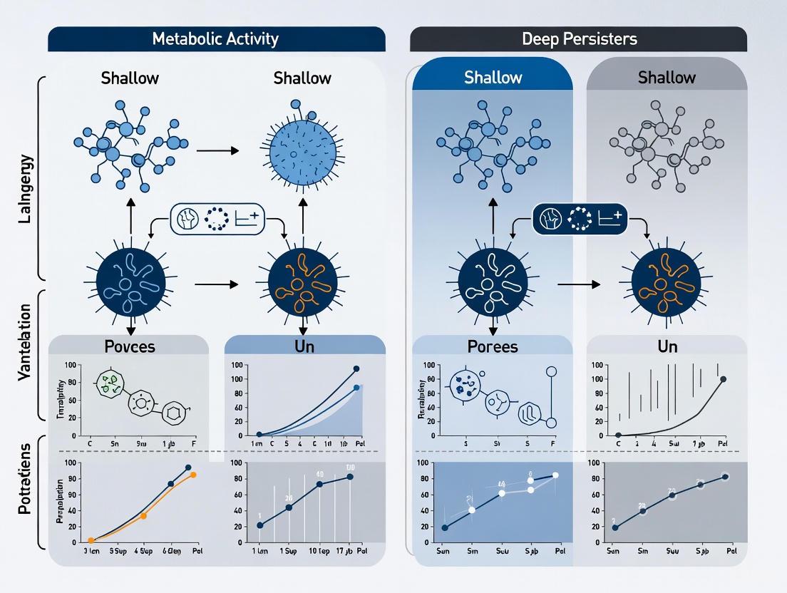
Bacterial persisters are a subpopulation of genetically drug-susceptible cells that enter a transient, non-growing, or slow-growing dormant state, enabling them to survive high doses of conventional antibiotic treatment [1] [3] [9]. Unlike antibiotic resistance, which is heritable, persistence is a non-heritable, phenotypic tolerance reversible upon antibiotic removal [3] [9]. A critical concept within persistence is the dormancy depth continuum, where persisters exhibit varying metabolic states ranging from "shallow" to "deep" [1] [10]. Shallow persisters occupy a unique position on this spectrum; they are characterized by low metabolic activity and growth arrest but retain a higher level of residual metabolic activity compared to their deep persister counterparts [1] [9]. This nuanced metabolic state allows them to resuscitate more quickly once the antibiotic stress is removed, posing a significant challenge in clinical settings by contributing to recurrent and relapsing infections [1] [10]. Understanding the characteristics of shallow persisters, particularly their metabolic profile, is therefore essential for developing novel therapeutic strategies to eradicate these resilient cells.
The shallow persister state is not merely a transient pause in activity but a carefully regulated physiological adaptation. Research indicates that after antibiotic removal, shallow persisters wake up and become susceptible to antibiotics much earlier than deep persisters [9]. This differential resuscitation timing is influenced by intracellular factors, such as the level of protein aggregates and ribosome content [9]. The lag time before re-growth correlates with the level of protein aggregates, and their removal by molecular chaperones like DnaK-ClpB is a prerequisite for resuscitation [9]. Furthermore, in heterogeneous populations, dormant bacteria with fewer ribosomes regrow much slower than cells with a greater ribosome content, highlighting the importance of metabolic machinery preservation in defining the shallow state [9]. This review will objectively compare the metabolic characteristics of shallow and deep persisters, synthesize key experimental findings, and provide a toolkit for researchers aiming to investigate this metabolically active dormant state.
Metabolic Profiling: Shallow vs. Deep Persisters
The fundamental distinction between shallow and deep persisters lies in their metabolic activity and subsequent resuscitation dynamics. While all persisters exhibit reduced metabolism compared to normal, actively growing cells, the degree of this reduction is paramount. Advanced techniques like stable isotope labeling have been instrumental in quantifying these differences.
Table 1: Comparative Characteristics of Shallow and Deep Persister Cells
| Characteristic | Shallow Persisters | Deep Persisters |
|---|---|---|
| Metabolic Activity Level | Low but significant residual activity [1] [9] | Severely reduced or near-complete metabolic shutdown [6] [11] |
| Resuscitation Time | Short lag time; resume growth quickly after stress removal [1] [9] | Prolonged lag time; resume growth very slowly or require specific resuscitation factors [1] [9] |
| Intracellular Features | Lower levels of protein aggregates; higher ribosome content [9] | Higher levels of protein aggregates; reduced ribosome content [9] |
| Carbon Source Utilization | Can metabolize certain carbon sources (e.g., glucose) more effectively than deep persisters [6] [11] | Substantially reduced utilization of various carbon sources, especially acetate [6] [11] |
| Therapeutic Susceptibility | Potentially vulnerable to metabolites, membrane-active agents, and resuscitated-killing strategies [12] [9] | Highly tolerant; may require strategies that force deeper dormancy into VBNC state or direct physical disruption [12] [10] |
A key study utilizing 13C-isotopolog profiling with E. coli persisters induced by carbonyl cyanide m-chlorophenyl hydrazone (CCCP) provided direct evidence of the metabolic state of persister cells [6] [11]. The results demonstrated that while persister cells exhibit reduced metabolism overall, their metabolic flexibility depends on the available carbon source. When glucose was the sole carbon source, persister cells showed generalized but reduced labeling in proteinogenic amino acids, indicating a uniform slowdown in protein synthesis but not a complete metabolic halt [6] [11]. In contrast, under acetate conditions, persister cells exhibited a more substantial metabolic shutdown, with markedly reduced labeling across nearly all pathway intermediates and amino acids [6] [11]. This suggests that shallow persisters are more likely to persist under conditions where more readily metabolizable carbon sources like glucose are available.
Table 2: Metabolic Flux Analysis in E. coli Persisters via 13C-Labeling [6] [11]
| Metabolic Pathway | Observations in Persister Cells (vs. Normal Cells) | Implication for Persister Type |
|---|---|---|
| Glycolysis | Reduced but detectable flux with 13C-glucose [6] [11] | Supports a shallow state with basal energy production. |
| Pentose Phosphate Pathway (PPP) | Delayed labeling dynamics [6] [11] | Reduced biosynthetic capacity for nucleotides. |
| Tricarboxylic Acid (TCA) Cycle | Delayed labeling dynamics; more substantial shutdown on acetate [6] [11] | Greatly reduced energy generation and biosynthetic precursors, favoring deep persistence. |
| Proteinogenic Amino Acid Synthesis | Reduced but uniform labeling with glucose; markedly reduced with acetate [6] [11] | Indicates ongoing but slow protein turnover in shallow states, nearly halted in deep states. |
Experimental Protocols for Metabolic Characterization
Stable Isotope Tracing and Metabolomics
This protocol is critical for directly measuring metabolic fluxes in persister cells, moving beyond indirect inferences from transcriptomics or proteomics [6] [11].
- Persister Induction: An overnight culture of E. coli BW25113 is sub-cultured in M9 minimal medium with 2 g/L glucose. When the subculture reaches mid-exponential phase (OD600 of 0.5), cells are exposed to 100 μg/mL of the protonophore CCCP for 15 minutes at 37°C with shaking to induce persister formation [6] [11].
- Cell Washing and Preparation: After induction, cells are collected via centrifugation (13,000 rpm, 3 min) and washed three times in M9 medium without a carbon source to remove CCCP and metabolic residues [6] [11].
- 13C-Labeling: Control and induced persister cells are concentrated to an OD600 of 5 in M9 medium and immediately exposed to 2 g/L of a 13C-labeled substrate, such as 1,2-13C2-glucose or 2-13C-sodium acetate. The culture is incubated at 37°C with shaking [6] [11].
- Precise Metabolic Quenching: At specific timepoints (e.g., 0, 20 s, 5 min, 30 min, 2 h), samples are rapidly cooled using liquid nitrogen within seconds to halt all metabolic activity [6] [11].
- Metabolite Extraction: The quenched cell pellets are lyophilized. Metabolites are then extracted using an 80:20 methanol-water solution at -20°C for 1 hour. After centrifugation and filtration, the supernatant is analyzed by Liquid Chromatography-Mass Spectrometry (LC-MS) for free metabolites [6] [11].
- Proteinogenic Amino Acid Analysis: The remaining cell pellets are hydrolyzed with 6N HCl at 100°C for 18 hours to break down proteins into amino acids. These hydrolyzed amino acids are analyzed as tert-butyldimethylsilyl derivatives using Gas Chromatography-Mass Spectrometry (GC-MS) [6] [11].
- Data Interpretation: The incorporation patterns of the 13C label into metabolic intermediates (e.g., TCA cycle acids) and proteinogenic amino acids are tracked. Shallow persisters will show faster and greater incorporation of the label compared to deep persisters, indicating higher metabolic flux [6] [11].
Single-Cell Growth Rate Reporter Assays
This method allows for the identification and tracking of persisters based on their growth status at the single-cell level, which is crucial for distinguishing heterogeneity within a persister population [13].
- Strain Engineering: A fluorescent reporter strain is constructed. One common method is the fluorescence dilution technique, where a bacterial strain is engineered to constitutively express a stable fluorescent protein. Upon cell division, the fluorescence is diluted in daughter cells, allowing growth rates to be tracked [13].
- Time-Lapse Microscopy: The reporter strain is cultured and exposed to a persister-inducing condition or antibiotic. Cells are monitored using time-lapse microscopy, often within microfluidic devices, to track individual cells over time [13].
- Persister Identification: After a period of antibiotic exposure, surviving cells are identified. By reviewing the pre-antibiotic growth history of these survivors, one can determine if they were non-growing (potential deep persisters) or slow-growing (potential shallow persisters) prior to the stress [13].
- Resuscitation Monitoring: After antibiotic removal, the lag time until the first cell division for each persister is recorded. Shallow persisters will have a significantly shorter lag time than deep persisters, providing a functional measure of dormancy depth [1] [9].
Key Signaling Pathways and Molecular Mechanisms
The formation and maintenance of the shallow persister state are regulated by complex molecular networks. The following diagram synthesizes key pathways influencing metabolic shutdown and dormancy depth.
The stringent response and toxin-antitoxin (TA) modules are central regulators. In response to stress like nutrient limitation, the alarmone ppGpp accumulates, triggering a global reprogramming of cellular metabolism [1] [14]. ppGpp downregulates genes for ribosome and tRNA synthesis, effectively halting growth [14]. This alarmone also activates TA systems [14]. When activated, toxins from these systems corrupt essential cellular processes. For example, the HipA toxin phosphorylates glutamyl-tRNA synthetase (GltX), inhibiting translation and mimicking amino acid starvation, which further amplifies the stringent response [14]. Other toxins, like HokB, form pores in the membrane, causing depolarization and ATP leakage, directly reducing the energy available to the cell [9]. The level of this energy reduction is a key determinant of dormancy depth. Fluctuations in Krebs cycle enzymes can lead to stochastic ATP deficiency and persister formation [9]. Furthermore, the depletion of intracellular ATP promotes the accumulation of protein aggregates ("aggresomes") [9] [10]. The amount of these aggregates correlates with the "depth" of dormancy; shallow persisters have fewer aggregates than deep persisters. The removal of these aggregates by chaperone systems like DnaK-ClpB is a necessary step for resuscitation, explaining the faster wake-up time of shallow persisters [9].
The Scientist's Toolkit: Essential Research Reagents and Materials
Table 3: Key Reagent Solutions for Persister Metabolism Research
| Research Reagent / Material | Function and Application in Persister Studies |
|---|---|
| 13C-labeled Substrates (e.g., 1,2-13C2-glucose, 2-13C-acetate) | Serve as tracer molecules for stable isotope labeling experiments. They allow for the precise tracking of metabolic flux through central carbon pathways (glycolysis, PPP, TCA cycle) in persister cells via LC-MS or GC-MS analysis [6] [11]. |
| Chemical Persister Inducers (e.g., CCCP, Rifampicin) | CCCP is a protonophore that disrupts the membrane potential and ATP synthesis, inducing a persister-like state. Rifampicin, an RNA synthesis inhibitor, can convert entire populations into persisters, facilitating the generation of sufficient biomass for omics studies [6] [11] [3]. |
| Fluorescent Protein Reporter Systems (e.g., fluorescence dilution constructs, TIMERbac) | Enable single-cell tracking of bacterial growth and resuscitation. By monitoring fluorescence dilution over time, researchers can retrospectively link the pre-antibiotic growth rate of a cell to its ability to survive treatment, distinguishing shallow from deep persisters [13]. |
| LC-MS (Liquid Chromatography-Mass Spectrometry) | An analytical chemistry technique used to identify and quantify metabolites. In persister research, it is crucial for measuring the incorporation of 13C from labeled substrates into metabolic intermediates, providing a direct readout of pathway activity [6] [11]. |
| GC-MS (Gas Chromatography-Mass Spectrometry) | Used particularly for the robust analysis of 13C-labeling in proteinogenic amino acids. This provides a time-integrated view of metabolic activity, as amino acids reflect the metabolic history of the cell over the protein's lifetime [6] [11]. |
| Microfluidic Devices | Provide a platform for high-resolution, single-cell analysis under controlled fluidic conditions. They are ideal for long-term time-lapse microscopy experiments to monitor persister formation, survival, and resuscitation at the single-cell level [13]. |
| Membrane-Active Compounds (e.g., XF-73, SA-558, synthetic retinoids) | A class of anti-persister agents that directly target and disrupt the bacterial cell membrane, a target that remains accessible in dormant cells. Used in combination studies to sensitize persisters to conventional antibiotics [12]. |
Bacterial persisters represent a fascinating and clinically challenging subpopulation of cells that exhibit a non-heritable, transient tolerance to antibiotic treatment. These cells are genetically identical to their antibiotic-susceptible counterparts but possess the remarkable ability to survive lethal doses of antimicrobials by entering a state of slowed or arrested growth [1] [15]. Within this persister population exists a spectrum of metabolic states, categorized along a continuum from "shallow" to "deep" persistence [1] [10]. This phenotypic heterogeneity has profound implications for treating chronic and recurrent infections, as deeper persisters demonstrate enhanced survival capabilities under sustained antibiotic pressure. Understanding the mechanisms that govern entry into profound metabolic quiescence is therefore paramount for developing novel therapeutic strategies against persistent infections.
The clinical significance of deep persisters cannot be overstated. They are increasingly recognized as the primary culprits behind treatment failures in chronic infections such as tuberculosis, recurrent urinary tract infections, Lyme disease, and biofilm-associated infections on medical devices [1] [12]. Unlike antibiotic resistance, which involves genetic mutations that can be vertically transmitted, persistence represents a phenotypic switch that allows a small fraction of bacterial populations to weather antimicrobial storms. When antibiotic pressure subsides, these dormant cells can resuscitate and repopulate the environment, leading to relapsing infections [3] [15]. The depth of a persister's metabolic quiescence directly correlates with its ability to survive various stressors, making the understanding of this continuum a critical frontier in medical microbiology.
Molecular Mechanisms of Deep Persistence Entry
Key Regulatory Pathways and Metabolic Shifts
The transition into a state of deep metabolic quiescence is orchestrated by a complex interplay of molecular mechanisms that collectively reprogram cellular physiology. One of the primary drivers of this transition is the (p)ppGpp-mediated stringent response, which is triggered by nutrient limitation and other environmental stresses [16]. This alarmone system dramatically alters gene expression profiles, downregulating energy-intensive processes like DNA replication, protein synthesis, and cell division while upregulating stress response pathways. Concurrently, toxin-antitoxin (TA) modules play a crucial role in persister formation by selectively inhibiting essential cellular processes [1] [16]. For instance, toxin proteins such as HipA phosphorylate and inhibit glutamyl-tRNA synthetase, thereby halting protein synthesis and inducing a dormant state [16].
The metabolic state of persister cells is characterized by a significant reduction in ATP generation and overall metabolic activity. Studies have demonstrated that decreased ATP production leads to lower activity of antibiotic targets, resulting in enhanced drug tolerance [3]. This metabolic downregulation is further reflected in the reprogramming of central carbon metabolism, with a shift toward pathways that maintain energy homeostasis and redox balance at minimal cost. Additionally, reactive oxygen species (ROS) management becomes critical during persistence, as cells must mitigate the damaging effects of oxidative stress while maintaining essential repair mechanisms [16] [12]. The collective action of these pathways establishes a protected, quiescent state that is remarkably resilient to antimicrobial assault.
The Metabolic Features of Shallow versus Deep Persisters
The distinction between shallow and deep persisters lies primarily in their degree of metabolic shutdown and corresponding survival capabilities. The table below summarizes the key differentiating features:
Table 1: Comparative Features of Shallow and Deep Persisters
| Feature | Shallow Persisters | Deep Persisters |
|---|---|---|
| Metabolic Activity | Slow but detectable metabolism | Profound metabolic quiescence |
| ATP Levels | Reduced but measurable | Extremely low or undetectable |
| Resuscitation Time | Short lag time after stress removal | Prolonged lag time, delayed regrowth |
| Antibiotic Survival | Moderate tolerance to single antibiotics | High tolerance to multiple antibiotic classes |
| Culturability | Culturable on standard media | May enter viable but non-culturable (VBNC) state |
| Detection Methods | Standard colony counting | Requires specialized enrichment or staining |
Deep persisters exhibit a more pronounced downregulation of biosynthetic processes and energy metabolism compared to their shallow counterparts [1]. Proteomic analyses of Staphylococcus aureus persisters have revealed distinct protein expression profiles between those induced by different antibiotics, suggesting that the route to deep persistence may vary depending on the environmental trigger [17]. For instance, vancomycin-induced persisters in S. aureus showed upregulation of 53 proteins and repression of 27 proteins, while enrofloxacin-induced persisters displayed 16 induced proteins and 51 decreased proteins [17]. This differential regulation highlights the plasticity of the persistence phenomenon and suggests multiple molecular routes can lead to similarly profound quiescent states.
Table 2: Molecular Regulators of Persister Formation and Their Mechanisms
| Regulatory System | Primary Trigger | Key Effectors | Impact on Persistence |
|---|---|---|---|
| Stringent Response | Nutrient limitation, starvation | (p)ppGpp, RelA, SpoT | Global metabolic slowdown, stress response activation |
| Toxin-Antitoxin Modules | Environmental stress, SOS response | HipA, MazF, RelE | Targeted inhibition of translation/DNA replication |
| ROS Defense Systems | Oxidative stress, antibiotic exposure | Superoxide dismutase, AhpC | Protection against oxidative damage, maintenance of redox balance |
| SOS Response | DNA damage | RecA, LexA | Cell cycle arrest, DNA repair activation |
| Quorum Sensing | Population density | Autoinducer molecules | Coordination of population-level persistence |
Experimental Approaches for Studying Deep Persisters
Isolation and Characterization Methodologies
Investigating deep persisters requires specialized methodologies capable of capturing and analyzing these rare, metabolically dormant cells. A critical advancement in this field has been the development of fluorescence-activated cell sorting (FACS) techniques that utilize metabolic dyes such as 5-(and-6)-Carboxyfluorescein diacetate (CFDA) combined with membrane integrity markers like propidium iodide (PI) [17]. This approach enables researchers to distinguish and isolate viable but non-growing persister cells from a mixed population based on their differential staining patterns. For example, CFDA+/PI- cells typically represent persisters with intact membranes but reduced metabolic activity, allowing for their purification and subsequent molecular analysis [17].
Once isolated, deep persisters can be characterized using a suite of omics technologies. Proteomic profiling via mass spectrometry has revealed distinct protein expression signatures in persisters induced by different antibiotic classes [17]. Similarly, metabolomic analyses provide insights into the metabolic pathways that are differentially regulated in deep persisters compared to normal cells and shallow persisters. Advanced techniques such as single-cell Raman spectroscopy (SCRS) and imaging flow cytometry (IFC) offer unprecedented resolution for examining the heterogeneity within persister populations at the individual cell level [15]. These methodologies have collectively revealed that deep persisters are not a uniform entity but rather exist along a continuum of metabolic states, with the deepest persisters potentially transitioning into a viable but non-culturable (VBNC) state that evades standard detection methods [1].
The Scientist's Toolkit: Essential Research Reagents
Table 3: Key Research Reagents for Persister Studies
| Reagent/Category | Specific Examples | Primary Function | Application Notes |
|---|---|---|---|
| Metabolic Activity Dyes | CFDA, CTC, Resazurin | Assess metabolic activity and viability | CFDA measures esterase activity; often used with membrane integrity markers |
| Membrane Integrity Stains | Propidium iodide, SYTOX Green | Differentiate live/dead cells based on membrane integrity | Impermeant dyes that only enter cells with compromised membranes |
| Antibiotics for Persistence Induction | Vancomycin, Enrofloxacin, Ciprofloxacin | Induce persister formation in bacterial populations | Different antibiotics generate persisters via distinct mechanisms |
| Proteomics Reagents | Trypsin, LC-MS/MS reagents, Protein extraction buffers | Characterize protein expression profiles in isolated persisters | Requires specialized protocols for low-abundance persister cells |
| RNA Stabilization & Extraction Kits | RNAprotect, RNA extraction kits with DNase treatment | Preserve and extract RNA for transcriptomic studies | Critical for capturing transient gene expression states in persisters |
The experimental workflow for deep persister research typically begins with the induction of persistence through exposure to bactericidal antibiotics at concentrations that kill the majority of the population. The surviving cells are then stained with appropriate fluorescent markers and sorted using FACS. Following isolation, molecular analyses such as proteomics, transcriptomics, and metabolomics can be performed to characterize the physiological state of the deep persisters. Additional functional assays may include measurements of ATP levels, membrane potential, redox status, and resuscitation kinetics to further delineate the depth of metabolic quiescence.
Therapeutic Strategies Targeting Deep Persisters
Current and Emerging Anti-Persister Approaches
Conventional antibiotics predominantly target active cellular processes such as cell wall synthesis, protein translation, and DNA replication, making them largely ineffective against deep persisters with arrested growth and minimal metabolic activity [12]. This therapeutic limitation has spurred the development of novel strategies specifically designed to eradicate persistent populations. One promising approach involves metabolic reprogramming, where exogenous metabolites are used to reactivate dormant bacteria, thereby sensitizing them to conventional antibiotics [16]. For instance, specific metabolites like mannitol, fructose, or certain amino acids can stimulate metabolic pathways and restore proton motive force, enabling improved uptake and efficacy of aminoglycoside antibiotics [16].
Another strategic avenue focuses on direct killing of persisters through compounds that target growth-independent cellular structures. Membrane-active agents such as XF-73, SA-558, and cationic silver nanoparticles physically disrupt bacterial membranes, leading to cell lysis regardless of metabolic state [12]. Similarly, the anti-tuberculosis drug pyrazinamide (PZA) exhibits unique activity against persistent Mycobacterium tuberculosis by disrupting membrane energetics and targeting the PanD enzyme, which is essential for coenzyme A biosynthesis [12]. Other innovative approaches include the use of ADEP4, which activates the ClpP protease and causes uncontrolled protein degradation in dormant cells, effectively eliminating persisters through self-digestion [12].
Combination Therapies and Clinical Translation
The complex nature of deep persistence suggests that combination therapies will likely be necessary for effective eradication. Strategic pairing of antibiotics with adjuvants that target persistence mechanisms has shown promise in experimental models. For example, inhibitors of H2S biogenesis can reduce persister formation and potentiate antibiotic efficacy against Staphylococcus aureus and Pseudomonas aeruginosa [12]. Similarly, membrane-permeabilizing agents like polymyxin B nonapeptide (PMBN) and synthetic retinoids can enhance the uptake of antibiotics that would otherwise be excluded from persister cells [12].
The clinical translation of anti-persister therapies faces several challenges, including the need to achieve effective local concentrations at infection sites, potential toxicity concerns with membrane-active compounds, and the optimization of treatment durations to prevent regrowth from the deepest persister reservoirs [16]. Nevertheless, the growing recognition of persistence as a major contributor to chronic and recurrent infections has accelerated research in this area, with several candidate compounds advancing through preclinical development. Future therapeutic regimens will likely involve sequential or simultaneous targeting of multiple persistence mechanisms to achieve complete sterilization of infections.
The study of deep persisters and their entry into profound metabolic quiescence represents a critical frontier in our understanding of bacterial survival strategies and treatment-recalcitrant infections. The emerging paradigm recognizes persistence not as a binary state but as a continuum of metabolic dormancy, with deep persisters occupying the most extreme end of this spectrum. Their remarkable ability to withstand multiple stressors, including high concentrations of diverse antibiotics, stems from a coordinated downregulation of metabolic activity and activation of specific protective mechanisms.
Future research directions will need to address several key challenges, including the development of more sophisticated methods for isolating and characterizing the deepest persister subsets, elucidating the resuscitation signals that trigger their reawakening, and identifying novel drug targets that are essential for maintaining the persistent state. The clinical translation of anti-persister therapies will require careful consideration of timing, dosing, and combination strategies to prevent both regrowth and resistance development. As our understanding of the molecular basis of deep persistence continues to evolve, so too will our ability to design targeted interventions that specifically address this fundamental cause of chronic and recurrent infections. The ongoing characterization of the deep persister phenotype and its distinguishing features from shallow persistence provides a roadmap for developing the next generation of antimicrobial therapies capable of addressing this enduring clinical challenge.
Within bacterial populations, a small subgroup of cells known as persisters demonstrates remarkable survival capabilities against antibiotic treatments. Unlike resistance, which is genetically inherited, this survival is a transient, phenotypic state of dormancy. The metabolic state of these persisters exists on a continuum, classified as either "shallow" or "deep" based on their dormancy depth and resuscitation potential. Shallow persisters maintain a higher level of metabolic activity and can quickly resume growth once the antibiotic pressure is removed. In contrast, deep persisters enter a state of profound metabolic shutdown, resulting in significantly longer lag times before resuscitation. This guide provides a comparative analysis of the key metabolic hallmarks—energy charge, substrate utilization, and biosynthetic activity—that differentiate shallow and deep persister cells, providing a structured overview of experimental data and methodologies for researchers in microbiology and drug development.
Comparative Analysis of Metabolic Hallmarks
The table below synthesizes key experimental findings that characterize the metabolic states of shallow and deep persisters.
Table 1: Key Metabolic Hallmarks of Shallow vs. Deep Persisters
| Metabolic Hallmark | Shallow Persisters | Deep Persisters | Supporting Experimental Evidence |
|---|---|---|---|
| Energy Charge (ATP levels) | Moderate ATP depletion [18] | Severe ATP depletion [18] | ATP depletion in S. aureus linked to deeper dormancy; CCCP-induced ATP depletion increases persister formation [18] [8] |
| Substrate Utilization | Can utilize substrates for energy maintenance without growth [19] | Limited to no substrate utilization; reliance on endogenous reserves [18] | C. jejuni uses amino/organic acids for survival via substrate-level/oxidative phosphorylation [19]; Deep S. aureus persisters show minimal metabolism [18] |
| Biosynthetic Activity (e.g., Translation) | Moderately reduced translation [18] | Severely reduced or arrested translation [18] | Host ROS-induced translational repression in S. aureus limits dormancy; protein synthesis inhibition prevents deep dormancy [18] |
| Resuscitation Lag Time | Short lag time upon stress relief [18] | Heterogeneous and increased lag times [18] | Single-cell analysis of S. aureus shows lag time correlated with host cell oxidative stress level [18] |
| Molecular Hallmarks | Transient protein aggregation [18] | Recruitment of DnaK-ClpB system for aggregate clearance; visible dark foci [18] | In high-ROS environments, S. aureus persisters show dark foci and chaperone recruitment, indicating protein damage [18] |
Essential Experimental Protocols for Metabolic Profiling
To investigate the metabolic hallmarks outlined above, specific and robust experimental protocols are required. The following section details key methodologies for isolating persisters and measuring their metabolic states.
Protocol for Persister Isolation and Characterization
This foundational protocol is used to obtain the persister cell population for subsequent metabolic analysis.
- Objective: To isolate a pure population of persister cells after antibiotic exposure.
- Principle: Persisters survive a lethal dose of antibiotics that kills regular cells. After antibiotic removal, these viable but non-growing cells can be characterized.
- Materials: Bacterial culture (e.g., Staphylococcus aureus), appropriate antibiotic (e.g., oxacillin at 50x MIC), culture medium, phosphate-buffered saline (PBS), flow cytometer with cell sorter (optional).
- Procedure:
- Culture and Infection: Grow a bacterial culture to the desired growth phase (e.g., stationary phase for higher persister frequency). Infect eukaryotic host cells (e.g., J774 or human macrophages) if studying intracellular persisters [18].
- Antibiotic Challenge: Expose the bacterial culture or infected host cells to a high concentration of antibiotic (e.g., 50x MIC of oxacillin) for a defined period (e.g., 24-48 hours) to eliminate growing cells [18].
- Persister Isolation:
- Enumeration and Purity Check:
- Determine the persister count by serially diluting and plating the washed cell suspension on antibiotic-free agar for Colony Forming Unit (CFU) counts. Only persisters will form colonies [18] [8].
- (Optional) Use fluorescence-activated cell sorting (FACS) with a viability stain (e.g., propidium iodide) and a fluorescent reporter (e.g., GFP) to isolate a population of viable, non-growing cells [18].
Protocol for Measuring Metabolic Activity via Phenotype Microarrays
This protocol assesses the metabolic capacity of persisters to utilize different substrates for energy generation.
- Objective: To profile the substrate utilization of non-growing bacterial cells for survival.
- Principle: Phenotype microarray plates (e.g., Biolog PM) contain different carbon sources. Metabolically active cells reduce a tetrazolium dye, producing a colorimetric change that can be quantified [19].
- Materials: Isolated persister cells, Biolog PM plates (e.g., PM1 for carbon sources), IF-0 inoculation fluid, tetrazolium dye, plate reader.
- Procedure:
- Cell Preparation: Harvest and wash persister cells thoroughly to remove any residual nutrients. Resuspend the cell pellet in inoculation fluid to a standardized optical density (e.g., OD₆₀₀ₙₘ of 0.4) [19].
- Dye Addition and Inoculation: Mix the cell suspension with the tetrazolium dye. Pipette 100 µL of the mixture into each well of the phenotype microarray plate [19].
- Incubation and Measurement:
- Measure the initial absorbance (e.g., at 605 nm) to establish a baseline.
- Incubate the plates under appropriate conditions (e.g., microaerophilic for Campylobacter jejuni at 42°C for 24 hours) [19].
- Measure the absorbance again after incubation. An increase in absorbance indicates substrate-dependent metabolic activity (respiration) without growth [19].
Visualization of Metabolic Pathways and Workflows
Signaling Pathways Influencing Persister Dormancy
This diagram illustrates the key signaling pathways that regulate the metabolic shift into the persister state, integrating inputs like nutrient starvation and oxidative stress.
Experimental Workflow for Persister Metabolic Analysis
This workflow outlines the key steps from cell preparation to data analysis when conducting a full metabolic profile of persister cells.
The Scientist's Toolkit: Key Research Reagents
The following table lists essential reagents and their applications for studying persister cell metabolism.
Table 2: Essential Research Reagents for Persister Metabolism Studies
| Reagent/Material | Function in Research | Specific Application Example |
|---|---|---|
| Carbonyl Cyanide m-Chlorophenyl Hydrazone (CCCP) | Induces ATP depletion by disrupting the proton motive force [18] [8]. | Used to artificially induce a deep dormancy state and study its effects on protein aggregation and resuscitation [18]. |
| Tetrazolium Dye (e.g., in Biolog Assays) | Acts as a redox indicator; color change signifies metabolic (respiratory) activity [19]. | Measuring substrate utilization in non-growing persister cells in phenotype microarrays [19]. |
| ¹³C-labeled Substrates (e.g., Glucose, Amino Acids) | Tracers for isotopolog profiling; allow tracking of metabolic flux through different pathways [8]. | Determining which metabolic pathways (e.g., glycolysis, TCA cycle) remain active in persisters after antibiotic challenge [8]. |
| Butylated Hydroxyanisole (BHA) | An antioxidant that reduces intracellular levels of reactive oxygen species (ROS) [18]. | Modulating the host cell's oxidative stress to investigate its direct effect on persister dormancy depth in infection models [18]. |
| Viability Stains (e.g., Propidium Iodide) | Fluorescent dyes that distinguish live from dead cells based on membrane integrity. | Used in flow cytometry to enumerate and sort the viable, non-culturable subpopulation after antibiotic treatment [18]. |
Bacterial persistence, a state of transient antibiotic tolerance, represents a significant challenge in treating chronic infections. This phenotype is primarily governed by a sophisticated interplay of molecular systems, chief among them the alarmone (p)ppGpp, toxin-antitoxin (TA) modules, and the alternative sigma factor σS (RpoS). These components form an integrated regulatory network that responds to metabolic stress by reprogramming cellular physiology towards dormancy. Within persister populations, a continuum of metabolic activity exists, from shallow persisters that maintain minimal metabolism to deep persisters in a near-dormant state. This review systematically compares the experimental evidence for the roles and interactions of these key regulatory systems, providing a structured analysis of their contributions to bacterial persistence and implications for therapeutic development.
Persisters are a subpopulation of genetically drug-susceptible but phenotypically tolerant bacterial cells that can survive antibiotic exposure and other stresses by entering a state of non-growing or slow-growing dormancy [20]. These cells underlie chronic and recurrent infections, including tuberculosis, cystic fibrosis, and Lyme disease, and contribute to treatment failures [21] [22]. The persistence phenotype is fundamentally different from antibiotic resistance, as persisters do not possess genetic resistance mechanisms but rather survive through physiological adaptations that reduce antibiotic target activity [20]. Research over the past decade has revealed that persistence is not a singular state but exists along a continuum of metabolic activity, often categorized as "shallow" versus "deep" persisters based on their depth of dormancy and capacity for resuscitation [20] [10].
At the molecular level, persistence is orchestrated by an integrated network of stress response systems. The second messenger (p)ppGpp serves as a central stress alarmone, toxin-antitoxin (TA) systems act as molecular switches that modulate cellular activity, and the σS stress response sigma factor directs transcriptional reprogramming [23] [21] [24]. These systems do not operate in isolation but function as a coordinated network that senses metabolic perturbation and executes an appropriate survival response. Understanding the hierarchy and interplay between these systems is crucial for elucidating the mechanisms underlying the spectrum of persister metabolic states and developing effective anti-persister therapies.
Comparative Analysis of Core Regulatory Systems
The (p)ppGpp Stringent Response Alarmone
(p)ppGpp (guanosine pentaphosphate/tetraphosphate) is a pivotal secondary messenger that orchestrates the bacterial stringent response to nutrient limitation and other stresses [21]. This alarmone serves as a master regulator that reprograms cellular metabolism from growth-oriented processes to survival mechanisms, making it fundamental to persister formation.
Table 1: Experimental Evidence for (p)ppGpp in Persister Formation
| Experimental Approach | Key Findings | Implications for Persistence | Reference |
|---|---|---|---|
| relA/spoT gene deletion | (p)ppGpp-deficient E. coli showed reduced persister formation but not complete elimination | (p)ppGpp is a major but not exclusive regulator of persistence | [21] [24] |
| valS ts mutation (impaired tRNA charging) | 16-fold increase in ppGpp; 3-4 orders of magnitude higher persister levels | Amino acid starvation → ppGpp → persistence | [25] |
| Single-cell analysis | Stochastic persister formation even with high (p)ppGpp; no direct correlation in individual cells | (p)ppGpp necessary but not sufficient; indicates bet-hedging strategy | [25] |
| Macrophage infection model | Salmonella required (p)ppGpp for persistence in acidified vacuoles | Connects (p)ppGpp to host-pathogen interactions and in vivo persistence | [21] |
The molecular mechanisms through which (p)ppGpp influences persistence include direct binding to RNA polymerase, which alters transcriptional priorities, and inhibition of DNA primase, thereby reducing DNA replication [21]. In Escherichia coli, (p)ppGpp accumulation leads to differential expression of approximately 500 genes, activating stress response pathways while repressing genes involved in rapid growth [21]. This comprehensive transcriptional rewiring results in slowed growth or dormancy, which protects cells from antibiotics that target active cellular processes. Interestingly, research has shown that while high levels of (p)ppGpp are critical for persister formation, the relationship is not deterministic at the single-cell level, suggesting that additional factors modulate the eventual phenotypic outcome [25].
Toxin-Antitoxin (TA) Systems as Molecular Switches
TA systems are genetic modules composed of a stable toxin protein that disrupts essential cellular processes and an unstable antitoxin that neutralizes the toxin [23] [22]. Under stress conditions, proteolytic degradation of antitoxins unleashes toxin activity, leading to growth arrest that facilitates persistence.
Table 2: Major TA System Types and Their Mechanisms in Persistence
| TA System Type | Antitoxin Nature | Mechanism of Action | Role in Persistence | Examples |
|---|---|---|---|---|
| Type I | RNA | Antisense RNA inhibits toxin translation | Growth arrest via membrane pore formation | Hok/Sok, TisB/IstR |
| Type II | Protein | Protein antitoxin binds and inhibits toxin | mRNA interference → translation inhibition | MazEF, RelBE, HipBA |
| Other Types (III-VIII) | Protein/RNA | Various inhibition mechanisms | Emerging roles in persistence | - |
Type II TA systems are the most extensively studied in the context of persistence. These systems are abundant in free-living bacteria and are characterized by protein antitoxins that form stable complexes with their cognate protein toxins, typically neutralizing them by blocking the active site [23]. Under optimal growth conditions, antitoxins are produced in excess, keeping toxins in check. However, during stress, when translation slows and cellular proteases degrade antitoxins, free toxins are released to exert their effects [23]. The MazEF system is one of the most widespread and well-characterized type II TA systems, with MazF exhibiting sequence-specific RNA cleavage (endonuclease) activity that diminishes protein synthesis and cellular metabolism [23].
The positioning of TA modules within bacterial genomes provides clues to their functional integration with broader stress response networks. Notably, in Gram-negative bacteria, mazEF is co-transcribed with relA, which encodes a key (p)ppGpp synthetase, while in Gram-positive bacteria, mazEF is located directly upstream of the sigB operon encoding the general stress response alternative sigma factor σB [23]. This genomic arrangement suggests evolutionary selection for coordinated regulation of these systems in response to nutrient limitation and other environmental stresses.
σS (RpoS) Stress Response Sigma Factor
The alternative sigma factor σS (RpoS) serves as the master regulator of the general stress response in many bacteria, controlling the expression of hundreds of genes involved in adaptation to adverse conditions [23] [24]. During nutrient limitation, σS levels increase due to enhanced translation and decreased degradation, leading to comprehensive transcriptional reprogramming [23].
Proteomic analyses of persister cells have revealed that the persister proteome is characterized by σS-mediated stress response and a metabolic shift toward catabolism [24]. This proteomic signature represents a state that starved cells attempt to reach but cannot fully achieve due to the absence of carbon and energy sources. In nutrient-shift-induced persisters, metabolism is geared toward energy production, with depleted metabolite pools, yet maintains minimal metabolic activity sufficient for ATP maintenance requirements [24]. This physiological state differs fundamentally from starvation-induced persistence, where the complete absence of nutrients leads to more profound metabolic shutdown.
Research has demonstrated that σS plays an active role in persistence as a response to metabolic flux limitation [24] [26]. When metabolic homeostasis is strongly perturbed, metabolic fluxes collapse, creating a vicious cycle that prevents a return to growth homeostasis. This state is stabilized and modulated by high ppGpp levels, TA systems, and the σS-mediated stress response [24]. The integration of these systems creates a robust network that enables bacteria to survive transient metabolic crises.
Experimental Approaches and Methodologies
Key Experimental Models for Persistence Research
Several well-established experimental models have been developed to study persister formation and resuscitation under controlled conditions:
Nutrient Shift Model: This approach involves switching bacterial cultures from a preferred carbon source (e.g., glucose) to a less favorable one (e.g., fumarate) or no carbon source [24]. Following such shifts, only a small fraction of cells (approximately 0.1%) adapt and grow, while the majority enter a non- or slow-growing persistent state despite the presence of utilizable nutrients [24]. This model generates large numbers of persisters in nutrient-rich conditions, enabling population-averaging experimental methods that require substantial biomass.
Stringent Response Induction Model: Utilizing bacterial strains with temperature-sensitive valyl-tRNA synthetase alleles (valSts) allows controlled induction of (p)ppGpp synthesis through modulation of tRNA charging [25]. At elevated temperatures, the limited activity of L-valyl tRNA synthetase leads to increased intracellular (p)ppGpp levels and enhanced persister formation without the need for ectopic gene expression [25]. This system provides a physiological means to study (p)ppGpp-dependent persister formation.
Stochastic Persister Model: In growing cultures without external stress, persisters arise spontaneously at low frequency (typically 10⁻⁶ to 10⁻⁵) [20] [25]. These cells can be isolated using antibiotic treatment followed by removal of the drug, allowing study of naturally occurring persisters. However, their low abundance makes biochemical analyses challenging.
Advanced Methodologies for Single-Cell Analysis
Recent technical advances have enabled unprecedented resolution in persister research through single-cell approaches:
Live Microscopy with Fluorescent Reporters: This methodology allows direct observation of the stochastic appearance, antibiotic tolerance, and resuscitation of persister cells [25]. By using fluorescent reporters for key persistence markers, researchers can correlate physiological parameters with persister status in real-time. Essential components of this approach include:
- (p)ppGpp reporters: Typically RpoS-mCherry fusions that indicate increased (p)ppGpp levels through enhanced fluorescence [25]
- TA activation reporters: Short-lived fluorescent proteins (e.g., YFPunstable) expressed from TA promoter regions [25]
- ATP sensors: QUEEN-7µ reporter that indicates intracellular ATP concentrations through conformational changes [25]
Proteomic and Metabolomic Analyses: High-throughput proteomic methods have been used to comprehensively map the molecular phenotype of E. coli during entry into and in the state of persistence [24]. These analyses have revealed that the persister proteome is characterized by σS-mediated stress response and a shift to catabolism [24]. Similarly, metabolomic approaches have shown that persister metabolism is geared toward energy production, with depleted metabolite pools [24].
Flow Cytometry and FACS: Fluorescence-activated cell sorting enables isolation and analysis of persister subpopulations based on specific markers, such as reduced membrane potential or reporter gene expression [24]. This approach facilitates the characterization of heterogeneity within persister populations and the identification of distinct metabolic states along the shallow-to-deep persistence continuum.
Integrated Regulatory Network
The regulatory systems controlling persistence do not function in isolation but form an integrated network that responds to metabolic cues and executes coordinated survival programs. The current evidence supports a model in which metabolic perturbations, particularly limitations in metabolic flux, initiate a system-level response involving all three key components [24].
Figure 1: Integrated Regulatory Network Governing Bacterial Persistence. This diagram illustrates the coordinated response to metabolic stress involving (p)ppGpp, TA systems, and σS, leading to heterogeneous persister populations with varying metabolic states.
The network operates through a system-level feedback mechanism wherein strong perturbations of metabolic homeostasis cause metabolic fluxes to collapse, preventing adjustments that would restore homeostasis [24]. This vicious cycle is stabilized by high ppGpp levels, TA systems, and the σS-mediated stress response. The interplay between these components creates a robust system that can generate heterogeneous outcomes, accounting for the spectrum of persister states observed experimentally.
Notably, the relationship between these systems exhibits both redundancy and hierarchy. While ppGpp is a central regulator, persisters can still form in ppGpp-negative strains, indicating alternative pathways to persistence [24]. Similarly, deletion of multiple TA modules reduces but does not eliminate persister formation [24]. This redundancy ensures that bacteria can activate persistence programs through multiple triggers, enhancing survival in fluctuating environments.
The Scientist's Toolkit: Essential Research Reagents and Methodologies
Table 3: Key Research Reagents and Experimental Tools for Persistence Studies
| Reagent/Tool | Function/Application | Example Use | Experimental Considerations |
|---|---|---|---|
| valSts mutant strains | Temperature-sensitive valyl-tRNA synthetase enables controlled (p)ppGpp induction | Study of (p)ppGpp-dependent persister formation without ectopic gene expression [25] | Requires precise temperature control; effects are pleiotropic |
| RpoS-mCherry fusions | Fluorescent reporter for (p)ppGpp levels and σS activity | Single-cell monitoring of stringent response activation [25] | Reporter response has time lag relative to actual ppGpp changes |
| TA promoter-YFP unstable | Reporter for TA system activation via short-lived fluorescent protein | Monitoring RelBE and other TA systems in live cells [25] | Distinguishes transient versus sustained TA activation |
| QUEEN-7µ ATP sensor | FRET-based ATP concentration measurement in single cells | Correlation of ATP levels with persistence status [25] | Calibration required for absolute concentration measurements |
| Nutrient shift protocols | Generation of synchronized persister populations | Proteomic and metabolomic analyses of persisters [24] | Enables population-level studies but may not capture natural heterogeneity |
| Microfluidics systems | Single-cell culture under controlled conditions | Long-term tracking of persister formation and resuscitation [25] | Technically challenging; requires specialized equipment |
This toolkit enables researchers to dissect the complex regulatory networks controlling persistence through complementary approaches. The combination of population-level biochemical analyses with single-cell live imaging provides both comprehensive molecular profiling and dynamic behavioral assessment. Genetic tools such as deletion mutants and reporter fusions allow functional validation of specific network components, while physiological reporters enable correlation of molecular events with phenotypic outcomes.
When designing experiments to investigate shallow versus deep persisters, researchers should employ multiple complementary approaches. For instance, ATP levels measured with QUEEN-7µ can help distinguish shallow persisters (moderate ATP) from deep persisters (low ATP), while RpoS-mCherry reporters can indicate the engagement of the σS-mediated stress response [25]. Combining these readouts with antibiotic tolerance assays provides a multi-dimensional characterization of persister subpopulations.
The integrated regulatory network comprising (p)ppGpp, TA systems, and the σS stress response represents a sophisticated bacterial adaptation for survival under adverse conditions. Rather than operating as independent pathways, these systems function as interconnected components of a robust network that translates metabolic cues into survival phenotypes. This network architecture provides both sensitivity to diverse stress signals and functional redundancy, ensuring reliable activation of persistence programs when needed.
Future research in this field should focus on elucidating the quantitative relationships between network components and their differential contributions to shallow versus deep persistence. Single-cell methodologies will be particularly valuable for mapping the continuum of persister states and identifying the critical transition points between metabolic activity levels. Additionally, more work is needed to understand how this regulatory network operates in bacterial pathogens during human infection, as most current knowledge derives from in vitro models.
From a therapeutic perspective, targeting components of this regulatory network holds promise for combating persistent infections. Potential strategies include inhibiting (p)ppGpp synthesis, disrupting TA module function, or interfering with σS-mediated transcriptional reprogramming [21]. An alternative approach involves driving persisters into deeper dormancy until they reach a viable but non-culturable (VBNC) state from which they cannot resuscitate [10]. As our understanding of these regulatory networks deepens, so too will our capacity to develop effective interventions against persistent bacterial infections.
Advanced Tools for Profiling Persister Metabolism: From Omics to Isotopic Tracing
Stable Isotope Labeling (13C-Glucose/Acetate) for Flux Analysis in Persisters
Bacterial persisters are a subpopulation of genetically drug-susceptible cells that exhibit high tolerance to antibiotics by entering a non-growing or slow-growing, dormant state [1] [9]. These cells are a significant clinical challenge as they underlie chronic, relapsing infections and contribute to treatment failure. A key characteristic of persisters is their metabolic dormancy, which renders conventional antibiotics ineffective, as these drugs typically target active cellular processes like cell wall synthesis, protein translation, and DNA replication [9]. Understanding the metabolic state of persister cells is therefore crucial for developing strategies to eradicate persistent infections.
The metabolic heterogeneity among persister cells is complex. Researchers often categorize persisters along a spectrum of "shallow" to "deep" dormancy [1]. Shallow persisters exhibit a reduced metabolic rate but maintain some level of activity and can resuscitate relatively quickly after antibiotic stress is removed. In contrast, deep persisters exist in a state of profound metabolic shutdown, resuscitating much more slowly and may even become viable but non-culturable (VBNC) [1] [9]. The level of "dormancy depth" correlates with the lag time before regrowth [9]. Stable isotope labeling, particularly with 13C-glucose and 13C-acetate, has emerged as a powerful tool to quantitatively probe these metabolic differences and understand the functional metabolic pathways active in these dormant cells [6].
Principles of 13C Metabolic Flux Analysis
Fundamental Concepts
13C Metabolic Flux Analysis (13C-MFA) is a powerful technique used to quantify the in vivo rates of metabolic reactions (fluxes) within a biological system [27]. The core principle involves feeding cells a growth substrate where one or more carbon atoms are replaced with the stable isotope 13C. As the cell metabolizes this labeled substrate, the 13C label is distributed throughout the metabolic network, incorporating into various metabolic intermediates and biomass components, such as proteinogenic amino acids [6] [28]. By measuring the resulting labeling patterns (the distribution of 13C atoms in different positions of metabolites), researchers can infer the activities of the metabolic pathways that led to those patterns [28] [27].
The analysis relies on measuring mass isotopomer distributions (MID), which represent the fractions of a metabolite pool that contain 0, 1, 2, ..., n 13C atoms (denoted M+0, M+1, M+2, ..., M+n) [28] [29]. These measurements, combined with knowledge of the biochemical reaction network, are used to computationally estimate the metabolic flux map that best fits the experimental labeling data [28] [30]. This approach provides a quantitative picture of metabolism that cannot be obtained from transcriptomic or proteomic data alone, as it reveals actual functional pathway activities [6].
Experimental Workflow for Persister Analysis
The general workflow for conducting 13C-MFA on persister cells involves several key stages, from cell preparation to data interpretation. The following diagram outlines this process, adapted for the specific challenge of analyzing low-biomass persister populations.
Figure 1: Experimental workflow for 13C flux analysis in bacterial persisters.
Comparative Flux Analysis: 13C-Glucose vs. 13C-Acetate in Persisters
Experimental Design and Protocol
A pivotal study investigating persister metabolism in Escherichia coli provides a clear template for a comparative flux experiment [6]. The researchers employed a well-defined protocol to induce persisters and trace their metabolic activities.
Key Experimental Steps [6]:
- Bacterial Strain and Culture: E. coli BW25113 is cultured in a defined M9 medium supplemented with 2 g/L of unlabeled glucose.
- Persister Induction: When the culture reaches the mid-exponential phase (OD600 of 0.5), cells are exposed to 100 μg/mL of the protonophore carbonyl cyanide m-chlorophenyl hydrazone (CCCP) for 15 minutes. CCCP disrupts the proton motive force and ATP synthesis, inducing a dormant, persister-like state without causing permanent damage.
- Cell Preparation: After induction, cells are collected by centrifugation and washed multiple times with M9 medium lacking a carbon source to remove CCCP and metabolic byproducts.
- 13C Tracer Incubation: The pelleted control (non-induced) and persister cells are resuspended to a high density (OD600 of 5) in fresh M9 medium and immediately exposed to the isotopic tracers.
- Tracer 1: 2 g/L [1,2-13C] glucose
- Tracer 2: 2 g/L [2-13C] sodium acetate
- Sampling and Quenching: Samples are taken at multiple time points (e.g., 0, 20 seconds, 5 minutes, 30 minutes, 2 hours). Metabolism is rapidly stopped by quenching the samples in liquid nitrogen within seconds.
- Metabolite Extraction and Analysis:
- Free Metabolites: Cell pellets are lyophilized and extracted with an 80:20 methanol-water solution. The extracts are analyzed via Liquid Chromatography-Mass Spectrometry (LC-MS) to measure labeling in central metabolic intermediates.
- Proteinogenic Amino Acids: The remaining cell pellets are hydrolyzed with 6M HCl at 100°C for 18 hours to release protein-bound amino acids. These are derivatized and analyzed via Gas Chromatography-Mass Spectrometry (GC-MS) to obtain a cumulative record of metabolic activity.
Quantitative Data and Metabolic Insights
The application of this protocol revealed stark differences in how persister cells process different carbon sources compared to normal cells. The table below summarizes the key quantitative findings from the isotopic labeling data.
Table 1: Comparative metabolic flux profiles of E. coli normal and persister cells
| Metabolic Parameter | Normal Cells (13C-Glucose) | Persister Cells (13C-Glucose) | Persister Cells (13C-Acetate) |
|---|---|---|---|
| Overall Metabolic Activity | High | Reduced, but detectable | Substantially shutdown |
| Glycolysis & PPP Labeling | Rapid 13C incorporation | Delayed labeling dynamics | Not Applicable (Acetate enters via TCA) |
| TCA Cycle Labeling | Rapid 13C incorporation | Delayed labeling dynamics | Markedly reduced across nearly all intermediates |
| Amino Acid Labeling | Generalized, rapid labeling | Generalized but reduced labeling | Extremely reduced for nearly all amino acids |
| Inferred Metabolic State | Metabolically active | Uniform metabolic slowdown | Profound metabolic dormancy ("Deep" persisters) |
| Proposed Reason for Reduction | N/A | General dormancy | Substrate inhibition + High ATP cost for acetate activation |
The data indicates that persister cells exhibit a uniform metabolic slowdown when glucose is the sole carbon source [6]. However, the metabolism does not completely stop. In contrast, when acetate is the substrate, persister cells undergo a more substantial metabolic shutdown, with markedly reduced labeling across pathway intermediates and proteinogenic amino acids [6]. This is likely because metabolizing acetate requires the ATP-dependent action of acetyl-CoA synthetase, making it an energetically costly substrate for energy-depleted persister cells. This suggests that the choice of carbon source can influence the perceived "depth" of dormancy.
Pathway Visualization of Differential Fluxes
The differential utilization of carbon sources by persisters can be visualized through their distinct entry points and routing in central carbon metabolism. The following diagram illustrates the key pathways and the observed flux reductions.
Figure 2: Metabolic pathways for 13C-glucose and 13C-acetate in persisters, showing flux reductions.
The Scientist's Toolkit: Essential Reagents and Materials
Successful execution of a 13C flux experiment for persisters requires specific reagents and tools. The table below lists key solutions and their functions.
Table 2: Essential research reagents for 13C flux analysis in persisters
| Reagent / Material | Function / Purpose | Example from Literature |
|---|---|---|
| 13C-Labeled Tracers | Serve as the metabolic probes to trace pathway activities. | [1,2-13C] Glucose, [2-13C] Sodium Acetate [6] |
| Persister-Inducing Agents | Chemically induce a dormant, persister state in a synchronized population. | Carbonyl Cyanide m-Chlorophenyl Hydrazone (CCCP) [6] |
| Quenching Solution | Rapidly halt all metabolic activity at the precise sampling time to "snapshot" metabolism. | Liquid Nitrogen [6] |
| Metabolite Extraction Solvent | Lyse cells and extract intracellular metabolites for analysis. | 80:20 Methanol-Water solution [6] |
| Protein Hydrolysis Agent | Hydrolyze cellular proteins to release proteinogenic amino acids for labeling analysis. | 6 N Hydrochloric Acid (HCl) [6] |
| Derivatization Reagent | Chemically modify metabolites (e.g., amino acids) for volatility and detection in GC-MS. | N-tert-butyldimethylsilyl-N-methyltrifluoroacetamide (TBDMS) [6] [29] |
| Analytical Instrumentation | Separate and measure the mass isotopomer distributions of metabolites. | LC-MS (for free metabolites), GC-MS (for derivatized amino acids) [6] |
The direct comparison of 13C-glucose and 13C-acetate utilization reveals that the metabolic state of persister cells is not static but is highly adaptable to the available carbon source [6]. While a general metabolic slowdown is a hallmark of persistence, the depth of this dormancy can vary, with acetate exposure leading to a more profound shutdown than glucose. This has critical implications for understanding the spectrum of shallow to deep persisters observed in infections.
From a therapeutic perspective, these findings suggest that the metabolic environment of an infection (e.g., nutrient availability) could influence the depth of persister dormancy and their susceptibility to future treatments. The insights gained from 13C-MFA are instrumental in moving beyond traditional, growth-active antibiotics. They help identify potential targets in the persister cell envelope that could be exploited by antimicrobial peptides or cell wall hydrolases, strategies that are effective regardless of the cell's metabolic state [9]. Thus, stable isotope labeling is not merely an observational tool but a gateway to developing novel strategies to combat persistent bacterial infections.
Proteomic and Metabolomic Profiling to Decipher Metabolic States
The precise characterization of metabolic states represents a formidable challenge in biological research, particularly when investigating phenotypically distinct cellular subpopulations. Proteomic and metabolomic profiling has emerged as a powerful integrated approach to unravel the complex molecular networks that define these states. Where genomics provides the blueprint, and transcriptomics indicates potential action, proteomics and metabolomics reveal the functional endpoints of cellular processes, capturing the dynamic activities within a biological system at a given time [31]. This is especially critical when studying subtle metabolic variations, such as those between shallow and deep persister cells—dormant bacterial subpopulations with differing levels of metabolic inactivity and antibiotic tolerance [1] [32]. The integration of these omics technologies provides a comprehensive analytical framework that enables researchers to move beyond simple snapshots to dynamic models of metabolic function, offering unprecedented insights into the biochemical mechanisms underlying phenotypic heterogeneity in microbial populations and beyond.
Analytical Technologies for Proteomic and Metabolomic Profiling
Core Proteomic Technologies
Proteomic technologies have evolved significantly to enable comprehensive quantification of protein expression and post-translational modifications. Reverse Phase Protein Arrays (RPPA) provide a high-throughput, cost-effective platform for quantifying protein expression and activation states, particularly valuable for signaling network analysis. This technology utilizes highly validated antibodies to detect specific proteins and their post-translational modifications, offering sensitivity that surpasses conventional mass spectrometry for certain applications, especially in measuring phosphoproteins [33]. RPPA has been successfully deployed to profile metabolic proteomes in various contexts, including lung adenocarcinomas, where it identified mitochondrial proteins of oxidative phosphorylation and fatty acid oxidation as predictors of patient survival [34].
Liquid Chromatography-Mass Spectrometry (LC-MS) based proteomics, particularly using Data-Independent Acquisition (DIA) methods, has become a cornerstone of modern quantitative proteomics. In DIA proteomics, all peptide ions within predetermined mass windows are fragmented, providing comprehensive recording of fragment ions and enabling highly reproducible protein quantification across large sample sets [35] [36]. This approach was effectively employed in Sansui duck muscle research, where it identified differentially expressed proteins enriched in oxidative phosphorylation and ribosomal biogenesis pathways, and in obesity research, where it quantified 135 differentially expressed proteins in visceral adipose tissue [35] [36]. The typical workflow involves protein extraction, enzymatic digestion (usually with trypsin), peptide separation via liquid chromatography, and mass spectrometry analysis with DIA acquisition, followed by database searching and statistical analysis.
Core Metabolomic Technologies
Metabolomic analysis employs two primary analytical approaches: untargeted metabolomics, which aims to comprehensively profile all measurable metabolites in a system, and targeted metabolomics, which focuses on precise quantification of specific metabolites or metabolic pathways [31].
Gas Chromatography-Mass Spectrometry (GC-MS) is particularly well-suited for analyzing volatile compounds or those that can be made volatile through chemical derivatization. It provides high chromatographic resolution and reproducible fragmentation patterns through electron impact ionization, facilitating metabolite identification using extensive spectral libraries [37] [31]. A comparative study demonstrated that comprehensive two-dimensional GC-MS (GC×GC-MS) detects approximately three times as many metabolite peaks as conventional GC-MS at a signal-to-noise ratio ≥50, significantly enhancing metabolite coverage in complex biological samples [37]. This advantage stems from superior chromatographic peak capacity that reduces peak overlap, enabling more accurate metabolite identification and quantification.
Liquid Chromatography-Mass Spectrometry (LC-MS) has become increasingly prominent in metabolomics due to its broad coverage of metabolites without requiring derivatization. With soft ionization techniques like electrospray ionization (ESI), LC-MS can detect a wide range of metabolite classes, from polar to non-polar compounds [31]. LC-MS-based metabolomics was instrumental in identifying 191 differential metabolites in visceral adipose tissue from obese patients and in characterizing metabolic changes during barley malting processes [38] [36]. Ultra-performance liquid chromatography (UPLC) coupled with high-resolution mass spectrometers further enhances separation efficiency and measurement precision.
Nuclear Magnetic Resonance (NMR) Spectroscopy provides an alternative metabolomic approach that requires minimal sample preparation and offers high reproducibility. Although NMR generally has lower sensitivity compared to mass spectrometry-based methods, it provides unparalleled structural information and absolute quantification capabilities without analytical biases related to metabolite physicochemical properties [31].
Table 1: Comparison of Major Analytical Platforms in Metabolomics
| Platform | Key Strengths | Limitations | Ideal Applications |
|---|---|---|---|
| GC-MS | High chromatographic resolution, extensive spectral libraries, reproducible fragmentation | Requires derivatization for non-volatile compounds, limited mass range | Primary metabolism analysis, volatile compound profiling, metabolic fingerprinting |
| GC×GC-MS | ~3x increased peak capacity vs. GC-MS, superior separation of complex mixtures | Complex data analysis, longer analysis times | Complex biological matrices, targeted analysis of co-eluting compounds |
| LC-MS | Broad metabolite coverage without derivatization, high sensitivity, versatile | Ion suppression effects, less standardized libraries | Lipidomics, secondary metabolism, high-throughput screening |
| NMR | Non-destructive, absolute quantification, minimal sample preparation, structural elucidation | Lower sensitivity, limited dynamic range | Biofluid analysis (urine, serum), metabolic flux studies, structural identification |
Experimental Design and Workflows
Integrated Multi-Omics Workflow
A robust integrated proteomic and metabolomic workflow requires careful experimental design from sample preparation through data integration. The foundational step involves appropriate sample collection that preserves the in vivo metabolic state. For tissue analysis, rapid freezing in liquid nitrogen or specialized preservation buffers is essential to prevent post-sampling metabolic alterations [35] [36]. In bacterial persister studies, careful separation of subpopulations through sorting techniques or selective isolation is critical before molecular profiling.
For proteomic analysis, the standard workflow involves protein extraction, reduction and alkylation of cysteine residues, enzymatic digestion (typically with trypsin), peptide purification, and LC-MS/MS analysis [36]. The DIA (Data-Independent Acquisition) method has gained prominence for its comprehensive and reproducible quantification capabilities. In DIA, the mass spectrometer cycles through sequential mass windows, fragmenting all ions within each window, thereby capturing fragment ion data for all eluting peptides [35]. This approach was effectively used in Sansui duck muscle profiling, where it identified differentially expressed proteins between breast and thigh muscles at different developmental stages [35].
For metabolomic analysis, sample preparation must account for the diverse chemical properties of metabolites. A typical protocol involves metabolite extraction using methanol/chloroform/water mixtures, which effectively extracts both polar and non-polar metabolites [37]. For GC-MS analysis, derivatization through methoxyamination and silylation is necessary to increase metabolite volatility and thermal stability [37]. LC-MS analysis typically requires simpler sample preparation, often involving protein precipitation with organic solvents followed by centrifugation and analysis of the supernatant [31].
Data Processing and Integration
The processing of proteomic and metabolomic data requires specialized bioinformatic pipelines. For proteomic data, tools like MaxQuant are widely used for peptide identification, quantification, and false discovery rate control [36]. Protein quantification is typically performed using intensity-based methods, with iBAQ (intensity-based absolute quantification) providing a normalization approach that accounts for protein length [36].
For metabolomic data, platforms like XCMS (in R) perform critical preprocessing steps including peak detection, retention time correction, peak alignment, and peak area extraction [36]. Metabolite identification combines retention index matching, accurate mass measurement, and fragmentation spectrum comparison against reference databases such as HMDB (Human Metabolome Database) and KEGG (Kyoto Encyclopedia of Genes and Genomes) [36].
Integrated analysis leverages both pathway enrichment approaches (GO, KEGG) and network-based methods to identify key regulatory nodes connecting proteomic and metabolomic changes. As demonstrated in obesity research, this integration can identify critical protein-metabolite interactions, such as OSBPL10, CUL2, and PRTN3 as potential regulators of lipid metabolism and insulin resistance [36].
The Scientist's Toolkit: Essential Research Reagents and Materials
Successful proteomic and metabolomic profiling depends on specialized reagents and materials designed to maintain sample integrity and enable precise measurements.
Table 2: Essential Research Reagents and Solutions for Multi-Omics Studies
| Category | Specific Reagents/Materials | Function | Key Considerations |
|---|---|---|---|
| Sample Preservation | Liquid nitrogen, specialized preservation buffers | Halts metabolic activity immediately post-sampling | Rapid processing critical for metabolome preservation |
| Protein Extraction & Digestion | RIPA buffer, urea/thiourea buffers, trypsin/Lys-C, DTT/TCEP, iodoacetamide | Protein solubilization, reduction, alkylation, and digestion | Compatibility with downstream MS analysis; prevent modifications |
| Metabolite Extraction | Methanol, chloroform, acetonitrile, water mixtures | Comprehensive extraction of polar and non-polar metabolites | Maintain cold temperature during extraction to preserve labile metabolites |
| Derivatization Reagents | Methoxyamine, MSTFA with TMCS, alkylchloroformates | Increase volatility and thermal stability for GC-MS | Fresh preparation critical for reproducibility |
| Chromatography | C18 columns, HILIC columns, GC capillary columns (DB-5, DB-17) | Separation of complex mixtures prior to MS detection | Column choice depends on analyte properties |
| Mass Spectrometry | Internal standards (heavy labeled peptides, stable isotope metabolites) | Quality control, retention time alignment, quantification | Should be added early in processing to monitor technical variability |
| Quality Control | Pooled quality control (QC) samples, alkane retention index standards | Monitor instrument performance, align retention times | Analyze QC samples at regular intervals throughout sequence |
Application in Shallow vs. Deep Persister Cell Research
Metabolic Characteristics of Bacterial Persisters
Bacterial persisters represent a metabolically heterogeneous population of non-growing or slow-growing cells that survive antibiotic exposure and other stresses without genetic resistance mechanisms [1] [32]. These cells exist along a continuum of dormancy depth, with "shallow" persisters demonstrating relatively higher metabolic activity and quicker resuscitation times, while "deep" persisters exhibit more profound metabolic shutdown and extended lag phases before regrowth [1] [32]. This metabolic heterogeneity presents significant challenges for therapeutic interventions and necessitates sophisticated profiling approaches to understand the underlying molecular basis.
Research has revealed that ATP depletion and the subsequent accumulation of protein aggregates (aggresomes) represent key features distinguishing persister subpopulations [32] [10]. The depth of bacterial dormancy and the lag time before resuscitation correlate with levels of protein aggregates, while the resuscitation rate depends on ribosome content and the cell's capacity to remove protein aggregates through molecular chaperones like DnaK-ClpB [32]. These findings highlight the interconnected nature of proteomic and metabolomic states in defining persistence characteristics.
Analytical Approaches for Persister Metabolic Profiling
The application of proteomic and metabolomic technologies to persister research requires specialized methodologies to address the challenges of working with often rare and metabolically fragile subpopulations. Fluorescence-activated cell sorting (FACS) using metabolic dyes or reporter systems enables isolation of persister subpopulations based on metabolic activity or other markers, facilitating subsequent omics analysis [32].
Proteomic profiling of persisters has revealed dysregulation of metabolic enzymes, particularly those involved in energy generation pathways like the Krebs cycle [32]. Fluctuations in the abundance of these enzymes result in ATP deficiency, which appears to be a driver of persistence formation. Additionally, proteins involved in the stringent response [(p)ppGpp signaling], toxin-antitoxin systems, ribosome modulation, and protein degradation have been implicated in persister formation and survival [1] [32].
Metabolomic analysis provides complementary insights by capturing the consequences of proteomic alterations on metabolic flux. Studies have identified perturbations in amino acid metabolism, purine metabolism, and lipid metabolism in persistent cells [32]. Of particular interest is the potential role of metabolites like guanosine tetraphosphate (ppGpp) and other alarmones in regulating bacterial persistence through their influence on ribosomal activity and cellular energy state [32].
Comparative Multi-Omics Analysis of Persister States
Integrated proteomic and metabolomic analysis enables systematic comparison between shallow and deep persister states, revealing key molecular determinants of dormancy depth. In shallow persisters, proteomic profiles typically show moderate reduction in metabolic enzymes versus normal cells, while metabolomic analysis reveals partial depletion of energy metabolites and key biosynthetic intermediates. In contrast, deep persisters demonstrate severe reduction of metabolic proteins, particularly those involved in ATP generation, with metabolomic profiles showing profound depletion of high-energy metabolites and accumulation of certain stress-related metabolites [32].
This multi-omics perspective has identified potential biomarkers distinguishing persister subpopulations, including specific proteins involved in stress response and ribosome hibernation, and metabolites such as alarmones, certain amino acids, and TCA cycle intermediates [32]. These molecular signatures not only provide insights into persistence mechanisms but also suggest potential targets for anti-persister therapies aimed at either eliminating these dormant cells or preventing their formation.
Proteomic and metabolomic profiling technologies provide powerful, complementary approaches for deciphering metabolic states across biological systems. The integration of these platforms offers unprecedented insights into the functional state of cells and tissues, from agricultural research to medical applications and bacterial persistence studies. As these technologies continue to advance in sensitivity, throughput, and accessibility, their application to understanding metabolic heterogeneity in challenging systems like bacterial persisters will undoubtedly yield novel insights into fundamental biological processes and new therapeutic strategies for combating persistent infections.
For researchers investigating complex metabolic states, particularly the nuanced differences between shallow and deep persister cells, a carefully designed multi-omics approach that leverages the complementary strengths of proteomic and metabolomic platforms provides the most comprehensive path toward mechanistic understanding and therapeutic innovation.
Single-Cell Approaches to Resolve Metabolic Heterogeneity
Metabolic heterogeneity, the variation in metabolic states between individual cells, is a fundamental biological phenomenon with profound implications across living systems. In the context of infectious disease and cancer biology, this heterogeneity manifests critically in the continuum between shallow and deep persisters—subpopulations of cells with varying degrees of metabolic dormancy and antibiotic tolerance [39] [9]. Deep persisters exhibit prolonged lag times before resuscitation and greater tolerance to antimicrobials, correlated with reduced ATP levels and increased protein aggregation [9]. Understanding this metabolic hierarchy is essential for addressing persistent infections and treatment-resistant cancers.
Single-cell technologies have revolutionized our ability to resolve this metabolic heterogeneity, moving beyond bulk population measurements that mask critical subpopulation differences. This guide compares the leading single-cell approaches used to dissect metabolic heterogeneity, providing experimental data, detailed methodologies, and resource information to equip researchers with the tools needed to advance this evolving field.
Comparative Analysis of Single-Cell Approaches
The table below summarizes four key single-cell technologies used to investigate metabolic heterogeneity, their applications, and comparative performance metrics.
Table 1: Comparison of Single-Cell Approaches for Metabolic Heterogeneity Studies
| Technology | Key Measured Parameters | Temporal Resolution | Throughput | Metabolic Pathway Coverage | Key Applications in Persister Research |
|---|---|---|---|---|---|
| scRNA-seq | Gene expression of metabolic regulators | Single time point (snapshot) | High (Thousands of cells) | Broad (Multiple pathways simultaneously) | Identification of metabolic subpopulations; Inference of pathway activities [40] [41] [42] |
| scMEP | Protein levels of metabolic regulators | Single time point (snapshot) | High (Hundreds to thousands of cells) | Targeted (Pre-defined metabolic proteins) | Linking metabolic phenotype to immune cell function [43] |
| SCLIMS | Metabolite abundance + oxidative state | Dynamic (Live cell imaging) | Low to Medium | Targeted (Measured metabolites) | Direct correlation of pre-existing metabolome with fate after oxidative stress [44] |
| Constraint-Based Modeling | Predicted metabolic flux | Static (Model-dependent) | In silico (Single cell to population) | Comprehensive (Genome-scale) | Integration with scRNA-seq to predict metabolic heterogeneity in cancer [40] |
Experimental Protocols for Key Methodologies
Single-Cell RNA Sequencing (scRNA-seq) for Metabolic Subpopulation Identification
Principle: This method captures transcriptomes of individual cells, allowing inference of metabolic pathway activities based on expression of metabolic genes and regulators [40] [42].
Detailed Workflow:
- Single-Cell Suspension: Dissociate tissue or cell culture into a single-cell suspension with viability typically >80%.
- Cell Barcoding: Use droplet-based (e.g., 10X Genomics) or plate-based systems to isolate individual cells and label their mRNA with unique cellular barcodes.
- Library Preparation: Reverse-transcribe barcoded mRNA into cDNA and prepare sequencing libraries.
- Sequencing: Perform high-depth sequencing on platforms like Illumina NovaSeq.
- Bioinformatic Analysis:
- Preprocessing: Align reads to a reference genome (e.g., STAR) and generate gene expression count matrices (Cell Ranger).
- Quality Control: Filter out low-quality cells based on unique feature counts, total counts, and mitochondrial gene percentage.
- Clustering: Perform dimensionality reduction (PCA, UMAP) and graph-based clustering (Seurat, Scanpy) to identify distinct cell subpopulations.
- Metabolic Pathway Scoring: Utilize specialized R packages (e.g.,
scMetabolism) or gene set variation analysis (GSVA) to score clusters for activity of specific metabolic pathways like glycolysis, oxidative phosphorylation, or fatty acid oxidation [41] [42]. - Regulator Identification: Find differentially expressed genes between metabolic clusters and identify key transcriptional regulators (e.g., using SCENIC).
Single-Cell Metabolic Regulome Profiling (scMEP)
Principle: scMEP uses mass cytometry (CyTOF) with antibodies targeting metabolic regulator proteins to approximate metabolic states at single-cell resolution, while simultaneously characterizing immune phenotype [43].
Detailed Workflow:
- Sample Preparation: Fix and permeabilize cells from primary tissues or culture.
- Antibody Staining: Incubate cells with a metal-tagged antibody panel targeting:
- Metabolic Regulators: Proteins such as mTOR, AMPK, HIF-1α.
- Metabolic Enzymes: Key enzymes like GLUT1, G6PD, ATP synthase.
- Cell Surface Phenotype Markers: CD45, CD3, CD8, CD4 for immune cell identification.
- Acquisition: Analyze cells on a CyTOF mass cytometer, which quantifies metal tags via time-of-flight mass spectrometry.
- Data Analysis:
- Dimensionality Reduction: Use algorithms like viSNE or UMAP to visualize high-dimensional data.
- Clustering: Apply PhenoGraph or similar clustering to identify cell subsets based on combined metabolic and phenotypic markers.
- Metabolic State Mapping: Define metabolic states (e.g., glycolytic, oxidative) based on regulator expression patterns within clusters.
Integrative Single-Cell Live Imaging and Mass Spectrometry (SCLIMS)
Principle: SCLIMS is a cross-modality technique that directly links the metabolome of individual cells with their phenotypic state, such as oxidative level, in a dynamic manner [44].
Detailed Workflow:
- Live Cell Imaging:
- Seed cells in a specialized chamber compatible with both microscopy and mass spectrometry.
- Load cells with fluorescent dyes reporting on physiological states (e.g., ROS dyes for oxidative level).
- Acquire live-cell images to record phenotypic parameters for each cell.
- Single-Cell Metabolome Analysis:
- Immediately after imaging, use a single-cell microsampling system (e.g., a glass capillary) to aspirate the contents of individual, mapped cells.
- Mass Spectrometry:
- Inject the contents of each single cell into a high-sensitivity mass spectrometer (e.g., Orbitrap).
- Acquire untargeted or targeted metabolomic profiles.
- Data Integration:
- Correlate the metabolomic data (e.g., levels of key metabolites like NADPH, glutathione) with the oxidative state measured from fluorescence for each cell.
- Use this correlation to predict how pre-existing metabolic heterogeneity dictates divergent cellular fates upon exposure to stresses like antibiotics [44].
Visualization of Concepts and Workflows
The Persister Metabolic Heterogeneity Spectrum
The following diagram illustrates the metabolic continuum between shallow and deep persister cells and their associated characteristics.
Integrated scRNA-seq Workflow for Metabolic Analysis
The diagram below outlines a standard computational workflow for analyzing metabolic heterogeneity from scRNA-seq data.
The Scientist's Toolkit: Essential Research Reagents and Solutions
The table below lists key reagents and tools essential for conducting single-cell metabolic heterogeneity studies.
Table 2: Key Research Reagent Solutions for Single-Cell Metabolic Studies
| Reagent / Tool Category | Specific Examples | Function in Research |
|---|---|---|
| Viability & Staining Dyes | Propidium Iodide, DRAQ7, CellTrace dyes, ROS-sensitive dyes (e.g., H2DCFDA) | Distinguish live/dead cells, track cell division, measure reactive oxygen species in live cells [44]. |
| Metabolic Pathway Reporter Kits | Seahorse XF Glycolysis Stress Test Kit, LC-MS/MS metabolite standards | Measure extracellular acidification/oxygen consumption rates; quantify intracellular metabolites. |
| Antibody Panels for scMEP/CyTOF | Anti-GLUT1, anti-HIF-1α, anti-phospho-S6, anti-CD45, anti-CD3 | Detect metabolic regulator proteins and define cell lineage simultaneously via mass cytometry [43]. |
| Single-Cell Isolation Kits | 10X Genomics Single Cell 3' Reagent Kits, BD Rhapsody Cartridges | Partition individual cells and barcode RNA/DNA for downstream sequencing. |
| Bioinformatics Software | Seurat, Scanpy, scMetabolism R package, Monocle | Perform quality control, clustering, trajectory inference, and metabolic pathway analysis on single-cell data [41] [42]. |
The resolution of metabolic heterogeneity at the single-cell level has fundamentally altered our understanding of persister cell biology. Technologies like scRNA-seq, scMEP, and SCLIMS each provide unique and complementary insights, from inferring pathway activities and identifying regulators to directly linking metabolites with cell fate. The experimental data and protocols compiled in this guide demonstrate that a multi-faceted approach is often necessary to fully dissect the metabolic continuum from shallow to deep persisters.
Future directions will likely involve greater integration of these multimodal single-cell datasets and the development of more dynamic, live-cell metabolic imaging tools. These advancements will be crucial for identifying novel therapeutic targets to eradicate the deep persister subpopulations responsible for recurrent and persistent infections.
The challenge of treating persistent bacterial infections often lies in the presence of metabolically heterogeneous persister cells—dormant or slow-growing bacterial subpopulations that survive antibiotic treatment despite genetic susceptibility. These persisters exhibit a spectrum of metabolic activity, categorized as "shallow" (capable of relatively quicker resuscitation) or "deep" (exhibiting prolonged dormancy) states [1] [9]. Understanding the distinct metabolic pathways that define these states is crucial for developing targeted therapeutic strategies. Genome-scale metabolic models (GEMs) have emerged as powerful computational frameworks for investigating this metabolic heterogeneity. By simulating the complete metabolic network of an organism, GEMs enable researchers to predict pathway activities, flux distributions, and metabolic vulnerabilities across different physiological states [45] [46]. This guide compares current methodologies that integrate diverse data types with GEMs to elucidate the critical metabolic differences between shallow and deep persisters, providing a objective performance evaluation to inform research and drug development.
Comparative Analysis of Metabolic Modeling Approaches
The table below summarizes four advanced approaches for metabolic modeling, highlighting their core methodologies, applications, and performance in the context of persister cell metabolism.
Table 1: Comparison of Metabolic Modeling Approaches for Pathway Prediction
| Approach Name | Core Methodology | Data Integration Type | Application in Persister Research | Reported Performance/Advantage |
|---|---|---|---|---|
| GEMsembler [45] | Consensus model assembly from multiple GEM reconstruction tools | Genome sequences, Gene-Protein-Reaction (GPR) rules | Improves auxotrophy and gene essentiality predictions for models of persistent pathogens (e.g., Lactiplantibacillus plantarum). | Outperforms single gold-standard models in functional predictions; explains model uncertainty by highlighting relevant metabolic pathways. |
| IMIC (Integration of Metatranscriptomes Into Community GEMs) [47] | Constraint-based modeling with transcriptomically-adjusted flux bounds | Metatranscriptomic data (condition-specific gene expression) | Predicts condition-specific metabolic interactions in microbial communities; can model metabolic states of dormant subpopulations. | Results in strong correlation between predicted and measured metabolite concentration changes; improves prediction of metabolite interactions. |
| 13C-Flux Analysis [6] | Experimental measurement of isotopic label incorporation into metabolites, integrated with GEMs | Stable isotope labeling (e.g., 13C-glucose, 13C-acetate) tracked via LC-MS/GC-MS | Directly measures metabolic flux differences in normal vs. CCCP-induced E. coli persister cells. | Identifies major reduction in TCA cycle and pentose phosphate pathway activities in persisters; reveals carbon-source-dependent metabolic shutdown. |
| CIRI (Competitive Inhibitory Regulatory Interaction) [48] | Supervised machine learning using metabolite and enzyme reaction fingerprints from GEMs | Metabolite-protein interaction (MPI) data, GEM reaction networks | Predicts metabolites that competitively inhibit enzymes; can identify internal regulators of metabolic flux in persisters. | Uses similarity in molecular fingerprints between metabolites and enzyme substrates to predict competitive inhibition with high accuracy. |
Experimental Protocols for Key Methodologies
Protocol: 13C-Flux Analysis in Persister Cells
This protocol, adapted from the study of E. coli persisters, details the steps to directly measure functional metabolic pathway activities [6].
Persister Cell Induction:
- Culture Preparation: Grow E. coli BW25113 in M9 minimal medium with 2 g/L glucose to an OD600 of 0.5.
- Treatment: Induce persister formation by exposing the culture to 100 µg/mL of the protonophore CCCP for 15 minutes at 37°C with shaking. CCCP disrupts the proton motive force, inducing a dormant state without permanent damage.
- Washing: Collect cells by centrifugation and wash three times with carbon-free M9 medium to remove CCCP and residual metabolites.
Stable Isotope Labeling:
- Resuspension: Concentrate the washed cell pellet to an OD600 of 5 in fresh M9 medium.
- Tracer Introduction: Initiate the labeling by adding 2 g/L of a 13C-labeled carbon source (e.g., 1,2-13C2 glucose or 2-13C sodium acetate).
- Time-Course Sampling: Incubate at 37°C and collect samples at critical timepoints (e.g., 0, 20 seconds, 5 minutes, 30 minutes, 2 hours) to capture labeling dynamics.
Metabolite Quenching and Extraction:
- Rapid Quenching: Immediately submerge samples in liquid nitrogen to halt all metabolic activity within seconds.
- Metabolite Extraction: Lyophilize the cell pellets. Add 0.5 mL of a cold 80:20 methanol-water solution and incubate at -20°C for 1 hour. After centrifugation, filter the supernatant through a 0.2 µm filter for LC-MS analysis.
LC-MS Analysis and Data Processing:
- Instrumentation: Analyze the extracted metabolites using a high-resolution LC-MS system (e.g., ThermoFisher Q-Exactive) with a HILIC chromatography column (e.g., Agilent Poroshell 120 HILIC-Z).
- Proteinogenic Amino Acid Analysis: Hydrolyze the remaining cell pellet with 6N HCl at 100°C for 18 hours. Derivatize the hydrolyzed amino acids and analyze their labeling patterns using GC-MS.
Flux Calculation:
- Model Integration: Use the measured 13C-labeling patterns of metabolic intermediates and proteinogenic amino acids as constraints in a genome-scale metabolic model to calculate in vivo metabolic flux distributions.
Protocol: Consensus Modeling with GEMsembler
This protocol describes the assembly of a consensus GEM to achieve a more accurate and comprehensive metabolic network, which is particularly useful for poorly characterized organisms [45].
Input Model Generation:
- Multiple Reconstruction: Generate several genome-scale metabolic models for the same target organism using different automated reconstruction tools (e.g., CarveMe, ModelSEED, RAVEN).
Model Comparison and Integration:
- Run GEMsembler: Use the GEMsembler Python package to compare the input models.
- Feature Tracking: The software identifies common and unique reactions, metabolites, and genes across the models, tracking their origins.
- Consensus Assembly: Build a unified consensus model that incorporates metabolic features from any subset of the input models, increasing network coverage and certainty.
Model Curation and Validation:
- Agreement-Based Curation: Use GEMsembler's built-in workflow to curate the model based on agreement levels across input models.
- Performance Testing: Evaluate the predictive capacity of the consensus model against experimental data, such as auxotrophy profiles and gene essentiality datasets. The performance is benchmarked against manually curated "gold-standard" models.
Visualizing Metabolic States and Modeling Workflows
The following diagrams illustrate the core metabolic characteristics of persister cells and the workflow for building consensus metabolic models.
Metabolic Pathway Activity in Persister Cell States
GEMsembler Consensus Model Assembly Workflow
The Scientist's Toolkit: Essential Research Reagents and Solutions
This table catalogs key reagents and computational tools used in the featured experimental and computational protocols for studying persister metabolism.
Table 2: Key Reagents and Tools for Metabolic Modeling of Persisters
| Item Name | Function/Application | Example/Specification |
|---|---|---|
| 13C-Labeled Substrates | Tracers for experimental flux analysis to measure pathway activities. | 1,2-13C2 Glucose; 2-13C Sodium Acetate [6]. |
| CCCP (Carbonyl Cyanide m-Chlorophenylhydrazone) | Protonophore used to induce a reversible, dormant persister state in model organisms like E. coli. | Working concentration: 100 µg/mL for 15 minutes [6]. |
| M9 Minimal Medium | Defined medium for bacterial culture, essential for controlled metabolic studies and isotope labeling experiments. | Contains M9 salts, MgSO4, CaCl2, supplemented with a carbon source [6]. |
| LC-MS/MS System with HILIC Column | Instrumentation for separating and analyzing 13C-labeled metabolic intermediates from cell extracts. | e.g., ThermoFisher Q-Exactive system with Agilent Poroshell 120 HILIC-Z column [6]. |
| GEMsembler Python Package | Computational tool for building consensus metabolic models from multiple input GEMs to improve predictive performance. | Used for cross-tool model comparison and consensus assembly [45]. |
| RetroRules Database | A resource of enzymatic reaction rules used by tools like MEANtools to predict potential biochemical transformations between metabolites. | Provides reaction rules for pathway prediction from omics data [49]. |
| LOTUS Database | A comprehensive, annotated resource of natural products used for putative structural annotation of mass features from metabolomics data. | Used for matching observed mass-to-charge ratios to known metabolites [49]. |
Bacterial persisters, a subpopulation of genetically susceptible but phenotypically tolerant cells, are a significant cause of treatment failure and relapse in persistent infections. Their tolerance is largely attributed to a non-growing or slow-growing, metabolically dormant state that renders conventional antibiotics ineffective [1] [9]. A critical concept in persister biology is the heterogeneity of this population, which exists in a continuum of metabolic states, often described as a hierarchy from "shallow" to "deep" persisters [1] [39]. Shallow persisters are characterized by a higher level of metabolic activity, resuscitate more quickly after antibiotic removal, and are susceptible to killing by certain metabolic potentiation strategies. In contrast, deep persisters exist in a state of profound metabolic quiescence or dormancy, are much slower to resuscitate, and remain highly tolerant to most therapeutic approaches [1] [9]. This hierarchy poses a major challenge for eradication, necessitating assays that can probe these varying metabolic states.
Functional assays that measure metabolic activity are therefore indispensable for linking the physiological state of persisters to strategies that potentiate antibiotic action. By quantifying how metabolic perturbations can resensitize persisters to antibiotics, these assays provide a pathway for developing novel therapeutic combinations to combat persistent infections. This guide compares key functional assays used to dissect this link, evaluating their protocols, applications, and suitability for probing different depths of persister dormancy.
Comparative Analysis of Key Functional Assays
The following table summarizes the core functional assays used to evaluate metabolic activity and antibiotic potentiation in persister cells.
Table 1: Comparison of Functional Assays for Metabolic Activity and Antibiotic Potentiation
| Assay Name | Measured Parameter | Key Readout | Suitability for Persister Depth | Key Advantage |
|---|---|---|---|---|
| Metabolite-enabled Aminoglycoside Killing [50] | Potentiation of antibiotic uptake and killing | Colony Forming Units (CFU/mL) after exposure to metabolite + antibiotic | Shallow to Medium Persisters | Directly links specific metabolite uptake to bacterial killing; amenable to HTP screening. |
| Time-Kill Assay [51] | Bacterial killing kinetics over time | Log reduction in CFU/mL over 6-24 hours | Shallow to Medium Persisters | Provides pharmacodynamic profile (KE50/KE90); confirms bactericidal activity of combinations. |
| Membrane Permeability & Uptake Assay [51] | Increased antibiotic influx | Intracellular antibiotic concentration (via HPLC/MS) | Shallow to Medium Persisters | Offers direct mechanistic proof by quantifying drug accumulation. |
| WST-1 Metabolic Assay [50] | Cellular reductase activity | Fluorescence or absorbance signal | Primarily Shallow Persisters | Measures general metabolic activity; useful for pre-screening. |
Experimental Protocols for Key Assays
Metabolite-enabled Aminoglycoside Killing Assay
This assay is founded on the principle that a metabolically dormant persister, upon uptake of a specific metabolite, can generate a proton motive force (PMF), which facilitates the uptake of aminoglycoside antibiotics and leads to cell death [50]. It is highly effective for probing the metabolic capabilities of shallow and medium-depth persisters.
Detailed Protocol:
- Persister Isolation: Generate a persister-rich population. A common method involves treating a mid-logarithmic phase culture (e.g., E. coli MG1655 in LB medium) with a high concentration of a bactericidal antibiotic like ampicillin (200 µg/mL) or ofloxacin (5 µg/mL) for 3-6 hours to kill the majority of the population [50].
- Cell Washing: Centrifuge the antibiotic-treated culture at 8,000 rpm for 5 minutes. Discard the supernatant and wash the pellet twice with sterile saline or phosphate-buffered saline (PBS) to remove residual antibiotics.
- Metabolite and Antibiotic Exposure: Resuspend the bacterial pellet in a minimal medium (e.g., M9). Divide the suspension into aliquots and treat as follows:
- Test Group: Add a carbon source (e.g., 0.2% glucose or glycerol) and an aminoglycoside antibiotic (e.g., 25 µg/mL kanamycin).
- Control Groups: Include groups with the metabolite alone, the antibiotic alone, and no additions.
- Incubation and Plating: Incubate the samples at 37°C with shaking for 1-3 hours. After incubation, serially dilute the cultures in PBS and plate on LB agar plates. Enumeration of CFU after 16-24 hours of incubation at 37°C reveals the extent of killing potentiated by the metabolite [50].
Time-Kill Assay for Combination Therapy
This assay evaluates the pharmacodynamic profile of an antibiotic combined with a metabolic potentiator, providing data on the rate and extent of bactericidal activity, which is crucial for preclinical validation [51].
Detailed Protocol:
- Bacterial Preparation: Grow clinical or laboratory strains to the desired phase. For Pseudomonas aeruginosa assays, cultures are often adjusted to an OD600 of 0.2 and diluted 300-fold in a defined medium like M9 with lysine [51].
- Combination Treatment: Expose the bacterial suspension to the antibiotic (e.g., Cefoperazone-Sulbactam at 3-50 µg/mL depending on the strain's resistance profile) both alone and in combination with the metabolic modulator (e.g., 20 mM glutamine).
- Sampling and Enumeration: Remove samples at predetermined time points (e.g., 0, 2, 4, 6, 24 hours). Serially dilute each sample and plate on LB agar for CFU counting.
- Data Analysis: Plot the log10 CFU/mL versus time. The combination is considered synergistic if it results in a ≥2-log10 reduction in CFU/mL compared to the most active single agent alone. The killing efficiency for 50% and 90% of a bacterial population (KE50 and KE90) can be calculated to standardize comparisons across strains [51].
Mechanistic Workflow: From Metabolic Reprogramming to Cell Death
The following diagram illustrates the general signaling pathway and workflow for how a metabolic modulator like glutamine can potentiate antibiotic activity against persisters, integrating the assays used for validation.
The Scientist's Toolkit: Essential Research Reagents and Materials
Successful execution of these functional assays requires specific, high-quality reagents. The following table details essential solutions and their critical functions.
Table 2: Key Research Reagent Solutions for Functional Assays
| Reagent / Solution | Function in Assay | Example Application |
|---|---|---|
| M9 Minimal Medium | Provides a defined, nutrient-controlled environment to study specific metabolite utilization without interference from complex nutrients. | Used as the base medium in aminoglycoside potentiation and time-kill assays to test the effect of added carbon sources like glucose or glutamine [51] [50]. |
| Luria-Bertani (LB) Medium & Agar | A complex medium for routine cultivation of bacteria and as the base for agar plates for Colony Forming Unit (CFU) enumeration. | Used for growing pre-cultures and for determining viable bacterial counts by plating after antibiotic and/or metabolic treatments [51] [50]. |
| Carbon Source Solutions (e.g., Glucose, Glycerol, Glutamine) | Serves as the metabolic modulator to re-activate energy generation and proton motive force in dormant persisters. | Prepared as 20-30% (w/v or v/v) stock solutions, filter-sterilized, and added to M9 medium at working concentrations (e.g., 0.1%-0.2%) to test potentiation of antibiotic killing [51] [50]. |
| Antibiotic Stock Solutions | Used to generate persister populations and to test killing potentiation. | Prepared at high concentrations (e.g., 10-50 mg/mL) in sterile water or specified buffer, aliquoted, and stored at -80°C. Examples: Ampicillin, Ofloxacin, Kanamycin, Cefoperazone-Sulbactam [51] [50]. |
| Phosphate-Buffered Saline (PBS) | An isotonic buffer for washing bacterial cells to remove residual antibiotics or metabolites and for serial dilution during plating. | Used after the initial antibiotic treatment to isolate persisters before exposing them to test conditions in a minimal medium [50]. |
The strategic application of functional assays is fundamental to advancing our understanding of persister metabolism and its exploitation for therapeutic purposes. The assays detailed herein—from the targeted metabolite-enabled aminoglycoside killing to the comprehensive time-kill kinetics—provide researchers with a robust toolkit to quantitatively link metabolic activity to antibiotic potentiation. The choice of assay should be guided by the specific research question, whether it is the high-throughput screening of metabolites' effects or the detailed mechanistic investigation of a promising combination therapy. As the field moves towards a more nuanced understanding of the persister continuum, these functional assays will remain critical for translating the basic science of metabolic reprogramming into effective strategies for eradicating persistent bacterial infections.
Navigating Technical Challenges and Controversies in Persister Metabolic Research
Bacterial persisters represent a small subpopulation of cells that exhibit transient, non-heritable tolerance to lethal antibiotics without undergoing genetic resistance mutations [1] [52]. These metabolically dormant cells underlie persistent and relapsing infections, posing significant challenges for effective antimicrobial therapy [53] [39]. The fundamental obstacle in persister research—the "biomass hurdle"—stems from their extremely low abundance in typical bacterial populations (often just 0.001% to 1%) and their phenotypic heterogeneity [54] [55]. This scarcity severely limits the ability to obtain sufficient biological material for comprehensive molecular analyses, creating a critical bottleneck in understanding persistence mechanisms.
The heterogeneous nature of persisters further complicates isolation strategies. Persisters exist along a continuum of metabolic states, often categorized as "shallow" or "deep" based on their depth of dormancy and resuscitation time [39] [9]. This Yin-Yang model describes a dynamic bacterial population where growing (Yang) and non-growing persister cells (Yin) coexist and can interconvert [39]. shallow persisters resuscitate quickly after antibiotic removal, while deep persisters remain dormant for extended periods and may transition into a viable but non-culturable (VBNC) state [9]. Understanding the metabolic distinctions between these persister subtypes requires sophisticated isolation and enrichment strategies that can address the biomass hurdle while preserving their physiological states.
Methodological Framework for Persister Isolation and Enrichment
Established Isolation Protocols
Fluorescence-Activated Cell Sorting (FACS) Fluorescence-activated cell sorting (FACS) has emerged as a powerful technique for isolating persister cells based on viability staining and metabolic markers. A validated protocol for Bacillus subtilis persister isolation employs double staining with 5-(and-6)-carboxyfluorescein diacetate (5(6)-CFDA) and propidium iodide (PI) to distinguish persisters from dead cells and spores [54]. The 5(6)-CFDA compound penetrates cell membranes and is hydrolyzed by intracellular esterases to produce green fluorescence, indicating metabolic activity and membrane integrity. PI is a membrane-impermeant red fluorescent dye that only enters cells with compromised membranes, indicating cell death [54].
In this protocol, persister cells are identified as the 5(6)-CFDA positive/PI negative population after antibiotic exposure (Figure 1). This population can be selectively sorted using FACS for downstream analysis. The method successfully isolates non-spore persister cells that maintain the same minimum inhibitory concentration (MIC) as the original population and can regrow after antibiotic removal, confirming their persister status rather than genetic resistance [54].
Chemical Induction and Enrichment Chemical induction provides an alternative approach for generating persister populations sufficient for metabolic studies. Carbonyl cyanide m-chlorophenyl hydrazone (CCCP), a proton ionophore that disrupts proton gradients and ATP synthesis, can reliably induce persister formation in Escherichia coli without permanent cellular damage [6]. The standardized protocol involves:
- Growing E. coli BW25113 in M9 minimal medium with 2 g/L glucose to mid-log phase (OD600 of 0.5)
- Exposing cells to 100 μg/mL CCCP for 15 minutes at 37°C with shaking
- Collecting cells by centrifugation and washing three times with carbon-free M9 medium [6]
This method induces a reversible dormant state, allowing subsequent metabolic profiling using techniques such as stable isotope tracing. The advantage of CCCP induction is the ability to generate synchronized persister populations, overcoming the natural low frequency of persisters in bacterial cultures [6].
Table 1: Comparison of Persister Isolation and Enrichment Methods
| Method | Principle | Applicable Species | Yield | Key Advantages | Limitations |
|---|---|---|---|---|---|
| FACS with Double Staining | Viability staining based on membrane integrity and enzymatic activity | B. subtilis, potentially other Gram-positive and Gram-negative bacteria | Low (natural frequency) | Purifies persisters from mixed population; enables single-cell analysis | Potential stress during sorting; requires specialized equipment |
| CCCP Induction | Chemical induction of dormancy through disruption of proton motive force | E. coli, potentially other Gram-negative bacteria | High (synchronized population) | Generates sufficient biomass for metabolic studies; highly reproducible | May not reflect natural persistence mechanisms |
| Stationary Phase Enrichment | Nutrient limitation-induced dormancy | Universal | Medium (1-10% of population) | Mimics natural stress conditions; technically simple | Mixed population with varying metabolic states |
| Anticillin Treatment | Enzymatic lysis of non-persisters | E. coli and other susceptible species | Low (natural frequency) | Rapid isolation (20 minutes); minimal induction of persistence | May include VBNC cells; requires optimization for different species |
Experimental Workflow for Persister Research
The following diagram illustrates a comprehensive experimental workflow for isolating and studying persister cells, integrating multiple methods to address the biomass challenge:
Diagram 1: Comprehensive Workflow for Persister Isolation and Analysis. This workflow integrates multiple enrichment and isolation strategies to overcome biomass limitations for downstream metabolic analysis.
Metabolic Characterization of Shallow versus Deep Persisters
Metabolic Pathways in Persister Cells
Advanced metabolic tracing techniques have revealed fundamental differences between normal cells and persisters, as well as gradations between shallow and deep persister states. Stable isotope labeling with 13C-glucose and 13C-acetate coupled with LC-MS and GC-MS analysis provides direct measurement of metabolic pathway activities in E. coli persisters [6]. Key findings demonstrate that persister cells exhibit significantly reduced metabolic activities compared to normal cells, with peripheral pathways including the pentose phosphate pathway and tricarboxylic acid (TCA) cycle showing delayed labeling dynamics [6].
The diagram below illustrates the major metabolic adaptations observed in persister cells:
Diagram 2: Metabolic Pathways and Adaptations in Persister Cells. Shallow and deep persisters exhibit varying degrees of metabolic shutdown across central carbon metabolism pathways, influencing their resuscitation dynamics and antibiotic tolerance.
Quantitative Metabolic Profiling Data
Stable isotope tracing provides quantitative data on metabolic flux differences between normal cells and persisters. The following table summarizes key metabolic parameters measured in E. coli persisters induced by CCCP treatment:
Table 2: Metabolic Parameters in Normal vs. Persister E. coli Cells Based on 13C-Tracing Experiments [6]
| Metabolic Parameter | Normal Cells | Shallow Persisters | Deep Persisters | Measurement Technique |
|---|---|---|---|---|
| Glucose Uptake Rate | High | Reduced by ~60% | Reduced by >90% | 13C-glucose labeling kinetics |
| TCA Cycle Flux | High | Delayed labeling | Minimal activity | 13C-acetate labeling patterns |
| ATP Production | Normal | Moderately reduced | Severely compromised | ATP assays, energy charge measurements |
| Amino Acid Labeling | Rapid incorporation | Delayed but detectable | Minimal incorporation | Proteinogenic amino acid analysis |
| Resuscitation Time | N/A | 2-4 hours | 12-24 hours | Regrowth monitoring after stress removal |
The metabolic profiling reveals that persister cells exhibit carbon source-dependent metabolic states. When glucose is the sole carbon source, persisters show generalized but reduced labeling across proteinogenic amino acids, indicating uniform slowdown in protein synthesis. Under acetate conditions, persister cells display a more substantial metabolic shutdown, with markedly reduced labeling across nearly all pathway intermediates and amino acids [6]. This substrate-dependent metabolic flexibility highlights the adaptability of persister cells to different nutrient environments.
Research Toolkit: Essential Reagents and Solutions
Successful isolation and study of persister cells requires specialized reagents and materials. The following table details key research tools for overcoming the biomass hurdle in persister research:
Table 3: Essential Research Reagents for Persister Isolation and Metabolic Studies
| Reagent/Category | Specific Examples | Function/Application | Experimental Notes |
|---|---|---|---|
| Viability Stains | 5(6)-CFDA, Propidium Iodide | Differentiation of live, dead, and persister cells by FACS | 5(6)-CFDA indicates esterase activity; PI indicates membrane damage [54] |
| Chemical Inducers | CCCP (Carbonyl cyanide m-chlorophenyl hydrazone) | Induction of synchronized persister populations | Disrupts proton motive force; use at 100μg/mL for 15 minutes [6] |
| Antibiotics | Vancomycin, Enrofloxacin, Tetracycline | Selection pressure for persister enrichment | Use at 100× MIC concentration; monitor biphasic killing curves [54] |
| Isotopic Tracers | 1,2-13C2 glucose, 2-13C sodium acetate | Metabolic flux analysis in persister cells | Track carbon fate through central metabolic pathways [6] |
| Culture Media | M9 minimal medium, LB broth | Support bacterial growth under controlled conditions | M9 medium enables precise carbon source control for metabolic studies [6] |
| Analytical Tools | LC-MS, GC-MS systems | Quantification of metabolic intermediates and fluxes | Enables measurement of 13C incorporation into metabolites [6] |
Overcoming the biomass hurdle in persister research requires methodical integration of isolation, enrichment, and analytical strategies. Fluorescence-activated cell sorting with viability stains enables purification of persisters from heterogeneous populations, while chemical induction methods like CCCP treatment generate synchronized persister populations sufficient for metabolic studies. The application of stable isotope tracing with 13C-labeled substrates has revealed fundamental metabolic differences between normal cells and persisters, as well as gradations between shallow and deep persister states.
Future methodological developments should focus on single-cell technologies that can characterize persister heterogeneity without requiring bulk biomass, microfluidic systems that enable long-term observation of persister formation and resuscitation, and advanced molecular probes that can distinguish between different persister subpopulations based on metabolic activity. Combining these approaches will ultimately overcome the biomass hurdle and accelerate the development of therapeutic strategies against persistent bacterial infections.
The phenomenon of metabolic dormancy, a reversible state of low metabolic activity and non-proliferation, presents a significant challenge across fields from infectious disease treatment to oncology. This survival strategy allows cells to withstand environmental stresses, including antibiotic exposure and chemotherapy, yet their capacity for reactivation underlies disease recurrence and treatment failure. Contemporary research has moved beyond viewing dormancy as a simple binary state, instead revealing a spectrum of metabolic activity that differentiates shallow from deep persisters. This guide provides an objective comparison of these metabolic states, detailing the experimental methodologies and reagent solutions essential for researchers investigating these elusive cell populations.
Defining the Spectrum: Shallow vs. Deep Persisters
The concept of a "persister continuum" has redefined our understanding of microbial and cancer cell dormancy. Rather than a single dormant phenotype, evidence points to a hierarchy of persistence levels with distinct metabolic characteristics and clinical implications [1].
Table: Characteristics of Shallow vs. Deep Persisters
| Feature | Shallow Persisters | Deep Persisters |
|---|---|---|
| Metabolic State | Slow-metabolizing/Slow-growing [1] | Metabolically stagnant/Non-growing [1] |
| Persistence Level | Low (Shallow) [1] | High (Deep) [1] |
| Regrowth Potential | Can revert to normal growth more readily [1] | May enter a "viable but non-culturable" (VBNC) state; regrowth is challenging [1] [10] |
| Typical Classification | Type II Persisters (spontaneously generated) [1] | Type I Persisters (induced by external factors) [1] |
| Therapeutic Vulnerability | May be susceptible to some antimicrobials [56] | Highly tolerant to conventional therapies [56] [1] |
Comparative Experimental Data on Metabolic Activity
Distinguishing between metabolically active and dormant states requires specific assays that target different aspects of cellular physiology. The data below, derived from key studies, provides a quantitative comparison of these states.
Table: Experimental Data from Key Dormancy Studies
| Experimental Model | Detection Target | Key Metabolic Readout | Detection Limit/Key Finding | Reference |
|---|---|---|---|---|
| E. coli & Salmonella on paper | Metabolically Active Cells: Oxidoreductases | Reduction of INT to formazan (color change) | Visible at 10³ CFU; Quantifiable at 10⁶ CFU [57] | Appl Microbiol Biotechnol. (2018) |
| E. coli & Salmonella on paper | Dormant Cells: Alkaline Phosphatases | Cleavage of pNPP (color change) | Visible at 10³ CFU; Quantifiable at 10⁶ CFU [57] | Appl Microbiol Biotechnol. (2018) |
| Cancer Cells (in vitro) | Metabolic Reprogramming | Preference for Fatty Acid Oxidation (FAO) & OXPHOS over glycolysis | Enables survival in hypoxic/nutrient-deprived niches [58] | Cancer Lett. (2025) |
| Cancer Cells (in vitro) | Metabolic Reprogramming | Reduced glucose uptake | Sustains ATP production via AMPK-driven mitochondrial biogenesis [58] | Cancer Lett. (2025) |
Detailed Experimental Protocols
To ensure reproducibility and provide a clear framework for scientists, here are the detailed methodologies for key experiments cited in this guide.
Protocol 1: Paper-Based Colorimetric Assay for Bacterial Metabolic State
This protocol enables rapid, low-cost distinction between metabolically active and dormant bacteria, suitable for environmental and industrial monitoring [57].
- Step 1: Sample Preparation and Capture. Bacterial samples (E. coli or Salmonella Typhimurium) are prepared in an appropriate buffer. To detect a specific bacterium, the paper substrate is first functionalized with immobilized bacteriophages (e.g., P22 for Salmonella or T4 for E. coli) for specific capture. This step can achieve capture at levels as low as 10² to 10⁴ CFU [57].
- Step 2: Metabolic State Detection. For metabolically active bacteria, a solution containing iodophenyl-nitrophenyl-phenyl tetrazolium salt (INT) and an electron coupler like methylphenazinium methyl sulfate is added to the paper. Active bacterial oxidoreductases reduce INT to a colored formazan product. For dormant bacteria, a solution containing INT and para-nitrophenyl phosphate (pNPP) salt is added. Dormant cells with high alkaline phosphatase activity cleave pNPP, producing a colored para-nitrophenol [57].
- Step 3: Incubation and Analysis. The paper is incubated at room temperature. A color change is typically visible in less than 60 minutes. The result can be qualitatively assessed visually or quantified using imaging software for more precise analysis [57].
Protocol 2: Analyzing Metabolic Reprogramming in Dormant Cancer Cells
This workflow outlines the approach for investigating the shift in energy metabolism that sustains dormant cancer cells, a key to understanding tumor recurrence [58].
- Step 1: Induce and Islete Dormant Populations. Cellular dormancy is induced in cancer cell lines (e.g., ER+ breast cancer models) using relevant microenvironmental cues. Common methods include:
- Nutrient Deprivation: Using low-glucose or low-amino-acid media.
- Hypoxia Culture: Maintaining cells in a low-oxygen incubator (e.g., 1% O₂).
- Chemical Induction: Treating with agents like TGF-β or all-trans retinoic acid (atRA) that activate dormancy pathways [59] [60]. Dormant (quiescent) cells can then be isolated based on markers like Ki67 negativity or using fluorescent cell cycle reporters.
- Step 2: Track Metabolic Activity. The metabolic profile of isolated dormant cells is analyzed and compared to proliferating controls.
- Isotope Tracing: Cells are fed stable isotope-labeled nutrients (e.g., ¹³C-glucose or ¹³C-palmitate). The incorporation of these labels into TCA cycle intermediates and other metabolites is tracked via Mass Spectrometry (MS) to determine nutrient utilization pathways [58].
- Seahorse Analysis: The Extracellular Flux Analyzer is used to measure Oxygen Consumption Rate (OCR, indicating OXPHOS) and Extracellular Acidification Rate (ECAR, indicating glycolysis) in real-time.
- Metabolite Uptake Assays: Uptake of fluorescent glucose analogs (e.g., 2-NBDG) can be measured by flow cytometry to confirm reduced glucose uptake [58].
- Step 3: Functional Validation. To confirm the necessity of specific metabolic pathways, genetic (shRNA, CRISPR) or pharmacological inhibitors targeting key processes like Fatty Acid Oxidation (FAO) or OXPHOS are applied. The effect on dormant cell survival and their ability to "reawaken" is then assessed [58].
Signaling Pathways and Molecular Mechanisms
The entry into, maintenance of, and exit from dormancy are governed by complex and interconnected signaling networks. The following diagrams illustrate the key pathways in bacteria and cancer cells.
Bacterial Persister Formation Pathways
Bacterial Persistence Pathways. Multiple stress signals can converge to induce the persister state. These include toxin-antitoxin system activation, the stringent response to nutrient shortage, the SOS response to DNA damage, and a general decrease in cellular ATP production. These pathways collectively lead to a global metabolic slowdown and cell cycle arrest, enabling survival during antibiotic exposure [56] [1].
Cancer Cell Dormancy Signaling Network
Cancer Cell Dormancy Regulation. The balance between ERK and p38 MAPK signaling is a critical switch. A low ERK/p38 ratio, often triggered by a restrictive microenvironment, upregulates transcription factors like NR2F1 and proteins like FBXW7 that promote degradation of cell cycle drivers. This initiates G0/G1 arrest and is accompanied by a metabolic reprogramming towards oxidative phosphorylation (OXPHOS) and fatty acid oxidation (FAO), sustaining the dormant state [59] [58] [60].
The Scientist's Toolkit: Key Research Reagent Solutions
Targeting metabolic dormancy requires specific reagents and tools. The following table details essential solutions for researchers in this field.
Table: Essential Research Reagents for Metabolic Dormancy Studies
| Research Solution | Function/Application | Specific Examples & Notes |
|---|---|---|
| Tetrazolium Salts | Detection of metabolically active cells via oxidoreductase activity [57] | Iodophenyl-nitrophenyl-phenyl tetrazolium salt (INT); Reduced to colored formazan [57] |
| Enzyme Substrates | Detection of dormant cells via specific phosphatase activity [57] | Para-nitrophenyl phosphate (pNPP); Cleaved by alkaline phosphatase to colored para-nitrophenol [57] |
| Metabolic Pathway Inhibitors | Functional validation of energy sources sustaining dormancy [58] | Inhibitors of Fatty Acid Oxidation (e.g., Etomoxir) or OXPHOS (e.g., Metformin) [58] |
| Stable Isotope-Labeled Metabolites | Tracing nutrient utilization routes in dormant cells (Activity Metabolomics) [61] [58] | ¹³C-glucose, ¹³C-palmitate; Analyzed via Mass Spectrometry to map metabolic flux [58] |
| Cytokine/Growth Factors | Modeling the microenvironment to induce dormancy in vitro [59] [60] | TGF-β2, Bone Morphogenetic Protein 7 (BMP-7), All-trans retinoic acid (atRA) [59] |
| Epigenetic Modulators | Investigating the role of chromatin remodeling in persistence [60] | Inhibitors of histone methyltransferases/demethylases; Key for studying epigenetic persistence [60] |
The evidence clearly demonstrates that metabolic dormancy is not a monolithic inactive state, but a dynamic continuum from shallow to deep persistence, each with distinct metabolic profiles and vulnerabilities. The ongoing debate now focuses on identifying the critical molecular triggers that dictate a cell's position on this spectrum and determining how to exploit these pathways therapeutically. The experimental data, protocols, and reagent solutions provided here offer a foundation for such research. Future work that systematically maps metabolic fluxes across this persistence continuum will be crucial for developing combination therapies that can eradicate both active and dormant cell populations, ultimately preventing disease recurrence in infections and cancer.
The phenomenon of bacterial persistence presents a formidable challenge in treating chronic infections, primarily due to the presence of metabolically heterogeneous subpopulations that exhibit varying degrees of antibiotic tolerance. Persisters are defined as genetically drug-susceptible, quiescent bacteria that survive under stress conditions such as antibiotic exposure, only to resume growth once the stress is removed [1]. Within this population, researchers have identified a continuum of metabolic states, broadly categorized as shallow persisters (with relatively higher metabolic activity and quicker resuscitation times) and deep persisters (characterized by profound metabolic dormancy and delayed regrowth) [1] [9]. This metabolic heterogeneity often leads to contradictory research findings across different experimental systems, as the metabolic wiring of these cells is exquisitely context-dependent, influenced by factors such as carbon source availability, stress induction methods, and environmental conditions.
Understanding the nuanced metabolic differences between shallow and deep persisters is critical for developing effective therapeutic strategies against persistent infections. This guide systematically compares the metabolic performance of these persister subtypes through an objective analysis of experimental data, methodological protocols, and underlying molecular mechanisms, providing researchers with a framework for interpreting seemingly contradictory results in the field.
Metabolic Comparison: Shallow vs. Deep Persisters
Quantitative Analysis of Metabolic Activity
Table 1: Comparative Metabolic Profiles of Shallow and Deep Persisters Under Different Carbon Sources
| Metabolic Parameter | Shallow Persisters | Deep Persisters | Experimental Basis |
|---|---|---|---|
| Global Metabolic Rate | Moderately reduced | Severely reduced | Isotopic tracing shows delayed labeling in central pathways [6] |
| Glucose Utilization | Reduced but detectable | Substantially reduced | 13C-glucose labeling demonstrates uniform slowdown in protein synthesis [6] |
| Acetate Utilization | Adaptable metabolism | Near-complete shutdown | Markedly reduced labeling across pathway intermediates [6] |
| TCA Cycle Activity | Partial functionality | Severely compromised | Delayed labeling dynamics in TCA cycle intermediates [6] |
| ATP Generation | Low but maintainable | Critically depleted | Association with stochastic enzyme fluctuations [9] |
| Protein Synthesis | Reduced labeling | Generalized reduced labeling | Proteinogenic amino acid profiling with 13C substrates [6] |
| Resuscitation Time | Minutes to hours | Hours to days | Correlation with protein aggregate levels [9] |
| Membrane Potential | Variable depolarization | Sustained depolarization | Link to HokB toxin-induced pore formation [9] |
Context-Dependent Metabolic Flexibility
The metabolic disparities between shallow and deep persisters are particularly evident when examining their adaptive responses to different carbon sources. Research utilizing stable isotope labeling with 13C-glucose and 13C-acetate has revealed that while both persister types exhibit reduced metabolic activity compared to normal cells, deep persisters demonstrate a more profound metabolic shutdown, especially when acetate serves as the sole carbon source [6]. This substrate-dependent effect likely stems from the higher ATP investment required to activate acetate for central metabolism, which deep persisters cannot sustain due to their critically depleted energy reserves.
Contradictory findings in the literature regarding metabolic activity in persisters often originate from methodological differences in persister generation and the specific carbon sources available during assessment. For instance, studies using carbonyl cyanide m-chlorophenyl hydrazone (CCCP) to induce persistence through membrane depolarization reveal a spectrum of metabolic states, with some cells maintaining minimal metabolic activity while others enter a near-complete metabolic arrest [6]. This continuum aligns with the observation that persisters constitute a heterogeneous population with varying "depths" of dormancy, from which they resuscitate at different rates depending on their metabolic status [9].
Experimental Methodologies for Metabolic Analysis
Standardized Protocol for Persister Metabolism Studies
Persister Induction via CCCP Treatment:
- Culture Escherichia coli BW25113 in M9 minimal medium supplemented with 2 g/L glucose at 37°C with shaking at 200 rpm [6]
- Subculture until mid-exponential phase (OD600 of 0.5)
- Expose bacterial culture to 100 μg/mL of CCCP for 15 minutes at 37°C with shaking at 200 rpm to induce persister formation through membrane depolarization
- Collect cells by centrifugation at 13,000 rpm for 3 minutes at room temperature
- Wash three times in M9 medium without carbon source to remove CCCP and residual metabolites
Metabolic Tracing with Stable Isotopes:
- Resuspend control and CCCP-induced persister cells to OD600 of 5 in 10 mL M9 medium
- Initiate labeling with 2 g/L 1,2-13C2 glucose or 2 g/L 2-13C sodium acetate
- Incubate at 37°C with shaking at 200 rpm
- Collect samples at specific timepoints (0, 20 seconds, 5 minutes, 30 minutes, 2 hours) for metabolic snapshot analysis
- Immediately quench metabolic activity by rapid cooling in liquid nitrogen
- Centrifuge at 5,000 × g for 3 minutes at 4°C and store pellets at -80°C until analysis
Metabolite Extraction and Analysis:
- Lyophilize cell pellets to complete dryness
- Add 0.5 mL extraction solution (80:20 methanol:water) and incubate at -20°C for 1 hour
- Centrifuge at 10,000 × g for 10 minutes at 0°C
- Filter supernatant through 0.2 µm filter into LC-MS vials
- Analyze free metabolites using LC-MS system with HILIC chromatography
- For proteinogenic amino acids, hydrolyze remaining pellets with 6N HCl at 100°C for 18 hours
- Derivatize amino acids using TBDMS method and analyze via GC-MS
Metabolic Pathway Tracing Workflow
Diagram Title: Experimental Workflow for Persister Metabolic Analysis
The Scientist's Toolkit: Essential Research Reagents
Table 2: Key Research Reagents for Persister Metabolic Studies
| Reagent/Instrument | Specific Function | Experimental Role |
|---|---|---|
| CCCP | Proton ionophore that dissipates membrane potential | Indces persister formation by depleting ATP without genetic modification [6] |
| 13C-labeled substrates | Isotopic tracers (glucose, acetate) with non-radioactive heavy carbon | Enables tracking of metabolic flux through various pathways [6] |
| M9 Minimal Medium | Defined minimal bacterial growth medium | Provides controlled nutrient environment without complex metabolites [6] |
| Liquid Chromatography-Mass Spectrometry | High-resolution analytical separation and detection | Quantifies incorporation of 13C labels into metabolic intermediates [6] |
| Gas Chromatography-Mass Spectrometry | Volatile compound separation and detection | Analyzes proteinogenic amino acid labeling patterns [6] |
| Anti-persister compounds | Agents targeting cell envelope structures | Serves as experimental controls and therapeutic candidates [9] |
Molecular Mechanisms Underlying Metabolic Heterogeneity
Signaling Pathways in Persister Formation and Metabolism
The formation of persister cells with varying metabolic states is regulated through multiple overlapping molecular pathways that respond to environmental stresses. The stringent response, mediated by the alarmone (p)ppGpp, plays a central role in transitioning bacteria to a dormant state by downregulating ribosomal RNA and protein synthesis [9]. This response can be triggered by diverse stressors including nutrient starvation, oxidative stress, heat shock, and antibiotic treatment [9]. Additionally, toxin-antitoxin modules contribute to persistence; for example, in E. coli, (p)ppGpp cooperates with the GTPase Obg to activate transcription of the pore-forming toxin HokB, leading to membrane depolarization, ATP leakage, and subsequent persistence [9].
The depth of persistence and corresponding metabolic reduction is influenced by several intracellular factors. Studies indicate that the level of protein aggregates within cells correlates with dormancy depth, where deeper persisters contain more extensive protein aggregation [9]. The removal of these aggregates by molecular chaperones like DnaK and ClpB appears to be a prerequisite for resuscitation from the persistent state [9]. Furthermore, ribosome content influences resuscitation rates, with persisters possessing fewer ribosomes requiring longer timeframes to resume growth [9].
Diagram Title: Molecular Pathways Driving Persister Metabolic States
Discussion: Resolving Contradictory Findings
The apparent contradictions in persister metabolism research often stem from methodological variations and the inherent complexity of bacterial dormancy. Studies reporting metabolic activity in persisters typically examine populations dominated by shallow persisters or utilize conditions that permit minimal metabolic function [6] [9]. In contrast, research demonstrating near-complete metabolic shutdown often investigates deep persisters or employs more stringent persistence-inducing conditions [6]. The carbon source available during assessment further influences outcomes, as evidenced by the more profound metabolic suppression observed in persisters utilizing acetate compared to those metabolizing glucose [6].
This context-dependent metabolic wiring necessitates careful experimental design and interpretation when comparing findings across studies. Researchers should explicitly define the "depth" of persistence in their experimental systems, control for carbon source effects, and employ direct metabolic flux analyses rather than relying solely on indirect measures of metabolic activity. Recognizing that persisters exist along a metabolic continuum rather than in binary active/inactive states provides a more nuanced framework for understanding these enigmatic cells and developing strategies to eradicate them.
Standardization in Sample Collection and Handling for Metabolomic Studies
In metabolomics, the comprehensive measurement of metabolite changes provides a molecular snapshot of the downstream effects of physiological, pathological, and genetic perturbations [62]. However, the reliability of these molecular profiles is fundamentally dependent on the standardization of sample collection and handling procedures. This is particularly critical in advanced research contexts such as comparing metabolic activity between shallow and deep bacterial persisters – subpopulations characterized by varying degrees of metabolic quiescence and antibiotic tolerance [1] [63]. The pre-analytical phase introduces significant variability that can obscure genuine biological signals if not properly controlled.
Metabolomic profiling can be performed using either nuclear magnetic resonance (NMR) spectroscopy or mass spectrometry (MS)-based platforms, each with distinct advantages. NMR is non-destructive, highly reproducible, and requires minimal sample preparation, while MS offers greater sensitivity and broader metabolite coverage [62]. Regardless of the analytical platform chosen, standardized procedures for sample selection, collection, and preparation are essential for generating comparable and meaningful data, especially in complex studies involving bacterial persisters with their inherent metabolic heterogeneity [1].
Standardized Protocols for Biofluid and Tissue Collection
Blood, Plasma, and Serum Collection
Blood-derived samples (whole blood, plasma, and serum) are the most common biofluids in clinical metabolomics due to their accessibility and rich metabolic information [62]. However, numerous pre-analytical confounders can significantly affect metabolite measurements:
- Patient-Specific Variables: Age, sex, diet, lifestyle, and health status influence metabolic composition [62]
- Collection Timing: Diurnal variations and recent food intake affect metabolite levels
- Sample Processing: Collection temperature, tube additives, processing times, and centrifugation protocols impact metabolic stability [62]
Recent international standards, particularly ISO 23118:2021, provide specific guidelines for pre-examination processes in metabolomics for urine, venous blood plasma, and serum [62]. These guidelines outline detailed recommendations for documentation, handling, and processing to ensure data reliability.
For plasma and serum preparation:
- Plasma: Collect blood in anticoagulant tubes (EDTA, heparin, or citrate), invert gently to mix, and centrifuge at 2,000-4,000 × g for 10-15 minutes at 4°C within 2 hours of collection [62]
- Serum: Collect blood in clot-activated tubes, allow to clot for 30-60 minutes at room temperature, and centrifuge at 2,000-4,000 × g for 10 minutes [62]
- Storage: Immediately aliquot supernatant and store at -80°C in low-protein-binding tubes; avoid repeated freeze-thaw cycles [62]
Tissue Collection with Rapid Metabolic Arrest
For tissue metabolomics, particularly brain and bacterial persister studies, rapid enzymatic inactivation is crucial to preserve accurate in vivo metabolite levels. Postmortem changes occur within seconds to minutes, dramatically altering labile metabolite concentrations [64].
Table 1: Tissue Collection Methods for Metabolomic Studies
| Method | Procedure | Advantages | Limitations | Key Applications |
|---|---|---|---|---|
| Ultra-rapid Freeze-blowing | Extrudes and freezes brain homogenate in <1 second [64] | Gold standard for metabolite preservation; prevents postmortem changes | Requires unique instrumentation; loses anatomical resolution | Brain energy metabolites; validation of other methods |
| In situ Freezing (Funnel-freezing) | Liquid nitrogen immersion of intact tissue in situ [64] | Preserves anatomical structure; accessible to most labs | Slower freezing of deep structures; requires anesthesia | Regional brain metabolomics; bacterial aggregates |
| Microwave Irradiation | Focused microwave energy for enzyme inactivation [64] | Rapid fixation (sub-second); avoids thawing artifacts | Equipment cost; potential for incomplete inactivation if underpowered | Whole-brain metabolomics; signaling metabolites |
The choice of euthanasia method significantly impacts metabolite preservation. CO₂ euthanasia introduces substantial artifacts and is inappropriate for metabolomic studies, causing terminal hypercapnia that alters energy metabolites, neurotransmitters, and lipid signaling molecules [64]. Microwave fixation without anesthesia or in situ freezing with appropriate anesthesia provide superior alternatives.
Specialized Methodologies for Bacterial Persister Research
Characterizing Shallow versus Deep Persisters
Bacterial persisters exhibit phenotypic heterogeneity with a continuum of metabolic states, from "shallow" persisters (shorter regrowth lag, moderate antibiotic tolerance) to "deep" persisters (extended regrowth lag, high antibiotic tolerance) [1]. This hierarchy of persistence levels correlates with distinct metabolic activities that require specialized methodologies for accurate characterization.
Key characteristics of persister subpopulations:
- Metabolic Diversity: Ranges from complete metabolic quiescence to slow metabolism [1]
- Regrowth Dynamics: Shallow persisters resume growth quickly after stress removal; deep persisters exhibit prolonged lag phases [63]
- Structural Markers: Deep persisters often contain regrowth-delay bodies – subcellular structures that sequester essential proteins like FtsZ [63]
Metabolic Profiling of Persister Subpopulations
Advanced methodologies for persister metabolomics include:
- Fluorescence-Activated Cell Sorting (FACS) with metabolic probes to isolate persister subpopulations based on enzymatic activity or membrane potential
- Microfluidic Culture Systems for monitoring individual cell regrowth kinetics after antibiotic removal [63]
- Protein Localization Tracking using FtsZ-mNeonGreen fusion proteins to identify regrowth-delay bodies as markers of deep persistence [63]
For intracellular metabolite extraction from persisters:
- Rapid Filtration to separate persisters from antibiotics and culture media
- Cold Methanol:Water Quenching (60:40 v/v at -40°C) to arrest metabolic activity
- Repeated Freeze-Thaw Cycles in liquid nitrogen followed by vortexing with glass beads for cell disruption
- Centrifugation at 16,000 × g for 15 minutes at 4°C to remove debris
Experimental Workflows and Quality Assessment
Integrated Workflow for Persister Metabolomics
The following diagram illustrates a standardized integrated workflow for metabolomic studies of bacterial persisters:
Quality Control and Metabolite Stability Assessment
Rigorous quality assessment is essential for valid metabolomic data. Key quality indicators include:
Table 2: Quality Control Metrics for Metabolomic Studies
| Quality Parameter | Assessment Method | Acceptance Criteria | Implications |
|---|---|---|---|
| Energy Charge | ATP/ADP/AMP ratios via LC-MS | ATP/ADP ratio >2.0; PCr/Cr >1.2 [64] | Indicates proper metabolic arrest |
| Lactate/Pyruvate Ratio | Quantitative LC-MS or enzymatic assay | Consistent with in vivo levels (~10-20:1) | Reflects anaerobic metabolism during processing |
| Sample Hemolysis | Visual inspection; hemoglobin measurement | Minimal hemolysis (<5% of samples) | Avoids erythrocyte metabolite contamination |
| Process Stability | Pooled quality control samples | CV <15% for most metabolites | Ensures analytical precision |
For bacterial persister studies, additional validation should include:
- Regrowth Kinetics to confirm persistence depth after metabolite profiling [63]
- Protein Localization to correlate metabolic profiles with regrowth-delay body formation [63]
- Viability Staining with propidium iodide/SYTO9 to confirm membrane integrity
Research Reagent Solutions for Metabolomic Studies
Table 3: Essential Research Reagents for Metabolomic Sample Preparation
| Reagent/Material | Function | Application Notes | Quality Requirements |
|---|---|---|---|
| Liquid Nitrogen | Rapid metabolic quenching | Tissue snap-freezing; -196°C | High-purity, vapor-phase for storage |
| Cold Methanol (-40°C) | Metabolite extraction | 60:40 methanol:water for intracellular metabolites | LC-MS grade; pre-cooled |
| Protein LoBind Tubes | Sample storage | Prevents analyte adsorption | Low protein binding; pre-tested |
| Enzyme Inhibitors | Prevent metabolite degradation | e.g., NaF for glycolytic enzymes | Specific to pathway of interest |
| Anticoagulants | Blood collection | EDTA, heparin, or citrate | Mass spectrometry compatible |
| Internal Standards | Quantitation control | Stable isotope-labeled metabolites | Cover multiple chemical classes |
Standardization in sample collection and handling is not merely a procedural formality but a fundamental requirement for generating reliable, reproducible metabolomic data. This is particularly crucial when investigating metabolically heterogeneous systems such as bacterial persister subpopulations, where subtle metabolic differences distinguish shallow and deep persistence states. By implementing the standardized protocols, quality control measures, and specialized methodologies outlined in this guide, researchers can significantly enhance the validity and translational potential of their metabolomic findings, ultimately accelerating the development of novel therapeutic strategies against persistent bacterial infections.
Distinguishing Between Tolerance, Persistence, and Resistance in Metabolic Studies
In the face of antibiotic treatment, bacterial populations employ distinct survival strategies, principally categorized as resistance, tolerance, and persistence. While antibiotic resistance describes the ability of bacteria to grow in the presence of an antibiotic, tolerance and persistence refer to the ability to survive extended antibiotic exposure without growing, and upon removal of the antibiotic, the population or a subpopulation can regrow, remaining susceptible to the drug [65] [66]. These phenomena are critically underpinned by metabolic states, which are the focus of this guide. For researchers and drug development professionals, accurately distinguishing these phenotypes is paramount, as they demand different therapeutic approaches and confound standard microbiological assays. This guide provides a structured comparison of these states, with an emphasis on their metabolic characteristics and the experimental frameworks used to study them.
Defining the Phenotypes: Resistance, Tolerance, and Persistence
The following table summarizes the core definitions and characteristics of resistance, tolerance, and persistence, providing a foundational comparison for researchers.
Table 1: Core Definitions and Characteristics of Antibiotic Survival Phenotypes
| Feature | Antibiotic Resistance | Antibiotic Tolerance | Antibiotic Persistence |
|---|---|---|---|
| Definition | Ability to grow in the presence of an antibiotic [65]. | Ability of an entire population to survive bactericidal antibiotic treatment for an extended time without growing [65] [1]. | Ability of a small subpopulation to survive bactericidal antibiotic treatment that kills the majority of the population [65] [1]. |
| Key Metric | Minimum Inhibitory Concentration (MIC); increased in resistant strains [65] [66]. | Minimum Duration for Killing (MDK), e.g., MDK99 (time to kill 99% of the population); increased in tolerant strains [67] [65]. | Persister Fraction (the size of the surviving subpopulation); measured via kill curves [65] [1]. |
| Genetic Basis | Stable, genetically inherited mutations or acquired resistance genes [65]. | Can be a genetically inherited trait of the entire population or a non-inherited physiological state [65]. | A non-inherited, phenotypic heterogeneity of a clonal population; the progeny of persisters are as susceptible as the parent population [65] [1]. |
| Killing Curve | Monophasic, with a higher MIC [65]. | Monophasic, but with a slower killing rate than a susceptible population [65]. | Biphasic, with a rapid initial kill of the main population followed by a plateau of surviving persisters [32] [65]. |
| Relationship to Metabolism | Not necessarily linked to metabolic state; resistance often involves specific mechanisms like drug inactivation or target modification [67]. | Strongly linked to slow growth or dormancy, reducing the efficacy of antibiotics that target active cellular processes [32] [1]. | Strongly linked to metabolic heterogeneity; persisters are often dormant or slow-growing, but their metabolic states can vary widely [1] [5]. |
Metabolic Characteristics and Comparison
The metabolic states of tolerant and persistent cells are complex and exist on a spectrum, often referred to as "shallow" versus "deep" persistence, which describes a continuum of dormancy depth and the corresponding time required for resuscitation [1] [10]. The following table compares the metabolic features of these states.
Table 2: Metabolic Comparison of Shallow vs. Deep Persisters and Tolerant Populations
| Metabolic Feature | Tolerant Population / Shallow Persisters | Deep Persisters |
|---|---|---|
| General Metabolic State | Reduced metabolic activity compared to exponential-phase cells, but may retain some metabolic flexibility and energy generation [1] [5]. | Extremely reduced or dormant metabolism; may be in a viable but non-culturable (VBNC) state [32] [1]. |
| Energy Metabolism (ATP) | Moderately reduced ATP levels, but energy metabolism (TCA cycle, ETC) is often still functional and critical for survival [68] [5]. | Drastically lower ATP levels; energy metabolism is severely limited or halted [32] [5]. |
| Resuscitation Time | Resume growth relatively quickly after antibiotic removal [32]. | Require a prolonged lag time before resuscitation, if they can resuscitate at all [32] [1]. |
| Response to Metabolites | Can be re-sensitized to antibiotics by metabolite-induced metabolic stimulation (e.g., potentiation of aminoglycosides) [16] [5]. | Largely refractory to metabolite-induced potentiation due to profound metabolic shutdown [32]. |
| Therapeutic Implication | Potential for "wake and kill" strategies using metabolic stimulants [16]. | May require agents that directly target cell envelope structures, as they are insusceptible to metabolic reactivation [32]. |
Key Metabolic Pathways and Regulatory Systems
The formation of tolerant and persister cells is regulated by several key bacterial systems that control metabolism and stress response. The diagram below illustrates the core pathways and their interactions.
The interplay of these systems leads to a comprehensive rewiring of bacterial metabolism:
- The Stringent Response and (p)ppGpp: This is a primary response to nutrient starvation. The alarmone (p)ppGpp acts as a global regulator, shutting down ribosome synthesis and diverting resources from growth to maintenance, thereby inducing a dormant, tolerant state [32] [1].
- Toxin-Antitoxin (TA) Modules: Under stress, toxins are stabilized or induced, which can disrupt key processes like translation (e.g., by cleaving mRNA) or ATP production (e.g., by forming membrane pores). This actively slows down cellular metabolism and promotes persistence [32] [1].
- Crp/cAMP Regulon: As a key regulator of carbon metabolism, the Crp/cAMP complex is activated upon starvation of preferred carbon sources. It redirects metabolism from anabolism to catabolism, maintaining oxidative phosphorylation and energy production even in non-growing, tolerant cells [5].
Essential Experimental Protocols for Metabolic Studies
Protocol 1: Generating and Isaling Persister Cells
Objective: To obtain a purified population of persister cells for downstream metabolic analysis. Principle: A bacterial culture is exposed to a high concentration of a bactericidal antibiotic, killing the majority of the population and enriching for the persister subpopulation [65] [1]. Methodology:
- Culture Preparation: Grow the bacterial strain to the desired phase (e.g., stationary phase for Type I/tiggered persisters, exponential phase for spontaneous persisters).
- Antibiotic Exposure: Treat the culture with a high concentration of an antibiotic (e.g., 10x MIC of a fluoroquinolone or β-lactam) for a defined period (typically 3-5 hours).
- Washing and Enrichment: Centrifuge the antibiotic-treated culture, discard the supernatant containing the antibiotic, and wash the cell pellet multiple times with fresh saline or buffer to remove all traces of the antibiotic.
- Confirmation: Plate the washed cells on drug-free agar to determine the persister titer (CFU/mL). The surviving population is considered enriched for persisters [1].
Protocol 2: Time-Kill Curve Assay
Objective: To distinguish between resistance, tolerance, and persistence by quantifying the kinetics of bacterial killing. Principle: This assay tracks the number of viable bacteria over time during antibiotic exposure, generating a killing curve that is fundamental to phenotyping [65]. Methodology:
- Setup: Expose a bacterial culture to a bactericidal antibiotic at a concentration of 5-10x MIC. Maintain a drug-free control.
- Sampling: At regular intervals (e.g., 0, 1, 2, 3, 4, 5, 6, 24 hours), remove aliquots from the culture.
- Viability Counting: Serially dilute and plate the aliquots on drug-free agar to count the number of Colony Forming Units (CFU).
- Data Analysis: Plot the log10(CFU/mL) versus time.
Protocol 3: Stable Isotope Tracing for Metabolic Flux Analysis
Objective: To directly measure the metabolic activity and functional pathways in persister cells. Principle: By providing 13C-labeled nutrients (e.g., glucose or acetate), researchers can track the incorporation of the label into metabolic intermediates and proteinogenic amino acids using LC-MS or GC-MS, revealing active pathways [6]. Methodology:
- Persister Induction and Isolation: Generate persisters as in Protocol 1.
- Tracer Incubation: Resuspend the purified persister cells in a medium containing a 13C-labeled carbon source (e.g., 1,2-13C2 glucose or 2-13C acetate).
- Rapid Quenching: At specific time points (from seconds to hours), rapidly quench metabolic activity by immersing the culture in liquid nitrogen.
- Metabolite Extraction: Lyse the quenched cells and extract intracellular metabolites using a methanol-water solution.
- Mass Spectrometry Analysis: Analyze the extracts via Liquid Chromatography-Mass Spectrometry (LC-MS) or Gas Chromatography-Mass Spectrometry (GC-MS) to determine the 13C labeling patterns in central carbon metabolism intermediates (e.g., from glycolysis, TCA cycle) [6].
- Data Interpretation: Reduced or delayed incorporation of 13C labels in persisters, as compared to normal cells, indicates a slowdown in specific metabolic pathways [6].
The Scientist's Toolkit: Key Research Reagents and Solutions
Table 3: Essential Reagents for Studying Persister Metabolism
| Reagent / Solution | Function in Research | Example Application |
|---|---|---|
| Carbonyl Cyanide m-chlorophenyl hydrazone (CCCP) | A protonophore that dissipates the proton motive force (PMF), depleting ATP. Used to chemically induce persister formation. | Induction of E. coli persisters for subsequent metabolic tracing experiments [6]. |
| 13C-labeled Substrates (e.g., 1,2-13C2 Glucose, 2-13C Acetate) | Tracers for Stable Isotope Labeling Experiments (SILE). Allow for the measurement of metabolic flux through biochemical pathways. | Comparing functional pathway activity (glycolysis, TCA cycle) between normal and persister E. coli cells [6]. |
| Aminoglycoside Antibiotics (e.g., Gentamicin, Amikacin) | Bactericidal antibiotics whose uptake is dependent on the proton motive force. Used in "wake and kill" assays. | Potentiating assays where a metabolite (e.g., mannitol, pyruvate) is added to increase PMF and AG uptake, killing tolerant cells [16] [5]. |
| Cyclic AMP (cAMP) | A second messenger in bacterial signaling. Critical for studying the Crp/cAMP regulon that redirects metabolism under starvation. | Investigating the role of the Crp/cAMP complex in maintaining energy metabolism in stationary-phase persister cells [5]. |
| Liquid Chromatography-Mass Spectrometry (LC-MS) | An analytical chemistry technique for identifying and quantifying metabolites in a complex biological sample. | Measuring the 13C enrichment (isotopologue distribution) in metabolites extracted from persister cells to map active pathways [6]. |
Comparative Metabolomics and Therapeutic Targeting of Persister Subpopulations
Bacterial persisters are a subpopulation of cells that exhibit transient, non-inherited tolerance to antibiotics, posing a significant challenge in treating persistent infections [1] [9]. Within this subpopulation exists a spectrum of metabolic states, broadly categorized as "shallow" and "deep" persisters, a concept critical to understanding persistence and developing eradication strategies [1] [69]. Shallow persisters exist in a state of reduced metabolic activity but retain the ability to resuscitate relatively quickly after antibiotic removal. In contrast, deep persisters enter a more profoundly dormant state characterized by severely depressed metabolism and significantly prolonged lag times before resuming growth [1] [69]. Some deep persisters may transition further into a Viable but Non-Culturable (VBNC) state, losing the ability to grow on routine media without specific resuscitation conditions [1] [9]. This gradient of dormancy, often termed the "dormancy depth continuum," directly influences bacterial survival under antibiotic pressure and the likelihood of infection relapse [69]. The metabolic flux—the rate of flow through metabolic pathways—differs fundamentally between these states, governing both their formation and resuscitation. This guide provides a direct, data-driven comparison of metabolic flux in shallow versus deep persisters, synthesizing current experimental evidence to inform research and therapeutic development.
Comparative Analysis of Metabolic Flux and Physiology
The metabolic states of shallow and deep persisters are not merely points on a scale but represent distinct physiological conditions with characteristic metabolic network activities, energy profiles, and molecular triggers. The table below provides a systematic comparison of these key attributes.
Table 1: Direct Comparison of Key Attributes in Shallow vs. Deep Persisters
| Attribute | Shallow Persisters | Deep Persisters |
|---|---|---|
| Metabolic State | Reduced but coordinated metabolism; geared towards energy maintenance [24] [5]. | Severe metabolic shutdown; collapsed metabolic fluxes and homeostasis [24] [6]. |
| Central Carbon Metabolism | Reduced but detectable flux through glycolysis, PPP, and TCA cycle [6] [11]. | Greatly diminished or delayed labeling in central pathways, especially under acetate conditions [6] [11]. |
| Energy (ATP) Level | Moderately reduced; overlap with sensitive cells possible [5]. | Drastically lower ATP levels; depletion linked to deeper dormancy [69] [9]. |
| Protein Synthesis | Reduced, generalized labeling of proteinogenic amino acids [6]. | Substantially suppressed protein synthesis and aggregation [69]. |
| Primary Regulators | Crp/cAMP, modulating catabolism and anabolism [5]. | High ppGpp, SoxRS regulon (oxidative stress), toxin/antitoxin systems [70] [24]. |
| Resuscitation Lag Time | Shorter lag time before regrowth [1]. | Significantly prolonged lag time; can transition to VBNC [69] [9]. |
| Key Formation Triggers | Nutrient shifts, mild stress, stochastic variation [24]. | Severe membrane depolarization, protein aggregation, ATP leakage [70] [69]. |
A crucial physiological difference lies in their energy metabolism and biosynthetic capabilities. Shallow persisters maintain a metabolic network that is rewired but functional. Research indicates their survival depends on Crp/cAMP-mediated metabolic redirection from anabolism to catabolism, supporting oxidative phosphorylation for energy production [5]. The TCA cycle, electron transport chain, and ATP synthase remain critical for maintaining persister levels, indicating active energy metabolism [5]. In stark contrast, deep persisters experience a system-level feedback collapse where perturbations in metabolic homeostasis are so severe that metabolic fluxes cannot readjust, leading to a vicious cycle of deepening dormancy stabilized by high ppGpp levels and toxin-antitoxin systems [24]. This state is characterized by depleted metabolite pools and a proteome shaped by the σS stress response [24].
Experimental Protocols for Metabolic Flux Analysis
Isotopic Tracer Analysis with 13C-Labeled Substrates
This protocol is designed to directly measure functional metabolic pathway activities in persister cells, moving beyond indirect transcriptomic or proteomic inferences [6] [11].
- Persister Induction: Grow Escherichia coli BW25113 in M9 minimal medium with 2 g/L glucose to mid-exponential phase (OD600 ~0.5). Induce persister formation by adding the protonophore Carbonyl Cyanide m-Chlorophenylhydrazone (CCCP) to a final concentration of 100 µg/mL. Incubate for 15 minutes at 37°C with shaking to generate a synchronized persister population without using lytic antibiotics [6] [11].
- Cell Washing and Preparation: Post-induction, collect cells via centrifugation (13,000 rpm, 3 min, room temperature). Wash the pellet three times with M9 medium lacking a carbon source to remove CCCP and residual metabolites [6].
- 13C Tracer Incubation: Resuspend the washed persister cells to a high density (OD600 of 5) in fresh M9 medium. Immediately introduce the tracer by adding either 2 g/L 1,2-13C2 glucose or 2 g/L 2-13C sodium acetate. Incubate at 37°C with shaking [6] [11].
- Time-Course Sampling: Withdraw samples at critical timepoints (e.g., 0, 20 seconds, 5 minutes, 30 minutes, and 2 hours) to capture labeling dynamics. Rapidly quench metabolic activity by submerging samples in liquid nitrogen within seconds [6].
- Metabolite and Protein Hydrolysate Extraction:
- For free metabolites: Lyophilize the cell pellets. Extract metabolites using an 80:20 methanol-water solution at -20°C for 1 hour. After centrifugation, filter the supernatant for LC-MS analysis [6].
- For proteinogenic amino acids: Hydrolyze the remaining cell pellet with 6N HCl at 100°C for 18 hours. Derivatize the hydrolyzed amino acids for analysis via GC-MS using the TBDMS method [6].
- Data Analysis: Analyze the mass isotopomer distributions of central metabolic intermediates (e.g., from glycolysis, PPP, TCA cycle) and proteinogenic amino acids. Compare the labeling incorporation rates and patterns between control cells and CCCP-induced persisters to identify pathway activities and fluxes [6] [11].
This method quantifies dormancy depth by measuring the time individual cells take to resume growth and form a visible colony [69].
- Sample Preparation: Harvest wild-type and mutant (e.g., ∆pspA) E. coli cells at the middle stationary phase, where deeper persisters are more prevalent [69].
- Plating and Incubation: Serially dilute the bacterial culture and plate on fresh Lysogeny Broth (LB) agar plates. Ensure a low cell density to facilitate the tracking of individual colonies [69].
- Automated Imaging and Tracking: Place the plates in an automated scanner system housed in a temperature-controlled incubator (e.g., 37°C). The system periodically captures high-resolution images of the plates over several days [69].
- Data Analysis: Use image analysis software to detect the emergence time of each new colony. The lag time for each cell is defined as the period between plating and the formation of a detectable colony. Plot the distribution of lag times across the population; a broader distribution with a longer tail indicates a higher proportion of deep persisters with extended resuscitation times [69].
Signaling Pathways and Metabolic Regulation
The formation of shallow and deep persisters is governed by distinct but interconnected molecular pathways that respond to different environmental and internal cues. The following diagram synthesizes these regulatory networks, highlighting the key mechanisms that drive cells toward either state.
Diagram: Regulatory Pathways in Persister Formation. The diagram illustrates how different stressors activate distinct signaling nodes (green for shallow, red for deep persistence), leading to divergent metabolic states and phenotypic outcomes. Key regulators like Crp/cAMP maintain active catabolism in shallow persisters, while ppGpp, toxin-antitoxin (TA) systems, and SoxR/S drive a metabolic collapse in deep persisters.
The Scientist's Toolkit: Essential Research Reagents
Studying the metabolism of bacterial persisters requires specific chemical and biological reagents to induce, isolate, and analyze these rare cell populations. The following table details key reagents and their applications in persister research.
Table 2: Essential Reagents for Persister Metabolism Research
| Reagent | Function/Application | Key Experimental Use |
|---|---|---|
| CCCP (Carbonyl Cyanide m-Chlorophenyl Hydrazone) | Proton ionophore that dissipates the proton motive force (PMF) [70] [6]. | Chemical induction of persister cells by causing membrane depolarization and ATP depletion, mimicking the action of toxins like TisB and HokB [6] [69]. |
| 13C-Labeled Substrates (e.g., 1,2-13C2 Glucose, 2-13C Acetate) | Stable isotopic tracers for metabolic flux analysis [6] [11]. | Direct measurement of functional metabolic pathway activities (e.g., glycolysis, TCA cycle) in persister cells via LC-MS/GC-MS analysis of labeling patterns in metabolites and proteinogenic amino acids [6] [11]. |
| DiBAC4(3) (Bis-(1,3-Dibutylbarbituric Acid) Trimethine Oxonol) | Fluorescent membrane potential-sensitive dye [70]. | Detection and quantification of membrane depolarization in persister cells via fluorescence microscopy or flow cytometry. Fluorescence increases upon entering depolarized cells [70]. |
| H2DCFDA (2',7'-Dichlorodihydrofluorescein Diacetate) | Cell-permeable fluorogenic dye for detecting reactive oxygen species (ROS) [70]. | Measurement of general ROS formation (e.g., hydrogen peroxide, peroxynitrite) in persister cells triggered by metabolic disturbances or toxin expression [70]. |
| Toxin Expression Plasmids (e.g., pBAD-tisB) | Plasmid-based systems for controlled overexpression of type I toxins [70]. | Investigating the molecular mechanisms of persister formation by inducing toxin expression (e.g., TisB, HokB) with inducers like L-arabinose, leading to PMF disruption and dormancy [70]. |
The direct comparison of metabolic flux reveals that shallow and deep persisters are not merely quantitatively different but represent qualitatively distinct physiological states. Shallow persisters undergo metabolic rewiring, maintaining core energy metabolism crucial for survival and eventual resuscitation. In contrast, deep persisters are defined by a comprehensive metabolic shutdown, often triggered by severe membrane damage and protein aggregation, leading to collapsed fluxes and extremely prolonged lag times. This distinction has profound implications for therapeutic development. Eradicating shallow persisters may require strategies that disrupt their active, albeit slow, energy metabolism. Targeting deep persisters, however, demands approaches like cell wall hydrolases or antimicrobial peptides that act independently of the cell's metabolic status [9]. Future research must continue to map these metabolic differences with high-resolution techniques, focusing on the transitions along the dormancy continuum to identify the most vulnerable points for effective intervention against persistent bacterial infections.
Persister cells are non-growing or slow-growing phenotypic variants within a bacterial population that exhibit high tolerance to conventional antibiotics. Their metabolic state is a cornerstone of this tolerance, and recent research underscores that persisters are not a uniform group but exist in a continuum of "shallow" to "deep" dormancy [20]. A critical factor influencing this metabolic state and the potential for resuscitation is the available carbon source [71] [72]. This guide objectively compares the performance of different carbon sources—specifically glucose and acetate—in driving the metabolic pathways of distinct persister states in Escherichia coli. We synthesize experimental data to elucidate how differential carbon utilization defines metabolic flexibility, antibiotic survival, and recovery dynamics, providing a framework for developing targeted anti-persister therapies.
Comparative Metabolic Profiles: Glucose vs. Acetate
The metabolic state of persister cells is not static but adapts to environmental nutrients. The table below summarizes key experimental findings comparing the utilization of glucose and acetate in E. coli persister cells.
Table 1: Comparative Metabolic Activity of E. coli Persister Cells on Different Carbon Sources
| Metabolic Parameter | Glucose Utilization | Acetate Utilization |
|---|---|---|
| Overall Metabolic Activity | Reduced but detectable [72] | Substantially reduced, near-complete shutdown [72] |
| Central Carbon Metabolism | Delayed labeling in glycolysis, PPP, and parts of the TCA cycle [71] [72] | Markedly reduced labeling across nearly all pathway intermediates [72] |
| TCA Cycle Activity | Present but slowed [72] | More severely impaired than with glucose [72] |
| Protein Synthesis (via amino acid labeling) | Generalized but reduced labeling, indicating uniform slowdown [72] | Markedly reduced labeling across nearly all proteinogenic amino acids [72] |
| Energetic Cost & Substrate Inhibition | Lower activation energy requirement [72] | High ATP demand for activation into Acetyl-CoA, leading to substrate inhibition [72] |
| Therapeutic Implication | May support shallow persistence and facilitate resuscitation [20] | Promotes a deeper persistence state, potentially hardening dormancy [72] |
Experimental Protocols for Metabolic Profiling
A critical methodology for generating the comparative data in Section 2 is stable isotope labeling coupled with mass spectrometry. The following workflow details the protocol used in key studies [71] [72].
Title: Experimental Workflow for Persister Metabolism
Key Experimental Steps
- Culture and Persister Induction: E. coli BW25113 is cultured in M9 minimal medium with 2 g/L unlabeled glucose to mid-exponential phase (OD600 ~0.5). Persister formation is then induced by adding the protonophore carbonyl cyanide m-chlorophenyl hydrazone (CCCP) at 100 μg/mL for 15 minutes. CCCP disrupts the proton motive force and ATP synthesis, creating a synchronized population of dormant, antibiotic-tolerant cells without using lethal antibiotics that could complicate metabolic analysis [72].
- Isotope Labeling: After induction, cells are washed to remove CCCP and the original carbon source. They are then resuspended in M9 medium containing a defined carbon source—either 1,2-¹³C₂-glucose or 2-¹³C-sodium acetate—at a concentration of 2 g/L. Samples are taken at critical time points (e.g., 20 seconds, 5 minutes, 30 minutes, 2 hours) to trace the incorporation of the labeled carbon over time [72].
- Metabolite and Protein Quenching/Processing: At each time point, metabolism is rapidly halted by quenching the sample in liquid nitrogen. The cell pellets are processed for two parallel analyses:
- Metabolic Intermediates: Cell pellets are lyophilized and metabolites are extracted using an 80:20 methanol-water solution. The extract is analyzed via Liquid Chromatography-Mass Spectrometry (LC-MS) to determine the labeling patterns in central metabolic pathway intermediates [71] [72].
- Proteinogenic Amino Acids: The remaining cell pellet is subjected to acid hydrolysis (6N HCl, 100°C for 18 hours) to break down proteins into constituent amino acids. These amino acids are derivatized and their ¹³C labeling is analyzed using Gas Chromatography-Mass Spectrometry (GC-MS). This provides a history of metabolic activity integrated over the protein synthesis timeline [72].
Metabolic Pathway Dynamics
The data from these experiments reveal distinct fates for carbon from glucose versus acetate. The following diagram illustrates the differential flow and utilization of these carbon sources in persister cells, highlighting key blockage points.
Title: Differential Carbon Utilization in Persisters
Pathway Logic and Interpretation
- Glucose Entry Point: In persisters, glucose is taken up and phosphorylated to Glucose-6-P (G6P). However, isotopic labeling shows delayed incorporation, indicating a general slowdown in glycolytic flux. G6P serves as a junction for the Pentose Phosphate Pathway (PPP) and glycolysis, both of which exhibit reduced but not absent activity [71] [72].
- Acetate Entry Point: Acetate assimilation is particularly disadvantaged in persisters. Its incorporation into the central metabolite Acetyl-CoA requires activation by acetyl-CoA synthetase, a process consuming two ATP equivalents. The low energy state (ATP depletion) of persister cells creates a critical bottleneck at this step, leading to effective substrate inhibition and a more profound metabolic shutdown compared to glucose [72].
- Downstream Consequences: The reduced flux from both carbon sources leads to diminished activity in the TCA cycle. This results in lower energy production and biosynthetic precursor supply, manifesting as globally reduced de novo synthesis of proteinogenic amino acids. This metabolic inertia is a hallmark of the persister state and underlies its tolerance to most antibiotics [72].
The Scientist's Toolkit: Essential Research Reagents
The following table catalogues key reagents and their applications for studying persister cell metabolism, as utilized in the cited experiments.
Table 2: Key Research Reagent Solutions for Persister Metabolism Studies
| Reagent / Material | Function in Research | Specific Example / Application |
|---|---|---|
| Carbon Source Isotopes | Tracing metabolic flux through central pathways | 1,2-¹³C₂-glucose; 2-¹³C-sodium acetate [72] |
| Persister Inducers | Synchronously induce a dormant, tolerant state without killing cells | Carbonyl cyanide m-chlorophenyl hydrazone (CCCP) [72] |
| Analytical Instrumentation | Detect and quantify isotope incorporation into metabolites and proteins | Liquid Chromatography-Mass Spectrometry (LC-MS); Gas Chromatography-Mass Spectrometry (GC-MS) [71] [72] |
| Bacterial Strain | Model organism for genetic consistency and mechanistic studies | Escherichia coli BW25113 (from the Keio collection) [72] |
| Culture Medium | Provide defined, minimal growth conditions for precise metabolic studies | M9 minimal salts medium [72] |
Discussion and Therapeutic Implications
The differential utilization of carbon sources has profound implications for understanding and combating persistent infections. The metabolic heterogeneity between "shallow" and "deep" persisters can be directly influenced by nutrient availability in the infection microenvironment [20]. An environment rich in readily usable carbon sources like glucose may maintain a population of "shallow" persisters that are more prone to resuscitate, potentially explaining relapse events. Conversely, micro-niches with less favorable carbon sources like acetate may select for or lock cells into a "deep" persistence, making infections extraordinarily difficult to eradicate [72].
These insights pave the way for novel therapeutic strategies. One approach is metabolic stimulation, where persisters are forced to resuscitate using specific metabolites, thereby re-sensitizing them to conventional antibiotics [12]. Alternatively, metabolic inhibition strategies could aim to deepen the dormant state to a point from which cells cannot return, effectively inducing a lethal, non-recoverable dormancy [9]. Furthermore, disrupting the specific pathways that allow adaptation to unfavorable carbon sources could directly kill persisters. Understanding these differential pathways provides a rational basis for developing drugs that target persister metabolism, a crucial step in overcoming chronic and recurrent bacterial infections.
Energy metabolism represents a fundamental frontier in understanding bacterial persistence. While the core pathways of the tricarboxylic acid (TCA) cycle, electron transport chain (ETC), and ATP synthase are well-characterized in growing cells, their regulation and activity in bacterial persisters—a dormant, drug-tolerant subpopulation—remain crucial for addressing chronic infections. This review systematically compares the metabolic activity between shallow (transiently dormant) and deep (profoundly dormant) persisters, synthesizing current experimental data on their metabolic configurations. We analyze quantitative proteomic and metabolomic evidence revealing how metabolic rewiring enables survival under antibiotic stress. Through detailed experimental protocols and structured data presentation, we provide researchers with methodologies for investigating persister metabolism. The findings underscore targeting metabolic pathways as a promising therapeutic strategy against recalcitrant bacterial infections.
Bacterial persisters are a subpopulation of genetically drug-susceptible but phenotypically tolerant cells that survive antibiotic exposure by entering a transient, non-growing or slow-growing state [1] [39]. First discovered by Gladys Hobby in 1942 when penicillin failed to eradicate entire bacterial populations, persisters underlie chronic and recurrent infections, including tuberculosis, recurrent urinary tract infections, and biofilm-associated infections [1] [39]. Unlike antibiotic-resistant mutants that possess genetic mutations enabling growth in antibiotic presence, persisters survive through phenotypic dormancy that reduces antibiotic target activity, exhibiting non-heritable multidrug tolerance [1].
The Yin-Yang model conceptualizes bacterial populations as dynamic continua containing growing (Yang) and non-growing (Yin) cells that interconvert in response to environmental conditions [39]. Within the persister (Yin) subpopulation, significant heterogeneity exists, forming a hierarchy from shallow persisters (transiently dormant, capable of quicker resuscitation) to deep persisters (profoundly dormant with extended lag phases before regrowth) [1] [39]. This metabolic gradient directly influences therapeutic outcomes, as deeper persisters exhibit enhanced tolerance to antimicrobial agents. Recent research challenges the historical paradigm of persisters as metabolically inactive, demonstrating through transcriptomic and proteomic analyses that they maintain specific metabolic processes essential for survival and resuscitation [7]. This article examines how core energy metabolism pathways—the TCA cycle, ETC, and ATP synthase—are reconfigured across the persistence spectrum, providing the foundation for innovative anti-persister therapeutic strategies.
Metabolic Pathways in Energy Production: Core Mechanisms
The Tricarboxylic Acid (TCA) Cycle
The TCA cycle (also known as the Krebs or citric acid cycle) serves as the central metabolic engine for aerobic energy production, occurring in the mitochondrial matrix of eukaryotes and the cytosol of prokaryotes [73]. This cyclic series of biochemical reactions primarily oxidizes acetyl-CoA derived from carbohydrates, fats, and proteins, generating reducing equivalents (NADH and FADH2) and limited ATP directly [73].
Key Steps and Outputs: The cycle begins with the condensation of acetyl-CoA with oxaloacetate to form citrate, catalyzed by citrate synthase. Through eight subsequent enzymatic reactions, two carbon atoms are completely oxidized to CO2, generating per acetyl-CoA: 3 NADH, 1 FADH2, and 1 GTP (or ATP) [73]. The NADH and FADH2 produced feed into the ETC to drive ATP synthesis through oxidative phosphorylation.
Biosynthetic Precursors: Beyond energy production, TCA cycle intermediates provide essential precursors for biosynthesis: citrate for fatty acid and cholesterol synthesis, α-ketoglutarate for amino acid synthesis, succinyl-CoA for heme synthesis, and oxaloacetate for amino acid and glucose synthesis [74].
Regulation: The cycle is tightly regulated by substrate availability, product inhibition, and allosteric effectors. Key regulatory enzymes include citrate synthase, isocitrate dehydrogenase, and α-ketoglutarate dehydrogenase, inhibited by high energy charges (ATP, NADH) and activated by low energy charges (ADP, NAD+) [74].
Table 1: Key Enzymes and Outputs of the TCA Cycle
| Step | Enzyme | Reaction | Products Generated |
|---|---|---|---|
| 1 | Citrate synthase | Acetyl-CoA + Oxaloacetate → Citrate | Citrate |
| 2 | Aconitase | Citrate → Isocitrate | Isocitrate |
| 3 | Isocitrate dehydrogenase | Isocitrate → α-Ketoglutarate | NADH, CO₂ |
| 4 | α-Ketoglutarate dehydrogenase | α-Ketoglutarate → Succinyl-CoA | NADH, CO₂ |
| 5 | Succinyl-CoA synthetase | Succinyl-CoA → Succinate | GTP/ATP |
| 6 | Succinate dehydrogenase | Succinate → Fumarate | FADH₂ |
| 7 | Fumarase | Fumarate → Malate | Malate |
| 8 | Malate dehydrogenase | Malate → Oxaloacetate | NADH |
The Electron Transport Chain (ETC)
The ETC is a series of protein complexes embedded in the inner mitochondrial membrane (eukaryotes) or plasma membrane (prokaryotes) that transfers electrons from NADH and FADH2 to oxygen, creating a proton gradient that drives ATP synthesis [75].
- Complex I (NADH Dehydrogenase): Transfers electrons from NADH to ubiquinone (CoQ), pumping 4 protons across the membrane [75].
- Complex II (Succinate Dehydrogenase): Directly feeds electrons from succinate oxidation (TCA cycle) into the ETC via FADH2 without proton pumping [75].
- Complex III (Cytochrome c Reductase): Transfers electrons from ubiquinol to cytochrome c via the Q cycle, pumping 4 protons [75].
- Complex IV (Cytochrome c Oxidase): Final complex that transfers electrons from cytochrome c to oxygen, forming water and pumping 2 protons [75].
The proton gradient generated by these complexes creates an electrochemical potential that drives ATP synthesis through chemiosmosis. Each NADH produces approximately 2.5 ATP, while each FADH2 produces approximately 1.5 ATP [75].
ATP Synthase
ATP synthase (Complex V) utilizes the proton motive force generated by the ETC to phosphorylate ADP, producing ATP [76] [75]. This remarkable molecular motor consists of two structural domains:
- F₀ subunit: Membrane-embedded proton channel that rotates as protons flow down their electrochemical gradient.
- F₁ subunit: Catalytic headpiece where ATP synthesis occurs through binding change mechanism.
For every 4 H+ ions passing through F₀, ATP synthase generates approximately 1 ATP molecule [75]. Recent structural studies reveal how conformational changes in the rotor-stator architecture optimize energy conversion efficiency [76].
Metabolic Differentiation Between Shallow and Deep Persisters
Proteomic and Metabolomic Profiles
Advanced omics technologies reveal distinct metabolic configurations between shallow and deep persister populations. Shallow persisters maintain limited TCA cycle activity and basal electron transport, enabling quicker metabolic reactivation when conditions improve [77]. In contrast, deep persisters exhibit more pronounced metabolic downregulation, with significant suppression of central carbon metabolism and ETC components, correlating with their extended dormancy and enhanced antibiotic tolerance [77].
Table 2: Metabolic Differentiation Between Persister Types
| Metabolic Parameter | Shallow Persisters | Deep Persisters |
|---|---|---|
| TCA Cycle Activity | Moderately reduced | Severely reduced |
| ETC Complex Expression | Partial maintenance | Significant downregulation |
| ATP Synthase Levels | ~40-60% of normal | <20% of normal |
| NADH/NAD+ Ratio | Lowered | Significantly elevated |
| ROS Production | Moderate | Minimal |
| Resuscitation Time | Hours to days | Days to weeks |
Proteomic analysis of Staphylococcus aureus persisters generated under vancomycin exposure revealed distinct expression patterns of TCA cycle enzymes. Citrate synthase (CitZ), dihydrolipoyl dehydrogenase, and fumarase (FumC) showed antibiotic-specific regulation, with vancomycin triggering rapid proteome changes within 1 hour, while enrofloxacin-induced alterations emerged more gradually over 4 hours [77]. This temporal dynamic suggests different persister formation mechanisms depending on antibiotic class.
Energy Regulation and Maintenance
Despite their non-growing state, persisters maintain basal metabolic activity essential for survival. Research challenging the traditional dormancy paradigm shows that persister cells actively produce RNA and adapt their transcriptome to enhance survival [7]. This metabolic activity, while reduced compared to growing cells, enables maintenance of membrane potential and ion homeostasis through limited ATP production.
Deep persisters appear to utilize alternative energy pathways, including substrate-level phosphorylation and glycogen metabolism, to generate ATP when oxidative phosphorylation is minimal [77]. This metabolic flexibility allows for survival under diverse stress conditions while avoiding antibiotic-induced killing mechanisms that often target active processes like ETC or ATP synthesis.
Experimental Approaches for Studying Persister Metabolism
Protocol for Persister Isolation and Characterization
Time-Kill Curve Assay for Persister Quantification
- Culture Preparation: Grow bacterial culture to early log phase (OD600 ≈ 0.3-0.4) in appropriate medium.
- Antibiotic Exposure: Expose to lethal concentrations of selected antibiotic (e.g., 10× MIC of vancomycin or enrofloxacin).
- Viability Monitoring: Sample at predetermined intervals (0, 1, 2, 4, 8, 24 hours), serially dilute, and plate on antibiotic-free media.
- Curve Analysis: Plot surviving fraction (CFU/mL) versus time. The biphasic kill curve shows rapid killing (phase I) followed by plateau (phase II) representing persisters.
- Validation: Use flow cytometry with viability stains to confirm persister proportion in 24-hour samples [77].
Proteomic Analysis of Persister Populations
- Sample Collection: Harvest cells (15 OD units) at key time points during kill curve (e.g., 1h, 4h, 24h).
- Protein Extraction: Lyse cells using mechanical disruption in appropriate buffer with protease inhibitors.
- Digestion and Cleanup: Digest proteins with trypsin, desalt peptides using C18 columns.
- LC-MS/MS Analysis: Separate peptides by reverse-phase liquid chromatography, analyze by tandem mass spectrometry.
- Data Processing: Identify proteins and quantify expression changes compared to untreated controls using bioinformatic tools [77].
Metabolic Flux Analysis
Approaches for Measuring Metabolic Activity in Persisters:
- Seahorse Extracellular Flux Analyzer: Measures oxygen consumption rate (OCR, indicator of ETC activity) and extracellular acidification rate (ECAR, indicator of glycolysis) in real-time.
- Stable Isotope Tracing: Track incorporation of 13C-labeled nutrients (e.g., 13C-glucose, 13C-glutamine) into TCA cycle intermediates via GC-MS or LC-MS.
- ATP Quantification: Luminescent ATP detection assays to measure intracellular ATP levels.
- Membrane Potential Probes: Fluorescent dyes (e.g., JC-1, TMRM) to assess mitochondrial membrane potential in eukaryotic cells or bacterial membrane potential.
The Scientist's Toolkit: Essential Research Reagents and Platforms
Table 3: Key Research Reagent Solutions for Persister Metabolism Studies
| Category | Specific Reagents/Platforms | Research Application |
|---|---|---|
| Antibiotics | Vancomycin, Enrofloxacin, Ampicillin | Persister induction via different mechanisms (cell wall synthesis, DNA replication) |
| Viability Stains | Propidium iodide, SYTO 9, CFDA-AM | Differentiation between live, dead, and persister cells via membrane integrity and esterase activity |
| Metabolic Probes | JC-1, TMRM (membrane potential), CTC (electron transport activity) | Assessment of metabolic activity in non-culturable cells |
| Protein Analysis | Trypsin, C18 columns, TMT/Isobaric tags | Sample preparation for quantitative proteomics |
| Separation Platforms | Reverse-phase LC columns (C18), GC columns | Separation of complex protein/metabolite mixtures prior to MS analysis |
| Mass Spectrometry | Q-Exactive, Orbitrap series, TripleTOF systems | High-resolution identification and quantification of proteins and metabolites |
| Data Analysis | MaxQuant, Skyline, XCMS, MetaboAnalyst | Bioinformatics processing of proteomic and metabolomic datasets |
| Flux Analysis | U-13C-labeled glucose, glutamine, palmitate | Tracing nutrient fate through metabolic pathways |
Discussion and Therapeutic Implications
The metabolic reconfiguration in bacterial persisters represents a sophisticated survival strategy with significant clinical implications. Understanding the differential regulation of TCA cycle, ETC, and ATP synthase across the persistence spectrum provides crucial insights for developing novel therapeutic approaches.
Metabolic Targets for Anti-Persister Therapies
Energy metabolism pathways offer promising targets for eradicating persisters. Compounds that disrupt the delicate energy balance in dormant cells could effectively eliminate these tolerant populations. For example, molecules that induce proton leakage or uncouple electron transport from ATP synthesis could deplete the limited energy reserves essential for persister maintenance and resuscitation [10]. Similarly, activation of specific TCA cycle enzymes might force persisters out of dormancy, potentially rendering them susceptible to conventional antibiotics.
The Yin-Yang model suggests that effective eradication of persistent infections requires combination therapies targeting both growing cells and the heterogeneous persister population [39]. This approach mirrors the successful TB treatment strategy incorporating pyrazinamide, which targets non-replicating populations, alongside drugs active against growing bacilli [39].
Future Research Directions
Several key questions remain unanswered in persister metabolism research. How do metabolic transitions between shallow and deep persistence states occur at the molecular level? What specific signals trigger resuscitation? How do metabolic adaptations differ between bacterial species and infection sites? Addressing these questions requires:
- Development of single-cell metabolic technologies to resolve heterogeneity within persister populations
- Temporal tracking of metabolic transitions during persistence formation and resuscitation
- In vivo validation of metabolic mechanisms in clinically relevant infection models
Advances in these areas will accelerate the development of effective therapies against persistent bacterial infections, potentially transforming treatment outcomes for chronic and recurrent infections.
The critical role of TCA cycle, ETC, and ATP synthase in bacterial persistence underscores the importance of energy metabolism as both a survival mechanism and potential therapeutic target. The metabolic differentiation between shallow and deep persisters reveals a sophisticated adaptation strategy that enables bacterial populations to withstand antibiotic pressure. Through advanced proteomic and metabolomic approaches, researchers can now delineate these metabolic configurations with increasing precision, providing the foundation for targeted anti-persister strategies. As our understanding of persister metabolism evolves, so too will opportunities to develop combination therapies that address the full spectrum of bacterial heterogeneity, offering hope for more effective treatments against recalcitrant infections.
Bacterial persister cells, a subpopulation of dormant and heterogenous phenotypic variants, pose a significant challenge in treating persistent infections due to their remarkable tolerance to conventional antibiotics. These cells are not genetically resistant but exist in a spectrum of metabolic states, leading to the classification of shallow persisters (metabolically active with quicker resuscitation) and deep persisters (deeply dormant with prolonged lag time before regrowth) [1] [9]. This metabolic heterogeneity is a critical consideration for developing effective therapeutic strategies. The dormancy depth of persisters, correlated with their metabolic activity and protein aggregation levels, directly influences their susceptibility to antimicrobial agents [9]. Consequently, validating targets for metabolic inhibitors requires a nuanced approach that accounts for the distinct physiological and biochemical properties of these persister subtypes. This guide objectively compares the efficacy of various metabolic inhibitors and provides a detailed framework for profiling and targeting the metabolic vulnerabilities of shallow versus deep persister cells, equipping researchers with the necessary experimental protocols and tools to advance this crucial area of study.
Metabolic Profiling of Persister Subtypes: A Foundation for Target Validation
Understanding the distinct metabolic landscapes of shallow and deep persisters is paramount for rational inhibitor design. The table below summarizes key metabolic features and functional implications of these subtypes, providing a basis for target identification.
Table 1: Comparative Metabolic Profiling of Persister Subtypes
| Feature | Shallow Persisters | Deep Persisters |
|---|---|---|
| Metabolic State | Slow-growing or metabolically quiescent [1] | Non-growing, deeply dormant, potentially VBNC [1] [9] |
| Resuscitation Rate | Faster regrowth after stress removal [9] | Slower resuscitation, prolonged lag phase [9] |
| Central Carbon Metabolism | Reduced but detectable activity in glycolysis, PPP, and TCA cycle [6] | Profound metabolic shutdown, especially on acetate [6] |
| ATP Levels | Depleted but may be higher than deep persisters [9] [78] | Extremely low, associated with deeper dormancy [9] |
| Protein Synthesis | Reduced, generalized labeling in amino acids [6] | Markedly reduced or absent protein synthesis [6] |
| Primary Vulnerability | Residual metabolic activity, energy-generating pathways | Cell envelope integrity, membrane composition |
A seminal study utilizing stable isotope labeling with 13C-glucose and 13C-acetate in E. coli persisters revealed major metabolic differences. While persisters universally exhibited reduced metabolism, the extent varied significantly. Under glucose conditions, shallow persisters showed delayed but present labeling in peripheral pathways like the pentose phosphate pathway (PPP) and the tricarboxylic acid (TCA) cycle. In contrast, deep persisters on acetate exhibited a more substantial metabolic shutdown, with markedly reduced labeling across nearly all pathway intermediates and amino acids, indicating a near-complete halt in protein synthesis and central metabolism [6]. This metabolic flexibility suggests that effective targeting strategies must be tailored to the specific nutrient environment and persister subtype.
Experimental Protocols for Metabolic Analysis and Inhibitor Validation
Protocol 1: Isotopic Tracer Analysis for Functional Metabolic Phenotyping
This protocol is critical for quantitatively measuring metabolic flux in persister cells, moving beyond static transcriptomic or proteomic data [6].
- Persister Induction and Isolation: Grow E. coli BW25113 in M9 medium with 2 g/L glucose to mid-log phase (OD600 of 0.5). Induce persister formation by exposing cells to 100 μg/mL of the protonophore carbonyl cyanide m-chlorophenyl hydrazone (CCCP) for 15 minutes at 37°C with shaking. CCCP disrupts the proton motive force, inducing a dormant state without permanent damage [6].
- Cell Washing and Preparation: Collect cells via centrifugation (13,000 rpm for 3 minutes at room temperature). Wash the pellet three times with M9 medium lacking a carbon source to remove CCCP and residual metabolites.
- 13C Tracer Incubation: Resuspend the washed cell pellet in M9 medium to a high density (OD600 of 5). Immediately initiate labeling by adding 2 g/L of a 13C-labeled carbon source (e.g., 1,2–13C2 glucose or 2–13C sodium acetate). Incubate at 37°C with shaking.
- Precise Metabolic Quenching: At specific timepoints (e.g., 0, 20 seconds, 5 minutes, 30 minutes, 2 hours), rapidly quench metabolic activity by submerging samples in liquid nitrogen within seconds.
- Metabolite Extraction and Analysis: Lyophilize the quenched cell pellets. For free metabolite analysis, add 0.5 mL of a cold 80:20 methanol-water solution, incubate at -20°C for 1 hour, centrifuge, and filter the supernatant for LC-MS analysis. For proteinogenic amino acid analysis, hydrolyze the pellet with 6N HCl at 100°C for 18 hours and analyze the hydrolysate using the TBDMS method with GC-MS [6].
- Data Interpretation: Analyze the incorporation of 13C labels into metabolic intermediates (e.g., from glycolysis, TCA cycle) and proteinogenic amino acids. Reduced and delayed labeling patterns indicate metabolic inactivity and define the depth of dormancy.
Protocol 2: Evaluating Inhibitor Efficacy Against Persister Subtypes
This protocol tests the potency of metabolic inhibitors on predefined persister populations.
- Preparation of Persister Subpopulations: Generate a high-titer persister population using a robust method like CCCP treatment or antibiotic killing (e.g., exposing stationary-phase cells to high concentrations of ampicillin or ofloxacin, followed by washing). The "depth" of persisters can be inferred from their resuscitation time or pre-selected by using different induction stimuli [1] [9].
- Inhibitor Exposure: Treat the isolated persister population with the metabolic inhibitor of interest across a range of concentrations. Include a vehicle control. Incubation time should be sufficient based on the inhibitor's proposed mechanism (e.g., 3-24 hours).
- Viability Assessment: After treatment, wash the cells to remove the inhibitor. Serially dilute and spot the cells onto fresh, rich medium agar plates to enumerate colony-forming units (CFUs) after incubation. A reduction in CFUs indicates killing efficacy.
- Resuscitation Monitoring: For a more nuanced view, resuspend the treated persister pellet in fresh broth and monitor the change in optical density (OD600) over time. Inhibitors that effectively kill persisters will cause a significant delay or complete prevention of culture regrowth, which can be quantified by comparing the area under the growth curve or the time to reach mid-log phase against controls.
Comparative Efficacy of Metabolic Inhibitors and Targeting Strategies
The efficacy of a metabolic inhibitor is highly dependent on its mechanism of action and the metabolic state of the target persister subtype. The table below provides a comparative analysis of strategies.
Table 2: Comparative Efficacy of Anti-Persister Strategies Targeting Metabolism
| Target / Strategy | Mechanism of Action | Efficacy Against Shallow Persisters | Efficacy Against Deep Persisters | Supporting Evidence |
|---|---|---|---|---|
| Carbon Source Manipulation | Limiting favorable carbon sources (e.g., glucose) to enforce metabolic shutdown [6]. | Moderate; may reduce energy further. | High; exacerbates existing metabolic inactivity. | E. coli persisters on acetate showed more substantial metabolic shutdown [6]. |
| Membrane-Targeting Agents (e.g., AMPs) | Disrupting cell envelope integrity; target is not growth-dependent [9]. | High | High | A promising broad-spectrum approach independent of cellular metabolism [9]. |
| Cell Wall Hydrolases | Degrading peptidoglycan, physically breaking down the cell wall [9]. | High | High | Effective against dormant cells; often used in combination with antibiotics [9]. |
| NAD+ Boosting Supplements | Restoring cellular energy levels and potentially sensitizing cells [79]. | Potentially High | Unknown | Computational models suggest NAD+ restoration can reverse age-related metabolic decline in neurons, a concept being explored in bacteria [79]. |
| Forcing Deeper Dormancy | Pushing persisters into a VBNC state from which they cannot resuscitate [10]. | Low | High | Proposed as a novel therapeutic approach targeting the deepest persisters [10]. |
The data underscores that no single inhibitor is universally effective. While membrane-targeting compounds and hydrolases offer broad-spectrum activity against all persisters by targeting structural components, strategies like carbon source manipulation are more effective against deep persisters. A particularly innovative approach involves forcing deep persisters into an even more dormant, viable but non-culturable (VBNC) state, effectively acting as a "metabolic trap" that prevents regrowth [10].
Visualizing Metabolic Pathways and Experimental Workflows
Metabolic States and Targeting Strategies in Bacterial Persisters
Workflow for Isotopic Tracing in Persister Metabolism
The Scientist's Toolkit: Essential Research Reagents
A successful investigation into persister metabolism and inhibitor validation relies on specific, high-quality reagents. The following table details essential tools for the featured experiments.
Table 3: Key Research Reagent Solutions for Persister Metabolic Studies
| Reagent / Material | Function in Research | Specific Example / Application |
|---|---|---|
| CCCP (Carbonyl cyanide m-chlorophenyl hydrazone) | Chemical inducer of persister cells by dissipating the proton motive force, leading to ATP depletion and dormancy [6]. | Used at 100 μg/mL for 15 min to generate a synchronized persister population in E. coli for metabolic tracing [6]. |
| 13C-Labeled Substrates | Tracers for functional metabolic flux analysis using LC-MS or GC-MS; reveal active pathways in persisters [6]. | 1,2–13C2 glucose or 2–13C sodium acetate used to map activity in glycolysis, PPP, and TCA cycle [6]. |
| Liquid Chromatography-Mass Spectrometry (LC-MS) | Analytical platform for detecting and quantifying the incorporation of 13C labels into free metabolic intermediates [6]. | Analyzes extracts from quenched cells to measure labeling dynamics in metabolites like ATP, ADP, and TCA cycle intermediates. |
| Gas Chromatography-Mass Spectrometry (GC-MS) | Analytical platform for measuring 13C incorporation into proteinogenic amino acids, reporting on longer-term metabolic activity [6]. | Used with the TBDMS method to analyze hydrolyzed protein pellets, providing data on metabolic history and protein synthesis. |
| Antimicrobial Peptides (AMPs) | Positive control agents for killing persisters via non-metabolic mechanisms (membrane disruption) [9]. | Used to validate killing protocols and compare efficacy of novel metabolic inhibitors against a known effective agent. |
| M9 Minimal Medium | Defined medium essential for isotopic tracing experiments, allowing precise control over carbon sources. | Serves as the base medium for 13C-labeling experiments, supplemented with defined 13C-carbon sources [6]. |
A fundamental challenge in treating persistent bacterial infections and combating antimicrobial resistance (AMR) lies in eradicating bacterial persisters—metabolically dormant cells that survive antibiotic treatment despite genetic susceptibility [1] [39]. These cells are phenotypically tolerant, capable of resuming growth after antibiotic removal, and are now recognized as a primary cause of chronic infections, post-treatment relapse, and biofilm-associated conditions [1] [9]. The term "persister" was first introduced by Joseph Bigger in 1944 to describe a small population of staphylococci that survived penicillin exposure [39]. Contemporary research reveals that persisters are not a uniform population but exist in a spectrum of metabolic dormancy, classified as "shallow" or "deep" based on their resuscitation dynamics and metabolic activity [1] [9]. This metabolic hierarchy represents the "metabolic Achilles' heel"—a vulnerability that, if properly understood, can be targeted for therapeutic development. This guide systematically compares the metabolic characteristics of shallow versus deep persisters and evaluates emerging strategies designed to exploit these differences, providing a framework for researchers developing novel antibacterial therapeutics.
Metabolic Heterogeneity: Comparing Shallow and Deep Persisters
The functional classification of persisters hinges on their metabolic activity and responsiveness to resuscitation signals. Shallow persisters are characterized by a lesser degree of dormancy and can resume growth relatively quickly after stress removal, whereas deep persisters exist in a state of profound metabolic arrest and require extended resuscitation periods [1] [9]. This metabolic continuum directly impacts therapeutic efficacy, as summarized in Table 1.
Table 1: Comparative Analysis of Shallow vs. Deep Persister Cells
| Characteristic | Shallow Persisters | Deep Persisters |
|---|---|---|
| Metabolic State | Slow-growing or transiently dormant [1] | Deeply dormant or non-growing [1] |
| Resuscitation Time | Short lag phase before regrowth [9] | Extended lag phase before regrowth [9] |
| Cultivability | Culturable on standard media [39] | May enter viable but non-culturable (VBNC) state [1] [9] |
| Primary Formation Triggers | Stochastic fluctuations, mild stress [9] | Severe nutrient starvation, extended antibiotic exposure [1] |
| Intracellular ATP Levels | Moderately reduced [9] | Severely depleted [9] |
| Ribosome Content | Moderate, enables faster protein synthesis upon resuscitation [9] | Low, delays resumption of growth [9] |
| Protein Aggregates | Lower levels, easier to clear [9] | Higher levels of "aggresomes" requiring ClpB-DnaK for clearance [9] |
| Therapeutic Vulnerability | More susceptible to some conventional antibiotics upon resuscitation [39] | Highly refractory to most conventional antibiotics [1] [39] |
The metabolic disparity between these persister subtypes is not merely academic; it directly informs therapeutic strategy. Deep persisters, for instance, often correlate with viable but non-culturable (VBNC) cells, complicating both detection and treatment [1] [9]. The resuscitation of shallow persisters is facilitated by their higher ribosome content, enabling a rapid return to protein synthesis, while deep persisters must first clear intracellular protein aggregates via molecular chaperones like ClpB-DnaK before growth can resume [9]. This hierarchy of dormancy is effectively described by the Yin-Yang model, which conceptualizes a dynamic bacterial population comprising growing (Yang) and non-growing (Yin) cells in a continuous, interconvertible state [39].
Figure 1: The Metabolic Persistence Continuum. This model visualizes the dynamic transitions between active cells, shallow persisters, and deep persisters in response to environmental stress and subsequent resuscitation, based on the Yin-Yang model [39]. VBNC: Viable But Non-Culturable.
Conventional Therapies and Their Limitations Against Dormancy
Traditional bactericidal antibiotics primarily target active cellular processes such as cell wall synthesis, protein translation, and DNA replication [9]. Consequently, their efficacy is intrinsically linked to the metabolic activity of their target. This creates a fundamental therapeutic blind spot: as bacterial metabolism slows or halts during dormancy, these drugs lose their killing capacity. The result is the classic biphasic killing curve, where the majority of the susceptible population is eliminated, leaving a small, tolerant subpopulation of persisters intact [9].
The biofilm microenvironment further amplifies this problem. Biofilms provide a physical and physiological sanctuary for persister formation, with gradient-driven heterogeneity in oxygen tension, nutrient availability, and waste accumulation fostering diverse metabolic states, including deep dormancy [9]. The extracellular polymeric substance (EPS) matrix also acts as a diffusion barrier, subverting antibiotic penetration and contributing to the tolerance of the entire community [9]. The failure of conventional antibiotics to eradicate these dormant reservoirs is a primary reason for the recalcitrance of chronic infections like those caused by Mycobacterium tuberculosis and Pseudomonas aeruginosa [1] [39].
Emerging Strategies to Target the Metabolic Achilles' Heel
Novel therapeutic approaches are moving beyond targeting active processes to directly attack the structural and metabolic vulnerabilities of dormant cells. These strategies are often designed to be effective irrespective of the bacterial metabolic state. Key approaches, their molecular targets, and representative experimental data are summarized in Table 2.
Table 2: Experimental Comparison of Advanced Anti-Persister Strategies
| Therapeutic Strategy | Key Experimental Findings | Proposed Mechanism of Action | Efficacy Against Persister Types |
|---|---|---|---|
| Cell Wall Hydrolases | Enhanced bacterial lysis in combination with antibiotics; disrupts biofilm integrity [9]. | Enzymatic degradation of peptidoglycan in the bacterial cell wall. | Effective against shallow and deep persisters (targets static structure) [9]. |
| Antimicrobial Peptides (AMPs) | Demonstrated killing of stationary-phase E. coli and S. aureus; synergy with rifampicin shown in vitro [9]. | Membrane disruption via pore formation; activity often independent of metabolism. | Effective against shallow and some deep persisters; efficacy can be limited in VBNC [9]. |
| Polysaccharide Depolymerases | Reduces biofilm biomass and potentiates antibiotic efficacy in catheter-associated infection models [9]. | Degradation of exopolysaccharides in the biofilm matrix. | Indirectly targets persisters by disrupting their protective niche [9]. |
| Metabolite Supplementation & Metabolic Stimulation | Resuscitation of VBNC cells via addition of specific nutrients (e.g., pyruvate), making them vulnerable to antibiotics [9]. | Re-activation of central carbon metabolism and energy production. | Primarily effective against deep persisters/VBNC by forcing them into a vulnerable active state. |
| Collagen-Derived Peptide Supplementation | In tendinopathy models, superior pain reduction at rest when combined with eccentric training (p < 0.05) [80]. | Provides raw materials for tissue repair; may influence local metabolic environment. | Adjuvant therapy to address systemic metabolic deficiencies impairing recovery [80]. |
The most promising outcomes often arise from combination therapies, where an agent that directly damages dormant cells (e.g., an antimicrobial peptide) or dismantles their protective environment (e.g., a depolymerase) is paired with a conventional antibiotic to clear the population comprehensively [9]. This mirrors the highly successful strategy in tuberculosis treatment, where pyrazinamide (PZA)—a drug unique for its activity against non-replicating M. tuberculosis—is included in the first-line regimen to shorten therapy and reduce relapse, effectively targeting the persister population [1] [39].
Figure 2: Logic of Anti-Persister Therapeutic Strategies. This diagram categorizes the primary approaches to targeting persisters, from direct killing of dormant cells to indirect methods that disrupt their protective niche or stimulate a return to vulnerability.
Experimental Protocols for Key Assays
Protocol: Persister Isolation and Time-Kill Assay
This foundational protocol is used to quantify persister levels and evaluate drug efficacy against them [39] [9].
- Culture Preparation: Grow a bacterial culture to the desired phase (mid-log, late-log, or stationary). The physiological state strongly influences persister frequency.
- Antibiotic Exposure: Treat the culture with a high concentration of a bactericidal antibiotic (e.g., 10x MIC of a fluoroquinolone or aminoglycoside). Ensure proper aeration and incubation conditions.
- Sampling and Viability Counts: At predetermined time points (e.g., 0, 2, 4, 6, 8, 24 hours), remove aliquots from the culture.
- Wash Procedure: Centrifuge samples to pellet cells, remove the antibiotic-containing supernatant, and resuspend the pellet in fresh, drug-free phosphate-buffered saline (PBS) or medium to prevent antibiotic carryover.
- Serial Dilution and Plating: Perform serial dilutions of the washed samples in PBS. Spot or spread plate appropriate dilutions onto nutrient agar plates. Use at least three technical replicates for statistical rigor.
- Incubation and Enumeration: Incubate plates for 24-48 hours (or longer for slow-growing bacteria) at the optimal growth temperature. Count the resulting colony-forming units (CFUs).
- Data Analysis: Plot log10(CFU/mL) versus time to generate a time-kill curve. A biphasic curve, with an initial rapid kill followed by a plateau, indicates the presence of a persister subpopulation. The MDK99 (Minimum Duration for killing 99% of cells) can be calculated for comparison between strains or conditions [9].
Protocol: Assessing Anti-Biofilm Activity of Depolymerases
This protocol evaluates the ability of agents like depolymerases to disrupt the biofilm matrix, thereby sensitizing embedded persisters [9].
- Biofilm Formation: Grow biofilms in vitro using standardized assays, such as in 96-well polystyrene plates or on catheter pieces. Inoculate the surface with bacteria in an appropriate growth medium and incubate under static conditions for 24-48 hours to allow for robust biofilm development.
- Treatment: Carefully remove the planktonic cells and growth medium. Add the test agent (e.g., purified depolymerase enzyme) in a buffer to the pre-formed biofilms. Include buffer-only controls and relevant antibiotic controls.
- Biofilm Quantification (Pre- and Post-Treatment):
- Crystal Violet (CV) Staining: Fix biofilms with methanol or ethanol, stain with 0.1% crystal violet, solubilize the bound dye with acetic acid or ethanol, and measure the absorbance at 595 nm. This provides a measure of total biofilm biomass.
- Viability Counts: To assess the viability of biofilm-associated cells, add a detergent like saponin or Triton X-100 to the treatment buffer to disaggregate the biofilm after treatment. Vortex vigorously, then perform serial dilution and plating as in Section 5.1 to determine CFU/biofilm.
- Analysis: Compare the reduction in CV staining and CFU counts in treatment groups versus controls. A successful anti-biofilm agent will show a significant reduction in biomass and/or will potentiate the killing effect of a co-administered antibiotic.
The Scientist's Toolkit: Essential Reagents and Technologies
Table 3: Key Research Reagent Solutions for Persister and Metabolic Studies
| Tool / Reagent | Function in Research | Specific Application Example |
|---|---|---|
| Liquid Chromatography-Mass Spectrometry (LC-MS) | High-sensitivity, broad-coverage identification and quantification of metabolites [81] [82]. | Profiling the intracellular metabolome of shallow vs. deep persisters to identify critical metabolic differences. |
| Stable Isotope Tracers (e.g., 13C-Glucose) | Enable Metabolic Flux Analysis (MFA) to track pathway activity and carbon flow in a dynamic manner [81]. | Determining if dormant cells utilize specific carbon sources for maintenance energy or upon resuscitation. |
| Viability Stains (e.g., LIVE/DEAD BacLight) | Differentiate between live and dead cells based on membrane integrity using fluorescence microscopy or flow cytometry [9]. | Rapid assessment of cell viability in biofilms before and after treatment with anti-persister compounds. |
| ATP Assay Kits (Bioluminescence) | Quantify intracellular ATP levels as a direct measure of cellular metabolic activity and energy charge [9]. | Correlating ATP concentration with the depth of dormancy and propensity for resuscitation. |
| Recombinant Cell Wall Hydrolases | Enzymes that selectively degrade bacterial peptidoglycan, leading to osmotic lysis [9]. | Used in combination studies to lyse persisters by targeting the static cell wall structure. |
| Synthetic Antimicrobial Peptides (AMPs) | Custom-designed peptides that disrupt bacterial membranes via electrostatic interactions and pore formation [9]. | Screening for compounds that kill persisters via a mechanism independent of metabolic activity. |
The paradigm for defeating persistent bacterial infections is shifting from a sole focus on bactericidal activity to a dual strategy that also targets metabolic dormancy. The critical insight is that the metabolic heterogeneity between shallow and deep persisters demands a correspondingly nuanced therapeutic approach. No single magic bullet will suffice; instead, the future of treating chronic, relapsing infections lies in rational drug combinations. These combinations should ideally include one agent that directly kills or disarms dormant cells (e.g., hydrolases, AMPs) and another that eliminates resuscitating or actively growing populations (conventional antibiotics). As our understanding of the metabolic pathways governing entry into, maintenance of, and exit from the persister state deepens—powered by advanced tools like metabolomics and flux analysis—so too will our ability to design precision interventions that target this metabolic Achilles' heel, ultimately turning the tide against the most stubborn of bacterial infections.
Conclusion
The metabolic landscape of bacterial persisters is not a binary state of on/off but a complex continuum from shallow to deep dormancy, governed by distinct metabolic activities and regulatory mechanisms. Shallow persisters maintain a baseline of metabolic processes that can be exploited therapeutically, for instance, through metabolic potentiation of aminoglycosides, while deep persisters represent a greater challenge due to their profound metabolic shutdown. The integration of advanced methodologies like isotopolog profiling and single-cell analysis is crucial for dissecting this heterogeneity. Future research must focus on mapping the precise metabolic vulnerabilities of each persister subtype and translating these findings into combination therapies that simultaneously target multiple points on the metabolic spectrum. This refined understanding promises to break the therapeutic stalemate against chronic and recurrent bacterial infections, offering a path to eradicate the reservoir from which relapses and resistance emerge.
References
- 1. Bacterial persisters: molecular mechanisms and ... [https://www.nature.com/articles/s41392-024-01866-5]
- 2. Persisters—as elusive as ever - PMC - PubMed Central - NIH [https://pmc.ncbi.nlm.nih.gov/articles/PMC4939303/]
- 3. Are all antibiotic persisters created equal? [https://www.frontiersin.org/journals/cellular-and-infection-microbiology/articles/10.3389/fcimb.2022.933458/full]
- 4. Interpreting phenotypic antibiotic tolerance and persister ... [https://pmc.ncbi.nlm.nih.gov/articles/PMC4846567/]
- 5. Unraveling CRP/cAMP-mediated metabolic regulation in ... [https://elifesciences.org/articles/99735]
- 6. Differential carbon source utilization drives metabolic state ... [https://pmc.ncbi.nlm.nih.gov/articles/PMC12440864/]
- 7. Antibiotic persisters are metabolically active, non-growing ... [https://www.sciencedirect.com/science/article/pii/S0924857924003029]
- 8. Metabolic aspects of bacterial persisters - PMC [https://pmc.ncbi.nlm.nih.gov/articles/PMC4205924/]
- 9. New Strategies to Kill Metabolically-Dormant Cells Directly ... [https://www.mdpi.com/2079-6382/12/6/1044]
- 10. Combatting persister cells: The daunting task in post- ... [https://www.sciencedirect.com/science/article/pii/S2772892723000287]
- 11. Frontiers | Differential carbon source utilization drives metabolic state and resuscitation in antibiotic-tolerant persister cells [https://www.frontiersin.org/journals/pharmacology/articles/10.3389/fphar.2025.1634627/full]
- 12. Frontiers | Mini review: Persister cell control strategies [https://www.frontiersin.org/journals/pharmacology/articles/10.3389/fphar.2025.1706115/full]
- 13. Intracellular persister: A stealth agent recalcitrant to ... [https://pmc.ncbi.nlm.nih.gov/articles/PMC10102521/]
- 14. Metabolic aspects of bacterial persisters - PMC [https://www.ncbi.nlm.nih.gov/pmc/articles/PMC4205924/]
- 15. Molecular mechanism and application of emerging ... [https://pmc.ncbi.nlm.nih.gov/articles/PMC11568608/]
- 16. Metabolite-driven reprogramming of bacterial persisters [https://www.sciencedirect.com/science/article/abs/pii/S1368764625001256]
- 17. Isolation and characterization of persisters of the pathogenic ... [https://pmc.ncbi.nlm.nih.gov/articles/PMC11166702/]
- 18. Host Cell Oxidative Stress Induces Dormant Staphylococcus ... [https://pmc.ncbi.nlm.nih.gov/articles/PMC8865412/]
- 19. Substrate Utilisation and Energy Metabolism in Non-Growing ... [https://pmc.ncbi.nlm.nih.gov/articles/PMC9318392/]
- 20. Bacterial persisters: molecular mechanisms and therapeutic ... [https://pmc.ncbi.nlm.nih.gov/articles/PMC11252167/]
- 21. (p)ppGpp and Its Role in Bacterial Persistence [https://pmc.ncbi.nlm.nih.gov/articles/PMC7508602/]
- 22. Toxin-Antitoxin Systems: A Key Role on Persister ... [https://pmc.ncbi.nlm.nih.gov/articles/PMC9541301/]
- 23. Toxin–antitoxins and sigma factors may optimize the fitness of ... [https://pmc.ncbi.nlm.nih.gov/articles/PMC12440627/]
- 24. Bacterial persistence is an active σS stress response to ... [https://pmc.ncbi.nlm.nih.gov/articles/PMC5043093/]
- 25. Birth and Resuscitation of (p)ppGpp Induced Antibiotic ... [https://www.nature.com/articles/s41598-019-42403-7]
- 26. Bacterial persistence is an active σS stress response to ... [https://pubmed.ncbi.nlm.nih.gov/27655400/]
- 27. 13 C metabolic flux analysis [https://www.frontiersin.org/journals/molecular-neuroscience/articles/10.3389/fnmol.2022.883466/full]
- 28. A roadmap for interpreting 13C metabolite labeling patterns ... [https://pmc.ncbi.nlm.nih.gov/articles/PMC4552607/]
- 29. Bacterial Metabolism During Biofilm Growth Investigated by ... [https://www.frontiersin.org/journals/microbiology/articles/10.3389/fmicb.2018.02657/full]
- 30. Metabolic flux elucidation for large-scale models using >13 ... [https://pure.psu.edu/en/publications/metabolic-flux-elucidation-for-large-scale-models-using-sup13supc/]
- 31. Metabolomics: Insights for Disease Mechanisms & ... [https://esmed.org/metabolomics-insights-for-disease-mechanisms-biomarkers/]
- 32. New Strategies to Kill Metabolically-Dormant Cells Directly ... [https://pmc.ncbi.nlm.nih.gov/articles/PMC10295454/]
- 33. Using Reverse Phase Protein Arrays (RPPA) to identify ... [https://pmc.ncbi.nlm.nih.gov/articles/PMC7382818/]
- 34. Analysis of the metabolic proteome of lung ... [https://www.nature.com/articles/s41389-022-00400-y]
- 35. Integrated Proteomics and Metabolomics Profiling Unravels ... [https://pmc.ncbi.nlm.nih.gov/articles/PMC12523418/]
- 36. Integrated proteomic and metabolomic profiling identifies ... [https://www.frontiersin.org/journals/endocrinology/articles/10.3389/fendo.2025.1625501/full]
- 37. Comparison of GC-MS and GC×GC-MS in the Analysis ... [https://pmc.ncbi.nlm.nih.gov/articles/PMC4849136/]
- 38. Integrative LC-MS and GC-MS metabolic profiling unveils ... [https://www.sciencedirect.com/science/article/pii/S0308814624031303]
- 39. Persisters, persistent infections and the Yin–Yang model [https://pmc.ncbi.nlm.nih.gov/articles/PMC3913823/]
- 40. A single-cell network approach to decode metabolic ... [https://www.nature.com/articles/s41540-025-00506-0]
- 41. Single-cell transcriptomic analysis reveals metabolic and ... [https://www.sciencedirect.com/science/article/pii/S2405844025020948]
- 42. Novel multi-omics analysis revealing metabolic heterogeneity ... [https://pmc.ncbi.nlm.nih.gov/articles/PMC12274946/]
- 43. Single-cell metabolic profiling of human cytotoxic T cells [https://www.nature.com/articles/s41587-020-0651-8]
- 44. Integrative single-cell metabolomics and phenotypic ... [https://pubmed.ncbi.nlm.nih.gov/40113759/]
- 45. GEMsembler: consensus model assembly and structural ... [https://pubmed.ncbi.nlm.nih.gov/40919938/]
- 46. Metabolic modeling of host-microbe interactions [https://www.sciencedirect.com/science/article/pii/S2001037025004076]
- 47. Integration of metatranscriptomics data improves the ... [https://pubmed.ncbi.nlm.nih.gov/40448581/]
- 48. Prediction and integration of metabolite-protein interactions ... [https://www.sciencedirect.com/science/article/abs/pii/S1096717624000247]
- 49. MEANtools integrates multi-omics data to identify metabolites ... [https://journals.plos.org/plosbiology/article?id=10.1371/journal.pbio.3003307]
- 50. Establishment of a Method To Rapidly Assay Bacterial Persister... [https://pmc.ncbi.nlm.nih.gov/articles/PMC3754326/]
- 51. Frontiers | Glutamine potentiates cefoperazone-sulbactam activity ... [https://www.frontiersin.org/journals/microbiology/articles/10.3389/fmicb.2025.1631646/full]
- 52. Recent Advances in Bacterial Persistence Mechanisms [https://www.mdpi.com/1422-0067/24/18/14311]
- 53. Bacterial Persister Cells and Development of Antibiotic ... - PMC [https://pmc.ncbi.nlm.nih.gov/articles/PMC11335562/]
- 54. Isolation of Persister Cells of Bacillus subtilis and ... [https://pmc.ncbi.nlm.nih.gov/articles/PMC8470456/]
- 55. From Dormancy to Eradication: Strategies for Controlling ... [https://www.mdpi.com/2304-8158/14/6/1075]
- 56. Are all antibiotic persisters created equal? - PMC [https://pmc.ncbi.nlm.nih.gov/articles/PMC9428696/]
- 57. Distinguishing between metabolically active and dormant ... [https://pubmed.ncbi.nlm.nih.gov/29098411/]
- 58. Cancer dormancy and metabolism: From molecular ... [https://www.sciencedirect.com/science/article/abs/pii/S030438352500669X]
- 59. Cancer cell dormancy: An update to 2025 [https://bmrat.org/index.php/BMRAT/article/view/994]
- 60. Metabolic Features of Tumor Dormancy - PubMed Central - NIH [https://pmc.ncbi.nlm.nih.gov/articles/PMC8833651/]
- 61. Active vs Inactive Metabolites: Understanding Their Role ... [https://www.metwarebio.com/active-metabolites-vs-inactive-metabolites/]
- 62. Recommendations for sample selection, collection and ... [https://link.springer.com/article/10.1007/s11306-025-02259-7]
- 63. Regrowth-delay body as a bacterial subcellular structure ... [https://pmc.ncbi.nlm.nih.gov/articles/PMC6341109/]
- 64. Setting standards for brain collection procedures in ... [https://pmc.ncbi.nlm.nih.gov/articles/PMC11765310/]
- 65. Definitions and guidelines for research on antibiotic ... [https://www.nature.com/articles/s41579-019-0196-3]
- 66. Distinguishing between resistance, tolerance and ... [https://pubmed.ncbi.nlm.nih.gov/27080241/]
- 67. Distinguishing between resistance, tolerance and persistence to antibiotic treatment | Nature Reviews Microbiology [https://www.nature.com/articles/nrmicro.2016.34]
- 68. Antibiotic tolerance, persistence, and resistance of the evolved minimal cell, Mycoplasma mycoides JCVI-Syn3B - PMC [https://pmc.ncbi.nlm.nih.gov/articles/PMC8091054/]
- 69. The metabolic slowdown caused by the deletion of pspA ... [https://pmc.ncbi.nlm.nih.gov/articles/PMC10848772/]
- 70. Type I toxin-dependent generation of superoxide affects ... [https://www.nature.com/articles/s41598-019-50668-1]
- 71. Differential carbon source utilization drives metabolic state ... [https://pubmed.ncbi.nlm.nih.gov/40969939/]
- 72. Differential Carbon Source Utilization Drives Metabolic ... [https://www.frontiersin.org/journals/pharmacology/articles/10.3389/fphar.2025.1634627/abstract]
- 73. Citric acid cycle [https://en.wikipedia.org/wiki/Citric_acid_cycle]
- 74. Mitochondrial TCA cycle metabolites control physiology ... [https://www.nature.com/articles/s41467-019-13668-3]
- 75. Biochemistry, Electron Transport Chain - StatPearls - NCBI - NIH [https://www.ncbi.nlm.nih.gov/books/NBK526105/]
- 76. Calcium signaling in postsynaptic mitochondria [https://www.frontiersin.org/journals/molecular-neuroscience/articles/10.3389/fnmol.2025.1621070/full]
- 77. Molecular physiological characterization of the dynamics of ... [https://pmc.ncbi.nlm.nih.gov/articles/PMC10777834/]
- 78. The role of bacterial metabolism in antimicrobial resistance [https://pmc.ncbi.nlm.nih.gov/articles/PMC12173792/]
- 79. Most in-depth simulation of brain metabolism yet reveals ... [https://www.frontiersin.org/news/2025/03/25/shichkova-markram-x-ray-view-brain-dementia]
- 80. Beyond Mechanical Load: Metabolic Factors and Advanced ... [https://pmc.ncbi.nlm.nih.gov/articles/PMC12610271/]
- 81. Metabolomics in drug research and development: The recent ... [https://pmc.ncbi.nlm.nih.gov/articles/PMC10465962/]
- 82. Metabolomics and Pharmacometabolomics [https://www.mdpi.com/2218-1989/15/11/750]