Metagenomic Next-Generation Sequencing for Pathogen Identification: A Comprehensive Guide for Biomedical Research and Therapeutic Development
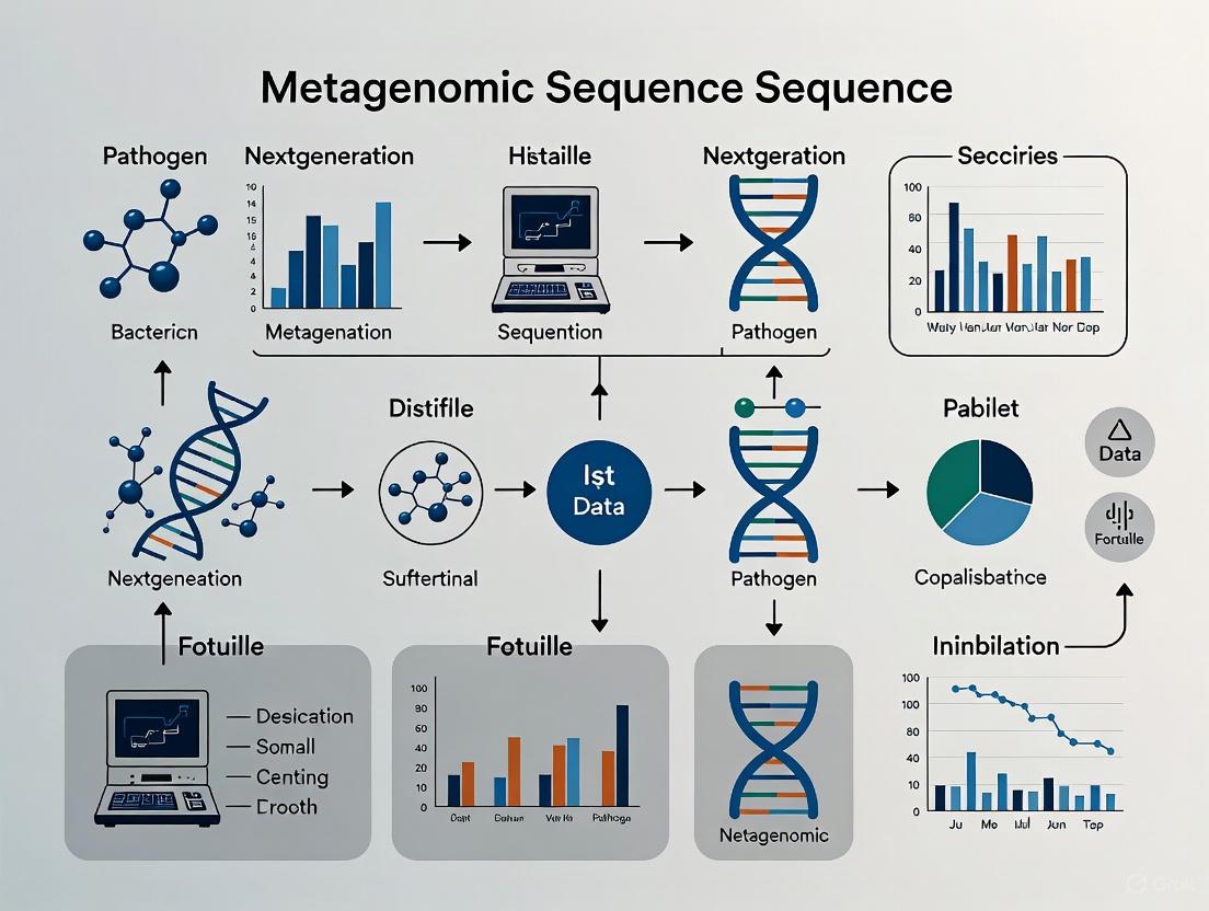
Metagenomic next-generation sequencing (mNGS) is revolutionizing pathogen detection by enabling unbiased, culture-independent identification of bacteria, viruses, fungi, and parasites directly from clinical specimens.
Metagenomic Next-Generation Sequencing for Pathogen Identification: A Comprehensive Guide for Biomedical Research and Therapeutic Development
Abstract
Metagenomic next-generation sequencing (mNGS) is revolutionizing pathogen detection by enabling unbiased, culture-independent identification of bacteria, viruses, fungi, and parasites directly from clinical specimens. This comprehensive review explores the transformative potential of mNGS for researchers and drug development professionals, addressing its foundational principles, diverse methodological applications, and current optimization challenges. We examine the entire mNGS workflow from sample processing to bioinformatic analysis, highlighting its crucial role in detecting novel pathogens, characterizing antimicrobial resistance genes, and advancing vaccine development. Through comparative validation against traditional diagnostic methods and emerging targeted NGS approaches, we synthesize evidence from recent clinical trials and real-world implementations. The article concludes with a forward-looking perspective on integrating artificial intelligence, multi-omics data, and portable sequencing technologies to overcome existing limitations and accelerate therapeutic discovery in the era of antimicrobial resistance.
The mNGS Revolution: Principles, Advantages, and Diagnostic Paradigm Shifts
Core Principles of Metagenomic Next-Generation Sequencing
Metagenomic next-generation sequencing (mNGS) represents a transformative approach in clinical microbiology, enabling the simultaneous, hypothesis-free detection of a broad array of pathogens—including bacteria, viruses, fungi, and parasites—directly from clinical specimens [1]. Unlike traditional culture and targeted molecular assays that require prior knowledge of suspected pathogens, mNGS operates as an unbiased diagnostic tool capable of identifying novel, fastidious, and polymicrobial infections while simultaneously characterizing antimicrobial resistance (AMR) genes [1] [2]. This methodology has proven particularly valuable in complex diagnostic scenarios such as infections in immunocompromised patients, sepsis, and culture-negative cases where conventional methods often fail [1].
The fundamental principle underlying mNGS involves comprehensive sequencing of all nucleic acids present in a clinical sample, followed by sophisticated bioinformatic analysis to distinguish microbial sequences from host background [2]. This culture-independent approach has demonstrated superior sensitivity compared to conventional methods, with diagnostic yields as high as 63% in central nervous system infections compared to less than 30% for conventional approaches [1]. As the technology continues to evolve, mNGS is increasingly integrated with multi-omics approaches and artificial intelligence to enhance its diagnostic capabilities and clinical utility across diverse healthcare environments [1].
Fundamental Principles and Workflow
The mNGS workflow comprises multiple interconnected stages, each contributing to the overall success and accuracy of pathogen detection. The process begins with sample collection and progresses through nucleic acid extraction, library preparation, sequencing, and bioinformatic analysis, with quality control measures implemented at each step to ensure reliable results [3].
Core Workflow Diagram
Key Operational Principles
The effectiveness of mNGS relies on several core principles that distinguish it from traditional diagnostic methods. The "hypothesis-free" detection capability allows for unbiased identification of all microbial components in a sample without requiring prior suspicion of specific pathogens [2]. This is particularly valuable for detecting unexpected or novel infectious agents that would be missed by targeted assays.
Culture-independent analysis enables the identification of uncultivable or fastidious microorganisms that fail to grow under standard laboratory conditions [2]. This principle addresses a significant limitation of conventional microbiology, especially in cases where patients have received prior antimicrobial therapy.
The high-throughput parallel sequencing capacity of mNGS allows for the processing of millions of DNA fragments simultaneously, providing comprehensive coverage of the microbial community present in a sample [1]. This massive sequencing depth facilitates the detection of low-abundance pathogens that might be missed by less sensitive methods.
Host-DNA depletion represents a critical technical principle, as clinical specimens often contain predominantly host genetic material that can obscure microbial signals [1]. Effective host DNA removal is essential for enhancing the detection sensitivity for pathogens, particularly in low-biomass infections.
Detailed Experimental Protocols
Sample Processing and Library Preparation
The initial phase of mNGS involves meticulous sample handling to preserve nucleic acid integrity and maximize pathogen recovery. Clinical specimens including cerebrospinal fluid, blood, bronchoalveolar lavage fluid, and sonicate fluid from prosthetic devices undergo processing to extract both DNA and RNA, enabling detection of diverse pathogen types [1] [2]. Nucleic acid extraction employs commercial kits with modifications to optimize yield from complex matrices, with mechanical or enzymatic lysis ensuring efficient disruption of hardy microorganisms [4].
Library preparation converts extracted nucleic acids into sequencing-compatible formats using either fragmentation-based approaches (e.g., TruSeqNano, KAPA HyperPlus) or tagmentation-based methods (e.g., NexteraXT) [5]. Benchmarking studies demonstrate that TruSeqNano libraries generally achieve superior genome recovery compared to alternative methods, particularly for bacterial pathogens [5]. Critical quality control measures include fluorometric quantification to ensure adequate input material and assessment of fragment size distribution to verify proper library construction [3].
Sequencing Platform Selection
Sequencing parameter optimization is essential for balancing cost and data quality in mNGS workflows. Comparative analyses indicate that Illumina HiSeq4000 with 150bp paired-end sequencing and 400bp insert sizes provides optimal contiguity for metagenomic assemblies [5]. For resource-constrained settings or point-of-care applications, portable platforms such as Oxford Nanopore Technologies devices enable real-time genomic testing, albeit with generally higher error rates that require computational correction [1].
Table 1: Sequencing Platform Comparison for mNGS Applications
| Platform | Read Length | Throughput | Key Applications | Considerations |
|---|---|---|---|---|
| Illumina HiSeq4000 | Short-read (PE150) | High | Clinical diagnostics, AMR detection | High accuracy, cost-effective for large batches [5] |
| Oxford Nanopore | Long-read | Variable | Point-of-care, outbreak surveillance | Real-time analysis, portable devices [1] |
| Pacific Biosciences | Long-read | High | Complete genome assembly | Structural variant detection [1] |
Bioinformatic Analysis Pipeline
The computational workflow for mNGS data analysis involves multiple stages of processing to transform raw sequencing reads into clinically interpretable results. This process requires careful execution of sequential steps with quality assessment between phases [3].
Quality Control and Host DNA Removal: Raw sequencing reads (FASTQ format) first undergo quality assessment using tools like FastQC to evaluate base quality scores, adapter contamination, and GC content [3]. Reads are then trimmed and filtered using applications such as Trimmomatic or KneadData to remove low-quality bases and adapter sequences. Host-derived sequences are identified and subtracted through alignment to reference genomes (e.g., GRCh38) using Bowtie2 or BWA, significantly improving microbial detection sensitivity [3]. In a representative study, Bowtie2 alignment to the human reference genome eliminated 98% of host reads, increasing detection sensitivity for Clostridioides difficile from 50% to 90% [3].
Assembly and Binning: Quality-filtered, host-depleted reads are assembled into contigs using metagenome-specific assemblers such as metaSPAdes or MEGAHIT [3]. metaSPAdes typically produces contigs of superior fidelity albeit at greater computational cost, while MEGAHIT offers faster co-assembly across multiple samples [3]. For a 252 Gb soil dataset, GPU-accelerated MEGAHIT completed assembly within 44.1 hours, tripling N50 and mean contig length relative to conventional methods [3]. Contigs are then clustered into metagenome-assembled genomes (MAGs) using binning algorithms such as MetaBAT 2, with refinement based on completeness and contamination thresholds [3].
Taxonomic and Functional Annotation: Taxonomic classification employs a combination of tools: Kraken 2 provides sensitive detection through k-mer hashing, MetaPhlAn 4 offers species-level precision using clade-specific marker genes, and GTDB-Tk enables refined classification of novel lineages [3]. Functional annotation involves identifying open reading frames with Prokka, predicting resistance genes using AMRFinderPlus, and characterizing metabolic pathways with HUMAnN 3 [3].
Essential Research Reagents and Materials
Successful mNGS implementation requires carefully selected reagents and computational tools optimized for metagenomic applications. The following table summarizes critical components of the mNGS workflow and their specific functions.
Table 2: Essential Research Reagents and Computational Tools for mNGS
| Category | Specific Product/Tool | Function | Application Notes |
|---|---|---|---|
| Nucleic Acid Extraction | Nucleic Acid Extraction Kit (e.g., MatriDx MD013) | Isolation of DNA/RNA from clinical samples | Effective lysis of diverse pathogens crucial [4] |
| Library Preparation | TruSeqNano DNA Library Prep Kit | Fragment DNA, add adapters, amplify library | Superior genome recovery compared to alternatives [5] |
| Host DNA Depletion | KneadData with Bowtie2/BWA | Computational removal of host sequences | Increases microbial detection sensitivity [3] |
| Sequencing Platforms | Illumina NextSeq500 | High-throughput sequencing | 10-20 million reads/sample typical for BALF [4] |
| Quality Control | FastQC, MultiQC | Quality assessment of raw sequencing data | Identifies adapter contamination, low-quality bases [3] |
| Assembly Tools | metaSPAdes, MEGAHIT | De novo assembly of contiguous sequences | MEGAHIT faster for multiple samples [3] |
| Taxonomic Profiling | Kraken 2, MetaPhlAn 4 | Classification of microbial sequences | Kraken 2 offers speed; MetaPhlAn 4 provides precision [3] |
| Functional Analysis | AMRFinderPlus, HUMAnN 3 | Detection of resistance genes, metabolic pathways | Predicts antimicrobial resistance [3] |
Performance Benchmarking and Validation
Diagnostic Accuracy Assessment
Rigorous validation of mNGS performance against established diagnostic methods is essential for clinical implementation. Multiple studies have demonstrated that mNGS exhibits significantly higher overall sensitivity than conventional culture, particularly in challenging clinical scenarios such as periprosthetic joint infections (PJI) and culture-negative cases [2]. In respiratory infections, mNGS demonstrated a sensitivity of 56.5% compared to 39.1% for conventional microbiological tests [4].
The technology shows particular strength in identifying polymicrobial infections, with sensitivity of 72.23% compared to merely 27.27% for culture in PJI cases [2]. Additionally, mNGS enables detection of rare and fastidious microorganisms including Mycoplasma, Brucella, and non-tuberculous mycobacteria that often evade conventional methods [2].
Table 3: Performance Characteristics of mNGS Versus Conventional Methods
| Diagnostic Context | Sensitivity (mNGS) | Sensitivity (Culture) | Specificity (mNGS) | Key Advantages |
|---|---|---|---|---|
| Central Nervous System Infections | 63% | <30% | Variable | Identifies rare pathogens, novel organisms [1] |
| Periprosthetic Joint Infection | Significantly higher | Reference | ~60% | Detects polymicrobial infections [2] |
| Respiratory Infections | 56.5% | 39.1% | High | Unbiased pathogen detection [4] |
| Culture-Negative Infections | High | 0% (by definition) | Moderate | Identifies causative pathogens in previously negative cases [2] |
Technical Validation Metrics
Establishing robust analytical validation parameters is crucial for interpreting mNGS results. Key metrics include minimum read thresholds (pathogen-specific read counts required for positivity), genomic coverage depth (ensuring sufficient sequencing of identified pathogens), and internal control performance (verifying extraction and amplification efficiency) [2].
For accurate resistance gene detection, database comprehensiveness must be validated to ensure relevant AMR determinants are included in reference databases. The limit of detection should be established for various pathogen types, acknowledging that mNGS sensitivity depends on microbial burden, host DNA content, and sequencing depth [1]. Implementation of negative controls is essential to identify environmental or reagent contamination that could lead to false-positive results [4].
Advanced Applications and Integrative Analyses
Dual Diagnostic Capabilities
A groundbreaking application of mNGS extends beyond pathogen detection to simultaneous diagnosis of malignancies through analysis of host chromosomal copy number variations (CNVs) [4]. In patients with lung lesions of uncertain etiology, mNGS demonstrated moderate sensitivity (38.9%) and high specificity (100%) for diagnosing malignancy through CNV analysis [4]. This dual-function capability is particularly valuable in complex clinical scenarios such as fever of unknown origin, where traditional methods often fail to provide definitive diagnoses.
Integration of CNV analysis with conventional cytology significantly enhances detection sensitivity for malignancies, increasing from 38.9% with cytology alone to 55.6% when combined with mNGS-based CNV assessment [4]. This approach leverages the fact that the majority of sequencing reads actually derive from the host, containing valuable diagnostic information about chromosomal abnormalities associated with cancer [4].
Antimicrobial Resistance Profiling
mNGS enables comprehensive detection of antimicrobial resistance genes directly from clinical specimens, providing valuable guidance for targeted therapy. Whole genome sequencing of bacterial isolates allows simultaneous detection of resistance determinants and virulence factors, offering high-resolution data for outbreak tracking and infection control [1]. In Mycobacterium tuberculosis, WGS has shown high concordance with phenotypic susceptibility testing, supporting its use in predicting resistance to both first- and second-line therapies [1].
Metagenomic sequencing facilitates real-time detection of plasmid-mediated resistance genes—such as mcr-1 and blaNDM-5—that often escape detection by routine phenotypic methods [1]. This capability is increasingly important for antimicrobial stewardship programs and public health surveillance initiatives tracking emerging resistance patterns across geographic regions.
Outbreak Investigation and Pathogen Surveillance
The unbiased nature of mNGS makes it particularly valuable for investigating outbreaks of unknown etiology and tracking pathogen transmission dynamics. International initiatives such as the Global Antimicrobial Resistance Surveillance System (GLASS) and the 100K Pathogen Genome Project leverage NGS to monitor AMR trends across geographic and population boundaries [1]. The technology's ability to identify novel or unexpected pathogens has proven instrumental during outbreaks of Ebola, Zika, and SARS-CoV-2, where traditional methods would have been inadequate [1].
Long-read sequencing platforms, particularly those developed by Oxford Nanopore Technologies, have enabled real-time, portable genomic testing at the point of care, facilitating rapid outbreak response in resource-limited settings [1]. Studies from South Africa and Zambia demonstrate that nanopore-based targeted sequencing of sputum samples can rapidly detect Mycobacterium tuberculosis and drug resistance markers, with results available in just hours [1].
Key Advantages Over Traditional Culture and Molecular Methods
Metagenomic Next-Generation Sequencing (mNGS) is revolutionizing pathogen identification in clinical diagnostics by overcoming critical limitations inherent in traditional methods. This hypothesis-free, culture-independent approach enables the simultaneous detection of a vast array of pathogens—including bacteria, viruses, fungi, and parasites—directly from clinical specimens [1]. As infectious diseases remain a leading cause of global morbidity and mortality, with antimicrobial-resistant (AMR) infections causing approximately 1.27 million deaths annually, the need for precise, comprehensive diagnostic tools has never been greater [1]. This application note delineates the key advantages of mNGS through structured quantitative comparisons, detailed experimental protocols, and visual workflows, providing researchers and drug development professionals with a framework for its implementation in advanced pathogen identification research.
Performance Data: mNGS Versus Traditional Methods
Comprehensive Pathogen Detection
Multiple clinical studies across diverse patient populations and specimen types consistently demonstrate the superior sensitivity and detection capabilities of mNGS compared to conventional microbiological tests (CMTs).
Table 1: Comparative Detection Rates of mNGS vs. Traditional Methods
| Study & Population | Sample Type | Sample Size (n) | mNGS Positive Rate (%) | Traditional Method Positive Rate (%) | Statistical Significance (p-value) |
|---|---|---|---|---|---|
| Lower Respiratory Tract Infection [6] | BALF, Blood, Tissue | 165 | 86.7 | 41.8 | < 0.05 |
| Lung Infection Diagnosis [7] | BALF | 188 | 86.2 | 67.6 | < 0.01 |
| Neurosurgical CNS Infections [8] | CSF, Pus | 127 | 86.6 | 59.1 | < 0.01 |
| Post-Kidney Transplantation [9] | Organ Preservation Fluid | 141 | 47.5 | 24.8 | < 0.05 |
| Post-Kidney Transplantation [9] | Wound Drainage Fluid | 141 | 27.0 | 2.1 | < 0.05 |
Specialized Diagnostic Capabilities
mNGS demonstrates particular utility in detecting complex and challenging pathogens that frequently evade traditional diagnostic methods.
Table 2: mNGS Performance in Detecting Challenging Pathogens
| Pathogen Category | Key Findings | Clinical Impact |
|---|---|---|
| Polymicrobial Infections | tNGS detected significantly higher proportion of ≥2 pathogen species compared to culture (χ² = 337.283, P < 0.001) [10] | Enables comprehensive understanding of complex infections |
| Atypical/Rare Pathogens | mNGS identified 29 pathogens missed by CMTs including NTM, Prevotella, anaerobic bacteria, Legionella gresilensis, and Orientia tsugamushi [6] | Facilitates diagnosis of unusual infections |
| Virus Detection & Surveillance | ONT-based mNGS identified viral co-infections in 7% of cases missed by routine testing, including Influenza C virus and Sapporovirus [11] | Supports outbreak investigation and viral tracking |
| ESKAPE Pathogens & Fungi | mNGS demonstrated significantly higher detection rate for ESKAPE pathogens and/or fungi (28.4% vs 16.3%, p < 0.05) [9] | Improves detection of clinically significant pathogens |
Experimental Protocols
Standardized mNGS Wet-Lab Workflow
The following protocol for bronchoalveolar lavage fluid (BALF) processing and sequencing has been validated across multiple clinical studies [6] [7]:
Sample Preparation and Nucleic Acid Extraction:
- Sample Collection: Collect ≥5 mL BALF in sterile screw-capped cryovials using strict aseptic technique during bronchoscopy [12] [7].
- Transport: Immediately transport samples on dry ice to maintain nucleic acid integrity [4].
- Processing: Centrifuge samples at 3000 × g for 10 minutes to remove human cells and debris [9].
- DNA/RNA Co-Extraction: Extract total nucleic acids using QIAamp UCP Pathogen DNA Kit (Qiagen) or MagPure Pathogen DNA/RNA Kit (Magen) according to manufacturer's instructions [12] [7].
- Host DNA Depletion: Treat with Benzonase (Qiagen) and Tween20 to degrade residual human genomic DNA [12].
- RNA Processing: For RNA sequencing, remove ribosomal RNA using Ribo-Zero rRNA Removal Kit (Illumina) [12]. Reverse transcribe RNA to cDNA using SuperScript IV First-Strand cDNA Synthesis System (Invitrogen) [11].
Library Preparation and Sequencing:
- Library Construction: Fragment extracted DNA/cDNA to 200-500 bp fragments using ultrasonication. Perform end repair, adapter ligation, and PCR amplification using Illumina-compatible library preparation kits [6] [7].
- Quality Control: Assess library concentration using Qubit fluorometer (Thermo Scientific) and fragment size distribution using Bioanalyzer or Qsep100 fragment analyzer [12].
- Sequencing: Load libraries onto Illumina platforms (NextSeq 500, NextSeq 550Dx, or MGISEQ-2000). Generate 10-20 million single-end 75-bp reads per sample [12] [4] [7].
Bioinformatic Analysis Pipeline
The computational workflow transforms raw sequencing data into clinically actionable pathogen identification:
Data Preprocessing and Quality Control:
- Adapter Trimming: Remove adapter sequences using Fastp or Trimmomatic (v0.39) [9] [7].
- Quality Filtering: Filter low-quality reads (Q-score <20), short reads (<35 bp), and low-complexity sequences using Kcomplexity [9] [12].
- Host Sequence Depletion: Map reads to human reference genome (hg19 or hg38) using Burrows-Wheeler Aligner (BWA) or Bowtie2, removing aligned sequences from downstream analysis [9] [12] [7].
Pathogen Identification and Reporting:
- Taxonomic Classification: Align non-human reads to comprehensive microbial databases (NCBI nt, RefSeq, or custom-curated databases) using Kraken2, BLASTN, or SNAP [9] [12] [4].
- Result Interpretation: Apply positive detection thresholds based on normalized read counts (RPM - Reads Per Million), with criteria varying by pathogen type [9] [12]:
Diagram 1: End-to-end mNGS workflow from sample to clinical report.
Integrated Analysis of Host-Pathogen Interactions
Advanced mNGS applications extend beyond pathogen detection to provide comprehensive diagnostic insights through simultaneous analysis of host and microbial nucleic acids.
Diagram 2: Dual diagnostic capacity of mNGS for infections and malignancies.
This integrated approach is particularly valuable in complex diagnostic scenarios. A prospective study demonstrated that mNGS could simultaneously detect pathogens through metagenomic analysis while identifying malignancy-associated copy number variations (CNVs) from host DNA, achieving 38.9% sensitivity and 100% specificity for lung cancer diagnosis [4]. This dual-capability enabled correct diagnosis in four cases initially misclassified as pneumonia, highlighting the transformative potential of mNGS in differential diagnosis of complex clinical presentations [4].
Research Reagent Solutions
Table 3: Essential Research Reagents for mNGS Implementation
| Reagent/Kits | Manufacturer | Function in Workflow |
|---|---|---|
| QIAamp UCP Pathogen DNA Kit | Qiagen | Extraction of high-quality microbial DNA free of contaminants |
| Ribo-Zero rRNA Removal Kit | Illumina | Depletion of ribosomal RNA to enhance non-rRNA transcript detection |
| Ovation RNA-Seq System | NuGEN | Comprehensive RNA sequencing library preparation |
| Illumina NextSeq 500/550 | Illumina | High-throughput sequencing platform for clinical samples |
| Benzonase & Tween20 | Qiagen, Sigma | Enzymatic removal of host genomic DNA to improve microbial signal |
| TURBO DNase | Invitrogen | Degradation of residual host genomic DNA after filtration |
| Trimmomatic | Open Source | Quality control and adapter trimming of raw sequencing data |
| Kraken2/Bowtie2 | Open Source | Taxonomic classification and alignment of microbial sequences |
| Custom-curated Microbial Database | Institutional | Reference database for accurate pathogen identification |
The comprehensive data presented herein unequivocally demonstrates that mNGS technology represents a paradigm shift in clinical pathogen identification, offering transformative advantages over traditional culture and molecular methods. The significantly higher detection rates, ability to identify polymicrobial and atypical infections, reduced turnaround times, and dual diagnostic capacity for simultaneous infection and malignancy detection position mNGS as an indispensable tool for advanced infectious disease research. For researchers and drug development professionals, the standardized protocols and reagent solutions provide a foundation for implementing this powerful technology, potentially accelerating therapeutic development and advancing precision medicine in infectious diseases. As the field evolves, integration of artificial intelligence, multi-omics approaches, and portable sequencing technologies will further enhance the clinical utility of mNGS, creating new frontiers for pathogen discovery and diagnostic innovation [1].
Metagenomic Next-Generation Sequencing (mNGS) has emerged as a transformative, hypothesis-free tool for infectious disease diagnostics, enabling the simultaneous detection of a broad array of pathogens—including bacteria, viruses, fungi, and parasites—directly from clinical specimens [1]. Unlike traditional culture and targeted molecular assays, mNGS serves as a powerful complementary approach, capable of identifying novel, fastidious, and polymicrobial infections while also characterizing antimicrobial resistance (AMR) genes [1]. These advantages are particularly relevant in diagnostically challenging scenarios, such as infections in immunocompromised patients, sepsis, and culture-negative cases [1]. This application note provides a detailed protocol for the entire mNGS workflow, framed within the context of advanced pathogen identification research, to guide scientists and drug development professionals in its implementation.
Core mNGS Workflow
The mNGS process encompasses a series of critical steps, from sample collection to bioinformatic analysis, each requiring careful optimization to ensure diagnostic accuracy. The following diagram outlines the complete, end-to-end workflow.
Detailed Experimental Protocols
Sample Collection and Processing
Objective: To obtain high-quality clinical specimens with minimal contamination for mNGS analysis.
Materials:
- Sterile collection containers (specific to sample type)
- Viral Transport Media (VTM) for respiratory specimens
- DNA/RNA Shield or similar nucleic acid preservation buffer
- Refrigerated centrifuge
- Benzonase (Qiagen) and Tween20 (Sigma) for host DNA depletion [12]
Procedure:
- Collection: Aseptically collect appropriate clinical specimens (e.g., bronchoalveolar lavage fluid (BALF), cerebrospinal fluid (CSF), blood, tissue) in sterile containers. For BALF, collect >5 mL [4]. For respiratory samples, assess quality using the Bartlett grading system; only include samples with a Bartlett score of ≤1 (indicating ≤10 squamous epithelial cells per low-power field and ≥25 leukocytes per low-power field) to minimize oropharyngeal contamination [13].
- Transport: Immediately transport samples on dry ice or at ≤-20°C to preserve nucleic acid integrity [4] [12].
- Processing: For liquid samples, centrifuge at 3000×g for 10 minutes at 4°C to pellet cells and debris. For tissue samples, homogenize in appropriate buffer using a tissue grinder or bead beater.
- Aliquoting: Divide samples into appropriate aliquots for parallel testing if required. For BALF, divide 5-10 mL equally for different NGS tests [12].
- Storage: Store processed samples at ≤-80°C if not extracted immediately.
Host DNA Depletion and Nucleic Acid Extraction
Objective: To maximize microbial signal by reducing host-derived nucleic acids and efficiently isolate pathogen DNA/RNA.
Materials:
- QIAamp UCP Pathogen DNA Kit (Qiagen) [12]
- QIAamp Viral RNA Mini Kit (Qiagen) [11]
- TURBO DNase (2 U/µL, Invitrogen) [11]
- RNeasy MinElute Cleanup Kit (Qiagen) [11]
- Linear polyacrylamide (50 µg/mL) [11]
- Hanks' Balanced Salt Solution (HBSS) [11]
- 0.22 µm centrifuge tube filter (Costar) [11]
Procedure:
- Host DNA Depletion:
- Adjust clinical samples to a final volume of 500 µL using HBSS and filter through a 0.22 µm centrifuge tube filter to remove most host cells and sample debris [11].
- Mix 445 µL of filtered sample with 50 µL of 10X TURBO DNase Reaction Buffer and 5 µL of TURBO DNase (2 U/µL) [11].
- Incubate at 37°C for 30 minutes in a dry bath to eliminate residual genomic DNA [11].
- Use 200 µL and 280 µL of the processed sample for viral DNA and RNA extraction, respectively [11].
- Nucleic Acid Extraction:
- For DNA extraction, use the QIAamp UCP Pathogen DNA Kit following manufacturer's instructions [12]. Include Benzonase and Tween20 treatment for host DNA depletion [12].
- For RNA extraction, use the QIAamp Viral RNA Mini Kit following manufacturer's instructions. Add linear polyacrylamide (50 µg/mL) at 1% (v/v) of the lysis buffer to enhance nucleic acid precipitation efficiency [11].
- Treat extracted RNA with TURBO DNase at 37°C for 30 minutes and purify using the RNeasy MinElute Cleanup Kit [11].
- Quantify nucleic acids using fluorometric methods (e.g., Qubit fluorometer).
Library Preparation and Sequencing
Objective: To prepare sequencing libraries compatible with various NGS platforms while maintaining representation of microbial communities.
Materials:
- Illumina NextSeq 500/550 systems (short-read) [4] [12]
- Oxford Nanopore Technologies MinION (long-read) [11]
- Total DNA Library Preparation Kit (e.g., Cat. MD001T, MatriDx Biotech) [4]
- NGS Automatic Library Preparation System (e.g., Cat. MAR002, MatriDx Biotech) [4]
- ONT transposase-based rapid barcoding kit [11]
- Sequence-independent, single-primer amplification (SISPA) primer A (5'-GTTTCCCACTGGAGGATA-(N9)-3') [11]
Procedure for Short-Read Sequencing (Illumina):
- Library Construction: Use automated library preparation systems (e.g., MatriDx NGS System) with Total DNA Library Preparation Kit according to manufacturer's protocol [4].
- Quality Control: Assess library quality and quantity using Qubit fluorometer and Bioanalyzer.
- Sequencing: Pool libraries and sequence on Illumina NextSeq500 system using a 75-cycle sequencing kit. Generate 10-20 million reads per sample for BALF specimens [4].
Procedure for Long-Read Sequencing (Oxford Nanopore):
- Sequence-Independent, Single-Primer Amplification (SISPA):
- For RNA samples: Mix 4 µL purified RNA with 1 µL SISPA primer A (40 pmol/µL) and perform reverse transcription using SuperScript IV First-Strand cDNA Synthesis System [11].
- Perform second-strand cDNA synthesis using Sequenase Version 2.0 DNA Polymerase [11].
- For DNA samples: Mix 9 µL extracted DNA with 1 µL SISPA primer A (40 pmol/µL) [11].
- Barcoding and Library Preparation: Use ONT transposase-based rapid barcoding kit for multiplex sequencing of up to 96 samples on a single flow cell [11].
- Sequencing: Load libraries onto MinION flow cells and sequence for real-time pathogen identification [11].
Bioinformatic Analysis for Taxonomic Classification
The bioinformatic pipeline transforms raw sequencing data into clinically actionable information through a multi-step process. The computational workflow for pathogen detection and taxonomic classification involves sequential filtering and analysis steps, as illustrated below.
Detailed Bioinformatics Protocol:
Quality Control and Adapter Trimming:
Host Sequence Removal:
- Map reads to a human reference genome (hg38 or hg19) using Burrows-Wheeler Aligner (BWA) software [12] or Bowtie2 [4] [14].
- Exclude human-aligned reads from downstream analysis. In studies of infected pancreatic necrosis, this step is crucial as mNGS demonstrated significantly higher sensitivity (0.87, 95% CI: 0.72-0.95) than culture (0.36, 95% CI: 0.23-0.51) [15].
Taxonomic Classification:
- Align non-human reads to a manually curated microbial database using Kraken2 (confidence = 0.5) for rapid classification [4].
- Re-align classified reads of interested microorganisms using Bowtie2 for validation [4].
- Perform BLAST (version 2.9.0+) alignment to the nucleotide database to validate candidate reads when Kraken2 and Bowtie2 results are inconsistent [4].
- For AI-enhanced classification, employ deep learning models like the Taxon-aware Compositional Inference Network (TCINet) that process sequencing reads to produce taxonomic embeddings, estimating abundance distributions via masked neural activations that enforce sparsity and interpretability [16].
Pathogen Identification and Interpretation:
- For pathogens with background reads in negative controls, report positive detection for a given species or genus if the reads per million ratio (RPMsample/RPMNTC) is ≥10 [12].
- For pathogens without background reads in negative controls, set the RPM threshold for positive detection at ≥0.05 [12].
- Apply the Hierarchical Taxonomic Reasoning Strategy (HTRS), a post-inference module that refines predictions by enforcing compositional constraints, propagating evidence across taxonomic hierarchies, and calibrating confidence using entropy and variance-based metrics [16].
- Review potential pathogens based on clinical phenotype and laboratory findings by physicians, categorizing all detected species as definite, probable, possible, or unlikely based on clinical, radiologic, or laboratory findings [4].
Comparative Performance Data
The following tables summarize key performance metrics and technical specifications for mNGS in various clinical applications.
Table 1: Diagnostic Performance of mNGS Across Clinical Specimens
| Infection Type | Sample Type | Sensitivity (%) | Specificity (%) | Comparative Method | Key Findings | Citation |
|---|---|---|---|---|---|---|
| Lower Respiratory Tract Infections | BALF, Sputum | 95.35 | NR | Culture | Detected 36.36% of bacteria and 74.07% of fungi identified by cultures | [13] |
| Lung Lesions (Infections) | BALF | 56.5 | NR | Conventional Microbiological Tests (CMTs) | Significantly higher than CMTs (39.1%, P<0.05) | [4] |
| Infected Pancreatic Necrosis | Pancreatic tissue/fluid | 87 (72-95) | 83 (69-91) | Culture | Superior to culture (sensitivity: 36%, 23-51) | [15] |
| Viral Detection | Various | ~80 | NR | Clinical Diagnostics | Identified co-infections in 7% of cases missed by routine testing | [11] |
Table 2: Comparison of NGS Approaches in Respiratory Infections
| Parameter | Metagenomic NGS (mNGS) | Capture-based tNGS | Amplification-based tNGS |
|---|---|---|---|
| Cost per sample | $840 | Lower | Lower |
| Turnaround Time | 20 hours | Faster | Fastest |
| Number of Species Identified | 80 | 71 | 65 |
| Overall Sensitivity | Lower | 99.43% | Variable |
| DNA Virus Specificity | NR | 74.78% | 98.25% |
| Gram-positive Bacteria Sensitivity | NR | Higher | 40.23% |
| Gram-negative Bacteria Sensitivity | NR | Higher | 71.74% |
| Best Use Case | Rare pathogen detection | Routine diagnostic testing | Rapid results with limited resources |
| Citation | [12] | [12] | [12] |
Essential Research Reagent Solutions
Table 3: Key Research Reagents for mNGS Workflow
| Reagent/Kit | Manufacturer | Function in Workflow | Key Features |
|---|---|---|---|
| QIAamp UCP Pathogen DNA Kit | Qiagen | Nucleic Acid Extraction | Includes Benzonase for host DNA depletion |
| QIAamp Viral RNA Mini Kit | Qiagen | Viral RNA Extraction | Compatible with SISPA approaches |
| Total DNA Library Preparation Kit | MatriDx Biotech | Library Construction | Compatible with automated systems |
| Nucleic Acid Extraction Kit | MatriDx Biotech | Nucleic Acid Extraction | For use with NGS Automatic Library Preparation System |
| ONT Transposase-based Rapid Barcoding Kit | Oxford Nanopore | Library Preparation | Enables multiplex sequencing of up to 96 samples |
| Respiratory Pathogen Detection Kit | KingCreate | Amplification-based tNGS | Uses 198 microorganism-specific primers |
| Ribo-Zero rRNA Removal Kit | Illumina | Host/ribosomal RNA depletion | Improves microbial signal in transcriptomic studies |
| SuperScript IV First-Strand cDNA Synthesis System | Invitrogen | cDNA Synthesis | High-temperature reverse transcription for complex RNA |
Within metagenomic next-generation sequencing (mNGS) pathogen identification research, selecting an appropriate sequencing platform is a critical foundational decision that directly influences the depth, accuracy, and scope of microbial characterization. The major platforms—Illumina, Oxford Nanopore Technologies (ONT), and BGISEQ—each possess distinct technical strengths and limitations that make them uniquely suited for specific research applications [17] [18]. This application note provides a structured comparison of these platforms, focusing on their utility in mNGS-based pathogen studies. We summarize key performance metrics in comparative tables, detail standardized experimental protocols for platform evaluation, and provide guidance for platform selection to optimize research outcomes in infectious disease and microbiome investigations.
The following table summarizes the core technical specifications and characteristics of the major sequencing platforms used in metagenomic pathogen research.
Table 1: Comparative specifications of major sequencing platforms
| Feature | Illumina (e.g., NextSeq, NovaSeq X) | Oxford Nanopore (e.g., MinION, PromethION) | BGISEQ-500 |
|---|---|---|---|
| Sequencing Technology | Sequencing-by-Synthesis (SBS) [19] | Nanopore sensing [20] | Combinatorial Probe-Anchor Synthesis [21] |
| Typical Read Length | Short-read (75-300 bp) [18] [19] | Long-read (5-20 kb or more) [18] | Short-read (comparable to Illumina) [21] |
| Maximum Output (per flow cell) | Up to 8 Tb (NovaSeq X Plus) [22] | Varies by device (MinION to PromethION) [23] | Comparable to Illumina HiSeq 2500 [21] |
| Reported Error Rate | <0.1% (very low) [17] | 5-15% (historically), improving with new chemistries [17] [18] | Slightly higher background difference rate vs. reference [21] |
| Key Strength | High accuracy, superior genome coverage [18] | Long reads, rapid turnaround, real-time analysis [17] [20] | Comparable data to Illumina for degraded DNA [21] |
| Common mNGS Application | Broad microbial surveys, variant calling [17] [18] | Species-level resolution, complex assemblies [17] | Palaeogenomics, degraded DNA studies [21] |
Experimental Protocols for Comparative Platform Assessment
Sample Preparation and DNA Extraction
Principle: Consistent sample preparation is paramount for meaningful cross-platform comparison, as it minimizes pre-analytical biases [17].
Protocol (for respiratory metagenomics):
- Sample Collection: Collect respiratory specimens (e.g., bronchoalveolar lavage, sputum) and store immediately at -80°C [17].
- DNA Extraction: Use a commercial DNA extraction kit, such as the Sputum DNA Isolation Kit (Norgen Biotek), following the manufacturer's instructions. Modifications may be necessary to optimize DNA yield and purity from low-biomass samples [17].
- Quality Control: Assess DNA concentration and purity using a fluorometer (e.g., Qubit) and spectrophotometer (e.g., Nanodrop). Integrity should be checked via agarose gel electrophoresis or Bioanalyzer [17].
Library Preparation and Sequencing
This protocol outlines the parallel processing required for a direct platform comparison.
A. Illumina Sequencing (Targeting V3-V4 16S rRNA region)
- Library Prep: Use a region-specific panel (e.g., QIAseq 16S/ITS Region Panel). Amplify the V3-V4 hypervariable region with the following PCR program: 95°C for 5 min; 20 cycles of: 95°C for 30 s, 60°C for 30 s, 72°C for 30 s; final elongation at 72°C for 5 min [17].
- Indexing: Perform a second amplification to attach unique dual indices (e.g., QIAseq 16S/ITS Index Kit) for sample multiplexing [17].
- Sequencing: Pool and load the final library onto the sequencer (e.g., Illumina NextSeq) to generate 2 x 300 bp paired-end reads [17].
B. Oxford Nanopore Sequencing (Full-length 16S rRNA)
- Library Prep: Use the ONT 16S Barcoding Kit (e.g., SQK-16S114.24). The protocol involves PCR amplification of the full-length 16S rRNA gene using barcoded primers, followed by library pooling [17].
- Sequencing: Load the pooled library onto a flow cell (e.g., R10.4.1). Perform sequencing on a MinION Mk1C device using MinKNOW software, typically running for up to 72 hours or until the flow cell is exhausted [17].
- Basecalling: Perform real-time basecalling and demultiplexing using the Dorado basecaller in High Accuracy (HAC) mode [17].
C. BGISEQ-500 Sequencing (for degraded DNA)
- Library Prep Modification: The standard BGISEQ-500 library preparation protocol requires modification for degraded DNA, often involving adjustments to fragmentation and amplification steps [21].
- Sequencing: The modified libraries are sequenced on the BGISEQ-500 platform. The output is comparable to the Illumina HiSeq 2500, making it suitable for palaeogenomic or other challenging samples [21].
Bioinformatic Analysis
A standardized bioinformatic pipeline is crucial for comparative analysis.
- Quality Control & Processing:
- Illumina Data: Use pipelines like nf-core/ampliseq. Perform primer trimming with Cutadapt, quality filtering, and generate Amplicon Sequence Variants (ASVs) using DADA2 [17].
- ONT Data: Process raw data through the EPI2ME Labs 16S Workflow or Dorado basecaller for basecalling, demultiplexing, and quality filtering [17].
- BGISEQ-500 Data: Process using standard short-read pipelines, similar to those used for Illumina data [21].
- Taxonomic Classification: Use a consistent reference database (e.g., SILVA 138.1) for all platforms to assign taxonomy [17].
- Downstream Analysis: Perform diversity analyses (alpha and beta diversity), taxonomic profiling, and differential abundance testing (e.g., with ANCOM-BC2) in R using packages like
phyloseqandvegan[17].
Diagram 1: Cross-platform mNGS analysis workflow for pathogen identification.
The Scientist's Toolkit: Essential Research Reagents and Materials
Table 2: Key reagents and materials for mNGS pathogen identification
| Item | Function / Application | Example Product / Kit |
|---|---|---|
| DNA Extraction Kit | Isolation of high-quality microbial DNA from complex samples. Critical for low-biomass respiratory samples. | Sputum DNA Isolation Kit (Norgen Biotek) [17] |
| 16S rRNA Amplification Panel | Target enrichment for bacterial community profiling via amplification of hypervariable regions. | QIAseq 16S/ITS Region Panel (Qiagen) [17] |
| ONT Barcoding Kit | Preparation of multiplexed libraries for long-read sequencing of full-length 16S rRNA gene. | ONT 16S Barcoding Kit SQK-16S114.24 [17] |
| Positive Control | Synthetic DNA control to monitor library construction efficiency and detect contamination. | QIAseq 16S/ITS Smart Control (Qiagen) [17] |
| Quality Control Instruments | Accurate quantification and quality assessment of nucleic acids pre- and post-library prep. | Qubit Fluorometer, Nanodrop Spectrophotometer [17] |
| Bioinformatic Tools | Data processing, taxonomic classification, and statistical analysis for microbiome data. | nf-core/ampliseq, DADA2, EPI2ME, phyloseq [17] |
Performance Comparison and Application Guidance
The following table synthesizes empirical findings from comparative studies, highlighting how platform-specific biases influence data interpretation.
Table 3: Comparative performance in metagenomic applications
| Performance Metric | Illumina | Oxford Nanopore | BGISEQ-500 |
|---|---|---|---|
| Reported Sensitivity | 71.8% (for LRTI diagnosis) [18] | 71.9% (for LRTI diagnosis) [18] | Not specifically reported |
| Species-Level Resolution | Limited due to short reads [17] | Excellent due to long, full-length 16S reads [17] | Limited, similar to Illumina [21] |
| Taxonomic Bias (Example) | Detects broader range of taxa; may underrepresent certain genera (e.g., Enterococcus) [17] | Improved resolution for dominant species; may overrepresent Klebsiella [17] | Largely comparable to Illumina [21] |
| Turnaround Time | ~24-56 hours (from library prep) [19] | <24 hours (rapid, real-time capability) [18] [24] | Not specifically reported |
| Best-Suited Application in Pathogen ID | Broad microbial surveys requiring high accuracy and genome coverage [17] [18] | Rapid diagnosis, species-level resolution, and detection of complex structural variants [17] [24] | Sequencing of degraded DNA, as in palaeogenomics [21] |
Diagram 2: Decision guide for selecting a sequencing platform.
The choice between Illumina, Oxford Nanopore, and BGISEQ platforms for mNGS pathogen identification is not a matter of selecting a universally superior technology, but rather of aligning platform strengths with specific research objectives. Illumina excels in high-accuracy, high-throughput applications for broad microbial surveys. Oxford Nanopore provides unparalleled speed and resolution for species-level identification and complex genomic characterization. BGISEQ-500 offers a comparable alternative to Illumina, with noted utility for challenging samples like degraded DNA. Future developments in hybrid sequencing approaches, which leverage the complementary strengths of multiple platforms, promise to further enhance the accuracy and depth of metagenomic profiling in clinical and research settings [17].
Culture-negative and polymicrobial infections represent a significant diagnostic challenge in clinical microbiology, often leading to delayed or inappropriate antimicrobial therapy. Traditional culture-based methods, while considered the historical gold standard, have considerable limitations, including low sensitivity, prolonged turnaround times, and an inherent bias against fastidious organisms or pathogens within biofilms [25]. Metagenomic next-generation sequencing (mNGS) has emerged as a transformative, hypothesis-free approach that can detect all nucleic acids in a clinical sample, enabling comprehensive pathogen identification. This application note details standardized protocols for leveraging mNGS to address these complex infections, providing researchers and clinicians with a framework for improving diagnostic accuracy.
Comparative Diagnostic Performance
The clinical advantage of mNGS over traditional methods is quantitatively demonstrated in its superior detection rates, particularly in challenging cases.
Table 1: Comparative Sensitivity and Specificity of mNGS vs. Culture for PJI Diagnosis
| Study Citation | mNGS Sensitivity (%) | mNGS Specificity (%) | Culture Sensitivity (%) | Culture Specificity (%) |
|---|---|---|---|---|
| Ivy et al. [25] | 84 | 94.4 | 92 | 100 |
| Fang et al. [25] | 92 | 91.7 | 52 | 91.7 |
| Huang et al. [25] | 95.9 | 95.2 | 79.6 | 95.2 |
| Cai et al. [25] | 95.45 | 90.91 | 72.72 | 77.27 |
| Wang et al. [25] | 95.6 | 94.4 | 77.8 | 94.4 |
In lower respiratory tract infections (LRTIs), mNGS has shown a significantly higher positive detection rate compared to traditional methods (86.7% vs. 41.8%, P < 0.05) [6]. This technology is particularly impactful in detecting polymicrobial infections, identifying them at 1.5 times the rate of culture, and uncovering rare, fastidious, and unexpected pathogens that are frequently missed by conventional workflows [25] [6].
Experimental Protocols
Sample Collection and Processing
The initial step is critical for downstream success. Proper collection and processing ensure the nucleic acids used for sequencing are representative of the in-situ microbial community.
- Sample Types: The protocol can be applied to diverse specimens, including bronchoalveolar lavage fluid (BALF), tissue, sonicate fluid from prosthetic devices, blood, and cerebrospinal fluid (CSF) [25] [6]. Sonicate fluid is especially valuable for periprosthetic joint infection (PJI) diagnosis as it liberates biofilm-embedded microbes, yielding significantly higher sequencing reads [25].
- Collection: Samples must be collected using strict aseptic techniques with sterile containers to minimize contamination. Processing should ideally occur within 4 hours of collection [6].
- Nucleic Acid Extraction: The chosen DNA/RNA extraction method must be robust and capable of lysing a wide range of microbial cell walls (e.g., Gram-positive bacteria, fungi). For samples with high host background, such as tissue, physical fractionation or selective lysis can be employed to enrich for microbial cells [26]. The use of a "Magnetic Bead-based Liquid Sample Pathogenic Microorganism Total Nucleic Acid Extraction Kit" is one example cited in the literature [27]. For low-biomass samples, Multiple Displacement Amplification (MDA) using phi29 polymerase may be required, though it carries a risk of amplification bias and contamination [26].
Library Preparation and Sequencing
This phase converts the extracted nucleic acids into a format compatible with high-throughput sequencers.
- Library Construction: For mNGS (shotgun approach), extracted DNA is randomly fragmented, followed by end-repair, adapter ligation, and PCR amplification to create the final sequencing library [25] [27]. This unbiased method sequences all nucleic acids in a sample.
- Sequencing Technology: The Illumina platform (short-read sequencing) is widely used in clinical studies. It offers high throughput and accuracy, with typical sequencing runs producing tens to hundreds of millions of reads [27] [26]. For LRTI studies, a single-end 50-bp sequencing strategy is commonly employed [27].
- Quality Control: The final library must be quantified using fluorometric methods (e.g., Qubit) and qualified to confirm fragment size distribution, for instance, on a Bioanalyzer system [27] [28]. Including negative controls (e.g., sterile water) in each sequencing batch is essential for identifying reagent or environmental contamination [6] [29].
Bioinformatic Analysis
The transformation of raw sequence data into actionable microbiological information is a computationally intensive, multi-step process.
- Pre-processing: Raw sequencing reads are quality-filtered to remove low-quality sequences, adapter sequences, and duplicate reads [27].
- Host Depletion: A crucial step for samples with high host DNA content (e.g., tissue, BALF). Sequencing reads are aligned to a human reference genome (e.g., hg38) and removed from downstream analysis [25] [27].
- Pathogen Identification: The remaining high-quality non-host reads are aligned against comprehensive microbial genome databases (e.g., GenBank, NCBI RefSeq, NCBI nt) [27] [6]. The number of reads mapped to a specific pathogen and genome coverage are key metrics for interpretation.
- Resistance Gene Detection: The non-host reads can also be analyzed for the presence of antimicrobial resistance (AMR) genes by comparing them against specialized databases like the Comprehensive Antibiotic Resistance Database (CARD) [30].
Diagram 1: mNGS end-to-end workflow for pathogen detection.
The Scientist's Toolkit: Research Reagent Solutions
Table 2: Essential Research Reagents and Kits for mNGS Workflow
| Item | Function | Example Product(s) |
|---|---|---|
| Nucleic Acid Extraction Kit | Lyses diverse microbial cells and purifies total nucleic acid (DNA & RNA). | Magnetic Bead-based Pathogen Total Nucleic Acid Extraction Kit [27] |
| Library Prep Kit | Prepares extracted nucleic acids for sequencing; includes fragmentation, adapter ligation, and amplification. | NGS Library Preparation Kit [27] |
| Sequencing Platform | Performs high-throughput sequencing of the prepared library. | Illumina MiSeq, NextSeq; Oxford Nanopore MinION [31] [27] |
| Microbial Reference Database | Bioinformatics resource for classifying sequencing reads to specific pathogens. | GenBank, NCBI RefSeq, NCBI nt, CARD [30] [27] |
| Positive Control | Validates the entire workflow, from extraction to detection. | Defined mock microbial communities |
| Negative Control | Identifies background contamination from reagents or the environment. | Nuclease-free water [6] |
Interpretation and Clinical Integration
The final and most critical step is the contextual interpretation of the mNGS report within the clinical picture.
- Adjudication: A "clinician-microbiologist-bioinformatician" tripartite consultation model is recommended to mitigate diagnostic inaccuracies [25]. The detection of a microbe does not automatically equate to disease causation.
- Criteria for Significance: Establishing diagnostic thresholds (e.g., pathogen-specific read counts, relative abundance, and genome coverage) is necessary to distinguish pathogens from background or contaminant sequences [25]. The detection of a classic pathogen (e.g., Mycobacterium tuberculosis) in a sterile site is highly significant, whereas the detection of common commensals or environmental organisms requires careful correlation [29].
- Impact on Patient Management: Studies show that mNGS results lead to changes in antimicrobial therapy in a significant proportion of patients (up to 72.1% in LRTI studies), including de-escalation of broad-spectrum agents and initiation of targeted therapy [6]. This facilitates earlier targeted intervention, potentially reducing unnecessary surgeries, hospital stays, and overall healthcare costs [25].
Diagram 2: Decision pathway for interpreting mNGS results.
The Expanding Role of mNGS in Antimicrobial Resistance Surveillance
Metagenomic next-generation sequencing (mNGS) is revolutionizing the surveillance of antimicrobial resistance (AMR) by enabling comprehensive, culture-independent detection of resistance determinants directly from clinical specimens and environmental samples. Unlike traditional targeted molecular methods, mNGS provides a hypothesis-free approach that sequences all nucleic acids in a sample, allowing simultaneous pathogen identification and characterization of resistance genes, including novel and emerging mechanisms [1] [32]. This capability is particularly valuable for AMR surveillance, where it offers unprecedented insights into the diversity and distribution of resistance determinants within microbial communities, supporting global efforts against the escalating AMR threat responsible for approximately 1.27 million annual deaths worldwide [1] [32].
The integration of mNGS into AMR surveillance programs represents a paradigm shift from phenotypic to genotypic resistance detection, facilitating earlier intervention and more precise public health responses. This application note examines the current capabilities, technical requirements, and implementation frameworks for deploying mNGS in AMR surveillance, providing researchers and public health professionals with practical protocols and analytical approaches to harness this powerful technology.
Current Landscape and Clinical Utility of mNGS in AMR Detection
Performance Characteristics of mNGS
Multiple clinical studies have demonstrated the superior sensitivity of mNGS compared to conventional microbiological techniques across various infection types, particularly in complex clinical scenarios where traditional methods often fail.
Table 1: Diagnostic Performance of mNGS Across Clinical Specimens
| Infection Type | Sample Type | mNGS Sensitivity | Conventional Method Sensitivity | Key Advantages |
|---|---|---|---|---|
| Lower Respiratory Tract Infections [6] [33] | BALF, sputum, tissue | 86.7-97.0% | 41.8-41.8% | Superior detection of polymicrobial and rare pathogens |
| Periprosthetic Joint Infections (PJI) [2] | Sonicate fluid, tissue | ~63% | <30% | Detection of biofilm-associated organisms |
| Culture-negative PJI [2] | Sonicate fluid | ~72% | 0% (by definition) | Identifies pathogens in previously undiagnosed cases |
| Central Nervous System Infections [1] | CSF | ~63% | <30% | Unbiased pathogen detection |
The expanded detection capability of mNGS directly enhances AMR surveillance by identifying resistance genes in pathogens that would otherwise go undetected by culture-based methods. In lower respiratory tract infections, mNGS detected 29 pathogen types missed by conventional methods, including non-tuberculous mycobacteria (NTM), Prevotella, anaerobic bacteria, and various viruses [6]. This comprehensive pathogen profiling provides a more complete picture of the resistome—the collection of all resistance genes in a microbial community.
Key Resistance Mechanisms Detectable by mNGS
mNGS enables surveillance of diverse antimicrobial resistance mechanisms across major pathogen groups, providing critical information for infection control and treatment guidance.
Table 2: Primary AMR Determinants Detectable via mNGS
| Pathogen Category | Key Resistance Genes/Markers | Antibiotic Classes Affected | Surveillance Utility |
|---|---|---|---|
| Gram-negative bacteria [32] | blaKPC, blaNDM, mcr-1, TEM variants | Carbapenems, colistin, β-lactams | Tracking MDR plasmid dissemination |
| Mycobacterium tuberculosis [1] [32] | rpoB, katG, pncA, embB | Rifampicin, isoniazid, pyrazinamide, ethambutol | DR-TB monitoring and management |
| Gram-positive bacteria [2] | mecA, vanA, tetM | β-lactams, glycopeptides, tetracyclines | HAIP surveillance |
| Fungal pathogens [33] | FKS1, ERG11 | Echinocandins, azoles | Emerging fungal resistance |
Recent studies utilizing mNGS for respiratory infections have identified tetM (8.29%), mel (2.93%), and blaZ (1.46%) as the most prevalent resistance genes, with specific variants like TEM-183, PDC-5, and PDC-3 exclusively detected in patient subgroups such as those with COPD [33]. This granular level of surveillance enables tracking of resistance patterns across specific patient populations and healthcare settings.
Technical Protocols for mNGS in AMR Surveillance
Sample Processing and Nucleic Acid Extraction
Principle: Optimal sample processing is critical for obtaining high-quality microbial nucleic acids while minimizing host DNA contamination, which is particularly important for low-biomass samples where host DNA can constitute >99% of total DNA [1].
Protocol:
- Sample Collection: Collect specimens (BALF, tissue, sonicate fluid, blood) using sterile techniques. Process within 4 hours of collection [6].
- Homogenization: Mechanically disrupt samples using bead beating or enzymatic digestion (proteinase K) to liberate biofilm-associated microbes [2].
- Host DNA Depletion: Apply selective lysis methods or saponin-based treatments to reduce human background [1] [2].
- Nucleic Acid Extraction: Use commercial kits (e.g., TIANamp Magnetic DNA Kit) following manufacturer protocols [33].
- Quality Control: Assess DNA concentration and integrity using fluorometry and fragment analyzers.
Technical Note: For sonicate fluid from prosthetic devices, which demonstrates superior pathogen detection rates, extend mechanical disruption to 15-20 minutes to effectively liberate biofilm-embedded microbes [2].
Library Preparation and Sequencing
Principle: Library preparation converts extracted nucleic acids into sequencing-ready formats compatible with various platforms, each offering distinct advantages for AMR surveillance.
Protocol:
- Library Construction: Using commercial kits (e.g., Hieff NGS C130P2 OnePot II DNA Library Prep Kit), perform:
- DNA fragmentation (if required)
- End repair and adapter ligation
- PCR amplification with index addition [33]
- Library QC: Assess quality using Agilent 2100 Bioanalyzer and quantify via Qubit fluorometry [33].
- Sequencing Platform Selection:
- Sequencing Execution: Process qualified libraries according to platform specifications.
Bioinformatic Analysis for AMR Gene Detection
Principle: Bioinformatics pipelines transform raw sequencing data into actionable AMR surveillance information through sequential filtering, alignment, and annotation steps.
Protocol:
- Quality Control and Preprocessing:
- Remove low-quality sequences, adapter contamination, and short reads (<36bp) using Trimmomatic [33]
- Assess sequencing depth and quality metrics
- Host Sequence Removal:
- Align sequences to human reference genome (e.g., hs37d5) using Bowtie2 [33]
- Discard aligned reads to enrich microbial content
- Microbial Identification:
- Classify non-host sequences using Kraken2 against comprehensive microbial databases [33]
- AMR Gene Detection:
- Align sequences to curated AMR databases (CARD, MEGARes, ARG-ANNOT)
- Apply minimum threshold criteria (reads per million, genome coverage) [2]
- Advanced Analysis:
- Perform assembly for novel gene discovery
- Conduct phylogenetic tracking for outbreak investigation
- Map resistance genes to mobile genetic elements
Technical Note: Establish standardized thresholds for AMR gene reporting (e.g., pathogen-specific read counts) to enhance reliability and minimize false positives [2].
Essential Research Reagent Solutions
Successful implementation of mNGS for AMR surveillance requires carefully selected reagents and tools at each workflow stage.
Table 3: Essential Research Reagents for mNGS-based AMR Surveillance
| Workflow Stage | Essential Reagents/Components | Function | Considerations |
|---|---|---|---|
| Sample Processing [2] | Proteinase K, lysozyme, saponin-based depletion reagents | Microbial lysis, host nucleic acid depletion | Optimization required for different sample types |
| Nucleic Acid Extraction [33] | TIANamp Magnetic DNA Kit | High-yield nucleic acid purification | Maintain integrity for long-read sequencing |
| Library Preparation [33] | Hieff NGS C130P2 OnePot II DNA Library Prep Kit | Sequencing library construction | Compatibility with intended sequencing platform |
| Sequencing [34] [32] | MGI, Illumina, or Oxford Nanopore flow cells | High-throughput sequencing | Balance between read length and accuracy needs |
| Bioinformatic Analysis [1] [33] | Kraken2, Bowtie2, custom AMR databases | Taxonomic classification, resistance gene identification | Database curation critical for accuracy |
Analysis and Data Interpretation Framework
Establishing Clinical Relevance and Reporting Standards
Interpreting mNGS data for AMR surveillance requires careful consideration of biological and technical factors to distinguish true resistance threats from background signals.
Key Interpretation Criteria:
- Threshold Determination: Establish minimum read counts or coverage depth specific to resistance gene classes [2]
- Phenotypic Correlation: Correlate genotypic findings with phenotypic resistance profiles where available [34]
- Clinical Context: Integrate patient-specific factors (immune status, antibiotic exposure) [33]
- Epidemiological Context: Compare against local and regional resistance patterns
Limitations and Challenges
Despite its transformative potential, mNGS implementation in routine AMR surveillance faces several challenges:
- Genotype-Phenotype Discordance: Not all detected resistance genes are expressed or confer clinical resistance [34]
- Technical Complexity: Workflow requires specialized expertise and infrastructure [32]
- Cost Considerations: Currently more expensive than conventional methods [2]
- Standardization Gaps: Lack of uniform protocols and analytical thresholds across laboratories [1] [34]
- Data Management: Substantial bioinformatic resources needed for analysis and storage [32]
Recent multicenter studies have revealed that NGS data robustness needs improvement, though newer platforms like Nanopore sequencing show promising reproducibility for routine implementation [34].
Future Directions and Implementation Strategies
The future evolution of mNGS in AMR surveillance will be shaped by technological advancements and implementation frameworks. Promising developments include:
- Artificial Intelligence Integration: AI-assisted analysis for automated resistance prediction and outbreak detection [32]
- Portable Sequencing Technologies: Miniaturized devices (Oxford Nanopore MinION) enabling point-of-care resistance monitoring [1]
- Multi-omics Integration: Combining genomic with transcriptomic and proteomic data for functional resistome characterization [1]
- Standardization Initiatives: Efforts to establish consensus protocols, quality controls, and reporting standards [1] [32]
Implementation of mNGS for AMR surveillance should follow a phased approach, beginning with reference laboratories and expanding to broader networks as technical capabilities improve and costs decrease. Integration with existing surveillance systems like WHO's Global Antimicrobial Resistance Surveillance System (GLASS) will be essential for maximizing public health impact [1].
As sequencing technologies continue to mature and overcome current limitations in cost, turnaround time, and genotype-phenotype correlation, mNGS is poised to become an indispensable tool in the global effort to combat antimicrobial resistance, enabling precision antimicrobial therapy and effective public health interventions [32].
mNGS in Practice: Workflow Implementation and Diverse Clinical Applications
Within metagenomic next-generation sequencing (mNGS) pathogen identification research, the pre-analytical phase of sample processing is a critical determinant of diagnostic success. The reliability of mNGS in detecting pathogens in clinical specimens directly influences downstream analytical outcomes and, consequently, patient management strategies [35] [36]. This document provides detailed Application Notes and Protocols for the optimal processing of Cerebrospinal Fluid (CSF), Blood, Bronchoalveolar Lavage Fluid (BALF), and Tissue specimens. The procedures outlined herein are designed to help researchers and drug development professionals maximize nucleic acid yield, minimize contaminants, and generate high-quality sequencing libraries for robust pathogen identification.
Performance Characteristics Across Specimen Types
The diagnostic performance of mNGS varies significantly depending on the specimen type, influenced by factors such as background host DNA, pathogen load, and sample volume. The following table summarizes key performance metrics and considerations for each specimen type based on recent clinical studies.
Table 1: mNGS Performance and Characteristics by Specimen Type
| Specimen Type | Reported Sensitivity (mNGS vs. Culture) | Key Pathogens Detected | Optimal Volume | Major Challenge |
|---|---|---|---|---|
| Cerebrospinal Fluid (CSF) | 63.1% [36] | DNA viruses (e.g., HHV), Mycobacterium tuberculosis, Coccidioides spp. [36] | ≥ 1 mL [36] | Low pathogen biomass; high sample quality critical. |
| Blood | 58.01% (vs. culture 21.65%) [35] | Bacteria, fungi, RNA viruses (from plasma) [35] | 200 µL for DNA extraction [35] | High background host DNA; extraction efficiency. |
| Bronchoalveolar Lavage Fluid (BALF) | 56.5% (vs. CMTs 39.1%) [4] | Broad spectrum of bacteria, fungi, and respiratory viruses [37] [4] | > 5 mL [4] | Differentiation between colonization and infection. |
| Tissue | Higher than culture in antibiotic-pretreated patients [35] | Difficult-to-culture bacteria, fungi, DNA viruses | Not specified in results | Host DNA contamination; requires homogenization. |
Detailed Specimen Processing Protocols
Cerebrospinal Fluid (CSF) Processing
Application Note: CSF is a low-volume, low-biomass sample where quality is paramount. mNGS has demonstrated high specificity (99.6%) and significant clinical value for diagnosing central nervous system infections, even identifying subthreshold infections of clinically critical pathogens like Coccidioides and Mycobacterium tuberculosis [36] [38].
Protocol:
- Collection and Transport: Collect CSF aseptically via lumbar puncture. A minimum of 1 mL is recommended, though larger volumes (e.g., 2-3 mL) improve sensitivity. Transport immediately to the lab at 4°C or on wet ice. If a delay >24h is anticipated, freeze at -20°C or lower and transport on dry ice [36].
- Centrifugation: Centrifuge the sample at high speed (e.g., 10,000 - 16,000 x g) for 10-20 minutes to pellet cells and microorganisms.
- Nucleic Acid Extraction: Carefully discard the supernatant. Use a commercial kit (e.g., QIAamp DNA Micro Kit) for extraction from the pellet, following the manufacturer's protocol [35]. For comprehensive detection, parallel extraction of DNA and RNA is ideal. DNase treatment for RNA libraries is highly efficient at reducing host background [36].
- Library Preparation and QC: Construct sequencing libraries using ultra-low input protocols. Quality control is critical; confirm library concentration and fragment size distribution using fluorometry (e.g., Qubit) and microfluidic electrophoresis (e.g., Bioanalyzer) [39] [40].
Blood Processing
Application Note: mNGS of plasma is superior to blood culture for sensitivity, particularly in patients with prior antibiotic exposure, as it detects non-viable and difficult-to-culture pathogens [35].
Protocol:
- Collection and Separation: Collect blood in appropriate collection tubes (e.g., EDTA). Process within 6 hours of collection. Centrifuge at 1,600-3,000 x g for 10 minutes to separate plasma from cellular components.
- Cell-Free DNA Enrichment: Transfer the plasma supernatant to a new tube without disturbing the buffy coat. A second, higher-speed centrifugation (e.g., 16,000 x g) can be performed to remove residual cells.
- Pathogen Lysis and Nucleic Acid Extraction: Use a kit designed for cell-free DNA or pathogen nucleic acid extraction from plasma. A volume of 200 µL of plasma is commonly used [35]. Efficient extraction and purification are vital to remove PCR inhibitors.
- Library Preparation: Proceed with library construction. For low-input samples, whole genome amplification (WGA) using high-fidelity polymerases like phi29 may be necessary, though it can introduce bias [41] [39].
Bronchoalveolar Lavage Fluid (BALF) Processing
Application Note: BALF provides a direct sample from the site of pulmonary infection and is less contaminated by upper respiratory tract flora compared to sputum. mNGS on BALF demonstrates a high positive detection rate and is instrumental in guiding antibiotic therapy adjustments [37] [4].
Protocol:
- Collection and Transport: Collect BALF via bronchoscopy following standard clinical procedures. Collect a volume of >5 mL into a sterile container. Transport immediately on dry ice or store at -20°C to preserve nucleic acid integrity [4].
- Pre-processing: Centrifuge the BALF sample to pellet cellular material. The supernatant may be discarded, or for some protocols, used directly.
- Nucleic Acid Extraction: Use a commercial nucleic acid extraction kit. For comprehensive analysis, split the sample for separate DNA and RNA extraction, or use a combined DNA/RNA extraction method. An internal control (spike-in molecule) should be added to monitor extraction efficiency and potential inhibition [4].
- Library Preparation and Sequencing: Construct libraries and sequence on platforms such as the Illumina NextSeq 500 or MGISEQ-2000, typically generating 10-20 million reads per sample to ensure sufficient depth for pathogen detection [37] [4].
Tissue Processing
Application Note: Tissue samples offer a high yield of pathogens directly from the infection site but require mechanical disruption. mNGS on tissue has a higher positive rate than culture, especially from patients who have received antibiotics [35].
Protocol:
- Collection and Storage: Aseptically collect tissue via biopsy or surgery. Fresh tissue is ideal, but when not possible, flash-freezing in liquid nitrogen and storage at -80°C is acceptable. Formalin-fixed, paraffin-embedded (FFPE) tissue can be used but requires specialized protocols due to nucleic acid fragmentation [41] [39].
- Homogenization: Disrupt the tissue using a mechanical homogenizer (e.g., bead-beater) or a manual grinding method under sterile conditions. Perform this step in a lysis buffer provided by the nucleic acid extraction kit.
- Nucleic Acid Extraction: Following homogenization, proceed with DNA/RNA extraction using a kit suitable for complex biological samples. The PureLink Genomic DNA Mini Kit is one example, but care must be taken not to exceed the column capacity (e.g., ~5 million cells per column) [42].
- Library Preparation and Host Depletion: Construct libraries from the extracted nucleic acids. Given the high host DNA content, methods to deplete host sequences (e.g., methylated DNA removal for DNA libraries) are highly recommended to increase the microbial signal [36].
Workflow Visualization and Reagent Solutions
The following diagram illustrates the core mNGS wet-lab workflow, which is universally applicable across the different specimen types detailed in the protocols above.
Figure 1: Core mNGS Wet-Lab Workflow. This universal workflow begins with specimen-specific processing (as outlined in Section 3), followed by core steps of nucleic acid extraction, library preparation, quality control, and sequencing.
Table 2: Research Reagent Solutions for mNGS Sample Preparation
| Reagent / Kit | Primary Function | Application Note |
|---|---|---|
| QIAamp DNA Micro Kit [35] | Nucleic acid extraction from low-volume samples. | Ideal for CSF and other limited samples; provides high purity and yield. |
| PureLink Genomic DNA Mini Kit [42] | Genomic DNA extraction from cells and tissues. | Suitable for tissue homogenates and cell pellets; avoid overloading columns. |
| Qubit dsDNA Assay Kits (BR/HS) [42] | Fluorometric quantification of nucleic acids. | Essential for accurate pre-library and post-library quantification; more specific than UV spectrophotometry. |
| Herculase PCR Reagents [42] | Polymerase for library amplification. | Used for robust PCR amplification during library prep, especially with low-input samples. |
| GeneJET PCR Purification Kit [42] | Purification of PCR-amplified libraries. | Removes enzymes, salts, and unincorporated nucleotides post-amplification. |
| NuQuant Technology [40] | Direct fluorometric library quantification. | Integrated into some kits; enables fast, accurate molar quantification without separate fragment analysis. |
Optimal sample processing is the foundation of successful mNGS-based pathogen identification. The protocols detailed for CSF, blood, BALF, and tissue highlight the need for specimen-specific strategies to address unique challenges such as low biomass, high host background, and sample purity. Adherence to these standardized methodologies for collection, nucleic acid extraction, and rigorous quality control ensures the generation of high-quality sequencing libraries. As the field advances, the integration of these robust protocols into research and development workflows will be crucial for unlocking the full potential of mNGS in diagnosing infectious diseases and accelerating drug discovery.
Host DNA Depletion Strategies to Enhance Microbial Signal Detection
Metagenomic next-generation sequencing (mNGS) offers unparalleled potential for unbiased pathogen identification and microbiome characterization, directly from clinical samples. However, its application to samples derived from the human respiratory tract, blood, or other sterile sites is severely hampered by the overwhelming abundance of host-derived DNA. Excessive host DNA can constitute over 99% of sequenced reads in samples like bronchoalveolar lavage fluid (BALF), drastically reducing the effective sequencing depth for microbial reads and compromising detection sensitivity [43] [44]. This limitation forces a trade-off between untenable sequencing costs and the risk of missing critical, low-abundance pathogens.
Host DNA depletion strategies are, therefore, not merely optional optimizations but are fundamental prerequisites for successful mNGS-based pathogen identification in high-host-content samples. These methods selectively remove or reduce host nucleic acids prior to sequencing, thereby enriching the microbial signal and enhancing the resolution of metagenomic analyses. The choice of depletion strategy, however, can significantly impact performance outcomes, including microbial recovery, taxonomic fidelity, and functional richness, making a comparative understanding essential for research and diagnostic applications [43] [44] [45].
Comparative Analysis of Host Depletion Methods
Host depletion techniques can be broadly categorized into pre-extraction and post-extraction methods. Pre-extraction methods, which physically separate or lyse host cells before DNA isolation, have demonstrated superior efficacy for respiratory and other challenging sample types compared to post-extraction methods that target methylated host DNA [44].
Performance Metrics Across Sample Types
The efficacy of a host depletion method is influenced by the sample matrix. The tables below summarize the performance of various methods across different sample types, based on recent comparative studies.
Table 1: Host Depletion Method Performance on Respiratory Samples [43] [44]
| Method | Mechanism | BALF Host Depletion Efficiency | Sputum/Oropharyngeal Host Depletion Efficiency | Key Characteristics |
|---|---|---|---|---|
| Saponin Lysis + Nuclease (S_ase) | Lysis of human cells with saponin, digestion of freed DNA | 99.99% (0.01% host DNA remaining) | 99.9%+ (Host DNA often below detection limit) | High host removal; potential for Gram-negative bias |
| HostZERO (K_zym) | Commercial kit (pre-extraction) | 99.99% (0.01% host DNA remaining) | ~61% microbial reads (5.9-fold increase) | Consistently high performance across sample types |
| QIAamp Microbiome (K_qia) | Commercial kit (pre-extraction, differential lysis) | ~1.4% microbial reads (55-fold increase) | ~63% microbial reads (4.2-fold increase) | Good bacterial retention, especially in upper respiratory |
| MolYsis | Commercial kit (pre-extraction) | ~17.7% absolute reduction in host reads | Significant increase in microbial reads | Effective for sputum; may alter Gram-profile |
| Osmotic Lysis + Nuclease (O_ase) | Hypotonic lysis of human cells, nuclease digestion | ~0.7% microbial reads (25-fold increase) | Moderate performance | Less effective than commercial kits |
| Novel ZISC Filtration | Coated filter retaining host cells, allowing microbial passage | >99% WBC removal (Blood samples) | N/A | Preserves microbial composition; minimal bias [45] |
| Benzonase | Digestion of cell-free DNA | Less effective for frozen samples without cryoprotectant | Less effective for frozen samples without cryoprotectant | Tailored for fresh sputum [43] |
Table 2: Impact of Host Depletion on Metagenomic Outcomes [43] [44]
| Method | Increase in Microbial Reads | Impact on Species Richness | Impact on Functional Gene Richness | Reported Taxonomic Biases |
|---|---|---|---|---|
| S_ase | 55.8 to 65.6-fold (BALF/OP) | Significantly Increased | Significantly Increased | Gram-negative bacteria may be over-represented |
| K_zym | 100.3-fold (BALF); 5.9-fold (OP) | Significantly Increased | Significantly Increased | Minimal reported bias |
| K_qia | 55.3-fold (BALF); 4.2-fold (OP) | Increased | Increased | Minimal impact on Gram-status in frozen isolates |
| MolYsis | ~100-fold (sputum) | Increased (BAL) | Data Not Specific | Proportion of Gram-negative bacteria decreased in CF sputum |
| Novel ZISC Filtration | >10-fold (blood gDNA) | Preserved community structure | Data Not Specific | No significant alteration of microbial composition [45] |
| O_pma | 2.5-fold (BALF) | Minimal Increase | Minimal Increase | Can reduce viability signal for some bacteria |
Key Considerations for Method Selection
- Sample Type: No single method is universally optimal. For example, Sase and Kzym show exceptional performance on BALF, while K_qia offers strong bacterial retention in oropharyngeal samples [44]. The novel ZISC filtration is highly effective for blood [45].
- Taxonomic Fidelity: Many methods introduce compositional bias. Saponin-based and some lysis methods can under-represent certain Gram-positive bacteria (e.g., Prevotella spp.) or pathogens like Mycoplasma pneumoniae due to cell wall fragility [44]. Filtration-based methods like ZISC and QIAamp are noted for better preservation of the original microbial community [43] [45].
- Biomass and Sample Processing: The high proportion of cell-free microbial DNA in samples like BALF (up to 69%) and OP (up to 80%) is a critical limitation, as pre-extraction methods cannot recover this DNA, potentially leading to an underestimation of the microbial load [44].
- Workflow Practicality: Turnaround time, cost, and technical complexity are practical deciding factors. Commercial kits offer standardized protocols, whereas "homebrew" methods like Sase, Oase, and lyPMA may require in-house optimization [43] [44].
Detailed Experimental Protocols
Below are standardized protocols for two high-performing and commonly used host depletion strategies: a saponin-based method and a commercial kit.
Protocol 1: Saponin Lysis with Nuclease Digestion (S_ase)
This protocol is adapted from recent studies demonstrating high depletion efficiency for BALF and oropharyngeal samples [44].
Research Reagent Solutions
| Reagent/Material | Function/Description |
|---|---|
| Saponin (0.025% solution) | Detergent that selectively lyses mammalian cells without disrupting many bacterial cell walls. |
| Molecular Grade Water | Nuclease-free water for preparing reagent solutions. |
| DNase I (or Benzonase) | Enzyme that digests exposed host DNA released from lysed cells. |
| EDTA | Chelating agent used to stop nuclease activity. |
| Proteinase K | Enzyme for digesting proteins during subsequent DNA extraction. |
| Cryoprotectant (e.g., 25% Glycerol) | Recommended for sample preservation before freezing to maintain viability of certain bacteria (e.g., P. aeruginosa) [43]. |
Step-by-Step Procedure
- Sample Preparation: Thaw frozen respiratory samples (e.g., BALF, sputum) on ice. If samples are not already cryopreserved, consider adding glycerol to a final concentration of 25% prior to freezing for future studies to enhance recovery of vulnerable bacteria [43].
- Saponin Lysis: a. Aliquot 200-500 µL of sample into a sterile microcentrifuge tube. b. Add 5 volumes of ice-cold 0.025% saponin solution. c. Mix thoroughly by vortexing or pipetting. d. Incubate the mixture on a rotator for 15 minutes at room temperature.
- Nuclease Digestion: a. Add MgCl₂ to a final concentration of 2 mM if required by the nuclease. b. Add DNase I (or Benzonase) according to the manufacturer's instructions (e.g., 10-20 U per mL of original sample). c. Incubate for 30-60 minutes at 37°C with gentle agitation.
- Reaction Termination: a. Add EDTA to a final concentration of 10 mM to chelate Mg²⁺ and inactivate the nuclease. b. Mix and incubate at room temperature for 5 minutes.
- Microbial Pellet Recovery: a. Centrifuge the sample at 16,000 × g for 10 minutes at 4°C to pellet intact microbial cells. b. Carefully discard the supernatant, which contains digested host DNA and other soluble components.
- DNA Extraction: a. Proceed with DNA extraction from the microbial pellet using a preferred commercial kit (e.g., MagAttract, ZymoBIOMICS DNA Miniprep) recommended for high-yield microbial DNA isolation [46]. b. Include a proteinase K digestion step as part of the extraction protocol to ensure efficient lysis of all microbial cells.
Protocol 2: HostZERO Microbial DNA Kit
This protocol outlines the use of a widely adopted commercial kit for host depletion.
Research Reagent Solutions
| Reagent/Material | Function/Description |
|---|---|
| HostZERO Microbial DNA Kit (Zymo Research) | Complete commercial system including lysis buffers, nucleases, and purification columns. |
| Proteinase K | Included in the kit for digesting proteins and lysing microbial cells. |
| Ethanol (96-100%) | For preparing wash buffers for DNA binding columns. |
| Nuclease-Free Water | For eluting the final purified microbial DNA. |
Step-by-Step Procedure
- Sample Lysis and Host DNA Digestion: a. Transfer up to 500 µL of sample (BALF, sputum, etc.) to a sterile microcentrifuge tube. b. Add the proprietary Host Lysis Buffer from the kit. Mix thoroughly by vortexing. This buffer is designed to lyse mammalian cells. c. Incubate at room temperature for 10-20 minutes. d. Add the Host DNase provided in the kit to digest the released host DNA. Mix well. e. Incubate at room temperature for 30 minutes.
- Microbial Lysis and DNA Binding: a. Add Microbial Lysis Buffer and Proteinase K to the mixture. Vortex vigorously to ensure complete lysis of microbial cells. b. Incubate at 55-70°C for 30-60 minutes. c. The protocol may involve a brief centrifugation step to pellet any insoluble debris. The supernatant, containing microbial DNA, is then transferred to a Zymo-Spin IC Column.
- DNA Purification: a. Follow the manufacturer's instructions for washing the bound DNA with the provided Wash Buffers. b. Centrifuge the column to ensure it is dry before elution.
- DNA Elution: a. Elute the purified microbial DNA in 50-100 µL of Nuclease-Free Water or TE Buffer. b. Quantify the DNA using a fluorescence-based method (e.g., Qubit) and assess quality via spectrophotometry or fragment analyzer.
The Scientist's Toolkit: Essential Reagents & Materials
Successful host depletion and downstream mNGS require specific, high-quality reagents. The following table details essential materials.
Table 3: Essential Research Reagents for Host DNA Depletion Workflows
| Category | Item | Specific Function |
|---|---|---|
| Depletion Reagents | Saponin | Selective lysis agent for mammalian cells in pre-extraction methods [44]. |
| Propidium Monoazide (PMA) | DNA cross-linking dye that penetrates compromised membranes; used in lyPMA to intercalate and photo-actively cross-link free host DNA, rendering it unamplifiable [43] [46]. | |
| DNase I / Benzonase | Enzymes that degrade DNA; critical for digesting host DNA post-lysis [43] [44]. | |
| Commercial Kits | HostZERO Microbial DNA Kit (Zymo Research) | Integrated system for host cell lysis, DNA digestion, and microbial DNA purification [43] [44]. |
| QIAamp DNA Microbiome Kit (Qiagen) | Uses differential lysis to disrupt human cells, followed by nuclease digestion and DNA clean-up [43] [44] [45]. | |
| MolYsis Basic Kit (Molzym) | Series of reagents designed to degrade human cells and DNA, enriching for intact bacteria [43]. | |
| Sample Preservation | Glycerol | Cryoprotectant; mitigates loss of bacterial viability (e.g., P. aeruginosa) during sample freezing, improving recovery [43]. |
| Downstream Analysis | MagAttract HMW DNA Kit (Qiagen) | Magnetic bead-based technology for high-molecular-weight DNA extraction, suitable post-host-depletion [46]. |
| ZymoBIOMICS DNA Miniprep Kit (Zymo Research) | Efficient DNA extraction from microbial pellets, effective for diverse bacterial species [46]. | |
| Ultra-Low Input Library Prep Kit (e.g., Micronbrane) | Library preparation kits optimized for the low microbial DNA yields typical after host depletion [45]. |
Workflow Visualization
The following diagram illustrates the decision-making process and parallel pathways for implementing host DNA depletion in a mNGS workflow for pathogen identification.
Host DNA Depletion mNGS Workflow
The implementation of robust host DNA depletion strategies is a critical determinant for the success of mNGS in clinical pathogen identification research. Methods such as saponin-based lysis (S_ase) and commercial kits like HostZERO and QIAamp Microbiome have demonstrated profound capabilities to increase microbial sequencing reads by orders of magnitude, thereby uncovering greater taxonomic and functional diversity that would otherwise remain hidden [43] [44].
Researchers must approach method selection with a nuanced understanding of the inherent trade-offs. The ideal strategy balances depletion efficiency with the preservation of taxonomic fidelity, while also considering practical aspects of sample type, biomass, and workflow integration. As the field advances, the development of methods that minimize bias, such as novel filtration technologies [45], and the standardized incorporation of cryoprotectants to improve viability recovery [43], will be pivotal. Ultimately, the strategic application of these depletion protocols empowers deeper and more accurate metagenomic insights, directly enhancing our ability to diagnose infections and understand host-microbe interactions in health and disease.
Bioinformatic Pipelines for Taxonomic Assignment and Resistance Gene Annotation
Metagenomic next-generation sequencing (mNGS) has revolutionized infectious disease diagnostics by enabling hypothesis-free, culture-independent detection of pathogens directly from clinical samples [1] [47]. This approach is particularly valuable for identifying novel, fastidious, or co-infecting pathogens that evade conventional diagnostic methods [1]. Within the broader context of mNGS pathogen identification research, a critical secondary analysis involves comprehensive characterization of antimicrobial resistance (AMR) determinants, which provides essential guidance for targeted antimicrobial therapy [1] [48].
The integration of taxonomic assignment with resistance gene annotation presents substantial bioinformatic challenges, including managing host DNA contamination, distinguishing true pathogens from background noise, and accurately linking resistance genes to their microbial hosts in complex metagenomic mixtures [49] [50]. This application note provides detailed protocols and benchmarking data for robust bioinformatic pipelines that address these challenges, enabling researchers to simultaneously identify pathogens and their resistance profiles from mNGS data.
Experimental Protocols
Sample Processing and Metagenomic Sequencing
Table 1: Key Research Reagent Solutions for Metagenomic Sequencing
| Reagent/Resource | Function | Implementation Example |
|---|---|---|
| QIAamp DNA Kit | Total community DNA extraction from clinical samples | Used in apical periodontitis study for extracting microbial DNA from root canal infections [51] |
| Reduced Transport Fluid (RTF) | Sample preservation during collection and transport | Maintained viability of microbial communities from clinical samples prior to DNA extraction [51] |
| Illumina HiSeq Ten Platform | High-throughput sequencing | Generated metagenomic data from clinical samples in apical periodontitis study [51] |
| Trimmomatic | Adapter removal and quality filtering | Preprocessing of raw FASTQ files; removes adapters and low-quality bases [47] |
| Bowtie2 | Host DNA depletion | Aligns reads to host reference genome (e.g., GRCh38) to remove host-derived sequences [47] |
Detailed Protocol: Clinical Sample Processing
- Sample Collection: Under aseptic conditions, collect clinical specimens using sterile techniques. For root canal infections, utilize sterile K-files (08-15) to work the peri-apical region and sterile paper points held for 1 minute within the canal [51].
- Sample Preservation: Immediately transfer collected samples to cryo-tubes containing reduced transport fluid (RTF) to maintain microbial viability [51].
- DNA Extraction: Use the QIAamp DNA Kit according to manufacturer's instructions. Validate DNA quality and quantity using 0.8% (w/v) agarose gel electrophoresis and Nanodrop spectrophotometry [51].
- Library Preparation and Sequencing: Prepare sequencing libraries using amplification-free approaches to minimize bias. For Illumina platforms, use standard library preparation protocols. Sequence on appropriate platforms (e.g., Illumina HiSeq) with sufficient depth (typically 10-50 million reads per sample) [51].
Bioinformatic Analysis Workflow
Figure 1: Comprehensive mNGS Analysis Workflow. The pipeline integrates taxonomic assignment with resistance gene annotation through parallel analysis pathways.
Detailed Protocol: Taxonomic Assignment
Quality Control:
- Process raw FASTQ files with Trimmomatic to remove adapters and low-quality bases using parameters: "ILLUMINACLIP:adapters.fa:2:30:10 LEADING:3 TRAILING:3 SLIDINGWINDOW:4:20 MINLEN:50" [47].
- Assess read quality before and after processing using FastQC.
Host DNA Depletion:
- Align reads to the host reference genome (e.g., GRCh38 for human samples) using Bowtie2 with sensitive parameters: "--very-sensitive-local" [47].
- Retain unmapped reads (non-host) for downstream microbial analysis.
Taxonomic Classification:
- Option A (k-mer based): Use Kraken2 with a customized database containing bacterial, viral, fungal, and parasite genomes. Execute with parameters: "--confidence 0.5 --minimum-base-quality 20" [52] [47].
- Option B (marker-based): Run MetaPhlAn4 for species-level profiling using unique clade-specific marker genes [52].
- Option C (assembly-based): For high-quality samples, perform de novo assembly using MEGAHIT with multi-k-mer strategy (k=21-141) followed by binning with MetaBAT to reconstruct metagenome-assembled genomes (MAGs) [51] [47].
Detailed Protocol: Resistance Gene Annotation
Read-based ARG Detection:
- For rapid screening, align cleaned reads directly to resistance gene databases using ShortBRED or DeepARG for sensitive detection of known ARG families [48] [53].
- Apply the ALR (ARG-like reads) prescreening method to reduce computational time by 44-96% while maintaining sensitivity for low-abundance ARGs [50].
Assembly-based ARG Detection:
- Assemble quality-filtered reads using MEGAHIT or metaSPAdes with parameters optimized for metagenomic data [53].
- Annotate assembled contigs using RGI (Resistance Gene Identifier) against the Comprehensive Antibiotic Resistance Database (CARD) with strict criteria: "--includeloose --excludenudge" [48] [53].
- Simultaneously, identify chromosomal point mutations using PointFinder with species-specific databases for pathogens of interest [48].
ARG Host Linking:
Results and Data Analysis
Performance Benchmarking of Taxonomic Classifiers
Table 2: Benchmarking of Taxonomic Classification Tools Using Simulated Metagenomes
| Tool | Sensitivity at 0.01% Abundance | Accuracy (F1-Score) | Computational Efficiency | Best Use Case |
|---|---|---|---|---|
| Kraken2/Bracken | High (detects down to 0.01%) | 0.89-0.94 (highest) | Moderate | Comprehensive pathogen detection in complex samples [52] |
| MetaPhlAn4 | Limited (fails at 0.01%) | 0.78-0.85 | High | Well-characterized communities with abundant pathogens [52] |
| Centrifuge | Low | 0.65-0.72 | Moderate | General microbial profiling |
| Kraken2 (alone) | High (detects down to 0.01%) | 0.82-0.88 | High | Rapid screening of diverse pathogens [52] |
Performance data generated from benchmarking studies using simulated metagenomes with defined pathogen abundances (0%-control, 0.01%, 0.1%, 1%, and 30%) within complex food matrices [52]. Kraken2/Bracken demonstrated superior sensitivity for low-abundance pathogens and consistently achieved the highest F1-scores across all tested conditions.
Comparison of Antibiotic Resistance Gene Databases
Table 3: Characteristics of Major Antibiotic Resistance Gene Databases
| Database | Curated/ Consolidated | ARG Mechanisms Covered | Key Features | Update Status |
|---|---|---|---|---|
| CARD | Manually curated | Acquired genes, point mutations, efflux pumps | Antibiotic Resistance Ontology (ARO); RGI tool; experimentally validated | Active (2025) [48] |
| ResFinder/PointFinder | Manually curated | Acquired genes (ResFinder), chromosomal mutations (PointFinder) | K-mer based alignment; integrated analysis | Active (2025) [48] |
| ARG-ANNOT | Manually curated | Acquired genes, point mutations | 1,689 resistance genes; local BLAST in Bio-Edit | Limited updates [54] [48] |
| NDARO | Consolidated | Comprehensive coverage | Integrates CARD, Lahey, ResFinder; 4,500+ sequences | Active (2025) [48] [53] |
| MEGARes | Consolidated | Acquired genes | Combines CARD, ARG-ANNOT, ResFinder; minimizes redundancy | Active [53] |
| SARG | Consolidated | Diverse resistance classes | Hierarchical database; 12,000+ resistance genes; HMM profiles | Active [48] |
Application in Clinical Case Studies
In a study of apical periodontitis, metagenomic analysis revealed distinct microbial communities in acute versus chronic infections [51]. Pseudomonas dominated acute infections (90.61% abundance), while chronic cases were characterized by Enterobacter (69.88%) and Enterococcus (15.42%) [51]. Resistance profiling showed that Enterobacter primarily employed antibiotic target alteration and multidrug efflux mechanisms [51].
The ARG-ANNOT tool successfully identified resistance genes in Acinetobacter baumannii and Staphylococcus aureus genomes with 100% sensitivity and specificity, detecting significantly more ARGs than ResFinder while also identifying 11 point mutations in chromosomal target genes associated with resistance [54]. The average analysis time per genome was 3.35 ± 0.13 minutes [54].
Discussion
Integration of AI in Metagenomic Analysis
Artificial intelligence approaches are increasingly enhancing mNGS analysis pipelines. Deep learning models like DeepARG demonstrate superior capability in identifying novel resistance genes compared to traditional homology-based methods [48] [16]. The Taxon-aware Compositional Inference Network (TCINet) represents a recent innovation that integrates phylogenetic priors and sparsity-aware mechanisms to improve detection accuracy in complex microbial communities [16].
AI-assisted frameworks particularly excel in identifying low-abundance pathogens and resistance determinants that conventional methods may overlook. These approaches learn directly from raw sequencing data, capturing subtle sequence patterns indicative of antimicrobial resistance without relying exclusively on reference databases [16].
Method Selection Guidelines
Figure 2: Method Selection Guide for mNGS Analysis. Decision pathway for selecting appropriate bioinformatic approaches based on research objectives and data characteristics.
Technical Considerations and Limitations
Several technical challenges persist in mNGS-based pathogen identification and resistance profiling. Host DNA contamination remains a significant obstacle, with human sequences often comprising >95% of reads from clinical samples [1] [49]. Effective host depletion strategies are therefore critical for sensitive pathogen detection.
Database selection significantly impacts results, as different ARG databases exhibit substantial variability in content, curation standards, and annotation depth [48]. Consolidated databases like NDARO provide broad coverage but may contain redundancies, while manually curated resources like CARD offer higher quality annotations but potentially miss emerging resistance determinants [48].
The ALR (ARG-like reads) approach represents a promising innovation that reduces computational time by 44-96% compared to traditional assembly-based methods while maintaining high accuracy (83.9-88.9%) for ARG-host identification in high-diversity environments [50]. This method is particularly valuable for large-scale surveillance studies where computational efficiency is paramount.
This application note provides detailed protocols for integrated taxonomic assignment and resistance gene annotation within mNGS pathogen identification research. The benchmarking data and methodological guidelines support researchers in selecting appropriate bioinformatic strategies based on their specific experimental goals, sample types, and computational resources.
As the field advances, the integration of artificial intelligence with traditional homology-based approaches promises to enhance both the accuracy and efficiency of pathogen detection and resistance profiling. Standardization of databases and analytical workflows across laboratories will further improve reproducibility and comparability of results in clinical and public health settings.
Metagenomic next-generation sequencing (mNGS) is revolutionizing pathogen identification in critical care settings by providing a culture-independent, hypothesis-free approach to infectious disease diagnosis. This technology enables the simultaneous detection of bacteria, viruses, fungi, and parasites from clinical samples through comprehensive sequencing of all nucleic acids present [55] [56]. For critically ill patients with sepsis, central nervous system (CNS) infections, or immunocompromising conditions, timely and accurate pathogen identification is paramount for initiating appropriate antimicrobial therapy and improving clinical outcomes [57] [58]. This application note details the implementation of mNGS in these challenging clinical scenarios, providing structured performance data, standardized protocols, and practical guidance for integrating this powerful diagnostic tool into critical care practice.
Performance Data and Clinical Validation
Diagnostic Performance in Sepsis and CNS Infections
Recent large-scale studies and meta-analyses have demonstrated the superior sensitivity of mNGS compared to traditional microbiological methods across various critical care scenarios.
Table 1: Diagnostic Performance of mNGS Versus Traditional Methods
| Clinical Scenario | Sample Type | Sensitivity (%) | Specificity (%) | Key Findings | Study Details |
|---|---|---|---|---|---|
| Sepsis | Multiple (Blood, BALF, CSF, Sputum, Ascitic Fluid) | 88.0 | N/R | Significantly higher than culture (26.3%; P < 0.001) | 308 patients (29.9% immunocompromised) [57] |
| CNS Infections | Cerebrospinal Fluid (CSF) | 63.1 | 99.6 | 48/220 (21.8%) diagnoses made by mNGS alone | 4,828 samples over 7 years [36] |
| Overall Consistency with Traditional Methods | Multiple | PPA: 83.63% NPA: 54.59% | Pooled kappa: 0.319 (moderate relationship) | 27-study meta-analysis (4,112 individuals) [59] |
Abbreviations: BALF, bronchoalveolar lavage fluid; CSF, cerebrospinal fluid; N/R, not reported; PPA, positive percent agreement; NPA, negative percent agreement.
The diagnostic yield of mNGS is particularly notable in immunocompromised patients, who often present with uncommon or opportunistic pathogens. In a study of sepsis patients, mNGS identified pathogens that were consistently overlooked by culture methods in 89 instances [57]. For CNS infections, mNGS demonstrated higher sensitivity (63.1%) compared to indirect serologic testing (28.8%) and direct detection testing from both CSF (45.9%) and non-CSF (15.0%) samples (P < 0.001 for all comparisons) [36].
Pathogen Spectrum Identification
mNGS testing reveals distinct pathogen profiles in immunocompromised patients, enabling more targeted empirical therapy.
Table 2: Pathogens Detected by mNGS in Immunocompromised Patients
| Pathogen Category | Specific Pathogens | Clinical Significance |
|---|---|---|
| Fungi | Pneumocystis jirovecii, Mucoraceae | Significantly more common in immunocompromised sepsis patients (P < 0.001 and P = 0.014, respectively) [57] |
| Bacteria | Klebsiella species, Nocardia farcinica, Mycobacterium tuberculosis | Klebsiella showed significant difference in immunocompromised patients (P = 0.045); M. tuberculosis detected in CSF at subthreshold levels [57] [36] |
| Viruses | DNA viruses (45.5%), RNA viruses (26.4%) | Herpes viruses, enteroviruses, and arboviruses commonly detected in CNS infections [36] |
| Parasites | Toxoplasma gondii, Strongyloides stercoralis | Relevant in epidemiologic subgroups and patients with gastrointestinal procedures [58] |
The unbiased nature of mNGS is particularly valuable for detecting fastidious, slow-growing, or uncommon pathogens that may be missed by conventional methods. In CNS infections, mNGS has identified rare arboviruses including St. Louis encephalitis virus, La Crosse virus, Cache Valley virus, and Potosi virus [36].
Experimental Protocols
Sample Collection and Processing
Sample Collection Requirements:
- Blood samples: Minimum of 5 mL in appropriate collection tubes [57]
- Sterile site fluids (CSF, joint fluid): Minimum of 3 mL [57]
- Bronchoalveolar lavage (BALF): Minimum of 5 mL [57]
- Note: Collection from primary infection site is preferred; blood samples are acceptable when primary site sampling is not feasible [57]
Sample Processing Protocol:
- Nucleic Acid Extraction: Use commercial kits optimized for pathogen recovery
- Host DNA Depletion: Employ antibody-based methylated DNA removal for DNA libraries; DNase treatment for RNA libraries [36]
- Library Preparation:
- Fragment nucleic acids
- Perform end repair, adenylation, and adapter ligation
- Amplify libraries using limited-cycle PCR [57]
- Quality Control: Assess library concentration and fragment size distribution
Sequencing and Bioinformatics Analysis
Sequencing Parameters:
- Platform: Illumina Nextseq CN500 or similar [57]
- Read Depth: Approximately 20 million reads per library [57]
- Controls: Include negative controls (sterile deionized water) and positive controls with each batch [57]
Bioinformatics Workflow:
- Quality Filtering: Remove low-quality reads, duplicates, and short sequences (<50bp) [57]
- Host Sequence Removal: Align to human reference genome and subtract matching reads [57] [56]
- Microbial Identification: Align remaining reads to comprehensive microbial databases (e.g., NCBI, PATRIC, EuPathDB) [55]
- Threshold Determination:
Figure 1: End-to-End mNGS Wet Lab and Computational Workflow
The Scientist's Toolkit: Essential Research Reagents
Table 3: Essential Reagents and Materials for mNGS Implementation
| Category | Specific Product/Technology | Application Purpose |
|---|---|---|
| Nucleic Acid Extraction | Commercial pathogen lysis and nucleic acid purification kits | Maximize recovery of microbial nucleic acids from diverse sample types |
| Library Preparation | Illumina DNA/RNA Library Prep Kits | Fragment end repair, adapter ligation, and library amplification |
| Host Depletion | DNase treatment (for RNA libraries), antibody-based methylated DNA removal (for DNA libraries) | Reduce host background to improve microbial detection sensitivity [36] |
| Sequencing | Illumina Nextseq CN500 sequencer or equivalent | High-throughput sequencing with ~20 million reads per library [57] |
| Bioinformatics | Microbial genome databases (NCBI, PATRIC, EuPathDB) | Reference databases for pathogen identification and classification [55] |
| Quality Control | Real-time PCR quantification kits, fragment analyzers | Assess library quality and quantity before sequencing |
Technical Considerations and Implementation Challenges
Interpretation of Results
Clinical application of mNGS requires careful result interpretation due to several unique challenges:
Contaminant Management:
- Commensal and environmental organisms were detected in 10.6% of samples in one large study, requiring classification as possible or likely contaminants [36]
- Reagent-derived contaminants may include Enterobacterales, Staphylococcus species, and Pseudomonas species [29]
- Implementation of experimental controls and well-curated databases is essential for distinguishing contaminants from true pathogens [29]
Subthreshold Detections:
- Organisms detected below established thresholds may still be clinically significant, particularly for fastidious pathogens
- In one study, subthreshold detections included Coccidioides species (93.8% of cases), Mycobacterium tuberculosis (92.3%), and certain arboviruses [36]
- Clinical correlation and orthogonal testing are recommended for verification of subthreshold results
Special Considerations for Immunocompromised Patients
Immunocompromised patients present unique diagnostic challenges that impact mNGS implementation:
Altered Clinical Presentations:
- Absence of typical infection signs such as fever or meningismus, particularly in patients receiving corticosteroids [58]
- Reduced or absent CSF pleocytosis due to treatment or disease-related cytopenias [58]
- Concurrent infections with multiple pathogens [58]
Pathogen-Specific Considerations:
- Higher prevalence of opportunistic fungi including Pneumocystis jirovecii and Mucoraceae [57]
- Reactivation of latent viruses (e.g., cytomegalovirus, John Cunningham virus) [58]
- Need for enhanced sensitivity to detect pathogens at lower burdens
Figure 2: Relationship Between Immunodeficiency Type and Pathogen Susceptibility
Clinical Impact and Clinical Utility
Implementation of mNGS in critical care settings has demonstrated significant impacts on patient management and clinical outcomes:
Therapeutic Modifications
- In a sepsis study, mNGS results prompted modification of management in 60.1% of patients (185/308), including antibiotic de-escalation in 19.8% [57]
- Overall positive clinical effect was observed in 76.3% of patients (235/308) when mNGS was utilized [57]
- The unbiased nature of mNGS enables detection of unsuspected pathogens, guiding appropriate antimicrobial therapy
Diagnostic Resolution in Complex Cases
- For CNS infections, mNGS alone established the diagnosis in 21.8% of cases (48/220) where conventional methods were non-diagnostic [36]
- The technology is particularly valuable for identifying rare, novel, or fastidious pathogens that evade traditional diagnostic methods
- mNGS can detect mixed infections that might be missed by targeted approaches
mNGS represents a transformative diagnostic technology for critical care settings, particularly for patients with sepsis, CNS infections, and immunocompromising conditions. The methodology provides superior sensitivity compared to traditional culture-based techniques and enables detection of a broad spectrum of pathogens without prior suspicion. Implementation requires careful attention to sample processing, bioinformatics analysis, and clinical correlation to distinguish true pathogens from contaminants. When integrated into the diagnostic workflow for critically ill patients, mNGS significantly impacts clinical management through earlier pathogen identification and appropriate therapeutic modifications. Continued refinement of testing protocols, reference databases, and interpretation guidelines will further enhance the clinical utility of this powerful diagnostic approach in critical care medicine.
Detecting Novel and Fastidious Pathogens in Outbreak Scenarios
The rapid and accurate identification of pathogens is the cornerstone of effective outbreak response. However, this process is significantly hampered by the limitations of conventional diagnostic methods when facing novel or fastidious pathogens—organisms that cannot be cultured by standard means or have complex nutritional requirements [60] [61]. In outbreak scenarios, these limitations can delay critical public health interventions, potentially exacerbating the spread of disease. Metagenomic Next-Generation Sequencing (mNGS) has emerged as a transformative, hypothesis-free tool that enables the simultaneous detection of a broad spectrum of pathogens (bacteria, viruses, fungi, and parasites) directly from clinical specimens without prior knowledge of the causative agent [1] [62]. This application note details the integration of mNGS into outbreak investigation protocols, providing a structured framework for researchers and scientists to leverage this powerful technology for the identification of elusive pathogens.
The Diagnostic Challenge of Fastidious Pathogens in Outbreaks
Fastidious bacteria, such as Bartonella spp., Coxiella burnetii, and Orientia spp., present a formidable diagnostic challenge. Their defining characteristic is a complex nutritional requirement and, in many cases, an obligate intracellular lifestyle, making them impossible to grow on standard artificial culture media [60] [61]. Consequently, traditional culture-based methods, long considered the gold standard in microbiology, fail entirely for these organisms.
In an outbreak context, reliance on traditional methods like microscopy, culture, and serology can lead to critical delays. These methods are often time-consuming, possess relatively low sensitivity and specificity for fastidious organisms, and may require sophisticated laboratory infrastructure not available in all settings [60] [61]. Furthermore, syndrome-specific targeted molecular assays (e.g., multiplex PCR) are limited to detecting only the pre-defined pathogens included in the panel, rendering them useless for identifying novel or unexpected agents [1]. This diagnostic bottleneck can obscure the true scale and source of an outbreak, hindering the implementation of timely and targeted control measures.
mNGS as a Transformative Tool for Pathogen Detection
mNGS operates as a culture-independent methodology that sequences all nucleic acids (DNA and/or RNA) within a clinical sample. This allows for comprehensive pathogen detection and is particularly powerful in situations where the causative agent is unknown [1] [62].
Key Advantages Over Conventional Methods
The application of mNGS in outbreak scenarios offers several distinct advantages:
- Unbiased Detection: Unlike targeted assays, mNGS does not require pre-suspicion of a specific pathogen, making it ideal for identifying novel, rare, or genetically engineered agents [1] [13].
- Superior Sensitivity for Fastidious Organisms: By circumventing the need for culture, mNGS can directly detect the nucleic acids of fastidious pathogens that would otherwise go undiagnosed [60].
- Polymicrobial Infection Resolution: mNGS can identify all co-infecting pathogens in a single test, a common scenario in outbreaks that is difficult to resolve with traditional methods [63] [13].
- Simultaneous Resistance and Virulence Profiling: The data generated can be mined for antimicrobial resistance (AMR) genes and virulence factors, providing immediate insights for infection control and treatment strategies [1] [63].
- Rapid Outbreak Strain Typing: Whole-genome sequencing (WGS) data from bacterial isolates or metagenomic data can be used for high-resolution phylogenetic analysis, enabling precise tracking of transmission pathways [1] [64].
Table 1: Comparison of Pathogen Detection Methods
| Method | Key Principle | Advantages | Limitations for Fastidious/Novel Pathogens |
|---|---|---|---|
| Culture | Growth on artificial media | Gold standard for viable organisms; enables AST | Fails for non-culturable, intracellular, and fastidious bacteria [60] [61] |
| Microscopy/Staining | Visual observation | Rapid, low cost | Low sensitivity and specificity; unsafe for highly pathogenic bacteria [61] |
| Serology | Detection of host antibodies | Indicates exposure | Cannot detect novel pathogens; cross-reactivity; window period [13] |
| Targeted PCR/qPCR | Amplification of known sequences | High sensitivity and speed; quantitative | Limited to pre-defined targets; misses novel agents [1] [65] |
| mNGS | Sequencing all nucleic acids in a sample | Unbiased, detects novel/rare pathogens, polymicrobial | Higher cost; complex data analysis; requires robust bioinformatics [1] [13] |
Application Notes: Implementing mNGS in Outbreak Investigation
The following section outlines critical experimental protocols and considerations for deploying mNGS in an outbreak setting.
Sample Collection and Processing
The choice of sample type is critical and should be guided by the clinical syndrome (e.g., bronchoalveolar lavage for respiratory outbreaks, cerebrospinal fluid for neurological outbreaks) [1]. For fastidious bacteria, which are often intracellular, samples like whole blood or tissue biopsies may be required. A key challenge in mNGS is the high abundance of host nucleic acid, which can obscure microbial signals, particularly in low-biomass infections.
Protocol: Host DNA Depletion and Library Preparation
- Nucleic Acid Extraction: Use a commercial kit that co-extracts both DNA and RNA to ensure comprehensive pathogen detection. Include a DNase treatment step to digest DNA if preparing an RNA library for transcriptome/viral detection.
- Host Depletion: Apply host depletion techniques (e.g., saponin-based lysis of human cells followed by centrifugation, or probe-based hybridization capture) to enrich for microbial nucleic acids [1].
- Library Construction: Convert the purified nucleic acids into a sequencing library using kits compatible with your chosen platform (e.g., Illumina, Oxford Nanopore). This typically involves fragmentation, end-repair, adapter ligation, and PCR amplification.
- Quality Control: Quantify the final library using fluorometric methods (e.g., Qubit) and assess fragment size distribution using an automated electrophoresis system (e.g., Bioanalyzer).
Sequencing Strategies and Bioinformatics Analysis
The sequencing strategy must balance cost, turnaround time, and data quality. Recent studies suggest that for many applications, 20 million reads in a single-end 75 bp (SE75) configuration provides an optimal balance of cost-effectiveness and detection performance [66]. However, for complex samples or for assembling complete genomes, deeper sequencing with longer reads (e.g., Paired-End 150 bp) may be necessary.
Protocol: Bioinformatic Analysis for Pathogen Identification A robust bioinformatics workflow is essential for translating raw sequencing data into actionable results.
- Quality Control and Pre-processing: Use tools like FastQC to assess read quality. Trimmomatic or Cutadapt can be used to remove adapter sequences and low-quality bases.
- Host Read Subtraction: Align reads to the human reference genome (e.g., hg38) using a rapid aligner like Bowtie2 and remove matching reads to reduce background noise.
- Taxonomic Classification: Align non-host reads to comprehensive microbial databases (e.g., NCBI NT/NR, RefSeq) using specialized classifiers such as Kraken2 or IDseq [13] [66]. The output is a list of detected microorganisms and their read counts.
- Interpretation and Reporting: The final and most critical step is clinical interpretation. Use a semi-quantitative measure like Reads Per Million (RPM) to gauge the relative abundance of a microbe. Correlate mNGS findings with clinical data to distinguish true pathogens from background colonization or contamination [13].
The following diagram illustrates the core workflow from sample to answer:
Integration with Epidemiological Investigations
mNGS data is most powerful when integrated with epidemiological intelligence. A case-control framework applied at the outbreak level can help elucidate the conditions that foster disease emergence and spread [67]. For example, comparing the microbial landscapes of affected versus unaffected populations, or environments linked to cases versus controls, can identify critical risk factors.
Protocol: Automated Outbreak Detection with WHONET-SaTScan For ongoing surveillance within hospitals, automated systems can flag unusual clusters of pathogens.
- Data Stream: Feed daily microbiology data (pathogen, patient location, date, antibiogram) from the laboratory information system into WHONET software [64].
- Statistical Analysis: Use the integrated SaTScan tool to perform a space-time permutation scan statistic. This identifies units, services, or time periods with a statistically significant increase in specific pathogens, adjusting for background rates [64].
- Alerting: Investigate all signals that meet a pre-defined recurrence interval threshold (e.g., expected by chance less than once per year). This method can detect clusters of organisms not under routine surveillance [64].
Table 2: Key Research Reagent Solutions for mNGS-Based Outbreak Detection
| Reagent / Material | Function | Considerations for Fastidious Pathogens |
|---|---|---|
| DNA/RNA Co-Extraction Kits | Simultaneous isolation of total nucleic acids | Essential for detecting DNA/RNA viruses and intracellular bacteria; ensures comprehensive pathogen coverage. |
| Host Depletion Kits | Selective removal of human DNA/RNA | Critical for samples with high human cellularity (e.g., blood, tissue) to improve sensitivity for low-biomass infections [1]. |
| Library Prep Kits (Illumina/Nanopore) | Preparation of nucleic acids for sequencing | Platform choice balances cost, speed, and read length. Nanopore offers real-time, portable sequencing for field deployment [1] [63]. |
| Positive Control Materials | Run-to-run quality control | Use synthetic controls or known viral particles to monitor entire workflow efficiency and detect PCR inhibition. |
| Bioinformatics Pipelines (e.g., Kraken2, IDseq) | Taxonomic classification of sequencing reads | Relies on curated, comprehensive databases. Accuracy is dependent on database quality and scope [13] [66]. |
Case Studies and Performance Data
Performance in Clinical Settings
Validation studies have demonstrated the superior sensitivity of mNGS. In a study of lower respiratory tract infections, mNGS achieved a sensitivity of 95.35%, compared to 81.08% for traditional culture, and detected a significantly broader range of pathogens, including 74.07% of the fungi identified [13]. For central nervous system infections, mNGS has shown diagnostic yields as high as 63%, vastly outperforming conventional methods which yield less than 30% [1].
Rapid Resistance Detection from Blood Cultures
A novel workflow termed LC-WGS integrates rapid microbial cell purification from positive blood cultures with real-time nanopore sequencing. This approach can accurately identify bacterial pathogens and their associated resistance gene profiles within 2.6 to 4 hours, a timeline that is significantly shorter than traditional culture and susceptibility testing and is actionable for severe infections like sepsis [63]. This workflow has also proven effective in managing polymicrobial infections and supporting real-time genomic surveillance of outbreaks [63].
The following diagram outlines this rapid resistance detection protocol:
mNGS represents a paradigm shift in the detection and investigation of outbreaks caused by novel and fastidious pathogens. Its ability to provide unbiased, comprehensive, and rapid pathogen identification makes it an indispensable tool for modern public health and clinical microbiology laboratories. When integrated with epidemiological data and automated statistical surveillance tools, mNGS significantly enhances our capacity for early detection, accurate resolution, and effective containment of infectious disease outbreaks.
Future developments will focus on reducing costs, simplifying workflows for resource-limited settings, and integrating artificial intelligence to automate data interpretation. Furthermore, the emergence of ultra-portable sequencing technologies promises to deploy this powerful capability directly to the point-of-care, emergency departments, and field hospitals, potentially revolutionizing outbreak response at its source [1]. The continued refinement and adoption of mNGS will be fundamental to building a more resilient global health defense system.
Antimicrobial Resistance Gene Profiling for Stewardship Programs
Antimicrobial resistance (AMR) presents a critical global health threat, directly contributing to millions of deaths annually and challenging the effective treatment of infectious diseases [1]. Within this context, antimicrobial stewardship programs are essential for optimizing antibiotic use, controlling the emergence of resistance, and improving patient outcomes. Next-generation sequencing (NGS) technologies have transformed AMR surveillance by enabling comprehensive detection and characterization of antimicrobial resistance genes (ARGs) directly from clinical specimens and microbial isolates [68] [69].
Metagenomic next-generation sequencing (mNGS) offers a particularly powerful, hypothesis-free approach that complements traditional culture-based methods. Unlike targeted molecular assays that require prior knowledge of specific pathogens, mNGS can identify virtually all nucleic acids in a sample—including bacteria, viruses, fungi, and parasites—while simultaneously profiling their resistance determinants [1] [6]. This capability is especially valuable for diagnosing complex infections, detecting emerging resistance threats, and guiding targeted antimicrobial therapy in clinical settings.
This application note provides detailed methodologies for implementing ARG profiling within stewardship programs, focusing on practical protocols, analytical frameworks, and clinical applications that leverage advancing sequencing technologies.
Technologies for AMR Gene Detection
Multiple sequencing-based approaches enable ARG detection, each offering distinct advantages for specific applications in antimicrobial stewardship.
Table 1: Comparison of Sequencing Approaches for AMR Gene Detection
| Technology | Key Features | Applications in AMR Stewardship | Limitations |
|---|---|---|---|
| Whole-Genome Sequencing (WGS) | Comprehensive genomic analysis of bacterial isolates; detects ARGs, mutations, and phylogenetic context [68] | Outbreak investigation; transmission tracking; resistance mechanism characterization [68] | Requires bacterial culture; does not detect unculturable organisms |
| Metagenomic NGS (mNGS) | Culture-independent detection of all microorganisms and ARGs directly from clinical samples [11] [1] | Diagnosis of culture-negative infections; polymicrobial infection analysis; unbiased pathogen detection [6] | Host DNA interference; complex bioinformatics; higher cost |
| Targeted Enrichment Panels | Focused analysis of predefined ARG targets using amplification or hybrid capture [68] | Syndromic testing; high-sensitivity detection of known resistance markers; rapid turnaround [68] | Limited to predetermined targets; misses novel resistance mechanisms |
| Long-Read Sequencing | Generation of extended reads (ONT, PacBio) that span complex genomic regions [11] [70] | Resolution of ARG context (plasmids, chromosomal location); host attribution [70] | Higher error rates than short-read platforms; requires more DNA |
The selection of an appropriate methodology depends on the specific stewardship application. For outbreak investigation involving known pathogens, WGS of isolates provides high-resolution strain typing and resistance profiling [68]. For diagnostically challenging cases where conventional tests are negative or ambiguous, mNGS offers an unbiased approach that can detect unexpected pathogens and their resistance profiles directly from clinical samples [6]. Targeted panels balance comprehensiveness with practicality for routine surveillance of specific resistance threats.
Bioinformatics Analysis and Databases
The accurate interpretation of sequencing data for AMR profiling relies on robust bioinformatics pipelines and comprehensive reference databases.
Table 2: Key Bioinformatics Resources for AMR Gene Profiling
| Resource | Type | Key Features | Application in Stewardship |
|---|---|---|---|
| CARD | Comprehensive ARG database | Antibiotic Resistance Ontology; reference sequences; detection models; RGI tool [71] | Standardized ARG annotation and prediction |
| BOARDS | Database with structural information | 3,943 AMR genes with predicted protein structures; integrates AlphaFold2 predictions [72] | Understanding resistance mechanisms at structural level |
| SARG+ | Curated ARG database | 104,529 protein sequences; expanded coverage beyond representative sequences [70] | Enhanced sensitivity for variant detection |
| Argo | Bioinformatics tool | Species-resolved ARG profiling from long-read data; cluster-based classification [70] | Tracking ARG hosts in complex samples |
| RADAR | Analysis pipeline | Integrated BLAST and visualization; customizable database reference [72] | Rapid AMR screening of WGS data |
Bioinformatics pipelines for ARG detection generally follow two main approaches: read-based mapping, where sequencing reads are directly aligned to reference ARG databases, and assembly-based methods, where reads are first assembled into contigs before ARG annotation [69]. Each approach offers distinct advantages—read-based methods are computationally efficient and sensitive for detecting known genes, while assembly-based approaches can reveal novel gene variants and genomic context.
The Argo tool exemplifies recent advances in long-read analysis, using a graph clustering approach to group overlapping reads before taxonomic classification. This method significantly improves the accuracy of host attribution for ARGs compared to per-read classification methods, enabling stewardship programs to track whether resistance genes are present in pathogenic species or commensal organisms [70].
Experimental Protocols
Metagenomic NGS for ARG Detection from Clinical Specimens
This protocol describes the comprehensive workflow for detecting ARGs directly from clinical samples using mNGS, based on established methodologies with demonstrated clinical utility [11] [6].
Sample Preparation and Library Generation:
- Sample Collection: Collect appropriate clinical specimens (e.g., bronchoalveolar lavage fluid, tissue, blood) using sterile techniques. Process samples within 4 hours of collection or preserve at -80°C. Include negative controls (sterile water) to monitor contamination [6].
- Host DNA Depletion: Resuspend samples in Hanks' Balanced Salt Solution (HBSS) and filter through 0.22 µm filters to remove host cells and debris. Treat filtrate with DNase (e.g., TURBO DNase) to degrade residual host genomic DNA [11].
- Nucleic Acid Extraction: Split processed sample for parallel DNA and RNA extraction. Use commercial kits (e.g., QIAamp DNA Mini Kit, QIAamp Viral RNA Mini Kit) with addition of linear polyacrylamide (50 µg/mL) at 1% (v/v) of lysis buffer to enhance precipitation efficiency [11].
- Sequence-Independent, Single-Primer Amplification (SISPA):
- For RNA: Perform reverse transcription using SISPA primer A (5'-GTTTCCCACTGGAGGATA-(N9)-3'), followed by second-strand synthesis with Sequenase Version 2.0 DNA Polymerase. Treat with RNaseH before amplification [11].
- For DNA: Mix extracted DNA with SISPA primer A and amplify using primer B (tag only) [11].
- Library Preparation and Sequencing: Prepare libraries using transposase-based rapid barcoding (e.g., Oxford Nanopore Rapid Barcoding kit). Sequence on appropriate platform (e.g., MinION for ONT, MiSeq for Illumina) following manufacturer protocols [11].
Bioinformatic Analysis:
- Basecalling and Quality Control: Perform basecalling of raw signals (for ONT) or image analysis (for Illumina). Remove low-quality reads and adapters.
- Host Read Depletion: Map reads to human reference genome (e.g., hg38) and remove aligned reads to reduce host background.
- Taxonomic Classification: Align non-host reads to comprehensive microbial databases (e.g., NCBI NT) using tools like Centrifuge or Kraken2.
- ARG Detection and Analysis:
- Map reads to ARG databases (e.g., CARD, SARG+) using BLAST or DIAMOND with frameshift-aware alignment [70] [72].
- Apply confidence thresholds (typically >90% protein similarity for known genes, >80% for novel variants) [72].
- Normalize ARG abundance by sequencing depth (reads per million) or microbial load (16S rRNA gene copies).
Species-Resolved ARG Profiling with Long Reads
This protocol leverages long-read sequencing to attribute ARGs to their specific bacterial hosts, providing critical information for understanding resistance transmission in complex samples [70].
Sample Processing and Sequencing:
- DNA Extraction: Extract high-molecular-weight DNA using kits designed for long-read sequencing (e.g., Illumina DNA Prep). Quality check DNA using pulse-field electrophoresis or Fragment Analyzer systems.
- Library Preparation: Prepare libraries according to platform-specific protocols (ONT Ligation Sequencing Kit or PacBio SMRTbell Prep). Do not perform fragmentation for long-read applications.
- Sequencing: Sequence on appropriate long-read platform (MinION, GridION, or PromethION for ONT; Sequel IIe for PacBio). For ONT, perform real-time sequencing when rapid turnaround is required.
Bioinformatic Analysis with Argo:
- ARG Identification: Identify reads containing ARGs using DIAMOND's frameshift-aware DNA-to-protein alignment against the SARG+ database. Apply adaptive identity cutoff based on per-base sequence divergence derived from read overlaps [70].
- Read Overlapping and Clustering: Build overlap graph using minimap2 for all ARG-containing reads. Segment graph into components (read clusters) using Markov Cluster (MCL) algorithm.
- Taxonomic Classification: Map ARG-containing reads to reference taxonomy database (GTDB) using minimap2's base-level alignment. Assign taxonomic labels on a per-cluster basis rather than individual reads to improve accuracy.
- Plasmid Detection: Mark ARG-containing reads as "plasmid-borne" if they additionally map to a decontaminated subset of RefSeq plasmid database.
- Profile Generation: Generate species-resolved ARG profiles quantifying abundance of each ARG per detected species.
The Scientist's Toolkit
Table 3: Essential Research Reagent Solutions for AMR Gene Profiling
| Category | Specific Products/Tools | Function | Application Notes |
|---|---|---|---|
| Sample Preparation | QIAamp DNA/RNA Mini Kits (QIAGEN) | Nucleic acid extraction from diverse sample types | Include linear polyacrylamide to enhance precipitation efficiency [11] |
| Host Depletion | TURBO DNase (Invitrogen) | Degradation of residual host DNA after filtration | Critical for improving microbial signal in low-biomass samples [11] |
| Amplification | SISPA Primers A & B | Sequence-independent single-primer amplification | Enables amplification of unknown pathogens without targeted primers [11] |
| Targeted Enrichment | AmpliSeq for Illumina Antimicrobial Resistance Panel (Illumina) | Targeted detection of 478 AMR genes across 28 antibiotic classes [68] | Focused resource-efficient alternative to whole metagenomics |
| Library Prep | ONT Rapid Barcoding Kit (Oxford Nanopore) | Rapid library preparation with multiplexing | Enables real-time sequencing; suitable for point-of-care applications [11] |
| DNA Prep | Illumina DNA Prep (Illumina) | Library preparation for diverse applications | Flexible solution for various input types and applications [68] |
| Bioinformatics | CARD & RGI (McMaster University) | ARG database and analysis platform [71] | Gold standard for ARG annotation; regularly updated |
| Specialized Tools | Argo Profiler | Species-resolved ARG profiling from long-read data [70] | Specifically designed for host attribution in complex samples |
Clinical Validation and Implementation
Clinical validation studies demonstrate that mNGS achieves approximately 80% concordance with conventional diagnostic methods while identifying additional pathogens in about 7% of cases that are missed by routine testing [11] [6]. In lower respiratory tract infections, mNGS has shown significantly higher detection rates (86.7%) compared to traditional methods (41.8%), with particular value in detecting polymicrobial infections and rare pathogens [6].
The implementation of ARG profiling in stewardship programs has demonstrated measurable clinical impact. In one study of 165 patients with lower respiratory tract infections, mNGS results led to treatment modifications in 72.1% of cases, with antibiotic de-escalation occurring in 32.7% of patients [6]. This highlights the potential of sequencing-based resistance profiling to optimize antimicrobial therapy and reduce unnecessary broad-spectrum antibiotic use.
For effective integration into stewardship programs, sequencing-based AMR profiling should be prioritized for:
- Complex cases with negative conventional microbiology
- Immunocompromised patients with severe infections
- Suspected outbreaks with potential transmission
- Infections with unexpected treatment failure
- Surveillance of multidrug-resistant organisms in high-risk units
Antimicrobial resistance gene profiling using metagenomic next-generation sequencing represents a transformative approach for antimicrobial stewardship programs. The methodologies outlined in this application note provide a roadmap for implementing comprehensive resistance detection that moves beyond traditional culture-based techniques. By enabling unbiased pathogen identification, detailed resistance mechanism characterization, and tracking of resistance transmission, these tools empower stewardship programs to make more informed, data-driven decisions with the ultimate goal of preserving antibiotic efficacy and improving patient outcomes.
As sequencing technologies continue to advance—with improvements in cost, turnaround time, and accessibility—their integration into routine stewardship activities promises to enhance our ability to combat the ongoing threat of antimicrobial resistance through precision infectious disease management.
Integration with Host Transcriptomics for Immune Response Characterization
Integrated host transcriptomics represents a transformative approach in infectious disease research and diagnostics by simultaneously analyzing pathogen presence and the host's immune response. This dual RNA-seq methodology moves beyond traditional metagenomic next-generation sequencing (mNGS) by capturing both microbial and host RNA in a single, unbiased sequencing run [73] [74]. This enables researchers to not only identify pathogens but also characterize the host's immunological status, providing critical insights into infection dynamics, disease severity, and appropriate therapeutic interventions.
The clinical value of this integration lies in its ability to address fundamental diagnostic challenges. In critically ill patients, distinguishing between infectious and non-infectious inflammatory conditions remains difficult using conventional methods [74]. Furthermore, differentiating between autoimmune and infectious encephalitis based solely on clinical presentation poses significant challenges that can delay appropriate treatment [75]. Integrated host-microbe analysis addresses these limitations by providing complementary data streams that increase diagnostic accuracy and biological understanding.
This Application Note provides detailed protocols and analytical frameworks for implementing integrated host transcriptomics within mNGS workflows, specifically designed for researchers and drug development professionals working in pathogen identification and host response characterization.
Key Concepts and Analytical Framework
Fundamental Principles
Integrated host transcriptomics leverages meta-transcriptomic next-generation sequencing (mtNGS), which sequences total RNA from clinical samples without prior targeting [75]. This approach simultaneously captures:
- Pathogen-derived RNA: Enabling identification of bacteria, viruses, fungi, and parasites
- Host messenger RNA: Revealing genome-wide transcriptional responses to infection
- Non-coding RNA species: Providing additional layers of regulatory information
The resulting data undergoes computational partitioning where sequences are classified as either host or microbial through alignment to reference genomes [73]. This separation enables parallel analytical pathways: microbial reads support taxonomic profiling and pathogen identification, while host reads facilitate gene expression analysis and immune response characterization.
Analytical Approaches
Differential Gene Expression Analysis identifies statistically significant differences in host transcript abundance between clinical conditions (e.g., infected vs. non-infected, bacterial vs. viral infection) [74] [75]. This approach reveals host gene signatures that serve as biomarkers for specific pathological states.
Gene Set Enrichment Analysis (GSEA) maps differentially expressed genes to predefined biological pathways, revealing coordinated immune programs activated during infection [74]. Commonly enriched pathways in infectious conditions include neutrophil degranulation, antigen processing and presentation, and innate immune signaling pathways.
Machine Learning Classification utilizes host gene expression patterns to build predictive models for disease classification. Support vector machines, random forests, and other algorithms can distinguish between clinical conditions with high accuracy [74] [75].
Workflow and Experimental Design
Integrated Host-Transcriptomics Analysis Workflow
The following diagram illustrates the complete experimental and computational workflow for integrated host transcriptomics analysis:
Sample Collection and Processing
Sample Types: Integrated host transcriptomics can be applied to diverse clinical specimens including whole blood, plasma, cerebrospinal fluid (CSF), bronchoalveolar lavage fluid, and tissue biopsies [74] [75]. Sample selection should be guided by the clinical syndrome and target pathogens.
Collection Methods:
- Whole blood: Collect in PAXgene Blood RNA tubes or similar stabilization systems to preserve RNA integrity
- Plasma: Process within 2-4 hours of collection; centrifuge at 1600×g for 10 minutes, then transfer supernatant to cryovials
- CSF: Centrifuge at 3000×g for 10 minutes; aliquot supernatant and cell pellet separately
- Storage: Flash-freeze samples in liquid nitrogen and store at -80°C until processing
Sample Quality Assessment:
- Quantify RNA yield using fluorometric methods (e.g., Qubit RNA HS Assay)
- Assess RNA integrity using TapeStation or Bioanalyzer; RIN >7.0 recommended
- Ensure minimal genomic DNA contamination using DNase treatment
Detailed Experimental Protocols
Protocol 1: Total RNA Extraction from Whole Blood
Principle: This protocol describes the isolation of high-quality total RNA from whole blood, suitable for both host transcriptomic and metagenomic analysis.
Materials:
- PAXgene Blood RNA Kit (Qiagen) or Tempus Blood RNA Kit (Thermo Fisher)
- RNase-free consumables and workspace
- Centrifuge with swing-bucket rotor
- β-mercaptoethanol
- 100% ethanol and 70% ethanol (RNase-free)
Procedure:
- Sample Lysis: Add 800 μL of lysis buffer containing β-mercaptoethanol to 2.5 mL of whole blood. Mix by inversion and incubate at room temperature for 10 minutes.
- Nucleic Acid Precipitation: Centrifuge at 5000×g for 10 minutes. Discard supernatant completely.
- RNA Binding: Resuspend pellet in 800 μL of RNA binding buffer. Transfer to spin column and centrifuge at 12,000×g for 30 seconds.
- DNase Treatment: Add 80 μL of DNase I solution directly to the membrane. Incubate at room temperature for 15 minutes.
- Washing: Perform three wash steps using wash buffers 1 and 2 as specified in the manufacturer's instructions.
- Elution: Elute RNA in 50 μL of RNase-free water. Centrifuge at 12,000×g for 2 minutes.
- Quality Control: Assess RNA concentration and integrity before proceeding to library preparation.
Protocol 2: rRNA Depletion and Library Preparation
Principle: This protocol removes abundant ribosomal RNA to enrich for both microbial RNA and host mRNA, enabling comprehensive transcriptomic analysis.
Materials:
- Illumina Stranded Total RNA Prep with Ribo-Zero Plus
- SuperScript II Reverse Transcriptase (Thermo Fisher)
- AMPure XP beads (Beckman Coulter)
- Agencourt RNAClean XP beads (Beckman Coulter)
Procedure:
- rRNA Depletion: Combine 100-500 ng of total RNA with rRNA removal beads. Incubate at 68°C for 10 minutes, then place on ice.
- RNA Purification: Clean up rRNA-depleted RNA using RNAClean XP beads according to manufacturer's specifications.
- Fragmentation and Priming: Fragment RNA and prime cDNA synthesis using random hexamers.
- First-Strand cDNA Synthesis: Add reverse transcriptase and reaction mix. Incubate at 25°C for 10 minutes, then 42°C for 15 minutes.
- Second-Strand Synthesis: Add second-strand synthesis mix. Incubate at 16°C for 1 hour.
- Adapter Ligation: Add Illumina sequencing adapters to double-stranded cDNA fragments.
- Library Amplification: Perform 12-15 cycles of PCR amplification to enrich for adapter-ligated fragments.
- Library Quality Control: Assess library size distribution using TapeStation and quantify by qPCR.
Protocol 3: Host Transcriptomic Data Analysis
Principle: This bioinformatics protocol processes sequencing data to characterize host gene expression signatures associated with specific infections.
Materials:
- High-performance computing cluster with ≥32 GB RAM
- FastQC (v0.11.9) for quality control
- STAR (v2.7.10a) for read alignment
- featureCounts (v2.0.3) for gene quantification
- DESeq2 (v1.38.3) for differential expression analysis
Procedure:
- Quality Control: Run FastQC on raw sequencing files. Remove adapter sequences and low-quality bases using Trimmomatic (v0.39).
- Host Read Alignment: Align reads to the human reference genome (GRCh38) using STAR with the following parameters:
--outFilterMultimapNmax 20 --alignSJoverhangMin 8 --alignSJDBoverhangMin 1 --outFilterMismatchNmax 999 --outFilterMismatchNoverLmax 0.1 - Gene Quantification: Count reads overlapping annotated genes using featureCounts with parameters:
-T 8 -t exon -g gene_id -s 2 - Differential Expression: Perform differential expression analysis using DESeq2 with appropriate experimental design formula.
- Pathway Analysis: Input significantly differentially expressed genes (adjusted p-value < 0.05) into clusterProfiler (v4.6.2) for Gene Ontology and KEGG pathway enrichment.
Data Analysis and Interpretation
Performance Metrics of Integrated Host Transcriptomics
Table 1: Diagnostic Performance of Integrated Host Transcriptomics in Clinical Studies
| Clinical Application | Sample Type | Classifier Type | Performance (AUC) | Key Discriminatory Genes/Pathways | Reference |
|---|---|---|---|---|---|
| Sepsis Diagnosis | Whole Blood | Bagged SVM | 0.81 (training)0.82 (validation) | Neutrophil degranulation,antigen presentation | [74] |
| Sepsis Diagnosis | Plasma | Bagged SVM | 0.97 (training)0.77 (validation) | CD177, HLA-DRA | [74] |
| Autoimmune vs Infectious Encephalitis | CSF | 5-Gene Classifier | 0.95 | Olfactory transduction,neutrophil degranulation | [75] |
| Asthma-Associated Microbes | Nasal Swab | Microbial + Host Signature | Microbial differences +host gene signature | M. catarrhalis associatedhost response | [73] |
Host Transcriptomic Signatures in Infectious Diseases
Table 2: Characteristic Host Transcriptomic Signatures in Infectious Diseases
| Infection Type | Upregulated Pathways/Genes | Downregulated Pathways/Genes | Biological Interpretation | |
|---|---|---|---|---|
| Bacterial Sepsis | Neutrophil degranulation,Antigen presentation,CD177 | HLA-DRA,Ribosomal processing | Robust innate immuneactivation withimpaired antigen presentation | [74] |
| Infectious Encephalitis | Neutrophil degranulation,Adaptive immune system,HIST1H4J | DONSON,MS4A4E,HYAL1 | Enhanced antimicrobialresponse andimmune cell trafficking | [75] |
| Autoimmune Encephalitis | Olfactory transduction,Sensory organ development,Synaptic signaling | Immune response pathways | Neuronal developmentpathways predominant | [75] |
| Asthma-AssociatedM. catarrhalis | Specific M. catarrhaliscore gene signature | Normal immune homeostasis | Distinct pathogen-specifichost response pattern | [73] |
Computational Analysis Pipeline
The following diagram illustrates the computational workflow for integrated host-pathogen data analysis:
Research Reagent Solutions
Table 3: Essential Research Reagents for Integrated Host Transcriptomics
| Reagent/Category | Specific Product Examples | Application Note | Considerations for Selection |
|---|---|---|---|
| RNA Stabilization | PAXgene Blood RNA Tubes,Tempus Blood RNA Tubes | Preserves RNA integrityduring sample transportand storage | Compatibility withdownstream extraction methods;stabilization duration |
| Total RNA Extraction | miRNeasy Kit (Qiagen),Tempus Spin RNA Kit | Simultaneous recovery ofhost and pathogen RNA;maintains representation | Yield from low-input samples;removal of PCR inhibitors |
| rRNA Depletion | Ribo-Zero Plus (Illumina),NEBNext rRNA Depletion | Enriches for messenger RNAand microbial transcripts;improves sequencing efficiency | Optimization required fordifferent sample types;potential for target loss |
| Library Preparation | Illumina Stranded Total RNA,SMARTer Stranded Total RNA | Maintains strand specificity;compatible with degraded RNAfrom clinical samples | Input RNA requirements;compatibility withdownstream sequencing platforms |
| Positive Controls | ERCC RNA Spike-In Mix,Sequins synthetic standards | Quality control andquantification calibration | Concentration optimization tomatch sample RNA abundance |
| Host Depletion | NEBNext Microbiome DNAEnrichment Kit,MICROBEnrich Kit | Reduces host backgroundto improve microbial detectionsensitivity | Potential loss ofintracellular pathogens;optimization required |
Troubleshooting and Optimization
Common Technical Challenges
Low Microbial RNA Yield:
- Cause: High host background overwhelming pathogen signals
- Solution: Implement targeted host nucleic acid depletion methods such as saponin-basedlysis or hybridization capture [1]
- Optimization: Titrate depletion reagents to balance host reduction with pathogen recovery
RNA Degradation:
- Cause: Improper sample handling or extended processing times
- Solution: Implement rapid processing protocols (≤4 hours from collection to stabilization)
- Quality Metric: Require RNA Integrity Number (RIN) ≥7.0 for host transcriptomics
Batch Effects:
- Cause: Technical variation between sequencing runs or processing dates
- Solution: Implement cross-study normalization using standardized pipelines and reference materials [76]
- Statistical Adjustment: Include batch as covariate in differential expression models
Analytical Validation
Reference Materials: Utilize standardized reference samples such as the Immune Signatures Data Resource [76] for cross-platform validation.
Performance Metrics:
- Sensitivity: Report limit of detection for pathogen identification
- Specificity: Quantify false discovery rates using negative controls
- Reproducibility: Assess technical replicates with Pearson correlation >0.98
Clinical Validation:
- Compare classifier performance against gold-standard clinical adjudication [74]
- Validate in independent cohorts with pre-specified statistical power
Integrated host transcriptomics represents a powerful paradigm shift in infectious disease diagnostics and research. By simultaneously interrogating pathogen presence and host immune response, this approach provides a comprehensive biological context that enhances diagnostic accuracy, enables novel classifier development, and reveals mechanistic insights into host-pathogen interactions. The protocols and analytical frameworks presented in this Application Note provide researchers with standardized methodologies to implement this cutting-edge approach in diverse clinical and research settings.
As the field advances, integration of multi-omics data, implementation of artificial intelligence approaches, and development of portable sequencing technologies will further expand the applications of integrated host transcriptomics in precision infectious disease medicine [1]. The continued refinement of these methodologies promises to transform our understanding of infectious diseases and improve patient outcomes through more precise diagnosis and targeted therapeutic interventions.
Overcoming mNGS Challenges: Optimization Strategies and Data Interpretation
Addressing High Host DNA Background in Low Microbial Biomass Samples
In metagenomic next-generation sequencing (mNGS) for pathogen identification, low microbial biomass samples present a formidable challenge. The predominant issue is the high background of host DNA, which can constitute over 99% of the total DNA in samples such as nasopharyngeal aspirates, blood, and other clinical specimens [77]. This overwhelming host background dilutes microbial signals, consumes sequencing resources, and severely compromises the sensitivity of pathogen detection [78]. The implications for clinical diagnostics and drug development are substantial, as false negatives can occur when pathogen DNA falls below the detection threshold. This application note details standardized protocols and analytical frameworks to overcome these limitations, enabling robust pathogen identification in research and diagnostic pipelines.
The Core Challenge: Host-to-Microbial DNA Disparity
The fundamental obstacle in low-biomass mNGS stems from the immense disparity in genome size between host and microbial cells. A single human cell contains approximately 3 Gb of genomic DNA, while a typical bacterial genome is only 3-5 Mb, and viral genomes are far smaller, often in the kilobase range [78]. This difference of several orders of magnitude means that even when host cells are vastly outnumbered by microbial cells in a sample, host DNA can still dominate the sequencing library. In practice, samples like nasopharyngeal aspirates from premature infants consistently demonstrate host DNA content exceeding 99% [77]. Similarly, high-quality raw milk may contain a 10,000-fold higher abundance of bovine DNA than bacterial DNA [46]. Consequently, without effective host depletion, over 90% of sequencing reads can be uninformative for pathogen detection, drastically increasing costs and reducing sensitivity [78].
Strategic Framework for Host DNA Removal
A multi-faceted approach is required to effectively manage host DNA background. The optimal strategy often combines wet-lab techniques for physical or enzymatic host depletion with bioinformatics solutions for post-sequencing filtering. The following diagram illustrates the integrated strategic framework for addressing host DNA contamination.
Comparison of Host DNA Depletion Methods
The following table summarizes the primary host DNA depletion methods, their mechanisms, advantages, and limitations for application in low-biomass samples.
Table 1: Comparison of Host DNA Depletion Methodologies
| Method Category | Specific Technique | Mechanism of Action | Advantages | Limitations |
|---|---|---|---|---|
| Physical Separation | Differential centrifugation | Exploits density differences between host and microbial cells [78]. | Low cost, rapid operation [78]. | Cannot remove intracellular or cell-free host DNA [78]. |
| Filtration | Uses pore size (0.22-5 μm) to trap host cells while microbes pass through [78]. | Effective for enriching viruses or small bacteria [78]. | May lose microbes that aggregate or are size-similar to host cells. | |
| Enzymatic & Chemical | MolYsis | Selectively lyses eukaryotic cells, followed by DNase degradation of released DNA [77]. | Effective in nasopharyngeal samples; varied host DNA reduction (15% to 98%) [77]. | May not efficiently lyse all host cell types; potential for microbial loss. |
| Selective lysis-PMA | Propidium monoazide (PMA) penetrates compromised host cells, crosslinks DNA upon light exposure, inhibiting amplification [46]. | Can differentiate between intact and compromised cells. | May introduce bias against specific microbe types (e.g., Gram-negatives) [46]. | |
| Targeted Amplification | Multiple Displacement Amplification (MDA) | Uses random primers to amplify low-abundance microbial DNA [78]. | High sensitivity for ultra-low biomass samples (e.g., cerebrospinal fluid) [78]. | Primer biases affect quantification and can skew community representation [78]. |
| Chemical Tagging | SIFT-seq | Tags sample-intrinsic DNA with bisulfite conversion before extraction; contaminants added later are bioinformatically identified and removed [79]. | Directly identifies and removes contaminating DNA; robust against reagent contamination [79]. | Requires specialized bioinformatics; bisulfite treatment can damage DNA. |
Optimized Experimental Protocols
Integrated Host Depletion and DNA Extraction for Nasopharyngeal Samples
The Mol_MasterPure protocol has been specifically validated for nasopharyngeal aspirates from preterm infants, which are characterized by low microbial biomass and high host content [77].
Workflow: Mol_MasterPure Protocol
Key Performance Metrics: This protocol achieved a 7.6 to 1,725.8-fold increase in bacterial reads compared to non-depleted samples in pooled patient samples. Host DNA content was reduced to levels as low as 15%, enabling effective microbiome and resistome characterization [77].
SIFT-seq for Contamination-Resistant Metagenomics
SIFT-seq (Sample-Intrinsic microbial DNA Found by Tagging and sequencing) is a novel method that makes metagenomic sequencing robust against environmental DNA contamination introduced during sample preparation [79].
Protocol Overview:
- Tagging: Add bisulfite salts directly to the raw sample (e.g., blood, urine) to deaminate unmethylated cytosines in sample-intrinsic DNA to uracils.
- DNA Extraction and Library Prep: Proceed with standard DNA isolation and library construction. Uracils are read as thymines during sequencing.
- Bioinformatic Filtration:
- Remove reads that map to the host reference genome.
- Discard sequences containing more than three cytosines or one cytosine-guanine (CG) dinucleotide, as these indicate non-converted (contaminant) DNA.
- Apply a species-level filter to remove any remaining reads originating from C-poor regions in reference genomes.
Performance Data: In validation experiments, SIFT-seq reduced molecules mapping to a spiked-in contaminant community by an average of 99.8%. When applied to clinical cell-free DNA samples from blood and urine, it reduced reads from known contaminant genera by up to three orders of magnitude, effectively eliminating background in low-biomass diagnostics [79].
The Scientist's Toolkit: Essential Reagents and Kits
Table 2: Key Research Reagent Solutions for Host DNA Depletion
| Product/Technology | Primary Function | Application Context |
|---|---|---|
| MolYsis Kit | Selective lysis of eukaryotic cells and degradation of released host DNA [77]. | Optimal for respiratory samples (e.g., nasopharyngeal aspirates) and other clinical swabs with high host cell load [77]. |
| MasterPure DNA Purification Kit | Complete DNA extraction using proteinase K lysis and protein precipitation [77]. | Effective for retrieving DNA from Gram-positive and Gram-negative bacteria in complex samples [77]. |
| Propidium Monoazide (PMA) | DNA cross-linking dye that penetrates only membrane-compromised cells (typically host cells in a fresh sample) [46]. | Useful for milk, food, and environmental samples where distinguishing intact from compromised cells is valuable [46]. |
| Maxwell RSC Blood DNA Kit | Automated, high-throughput purification of high molecular weight DNA on a promega instrument [80]. | Validated for low-biomass skin swabs; compatible with large longitudinal studies [80]. |
| SIFT-seq Reagents | Bisulfite salt-based chemical tagging of sample-intrinsic DNA [79]. | Ideal for ultra-low biomass cell-free DNA applications in plasma and urine where reagent contamination is a major concern [79]. |
Critical Considerations for Research and Development
Contamination Prevention and Control
Low-biomass studies are exceptionally vulnerable to contamination. Adherence to stringent guidelines is non-negotiable for generating reliable data [81].
- Pre-Sampling Decontamination: Treat equipment, tools, and vessels with 80% ethanol followed by a nucleic acid-degrading solution (e.g., bleach, UV-C light) to remove viable cells and residual DNA [81].
- Use of Personal Protective Equipment (PPE): Researchers should wear gloves, masks, and clean lab coats to minimize the introduction of human-associated contaminants [81].
- Rigorous Controls: Include extraction blanks (reagents only) and sampling controls (e.g., empty collection vessels, swabs of the air) processed alongside samples to identify the profile of contaminating DNA [81].
Determining Absolute Abundance
Relative abundance data from standard sequencing can be misleading. A taxon's increase in relative abundance could mean it actually grew, or that other taxa declined. For true quantitative insights, especially in dietary or intervention studies, measuring absolute abundance is critical [82].
- Digital PCR (dPCR) Anchoring: This method involves using dPCR to precisely quantify the total number of 16S rRNA gene copies in a sample before sequencing. This absolute count is then used to convert relative sequencing abundances back to absolute counts [82].
- Benefits: This framework reveals whether total microbial load changes with a intervention and determines the true direction and magnitude of change for each taxon, preventing misinterpretations [82].
Effectively addressing the high host DNA background in low microbial biomass samples is an achievable goal through integrated methodological strategies. The combination of wet-lab depletion techniques like the Mol_MasterPure protocol, innovative contamination-resistant methods like SIFT-seq, and rigorous bioinformatics filtering enables researchers and drug developers to overcome a significant bottleneck in metagenomic pathogen identification. By adopting the standardized protocols and quality control measures outlined in this application note, the sensitivity and reliability of mNGS in clinical and research applications can be substantially enhanced, paving the way for more accurate diagnostics and therapeutics.
Novel Bioinformatics Parameters for Improved Pathogen Identification
Metagenomic next-generation sequencing (mNGS) has revolutionized clinical microbiology by enabling unbiased detection of pathogens directly from clinical samples [83] [84]. Despite its transformative potential, the widespread adoption of mNGS in diagnostic settings faces significant challenges, particularly in the interpretation of complex sequencing data [85] [86]. Traditional parameters for pathogen identification, such as read count and genome coverage, lack standardized performance evaluation and may not adequately distinguish pathogens from background noise or contaminants [86].
The development of novel, rigorously validated bioinformatics parameters is essential to fully leverage the diagnostic power of mNGS. This application note outlines recently developed parameters and standardized protocols for enhanced pathogen identification, framed within the context of advancing mNGS pathogen identification research. We present quantitative comparisons of parameter performance, detailed experimental methodologies, and essential reagent solutions to facilitate implementation in research and clinical settings.
Novel Bioinformatics Parameters for Pathogen Identification
Traditional versus Novel Parameter Comparison
Current mNGS bioinformatics pipelines primarily rely on conventional metrics such as reads per million mapped reads (RPM), transcripts per kilobase per million mapped reads (TPM), and in-genus rank for pathogen identification [86]. However, these parameters lack comprehensive performance validation and can yield inconsistent interpretations across different analysts and laboratories.
Recent research has introduced several novel parameters that demonstrate superior diagnostic efficacy [86]. These include normalized read counts, refined read-discard methods, and rank-based indicators that integrate multiple dimensions of sequencing data. The development of these parameters represents a significant advancement toward standardizing mNGS reporting and improving diagnostic accuracy.
Table 1: Definition of Novel Bioinformatics Parameters for Pathogen Identification
| Parameter Category | Parameter Name | Definition and Calculation Method |
|---|---|---|
| Read Indicators | 10M Normalized Reads | Normalizes raw read counts to 10 million total reads to enable cross-sample comparison |
| Read Indicators | Double-Discard Reads | Implements a two-step filtering process to remove low-complexity and duplicate reads |
| Rank Indicators | Genus Rank Ratio | Calculates the ratio of the target genus rank to the total number of genera detected |
| Rank Indicators | King Genus Rank Ratio | Similar to Genus Rank Ratio but uses a curated "king" database of high-confidence pathogens |
| Composite Indicators | Genus Rank Ratio * Genus Rank | Multiplicative combination of rank ratio and absolute rank position |
| Composite Indicators | King Genus Rank Ratio * Genus Rank | Enhanced version using the king database for improved specificity |
Quantitative Performance Evaluation
Studies evaluating these novel parameters have demonstrated significant improvements in diagnostic performance compared to traditional metrics. In validation studies using bronchoalveolar lavage fluid (BALF) samples from 605 patients, novel parameters showed exceptional performance for eight common respiratory pathogens: Acinetobacter baumannii, Klebsiella pneumoniae, Streptococcus pneumoniae, Staphylococcus aureus, Hemophilus influenzae, Stenotrophomonas maltophilia, Pseudomonas aeruginosa, and Aspergillus fumigatus [86].
Table 2: Performance Comparison of Traditional vs. Novel Bioinformatics Parameters
| Parameter | Average AUC | Average Sensitivity | Average Specificity | Negative Predictive Value |
|---|---|---|---|---|
| Raw Reads | 0.92 | 0.83 | 0.86 | 0.94 |
| RPM | 0.91 | 0.82 | 0.85 | 0.93 |
| TPM | 0.90 | 0.81 | 0.84 | 0.93 |
| Genus Rank | 0.93 | 0.85 | 0.88 | 0.95 |
| Double-Discard Reads | 0.96 | 0.89 | 0.92 | 0.97 |
| Genus Rank Ratio * Genus Rank | 0.97 | 0.91 | 0.94 | 0.98 |
| King Genus Rank Ratio * Genus Rank | 0.98 | 0.93 | 0.95 | 0.99 |
The superior performance of these novel parameters is particularly evident in their higher area under the curve (AUC) values, sensitivity, and specificity compared to traditional metrics. The composite indicators, which integrate multiple aspects of the sequencing data, consistently outperformed single-dimension parameters, providing more reliable pathogen identification [86].
Experimental Protocols for Parameter Validation
Sample Processing and Nucleic Acid Extraction
Protocol: Standardized Processing of BALF Samples for Parameter Validation
Sample Preparation:
- Centrifuge 400 μL of patient BALF at 14,000 × g for 3 minutes
- Discard supernatant and resuspend pellet in 200 μL of PBS
- Incubate with 5% saponins and nuclease at 37°C, 1,000 rpm for 10 minutes
Host DNA Depletion:
- Add 1 mL PBS to dilute and centrifuge at 15,000 × g for 3 minutes
- Remove supernatant and resuspend in 400 μL PBS
- Add lysozyme and glass beads, then vortex intensely at 2,800-3,200 rpm for 30 minutes
Nucleic Acid Extraction:
- Use commercial DNA extraction kits (e.g., Micro DNA kit, Guangzhou Darui Biotechnology)
- Include negative controls to detect contamination
- Quantify DNA using fluorometric methods [86]
Library Preparation and Sequencing
Protocol: Streamlined mNGS Library Preparation
Library Construction:
- Process DNA and RNA libraries independently
- For DNA libraries: Use 4 hours preparation time
- For RNA libraries: Use 7 hours preparation time
- Process approximately 20 samples in parallel within one working day [83]
Sequencing Approaches:
- Illumina Sequencing: Provides high accuracy for comprehensive pathogen detection
- Nanopore Sequencing: Enables real-time data acquisition for rapid turnaround
- Multiplex Sequencing: Utilize barcoding for efficient processing of multiple samples [11]
Quality Control:
- For DNA libraries: Monitor host background (acceptable threshold: <12.2%)
- For RNA libraries: Ensure read counts >5 million (failure rate: <5.3%)
- Implement duplicate testing for samples with processing errors (<1% of cases) [36]
Bioinformatics Analysis Workflow
The following workflow illustrates the complete process for implementing novel bioinformatics parameters in mNGS analysis:
Advanced Analytical Approaches
Machine Learning-Enhanced Pathogen Detection
Machine learning approaches represent a promising frontier in pathogen identification, overcoming limitations of similarity-based methods. The PaPrBaG (Pathogenicity Prediction for Bacterial Genomes) algorithm uses a random forest classifier trained on comprehensive genomic datasets to predict bacterial pathogenicity, even for novel species with limited sequence similarity to known pathogens [87].
Key Advantages of Machine Learning Approaches:
- Provides predictions for reads with low similarity to reference databases
- Maintains reliability at low genomic coverages
- Enhances detection when combined with traditional methods
- Reduces false negatives for novel or divergent pathogens [87]
Cohort-Based Analysis for Outbreak Investigation
Advanced bioinformatics tools like DAMIAN (Detection & Analysis of Microbial Infectious Agents by NGS) incorporate cohort-based analysis to identify sequence signatures associated with disease outbreaks. This approach compares samples from case cohorts against control groups to identify pathogens that are significantly enriched in the disease group, enabling detection of both known and novel infectious agents [88].
Implementation Protocol:
- Assign samples to positive (outbreak), negative (control), and unclassified groups
- Perform pairwise BLAST alignment among all assembled contigs
- Cluster sequences by similarity and calculate association scores
- Prioritize contigs with highest enrichment in positive cohort [88]
Parameter Selection and Implementation Logic
The following diagram illustrates the decision process for selecting appropriate bioinformatics parameters based on sample characteristics and diagnostic goals:
Research Reagent Solutions
Implementation of novel bioinformatics parameters requires specific laboratory and computational resources. The following table details essential reagents, tools, and their functions for establishing a robust mNGS pathogen identification pipeline.
Table 3: Essential Research Reagent Solutions for mNGS Pathogen Identification
| Category | Item/Software | Specification/Version | Primary Function |
|---|---|---|---|
| Wet Lab Reagents | Micro DNA Kit | DR-HS-A010 (Darui Biotechnology) | Nucleic acid extraction from clinical samples |
| Wet Lab Reagents | TURBO DNase | 2 U/μL (Invitrogen) | Degradation of residual host genomic DNA |
| Wet Lab Reagents | SISPA Primer A | 5'-GTTTCCCACTGGAGGATA-(N9)-3' | Sequence-independent single-primer amplification |
| Bioinformatics Tools | HPD-Kit | Custom (Henbio Pathogen Detection) | Integrated pipeline with curated pathogen database |
| Bioinformatics Tools | DAMIAN | Open source | Cohort-based analysis for outbreak investigation |
| Bioinformatics Tools | PaPrBaG | R package | Machine learning pathogenicity prediction |
| Bioinformatics Tools | Kraken2 | 2.1.3+ | Taxonomic classification of sequencing reads |
| Bioinformatics Tools | Bowtie2 | 2.5.3+ | Refined alignment to reference genomes |
| Bioinformatics Tools | Komplexity | 0.3.6+ | Sequence complexity filtering |
| Reference Databases | Curated Pathogen Database | Custom (HPD-Kit) | Non-redundant reference genomes for human/animal pathogens |
| Reference Databases | NCBI nt/nr | Latest version | Comprehensive sequence databases for taxonomic assignment |
The development and validation of novel bioinformatics parameters represent a significant advancement in mNGS-based pathogen identification. Parameters such as double-discard reads, Genus Rank Ratio, and their composite derivatives demonstrate superior diagnostic performance compared to traditional metrics, with AUC values exceeding 0.95 for common respiratory pathogens [86].
When integrated with machine learning approaches and cohort-based analysis, these parameters enable more accurate, standardized, and actionable pathogen detection. The protocols and reagents outlined in this application note provide a foundation for implementing these advanced bioinformatics approaches in both research and clinical settings, ultimately enhancing our ability to diagnose and manage infectious diseases.
As mNGS technology continues to evolve, further refinement of these parameters and development of novel analytical frameworks will be essential to fully realize the potential of metagenomic sequencing in precision infectious disease medicine.
Differentiating Colonization from True Infection in Clinical Samples
Accurately differentiating colonization from true infection represents a significant challenge in clinical microbiology, directly impacting patient management and antimicrobial stewardship. The advent of metagenomic next-generation sequencing (mNGS) and targeted next-generation sequencing (tNGS) has revolutionized pathogen detection but simultaneously complicated clinical interpretation by detecting microorganisms with unprecedented sensitivity without inherently distinguishing their clinical significance [89] [31]. This application note synthesizes current evidence and methodologies for differentiating colonization from true infection within the broader context of metagenomic pathogen identification research, providing structured protocols and analytical frameworks for researchers and clinical scientists.
The fundamental distinction hinges on recognizing that microbial presence alone is insufficient for diagnosing infection. Colonization involves microbial persistence without a host response, while true infection invokes pathological host reactions and tissue damage. Molecular diagnostics, particularly NGS-based approaches, must therefore integrate quantitative, clinical, and host-specific parameters to accurately classify microbial significance [90] [91].
Quantitative Biomarkers for Differentiation
Pathogen-Specific Sequence Thresholds
Research has established that pathogen-specific sequence counts from NGS assays provide valuable quantitative thresholds for distinguishing infection from colonization across multiple pathogen categories.
Table 1: Validated Pathogen Sequence Thresholds for Differentiating Infection from Colonization
| Pathogen | Sequencing Method | Threshold Value | Sensitivity | Specificity | AUC | Citation |
|---|---|---|---|---|---|---|
| Pneumocystis jirovecii | DNA-mNGS | 37 sequence reads | 91.0% | 87.8% | 0.964 | [89] |
| Aspergillus spp. | DNA-mNGS | 23 RPTM* | - | - | 0.894 | [92] |
| Pneumocystis jirovecii | DNA-mNGS | 14 sequence reads | - | - | 0.973 | [92] |
| Bacterial pathogens | RNA-mNGS | 26.28% relative abundance | 95.7% | 97.4% | 0.991 | [91] |
RPTM: Reads Per Ten Million
For bacterial pathogens in lower respiratory tract infections, RNA-mNGS relative abundance demonstrated superior discriminatory capability compared to DNA-based assessments [91]. The relative abundance threshold of 26.28% achieved exceptional sensitivity (95.7%) and specificity (97.4%) for distinguishing true infection from colonization [91].
Host Factor and Clinical Parameter Integration
Beyond microbial sequence data, host-specific clinical and laboratory parameters significantly enhance differentiation accuracy. A multidimensional diagnostic model for Pneumocystis jirovecii pneumonia (PJP) incorporated immunosuppression status, lymphocyte counts, 1,3-β-D-glucan (BDG) levels, and lactate dehydrogenase (LDH) levels, achieving an area under the receiver operating characteristic curve (AUC) of 0.892 [89].
Table 2: Host-Specific Parameters for Differentiating Pulmonary Aspergillus Infection vs. Colonization
| Parameter | Infection Group | Colonization Group | P-value |
|---|---|---|---|
| Median Age (years) | 68 | 62 | <0.05 |
| Hospital Stay (days) | 21 | 14 | <0.05 |
| Hemoglobin (g/L) | 97 | 108 | <0.05 |
| Antibiotic Adjustment Rate | 50% | 12.5% | 0.001 |
| Cough & Chest Distress | More frequent | Less frequent | <0.05 |
Patients with true Aspergillus infection demonstrated significantly longer hospital stays, lower hemoglobin levels, and higher rates of antibiotic adjustments compared to colonized individuals [92]. These clinical parameters provide valuable contextual information when interpreting NGS results.
Experimental Protocols for Differentiation
Integrated Diagnostic Protocol for Pneumocystis jirovecii
Principle: This protocol combines mNGS quantification with host biomarker assessment to differentiate PJP from colonization.
Specimen Requirements: Bronchoalveolar lavage fluid (BALF) or deep sputum samples.
Procedure:
- DNA Extraction: Extract nucleic acids using the TIANamp Micro DNA Kit (DP316, TIANGEN BIOTECH) [89].
- Library Preparation: Construct metagenomic libraries using QIAseq Ultralow Input Library Kit (QIAGEN) [89].
- Sequencing: Sequence on Illumina NextSeq 550 platform (75 bp, single-end) [89] [92].
- Bioinformatic Analysis:
- Quality control: Remove low-quality reads (Q score <20)
- Host sequence depletion: Map to human reference genome (hg38) using Burrows-Wheeler Alignment
- Microbial classification: Align remaining reads to curated pathogen databases
- Quantification: Calculate strictly mapped reads number (SMRN) for P. jirovecii [89]
- Host Parameter Assessment:
- Complete blood count with differential (focus on lymphocyte count)
- Serum 1,3-β-D-glucan (BDG) testing
- Lactate dehydrogenase (LDH) measurement
- Interpretation:
- SMRN ≥37 suggests true PJP infection [89]
- SMRN <37 suggests colonization, but clinical correlation required
- Apply multidimensional model incorporating immunosuppression status, lymphocyte count, BDG, and LDH
Validation: This approach validated in 292 patients (210 PJP, 82 colonized) with 91% sensitivity and 87.8% specificity when using the 37-read threshold [89].
RNA/DNA Parallel Sequencing Protocol for Bacterial Pathogens
Principle: Simultaneous RNA and DNA mNGS to distinguish transcriptionally active infections from colonization.
Specimen Requirements: Bronchoalveolar lavage fluid (BALF) preserved in DNA/RNA Shield.
Procedure:
- Nucleic Acid Extraction:
- DNA: TIANamp Magnetic DNA Kit (TIANGEN)
- RNA: QIAamp Viral RNA Mini Kit (QIAGEN) [91]
- Library Preparation:
- DNA library: Hieff NGS C130P2 OnePot II DNA Library Prep Kit for MGI
- RNA library: Ribosomal RNA depletion using Hieff NGS MaxUp rRNA Depletion Kit, followed by reverse transcription and strand-specific library construction [91]
- Sequencing: MGI platforms with minimum 20 million reads per sample
- Bioinformatic Analysis:
- Calculate sequencing reads and relative abundance for each pathogen
- Determine RNA/DNA ratios for potential pathogens
- Interpretation Criteria:
Validation: This protocol successfully differentiated infection from colonization in 69 patients with 85 detections of target bacterial species (Pseudomonas aeruginosa, Acinetobacter baumannii, Klebsiella pneumoniae, and Corynebacterium striatum) [91].
Targeted NGS Protocol for Resistance and Virulence Detection
Principle: tNGS enables sensitive detection of antimicrobial resistance and virulence genes to assess pathogenicity potential.
Specimen Requirements: BALF, sputum, or wound effluent samples.
Procedure:
- Panel Design: Design probes for:
- Taxonomic identification: Species-specific conserved genomic loci
- Antimicrobial resistance: 702 AMR genes with 3,059 probes
- Virulence factors: 3,217 probes targeting adherence, invasion, motility, and biofilm formation genes [93]
- Library Preparation: Multiplex PCR amplification with targeted primers
- Sequencing: Illumina NextSeq platform
- Analysis:
- Pathogen identification and quantification
- AMR gene detection and characterization
- Virulence gene profiling
- Interpretation:
- Detection of relevant virulence genes supports pathogenic potential
- AMR profile informs treatment selection
- Semi-quantitative assessment of pathogen load
Validation: This approach effectively profiled wound bioburden in combat-injured patients, identifying Acinetobacter baumannii and Pseudomonas aeruginosa in critically colonized wounds [93].
Visualization of Diagnostic Workflows
Integrated Pathogen Differentiation Algorithm
Experimental Protocol Workflow
The Scientist's Toolkit: Research Reagent Solutions
Table 3: Essential Research Reagents for NGS-Based Pathogen Differentiation
| Reagent/Category | Specific Product Examples | Function/Application | Considerations |
|---|---|---|---|
| Nucleic Acid Extraction | TIANamp Micro DNA Kit (TIANGEN), PathoXtract Basic Pathogen Nucleic Acid Kit | Efficient extraction of pathogen nucleic acids from clinical samples | Optimize for difficult samples (sputum, BALF); consider pathogen lysis efficiency |
| Library Preparation | QIAseq Ultralow Input Library Kit (QIAGEN), Hieff NGS C130P2 OnePot II DNA Library Prep Kit | Convert nucleic acids to sequencing-ready libraries | Select based on input material (DNA, RNA); consider fragmentation method |
| Host Depletion | Hieff NGS MaxUp rRNA Depletion Kit (RNA), probe-based hybridization (DNA) | Reduce host background to enhance pathogen detection | Balance host removal with potential pathogen loss; optimize for sample type |
| Targeted Panels | Custom tNGS panels (bacteria, fungi, viruses, AMR genes) | Focused detection of pre-specified pathogens and resistance markers | Design for local epidemiology; include relevant AMR/VF genes |
| Sequencing Platforms | Illumina NextSeq, Oxford Nanopore MinION | High-throughput sequencing with different read lengths and turnaround times | Illumina: higher accuracy; Nanopore: faster results, longer reads |
| Positive Controls | Commercial reference materials (BeNa Culture Collection, BDS Biotechnology) | Assay validation and quality control | Select clinically relevant pathogens; verify concentrations |
| Bioinformatic Tools | Trimmomatic, Bowtie2, Kraken2, custom pipelines | Data QC, host sequence removal, pathogen classification | Validate against known datasets; maintain updated databases |
Discussion and Future Directions
Differentiating colonization from true infection requires a multifaceted approach that integrates quantitative NGS metrics with host response biomarkers and clinical assessment. The protocols outlined herein provide a framework for implementing these strategies in research and clinical settings.
Key considerations for implementation include:
Pathogen-Specific Thresholds: Optimal cutoff values vary by pathogen and sampling site, necessitating validation for specific clinical and laboratory contexts [89] [91] [92].
Method Selection: RNA-mNGS demonstrates superior performance for differentiating bacterial infections, while DNA-mNGS provides adequate performance when combined with relative abundance assessments and dominance ratios [91].
Host-Pathogen Interactions: Beyond microbial quantification, assessing host immune response and tissue damage through biomarkers like BDG, LDH, and inflammatory markers significantly enhances classification accuracy [89].
Resistance and Virulence Profiling: tNGS approaches enable simultaneous detection of antimicrobial resistance and virulence genes, providing functional insights into pathogenic potential beyond mere presence/absence [93] [94].
Future developments should focus on standardized reporting metrics, multi-optic integration (transcriptomics, proteomics), and machine learning approaches to further refine classification algorithms. Additionally, expanding validated thresholds to encompass emerging pathogens and rare infections will enhance the clinical utility of NGS-based pathogen detection.
As the field advances, the integration of these sophisticated molecular tools with traditional clinical assessment will continue to refine our ability to distinguish inconsequential colonization from clinically significant infection, ultimately guiding appropriate antimicrobial therapy and improving patient outcomes.
Standardization of Analytical Thresholds and Reporting Criteria
Metagenomic next-generation sequencing (mNGS) has revolutionized pathogen identification by enabling unbiased, comprehensive detection of microbial nucleic acids in clinical samples. However, the transformative potential of this technology is contingent upon the standardization of its analytical processes and reporting criteria. The inherent complexity of mNGS workflows—encompassing sample preparation, sequencing, and bioinformatic analysis—introduces multiple potential sources of variability and bias. Without standardized frameworks, results lack comparability across laboratories and clinical validity remains uncertain. This application note synthesizes current evidence and methodologies to establish robust, standardized protocols for analytical thresholds and reporting criteria in mNGS pathogen identification, providing researchers with practical guidelines for implementing reproducible and clinically actionable mNGS workflows.
Current Landscape of mNGS Standardization
The standardization landscape for mNGS is evolving rapidly, with international organizations and research consortia developing guidelines to address pre-analytical, analytical, and post-analytical challenges. Regulatory bodies and expert consensus groups have established foundational standards that provide critical guidance for clinical application of metagenomic sequencing [95]. The National Institute of Standards and Technology (NIST) has recognized the critical need for metagenomics reference materials, noting that each step in the mNGS workflow—sample collection, extraction, sequencing, and bioinformatics—contributes measurable error or bias to the overall measurement [96]. This linear error propagation necessitates systematic characterization using well-characterized materials suited for benchmarking each critical step.
Internationally, standards such as ISO15189, ISO20397, and ISO24420 provide frameworks for quality management, technical performance evaluation, and data processing in molecular diagnostics [95]. Implementation of these standards enhances the accuracy and clinical utility of mNGS by establishing uniform requirements for validation, verification, and quality control. The complexity of mNGS workflows, combined with the diverse nature of clinical specimens and pathogens, demands standardized approaches that maintain analytical sensitivity while ensuring specificity and reproducibility across different laboratory environments.
Establishing Analytical Thresholds
Quantitative Thresholds for Pathogen Detection
Defining appropriate analytical thresholds is fundamental for distinguishing true pathogens from background noise or contamination. Evidence-based threshold setting requires consideration of multiple parameters, including read counts, genomic coverage, and statistical measures relative to negative controls. The following table summarizes recommended analytical thresholds based on recent studies:
Table 1: Evidence-Based Analytical Thresholds for mNGS Pathogen Detection
| Pathogen Category | Recommended Threshold | Statistical Measures | Study Context |
|---|---|---|---|
| Mycoplasma pneumoniae, Aspergillus fumigatus, Pneumocystis jirovecii, Human adenovirus | RPM ≥ 0.1 | z-score > 3 compared to negative controls; reads mapping to ≥5 genomic regions | BALF and sputum samples [97] |
| Most bacteria and fungi | RPM ≥ 1 | z-score > 3 compared to negative controls; reads mapping to ≥5 genomic regions | BALF and sputum samples [97] |
| Bacterial detection | Read counts > 100 | Species retention only if read count ≥10-fold greater than other species in same genus | Body fluid samples [98] |
| Fungal or viral detection | Read counts > 10 | Exclusion of contaminants, colonizers, and commensals | Body fluid samples [98] |
These thresholds must be adapted to specific sample types and clinical contexts. For body fluid samples, wcDNA mNGS has demonstrated superior sensitivity (74.07%) compared to cfDNA mNGS, though with compromised specificity (56.34%), highlighting the importance of context-specific threshold optimization [98].
Thresholds for Malignancy Detection via CNV Analysis
mNGS also enables concurrent detection of malignancies through analysis of host-derived chromosomal copy number variations (CNVs) in bronchoalveolar lavage fluid samples. The analytical approach involves:
- Alignment of sequencing reads to the human reference genome (hg19)
- Segmentation of the genome into fixed-length windows with read depth calculation
- Normalization of read depth against total read count
- Determination of copy number ratios for each window
- Application of fused lasso method to log2-transformed copy number ratios
- amalgamation of adjacent windows with similar ratios into segments
- Comparison of calculated copy numbers against predefined thresholds [4]
This approach demonstrates moderate sensitivity (38.9%) but high specificity (100%) for malignancy diagnosis, which increases to 55.6% when combined with BALF cytology [4]. The following table summarizes the diagnostic performance of CNV analysis in mNGS:
Table 2: Diagnostic Performance of CNV Analysis for Malignancy Detection in Lung Lesions
| Diagnostic Method | Sensitivity | Specificity | Notes |
|---|---|---|---|
| CNV analysis alone | 38.9% | 100% | High specificity confirms utility in rule-in scenarios |
| CNV analysis with BALF cytology | 55.6% | - | Combined approach enhances detection |
| CNV with positive bronchoscopy signs | 50.0% | - | Higher yield when direct visualization shows neoplasms |
Standardized Reporting Criteria
Pathogen Reporting Framework
Standardized reporting of mNGS results requires both technical and clinical interpretation frameworks. A four-category classification system provides structure for clinical decision support:
- Definite: Pathogen detection with strong clinical, radiologic, and laboratory correlation
- Probable: Pathogen detection with moderate clinical correlation
- Possible: Pathogen detection with minimal clinical correlation
- Unlikely: Pathogen detection without clinical correlation or consistent with contamination
For clinical diagnosis, definite and probable categories are considered positive, while possible and unlikely are considered negative [4]. This classification system enables appropriate clinical weighting of mNGS findings while acknowledging the technique's sensitivity for detecting microorganisms that may not be causally related to the disease process.
Quality Metrics Reporting
Comprehensive reporting must include quality metrics that enable evaluation of assay performance. Essential metrics include:
- Total sequencing reads: Minimum of 10-20 million reads per sample [4]
- Proportion of host versus microbial reads: Mean host DNA 84% in wcDNA mNGS versus 95% in cfDNA mNGS [98]
- Library preparation quality: Assessment of insert size distribution and complexity
- Negative control performance: z-score comparison to detect contamination
- Coverage uniformity: Across target pathogens or genomic regions
These metrics provide crucial context for interpreting results and identifying potential technical artifacts that may affect diagnostic accuracy.
Experimental Protocols
DNA Extraction and Library Preparation Protocol
The following protocol is adapted from validated workflows for body fluid and respiratory samples:
Sample Processing:
- Centrifuge BALF or body fluid samples at 20,000 × g for 15 minutes at 4°C
- For cfDNA extraction: Use 400μL supernatant with VAHTS Free-Circulating DNA Maxi Kit
- For wcDNA extraction: Retain precipitate, add 3mm nickel beads, shake at 3,000 rpm for 5 minutes for cell lysis
- Extract wcDNA using Qiagen DNA Mini Kit according to manufacturer's protocol [98]
Library Preparation:
- Utilize NGS Automatic Library Preparation System (e.g., MatriDx MAR002)
- Employ Nucleic Acid Extraction Kit (e.g., MatriDx MD013) and Total DNA Library Preparation Kit (e.g., MatriDx MD001T)
- Include negative parallel control with sterile deionized water
- Incorporate internal control (spike-in molecules) with minimum read threshold of 115 for process monitoring [4]
- Alternative: Use VAHTS Universal Pro DNA Library Prep Kit for Illumina platforms [98]
Sequencing Parameters:
- Platform: Illumina NextSeq500 or NovaSeq platforms
- Configuration: 75-cycle or 2×150 paired-end sequencing
- Target: 10-20 million reads per sample (∼8 GB data) [4] [98]
Bioinformatic Analysis Workflow
The bioinformatic pipeline involves sequential steps for human read subtraction, pathogen identification, and CNV analysis:
CNV Analysis Protocol:
- Align sequencing reads to human reference genome (hg19) using unique, mapped reads
- Segment reference genome into continuous, fixed-length windows
- Calculate read depth for each window and normalize against total read count
- Determine copy number ratios by dividing normalized read depth by average read depth in reference dataset
- Apply fused lasso method to log2-transformed copy number ratios
- Combine adjacent windows with similar ratios into segments with chromosome positions and average ratios
- Calculate copy number for each segment based on average ratio and normal chromosome copy number
- Compare calculated copy numbers against predefined thresholds to confirm CNVs [4]
Research Reagent Solutions
The following essential reagents and materials represent critical components for standardized mNGS workflows:
Table 3: Essential Research Reagents for mNGS pathogen Identification
| Reagent/Material | Manufacturer/Example | Function | Application Notes |
|---|---|---|---|
| Nucleic Acid Extraction Kit | Qiagen DNA Mini Kit; VAHTS Free-Circulating DNA Maxi Kit | Isolation of wcDNA or cfDNA from clinical samples | wcDNA preferred for sensitivity; cfDNA for specific applications [98] |
| Library Preparation Kit | MatriDx MD001T; VAHTS Universal Pro DNA Library Prep Kit | Preparation of sequencing libraries from extracted DNA | Automated systems enhance reproducibility [4] |
| Reference Materials | NIST RM 8375 (4-bacteria); RM 8376 (19 bacteria + 1 human) | Benchmarking sequencing and analysis steps | Provide known abundance controls for measurement assurance [96] |
| Internal Controls | Spike-in molecules | Process monitoring and quantification | Must detect >115 reads; critical for threshold determination [4] |
| Sequencing Platforms | Illumina NextSeq500; NovaSeq; VisionSeq 1000 | High-throughput sequencing | PE150 with 400-bp inserts optimal for cost-effective assembly [99] |
| Bioinformatic Tools | Kraken2; Bowtie2; BLAST; metaSPAdes | Taxonomic classification; sequence alignment; genome assembly | Combined approach enhances accuracy of pathogen identification [4] |
Implementation Considerations
Successful implementation of standardized analytical thresholds and reporting criteria requires careful consideration of several factors:
Sample Type Optimization: Thresholds and protocols must be validated for specific sample matrices. Bronchoalveolar lavage fluid, sputum, and various body fluids (pleural, pancreatic, ascites, CSF) demonstrate different performance characteristics, with wcDNA mNGS showing particular advantage for body fluid samples associated with abdominal infections [98].
Reference Materials Integration: Incorporation of DNA-based reference materials (e.g., NIST RM 8375/RM 8376) and whole-cell materials under development enables standardized benchmarking across laboratories and platforms [96]. These materials facilitate quality control and inter-laboratory comparison.
Clinical Context Integration: Analytical thresholds must be interpreted alongside clinical data. The moderate sensitivity (56.5% vs. 39.1% for CMTs) but high comprehensive detection capability of mNGS necessitates correlation with patient symptoms, epidemiology, and complementary diagnostic results [4] [97].
Standardization of analytical thresholds and reporting criteria represents an essential foundation for realizing the full potential of mNGS in clinical pathogen identification and malignancy detection. Through implementation of evidence-based thresholds, standardized protocols, and comprehensive reporting frameworks, researchers and clinicians can enhance the reproducibility, reliability, and clinical utility of metagenomic sequencing applications.
Contamination Control Throughout the mNGS Workflow
Metagenomic next-generation sequencing (mNGS) is a powerful, hypothesis-free tool for infectious disease diagnostics, capable of detecting a broad spectrum of pathogens directly from clinical specimens [1]. However, the sensitivity and clinical utility of mNGS are critically dependent on effective contamination control. This is particularly vital when analyzing low-microbial-biomass samples, such as cerebrospinal fluid (CSF), blood, or tissue biopsies, where the target microbial signal can be easily overwhelmed by contaminating nucleic acids [1] [81]. Such contamination, introduced from reagents, the laboratory environment, or personnel, can lead to false-positive results, misinterpretation of data, and ultimately, incorrect clinical conclusions [81] [100]. Therefore, a systematic approach to minimizing, monitoring, and identifying contamination across the entire mNGS workflow—from sample collection to data analysis—is essential for generating reliable and clinically actionable data. This Application Note provides a detailed framework for contamination control, integral to ensuring the rigor and reproducibility of pathogen identification research.
Contamination in mNGS can originate from multiple sources and be introduced at any stage of the experimental process. The table below summarizes the major sources and corresponding strategic controls.
Table 1: Major Contamination Sources and Strategic Controls in the mNGS Workflow
| Workflow Stage | Major Contamination Sources | Recommended Control Strategies |
|---|---|---|
| Sample Collection | Human operator (skin, hair, aerosol), sampling equipment, collection environment [81]. | Use single-use, DNA-free consumables; decontaminate surfaces with 80% ethanol followed by a DNA-degrading solution (e.g., bleach); utilize personal protective equipment (PPE) including gloves, masks, and clean suits [81]. |
| Nucleic Acid Extraction | Commercial kit reagents, laboratory plasticware, extraction systems [81] [100]. | Employ ultrapure, DNA-free reagents; include multiple negative extraction controls (e.g., blank tubes with water) in every batch to identify reagent-derived contaminants [81]. |
| Library Preparation & Sequencing | Laboratory environment, cross-contamination between samples, index misassignment [81] [100]. | Use UV-irradiated hoods and dedicated pre-PCR rooms; employ unique dual-indexed adapters; include negative library controls [1] [100]. |
| Bioinformatic Analysis | Inadequately filtered contaminant reads, poorly curated reference databases [1] [36]. | Use blank subtraction to remove reads present in controls; apply validated, contamination-aware computational pipelines (e.g., PathoScope, IDSeq); maintain curated, study-specific negative control databases [1] [36]. |
Implementing a Comprehensive Control Strategy
Pre-Analytical Controls: Sampling and Sample Handling
The integrity of an mNGS experiment is established at the moment of sample collection. For low-biomass samples, a contamination-informed sampling design is non-negotiable [81]. Key practices include:
- Decontamination of Sources: Sampling equipment and tools should be decontaminated with 80% ethanol to kill microorganisms, followed by a nucleic acid-degrading solution such as sodium hypochlorite (bleach) to remove residual DNA [81].
- Use of Barriers: Personnel should wear appropriate PPE, including gloves, goggles, coveralls, and masks, to limit sample contact with human-derived contaminants like skin cells and aerosol droplets [81].
- Collection of Controls: It is critical to collect and process various negative controls alongside actual samples. These can include [81]:
- Empty collection vessels to control for sterility of containers.
- Swabs of the sampling environment (e.g., air, PPE, surfaces) to identify ambient contaminants.
- Aliquots of sample preservation solutions to check for contamination in reagents.
These sampling controls must be carried through the entire wet-lab and bioinformatic workflow to provide a representative profile of background contamination.
Analytical and Post-Analytical Controls: Wet-Lab and Bioinformatics
During the laboratory phase, the primary goals are to minimize new contamination and to monitor it rigorously.
- Nucleic Acid Extraction and Library Preparation: The use of multiple negative extraction controls (blanks) is paramount for identifying contaminants derived from kits and reagents [81]. These controls should be processed in the same batch and alongside patient samples. For library preparation, using unique dual-indexed adapters reduces the risk of cross-talk (index hopping) between samples during multiplexed sequencing [100].
In the computational phase, bioinformatic subtraction is used to mitigate the impact of contamination that inevitably enters the workflow.
- Bioinformatic Subtraction: Sequence data from the negative controls collected during sampling and extraction are used to create a "background profile." Any microbial reads found in the clinical samples that are also present in these controls, especially at similar or lower abundances, should be considered potential contaminants and removed computationally [36]. This step requires validated pipelines and curated databases to avoid over-filtering true, low-abundance pathogens [1].
Table 2: Key Quality Metrics and Interpretation for Contamination Monitoring
| Quality Metric | Target / Acceptable Range | Implication of Deviation |
|---|---|---|
| Negative Control Reads | No dominant microbial taxon; minimal total microbial reads [36]. | High microbial reads in control indicate contaminated reagents or process failure, compromising sample results. |
| Host DNA Percentage | Variable by sample type; depletion strategies can reduce host background [1] [36]. | Excess host DNA can reduce microbial sequencing depth and sensitivity. RNA-based workflows often have lower host background [36]. |
| Sample-to-Sample Cross-Talk | < 0.1% reads misassigned (with dual indexing) [100]. | Suggests index hopping, potentially leading to false-positive signals from one sample appearing in another. |
Experimental Protocol for Low-Biomass Respiratory Samples
The following protocol provides a detailed methodology for processing low-biomass respiratory samples, incorporating specific contamination controls, based on optimized workflows [101].
Sample Preparation, Host Depletion, and Nucleic Acid Extraction
Materials & Reagents:
- Sputasol [101]
- Phosphate Buffer Saline (PBS) [101]
- HL-SAN Buffer and HL-SAN Triton Free DNase [101]
- Saponin solution (0.2% in PBS) [101]
- MagMAX Viral/Pathogen Nucleic Acid Isolation Kit [101]
Procedure:
- Sample Homogenization: Mix ≥250 µl of sputum or endotracheal aspirate with an equal volume of Sputasol. Vortex thoroughly and incubate at room temperature for 15 minutes [101].
- Host Cell Lysis and DNase Treatment:
- Add 1 ml of 0.2% saponin solution to the homogenized sample. Mix by vortexing and incubate at room temperature for 10 minutes.
- Centrifuge at 4000 x g for 10 minutes. Carefully discard the supernatant.
- Resuspend the pellet in 500 µl of PBS.
- Add 500 µl of 2X HL-SAN buffer and 2 µl of HL-SAN DNase. Mix by inverting and incubate at 37°C for 30 minutes [101].
- Nucleic Acid Extraction: Purify total nucleic acids from the DNase-treated sample using the MagMAX Viral/Pathogen Nucleic Acid Isolation Kit, following the manufacturer's instructions. Elute in 50-90 µl of nuclease-free water [101].
- Include Controls: Process a negative extraction control (nuclease-free water) simultaneously with the patient samples through the entire protocol, from homogenization to sequencing.
Library Preparation and Sequencing for Bacterial, Fungal, and Viral Detection
Materials & Reagents:
- Rapid PCR Barcoding Kit V14 (SQK-RPB114.24) [101]
- RLB RT 9N primer and TSOmG template-switching oligo (for viral/RNA detection) [101]
- LongAmp Hot Start Taq 2X Master Mix [101]
- Agencourt AMPure XP beads [101]
Procedure: A. For DNA (Bacterial/Fungal) Detection:
- Tagmentation: Combine 100 ng of extracted DNA (or up to 10 µl if concentration is low) with 5 µl of Fragmentation Mix (from kit). Incubate at 32°C for 10 minutes [101].
- PCR Amplification: Add a unique barcoded primer to the tagmented DNA, followed by LongAmp Hot Start Master Mix. Amplify using a thermal cycler: 95°C for 3 min; 18-22 cycles of (95°C for 15s, 62°C for 60s, 65°C for 120s); 65°C for 5 min [101].
B. For DNA/RNA (Viral) Detection:
- Reverse Transcription: For RNA viruses, combine extracted nucleic acid with RLB RT 9N primer, dNTPs, and Maxima H Minus Reverse Transcriptase. Incubate at 42°C for 60 min, then 85°C for 5 min [101].
- PCR Amplification: Add TSOmG oligo and LongAmp Hot Start Master Mix to the cDNA. Amplify using the same cycling conditions as for DNA [101].
C. Final Pooling, Clean-up, and Loading:
- Pooling: Quantify all barcoded PCR products and pool them in equimolar ratios.
- Clean-up: Purify the pooled library using AMPure XP beads.
- Adapter Ligation and Sequencing: Attach sequencing adapters using the Rapid Adapter Auxiliary V14. Prime a MinION R10.4.1 flow cell and load the library for a 24-72 hour sequencing run using MinKNOW software [101].
Workflow Visualization and Research Toolkit
Contamination Control Workflow Diagram
The following diagram summarizes the critical control points throughout the mNGS workflow.
Essential Research Reagent Solutions
The table below lists key reagents and their critical functions for implementing the contamination-controlled protocol described above.
Table 3: Research Reagent Solutions for mNGS Workflow
| Reagent / Kit | Function / Application in Protocol |
|---|---|
| HL-SAN Triton Free DNase | Enzymatically degrades host and background DNA after saponin-based lysis, crucial for enriching microbial signal in respiratory samples [101]. |
| Saponin Solution (0.2%) | A detergent that selectively lyses human and mammalian cells without disrupting the cell walls of many bacteria and fungi, enabling their subsequent enrichment by centrifugation [101]. |
| Rapid PCR Barcoding Kit V14 (SQK-RPB114.24) | Provides reagents for simultaneous DNA tagmentation and PCR amplification with up to 24 unique barcodes, allowing multiplexed library preparation for bacterial/fungal detection [101]. |
| RLB RT 9N Primer & TSOmG Oligo | The 9N primer enables random-primed reverse transcription of RNA genomes, while the template-switching oligo allows for full-length cDNA amplification, essential for unbiased viral pathogen detection [101]. |
| Agencourt AMPure XP Beads | Magnetic beads used for size-selective purification and clean-up of nucleic acids after library preparation, removing enzymes, salts, and short fragments to ensure library quality [101]. |
| MagMAX Viral/Pathogen Nucleic Acid Isolation Kit | A magnetic-bead based system for the simultaneous purification of both DNA and RNA from complex clinical samples, providing high yield and purity suitable for downstream mNGS [101]. |
Computational Challenges in Data Storage and Processing Infrastructure
Metagenomic next-generation sequencing (mNGS) has emerged as a powerful, hypothesis-free approach for pathogen identification, capable of detecting bacteria, viruses, fungi, and parasites in a single assay without prior knowledge of the causative agent [45]. Its application in critical clinical scenarios, such as sepsis and encephalitis, has demonstrated the potential to guide targeted antimicrobial therapy and improve patient outcomes [45] [102]. However, the transformative potential of mNGS in diagnostic microbiology is constrained by significant computational challenges. The technology generates extraordinarily complex and voluminous datasets, characterized by high dimensionality and sparsity [103]. The sheer volume of data, coupled with the complexity of analytical workflows and the urgent need for rapid clinical turnaround times, creates a critical bottleneck in the translation of mNGS from research to routine clinical practice [104] [105]. This application note details these computational infrastructure challenges within the context of pathogen identification research and provides detailed protocols for implementing scalable solutions.
Core Computational Challenges in mNGS
The implementation of mNGS for pathogen identification presents three primary computational hurdles: massive data volumes, the "needle-in-a-haystack" problem of host sequence depletion, and the analytical complexity of multi-omics integration.
Data Volume and Management
mNGS produces data on a terabyte scale, far exceeding the capacity of traditional data management systems [106]. This data deluge stems from the fundamental nature of metagenomics, which sequences all nucleic acids in a sample with less redundancy than conventional genomics [104]. The growth of public DNA sequence data has been exponential, with a doubling time of about 14 months, and metagenomics projects are expected to have a substantially shorter doubling time [104]. The National Microbiome Data Collaborative (NMDC) highlights that processing petabyte-level ((10^{15}) bytes) raw multi-omics data represents a (10^6)-fold increase compared to a typical gigabyte-scale ((10^9)) microbiome study [105].
Table 1: Data Output Specifications of Modern NGS Platforms
| Platform / System Type | Data Output per Run | Key Applications in Pathogen ID |
|---|---|---|
| Production-Scale Sequencers (e.g., Illumina NovaSeq X) | Up to multiple Terabases (Tb); can process over 6 TB daily [106] | Large-scale surveillance studies, pathogen discovery, biomarker identification |
| Benchtop Sequencers (e.g., MiSeq i100) | Kilobases (Kb) to Gigabases (Gb); runs as fast as four hours [106] [107] | Targeted pathogen panels, outbreak investigation, rapid diagnostics |
| Long-Read Sequencers (e.g., PacBio, Oxford Nanopore) | Read lengths of 10,000-30,000 bases [108] | Resolving complex genomic regions, detecting structural variants in pathogens |
Host Depletion and Signal-to-Noise Ratio
A pivotal challenge in clinical mNGS is the overwhelming abundance of host DNA, which can constitute over 99% of the sequenced material, drastically reducing the sensitivity for detecting microbial pathogens [45]. This host background consumes valuable sequencing capacity and computational resources during analysis. Effective wet-lab and computational host depletion strategies are therefore critical. A recent study evaluated a novel Zwitterionic Interface Ultra-Self-assemble Coating (ZISC)-based filtration device for depleting white blood cells (WBCs) while preserving microbial integrity [45]. This pre-analytical method achieved >99% WBC removal and, when applied to genomic DNA (gDNA) from cell pellets, enabled a greater than tenfold enrichment of microbial reads compared to unfiltered samples (average of 9351 vs. 925 reads per million) [45]. This demonstrates how optimized wet-lab protocols directly alleviate downstream computational burdens by enhancing the target signal.
Analytical Complexity and Workflow Integration
The bioinformatics analysis of mNGS data involves a multi-step workflow requiring diverse computational tools. The complexity is magnified when moving from a single sample analysis to large-scale studies. Key steps include quality control, host sequence subtraction (if not depleted wet-lab), metagenomic assembly, taxonomic classification, and functional annotation [103]. The lack of standardized, scalable bioinformatics workflows impedes cross-study comparisons and data reproducibility [105]. Furthermore, there is a growing need to integrate metagenomic data with other omics layers (e.g., transcriptomics, proteomics) to understand pathogen activity and host response, which introduces additional data heterogeneity and computational demands [103] [109].
Experimental Protocol: Host Depletion for Enhanced Pathogen Detection
This protocol details the use of a ZISC-based filtration device for enriching microbial content from whole blood samples, a common specimen in sepsis diagnostics [45].
Materials and Equipment
Table 2: Research Reagent Solutions for mNGS Host Depletion
| Item | Function/Description | Example Product/Note |
|---|---|---|
| ZISC-based Filtration Device | Selectively binds and retains host leukocytes via a zwitterionic coating, allowing microbes to pass through. | Devin (Micronbrane, Taiwan); compatible with various blood volumes (3-13 mL) [45]. |
| Whole Blood Sample | Clinical specimen containing the potential pathogens and overwhelming host DNA background. | Collect in EDTA tubes; process fresh for optimal results [45]. |
| Internal Spike-in Control | Defined microbial community added to the sample to monitor technical performance and recovery. | ZymoBIOMICS D6331 or similar [45]. |
| Low-Speed Centrifuge | To separate plasma and cellular components after filtration. | Capable of 400g for 15 min [45]. |
| High-Speed Centrifuge | To pellet microbial cells from the plasma filtrate for DNA extraction. | Capable of 16,000g [45]. |
| DNA Extraction Kit | To isolate genomic DNA (gDNA) from the microbial pellet. | Use kits designed for microbial DNA [45]. |
| NGS Library Prep Kit | To prepare sequencing libraries from the extracted gDNA. | Ultra-Low Library Prep Kit (Micronbrane) or equivalent [45]. |
Step-by-Step Procedure
- Sample Preparation: Aseptically collect whole blood from the patient. For the validation of the protocol, blood samples can be spiked with known concentrations of microbial cultures (e.g., Escherichia coli, Staphylococcus aureus at 10^4 CFU/mL) or a synthetic microbial community standard (e.g., ZymoBIOMICS D6320 at 10^4 genome equivalents/mL) [45].
- Host Cell Depletion:
- Transfer approximately 4 mL of whole blood into a syringe.
- Securely connect the ZISC-based fractionation filter to the syringe.
- Gently depress the plunger to push the blood sample through the filter into a clean 15 mL collection tube.
- Note: This step achieves >99% removal of white blood cells, as verified by complete blood count analysis of pre- and post-filtration samples [45].
- Plasma and Microbial Pellet Isolation:
- Centrifuge the filtered blood at 400g for 15 minutes at room temperature to separate the plasma.
- Transfer the plasma to a new tube.
- Centrifuge the plasma at 16,000g to pellet microbial cells and cell-free DNA (cfDNA). The resulting pellet is used for gDNA extraction, which is amenable to host depletion methods. The supernatant can be used for cfDNA extraction as an alternative approach [45].
- DNA Extraction and Library Preparation:
- Extract gDNA from the microbial pellet using a dedicated microbial DNA enrichment kit, following the manufacturer's instructions.
- Prepare the NGS library using the extracted gDNA and an appropriate library preparation kit. The use of barcoded adapters allows for multiplexing multiple samples in a single sequencing run [106] [45].
- Sequencing and Analysis:
- Sequence the library on a high-throughput platform (e.g., Illumina NovaSeq 6000), aiming for at least 10 million reads per sample [45].
- Process the raw sequencing data through a bioinformatics pipeline for quality control, host read subtraction (if any residual host DNA remains), taxonomic profiling, and pathogen identification.
Computational Solutions and Infrastructure
To overcome the described challenges, a multi-faceted approach combining specialized hardware, scalable software, and federated data architectures is required.
High-Performance Computing (HPC) and Cloud Platforms
The terabytes of data generated by production-scale sequencers necessitate either local HPC clusters or cloud computing platforms [110]. Cloud-based solutions (e.g., Amazon AWS, Google Cloud Genomics, Microsoft Azure) offer scalable storage and on-demand computational power, which is particularly advantageous for projects with variable data processing needs [109] [110]. These platforms provide pre-configured environments and comply with regulatory frameworks like HIPAA and GDPR, which is crucial for handling clinical genomic data [109]. The Illumina DRAGEN (Dynamic Read Analysis for GENomics) Bio-IT Platform is an example of a proprietary technology specifically designed for high-throughput, accelerated processing of NGS data, leveraging hardware-optimized algorithms [110].
AI-Enhanced Bioinformatics Pipelines
Artificial Intelligence (AI), particularly machine learning (ML) and deep learning (DL), is revolutionizing mNGS data analysis. AI-driven tools enhance the accuracy and speed of key analytical steps [102].
- Variant Calling: Tools like Google's DeepVariant use deep neural networks to identify genetic variants with superior accuracy compared to traditional heuristic methods, which is vital for detecting single nucleotide polymorphisms (SNPs) in pathogen genomes [109] [102].
- Taxonomic Classification: AI models can improve the classification of short, ambiguous reads by learning from vast genomic databases, thereby increasing the sensitivity for detecting low-abundance pathogens [103] [102].
- Predictive Modeling: ML models can analyze complex metagenomic features to predict antibiotic resistance profiles or virulence potential directly from sequencing data [103].
Federated Data Ecosystems and Standardization
The NMDC has adopted a data federation architecture to address the challenges of distributed, large-scale microbiome data management [105]. This model allows different institutions to maintain their own data storage and computing environments (satellite sites) while a central registry maintains a global catalog of metadata and enables cross-database queries. This avoids the need to duplicate massive datasets in a single location and facilitates collaboration while respecting data governance at each site [105]. The implementation of Findable, Accessible, Interoperable, and Reusable (FAIR) principles and community-agreed metadata standards (e.g., describing sample location, pH, temperature, host health status) is fundamental to making data meaningfully comparable and reusable [104] [105].
The effective application of mNGS for pathogen identification is inextricably linked to robust computational infrastructure. The challenges of data volume, host sequence contamination, and analytical complexity are significant but can be addressed through a combination of advanced experimental methods and sophisticated computational strategies. The integration of specialized wet-lab protocols like ZISC-filtration, powerful cloud-based HPC resources, AI-driven bioinformatics tools, and federated data systems provides a roadmap for building the scalable and efficient infrastructure necessary to unlock the full potential of metagenomic sequencing in clinical diagnostics and therapeutic development. Future advancements will continue to rely on interdisciplinary collaboration among microbiologists, clinical researchers, bioinformaticians, and data scientists to further refine these systems and accelerate the translation of mNGS from research to bedside.
Economic Considerations and Cost-Effectiveness Optimization
The integration of metagenomic next-generation sequencing (mNGS) into clinical diagnostic pathways represents a significant technological advancement for pathogen identification. However, its adoption necessitates rigorous health economic evaluation to justify the initial investment and guide resource allocation in healthcare systems. This is particularly critical in severe infections, where delayed appropriate antimicrobial therapy is a key risk factor for poor patient outcomes [111]. While mNGS demonstrates superior sensitivity and a dramatically shorter turnaround time compared to traditional culture methods, this diagnostic advantage comes at a substantial upfront cost, being 10 to 20 times more expensive than conventional techniques [111]. Therefore, a comprehensive cost-effectiveness analysis is required to balance clinical urgency with fiscal responsibility, moving beyond diagnostic accuracy to encompass broader clinical outcomes and economic consequences [111].
The fundamental economic question revolves around whether the higher detection cost of mNGS is offset by downstream savings and improved patient outcomes. These potential benefits include reduced expenditure on broad-spectrum antimicrobials, shorter intensive care unit (ICU) and hospital stays, and improved survival rates. This application note provides a structured framework for researchers and health economists to design studies and analyze the cost-effectiveness of mNGS, enabling its optimized deployment in clinical settings.
Quantitative Cost-Effectiveness Analysis
A prospective pilot study conducted in a critical care setting provides compelling initial evidence for the cost-effectiveness of mNGS. The study involved 60 post-neurosurgical patients with central nervous system infections (CNSIs) who were randomized to receive either mNGS-guided diagnosis or conventional pathogen culture [111] [112]. The analysis compared key economic and clinical metrics between the two groups.
Table 1: Comparative Cost and Efficiency Metrics of mNGS vs. Culture
| Parameter | mNGS Group | Conventional Culture Group | P-value |
|---|---|---|---|
| Diagnostic Turnaround Time | 1 day | 5 days | <0.001 |
| Pathogen Detection Cost | ¥4,000 | ¥2,000 | <0.001 |
| Anti-infective Treatment Cost | ¥18,000 | ¥23,000 | 0.02 |
| Length of Hospital Stay | 26.5 days | 26.5 days | >0.05 |
| Total Hospitalization Cost | Not significantly different | Not significantly different | >0.05 |
The primary health economic metric derived from such data is the Incremental Cost-Effectiveness Ratio (ICER). The ICER represents the cost per unit of health gain achieved by the new intervention (mNGS) compared to the standard of care (culture) [111]. The formula is:
ICER = (CostmNGS - CostCulture) / (EffectivenessmNGS - EffectivenessCulture)
In the cited study, the health gain was measured as a "timely diagnosis." The calculated ICER was ¥36,700 per additional timely diagnosis [111]. Contextualizing this against China's 2023 GDP per capita willingness-to-pay (WTP) threshold of ¥89,000, the ICER falls within the highly cost-effective range (less than one times the GDP per capita) [111]. This suggests that the healthcare system would be willing to pay ¥36,700 for the benefit of achieving a faster diagnosis with mNGS.
Further evidence from a study on sepsis in the ICU underscores the potential for cost savings. The implementation of an ultra-rapid mNGS workflow (with a turnaround time of 7.4-10.5 hours) led to changes in antibiotic management, which resulted in a net reduction of antibiotic costs in a majority of cases [113]. The aggregate reduction across 15 cases was ¥10,909.52, demonstrating that the information provided by mNGS can directly and positively influence resource utilization [113].
Table 2: Impact of Ultra-Rapid mNGS on Antibiotic Management and Costs
| Parameter | Finding | Context |
|---|---|---|
| Average Turnaround Time | 10.53 hours | Minimum of 7.4 hours [113] |
| Most Common Clinical Action | Validation of empirical therapy (n=14/36) | Led to the highest 30-day survival rate (9/10 patients) [113] |
| Net Change in Antibiotic Costs | Reduction of ¥10,909.52 across 15 cases | Increase of ¥1,413.12 seen in 5 cases due to added antibiotics [113] |
Detailed Experimental Protocols for mNGS
To ensure the validity and reproducibility of cost-effectiveness studies, standardized protocols for mNGS testing are essential. The following sections detail two distinct experimental workflows: a standard protocol for formalin-fixed paraffin-embedded (FFPE) tissues and an ultra-rapid protocol for critical care scenarios.
Protocol 1: Standard mNGS for FFPE Tissue Samples
This protocol is designed for robust pathogen detection in FFPE tissue samples, which are often challenging to work with due to cross-linking and nucleic acid fragmentation [114].
A. Sample Preparation and DNA Extraction:
- Deparaffinization: Cut 3-5 sections of 10 μm thickness from the FFPE block. Deparaffinize using xylene or a commercially available deparaffinization solution, followed by ethanol washes.
- Lysis and Digestion: Digest the tissue sample using a proteinase K buffer at 56°C for several hours (or overnight) to reverse formaldehyde cross-links and release nucleic acids.
- Nucleic Acid Extraction: Perform automated or manual DNA extraction using a silica-column or magnetic bead-based method. This step purifies DNA and removes inhibitors. Quantify the extracted DNA using a fluorometric method (e.g., Qubit).
B. Library Preparation and Sequencing:
- Library Construction: Use a library preparation kit that is compatible with degraded DNA. This typically involves end-repair, dA-tailing, and adapter ligation. PCR amplification may be applied, but PCR-free protocols are preferred to reduce bias.
- Quality Control: Assess the final library concentration and fragment size distribution using methods like qPCR and a bioanalyzer/tapestation.
- Sequencing: Load the library onto a sequencing platform such as the Thermo Fisher Ion Torrent or Illumina Nextseq 550Dx. A low-depth sequencing approach (e.g., 1-5 million reads) can be sufficient for pathogen detection in these samples [114].
C. Bioinformatic Analysis:
- Primary Analysis: Perform base-calling and generate FASTQ files.
- Quality Trimming: Use tools like AlienTrimmer to remove low-quality sequences and adapter contaminants [115].
- Host Depletion: Map reads to the host reference genome (e.g., human) using Bowtie 2 and remove aligned reads to enrich for microbial sequences [115].
- Pathogen Identification: Align non-host reads to comprehensive microbial genome databases (e.g., NCBI nt/nr, custom pathogen databases). A microorganism is considered a positive detection if it passes quality thresholds and its abundance is significantly higher than in negative control samples [114].
Protocol 2: Ultra-Rapid mNGS for Sepsis
This protocol is optimized for speed, aiming for a theoretical turnaround time of under 8 hours, which is critical for septic shock where mortality increases with each hour of delayed treatment [113].
A. Sample Preparation and DNA Extraction:
- Cartridge-Based Automation: Use a cartridge-based point-of-care device that automates the entire nucleic acid extraction process. This integrates liquid handling, temperature control, and magnetic separation, minimizing manual steps and time [113].
B. Library Preparation and Sequencing:
- PCR-Free Library Prep: Employ a PCR-free library preparation method within the automated cartridge. This eliminates the amplification step and requires only one nucleic acid purification step, saving 1-2 hours [113].
- Rapid Run Sequencing: Use a rapid-run reagent kit on an Illumina platform (e.g., Miniseq). Opt for shorter read lengths (e.g., 50 base pairs instead of 100) and sequence a single sample alongside a negative control per run to simplify the workflow and avoid batching delays [113].
C. Bioinformatic Analysis:
- Optimized Pipeline: Use a highly optimized and streamlined bioinformatics pipeline with reduced runtime. The analysis should be run immediately upon completion of sequencing.
- Rapid Reporting: Report microbial identifications based on predefined criteria (e.g., RPM ratio of sample to negative control ≥5) as soon as the analysis is complete, without waiting for batch processing [113].
The Scientist's Toolkit: Research Reagent Solutions
The successful implementation of the aforementioned protocols relies on a suite of specific reagents and computational tools. The following table catalogues the essential components and their functions for a typical mNGS workflow.
Table 3: Essential Research Reagents and Tools for mNGS Workflows
| Item Name | Function / Application | Specific Examples / Notes |
|---|---|---|
| Proteinase K | Enzymatic digestion of proteins and reversal of formaldehyde cross-links in FFPE tissues. | Critical for efficient nucleic acid release from complex samples [114]. |
| Silica-column/Magnetic Beads | Solid-phase matrix for binding, washing, and eluting purified nucleic acids. | Forms the core of most modern DNA extraction kits. |
| Library Prep Kit | Prepares DNA fragments for sequencing via end-repair, dA-tailing, and adapter ligation. | PCR-free kits are recommended to minimize bias [113]. |
| Rapid Run Sequencing Kit | Provides reagents for clustered generation and sequencing on specific platforms. | Miniseq rapid kit enables faster run times [113]. |
| Bioinformatic Tools (e.g., Bowtie 2, AlienTrimmer) | Perform quality control, host read depletion, and microbial alignment. | Bowtie 2 for host read mapping; AlienTrimmer for adapter removal [115]. |
| Microbial Genomic Databases | Reference databases for taxonomic classification of non-host sequencing reads. | NCBI, KEGG, custom clinical pathogen databases [114] [115]. |
The optimization of mNGS for cost-effectiveness is a multi-faceted endeavor. Evidence from clinical studies demonstrates that despite higher initial detection costs, mNGS can be a cost-effective solution through its ability to guide more targeted antimicrobial therapy, leading to drug cost savings and improved patient outcomes. The key to maximizing value lies in the strategic application of the technology—particularly in critical care settings where time is of the essence—and in the continuous refinement of both wet-lab protocols and bioinformatic pipelines to enhance speed, accuracy, and affordability. The standardized protocols and economic frameworks provided here serve as a foundation for researchers and clinicians to critically evaluate and implement mNGS, ultimately supporting its broader adoption as a valuable tool in modern infectious disease diagnostics.
mNGS Performance Assessment: Validation Frameworks and Comparative Analyses
Within clinical microbiology and infectious disease diagnostics, the rigorous analytical validation of any new methodology is paramount to ensuring reliable and accurate patient results. For metagenomic next-generation sequencing (mNGS), a transformative technology enabling hypothesis-free pathogen detection, establishing robust performance characteristics is especially critical due to its complex, untargeted nature [1]. Unlike traditional single-analyte tests, mNGS must be validated for its ability to detect a vast array of potential pathogens while correctly excluding non-pathogenic organisms and background noise.
This document outlines the core principles and practical protocols for evaluating the essential analytical validation parameters—sensitivity, specificity, and limit of detection (LOD)—specifically within the context of mNGS pathogen identification. These parameters form the foundation for determining whether an mNGS assay is "fit for purpose" in clinical or research settings [116]. Adherence to these validation standards provides confidence in the assay's capabilities and limitations, ultimately supporting its integration into diagnostic pathways and drug development programs.
Core Analytical Performance Characteristics
Diagnostic vs. Analytical Sensitivity and Specificity
A crucial distinction must be made between diagnostic and analytical performance metrics. Diagnostic sensitivity and specificity describe a test's clinical accuracy in identifying patients with or without a disease, defined against a clinical gold standard [117].
- Diagnostic Sensitivity: The probability of a positive test result when the disease is truly present. Also called the "true positive rate."
- Diagnostic Specificity: The probability of a negative test result when the disease is truly absent. Also called the "true negative rate."
In contrast, analytical sensitivity and specificity are intrinsic properties of the assay itself, independent of the patient population [117] [118].
- Analytical Sensitivity: This is synonymous with the Limit of Detection (LOD), defined as the lowest quantity of an analyte that can be reliably distinguished from its absence. For mNGS, the analyte could be a single pathogen's DNA or RNA [116] [118].
- Analytical Specificity: The assay's ability to detect only the intended target(s) without cross-reacting with other similar organisms or background material. This encompasses both cross-reactivity (distinguishing target from related non-target microbes) and interference (resistance to effects from substances like host DNA or reagents) [118].
The Hierarchy of Detection and Quantitation Limits
Characterizing an assay's performance at low analyte concentrations involves three distinct tiers, defined by the Clinical and Laboratory Standards Institute (CLSI) guideline EP17 [116]:
- Limit of Blank (LoB): The highest apparent analyte concentration expected when replicates of a blank sample (containing no analyte) are tested. It is calculated as: LoB = meanblank + 1.645(SDblank). This establishes the background "no floor" of the assay.
- Limit of Detection (LoD): The lowest analyte concentration that can be reliably distinguished from the LoB. It is determined using both the LoB and test replicates of a sample with a low concentration of analyte: LoD = LoB + 1.645(SDlow concentration sample).
- Limit of Quantitation (LoQ): The lowest concentration at which the analyte can not only be detected but also measured with acceptable precision (imprecision) and accuracy (bias). The LoQ is always greater than or equal to the LoD [116].
The relationship between these limits is hierarchical, with each building upon the previous to define the assay's lower working range.
Performance of mNGS in Clinical Studies
The application of mNGS for pathogen detection has been extensively evaluated against conventional microbiological tests (CMTs) across various patient populations and sample types. The following table synthesizes key performance metrics from recent clinical studies, illustrating the real-world diagnostic characteristics of mNGS.
Table 1: Clinical Performance of mNGS for Pathogen Detection in Recent Studies
| Study Population | Sample Type | Sensitivity (%) | Specificity (%) | Key Finding | Citation |
|---|---|---|---|---|---|
| Severe Pneumonia (n=323) | BALF & Blood | 94.74 | 26.32 | Significantly higher positivity rate (93.5%) vs. CMT (55.7%); identified broader pathogen spectrum. | [119] |
| Persons with HIV (n=246) | BALF | 98.0 | N/R | Detected 123 pathogens vs. 17 by culture; high rate of mixed infections (94.2%). | [120] |
| Lung Lesions (n=45) | BALF | 56.5 | N/R | Superior sensitivity for infection diagnosis vs. CMT (39.1%); concurrent CNV analysis aided cancer diagnosis. | [4] |
Abbreviations: BALF: Bronchoalveolar Lavage Fluid; CMT: Conventional Microbiological Test; N/R: Not Reported; CNV: Copy Number Variation.
The consistently high sensitivity of mNGS makes it a powerful tool for ruling out infections, particularly in immunocompromised patients where it can identify mixed and opportunistic infections missed by conventional methods [120] [119]. However, the lower specificity noted in some studies underscores the challenge of distinguishing colonization from true infection and the critical need for careful clinical interpretation of results.
Experimental Protocols for Analytical Validation
Protocol for Determining Limit of Detection (LOD)
The LOD establishes the minimal amount of a pathogen that an mNGS assay can reliably detect. This protocol follows CLSI EP17 guidelines [116] [118].
1. Experimental Design:
- Sample Type: Use a well-characterized, commutable sample matrix such as synthetic BALF or negative human plasma.
- Pathogen Selection: Include representative organisms from key groups (bacteria, viruses, fungi).
- Sample Preparation: Create a dilution series of the target pathogen(s) in the chosen matrix. The concentrations should bracket the expected LOD.
- Replicates: A minimum of 20 replicate measurements are required at each concentration level near the expected LOD to achieve statistical power [118].
2. Data Analysis:
- Calculate the LoB by testing at least 20 replicates of a negative (blank) sample. LoB = meanblank + 1.645(SDblank). This defines the 95th percentile of the blank signal.
- For the low-concentration sample, calculate the Provisional LoD using the formula: LoD = LoB + 1.645(SDlow concentration sample).
- Verification: Test 20 independent replicates at the provisional LoD concentration. The LOD is verified if ≥ 19/20 (95%) of the results are positive, demonstrating the concentration can be reliably distinguished from the blank.
Protocol for Determining Analytical Specificity
This protocol assesses the assay's ability to correctly identify the target pathogen without cross-reactivity or interference.
1. Cross-Reactivity Testing:
- Panel Construction: Assemble a panel of nucleic acids from genetically similar organisms, common commensals, and pathogens frequently found in the same sample type.
- Testing: Process each member of the panel individually through the full mNGS workflow.
- Analysis: The assay is considered specific if it correctly identifies the target pathogen and returns negative or clearly distinct results for all non-target organisms in the panel.
2. Interference Testing:
- Interferents: Test the impact of substances commonly encountered in samples, such as high levels of host genomic DNA, hemoglobin (in blood), mucus (in respiratory samples), or common medications.
- Method: Spike a known concentration of the target pathogen (near the LoD) into the sample matrix containing the potential interferent. Compare the detection result with a control sample without the interferent.
- A significant drop in detection sensitivity indicates interference.
General mNGS Wet-Lab Workflow
The following diagram outlines the core steps of a standard mNGS workflow for pathogen detection, from sample collection to sequencing. Each step is a potential source of variation that must be controlled during validation.
The Scientist's Toolkit: Essential Research Reagents and Materials
Successful validation and execution of an mNGS assay depend on a suite of high-quality reagents and computational tools. The following table details key components of the mNGS workflow.
Table 2: Essential Reagents and Tools for mNGS Pathogen Detection
| Category | Item | Function / Description | Considerations for Validation |
|---|---|---|---|
| Sample Prep | Nucleic Acid Extraction Kit | Iserts total nucleic acid (DNA & RNA) from clinical samples. | Must be validated for each specimen matrix (e.g., BALF, blood) [118]. |
| Host DNA Depletion Reagents | Selectively reduces human DNA to improve microbial signal. | Critical for low-biomass samples; efficiency impacts sensitivity [1]. | |
| Wet-Lab | Library Prep Kit | Prepares nucleic acid fragments for sequencing by adding adapters. | Kit performance affects coverage uniformity and bias [1]. |
| Positive Control Material | Whole-cell or whole-organism controls (e.g., ACCURUN) [118]. | Used to challenge the entire workflow from extraction to detection. | |
| Negative Template Control (NTC) | Sterile water processed alongside samples. | Monors for laboratory or reagent contamination [119]. | |
| Bioinformatics | Microbial Genome Database | Curated database of bacterial, viral, fungal, and parasitic genomes. | Comprehensiveness and quality directly impact taxonomic assignment accuracy [1] [120]. |
| Classification Tools | Software like Kraken2, PathoScope, or IDSeq. | Assign sequencing reads to taxonomic units. Must be standardized for reproducibility [1] [120]. | |
| Human Reference Genome (e.g., hg19) | Used for filtering host-derived sequences from the data. | Essential for patient privacy and reducing non-microbial data [4] [120]. |
The analytical validation of mNGS for pathogen identification is a multifaceted but essential process. By systematically determining the LOD, analytical specificity, and other performance characteristics, researchers and clinicians can define the boundaries within which the assay provides reliable results. The high sensitivity of mNGS, as demonstrated in clinical studies, offers a clear advantage for detecting fastidious, novel, or mixed infections that evade conventional methods. However, this power comes with the responsibility of understanding its limitations, including the potential for false positives and the challenge of result interpretation. As the field advances, standardization of these validation protocols and bioinformatic pipelines will be crucial for integrating mNGS into routine clinical practice and precision medicine initiatives [1].
Comparative Performance Against Culture, Multiplex PCR, and Serological Testing
The precise and timely identification of pathogens is a cornerstone of effective clinical management for infectious diseases. Traditional methods, including culture, serological tests, and multiplex polymerase chain reaction (PCR), have long been the mainstays of diagnostic microbiology. However, the emergence of metagenomic next-generation sequencing (mNGS) represents a paradigm shift, offering a hypothesis-free, broad-based approach to pathogen detection. This application note delineates the comparative performance of mNGS against traditional diagnostic modalities, providing a structured analysis of quantitative data and detailed experimental protocols to guide researchers and scientists in the field of pathogen identification. The data presented herein is framed within a broader research context aimed at evaluating the clinical utility and diagnostic efficacy of mNGS across a spectrum of infectious syndromes.
The diagnostic performance of mNGS, culture, multiplex PCR, and serological testing has been evaluated across numerous studies involving various sample types and patient populations. The following tables synthesize key quantitative findings from recent comparative studies.
Table 1: Overall Diagnostic Performance Across Sample Types
| Diagnostic Method | Sensitivity (%) | Specificity (%) | Positive Predictive Value (%) | Negative Predictive Value (%) | Overall Detection Rate | Reference |
|---|---|---|---|---|---|---|
| Metagenomic NGS (mNGS) | 58.0 - 63.1 | 85.4 - 99.6 | 87.0 | 54.7 | 14.4% (697/4,828 CSF samples) | [121] [36] |
| Culture (Bacterial/Fungal) | 21.7 | 99.3 | 98.8 | 42.9 | 60.0% (12/20 patients) | [121] [122] |
| Multiplex PCR | 93.9 (Sensitivity for on-panel bacteria) | 43.2 | - | 92.1 (NPV) | 73.2% vs 55.3% (Culture) in intubation TAs | [123] |
| Serological Testing | 28.8 | - | - | - | - | [36] |
Table 2: Pathogen-Class Specific Detection Performance
| Pathogen Type | mNGS Performance | Comparative Method Performance | Notes | Reference |
|---|---|---|---|---|
| Bacteria | Detected 86 readily culturable and 24 difficult-to-culture species (e.g., Mycobacterium tuberculosis). | Culture is gold standard but fails for fastidious/slow-growing organisms. | mNGS identified 132 bacteria in CNS infections. | [36] |
| Viruses | High detection of DNA (n=363) and RNA viruses (n=211) in CSF. | Serology showed low sensitivity (28.8%). Multiplex PCR is target-limited. | mNGS identified uncommon arboviruses and typeable enteroviruses. | [36] |
| Fungi | Detected 68 fungi, including Coccidioides and Cryptococcus spp. | Culture can be slow and insensitive. | Some mNGS detections (e.g., Cryptococcus gattii) were negative by antigen testing. | [36] |
| Mixed Infections | Suitable for identifying co-infections. | Traditional methods often miss co-infections. | Bacterial-viral co-infection was most common (16.7%) in LRTIs via tNGS. | [27] |
Experimental Protocols
To ensure reproducibility and provide a clear technical foundation, detailed methodologies for the key assays cited in the performance comparison are outlined below.
Metagenomic Next-Generation Sequencing (mNGS) Protocol
The following protocol is adapted from the 7-year performance study of CSF mNGS [36] and a comparative study on febrile patients [121].
1. Sample Preparation and Nucleic Acid Extraction:
- Sample Types: Cerebrospinal fluid (CSF), bronchoalveolar lavage fluid (BALF), blood, tissue, puncture fluid.
- DNA/RNA Co-Extraction: For a comprehensive assay, extract total nucleic acid from 200-300 µL of sample using a commercial kit (e.g., QIAamp DNA Micro Kit). For RNA sequencing, treat samples with DNase to reduce host DNA background.
- Quality Control: Assess DNA concentration and quality using a fluorometer (e.g., Qubit 3.0) and agarose gel electrophoresis.
2. Library Preparation:
- Library Construction: Use an ultra-low input library preparation kit (e.g., QIAseq Ultralow Input Library Kit). The process involves DNA fragmentation, end-repair, adapter ligation, and PCR amplification.
- Library QC: Evaluate the quality and concentration of the final DNA libraries using a fluorometer and a bioanalyzer (e.g., Agilent 2100 Bioanalyzer).
3. Sequencing and Bioinformatic Analysis:
- Sequencing Platform: Sequence the qualified libraries on a high-throughput platform (e.g., Illumina NextSeq 550 or BGISEQ-50).
- Data Preprocessing: Remove low-quality reads, short sequences (<35-50 bp), adapter sequences, and duplicate reads from the raw data.
- Host Depletion: Align sequences to a human reference genome (e.g., hg19 or hg38) using tools like Burrows-Wheeler Alignment (BWA) or SNAP and subtract matching reads.
- Pathogen Identification: Align the remaining high-quality non-host reads to comprehensive microbial genome databases (e.g., NCBI RefSeq, GenBank) containing sequences for viruses, bacteria, fungi, and parasites. The reporting threshold for a positive result should be pre-established and validated.
Targeted Next-Generation Sequencing (tNGS) Protocol
Targeted NGS uses multiplex PCR for amplification and is detailed in a 2025 study on lower respiratory tract infections [27].
1. Nucleic Acid Extraction:
- Extract total DNA and RNA from 300 µL of sample (e.g., BALF) using a magnetic bead-based kit after mechanical lysis with glass beads.
2. Multiplex PCR and Library Construction:
- Reverse Transcription: Convert extracted RNA to cDNA.
- First PCR (Target Enrichment): Perform multiplex PCR using a large panel of pathogen-specific primers (e.g., 288 primers for 288 pathogens) to enrich target sequences. The primers used in this step are designed to target specific pathogens within the panel.
- Second PCR (Indexing): Purify the PCR products and use them as templates for a second, limited-cycle PCR to add sequencing adapters and unique sample barcodes.
3. Sequencing and Analysis:
- Sequencing: Pool the purified, barcoded libraries and sequence on a platform like the Illumina MiSeq (e.g., PE75, 100k reads/sample).
- Data Analysis: Demultiplex the data, identify primers, and align the sequences to a curated pathogen database to determine the species and abundance.
Multiplex PCR Protocol (GeXP Assay)
This protocol, comparing multiplex PCR to serology for Mycoplasma pneumoniae, is derived from a 2017 study [124].
1. Sample Processing:
- Collect sputum or oropharyngeal suction samples and store in transport medium.
- Extract total nucleic acid (DNA and RNA) from 200 µL of sample.
2. GeXP Multiplex PCR:
- Reverse Transcription and PCR: Use a commercial multiplex kit (e.g., 13 Respiratory Pathogens Multiplex Kit) that combines reverse transcription and PCR amplification in a single tube using multiple primer sets for different pathogens.
- Capillary Electrophoresis: Analyze the PCR products using an automated capillary electrophoresis system (GenomeLab GeXP Genetic Analysis System). The system separates amplification products by size, and the software identifies the pathogen based on the resulting fragment profile.
Serological and Culture-Based Protocols
Serological Testing (Passive Particle Agglutination) [124]:
- Principle: Detect pathogen-specific antibodies in patient serum.
- Procedure: Perform serial dilutions of the patient's serum. Mix with latex particles or red blood cells coated with pathogen-specific antigens. Observe for agglutination.
- Interpretation: A positive diagnosis is typically defined as a single serum titer ≥1:160 or a fourfold rise in antibody titer between acute and convalescent sera.
Standard Bacterial Culture [125] [121]:
- Sample Inoculation: Inoculate clinical samples (e.g., stool, blood, BALF) onto selective and differential culture media (e.g., MacConkey agar, Hektoen enteric agar, CCDA for Campylobacter). For blood cultures, use enrichment broths.
- Incubation: Incubate plates and broths at appropriate temperatures and atmospheres (aerobic, microaerophilic, or anaerobic) for 24-48 hours (longer for slow-growing organisms).
- Pathogen Identification: Identify colonies of interest using matrix-assisted laser desorption/ionization-time of flight mass spectrometry (MALDI-TOF MS) or automated biochemical systems (e.g., MicroScan). Perform antibiotic susceptibility testing on confirmed isolates.
The Scientist's Toolkit
Table 3: Essential Research Reagent Solutions for Pathogen Identification Studies
| Item | Function/Application | Specific Example (from cited studies) |
|---|---|---|
| Nucleic Acid Extraction Kit | Purification of DNA and/or RNA from diverse clinical samples. | EasyPure Viral DNA/RNA Kit [124], TIANamp Micro DNA Kit [122], Magnetic Bead-based Pathogen Nucleic Acid Kit [27] |
| Library Preparation Kit | Construction of sequencing libraries for NGS from low-input samples. | QIAseq Ultralow Input Library Kit (mNGS) [121], Pathogeno One 400+ Library Prep Kit (tNGS) [27] |
| Multiplex PCR Assay Kits | Simultaneous detection of multiple targeted pathogens in a single reaction. | Biofire FilmArray Pneumonia Panel [123], GeXP 13 Respiratory Pathogens Multiplex Kit [124] |
| Selective Culture Media | Isolation and presumptive identification of bacterial and fungal pathogens. | Charcoal cefoperazone deoxycholate agar (CCDA) for Campylobacter, Cefsulodin-Irgasan-Novobiocin (CIN) agar for Yersinia [125] |
| Serological Assay Kits | Detection of pathogen-specific antibodies in patient serum. | Serodia-MycoII kit for Mycoplasma pneumoniae [124], Cysticercus IgG antibody test [126] |
| Bioinformatics Software/Pipelines | For analysis of NGS data: quality control, host depletion, and pathogen identification. | Burrows-Wheeler Alignment (BWA), SNAP, fastp, custom in-house pipelines [127] [122] [121] |
The collective data from recent studies firmly establishes that mNGS offers a significant advantage in diagnostic sensitivity and the ability to detect unexpected, fastidious, or mixed infections compared to culture and serology. Its hypothesis-free nature is particularly valuable in complex cases where traditional tests are negative. Multiplex PCR remains a highly sensitive and rapid tool for syndrome-specific panels where the causative agents are likely within its detection range. Culture retains its critical role as a highly specific "gold standard" for cultivable organisms and is essential for providing isolates for antibiotic susceptibility testing. The future of infectious disease diagnostics lies in a synergistic approach, leveraging the broad screening power of mNGS alongside the rapid, targeted capabilities of multiplex PCR and the confirmatory strength of culture and serology to achieve the most accurate and clinically actionable results.
Benchmarking mNGS Against Targeted NGS and 16S rRNA Sequencing
Metagenomic Next-Generation Sequencing (mNGS) has emerged as a powerful, hypothesis-free tool for pathogen identification, revolutionizing diagnostic microbiology. This application note provides a comprehensive benchmarking analysis of mNGS against two established sequencing approaches—Targeted NGS (tNGS) and 16S rRNA gene sequencing (16S NGS)—within the broader context of advancing pathogen identification research. As infectious disease diagnostics evolve toward more comprehensive pathogen detection, understanding the relative performance characteristics, applications, and limitations of these technologies becomes paramount for researchers, scientists, and drug development professionals. We synthesize recent evidence to guide method selection for specific research scenarios and clinical applications, focusing on practical implementation considerations and analytical performance metrics across diverse specimen types and pathogen categories.
Performance Comparison of Sequencing Methodologies
Diagnostic Accuracy Across Infectious Disease Syndromes
Table 1: Overall Diagnostic Performance of Sequencing Methodologies
| Method | Pooled Sensitivity (95% CI) | Pooled Specificity (95% CI) | Area Under Curve (AUC) | Primary Strengths | Optimal Applications |
|---|---|---|---|---|---|
| mNGS | 0.75 (0.21-1.00) [128] | 0.68 (0.14-1.00) [128] | 0.85 [128] | Comprehensive pathogen detection, novel pathogen identification | Unexplained infections, polymicrobial infections, culture-negative cases |
| tNGS | 0.84 (0.74-0.91) [129] | 0.97 (0.88-0.99) [129] | 0.911 [129] | Excellent specificity, antimicrobial resistance profiling | Confirmation of specific infections, drug resistance testing |
| 16S NGS | 0.58-0.71 (vs. culture) [130] | Variable by specimen type | Not reported | Cost-effective bacterial identification, performs during antibiotic therapy | Bacterial pathogen detection, polymicrobial infection characterization |
The diagnostic landscape reveals a clear trade-off between sensitivity and specificity across platforms. mNGS demonstrates excellent overall sensitivity (pooled sensitivity: 75%) and area under the curve (AUC: 85%) according to a recent meta-analysis of 20 studies [128]. This comprehensive approach enables detection of unexpected, novel, or fastidious pathogens without prior knowledge of the etiological agent [131]. In contrast, tNGS achieves superior specificity (97%) while maintaining respectable sensitivity (84%), making it particularly valuable for confirming infections when specific pathogens are suspected [129].
Performance characteristics vary substantially across specimen types and clinical syndromes. In periprosthetic joint infection (PJI), mNGS demonstrates superior sensitivity (89%) compared to tNGS (84%), while tNGS achieves higher specificity (97% vs. 92% for mNGS) [129]. For respiratory virus detection, optimized mNGS assays demonstrate exceptional performance with 93.6% sensitivity, 93.8% specificity, and 93.7% accuracy compared to gold-standard RT-PCR, with performance increasing to 97.9% agreement after discrepancy testing [132].
Technical Performance in Body Fluid Specimens
Table 2: Technical Performance in Clinical Body Fluid Samples
| Parameter | wcDNA mNGS | cfDNA mNGS | 16S rRNA NGS |
|---|---|---|---|
| Host DNA Proportion | 84% [98] | 95% [98] | Not applicable |
| Concordance with Culture | 63.33% (19/30) [98] | 46.67% (14/30) [98] | 58.54% (24/41) [98] |
| Bacterial Detection Concordance | 70.7% (29/41) [98] | Not reported | 58.54% (24/41) [98] |
| Impact of Prior Antibiotics | Moderate reduction [98] | Moderate reduction [98] | Minimal impact [130] |
| Polymicrobial Infection Detection | Excellent [98] | Good [98] | Good [130] |
A comparative study of 125 clinical body fluid samples revealed that whole-cell DNA (wcDNA) mNGS demonstrated significantly higher sensitivity for pathogen identification compared to both cell-free DNA (cfDNA) mNGS and 16S rRNA NGS [98]. The mean proportion of host DNA was significantly lower in wcDNA mNGS (84%) versus cfDNA mNGS (95%), contributing to its improved performance [98]. When using culture results as a reference, concordance rates were 63.33% for wcDNA mNGS compared to 46.67% for cfDNA mNGS [98]. Additionally, wcDNA mNGS showed greater consistency in bacterial detection with culture results (70.7%) compared to 16S rRNA NGS (58.54%) [98].
The sensitivity and specificity of wcDNA mNGS for pathogen detection in body fluid samples were 74.07% and 56.34%, respectively, when compared to culture results [98]. This compromised specificity highlights the necessity for careful interpretation in clinical practice, as mNGS may detect contaminants, colonizers, or commensal organisms that are not clinically significant [98].
16S rRNA NGS maintains particular utility in patients receiving antibiotic therapy before sampling. One study of 123 clinical specimens demonstrated that pre-sampling antibiotic consumption (mean 2.3 days) did not significantly affect the sensitivity of 16S NGS, whereas it substantially reduced the sensitivity of conventional culture methods [130]. In samples collected from patients with confirmed infections, 16S NGS demonstrated diagnostic utility in over 60% of cases, either by confirming culture results (21%) or providing enhanced detection (40%) [130].
Experimental Protocols for Method Comparison
Sample Processing and Nucleic Acid Extraction
Protocol 1: Comparative Processing of Body Fluid Samples for mNGS
- Sample Collection: Collect sterile body fluids (pleural, pancreatic, drainage, ascites, or cerebrospinal fluid) in appropriate containers [98].
- Centrifugation: Centrifuge 30 clinical body fluid samples at 20,000 × g for 15 min to separate supernatant from precipitate [98].
- cfDNA Extraction: Extract cell-free DNA from 400 μl of supernatant using the VAHTS Free-Circulating DNA Maxi Kit (Vazyme Biotech). Add 25 μl of Proteinase K, 800 μl of Buffer L/B, and 15 μl of magnetic beads. Incubate at room temperature for 5 min, then place on magnetic rack. Remove supernatant after clearing, wash sample, and elute DNA in 50 μl elution buffer [98].
- wcDNA Extraction: Add two 3-mm nickel beads to the retained precipitate and shake at 3,000 rpm for 5 min for cell lysis. Extract wcDNA from the precipitate using the Qiagen DNA Mini Kit according to manufacturer's protocol [98].
- Quality Control: Assess DNA quantity and quality using fluorometric methods. Store extracts at -80°C until library preparation [98].
Protocol 2: Respiratory Virus Detection by mNGS
- Sample Input: Use 450 μL of upper respiratory swab or bronchoalveolar lavage (BAL) fluid samples [132].
- Processing: Centrifuge samples (~15 min) prior to total nucleic acid extraction [132].
- Nucleic Acid Extraction: Perform total nucleic acid extraction and DNase treatment for isolation of total RNA (~1 h) [132].
- cDNA Synthesis: Conduct cDNA synthesis with ribosomal RNA (rRNA) depletion (~1 h) using a 15-min protocol for human rRNA depletion [132].
- Internal Controls: Spike with MS2 phage and External RNA Controls Consortium (ERCC) RNA Spike-In Mix as internal qualitative and quantitative controls [132].
Protocol 3: 16S rRNA NGS Library Preparation
- Target Region: Amplify the V3 region of the 16S rRNA gene [130].
- Platform: Utilize Ion PGM platform (Thermo Fisher Scientific) for sequencing [130].
- Sequencing Parameters: Use 2 × 250 paired-end configuration, generating approximately 0.05 million reads per sample [98].
- Bioinformatic Analysis: Identify operational taxonomic units (OTUs) that cannot be accurately identified at the species level by manually aligning 16S rRNA sequences with known species on the NCBI website using BLAST [98].
Library Preparation and Sequencing
Protocol 4: mNGS Library Construction and Sequencing
- Library Preparation: Use VAHTS Universal Pro DNA Library Prep Kit for Illumina (Vazyme Biotech) following manufacturer's instructions [98].
- Sequencing Platform: Conduct sequencing using NovaSeq platform (Illumina) with 2 × 150 paired-end configuration [98].
- Sequencing Depth: Generate approximately 8 GB of sequencing data per sample (roughly 26.7 million reads) [98].
- Run Configuration: Process 100 samples per sequencing run [98].
Protocol 5: tNGS for Tuberculosis Drug Resistance
- Target: Focus on genes associated with tuberculosis drug resistance [133].
- Analysis: Compare with phenotypic drug susceptibility testing (pDST) as reference standard [133].
- Cost-Effectiveness Considerations: Model based on diagnostic accuracy and cost data from systematic review; consider local DST practices and healthcare infrastructure before implementation [133].
Bioinformatic Analysis and Interpretation
Protocol 6: Bioinformatic Analysis for mNGS
- Pipeline Selection: Utilize SURPI+ computational pipeline for pathogen identification [132] [134].
- Database Alignment: Align sequences against comprehensive microbial databases, including FDA-ARGOS for curated reference genomes [132].
- Pathogen Reporting Criteria:
- For viruses: ≥3 non-overlapping reads from distinct genomic regions; exclude known contaminants [134]
- For bacteria, fungi, parasites: RPM-r (RPMsample/RPMNTC) ratio ≥10, with minimum RPMNTC set to 1 [134]
- Read counts for bacteria >100; for fungi or viruses >10 [98]
- z-score of species >3-fold that of negative control [98]
- Novel Pathogen Detection: Incorporate de novo assembly and translated nucleotide alignment to identify divergent viruses [132].
- Visualization: Use SURPIviz graphical interface for results review and clinical reporting [134].
Protocol 7: Criteria for Pathogen Reporting in 16S NGS
- z-score Threshold: Species z-score threefold that of negative control [98].
- Read Count Minimum: >100 reads [98].
- Amplification Control: Samples with no 16S rRNA gene amplification product considered negative [98].
- Species Discrimination: When reads map to multiple species within same genus, retain species with highest read count only if ≥10-fold greater than other species [98].
Workflow Visualization
Figure 1: Integrated Workflow for Pathogen Detection Using Sequencing Technologies
The Scientist's Toolkit: Essential Research Reagents and Platforms
Table 3: Key Research Reagent Solutions for Pathogen Sequencing Studies
| Category | Specific Product/Platform | Application | Key Features |
|---|---|---|---|
| Nucleic Acid Extraction | VAHTS Free-Circulating DNA Maxi Kit (Vazyme Biotech) [98] | cfDNA isolation from body fluids | Magnetic bead-based purification, suitable for low-biomass samples |
| Qiagen DNA Mini Kit [98] | wcDNA extraction from clinical samples | Comprehensive solution for cellular DNA recovery | |
| Library Preparation | VAHTS Universal Pro DNA Library Prep Kit for Illumina (Vazyme Biotech) [98] | mNGS library construction | Compatible with Illumina platforms, optimized for metagenomic applications |
| Sequencing Platforms | Illumina NovaSeq [98] | High-throughput mNGS | 2 × 150 paired-end configuration, ~26.7 million reads per sample |
| Illumina NextSeq/MiniSeq [132] | Rapid mNGS for respiratory pathogens | 14-24h turnaround time, 5-13h sequencing time | |
| Ion PGM Platform (Thermo Fisher) [130] | 16S rRNA NGS | Targets V3 region of 16S rRNA gene | |
| Bioinformatic Tools | SURPI+ Pipeline [132] [134] | mNGS data analysis | Species-level identification, novel virus detection, integrated with FDA-ARGOS |
| Pavian [98] | Pathogen reporting | Calculates percentage of read counts and z-scores for species identification | |
| Quality Controls | MS2 Phage [132] | Internal process control | Monitors extraction and amplification efficiency |
| ERCC RNA Spike-In Mix [132] | Quantitative standardization | Enables viral load quantification via standard curve | |
| Accuplex Verification Panel (SeraCare) [132] | External positive control | Contains SARS-CoV-2, influenza A/B, RSV for validation |
This benchmarking analysis demonstrates that mNGS, tNGS, and 16S rRNA NGS offer complementary strengths for pathogen identification research. mNGS provides the most comprehensive detection capability for unexplained infections and novel pathogen discovery, while tNGS offers superior specificity for confirming suspected pathogens and detecting resistance markers. 16S rRNA NGS remains a valuable tool for bacterial identification, particularly in patients receiving antimicrobial therapy. Method selection should be guided by clinical context, suspected pathogen spectrum, required turnaround time, and available resources. As sequencing technologies continue to evolve, standardization of protocols and bioinformatic pipelines will be essential for maximizing the clinical utility of these powerful diagnostic tools.
Metagenomic next-generation sequencing (mNGS) is revolutionizing infectious disease diagnostics by enabling hypothesis-free detection of a broad spectrum of pathogens—including bacteria, viruses, fungi, and parasites—directly from clinical specimens [1]. Unlike traditional culture and targeted molecular assays, mNGS serves as a powerful complementary approach capable of identifying novel, fastidious, and polymicrobial infections while simultaneously characterizing antimicrobial resistance (AMR) genes [1]. These advantages are particularly relevant in challenging diagnostic scenarios involving immunocompromised patients, sepsis, and culture-negative cases [1].
Despite its transformative potential, a significant gap persists between the technical capabilities of mNGS and its routine adoption in clinical microbiology laboratories [1]. The transition from research tool to standardized diagnostic solution requires robust validation through large-scale clinical trials generating real-world evidence. Three pivotal studies—DISQVER, GRAIDS, and NGS-CAP—are generating critical data to bridge this implementation gap, each addressing distinct clinical applications and demonstrating the utility of mNGS across varied patient populations and healthcare settings [1]. This application note synthesizes evidence from these trials, providing detailed methodologies, performance metrics, and practical protocols to guide researchers and clinicians in implementing mNGS technologies.
The DISQVER, GRAIDS, and NGS-CAP trials represent significant milestones in generating real-world evidence for mNGS implementation, each focusing on distinct clinical applications and settings.
Table 1: Key Characteristics of Major mNGS Clinical Trials
| Trial Characteristic | DISQVER Trial | GRAIDS Study | NGS-CAP Study |
|---|---|---|---|
| Primary Focus | Febrile neutropenia in immunocompromised patients [135] | General infectious disease diagnostics [1] | Community-acquired pneumonia (CAP) diagnosis [1] |
| Clinical Context | High-risk febrile neutropenia (FN) in hematological malignancies [135] | Broad infectious disease syndromes [1] | Lower respiratory tract infections [1] |
| Patient Population | Adults (≥18) with hematological malignancies, high-risk FN (MASCC score ≤21) [135] | Not specified in available data | Patients with community-acquired pneumonia [1] |
| Sample Type | Blood (plasma) collected in Streck tubes [135] | Various clinical specimens [1] | Lower respiratory samples [1] |
| Comparator | Conventional microbiological tests (blood culture, etc.) [135] | Standard diagnostic methods [1] | Conventional microbiological techniques (CMT) [1] |
| Key Outcomes | Pathogen detection rate, impact on antimicrobial therapy [135] | Diagnostic yield, clinical utility [1] | Pathogen detection, antimicrobial resistance profiling [1] |
| Status | Ongoing, results expected 2025 [135] | Completed, evidence integrated into review [1] | Completed, evidence integrated into review [1] |
Detailed Trial Protocols and Methodologies
DISQVER Trial Protocol for Febrile Neutropenia
The DISQVER trial employs a specific protocol designed for detecting pathogens from plasma cell-free DNA in febrile neutropenia patients.
Objective: To evaluate the clinical utility of mNGS (DISQVER technology) in detecting pathogenic microorganisms from blood samples of patients undergoing high-risk febrile neutropenia treatment [135].
Patient Enrollment and Sample Collection:
- Inclusion Criteria: Patients aged ≥18 years treated for hematological malignancy or solid tumor, presenting with high-risk febrile neutropenia (Multinational Association for Supportive Care in Cancer [MASCC] score ≤21) with expected neutropenia duration ≥7 days [135].
- Exclusion Criteria: Antibiotic treatment within 24 hours prior to enrollment (except prophylactic trimethoprim-sulfamethoxazole and penicillin G), previous participation in the study, or enhanced protection status [135].
- Sample Collection: Blood samples are collected in Streck Cell-Free DNA Blood Collection Tubes, which stabilize cell-free DNA and prevent background DNA release from blood cells. Samples are shipped at room temperature to the central laboratory [135].
Laboratory Processing (mNGS Wet-Bench Protocol):
- Plasma Separation: Centrifugation of Streck tubes at 1600 × g for 20 minutes at 4°C, followed by secondary centrifugation of supernatant at 16,000 × g for 10 minutes to remove residual cells [135].
- Nucleic Acid Extraction: Using commercial kits (e.g., TIANamp Magnetic DNA Kit) according to manufacturer's protocols [135].
- Library Preparation: End repair, adapter ligation, and PCR amplification using library preparation kits (e.g., Hieff NGS C130P2 OnePot II DNA Library Prep Kit for MGI) [135].
- Quality Control: Assessment of DNA libraries using Agilent 2100 Bioanalyzer and Qubit fluorometer [135].
- Sequencing: Performing on compatible platforms (e.g., DIFSEQ-200, Illumina, or MGI platforms) with 50 bp single-end reads being common [135].
Table 2: DISQVER mNGS Wet-Lab Protocol Specifications
| Protocol Step | Specific Reagents/Equipment | Key Parameters | Quality Control Measures |
|---|---|---|---|
| Sample Collection | Streck Cell-Free DNA Blood Collection Tubes | Room temperature shipping | Visual inspection for hemolysis |
| Plasma Separation | Refrigerated centrifuge | 1600 × g, 20 min, 4°C; then 16,000 × g, 10 min | Assessment of plasma clarity |
| Nucleic Acid Extraction | TIANamp Magnetic DNA Kit (Tiangen) | Follow manufacturer's protocol | DNA quantity (Qubit), integrity (Bioanalyzer) |
| Library Preparation | Hieff NGS C130P2 OnePot II DNA Library Prep Kit | End repair, adapter ligation, PCR amplification | Library size distribution (Bioanalyzer) |
| Sequencing | DIFSEQ-200, Illumina, or MGI platforms | 50 bp single-end reads common | Phred quality scores, cluster density |
Bioinformatic Analysis (Dry-Lab Pipeline):
- Raw Data Pre-processing: Trimming of adapter sequences and removal of low-quality reads using tools like Trimmomatic [135].
- Host Depletion: Alignment to human reference genome (e.g., hs37d5) using Bowtie2 and subsequent removal of matching sequences [135].
- Taxonomic Classification: Alignment of non-host reads to comprehensive curated microbial databases using tools like Kraken2 [135]. The DISQVER platform utilizes a proprietary database of over 16,000 microbial genome sequences, including 1,500 pathogens [135].
- Result Interpretation: Integration of clinical metadata and quantitative metrics (e.g., reads per million) to differentiate pathogens from contaminants or commensals [135].
GRAIDS and NGS-CAP Methodologies
While complete technical protocols for GRAIDS and NGS-CAP are not fully detailed in the available literature, their general approaches can be summarized based on established mNGS methodologies and contextual information.
GRAIDS (General Infectious Disease Application): The GRAIDS study implemented mNGS for broad infectious disease diagnosis, utilizing a similar core workflow to DISQVER but optimized for diverse sample types including cerebrospinal fluid, tissue biopsies, and other sterile site specimens [1]. The methodology emphasized:
- Sample Processing Optimization: Specific protocols tailored to different sample matrices to maximize microbial nucleic acid yield [1].
- Host DNA Depletion: Employing methods such as differential centrifugation, filtration, or enzymatic degradation to enhance sensitivity for low-biomass infections [1].
- Bioinformatic Standardization: Implementation of validated pipelines for consistent pathogen detection and resistance gene identification across multiple testing sites [1].
NGS-CAP (Community-Acquired Pneumonia Focus): The NGS-CAP study specifically validated mNGS for lower respiratory tract infections, employing:
- Respiratory Sample Processing: Focus on bronchoalveolar lavage fluid (BALF) and sputum samples with optimized processing to manage viscous specimens and high human DNA background [1] [136].
- Microbiome Analysis: Capability to characterize complex polymicrobial communities in the respiratory tract and differentiate colonization from infection [136].
- Antimicrobial Resistance Prediction: Detection of resistance genes (e.g., tetM, blaZ) to guide targeted therapy [1] [33].
Performance Metrics and Clinical Validation
Diagnostic Performance Across Trials
Real-world evidence from these large-scale trials demonstrates the substantial impact of mNGS on diagnostic capabilities across various clinical scenarios.
Table 3: Performance Metrics of mNGS in Clinical Trials
| Performance Measure | DISQVER Trial (Interim) | NGS-CAP / LRI Studies | Conventional Methods |
|---|---|---|---|
| Overall Detection Rate | Primary outcome pending [135] | 95.2% (205/215 patients with LRI) [33] | 41.8% sensitivity (CMT in LRI) [33] |
| Sensitivity | Compared to blood culture [135] | 97.0% [33] | 41.8% (CMT in LRI) [33] |
| Specificity | Adjudication committee assessment [135] | 75.6% accuracy [33] | 56.7% accuracy (CMT in LRI) [33] |
| Turnaround Time | ~48 hours from sample receipt [135] | Varies by laboratory (typically 24-48 hours) [1] | 24-72 hours for culture, longer for fastidious organisms [1] |
| Mixed Infection Detection | Capability demonstrated in preliminary data [135] | 60.8% bacterial prevalence, significant viral/fungal co-detection [33] | Limited by culture requirements and targeted assays [1] |
| Impact on Therapy | Secondary outcome measure [135] | Guided targeted antimicrobial therapy [33] | Often leads to empirical broad-spectrum antibiotic use [1] |
The DISQVER trial employs a unique adjudication committee structure to determine the clinical significance of detected microorganisms, weighing both conventional and mNGS results to establish reference standards for performance calculations [135]. This approach addresses the challenge of determining "true positives" in the absence of a perfect gold standard.
In lower respiratory infections, mNGS demonstrates remarkable detection capabilities for difficult-to-culture pathogens including Mycobacterium tuberculosis (14.4% prevalence), Candida albicans (15.7%), and Epstein-Barr virus (14.9%) in suspected lung infection cases [33]. The technology also identifies resistance markers including tetM (8.3%), mel (2.9%), and PC1 beta-lactamase (blaZ) (1.5%), with specific resistance genes like TEM-183, PDC-5, and PDC-3 exclusively detected in COPD patient subgroups [33].
Analytical Validation Framework
Clinical implementation of mNGS requires rigorous analytical validation following established frameworks:
- Reference Materials: Well-characterized controls and reference materials are essential for establishing assay performance characteristics [137] [138].
- Accuracy Assessment: Comparison with gold standard methods where available, though NGS is increasingly considered the new standard itself [138].
- Precision Testing: Inter-run and intra-run reproducibility assessment across multiple operators and days [137].
- Limit of Detection: Establishing the lowest microbial load detectable with high confidence for various pathogen classes [138].
- Specificity Evaluation: Testing against negative controls and samples containing near-neighbor organisms to assess cross-reactivity [137].
The College of American Pathologists (CAP) and Clinical Laboratory Standards Institute (CLSI) provide structured worksheets that guide laboratories through the entire life cycle of an NGS test, covering test familiarization, content design, assay optimization, validation, quality management, bioinformatics, and interpretation [137].
The Scientist's Toolkit: Essential Research Reagents and Materials
Successful implementation of mNGS requires carefully selected reagents and materials optimized for metagenomic applications.
Table 4: Essential Research Reagents for mNGS Implementation
| Category | Specific Product Examples | Function/Application | Key Considerations |
|---|---|---|---|
| Sample Collection & Stabilization | Streck Cell-Free DNA Blood Collection Tubes, DNA/RNA Shield | Preserves nucleic acid integrity, prevents background microbial growth | Room temperature stability, compatibility with downstream extraction |
| Nucleic Acid Extraction | TIANamp Magnetic DNA Kit (Tiangen), QIAamp DNA Microbiome Kit | Simultaneous extraction of microbial and host DNA, effective cell lysis | Yield efficiency, removal of PCR inhibitors, handling of diverse sample types |
| Host Depletion | NEBNext Microbiome DNA Enrichment Kit, MolYsis Basic series | Selective removal of human host DNA to increase microbial sequencing depth | Specificity for human vs. microbial DNA, compatibility with extraction method |
| Library Preparation | Hieff NGS C130P2 OnePot II DNA Library Prep Kit, Illumina DNA Prep | Fragmentation, adapter ligation, and amplification for sequencing | Input DNA flexibility, compatibility with sequencer, minimal bias |
| Sequencing Platforms | Illumina NovaSeq, MGI DNBSEQ-G400, Oxford Nanopore PromethION | High-throughput DNA sequencing | Read length, error profile, cost per sample, turnaround time |
| Bioinformatic Tools | Kraken2, Bracken, Bowtie2, Trimmomatic, PathoScope | Quality control, host depletion, taxonomic classification, abundance estimation | Database comprehensiveness, algorithm accuracy, computational efficiency |
| Reference Databases | GenBank, RefSeq, GRCh38 human genome, custom curated databases | Taxonomic classification and reference alignment | Currency, curation quality, clinical relevance of included organisms |
| Quality Control | Agilent 2100 Bioanalyzer, Qubit Fluorometer, serological controls | Assessment of nucleic acid quality, quantity, and library preparation | Sensitivity, reproducibility, correlation with sequencing performance |
Implementation Challenges and Future Directions
Despite promising results, several challenges remain for widespread mNGS implementation. The complex workflow poses barriers to extensive use, particularly in resource-constrained settings [136]. Issues of host DNA interference, contamination control, database standardization, and inconsistent resistance gene annotation require ongoing attention [1]. Furthermore, regulatory frameworks and reimbursement models for mNGS testing remain underdeveloped, creating economic obstacles to clinical adoption [1].
Future directions for mNGS include integration with artificial intelligence and machine learning for automated taxonomic classification and AMR gene detection [1]. Portable sequencing technologies from Oxford Nanopore Technologies enable real-time, point-of-care genomic testing, which has been deployed in field settings during outbreaks of Ebola, Zika, and SARS-CoV-2 [1]. Multi-omics approaches combining host transcriptome profiling with microbial sequencing show promise for differentiating bacterial versus viral infections and predicting disease severity [1]. As these technologies mature and evidence from trials like DISQVER, GRAIDS, and NGS-CAP accumulates, mNGS is poised to become an indispensable tool in clinical microbiology, ultimately enabling more precise diagnosis and targeted treatment of infectious diseases.
Whole-Cell DNA versus Cell-Free DNA mNGS Approaches
Metagenomic next-generation sequencing (mNGS) has revolutionized pathogen identification in infectious disease diagnostics by enabling unbiased detection of bacteria, viruses, fungi, and parasites directly from clinical specimens [1]. Two primary methodological approaches have emerged for nucleic acid extraction in mNGS workflows: whole-cell DNA (wcDNA) and cell-free DNA (cfDNA). The wcDNA method involves extracting DNA directly from intact microbial cells and human nuclei, typically through mechanical or chemical lysis of the entire sample [139] [140]. In contrast, the cfDNA approach targets extracellular DNA released from pathogens and host cells into body fluids, which is obtained by centrifuging samples and extracting DNA from the supernatant [140] [141]. Understanding the comparative advantages, limitations, and appropriate applications of these approaches is essential for optimizing diagnostic strategies in clinical and research settings. This application note provides a comprehensive comparison of wcDNA and cfDNA mNGS methodologies, supported by experimental data and detailed protocols to guide researchers in selecting the optimal approach for specific diagnostic scenarios.
Performance Comparison and Clinical Applications
Comprehensive Performance Metrics
The diagnostic performance of wcDNA and cfDNA mNGS varies significantly across sample types and clinical scenarios. The table below summarizes key comparative metrics based on recent clinical studies:
Table 1: Comparative Performance of wcDNA versus cfDNA mNGS Across Sample Types
| Performance Metric | wcDNA mNGS | cfDNA mNGS | Sample Types Studied | References |
|---|---|---|---|---|
| Host DNA Proportion | 84% (mean) | 95% (mean) | Body fluids (pleural, pancreatic, drainage, ascites, CSF) | [139] |
| Concordance with Culture | 63.33% (19/30) | 46.67% (14/30) | Clinical body fluid samples | [139] |
| Detection Rate | 83.1% | 91.5% | Bronchoalveolar lavage fluid (BALF) | [140] [141] |
| Sensitivity | 74.07% | Not reported | Body fluid samples (vs. culture) | [139] |
| Specificity | 56.34% | Not reported | Body fluid samples (vs. culture) | [139] |
| Fungi Detection (Exclusive) | 19.7% (13/66) | 31.8% (21/66) | BALF from pulmonary infections | [140] [141] |
| Virus Detection (Exclusive) | 14.3% (10/70) | 38.6% (27/70) | BALF from pulmonary infections | [140] [141] |
| Intracellular Microbe Detection (Exclusive) | 6.7% (2/30) | 26.7% (8/30) | BALF from pulmonary infections | [140] [141] |
Pathogen-Type Specific Performance
The effectiveness of wcDNA versus cfDNA mNGS varies considerably by pathogen type, as demonstrated in the following comparative analysis:
Table 2: Pathogen-Type Specific Performance of wcDNA and cfDNA mNGS
| Pathogen Category | wcDNA mNGS Advantage | cfDNA mNGS Advantage | Clinical Implications | |
|---|---|---|---|---|
| Intracellular Bacteria | Moderate detection | Superior detection (26.7% exclusive detection) | cfDNA preferred for tuberculosis, mycoplasma | [140] |
| Fungi | Limited sensitivity | Enhanced detection (31.8% exclusive detection) | cfDNA superior for fungal pneumonia diagnosis | [140] [141] |
| Viruses | Moderate detection | Significantly enhanced (38.6% exclusive detection) | cfDNA recommended for viral pathogen identification | [140] [141] |
| High Bacterial Load | Excellent detection | Comparable performance | Both methods effective | [140] |
| Low Abundance Bacteria | Good detection with bead-beating | Variable performance | wcDNA more consistent for low-biomass bacterial infections | [139] |
Sample-Type Specific Recommendations
Different sample types present unique challenges and opportunities for mNGS pathogen detection:
Body Fluids (Pleural, Ascites, CSF): wcDNA mNGS demonstrates significantly higher sensitivity (74.07%) compared to cfDNA approaches in body fluid samples associated with abdominal infections, though with compromised specificity (56.34%) that necessitates careful clinical interpretation [139].
Bronchoalveolar Lavage Fluid (BALF): For pulmonary infections, cfDNA mNGS shows superior overall detection rates (91.5% vs. 83.1%) and total coincidence rates (73.8% vs. 63.9%) compared to wcDNA mNGS, making it particularly valuable for comprehensive pathogen detection in respiratory infections [140] [141].
Blood Samples: Plasma cfDNA mNGS offers high sensitivity (84.4% positivity rate) but with increased false-positive rates, while blood cell wcDNA mNGS provides higher specificity but lower sensitivity (46.9% positivity rate). Integration of both approaches increases sensitivity to 87.5% but further reduces specificity to 15.0% [142].
Experimental Protocols
Sample Processing and DNA Extraction Protocols
cfDNA Extraction from Body Fluids
Principle: cfDNA extraction targets extracellular DNA released from pathogens and host cells into body fluids, providing advantage for detecting intracellular and difficult-to-lyse microorganisms [140] [141].
Protocol Steps::
- Centrifuge 1-3 mL of clinical sample (BALF, pleural fluid, ascites, etc.) at 20,000 × g for 15 minutes at 4°C to separate cellular components from supernatant [139].
- Transfer 400 μL of supernatant to a sterile tube, ensuring minimal disturbance of the pellet.
- Extract cfDNA using the VAHTS Free-Circulating DNA Maxi Kit (Vazyme Biotech) or QIAamp DNA Micro Kit (QIAGEN) according to manufacturer's instructions [139] [140].
- Add 25 μL of Proteinase K, 800 μL of Buffer L/B, and 15 μL of magnetic beads to the sample. Mix briefly and incubate at room temperature for 5 minutes [139].
- Place tube on magnetic rack until solution clears, then carefully remove and discard supernatant.
- Wash beads according to kit protocol, then elute DNA in 50 μL of elution buffer [139].
- Transfer supernatant containing extracted cfDNA to a new centrifuge tube and quantify using Qubit 4.0 (Thermo Fisher Scientific) [140].
wcDNA Extraction from Body Fluids
Principle: wcDNA extraction targets both intracellular and extracellular DNA through comprehensive lysis of all cells in the sample, potentially providing more representative detection of diverse pathogens, particularly those with robust cell walls [139] [140].
Protocol Steps::
- Retain the precipitate from the initial centrifugation step (see cfDNA protocol) or use 200-500 μL of uncentrifuged sample [139] [140].
- Add two 3-mm nickel beads to the precipitate or sample and shake at 3,000 rpm for 5 minutes to facilitate mechanical cell lysis [139].
- Extract wcDNA using the Qiagen DNA Mini Kit according to manufacturer's protocol [139].
- Incubate sample with proteinase K and lysis buffer at 56°C for 30 minutes to complete cell lysis.
- Bind DNA to silica membrane, wash with appropriate buffers, and elute in 50-100 μL of elution buffer [139].
- Quantify DNA using Qubit 4.0 and assess quality via spectrophotometry or fluorometry [140].
Library Preparation and Sequencing
Protocol Steps::
- Perform DNA library preparation using the VAHTS Universal Pro DNA Library Prep Kit for Illumina (Vazyme Biotech) or QIAseq Ultralow Input Library Kit (QIAGEN) following manufacturer's instructions [139] [140].
- Use 1-50 ng of input DNA for library construction, with fragmentation time adjusted based on desired insert size.
- Perform end repair, A-tailing, and adapter ligation according to kit specifications.
- Amplify libraries with appropriate cycle number (typically 8-15 cycles) based on input DNA quantity.
- Clean up libraries using SPRI beads and quantify using Qubit with dsDNA HS Assay Kit [139].
- Pool libraries in equimolar ratios and sequence on Illumina platforms (NovaSeq, NextSeq) with 2 × 150 bp or 2 × 250 bp configuration [139] [140].
- Generate approximately 8-20 million reads per sample, corresponding to roughly 1-8 GB of sequencing data depending on application [139] [140].
Bioinformatic Analysis
Protocol Steps::
- Perform quality control on raw sequencing data using fastp (v0.20.0) to remove adapters and low-quality reads (length <35 bp, quality score
- Remove host-derived sequences by mapping to human reference genome (hg38) using Bowtie2 (v2.3.5.1) or BWA (v0.7.17) [140] [143].
- Align non-human reads to comprehensive microbial databases (NCBI, RefSeq) using specialized classifiers or alignment tools.
- Apply stringent criteria for pathogen identification:
- Calculate normalized metrics such as reads per million (RPM) or standardized microbial read numbers (SMRNs) for quantitative comparisons [140] [143].
- Report pathogens after excluding common contaminants, colonizers, and commensals based on established criteria [139].
Workflow Visualization
The Scientist's Toolkit: Research Reagent Solutions
Table 3: Essential Research Reagents for wcDNA and cfDNA mNGS Workflows
| Reagent/Category | Specific Product Examples | Function/Application | Considerations |
|---|---|---|---|
| cfDNA Extraction Kits | VAHTS Free-Circulating DNA Maxi Kit (Vazyme); QIAamp DNA Micro Kit (QIAGEN) | Extraction of extracellular DNA from supernatant | Preserves cfDNA fragment integrity; minimizes human genomic DNA contamination |
| wcDNA Extraction Kits | Qiagen DNA Mini Kit (Qiagen); QIAamp DNA Micro Kit (QIAGEN) | Comprehensive DNA extraction including intracellular pathogens | Bead-beating enhances lysis of tough cell walls (e.g., fungi, mycobacteria) |
| Library Preparation Kits | VAHTS Universal Pro DNA Library Prep Kit for Illumina (Vazyme); QIAseq Ultralow Input Library Kit (QIAGEN) | DNA library construction for NGS | Optimized for low-input DNA; compatible with Illumina platforms |
| DNA Quantification | Qubit 4.0 with dsDNA HS Assay Kit (Thermo Fisher) | Accurate quantification of DNA concentration | Fluorometric method preferred over spectrophotometry for low-concentration samples |
| Sequencing Platforms | Illumina NovaSeq 6000; NextSeq 550 | High-throughput sequencing | 2×150 bp or 2×250 bp configurations commonly used |
| Host Depletion Reagents | NEBNext Microbiome DNA Enrichment Kit (NEB) | Depletion of human host DNA | Improves microbial sequencing depth; particularly valuable for wcDNA with high host DNA content |
| Bioinformatics Tools | Bowtie2; BWA; fastp; IDSeq; PathoScope | Data analysis, host read removal, pathogen identification | Open-source pipelines reduce analysis costs; cloud-based platforms increase accessibility |
The choice between wcDNA and cfDNA mNGS approaches represents a critical methodological decision that significantly impacts pathogen detection efficacy in clinical and research applications. wcDNA mNGS demonstrates superior sensitivity for bacterial pathogens in body fluid samples and remains the preferred approach for standard bacteriological applications [139]. Conversely, cfDNA mNGS exhibits remarkable advantages for detecting intracellular pathogens, fungi, and viruses, particularly in pulmonary infections and low-biomass scenarios [140] [141]. The integration of both approaches may provide optimal diagnostic sensitivity in complex clinical cases, especially for immunocompromised patients where comprehensive pathogen detection is crucial [6] [142]. Future methodological developments should focus on standardized protocols, improved host DNA depletion strategies for wcDNA approaches, and optimized cfDNA extraction techniques that maximize recovery of microbial DNA while minimizing contamination. As mNGS continues to evolve into an essential diagnostic tool, understanding these complementary approaches will empower researchers and clinicians to implement precision diagnostics for improved patient management and therapeutic outcomes.
Concordance Rates with Gold Standard Methods Across Sample Types
Metagenomic next-generation sequencing (mNGS) is transforming infectious disease diagnostics by enabling unbiased, comprehensive pathogen detection directly from clinical specimens. Unlike traditional culture and targeted molecular assays, this culture-independent approach can identify novel, fastidious, and polymicrobial infections that often evade conventional methods [1]. The technology's clinical utility is particularly relevant in complex diagnostic scenarios involving immunocompromised patients, sepsis, and culture-negative cases where rapid pathogen identification is crucial for targeted treatment. As mNGS increasingly transitions from research to clinical settings, establishing its diagnostic accuracy relative to established gold standard methods becomes paramount. This application note synthesizes current evidence on the concordance rates between mNGS and conventional microbiological tests across diverse clinical sample types, providing researchers and clinicians with critical performance metrics for informed methodological selection and results interpretation.
Table 1: Overall diagnostic performance of mNGS versus culture methods
| Performance Metric | Value | Study Details | Citation |
|---|---|---|---|
| Sensitivity | 74.07% | wcDNA mNGS vs. culture in body fluids (n=125) | [98] |
| Specificity | 56.34% | wcDNA mNGS vs. culture in body fluids (n=125) | [98] |
| Sensitivity | 75.0% | NGS vs. culture in ICU samples (n=187) | [144] |
| Specificity | 59.6% | NGS vs. culture in ICU samples (n=187) | [144] |
| Positive Predictive Value (PPV) | 62.23% | NGS vs. culture in ICU samples (n=187) | [144] |
| Negative Predictive Value (NPV) | 72.84% | NGS vs. culture in ICU samples (n=187) | [144] |
| Overall Concordance | 57.2% | NGS vs. culture across sample types (n=187) | [144] |
| Pathogen Detection Rate | 56.68% | NGS detection rate vs. 47.06% for culture (n=187) | [144] |
Sample-Type Specific Concordance Rates
Table 2: Concordance rates by clinical sample type
| Sample Type | Concordance/Sensitivity | Reference Method | Study Details | Citation |
|---|---|---|---|---|
| Cerebrospinal Fluid (CSF) | 100% sensitivity | Culture | ICU study (n=3) | [144] |
| Bronchoalveolar Lavage (BALF) | 87.5% sensitivity | Culture | ICU study (n=19) | [144] |
| Pleural Fluid | 100% specificity | Culture | ICU study (n=6) | [144] |
| Blood | 87.5% specificity | Culture | ICU study (n=61) | [144] |
| Ascitic Fluid | 66.67% sensitivity | Culture | ICU study (n=5) | [144] |
| Urine | 83.87% sensitivity | Culture | ICU study (n=59) | [144] |
| Lower Respiratory Tract | 56.5% sensitivity | Composite clinical diagnosis | Lung lesions study (n=45) | [4] |
| Mycobacterium tuberculosis | 98.38% overall agreement | RT-PCR | Multi-sample study (n=556) | [145] |
Comparison of mNGS Methodologies
Table 3: Performance comparison between mNGS methodologies
| Methodology | Concordance with Culture | Host DNA Proportion | Strengths | Limitations | Citation |
|---|---|---|---|---|---|
| Whole-Cell DNA (wcDNA) mNGS | 63.33% (19/30) | Mean 84% | Higher sensitivity for bacterial detection | Compromised specificity | [98] |
| Cell-Free DNA (cfDNA) mNGS | 46.67% (14/30) | Mean 95% | Reduced background from intact human cells | Lower sensitivity for pathogen identification | [98] |
| 16S rRNA NGS | 58.54% (24/41) | N/A | Cost-effective for bacterial identification | Limited to bacteria, species-level resolution challenges | [98] |
Experimental Protocols
Standardized mNGS Wet-Lab Workflow
The following protocol outlines the standardized procedure for mNGS analysis of clinical body fluid samples, derived from recent studies evaluating concordance with gold standard methods.
Sample Collection and Processing
- Sample Requirements: Collect a minimum of 5mL of body fluid (pleural, pancreatic, drainage, ascites, or cerebrospinal fluid) in sterile containers [98] [4]. For BALF samples, ensure >5mL volume is obtained during bronchoscopy [4].
- Transport and Storage: Immediately transport samples on dry ice and store at -80°C until processing [98] [4].
- Centrifugation: Centrifuge samples at 20,000 × g for 15 minutes to separate supernatant and precipitate [98].
DNA Extraction Methods
Whole-Cell DNA (wcDNA) Extraction:
- Add two 3-mm nickel beads to the retained precipitate and shake at 3,000 rpm for 5 minutes to facilitate cell lysis [98].
- Extract DNA from the precipitate using the Qiagen DNA Mini Kit according to manufacturer's protocol [98].
- For tissue samples, consider the MolYsis Ultra-deep microbiome prep kit for enhanced host DNA depletion [146].
Cell-Free DNA (cfDNA) Extraction:
- Extract cfDNA from 400μL of supernatant using the VAHTS Free-Circulating DNA Maxi Kit [98].
- Add 25μL of Proteinase K, 800μL of Buffer L/B, and 15μL of magnetic beads to the sample [98].
- Incubate at room temperature for 5 minutes, then place on a magnetic rack until solution clears [98].
- Carefully remove supernatant, wash sample, and elute DNA in 50μL of elution buffer [98].
Library Preparation and Sequencing
- Library Construction: Use the VAHTS Universal Pro DNA Library Prep Kit for Illumina following manufacturer's instructions [98]. For automated processing, the NGS Automatic Library Preparation System (MatriDx Biotech) can be employed with Nucleic Acid Extraction and Total DNA Library Preparation Kits [4].
- Sequencing Parameters:
Bioinformatic Analysis Pipeline
Data Preprocessing and Host Sequence Removal
- Quality Filtering: Use fastp software to filter out low-quality sequences and short reads (<35bp) [145].
- Host DNA Depletion: Align sequences to human reference genome (GRCh38/hg19) using BWA alignment and remove matching reads [4] [145].
- Microbial Classification: Align non-human reads to curated microbial databases using Kraken2 (confidence=0.5) [4].
Pathogen Identification Criteria
- Threshold Settings:
- For mNGS: Require z-score ratio to negative control >3; reads mapping to ≥5 different genomic regions; >100 read counts for bacteria; >10 for fungi/viruses [98].
- For 16S rRNA NGS: Apply z-score threshold threefold that of negative control; read counts >100; retain species with highest read count only if ≥10-fold greater than other species in same genus [98].
- Validation: For inconsistent classifications between Kraken2 and Bowtie2, perform BLAST (version 2.9.0+) alignment to nucleotide database for verification [4].
Complementary Assay Protocols
16S rRNA NGS Methodology
- Library Preparation: Amplify 16S rRNA gene regions using broad-range PCR primers [98] [146].
- Sequencing Parameters: Perform on NovaSeq platform with 2×250 paired-end configuration, generating approximately 0.05 million reads per sample [98].
Real-Time PCR for Mycobacterium tuberculosis
- DNA Amplification: Use commercial RT-PCR kits targeting IS6110 insertion element with automated Sanity 2.0 system [145].
- Thermal Cycling Conditions: Initial denaturation at 95°C for 2min; 10 touchdown cycles (95°C for 7s, 65°C for 15s decreasing 1°C/cycle); 40 amplification cycles (95°C for 7s, 55°C for 15s) [145].
- Result Interpretation: FAM channel Ct value ≤25 indicates positive result; internal control (HEX channel) Ct value <26 required for valid negative result [145].
Workflow and Pathway Diagrams
Diagram 1: Comprehensive mNGS workflow showing sample processing, sequencing, bioinformatics, and validation steps that contribute to concordance rates with gold standard methods. Key decision points affecting concordance include DNA extraction method choice and bioinformatic threshold settings.
The Scientist's Toolkit
Essential Research Reagent Solutions
Table 4: Key reagents and kits for mNGS concordance studies
| Reagent/Kit | Manufacturer | Primary Function | Application in Concordance Studies | Citation |
|---|---|---|---|---|
| Qiagen DNA Mini Kit | Qiagen | Whole-cell DNA extraction | Standardized DNA isolation from clinical body fluid precipitates | [98] |
| VAHTS Free-Circulating DNA Maxi Kit | Vazyme Biotech | Cell-free DNA extraction | Isolation of microbial cfDNA from body fluid supernatants | [98] |
| MolYsis Complete5 | Molzym | Host DNA depletion | Manual depletion of human DNA from liquid samples | [146] |
| VAHTS Universal Pro DNA Library Prep Kit | Vazyme Biotech | Library preparation | Illumina-compatible library construction for metagenomic sequencing | [98] |
| Ion AmpliSeq Cancer Panel | Life Technologies | Targeted amplification | Multiplex PCR amplification of cancer-related genes | [147] |
| IDSeq Micro DNA Kit | Vision Medicals | DNA extraction for sequencing | Standardized extraction specifically optimized for mNGS | [145] |
Critical Bioinformatics Tools
Table 5: Bioinformatics software for variant calling and pathogen detection
| Tool | Function | Application Context | Performance Notes | Citation |
|---|---|---|---|---|
| Kraken2 | Taxonomic classification | Microbial sequence identification | Used with confidence threshold=0.5 for pathogen detection | [4] |
| Bowtie2 | Sequence alignment | Validation of microbial classifications | Confirms Kraken2 results; BLAST used for discrepancies | [4] |
| BWA | Read alignment | Host sequence removal | Aligns to human reference genome (GRCh38/hg19) | [145] [148] |
| GATK HaplotypeCaller | Variant calling | SNP and indel identification | Outperforms others for indel calls in Illumina data | [148] |
| Samtools mpileup | Variant calling | SNP and indel identification | Best performance for SNPs in Illumina data | [148] |
| Freebayes | Variant calling | SNP and indel identification | Biased toward ignoring reference allele | [148] |
| Pavian | Statistical analysis | Pathogen reporting | Calculates percentage of read counts and z-scores | [98] |
Technical Notes and Optimization Strategies
Maximizing Concordance Through Method Selection
The choice between wcDNA and cfDNA mNGS significantly impacts concordance rates with gold standard methods. wcDNA mNGS demonstrates superior sensitivity (63.33% vs. 46.67% concordance with culture) due to lower host DNA proportion (mean 84% vs. 95%) [98]. However, this comes at the cost of compromised specificity, highlighting the need for careful result interpretation in clinical practice. For bacterial detection, wcDNA mNGS shows greater consistency with culture results (70.7%) compared to 16S rRNA NGS (58.54%), though the latter remains a cost-effective alternative for bacterial identification alone [98].
Addressing Sample-Type Specific Variability
Concordance rates show substantial variation across sample types, requiring tailored methodological approaches. Cerebrospinal fluid and BALF samples demonstrate excellent sensitivity (100% and 87.5% respectively), while ascitic fluid and pleural fluid show more moderate performance (66.67% sensitivity and 50% sensitivity respectively) [144]. This variability reflects differences in microbial burden, host DNA contamination, and sample collection challenges. For tuberculosis detection, mNGS and RT-PCR show remarkable agreement (98.38% overall), with concordance strongly influenced by microbial burden as reflected in Ct values [145].
Quality Control and Standardization
Implementation of comprehensive controls at each processing stage is critical for reliable concordance assessment [146]. This includes negative controls to identify contamination, positive controls such as external quality assurance samples, internal extraction controls, and in silico mock communities for bioinformatic validation. Standardization of bioinformatic thresholds (z-scores >3, read count thresholds, and genomic region requirements) ensures consistent pathogen reporting across studies and facilitates meaningful comparison between mNGS and gold standard methods [98].
Regulatory Landscape and Quality Assurance Frameworks
The integration of metagenomic next-generation sequencing (mNGS) into clinical microbiology represents a paradigm shift in infectious disease diagnostics, enabling hypothesis-free detection of pathogens directly from clinical specimens [1]. Unlike traditional targeted molecular assays, mNGS simultaneously identifies bacteria, viruses, fungi, and parasites while characterizing antimicrobial resistance (AMR) genes, making it particularly valuable for diagnostically challenging scenarios such as infections in immunocompromised patients, sepsis, and culture-negative cases [1] [4]. However, the transformative potential of mNGS is moderated by a complex regulatory landscape and the need for robust quality assurance frameworks that ensure reliability, reproducibility, and clinical validity across diverse healthcare environments.
The regulatory pathway for mNGS assays involves multiple challenges, including standardization of analytical and clinical validation approaches, establishment of performance characteristics, and demonstration of clinical utility [1]. Furthermore, quality assurance must address the entire mNGS workflow—from sample collection and nucleic acid extraction to sequencing, bioinformatic analysis, and result interpretation—each stage introducing potential variability that impacts diagnostic accuracy [1] [2]. This document outlines the current regulatory requirements, quality control measures, and standardized protocols necessary to implement clinical-grade mNGS for pathogen identification in diagnostic laboratories.
Current Regulatory Frameworks and Guidelines
Key Regulatory Bodies and Standards
Clinical laboratory testing, including mNGS, is subject to oversight by various regulatory bodies depending on geographical location. In the United States, the Centers for Medicare & Medicaid Services (CMS) regulates laboratory testing through the Clinical Laboratory Improvement Amendments (CLIA), while the College of American Pathologists (CAP) provides additional accreditation standards specifically for laboratory quality [1]. The Food and Drug Administration (FDA) oversees in vitro diagnostic (IVD) test systems, though many laboratory-developed tests (LDTs) including mNGS assays currently operate under CLIA certification [1].
Table 1: Key Regulatory and Accreditation Frameworks for mNGS-Based Testing
| Regulatory Body | Scope of Oversight | Relevance to mNGS Implementation |
|---|---|---|
| CLIA (Clinical Laboratory Improvement Amendments) | Quality standards for all clinical laboratory testing | Establishes requirements for proficiency testing, quality control, and personnel qualifications for mNGS workflows [1] |
| CAP (College of American Pathologists) | Laboratory accreditation program | Provides specific checklist requirements for molecular infectious disease testing and bioinformatics processes [1] |
| FDA (Food and Drug Administration) | Regulation of in vitro diagnostic products | Guides pre-market approval or clearance for mNGS kits; oversight of laboratory-developed tests (LDTs) under evolution [1] |
| EMA (European Medicines Agency) | Regulation of medicines and medical devices in the EU | CE marking requirements for in vitro diagnostic mNGS systems in European markets |
Regulatory frameworks are beginning to accommodate metagenomic assays, but validation procedures and reimbursement models remain inconsistent and underdeveloped [1]. The agnostic nature of mNGS presents unique regulatory challenges compared to targeted assays, as analytical validation must account for a theoretically unlimited number of potential pathogens rather than predefined targets.
Regulatory Challenges for mNGS Assays
The implementation of mNGS in clinical practice faces several regulatory hurdles that impact quality assurance frameworks. Key challenges include:
- Validation Complexity: Unlike targeted tests with defined analytical targets, mNGS requires demonstration of proficiency across diverse pathogen types (bacteria, viruses, fungi, parasites) with varying genomic characteristics [1] [2].
- Bioinformatic Pipeline Validation: Regulatory frameworks require demonstration that bioinformatic tools for taxonomic classification, resistance gene detection, and human read depletion perform consistently and accurately [1].
- Reference Material Development: The lack of well-characterized reference materials spanning the full spectrum of detectable pathogens complicates analytical validation and proficiency testing [2].
- Reimbursement Models: Inconsistent reimbursement policies for mNGS testing create economic barriers to implementation, particularly in resource-constrained settings [1].
Despite these challenges, regulatory science for NGS-based tests is evolving, with recent frameworks addressing the unique characteristics of comprehensive genomic tests, though specific guidance for infectious disease mNGS remains limited.
Quality Assurance in mNGS Workflows
Comprehensive mNGS Quality Control Framework
Quality assurance for mNGS encompasses the entire testing process, from pre-analytical sample handling to analytical testing and post-analytical bioinformatic analysis. The table below outlines critical quality control checkpoints throughout the mNGS workflow.
Table 2: Quality Control Checkpoints in the mNGS Workflow
| Workflow Stage | Quality Control Parameters | Acceptance Criteria |
|---|---|---|
| Sample Collection & Nucleic Acid Extraction | - Sample volume and quality- Inhibition testing- Host DNA quantification- Negative control (extraction) | - Adequate input material- No amplification inhibition- Minimum host DNA depletion efficiency [1] |
| Library Preparation | - DNA fragmentation size- Library concentration- Adapter ligation efficiency- Positive control (process) | - Appropriate fragment size distribution- Minimum library concentration for sequencing [149] |
| Sequencing | - Cluster density (Illumina)- Q score distribution- % bases ≥ Q30- % aligned to control | - Cluster density within platform specifications |
| Bioinformatic Analysis | - Minimum read depth- Host read depletion efficiency- Database version control- Negative control analysis | - Established minimum reads per sample- Documented database versions [1] [4] |
| Interpretation & Reporting | - Pathogen threshold validation- Contamination assessment- Clinical correlation- Turnaround time monitoring | - Established read count thresholds- Consistent with clinical presentation [4] [2] |
Quality Metrics for Sequencing Data
Primary analysis of sequencing data provides essential quality metrics that determine the success of the sequencing run and suitability of data for clinical interpretation. Key metrics include:
- Phred Quality Score (Q Score): Each base receives a quality score based on the probability of incorrect base calling using the equation Q = -10 log10P. For clinical applications, Q>30 (representing <0.1% base call error) is generally acceptable [149].
- Cluster Density: The density of clonal clusters generated for sequencing, assessed by clusters passing filter (CFP) or the percentage of passed-filtered (%PF). Optimal clustering typically exceeds 80% PF [149].
- Error Rate: The number of incorrect reads, typically measured using an internal control such as the PhiX genome included in Illumina sequencing runs [149].
- Demultiplexing Efficiency: The percentage of reads that are successfully assigned to their sample of origin based on index sequences, with low efficiency potentially indicating index hopping or poor library preparation [149].
These metrics are typically assessed during primary analysis, which converts raw binary base call files (.bcl) to FASTQ format files for downstream analysis [149].
Essential Research Reagents and Materials
Successful implementation of mNGS for pathogen detection requires carefully selected reagents and materials throughout the workflow. The table below catalogues essential research reagent solutions for mNGS-based pathogen identification.
Table 3: Essential Research Reagent Solutions for mNGS Pathogen Detection
| Reagent Category | Specific Examples | Function in Workflow |
|---|---|---|
| Nucleic Acid Extraction Kits | - QIAamp DNA/RNA Mini Kits (QIAGEN)- Nucleic Acid Extraction Kit (MatriDx Biotech) | Isolation of pathogen nucleic acids from clinical specimens; critical for yield and purity [4] [11] |
| Host DNA Depletion Reagents | - TURBO DNase (Invitrogen)- Custom hybridization probes | Selective removal of human background DNA to enhance microbial signal detection [1] [11] |
| Library Preparation Kits | - Total DNA Library Preparation Kit (MatriDx)- ONT Rapid Barcoding Kit | Fragmentation, adapter ligation, and amplification of nucleic acids for sequencing [4] [11] |
| Sequencing Controls | - PhiX Control- Internal spike-in controls (e.g., SERC) | Monitoring sequencing performance and quantifying sensitivity [4] |
| Enzymatic Mixes | - SuperScript IV Reverse Transcriptase- Sequenase DNA Polymerase | cDNA synthesis and amplification steps in SISPA workflows [11] |
| Bioinformatic Tools | - Kraken2, Bowtie2, BWA- IDSeq, PathoScope | Taxonomic classification, sequence alignment, and pathogen identification [1] [4] |
Standardized Experimental Protocols
Comprehensive mNGS Wet-Lab Protocol for Pathogen Detection
This protocol outlines a standardized approach for mNGS-based pathogen identification from clinical samples, incorporating quality control measures at each step.
Sample Processing and Nucleic Acid Extraction
- Sample Collection: Collect appropriate clinical specimens (BALF, CSF, tissue, etc.) in sterile containers. Maintain cold chain during transport. Record sample quality metrics [4] [2].
- Sample Pre-treatment: Process samples based on type:
- Host DNA Depletion:
- Nucleic Acid Extraction:
- Extract DNA and RNA using commercial kits (e.g., QIAamp DNA/RNA Mini Kits) following manufacturer protocols [4] [11].
- Include extraction controls: negative control (sterile water) and positive control (known pathogen).
- Elute in appropriate volume (30-50µL) of elution buffer.
- Quantify nucleic acid yield using fluorometric methods.
Library Preparation and Sequencing
- Library Preparation:
- For RNA viruses: Perform reverse transcription using random hexamers or SISPA primer A (5'-GTTTCCCACTGGAGGATA-(N9)-3') [11].
- Fragment DNA to optimal size (200-500bp) if necessary using enzymatic or mechanical methods.
- Perform end-repair, A-tailing, and adapter ligation using commercial library preparation kits (e.g., MatriDx Total DNA Library Preparation Kit) [4].
- For multiplexing: Incorporate dual index barcodes during library preparation.
- Amplify libraries with limited-cycle PCR (typically 8-12 cycles).
- Clean up libraries using SPRI beads and quantify by fluorometry.
- Quality Control of Libraries:
- Assess library size distribution using bioanalyzer or tape station.
- Verify concentration by qPCR for accurate pooling.
- Sequencing:
- Pool barcoded libraries in equimolar ratios.
- Sequence on appropriate platform (Illumina NextSeq500, Oxford Nanopore MinION) following manufacturer protocols [4] [11].
- For Illumina: Target 10-20 million reads per sample for bacterial detection; higher depth may be needed for viral identification [4].
- For Nanopore: Use rapid barcoding kits for multiplexed sequencing [11].
Bioinformatic Analysis Protocol
The bioinformatic workflow for mNGS data analysis comprises three core stages: primary, secondary, and tertiary analysis [149].
Primary Analysis
Base Calling and Demultiplexing:
- Convert raw data (e.g., .bcl files for Illumina) to FASTQ format using bcl2fastq or similar tools [149].
- Assign reads to samples based on barcode sequences.
- Generate quality metrics: Phred scores, base call quality, and cluster density.
Initial Quality Assessment:
- Assess per-base sequence quality using FastQC [149].
- Check for adapter contamination and overrepresented sequences.
- Evaluate GC content relative to expected distribution.
Secondary Analysis
Read Preprocessing:
- Trim adapters and low-quality bases using tools such as Trimmomatic or Cutadapt.
- Remove host-derived reads by alignment to human reference genome (hg19 or hg38) using BWA or Bowtie2 [4].
- For RNA-seq data: Consider strandedness and remove rRNA contaminants.
Taxonomic Classification:
Antimicrobial Resistance Gene Detection:
- Align sequences to AMR gene databases (e.g., CARD, ARG-ANNOT).
- Identify resistance determinants and report variants.
Tertiary Analysis
Result Interpretation:
Report Generation:
- Create clinical reports highlighting likely pathogens and detected resistance markers.
- Include confidence metrics and limitations of the analysis.
Figure 1: Integrated mNGS workflow showing wet-lab and bioinformatic processes with regulatory and quality oversight.
Validation and Proficiency Testing
Analytical Validation Requirements
Comprehensive analytical validation is essential before implementing mNGS for clinical use. Key performance characteristics to establish include:
- Accuracy and Precision: Determine concordance with reference methods and repeatability/reproducibility through replicate testing [4] [2].
- Analytical Sensitivity: Establish limits of detection for various pathogen types using serial dilutions of reference materials [2].
- Analytical Specificity: Evaluate cross-reactivity and interference using panels of closely related organisms and potentially interfering substances [2].
- Reportable Range: Verify performance across the dynamic range of pathogen loads encountered clinically.
Proficiency Testing and Quality Monitoring
Ongoing quality monitoring ensures sustained performance of mNGS testing:
- External Proficiency Testing: Participate in formal proficiency testing programs when available, or establish alternative assessment methods [1].
- Internal Quality Control: Monitor negative and positive control performance with each batch [4].
- Bioinformatic Pipeline Monitoring: Track classification consistency and database performance using standardized datasets.
- Turnaround Time Monitoring: Ensure clinical utility through timely result reporting.
Emerging Regulatory Considerations
As mNGS technology evolves, several emerging areas require regulatory attention:
- Multi-omics Integration: Regulatory frameworks must adapt to integrated pathogen-host interaction analyses [1].
- Artificial Intelligence Applications: Quality assurance must address validation of machine learning algorithms for pathogen detection and resistance prediction [1].
- Point-of-Care Testing: Ultra-portable sequencing technologies introduce new regulatory challenges for decentralized testing [1].
- Data Privacy and Security: Ethical considerations including incidental findings and patient privacy must be addressed within regulatory frameworks [1].
The regulatory landscape for mNGS continues to evolve as the technology matures and clinical utility is demonstrated across diverse applications. Laboratories implementing mNGS must maintain vigilance regarding regulatory updates and participate in standardization efforts to ensure the delivery of high-quality, reliable diagnostic results that improve patient care.
Conclusion
Metagenomic next-generation sequencing represents a paradigm shift in pathogen identification, offering unprecedented capabilities for comprehensive microbial detection that directly addresses critical challenges in biomedical research and therapeutic development. The technology's ability to identify novel, fastidious, and co-infecting pathogens while simultaneously profiling antimicrobial resistance markers positions it as an indispensable tool for modern infectious disease management. Despite persistent hurdles in standardization, cost, and data interpretation, emerging innovations in bioinformatics, host DNA depletion, and portable sequencing platforms are rapidly addressing these limitations. Future integration with artificial intelligence, multi-omics approaches, and real-time analysis will further enhance mNGS utility, enabling personalized treatment strategies and accelerating drug discovery. For researchers and pharmaceutical developers, mNGS offers a powerful platform for understanding host-pathogen interactions, tracking resistance transmission, and developing targeted therapies and vaccines. As validation frameworks mature and accessibility increases, mNGS is poised to transition from a specialized tool to a cornerstone of precision infectious disease medicine, fundamentally transforming how we diagnose, monitor, and treat infections in increasingly complex clinical and research scenarios.
References
- 1. Metagenomic Next-Generation Sequencing in Infectious ... [https://pmc.ncbi.nlm.nih.gov/articles/PMC12384723/]
- 2. The application value and challenges of metagenomic next ... [https://www.frontiersin.org/journals/medicine/articles/10.3389/fmed.2025.1686503/full]
- 3. Metagenomic Sequencing Data: Step-by-Step Guide From ... [https://www.cd-genomics.com/resource-how-analyze-metagenomic-sequencing-data.html]
- 4. Metagenomic next-generation sequencing for concurrent ... [https://www.nature.com/articles/s41598-025-18812-2]
- 5. Optimizing sequencing protocols for leaderboard ... [https://pmc.ncbi.nlm.nih.gov/articles/PMC6822431/]
- 6. Diagnostic value of metagenomic next-generation ... [https://www.nature.com/articles/s41598-025-23695-4]
- 7. Application of metagenomic next-generation sequencing in ... [https://www.frontiersin.org/journals/cellular-and-infection-microbiology/articles/10.3389/fcimb.2025.1513603/full]
- 8. Comparison of microbial culture, metagenomic next ... [https://pmc.ncbi.nlm.nih.gov/articles/PMC12343261/]
- 9. Comparison of pathogen detection performance between ... [https://www.frontiersin.org/journals/cellular-and-infection-microbiology/articles/10.3389/fcimb.2025.1563962/full]
- 10. Comparison of targeted next-generation sequencing and ... [https://www.sciencedirect.com/science/article/pii/S0732889324003596]
- 11. Multiplex metagenomic sequencing for rapid viral pathogen ... [https://bmcinfectdis.biomedcentral.com/articles/10.1186/s12879-025-11952-w]
- 12. Comparative diagnostic performance of metagenomic and ... [https://www.nature.com/articles/s41598-025-11834-w]
- 13. Metagenomic next-generation sequencing reveals ... [https://www.nature.com/articles/s41598-025-21722-y]
- 14. Comparison of short-read and long-read metagenome ... [https://pmc.ncbi.nlm.nih.gov/articles/PMC12634412/]
- 15. A Systematic Review and Meta-Analysis [https://link.springer.com/article/10.1007/s10620-025-09474-1]
- 16. Enhancing pathogen identification through AI-assisted ... [https://www.frontiersin.org/journals/microbiology/articles/10.3389/fmicb.2025.1634194/full]
- 17. Comparative analysis of illumina and oxford nanopore ... [https://pmc.ncbi.nlm.nih.gov/articles/PMC12480654/]
- 18. Comparative Meta-Analysis of Long-Read and Short ... [https://www.mdpi.com/2076-2607/13/10/2366]
- 19. MiSeq Sequencing System specifications [https://www.illumina.com/systems/sequencing-platforms/miseq/specifications.html]
- 20. London Calling 2025 Technology Update [https://nanoporetech.com/news/london-calling-2025-technology-update]
- 21. Comparative performance of the BGISEQ-500 vs Illumina ... [https://www.lifescience.net/publications/1558213/comparative-performance-of-the-bgiseq-500-vs-illum/]
- 22. NovaSeq X Series specifications [https://www.illumina.com/systems/sequencing-platforms/novaseq-x-plus/specifications.html]
- 23. Product Specifications | Oxford Nanopore ... [https://nanoporetech.com/products/specifications]
- 24. Oxford Nanopore launches a 24-hour whole genome ... [https://nanoporetech.com/news/oxford-nanopore-launches-a-24-hour-whole-genome-sequencing-workflow-for-rare-disease-research]
- 25. The application value and challenges of metagenomic next ... [https://pmc.ncbi.nlm.nih.gov/articles/PMC12528123/]
- 26. Metagenomics - a guide from sampling to data analysis - PMC [https://pmc.ncbi.nlm.nih.gov/articles/PMC3351745/]
- 27. Utilizing Targeted Next-Generation Sequencing for Rapid ... [https://pmc.ncbi.nlm.nih.gov/articles/PMC11748758/]
- 28. Analysis and Interpretation of metagenomics data: an approach [https://biologicalproceduresonline.biomedcentral.com/articles/10.1186/s12575-022-00179-7]
- 29. next-generation sequencing for infectious-disease ... [https://www.jci.org/articles/view/178003]
- 30. Perspectives in clinical microbiology for combating multi-drug ... [https://pmc.ncbi.nlm.nih.gov/articles/PMC12500734/]
- 31. Next Generation Sequencing of Infectious Pathogens - PMC [https://pmc.ncbi.nlm.nih.gov/articles/PMC6682455/]
- 32. Application of next-generation sequencing in the detection ... [https://www.sciencedirect.com/science/article/abs/pii/S0009898125003997]
- 33. The application prospect of metagenomic next-generation ... [https://www.frontiersin.org/journals/cellular-and-infection-microbiology/articles/10.3389/fcimb.2025.1494638/full]
- 34. Next generation sequencing as a panacea for antibiotic ... [https://www.frontiersin.org/journals/public-health/articles/10.3389/fpubh.2025.1650925/full]
- 35. Comparison of metagenomic next-generation sequencing and ... [https://pmc.ncbi.nlm.nih.gov/articles/PMC10964532/]
- 36. Seven-year performance of a clinical metagenomic next- ... [https://www.nature.com/articles/s41591-024-03275-1]
- 37. Application of metagenomic next-generation sequencing in ... [https://pmc.ncbi.nlm.nih.gov/articles/PMC12078294/]
- 38. Clinical Application Value of Metagenomic Next ... [https://www.sciencedirect.com/science/article/pii/S1878875025001809]
- 39. Next-Generation Sequencing Illumina Workflow–4 Key Steps [https://www.thermofisher.com/us/en/home/life-science/cloning/cloning-learning-center/invitrogen-school-of-molecular-biology/next-generation-sequencing/illumina-workflow.html]
- 40. How to optimize NGS library QC to improve your ... [https://www.tecan.com/blog/the-benefits-of-optimizing-ngs-library-qc]
- 41. Sample Preparation for NGS – A Comprehensive Guide [https://frontlinegenomics.com/sample-preparation-for-ngs-a-comprehensive-guide/]
- 42. An optimized NGS sample preparation protocol for in vitro ... [https://pmc.ncbi.nlm.nih.gov/articles/PMC9680194/]
- 43. Host DNA depletion on frozen human respiratory samples ... [https://www.nature.com/articles/s42003-024-07290-3]
- 44. Benefits and challenges of host depletion methods in ... [https://www.nature.com/articles/s41522-025-00762-2]
- 45. Optimization of Metagenomic Next-Generation Sequencing ... [https://pmc.ncbi.nlm.nih.gov/articles/PMC12436562/]
- 46. DNA Extraction and Host Depletion Methods Significantly ... [https://pmc.ncbi.nlm.nih.gov/articles/PMC8574158/]
- 47. PGIP: a web server for the rapid taxonomic identification of ... [https://parasitesandvectors.biomedcentral.com/articles/10.1186/s13071-025-07007-3]
- 48. Identification of antibiotic resistance genes from whole ... [https://onehealthadv.biomedcentral.com/articles/10.1186/s44280-025-00079-x]
- 49. PathoLive—Real-Time Pathogen Identification from ... [https://pmc.ncbi.nlm.nih.gov/articles/PMC9505849/]
- 50. Rapid identification of antibiotic resistance gene hosts by ... [https://www.sciencedirect.com/science/article/pii/S2666498424001169]
- 51. Taxonomic Assignment-Based Genome Reconstruction ... [https://pmc.ncbi.nlm.nih.gov/articles/PMC9861942/]
- 52. Benchmarking Metagenomic Pipelines for the Detection of ... [https://www.sciencedirect.com/science/article/pii/S0362028X25001358]
- 53. ARGs: Detection, Database, and Bioinformatics Tools [https://www.cd-genomics.com/microbioseq/resource-args-detection-database-bioinformatics-tools.html]
- 54. ARG-ANNOT, a New Bioinformatic Tool To Discover ... [https://pmc.ncbi.nlm.nih.gov/articles/PMC3910750/]
- 55. Next-generation sequencing: insights to advance clinical ... [https://pmc.ncbi.nlm.nih.gov/articles/PMC8970668/]
- 56. Clinical metagenomics | Nature Reviews Genetics [https://www.nature.com/articles/s41576-019-0113-7]
- 57. The clinical application of metagenomic next-generation ... [https://pmc.ncbi.nlm.nih.gov/articles/PMC10164957/]
- 58. Central Nervous System Infections in Immunocompromised ... [https://pmc.ncbi.nlm.nih.gov/articles/PMC8150146/]
- 59. systemic review and meta-analysis | Critical Care [https://ccforum.biomedcentral.com/articles/10.1186/s13054-025-05288-9]
- 60. Contemporary diagnostics for medically relevant fastidious ... [https://pmc.ncbi.nlm.nih.gov/articles/PMC9300619/]
- 61. Advances in detection of fastidious bacteria [https://www.sciencedirect.com/science/article/abs/pii/S0165993619300081]
- 62. Application of next-generation sequencing to identify different ... [https://pmc.ncbi.nlm.nih.gov/articles/PMC10859930/]
- 63. Rapid high-throughput sequencing: a game-changer for timely ... [https://pmc.ncbi.nlm.nih.gov/articles/PMC11924925/]
- 64. Automated Outbreak Detection of Hospital-Associated ... [https://pmc.ncbi.nlm.nih.gov/articles/PMC9451926/]
- 65. Novel next generation sequencing panel method for the ... [https://www.frontiersin.org/journals/microbiology/articles/10.3389/fmicb.2023.1179934/full]
- 66. The effects of sequencing strategies on Metagenomic ... [https://www.sciencedirect.com/science/article/pii/S240584402409460X]
- 67. Identifying outbreak risk factors through case-controls ... [https://www.nature.com/articles/s43856-025-00916-5]
- 68. Antimicrobial Resistance | Detection with NGS [https://www.illumina.com/areas-of-interest/microbiology/public-health-surveillance/antimicrobial-resistance-detection.html]
- 69. Sequencing-based methods and resources to study ... [https://pmc.ncbi.nlm.nih.gov/articles/PMC6525649/]
- 70. Species-resolved profiling of antibiotic resistance genes in ... [https://pmc.ncbi.nlm.nih.gov/articles/PMC11836353/]
- 71. The Comprehensive Antibiotic Resistance Database [https://card.mcmaster.ca/]
- 72. Blanket antimicrobial resistance gene database with ... [https://pmc.ncbi.nlm.nih.gov/articles/PMC10871167/]
- 73. Integrating microbial and host transcriptomics to characterize ... [https://pubmed.ncbi.nlm.nih.gov/26277095/]
- 74. Integrated host-microbe plasma metagenomics for sepsis ... [https://www.nature.com/articles/s41564-022-01237-2]
- 75. Integrating host transcriptomic signatures for distinguishing ... [https://cellandbioscience.biomedcentral.com/articles/10.1186/s13578-023-01047-x]
- 76. The Immune Signatures data resource, a compendium of ... [https://www.nature.com/articles/s41597-022-01714-7]
- 77. extraction of Microbial - DNA content and high ... host low biomass [https://pubmed.ncbi.nlm.nih.gov/36620054/]
- 78. Metagenomic Sequencing: Strategies for Host DNA Removal [https://www.cd-genomics.com/microbioseq/resource-metagenomic-sequencing-host-dna-removal.html]
- 79. A metagenomic sequencing assay that... | Nature Communications DNA [https://www.nature.com/articles/s41467-022-31654-0?error=cookies_not_supported&code=91159535-4254-4586-b637-03c87855ba50]
- 80. An efficient method for high molecular weight bacterial ... [https://pmc.ncbi.nlm.nih.gov/articles/PMC10438817/]
- 81. Guidelines for preventing and reporting contamination in ... [https://www.nature.com/articles/s41564-025-02035-2]
- 82. A quantitative sequencing framework for absolute ... [https://www.nature.com/articles/s41467-020-16224-6]
- 83. A streamlined clinical metagenomic sequencing protocol ... [https://www.nature.com/articles/s41598-021-83812-x]
- 84. Clinical Metagenomic Next-Generation Sequencing for ... [https://pmc.ncbi.nlm.nih.gov/articles/PMC6345613/]
- 85. HPD-Kit: a comprehensive toolkit for pathogen detection and ... [https://pmc.ncbi.nlm.nih.gov/articles/PMC12081367/]
- 86. Development of novel parameters for pathogen ... [https://www.frontiersin.org/journals/genetics/articles/10.3389/fgene.2023.1266990/full]
- 87. PaPrBaG: A machine learning approach for the detection of ... [https://www.nature.com/articles/srep39194]
- 88. DAMIAN: an open source bioinformatics tool for fast ... [https://www.nature.com/articles/s41598-019-52881-4]
- 89. Differentiation of Pneumocystis jirovecii pneumonia from ... [https://pmc.ncbi.nlm.nih.gov/articles/PMC12220454/]
- 90. Gut Microbiota and Colonization Resistance against Bacterial ... [https://pmc.ncbi.nlm.nih.gov/articles/PMC6710460/]
- 91. The diagnostic value of RNA-mNGS and DNA ... [https://pmc.ncbi.nlm.nih.gov/articles/PMC12454375/]
- 92. Clinical performance of metagenomic next-generation ... [https://www.frontiersin.org/journals/cellular-and-infection-microbiology/articles/10.3389/fcimb.2024.1345706/full]
- 93. Targeted metagenomic assessment reflects critical ... [https://pmc.ncbi.nlm.nih.gov/articles/PMC10714869/]
- 94. Review Targeted next-generation sequencing (tNGS): an ... [https://www.sciencedirect.com/science/article/pii/S1876034125002850]
- 95. Utilization of International Standards and Guidelines for ... [https://pubmed.ncbi.nlm.nih.gov/38542307/]
- 96. Standards for Metagenomics | NIST [https://www.nist.gov/programs-projects/standards-metagenomics]
- 97. PMC Search Update - PubMed Central [https://pmc.ncbi.nlm.nih.gov/articles/PMC11944894/]
- 98. Comparative analysis of metagenomic next-generation ... [https://bmcmicrobiol.biomedcentral.com/articles/10.1186/s12866-025-03887-8]
- 99. Optimizing sequencing protocols for leaderboard ... [https://genomebiology.biomedcentral.com/articles/10.1186/s13059-019-1834-9]
- 100. Next-Generation Sequencing for Bloodstream Infections [https://www.mdpi.com/2075-4418/15/23/2944]
- 101. Rapid metagenomic sequencing for surveillance of ... [https://nanoporetech.com/document/rapid-sequencing-metagenomics-sqk-rpb114-24]
- 102. Integrating Artificial Intelligence in Next-Generation Sequencing [https://pmc.ncbi.nlm.nih.gov/articles/PMC12191491/]
- 103. Computational Metagenomics: State of the Art [https://www.mdpi.com/1422-0067/26/18/9206]
- 104. Data Management and Bioinformatics Challenges of ... - NCBI [https://www.ncbi.nlm.nih.gov/books/NBK53999/]
- 105. Challenges in Bioinformatics Workflows for Processing ... [https://www.frontiersin.org/journals/bioinformatics/articles/10.3389/fbinf.2021.826370/full]
- 106. Next Generation Sequencing Platform: Ultimate Guide 2025 [https://lifebit.ai/blog/next-generation-sequencing-platform/]
- 107. Next-Generation Sequencing (NGS) | Explore the technology [https://www.illumina.com/science/technology/next-generation-sequencing.html]
- 108. Next-Generation Sequencing Technology: Current Trends ... [https://pmc.ncbi.nlm.nih.gov/articles/PMC10376292/]
- 109. 2025 and Beyond: The Future of Genomic Data Analysis ... [https://blog.crownbio.com/2025-and-beyond-the-future-of-genomic-data-analysis-and-innovations-in-genomics-services]
- 110. How to tackle large-scale NGS data processing with ... [https://biosistemika.com/blog/ngs-data-processing-with-software-and-platform-solutions/]
- 111. Cost-effectiveness analysis of metagenomic next-generation ... [https://pmc.ncbi.nlm.nih.gov/articles/PMC12604022/]
- 112. Cost-effectiveness analysis of metagenomic next- ... [https://www.frontiersin.org/journals/cellular-and-infection-microbiology/articles/10.3389/fcimb.2025.1710412/full]
- 113. Clinical and cost analysis of an ultra-rapid metagenomic ... [https://pmc.ncbi.nlm.nih.gov/articles/PMC12077567/]
- 114. Unbiased DNA pathogen detection in tissues: Real-world ... [https://www.sciencedirect.com/science/article/pii/S0023683725001643]
- 115. Protocol for DNA extraction and shotgun metagenomic ... [https://www.protocols.io/view/protocol-for-dna-extraction-and-shotgun-metagenomi-c8wqzxdw.html]
- 116. Limit of Blank, Limit of Detection and Limit of Quantitation [https://pmc.ncbi.nlm.nih.gov/articles/PMC2556583/]
- 117. Sensitivity and specificity [https://en.wikipedia.org/wiki/Sensitivity_and_specificity]
- 118. Molecular Diagnostics - Part 5: Analytical Sensitivity and ... [https://blog.seracare.com/molecular-serology-blog/verification-part5]
- 119. Clinical application of metagenomic next-generation ... [https://www.frontiersin.org/journals/cellular-and-infection-microbiology/articles/10.3389/fcimb.2025.1661213/full]
- 120. Metagenomic next-generation sequencing for pathogen ... [https://aidsrestherapy.biomedcentral.com/articles/10.1186/s12981-025-00801-0]
- 121. Comparison of metagenomic next-generation sequencing and ... [https://bmcinfectdis.biomedcentral.com/articles/10.1186/s12879-024-09236-w]
- 122. Comparison of the next-generation sequencing (NGS) ... [https://pmc.ncbi.nlm.nih.gov/articles/PMC7578456/]
- 123. Multiplex Polymerase Chain Reaction Versus Standard ... [https://pubmed.ncbi.nlm.nih.gov/39853255/]
- 124. A comparison study between GeXP-based multiplex-PCR and ... [https://pmc.ncbi.nlm.nih.gov/articles/PMC5527399/]
- 125. Comparison of several Real-Time PCR Kits versus a ... [https://www.nature.com/articles/s41598-020-61202-z]
- 126. Next-generation sequencing combined with serological tests ... [https://pmc.ncbi.nlm.nih.gov/articles/PMC8222500/]
- 127. Comparative analysis of metagenomic and targeted next ... [https://pubmed.ncbi.nlm.nih.gov/39258254/]
- 128. Diagnostic accuracy of metagenomic next-generation ... [https://pmc.ncbi.nlm.nih.gov/articles/PMC9723114/]
- 129. Comparative Diagnostic Accuracy of Metagenomic Next ... [https://www.sciencedirect.com/science/article/pii/S0163445325002610]
- 130. Performance of 16S rRNA Gene Next-Generation ... [https://www.mdpi.com/2075-4418/14/13/1318]
- 131. Unveiling the Future of Infective Endocarditis Diagnosis [https://link.springer.com/article/10.1007/s44197-025-00455-1]
- 132. Laboratory validation of a clinical metagenomic next ... [https://www.nature.com/articles/s41467-024-51470-y]
- 133. Cost-effectiveness of targeted next-generation sequencing ... [https://pubmed.ncbi.nlm.nih.gov/39810935/]
- 134. Laboratory validation of a clinical metagenomic sequencing ... [https://pmc.ncbi.nlm.nih.gov/articles/PMC6499319/]
- 135. Diagnostic performance of the DISQVER metagenomic ... [https://pmc.ncbi.nlm.nih.gov/articles/PMC11784336/]
- 136. Metagenomics next-generation sequencing tests take the ... [https://www.sciencedirect.com/science/article/pii/S2090123221001934]
- 137. Next Generation Sequencing (NGS) Worksheets [https://www.cap.org/member-resources/precision-medicine/next-generation-sequencing-ngs-worksheets]
- 138. Paths to validating next-gen sequencing assays [https://www.captodayonline.com/paths-validating-next-gen-sequencing-assays/?print=print]
- 139. Comparative analysis of metagenomic next-generation ... [https://pmc.ncbi.nlm.nih.gov/articles/PMC11934473/]
- 140. Comparison of metagenomic next-generation sequencing ... [https://pmc.ncbi.nlm.nih.gov/articles/PMC9684712/]
- 141. Comparison of metagenomic next-generation sequencing ... [https://www.frontiersin.org/journals/cellular-and-infection-microbiology/articles/10.3389/fcimb.2022.1042945/full]
- 142. Comparison of plasma and blood cell samples in ... [https://www.sciencedirect.com/science/article/pii/S2405844024077089]
- 143. Comparison and evaluation of metagenomic next- ... [https://pmc.ncbi.nlm.nih.gov/articles/PMC12634516/]
- 144. Cross-sectional Study: Diagnostic Accuracy of Next ... [https://pmc.ncbi.nlm.nih.gov/articles/PMC12186076/]
- 145. Frontiers | Comparison and evaluation of metagenomic next-generation sequencing (mNGS) and real-time PCR for the detection of Mycobacterium tuberculosis [https://www.frontiersin.org/journals/cellular-and-infection-microbiology/articles/10.3389/fcimb.2025.1694179/full]
- 146. Key Insights from ESCMID 2025: Advancing Metagenomic Sequencing for Clinical Practice [https://www.vhbio.com/escmid-2025-advancing-metagenomic-sequencing-clinical-practice/]
- 147. Validation of Next Generation Sequencing Technologies in ... [https://journals.plos.org/plosone/article?id=10.1371/journal.pone.0069604]
- 148. Systematic comparison of variant calling pipelines using ... [https://www.nature.com/articles/srep17875]
- 149. NGS Data Analysis for Illumina Platform—Overview and ... [https://www.thermofisher.com/us/en/home/life-science/cloning/cloning-learning-center/invitrogen-school-of-molecular-biology/next-generation-sequencing/ngs-data-analysis-illumina.html]