Nanomechanical Mapping of Biofilms: A Comprehensive Guide to AFM Force Volume Technique for Elasticity Analysis
This article provides a comprehensive overview of the Atomic Force Microscopy (AFM) Force Volume technique for mapping the nanomechanical properties of bacterial biofilms.
Nanomechanical Mapping of Biofilms: A Comprehensive Guide to AFM Force Volume Technique for Elasticity Analysis
Abstract
This article provides a comprehensive overview of the Atomic Force Microscopy (AFM) Force Volume technique for mapping the nanomechanical properties of bacterial biofilms. Aimed at researchers, scientists, and drug development professionals, it covers the foundational principles of biofilm mechanics, detailed methodological protocols for Force Volume acquisition and data analysis, and strategies for troubleshooting common experimental challenges. It further validates the technique through comparative analysis with other methods and highlights its critical applications in biomedical research for understanding biofilm resilience, evaluating anti-biofilm agents, and informing the development of novel therapeutic strategies. By integrating the latest advancements, including machine learning and high-speed AFM, this guide serves as a vital resource for quantifying biofilm elasticity to combat antimicrobial resistance.
Understanding Biofilm Biomechanics and the Principles of AFM
The Critical Role of Biofilm Mechanical Properties in Antimicrobial Resistance
Biofilms represent the predominant mode of microbial growth in nature, forming structured communities of microorganisms encased in a self-produced matrix of Extracellular Polymeric Substances (EPS) [1] [2]. This matrix, comprising polysaccharides, proteins, lipids, and extracellular DNA (eDNA), provides structural integrity and creates a protective environment for embedded cells [1] [2]. While the biochemical basis of antimicrobial resistance in biofilms has been studied extensively, the contribution of their physical and mechanical properties remains a critical yet underexplored frontier. The mechanical robustness of biofilms, governed by the EPS matrix, contributes significantly to their recalcitrance by limiting antimicrobial penetration and creating heterogeneous microenvironments [1] [3].
The Atomic Force Microscopy (AFM) force volume technique has emerged as a powerful tool for quantifying the nanomechanical properties of biofilms under physiologically relevant conditions [4] [5]. This technique enables researchers to create spatial maps of mechanical properties such as elasticity, adhesion, and cohesiveness, providing unprecedented insight into structure-function relationships within biofilm architectures [6] [5]. Understanding how these mechanical properties contribute to antimicrobial resistance is paramount for developing novel therapeutic strategies aimed at disrupting biofilm integrity and enhancing treatment efficacy for chronic infections [1] [7].
Biofilm Mechanical Properties: Quantitative Foundations
The mechanical characteristics of biofilms are primarily dictated by their EPS composition and organizational structure. Research has demonstrated that these properties are not uniform but exhibit significant spatial and temporal heterogeneity, which directly influences antimicrobial penetration and efficacy [3] [5].
Key Mechanical Parameters and Their Ranges
Table 1: Quantitative Mechanical Properties of Biofilms Measured by AFM
| Mechanical Parameter | Measurement Range | Significance in Antimicrobial Resistance | Influencing Factors |
|---|---|---|---|
| Elastic Modulus (Stiffness) | 0.1 kPa - 1000 kPa | Determines resistance to deformation; higher stiffness can impede antibiotic penetration [5] [8] | EPS composition, cross-linking, bacterial species [3] [5] |
| Cohesive Energy | 0.10 - 2.05 nJ/μm³ [3] | Higher cohesion increases resistance to mechanical disruption and detachment [3] | Calcium concentration, EPS production, matrix integrity [3] |
| Adhesion Force | 0.1 - 20 nN [5] | Affects attachment to surfaces and other cells; influences biofilm stability [5] | Surface proteins, extracellular appendages, polymer interactions [6] [5] |
| Viscoelastic Properties | Varies widely | Determines time-dependent deformation and recovery; affects biofilm stability under stress [4] [5] | Polymer entanglement, water content, molecular interactions [3] [4] |
Impact of Environmental Factors on Biofilm Mechanics
Table 2: Environmental Modulators of Biofilm Mechanical Properties
| Environmental Factor | Effect on Mechanical Properties | Impact on Antimicrobial Resistance |
|---|---|---|
| Calcium Ions (10 mM) | Increases cohesive energy from 0.10 ± 0.07 nJ/μm³ to 1.98 ± 0.34 nJ/μm³ [3] | Enhances matrix integrity, reducing antibiotic penetration [3] [2] |
| Nutrient Availability | Alters EPS production and composition [1] | Modifies matrix density and diffusion barriers [1] |
| Growth Substrate | Affects biofilm architecture and mechanical heterogeneity [6] | Creates variable penetration pathways for antimicrobials [1] [6] |
| Multispecies Composition | Introduces mechanical complexity through interspecies interactions [1] | Enhances overall community resistance through niche specialization [1] [7] |
AFM Force Volume Methodology for Biofilm Elasticity Mapping
The AFM force volume technique provides a comprehensive approach for quantifying the spatial distribution of mechanical properties across biofilm surfaces. This method involves acquiring arrays of force-distance curves at predefined locations, generating detailed maps of nanomechanical properties [5] [8].
Instrumentation and Probe Selection
AFM Cantilevers: For biofilm analysis, soft cantilevers with spring constants of 0.01-0.5 N/m are recommended to prevent sample damage while ensuring sufficient sensitivity [4] [5]. Silicon nitride cantilevers with spherical tip geometries (2-5 μm diameter) are ideal for mechanical characterization as they simplify contact mechanics modeling and minimize sample indentation [4] [5].
Detection System: An infrared laser-based optical detection system with a four-quadrant photodetector enables precise measurement of cantilever deflection. The system must be calibrated for sensitivity and spring constant before measurements using thermal tuning or reference cantilevers [4] [9].
Piezoelectric Scanner: High-precision piezo components capable of sub-nanometer resolution in x, y, and z directions are essential for accurate positioning and force curve acquisition [4] [9].
Sample Preparation Protocol
Biofilm Growth: Grow biofilms on adhesion-promoting substrates (e.g., glass, polystyrene, or membrane filters) under conditions relevant to the research question. For consistent results, standardize growth medium, temperature, and incubation time (typically 24-48 hours) [3] [5].
Immobilization: Proper immobilization is critical for reliable AFM measurements. For single-species biofilms, use porous membranes with pore diameters similar to cell dimensions or chemically functionalized surfaces (e.g., poly-L-lysine coated substrates) that facilitate attachment without altering mechanical properties [5].
Hydration Maintenance: Perform AFM measurements in liquid or controlled humidity environments (>90% relative humidity) to prevent dehydration artifacts. For aqueous measurements, use physiological buffers that maintain biofilm viability and structure [3] [5].
Force Volume Acquisition Parameters
Grid Resolution: Typically 16×16 to 64×64 force curves acquired over the region of interest, balancing spatial resolution with acquisition time [8].
Approach/Retract Velocity: 0.5-2 μm/s to minimize hydrodynamic effects while maintaining measurement efficiency [5] [8].
Force Trigger: 1-10 nN, set to ensure sufficient indentation for mechanical analysis without damaging the biofilm structure [5] [8].
Z-range: Sufficient to capture the full approach and retraction cycle, typically 2-5 μm [8].
Diagram 1: AFM Force Volume Workflow for Biofilm Elasticity Mapping
Data Processing and Mechanical Analysis Protocols
Force Curve Processing Algorithm
Raw force-distance curves require sophisticated processing to extract meaningful mechanical parameters. The following protocol outlines the essential steps for automated analysis of force volume datasets [8]:
1. Baseline Correction: Subtract the non-contact portion of the approach curve to establish a zero-force baseline, correcting for any laser drift or non-specific interactions [8].
2. Contact Point Detection: Implement robust algorithms to identify the precise point of tip-sample contact, a critical parameter for accurate indentation calculation. Change-point detection methods that identify transitions in slope or curvature have proven most effective for biological samples [8].
3. Indentation Calculation: Compute indentation depth (δ) as the difference between the piezo displacement after contact and the cantilever deflection: δ = (Zp - Zc) - (D - D0), where Zp is piezo position, Zc is contact point, D is deflection, and D0 is baseline deflection [5] [8].
4. Model Fitting: Fit appropriate contact mechanics models to the approach portion of the force curve to extract mechanical parameters. The Hertz model is most commonly applied for biofilm analysis:
[ F = \frac{4}{3} \cdot \frac{E}{1-\nu^2} \cdot \sqrt{R} \cdot \delta^{3/2} ]
Where F is force, E is Young's modulus, ν is Poisson's ratio (typically assumed 0.5 for incompressible biological materials), R is tip radius, and δ is indentation depth [5] [8].
5. Adhesion Analysis: Analyze retraction curves to quantify adhesion forces using Freely Jointed Chain (FJC) or Worm-Like Chain (WLC) models for polymer extension events, particularly when studying EPS components [8].
Spatial Mapping and Heterogeneity Analysis
Following mechanical parameter extraction from individual force curves, generate two-dimensional maps showing the spatial distribution of elasticity, adhesion, and other properties across the biofilm surface [6] [8]. Implement automated segmentation algorithms to identify regions with distinct mechanical signatures, correlating these domains with structural features observed through complementary microscopy techniques [6].
For large-area analysis, employ automated AFM systems with machine learning-assisted image stitching to create comprehensive mechanical maps spanning millimeter scales, enabling correlation between local mechanical properties and global biofilm architecture [6].
Diagram 2: Data Processing Pipeline for Mechanical Analysis
Essential Research Reagent Solutions for Biofilm Mechanobiology
Table 3: Key Research Reagents and Materials for AFM Biofilm Studies
| Reagent/Material | Function/Application | Specifications | Experimental Considerations |
|---|---|---|---|
| Silicon Nitride AFM Probes | Nanomechanical probing of biofilm surfaces [4] [5] | Spherical tip geometry (2-5 μm diameter); spring constant: 0.01-0.5 N/m [4] [5] | Calibrate spring constant before use; ensure tip geometry matches modeling assumptions [4] |
| Poly-L-Lysine Coated Substrata | Enhanced bacterial immobilization for AFM imaging [5] | Molecular weight: 70,000-150,000; concentration: 0.01%-0.1% w/v [5] | Optimize concentration to maintain cell viability and native mechanical properties [5] |
| Defined Growth Media | Controlled biofilm cultivation for reproducible mechanical properties [1] [3] | Chemical composition affects EPS production and matrix mechanics [1] | Standardize carbon sources that influence biofilm architecture (e.g., glucose vs citrate) [1] |
| Calcium Chloride Supplements | Modulation of biofilm cohesion through ionic cross-linking [3] | Concentration range: 1-10 mM for cohesion studies [3] | Higher concentrations (10 mM) significantly increase cohesive energy [3] |
| Enzymatic Matrix Disruption Agents | Selective degradation of EPS components for mechanistic studies [1] | Glycoside hydrolases, DNases, proteases at specific activity units [1] | Use to dissect contribution of specific matrix components to mechanical properties [1] |
| Viability Staining Kits | Correlation of mechanical properties with metabolic activity [1] | LIVE/DEAD BacLight bacterial viability kits [1] | Combine with AFM to identify persister cell niches based on mechanical signatures [1] |
Applications and Correlation with Antimicrobial Resistance
The mechanical mapping of biofilms provides critical insights into the mechanisms underlying antimicrobial resistance. Several key correlations have emerged from recent research:
Matrix Barrier Function and Antibiotic Penetration
Biofilms with higher elastic moduli and cohesive energies demonstrate significantly reduced antibiotic penetration rates. The dense EPS matrix acts as a diffusional barrier, binding to positively charged aminoglycosides and neutralizing their efficacy through molecular interactions with anionic components like eDNA [1]. Regions of increased stiffness within biofilms correlate with limited antibiotic diffusion and create niches where persister cells can survive antimicrobial challenge [1] [2].
Mechanical Heterogeneity and Treatment Failure
AFM elasticity mapping reveals substantial mechanical heterogeneity within single biofilms, with elastic moduli varying by orders of magnitude across micrometre scales [6] [5]. This mechanical heterogeneity creates differential susceptibility zones within the biofilm, where subpopulations experience varying degrees of antimicrobial exposure. Treatment strategies that fail to account for this mechanical complexity often eliminate only the most accessible cells while leaving resistant subpopulations intact [1] [7].
Targeted Matrix Disruption Strategies
Quantifying the mechanical contribution of specific EPS components enables rational design of matrix-disrupting adjuvants. Enzymatic degradation of matrix polymers using glycoside hydrolases or DNases significantly reduces biofilm cohesion and enhances antibiotic penetration [1] [7]. The efficacy of these treatments can be precisely monitored through changes in mechanical parameters measured by AFM, providing quantitative metrics for therapeutic development [1] [5].
Future Directions and Concluding Remarks
The integration of AFM force volume techniques with complementary analytical methods represents the future of biofilm mechanobiology research. Large-area automated AFM approaches now enable correlation of nanomechanical properties with macroscopic biofilm architecture, while machine learning algorithms facilitate rapid analysis of complex mechanical datasets [6]. The emerging field of CRISPR-nanoparticle hybrid systems offers potential for precisely targeting the genetic determinants of matrix production while simultaneously disrupting mechanical integrity through nanoparticle penetration [10].
Understanding the critical role of biofilm mechanical properties in antimicrobial resistance provides a foundation for developing novel therapeutic strategies that specifically target the physical integrity of biofilms. By combining mechanical mapping with molecular interventions, researchers can design multi-faceted approaches to combat biofilm-associated infections that remain intractable to conventional antibiotics. The protocols and applications outlined in this document provide a roadmap for integrating mechanical characterization into standard biofilm research, advancing both fundamental understanding and therapeutic innovation in this challenging field.
Atomic force microscopy (AFM) has emerged as a powerful tool for investigating microbial biofilms, providing unprecedented insights into their structural and mechanical properties. Unlike conventional microscopy techniques, AFM enables researchers to characterize surface topography, nanomechanical properties, and functional responses at the sub-cellular level without extensive sample preparation, and it can even be used under physiological conditions [6]. This capability is particularly valuable for studying biofilms—complex microbial communities held together by self-produced extracellular polymeric substances (EPS) that play critical roles in various ecosystems while posing significant challenges in healthcare due to their resilience against antibiotics and disinfectants [6].
The transition from basic topographical imaging to advanced force spectroscopy represents a significant evolution in AFM capabilities for biofilm research. While traditional AFM imaging reveals surface morphology and cellular arrangements, force spectroscopy enables quantitative mapping of mechanical properties such as stiffness, adhesion, and viscoelasticity, which are crucial for understanding biofilm assembly, stability, and resistance mechanisms [6]. This technical advancement is especially relevant for investigating biofilm elasticity patterns, which correlate with functional heterogeneity and antimicrobial tolerance within these complex microbial communities.
Technical Foundations: AFM Operating Principles
Core Imaging and Spectroscopy Modes
AFM operates by scanning a sharp probe across a sample surface while measuring the forces between the probe and the sample, generating nanometer-scale topographical images and quantitative maps of nanomechanical properties [6]. The fundamental components include a microfabricated cantilever with a sharp tip, a piezoelectric scanner that positions the sample with high precision, and a detection system that monitors cantilever deflection. This configuration enables multiple operational modes essential for comprehensive biofilm characterization:
- Contact Mode: The tip maintains constant contact with the surface, providing high-resolution topographical data but potentially inducing sample deformation.
- Tapping Mode: The cantilever oscillates near its resonance frequency, minimizing lateral forces and reducing sample damage during imaging.
- Force Spectroscopy Mode: The tip approaches, contacts, and retracts from the surface at specific locations, generating force-distance curves that quantify mechanical properties and interaction forces.
The integration of these complementary approaches enables correlative analysis of structural features and mechanical properties within biofilm systems, revealing connections between spatial organization and functional heterogeneity at the microscale.
Advanced Property Mapping Capabilities
Beyond basic topography, modern AFM systems offer diverse characterization capabilities particularly relevant to biofilm research. When operated in liquids, AFM preserves the native state of microbial cells and can measure mechanical properties like stiffness, adhesion, and viscoelasticity [6]. These measurements can be extended to map electrical, magnetic, and thermal properties at the nanoscale, while specially equipped systems can perform chemical identification using patented photothermal AFM-based IR spectroscopy (AFM-IR) technology [11]. This multifunctional capability makes AFM particularly valuable for investigating the complex, heterogeneous nature of biofilms, where structural, mechanical, and chemical variations coexist across micron-length scales.
AFM Protocols for Biofilm Elasticity Mapping
Biofilm Preparation and Immobilization
Objective: To prepare reproducible biofilm samples suitable for AFM elasticity mapping while preserving native structural and mechanical properties.
Materials:
- Pantoea sp. YR343 (gram-negative bacterium from poplar rhizosphere) or target biofilm-forming strain [6]
- PFOTS-treated glass coverslips or appropriate substrate [6]
- Appropriate liquid growth medium (e.g., Lysogeny Broth for Pantoea sp.)
- Petri dishes or culture vessels
- Phosphate buffered saline (PBS) for rinsing
Procedure:
- Surface Treatment: Prepare PFOTS-treated glass coverslips to create a hydrophobic surface that promotes bacterial attachment [6].
- Inoculation: Place treated coverslips in petri dishes and inoculate with Pantoea cells suspended in liquid growth medium.
- Biofilm Growth: Incubate at appropriate temperature (e.g., 28-30°C for Pantoea sp.) for selected time periods:
- Sample Harvesting: Gently remove coverslips from culture vessel and rinse with PBS to remove unattached cells.
- AFM Mounting: Securely mount prepared biofilm samples on AFM specimen disks using double-sided tape or appropriate mounting method.
Critical Considerations:
- Maintain hydration during transfer to prevent structural artifacts
- Minimize mechanical disturbance during rinsing to preserve delicate structures
- For live cell imaging, perform AFM in appropriate liquid medium
- For high-resolution imaging, air-drying may be employed as described in the Pantoea study [6]
Force Volume Acquisition for Elasticity Mapping
Objective: To acquire spatially-resolved mechanical property data across biofilm surfaces using force volume techniques.
Materials:
- Atomic Force Microscope with force volume capability
- Appropriate cantilevers (see Table 1)
- Calibration materials (e.g., clean glass substrate for spring constant calibration)
- Liquid cell if performing hydrated measurements
Procedure:
- Cantilever Selection and Calibration:
- Select appropriate cantilever based on required sensitivity and spatial resolution (see Research Reagent Solutions)
- calibrate cantilever spring constant using thermal tune or reference sample method
- Determine optical lever sensitivity by acquiring force curve on rigid reference surface
Experimental Parameter Optimization:
- Set force mapping grid dimensions (typically 16×16 to 64×64 points)
- Define maximum applied force (typically 0.5-5 nN to avoid sample damage)
- Set approach/retract velocity (typically 0.5-2 μm/s)
- Determine trigger threshold for surface detection
Data Acquisition:
- Approach sample surface and engage in imaging mode
- Select representative regions for elasticity mapping
- Initiate force volume acquisition sequence
- Monitor data quality and adjust parameters if necessary
- Acquire multiple maps across different biofilm regions
Data Validation:
- Verify approach and retract curves show minimal hysteresis
- Confirm consistent contact point detection
- Check for tip contamination by comparing approach/retract adhesion
Critical Considerations:
- Maintain constant environmental conditions (temperature, humidity)
- For hydrated biofilms, ensure complete immersion to prevent meniscus effects
- Limit measurement duration to minimize biological changes
- Include control measurements on bare substrate for reference
Data Processing and Analysis
Objective: To extract quantitative elasticity parameters from force volume data and generate spatial property maps.
Processing Workflow:
- Force Curve Preprocessing:
- Flatten baseline regions of force curves
- Correct for instrumental drift and offset
- Align contact points
Mechanical Model Fitting:
- Select appropriate contact mechanics model (e.g., Hertz, Sneddon, Johnson-Kendall-Roberts)
- Fit retract portion of force curves with selected model
- Extract Young's modulus (E) for each spatial location
Spatial Mapping and Statistical Analysis:
- Generate 2D elasticity maps from point-wise modulus values
- Calculate descriptive statistics for different biofilm regions
- Correlate elasticity with topological features from simultaneous height data
Validation Methods:
- Compare results from different contact models
- Verify consistency across multiple sample regions
- Validate against reference materials with known mechanical properties
Research Reagent Solutions
Table 1: Essential Materials for AFM Biofilm Elasticity Studies
| Item | Specifications | Function |
|---|---|---|
| AFM Cantilevers | Silicon nitride, nominal spring constant 0.01-0.6 N/m, resonant frequency 5-70 kHz in liquid | Force sensing; softer cantilevers for delicate biological samples, stiffer for higher resolution |
| Biofilm Substrates | PFOTS-treated glass coverslips, silicon wafers, mica surfaces | Controlled surface properties for reproducible biofilm growth |
| Cell Culture Media | Lysogeny Broth or appropriate defined medium | Support microbial growth and biofilm development |
| Buffer Solutions | Phosphate buffered saline (PBS), Tris-HCl, HEPES | Maintain physiological conditions during liquid AFM measurements |
| Calibration References | Polydimethylsiloxane (PDMS) standards, clean glass slides | Cantilever calibration and mechanical property validation |
| Surface Treatment | PFOTS [(heptadecafluoro-1,1,2,2-tetrahydrooctyl)trichlorosilane] | Creates hydrophobic surfaces that promote bacterial attachment [6] |
Table 2: Key Parameters from AFM Biofilm Studies
| Parameter | Typical Values | Measurement Significance |
|---|---|---|
| Bacterial Cell Dimensions | Length: ~2 μm, Diameter: ~1 μm, Surface area: ~2 μm² [6] | Baseline structural reference for Pantoea sp. YR343 |
| Flagellar Structures | Height: ~20-50 nm, Extension: tens of micrometers [6] | Appendage involvement in attachment and biofilm assembly |
| Young's Modulus Range | 0.1-100 kPa (highly dependent on biofilm age, species, and hydration) | Mechanical integrity and resistance to external stresses |
| Spatial Resolution | Topography: <1 nm, Force mapping: 10-100 nm (dependent on tip geometry) | Capability to resolve subcellular features and local property variations |
| Scanning Areas | Conventional AFM: <100 μm², Large-area AFM: millimeter-scale [6] | Representative sampling of heterogeneous biofilm architecture |
| Critical Contrast Ratios | Minimum 3:1 for graphical objects, 4.5:1 for text in visualizations [12] | Accessibility compliance for published data representations |
Visualizing AFM Workflows
Biofilm Preparation Protocol
Force Spectroscopy Measurement
Data Analysis Pipeline
Advanced Applications in Biofilm Research
Large-Area AFM and Automated Imaging
Recent advancements in AFM technology have addressed one of the technique's primary limitations: the restricted scanning area that hinders representative sampling of heterogeneous biofilms. Automated large-area AFM approaches now enable high-resolution imaging over millimeter-scale areas, providing comprehensive views of spatial heterogeneity and cellular morphology during biofilm development [6]. This innovation is particularly valuable for capturing the inherent complexity of microbial communities, revealing organizational patterns like the distinctive honeycomb arrangement observed in Pantoea sp. YR343 biofilms [6].
The integration of machine learning and artificial intelligence has transformed AFM operation and data analysis, with applications in four key areas: sample region selection, scanning process optimization, data analysis, and virtual AFM simulation [6]. ML algorithms enable automated segmentation, cell detection, and classification, efficiently processing the high-volume data generated by large-area scans. These developments facilitate continuous, multi-day experiments without human supervision and enhance the statistical significance of AFM measurements by enabling analysis of thousands of cells across representative biofilm areas [6].
Correlative Microscopy and Multi-modal Integration
The combination of AFM with complementary analytical techniques provides a more comprehensive understanding of biofilm systems. While AFM excels at surface topography and nanomechanical characterization, its integration with optical microscopy, confocal laser scanning microscopy, and spectroscopic methods enables correlative analysis of structural, mechanical, and chemical properties. This multi-modal approach is particularly powerful for investigating structure-function relationships in biofilms, connecting mechanical properties with metabolic activity or compositional variations.
For example, simultaneous AFM and fluorescence imaging can identify specific biofilm components (via fluorescent labels) while mapping their mechanical contributions, and AFM-IR spectroscopy can provide chemical identification of extracellular polymeric substances alongside stiffness measurements. These integrated approaches are advancing our understanding of how localized mechanical properties influence biofilm resilience, nutrient transport, and resistance mechanisms at the micro-scale.
Troubleshooting and Technical Considerations
Common Experimental Challenges
Successful implementation of AFM force spectroscopy for biofilm elasticity mapping requires addressing several technical challenges:
- Tip Contamination: Biofilm components can adhere to AFM tips, altering their geometry and mechanical properties. Regular tip inspection and cleaning protocols are essential, along with monitoring force curves for signs of contamination.
- Sample Deformation: Excessive loading forces can compress or damage delicate biofilm structures, producing artifactual mechanical data. Progressive force testing should determine the minimum force required for reliable measurements.
- Environmental Stability: Thermal drift and environmental vibrations compromise spatial registration in long-duration experiments. Adequate equilibration time and vibration isolation are critical.
- Hydration Effects: Mechanical properties vary significantly between hydrated and dehydrated biofilms. Controlled liquid environments are essential for physiologically relevant measurements.
Data Interpretation Considerations
Accurate interpretation of AFM elasticity data requires careful consideration of several factors:
- Contact Model Selection: The Hertz model assumes linear elastic, isotropic materials with small deformations—conditions not fully met by viscoelastic, heterogeneous biofilms. Alternative models (Sneddon, JKR) may be more appropriate depending on indentation depth and adhesion characteristics.
- Spatial Heterogeneity: Local variations in EPS composition, cellular density, and hydration create mechanical heterogeneity that should be represented through sufficient spatial sampling.
- Rate-Dependent Effects: Biofilms exhibit viscoelastic responses where apparent stiffness depends on loading rate. Standardized approach velocities enable reproducible comparisons.
- Substrate Effects: Measurements on thin biofilms may include contributions from the underlying substrate. Appropriate biofilm thickness and controlled indentation depths minimize this effect.
The protocols and methodologies presented here provide a foundation for reliable AFM-based elasticity mapping of biofilms, enabling quantitative investigation of the mechanical heterogeneities that underlie biofilm resilience and functional properties.
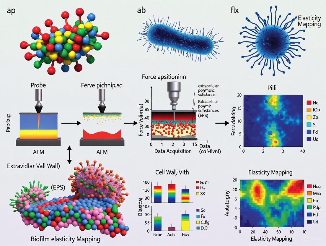
Atomic Force Microscopy (AFM) is a powerful scanning probe technique that has revolutionized the characterization of biological samples, providing nanoscale resolution of topographical and mechanical properties under physiological conditions [13]. The Force Volume (FV) technique is an advanced AFM mode that captures the spatial heterogeneity of mechanical properties by collecting an array of force-distance curves across a defined sample area [14] [15]. This method is particularly valuable for studying complex, heterogeneous biological systems such as bacterial biofilms, where mechanical properties vary considerably across different regions and structural components [16] [3].
Unlike single-point force measurements, Force Volume generates a complete mechanical map by performing indentation measurements at regular grid points, creating a "force volume" dataset where each pixel contains a full force-distance curve [14]. This capability is crucial for biofilm research, as it enables researchers to correlate mechanical properties with structural features, developmental stages, and compositional variations within these complex microbial communities [16] [5].
Theoretical Foundations
Basic Principles of Force-Distance Curves
The fundamental measurement in Force Volume AFM is the force-distance curve, which records the interaction forces between the AFM tip and sample surface as a function of piezoelectric actuator displacement [13]. Each force curve contains distinct regions reflecting different interaction regimes:
- Non-contact region: The tip and sample are separated with no significant interaction forces
- Jump-to-contact: Attractive forces may cause the tip to suddenly snap into contact with the surface
- Contact region: Repulsive forces dominate as the tip indents the sample
- Adhesion peak: Upon retraction, adhesive forces may cause the tip to stick to the surface before final separation [5] [13]
The deflection of the cantilever is measured using a laser beam reflected from the cantilever onto a position-sensitive photodetector, allowing precise force quantification with piconewton sensitivity [13].
Contact Mechanics Models
To extract quantitative mechanical properties from force-distance curves, several contact mechanics models can be applied. The most common for biological samples is the Hertz model, which describes the elastic deformation of two perfectly smooth, homogeneous bodies [5] [17]:
For a spherical indenter: [ F = \frac{4}{3}E^*R^{1/2}\delta^{3/2} ]
Where:
- (F) = applied force
- (E^*) = reduced Young's modulus
- (R) = tip radius
- (\delta) = indentation depth
The reduced modulus relates to the sample's Young's modulus ((E)) through: [ \frac{1}{E^*} = \frac{1-\nu{tip}^2}{E{tip}} + \frac{1-\nu{sample}^2}{E{sample}} ]
Where (\nu) is Poisson's ratio, typically assumed to be 0.5 for soft, incompressible biological samples [5] [17].
Table 1: Common Contact Mechanics Models for Biological Samples
| Model | Indenter Geometry | Application | Limitations |
|---|---|---|---|
| Hertz | Parabolic/Spherical | Homogeneous, linear elastic materials | Assumes small deformations, infinite thickness |
| Sneddon | Conical/Pyramidal | Sharper indenters, larger deformations | Stress concentration at tip |
| Johnson-Kendall-Roberts (JKR) | Spherical | Strong adhesive contacts | Complex fitting procedure |
| Derjaguin-Muller-Toporov (DMT) | Spherical | Weak adhesive contacts, stiff materials | May underestimate adhesion |
For biofilms, which often exhibit viscoelastic behavior and significant adhesion, modified approaches such as the Oliver-Pharr method or Johnson-Kendall-Roberts (JKR) model may be more appropriate, particularly when substantial adhesive forces are present [5] [17].
Experimental Design and Setup
Instrumentation Requirements
A standard AFM system equipped for Force Volume measurements requires several key components:
- Piezoelectric scanner: Capable of precise XYZ positioning, typically with a Z-range ≥5-7μm for accommodating biofilm topography [3]
- Photodetector: Position-sensitive for measuring cantilever deflection
- Fluid cell: For imaging in physiological conditions
- Environmental control: Temperature and humidity regulation for maintaining biofilm viability [3] [13]
Modern bio-AFMs often integrate with optical microscopy systems, allowing correlation of mechanical properties with fluorescence markers or structural features [13].
Cantilever and Probe Selection
Appropriate cantilever selection is critical for accurate mechanical characterization of soft biological samples like biofilms:
Table 2: Cantilever Selection Guide for Biofilm Characterization
| Parameter | Recommended Specification | Rationale |
|---|---|---|
| Spring Constant | 0.01-0.5 N/m | Soft enough to detect small forces without damaging samples |
| Tip Geometry | Spherical (2-10μm diameter) or pyramidal | Spherical tips reduce local stress and provide well-defined contact area |
| Tip Material | Silicon nitride | Biocompatible, suitable for liquid imaging |
| Resonance Frequency | 5-30 kHz in liquid | Optimized for operation in fluid environments |
| Cantilever Length | 100-200μm | Provides appropriate sensitivity |
Spherical probes are often preferred for biofilm mechanics as they minimize sample damage and provide a well-defined contact area for modulus calculation [15]. Cantilever spring constants must be calibrated before measurements, typically using thermal tuning or reference cantilever methods [13].
Step-by-Step Force Volume Protocol
Sample Preparation
Biofilm samples require careful preparation to maintain structural integrity and mechanical properties during AFM analysis:
- Substrate selection: Choose appropriate substrates such as glass coverslips, silicone sheets, or membrane filters that promote biofilm adhesion [3] [18]
- Hydration control: Maintain biofilms in hydrated conditions using fluid cells or humidity chambers (≥90% RH) to prevent artifacts from dehydration [3]
- Immobilization: For single-cell analysis within biofilms, consider gentle chemical fixation or mechanical confinement using porous membranes [5]
- Mounting: Secure sample firmly to prevent movement during scanning
System Configuration
- Cantilever installation: Mount selected cantilever and align laser detection system
- Approach: Engage tip with surface using minimal force setpoint to prevent sample damage
- Deflection sensitivity calibration: Perform on a rigid reference sample (e.g., silicon wafer) by measuring slope of force curve in contact region [14]
- Spring constant calibration: Determine using thermal noise method or reference cantilever [15]
Force Volume Acquisition Parameters
Configure scanning parameters to balance resolution, measurement quality, and acquisition time:
Table 3: Typical Force Volume Parameters for Biofilm Characterization
| Parameter | Recommended Value | Notes |
|---|---|---|
| Scan Size | 5-100μm | Dependent on feature size and heterogeneity |
| Force per Line | 16-64 | Determines XY resolution of mechanical map |
| Samples per Line | 128-512 | Affects resolution of height image |
| Number of Samples | 32-128 points/curve | Sets Z-resolution of force curves |
| Ramp Size | 500-2000nm | Must exceed sample roughness and indentation depth |
| Ramp Rate | 0.5-4Hz | Lower rates reduce hydrodynamic effects in fluid |
| Trigger Mode | Relative | Compensates for drift, protects tip and sample |
| Setpoint | Minimal contact force | Typically 0.5-2nN for soft samples |
The total acquisition time for a Force Volume map can be calculated as: [ T_{acquisition} = \frac{(Samples\;per\;line) \times (Force\;per\;line)}{Ramp\;rate} ]
Higher resolution maps with more force curves require significantly longer acquisition times [14].
Data Acquisition Workflow
The following diagram illustrates the complete Force Volume experimental workflow:
Optimization and Troubleshooting
- Minimize scanning forces: Use the lowest possible setpoint that maintains contact to prevent sample damage [14]
- Reduce hydrodynamic effects: Lower ramp rates in fluid environments to minimize drag forces on the cantilever [14]
- Center Z-position: Ensure piezoelectric actuator is centered in Z-direction to accommodate surface topography [14]
- Check trigger settings: Use relative trigger mode to compensate for drift and protect the tip [14]
- Monitor data quality: Regularly inspect force curves for proper shape and adequate indentation depth
Data Analysis Pipeline
Force Curve Processing
Raw force-distance data requires several processing steps before mechanical properties can be extracted:
- Conversion to force-displacement: Transform raw photodetector voltage and scanner position to true force and tip-sample separation using sensitivity and spring constant values
- Baseline correction: Subtract non-contact portion of approach curve to establish zero force baseline
- Contact point determination: Identify point of initial tip-sample contact using automated algorithms or manual selection
- Indentation calculation: Subtract cantilever deflection from scanner movement to determine sample indentation: ( \delta = (z - z0) - d ), where (z) is scanner position, (z0) is contact point, and (d) is cantilever deflection [13]
Modulus Extraction and Mapping
- Model fitting: Apply appropriate contact mechanics model (e.g., Hertz, Sneddon) to the approach portion of the force curve
- Parameter optimization: Use least-squares fitting to determine Young's modulus that best fits the experimental indentation data
- Map generation: Create spatial maps of Young's modulus by assigning calculated values to corresponding XY positions
- Statistical analysis: Calculate descriptive statistics (mean, median, distribution) for the mapped region to quantify heterogeneity [15]
Handling Biological Heterogeneity
Biofilms exhibit significant spatial and temporal mechanical heterogeneity, requiring specialized analytical approaches:
- Log-normal transformation: Many biological mechanical property distributions follow log-normal rather than normal distributions [15]
- Outlier exclusion: Remove artifacts from dust contamination, surface defects, or invalid measurements
- Regional analysis: Segment maps based on structural features identified from topography or complementary microscopy
- Time-series analysis: For dynamic studies, track mechanical changes at equivalent positions over time
Applications in Biofilm Research
Mapping Biofilm Mechanical Heterogeneity
Force Volume technique has revealed substantial spatial variations in mechanical properties within biofilms. A study measuring cohesive energy in activated sludge biofilms found values ranging from 0.10 ± 0.07 nJ/μm³ at the surface to 2.05 ± 0.62 nJ/μm³ in deeper regions, demonstrating increasing cohesiveness with biofilm depth [3]. Similar heterogeneity has been observed in elastic modulus maps of mixed-culture biofilms, with variations of an order of magnitude across different microenvironments [16].
Investigating Matrix Composition Effects
The Force Volume technique enables correlation of mechanical properties with biofilm composition and environmental factors:
- Calcium effects: Biofilms grown with 10mM calcium showed increased cohesive energy (1.98 ± 0.34 nJ/μm³) compared to controls (0.10 ± 0.07 nJ/μm³) [3]
- EPS contribution: Regions rich in extracellular polymeric substances typically exhibit higher adhesion and different viscoelastic properties [16] [5]
- Cellular vs. matrix regions: Distinct mechanical signatures can differentiate cellular clusters from surrounding matrix material [6]
Monitoring Biofilm Development
Time-dependent Force Volume mapping can track mechanical changes during biofilm maturation:
- Early attachment: Initial surface colonization shows higher elasticity and lower adhesion
- Maturation: Developing biofilms exhibit increased stiffness and cohesiveness as matrix production accumulates
- Dispersion: Mechanical properties shift at onset of dispersal phase, often showing reduced cell-matrix adhesion [16] [18]
The Scientist's Toolkit: Essential Materials
Table 4: Key Research Reagent Solutions for Biofilm Force Volume Experiments
| Item | Specification | Function | Example Applications |
|---|---|---|---|
| Cantilevers | Silicon nitride, spring constant: 0.01-0.5 N/m, spherical tips (2-10μm) | Force sensing and indentation | General biofilm mechanics [15] [13] |
| Calibration Samples | Rigid reference (silicon wafer), soft reference (PDMS gels) | System calibration and validation | Deflection sensitivity, spring constant [13] |
| Biofilm Substrates | Glass coverslips, membrane filters, silicone sheets | Biofilm growth support | Controlled adhesion surfaces [3] [18] |
| Immobilization Aids | Poly-L-lysine, porous membranes, PDMS microstructures | Sample stabilization during imaging | Single-cell analysis within biofilms [5] |
| Fluic Cells | Temperature-controlled, gas-permeable | Physiological environment maintenance | Live biofilm imaging in liquid [3] [13] |
| Culture Media | Chemically defined or complex growth media | Biofilm cultivation and maintenance | Specific nutritional environments [18] |
Advanced Applications and Future Directions
Recent technological advancements are expanding Force Volume capabilities for biofilm research:
- Large-area automated AFM: Combining multiple scans to create millimeter-scale maps while maintaining nanometer resolution [6]
- Machine learning integration: Automated analysis of high-dimensional mechanical data for pattern recognition and classification [6]
- Multimodal correlation: Combining Force Volume with fluorescence microscopy, Raman spectroscopy, or other techniques to correlate mechanics with composition and metabolic activity [18]
- High-speed Force Volume: Improved temporal resolution for capturing dynamic processes in living biofilms [13]
These developments are enabling researchers to bridge the gap between nanoscale mechanical properties and macroscopic biofilm behavior, providing new insights into biofilm resilience, dispersal, and response to antimicrobial agents.
The Force Volume technique represents a powerful approach for quantifying the spatial mechanical heterogeneity of biofilms, offering unique insights into their structure-function relationships and response to environmental challenges. When properly implemented with appropriate controls and analytical methods, it provides invaluable data for understanding biofilm mechanics in biomedical, environmental, and industrial contexts.
The mechanical characterization of biofilms provides critical insights into their behavior, stability, and resistance to external forces. Two parameters are fundamental to understanding biofilm mechanics: the elastic modulus, which quantifies a material's stiffness and its resistance to deformation under stress and adhesion forces, which describe the strength of attachment between the biofilm and a substrate or within the biofilm matrix itself [19]. Atomic Force Microscopy (AFM) has emerged as a powerful tool for quantifying these parameters at the nanoscale, enabling researchers to probe mechanical properties under physiological conditions [20] [5]. This application note details standardized protocols for AFM-based measurement of elastic modulus and adhesion forces in biofilms, supporting ongoing research in AFM force volume technique for biofilm elasticity mapping.
Quantitative Biomechanical Data in Biofilm Research
The following tables consolidate key quantitative findings from recent biofilm biomechanics research, providing reference values for experimental planning and data interpretation.
Table 1: Experimental Measurements of Biofilm Elastic Modulus
| Biofilm Type / Experimental Condition | Elastic Modulus (Approximate Range) | Measurement Technique | Reference |
|---|---|---|---|
| P. aeruginosa (Early biofilm) | Not Quantified | Microbead Force Spectroscopy | [20] |
| P. aeruginosa (Mature biofilm) | Instantaneous & delayed elastic moduli "drastically reduced" with maturation | Microbead Force Spectroscopy (Voigt Model) | [20] |
| Biofilm Streamers (P. aeruginosa PA14) | Differential Young's Modulus increases linearly with external stress | In-situ extensional rheology | [21] |
| General Biofilms | Viscous and elastic components characterized | Nanoindentation (Hertz Model) | [5] |
Table 2: Experimental Measurements of Biofilm Adhesion Forces
| Biofilm Type / Experimental Condition | Adhesion Force / Energy (Approximate Range) | Measurement Technique | Reference |
|---|---|---|---|
| Mixed Culture (from activated sludge) | Cohesive Energy: 0.10 ± 0.07 nJ/µm³ to 2.05 ± 0.62 nJ/µm³ | AFM-based abrasion with friction measurement | [3] |
| Mixed Culture (with 10 mM Ca²⁺) | Cohesive Energy: 0.10 ± 0.07 nJ/µm³ to 1.98 ± 0.34 nJ/µm³ | AFM-based abrasion with friction measurement | [3] |
| P. aeruginosa PAO1 (Early biofilm) | Adhesive Pressure: 34 ± 15 Pa | Microbead Force Spectroscopy | [20] |
| P. aeruginosa PAO1 (Mature biofilm) | Adhesive Pressure: 19 ± 7 Pa | Microbead Force Spectroscopy | [20] |
| P. aeruginosa wapR mutant (Early biofilm) | Adhesive Pressure: 332 ± 47 Pa | Microbead Force Spectroscopy | [20] |
| P. aeruginosa wapR mutant (Mature biofilm) | Adhesive Pressure: 80 ± 22 Pa | Microbead Force Spectroscopy | [20] |
| S. aureus (on 58S BAG, 0-1s contact) | Adhesive Force: ~3 nN | AFM Force Spectroscopy | [22] |
| E. coli (on 58S BAG, 0-1s contact) | Adhesive Force: ~6 nN | AFM Force Spectroscopy | [22] |
| Sulfate-Reducing Bacteria (on mica) | Tip-Cell Interaction Force: -3.9 to -4.3 nN | AFM Force-Distance Curves | [23] |
| V. cholerae (Colony biofilm) | Interfacial Adhesive Energy: ~5 mJ/m² | Capillary Peeling Technique | [19] |
Experimental Protocols
Protocol for AFM-Based Nanoindentation and Elastic Modulus Measurement
This protocol details the procedure for determining the elastic modulus of a biofilm using AFM nanoindentation, based on the Hertz model of contact mechanics [24] [5].
Principle: The AFM cantilever acts as a soft nanoindenter. Force-distance curves are collected at multiple points on the biofilm surface. The elastic modulus is derived by fitting the retraction curve to a theoretical contact mechanics model, most commonly the Hertz model, which describes the elastic deformation of two homogeneous smooth bodies touching under load [24].
Pre-Experimental Preparations
Biofilm Cultivation:
- Grow biofilms on suitable substrates (e.g., glass coverslips, membrane filters) in relevant growth media under controlled conditions appropriate for the bacterial strain.
- For consistent results, standardize growth time, temperature, and nutrient availability. Note that mechanical properties change with biofilm maturation [20].
Sample Immobilization:
- Chemical Fixation: Securely attach the biofilm-substrate to the AFM sample disk using a double-sided adhesive tape or a thin layer of epoxy. For hydrated measurements, use a liquid cell.
- Hydration Control: For measurements in moist conditions, equilibrate the sample in a humidity chamber (e.g., ~90% relative humidity) for at least one hour prior to measurement to maintain consistent water content [3].
AFM Calibration:
- Cantilever Selection: Use soft, rectangular cantilevers with nominal spring constants of 0.01 - 0.08 N/m for biological samples to avoid excessive deformation [20].
- Spring Constant Calibration: Employ the thermal tune method to determine the exact spring constant (k) of the cantilever for each experiment [20].
- Tip Characterization: Determine the geometry and radius of the AFM tip, as this is a critical parameter in the Hertz model.
Step-by-Step Measurement Procedure
- Instrument Setup: Mount the calibrated cantilever and the immobilized sample in the AFM. Engage the tip in contact mode or force volume mode under the desired environmental condition (air or liquid).
- Topographical Imaging: First, obtain a low-resolution topographic image of the biofilm area of interest at a low applied load (~0 nN) to identify measurement locations [3].
- Force Volume Mapping: Define a grid over the region of interest. The AFM will automatically acquire force-distance curves at each point in the grid. Key parameters include:
- Trigger Point: Set to ensure sufficient indentation depth while avoiding damage to the biofilm or the tip.
- Approach/Retract Speed: A typical range is 0.5 - 1.0 µm/s to minimize viscous effects.
- Data Points: Acquire at least 512 data points per curve for adequate resolution.
- Data Acquisition: Collect a minimum of 50-100 force curves from different locations across multiple biofilm samples to account for spatial heterogeneity.
Data Analysis and Interpretation
- Curve Pre-processing: Level the baseline of the force curve and correct for the trigger point.
- Hertz Model Fitting: For each force curve, fit the indentation segment of the approach curve to the Hertz model. For a pyramidal tip, the model is often expressed as: ( F = \frac{2 \cdot \tan(\alpha)}{\pi} \cdot \frac{E}{1 - \nu^2} \cdot \delta^2 ) where ( F ) is the applied force, ( \alpha ) is the face angle of the tip, ( E ) is the elastic modulus, ( \nu ) is the Poisson's ratio (typically assumed to be 0.5 for biofilms), and ( \delta ) is the indentation depth [24] [5].
- Statistical Analysis: Calculate the mean, median, and standard deviation of the elastic modulus values obtained from all valid force curves. Present data as mean ± standard deviation.
Protocol for Quantifying Adhesion Forces via Microbead Force Spectroscopy
This protocol describes a standardized method for quantifying biofilm adhesion forces using Microbead Force Spectroscopy (MBFS), which provides a defined contact geometry and high reproducibility [20].
Principle: A glass microbead attached to a tipless AFM cantilever is used as a probe. The bead is brought into contact with the biofilm surface and then retracted. The adhesion force is determined from the maximum pull-off force observed in the retraction force-distance curve.
Pre-Experimental Preparations
Probe Functionalization:
- Attach a 50 µm diameter glass microbead to a tipless silicon cantilever using a small amount of epoxy glue.
- Alternatively, for single-cell adhesion studies, a single bacterial cell can be attached to the cantilever to create a "cell probe" [5].
Biofilm Sample Preparation:
- Prepare biofilms as described in Section 3.1. For MBFS, a relatively flat and uniform biofilm surface is ideal.
Standardization of Conditions:
- To enable cross-experiment comparison, standardize the following parameters [20]:
- Loading Pressure: The force applied during the contact period.
- Contact Time: The duration the bead remains in contact with the biofilm (typically brief, e.g., 0-1 second [22]).
- Retraction Speed: The speed at which the bead is pulled away from the surface.
- To enable cross-experiment comparison, standardize the following parameters [20]:
Step-by-Step Measurement Procedure
- Instrument Setup: Mount the functionalized cantilever and the biofilm sample in the AFM. Use a closed-loop AFM system for accurate distance control.
- Approach and Contact: Position the bead above a selected location on the biofilm. Approach the surface at a constant velocity (e.g., 1 µm/s) until a predefined loading force or trigger point is reached.
- Dwell Period: Maintain the bead in contact with the biofilm for a standardized contact time (e.g., 250 ms to 1 second) to allow for bond formation [22].
- Retraction and Adhesion Measurement: Retract the cantilever at a constant speed (e.g., 1 µm/s) while recording the cantilever deflection. The adhesion force manifests as a negative peak (a "pull-off" event) in the retraction curve due to the cantilever bending downwards before detachment.
- Data Collection: Repeat steps 2-4 at a minimum of 50-100 different locations on the biofilm sample to obtain statistically significant data.
Data Analysis and Interpretation
- Force Curve Analysis: For each retraction curve, identify the minimum force value, which corresponds to the maximum adhesion force required to detach the probe from the biofilm.
- Adhesive Pressure Calculation (Optional): If the contact area between the microbead and the biofilm is known or can be estimated, the adhesion force can be normalized to this area to report an adhesive pressure in Pascals (Pa) [20].
- Binding Event Analysis: Analyze the retraction curve for multiple discrete "unbinding" events (sawtooth pattern), which can provide information on the number and strength of individual molecular bonds involved in adhesion.
The Scientist's Toolkit: Essential Research Reagents and Materials
Table 3: Key Research Reagents and Solutions for AFM Biofilm Biomechanics
| Item Name | Function / Application | Specific Examples / Notes |
|---|---|---|
| Tipless Cantilevers | Base for functionalized probes in adhesion studies. | CSC12/Tipless/No Al; used with microbeads for defined contact geometry [20]. |
| Glass Microbeads | Spherical probes for Microbead Force Spectroscopy (MBFS). | ~50 µm diameter; provide quantifiable contact area for adhesion pressure calculation [20]. |
| Polyolefin Membrane | Substrate for growing biofilms in reactor systems. | Used in membrane-aerated biofilm reactors (MABR) for consistent biofilm cultivation [3]. |
| Calcium Chloride (CaCl₂) | Modifier of biofilm cohesive strength. | Addition of 10 mM Ca²⁺ during cultivation increases biofilm cohesiveness [3]. |
| Extracellular Nucleic Acids | Target for mechanistic studies of matrix mechanics. | DNase I/RNase A; used to degrade eDNA/eRNA to probe their structural role in streamers and biofilms [21]. |
| Polydimethylsiloxane (PDMS) Stamps | Cell immobilization for stable AFM imaging. | Microstructured stamps immobilize microbial cells without chemical fixation, preserving native properties [5]. |
| Modified Postgate's Medium C | Culture medium for specific bacterial strains. | Used for cultivating Sulfate-Reducing Bacteria (SRB) in adhesion studies [23]. |
The precise quantification of elastic modulus and adhesion forces is paramount for advancing our understanding of biofilm mechanics. The protocols and data summarized in this application note provide a framework for standardized, reproducible AFM measurements. Key findings indicate that biofilm mechanical properties are dynamic, influenced by factors such as maturation time [20], the presence of divalent cations like calcium [3], and the composition of the extracellular matrix, particularly extracellular DNA (eDNA) which can confer stress-hardening behavior [21]. Adhering to these detailed methodologies will enable researchers in drug development and related fields to generate robust, comparable data, ultimately accelerating the development of strategies to control or eradicate biofilms.
Linking EPS Matrix Composition to Macroscopic Mechanical Behavior
Microbial biofilms are complex, structured communities of microorganisms enclosed in a self-produced extracellular polymeric substance (EPS) matrix. This matrix, which can account for up to 90% of the biofilm's dry mass, is primarily responsible for the mechanical stability and functional integrity of the biofilm [25]. The EPS is a hydrated gel-like structure comprising a network of polysaccharides, proteins, lipids, and nucleic acids, with a high water content typically ranging from 87% to 99% [26]. Understanding the relationship between the composition of this EPS matrix and the resulting macroscopic mechanical behavior is crucial for both controlling harmful biofilms and harnessing beneficial ones in industrial and medical applications.
The mechanical properties of biofilms represent a critical facet of their functionality, influencing their resistance to environmental stresses, detachment under fluid flow, and persistence against antimicrobial agents [25]. Biofilms exhibit viscoelastic behavior, meaning they combine both solid-like (elastic) and liquid-like (viscous) characteristics, enabling them to dissipate energy from external forces while maintaining structural cohesion [26] [25]. This property is fundamental to how biofilms withstand mechanical perturbations, spread to colonize new surfaces, or strengthen their existing structures. The cohesive strength of a biofilm, largely derived from the EPS matrix, is a primary factor affecting the balance between growth and detachment processes [3].
Advanced techniques like Atomic Force Microscopy (AFM) have revolutionized our ability to probe these mechanical properties at the nanoscale, under physiological conditions, and without extensive sample preparation that could alter inherent biofilm characteristics [27] [6]. This application note details how AFM-based force volume techniques can be systematically employed to map biofilm elasticity and establish quantitative correlations between EPS composition and macroscopic mechanical behavior.
Theoretical Foundation: From Polymer Networks to Macroscopic Behavior
The mechanical behavior of the EPS matrix can be conceptually understood through continuum mechanics models developed for polymer networks. For biofilms formed by mucoid Pseudomonas aeruginosa strains, whose major EPS carbohydrate component is alginate, the matrix can be modeled as a network of worm-like chains (WLCs) connected by transient junctions of specific lifetimes [26]. In this model, individual polysaccharide chains are characterized by their contour length (L), representing their fully extended length, and their persistence length (l_p), which indicates the typical length scale over which directional correlation along the chain is lost [26].
The force-extension relationship for a single WLC can be described by an interpolation formula that accounts for chain stiffness:
[ f(r) \approx \frac{kB\Theta}{lp} \left[ \frac{1}{4}\left(1 - \frac{r}{L}\right)^{-2} - \frac{1}{4} + \frac{r}{L} \right] ]
Where (k_B) is Boltzmann's constant, (\Theta) is the absolute temperature, and (r) is the end-to-end distance of the polymer chain [26]. This single-chain behavior can be upscaled to a three-dimensional network using approaches like the eight-chain model, which considers chains spanning from the center to the vertices of a cube, resulting in a strain energy function that describes the hyperelastic and viscoelastic response of the entire biofilm [26].
The diagram below illustrates the theoretical relationship between EPS composition and macroscopic mechanical properties:
Figure 1: Theoretical framework linking EPS composition to macroscopic mechanical properties through polymer network formation.
Experimental Protocols for AFM-Based Mechanical Characterization
Biofilm Cultivation and Sample Preparation
Protocol: Standardized Biofilm Growth for Mechanical Testing
Inoculum Preparation:
Substrate Coating:
Biofilm Growth:
- Incubate collagen-coated discs in BHI-plaque suspension under anaerobic conditions at 37°C [28] [29].
- Maintain with weekly fresh medium changes for maturity studies (e.g., 1-week vs. 3-week biofilms) [28].
- For defined chemical manipulation, incorporate specific ions (e.g., 10 mM CaCl₂) during cultivation to examine their effect on cohesive strength [3].
Sample Preparation for AFM:
- Gently rinse biofilm samples with 0.85% physiological saline for 1 minute to remove unattached cells [28].
- For hydrated imaging, mount samples in liquid cells with appropriate growth medium or buffer [6].
- For controlled humidity imaging, equilibrate samples in ~90% humidity for 1 hour before AFM analysis [3].
AFM Force Volume Technique for Elasticity Mapping
Protocol: Nanomechanical Mapping via Force Volume AFM
Cantilever Selection and Calibration:
- Select appropriate cantilevers based on biofilm stiffness: use softer cantilevers (spring constant ~0.01-0.1 N/m) for compliant biofilms and stiffer cantilevers (~0.5-1 N/m) for rigid biofilms [27] [3].
- Precisely calibrate the spring constant using thermal tuning or reference cantilever methods [27].
- Determine the optical lever sensitivity by acquiring force curves on a rigid reference sample (e.g., clean silicon wafer) [27].
AFM Operation Modes Selection:
- Utilize contact mode for high-resolution topographical imaging of rigid biofilm regions [27] [30].
- Employ tapping mode for delicate, loosely attached biofilms to minimize lateral forces that could damage the sample [30].
- Implement force volume mode for elasticity mapping: acquire arrays of force-distance curves (typically 16×16 to 128×128) across the biofilm surface [27].
Data Acquisition Parameters:
- Set appropriate loading rates (typically 0.5-2 Hz approach/retract rate) to capture viscoelastic responses [27].
- Apply constant loads in the range of 0.5-10 nN to remain in the linear elastic regime while ensuring sufficient indentation depth [3].
- Set maximum indentation depth to 10-15% of biofilm thickness to avoid substrate effects [27].
Mechanical Property Extraction:
- Fit the approach portion of force curves with appropriate contact mechanics models (e.g., Hertz, Sneddon, or Johnson-Kendall-Roberts models) to derive Young's modulus values [27].
- Calculate adhesion energy from the retraction curve hysteresis [28] [3].
- Generate spatial elasticity maps by assigning Young's modulus values to each force curve location in the array [27].
The experimental workflow for AFM-based mechanical characterization is summarized below:
Figure 2: Experimental workflow for AFM-based mechanical characterization of biofilms.
Cohesive Energy Measurement via AFM Abrasion
Protocol: Quantifying Biofilm Cohesive Strength
Baseline Topographical Imaging:
Controlled Abrasion Phase:
Post-Abrasion Imaging:
Cohesive Energy Calculation:
- Subtract consecutive height images to determine the volume of displaced biofilm material [3].
- Calculate frictional energy dissipated during abrasion from friction force data and scan parameters [3].
- Compute cohesive energy as the ratio of frictional energy dissipated to the volume of biofilm displaced (nJ/μm³) [3].
Quantitative Data on EPS-Mechanics Relationships
Maturation-Dependent Mechanical Properties
Table 1: Evolution of EPS and Mechanical Properties During Oral Biofilm Maturation
| Parameter | 1-Week Biofilm (Young) | 3-Week Biofilm (Mature) | Measurement Technique | Statistical Significance |
|---|---|---|---|---|
| Live Bacteria Volume | Baseline | Significantly Higher | CLSM with SYTO 9 staining | P < 0.01 [28] |
| EPS Matrix Volume | Baseline | Significantly Higher | CLSM with Alexa Fluor 647-dextran | P < 0.01 [28] |
| Surface Roughness | Higher | Significantly Lower | AFM Topography | P < 0.01 [28] |
| Cell-Surface Adhesion Force | Fairly Constant | Fairly Constant | AFM Force-Distance | Not Significant [28] |
| Cell-Cell Adhesion Force | Lower | Significantly Higher | AFM Force-Distance | P < 0.01 [28] |
Environmental Influence on Mechanical Properties
Table 2: Impact of Environmental Factors on Biofilm Mechanical Properties
| Factor | Effect on Mechanical Properties | Magnitude of Change | Proposed Mechanism | Reference |
|---|---|---|---|---|
| Calcium Ions (10 mM) | Increased cohesive energy | From 0.10 ± 0.07 to 1.98 ± 0.34 nJ/μm³ | Enhanced ionic cross-linking between polymer chains | [3] |
| Maturation (1 to 3 weeks) | Increased cell-cell adhesion | Significantly more attractive | Enhanced EPS production and cell interconnection | [28] |
| Shear Stress | Altered biofilm structure and exopolysaccharide production | Strain-dependent | Bacterial mechanosensing and response | [25] |
| Antibiotic Treatment | Modified mechanical response | Treatment-dependent | Matrix degradation or cellular lysis | [25] |
The Scientist's Toolkit: Essential Research Reagents and Materials
Table 3: Key Research Reagent Solutions for Biofilm Mechanics Studies
| Reagent/Material | Function/Application | Example Specifications | Experimental Considerations |
|---|---|---|---|
| Hydroxyapatite Discs | Mimics tooth/enamel surface for oral biofilms | 0.38-inch diameter, 0.06-inch thickness | Collagen coating enhances bacterial attachment [28] |
| Type I Collagen | Surface coating for improved biofilm adhesion | Bovine dermal, 10 mg/mL in 0.012 N HCl | Creates more uniform biofilm growth surface [28] |
| Brain Heart Infusion (BHI) Broth | Nutrient medium for biofilm growth | Standard formulation | Supports diverse microbial community growth [28] |
| Alexa Fluor 647-dextran | EPS matrix fluorescent labeling | 10 kDa molecular weight | Incorporated during biofilm formation for visualization [28] |
| SYTO 9 green-fluorescent stain | Live bacteria labeling | Nucleic acid stain | Enables viability assessment and volume quantification [28] |
| Calcium Chloride (CaCl₂) | Ionic cross-linking enhancement | 10 mM concentration in medium | Significantly increases cohesive energy [3] |
| Silicon Nitride AFM Cantilevers | Nanomechanical probing | Spring constant: 0.01-1 N/m | Proper spring constant calibration critical [27] [3] |
| Polyolefin Membrane | Biofilm growth substrate | 0.1-μm mean pore diameter, 34% porosity | Used in membrane-aerated biofilm reactors [3] |
Applications in Drug Development and Beyond
The quantitative relationship between EPS composition and mechanical properties has significant implications for pharmaceutical development and therapeutic strategies. Mechanical properties can serve as biomarkers of biofilm-related infection progression and treatment efficacy [25]. When biofilms are treated with antimicrobial agents, changes in their mechanical response provide insights into the compound's mechanism of action—whether it primarily kills bacteria, disrupts matrix cohesion, or both [25]. This approach enables high-throughput screening of anti-biofilm compounds by quantifying mechanical parameters before and after treatment [25].
The viscoelastic character of biofilms plays a crucial role in clinically relevant phenomena such as the formation of streamers—flexible three-dimensional filaments that cause clogging in medical devices [25]. Understanding how EPS composition influences viscoelasticity enables the development of combined chemical-mechanical strategies where chemical treatments reduce biofilm cohesiveness, thereby decreasing the force required for mechanical removal [25]. Furthermore, the ability to measure interaction forces between specific biomolecules using AFM techniques like Molecular Recognition Force Microscopy (MRFM) provides opportunities for investigating ligand-receptor interactions relevant to biofilm formation and stability [27].
Advanced AFM applications now allow for the large-area automated imaging of biofilm communities over millimeter-scale areas, overcoming traditional limitations of small imaging areas and enabling correlation of nanoscale mechanical properties with macroscopic biofilm architecture [6]. When combined with machine learning approaches for image analysis, these techniques provide unprecedented capability to map the spatial heterogeneity of mechanical properties throughout mature biofilm structures [6]. This comprehensive mechanical profiling is essential for developing targeted interventions against harmful biofilms while promoting the stability of beneficial biofilms in industrial and environmental applications.
A Step-by-Step Protocol for AFM Force Volume on Biofilms
Within the broader context of research employing Atomic Force Microscopy (AFM) force volume technique for biofilm elasticity mapping, the critical first step is the effective immobilization of hydrated, live biofilms. Successful nanomechanical characterization via AFM force spectroscopy hinges on sample preparation methods that preserve the native structure and mechanical properties of the biofilm. AFM is a powerful tool for generating high-spatial-resolution maps of nanomechanical attributes like elasticity under physiological conditions [31]. However, acquiring meaningful force-distance (f-d) curves using the force volume technique requires biofilms to be securely immobilized without altering their delicate extracellular polymeric substance (EPS) matrix or mechanical integrity [32]. This application note details standardized protocols for preparing and immobilizing hydrated biofilms to ensure reliable and reproducible AFM elasticity measurements.
Surface Selection and Functionalization
The substrate onto which a biofilm is grown is a primary factor controlling its immobilization during AFM analysis. The surface must provide sufficient adhesion to prevent detachment under the force of the AFM tip while being compatible with both biofilm growth and the AFM measurement itself.
Table 1: Common Substrates for Biofilm Immobilization in AFM Studies
| Substrate Material | Functionalization/Treatment | Key Characteristics | Compatibility with AFM Elasticity Mapping |
|---|---|---|---|
| Glass [6] | PFOTS (Perfluorooctyltrichlorosilane) treatment | Creates a hydrophobic surface; promotes specific cellular orientation and honeycomb pattern formation [6]. | Excellent for high-resolution topographical and mechanical mapping of initial attachment. |
| Hydroxyapatite (HAP) [33] | Fabricated into discs from <75 µm particle size HAP. | Mimics mineralized tooth surfaces; ideal for oral biofilm studies [33]. | Suitable for force-volume imaging under physiological fluid (PBS). |
| Silicon [6] | Various chemical modifications (e.g., PFOTS). | Allows for significant reduction in bacterial density via surface modification [6]. | Ideal for combinatorial studies on how surface properties influence attachment and mechanics. |
| Titanium [34] | Often used as discs in static models. | Relevant for studying biofilms on medical implants. | Compatible with AFM; requires secure mounting in the fluid cell. |
Biofilm Growth and Immobilization Protocols
Static Growth on Functionalized Surfaces
This protocol is ideal for growing biofilms directly on AFM-compatible substrates, ensuring firm attachment from the initial stages of colonization.
Detailed Protocol:
- Surface Preparation: Place sterilized substrates (e.g., PFOTS-treated glass coverslips or HAP discs) horizontally in a multi-well plate [6] [33].
- Inoculation: Inoculate the well with the bacterial strain of interest (e.g., Pantoea sp. YR343, Escherichia coli) in an appropriate liquid growth medium [6] [32].
- Incubation: Incubate at the optimal temperature for the strain (e.g., 37°C for many human pathogens) for the desired attachment period. For early attachment studies, this can be as brief as 30 minutes [6].
- Rinsing: After incubation, gently rinse the substrate with a sterile buffer (e.g., Phosphate Buffered Saline - PBS) to remove non-adherent planktonic cells. This step is critical to leave only the firmly attached cells for AFM analysis [6].
- Hydration Maintenance: Transfer the substrate to the AFM fluid cell and keep it submerged in PBS or growth medium throughout the measurement to maintain hydration [33].
Dynamic Growth and Transfer
For biofilms grown under flow conditions (e.g., in flow cells or bioreactors) that more closely mimic natural environments, a transfer protocol is required.
Detailed Protocol:
- Biofilm Growth: Grow biofilms under dynamic conditions in a flow cell system or constant flow chamber to achieve mature, 3D structures [34].
- Sample Extraction: Carefully extract a section of the colonized surface or a representative fragment of the biofilm.
- Immobilization in a Cell Culture Insert: Place the biofilm sample on a sterile membrane (e.g., polycarbonate) within a cell culture insert. This setup allows for nutrient exchange from below while providing a stable, flat surface for AFM indentation from above.
- Gentle Rinsing: Rinse with buffer to remove loosely attached cells, taking care not to disrupt the biofilm architecture.
- AFM Mounting: Securely mount the entire culture insert into the AFM fluid cell and submerge in buffer for measurement.
The following workflow diagram illustrates the key decision points and steps for selecting and executing these protocols:
Chemical Fixation for Structural Preservation
For certain experiments where high-resolution topography is the priority, or where the viability of cells during scanning is not a concern, chemical fixation provides excellent structural preservation.
Detailed Protocol:
- Growth and Rinsing: Grow and rinse the biofilm on the chosen substrate using the static method described in section 3.1.
- Fixation: Immerse the sample in a buffered paraformaldehyde solution (commonly 2-4%) for a defined period (e.g., 30-60 minutes) at room temperature. This crosslinks proteins and stabilizes the structure [35].
- Salt Removal: Rinse the fixed sample with deionized water or a volatile buffer like ammonium acetate to remove crystallizable salts [35].
- Dehydration (Optional): For AFM imaging in air, dehydrate the sample through a graded series of ethanol or acetone [35].
- Drying (Optional): Air-dry the sample in a desiccator or under a gentle stream of inert gas [35].
Note: Fixation and dehydration can significantly alter the native mechanical properties of the biofilm. This protocol is not recommended for studies aiming to measure the true in vivo elasticity of the biofilm.
The Scientist's Toolkit: Essential Research Reagents
Table 2: Key Research Reagent Solutions for Biofilm Immobilization
| Reagent/Material | Function in Immobilization Protocol | Example Usage |
|---|---|---|
| PFOTS (Perfluorooctyltrichlorosilane) [6] | Surface functionalization to create a hydrophobic surface that promotes specific bacterial attachment and orientation. | Used to treat glass coverslips for studying early biofilm assembly patterns [6]. |
| Hydroxyapatite (HAP) Discs [33] | Provides a clinically relevant substrate that mimics tooth enamel for growing oral biofilms. | Serves as the growth substrate for microcosm biofilms in studies of dental plaque mechanics [33]. |
| Paraformaldehyde [35] | Cross-linking fixative agent that preserves the 3D structure of biofilms and individual cells. | Used in a step-by-step preparation protocol for stabilizing cell morphology prior to analysis [35]. |
| Phosphate Buffered Saline (PBS) [33] | Isotonic buffer for rinsing and maintaining hydration; prevents osmotic shock to live cells. | Used to submerge biofilm samples during AFM analysis to maintain physiological conditions [33]. |
| Borosilicate Glass Spheres [33] | AFM probe functionalization; attached to tipless cantilevers to create a spherical indenter for reliable nanoindentation on soft biofilms. | Used for force-volume imaging to obtain mechanical properties like Young's modulus [33]. |
| UV Curing Resin [33] | Adhesive for securely attaching spherical indenters to AFM cantilevers. | Employed in the functionalization of NPO-10 tipless cantilevers with glass spheres [33]. |
Integration with AFM Elasticity Mapping
The ultimate goal of these immobilization strategies is to enable successful AFM force volume measurements. The prepared sample must be compatible with the AFM fluid cell and stable during the acquisition of hundreds to thousands of force curves.
Critical Considerations for AFM Measurement:
- Hydration: The biofilm must remain fully hydrated throughout the experiment. Any drying will drastically alter its mechanical properties and structure [13] [33].
- Substrate Mounting: The prepared substrate (coverslip, HAP disc, culture insert) must be firmly secured in the AFM fluid cell using a compatible holder or double-sided tape to prevent drift during long acquisition times.
- Control Measurements: Always perform control indentations on the bare substrate to characterize its mechanical properties. This allows the biofilm's contribution to the measured elasticity to be isolated.
- Model Selection: Analyze the resulting force curves with appropriate contact mechanics models (e.g., Hertz, Sneddon, Johnson-Kendall-Roberts) to extract quantitative nanomechanical properties like Young's modulus [31] [32]. The choice of model depends on tip geometry and the nature of the sample.
Cantilever Selection and Functionalization for Accurate Indentation
Within the broader scope of employing Atomic Force Microscopy (AFM) force volume techniques for biofilm elasticity mapping, the selection and preparation of the AFM probe are paramount. The cantilever acts as the primary transducer, converting tip-sample interactions into measurable signals. Its properties directly dictate the resolution, accuracy, and reliability of the nanomechanical data acquired. This application note provides detailed protocols and guidelines for researchers aiming to obtain quantitative indentation data on complex, viscoelastic biological samples like biofilms, ensuring that the methodology supports robust structure-property relationships in drug development research.
Key Concepts and Cantilever Selection Criteria
The mechanical properties of biofilms are heterogeneous and viscoelastic, requiring careful consideration of cantilever parameters to avoid measurement artifacts and ensure data validity [36] [5]. An inappropriate cantilever choice can lead to sample damage, over-estimation of elastic moduli, or poor signal-to-noise ratios.
Table 1: Cantilever Selection Criteria for Biofilm Indentation
| Parameter | Target Specification | Rationale | Considerations for Biofilms |
|---|---|---|---|
| Spring Constant (k) | 0.01 - 0.5 N/m [20] [36] [5] | Exerts minimal force on soft, delicate structures to prevent damage and ensure reversible, elastic contact. | Softer cantilevers (e.g., 0.01 N/m) are ideal for high-resolution cell surface mapping, while stiffer ones (e.g., 0.1-0.5 N/m) are better for penetrating the thicker EPS matrix [33]. |
| Tip Geometry | Spherical colloid probes (2-20 µm diameter) [20] [33] [37] | Defines a known, reproducible contact area with the sample, which is crucial for quantitative mechanical modeling. Avoids excessive local strain and sample piercing. | A larger sphere (e.g., 10 µm) provides an average property over multiple cells and EPS, representative of the bulk biofilm. A smaller sphere (e.g., 2 µm) can target specific features. |
| Tip Material | Silicon Nitride (Si₃N₄) | Biocompatible and commonly used for biological samples in fluid. | The native oxide layer can be functionalized for specific adhesion studies [37]. |
| Resonance Frequency | High (in the kHz range) in fluid [38] | Enables stable operation in liquid environments and facilitates faster scanning modes, reducing drift and experiment time. | A higher frequency minimizes the impact of fluid damping and environmental noise. |
| Cantilever Length | Short (e.g., 100-200 µm) [38] | Increases resonant frequency and improves stability during force spectroscopy measurements. | Shorter cantilevers are less susceptible to thermal drift and vibrations. |
The following workflow diagram outlines the key decision points and procedural steps for cantilever selection and functionalization.
Detailed Experimental Protocols
Protocol: Functionalization with Colloidal Probes
This protocol describes the procedure for attaching a spherical microbead to a tipless cantilever to create a probe with a defined geometry for quantitative nanomechanical mapping of biofilms [20] [33].
Principle: A borosilicate or glass microbead is attached to the end of a tipless cantilever using a UV-curing adhesive. This provides a well-defined, reproducible spherical contact geometry essential for applying contact mechanics models like Hertz or Sneddon.
Materials:
- Tipless cantilevers (e.g., Mikromasch CSC12/Tipless or Bruker NPO-10).
- Borosilicate microbeads (2 µm to 20 µm diameter, e.g., Whitehouse Scientific).
- UV-curing optical adhesive (e.g., Loctite 352).
- Micromanipulation system or fine probe station.
- Stereo microscope with high magnification.
- UV light source (λ = 400 nm).
Step-by-Step Procedure:
- Cantilever Preparation: Mount a tipless cantilever onto the AFM holder. Visually inspect it under the microscope to ensure it is clean and undamaged.
- Adhesive Application: Using a fine tungsten wire or glass fiber, apply a minuscule droplet of UV-curing adhesive to the very end of the cantilever. The goal is a small, controlled droplet just large enough to hold the microbead.
- Bead Attachment: Under the view of the stereo microscope, use a second micromanipulator to pick up a single microbead. Carefully bring the bead into contact with the adhesive droplet on the cantilever. Gently press and hold to ensure a strong bond.
- Curing: Expose the adhesive joint to UV light for the time specified by the manufacturer (e.g., 5 minutes [33]) to fully cure the resin.
- Curing Check: Visually verify under the microscope that the bead is securely attached and correctly aligned.
Protocol: Force Volume-Based Indentation on Biofilms
This protocol outlines the steps for acquiring nanomechanical maps of a hydrated biofilm using the force volume mode, which collects an array of force-distance curves across the sample surface.
Principle: The AFM tip performs a series of approach-retract cycles at predefined grid points. Each force-distance curve is analyzed to extract local mechanical properties, such as the Elastic (Young's) Modulus, by fitting the indentation segment to a contact mechanics model [33] [39] [5].
Materials:
- Functionalized cantilever (e.g., colloidal probe).
- Hydrated biofilm sample, grown on a suitable substrate (e.g., hydroxyapatite disc [33]).
- AFM equipped with a liquid cell and force volume capability.
- Appropriate physiological buffer (e.g., PBS).
Step-by-Step Procedure:
- System Setup: Mount the functionalized cantilever into the AFM head. Calibrate the spring constant using the thermal tune method [20] and determine the optical lever sensitivity on a clean, rigid surface (e.g., sapphire or clean glass).
- Sample Immobilization: Secure the biofilm sample in the fluid cell. For diffuse biofilms, chemical immobilization on poly-L-lysine coated substrates or mechanical trapping in porous membranes may be necessary to prevent detachment during scanning [5].
- Engagement and Imaging: Approach the cantilever to the biofilm surface in a region of interest under fluid. Obtain a low-force topographic image (e.g., using tapping mode) to identify the area for force mapping.
- Force Volume Parameter Setup:
- Define the scan size and pixel resolution (e.g., 32x32 or 64x64 grid over a 10x10 µm area).
- Set the maximum applied force (trigger threshold). This should be high enough to achieve sufficient indentation but low enough to avoid plastic deformation (typically 0.5-5 nN).
- Set the approach/retraction speed (typically 0.5-2 µm/s). Lower speeds are preferable for viscoelastic materials to minimize hydrodynamic effects.
- Set a dwell time at maximum load (e.g., 0-500 ms) to probe relaxation behavior.
- Data Acquisition: Initiate the force volume scan. The system will automatically acquire a force-distance curve at every pixel in the defined grid.
- Data Analysis:
- Use the AFM software or specialized analysis tools (e.g., MountainsSPIP [40]) to batch-process all force curves.
- For each curve, fit the indentation region of the approach curve with the Hertz model (for a spherical indenter):
- ( F = \frac{4}{3} \frac{E}{1-\nu^2} \sqrt{R} \delta^{3/2} )
- Where F is force, E is Young's Modulus, ν is the Poisson's ratio (assumed, typically 0.5 for incompressible materials), R is the tip radius, and δ is the indentation depth.
- Generate a spatial map of the calculated Young's Modulus.
The Scientist's Toolkit
Table 2: Essential Research Reagent Solutions for AFM Biofilm Mechanics
| Item | Function/Description | Example Use Case |
|---|---|---|
| Tipless Cantilevers | Base for functionalization with colloidal probes or specific ligands. | Creating custom spherical probes for quantitative indentation [20] [33]. |
| Borosilicate Microspheres | Provide a defined spherical geometry for the AFM tip. | Attached to tipless cantilevers to create colloidal probes with known contact area [33]. |
| UV-Curing Adhesive | Chemically inert glue that cures rapidly under UV light for secure attachment. | Used to fix microspheres to tipless cantilevers during probe functionalization [33]. |
| Poly-L-Lysine | Positively charged polymer that promotes cell and biofilm adhesion to negatively charged substrates. | Coating mica or glass slides to immobilize loose biofilms for stable AFM measurement [5]. |
| Calcium Chloride (CaCl₂) | Divalent cation that can cross-link anionic groups in EPS, altering biofilm cohesion. | Added to growth media or buffer to study the effect of ionic bridging on biofilm mechanical strength [3]. |
| JPK/Asylum/Bruker AFM Systems | Commercial AFM platforms with advanced force spectroscopy and force volume imaging capabilities. | Performing nanomechanical mapping and single-cell force spectroscopy in fluid [20] [33]. |
| MountainsSPIP Software | Advanced image and data analysis software for processing AFM force curves and generating property maps. | Batch-fitting thousands of force-distance curves to the Hertz model to create elasticity maps [40]. |
Atomic Force Microscopy (AFM) has become the dominant technique for characterizing mechanical properties at the nanoscale, transforming tip-sample interaction forces into quantifiable measurements of material properties [41]. In biofilm research, AFM-based force-distance curves (FDCs) provide critical insights into the spatial heterogeneity, cellular morphology, and mechanical resilience of these complex microbial communities [6]. The force volume technique, which involves acquiring a force-distance curve at each pixel across a sample surface, enables the generation of detailed nanomechanical maps that reveal how local subcellular features influence broader biofilm architecture and function [6] [41]. This capability is particularly valuable for understanding biofilm assembly, as mechanical properties significantly influence surface attachment, community development, and resistance to environmental stresses [6]. For researchers and drug development professionals, mastering the acquisition of high-quality FDCs is essential for developing effective biofilm control strategies, as the mechanical properties of biofilms often correlate with their recalcitrance to antimicrobial treatments.
Theoretical Principles of Force-Distance Curve Acquisition
Fundamental Mechanics
Force-distance curves are generated by modulating the tip-sample distance and recording the cantilever's deflection as a function of this distance [41]. The resulting curves capture the complete interaction history between the AFM tip and the sample surface during a single approach-retract cycle. The repulsive component of this interaction force is analyzed using contact mechanics models to extract quantitative mechanical parameters [41]. When operating on biofilms, which exhibit pronounced viscoelastic characteristics, the approach and retraction sections of the FDC often do not overlap, forming a hysteresis cycle that indicates inelastic and energy dissipation processes within the extracellular polymeric substance (EPS) matrix [41]. This hysteresis represents the viscoelastic nature of biofilms, where the sample deformation lags behind the tip's retraction due to energy dissipation [41].
Workflow for Nanomechanical Mapping
The process of nanomechanical mapping through force-volume acquisition follows a sequential pattern where mechanical properties are measured at individual points across the surface, with a complete map generated by repeating this process across all designated pixels [41]. This systematic approach ensures that all measurements within a single map are performed using the same contact mechanics model, enabling direct comparison of mechanical properties throughout the scanned area. For biofilm applications, this methodology allows researchers to correlate spatial variations in mechanical properties with structural features observed through complementary techniques, including the honeycomb patterns and cellular orientations that characterize early biofilm formation [6].
Critical Parameters for Optimal Data Collection
Instrument Configuration and Settings
The tables below summarize the essential parameters for acquiring high-quality force-distance curves on biofilm samples.
Table 1: Cantilever and Tip Selection Parameters
| Parameter | Recommended Specification | Functional Significance |
|---|---|---|
| Spring Constant | 0.01-0.5 N/m | Ensures sufficient sensitivity for soft biological samples without causing damage |
| Tip Geometry | Sharpened pyramidal or spherical tips (2-20 nm radius) | Determines contact area and spatial resolution; sharper tips for cellular features, spherical for bulk properties |
| Resonant Frequency | 1-60 kHz in fluid | Affects operating stability and noise floor in biological buffers |
| Cantilever Material | Silicon or Silicon Nitride | Biocompatibility and consistent mechanical properties in liquid environments |
| Reflective Coating | Gold (20-30 nm) | Enhances laser reflection signal while maintaining chemical inertness |
Table 2: Force-Distance Curve Acquisition Parameters
| Parameter | Optimal Range for Biofilms | Impact on Data Quality |
|---|---|---|
| Loading Rate | 0.1-10 Hz | Balances temporal resolution with force sensitivity; lower rates reduce hydrodynamic effects |
| Maximum Force | 0.5-5 nN | Minimizes sample damage while ensuring sufficient indentation for accurate modulus calculation |
| Z-Length | 500-2000 nm | Accommodates biofilm topography and ensures complete approach-retract cycle |
| Dwell Time | 0-1000 ms | Allows stress relaxation for viscoelastic characterization |
| Points per Curve | 512-4096 | Determines resolution of the force curve; higher values better capture mechanical transitions |
| Approach/Retract Velocity | 0.1-10 µm/s | Affects hydrodynamic drag and viscoelastic response; critical for accurate adhesion measurements |
Environmental Control Parameters
Maintaining physiological conditions during AFM imaging is paramount for preserving the native mechanical properties of biofilms. Measurements should be conducted in appropriate biological buffers (e.g., PBS) at controlled temperatures (20-37°C depending on the microbial species) to maintain viability and natural matrix hydration [6]. For time-series studies examining biofilm development, environmental chambers that control temperature, humidity, and gas composition are essential for maintaining sample viability over extended acquisition periods [6]. The liquid environment significantly influences tip-sample interactions, reducing adhesive forces compared to air while introducing hydrodynamic drag effects that must be accounted for in data interpretation [41].
Experimental Protocols
Sample Preparation of Biofilms
Protocol: Preparation of Pantoea sp. YR343 Biofilms for AFM Analysis
- Surface Treatment: Use PFOTS-treated glass coverslips to provide a consistent hydrophobic surface for bacterial attachment [6].
- Inoculation: Incubate treated coverslips in petri dishes containing Pantoea cells suspended in appropriate liquid growth medium [6].
- Attachment Period: Allow bacterial attachment to proceed for specific durations (e.g., 30 minutes for initial attachment studies, 6-8 hours for cluster formation) under optimal growth conditions [6].
- Rinsing: Gently rinse coverslips to remove unattached cells while preserving the architecture of adhered cells and extracellular matrix [6].
- Immobilization: Secure rinsed coverslips in AFM liquid cells using appropriate mounting techniques to prevent movement during scanning.
- Hydration Maintenance: Ensure biofilms remain fully hydrated throughout transfer and measurement processes to preserve native mechanical properties.
This protocol yields biofilms with characteristic cellular orientations and honeycomb patterning that are ideal for investigating structure-function relationships through nanomechanical mapping [6].
Force Volume Acquisition Protocol
Protocol: Sequential Acquisition of Force-Distance Curves
- Cantilever Calibration: Perform thermal tune or force curve on rigid surface (e.g., clean glass) to determine exact spring constant and optical lever sensitivity [41].
- Engage Parameters: Set engage parameters with reduced threshold force (typically 0.1-0.5 nN) to prevent excessive loading during initial engagement with soft biofilm surfaces.
- Region Selection: Define scan area size (typically 10×10 µm to 50×50 µm for cellular resolution) and pixel density (64×64 to 128×128 for force volume mapping) [6] [41].
- Waveform Selection: Choose appropriate modulation waveform based on imaging requirements:
- Set Point Optimization: Adjust maximum force set point to achieve 10-20% indentation depth relative to biofilm height to avoid substrate effects.
- Mapping Execution: Initiate force volume acquisition, monitoring data quality periodically to ensure consistent curve shape and absence of tip contamination.
- Quality Verification: Post-acquisition, review representative curves across the scan area to identify and exclude regions with artifacts or contamination.
Advanced Operational Modes
For specific biofilm investigations, alternative AFM operational modes offer complementary advantages:
Nano-DMA (Nanoscale Dynamic Mechanical Analysis): After establishing contact with a predefined setpoint force (1-20 nN), an oscillatory signal is applied to the cantilever or z-piezo while the tip remains in contact with the sample [41]. The resulting low-amplitude tip oscillation (10-50 nm) records force as a function of time, with the viscoelastic properties encoded in the time lag between tip indentation and applied force [41]. This method is particularly effective for characterizing frequency-dependent mechanical behavior of biofilm matrix components.
Parametric Nanomechanical Modes: Techniques such as bimodal AFM, contact resonance AFM, or multi-harmonic AFM drive the cantilever-tip system at its resonant frequency while recording oscillation parameters (amplitude, phase shift, frequency shifts) at each surface point [41]. These methods enable higher-speed imaging while maintaining nanomechanical sensitivity, though they typically require more complex data analysis approaches, including numerical methods to relate observables to mechanical parameters [41].
Data Processing and Analysis Methods
Curve Fitting and Mechanical Modeling
Protocol: Processing Force-Distance Curves for Elastic Modulus Calculation
- Baseline Correction: Subtract the non-contact portion of the approach curve to establish zero force and deflection references.
- Contact Point Identification: Locate the precise point where tip-sample interaction begins, typically identified as the deviation from baseline deflection.
- Indentation Calculation: Convert deflection data to indentation depth (δ) using the relationship: δ = (z - z₀) - (d - d₀), where z is scanner position, z₀ is contact point, d is deflection, and d₀ is baseline deflection.
- Force Calculation: Calculate applied force (F) using Hooke's Law: F = k × d, where k is the calibrated spring constant.
- Model Selection: Apply appropriate contact mechanics model:
- Hertz model: For elastic, non-adhesive contacts; most applicable to biofilm matrix.
- Sneddon extension: For different tip geometries (pyramidal, spherical).
- Johnson-Kendall-Roberts (JKR) model: When adhesion forces are significant.
- Parameter Extraction: Fit the indentation curve to extract Young's modulus (E) and other relevant mechanical parameters.
- Spatial Mapping: Compile extracted parameters from all curves to generate modulus, adhesion, or deformation maps co-registered with topographic data.
Machine Learning Integration
Recent advances incorporate machine learning (ML) approaches to enhance FDC data analysis through automated segmentation, classification, and defect detection in AFM images [6]. ML algorithms can significantly improve processing efficiency for the high-volume, information-rich data generated by force volume measurements, particularly when analyzing complex biofilm architectures containing numerous cells and extracellular components [6]. These tools assist in automating parameter extraction (cell count, confluency, cell shape, orientation) and enable quantitative analysis of microbial community characteristics over extensive areas [6].
Research Reagent Solutions
Table 3: Essential Materials for AFM Biofilm Mechanics Research
| Reagent/Material | Specification | Research Function |
|---|---|---|
| PFOTS-treated Glass | (Perfluorooctyltrichlorosilane) | Creates hydrophobic surface for controlled bacterial attachment and biofilm formation [6] |
| Liquid Growth Medium | Appropriate for microbial strain (e.g., LB for Pantoea sp.) | Supports bacterial viability during biofilm development and AFM measurement [6] |
| Biological Buffers | PBS, HEPES, or MOPS (pH 7.2-7.4) | Maintains physiological conditions during AFM imaging in liquid |
| Cantilever Cleaners | Piranha solution, UV ozone cleaner | Removes organic contaminants from AFM tips between measurements |
| Calibration Standards | Polystyrene, PDMS of known modulus | Verifies accuracy of mechanical property measurements |
| Fixatives | Glutaraldehyde, formaldehyde (0.1-2%) | Optional for stabilizing biofilm structure while minimizing mechanical alterations |
Troubleshooting and Quality Control
Common Artifacts and Solutions
- Tip Contamination: Manifested as irregular, inconsistent force curves with abnormal adhesion profiles. Solution: Implement regular tip cleaning protocols and verify tip integrity using standard samples.
- Substrate Effect: Artificially elevated modulus measurements when indenting thin biofilms. Solution: Maintain indentation depth below 10-20% of biofilm thickness and validate with varying maximum forces.
- Thermal Drift: Gradual changes in zero deflection during measurement sessions. Solution: Allow sufficient thermal equilibration time (30-60 minutes) and monitor drift rates before formal data collection.
- Hydrodynamic Effects: Excessive drag forces in liquid environments, particularly at higher approach velocities. Solution: Reduce approach velocities (<1 µm/s) and employ appropriate baseline correction methods.
Validation Methods
- Internal Consistency: Compare mechanical properties extracted from approach and retraction curves for similar indentation depths.
- Spatial Correlation: Verify that mechanical heterogeneities correspond to structural features observed in topography.
- Reproducibility: Repeat measurements on different sample regions and different days to establish measurement variance.
- Comparative Analysis: Validate against bulk mechanical measurements when available, recognizing potential scale-dependent differences.
The acquisition of high-quality force-distance curves requires meticulous attention to instrumental parameters, sample preparation, and environmental conditions, particularly when studying complex, hydrated systems like microbial biofilms. The force volume technique provides unparalleled capability to map spatial variations in mechanical properties across developing biofilm architectures, revealing fundamental structure-function relationships that govern biofilm resilience and persistence. By adhering to the detailed protocols and parameter guidelines outlined in this document, researchers can generate reproducible, quantitative nanomechanical data that advances our understanding of biofilm mechanics and informs the development of novel anti-biofilm strategies. Future developments in high-speed AFM modalities, combined with machine learning-assisted analysis, promise to further enhance our ability to correlate mechanical properties with biological function in these clinically relevant microbial communities.
Atomic Force Microscopy (AFM) has emerged as a pivotal technique for characterizing the nanomechanical properties of biological samples, including biofilms and living cells, under near-physiological conditions [42] [31] [24]. The core methodology involves nanoindentation experiments where a sharp probe interacts with the sample surface, generating force-distance (f-d) curves that contain rich information about local mechanical properties [31]. The processing and interpretation of these raw f-d curves to generate quantitative nanomechanical maps, such as Young's modulus (elasticity) maps, is a critical procedure in biofilm research, enabling the correlation of local mechanical properties with biological function and response to external stimuli [6] [32].
The inherent heterogeneity and complexity of biofilms present unique challenges for mechanical characterization. Traditional analytical methods often fail to capture the full spatial complexity of these structures, creating a need for advanced techniques that can link local cellular-scale properties to larger functional architectures [6]. AFM force volume imaging (FVI), which collects arrays of f-d curves over a defined spatial grid, addresses this need by providing a means to create detailed maps of mechanical properties [32]. However, the transformation of raw force curve data into reliable nanomechanical maps requires careful application of contact mechanics models, awareness of their limitations, and appropriate correction factors to account for experimental realities [42] [43].
Theoretical Foundations: Contact Mechanics Models
The interpretation of AFM indentation data relies fundamentally on models derived from contact mechanics theory. These mathematical frameworks relate the measured force and indentation depth to the intrinsic mechanical properties of the sample.
The Hertzian Framework and Its Extensions
The Hertz model serves as the cornerstone for analyzing AFM nanoindentation experiments on soft biological samples [42] [24]. Originally developed for the contact between two purely elastic spheres, it was later extended by Sneddon for various indenter geometries [43] [24]. The model assumes the sample is an elastic, homogeneous, and isotropic half-space [42].
For a conical indenter with half-opening angle α, the force-indentation (F-δ) relationship is expressed as:
Where E is the Young's modulus (Pa) and ν is the Poisson's ratio of the sample [43].
For spherical indenters of radius R, the relationship becomes:
In practice, most AFM tips are pyramidal with a rounded apex, making the sphero-conical model the most accurate representation. The Briscoe et al. equation provides the precise F-δ relationship for this geometry, though it is computationally more complex [42].
Limitations and Necessary Corrections
The standard Hertz model does not account for several factors critical to biological measurements:
- Viscoelasticity: Biological materials exhibit rate-dependent behavior. The Hertz model assumes purely elastic response, which can lead to inaccuracies unless measurements are performed at sufficiently slow rates to minimize viscous effects [42].
- Sample Dimensions and Substrate Effects: The model assumes the sample is infinitely thick. For thin samples like bacterial cells, the underlying rigid substrate can significantly stiffen the measurement if indentation exceeds 10% of sample thickness [42] [31].
- Sample Geometry and Tilt: The assumption of a perfectly planar surface is often violated with biological samples. Local tilt at the probe-sample interface introduces asymmetry, requiring incorporation of correction coefficients into the Hertz model for accurate measurements [43].
- Adhesive Forces: The standard Hertz model neglects adhesive interactions between the tip and sample, which can be significant in biological systems. Alternative models like Johnson-Kendall-Roberts (JKR) or Derjaguin-Müller-Toporov (DMT) are more appropriate when adhesion is prominent [31].
Table 1: Key Contact Mechanics Models for AFM Nanoindentation
| Model/Correction | Best Suited For | Key Assumptions & Limitations |
|---|---|---|
| Standard Hertz Model | Isotropic, elastic, homogeneous materials; small deformations [42] | Neglects adhesion, viscoelasticity, substrate effects, and sample tilt [42] [43] |
| Sneddon Extensions | Conical, spherical, or parabolic indenters [43] | Same core limitations as Hertz model; geometry-specific |
| Sphero-Conical Model | Realistic pyramidal AFM tips with rounded apex [42] | More complex calculation; other Hertzian limitations may still apply |
| Tilt-Corrected Hertz | Non-planar samples, inclined surfaces [43] | Incorporates local tilt angle; improves accuracy on heterogeneous biofilms |
| JKR/DMT Models | Samples with significant adhesive forces [31] | Accounts for adhesion; requires knowledge of surface energy or adhesion range |
Experimental Protocol: From Sample Preparation to Data Acquisition
Sample Preparation and Immobilization
Materials:
- Bacterial Strain: Pantoea sp. YR343 (or biofilm organism of interest) [6].
- Substrate: PFOTS-treated glass coverslips or other functionally-modified surfaces (e.g., silicon, polyacrylamide gels) to study surface property effects [6] [43].
- Growth Medium: Appropriate liquid growth medium (e.g., Lysogeny Broth).
- Fixation: Glutaraldehyde (optional, for fixed samples) or physiological buffer for live imaging.
Procedure:
- Inoculation: Place substrate (e.g., PFOTS-treated glass coverslip) in a petri dish and inoculate with bacterial culture in liquid growth medium [6].
- Incubation: Incubate at appropriate temperature for selected time points (e.g., 30 minutes for initial attachment studies; 6-8 hours for cluster formation) [6].
- Rinsing: Gently rinse the substrate with deionized water or buffer to remove non-adherent cells [6].
- Immobilization: For live imaging, maintain sample in appropriate buffer. For fixed imaging, treat with glutaraldehyde (2.5% in buffer for 1 hour) followed by thorough rinsing [32].
AFM Instrumentation and Measurement Parameters
Essential Equipment and Reagents:
Table 2: Research Reagent Solutions and Essential Materials
| Item/Category | Specific Examples | Function/Purpose |
|---|---|---|
| AFM System | Bruker NanoWizard, CellHesion [24] | Core instrument for force volume imaging and nanoindentation. |
| Cantilevers | Silicon Nitride (Si₃N₄) tips, spherical colloidal probes [43] [32] | Transduce force; different geometries (pyramidal, spherical) suit different models. |
| Calibration | Standard sample of known modulus (e.g., polyacrylamide gel) [43] | Validate the entire measurement and processing pipeline. |
| Sample Substrates | PFOTS-treated glass, silicon, polyacrylamide gels [6] [43] | Provide a surface for biofilm growth with controllable properties. |
| Buffer Systems | Phosphate Buffered Saline (PBS), appropriate growth media | Maintain physiological conditions for live samples. |
Measurement Steps:
- Cantilever Selection and Calibration:
- Choose a cantilever with an appropriate spring constant (k), typically 0.01-0.1 N/m for soft biological samples [42] [32].
- Calibrate the cantilever's spring constant using the thermal tune method or other established protocols.
- Determine the precise tip geometry (radius for spherical tips, half-angle for conical/pyramidal tips) via electron microscopy or blind reconstruction [42].
- Force Volume Imaging Acquisition:
- Engage the AFM on the region of interest under fluid conditions if required.
- Define a spatial grid (e.g., 16x16 to 64x64 points) over the sample surface.
- At each point, acquire a complete force-distance curve with parameters such as:
- Z-length: Sufficient to engage and retract fully from the surface (typically 500-1000 nm).
- Approach/Retract Velocity: Slow enough to minimize viscous effects (e.g., 0.5-2 µm/s) [42].
- Trigger Point: Set to a force sufficient for measurable indentation without damaging cells.
- Save the entire force volume dataset for subsequent offline processing.
Data Processing Workflow: From Raw Curves to Elasticity Maps
The transformation of raw force-distance curves into a reliable nanomechanical map involves a multi-stage computational process. The following workflow diagram outlines the key steps, highlighting critical decision points and potential correction pathways.
Step-by-Step Computational Analysis
Step 1: Pre-processing and Critical Point Detection
The initial stage involves preparing the raw data for analysis, which is often automated using specialized algorithms [32].
- Baseline Correction: Subtract the non-contact portion of the approach curve to define the zero-force baseline.
- Contact Point Identification: Automatically detect the precise point of contact between the tip and the sample, a critical step for accurate indentation calculation [32]. This is often identified by a change in slope or curvature from the baseline.
- Detect Other Transitions: Identify other critical points in retraction curves, such as jumps or slope changes, marking molecular unbinding or polymer elongation events [32].
Step 2: Indentation Calculation and Data Segmentation
- Indentation Depth (δ): For each data point after contact, calculate the indentation as δ = (Z - Z₀) - (d - d₀), where Z is the scanner position, Z₀ is the contact point, d is the deflection, and d₀ is the baseline deflection.
- Force Calculation: Calculate the force F = k × d, where k is the cantilever spring constant.
- Data Segmentation: Segment the force curve into relevant regions for piecewise parametric modeling (e.g., electrostatic region, contact region) [32].
Step 3: Model Fitting and Parameter Extraction
- Model Selection: Based on sample properties and experimental conditions (see Table 1), select the appropriate contact mechanics model.
- Curve Fitting: Fit the F-δ data in the contact region to the selected model using a least-squares regression algorithm to extract the Young's modulus (E) [32]. The Poisson's ratio (ν) is often assumed (e.g., 0.5 for incompressible biological materials).
- Adhesion Analysis (for retraction curves): Analyze the retraction curve using models like the Freely Jointed Chain (FJC) or Worm-Like Chain (WLC) to quantify adhesion forces and polymer unfolding properties [32].
Step 4: Spatial Map Generation and Validation
- Map Construction: Compile the Young's modulus values extracted from each force curve in the force volume grid into a two-dimensional spatial map, visually representing the elasticity distribution across the scanned area.
- Statistical Validation: Assess the quality and reliability of the map. Large datasets enabled by High-Speed AFM (HS-AFM) allow for robust statistical analysis and determination of measurement uncertainty [44]. Compare results against a reference material of known modulus to validate the entire processing pipeline [43].
Critical Parameters and Best Practices for Biofilm Characterization
Successful nanomechanical mapping of biofilms requires careful attention to experimental parameters and data interpretation. The following table summarizes key parameters and their typical values or considerations for biofilm studies.
Table 3: Key Experimental Parameters for AFM Nanoindentation of Biofilms
| Parameter | Consideration/Range for Biofilms | Impact on Results |
|---|---|---|
| Spring Constant (k) | 0.01 - 0.1 N/m [32] | Softer levers for sensitive detection on soft samples. |
| Indentation Depth (δ) | < 10% of sample thickness [31]; typically 100-500 nm | Avoids substrate effect; stays in linear elastic regime. |
| Indentation Rate | Slow (e.g., 0.5 - 2 µm/s) [42] | Minimizes viscoelastic effects. |
| Poisson's Ratio (ν) | Often assumed 0.5 (incompressible) [43] | Required for model fitting; small error if assumption is reasonable. |
| Tip Geometry | Spherical (R = 0.5 - 5 µm) preferred for biofilms [31] | Reduces local stress and sample damage. |
| Number of Curves | 100s to 1000s per map [44] [32] | Ensures statistical power and representativeness. |
| Environmental Control | Fluid cell, temperature control | Maintains native state and viability for live biofilms. |
Overcoming Common Challenges
- Accounting for Sample Tilt and Heterogeneity: Biofilms are rarely flat. The local tilt angle (β) at each indentation point must be estimated from topography data and incorporated into the Hertz model using correction coefficients to prevent overestimation of Young's modulus [43].
- Managing Biological Variability: Biofilm mechanical properties can vary over orders of magnitude. Collecting large datasets (e.g., >200 force volume images) is crucial for achieving statistical significance and distinguishing subtle differences between samples [44].
- Automated Processing: Given the volume of data (a single 64x64 force volume generates 4,096 curves), robust, automated processing algorithms are essential for efficient and objective analysis [6] [32]. Machine learning methods are increasingly used for tasks like segmentation and classification of AFM data [6].
- Model Selection Justification: Always report the specific model used, the assumed parameters (like ν and tip geometry), and the justification for the choice. This transparency is critical for reproducibility and comparing results across studies [42].
The pathway from raw AFM force curves to quantitative nanomechanical maps is a multi-step process that integrates careful experiment design, rigorous application of contact mechanics models, and robust computational processing. While the Hertz model provides a foundational framework, its successful application to complex, heterogeneous biofilms necessitates awareness of its limitations and the implementation of advanced corrections for tilt, adhesion, and substrate effects. By adhering to detailed protocols for sample preparation, data acquisition, and automated data processing—incorporating validation and correction loops—researchers can generate reliable, high-resolution maps of biofilm elasticity. These maps are powerful tools for elucidating the structure-function relationships in microbial communities, ultimately advancing research in antimicrobial resistance, biofilm mitigation, and drug development.
Atomic Force Microscopy (AFM) force volume technique has emerged as a powerful tool in biofilm research, enabling the nanoscale mapping of mechanical properties crucial for understanding biofilm development and resilience. This application note details protocols for utilizing AFM force volume to track the dynamic changes in biofilm elasticity during maturation and to quantitatively assess the efficacy of anti-biofilm agents. By providing high-resolution spatial maps of Young's modulus, this method offers researchers and drug development professionals critical insights into structure-function relationships and treatment outcomes, bridging the gap between cellular-scale events and the functional macroscale organization of biofilms [6].
Application Note 1: Tracking Biofilm Maturation via Elasticity Mapping
The cohesive strength and mechanical integrity of a biofilm evolve significantly during its maturation, directly influencing its stability and resistance to removal strategies [3]. AFM force volume mapping allows for the in situ measurement of local elastic modulus as a function of biofilm depth and age, providing a quantitative descriptor of maturation.
Key Experimental Findings
Table 1: Changes in Biofilm Cohesive Energy During Maturation
| Biofilm Depth / Maturation Stage | Cohesive Energy (nJ/μm³) | Experimental Conditions | Significance |
|---|---|---|---|
| Superficial Layer / Early Stage | 0.10 ± 0.07 [3] | Undefined mixed culture from activated sludge; 1-day growth | Represents initial, weakly attached layers. |
| Deeper Layer / Mature Stage | 2.05 ± 0.62 [3] | Undefined mixed culture from activated sludge; 1-day growth | Indicates 20-fold increase in cohesion, reflecting matrix development and cellular packing. |
| With Calcium Addition (10 mM) | 1.98 ± 0.34 [3] | Biofilm cultivated with supplemental CaCl₂ | Demonstrates the role of divalent cations in cross-linking EPS, significantly enhancing biofilm mechanical strength. |
Detailed Protocol: Force Volume Mapping for Maturation Tracking
This protocol is adapted from methods for measuring cohesive energy and automated force volume processing [3] [8].
Step 1: Biofilm Cultivation and Sample Preparation
- Reactor Setup: Grow biofilms in a suitable reactor (e.g., membrane-aerated biofilm reactor, CDC Biofilm Reactor [3]).
- Sample Extraction: After a designated growth period (e.g., 1 day), extract a small piece (∼1 cm x 1 cm) of the biofilm-substrate complex.
- Humidity Control: Place the sample in a chamber with a saturated NaCl solution for 1 hour to equilibrate to ~90% relative humidity. This step is critical for maintaining consistent biofilm-water content without submerging the sample during AFM measurement [3].
- Mounting: Secure the equilibrated sample on the AFM stage. If using a humidity-controlled chamber, maintain 90% RH throughout the experiment [3].
Step 2: AFM Force Volume Acquisition
- Probe Selection: Use a V-shaped microfabricated cantilever with a pyramidal, oxide-sharpened Si₃N₄ tip (e.g., nominal spring constant of 0.58 N/m) [3]. Pre-calibrate the exact spring constant.
- Mapping Parameters: Define a grid (e.g., 64x64 or 128x128 points) over the region of interest on the biofilm surface. At each point, a full force-distance curve will be acquired.
- Acquisition Settings: Set a scan velocity in the range of 50 to 100 μm/s. For approach/retraction, use a maximum force trigger setpoint (e.g., 0.5-1 nN) sufficient to achieve a measurable indentation without damaging the biofilm [8].
Step 3: Data Processing and Young's Modulus Extraction
- Automated Curve Analysis: Process the force-volume dataset using automated algorithms [8]. The key steps include:
- Baseline Correction: Subtract the non-contact baseline from each force curve.
- Contact Point Detection: Automatically identify the point of tip-sample contact, a critical step for accurate indentation calculation [8].
- Model Fitting: Fit the indentation segment of the approach curve to the Sneddon model for a pyramidal tip [45]: ( F = \frac{E}{1-ν^2} \cdot \frac{2 \cdot \tanα}{π} \cdot δ^2 ) where F is force, E is Young's Modulus, ν is Poisson's ratio (assumed to be 0.5 for soft biological samples), α is the semi-top angle of the tip, and δ is the indentation depth.
- Spatial Mapping: Generate a 2D map of Young's modulus representing the spatial distribution of elasticity across the scanned biofilm area.
Application Note 2: Testing Anti-biofilm Agent Efficacy
AFM force volume can quantify the nanomechanical alterations biofilms undergo upon treatment with antimicrobials, providing a direct measure of efficacy that complements traditional bioburden assays.
Key Experimental Findings
Table 2: AFM-based Assessment of Anti-biofilm Agent Efficacy
| Anti-biofilm Treatment | Target Biofilm | Key AFM Findings & Efficacy Metrics | Experimental Context |
|---|---|---|---|
| Not Specified | Pantoea sp. YR343 | High-resolution imaging revealed disruption of characteristic honeycomb pattern and flagellar connections, key structural elements for biofilm cohesion [6]. | Demonstration of large-area AFM's capability to visualize treatment-induced structural degradation. |
| Tobramycin | P. aeruginosa | Significantly greater reduction in biofilm bioburden on glass beads compared to standard coupons (p=0.035), demonstrating platform-dependent efficacy [46]. | Evaluation of antibiotic efficacy under different biofilm growth models (glass bead reactor vs. CDC reactor). |
| Calcium Chelation | Mixed-species Biofilm | Reduction of cohesive energy from ~2.0 nJ/μm³ to near 0.1 nJ/μm³, demonstrating the critical role of divalent cations in maintaining biofilm mechanical strength [3]. | Proof-of-concept for targeting ionic cross-linking in the EPS as an anti-biofilm strategy. |
Detailed Protocol: Efficacy Testing via Nanomechanical Profiling
This protocol outlines a comparative assessment of treated versus untreated biofilms.
Step 1: Pre-treatment Baseline Characterization
- Control Sample: Prepare a baseline biofilm sample as described in Section 2.2.
- Elasticity Map: Acquire a force volume map of the untreated biofilm to establish the baseline mechanical properties (Young's Modulus) and spatial heterogeneity.
Step 2: Application of Anti-biofilm Agent
- Treatment Group: Apply the anti-biofilm agent (e.g., antibiotic, enzyme, surfactant) to the biofilm under conditions relevant to its intended use (e.g., concentration, contact time, solvent). Critical parameters like humidity must be controlled to avoid confounding results [47].
- Negative Control: A separate biofilm sample should be subjected to the same handling and solvent (e.g., buffer) without the active agent.
- Rinsing: Gently rinse the sample with an appropriate buffer (e.g., PBS) to remove residual agent and detached cells. Allow the sample to equilibrate to the measurement humidity.
Step 3: Post-treatment AFM Measurement and Analysis
- Location Matching: If possible, locate and scan the same general area measured during baseline characterization, though large-area AFM with machine learning can analyze entire communities [6] [48].
- Data Acquisition: Acquire a new force volume map using identical AFM parameters as the baseline measurement.
- Comparative Analysis:
- Generate post-treatment Young's modulus maps.
- Quantify the mean and distribution of Young's modulus for both control and treated biofilms.
- Statistically compare the values (e.g., Student's t-test) to determine if the treatment induced a significant softening (reduction in Young's modulus) of the biofilm structure.
- Correlate nanomechanical changes with traditional efficacy metrics like Log Reduction from CFU counts [46].
The Scientist's Toolkit: Essential Research Reagents and Materials
Table 3: Key Reagent Solutions for AFM-based Biofilm Elasticity Studies
| Item | Function / Role | Specification / Example |
|---|---|---|
| AFM Probes | Nanoscale indenter for force curve acquisition. | Sharp, oxide-sharpened Si₃N₄ tips (e.g., PPP-CONTPt); V-shaped or rectangular cantilevers with low spring constants (0.1 - 1 N/m) [3] [45]. |
| Biofilm Reactor | Cultivating standardized, reproducible biofilms. | CDC Biofilm Reactor [46], Membrane-aerated Biofilm Reactor [3], or custom Glass Bead Reactor for high-throughput screening [46]. |
| Growth Medium | Supporting biofilm growth with defined chemistry. | Cation-adjusted Mueller Hinton Broth (CAMHB) for antibiotic tests [46]; BHI or defined media with/without Ca²⁺ supplementation to modulate cohesion [3]. |
| Buffers & Salts | Maintaining physiological conditions during AFM; manipulating EPS cross-linking. | PBS for rinsing; NaCl for humidity control; CaCl₂ to enhance cohesion; chelators (e.g., EDTA) to disrupt ionic bonds in EPS [3]. |
| Software for FV Processing | Automated analysis of force-volume images; extraction of Young's modulus maps. | Custom algorithms (e.g., Hertz/Sneddon model fitting) [45] [8] or commercial software with nanoindentation analysis modules. |
| Reference Strains | Benchmarking performance across labs. | Staphylococcus aureus ATCC 6538, Pseudomonas aeruginosa ATCC 15442 or 27853, known for robust biofilm formation [49] [46]. |
The AFM force volume technique provides an unparalleled, quantitative method for tracking the mechanical maturation of biofilms and evaluating the mode of action and efficacy of anti-biofilm agents. By moving beyond simple topographical imaging to generate nanomechanical property maps, researchers can gain deeper insights into the structural-functional relationships that underpin biofilm resilience. The protocols and applications detailed herein provide a framework for integrating this powerful technology into the drug development pipeline, ultimately contributing to the creation of more effective strategies for biofilm control.
Overcoming Challenges and Enhancing Force Volume Technique Performance
In the study of biofilm elasticity using Atomic Force Microscopy (AFM) force volume techniques, data integrity is paramount. The mechanical properties derived from this method, such as Young's modulus, provide critical insight into biofilm behavior, antimicrobial resistance, and cellular functions [31]. However, the soft, hydrated, and heterogeneous nature of biofilms makes their nanomechanical characterization particularly susceptible to artifacts, primarily from tip-sample adhesion and substrate effects [38]. These artifacts can skew data, leading to inaccurate biological interpretations—for instance, overestimating stiffness by an order of magnitude or misrepresenting local mechanical variations. This application note details the identification and correction of these common artifacts within the context of biofilm elasticity mapping, providing structured protocols to ensure robust and reproducible data for researchers and drug development professionals.
Artifact I: Tip-Sample Adhesion
Principle and Identification
Tip-sample adhesion arises from attractive forces between the AFM probe and the biofilm surface. These forces, including van der Waals, electrostatic, and capillary interactions, can significantly distort force-distance (f-d) curves, which are the foundation of elasticity mapping [31] [50]. In force volume mode, where arrays of f-d curves are captured, uncontrolled adhesion can lead to a systematic overestimation of the biofilm's apparent stiffness.
Key indicators of significant adhesive interactions can be identified in the retract segment of the f-d curve [31]:
- Adhesion Hysteresis: The area between the approach and retract curves indicates energy dissipation due to adhesive forces.
- Adhesion Force (
F_ad): A negative deflection "pull-off" event during tip retraction signifies the force required to break the tip-sample bond. - Non-Baseline Return: The retract curve fails to return to the initial baseline, indicating material sticking to the tip.
The following workflow outlines the diagnostic process for this artifact:
Quantitative Impact and Thresholds
The following table summarizes key adhesion-related parameters, their quantitative impact on elasticity measurement, and proposed thresholds for biofilm studies. Adhesion forces substantially higher than these benchmarks will dominantly influence the contact mechanics, violating the assumptions of standard elastic models [3].
Table 1: Adhesion Parameters and Their Impact on Biofilm Elasticity Measurement
| Parameter | Description | Typical Range in Biofilms | Proposed Action Threshold | Impact on Young's Modulus |
|---|---|---|---|---|
Adhesion Force (F_ad) |
Force required to separate tip from sample [3]. | 0.1 - 10 nN | > 5% of maximum applied load | Leads to overestimation; effect magnified in softer samples. |
| Adhesion Energy | Work done during tip-sample separation (area under retract curve) [3]. | 0.1 - 2.0 nJ/µm³ [3] | > 1 nJ/µm³ | Indicates strong bonding, invalidating simple Hertzian fit. |
| Work of Adhesion | Energy per unit area required to separate the interfaces. | Varies with surface chemistry | Qualitative comparison | Higher work of adhesion necessitates use of adhesive contact models. |
Correction Protocols
Protocol 2.3.1: Optimization of Imaging Environment and Probe
- Objective: Minimize spurious adhesive forces.
- Materials:
- Liquid cell for AFM.
- Appropriate buffer (e.g., PBS or growth medium).
- Sharp, non-functionalized silicon nitride tips (e.g., for contact mode).
- Procedure:
- Conduct all measurements in liquid environment (e.g., relevant buffer) to eliminate capillary forces present in air [5].
- Use tips with lower spring constants (e.g., 0.01 - 0.1 N/m) to reduce applied load while maintaining sensitivity.
- For non-specific adhesion, briefly plasma-clean probes to ensure a hydrophilic, contaminant-free surface.
- Functionally coat the tip (e.g., with PEG) to suppress non-specific interactions for specific molecular studies [31].
- Validation: Capture multiple f-d curves on a clean, inert surface (e.g., freshly cleaved mica) in the same buffer to establish a baseline for negligible adhesion.
Protocol 2.3.2: Data Analysis with Adhesive Contact Models
- Objective: Account for adhesion in the mechanical model during data processing.
- Materials: Software capable of implementing Johnson-Kendall-Roberts (JKR) or Derjaguin-Müller-Toporov (DMT) models.
- Procedure:
- If adhesion is significant and unavoidable, replace the Hertz model with an adhesive contact model.
- Use the JKR model for soft biofilms with strong adhesion and large tip radii.
- Use the DMT model for stiffer samples with weaker adhesion and smaller tip radii [31].
- Fit the entire retract curve, which contains the adhesive information, rather than just the approach curve.
- Validation: Compare the extracted Young's modulus values from Hertz, JKR, and DMT fits. A significant discrepancy (>20%) between Hertz and adhesive models confirms the need for the latter.
Artifact II: Substrate Effects
Principle and Identification
Substrate effects occur when the presence of a rigid underlying surface (e.g., glass, silicon) influences the measured mechanical response of a thin, soft biofilm. When the indentation depth exceeds 10-20% of the sample thickness, the measured stiffness becomes a composite of the biofilm and the substrate, leading to a dramatic overestimation of Young's modulus [31].
Indicators of potential substrate effect include:
- A rapid, non-linear increase in measured stiffness with increasing indentation depth.
- Measured elasticity values that approach the known modulus of the substrate.
- Artificially high stiffness measurements from areas of the biofilm confirmed to be thin via correlative microscopy (e.g., confocal).
The diagnostic logic is summarized below:
Quantitative Impact and Guidelines
The following table outlines the critical relationship between biofilm thickness, indentation depth, and the resulting error. Adherence to the 10% rule is a fundamental first step in mitigating this artifact.
Table 2: Substrate Effect Guidelines for Biofilm Nanoindentation
| Biofilm Thickness (h) | Maximum Recommended Indentation (δmax) | Indentation Ratio (δ/h) | Expected Error in Young's Modulus |
|---|---|---|---|
| > 1 µm | 100 - 200 nm | ≤ 0.1 - 0.2 | < 10% |
| 500 nm | 50 nm | 0.1 | ~10% |
| 200 nm | 20 nm | 0.1 | ~10% |
| 100 nm | 5 - 10 nm | 0.05 - 0.1 | Significant (>50%) if >0.1 |
Correction Protocols
Protocol 3.3.1: Indentation Depth Control and Thickness Mapping
- Objective: Ensure measurements are taken within a valid indentation regime.
- Materials: AFM with precise Z-axis control; correlative technique for thickness measurement (e.g., confocal microscope, scanning electron microscopy (SEM) after analysis).
- Procedure:
- Estimate biofilm thickness prior to AFM indentation using confocal microscopy or by creating a scratch in the biofilm to expose the substrate.
- Set the maximum indentation depth in the force volume settings to not exceed 10% of the local biofilm thickness.
- Use the minimum possible applied force that still yields a high-quality, measurable f-d curve.
- Validation: Plot the derived Young's modulus as a function of indentation depth. A flat profile indicates the absence of a substrate effect within the analyzed depth range.
Protocol 3.3.2: Application of Thin-Layer Contact Mechanics Models
- Objective: Extract accurate biofilm stiffness from data inevitably influenced by the substrate.
- Materials: Analytical software with implemented thin-layer models (e.g., Chen, Tu, or Cappella models) [31].
- Procedure:
- For thin biofilms where shallow indentation is not feasible, use models specifically designed for thin samples on rigid substrates.
- The Chen model is often appropriate for fitting data from soft, thin biological films.
- Accurate input of sample thickness (
h) and Poisson's ratio (ν) is critical for a valid fit. Assume a Poisson's ratio of 0.5 for incompressible, hydrated biofilms if the value is unknown.
- Validation: Compare results obtained from the standard Hertz model and the thin-layer model. The thin-layer model should yield a lower, more physiologically reasonable modulus and be less sensitive to variations in indentation depth.
The Scientist's Toolkit: Research Reagent Solutions
Table 3: Essential Materials for AFM-based Biofilm Elasticity Mapping
| Item | Function/Justification | Example Specifications |
|---|---|---|
| Silicon Nitride AFM Probes | Standard probes for force spectroscopy; low spring constant reduces sample deformation and adhesive pull-off forces. | Spring Constant: 0.01 - 0.1 N/m; Tip Radius: 20-60 nm [38] [5] |
| PEG-coated Probes | Polyethylene glycol (PEG) coatings minimize non-specific adhesion, crucial for isolating specific molecular interactions or studying highly adhesive EPS [31]. | - |
| Functionalized Probes | Tips coated with specific molecules (e.g., antibodies, lectins) enable measurement of specific binding affinities within the biofilm matrix via Single-Molecule Force Spectroscopy (SMFS) [31]. | - |
| Liquid Cell | Enables imaging and force measurement under physiological buffer conditions, preserving native biofilm structure and eliminating capillary adhesion forces [51] [5]. | - |
| Poly-L-Lysine or APTES | Chemical adhesives for immobilizing biofilm or individual cells to a substrate, preventing sample push-off during scanning [5]. | - |
| Porous Membranes (e.g., PDMS stamps) | Mechanical method for immobilizing biofilms or cells, avoiding potential chemical alteration of surface properties [5]. | - |
| Buffer Solutions (e.g., PBS) | Maintain biofilm hydration and physiological state during measurement. Can be modified with cations (e.g., Ca²⁺) to study their effect on biofilm mechanics [3]. | - |
Integrated Experimental Workflow for Artifact-Free Biofilm Elasticity Mapping
The following integrated protocol combines the corrective actions for both artifacts into a single, robust workflow for generating reliable biofilm elasticity maps using the force volume technique.
Protocol 5.1: Comprehensive Workflow for Reliable Elasticity Mapping
- Objective: To acquire an accurate map of Young's modulus for a hydrated biofilm, minimizing the impact of tip adhesion and substrate effects.
- Materials: As listed in Table 3, plus access to confocal microscopy.
- Procedure:
- Sample Preparation & Immobilization:
- Grow biofilm on a suitable substrate (e.g., glass bottom dish).
- If necessary, immobilize the sample using a porous membrane or a benign chemical adhesive to prevent displacement during scanning [5].
- Correlative Thickness Mapping:
- Use confocal microscopy to determine the local thickness of the biofilm at the areas of interest. This is a critical pre-requisite step.
- AFM Setup in Liquid:
- Mount the sample in the liquid cell and immerse in the appropriate buffer.
- Select a low spring constant silicon nitride probe (e.g., 0.06 N/m) and calibrate its exact spring constant and sensitivity.
- Force Volume Acquisition with Optimized Parameters:
- Set the force volume parameters (array size, pixel resolution).
- Key Step: Program the maximum indentation force to ensure the indentation depth at any pixel does not exceed 10% of the local biofilm thickness measured in Step 2.
- Set a approach/retract rate that is optimized for soft, viscoelastic materials (e.g., 0.5 - 1 µm/s).
- Data Analysis and Model Selection:
- Process the array of f-d curves.
- For each curve, inspect the retract segment for adhesion.
- If adhesion is negligible, fit the approach curve with the Hertz model.
- If adhesion is significant, fit the data with an adhesive contact model (JKR/DMT).
- If the biofilm is thin relative to indentation, employ a thin-layer model (e.g., Chen).
- Validation and Cross-Check:
- Generate the final Young's modulus map.
- Check that the values are biologically plausible (typically in the kPa to MPa range for most biofilms).
- Correlate stiffness features with topographic or fluorescence images to ensure they have a biological origin, not an artifact.
- Sample Preparation & Immobilization:
By rigorously following these identification guidelines and correction protocols, researchers can significantly enhance the reliability of their nanomechanical data, leading to more accurate insights into biofilm structure-function relationships and more effective evaluation of antimicrobial and anti-biofilm agents.
The atomic force microscopy (AFM) force volume technique is a powerful tool for mapping the nanomechanical properties of complex biological structures like biofilms. A central challenge in this application is the inherent trade-off between spatial resolution and acquisition time, which can limit the statistical power and biological relevance of the data. This application note details practical strategies to overcome this limitation, leveraging advanced actuation methods, automated data acquisition, and robust processing algorithms to optimize throughput without compromising data quality. The protocols herein are designed to enable researchers to efficiently characterize the spatial heterogeneity of biofilm elasticity, providing critical insights for antimicrobial drug development.
Biofilms are multicellular microbial communities whose function and resistance to antimicrobial agents are heavily influenced by their mechanical properties and spatial organization [6]. AFM force volume imaging, which involves collecting arrays of force-distance curves, is the premier technique for quantifying properties like Young’s modulus with nanoscale resolution. However, traditional force spectroscopy is prohibitively slow; with typical Z-scanner resonances of several kHz, acquisition rates are limited to about 100 Hz, making a high-resolution map a process that can take an hour or more [52]. This slow speed creates a critical bottleneck, as it prevents the capture of biofilm dynamics and makes comprehensive mapping of their inherent heterogeneity impractical. This document provides a structured framework for overcoming this bottleneck, balancing the competing demands of resolution, area, and speed.
Strategic Framework and Comparison of AFM Modalities
Optimizing throughput requires a multi-faceted strategy that encompasses the choice of imaging modality, system hardware, and data processing workflow. The following diagram outlines the core decision-making pathway for enhancing throughput in AFM-based biofilm elasticity mapping.
Quantitative Comparison of AFM Operational Modes
The choice of AFM mode is the most critical factor determining acquisition speed. The table below summarizes the key performance characteristics of different modalities relevant to biofilm studies.
Table 1: Performance Comparison of AFM Modes for Nanomechanical Mapping
| AFM Mode | Maximum Force Curve Rate | Key Technical Feature | Best Use Case in Biofilm Research |
|---|---|---|---|
| Traditional Force Spectroscopy [52] | ~100 Hz | Piezo-driven Z-scanner actuation | Small-scale, high-force resolution mapping on homogeneous samples. |
| High-Speed AFM (HS-AFM) [44] | Several kHz (frame rate) | Dedicated fast Z-scanner design | Quality control and rapid assessment of sample variability. |
| Photothermal Off-Resonance Tapping (PORT) [52] | >25 kHz | Photothermal cantilever actuation | High-throughput, high-resolution elasticity mapping of heterogeneous biofilms. |
Core Protocol: High-Throughput Nanomechanical Mapping of Biofilms
This protocol details the steps for implementing PORT-based nanomechanical mapping, which offers a significant speed advantage for characterizing biofilm elasticity.
Materials and Reagents
Table 2: Essential Research Reagent Solutions for AFM Biofilm Mechanics
| Item | Function/Description | Example Application/Justification |
|---|---|---|
| Pantoea sp. YR343 [6] | A model Gram-negative, biofilm-forming bacterium. | Used for studying early attachment, cellular orientation, and honeycomb pattern formation. |
| PFOTS-Treated Glass Coverslips [6] | Creates a hydrophobic surface to promote robust bacterial adhesion for AFM scanning. | Provides a consistent substrate for immobilization without harsh chemical fixation. |
| Polydimethylsiloxane (PDMS) Stamps [5] | Micro-patterned surfaces for mechanical cell entrapment. | Gently immobilizes spherical microbial cells for reproducible analysis under aqueous conditions. |
| Liquid Growth Medium [6] | Standard culture medium for sustaining bacterial viability during experiments. | Enables AFM measurements under physiologically relevant conditions. |
| Silicon Nitride AFM Probes [13] [32] | Standard probes for force spectroscopy; cantilever stiffness must be calibrated. | Suitable for nanoindentation of soft biological samples; stiffness should be matched to the sample. |
Step-by-Step Procedure
Sample Preparation and Immobilization
- Cultivate the biofilm-forming strain (e.g., Pantoea sp. YR343) in a suitable liquid growth medium [6].
- Inoculate a Petri dish containing PFOTS-treated glass coverslips with the bacterial culture.
- At the desired time point (e.g., 30 minutes for initial attachment, 6-8 hours for cluster formation), gently rinse the coverslip to remove non-adherent cells and dry it before imaging [6]. For imaging in liquid, use PDMS stamps or poly-L-lysine-treated substrates for immobilization [5].
AFM System Configuration and Photothermal Calibration
- Mount a calibrated, soft silicon nitride cantilever into the AFM holder.
- Align the detection laser and the photothermal excitation laser. Focus the latter near the base of the cantilever for optimal actuation efficiency [52].
- In a force-free region (away from the sample), record the cantilever's deflection response to a sinusoidal photothermal excitation. This serves as the reference signal for subsequent force reconstruction.
High-Speed Force Volume Acquisition
- Engage the AFM in the PORT (or WaveMode) imaging mode.
- Define the scan area and the pixel resolution (e.g., 128 x 128 or 256 x 256) for the force volume image. A higher pixel density increases resolution but also acquisition time.
- Set the photothermal excitation frequency. This can range from 0.1% to 10% of the cantilever's resonance frequency, balancing speed and data quality [52].
- Initiate the scan. The system will automatically acquire a force-distance curve at each pixel using the high-frequency photothermal actuation.
Data Processing and Young's Modulus Extraction
- Reconstruct Force Curves: For each pixel, subtract the free cantilever deflection (reference signal) from the deflection measured during surface contact to obtain the tip-sample interaction force [52].
- Determine Contact Point: Use a robust algorithm to automatically identify the point of contact between the tip and the biofilm for each curve. This is a prerequisite for calculating indentation [32].
- Apply Contact Model: Fit the retraction portion of the force curve with an appropriate mechanical model (e.g., Hertz, Sneddon, or JKR model) to extract the Young's modulus [32] [5]. For biofilms, the Hertz model is commonly used: ( F = \frac{4}{3} E{eff} R^{1/2} \delta^{3/2} ) where ( F ) is force, ( E{eff} ) is the effective Young's modulus, ( R ) is the tip radius, and ( \delta ) is the indentation [5].
- Generate Elasticity Maps: Compile the Young's modulus values from all pixels into a spatial map, visually representing the stiffness distribution across the scanned biofilm area.
Advanced Implementation: Integrating Automation and Machine Learning
For maximum throughput over biologically relevant scales, the core high-speed protocol can be augmented with automation and machine learning.
Large-Area Automated AFM
- Principle: This approach automates the collection and stitching of multiple high-resolution AFM images to create a composite map over millimeter-scale areas, bridging the gap between cellular and community-scale structures [6].
- Protocol: Implement software-controlled stage movement to sequentially scan adjacent tiles. Use image-stitching algorithms with minimal overlap to maximize acquisition speed. This method has been successfully used to reveal preferred cellular orientations and honeycomb patterns in early-stage biofilms [6].
Machine Learning for Automated Analysis
- Principle: The large datasets generated by high-throughput AFM require automated analysis. Machine learning (ML) algorithms can segment cells, detect features like flagella, and classify structures without manual intervention [6].
- Protocol: Train a convolutional neural network (CNN) on a manually annotated set of AFM topography and elasticity images. The trained model can then be deployed to automatically extract quantitative parameters such as cell count, confluency, shape, and orientation from large-area scans, drastically reducing analysis time and introducing objectivity [6].
Technical Considerations and Troubleshooting
- Cantilever Selection: The spring constant of the cantilever must be appropriate for the stiffness of the biofilm. Probes that are too stiff will not deflect sufficiently, while probes that are too soft may not indent the sample.
- Speed vs. Force Resolution: Higher force curve acquisition rates can come at the cost of increased noise. For applications requiring very precise adhesion force measurements, a slower, more sensitive mode may be preferable.
- Sample Variability: To ensure results are statistically representative, determine the minimum number of force curves or frames required to account for the inherent spatial heterogeneity of the biofilm [44]. High-speed AFM enables the collection of these large, statistically powerful datasets.
Atomic force microscopy (AFM) has evolved beyond topographical imaging to become a principal tool for nanomechanical characterization, crucial for understanding the viscoelastic properties of complex biological systems such as biofilms. The integration of nano-Dynamic Mechanical Analysis (nano-DMA) and parametric AFM methods addresses the pressing need to quantify time-dependent mechanical behavior in microbiological structures. For biofilm research, this is particularly salient, as viscoelastic properties govern biofilm stability, resistance to mechanical disruption, and response to antimicrobial agents [5]. This application note details the implementation of AFM-based nano-DMA and parametric methods, providing a standardized framework for obtaining quantitative, nanoscale viscoelastic data on biofilm systems, thereby supporting advanced research and therapeutic development.
Fundamental Principles of Viscoelasticity in AFM
Viscoelastic materials exhibit a mechanical response that combines solid-like elasticity and fluid-like viscosity. When characterized with AFM, this behavior is described by several key parameters:
- Storage Modulus (E'): The elastic component, representing the energy stored and recovered during a deformation cycle [53].
- Loss Modulus (E''): The viscous component, representing the energy dissipated as heat during deformation [53].
- Loss Tangent (tan δ): The ratio of the loss modulus to the storage modulus (E''/E'). It quantifies the material's damping characteristics, where a higher tan δ indicates more liquid-like behavior [53] [54].
In the context of AFM, these properties are determined by analyzing the interaction force between the probe tip and the sample, and the phase lag that arises between the applied oscillatory deformation and the material's response [39]. This fundamental relationship enables the extraction of quantitative viscoelastic properties from force-distance data or the dynamics of an oscillating cantilever.
Operational Modes for Viscoelastic Characterization
AFM-based Nano-Dynamic Mechanical Analysis (Nano-DMA)
AFM-based nano-DMA is a nanorheology technique where the AFM tip is first brought into contact with the sample under a controlled preload. A small oscillatory motion is then applied to the tip, and the material's response is measured at one or multiple frequencies [39] [55]. The technique directly mirrors bulk DMA, allowing for the measurement of frequency-dependent viscoelastic properties at the nanoscale.
Key Technical Considerations:
- Linear Tip-Sample Interaction: Measurements must occur within the linear viscoelastic regime, requiring low oscillation amplitudes (typically 10-50 nm) and well-controlled, low preload forces [53] [55].
- Rheologically Relevant Frequencies: Unlike high-frequency resonant AFM modes, nano-DMA operates in a low-frequency regime (0.1 Hz to 20,000 Hz, though 0.1-300 Hz is most common for biofilms) to match the timescales of polymer relaxations in biological materials [53] [55].
- Adhesion and Contact Mechanics: Accurate modeling requires accounting for adhesion forces, often using models like Derjaguin-Muller-Toporov (DMT) or Johnson-Kendall-Roberts (JKR) to determine the tip-sample contact radius, which is essential for converting measured stiffness to material modulus [53] [55].
Parametric Methods
Parametric methods involve driving the cantilever at one or more of its resonant frequencies while it is in contact with the sample. The viscoelastic properties of the sample alter the oscillation parameters—such as amplitude, phase, and resonant frequency—which are then mapped across the surface [39].
Common Parametric Modes:
- Bimodal AFM: Two eigenmodes of the cantilever are excited simultaneously. The interaction of the higher eigenmode with the sample provides nanomechanical property maps with high spatial resolution [39].
- Contact Resonance AFM: This method monitors the shift in the cantilever's resonant frequency when in contact with the sample. Stiffer materials cause a greater upward shift in the contact resonance frequency [39].
Table 1: Comparison of AFM Viscoelasticity Measurement Modes
| Feature | Nano-DMA | Parametric Methods |
|---|---|---|
| Primary Output | Storage (E') & Loss (E'') Moduli, tan δ | Relative mechanical contrast, quantitative modulus with models |
| Frequency Range | 0.1 Hz - 20,000 Hz [53] [55] | kHz - MHz (dictated by cantilever resonances) [53] |
| Spatial Resolution | ~10 nm [53] | ~1 nm (for higher eigenmodes) [39] |
| Data Acquisition Speed | Slower (seconds to minutes per point) | Faster (imaging at standard speeds) |
| Best for Biofilm Studies | Quantifying frequency-dependent viscoelasticity and relaxation processes [56] | High-resolution mapping of mechanical heterogeneity in complex biofilm matrices [6] |
Experimental Protocols
Protocol 1: Nano-DMA Frequency Spectroscopy on Biofilm Microdomains
This protocol measures the viscoelastic properties of specific biofilm components as a function of frequency.
Workflow Overview:
Step-by-Step Procedure:
- Sample Preparation
- Grow biofilms on suitable substrates (e.g., glass, membrane filters).
- For hydrated measurements, immobilize cells chemically (e.g., poly-L-lysine) or mechanically (e.g., porous membranes) to withstand scanning forces [5].
- Gently rinse with appropriate buffer to remove planktonic cells and maintain biofilm hydration. Avoid fixation unless specifically required for the experiment, as it alters native mechanical properties.
Pre-calibrated Probe Selection
- Use spherical-tipped probes to ensure well-defined, linear contact mechanics and avoid sample damage.
- Select a cantilever spring constant appropriate for the expected biofilm stiffness (typically 0.1 - 5 N/m). Probes with pre-calibrated spring constants and tip radii are strongly recommended for quantitative accuracy [55]. See Table 3 for details.
Topographical Imaging and ROI Selection
Nano-DMA Spectroscopy Measurement
- Navigate the probe to the first ROI.
- Approach and Preload: Bring the tip into contact and apply a low, predefined preload force (e.g., 1-5 nN) [55].
- Creep Relaxation: Allow the system to relax (e.g., 1-2 seconds) to minimize the effects of time-dependent deformation on the contact area [53] [55].
- Oscillatory Modulation: Apply a Z-modulation signal with a small amplitude (e.g., 2-5 nm) at a series of user-defined frequencies (e.g., 1, 10, 50, 100 Hz). The order of frequencies should be randomized to decouple time and frequency effects [53].
- Data Acquisition: Record the amplitude (D₁) of the cantilever deflection and the phase shift (φ - ψ) relative to the Z-piezo motion at each frequency.
- Retract and Adhesion Measurement: Retract the tip and record the full retract curve to measure adhesion and calculate the contact radius using an adhesive contact model (e.g., DMT) [55].
- Repeat steps for all ROIs.
Data Analysis
- Calculate the dynamic stiffness, ( k^* = ks \cdot \left( \frac{D1}{Z1} e^{i(\phi - \psi)} - 1 \right) ), where ( ks ) is the cantilever's spring constant [55].
- Extract the storage stiffness ( ( k' ) , real part) and loss stiffness ( ( k'' ) , imaginary part).
- Calculate the loss tangent, ( \tan \delta = k'' / k' ) [55].
- Using the contact radius ( ( ac ) ) determined from the retract curve, compute the complex modulus and its components, e.g., ( E' \propto k' / ac ) [55].
Protocol 2: Parametric Mapping of Biofilm Mechanical Heterogeneity
This protocol enables high-resolution, high-speed mapping of viscoelastic properties across a biofilm surface.
Workflow Overview:
Step-by-Step Procedure:
- Probe Tuning and Model Selection
- Select a cantilever with a well-defined first and higher eigenmode.
- In air or liquid, tune the cantilever to excite at least the first two flexural eigenmodes.
- Choose an appropriate contact mechanics model for data conversion (e.g., Hertzian, DMT).
Engage and Setpoint Adjustment
- Engage the probe with the biofilm surface in a dynamic (e.g., amplitude modulation) mode.
- Adjust the setpoint to a low amplitude reduction (e.g., 10-20%) to maintain a stable, low-force interaction that minimizes sample deformation.
Multi-Frequency Excitation and Data Acquisition
- Activate the excitation for two or more eigenmodes.
- Scan the area of interest. Simultaneously record the topographical signal and the amplitude and phase of each excited eigenmode at every pixel.
Data Processing and Map Reconstruction
- Use analytical expressions to convert the observables (amplitude and phase of the higher eigenmodes) into maps of elastic modulus and loss tangent [39].
- The phase channel of the first eigenmode can often be used as a qualitative map of energy dissipation (viscoelastic contrast).
The Scientist's Toolkit: Essential Research Reagents and Materials
Table 2: Essential Materials for AFM Viscoelasticity Studies of Biofilms
| Item | Function/Description | Example Specifications/Notes |
|---|---|---|
| AFM Probes | Transducer for force application and detection. | Spherical tips (e.g., 33 nm or 125 nm radius) for defined contact; pre-calibrated spring constants (0.25 - 200 N/m) for quantitative accuracy [55]. |
| Immobilization Substrates | Secure biofilm/cells during measurement. | Functionalized glass/mica (e.g., with poly-L-lysine), porous membranes, or PDMS micro-wells [5]. |
| Buffer Solutions | Maintain physiological conditions for hydrated biofilms. | e.g., Phosphate Buffered Saline (PBS); concentration can be modified to study ion effects (e.g., Ca²⁺ on cohesion) [3]. |
| Calibration Samples | Verify probe performance and measurement accuracy. | Rigent, sapphire, or polystyrene/polyethylene-polypropylene reference samples of known modulus [55]. |
| Humidity Chamber | Control environment for moist biofilm studies. | Prevents dehydration during extended experiments in air [3]. |
| Temperature Control Stage | For temperature-dependent viscoelastic studies. | Enables measurement of glass transition or thermal responses (e.g., -20°C to 170°C) [54]. |
Table 3: Example of Pre-calibrated AFM-nDMA Probes [55]
| Probe Type | Spring Constant (N/m) | Tip Radius (nm) | Suggested Storage Modulus Range (MPa) |
|---|---|---|---|
| SAA-HPI-DC-125 | 0.25 | 125 | 0.1 - 10 |
| RTESPA150-125 | 5 | 125 | 2 - 200 |
| RFESPA-40-30 | 0.9 | 33 | 0.8 - 80 |
| RTESPA150-30 | 5 | 33 | 4 - 400 |
Data Interpretation and Application in Biofilm Research
Interpreting AFM viscoelastic data requires understanding the biofilm's composite nature. EPS, cells, and water contribute differently to the overall mechanical response. A high loss tangent in a specific region may indicate EPS-rich, dissipative zones, while a higher storage modulus could correspond to dense bacterial cell clusters [56] [5].
Key Biofilm Research Applications:
- Cohesive Strength Mapping: Correlate local viscoelastic properties with biofilm stability and detachment behavior [3].
- Antibiofilm Agent Efficacy: Quantify changes in viscoelasticity following treatment with antimicrobials or matrix-degrading enzymes, providing insight into mechanisms of action beyond simple viability counts [5].
- Interspecies Interactions: Map mechanical heterogeneity in multi-species biofilms to understand how different species contribute to the community's structural integrity [6].
- Time-Temperature Superposition (TTS): Generate master curves from frequency-dependent data measured at different temperatures to predict long-term biofilm behavior and identify key transition temperatures [53] [54].
The protocols outlined herein for AFM-based nano-DMA and parametric viscoelasticity mapping provide robust, quantitative methodologies for elucidating the structure-property relationships in biofilms at the nanoscale. The integration of these advanced operational modes into biofilm research enables a deeper understanding of biofilm mechanics, paving the way for novel strategies to control biofilm formation and enhance removal in clinical and industrial settings.
Integrating Machine Learning for Automated Analysis and Large-Area Mapping
Atomic force microscopy (AFM) is a powerful tool for characterizing the structural and nanomechanical properties of biofilms, yet its impact on biofilm research has been limited by fundamental constraints [6]. The small imaging area (typically <100 µm) of conventional AFM restricts analysis to micro-scale regions, failing to capture the macro-scale heterogeneity inherent to biofilm architecture [6]. Furthermore, the labor-intensive nature of traditional operation and data analysis hinders the extraction of statistically significant information from large datasets [6]. This application note details methodologies that overcome these limitations by integrating machine learning (ML) with automated large-area AFM and force volume techniques, enabling comprehensive, high-throughput elasticity mapping over millimeter-scale biofilm areas.
Core Methodologies and Quantitative Data
The integrated workflow combines hardware automation for data acquisition with machine learning for intelligent analysis. The key is to link large-scale spatial information with localized nanomechanical property mapping.
Automated Large-Area AFM Imaging
The foundation for large-area mapping is an automated AFM system capable of acquiring and stitching hundreds of high-resolution images with minimal user intervention [6].
- Spatial Coverage: The system captures high-resolution images over millimeter-scale areas, bridging the gap between cellular and community-scale features [6].
- Automated Stitching: Machine learning algorithms assist in seamlessly stitching individual scans, even with minimal overlapping features, creating a continuous topographic map [6].
- Application Insight: This approach has revealed previously obscured spatial heterogeneities, such as a preferred cellular orientation and honeycomb pattern formation in Pantoea sp. YR343 biofilms [6].
Machine Learning for Image Analysis
Once large-area images are acquired, ML models automate the extraction of quantitative morphological data.
- Cell Detection & Classification: ML algorithms automatically identify and classify individual cells within complex biofilm images, enabling high-throughput analysis of cell count, confluency, and distribution [6].
- Feature Extraction: Beyond classification, these tools can quantify parameters such as cell shape, orientation, and the presence of appendages like flagella, which are critical for understanding early biofilm assembly [6].
AFM Force Volume for Elasticity Mapping
The force volume technique is used to collect arrays of force-distance (f-d) curves across the biofilm surface, which are then processed to derive nanomechanical properties [31] [5].
- Data Acquisition: A two-dimensional grid of f-d curves is obtained via AFM indentation experiments, capturing local mechanical behavior [31].
- Elasticity Modeling: The approach f-d curves are typically analyzed using mechanical models. The Hertz model is most common for estimating the elastic (Young's) modulus, though derivative models like Chen, Tu, and Cappella are used for thin samples on hard substrates [31] [5].
- Adhesion Analysis: Retract f-d curves can be analyzed with models like Johnson-Kendall-Roberts (JKR) or Derjaguin-Müller-Toporov (DMT) to understand adhesive properties and single-molecule interactions [31].
Integrated ML-Force Volume Analysis
Machine learning transforms force volume data from a spatial map of point measurements into an intelligent, predictive model of biofilm mechanics.
- Automated Curve Processing: ML classifies and processes thousands of f-d curves, eliminating manual curation and enabling robust, automated calculation of elasticity and adhesion maps [6].
- Structure-Property Correlation: By correlating large-area topography with elasticity maps, ML can identify patterns and relationships between biofilm microstructure (e.g., cell clusters, EPS-rich regions) and local mechanical properties [6].
Table 1: Core Techniques for ML-Enhanced AFM Biofilm Analysis
| Technique | Primary Function | Key Outputs | Advantages |
|---|---|---|---|
| Automated Large-Area AFM | High-resolution imaging over mm-scale areas | Stitched topographical maps, spatial heterogeneity data | Links cellular & macro-scale structure; reveals organizational patterns [6] |
| ML-Based Image Analysis | Automated segmentation & feature extraction | Cell count, confluency, morphology, orientation | High-throughput, quantitative analysis of community structure [6] |
| AFM Force Volume | Nanomechanical property mapping | Elasticity (Young's modulus) maps, adhesion maps | Quantifies mechanical heterogeneity; links function to structure [31] [5] |
| ML-Force Volume Integration | Automated f-d curve analysis & correlation | Predictive models, classified mechanical regions | Uncover subtle structure-property relationships; full automation [6] |
Experimental Protocols
Protocol: Large-Area Topographical Mapping of an Early Biofilm
This protocol is designed to characterize the spatial organization of surface-attached cells during the initial stages of biofilm formation.
Sample Preparation:
- Substrate Treatment: Use PFOTS-treated glass coverslips to promote bacterial adhesion [6].
- Inoculation: Inoculate a petri dish containing the treated coverslips with a liquid culture of the target bacterium (e.g., Pantoea sp. YR343) [6].
- Incubation & Harvesting: Incubate for a defined period (e.g., 30 minutes for initial attachment). Gently rinse the coverslip to remove non-adherent cells and air-dry before AFM imaging [6].
Automated AFM Imaging:
- System Calibration: Calibrate the AFM scanner and select an appropriate cantilever for tapping mode in air.
- Region Selection: Define a large, rectangular area (e.g., 1 mm x 1 mm) for scanning.
- Automated Scan Setup: Program the AFM software to automatically acquire a grid of individual high-resolution images (e.g., 50 µm x 50 µm) with minimal predefined overlap (e.g., 5-10%) [6].
Data Processing:
- Image Stitching: Use integrated ML-powered stitching algorithms to assemble the individual scans into a seamless, large-area topographic image [6].
- Morphological Analysis: Apply ML-based segmentation and classification tools to the stitched image to automatically identify cells and extract parameters like cell density, distribution, and preferred orientation [6].
Protocol: Correlative Topography and Elasticity Mapping
This protocol details the steps for acquiring and correlating structural and nanomechanical data from a mature biofilm.
Sample Preparation:
- Biofilm Growth: Grow a mature biofilm (e.g., 6-8 hours for Pantoea sp.) on a suitable substrate in a nutrient medium [6].
- Hydrated Measurement: For force volume mapping, the biofilm must be measured in its hydrated, near-native state. Use a liquid AFM cell and perform measurements in a buffer solution [5].
Integrated AFM Measurement:
- Large-Area Topography: First, perform a large-area scan in tapping mode in fluid to identify regions of interest (ROIs), such as cell clusters, EPS areas, and voids [6].
- Force Volume Acquisition: Select specific ROIs within the large-area map. Define a grid (e.g., 64x64 or 128x128 points) over each ROI and acquire a force-distance curve at every point [31] [5].
- Consistent Tip: Use a single, calibrated cantilever with a known spring constant and tip geometry for both imaging and force spectroscopy.
Data Analysis:
- Elasticity Map Generation: Batch-process all force-distance curves using a Hertzian contact model (or appropriate variant) to calculate the Young's modulus at each point, generating a spatial elasticity map [31] [5].
- ML-Driven Correlation: Use machine learning algorithms to segment the topographic image and automatically correlate distinct structural regions (e.g., cell bodies, EPS, bare substrate) with their respective mechanical properties from the elasticity map.
- Statistical Analysis: Generate histograms of Young's modulus values for each classified region to quantify mechanical heterogeneity.
Table 2: Key Research Reagent Solutions
| Item | Function/Description | Application Notes |
|---|---|---|
| PFOTS-Treated Glass | Creates a hydrophobic surface that promotes uniform bacterial adhesion for consistent imaging [6]. | Essential for studying early attachment dynamics. |
| ML-Ready AFM Software | Software with integrated machine learning for image stitching, cell detection, and classification [6]. | Critical for automating analysis of large, complex datasets. |
| Sharp Nitride Lever Probes | AFM cantilevers with high resonant frequency and small tip radius for high-resolution imaging in air and fluid. | Necessary for resolving fine features like flagella and EPS [6]. |
| Functionalized Tips | AFM tips coated with specific molecules (e.g., lectins) to probe adhesion to biofilm components via chemical force microscopy [31]. | For measuring specific binding forces within the matrix. |
| Hertz Model Analysis Package | Software module for applying the Hertz contact model (and derivatives) to force-curves to calculate Young's modulus [31] [5]. | Foundation for nanomechanical property mapping. |
Workflow Visualization
The following diagram illustrates the integrated experimental and computational workflow for combining large-area AFM with machine learning for automated biofilm analysis.
Best Practices for Maintaining Sample Viability and Data Reproducibility
The atomic force microscopy (AFM) force volume technique has become an indispensable tool in biofilm research, enabling the quantitative mapping of nanomechanical properties such as elasticity, adhesion, and cohesiveness. These properties are critical for understanding biofilm resilience, antibiotic resistance, and dispersal mechanisms. However, the acquisition of reliable and reproducible data is fundamentally dependent on maintaining biofilm sample viability throughout the experimental workflow. This application note provides a standardized protocol for researchers aiming to conduct AFM force volume measurements for biofilm elasticity mapping while preserving sample integrity and ensuring data reproducibility.
Sample Preparation and Viability Maintenance
Proper sample preparation is the most critical step for ensuring that the measured mechanical properties reflect those of a living, hydrated biofilm.
Core Principles for Viable Biofilm Preparation
- Hydration Maintenance: Biofilms must never be allowed to dry out. Drying significantly alters the architecture of the extracellular polymeric substance (EPS) and drastically changes nanomechanical properties [3] [5]. All preparation and measurement steps should be performed in aqueous conditions or controlled humidity environments.
- Minimal Chemical Perturbation: Avoid the use of harsh chemical fixatives like glutaraldehyde for viability-focused studies. While fixation provides structural stability for topographical imaging, it cross-links proteins and alters the native mechanical response of the biofilm [57].
- Gentle Immobilization: Biofilms are soft and easily disrupted by AFM tip scanning forces. Effective immobilization to a solid substrate is essential, but the method must be benign to avoid inducing physiological or nanomechanical changes [5].
Optimized Immobilization Protocols
Different immobilization strategies offer trade-offs between attachment strength and preservation of viability.
Table 1: Comparison of Biofilm Immobilization Methods for AFM
| Method | Procedure | Advantages | Limitations | Best Use Cases |
|---|---|---|---|---|
| Mechanical Entrapment [5] | Biofilms are grown on or transferred to porous membrane filters or patterned polydimethylsiloxane (PDMS) stamps with feature sizes similar to the cells. | Maintains near-native state; no chemical modification. | Sporadic and unpredictable immobilization; can be difficult to achieve uniform coverage. | Ideal for studying the mechanics of undisturbed, hydrated biofilms. |
| Physico-Chemical Adsorption [5] | Substrate (e.g., glass, mica) is coated with poly-L-lysine or treated with divalent cations (Mg²⁺, Ca²⁺). | Strong, uniform attachment; simple protocol. | Chemical treatment may alter surface properties and potentially affect cell viability. | Suitable for single-cell force spectroscopy and well-adhered monolayer biofilms. |
| In Situ Growth [6] | Biofilms are cultivated directly on AFM-compatible substrates (e.g., glass, silicon, titanium) placed within a reactor. | Preserves the native biofilm structure and EPS matrix; highest viability. | Time-consuming; requires careful handling to avoid contamination. | The gold standard for measuring the true mechanical properties of mature, intact biofilms. |
The following workflow diagram summarizes the key steps for preparing a viable biofilm sample for AFM analysis:
AFM Instrumentation and Measurement Parameters
Accurate force volume mapping requires precise configuration of the AFM instrument and scanning parameters to balance data quality with sample preservation.
Key Instrumentation and Mode Selection
- Microscope Type: Use an AFM system equipped with a liquid cell and a precise XYZ piezoelectric scanner. Systems with environmental control (temperature, humidity, CO₂) are advantageous for long-term viability studies [3] [50].
- Imaging Mode: Tapping Mode (intermittent contact) is strongly recommended for initial topographic imaging. It minimizes lateral (shear) forces on the soft biofilm sample, preventing disruption during location selection [5].
- Cantilever and Probe Selection: The choice of cantilever is critical for force spectroscopy. Use sharp, non-functionalized tips with a nominal radius of <10 nm for high-resolution mapping [6] [57]. The spring constant (k) must be calibrated for each cantilever before measurement.
Table 2: Optimized AFM Parameters for Biofilm Force Volume Mapping
| Parameter | Recommended Setting | Rationale | Impact of Deviation |
|---|---|---|---|
| Scanning Mode | Tapping Mode (for imaging) + Force Volume Mode | Minimizes shear forces; allows direct elasticity measurement. | Contact mode can scrape and damage soft biofilms. |
| Environment | Liquid (appropriate growth medium or buffer) | Preserves native biofilm structure and turgor pressure. | Measurements in air yield artificially high stiffness values. |
| Cantilever Spring Constant (k) | 0.01 - 0.1 N/m | Sufficient sensitivity to measure soft samples without excessive indentation. | A stiff cantilever (>>0.1 N/m) will not deflect enough, leading to poor signal-to-noise. |
| Applied Force | 0.5 - 2 nN | High enough for good signal, low enough to avoid sample damage. | Excessive force can permanently compress or rupture cells and the EPS matrix. |
| Tip Velocity / Approach Rate | 0.5 - 2 µm/s | Allows for quasi-static conditions and prevents hydrodynamic effects. | Too fast can cause viscous drag to dominate the force curve. |
| Spatial Resolution (Grid Points) | 64 x 64 or 128 x 128 over the area of interest | Balances measurement time with sufficient spatial sampling of heterogeneity. | Too few points miss structural features; too many lead to long scans and potential drift. |
| Trigger Point | Set to a small deflection value | Ensures a light, consistent touch to the surface for each curve. | A high trigger point applies variable, often excessive, force. |
Data Acquisition, Processing, and Reproducibility
Force Volume Data Acquisition Workflow
The process of acquiring a force volume map involves automated collection of force-distance curves at predefined grid points.
Data Processing and Elasticity Analysis
Raw force-distance curves must be processed to extract quantitative mechanical properties. The Young's modulus (E) is typically calculated by fitting the retraction curve with an appropriate contact mechanics model.
- Conversion: Convert raw photodiode voltage and scanner position data into force vs. tip-sample separation distance curves.
- Baseline Correction: Subtract the baseline of the non-contact portion of the curve to define zero force.
- Contact Point Detection: Identify the point where the tip first contacts the sample surface.
- Indentation Calculation: δ = (z - z₀) - (d - d₀), where z is the piezo position, z₀ is the contact point, d is the deflection, and d₀ is the non-contact deflection.
- Model Fitting: Fit the indentation data (δ) to the Hertz model for a parabolic indenter: F = (4/3) * (E / (1-ν²)) * √R * δ^(3/2) where F is force, E is Young's Modulus, ν is the Poisson's ratio (typically assumed to be 0.5 for incompressible biological samples), and R is the tip radius.
- Spatial Mapping: Generate a 2D elasticity map by assigning the calculated E value to its corresponding (x, y) grid location.
Ensuring Data Reproducibility
- Calibration: Regularly calibrate the AFM piezo scanner (for distance) and the cantilever's spring constant (for force) using standard samples.
- Negative Controls: Include measurements on a hard, reference surface (e.g., clean glass or silicon) to verify the accuracy of the tip radius and spring constant calibration.
- Multiple Samples and Locations: Collect force volume maps from at least three different biofilms (biological replicates) and multiple locations within each biofilm to account for spatial heterogeneity [6].
- Standardized Reporting: Always report key parameters including cantilever type, calibrated spring constant, assumed Poisson's ratio, fitting model, and indentation depth. This allows for direct comparison between studies.
The Scientist's Toolkit: Research Reagent Solutions
Table 3: Essential Materials and Reagents for AFM Biofilm Elasticity Studies
| Item | Function / Application | Examples / Notes |
|---|---|---|
| AFM-Compatible Substrates | Provides a surface for biofilm growth and immobilization. | Glass coverslips, Mica, Silicon wafers, Medical-grade Titanium (Ti) [57]. |
| Poly-L-Lysine Solution | Coats substrates to enhance cell adhesion for single-cell studies. | A 0.1% (w/v) aqueous solution is commonly used [5]. |
| PDMS Stamps | For mechanical entrapment of cells/biofilms. | Can be custom-fabricated with micro-wells of specific sizes [5]. |
| Culture Media & Buffers | Maintains biofilm viability during measurement. | Use the organism's specific growth medium or a physiological buffer like PBS. |
| Uncoated Silicon Cantilevers | The standard probe for force volume mapping. | ACL (AppNano) or similar; nominal spring constant: 0.01-0.1 N/m, tip radius <10 nm [6] [57]. |
| Calibration Samples | For calibrating cantilever spring constant and piezo scanner. | A clean silicon wafer for tip characterization, and a reference cantilever for spring constant calibration. |
| Software Tools | For data processing, analysis, and machine learning. | JPKSPM, Bruker NanoScope Analysis, custom Matlab/Python scripts, open-source tools like AFM/SPM data analysis libraries [6] [57]. |
Mapping the elasticity of biofilms using AFM force volume techniques provides profound insights into their mechanical resilience. The reliability of this data is inextricably linked to methodologies that prioritize sample viability through hydration, gentle immobilization, and measurement in liquid. By adhering to the standardized protocols and best practices outlined in this document—from sample preparation through data analysis—researchers can achieve highly reproducible and biologically relevant nanomechanical data, thereby accelerating the development of effective anti-biofilm strategies.
Benchmarking AFM Force Volume Against Other Biofilm Analysis Techniques
Atomic Force Microscopy (AFM) and specifically the Force Volume (FV) mode has emerged as a powerful tool for quantifying the nanomechanical properties of biofilms, providing insights that complement and extend beyond the capabilities of established microscopic techniques. This application note provides a comparative analysis of AFM-FV against Confocal Laser Scanning Microscopy (CLSM), Scanning Electron Microscopy (SEM), and traditional staining assays, framed within biofilm elasticity mapping research. We detail specific methodologies, present quantitative comparisons, and provide standardized protocols to guide researchers in selecting and implementing the most appropriate techniques for their investigative needs.
Comparative Technique Analysis
The following table summarizes the core capabilities, outputs, and limitations of each technique in the context of biofilm analysis.
Table 1: Comparative analysis of biofilm characterization techniques
| Technique | Core Principle | Key Biofilm Outputs | Spatial Resolution | Key Advantages | Primary Limitations |
|---|---|---|---|---|---|
| AFM Force Volume | Array of force-distance curves via tip indentation [31] | Elastic/Young's modulus, adhesion forces, cohesive energy, stiffness maps [31] [3] [5] | Nanoscale (sub-nm vertical) [58] | Measures quantitative nanomechanical properties under physiological conditions; minimal sample preparation for hydrated samples [31] [5] | Small scan area; slow data acquisition; complex data analysis; can require robust biofilm immobilization [6] [5] |
| CLSM | Fluorescence imaging with spatial filtering and optical sectioning [58] | 3D biovolume, surface area, thickness, spatial distribution [58] [59] | ~200 nm (lateral) [58] | Non-destructive 3D imaging of live, hydrated biofilms; can track real-time dynamics and use multiple fluorescent labels [58] [60] | Requires fluorescent staining; provides structural but not direct mechanical data; thresholding can bias data [59] |
| SEM | Focused electron beam scanning for surface imaging [58] | High-resolution surface topography, cell arrangement, EPS visualization (with staining) [61] | ~1-10 nm (lateral) [58] | Exceptional surface detail and resolution; can be combined with machine learning for quantitative area analysis [61] | Typically requires dehydration, fixation, and coating, potentially introducing artifacts; generally qualitative without advanced analysis [61] [62] |
| Traditional Staining (e.g., Crystal Violet, Congo Red) | Colorimetric or dye-binding assay [63] [60] | Semi-quantitative total biomass, often via absorbance [63] | N/A (bulk measurement) | High-throughput, cost-effective, simple; well-suited for initial biofilm screening and quantification [63] | Does not differentiate live/dead cells or cells from matrix; no spatial or structural information; prone to variability [63] |
Unique Capabilities of AFM Force Volume
AFM-FV is distinguished by its ability to perform quantitative nanomechanical characterization under physiologically relevant conditions. The technique generates a two-dimensional array of force-distance curves, where each curve contains information about the mechanical interaction between the AFM tip and the biofilm surface [31]. Analysis of the approach portion of the curve with contact mechanics models, such as the Hertz model, allows for the calculation of the Young's modulus (elasticity) [31] [5]. The retract portion of the curve reveals adhesive interactions, such as those mediated by extracellular polymeric substances (EPS) or specific ligand-receptor bonds [31]. This capability has been leveraged to measure the cohesive energy of biofilms, demonstrating, for instance, that cohesive strength increases with biofilm depth and in the presence of calcium ions [3]. Furthermore, the high spatial resolution of AFM-FV enables the creation of elasticity maps that can correlate local mechanical properties with structural features, a feat unmatched by other techniques listed [5].
Experimental Protocols
Protocol: AFM Force Volume for Biofilm Elasticity Mapping
This protocol outlines the procedure for mapping the nanomechanical properties of a hydrated bacterial biofilm using AFM Force Volume.
Research Reagent Solutions & Essential Materials
| Item/Category | Specific Examples & Notes | Critical Function |
|---|---|---|
| AFM Instrument | Bruker Dimension FastScan, BioScope Resolve | Must be equipped with a liquid cell and force volume or PeakForce QNM mode. |
| AFM Probes | Sharpened silicon nitride tips (e.g., Bruker MLCT, DNP-S10) | Cantilever with well-calibrated spring constant (typically 0.01-0.1 N/m) is essential for accurate force measurement. |
| Biofilm Substrate | Glass bottom Petri dish, Mica, Polydimethylsiloxane (PDMS) stamps [5] | Provides a smooth, rigid surface for biofilm growth and AFM analysis. |
| Immobilization Agent | Poly-L-Lysine, Gelatin, or Cation solutions (e.g., Mg²⁺, Ca²⁺) [5] | Secures biofilm/cells to substrate to withstand lateral scanning forces. |
| Buffer Solution | Phosphate Buffered Saline (PBS), or specific growth medium | Maintains physiological conditions and biofilm hydration during measurement. |
Step-by-Step Procedure:
- Biofilm Growth & Immobilization: Grow a mono-species biofilm (e.g., Pseud aeruginosa or Staphylococcus aureus) for 24-48 hours directly on a suitable substrate (e.g., glass dish). For less adherent biofilms or single-cell studies, chemical immobilization using poly-L-lysine coating or divalent cations (Mg²⁺, Ca²⁺) is recommended to prevent displacement by the AFM tip [5].
- AFM Setup and Calibration: Mount the substrate with the biofilm in the AFM liquid cell and immerse in the appropriate buffer. Install a soft, sharp silicon nitride cantilever. Calibrate the cantilever's spring constant using the thermal tuning method and determine the optical lever sensitivity on a clean, rigid surface (e.g., bare glass or silicon).
- Force Volume Imaging Parameter Setup:
- Select a representative scan area (e.g., 10 µm x 10 µm to 50 µm x 50 µm).
- Define the resolution of the force map (e.g., 64 x 64 or 128 x 128 pixels), determining the number of force-distance curves acquired.
- Set the maximum trigger force to a low value (typically 0.5-5 nN) to avoid damaging the soft biofilm.
- Set the extension and retraction velocity and the sampling rate for the force curves.
- Data Acquisition: Initiate the Force Volume scan. The AFM will automatically acquire a force-distance curve at every pixel in the defined array.
- Data Analysis:
- Model Fitting: Use appropriate software (e.g., Bruker NanoScope Analysis, JPK DP) to fit the approach segment of each force curve with a contact mechanics model. The Hertz model is commonly used for elastic, non-adhesive samples [31] [5].
- Elasticity Mapping: From the model fit, extract the Young's modulus (E) for each pixel and compile these values into a spatial elasticity map (see Diagram 1).
- Adhesion Analysis: Analyze the retraction curves to quantify adhesion forces and work of adhesion, which can be linked to EPS and cohesive properties [3].
Protocol: CLSM for 3D Biofilm Architecture with BEM Thresholding
This protocol includes an advanced automatic thresholding method, the Biovolume Elasticity Method (BEM), to improve the accuracy of CLSM image analysis [59].
Step-by-Step Procedure:
- Biofilm Staining: Incubate the live biofilm with a fluorescent stain, such as SYTO 9 (for live cells) and/or a conjugate like Concanavalin A tagged with a different fluorophore (for EPS polysaccharides).
- CLSM Imaging: Image the stained biofilm using a CLSM. Acquire a Z-stack series with a step size appropriate for the objective lens (e.g., 0.5 µm) to capture the full 3D structure.
- Image Thresholding with BEM: Export the image stack and apply the BEM algorithm. This method is less aggressive than Otsu or Iterative Selection methods, preserving more of the low-intensity biofilm signal that represents true biomass, thereby providing greater acuity for single cells and smaller structures [59].
- Quantitative Analysis: Input the thresholded image stack into analysis software like COMSTAT, IMARIS, or ICY to quantify key architectural parameters: biovolume (µm³/µm²), average thickness (µm), substratum coverage (%), and surface area to biovolume ratio.
Protocol: SEM for High-Resolution Surface Topography
Step-by-Step Procedure:
- Chemical Fixation: Fix the biofilm sample with a solution of 2.5–4% glutaraldehyde in a 0.1 M phosphate buffer for a minimum of 2 hours at 4°C to preserve structure [63].
- Dehydration: Gradually dehydrate the fixed biofilm using a graded ethanol series (e.g., 30%, 50%, 70%, 80%, 90%, 100%), with each step lasting ~10 minutes [63].
- Critical Point Drying: Transfer the sample to a critical point dryer to remove the ethanol without causing the structural collapse associated with air drying.
- Sputter Coating: Coat the dried sample with a thin, conductive layer of gold or gold/palladium using a sputter coater.
- Imaging and Quantitative Analysis: Image the sample in the SEM. For quantitative analysis of biofilm coverage, use machine learning-based segmentation plugins (e.g., Trainable Weka Segmentation in Fiji/ImageJ) to distinguish biofilm from the complex background of rough surfaces, such as on dental implants [61].
Workflow and Data Integration
The following diagram illustrates the experimental workflow for AFM Force Volume analysis and how its data complements other techniques.
Diagram 1: Experimental workflow for multi-technique biofilm analysis. The diagram outlines the parallel sample preparation paths and data streams for AFM, CLSM, and SEM, culminating in integrated data correlation. AFM Force Volume uniquely provides quantitative mechanical property maps derived from force-distance curve analysis.
The choice between AFM Force Volume, CLSM, SEM, and traditional staining is not a matter of selecting a single superior technique, but rather of employing complementary tools to answer specific research questions. For high-throughput biomass screening, staining is efficient. CLSM is unparalleled for 3D architectural analysis of live biofilms. SEM provides exquisite surface detail. However, for investigations where the mechanical properties of biofilms—such as elasticity, adhesion, and cohesion—are central to the research, such as in understanding biofilm resilience, drug efficacy, or material-biofilm interactions, AFM Force Volume is the indispensable tool. Integrating AFM-FV's quantitative nanomechanical data with the structural context provided by CLSM and SEM offers the most comprehensive path forward for advancing biofilm research and therapeutic development.
Application Note
This application note details the utilization of Atomic Force Microscopy (AFM) force volume technique to quantitatively map the nanomechanical properties of oral microcosm biofilms and correlate these properties with structural changes induced by sucrose concentration and biofilm age. The findings demonstrate that increased sucrose availability leads to higher extracellular polymeric substance (EPS) production, which directly results in a significant decrease in biofilm stiffness (Young's modulus) and an increase in adhesion forces. This study establishes a direct structure-property relationship, providing researchers and drug development professionals with a validated protocol for assessing biofilm mechanical properties as a metric for interventional efficacy.
Within the context of a broader thesis on AFM force volume technique for biofilm elasticity mapping, understanding the link between biofilm structure and mechanical properties is paramount. Biofilms are complex microbial communities encased in a self-produced EPS matrix, conferring significant resistance to antimicrobials [64]. The AFM force volume technique enables the generation of high-spatial-resolution maps of nanomechanical properties under physiological conditions, providing insights unattainable with other methods [31]. This case study focuses on oral biofilms, where sucrose is a key environmental variable influencing EPS production [33].
Key Experimental Findings
AFM force volume mapping, combined with optical coherence tomography (OCT) for mesoscale structural analysis, revealed significant correlations between sucrose concentration, biofilm age, and mechanical properties [33].
Table 1: Nanomechanical Properties of Oral Biofilms under Varying Conditions
| Biofilm Condition | Young's Modulus (Elasticity) | Adhesion Force | OCT-Based EPS Observation |
|---|---|---|---|
| Low Sucrose (0.1% w/v), 3 days | High | Low | Regions of low EPS density |
| High Sucrose (5% w/v), 3 days | Decreased (p < 0.0001) | Increased (p < 0.0001) | Regions of high EPS density |
| High Sucrose (5% w/v), 5 days | Decreased (p < 0.0001) | Decreased (p < 0.0001) | High EPS, but bacterial proliferation increases confluency |
The data shows that increasing sucrose concentration from 0.1% to 5% w/v significantly decreased Young's modulus and increased cantilever adhesion relative to the biofilm. Furthermore, increasing biofilm age from 3 to 5 days resulted in decreased adhesion, attributed to bacterial proliferation altering the contact mechanics with the AFM probe [33].
Experimental Protocols
Biofilm Cultivation Protocol
Substrate Preparation
- Hydroxyapatite (HAP) Discs: Fabricate 5 mm diameter discs from <75 µm particle size HAP using a pressing die under 2-tonne pressure. Sterilize before use to mimic mineralized oral surfaces [33].
Inoculum and Growth Media
- Saliva Inoculum: Collect stimulated, pooled human saliva and use as the microbial source for microcosm biofilms.
- Growth Media Preparation:
- Nutrient Poor (NP) Media: Artificial saliva base containing 0.1% (w/v) sucrose.
- Nutrient Rich (NR) Media: Brain Heart Infusion (BHI) base containing 5% (w/v) sucrose.
- Culture Technique: Inoculate HAP discs horizontally in a 96-well plate with 180 µL aliquots of inoculum-media mixture. Incubate at 37°C in 5% CO₂. Replace growth media at 24-hour intervals using a pipette. Harvest biofilms at 72 hours (Day 3) and 120 hours (Day 5) for analysis [33].
Multi-Scale Structural and Mechanical Analysis Protocol
Mesoscale Structural Imaging with Optical Coherence Tomography (OCT)
- Equipment: VivoSight Multi-Beam Swept Source OCT system.
- Procedure: Attach biofilm-covered HAP discs to a 35 mm petri dish using perfluoropolyether lubricant and submerge in phosphate-buffered saline (PBS) for 1 hour before analysis. Acquire a total of 500 B-scans over a default 6 x 6 mm, ~2 mm deep scanning volume. Record each B-scan 10 µm apart with a pixel size of 4.53 µm [33].
- Analysis: Identify distinct mesoscale features such as regions of low and high EPS density based on scattering intensity profiles [33].
Nanomechanical Mapping with AFM Force Volume
- Equipment: JPK Nanowizard AFM.
- Probe Functionalization: Modify NPO-10 tip-less cantilevers by attaching 10 µm borosilicate glass spheres using UV-curing resin. Calibrate functionalized cantilevers to determine spring constant (e.g., 0.36 ± 0.18 N/m) [33].
- AFM Imaging & Force Mapping: Perform AFM imaging and force-volume indentation under PBS conditions. Acquire topographical images and two-dimensional arrays of force-distance (f-d) curves over regions of interest identified by OCT [33].
- Data Analysis - Elasticity (Young's Modulus):
- Model Selection: For soft, cellular samples, the Hertz contact model is most applicable for analyzing the approach portion of the f-d curve. For thin samples, the Chen, Tu, or Cappella models, developed from the Hertz model, are appropriate [31].
- Fitting: Fit the approach curve from each indentation point with the selected model to calculate a local Young's modulus value, generating a spatial elasticity map.
- Data Analysis - Adhesion:
- Analyze the retract portion of the f-d curve.
- Measure the minimum force value, which corresponds to the maximum adhesive force between the modified AFM probe and the biofilm surface [33].
The Scientist's Toolkit: Research Reagent Solutions
Table 2: Essential Materials and Reagents for AFM Biofilm Elasticity Mapping
| Item | Function/Brief Explanation |
|---|---|
| Hydroxyapatite (HAP) Discs | Abiotic substrate mimicking tooth enamel for growing oral microcosm biofilms in vitro [33]. |
| Borosilicate Glass Sphere-Modified Cantilevers | AFM probes functionalized with microspheres for standardized nanoindentation and reliable mechanical property measurement across samples [33]. |
| Defined Growth Media (NP/NR) | Enables controlled manipulation of sucrose concentration (e.g., 0.1% vs. 5% w/v) to directly study its effect on EPS production and biofilm mechanics [33]. |
| Force-Distance (f-d) Curve Models (Hertz, Chen) | Mathematical models applied to AFM indentation data to quantify nanomechanical parameters, such as Young's modulus, from approach curves [31]. |
| Phosphate-Buffered Saline (PBS) | Isotonic solution for submerging samples during AFM/OCT analysis, maintaining biofilm under physiological conditions [33]. |
| Machine Learning (ML) Image Stitching & Analysis | AI-driven tools for automated analysis of large-area AFM data, enabling efficient cell detection, classification, and parameter extraction over millimeter scales [6]. |
Experimental and Analytical Workflow Diagrams
Biofilm Analysis Workflow
AFM Data Analysis Pathway
Atomic Force Microscopy (AFM) is a powerful tool for generating high-spatial-resolution images and providing insightful perspectives on the nanomechanical attributes of soft matter, including biofilms, under physiological conditions [31]. The AFM force volume technique, which involves obtaining two-dimensional arrays of force-distance (f-d) curves through indentation experiments, is particularly valuable for investigating mechanical properties such as elastic moduli and binding forces [31] [65]. However, when used in isolation, AFM presents limitations that constrain its interpretive power, including small imaging areas, labor-intensive operation, and an inability to directly correlate mechanical properties with genetic or molecular activity [6] [5].
This application note demonstrates how these limitations can be overcome through the strategic integration of AFM with microfluidics and molecular biology techniques. Such cross-validation provides researchers with a comprehensive toolkit for elucidating the structure-function relationships in biofilms, enabling transformative advances in antimicrobial development, industrial biofilm control, and fundamental microbial ecology. By framing this integrated approach within the context of AFM force volume technique for biofilm elasticity mapping, we provide a structured pathway for researchers to obtain multidimensional datasets from a single biofilm sample, significantly enhancing the robustness and biological relevance of their findings.
Integrated Methodologies: Technical Synergies and Experimental Design
AFM and Microfluidics: Controlling the Physical Environment
The integration of AFM with microfluidic systems creates a powerful platform for studying biofilm mechanics under precisely controlled hydrodynamic and chemical conditions. Microfluidic flow cells enable excellent control over a host of physiochemical parameters that are crucial for reproducible biofilm studies [66]. This combination allows researchers to maintain biofilms in hydrated, physiologically relevant states during AFM characterization while simultaneously controlling nutrient delivery, waste removal, and shear stress conditions.
Technical Synergies: Microfluidics addresses AFM's inherent limitation of small imaging area (<100 µm) and restricted scan range by enabling the selection of representative regions of interest within larger biofilm structures [6]. Conversely, AFM brings nanoscale resolution to microfluidic studies, providing quantitative mechanical data that other techniques cannot obtain. When operated in liquids, AFM preserves the native state of microbial cells and can measure mechanical properties like stiffness, adhesion, and viscoelasticity under conditions that mimic natural environments [6] [5].
Experimental Setup Considerations: For combined AFM-microfluidics studies, specialized petri dishes containing treated glass coverslips can be inoculated with bacterial cells growing in liquid growth medium [6]. At selected time points, coverslips are removed, gently rinsed to remove unattached cells, and prepared for AFM imaging. Alternatively, some researchers employ microfluidic systems with integrated AFM capabilities that allow for continuous monitoring without sample disruption.
Table 1: Key Parameters for AFM-Microfluidics Integration in Biofilm Studies
| Parameter | Typical Range | Impact on Biofilm Properties | Measurement Technique |
|---|---|---|---|
| Shear Stress | 0.01-0.1 Pa | Influences biofilm structure, thickness, and mechanical properties | Controlled via flow rate in microfluidic channels |
| Channel Height | 50-600 µm | Affects nutrient gradients and biofilm architecture | Microfabrication specifications |
| Flow Rate | 0.1-100 µL/min | Controls nutrient availability and waste removal | Syringe pump calibration |
| Substrate Stiffness | 0.1 kPa-1 GPa | Affects bacterial attachment and biofilm development | AFM nanoindentation |
| Elastic Modulus | 0.1-100 kPa | Indicator of biofilm mechanical integrity | AFM force volume mapping |
AFM and Molecular Biology: Linking Mechanics to Genetics
The combination of AFM with molecular biology techniques creates unprecedented opportunities to correlate nanomechanical properties with genetic expression and molecular composition in biofilms. This integration is particularly valuable for understanding how genetic modifications, antimicrobial treatments, or environmental stresses manifest in changes to biofilm mechanical properties.
Quorum Sensing and Biofilm Mechanics: Molecular biology techniques have revealed that quorum sensing (QS) plays a central role in orchestrating transcriptional regulation and phenotypic heterogeneity within maturing biofilm consortia [67]. Through the production and detection of small signaling molecules (autoinducers), QS enables microbial populations to sense cell density and coordinate gene expression collectively, governing key biofilm-associated processes such as EPS production, motility, and stress responses [67] [68]. AFM can quantitatively measure how QS inhibition alters biofilm mechanical properties, providing insights into the functional consequences of disrupting microbial communication.
Genetic Analysis Correlations: Techniques such as quantitative PCR (qPCR) and Next-Generation Sequencing (NGS) allow comprehensive profiling of the taxonomic composition and metabolic activity within biofilms [67] [68]. When correlated with AFM data, researchers can establish connections between specific genetic markers and mechanical properties. For instance, the expression of genes encoding EPS production proteins can be directly correlated with changes in biofilm viscoelasticity measured by AFM.
Table 2: Molecular Biology Techniques for Correlating Genetic Expression with AFM Mechanical Data
| Technique | Key Outputs | Integration Potential with AFM | Time Requirements |
|---|---|---|---|
| qPCR | Quantification of specific gene expression (e.g., EPS genes, virulence factors) | Direct correlation of gene expression levels with nanomechanical properties | 4-6 hours post-sample collection |
| CRISPR-based Interference | Targeted gene knockdown to study gene function in biofilm formation | Assessment of mechanical consequences of specific gene suppression | 24-48 hours for genetic manipulation |
| Next-Generation Sequencing | Comprehensive profiling of microbial community composition and transcriptional activity | Linking community structure and metabolic potential to mechanical properties | 3-7 days including library prep and analysis |
| RNA Sequencing | Genome-wide transcriptional profiling under different conditions | Identification of genetic pathways correlated with mechanical changes | 2-5 days including RNA extraction and analysis |
Experimental Protocols: Practical Implementation
Protocol 1: Combined AFM Force Volume Mapping and Microfluidic Cultivation
This protocol describes the procedure for cultivating biofilms under controlled flow conditions and performing AFM elasticity mapping to characterize their nanomechanical properties.
Materials and Reagents:
- Polydimethylsiloxane (PDMS) or glass microfluidic channels
- Sterile bacterial culture (e.g., Pantoea sp. YR343, Pseudomonas aeruginosa)
- Appropriate growth medium (e.g., modified Postgate's medium for sulfate-reducing bacteria)
- PFOTS-treated glass coverslips
- Atomic force microscope with force volume capability
- Cantilevers with appropriate spring constants (typically 0.01-0.1 N/m for biofilms)
Procedure:
- Microfluidic System Preparation: Fabricate or obtain microfluidic channels with appropriate dimensions (typically 50-600 µm in height). Sterilize the system using UV treatment or ethanol flushing.
- Biofilm Cultivation: Inoculate the microfluidic system with bacterial culture and maintain under controlled flow conditions (typically 0.1-10 µL/min) for desired duration (e.g., 24-72 hours) at appropriate temperature.
- Sample Harvesting: Carefully remove the substrate containing the biofilm from the microfluidic device. Gently rinse with appropriate buffer (e.g., PBS) to remove non-adherent cells.
- AFM Immobilization: For hydrated imaging, immobilize the biofilm substrate on an AFM specimen disc using a biocompatible adhesive. Maintain hydration throughout the transfer process.
- Force Volume Mapping: Engage the AFM tip in contact or tapping mode. Program the system to acquire force-distance curves at predetermined grid points (typically 16x16 to 128x128 points over the area of interest).
- Data Acquisition: Set appropriate parameters for force measurements (approach/retract speed: 0.5-2 µm/s; applied force: 0.1-5 nN). Ensure sufficient sampling density to capture biofilm heterogeneity.
- Data Analysis: Fit approach curves to appropriate contact mechanics models (Hertz, Sneddon, or Johnson-Kendall-Roberts) to extract elastic modulus values. Generate spatial elasticity maps correlating mechanical properties with topographic features.
Critical Considerations:
- Maintain physiological conditions (temperature, pH, ionic strength) throughout the process to preserve native biofilm properties.
- For soft biofilms (E < 1 kPa), use cantilevers with low spring constants to avoid excessive deformation.
- Include control measurements on bare substrates to validate proper curve fitting and exclude substrate effects.
Protocol 2: Correlative AFM Elasticity Mapping and Genetic Analysis
This protocol enables researchers to perform AFM mechanical characterization followed by molecular biology analysis on the same biofilm sample, creating direct structure-function-genetics correlations.
Materials and Reagents:
- Custom-designed culture dishes compatible with both AFM and RNA/DNA extraction
- RNA/DNA stabilization solution (e.g., RNAlater)
- Nucleic acid extraction kits
- PCR/qPCR reagents and equipment
- Appropriate primers for target genes of interest
Procedure:
- Biofilm Cultivation: Grow biofilms on appropriate substrates (e.g., glass coverslips) under defined conditions. Include replicates for destructive sampling.
- AFM Characterization: Perform AFM force volume mapping as described in Protocol 1 to obtain mechanical property maps of the biofilm.
- Sample Stabilization: Immediately following AFM analysis, add nucleic acid stabilization solution to preserve the transcriptional state of the biofilm.
- Nucleic Acid Extraction: Extract total RNA/DNA from the characterized biofilm regions using appropriate extraction protocols. For spatial correlations, use micro-dissection techniques to isolate specific regions of interest.
- Genetic Analysis: Perform qPCR analysis of genes related to EPS production (e.g., psl, pel in P. aeruginosa), stress response, or virulence factors. Alternatively, prepare libraries for transcriptomic analysis.
- Data Integration: Correlate spatial variations in elastic modulus with gene expression patterns. Use statistical methods to identify significant correlations between mechanical properties and genetic markers.
Critical Considerations:
- Minimize time between AFM characterization and sample stabilization to preserve accurate transcriptional profiles.
- Include technical and biological replicates to account for inherent biofilm heterogeneity.
- For robust correlations, analyze multiple biofilm samples representing different developmental stages or treatment conditions.
Visualization: Experimental Workflows and Data Integration
Integrated Biofilm Analysis Workflow
AFM Data Processing Pathway
Research Reagent Solutions: Essential Materials for Integrated Biofilm Studies
Table 3: Key Research Reagent Solutions for AFM-Based Biofilm Studies
| Reagent/Material | Function | Application Notes | Commercial Examples |
|---|---|---|---|
| Functionalized Cantilevers | Measurement of specific interactions (chemical force microscopy) | Tips modified with chemical groups to probe adhesion properties | Bruker MLCT-BIO, Olympus Biolever |
| Microfluidic Flow Cells | Controlled biofilm cultivation under defined shear stress | Compatible with AFM stage; various channel geometries available | Ibidi µ-Slides, CellASIC ONIX Platform |
| Nucleic Acid Stabilization Solutions | Preservation of transcriptional profiles post-AFM | Compatible with AFM substrates; minimal interference with measurements | RNAlater, DNA/RNA Shield |
| Surface Modification Reagents | Substrate functionalization to control initial adhesion | PFOTS, poly-L-lysine for controlled attachment | Sigma-Aldrich silane reagents |
| Mechanical Models Software | Analysis of force-distance curves for property extraction | Implementation of Hertz, Sneddon, JKR models | Bruker NanoScope Analysis, JPK DP, AtomicJ |
| Viability Staining Kits | Correlation of mechanical properties with cell viability | Live/dead staining compatible with AFM substrates | BacLight Bacterial Viability Kits |
Data Interpretation and Cross-Validation Strategies
The true power of integrating AFM with microfluidics and molecular biology emerges during data interpretation, where cross-validation between techniques provides insights inaccessible to any single method. This section outlines systematic approaches for extracting meaningful biological conclusions from multi-technique datasets.
Spatial Correlation Analysis: By registering AFM elasticity maps with fluorescence microscopy images of molecular markers or with microdissected regions for genetic analysis, researchers can establish direct spatial correlations between mechanical properties and biological activity. For example, regions of high extracellular polymeric substance (EPS) density, identified by specific staining, often correlate with increased elastic moduli [67] [69]. Similarly, spatial variations in gene expression, measured through techniques like spatial transcriptomics, can be mapped onto mechanical property maps to identify genetic determinants of biofilm stiffness heterogeneity.
Temporal Dynamics Reconstruction: Combining time-lapse AFM with endpoint molecular biology enables reconstruction of biofilm development trajectories. Microfluidic systems allow for precisely controlled growth conditions over time, while periodic AFM characterization tracks mechanical evolution [66]. Subsequent genetic analysis of samples at selected timepoints reveals how transcriptional reprogramming drives mechanical changes during biofilm maturation, including the expression of matrix production genes and stress response pathways [67].
Multivariate Statistical Approaches: Advanced statistical methods, including principal component analysis (PCA) and multivariate regression, can identify the dominant factors influencing biofilm mechanical properties from complex, multi-parameter datasets. These approaches can reveal, for instance, how specific EPS components (quantified by biochemical assays) contribute to overall stiffness (measured by AFM) under different hydrodynamic conditions (controlled by microfluidics).
The integration of AFM with microfluidics and molecular biology techniques represents a paradigm shift in biofilm research, moving beyond descriptive characterization to mechanistic understanding. The protocols and methodologies outlined in this application note provide researchers with a structured framework for implementing this powerful multidisciplinary approach.
Looking forward, several emerging technologies promise to further enhance these integrated platforms. The incorporation of machine learning and artificial intelligence for automated AFM operation and data analysis is already transforming the field, enabling large-area scanning, automated cell detection, and classification [6]. Advanced microfluidic designs with integrated sensors for real-time monitoring of metabolic activity during AFM characterization will provide additional layers of correlative data. Similarly, the development of minimally destructive sampling methods will improve the fidelity of post-AFM molecular analysis.
For researchers focused on AFM force volume technique for biofilm elasticity mapping, the cross-validation approaches described here offer a pathway to transform mechanical measurements from mere physical descriptors into windows on biofilm physiology, genetics, and adaptive responses. By embracing these integrated methodologies, the scientific community can accelerate progress toward addressing critical challenges in healthcare, industrial operations, and environmental management where biofilms play a decisive role.
The mechanical characterization of biofilms has emerged as a critical frontier in understanding and combating persistent infections and industrial biofouling. As complex microbial communities encased in an extracellular polymeric substance (EPS) matrix, biofilms exhibit distinctive mechanical properties that underpin their resilience and recalcitrance. The atomic force microscopy (AFM) force volume technique, which involves acquiring arrays of force-distance curves across a sample surface, provides nanoscale resolution of these mechanical properties, particularly biofilm elasticity [31] [41]. This technical approach has transitioned from basic science to translational applications by enabling researchers to quantify how biofilms respond to mechanical stress, antimicrobial treatments, and surface modifications. The translational significance of this paradigm lies in its ability to bridge mechanistic understanding of biofilm cohesion, dispersal, and resistance with the development of targeted clinical and industrial interventions [25]. By mapping elasticity variations within heterogeneous biofilm structures, researchers can identify mechanical vulnerabilities that can be exploited for biofilm control, thereby moving beyond conventional antimicrobial approaches that often fail against biofilm-protected microorganisms.
Quantitative Mechanical Properties of Biofilms
The mechanical properties of biofilms, as quantified through AFM force volume and other nanomechanical mapping techniques, provide crucial parameters for understanding biofilm behavior and developing intervention strategies. Biofilms are universally recognized as viscoelastic materials, meaning they exhibit both solid-like (elastic) and liquid-like (viscous) mechanical responses when subjected to stress [70] [71] [25]. This dual nature is fundamental to their biological functionality and environmental persistence.
Table 1: Key Mechanical Properties of Biofilms and Their Biological Significance
| Mechanical Property | Typical Range for Biofilms | Biological Significance | Measurement Techniques |
|---|---|---|---|
| Young's Modulus (Elasticity) | 1 Pa - 100 kPa [71] [25] | Determines biofilm stiffness and structural integrity; softer regions may indicate higher metabolic activity or matrix composition differences | AFM force volume [31] [41], Micropipette Aspiration |
| Adhesion Force | 0.1 - 20 nN [31] | Influences initial surface attachment and intercellular cohesion within biofilm community | AFM force spectroscopy [72], Single molecule force spectroscopy [31] |
| Complex Shear Modulus | 1 - 10,000 Pa [71] | Characterizes viscoelastic response to shear stress; relevant for biofilm behavior under flow conditions | Rheometry, AFM-based nanorheology [41] |
| Loss Tangent (tan δ) | 0.1 - 1.0 [71] | Ratio of viscous to elastic properties; higher values indicate more fluid-like behavior | Dynamic Mechanical Analysis (DMA), Nano-DMA [41] |
The variation in reported mechanical properties spans several orders of magnitude, reflecting the profound biological diversity of biofilms and methodological differences in characterization approaches [25]. This variability is influenced by microbial species composition, environmental conditions, matrix composition, growth substrate, and hydration state. For translational applications, this mechanical heterogeneity is not merely experimental noise but contains critical information about biofilm function and vulnerability.
Application Notes: From Mechanistic Insights to Interventions
Clinical Applications: Diagnostic and Therapeutic Innovations
The mechanical properties of biofilms serve as valuable biomarkers for infection progression and treatment efficacy. A pivotal finding with direct diagnostic implications is that cancerous cells exhibit significantly reduced elasticity compared to healthy cells, a mechanical signature that may extend to biofilm-associated infections [31]. AFM elasticity mapping can detect these mechanical alterations in infection-relevant biofilms, potentially enabling early diagnosis of biofilm-associated medical conditions.
From a therapeutic perspective, mechanical characterization provides critical insights for developing novel treatment strategies. Research has demonstrated that alterations in biofilm mechanical properties directly correlate with antibiotic efficacy [25]. For instance, treatment with ciprofloxacin, glutaraldehyde, or urea significantly modifies the mechanical response of Pseudomonas aeruginosa and Streptococcus epidermis biofilms [25]. This mechanical monitoring approach enables researchers to distinguish between biocidal agents that kill embedded bacteria and those that primarily disrupt matrix cohesion, guiding the development of optimized treatment regimens.
The translational potential of this approach is particularly evident in combating chronic infections, where biofilms contribute significantly to treatment failure. The viscoelastic nature of biofilms promotes their survival under shear forces, facilitates expansion through viscous flow, and enhances resistance to both mechanical and chemical clearance mechanisms [70]. By identifying mechanical vulnerabilities through AFM force volume mapping, researchers can design combination therapies that first weaken biofilm cohesion (potentially through matrix-degrading enzymes) before administering conventional antibiotics, thereby enhancing antimicrobial penetration and efficacy [25].
Industrial and Environmental Applications
Beyond clinical settings, mechanical characterization of biofilms informs intervention strategies across multiple industrial sectors. The global economic impact of biofilms is estimated at $5 trillion annually, affecting water security, food safety, energy infrastructure, and industrial processes [73]. Understanding biofilm mechanics enables the design of targeted approaches for both beneficial biofilm management (e.g., in wastewater treatment) and detrimental biofilm mitigation (e.g., in industrial biofouling).
In industrial systems, AFM force volume mapping facilitates the rational design of anti-fouling surfaces and cleaning protocols. By quantifying adhesion forces between biofilms and various surface materials, researchers can identify surface modifications that minimize bacterial attachment and enhance cleaning efficiency [6]. For example, studies on silicon substrates with varying surface modifications have revealed significant reductions in bacterial density, highlighting the potential of surface engineering for biofilm control [6].
The integration of advanced imaging technologies with mechanical characterization has further accelerated industrial applications. Automated large-area AFM approaches, capable of capturing high-resolution images over millimeter-scale areas, now enable comprehensive analysis of biofilm heterogeneity and cellular organization during early attachment phases [6]. When combined with machine learning algorithms for image stitching and analysis, these approaches provide unprecedented insights into how surface properties influence biofilm development at scales relevant to industrial applications.
Experimental Protocols
Protocol: AFM Force Volume for Biofilm Elasticity Mapping
Principle: This protocol details the application of AFM force volume mode to generate spatially resolved elasticity maps of microbial biofilms by acquiring force-distance curves at predefined grid points across the sample surface. The approach enables quantification of Young's modulus values through appropriate contact mechanics models.
Materials and Reagents:
- Microbial strains of interest (e.g., Pseudomonas aeruginosa, Staphylococcus aureus)
- Appropriate culture media for selected strains
- Sterile substrate surfaces (glass coverslips, medical-grade polymers, etc.)
- Atomic Force Microscope with force volume capability
- AFM cantilevers with appropriate spring constants (typically 0.01-0.5 N/m for biofilms)
- Phosphate Buffered Saline (PBS) or appropriate physiological buffer
- Chemical fixation reagents (glutaraldehyde, formaldehyde) if fixed samples are required
Procedure:
Biofilm Preparation:
- Grow biofilms on appropriate substrates under conditions relevant to your research question (flow cells for shear conditions, static culture for initial attachment studies).
- For clinical isolates, consider using conditions that mimic the infection environment (temperature, nutrient availability).
- For industrial applications, grow biofilms on material surfaces relevant to the application.
- Determine optimal growth duration to achieve desired biofilm maturity (typically 24-72 hours).
Sample Mounting:
- Carefully rinse biofilm samples with appropriate buffer to remove non-adherent cells.
- If using liquid imaging, mount samples in fluid cell containing appropriate buffer to maintain physiological conditions.
- For fixed samples, treat with 2-4% glutaraldehyde in buffer for 30-60 minutes, followed by thorough rinsing.
AFM Cantilever Selection and Calibration:
- Select cantilevers with appropriate spring constants (0.01-0.5 N/m for biofilms).
- Calibrate cantilever sensitivity using a clean, rigid surface (e.g., glass or silicon).
- Determine cantilever spring constant using thermal tune or added mass methods.
Force Volume Acquisition Parameters:
- Set scan size and resolution based on biological features of interest (typically 10×10 μm to 100×100 μm areas).
- Define force curve density (number of curves per linear micron) based on required spatial resolution.
- Set approach/retraction speed (typically 0.5-2 μm/s) to minimize hydrodynamic effects.
- Adjust maximum indentation force (typically 0.5-5 nN) to ensure sufficient deformation while avoiding sample damage.
- Set trigger threshold to detect surface contact reliably.
Data Acquisition:
- Approach the surface carefully to establish initial contact.
- Initiate force volume scan to acquire array of force-distance curves.
- Monitor data quality during acquisition to adjust parameters if necessary.
- For heterogeneous samples, consider acquiring multiple regions of interest.
Data Processing and Analysis:
- Convert cantilever deflection versus piezo displacement curves to force versus indentation curves.
- Fit approach portion of curves with appropriate contact mechanics model (Hertz, Sneddon, or Johnson-Kendall-Roberts models).
- Calculate Young's modulus values for each force curve.
- Generate elasticity maps by spatially organizing calculated Young's modulus values.
- Perform statistical analysis on elasticity distributions within and between samples.
Troubleshooting Notes:
- If excessive adhesion is observed, consider using sharper tips or reducing contact time.
- If curves show irregular features, verify sample stability and check for tip contamination.
- For highly heterogeneous samples, increase curve density to adequately capture variability.
- When comparing between samples, ensure consistent loading rates and indentation depths.
Protocol: Screening Anti-Biofilm Compounds Using Mechanical Properties
Principle: This protocol utilizes AFM force volume measurements to quantitatively assess the efficacy of anti-biofilm compounds by detecting changes in biofilm mechanical properties following treatment, providing insights into compound mechanism of action.
Materials and Reagents:
- Established biofilm model system
- Anti-biofilm compounds for testing (antibiotics, matrix-degrading enzymes, QS inhibitors)
- Appropriate compound solvents and controls
- Microtiter plates for compound treatment
- AFM equipment and supplies as in Protocol 4.1
Procedure:
Biofilm Growth and Compound Treatment:
- Grow standardized biofilms in appropriate vessels.
- Apply test compounds at desired concentrations for specified duration.
- Include appropriate controls (untreated, solvent-only, etc.).
- Perform treatments in replicates for statistical analysis.
Mechanical Characterization:
- Follow AFM force volume protocol (4.1) for treated and control biofilms.
- Ensure consistent measurement parameters across all samples.
- Focus on key mechanical parameters: Young's modulus, adhesion force, and viscoelastic properties.
Data Analysis and Interpretation:
- Compare mechanical property distributions between treated and control biofilms.
- Statistically significant decreases in Young's modulus suggest matrix disruption.
- Changes in adhesion forces may indicate alterations in surface biochemistry.
- Correlate mechanical changes with conventional viability assays (e.g., CFU counts).
Mechanism Assignment:
- Matrix-targeting compounds typically reduce stiffness and cohesion.
- Biocidal compounds may show minimal mechanical changes until significant cell lysis occurs.
- Combination approaches may show sequential effects on mechanics and viability.
Visualization and Data Analysis
Workflow Visualization: AFM Force Volume for Biofilm Research
The following diagram illustrates the integrated workflow for applying AFM force volume techniques in translational biofilm research, highlighting the pathway from fundamental mechanical characterization to clinical and industrial interventions:
Data Analysis and Interpretation Framework
The analysis of AFM force volume data requires careful consideration of biofilm heterogeneity and appropriate statistical approaches. Biofilms inherently exhibit spatial and temporal mechanical variations that reflect their structural complexity and functional differentiation [25]. High-speed AFM enables the collection of statistically powerful datasets that can reliably quantify this heterogeneity, which is essential for both basic understanding and translational applications [44].
When analyzing force volume data, researchers should:
- Apply appropriate contact mechanics models that account for biofilm thickness, substrate effects, and indentation depth.
- Use statistical distributions rather than single values to represent biofilm mechanical properties.
- Correlate mechanical maps with topographic images to understand structure-function relationships.
- For interventional studies, track temporal changes in mechanical properties following treatment.
Table 2: Mechanical Signatures of Biofilm Responses to Interventions
| Intervention Type | Effect on Young's Modulus | Effect on Adhesion | Interpretation |
|---|---|---|---|
| Matrix-Targeting Enzymes | Significant decrease (>50%) | Variable change | Successful disruption of EPS structure leading to loss of mechanical integrity |
| Conventional Antibiotics | Minimal initial change, possible increase at later stages | Minimal change | Antimicrobial action without immediate matrix disruption; stiffness increase may indicate cellular debris accumulation |
| Quorum Sensing Inhibitors | Moderate decrease (20-40%) | Moderate decrease | Interference with coordinated matrix production and community organization |
| Surface Modifications | Variable | Significant decrease (>60%) | Prevention of firm attachment without necessarily affecting mature biofilm mechanics |
The Scientist's Toolkit: Research Reagent Solutions
Table 3: Essential Materials and Reagents for AFM Biofilm Mechanics
| Item | Function/Application | Examples/Specifications |
|---|---|---|
| AFM Cantilevers | Transducer for force measurement | Soft cantilevers (0.01-0.5 N/m) with colloidal probes for homogeneous contact; sharp tips for high-resolution mapping |
| Contact Mechanics Models | Extraction of mechanical parameters from force curves | Hertz model for elastic response; Sneddon extension for different tip geometries; Johnson-Kendall-Roberts model when adhesion significant [31] |
| Biofilm Growth Systems | Reproducible biofilm cultivation | Flow cells for shear conditions; microtiter plates for high-throughput; drip-flow reactors for air-liquid interfaces [64] |
| Reference Materials | Calibration and method validation | Polyacrylamide gels with known stiffness; polydimethylsiloxane (PDMS) elastomers with controlled elasticity |
| Data Analysis Software | Processing force volume datasets | Commercial AFM software packages; open-source solutions like AtomicJ; custom algorithms for specific models [72] |
| Standardized Strains | Method comparison across laboratories | Pseudomonas aeruginosa PAO1; Staphylococcus aureus ATCC 25923; relevant clinical isolates with well-characterized biofilm formation |
The translational application of AFM force volume techniques for biofilm elasticity mapping represents a paradigm shift in how researchers approach biofilm-associated challenges across clinical and industrial domains. By quantifying the mechanical properties that underpin biofilm resilience, this approach enables evidence-based development of intervention strategies that target specific biofilm vulnerabilities. The integration of high-speed AFM with machine learning algorithms for automated analysis and large-area mapping [6] promises to further accelerate the translation of mechanical insights into practical solutions.
Future advancements in this field will likely focus on standardizing mechanical characterization protocols to enable reliable comparison across laboratories and biofilm systems [25], developing microfluidic platforms that combine mechanical testing with real-time imaging of biofilm response, and creating multi-scale models that link nanomechanical properties to macroscopic biofilm behavior. As these technologies mature, mechanical characterization will increasingly inform clinical diagnostics based on biofilm mechanical signatures and guide the design of surface modifications that resist biofilm formation through mechanobiological principles. The continued convergence of nanomechanical mapping with molecular biology and materials science will ultimately yield more effective strategies for managing biofilms in both medical and industrial contexts.
Establishing Force Volume as a Gold Standard for Quantitative Biofilm Biomechanics
Biofilms are structured microbial communities encased in a self-produced extracellular polymeric substance (EPS) matrix that exhibit significant resistance to antimicrobial treatments [2]. This resilience is a major concern in clinical and industrial settings, contributing to persistent infections and economic burdens [64]. Understanding the nanomechanical properties of biofilms, such as elasticity and adhesion, is crucial for developing strategies to disrupt their integrity and overcome treatment limitations [31].
Atomic Force Microscopy (AFM) has emerged as a powerful tool for characterizing biological samples under physiological conditions, providing high-resolution topographical imaging and quantitative mechanical mapping at the nanoscale [6] [31] [39]. Among AFM techniques, Force Volume (FV) is particularly valuable for biomechanical studies as it captures force-distance curves at discrete points across a sample surface, generating spatially resolved maps of mechanical properties [39]. This application note establishes a standardized protocol for utilizing FV to quantify biofilm elasticity, providing researchers with a robust framework for reproducible nanomechanical characterization.
Theoretical Foundations of Force Volume
The Force Volume technique operates by acquiring an array of force-distance curves (FDCs) across a defined grid on the sample surface [39]. Each FDC contains both approach and retraction data, enabling the quantification of mechanical properties through appropriate contact mechanics models [31] [39].
For biofilm elasticity determination, the Hertz contact model and its derivatives are most commonly applied to the approach portion of the FDC [31] [39]. These models relate the applied force to the indentation depth, allowing calculation of the Young's modulus (E), which quantifies material stiffness [39]. The Hertz model assumes linear elasticity, small deformations, and an isotropic material, making it suitable for initial biofilm characterization. For more complex scenarios involving thin biofilms on stiff substrates, advanced models such as those developed by Chen, Tu, and Cappella provide more accurate results [31].
Table 1: Key Contact Mechanics Models for Biofilm Elasticity Calculation
| Model Name | Primary Application | Key Assumptions | Limitations |
|---|---|---|---|
| Hertz Model | Bulk, homogeneous biofilms | Linear elasticity, isotropic material, small strains | Less accurate for thin samples on hard substrates |
| Johnson-Kendall-Roberts (JKR) | High adhesion systems | Includes adhesive forces | More complex parameter fitting |
| Derjaguin-Müller-Toporov (DMT) | Low adhesion, small tips | Accounts for long-range forces | Requires knowledge of tip geometry |
| Chen/Tu/Cappella Models | Thin biofilms on stiff substrates | Modifies Hertz for substrate effect | Increased computational complexity |
The retraction segment of the FDC provides critical information on adhesive properties and binding forces, which are essential for understanding biofilm cohesion and surface attachment [31] [39]. Analysis of hysteresis between approach and retraction curves further reveals viscoelastic behavior and energy dissipation characteristics of the biofilm matrix [39].
Experimental Protocol for Biofilm Elasticity Mapping
Sample Preparation
Bacterial Strain and Growth Conditions
- Utilize Pantoea sp. YR343 (gram-negative, rhizosphere isolate) as a model biofilm-forming bacterium [6]. Alternatively, clinical isolates such as Cryptococcus neoformans can be used for fungal biofilm studies [74].
- Culture bacteria in appropriate liquid growth medium (e.g., LB broth) to mid-exponential phase (OD600 ≈ 0.5-0.6) [6].
Substrate Selection and Surface Treatment
- Use PFOTS-treated glass coverslips to promote uniform bacterial attachment [6]. Silicon substrates can be employed to study surface property effects on adhesion [6].
- Sterilize substrates by UV treatment or ethanol washing before inoculation.
Biofilm Establishment
- Incubate substrates with bacterial suspension for defined periods (e.g., 30 minutes for initial attachment studies; 6-8 hours for microcolony formation) under optimal growth conditions [6].
- Gently rinse samples with sterile buffer (e.g., PBS) to remove non-adherent cells before AFM analysis [6]. For live cell imaging, maintain hydration throughout transfer process.
AFM Instrumentation and Calibration
Equipment Requirements
- Atomic Force Microscope with force volume capability and large-area scanning functionality [6].
- Liquid cell for physiological measurements when studying live biofilms [31] [39].
- Cantilevers: Sharp silicon nitride probes with nominal spring constants of 0.01-0.1 N/m for soft biological samples [39]. CSC38/noAl from MikroMasch or similar probes recommended.
Cantilever Calibration
- Determine spring constant (k) using thermal tuning method or Sader method [39].
- Calculate deflection sensitivity by acquiring FDC on a rigid surface (e.g., clean silicon wafer) [39].
- Verify tip geometry through electron microscopy if accurate contact mechanics modeling is required.
Force Volume Data Acquisition
Parameter Optimization
- Set scan size appropriate to biofilm features: 10×10 μm for single cells to 100×100 μm for community structures [6].
- Define spatial resolution: 64×64 or 128×128 pixel grid provides sufficient mechanical mapping detail [39].
- Adjust maximum force (typically 0.5-2 nN) to ensure measurable indentation without sample damage [39].
- Set approach/retraction velocity: 0.5-2 μm/s to minimize hydrodynamic effects while maintaining practical acquisition times [39].
Environmental Control
- Perform measurements in appropriate buffer solution to maintain biofilm viability and native structure [31] [39].
- Conduct experiments at stable temperature (e.g., 25°C or 37°C) using environmental control when possible.
- Allow 15-minute thermal equilibration after sample loading before data acquisition.
Table 2: Essential Research Reagent Solutions for Biofilm Biomechanics
| Reagent/Material | Specification | Function in Protocol |
|---|---|---|
| PFOTS-treated coverslips | 12 mm diameter, sterile | Provides hydrophobic surface for uniform biofilm formation |
| Silicon Nitride Cantilevers | Spring constant: 0.01-0.1 N/m, sharp tip (r<10 nm) | Ensures optimal force sensitivity for soft samples |
| Phosphate Buffered Saline (PBS) | 1X, pH 7.4, sterile | Maintains physiological conditions during measurement |
| Liquid Growth Medium | LB broth or appropriate for strain | Supports bacterial viability during incubation |
| Fixative Solution | 2-4% glutaraldehyde in buffer (optional) | Preserves biofilm structure for extended measurements |
Data Processing and Analysis
Force Curve Pre-processing
- Convert raw deflection vs. position data to force vs. separation curves using cantilever spring constant and deflection sensitivity [39].
- Apply baseline correction to ensure zero force at large separations.
- Identify contact points using established algorithms (e.g., proportional method).
Elasticity Calculation
- Fit approach portion of each valid FDC to appropriate contact model (typically Hertz model for spherical tip) [31] [39].
- Extract Young's modulus (E) from fitting parameters with known tip geometry.
- Apply correction factors for thin samples when biofilm thickness is comparable to indentation depth [31].
Spatial Mapping and Statistics
- Generate 2D elasticity maps by color-coding Young's modulus values at corresponding spatial coordinates [39].
- Calculate statistical parameters (mean, median, standard deviation) from elasticity distributions.
- Correlate mechanical properties with topological features from height channel data.
Workflow Visualization
Applications and Validation
The Force Volume method has been successfully applied to investigate structure-function relationships in various biofilm systems [6] [39]. Research on Pantoea sp. YR343 revealed a preferred cellular orientation and honeycomb patterning during early biofilm formation, with flagellar coordination playing a crucial role in assembly beyond initial attachment [6]. High-resolution AFM imaging visualized flagellar structures measuring approximately 20-50 nm in height extending tens of micrometers across surfaces, facilitating cell-surface and cell-cell interactions [6].
Force Volume enables correlation of mechanical properties with biofilm developmental stages. Studies have demonstrated that elasticity gradients exist within mature biofilms, with variations up to several orders of magnitude between different regions [39]. These mechanical heterogeneities reflect structural and compositional differences that influence biofilm stability and antibiotic penetration.
Table 3: Representative Elasticity Values for Biological Materials
| Material/Cell Type | Young's Modulus Range | Measurement Conditions |
|---|---|---|
| Bacterial biofilms | 0.1 - 100 kPa | Liquid, various species and maturation states |
| Pantoea sp. YR343 single cells | 10 - 50 kPa | Liquid, early attachment phase |
| Mammalian cells (healthy) | 0.5 - 10 kPa | Liquid, various cell types |
| Cancer cells | 0.2 - 2 kPa | Liquid, typically softer than healthy |
| Virus capsids | 1 - 10 GPa | Liquid, varying with infectivity |
The technique has particular value in assessing intervention strategies, as changes in biofilm mechanical properties often precede structural disruption [39]. For example, stiffer virus capsids have been correlated with reduced infectivity, informing antiviral development [31]. Similarly, antibiotic-induced changes in bacterial stiffness can be quantified to understand adaptation mechanisms and treatment efficacy [31].
Technical Considerations and Limitations
While Force Volume provides valuable nanomechanical data, researchers should consider several technical aspects for accurate interpretation:
Throughput Limitations: Traditional FV acquisition is relatively slow, requiring minutes to hours depending on resolution and area [6] [39]. Recent advances implementing sinusoidal z-modulation and photothermal cantilever actuation have improved imaging rates up to 0.4 frames per second (512×256 pixels) while maintaining force sensitivity [39].
Spatial Resolution: The effective resolution of elasticity maps depends on tip geometry, pixel density, and biofilm heterogeneity [39]. Under optimal conditions, nanomechanical features as small as 10-20 nm can be resolved [39].
Model Selection: The Hertz model provides reasonable estimates for many biofilms, but more sophisticated models accounting for adhesion, viscoelasticity, and sample thickness may be necessary for accurate absolute modulus values [31] [39].
Data Management: Large-area FV mapping generates substantial datasets (hundreds to thousands of FDCs per sample) [6]. Implementing machine learning algorithms for automated curve processing, classification, and analysis significantly enhances efficiency and objectivity [6].
Force Volume AFM represents a powerful methodology for quantifying the biomechanical properties of biofilms with nanoscale spatial resolution. The standardized protocol outlined in this application note provides researchers with a comprehensive framework for obtaining reproducible elasticity measurements under physiologically relevant conditions. As technological advances continue to address throughput limitations and analytical complexity, Force Volume is positioned to become the gold standard technique for elucidating the mechanical underpinnings of biofilm resilience and guiding the development of targeted disruption strategies.
Conclusion
The AFM Force Volume technique has firmly established itself as an indispensable tool for quantitatively mapping the nanomechanical properties of biofilms, providing unparalleled insights into their spatial heterogeneity and resilience. By bridging foundational biomechanics with robust methodological protocols, this approach allows researchers to decode how the extracellular polymeric substance (EPS) matrix and cellular organization confer resistance to mechanical and chemical stressors. Future directions point toward the deeper integration of high-speed AFM, machine learning for automated analysis, and multiparametric imaging to capture dynamic, real-time responses to treatments. These advancements will accelerate the translation of nanomechanical data into novel, targeted strategies for biofilm control in clinical medicine, drug development, and industrial settings, ultimately helping to overcome the pervasive challenge of antimicrobial resistance.
References
- 1. Mechanisms of antimicrobial resistance in biofilms [https://www.nature.com/articles/s44259-024-00046-3]
- 2. Biofilm and Antimicrobial Resistance: Mechanisms, ... [https://www.mdpi.com/2036-7481/16/8/183]
- 3. Biofilm Cohesiveness Measurement Using a Novel Atomic ... [https://pmc.ncbi.nlm.nih.gov/articles/PMC1892862/]
- 4. Atomic force microscopy-mediated mechanobiological ... - PMC [https://pmc.ncbi.nlm.nih.gov/articles/PMC10842832/]
- 5. Atomic Force Microscopy of Biofilms—Imaging, Interactions ... [https://www.intechopen.com/chapters/50739]
- 6. Analysis of biofilm assembly by large area automated AFM [https://www.nature.com/articles/s41522-025-00704-y]
- 7. The Battle Against Biofilms: A Focus on Novel Antimicrobial ... [https://pmc.ncbi.nlm.nih.gov/articles/PMC11852117/]
- 8. Automated Force Volume Image Processing for Biological ... [https://journals.plos.org/plosone/article?id=10.1371/journal.pone.0018887]
- 9. Force measurements with the atomic force microscope [https://www.sciencedirect.com/science/article/abs/pii/S0167572905000488]
- 10. integrating CRISPR/Cas9 and nanoparticles against biofilm ... [https://bmcmedicine.biomedcentral.com/articles/10.1186/s12916-025-04323-4]
- 11. Atomic Force Microscopy Methods for Semiconductor ... [https://www.bruker.com/en/news-and-events/webinars/2025/atomic-force-microscopy-methods-for-semiconductor-failure-analys.html]
- 12. Color contrast - Accessibility - MDN Web Docs - Mozilla [https://developer.mozilla.org/en-US/docs/Web/Accessibility/Guides/Understanding_WCAG/Perceivable/Color_contrast]
- 13. A beginner's guide to atomic force microscopy probing for ... [https://pmc.ncbi.nlm.nih.gov/articles/PMC5217064/]
- 14. Detailed Force Volume Procedure [https://www.nanophys.kth.se/nanolab/afm/icon/bruker-help/Content/ForceVolume/DetailedForceVolProcedure.htm]
- 15. A Method for Analyzing AFM Force Mapping Data Obtained ... [https://pmc.ncbi.nlm.nih.gov/articles/PMC11128031/]
- 16. [Application of atomic force microscopy (AFM) to study bacterial biofilms] - PubMed [https://pubmed.ncbi.nlm.nih.gov/28956391/]
- 17. How do you measure Young's Modulus with an AFM? [https://afm.oxinst.com/outreach/how-do-you-measure-youngs-modulus-with-an-afm]
- 18. How to study biofilms: technological advancements in ... [https://www.frontiersin.org/journals/cellular-and-infection-microbiology/articles/10.3389/fcimb.2023.1335389/full]
- 19. Searching for the Secret of Stickiness: How Biofilms Adhere to ... [https://pmc.ncbi.nlm.nih.gov/articles/PMC8295476/]
- 20. Absolute Quantitation of Bacterial Biofilm Adhesion and ... [https://pmc.ncbi.nlm.nih.gov/articles/PMC2711294/]
- 21. Stress-hardening behaviour of biofilm streamers [https://www.nature.com/articles/s41467-025-64557-x]
- 22. Quantitative study of early-stage transient bacterial ... [https://www.nature.com/articles/s41598-024-67716-0]
- 23. Quantification of bacterial adhesion forces using atomic ... [https://www.sciencedirect.com/science/article/abs/pii/S0167701299001372]
- 24. Application Note: Determining the Elastic Modulus of ... [https://www.bruker.com/en/products-and-solutions/microscopes/bioafm/resource-library/determining-the-elastic-modulus-of-biological-samples-using-the-atomic-force-microscope.html]
- 25. analysis and critical review | npj Biofilms and Microbiomes [https://www.nature.com/articles/s41522-018-0062-5]
- 26. Modelling mechanical characteristics of microbial biofilms ... [https://pmc.ncbi.nlm.nih.gov/articles/PMC3565799/]
- 27. Applying the Atomic Force Microscopy Technique in ... [https://pmc.ncbi.nlm.nih.gov/articles/PMC11429229/]
- 28. Evaluation of extracellular polymeric substances matrix ... - PMC [https://pmc.ncbi.nlm.nih.gov/articles/PMC10547949/]
- 29. Evaluation of extracellular polymeric substances matrix ... [https://www.sciencedirect.com/science/article/pii/S199179022200352X]
- 30. Atomic force microscopy and drug discovery [https://www.sciencedirect.com/science/article/abs/pii/S1359644603029052]
- 31. Nanomechanical characterization of soft nanomaterial ... [https://www.sciencedirect.com/science/article/pii/S259000642500064X]
- 32. Automated Force Volume Image Processing for Biological ... [https://pmc.ncbi.nlm.nih.gov/articles/PMC3084721/]
- 33. A Multi-scale Biophysical Approach to Develop Structure ... [https://www.nature.com/articles/s41598-018-23798-1]
- 34. How to study biofilms: technological advancements in clinical ... [https://pmc.ncbi.nlm.nih.gov/articles/PMC10753778/]
- 35. Data on step-by-step atomic force microscopy monitoring of ... [https://www.sciencedirect.com/science/article/pii/S2352340916304887]
- 36. Atomic force microscopy for the investigation of molecular ... [https://www.sciencedirect.com/science/article/abs/pii/S0968432816301846]
- 37. Atomic force microscopy measurements of bacterial ... [https://www.nature.com/articles/srep16857]
- 38. A guide for nanomechanical characterization of soft matter ... [https://www.sciencedirect.com/science/article/pii/S2666166725002151]
- 39. Advances in nanomechanical property mapping by atomic ... [https://pubs.rsc.org/en/content/articlehtml/2025/na/d5na00702j]
- 40. AFM image analysis: a comprehensive guide [https://www.digitalsurf.com/blog/demystifying-afm-image-analysis-a-comprehensive-guide/]
- 41. Advances in nanomechanical property mapping by atomic ... [https://pmc.ncbi.nlm.nih.gov/articles/PMC12379807/]
- 42. The Hertzian theory in AFM nanoindentation experiments ... [https://www.sciencedirect.com/science/article/abs/pii/S0968432822000245]
- 43. A comprehensive study of AFM stiffness measurements on ... [https://www.nature.com/articles/s41598-024-75958-1]
- 44. Quantitative atomic force microscopy: A statistical treatment ... [https://www.sciencedirect.com/science/article/pii/S030439912200078X]
- 45. Atomic Force Microsocopy: Key Unconventional Approach for ... [https://pmc.ncbi.nlm.nih.gov/articles/PMC11603319/]
- 46. In vitro antibiofilm efficacy of ertapenem, tobramycin, and ... [https://journals.plos.org/plosone/article?id=10.1371/journal.pone.0318487]
- 47. Evaluating antimicrobial efficacy in medical devices [https://pubmed.ncbi.nlm.nih.gov/40010022]
- 48. New imaging approach transforms study of bacterial biofilms [https://www.ornl.gov/news/new-imaging-approach-transforms-study-bacterial-biofilms]
- 49. Efficacy Test Methods, Test Criteria, and Labeling ... [https://www.epa.gov/pesticide-analytical-methods/efficacy-test-methods-test-criteria-and-labeling-guidance]
- 50. Applying the Atomic Force Microscopy Technique in ... [https://www.mdpi.com/2227-9059/12/9/2012]
- 51. Review: Advanced Atomic Force Microscopy Modes for ... [https://pmc.ncbi.nlm.nih.gov/articles/PMC9775674/]
- 52. High‐Speed Quantitative Nanomechanical Mapping by ... [https://pmc.ncbi.nlm.nih.gov/articles/PMC12508714/]
- 53. Measuring Nanoscale Viscoelastic Properties with AFM- ... [https://www.bruker.com/en/products-and-solutions/microscopes/materials-afm/resource-library/an-154-measuring-nanoscale-viscoelastic-properties-with-afm-based-nanoscale-dma.html]
- 54. dynamic-mechanical-analysis [https://www.parksystems.com/en/products/research-afm/AFM-modes/Nanomechanical-Modes/dynamic-mechanical-analysis1]
- 55. Measuring Nanoscale Viscoelastic Properties with AFM-DMA [https://www.azom.com/article.aspx?ArticleID=18383]
- 56. An explicit model to extract viscoelastic properties of cells ... [https://pmc.ncbi.nlm.nih.gov/articles/PMC8931349/]
- 57. Machine learning assisted classification of staphylococcal ... [https://pmc.ncbi.nlm.nih.gov/articles/PMC12127609/]
- 58. Traditional Microscopic Techniques Employed in Dental ... [https://pmc.ncbi.nlm.nih.gov/articles/PMC8615842/]
- 59. A Sensitive Thresholding Method for Confocal Laser ... [https://www.nature.com/articles/s41598-018-31012-5]
- 60. A Technological Understanding of Biofilm Detection ... [https://pmc.ncbi.nlm.nih.gov/articles/PMC7412299/]
- 61. A quantitative method to measure biofilm removal ... [https://www.nature.com/articles/srep32694]
- 62. Advances in “wet” electron microscopy techniques and ... [https://www.sciencedirect.com/science/article/abs/pii/S0924224409000570]
- 63. A novel dual-staining method for cost-effective visualization ... [https://www.nature.com/articles/s41598-024-80644-3]
- 64. Biofilms Exposed: Innovative Imaging and Therapeutic ... [https://pmc.ncbi.nlm.nih.gov/articles/PMC12466655/]
- 65. Nanomechanical characterization of soft nanomaterial ... [https://pubmed.ncbi.nlm.nih.gov/40018054/]
- 66. bioanalytical flow cells for Microfluidic studies: a review... biofilm [https://pubs.rsc.org/en/content/articlehtml/2019/an/c8an01526k]
- 67. New Methodologies as Opportunities in the Study of Bacterial Biofilms ... [https://pmc.ncbi.nlm.nih.gov/articles/PMC12114119/]
- 68. New Methodologies as Opportunities in the Study of ... [https://www.mdpi.com/2076-2607/13/5/1062]
- 69. Advances in biofilm characterization: utilizing rheology and ... [https://link.springer.com/article/10.1007/s42114-024-00950-2]
- 70. Biofilm mechanics: Implications in infection and survival [https://www.sciencedirect.com/science/article/pii/S2590207519300176]
- 71. The mechanical properties of microbial surfaces and biofilms [https://pmc.ncbi.nlm.nih.gov/articles/PMC7389442/]
- 72. A Practical Guide to AFM Force Spectroscopy and Data ... [https://www.bruker.com/pl/products-and-solutions/microscopes/bioafm/library-assets/a-practical-guide-to-afm-force-spectroscopy-and-data-analysis.html]
- 73. Translational challenges and opportunities in biofilm science [https://pmc.ncbi.nlm.nih.gov/articles/PMC9424220/]
- 74. Cryptococcus neoformans Biofilm Formation and ... [https://pubmed.ncbi.nlm.nih.gov/40279259/]