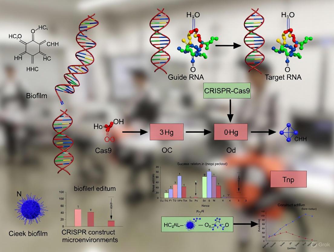
Nanoparticle-Enhanced CRISPR Delivery: Strategies for Stabilizing Gene Editing in Biofilm Microenvironments
This article provides a comprehensive analysis of innovative strategies to enhance the stability and efficacy of CRISPR constructs within challenging biofilm microenvironments.
Nanoparticle-Enhanced CRISPR Delivery: Strategies for Stabilizing Gene Editing in Biofilm Microenvironments
Abstract
This article provides a comprehensive analysis of innovative strategies to enhance the stability and efficacy of CRISPR constructs within challenging biofilm microenvironments. Aimed at researchers, scientists, and drug development professionals, it explores the fundamental barriers biofilms pose to conventional gene editing, details advanced nanoparticle-based delivery systems that protect and transport CRISPR cargo, and offers practical optimization and troubleshooting guidance. The content further covers critical validation techniques for assessing editing efficiency and off-target effects, synthesizing key takeaways to outline a clear path for translating these advanced antimicrobial therapies into clinical practice.
Understanding the Battlefield: Why Biofilm Microenvironments Compromise CRISPR Stability and Efficacy
The extracellular polymeric substance (EPS) matrix is a self-produced, highly hydrated network that encapsulates biofilm cells, forming a protective "house" or "fortress" [1]. This matrix is not merely a physical scaffold; it is a complex, dynamic, and functionally active component that determines the immediate conditions of life for biofilm microorganisms [1]. The EPS presents a formidable multi-faceted barrier to conventional antimicrobials and, as emerging research shows, to novel therapeutic strategies like CRISPR/Cas9 delivery.
The composition of the EPS is surprisingly diverse. Contrary to common belief, it consists of more than just polysaccharides. The matrix comprises a wide variety of proteins, glycoproteins, glycolipids, and significant amounts of extracellular DNA (e-DNA) [1]. In many environmental biofilms, polysaccharides can even be a minor component [1]. This biochemical complexity creates a dense, negatively charged sieve that severely limits the penetration and efficacy of CRISPR-based therapeutics.
Frequently Asked Questions (FAQs)
FAQ 1: What specific components of the EPS matrix are the primary culprits in hindering CRISPR delivery?
The EPS matrix employs multiple components to create a barrier:
- Physical Sieve: The dense, cross-linked network of polysaccharides and proteins creates a porous structure that physically blocks the diffusion of large CRISPR/Cas9 complexes (which often exceed 10 nm in size) [2].
- Charge Interactions: The abundance of negatively charged polymers, such as alginate in P. aeruginosa biofilms, can electrostatically bind to and sequester positively charged nanocarriers, preventing them from reaching their target cells [1] [2].
- Enzymatic Degradation: The matrix retains extracellular enzymes, including nucleases, which can degrade the guide RNA (gRNA) and the plasmid DNA encoding the CRISPR system before it can enter bacterial cells [1] [3].
- Matrix Trapping: e-DNA, which forms distinct grid-like structures and filamentous networks within the biofilm, can actively bind to CRISPR components, further reducing delivery efficiency [1].
FAQ 2: How does the biofilm microenvironment reduce the stability of CRISPR/Cas9 constructs?
Once inside the biofilm, the CRISPR/Cas9 system faces a hostile microenvironment:
- Altered Metabolic States: Biofilms contain gradients of nutrients, oxygen, and waste products. Subpopulations of bacteria, especially persister cells in the inner layers, enter a dormant, slow-growing state [2] [4]. Since CRISPR delivery often relies on active bacterial processes for cellular uptake and expression, these metabolic differences drastically reduce editing efficiency.
- Horizontal Gene Transfer (HGT) Hotspot: While biofilms facilitate HGT, allowing the spread of antibiotic resistance genes, this same property can be a hurdle. Delivered CRISPR plasmids may be transferred to non-target species, posing a risk of off-target effects and reducing the concentration available for the intended pathogenic targets [1] [4].
FAQ 3: Are certain types of biofilms more challenging for CRISPR delivery than others?
Yes, the resistance and barrier properties can vary significantly:
- Species-Specific Composition: Biofilms formed by different bacterial species have unique EPS compositions. For instance, P. aeruginosa biofilms may be rich in alginate and e-DNA, while E. coli and S. aureus biofilms rely heavily on curli fibers and proteinaceous components, respectively [1]. This requires a tailored approach to nanoparticle design for different pathogens.
- Environmental vs. Clinical Isolates: The EPS of biofilms from natural environments can be structurally and compositionally distinct from those studied in laboratory models, often being more complex and resistant [1]. Extrapolating results from model organisms like P. aeruginosa to all biofilms is not suitable and requires validation across diverse species [1].
Troubleshooting Guide: Common CRISPR Delivery Failures in Biofilms
The following table outlines frequent experimental issues, their potential causes, and recommended solutions.
| Problem Phenotype | Potential Root Cause | Proposed Solution & Experimental Adjustments |
|---|---|---|
| Low Editing Efficiency | - CRISPR constructs degraded by matrix nucleases.- Nanoparticles trapped in EPS.- Target cells are dormant persisters. | - Encapsulate CRISPR components in nuclease-resistant lipid nanoparticles (LNPs) [2].- Use protease-resistant Cas9 variants.- Pre-treat with EDTA to chelate cations and disrupt matrix integrity [1]. |
| Poor Nanoparticle Penetration | - Large nanoparticle (NP) size.- Strong electrostatic adhesion to EPS. | - Optimize NP size <100 nm and use PEGylation to reduce biofouling [2].- Employ enzyme-functionalized NPs (e.g., with DNase I to degrade e-DNA or proteinase K to digest proteins) [2] [3]. |
| Lack of Target Specificity | - Off-target editing in non-pathogenic species.- Conjugative transfer of CRISPR plasmid. | - Utilize phage-derived delivery systems or conjugative plasmids with narrow host ranges [5] [3].- Employ CRISPRi (dCas9) for reversible gene knockdown instead of permanent cleavage. |
| Inconsistent Results Between Species | - Differences in EPS composition and matrix structure. | - Characterize the EPS of your target biofilm (e.g., using lectin staining for polysaccharides, e-DNA quantification) [1].- Customize gRNA to target essential biofilm genes (e.g., pelA, pslG in Pseudomonas; ica operon in Staphylococcus) [4] [3]. |
Quantitative Data: Nanoparticle Performance Against Biofilm Barriers
Research has quantified the efficacy of various nanoparticle systems in overcoming EPS barriers. The data below summarizes key findings from recent studies.
Table: Efficacy of Nanoparticle-Mediated CRISPR Delivery Against Biofilms
| Nanoparticle Type | Target Biofilm | Key Outcome Metric | Result | Reference Context |
|---|---|---|---|---|
| Liposomal Cas9 | P. aeruginosa | Reduction in biofilm biomass | >90% reduction in vitro | [2] |
| Gold Nanoparticle-CRISPR | P. aeruginosa | Gene-editing efficiency | 3.5-fold increase vs. non-carrier systems | [2] |
| CRISPR-Engineered Bacteriophage (crPhage) | E. coli | Clinical trial phase (UTI treatment) | Phase 1b trial completed | [5] |
| Cationic Polymer Nanoparticles | Multi-species food biofilm | Log reduction of pathogens | Up to ~3-log target reduction | [3] |
The Scientist's Toolkit: Essential Research Reagents
Table: Key Reagents for Investigating CRISPR-Biofilm Interactions
| Reagent / Material | Primary Function in Research | Application Note |
|---|---|---|
| Fluorescently Labeled Lectins | To visualize and characterize polysaccharide components in the EPS matrix in situ [1]. | Crucial for understanding the physical barrier before designing delivery systems. |
| DNase I (e.g., Pulmozyme) | To degrade e-DNA, a key structural component in many biofilms (e.g., P. aeruginosa, S. aureus) [1]. | Can be used as a pre-treatment or co-delivered with NPs to enhance penetration. |
| Cation-Chelating Agents (EDTA) | Disrupts ionic cross-linking (e.g., by Ca2+ ions) that provides mechanical stability to the EPS [1]. | Useful for pre-treatment to loosen the matrix structure. |
| Nuclease-Resistant gRNA (chemically modified) | Increases the half-life of gRNA in the nuclease-rich biofilm microenvironment [2] [3]. | Modifications like 2'-O-methyl, 2'-fluoro improve stability and editing outcomes. |
| dCas9 (CRISPRi/a systems) | Enables temporary gene knockdown (interference) or activation without double-strand breaks [3]. | Redders ethical and safety concerns and allows study of essential genes. |
Experimental Protocols for Key Assays
Protocol 1: Assessing CRISPR Construct Penetration through EPS
Aim: To visualize and quantify the diffusion of CRISPR-carrying nanoparticles into a mature biofilm.
- Biofilm Growth: Grow a standardized biofilm (e.g., P. aeruginosa or S. aureus) in a flow cell or on a coverslip for 48-72 hours.
- NP Preparation: Synthesize nanoparticles (e.g., ~50 nm gold or polymeric NPs) loaded with a fluorescently labeled, non-functional CRISPR plasmid (e.g., Cy5-dye labeled).
- Application: Apply the fluorescent NPs to the mature biofilm and incubate for a defined period (e.g., 1-4 hours).
- Imaging & Analysis: Use Confocal Laser Scanning Microscopy (CLSM) to take Z-stack images through the biofilm depth. Quantify fluorescence intensity as a function of depth using image analysis software (e.g., ImageJ) to generate a penetration profile [2] [4].
Protocol 2: Evaluating gRNA Stability in Biofilm Conditioning
Aim: To determine the degradation kinetics of gRNA when exposed to the biofilm matrix.
- Matrix Extraction: Harvest a mature biofilm via gentle scraping and centrifugation. Filter the supernatant to remove cells, obtaining a cell-free EPS extract [1].
- Incubation Setup: Incubate your gRNA (both naked and nanoparticle-encapsulated) with the EPS extract at 37°C.
- Sampling: Withdraw aliquots at various time points (0, 15, 30, 60, 120 min).
- Analysis: Run samples on an agarose gel to visualize gRNA integrity. Compare the band intensity of full-length gRNA over time to calculate its half-life in the biofilm environment [3].
Visualization of Concepts and Workflows
The following diagram illustrates the multi-layered defense mechanisms of the biofilm EPS matrix against CRISPR-Cas9 delivery systems.
This workflow outlines a systematic experimental approach to develop and validate EPS-penetrating CRISPR delivery systems.
For researchers developing CRISPR-based antimicrobials, the biofilm microenvironment presents a formidable delivery challenge. The very factors that confer protection to bacterial cells—the dense extracellular matrix, physiological heterogeneity, and nutrient gradients—create a "hostile territory" that severely limits the access and activity of CRISPR-Cas systems [2] [4]. This technical support article provides targeted guidance for scientists navigating these obstacles, offering troubleshooting advice, detailed protocols, and reagent solutions to advance your research on improving CRISPR construct stability within biofilms.
# Frequently Asked Questions (FAQs)
1. What are the primary physiological barriers to CRISPR delivery in biofilms? The main barriers stem from the biofilm's structure and the physiological state of its resident cells. The dense extracellular polymeric substance (EPS) matrix physically impedes the penetration of CRISPR constructs [2] [6]. More critically, the heterogeneous microenvironments within biofilms lead to gradients of nutrient availability, oxygen, and waste products [2] [6]. This results in subpopulations of bacteria, particularly those in inner layers, entering a state of reduced metabolic activity or dormancy (persister cells) [2] [4]. Since many CRISPR delivery mechanisms rely on active bacterial processes for uptake and expression, these dormant, slow-growing cells are largely refractory to genetic editing [2].
2. Which nanoparticle properties are most critical for enhancing CRISPR delivery against biofilms? Nanoparticles must be engineered to overcome multiple barriers simultaneously. Key properties include:
- Small size and positive surface charge: To facilitate penetration through the negatively charged EPS matrix and interaction with bacterial cell membranes [2].
- Controlled release capability: To ensure CRISPR payload protection during transit and sustained release within the biofilm [2] [7].
- Intrinsic biofilm-disrupting activity: Some metallic nanoparticles, like gold, can weaken the EPS matrix while serving as delivery vehicles [2].
- Co-delivery capacity: The ability to carry additional agents, such as antibiotics or quorum-sensing inhibitors, for synergistic effects [2] [7].
3. How can I assess and account for metabolic heterogeneity in my biofilm models? Utilize a combination of vital staining and advanced microscopy. Fluorescent dyes that indicate metabolic activity (e.g., CTC for respiratory activity) or membrane integrity can be used in conjunction with confocal laser scanning microscopy (CLSM) to visualize the spatial distribution of active versus dormant cells within the biofilm architecture [2] [6]. Furthermore, designing CRISPR systems that target essential genes or utilize promoters active under low-nutrient conditions can help broaden efficacy across different physiological states [4].
# Troubleshooting Guide
Table 1: Common Experimental Challenges and Proposed Solutions
| Challenge | Potential Cause | Solution | Key References |
|---|---|---|---|
| Low CRISPR editing efficiency in mature biofilms | Poor penetration of CRISPR constructs through EPS High proportion of metabolically inactive persister cells | Utilize nanoparticle carriers (e.g., liposomal, gold NPs) Pre-treat with EPS-disrupting enzymes (e.g., DNase, dispersin B) Target quorum-sensing genes to disrupt biofilm integrity | [2] [4] [7] |
| High variability in results between biofilm replicates | Inconsistent biofilm growth conditions Heterogeneous biofilm architecture | Standardize growth media, flow rates (in flow cells), and inoculation methods Use multiple biofilm models (e.g., static, flow cell) for validation Increase sample size and randomize sampling points | [6] |
| Lack of observed synergistic effect with antibiotics | Incorrect timing of antibiotic administration CRISPR not effectively sensitizing bacteria | Ensure CRISPR system successfully knocks out resistance genes before antibiotic challenge Use nanoparticles designed for co-delivery of CRISPR and antibiotics | [2] [7] |
| Cytotoxicity of nanoparticle delivery system | Material-specific toxicity (e.g., certain cationic polymers) | Optimize nanoparticle composition and surface chemistry Test lower concentrations or switch to more biocompatible materials (e.g., lipid-based NPs) | [2] |
Table 2: Efficacy of Nanoparticle-Enhanced CRISPR Strategies Against Biofilms
| Delivery Platform | Target Bacteria / Biofilm | Key Outcome | Efficiency / Improvement |
|---|---|---|---|
| Liposomal Cas9/gRNA formulation | Pseudomonas aeruginosa | Reduction in biofilm biomass | >90% reduction in vitro [2] |
| Gold nanoparticle carriers | Model bacterial systems | Enhancement in gene-editing efficiency | 3.5-fold increase compared to non-carrier systems [2] [7] |
| CRISPR-NP hybrid system + Antibiotic | Antibiotic-resistant biofilm | Synergistic biofilm disruption and bacterial killing | Superior to either treatment alone [2] [7] |
# Experimental Protocols
Protocol 1: Assessing CRISPR/nanoparticle penetration in biofilms
Objective: To visualize and quantify the distribution and penetration efficiency of nanoparticle-based CRISPR delivery systems within a mature biofilm.
Materials:
- Mature bacterial biofilm (e.g., 48-72 hour culture)
- Fluorescently labeled CRISPR-Cas9/nanoparticle complex
- Confocal Laser Scanning Microscope (CLSM)
- Suitable staining dyes (e.g., SYTO for total cells)
Method:
- Biofilm Growth: Grow biofilms under optimized, standardized conditions on a surface compatible with CLSM (e.g., glass-bottom dish) [6].
- Treatment: Apply the fluorescently labeled CRISPR/nanoparticle complex to the mature biofilm and incubate for a predetermined time.
- Staining and Washing: Gently wash the biofilm with buffer to remove non-adherent complexes. Counterstain the biofilm biomass to distinguish bacterial cells.
- Imaging: Use CLSM to capture Z-stack images through the entire biofilm depth.
- Analysis: Analyze image stacks with software (e.g., ImageJ) to determine the fluorescence intensity of the CRISPR signal as a function of biofilm depth. This quantifies penetration and identifies diffusion barriers.
Protocol 2: Evaluating metabolic state-dependent CRISPR efficacy
Objective: To correlate the success of CRISPR-mediated gene editing with the metabolic activity of cells within a biofilm.
Materials:
- Mature bacterial biofilm
- CRISPR-Cas9 system targeting a reporter or resistance gene
- Metabolic activity stain (e.g., 5-cyano-2,3-ditolyl tetrazolium chloride - CTC)
- Fluorescence-Activated Cell Sorting (FACS)
- Viability plating media
Method:
- Biofilm Treatment: Treat the biofilm with the CRISPR-Cas9 system.
- Dissociation and Staining: Gently disaggregate the biofilm to create a cell suspension. Stain the cells with CTC, which is reduced to a fluorescent formazan by metabolically active bacteria.
- Cell Sorting: Use FACS to sort the population into high-CTC (metabolically active) and low-CTC (less active/dormant) subpopulations.
- Plating and Analysis: Plate each sorted subpopulation on selective and non-selective media. Compare the survival rates (CFU counts) between the two populations to determine if CRISPR killing efficacy is biased toward metabolically active cells.
# Pathway and Workflow Visualizations
Diagram 1: CRISPR-biofilm experiment workflow.
Diagram 2: Biofilm defense mechanisms vs CRISPR.
# Research Reagent Solutions
Table 3: Essential Research Reagents for CRISPR-Biofilm Studies
| Reagent / Material | Function / Application | Specific Examples / Notes |
|---|---|---|
| Gold Nanoparticles (AuNPs) | Carrier for CRISPR components; enhances stability and cellular uptake. Can be functionalized with targeting ligands. Intrinsic biofilm-disrupting properties. | Spherical, ~20-50 nm diameter; functionalized with cationic polymers for DNA binding [2] [7]. |
| Cationic Liposomes | Lipid-based nanoparticles that encapsulate and protect CRISPR payloads. Fuse with bacterial membranes to deliver content. | Liposomal Cas9/sgRNA formulations; used to achieve >90% biofilm biomass reduction [2]. |
| EPS-Disrupting Enzymes | Pre-treatment to degrade the biofilm matrix and improve nanoparticle penetration. | DNase I (targets eDNA), proteinase K (targets proteins), dispersin B (targets polysaccharides) [6]. |
| Metabolic Activity Probes | To stain and identify metabolically active vs. dormant subpopulations in biofilms for analysis. | CTC (5-cyano-2,3-ditolyl tetrazolium chloride) for respiration; CFDA, SE for esterase activity [6] [4]. |
| Fluorescent Dyes for Labeling | To tag CRISPR components or nanoparticles for tracking and visualization in penetration studies. | Cy3, Cy5, FITC; covalently linked to sgRNA or Cas9 protein. |
| Quorum Sensing Inhibitors | Co-delivery agent to disrupt biofilm coordination and integrity, sensitizing it to CRISPR attack. | Natural or synthetic molecules that block autoinducer signaling (e.g., furanones) [8]. |
Frequently Asked Questions & Troubleshooting Guides
FAQ 1: Why is my CRISPR-Cas9 system losing efficiency when targeting bacterial biofilms?
Answer: The biofilm microenvironment is uniquely hostile to biomolecular cargo. The primary causes of efficiency loss are:
- Extracellular Nucleases (DNases/RNases): Secreted by biofilm-resident bacteria and neutrophils to degrade extracellular DNA/RNA, which directly attacks your CRISPR cargo.
- Reactive Oxygen Species (ROS): High levels of ROS within the inflammatory microenvironment of a biofilm can cause oxidative damage to nucleic acids and proteins.
- Entrapment in Extracellular Polymeric Substance (EPS): The dense EPS matrix can physically sequester cargo, preventing it from reaching target cells and prolonging its exposure to degradative factors.
- Acidic pH: Microenvironments within biofilms can have a lower pH, which can accelerate the hydrolysis of RNA and DNA.
Troubleshooting Guide: Addressing Cargo Degradation
| Observed Problem | Potential Cause | Recommended Solution | Key Performance Indicator to Monitor |
|---|---|---|---|
| Rapid loss of plasmid DNA activity. | Degradation by extracellular DNases. | • Use modified, nuclease-resistant plasmid backbones (e.g., phosphorothioate modifications).• Co-deliver with nuclease inhibitors (e.g., actinonin).• Employ a lipid-based nanocarrier for protection. | % of recovered plasmid DNA remaining intact after biofilm exposure (gel electrophoresis). |
| mRNA cargo fails to express protein. | Degradation by RNases and/or acidic hydrolysis. | • Incorporate chemically modified nucleotides (e.g., N1-methylpseudouridine).• Use optimized 5' cap analogs (e.g., CleanCap) and poly(A) tail stabilization.• Formulate mRNA within LNPs or polymer-based nanoparticles. | mRNA half-life in biofilm-conditioned medium; Protein expression levels via fluorescence (if encoding GFP). |
| RNP complex shows poor gene editing efficiency. | Dissociation of Cas9-gRNA complex or degradation of gRNA. | • Use chemically synthesized, end-modified sgRNA (2'-O-methyl, phosphorothioate).• Pre-complex RNPs with cationic polymers to form stable polyplexes.• Ensure RNP storage buffer is optimized for complex stability. | Gel shift assay to confirm RNP integrity; NGS-based quantification of indel formation efficiency. |
| All cargo types show poor penetration. | Physical entrapment in the EPS matrix. | • Utilize biofilm matrix-degrading enzymes (e.g., DNase I, dispersin B, alginate lyase) as co-treatments.• Employ nanoparticles with a positive surface charge to reduce EPS adhesion.• Use engineered phages for targeted delivery through the matrix. | Confocal microscopy with fluorescently labeled cargo to visualize penetration depth. |
FAQ 2: How can I experimentally quantify the degradation rate of my CRISPR cargo in a biofilm model?
Answer: You need to directly expose your cargo to the biofilm microenvironment and track its integrity over time. Below is a standardized protocol.
Experimental Protocol: Quantifying Cargo Half-life in a Biofilm Model
Objective: To determine the stability and half-life of DNA, mRNA, and RNP cargoes when exposed to biofilm-conditioned medium or within a mature biofilm.
Materials:
- Mature biofilm of your target organism (e.g., Pseudomonas aeruginosa, Staphylococcus aureus).
- Purified CRISPR cargo: plasmid DNA, in vitro transcribed mRNA, pre-complexed RNP.
- Biofilm-conditioned medium (BCM): Sterile-filtered supernatant from a mature biofilm culture.
- Control: Fresh growth medium.
- Quantification tools: Qubit fluorometer, NanoDrop, Bioanalyzer, or RT-qPCR.
- Gel electrophoresis system.
Methodology:
- Preparation: Generate a mature biofilm (e.g., 48-72 hours) in your preferred model (flow cell, microtiter plate). Prepare BCM by centrifuging and filter-sterilizing the supernatant.
- Cargo Exposure: Aliquot your CRISPR cargo (e.g., 1 µg) into:
- Test: BCM.
- Control: Fresh, sterile growth medium.
- Incubate at 37°C with mild agitation.
- Sampling: Withdraw samples at defined time points (e.g., 0, 15, 30, 60, 120, 240 minutes).
- Analysis:
- DNA/RNA Integrity: Run samples on an agarose gel to visualize smearing versus a sharp band. Use a Bioanalyzer to generate an RNA Integrity Number (RIN) for mRNA.
- Quantitative Recovery: Use Qubit or RT-qPCR to quantify the amount of intact nucleic acid remaining.
- RNP Integrity: Use a native gel shift assay to visualize the intact Cas9-sgRNA complex.
Data Interpretation: Plot the percentage of intact cargo remaining versus time. Fit the data to a first-order decay model to calculate the half-life (t1/2).
Table: Example Quantitative Data from a Cargo Stability Assay
| Cargo Type | Condition | Half-life (t1/2) | Notes |
|---|---|---|---|
| Plasmid DNA | Fresh Medium | >240 min | Minimal degradation. |
| Plasmid DNA | Biofilm-Conditioned Medium | 45 min | Significant degradation after 60 min. |
| Unmodified mRNA | Fresh Medium | 90 min | Baseline hydrolysis. |
| Unmodified mRNA | Biofilm-Conditioned Medium | <15 min | Rapid, complete degradation. |
| Chemically modified mRNA | Biofilm-Conditioned Medium | 75 min | Improved stability over unmodified. |
| RNP (unmodified sgRNA) | Biofilm-Conditioned Medium | 30 min | Loss of functional complex. |
| RNP (modified sgRNA) | Biofilm-Conditioned Medium | 110 min | Significant stability improvement. |
The Scientist's Toolkit: Research Reagent Solutions
| Reagent / Material | Function in Biofilm CRISPR Research |
|---|---|
| Nuclease-Resistant DNA Backbones | Plasmid vectors with phosphorothioate linkages in the backbone to resist degradation by extracellular DNases. |
| Chemically Modified Nucleotides | (e.g., 2'-Fluoro, 2'-O-Methyl, N1-methylpseudouridine). Incorporated into RNA to shield against RNase cleavage and reduce immunogenicity. |
| Cationic Lipid Nanoparticles (LNPs) | Formulations that encapsulate and protect nucleic acid cargo (mRNA, DNA), facilitating fusion with bacterial membranes and release inside the cell. |
| Cationic Polymers (e.g., PEI, Chitosan) | Can condense nucleic acids or form co-complexes with RNPs to improve stability, prevent dissociation, and enhance delivery. |
| Biofilm Matrix-Degrading Enzymes | DNase I (degrades eDNA), Dispersin B (degrades PNAG), Alginate Lyase (degrades alginate). Used as co-treatments to disrupt the EPS for improved cargo penetration. |
| Reactive Oxygen Species (ROS) Scavengers | (e.g., N-acetylcysteine, Thiourea). Can be included in formulations to protect cargo from oxidative damage within the biofilm. |
Experimental Workflow & Pathway Diagrams
Cargo Degradation & Mitigation Workflow
Biofilm Defense Against CRISPR Cargo
Technical Support Center
Troubleshooting Guides
Issue: Poor CRISPR Editing Efficiency in Mature Biofilms
Q1: Why is my CRISPR-Cas9 system showing high editing efficiency in planktonic cells but very low efficiency in a 48-hour mature biofilm?
A: This is a common issue rooted in the biofilm microenvironment. The primary factors are:
- Reduced Penetration: Plasmid vectors or ribonucleoprotein (RNP) complexes cannot effectively diffuse through the dense, negatively charged extracellular polymeric substance (EPS).
- Altered Cell Physiology: Cells in a biofilm, especially those in deeper, nutrient-deprived layers, have reduced metabolic activity and may not express the necessary machinery for plasmid replication or Cas protein translation.
- Presence of Nucleases: The EPS can contain extracellular nucleases that degrade DNA or RNA guides before they reach the target cells.
Troubleshooting Steps:
- Assess Delivery Vector: Switch from plasmid DNA to pre-assembled Cas9 RNP complexes. RNPs are faster-acting and avoid the need for intracellular transcription and translation, which is beneficial for metabolically dormant cells.
- Modify Delivery Method:
- Chemical Permeabilization: Co-administer with EPS-disrupting agents like DNase I (to degrade extracellular DNA in the matrix) or dispersin B (to hydrolyze polysaccharides). See Table 1 for dosage.
- Physical Methods: Consider using electroporation optimized for surface-associated biofilms or microinjection for single-cell analysis within the biofilm structure.
- Verify Target Accessibility: Re-design sgRNAs to target genomic regions that are accessible in the biofilm state. Some genes may be transcriptionally silenced in sessile cells. Perform an RNA-seq comparison to confirm target gene expression.
Q2: My CRISPR-modified biofilm cells rapidly revert to the wild-type genotype after the selective pressure is removed. How can I improve construct stability?
A: This indicates a lack of stable genomic integration or the survival and regrowth of non-edited persister cells.
Troubleshooting Steps:
- Confirm Editing: Use a combination of assays. Amplify the target region from cells extracted from different biofilm layers (top, middle, bottom) and sequence them individually. Bulk sequencing can miss heterogeneous editing.
- Employ a Double-Selection Strategy:
- Use a CRISPR system that introduces a stable antibiotic resistance marker alongside the gene knockout.
- Alternatively, use a toxin-antidote system (e.g., CRISPRi) where continuous expression of the sgRNA is required to repress a toxic gene.
- Target Essential Genes: For functional studies, design your CRISPR system to target a gene essential for biofilm integrity (e.g., pel or psl in P. aeruginosa). The loss-of-function mutation will be inherently selected against in a biofilm context.
Frequently Asked Questions (FAQs)
Q3: What is the most effective method for delivering CRISPR components into a bacterial biofilm? A: Currently, no single method is perfect. The most promising paradigm is a combinatorial approach. Pre-assembled RNP complexes, delivered with EPS-disrupting enzymes (e.g., DNase I) and a carrier system like lipid-based nanoparticles or engineered bacteriophages, show significantly improved efficacy over traditional plasmids.
Q4: How do I quantify and normalize editing efficiency in a heterogeneous biofilm? A: This is a critical challenge. Do not rely on bulk measurements.
- Spatial Resolution: Section the biofilm using cryosectioning or laser capture microdissection.
- Single-Cell Analysis: Use fluorescence-activated cell sorting (FACS) to separate cells based on a reporter (like GFP) coupled to the CRISPR system, then plate or sequence them.
- Normalization: Normalize your editing data (e.g., from qPCR or sequencing) to the total bacterial biomass (via DNA content) or a constitutively expressed housekeeping gene from the same section.
Q5: Are there specific Cas proteins better suited for biofilm applications? A: Yes. Smaller Cas proteins (e.g., Cas12f, CasΦ) are advantageous for delivery via viral vectors. Furthermore, Cas proteins with high activity at lower temperatures (relevant for deeper, nutrient-poor biofilm layers) or those engineered for higher fidelity (e.g., HiFi Cas9) are beneficial for reducing off-target effects in slow-growing cells.
Table 1: Comparative Efficacy of CRISPR Delivery Modalities in Biofilm vs. Planktonic Cells
| Delivery Modality | Editing Efficiency (Planktonic) | Editing Efficiency (Biofilm) | Key Advantage | Key Limitation in Biofilm |
|---|---|---|---|---|
| Plasmid DNA (Electroporation) | 70-95% | 5-15% | Stable, long-term expression | Poor diffusion; requires cell division |
| Pre-assembled RNP (Electroporation) | 80-90% | 20-40% | Fast action; no replication needed | Limited by delivery efficiency |
| RNP + DNase I Co-treatment | 85-95% | 45-65% | Disrupts matrix; enhances penetration | Can be cytotoxic at high doses |
| Bacteriophage-Mediated | 60-80%* | 25-50%* | High target specificity | Limited cargo capacity; host range |
| Lipid Nanoparticles (LNPs) | 50-70% | 30-55% | Protects cargo; biocompatible | Optimization required for bacterial use |
*Efficiency is highly dependent on the host range and infectivity of the phage.
Table 2: Impact of Biofilm Age and Matrix Composition on CRISPR-Cas9 Efficacy
| Biofilm Age (Hours) | EPS Thickness (µm) | Metabolic Activity (Relative %) | RNP Delivery Efficiency (%) |
|---|---|---|---|
| 24 | 15 ± 3 | 100% | 35 ± 8 |
| 48 | 40 ± 7 | 65 ± 10 | 18 ± 5 |
| 72 | 85 ± 12 | 30 ± 8 | 5 ± 2 |
| 96 | 120 ± 15 | 15 ± 5 | <2 |
Experimental Protocols
Protocol 1: Assessing CRISPR Editing in a Spatial Context within a Biofilm
Objective: To determine the CRISPR-Cas9 editing efficiency across different layers of a mature biofilm.
Materials:
- Mature bacterial biofilm (e.g., 48-72 hour P. aeruginosa or S. aureus)
- Pre-assembled Cas9-sgRNA RNP complex
- DNase I solution (1 mg/mL in PBS)
- Cryostat
- Luria-Bertani (LB) agar plates
- PCR reagents and primers for the target locus
- T7 Endonuclease I assay kit
Methodology:
- Biofilm Treatment: Gently wash the established biofilm with PBS. Apply the Cas9 RNP complex (e.g., 5 µM) with or without co-treatment with DNase I (100 µg/mL) for 1 hour.
- Spatial Sectioning: Embed the entire biofilm in OCT compound and rapidly freeze it. Using a cryostat, serially section the biofilm into 10 µm thick slices representing the top, middle, and bottom layers.
- Cell Recovery & DNA Extraction: Dissolve the OCT compound from each section in PBS, vortex vigorously to disaggregate cells, and pellet them. Extract genomic DNA from each pellet.
- Editing Analysis:
- T7E1 Assay: Amplify the target genomic region from each sample. Hybridize and digest with T7 Endonuclease I, which cleaves mismatched heteroduplex DNA. Analyze fragments on an agarose gel.
- Calculation: Editing efficiency (%) = (1 - (1 / (fraction cleaved))^0.5) * 100, where fraction cleaved = (sum of cleaved band intensities) / (sum of cleaved and uncut band intensities).
Protocol 2: Evaluating Construct Stability Using a Fluorescence-Based Competition Assay
Objective: To track the persistence of CRISPR-edited cells within a mixed-population biofilm over time.
Materials:
- Wild-type strain (WT)
- CRISPR-edited strain, constitutively expressing a fluorescent protein (e.g., GFP) (EDIT)
- Confocal laser scanning microscope (CLSM)
- Flow cytometer
Methodology:
- Biofilm Establishment: Inoculate a flow cell or microtiter plate with a 1:1 mixture of WT and EDIT cells. Allow a biofilm to form for 24 hours under standard conditions.
- Long-Term Flow: Continuously feed the biofilm with fresh, non-selective medium for 5-7 days.
- Monitoring: At 24-hour intervals, harvest biofilms and analyze the ratio of GFP-positive (EDIT) to GFP-negative (WT) cells using flow cytometry.
- Imaging: Use CLSM to capture z-stack images of the biofilm at each time point to visualize the spatial distribution and dominance of each population.
- Data Analysis: Plot the percentage of EDIT cells over time. A rapid decline indicates low construct stability or a fitness cost associated with the edit.
Visualizations
Title: CRISPR Delivery Barriers in Biofilm
Title: Spatial Editing Analysis Workflow
The Scientist's Toolkit: Research Reagent Solutions
| Reagent / Material | Function in Biofilm CRISPR Research |
|---|---|
| Pre-assembled Cas9 RNP | Bypasses the need for intracellular transcription/translation; crucial for targeting metabolically dormant biofilm cells. |
| DNase I | An enzyme that degrades extracellular DNA (eDNA) in the biofilm matrix, reducing viscosity and improving diffusion of CRISPR components. |
| Dispersin B | A glycoside hydrolase that hydrolyzes poly-N-acetylglucosamine (PNAG), a key polysaccharide in many bacterial biofilms, disrupting the matrix. |
| Cryostat | A device used to cut thin, frozen sections of a biofilm, enabling spatial analysis of editing efficiency from top to bottom. |
| T7 Endonuclease I | An enzyme used in a mismatch detection assay to quantify the frequency of CRISPR-induced indels without the need for deep sequencing. |
| Conjugative Plasmid | A vector capable of transferring DNA from a donor to a recipient cell via conjugation, potentially useful for delivering CRISPR machinery to inner biofilm layers. |
| Synthetic Lipid Nanoparticles (LNPs) | Engineered nanocarriers that can encapsulate and protect CRISPR RNPs or DNA, facilitating fusion with bacterial membranes for delivery. |
Advanced Armor: Nanoparticle Engineering for Robust CRISPR Delivery and Stability in Biofilms
Technical Support Center
Troubleshooting Guides
Problem 1: Poor CRISPR Editing Efficiency in Biofilm Models
- Q: I am observing very low knockout efficiency in my biofilm model when using plasmid DNA. What could be the cause?
- A: Low efficiency with plasmid DNA is frequently due to poor penetration through the dense extracellular polymeric substance (EPS) of the biofilm and the inability to transfert the majority of bacterial cells within the structure. The plasmid may also be degraded by nucleases present in the biofilm microenvironment before it can enter cells.
- Troubleshooting Steps:
- Quantify Penetration: Use a fluorescently labelled plasmid (e.g., with Cy5) and perform confocal microscopy with z-stacking to visualize and quantify penetration depth into the biofilm.
- Alternative Cargo: Switch to a pre-assembled Ribonucleoprotein (RNP) complex. RNPs are smaller and more stable, often showing superior penetration and immediate activity without the need for transcription/translation.
- Optimize Delivery: Consider using a nanoparticle or lipid-based carrier specifically designed for biofilm penetration to co-deliver with your CRISPR construct.
- Check Cargo Stability: Assess the integrity of your plasmid after exposure to biofilm conditioned medium via gel electrophoresis to check for degradation.
Problem 2: High Off-Target Effects with mRNA Delivery
- Q: My mRNA-based CRISPR system shows high editing efficiency but also significant off-target effects in my biofilm assays. How can I reduce this?
- A: mRNA delivery can lead to prolonged and high levels of Cas protein expression, which is a known factor for increasing off-target activity. The extended presence of the nuclease increases the chance of cleavage at imperfectly matched sites.
- Troubleshooting Steps:
- Use High-Fidelity Cas Variants: Switch from standard Cas9 to a high-fidelity version like eSpCas9(1.1) or SpCas9-HF1, which can be delivered as mRNA.
- Optimize Dosage: Titrate the mRNA concentration to find the lowest dose that provides sufficient on-target editing, thereby minimizing off-target effects.
- Switch to RNP: RNP complexes have a shorter intracellular half-life, which drastically reduces off-target effects while maintaining high on-target efficiency. This is often the most effective solution.
- Analyze Off-Targets: Perform genome-wide off-target analysis (e.g., GUIDE-seq or CIRCLE-seq) specific to your biofilm-grown cells to identify and confirm problematic sites.
Problem 3: Rapid Degradation of Cargo in Biofilm Conditioning Medium
- Q: My nucleic acid cargo (DNA/mRNA) appears to be degraded when incubated with biofilm-conditioned medium, before even reaching the cells. How can I improve stability?
- A: Biofilm microenvironments are rich in nucleases (DNases and RNases) released by cells and other microbes. Standard nucleic acids are highly susceptible.
- Troubleshooting Steps:
- Use Chemically Modified Nucleic Acids: For mRNA, use chemically modified nucleotides (e.g., pseudo-uridine, 5-methylcytidine) to increase stability and reduce immunogenicity. For DNA, consider phosphorothioate backbone modifications.
- Employ Protective Carriers: Formulate your cargo within protective nanoparticles (e.g., lipid nanoparticles (LNPs) or polymeric nanoparticles) that shield it from nucleases until cellular uptake.
- Pre-assembled RNP: The protein component of the RNP complex offers inherent protection to the guide RNA. Furthermore, chemically modified sgRNAs (with 2'-O-methyl, phosphorothioate modifications at the 3' end) are highly resistant to RNase degradation.
Frequently Asked Questions (FAQs)
Q: Which cargo type is generally best for achieving high editing efficiency with low off-target effects in a mature biofilm?
- A: Based on current research, pre-assembled Ribonucleoprotein (RNP) complexes are often superior. They combine rapid activity (no transcription/translation needed), short intracellular lifetime (reducing off-targets), and favorable size/charge characteristics for better penetration compared to plasmid DNA.
Q: Can I use standard transfection reagents developed for planktonic cells for biofilm delivery?
- A: Generally, no. Standard transfection reagents are optimized for single, suspended cells. The EPS matrix of a biofilm can inhibit the interaction between the reagent and the target cells, leading to very low efficiency. You should use reagents or methods specifically validated for biofilms or complex 3D structures.
Q: How do I quantify and compare the penetration depth of different cargos into my biofilm model?
- A: The standard method is to fluorescently label each cargo type (e.g., with different dyes like Cy3, Cy5) and use confocal laser scanning microscopy (CLSM). Acquire z-stack images through the biofilm depth and use image analysis software (e.g., ImageJ) to plot fluorescence intensity versus depth to generate penetration profiles.
Q: Why is RNP considered more stable than mRNA in the biofilm context?
- A: Stability refers to resistance to the biofilm microenvironment. The sgRNA in an RNP is physically protected within the Cas protein scaffold, making it less accessible to RNases. In contrast, free mRNA is rapidly degraded by ubiquitous RNases. Chemically modified sgRNAs within the RNP further enhance this stability.
Table 1: Quantitative Comparison of CRISPR Cargo Properties for Biofilm Applications
| Property | Plasmid DNA | mRNA | Ribonucleoprotein (RNP) |
|---|---|---|---|
| Typical Size (kDa/nm) | ~3000-5000 kDa / >100 nm | ~300-500 kDa / ~10-15 nm | ~160 kDa / ~10-15 nm |
| Biofilm Penetration Depth | Low (10-20% of biofilm thickness) | Moderate (30-50% of biofilm thickness) | High (50-80% of biofilm thickness) |
| Onset of Action | Slow (24-72 h) | Moderate (12-24 h) | Fast (1-6 h) |
| Duration of Action | Long (days) | Moderate (1-3 days) | Short (< 24 h) |
| Off-Target Effect Risk | High | High | Low |
| Stability in Biofilm Medium | Low (DNase sensitive) | Very Low (RNase sensitive) | High (RNase resistant, esp. with modified sgRNA) |
| Immunogenicity | High (TLR9 activation) | High (TLR7/8 activation) | Low |
Experimental Protocols
Protocol 1: Assessing Cargo Penetration in a Biofilm using Confocal Microscopy
- Biofilm Growth: Grow a standardized biofilm (e.g., P. aeruginosa or S. aureus) on a glass-bottom dish or a flow cell for 48-72 hours.
- Cargo Labeling: Label your cargos with distinct fluorophores.
- Plasmid DNA: Label with Cy5 using a Label IT nucleic acid labeling kit.
- mRNA: Use commercially purchased Cy3-labeled mRNA or label in-house.
- RNP: Use a fluorescently labeled Cas9 protein (e.g., SNAP-tag) or a Cy5-labeled sgRNA.
- Application: Gently apply a solution containing a standardized concentration (e.g., 500 nM) of the labeled cargo onto the mature biofilm. Incubate for a set time (e.g., 4 hours) under appropriate conditions.
- Washing and Fixation: Gently wash the biofilm with PBS to remove non-internalized cargo. Fix with 4% paraformaldehyde for 15-30 minutes.
- Imaging: Image using a confocal microscope. Collect z-stack images from the top to the bottom of the biofilm at consistent intervals (e.g., 1 µm steps).
- Analysis: Use ImageJ or similar software to measure the mean fluorescence intensity in each z-slice. Normalize the data to the maximum intensity and plot against depth to generate a penetration profile.
Protocol 2: Evaluating CRISPR-Cas Editing Efficiency in a Biofilm
- Biofilm Setup & Treatment: Establish biofilms from a strain containing a reporter gene (e.g., GFP) that can be knocked out. Treat the mature biofilm with your CRISPR cargo (DNA, mRNA, or RNP) complexed with your chosen delivery vehicle.
- Recovery and Dispersal: After treatment (e.g., 24-48 hours), gently scrape the biofilm from the surface and disperse it into single-cell suspension using vigorous vortexing and/or enzymatic treatment with DNase I and dispersin B.
- Analysis:
- Flow Cytometry: For a GFP knockout assay, analyze the cell suspension by flow cytometry. The percentage of GFP-negative cells indicates the editing efficiency.
- Next-Generation Sequencing (NGS): For endogenous targets, extract genomic DNA from the dispersed biofilm cells. Amplify the target region by PCR and subject the amplicons to NGS. Analyze the sequences for insertions/deletions (indels) at the target site to calculate precise editing efficiency.
Visualizations
Diagram 1: Cargo Penetration & Activity Workflow
Diagram 2: Intracellular Mechanism of Cargo Types
The Scientist's Toolkit
Table 2: Essential Research Reagents for Biofilm CRISPR Delivery
| Reagent / Material | Function & Application |
|---|---|
| Chemically Modified sgRNA | Increases nuclease resistance and stability of RNP complexes within the biofilm matrix. Crucial for maintaining activity. |
| High-Fidelity Cas Protein | Reduces off-target editing effects, which is critical for accurate genetic analysis in a heterogeneous biofilm population. |
| Lipid Nanoparticles (LNPs) | A delivery vehicle that encapsulates nucleic acids (mRNA, DNA) or proteins (RNP), protecting them from degradation and enhancing cellular uptake in biofilms. |
| Biofilm-Disrupting Enzymes (e.g., DNase I, Dispersin B) | Used to disperse biofilms into single-cell suspensions for accurate downstream analysis like flow cytometry or colony counting. |
| Confocal Microscopy Dish | Specialized glass-bottom dishes for growing biofilms and performing high-resolution z-stack imaging to quantify cargo penetration. |
| Fluorescent Labeling Kits (e.g., Cy3, Cy5) | For covalently tagging DNA, RNA, or proteins to enable visualization and tracking of cargo penetration and localization. |
| Synergy Hydrogels | Synthetic hydrogels used to create a standardized, EPS-mimicking environment for initial screening of cargo penetration and stability. |
Technical Support Center
Troubleshooting Guide: Common LNP Formulation & CRISPR Delivery Issues
Q1: My LNP formulations consistently show low encapsulation efficiency (<70%) for CRISPR ribonucleoproteins (RNPs). What could be the cause and how can I improve this?
A: Low encapsulation efficiency typically stems from suboptimal formulation conditions or RNP compatibility issues.
Primary Causes and Solutions:
- Cause: Incorrect lipid-to-RNP mass ratio.
- Solution: Titrate the ionizable lipid to RNP ratio. We recommend starting with a range of 10:1 to 50:1 (w/w) and using a microfluidics device for reproducible mixing.
- Cause: Instability of the RNP complex during the acidic buffer exchange step.
- Solution: Ensure the RNP is properly complexed and stabilized. Consider using a citrate buffer at pH 4.0 instead of acetate, and minimize the time the RNP spends in the acidic environment.
- Cause: Inefficient mixing in microfluidics device.
- Solution: Optimize the flow rate ratio (FRR). A higher aqueous-to-organic flow rate ratio (e.g., 3:1) often improves encapsulation for large complexes like RNPs.
Recommended Optimization Protocol:
- Prepare lipid stock in ethanol (e.g., Ionizable Lipid:DSPC:Cholesterol:DMG-PEG 2000 at 50:10:38.5:1.5 mol%).
- Dilute CRISPR RNP in 25 mM citrate buffer, pH 4.0.
- Use a microfluidic device to mix the two phases. Systematically vary the Total Flow Rate (TFR) from 8 to 16 mL/min and the Flow Rate Ratio (FRR, aqueous:organic) from 2:1 to 4:1.
- Dialyze the formed LNPs against 1X PBS, pH 7.4, for 2 hours at 4°C.
- Measure encapsulation efficiency using the Quant-iT RiboGreen assay.
Q2: I am observing high cytotoxicity and low transfection efficiency when treating bacterial biofilms with my CRISPR-LNPs. What factors should I investigate?
A: This is a common challenge in the harsh biofilm microenvironment. The issue likely relates to LNP stability, surface properties, or biofilm penetration.
Investigation Pathway:
- Check LNP Stability: Use Dynamic Light Scattering (DLS) to confirm your LNPs are stable in the specific biofilm culture medium (e.g., LB, TSB). Aggregation can cause non-specific toxicity.
- Modify Surface Charge: Highly cationic surfaces can cause non-specific binding and toxicity. Ensure your formulation uses an ionizable lipid that is neutral at physiological pH to reduce non-specific interactions.
- Functionalize with Targeting Ligands: To enhance penetration and uptake within the biofilm, consider conjugating your LNPs with biofilm-penetrating peptides (e.g., KFF-KFF-KFF) or enzymes that degrade the extracellular polymeric substance (EPS).
Experimental Workflow for Biofilm Transfection:
Title: Biofilm Transfection with CRISPR-LNPs
Q3: My CRISPR-LNPs show poor stability and payload leakage during storage. How can I enhance their long-term stability?
A: Payload leakage is often due to lipid packing defects or chemical degradation.
Stabilization Strategies:
- Lyophilization (Freeze-Drying): This is the gold standard for long-term storage. Include cryoprotectants like sucrose or trehalose (e.g., 10% w/v) in the final buffer before freezing and lyophilization.
- Optimize Lipid Composition: Increase the molar percentage of the structural lipid (e.g., DSPC) to strengthen the LNP bilayer and reduce permeability.
- Storage Conditions: Always store LNPs at 4°C. For lyophilized products, store at -20°C or -80°C. Avoid repeated freeze-thaw cycles of liquid formulations.
Frequently Asked Questions (FAQs)
Q1: What is the critical difference between formulating LNPs for siRNA versus CRISPR RNP delivery? A: The primary difference lies in the payload's size, charge, and stability. CRISPR RNPs are large, multi-subunit protein-nucleic acid complexes, whereas siRNA is a small, double-stranded RNA. This requires optimization of the ionizable lipid for efficient RNP encapsulation, often needing a higher pKa (~6.5-6.8) than for siRNA (~6.2-6.4). Furthermore, the formulation process must be gentle to avoid denaturing the Cas9 protein.
Q2: Which technique is most accurate for measuring the encapsulation efficiency of CRISPR cargo in LNPs? A: The Ribogreen Assay is the most accurate and widely accepted method. It involves measuring the fluorescence of the RNA guide in the RNP complex before and after disruption of the LNPs with a detergent (like Triton X-100). The difference between the total and free signal gives the encapsulated fraction.
Q3: How can I achieve controlled or triggered release of the CRISPR payload from LNPs specifically within a biofilm? A: Controlled release can be engineered by designing LNPs that respond to stimuli unique to the biofilm microenvironment.
- pH-Triggered Release: Use an ionizable lipid with a pKa tuned to become positively charged and disruptive in the slightly acidic regions of a mature biofilm.
- Enzyme-Triggered Release: Incorporate lipids linked to a substrate that is cleaved by biofilm-specific enzymes (e.g., matrix-degrading enzymes like DNase or dispersin B). The cleavage event destabilizes the LNP, releasing the payload.
Controlled Release Mechanisms:
Title: Triggered Release from CRISPR-LNPs
Table 1: Impact of Flow Rate Ratio (FRR) on LNP Characteristics for RNP Encapsulation
| Aqueous:Organic FRR | Average Size (nm) | PDI | Encapsulation Efficiency (%) | Zeta Potential (mV) |
|---|---|---|---|---|
| 2:1 | 145 | 0.18 | 75 | -2.1 |
| 3:1 | 112 | 0.12 | 88 | -3.5 |
| 4:1 | 98 | 0.09 | 82 | -4.8 |
| 5:1 | 135 | 0.15 | 70 | -5.2 |
Data generated using a fixed lipid composition and TFR of 12 mL/min.
Table 2: Efficacy of Different Ionizable Lipids in CRISPR-Mediated Gene Knockout in a S. aureus Biofilm Model
| Ionizable Lipid | pKa (Theoretical) | Gene Editing Efficiency (%) | Biofilm Cell Viability (% of Control) |
|---|---|---|---|
| Lipid A (DLin-MC3-DMA) | 6.4 | 15 | 85 |
| Lipid B (SM-102) | 6.7 | 45 | 60 |
| Lipid C (Custom) | 6.9 | 65 | 40 |
| LNP-only Control | N/A | 0 | 95 |
Editing efficiency measured by NGS of target locus after 24h treatment. Viability measured by ATP-based assay.
The Scientist's Toolkit: Research Reagent Solutions
| Reagent/Material | Function in CRISPR-LNP Formulation |
|---|---|
| Ionizable Lipid (e.g., DLin-MC3-DMA, SM-102) | The key functional lipid that enables encapsulation and endosomal escape. Becomes positively charged in acidic endosomes. |
| Helper Lipid (DSPC) | A structural phospholipid that enhances bilayer stability and promotes fusion with endosomal membranes. |
| Cholesterol | Incorporates into the LNP bilayer to improve stability, rigidity, and fluidity. Aids in cellular uptake. |
| PEG-lipid (e.g., DMG-PEG2000) | Provides a hydrophilic corona that stabilizes LNPs during formation, reduces aggregation, and modulates pharmacokinetics. |
| Microfluidic Device (e.g., NanoAssemblr) | Enables rapid, reproducible, and scalable mixing of lipid and aqueous phases to form uniform, monodisperse LNPs. |
| SYBR Gold / RiboGreen Assay | Fluorescent dyes used to accurately quantify the encapsulation efficiency of the gRNA within the RNP complex. |
| Sucrose/Trehalose | Cryoprotectants used during lyophilization (freeze-drying) to maintain LNP integrity and payload stability for long-term storage. |
Troubleshooting Guides
Problem 1: Low Gene Editing Efficiency
Q: I am using gold nanoparticles to deliver Cas9 RNP, but my gene editing efficiency is lower than expected. What could be the cause?
| Potential Cause | Explanation | Solution |
|---|---|---|
| Suboptimal RNP Loading | Inefficient conjugation of the RNP complex to the nanoparticle surface reduces the functional payload delivered into the cell. | - Ensure proper functionalization of gold nanoparticles with cationic coatings (e.g., cationic arginine) to enhance RNP binding via electrostatic interactions [9].- Characterize loading efficiency using techniques like gel electrophoresis or HPLC to quantify unbound RNP. |
| Poor Endosomal Escape | The nanoparticle is trapped and degraded in the endosome, preventing RNP release into the cytoplasm. | - Co-deliver endosomolytic agents. For example, "CRISPR-Gold" incorporates an endosomal disruption polymer [9].- Optimize the surface chemistry and size of nanoparticles to promote endosomal escape mechanisms [10]. |
| RNP Aggregation | Cas9 protein aggregation can form large, insoluble clusters that are difficult to encapsulate and deliver efficiently, reducing functional editing complexes [10]. | - Use fresh, high-quality Cas9 protein and avoid repeated freeze-thaw cycles.- Include stabilizing agents in your formulation buffers.- Monitor aggregation status via dynamic light scattering (DLS) to ensure nanoparticle size remains within the optimal sub-150 nm range. |
| Inadequate Nuclear Localization | The RNP complex fails to be imported into the nucleus where it can access the genomic DNA. | - Fuse nuclear localization signals (NLS) to the Cas9 protein sequence to actively shuttle the complex through the nuclear pore [11]. |
Problem 2: Nanoparticle Instability in Biofilm Microenvironments
Q: My nanoparticle-RNP complexes aggregate or degrade when introduced to biofilm cultures. How can I improve their stability?
| Potential Cause | Explanation | Solution |
|---|---|---|
| Non-Specific Interactions with EPS | The extracellular polymeric substance (EPS) in biofilms can entrap nanoparticles or foul their surfaces, reducing penetration and delivery efficiency [12]. | - Functionalize nanoparticles with a dense layer of PEG (polyethylene glycol) to create a "stealth" effect, reducing non-specific binding [9].- Use coatings like chitosan, which has inherent biofilm-penetrating and antimicrobial properties [9]. |
| Degradation by Bacterial Nucleases | Nucleases present in the biofilm microenvironment can degrade the sgRNA component of the RNP, rendering it inactive. | - Ensure the RNP is fully complexed and protected within the nanoparticle's core or shell. Pre-formed RNP complexes offer some inherent protection compared to nucleic acid delivery [11].- Utilize chemically synthesized, modified sgRNAs with phosphorothioate bonds or 2'-O-methyl groups to enhance nuclease resistance [13]. |
Problem 3: Off-Target Editing Effects
Q: Despite using RNP delivery to reduce off-target effects, I am still detecting unintended edits. How can nanoparticle design further improve specificity?
| Potential Cause | Explanation | Solution |
|---|---|---|
| Prolonged Intracellular Activity | Even with transient RNP delivery, extended presence of active Cas9 can increase the chance of off-target cleavage. | - The transient nature of RNP delivery itself is the best mitigation. Using nanoparticles ensures this transient activity [11]. Confirm that your formulation does not inadvertently cause RNP accumulation or aggregation that extends its half-life.- Utilize high-fidelity Cas9 variants (e.g., eSpCas9, SpCas9-HF1) in your RNP complexes, which are engineered for reduced off-target activity [14]. |
| Inconsistent Cellular Uptake | Heterogeneous delivery across a cell population can lead to variable editing, where some cells are undertreated and others are overtreated. | - Optimize nanoparticle surface charge (zeta potential) and size for uniform and efficient cellular uptake. A slightly positive surface charge often enhances interaction with negatively charged cell membranes [10]. |
Frequently Asked Questions (FAQs)
Q1: Why are gold nanoparticles (AuNPs) particularly popular for RNP delivery?
Gold nanoparticles offer several unique advantages:
- Surface Functionalization: Their surface can be easily and stably modified with a variety of molecules (e.g., thiol-terminated polymers, peptides, DNA) for conjugating RNPs and enhancing stability [9].
- Biocompatibility: They are generally considered biocompatible and exhibit low cytotoxicity [9].
- Proven Efficacy: Systems like cationic arginine gold nanoparticles (ArgNPs) have demonstrated high delivery efficiency (~90%) and significant gene editing efficiency (23–30%) in vitro [9]. Another platform, CRISPR-Gold, showed 40-50% editing efficiency at the protein and mRNA level in vivo for a target gene [9].
Q2: What other inorganic nanoparticles are used for RNP delivery besides gold?
While gold is prominent, other inorganic materials are being explored:
- Zeolite Imidazole Frameworks (ZIFs): These are porous, biodegradable materials that can encapsulate large biomolecules like RNPs and release them in response to specific physiological conditions, such as the acidic pH found in endosomes or some biofilm microenvironments [9].
Q3: How does RNP delivery via nanoparticles compare in efficiency to viral delivery methods?
It's a trade-off between efficiency, safety, and payload capacity. The table below summarizes key differences relevant to biofilm and antimicrobial research:
| Feature | Viral Vectors (e.g., AAV) | Nanoparticle RNP Delivery |
|---|---|---|
| Editing Speed | Slow (requires transcription/translation) | Very fast (functional RNP is active immediately) [11] |
| Duration of Activity | Long-term/persistent expression | Short, transient activity [11] |
| Immunogenicity | Can trigger significant immune responses [9] | Generally lower immunogenicity [15] |
| Off-Target Risk | Higher (prolonged Cas9 expression) | Lower (transient activity reduces off-target effects) [15] [11] |
| Payload Capacity | Limited (AAV: ~4.7 kb) [9] | High (can deliver large, pre-assembled RNPs) [15] |
| Targeting Flexibility | Moderate (depends on serotype) | High (surface can be easily modified for targeting) [12] |
| Applicability to Bacteria | Low (viruses infect specific hosts) | High (can be engineered to target bacterial cells in biofilms) [12] |
Q4: For targeting biofilms, what specific bacterial genes should my gRNA target?
When designing CRISPR/Cas9 to combat biofilm-driven antibiotic resistance, target selection is critical. Effective gRNAs can be designed to disrupt:
- Antibiotic Resistance Genes (ARGs): Directly target and disrupt genes like bla (beta-lactamase), mecA (methicillin resistance), or ndm-1 (carbapenem resistance) [12].
- Quorum Sensing (QS) Genes: Disrupt bacterial cell-to-cell communication, which is crucial for biofilm formation and maturation [12].
- Biofilm-Regulating Factors: Target genes involved in the production of the extracellular polymeric substance (EPS) matrix, adhesins, or regulators of the biofilm lifecycle [12].
Experimental Protocols & Workflows
Key Protocol: Formulating Cationic Polymer-Coated Gold Nanoparticles for RNP Delivery
This protocol outlines a common method for creating gold nanoparticles capable of binding and delivering Cas9 RNP complexes.
Research Reagent Solutions
| Reagent | Function/Brief Explanation |
|---|---|
| Chloroauric Acid (HAuCl₄) | Precursor for synthesizing gold nanoparticle cores. |
| Citrate or Borohydride Reducers | Used to reduce gold ions to form colloidal gold nanoparticles. |
| Cationic Polymer (e.g., PEI) | Coats the nanoparticle, providing a positive surface charge to bind negatively charged RNPs. |
| Cas9 Nuclease | The core editing protein. Must be pure and have high activity. |
| Chemically Modified sgRNA | Guides the Cas9 to the target DNA sequence. Modifications enhance stability. |
| Dialysis Membranes or Filters | For purifying and concentrating the final nanoparticle-RNP complex. |
Step-by-Step Methodology:
- Synthesize Gold Nanoparticles (AuNPs): Prepare ~15-20 nm AuNPs by reducing HAuCl₄ with sodium citrate. This results in negatively charged citrate-capped AuNPs [9].
- Functionalize with Cationic Coating: Incubate the AuNPs with a cationic polymer, such as polyethylenimine (PEI). The polymer will electrostatically adsorb to the AuNP surface, creating a positively charged layer. Purify the coated particles to remove excess polymer [9].
- Formulate RNP Complex: Pre-complex the Cas9 protein and sgRNA at an optimal molar ratio (e.g., 1:1.2) in a suitable buffer. Incubate at room temperature for 10-20 minutes to form the active ribonucleoprotein (RNP) complex [13] [11].
- Load RNP onto Nanoparticles: Mix the cationic AuNPs with the pre-formed RNP complex. The positive charge on the nanoparticles will attract and bind the negatively charged RNP. Incubate for 30-60 minutes with gentle agitation.
- Purify the Complex: Remove unbound RNP and concentrate the final formulation using centrifugal filters or dialysis. This step is crucial for ensuring consistency and maximizing the signal-to-noise ratio in your experiments.
- Characterize the Final Product: Use Dynamic Light Scattering (DLS) to measure the hydrodynamic diameter and zeta potential. Confirm an increase in size after RNP loading and a zeta potential that is less positive than the coated-but-unloaded AuNPs, indicating successful binding.
Workflow Diagram: RNP Delivery via Inorganic Nanoparticles for Biofilm Targeting
The following table summarizes key performance metrics from studies utilizing inorganic nanoparticles for CRISPR RNP delivery, providing benchmarks for your own experiments.
| Nanoparticle Platform | Cargo Type | Reported Editing Efficiency | Key Application Context | Reference |
|---|---|---|---|---|
| Cationic Arginine Gold Nanoparticles (ArgNPs) | RNP | ~90% delivery efficiency; 23–30% gene editing efficiency | in vitro editing in human cell lines [9] | |
| CRISPR-Gold | RNP | 40–50% (reduction in target protein/mRNA) | in vivo editing in mouse model [9] | |
| Gold Nanoparticle Hybrids | CRISPR/Cas9 | 3.5-fold increase in editing efficiency vs. non-carrier | Anti-biofilm application against P. aeruginosa [12] |
Technical Support Center
Troubleshooting Guides
Issue 1: Low Encapsulation Efficiency of CRISPR-Cas9 RNP
- Problem: The encapsulation efficiency (EE) of the Cas9 ribonucleoprotein (RNP) complex into your PLGA-PEG nanoparticles is consistently below 40%, leading to poor gene editing outcomes.
- Potential Causes & Solutions:
- Cause: Incompatibility between the aqueous RNP solution and the organic solvent (e.g., DCM, ethyl acetate) used in the double emulsion process.
- Solution: Optimize the primary emulsion sonication time and amplitude. A shorter, gentler sonication (e.g., 10-15 seconds at 30% amplitude) can help stabilize the first water-in-oil emulsion before adding to the outer aqueous phase.
- Cause: Insufficient concentration of stabilizer (e.g., PVA) in the outer aqueous phase.
- Solution: Increase the concentration of PVA to 2-3% (w/v) to better stabilize the double emulsion droplets and prevent RNP leakage.
- Cause: The RNP complex is degrading during the emulsion process.
- Solution: Include RNase inhibitors and protease inhibitors in the inner aqueous phase buffer. Perform the entire process on ice or in a cold room.
Issue 2: Ineffective Antibiotic Release in Biofilm Microenvironment
- Problem: The encapsulated antibiotic (e.g., Ciprofloxacin) is not being released effectively at the biofilm site, despite in vitro data showing good release profiles.
- Potential Causes & Solutions:
- Cause: The biofilm's hypoxic and acidic conditions are not sufficient to trigger the release from your pH-sensitive polymer (e.g., Eudragit).
- Solution: Characterize the exact pH of your specific biofilm model. Consider using a polymer with a higher pKa or one that is enzymatically cleaved by biofilm-specific enzymes (e.g., matrix-degrading enzymes).
- Cause: The nanoparticles are failing to penetrate the dense extracellular polymeric substance (EPS) of the biofilm.
- Solution: Functionalize the nanoparticle surface with biofilm-penetrating peptides (e.g., DNase I, dispersin B, or cationic peptides). Refer to the protocol below.
Issue 3: Reduced CRISPR-Mediated Gene Editing in Biofilm Bacteria
- Problem: Even with good encapsulation and cellular uptake, the targeted gene knockout (e.g., of a beta-lactamase gene) is inefficient within the biofilm.
- Potential Causes & Solutions:
- Cause: Low metabolic activity of bacteria in the biofilm core reduces the efficiency of homology-directed repair (HDR) or non-homologous end joining (NHEJ).
- Solution: Time the administration of nanoparticles to coincide with a more active growth phase, or pre-treat with a sub-inhibitory concentration of antibiotic to induce a stress response that may increase editing efficiency.
- Cause: The CRISPR-Cas9 construct is unstable or degraded in the biofilm microenvironment before reaching the bacterial cytoplasm.
- Solution: Ensure the RNP is properly complexed with a protective, cationic polymer like polyethyleneimine (PEI) within the nanoparticle core to shield it from nucleases.
Frequently Asked Questions (FAQs)
Q1: What is the optimal N/P ratio for complexing the anionic RNP with cationic polymers inside the nanoparticle? A1: The optimal N/P (Nitrogen/Phosphate) ratio is critical. For PEI-based complexation, a ratio between 8 and 12 typically provides a good balance between efficient RNP condensation, colloidal stability, and minimal cytotoxicity. We recommend performing a gel retardation assay to confirm complete complexation.
Q2: How do I quantify the synergistic effect between the antibiotic and the CRISPR component? A2: Synergy is best quantified using the Fractional Inhibitory Concentration Index (FICI). Calculate it using the checkerboard assay method outlined in the protocol section. A FICI of ≤0.5 indicates synergy.
Q3: My nanoparticles are aggregating in the bacterial culture media. How can I improve stability? A3: Aggregation is often due to salt-induced instability. Ensure you are using a sufficient concentration of PEG in your polymer (PLGA-PEG) to provide a steric hydration barrier. You can also add a small amount of a non-ionic surfactant (e.g., 0.01% Tween 80) to your suspension buffer.
Q4: Which biofilm model is most appropriate for testing these hybrid systems? A4: For initial screening, the static 96-well plate crystal violet assay is sufficient. For more advanced, physiologically relevant testing, a flow cell system that allows for continuous nutrient supply and waste removal is recommended, as it forms thicker, more robust biofilms.
Table 1: Characterization of Optimized Co-delivery Nanoparticles
| Parameter | Value (Mean ± SD) | Measurement Technique |
|---|---|---|
| Hydrodynamic Diameter | 185.4 ± 4.2 nm | Dynamic Light Scattering (DLS) |
| Polydispersity Index (PDI) | 0.11 ± 0.03 | DLS |
| Zeta Potential | -12.5 ± 1.8 mV | Laser Doppler Electrophoresis |
| CRISPR RNP EE% | 68.5 ± 3.1% | Fluorescence Spectroscopy (FITC-labeled RNP) |
| Antibiotic (Cipro) EE% | 82.7 ± 2.5% | HPLC-UV |
| Drug Loading (CRISPR) | 4.2 ± 0.3% (w/w) | Calculated from EE% |
| Drug Loading (Antibiotic) | 8.9 ± 0.5% (w/w) | Calculated from EE% |
Table 2: Synergistic Effect Assessment via Checkerboard Assay
| Treatment | MIC (µg/mL) for Planktonic | MIC (µg/mL) for Biofilm | FICI | Interpretation |
|---|---|---|---|---|
| Ciprofloxacin alone | 0.5 | 32.0 | - | - |
| CRISPR-NP alone | 64.0* | 256.0* | - | - |
| Combination (Cipro+CRISPR-NP) | 0.125 | 4.0 | 0.375 | Synergy |
*MIC value for CRISPR-NP represents the nanoparticle concentration required for a 90% reduction in bacterial growth (MIC-90), as it is not directly bactericidal.
Experimental Protocols
Protocol 1: Synthesis of PLGA-PEG Nanoparticles via Double Emulsion (W/O/W)
- Prepare the inner aqueous phase: Dissolve 20 µg of Cas9-sgRNA RNP complex and 1 mg of Ciprofloxacin in 200 µL of nuclease-free water containing 0.1% (v/v) RNase inhibitor.
- Prepare the organic phase: Dissolve 50 mg of PLGA-PEG (50:50, 10kDa PLGA, 2kDa PEG) and 5 mg of cationic polymer (e.g., PEI, 10kDa) in 2 mL of dichloromethane (DCM).
- Form the primary emulsion (W1/O): Add the inner aqueous phase to the organic phase. Sonicate the mixture using a probe sonicator on ice (30% amplitude, 30 seconds pulse on, 10 seconds pulse off).
- Form the secondary emulsion (W1/O/W2): Pour the primary emulsion into 8 mL of a 2% (w/v) polyvinyl alcohol (PVA) solution. Sonicate again on ice (30% amplitude, 60 seconds).
- Evaporate solvent: Stir the double emulsion overnight at room temperature to evaporate the organic solvent.
- Collect nanoparticles: Centrifuge the suspension at 20,000 x g for 30 minutes at 4°C. Wash the pellet twice with nuclease-free water to remove excess PVA and unencapsulated material.
- Resuspend: Resuspend the final nanoparticle pellet in 1 mL of PBS or a suitable storage buffer. Store at 4°C.
Protocol 2: Checkerboard Assay for Synergy Determination (FICI)
- Prepare antibiotic dilutions: In a 96-well plate, perform a 2-fold serial dilution of the Ciprofloxacin-loaded nanoparticles along the x-axis (e.g., 64 to 0.5 µg/mL).
- Prepare CRISPR-NP dilutions: Perform a 2-fold serial dilution of the CRISPR-only nanoparticles along the y-axis.
- Inoculate with bacteria: Add a standardized bacterial inoculum (~5 x 10^5 CFU/mL) to each well. Include growth and sterility controls.
- Incubate: Incubate the plate at 37°C for 18-24 hours.
- Determine MIC: Determine the MIC for each agent alone and in combination. The MIC is the lowest concentration that prevents visible growth.
- Calculate FICI:
- FIC (Cipro) = MIC of Cipro in combination / MIC of Cipro alone
- FIC (CRISPR) = MIC of CRISPR in combination / MIC of CRISPR alone
- FICI = FIC (Cipro) + FIC (CRISPR)
- Interpret: FICI ≤ 0.5 = Synergy; 0.5 < FICI ≤ 4.0 = No Interaction; FICI > 4.0 = Antagonism.
Visualizations
Experimental Workflow
Nanoparticle Action in Biofilm
The Scientist's Toolkit: Research Reagent Solutions
| Reagent / Material | Function / Rationale |
|---|---|
| PLGA-PEG Copolymer | Biodegradable polymer backbone forming the nanoparticle matrix. PEG provides "stealth" properties to reduce opsonization and improve stability. |
| Cas9 Ribonucleoprotein (RNP) | The functional CRISPR complex. Using pre-assembled RNP (rather than plasmid DNA) reduces off-target effects and allows for rapid, transient activity, which is crucial for targeting prokaryotes. |
| Ciprofloxacin HCl | A broad-spectrum fluoroquinolone antibiotic model. It acts on DNA gyrase and topoisomerase IV. Its co-delivery with a CRISPR system targeting a resistance gene (e.g., gyrA mutation) demonstrates synergy. |
| Polyethyleneimine (PEI), 10kDa | A cationic polymer used to complex and condense the anionic RNP, protecting it from degradation and enhancing its loading into the hydrophobic nanoparticle core. |
| Polyvinyl Alcohol (PVA) | A stabilizer and surfactant used in the double emulsion process to control nanoparticle size and prevent coalescence. |
| Biofilm-Penetrating Peptide (e.g., HYL1) | A peptide conjugated to the nanoparticle surface to facilitate diffusion through the dense, negatively charged extracellular polymeric substance (EPS) of the biofilm. |
| Eudragit L100-55 | A pH-sensitive polymer that can be incorporated into the nanoparticle to trigger antibiotic release specifically in the acidic microenvironment of a mature biofilm. |
Troubleshooting Guides & FAQs
Cargo Size Limitations
Q1: Our AAV packaging efficiency for a CRISPR construct containing SpCas9 and multiple gRNAs is low. What are our primary options for smaller Cas variants?
A1: The primary smaller Cas variants suitable for AAV delivery are listed below. SaCas9 is the most established, while Cas12f systems are the smallest but may have different efficiency profiles.
| Cas Variant | Size (aa) | PAM Requirement | Notes for AAV Delivery |
|---|---|---|---|
| SaCas9 | 1,053 | NNGRRT | Well-characterized; sufficient space for single gRNA and promoter in a single AAV. |
| Nme2Cas9 | 1,082 | NNNNCC | Compact size with simple PAM; offers high fidelity. |
| CjCas9 | 984 | NNNVRYM | One of the smallest Cas9 orthologs; requires a complex PAM. |
| Cas12f (Cas14) | ~400-700 | T-rich | Extremely compact, allowing for complex cargo; lower editing efficiency in mammalian cells may require optimization. |
Q2: After switching to the smaller SaCas9, we observe no editing in our biofilm model. What are the potential causes?
A2:
- Promoter Incompatibility: The SaCas9 gene or its gRNA may be expressed from a promoter that is not functional in your specific bacterial strain within the biofilm. Verify promoter activity.
- gRNA Design: SaCas9 requires its own specific gRNA scaffold, which is different from the SpCas9 scaffold. Ensure you are using the correct sequence.
- PAM Availability: SaCas9 requires a NNGRRT PAM. Re-sequence your target site to confirm the presence of the correct PAM and that no mutations have occurred.
- Delivery Issue: Confirm successful transfection or transduction of the SaCas9 construct into the biofilm cells using a fluorescent marker or antibiotic resistance gene.
- Biofilm Extracellular Polymeric Substance (EPS): The EPS may hinder vector penetration or sequester the CRISPR machinery. Pre-treat biofilms with EPS-disrupting agents (e.g., DNase I, dispersin B) in a control experiment to test this.
Specificity and Fidelity Issues
Q3: We are concerned about off-target effects with SpCas9 in our chronic biofilm infection model. Which high-fidelity variants should we consider?
A3: High-fidelity variants contain mutations that reduce non-specific interactions with DNA. eSpCas9(1.1) and SpCas9-HF1 are leading choices.
| High-Fidelity Variant | Key Mutations | On-Target Efficiency (Relative to WT SpCas9) | Specificity Improvement |
|---|---|---|---|
| eSpCas9(1.1) | K848A, K1003A, R1060A | ~70-90% | Significant reduction in off-targets with minimal on-target impact. |
| SpCas9-HF1 | N497A, R661A, Q695A, Q926A | ~60-80% | Dramatically increased specificity, with a potential trade-off in on-target efficiency. |
| evoCas9 | M495V, Y515N, K526E, R661Q | ~50-70% | Evolved for high fidelity; robust performance across diverse targets. |
| HiFi Cas9 | R691A | ~80-95% | Excellent balance of high on-target efficiency and significantly reduced off-target effects. |
Q4: Our high-fidelity Cas9 variant (eSpCas9(1.1) shows significantly reduced on-target editing in biofilm-grown cells compared to planktonic cells. How can we troubleshoot this?
A4:
- Cellular State: Biofilm cells are often metabolically dormant and have altered membrane permeability, which can reduce the uptake and/or activity of CRISPR components. Consider using a constitutive promoter with high activity in slow-growing cells.
- gRNA Efficacy: Design and test 3-5 different gRNAs for the same target. gRNA secondary structure and accessibility can be differentially affected in the biofilm microenvironment.
- Delivery Timing: The efficiency of CRISPR delivery may vary with biofilm maturity. Try transducing/transfecting at different time points (e.g., 4h, 24h post-initiation of biofilm formation).
- Dosage: Titrate the amount of CRISPR construct delivered. Biofilm cells may require a higher dose for efficient editing.
Experimental Protocols
Protocol 1: Evaluating SaCas9-Mediated Gene Knockout in a Staphylococcal Biofilm Model
Objective: To disrupt a target gene in a Staphylococcus aureus biofilm using an all-in-one AAV-SaCas9 system.
Materials:
- S. aureus strain
- All-in-one AAV vector plasmid encoding SaCas9 and target gRNA (e.g., pAAV-SaCas9-U6-gRNA)
- HEK293T cells (for AAV production)
- Polyethylenimine (PEI)
- pAAV-DJ and pAAV-RC helper plasmids
- Biofilm culture medium (e.g., TSB + 1% glucose)
- 96-well polystyrene plates
- DNase I
- Crystal Violet stain
Methodology:
- AAV Production: Package the pAAV-SaCas9-gRNA plasmid into AAV serotype DJ using the triple-transfection method in HEK293T cells. Purify and titer the virus.
- Biofilm Formation: Grow S. aureus in 96-well plates for 24 hours to form mature biofilms.
- AAV Transduction: Gently wash biofilms and incubate with AAV-SaCas9 (MOI 10,000-100,000) in fresh medium for 48-72 hours. Include a control AAV with a non-targeting gRNA.
- Biofilm Disruption & Recovery: Treat biofilms with DNase I (100 µg/mL) for 1 hour to disrupt the EPS and facilitate bacterial recovery. Gently scrape and vortex the biofilms.
- Plating and Screening: Serially dilute the bacterial suspension and plate on solid medium. Isolate individual colonies.
- Efficiency Analysis: Screen colonies by PCR and Sanger sequencing of the target locus to calculate editing efficiency. Quantify residual biofilm biomass using crystal violet staining.
Protocol 2: Assessing Off-Target Effects Using GUIDE-seq in Biofilm-Derived Cells
Objective: To profile the genome-wide specificity of SpCas9-HF1 in cells extracted from a Pseudomonas aeruginosa biofilm.
Materials:
- P. aeruginosa biofilm
- Plasmid encoding SpCas9-HF1 and target gRNA
- GUIDE-seq oligonucleotide duplex
- Nucleofection system (e.g., Lonza)
- Lysis buffer and DNA purification kit
- PCR reagents for GUIDE-seq library preparation
- Next-generation sequencing platform
Methodology:
- Biofilm Harvesting: Grow a P. aeruginosa biofilm on pegs or in a flow cell. Gently disrupt the biofilm to create a single-cell suspension.
- Electroporation: Co-electroporate the cells with the SpCas9-HF1/gRNA plasmid and the GUIDE-seq oligonucleotide duplex using optimized nucleofection parameters.
- Genomic DNA Extraction: After 72 hours, harvest cells and extract high-molecular-weight genomic DNA.
- GUIDE-seq Library Prep & Sequencing: Digest genomic DNA, generate sequencing libraries incorporating the GUIDE-seq tag, and perform paired-end sequencing.
- Data Analysis: Use the GUIDE-seq computational pipeline to align sequences and identify potential off-target sites. Compare the off-target profile of SpCas9-HF1 to that of wild-type SpCas9.
Visualizations
SaCas9 AAV Biofilm Workflow
Smaller Cas Variant Strategy
The Scientist's Toolkit: Research Reagent Solutions
| Reagent / Material | Function | Application in Biofilm CRISPR |
|---|---|---|
| AAV Serotype DJ | A synthetic AAV capsid with broad tropism and high transduction efficiency. | Delivery of CRISPR-Cas constructs into a wide range of bacterial species within biofilms. |
| DNase I | An enzyme that degrades extracellular DNA (eDNA). | Disruption of the biofilm EPS matrix to improve antibiotic penetration and recovery of edited cells. |
| High-Fidelity DNA Polymerase | PCR enzyme with low error rate for accurate amplification. | Generation of DNA fragments for cloning and verification of edited target sites via sequencing. |
| PEG-it Virus Concentration Solution | Polyethylene glycol solution for precipitating and concentrating viral particles. | Concentrating low-titer AAV preparations to achieve higher MOI for challenging-to-transduce biofilms. |
| Crystal Violet | A dye that binds to polysaccharides and proteins. | Staining and semi-quantitative measurement of total biofilm biomass after genetic manipulation. |
| GUIDE-seq Oligo Duplex | A short, double-stranded oligonucleotide that tags double-strand break sites. | Genome-wide profiling of off-target effects of CRISPR nucleases in biofilm-derived bacterial cells. |
From Bench to Biofilm: Optimizing Formulations and Overcoming Off-Target Effects
Technical Support Center
Frequently Asked Questions (FAQs)
Q1: Why is my functionalized nanoparticle aggregation occurring in the biofilm growth medium?
- A: This is often due to non-specific protein adsorption (fouling) or salt-induced aggregation. The complex biofilm medium contains proteins and ions that can bridge nanoparticles. To mitigate this, ensure your PEGylation density is sufficient (≥ 5 PEG chains per 10 nm²) to create an effective steric barrier. Alternatively, consider using zwitterionic ligands which provide superior anti-fouling properties.
Q2: My ligand-conjugated nanoparticles show poor binding to the target biofilm. What could be wrong?
- A: This can result from several factors:
- Ligand Density: The ligand density on the nanoparticle surface may be too low (ineffective) or too high (causing steric hindrance). Perform a binding curve assay with varying ligand densities.
- Orientation: Random conjugation can block the ligand's active site. Use site-specific conjugation strategies (e.g., click chemistry with engineered tags).
- Biofilm Maturity: The receptor expression in the biofilm may change with maturity. Validate your target receptor's presence in biofilms of the specific growth stage you are testing (e.g., 48h vs. 72h).
- A: This can result from several factors:
Q3: How can I quantify nanoparticle penetration depth into a biofilm?
- A: The most reliable method is Confocal Laser Scanning Microscopy (CLSM) combined with image analysis. Label your nanoparticles with a fluorescent dye (e.g., Cy5). Acquire Z-stack images of the biofilm and use software like ImageJ or Imaris to create depth-intensity profiles. The table below summarizes quantification methods.
Q4: My CRISPR-carrying nanoparticles are unstable and release their payload prematurely. How can I improve stability?
- A: Premature release in the biofilm microenvironment is often due to degradation of the nanoparticle core or shell. For lipid nanoparticles (LNPs), optimize the ionizable lipid to PEG-lipid ratio to balance stability and endosomal escape. For polymeric nanoparticles, use cross-linked shells or polyplexes with higher molecular weight cationic polymers. Always test stability in a relevant biofilm conditioned medium.
Troubleshooting Guides
Issue: Low CRISPR Gene Editing Efficiency in Biofilm Bacteria
- Potential Cause 1: Inadequate Cellular Uptake.
- Solution: Re-evaluate your targeting ligand. Use a known positive control ligand (e.g., a cell-penetrating peptide) to establish a baseline. Increase the valence of targeting ligands (multivalency) to enhance binding and uptake.
- Potential Cause 2: Payload Degradation.
- Solution: Incorporate endosomolytic agents (e.g., chloroquine) into your nanoparticle formulation or use pH-sensitive linkers that release the CRISPR machinery only in the endosome. Ensure your nanoparticle core protects the CRISPR ribonucleoprotein (RNP) from nucleases present in the biofilm matrix.
- Potential Cause 3: Insufficient Penetration.
- Solution: Functionalize with biofilm matrix-degrading enzymes (e.g., DNase I, dispersin B) co-conjugated on the nanoparticle surface. Use smaller nanoparticles (<50 nm) and a positive surface charge to improve diffusion through the matrix, but balance this with the potential for increased non-specific binding.
- Potential Cause 1: Inadequate Cellular Uptake.
Issue: High Non-Specific Binding to Non-Target Biofilm Regions
- Potential Cause: Incomplete PEGylation or Non-Optimal Surface Charge.
- Solution: Purify nanoparticles after PEGylation to remove unreacted PEG. Characterize the surface zeta potential; aim for a slightly negative charge (e.g., -10 to -20 mV) to reduce electrostatic non-specific binding to anionic components of the biofilm matrix (e.g., eDNA). Use a "PEG cloud" density calculation to ensure full coverage.
- Potential Cause: Incomplete PEGylation or Non-Optimal Surface Charge.
Quantitative Data Summary
Table 1: Comparison of Common Surface Functionalization Strategies for Biofilm Penetration
| Functionalization Strategy | Typical Ligand/Tool | Target | Avg. Penetration Depth (in µm) in P. aeruginosa biofilm | Key Advantage | Key Limitation |
|---|---|---|---|---|---|
| Passive Targeting | PEG (Stealth) | N/A | 10-30 | Reduces non-specific binding, improves circulation | Limited active uptake |
| Active Targeting | Lectins (e.g., WGA) | Biofilm glycans | 20-40 | High affinity to specific biofilm components | Potential immunogenicity |
| Enzyme-Assisted | DNase I co-conjugation | Extracellular DNA (eDNA) | 40-80 | Degrades matrix barrier, enhances diffusion | Enzyme stability and activity loss over time |
| Charge-Mediated | Cationic Polymers (e.g., PEI) | Anionic matrix | 15-35 | Promotes adhesion and penetration | High non-specific toxicity |
| Stimuli-Responsive | pH-sensitive linkers | Acidic microenvironment | 30-60 | Controlled release at target site | Complex synthesis and characterization |
Table 2: Impact of Nanoparticle Properties on Biofilm Penetration Efficiency
| Nanoparticle Property | Optimal Range for Biofilm Penetration | Effect on Penetration | Relevance to CRISPR Delivery |
|---|---|---|---|
| Size | 20 - 100 nm | Smaller size (<50 nm) favors diffusion through matrix pores. | Must be large enough to encapsulate CRISPR RNP (~10-15 nm). |
| Surface Charge (Zeta Potential) | Slightly Negative (-10 to -20 mV) | Minimizes non-specific binding to anionic matrix components. | Positive charge can improve bacterial uptake but hinders penetration and increases toxicity. |
| Hydrophobicity | Low (Hydrophilic) | Reduces aggregation and interaction with hydrophobic matrix domains. | Critical for maintaining colloidal stability of CRISPR-loaded carriers. |
| Ligand Density | 1-5 ligands per 10 nm² | Optimal multivalency for binding without steric hindrance. | High density is crucial for effective targeting in a competitive biofilm environment. |
Experimental Protocols
Protocol 1: Conjugation of Targeting Ligands to PEGylated Nanoparticles via EDC/NHS Chemistry
- Objective: To covalently attach a carboxyl-terminated targeting ligand (e.g., a peptide) to amine-functionalized PEG on the nanoparticle surface.
- Materials: Nanoparticles with amine-PEG, targeting ligand (COOH-terminal), EDC (1-Ethyl-3-(3-dimethylaminopropyl)carbodiimide), NHS (N-Hydroxysuccinimide), PBS Buffer (pH 7.4), Purification columns (e.g., PD-10).
- Steps:
- Activation: Dilute the carboxylated ligand in MES buffer (pH 5.5). Add a 10:5:1 molar ratio of EDC:NHS:Ligand. React for 15-30 minutes at room temperature to form an NHS-ester.
- Conjugation: Add the activated ligand solution to the amine-PEG nanoparticle suspension. Adjust the pH to 7.4-8.0 using PBS. React for 2-4 hours at room temperature with gentle stirring.
- Quenching & Purification: Quench the reaction by adding a 100x molar excess of glycine or Tris buffer. Purify the conjugated nanoparticles using size-exclusion chromatography (e.g., PD-10 column) with PBS as the eluent to remove unreacted reagents.
- Validation: Characterize the successful conjugation using Dynamic Light Scattering (DLS) for size/zeta potential shift and UV-Vis spectroscopy or HPLC to quantify ligand attachment efficiency.
Protocol 2: Evaluating Nanoparticle Penetration using Confocal Microscopy
- Objective: To quantitatively measure the depth of penetration of fluorescently labeled nanoparticles into a mature biofilm.
- Materials: 3-day mature biofilm (e.g., P. aeruginosa), fluorescent nanoparticles (e.g., labeled with Cy5), Confocal Laser Scanning Microscope (CLSM), Image analysis software (e.g., ImageJ/Fiji).
- Steps:
- Treatment: Incubate the biofilm with the nanoparticle suspension (in relevant medium) for a defined period (e.g., 2-4 hours).
- Washing: Gently wash the biofilm 3 times with PBS or buffer to remove non-adherent nanoparticles.
- Imaging: Place the biofilm under the CLSM. Acquire Z-stack images from the top of the biofilm to the bottom (substrate) with a step size of 1-2 µm.
- Analysis:
- Open the Z-stack in ImageJ.
- Use the "Plot Z-axis Profile" function to get the mean fluorescence intensity at each depth.
- Normalize the intensity to the maximum value.
- The penetration depth is reported as the depth at which the fluorescence intensity drops to 50% of its maximum value (D50) or 10% (D10).
Visualizations
Title: Nanoparticle Functionalization Workflow
Title: CRISPR NP Challenges in Biofilm
The Scientist's Toolkit
Table 3: Essential Research Reagent Solutions
| Reagent / Material | Function in Experiment | Key Consideration |
|---|---|---|
| Amine-PEG-Carboxyl (NHS Ester) | A heterobifunctional crosslinker for conjugating amine-modified NPs to carboxylated ligands. | PEG chain length (e.g., 2kDa vs 5kDa) impacts stealth and ligand presentation. |
| EDC / NHS Chemistry Kit | Standard carbodiimide crosslinking chemistry for covalent conjugation of ligands to nanoparticles. | Fresh preparation is critical as NHS-esters are hydrolytically unstable. |
| Fluorescent Dye (e.g., Cy5-NHS) | For labeling nanoparticles to enable tracking and quantification via fluorescence microscopy or flow cytometry. | Ensure the dye does not alter nanoparticle surface properties or functionality. |
| Dispersion Stabilizers (e.g., Trehalose) | Added before lyophilization to protect nanoparticle integrity and prevent aggregation upon reconstitution. | Essential for long-term storage of functionalized nanoparticles. |
| Biofilm Conditioned Medium | Growth medium filtered from a mature biofilm; used to test nanoparticle stability in a realistic environment. | Contains matrix components and enzymes that can challenge nanoparticle stability. |
This technical support center is designed to assist researchers working to improve CRISPR-Cas system stability and efficacy within challenging biofilm microenvironments. A primary obstacle in this context is off-target editing, which can confound results and impede therapeutic development. This guide provides targeted troubleshooting for implementing high-fidelity Cas variants and optimized gRNA design to enhance the specificity of your genetic interventions in biofilm-related research.
Frequently Asked Questions (FAQs) & Troubleshooting
Q1: Why am I observing high off-target editing rates in my biofilm models even with published gRNA sequences? A: Biofilm microenvironments can alter gRNA secondary structure and Cas protein kinetics. The complex extracellular polymeric substance (EPS) may affect the delivery and stability of your CRISPR construct.
- Troubleshooting Steps:
- Re-analyze gRNA Specificity: Use the latest version of an off-target prediction tool (e.g., CRISPRoff, Cas-OFFinder) with the most current genome build for your bacterial strain. The published gRNA may have been designed for a different genomic context.
- Profile gRNA Secondary Structure: Use in silico tools (e.g., RNAfold) to ensure your gRNA does not form stable secondary structures that impair Cas binding. A low minimum free energy (MFE) is desirable.
- Switch to a High-Fidelity Cas Variant: Standard SpCas9 is prone to off-target effects. Consider switching to a high-fidelity variant like SpCas9-HF1 or eSpCas9(1.1), which have engineered mutations that reduce non-specific interactions with the DNA backbone.
Q2: My high-fidelity Cas variant shows minimal off-target activity but also has significantly reduced on-target efficiency in biofilm cells. What is the cause? A: This is a common trade-off. High-fidelity variants achieve specificity by making fewer energetically favorable contacts with the DNA sugar-phosphate backbone, which can also slightly weaken on-target binding, an effect that may be exacerbated in the diffusion-limiting biofilm environment.
- Troubleshooting Steps:
- Optimize gRNA Length: Truncated gRNAs (tru-gRNAs) of 17-18 nucleotides can increase specificity but may require empirical testing to maintain on-target potency.
- Modify Delivery Vector Promoter: Use a strong, constitutive promoter specifically validated in your biofilm-forming species to ensure sufficient Cas and gRNA expression.
- Validate with a Positive Control: Always include a gRNA with a known high on-target efficiency to confirm that the entire CRISPR system is functional in your experimental setup.
Q3: How do I quantitatively compare the performance of different high-fidelity Cas variants for my specific target gene? A: A side-by-side comparison using a standardized assay is essential. The data should be summarized in a table format for clear decision-making.
Table 1: Comparative Analysis of High-Fidelity Cas9 Variants for a Model Biofilm Gene Target
| Cas Variant | Key Mutation(s) | Reported On-Target Efficiency (%) | Reported Off-Target Reduction (Fold vs. WT) | Recommended for Biofilm Studies? |
|---|---|---|---|---|
| Wild-Type SpCas9 | N/A | 100 (Baseline) | 1x (Baseline) | Not recommended for sensitive applications. |
| SpCas9-HF1 | N467A, R661A, Q695A, Q926A | 60 - 85 | ~10 - 100x | Yes, but verify on-target efficiency. |
| eSpCas9(1.1) | K848A, K1003A, R1060A | 70 - 90 | ~10 - 100x | Yes, good balance of fidelity and activity. |
| HypaCas9 | N692A, M694A, Q695A, H698A | 70 - 95 | ~100 - 400x | Yes, excellent choice for maximal specificity. |
| evoCas9 | M495V, Y515N, K526E, R661Q | 50 - 80 | ~100 - 1000x | Yes, if lower on-target activity is acceptable. |
Q4: What is the most reliable method to detect off-target edits in a heterogeneous biofilm population? A: Whole-genome sequencing (WGS) is the gold standard but is costly. For a targeted approach, GUIDE-seq or CIRCLE-seq is recommended, though they require adaptation for bacterial systems.
- Experimental Protocol: Targeted NGS for Off-Target Validation
- In Silico Prediction: Identify the top 10-20 potential off-target sites using a tool like Cas-OFFinder.
- PCR Amplification: Design primers to amplify these genomic loci (amplicon size: 250-400 bp) from both treated and untreated biofilm samples.
- NGS Library Prep: Barcode the amplicons and prepare a sequencing library using a kit like Illumina MiSeq.
- Data Analysis: Use a CRISPR-specific variant caller (e.g., CRISPResso2) to align sequences and quantify insertion/deletion (indel) frequencies at each predicted off-target site. An indel frequency significantly above the negative control level indicates an off-target event.
Experimental Protocols
Protocol 1: gRNA Design and Specificity Screening Workflow
Objective: To design and select a high-specificity gRNA for a target gene in a biofilm-forming bacterium.
- Target Identification: Select a 20-nt DNA sequence directly upstream of a 5'-NGG-3' PAM in your target gene.
- On-Target Scoring: Use a tool like ChopChop or CRISPy to score the predicted on-target activity.
- Off-Target Prediction: Input the candidate gRNA sequence into Cas-OFFinder. Set search parameters to allow up to 3-4 mismatches, and use the correct genome sequence.
- Specificity Filtering: Prioritize gRNAs with zero or one off-target site(s) that have 3 or more mismatches, especially in the "seed" region (positions 1-12 proximal to the PAM).
- Secondary Structure Check: Analyze the final candidate gRNA sequence with RNAfold to confirm a lack of stable secondary structures (aim for MFE > -10 kcal/mol).
Diagram: gRNA Design and Screening Workflow
Protocol 2: Validating CRISPR Editing in Biofilm Cultures
Objective: To confirm on-target editing and assess off-target effects in a bacterial biofilm.
- Construct Delivery: Transform your high-fidelity Cas + gRNA construct into the target bacterium using electroporation or conjugation.
- Biofilm Cultivation: Grow the transformed bacteria in a relevant biofilm model (e.g., flow cell, peg lid, microtiter plate) for 24-72 hours.
- Harvesting: Gently wash the biofilm to remove planktonic cells and then disaggregate it (e.g., via sonication or enzymatic treatment) to create a single-cell suspension.
- Genomic DNA Extraction: Extract high-quality gDNA from the harvested cells.
- On-Target Validation: Perform a T7 Endonuclease I (T7E1) assay or Sanger sequencing of the target locus to confirm editing.
- Off-Target Analysis: Use the Targeted NGS protocol (from FAQ A4) on the harvested gDNA to screen the top predicted off-target sites.
Diagram: Biofilm CRISPR Validation Workflow
The Scientist's Toolkit
Table 2: Essential Reagents for High-Fidelity CRISPR-Cas Biofilm Research
| Reagent / Material | Function / Explanation | Example Product / Vendor |
|---|---|---|
| High-Fidelity Cas9 Plasmid | Engineered Cas9 protein with reduced off-target interactions while maintaining on-target activity. | Addgene: #Plasmid 72247 (SpCas9-HF1) |
| gRNA Cloning Vector | A backbone for expressing the target-specific gRNA, often containing a selectable marker. | Addgene: #Plasmid 41824 (pCRISPR-ECK) |
| Electrocompetent Cells | Genetically engineered strains of your target biofilm-forming species optimized for DNA uptake via electroporation. | Prepared in-house or from ATCC |
| Biofilm Cultivation System | Provides a controlled environment for reproducible biofilm growth (e.g., flow cells, peg lids). | MBEC Assay System; Ibidi µ-Slides |
| T7 Endonuclease I | Enzyme that detects and cleaves DNA heteroduplexes formed by indels, used for initial on-target validation. | New England Biolabs (#M0302) |
| NGS Library Prep Kit | For preparing targeted amplicon libraries from genomic DNA for high-sensitivity off-target detection. | Illumina MiSeq DNA Prep Kit |
| CRISPR Analysis Software | Computational tool for quantifying indels and analyzing editing outcomes from NGS data. | CRISPResso2 (open source) |
Technical Support Center
Frequently Asked Questions (FAQs)
What are the first steps to take if my CRISPR experiment shows low editing efficiency? The most common solution is to verify the concentration of your guide RNAs to ensure an appropriate dose is being delivered. Controlling the guide-to-nuclease ratio is critical for maximizing editing efficiency while minimizing cellular toxicity [13]. Furthermore, testing two or three different guide RNAs in your specific experimental system is highly recommended, as their effectiveness can vary significantly in different cellular contexts [13].
How can I reduce off-target effects in my CRISPR experiments? Using ribonucleoproteins (RNPs)—which are complexes of preassembled Cas9 or Cas12a protein and guide RNA—can lead to high editing efficiency while reducing off-target effects. This method helps avoid issues caused by inconsistent expression levels of individual CRISPR components that can occur with plasmid-based delivery [13].
What should I do if I cannot detect a cleavage band after transfection? If no cleavage band is visible, possible causes include the nuclease being unable to access the target sequence or low transfection efficiency. It is recommended to design a new targeting strategy for a nearby sequence and to optimize your transfection protocol. Using a kit control template and primers can help verify that all kit components and the protocol itself are functioning correctly [16].
Why is there high background fluorescence in my CRISPR imaging experiment, and how can I fix it? High background can be due to plasmid contamination or can be specific to certain cell lines and targets. To address this, ensure you pick single clones when culturing the plasmid and consider reducing the amount of vector used in the transfection [16].
Which CRISPR system should I choose for my experiment? The best system depends on your experimental needs. The Cas9 nuclease is generally suitable for most genome editing, especially in GC-rich genomes. For AT-rich genomes, or when targeting regions with limited design space, the Cas12a system may be a better fit. You should compare different CRISPR systems based on their PAM sequence, nuclease size, and cleavage activity [13].
Troubleshooting Guide
| Problem | Possible Cause | Recommended Solution |
|---|---|---|
| Low Editing Efficiency | Suboptimal guide RNA concentration or activity [13] | Verify guide RNA concentration; test 2-3 different guide RNAs; use modified, chemically synthesized guides for improved stability and activity [13]. |
| High Off-Target Effects | Prolonged expression from plasmid DNA delivery [13] | Switch to Ribonucleoprotein (RNP) delivery for a more transient presence and reduced off-target mutations [13]. |
| No Cleavage Band Detected | Nucleases cannot access target; Low transfection efficiency [16] | Design a new targeting strategy; optimize transfection protocol; use control template to verify kit components [16]. |
| High Background Fluorescence | Plasmid contamination; Cell line-specific issues [16] | Pick single clones during culture; reduce the amount of vector used in transfection [16]. |
| Unexpected Immune Response | Use of unmodified guide RNAs (e.g., IVT guides) [13] | Use chemically synthesized guide RNAs with proprietary modifications (e.g., 2’-O-methyl at terminal residues) to reduce immune stimulation and toxicity [13]. |
| Poor CRISPR Labeling Signal | Insufficient signal amplification [17] | Use a signal amplification system such as dCas9 fused with a repeating array of GFP11 tags (e.g., dCas9-GFP14x) to enhance the signal-to-noise ratio [17]. |
The table below summarizes key quantitative findings from recent studies on nanoparticle-enhanced CRISPR delivery, which is highly relevant for optimizing dose and stability in biofilm environments.
| Nanoparticle (NP) Carrier | Target / Application | Key Quantitative Outcome | Reference |
|---|---|---|---|
| Liposomal Cas9 Formulation | Pseudomonas aeruginosa biofilm | Reduced biofilm biomass by over 90% in vitro [2]. | |
| Gold Nanoparticle Carrier | General CRISPR component delivery | Enhanced gene-editing efficiency by up to 3.5-fold compared to non-carrier systems [2]. | |
| dCas9-GFP14x System | Imaging of non-repetitive genes | Increased signal-to-noise ratio (SNR) by a factor of 3 compared to dCas9-EGFP [17]. | |
| CRISPR-Tag (with 4 sgRNAs) | Labeling H2B locus in human cells | Achieved high labeling efficiency in single-cell clones: 85%, 51%, and 54% in clones 9, 12, and 14, respectively [17]. |
Experimental Protocols
Protocol 1: Testing Guide RNA Efficiency for Dose Optimization
- Design: Select two or three bioinformatically predicted guide RNAs for your target gene.
- Delivery: Transfert or transduce your target cells with the CRISPR/Cas9 system (as plasmid, RNP, etc.) containing each guide RNA.
- Analysis: After 48-72 hours, extract genomic DNA from the cells.
- Validation: Amplify the target region by PCR and analyze editing efficiency via sequencing (Sanger or NGS) or an enzymatic mismatch cleavage assay (e.g., T7 endonuclease I assay) [13].
Protocol 2: Ribonucleoprotein (RNP) Delivery for Transient Expression and Reduced Immune Stimulation
- Complex Formation: In vitro, complex the purified Cas9 or Cas12a protein with chemically synthesized, modified guide RNA to form RNPs. This ensures the nuclease is active only for a short period upon delivery.
- Delivery: Introduce the preassembled RNPs into your target cells via electroporation or lipofection. Electroporation of RNPs has been shown to increase knock-in efficiency for tagging genes [17].
- Assessment: Analyze editing efficiency and off-target effects 2-3 days post-delivery. RNP delivery provides a transient, "DNA-free" editing platform that minimizes off-target effects and reduces immune stimulation compared to plasmid-based methods [13].
Protocol 3: Enhanced CRISPR Imaging with Signal Amplification
- Construct Design: Use a nuclease-deficient Cas9 (dCas9) fused to a fluorescent protein tag, such as a tandem array of 14x GFP11 (dCas9-GFP14x), for signal amplification [17].
- sgRNA Design: Co-express multiple highly active sgRNAs (e.g., 4 sgRNAs) targeting the genomic locus of interest.
- Transfection & Imaging: Co-transfect the dCas9-GFP14x and sgRNA constructs into cells. The co-expression of GFP1-10 will complement the GFP11 tags, leading to a significantly amplified fluorescent signal at the target locus suitable for live-cell imaging [17].
Visualized Workflows and Strategies
The Scientist's Toolkit: Research Reagent Solutions
| Reagent / Material | Function & Application | Key Benefit |
|---|---|---|
| Chemically Modified sgRNAs | Chemically synthesized single-guide RNAs with modifications (e.g., 2’-O-methyl). | Improves stability against RNases, increases editing efficiency, and reduces immune stimulation and toxicity compared to unmodified or in vitro transcribed (IVT) guides [13]. |
| Ribonucleoproteins (RNPs) | Preassembled complexes of Cas nuclease (Cas9/Cas12a) and guide RNA. | Enables transient expression, "DNA-free" editing, high on-target efficiency, and reduced off-target effects [13]. Ideal for clinical translation. |
| Liposomal Nanoparticles | Lipid-based nanoparticles for delivering CRISPR components. | Enhances cellular uptake and protects genetic material; can reduce biofilm biomass by over 90% in anti-biofilm applications [2]. |
| Gold Nanoparticles | Metallic nanoparticles used as carriers for CRISPR/Cas9 components. | Can enhance gene-editing efficiency up to 3.5-fold and enable co-delivery with antibiotics for synergistic effects against biofilms [2]. |
| dCas9-GFP14x System | A dCas9 protein fused to 14 repeats of the GFP11 tag for live-cell imaging. | Amplifies fluorescence signal (3x higher SNR) for efficient visualization of non-repetitive genomic loci in living cells [17]. |
FAQs: CRISPR-Cas9 Delivery in Biofilm Microenvironments
What are the primary barriers to efficient CRISPR delivery in biofilm-associated bacteria?
The primary barriers are twofold. First, the dense extracellular polymeric substance (EPS) matrix of biofilms acts as a physical barrier, limiting the penetration of antimicrobial agents and CRISPR complexes [12]. Second, on a cellular level, efficient CRISPR-Cas9 function requires that the large Cas9 ribonucleoprotein (RNP) complex not only cross the cellular membrane but also escape the endosomal compartment to reach the cytoplasm and, ultimately, the bacterial nucleoid for genome editing [18].
Why is endosomal escape a critical challenge for non-viral CRISPR delivery?
After cellular uptake through endocytosis, the CRISPR-Cas9 cargo is trapped in endosomes. These endosomes mature into lysosomes, where the acidic environment and digestive enzymes can degrade the Cas9 protein and guide RNA, preventing gene editing. Efficient endosomal escape is therefore crucial to release the functional CRISPR components into the cytoplasm before degradation occurs [18].
How can I improve the stability of CRISPR constructs in the challenging biofilm microenvironment?
Utilizing nanoparticle carriers can significantly enhance stability. For instance, gold nanoparticles and lipid-based nanoparticles protect CRISPR components from degradation by nucleases and proteases present in the biofilm environment [12] [19]. Furthermore, these nanoparticles can be engineered with surface modifications to enhance their penetration through the protective biofilm matrix [12].
Troubleshooting Guides
Problem: Low Gene-Editing Efficiency in Target Bacteria
| Possible Cause | Recommended Solution | Underlying Principle |
|---|---|---|
| Inefficient Endosomal Escape | Use gold or polymer nanoparticles that leverage the proton-sponge effect or membrane disruption in acidic endosomes [18]. | These materials buffer the low pH in late endosomes, causing osmotic swelling and rupture, releasing the cargo into the cytoplasm [18]. |
| Poor Penetration through Biofilm Matrix | Co-deliver CRISPR with matrix-disrupting enzymes (e.g., DNase, dispersin B) or use NPs with intrinsic anti-biofilm properties [12] [6]. | Degrades key structural components (e.g., eDNA, polysaccharides) of the EPS, allowing better diffusion of CRISPR complexes [12]. |
| Instability of CRISPR Cargo | Deliver CRISPR as a pre-assembled Ribonucleoprotein (RNP) complex packaged within lipid nanoparticles (LNPs) [19]. | RNPs act rapidly, reducing off-target effects. LNPs protect the RNP from degradation in the extracellular space [19]. |
| Low Cellular Uptake | Formulate CRISPR components with cationic lipids or polymers to form stable, positively charged complexes [18] [19]. | The positive charge facilitates interaction with the negatively charged bacterial cell membranes, promoting uptake [18]. |
Problem: High Cytotoxicity or Off-Target Effects
| Possible Cause | Recommended Solution | Underlying Principle |
|---|---|---|
| Cationic Carrier Toxicity | Use biodegradable lipid-like nanoparticles (LLNs) that incorporate ester groups or reducible disulfide bonds [19]. | These lipids are less toxic and break down into benign byproducts after facilitating delivery and endosomal escape [19]. |
| Prolonged Cas9 Expression | Deliver CRISPR as a pre-assembled RNP complex instead of plasmid DNA [18] [19]. | RNP delivery provides transient, rapid activity and is cleared quickly, minimizing the window for off-target editing [18]. |
| Non-Specific Cellular Uptake | Functionalize nanoparticles with targeting ligands such as antibodies or peptides specific to bacterial surface markers [12]. | Enhances specificity for target bacterial species within the complex biofilm community, reducing effects on non-target cells [12]. |
Experimental Protocols
Protocol 1: Assessing Endosomal Escape Efficiency using a Split-GFP Assay
This protocol provides a qualitative method to visualize and confirm the release of CRISPR-Cas9 complexes from endosomes into the cytoplasm.
Key Research Reagent Solutions:
- Cationic Polymer Nanoparticles (e.g., PEI): Serves as a non-viral delivery vector with known proton-sponge effect [18].
- Split-GFP System: A plasmid encoding GFP1-10 is transfected into cells. The CRISPR-Cas9 RNP is fused to GFP11. Fluorescence only occurs upon complementation in the cytoplasm [18].
- LysoTracker Deep Red: A fluorescent dye that stains acidic endolysosomal compartments.
Methodology:
- Cell Preparation: Seed mammalian cells (e.g., HEK293T) in a glass-bottom culture dish and culture until 70-80% confluent.
- Pre-labeling: Transfect cells with the GFP1-10 plasmid and allow 24 hours for expression.
- CRISPR Delivery: Complex the GFP11-tagged Cas9 RNP with cationic polymer nanoparticles according to optimized transfection protocols.
- Staining: Incubate cells with LysoTracker Deep Red dye for 1 hour to visualize endolysosomal compartments.
- Imaging and Analysis: Use confocal laser scanning microscopy (CLSM) to image cells 4-8 hours post-transfection. Co-localization of green fluorescence (from the split-GFP) with red LysoTracker signal indicates trapped RNP. Cytosolic, non-colocalized green fluorescence indicates successful endosomal escape.
Protocol 2: Evaluating Biofilm Penetration using CLSM
This protocol measures the ability of nanoparticle-delivered CRISPR systems to penetrate and distribute within a mature biofilm.
Key Research Reagent Solutions:
- Fluorescently-Labeled Cas9 Protein: Cas9 protein conjugated with a fluorophore (e.g., Alexa Fluor 488).
- Cationic Liposomes: Lipid-based nanoparticles known to fuse with bacterial membranes and facilitate delivery [12] [19].
- Syto 62: A far-red fluorescent nucleic acid stain used to counter-stain all bacterial cells in the biofilm.
Methodology:
- Biofilm Cultivation: Grow a static biofilm of the target bacterium (e.g., Pseudomonas aeruginosa) on a glass coverslip for 48-72 hours to establish a mature structure.
- NP Formulation: Encapsulate the fluorescently-labeled Cas9 RNP within cationic liposomes.
- Treatment: Apply the formulated NPs to the mature biofilm and incubate for a predetermined period (e.g., 4 hours).
- Fixation and Staining: Gently wash the biofilm to remove non-adherent cells and stain with Syto 62.
- Imaging and Analysis: Use CLSM to capture Z-stack images through the biofilm depth. Analyze the fluorescence intensity of the labeled Cas9 (green) relative to the bacterial signal (red) at different depths (0, 10, 20, 30 µm) from the substratum to quantify penetration efficiency. Advanced image analysis software can generate 3D reconstructions and line-scan plots of fluorescence intensity.
Technical Diagrams
CRISPR Delivery and Endosomal Escape Pathways
Nanoparticle-Mediated Delivery Workflow
Measuring Success: Analytical Frameworks for Validating CRISPR Efficacy and Safety in Biofilm Models
Biofilms are structured communities of microorganisms embedded in a self-produced extracellular polymeric substance (EPS) matrix, which confers significant resistance to antimicrobial agents and host immune responses [20] [21]. This resilience makes biofilm-associated infections particularly challenging in clinical settings, especially with the rising prevalence of antibiotic resistance [2]. Accurate quantification of biofilm disruption is therefore fundamental to advancing therapeutic research, including the development of CRISPR-based strategies to target biofilm stability and resistance mechanisms.
Standardized in vitro assays provide critical tools for evaluating the efficacy of novel antibiofilm compounds and technologies. These methodologies primarily focus on two key parameters: total biofilm biomass and bacterial viability within the biofilm. The selection of an appropriate assay depends on the specific research question, whether it involves screening for new anti-biofilm agents, determining minimal inhibitory concentrations, or investigating disruption mechanisms within the complex biofilm microenvironment [20] [22]. This guide outlines the core protocols, troubleshooting, and reagent solutions essential for researchers in this field.
Core Methodologies for Biomass and Viability Assessment
The two most fundamental categories of biofilm assessment are biomass quantification, which measures the total amount of biofilm present, and viability assays, which determine the metabolic activity or cultivability of the embedded cells. The table below summarizes the primary assays used for these purposes.
Table 1: Core Assays for Quantifying Biofilm Biomass and Viability
| Assay Category | Assay Name | What It Measures | Key Output | Best For |
|---|---|---|---|---|
| Biomass Quantification | Crystal Violet (CV) Staining | Total attached biomass (cells + EPS) | Optical Density (OD) at 570-600 nm [23] [22] | High-throughput screening of biofilm formation inhibition or dispersal [22] |
| Viability Assessment | Metabolic Assays (e.g., XTT, PrestoBlue, Resazurin) | Metabolic activity of viable cells | Fluorescence or OD of reduced formazan dye [23] [24] | Measuring anti-biofilm drug efficacy and cell viability [25] [24] |
| Viability Assessment | Colony Forming Units (CFU) | Number of cultivable bacteria | CFU/mL or CFU/cm² [26] | Determining the exact count of viable cells post-treatment |
Detailed Protocol: Crystal Violet Assay for Total Biomass
The Crystal Violet (CV) assay is a widely used, cost-effective method for quantifying total biofilm biomass. The following protocol is adapted for a 96-well plate format [23] [22].
Materials:
- Flat-bottom polystyrene 96-well tissue culture plate
- Test microbial culture (e.g., C. jejuni, S. aureus, E. coli)
- Appropriate growth broth (e.g., Mueller-Hinton Broth, Tryptic Soy Broth)
- Phosphate-buffered saline (PBS), pH 7.4
- 0.1% (w/v) Crystal Violet solution in distilled water
- Modified Biofilm Dissolving Solution (MBDS): 10% Sodium Dodecyl Sulfate (SDS) in 80% Ethanol
- Microplate reader
Procedure:
- Biofilm Formation: Prepare a diluted bacterial suspension to a standardized optical density (e.g., OD₆₀₀ of 0.05). Dispense 180 µL per well into a 96-well plate. Include broth-only wells as negative controls. Incubate under optimal conditions for the organism (e.g., 24-48 hours, static) [22].
- Washing: After incubation, carefully invert the plate to discard the medium. Gently rinse each well twice with distilled water or PBS to remove non-adherent planktonic cells. Tap the plate on a paper towel to remove residual liquid.
- Staining: Add 125 µL of 0.1% Crystal Violet solution to each well. Incubate at room temperature for 10 minutes.
- Destaining: Remove the Crystal Violet solution and rinse the wells thoroughly with distilled water until the runoff is clear.
- Elution: Add 200 µL of MBDS to each well to solubilize the dye bound to the biofilm. Incubate for 10 minutes at room temperature. Pipette up and down to ensure complete mixing and dissolution.
- Quantification: Transfer 125-200 µL of the eluted dye solution to a new flat-bottom 96-well plate. Measure the optical density at a wavelength between 570-600 nm using a microplate reader. Subtract the average OD of the blank control wells from the test wells [22].
Detailed Protocol: Metabolic Viability Assay Using Resazurin
Resazurin-based assays (e.g., PrestoBlue HS, alamarBlue HS) provide a simple, add-and-read method to assess the metabolic activity of biofilms, which correlates with cell viability [24].
Materials:
- PrestoBlue HS or alamarBlue HS Cell Viability Reagent
- 96-well or 384-well microplate with pre-formed biofilms
- PBS, pH 7.4
- Fluorescence microplate reader
Procedure:
- Prepare Biofilms: Grow biofilms in a microplate as described in the CV assay protocol (steps 1-2).
- Wash: Carefully remove the planktonic culture and gently wash the biofilms once with PBS.
- Add Reagent: Add PrestoBlue HS or alamarBlue HS reagent directly to the wells containing the washed biofilms. The volume should be consistent with the well format (e.g., 200 µL for 96-well plates).
- Incubate and Measure:
- Endpoint Mode: Incubate at room temperature for 40 minutes. Measure the fluorescence (Excitation/Emission = 560/590 nm) [24].
- Kinetic Mode: Immediately after adding the reagent, place the plate in a pre-warmed reader and take fluorescence measurements every 2 minutes for 40 minutes. The slope of the resulting curve indicates the metabolic rate.
- Data Analysis: Subtract the fluorescence values of the reagent-only control wells. The fluorescence intensity is directly proportional to the number of metabolically active cells in the biofilm.
Biofilm Quantification Workflow: This diagram outlines the parallel paths for measuring total biofilm biomass versus metabolic viability.
The Scientist's Toolkit: Essential Research Reagents
Successful and reproducible biofilm research relies on a set of key reagents and materials. The following table details essential components for setting up and analyzing in vitro biofilms.
Table 2: Essential Reagents and Materials for Biofilm Research
| Item | Function/Application | Examples & Notes |
|---|---|---|
| Microplates | Platform for high-throughput biofilm growth | 96-well, flat-bottom polystyrene plates are standard [23]. Use plates with peg lids (e.g., MBEC Assay) for easier processing [25]. |
| Growth Media | Supports microbial growth and biofilm formation | Tryptic Soy Broth (TSB), Mueller-Hinton Broth (MHB), Luria-Bertani (LB). Supplementation with 1% glucose can enhance biofilm formation [20]. |
| Staining Dyes | Visualizing and quantifying biofilms | Crystal Violet: For total biomass [22]. Resazurin (PrestoBlue/alamarBlue): For metabolic viability [24]. SYTO-9/Propidium Iodide: For live/dead confocal microscopy. |
| Solubilization Agents | Extracting bound dyes for quantification | MBDS (10% SDS in 80% Ethanol): For eluting crystal violet [22]. DMSO or Ethanol: Can also be used for crystal violet elution. |
| Biofilm Disruption Solutions | Extracting bacteria for viability counts | Combination of vortexing and sonication in PBS is effective for robust biofilms on complex surfaces like catheters [26]. |
Troubleshooting Common Experimental Issues
Even with standardized protocols, researchers often encounter challenges. This FAQ section addresses common problems and provides evidence-based solutions.
Q1: My negative control wells show high crystal violet staining (high background). What could be the cause?
- A: This is often due to incomplete washing or precipitation of the dye. Ensure you rinse the wells thoroughly after the staining step until the water runs completely clear. Additionally, make sure your biofilm dissolving solution (MBDS) is fresh and properly mixed. Always include a medium-only control to account for any background staining of the plate itself [22].
Q2: The signal in my metabolic viability assay (e.g., PrestoBlue) is low and variable. How can I improve it?
- A: Low signal can result from several factors:
- Biofilm Thickness: Ensure your biofilms are mature and consistently formed. Optimize incubation time and inoculum size.
- Reagent Incubation Time: The incubation time with the reagent may need optimization for your specific bacterial strain. While 40 minutes works for S. aureus, other species may require longer incubation. Performing a kinetic measurement to determine the linear range of the assay for your conditions is recommended [24].
- Reagent Purity: Use high-sensitivity, highly purified reagents like PrestoBlue HS to reduce background fluorescence and improve the signal-to-noise ratio [24].
Q3: My biofilm is detaching during the washing steps. How can I prevent this?
- A: Detachment is a common issue, particularly with weaker biofilms. To mitigate this:
- Gentle Handling: Always add wash buffers to the side of the well, not directly onto the biofilm. Avoid vigorous shaking or pipetting.
- Use Peg Lids: Consider using a system where biofilms are formed on pegs attached to the lid (e.g., Biofilm Viability Assay Kits). This allows you to transfer the entire biofilm from one solution to another with minimal shear stress, significantly reducing peeling and improving reproducibility [25].
Q4: When extracting bacteria from a mature biofilm on a medical device (e.g., a catheter) for CFU counting, my yields are low. What is an effective method?
- A: For sturdy, mature biofilms on complex surfaces, a combination of physical disruption methods is most effective. A validated protocol involves:
- Wash: Gently dip the catheter segment in PBS to remove loosely attached cells.
- Vortex: Vortex the sample in PBS for 30 seconds to dislodge loosely attached layers.
- Sonicate: Sonicate the sample in a water bath sonicator for 5-10 minutes. This uses ultrasonic energy to break apart the strong EPS matrix.
- Vortex Again: Briefly vortex the sample again to break up cell clusters into individual cells for accurate plating [26]. This vortex-sonication-vortex combination has been shown to provide consistent and reproducible yields from complex biofilms.
Q5: How do I choose between a static model (like a microtiter plate) and a dynamic model (like a flow cell) for my research on CRISPR delivery?
- A: The choice depends on your research question.
- Static Models (Microtiter Plates): Best for high-throughput screening of anti-biofilm compounds or initial evaluation of CRISPR construct efficacy. They are simple, inexpensive, and allow many replicates [20]. However, they lack the fluid shear forces that influence biofilm structure and gene expression in natural environments.
- Dynamic Models (Flow Cells with Syringe Pumps): Essential for studying biofilms under conditions that mimic in vivo environments, such as those in catheters or implants. Flow provides constant nutrient supply and shear stress, which leads to the formation of more structurally complex and physiologically relevant biofilms [21]. This is critical for testing the stability and penetration efficiency of CRISPR-nanoparticle complexes in a realistic biofilm microenvironment.
Troubleshooting Logic for Biofilm Assays: This diagram maps common experimental symptoms to their potential causes and recommended solutions.
Technical Support Center
Frequently Asked Questions (FAQs)
Q1: My CRISPR-cas9 plasmid shows rapid degradation when incubated with biofilm-conditioned media. How can I improve its stability? A1: This is a common issue due to nucleases in the biofilm microenvironment. We recommend complexing your plasmid with cationic polymers (e.g., PEI) or lipids to form stable polyplexes/lipoplexes. These non-viral systems condense and protect nucleic acids. Alternatively, consider using a viral system like a lentivirus, which encapsulates the CRISPR construct within a lipid envelope, offering superior nuclease resistance.
Q2: I am using lipid nanoparticles (LNPs) for delivery, but I observe low transfection efficiency in the inner layers of a mature biofilm. What could be the cause? A2: This is likely due to poor penetration and the anionic nature of the biofilm extracellular polymeric substance (EPS). The anionic EPS can bind to and neutralize cationic LNPs. Try formulating LNPs with a neutral or slightly negative surface charge to reduce non-specific binding. Incorporating biofilm-penetrating peptides (e.g., KFF-KFF-KFF) into your LNP formulation can also enhance diffusion.
Q3: My adenoviral vectors achieve high initial transfection but trigger a strong inflammatory response in my in vivo biofilm model, confounding results. How can I mitigate this? A3: Adenoviruses are known to provoke innate immune responses. Consider switching to adeno-associated viruses (AAV), which have lower immunogenicity. For non-viral alternatives, you can use polymeric nanoparticles made from PLGA, which are biodegradable and exhibit minimal inflammatory profiles. Pre-treating your model with anti-inflammatory agents is not recommended as it alters the biofilm microenvironment.
Q4: After successful transfection with my non-viral system, I see minimal CRISPR-mediated killing. What are the potential failure points? A4: The issue likely lies in the intracellular release and endosomal escape. Non-viral systems often get trapped in endosomes and are degraded. Ensure your formulation includes endosomolytic agents, such as the lipid DOPE or cell-penetrating peptides. Verify the activity of your guide RNA and Cas9 protein/plasmid separately in a simple system to rule out functional issues with the CRISPR machinery itself.
Troubleshooting Guide
| Problem | Possible Cause | Solution |
|---|---|---|
| Low Transfection Efficiency (All Systems) | Biofilm EPS barrier; Nuclease degradation. | Pre-treat biofilm with DNase I to reduce viscosity; Use nuclease-resistant formulations (e.g., viral, or chemically modified gRNA). |
| High Cytotoxicity (Non-Viral) | Excessive positive charge on nanoparticles. | Titrate the N/P ratio (nitrogen-to-phosphate) to find the optimal balance between efficiency and cytotoxicity. |
| Inconsistent Batch-to-Batch Results (LNPs) | Improper mixing during microfluidic formulation. | Standardize flow rate ratio (FRR) and total flow rate (TFR); Use precision syringes and a calibrated microfluidic device. |
| Rapid Clearance (In Vivo) | Opsonization and recognition by the immune system. | Functionalize nanoparticle surface with PEG ("PEGylation") to create a stealth effect and prolong circulation time. |
Data Presentation
Table 1: Quantitative Comparison of Viral vs. Non-Viral Delivery Systems in a P. aeruginosa Biofilm Model
| Performance Metric | Adenovirus (Viral) | AAV (Viral) | Lipid Nanoparticles (Non-Viral) | Polymeric Nanoparticles (PLGA, Non-Viral) |
|---|---|---|---|---|
| Transfection Efficiency (%) | 85 ± 7 | 45 ± 10 | 60 ± 12 | 35 ± 8 |
| Penetration Depth (µm) | 40 ± 5 | 25 ± 4 | 55 ± 7 | 50 ± 6 |
| CRISPR Construct Stability (Half-life, hours) | 72 | 240 | 24 | 48 |
| Immune Response (TNF-α level, pg/ml) | 450 ± 80 | 50 ± 15 | 90 ± 20 | 70 ± 18 |
| Biofilm Eradication Efficacy (% reduction in CFU) | 75 ± 8 | 50 ± 9 | 65 ± 10 | 55 ± 11 |
Experimental Protocols
Protocol 1: Formulating CRISPR-Loaded Lipid Nanoparticles (LNPs) via Microfluidics
- Prepare Aqueous Phase: Dilute your CRISPR-Cas9 ribonucleoprotein (RNP) or plasmid in sodium acetate buffer (pH 5.0) to a final concentration of 50 µg/mL.
- Prepare Lipid Phase: Dissolve the ionizable cationic lipid (e.g., DLin-MC3-DMA), DSPC, Cholesterol, and PEG-lipid (e.g., DMG-PEG 2000) in ethanol at a molar ratio of 50:10:38.5:1.5. The total lipid concentration should be 10 mM.
- Mixing: Using a microfluidic device (e.g., NanoAssemblr), mix the aqueous and lipid phases at a 3:1 flow rate ratio (aqueous:lipid) with a total flow rate of 12 mL/min.
- Dialyze: Collect the formed LNPs and dialyze against 1X PBS (pH 7.4) for 4 hours at 4°C to remove ethanol and buffer exchange.
- Characterize: Measure particle size and zeta potential using dynamic light scattering (DLS). Determine encapsulation efficiency using a Ribogreen assay.
Protocol 2: Evaluating Biofilm Penetration using Confocal Microscopy
- Grow Biofilm: Grow a 72-hour mature biofilm of your target bacterium (e.g., Staphylococcus aureus) on a glass-bottom confocal dish.
- Label Nanoparticles: Label your viral or non-viral nanoparticles with a fluorescent dye (e.g., Cy5 for far-red channel).
- Treat Biofilm: Apply the fluorescently labeled nanoparticles to the biofilm and incubate for 4 hours at 37°C.
- Stain Biofilm: Wash gently with PBS to remove unbound particles. Stain the biofilm biomass with a compatible dye (e.g., SYTO 9 for green channel).
- Image: Use a confocal laser scanning microscope to capture Z-stack images through the entire biofilm depth. Analyze fluorescence intensity with depth using ImageJ software to generate penetration profiles.
Mandatory Visualization
Diagram 1: CRISPR Delivery Workflow to Biofilm
Diagram 2: Key Challenges in Biofilm Microenvironment
The Scientist's Toolkit
Table 2: Essential Research Reagent Solutions for Nanoparticle-Mediated CRISPR Delivery
| Reagent / Material | Function / Application |
|---|---|
| Ionizable Cationic Lipid (e.g., DLin-MC3-DMA) | Core component of LNPs; binds nucleic acids and facilitates endosomal escape via proton sponge effect. |
| Polyethyleneimine (PEI) | Cationic polymer for forming polyplexes; effective for condensing large CRISPR plasmids. |
| DMG-PEG 2000 | PEGylated lipid used in LNP formulations to provide a stealth coating, reducing aggregation and improving stability. |
| Adeno-Associated Virus (AAV) Serotype 5 | Viral vector with high tropism for respiratory epithelial cells, suitable for lung biofilm models. |
| Ribogreen Assay Kit | Fluorescent quantitation of nucleic acid encapsulation efficiency in nanoparticles. |
| SYTO 9 Stain | Green-fluorescent nucleic acid stain for labeling and visualizing total biofilm biomass via confocal microscopy. |
| DNase I | Enzyme used to degrade extracellular DNA in the biofilm EPS, reducing viscosity and improving nanoparticle penetration. |
| Cell-Penetrating Peptide (e.g., TAT) | Peptide conjugated to nanoparticles to enhance cellular uptake in biofilm bacteria. |
Technical Support Center
Frequently Asked Questions (FAQs)
Q1: For our research on CRISPR in biofilms, when should I use an in silico prediction tool versus an empirical method like GUIDE-Seq? A: The choice depends on your experimental stage and the required depth of analysis. In silico tools are excellent for initial gRNA design and rapid, low-cost screening. Empirical methods are essential for definitive, unbiased off-target profiling, especially when characterizing a lead construct for therapeutic development.
| Method Type | When to Use | Key Advantages | Key Limitations |
|---|---|---|---|
| In Silico (e.g., CFD, MIT) | - Initial gRNA screening and selection.- Rapid, cost-effective prioritization.- When computational resources are available, but lab resources are limited. | - Speed and low cost.- Can predict a vast number of potential sites.- Continuously improving algorithms. | - High false positive and false negative rates.- Cannot detect off-targets independent of reference genome.- Poor performance with structural variants. |
| Empirical (e.g., GUIDE-Seq) | - Validating lead gRNA constructs before in vivo studies.- Unbiased, genome-wide off-target detection.- Essential for preclinical safety assessment. | - Experimental, unbiased discovery.- Detects off-targets in their genomic context.- Higher sensitivity and specificity. | - Higher cost and labor.- Requires specialized expertise and NGS.- Can miss off-targets in repetitive regions. |
Q2: Our GUIDE-Seq experiment in a biofilm-forming bacterium yielded a very low number of aligned reads. What could be the cause? A: Low read alignment in GUIDE-Seq for biofilm-related work is often due to inefficient tag integration, which can be exacerbated by the biofilm microenvironment.
- Primary Cause: Inefficient DSB formation and tag integration. In biofilms, reduced transfection/electroporation efficiency, extracellular polymeric substances (EPS) acting as a physical barrier, or altered cell membrane permeability can prevent the GUIDE-Seq tag oligonucleotide from entering the cell effectively.
- Troubleshooting:
- Optimize Delivery: Increase the concentration of the tag oligonucleotide. For electroporation, optimize voltage and pulse length. Consider using chemical transformation or conjugation if electroporation is inefficient for your biofilm-derived cells.
- Verify Nuclease Activity: Ensure your CRISPR construct is active in the biofilm state. Biofilm microenvironments (e.g., low pH, nutrient limitation) can impair nuclease stability and activity. Use a T7E1 assay or SURVEYOR assay on a known on-target site to confirm cutting efficiency.
- Check Tag Quality: Verify the integrity and purity of the synthesized tag oligonucleotide via HPLC or PAGE.
- Library Prep Quality: Ensure the PCR amplification post-tag integration is not over-cycled, leading to excessive amplification of non-specific products.
Q3: We are considering CIRCLE-Seq for its sensitivity. What is its major drawback in the context of a complex biofilm community? A: The primary limitation of CIRCLE-Seq is that it is an in vitro assay performed on purified genomic DNA. It completely decouples the nuclease activity from the cellular and microenvironmental context. For biofilm research, this is a critical shortfall because:
- It does not account for the impact of the biofilm matrix (EPS) on gRNA/Cas9 delivery and stability.
- It cannot model chromatin accessibility or the 3D genome architecture of cells within a biofilm, which significantly influences off-target activity.
- It may detect off-target sites that are biologically irrelevant in vivo due to these very factors.
CIRCLE-Seq is best used as a highly sensitive first-pass screen, but its findings must be validated with a cell-based method (like GUIDE-Seq) or, ideally, within a relevant biofilm model.
Q4: How does the stability of the gRNA and Cas9 protein in the unique biofilm microenvironment (e.g., presence of nucleases, acidic pH) impact off-target detection? A: Instability can significantly bias off-target detection results.
- Rapid Degradation: If the gRNA or Cas9 protein is degraded quickly within the biofilm, the "window of activity" is shortened. This can lead to an underestimation of off-target effects because the nuclease is not active long enough to cut at lower-affinity off-target sites.
- Altered Kinetics: Sub-optimal conditions (e.g., acidic pH) may alter the kinetics of the RNP complex formation and DNA binding, potentially increasing the relative probability of off-target binding versus on-target cutting.
- Mitigation: Use chemically modified, stabilized gRNAs (e.g., with 2'-O-methyl analogs) and high-fidelity Cas9 variants (e.g., eSpCas9, SpCas9-HF1) that are more resilient to environmental fluctuations and inherently more specific.
Experimental Protocols
Protocol 1: GUIDE-Seq for Biofilm-Derived Bacterial Cells
Principle: A double-stranded oligodeoxynucleotide (dsODN) tag is integrated into CRISPR-Cas9-induced double-strand breaks (DSBs) via endogenous repair. Tagged sites are then enriched and identified by next-generation sequencing (NGS).
Key Reagents:
- CRISPR-Cas9 plasmid or RNP complex.
- GUIDE-Seq dsODN tag (e.g., /5Phos/ACACTCTTTCCCTACACGACGCTCTTCCGATCT-3Biosg/).
- Cells harvested from a mature biofilm (e.g., via scraping and dispersion).
- NGS Library Prep Kit.
Methodology:
- Biofilm Cell Preparation & Transfection: Grow a biofilm for 48-72 hours. Gently harvest cells and disperse into a single-cell suspension. Co-transfect/electroporate ~1x10^6 cells with your CRISPR construct (e.g., 2 µg plasmid or 2 µM RNP) and the GUIDE-Seq dsODN tag (e.g., 100 nM-1 µM). Include a negative control (tag only, no nuclease).
- Genomic DNA (gDNA) Extraction: Incubate cells for 48-72 hours post-transfection. Extract high-molecular-weight gDNA using a silica-column or magnetic bead-based method. Quantify DNA.
- Library Preparation for Sequencing:
- Shearing: Fragment 1 µg of gDNA to ~400 bp via sonication.
- End-Repair & A-Tailing: Perform standard end-repair and dA-tailing reactions.
- Adapter Ligation: Ligate sequencing adapters.
- dsODN-Tagged Fragment Enrichment: Perform a first-round PCR (10-12 cycles) using one primer that binds the ligated adapter and a second primer that is fully complementary to the integrated GUIDE-Seq dsODN tag. This selectively amplifies fragments containing the tag.
- Indexing PCR: Use a second, limited-cycle PCR (8-10 cycles) with primers containing unique dual indices (UDIs) and the remaining adapter sequence to complete the library.
- Sequencing & Data Analysis: Pool libraries and sequence on an Illumina platform (e.g., 2x150 bp MiSeq run). Analyze data using the published GUIDE-Seq computational pipeline or other specialized software (e.g., CRISPResso2) to map reads and identify off-target integration sites.
Protocol 2: CIRCLE-Seq for In Vitro Off-Target Profiling
Principle: Genomic DNA is sheared, circularized, and digested with a Cas9-gRNA complex in a test tube. Only linearized fragments (containing a cut site) are amplified and sequenced, providing a highly sensitive, cell-free off-target map.
Key Reagents:
- Purified Cas9 protein and in vitro transcribed gRNA.
- Purified genomic DNA from your target organism.
- T4 DNA Ligase, Exonuclease III, Plasmid-Safe ATP-Dependent DNase.
- NGS Library Prep Kit.
Methodology:
- gDNA Preparation & Shearing: Extract gDNA from your biofilm-forming strain. Shear 1-5 µg of gDNA to an average fragment size of 300-500 bp.
- Circularization: Repair the ends of the sheared DNA and ligate under dilute conditions to promote self-circularization of the fragments using T4 DNA Ligase.
- Digestion of Linear DNA: Treat the circularized DNA product with a cocktail of exonucleases (e.g., Exonuclease III, Plasmid-Safe DNase) to degrade any remaining linear DNA fragments, enriching for successfully circularized molecules.
- In Vitro Digestion with Cas9-gRNA: Incubate the purified circular DNA (e.g., 100 ng) with a pre-complexed Cas9-gRNA RNP (e.g., 200 nM Cas9, 240 nM gRNA) in the provided reaction buffer.
- Library Preparation:
- The Cas9-cut sites linearize the circular DNA.
- Perform an end-repair and dA-tailing reaction on the entire product.
- Ligate sequencing adapters.
- Amplify the library with a limited-cycle PCR (14-18 cycles) using primers containing indices and adapter sequences.
- Sequencing & Data Analysis: Sequence the library and analyze the data using the CIRCLE-Seq analysis suite to identify the exact sequences at the cleavage junctions, revealing potential off-target sites.
Visualizations
Title: Off-Target Detection Workflow
Title: GUIDE-Seq Experimental Steps
Title: CIRCLE-Seq Experimental Steps
The Scientist's Toolkit: Research Reagent Solutions
| Item | Function | Application Note |
|---|---|---|
| High-Fidelity Cas9 Variants (e.g., eSpCas9(1.1), SpCas9-HF1) | Engineered Cas9 proteins with reduced off-target activity by weakening non-specific DNA binding. | Critical for improving specificity in all applications, especially when using potent delivery systems. |
| Chemically Modified Synthetic gRNA (e.g., 2'-O-methyl, phosphorothioate) | Increases gRNA stability against nucleases present in the biofilm microenvironment or cellular nucleases. | Enhances editing efficiency and can alter the kinetics of RNP activity, potentially reducing off-targets. |
| GUIDE-Seq dsODN Tag | A short, double-stranded oligodeoxynucleotide that serves as a repair template for NHEJ, tagging DSB sites. | Must be optimized for concentration and delivery efficiency. A key reagent for the GUIDE-Seq protocol. |
| Plasmid-Safe ATP-Dependent DNase | Degrades linear double-stranded DNA but not circular or single-stranded DNA. | Essential for CIRCLE-Seq to enrich for successfully circularized genomic DNA fragments prior to Cas9 digestion. |
| Biofilm Dispersal Reagent (e.g., DNase I, Dispersin B) | Enzymatically degrades key components of the EPS matrix (eDNA, PNAG). | Used to harvest individual cells from a biofilm for subsequent transfection/electroporation, improving reagent delivery. |
| NGS Library Prep Kit with UDIs | For preparing sequencing libraries from amplified DNA fragments. UDIs (Unique Dual Indices) prevent index hopping. | Essential for all NGS-based off-target detection methods to ensure accurate and demultiplexable sequencing data. |
Technical Support Center
Troubleshooting Guides & FAQs
FAQ Category: Nanoparticle Synthesis & CRISPR Loading
Q1: My lipid nanoparticles (LNPs) are aggregating during synthesis, leading to inconsistent sizes. What could be the cause? A: Aggregation is often due to inconsistent mixing speeds or impure lipid components.
- Solution: Ensure the aqueous and lipid phases are mixed at a turbulent flow rate (≥ 4000 rpm). Use a microfluidic device for reproducible results. Filter all solvents through a 0.22 µm filter before use to remove particulate contaminants. Check the purity of ionizable lipids via HPLC; purity should be >98%.
Q2: The encapsulation efficiency (EE%) of my CRISPR-cargo is consistently low (<70%). How can I improve this? A: Low EE% is typically a problem of the aqueous-to-lipid ratio or the pH of the buffer.
- Solution:
- Optimize the N/P ratio (molar ratio of amine groups in the lipid to phosphate groups in the RNA). For siRNA/mRNA, start with an N/P ratio between 3 and 6. For larger ribonucleoprotein (RNP) complexes, a higher ratio (e.g., 8-12) may be required.
- Ensure the pH of the internal aqueous buffer is at least 4.0, as the ionizable lipid must be protonated to efficiently encapsulate nucleic acids.
FAQ Category: Biofilm Penetration & Delivery
Q3: My CRISPR-LNPs show poor penetration into the mature biofilm matrix. What modifications can enhance diffusion? A: The dense extracellular polymeric substance (EPS) of biofilms is a major barrier.
- Solution: Functionalize the LNPs with biofilm-penetrating peptides (e.g., DNase I, dispersin B, or a novel synthetic peptide). Pre-treat the biofilm with a sub-inhibitory concentration of N-Acetylcysteine (NAC) to disrupt disulfide bonds in the EPS, enhancing nanoparticle penetration without killing the bacteria.
Q4: I observe high off-target effects in my biofilm model. How can I improve the specificity of the CRISPR system? A: This can be due to prolonged expression from a plasmid-based system or non-specific gRNA activity.
- Solution: Use a pre-assembled Cas9/gRNA Ribonucleoprotein (RNP) complex instead of plasmid DNA. The RNP has a shorter intracellular half-life, reducing off-target effects. Utilize high-fidelity Cas9 variants (e.g., SpCas9-HF1) and rigorously validate gRNA specificity using tools like BLAST against the target genome and in silico off-target prediction software.
FAQ Category: Efficacy & Stability in Biofilm Microenvironment
Q5: The antimicrobial efficacy of my CRISPR-NP platform drops significantly after 24 hours in a biofilm assay. Why? A: This is a classic symptom of CRISPR construct instability. The biofilm microenvironment is hostile, with nucleases, variable pH, and immune factors.
- Solution:
- Stabilize the RNP: Incorporate the RNP complex with a chemical stabilizer (e.g., Trehalose) within the nanoparticle core.
- Modify the gRNA: Use chemically modified gRNAs (e.g., 2'-O-methyl, phosphorothioate linkages) at the terminal nucleotides to increase nuclease resistance.
- Triggered Release: Formulate LNPs with pH-sensitive lipids that release their cargo specifically in the acidic niche of the biofilm.
Q6: How do I quantitatively compare the efficacy of my CRISPR-NP platform to a traditional antibiotic? A: Use a standardized biofilm assay and measure multiple endpoints.
- Solution: Employ the Calgary Biofilm Device (CBD) or a similar biofilm reactor. Treat mature biofilms with your CRISPR-NP and a relevant antibiotic (e.g., Tobramycin for P. aeruginosa). Compare the following metrics:
Quantitative Data Comparison
Table 1: Benchmarking CRISPR-NP vs. Tobramycin against P. aeruginosa PAO1 Biofilm
| Metric | Tobramycin (64 µg/mL) | CRISPR-NP (Targeting algD) | Measurement Method |
|---|---|---|---|
| Minimum Biofilm Eradicating Concentration (MBEC) | >512 µg/mL | 128 nM (RNP conc.) | CBD & viability staining |
| Log Reduction in CFU | 2.1 ± 0.4 | 4.8 ± 0.6 | Serial dilution & plating |
| EPS Reduction | 15% ± 5% | 60% ± 8% | Crystal Violet assay |
| Effect on Biofilm Structure | Thinning | Disaggregation | Confocal Laser Scanning Microscopy (CLSM) |
| Resistance Emergence (after 10 passages) | 28% of samples | 0% of samples | Population Analysis Profile |
Experimental Protocols
Protocol 1: Synthesis of CRISPR-RNP Loaded Lipid Nanoparticles (LNPs) This protocol is adapted from for enhanced biofilm stability.
- Prepare Lipid Mixture: Dissolve ionizable lipid (e.g., DLin-MC3-DMA), DSPC, Cholesterol, and PEG-lipid in ethanol at a molar ratio of 50:10:38.5:1.5. Warm to 50°C.
- Prepare Aqueous Phase: Dilute pre-complexed Cas9-RNP (at a 1:2 molar ratio) in sodium acetate buffer (25 mM, pH 4.0). Add Trehalose to a final concentration of 10% w/v as a stabilizer.
- Formulation: Using a microfluidic mixer (e.g., NanoAssemblr), rapidly mix the aqueous phase with the lipid phase at a 3:1 flow rate ratio (total flow rate: 12 mL/min).
- Dialysis: Immediately dialyze the formed LNPs against 1X PBS (pH 7.4) for 4 hours at 4°C using a 20kD MWCO membrane to remove ethanol and exchange the buffer.
- Characterization: Measure particle size and PDI via Dynamic Light Scattering (DLS). Determine encapsulation efficiency using a Quant-iT RiboGreen assay.
Protocol 2: Standardized Biofilm Efficacy Assay (Calgary Biofilm Device) This protocol is used to generate the data in Table 1.
- Biofilm Growth: Inoculate the wells of a 96-well peg lid CBD with 150 µL of a 1:100 dilution of an overnight bacterial culture in fresh Mueller Hinton Broth (MHB).
- Incubation: Place the peg lid into a microtiter plate containing broth and incubate for 24-48 hours at 37°C with shaking (125 rpm) to form mature biofilms on the pegs.
- Treatment: Transfer the peg lid with established biofilms to a new challenge plate containing serial dilutions of the CRISPR-NP formulation or antibiotic in MHB. Incubate for 24 hours.
- Viability Assessment:
- CV Assay: Place one peg lid in a plate with 0.1% Crystal Violet for 15 min, wash, and destain with 30% acetic acid. Measure OD590 for biomass.
- CFU Counting: Transfer another peg lid to a recovery plate with 200 µL of PBS per well. Sonicate for 15 minutes to dislodge biofilm. Serially dilute and spot-plate on agar to count CFUs after overnight incubation.
Visualizations
CRISPR-NP Anti-Biofilm Mechanism
CRISPR vs Antibiotic Mode of Action
The Scientist's Toolkit: Research Reagent Solutions
Table 2: Essential Materials for CRISPR-NP Biofilm Research
| Reagent / Material | Function | Example Product / Note |
|---|---|---|
| Ionizable Cationic Lipid | Forms the core of the LNP, encapsulates nucleic acids/RNP via electrostatic interaction, enables endosomal escape. | DLin-MC3-DMA, SM-102, ALC-0315 |
| Chemically Modified gRNA | Increases nuclease resistance and stability in the harsh biofilm microenvironment. | 2'-O-methyl analogs, Phosphorothioate backbone |
| Pre-complexed Cas9 RNP | The active CRISPR machinery. Reduces off-target effects and immune responses compared to plasmid DNA. | Alt-R S.p. Cas9 Nuclease V3, custom sgRNA |
| Biofilm-Penetrating Peptide | Conjugated to LNP surface to enhance diffusion through the extracellular polymeric substance (EPS). | HYL1, DNase I-functionalized lipids |
| Trehalose | A chemical stabilizer added to the aqueous phase during LNP formation to protect the RNP cargo from denaturation. | Molecular Biology Grade |
| Calgary Biofilm Device | A standardized platform for growing, treating, and analyzing reproducible and consistent biofilms. | Innovotech MBEC Assay Kit |
| N-Acetylcysteine (NAC) | A mucolytic agent used as a pre-treatment to disrupt biofilm EPS, improving nanoparticle penetration. | Cell Culture Grade, use at sub-MIC |
Technical Support Center
Troubleshooting Guides & FAQs
FAQ Category: Model Selection & Validation
Q1: Our in vitro CRISPR-knockdown construct shows excellent biofilm disruption in static plate assays, but fails in a murine catheter model. What are the primary factors to investigate?
A: This common issue often stems from microenvironmental differences. Investigate these key areas:
- Oxygen Tension: In vitro static models are often aerobic, while in vivo biofilms can be hypoxic. Hypoxia can alter bacterial metabolism and reduce the activity of CRISPR-associated nucleases.
- Nutrient Availability: In vivo, nutrients are limited and dynamic, which can reduce bacterial growth rate and plasmid replication, thereby diluting your construct.
- Host Factors: The presence of host immune cells (e.g., neutrophils) and serum proteins can lead to rapid clearance of plasmid delivery vehicles or degradation of the construct itself.
Experimental Protocol: Assessing Construct Stability Under Hypoxia
- Grow the target biofilm-forming pathogen (e.g., P. aeruginosa) to mid-log phase.
- Transfer cultures to an anaerobic chamber or use hypoxia-specific culture media.
- Introduce the CRISPR construct via your chosen method (e.g., electroporation, conjugation, nanocarrier).
- Incubate for 24-48 hours under hypoxic vs. normoxic conditions.
- Measure:
- Plasmid retention rate (via plating and colony PCR).
- Expression levels of the CRISPR nuclease (via qRT-PCR or Western Blot).
- Biofilm biomass (via crystal violet assay on recovered bacteria).
Q2: How do we accurately quantify the bacterial burden and construct penetration in a tissue-infected biofilm model?
A: Relying solely on CFU counts from homogenized tissue can be misleading, as it does not distinguish between planktonic and biofilm-embedded bacteria. A multi-modal approach is required.
Table: Methods for Quantifying Biofilm Burden In Vivo
| Method | What It Measures | Advantage | Limitation |
|---|---|---|---|
| Standard CFU Counting | Total viable bacteria in a sample. | Simple, quantitative. | Does not differentiate biofilm vs. planktonic; loses spatial context. |
| Imaging (e.g., confocal) | 3D structure and spatial distribution of biofilms. | Visual confirmation; shows construct colocalization (if fluorescently tagged). | Semi-quantitative; requires specialized equipment. |
| qPCR for Bacterial Load | Total number of bacterial genomes. | Highly sensitive; not dependent on viability. | Cannot differentiate live/dead bacteria; may overestimate burden. |
| Bioluminescence Imaging | Real-time location and magnitude of infection. | Allows longitudinal tracking in the same animal. | Requires engineered bioluminescent strains; signal can be attenuated in deep tissues. |
Experimental Protocol: Ex Vivo Confocal Microscopy of Explanted Tissue
- Infection: Establish a subcutaneous catheter biofilm model in a rodent.
- Explants: At endpoint, surgically remove the catheter and surrounding tissue.
- Staining: Fix tissue with 4% PFA. Stain with:
- SYTO 9 (green, labels all bacteria).
- Wheat Germ Agglutinin (WGA, red, labels host cell membranes and biofilm matrix).
- Hoechst 33342 (blue, labels host and bacterial DNA).
- Sectioning: Create thin sections (50-100 µm) using a vibratome.
- Imaging: Use a confocal microscope with appropriate lasers. Acquire Z-stacks to visualize the 3D biofilm structure.
FAQ Category: CRISPR Construct Delivery & Stability
Q3: Our plasmid-based CRISPR system is unstable in the target pathogen when introduced in vivo. What delivery and stability strategies should we consider?
A: Plasmid loss is a major hurdle. Consider the following strategies:
- Switch to a Chromosomal Integration System: Use transposons or phage integrases to stably incorporate the CRISPR cassette into the bacterial chromosome.
- Utilize a Different Delivery Vector: For in vivo work, bacteriophage-derived particles or specialized conjugative plasmids can offer better stability than standard electroporation plasmids.
- Apply Selective Pressure: If using a plasmid, incorporate an antibiotic resistance marker. However, this is often not feasible in clinical translation. An alternative is an essential gene complementation system, where the plasmid carries a gene essential for survival in the specific environment.
Experimental Protocol: Testing Plasmid Stability In Vivo
- Construct Design: Create two versions of your anti-biofilm CRISPR construct: one on a standard plasmid and one integrated into the chromosome.
- Inoculation: Infect animal models with both strains.
- Harvesting: At various time points (e.g., 24h, 48h, 72h), harvest the infected tissue/device.
- Analysis:
- Plate homogenized tissue on both non-selective and antibiotic-selective media.
- Calculate the percentage of bacteria that have retained the construct: (CFU on selective / CFU on non-selective) x 100.
Q4: The biofilm microenvironment is known to have poor penetration for antibiotics. Does this also affect CRISPR delivery vehicles like lipid nanoparticles (LNPs)?
A: Yes, profoundly. The biofilm's extracellular polymeric substance (EPS) acts as a diffusion barrier and can bind to nanocarriers.
- Strategy 1: Size and Charge Optimization: Smaller nanoparticles (<100 nm) with a neutral or slightly positive surface charge typically penetrate better than larger, negatively charged ones.
- Strategy 2: "Biofilm-Disrupting" Co-delivery: Co-deliver your CRISPR-LNPs with a known biofilm matrix degrading agent, such as DNase I (targets eDNA) or dispersin B (targets PNAG).
- Strategy 3: Active Targeting: Functionalize LNPs with ligands (e.g., peptides, antibodies) that bind specifically to biofilm components or bacterial surface antigens.
The Scientist's Toolkit: Research Reagent Solutions
Table: Essential Materials for Translating CRISPR Anti-Biofilm Research
| Item | Function & Rationale |
|---|---|
| Conditionally Replicative Plasmids | Plasmids with an inducible origin of replication that only amplifies in vivo, maintaining a high copy number precisely where needed. |
| Fluorescent Protein Reporters (e.g., GFP, mCherry) | Fused to your CRISPR construct or nuclease to enable tracking of delivery, localization, and expression in vivo via imaging. |
| DNase I / Dispersin B | Enzymes used to pre-treat or co-deliver with CRISPR systems to degrade the biofilm matrix, enhancing penetration. |
| Cationic Polymers (e.g., PEI) | Can be used to complex with CRISPR plasmids, forming polyplexes that protect the DNA from nucleases and can improve uptake in some bacterial species. |
| Anaerobic Chamber & Media | Essential for pre-adapting bacterial strains and testing constructs under the hypoxic conditions they will encounter in vivo. |
| Bioluminescent Bacterial Strains | Engineered strains that allow for real-time, non-invasive monitoring of infection burden and location in live animals, crucial for longitudinal studies. |
| Microdialysis Probes | Can be implanted near the biofilm to sample the local chemical microenvironment (pH, metabolites, ions) and assess construct stability. |
Experimental Workflows & Pathways
Diagram 1: In Vitro to In Vivo Translation Workflow
Diagram 2: CRISPR System Failure in Biofilm Microenvironment
Conclusion
The integration of advanced nanoparticle systems with CRISPR technology represents a paradigm shift in the fight against biofilm-associated antimicrobial resistance. By creating stable, targeted, and efficient delivery platforms, researchers can overcome the fundamental barriers posed by the biofilm microenvironment. Key takeaways include the superior protective capacity of nanoparticle-encapsulated RNP complexes, the critical importance of surface engineering for biofilm penetration, and the necessity of rigorous off-target profiling using high-fidelity Cas enzymes. Future directions must focus on developing organ-targeted nanoparticles, standardizing efficacy and safety validation protocols across complex in vivo models, and addressing scalable manufacturing processes. Success in this interdisciplinary endeavor will pave the way for a new class of precision antimicrobial therapies capable of treating persistent and chronic infections, directly addressing a major global health challenge.
References
- 1. The EPS Matrix: The “House of Biofilm Cells” - PMC [https://pmc.ncbi.nlm.nih.gov/articles/PMC2168682/]
- 2. integrating CRISPR/Cas9 and nanoparticles against biofilm ... [https://pmc.ncbi.nlm.nih.gov/articles/PMC12366227/]
- 3. CRISPR–Cas systems as emerging tools for precision ... [https://www.sciencedirect.com/science/article/abs/pii/S0963996925021416]
- 4. Beyond antibiotics: CRISPR/Cas9 triumph over biofilm- ... [https://www.frontiersin.org/journals/cellular-and-infection-microbiology/articles/10.3389/fcimb.2024.1408569/full]
- 5. Fighting Infectious Disease with CRISPR [https://www.synthego.com/blog/crispr-infectious-disease/]
- 6. Biofilms: structure, resistance mechanism, emerging control ... [https://pubs.rsc.org/en/content/articlehtml/2025/pm/d5pm00094g]
- 7. Innovative approaches to combat antibiotic resistance [https://pubmed.ncbi.nlm.nih.gov/40830872/]
- 8. Biofilm, resistance, and quorum sensing: The triple threat in ... [https://www.sciencedirect.com/science/article/pii/S2950194625003462]
- 9. Nanoparticle Delivery of CRISPR/Cas9 for Genome Editing [https://pmc.ncbi.nlm.nih.gov/articles/PMC8149999/]
- 10. CRISPR/Cas9 Delivery Systems to Enhance Gene Editing ... [https://www.mdpi.com/1422-0067/26/9/4420]
- 11. Strategies in the delivery of Cas9 ribonucleoprotein for ... [https://pmc.ncbi.nlm.nih.gov/articles/PMC7738854/]
- 12. integrating CRISPR/Cas9 and nanoparticles against biofilm ... [https://bmcmedicine.biomedcentral.com/articles/10.1186/s12916-025-04323-4]
- 13. 5 tips when starting a CRISPR-Cas9 experiment | IDT [https://www.idtdna.com/page/support-and-education/decoded-plus/quick-tips-when-starting-a-crispr-experiment/]
- 14. CRISPR/Cas9 therapeutics: progress and prospects [https://www.nature.com/articles/s41392-023-01309-7]
- 15. CRISPR Delivery Methods: Cargo, Vehicles, and Challenges [https://www.synthego.com/blog/delivery-crispr-cas9/]
- 16. CRISPR-Based Genome Editing Support—Troubleshooting [https://www.thermofisher.com/us/en/home/technical-resources/technical-reference-library/genome-editing-support-center/crispr-based-genome-editing-support/crispr-based-genome-editing-support-troubleshooting.html]
- 17. Efficient labeling and imaging of protein-coding genes in ... [https://www.nature.com/articles/s41467-018-07498-y]
- 18. New approaches to moderate CRISPR-Cas9 activity [https://pmc.ncbi.nlm.nih.gov/articles/PMC8753288/]
- 19. Nanoparticle Delivery of CRISPR/Cas9 for Genome Editing [https://www.frontiersin.org/journals/genetics/articles/10.3389/fgene.2021.673286/full]
- 20. An overview of various methods for in vitro biofilm formation [https://pmc.ncbi.nlm.nih.gov/articles/PMC10533769/]
- 21. Biofilm Testing Under Flow Using a Syringe Pump [https://www.ossila.com/pages/biofilm-testing]
- 22. A Guideline for Assessment and Characterization of ... [https://pmc.ncbi.nlm.nih.gov/articles/PMC10632153/]
- 23. Cryptococcus neoformans Biofilm Formation and Quantification [https://pmc.ncbi.nlm.nih.gov/articles/PMC12026377/]
- 24. Assess the Cell Viability of Staphylococcus aureus Biofilms [https://www.thermofisher.com/us/en/home/references/newsletters-and-journals/bioprobes-journal-of-cell-biology-applications/bioprobes-79/microplate-viability-assays-staphylococcus-aureus-biofilms.html]
- 25. Biofilm Viability Assay Kit [https://www.dojindo.com/products/B603/]
- 26. Extraction and quantification of biofilm bacteria: Method ... [https://www.nature.com/articles/s41598-018-26342-3]