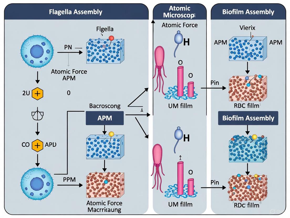
Nanoscale Insights: How Flagella Drive Biofilm Assembly Revealed by Advanced AFM
This article synthesizes cutting-edge research on the role of bacterial flagella in biofilm assembly, with a specific focus on breakthroughs enabled by Atomic Force Microscopy (AFM).
Nanoscale Insights: How Flagella Drive Biofilm Assembly Revealed by Advanced AFM
Abstract
This article synthesizes cutting-edge research on the role of bacterial flagella in biofilm assembly, with a specific focus on breakthroughs enabled by Atomic Force Microscopy (AFM). It explores the foundational mechanisms of flagella-mediated attachment and surface sensing, details innovative methodologies like automated large-area AFM for high-resolution imaging, and addresses key challenges in nanoscale biofilm analysis. By comparing flagellar functions across pathogens such as Pseudomonas aeruginosa and Pantoea sp., we provide a validated framework that links nanoscale cellular orientation to macroscale biofilm architecture. This knowledge is critical for developing targeted strategies to combat biofilm-associated antimicrobial resistance in clinical and industrial settings.
The Flagellum's Role: From Surface Sensing to Biofilm Architecture
While traditionally recognized for their role in bacterial propulsion, flagella are increasingly understood as sophisticated sensory organelles critical for surface colonization and biofilm development. This whitepaper synthesizes current research demonstrating how flagella function beyond motility, serving as mechanosensors that detect surface contact and initiate complex genetic regulatory programs for biofilm formation. We examine the molecular mechanisms underlying this transition, with particular emphasis on applications in atomic force microscopy (AFM) research that have revealed nanoscale interactions between flagella and surfaces. The integration of advanced AFM methodologies with molecular biology provides unprecedented insights into early biofilm assembly, offering potential avenues for therapeutic intervention in biofilm-associated infections and biofouling control.
The Dual Role of Flagella: Propulsion and Surface Sensing
Flagellar Structure and Motility
The bacterial flagellum is a complex nanomachine composed of over thirty proteins with a structural organization that includes a basal body, hook, and filament [1]. The basal body contains a rotary motor embedded in the cell membranes, powered by proton motive force that drives rotation at speeds reaching 100-1500 Hz [1]. Connected to the basal body is the hook, a curved polymeric structure that functions as a universal joint, transmitting torque to the filament - a long, helical propeller that can extend several micrometers from the cell surface [2]. This elaborate structure consumes approximately 2% of a cell's metabolic resources, indicating its critical importance for bacterial survival and adaptation [1].
Flagella as Mechanosensors
Beyond propulsion, flagella function as sophisticated mechanosensors that detect surface contact and changes in environmental viscosity [3] [4]. When flagellar rotation is impeded by surface contact or increased fluid viscosity, the resulting change in motor torque triggers intracellular signaling pathways that promote surface adaptation [5] [4]. This sensing capability enables bacteria to distinguish between planktonic and surface-associated states, initiating the genetic reprogramming necessary for biofilm development [3]. The flagellar motor stators (MotA/MotB complexes), which channel ions to drive rotation, play a particularly important role in this mechanosensing process by detecting load changes on the motor [3] [4].
Molecular Mechanisms of the Motile-Sessile Transition
Regulatory Hierarchy and Signaling Networks
The transition from motility to biofilm formation involves a complex regulatory network centered on the second messenger cyclic diguanylate monophosphate (c-di-GMP). Elevated levels of c-di-GMP inhibit motility while activating exopolysaccharide production and other biofilm-related components [1] [4]. This signaling molecule is synthesized by diguanylate cyclase (DGC) enzymes containing GGDEF domains and degraded by phosphodiesterase (PDE) enzymes containing EAL or HD-GYP domains [1]. The flagellum integrates with this network through multiple mechanisms:
- Stator-dependent signaling: Changes in motor torque directly activate c-di-GMP production, promoting sessile behaviors [4].
- Stator-independent pathways: Mutations disrupting flagellar assembly can activate extracellular polysaccharide production even in the absence of functional stators [4].
- Transcriptional reprogramming: Surface contact triggers genome-wide expression changes, including upregulation of biofilm matrix components and downregulation of flagellar biosynthesis genes [5].
The following diagram illustrates the core signaling pathway that regulates the transition from motility to biofilm formation:
Functional Regulation of Flagellar Activity
Bacteria employ both short-term and long-term strategies to regulate flagellar activity during surface colonization:
Short-term regulation: Existing flagella are functionally regulated through "brake" and "clutch" mechanisms that either inhibit rotation or modulate reversal frequency without degrading the structures [1]. Proteins such as EpsE in Bacillus subtilis and YcgR in Escherichia coli can directly interact with flagellar components to impede rotation [1].
Long-term regulation: Flagellar gene transcription is systematically repressed, and existing flagella are diluted through growth in the absence of de novo synthesis [1]. This resource reallocation optimizes energy investment toward biofilm matrix production rather than maintaining motility structures.
AFM Methodologies for Flagellar Research
Advanced AFM Imaging Techniques
Atomic force microscopy has revolutionized the study of flagella and biofilms by enabling high-resolution imaging under physiological conditions. Recent technological advances have addressed traditional limitations of AFM:
Large-area automated AFM: This approach combines multiple high-resolution scans over millimeter-scale areas, overcoming the limited field of view that previously restricted AFM imaging [6] [7]. Machine learning algorithms assist with image stitching, cell detection, and classification, enabling comprehensive analysis of biofilm organization [6].
In situ biofilm characterization: AFM can probe moist biofilms in conditions that preserve native structure and function, unlike electron microscopy which requires extensive sample preparation that may alter biofilm properties [8].
The workflow below outlines the key steps in AFM-based analysis of flagella-mediated biofilm formation:
Quantitative Cohesion Measurements
AFM enables direct quantification of biofilm cohesive strength, a critical parameter influencing biofilm stability and detachment. The methodology developed by [8] involves:
- Non-perturbative baseline imaging: Collecting topographic images of a biofilm region at low applied load (~0 nN).
- Controlled abrasion phase: Scanning a subregion at elevated load (40 nN) to displace biofilm material.
- Post-abrasion imaging: Returning to low load conditions to image the abraded region.
- Cohesive energy calculation: Determining the volume of displaced biofilm and corresponding frictional energy dissipated, resulting in cohesive energy values (nJ/μm³) [8].
This approach has revealed that biofilm cohesive energy increases with depth (from 0.10 ± 0.07 nJ/μm³ to 2.05 ± 0.62 nJ/μm³) and is enhanced by calcium concentration [8].
Experimental Data on Flagella-Mediated Biofilm Formation
Quantitative Impact of Flagellar Motility on Surface Colonization
Recent studies have provided quantitative insights into how flagellar motility affects biofilm formation under controlled conditions:
Table 1: Kinetic Parameters of Biofilm Formation in Motile vs. Non-Motile E. coli [9]
| Parameter | Motile Strain | Non-Motile Strain | Experimental Conditions |
|---|---|---|---|
| Initial attachment delay | Several hours | Minimal delay | Millifluidic channel, glass surface |
| Biofilm growth rate | Similar to non-motile | Similar to motile | After initial colonization |
| Surface coverage | Reduced in mono-culture | Enhanced in mono-culture | 36-40 hours growth |
| Competitive colonization | Disadvantage diminishes with co-colonizers | Advantage diminishes with co-colonizers | Multi-species community |
Table 2: Effect of Nutrient Dilution on P. aeruginosa Biofilm Initiation [10]
| Dilution Factor | Attached Cells (×10⁷ cells/cm²) | Motility Response | EPS Production |
|---|---|---|---|
| No nutrient (saline) | 0.51 | Baseline | Not reported |
| 1/100 dilution | Maximum attachment | Strongly enhanced | Not reported |
| 1/50 dilution | 0.99 | Enhanced | Not reported |
| Undiluted | 0.24 | Suppressed | Not reported |
Flagellar Coordination in Biofilm Architecture
High-resolution AFM imaging has revealed striking organizational patterns in bacterial biofilms that involve flagellar coordination:
Honeycomb patterning: Pantoea sp. YR343 cells form distinctive honeycomb-like patterns during early biofilm development, with flagellar structures bridging gaps between cells [6]. These patterns likely enhance biofilm cohesion and stability.
Flagellar entanglement: AFM visualization shows flagella extending tens of micrometers across surfaces, with some appendages appearing to originate from individual cells while others adhere to surfaces after detachment [6]. This network of flagella may facilitate cell-cell communication and structural integrity.
Preferred cellular orientation: Large-area AFM mapping has identified consistent alignment of surface-attached cells, suggesting flagella-mediated coordination during initial attachment phases [6].
Research Reagent Solutions and Methodologies
Essential Research Tools
Table 3: Key Reagents and Materials for Flagella and Biofilm Research
| Reagent/Material | Function/Application | Example Use |
|---|---|---|
| Millifluidic channels | Controlled hydrodynamic conditions for biofilm growth | Studying colonization kinetics in defined geometries [9] |
| PFOTS-treated glass | Hydrophobic surface for bacterial attachment | Examining initial surface attachment dynamics [6] |
| Polyvinylpyrrolidone (PVP) | Viscosity-modifying agent | Mimicking high-viscosity environments like host mucus [5] |
| Modified membrane substrates | Supports for biofilm growth in AFM | In situ cohesion measurements [8] |
| FAST fluorescent protein tags | Biofilm-relevant fluorescent labeling | Real-time monitoring of biofilm development [9] |
| CaCl₂ supplementation | Enhances biofilm cohesion | Studying matrix reinforcement effects [8] |
Protocol: AFM Cohesion Measurement in Biofilms
This protocol adapts the methodology from [8] for measuring cohesive energy in bacterial biofilms:
Biofilm cultivation: Grow biofilms in membrane-aerated bioreactors using activated sludge inoculum or specific bacterial strains. Maintain consistent nutrient conditions (e.g., 147 ± 37 mg/L chemical oxygen demand).
Sample preparation: Equilibrate biofilm samples in a controlled humidity chamber (90% RH) using saturated NaCl solution for 1 hour to maintain consistent water content.
Baseline imaging: Collect non-perturbative topographic images of 5×5 μm biofilm regions at minimal applied load (~0 nN) using oxide-sharpened Si₃N₄ tips (spring constant 0.58 N/m).
Abrasion phase: Select a 2.5×2.5 μm subregion and perform repeated raster scanning (4 scans) at elevated load (40 nN) with scan velocity of 50-100 μm/s.
Post-abrasion imaging: Return to low load conditions and collect another 5×5 μm image of the abraded region.
Data analysis: Calculate displaced biofilm volume through image subtraction. Determine frictional energy dissipation from lateral deflection signals. Compute cohesive energy as the ratio of energy dissipated to volume displaced (nJ/μm³).
Implications for Drug Development and Biofilm Control
The evolving understanding of flagellar functions in surface colonization presents new opportunities for therapeutic intervention:
Anti-biofilm strategies: Targeting flagellar mechanosensing pathways rather than bacterial viability may reduce selective pressure for resistance development [4]. Small molecules that disrupt c-di-GMP signaling or stator function could prevent biofilm formation without killing bacteria.
Surface engineering: Nanoscale topographic patterns that disrupt flagellar sensing or attachment can reduce biofilm formation on medical implants and industrial surfaces [6] [7]. AFM studies have demonstrated that specific surface patterns can significantly reduce bacterial density [6].
Viscosity-modifying approaches: Since flagellar mechanosensing responds to environmental viscosity, modulating mucus viscosity or composition could interfere with pathogen colonization in specific host niches [5].
Flagella represent a sophisticated multifunctional system that extends far beyond bacterial swimming capability. As mechanosensors and integration hubs for surface adaptation, flagella coordinate the complex transition from motility to biofilm formation through sophisticated regulatory networks centered on c-di-GMP signaling. Advanced AFM methodologies now enable researchers to visualize and quantify these processes at unprecedented resolution, revealing organizational patterns and physical interactions that underlie biofilm assembly. These insights provide a foundation for developing novel anti-biofilm strategies that target the initial stages of surface colonization rather than mature biofilm structures. For drug development professionals, understanding these mechanisms offers promising avenues for interfering with biofilm-related infections without applying direct bactericidal pressure.
The bacterial flagellum has traditionally been characterized as a motility organelle, enabling bacterial movement and chemotaxis. However, extensive research has now established that flagella function as critical adhesins, directly mediating the initial attachment phase of biofilm formation [11]. This adhesive capability is independent of flagellar motility, representing a sophisticated structural adaptation for surface colonization. In the context of biofilm assembly researched via Atomic Force Microscopy (AFM), understanding these non-motile functions of flagella provides essential insights into the nanoscale forces and interactions that underpin bacterial adhesion [6] [12]. The flagellum is a complex apparatus assembled from more than 20 different proteins, with its extracellular structure comprising a basal body, hook, hook-filament junction, filament, and filament cap [11] [2]. Over 60 structural and regulatory proteins are required for its assembly and function, with the entire structure extending up to 10 µm from the bacterial cell surface [11]. This extensive extracellular presence positions the flagellum as a primary interface for bacterium-surface interactions.
Structural Mechanisms of Flagellar Adhesion
Flagellar Components with Adhesive Properties
The flagellum facilitates adhesion through multiple structural components, with the flagellin (FliC) subunit and the filament cap protein (FliD) playing particularly significant roles.
- Flagellin (FliC): The major structural protein of the filament, FliC, has been demonstrated to function as an adhesin across multiple pathogenic species. The central region of FliC is variable in sequence and surface-exposed, explaining the observed differences in adhesive functions between bacterial strains and species [11]. In Pseudomonas aeruginosa, FliC adheres to glycosphingolipids including GM1, GD1a, and asialo-GM1 [11]. Similarly, FliC of Escherichia coli is involved in adhesion to mucins, bovine intestinal mucus, laminin, and collagen, and mediates cellular invasion via lipid rafts [11].
- Filament Cap (FliD): The pentameric FliD cap complex, located at the distal end of the growing flagellar filament, is critical for filament assembly and also serves adhesive functions [11] [2]. In P. aeruginosa, FliD mediates adhesion to human respiratory mucin, specifically recognizing the Lewis x glycotype [11]. Recent cryo-EM structures of the complete Salmonella enterica extracellular flagellum reveal that FliD forms a pentameric complex with a cavity enclosed by its D2-D3 and D0-D1 domains, creating a structure that facilitates flagellin incorporation but may also participate in surface recognition [2].
- Complete Filament: Beyond individual proteins, the entire flagellar filament can act as an adhesin. In pathogens such as E. coli, P. aeruginosa, and Clostridium difficile, the whole flagellum has been indicated as significant in bacterial adhesion to and invasion into host cells [11].
Nanoscale Architecture Revealed by Structural Biology
Recent advances in structural biology have provided unprecedented insights into the flagellar architecture that underpins its adhesive functions. Cryo-electron microscopy (cryo-EM) studies of the complete extracellular flagellum from Salmonella enterica have resolved the native structure of the FliD cap complex at 3.7 Å resolution and the FlgKL hook-filament junction at 2.9 Å resolution [2]. The structural analysis reveals that the hook-filament junction, composed of 11 subunits each of FlgK and FlgL, acts as a molecular buffer that prevents transfer of mechanical stress from the flexible hook to the rigid filament [2]. This structural stabilization may be crucial for maintaining adhesive interactions under fluid shear forces. The FliD cap complex exhibits a pseudosymmetric arrangement with varying heights for each FliD subunit, creating a tilted D2-D3 plane that interacts with the terminal ends of the flagellin filaments [2]. This intricate molecular arrangement creates multiple potential binding surfaces for host cell receptors and extracellular matrix components.
Diagram 1: Structural and functional relationships in flagella-mediated adhesion, showing how specific flagellar components interact with host molecules to drive biofilm initiation.
Quantitative Analysis of Flagellar Adhesion Forces
Atomic Force Microscopy has enabled direct quantification of the adhesion forces between bacterial flagella and surfaces, providing critical nanoscale measurements that underpin theoretical models of initial attachment.
Single-Cell Adhesion Force Measurements
AFM force spectroscopy measurements reveal specific interaction forces between bacterial cells and mineral surfaces, with flagella playing a significant role in these interactions.
Table 1: AFM Force Measurements of Bacterial Adhesion to Mineral Surfaces
| Bacterial Strain | Mineral Surface | Adhesion Force | Adhesion Energy | Key Findings | Reference |
|---|---|---|---|---|---|
| Escherichia coli | Goethite | -3.0 ± 0.4 nN | -330 ± 43 aJ (10⁻¹⁸ J) | Bond strengthening occurred within 4 seconds to maximum adhesion | [12] |
| Escherichia coli | Goethite | 97 ± 34 pN | N/R | Initial attractive force during approach ("jump-in" event) | [12] |
| Shewanella oneidensis | Goethite (010) face | -0.80 ± 0.15 nN | N/R | Anaerobic conditions, after 30-45 min contact | [12] |
| Shewanella oneidensis | Goethite (010) face | -0.25 ± 0.10 nN | N/R | Aerobic conditions, after 30-45 min contact | [12] |
| Pantoea sp. YR343 | Glass (PFOTS-treated) | N/Q | N/Q | Flagellar structures ~20-50 nm in height, extending tens of micrometers | [6] |
N/R = Not Reported; N/Q = Not Quantified
The measured forces demonstrate that flagella-mediated adhesion involves both initial attractive forces and subsequent bond strengthening mechanisms. The observation that E. coli adhesion forces to goethite strengthened to -3.0 nN within 4 seconds suggests rapid structural or chemical adaptations at the flagella-surface interface [12]. AFM imaging of Pantoea sp. YR343 further revealed flagellar structures bridging gaps between cells during early attachment stages, forming intricate networks that facilitate community organization [6].
Species-Specific and Surface-Specific Adhesion Patterns
Comparative studies across bacterial species and surface types reveal important patterns in flagellar adhesion efficacy and specificity.
Table 2: Flagella-Mediated Adhesion Across Bacterial Species and Target Surfaces
| Bacterial Species | Flagellar Component | Adhesion Target | Receptor/Mechanism | Functional Role | Reference |
|---|---|---|---|---|---|
| Escherichia coli | Flagellum, FliC | HeLa cells, mucins, laminin, collagen | EtpA, gluconate, lipid rafts | Adhesion, microcolony formation, invasion | [11] |
| Pseudomonas aeruginosa | FliC, FliD | Human respiratory mucin, Calu-3 cells | GM1, GD1a, asialo-GM1, Lewis x, HSPGs | Adhesion, virulence | [11] |
| Clostridium difficile | FliC, FliD | Mouse cecal mucus, hamster model | Not Determined (ND) | Binding | [11] |
| Campylobacter jejuni | Flagellum | Intestine-407 cells | ND | Adhesion | [11] |
| Bacillus pseudomallei | Flagellum | Acanthamoeba astronyxis | ND | Adhesion, invasion | [11] |
| Segmented Filamentous Bacteria | Flagellin (FliC) | Intestinal epithelial cells | Endophilin A2, αM integrin | Adhesion, endocytosis, Th17 immune response | [13] |
The data indicates that flagellar adhesion is a widespread mechanism across diverse bacterial species, targeting both abiotic surfaces and specific host tissues. The molecular mechanisms, however, vary significantly, with some pathogens employing specific protein-receptor interactions (e.g., P. aeruginosa FliD binding to Lewis x glycotype) while others utilize more generalized interactions with extracellular matrix components [11].
Methodologies for Investigating Flagellar Adhesion
Atomic Force Microscopy (AFM) Protocols
AFM provides unparalleled capability for direct nanoscale imaging and force measurement of flagella-surface interactions under physiologically relevant conditions.
Sample Preparation for Flagellar AFM:
- Bacterial Probes: Bacterial cells are attached to AFM cantilevers using bio-compatible adhesives like polyethyleneimine or concanavalin A, ensuring proper orientation for flagellar contact [12].
- Substrate Functionalization: Mineral surfaces (e.g., goethite, kaolinite) are immobilized on glass substrates using electrostatic attachment or thin film deposition. For clay-sized particles, suspensions are sonicated and deposited onto freshly cleaved mica surfaces [12].
- Liquid Imaging Conditions: Measurements are performed in appropriate buffer solutions (e.g., 10 mM Tris-HCl, pH 7.0) or growth media to maintain flagellar structural integrity and function [12].
Force Spectroscopy Measurements:
- Approach-Retraction Cycles: Multiple force-distance curves (typically 100-1000 per sample) are collected across different surface locations to account for heterogeneity [12].
- Adhesion Force Mapping: Spatial adhesion maps are generated by performing force volume analysis across 1×1 µm to 10×10 µm areas, correlating adhesion events with surface topography [6].
- Bond Strength Analysis: Rupture forces and energies are calculated from retraction curve analysis, with multiple unbinding events indicating the involvement of multiple flagellar filaments or repeated binding domains [12].
Large-Area Automated AFM for Biofilm Assembly:
- Recent advancements enable automated large-area AFM imaging over millimeter-scale areas, overcoming traditional AFM limitations [6].
- Machine learning algorithms assist with image stitching, cell detection, and classification, allowing comprehensive analysis of flagellar networks during early biofilm development [6].
- This approach reveals spatial heterogeneity and preferred cellular orientation during surface attachment, with flagellar coordination playing a significant role in biofilm assembly beyond initial attachment [6].
Genetic and Molecular Biology Approaches
Flagellar Mutant Construction:
- Knockout Strategies: Targeted gene deletion of flagellar components (e.g., fliC, fliD, flgE) using two-step allelic exchange with counterselectable markers (e.g., sacB) [14].
- Complementation assays: Reintroduction of wild-type genes on plasmids to verify phenotype restoration and rule out polar effects [15].
Adhesion Quantification Methods:
- Crystal Violet Staining: Semi-quantitative assessment of biofilm biomass after fixation and staining [14].
- Fluorescent Labeling: Wheat germ agglutinin (WGA) staining for holdfast polysaccharide visualization in Caulobacter crescentus [15].
- Adhesion Profiling: Genome-wide transposon mutant screening to identify genes affecting adhesion through competitive selection assays [15].
Flagellar Adhesion in Biofilm Development Pathways
The transition from motile to sessile lifestyle represents a critical developmental switch in bacterial life history, with flagellar adhesion serving as a key regulatory point.
From Motility to Adhesion: Physiological Switching
Bacteria exhibit sophisticated regulation of flagellar function to transition between exploration and colonization phases:
- Transcriptional Control: The flhDC flagellar master operon is regulated by environmental conditions including temperature, osmolarity, and pH, controlling the switch from motile to sessile lifestyle [11].
- Mechanosensing: Flagella act as surface sensors, with mechanical impediment of rotation triggering intracellular signaling cascades that promote adhesion factor production [15].
- Second Messenger Signaling: In Caulobacter crescentus, flagellar perturbations activate two distinct pathways for adhesin production: a PleD-dependent pathway and a MotAB stator-dependent pathway involving the diguanylate cyclase DgcB [15].
Diagram 2: Signaling pathways linking flagellar perturbation to adhesin production, showing two genetically distinct pathways that coordinate the motile-to-sessile transition in bacteria.
Paradoxical Role of Flagellar Impairment in Biofilm Development
Interestingly, genetic impairment of flagellar function often enhances adhesion through compensatory mechanisms:
- Hyperadhesive Mutants: Mutations disrupting flagellar assembly in Caulobacter crescentus stimulate production of holdfast polysaccharide, creating a hyperadhesive phenotype [15].
- Structural Adaptations: P. aeruginosa flgE hook protein mutants exhibit reduced initial adhesion but enhanced formation of microcolony aggregates with increased antibiotic tolerance [14].
- Resource Reallocation: The metabolic cost savings from flagellar loss may be redirected toward exopolysaccharide production and other adhesion factors [14].
Research Reagents and Methodological Toolkit
Table 3: Essential Research Reagents for Investigating Flagella-Mediated Adhesion
| Reagent/Category | Specific Examples | Research Application | Key Function | Reference |
|---|---|---|---|---|
| Bacterial Strains | Pseudomonas aeruginosa MPAO1, Escherichia coli BW25113, Caulobacter crescentus | Genetic studies, adhesion assays | Model organisms with well-characterized flagellar systems | [15] [14] |
| Genetic Tools | pK19mobsacB vector, CRISPR/Cas9 systems, transposon libraries | Mutant construction, adhesion profiling | Targeted gene deletion, genome-wide screening | [15] [14] |
| Detection Reagents | Crystal violet, fluorescent WGA, FITC-conjugated antibodies | Biofilm quantification, polysaccharide staining | Visualize and quantify adhesion and matrix production | [15] [14] |
| AFM Consumables | PEI-coated cantilevers, functionalized tips, mica substrates | Nanoscale force measurements, topography imaging | Direct measurement of adhesion forces at single-cell level | [6] [12] |
| Microfluidic Devices | PDMS channels, flow cells | Biofilm development under shear stress | Mimic physiological conditions for colonization | [9] |
Implications for Therapeutic Development and Future Research
The understanding of flagella as adhesins rather than purely motility organelles opens new avenues for antibiofilm strategies:
- Anti-Adhesion Therapeutics: Targeting flagellar adhesive components (FliC, FliD) without impairing motility could prevent biofilm formation without inducing evolutionary pressure toward hyperadhesive mutants [16].
- Surface Modification Approaches: Nanoscale engineering of medical implant surfaces to resist flagellar adhesion based on AFM force measurements [6] [12].
- Signal Interference: Disruption of the flagellar mechanosensing pathways that trigger the transition to biofilm growth [15].
Future research directions should focus on:
- High-throughput screening of compounds that specifically inhibit flagellar adhesion domains
- Multiscale modeling integrating nanoscale AFM data with population-level biofilm dynamics
- Clinical translation of anti-adhesion strategies targeting flagella in device-related infections
In conclusion, flagella serve as sophisticated adhesive organelles that mediate critical initial attachment phases in biofilm formation. Through direct molecular interactions, nanoscale force generation, and integrated signaling pathways, flagellar adhesion represents a fundamental mechanism in bacterial surface colonization. The continuing advancement of AFM methodologies, particularly automated large-area analysis and machine learning-assisted force mapping, provides increasingly powerful tools to decipher these complex interactions at relevant biological scales.
The transition from reversible to irreversible bacterial attachment is a critical, yet poorly understood, pivot point in biofilm formation. This whitepaper synthesizes recent advances in atomic force microscopy (AFM) to delineate the precise role of flagella in this process. Moving beyond their established function in initial surface contact, we examine how flagellar coordination directly influences the architectural assembly of early biofilms. By integrating quantitative data on adhesion timescales with high-resolution structural data, this guide provides researchers with a detailed framework for investigating this fundamental shift, offering novel perspectives for targeting biofilm-related infections and antifouling strategies.
Biofilms are structured microbial communities encased in a self-produced matrix that pose significant challenges in healthcare and industry due to their resilience against antibiotics and disinfectants [6]. The formation of a biofilm is a multi-stage process beginning with the initial attachment of planktonic cells to a surface. The flagellum is a key cellular appendage historically recognized for its role in bacterial motility and the initial, transient contact with a surface, termed reversible attachment [17] [18]. In this stage, cells are not permanently fixed and can still move laterally or detach from the surface.
Emerging research, powered by high-resolution imaging techniques like atomic force microscopy (AFM), now reveals a more complex and active role for flagella. They are implicated in the crucial transition to irreversible attachment, a permanent state that commits a cell to the biofilm lifestyle [6]. This pivotal shift involves the secretion of permanent adhesins and is a prerequisite for subsequent microcolony formation and mature biofilm development. Understanding the mechanisms governing this transition is therefore essential for developing strategies to control problematic biofilms.
Mechanistic Insights: The Flagellar Pivot
The journey from a free-swimming cell to a surface-anchored one involves a finely tuned sequence of events. The following diagram illustrates the critical pathway and key decision points a bacterial cell undergoes during this process.
Diagram Title: Bacterial Cell Attachment Pathway
From Transient Contact to Permanent Adhesion
As illustrated, the process initiates when a motile cell is brought into contact with a surface via its flagellum. This constitutes reversible attachment, a state characterized by short dwell times; for Caulobacter crescentus, this averages 12 seconds before the cell either detaches or commits to the surface [18]. During this brief window, flagella are thought to act as mechanosensors, transducing the physical signal of surface contact into biochemical signals that can trigger the next phase.
The pivotal shift to irreversible attachment is marked by the rapid secretion of a permanent adhesin. In C. crescentus, this is a polar polysaccharide called holdfast. Remarkably, surface contact stimulates holdfast production in approximately 23 seconds—30 times faster than the developmentally regulated holdfast production in the absence of such contact [18]. This underscores the critical importance of flagellar signaling in accelerating the transition to a sessile state. High-resolution AFM imaging of Pantoea sp. YR343 has further revealed that flagella are not merely discarded after this stage; instead, they form intricate networks, bridging gaps between cells and suggesting a role in the coordination of early biofilm architecture beyond initial attachment [6].
Quantitative Profiling of Attachment Dynamics
A detailed understanding of the attachment transition requires quantitative data on its timing and frequency. The following table summarizes key experimental findings from single-cell studies.
Table 1: Timescales and Frequencies of Bacterial Attachment Events
| Parameter | Wild-Type C. crescentus | Pilus-Minus Mutant | Experimental Context |
|---|---|---|---|
| Holdfast Production (Surface-initiated) | 23 seconds | Not Reported | Microfluidic device, glass surface [18] |
| Holdfast Production (Developmental) | 13 minutes | Not Reported | Standard growth conditions [18] |
| Reversible Adhesion Dwell Time | 12 seconds | 13 seconds | Microfluidic device, glass surface [18] |
| Frequency of Reversible Adhesion | 6.8 events/min | ~4 events/min (estimated) | Microfluidic device, glass surface [18] |
| Frequency of Irreversible Adhesion | 3.3 events/min | 0.2 events/min | Microfluidic device, glass surface [18] |
| Cell Dimensions (Pantoea sp. YR343) | ~2 µm length, ~1 µm diameter | Not Applicable | AFM on PFOTS-treated glass [6] |
| Flagella Height (Pantoea sp.) | 20-50 nm | Not Detected | AFM on PFOTS-treated glass [6] |
The data reveals several critical insights. First, the similar dwell times in reversible attachment for both wild-type and pilus-minus mutants suggest that pili do not significantly influence the duration of transient attachment [18]. Second, the drastic reduction in irreversible adhesion frequency in the mutant (15-fold less than wild-type) highlights that pili are indispensable for the transition from reversible to irreversible attachment. Finally, the higher frequency of reversible versus irreversible events in wild-type cells indicates that cells often sample a surface multiple times before committing to permanent adhesion [18].
A New Imaging Paradigm: Large Area Automated AFM
Traditional analytical methods have struggled to capture the full spatial complexity and dynamic nature of early biofilm formation. Atomic force microscopy (AFM) has overcome this barrier by providing nanometer-scale resolution of structural and functional properties under physiological conditions, without extensive sample preparation that can distort microbial structures [6].
Technical Workflow for Large Area AFM
The recent development of automated large area AFM has been a game-changer, enabling the capture of high-resolution images over millimeter-scale areas. The following diagram outlines the core workflow of this powerful methodology.
Diagram Title: Large Area Automated AFM Workflow
This automated approach overcomes the traditional limitations of AFM, such as its small imaging area (typically <100 µm) and labor-intensive operation [6]. The integration of machine learning (ML) is crucial at multiple stages: for optimizing the scanning process, stitching images with minimal overlapping features, and performing high-volume, automated analysis of the resulting dataset [6]. This allows for the efficient extraction of quantitative parameters like spatial heterogeneity, cellular morphology, and the distribution of extracellular features like flagella over biologically relevant length scales.
Key Research Reagents and Materials
The application of this AFM methodology relies on a specific set of research reagents and materials, as detailed below.
Table 2: Essential Research Reagent Solutions for AFM Biofilm Studies
| Reagent/Material | Specification / Function | Application in Featured Research |
|---|---|---|
| Bacterial Strain | Pantoea sp. YR343 (gram-negative, rhizosphere isolate) / Model biofilm-forming organism with peritrichous flagella [6]. | Studying initial surface attachment dynamics and honeycomb pattern formation [6]. |
| Functionalized Surface | PFOTS-treated glass coverslips / Creates a controlled hydrophobic surface to study bacterial adhesion [6]. | Substrate for observing preferred cellular orientation and flagellar coordination [6]. |
| Microfluidic Device | PDMS replicas sealed with glass coverslips / Enables precise control of flow and environment for single-cell observation [18]. | Quantifying timescales and frequencies of reversible/irreversible adhesion events [18]. |
| Fluorescent Lectin | Wheat Germ Agglutinin (WGA) labeled with Alexa Fluor 555 / Binds specifically to holdfast polysaccharide [18]. | Differentiating irreversibly attached cells (holdfast-positive) from reversibly attached ones [18]. |
| Machine Learning Tools | AI-driven image analysis algorithms / For automated image stitching, cell detection, and classification [6]. | Managing high-volume AFM data and extracting quantitative morphological parameters over large areas [6]. |
Experimental Protocols
Protocol A: Quantifying Adhesion Dynamics via Microfluidics and Fluorescence
This protocol is adapted from studies on Caulobacter crescentus to characterize the timescales of reversible and irreversible attachment at the single-cell level [18].
- Device Fabrication: Fabricate a microfluidic channel (e.g., 110 µm wide, 20 µm high) in PDMS using standard soft lithography techniques and reversibly seal it to a clean glass coverslip.
- Cell Preparation: Grow the bacterial strain of interest (e.g., C. crescentus) in a suitable complex medium (e.g., PYE). Isolate motile swarmer cells for analysis.
- Fluorescent Labeling: Introduce a fluorescently labeled lectin (e.g., WGA-AF555) specific to the irreversible adhesin (e.g., holdfast) into the medium at a concentration of ~1 µg/mL.
- Image Acquisition: Mount the microfluidic device on an inverted fluorescence microscope. Perfuse the cell suspension through the channel at a controlled, low flow rate. Use time-lapse microscopy to track:
- GFP-expressing cells for position and motility.
- Fluorescence from the lectin probe to detect adhesin secretion.
- Data Analysis:
- Reversible Adhesion: Track cells that contact the surface and then depart. Measure the dwell time.
- Irreversible Adhesion: Identify cells that contact the surface and subsequently show localized lectin fluorescence. Record the time from contact to fluorescence as the adhesin production time.
- Frequency: Calculate the rate of reversible and irreversible adhesion events per minute within the field of view.
Protocol B: Imaging Early Biofilm Assembly via Large Area AFM
This protocol details the use of automated AFM to visualize the structural role of flagella in early biofilm formation on surfaces [6].
- Surface Preparation: Treat glass coverslips with PFOTS to create a uniform, hydrophobic surface. Sterilize the coverslips before use.
- Biofilm Growth: Place the treated coverslips in a Petri dish and inoculate with the bacterial strain (e.g., Pantoea sp. YR343) in liquid growth medium. Incubate for a defined period (e.g., 30 minutes for initial attachment; 6-8 hours for cluster formation).
- Sample Harvesting: At selected time points, gently remove the coverslips from the culture and rinse with a buffer or deionized water to remove non-adherent cells. Air-dry the samples.
- Automated AFM Imaging:
- Mount the sample on the AFM stage.
- Initiate an automated large-area scan protocol, defining a millimeter-sized area of interest.
- The system will automatically capture hundreds of contiguous high-resolution AFM images (e.g., tapping mode in air).
- Data Processing and Analysis:
- Stitching: Use integrated machine learning algorithms to stitch individual images into a seamless, large-area mosaic.
- Feature Extraction: Apply ML-based segmentation to identify individual cells and flagella.
- Quantification: Extract quantitative data on cellular density, distribution, orientation, and the presence of extracellular appendages.
The integration of advanced techniques like large area AFM and microfluidics has fundamentally shifted our understanding of the flagellum's role in biofilm formation. It is no longer viewed as a simple propeller for motility but as a sophisticated sensing and coordinating device that directly governs the pivotal shift from reversible to irreversible attachment. The quantitative data and detailed methodologies presented in this whitepaper provide a robust framework for future research. For drug development professionals, targeting the molecular mechanisms that underpin flagellar-mediated signaling and adhesion commitment presents a promising, and potentially transformative, strategy for preventing biofilm-associated infections and mitigating biofouling.
This technical guide explores the role of bacterial flagella in orchestrating the spatial organization of biofilms, with a specific focus on the emergence of honeycomb patterns and cellular alignment. The assembly of biofilms into complex architectures is a critical factor in their resilience and functional properties. Recent advancements in atomic force microscopy (AFM), particularly automated large-area AFM, have provided unprecedented high-resolution insights into the early stages of biofilm formation. This whitepaper synthesizes cutting-edge research demonstrating how flagella, beyond their role in motility, mediate cell-surface and cell-cell interactions to direct the assembly of structured microbial communities. The findings and methodologies detailed herein are framed within the broader thesis that flagellar function is a fundamental driver of biofilm architecture, offering potential novel targets for therapeutic intervention in drug development.
The transition from free-swimming planktonic bacteria to structured, surface-associated biofilm communities is a complex process with profound implications in both clinical and environmental contexts. Biofilms are multicellular aggregates held together by an extracellular polymeric substance (EPS), conferring significant resistance to antibiotics and disinfectants [19]. While the protective role of the EPS matrix is well-known, the underlying physical and biological mechanisms guiding the initial spatial assembly of cells into functional architectures are less understood.
Emerging evidence positions the flagellum as a central player in this process, functioning not only as a propulsion organelle but also as a sensor, an adhesin, and a direct mediator of spatial organization. The regulation of flagellar activity is therefore critical for the motility-to-biofilm transition, a process often governed by the intracellular secondary messenger cyclic di-GMP (c-di-GMP) [1]. Elevated levels of c-di-GMP are associated with the inhibition of motility and the activation of biofilm formation, implicating flagellar control in the shift from a motile to a sessile lifestyle.
This guide details how advanced imaging techniques, specifically large-area automated Atomic Force Microscopy (AFM), are unveiling the nanoscale dynamics of flagella-mediated biofilm assembly. We provide a comprehensive analysis of the experimental evidence for flagella-driven patterning, detailed protocols for its investigation, and a discussion of the implications for anti-biofilm strategies.
High-Resolution Imaging Reveals Flagella-Mediated Patterning
Direct Visualization via Large-Area Automated AFM
Traditional AFM has been limited in its application to biofilm research due to its restricted scan range (typically <100 µm), which is insufficient to capture the millimeter-scale heterogeneity of nascent biofilms [19]. The advent of automated large-area AFM has begun to overcome this limitation. This approach integrates automated scanning over millimeter-scale areas with machine learning (ML) for image stitching, cell detection, and classification, enabling a seamless link between nanoscale cellular features and the emergent macroscale organization of the film [19].
A pivotal application of this technology has been the study of Pantoea sp. YR343, a gram-negative, plant-growth-promoting bacterium. When researchers used large-area AFM to image the early stages (approximately 30 minutes) of biofilm formation on PFOTS-treated glass surfaces, they made a key observation: the surface-attached cells exhibited a preferred cellular orientation, forming a distinctive honeycomb pattern [19]. This highly organized structure was previously obscured by the resolution and area limitations of other microscopy techniques.
The Role of Flagella in Pattern Formation
The high-resolution capability of AFM was crucial for hypothesizing the mechanism behind this pattern. The technique enabled clear visualization of flagellar structures, measuring 20–50 nm in height and extending for tens of micrometers across the surface [19]. Detailed mapping of these appendages suggested that flagellar coordination plays a role in biofilm assembly beyond initial attachment.
The observed honeycomb pattern, which emerged within 6-8 hours of incubation, points to a model where flagella are not merely used for swimming towards a surface but are actively involved in organizing cells after attachment. Flagellar filaments were seen bridging gaps between cells, suggesting they may help pull cells into a structured configuration or mediate chemical signaling that guides organization [19]. The spatial arrangement of these flagellar interactions provides a physical basis for the emergent honeycomb architecture.
Table 1: Key Quantitative Findings from AFM Studies of Flagella-Driven Organization
| Parameter | Measurement | Significance | Experimental Context |
|---|---|---|---|
| Flagellar Diameter | 20 - 50 nm | Visualized via high-res AFM; confirmed as flagella via mutant strain [19] | Pantoea sp. YR343, early attachment (30 min) [19] |
| Cellular Dimensions | ~2 µm length, ~1 µm diameter | Rod-shaped cells providing building blocks for pattern [19] | Pantoea sp. YR343 on PFOTS-treated glass [19] |
| Pattern Emergence Time | 6 - 8 hours | Timeframe for formation of mature honeycomb-like cell clusters [19] | Pantoea sp. YR343 biofilm development [19] |
| Spatial Flagellin Arrangement | FlaA: base (8-28%); FlaB: remainder | Specialized flagellar filament composition improves motility in diverse conditions [20] | Shewanella putrefaciens polar flagellum [20] |
Molecular and Functional Mechanisms of Flagellar Organization
The spatial patterns observed by AFM are the result of a complex interplay of molecular mechanisms that regulate and utilize flagellar function.
Regulatory Pathways: The c-di-GMP Switch
A key regulator governing the transition from motility to biofilm formation is the ubiquitous bacterial second messenger, cyclic di-GMP (c-di-GMP). This molecule acts as a central switch [1]:
- Elevated c-di-GMP levels promote biofilm formation and inhibit motility.
- Diminished c-di-GMP levels activate motility and inhibit biofilm formation.
This regulation operates on two levels:
- Short-term functional control: c-di-GMP binds to effector proteins like YcgR, which acts as a "clutch" or "brake" on the flagellar motor. In E. coli and Bacillus subtilis, the c-di-GMP/YcgR complex interacts with the motor switch protein FliG, effectively arresting motor rotation and stopping swimming without disassembling the flagellum [1].
- Long-term transcriptional control: High c-di-GMP levels can downregulate the expression of flagellar biosynthetic genes. Over time, as cells grow and divide, the existing flagella are diluted out, permanently locking the population in a sessile state [1].
This regulatory paradigm ensures that the energetically costly process of flagellar synthesis and rotation is halted once surface attachment and community formation are initiated.
Spatial Arrangement of Multiple Flagellins
The flagellar filament itself can be a complex structure. Many bacteria possess multiple flagellin genes. For example, Shewanella putrefaciens has two flagellins, FlaA and FlaB, which are not randomly incorporated into its polar flagellum. Instead, they exhibit a spatial arrangement: FlaA is predominantly found in the proximal region of the filament (closer to the motor), while FlaB forms the majority of the distal filament [20].
This segmentation is functionally critical. Observations of swimming trajectories and numerical simulations demonstrate that this specific flagellin arrangement improves motility across a range of environmental conditions and facilitates a screw-like motility that enhances cellular spreading through obstructed environments [20]. This sophisticated design allows for flagella that are optimized for multiple tasks—a rigid base for efficient propulsion and a more flexible tip for navigating complexity—which directly influences how cells explore surfaces and initiate colonization.
Context-Dependent Role of Flagellar Motility
The contribution of flagellar motility to biofilm formation is not universal but is highly dependent on environmental and genetic contexts. For instance:
- Nutrient Stress: In oligotrophic (nutrient-poor) environments, Pseudomonas aeruginosa augments its flagellar motility as a stress response to scavenge for nutrients, which in turn increases cell-wall collision frequency and enhances initial attachment [10].
- Competitive Disadvantage: In a controlled millifluidic channel, motile E. coli cells exhibited a significant delay in biofilm formation compared to non-motile isogenic mutants. This suggests that in certain geometries, motility promotes continued surface exploration rather than stable attachment [21].
- Presence of Co-colonizers: The competitive disadvantage of motile E. coli recedes in the presence of other bacterial species, likely due to resource consumption by co-colonizers that inhibits motility, or through changes in the physicochemical environment [21].
These findings indicate that the role of flagella in biofilm development is multifaceted, involving direct physical interactions, regulated motility, and sophisticated filament assembly, all of which are tuned by environmental conditions.
Experimental Protocols for Investigating Flagella-Driven Organization
Large-Area Automated AFM for Biofilm Imaging
The following methodology, adapted from Millan-Solsona et al. (2025), is designed for capturing flagella-mediated spatial organization in nascent biofilms [19].
1. Sample Preparation:
- Strain: Pantoea sp. YR343 (wild-type) and an isogenic flagella-deficient mutant (e.g., ΔfliC) as a control.
- Surface Treatment: Use glass coverslips treated with PFOTS (perfluorooctyltrichlorosilane) to create a homogeneous, hydrophobic surface.
- Biofilm Growth: Inoculate a petri dish containing the treated coverslips with bacteria in liquid growth medium. Incubate for selected time points (e.g., 30 min for initial attachment, 6-8 h for pattern formation).
- Fixation: At each time point, remove a coverslip, gently rinse with buffer (e.g., PBS) to remove non-adherent cells, and air-dry. (Note: Drying is used for this specific protocol, although AFM can also be performed under liquid for physiological conditions).
2. Automated AFM Imaging:
- Instrumentation: A commercial AFM system equipped with a large-range piezoelectric scanner (capable of millimeter-scale travel).
- Automation Software: Implement software for automated selection of multiple imaging sites across the sample surface to cover a large area (e.g., >1 mm²).
- Scanning Parameters:
- Mode: Intermittent-contact (tapping) mode is recommended to minimize shear forces on delicate biological structures.
- Probes: Sharp silicon cantilevers with resonant frequencies of ~300 kHz and spring constants of ~40 N/m.
- Resolution: Set a high pixel resolution (e.g., 512 x 512 or 1024 x 1024) per individual image to resolve flagella (~20 nm diameter).
3. Image Processing and Analysis:
- Stitching: Use machine learning-based algorithms to stitch individual high-resolution AFM images into a seamless, large-area topographic map. This is critical for identifying large-scale patterns.
- Feature Identification: Apply ML-based segmentation and classification to automatically identify and count cells, measure cellular orientation (e.g., using Fourier analysis), and detect flagellar filaments.
- Quantitative Analysis: Extract key parameters including:
- Cell Density: Number of cells per unit area.
- Confluency: Percentage of surface area covered by cells.
- Cellular Orientation: Preferred angle of cell alignment.
- Morphology: Cell length, width, and volume.
Functional Motility Assays
To correlate spatial organization with flagellar function, the following assays are essential:
- Soft Agar Swarming Assay: Plate bacteria in low-concentration agar (0.3-0.6%) to assess the ability of cells to move collectively over a solid surface. Measure the diameter of the swarm colony over time [20].
- Liquid Swimming Assay: Inoculate bacteria in low-viscosity liquid medium (0.2-0.3% agar) and observe the turbidity halo formed by motile cells radiating from the inoculation point.
- Single-Cell Tracking: Use phase-contrast or dark-field microscopy to track the swimming trajectories of individual planktonic cells in liquid. Analyze velocity, turning frequency, and run length to quantify motility behavior [20] [21].
Table 2: Research Reagent Solutions for Flagella and Biofilm Research
| Reagent / Material | Function in Research | Specific Example |
|---|---|---|
| PFOTS-Treated Substrates | Creates a defined, hydrophobic surface to study the effect of surface properties on bacterial attachment and pattern formation [19]. | Glass coverslips for AFM imaging [19]. |
| Isogenic Flagellar Mutants | Serves as essential controls to confirm that observed structures are flagella and to delineate the specific role of flagella in a process [19] [20]. | ΔfliC (flagellin-deficient) or mot mutants (paralyzed flagella) [19] [21]. |
| C-di-GMP Modulators | Chemicals or genetic constructs that alter intracellular c-di-GMP levels to study its role in the motility-to-biofilm transition [1]. | Overexpression of diguanylate cyclases (to raise c-di-GMP) or phosphodiesterases (to lower it). |
| Fluorescent Maleimide Dyes | Covalently labels engineered cysteine residues in flagellins for spatial visualization of specific flagellins within a filament [20]. | Studying the distribution of FlaA vs. FlaB in Shewanella putrefaciens [20]. |
| Millifluidic Devices | Provides a controlled hydrodynamic environment for real-time, kinetic studies of biofilm development under flow [21]. | PDMS channels for monitoring E. coli biofilm formation [21]. |
Visualization of Regulatory and Experimental Pathways
Flagellar Regulation by c-di-GMP
Diagram Title: Flagellar Regulation by c-di-GMP
Large-Area AFM Workflow
Diagram Title: Large-Area AFM Biofilm Analysis Workflow
Discussion and Future Perspectives
The integration of large-area AFM with molecular genetics has definitively shown that flagella are nanoscale architects of biofilm spatial organization. The discovery of honeycomb patterns in Pantoea sp. biofilms provides a tangible model for understanding how flagellar appendages direct the assembly of complex communities from the bottom up. The regulatory control exerted by c-di-GMP and the functional specialization of flagellar filaments underscore the sophistication of this biological process.
For researchers and drug development professionals, these insights open new avenues for combating problematic biofilms. Rather than targeting bacterial viability—which drives antibiotic resistance—therapeutic strategies could aim to disrupt the spatial organization critical to biofilm resilience. Potential targets include:
- The c-di-GMP signaling network to lock bacteria in a motile, non-biofilm state.
- The flagellar stator complex to impair torque generation without preventing flagellin synthesis, potentially avoiding immune system activation directed against flagellin.
- The specific molecular interactions that allow flagella to act as intercellular tethers.
Future research should focus on correlating nanoscale AFM findings with transcriptomic and proteomic data from spatially resolved regions of the biofilm. Furthermore, applying these advanced AFM techniques to multi-species biofilms and under varying fluid shear stresses will provide a more holistic understanding of biofilm architecture in clinically and environmentally relevant scenarios. The continued refinement of automated, large-area AFM promises to be an indispensable tool in this endeavor, finally allowing scientists to link the subcellular world of flagellar motors to the functional architecture of the biofilm community.
{ create a technical guide document with the exact title, tables, diagrams, and experimental protocols as requested }
Interspecies Comparison: Flagellar Functions inPseudomonas aeruginosavs.Pantoeasp.
This technical guide provides a comparative analysis of flagellar functions in Pseudomonas aeruginosa and Pantoea* sp., with a specific focus on their roles in biofilm assembly as revealed by advanced Atomic Force Microscopy (AFM) research. For researchers and drug development professionals, understanding these interspecies differences is critical for developing targeted anti-biofilm strategies. Key findings indicate that while both organisms utilize flagella for initial surface attachment, P. aeruginosa employs its single polar flagellum for complex three-dimensional structuring and as a potential structural scaffold within mature biofilms. In contrast, Pantoea sp. relies on its peritrichous flagella to form distinctive honeycomb-patterned biofilms on hydrophobic surfaces. The integration of large-area automated AFM with machine learning is revolutionizing this field, enabling unprecedented nanoscale resolution over millimeter-scale areas to visualize flagellar coordination and its impact on biofilm architecture. This whitepaper details specific methodologies, quantitative findings, and essential research tools to advance the study of bacterial flagella in biofilm development.
The flagellar apparatus, while functionally conserved for motility, exhibits significant structural and regulatory differences between Gram-negative bacteria, which in turn dictate their unique biofilm formation pathways.
Core Structural and Functional Differences
Pseudomonas aeruginosa typically possesses a single, unsheathed polar flagellum that facilitates swimming motility in liquid environments [22] [23]. This polar placement is a defining characteristic, contrasting with the peritrichous flagella of enteric bacteria. The flagellum is not merely a motility organelle; it is a multi-component nanomachine composed of a membrane-embedded basal body (containing C, MS, P, and L rings), a flexible hook, and a long, helical filament composed of flagellin (FliC) subunits [22]. In P. aeruginosa, the flagellar cap protein FliD is particularly crucial for adhesion to mucin, highlighting its direct role in virulence and initial surface colonization [22] [23].
Conversely, Pantoea sp. YR343, a Gram-negative bacterium isolated from the poplar rhizosphere, is a rod-shaped, motile bacterium with peritrichous flagella [19] [24]. This means multiple flagella are distributed randomly over the entire cell surface. These flagella facilitate the bacterium's movement and have been directly observed via high-resolution AFM to extend tens of micrometers across surfaces, often appearing to bridge gaps between cells during the early stages of biofilm assembly [19].
Distinct Biofilm Architectures and Flagellar Contributions
The structural difference in flagellation has a direct and observable impact on the biofilm architecture of each species.
P. aeruginosa Biofilm Development: The flagellum is critical for the initial attachment to surfaces [14] [25]. Live-cell imaging using genetic code expansion to label flagella has revealed their presence throughout the biofilm life cycle, suggesting a potential role as a structural scaffold [25]. Flagellum-driven motility enhances biofilm formation by altering bacterial cell orientation under fluid flow. While non-motile cells align with the flow, motile cells can reorient toward channel sidewalls, increasing cell density by up to 10-fold [26]. Furthermore, mutations in flagellar genes, such as the hook protein gene flgE, lead to significant changes in biofilm structure, promoting the emergence of aggregated structures that exhibit drastically increased tolerance to antibiotics like gentamicin and colistin [14].
Pantoea sp. Biofilm Development: Pantoea sp. YR343 exhibits a strong propensity to form biofilms with a distinctive "honeycomb" morphology on hydrophobic surfaces [24] [19]. This pattern is characterized by cells forming clusters with characteristic gaps, resembling a honeycomb. AFM imaging has captured flagellar structures bridging these gaps between cells, suggesting a role for flagella in coordinating this specific spatial arrangement beyond mere initial attachment [19]. Quantitative analysis of this propagation shows it follows a logarithmic growth pattern [24]. Crucially, a flagella-deficient ΔfliR mutant of Pantoea sp. shows reduced surface attachment and quantifiable differences in biofilm morphology compared to the wild type, confirming the importance of functional flagella in this process [24].
Table 1: Quantitative Comparison of Flagellar Functions in Biofilm Assembly
| Feature | Pseudomonas aeruginosa | Pantoea sp. YR343 |
|---|---|---|
| Flagellar Arrangement | Single, polar [22] [23] | Multiple, peritrichous [19] |
| Key Biofilm Morphology | Mushroom-like structures, dense aggregates [14] [25] | Distinctive honeycomb pattern [19] [24] |
| Impact of Flagella Knockout | Altered 3D structure, enhanced aggregation, & increased antibiotic tolerance [14] | Reduced initial attachment, delayed propagation, altered honeycomb structure [24] |
| Role in Mature Biofilm | Potential structural scaffold; presence throughout lifecycle [25] | Bridging gaps between cells in the honeycomb architecture [19] |
| Quantified Effect of Motility | Up to 10x increase in biofilm cell density due to flow reorientation [26] | Propagation follows a logarithmic growth curve on hydrophobic surfaces [24] |
Experimental Methodologies for Flagellar and Biofilm Analysis
To elucidate the roles of flagella, researchers employ a suite of sophisticated techniques, from genetic manipulation to high-resolution imaging.
Genetic Manipulation and Mutant Analysis
A. Generating Flagellar Knockouts in P. aeruginosa The study of flgE (flagellar hook protein) mutants in P. aeruginosa MPAO1 provides a protocol for assessing the functional impact of flagellar genes [14].
- Vector Construction: A suicide vector, pK19mobsacB, is assembled with the upstream and downstream regions of the target flgE gene using NEBuilder HiFi DNA Assembly Master Mix.
- Conjugation: The assembled vector is transformed into E. coli St18 and then conjugated with P. aeruginosa.
- Homologous Recombination: The first recombination event is selected using kanamycin resistance. A second recombination event, leading to the excision of the vector and the target gene, is selected for using sucrose sensitivity (as the sacB gene is lethal in Gram-negative bacteria in the presence of sucrose).
- Confirmation: Gene deletion is confirmed via PCR amplification of the deletion site and subsequent sequencing.
B. Site-Specific Flagella Labeling in P. aeruginosa via Genetic Code Expansion This advanced technique allows for live tracking of flagella within biofilms [25].
- System Design: A genetic code expansion plasmid (pPaGE) is constructed using P. aeruginosa endogenous promoters and terminators to express an orthogonal translation system (MmPyl OTS).
- Uaa Incorporation: The TAG stop codon is reassigned. A TAG mutation is introduced into the gene of interest (e.g., fliC, which codes for flagellin).
- Labeling: The bacterium is grown in the presence of an unnatural amino acid (Uaa) like propargyl-l-lysine (PrK), which is incorporated site-specifically into the flagellin protein in response to the TAG codon.
- Visualization: The incorporated alkyne-containing PrK is then labeled via a click reaction (CuAAC) with an azide-bearing fluorophore (e.g., TAMRA azide), enabling live-cell fluorescence imaging of flagella throughout the biofilm lifecycle.
Large-Area Automated Atomic Force Microscopy (AFM)
This methodology is pivotal for linking nanoscale flagellar features to microscale biofilm organization [19].
Sample Preparation: A petri dish containing surface-modified substrates (e.g., PFOTS-treated glass to induce hydrophobicity) is inoculated with bacterial culture (Pantoea sp. YR343). At selected time points, the substrate is gently rinsed to remove unattached cells and dried.
Automated Large-Area Scanning:
- An AFM is controlled via a Python library script to automate the scanning process.
- The system captures multiple high-resolution images (e.g., 256x256 pixels) over a predefined grid to cover a millimeter-scale area.
- The scanning process is optimized for minimal overlap between individual images to maximize acquisition speed.
Image Stitching and Machine Learning Analysis:
- A stitching algorithm aligns the individual AFM images into a single, seamless large-area map.
- Machine learning-based segmentation and classification tools are implemented to automatically detect cells and extract quantitative parameters such as cell count, confluency, shape, and orientation.
Data Extraction: This automated analysis allows for the quantitative description of biofilm propagation and the visualization of fine structures like flagella (~20-50 nm in height) and their interactions across a large, statistically relevant field of view.
Table 2: Key Research Reagent Solutions for Flagellar Biofilm Studies
| Reagent / Tool | Function / Application | Example in Use |
|---|---|---|
| pK19mobsacB Vector | A suicide vector for generating unmarked gene knockouts in bacteria via allelic exchange. | Used for creating flgE knockout mutant in P. aeruginosa [14]. |
| Self-Assembled Monolayers (SAMs) | To create surfaces with defined chemistry (e.g., hydrophobicity) for studying adhesion. | PFOTS-treated glass used to promote honeycomb biofilm formation in Pantoea sp. [24]. |
| Genetic Code Expansion System (pPaGE) | Enables site-specific incorporation of unnatural amino acids for bioorthogonal protein labeling. | Used to label P. aeruginosa flagellin (FliC) for live biofilm imaging [25]. |
| Unnatural Amino Acids (PrK, AzCK) | Incorporated into proteins; contain bioorthogonal chemical handles (e.g., alkynes, azides) for click chemistry. | PrK incorporated into flagellin, then clicked to TAMRA-azide for fluorescence imaging [25]. |
| Large-Area Automated AFM | High-resolution nanoscale imaging over millimeter-scale areas to analyze biofilm structure and flagella. | Used to visualize flagellar bridging and honeycomb patterns in Pantoea sp. [19]. |
| Machine Learning Segmentation | Automated analysis of large AFM image datasets for cell detection, classification, and morphological quantification. | Applied to quantify cell orientation, confluency, and honeycomb pattern evolution [19]. |
Visualization of Research Workflows
The following diagrams illustrate the core experimental and conceptual pathways discussed in this guide.
Flagellar Biotracking Workflow
Diagram 1: This workflow outlines the process of site-specific flagella labeling in P. aeruginosa using genetic code expansion, enabling live tracking within biofilms [25].
AFM Analysis of Biofilm Assembly
Diagram 2: This flowchart details the protocol for using large-area automated AFM and machine learning to quantify species-specific biofilm assembly, such as the honeycomb pattern in Pantoea sp. and aggregate structures in P. aeruginosa [19] [24].
Flagella Functions in Biofilm Lifecycle
Diagram 3: This diagram compares the distinct functional roles of flagella during the key stages of biofilm development in P. aeruginosa (red) and Pantoea sp. (green), highlighting their unique contributions from attachment to maturation [14] [19] [26].
Revolutionizing Biofilm Imaging: Automated Large-Area AFM and Machine Learning
Atomic Force Microscopy (AFM) has long been a cornerstone technique for high-resolution topographical and mechanical characterization in biofilm research, offering unparalleled insights at the nanoscale. However, its impact has been limited by a fundamental constraint: a scan range typically confined to less than 100 micrometers, which creates a critical scale mismatch with the millimeter-scale architecture of functional microbial communities. This technical guide examines the development and application of automated large-area AFM methodologies that overcome this limitation. Framed within a broader thesis on flagellar contribution to biofilm assembly, we detail how these advanced techniques, augmented by machine learning, now enable researchers to quantitatively link sub-cellular features, such as flagellar interactions, with the emergent spatial organization of entire biofilms. The protocols and data presented herein provide researchers and drug development professionals with a new toolkit for comprehensive, multiscale biofilm analysis.
Biofilms are complex, heterogeneous microbial communities encased in extracellular polymeric substances (EPS). Their study is critical in medical, industrial, and environmental contexts due to their innate resilience to antibiotics and disinfectants [6]. A complete understanding of biofilm assembly requires correlating structural and functional properties across multiple spatial scales, from the sub-cellular to the community level.
Traditional Atomic Force Microscopy (AFM) operates by scanning a sharp probe over a surface to measure forces, generating nanoscale topographical images and mechanical property maps without extensive sample preparation, often under physiological conditions [27] [28]. This allows it to reveal structural details beyond the capabilities of optical or electron microscopy, including membrane protrusions, surface proteins, and fine features like bacterial flagella and pili [6] [29].
Despite its powerful resolution, conventional AFM's limited imaging area—restricted by piezoelectric actuator constraints to typically less than 100 µm—poses a significant obstacle [6]. This small scan range makes it difficult to capture the full spatial complexity of biofilms and raises questions about the representativeness of the collected data [6] [30]. Furthermore, the process is often slow and labor-intensive, hindering the study of dynamic structural changes over relevant time and length scales. This gap between the nanoscale observational power of AFM and the millimeter-scale organization of biofilms has historically prevented researchers from connecting intricate cellular mechanisms with the macroscopic architecture of the biofilm community.
The Role of Flagella in Biofilm Assembly: A Mechanosensing Perspective
Flagella are not merely organelles for motility; they are sophisticated mechanosensory devices that enable bacteria to sense and respond to surface contact, initiating the developmental pathway toward biofilm formation [3]. The prevailing model suggests that the transition from a planktonic, motile lifestyle to a sessile, biofilm-forming one is determined by a 'swim-or-stick' switch, phase of which is governed by flagellar mechanosensing [3].
Mechanosensing Signaling Pathway
When a bacterial cell approaches or contacts a surface, the physical resistance imposed on the rotating flagellum is sensed by the cell. This resistance alters the function of the flagellar motor stators (e.g., MotAB), which channel ions into the cell to drive flagellar rotation [3]. The disturbance in ion flow, particularly the proton motive force (PMF), acts as a primary signal. This mechanical cue is then transduced into the cell, ultimately influencing master transcriptional regulatory circuits. These circuits control the expression of genes critical for the flagellar hierarchy and the production of adhesins and EPS components, thereby committing the cell to surface attachment and biofilm development [3].
The following diagram illustrates this key signaling pathway:
Large-Area Automated AFM: Bridging the Scale Gap
A novel approach to overcoming AFM's traditional limitations involves the integration of automation, advanced staging, and machine learning to create a large-area AFM platform. This system is capable of capturing high-resolution images over millimeter-scale areas, effectively bridging the critical gap between nanoscale detail and macroscale organization [6] [31].
Core Technological Components
The operational workflow of a large-area AFM system can be broken down into several key stages, from initial setup to final quantitative analysis:
- Automated Sample Stage and Rastering: A commercial AFM system is equipped with a high-precision, automated sample stage that allows for coordinated movement over large distances (millimeters). The stage systematically raster-scans the sample, acquiring numerous contiguous or overlapping high-resolution image tiles [30].
- Image Stitching Algorithms: Specialized software algorithms stitch the individual image tiles together to create a seamless, high-resolution composite image of the entire scanned area. Advanced machine learning techniques aid this process, ensuring accuracy even with minimal matching features between individual scans, which maximizes acquisition speed [6].
- Machine Learning for Data Analysis: The massive datasets generated by large-area scans, which can contain information on tens of thousands of cells, are processed using machine learning models. These models automate critical tasks such as image segmentation, cell detection, classification, and the extraction of quantitative parameters (e.g., cell count, confluency, morphology, orientation) [6] [31].
The Researcher's Toolkit: Essential Materials and Reagents
Table 1: Key Research Reagents and Materials for Large-Area AFM Biofilm Studies
| Item Name | Function/Application | Specific Example/Note |
|---|---|---|
| Pantoea sp. YR343 | Model gram-negative, rod-shaped bacterium for studying flagella-mediated biofilm formation. | Isolated from poplar rhizosphere; possesses peritrichous flagella and forms honeycomb-patterned biofilms [6]. |
| PFOTS-Treated Glass | Hydrophobic surface substrate for bacterial attachment studies. | (Perfluorooctyltrichlorosilane) creates a defined surface chemistry to study initial attachment dynamics [6] [30]. |
| Silicon Substrates with Micro-pillars | Engineered surfaces to study the effect of topography on biofilm organization. | Used to demonstrate how surface geometry can disrupt native biofilm patterns like the honeycomb structure [30]. |
| Tipless AFM Cantilevers | Base for attaching microbeads for force spectroscopy. | Used in Microbead Force Spectroscopy (MBFS) for quantifiable contact area with the sample [32]. |
| Glass Microbeads (∼50 µm diameter) | Functionalized AFM probes for standardized adhesion and viscoelasticity measurements. | Attached to tipless cantilevers to create a probe with a defined spherical geometry [32]. |
| Flagella-Deficient Mutant Strains | Isogenic control strains to confirm the role of flagella in observed phenomena. | Used to verify that filamentous appendages visualized by AFM are indeed flagella [6]. |
Experimental Protocols for Multiscale Biofilm Analysis
Protocol: Large-Area AFM of Early Biofilm Formation
This protocol is adapted from studies on Pantoea sp. YR343 and provides a methodology for capturing both cellular and community-scale organization [6] [30].
Surface Preparation and Inoculation:
- Treat glass coverslips with PFOTS to create a uniform, hydrophobic surface.
- Place the treated coverslips in a petri dish and inoculate with bacterial cells suspended in a suitable liquid growth medium.
Sample Harvesting at Time Points:
- At selected time points (e.g., 30 minutes for initial attachment, 6-8 hours for early cluster formation), gently remove a coverslip from the petri dish.
- Rinse the coverslip gently with a buffer solution (e.g., deionized water or PBS) to remove non-attached cells.
- Air-dry the sample prior to imaging. Note: While AFM can be performed in liquid, drying may be used to enhance structural stability for high-resolution imaging of fine features like flagella.
Large-Area AFM Imaging:
- Mount the prepared sample on the automated stage of a large-area AFM system (e.g., a DriveAFM microscope).
- Define the millimeter-scale area to be scanned using the instrument's software.
- Set the imaging parameters for individual tiles (e.g., scan size, pixels per line, scan rate). Use tapping mode to minimize lateral forces that could damage soft biological samples.
- Initiate the automated scanning sequence. The system will acquire multiple high-resolution tiles across the defined area.
Image Processing and Analysis:
- Use integrated software to stitch the individual tiles into a single, large-area composite image.
- Apply machine learning-based algorithms to segment the image, identifying individual cells and flagella.
- Automate the extraction of quantitative data, such as cellular dimensions, orientation, surface coverage, and flagellar length/distribution.
Protocol: Microbead Force Spectroscopy (MBFS) for Adhesion and Viscoelasticity
This protocol details a method for the absolute quantitation of biofilm adhesive and viscoelastic properties, which can be correlated with structural data [32].
Probe Functionalization:
- Calibrate the spring constant of a tipless AFM cantilever using the thermal tune method.
- Attach a ~50 µm diameter glass microbead to the end of the cantilever using a suitable epoxy, creating a spherical probe with a defined contact area.
Biofilm Coating:
- Grow a biofilm of the target bacterium (e.g., Pseudomonas aeruginosa PAO1) in a culture dish.
- Gently press the microbead probe onto the mature biofilm surface, allowing cells and EPS to adhere to the bead. This creates a biofilm-coated probe.
Standardized Force Measurement:
- Approach the biofilm-coated probe towards a clean glass surface in a liquid environment at a defined speed.
- Set a standardized loading force and contact time to ensure reproducibility between experiments.
- Retract the probe at a constant speed while recording the cantilever deflection.
Data Analysis:
- Adhesion Pressure: Calculate from the maximum pull-off force during retraction, divided by the contact area of the microbead.
- Viscoelastic Properties: Fit the indentation-versus-time data from the "hold" segment of the force curve to a viscoelastic model (e.g., Voigt Standard Linear Solid model) to extract parameters such as instantaneous elastic modulus, delayed elastic modulus, and viscosity.
Quantitative Insights: From Nanoscale Features to Macroscale Patterns
The application of large-area AFM reveals quantitative data across scales, providing a more complete picture of biofilm organization. The following table summarizes key morphological and mechanical parameters that can be measured.
Table 2: Multiscale Quantitative Data from AFM Biofilm Analysis
| Parameter | Nanoscale (Single Cell/Flagella) | Microscale (Cell Cluster) | Macroscale (Community) |
|---|---|---|---|
| Spatial Measurement | Flagella height: ~20-50 nm [6] | Cellular length: ~2 µm; Diameter: ~1 µm [6] | Honeycomb pattern spacing: Tens to hundreds of micrometers |
| Mechanical Property | Single-molecule adhesion forces (pN range) | Local elastic modulus of a cell cluster (kPa-MPa range) [32] | Bulk viscoelasticity of biofilm; Adhesive pressure (Pa range) [32] |
| Topographical Feature | Flagellar filaments bridging surface gaps | Formation of honeycomb-like cellular networks [6] [31] | Spatial heterogeneity in surface coverage over >1 mm areas |
| Statistical Power | Manual analysis of single features | Automated analysis of 1,000s of cells in a single FOV | Analysis of >19,000 cells across millimeter scales [31] |
The advent of large-area automated AFM represents a paradigm shift in biofilm research. By integrating automation, advanced staging, and machine learning, this methodology successfully overcomes the traditional scale limitations of conventional AFM. It empowers researchers to move beyond isolated snapshots and conduct comprehensive, quantitative analyses that directly link sub-cellular structures, such as the mechanosensory flagellum, with the emergent, large-scale spatial organization of biofilms. For scientists and drug development professionals, these technological advances provide a powerful new platform to understand the fundamental principles of biofilm assembly and resilience, thereby accelerating the development of targeted strategies for biofilm control and eradication across medical and industrial domains.
Principles of Automated Large-Area AFM for High-Resolution Biofilm Mapping
Atomic force microscopy (AFM) has long been recognized for its ability to provide high-resolution insights into structural and functional properties of biological samples at the nanoscale. However, its impact on biofilm research has been limited by a fundamental constraint: the restricted scan range of traditional piezoelectric actuators, which typically limits imaging areas to less than 100 micrometers [6]. This limitation creates a critical scale mismatch when studying biofilms—complex, heterogeneous microbial communities that organize across millimeter-scale areas [6] [7]. While conventional AFM could examine "the trees" (individual bacterial cells), it couldn't capture "the forest" (how these cells organize and interact as communities) [7] [31].
Automated large-area AFM represents a transformative advancement that bridges this scale gap. By integrating precision automation, advanced software control, and machine learning algorithms, this approach enables high-resolution imaging across millimeter-scale areas [6] [33]. The significance of this technological innovation extends across medical, industrial, and environmental contexts where understanding biofilm assembly is crucial for developing effective control and mitigation strategies [34] [35]. This guide examines the core principles of this emerging methodology, with particular emphasis on its application for investigating the role of flagella in biofilm assembly.
Technical Foundations and System Architecture
Core Technical Components
The automated large-area AFM system comprises several integrated components that work in concert to overcome traditional limitations:
Precision Motion Control System: The foundation of large-area scanning relies on high-precision linear stages or expanded-range piezoelectric scanners that enable precise probe positioning across millimeter-scale coordinates while maintaining nanometer-scale resolution [6] [7]. This extended range is essential for capturing biofilm heterogeneity and organizational patterns across relevant length scales.
Automated Control Software: Traditional AFM operation requires specialized operators and is labor-intensive. The automated system utilizes a comprehensive Python library Application Programming Interface (API) that enables full control of AFM operations through scripting [36]. This automation allows continuous, multiday experiments without human supervision and significantly reduces operator-dependent variability [6].
Machine Learning Integration: AI-driven models optimize multiple aspects of the imaging process, including automated sample region selection, scanning process optimization, tip-sample interaction control, and probe conditioning [6]. These ML applications address key bottlenecks in traditional AFM by reducing human intervention and accelerating data acquisition.
Key Technical Specifications
Table 1: Technical specifications of automated large-area AFM for biofilm mapping
| Parameter | Traditional AFM | Automated Large-Area AFM |
|---|---|---|
| Maximum Scan Area | < 100 × 100 µm | > 1 × 1 mm [6] [7] |
| Resolution | < 1 nm (vertical), < 5 nm (lateral) | < 1 nm (vertical), < 5 nm (lateral) [6] |
| Data Acquisition | Manual operation | Fully automated with Python scripting [36] |
| Image Processing | Manual stitching and analysis | Machine learning-based stitching and segmentation [6] [7] |
| Throughput | Low (single images per session) | High (multiple large-area maps per session) [6] |
| Cell Analysis Capacity | Dozens to hundreds of cells | >19,000 individual cells automatically analyzed [7] [31] |
Experimental Methodology and Workflow
Sample Preparation Protocol
The application of large-area AFM to biofilm research requires careful sample preparation to preserve native structures while enabling high-resolution imaging:
Surface Treatment: In studies of Pantoea sp. YR343, glass coverslips are treated with PFOTS (perfluorooctyltrichlorosilane) to create a defined surface chemistry for bacterial attachment [6]. Alternative surfaces with nanoscale ridges have been engineered to probe how surface topography influences biofilm formation [7].
Biofilm Growth Conditions: A petri dish containing treated coverslips is inoculated with bacterial cells suspended in liquid growth medium. For Pantoea sp. YR343, a gram-negative bacterium isolated from the poplar rhizosphere, appropriate growth media and conditions are maintained to support biofilm development [6].
Sample Harvesting: At designated time points (e.g., 30 minutes for initial attachment studies, 6-8 hours for early cluster formation), coverslips are removed from the petri dish and gently rinsed to remove unattached cells while preserving the attached biofilm architecture [6].
Sample Drying: Prepared samples are dried before AFM imaging, though the technique can also be operated under physiological liquid conditions when appropriate [6].
Automated Large-Area Imaging Workflow
Machine Learning and Data Processing
The massive datasets generated by large-area AFM (containing information on tens of thousands of cells) necessitate automated processing approaches [7]:
Image Stitching: Machine learning algorithms seamlessly combine multiple high-resolution AFM images into a continuous millimeter-scale map, even with minimal overlap between individual scans [6]. This limited overlap strategy maximizes acquisition speed while maintaining spatial continuity.
Cell Detection and Classification: ML-based segmentation automatically identifies individual bacterial cells within complex biofilm architectures, enabling quantitative analysis of cellular features including count, confluency, shape, and orientation [6] [7].
Morphometric Analysis: The automated extraction of parameters such as cellular dimensions, surface coverage, and spatial distribution patterns enables statistical characterization of biofilm heterogeneity across large areas [6].
Research Reagent Solutions and Materials
Table 2: Essential research reagents and materials for large-area AFM biofilm studies
| Item | Specification/Function | Application Example |
|---|---|---|
| Bacterial Strain | Pantoea sp. YR343 (gram-negative, rod-shaped, motile) [6] | Model organism for flagella-mediated biofilm assembly |
| Surface Substrate | PFOTS-treated glass coverslips [6] | Controlled surface chemistry for attachment studies |
| Alternative Surfaces | Silicon substrates with nanoscale ridges [6] [7] | Investigating surface topographical effects on biofilm formation |
| Growth Medium | Appropriate liquid growth medium for target bacteria | Supporting biofilm development under controlled conditions |
| Control Strain | Flagella-deficient mutant of study bacterium [6] | Confirming flagellar identification and function |
| AFM System | Nanosurf or equivalent with Python API control [36] | Automated large-area scanning capability |
Investigating Flagellar Function in Biofilm Assembly
Flagellar Imaging and Analysis
The high-resolution capability of automated large-area AFM provides unprecedented insights into flagellar organization and function during biofilm development:
Structural Characterization: AFM imaging reveals flagellar structures with heights of approximately 20-50 nanometers, extending tens of micrometers across surfaces [6]. These appendages appear as both extensions from individual cells and as detached structures adhering to the surface.
Identification Confirmation: The definitive identification of these nanoscale structures as flagella is confirmed through comparison with flagella-deficient control strains, which show no similar appendages under identical imaging conditions [6].
Spatial Mapping: Large-area AFM enables comprehensive mapping of flagellar distributions and interaction networks across millimeter-scale areas, revealing organizational patterns not apparent in smaller scan areas [6].
Flagellar Coordination in Biofilm Assembly
Beyond their established role in initial surface attachment, flagella appear to play sophisticated coordination roles in biofilm assembly:
Intercellular Bridging: High-resolution AFM visualizations show flagellar structures bridging gaps between cells during early attachment and microcolony development phases [6]. These observations suggest flagella may facilitate physical connections that strengthen biofilm cohesion.
Pattern Formation: Large-area analysis reveals that flagella contribute to the emergence of organized cellular patterns, particularly the distinctive honeycomb arrangement observed in Pantoea sp. YR343 biofilms [6] [7].
Multi-stage Involvement: The detailed mapping of flagellar interactions indicates coordinated activity that extends beyond initial attachment to influence subsequent stages of biofilm maturation and architecture [6] [34].
Quantitative Analysis of Cellular Organization
The automated analysis capabilities of large-area AFM enable quantitative characterization of biofilm architectural features:
Table 3: Quantitative morphological parameters of Pantoea sp. YR343 biofilms
| Parameter | Measured Value | Significance |
|---|---|---|
| Cell Length | ~2 µm [6] | Characteristic dimension of rod-shaped bacteria |
| Cell Diameter | ~1 µm [6] | Cross-sectional dimension |
| Surface Area | ~2 μm² [6] | Calculated cellular surface area |
| Flagellar Height | 20-50 nm [6] | Nanoscale appendage dimension |
| Cellular Orientation | Preferred alignment [6] | Evidence of coordinated organization |
| Spatial Pattern | Honeycomb arrangement [6] [7] | Emergent architectural motif |
Applications in Surface Engineering and Biofilm Control
Automated large-area AFM provides powerful capabilities for evaluating surface modifications aimed at controlling biofilm formation:
Antifouling Surface Design: By enabling high-resolution characterization across combinatorial surface libraries, the technique identifies topographical patterns that disrupt normal biofilm development [7]. Specifically, nanoscale ridge patterns thousands of times thinner than a human hair have been shown to significantly reduce bacterial density [6] [7].
Quantification of Surface Efficacy: The methodology provides quantitative comparisons of bacterial density, spatial distribution, and morphological adaptations across different surface treatments [6] [33]. This quantitative approach supports rational design of antifouling surfaces for medical devices, industrial equipment, and environmental applications.
Mechanistic Insights: Beyond simply documenting reduction in biofilm formation, large-area AFM can reveal the mechanistic basis for surface efficacy, including disruptions to cellular orientation, flagellar anchoring, and intercellular organization [6].
Future Directions and Methodological Advancements
The integration of automated large-area AFM with complementary analytical techniques represents a promising frontier for comprehensive biofilm characterization. Machine learning continues to transform AFM capabilities, with emerging applications in autonomous experiment design, adaptive scanning methodologies, and multimodal data correlation [6] [37]. Further development of AI-driven analytical pipelines will enhance the extraction of biologically meaningful patterns from large, complex datasets, potentially identifying previously unrecognized architectural principles in biofilm organization [7] [37].
For researchers investigating flagellar contributions to biofilm assembly, ongoing methodological refinements promise even greater capabilities for correlating structural organization with functional outcomes across multiple spatial scales, from individual appendages to community-level architecture.
In biofilm research, a significant technological gap has long separated our understanding of nanoscale cellular appendages from the macroscopic architecture of microbial communities. Atomic force microscopy (AFM) provides critically important high-resolution insights on structural and functional properties at the cellular and even sub-cellular level, yet its limited scan range and labor-intensive nature restricts the ability to link these smaller-scale features to the functional macroscale organization of the films [6]. This limitation is particularly problematic when investigating the role of flagella in biofilm assembly, as these nanoscale appendages (measuring ~20-50 nm in height) potentially coordinate over tens of micrometers to orchestrate community-scale patterns [6].
The integration of machine learning (ML) with automated large-area AFM now offers a solution to this resolution-scale dilemma. By automating the scanning process across millimeter-scale areas and implementing intelligent algorithms for image stitching, cell detection, and classification, researchers can now bridge the cellular and community scales [6]. This technical guide details the methodologies and protocols for implementing these integrated approaches, with specific focus on their application in elucidating how flagellar coordination contributes to biofilm assembly beyond initial attachment.
Machine Learning-Driven Workflow Architecture
The integration of machine learning into biofilm imaging pipelines transforms disconnected imaging and analysis steps into a seamless, automated workflow. This architectural framework enables researchers to move from raw, large-area AFM data to quantitative biological insights with minimal manual intervention.
Workflow Diagram and System Logic
The following diagram visualizes the core automated workflow for large-area AFM imaging and analysis, highlighting the integration of machine learning at critical stages:
Core Technical Components
Automated Large-Area AFM with Intelligent Region Selection
Traditional AFM systems are limited by piezoelectric actuator constraints to imaging areas typically below 100×100 μm, creating a fundamental barrier to studying biofilm heterogeneity across relevant spatial scales [6]. The large-area AFM methodology overcomes this through coordinated hardware and software innovations:
- Hardware Automation: Motorized stages enable precise sample positioning across millimeter-scale areas, exceeding traditional AFM scan ranges by orders of magnitude.
- ML-Guided Region Selection: Algorithms optimize scanning site selection based on initial reconnaissance scans, prioritizing areas with high biological interest and minimizing redundant imaging [6].
- Adaptive Scanning Parameters: Machine learning models adjust scanning parameters (speed, force, resolution) in real-time based on surface topography, preventing tip damage and optimizing image quality [6].
This approach has been successfully applied to study Pantoea sp. YR343 biofilm formation on PFOTS-treated glass surfaces, revealing previously obscured spatial heterogeneity and cellular morphology during early biofilm formation stages [6].
Automated Image Stitching with Minimal Overlap
A critical challenge in large-area AFM is assembling numerous high-resolution images into a seamless mosaic without introducing distortions or requiring excessive overlap that slows data acquisition. Machine learning enhances this process through:
- Feature-Light Stitching: Traditional stitching algorithms rely on distinctive features for image alignment, which can be scarce in biological samples. ML algorithms can perform accurate alignment with minimal matching features between images [6].
- Distortion Correction: Neural networks correct for piezoelectric scanner nonlinearities and thermal drift, improving final image accuracy [6].
- Computational Efficiency: By limiting overlap between adjacent scans to the minimum required for accurate alignment, the ML approach maximizes acquisition speed while maintaining spatial continuity across the entire imaging area [6].
Cell Detection and Segmentation Architectures
Accurate identification of individual cells within complex biofilm images presents significant challenges due to cell crowding, variable morphology, and the presence of extracellular matrix components. Multiple computational approaches have demonstrated effectiveness:
Table 1: Cell Detection and Segmentation Methods
| Method | Architecture | Application Context | Performance Metrics | Advantages |
|---|---|---|---|---|
| U-Net with Regularization | Modified U-Net with autoencoding and cell-counting tasks [38] | Immune cell segmentation in cyclic fluorescence microscopy [38] | F1 score: 0.94 for pixel classification [38] | Boundary detection separates touching cells; multi-task learning improves feature learning |
| RetinaNet | Deep learning with feature pyramid networks [39] | Neuron and astrocyte detection in cleared mouse brains [39] | Adapted for 6 cell classes simultaneously [39] | Superior performance in dense object detection; handles class imbalance |
| Traditional Image Processing | Morphological operations, watershed algorithms [40] | Microfluidic biosensor imaging [40] | Varies by implementation | Computationally efficient; requires less training data |
For flagella detection in AFM images, the U-Net approach has proven particularly valuable due to its ability to distinguish fine cellular appendages from background noise and debris. The boundary detection capability is essential for separating interconnected cells and identifying individual flagella that may span multiple cells [6].
Cell Classification Based on Morphology and Context
Following detection, classification algorithms categorize cells based on morphological features and spatial context. This process typically involves:
- Feature Extraction: Quantification of cellular dimensions, shape descriptors, texture metrics, and orientation patterns.
- Contextual Analysis: Evaluation of spatial relationships, including cell clustering patterns and proximity to specific features.
- Multi-Modal Integration: Incorporation of data from complementary techniques when available, such as fluorescence markers for validation [41].
In Pantoea sp. YR343 biofilms, classification algorithms have identified a distinctive honeycomb pattern formed by surface-attached cells with preferred orientation, a structural organization that would be difficult to discern without automated analysis of large-area datasets [6].
Experimental Protocols and Methodologies
Large-Area AFM for Biofilm Imaging
Sample Preparation Protocol
- Bacterial Strain and Culture: Pantoea sp. YR343, a gram-negative rod-shaped bacterium with peritrichous flagella, isolated from poplar rhizosphere [6]. Culture in appropriate liquid growth medium to mid-exponential phase.
- Surface Treatment: PFOTS-treated glass coverslips create a uniform hydrophobic surface for consistent bacterial attachment [6].
- Biofilm Formation: Inoculate petri dishes containing treated coverslips with bacterial culture. Incubate for selected time points (30 minutes for initial attachment studies; 6-8 hours for cluster formation) [6].
- Sample Processing: Gently rinse coverslips to remove unattached cells. Air-dry before AFM imaging to preserve structural details [6].
Automated AFM Imaging Parameters
- Scan Area: Programmable to cover millimeter-scale regions through automated stage movement.
- Resolution: High-resolution imaging maintained at the nanoscale (sufficient to resolve flagella of 20-50 nm height) [6].
- Scan Rate: Optimized through ML algorithms to balance throughput and image quality.
- Image Capture: Multiple overlapping fields captured systematically across the entire region of interest.
ML Training and Validation Protocols
Dataset Preparation for Flagella Detection
- Training Data Curation: Collect AFM images of wild-type Pantoea sp. YR343 (with flagella) and flagella-deficient mutant strains [6].
- Annotation: Manually label flagella, cell bodies, and background in training images.
- Data Augmentation: Apply rotations, translations, and noise injection to increase dataset diversity and model robustness.
- Validation Split: Reserve 20-30% of annotated images for model validation.
Model Training Protocol
- Architecture Selection: Implement U-Net style architecture with encoder-decoder structure for segmentation tasks [38].
- Loss Function: Combined loss incorporating cross-entropy, Jaccard similarity, and regularisation terms:
L = L_seg + λ_c·L_count + λ_a·L_auto + β·L_reg[38] - Training Regimen: Train with progressive resolution, starting with downsampled images and gradually increasing to full resolution.
- Validation Metrics: Track F1 scores, precision, and recall throughout training.
Quantitative Data Outputs and Analysis
Automated analysis pipelines extract quantitative descriptors of biofilm architecture and cellular features, enabling statistical comparison across experimental conditions.
Table 2: Quantitative Parameters for Biofilm Characterization
| Parameter Category | Specific Metrics | Biological Significance | Example Values from Pantoea sp. YR343 |
|---|---|---|---|
| Cellular Morphology | Cell length, diameter, surface area [6] | Cell size distribution and growth state | Length: ~2 μm; Diameter: ~1 μm; Surface area: ~2 μm² [6] |
| Spatial Organization | Cellular orientation, nearest-neighbor distance, cluster size [6] | Pattern formation and cell-cell coordination | Preferred orientation; honeycomb pattern with characteristic gaps [6] |
| Flagellar Properties | Flagella length, count per cell, interaction mapping [6] | Role in surface attachment and biofilm assembly | Height: 20-50 nm; Extension: tens of micrometers [6] |
| Surface Coverage | Confluency percentage, cell density [6] | Biofilm development stage | Varies with incubation time (30 min vs. 6-8 h) [6] |
The Scientist's Toolkit: Essential Research Reagents and Materials
Table 3: Key Research Reagents and Experimental Materials
| Item | Specification/Function | Application Context |
|---|---|---|
| Bacterial Strain | Pantoea sp. YR343 (wild-type and flagella-deficient mutant) [6] | Gram-negative model for biofilm studies with genetic tractability |
| Surface Treatment | PFOTS (Perfluorooctyltrichlorosilane) for glass hydrophobization [6] | Creates uniform surface properties for controlled bacterial attachment |
| AFM System | Automated large-area AFM with motorized stage and ML integration [6] | Enables millimeter-scale high-resolution imaging |
| Cell Segmentation Model | U-Net architecture with boundary detection [38] | Accurate cell and flagella segmentation in complex images |
| Image Registration Tool | Masked cross-correlation with maximum projection [38] | Aligns multi-image datasets with subcellular accuracy |
| Validation Reagents | Multiplex immunofluorescence markers (pan-CK, CD3, CD20, CD66b, CD68) [41] | Provides ground truth for cell classification in validation studies |
Flagella-Centric Biofilm Analysis: Technical Implementation
Flagellar Coordination Mapping
The integration of automated stitching, cell detection, and classification enables specific investigation of flagellar contributions to biofilm assembly:
- Multi-Cell Flagellar Network Analysis: Large-area AFM reveals flagellar structures bridging gaps between cells during early attachment and development phases [6].
- Temporal Dynamics: Time-series imaging captures the evolution of flagellar-mediated interactions throughout biofilm formation.
- Mutant Validation: Comparison with flagella-deficient strains confirms the structural identity of flagella and their functional significance [6].
Classification Diagram for Biofilm Analysis
The following diagram illustrates the cell classification logic specifically applied to flagella-containing biofilms:
The integration of machine learning with automated large-area AFM creates a powerful framework for investigating the role of flagella in biofilm assembly at multiple spatial scales. By implementing the automated stitching, cell detection, and classification methodologies detailed in this technical guide, researchers can overcome traditional limitations of AFM and extract quantitative, statistically robust data on biofilm organization and dynamics. The specific application to Pantoea sp. YR343 demonstrates how these approaches can reveal previously obscured structural patterns, such as the honeycomb organization and flagellar coordination between cells. As these methodologies continue to evolve, they promise to further illuminate the complex relationship between nanoscale cellular features and emergent community-scale architectures in microbial systems.
Atomic force microscopy (AFM) has traditionally provided high-resolution insights into biofilm structure at the cellular and sub-cellular level, but its limited scan range has hindered connections between nanoscale features and functional macroscale organization. This technical guide explores the development of an automated large-area AFM approach that overcomes these limitations, enabling the capture of high-resolution images over millimeter-scale areas. Using Pantoea sp. YR343 as a model organism on PFOTS-treated glass surfaces, researchers have revealed unprecedented details about flagellar coordination during early biofilm formation. The findings demonstrate that flagella play a crucial role in biofilm assembly beyond initial attachment, facilitating the formation of distinctive honeycomb patterns through coordinated cellular orientation. This advancement, supported by machine learning for image stitching and analysis, provides a powerful methodology for understanding how flagellar interactions contribute to biofilm architecture and opens new avenues for developing anti-biofilm strategies.
Biofilms represent complex microbial communities encased in extracellular polymeric substances (EPS) that pose significant challenges in medical, industrial, and environmental contexts [42]. Their inherent resistance to antibiotics and disinfectants makes understanding their assembly mechanisms particularly critical for healthcare applications [34] [19]. The initial attachment of bacteria to surfaces marks the first stage of biofilm development, a process where flagella play multiple roles that extend beyond their well-established function in motility [42] [43].
Traditional analytical methods for studying biofilms, including confocal laser scanning microscopy and scanning electron microscopy, present limitations such as requiring fluorescent staining, sample dehydration, or metallic coatings that may alter native biofilm properties [19]. While conventional atomic force microscopy (AFM) offers nanoscale resolution without extensive sample preparation, its restricted imaging area (<100 µm) has limited its ability to capture the full spatial complexity of biofilm architectures [34] [19] [33].
This technical guide examines how automated large-area AFM addresses these limitations, with a specific focus on flagellar interactions during early biofilm formation in Pantoea sp. YR343. By integrating machine learning for automated image stitching, cell detection, and classification, this approach enables comprehensive analysis of microbial communities across extended surface areas, revealing previously obscured aspects of spatial heterogeneity and cellular morphology [34] [19] [35].
Technical Foundations of Large-Area AFM for Biofilm Research
Methodological Advancements in AFM Imaging
The automated large-area AFM system represents a significant evolution from conventional AFM by addressing three primary limitations: small imaging area, labor-intensive operation, and inability to capture dynamic structural changes over extended scales. This system combines hardware automation with computational advances to enable high-resolution imaging across millimeter-scale areas, effectively bridging the gap between nanoscale cellular features and macroscale biofilm organization [19] [33].
Key technical innovations include automated scanning processes that minimize user intervention, allowing imaging of inherent millimeter-sized microbial communities. The system employs sophisticated image stitching algorithms that perform effectively even with minimal matching features between individual scans. By limiting overlap between adjacent scans, the system maximizes acquisition speed while producing seamless, high-resolution images that comprehensively capture spatial complexity of surface attachment [19].
Machine Learning Integration
Machine learning (ML) and artificial intelligence (AI) transform AFM capabilities in four critical areas relevant to biofilm research: sample region selection, scanning process optimization, data analysis, and virtual AFM simulation [19]. For biofilm imaging specifically, ML algorithms enable:
- Automated region selection: AI-driven models optimize scanning site selection, reducing human intervention and accelerating acquisition [19].
- Enhanced scanning processes: ML refines tip-sample interactions, corrects distortions, and enables sparse scanning approaches to reduce imaging time [19].
- Advanced data analysis: ML-based image segmentation and analysis tools automate extraction of critical parameters including cell count, confluency, cell shape, and orientation [19].
- Continuous operation: AI frameworks enable autonomous AFM operation, allowing continuous multi-day experiments without human supervision [19].
This integration manages the high-volume, information-rich data generated by large-area scanning, facilitating efficient quantitative analysis of microbial community characteristics across extensive areas [19].
Experimental Case Study: Pantoea sp. YR343 on PFOTS-Treated Surfaces
Experimental Protocol and Methodology
Bacterial Strain and Growth Conditions
Pantoea sp. YR343, a gram-negative bacterium isolated from the poplar rhizosphere, served as the model organism. This rod-shaped, motile bacterium possesses pili and peritrichous flagella that facilitate environmental interactions. The strain is known for its plant-growth-promoting properties and ability to form biofilms on both plant roots and abiotic surfaces [19].
Surface Preparation and Bacterial Inoculation
PFOTS-treated glass coverslips were prepared as the substrate surface. A petri dish containing these treated coverslips was inoculated with Pantoea cells growing in liquid growth medium. At selected time points (approximately 30 minutes for initial attachment studies, and 6-8 hours for cluster formation), coverslips were removed from the petri dish, gently rinsed to remove unattached cells, and dried before AFM imaging [19].
Large-Area AFM Imaging Parameters
The automated large-area AFM system was configured to capture multiple high-resolution images across millimeter-scale areas. The specific parameters included:
- Scan size: Individual scans at high resolution, stitched to cover millimeter areas
- Resolution: Sufficient to visualize cellular features and flagellar structures
- Image overlap: Minimal overlap between adjacent scans to maximize acquisition speed
- Processing: Machine learning-based stitching for seamless composite images [19]
Table 1: Key Research Reagent Solutions and Experimental Materials
| Material/Reagent | Function/Application | Specifications |
|---|---|---|
| Pantoea sp. YR343 | Model bacterial strain for biofilm formation | Gram-negative, rod-shaped, motile with peritrichous flagella |
| PFOTS-treated glass coverslips | Substrate for bacterial attachment | Creates a defined surface for studying initial attachment dynamics |
| Liquid growth medium | Supports bacterial growth and biofilm formation | Standard laboratory recipe for Pantoea species |
| Automated AFM system | High-resolution imaging across millimeter areas | Integrated with machine learning for image stitching and analysis |
Quantitative AFM Findings on Cellular and Flagellar Morphology
Large-area AFM imaging of surface-attached Pantoea cells after approximately 30 minutes of incubation revealed precise cellular dimensions and flagellar structures. The measurements provide quantitative baseline data for understanding the physical parameters of early biofilm formation [19].
Table 2: AFM Measurements of Pantoea sp. YR343 Cellular and Flagellar Features
| Feature | Measured Dimension | Functional Significance |
|---|---|---|
| Bacterial cell length | ~2 µm | Consistent with previous findings for rod-shaped bacteria |
| Bacterial cell diameter | ~1 µm | Standard dimension for Pantoea species |
| Calculated surface area | ~2 μm² | Aligns with previous morphological studies |
| Flagellar height | ~20-50 nm | Confirms identity as flagellar structures rather than other appendages |
| Flagellar length | Tens of micrometers | Enables long-range interactions between cells |
AFM imaging provided structural details unachievable with optical microscopy or other methods, enabling clear visualization of flagellar structures around cells. The identification of these structures as flagella was confirmed using a flagella-deficient control strain, which showed no similar appendages under AFM [19].
Flagellar Coordination in Early Biofilm Assembly
The high-resolution capability of large-area AFM revealed that flagellar coordination plays a significant role in biofilm assembly beyond initial attachment. In Pantoea sp. YR343, AFM imaging showed flagellar structures bridging gaps between cells during early attachment and development phases. These observations suggest that flagella facilitate intercellular connections that precede the formation of more mature biofilm structures [19].
After 6-8 hours of propagation on surfaces, Pantoea cells formed clusters with characteristic honeycomb-like gaps. The high resolution of AFM allowed clear visualization of individual cells and flagella within these emerging architectures. The preferred cellular orientation observed among surface-attached cells, forming a distinctive honeycomb pattern, indicates coordinated organization potentially facilitated by flagellar interactions [19].
These findings align with earlier AFM studies using patterned substrates, which demonstrated that flagella exhibit preferred orientations toward neighboring bacteria during early biofilm formation stages. Previous research observed that flagella form connections between groups of bacteria and display curly morphology around bacterial assemblages on non-toxic smooth surfaces [44].
Comparative Analysis of Flagellar Functions in Bacterial Adhesion
Flagellar Influence on Surface Attachment Efficiency
Research across multiple bacterial species demonstrates that flagella significantly enhance attachment efficiency to various surfaces. Studies examining E. coli MG1655 with and without flagella revealed that flagellated strains exhibit superior attachment performance to various plastic surfaces compared to non-flagellated mutants. This advantage was consistent across six types of plastics (PP, PE, PVC, PU, PET, and PS) under different ionic strength conditions [43].
The mechanism behind enhanced attachment involves flagella helping bacteria overcome repulsive forces between cells and surfaces. Flagella enable initial surface contact and facilitate closer approach, allowing shorter-range interactions like hydrophobic bonding to occur. This function is particularly important on hydrophobic surfaces, where flagella can significantly increase bacterial adhesion [45].
QCM-D (quartz crystal microbalance with dissipation) measurements further revealed that flagella contribute to the formation of more rigid bacterial attachments on surfaces. This increased rigidity potentially enhances the stability of early-stage biofilms and reinforces the structural integrity of developing microbial communities [43].
Flagellar Properties and Attachment Dynamics
Beyond mere presence, the specific properties of flagella significantly influence attachment dynamics. Research comparing E. coli RP437 with normal flagella to a mutant strain with "sticky" flagella (RP437 fliCst) demonstrated that flagellar characteristics directly impact attachment efficiency to plastic surfaces [43].
The contribution of flagella to attachment varies with environmental conditions. Higher ionic strength solutions (25 mM NaCl) generally promote better bacterial attachment compared to lower ionic strength environments (5 mM NaCl), regardless of flagellation status. However, flagellated strains consistently outperform their non-flagellated counterparts across both conditions [43].
In natural aqueous environments containing humic acid, flagella maintain their functional advantage in bacterial attachment. This suggests that the flagellar contribution to biofilm initiation remains relevant across diverse environmental conditions, though the presence of natural organic matter may moderate the magnitude of their effect [43].
Methodological Framework for Flagellar Interaction Studies
Protocol for Large-Area AFM in Biofilm Research
Implementing large-area AFM for visualizing flagellar interactions requires careful methodological planning. The following protocol outlines the key steps for studying early biofilm formation:
Sample Preparation Phase:
- Surface treatment: Prepare PFOTS-treated glass coverslips to create standardized hydrophobic surfaces
- Bacterial inoculation: Apply Pantoea sp. YR343 cells in liquid growth medium to coated surfaces
- Incubation: Allow controlled attachment periods (30 min for initial studies, 6-8 h for cluster formation)
- Rinsing: Gently remove non-attached cells without disrupting surface-associated cells
- Drying: Prepare samples for AFM imaging while preserving native structures [19]
AFM Imaging Phase:
- System calibration: Verify AFM probe functionality and calibration standards
- Area selection: Use machine learning algorithms to identify representative scanning regions
- Automated scanning: Execute large-area scanning with minimal overlap between adjacent images
- Image stitching: Apply ML-based stitching algorithms to create seamless composite images
- Quality verification: Confirm image integrity and resolution across the entire scanned area [19]
Data Analysis Phase:
- Feature identification: Employ ML segmentation for automatic detection of cells and flagella
- Morphometric analysis: Quantify cellular dimensions, orientations, and spatial relationships
- Pattern recognition: Identify organizational patterns such as honeycomb structures
- Statistical analysis: Derive quantitative parameters including cell density, distribution, and alignment [19]
Technical Considerations for Flagellar Visualization
Successfully visualizing flagellar interactions requires addressing several technical challenges:
Spatial Resolution Requirements: Flagella typically measure 20-50 nm in height, necessitating AFM capabilities capable of resolving nanoscale features. Conventional optical microscopy lacks the resolution for detailed flagellar imaging, while electron microscopy requires extensive sample preparation that may alter native structures [19] [44].
Surface Selection Criteria: PFOTS-treated glass provides an optimal balance of hydrophobicity and smoothness for flagellar studies. The controlled surface properties facilitate reproducible bacterial attachment while minimizing background interference for high-resolution AFM imaging [19].
Temporal Resolution Considerations: Early biofilm formation involves dynamic processes. While the current protocol uses fixed time points, future adaptations could incorporate time-lapse capabilities to track flagellar interactions throughout biofilm development [19].
Research Implications and Applications
Implications for Biofilm Control Strategies
Understanding flagellar interactions in early biofilm formation opens new avenues for controlling problematic biofilms. The demonstrated role of flagella in coordinating cellular orientation and facilitating intercellular connections suggests that targeting flagellar function could disrupt initial biofilm assembly before mature, resistant structures form [19] [14].
Research on Pseudomonas aeruginosa highlights the complex relationship between flagella and biofilm resilience. Mutants deficient in the flagellar hook protein FlgE exhibit altered biofilm architecture with enhanced antibiotic tolerance, despite reduced initial adhesion capacity. This suggests that flagellar interventions would need precise timing—disrupting initial attachment without triggering adaptive responses that increase tolerance [14].
The finding that flagella facilitate specific spatial arrangements like the honeycomb pattern observed in Pantoea sp. YR343 provides potential targets for interfering with optimal biofilm organization. Strategies that disrupt flagellar coordination without completely eliminating motility might prevent the formation of structured, resistant biofilms while minimizing selective pressure for tolerance adaptations [19].
Applications in Material Science and Surface Engineering
Large-area AFM mapping of bacterial adhesion across surface modifications demonstrates the potential for designing anti-biofilm surfaces. Studies showing significant reduction in bacterial density on specifically modified silicon substrates highlight how surface engineering can leverage knowledge of flagellar interactions to control biofilm formation [34] [19] [35].
The correlation between flagellar function and surface properties suggests that materials could be engineered to selectively discourage problematic biofilm formation while maintaining compatibility with beneficial microorganisms. This approach could prove valuable in medical devices, industrial water systems, and environmental applications where biofilm control remains challenging [19] [43].
The integration of automated large-area AFM with machine learning analytics has transformed our ability to visualize and quantify flagellar interactions during early biofilm formation. The case study of Pantoea sp. YR343 on PFOTS-treated surfaces demonstrates that flagella play sophisticated roles in biofilm assembly beyond their established function in motility, including coordinating cellular orientation, bridging intercellular gaps, and facilitating the development of organized architectures like honeycomb patterns.
These findings, enabled by technological advances that overcome traditional limitations of AFM imaging, provide a more comprehensive understanding of how flagellar interactions contribute to spatial organization in microbial communities. The methodological framework presented here offers researchers a standardized approach for investigating similar phenomena across different bacterial species and surface types.
As large-area AFM technology continues to evolve with enhanced automation, resolution, and analytical capabilities, it will further illuminate the complex dynamics of biofilm formation. These insights will accelerate the development of targeted strategies for controlling problematic biofilms in medical, industrial, and environmental contexts, potentially leading to novel approaches that disrupt specific stages of biofilm assembly without promoting resistance adaptations.
The transition from planktonic cells to a structured biofilm is a critical event in microbial life, and flagella are now recognized as playing a role that extends far beyond initial surface attachment. Advanced imaging techniques, particularly Atomic Force Microscopy (AFM), are revealing that flagella contribute to the structural integrity and spatial organization of the entire biofilm community [6] [44] [25]. Quantitative analysis of cellular features such as orientation, confluency, and morphometry provides a powerful, data-driven window into these complex processes. By precisely measuring how cells are arranged and shaped within a biofilm, researchers can infer the underlying biomechanical roles of appendages like flagella. This technical guide details the methodologies for extracting these key quantitative parameters, framed within the context of how flagella influence biofilm assembly as revealed by cutting-edge AFM research.
Quantitative Parameters and Their Significance in Biofilm Research
The following parameters serve as essential metrics for understanding biofilm architecture and the functional role of bacterial components.
- Cellular Orientation: This measures the alignment and directional ordering of rod-shaped bacterial cells within a biofilm. Preferred cellular orientation is a direct indicator of coordinated cell-cell and cell-surface interactions. For instance, AFM studies of Pantoea sp. YR343 have revealed a distinctive honeycomb pattern in surface-attached cells, suggesting a highly organized assembly process likely facilitated by extracellular components [6]. Quantifying orientation helps researchers understand how flagella might guide this organization.
- Confluency: In biofilm research, confluency refers to the percentage of a surface area covered by bacterial cells. It is a fundamental measure of attachment success and biofilm growth. Automated large-area AFM allows for the accurate calculation of confluency over millimeter-scale areas, moving beyond the limited representativeness of single, small scans [6].
- Cellular Morphometry: This involves the precise measurement of cellular dimensions, including cell length, width, and surface area. These metrics can change in response to environmental conditions, surface properties, or genetic modifications. AFM provides high-resolution data to track these nanoscale changes, which can be linked to phenotypic variations during biofilm development [6].
Experimental Protocols for AFM-Based Biofilm Analysis
Automated Large-Area AFM for High-Resolution Imaging
Protocol Overview: This protocol uses an automated AFM system to overcome the traditional limitation of small scan ranges, enabling the stitching of multiple high-resolution images into a millimeter-scale map of the biofilm.
Detailed Methodology:
- Sample Preparation: Inoculate a Petri dish containing PFOTS-treated glass coverslips with the bacterial strain of interest (e.g., Pantoea sp. YR343) in a liquid growth medium [6].
- Incubation and Fixation: At selected time points (e.g., 30 minutes for early attachment), remove a coverslip, gently rinse it with a buffer like PBS to remove unattached cells, and air-dry the sample. For morphological studies, chemical fixation may not be required, preserving native structures [6].
- Automated AFM Imaging:
- Use an AFM system equipped with a large-range scanner and controlled via a scripting library (e.g., Python) for full automation [36].
- Define a grid of adjacent measurement points over the desired millimeter-scale area.
- Automatically acquire high-resolution AFM topographical images at each point with minimal overlap.
- Image Stitching and Analysis:
- Apply a machine learning-based image stitching algorithm to seamlessly merge the individual scans into a single, large-area image [6].
- Use integrated machine learning tools for image segmentation, cell detection, and classification to automatically extract quantitative parameters like cell count, confluency, and cell shape [6].
Genetic Labeling for Flagella Visualization
Protocol Overview: To specifically investigate the role of flagella, this protocol employs genetic code expansion to site-specifically label flagellin, the main flagellar filament protein, allowing for its visualization throughout the biofilm lifecycle [25].
Detailed Methodology:
- Plasmid Construction: Construct a genetic code expansion plasmid (e.g., pPaGE for P. aeruginosa) containing an orthogonal translation system (e.g., the Methanosarcina mazei pyrrolysyl system). The plasmid should include a tRNA-synthetase pair and a flagellin (fliC) gene with a reassigned stop codon (e.g., TAG) at a specific site [25].
- Bacterial Transformation and Culture: Introduce the plasmid into the target bacterial strain. Grow the transformed bacteria in a medium supplemented with an unnatural amino acid (Uaa), such as propargyl-l-lysine (PrK) or azido-carboxy-lysine (AzCK) [25].
- Biofilm Growth and Labeling: Allow biofilms to form on a suitable substrate. For visualization, perform a bioorthogonal click reaction (e.g., CuAAC for PrK) to attach a fluorophore (e.g., TAMRA azide) to the incorporated Uaa within the flagellin protein [25].
- Correlative Microscopy: While this protocol uses fluorescence for detection, the samples can be subsequently imaged with AFM. This correlative approach allows for the direct correlation of flagella location (via fluorescence) with the nanoscale topological and mechanical properties of the biofilm (via AFM).
Data Presentation: Quantitative Analysis Tables
The following tables summarize typical quantitative data obtained from the described AFM analyses, providing a template for reporting key findings.
Table 1: Cellular Morphometry and Confluency of Pantoea sp. YR343 from Large-Area AFM [6]
| Parameter | Average Value (Early Attachment ~30 min) | Significance / Note |
|---|---|---|
| Cell Length | ~2 µm | Aligns with known dimensions for this strain. |
| Cell Diameter | ~1 µm | Indicates rod-shaped morphology. |
| Cell Surface Area | ~2 µm² | Calculated from length and diameter. |
| Flagella Height | 20-50 nm | Confirms identity as flagellar filaments. |
| Surface Confluency | Variable, can be quantified over mm² | Highlights spatial heterogeneity; can be tracked over time. |
Table 2: Flagellar Structural Metrics from AFM Studies [6] [44]
| Parameter | Observation | Proposed Functional Implication |
|---|---|---|
| Flagella Length | Tens of micrometers; 3.5-5 µm observed in P. fluorescens [44] | Enables long-range interactions between cells. |
| Flagella Orientation | Curly, oriented towards neighbouring bacteria; bridging gaps between cells [6] [44] | Suggests a role in cell-cell communication and cluster formation. |
| Structural Role | Acts as a scaffold; biofilm stiffness reduced in knockout strain [25] | Provides structural integrity to the biofilm matrix. |
Mandatory Visualization
Flagella Signaling Pathway
AFM Workflow Diagram
The Scientist's Toolkit: Research Reagent Solutions
Table 3: Essential Research Reagents and Materials for Flagella and Biofilm AFM Studies
| Item | Function / Application | Example Use-Case |
|---|---|---|
| PFOTS-Treated Glass | Creates a hydrophobic surface to study bacterial adhesion dynamics. | Used as a standardized substrate for observing early attachment and honeycomb pattern formation in Pantoea sp. [6]. |
| Patterned Substrates (e.g., with sub-micron trenches) | Acts as a non-invasive bacterial trap, immobilizing rod-shaped cells for high-resolution AFM imaging without chemicals. | Essential for studying flagellar orientation in P. fluorescens by hindering cell movement [44]. |
| pPaGE Plasmid (or similar) | Genetic tool for site-specific labeling of flagellin via genetic code expansion. | Enables live-cell imaging and biotracking of flagella within mature P. aeruginosa biofilms [25]. |
| Unnatural Amino Acids (PrK, AzCK) | Incorporated into proteins by orthogonal translation systems; can be tagged with fluorophores via click chemistry. | Used with the pPaGE plasmid to fluorescently label flagella, confirming their presence throughout the biofilm lifecycle [25]. |
| Machine Learning Segmentation Software (e.g., Trainable Weka in Fiji) | Enables accurate, automated segmentation of biofilm features from complex AFM or SEM images, even on rough surfaces. | Critical for quantifying biofilm area and removal efficiency from textured titanium implant surfaces [46]. |
Navigating Challenges in Flagella and Biofilm AFM Analysis
In atomic force microscopy (AFM) studies of bacterial biofilms, accurately identifying flagellar structures is crucial for understanding their role in initial surface attachment and community assembly. However, this task is frequently complicated by the presence of filamentous surface contaminants such as extracellular polymeric substances (EPS), pili, or residual sample preparation materials. These contaminants can closely mimic the appearance of flagella, leading to misinterpretation of data and incorrect conclusions about bacterial adhesion mechanisms. This technical guide provides AFM researchers with definitive criteria and methodologies to reliably distinguish bacterial flagella from common artifacts, thereby enhancing the validity of structural and functional analyses in biofilm research.
Fundamental Characteristics of Bacterial Flagella
Bacterial flagella are sophisticated filamentous organelles responsible for motility, but in the context of biofilm formation, they play additional roles in surface sensing, attachment, and intercellular interactions [47]. Understanding their native structural properties is the first essential step toward their accurate identification.
Structural Properties and Dimensions
True flagellar filaments exhibit remarkably consistent physical characteristics across bacterial species, which can be quantified through AFM analysis:
| Characteristic | Typical Range | Measurement Context |
|---|---|---|
| Diameter/Height | 20–50 nm [6] | Height in AFM cross-section analysis |
| Length | Can extend tens of micrometers [6] | Variable, often observed >10 μm |
| Mechanical Properties | High flexibility, often showing curvature [44] | Visually apparent in topographic images |
| Surface Morphology | Molecular striations with ~1 nm periodicity [48] | High-resolution AFM imaging |
| Origin Point | Emerge from specific cell envelope sites [49] | Location at cell pole or periphery |
The flagellar filament is a helical assembly of flagellin proteins, typically organized into 11 protofilaments [47] [49]. This complex architecture results in a hollow tubular structure with characteristic mechanical properties and surface features that are identifiable via high-resolution AFM.
Common Contaminants and Distinguishing Features
AFM samples are susceptible to various filamentous contaminants that can be mistaken for flagella. The following table summarizes the key differentiating criteria:
| Contaminant Type | Key Differentiating Features | Flagellar Indicators |
|---|---|---|
| Extracellular Polymeric Substances (EPS) | Irregular, amorphous networks; variable thickness (often >20 nm); lack defined origin points [6] [50] | Defined filamentous structure; consistent diameter; originates from cell body |
| Pili (Type IV, etc.) | Generally thinner (3-8 nm diameter); often more numerous per cell; shorter length [6] | Larger diameter (20-50 nm); fewer filaments per cell; extensive length |
| Sample Preparation Residue | Random orientation; no connection to cells; inconsistent morphology [50] | Biologically relevant orientation; connected to bacterial cells |
| Fimbriae | Thin, hair-like (~10 nm diameter); often form dense mats [48] | Thicker, whip-like filaments; often solitary or in small numbers |
Experimental Approaches for Definitive Identification
Genetic and Mutant Validation
The most definitive method for verifying flagellar structures involves using isogenic bacterial mutants lacking specific flagellar genes:
Protocol Implementation:
- Strain Selection: Utilize mutants with deletions in key flagellar genes (e.g., fliC encoding flagellin, or flhD encoding the master regulator of flagellar synthesis) [51].
- Control Experiment: Process wild-type and mutant strains simultaneously using identical culture conditions, substrate preparation, and AFM parameters.
- Validation Criterion: Filamentous structures present in wild-type strains but absent in isogenic flagella-deficient mutants can be confidently identified as flagella [6] [51].
High-Resolution AFM Imaging Protocols
Optimized AFM methodologies enable visualization of flagella's distinctive substructure:
Sample Preparation for Flagella Preservation:
- Minimal Processing: Avoid chemical fixation, dehydration, or staining that may damage or obscure flagella [44] [50].
- Gentle Rinsing: Use low-ionic-strength buffers (e.g., 1-10 mM HEPES or Tris-HCl) to remove unattached cells without disrupting fragile flagella [6].
- Surface Immobilization: Employ mechanical entrapment on patterned substrates with submicrometer trenches or porous membranes to secure cells without chemical modification [44] [50].
Optimal AFM Imaging Parameters:
- Mode: Tapping mode in liquid or air with high relative humidity (70%) to minimize lateral forces [44] [50].
- Probes: Sharp, high-resolution tips (tip radius <10 nm) for adequate resolution of flagellar substructure.
- Scan Parameters: Moderate scan rates (0.5-1.5 Hz) with small scan sizes (1×1 μm to 5×5 μm) for high-resolution detail [44].
Functional Orientation Analysis
Genuine flagella frequently exhibit biologically relevant spatial organization during early biofilm formation, which can serve as an identification criterion:
Pattern Analysis:
- Flagella often orient toward neighboring bacteria rather than randomly [44].
- They may form connecting bridges between cells or between cells and the substrate [6] [44].
- In some species, flagella coordinate to form distinctive patterns, such as honeycomb networks [6].
Research Reagent Solutions for Flagella Studies
| Reagent/Category | Specific Examples | Function in Experiment |
|---|---|---|
| Patterned Substrates | PFOTS-treated glass; Gold with sub-micron trenches; PDMS with hexagonal patterns [6] [51] [44] | Immobilize bacteria without chemicals; control surface properties |
| Immobilization Aids | Poly-L-lysine; Porous membranes; PDMS microstructures [44] [50] | Secure cells during AFM scanning while preserving viability |
| Flagella-Deficient Mutants | ΔfliC (flagellin); ΔflhD (master regulator); ΔmotB (paralyzed flagella) [6] [51] | Provide definitive negative controls for flagellar identification |
| Imaging Buffers | Low-ionic-strength buffers (HEPES, Tris-HCl); Divalent cations (Mg²⁺, Ca²⁺) [50] | Maintain flagellar structural integrity and cell viability |
Integrated Workflow for Flagella Identification
A systematic, multi-technique approach provides the most reliable identification of flagellar structures:
Implications for Biofilm Assembly Research
Accurate flagellar identification directly impacts the interpretation of their role in biofilm development. When correctly identified, flagella have been observed to contribute to biofilm formation through multiple mechanisms beyond motility:
- Surface Exploration: Flagella can penetrate topographic crevices inaccessible to cell bodies, accessing additional surface area for attachment [51].
- Structural Integration: Flagellar filaments form dense, fibrous networks that contribute to the architectural integrity of developing biofilms [51].
- Intercellular Coordination: During early biofilm formation, flagella from different cells orient toward each other, suggesting a sensory or communicative function [44].
- Attachment Reinforcement: Flagella facilitate firm attachment under hydrodynamic flow conditions, serving as additional anchorage points beyond the cell body [51].
Misidentification of contaminants as flagella obscures these critical functions and can lead to erroneous models of biofilm initiation and development.
Distinguishing bacterial flagella from surface contaminants in AFM studies requires a multifaceted approach combining high-resolution imaging, genetic controls, and morphological analysis. The consistent diameter of 20-50 nm, extensive length, characteristic surface striations, and biologically relevant orientation patterns serve as reliable identification criteria when supported by mutant validation. Implementation of these standardized protocols will enhance the reliability of AFM-based studies and advance our understanding of how flagella contribute to the complex process of biofilm assembly, with significant implications for antimicrobial surface design and biofilm prevention strategies.
Optimizing Sample Preparation for Native-State Flagella Preservation
Flagella are far more than mere organelles of locomotion; they are sophisticated nanomachines critical in the initial stages of biofilm development. Their role extends from facilitating surface attachment to mediating cell-cell interactions and forming structural scaffolds within the extracellular matrix. For researchers using Atomic Force Microscopy (AFM) to study biofilms, preserving the native state of these delicate structures is paramount, as flagellar integrity directly influences the observed biofilm architecture and dynamics. Recent advancements in AFM technology, particularly automated large-area scanning, have revealed intricate patterns in bacterial communities, such as honeycomb structures interconnected by a network of flagella [6] [31]. These findings underscore that flagella function beyond initial attachment, playing a yet-to-be fully elucidated role in strengthening biofilm cohesion and adaptability. Consequently, the preparation of samples that maintain flagella in their native state becomes a prerequisite for obtaining biologically relevant data. This guide details optimized protocols for the preservation of native-state flagella, framing them within the context of AFM research on biofilm assembly.
Flagella in Biofilm Assembly: A Primer for AFM Research
Functional Roles of Flagella in Biofilms
Flagella contribute to biofilm formation through multiple, interconnected mechanisms. Understanding these roles provides the rationale for specific steps in the sample preparation protocol.
- Surface Exploration and Initial Attachment: Flagellar motility enables bacteria to navigate toward surfaces and initiate reversible attachment. Studies on Salmonella enterica serovar Typhimurium demonstrate that mutants lacking flagellar motility (
ΔflgEandΔfliC) exhibit significantly reduced ability to adhere to abiotic surfaces in the early stages of biofilm formation [52]. - Microcolony Development and Organization: Beyond attachment, flagella are involved in the organization of cells into microcolonies. High-resolution AFM imaging of Pantoea sp. YR343 has revealed a preferred cellular orientation among surface-attached cells, forming a distinctive honeycomb pattern, with flagella visibly bridging gaps between individual cells [6]. This suggests a direct structural role in the early biofilm architecture.
- Motility Suppression for Maturation: The transition from a motile to a sessile lifestyle is a hallmark of biofilm maturation. Research on Serratia marcescens has identified a regulatory axis where the metalloprotease PrtA selectively degrades depolymerized flagellar filaments, facilitating biofilm progression by removing excess flagellar material [53]. This highlights the importance of not just the presence, but the regulated turnover of flagella.
Implications for AFM Imaging
The diverse functions of flagella have direct implications for AFM experimental design and interpretation.
- Structural Integrity: AFM relies on physical probing of surfaces. Artificially damaged or missing flagella due to harsh preparation would lead to an incomplete understanding of the biofilm's physical connectivity and resilience.
- Nanomechanical Properties: Operating in liquid, AFM can measure the nanomechanical properties of biological samples [6]. Preserved native-state flagella allow for accurate measurement of their mechanical characteristics, such as stiffness and adhesion, which could be crucial for understanding their function.
- Data Interpretation: The discovery of complex patterns like the honeycomb structure in Pantoea sp. was made possible by large-area AFM that could link cellular-scale features to macroscale organization [6] [31]. In such studies, the accurate visualization of flagellar networks is key to formulating correct hypotheses about biofilm assembly mechanisms.
Optimized Protocols for Native-State Flagella Preservation
The following protocols are designed to minimize mechanical shear, chemical disruption, and dehydration of flagella, with a specific focus on preparing samples for AFM analysis.
Cell Culture and Biofilm Growth
The foundation of good sample preservation begins with appropriate culture conditions.
- Strain Selection and Validation: Utilize motile bacterial strains relevant to your research. Include isogenic flagella-deficient mutants (e.g.,
ΔfliCorΔflgE) as negative controls to help unambiguously identify flagellar structures in AFM images [6] [52]. - Growth Medium and Conditions: Culture bacteria in a suitable liquid medium (e.g., Luria-Bertani (LB) broth) at the optimal temperature for flagellar expression. For some organisms like Serratia marcescens, flagellar gene expression can be temperature-sensitive, being repressed at temperatures above 37°C [53].
- Surface Selection for Biofilm Growth: The physicochemical properties of the substrate significantly influence adhesion and biofilm structure. For AFM, use atomically flat surfaces such as:
- Biofilm Growth Time: For studying early attachment and flagellar function, incubate for a shorter duration (e.g., 30 minutes to a few hours). For mature biofilms, incubate for 24-72 hours [55] [52].
Gentle Harvesting and Fixation
This is the most critical phase for preserving delicate flagellar structures.
Harvesting:
- For Planktonic Cells: Avoid centrifugation if possible, as the shear forces can shred flagella. Instead, allow cells to settle onto the AFM substrate by gravity or use very low-speed centrifugation (e.g., 500 x g for 5 minutes).
- For Biofilms: Do not scrape or vortex. Gently rinse the substrate with an appropriate buffer (e.g., phosphate-buffered saline - PBS) to remove non-adherent cells by carefully pipetting buffer along the side of the well or dish [6].
Fixation (Chemical Stabilization):
- Primary Fixation with Glutaraldehyde: Prepare a 2.5% glutaraldehyde solution in a 0.1 M sodium cacodylate buffer (pH 7.2-7.4). This cross-linking fixative is excellent for preserving proteinaceous structures like flagella. Gently add the fixative to the sample and incubate at 4°C for a minimum of 1 hour, or overnight for better preservation.
- Optional Secondary Fixation with Osmium Tetroxide: For additional structural support, especially if any subsequent processing is needed, a post-fixation in 1% osmium tetroxide in the same buffer for 1 hour at 4°C can be used. Note: This is more common for electron microscopy and may be omitted for AFM if the goal is purely topological imaging.
Dehydration and AFM Substrate Mounting
Dehydration:
- If the sample has been fixed and is to be imaged in air, a gentle dehydration series is required to prevent the collapse of structures due to surface tension during air-drying.
- Transfer the sample through a graded ethanol series (e.g., 30%, 50%, 70%, 80%, 90%, 100%) allowing 5-10 minutes per step.
- After the final 100% ethanol step, allow the sample to air-dry fully in a clean, dust-free environment [6].
Mounting for AFM:
- Secure the prepared substrate (e.g., mica or coverslip) onto an AFM metal puck using a small piece of double-sided adhesive tape.
- Ensure the mount is secure to prevent drift during scanning.
Table 1: Critical Steps and Rationale for Flagella Preservation
| Protocol Step | Key Parameter | Rationale for Flagella Preservation |
|---|---|---|
| Harvesting | Avoid centrifugation; gentle rinsing | Prevents mechanical shearing of fragile flagellar filaments. |
| Primary Fixation | 2.5% Glutaraldehyde in buffer, 4°C | Cross-links and stabilizes flagellin proteins without dissolution. |
| Rinsing | Isotonic, pH-balanced buffer (e.g., PBS) | Prevents osmotic shock that could detach flagella. |
| Dehydration | Gradual ethanol series | Minimizes structural collapse from surface tension during drying. |
| Drying | Passive air-drying | Avoids the extreme forces associated with critical point drying. |
AFM Imaging and Analysis of Preserved Flagella
Instrumentation and Scanning Parameters
With the sample optimally prepared, selecting the correct AFM mode and parameters is essential.
- Large-Area Automated AFM: For biofilm research, this is a transformative advancement. It combines multiple high-resolution scans using machine learning-based stitching to create a millimeter-scale map, revealing how flagella contribute to large-scale biofilm organization [6] [31].
- Scanning Mode:
- Tapping (Intermittent Contact) Mode: This is generally preferred for biological samples. It minimizes lateral shear forces that could sweep away or damage flagella, preserving them for repeated scanning.
- Contact Mode: Can be used with extreme caution and very low applied force, but carries a higher risk of damaging soft samples.
- Probes: Use sharp, high-resolution probes (e.g., silicon nitride tips) with a nominal spring constant of ~0.1-0.5 N/m and a tip radius of <10 nm for resolving fine flagellar details.
- Environment: Whenever possible, perform imaging in liquid (e.g., buffer solution). This maintains the flagella in a hydrated, near-native state and allows for the measurement of nanomechanical properties [6] [54].
Data Analysis and Validation
- Machine Learning (ML) and Image Analysis: The large datasets generated by automated AFM require automated analysis. ML algorithms can be trained for tasks like:
- Validation with Mutants: As mentioned, consistently include and image flagella-deficient mutant strains. The absence of filamentous appendages in these controls provides the strongest evidence that the structures observed in the wild-type strain are indeed flagella [6].
Table 2: Quantitative AFM Measurements of Bacterial Flagella
| Characteristic | Representative Value | Organism / Context | Measurement Technique |
|---|---|---|---|
| Filament Height | 20 - 50 nm | Pantoea sp. YR343 on surface [6] | Large-Area AFM |
| Filament Length | Tens of micrometers | Pantoea sp. YR343 on surface [6] | Large-Area AFM |
| Motor Component (FliG) Aggregate Diameter | ~20 nm | E. coli FliG protein on mica/SBM [54] | AFM in Liquid |
| Impact on Biofilm Biomass | Significant reduction in early-stage biofilm | S. Typhimurium ΔflgE/ΔfliC [52] |
Crystal Violet Assay |
The Scientist's Toolkit: Essential Research Reagents and Materials
Table 3: Key Reagent Solutions for Flagella Preservation and AFM
| Reagent / Material | Function / Application | Example Use in Protocol |
|---|---|---|
| Glutaraldehyde (2.5%) | Primary fixative; cross-links proteins. | Stabilizes flagellar filaments and cell bodies. |
| Sodium Cacodylate Buffer (0.1 M) | Buffering system for fixatives. | Maintains physiological pH during fixation. |
| Phosphate-Buffered Saline (PBS) | Isotonic rinsing solution. | Removes salts and media without osmotic shock. |
| Ethanol Series | Dehydrating agent. | Gradual replacement of water to prepare for air-drying. |
| Freshly Cleaved Mica | Atomically flat AFM substrate. | Provides an ultra-smooth surface for high-resolution imaging. |
| PFOTS-treated Glass | Hydrophobic AFM substrate. | Used to study bacterial adhesion on modified surfaces [6]. |
Experimental Workflow and Signaling Pathway
The following diagram illustrates the complete experimental journey from cell culture to data analysis, emphasizing the steps critical for flagella preservation.
Diagram 1: From culture to data: The complete workflow for AFM analysis of biofilms with preserved flagella, highlighting critical preservation steps (in yellow) and the regulatory transition from motility to sessility (in blue/green).
The integrity of flagellar structures is inextricably linked to the quality of data obtained in biofilm AFM research. The protocols detailed herein—emphasizing gentle handling, appropriate chemical fixation, and compatible AFM techniques—provide a robust framework for preserving these delicate nanostructures in their native state. By adopting these methods, researchers can reliably investigate the true architectural role of flagella in biofilm assembly, from initial surface attachment to the development of complex, community-level structures. The integration of these sample preparation standards with emerging technologies like large-area automated AFM and machine learning analysis promises to unlock deeper insights into the dynamic processes that govern biofilm formation and resilience.
Biofilms are complex microbial communities that form on virtually all natural and engineered surfaces, playing critical roles in medical, industrial, and environmental contexts [6]. The formation, structure, and resilience of these communities are profoundly influenced by the properties of the underlying substrate upon which they assemble. Understanding biofilm-substrate interactions is therefore essential for developing effective control strategies for harmful biofilms and promoting beneficial ones. This technical guide examines how modern analytical techniques, particularly advanced atomic force microscopy (AFM), are revealing new insights into how microbial cells, aided by appendages like flagella, interact with different surfaces during the early critical stages of biofilm establishment.
The inherent heterogeneity and dynamic nature of biofilms, characterized by spatial and temporal variations in structure, composition, and metabolic activity, necessitates analytical approaches that can capture complexity across multiple scales [6]. Traditional imaging techniques often fail to link local subcellular and cellular scale events to the evolution of larger functional architectures, creating a significant knowledge gap in our understanding of biofilm development processes. This guide focuses on addressing these challenges through the lens of substrate effects, with particular emphasis on how flagella contribute to surface attachment and organization in the context of AFM research.
Technical Challenges in Biofilm Analysis on Diverse Substrates
Analyzing biofilm formation across different substrate types presents significant methodological challenges that stem from several inherent properties of microbial communities and analytical systems:
- Spatial heterogeneity: Biofilms exhibit complex three-dimensional organization with variations in density, composition, and metabolic activity that vary both spatially and temporally [6] [55].
- Multi-scale dynamics: Relevant processes occur across scales from nanometer-level molecular interactions to millimeter-scale community organization [6].
- Substrate-dependent responses: Microbial attachment and growth behaviors differ substantially based on surface chemistry, topography, and energy [6] [56].
- Matrix complexity: The extracellular polymeric substance (EPS) presents a dynamic, protective matrix that can obscure underlying cellular organization and interfere with analytical techniques [56] [55].
Conventional analytical methods each present limitations for comprehensive biofilm analysis. Light microscopy offers basic morphological data but suffers from low resolution, while confocal laser scanning microscopy provides better 3D imaging but requires fluorescent staining that may alter biofilm properties [6]. Scanning electron microscopy offers detailed surface imaging but involves sample dehydration and metallic coatings that can distort native structures [6]. Even conventional AFM, while providing nanoscale resolution, has been limited by small imaging areas (<100 µm) that cannot capture the full spatial complexity of biofilm development [6].
Advanced Methodologies for Substrate-Focused Biofilm Analysis
Automated Large Area Atomic Force Microscopy
Recent advancements in AFM technology have begun to address scale limitations through automated large-area AFM approaches capable of capturing high-resolution images over millimeter-scale areas [6] [34]. This methodology combines hardware automation with computational image analysis to overcome traditional restrictions:
- Extended scan range: Piezoelectric actuators with increased travel distance enable imaging over larger areas while maintaining nanometer-scale resolution.
- Automated image stitching: Machine learning algorithms seamlessly combine multiple high-resolution scans into coherent large-area maps with minimal feature matching requirements [6].
- Intelligent region selection: AI-driven models optimize scanning site selection, reducing human intervention and accelerating data acquisition [6].
- Multi-day operation: Automated systems can run continuously for extended periods, enabling capture of dynamic structural changes during biofilm development [6].
The application of this approach to Pantoea sp. YR343 on PFOTS-treated glass surfaces has revealed previously obscured architectural features, including a preferred cellular orientation forming distinctive honeycomb patterns and detailed mapping of flagellar interactions between cells [6]. These findings suggest flagellar coordination plays roles in biofilm assembly extending beyond initial attachment, potentially serving as structural elements or communication channels in developing communities.
Correlative Microscopy and Image Cytometry
For comprehensive analysis across scales, researchers are increasingly adopting correlative approaches that combine AFM with other modalities:
- BiofilmQ software: This image cytometry tool enables automated high-throughput quantification, analysis, and visualization of numerous biofilm-internal and whole-biofilm properties in three-dimensional space and time [57]. It can dissect biofilm biovolume into cubical grids for spatially resolved quantification of structural, textural, and fluorescence properties [57].
- ToF-SIMS integration: Time-of-Flight Secondary Ion Mass Spectrometry enables spatial tracking of organic macromolecules and their interactions with mineral substrates during biomineralization processes [56].
- Raman spectroscopy: Provides detailed chemical information about biofilm composition and extracellular polymeric substances without extensive sample preparation [6].
Table 1: Comparative Analysis of Biofilm Imaging Techniques
| Technique | Resolution | Sample Preparation | Key Applications | Substrate Compatibility |
|---|---|---|---|---|
| Large Area AFM | Nanoscale | Minimal, can image under physiological conditions | Surface attachment dynamics, flagellar mapping, nanomechanical properties | Engineered surfaces, biological specimens |
| Confocal Laser Scanning Microscopy | Sub-micron | Fluorescent staining required | 3D architecture, live cell imaging, metabolic activity | Transparent substrates, cover glasses |
| ToF-SIMS | ~100 nm | Vacuum compatible | Chemical mapping, EPS composition, ion adsorption | Mineral substrates, engineered surfaces |
| Scanning Electron Microscopy | Nanoscale | Dehydration, metallic coating | Surface topography, cellular morphology | Conductive or coated surfaces |
Experimental Protocols for Substrate-Based Biofilm Analysis
Large Area AFM for Flagella and Surface Attachment Studies
Sample Preparation:
- Grow Pantoea sp. YR343 (or target organism) in appropriate liquid growth medium.
- Inoculate petri dishes containing substrate of interest (e.g., PFOTS-treated glass coverslips, silicon substrates, mineral surfaces).
- At selected time points (e.g., 30 minutes for initial attachment), remove substrates and gently rinse to remove unattached cells.
- For AFM imaging, air dry samples before analysis [6].
AFM Imaging Parameters:
- Employ tapping mode to minimize sample disturbance.
- Set scan size to individual tiles (typically <100 µm) for high-resolution data.
- Implement overlap between adjacent tiles (10-15%) to facilitate accurate stitching.
- Use automated pattern recognition to ensure comprehensive area coverage.
Image Processing and Analysis:
- Apply machine learning algorithms for seamless stitching of individual scans.
- Implement cell detection and classification routines for quantitative analysis of spatial distribution and orientation.
- Calculate parameters including cell count, confluency, cell shape, and orientation relative to substrate features.
- For flagella analysis, utilize height thresholding (20-50 nm) to identify and map appendages [6].
Substrate Modification and Combinatorial Screening
Surface Treatment Protocol:
- Prepare silicon substrates with gradient surface modifications using chemical vapor deposition.
- Characterize surface properties using contact angle measurements and surface roughness analysis.
- Inoculate treated surfaces with bacterial suspension and incubate for predetermined attachment periods.
- Process for AFM analysis as described above.
Quantitative Analysis:
- Measure bacterial density across surface gradients to identify critical surface energy thresholds.
- Correlate surface properties with cellular morphology and orientation patterns.
- Map flagellar distribution relative to surface chemical heterogeneity.
Table 2: Research Reagent Solutions for Substrate-Based Biofilm Studies
| Reagent/Category | Specific Example | Function/Application | Technical Considerations |
|---|---|---|---|
| Bacterial Strains | Pantoea sp. YR343 | Model organism for flagella studies; gram-negative, motile with peritrichous flagella | Mutants available with defective biofilm formation [6] |
| Surface Treatments | PFOTS (Perfluorooctyltrichlorosilane) | Creates hydrophobic surfaces for studying attachment dynamics | Requires vapor deposition for uniform coverage [6] |
| Growth Media | Nutrient Broth-Urea (NBU) | Supports ureolytic bacteria for biomineralization studies | Filter-sterilized urea added post-autoclaving [56] |
| Mineral Substrates | Apatite, Calcite, Quartz | Natural geological minerals for studying substrate mineralogy effects | Surface characteristics affect bacterial attachment [56] |
| Analytical Tools | BiofilmQ Software | Image cytometry for 3D quantification of biofilm properties | Requires fluorescence images for segmentation [57] |
Key Findings: Substrate Effects on Biofilm Assembly and Flagellar Function
Research utilizing these advanced methodologies has yielded several critical insights into how substrates influence biofilm development:
Surface Property-Dependent Assembly Patterns
Studies with Pantoea sp. YR343 have demonstrated that surface energy and chemistry directly influence early attachment patterns and subsequent community organization [6]. On PFOTS-treated glass surfaces, cells exhibit a preferred orientation, forming distinctive honeycomb patterns rather than random arrangements. This suggests that surface cues direct the spatial organization of developing biofilms at the earliest stages of establishment.
Comparative analysis of silicon substrates with gradient surface modifications revealed significant reductions in bacterial density correlated with specific surface treatments, highlighting the potential for surface engineering to control biofilm formation [6]. These findings have important implications for designing anti-fouling surfaces in medical and industrial contexts.
Flagellar Roles Beyond Motility
High-resolution AFM imaging has revealed that flagella play structural and organizational roles in biofilm development that extend beyond their established function in initial surface attachment [6]. Detailed mapping of flagellar interactions shows these appendages forming bridges between cells and across gaps in developing communities, potentially serving as:
- Physical scaffolds guiding community architecture
- Communication channels for intercellular signaling
- Anchoring systems stabilizing community structure
The coordination of flagellar orientation across multiple cells suggests a previously unrecognized level of organization in early biofilm development that is influenced by substrate properties.
Substrate Mineralogy in Biomineralization
Research on bacterial mineralization processes has demonstrated that substrate mineralogy significantly influences CaCO₃ polymorph selection [56]. Sporosarcina pasteurii favors precipitation of rhombohedral calcite crystals (2-40 μm) regardless of mineral substrate, while EPS-producing Bacillus subtilis induces significantly larger vaterite structures (20-100 μm) in spheroid and hexagonal shapes [56]. This highlights how both microbial species and substrate properties interact to determine mineralization outcomes.
Future Directions and Applications
The integration of automated large-area AFM with machine learning analytics represents a powerful emerging paradigm for understanding substrate-biofilm interactions. Future developments will likely focus on:
- Real-time imaging under flow conditions to better simulate natural and industrial environments
- Multi-modal correlation combining AFM with chemical mapping techniques for comprehensive structural and compositional analysis
- High-throughput screening of anti-fouling surface treatments using combinatorial approaches
- In situ mechanical characterization of biofilm-substrate interfaces to understand detachment mechanisms
These approaches hold significant promise for addressing biofilm-related challenges across multiple domains, including medical device design, industrial process optimization, and environmental biotechnology. The ability to precisely visualize and quantify how microbial communities respond to different surfaces at multiple scales provides unprecedented opportunities for controlling biofilm growth through surface engineering.
Substrate effects play a fundamental role in directing biofilm assembly, organization, and development, with flagella serving as critical intermediaries in surface sensing and community organization. Advanced analytical techniques, particularly automated large-area AFM combined with machine learning analytics, are revealing new dimensions of these complex interactions across spatial scales. By elucidating how surface properties influence microbial attachment and community development, researchers can develop more effective strategies for controlling harmful biofilms while promoting beneficial ones in medical, industrial, and environmental contexts.
Strategies for Managing Large, Complex Datasets from High-Throughput AFM
High-throughput Atomic Force Microscopy (AFM) has revolutionized the study of dynamic biological processes, such as biofilm assembly, by generating vast quantities of high-resolution nanoscale data. This capability is particularly transformative for investigating how bacterial appendages like flagella contribute to the initial stages of surface attachment and community organization—a critical aspect of biofilm development. However, the immense data volumes produced by these advanced imaging techniques present significant challenges in management, processing, and interpretation. Traditional analytical methods are often inadequate for extracting meaningful biological insights from terabytes of topographic information. This technical guide outlines comprehensive strategies for managing these complex datasets, with specific emphasis on how automated AFM and machine learning approaches are enabling new discoveries in flagella-mediated biofilm assembly. By implementing robust data management frameworks, researchers can fully leverage high-throughput AFM to unravel the complex relationship between nanoscale cellular features and emergent biofilm architecture [6].
The High-Throughput AFM Data Challenge in Biofilm Research
Scale and Complexity of AFM Data
The transition from conventional AFM to high-throughput systems has dramatically increased data generation capabilities. While traditional AFM imaging is typically restricted to areas below 100×100 μm, automated large-area AFM now enables high-resolution imaging across millimeter-scale surfaces, generating datasets several orders of magnitude larger [6]. This expanded capability is essential for capturing the spatial heterogeneity inherent in biofilm systems, but introduces significant computational challenges:
- Data Volume: A single large-area scan can comprise thousands of individual high-resolution images that must be seamlessly stitched together, creating filesizes ranging from gigabytes to terabytes depending on the resolution and area covered [6].
- Temporal Complexity: Time-series experiments tracking biofilm development over hours or days compound data management challenges, requiring sophisticated version control and storage solutions [6] [58].
- Multi-modal Data: Modern AFM systems often integrate topographic, mechanical, and chemical mapping, generating heterogeneous data types that must be correlated and analyzed collectively [6].
Specific Challenges in Flagella Research
The study of flagella in biofilm assembly presents particular analytical difficulties that necessitate advanced data management approaches:
- Nanoscale Features: Flagellar filaments measure only 20-50 nm in diameter, requiring extremely high resolution imaging that generates dense data packets [6].
- Dynamic Behavior: Flagellar coordination during surface attachment and biofilm maturation occurs rapidly, demanding high temporal resolution to capture relevant interactions [6].
- Spatial Distribution: Understanding how flagella contribute to the formation of organized cellular patterns (such as the honeycomb structures observed in Pantoea sp. YR343) requires statistical analysis across millimeter-scale areas [6].
Table 1: Data Management Challenges in High-Throughput AFM Biofilm Research
| Challenge Type | Traditional AFM | High-Throughput AFM | Impact on Flagella Research |
|---|---|---|---|
| Spatial Scale | <100×100 μm | Up to millimeter scale | Enables statistical analysis of flagellar distribution patterns across entire biofilm communities |
| Data Volume | Megabytes to gigabytes | Gigabytes to terabytes | Requires automated processing for flagellar detection and measurement |
| Temporal Resolution | Minutes to hours per image | Seconds to minutes per image | Captures dynamic flagellar interactions during initial surface attachment |
| Feature Detection | Manual identification | Automated ML-based detection | Enables quantitative analysis of thousands of flagellar filaments |
Strategic Framework for AFM Data Management
Automated Large-Area Imaging and Image Stitching
Conventional AFM's limited scan range restricts its ability to link nanoscale features to functional macroscale organization—a critical limitation when studying how individual flagellar interactions contribute to emergent biofilm architecture. The implementation of automated large-area AFM addresses this fundamental constraint through:
- Coordinated Multi-Region Imaging: Automated systems programmatically capture hundreds to thousands of overlapping high-resolution images across millimeter-scale surfaces, enabling comprehensive analysis of spatial heterogeneity in cellular organization and flagellar distribution [6].
- Advanced Stitching Algorithms: Machine learning-assisted algorithms seamlessly merge individual images into unified large-area maps with minimal feature matching requirements, even with limited overlap between scans to maximize acquisition speed [6].
- Adaptive Sampling Strategies: Intelligent region selection focuses imaging efforts on areas with high biological activity, such as regions showing early signs of flagella-mediated cellular alignment, optimizing data relevance while managing volume [58].
Machine Learning for Automated Analysis
The manual analysis of high-throughput AFM data represents a critical bottleneck, particularly for detecting subtle flagellar interactions among thousands of cells. Machine learning approaches dramatically accelerate and enhance this process through several key applications:
- Image Quality Filtering: Fully Convolutional Networks (FCNs) can automatically assess image quality in real-time, reproducing human categorization with up to 96% accuracy and an Area Under Curve (AUC) of 0.990. This ensures that only data with sufficient resolution for flagellar analysis proceeds to detailed study, significantly reducing storage and processing requirements for downstream analysis [58].
- Feature Detection and Classification: Deep learning architectures like YOLOv8 enable automated detection and classification of biofilm components, achieving average precision scores of 0.966 for identifying specific molecular targets. When applied to flagella research, these models can rapidly identify and characterize flagellar filaments across large areas, enabling statistical analysis of their distribution and interaction patterns [58].
- Segmentation and Morphometrics: Machine learning algorithms automate the extraction of critical parameters including cell count, confluency, shape, and orientation—all essential metrics for understanding how flagella contribute to organized cellular arrangements like the honeycomb patterns observed in Pantoea sp. YR343 biofilms [6].
Diagram 1: AFM Data Analysis Workflow
Data Storage and Computational Infrastructure
Effective management of high-throughput AFM data requires specialized computational infrastructure designed to handle the unique characteristics of nanoscale bioimaging data:
- Hierarchical Storage Architectures: Implement tiered storage solutions with high-performance SSDs for active processing, large-capacity HDDs for medium-term storage, and archival systems for long-term data preservation, optimizing both access speed and cost-effectiveness for large datasets [6] [58].
- Distributed Processing Frameworks: Leverage parallel computing architectures to simultaneously process multiple image tiles from large-area scans, dramatically reducing computation time for resource-intensive operations like 3D reconstruction and spatial statistics calculation [58].
- Metadata Standards: Develop comprehensive metadata schemas that capture essential experimental parameters including surface chemistry, growth conditions, temporal information, and instrument settings—all crucial for reproducible analysis of flagella-mediated attachment phenomena [6].
Experimental Protocols for Flagella-Centric Biofilm Studies
Sample Preparation for Flagella Imaging
Proper sample preparation is essential for high-resolution AFM studies of flagella and their role in biofilm assembly. The following protocol is adapted from methods used to investigate Pantoea sp. YR343 biofilm formation:
- Surface Functionalization: Use PFOTS-treated glass coverslips to create controlled hydrophobic surfaces that promote bacterial attachment while facilitating high-resolution AFM imaging. Treatment consistency is critical for reproducible analysis of flagella-surface interactions [6].
- Bacterial Culture and Inoculation: Grow Pantoea sp. YR343 (or target species) in appropriate liquid growth medium to mid-log phase. Inoculate petri dishes containing functionalized coverslips with bacterial suspension and incubate for specific durations (e.g., 30 minutes for initial attachment studies, 6-8 hours for early biofilm development) [6].
- Sample Preservation: Gently rinse coverslips to remove unattached cells, then air-dry before AFM imaging. For live cell imaging under physiological conditions, maintain samples in appropriate buffer solutions during scanning [6].
Table 2: Research Reagent Solutions for Flagella Biofilm Studies
| Reagent/Material | Specification | Function in Experiment |
|---|---|---|
| PFOTS-Treated Glass | (Tridecafluoro-1,1,2,2-tetrahydrooctyl)trichlorosilane treated coverslips | Creates defined hydrophobic surface for controlled bacterial attachment and high-resolution AFM imaging |
| Pantoea sp. YR343 | Gram-negative rhizosphere bacterium with peritrichous flagella | Model organism for studying flagella-mediated biofilm assembly and pattern formation |
| Growth Medium | Appropriate liquid culture medium (e.g., LB) | Supports bacterial growth and flagella expression prior to surface attachment |
| Imaging Buffer | Physiological buffer solution (e.g., PBS) | Maintains bacterial viability and flagellar integrity during live cell AFM imaging |
Automated Large-Area AFM Imaging Protocol
This protocol enables comprehensive analysis of flagella distribution and organization during early biofilm formation:
- Instrument Calibration: Perform detailed calibration of piezoelectric scanners and cantilever sensitivity to ensure accurate dimensional measurements—particularly critical for quantifying nanoscale flagellar filaments [6].
- Multi-Region Scanning Programming: Define a grid pattern covering the desired millimeter-scale area, with individual scan regions typically ranging from 50×50 μm to 100×100 μm. Program overlap regions (5-10%) between adjacent scans to facilitate subsequent stitching operations [6].
- Parameter Optimization: Set optimal scanning parameters (scan rate, feedback gains, setpoint) using pilot scans, then implement across all regions. For flagella imaging, higher resolution scans (512×512 or 1024×1024 pixels) are necessary to resolve subcellular features [6].
- Automated Execution: Initiate programmed scanning sequence with minimal user intervention. Automated systems can continuously acquire data for extended periods (hours to days), capturing temporal evolution of flagella-mediated biofilm assembly [6].
Data Processing and Analysis Workflow
Once acquired, AFM data requires sophisticated processing to extract biologically meaningful information about flagella function:
- Image Stitching: Apply machine learning-assisted algorithms to merge individual scans into seamless large-area maps. Implement quality control checks to identify and correct stitching artifacts that might misinterpret flagellar connectivity between cells [6].
- Flagella Detection and Quantification: Deploy trained YOLOv8 models to automatically identify flagellar filaments across large areas. Quantify parameters including length, orientation, and spatial distribution relative to cellular bodies and surface features [58].
- Spatial Pattern Analysis: Calculate spatial statistics (e.g., pair correlation functions, orientation order parameters) to quantitatively characterize emergent patterns in cellular organization resulting from flagellar interactions [6].
- Time-Series Registration: For dynamic studies, implement non-rigid registration algorithms to align time-lapse data, enabling tracking of individual cells and their flagella over the course of biofilm development [6].
Diagram 2: Flagella Analysis Pipeline
Case Study: Flagella-Mediated Biofilm Assembly in Pantoea sp. YR343
The application of these data management strategies is exemplified in research investigating Pantoea sp. YR343 biofilm formation, where flagella play a crucial role in organizing cellular architecture:
- High-Resolution Imaging of Initial Attachment: Automated large-area AFM revealed that surface-attached Pantoea cells (approximately 2 μm in length and 1 μm in diameter) frequently display extensive flagellar networks measuring 20-50 nm in height and extending tens of micrometers across surfaces. These detailed structural observations were previously obscured by the limited scan range of conventional AFM [6].
- Pattern Formation Analysis: After 6-8 hours of surface growth, cells formed clusters with distinctive honeycomb-like gaps. High-resolution imaging enabled by automated AFM showed flagellar structures bridging gaps between cells during early biofilm development, suggesting flagellar coordination contributes to biofilm assembly beyond initial attachment [6].
- Quantitative Orientation Analysis: Large-area mapping revealed a preferred cellular orientation among surface-attached cells, forming organized patterns that would be impossible to detect without millimeter-scale imaging capabilities. This finding provides quantitative evidence for flagella-mediated self-organization during biofilm development [6].
Table 3: Quantitative Parameters from Pantoea sp. YR343 Flagella Study
| Measured Parameter | Value/Range | Significance in Biofilm Assembly |
|---|---|---|
| Cell Dimensions | 2 μm length, 1 μm diameter | Establishes baseline cellular morphology for interpreting spatial organization patterns |
| Flagella Height | 20-50 nm | Confirms identity as flagellar filaments rather than other extracellular structures |
| Flagella Extension | Tens of micrometers | Demonstrates potential for long-range intercellular interactions via flagellar networks |
| Temporal Pattern Formation | 6-8 hours | Reveals timeframe for transition from individual cells to organized multicellular structures |
| Surface Coverage Impact | Significant reduction on modified silicon | Highlights importance of surface properties in flagella-mediated attachment |
Implementation Tools and Best Practices
Software and Computational Tools
Successful implementation of high-throughput AFM data management strategies requires specialized software tools:
- Image Processing Platforms: Leverage open-source platforms like ImageJ/Fiji with custom plugins for AFM data, complemented by Python libraries (OpenCV, Scikit-image) for implementing machine learning-based segmentation and analysis algorithms [58].
- Machine Learning Frameworks: Utilize TensorFlow, PyTorch, or similar frameworks to develop and deploy custom models for image quality assessment (FCN) and feature detection (YOLOv8), trained on domain-specific AFM data [58].
- Spatial Analysis Tools: Implement spatial statistics using specialized packages (e.g., Python's Scikit-learn, R's Spatstat) to quantify organizational patterns in bacterial arrangements and flagellar distributions [6].
Quality Control and Validation
Maintaining data quality and analytical rigor is essential when implementing automated high-throughput approaches:
- Ground Truth Validation: Create manually annotated datasets for training and validating machine learning models, with categorization performed by multiple trained AFM operators to establish reliable ground truth labels [58].
- Cross-Validation Procedures: Implement rigorous train-test splits and cross-validation protocols when developing analytical models, ensuring generalizability beyond specific training datasets [58].
- Algorithm Performance Metrics: Continuously monitor key performance indicators including accuracy, precision, recall, and F1 scores for classification tasks, and spatial accuracy metrics for detection algorithms [58].
Future Directions and Emerging Solutions
The field of high-throughput AFM data management continues to evolve rapidly, with several promising developments on the horizon:
- Integrated Multimodal Platforms: Future systems will increasingly correlate AFM data with complementary techniques like fluorescence microscopy, Raman spectroscopy, and transcriptomics, requiring even more sophisticated data integration and management solutions [6].
- Edge Computing for Real-Time Analysis: Implementation of ML algorithms directly on AFM instrumentation will enable real-time decision making during data acquisition, optimizing scanning parameters based on immediate analysis of image quality and feature density [58].
- Standardized Data Formats: Community-wide adoption of standardized data formats and metadata schemas will facilitate data sharing, reproducibility, and collaborative analysis across research institutions [6].
- Advanced Visualization Techniques: Virtual and augmented reality interfaces may eventually enable researchers to intuitively navigate and interact with complex multiscale biofilm datasets, from individual flagellar filaments to millimeter-scale community architecture [6].
Effective management of large, complex datasets from high-throughput AFM is no longer merely a technical consideration but a fundamental requirement for advancing our understanding of flagella-mediated biofilm assembly. The strategies outlined in this guide—encompassing automated large-area imaging, machine learning-assisted analysis, robust computational infrastructure, and rigorous experimental protocols—provide a comprehensive framework for extracting meaningful biological insights from terabytes of nanoscale data. By implementing these approaches, researchers can fully leverage the power of high-throughput AFM to unravel the complex relationship between individual flagellar interactions and emergent biofilm architecture, ultimately accelerating discovery in microbial ecology, antimicrobial development, and biofilm engineering.
Atomic Force Microscopy (AFM) has emerged as a powerful tool for probing the nanoscale structural and mechanical properties of bacterial biofilms. However, the full potential of AFM-derived data is only realized when these findings are rigorously validated through correlation with genetic studies. The use of well-characterized genetic mutants, particularly flagella-deficient strains, provides a critical framework for interpreting AFM topographical images and force measurements, transforming observational data into mechanistically significant findings. This technical guide outlines integrated methodologies and validation frameworks for correlating AFM findings with genetic approaches, with specific focus on how flagella contribute to biofilm assembly. For researchers in biofilm science and antimicrobial drug development, this approach offers a robust validation paradigm that bridges microscopic observation with genetic causality, ultimately strengthening conclusions about structure-function relationships in microbial communities.
Flagella-Mediated Biofilm Assembly: An AFM Perspective
High-resolution AFM imaging has revealed intricate details of how flagella influence early biofilm formation that were previously obscured by the resolution limitations of conventional microscopy techniques. When integrated with genetic mutant studies, these observations provide compelling evidence for flagellar functions that extend beyond their established role in motility.
High-Resolution Structural Analysis
Automated large-area AFM approaches have enabled visualization of biofilm development across millimeter-scale areas, revealing remarkable spatial organization during early attachment phases. Studies of Pantoea sp. YR343 on hydrophobic surfaces have demonstrated that surface-attached cells exhibit a preferred cellular orientation, forming distinctive honeycomb patterns [6]. AFM's nanoscale resolution permits clear visualization of individual cells and their flagellar structures, measuring approximately 20-50 nm in height and extending tens of micrometers across surfaces [6]. These flagellar appendages appear to bridge gaps between cells during early attachment and development phases, suggesting a structural role in biofilm assembly that complements their function in initial surface contact.
Genetic validation of these observations comes from parallel studies of flagella-deficient mutants (ΔfliR) of the same Pantoea sp. YR343 strain, which resulted in reduced surface attachment and altered biofilm morphology compared to wild-type strains [24]. This quantitative difference in attachment capacity and structural organization provides critical validation that the filamentous structures observed via AFM are indeed flagella and that they play a definitive role in establishing the architectural foundation of biofilms.
Flagellar Function in Surface Topography Engagement
Research utilizing E. coli has demonstrated that flagella perform crucial functions in bacterial adhesion to topographically structured surfaces. AFM measurements revealed bacterial diameters of 0.60 ± 0.10 μm [51], enabling the design of surfaces with topographic features sized to test hypotheses about flagellar function. When wild-type E. coli was introduced to surfaces with hexagonal features (2.7 μm height, 3 μm diameter, separated by 440-nm trenches), a surprising phenomenon emerged: after initial reduction in adhesion, wild-type cells displayed significantly increased adhesion after 4 hours coinciding with a bacterially induced wetting transition [51].
Mutant studies provided the key to interpreting these AFM observations. While ΔfliC (flagellin-deficient) and ΔmotB (paralyzed flagella) mutants both showed defective biofilm formation, the ΔmotB mutant accumulated more biomass than ΔfliC on both flat and patterned surfaces [51]. This critical comparison indicates that the physical presence of flagella, even non-motile ones, contributes to adhesion, suggesting flagella act as structural elements enabling bacteria to overcome unfavorable surface topographies. This structural function complements the swimming motility advantage provided by flagellar rotation.
Table 1: Quantitative AFM Findings Validated by Genetic Mutant Studies
| Organism | AFM Observation | Genetic Validation | Impact on Biofilm |
|---|---|---|---|
| Pantoea sp. YR343 | Honeycomb pattern formation with flagellar bridging (20-50 nm height) | ΔfliR mutant: Reduced attachment & altered morphology | Flagella enable structured assembly beyond initial attachment |
| E. coli | Enhanced adhesion to topographic surfaces after wetting transition | ΔfliC: No fibrous network; ΔmotB: Intermediate phenotype | Flagella act as structural elements accessing crevices |
| P. aeruginosa | Not specified in AFM context | ΔfliC (flagellar mutant): 10x lower biofilm density at low shear | Flagellar motility enables reorientation toward surfaces under flow |
| P. aeruginosa | Not applicable | ΔflgE: Altered biofilm architecture with enhanced aggregation | Flagellar hook protein mutation increases antibiotic tolerance |
Mechanistic Insights from Mutant Studies Under Flow Conditions
While AFM provides structural information, microfluidic studies with genetic mutants offer complementary mechanistic insights into how flagella function under physiologically relevant flow conditions. Research with Pseudomonas aeruginosa demonstrated that flagellum-driven motility enhances biofilm formation by enabling cells to alter their orientation in flow [59]. Whereas non-motile mutants (ΔfliC and ΔmotAB motCD) primarily aligned with flow streamlines, motile wild-type cells reoriented toward channel sidewalls, increasing biofilm cell density by up to 10-fold at low shear stress (12 mPa) [59].
This finding is particularly significant for interpreting AFM data, as it suggests that the spatial organization observed in static AFM samples may result from active positioning processes mediated by flagellar function. The combination of AFM's structural resolution with genetic manipulation provides a more complete picture of how biofilms establish their architecture in diverse hydrodynamic environments.
Integrated Experimental Protocols
Large-Area AFM for Biofilm Imaging
Protocol Overview: This methodology enables correlation of nanoscale cellular features with millimeter-scale biofilm organization [6].
Surface Preparation:
- Substrates: Silicon wafers with silicon dioxide coating or glass coverslips
- Chemical functionalization: Vapor deposition of PFOTS (trichloro(1H,1H,2H,2H-perfluorooctyl)silane) for hydrophobic surfaces; APTMS (3-aminopropyl trimethoxysilane) for hydrophilic surfaces
- Characterization: Contact angle measurements to verify surface properties (e.g., hydrophobic PFOTS-treated surfaces promote Pantoea sp. YR343 biofilm formation)
Bacterial Culture and Sample Preparation:
- Strain: Pantoea sp. YR343 (gram-negative, rhizosphere isolate) expressing fluorescent markers if needed
- Culture conditions: R2A liquid medium, stationary phase overnight culture diluted to OD₆₀₀ ≈ 0.1
- Incubation: Submerge functionalized substrates in bacterial culture for selected time points (e.g., 30 min for initial attachment; 6-8 h for early biofilm development)
- Processing: Gently rinse with DI water to remove unattached cells, dry with filtered air
AFM Imaging:
- Instrumentation: Automated large-area AFM system
- Scanning parameters: Multiple contiguous scans with minimal overlap (∼10%)
- Image processing: Machine learning-assisted stitching to create seamless millimeter-scale images
- Analysis: ML-based segmentation for cell detection, classification, and morphological parameter extraction (cell count, confluency, orientation)
Genetic Validation Integration:
- Parallel experiments with isogenic flagella-deficient mutants (e.g., ΔfliR)
- Quantitative comparison of attachment density, spatial distribution, and morphological features
Microfluidic Biofilm Analysis with Genetic Mutants
Protocol Overview: This approach quantitatively assesses how flagellar motility influences biofilm formation under controlled flow conditions [59].
Microfluidic Device Operation:
- Channel design: Straight channels with defined geometry for uniform shear stress calculation
- Flow system: Syringe pump for precise flow control
- Shear stress range: 12-120 mPa (covering physiologically relevant conditions)
Bacterial Strains and Preparation:
- Strains: P. aeruginosa wild-type (PAO1) and isogenic mutants:
- ΔfliC (flagellin deletion, aflagellate)
- ΔpilA (type IV pilus deletion)
- ΔmotAB motCD (paralyzed flagella)
- Culture conditions: Standard broth cultures to mid-exponential phase
Experimental Procedure:
- Cell introduction: Inject bacterial suspension at controlled density
- Flow conditions: Maintain constant wall shear stress
- Time course: Monitor biofilm development over 15+ hours
- Visualization: High-speed camera for cell tracking; confocal microscopy for biofilm architecture
Data Collection and Analysis:
- Cell tracking: Trajectory analysis to quantify orientation relative to flow
- Biofilm quantification: Biomass determination from confocal z-stacks
- Comparative analysis: Wild-type vs. mutant performance under identical conditions
AFM-Based Flagellar Morphometry Under Environmental Stress
Protocol Overview: This protocol examines how environmental factors influence flagellar structure and function [60].
Environmental Manipulation:
- Parameter: Culture medium pH variation (pH 6, 7, and 8)
- Model organism: Escherichia coli
- Culture conditions: Standard broth adjusted to target pH
AFM Analysis of Flagellar Properties:
- Sample preparation: Negative staining or native conditions as required
- Imaging mode: Tapping mode in liquid for native structures; contact mode for high-resolution
- Morphometric parameters: Flagellar length, diameter, distribution
Functional Assays:
- Motility assessment: Swarming plate assays under corresponding pH conditions
- Structural analysis: ATR-FTIR for flagellin secondary structure changes
- Expression analysis: Western blot for flagellin expression levels
Data Correlation:
- Relate flagellar morphological changes to functional motility alterations
- Connect structural and functional changes to biofilm formation capacity
Signaling Pathways and Regulatory Networks
The integration of AFM findings with genetic studies reveals that flagellar function in biofilm assembly is governed by complex regulatory networks. These pathways coordinate the transition between motile and sessile lifestyles in response to environmental cues and surface contact.
Diagram 1: Regulatory network governing flagellar function during the motility-to-biofilm transition. The pathway integrates environmental cues with intracellular signaling to coordinate flagellar gene expression, motor function, and biofilm matrix production.
This regulatory framework explains several key observations from AFM and genetic studies:
Short-term regulation: Flagellar motor proteins (e.g., EpsE, YcgR) can act as "clutches" or "brakes" to inhibit rotation without disassembling flagella [1], consistent with AFM observations of flagellar presence in early biofilms.
Long-term regulation: Elevated c-di-GMP levels repress flagellar gene transcription [1], leading to gradual dilution of flagella through growth, which correlates with reduced flagellar density observed in mature biofilms via AFM.
Environmental modulation: Factors such as pH [60] and fluid shear [59] influence flagellar expression and function, affecting the resulting biofilm architecture visible in AFM topographs.
Research Reagent Solutions
Table 2: Essential Research Reagents for Integrated AFM-Genetic Biofilm Studies
| Reagent/Category | Specific Examples | Function/Application | Technical Considerations |
|---|---|---|---|
| Functionalized Surfaces | PFOTS, APTMS, OTS, MTMS | Control surface hydrophobicity for attachment studies | PFOTS creates hydrophobic surfaces that promote Pantoea sp. YR343 honeycomb biofilm formation [24] |
| Bacterial Strains (Wild-type) | Pantoea sp. YR343, P. aeruginosa PAO1, E. coli K-12 | Model organisms for biofilm studies | Pantoea sp. YR343 forms distinctive honeycomb structures on hydrophobic surfaces [6] |
| Flagella-Deficient Mutants | ΔfliC, ΔfliR, ΔflgE, ΔmotB | Genetic validation of flagellar function | ΔmotB retains flagella but cannot rotate them, distinguishing structural vs. motility functions [51] |
| Fluorescent Protein Plasmids | pBBR1-MCS5-EGFP | Bacterial labeling for microscopy | Enables correlation of AFM data with fluorescence microscopy [24] |
| Microfluidic Components | PDMS channels, syringe pumps, flow sensors | Controlled hydrodynamic studies | Enables quantification of biofilm formation under defined shear stress [59] |
| AFM Probes | Silicon nitride cantilevers with sharp tips | High-resolution topographical imaging | Appropriate spring constants (0.1-1.0 N/m) for bacterial biofilms [6] |
Discussion and Technical Considerations
Validation Framework for AFM Findings
The correlation between AFM observations and genetic manipulations establishes a robust validation framework for biofilm studies. Key considerations include:
Specificity of Genetic Constructs: The use of targeted mutations (e.g., ΔfliC vs. ΔmotB) enables discrimination between different flagellar functions. AFM observations of similar biofilm defects in both mutants would suggest the importance of physical flagellar presence, while differential defects would indicate specific motility requirements.
Multi-scale Correlation: Large-area AFM addresses the critical challenge of correlating nanoscale cellular features with millimeter-scale biofilm organization [6]. This is essential for understanding how flagella-mediated interactions at the cellular level translate to community-level architecture.
Temporal Dynamics: Combining AFM snapshots with time-course studies of mutant phenotypes reveals the sequence of flagellar involvement in biofilm development. For example, the reversal of adhesion patterns on topographic surfaces after 4 hours [51] coincided with flagella-mediated access to additional surface area.
Technical Limitations and Alternative Approaches
While powerful, the integrated AFM-genetic approach has limitations:
AFM Imaging Constraints: Traditional AFM has limited scan range (<100 μm) [6], but automated large-area approaches now enable millimeter-scale imaging. Sample preparation (e.g., drying) may also introduce artifacts, though liquid AFM can preserve native conditions.
Genetic Tool Limitations: Mutations in essential flagellar genes may have pleiotropic effects beyond motility. Complementary approaches (inducible expression, paralogous gene analysis) can address these concerns.
Complexity of Natural Systems: Laboratory strains and simplified mutant backgrounds may not fully recapitulate the behavior of clinical or environmental isolates. Validation in complex communities remains challenging.
The integration of AFM with genetic mutant studies provides a powerful validation framework for investigating flagellar contributions to biofilm assembly. This approach has revealed that flagella function not only in initial surface attachment and motility, but also as structural elements that enable bacteria to overcome topographic constraints and establish complex community architectures. The protocols and methodologies outlined in this technical guide offer researchers a roadmap for implementing this integrated approach, with particular relevance for antimicrobial drug development targeting biofilm-associated infections. As AFM technologies continue to advance, particularly through automation and machine learning-assisted analysis [6], and genetic tools become increasingly sophisticated, this correlative framework will yield ever deeper insights into the fundamental mechanisms of biofilm formation and persistence.
Correlative Microscopy and Functional Validation of Flagellar Contributions
Atomic Force Microscopy (AFM) has emerged as a powerful tool for probing the structural and functional properties of bacterial biofilms at unprecedented resolution. Its capacity to operate under physiological conditions without extensive sample preparation makes it particularly valuable for studying delicate biological structures, including bacterial flagella and the early stages of biofilm assembly [61]. However, the limitations of AFM, including its restricted scan range and inability to directly provide chemical or genetic information, necessitate a multimodal approach to fully validate and contextualize its findings [19]. This technical guide outlines comprehensive methodologies for correlating AFM data with insights from Confocal Laser Scanning Microscopy (CLSM), Scanning Electron Microscopy (SEM), and genetic analysis, with a specific focus on understanding flagellar function in biofilm development.
The AFM Foundation: High-Resolution Topographical and Mechanical Mapping
AFM provides the foundational high-resolution data on biofilm topography and nanomechanical properties that subsequent techniques help to validate and explain.
Key AFM Methodologies for Biofilm and Flagella Imaging
Tapping Mode AFM in Liquid: This is the preferred mode for imaging soft biological samples like biofilms and flagella. The cantilever oscillates at its resonant frequency, minimizing lateral forces and sample damage while providing high-resolution topographical data [61]. Operational parameters typically include a set point amplitude of 0.5-1.5 V, a resonant frequency of 50-150 kHz in liquid, and a scan rate of 0.5-1.5 Hz.
Large-Area Automated AFM: To overcome the traditional limitation of small scan areas (typically <100 µm), automated large-area AFM systems capture high-resolution images over millimeter-scale areas. This approach is aided by machine learning algorithms for seamless image stitching, cell detection, and classification, enabling the study of spatial heterogeneity across relevant length scales [19] [34].
Force Spectroscopy: This technique measures nanomechanical properties by recording force-distance curves. Cantilevers with spring constants of 0.01-0.5 N/m are typically used for biological samples. The resulting data can be analyzed using Hertz or Sneddon contact models to quantify Young's modulus, adhesion forces, and viscoelastic properties of biofilm matrices [61].
Experimental Protocol: AFM Imaging of Bacterial Flagella
Sample Preparation:
- Grow bacterial cultures (e.g., Pantoea sp. YR343 or Pseudomonas fluorescens) to mid-log phase.
- For initial attachment studies, incubate sterile substrates (e.g., PFOTS-treated glass, patterned gold surfaces) with bacterial suspension for 30 minutes to 2 hours [19] [44].
- Gently rinse with appropriate buffer (e.g., PBS) to remove non-adherent cells.
- For imaging in liquid, maintain hydrated conditions throughout. For air imaging, critical point drying is recommended to minimize structural collapse while preserving flagellar integrity.
AFM Imaging Parameters:
- Probe Selection: Sharpened silicon nitride tips (tip radius <10 nm) with nominal spring constants of 0.1-0.5 N/m.
- Scan Parameters: Setpoint amplitude 0.8-1.2 V, drive frequency 8-12 kHz below resonant frequency, scan rate 0.8-1.2 Hz.
- Large-Area Mapping: Implement automated stitching with 10-15% image overlap, utilizing machine learning-based registration for seamless composite image generation [19].
Table 1: Key AFM Findings on Flagella in Biofilm Assembly
| Organism | AFM Finding | Biological Implication | Citation |
|---|---|---|---|
| Pantoea sp. YR343 | Flagellar structures bridge gaps between cells during early attachment | Suggests role beyond initial attachment in coordinating multicellular assembly | [19] |
| Pseudomonas fluorescens | Flagella oriented toward neighboring bacteria on patterned substrates | Indicates directed sensing and interaction between surface-attached cells | [44] |
| Pantoea sp. YR343 | Distinctive honeycomb pattern formation with preferred cellular orientation | Reveals structural role in organizing biofilm architecture | [19] [34] |
Correlative Microscopy: Integrating AFM with CLSM and SEM
Atomic Force Microscopy - Confocal Laser Scanning Microscopy (AFM-CLSM) Correlation
CLSM provides complementary 3D structural and chemical information about biofilms, particularly through fluorescent labeling of specific components.
Experimental Workflow for AFM-CLSM Correlation:
- Sample Preparation for Dual Imaging:
- Grow biofilms expressing fluorescent protein tags (e.g., GFP) or stained with appropriate fluorescent dyes (e.g., SYTO dyes for nucleic acids, ConA for polysaccharides).
- Use optically clear substrates suitable for both inverted CLSM and AFM imaging.
Sequential Imaging Protocol:
- First, perform CLSM imaging to identify regions of interest based on fluorescence patterns, cellular density, or matrix distribution.
- Transfer sample to AFM system without disturbing the biofilm structure.
- Relocate the same regions of interest using coordinate systems or fiduciary markers.
- Acquire high-resolution AFM topographical and mechanical data on the pre-identified regions.
Data Correlation and Analysis:
- Use image registration algorithms to align AFM and CLSM datasets.
- Correlate topographical features with fluorescence signals to identify chemical composition of specific structures.
- Overlay mechanical property maps with matrix component distributions.
Application Example: In studies of Xylella fastidiosa biofilm formation, GFP-expressing strains allowed CLSM identification of initial attachment sites on gold-patterned substrates, followed by AFM characterization of the nanoscale topographical changes associated with cell filamentation and cluster interconnection [62].
Atomic Force Microscopy - Scanning Electron Microscopy (AFM-SEM) Correlation
SEM provides ultra-high resolution surface details but requires extensive sample preparation that can introduce artifacts.
Integrated AFM-SEM Protocol:
- Sample Preparation for Correlative AFM-SEM:
- First, perform AFM imaging on hydrated, native-state biofilms.
- Subsequently fix samples with 2.5% glutaraldehyde in appropriate buffer for 1-2 hours.
- Dehydrate through graded ethanol series (30%, 50%, 70%, 90%, 100%).
- Critical point dry to minimize structural collapse.
- Sputter-coat with 5-10 nm of gold-palladium for SEM imaging.
Region Relocation Strategy:
- Use patterned substrates with distinctive features for precise region relocation.
- Create strategic scratches or deposit fiduciary markers near areas of interest.
- Document coordinate positions during AFM for subsequent SEM navigation.
Comparative Analysis:
- Directly compare AFM topographical data with SEM secondary electron images.
- Validate nanoscale features observed in both techniques (e.g., flagellar diameter, cellular dimensions).
- Account for preparation-induced artifacts in SEM through comparison with native-state AFM images.
Validation Case Study: Flagellar imaging in Pseudomonas fluorescens demonstrated that AFM could visualize flagellar structures without chemical fixation, revealing their natural orientation toward neighboring cells, while subsequent SEM provided higher magnification details of flagellar ultrastructure but with potential preparation artifacts [44].
Genetic Validation of AFM Findings
Genetic approaches provide mechanistic insights into the structural observations made by AFM, particularly regarding flagellar function in biofilm assembly.
Genetic Code Expansion for Flagella Labeling
A breakthrough technique for tracking flagella in live biofilms involves site-specific labeling of flagellin subunits through genetic code expansion.
Experimental Protocol:
- Plasmid Construction:
- Develop a genetic code expansion plasmid (e.g., pPaGE for P. aeruginosa) containing an orthogonal translation system (MmPyl OTS) with Pyrrolysyl-tRNA synthetase (PylRS) and tRNAPyl under endogenous promoters [25].
- Clone flagellin gene (fliC) with a TAG mutation at selected sites for incorporation of unnatural amino acids (Uaas).
Unnatural Amino Acid Incorporation:
- Culture bacteria in media supplemented with Uaas such as propargyl-l-lysine (PrK) or azido-carboxy-lysine (AzCK) at 1-2 mM concentration.
- Allow incorporation of Uaas into flagellin during protein synthesis.
Bioorthogonal Labeling and Imaging:
- For PrK-labeled flagella, perform Cu(I)-catalyzed azide-alkyne cycloaddition (CuAAC) with TAMRA-azide.
- For AzCK-labeled flagella, use strain-promoted azide-alkyne cycloaddition (SPAAC) with DBCO-fluorophore conjugates.
- Image labeled flagella in live biofilms using CLSM to track their presence and distribution throughout biofilm development [25].
Mutant Analysis for Functional Validation
Targeted gene knockout studies establish causal relationships between specific flagellar components and biofilm phenotypes observed by AFM.
Key Genetic Targets and Their Biofilm Phenotypes:
Table 2: Flagellar Gene Mutants and Their Impact on Biofilm Formation
| Gene | Protein Function | Biofilm Phenotype of Mutant | AFM Observations | Citation |
|---|---|---|---|---|
| fliC | Flagellin subunit | Reduced initial attachment, impaired mature biofilm structure | Decreased cellular connectivity, absence of filamentous bridges | [63] [64] |
| flgE | Flagellar hook protein | Defective in pellicle and ring formation | Altered cluster organization, reduced intercellular connections | [65] |
| flgK | Flagellar hook-filament junction | Enhanced Pel exopolysaccharide production, increased c-di-GMP | Changes in matrix rigidity and surface roughness | [63] |
| motA/motB | Flagellar stator proteins | Impaired motility but less impact on biofilm than structural mutants | Near-normal biofilm architecture despite motility defect | [65] |
Genetic Screen Implementation:
- Mutant Library Construction:
- For Bacillus cereus, use mariner-based transposon systems (e.g., pMarA-cat) to generate random mutagenesis libraries [65].
- For Pseudomonas aeruginosa, employ targeted gene knockout using suicide vectors with counterselectable markers.
High-Throughput Phenotypic Screening:
- Screen for biofilm-deficient mutants using microtiter plate assays with crystal violet staining.
- Identify motility mutants through swimming and swarming assays on soft agar.
AFM Validation of Candidate Mutants:
- Compare topographical features of wild-type versus mutant biofilms.
- Quantify differences in surface roughness, matrix elasticity, and cellular organization.
- Specifically assess presence/absence of flagellar structures and their orientation.
Experimental Design: Workflows for Comprehensive Validation
Integrated Workflow for Flagellar Function Analysis
The following diagram illustrates the comprehensive experimental workflow for validating AFM findings on flagellar function in biofilms:
Diagram: Workflow for Validating Flagellar Function in Biofilms
Flagella-Mediated Surface Sensing Pathway
Genetic analyses have elucidated key signaling pathways in flagella-mediated surface sensing and biofilm initiation, particularly in Pseudomonas aeruginosa:
Diagram: Flagella-Mediated Surface Sensing Pathway in P. aeruginosa
Essential Research Reagents and Materials
Table 3: Key Research Reagent Solutions for Flagella-Biofilm Studies
| Reagent/Category | Specific Examples | Function/Application | Technical Notes |
|---|---|---|---|
| AFM Probes | Sharpened silicon nitride tips, triangular cantilevers | High-resolution imaging of soft biological samples | Spring constant: 0.1-0.5 N/m; tip radius <10 nm for flagellar imaging |
| Surface Substrates | PFOTS-treated glass, patterned gold surfaces, silicon wafers | Controlled bacterial attachment and growth | Patterned surfaces enable spatial organization studies [62] |
| Genetic Tools | pPaGE plasmid, pMarA transposon system, suicide vectors | Genetic manipulation and flagellar labeling | pPaGE enables Uaa incorporation in P. aeruginosa [25] |
| Unnatural Amino Acids | Propargyl-l-lysine (PrK), Azido-carboxy-lysine (AzCK) | Site-specific protein labeling via genetic code expansion | Enable bioorthogonal chemistry for flagella tracking [25] |
| Fluorescent Probes | TAMRA-azide, Cyanine5-DBCO, SYTO dyes, ConA conjugates | Bioorthogonal labeling and matrix component staining | DBCO-fluorophores enable copper-free click chemistry |
| Fixation Reagents | Glutaraldehyde, formaldehyde, critical point drying apparatus | Sample preservation for SEM and structural analysis | Sequential fixation protocols minimize artifacts |
The integration of AFM with complementary techniques provides a powerful framework for elucidating the multifaceted role of flagella in biofilm assembly. AFM delivers unparalleled nanoscale topographical and mechanical information under physiological conditions, while CLSM adds chemical specificity and 3D architectural context, SEM provides ultra-structural details, and genetic approaches establish mechanistic causality. The coordinated application of these methodologies, as outlined in this technical guide, enables researchers to move beyond correlative observations to mechanistic understanding of how flagella contribute to surface sensing, intercellular communication, and the structural foundation of biofilms. This multimodal validation approach is essential for developing targeted strategies to control problematic biofilms in medical, industrial, and environmental contexts.
Flagella are critical bacterial organelles for motility, but their role extends to the initial stages of biofilm formation, a key virulence factor in pathogenic bacteria. This case study examines functional defects in flagella-deficient mutants of Proteus mirabilis and Pseudomonas aeruginosa, two clinically significant pathogens. Through quantitative analysis of biofilm formation, motility assays, and advanced imaging techniques, we demonstrate that flagellar impairment significantly disrupts early biofilm development despite species-specific mechanistic differences. The findings underscore flagella as promising targets for anti-biofilm strategies, particularly for catheter-associated urinary tract infections and other device-related infections.
Biofilm formation represents a major virulence mechanism for many bacterial pathogens, contributing to antibiotic resistance and persistence in clinical settings [66] [59]. Proteus mirabilis and Pseudomonas aeruginosa are of particular interest due to their association with hospital-acquired infections, including catheter-associated urinary tract infections (CAUTIs) and ventilator-associated pneumonia [66] [59]. Flagella, traditionally studied for their role in motility, have emerged as crucial factors in the initial attachment and colonization phases of biofilm development [66] [59] [6].
The structural and functional contributions of flagella to biofilm assembly can be visualized at unprecedented resolution using Atomic Force Microscopy (AFM), revealing intricate details of bacterial adhesion and community formation [67] [6]. This case study integrates findings from recent investigations to provide a comparative analysis of how flagella deficiencies impact biofilm formation in P. mirabilis and P. aeruginosa, with particular emphasis on quantitative metrics, experimental methodologies, and implications for therapeutic development.
Flagella and Biofilm Assembly: An AFM Research Context
Atomic Force Microscopy has revolutionized our understanding of biofilm assembly by enabling high-resolution imaging of bacterial surfaces and their interactions. AFM reveals that flagella facilitate initial surface attachment and participate in inter-cellular coordination during microcolony formation [6]. In Pantoea sp. YR343, a related Gram-negative bacterium, AFM imaging visualizes flagellar structures measuring 20–50 nm in height and extending tens of micrometers across surfaces, forming bridges between cells during early biofilm development [6].
Advanced AFM techniques demonstrate that bacterial adhesion is governed by complex physical interactions, including steric repulsion and electrostatic forces mediated by surface polymers like lipopolysaccharides (LPS) and extracellular polymers (ECP) [67]. These interactions can be quantified through AFM force measurements, providing insights into how flagella and other surface appendages overcome repulsive barriers to enable firm attachment [67]. The development of automated large-area AFM approaches now allows researchers to capture high-resolution images over millimeter-scale areas, bridging the gap between nanoscale cellular features and macroscale biofilm organization [6].
Functional Defects in Proteus mirabilis Flagella Mutants
Experimental Models and Quantitative Assessment
A 2023 study by Scavone et al. systematically investigated the role of flagella in P. mirabilis biofilm formation using an isogenic allelic replacement mutant (AF strain) unable to express flagellin [66] [68]. The research employed multiple complementary approaches, including surface hydrophobicity assessment, motility assays, catheter migration models, and biofilm quantification using crystal violet staining and confocal microscopy [66] [68].
Table 1: Quantitative Defects in P. mirabilis Flagella-Deficient Mutant
| Functional Parameter | Experimental Condition | Wild-Type Performance | Flagella-Deficient Mutant (AF) | Significance |
|---|---|---|---|---|
| Swarming Motility | LB agar surfaces | Normal swarming patterns | Completely abolished | p<0.001 |
| Swimming Motility | Semi-solid LB medium | Normal swimming zones | Completely abolished | p<0.001 |
| Catheter Migration | Silicone catheter sections | Successful migration | Significantly impaired | p<0.001 |
| Biofilm Biomass | Polystyrene, LB medium | Robust biofilm formation | Significantly reduced | p<0.001 |
| Biofilm Biomass | Polystyrene, artificial urine | Robust biofilm formation | Significantly reduced | p<0.001 |
| Surface Hydrophobicity | LB medium | Variable | Medium-dependent alteration | Significant |
| Competitive Biofilm Formation | Co-culture with WT | Dominant colonization | Outcompeted by wild-type | p<0.01 |
Key Methodologies for P. mirabilis Flagella Research
Genetic Construction of Mutant Strain: The flagella-deficient mutant (AF strain) was generated through allelic replacement, partially deleting both flaA and flaB structural genes and interrupting them with a Kanamycin resistance cassette [68]. The absence of flagella was confirmed through Western blot analysis, and motility defects were verified through swimming and swarming assays [68].
Biofilm Quantification Assays: Biofilm formation was evaluated using crystal violet staining in 96-well polystyrene plates. Bacteria were cultured in LB or artificial urine for 48 hours at 37°C, after which adherent biofilms were stained with 0.1% crystal violet, dissolved in ethanol-acetone, and quantified by measuring absorbance at 540 nm [68].
Catheter Migration Assay: Bacterial migration across urinary catheters was assessed using sections of silicone and latex catheters (Teleflex Medical) according to the method described by Stickler and Hughes. Migration capability was tested 15 times for each catheter type to ensure statistical significance [68].
Confocal Microscopy of Biofilms: Biofilm architecture was analyzed over 2, 5, and 7 days using confocal laser scanning microscopy. Bacteria were stained with Syto 9, while the extracellular matrix was stained with FilmTracer SYPRO Ruby Biofilm matrix stain. 3D images were acquired using a Leica TCS LSI confocal fluorescent microscope with a 100× oil immersion objective [68].
Functional Defects in Pseudomonas aeruginosa Flagella Mutants
Experimental Models and Quantitative Assessment
Recent research on P. aeruginosa reveals that flagellum-driven motility significantly enhances biofilm formation under flow conditions by altering bacterial orientation toward surfaces [59]. Studies comparing wild-type P. aeruginosa with isogenic mutants lacking flagella (ΔfliC) or flagellar rotation (ΔmotAB motCD) demonstrate profound defects in initial surface attachment and subsequent biofilm development [59].
Table 2: Quantitative Defects in P. aeruginosa Flagella-Deficient Mutants
| Functional Parameter | Experimental Condition | Wild-Type Performance | Flagella-Deficient Mutant | Significance |
|---|---|---|---|---|
| Biofilm Cell Density | Low shear stress (12 mPa) | High density (~10 µm thick) | ~10-fold reduction | p<0.001 |
| Surface Attachment | Microfluidic channel | Active orientation toward surfaces | Passive alignment with flow | p<0.001 |
| Biofilm Architecture | Flow systems | Structured 3D biofilms | Only sparse single cells | p<0.001 |
| Shear Stress Response | Varying viscosity | Adaptation to shear stress | Limited attachment across all conditions | p<0.001 |
| Cell Trajectory | Near-surface environment | Active movement toward walls | Passive movement with flow streamlines | p<0.001 |
Key Methodologies for P. aeruginosa Flagella Research
Microfluidic Flow Systems: Biofilm formation was assessed under precisely controlled flow conditions using microfluidic channels. Wall shear stress was systematically varied from 12-120 mPa to investigate the interplay between fluid dynamics and bacterial motility [59].
Bacterial Strain Construction: Isogenic mutants included ΔfliC (lacking flagellin), ΔpilA (lacking type IV pili), and ΔmotAB motCD (flagellated but non-motile). Genetic complementation of ΔpilA (ΔpilA pilA+) restored wild-type phenotypes, confirming gene-specific effects [59].
High-Speed Microscopy and Cell Tracking: Bacterial trajectories near surfaces were captured at high temporal resolution using a high-speed camera, allowing quantitative analysis of cell orientation and movement in relation to fluid flow and channel walls [59].
Confocal Microscopy with Fluorescent Staining: Biofilm development was monitored over 15 hours using confocal laser scanning microscopy. Exopolymeric substances were visualized with specific fluorescent dyes to confirm mature biofilm formation [59].
Comparative Analysis of Flagella Contributions
Species-Specific Mechanistic Differences
While both P. mirabilis and P. aeruginosa require flagella for effective biofilm formation, the specific mechanisms differ between these pathogens:
Proteus mirabilis relies on flagella for the characteristic swarming motility that enables surface colonization, with additional contributions to initial attachment and competitive biofilm maintenance [66] [68]. The flagellar structure in P. mirabilis is similar to that of E. coli and Salmonella, lacking specialized periplasmic elaborations but featuring a distinctive C-ring architecture with a wider base (~42 nm) compared to the upper portion (~38 nm) [69].
Pseudomonas aeruginosa utilizes flagella primarily for initial surface orientation and attachment under flow conditions, with flagellar rotation enabling cells to overcome fluid shear forces and reorient toward channel sidewalls [59]. This active positioning capability results in up to 10-fold increases in biofilm cell density compared to non-motile mutants [59].
Mechanosensing and Signaling Pathways
Both pathogens exhibit sophisticated mechanosensing capabilities that link surface engagement to intracellular signaling and gene regulation. In P. aeruginosa, surface attachment triggers mechanical stress and strain in the bacterial envelope, particularly on stiffer surfaces, leading to increased cyclic-di-GMP (c-di-GMP) production through the cell-surface-exposed protein PilY1 [70]. Elevated c-di-GMP levels reduce motility and inhibit detachment, thereby promoting bacterial accumulation and biofilm initiation [70].
The following diagram illustrates the mechanotransduction pathway linking surface engagement to biofilm formation:
Figure 1: Mechanotransduction Pathway from Surface Engagement to Biofilm Formation
The Scientist's Toolkit: Essential Research Reagents and Methods
Table 3: Key Research Reagents and Methodologies for Flagella-Biofilm Research
| Reagent/Method | Specific Application | Function/Utility | Representative Examples |
|---|---|---|---|
| Isogenic Mutants | Genetic studies | Establish causality between flagella and biofilm phenotypes | ΔfliC, ΔmotAB motCD in P. aeruginosa; AF strain in P. mirabilis |
| Artificial Urine | Physiological modeling | Simulate in vivo conditions for urinary pathogens | Scavone et al. formulation for P. mirabilis studies |
| Microfluidic Systems | Flow-based assays | Precise control of shear stress and flow conditions | Biofilm studies under defined shear stress (12-120 mPa) |
| Crystal Violet Staining | Biofilm quantification | Semi-quantitative assessment of biofilm biomass | Standard 96-well plate biofilm assays |
| Confocal Microscopy | 3D biofilm imaging | High-resolution visualization of biofilm architecture | Leica TCS LSI with Syto 9 and SYPRO Ruby staining |
| Atomic Force Microscopy | Nanoscale surface analysis | High-resolution imaging of bacterial structures and forces | Visualization of flagella (20-50 nm height) and adhesion forces |
| Cryo-Electron Tomography | Structural biology | Nanometer-resolution 3D structure of flagellar motors | P. mirabilis motor structure determination |
This case study demonstrates that functional flagella are indispensable for robust biofilm formation in both Proteus mirabilis and Pseudomonas aeruginosa, though through partially distinct mechanisms. P. mirabilis primarily utilizes flagella for swarming motility and surface colonization, while P. aeruginosa employs flagellar rotation for active surface orientation under flow conditions. In both pathogens, flagella impairment results in substantial defects in initial surface attachment, biofilm architecture, and resilience under flow conditions.
The experimental methodologies outlined provide robust frameworks for investigating flagella-biofilm relationships, with AFM emerging as a particularly powerful tool for elucidating nanoscale interactions at the bacterium-surface interface. These findings validate flagellar function as a promising target for anti-biofilm strategies, particularly for preventing catheter-associated infections where initial surface colonization represents a critical intervention point. Future research should explore small-molecule inhibitors of flagellar assembly or function as potential therapeutic agents against biofilm-associated infections.
Bacterial transition from a free-swimming, planktonic state to a surface-attached, biofilm lifestyle is a critical event in microbial ecology with profound implications for environmental science, industrial processes, and human health. This transition occurs at a dynamic interface where two fundamental physical forces intersect: the active propulsion generated by flagellar motility and the passive drag forces imposed by fluid shear stress. Understanding the mechanical principles governing this process requires a multidisciplinary approach integrating fluid dynamics, bacterial biophysics, and materials science.
Flagella, the helical filaments rotated by bacterial motors, serve as powerful propulsive organelles that enable bacteria to navigate fluid environments. However, in natural habitats—from soil and waterways to medical devices and human tissues—these environments are rarely static. Fluid flow generates shear stress that can impede bacterial approach to surfaces and disrupt initial attachment. The competing dynamics of active swimming and passive drift create a complex mechanical landscape that bacteria must navigate to successfully colonize surfaces [59] [71].
Recent advances in imaging technologies, particularly atomic force microscopy (AFM) and microfluidics, have enabled unprecedented visualization of the nanoscale interactions between bacteria and surfaces during early biofilm development. These techniques reveal that flagella contribute to biofilm assembly not merely as propulsion organs but as sophisticated mechanosensory and adhesive appendages that actively coordinate with fluid dynamics to enhance attachment [6]. This review synthesizes current understanding of the comparative mechanics through which flagellar motility enables bacteria to overcome fluid shear stress, with particular emphasis on insights derived from AFM research on biofilm assembly.
Theoretical Framework: Fluid-Structure Interactions at the Bacterial Scale
Hydrodynamics of Flagellar Propulsion
At the microscopic scale where bacteria operate, inertial forces are negligible compared to viscous forces, resulting in low Reynolds number hydrodynamics. In this regime, flagella function through reversible, non-inertial mechanics—thrust generated by flagellar rotation is balanced by viscous drag on the cell body. The helical flagellum acts as a screw propeller, converting rotational motion into linear thrust through the viscous resistance of the fluid. The resulting swimming speed depends on flagellar motor torque, filament geometry, and fluid viscosity [72].
The mathematical modeling of flagellar propulsion often employs Kirchhoff rod theory, which describes the forces and torques generated by an elastic rod in terms of the position of the centerline and an orthonormal set of director vectors attached to the centerline. This approach effectively captures the mechanics of bacterial flagella, which are long (approximately 10 μm) but very thin (approximately 20 nm) structures [72] [73].
Fluid Shear Stress and Bacterial Deformation
Fluid shear stress (τ) represents the frictional force per unit area that a flowing fluid exerts on a surface parallel to the flow. For bacteria near surfaces, shear stress affects both transport to the surface and attachment strength. The effect of shear flow on bacterial orientation can be described by Jeffery's orbit equations for elongated particles, though flagellated bacteria deviate from these passive trajectories due to their self-propulsion [74].
Under high-shear conditions (typically above 200-400 s⁻¹), the background shear flow can overcome flagellar-generated flows, preventing the formation of coherent flagellar bundles necessary for efficient swimming. This occurs because the shear rate exceeds the threshold for flagellar bundling, fundamentally altering bacterial locomotion strategies [71].
Quantitative Analysis of Motility and Attachment Dynamics
Table 1: Comparative Biofilm Formation by Motile and Non-Motile Pseudomonas aeruginosa Under Shear Stress
| Strain Type | Wall Shear Stress | Biofilm Density | Biofilm Thickness | Key Observation |
|---|---|---|---|---|
| Wild-Type (Motile) | 12 mPa | ~10X higher than non-motile | ~10 μm | Cells reorient toward sidewalls |
| Wild-Type (Motile) | 120 mPa | Reduced density | N/D | Attachment decreases with increasing shear |
| ΔfliC (Non-motile, flagella-deficient) | 12-120 mPa | Sparse single cells | Minimal | No structured biofilms form |
| ΔpilA (Pilus-deficient) | 12 mPa | Similar to wild-type | Similar to wild-type | Pili not essential in this context |
| ΔmotAB motCD (Non-motile, flagellated) | 12-120 mPa | Sparse single cells | Minimal | Flagellar rotation critical |
Table 2: Flagellar Bundling Threshold and Bacterial Response to Shear Flow
| Shear Rate (s⁻¹) | Flagellar Bundling | Drift Behavior | Primary Orientation Mechanism |
|---|---|---|---|
| < 200 s⁻¹ | Maintained | Significant spanwise drift | Chirality-induced reorientation |
| > 200 s⁻¹ | Disrupted | Drift significantly reduced | Passive alignment with flow |
| > 400 s⁻¹ | Prevented | Minimal drift | Jeffery-like orbits |
Table 3: AFM-Based Characterization of Pantoea sp. YR343 Surface Attachment
| Parameter | Measurement | Significance in Biofilm Assembly |
|---|---|---|
| Cell Dimensions | ~2 μm length, ~1 μm diameter | Consistent cell size distribution |
| Flagellar Diameter | ~20-50 nm height | Confirmed via flagella-deficient mutants |
| Flagellar Extension | Tens of micrometers | Enables long-range surface exploration |
| Community Pattern | Honeycomb organization | Emergent spatial structure |
| Surface Coverage | Variable with surface treatment | PFOTS-treated glass vs. silicon substrates |
Mechanisms of Shear Stress Overcoming
Active Reorientation Against Flow
A fundamental mechanism by which flagellar motility enhances attachment is through active reorientation against fluid flow. While non-motile cells primarily align with flow streamlines, motile cells can overcome fluid shear forces to reorient toward channel sidewalls. This reorientation is controlled by shear stress rather than shear rate, as demonstrated through experiments with varying fluid viscosities [59].
The reorientation mechanism stems from the competition between flagellar propulsion and shear-induced rotation. Motile cells can generate sufficient torque to resist passive alignment with flow, enabling them to maintain trajectories toward surfaces. This capability directly enhances surface attachment opportunities, resulting in up to 10-fold increases in biofilm cell density compared to non-motile mutants under equivalent flow conditions [59].
Flagellar Bundling Dynamics in Shear Flow
For peritrichously flagellated bacteria like Escherichia coli, the formation and maintenance of a coherent flagellar bundle is essential for directed swimming. Under low-shear conditions (below 200 s⁻¹), flagella can form a tight bundle through hydrodynamic interactions, enabling straight runs. However, as shear increases, the background flow can prevent bundling, as it becomes stronger than the flagellar-generated flow required for bundle formation [71].
The disruption of flagellar bundling above threshold shear rates (approximately 200-400 s⁻¹) fundamentally alters bacterial movement near surfaces. Without coherent bundling, bacteria cannot maintain directed swimming and exhibit reduced spanwise drift. This explains the decreased separation efficiency of bacteria in microfluidic devices at high shear rates and has implications for bacterial colonization in high-flow environments like the cardiovascular system, where wall shear rates can reach 700-800 s⁻¹ [71].
Multi-Mode Swimming in Complex Flows
Different bacterial species employ varied flagellar arrangements and swimming strategies to navigate shear flows. While E. coli exhibits a run-and-tumble pattern with peritrichous flagellation, other species like Pseudomonas putida demonstrate multi-mode swimming with lophotrichous (polar) flagellation. P. putida can switch between pushing, pulling, and wrapped flagellar configurations, each exhibiting different responses to shear flow [74].
The wrapped mode of P. putida, where flagella wrap around the cell body to create a screw-like motion, becomes less efficient with increasing shear stress. This mode switching represents an adaptive strategy for navigating different flow environments, though the functional benefits of each mode under varying shear conditions remain an active research area [74].
Experimental Approaches and Methodologies
Microfluidic Assays for Quantifying Motility-Attachment Relationships
Microfluidic platforms enable precise control over flow parameters while visualizing bacterial behavior during attachment. The following protocol represents state-of-the-art methodologies for investigating flagellar motility under shear stress:
Channel Design and Flow Control:
- Use rectangular-section microfluidic channels (height: 100-200 μm) to establish predictable laminar flow profiles
- Generate wall shear stresses ranging from 12-120 mPa using syringe pumps for precise flow rate control
- Calculate local shear rate using the equation: (\dot{\gamma} = \frac{6Q}{wh^2}) for rectangular channels, where Q is flow rate, w is width, and h is height [59] [71]
* Bacterial Strain Preparation:*
- Compare wild-type motile strains with isogenic non-motile mutants (e.g., ΔfliC for flagella-deficient, ΔmotAB for motor-deficient)
- Culture bacteria to mid-exponential phase (OD₆₀₀ ≈ 0.4) in appropriate growth media
- Wash and resuspend in motility buffer to maintain motility while halting growth
- For P. aeruginosa, include ΔpilA mutants to differentiate flagellar versus pili contributions [59]
Image Acquisition and Analysis:
- Use high-speed phase-contrast microscopy (100 fps) to track bacterial trajectories
- Employ fluorescent staining (e.g., membrane dyes) for enhanced visualization when needed
- Track individual cells using automated particle tracking algorithms
- Quantify orientation angles relative to flow direction and surface proximity
- Measure attachment rates by counting surface-associated cells over time [59] [74]
Atomic Force Microscopy (AFM) for Nanoscale Visualization
AFM provides unprecedented resolution for visualizing flagellar structures and their interactions with surfaces during early biofilm formation:
Sample Preparation:
- Grow biofilms on appropriate substrates (e.g., PFOTS-treated glass, silicon)
- For Pantoea sp. YR343, allow attachment for 30 minutes to 8 hours to capture different stages
- Gently rinse to remove unattached cells while preserving surface structures
- Air-dry samples before imaging, though some advanced systems enable liquid imaging [6]
Large-Area Automated AFM Imaging:
- Implement automated large-area AFM capable of scanning millimeter-scale areas
- Use machine learning algorithms for seamless image stitching of multiple scan regions
- Apply cell detection and classification algorithms for high-throughput analysis
- Resolve flagellar structures (20-50 nm height) bridging gaps between cells [6] [34]
Mechanical Properties Mapping:
- Operate in force spectroscopy mode to measure nanomechanical properties
- Map adhesion forces, stiffness, and viscoelasticity of surface-attached cells
- Correlate mechanical properties with attachment strength under flow conditions [6]
The Scientist's Toolkit: Essential Research Reagents and Materials
Table 4: Key Research Reagents and Materials for Flagellar Motility Studies
| Category | Specific Item | Function/Application | Example Use |
|---|---|---|---|
| Microfluidic Systems | Rectangular polymer channels (e.g., μ-Slide VI) | Precise flow control and visualization | Creating defined shear environments [59] |
| Imaging Equipment | High-speed camera (100+ fps) | Capturing rapid bacterial movements | Tracking individual cell trajectories [59] |
| Confocal laser scanning microscope | 3D biofilm architecture visualization | Measuring biofilm thickness and biovolume [59] | |
| Atomic force microscope with automated stage | Nanoscale resolution of surface interactions | Visualizing flagellar structures and forces [6] | |
| Surface Substrates | PFOTS-treated glass | Controlled hydrophobicity for attachment studies | Pantoea sp. YR343 biofilm assembly [6] |
| Silicon wafers with modified surfaces | Studying surface property effects on attachment | Combinatorial surface screening [6] | |
| Molecular Biology Tools | Flagella-deficient mutants (e.g., ΔfliC) | Disrupting flagellar filament assembly | Control for flagella-specific effects [59] |
| Motor-deficient mutants (e.g., ΔmotAB) | Non-motile but flagellated controls | Differentiating physical presence vs. rotation [59] | |
| Fluorescent dyes (e.g., FM4-64, Alexa Fluor tags) | Cell membrane and flagellar staining | Dual-color visualization of cell body and flagella [74] | |
| Analysis Software | Machine learning segmentation algorithms | Automated cell detection and classification | High-throughput analysis of AFM images [6] |
| Particle tracking algorithms | Quantitative analysis of bacterial trajectories | Measuring orientation angles and swimming speeds [74] |
The mechanical interplay between flagellar motility and fluid shear stress represents a sophisticated biological adaptation that enhances bacterial attachment and biofilm initiation. Through active reorientation against flow, maintenance of flagellar bundling under moderate shear, and multi-mode swimming strategies, flagellated bacteria can overcome hydrodynamic barriers to surface colonization. Atomic force microscopy research has been particularly instrumental in revealing the nanoscale architecture of flagellar-surface interactions, highlighting the role of flagella not just as propulsive organelles but as structural elements in early biofilm assembly.
These insights have significant implications for controlling biofilm formation in medical, industrial, and environmental contexts. Understanding the mechanical principles of bacterial attachment could inform the development of novel anti-biofilm strategies that target the flagellar machinery or interfere with the attachment process itself. Future research integrating real-time AFM with microfluidic shear control promises to further unravel the dynamic mechanics of biofilm formation, potentially revealing new targets for biofilm prevention and management.
The transition of bacterial communities from solitary cells to robust biofilms represents a critical challenge in combating persistent infections. This technical guide elucidates the pathway from nanoscale structural analysis to macroscale phenotypic prediction in bacterial biofilms, with a focused examination of the role of flagella in biofilm assembly. We detail how advanced Atomic Force Microscopy (AFM) methodologies, particularly automated large-area scanning, are bridging a critical scale gap in microbiology. By enabling high-resolution visualization over millimeter-scale areas, these techniques quantitatively link subcellular features, such as flagellar coordination, to the development of complex community architectures and emergent properties like antibiotic tolerance. This synthesis provides researchers and drug development professionals with a framework to deconstruct the structural foundations of biofilm phenotypes and identify novel therapeutic targets.
Biofilms are structured microbial communities encased in a self-produced extracellular polymeric substance (EPS) matrix. They are the predominant mode of bacterial growth in nature and a principal cause of persistent clinical infections, responsible for up to 80% of chronic human diseases [75]. The hallmark of the biofilm phenotype is its profound resilience and intrinsic resistance to antimicrobial agents and host immune defenses, which can be up to 1000-fold greater than that of their planktonic counterparts [75].
A comprehensive understanding of biofilm formation requires an appreciation of its developmental dynamics. The classic model describes a five-stage process: (1) reversible attachment, (2) irreversible attachment, (3) maturation I, (4) maturation II, and (5) dispersion [76]. However, a more flexible conceptual model has emerged, emphasizing three core phases: (1) aggregation and/or attachment, (2) growth and accumulation, and (3) disaggregation and/or detachment [76]. This revised model better accommodates the diversity of biofilm formations observed in vivo, including non-surface-associated aggregates found in cystic fibrosis airways and chronic wounds [77].
The central challenge in biofilm research has been a mismatch in observational scales. While critical structural features—such as flagellar appendages, pili, and initial cell-surface interactions—occur at the nanoscale, the functionally resilient biofilm phenotype manifests at the macroscale. Traditional analytical methods have struggled to connect these domains. Conventional AFM, while providing nanoscale resolution, is typically restricted to scan ranges under 100 µm, making it difficult to capture the inherent spatial heterogeneity of millimeter-scale biofilms [19]. This guide outlines how modern AFM technologies are overcoming this limitation, creating a vital bridge between nanoscale mechanisms and macroscale outcomes.
The Scientist's Toolkit: Core Methodologies and Reagents
This section details the essential experimental models, imaging techniques, and reagents central to contemporary biofilm research, with a focus on applications in AFM and structural analysis.
Table 1: Key Research Reagent Solutions for Biofilm and Flagella Research
| Reagent/Material | Function/Application | Technical Context |
|---|---|---|
| PFOTS-Treated Glass | Creates a hydrophobic surface for studying bacterial adhesion dynamics. | Used in large-area AFM to examine Pantoea sp. YR343 attachment and honeycomb pattern formation [19]. |
| M9 Minimal Medium | Defined growth medium for biofilm cultivation under controlled nutrient conditions. | Employed in antibiotic tolerance assays for P. aeruginosa biofilms to assess metabolic activity without rich medium interference [14]. |
| Crystal Violet Stain | Colorimetric dye for quantifying total biofilm biomass. | A classical, high-throughput method for assessing surface adherence; does not distinguish viable cells [75] [77]. |
| FlgE Knockout Mutant | Genetically modified P. aeruginosa strain deficient in the flagellar hook protein. | Model organism for interrogating the role of flagella in biofilm structuring and antibiotic tolerance [14]. |
| Gentamicin & Colistin | Clinically relevant antibiotics for biofilm tolerance and penetration assays. | Used to challenge mature biofilms; tolerance is quantified via colony-forming unit (CFU) counts or optical density after recovery [14]. |
Experimental Biofilm Models
The choice of an appropriate model system is paramount and depends on the specific research question.
- Static Models (e.g., Microtiter Plates): These are cost-effective, simple, and ideal for high-throughput screening of biofilm formation or antibiotic efficacy. A significant limitation is that they do not produce true, mature biofilms due to the lack of fluid shear forces and limited nutrient availability [77].
- Dynamic Models (e.g., Flow Cells, Bioreactors): These systems provide a constant flow of fresh medium, mimicking the shear stresses and nutrient conditions found in many natural environments (e.g., urinary tract, industrial pipelines). This leads to the development of more structurally complex and physiologically relevant biofilms, making them essential for studying maturation and architecture [77].
- In Vivo and Ex Vivo Models: These systems use animal models or explanted human tissue to preserve the complex host-pathogen interactions that influence biofilm formation. While offering the highest translational relevance, they present challenges related to donor availability, ethical considerations, and difficulty in imaging [77].
Bridging the Scale Gap: Large-Area Automated AFM
The core technological advancement enabling the linkage of nanoscale data to macroscale phenotypes is large-area automated AFM. This approach directly addresses the fundamental limitations of conventional AFM.
Technical Principles and Workflow
Traditional AFM is restricted by the limited travel of its piezoelectric actuators, confining imaging to areas typically less than 100 µm per side. To overcome this, automated systems integrate precision motorized stages that move the sample relative to the AFM probe. The process involves [19]:
- Automated Tiling: The system acquires a grid of hundreds of contiguous, high-resolution AFM scans over millimeter-scale areas.
- Seamless Stitching: Advanced algorithms, often aided by machine learning, combine these individual tiles into a single, coherent image with nanoscale detail across a macroscopic field of view.
- Machine Learning-Enhanced Analysis: Automated image segmentation and classification tools extract quantitative parameters—such as cell count, confluency, cell shape, and orientation—from the vast datasets generated.
Table 2: Quantitative Comparison of AFM Imaging Modalities
| Imaging Characteristic | Conventional AFM | Large-Area Automated AFM |
|---|---|---|
| Maximum Scan Area | < 100 x 100 µm | Several mm² |
| Lateral Resolution | Nanoscale (< 1 nm) | Nanoscale (< 1 nm) maintained across full area |
| Data Volume | Kilobytes to Megabytes | Gigabytes (high-volume, information-rich) |
| Cell Detection & Classification | Manual, qualitative | Automated, quantitative via ML |
| Representativeness | Limited, may miss heterogeneity | High, captures spatial complexity |
| Throughput | Low, labor-intensive | High, minimal user intervention |
Experimental Protocol: Large-Area AFM for Early Biofilm Formation
Application: Imaging the initial attachment and organization of Pantoea sp. YR343 on PFOTS-treated glass [19].
- Surface Preparation: Treat glass coverslips with PFOTS to create a standardized, hydrophobic surface.
- Inoculation and Incubation: Inoculate a Petri dish containing the prepared coverslips with a liquid culture of Pantoea cells.
- Sample Harvesting: At defined time points (e.g., 30 minutes for initial attachment), remove a coverslip and gently rinse with a buffer (e.g., PBS) to remove non-adherent planktonic cells.
- Sample Drying: Air-dry the sample prior to AFM imaging. (Note: While AFM can be performed in liquid, this protocol used dry samples for high-resolution structural analysis).
- Automated AFM Imaging:
- Mount the sample on the motorized stage.
- Define the large area (e.g., 1 mm x 1 mm) to be scanned within the software.
- Initiate the automated tiling and scanning procedure.
- Post-Processing: Use integrated software to stitch individual image tiles and apply ML-based analysis for feature quantification.
The Flagellum's Dual Role: From Nanoscale Adhesion to Macroscale Structure
The flagellum is a prime example of a nanoscale structure whose function directly dictates macroscale biofilm organization and phenotype. Advanced AFM has been critical in elucidating its multifaceted role beyond mere motility.
Flagella in Initial Attachment and Spatial Organization
High-resolution AFM imaging of Pantoea sp. YR343 reveals that flagella are not only for swimming. During the early stages of attachment (~30 minutes), AFM visualizes flagellar structures with heights of 20–50 nm extending tens of micrometers across the surface [19]. These appendages facilitate the initial approach and reversible attachment to the surface. More importantly, large-area AFM has uncovered that flagellar coordination among surface-attached cells leads to a distinctive honeycomb pattern during early biofilm development (6-8 hours) [19]. This finding provides direct, visual evidence that flagella mediate cell-to-cell interactions and guide the spatial architecture of the emerging community.
Diagram 1: From nanoscale flagellar function to macroscale biofilm structure, as revealed by large-area AFM.
Flagellar Loss as an Adaptive Strategy for Tolerance
Paradoxically, the loss of flagellar structures can be a selected adaptive strategy that enhances biofilm resilience. Research on Pseudomonas aeruginosa demonstrates that mutations in the flagellar hook protein gene, flgE, lead to profound changes in biofilm phenotype.
- Altered Architecture: A flgE knockout mutant exhibits reduced initial adhesion but subsequently forms dense, aggregated microcolonies instead of the classic mushroom-shaped structures of the wild type [14].
- Enhanced Antibiotic Tolerance: Biofilms formed by the flgE mutant show significantly increased tolerance to multiple antibiotics, including gentamicin and colistin, while the tolerance of their planktonic cells remains unchanged [14]. Confocal microscopy confirms that this is linked to reduced antibiotic penetration into the dense, aggregated structure of the mutant biofilm.
This evolutionary adaptation is frequently observed in clinical settings, such as in cystic fibrosis airways, where P. aeruginosa isolates often downregulate or lose flagellar components [14]. This transition represents a direct link between a nanoscale structural change (loss of the flagellar hook) and a therapeutically critical macroscale phenotype (enhanced multidrug tolerance).
Table 3: Phenotypic Consequences of Flagellar Deficiency in P. aeruginosa Biofilms
| Phenotypic Characteristic | Wild Type (Motile) | flgE Knockout Mutant |
|---|---|---|
| Initial Surface Adhesion | Normal | Reduced |
| Resulting Biofilm Architecture | Classic mushroom-shaped structures | Dense, aggregated microcolonies |
| Expression of EPS Genes | Normal | Reduced |
| Tolerance of Biofilm Cells* | Baseline | Enhanced (e.g., to Gentamicin, Colistin) |
| Tolerance of Planktonic Cells* | Baseline | Similar to Wild Type |
| Antibiotic Penetration | Moderate | Significantly Reduced |
*Compared to wild-type baseline.
The integration of large-area automated AFM with machine learning analytics has fundamentally enhanced our ability to dissect the biofilm life cycle from the nanoscale to the macroscale. By quantitatively linking subcellular structures like flagella to the development of complex community architectures and emergent properties such as antibiotic tolerance, this approach provides a powerful framework for targeted intervention.
Future research will focus on the deeper integration of multimodal data. Correlating large-area AFM topographical data with chemical information from techniques like Raman spectroscopy, and genetic expression data from spatial transcriptomics, will build comprehensive models of biofilm organization and function. Furthermore, the application of these advanced AFM techniques for real-time, in situ monitoring of biofilm development under dynamic fluid conditions and in response to antimicrobial challenges will uncover new vulnerabilities. This multifaceted, scale-bridging strategy is paving the way for a new generation of precision therapies designed to disrupt biofilm assembly and overcome their recalcitrance, ultimately translating into improved clinical outcomes.
The transition of individual bacterial cells into complex, resilient biofilm communities is a critical area of study in microbiology. Within this process, the spatial arrangement and orientation of individual cells are not merely structural outcomes but are increasingly recognized as quantitative indicators of a biofilm's developmental state and functional robustness. This technical guide examines how cellular orientation data, obtained through advanced imaging techniques, can be correlated with established metrics of biofilm density and resilience. Framed within the context of a broader thesis on how flagella contribute to biofilm assembly, this review leverages cutting-edge Atomic Force Microscopy (AFM) research to establish a quantitative framework for understanding biofilm architecture. For researchers and drug development professionals, these correlations provide a predictive model for assessing biofilm maturity, stability, and tolerance to antimicrobial agents, thereby informing more effective intervention strategies.
High-Resolution Imaging to Decipher Spatial Organization
Advanced imaging technologies are fundamental for quantifying the physical parameters of biofilms, bridging the gap from nanoscale cellular features to millimeter-scale community organization.
Automated Large-Area Atomic Force Microscopy (AFM)
Conventional AFM offers high-resolution insights at the cellular and sub-cellular level but is limited by a small scan range (typically <100 µm), making it difficult to capture the spatial heterogeneity of a mature biofilm [6]. This limitation has been addressed by the development of an automated large-area AFM approach. This method captures high-resolution images over millimeter-scale areas, which is a scale relevant to functional biofilm architectures [6] [34]. The process is aided by machine learning (ML) for seamless image stitching, cell detection, and classification, minimizing user intervention and enabling the collection of statistically significant datasets from large, complex samples [6].
- Key Application: The power of this methodology is demonstrated in studies of Pantoea sp. YR343 biofilm formation on PFOTS-treated glass. Large-area AFM revealed that surface-attached cells exhibit a preferred cellular orientation, forming a distinctive honeycomb pattern during early assembly (6-8 hours) [6]. Furthermore, detailed mapping visualized flagellar structures bridging gaps between cells, suggesting that flagellar coordination plays a critical role in biofilm assembly beyond the initial attachment phase [6].
Digital Processing of Light Microscopy Images
For a broader statistical analysis of biofilm morphology, digital processing of light microscopy images provides a powerful and accessible complementary technique. This approach involves processing complex, high-coverage biofilm images without segmentation or unrealistic simplifications [78].
- Key Application: A study on Vibrio campbellii wild-type and isogenic mutants used the OrientationJ plugin in Fiji-ImageJ for a deep statistical investigation of mature biofilms. This analysis disclosed a mutant-dependent local orientational correlation—a finding unattainable through visual inspection alone [78]. The results demonstrated that specific genetic deletions, particularly in quorum-sensing pathways (e.g., ΔluxM mutant JAF633), lead to distinct, quantifiable differences in the orientational order of the biofilm structure, linking genetic background to macroscopic spatial organization [78].
Table 1: Key Imaging Techniques for Quantifying Biofilm Spatial Organization
| Technique | Principal Function | Spatial Scale | Key Measurable Parameters |
|---|---|---|---|
| Automated Large-Area AFM [6] [34] | High-resolution topographical and mechanical mapping | Nanoscale to millimeter-scale | Cellular orientation, flagellar arrangement, surface roughness, viscoelastic moduli |
| Digital Light Microscopy & Image Processing [78] | Statistical analysis of complex biofilm morphology | Macroscopic (whole biofilm) | Local orientational correlation, coverage, global alignment |
Quantitative Data Correlation: Structure, Density, and Resilience
The data obtained from high-resolution imaging can be systematically correlated with biofilm density and functional resilience, creating a predictive framework.
Cellular Orientation and Pattern Formation
The emergence of specific cellular patterns, such as the honeycomb structure observed in Pantoea sp. YR343, is a direct indicator of a specific stage in biofilm development [6]. This organized architecture is facilitated by flagellar appendages, which were measured to be 20–50 nm in height and can extend for tens of micrometers across the surface to connect cells and coordinate assembly [6].
Founder Cell Density and Spatial Segregation
The initial conditions of biofilm formation have a profound impact on its subsequent spatial structure. Research on Bacillus subtilis has demonstrated that the density of founder cells at the onset of biofilm growth directly affects spatial pattern formation [79].
- Low Initial Density: Leads to strong spatial segregation of different strains or phenotypes. This segregation can prevent non-cooperative (e.g., EPS-deficient) cells from exploiting cooperative neighbors, thereby favoring the evolution of cooperation [79].
- High Initial Density: Results in mixed populations where non-cooperative mutants can exploit the public goods produced by cooperators, gaining a competitive advantage [79].
This correlation is critical for designing experiments to test the efficacy of anti-biofilm agents, as the initial inoculation density will directly influence community structure and, consequently, its resilience.
Metabolic Cross-Talk and Community-Wide Resilience
Spatial structure facilitates metabolic interactions that enhance the overall resilience of the biofilm. A 2024 study revealed that in mature E. coli biofilms, a spatially structured exchange of metabolites occurs between nutrient-starved interior cells and peripheral cells [80].
- Interior Cells: Supply amino acids to the periphery.
- Peripheral Cells: Experience a decrease in membrane potential and provide fatty acids to the interior cells, facilitating the repair of starvation-induced membrane damage [80].
This cross-feeding results in a community-wide reduction in membrane potential and energy metabolism, which is directly correlated with increased tolerance to antibiotics like tetracycline. Smaller biofilms without this internal stratification showed ~40% higher membrane potential and accumulated tetracycline at significantly higher rates, making them more susceptible [80]. This demonstrates how a specific spatial organization (interior vs. periphery) gives rise to metabolic heterogeneity that is quantitatively correlated with enhanced survival.
Table 2: Quantitative Correlations Between Biofilm Structure and Functional Properties
| Structural Feature | Quantitative Measure | Correlated Functional Outcome | Experimental System |
|---|---|---|---|
| Honeycomb Pattern [6] | Preferred cellular orientation | Enhanced structural integrity & coordinated assembly | Pantoea sp. YR343 |
| Founder Cell Density [79] | Cells per unit area | Degree of spatial segregation; evolutionary stability of cooperative traits | Bacillus subtilis |
| Interior-Periphery Stratification [80] | Membrane potential (e.g., via ViBac2 sensor) | Reduced antibiotic accumulation (e.g., Tetracycline) & increased tolerance | Escherichia coli |
| Local Orientational Order [78] | Orientational correlation angle | Mutant-specific biofilm architecture and developmental pathway | Vibrio campbellii |
Experimental Protocols for Key Analyses
Protocol: Large-Area AFM for Flagellar Visualization
This protocol is adapted from the study on Pantoea sp. YR343 [6].
- Surface Preparation and Inoculation: Treat glass coverslips with PFOTS to create a hydrophobic surface. Inoculate a petri dish containing the treated coverslips with bacteria in a liquid growth medium.
- Sample Harvesting: At selected time points (e.g., 30 minutes for initial attachment, 6-8 hours for early pattern formation), remove a coverslip and gently rinse it with deionized water to remove unattached cells.
- Sample Drying: Air-dry the sample before imaging. While AFM can be performed in liquids, this specific study used dry samples for high-resolution flagellar imaging.
- Automated AFM Imaging: Mount the sample on an AFM equipped with a large-area scanner. Use a software-controlled stage to automatically capture a grid of high-resolution images (e.g., 50x50 µm each) over a millimeter-scale area.
- Data Processing: Employ a machine learning-based image stitching algorithm to create a seamless, large-area topographic map. Use subsequent ML models for automated cell detection, classification, and extraction of parameters like cell count, confluency, and orientation.
Protocol: Quantifying Local Orientational Correlation
This protocol is derived from the analysis of Vibrio campbellii biofilms [78].
- Biofilm Growth and Imaging: Grow biofilms of the relevant strains on a hydroxyapatite (HA) substrate. Image the mature, high-coverage biofilms using a standard stereomicroscope.
- Image Import and Pre-processing: Import the acquired images into the free-license software Fiji-ImageJ. Convert the image to 8-bit grayscale if necessary.
- Orientation Analysis: Run the OrientationJ plugin (available within Fiji). Set the analysis parameters, including the local gradient window size, which determines the scale of the orientational analysis.
- Data Extraction: The plugin generates maps and distributions of local orientation angles. Extract quantitative data, such as the coherency value (degree of local alignment) and the dominant orientation angle for subdomains of the image.
- Statistical Comparison: Statistically compare the orientational correlation data across different genetic mutants (e.g., wild type vs. ΔluxM) to identify mutation-dependent patterning.
Metabolic Pathways Linking Spatial Structure to Resilience
The spatial stratification observed in mature biofilms initiates a cascade of metabolic adaptations that underpin resilience. The following diagram illustrates the key signaling and metabolic exchange pathway that connects biofilm structure to antibiotic tolerance.
This metabolic cross-talk, enabled by the biofilm's spatial structure, results in a system-level reduction in energy metabolism and membrane potential. This state directly reduces the intracellular accumulation of energy-dependent antibiotics like tetracycline, thereby significantly enhancing the community's overall tolerance [80].
The Scientist's Toolkit: Essential Research Reagents and Materials
Table 3: Key Research Reagent Solutions for Biofilm Structure-Resilience Studies
| Reagent/Material | Function in Experimental Workflow | Specific Application Example |
|---|---|---|
| PFOTS-Treated Glass [6] | Creates a defined hydrophobic surface for controlled bacterial attachment and pattern formation. | Studying early biofilm assembly and honeycomb pattern formation in Pantoea sp. YR343. |
| Hydroxyapatite (HA) Substrate [78] | Provides a biologically relevant solid surface mimicking mineral coatings for growing mature biofilms. | Culturing Vibrio campbellii biofilms for orientational analysis. |
| Microbead Force Spectroscopy (MBFS) Probe [32] | A glass bead attached to an AFM cantilever, coated with biofilm for standardized force measurements. | Quantifying absolute adhesion pressure and viscoelastic moduli of P. aeruginosa biofilms. |
| ViBac2 Sensor [80] | A genetically encoded fluorescent sensor for visualizing and quantifying bacterial membrane potential. | Mapping spatial gradients of membrane potential in E. coli biofilms to correlate with antibiotic accumulation. |
| OrientationJ (Fiji Plugin) [78] | Digital image processing tool for quantifying local orientation and anisotropy in complex images. | Revealing mutant-dependent orientational ordering in V. campbellii biofilms from light microscopy images. |
Conclusion
The integration of advanced AFM techniques, particularly automated large-area mapping, has unequivocally established that flagella are master regulators of biofilm assembly, functioning far beyond their classical role in motility. These nanoscale insights reveal that flagella coordinate critical early events—from overcoming hydrodynamic shear forces and enabling surface sensing to directing the spatial organization of cells into complex architectures like honeycomb patterns. The methodological progress in AFM, powered by machine learning, now allows researchers to quantitatively link these subcellular interactions to the emergent, treatment-resistant properties of biofilms. For biomedical research and drug development, these findings illuminate promising anti-biofilm strategies: targeting flagellar function, disrupting initial attachment, and interfering with the mechanical coordination that precedes matrix encapsulation. Future work should focus on real-time, in-situ AFM under physiological flow conditions to capture the dynamic interplay between flagella and the evolving biofilm matrix, ultimately accelerating the development of novel therapeutics against chronic, biofilm-mediated infections.
References
- 1. Regulation of flagellar motility during biofilm formation - PMC [https://pmc.ncbi.nlm.nih.gov/articles/PMC3718880/]
- 2. The structure of the complete extracellular bacterial ... [https://www.nature.com/articles/s41564-025-02037-0]
- 3. Biofilms, flagella, and mechanosensing of surfaces by ... [https://pubmed.ncbi.nlm.nih.gov/24894628/]
- 4. Integrated control of surface adaptation by the bacterial ... [https://www.sciencedirect.com/science/article/abs/pii/S1369527421000278]
- 5. Flagella are required to coordinately activate competition ... [https://pmc.ncbi.nlm.nih.gov/articles/PMC10802311/]
- 6. Analysis of biofilm assembly by large area automated AFM [https://www.nature.com/articles/s41522-025-00704-y]
- 7. New imaging approach transforms study of bacterial biofilms [https://www.ornl.gov/news/new-imaging-approach-transforms-study-bacterial-biofilms]
- 8. Biofilm Cohesiveness Measurement Using a Novel Atomic ... [https://pmc.ncbi.nlm.nih.gov/articles/PMC1892862/]
- 9. Flagellar Motility During E. coli Biofilm Formation Provides ... [https://pmc.ncbi.nlm.nih.gov/articles/PMC9307998/]
- 10. Flagellar motility mediates early-stage biofilm formation in ... [https://www.sciencedirect.com/science/article/abs/pii/S0147651320301792]
- 11. The Role of the Bacterial Flagellum in Adhesion and Virulence [https://pmc.ncbi.nlm.nih.gov/articles/PMC4009794/]
- 12. Atomic force microscopy measurements of bacterial ... [https://www.nature.com/articles/srep16857]
- 13. SFB flagellin mediates cell adhesion, endocytosis and ... [https://www.frontiersin.org/journals/immunology/articles/10.3389/fimmu.2025.1624092/full]
- 14. Role of the flagellar hook in the structural development and ... [https://www.nature.com/articles/s41396-021-01157-9]
- 15. Flagellar Perturbations Activate Adhesion through Two ... [https://pmc.ncbi.nlm.nih.gov/articles/PMC7885107/]
- 16. Development of Antibiofilm Therapeutics Strategies to ... [https://pmc.ncbi.nlm.nih.gov/articles/PMC8879831/]
- 17. Irreversible Attachment - an overview [https://www.sciencedirect.com/topics/engineering/irreversible-attachment]
- 18. Timescales and Frequencies of Reversible and Irreversible ... [https://pmc.ncbi.nlm.nih.gov/articles/PMC4756760/]
- 19. Analysis of biofilm assembly by large area automated AFM [https://pmc.ncbi.nlm.nih.gov/articles/PMC12062311/]
- 20. Spatial arrangement of several flagellins within bacterial ... [https://pmc.ncbi.nlm.nih.gov/articles/PMC6299084/]
- 21. Flagellar Motility During E. coli Biofilm Formation Provides ... [https://www.frontiersin.org/journals/cellular-and-infection-microbiology/articles/10.3389/fcimb.2022.896898/full]
- 22. Pseudomonas Flagella: Generalities and Specificities - PMC [https://pmc.ncbi.nlm.nih.gov/articles/PMC8036289/]
- 23. The Flagellar System of Pseudomonas aeruginosa | SpringerLink [https://link.springer.com/chapter/10.1007/978-1-4419-9086-0_22]
- 24. Quantifying biofilm propagation on chemically modified ... [https://pmc.ncbi.nlm.nih.gov/articles/PMC9594113/]
- 25. Pseudomonas aeruginosa flagella biotracking - PMC [https://pmc.ncbi.nlm.nih.gov/articles/PMC8195488/]
- 26. Flagellum-driven motility enhances Pseudomonas aeruginosa ... [https://pubmed.ncbi.nlm.nih.gov/40607807/]
- 27. The Applications of Atomic Force Microscopy to Vision Science [https://pmc.ncbi.nlm.nih.gov/articles/PMC3055745/]
- 28. Atomic force microscopy [https://en.wikipedia.org/wiki/Atomic_force_microscopy]
- 29. Methods to probe the formation of biofilms: applications in ... [https://pubs.rsc.org/en/content/articlehtml/2020/ay/c9ay02214g]
- 30. Analysis of Biofilm Assembly by Large Area Automated AFM | Research Square [https://www.researchsquare.com/article/rs-5537963/v1]
- 31. DOE Scientists Develop Novel AFM Method to Transform ... [https://www.labcompare.com/617-News/620684-DOE-Scientists-Develop-Novel-AFM-Method-to-Transform-Biofilm-Research/?catid=61]
- 32. Absolute Quantitation of Bacterial Biofilm Adhesion and ... [https://pmc.ncbi.nlm.nih.gov/articles/PMC2711294/]
- 33. Analysis of biofilm assembly by large area automated AFM [https://www.osti.gov/pages/biblio/2565052]
- 34. Analysis of biofilm assembly by large area automated AFM [https://pubmed.ncbi.nlm.nih.gov/40341406/]
- 35. Analysis of biofilm assembly by large area automated AFM [https://impact.ornl.gov/en/publications/analysis-of-biofilm-assembly-by-large-area-automated-afm/]
- 36. Analysis of Biofilm Assembly by Large Area Automated AFM [https://www.nanosurf.com/en/en/analysis-of-biofilm-assembly-by-large-area-automated-afm]
- 37. Artificial intelligence-empowered imaging technologies for ... [https://www.sciencedirect.com/science/article/abs/pii/S1385894725084311]
- 38. Deep learning pipeline for automated cell profiling from ... [https://www.nature.com/articles/s41598-024-74597-w]
- 39. COMBINe enables automated detection and classification ... [https://pmc.ncbi.nlm.nih.gov/articles/PMC10163164/]
- 40. Machine learning for image-based cell classification ... [https://www.sciencedirect.com/science/article/abs/pii/S0026265X25017114]
- 41. Automated cell annotation and classification on ... [https://www.nature.com/articles/s41467-025-61349-1]
- 42. Understanding bacterial biofilms: From definition to treatment ... [https://pmc.ncbi.nlm.nih.gov/articles/PMC10117668/]
- 43. Influence of flagella and their property on the initial ... [https://www.sciencedirect.com/science/article/abs/pii/S004313542300091X]
- 44. Have flagella a preferred orientation during early stages of ... [https://www.sciencedirect.com/science/article/abs/pii/S0927776510005783]
- 45. A modular atomic force microscopy approach reveals ... [https://www.nature.com/articles/s41396-019-0404-1]
- 46. A quantitative method to measure biofilm removal ... [https://www.nature.com/articles/srep32694]
- 47. Flagella-Driven Motility of Bacteria [https://www.mdpi.com/2218-273X/9/7/279]
- 48. AFM imaging of microbial biofilms [https://biocubic.com/afm-of-biofilms/]
- 49. Structural differences in the bacterial flagellar motor among ... [https://pmc.ncbi.nlm.nih.gov/articles/PMC5774414/]
- 50. Atomic Force Microscopy of Biofilms—Imaging, Interactions ... [https://www.intechopen.com/chapters/50739]
- 51. Bacterial flagella explore microscale hummocks and hollows ... [https://pmc.ncbi.nlm.nih.gov/articles/PMC3619351/]
- 52. Flagellar Motility Is Critical for Salmonella enterica Serovar ... [https://www.frontiersin.org/journals/microbiology/articles/10.3389/fmicb.2020.01695/full]
- 53. PrtA-mediated flagellar turnover is essential for robust biofilm ... [https://pmc.ncbi.nlm.nih.gov/articles/PMC12442352/]
- 54. Contribution to the elucidation of the structure ... [https://www.sciencedirect.com/science/article/abs/pii/S0304399109000540]
- 55. Quantitative and Qualitative Assessment Methods for Biofilm ... [https://pmc.ncbi.nlm.nih.gov/articles/PMC6133255/]
- 56. Effect of substrate mineralogy, biofilm and extracellular ... [https://www.nature.com/articles/s41598-025-14083-z]
- 57. Quantitative image analysis of microbial communities with ... [https://www.nature.com/articles/s41564-020-00817-4]
- 58. Computer vision techniques for high-speed atomic force ... [https://pmc.ncbi.nlm.nih.gov/articles/PMC12265878/]
- 59. Flagellum-driven motility enhances Pseudomonas ... [https://pmc.ncbi.nlm.nih.gov/articles/PMC12285247/]
- 60. Nanoscopic analysis on pH induced morphological ... [https://www.sciencedirect.com/science/article/pii/S168411821200165X]
- 61. Atomic Force Microscopy on Biological Materials Related ... [https://pmc.ncbi.nlm.nih.gov/articles/PMC6535871/]
- 62. Controlled spatial organization of bacterial growth reveals ... [https://www.nature.com/articles/s41522-021-00258-9]
- 63. Genetic analysis of flagellar-mediated surface sensing by ... [https://pubmed.ncbi.nlm.nih.gov/40470954/]
- 64. Role of fliC on biofilm formation, adhesion, and cell motility ... [https://www.sciencedirect.com/science/article/abs/pii/S0278691520308309]
- 65. A Flagella Hook Coding Gene flgE Positively Affects Biofilm ... [https://www.frontiersin.org/journals/microbiology/articles/10.3389/fmicb.2022.897836/full]
- 66. Role of Proteus mirabilis flagella in biofilm formation [https://pubmed.ncbi.nlm.nih.gov/37076397/]
- 67. Atomic Force Microscopy Study of the Effect ... [https://pmc.ncbi.nlm.nih.gov/articles/PMC2168939/]
- 68. Role of Proteus mirabilis flagella in biofilm formation [https://www.elsevier.es/en-revista-revista-argentina-microbiologia-372-articulo-role-proteus-mirabilis-flagella-in-S0325754123000202]
- 69. Structure and Assembly of the Proteus mirabilis Flagellar ... [https://www.mdpi.com/1422-0067/24/9/8292]
- 70. The accumulation and growth of Pseudomonas aeruginosa ... [https://www.nature.com/articles/s41522-023-00436-x]
- 71. High shear flow prevents bundling of bacterial flagella and ... [https://www.nature.com/articles/s42005-023-01471-y]
- 72. Flagellum Pumping Efficiency in Shear-Thinning ... [https://pmc.ncbi.nlm.nih.gov/articles/PMC11566911/]
- 73. Flagellum pumping efficiency in shear-thinning viscoelastic ... [https://www.cambridge.org/core/journals/journal-of-fluid-mechanics/article/flagellum-pumping-efficiency-in-shearthinning-viscoelastic-fluids/CF78DC69F7CCA28A27F4766544757A06]
- 74. Swimming patterns of a multi-mode bacterial swimmer in ... [https://arxiv.org/html/2510.21548v1]
- 75. Biofilms Exposed: Innovative Imaging and Therapeutic ... [https://pmc.ncbi.nlm.nih.gov/articles/PMC12466655/]
- 76. The biofilm life cycle– expanding the conceptual model of ... [https://pmc.ncbi.nlm.nih.gov/articles/PMC9841534/]
- 77. How to study biofilms: technological advancements in clinical ... [https://pmc.ncbi.nlm.nih.gov/articles/PMC10753778/]
- 78. Mutant-Dependent Local Orientational Correlation in ... [https://www.mdpi.com/1422-0067/24/6/5423]
- 79. Density of founder cells affects spatial pattern formation ... [https://pmc.ncbi.nlm.nih.gov/articles/PMC4184017/]
- 80. Spatially structured exchange of metabolites enhances ... [https://www.nature.com/articles/s41467-024-51940-3]