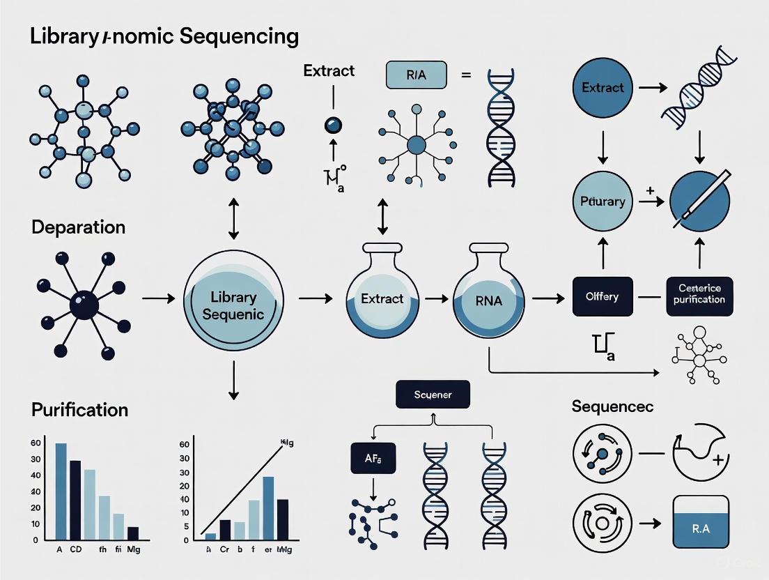
Optimizing Library Preparation for Metagenomic Sequencing: A Comprehensive Guide for Robust Microbial Profiling
Metagenomic next-generation sequencing (mNGS) is revolutionizing microbial community analysis and infectious disease diagnostics by enabling unbiased detection of pathogens.
Optimizing Library Preparation for Metagenomic Sequencing: A Comprehensive Guide for Robust Microbial Profiling
Abstract
Metagenomic next-generation sequencing (mNGS) is revolutionizing microbial community analysis and infectious disease diagnostics by enabling unbiased detection of pathogens. However, the accuracy and reliability of results are profoundly influenced by the library preparation workflow. This article provides a comprehensive guide for researchers and drug development professionals, covering foundational principles, methodological choices, and advanced optimization strategies. We synthesize current evidence to address key challenges, including host DNA depletion, input material selection, and kit bias, while offering practical troubleshooting and comparative performance data from recent clinical and environmental studies. The goal is to empower scientists with the knowledge to design robust, reproducible metagenomic studies that yield high-quality, clinically actionable data.
Core Principles and Impact of Library Preparation on Metagenomic Data Quality
In metagenomic sequencing research, library preparation constitutes the critical suite of molecular biology techniques that transform raw, extracted nucleic acids from complex samples into sequencing-ready formats. This process encodes the sample's genetic material with all necessary platform-specific motifs, enabling the subsequent detection of nucleotide sequences. The fidelity, efficiency, and quantitative accuracy of this preparatory bridge profoundly influence all downstream data, from taxonomic classification to functional characterisation of microbial communities [1]. The choice of methodology is particularly consequential in metagenomics, where the goal is to comprehensively capture the genomic diversity of a sample without introducing technical artefacts that could bias biological interpretation. As such, defining and optimising library preparation is a cornerstone of robust metagenomic research.
A Comparative Analysis of Library Preparation Methods
The selection of a library preparation method involves trade-offs between input requirements, bias, yield, and time efficiency. Systematic comparisons using defined samples provide critical guidance for selecting the most appropriate protocol.
Comparison of RNA Library Preparation Kits for Metatranscriptomics
A simplified benchmark using total RNA from four microbial species (Escherichia coli, Acinetobacter baylyi, Lactococcus lactis, and Bacillus subtilis) evaluated four cDNA synthesis and Illumina library preparation protocols: TruSeq Stranded Total RNA (TS), SMARTer Stranded RNA-Seq (SMART), Ovation RNA-Seq V2 (OV), and Encore Complete Prokaryotic RNA-Seq (ENC). Significant variations in organism representation and gene expression patterns were observed [1].
Table 1: Performance Comparison of RNA-Seq Library Preparation Methods [1]
| Method | Minimum Input Requirement | rRNA Depletion Required? | Key Synthesis Principle | Stranded? | Performance Summary |
|---|---|---|---|---|---|
| TruSeq Stranded (TS) | 100 ng depleted RNA | Yes | Random priming after RNA fragmentation | Yes | Generally best performance; limited by high input requirement. |
| SMARTer Stranded (SMART) | 1 ng depleted RNA | Yes | Random priming after RNA fragmentation | Yes | Best compromise for low input RNA; reliable quantitative results. |
| Ovation RNA-Seq V2 (OV) | 0.5 ng depleted RNA | Yes | Random and oligo(dT) priming with linear amplification | No | Only option for very low input; observed biases limit quantitative use. |
| Encore Complete (ENC) | 100 ng total RNA | No | Selective priming with decreased rRNA affinity | Yes | No prior depletion needed; uses bespoke adaptor ligation. |
The study concluded that the TruSeq method generally performed best but required hundreds of nanograms of total RNA. The SMARTer method was the best solution for lower amounts of input RNA, while the Ovation system, despite its utility for ultra-low inputs, introduced significant biases that limited its utility for quantitative analyses [1].
Comparison of DNA Library Preparation Kits for Illumina Sequencing
A separate systematic study compared nine commercial DNA library preparation kits using the same DNA sample (barcoded amplicons from phiX174) and a droplet digital PCR (ddPCR) assay to quantify efficiency at each protocol step [2]. The kits compared were NEBNext, NEBNext Ultra (New England Biolabs), SureSelectXT (Agilent), Truseq Nano, Truseq DNA PCR-free (Illumina), Accel-NGS 1S, Accel-NGS 2S (Swift Biosciences), KAPA Hyper, and KAPA HyperPlus (KAPA Biosystems).
The study revealed important variations in overall library preparation efficiencies, with kits that combined several steps into a single one exhibiting final yields 4 to 7 times higher than others. The most critical step, adaptor ligation, showed yield variations of more than a factor of 10 between kits. Some ligation efficiencies were so low they could impair the original library complexity. The anticorrelation observed between ligation and PCR yields means that a low ligation efficiency can be masked by a high-yield PCR amplification step, which itself can introduce bias and reduce complexity [2].
Table 2: Selected DNA Library Kit Preparation Efficiencies [2]
| Kit Name | Ligation Efficiency | Notable Protocol Features | Impact on Library |
|---|---|---|---|
| KAPA HyperPlus | ~100% | Combined steps; fragmentase treatment. | Preserves sample heterogeneity. |
| NEBNext Ultra | ~3.5% | Combined end-repair and A-tailing. | Very low ligation yield. |
| Illumina Truseq Nano | 15-40% | Classical multi-step protocol. | Moderate efficiency. |
| Truseq DNA PCR-free | N/A (Adaptors contain P5/P7) | No PCR step; stringent clean-ups. | Requires high input (1 μg). |
Automation in Library Preparation
Automation using liquid handling robotics presents a solution for enhancing throughput, reproducibility, and accuracy. A 2025 study compared manual and automated library preparation for Oxford Nanopore Technologies (ONT) long-read sequencing of environmental soil samples [3]. The findings demonstrated that automated preparation, while leading to a minor reduction in read and contig lengths, resulted in a slightly higher taxonomic classification rate and alpha diversity, including the detection of more rare taxa. Crucially, no significant difference in microbial community structure was identified between manual and automated libraries, validating automation for high-throughput applications where reproducibility and efficiency are paramount [3].
Detailed Experimental Protocols
This section outlines specific wet-lab methodologies as described in the comparative studies.
Application: Metatranscriptomic library preparation from microbial total RNA. Key Materials:
- Total RNA (from pure cultures or mixed community).
- Ribosomal RNA depletion kit (e.g., Ribo-Zero).
- Selected library prep kit (see Table 1).
- Magnetic beads for clean-up (e.g., SPRI).
- PCR cycler.
- Bioanalyzer or TapeStation for quality control.
Methodology:
- RNA Depletion: Perform ribosomal RNA depletion on total RNA according to the depletion kit's instructions. This step is crucial for all methods except the Encore Complete system.
- cDNA Synthesis & Library Build: Follow the specific protocol for the chosen kit, noting fundamental differences:
- TruSeq & SMARTer: Fragment depleted RNA using divalent cations + heat or heat alone, respectively. Synthesise cDNA using random primers.
- Ovation RNA-Seq V2: Synthesise cDNA from depleted RNA using a mix of random and oligo(dT) primers, followed by a linear amplification step.
- Encore Complete: Proceed directly from total RNA using selective primers designed to have decreased affinity for rRNA sequences.
- Adapter Ligation & Indexing: Ligate platform-specific adapters. For most kits, this includes index sequences for sample multiplexing.
- Library Amplification & Clean-up: Amplify the adapter-ligated DNA via PCR for a kit-dependent number of cycles. Perform final clean-up using magnetic beads to purify the sequencing-ready library.
- Quality Control: Quantify the final library using a fluorometric method (e.g., Qubit) and assess size distribution using a Bioanalyzer.
Application: High-throughput preparation of ONT sequencing libraries from environmental DNA. Key Materials:
- Genomic DNA (1 μg input recommended).
- ONT Ligation Sequencing Kit (e.g., SQK-LSK114).
- ONT PCR Barcoding Expansion 96 (EXP-PBC096).
- Bravo Automated Liquid Handing Platform (Agilent) or equivalent.
- PCR cycler.
Methodology:
- DNA Normalisation: Normalise all DNA samples to a uniform concentration (e.g., 1 μg in a standard volume) using ultra-pure water.
- Automated Setup: Transfer normalised DNA samples to a 96-well plate compatible with the liquid handling robot.
- Automated Library Construction: Execute the ONT Ligation Sequencing Kit protocol on the Bravo platform. The process typically includes:
- DNA Repair and A-tailing.
- Adapter Ligation: Ligation of barcoded adapters from the PCR Barcoding Expansion kit.
- Purification Steps: Bead-based clean-ups between major steps. (Note: A potential limitation is the lack of simultaneous temperature control and shaking during bead elution, which may reduce long fragment recovery).
- Pooling: Following automated preparation, pool barcoded libraries in equimolar ratios based on quantification.
- Sequencing: Load the pooled library onto a primed ONT flow cell (e.g., R10.4.1 PromethION) for sequencing.
The Scientist's Toolkit: Key Research Reagent Solutions
Table 3: Essential Materials for Library Preparation Workflows
| Item | Function | Example Kits & Reagents |
|---|---|---|
| Magnetic Beads | Purification and size selection of nucleic acids after various enzymatic reactions. | SPRIselect beads, SparQ beads, MagBio HighPrep beads. |
| rRNA Depletion Kits | Reduces the abundant ribosomal RNA fraction in total RNA samples to enrich for mRNA. | Illumina Ribo-Zero Plus, QIAseq FastSelect, NEBNext rRNA Depletion. |
| Ultra II DNA Library Prep Kit | A widely used kit for Illumina sequencing based on the classical end-repair, A-tailing, and ligation workflow. | NEBNext Ultra II DNA Library Prep Kit (New England Biolabs) [4]. |
| Ligation Sequencing Kit | The standard kit for preparing genomic DNA or metagenomic samples for sequencing on Oxford Nanopore platforms. | ONT Ligation Sequencing Kit (e.g., SQK-LSK114) [3]. |
| PCR Barcoding Kit | Provides barcoded adapters for multiplexing samples in a single sequencing run, essential for high-throughput studies. | ONT PCR Barcoding Expansion 96 (EXP-PBC096) [3]. |
| DIY Library Prep Reagents | Low-cost, non-proprietary reagents for constructing sequencing libraries, ideal for scaling and cost-sensitive projects. | Santa Cruz Reaction (SCR) reagents [4]. |
Workflow Visualization of Key Library Preparation Paths
The following diagram synthesises the core pathways for preparing metagenomic and metatranscriptomic libraries, highlighting critical decision points related to sample type, input, and methodology.
Diagram 1: Library Preparation Decision Workflow. This chart outlines the primary pathways for constructing sequencing libraries from DNA and RNA, with key decision points based on sample input, throughput needs, and the requirement for long-read or PCR-free data.
Within metagenomic sequencing research, the journey from a raw biological sample to a sequenced library is a critical determinant of data quality and reliability. This process, encompassing nucleic acid extraction through adapter ligation, constitutes the foundational wet-lab phase of any metagenomic study. The specific choices made during this preparatory stage can profoundly influence downstream analyses, including the detection of low-abundance taxa, the accuracy of taxonomic profiling, and the identification of functional potential within a microbial community [5]. In the context of ancient oral microbiome research, for instance, the selection of DNA extraction and library construction methods has been shown to significantly impact the recovery of endogenous DNA, microbial community composition, and the assessment of DNA damage patterns [5]. This application note details the core protocols and strategic considerations for these key workflow components, providing a structured guide for researchers aiming to optimize their metagenomic sequencing projects.
Nucleic Acid Extraction
The initial step in any metagenomic workflow is the liberation and purification of nucleic acids from the complex matrix of the sample, which can range from soil and water to human-associated biofilms like dental calculus.
Core Principles and Sample Considerations
The primary goal of extraction is to obtain pure, high-quality DNA or RNA that is representative of the entire microbial community present, while simultaneously removing substances that can inhibit downstream enzymatic reactions (e.g., humic acids, pigments, or calcium phosphates) [6] [5]. The quality of the extracted nucleic acids is intrinsically linked to the quality and preservation of the starting material. Fresh or appropriately frozen samples are always recommended, though this is not always feasible with archaeological or clinical samples [6].
The physical and chemical nature of the sample dictates the stringency of the lysis conditions required. Dense, mineralized matrices like dental calculus necessitate rigorous lysis buffers containing ethylenediaminetetraacetic acid (EDTA) to chelate calcium and destabilize the structure, alongside prolonged digestion with proteinase K to effectively release encapsulated DNA [5].
Method Comparison and Selection
Two silica-based extraction methods, optimized for recovering short, degraded DNA fragments, are commonly used in challenging metagenomic contexts such as ancient DNA research [5].
Table 1: Comparison of DNA Extraction Methods for Challenging Samples
| Feature | QG Method [5] | PB Method [5] |
|---|---|---|
| Core Principle | Silica-based purification with a binding buffer containing guanidinium thiocyanate. | Silica-based purification with a binding buffer of sodium acetate, isopropanol, and guanidinium hydrochloride. |
| Key Advantage | Effective DNA release and minimization of PCR inhibitors. | Enhanced recovery of ultra-short DNA fragments (<50 bp). |
| Typical Input | Standard to low input samples. | Ideal for highly degraded or low-biomass samples. |
| Considerations | May under-recover the shortest DNA fragments. | Particularly suited for ancient metagenomic or forensic applications. |
No single extraction method consistently outperforms another across all sample types and preservation states. The effectiveness of a protocol often depends on the specific sample context, and researchers must weigh factors such as expected DNA fragment length, sample age, and the presence of co-extracted inhibitors when selecting a method [5].
Library Construction: Fragmentation to Adapter Ligation
Following extraction, purified DNA must be converted into a sequencing-compatible format, known as a library. This process involves several standardized steps to prepare the DNA for the sequencing platform.
DNA Fragmentation
Short-read sequencing technologies require DNA fragments of a uniform, specific length (e.g., 200-600 bp) for optimal performance [7] [8]. The choice of fragmentation method can influence coverage uniformity and sequence bias.
Table 2: Comparison of DNA Fragmentation Methods
| Method | Mechanism | Advantages | Disadvantages |
|---|---|---|---|
| Physical Shearing (e.g., Acoustic) [7] [8] | Uses physical force (e.g., acoustics) to break DNA. | Minimal sequence bias; reproducible and uniform size distributions. | Requires specialized equipment (e.g., Covaris); potential for sample loss during handling. |
| Enzymatic Fragmentation [7] [8] | Uses enzymes (e.g., nucleases) to digest DNA. | Quick, cost-effective, and easily automated; suitable for low-input samples. | Potential for sequence-specific bias (e.g., GC bias); sensitive to reaction conditions. |
| Tagmentation [8] | Uses a transposase enzyme to simultaneously fragment DNA and attach adapter sequences. | Rapid and efficient; combines two steps into one, reducing hands-on time and sample loss. | Introduces sequence bias; optimization of enzyme-to-DNA ratio is critical. |
End Repair and A-Tailing
Fragmentation produces ends that are often incompatible with adapter ligation. The end-repair and A-tailing steps convert these heterogeneous ends into a uniform, ligation-ready format [8].
- End Repair: This process uses a combination of enzymes, typically T4 DNA polymerase and T4 polynucleotide kinase (PNK), to "blunt" the ends of the DNA fragments. The polymerase fills in 5' overhangs and chews back 3' overhangs, while the kinase phosphorylates the 5' ends, which is essential for subsequent ligation [8].
- A-Tailing: Following blunting, a single adenine (A) base is added to the 3' end of each fragment using an enzyme like Taq DNA polymerase. This creates a complementary overhang for ligation with thymine (T)-overhanging adapters, a strategy that minimizes fragment-to-fragment self-ligation and promotes correct adapter binding [7] [8].
Adapter Ligation
Adapter ligation is the final critical step, where short, double-stranded oligonucleotides are covalently attached to the prepared DNA fragments [6] [7]. These adapters are multifunctional, containing:
- Flow Cell Binding Sequences: Essential for attaching the library fragments to the sequencing surface (e.g., Illumina flow cells).
- Barcodes/Indexes: Short, unique DNA sequences that allow multiple libraries to be pooled and sequenced simultaneously (multiplexing) and later bioinformatically separated [6] [7].
- Unique Molecular Identifiers (UMIs): Random sequences used to tag individual molecules prior to amplification, enabling the bioinformatic correction of PCR duplicates and improving variant calling accuracy [7].
The ligation reaction is typically catalyzed by a DNA ligase enzyme, such as T4 DNA Ligase, which forms a phosphodiester bond between the fragment and the adapter [7] [8]. After ligation, a cleanup step is essential to remove excess adapters, adapter dimers, and enzyme buffers, which can interfere with sequencing efficiency [8].
Diagram 1: NGS Library Prep Workflow. This diagram outlines the key steps in preparing a next-generation sequencing library, from DNA fragmentation to the final adapter-ligated product.
The Scientist's Toolkit: Essential Reagents and Materials
Successful library preparation relies on a suite of specialized reagents and kits. The following table details key solutions used in the featured workflows.
Table 3: Key Research Reagent Solutions for NGS Library Preparation
| Item | Function | Application Notes |
|---|---|---|
| Proteinase K [5] | Digests proteins and degrades nucleases, facilitating DNA release from complex samples. | Critical for tough matrices like dental calculus; used with EDTA in lysis buffer. |
| Silica-based Binding Buffers (QG, PB) [5] | Enable purification and concentration of nucleic acids by binding them to a silica membrane/matrix in the presence of chaotropic salts. | Different formulations (e.g., QG vs. PB) optimize recovery of DNA across a range of fragment sizes. |
| T4 DNA Polymerase & T4 PNK [8] | Work in concert during end-repair to create blunt, phosphorylated ends on DNA fragments. | Essential for generating ends compatible with adapter ligation. |
| Taq DNA Polymerase [8] | Adds a single 'A' nucleotide to the 3' end of blunted DNA fragments (A-tailing). | Creates a complementary overhang for T-overhang adapters, guiding correct ligation. |
| T4 DNA Ligase [7] [8] | Catalyzes the formation of a phosphodiester bond between the DNA fragment and the adapter. | High-efficiency ligation is crucial for maximizing library yield and complexity. |
| Specialized Library Prep Kits (e.g., xGen kits) [7] | Provide optimized, pre-tested reagent mixes for a specific library prep method (e.g., ligation-based). | Streamline workflow, improve reproducibility, and reduce hands-on time. |
Metagenomic sequencing has revolutionized our ability to study complex microbial communities without the need for cultivation. However, the accuracy of these analyses is critically dependent on the quality of the library preparation process. Three major technical biases—host DNA contamination, GC content bias, and external DNA contamination—can severely skew results, leading to inaccurate biological interpretations. These challenges are particularly pronounced in low-biomass samples and clinical specimens, where microbial signals may be overwhelmed by non-target DNA. This application note examines the sources and impacts of these biases within the context of metagenomic library preparation and provides detailed protocols for their mitigation, enabling more reliable and reproducible research outcomes.
Host DNA Contamination: Impact and Depletion Strategies
The Challenge of Host DNA in Metagenomic Sequencing
Host DNA constitutes a major impediment to effective metagenomic sequencing, particularly in samples derived from host-associated environments. In respiratory samples like bronchoalveolar lavage (BAL) fluid, host DNA content can exceed 99.7%, while even nasal swabs average 94.1% host DNA [9]. This overwhelming presence of host genetic material drastically reduces the effective sequencing depth for microbial communities, limiting sensitivity for detecting low-abundance species and increasing sequencing costs substantially.
The impact of host DNA on taxonomic profiling is quantifiable and severe. Studies have demonstrated that increasing proportions of host DNA lead to decreased sensitivity in detecting both very low and low-abundant bacterial species [10]. When host DNA reaches 90% of a sample, even substantial sequencing efforts may fail to detect a significant number of microbial species present in the community. This effect is particularly problematic for clinical diagnostics where missing low-abundance pathogens could have significant implications for patient care.
Host DNA Depletion Methods: A Comparative Analysis
Multiple host depletion strategies have been developed, falling into two primary categories: pre-extraction methods that selectively lyse host cells before DNA isolation, and post-extraction methods that enrich for microbial DNA based on sequence characteristics. The performance of these methods varies significantly across sample types.
Table 1: Comparison of Host DNA Depletion Methods for Respiratory Samples
| Method | Mechanism | BAL Fluid (% Host DNA Reduction) | Nasal Swabs (% Host DNA Reduction) | Sputum (% Host DNA Reduction) | Bacterial DNA Retention |
|---|---|---|---|---|---|
| HostZERO | Pre-extraction: Selective lysis | 18.3% | 73.6% | 45.5% | Moderate |
| MolYsis | Pre-extraction: Selective lysis | 17.7% | 57.1% | 69.6% | Moderate |
| QIAamp Microbiome | Pre-extraction: Selective lysis | 13.5% | 75.4% | 22.5% | High |
| Benzonase | Pre-extraction: Enzyme-based | 10.8% | Not significant | 19.8% | Variable |
| lyPMA | Pre-extraction: Osmotic lysis + PMA | 5.7% | 41.1% | 18.3% | Low |
| S_ase | Pre-extraction: Saponin lysis + nuclease | ~99.99%* | - | - | Moderate |
| Microbiome Enrichment Kit | Post-extraction: Methylation-based | Poor performance for respiratory samples [11] | - | - | - |
*Data derived from different studies; direct comparisons should be made with caution [11] [9].
The efficacy of host depletion methods shows significant variation across sample types. For BAL fluid with extremely high host DNA content (>99%), even the most effective methods typically reduce host DNA by less than 20% [9]. In contrast, for nasal swabs with lower initial host DNA levels (~94%), methods like QIAamp and HostZERO can reduce host DNA by 75% or more [9]. This highlights the importance of matching depletion strategies to specific sample characteristics.
Detailed Protocol: Host Depletion Using Pre-extraction Methods
Principle: Selective lysis of mammalian cells followed by degradation of released DNA, while intact microbial cells remain protected by their cell walls.
Reagents Required:
- Saponin solution (0.025-0.5%)
- DNase I or Benzonase endonuclease
- DNase digestion buffer
- EDTA (for enzyme inactivation)
- Phosphate-buffered saline (PBS)
- Microbial DNA-free water
Procedure:
- Sample Preparation: Centrifuge 500 μL-1 mL of sample at low speed (500-1000 × g) to pellet host cells while leaving microbial cells in suspension.
- Host Cell Lysis: Resuspend pellet in 200 μL of saponin solution (0.025% for respiratory samples). Vortex thoroughly and incubate at room temperature for 15 minutes.
- DNase Treatment: Add 5 μL of Benzonase endonuclease or DNase I to the lysate. Include Mg²⁺ in the reaction buffer for enzyme activity.
- Digestion: Incubate at 37°C for 30 minutes with occasional mixing to digest released host DNA.
- Enzyme Inactivation: Add EDTA to a final concentration of 5 mM and incubate at 65°C for 10 minutes.
- Microbial Cell Collection: Centrifuge at high speed (10,000 × g) for 10 minutes to pellet microbial cells.
- DNA Extraction: Proceed with standard microbial DNA extraction protocols on the pellet.
Validation: Quantify host DNA depletion using qPCR targeting single-copy host genes (e.g., β-actin) and compare to microbial gene targets (e.g., 16S rRNA genes) [11].
GC Content Bias: Mechanisms and Correction Methods
Understanding GC Bias in Sequencing Data
GC content bias refers to the dependence between fragment count (read coverage) and GC content observed in Illumina sequencing data [12]. This bias presents as a unimodal relationship, where both GC-rich and AT-rich fragments are underrepresented in sequencing results, with optimal representation typically occurring at moderate GC content levels. This pattern can dominate the biological signal in analyses that focus on measuring fragment abundance within a genome, such as copy number estimation or comparative metagenomics.
The bias manifests differently across samples and is not consistent between experiments, making it challenging to develop universal correction methods. Research has demonstrated that it is the GC content of the full DNA fragment, not just the sequenced portion, that primarily influences fragment count [12]. This finding has important implications for library preparation and data analysis approaches.
Impact of GC Bias on Metagenomic Analyses
GC bias can substantially distort microbial community representations in metagenomic studies. Species with GC contents at the extremes of the distribution may be systematically underdetected, leading to:
- Inaccurate estimation of microbial relative abundances
- False negatives for potentially important community members
- Distorted functional predictions based on skewed taxonomic assignments
- Reduced comparability between studies using different sequencing protocols
The effect is particularly problematic when comparing communities across different samples or treatments, where technical bias may be confounded with biological signals of interest.
Detailed Protocol: Computational Correction of GC Bias
Principle: Model the relationship between observed read coverage and GC content, then normalize coverage based on this relationship to remove technical bias.
Software Requirements:
- R programming environment
- BEADS algorithm or similar GC correction tool
- BAM files from aligned sequencing data
- Reference genome or metagenome assembly
Procedure:
- GC Content Calculation: Compute GC content for sliding windows across the reference or for each contig in a metagenome assembly. Window size should approximate average fragment length.
- Read Coverage Calculation: Calculate read depth for each genomic window using tools like bedtools or custom scripts.
- GC-R coverage Relationship Modeling: Fit a unimodal curve to describe the relationship between GC content and read coverage. The BEADS algorithm uses the following approach:
- Model expected coverage as a function of GC content using local regression
- Account for fragment length effects if paired-end data available
- Generate normalization factors for each GC value
- Coverage Normalization: Adjust raw coverage values by applying GC-specific normalization factors.
- Validation: Assess correction efficacy by examining the relationship between normalized coverage and GC content, which should appear flat after successful correction.
Considerations: GC correction methods work best for high-coverage datasets and may be challenging to apply directly to complex metagenomic samples with heterogeneous GC contents across numerous microbial genomes [12].
Diagram 1: GC Bias Correction Workflow - This workflow outlines the computational process for identifying and correcting GC content bias in sequencing data.
Environmental and Reagent Contamination: Identification and Elimination
Contamination in metagenomic studies originates from multiple sources, including laboratory reagents, sampling equipment, personnel, and the laboratory environment itself. The impact of contamination is inversely proportional to sample microbial biomass—low-biomass samples such as fetal tissues, blood, and certain environmental samples are particularly vulnerable [13]. In these samples, contaminating DNA can comprise the majority of sequences obtained, potentially leading to spurious conclusions about community composition.
The controversial debate surrounding the existence of a placental microbiome exemplifies the critical importance of proper contamination control [13] [14]. Early reports of placental bacteria were later challenged by studies demonstrating that signal intensities in placental samples were indistinguishable from negative controls, highlighting how contamination can misdirect entire research fields.
Strategies for Contamination Prevention and Identification
Effective contamination management requires a multi-faceted approach addressing all stages from sample collection to data analysis:
Prevention During Sample Collection:
- Use single-use, DNA-free collection equipment
- Decontaminate reusable equipment with ethanol followed by DNA-degrading solutions (e.g., bleach, UV irradiation)
- Implement appropriate personal protective equipment (PPE) to minimize operator-derived contamination
- Include field blanks and sampling controls to identify environmental contaminants [13]
Laboratory Processing Controls:
- Process negative controls (reagent-only blanks) alongside experimental samples
- Use clean room facilities for low-biomass samples when possible
- Employ unique dual indices and unique molecular identifiers (UMIs) to identify cross-contamination [15]
Bioinformatic Identification:
- Utilize tools like decontam that implement statistical classification based on contaminant patterns [14]
- Apply frequency-based methods that exploit the inverse correlation between contaminant frequency and sample DNA concentration
- Use prevalence-based methods that identify sequences more common in negative controls than true samples
Detailed Protocol: Statistical Contaminant Identification with Decontam
Principle: Leverage the statistical properties of contaminants—specifically, their higher prevalence in low-DNA samples and negative controls—to distinguish them from true sample-derived sequences.
Software and Data Requirements:
- R programming environment with decontam package installed
- Feature table (ASV, OTU, or species table)
- Sample metadata including DNA concentrations
- Negative control sequencing data (optional but recommended)
Frequency-Based Method (Requires DNA Concentration Data):
- Data Preparation: Import feature table and sample metadata containing quantitation data.
- Contaminant Identification:
- Result Interpretation: Features with a probability score >0.5 are classified as contaminants.
- Table Filtering: Remove contaminant features from downstream analyses.
Prevalence-Based Method (Uses Negative Controls):
- Data Preparation: Include negative control samples in the feature table.
- Contaminant Identification:
- Validation: Compare contaminant classifications with known contaminant taxa databases.
Combined Approach: For maximum sensitivity, apply both methods independently and treat features identified by either method as contaminants [14].
Table 2: Common Contaminant Genera in Metagenomic Studies and Their Sources
| Contaminant Genus | Frequency of Detection | Primary Source | Recommended Handling |
|---|---|---|---|
| Cutibacterium acnes | Detected in 100% of plasma and urine samples [15] | Human skin, laboratory reagents | Remove with decontam or SIFT-seq |
| Pseudomonas | Common in multiple studies [14] | Water systems, laboratory surfaces | Include in negative controls |
| Bradyrhizobium | Common in soil studies [14] | Laboratory reagents | Statistical identification and removal |
| Methylobacterium | Frequent in low-biomass studies [14] | Laboratory water, plastics | Monitor via negative controls |
| Staphylococcus | Variable across studies | Human skin, cross-contamination | Careful interpretation in host-associated studies |
Integrated Workflow for Comprehensive Bias Control
A Unified Approach to Bias Mitigation
Effective management of the three major biases in metagenomic sequencing requires an integrated approach spanning experimental design, laboratory processing, and bioinformatic analysis. The following workflow provides a comprehensive strategy for minimizing these technical artifacts:
Experimental Design Phase:
- Determine sample biomass levels to assess contamination risk
- Select appropriate host depletion methods based on sample type
- Plan for sufficient sequencing depth to account for host DNA dilution
- Include appropriate controls (negative, positive, and sampling controls)
Laboratory Processing Phase:
- Implement selected host depletion protocol before DNA extraction
- Use contamination-aware techniques (sterile equipment, PPE, reagent screening)
- Employ unique dual indices to track cross-contamination
- Quantitate DNA to support frequency-based contaminant identification
Bioinformatic Analysis Phase:
- Apply GC bias correction to read coverage data
- Implement statistical contaminant identification (decontam)
- Remove identified contaminants from feature tables
- Validate results through comparison with negative controls
Advanced Method: SIFT-Seq for Contamination-Resistant Sequencing
Sample-Intrinsic microbial DNA Found by Tagging and sequencing (SIFT-seq) represents a novel approach that proactively labels sample-intrinsic DNA before library preparation, allowing bioinformatic identification and removal of contaminating DNA introduced during processing [15].
Principle: Chemical tagging of DNA in the original sample before DNA isolation, enabling distinction between true sample DNA and contaminants based on the presence of the tag.
Protocol Overview:
- DNA Tagging: Treat raw sample with bisulfite to convert unmethylated cytosines to uracils directly in the sample matrix.
- Library Preparation: Proceed with standard metagenomic library preparation.
- Bioinformatic Filtering: Identify and retain only sequences showing the bisulfite conversion pattern, indicating they were present in the original sample.
Performance: SIFT-seq reduces contaminant reads by up to three orders of magnitude and completely removes specific contaminant genera like Cutibacterium acnes from 62 of 196 clinical samples tested [15].
Diagram 2: Integrated Bias Mitigation Workflow - A comprehensive approach addressing multiple biases throughout the metagenomic sequencing pipeline.
The Scientist's Toolkit: Essential Reagents and Materials
Table 3: Key Research Reagent Solutions for Addressing Metagenomic Biases
| Reagent/Kit | Primary Function | Application Context | Performance Considerations |
|---|---|---|---|
| HostZERO Microbial DNA Kit | Host DNA depletion | Respiratory samples, tissues | High host depletion for nasal swabs (73.6% reduction) |
| QIAamp DNA Microbiome Kit | Host DNA depletion | Various sample types | High bacterial retention (21% in OP samples) |
| Nextera XT DNA Library Prep Kit | Library preparation | Low-input metagenomic samples | Integrated tagmentation, low input requirements (1ng) |
| NEBNext Microbiome DNA Enrichment Kit | Methylation-based enrichment | Samples with differential methylation | Poor performance for respiratory samples |
| Benzonase Nuclease | Host DNA degradation | Pre-extraction protocols | Requires optimization for different sample types |
| Saponin | Selective host cell lysis | Pre-extraction protocols | Effective at low concentrations (0.025%) |
| Unique Dual Indices | Cross-contamination tracking | All metagenomic studies | Essential for identifying index hopping |
| Decontam R Package | Statistical contaminant identification | All metagenomic studies | Frequency and prevalence-based methods |
| BEADS Algorithm | GC bias correction | DNA-seq, metagenomics | Models unimodal GC-coverage relationship |
Host DNA contamination, GC content bias, and environmental contamination represent three major technical challenges that can severely compromise metagenomic sequencing results. Through implementation of appropriate host depletion strategies, computational correction methods, and rigorous contamination control protocols, researchers can substantially improve the accuracy and reliability of their microbial community analyses. The protocols and comparative data presented here provide a practical framework for addressing these biases across diverse sample types and research applications. As metagenomic sequencing continues to expand into increasingly challenging sample matrices, particularly in clinical diagnostics where low-biomass samples are common, robust bias mitigation strategies will become ever more critical for generating biologically meaningful results.
Within metagenomic next-generation sequencing (mNGS), the choice of nucleic acid source is a pivotal first step that fundamentally influences the profiling of a microbial community. The two principal pathways are whole-cell DNA (wcDNA), which extracts genomic material from intact microorganisms, and cell-free DNA (cfDNA), which targets short, extracellular DNA fragments freely circulating in body fluids or sample supernatants [16] [17]. This decision carries significant weight for researchers and drug development professionals, as it directly impacts the sensitivity, specificity, and representativeness of the results in the context of library preparation. The optimal choice is highly dependent on the sample type, the target pathogens, and the specific clinical or research question. This application note provides a structured comparison of these two pathways, supported by recent quantitative data, detailed experimental protocols, and visualization to guide this critical methodological choice.
Comparative Performance Analysis
Recent clinical studies have directly compared the effectiveness of wcDNA and cfDNA mNGS across various sample types, revealing distinct performance profiles. The table below summarizes key quantitative findings from comparative studies on body fluid and bronchoalveolar lavage fluid (BALF) samples.
Table 1: Comparative Performance of wcDNA mNGS and cfDNA mNGS in Clinical Studies
| Metric | Sample Type | wcDNA mNGS Performance | cfDNA mNGS Performance | Reference & Context |
|---|---|---|---|---|
| Host DNA Proportion | Clinical Body Fluids | Mean: 84% [16] | Mean: 95% (p < 0.05) [16] | PMC11934473 |
| Concordance with Culture | Clinical Body Fluids | 63.33% (19/30 samples) [16] | 46.67% (14/30 samples) [16] | PMC11934473 |
| Sensitivity (vs. Culture) | Body Fluid Samples | 74.07% [16] | Not Reported | PMC11934473 |
| Specificity (vs. Culture) | Body Fluid Samples | 56.34% [16] | Not Reported | PMC11934473 |
| Diagnostic Performance | BALF (Pulmonary Aspergillosis) | Outperformed conventional tests; inferior to cfDNA in RPM for Aspergillus [17] | Superior reads per million (RPM) for Aspergillus; AUC of 0.779 for predicting infection [17] | Frontiers in Cellular and Infection Microbiology, 2024 |
| Consistency with 16S NGS | Clinical Body Fluids | 70.7% (29/41 samples) [16] | Not Reported | PMC11934473 |
Experimental Protocols
Protocol A: Dual-Pathway DNA Extraction from Body Fluids
This protocol is adapted from a comparative study on clinical body fluid samples [16].
I. Sample Pre-Processing
- Centrifuge the collected body fluid sample (e.g., pleural, ascites, or drainage fluid) at 20,000 × g for 15 minutes at room temperature.
- Carefully transfer the supernatant to a new tube for cfDNA extraction. The resulting pellet will be used for wcDNA extraction.
II. Cell-Free DNA (cfDNA) Extraction from Supernatant
- Use the VAHTS Free-Circulating DNA Maxi Kit (Vazyme Biotech) or the QIAamp DNA Micro Kit (QIAGEN).
- Add 25 μl of Proteinase K and 800 μl of Buffer L/B to 400 μl of supernatant.
- Add 15 μl of magnetic beads, mix briefly, and incubate at room temperature for 5 minutes.
- Place the tube on a magnetic rack until the solution clears. Carefully remove and discard the supernatant.
- Wash the beads as per the manufacturer's instructions. Elute the extracted cfDNA in 50 μl of elution buffer.
III. Whole-Cell DNA (wcDNA) Extraction from Pellet
- Use a kit designed for microbial DNA, such as the Qiagen DNA Mini Kit or the Mag-Bind Universal Metagenomics Kit (Omega Biotek).
- Add two 3-mm nickel beads to the pellet and shake at 3,000 rpm for 5 minutes to mechanically lyse cells.
- Proceed with the DNA extraction according to the manufacturer's protocol.
- Elute the final wcDNA in 50-100 μl of elution buffer.
IV. Quality Control and Quantification
- Quantify DNA concentration using a fluorometric method (e.g., Qubit Fluorometric Quantitation).
- Assess DNA quality and fragment size via 0.8% Agarose Gel Electrophoresis (AGE).
Protocol B: Library Preparation and Sequencing
I. Library Construction
- Use the VAHTS Universal Pro DNA Library Prep Kit for Illumina (Vazyme) or the KAPA Hyper Prep Kit (KAPA Biosystems), which has been shown to detect a higher number of genes compared to transposase-based methods [18].
- For each sample, use 50–250 ng of input DNA for library preparation. Note that inputs within this range have shown no significant difference in gene detection for shotgun metagenomics [18].
- Follow the manufacturer's protocol for end-repair, adapter ligation, and library amplification.
II. Sequencing
- Sequence the pooled libraries on an Illumina NovaSeq platform using a 2 × 150 bp paired-end configuration.
- Aim for approximately 8 GB of data (~26 million reads) per sample for comprehensive analysis [16].
Workflow Visualization
The following diagram illustrates the critical decision points and parallel pathways for wcDNA and cfDNA analysis in mNGS.
Decision Pathway for wcDNA vs. cfDNA mNGS
The Scientist's Toolkit: Essential Research Reagents
The table below lists key reagents and kits critical for implementing the wcDNA and cfDNA pathways.
Table 2: Essential Reagents for wcDNA and cfDNA mNGS Workflows
| Reagent/Kits | Function | Specific Application Note |
|---|---|---|
| VAHTS Free-Circulating DNA Maxi Kit (Vazyme) | Extraction of cell-free DNA from sample supernatants. | Optimized for short-fragment cfDNA; includes magnetic bead-based purification [16]. |
| QIAamp DNA Micro Kit (QIAGEN) | Extraction of DNA from small volumes, suitable for both cfDNA and wcDNA. | Used for extracting cfDNA from BALF supernatant and wcDNA from pellets [17]. |
| Mag-Bind Universal Metagenomics Kit (Omega Biotek) | Extraction of microbial DNA from complex samples. | Demonstrated higher DNA yield and more detected genes compared to other soil-based kits [18] [19]. |
| Qiagen DNeasy PowerSoil Kit | DNA extraction from environmental and challenging clinical samples. | Effective for lysis of difficult-to-break microbial cell walls; includes inhibitor removal [18]. |
| KAPA Hyper Prep Kit (KAPA Biosystems) | DNA library construction for NGS. | Outperformed transposase-based kits in detected gene number and Shannon diversity index [18]. |
| VAHTS Universal Pro DNA Library Prep Kit (Vazyme) | Library preparation for Illumina sequencing. | Used in conjunction with mNGS for pathogen detection in body fluids [16]. |
Concluding Recommendations
The choice between wcDNA and cfDNA is context-dependent. wcDNA mNGS is generally recommended for maximum sensitivity in detecting a broad range of intracellular pathogens, particularly in samples from abdominal and other sterile site infections, despite its compromised specificity which requires careful clinical interpretation [16]. Conversely, cfDNA mNGS is superior for detecting pathogens that release DNA into the surrounding environment, as demonstrated in pulmonary aspergillosis, and is less affected by host DNA interference in certain fluid samples [17]. For the most comprehensive diagnostic picture, especially in critically ill patients, a dual-pathway approach utilizing both wcDNA and cfDNA from a single sample can provide complementary insights that enhance diagnostic precision beyond conventional microbiological tests alone.
In metagenomic sequencing, the quality and interpretability of data are profoundly shaped by the initial library preparation. Three technical metrics are paramount for evaluating library quality and informing downstream analysis: insert size, library complexity, and PCR duplication rates. Insert size refers to the length of the sample DNA fragment that is sequenced, which is a critical parameter influencing assembly and coverage [20]. Library complexity measures the diversity of unique DNA molecules in the library, indicating how well the original microbial community's diversity is represented [21] [22]. PCR duplication rate quantifies the fraction of sequencing reads that are artificial copies from a single original molecule, which can skew abundance estimates [23] [24]. Understanding and controlling these interrelated metrics is essential for generating robust, representative metagenomic data, particularly when dealing with diverse microbial communities of varying biomass.
Foundational Concepts and Their Experimental Measurement
Defining Insert Size and Fragment Size
In paired-end sequencing, the insert is the sample DNA fragment of interest that is sequenced from both ends. The insert size is the length of this fragment in base pairs. The fragment size, a related but distinct term, includes the insert plus the attached adapter sequences on both ends [20]. The selection of an appropriate insert size is a critical experimental design choice. If the insert size is shorter than the combined length of the two sequencing reads, the reads will overlap in the middle, facilitating more accurate sequence assembly. Conversely, if the insert size is longer, an unsequenced inner distance remains [20]. The distribution of insert sizes is not uniform; fragmentation methods produce a range of sizes, and the median of this distribution is typically reported [20].
Quantifying Library Complexity
Library complexity describes the number of unique DNA molecules in a sequencing library. A library with high complexity contains a vast diversity of unique fragments, which is vital for achieving uniform coverage across the genome or metagenome and for detecting rare variants. Low-complexity libraries, often resulting from insufficient input material or over-amplification, are dominated by a smaller set of sequences and yield uneven, biased data [22]. In metagenomics, the "complexity" of the biological sample itself (e.g., low-complexity coral microbiome vs. high-complexity soil microbiome) also interacts with library preparation, influencing achievable sequencing depth and duplication rates [21]. Complexity can be estimated bioinformatically using measures of sequence uniqueness and entropy, or by tracking unique molecular identifiers (UMIs) [22].
Understanding and Identifying PCR Duplicates
PCR duplicates are multiple sequencing reads that originate from an identical template DNA molecule due to amplification during the library preparation process [23] [24]. These duplicates do not represent independent biological observations and can lead to false positives in variant calling or inaccurate estimates of microbial abundance if misinterpreted as unique sequences. The frequency of PCR duplicates is highly dependent on the amount of starting material and the sequencing depth, with lower inputs and higher depths leading to higher duplicate rates [24]. Standard bioinformatic tools like Picard MarkDuplicates or SAMTools rmdup identify duplicates by finding read pairs that align to the exact same genomic start and end positions [23].
Quantitative Data on Influencing Factors
Impact of Input DNA and Community Type on Key Metrics
Experimental data demonstrates that input DNA quantity and microbial community type significantly influence key library metrics. One systematic assessment of five library preparation methods found that these factors statistically affected median fragment size, library concentration, read GC content, and duplication rate [21]. The duplication rate, in particular, was especially sensitive to community type, with low-diversity communities (e.g., coral, mock) exhibiting significantly elevated duplication rates compared to more complex communities [21]. Another study on a mock microbial community found that the percentage of reads lost during quality control increased with decreasing input DNA, particularly for the Nextera XT protocol [25].
Table 1: Impact of Input DNA and Community Type on Library Metrics [21] [25]
| Factor | Impact on Library Metrics |
|---|---|
| Input DNA Quantity | Lower inputs can shift GC content towards more GC-rich sequences [25], increase the number of low-quality/unmapped reads [25], and increase the fraction of reads removed during QC for some protocols (e.g., Nextera XT) [25]. |
| Community Complexity | Low-complexity communities (e.g., coral, mock) have statistically elevated sequence duplication rates compared to high-complexity communities (e.g., soil) [21]. |
Comparing Library Preparation Methods
The choice of library preparation method introduces specific biases and performance characteristics. A comparative study of methods including Illumina Nextera DNA Flex, Qiagen QIASeq FX DNA, PerkinElmer NextFlex Rapid DNA-Seq, and seqWell plexWell96 showed that the procedure, community type, and input DNA concentration all interact to influence final library characteristics [21]. Furthermore, the fragmentation method (e.g., mechanical shearing vs. enzymatic tagmentation) significantly impacts the distribution of insert sizes. Nextera XT libraries, which use tagmentation, had a significantly smaller mean insert size (110 bp) compared to methods using mechanical shearing like Mondrian (200 bp) and MALBAC (208 bp) [25].
Table 2: Characteristics and Performance of Different Library Prep Methods [21] [25] [8]
| Method / Characteristic | Fragmentation Approach | Typical Insert Size Bias/Note | Key Finding |
|---|---|---|---|
| Nextera XT / DNA Flex | Enzymatic (Tagmentation) | Smaller mean insert size (e.g., 110 bp) [25]; sensitive to DNA concentration [26]. | Cost-effective; performance comparable to gold-standard for high-complexity communities [21]. |
| Mechanical Shearing (e.g., Covaris) | Physical (Acoustic) | Larger, more tunable insert sizes; more random fragmentation [25] [8]. | Minimal sequence bias; considered robust and reproducible [8]. |
| Other Enzymatic Kits | Enzymatic (Non-Tagmentation) | Varies by kit; modern kits have reduced motif/GC bias [8]. | Automation-friendly and lower equipment cost [8]. |
Experimental Protocols for Measurement and Control
Protocol 1: Measuring Insert Size Without a Reference Genome
Accurately determining insert size is crucial for quality control, especially when a reference genome is unavailable or incomplete, as is common in metagenomics. This protocol uses the tool FLASH to measure insert sizes directly from FASTQ files.
- Principle: For read pairs where the insert size is less than the combined read length, the 3' ends of the reads will overlap. FLASH can merge these overlaps, and the length of the resulting contig equals the insert size [26].
- Procedure:
- Software Installation: Install FLASH (Fast Length Adjustment of SHort reads) from its official repository.
- Command Execution: Run FLASH on your paired-end FASTQ files. A basic command is:
flash read1.fastq read2.fastq -m 10 -M 100 -o output_prefix-m: Minimum overlap length (e.g., 10 bp).-M: Maximum overlap length (e.g., 100 bp).
- Data Extraction: After execution, FLASH will generate a histogram file (
output_prefix.hist) containing the distribution of assembled insert sizes. - Interpretation: The peak of this histogram represents the most common insert size. A broad or multi-peaked distribution may indicate issues with the fragmentation or size selection steps during library prep [26].
- Considerations: This method reliably identifies fragments with small inserts that are likely to contain adapter sequence, a common issue in Nextera XT libraries [26].
Protocol 2: Using UMIs to Accurately Remove PCR Duplicates
Standard duplicate removal based on mapping coordinates can be overly aggressive and biased. This protocol incorporates Unique Molecular Identifiers (UMIs) to distinguish technical duplicates from biologically identical reads.
- Principle: Before amplification, a random nucleotide UMI is ligated to each original cDNA/DNA molecule. All reads with the same UMI are definitively identified as PCR duplicates derived from a single molecule, regardless of their final sequence or mapping position [24].
- Procedure:
- Adapter Design: Modify standard sequencing adapters to include a random nucleotide UMI (e.g., 5-10 random bases) and a short, fixed "locator" sequence to anchor the UMI during sequencing [24].
- Library Preparation: Proceed with the standard library prep protocol using the UMI-containing adapters.
- Bioinformatic Processing: Use a UMI-aware bioinformatics pipeline (e.g., built with tools like
umisorfgbio) to:- Extract UMIs from read headers.
- Group reads by their UMI and genomic mapping coordinates.
- For each group of reads sharing the same UMI and coordinates, retain a single consensus read to correct for sequencing errors and discard the rest as duplicates [24].
- Considerations: UMI length is critical. For RNA-seq or small RNA-seq of highly abundant molecules, a 10-nt UMI (providing ~1 million unique combinations) may be necessary to ensure every unique molecule gets a unique tag [24].
Protocol 3: Standard Bioinformatic PCR Duplicate Removal
For libraries prepared without UMIs, this protocol uses Picard MarkDuplicates, a standard tool for identifying duplicates based on mapping coordinates.
- Principle: Picard identifies read pairs with the same outer alignment start and end positions and orientation. It marks all but the highest-quality read pair as duplicates, which are then ignored by downstream variant callers [23].
- Procedure:
- Input Data: You will need a coordinate-sorted BAM file containing your aligned sequencing reads.
- Software: Ensure Picard Tools (or a compatible replacement like GATK) is installed.
- Command Execution: Run the MarkDuplicates command. An example is:
I: Input sorted BAM file.O: Output BAM file with duplicate flags set.M: File to write duplicate metrics.
- Output Interpretation: The metrics file reports the number and percentage of duplicated reads. A high percentage (>20-50%) may indicate issues with low input DNA or over-amplification [23].
- Considerations: This method can incorrectly mark biologically identical reads from repetitive regions or highly expressed genes as duplicates, potentially introducing bias [24].
The Scientist's Toolkit: Research Reagent Solutions
Selecting the right reagents and kits is fundamental to successful library preparation. The following table details essential materials and their functions.
Table 3: Essential Research Reagents for Metagenomic Library Preparation
| Reagent / Kit | Primary Function | Key Considerations |
|---|---|---|
| Nextera DNA Flex / XT Kit | Transposase-based fragmentation and adapter tagging ("tagmentation") in a single step [21] [25]. | Sensitive to input DNA quantity; can produce a broad insert size distribution [26] [25]. Cost-effective for high-complexity communities [21]. |
| UMI Adapters (Custom) | Ligation of unique molecular identifiers to original molecules pre-amplification [24]. | UMI length must provide sufficient diversity for the experiment (e.g., 10-nt for small RNA-seq). A fixed "locator" sequence aids in accurate UMI identification [24]. |
| Covaris AFA System | Mechanical DNA shearing via focused acoustic energy for random fragmentation [25] [8]. | Produces a tight, tunable insert size distribution with minimal sequence bias. Requires specialized equipment [8]. |
| AMPure XP Beads | Solid-phase reversible immobilization (SPRI) for post-ligation and post-amplification cleanup and size selection [8]. | Critical for removing adapter dimers, unligated adapters, and short fragments. Bead-to-sample ratio controls size selection cutoff. |
| High-Fidelity PCR Mix | Amplification of adapter-ligated fragments, especially for low-input samples [8]. | Minimizes introduction of errors during amplification. The number of PCR cycles should be minimized to preserve library complexity and reduce duplicates [8]. |
| Host Depletion Kit (e.g., HostZERO) | Selective reduction of host (e.g., human) DNA in host-associated microbiome samples [27]. | Dramatically increases the fraction of microbial reads in shotgun metagenomic data, improving sequencing efficiency for the target community [27]. |
Selecting and Implementing the Right Protocol for Your Sample and Study
Within metagenomic sequencing research, the initial conversion of extracted DNA into a sequence-ready library is a critical step that profoundly influences the quality, reliability, and interpretability of the generated data. The choice of library preparation method can introduce biases in genome coverage, affect the detection of single nucleotide variants (SNVs) and indels, and ultimately determine the success of a study aimed at characterizing complex microbial communities [28]. This application note provides a structured comparison of predominant library preparation kits—including those from Illumina, KAPA HyperPlus/HyperPrep, and Nextera XT/Nextera DNA Flex—framed within the context of metagenomic sequencing. We summarize key performance data from controlled studies, detail standardized protocols for reproducibility, and visualize workflows to guide researchers and drug development professionals in selecting and implementing the optimal library preparation strategy for their specific research needs.
Kit Comparison and Performance Data
Comparative Specifications of Commercial Kits
The selection of a library preparation kit requires careful consideration of input DNA, workflow time, and application suitability. The table below compares key specifications for a range of commercially available kits.
Table 1: Specifications of Selected DNA Library Preparation Kits for Short-Read Sequencing [29] [30] [28]
| Supplier | Kit Name | System Compatibility | Assay Time | Input Quantity | PCR Required? | Key Applications |
|---|---|---|---|---|---|---|
| Illumina | Illumina DNA PCR-Free Prep | Illumina platforms | ~1.5 hours | 25 ng – 300 ng | No | De novo assembly, WGS |
| Illumina | Illumina DNA Prep | Illumina platforms | 3-4 hours | 1-500 ng (varies by genome size) | Yes | Amplicon sequencing, WGS |
| Illumina | Nextera XT | iSeq 100, MiSeq, NextSeq series | ~5.5 hours | 1 ng | Yes | 16S rRNA sequencing, amplicon sequencing, WGS |
| Roche | KAPA HyperPlus/HyperPrep | Illumina platforms | 2-3 hours | 1 ng – 1 μg | Optional (kit dependent) | WGS, WES, metagenomic sequencing |
| Integrated DNA Technologies (IDT) | xGen DNA EZ Library Prep Kit | Illumina platforms | <2 hours | 100 pg – 1 μg | Yes | Genotyping, WES, WGS |
| Arbor Biosciences | Library Prep Kit for myBaits | User-supplied adapters for Illumina | Protocol-dependent | 1 – 500 ng | Yes (post-capture) | Targeted sequencing (e.g., whole exome, phylogenetics) |
Performance Metrics in Whole Genome Sequencing
A independent study compared several enzymatic fragmentation-based kits and the tagmentation-based Illumina Nextera DNA Flex kit using human genomic DNA (cell line NA12878) with 10 ng and 100 ng input amounts [28]. The following table summarizes the key outcomes, which are highly relevant for metagenomic sequencing where input DNA can be limited and representative coverage is paramount.
Table 2: Performance Metrics of Library Prep Kits in a Whole Genome Sequencing Study [28]
| Kit | Fragmentation Method | Input DNA (PCR cycles) | Mean Insert Size from Sequencing (bp) | Key Performance Findings |
|---|---|---|---|---|
| Nextera DNA Flex (Illumina) | Tagmentation | 10 ng (8 cycles) | 326 (±2) | Reproducible performance. Coverage gaps can occur in specific genomic regions with tagmentation-based methods [31]. |
| 100 ng (5 cycles) | 366 (±2) | |||
| KAPA HyperPlus (Roche) | Enzymatic | 10 ng (9 cycles) | 240 (±9) | Robust performance. Produced consistent, high coverage; better coverage of low-coverage regions compared to Nextera XT [31]. |
| 100 ng (0 cycles, PCR-free) | 227 (±3) | |||
| NEBNext Ultra II FS (NEB) | Enzymatic | 10 ng (7 cycles) | 206 (±7) | Good performance. Libraries with insert sizes longer than the cumulative read length showed improved coverage and variant detection. |
| 100 ng (3 cycles) | 188 (±6) | |||
| SparQ (Quantabio) | Enzymatic | 10 ng (9 cycles) | 185 (±3) | Good performance. Shorter insert sizes observed, but performance improved with longer inserts. |
| 100 ng (0 cycles, PCR-free) | 244 (±10) | |||
| Swift 2S Turbo (Swift) | Enzymatic | 10 ng (6 cycles) | 330 (±12) | Good performance. Achieved one of the longest insert sizes among enzymatic methods in this study. |
| 100 ng (0 cycles, PCR-free) | 226 (±7) |
The study concluded that all tested kits produced high-quality data, but library insert size was a critical factor. Libraries with DNA insert fragments longer than the cumulative sum of both paired-end reads (e.g., >300 bp for 2x150 bp sequencing) avoid read overlap, leading to more unique sequence information, improved genome coverage, and increased sensitivity for SNV and indel detection [28]. Furthermore, libraries prepared with minimal or no PCR demonstrated the best performance for indel detection, highlighting the value of PCR-free workflows where input DNA allows [28].
Detailed Experimental Protocols
Protocol for KAPA HyperPlus Library Preparation (96 rxn)
The KAPA HyperPlus kit offers a streamlined, single-tube protocol that combines several enzymatic steps and reduces bead cleanups, making it suitable for a wide range of input amounts and sample types, including FFPE and cell-free DNA [30].
Reagents and Materials:
- KAPA HyperPlus Kit (Roche Cat. No. 07962347001 for 24 rxn or 07962363001 for 96 rxn) containing:
- KAPA End Repair & A-Tailing Buffer
- KAPA End Repair & A-Tailing Enzyme
- KAPA Ligation Buffer
- KAPA DNA Ligase
- KAPA HiFi HotStart ReadyMix (2X)
- KAPA Library Amplification Primer Mix (10X) [30]
- KAPA HyperPure Beads (Roche Cat. No. 08963843001) [32]
- KAPA Adapters (Single- or Dual-Indexed, purchased separately)
- Nuclease-free water
- Ethanol (80%)
- Thermal cycler
- Magnetic separation rack
- Agilent Tapestation or Bioanalyzer for quality control
Procedure:
- Fragmentation and End-Repair/A-Tailing: In a single tube, combine 1-1000 ng of genomic DNA in 50 µL of nuclease-free water with 7 µL of KAPA End Repair & A-Tailing Buffer and 3 µL of KAPA End Repair & A-Tailing Enzyme. Mix thoroughly and incubate in a thermal cycler at 65 °C for 30 minutes to fragment the DNA, followed immediately by 4 °C for 5 minutes for end-repair and A-tailing [30] [28].
- Adapter Ligation: To the same tube, add 30 µL of KAPA Ligation Buffer, 10 µL of KAPA DNA Ligase, and 10 µL of diluted KAPA Adapters (final concentration ~0.5 µM). Mix well and incubate at 20 °C for 60 minutes [30].
- Post-Ligation Cleanup: Add 50 µL of KAPA HyperPure Beads to the ligation reaction (1.0x ratio) to purify the adapter-ligated library. Follow the standard bead-based purification protocol: bind, wash twice with 80% ethanol, elute in a low-volume buffer (e.g., 22 µL) [30].
- Library Amplification (Optional): For PCR-amplified libraries, combine 20 µL of the purified ligation product with 25 µL of KAPA HiFi HotStart ReadyMix (2X) and 5 µL of KAPA Library Amplification Primer Mix (10X). Amplify using the following cycling conditions: 98 °C for 45 seconds; 6-10 cycles of 98 °C for 15 seconds, 60 °C for 30 seconds, 72 °C for 30 seconds; 72 °C for 1 minute; hold at 4 °C [30]. The number of cycles should be optimized based on input DNA.
- Post-Amplification Cleanup and Size Selection: Perform a double-sided SPRI (bead-based) size selection. First, add beads at a 0.6x ratio to remove large fragments. Transfer the supernatant to a new tube and add beads at a 0.15x ratio to recover the desired library fragments (typically ~300-600 bp). Elute the final library in 20-30 µL of buffer [30].
- Quality Control and Quantification: Assess the library's size distribution and integrity using an Agalient Tapestation D1000 or Bioanalyzer. Quantify the library using a fluorescence-based method (e.g., Qubit) and qPCR for accurate molar concentration prior to pooling and sequencing [28].
Protocol for Nextera XT DNA Library Preparation (96 rxn)
The Nextera XT kit utilizes a tagmentation reaction that simultaneously fragments DNA and adds adapter sequences, enabling a very rapid workflow suitable for high-throughput processing of amplicons, though it requires precise input DNA [31].
Reagents and Materials:
- Nextera XT DNA Library Prep Kit (Illumina, 96 samples) containing:
- Nextera XT Amplicon Tagment Buffer (ATB)
- Nextera XT Tagment DNA Enzyme (TDK)
- Nextera PCR Master Mix (NPM)
- Resuspension Buffer (RSB)
- Nextera XT Index Kit 1, 2 (Illumina)
- Nuclease-free water
- Magnetic beads (e.g., AMPure XP)
- Ethanol (80%)
- Thermal cycler
- Magnetic separation rack
- Agilent Tapestation or Bioanalyzer
Procedure:
- Tagmentation: Dilute genomic DNA or amplicons to 1 ng/µL in RSB. Combine 5 µL (5 ng) of diluted DNA with 10 µL of ATB and 5 µL of TDK. Mix thoroughly and incubate in a thermal cycler at 55 °C for 5-15 minutes, then hold at 10 °C [31]. The fragmentation time can be adjusted to modify the insert size distribution.
- Neutralize Tagmentation: Add 5 µL of Neutralize Tagment Buffer (NTB) to the tagmentation reaction. Mix by pipetting and incubate at room temperature for 5 minutes.
- PCR Amplification and Indexing: Add 5 µL of each Nextera XT Index Primer (i5 and i7) and 15 µL of NPM to the neutralized tagmentation reaction. Amplify using the following cycling conditions: 72 °C for 3 minutes; 95 °C for 30 seconds; 12 cycles of 95 °C for 10 seconds, 55 °C for 30 seconds, 72 °C for 30 seconds; 72 °C for 5 minutes; hold at 4 °C [31].
- Library Cleanup: Add 45 µL of magnetic beads (0.9x ratio) to the 50 µL PCR reaction. Purify the library by binding, washing twice with 80% ethanol, and eluting in 22.5 µL of RSB.
- Quality Control and Quantification: As with the KAPA protocol, assess the library's size distribution and concentration using instrumentation like the Tapestation and qPCR. Normalize libraries to 4 nM prior to pooling for sequencing [31].
Workflow Visualization and Technical Diagrams
Library Preparation Workflow Comparison
The following diagram illustrates the core procedural steps and key decision points for the two primary library preparation methods discussed: enzymatic fragmentation/ligation (e.g., KAPA HyperPlus) and tagmentation (e.g., Nextera XT).
Diagram 1: Comparison of enzymatic fragmentation/ligation and tagmentation library prep workflows. Key differences include the initial fragmentation/adapter addition step and the optionality of PCR in some enzymatic protocols [30] [31].
The Scientist's Toolkit: Essential Reagent Solutions
Successful library preparation relies on a suite of specialized reagents beyond the core kit components. The following table details key reagent solutions and their critical functions in the workflow.
Table 3: Essential Research Reagent Solutions for NGS Library Preparation
| Reagent/Material | Function/Description | Example Product(s) |
|---|---|---|
| Magnetic SPRI Beads | Size-selective purification of nucleic acids; used for cleanups and size selection between reaction steps. | KAPA HyperPure Beads [30], AMPure XP Beads |
| Universal Stubby Adapters | Short, double-stranded adapters with T-overhangs for ligation to A-tailed DNA fragments; require indexing via PCR. | xGen Stubby Adapters (IDT) [33] |
| Dual Indexed Adapters | Full-length or stubby adapters containing unique combinatorial barcodes (i5 and i7) for sample multiplexing; reduce index hopping. | KAPA Dual-Indexed Adapter Kits [30], xGen UDI-UMI Adapters (IDT) [33] |
| High-Fidelity DNA Polymerase | PCR enzyme with high accuracy and processivity; used for library amplification with minimal bias and high yield. | KAPA HiFi HotStart ReadyMix [30] |
| Library Quantification Kits | qPCR-based assays for accurate determination of the molar concentration of adapter-ligated fragments; essential for pooling libraries. | KAPA Library Quantification Kit [30] |
| Enzymatic Fragmentation Mix | Controlled digestion of DNA by a proprietary mix of enzymes to a desired fragment length; alternative to mechanical shearing. | Component of KAPA HyperPlus, NEBNext Ultra II FS [28] |
The landscape of NGS library preparation kits offers multiple robust paths for creating metagenomic sequencing libraries. Enzymatic fragmentation-based kits, such as KAPA HyperPlus/HyperPrep, provide flexibility in input DNA, reduced hands-on time, and performance comparable to established tagmentation-based methods like Illumina's Nextera DNA Flex and Nextera XT [28]. The critical technical considerations for kit selection include DNA input amount, the desire for a PCR-free workflow to minimize bias, and the paramount importance of achieving an optimal library insert size longer than the sequenced read length to maximize unique coverage and variant detection sensitivity [28]. By leveraging the comparative data, detailed protocols, and visual workflows provided in this application note, researchers can make informed decisions that enhance the quality and efficiency of their metagenomic sequencing projects, thereby accelerating discovery in microbial ecology and drug development.
Within metagenomic sequencing research, the accuracy and completeness of genomic data are fundamentally dependent on the initial steps of sample handling and nucleic acid extraction. The complexity and diversity of microbial communities, coupled with the unique biochemical challenges posed by different sample matrices, necessitate a tailored approach for each specimen type. This Application Note provides a structured guide to selecting and optimizing sample preparation protocols for three critical sample categories in microbiome research: soil, gut, and clinical specimens. Proper matching of extraction kits and methods to specific sample types ensures higher DNA yield, improved quality, and ultimately, more reliable sequencing data, forming the cornerstone of robust metagenomic library preparation.
Sample Type-Specific Challenges and Strategic Approaches
The table below summarizes the primary challenges and corresponding strategic solutions for different sample types.
Table 1: Key Challenges and Strategic Approaches for Different Sample Types
| Sample Type | Primary Challenges | Strategic Approach | Key Considerations |
|---|---|---|---|
| Soil | High inhibitor content (humic acids), immense microbial diversity, particle heterogeneity [34] [35]. | Physical separation of cells from soil matrix; inhibitor removal washes; size-selection for long-read sequencing [35]. | Avoid atypical areas during sampling; use stainless steel tools to prevent chemical contamination [34]. |
| Gut (Feces) | High host DNA content, variable biomass, sensitivity to confounders (diet, antibiotics) [36] [37]. | Standardized collection in stabilizers; host DNA depletion; careful confounder documentation [36] [38]. | Consistency in collection and storage is critical; document diet, medication, and host age [37]. |
| Clinical Body Fluids | Low microbial biomass (high contamination risk), high host DNA background, need for rapid diagnostics [39] [40]. | Centrifugation-based enrichment; cell-free vs. whole-cell DNA extraction; integration with culture [39] [38]. | Strict negative controls are mandatory to identify reagent or cross-sample contamination [39] [37]. |
Detailed Experimental Protocols
Soil Sample Protocol for Long-Read Metagenomics
The following protocol is optimized for obtaining high-molecular-weight (HMW) DNA from soil for advanced long-read sequencing, enabling the recovery of complete microbial genomes [35].
Materials & Reagents:
- Nycodenz gradient solution
- Skim-milk wash buffer
- Monarch HMW DNA Extraction Kit
- Oxford Nanopore Small Fragment Eliminator Kit (or equivalent)
- Stainless steel sampling tools and sterile spatula
Procedure:
- Sampling: Collect soil using a sterile stainless steel corer or spatula. Avoid areas with roots, debris, or atypical conditions. For a representative profile, collect multiple subsamples from the area of interest [34].
- Cell Separation (from [35]):
- Suspend 5-10 g of soil in a buffered solution (e.g., PBS) and homogenize gently.
- Layer the suspension over a Nycodenz gradient and centrifuge.
- Carefully extract the bacterial cell band from the gradient interface.
- Inhibitor Removal: Wash the harvested cell suspension with a skim-milk-based buffer to adsorb and remove PCR inhibitors like humic acids [35].
- HMW DNA Extraction: Extract DNA from the cleaned cell pellet using the Monarch HMW DNA Extraction Kit, following the manufacturer's instructions. This kit is designed to preserve long DNA fragments.
- DNA Size Selection: Purity and size-select the eluted DNA using the Small Fragment Eliminator Kit to enrich for fragments >10 kbp, which is crucial for long-read sequencing [35].
- Quality Control: Assess DNA quantity using a Qubit fluorometer and quality/fragment size using pulsed-field gel electrophoresis or a Fragment Analyzer.
Gut Microbiome Protocol for Shotgun Metagenomics
This protocol focuses on obtaining unbiased microbial DNA from fecal samples for shotgun metagenomic sequencing, which is critical for functional profiling [36] [41].
Materials & Reagents:
- OMNIgene Gut kit or 95% Ethanol (for field stabilization)
- QIAamp PowerFecal Pro DNA Kit (or equivalent with bead-beating)
- Benzonase (for host DNA depletion, optional)
Procedure:
- Sample Collection & Stabilization: Collect fecal sample directly into the OMNIgene Gut kit tube or a container with 95% ethanol. Immediate freezing at -80°C is an alternative if logistics allow [37]. Maintain consistent storage conditions for all samples in a study.
- Homogenization & Lysis:
- Weigh 180-220 mg of feces into a tube containing garnet beads and lysis buffer.
- Vortex thoroughly to homogenize. Bead-beating is essential for lysing tough Gram-positive bacterial cells.
- DNA Extraction: Proceed with the DNA extraction protocol of the QIAamp PowerFecal Pro DNA Kit, which includes steps to remove inhibitors common in feces.
- Host DNA Depletion (Optional): For samples with high host DNA content, treat the extracted DNA with Benzonase, an enzyme that digests linear DNA fragments, followed by a cleanup step. This enriches for microbial, often circular, DNA [38].
- Quality Control: Quantify DNA and confirm the absence of degradation via agarose gel electrophoresis.
Clinical Body Fluid Protocol for Pathogen Detection
This protocol compares two main approaches for clinical body fluids: whole-cell DNA (wcDNA) and microbial cell-free DNA (cfDNA) extraction, with wcDNA showing higher sensitivity for pathogen identification [39].
Materials & Reagents:
- Sterile containers for body fluid collection (e.g., BALF, CSF)
- Qiagen DNA Mini Kit
- VAHTS Free-Circulating DNA Maxi Kit
- Benzonase and Tween20 (for host depletion in mNGS)
Procedure:
- Sample Collection: Aseptically collect bronchoalveolar lavage fluid (BALF), cerebrospinal fluid (CSF), or other sterile body fluids into sterile containers. Process immediately or store at -80°C.
- Dual-Path Extraction (for comparison):
- A. Whole-Cell DNA (wcDNA) Extraction:
- Centrifuge the sample at high speed (e.g., 20,000 × g for 15 min) to pellet microbial cells [39].
- Discard the supernatant. Use the Qiagen DNA Mini Kit to extract DNA from the pellet.
- B. Cell-Free DNA (cfDNA) Extraction:
- Use the supernatant from the previous centrifugation step.
- Extract cfDNA from 400 μL of supernatant using the VAHTS Free-Circulating DNA Maxi Kit [39].
- A. Whole-Cell DNA (wcDNA) Extraction:
- Host DNA Depletion (for mNGS): For wcDNA samples intended for shotgun mNGS, a dedicated host DNA depletion step using Benzonase and Tween20 is highly recommended to increase pathogen detection sensitivity [38].
- Library Preparation & Sequencing: Proceed with library prep using a metagenomic sequencing kit. Note that wcDNA mNGS has demonstrated higher sensitivity (74.07% vs. 46.67% for cfDNA) compared to culture [39].
Comparative Analysis of Sequencing Technologies
The selection of an appropriate sequencing platform is a critical decision following nucleic acid extraction. The table below compares the performance of various sequencing approaches as applied to metagenomic samples.
Table 2: Comparative Performance of Sequencing Technologies for Metagenomics
| Sequencing Method | Typical Read Length | Key Advantages | Key Limitations | Best-Suited Applications |
|---|---|---|---|---|
| Short-Read (Illumina) | 150-300 bp | High accuracy (<0.1% error rate), low cost per Gb, well-established bioinformatics tools [42]. | Short reads struggle with repetitive regions and genome assembly [35] [42]. | Microbial community profiling (shotgun), species-level identification, high-depth coverage. |
| Long-Read (Nanopore) | 10-100+ kbp (N50 ~32 kbp with optimized DNA [35]) | Resolves complex regions, enables complete genome assembly from metagenomes [35]. | Higher error rate (indels), requires high-input HMW DNA [42]. | De novo genome assembly from complex samples (e.g., soil), resolving haplotypes and structural variants. |
| Long-Read (PacBio) | 10-25 kbp | High accuracy in HiFi mode. | Lower throughput, higher DNA input requirements. | High-quality metagenome-assembled genomes (MAGs). |
| Synthetic Long-Read (ICLR) | 6-7 kbp (N50) | High accuracy, low DNA input requirements, simplified workflow [42]. | Read length may not resolve all repeats [42]. | A balanced option for improving contiguity in gut/metagenomes without the high error rates of long-read. |
| Targeted NGS (tNGS) | Varies | High sensitivity for pre-defined targets, lower cost, detects AMR genes [38]. | Bias towards targeted pathogens, misses novel organisms. | Routine diagnostics, pathogen identification, and antimicrobial resistance profiling. |
The Scientist's Toolkit: Essential Research Reagent Solutions
Table 3: Key Reagent Solutions for Metagenomic Sample Preparation
| Reagent / Kit | Function | Application Notes |
|---|---|---|
| Monarch HMW DNA Extraction Kit | Extracts long, intact DNA fragments. | Critical for long-read sequencing of complex samples like soil [35]. |
| QIAamp PowerFecal Pro DNA Kit | DNA extraction with inhibitor removal. | Industry standard for fecal samples, effective against PCR inhibitors. |
| Small Fragment Eliminator Kit | Size-selection of DNA fragments. | Enriches for long fragments >10 kbp, improving assembly outcomes [35]. |
| Benzonase | Digests linear DNA molecules. | Depletes host (human) DNA in clinical samples to enhance microbial signal [38]. |
| Skim Milk / Nycodenz | Physical separation and inhibitor binding. | Used in soil protocols to separate cells from particles and absorb humic acids [35]. |
| OMNIgene Gut Kit | Stabilizes fecal microbial composition at ambient temperature. | Essential for longitudinal studies and multi-center trials with variable sample transit times [37]. |
Workflow Visualization and Decision Pathways
The following diagram summarizes the key decision points and protocols for different sample types, from collection to sequencing readiness.
Diagram 1: Sample-type-specific workflows for metagenomic DNA preparation. SFE: Small Fragment Eliminator. The workflow highlights the critical divergence in methods immediately after sample type selection, with soil requiring physical cell separation, gut needing stabilization and inhibitor removal, and clinical fluids offering a choice between whole-cell and cell-free DNA analysis.
In metagenomic sequencing research, the transformation of extracted environmental DNA into a sequencing-ready library is a critical step that directly determines the accuracy and reliability of downstream biological interpretations. Two principal methodological paths—PCR-amplified and PCR-free library preparation—present researchers with a fundamental dilemma centered on the trade-offs between sequencing yield, data fidelity, and genomic coverage. PCR amplification bias significantly impacts sequencing results by causing uneven representation of genomic regions, preferentially amplifying certain DNA fragments over others based on their sequence composition [43]. This selective amplification manifests as duplicate reads and skewed representation, particularly problematic in metagenomic contexts where quantifying the relative abundance of different organisms or genes is essential [43] [44].
The implications of these biases extend throughout the analytical pipeline. Variant calling accuracy is directly compromised, with poorly covered regions yielding false-negative results and sequencing artifacts potentially creating false positives [43]. In complex microbial systems, such as those found in soil or human gut environments, these biases can obscure genuine ecological patterns and functional relationships [45]. Understanding the mechanisms, magnitudes, and mitigation strategies for these biases is therefore essential for researchers aiming to generate meaningful metagenomic insights, particularly when investigating low-abundance community members or making quantitative comparisons across samples.
Understanding PCR and PCR-Free Approaches
Mechanisms of PCR-Induced Bias
PCR amplification bias in library preparation arises from several interconnected mechanisms that distort the true representation of template DNA. During the PCR amplification steps incorporated into standard library protocols, DNA fragments amplify at different rates depending on their sequence characteristics and length. GC content represents a primary source of this bias, where regions with extreme GC composition (either GC-rich >60% or GC-poor <40%) typically exhibit reduced amplification efficiency [43]. GC-rich regions tend to form stable secondary structures that hinder polymerase activity, while GC-poor regions may amplify less efficiently due to lower thermostability of DNA duplexes [46].
This amplification inefficiency becomes exponentially exaggerated over multiple PCR cycles, leading to substantial coverage irregularities in final sequencing data [46]. The bias manifests practically as under-representation of specific genomic regions, creation of artificial coverage gaps, and generation of duplicate reads from over-amplified fragments [43]. These distortions are particularly problematic in metagenomic studies aiming to characterize community structure, as they can systematically under-represent certain taxa while over-representing others, ultimately skewing diversity estimates and functional profiles.
The PCR-Free Alternative
PCR-free library preparation methodologies eliminate the amplification step entirely, instead relying on direct ligation of adapters to fragmented DNA templates. By circumventing PCR amplification, these approaches fundamentally avoid the associated coverage biases, resulting in more uniform genomic coverage and superior representation of extreme GC regions [47] [48]. This comes with significant practical tradeoffs, primarily the substantially higher input DNA requirements—typically 200-1000 ng compared to 1-100 ng for PCR-based approaches [49] [48]. The PCR-free workflow also demands more stringent quality control measures throughout the library preparation process [49].
Recent methodological advances have made PCR-free approaches accessible for more challenging sample types. For ancient DNA and other degraded samples, specialized single-stranded protocols have been developed that maintain the PCR-free principle while accommodating the short, damaged template molecules characteristic of these materials [47]. The elimination of amplification also provides a more direct characterization of the genetic material present in a sample, which is particularly valuable for studying copy number variations or relative allele frequencies in pool-seq experimental designs [47].
Comparative Performance Analysis
Coverage Uniformity and GC Bias
The most consistently reported advantage of PCR-free library preparation is superior coverage uniformity, particularly across regions with challenging GC content. Research comparing both approaches has demonstrated that PCR-free libraries provide significantly better coverage of GC-rich regions and more even read distribution across genomes [48]. This improvement directly addresses one of the most persistent limitations of PCR-based methods, which often suffer from substantial coverage drop-outs in high-GC regions such as promoter sequences and CpG islands [43].
Table 1: Impact of Library Preparation Method on Coverage Characteristics
| Parameter | PCR-Based Libraries | PCR-Free Libraries |
|---|---|---|
| Coverage of GC-rich regions | Reduced efficiency and under-representation [43] | Significantly improved [48] |
| Coverage uniformity | Irregular, with artificial gaps [43] | More even distribution across genome [48] |
| Duplicate read rate | Higher (from over-amplification) [43] | Lower (no amplification duplicates) [43] |
| Base composition bias | More biased, especially with certain enzymes [46] | Less biased [50] |
| Representation of low-abundance sequences | Skewed, potential loss of rare fragments [44] | More accurate representation [44] |
The uniformity offered by PCR-free approaches extends beyond GC-rich regions to overall genome coverage. Libraries constructed by PCR-free workflows provide more uniform sequence coverage than amplified libraries, with demonstrated improvements in covering known low-coverage regions of the human genome that typically have high GC content [50]. This characteristic makes PCR-free methods particularly valuable for applications requiring comprehensive genomic representation, such as de novo genome assembly or structural variant detection.
Impact on Diversity Assessments in Metagenomics
In metagenomic research, accurate characterization of community diversity depends on faithful representation of all members, particularly those at low abundance. PCR amplification biases significantly impact this representation, as demonstrated in virome studies where PCR-based preparation led to decreased alpha-diversity indices (Chao1 p-value = 0.045, Simpson p-value = 0.044) and loss of lower-abundance viral operational taxonomic units (vOTUs) evident in their PCR-free counterparts [44].
Table 2: Methodological Impact on Metagenomic Diversity Assessment
| Analysis Type | PCR Bias Effect | PCR-Free Advantage |
|---|---|---|
| Rare species detection | Loss of low-abundance members [44] | Preservation of rare community members [44] |
| Alpha diversity estimates | Reduced values [44] | More accurate diversity quantification [44] |
| Quantitative abundance | Skewed toward "easy-to-amplify" sequences [43] | More proportional representation [43] |
| Strain-level resolution | Potentially compromised by uneven coverage [43] | Improved through more uniform coverage [43] |
The differential impact on rare versus abundant community members is particularly noteworthy. While PCR-based methods reliably detect moderately and highly abundant viruses, differences between PCR and PCR-free methods become crucial when investigating "rare" members of communities like the gut virome [44]. This suggests that research questions focused on discovering low-abundance taxa or tracking subtle shifts in community structure would benefit most from PCR-free approaches.
Practical Protocols and Implementation
PCR-Free Library Preparation Workflow
The following protocol adapts the single-stranded library method for ancient DNA [47] for general metagenomic applications where amplification bias must be minimized:
Input DNA Requirements and Quality Control:
- Input: >200 ng DNA recommended for most commercial PCR-free kits [48]
- Quality: DNA should be free of organic contaminants (phenol, ethanol) and excessive EDTA (<1 mM), which can interfere with tagmentation reactions [48]
- Quantification: Use fluorometric measurements (e.g., Qubit) rather than absorbance, as the latter is less accurate for purity assessment [48]
Step-by-Step Protocol:
- DNA Fragmentation: Utilize mechanical shearing (e.g., focused acoustics) for most unbiased fragmentation. Covaris systems provide excellent size uniformity with minimal sample loss [51].
- End Repair: Convert fragmented DNA to blunt ends using a combination of:
- T4 DNA polymerase (5'→3' polymerase + 3'→5' exonuclease activities)
- T4 polynucleotide kinase (5' phosphorylation) [51]
- Adapter Ligation: Ligate blunt-ended, phosphorylated fragments to full-length Illumina adapters using T4 DNA ligase. Use stoichiometric excess of adapters relative to DNA [51].
- Library Purification: Cleanup using solid-phase reversible immobilization (SPRI) beads to remove adapter dimers and size select.
- Quality Control and Quantification:
- Assess library size distribution using Bioanalyzer or TapeStation
- Quantify using qPCR-based methods for accurate sequencing loading [48]
This protocol typically yields libraries with insert sizes of approximately 350 bp, suitable for most Illumina sequencing platforms [48]. For low-input challenging samples (ancient DNA, forensic samples, or low-biomass microbiomes), consider the single-stranded protocol described by Henneberger et al. which has been successfully applied to Pleistocene samples with minimal input [47].
Optimizing PCR-Dependent Protocols
When PCR-free approaches are impractical due to limited DNA input, several strategies can minimize amplification bias:
Polymerase Selection:
- Use modern high-fidelity polymerases specifically engineered for complex samples (e.g., KAPA HiFi, NEB Q5, QIAseq HiFi) [46] [49]
- Avoid traditional enzymes like Phusion which demonstrate significant bias with complex samples [49]
- KAPA HiFi DNA polymerase has shown particularly uniform genomic coverage across varying GC content, closest to PCR-free results [46]
Reaction Optimization:
- Minimize PCR cycle number: A single PCR cycle can substantially reduce bias while creating fully double-stranded molecules that simplify library QC [49]
- For AT-rich templates: Consider additives like tetramethyleneammonium chloride (TMAC) to increase melting temperature, though compatibility with polymerase must be verified [46]
- Adjust annealing temperatures and extension times according to polymerase manufacturer recommendations
Alternative Approach: For ultra-low-input samples where neither standard PCR nor PCR-free methods are suitable, single-cell metagenomic approaches using semi-permeable capsules (SPCs) enable genome amplification from individual bacterial cells, though with their own amplification biases [52].
Decision Framework and Recommended Applications
Application-Specific Recommendations
Choose PCR-Free When:
- Whole genome sequencing requiring uniform coverage, especially for GC-rich genomes [48]
- De novo genome assembly where coverage gaps could disrupt contiguity [43]
- Quantitative metagenomics comparing species abundance across samples [44]
- Rare variant detection in heterogeneous samples [44]
- Structural variant analysis where coverage artifacts could obscure genuine rearrangements [43]
PCR-Based Methods Remain Suitable For:
- Routine sequencing of samples with balanced GC content
- High-throughput screening where cost and efficiency are prioritized
- Targeted sequencing of moderate abundance targets
- Low-input samples where PCR-free is technically impossible
- Applications where validated bioinformatic correction methods exist
Essential Research Reagent Solutions
Table 3: Key Reagents for Library Preparation Methods
| Reagent Category | Specific Examples | Function & Application Notes |
|---|---|---|
| PCR-Free Kits | Illumina DNA PCR-Free [48], NEBNext Ultra II [50] | Tagmentation-based or ligation-based workflows for bias-free library prep |
| High-Fidelity Polymerases | KAPA HiFi [46] [49], NEB Q5 [49] | Engineered for uniform amplification of complex templates; minimize GC bias |
| Fragmentation Reagents | Covaris focused acoustics [51], NEBNext dsDNA Fragmentase [51] | Mechanical vs. enzymatic DNA shearing; mechanical generally less biased |
| Unique Molecular Identifiers (UMIs) | Various UMI adapter systems [43] | Molecular barcoding to distinguish PCR duplicates from biological duplicates |
| Single-Cell Encapsulation | Semi-Permeable Capsules (SPCs) [52] | Microfluidics-based isolation for low-input and single-cell metagenomics |
The PCR versus PCR-free dilemma represents a fundamental methodological consideration in metagenomic sequencing research, with significant implications for data quality and biological interpretation. PCR-free library preparation delivers superior coverage uniformity and more accurate representation of challenging genomic regions, particularly those with extreme GC content, making it the gold standard for quantitative applications and rare variant detection. However, modern optimized PCR-based approaches utilizing high-fidelity polymerases and minimized cycle numbers remain practical alternatives for routine sequencing or low-input scenarios where PCR-free methods are not feasible.
The optimal choice depends critically on specific research objectives, sample characteristics, and analytical requirements. Researchers should prioritize PCR-free methods for discovery-focused metagenomics requiring comprehensive community representation, while recognizing that optimized PCR protocols can still deliver robust results for many applications. As sequencing technologies continue to evolve, the development of improved enzymes with reduced bias and emerging methods like single-cell metagenomics will further expand the available toolkit for navigating this central dilemma in library preparation.
In metagenomic sequencing research, the scale and complexity of microbial community analysis present significant challenges for traditional laboratory methods. Automation and multiplexing have emerged as transformative strategies to overcome these hurdles, enabling researchers to achieve unprecedented throughput, reproducibility, and efficiency in library preparation workflows. This technical guide explores practical implementations of these strategies, providing detailed protocols and analytical frameworks for scaling metagenomic sequencing operations. By integrating automated liquid handling with multiplexed library preparation, laboratories can significantly reduce hands-on time, minimize human error, and generate consistent, high-quality sequencing data essential for comprehensive microbiome studies and therapeutic development.
Automation Platforms for Metagenomic Library Preparation
Automation in metagenomic workflows primarily addresses critical bottlenecks in library preparation—particularly the numerous pipetting steps required for multiplexed samples, which consume considerable hands-on time and introduce potential for human error and inter-sample variation [3]. Several automated systems have been validated for metagenomic applications, each offering distinct advantages for different laboratory settings and throughput requirements.
Table 1: Comparison of Automation Platforms for Metagenomic Workflows
| Platform Name | Throughput Capacity | Key Features | Metagenomic Application Evidence |
|---|---|---|---|
| Bravo Automated Liquid Handling Platform [3] | 96 samples simultaneously | 96-channel pipetting head for parallel processing | Validated for long-read metagenomic sequencing of environmental samples; demonstrated comparable results to manual preparation |
| ASSIST PLUS Pipetting Robot [53] | Up to 24 samples per run | Integrated MAG module for automated bead clean-up; pre-programmed VIALAB scripts | Optimized for 16S metagenomic sequencing library preparation with walk-away operation |
| Fully Automated Rotary Microfluidic Platform (FA-RMP) [54] | 4 samples simultaneously; 16 reactions each | Integrated sample lysis, partitioning, amplification, and detection; "sample-in, result-out" | Demonstrated detection of respiratory pathogens with limit of detection of 50 copies/μL within 30 minutes |
| Veya Liquid Handler [55] | Variable, walk-up automation | Accessible benchtop system; designed for ease of use | Part of trend toward simple, accessible automation systems for routine laboratory workflows |
The implementation of these systems demonstrates measurable benefits for metagenomic research. A comparative study of manual versus automated library preparation for long-read metagenomic sequencing found that although automated preparation led to a minor reduction in read length (mean difference of 756 bp), it resulted in a slightly higher taxonomic classification rate and increased detection of rare taxa [3]. Critically, the study found no significant difference in microbial community structure between manual and automated libraries, confirming that automation maintains biological fidelity while enhancing throughput [3].
Automated 16S Metagenomic Sequencing Library Preparation Protocol
The following detailed protocol adapts the Illumina Nextera 16S metagenomic sequencing workflow for automation on the ASSIST PLUS pipetting robot, enabling processing of up to 24 samples with minimal manual intervention [53].
Equipment and Reagent Setup
Instruments and Modules:
- ASSIST PLUS pipetting robot (INTEGRA)
- 300 µL D-ONE pipette (INTEGRA, cat. no. 4531)
- 125 µL 8-channel VOYAGER electronic pipette (INTEGRA, cat. no. 4722)
- MAG module with 96-well PCR adapter plate (INTEGRA, cat. no. 4900 module, 4906 adapter)
- COLDPLATE (INTEGRA, cat. no. 4950 module, 4954 adapter)
- PCR thermal cycler
Consumables and Reagents:
- Hard-Shell 96-well PCR Plate (Bio-Rad, cat. no. HSP9601)
- 96-well deep well plate, polypropylene, sterile, 2.2 mL (INTEGRA, cat. no. 6353)
- 12.5 µL and 300 µL sterile, filter GRIPTIPS (INTEGRA, cat. no. 6455 and 6435)
- 2x KAPA HiFi HotStart ReadyMix
- Nextera index primers (N701-N710 and S501-S508), 10 µM each
- SPRI magnetic beads (e.g., MAGFLO NGS)
- 80% ethanol
- Elution buffer (10 mM Tris-HCl, pH 8.0-8.5)
- Microbial DNA template (5 ng/µL recommended)
Automated Workflow Procedure
First Stage PCR (Approximately 18 minutes) Objective: Amplify target V3-V4 regions of 16S rRNA gene with overhang adapters.
Master Mix Preparation (Program Steps 1-3):
- The ASSIST PLUS prepares a master mix containing:
- 12.5 µL 2x KAPA HiFi HotStart ReadyMix per sample
- 5 µL forward primer (1 µM) per sample
- 5 µL reverse primer (1 µM) per sample
- The system distributes 22.5 µL of master mix to each well of a 96-well PCR plate.
- The ASSIST PLUS prepares a master mix containing:
DNA Template Addition (Steps 4-5):
- The robot adds 2.5 µL of microbial DNA template (5 ng/µL) to each well containing master mix.
- Mixing is performed automatically to ensure homogeneity.
PCR Amplification (Step 6):
- Manually seal the PCR plate and transfer to a thermal cycler.
- Use the following cycling conditions:
- Initial denaturation: 95°C for 3 minutes (1 cycle)
- Denaturation: 95°C for 30 seconds
- Annealing: 55°C for 30 seconds
- Extension: 72°C for 30 seconds (25 cycles)
- Final extension: 72°C for 5 minutes (1 cycle)
- Hold: 4°C indefinitely
First PCR Clean-up (Approximately 46 minutes) Objective: Remove excess primers, nucleotides, and enzymes from first PCR.
Magnetic Bead Addition (Program Steps 2-8):
- The system adds 20 µL of SPRi magnetic beads to each 25 µL PCR reaction.
- Mixing is performed automatically to ensure complete binding.
Wash Steps (Steps 9-25):
- After binding incubation, the MAG module engages magnets to separate beads from solution.
- The robot performs two washes with 125 µL of 80% ethanol per sample while maintaining bead immobilization.
Elution (Steps 26-33):
- After removing residual ethanol, the system elutes purified DNA in 52.5 µL of elution buffer.
- The MAG module disengages magnets during elution to resuspend beads.
Second Stage PCR (Approximately 47 minutes) Objective: Add indexing barcodes and full sequencing adapters for multiplexing.
Index Primer Distribution (Steps 2-14):
- The ASSIST PLUS distributes 5 µL of unique Nextera index primer 1 (N701-N703) to each well.
- The system then distributes 5 µL of unique Nextera index primer 2 (S501-S508) to each well.
Master Mix and Template Addition (Steps 15-18):
- The robot prepares and distributes 35 µL of master mix (25 µL 2x KAPA HiFi HotStart ReadyMix, 10 µL PCR-grade H2O) to each well.
- 5 µL of purified DNA template from the first clean-up is added to each well and mixed.
PCR Amplification (Step 19):
- Manually seal the PCR plate and transfer to a thermal cycler.
- Use the following cycling conditions:
- Initial denaturation: 95°C for 3 minutes (1 cycle)
- Denaturation: 95°C for 30 seconds
- Annealing: 55°C for 30 seconds
- Extension: 72°C for 30 seconds (8 cycles)
- Final extension: 72°C for 5 minutes (1 cycle)
- Hold: 4°C indefinitely
Second PCR Clean-up (Approximately 48 minutes) Objective: Remove excess primers, adapters, and contaminants to produce sequencing-ready libraries.
Magnetic Bead Addition (Program Steps 2-8):
- The system adds 56 µL of SPRi magnetic beads to each PCR reaction.
- Binding incubation occurs with automatic mixing.
Wash and Elution (Steps 9-29):
- Two washes with 125 µL of 80% ethanol per sample are performed automatically.
- Purified libraries are eluted in 27.5 µL of elution buffer.
Library Normalization and Pooling Objective: Quantify and normalize libraries, then combine for multiplexed sequencing.
- Use INTEGRA's pre-programmed normalization and pooling protocols.
- Quantify libraries using fluorometric methods (e.g., Qubit, Fragment Analyzer).
- Combine equal masses of each indexed library into a single pool for sequencing.
Quality Control and Performance Metrics
Automated 16S library preparation consistently demonstrates high quality and reproducibility. Key performance indicators include:
- Library concentrations typically range 15-45 nM after cleanup
- Fragment size distribution peaks at approximately 630 bp (including V3-V4 amplicon plus adapters)
- Minimal adapter dimer contamination (<1% of total fragments)
- High library diversity scores during sequencing (>90% in Q30 bases for Illumina platforms)
Advanced Applications and Integrated Systems
Beyond amplicon sequencing, automation enables complex metagenomic applications including shotgun sequencing and functional metagenomics. Liquid handling robots facilitate the construction of large-insert environmental DNA (eDNA) libraries—essential for accessing biosynthetic gene clusters from uncultured microorganisms [56]. Recent advancements include fully integrated systems that combine multiple processing steps.
Table 2: Performance Metrics of Automated vs. Manual Metagenomic Library Preparation
| Performance Metric | Manual Preparation | Automated Preparation | Statistical Significance |
|---|---|---|---|
| Average Read Length | Significantly longer (mean difference 756 bp) [3] | Shorter | p < 0.05 |
| Taxonomic Classification Rate | Slightly lower (mean difference -0.5%) [3] | Higher | p < 0.05 |
| Alpha Diversity (Shannon Index) | Lower [3] | Significantly higher | p < 0.05 |
| Rare Taxa Detection | Reduced [3] | Enhanced detection of rare microorganisms | p < 0.05 |
| Community Composition (Beta Diversity) | No significant difference from automated [3] | No significant difference from manual | p > 0.05 |
| Hands-on Time (24 samples) | ~4-6 hours | ~30 minutes active time | Not applicable |
| Inter-sample Variability | Higher coefficient of variation (~15-25%) | Lower coefficient of variation (~5-10%) | Not applicable |
The FA-RMP platform exemplifies integration, combining swab lysis, reagent partitioning, lyophilized RT-LAMP amplification, and moving-probe fluorescence detection in a single automated system [54]. This "sample-in, result-out" approach demonstrates a limit of detection of 50 copies/μL for Mycoplasma pneumoniae DNA with a log-linear correlation between threshold time and template load (R² = 0.9528) [54]. Such systems highlight the potential for automation to bridge the gap between laboratory sequencing and point-of-care metagenomic applications.
Essential Research Reagent Solutions
Successful implementation of automated metagenomic workflows requires carefully selected reagents optimized for consistency and compatibility with liquid handling systems.
Table 3: Essential Research Reagents for Automated Metagenomic Workflows
| Reagent Category | Specific Examples | Function in Workflow | Automation Considerations |
|---|---|---|---|
| DNA Extraction Kits | DNeasy PowerSoil Pro Kit [3] | Isolation of high-quality microbial DNA from complex samples | Compatibility with plate formats; minimal inhibitor carryover |
| Host Depletion Technologies | ZISC-based filtration [57] | Selective removal of human host DNA from clinical samples | >99% white blood cell removal while preserving microbial integrity |
| Library Preparation Master Mixes | 2x KAPA HiFi HotStart ReadyMix [53] | High-fidelity amplification of target regions | Stability at room temperature; minimal liquid handling variability |
| Magnetic Beads | SPRi beads (e.g., MAGFLO NGS) [53] | Size selection and purification of DNA fragments | Consistent bead size distribution; rapid magnetic response |
| Indexing Primers | Nextera XT Index Kit [53] | Dual indexing for sample multiplexing | Pre-normalized concentrations; 96-well plate format |
| Lyophilized Reagents | Lyo-Ready RT-LAMP mixes [54] | Stable room-temperature storage of amplification reagents | Rapid rehydration properties; minimal cross-contamination |
Strategic Implementation Framework
Implementing automation successfully requires addressing both technical and operational considerations. The following framework guides laboratories in developing optimized automated workflows:
Technical Implementation Considerations:
- Platform Selection: Match automation solutions to throughput requirements and existing workflows. Low- to medium-throughput laboratories (≤24 samples per run) benefit from benchtop systems like ASSIST PLUS, while high-throughput facilities may require 96-channel platforms like the Bravo system [3] [53].
- Reagent Optimization: Validate all reagents for compatibility with automated liquid handling, focusing on viscosity, stability, and surface tension properties that affect pipetting accuracy.
- Process Validation: Establish rigorous quality control metrics comparing automated and manual results for critical parameters including library yield, fragment size distribution, and sequencing metrics [3].
Operational Implementation Considerations:
- Workflow Integration: Ensure seamless data tracking from sample accessioning through sequencing by integrating automation platforms with laboratory information management systems (LIMS).
- Personnel Training: Develop comprehensive training programs addressing both routine operation and troubleshooting of automated systems.
- Continuous Monitoring: Implement regular performance verification using control materials to detect process drift or reagent lot variations.
Automation and multiplexing strategies represent fundamental advancements in metagenomic library preparation, enabling researchers to scale workflows while maintaining data quality and reproducibility. The protocols and frameworks presented here provide a foundation for laboratories to implement these approaches effectively, from automated 16S rRNA amplicon sequencing to complex shotgun metagenomics. As automation technologies continue evolving toward fully integrated systems, they promise to further accelerate discovery in microbial ecology, drug development, and clinical diagnostics—ultimately transforming our ability to decipher complex microbial communities across diverse environments and applications.
Tailoring Input DNA Quantity and Quality for Diverse and Low-Biomass Communities
The success of metagenomic sequencing, a cornerstone of modern microbial ecology and clinical diagnostics, is fundamentally dependent on the quality and quantity of the input DNA. This dependency becomes critically acute when investigating diverse soil environments or low-biomass ecosystems, where the starting material is often minimal, degraded, or contaminated with inhibitory substances. Within the broader context of a thesis on library preparation for metagenomic sequencing, this application note addresses the pivotal challenge of optimizing DNA input to ensure accurate and representative genomic reconstructions. The inherent compositionality of sequencing data means that without careful calibration of DNA input, quantitative comparisons across samples with varying microbial loads can lead to distorted biological conclusions [58]. This document provides detailed, evidence-based protocols and data analysis frameworks tailored for researchers and drug development professionals working with challenging sample types, from nutrient-rich soils to ultra-low biomass cleanrooms.
DNA Quantification and Quality Control
Accurate assessment of DNA concentration and quality is a critical first step, as the choice of quantification method significantly impacts the reliability of downstream sequencing results, especially for low-input samples.
Comparison of DNA Quantification Methods
The table below summarizes the performance characteristics of common DNA quantification methods, highlighting their suitability for low-input workflows.
Table 1: Key Methods for DNA Quantification and Quality Assessment
| Method | Principle | Sensitivity | Advantages | Limitations | Ideal for Low-Input? |
|---|---|---|---|---|---|
| UV Spectrophotometry (e.g., NanoDrop) | Absorbance of UV light at 260 nm [59] [60] | ~2-50 ng/µL [60] | Fast; requires small volume; assesses purity via A260/A280 & A260/A230 ratios [59] [60] | Cannot distinguish between DNA and RNA; overestimates concentration if contaminated; low sensitivity [59] [61] [60] | No |
| Fluorometry (e.g., Qubit with dsDNA HS Assay) | Fluorescence of dyes binding specifically to dsDNA [59] [60] | 0.01 - 100 ng/µL (Qubit HS) [61] | Highly sensitive and specific for dsDNA; accurate for low-concentration samples [61] [60] | Requires standard curve; does not provide purity information on contaminants [60] | Yes |
| Agarose Gel Electrophoresis | Visual estimation of DNA amount and size using intercalating dyes [59] [60] | ~20 ng/band [59] | Assesses DNA integrity and size; confirms presence of high molecular weight DNA [61] [60] | Semi-quantitative; low sensitivity; time-consuming [59] [60] | Supplementary |
| Capillary Electrophoresis (e.g., TapeStation, Fragment Analyzer) | Electrokinetic separation and fluorescence detection in capillaries [61] [60] | ~1 µL sample volume | Provides precise size distribution and integrity scores (e.g., DIN, GQN); automates analysis [61] | Higher equipment cost; not for routine quantification of pure samples [60] | Yes, for integrity |
Recommended QC Workflow for Low-Input and Challenging Samples
For samples where yield is expected to be low or quality compromised, a multi-step QC workflow is essential:
- Initial Quantification: Use a fluorescence-based method (e.g., Qubit dsDNA HS Assay) for the most accurate concentration measurement of low-yield samples [61].
- Purity Check: Employ UV spectrophotometry to check for common contaminants. Pure DNA typically has an A260/A280 ratio of 1.7-1.9 when measured in a slightly alkaline buffer [59].
- Integrity and Size Assessment: Analyze the sample using capillary electrophoresis (e.g., Agilent TapeStation) to obtain a DNA Integrity Number (DIN). A DIN ≥7 is generally recommended for high-quality sequencing, though this may not be achievable for degraded samples [61].
Optimized Protocols for DNA Extraction and Library Preparation
DNA Extraction from Diverse and Challenging Soil Samples
Soil is a heterogenous matrix containing humic acids, phenolic compounds, and other PCR inhibitors that co-purify with nucleic acids. A study comparing multiple extraction protocols from different orchard soils (varying from loamy to sandy clay textures) found significant differences in DNA yield and purity [62].
Table 2: Comparison of Metagenomic DNA Extraction Protocols for Soil
| Protocol / Method | Key Features | Reported Yield | Purity (A260/A280) | Key Findings |
|---|---|---|---|---|
| Direct Lysis with Skimmed Milk | Liquid nitrogen grinding, SDS-based buffer, skimmed milk to bind humic acids [62] | 0.11 - 2.76 µg/g | 1.46 - 1.89 | Most effective for humic acid removal; produced DNA suitable for restriction digestion [62] |
| Enzymatic Lysis with CTAB/SDS | Proteinase K digestion, CTAB buffer to remove polysaccharides and polyphenols [62] | 0.09 - 3.11 µg/g | 1.41 - 1.92 | Provided high yield but purity was more variable and often lower [62] |
| PEG8000/NaCl Washing | Post-lysis purification with PEG8000, CaCl₂, and NaCl to precipitate impurities [62] | 0.10 - 2.98 µg/g | 1.43 - 1.90 | Effective for removing humic contaminants; a reliable alternative [62] |
| Commercial Silica-Based Kits | Spin columns with silica membranes for DNA binding and washing [62] | 0.08 - 2.11 µg/g | 1.45 - 1.93 | Convenient and fast, but may require protocol optimization for specific soil types [62] |
Recommendation: The skimmed milk and PEG8000-based protocols were most effective at removing humic acids, a critical step for obtaining PCR-amplifiable DNA from diverse soil types [62]. Prior standardization for a specific soil type is strongly recommended.
Protocol for Ultra-Low Biomass Surface Sampling and Processing
Sampling ultra-low biomass environments, such as cleanrooms or hospital operating rooms, requires specialized collection and concentration techniques to obtain sufficient DNA while managing ubiquitous background contamination.
Workflow: Low-Biomass Surface Sampling & DNA Prep
Key Experimental Steps:
- Efficient Sample Collection: Use the SALSA (Squeegee-Aspirator for Large Sampling Area) device, which has demonstrated >60% recovery efficiency—significantly higher than traditional swabs (∼10%)—by aspirating liquid directly into a collection tube, avoiding loss on swab fibers [63].
- Sample Concentration: Concentrate the collected sample using a device like the InnovaPrep CP-150 with a hollow fiber concentrating pipette tip, eluting into a small volume (e.g., 150 µL) to increase analyte concentration [63].
- DNA Extraction and Quantification: Extract DNA using a bead-based kit optimized for low biomass. Accurately quantify the yield using a fluorometric method like Qubit [61]. It is critical to include multiple negative controls (process controls, extraction blanks) to account for contamination from reagents and the environment ("kitome") [63].
Library Preparation with Low DNA Input Quantities
When working with picogram to nanogram quantities of DNA, the choice of library prep protocol can introduce significant biases in genome coverage and community composition.
Key Findings from Benchmarking Studies:
- Input Quantity and GC Bias: A benchmark study using a mock microbial community found that as input DNA decreases below 1 ng, the GC content of the resulting metagenomes shifts towards more GC-rich sequences, regardless of the library prep method (Nextera XT or Mondrian). This suggests an under-representation of AT-rich genomes at very low inputs [25].
- Protocol-Specific Biases: The same study showed that the number of low-quality reads increases with decreasing input, and the library preparation method itself impacts the perceived metagenomic community composition [25]. Enzymatic shearing (e.g., Nextera XT) can also lead to different insert size distributions compared to mechanical shearing [25].
Strategies for Quantitative Profiling:
To move beyond relative abundances and enable estimation of absolute microbial loads, incorporate internal standards.
- Spike-in Controls: Add a known quantity of synthetic DNA standards or foreign genomic DNA (e.g., ZymoBIOMICS Spike-in Control) to the sample prior to DNA extraction. This allows for the normalization of sequencing read counts to determine absolute abundance [58] [64].
- Computational Quantification: Use computational tools like
QuantMeta,
which establishes detection thresholds and corrects for read mapping errors to accurately determine the absolute abundance of targets in spike-in calibrated metagenomes [64].
The Scientist's Toolkit: Essential Reagents and Materials
Table 3: Key Research Reagent Solutions for Low-Biomass Metagenomics
| Item | Function | Example Use-Case |
|---|---|---|
| SALSA Sampling Device | High-efficiency collection of cells and eDNA from large surfaces via squeegee and aspiration [63]. | Sampling cleanroom floors or hospital surfaces where biomass is ultra-low [63]. |
| Magnetic Bead Kits with Carrier RNA | High-recovery DNA purification; carrier RNA prevents adsorption losses of trace DNA [61]. | Extracting DNA from laser-captured microdissection tissues or needle biopsies [61]. |
| Synthetic DNA Spike-in Controls | Internal standards for absolute quantification and quality control [58] [64]. | Differentiating between true low-abundance taxa and background noise in any low-biomass sample [58] [64]. |
| Fluorometric DNA Quantification Kits | Highly sensitive and specific measurement of dsDNA concentration [61] [60]. | Accurately quantifying DNA from extracts prior to low-input library preparation [61]. |
| Full-Length 16S rRNA Sequencing (Nanopore) | Provides species-level resolution for community profiling [58]. | Quantitative profiling of human microbiome samples (stool, saliva) when combined with spike-ins [58]. |
Obtaining robust and quantitatively accurate metagenomic data from diverse and low-biomass communities demands a tailored, end-to-end approach. This begins with an efficient, inhibitor-aware DNA extraction, followed by accurate quantification using fluorometry. The subsequent library preparation must be chosen with an awareness of its inherent biases at low input levels. Finally, the incorporation of spike-in controls and specialized bioinformatics tools like QuantMeta is essential for transitioning from relative to absolute abundance measurements, a critical requirement for clinical diagnostics and many environmental applications. By adhering to these detailed protocols and leveraging the recommended toolkit, researchers can significantly enhance the reliability and interpretability of their metagenomic studies.
Solving Common Problems and Fine-Tuning Your Protocol for Peak Performance
Within the broader thesis on advancing metagenomic sequencing research, robust library preparation stands as a critical pillar. A frequent and formidable challenge encountered in this phase is low library yield, an issue that can compromise data quality, inflate sequencing costs, and derail project timelines. This application note provides a structured, step-by-step framework for diagnosing and remedying the root causes of low yield, with a specific focus on metagenomic applications. The guidance herein is designed to empower researchers, scientists, and drug development professionals to systematically troubleshoot their protocols, transition from reactive debugging to predictive prevention, and ensure the generation of high-quality sequencing libraries [65].
Understanding Low Yield and Its Root Causes
Low library yield manifests as an unexpectedly low final concentration of sequencing-ready molecules. Before initiating troubleshooting, it is crucial to verify the yield measurement using reliable quantification methods. Discrepancies between UV absorbance (e.g., NanoDrop), fluorometric (e.g., Qubit), and qPCR-based quantification can themselves be diagnostic of issues such as adapter dimer contamination or the presence of inhibitors [65].
The primary causes of low yield can be systematically categorized. The table below outlines the major problem categories, their typical failure signals, and common root causes, synthesizing common failure patterns in library preparation [65].
Table 1: Major Problem Categories Leading to Low Library Yield
| Problem Category | Typical Failure Signals | Common Root Causes |
|---|---|---|
| Sample Input / Quality | Low starting yield; smear in electropherogram; low complexity | Degraded DNA/RNA; sample contaminants (phenol, salts); inaccurate quantification [65] |
| Fragmentation & Ligation | Unexpected fragment size; inefficient ligation; adapter-dimer peaks | Over- or under-shearing; improper buffer conditions; suboptimal adapter-to-insert ratio [65] |
| Amplification & PCR | Overamplification artifacts; high duplicate rate; bias | Too many PCR cycles; inefficient polymerase or inhibitors; primer exhaustion [65] |
| Purification & Cleanup | Incomplete removal of small fragments; high adapter dimer signals; sample loss | Wrong bead-to-sample ratio; bead over-drying; inefficient washing; pipetting errors [65] |
Diagnostic Strategy and Workflow
A systematic diagnostic approach is essential for efficiently identifying the source of low yield. The following workflow provides a logical sequence of steps, from initial assessment to targeted investigation.
Diagnostic Steps Explained
- Verify Quantification Method: Cross-validate library concentration using a fluorometric method (Qubit) for total DNA and a qPCR-based method for amplifiable library molecules. A significant discrepancy can indicate a high proportion of non-ligated fragments or adapter dimers [65].
- Analyze Electropherogram: Assess the fragment size distribution. A sharp peak at ~70-90 bp suggests adapter-dimer contamination, while a broad or faint profile may indicate general yield loss or fragmentation issues [65].
- Trace Back Through Protocol: Pinpoint the step where yield was lost. Compare post-amplification and post-cleanup yields, or check intermediate yields after ligation and fragmentation.
- Review Reagent and Logs: Check the lot numbers and expiration dates of enzymes (ligase, polymerase), buffers, and magnetic beads. Review operator notes for any protocol deviations [65].
Quantitative Data and Corrective Actions
The following table provides a detailed breakdown of the primary causes of low yield, their mechanisms, and specific, actionable corrective measures.
Table 2: Root Causes and Corrective Actions for Low Library Yield
| Root Cause | Mechanism of Yield Loss | Corrective Action |
|---|---|---|
| Poor Input Quality / Contaminants | Enzyme inhibition (ligase, polymerase) by residual salts, phenol, EDTA, or polysaccharides [65]. | Re-purify input sample using clean columns or beads; ensure wash buffers are fresh; target high purity (260/230 > 1.8, 260/280 ~1.8); dilute out residual inhibitors if necessary [65]. |
| Inaccurate Quantification / Pipetting Error | Under- or over-estimating input concentration leads to suboptimal enzyme stoichiometry in reactions [65]. | Use fluorometric methods (Qubit, PicoGreen) rather than UV for template quantification; calibrate pipettes; run technical replicates; use master mixes to reduce pipetting error [65]. |
| Fragmentation / Tagmentation Inefficiency | Over- or under-fragmentation reduces adapter ligation efficiency or shifts library molecules outside the target size range [65]. | Optimize fragmentation parameters (time, energy, enzyme concentrations); verify fragmentation profile on a bioanalyzer before proceeding; adjust for difficult sample types (e.g., FFPE, GC-rich) [65]. |
| Suboptimal Adapter Ligation | Poor ligase performance, incorrect molar ratios, or suboptimal reaction conditions drastically reduce adapter incorporation [65]. | Titrate adapter-to-insert molar ratios (common range: 5:1 to 20:1); ensure fresh ligase and ATP-containing buffer; maintain optimal temperature (~20°C); avoid heated lid interference on thermocyclers [65]. |
| Overly Aggressive Purification / Size Selection | Desired library fragments are inadvertently excluded or lost during bead clean-up or size selection steps [65]. | Optimize bead-to-sample ratio (e.g., test ratios from 0.6x to 1.8x); avoid over-drying bead pellets; ensure complete resuspension during washing steps; elute in the appropriate buffer volume [65]. |
Detailed Experimental Protocols for Remediation
Protocol 1: Two-Step PCR Amplification with Optimized Cleanup
This protocol is adapted from automated 16S metagenomic sequencing workflows and is highly effective for amplicon-based metagenomic studies, reducing artifacts and improving yield [53].
Principle: A two-stage PCR approach first amplifies the target region (e.g., V3-V4 of 16S rRNA) with overhang adapters, followed by a second PCR that adds full indexing and sequencing adapters. This reduces the formation of primer-dimers and improves the specificity of the final library [53].
Procedure:
- First Stage PCR:
- Reaction Setup: Prepare a master mix on ice. Per sample, combine 12.5 µL of 2x KAPA HiFi HotStart ReadyMix, 5 µL each of forward and reverse amplicon PCR primers (1 µM), and 2.5 µL of microbial DNA template (e.g., 5 ng/µL) for a total reaction volume of 25 µL [53].
- Cycling Conditions: Initial denaturation at 95°C for 3 min; 25 cycles of 95°C for 30 sec, 55°C for 30 sec, 72°C for 30 sec; final extension at 72°C for 5 min; hold at 4°C [53].
- First PCR Clean-up:
- Add 20 µL of magnetic beads (e.g., SPRI) to the 25 µL PCR product and incubate.
- Wash twice with 125 µL of freshly prepared 80% ethanol.
- Elute the purified DNA in 52.5 µL of elution buffer [53].
- Second Stage PCR (Indexing):
- Reaction Setup: Per sample, combine 5 µL of purified DNA from the previous step, 25 µL of 2x KAPA HiFi HotStart ReadyMix, 5 µL each of Nextera index primer 1 (N70x) and primer 2 (S50x), and 10 µL of PCR-grade water for a total volume of 50 µL [53].
- Cycling Conditions: Use fewer cycles (e.g., 8 cycles) with the same temperature profile as the first PCR to minimize bias [53].
- Second PCR Clean-up:
- Perform a second bead-based clean-up using 56 µL of magnetic beads added to the 50 µL PCR reaction.
- Wash twice with 125 µL of 80% ethanol.
- Elute the final, sequencing-ready library in 27.5 µL of elution buffer [53].
Protocol 2: Integrated DNA/RNA Workflow for Unbiased Metagenomics
For unbiased (shotgun) metagenomic sequencing, a combined DNA/RNA workflow maximizes the detection of all pathogen types while minimizing starting material requirements and hands-on time [66].
Principle: This protocol uses a single-tube library preparation method (AmpRE) that accepts both DNA and RNA as total nucleic acid (TNA) input, coupled with a host depletion step (HostEL) to enrich for microbial sequences and reduce non-informative background reads [66].
Procedure:
- Host Depletion and Nucleic Acid Extraction:
- Mix 500 µL of plasma with 30 µL of incubation buffer and 10 µL of nuclease beads. Incubate with shaking at 37°C for 20 minutes.
- Use a magnetic rack to remove the nuclease beads.
- Bead-beat the supernatant for 30 seconds and extract TNA using a viral DNA/RNA kit, eluting in 30 µL [66].
- Combined DNA/RNA Library Preparation (AmpRE):
- Use 7 µL of extracted TNA as input.
- First Strand Synthesis: Perform in a 10 µL reaction.
- Second Strand Synthesis: Add 10 µL of second strand reagent to the same tube.
- Amplification: Add 30 µL of an amplification mix containing methylated nucleotides and amplify.
- Clean-up 1: Use SPRI beads to clean up the amplified product; elute in 10 µL.
- Restriction Digest: Digest the eluted DNA in a 5 µL reaction to fragment the methylated genomic regions.
- Ligation and Tailing: Add 10 µL of ligation buffer to the fragmented DNA for ligation and tailing of sequencing arms.
- Indexing PCR: Amplify and barcode the library.
- Clean-up 2: Perform a final SPRI bead clean-up; elute in 10 µL [66].
- Library QC and Sequencing:
- Quantify the final library using an Agilent TapeStation or similar system.
- Pool libraries at equimolar concentrations and sequence on platforms such as the iSeq 100 or MiniSeq, which are suited for rapid, low-depth metagenomic identification [66].
The Scientist's Toolkit: Essential Reagents and Materials
The following table lists key reagents and their critical functions in ensuring successful, high-yield library preparation for metagenomic sequencing.
Table 3: Research Reagent Solutions for Metagenomic Library Preparation
| Reagent / Material | Function in Workflow |
|---|---|
| High-Fidelity HotStart Polymerase (e.g., KAPA HiFi) | Provides robust and accurate amplification of target regions, minimizing PCR errors and primer-dimer formation, which is crucial for maintaining library complexity and yield [53]. |
| Magnetic Beads (SPRI) | Used for size-selective clean-up and purification to remove primers, dimers, and other contaminants while concentrating the library. The bead-to-sample ratio is a critical parameter for yield [65] [53]. |
| Dual-Indexed Adapters (e.g., Nextera Index Primers) | Enable multiplexing of numerous samples in a single sequencing run by attaching unique barcode sequences to each library, which is essential for cost-effective metagenomic studies [53]. |
| Total Nucleic Acid Extraction Kit | Designed to co-purify both DNA and RNA from complex samples like plasma, enabling comprehensive detection of all microbial types in a single workflow [66]. |
| Host Depletion Reagents (e.g., HostEL) | Selectively removes abundant human nucleic acids from the sample, thereby enriching microbial sequences and significantly increasing the sensitivity of pathogen detection without requiring deeper sequencing [66]. |
| Combined DNA/RNA Library Prep Kit (e.g., AmpRE) | Streamlines the workflow by allowing both DNA and RNA to be processed into a sequencing-ready library in a single tube, reducing hands-on time, sample loss, and potential contamination [66]. |
Identifying and Eliminating Adapter Dimers and Other Sequencing Artifacts
In metagenomic sequencing research, the accuracy of downstream biological interpretation is fundamentally dependent on the quality of the initial library preparation. Sequencing artifacts, particularly adapter dimers, represent a pervasive challenge that can compromise data integrity, especially in studies involving low-biomass samples common in metagenomic analyses [67]. Adapter dimers are by-products of library preparation formed when sequencing adapters ligate to each other without an intervening DNA insert [68]. Due to their small size, they amplify with high efficiency and can dominate sequencing runs, thereby drastically reducing reads from the target library [68] [67]. For metagenomic researchers, this translates to reduced sensitivity for detecting low-abundance species and potential false negatives. This application note provides a comprehensive framework for identifying, preventing, and eliminating adapter dimers and other common artifacts, with specific considerations for metagenomic sequencing workflows.
Identification and Quantification of Adapter Dimers
Physical Characteristics and Detection
Adapter dimers appear as a distinct, sharp peak between 120-170 bp on electrophoretic traces generated by quality control instruments such as the BioAnalyzer, Fragment Analyzer, or TapeStation [68] [69]. Figure 1 illustrates a typical electropherogram showing an adapter dimer peak.
Table 1: Characteristics of Common Sequencing Artifacts
| Artifact Type | Typical Size Range | Primary Detection Method | Key Identifying Feature |
|---|---|---|---|
| Adapter Dimer | 120-170 bp [68] | Capillary Electrophoresis (BioAnalyzer) | Sharp peak; contains full adapter sequences [68] |
| Primer Dimer | < 100 bp [65] | Capillary Electrophoresis | Does not contain complete adapter sequences [68] |
| Chimeric Artifacts (Sonication) | Variable | Bioinformatics (e.g., IGV) | Misalignments containing inverted repeat sequences [70] |
| Chimeric Artifacts (Enzymatic) | Variable | Bioinformatics (e.g., IGV) | Misalignments containing palindromic sequences with mismatches [70] |
| PCR "Bubble" Products | High Molecular Weight | Capillary Electrophoresis | High molecular weight "bump" from overcycling [69] |
During sequencing, adapter dimers produce a characteristic signature in the percent base (%base) plot visible in Sequence Analysis Viewer or BaseSpace, typically showing a region of low diversity, followed by the index region, another region of low diversity, and a base overcall (often "A" or "G") [68].
Impact on Metagenomic Sequencing Data
The presence of adapter dimers has several detrimental effects on metagenomic sequencing:
- Reduced Sequencing Capacity: Adapter dimers compete with library fragments for sequencing cycles, potentially wasting a significant portion of the flow cell's capacity [67].
- Decreased Sensitivity: Reads corresponding to low-abundance organisms may be lost, leading to false negatives and skewed abundance profiles [67].
- Data Quality Issues: High levels of adapter dimers can negatively impact overall sequencing data quality and may even cause runs to stop prematurely [68].
- Batch Effects: Variable levels of adapter dimer contamination between samples can introduce technical batch effects, impairing reproducibility and comparability across samples [67].
For patterned flow cells, Illumina recommends limiting adapter dimers to 0.5% or lower of the total library, and to 5% or lower for non-patterned flow cells [68].
Prevention Strategies During Library Preparation
Optimizing Input Material and Adapter Ligation
Prevention is the most effective strategy for managing adapter dimers. Key considerations include:
- Input DNA Quality and Quantity: Using insufficient or degraded input material increases adapter dimer formation [68]. Fluorometric quantification (e.g., Qubit) is recommended over absorbance methods alone to ensure accurate input measurement [65]. For degraded samples common in metagenomic studies (e.g., FFPE or ancient DNA), specialized library preparation kits designed for fragmented DNA are preferable.
- Adapter-to-Insert Ratio: Carefully titrating adapter concentration is critical. Excessive adapters promote dimer formation, while too few reduce library yield [65] [71]. Table 2 provides recommended adapter concentrations based on input DNA amount.
Table 2: Recommended Adapter Concentrations for Various Input DNA Masses
| Input DNA | Adapter Stock Concentration | Adapter:Insert Molar Ratio |
|---|---|---|
| 1 μg | 15 μM | 10:1 |
| 500 ng | 15 μM | 20:1 |
| 250 ng | 15 μM | 40:1 |
| 100 ng | 15 μM | 100:1 |
| 50 ng | 15 μM | 200:1 |
| 25 ng | 7.5 μM | 200:1 |
| 10 ng | 3 μM | 200:1 |
| 5 ng | 1.5 μM | 200:1 |
| 1 ng | 300 nM | 200:1 |
Based on a mode DNA fragment length of 200 bp [71]
- PCR Cycle Optimization: Determining the optimal number of PCR cycles using qPCR assays prevents overamplification, which can introduce artifacts and increase duplication rates [69]. Overcycling is characterized by formation of aberrant "bubble" products visible as high molecular weight bumps in electrophoretic traces [69].
Special Considerations for Low-Input Metagenomic Samples
Metagenomic samples often yield limited DNA, increasing vulnerability to adapter dimer formation:
- Include Appropriate Controls: Negative controls without template DNA are essential for identifying contamination sources [72] [67].
- Utilize Duplex Sequencing: For high-sensitivity applications, incorporate unique molecular identifiers (UMIs) and duplex sequencing approaches that require mutation confirmation on both strands to distinguish true variants from PCR artifacts [73].
- Implement Background Filtering Models: For clinical metagenomic sequencing, the BECLEAN model uses the inverse linear relationship between microbial sequencing reads and sample library concentration to identify and filter contaminants [72].
Removal and Remediation Protocols
When adapter dimers are detected in final libraries, the following protocols can be implemented for their removal.
Bead-Based Cleanup and Size Selection
Magnetic bead-based cleanup (using AMPure XP, SPRI, or Sample Purification Beads) is the most common method for removing adapter dimers [68].
Protocol: Bead-Based Cleanup for Adapter Dimer Removal
Objective: Remove adapter dimers (~120-170 bp) while retaining library fragments (>200 bp). Principle: Magnetic beads bind nucleic acids with size-dependent efficiency; lower bead ratios preferentially bind longer fragments.
- Bring AMPure XP Beads to Room Temperature (approximately 30 minutes) and vortex thoroughly to ensure a homogeneous suspension [71].
- Add Beads to Library: Transfer library to a clean tube and add AMPure XP beads at a 0.8X-1.0X ratio (bead volume: sample volume) [68]. For example, for a 50 μL library, add 40-50 μL of beads.
- Mix Thoroughly: Pipette mix entire volume at least 10 times until the solution is homogeneous.
- Incubate: Incubate at room temperature for 5-15 minutes to allow DNA binding.
- Pellet Beads: Place tube on a magnetic stand until the solution clears (approximately 2-5 minutes).
- Wash: With tube on magnetic stand, add 200 μL of freshly prepared 80% ethanol without disturbing the bead pellet. Incubate for 30 seconds, then carefully remove and discard supernatant.
- Repeat Wash: Repeat the ethanol wash step once.
- Air Dry: Briefly air-dry bead pellet (approximately 5 minutes) until it appears matte rather than shiny. Avoid over-drying, which can reduce DNA elution efficiency [65].
- Elute: Remove tube from magnetic stand and resuspend beads in an appropriate elution buffer (e.g., 10 mM Tris-HCl, pH 8.0-8.5). Mix thoroughly.
- Recover DNA: Place tube back on magnetic stand until solution clears. Transfer eluted DNA to a new tube.
This protocol typically reduces adapter dimer content to acceptable levels with minimal loss of library material. A second round of purification may be necessary for heavily contaminated libraries but will further reduce yields [68].
Alternative and Complementary Methods
- Gel Purification: For libraries with severe adapter dimer contamination, gel extraction provides superior size selection but may result in greater sample loss and requires more hands-on time [68].
- Bioinformatic Filtering: While not a replacement for physical removal, bioinformatic tools can help identify and filter residual artifacts. The ArtifactsFinder algorithm identifies chimeric reads containing inverted repeat sequences or palindromic sequences that are characteristic of artifacts induced by sonication or enzymatic fragmentation [70].
Quality Control and Validation
Robust quality control is essential throughout the metagenomic library preparation workflow.
Figure 1: Quality control workflow for metagenomic library preparation. This flowchart outlines key checkpoints for preventing and detecting adapter dimers throughout the library preparation process.
Essential QC Instruments and Their Applications
Table 3: Research Reagent Solutions for Artifact Management
| Reagent/Instrument | Primary Function | Role in Artifact Management |
|---|---|---|
| AMPure XP/SPRI Beads | Nucleic acid purification | Size-selective cleanup to remove adapter dimers [68] |
| BioAnalyzer/Fragment Analyzer | Capillary electrophoresis | Detection and quantification of adapter dimers via size distribution [68] [69] |
| Qubit Fluorometer | DNA quantification | Accurate measurement of usable DNA concentration [65] |
| qPCR with adapter-specific primers | Library quantification | Determination of amplifiable library fraction and optimal PCR cycles [69] |
| KAPA HyperPrep Kit | Library preparation | Enzymatic fragmentation and adapter ligation with optimized buffers [71] |
Establishing QC Thresholds
For metagenomic studies, establish and adhere to strict quality thresholds:
- Adapter Dimer Content: Aim for <0.5% for patterned flow cells and <5% for non-patterned flow cells, as determined by regional analysis of electropherogram traces [68].
- Sample Purity: Ensure spectrophotometric ratios are within acceptable ranges (260/280 ~1.8, 260/230 >1.8) to prevent enzyme inhibition during library preparation [65].
- Library Complexity: Monitor duplication rates in sequencing data as an indicator of library complexity and potential overamplification.
In metagenomic sequencing research, vigilant management of adapter dimers and other sequencing artifacts is not merely a technical consideration but a fundamental requirement for data integrity. The strategies outlined herein—including careful optimization of input material and adapter ratios, robust quality control measures, and effective cleanup protocols—provide a comprehensive framework for minimizing the impact of these artifacts. By implementing these practices as standard protocol, researchers can significantly improve the sensitivity, accuracy, and reproducibility of their metagenomic studies, particularly when working with challenging low-biomass samples where the efficient use of sequencing capacity is paramount.
Optimizing Bead-Based Cleanup and Size Selection to Minimize Sample Loss
In the context of metagenomic sequencing research, the library preparation phase is a critical determinant of final sequencing output quality and reliability. A pivotal yet challenging step within this phase is the cleanup and size selection of DNA fragments, where significant sample loss can occur, potentially biasing downstream analyses and compromising the representation of low-abundance species in complex microbial communities. Bead-based cleanup methods, primarily utilizing Solid Phase Reversible Immobilization (SPRI) technology, have become the standard for this purpose due to their efficiency, scalability, and automation compatibility [74]. This application note provides a detailed, evidence-based framework for optimizing these procedures to maximize nucleic acid recovery, thereby supporting the generation of robust and unbiased metagenomic data essential for advanced research and drug development.
Theoretical Foundation of SPRI Bead Technology
The principle behind SPRI technology involves the use of silica- or carboxyl-coated paramagnetic beads that reversibly bind nucleic acids in the presence of a binding buffer containing polyethylene glycol (PEG) and a high concentration of salt [74]. This binding is size-dependent, allowing for the selective isolation of DNA fragments within a desired size range.
- Binding Mechanism: Under optimized conditions of PEG and salt, DNA molecules are dehydrated and forced out of solution, adsorbing onto the surface of the magnetic beads. The binding affinity is directly correlated with the length of the DNA fragment; longer molecules bind more efficiently than shorter ones at a given concentration of PEG and salt [74].
- Size Selection: The core of optimization lies in precisely adjusting the sample-to-bead ratio. By varying the volume of beads added to a sample, researchers can define a size cutoff. A lower ratio (less beads) selectively binds larger fragments, while a higher ratio (more beads) binds progressively smaller fragments. This enables both single-sided selection (removing small fragments) and double-sided selection (enriching for a specific size window by removing both small and large contaminants) [75] [76].
- Wash and Elution: Once the target DNA is bound, a magnetic field captures the beads, allowing contaminants like salts, enzymes, and unused dNTPs to be washed away. The purified DNA is then eluted in a low-ionic-strength buffer, such as 10 mM Tris-HCl, which rehydrates the DNA and disrupts its interaction with the beads [74] [75].
Quantitative Performance Data of Bead Systems
Selecting the appropriate magnetic beads is fundamental to minimizing sample loss. The following table summarizes key performance metrics for several commercially available bead systems, as derived from manufacturer data and independent protocols.
Table 1: Comparative Performance of Magnetic Bead Systems for NGS Cleanup
| Product Name | Reported DNA Recovery Rate | Key Characteristics | Cost & Sustainability |
|---|---|---|---|
| CeleMag Clean-up Beads | 86.5% (from 500 ng input) [76] | High efficiency, reproducibility, and robustness; effective for double-sided size selection [76]. | Information not specified in search results. |
| MagMAX Pure Bind | >90% (for amplicons >90 bp) [74] | Performance equivalent to market leaders; compatible with automated workflows on KingFisher systems [74]. | Up to 40% cost savings; ambient temperature stability for up to 18 months [74]. |
| KAPA Cleanup Beads | Protocol-dependent [75] | Used in detailed double-sided size selection protocols; requires full equilibration to room temperature before use [75]. | Information not specified in search results. |
Detailed Experimental Protocol for Double-Sided Size Selection
This protocol, adapted from a public laboratory manual, describes a double-sided size selection method to isolate DNA fragments in a specific range (e.g., 250-450 bp), which is common in metagenomic library construction [75]. The workflow involves an initial cut to remove large fragments, followed by a second cut on the supernatant to bind and retain the desired fragments.
Figure 1: Double-sided size selection workflow for NGS library preparation.
Materials and Reagents
- Magnetic Beads: KAPA Cleanup Beads, fully equilibrated to room temperature and resuspended [75].
- DNA Sample: 50 µL of adapter-ligated DNA library in a buffered solution (e.g., 10 mM Tris-HCl, pH 8.0–8.5).
- Ethanol: 80% (v/v) solution in nuclease-free water.
- Elution Buffer: 10 mM Tris-HCl, pH 8.0–8.5.
- Equipment: Magnetic stand, microcentrifuge tubes, and pipettes.
Step-by-Step Procedure
First Size Cut (Remove Large Fragments > ~450 bp):
- Combine 50 µL of DNA sample with 35 µL of KAPA Cleanup Beads (a 0.7X ratio) [75].
- Mix thoroughly by pipetting and incubate at room temperature for 5–15 minutes to bind large fragments.
- Place the tube on a magnetic stand until the supernatant is clear.
- Carefully transfer ~80 µL of the supernatant (containing fragments < ~450 bp) to a new tube. Take care not to disturb the bead pellet containing the large, unwanted fragments, which are discarded.
Second Size Cut (Recover Target Fragments > ~250 bp):
- To the 80 µL supernatant, add 10 µL of KAPA Cleanup Beads. This constitutes a 0.9X ratio relative to the original 50 µL sample volume [75].
- Mix thoroughly and incubate at room temperature for 5–15 minutes to bind the target fragments.
- Place the tube on the magnetic stand until the liquid is clear.
- Carefully remove and discard the supernatant, which contains primers, primer-dimers, and other small fragments (< ~250 bp).
Wash and Elution:
- With the tube remaining on the magnet, add 200 µL of 80% ethanol to wash the bead pellet. Incubate for at least 30 seconds, then carefully remove and discard the ethanol.
- Repeat the ethanol wash step a second time. Ensure all residual ethanol is removed without disturbing the beads.
- Air-dry the beads for 3–5 minutes at room temperature. Caution: Over-drying can significantly reduce DNA elution efficiency.
- Remove the tube from the magnetic stand. Resuspend the dried beads thoroughly in the desired volume of Elution Buffer (e.g., 20-30 µL).
- Incubate at room temperature for 2 minutes to elute the DNA.
- Return the tube to the magnetic stand. Once the solution is clear, transfer the supernatant containing the size-selected DNA to a new tube.
- The purified library can be stored at -15 °C to -25 °C or proceed directly to downstream sequencing steps [75].
Optimization Parameters
- Adjusting Size Range: The size cutoff can be fine-tuned by modifying the bead ratios.
- To increase the upper size limit (recover larger fragments), decrease the ratio of the first bead cut.
- To increase the lower size limit (exclude smaller fragments), decrease the ratio of the second bead cut [75].
- Maximizing Yield: Recovery is dramatically reduced if the difference between the first and second bead volumes is too small. The second cut should be performed with at least a 0.2X ratio relative to the original sample volume to ensure adequate DNA recovery [75].
Essential Research Reagent Solutions
The following table catalogues key reagents and their critical functions in bead-based NGS workflow steps, providing a toolkit for researchers to assemble their optimal protocol.
Table 2: Key Research Reagent Solutions for Bead-Based NGS Workflows
| Reagent / Kit | Primary Function in Workflow |
|---|---|
| NEXTFLEX NGS Kits [77] | A comprehensive portfolio for DNA and RNA library prep, including whole-genome, targeted, and RNA sequencing. |
| KAPA HyperPlus Kit [75] | Enzymatic fragmentation, end-repair, A-tailing, and adapter ligation for rapid library construction (1.5–3 hours). |
| MagMAX Pure Bind [74] | Magnetic beads for DNA cleanup and size selection, offering high recovery and cost savings. |
| CeleMag Clean-up Beads [76] | Magnetic beads for DNA purification and size selection, noted for high recovery rates and reproducibility. |
| Oligo d(T)25 Magnetic Beads [78] | Isolation of eukaryotic mRNA from total RNA or cell lysates for transcriptomic or metatranscriptomic studies. |
| DynaBeads Streptavidin [74] | Target enrichment for NGS libraries by pulling down biotinylated probes bound to regions of interest. |
Optimizing bead-based cleanup and size selection is not merely a technical exercise but a fundamental requirement for achieving high-quality, representative metagenomic sequencing data. By understanding the principles of SPRI technology, selecting beads based on empirical performance data, and meticulously executing and fine-tuning protocols like the double-sided size selection described herein, researchers can significantly minimize sample loss. This approach ensures the preservation of microbial diversity within samples, thereby enhancing the validity of findings in research and accelerating the pipeline for drug development. The strategies outlined provide a robust foundation for improving the efficiency and reliability of next-generation sequencing library preparation.
Correcting for Fragmentation and Amplification Bias in Complex Communities
Within the framework of library preparation for metagenomic sequencing, managing bias is paramount for achieving quantitative accuracy. Bias, defined as the systematic distortion of measured relative abundances from their true values, confounds comparisons between different experiments and can lead to spurious biological conclusions [79]. This document addresses two critical sources of this bias: fragmentation bias, related to the physical size distribution of DNA fragments, and amplification bias, introduced during polymerase chain reaction (PCR).
Fragmentation bias is particularly crucial in cell-free DNA (cfDNA) metagenomics, where microbial DNA fragments are often ultrashort (<100 bp), and their recovery is highly dependent on the isolation and library preparation methods [80]. Amplification bias, on the other hand, arises from the preferential PCR amplification of certain sequences over others due to factors like GC content, primer mismatches, and amplicon length [81]. Together, these biases can reduce the sensitivity of an assay by more than five-fold [80]. The protocols herein are designed to quantify, correct, and mitigate these biases, moving toward reproducible and quantitatively accurate metagenomic measurements.
The following tables summarize key quantitative findings on the impact of bias and the performance of various correction strategies.
Table 1: Impact of Experimental Choices on Sequencing Bias and Sensitivity
| Experimental Factor | Impact on Bias or Sensitivity | Quantitative Effect | Citation |
|---|---|---|---|
| DNA Isolation & Library Prep Combination | Sensitivity for detecting microorganisms | >5-fold variation in sensitivity | [80] |
| DNA Extraction Protocol | Error in observed community proportions | Error rates exceeding 85% in some samples | [82] |
| PCR Cycle Reduction | Association between taxon abundance and read count | Less predictable correlation with fewer cycles | [81] |
| Primer Design (Degenerate vs. Non-degenerate) | Reduction in amplification bias | Considerable reduction with degenerate primers | [81] |
Table 2: Performance of Normalization Methods on Sparse Data (e.g., 16S Metagenomics)
| Normalization Method | Performance on Sparse Count Data | Key Limitation | Citation |
|---|---|---|---|
| DESeq / TMM | Failed to provide a solution or used very few features | Cannot handle sparsity and low sequencing depth | [83] |
| Centered Log-Ratio (CLR) Transform | Behavior dictated by pseudo-count value | Fails with high sparsity when using pseudo-counts | [83] |
| Scran | Failed for a significant fraction of samples (up to 74%) | Designed for higher-coverage single-cell RNAseq | [83] |
| Wrench (Empirical Bayes) | Improved performance in sparse data | Robustly borrows information across features and samples | [83] |
Protocol 1: Estimating and Correcting for Amplification Bias
Background
Amplification bias is a pervasive issue in amplicon-based metabarcoding and shotgun metagenomics that involve a PCR step. It can be mitigated by wet-lab techniques and corrected computationally. This protocol is adapted from experiments on diverse arthropod communities [81].
Detailed Methodology
- Mock Community Preparation: Create a defined community by pooling DNA from taxonomically diverse specimens. Use randomized volumes (e.g., 0.7 to 5 µl per sample) to create known, but varied, abundance profiles [81].
- Primer Testing for Bias Reduction:
- Procedure: Test multiple primer pairs with varying degeneracy and target conservation (e.g., highly conserved nuclear rDNA vs. variable mitochondrial markers) on the same mock community.
- Analysis: Compare the measured read counts to the expected abundances from the mock community. Calculate taxon-specific correction factors as the ratio of expected to observed reads [81].
- PCR Optimization:
- Template Concentration: Increase the initial DNA template (e.g., 60 ng in a 10 µl PCR) to prime more template molecules [81].
- Cycle Number: Perform a first-round PCR with a low number of cycles (e.g., 4, 8, 16, 32) followed by a second-round indexing PCR to complete the cycle total. This minimizes bias from locus-specific priming [81].
- PCR Additives: Use an optimal PCR mastermix. Include additives like betaine to improve coverage of GC-rich regions or trimethylammonium chloride for GC-poor regions [82].
- Computational Correction: Apply a mathematical model where observed relative abundances are modeled as the true abundances multiplied by a taxon-specific,,
- Efficiency Factor: For each taxon ( i ), the efficiency ( bi ) can be estimated from control experiments. The true proportion ( Ai ) can be approximated as ( Ai \propto Oi / bi ), where ( Oi ) is the observed proportion [79].
The Scientist's Toolkit: Research Reagent Solutions
| Item | Function in Protocol |
|---|---|
| Defined DNA Mock Community | Provides a known ground truth for quantifying and calculating amplification bias correction factors. |
| Degenerate Primer Mixes | Reduces priming bias by allowing mismatches, enabling broader taxonomic amplification. |
| PCR Additives (e.g., Betaine) | Equalizes amplification efficiency across sequences with varying GC content, mitigating GC bias. |
| Multiplex PCR Kit | Provides optimized buffers and enzymes for efficient and simultaneous amplification of multiple targets. |
| PCR-Free Library Prep Kit | Eliminates amplification bias entirely by avoiding the PCR step; used for comparison and validation. |
Protocol 2: Quantifying and Mitigating Fragmentation Bias
Background
Fragmentation bias is a critical determinant of sensitivity in metagenomic sequencing assays, especially for applications involving cfDNA where microbial DNA is often ultrashort. The choice of DNA isolation and library preparation methods introduces fragment length biases that can be characterized and modeled [80].
Detailed Methodology
- Characterizing Fragment Length Bias:
- Procedure: Process a reference DNA sample or synthetic DNA ladder with a known, diverse fragment length distribution using different DNA isolation and library preparation kits. Sequence the resulting libraries.
- Analysis: Align reads and plot the observed fragment length distribution for each protocol. Compare it to the expected distribution to identify protocol-specific biases, particularly in the recovery of sub-100 bp fragments [80].
- Model-Based Bias Correction:
- Procedure: Develop a model that corrects the measured fragment length distributions based on the biases inherent to the experimental procedures. This correction reveals the true, underlying fragment length profile [80].
- Protocol Standardization for Short Fragments:
- DNA Isolation: Select DNA isolation kits proven to recover short fragments efficiently.
- Library Preparation: Use library preparation methods optimized for short-fragment DNA. This may include specialized kits that minimize enzymatic steps that lose short fragments and avoid size selection steps that deliberately remove them [80].
- Sensitivity Assessment:
- Procedure: Apply different protocol combinations (DNA isolation + library prep) to a set of clinical or environmental samples with a known, low-abundance microbe.
- Analysis: Calculate the sensitivity as the proportion of samples in which the microbe was detected. Compare the sensitivity across protocol combinations to identify the optimal one for the sample type [80].
Integrated Workflow for Comprehensive Bias Management
A robust metagenomic study should proactively address multiple sources of bias throughout the entire workflow, from sample collection to data analysis.
Key Integrated Steps:
- Sample Collection & Stabilization: Preserve samples immediately after collection using stabilization chemistry or deep freezing to prevent microbial community shifts [82].
- Sample Lysis & Nucleic Acid Extraction: Employ rigorous, standardized lysis protocols (e.g., optimized bead beating) and use extraction kits dedicated to the specific sample type (e.g., stool, soil) to maximize yield and diversity while minimizing bias [82].
- Library Preparation: Based on the goals of the study, implement the amplification bias mitigation strategies from Protocol 1 or opt for PCR-free library preparation to entirely avoid amplification bias [81] [82].
- Bioinformatic Analysis & Correction: Perform quality control and read alignment with care to avoid introducing further bias [82]. Finally, apply appropriate normalization and correction techniques, such as the empirical Bayes method (Wrench) for sparse 16S data or the model-based bias correction for fragment profiles, to account for compositional and technical biases [83] [80].
Within the broader context of library preparation for metagenomic sequencing research, accurate quality control (QC) of nucleic acids represents a foundational step. Its precision directly dictates the success of downstream sequencing applications, influencing data quality, taxonomic accuracy, and functional insights. Inaccurate library quantification is a primary cause of suboptimal sequencing performance, leading to either overclustering or underclustering on the flow cell, which compromises data output and quality [84]. This application note details a standardized framework for QC, integrating fluorometric quantification and bioanalyzer trace analysis to ensure the generation of high-fidelity metagenomic libraries.
The Scientist's Toolkit: Essential QC Instrumentation and Reagents
A robust QC workflow relies on specific instruments and reagents, each designed to assess a particular property of the nucleic acid sample. The following table catalogues the essential solutions for comprehensive QC.
Table 1: Key Research Reagent Solutions for Nucleic Acid QC
| Item | Primary Function | Key Application Notes |
|---|---|---|
| Qubit Fluorometer & dsDNA BR Assay [85] | Accurate mass-based quantification of double-stranded DNA (dsDNA). | Superior to spectrophotometry for quantifying DNA in the presence of common contaminants like RNA, proteins, or free nucleotides [85]. |
| Agilent 2100 Bioanalyzer [84] | Microfluidic electrophoresis for analyzing DNA fragment size distribution and sample integrity. | Critical for quality control; recommended for libraries with narrow size distributions. Not optimal for quantifying libraries with broad fragment distributions [84]. |
| NanoDrop Spectrophotometer [85] | Assessment of sample purity via absorbance ratios (A260/A280 and A260/A230). | Identifies contaminants such as proteins, phenol, or salts. A pure DNA sample has an A260/A280 ratio of ~1.8 and A260/A230 of 2.0-2.2 [85]. |
| KAPA Library Quantification Kits [84] | qPCR-based kits for precise quantification of amplifiable, adapter-ligated fragments. | Selectively quantifies full-length library fragments containing both P5 and P7 adapter sequences, which are the only molecules capable of forming clusters on a flow cell [84]. |
| Pulsed-Field Gel Electrophoresis [85] | Size analysis for high molecular weight (HMW) DNA fragments (>10 kb). | Essential for verifying the integrity of HMW DNA intended for long-read sequencing, as standard bioanalyzers cannot resolve large fragments [85]. |
Quantitative QC Methods: A Comparative Analysis
Choosing the correct quantification method is paramount, as each technique provides different information with distinct advantages and limitations. The selection should be guided by the specific QC question—whether it pertains to mass concentration, fragment distribution, or the concentration of sequencer-compatible molecules.
Table 2: Comparison of Nucleic Acid Quantification Methods
| Method | Measures | Optimal Use Cases | Advantages | Limitations/Pitfalls |
|---|---|---|---|---|
| Fluorometry (Qubit) [84] [85] | Mass concentration (ng/µL) of dsDNA or ssDNA. | General quantification of DNA yield post-extraction; recommended for broad-size distribution libraries [84]. | DNA-specific dye; not affected by common contaminants like salts or free nucleotides [85]. | Overestimates functional library concentration by measuring non-ligated fragments and primer dimers [84]. |
| qPCR (KAPA Kit) [84] | Molar concentration (nM) of amplifiable, full-length library fragments. | Final library quantification for accurate sequencing pool normalization. | Quantifies only fragments competent for cluster amplification; ensures accurate pooling [84]. | Requires specific standards and primers; does not provide information on fragment size. |
| Bioanalyzer/Fragment Analyzer [84] | Fragment size distribution and qualitative integrity. | Quality control for assessing library profile; quantification only for narrow-size distribution libraries (e.g., small RNA) [84]. | Visual assessment of library profile and detection of adapter dimers or degradation. | Decreasing quantification accuracy with increasing library fragment size distribution [84]. |
| UV Spectrophotometry (NanoDrop) [84] [85] | Absorbance of all nucleic acids and free nucleotides. | Rapid assessment of sample purity and presence of contaminants [85]. | Fast; requires minimal sample volume. | Overestimates DNA concentration; sensitive to many common contaminants; not recommended for final library quantification [84]. |
Experimental Protocols for Key QC Workflows
Protocol: Fluorometric Quantification using Qubit
Principle: Fluorescent dyes that bind specifically to dsDNA provide a mass-based concentration that is highly accurate and resistant to interference from other biomolecules [85].
Procedure:
- Prepare Working Solution: Dilute the Qubit dsDNA BR reagent 1:200 in Qubit buffer.
- Prepare Standards: Add 190 µL of working solution to each of two tubes for the standards. Add 10 µL of Standard #1 to one tube and 10 µL of Standard #2 to the other. Mix by vortexing.
- Prepare Samples: Add 199 µL of working solution and 1 µL of sample to a Qubit assay tube. For low-concentration samples, use 2 µL of sample and 198 µL of working solution.
- Incubate and Measure: Incubate all tubes at room temperature for 2 minutes. Select "dsDNA BR" assay on the Qubit fluorometer, read the standards, and then measure the unknown samples.
- Analysis: Record the concentration in ng/µL. If the reading is outside the assay's linear range, dilute the sample and re-measure.
Protocol: qPCR Quantification for Sequencing Libraries
Principle: This method uses primers annealing to the P5 and P7 adapter sequences to selectively amplify and quantify only full-length library fragments, providing the molarity of sequencing-competent molecules [84].
Procedure:
- Sample Dilution: Serially dilute the library sample to recommended concentrations (e.g., 1:10,000 and 1:20,000) in nuclease-free water.
- Prepare qPCR Reaction Mix: Use a kit such as the KAPA Library Quantification Kit. Prepare a master mix containing SYBR Green qPCR master mix, primer mix, and water.
- Plate Setup: Aliquot the reaction mix into a qPCR plate. In triplicate, add the diluted DNA standards (to generate a standard curve), the diluted library samples, and a no-template control. A previously sequenced library is recommended as a positive control [84].
- Run qPCR Program: Perform the run according to kit specifications (e.g., initial denaturation at 95°C for 5 min, followed by 35 cycles of 95°C for 30 sec and 60°C for 45 sec).
- Data Analysis: The qPCR software generates a standard curve from the known standards. Use this curve to determine the molar concentration (nM) of each library sample based on its Ct value.
Protocol: Library Profile Assessment using Bioanalyzer
Principle: Microfluidic electrophoresis separates DNA fragments by size, providing an electrophoretogram and gel-like image to visualize the library's size distribution and integrity [84].
Procedure:
- Prepare Gel-Dye Mix: Pipette 25 µL of the appropriate gel matrix into a spin filter and centrifuge. Add 1 µL of DNA dye to the filtered gel, mix well, and centrifuge.
- Prime the Instrument: Load 9 µL of the gel-dye mix into the designated well on the Bioanalyzer chip. Place the chip in the priming station and close the lid. Press the plunger until held by the clip, wait 30 seconds, then release the clip.
- Load Samples: Add 9 µL of marker to all sample and ladder wells. Add 1 µL of DNA ladder to the ladder well. Add 1 µL of each sample to the remaining wells.
- Run the Assay: Place the chip in the Agilent 2100 Bioanalyzer and start the assay. The run completes in approximately 30 minutes.
- Interpret Results: Review the electrophoretogram and virtual gel image. A clean, single peak at the expected size range indicates a high-quality library. A peak below 100 bp typically indicates primer dimers.
Data Interpretation and Decision Framework
The following workflow synthesizes QC data into a clear decision-making pathway for proceeding with metagenomic sequencing.
Diagram 1: A logical workflow for nucleic acid QC, guiding researchers from initial extraction to sequencing-ready libraries.
Critical QC Metrics and Thresholds
- Purity Ratios: As measured by spectrophotometry, A260/A280 and A260/230 ratios are critical indicators. A 260/280 ratio lower than ~1.8 suggests protein or phenol contamination, while a low 260/230 ratio indicates the presence of salts or other organic compounds [85].
- Fragment Size: For short-read metagenomics, a tight size distribution post-library prep is ideal. For long-read sequencing, starting with high molecular weight (HMW) DNA is non-negotiable; verification via pulsed-field gel electrophoresis is recommended [85].
- Functional Concentration: The qPCR-derived molarity is the most reliable metric for pooling libraries. Best practices include using technical triplicates and at least two separate dilutions to ensure accuracy [84].
Integrating robust QC practices, from initial fluorometric quantification to detailed bioanalyzer trace analysis, is not an optional step but a prerequisite for successful metagenomic research. The synergistic application of these methods ensures that sequencing resources are used efficiently and that the resulting data accurately reflects the true taxonomic and functional composition of the microbial community under study. By adhering to these detailed protocols and decision frameworks, researchers and drug development professionals can significantly enhance the reliability and reproducibility of their genomic findings.
Benchmarking Performance and Validating Methods for Clinical and Research Use
This application note provides a comparative analysis of three library preparation kits—Illumina TruSeq Nano, KAPA HyperPlus, and Illumina Nextera XT—evaluated specifically for metagenomic sequencing within a research framework termed "leaderboard metagenomics." This approach prioritizes the assembly of abundant microbes across many samples rather than exhaustive assembly of fewer samples, making the efficiency and performance of library preparation critical [86].
The evaluation was conducted using human fecal microbiome samples and employed TruSeq Synthetic Long Reads (TSLR) to generate high-quality internal reference genome bins for benchmarking. Libraries from each kit were sequenced on Illumina HiSeq platforms, and assemblies were generated with metaSPAdes for comparison against the TSLR-derived references [86].
Table 1: Key Performance Metrics for Metagenomic Assembly
| Performance Metric | TruSeq Nano | KAPA HyperPlus | Nextera XT |
|---|---|---|---|
| Assembled Genome Fraction (Median) | ~100% [86] | Similar to TruSeq Nano for 11/20 references [86] | ≥80% completeness for 26/40 genomes [86] |
| Comparative Performance | Best overall contiguity and fraction [86] | Better than Nextera XT, similar to TruSeq Nano in some cases [86] | Lower assembled fraction compared to other two kits [86] |
| Per-Nucleotide Error Rate | Similar across all kits [86] | Similar across all kits [86] | Similar across all kits [86] |
| Fragmentation Method | Mechanical shearing (Covaris) [87] | Enzymatic fragmentation [88] | Tagmentation (enzymatic) [86] |
| Fragmentation Bias | Minimal bias (mechanical shearing) [88] | Minimal bias, less than tagmentation [88] | Higher bias (tagmentation) [88] |
| Coverage Uniformity | Information not available | High uniformity, minimal low-coverage regions [88] [89] | Low-coverage regions consistent across samples [89] |
Detailed Experimental Protocols
Benchmarking Study Workflow
The head-to-head comparison followed a rigorous workflow to ensure a fair and quantitative assessment of each kit's performance in metagenomic assembly [86].
Library Preparation Protocols
Illumina TruSeq Nano DNA Library Prep Protocol
The TruSeq Nano protocol utilizes mechanical shearing and is designed for lower-quality or low-quantity samples, but was used here standardly [86] [87].
- Input DNA: 200 ng of metagenomic DNA is diluted in a volume of 52.5 µL of RSB or EB buffer [87].
- DNA Fragmentation: DNA is sheared using a Covaris instrument with settings targeting 550 bp inserts [87].
- Clean-up: Post-shearing clean-up is performed using AMPure XP beads following the 550 bp insert bead ratio [87].
- Library Construction: End-repair, A-tailing, and adapter ligation are performed following the "HT Nano Protocol" of the TruSeq Nano DNA Sample Preparation Guide. Libraries are individually indexed using a DAP plate [87].
- PCR Amplification: PCR is performed using 8 cycles [87].
- Final Clean-up: The PCR-amplified DNA is cleaned up using AMPure XP beads with the 550 bp insert ratio [87].
- Quality Control: Final library size is verified on a LabChip GX, Bioanalyzer, or TapeStation. Concentration is measured by Qubit HS or PicoGreen, and libraries are normalized to 10 nM for pooling [87].
KAPA HyperPlus Library Prep Protocol
The KAPA HyperPlus protocol employs an enzymatic fragmentation method in a single-tube, automatable workflow [88].
- Input DNA: The kit supports a wide input range from 1 ng to 1 μg. The protocol is tunable, but for metagenomics, it would be optimized for a similar input as other kits (e.g., 100-200 ng) [88].
- Enzymatic Fragmentation: DNA is fragmented enzymatically at 37°C. The fragmentation time (e.g., 5-45 minutes) can be adjusted to achieve a desired insert size from 150–800 bp [88].
- Integrated Workflow: The workflow seamlessly integrates fragmentation, end-repair, and A-tailing in a single tube, reducing hands-on time and sample loss [88].
- Adapter Ligation: Full-length adapters are ligated to the fragmented DNA.
- PCR Amplification: If required, libraries are amplified with a low number of PCR cycles. The kit enables PCR-free workflows from lower inputs due to its high conversion efficiency (>80% from ≥100 ng input) [88].
- Clean-up: Clean-up steps are performed using KAPA Pure Beads [88].
Illumina Nextera XT Protocol
The Nextera XT kit uses a tagmentation-based method that simultaneously fragments DNA and adds adapter sequences [86] [89].
- Input DNA: The kit is designed for low DNA input.
- Tagmentation: The DNA is incubated with a Tn5 transposase, which fragments the DNA and ligates adapter sequences in a single step [86].
- PCR Amplification: A limited-cycle PCR reaction amplifies the tagmented DNA and adds full adapter sequences and sample indexes.
- Library Normalization & Pooling: Libraries are normalized using a bead-based method and pooled.
- Note: The benchmarking study observed that Nextera XT libraries resulted in consistent low-coverage regions across samples and a lower assembled genome fraction compared to the other two kits [86] [89].
The Scientist's Toolkit: Essential Research Reagents
Table 2: Key Reagents and Equipment for Metagenomic Library Prep
| Item | Function/Description | Example Kits/Models |
|---|---|---|
| Covaris Shearer | Instrument for acoustic shearing that provides consistent, mechanical DNA fragmentation with minimal bias. | Covaris S2 or E-series [87] |
| AMPure XP Beads | Magnetic SPRI beads used for post-reaction clean-up and size selection of DNA fragments. | Beckman Coulter AMPure XP [87] |
| KAPA Pure Beads | Magnetic beads optimized for clean-up steps in the KAPA HyperPlus workflow. | KAPA Pure Beads [88] |
| LabChip GX / Bioanalyzer | Microfluidic capillary electrophoresis instruments for high-sensitivity size profiling and quantification of DNA libraries. | PerkinElmer LabChip GX; Agilent Bioanalyzer [88] [87] |
| Qubit Fluorometer | Fluorescence-based quantification instrument for precise measurement of DNA concentration using dsDNA HS assay. | Thermo Fisher Scientific Qubit [87] |
| metaSPAdes | Metagenomic assembler designed to assemble single-cell and standard metagenomic datasets. Used in the benchmark study. | metaSPAdes [86] |
| metaQUAST | Tool for evaluating and comparing metagenome assemblies against reference genomes. Used for performance evaluation. | metaQUAST [86] |
Workflow and Performance Visualization
The performance differences between the kits can be understood through their underlying workflows and the resulting assembly outcomes. The following diagram synthesizes the core workflows and primary findings.
Accurate and rapid pathogen identification is critical for the effective management of infections involving normally sterile body fluids. Conventional culture, while a longstanding gold standard, has significant limitations including prolonged turnaround times and low sensitivity for fastidious or prior-antibiotic-exposed organisms [39] [90]. Molecular techniques have emerged as powerful complements, with metagenomic next-generation sequencing (mNGS) offering hypothesis-free, broad-spectrum pathogen detection. However, the clinical validation of mNGS against established methods like culture and 16S rRNA gene sequencing (16S NGS) is essential for its integration into diagnostic workflows. This application note synthesizes recent clinical evidence to compare the performance of mNGS, culture, and 16S NGS for pathogen identification in body fluids, providing validated protocols and a detailed framework for implementation within a broader research program on metagenomic library preparation.
The clinical performance of pathogen detection methods varies significantly based on the sample type, the target pathogen, and the specific methodology employed (e.g., whole-cell DNA vs. cell-free DNA mNGS). The tables below summarize key quantitative findings from recent clinical studies.
Table 1: Comparative Sensitivity and Specificity of Pathogen Detection Methods in Body Fluids
| Detection Method | Sample Type | Reference Standard | Sensitivity (%) | Specificity (%) | Key Findings | Citation |
|---|---|---|---|---|---|---|
| wcDNA mNGS | Clinical body fluids (n=125) | Culture | 74.07 | 56.34 | Higher sensitivity for abdominal infections | [39] |
| cfDNA mNGS | Clinical body fluids (n=30) | Culture | 46.67 | Not Reported | Lower concordance vs. wcDNA mNGS | [39] |
| Plasma cfDNA mNGS | Blood (n=43 pairs) | Blood Culture | 62.07 | 57.14 | Better for Gram-negative rods (78.26%) than Gram-positive cocci (17%) | [91] |
| 16S rRNA NGS | Clinical body fluids (n=41) | Culture | 58.54 | Not Reported | Lower consistency with culture than wcDNA mNGS | [39] |
| Nanopore 16S (Emu) | Monomicrobial body fluids (n=128) | Culture | 97.70* | Not Reported | *Correct species identification rate | [92] |
Table 2: Methodological Comparison of 16S rRNA Sequencing and Shotgun mNGS
| Characteristic | 16S rRNA NGS | Shotgun mNGS |
|---|---|---|
| Taxonomic Resolution | Genus to species-level (can have false positives at species level) [93] | Species to strain-level resolution [93] [94] |
| Taxonomic Coverage | Bacteria and Archaea only [95] | Bacteria, Archaea, Fungi, Viruses, Protists (multi-kingdom) [93] [95] |
| Functional Profiling | No direct functional data; requires prediction tools (e.g., PICRUSt) [95] | Yes; direct detection of functional genes and pathways (e.g., AMR) [93] [95] |
| Host DNA Interference | Low (PCR amplifies specific target) [93] [94] | High; requires high sequencing depth or host DNA depletion [93] [91] |
| Typical Cost per Sample | ~$50 - $80 USD [94] [95] | ~$150 - $200 USD (deep); ~$120 USD (shallow) [94] [95] |
| Recommended Sample Type | All, especially low-biomass/high-host-DNA samples [93] | Human microbiome (e.g., stool) for shallow shotgun; all types with deep sequencing [93] [95] |
Detailed Experimental Protocols
Protocol A: mNGS for Pathogen Detection in Body Fluids
This protocol is adapted from studies comparing whole-cell DNA (wcDNA) and cell-free DNA (cfDNA) mNGS from clinical body fluid samples [39] [91].
1. Sample Collection and Processing
- Collect body fluid (pleural, ascites, CSF, etc.) in sterile containers.
- For wcDNA and cfDNA parallel extraction: Centrifuge the sample at 20,000 × g for 15 min at 4°C.
- cfDNA Extraction: Carefully transfer 400 µL of supernatant to a new tube. Extract cfDNA using a commercial kit (e.g., VAHTS Free-Circulating DNA Maxi Kit). Elute in 50 µL elution buffer.
- wcDNA Extraction: Retain the pellet. Add lysis beads and shake at 3,000 rpm for 5 min. Extract genomic DNA from the precipitate using a silica-column-based kit (e.g., Qiagen DNA Mini Kit). Elute in 50-100 µL elution buffer.
- For plasma cfDNA mNGS from blood: Collect blood in EDTA tubes. Centrifuge at 16,000 × g for 10 min. Transfer 600-800 µL of plasma to a new tube and extract nucleic acids using a magnetic bead-based system [91].
2. Library Preparation
- Use 1-50 ng of extracted DNA for library construction.
- For mNGS (Illumina): Perform DNA fragmentation (e.g., using Covaris S220 or enzymatically) to an average size of 300-350 bp. Use a commercial library prep kit (e.g., VAHTS Universal Pro DNA Library Prep Kit for Illumina) for end repair, adapter ligation, and PCR amplification [39] [91].
- For 16S rRNA NGS: Amplify the hypervariable regions (e.g., V3-V4) using universal primers (341F/806R) with adapter sequences. Perform a clean-up step to purify the amplicons [39] [91].
3. Sequencing and Bioinformatic Analysis
- Sequence libraries on an Illumina NovaSeq platform.
- Bioinformatic Analysis:
- Quality Control: Remove low-quality reads and adapters using tools like Fastp.
- Host DNA Depletion: Align reads to the human reference genome (GRCh38) using BWA-MEM and remove matching reads.
- Pathogen Identification: Align non-host reads to comprehensive microbial genome databases. Use reporting criteria such as:
Protocol B: Nanopore 16S rRNA Gene Sequencing for Direct Specimen Analysis
This protocol enables rapid, real-time bacterial identification from body fluids, suitable for acute clinical settings [92].
1. Sample and Library Preparation
- Extract DNA from body fluids using a kit suitable for low-biomass samples (e.g., QIAamp BiOstic Bacteremia DNA Kit).
- Library Preparation (SQK-16S024):
- Use up to 15 µL of extracted DNA as input, regardless of concentration.
- Amplify the full-length 16S rRNA gene via PCR using barcoded primers. Increase PCR cycles to 35 to enhance sensitivity.
- Pool up to 24 barcoded libraries in equimolar concentrations.
2. Sequencing and Real-Time Analysis
- Load the pooled library onto a FLO-MIN106 (R9.4.1) flow cell.
- Sequence on a GridION sequencer using MinKNOW software with super-accuracy basecalling enabled.
- For real-time analysis, stream sequencing reads to the Epi2me platform using the FASTQ 16S workflow (min QSCORE: 10). This can provide preliminary results within 6 hours of sequencing start [92].
- For post-run analysis, use alternative classifiers like Emu or NanoCLUST for improved accuracy, applying a threshold of relative abundance (TRA) of 0.058 to distinguish pathogens from background noise in monomicrobial samples [92].
Workflow Visualization
Figure 1: Workflow for Parallel cfDNA and wcDNA mNGS from Body Fluids. This diagram outlines the key steps for processing a single body fluid sample to generate both cell-free and whole-cell metagenomic libraries, enabling direct performance comparison [39].
Figure 2: Conceptual Comparison of 16S vs. Shotgun mNGS Workflows. This diagram highlights the fundamental methodological differences, with 16S sequencing relying on targeted PCR amplification and mNGS sequencing all genomic content, leading to divergent analytical outputs [93] [95].
The Scientist's Toolkit: Research Reagent Solutions
Table 3: Essential Reagents and Kits for Metagenomic Sequencing of Body Fluids
| Item | Function/Application | Example Products |
|---|---|---|
| cfDNA Extraction Kit | Isolation of cell-free DNA from supernatant of centrifuged body fluids. Critical for detecting circulating pathogen DNA. | VAHTS Free-Circulating DNA Maxi Kit [39] |
| wcDNA Extraction Kit | Isolation of genomic DNA from cellular pellet. Effective for lysing hardy pathogens. | Qiagen DNA Mini Kit [39] |
| Magnetic Bead DNA Extraction Kit | Automated, high-throughput nucleic acid extraction suitable for both blood and plasma. | DaAnGene RNA/DNA Purification Kit (Magnetic Bead) [91] |
| DNA Library Prep Kit (Illumina) | Preparation of sequencing-ready libraries from fragmented DNA. The industry-standard for mNGS. | VAHTS Universal Pro DNA Library Prep Kit for Illumina; Illumina DNA Prep (Nextera Flex) [39] [21] |
| 16S Library Prep Kit (Nanopore) | Amplification and barcoding of the full-length 16S rRNA gene for real-time sequencing on Nanopore platforms. | ONT 16S Barcoding Kit (SQK-16S024) [92] |
| Host DNA Depletion Kit | Selective removal of human host DNA to increase microbial sequencing depth in high-host-content samples. | Not specified in results, but commercially available (e.g., HostZERO) [94] |
| Positive Control (Mock Community) | Validates entire workflow, from extraction to bioinformatics, ensuring sensitivity and specificity. | ZymoBIOMICS Microbial Community Standard [94] |
Clinical validation studies consistently demonstrate that mNGS, particularly using wcDNA from body fluids, offers superior sensitivity for pathogen identification compared to conventional culture and 16S NGS, albeit with variable specificity that requires careful interpretation [39]. The choice between mNGS and 16S NGS hinges on the clinical or research question: 16S NGS remains a cost-effective, rapid option for bacterial profiling, while mNGS provides a comprehensive, agnostic approach for detecting diverse pathogens and uncovering their functional potential. Integrating these advanced molecular tools into diagnostic pathways, potentially with optimized, cost-effective library preparation methods [21], holds the promise of transforming the clinical management of complex infections.
Next-generation sequencing (NGS) technologies have revolutionized pathogen detection in clinical microbiology, enabling unprecedented capabilities for identifying infectious agents without prior knowledge of the causative organism. Within diagnostic laboratories, two primary approaches have emerged: metagenomic NGS (mNGS) and targeted NGS (tNGS). The fundamental distinction between these methodologies lies in their scope and enrichment strategies. While mNGS sequences all nucleic acids present in a sample, tNGS employs enrichment techniques—such as multiplex PCR amplification or probe hybridization—to focus sequencing efforts on predefined pathogenic targets [6] [96]. This application note provides a detailed comparative analysis of these technologies, focusing on their application in lower respiratory tract infections (LRTI) and invasive pulmonary fungal infections (IPFI), with specific protocols and performance metrics to guide researchers in selecting appropriate methodologies for their diagnostic and research objectives.
Comparative Performance Analysis
Diagnostic Accuracy in Clinical Settings
Recent comparative studies demonstrate that both mNGS and tNGS offer superior diagnostic capabilities compared to conventional microbiological tests (CMTs), though with distinct performance profiles across pathogen types and clinical scenarios.
Table 1: Comparative Diagnostic Performance of mNGS and tNGS in Lower Respiratory Tract Infections
| Parameter | mNGS | Amplification-based tNGS | Capture-based tNGS |
|---|---|---|---|
| Overall Sensitivity | 74.75% - 95.08% [97] [96] | 78.64% [97] | 84-91% [38] [98] |
| Overall Specificity | 81.82% - 90.74% [97] [96] | 93.94% [97] | 88-97% [38] [98] |
| Fungal Sensitivity | 17.65% - 95.08% [97] [96] | 27.94% [97] | High (exact values N/A) [96] |
| Gram-positive Bacteria Sensitivity | High (exact values N/A) [38] | 40.23% [38] | High (exact values N/A) [38] |
| Gram-negative Bacteria Sensitivity | High (exact values N/A) [38] | 71.74% [38] | High (exact values N/A) [38] |
| DNA Virus Specificity | Moderate (exact values N/A) [38] | 98.25% [38] | 74.78% [38] |
| Pathogen Coverage | 80 species [38] | 65 species [38] | 71 species [38] |
A prospective observational study involving 136 patients with suspected LRTI found no statistically significant difference in overall sensitivity (74.75% vs. 78.64%) and specificity (81.82% vs. 93.94%) between mNGS and tNGS [97]. However, tNGS demonstrated significantly higher sensitivity (27.94% vs. 17.65%, p=0.043) and specificity (88.78% vs. 84.82%, p=0.048) for fungal pathogens [97]. In a separate study of 115 patients with probable pulmonary infection, both technologies showed high sensitivity (95.08% each) and specificity (90.74% for mNGS, 85.19% for tNGS) for diagnosing invasive pulmonary fungal infections [96].
For bacterial detection, amplification-based tNGS showed limited sensitivity for gram-positive (40.23%) and gram-negative bacteria (71.74%), while capture-based tNGS demonstrated significantly higher overall accuracy (93.17%) and sensitivity (99.43%) compared to other NGS methods [38]. A meta-analysis of 23 studies on periprosthetic joint infection found mNGS had superior sensitivity (0.89 vs. 0.84) while tNGS showed higher specificity (0.97 vs. 0.92) [98].
Operational and Economic Considerations
Beyond diagnostic accuracy, practical considerations including turnaround time, cost, and workflow complexity significantly impact technology selection for clinical and research applications.
Table 2: Operational Characteristics of NGS Methodologies
| Characteristic | mNGS | tNGS |
|---|---|---|
| Turnaround Time | 20-24 hours [38] | Shorter than mNGS [38] |
| Cost per Test | $840 [38] | Lower than mNGS [38] [97] |
| Simultaneous DNA/RNA Detection | Requires separate processes [97] | Single process [97] |
| Host DNA Interference | High (~90% human reads in BALF) [97] | Minimal [97] |
| Antimicrobial Resistance Detection | Possible [38] | Possible [38] |
| Automation Potential | Moderate [53] | High [53] |
mNGS incurs significantly higher costs ($840 vs. lower for tNGS) and longer turnaround times (20 hours vs. shorter for tNGS) [38]. The economic implications extend beyond direct testing costs; a cost-effectiveness analysis in critical care patients with central nervous system infections found that despite higher detection costs (¥4,000 vs. ¥2,000), mNGS demonstrated favorable cost-effectiveness due to shorter turnaround time (1 vs. 5 days) and reduced anti-infective costs (¥18,000 vs. ¥23,000) [99] [100].
tNGS offers practical advantages including simultaneous DNA and RNA pathogen detection in a single process, minimal interference from human host DNA, lower sample requirements, and easier standardization of workflows [97]. These characteristics make tNGS particularly suitable for routine diagnostic applications where cost-effectiveness and workflow efficiency are prioritized.
Experimental Protocols
Metagenomic NGS (mNGS) Workflow
The mNGS protocol enables comprehensive detection of all microorganisms in a sample through untargeted sequencing of all nucleic acids.
Protocol Steps:
Sample Collection and Nucleic Acid Extraction:
- Collect bronchoalveolar lavage fluid (BALF) or cerebrospinal fluid (CSF) in sterile cryovials [38] [97].
- Extract DNA using QIAamp UCP Pathogen DNA Kit (Qiagen) following manufacturer's instructions [38] [96].
- Extract total RNA using QIAamp Viral RNA Kit (Qiagen) [38].
- Remove human DNA using Benzonase (Qiagen) and Tween20 (Sigma) [38] [96].
- Remove ribosomal RNA using Ribo-Zero rRNA Removal Kit (Illumina) [38].
Library Preparation and Sequencing:
- Reverse transcribe RNA and amplify using Ovation RNA-Seq system (NuGEN) [38].
- Fragment DNA/cDNA and construct library using Ovation Ultralow System V2 (NuGEN) [38].
- Alternatively, digest DNA to 200-300 bp fragments, end-repair, A-tail, and ligate with adapters [97].
- Measure library concentration using Qubit fluorometer [38] [97].
- Sequence on Illumina NextSeq 550 platform with 75-bp single-end reads [38] [97].
Bioinformatic Analysis:
- Process raw data with Fastp to remove adapters and low-quality reads [38] [97].
- Remove human sequences by mapping to hg38 reference genome using Burrows-Wheeler Aligner [38] [97].
- Align microbial reads to comprehensive pathogen databases using SNAP or similar aligners [38] [97].
- Apply cutoff values: for pathogens with background in negative controls, use RPM ratio ≥10; for others, use RPM threshold ≥0.05 [38].
Targeted NGS (tNGS) Workflow
tNGS focuses on specific pathogens through targeted enrichment, offering enhanced sensitivity for predefined targets.
Protocol Steps:
Sample Preparation and Nucleic Acid Extraction:
- Liquefy 650 μL BALF with equal volume dithiothreitol (80 mmol/L) and vortex for 15 seconds [96].
- Extract total nucleic acid using MagPure Pathogen DNA/RNA Kit (Magen) following manufacturer's protocol [38] [96].
- Include positive and negative controls from Respiratory Pathogen Detection Kit (KingCreate) to monitor experimental process [96].
Library Preparation and Target Enrichment:
- Perform two rounds of PCR amplification with 198 pathogen-specific primers for ultra-multiplex PCR enrichment [38] [96].
- Use Respiratory Pathogen Detection Kit (KingCreate) for library construction [38] [96].
- Purify PCR products using bead-based clean up [38] [96].
- Amplify with primers containing sequencing adapters and unique barcodes [38] [96].
- Evaluate library quality using Qsep100 Bio-Fragment Analyzer (Bioptic) and quantify with Qubit 4.0 fluorometer [38] [96].
- Library fragments should be 250-350 bp with concentration ≥0.5 ng/μL [96].
Sequencing and Data Analysis:
- Sequence on Illumina MiniSeq platform with 100-bp single-end reads, generating approximately 0.1 million reads per library [38].
- Analyze data using KingMed-developed analysis pipeline [38].
- Retain reads with lengths exceeding 50 bp after adapter identification [38].
- Perform low-quality filtering to retain reads with Q30 > 75% [38].
- Align reads to self-building clinical pathogen database to determine read counts of specific amplification targets [38].
Automated 16S Metagenomic Sequencing Library Preparation
For targeted analysis of bacterial communities, automated 16S metagenomic sequencing provides a standardized approach.
Protocol Steps:
First Stage PCR:
- Amplify V3 and V4 regions of 16S rRNA gene using specific primers with overhang adapters [53].
- Set up master mix with 2× KAPA HiFi HotStart ReadyMix and forward/reverse primers (1 μM each) [53].
- Distribute 22.5 μL master mix into PCR plate, add 2.5 μL microbial DNA template (5 ng/μL), and mix [53].
- Use PCR program: 95°C for 3 min; 25 cycles of 95°C for 30 sec, 55°C for 30 sec, 72°C for 30 sec; 72°C for 5 min; hold at 4°C [53].
First PCR Clean-up:
Second Stage PCR:
- Add indexing barcodes and sequencing adapters using Nextera index primers [53].
- Distribute 5 μL of each Nextera index primer (N701-N703 and S501-S508) into plate [53].
- Set up master mix with 2× KAPA HiFi HotStart ReadyMix and PCR-grade H2O [53].
- Distribute 35 μL master mix, add 5 μL purified DNA template, and mix [53].
- Use PCR program: 95°C for 3 min; 8 cycles of 95°C for 30 sec, 55°C for 30 sec, 72°C for 30 sec; 72°C for 5 min; hold at 4°C [53].
Second PCR Clean-up:
Essential Research Reagents and Materials
Table 3: Essential Research Reagent Solutions for NGS Library Preparation
| Category | Product Name | Manufacturer | Function |
|---|---|---|---|
| Nucleic Acid Extraction | QIAamp UCP Pathogen DNA Kit | Qiagen | DNA extraction from clinical samples |
| QIAamp Viral RNA Kit | Qiagen | RNA extraction from clinical samples | |
| MagPure Pathogen DNA/RNA Kit | Magen | Total nucleic acid extraction for tNGS | |
| Host Depletion | Benzonase | Qiagen | Degradation of human DNA |
| Ribo-Zero rRNA Removal Kit | Illumina | Removal of ribosomal RNA | |
| Library Preparation | Ovation Ultralow System V2 | NuGEN | Library construction for mNGS |
| Respiratory Pathogen Detection Kit | KingCreate | Target enrichment for tNGS | |
| KAPA HiFi HotStart ReadyMix | KAPA Biosystems | High-fidelity PCR amplification | |
| Target Enrichment | Nextera Index Primers | Illumina | Dual indexing for multiplexing |
| Sample Preparation | Dithiothreitol (DTT) | Various | Liquefaction of respiratory samples |
| Automation | ASSIST PLUS Pipetting Robot | INTEGRA | Automated library preparation |
The choice between mNGS and tNGS technologies depends on specific research objectives, clinical scenarios, and resource constraints. mNGS provides broader pathogen detection capabilities, making it suitable for identifying rare or unexpected pathogens in complex infections [38]. Conversely, tNGS offers advantages in cost-effectiveness, turnaround time, and sensitivity for targeted pathogens, particularly fungi, making it preferable for routine diagnostic applications [38] [97] [96]. As these technologies continue to evolve, their strategic implementation in clinical and research settings will enhance our ability to rapidly identify pathogens, guide targeted antimicrobial therapy, and improve patient outcomes in infectious diseases.
Using Mock Communities and Synthetic Long Reads as Gold Standards for Benchmarking
In the field of metagenomic sequencing, the immense complexity of natural microbial communities presents significant challenges for accurately determining their true composition and function. Mock communities and synthetic long reads have emerged as indispensable gold standards for benchmarking and validating the entire metagenomic workflow, from sample preparation and sequencing to bioinformatic analysis. These controlled reference materials, constructed with precisely defined compositions, enable researchers to quantify methodological biases, evaluate platform performance, and optimize protocols by providing a known ground truth against which experimental results can be measured. Their use is particularly crucial for methodological standardization, as they help identify procedural drawbacks and biases that could otherwise lead to data misinterpretation [101]. This application note details the implementation of these gold standards within the broader context of library preparation for metagenomic sequencing research.
Key Applications of Mock Communities and Synthetic Long Reads
Mock communities and synthetic long reads serve multiple critical functions in assay development and validation. The table below summarizes their primary applications and the specific research questions they help address.
Table 1: Key Applications of Mock Communities and Synthetic Long Reads in Metagenomic Benchmarking
| Application Area | Specific Use Case | Research Question Addressed |
|---|---|---|
| Technology Validation | Benchmarking sequencing platforms (e.g., Illumina, PacBio, ONT) and library prep kits. | How does platform choice (short-read vs. long-read) affect error rates, chimera formation, and community composition recovery? [101] |
| Protocol Optimization | Comparing DNA extraction methods, PCR cycle numbers, and amplification polymerases. | To what extent do library preparation methodologies introduce bias in observed community structure? [101] |
| Bioinformatic Pipeline Assessment | Evaluating tools for assembly, binning, taxonomic profiling, and transcript quantification. | How accurately can computational tools reconstruct known genomes or quantify transcript abundance from complex data? [102] [103] |
| Sensitivity and Specificity Analysis | Establishing limits of detection for low-abundance taxa and validating novel transcript or gene predictions. | Can the method reliably detect rare microorganisms or novel transcripts, and what is the false discovery rate? [104] [103] |
Experimental Protocols for Benchmarking
Protocol 1: Constructing and Utilizing DNA-Based Mock Communities
This protocol outlines the steps for creating and using a synthetic community (SynCom) composed of multiple bacterial strains with known genome sequences and abundance profiles, suitable for benchmarking methods like virus-host linkage inference [104].
Materials and Reagents:
- Genomic DNA: Purified high-molecular-weight DNA from multiple bacterial strains (e.g., Cellulophaga baltica, Pseudoalteromonas spp.) [104].
- Quantification Tools: Fluorometer (e.g., Qubit 4.0) with dsDNA BR Assay kit for accurate DNA concentration measurement [101].
- Assembly Buffer: TE buffer (pH 8.0).
- PCR Reagents: High-fidelity polymerase (e.g., Kapa HiFi HotStart ReadyMix or NEB Q5) to minimize amplification bias [101].
Procedure:
- Strain Selection and Cultivation: Select microbial strains representing the diversity of interest. Culture each strain independently under optimal conditions.
- DNA Extraction and Quantification: Extract genomic DNA from each pure culture using a standardized mechanical and organic lysis method. Quantify DNA concentration using a fluorometric assay [101].
- Community Assembly: Calculate the genomic copy number for each DNA preparation. Combine the DNA from individual strains in predefined proportions to create either an Even Community (EM) with equal copy numbers or an Uneven Community (UM) with a log-normal abundance distribution [101].
- Experimental Processing: Use the assembled synthetic community as input for the metagenomic library preparation protocol being benchmarked (e.g., Hi-C for virus-host linkage, 16S amplicon sequencing, or shotgun metagenomics) [104].
- Data Analysis and Benchmarking: Sequence the resulting libraries and compare the observed community composition (e.g., read counts, assembled genomes) to the known expected composition. Calculate metrics such as sensitivity, specificity, and bias to evaluate performance [104].
Protocol 2: Benchmarking with RNA Mock Communities for Metatranscriptomics
This protocol describes the creation of RNA mock communities with predefined abundance ratios for benchmarking metatranscriptomic analysis pipelines, which is critical for assessing gene expression in microbial communities [102].
Materials and Reagents:
- Microbial Strains: A selection of species from diverse environments (e.g., deep-sea hydrothermal vents, marine, soil).
- RNA Extraction Reagents: TRIzol for cell lysis, DNase I for DNA removal, and ethanol for purification [102].
- RNA Quantification: Qubit RNA HS Assay Kit and Qubit fluorometer.
- rRNA Depletion Kit: e.g., ALFA-SEQ rRNA Depletion Kit or Ribo-Zero Plus.
- Library Prep Kit: NEBNext Ultra II Directional RNA Library Prep Kit for Illumina.
Procedure:
- Cell Culture and Harvest: Grow each microbial strain to mid-log phase. Harvest cells by centrifugation and snap-freeze them [102].
- RNA Extraction: Extract total RNA from each strain using a modified TRIzol protocol, which includes mechanical disruption with glass beads, purification with trichloromethane and isoamyl alcohol, and DNase I treatment to remove residual DNA [102].
- Quality Control and Quantification: Check RNA integrity via gel electrophoresis and quantify concentration fluorometrically. Calculate the theoretical RNA addition amounts for the mock community based on the desired abundance profile [102].
- Formulate Mock Communities:
- RNA-Mixed: Combine extracted RNA from each strain in the predefined ratios on ice under RNase-free conditions.
- Cell-Mixed: Mix harvested cell pellets based on calculated volumes from RNA yield estimates, then perform a single, coordinated RNA extraction on the mixed pellet [102].
- Library Preparation and Sequencing: Perform rRNA depletion on the mock community RNA. Construct sequencing libraries and sequence on an appropriate platform (e.g., Illumina HiSeq 2500) [102].
- Pipeline Evaluation: Process the generated data through the bioinformatic pipeline under evaluation. Compare the taxonomically profiled and transcript quantification results against the expected "ground truth" values to assess accuracy [102].
Workflow Visualization
Diagram 1: Overall benchmarking workflow for mock communities, from design to evaluation.
Diagram 2: Data analysis and benchmarking workflow against known ground truth.
Quantitative Benchmarking Data from Recent Studies
Empirical data from benchmarking studies provides critical benchmarks for expected performance. The table below compiles key quantitative findings from recent publications.
Table 2: Key Performance Metrics from Recent Benchmarking Studies
| Benchmarking Focus | Method / Tool | Key Performance Metric | Result / Finding | Source |
|---|---|---|---|---|
| Virus-Host Linkage (Hi-C) | Standard Hi-C Analysis | Specificity / Sensitivity | 26% specificity, 100% sensitivity | [104] |
| Hi-C with Z-score filtering (Z ≥ 0.5) | Specificity / Sensitivity | 99% specificity, 62% sensitivity | [104] | |
| Hi-C vs. in silico predictions | Genus-level congruence | 43% (increased to 48% post Z-score) | [104] | |
| Long-Read Assemblers | NextDenovo, NECAT | Assembly Quality | Near-complete, single-contig assemblies | [105] |
| Canu | Assembly Quality / Runtime | High accuracy but fragmented (3-5 contigs), longest runtimes | [105] | |
| Miniasm, Shasta | Speed vs. Quality | Rapid draft assemblies but require polishing | [105] | |
| Library Prep Automation | Automated (Bravo) vs. Manual (ONT) | Taxonomic Classification Rate | Slightly higher in automated (≈ +0.5%) | [3] |
| Automated (Bravo) vs. Manual (ONT) | Read/Contig Length | Significantly longer in manual (≈ +750 bp N50) | [3] | |
| Automated (Bravo) vs. Manual (ONT) | Community Structure (Bray-Curtis) | No significant difference found | [3] |
The Scientist's Toolkit: Essential Research Reagents and Materials
Successful benchmarking requires careful selection of reagents and materials. The following table details key solutions used in the protocols cited in this note.
Table 3: Essential Research Reagent Solutions for Metagenomic Benchmarking
| Reagent / Material | Function / Application | Example Product / Kit |
|---|---|---|
| High-Fidelity Polymerase | Amplifies target regions (e.g., 16S rRNA) with minimal bias and errors during PCR. | Kapa HiFi HotStart ReadyMix, NEB Q5 Polymerase [101] |
| Magnetic Beads | Purifies PCR products by removing primers, dimers, and other contaminants; used in clean-up steps. | SPRI magnetic beads (e.g., MAGFLO NGS) [53] |
| rRNA Depletion Kit | Removes abundant ribosomal RNA from total RNA samples to enrich for mRNA in metatranscriptomic studies. | ALFA-SEQ rRNA Depletion Kit, Ribo-Zero Plus [102] |
| Library Preparation Kit | Adds platform-specific adapters and barcodes to DNA or cDNA for multiplexed sequencing. | Ligation Sequencing Kit (SQK-LSK114, ONT), NEBNext Ultra II Library Prep Kit [102] [3] |
| Nextera Index Primers | Adds unique barcodes to amplicons during a second PCR, enabling sample multiplexing. | Nextera XT Index Kit (e.g., N701-N712, S501-S508) [53] |
| Nucleic Acid Quantification Kits | Accurately measures DNA or RNA concentration using fluorescence, critical for normalizing inputs. | Qubit dsDNA BR Assay Kit, Qubit RNA HS Assay Kit [102] [101] |
Within metagenomic sequencing research, a central challenge is the reliable differentiation of true pathogenic signals from background noise, which includes non-pathogenic microbiota, reagent contaminants, and host DNA. Establishing robust reporting criteria is critical for the accurate interpretation of data, particularly in clinical diagnostics and drug development where false positives can lead to unnecessary treatments, and false negatives can leave infections undiagnosed. This application note details a combined experimental and bioinformatic protocol, framed within a 16S metagenomic sequencing workflow, to define these essential criteria. By integrating wet-lab techniques with quantitative analytical models, the protocol provides a standardized framework for determining detection thresholds, ensuring that reported findings are both statistically significant and biologically relevant.
Quantitative Reporting Criteria and Thresholds
Establishing clear, data-driven thresholds is fundamental to distinguishing pathogen-derived signals from background noise. The following criteria should be established during assay validation and applied during routine diagnostics.
Table 1: Key Analytical Performance Metrics for Pathogen Detection
| Performance Metric | Target Value | Measurement Protocol |
|---|---|---|
| Limit of Detection (LoD) | 5 copies/μL [106] | Determine via probit analysis using a dilution series of the target pathogen DNA; LoD is the concentration at which 95% of replicates test positive. |
| Analytical Sensitivity | ≥ 95% (for respiratory samples) [106] | Calculate as (True Positives / (True Positives + False Negatives)) × 100 using a panel of confirmed positive samples. |
| Analytical Specificity | 100% (no cross-reactivity with non-target species) [106] | Test against a panel of near-neighbor and common commensal microorganisms; specificity = (True Negatives / (True Negatives + False Positives)) × 100. |
| Time to Positivity | ≤ 15 minutes for high-titer samples; ≤ 45 minutes for maximum sensitivity [106] | Measure the time from assay initiation to the first signal detection exceeding the threshold for a defined set of samples. |
Table 2: Clinical Validation Criteria Across Specimen Types
| Specimen Type | Clinical Sensitivity | Key Reporting Consideration |
|---|---|---|
| Adult Respiratory | 93% [106] | Signal must be above threshold in two independent PCR replicates. |
| Pediatric Stool | 83% [106] | Requires higher read coverage to overcome PCR inhibition and complex background flora. |
| Adult Cerebral Spinal Fluid | 93% [106] | Any positive signal is significant due to the sterility of the site; confirm with a second target gene if possible. |
| Tongue Swabs | 74% [106] | Superior sensitivity to some reference tests; ideal for screening but may require confirmation with other specimens. |
Experimental Protocol for 16S Metagenomic Sequencing Library Preparation
This section provides a detailed methodology for preparing sequencing libraries from complex microbial communities, forming the foundation for subsequent bioinformatic analysis and application of reporting criteria [53].
The library preparation process involves a two-stage PCR approach to amplify the target 16S rRNA gene region and append necessary adapters for sequencing. The following diagram illustrates the complete workflow.
Detailed Stepwise Procedure
First-Stage PCR: Target Amplification
- Objective: Amplify the hypervariable V3 and V4 regions of the 16S rRNA gene using primers that include Illumina overhang adapter sequences [53].
- Master Mix Preparation (per reaction):
- 2x KAPA HiFi HotStart ReadyMix: 12.5 μL
- Forward Primer (1 μM): 5 μL
- Reverse Primer (1 μM): 5 μL
- PCR-grade H2O: 2.5 μL
- Procedure:
- Distribute 22.5 μL of master mix into each well of a 96-well PCR plate.
- Add 2.5 μL of microbial DNA template (e.g., 5 ng/μL) to each well.
- Seal the plate and centrifuge briefly.
- Run the following PCR program [53]:
- Initial Denaturation: 95°C for 3 minutes (1 cycle)
- Amplification (25 cycles): Denature at 95°C for 30 seconds, Anneal at 55°C for 30 seconds, Extend at 72°C for 30 seconds
- Final Extension: 72°C for 5 minutes (1 cycle)
- Hold: 4°C ∞
First PCR Clean-up
- Objective: Remove excess primers, nucleotides, and enzymes using magnetic beads [53].
- Procedure:
- Add 20 μL of SPRI magnetic beads to the 25 μL PCR product.
- Incubate for 5 minutes to bind DNA.
- Place on a magnetic stand until the solution clears. Discard the supernatant.
- Wash the bead-bound DNA twice with 125 μL of freshly prepared 80% ethanol.
- Air-dry the beads briefly and elute the purified DNA in 52.5 μL of elution buffer.
Second-Stage PCR: Indexing
- Objective: Attach dual indices (barcodes) and full Illumina sequencing adapters to the amplicons via a limited-cycle PCR [53].
- Master Mix Preparation (per reaction):
- 2x KAPA HiFi HotStart ReadyMix: 25 μL
- PCR-grade H2O: 5 μL
- Nextera Index Primer 1 (N7xx): 5 μL
- Nextera Index Primer 2 (S5xx): 5 μL
- Procedure:
- Distribute 5 μL of each index primer into the plate.
- Add 35 μL of master mix to each well.
- Transfer 5 μL of the purified DNA from the first PCR into the corresponding well.
- Seal the plate and centrifuge briefly.
- Run the following PCR program (8 cycles) with the same thermal profile as the first-stage PCR but with a reduced cycle count [53].
Second PCR Clean-up
- Objective: Remove excess primers and adapters to yield a high-quality, sequencing-ready library.
- Procedure: Repeat the magnetic bead clean-up process as described after the first-stage PCR, using 56 μL of beads for the 50 μL PCR reaction and eluting in 27.5 μL of elution buffer [53].
The Scientist's Toolkit: Essential Research Reagents and Materials
The following table details the key reagents and materials required for the 16S metagenomic library preparation protocol, along with their critical functions.
Table 3: Research Reagent Solutions for 16S Metagenomic Library Prep
| Item | Function / Role in Workflow |
|---|---|
| KAPA HiFi HotStart ReadyMix | A high-fidelity PCR mix designed for accurate amplification of long or complex targets, minimizing errors during the amplification of the 16S gene [53]. |
| 16S V3-V4 Primers with Overhangs | Custom primers that specifically amplify the ~460 bp V3-V4 region of the 16S rRNA gene and add partial adapter sequences for subsequent indexing [53]. |
| Nextera Index Primers (N7xx, S5xx) | Unique molecular barcodes that are attached to each sample during the second-stage PCR, enabling multiplexing of multiple samples in a single sequencing run [53]. |
| SPRI Magnetic Beads | Used for solid-phase reversible immobilization (SPRI) to purify DNA fragments from salts, primers, and other contaminants after each PCR step, based on size selection [53]. |
| Hard-Shell 96-Well PCR Plates | Thin-walled PCR plates ensuring optimal heat transfer during thermal cycling, crucial for reaction efficiency and reproducibility [53]. |
Bioinformatic Analysis and Signal Thresholding
Following sequencing, raw data must be processed to assign taxonomy and, critically, to apply thresholds that differentiate true signals from noise.
Data Analysis Workflow and Decision Logic
The process from raw sequencing reads to final pathogen identification involves multiple quality control and filtering steps. The logical pathway for establishing a positive call is summarized below.
Application of Reporting Criteria
- Background Threshold Calculation: The background noise threshold must be empirically determined for each assay and lab environment. Sequence the negative control (no-template) samples included in every run. The threshold for reporting a pathogen is typically set at a level significantly above the maximum level of any contaminating signal observed in these negative controls (e.g., 10x the mean relative abundance in negatives).
- Statistical Confidence: For a pathogen to be reported, it should not only exceed the abundance threshold but also be statistically different from the background distribution. Tools like
DESeq2oredgeRcan be used to test for differential abundance against the negative control group. - Validation with Complementary Assays: For critical findings, especially in low-biomass samples, confirmation with an orthogonal method is recommended. As demonstrated in recent literature, a "one-pot" asymmetric CRISPR assay (ActCRISPR-TB) can provide rapid, highly sensitive validation of pathogen detection from the same extracted DNA, achieving a LoD of 5 copies/μL and detecting 93% of positive samples from cerebral spinal fluid [106]. This integrates the specificity of CRISPR with the sensitivity of isothermal amplification, favoring trans-cleavage to enhance signal detection.
Conclusion
Library preparation is not merely a technical step but a fundamental determinant of success in metagenomic sequencing, directly impacting the accuracy, reproducibility, and clinical utility of the generated data. As this synthesis demonstrates, there is no universal 'best' protocol; the optimal choice depends on sample type, microbial community complexity, and the specific research or diagnostic question. Key takeaways include the superior sensitivity of wcDNA mNGS for certain clinical samples, the significant performance variations between commercial kits, and the critical need for standardized bioinformatics and reporting criteria. Future directions point toward the integration of artificial intelligence for automated analysis, the rise of portable point-of-care sequencing, and the continued refinement of cost-effective, high-throughput protocols. By adopting a rigorous, evidence-based approach to library prep, researchers can fully leverage the power of metagenomics to advance our understanding of microbial ecosystems and improve patient diagnostics and drug development.
References
- 1. Comparison of library preparation methods reveals their ... [https://pmc.ncbi.nlm.nih.gov/articles/PMC4213505/]
- 2. a systematic comparison of DNA library preparation kits for ... [https://bmcgenomics.biomedcentral.com/articles/10.1186/s12864-016-2757-4]
- 3. Automated environmental metagenomics using Oxford ... [https://bmcgenomics.biomedcentral.com/articles/10.1186/s12864-025-11989-w]
- 4. a comparison of DNA extraction and library building methods [https://www.nature.com/articles/s41598-025-88443-0]
- 5. Wet Lab Protocols Matter: Choice of DNA Extraction and ... [https://pmc.ncbi.nlm.nih.gov/articles/PMC12550470/]
- 6. Sample Preparation for NGS – A Comprehensive Guide [https://frontlinegenomics.com/sample-preparation-for-ngs-a-comprehensive-guide/]
- 7. Ligation based library preparation| IDT [https://www.idtdna.com/pages/technology/next-generation-sequencing/library-preparation/ligation-based-library-prep]
- 8. NGS Library Preparation [https://www.cd-genomics.com/resource-library-preparation-for-ngs.html]
- 9. Host DNA depletion on frozen human respiratory samples ... [https://www.nature.com/articles/s42003-024-07290-3]
- 10. Impact of Host DNA and Sequencing Depth on the ... [https://www.frontiersin.org/journals/microbiology/articles/10.3389/fmicb.2019.01277/full]
- 11. Benefits and challenges of host depletion methods in ... [https://pmc.ncbi.nlm.nih.gov/articles/PMC12234985/]
- 12. Summarizing and correcting the GC content bias in high- ... [https://pmc.ncbi.nlm.nih.gov/articles/PMC3378858/]
- 13. Guidelines for preventing and reporting contamination in ... [https://www.nature.com/articles/s41564-025-02035-2]
- 14. Simple statistical identification and removal of contaminant ... [https://microbiomejournal.biomedcentral.com/articles/10.1186/s40168-018-0605-2]
- 15. A metagenomic DNA sequencing assay that is robust ... [https://pmc.ncbi.nlm.nih.gov/articles/PMC9304412/]
- 16. Comparative analysis of metagenomic next-generation ... [https://pmc.ncbi.nlm.nih.gov/articles/PMC11934473/]
- 17. The value of metagenomic next-generation sequencing with ... [https://pmc.ncbi.nlm.nih.gov/articles/PMC11317373/]
- 18. Comparative Analysis of Sample Extraction and Library ... [https://pmc.ncbi.nlm.nih.gov/articles/PMC7271268/]
- 19. Cross-comparison of gut metagenomic profiling strategies [https://www.nature.com/articles/s42003-024-07158-6]
- 20. Fragments and Inserts [https://cegat.com/fragments-and-inserts/]
- 21. Evaluation of the Effects of Library Preparation Procedure ... [https://pmc.ncbi.nlm.nih.gov/articles/PMC8510527/]
- 22. Bioinformatics tools for the sequence complexity estimates [https://pmc.ncbi.nlm.nih.gov/articles/PMC10643780/]
- 23. Evaluating the necessity of PCR duplicate removal from next ... [https://bmcbioinformatics.biomedcentral.com/articles/10.1186/s12859-016-1097-3]
- 24. Elimination of PCR duplicates in RNA-seq and small ... [https://pmc.ncbi.nlm.nih.gov/articles/PMC6044086/]
- 25. Impact of library preparation protocols and template quantity ... [https://bmcgenomics.biomedcentral.com/articles/10.1186/s12864-015-2063-6]
- 26. Assessment of insert sizes and adapter content in fastq ... [https://pmc.ncbi.nlm.nih.gov/articles/PMC3906532/]
- 27. How to Master the Microbiome: Library Preparation for NGS [https://www.zymoresearch.com/blogs/blog/how-to-master-the-microbiome-library-preparation-for-ngs?srsltid=AfmBOop4tK-JEVBOuaFSDiBA4vdlyE_qUDhLRFj1USevOccz4TbR0eOP]
- 28. a comparison of library preparation methods for Illumina ... [https://pmc.ncbi.nlm.nih.gov/articles/PMC8805253/]
- 29. Which NGS DNA Library Prep Kit should you choose? [https://frontlinegenomics.com/which-ngs-dna-library-prep-kit-should-you-choose/]
- 30. KAPA HyperPrep Kits [https://sequencing.roche.com/us/en/products/group/kapa-hyperprep-kits.html]
- 31. A performance evaluation of Nextera XT and KAPA ... [https://www.sciencedirect.com/science/article/abs/pii/S187249731730087X]
- 32. KAPA EvoPrep Kits [https://sequencing.roche.com/us/en/products/group/kapa-evoprep-kits.html]
- 33. NGS Library Preparation Kit for myBaits [https://arborbiosci.com/products/targeted-ngs/library-preparation-kits/]
- 34. Sample Preparation Protocols for Soil Microbiome ... [https://www.cd-genomics.com/microbioseq/sample-preparation-protocols-for-soil-microbiome-sequencing.html]
- 35. Bioactive molecules unearthed by terabase-scale long- ... [https://www.nature.com/articles/s41587-025-02810-w]
- 36. Gut microbiome metagenomics in clinical practice [https://pmc.ncbi.nlm.nih.gov/articles/PMC12562794/]
- 37. Optimizing methods and dodging pitfalls in microbiome research [https://microbiomejournal.biomedcentral.com/articles/10.1186/s40168-017-0267-5]
- 38. Comparative diagnostic performance of metagenomic and ... [https://www.nature.com/articles/s41598-025-11834-w]
- 39. Comparative analysis of metagenomic next-generation ... [https://bmcmicrobiol.biomedcentral.com/articles/10.1186/s12866-025-03887-8]
- 40. Comparative Analysis of Metagenomic Next-Generation ... [https://www.mdpi.com/2076-2607/13/3/682]
- 41. Gut microbiota and SCFAs improve the treatment efficacy ... [https://www.nature.com/articles/s41522-025-00785-9]
- 42. Illumina complete long read assay yields contiguous ... [https://pmc.ncbi.nlm.nih.gov/articles/PMC12363240/]
- 43. Reducing GC & PCR Bias in Whole-Genome Sequencing [https://www.revvity.com/blog/understanding-impact-gc-and-pcr-biases-whole-genome-sequencing]
- 44. Comparison of PCR versus PCR-Free DNA Library ... [https://pmc.ncbi.nlm.nih.gov/articles/PMC8537689/]
- 45. High-Throughput Metagenomic Technologies for Complex ... [https://pmc.ncbi.nlm.nih.gov/articles/PMC4324309/]
- 46. Library Preparation for Next-Generation Sequencing [https://sequencing.roche.com/global/en/article-listing/library-preparation-for-next-generation-sequencing-dealing-with-pcr-bias.html]
- 47. A method for PCR-free library preparation for sequencing ... [https://journals.plos.org/plosone/article?id=10.1371/journal.pone.0319573]
- 48. Illumina DNA PCR-Free - NGI Sweden - SciLifeLab [https://ngisweden.scilifelab.se/methods/illumina-dna-pcr-free/]
- 49. Do you recommend PCR-free sequencing library preparations? [https://dnatech.ucdavis.edu/faqs/do-you-recommend-pcr-free-sequencing-library-preparations]
- 50. Improvements In Library Quality [https://www.neb.com/en-us/products/nebnext-ultra-ii/improvements-in-library-quality?srsltid=AfmBOop5q2pUvcBgP5E6YbwcOq2Ki5TOH_Z961y3D_tjmJLTqsVdDxlI]
- 51. Preparation of DNA Sequencing Libraries for Illumina ... [https://www.thermofisher.com/br/en/home/life-science/cloning/cloning-learning-center/invitrogen-school-of-molecular-biology/next-generation-sequencing/dna-sequencing-preparation-illumina.html]
- 52. High throughput single cell metagenomic sequencing with ... [https://www.frontiersin.org/journals/microbiology/articles/10.3389/fmicb.2024.1516656/full]
- 53. Automated NGS Nextera 16S metagenomic sequencing ... [https://www.integra-biosciences.com/global/en/applicationguide/automated-ngs-nextera-16s-metagenomic-sequencing-library-preparation-using-assist-plus-pipetting-robot]
- 54. A fully automated rotary microfluidic platform for high ... [https://www.nature.com/articles/s41378-025-01044-9]
- 55. Inside ELRIG's Drug Discovery 2025 [https://www.drugtargetreview.com/article/190237/inside-elrigs-drug-discovery-2025/]
- 56. Metagenomic small molecule discovery methods - PMC [https://pmc.ncbi.nlm.nih.gov/articles/PMC4135586/]
- 57. Optimization of Metagenomic Next-Generation Sequencing ... [https://pmc.ncbi.nlm.nih.gov/articles/PMC12436562/]
- 58. Quantitative profiling of bacterial communities via full ... [https://pmc.ncbi.nlm.nih.gov/articles/PMC12581304/]
- 59. Quantification of DNA [https://www.qiagen.com/us/knowledge-and-support/knowledge-hub/bench-guide/dna/analysing-dna/quantification-of-dna]
- 60. Quantifying DNA? Here are Five DNA Quantification ... [https://blog.addgene.org/five-methods-for-quantifying-dna]
- 61. Low-Input DNA Extraction Guide: Best Kits, QC & Sample ... [https://www.cd-genomics.com/resource-low-input-dna-extraction.html]
- 62. Qualitative and quantitative assessment of metagenomic ... [https://www.sciencedirect.com/science/article/abs/pii/S2405985423000356]
- 63. Analysis of Microbiomes from Ultra-Low Biomass Surfaces ... [https://pmc.ncbi.nlm.nih.gov/articles/PMC10644977/]
- 64. Development of a quantitative metagenomic approach to ... [https://pmc.ncbi.nlm.nih.gov/articles/PMC11878531/]
- 65. How to Troubleshoot Sequencing Preparation Errors (NGS ... [https://www.cd-genomics.com/resource-sequencing-preparation-troubleshooting.html]
- 66. Towards a Rapid-Turnaround Low-Depth Unbiased ... [https://pmc.ncbi.nlm.nih.gov/articles/PMC10215873/]
- 67. Adapter dimer contamination in sRNA‐sequencing ... [https://pmc.ncbi.nlm.nih.gov/articles/PMC11080836/]
- 68. Adapter dimers causes, effects, and how to remove them [https://knowledge.illumina.com/library-preparation/general/library-preparation-general-troubleshooting-list/000001911]
- 69. Library Preparation Quality Control and Quantification [https://www.lexogen.com/blog/rna-lexicon-library-preparation-quality-control-and-quantification/]
- 70. Characterization and mitigation of artifacts derived from NGS ... [https://pmc.ncbi.nlm.nih.gov/articles/PMC10908179/]
- 71. Library Construction Protocol [https://www.protocols.io/view/library-construction-protocol-bddxi27n.html]
- 72. Background Filtering of Clinical Metagenomic Sequencing ... [https://pmc.ncbi.nlm.nih.gov/articles/PMC9603461/]
- 73. A duplex sequencing approach for high-sensitivity ... [https://pmc.ncbi.nlm.nih.gov/articles/PMC12312065/]
- 74. Next-Generation Sequencing DNA Cleanup and PCR ... [https://www.thermofisher.com/us/en/home/life-science/sequencing/next-generation-sequencing/ion-torrent-next-generation-sequencing-workflow/library-cleanup-pcr-purification.html]
- 75. Appendix 1: Size Selection [https://www.protocols.io/view/appendix-1-size-selection-bd2ii8ce.html]
- 76. Efficient DNA Purification with CeleMag™ Clean-up Beads ... [https://www.celemics.com/resources/blogs/efficient-dna-purification-with-celemag-clean-up-beads-for-next-gen-sequencing/]
- 77. NGS Library Prep Kits for Illumina Sequencing [https://www.revvity.com/category/library-prep-kits]
- 78. Oligo d(T)25 Magnetic Beads [https://www.neb.com/en-us/products/s1419-oligo-dt25-magnetic-beads?srsltid=AfmBOopMGW9SRe5nkjY1wKTjFfL4nDOi4HWHrs8OlXBUv4kxfYmKsKRI]
- 79. Consistent and correctable bias in metagenomic sequencing ... [https://pmc.ncbi.nlm.nih.gov/articles/PMC6739870/]
- 80. Measurement Biases Distort Cell-Free DNA Fragmentation Profiles and Define the Sensitivity of Metagenomic Cell-Free DNA Sequencing Assays - PubMed [https://pubmed.ncbi.nlm.nih.gov/34718476/]
- 81. Estimating and mitigating amplification bias in qualitative ... [https://www.nature.com/articles/s41598-017-17333-x]
- 82. Five ways to bust biases in metagenomics [https://www.qiagen.com/us/knowledge-and-support/knowledge-hub/science-matters/microbiome/five-ways-to-bust-biases-in-metagenomics]
- 83. Analysis and correction of compositional bias in sparse sequencing count data | BMC Genomics | Full Text [https://bmcgenomics.biomedcentral.com/articles/10.1186/s12864-018-5160-5]
- 84. Best practices for library quantification [https://knowledge.illumina.com/library-preparation/general/library-preparation-general-reference_material-list/000003750]
- 85. Input DNA/RNA QC [https://nanoporetech.com/document/input-dna-rna-qc]
- 86. Optimizing sequencing protocols for leaderboard metagenomics by... [https://genomebiology.biomedcentral.com/articles/10.1186/s13059-019-1834-9]
- 87. meth_4.1.7 Illumina TruSeq Nano Library Preparation for ... [https://research.csiro.au/ambsm/4-omics-sequencing-procedures/4-1-library-preparation-incl-amplicon-generation-where-appropriate/meth_4-1-7-illumina-truseq-nano-library-preparation-for-metagenomics-base/]
- 88. KAPA HyperPlus Kits: A streamlined, automatable, single- ... [https://sequencing.roche.com/us/en/products/group/kapa-hyperplus-kits.html]
- 89. A performance evaluation of Nextera XT and KAPA for... HyperPlus [https://pubmed.ncbi.nlm.nih.gov/28448897/]
- 90. Rapid Pathogen Detection by Metagenomic Next ... [https://pmc.ncbi.nlm.nih.gov/articles/PMC9020267/]
- 91. Frontiers | Comparison of metagenomic next-generation sequencing ... [https://www.frontiersin.org/journals/cellular-and-infection-microbiology/articles/10.3389/fcimb.2024.1338861/full]
- 92. The clinical utility of Nanopore 16S rRNA gene sequencing ... [https://www.frontiersin.org/journals/microbiology/articles/10.3389/fmicb.2023.1324494/full]
- 93. 16S vs Metagenomics: What's the Difference? [https://www.cosmosid.com/blog/16s-vs-metagenomics-whats-the-difference/]
- 94. 16S Sequencing vs Shotgun Metagenomic Sequencing [https://www.zymoresearch.com/blogs/blog/16s-sequencing-vs-shotgun-metagenomic-sequencing?srsltid=AfmBOoqZpHaSUdwLHsDOz8LkZbjnNoYuLE_kV4B6zTF05G55de607e9L]
- 95. 16S rRNA Gene Sequencing vs. Shotgun Metagenomic ... [https://blog.microbiomeinsights.com/16s-rrna-sequencing-vs-shotgun-metagenomic-sequencing]
- 96. Comparative of metagenomic and targeted next-generation ... [https://www.frontiersin.org/journals/cellular-and-infection-microbiology/articles/10.3389/fcimb.2025.1534519/full]
- 97. Comparison of the diagnostic capabilities of tNGS and ... [https://www.frontiersin.org/journals/cellular-and-infection-microbiology/articles/10.3389/fcimb.2025.1578939/full]
- 98. Comparative Diagnostic Accuracy of Metagenomic Next ... [https://www.sciencedirect.com/science/article/pii/S0163445325002610]
- 99. Cost-effectiveness analysis of metagenomic next ... [https://pubmed.ncbi.nlm.nih.gov/41230409/?utm_source=FeedFetcher&utm_medium=rss&utm_campaign=None&utm_content=10MSL6XygFtCE0XpaQpog-pEt8342fXkY9lzrR7q6H0wVRyEa2&fc=None&ff=20251113235252&v=2.18.0.post22+67771e2]
- 100. Cost-effectiveness analysis of metagenomic next- ... [https://www.frontiersin.org/journals/cellular-and-infection-microbiology/articles/10.3389/fcimb.2025.1710412/full]
- 101. A comprehensive benchmarking study of protocols and ... [https://bmcgenomics.biomedcentral.com/articles/10.1186/s12864-015-2194-9]
- 102. Benchmarking and optimizing qualitative and quantitative ... [https://pmc.ncbi.nlm.nih.gov/articles/PMC12202999/]
- 103. Systematic assessment of long-read RNA-seq methods for ... [https://www.nature.com/articles/s41592-024-02298-3]
- 104. Benchmarking with synthetic communities provides a baseline ... [https://journals.plos.org/plosbiology/article?id=10.1371/journal.pbio.3003510]
- 105. Research Article Benchmarking long-read assembly tools ... [https://www.sciencedirect.com/science/article/pii/S2215017X2500058X]
- 106. Sensitive pathogen DNA detection by a multi-guide RNA ... [https://www.nature.com/articles/s41467-025-63094-x]