Optimizing Periodic Antibiotic Dosing to Eradicate Biofilms: A Research and Development Guide
Biofilms are a primary cause of chronic, recalcitrant infections, exhibiting extreme tolerance to conventional antibiotic regimens.
Optimizing Periodic Antibiotic Dosing to Eradicate Biofilms: A Research and Development Guide
Abstract
Biofilms are a primary cause of chronic, recalcitrant infections, exhibiting extreme tolerance to conventional antibiotic regimens. This article synthesizes current research and emerging strategies for optimizing periodic antibiotic dosing to combat biofilm-associated infections. We explore the foundational mechanisms of biofilm tolerance, including the critical role of persister cells. The review delves into methodological approaches for designing dosing regimens, supported by experimental data and computational modeling. We address key challenges such as the risk of resistance evolution and present optimization frameworks to enhance efficacy. Finally, we compare periodic dosing with emerging combinatorial therapies, providing a validated, multidisciplinary perspective for researchers and drug development professionals aiming to translate these strategies into clinical practice.
Understanding Biofilm Tolerance and the Rationale for Periodic Dosing
Frequently Asked Questions (FAQs) and Troubleshooting
FAQ 1: Why are my antibiotics failing to eradicate a mature biofilm, even when using concentrations far above the planktonic MIC?
- Answer: Failure is likely due to a combination of structural and physiological factors intrinsic to the biofilm lifestyle.
- Penetration Barrier: The extracellular polymeric substance (EPS) matrix can physically hinder antibiotic diffusion. This barrier is not universal; it exhibits genus-, strain-, and antibiotic-specific differences. For example, vancomycin and chloramphenicol penetration is often significantly hindered, while other antibiotics may diffuse more freely [1] [2] [3].
- Metabolic Heterogeneity: Biofilms contain gradients of oxygen and nutrients. This creates microenvironments where bacteria enter a slow-growing or dormant state. Since most antibiotics require active cell processes to be effective, these dormant cells are protected [4] [2] [5].
- Persister Cells: A subpopulation of cells enters a dormant, "persister" state that is highly tolerant to antibiotics. These cells are not genetically resistant but can survive treatment and repopulate the biofilm once the antibiotic pressure is removed [6] [5] [3].
FAQ 2: My periodic dosing regimen was effective in a planktonic model but failed against a biofilm. What went wrong?
- Answer: The timing of your dosing cycles is likely misaligned with the biofilm's unique dynamics. In a planktonic culture, persister cells may resuscitate synchronously. In a biofilm, the heterogeneity means resuscitation happens asynchronously.
- Troubleshooting Tip: The "off" period in a periodic dose must be long enough to allow a significant portion of dormant persisters to resuscitate and re-enter a susceptible, growth state. However, it must not be so long that the biofilm can rebuild its biomass or that resistant mutants emerge. Computational models suggest that optimizing this timing can reduce the total antibiotic dose required by nearly 77% [6]. Furthermore, recent studies caution that intermittent antibiotic treatment can, under some conditions, favor the rapid evolution of resistance in biofilms, a phenomenon observed less frequently in planktonic populations [7].
FAQ 3: Why do I observe conflicting results for the same antibiotic against different bacterial species in a biofilm assay?
- Answer: This is expected behavior. Research has conclusively shown that the penetration barrier is not a universal mechanism. The ability of an antibiotic to traverse a biofilm depends heavily on the specific bacterial genus and strain, as well as the chemical properties of the antibiotic itself [1]. For instance, the EPS composition of a S. aureus biofilm is fundamentally different from that of P. aeruginosa or E. coli, leading to vastly different penetration profiles for the same drug [2].
FAQ 4: How can I visualize and confirm the presence of a biofilm and its matrix in my experimental setup?
- Answer: Confocal Laser Scanning Microscopy (CLSM) combined with specific fluorescent stains is the gold standard.
- Methodology: Use fluorescent dyes or probes to label different biofilm components.
- Bacterial Cells: Use a general nucleic acid stain like SYTO 9.
- Extracellular DNA (eDNA): Use a stain like propidium iodide that can intercalate with eDNA.
- Polysaccharides: Use specific fluorescently-labeled lectins that bind to sugar complexes in the matrix.
- Advanced Technique: Fluorescence In Situ Hybridization (FISH) can be used with CLSM to identify and localize specific bacterial species within a polymicrobial biofilm, providing visual proof of the pathogen's existence and spatial organization [8].
- Methodology: Use fluorescent dyes or probes to label different biofilm components.
Experimental Protocols for Key Cited Studies
Protocol 1: Agar Disk Diffusion Assay for Assessing Antibiotic Penetration through Biofilms
This protocol is adapted from the methodology used to determine genus- and antibiotic-specific penetration differences [1].
- Objective: To measure the ability of various antibiotics to diffuse through a bacterial biofilm and inhibit a lawn of susceptible cells.
- Materials:
- Mueller-Hinton Agar (MHA) plates
- Sterile antibiotic disks
- Target bacterial strain for lawn culture (e.g., S. aureus, E. coli)
- Bacterial strains for biofilm formation (standard and clinical isolates)
- Spectrophotometer and sterile swabs
- Procedure:
- Grow biofilms of the test strains on sterile membranes placed on MHA plates for 24-48 hours.
- Prepare a lawn of the susceptible indicator strain on a fresh MHA plate to a standard McFarland turbidity.
- Carefully transfer the pre-formed biofilm on its membrane onto the surface of the inoculated lawn.
- Place antibiotic disks on top of the biofilm membrane.
- Incubate the plate for 18-24 hours at the appropriate temperature.
- Measure the zone of inhibition around the disk. A smaller zone compared to a control disk placed directly on the lawn (without a biofilm) indicates that the biofilm is hindering antibiotic penetration [1].
Protocol 2: Flow System for Testing Periodic Dosing Regimens against Biofilms
This protocol is adapted from studies investigating pulse dosing against S. aureus biofilms [6] [5].
- Objective: To compare the efficacy of continuous versus periodic antibiotic dosing in eradicating mature biofilms under flow conditions.
- Materials:
- Peristaltic pump system with silicone tubing
- Growth medium (e.g., BHI + 1% glucose)
- Medical-grade substrates (e.g., silicone catheter segments)
- Antibiotic stock solutions (e.g., oxacillin)
- Programmable timers and syringe pumps for automated dosing
- Sonicator and materials for CFU enumeration
- Procedure:
- Pre-condition catheter segments in serum to promote bacterial adherence.
- Inoculate segments with the bacterial strain and incubate statically to initiate biofilm formation.
- Transfer the colonized segments into a flow cell system and initiate a continuous flow of growth medium to mature the biofilm for 1-2 days.
- Initiate the antibiotic treatment phase:
- Continuous Dosing: Add antibiotic to the input medium for the entire treatment duration.
- Periodic (Pulse) Dosing: Use programmable pumps and timers to alternate the input medium between antibiotic-containing and antibiotic-free broth at specific intervals (e.g., 8 hours on, 4 hours off).
- At the end of the experiment, remove the biofilm segments, sonicate to disaggregate the cells, and perform serial dilution and plating to determine the surviving Colony Forming Units (CFUs) [5].
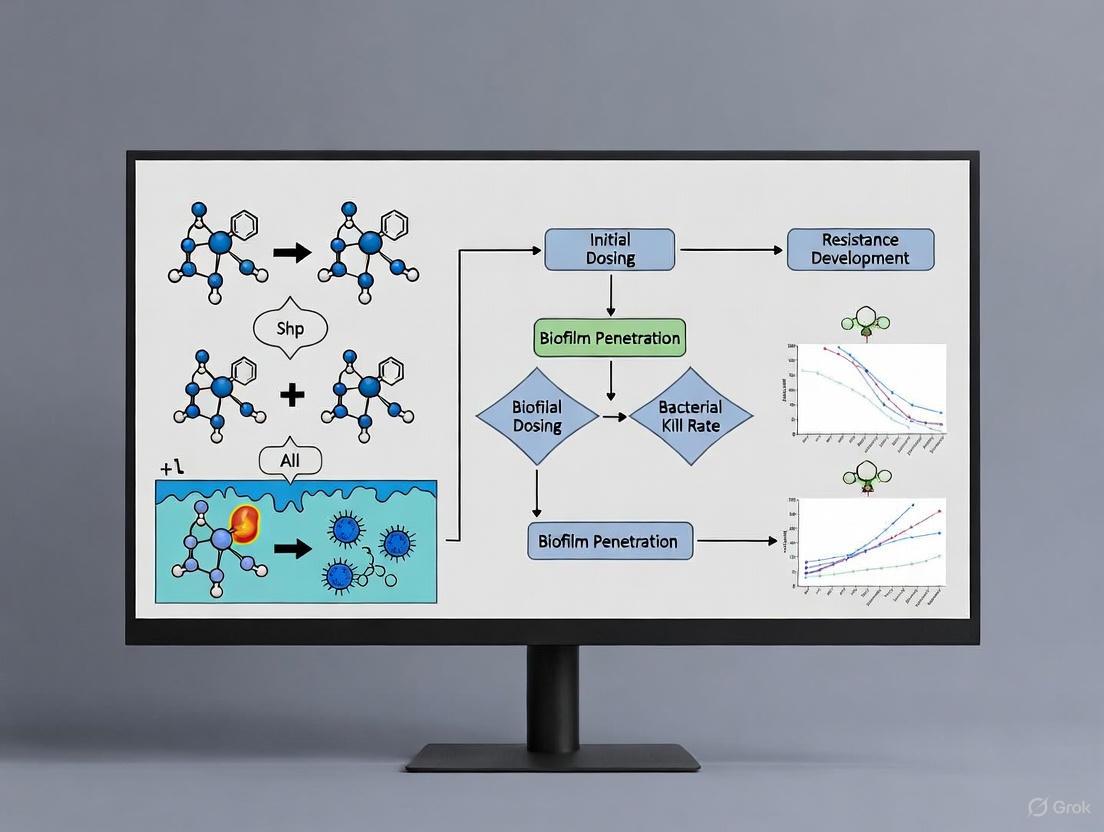
The following workflow diagram illustrates the key stages of this protocol:
Table 1: Antibiotic Penetration Capacity through Different Biofilms
This table summarizes findings from disk diffusion assays, showing how penetration is not a universal property but depends on the specific antibiotic and bacterial species [1].
| Antibiotic Class | Example Antibiotic | Penetration through S. aureus Biofilm | Penetration through E. coli Biofilm |
|---|---|---|---|
| Glycopeptides | Vancomycin | Significantly Hindered | Varies by strain |
| Phenicols | Chloramphenicol | Significantly Hindered | Varies by strain |
| β-lactams | Oxacillin | Variable | Variable |
| Aminoglycosides | Tobramycin | Variable (may bind to eDNA) | Variable (may bind to eDNA) [2] |
| Fluoroquinolones | Ciprofloxacin | Less Hindered | Less Hindered |
Table 2: Impact of Optimized Periodic Dosing on Biofilm Eradication
Data from computational and in vitro studies demonstrating the potential benefit of optimized treatment schedules [6] [5].
| Treatment Strategy | Reduction in Total Antibiotic Dose | Key Parameter for Success | Major Risk |
|---|---|---|---|
| Continuous Dosing | Baseline (0%) | N/A | Incomplete killing of persisters |
| Non-optimized Periodic Dosing | Variable / Ineffective | Poorly timed "off" cycle | Biofilm regrowth; Resistance evolution |
| Optimized Periodic Dosing | Up to 77% [6] | Timing aligned with persister resuscitation dynamics | Rapid evolution of resistance if timing is incorrect [7] |
The Scientist's Toolkit: Research Reagent Solutions
Table 3: Essential Materials for Biofilm and Periodic Dosing Research
| Item / Reagent | Function in Experiment | Specific Example / Note |
|---|---|---|
| Silicone Catheters / Coupons | Provides a medically relevant, inert surface for biofilm growth. | Medical-grade silicone is often used to mimic implant-related infections [5] [7]. |
| Flow Cell System | Enables robust biofilm growth under controlled shear stress and tractable pharmacokinetics. | Critical for testing dosing regimens as it allows for precise antibiotic on/off cycles [5]. |
| Programmable Syringe Pumps | Automates the delivery of antibiotics in precise, timed intervals for periodic dosing. | Essential for maintaining the accuracy and reproducibility of complex dosing schedules [5]. |
| Extracellular DNA (eDNA) | A key component of the biofilm matrix that can bind antibiotics and contribute to tolerance. | Can be targeted with DNase to sensitize biofilms to aminoglycosides [4] [2]. |
| CLSM with FISH Probes | Allows for high-resolution 3D visualization and identification of specific pathogens within a biofilm. | Used to confirm biofilm structure and composition; FISH probes target species-specific rRNA [8]. |
| Agent-Based Model (Computational) | Simulates biofilm growth and treatment response to identify optimal dosing parameters before wet-lab experiments. | Can test a broad range of persistence switching dynamics to streamline experimental design [6]. |
Troubleshooting Common Experimental Challenges
FAQ: Why do my antibiotic killing assays consistently show a biphasic pattern? This is a classic indicator of a persister cell subpopulation. The initial rapid decline represents the death of actively growing, susceptible cells. The subsequent plateau phase, where the killing rate slows dramatically, signifies the survival of dormant persisters [6] [9]. This subpopulation is metabolically inactive and thus tolerant to antibiotics that target growth processes.
FAQ: My biofilm assays are highly variable. What are the key factors to control? Biofilm architecture and persister formation are exquisitely sensitive to environmental conditions. Key parameters to standardize include:
- Nutrient availability: Limitation can increase persister levels [10].
- Growth phase: Persister proportions are typically lowest during the log phase and peak in the stationary phase [10].
- Extracellular Polymeric Substance (EPS): The composition and density of the biofilm matrix can significantly impede antibiotic penetration [11] [10]. Ensure consistent flow rates and surface materials in your biofilm models.
FAQ: How can I distinguish between antibiotic resistance and tolerance in my isolates? The distinction is fundamental. Antibiotic resistance is the ability to grow in the presence of an antibiotic, often due to genetic mutations, and is characterized by an elevated Minimum Inhibitory Concentration (MIC). Antibiotic tolerance, a hallmark of persisters, is the ability to survive but not grow in the presence of a lethal antibiotic concentration, with no change in MIC [9] [10]. Persister cells remain genetically identical to the susceptible population and will regrow once the antibiotic is removed.
Essential Experimental Protocols
Protocol 1: Optimizing Periodic Antibiotic Dosing Using an Agent-Based Model
This protocol is based on a computational framework designed to identify treatment schedules that exploit persister "reawakening" [6].
Key Materials & Setup:
- Model Platform: NetLogo for simulating biofilm growth [6].
- Core Parameters: Define bacterial agents with states for "Susceptible" and "Persister."
- Dynamic Switching: Program switching rates between susceptible and persister states that are dependent on both local substrate (nutrient) availability and antibiotic presence [6].
- Antibiotic Diffusion: Simulate the diffusion of antibiotic from the bulk fluid into the biofilm structure.
Methodology:
- Initialization: Seed a surface with a random distribution of susceptible bacterial agents.
- Biofilm Growth: Simulate growth using Monod kinetics, where individual cell mass increase is governed by
dmi/dt = mi * μmax * CS / (CS + KS), whereCSis local substrate concentration,μmaxis maximal growth rate, andKSis the half-saturation constant [6]. - Treatment Simulation:
- Introduce an antibiotic concentration above the MIC into the bulk fluid.
- Apply the antibiotic in periodic pulses, varying the dosing interval and treatment duration.
- Monitor the collapse and potential regrowth of the biofilm between doses.
- Optimization: Run iterative simulations to find the dosing regimen that minimizes both the total antibiotic dose and the final biofilm biomass. The referenced study achieved a dose reduction of up to 77% with a tuned periodic regimen [6].
Protocol 2: Single-Cell Analysis of Persister Dynamics Using Microfluidics
This protocol details a method for tracking the fates of individual persister cells before, during, and after antibiotic exposure [12].
Key Materials & Setup:
- Strain: E. coli MG1655 (or other relevant wild-type strains).
- Device: A Membrane-Covered Microchamber Array (MCMA) microfluidic device [12].
- Antibiotics: Use high concentrations (e.g., 200 µg/mL ampicillin, ~12.5x MIC; or 1 µg/mL ciprofloxacin, ~32x MIC) to ensure lethality to non-persisters [12].
- Imaging: Time-lapse microscopy setup for phase-contrast and fluorescence imaging.
Methodology:
- Cell Loading: Load bacteria from either exponential or stationary phase cultures into the microchambers of the MCMA device.
- Pre-treatment Monitoring: Flow fresh medium and record single-cell growth histories for several hours to establish baseline activity.
- Antibiotic Treatment: Switch the medium flow to one containing the antibiotic. Maintain treatment for a defined period (e.g., 3-8 hours).
- Post-treatment Recovery: Switch back to fresh, antibiotic-free medium and monitor for regrowth.
- Data Analysis: Correlate the survival and regrowth of individual cells with their pre-treatment state (growing vs. non-growing). This method has revealed that a significant fraction of persisters can originate from actively growing cells, not just dormant ones, depending on the antibiotic and culture history [12].
Research Reagent Solutions
The table below summarizes key reagents and their applications in persister cell research.
Table 1: Essential Research Reagents for Targeting Persister Cells
| Reagent / Material | Function & Application | Key Experimental Insight |
|---|---|---|
| Caffeine-functionalized Gold Nanoparticles (Caff-AuNPs) [11] | Directly kills both planktonic and biofilm-associated persisters. | Effective against Gram-positive and Gram-negative persisters; also disrupts mature biofilms. |
| Cationic Polymer PS+(triEG-alt-octyl) [11] | "Wake-and-kill" strategy; reactivates dormant persisters by stimulating the electron transport chain, then lyses cells. | When loaded onto PDA nanoparticles, enables photothermal-triggered release and enhanced biofilm penetration. |
| Membrane-Targeting Compounds (e.g., XF-73, SA-558) [13] [14] | Directly disrupts bacterial cell membranes, a target that remains in dormant cells. | Effective against non-dividing S. aureus; XF-73 can also generate ROS upon light activation. |
| H₂S Scavengers / CSE Inhibitors [13] [14] | Suppresses persister formation by neutralizing hydrogen sulfide (H₂S), a key player in bacterial stress defense. | Sensitizes S. aureus, P. aeruginosa, and E. coli persisters to antibiotics like gentamicin. |
| Pyrazinamide (PZA) [13] [9] [14] | Anti-persister prodrug; active form disrupts membrane energetics and targets PanD. | Clinically crucial for shortening tuberculosis therapy by effectively targeting M. tuberculosis persisters. |
| ADEP4 [13] [14] | Activates the ClpP protease, leading to uncontrolled ATP-independent protein degradation. | Causes the destruction of metabolic enzymes essential for persister resuscitation, preventing regrowth. |
Table 2: Computational and Analytical Tools for Persister Research
| Tool / Technique | Primary Function | Key Parameters & Outputs |
|---|---|---|
| Agent-Based Model (e.g., in NetLogo) [6] | Simulates emergent biofilm properties and tests antibiotic dosing regimens in silico. | Parameters: Persister switching rates, nutrient diffusion, antibiotic kinetics. Output: Optimized treatment schedule. |
| Microfluidic Single-Cell Analysis (e.g., MCMA) [12] | Tracks the lineage and behavior of individual cells before, during, and after stress. | Parameters: Pre-treatment growth history, morphological changes. Output: Heterogeneous survival dynamics of persisters. |
| Reactive Oxygen Species (ROS) Generating Systems (e.g., MPDA/FeOOH-GOx@CaP) [11] | Directly eliminates persisters via physical membrane damage, independent of metabolism. | Parameters: Local glucose and H₂O₂ concentration, acidic pH. Output: Potent killing of S. aureus and S. epidermidis persisters. |
Visualizing Key Pathways and Workflows
Persister Control Strategies
The following diagram illustrates the three main strategic approaches to combat persister cells as identified in recent literature.
Experimental Workflow for Periodic Dosing Optimization
This diagram outlines the integrated computational and experimental workflow for developing optimized periodic antibiotic treatments against biofilms.
Frequently Asked Questions (FAQs)
1. What are bacterial persister cells and why are they a problem in biofilm infections? Bacterial persisters are a small subpopulation of cells within a biofilm that enter a dormant, slow-growing or non-growing state to survive antibiotic treatment. Unlike resistant bacteria, persisters do not possess genetic resistance mutations; their survival is a reversible phenotypic change. When the antibiotic treatment ceases, these cells can "reawaken," resume growth, and lead to a relapse of the infection. This makes biofilm-mediated infections particularly challenging to eradicate and is a significant cause of chronic and recurrent conditions [6] [3].
2. How does pulse dosing differ from conventional continuous antibiotic dosing? Conventional continuous dosing aims to maintain a constant level of antibiotic in the system over a treatment period. In contrast, pulse dosing involves administering antibiotics in a series of discrete, high-concentration bursts, interspersed with designated antibiotic-free periods. This on-off cycle is strategically designed to target the unique physiology of persister cells [15].
3. What is the core principle behind using pulse dosing to eradicate persisters? The core principle is to exploit the dynamic state of persister cells. During the antibiotic pulse, susceptible active bacteria are killed. During the subsequent antibiotic-free period, the dormant persister cells are given a window to "reawaken" or revert to an active, metabolizing state. A subsequent pulse of antibiotic can then target and kill these newly active cells. By timing the pulses to coincide with this resuscitation, the treatment can significantly reduce the total biofilm biomass and the likelihood of regrowth [6].
4. My biofilm experiments show regrowth after pulse dosing. What could be going wrong? Regrowth typically indicates that the dosing regimen is not fully aligned with the biofilm's specific dynamics. Key parameters to troubleshoot include:
- Pulse Timing: The antibiotic-free window may be too long, allowing resuscitated cells to proliferate and re-establish the biofilm before the next pulse. Alternatively, it may be too short, not allowing enough persisters to exit dormancy [6].
- Pulse Strength: The antibiotic concentration during the pulse may be insufficient to kill all active cells.
- Biofilm Heterogeneity: The model you are using may have persister subpopulations with different switching dynamics. An optimized general regimen can still be effective, but fine-tuning may be required [6].
5. Are there computational tools to help design a pulse dosing regimen? Yes, computational models are increasingly valuable for streamlining regimen design. Agent-based models, which can simulate the stochastic and heterogeneous nature of biofilms, have been used to test a broad range of persistence switching dynamics and identify key parameters for effective treatment. These models have demonstrated that tuned periodic dosing can reduce the required antibiotic dose for effective treatment by nearly 77% [6].
Experimental Protocols for Key Pulse Dosing Experiments
Protocol 1: Establishing a Baseline Biofilm Model with Persisters
This protocol outlines the creation of a standardized biofilm for initial pulse dosing experiments.
1. Materials:
- Strain: Staphylococcus epidermidis or Pseudomonas aeruginosa (common biofilm-forming models).
- Growth Medium: Tryptic Soy Broth (TSB) or another appropriate medium.
- Substrate: Glucose or other relevant nutrient source as a limiting substrate for Monod kinetics [6].
- Biofilm Substrate: 96-well polystyrene plates or flow-cell chambers [16].
- Staining Reagents: Crystal violet for biomass quantification or fluorescent dyes (e.g., SYTO 9) for confocal microscopy.
2. Methodology:
- Inoculation: Prepare a diluted overnight culture of the chosen strain and inoculate it into the wells of the 96-well plate containing fresh medium [6].
- Biofilm Formation: Incubate the plate under static conditions (e.g., 48 hours at 37°C) to allow for biofilm formation. For more controlled shear stress and nutrient delivery, a microfluidic flow-cell system can be used with a continuous perfusion of medium [16].
- Baseline Assessment: After incubation, gently wash the biofilms to remove non-adherent cells. Quantify the biofilm using crystal violet staining (absorbance measurement) or use fluorescent staining and microscopy to visualize the 3D architecture.
Protocol 2: Evaluating a Periodic Dosing Regimen Using an Agent-Based Model
This protocol utilizes a computational approach to test dosing regimens before wet-lab validation.
1. Materials:
- Software: NetLogo platform (or other agent-based modeling software) [6].
- Model Parameters: The model should incorporate key variables such as bacterial growth rate (using Monod kinetics for nutrient-limited growth), rates of switching from susceptible to persister state (and back), and antibiotic killing rates for both cell types [6].
2. Methodology:
- Model Initialization: Set up the simulation with parameters that reflect your experimental conditions. Initialize a population of susceptible bacteria on a surface [6].
- Define Dosing Regimen: Input the proposed pulse dosing regimen, including antibiotic concentration, pulse duration, and interval length.
- Run Simulation: Execute the model to simulate biofilm growth and treatment over time. The model will track the population dynamics of both susceptible and persister cells.
- Output Analysis: Analyze the simulation output to see if the regimen leads to biofilm eradication. The model's graphical interface allows for visualization of the biofilm structure in response to treatment [6]. Key is to run simulations across a wide range of switching dynamics to find a generally effective regimen.
Data Presentation: Quantitative Findings on Pulse Dosing Efficacy
The table below summarizes key quantitative findings from computational and theoretical studies on optimized periodic dosing.
Table 1: Quantitative Efficacy of Optimized Periodic Antibiotic Dosing Against Biofilms
| Study Model / Type | Key Optimized Dosing Parameter | Efficacy Outcome | Reported Reduction in Required Dose |
|---|---|---|---|
| Agent-Based Computational Model [6] | Dosing interval tuned to persister switching dynamics (stochastic & triggered) | Near-complete biofilm eradication | Up to 77% reduction compared to non-optimized dosing |
| Mathematical Model (Control Theory) [15] | Optimal protocol derived via control theory; early-stage intervention | Successful bacterial elimination; wider margin for eradication | Ensures elimination across a wider range of initial conditions compared to non-optimal techniques |
Research Reagent Solutions
The table below lists essential materials and tools used in pulse dosing and biofilm research.
Table 2: Key Research Reagents and Tools for Biofilm Persister Studies
| Item | Function / Application | Example / Notes |
|---|---|---|
| Microfluidic Perfusion System [16] | Provides precise, pulse-like fluid control for dynamic antibiotic delivery to biofilms under shear stress. | Fluigent Omi Platform or similar. Enables replication of physiological flow conditions. |
| Agent-Based Modeling Software [6] | Computational testing of countless pulse dosing regimens to identify optimal timing and concentration before lab work. | NetLogo platform. Allows for incorporation of stochastic persister switching dynamics. |
| VRprofile2 Software [17] | Analyzes bacterial mobilome (plasmids, transposons) to understand genetic context of resistance in clinical isolates. | Useful for characterizing strains used in biofilm models and tracking resistance gene transfer. |
| Engineered Phage with DspB [18] | A biological tool to degrade the biofilm matrix (via DspB enzyme), enhancing antibiotic penetration. | Modified T7 phage. Can be used in combination with antibiotic pulse dosing strategies. |
Mechanism and Workflow Visualization
The following diagrams illustrate the core concept of pulse dosing and a proposed experimental workflow.
Diagram 1: Pulse Dosing Mechanism to Eradicate Persisters
Diagram 2: Integrated Experimental Workflow
Contrasting Biofilm Tolerance with Genetic Antibiotic Resistance
Frequently Asked Questions (FAQs)
Q1: What is the fundamental difference between biofilm tolerance and genetic antibiotic resistance? Biofilm tolerance is a phenotypic survival state where bacteria transiently withstand antibiotic exposure without genetic change. In contrast, genetic antibiotic resistance involves heritable genetic mutations or acquired genes that confer the ability to grow at elevated antibiotic concentrations. Biofilm-tolerant cells, including persisters, typically exhibit recalcitrance to killing without an increase in Minimum Inhibitory Concentration (MIC), whereas genetically resistant strains demonstrate a significantly elevated MIC [2] [19].
Q2: How do persister cells contribute to biofilm-associated treatment failure? Persisters are a dormant subpopulation within biofilms that exhibit extreme tolerance to lethal antibiotics. They are genetically identical to susceptible cells but survive treatment due to phenotypic dormancy. After antibiotic concentrations drop, these cells can resume growth and repopulate the biofilm, leading to chronic infection recurrence. This cycle occurs without the acquisition of resistance genes [6] [19].
Q3: Why is periodic dosing considered a potential strategy against biofilm infections? Periodic (or pulse) dosing protocols alternate antibiotic treatment with antibiotic-free periods. This strategy aims to exploit the phenotypic switching of persister cells. The drug-free intervals allow dormant persisters to resuscitate into metabolically active, antibiotic-susceptible cells, which are then vulnerable to the next antibiotic pulse. Computational models suggest optimally timed periodic dosing can reduce the total antibiotic dose required for eradication by up to 77% [6] [5].
Q4: What are the risks associated with intermittent antibiotic treatment of biofilms? While designed to exploit tolerance, intermittent lethal dosing can inadvertently accelerate the evolution of genetic resistance. The biofilm environment provides intrinsic tolerance, genetic heterogeneity, and high cell density, creating a fertile ground for selecting resistance mutations. Studies with E. coli show that intermittent treatment can rapidly select for mutations in genes like fusA and sbmA, leading to stable, heritable resistance [7].
Troubleshooting Common Experimental Challenges
Challenge 1: Inconsistent Persister Cell Yields in Biofilm Models
- Potential Cause: Variations in nutrient availability, oxygen gradients, and biofilm maturity.
- Solution: Standardize growth conditions meticulously. Use continuous flow systems to maintain consistent nutrient and waste levels. Monitor biofilm maturity using metrics like biomass or 3D architecture over time, rather than just incubation duration [5].
Challenge 2: Differentiating Between True Resistance and Tolerance in Survival Assays
- Potential Cause: Relying solely on survival counts after antibiotic exposure without subsequent MIC determination.
- Solution: After performing a killing assay to determine tolerance, always isolate surviving cells. Re-culture them in the absence of antibiotic and determine the MIC of the progeny. If the MIC remains unchanged, the survival was due to tolerance; if elevated, genetic resistance has been selected [7] [19].
Challenge 3: Optimizing Pulse Dosing Intervals
- Potential Cause: The resuscitation time of persisters is strain- and environment-dependent.
- Solution: There is no universal interval. Determine the optimal off-period experimentally by tracking the resurgence of metabolic activity after antibiotic removal, using methods like ATP assays or reporter strains. Agent-based in silico models can also help predict effective dosing schedules for specific experimental conditions [6] [5].
Quantitative Data on Biofilm Tolerance & Dosing
Table 1: Key Mechanisms Contrasting Biofilm Tolerance and Genetic Resistance
| Feature | Biofilm Tolerance (Phenotypic) | Genetic Resistance |
|---|---|---|
| Basis | Transient, non-heritable phenotype | Heritable genetic changes (mutations, horizontal gene transfer) |
| MIC Change | Typically unchanged | Significantly increased |
| Key Mechanisms | - Poor antibiotic penetration [2]- Metabolic heterogeneity & dormancy [20]- Persister cell formation [6] | - Enzyme-mediated drug inactivation- Target site modification- Efflux pump upregulation |
| Reversibility | Reversible upon biofilm dispersal | Stable without genetic reversion |
Table 2: Efficacy of Different Antibiotic Dosing Strategies Against Biofilms
| Dosing Strategy | Reported Efficacy | Key Findings & Risks |
|---|---|---|
| Continuous Dosing | Limited efficacy against mature biofilms | Kills susceptible cells but leaves a persistent fraction unchanged; can select for resistance over time [5]. |
| Periodic/Pulse Dosing | Up to ~77% reduction in total dose required (in silico model) [6] | Effective when timed with persister resuscitation; optimizes killing of susceptible cells repopulated from persisters [5]. |
| Intermittent Lethal Dosing | Rapid evolution of resistance in biofilms [7] | Provides a "see-saw" dynamic of killing and regrowth that enriches for pre-existing resistance mutants (e.g., in fusA, sbmA). |
Experimental Protocols
Protocol 1: Evaluating Periodic Dosing In Vitro Using a Biofilm Flow System
This protocol is adapted from studies on S. aureus biofilms [5].
Key Reagents & Materials:
- Organism: Staphylococcus aureus HG003 (or relevant strain).
- Growth Medium: Brain Heart Infusion (BHI) broth supplemented with 1% glucose.
- Substrate: Medical-grade silicone coupons or 14G polyurethane catheter segments.
- Equipment: Peristaltic pump, silicone tubing, glass flow cells, programmable syringe pumps for antibiotic dosing.
Methodology:
- Biofilm Preparation: Pre-coat substrates in fetal bovine serum (FBS) overnight. Inoculate substrates with a bacterial suspension (OD600 ~0.01) and incubate for 24 hours.
- Maturation: Transfer substrates to fresh medium for another 24 hours. Then, place them into a flow cell system and perfuse with medium (e.g., 0.1 ml/min) for 16-21 hours to establish mature, tolerant biofilms.
- Pulse Dosing Regimen:
- Pulse: Introduce antibiotic (e.g., oxacillin) at the desired concentration (e.g., 5xMIC or 80xMIC) into the medium for a defined period (e.g., 24 hours).
- Break: Switch to antibiotic-free medium for a predetermined interval. The length of this break is critical and must be optimized to allow persister resuscitation without significant resistance expansion.
- Assessment: After each pulse-break cycle, disrupt biofilms via sonication and vortexing. Perform serial dilution and plate counts to enumerate Colony Forming Units (CFUs). Compare survival against continuous dosing controls.
Protocol 2: Agent-Based Modeling for Dosing Optimization
This computational approach helps predict effective dosing schedules before wet-lab experiments [6] [21].
Key Parameters:
- Model Framework: Implement an agent-based model (e.g., in NetLogo) where individual bacteria are represented as agents.
- Core Rules: Program rules for bacterial growth (e.g., Monod kinetics), division, switching to/from persister state based on local substrate and antibiotic concentration, and death.
- Environmental Factors: Simulate diffusion of antibiotic and nutrients from the bulk fluid.
Workflow:
- Parameterization: Calibrate the model with experimental data on persister switching rates and antibiotic killing kinetics for your specific bacterial strain.
- Simulation: Run in silico experiments testing a wide range of periodic dosing schedules (varying pulse duration and frequency).
- Optimization: Identify the treatment regimen that minimizes both the total antibiotic dose and the final bacterial load. The model can highlight key parameters, such as the critical persister resuscitation time, for effective treatment.
Signaling Pathways and Experimental Workflows
Persister Cell Dynamics and Periodic Dosing Strategy
Key Signaling Pathways in Biofilm Persister Formation
The Scientist's Toolkit: Research Reagent Solutions
Table 3: Essential Research Reagents for Biofilm and Persister Studies
| Reagent / Material | Function / Application | Example Use Case |
|---|---|---|
| Medical-grade Silicone Coupons | Provides a standardized, non-biodegradable surface for robust and reproducible biofilm growth. | Used in flow system models to study biofilm formation on implant-relevant materials [5]. |
| Continuous Flow Cell System | Maintains constant nutrient supply and shear force, enabling the development of mature, structured biofilms with natural physiological heterogeneity. | Crucial for generating biofilms with realistic gradients of oxygen and nutrients, which drive persister formation [5]. |
| Agent-Based Modeling Software (e.g., NetLogo) | In silico platform to simulate individual bacterial behavior, interactions, and response to treatments within a biofilm. | Used to test thousands of hypothetical periodic dosing regimens rapidly and cheaply before wet-lab validation [6] [21]. |
| ATP Assay Kits | Quantifies cellular ATP levels as a direct measure of metabolic activity and viability. | Differentiates metabolically active susceptible cells from dormant persisters in a biofilm after antibiotic treatment [20]. |
| Lux-operon Reporter Strains | Genetically modified bacteria that produce bioluminescence, correlating with metabolic activity. | Can be used to non-invasively monitor biofilm metabolic activity and potential regrowth during pulse-dosing experiments [5]. |
Designing and Implementing Effective Periodic Dosing Regimens
Frequently Asked Questions (FAQs)
Q1: What are the key advantages of using in vitro flow systems over static models for biofilm antibiotic dosing studies? In vitro flow systems, such as the one detailed for Staphylococcus aureus biofilms, enable robust biofilm growth with tractable pharmacokinetics. They allow researchers to precisely control the antibiotic concentration over time and simulate the dynamic conditions of fluid flow found in many physiological and industrial settings, which is not possible in static models. This is crucial for testing periodic dosing regimens where the timing of antibiotic application and removal is critical for efficacy [5].
Q2: Our periodic dosing experiments are not showing improved killing. What could be going wrong? The most common issue is an incorrectly timed "off" period (the break from antibiotic). If the break is too short, persistent cells do not have sufficient time to resuscitate back to an antibiotic-susceptible state. If the break is too long, it can allow not only for resuscitation but also for significant regrowth of the biofilm and potential expansion of resistant populations. You should empirically test a range of off-periods to find the optimal timing for your specific bacterial strain and biofilm maturity [5]. Furthermore, ensure your biofilm is mature and has developed significant tolerance, as young biofilms may not have a substantial persister population to target [5].
Q3: How can we differentiate between antibiotic resistance and tolerance (persistence) in our biofilm experiments? Tolerance (persistence) is characterized by a biphasic killing curve, where a population of susceptible cells dies rapidly, followed by a much slower rate of killing of a subpopulation (persisters). Crucially, upon re-culturing without antibiotic, the surviving population will have the same minimum inhibitory concentration (MIC) as the original strain. Resistance, on the other hand, involves a genetic change and will manifest as a stable, heritable increase in the MIC of the entire population [6] [5].
Q4: What advanced techniques can be used to analyze the structure and metabolic state of biofilms post-treatment? Imaging Flow Cytometry (IFC) combined with machine learning-based analysis is a powerful tool. It allows for high-throughput, quantitative analysis of biofilm dispersal aggregates and single cells. You can simultaneously assess the degree of cellular aggregation (singlets, small vs. large aggregates) and the metabolic activity (e.g., active, mid-active, dead) of the cells within those structures, providing a detailed picture of the biofilm's physiological response to treatment [22].
Troubleshooting Guides
Table 1: Common Experimental Challenges in Biofilm Dosing Studies
| Challenge | Possible Causes | Suggested Solutions |
|---|---|---|
| High variability in biofilm biomass | Inconsistent surface conditioning, uneven flow rates, variations in initial inoculum. | Standardize preconditioning protocol (e.g., uniform FBS coating [5]); calibrate peristaltic pumps regularly; use a standardized and well-mixed inoculum. |
| Insufficient persister population | Biofilm is too young; overly nutritious media preventing dormancy. | Grow biofilms for a longer duration (e.g., 48+ hours); consider using media with lower nutrient content or adding 1% glucose to stimulate mature biofilm formation [5]. |
| Failure of periodic dosing regimen | Incorrect timing of antibiotic pulse; inadequate antibiotic concentration during "on" phase. | Systematically test a range of "on" and "off" durations; ensure antibiotic concentration is well above the MIC for the susceptible population during the treatment phase [6] [5]. |
| Difficulty dispersing biofilm for CFU counting | Strong extracellular matrix; inadequate disruption method. | Use a combination of sonication and vortexing in multiple cycles (e.g., 3 cycles of 5 min sonication + 30s vortexing) [5]; validate your method by visual inspection (e.g., microscopy) to confirm complete disaggregation. |
Table 2: Quantitative Findings on Periodic Dosing Efficacy
| Study Model | Key Finding | Quantitative Result | Implication for Protocol Design |
|---|---|---|---|
| Computational Agent-Based Model [6] | Optimized periodic dosing can dramatically reduce total antibiotic dose. | Reduced required antibiotic dose by nearly 77% [6]. | Computational modeling can be used as a first step to identify promising dosing schedules for in vitro testing. |
| S. aureus Biofilm Flow System [5] | Pulse dosing is more effective than continuous dosing at killing biofilms. | Correctly timed antibiotic breaks decreased the surviving persister population, which continuous dosing could not achieve [5]. | The "off" period is critical for sensitizing the biofilm. The optimal length is specific to the experimental setup and must be determined. |
| S. aureus Biofilm Flow System [5] | The length of the antibiotic-free break impacts efficacy. | An optimal break length exists that sensitizes the biofilm without allowing resistance expansion; periods that were too short or too long were less effective [5]. | A pilot experiment to titrate the "off" period is essential for protocol optimization. |
Experimental Protocols
Protocol 1: Periodic Dosing in a S. aureus Biofilm Flow System
This protocol is adapted from Frontiers in Microbiology (2020) for testing pulse dosing of oxacillin against S. aureus biofilms [5].
Key Research Reagent Solutions:
- Bacterial Strain: Staphylococcus aureus HG003 with a chromosomal lux operon and chloramphenicol resistance marker.
- Growth Media: Brain Heart Infusion (BHI) broth supplemented with 1% glucose to stimulate biofilm formation.
- Antibiotic Stock: Oxacillin dissolved in water, stored at -20°C.
- Surfaces: 14G polyurethane I.V. catheters, cut into 1 cm segments.
- Flow System Components: Silicone tubing, peristaltic pumps, glass segments to house catheters, and programmable syringe pumps for antibiotic dosing.
Methodology:
- Surface Preparation: Pre-condition catheter segments in Fetal Bovine Serum (FBS) overnight at 37°C to promote bacterial adherence.
- Initial Adhesion: Transfer segments to a microcentrifuge tube with 1 ml of S. aureus suspension (OD600 of 0.01 in BHI + 1% glucose) and incubate for 24 hours at 37°C.
- Biofilm Maturation: Transfer catheters to fresh BHI + 1% glucose for another 24 hours.
- Flow System Setup: Place catheters in triplicate inside sterile glass segments connected to the flow system. Pump BHI + 1% glucose through the system at 0.1 ml/min for 16-21 hours at 37°C to establish mature biofilms.
- Periodic Dosing Regimen: Initiate treatment by adding oxacillin to the input media. For periodic dosing, use programmable syringe pumps or switch input bottles to alternate between antibiotic-containing and antibiotic-free media according to the predetermined schedule (e.g., several hours on/several hours off).
- Biofilm Harvesting and CFU Enumeration:
- Disconnect the system and rinse catheters in saline.
- Sonicate catheters in 1 ml saline for 5 minutes, followed by 30 seconds of vortexing. Repeat this cycle three times to disrupt the biofilm.
- Serially dilute the resulting suspension and plate on BHI agar plates in duplicate.
- Incubate plates at 37°C and enumerate Colony Forming Units (CFUs) after 24 and 72 hours.
Protocol 2: Agent-Based Modeling for Dosing Regimen Optimization
This protocol is based on a 2024 study in the Journal of the Royal Society Interface that used an agent-based model to optimize periodic treatment [6].
Key Research Reagent Solutions:
- Software: NetLogo platform for implementing the agent-based model.
- Model Parameters: Key parameters to define include bacterial growth rates (using Monod kinetics), persister switching dynamics (dependent on both substrate availability and antibiotic presence), and antibiotic killing rates for susceptible and persister cells.
Methodology:
- Model Initialization: Seed 27 susceptible bacterial agents randomly on a simulated surface.
- Define Growth and Rules: Program agent behavior based on:
- Growth: Cell mass increases via Monod kinetics, dependent on local substrate concentration.
- Division: Cells divide upon reaching a threshold mass (e.g., 500 fg).
- Phenotypic Switching: Rules for switching between susceptible and persister states are defined as functions of local substrate and antibiotic concentration.
- Simulate Treatment: Run the model to grow a mature biofilm, then apply virtual antibiotic treatments with different periodic schedules.
- Output Analysis: The model outputs the biofilm architecture, the number and location of persister cells, and the total killing efficacy for each dosing regimen, allowing for the identification of optimized treatment schedules before wet-lab testing.
Diagram: Experimental Workflow for Biofilm Dosing Research
Diagram: Persister Dynamics in Periodic Dosing
The Scientist's Toolkit: Essential Materials
Table 3: Key Research Reagent Solutions for Biofilm Dosing Studies
| Item | Function/Application |
|---|---|
| Polyurethane I.V. Catheters | A common and standardized surface for growing biofilms in flow systems, providing a relevant model for medical device-associated infections [5]. |
| Silicone Tubing & Peristaltic Pumps | Create a controlled flow environment for biofilm growth, allowing for the simulation of physiological shear forces and precise management of antibiotic pharmacokinetics [5]. |
| Programmable Syringe Pumps | Enable the automated and precise addition of antibiotics to the flow system, which is critical for implementing complex and reproducible periodic dosing schedules [5]. |
| Oxacillin (or other antibiotics) | A beta-lactam antibiotic used to treat S. aureus infections. It serves as a model drug for studying antibiotic tolerance and the efficacy of novel dosing regimens against biofilms [5]. |
| NetLogo Software | An accessible platform for developing agent-based computational models to simulate biofilm growth, persister dynamics, and treatment outcomes, helping to guide wet-lab experiments [6]. |
| Imaging Flow Cytometer (e.g., Amnis FlowSight) | Allows for high-throughput, quantitative analysis of biofilm dispersal forms (single cells and aggregates) and their metabolic activity, providing deep insight into treatment effects [22]. |
Quantitative Data Tables on Antibiotic Dosing
Table 1: Comparative Efficacy of Standard vs. Optimized Periodic Dosing
| Parameter | Standard Continuous Dosing | Optimized Periodic Dosing | Key Findings |
|---|---|---|---|
| Total Antibiotic Dose | Baseline | Reduced by up to 77% [6] | Significant reduction in total antibiotic exposure. |
| Persister Cell Elimination | Ineffective; persister levels remain stable [5] | Substantial reduction with correctly timed breaks [5] | Breaks allow persisters to "reawaken" into a susceptible state. |
| Treatment Efficacy on Mature Biofilms | Limited efficacy due to tolerance [5] | Dramatically improved killing of Staphylococcus aureus biofilms [5] | Timing of antibiotic pulses is critical for success. |
Table 2: Key Dosing Parameters and Their Experimental Ranges
| Parameter | Definition | Experimental Range / Value | Impact on Treatment Outcome |
|---|---|---|---|
| Antibiotic Concentration | Concentration of antibiotic applied during the "on" pulse. | Tested at multiples of the Minimum Biofilm Eradication Concentration (MBEC), which can be 100-800x higher than the MIC for planktonic cells [23] [24]. | Must be high enough to penetrate the biofilm matrix and kill susceptible cells. |
| Pulse Duration (On-period) | Time for which antibiotic is continuously present. | Modeled and tested in specific cycles; requires alignment with biofilm dynamics [6]. | Must be long enough to kill the majority of susceptible populations. |
| Off-period Duration | Antibiotic-free period allowing persister cell resuscitation. | Critical parameter; an optimal length exists that sensitizes the biofilm without allowing regrowth or resistance expansion [5]. | Too short: persisters do not resuscitate. Too long: biofilm regrows and risk of resistance increases. |
Experimental Protocols
Protocol 1: Determining Minimum Biofilm Eradication Concentration (MBEC) Using a Resazurin-Based Assay
This protocol provides a robust method for determining the antibiotic concentration required to eradicate biofilms, which is fundamental for setting the pulse dose [24].
Biofilm Cultivation:
- Inoculate a 96-well plate with a bacterial suspension (e.g., Pseudomonas aeruginosa) in an appropriate broth like Müller-Hinton II.
- Incubate under static conditions for 24-48 hours at 37°C to allow mature biofilm formation on the well surfaces.
Biofilm Maturation and Washing:
- Carefully aspirate the planktonic-phase culture from the wells.
- Gently wash the biofilms three times with fresh broth or saline to remove all non-adherent cells.
Antibiotic Exposure:
- Prepare a serial dilution of the test antibiotic in the broth.
- Add the antibiotic dilutions to the wells containing the mature biofilms.
- Incubate the plate for a further 24 hours at 37°C.
Viability Assessment:
- After incubation, remove the antibiotic solution and wash the biofilm once.
- Add a resazurin-based viability reagent (e.g., PrestoBlue) to each well.
- Incubate for a defined period (e.g., 30-60 minutes) and then measure the fluorescence or absorbance.
- The MBEC is defined as the lowest antibiotic concentration that reduces cell viability by a predetermined cutoff (e.g., 75% or 90%) compared to an untreated control [24].
Protocol 2: In Vitro Evaluation of Pulse Dosing Regimens in a Flow System
This protocol describes an advanced system to test dynamic dosing regimens against biofilms grown under flow conditions, closely mimicking in vivo scenarios like catheter infections [5].
Surface Preparation and Biofilm Initiation:
- Use a relevant substrate (e.g., a 1 cm segment of a 14G polyurethane IV catheter).
- Pre-coat the substrate in Fetal Bovine Serum (FBS) overnight to promote bacterial adherence.
- Transfer the catheter to a microcentrifuge tube containing a bacterial suspension (e.g., Staphylococcus aureus in Brain Heart Infusion broth with 1% glucose) and incubate for 24 hours at 37°C.
Biofilm Maturation under Flow:
- Place the colonized catheter inside a sterile glass flow cell.
- Connect the flow cell to a peristaltic pump and continuously feed with fresh, pre-warmed nutrient broth (e.g., BHI + 1% glucose) at a low flow rate (e.g., 0.1 ml/min).
- Maintain the flow for 16-21 hours in a 37°C incubator to establish a mature, tolerant biofilm.
Implementation of Pulse Dosing:
- Initiate treatment by adding antibiotic to the input broth. For pulse dosing, use programmable syringe pumps and timers to switch the input between antibiotic-containing and antibiotic-free broth according to the defined schedule (e.g., 12 hours on / 12 hours off).
- Run the experiment for multiple cycles (e.g., 2-3 cycles).
Biofilm Harvesting and Quantification:
- At the end of the experiment, remove the catheter from the flow system and rinse it gently in saline to remove loosely attached cells.
- Disrupt the biofilm by sonicating the catheter in saline for 5 minutes, followed by 30 seconds of vortexing. Repeat this process three times.
- Serially dilute the resulting bacterial suspension, plate it on agar plates, and incubate to enumerate the remaining Colony Forming Units (CFUs).
Signaling Pathways and Experimental Workflows
Persister Cell Targeting Mechanism
Experimental Workflow for Pulse Dosing
The Scientist's Toolkit: Research Reagent Solutions
Table 3: Essential Materials and Reagents for Biofilm Dosing Studies
| Item | Function / Application | Specific Examples / Notes |
|---|---|---|
| Resazurin-based Viability Reagents | Fluorometric quantification of metabolically active cells in a biofilm after antibiotic exposure [24]. | PrestoBlue; used for high-throughput MBEC determination. |
| 96-well Plate Assay Platforms | Standardized in vitro platform for growing biofilms and performing susceptibility screening [24]. | Compatible with automation; allows testing of multiple conditions and replicates. |
| Calgary Biofilm Device (CBD) | Another standardized tool for growing and harvesting biofilms for susceptibility testing [24]. | Provides a reproducible source of biofilm cells. |
| Peristaltic Pumps & Programmable Timers | To maintain continuous flow and implement precise, automated periodic dosing regimens in flow cell systems [5]. | Critical for mimicking in vivo conditions and complex dosing schedules. |
| Physiologically Relevant Substrata | Surfaces for biofilm growth that mimic clinical environments (e.g., catheters, implants) [5]. | Polyurethane IV catheter segments; pre-coating with FBS can enhance adherence. |
| Sonicator | To disrupt the biofilm structure and harvest cells for accurate CFU enumeration after treatment [5]. | Essential for recovering deeply embedded persister cells. |
Troubleshooting Guides and FAQs
FAQ 1: Why are traditional MIC values from planktonic bacteria ineffective for designing biofilm treatments?
- Answer: Minimum Inhibitory Concentration (MIC) tests are performed on free-floating (planktonic) bacteria. Biofilms are inherently more tolerant, with Minimum Biofilm Eradication Concentrations (MBECs) often 100 to 800 times higher than the MIC for the same strain [23] [24]. Furthermore, biofilms contain phenotypically heterogeneous populations, including dormant persister cells, which are not accounted for in standard MIC tests [6] [25]. Basing dosing solely on MIC values will likely result in sub-lethal antibiotic exposure at the biofilm site, leading to treatment failure.
FAQ 2: What is the primary mechanism by which periodic dosing overcomes biofilm tolerance compared to continuous therapy?
- Answer: Continuous antibiotic exposure primarily kills the susceptible cells but has little effect on dormant persister cells, which can survive indefinitely in the presence of the drug [5]. Periodic dosing introduces a critical "off-period." During this break, the antibiotic concentration drops, allowing the dormant persister cells to resuscitate, resume growth, and revert to an antibiotic-susceptible state. A subsequent "on-pulse" of antibiotic is then able to kill this newly susceptible population. This cycle is repeated until the persister reservoir is depleted [6] [5].
FAQ 3: How do I determine the optimal "off-period" duration in my pulse dosing regimen?
- Answer: The off-period is a critical parameter and must be determined empirically for your specific bacterial strain and antibiotic. The goal is to find a duration that allows for maximum resuscitation of persister cells without allowing significant regrowth of the biofilm or the expansion of resistant mutants [5]. This requires time-course experiments where the off-period is systematically varied (e.g., 4, 8, 12, 16 hours) and the resulting bacterial load is quantified after the subsequent antibiotic pulse. The optimal off-period is the one that results in the lowest final CFU count.
FAQ 4: My pulse dosing regimen is not achieving biofilm eradication. What are the potential causes?
- Answer:
- Insufficient Antibiotic Concentration: The pulse concentration may be below the effective MBEC, failing to kill the susceptible population. Re-evaluate your concentration using a biofilm-specific assay [24].
- Sub-optimal Timing: The pulse duration may be too short, or the off-period may be too long or too short. Fine-tuning these temporal parameters is essential [6] [5].
- Biofilm Architecture: The extracellular polymeric substance (EPS) matrix may be limiting antibiotic penetration. Consider combining your regimen with anti-biofilm agents that disrupt the matrix [25] [23].
- Multi-species Biofilm: The treatment might be effective against one species but allows another to proliferate. Perform species identification and susceptibility testing on the recovered biofilm [26].
The Role of Agent-Based and Computational Modeling in Regimen Design
Frequently Asked Questions (FAQs)
Q1: What is the primary advantage of using Agent-Based Models (ABMs) over traditional differential equation models for biofilm research? ABMs excel at capturing the inherent heterogeneity, stochasticity, and emergent collective behaviors within bacterial biofilms. Unlike traditional models that assume homogeneous populations, ABMs simulate individual bacteria (agents) with their own set of rules, allowing them to represent variations in cell states, spatial organization, and local interactions that are crucial for understanding persistence and treatment failure [6] [27]. This makes them particularly suited for predicting how localized phenomena, like the formation of persister cell niches, influence overall treatment efficacy.
Q2: How can computational models identify optimized periodic dosing schedules for antibiotics? Computational models, including ABMs, allow researchers to simulate a wide range of dosing regimens—varying antibiotic type, sequence, duration, and frequency—to find schedules that maximize bacterial killing while minimizing total antibiotic use. For instance, models have demonstrated that periodic dosing tuned to a biofilm's specific dynamics can reduce the required antibiotic dose by nearly 77% by effectively "reawakening" dormant persister cells to make them susceptible to treatment [6].
Q3: What are "collateral sensitivity" patterns, and how can models use them to design better therapies? Collateral sensitivity (CS) is a phenomenon where resistance to one antibiotic causes increased susceptibility to another [28]. Computational frameworks can systematically analyze laboratory data on these patterns to predict and avoid therapeutic sequences that trigger the emergence of multi-drug resistant strains. The models help identify optimal antibiotic cycles that exploit these evolutionary "loopholes" to suppress resistance [28].
Q4: What are common reasons for the failure of a simulated treatment regimen in an ABM? Treatment failure in an ABM typically arises from several key factors:
- Unexpected Persister Dynamics: The regimen may not account for the timing with which persister cells revert to an active, susceptible state, allowing the population to recover [6].
- Sub-optimal Antibiotic Sequencing: The order of antibiotics may inadvertently leverage cross-resistance (where resistance to one drug confers resistance to another) instead of collateral sensitivity, guiding the population toward a multi-drug resistant variant [28].
- Inadequate Spatial Penetration: The antibiotic may fail to diffuse effectively through the biofilm's extracellular matrix or may not reach protected cellular niches within the biofilm's architecture [6] [27].
Q5: Which software platforms are commonly used for building Agent-Based Models of biofilms? Two prominent open-source platforms are:
- NetLogo: A versatile modeling environment with a lower barrier to entry, often used for prototyping and educational purposes. It provides a graphical interface to visualize simulations in real-time [6].
- iDynoMiCS: A more specialized, high-performance software package designed specifically for individual-based modeling of microbial communities and biofilms [27].
Troubleshooting Guides
Guide 1: Addressing Unrealistic Biofilm Morphology in Your ABM
Problem: The simulated biofilm structure in your model does not resemble experimental images (e.g., it appears too uniform, fails to form clusters, or has an unnatural texture).
Solution Steps:
- Check Initialization Parameters: Verify how the biofilm is seeded. Biofilms initiated with a mix of single cells and bacterial aggregates (rather than single cells alone) produce more realistic, rough-textured biofilms [27].
- Review Detachment Mechanisms: Incorporate and calibrate multiple detachment mechanisms. Shear-driven detachment (influenced by biofilm thickness and fluid flow), erosion (continuous loss of single cells), and nutrient-limited detachment (sloughing in thick, nutrient-poor regions) are all critical for shaping biofilm architecture [27].
- Validate Growth Parameters: Ensure that the maximum specific growth rate (( \mu{max} )) and half-saturation constant (( KS )) for substrate uptake are accurate for your bacterial species. Overly fast or uniform growth will lead to structurally simplistic biofilms.
Guide 2: Calibrating Persister Cell Dynamics
Problem: The model fails to recapitulate the biphasic killing curve (a rapid initial kill followed by a persistent subpopulation) observed in time-kill experiments.
Solution Steps:
- Implement Dual Switching Rates: Persister formation should be dependent on both antibiotic presence (triggered persistence) and local substrate availability (stochastic persistence). Similarly, the switching rate from persister back to susceptible must be included [6].
- Adjust Death Rates: Set distinct death rates for susceptible and persister cell populations. The persister death rate should be several orders of magnitude lower than that of susceptible cells when exposed to the antibiotic [6].
- Incorporate Spatial Gradients: Ensure that nutrient gradients emerge in your simulation. Persisters should predominantly form in nutrient-poor or hypoxic regions of the biofilm, which aligns with experimental observations [29] [6].
Guide 3: Integrating Experimental 'Omics' Data into Your Model
Problem: You have transcriptomic or proteomic data but are unsure how to use it to parameterize your computational model.
Solution Steps:
- Identify Key Pathways: Use your 'omics data to pinpoint which metabolic pathways or stress response systems (e.g., quorum sensing, toxin-antitoxin modules) are up- or down-regulated in response to antibiotic treatment [29] [30].
- Link to Model Parameters: Map these pathways to concrete parameters in your model. For example:
- Upregulation of efflux pumps → Increase in the minimum inhibitory concentration (MIC) parameter for relevant antibiotics.
- Activation of general stress response → Increase in the switching rate to the persister state.
- Changes in metabolic gene expression → Modify the half-saturation constant (( K_S )) for nutrient uptake [29] [27].
- Use Constraint-Based Modeling: For genome-scale models, consider using the COBRA (Constraint-Based Reconstruction and Analysis) framework to predict metabolic fluxes, which can then inform the growth rates and metabolic constraints of agents in your ABM [27].
Summarized Quantitative Data
Table 1: Key Parameters for an Agent-Based Model of Biofilm Treatment.
| Parameter | Description | Typical Value/Range | Source/Experimental Method |
|---|---|---|---|
| ( \mu_{max} ) | Maximum specific growth rate | Species-specific (e.g., ~0.1 - 2.0 ( h^{-1} )) | Planktonic growth curves in rich media [6] |
| ( K_S ) | Half-saturation constant for substrate | Species-specific (e.g., 0.1 - 20 ( mg/L )) | Monod kinetic studies in chemostats [6] |
| Persister Switch Rate (to) | Rate of switching from susceptible to persister state | ( 10^{-6} - 10^{-3} ) per cell per generation | Fluorescence-activated cell sorting (FACS) of reporter strains [6] |
| Persister Switch Rate (from) | Rate of reverting from persister to susceptible state | ( 10^{-2} - 10^{-1} ) per cell per hour | Monitoring regrowth after antibiotic removal [6] |
| Death Rate (Susceptible) | Death rate of susceptible cells under antibiotic | ~0.1 - 10 ( h^{-1} ) (high) | Time-kill assays [6] |
| Death Rate (Persister) | Death rate of persister cells under antibiotic | ~0.001 - 0.1 ( h^{-1} ) (low) | Time-kill assays (tail of the curve) [6] |
| MIC Fold Change | Change in Minimum Inhibitory Concentration | Fold increase (Cross-Resistance) or decrease (Collateral Sensitivity) | Broth microdilution assays [28] |
Table 2: Optimized Dosing Regimen Outcomes from Computational Studies.
| Study Focus | Original Dosing | Optimized Dosing (from model) | Result |
|---|---|---|---|
| Periodic Dosing vs. Persisters [6] | Continuous or untuned periodic dosing | Periodic dosing aligned to persister reversion dynamics | ~77% reduction in total antibiotic dose required for eradication. |
| Collateral Sensitivity Cycling [28] | Empirical sequential therapy | Data-driven sequence avoiding cross-resistance | Prevents emergence of multi-drug resistant FRCRARDR P. aeruginosa variant. |
Experimental Protocols & Workflows
Protocol 1: Generating Collateral Sensitivity Heat Map Data for Model Input
Objective: To create a dataset of Minimum Inhibitory Concentration (MIC) fold changes for resistant bacterial variants, which serves as the primary input for collateral sensitivity models [28].
Materials:
- Wild-type bacterial strain (e.g., Pseudomonas aeruginosa PAO1)
- Panel of 24+ clinically relevant antibiotics
- Cation-adjusted Mueller-Hinton Broth (CAMHB)
- 96-well microtiter plates
- Automated plate reader
Methodology:
- Adaptive Laboratory Evolution: Evolve the wild-type strain by serially passaging it in sub-inhibitory concentrations of a single antibiotic (e.g., Antibiotic A) until a stable, resistant population is obtained.
- Whole-Genome Sequencing: Sequence the evolved populations to identify mutations associated with resistance.
- Phenotypic Susceptibility Profiling:
a. Determine the MIC of all antibiotics in your panel against both the wild-type and the evolved resistant strain.
b. Calculate the MIC fold change for each antibiotic as:
MIC (evolved strain) / MIC (wild-type strain). - Data Visualization: Plot the data as a heat map:
- Red: MIC fold increase (Cross-Resistance, CR)
- Blue: MIC fold decrease (Collateral Sensitivity, CS)
- Gray: No significant change (Insensitive, IN)
Protocol 2: Validating ABM Predictions with a Biofilm Flow-Cell System
Objective: To experimentally test an optimized periodic dosing regimen predicted by an ABM using a standard biofilm model.
Materials:
- Biofilm flow cells and peristaltic pump system
- Specific bacterial strain and growth media
- Fluorescent dyes (e.g., SYTO 9 for live cells, propidium iodide for dead cells)
- Confocal Laser Scanning Microscope (CLSM)
- Antibiotic stock solutions for the predicted regimen
Methodology:
- Biofilm Growth: Grow biofilms on the surface of flow cells under a continuous flow of nutrient medium for 48-72 hours to establish mature biofilms.
- Treatment Application: Switch the medium to one containing the first antibiotic in the predicted sequence for the specified duration. Follow with a wash phase and subsequent antibiotics as per the optimized schedule.
- Staining and Imaging: At designated time points, stain the biofilms with live/dead fluorescent dyes and image using CLSM to obtain 3D structural data.
- Image Analysis: Quantify key metrics using image analysis software (e.g., COMSTAT, ImageJ):
- Biovolume (( \mu m^3 / \mu m^2 ))
- Average Thickness (( \mu m ))
- Live/Dead Cell Ratio
- Model Validation: Compare the experimental results for biomass reduction and killing with the predictions from your ABM. Discrepancies can inform refinements to the model's rules and parameters.
Workflow and Pathway Visualizations
Diagram Title: Integrated Workflow for Regimen Design.
The Scientist's Toolkit: Research Reagent Solutions
Table 3: Essential Materials for Biofilm Modeling and Experimental Validation.
| Item | Function/Application | Brief Explanation |
|---|---|---|
| NetLogo [6] | ABM Software Platform | An accessible, open-source platform for developing agent-based models. Its graphical interface allows for rapid prototyping and visualization of biofilm simulations. |
| iDynoMiCS [27] | ABM Software Platform | A high-performance, specialist software for individual-based modeling of microbial communities, offering more detailed biophysical resolution. |
| 96-well Microtiter Plates (with lid) | High-throughput Biofilm Assays | The standard platform for the Crystal Violet biofilm assay, enabling quantitative screening of biofilm formation and antibiotic efficacy under static conditions [31]. |
| Confocal Laser Scanning Microscope (CLSM) | 3D Biofilm Imaging | Essential for non-destructively visualizing the 3D architecture of biofilms, quantifying biovolume, and determining the spatial distribution of live/dead cells after treatment [29] [27]. |
| Cation-Adjusted Mueller-Hinton Broth (CAMHB) | Standardized MIC Testing | The recommended medium for antimicrobial susceptibility testing, ensuring reproducible and comparable MIC results critical for model parameterization [28] [30]. |
| SYTO 9 & Propidium Iodide (Live/Dead Stain) | Cell Viability Staining | A common two-color fluorescence assay used to distinguish between live (green) and dead (red) bacterial cells in a biofilm, a key metric for treatment validation [6]. |
Troubleshooting Guides and FAQs
Frequently Asked Questions (FAQs)
FAQ 1: What is the fundamental principle behind using periodic antibiotic dosing against biofilms?
Periodic dosing, also known as pulse dosing, is designed to target a specific subpopulation of bacteria within the biofilm known as persister cells [6] [5]. Unlike resistant bacteria, persisters are not genetically different but are in a slow-growing or dormant state, which allows them to survive high concentrations of antibiotics that kill actively growing cells [6]. When an antibiotic is applied continuously, it kills the susceptible, active cells but leaves the persisters untouched. By introducing a timed break from the antibiotic, some of these persister cells "reawaken" and return to a metabolically active, susceptible state. A subsequent dose of antibiotic can then effectively kill this newly susceptible population [5]. This cycle can be repeated to progressively reduce the biofilm burden.
FAQ 2: My biofilm assays show high variability in response to treatment. What could be the cause?
Variability in biofilm treatment response is common and can be attributed to several factors:
- Biofilm Age and Maturity: Biofilm tolerance to antibiotics increases significantly as it matures [5]. A biofilm at 24 hours may respond very differently than one at 48 or 72 hours. It is crucial to standardize and report the exact age of the biofilm used in your assays.
- Strain Diversity: Different clinical strains of S. aureus can exhibit vast differences in their biofilm architecture, matrix composition, and virulence factor repertoire, all of which impact treatment efficacy [32] [33]. Results obtained with one laboratory strain may not translate directly to clinical isolates.
- Growth Conditions: Environmental factors such as nutrient availability, oxygen concentration, and the presence of specific ions (e.g., calcium for daptomycin testing) profoundly influence biofilm structure and metabolic activity, thereby altering antibiotic tolerance [6] [34].
FAQ 3: How do I determine the optimal "off" period for a periodic dosing regimen?
The optimal "off" period is not universal and must be empirically determined for your specific experimental conditions, as it depends on the resuscitation dynamics of the persister cells in your biofilm model [6]. A general strategy is to conduct time-kill studies first to understand how quickly the antibiotic reduces the bacterial load. After the initial dose, monitor the recovery of the biofilm by periodically sampling and quantifying colony-forming units (CFUs) during the antibiotic-free period. The goal is to reapply the antibiotic just as the population begins to recover, but before new persisters are formed in significant numbers. Computational agent-based models can be valuable tools to simulate a wide range of timing scenarios before wet-lab testing [6].
Troubleshooting Common Experimental Issues
Problem: Inconsistent or weak biofilm formation in in vitro models.
- Solution: Ensure culture media is supplemented with glucose (e.g., 1%) to stimulate biofilm formation [5]. For surface attachment, precondition surfaces with human plasma or fetal bovine serum to provide a protein coat that facilitates bacterial adhesion [5] [35].
Problem: Failure to eradicate biofilm even with high antibiotic concentrations.
- Potential Cause: The antibiotic may not be penetrating the biofilm matrix effectively, or the presence of a high proportion of persister cells and small colony variants (SCVs) is conferring tolerance [33].
- Investigation Steps:
- Check the Minimum Inhibitory Concentration (MIC) for the planktonic version of your strain to ensure the antibiotic is inherently effective.
- Determine the Minimum Eradication Concentration (MEC) for the biofilm, which can be 100-10,000 times higher than the MIC [34].
- Consider combination therapy with an anti-biofilm agent (e.g., DNase, lysostaphin) to disrupt the matrix and improve antibiotic penetration [36].
Problem: Biofilm regrows rapidly after apparently successful treatment.
- Explanation: This is a classic sign of persister cell survival and resuscitation [5]. A continuous dosing regimen may have killed the susceptible population but left persisters unharmed. Once the antibiotic pressure is removed, these cells repopulate the biofilm.
- Action: Switch from a continuous to a periodic dosing strategy, as this is specifically designed to address the persister subpopulation [6] [5].
Experimental Protocols & Data Analysis
Case Study 1: Optimizing Periodic Dosing of Oxacillin Against anS. aureusBiofilm
This protocol is adapted from a study demonstrating that pulse dosing can enhance the killing of a mature S. aureus biofilm [5].
1. Aim: To compare the efficacy of continuous versus periodic oxacillin dosing in eradicating a mature S. aureus biofilm grown under flow conditions.
2. Materials:
- Bacterial Strain: S. aureus HG003 (or other relevant strain) [5].
- Growth Medium: Brain Heart Infusion (BHI) broth supplemented with 1% glucose [5].
- Antibiotic: Oxacillin stock solution.
- Biofilm Substrate: 1 cm segments of 14G polyurethane I.V. catheters.
- Flow System: Peristaltic pump, silicone tubing, glass segments for housing catheters, and medium input/waste bottles [5].
3. Methodology:
- Step 1: Biofilm Setup. Pre-condition catheter segments in fetal bovine serum (FBS) overnight. Transfer segments to a bacterial suspension (OD600 ~0.01) and incubate statically for 24 hours. Then, transfer catheters to fresh medium for another 24 hours to form a mature, tolerant biofilm [5].
- Step 2: Flow System Integration. Place the colonized catheters in triplicate inside glass segments and connect them to the flow system. Maintain a continuous flow of BHI + 1% glucose at 0.1 ml/min for 16-21 hours to establish a steady-state biofilm under shear stress [5].
- Step 3: Antibiotic Dosing Regimens.
- Group 1 (Continuous): Add oxacillin to the input bottle to achieve the desired concentration and maintain continuous flow for the duration of the experiment.
- Group 2 (Periodic/Pulse): Program the system to alternate between periods with oxacillin in the medium and periods without (e.g., 12 hours on, 12 hours off). This can be achieved by switching input bottles or using programmable syringe pumps [5].
- Group 3 (Control): Maintain flow with antibiotic-free medium.
- Step 4: Biofilm Harvesting and Quantification. At the end of the treatment, carefully remove catheters and rinse in saline to remove non-adherent cells. Dislodge biofilm cells by sonication and vigorous vortexing in saline. Perform serial dilution and plate on BHI agar to enumerate Colony Forming Units (CFUs) [5].
4. Key Quantitative Findings: Table 1: Efficacy of Periodic vs. Continuous Oxacillin Dosing on Mature S. aureus Biofilm [5].
| Treatment Regimen | Dosing Schedule | Reduction in Biofilm Viability (CFU) | Key Observation |
|---|---|---|---|
| Continuous Dosing | Antibiotic present 100% of the time | Limited reduction; persister population remains | Surviving population does not decline after initial kill. |
| Periodic Dosing | Alternating cycles (e.g., 12h on/12h off) | Dramatically enhanced reduction | Correctly timed breaks sensitize the biofilm to repeated treatment. |
| Optimal Pulse | Timing aligned to persister resuscitation | Maximum killing efficacy | Prevents resistance expansion while eliminating resuscitated persisters. |
The workflow for this experimental approach is outlined below.
Case Study 2: Computational Modeling to Guide Periodic Dosing Strategies
For researchers aiming to design informed wet-lab experiments, computational modeling provides a powerful, low-cost starting point.
1. Aim: To use an agent-based model to identify key parameters for effective periodic dosing and predict the reduction in total antibiotic dose required.
2. Model Setup (Based on NetLogo Platform):
- Agents: Individual bacteria are modeled as agents that can exist in one of two states: Susceptible or Persister [6].
- Environment: A two-dimensional grid representing the surface for biofilm growth. Substrate (nutrient) and antibiotic diffuse from the top of the grid [6].
- Dynamics:
- Growth: Susceptible cells grow and divide based on local substrate availability using Monod kinetics [6].
- Persistence Switching: Cells can stochastically switch from susceptible to persister states. Switching rates can be made dependent on both substrate availability and antibiotic presence, reflecting more realistic triggers [6].
- Killing: Antibiotic kills susceptible cells at a high rate and persister cells at a much slower rate (or not at all) [6].
3. Methodology:
- Step 1: Parameterization. Define model parameters based on experimental data (e.g., growth rate, spontaneous persister formation rate, antibiotic killing rates).
- Step 2: Simulate Treatments. Run the model to test a broad range of periodic dosing regimens (e.g., varying the duration of the "on" and "off" pulses).
- Step 3: Optimize. Identify the regimen that achieves biofilm eradication with the lowest total antibiotic exposure.
4. Key Quantitative Findings from Model: Table 2: Insights from Agent-Based Modeling of Periodic Antibiotic Dosing [6].
| Model Parameter | Impact on Treatment Efficacy | Outcome from Optimization |
|---|---|---|
| Persistence Switching Rate | Influences biofilm architecture and location of persister cells. | Model can account for diverse switching dynamics. |
| Duration of Antibiotic-Free "Off" Period | Too short: Persisters do not resuscitate. Too long: Biofilm regrows and new persisters form. | An optimal window exists for maximum killing. |
| Total Antibiotic Dose | Continuous high dosing is inefficient and may promote resistance. | Optimized periodic dosing reduced the required total dose by up to 77%. |
The logic of how persistence influences treatment is summarized in the following pathway diagram.
The Scientist's Toolkit: Research Reagent Solutions
Table 3: Essential Reagents and Materials for Biofilm and Periodic Dosing Research.
| Reagent / Material | Function / Application | Key Considerations |
|---|---|---|
| Cation-Adjusted Mueller Hinton Broth (CA-MHB) | Standard medium for antibiotic susceptibility testing (MIC/MBC). | Must be supplemented with Ca²⁺ (50 mg/L) for daptomycin efficacy testing [34]. |
| Daptomycin, Vancomycin, Levofloxacin | First-line antibiotics for MRSA biofilm studies. | Daptomycin has shown efficacy in reducing biofilm viability at 64-512× MIC [34]. |
| Polyurethane Catheters / Cellulose Disks | Surfaces for in vitro biofilm formation. | Pre-conditioning with human plasma or FBS mimics in vivo conditions and enhances bacterial attachment [37] [5]. |
| BHI Broth + 1% Glucose | Medium for robust in vitro biofilm growth. | Glucose supplementation stimulates biofilm matrix production, leading to mature, tolerant biofilms [5]. |
| Programmable Syringe Pumps / Electronic Timers | For automating periodic antibiotic delivery in flow systems. | Essential for achieving precise, hands-off switching between antibiotic and antibiotic-free medium [5]. |
| NetLogo Platform | For developing agent-based computational models of biofilm growth and treatment. | Allows simulation of stochasticity, spatial heterogeneity, and emergent behavior in biofilms [6]. |
| Proteinase K, d-tyrosine | Biofilm-disrupting agents for mechanistic studies. | Used to investigate the role of the biofilm matrix in conjugation and antibiotic tolerance [37]. |
Navigating Challenges and Fine-Tuning Dosing Strategies
Mitigating Resistance Evolution During Intermittent Therapy
Frequently Asked Questions (FAQs)
Q1: Why does intermittent antibiotic therapy for biofilms carry a specific risk of driving resistance?
Intermittent, high-dose (lethal) antibiotic treatment creates strong selective pressure that can favor the rapid emergence of resistant mutants within biofilms. Unlike planktonic cultures, the biofilm environment provides intrinsic tolerance, allowing a subpopulation of bacteria to survive initial treatment. When the antibiotic concentration falls during the off-cycle, these survivors can proliferate and evolve resistance more efficiently. A key study on E. coli biofilms showed that intermittent amikacin treatment rapidly selected for resistance mutations in genes like sbmA and fusA, which were not observed in parallel planktonic populations [38].
Q2: What are the primary molecular mechanisms through which resistance evolves in biofilms during intermittent treatment? Resistance can evolve through several mechanisms, which are often heightened in biofilms:
- De novo Mutations: The biofilm state can be associated with enhanced mutation rates. Specific mutations, such as those in the inner membrane transporter
sbmAor elongation factor G genefusAin E. coli, have been directly linked to survival under intermittent amikacin treatment [38]. - Selection for Enhanced Adhesion: Mutations that increase biofilm formation capacity itself can be selected. For example, mutations in the type 1 fimbriae tip lectin FimH promote stronger adhesion, which in turn enhances the intrinsic tolerance of the biofilm community [38].
- Horizontal Gene Transfer: The dense, structured environment of a biofilm facilitates the horizontal transfer of mobile genetic elements (plasmids, transposons) carrying resistance genes [39] [3].
Q3: Are certain classes of antibiotics more prone to driving resistance in intermittent therapy regimens? Yes, the antibiotic's mechanism of action is a critical factor. Recent evidence suggests that antibiotics which only target intracellular proteins (e.g., dual-targeting topoisomerase inhibitors) remain prone to resistance evolution. In contrast, compounds with a dual mode of action that includes bacterial membrane disruption demonstrate a significantly lower risk of resistance development. Examples include candidates like POL7306 (binds LPS and BamA), Tridecaptin M152-P3 (binds lipid II and dissipates proton motive force), and SCH79797 (damages membrane and inhibits folate synthesis) [40].
Q4: What experimental strategies can be used to assess the potential for resistance evolution against a new therapeutic? A multi-pronged approach is recommended to thoroughly evaluate resistance risk:
- Frequency-of-Resistance (FoR) Assays: Measures the rate of spontaneous resistance emergence in a large bacterial population (e.g., >10^10 cells) at various antibiotic concentrations [40].
- Adaptive Laboratory Evolution (ALE): Subjects bacterial populations to repeated cycles of antibiotic treatment over many generations (e.g., 60-120) to simulate long-term exposure and monitor for increasing MICs [38] [40].
- Functional Metagenomics: Screens metagenomic libraries from clinical, environmental, or gut microbiomes to identify pre-existing mobile resistance genes that could confer resistance to the new compound [40].
Q5: Besides antibiotic optimization, what non-antibiotic approaches can help mitigate resistance? Combining antibiotics with non-antibiotic therapies that disrupt the biofilm can enhance efficacy and reduce resistance evolution.
- Physical Disruption: Intermittent Alternating Magnetic Fields (iAMF) can be applied to heat metal implants from the surface, disrupting the surrounding biofilm. When combined with antibiotics, iAMF has been shown to achieve a >1-log further reduction in biofilm burden (S. aureus, P. aeruginosa) in vivo compared to antibiotics alone [41].
- Potentiators: Using Quorum Sensing Inhibitors (QSIs) or enzymes like Dispersin B (degrades PIA polysaccharide) can interfere with biofilm structure and coordination, making the embedded cells more susceptible to co-administered antibiotics [39].
Troubleshooting Guides
Problem: Rapid Rise in MIC Observed During Cyclic Antibiotic Treatment of Biofilms
Potential Cause #1: Selection of pre-existing resistant mutants due to sub-optimal dosing.
- Solution: Ensure the peak antibiotic concentration during the "on" cycle significantly exceeds the Mutant Prevention Concentration (MPC). In one study, even concentrations above the MPC (80xMIC) selected for resistance when applied intermittently, highlighting the need for a sufficiently high dose and/or combination therapy [38].
Potential Cause #2: The antibiotic's mechanism is inherently susceptible to resistance.
- Solution: Consider switching to or developing a dual-targeting permeabilizer—an antibiotic that disrupts membrane integrity while simultaneously inhibiting a second, essential pathway. Research shows this class exhibits a dramatically lower propensity for resistance evolution in ESKAPE pathogens [40].
Problem: Biofilm Regrowth During Therapy "Off-Cycles"
Potential Cause: Inadequate killing of dormant "persister" cells and tolerant populations during the "on" cycle.
- Solution: Incorporate an antibiotic potentiator. Combine your primary antibiotic with an adjuvant that targets persister cells or the biofilm matrix. This can include:
Table 1: Resistance Development in Biofilm vs. Planktonic Cultures Under Intermittent Antibiotic Treatment
| Bacterial Strain | Antibiotic (Concentration) | Lifestyle | Resistance Metric | Key Findings | Source |
|---|---|---|---|---|---|
| E. coli LF82 | Amikacin (5x & 80x MIC) | Biofilm | Survival & MIC | Rapid evolution of high-level resistance (mutations in sbmA, fusA); Survival recovered to ~100% (5xMIC) and ~1% (80xMIC) within 2-3 cycles. |
[38] |
| E. coli LF82 | Amikacin (5x & 80x MIC) | Planktonic | Survival & MIC | No recovery after first cycle at 80xMIC; only 0.1% survival after 7-10 cycles at 5xMIC. Minimal MIC increase. | [38] |
| E. coli, K. pneumoniae, A. baumannii, P. aeruginosa | POL7306, Tridecaptin M152-P3, SCH79797 (DT Permeabilizers) | Both | Relative MIC Fold-Change | FoR Assay: <4-fold MIC increase. ALE: Significantly lower median MIC increase vs. other antibiotic classes. | [40] |
| S. aureus U1 (MSSA) | iAMF (65°C) + Ceftriaxone | Biofilm (in vivo) | Log CFU Reduction | iAMF + antibiotic resulted in >1-log further reduction compared to antibiotic or iAMF alone. | [41] |
Table 2: Key Reagents and Materials for Featured Experiments
| Reagent/Material | Function/Description | Experimental Context |
|---|---|---|
| Medical-grade Silicone Coupons | A standard substrate for growing standardized biofilms in vitro. | In vitro biofilm evolution experiments [38]. |
| Stainless Steel Beads (6mm) | Used as a metallic implant surrogate for in vivo biofilm infection models. | In vivo iAMF and antibiotic efficacy studies [41]. |
| Cationic Antimicrobial Peptides (AMPs) | Engineered or natural peptides that disrupt bacterial membranes; often have dual mechanisms. | Studying next-generation antibiotics with low resistance potential [42] [43]. |
| Hollow Fiber Infection Model (HFIM) | An in vitro system that simulates human PK parameters for more predictive time-kill studies. | PK/PD modeling and dynamic antibiotic efficacy testing [45]. |
| Quorum Sensing Inhibitors (QSIs) e.g., AHL analogs, curcumin | Compounds that disrupt bacterial cell-to-cell communication, weakening biofilm formation. | Anti-virulence/anti-biofilm combination therapy approaches [39]. |
Experimental Protocols
Protocol 1: Adaptive Laboratory Evolution (ALE) of Biofilms Under Intermittent Antibiotic Pressure
This protocol is adapted from studies investigating the evolution of resistance in E. coli biofilms [38].
Key Materials:
- Bacterial strain of interest (e.g., pathogenic E. coli LF82).
- Medical-grade silicone coupons or other relevant biofilm substrata.
- Appropriate growth medium (e.g., Mueller-Hinton Broth).
- Antibiotic stock solution.
Methodology:
- Biofilm Growth: Grow biofilms on silicone coupons placed in culture medium, incubating for a set period (e.g., 48h) to establish mature biofilms.
- Intermittent Treatment Cycles:
- ON-cycle: Expose biofilms to a lethal concentration of antibiotic (e.g., 5xMIC or 80xMIC) for a defined period (e.g., 24 hours) in fresh medium.
- OFF-cycle: Carefully remove the antibiotic-containing medium, wash the biofilm gently to remove non-adherent cells, and transfer the coupon to fresh, antibiotic-free medium. Incubate for a set period (e.g., 24 hours) to allow for regrowth of surviving cells.
- Population Monitoring: At the end of each OFF-cycle, harvest a portion of the biofilm population to determine:
- Bacterial Survival: Plate serial dilutions for CFU counting.
- MIC Evolution: Determine the MIC of the evolved population against the selecting antibiotic.
- Passaging: Use a sample of the harvested biofilm to inoculate a new silicone coupon, initiating the next cycle of growth and treatment.
- Endpoint Analysis: After a set number of cycles (e.g., 10), isolate individual clones for whole-genome sequencing to identify selected resistance mutations.
Protocol 2: In Vivo Combination Therapy Using iAMF and Antibiotics
This protocol summarizes the approach for treating biofilm infections on metal implants in a mouse model [41].
Key Materials:
- Mouse model (e.g., Swiss Webster).
- Sterile stainless steel beads (6mm diameter).
- Bacterial pathogen (e.g., S. aureus U1 (MSSA) or P. aeruginosa PAO1).
- Alternating Magnetic Field (AMF) generator with a solenoid coil.
- Infrared camera for temperature monitoring.
- Relevant antibiotics (e.g., Ceftriaxone for S. aureus).
Methodology:
- Implant Preparation: Pre-coat sterile steel beads with biofilm by incubating them in a bacterial suspension (e.g., ~10^6 CFU/mL) for several hours.
- Surgical Implantation: Aseptically implant a single biofilm-coated bead into the muscle tissue of the mouse's left thigh.
- Treatment Regimen (beginning ~18h post-surgery):
- iAMF Treatment: Anesthetize the mouse and position the implanted thigh within the AMF solenoid coil. Administer intermittent AMF exposures. A sample regimen for a Tmax of 65°C is Nexp = 12 cycles of 5-min AMF ON / 5-min AMF OFF, delivered once or twice daily for several days.
- Antibiotic Treatment: Immediately following each iAMF session, administer a single daily dose of antibiotic via intraperitoneal (IP) injection.
- Assessment of Efficacy: Euthanize animals at the end of the treatment period. Aseptically explant the steel bead, place it in buffer, and disaggregate the biofilm using a bead beater. Plate serial dilutions for CFU enumeration to quantify the remaining biofilm burden.
Conceptual Diagrams
Diagram Title: Resistance Evolution in Intermittent Therapy
Diagram Title: Mechanism of Low-Resistance Antibiotics
The Impact of Biofilm Architecture and Pathogen Diversity on Dosing Efficacy
Frequently Asked Questions (FAQs)
Q1: Why are biofilms inherently resistant to periodic antibiotic dosing? Biofilms possess multifaceted defense mechanisms that contribute to their resistance. The extracellular polymeric substance (EPS) matrix acts as a physical barrier, hindering antibiotic penetration [25]. Within biofilms, metabolic heterogeneity leads to gradients of oxygen and nutrients, creating microenvironments with dormant bacterial sub-populations known as "persisters" that are highly tolerant to antibiotics [21] [46]. Furthermore, the biofilm environment accelerates horizontal gene transfer (HGT), facilitating the spread of antimicrobial resistance (AMR) genes among the community [46].
Q2: How does biofilm architecture influence the design of a periodic dosing regimen? The 3D structure of a biofilm directly impacts treatment efficacy. The spatial arrangement and density of the matrix determine the rate and extent of antibiotic diffusion to the core of the biofilm [25]. Furthermore, the architecture influences the distribution and proportion of persister cells. Agent-based modeling studies have shown that tuning the timing of periodic doses to match the dynamics of persister cell switching (from dormant to active states) can maximize eradication and reduce the total antibiotic dose required by up to 77% [21].
Q3: What role does pathogen diversity play in dosing failure? In polymicrobial biofilms, different species can exhibit synergistic interactions that enhance overall community resistance. Some species may produce enzymes that degrade antibiotics, protecting other community members [25]. Pathogen diversity also means varied susceptibility to a given antibiotic, and interactions via quorum sensing (QS) can coordinate virulence and resistance gene expression across the community, making a single-antibiotic regimen less effective [47].
Q4: What are the common signs of an inadequate periodic dosing protocol? The primary signs are regrowth of the biofilm between treatment doses and the inability to achieve bacterial eradication despite repeated cycles. Mathematically, this can manifest as a bistable state where both infection-free and infection-present states are locally stable, meaning the treatment is insufficient to push the system toward a cure [48]. The emergence of a highly resilient biofilm after treatment is also a key indicator [49].
Troubleshooting Guides
Problem: Inconsistent Treatment Efficacy Between Model Systems
Potential Cause and Solution:
- Cause 1: Differences in Biofilm Maturation. Biofilms at different developmental stages have varying matrix composition and cellular physiology.
- Solution: Standardize the growth time for biofilms before initiating treatment. Use methods like crystal violet staining or confocal microscopy to verify consistent biofilm biomass and architecture across replicates [25].
- Cause 2: Unaccounted for Persister Cell Dynamics.
- Solution: Incorporate assays to quantify persister cells (e.g., survival after high-dose antibiotic exposure) before and during treatment. Adjust the timing of your periodic dose to target the wake-up phase of persister cells [21].
Problem: Failure to Eradicate Biofilm with Optimized Dosing
Potential Cause and Solution:
- Cause 1: Sub-optimal Dosing Frequency.
- Solution: Employ mathematical modeling to simulate different dosing intervals. An in silico agent-based model can help identify a treatment frequency that prevents the regrowth of the biofilm from persister cells [21].
- Cause 2: Inadequate Penetration of Antibiotic.
Table 1: Strategies for Optimizing Periodic Antibiotic Dosing Against Biofilms
| Strategy | Key Finding/Parameter | Quantitative Outcome | Reference |
|---|---|---|---|
| Computer-Optimized Periodic Dosing | Tuning antibiotic pulse timing to persister cell switching dynamics. | Reduced total antibiotic dose required by nearly 77%. | [21] |
| Mathematical Model-Based Optimal Control | Application of optimal control theory to derive dosing protocols. | Ensures bacteria elimination for a wide variety of cases, especially when treatment is initiated early. | [48] |
| Combination Therapy (Phage-Antibiotic) | Using bacteriophages to lyse biofilm structure before antibiotic application. | Sensitizes embedded bacteria, allowing antibiotics to penetrate and act more effectively. | [46] |
| Non-Optimal Periodic Dosing | Marginal dosing can lead to bi-stability. | Both infection-free and infection states are locally stable, leading to potential treatment failure. | [48] |
Experimental Protocols
Protocol 1: Evaluating Periodic Dosing Efficacy in a Static Biofilm Model
Method:
- Biofilm Cultivation: Grow biofilms in a 96-well plate for a standardized period (e.g., 24-48 hours) using an appropriate growth medium.
- Treatment Application: Replace the medium with a fresh one containing the antibiotic at the desired concentration for a fixed "pulse" duration.
- Wash and Recovery: After the pulse, carefully aspirate the antibiotic medium, wash the biofilm with sterile saline or buffer, and add fresh growth medium without antibiotic for the "off" period.
- Repetition: Repeat steps 2 and 3 for the desired number of cycles.
- Viability Assessment: At the end of the experiment, assess biofilm viability using metabolic assays (e.g., resazurin) and quantify total biomass using crystal violet staining. Compare to untreated controls.
Protocol 2: Agent-Based Modeling of Periodic Dosing
Method:
- Parameterization: Define key parameters for the virtual biofilm, including bacterial growth rate, nutrient diffusion, rates of switching between susceptible and persister cell states, and antibiotic killing kinetics [21].
- Model Implementation: Implement the rules for bacterial behavior and antibiotic action in a computational framework (e.g., NetLogo).
- Simulation and Optimization: Run simulations with different periodic dosing regimens (varying concentration, pulse duration, and frequency). The output to optimize is typically the minimal total antibiotic dose that leads to biofilm eradication within a specified time.
- Validation: Validate the in silico predictions using in vitro data from Protocol 1.
Signaling Pathways and Workflows
Diagram Title: Biofilm Resistance Mechanisms
Diagram Title: Dosing Optimization Workflow
The Scientist's Toolkit: Research Reagent Solutions
Table 2: Essential Reagents for Biofilm Dosing Studies
| Reagent / Material | Function in Experiment | Key Consideration |
|---|---|---|
| Crystal Violet Stain | Quantifies total biofilm biomass (live and dead cells) attached to a surface. | A basic, high-throughput method; does not distinguish viability. |
| Resazurin (AlamarBlue) | Measures the metabolic activity of the biofilm, serving as a proxy for cell viability. | More reflective of the number of active cells than total biomass. |
| Confocal Microscopy with Live/Dead Stains | Provides 3D visualization of biofilm architecture and spatial distribution of live/dead cells. | Essential for correlating structural changes with treatment efficacy. |
| Synthetic Quorum Sensing Inhibitors (e.g., AHL analogs) | Disrupts bacterial cell-to-cell communication, potentially weakening biofilm integrity and virulence. | Used in combination therapy to enhance antibiotic penetration [46] [47]. |
| Matrix-Degrading Enzymes (e.g., Dispersin B, DNase I) | Targets and degrades specific components (polysaccharides, eDNA) of the biofilm matrix. | Used to sensitize biofilms prior to antibiotic application [46] [25]. |
| Agent-Based Modeling Software (e.g., NetLogo) | Creates in silico models to simulate and optimize dosing regimens before wet-lab testing. | Highly dependent on accurate input parameters for growth and persistence [21]. |
The escalating global crisis of antimicrobial resistance (AMR) demands innovative therapeutic strategies that extend beyond conventional antibiotics. This technical support guide focuses on a multifaceted approach for combating multidrug-resistant bacterial biofilms, which are a primary cause of persistent, hard-to-treat infections. The synergistic combination of phage therapy, nanoparticles, and quorum sensing inhibitors (QSIs) presents a powerful, targeted strategy to disrupt biofilm integrity, enhance antimicrobial penetration, and mitigate resistance development.
This approach is particularly relevant within the context of optimizing periodic antibiotic dosing for biofilm research. By integrating these non-antibiotic agents, researchers can potentially resensitize bacterial communities to traditional antibiotics, allowing for more effective and lower-dose treatment regimens. The following sections provide detailed troubleshooting guides, FAQs, and experimental protocols to support scientists in implementing these complex combination therapies in their research.
Core Concepts and Definitions
What are the key components of this synergistic strategy? The synergy arises from the distinct yet complementary mechanisms of action of each component:
- Bacteriophages (Phages): Viruses that specifically infect and lyse bacterial cells. They can produce depolymerases that degrade the extracellular polymeric substance (EPS) matrix of biofilms, facilitating the penetration of other antimicrobials [50] [51].
- Nanoparticles (NPs): Tiny materials (1-100 nm) that can be engineered for targeted drug delivery or possess intrinsic antimicrobial properties. For example, silver nanoparticles (AgNPs) generate reactive oxygen species (ROS), disrupt membranes, and inhibit enzymes [52] [53].
- Quorum Sensing Inhibitors (QSIs): Compounds that disrupt bacterial cell-to-cell communication (quorum sensing), which governs virulence, biofilm formation, and antibiotic tolerance. This "disarms" the pathogen without exerting lethal selective pressure [54].
The diagram below illustrates how these components logically interact to combat a bacterial biofilm.
Experimental Protocols & Workflows
Protocol 1: Evaluating Phage-Nanoparticle Synergy against Mature Biofilms
This protocol details a methodology for assessing the combined efficacy of phages and nanoparticles in disrupting pre-formed biofilms, a key experiment for evaluating synergy.
Materials:
- Microorganism: Target bacterial strain (e.g., Pseudomonas aeruginosa PAO1).
- Growth Medium: Appropriate broth and agar (e.g., Tryptic Soy Broth - TSB).
- Test Agents: Purified phage stock (e.g., Pseudomonas phage Motto [51]), suspension of nanoparticles (e.g., AgNPs, ZnO NPs [53]).
- Equipment: 96-well flat-bottom polystyrene microtiter plates, microplate reader, sonication water bath, colony counter.
Procedure:
- Biofilm Formation: Grow the bacterial strain overnight in broth. Dilute the culture to ~10^6 CFU/mL in fresh medium. Dispense 200 µL per well into a 96-well plate. Incubate under static conditions for 24-48 hours at the optimal growth temperature (e.g., 37°C) to allow for mature biofilm formation.
- Washing: Carefully aspirate the planktonic culture from each well. Gently wash the adhered biofilm twice with 200 µL of sterile phosphate-buffered saline (PBS) to remove non-adherent cells.
- Treatment Application: Prepare fresh treatment solutions in the appropriate diluent (e.g., PBS or broth).
- Group 1 (Control): Add 200 µL of fresh medium.
- Group 2 (Phage alone): Add 200 µL of phage suspension at the desired titer (e.g., 10^6 PFU/mL).
- Group 3 (NP alone): Add 200 µL of nanoparticle suspension at a sub-inhibitory concentration.
- Group 4 (Phage+NP): Add 200 µL of a mixture containing both phage and NP at the same concentrations as the monotherapy groups.
- Include replicates for each group (n≥3).
- Incubation: Incubate the plate for a predetermined period (e.g., 4-24 hours).
- Biofilm Quantification:
- Viability Assay (CFU Count): Aspirate treatments, wash wells with PBS. Add 200 µL of PBS to each well and sonicate the plate in a water bath for 5-10 minutes to dislodge biofilm. Serially dilute the suspensions and plate on agar for viable cell counting.
- Biomass Assay (Crystal Violet): After treatment, fix biofilms with 200 µL of 99% methanol for 15 minutes. Discard methanol, air-dry the plate. Stain wells with 200 µL of 0.1% crystal violet for 15 minutes. Wash off excess stain, elute the bound dye with 200 µL of 33% acetic acid. Measure the absorbance at 570-600 nm.
Troubleshooting:
- Problem: High variability in biofilm formation between wells.
- Solution: Ensure a uniform bacterial inoculum by vortexing the diluted culture thoroughly before dispensing. Use plates from the same manufacturing batch.
- Problem: Nanoparticles interfere with absorbance readings.
- Solution: Include NP-only controls and subtract their background absorbance from the test wells. Use the CFU counting method for a more direct measure of viability.
- Problem: No synergistic effect is observed.
- Solution: Titrate the concentrations of both phage and NP. A sub-inhibitory concentration of NP is often crucial for observing synergy, as high concentrations may be antagonistic.
Protocol 2: Incorporating QSIs into Phage-Antibiotic Cycles
This protocol is designed to test the hypothesis that pre-treating biofilms with QSIs can enhance the efficacy of subsequent phage or antibiotic treatment, which is central to optimizing periodic dosing schedules.
Materials:
- QSI: A validated inhibitor, such as a natural product like hamamelitannin or a synthetic compound like furanone C-30 [54].
- Antibiotic: A relevant antibiotic for the bacterial strain (e.g., Ciprofloxacin for P. aeruginosa).
- Other materials are the same as in Protocol 1.
Procedure:
- Biofilm Formation: Follow Steps 1 and 2 from Protocol 1.
- Pre-treatment with QSI: Apply a sub-inhibitory concentration of the QSI in fresh medium to the washed biofilm. Incubate for a set period (e.g., 2-4 hours) to allow for disruption of quorum sensing without killing the cells.
- Secondary Treatment:
- Aspirate the QSI solution and wash gently with PBS.
- Apply the secondary treatment: either phage, antibiotic, or a combination of both. The antibiotic should be used at a concentration that is sub-lethal or marginally effective against the biofilm alone.
- Incubation and Quantification: Incubate and quantify the remaining biofilm as described in Protocol 1 (Step 5).
Troubleshooting:
- Problem: The QSI itself shows strong antibacterial activity.
- Solution: Determine the Minimum Biofilm Inhibitory Concentration (MBIC) and use a concentration well below this value (e.g., 1/4 or 1/8 MBIC) to ensure the effect is primarily on quorum sensing.
- Problem: The enhancement effect of QSI is inconsistent.
- Solution: Standardize the growth phase of the biofilm. The impact of QSIs can be density-dependent. Ensure the QSI is prepared fresh in the correct solvent (e.g., DMSO) and that the final solvent concentration is consistent and non-inhibitory across all groups (including controls).
Quantitative Data and Comparison Tables
Table 1: Efficacy of Nanoparticles Against Biofilm-Related Genes
This table summarizes how different nanoparticles can target specific genes involved in biofilm formation in key pathogens [53].
| Nanoparticle (NP) | Target Bacterial Species | Key Biofilm-Related Genes Affected | Observed Effect on Biofilm |
|---|---|---|---|
| Silver (AgNPs) | Pseudomonas aeruginosa | lasI, lasR, rhlI, rhlR |
Downregulation of QS genes; reduced virulence and biofilm formation. |
| Staphylococcus aureus | icaA, icaD |
Inhibition of polysaccharide intercellular adhesin (PIA) synthesis. | |
| Zinc Oxide (ZnO NPs) | P. aeruginosa | pslA, pelA |
Downregulation of exopolysaccharide (EPS) production genes. |
| Escherichia coli | fimA, fimH |
Reduced expression of type I fimbriae, impairing initial adhesion. | |
| Titanium Dioxide (TiO2 NPs) | S. aureus | agrA, sarA |
Disruption of global regulatory systems controlling biofilm. |
Table 2: Synergistic Effects of Phage-Antibiotic Combinations on PlanktonicP. aeruginosaPAO1
This table provides a quantitative example of the powerful synergy that can be achieved by combining phages with antibiotics, even at sublethal concentrations [51].
| Treatment Group | Concentration | Viable Bacterial Count (CFU/mL) after 18h | Log Reduction vs. Control |
|---|---|---|---|
| Control (No treatment) | - | 3.4 x 10^9 | - |
| Phage Motto alone | 10^3 PFU/mL | 2.1 x 10^6 | ~3-log |
| Ciprofloxacin alone | 0.5 µg/mL (1/4 MIC) | 1.1 x 10^9 | ~0-log |
| Phage + Ciprofloxacin | 10^3 PFU/mL + 0.5 µg/mL | Not Detected | >9-log (Complete eradication) |
| Meropenem alone | 8 µg/mL (1/4 MIC) | 2.9 x 10^9 | ~0-log |
| Phage + Meropenem | 10^3 PFU/mL + 8 µg/mL | ~1 x 10^3 | ~6-log |
The Scientist's Toolkit: Research Reagent Solutions
| Reagent / Material | Function / Application in Research | Key Considerations |
|---|---|---|
| Pseudomonas phage Motto | Model phage for anti-biofilm studies against P. aeruginosa; known to work synergistically with antibiotics like ciprofloxacin [51]. | Host range must be verified for your specific strain. Propagate using standard double-agar overlay method to maintain high titer. |
| Silver Nanoparticles (AgNPs) | Broad-spectrum antimicrobial; disrupts biofilms via membrane damage, ROS generation, and inhibition of QS genes [52] [53]. | Source or synthesize particles with well-characterized size and coating (e.g., PVP-capped). Concentration and stability in suspension are critical. |
| Natural QSIs (e.g., Hamamelitannin, Curcumin) | Inhibit bacterial QS systems, attenuating virulence and biofilm formation without exerting lethal pressure, reducing resistance selection [54]. | Solubility can be an issue (may require DMSO). Purity and verification of QSI activity (e.g., in a reporter strain assay) are essential. |
| Ciprofloxacin | Fluoroquinolone antibiotic; used in combination studies to demonstrate resensitization effects when paired with phages or QSIs [51]. | Prepare fresh stock solutions. Determine the MIC and MBIC for your strain prior to synergy experiments. |
| 96-well Polystyrene Microtiter Plates | Standard platform for high-throughput biofilm formation and anti-biofilm efficacy testing (e.g., via crystal violet or CFU assays). | Ensure plate material supports robust biofilm formation by your target strain. Use non-treated plates for most bacterial biofilms. |
Frequently Asked Questions (FAQs)
Q1: How can I determine if the effect of my triple combination (Phage+NP+QSI) is truly synergistic and not just additive? To robustly quantify synergy, you must move beyond single-point measurements. Employ checkerboard assays or synography models where you titrate the concentrations of each component in a matrix. The data can then be analyzed using mathematical models like the Zero Interaction Potency (ZIP) model or the Bliss independence model. These models compare the observed effect of the combination to the expected effect if the drugs were acting independently, allowing you to calculate a synergy score [51].
Q2: What is the most critical factor to consider when designing phage-antibiotic periodic dosing regimens? The most critical factor is the order and timing of administration. Research indicates that pre-treating biofilms with phages can degrade the EPS matrix, thereby enhancing the penetration of a subsequently administered antibiotic. Conversely, in some cases, using a QSI first to "disable" bacterial defense mechanisms can make the population more susceptible to a following phage attack. The optimal sequence is pathogen- and treatment-specific and must be determined empirically. Always include time-course and order-of-addition controls in your experiments [55] [54] [51].
Q3: Why would bacteria not develop resistance to QSIs, given that they evolve resistance to almost everything? While bacteria can potentially develop resistance to QSIs, the selective pressure is fundamentally different. Traditional antibiotics are bactericidal or bacteriostatic, directly killing or inhibiting growth, which powerfully selects for any mutant that can survive. QSIs, in contrast, typically act as "anti-virulence" agents by blocking communication. They do not directly threaten bacterial survival, thereby theoretically imposing a much weaker selective pressure for resistance. However, this is not a guarantee, and monitoring for adapted strains in long-term evolution experiments is still necessary [54].
Q4: How can nanoparticle delivery be optimized to specifically target biofilms in vivo? Nanoparticles can be functionally engineered for targeted delivery. Strategies include:
- Surface Functionalization: Coating NPs with ligands or antibodies that bind to specific antigens within the biofilm EPS.
- Stimuli-Responsive Systems: Designing NPs that release their payload (e.g., antibiotics, QSIs) in response to unique biofilm microenvironment triggers, such as low pH, specific enzymes (e.g., matrix-degrading enzymes), or low oxygen levels.
- Encapsulation: Loading NPs into larger, biofilm-penetrating carriers like liposomes or hydrogels, which can enhance retention at the infection site and provide controlled release [50] [52] [53].
Adaptive Treatment Strategies Informed by Real-Time Biomarkers
Troubleshooting Guides
FAQ 1: Why does my biofilm regrow after a seemingly successful periodic antibiotic treatment?
Answer: Biofilm regrowth is often due to the survival and subsequent reactivation of persister cells. These are dormant, phenotypically variant cells that can tolerate antibiotic exposure and repopulate the biofilm once the treatment ceases [6].
- Potential Cause 1: Suboptimal Dosing Interval. The interval between antibiotic doses may not be synchronized with the "reawakening" dynamics of the persister cells in your specific biofilm.
- Solution: Utilize computational modeling to optimize the timing. Agent-based models can simulate different persister switching rates and identify a treatment frequency that effectively targets cells as they transition back to a susceptible state [6]. One study found that a tuned periodic dosing could reduce the total antibiotic dose required by nearly 77% [6].
- Potential Cause 2: Inadequate Biomarker Monitoring. You might be relying on endpoint detection methods that fail to capture the real-time dynamics of biofilm recovery.
- Solution: Implement a real-time detection system. Electrochemical sensors, such as those using Cyclic Voltammetry (CV), can monitor changes in current or impedance that signal initial re-growth before it becomes macroscopic [56].
FAQ 2: How do I choose the best real-time detection method for my specific biofilm model?
Answer: The choice depends on your experimental setup, the required sensitivity, and the type of data you need. Below is a comparison of advanced real-time detection techniques.
Table 1: Comparison of Real-Time Biofilm Detection Methods
| Method | Principle | Key Advantages | Common Applications | Considerations |
|---|---|---|---|---|
| Fluorescence Imaging [57] | Detects natural fluorescence from certain bacteria (e.g., P. aeruginosa) or uses specific fluorescent probes. | High sensitivity (84%), bedside applicability, provides spatial localization of bacterial load [57]. | Clinical wound monitoring, in vitro biofilm visualization. | Limited penetration in thick biofilms; may require specific bacterial species or staining. |
| Electrochemical (Cyclic Voltammetry) [56] | Measures changes in electrical current related to redox-active molecules in the biofilm. | Label-free, real-time, high sensitivity, cost-effective, non-invasive [56]. | Studying electroactive biofilms, monitoring biofilm formation on sensors. | Requires conductive surfaces; signal can be complex to interpret. |
| Electrochemical (Impedance/EIS) [56] | Measures changes in electrical impedance as biofilm accumulates on a sensor surface. | Highly sensitive to initial attachment and biofilm structural changes [56]. | Early detection of biofilm formation on medical devices and industrial surfaces. | Can be influenced by non-biological fouling. |
| Quartz Crystal Microbalance (QCM) [56] | Detects mass changes on a sensor crystal due to biofilm adhesion. | Highly sensitive to mass and viscoelastic properties in real-time [56]. | Studying early adhesion dynamics and biofilm mechanics. | Requires specialized equipment; signal can be dampened by thick, viscous biofilms. |
FAQ 3: My experimental results do not match my computational model's predictions for treatment efficacy. What should I check?
Answer: Discrepancies between in silico and in vitro models often arise from inaccurate parameterization of the model with biological data.
- Potential Cause 1: Incorrect Persister Switching Rates. The rates at which susceptible cells become persisters and vice versa are highly dependent on your specific strain and environmental conditions (e.g., nutrient availability, stress) [6].
- Solution: Empirically measure the persister cell dynamics in your specific biofilm model under the exact conditions used in your experiment. Use this data to refine the parameters in your agent-based model [6].
- Potential Cause 2: Overlooking Biofilm Architecture. The physical structure and heterogeneity of your biofilm can create microenvironments that affect antibiotic penetration and persister cell distribution.
- Solution: Use advanced imaging (e.g., confocal microscopy) to characterize the 3D architecture of your control biofilm. Ensure your model accounts for spatial heterogeneity and diffusion barriers [58] [6].
Experimental Protocols
Protocol 1: Agent-Based Modeling of Periodic Antibiotic Dosing
This protocol outlines the methodology for developing a computational model to simulate and optimize periodic antibiotic treatment against biofilms, based on the work of Carvalho et al. and subsequent studies [6].
1. Objective: To create a simulated biofilm environment that incorporates persister cell dynamics and test the efficacy of various periodic antibiotic dosing regimens in silico.
2. Materials and Reagents:
- Software: NetLogo modeling environment (or another agent-based modeling platform).
- Computational Resources: A standard computer workstation is sufficient for initial models; larger simulations may require high-performance computing.
3. Methodology:
- Step 1: Model Initialization. Seed a surface with a defined number of susceptible bacterial agents (e.g., 27 cells on a 2D grid) [6].
- Step 2: Define Growth Dynamics. Program bacterial growth to follow Monod kinetics, where the growth rate of each agent is a function of local substrate availability [6].
dmi/dt = mi * μmax * (CS / (CS + KS))wheremiis cell mass,μmaxis maximal growth rate,CSis substrate concentration, andKSis the half-saturation constant [6].
- Step 3: Incorporate Persister Switching. Define rules for phenotypic switching between susceptible and persister states. Switching rates should be dependent on both local substrate concentration and the presence of antibiotics to reflect realistic triggers [6].
- Step 4: Introduce Antibiotic Treatment. Simulate the application of an antibiotic at a concentration above the Minimum Inhibitory Concentration (MIC). Define distinct death rates for susceptible cells (high) and persister cells (low) [6].
- Step 5: Implement Periodic Dosing. Program cycles of antibiotic application (ON) and removal (OFF). Systematically vary the duration of the ON and OFF periods.
- Step 6: Output and Analysis. Run simulations for each dosing regimen and measure outcomes such as total biofilm eradication time, final biomass, and total antibiotic dose used. Identify the regimen that achieves eradication with the minimal cumulative dose.
The following workflow diagram illustrates the key stages of this computational experiment.
Protocol 2: Validating Treatment with Fluorescence Imaging at the Bedside
This protocol describes a clinical method for detecting and localizing biofilm in wounds to guide and validate debridement and topical treatment, as validated by Metcalf et al. [57].
1. Objective: To accurately identify the presence and location of biofilm in a chronic wound using fluorescence imaging to inform targeted treatment.
2. Materials and Reagents:
- Fluorescence Imaging Device: e.g., MolecuLight.
- Personal Protective Equipment (PPE).
- Saline and gauze for wound cleansing.
3. Methodology:
- Step 1: Patient Preparation and Consent. Obtain informed consent. Position the patient for clear wound access.
- Step 2: Wound Cleansing. Gently cleanse the wound with saline-soaked gauze to remove loose debris and exudate. Do not use skin cleansers or antiseptics immediately before imaging, as they may quench fluorescence.
- Step 3: Fluorescence Imaging.
- Turn off overhead lights to darken the room.
- Use the imaging device to capture a standard-color image of the wound.
- Switch to fluorescence mode to capture the autofluorescence image. Red fluorescence indicates the presence of certain bacteria, while cyan may indicate compromised tissue.
- Step 4: Image Interpretation and Action.
- Identify areas of positive fluorescence signaling high bacterial load/biofilm.
- Use this real-time map to guide precise, targeted debridement of the fluorescent regions.
- After debridement, re-image the wound to verify the removal of fluorescent signals.
- Step 5: Targeted Topical Treatment. Apply topical antimicrobials specifically to the areas previously identified as fluorescent, ensuring the treatment is directed at the biofilm reservoirs.
Table 2: Research Reagent Solutions for Biofilm Studies
| Item | Function in Experiment | Example Application |
|---|---|---|
| Crystal Violet Stain [58] | Colorimetric dye that binds to cells and extracellular matrix to quantify total biofilm biomass. | Standard, high-throughput screening of biofilm formation and antimicrobial efficacy [58]. |
| Specific Fluorescent Probes/Dyes [58] | Label specific biofilm components (e.g., live/dead cells, EPS) for visualization with confocal microscopy. | Detailed 3D structural analysis of biofilm architecture and composition [58]. |
| Electrochemical Sensor Chip [56] | Serves as a substrate for biofilm growth while transcribing biological activity into an electrical signal. | Real-time, label-free monitoring of biofilm growth and response to treatments like periodic dosing [56]. |
| Microfluidic Flow Cell [58] [56] | Provides a controlled dynamic environment for growing biofilms under fluid shear stress. | Studying biofilm development and treatment under physiologically relevant flow conditions [58]. |
| Enzyme-based Matrix Dispersants [58] | Degrade specific components of the extracellular polymeric substance (EPS). | Used in combination with antibiotics to enhance penetration and efficacy against biofilms [58]. |
The following diagram outlines the clinical workflow for using real-time imaging to guide an adaptive treatment strategy.
Evaluating Efficacy: Periodic Dosing vs. Conventional and Emerging Therapies
Within the critical field of antimicrobial research, optimizing periodic antibiotic dosing regimens against biofilm-associated infections is a paramount challenge. Biofilms, structured communities of bacteria encased in a protective matrix, demonstrate adaptive resistance to antibiotics, often requiring concentrations 10 to 1000 times higher than those needed to kill their planktonic counterparts [59]. Evaluating the success of anti-biofilm treatments, therefore, relies on a suite of quantitative metrics that go beyond traditional planktonic minimum inhibitory concentration (MIC) measurements. This technical support guide details the key methodologies, from foundational log reduction counts to advanced regrowth delay analyses, to empower researchers in accurately quantifying the efficacy of their experimental dosing protocols.
FAQs & Troubleshooting Guides
FAQ 1: What are the core quantitative methods for assessing anti-biofilm activity?
Answer: The assessment of anti-biofilm activity typically relies on a combination of methods that evaluate different aspects of the biofilm community. No single metric provides a complete picture, which is why a multi-pronged approach is recommended [60]. The core methodologies can be categorized as follows:
- Viable Cell Count: This method quantifies cultivable bacteria, providing a direct measure of cell death.
- Biomass Quantification: This approach measures the total adhered biofilm material, including cells and extracellular matrix.
- Metabolic Activity Assay: This technique assesses the physiological activity of the biofilm cells.
- Regrowth Capacity: This advanced method evaluates the time for a treated biofilm to resume growth, an indirect measure of viable cells.
The table below summarizes the primary techniques, their outputs, and key considerations for researchers investigating periodic antibiotic dosing.
Table 1: Core Biofilm Quantification Methods
| Method | What It Measures | Primary Output | Key Advantage | Key Limitation |
|---|---|---|---|---|
| Colony Forming Units (CFU) [61] | Number of cultivable bacteria | Log10 Reduction | Considered the "gold standard" for viability [62] | Labor-intensive; cannot distinguish between attached and planktonic cells in some protocols [59] |
| Crystal Violet (CV) Staining [59] | Total adhered biomass | Optical Density (OD) | Inexpensive, high-throughput suitable for screening | Stains both live and dead cells and matrix; does not indicate viability [59] |
| Resazurin Assay [62] | Cellular metabolic activity | Fluorescence/Time to threshold | High-throughput; can be performed in real-time | Metabolic rate differs between planktonic and biofilm cells [63] |
| Start-Growth-Time (SGT) [62] | Regrowth capacity of viable cells | Time to reach set OD (Growth delay) | High-throughput; indirect measure of CFU; informs on recovery post-dosing | Interference from antibiotics that bind to biofilm [62] |
| Live/Dead Staining & Microscopy [64] | Spatial distribution of live/dead cells | Microscopy images / Fluorescence intensity | Provides visual confirmation and structural data | Semi-quantitative; low-throughput; specialized equipment needed |
FAQ 2: How do I interpret a log reduction from CFU counts, and what is a meaningful value?
Answer: Log reduction is a logarithmic measure of the percentage of bacteria killed by a treatment. It is calculated by comparing the CFU/mL of the treated biofilm to the CFU/mL of an untreated control.
Calculation:
Log10 Reduction = Log10(CFU/mL untreated control) - Log10(CFU/mL treated sample)
Interpretation: A 1-log reduction corresponds to a 90% kill rate (10% of bacteria survive). A 3-log reduction is a 99.9% kill rate, and a 5-log reduction is a 99.999% kill rate. The required log reduction for "successful" disinfection or treatment depends on the regulatory and clinical context. For example, in the veterinary and food-industrial sector, a 5-log10 reduction is often required for suspension tests, while a 4-log10 reduction may be acceptable for surface tests [65].
Troubleshooting Common Issues:
- Problem: High variability between replicates.
- Solution: Ensure biofilms are homogenized thoroughly after scraping. Use of sonication or enzymes to break up clumps can improve consistency, but may affect viability [61].
- Problem: Carryover of antibiotic affecting plate counts.
- Solution: Include adequate wash steps after treatment and use a sufficient dilution factor during plating to dilute out any residual antibiotic [61].
FAQ 3: The SGT method is showing a growth delay, but my CFU counts haven't changed. What does this mean?
Answer: This discrepancy highlights the importance of understanding what each metric assesses. The SGT method measures the regrowth capacity and the time needed for a population of cells to resume exponential growth, which is influenced by the initial number of viable cells and their metabolic state [62]. CFU counting measures the number of cells capable of forming a colony at the time of plating.
Interpretation: This specific result suggests that the antibiotic treatment (e.g., with a drug like dalbavancin) has not killed the cells but has induced a state of metabolic inhibition or damage that delays their recovery [62]. The cells are alive and cultivable (hence unchanged CFU), but they require a longer period to repair and start dividing when placed in fresh media. This is a crucial finding in the context of periodic dosing, as it indicates a bacteriostatic effect against the biofilm population rather than a bactericidal one. The prolonged growth delay could potentially extend the time between antibiotic doses in a treatment regimen.
Troubleshooting:
- Problem: SGT method fails to show a correlation with CFU for a specific antibiotic.
- Solution: Investigate antibiotic binding. If the antibiotic adheres to the biofilm matrix or bacterial cell wall, it can be released during the detachment and regrowth phase of the SGT assay, causing an continued antimicrobial effect that skews the results [62]. This limitation also applies to metabolic assays like resazurin.
FAQ 4: Why do my metabolic assay results not correlate with CFU data?
Answer: A lack of correlation between metabolic assays (e.g., resazurin) and CFU data is a common challenge and stems from fundamental physiological differences.
- Different Physiological States: Biofilm cells often have a reduced metabolic rate compared to their planktonic counterparts [63]. A metabolic assay calibrated with planktonic cells will therefore underestimate the number of viable cells in a biofilm.
- Variable Metabolic Responses: An antibiotic may inhibit bacterial metabolism without immediately causing cell death. In this case, the metabolic signal will drop dramatically, while the CFU count remains unchanged, indicating a bacteriostatic effect [62] [63].
- Presence of Persister Cells: Dormant persister cells within the biofilm are viable and will form colonies on agar plates but exhibit very low metabolic activity, contributing to the discrepancy [66].
Troubleshooting:
- Problem: Resazurin signal is too low for the estimated cell count.
The Scientist's Toolkit: Research Reagent Solutions
Table 2: Essential Reagents for Biofilm Quantification
| Reagent / Material | Function in Experiment |
|---|---|
| Crystal Violet (CV) [59] | A dye that binds to negatively charged surface molecules and polysaccharides in the biofilm matrix, quantifying total adhered biomass. |
| Resazurin (AlamarBlue) [62] | A blue, non-fluorescent dye that is reduced to pink, fluorescent resorufin by metabolically active cells. |
| Tetrazolium Salts (e.g., XTT, MTT) [59] | Yellow compounds reduced to colored formazan products by mitochondrial enzymes, indicating metabolic activity. |
| Sytox Green / Live-Dead Stains [67] [64] | Nucleic acid stains used in combination (e.g., with a membrane-impermeant dye) to distinguish between live and dead cells via microscopy or flow cytometry. |
| 96-well Polystyrene Microtiter Plates [59] | The standard platform for high-throughput static biofilm formation and assay quantification. |
| Calgary Biofilm Device (CBD) [59] | A specialized lid with pegs that allows for the growth of multiple uniform biofilms and their transfer to anti-microbial solutions. |
| Periodic Acid-Schiff (PAS) Assay Reagents [60] | Used to detect and quantify polysaccharide components, a key element of the extracellular polymeric substance (EPS). |
Standard Experimental Protocols
Protocol 1: Biofilm Inhibition & Eradication Assay with CV and CFU
This protocol outlines a standard static biofilm assay suitable for testing the efficacy of periodic antibiotic dosing [59] [64].
Workflow:
Steps:
- Biofilm Formation: Prepare a bacterial suspension in an appropriate growth medium (e.g., TSB, MHB) and add to a 96-well microtiter plate. Incubate statically to allow for adhesion and biofilm maturation (e.g., 24-48 hours at 37°C) [59] [64].
- Antibiotic Dosing:
- Inhibition Assay: Add the antibiotic immediately after the adhesion phase to measure prevention of biofilm formation.
- Eradication Assay: After biofilm maturation, carefully remove the planktonic culture, add fresh medium containing the antibiotic to the pre-formed biofilm, and incubate further [59].
- Post-Treatment Processing: After treatment, remove the antibiotic solution and gently wash the biofilm twice with saline or PBS to remove non-adherent cells.
- Quantification:
- CFU Enumeration: Add fresh media to the wells and detach the biofilm by vigorous scraping or sonication. Perform serial dilutions, plate on agar, and count CFUs after incubation [64].
- CV Staining: Fix the biofilm with methanol or ethanol, stain with 0.1% crystal violet solution, wash, and solubilize the bound dye with acetic acid or ethanol. Measure the optical density at 595 nm [59] [64].
Protocol 2: Start-Growth-Time (SGT) Method for Regrowth Delay
This protocol adapts the SGT method to evaluate the recovery of biofilms after antibiotic treatment, providing an indirect high-throughput measure of viable cells [62].
Workflow:
Steps:
- Biofilm Treatment: Grow and treat biofilms as described in Protocol 1.
- Biofilm Detachment: After treatment and washing, resuspend the biofilms in fresh media by vigorous scraping or pipetting to create a homogeneous suspension [62].
- Regrowth Initiation: Dilute the biofilm suspension (e.g., 1:10) in fresh media in a new 96-well plate.
- Growth Kinetic Measurement: Place the plate in a spectrophotometer equipped with a shaking incubator. Measure the optical density at 600 nm (OD600) every 10-30 minutes for 18-24 hours at 37°C [62].
- Data Analysis:
- For each well, determine the Start-Growth-Time (SGT), defined as the time required to reach a pre-set OD threshold within the early to mid-logarithmic phase.
- Calculate the growth delay (ΔSGT) for treated samples relative to an untreated control:
ΔSGT = SGT(treated) - SGT(control). - A standard curve correlating ΔSGT with log10 CFU reduction (from parallel plating) can be established to convert future ΔSGT values into estimated log reductions [62].
Data Presentation & Analysis
When presenting data from periodic dosing studies, clearly structured tables are essential for comparing the effects of different antibiotics and concentrations. Below is a template based on data that might be generated from the protocols above.
Table 3: Example Data from a Biofilm Eradication Assay Against Staphylococcus aureus
| Antibiotic | Concentration (µg/mL) | Log10 CFU Reduction | CV Biomass Reduction (%) | ΔSGT (Hours) | Metabolic Activity Reduction (%) |
|---|---|---|---|---|---|
| Gentamicin | 10 | 0.5 | 15 | 0.5 | 20 |
| 100 | 3.2 | 45 | 4.1 | 85 | |
| Rifampicin | 10 | 2.8 | 60 | 3.8 | 90 |
| 100 | 4.5 | 75 | 6.5 | 99 | |
| Dalbavancin | 10 | 0.8 | 50 | 5.0 | 95 |
| 100 | 1.0 | 55 | 7.5 | 99 |
Interpretation of Example Data:
- Gentamicin shows a dose-dependent response across all metrics, indicating a classic bactericidal effect.
- Rifampicin is highly effective at both low and high concentrations, showing strong correlation between killing (CFU reduction) and suppression of regrowth (ΔSGT).
- Dalbavancin presents a compelling case for the need for multiple metrics. It shows minimal killing (low CFU reduction) even at high doses, but a profound effect on biomass and, most notably, a very long regrowth delay (high ΔSGT) and near-complete suppression of metabolism. This profile is characteristic of a potent bacteriostatic agent against biofilms, which would be a key finding for designing extended-interval dosing regimens [62].
Frequently Asked Questions (FAQs)
1. What is the core pharmacological difference between continuous and periodic dosing? Continuous dosing aims to maintain a constant drug level above a target threshold. In contrast, periodic dosing involves administering the drug in interrupted pulses, leading to fluctuating concentrations. The choice between them is not one-size-fits-all; it depends on the mechanism of action of the drug and the dynamics of the target. For instance, some antibiotic classes like beta-lactams are most effective when concentration is kept continuously above the Minimum Inhibitory Concentration (MIC). Conversely, for other drugs, a high peak concentration (Cmax) or the total drug exposure (AUC) may be a better predictor of efficacy [68].
2. Why would a periodic (or "tapering") regimen be beneficial for treating biofilms? Bacterial biofilms often contain persister cells—dormant, tolerant cells that survive antibiotic treatment. Periodic dosing can be optimized to "reawaken" these persister cells. By pulsing the antibiotic, you allow persisters to switch back to a susceptible, growing state, making them vulnerable to the next antibiotic dose. Computational models have shown that tuning periodic dosing to these dynamics can reduce the total antibiotic dose required for effective treatment by nearly 77% compared to sustained exposure [6].
3. My experimental results with periodic dosing are inconsistent. What could be the cause? Inconsistency can often be traced to a misalignment between the dosing schedule and the target's biological dynamics. Key factors to investigate include:
- Persister Switching Rates: The rates at which bacteria switch between susceptible and persister states can vary based on strain and environmental conditions. Your dosing interval may not be synchronized with these phenotypic switching dynamics [6].
- Drug Pharmacokinetics/Pharmacodynamics (PK/PD): The timing of your doses must account for the drug's half-life and the kinetics of its binding to the target. A slow drug-target dissociation rate can create a prolonged post-antibiotic effect, which should be leveraged in your schedule [68].
- Biofilm Architecture: The physical structure of the biofilm can create heterogeneous microenvironments, affecting how deeply and uniformly the antibiotic penetrates. This can lead to pockets of surviving cells [6].
4. How can I determine the optimal number of discrete doses to test in a preclinical trial? While using a continuous range of doses minimizes information loss, practical experiments require discrete levels. Simulation studies for phase I trials suggest that a scheme with 9 to 11 distinct dose levels can yield operating characteristics (like accurately identifying the maximum tolerated dose) that are nearly equivalent to a continuous dose scheme. Using fewer than 5 doses can result in a significant loss of information and precision [69].
5. What is a "loading dose" and when should it be used? A loading dose is a higher initial dose (or series of doses) administered to rapidly achieve a therapeutically effective drug concentration in the body. This is particularly crucial for drugs with a long half-life, where it would otherwise take a long time to reach effective levels with a standard maintenance dose. Once the target concentration is achieved, a lower maintenance dose regimen is initiated to maintain it [70]. Research in insect infection models has found that optimal regimens often begin with a large loading dose followed by subsequent, smaller tapering doses [71].
Troubleshooting Guides
Problem: Failure to Eradicate Bacterial Biofilms with Periodic Dosing
Potential Causes and Solutions:
Cause: Incorrect Dosing Interval
- Solution: The dosing interval is misaligned with the persister cell "reawakening" time.
- Actionable Protocol:
- Model the System: Develop an agent-based or pharmacokinetic-pharmacodynamic (PK/PD) model of your specific biofilm. Incorporate measured or literature-based parameters for bacterial growth, persister switching rates (both to and from the persistent state), and antibiotic kill rates [6].
- Simulate Treatments: Use the model to simulate various periodic dosing regimens, varying the interval between doses.
- Identify Optimal Schedule: The computational model will output predictions on biofilm eradication for each schedule. Select the interval that yields the highest bacterial killing in silico.
- Validate Experimentally: Test the top-performing schedule(s) in your preclinical model (e.g., an in vitro biofilm assay or an in vivo insect model like Galleria mellonella) [71].
Cause: Inadequate Initial "Attack" Dose
- Solution: The initial dose is insufficient to reduce the bulk susceptible population and trigger persister formation.
- Actionable Protocol:
- Determine the Minimum Inhibitory Concentration (MIC): Establish the MIC for the planktonic counterpart of your biofilm-forming bacteria.
- Establish a Loading Dose: Design a regimen that starts with a "loading dose" significantly higher than the MIC (e.g., 10-50x MIC, based on toxicity constraints) to achieve rapid and extensive killing of susceptible cells [71].
- Taper Subsequent Doses: Follow the loading dose with lower, tapering maintenance doses aimed at killing the persister cells as they resuscitate. The model from Benzekry et al. [72], while developed for cancer, illustrates the principle of using a carrying capacity for a population (e.g., tumor/biofilm) which can be adapted for bacterial populations.
Problem: High Toxicity in Preclinical Models During Dosing Regimen
Potential Causes and Solutions:
Cause: Poor Translation from In Vitro to In Vivo PK
- Solution: The drug's pharmacokinetic profile (absorption, distribution, metabolism, excretion) in the animal model is not adequately characterized, leading to unexpected accumulation and toxicity.
- Actionable Protocol:
- Conduct a PK Study: Administer a single dose of the drug to your animal model and collect serial blood samples at predetermined time points (e.g., 5 min, 15 min, 30 min, 1h, 2h, 4h, 8h, 24h).
- Analyze Drug Concentration: Use LC-MS or another suitable assay to determine plasma drug concentration over time.
- Calculate Key Parameters: Determine the drug's half-life, clearance (CL), and volume of distribution (V). The principle of superposition can then be used to predict drug accumulation under repeated dosing [70].
- Refine the Regimen: Use the formula for a maintenance dose (
Dose = Target Concentration × CL × Dosing Interval) to adjust your dosing regimen and avoid toxic accumulation while maintaining efficacy [73].
Cause: Overly Aggressive Escalation in Dose-Finding Studies
- Solution: Using a pre-specified, limited set of discrete doses may miss the true Maximum Tolerated Dose (MTD) and lead to selecting an inaccurately high dose for subsequent studies.
- Actionable Protocol:
- Implement Model-Based Designs: Utilize rigorous statistical designs like the Continual Reassessment Method (CRM) or Escalation With Overdose Control (EWOC) for your dose-finding studies [69].
- Treat Dose as Continuous: These methods treat dose as a continuous variable, using a parametric model to describe the dose-toxicity relationship from all collected data.
- Safely Escalate: The model updates the probability of toxicity after each cohort of animals, recommending the next dose that is both informative and has a controlled risk of overdose. This leads to a more precise and safer identification of the MTD compared to traditional rule-based methods like the "3 + 3" design [69].
Comparative Data at a Glance
Table 1: Key Outcomes from Preclinical Studies on Dosing Strategies
| Therapeutic Area / Model | Continuous Dosing Outcomes | Periodic/Tapered Dosing Outcomes | Key Insight |
|---|---|---|---|
| Bacterial Biofilms (In Silico Agent-Based Model) | Requires higher total antibiotic dose for eradication [6]. | Reduced total antibiotic dose required by up to 77% when tuned to persister dynamics [6]. | Efficacy depends on synchronizing dosing with phenotypic switching rates of persister cells. |
| Systemic Bacterial Infection (Galleria mellonella) | Not the optimal strategy for maximizing host survival [71]. | A large initial dose followed by tapering doses (dose tapering) maximized host survival [71]. | Single-dose administration was only optimal when the total quantity of antibiotic was very low. |
| Cancer Therapy (Combined Angiogenic & Cytostatic) | Can decrease tumor blood flow, potentially reducing delivery of co-administered cytotoxic drugs [72]. | May normalize tumor vasculature, facilitating drug delivery but potentially aiding tumor cell recovery [72]. | The choice involves a trade-off between vascular normalization and drug delivery efficiency. |
| Phase I Clinical Trial Design (Simulation) | Using a continuous dose scheme minimizes bias and error in estimating the Maximum Tolerated Dose (MTD) [69]. | A discrete set of 9 to 11 doses approximates the performance of a continuous scheme; using only 5 doses leads to significant information loss [69]. | A richer set of discrete doses is crucial for accurate dose-finding in early development. |
Table 2: The Scientist's Toolkit: Essential Reagents and Models
| Item / Reagent | Function / Rationale | Example Context in Dosing Research |
|---|---|---|
| Galleria mellonella (Wax Moth Larvae) | An in vivo insect model for systemic infection. Offers an ethical, low-cost alternative to vertebrates for initial in vivo testing of treatment regimens [71]. | Used to parameterize and validate mathematical models of infection and treatment, identifying optimal tapering regimens [71]. |
| Agent-Based Model (ABM) | A computational model that simulates actions and interactions of individual cells (agents) to assess emergent system behavior. | Used to simulate biofilm growth, persister formation, and the effects of different antibiotic dosing schedules in a virtual environment, drastically reducing wet-lab experiment costs [6]. |
| Pharmacokinetic/Pharmacodynamic (PK/PD) Model | A mathematical framework describing the relationship between drug dose, concentration over time (PK), and the pharmacological effect (PD). | Critical for translating in vitro efficacy to in vivo dosing regimens. Helps predict parameters like loading and maintenance doses [73]. |
| Continual Reassessment Method (CRM) | A model-based statistical design for phase I dose-finding trials. | Improves the precision and safety of identifying the Maximum Tolerated Dose (MTD) in preclinical and clinical settings by treating dose as continuous [69]. |
Experimental Workflow and Conceptual Diagrams
Workflow for Optimizing a Periodic Dosing Regimen
The following diagram outlines a robust methodology for developing an effective periodic dosing schedule, integrating computational and experimental approaches.
Mechanism of Periodic Dosing Against Biofilms
This diagram illustrates the core biological concept of how an optimized periodic dosing regimen works to eradicate biofilms by targeting persister cells.
FAQs and Troubleshooting Guides
CRISPR-Cas Systems
Q: What are the primary challenges of using CRISPR for biofilm-related research and how can they be mitigated?
The main challenges for using CRISPR in biofilm research, particularly for antibiotic dosing studies, include delivery efficiency, off-target effects, and limited in vivo application. Biofilms' protective extracellular matrix significantly hinders the delivery of CRISPR components into the target bacterial cells.
- Challenge 1: Delivery Efficiency into Biofilm Communities The extracellular polymeric substance (EPS) of biofilms acts as a physical barrier. To mitigate this, consider utilizing lipid nanoparticles (LNPs), which have demonstrated success in clinical settings for in vivo delivery. LNPs naturally accumulate in the liver and can be engineered to encapsulate CRISPR machinery, facilitating fusion with cell membranes [74] [75]. Furthermore, the development of biodegradable ionizable lipids (e.g., A4B4-S3) shows promise for improved mRNA delivery compared to earlier benchmarks [75].
- Challenge 2: Off-Target Editing Unintended genetic modifications remain a concern. Employ high-fidelity Cas9 variants and carefully design guide RNA (gRNA) sequences to minimize off-target activity. A novel strategy is CRISPR MiRAGE (miRNA-activated genome editing), which uses tissue-specific miRNA signatures to activate CRISPR only in target cells, thereby enhancing specificity and reducing off-target effects in complex communities [75].
- Challenge 3: Limited In Vivo Application Moving from in vitro models to in vivo efficacy is difficult. Recent advances show that LNPs enable systemic delivery via IV infusion, as demonstrated in clinical trials for hereditary transthyretin amyloidosis (hATTR) and hereditary angioedema (HAE) [74]. This approach could be adapted for targeting biofilm infections in specific organs.
Q: What recent clinical advances demonstrate the potential of CRISPR for therapeutic applications?
The field has seen rapid clinical progress, moving from ex vivo to in vivo applications. The following table summarizes key, recent clinical developments.
Table 1: Recent Clinical Advances in CRISPR-Based Therapeutics
| Therapy / Trial | Target Condition | Delivery Method | Key Result / Status | Relevance to Biofilm Research |
|---|---|---|---|---|
| Casgevy | Sickle Cell Disease (SCD) & Transfusion-Dependent Beta Thalassemia (TBT) | Ex Vivo (Cell Therapy) | First-ever approved CRISPR medicine; demonstrates permanent genetic correction [74]. | Proof-of-concept for precise genomic modification. |
| NTLA-2002 (Intellia) | Hereditary Angioedema (HAE) | In Vivo (LNP, Systemic IV) | Phase I/II: ~86% reduction in disease-related protein (kallikrein); single-dose efficacy [74] [75]. | Validates LNP delivery for systemic, in vivo gene editing. |
| hATTR Trial (Intellia) | Hereditary Transthyretin Amylobasis (hATTR) | In Vivo (LNP, Systemic IV) | ~90% sustained reduction in TTR protein levels over two years [74]. | Demonstrates long-term efficacy and safety of in vivo editing. |
| Personalized CPS1 Treatment | Carbamoyl-phosphate synthetase 1 (CPS1) deficiency | In Vivo (LNP, Systemic IV) | First personalized in vivo CRISPR treatment; developed and delivered in 6 months [74] [75]. | Establishes a regulatory and technical precedent for rapid, bespoke therapies. |
Experimental Protocol: Assessing CRISPR-Cas9 Efficacy in a Biofilm Model
- Objective: To evaluate the knockout efficiency of a target gene in biofilm-dwelling bacteria using LNP-delivered CRISPR-Cas9.
- Materials:
- LNP formulation encapsulating CRISPR-Cas9 plasmid mRNA and target-specific gRNA.
- Mature bacterial biofilm (e.g., Pseudomonas aeruginosa or Staphylococcus aureus).
- Confocal microscopy system with viability staining.
- PCR and DNA sequencing reagents.
- Methodology:
- Biofilm Cultivation: Grow biofilms for 48-72 hours in a flow cell or microtiter plate to achieve maturity.
- Treatment: Apply LNP-CRISPR formulation to the biofilm. Include controls (untreated, LNP-only).
- Incubation: Incubate for 24-48 hours to allow for cellular uptake and editing.
- Efficacy Analysis:
- Molecular Confirmation: Extract genomic DNA from disaggregated biofilm. Perform PCR amplification of the target locus and use Sanger sequencing or T7E1 assay to confirm indels and editing efficiency [76].
- Phenotypic Assessment: Use viability stains (e.g., LIVE/DEAD) and confocal microscopy to quantify changes in biofilm biomass and bacterial viability post-treatment.
- Off-Target Analysis: Perform whole-genome sequencing on a subset of treated samples to identify potential off-target mutations [76].
Probiotics and Microbiome Modulation
Q: What is the current evidence for using probiotics to prevent antibiotic-associated diarrhea (AAD) and what are the controversies?
The effectiveness of probiotics for AAD is supported by several meta-analyses but remains controversial due to conflicting evidence on long-term microbiome recovery.
- Supporting Evidence: A 2017 Cochrane review of 17 studies (3,631 participants) concluded that co-administration of probiotics with antibiotics was associated with a significant reduction in the risk of AAD, cutting the likelihood from 19% in the control group to 8% in the probiotic group [77] [78]. Commonly effective strains include Lactobacillus rhamnosus and Saccharomyces boulardii at 5 to 40 billion colony-forming units per day [78].
- Controversial Evidence: A 2019 study from the Weizmann Institute reported that while probiotics prevented AAD, they also delayed the natural reconstitution of the gut microbiome. The gut microbiome of probiotic-taking subjects took six months to return to its pre-antibiotic state, compared to only three weeks for those who took no probiotics [78]. This suggests that probiotic bacteria may colonize the gut and potentially inhibit the return of the native microbiota.
Q: How should probiotics be administered with antibiotics in a clinical research setting?
If used, a specific protocol should be followed to maximize potential benefits and minimize interference.
- Timing: Start the probiotic on the same day as the antibiotic, but do not administer them simultaneously. Allow at least a two-hour gap after the antibiotic dose before taking the probiotic to reduce the chance of the antibiotic killing the probiotic bacteria [78].
- Duration: Continue the probiotic for at least several weeks after the antibiotic course has finished [78].
- Strain Selection: Choose a high-quality product from a reputable manufacturer containing evidence-based strains like Lactobacillus rhamnosus or Saccharomyces boulardii [78].
Experimental Protocol: Evaluating Probiotic Intervention in an Antibiotic-Treated Murine Model
- Objective: To investigate the effect of a specific probiotic strain on the incidence of AAD and microbiome reconstitution post-antibiotic treatment.
- Materials:
- Animal model (e.g., C57BL/6 mice).
- Broad-spectrum antibiotic (e.g., ampicillin).
- Probiotic strain (e.g., Lactobacillus rhamnosus GG).
- DNA extraction kit and 16S rRNA sequencing services.
- Facilities for fecal sample collection and metabolic cage monitoring.
- Methodology:
- Group Allocation: Randomize mice into three groups: (1) Antibiotic + Probiotic, (2) Antibiotic + Placebo, (3) Untreated Control.
- Intervention:
- Administer antibiotic via drinking water for 7 days.
- For the probiotic group, administer the probiotic via oral gavage daily, starting on day 1, with dosing timed at least 2 hours after peak antibiotic exposure.
- Continue the probiotic for 14 days after the antibiotic course ends.
- Data Collection:
- Diarrhea Incidence: Monitor and score stool consistency daily.
- Microbiome Analysis: Collect fecal samples at baseline, end of antibiotic treatment, and at several time points post-treatment (e.g., 1, 3, 6 weeks). Analyze using 16S rRNA sequencing to assess diversity and taxonomic shifts [77] [78].
- Functional Assessment: Measure weight, food/water intake, and inflammatory markers (e.g., fecal lipocalin-2).
Electrochemical Disruption
Q: How can single-entity electrochemistry (SEE) be applied to study biofilm disruption?
While the provided search results focus on single nanoparticles and cells, the principles of SEE can be translated to biofilm research by studying the electrochemical activity of individual bacterial cells or the localized breakdown of the biofilm matrix.
- Single-Cell Metabolic Analysis: Nanoelectrodes can be used to detect and quantify the release of electroactive metabolites (e.g., phenazines, quinones) from single bacterial cells within a biofilm. This can reveal metabolic heterogeneity and identify metabolically active "hot spots" that may be more resistant to antibiotics [79].
- Real-Time Monitoring of Disruption: Functionalized nanoelectrodes can be employed to monitor in real-time the breakdown of the biofilm matrix by disruptive agents (e.g., enzymes like DNase or dispersin B). Changes in electrochemical impedance or current can correlate with the dissolution of the matrix [79].
Experimental Protocol: Probing Biofilm Metabolic Heterogeneity with Nanoelectrodes
- Objective: To map the metabolic heterogeneity of a biofilm by detecting redox-active molecules released from single cells using a nanoelectrode.
- Materials:
- Fabricated nanoelectrode (e.g., carbon nanoelectrode or platinum-coated nanopipette).
- Potentiostat with high sensitivity for low-current measurements.
- Mature bacterial biofilm grown on a suitable substrate.
- Micromanipulator for precise electrode positioning.
- Methodology:
- Electrode Preparation and Calibration: Fabricate and polish nanoelectrodes. Calibrate using a standard redox couple (e.g., potassium ferricyanide) to confirm performance [79].
- Biofilm Setup: Position the biofilm in a buffered electrolyte solution under a microscope.
- Scanning Electrochemical Cell Microscopy (SECCM):
- Use a micromanipulator to position the nanoelectrode tip in close proximity to, or in gentle contact with, the biofilm surface.
- Apply a constant potential to oxidize/reduce secreted metabolites and record the faradaic current.
- Raster the electrode across the biofilm surface to create a 2D map of electrochemical current, correlating current "spikes" with locations of high metabolic activity [79].
- Data Analysis: Correlate the electrochemical map with optical microscopy images. Statistical analysis of the current transients can provide information on the rate and quantity of molecule release from different micro-environments within the biofilm.
The Scientist's Toolkit: Research Reagent Solutions
Table 2: Essential Reagents and Materials for Novel Anti-Biofilm Strategies
| Item | Function / Application | Examples / Specifications |
|---|---|---|
| Lipid Nanoparticles (LNPs) | In vivo delivery vehicle for CRISPR-Cas mRNA and gRNA. Protects cargo and facilitates cellular uptake. | Biodegradable ionizable lipids (e.g., A4B4-S3, SM-102); composition ratios optimized for target cells (e.g., liver-tropic) [74] [75]. |
| High-Fidelity Cas9 | CRISPR-associated enzyme with reduced off-target activity for more precise genetic editing. | Commercial HiFi Cas9 or eSpCas9(1.1) variants [76]. |
| miRNA-sensing gRNA | Enables cell-specific CRISPR activity. The gRNA is designed to be inactivated by endogenous miRNAs, restricting editing to target tissues. | Core component of the CRISPR MiRAGE system [75]. |
| Evidence-Based Probiotic Strains | Live microorganisms used to modulate the gut microbiome and potentially prevent antibiotic-associated side effects. | Lactobacillus rhamnosus GG, Saccharomyces boulardii (5-40 billion CFU/day) [78]. |
| Nanoelectrodes | Ultra-small electrodes for high-spatial-resolution electrochemical measurements, including single-cell analysis and biofilm mapping. | Carbon nanoelectrodes (CNEs), platinized nanopipettes, carbon nanospike-coated electrodes [79]. |
| Ionizable Lipids | Critical component of LNPs; its structure determines encapsulation efficiency, delivery efficacy, and biodegradability. | Novel lipids developed via Passerini reaction; subject of intense patent activity [75]. |
Workflow and Pathway Diagrams
Diagram Title: Anti-Biofilm Strategy Benchmarking Workflow
Diagram Title: CRISPR-Cas9 LNP Delivery and Editing Mechanism
Troubleshooting Guide: FAQs for Periodic Antibiotic Dosing in Biofilm Research
Why is my periodic antibiotic treatment failing to eradicate the biofilm, despite in vitro susceptibility?
Several factors could contribute to this treatment failure:
- Persister Cell Re-growth: The interval between antibiotic doses may be too long, allowing persister cells to switch back to a susceptible, active state and re-populate the biofilm [6]. The duration of the off-period is critical.
- Sub-optimal Dosing Schedule: The chosen periodic regimen (e.g., 6 hours on/6 hours off) may not be aligned with the specific dynamics of persister switching for your bacterial strain, which can be influenced by environmental conditions like nutrient availability [6].
- Insufficient Disruption of Biofilm Architecture: Biofilms have a protective extracellular matrix. The treatment may not effectively penetrate and disrupt this physical barrier, shielding inner cells from the antibiotic [80].
Solution: Review your dosing schedule. Computational models, such as agent-based models, suggest that tuning the periodic dose to the biofilm's specific dynamics can reduce the required antibiotic dose by nearly 77% [6]. Ensure your experimental design includes methods to disrupt the biofilm matrix, such as physical debridement or use of matrix-disrupting agents like hypochlorous acid [80].
How can I address high variability in biofilm recovery after periodic antibiotic treatment in my in vitro model?
High variability often stems from uncontrolled parameters in the biofilm model.
- Confirm Biofilm Uniformity: Variability in initial biofilm growth (thickness, cell density) can lead to inconsistent treatment responses. Use established methods to ensure reproducible biofilm formation across experimental replicates.
- Standardize Environmental Cues: Persister cell formation is highly dependent on environmental conditions such as nutrient starvation, pH, and oxygen availability [6]. Carefully control and document these factors throughout both the growth and treatment phases.
- Incorporate Appropriate Controls: Always include a positive control (e.g., a continuous high-dose antibiotic treatment) and a negative control (no treatment) to contextualize the performance of your periodic dosing regimen. A dim result could indicate a protocol problem or a genuine biological effect [81].
Solution: When troubleshooting, change only one variable at a time [81]. For example, systematically test different "off" periods in your dosing cycle while keeping the "on" period and antibiotic concentration constant. Document all changes meticulously [81].
What are the key parameters to optimize when designing a preclinical trial for a periodic dosing regimen?
Focus on parameters that influence the dynamic interaction between the antibiotic and the persister subpopulation.
- Persister Switching Rates: The rates at which susceptible cells switch to a persister state and vice versa are critical. These rates can be antibiotic-dependent or substrate-dependent [6].
- Duty Cycle and Frequency: The ratio of "on" time to total cycle time (duty cycle) and the total length of one cycle (frequency) are paramount. Optimizing this can leverage the "reawakening" of persisters to make them susceptible again [6].
- Total Treatment Duration: The regimen must be long enough to eliminate persister cells that have been re-sensitized, not just the bulk susceptible population [6].
Quantitative Data on Optimized Periodic Dosing
Table 1: Impact of Optimized Periodic Dosing on Antibiotic Efficacy
| Metric | Outcome with Unoptimized (Standard) Dosing | Outcome with Computationally-Optimized Periodic Dosing | Key Factor for Optimization |
|---|---|---|---|
| Total Antibiotic Dose Required | Baseline (100%) | Reduced by up to 77% [6] | Alignment with persister switching dynamics [6] |
| Biofilm Architecture Post-Treatment | Varies significantly with persister mechanism [6] | More consistent and predictable erosion | Dependent on initial persister location and switching trigger [6] |
| Persister Cell Survival | High, leading to regrowth [6] | Significantly reduced through timed "reawakening" [6] | Duration of antibiotic-free period [6] |
Table 2: Key Reagent Solutions for Biofilm Dosing Experiments
| Research Reagent / Material | Function in Experiment |
|---|---|
| Agent-Based Modeling Software (e.g., NetLogo) | To computationally simulate a wide range of biofilm dynamics and dosing regimens in silico before wet-lab experiments, saving time and resources [6]. |
| Hypochlorous Acid (HOCl) Solution | Used as a wound care agent to mechanically remove and disrupt the extracellular polymeric matrix of biofilms, improving antibiotic penetration and efficacy [80]. |
| Pressurized Delivery System (e.g., Jet Lavage) | Applies mechanical force to debride and disrupt the biofilm's physical structure, working synergistically with antimicrobial solutions [80]. |
Detailed Experimental Protocols
Protocol 1: Agent-Based Modeling of Periodic Dosing Regimens
This protocol outlines how to use computational modeling to pre-optimize periodic antibiotic dosing schedules for biofilm eradication [6].
- Model Setup: Initialize a computational surface with a random distribution of susceptible bacterial cells [6].
- Define Growth Parameters: Program cell growth to follow Monod kinetics, where growth rate depends on local substrate availability [6].
- Incorporate Persister Dynamics: Define rules for stochastic switching between susceptible and persister cell states. These switching rates should be dependent on both local substrate concentration and the presence of antibiotic [6].
- Simulate Antibiotic Diffusion: Model the diffusion of the antibiotic from the bulk liquid above the biofilm, creating a concentration gradient within the biofilm structure [6].
- Apply Treatment Regimens: Run simulations applying different periodic dosing schedules (varying the "on" and "off" durations) and a continuous dosing control.
- Output Analysis: Measure key outcomes such as total biofilm eradication time, minimum antibiotic dose required, and the number of surviving persister cells after treatment.
Protocol 2: In Vitro Validation of Optimized Periodic Dosing
This protocol describes a method to test computationally optimized dosing schedules in a laboratory biofilm model.
- Biofilm Cultivation: Grow standardized biofilms in a reproducible system (e.g., Calgary Biofilm Device or flow-cell reactors).
- Treatment Application: Apply the pre-determined optimized periodic dosing regimen. This includes:
- Dosing Phase: Expose biofilms to a concentration of antibiotic above the MIC for the programmed "on" period.
- Wash/Recovery Phase: Remove the antibiotic and replace with fresh medium for the programmed "off" period.
- Repeat for the number of cycles determined by the model.
- Viability Assessment: At the end of the treatment and after a recovery period, disaggregate the biofilm and perform viable cell counts (CFU/mL) to quantify surviving cells.
- Control Groups: Run parallel experiments with continuous antibiotic treatment and a no-treatment control.
- Confocal Microscopy: Use live/dead staining and confocal laser scanning microscopy to visualize the spatial architecture of the biofilm and the distribution of live and dead cells before and after treatment.
Diagram: Experimental Workflow for Dosing Optimization
Diagram: Biofilm Persister Dynamics Under Treatment
Conclusion
Optimizing periodic antibiotic dosing represents a paradigm shift from continuous administration, leveraging a dynamic understanding of biofilm biology to overcome treatment failure. The synthesis of research confirms that correctly timed pulses can significantly reduce the total antibiotic dose required and enhance the killing of persistent subpopulations. However, the risk of resistance evolution under intermittent treatment necessitates careful regimen design, often supported by computational modeling. The future of biofilm treatment lies in multimodal strategies, where periodic antibiotic dosing is synergistically combined with adjuvant therapies like phage, nanoparticles, and quorum sensing inhibitors to disrupt the biofilm matrix, sensitize pathogens, and prevent regrowth. For clinical translation, the field must develop standardized biofilm efficacy tests, validate biomarkers for treatment guidance, and create innovative clinical trial frameworks that evaluate these complex, time-dependent regimens. This integrated approach holds the potential to outmaneuver biofilm defenses and address a critical frontier in the global AMR crisis.
References
- 1. Penetration barrier contributes to bacterial biofilm- ... [https://pubmed.ncbi.nlm.nih.gov/27402781/]
- 2. Mechanisms of antimicrobial resistance in biofilms [https://www.nature.com/articles/s44259-024-00046-3]
- 3. The Role of Bacterial Biofilms in Antimicrobial Resistance [https://asm.org/articles/2023/march/the-role-of-bacterial-biofilms-in-antimicrobial-re]
- 4. Strategies for combating antibiotic resistance in bacterial ... [https://pmc.ncbi.nlm.nih.gov/articles/PMC10846525/]
- 5. Pulse Dosing of Antibiotic Enhances Killing of a ... [https://www.frontiersin.org/journals/microbiology/articles/10.3389/fmicb.2020.596227/full]
- 6. Optimization of periodic treatment strategies for bacterial ... [https://pmc.ncbi.nlm.nih.gov/articles/PMC11003776/]
- 7. Intermittent antibiotic treatment of bacterial biofilms favors ... [https://www.nature.com/articles/s42003-023-04601-y]
- 8. Biofilm Research [https://www.ahn.org/health-care-professionals/research/patient-information/biofilm]
- 9. Bacterial persisters: molecular mechanisms and ... [https://www.nature.com/articles/s41392-024-01866-5]
- 10. Bacterial Persister Cells and Development of Antibiotic ... [https://www.frontierspartnerships.org/journals/british-journal-of-biomedical-science/articles/10.3389/bjbs.2024.12958/full]
- 11. Antibacterial nanoagents: an emerging arsenal against ... [https://pmc.ncbi.nlm.nih.gov/articles/PMC12537890/]
- 12. Observation of persister cell histories reveals diverse ... [https://elifesciences.org/articles/79517]
- 13. Mini review: Persister cell control strategies [https://www.frontiersin.org/journals/pharmacology/articles/10.3389/fphar.2025.1706115/full]
- 14. Mini review: Persister cell control strategies - PMC [https://pmc.ncbi.nlm.nih.gov/articles/PMC12568461/]
- 15. A model of optimal dosing of antibiotic treatment in biofilm [https://www.aimspress.com/article/doi/10.3934/mbe.2014.11.547]
- 16. 微流控灌流的创新- Fluigent [https://www.fluigent.com/zh-hans/%E5%BE%AE%E6%B5%81%E4%BD%93%E5%8D%9A%E5%AE%A2/optimising-microfluidic-perfusion/]
- 17. 上海交大欧竑宇研究组发展病原菌耐药移动元件的一键式分析软件_交大智慧_上海交通大学新闻学术网 [https://news.sjtu.edu.cn/jdzh/20220509/171193.html]
- 18. 噬菌体合成生物学研究进展和应用 [https://synbioj.cip.com.cn/article/2020/2096-8280/2096-8280-2020-1-6-635.shtml]
- 19. Tolerance and Persistence of Pseudomonas aeruginosa in ... [https://www.frontiersin.org/journals/microbiology/articles/10.3389/fmicb.2020.02057/full]
- 20. Understanding the Mechanism of Bacterial Biofilms ... [https://pmc.ncbi.nlm.nih.gov/articles/PMC5427689/]
- 21. Optimization of periodic treatment strategies for bacterial ... [https://pubmed.ncbi.nlm.nih.gov/38593842/]
- 22. Imaging Flow Cytometry to Study Biofilm-Associated ... [https://pmc.ncbi.nlm.nih.gov/articles/PMC8659131/]
- 23. Strategies for combating antibiotic resistance in bacterial ... [https://www.frontiersin.org/journals/cellular-and-infection-microbiology/articles/10.3389/fcimb.2024.1352273/full]
- 24. A rapid and simple method for routine determination of ... [https://ann-clinmicrob.biomedcentral.com/articles/10.1186/s12941-020-00350-6]
- 25. Strategies for combating bacterial biofilms: A focus on anti ... [https://pmc.ncbi.nlm.nih.gov/articles/PMC5955472/]
- 26. Recurrent UTI Treatment: Which Approach Is Right For You? [https://liveutifree.com/uti-treatment/]
- 27. Agent Based Models of Polymicrobial Biofilms and the ... [https://www.mdpi.com/2076-2607/9/2/417]
- 28. Computational framework for streamlining the success of ... [https://pmc.ncbi.nlm.nih.gov/articles/PMC12583666/]
- 29. An Overview of Biological and Computational Methods for ... [https://pmc.ncbi.nlm.nih.gov/articles/PMC8076610/]
- 30. A Comprehensive Review of Recent Research into the ... [https://www.mdpi.com/2079-6382/13/4/343]
- 31. Integrative Experimental and Computational Dataset for ... [https://www.sciencedirect.com/science/article/pii/S2352340925007474]
- 32. Prevention and treatment of Staphylococcus aureus biofilms [https://pmc.ncbi.nlm.nih.gov/articles/PMC5142822/]
- 33. Staphylococcus aureus Biofilm-Associated Infections: Have We Found a Clinically Relevant Target? [https://www.mdpi.com/2076-2607/13/4/852]
- 34. Staphylococcus biofilm dynamics and antibiotic resistance [https://pmc.ncbi.nlm.nih.gov/articles/PMC12054104/]
- 35. Immune dysfunction during S. aureus biofilm-associated ... [https://www.nature.com/articles/s41522-025-00782-y]
- 36. An update on recent developments in the prevention and treatment of Staphylococcus aureus biofilms - PubMed [https://pubmed.ncbi.nlm.nih.gov/30503373/]
- 37. Staphylococcus aureus Biofilms Promote Horizontal Transfer of Antibiotic Resistance - PMC [https://pmc.ncbi.nlm.nih.gov/articles/PMC3623343/]
- 38. Intermittent antibiotic treatment of bacterial biofilms favors ... [https://pmc.ncbi.nlm.nih.gov/articles/PMC10020551/]
- 39. Biofilms and multidrug resistance: an emerging crisis and the ... [https://pmc.ncbi.nlm.nih.gov/articles/PMC12237878/]
- 40. Exploring the principles behind antibiotics with limited ... [https://www.nature.com/articles/s41467-025-56934-3]
- 41. Intermittent alternating magnetic fields diminish metal- ... [https://www.nature.com/articles/s41598-023-49660-7]
- 42. Combating Antimicrobial Resistance: Innovative Strategies ... [https://pmc.ncbi.nlm.nih.gov/articles/PMC12389514/]
- 43. Antibiotics, Volume 14, Issue 11 (November 2025) [https://www.mdpi.com/2079-6382/14/11]
- 44. Antibiotics re-booted—time to kick back against drug ... [https://www.nature.com/articles/s44259-025-00096-1]
- 45. Enhancing antibiotic therapy through comprehensive ... [https://www.frontiersin.org/journals/cellular-and-infection-microbiology/articles/10.3389/fcimb.2025.1521091/full]
- 46. Biofilms and multidrug resistance: an emerging crisis ... [https://www.frontiersin.org/journals/bioengineering-and-biotechnology/articles/10.3389/fbioe.2025.1625356/full]
- 47. Biofilm, resistance, and quorum sensing: The triple threat in ... [https://www.sciencedirect.com/science/article/pii/S2950194625003462]
- 48. A model of optimal dosing of antibiotic treatment in biofilm [https://www.aimsciences.org/article/doi/10.3934/mbe.2014.11.547]
- 49. Biofilm and Legionella FAQ [https://legionellacontrolsystems.com/about-legionella/biofilm-and-legionella-faq/]
- 50. Nanomedicine for phage therapy: encapsulation strategies ... [https://www.sciencedirect.com/science/article/pii/S0924857925002158]
- 51. Phage-antibiotic combinations to control Pseudomonas ... [https://www.nature.com/articles/s41598-024-59444-2]
- 52. Silver nanoparticles as next-generation antimicrobial agents [https://www.frontiersin.org/journals/cellular-and-infection-microbiology/articles/10.3389/fcimb.2025.1599113/full]
- 53. Targeting bacterial biofilm-related genes with nanoparticle ... [https://www.frontiersin.org/journals/microbiology/articles/10.3389/fmicb.2024.1387114/full]
- 54. Natural product-based inhibitors of quorum sensing: A novel ... [https://pmc.ncbi.nlm.nih.gov/articles/PMC12242448/]
- 55. Quorum sensing inhibits phage infection by regulating ... [https://pmc.ncbi.nlm.nih.gov/articles/PMC11853092/]
- 56. Real-time biofilm detection techniques: advances and ... [https://pmc.ncbi.nlm.nih.gov/articles/PMC11318681/]
- 57. Exploring Reliable Accessible Biofilm Detection Methods [https://www.mdpi.com/2075-4418/14/19/2116?type=check_update&version=1]
- 58. Biofilms Exposed: Innovative Imaging and Therapeutic ... [https://pmc.ncbi.nlm.nih.gov/articles/PMC12466655/]
- 59. Critical Assessment of Methods to Quantify Biofilm Growth ... [https://pmc.ncbi.nlm.nih.gov/articles/PMC6022921/]
- 60. A multipronged approach for systematic in vitro ... [https://www.sciencedirect.com/science/article/pii/S2666911021000204]
- 61. Quantitative and Qualitative Assessment Methods for Biofilm ... [https://pmc.ncbi.nlm.nih.gov/articles/PMC6133255/]
- 62. Adaptation of the Start-Growth-Time Method for High- ... [https://www.frontiersin.org/journals/microbiology/articles/10.3389/fmicb.2021.631248/full]
- 63. A Method for Quantitative Determination of Biofilm Viability [https://www.mdpi.com/2079-4983/3/2/418]
- 64. Cell death and biomass reduction in biofilms of multidrug ... [https://journals.plos.org/plosone/article?id=10.1371/journal.pone.0241978]
- 65. Evaluation of Biofilm Cultivation Models for Efficacy Testing ... [https://pmc.ncbi.nlm.nih.gov/articles/PMC10052953/]
- 66. Biofilm antimicrobial susceptibility testing: where are we ... [https://pmc.ncbi.nlm.nih.gov/articles/PMC10732061/]
- 67. Association of Biofilm and Microbial Metrics with Healing ... [https://pmc.ncbi.nlm.nih.gov/articles/PMC11585430/]
- 68. Using Chemical Reaction Kinetics to Predict Optimal Antibiotic ... [https://pmc.ncbi.nlm.nih.gov/articles/PMC5257006/]
- 69. Comparison between continuous and discrete doses for ... [https://pmc.ncbi.nlm.nih.gov/articles/PMC6326565/]
- 70. Understanding PK Considerations for Repeat Dosing [https://www.allucent.com/resources/blog/pharmacokinetic-considerations-repeat-dosing]
- 71. Optimising efficacy of antibiotics against systemic infection by ... [https://journals.plos.org/ploscompbiol/article?id=10.1371/journal.pcbi.1008037]
- 72. Comparison of Simple Models of Periodic Protocols for ... [https://pmc.ncbi.nlm.nih.gov/articles/PMC3638653/]
- 73. Advanced Methods for Dose and Regimen Finding During ... [https://pmc.ncbi.nlm.nih.gov/articles/PMC5529745/]
- 74. CRISPR Clinical Trials: A 2025 Update [https://innovativegenomics.org/news/crispr-clinical-trials-2025/]
- 75. The CRISPR therapeutics landscape in 2025 [https://www.kilburnstrode.com/knowledge/european-ip/the-crispr-therapeutics-landscape-in-2025]
- 76. Recent applications, future perspectives, and limitations of ... [https://pubmed.ncbi.nlm.nih.gov/40777740/]
- 77. : Usefulness and Safety | NCCIH Probiotics [https://www.nccih.nih.gov/health/probiotics-usefulness-and-safety]
- 78. Should you take probiotics with antibiotics? [https://www.drugs.com/medical-answers/probiotics-with-antibiotics-3121702/]
- 79. Recent Developments in Single-Entity Electrochemistry - PMC [https://pmc.ncbi.nlm.nih.gov/articles/PMC11112546/]
- 80. Answering 4 Big Questions on Biofilms - Biomiq [https://biomiq.health/blogs/blog/answering-4-big-questions-on-biofilms?srsltid=AfmBOooda3y0f_-OOVqA7MXWZR2oiUyvPqZjH6lYAtKNc13dQaaq1YLZ]
- 81. Troubleshooting Protocols - Biological Sciences: Summer ... [https://guides.library.cmu.edu/biologicalsciencesSRI/troubleshooting]