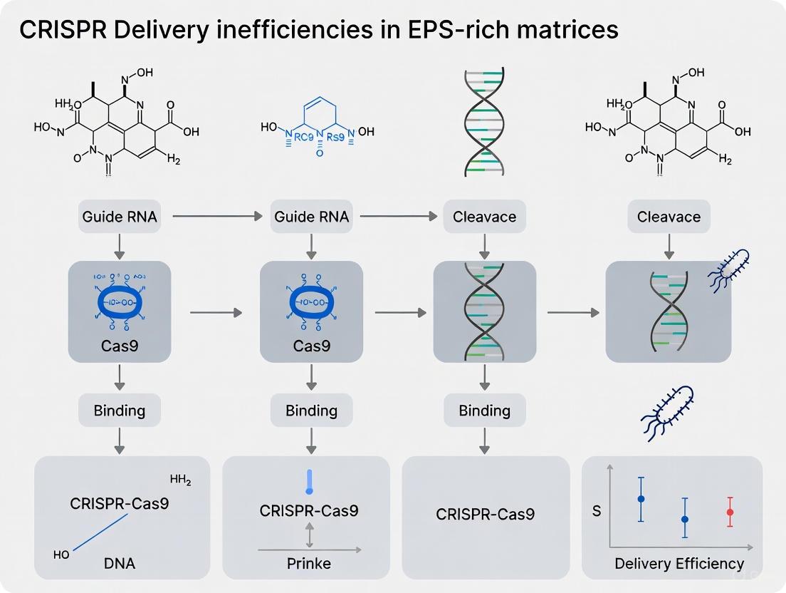
Overcoming the Barrier: Strategies for Efficient CRISPR Delivery in EPS-Rich Matrices
The therapeutic application of CRISPR-Cas9 technology is significantly hampered by inefficient delivery, particularly within extracellular polymeric substance (EPS)-rich matrices like bacterial biofilms and certain tumor microenvironments.
Overcoming the Barrier: Strategies for Efficient CRISPR Delivery in EPS-Rich Matrices
Abstract
The therapeutic application of CRISPR-Cas9 technology is significantly hampered by inefficient delivery, particularly within extracellular polymeric substance (EPS)-rich matrices like bacterial biofilms and certain tumor microenvironments. This article provides a comprehensive analysis for researchers and drug development professionals, exploring the fundamental challenges posed by EPS, reviewing innovative delivery platforms from viral vectors to nanoparticles, and offering practical optimization strategies. We further synthesize methods for validating delivery efficiency and editing success, presenting a consolidated framework to advance the translation of CRISPR-based therapies against resilient, matrix-protected cellular communities.
The EPS Barrier: Understanding the Fundamental Challenges to CRISPR Delivery
Frequently Asked Questions (FAQs) & Troubleshooting Guide
This support center is designed for researchers tackling the challenge of CRISPR-Cas9 delivery within EPS-rich matrices, a common barrier in biofilm and tumor microenvironment research. The FAQs below address specific, high-priority experimental issues.
FAQ 1: Why is my CRISPR-Cas9 editing efficiency so low in EPS-rich environments like bacterial biofilms?
The Challenge: EPS matrices act as a formidable physical and chemical barrier. The extracellular polymeric substances (EPS), including exopolysaccharides, proteins, and extracellular DNA (eDNA), significantly hinder the diffusion and uptake of CRISPR-Cas9 delivery vectors [1] [2].
Troubleshooting Steps:
- Characterize the EPS Barrier:
- Action: Quantify the major components of your specific EPS-rich matrix (e.g., uronic acid content for polysaccharides, eDNA concentration). High uronic acid and eDNA content are known to chelate particles and increase viscosity [1].
- Rationale: Understanding the specific composition allows for a targeted disruption strategy.
Pre-treat to Perturb the EPS Matrix:
- Action: Implement a mild pre-treatment step to disrupt the matrix without killing the cells. Consider using enzymes like:
- DNase I: Degrades eDNA, a key structural component [2].
- Dispersin B: Hydrolyzes polysaccharides in some biofilms.
- Protocol Note: Treat the biofilm with a suitable concentration of enzyme (e.g., 10-100 µg/mL DNase I in PBS) for 1-2 hours at 37°C prior to delivering the CRISPR-Cas9 system. Always include a no-enzyme control to confirm the effect is due to matrix disruption.
- Action: Implement a mild pre-treatment step to disrupt the matrix without killing the cells. Consider using enzymes like:
Switch to a Smaller Delivery Vehicle:
- Action: If using viral vectors (e.g., AAV), which can be filtered by the EPS mesh, consider switching to physically compact ribonucleoprotein (RNP) complexes of Cas9 protein and sgRNA [3].
- Rationale: Pre-assembled RNP complexes are significantly smaller than plasmids and can diffuse more readily through the matrix, often resulting in higher editing efficiency and reduced off-target effects [3].
FAQ 2: How can I accurately measure the penetration efficiency of my CRISPR delivery system into a 3D biofilm?
The Challenge: Visualizing and quantifying the distribution of CRISPR carriers within a dense, three-dimensional structure is non-trivial.
Troubleshooting Steps:
- Use a Fluorescent Proxy:
- Action: Label your delivery vehicle (e.g., lipid nanoparticles with a fluorescent dye, or use a fluorescently tagged Cas9 protein) with a stable, bright fluorophore (e.g., Cy5, ATTO 647N).
- Employ Confocal Microscopy and Quantitative Analysis:
- Action: Acquire z-stack images of the biofilm using a confocal laser scanning microscope. Use image analysis software (e.g., ImageJ/Fiji) to create a depth-intensity profile.
- Protocol Note:
- Embed and cryosection the biofilm if needed for higher resolution imaging.
- Measure the fluorescence intensity at various depths (e.g., every 10 µm) from the surface inward.
- Calculate the Penetration Efficiency as the percentage of fluorescence intensity retained at a target depth (e.g., 50 µm) compared to the surface intensity. A significant drop in intensity indicates poor penetration.
FAQ 3: What are the best strategies to increase HDR-mediated knock-in efficiency for precise gene editing in these challenging systems?
The Challenge: The non-homologous end joining (NHEJ) pathway dominates in most somatic cells, leading to stochastic indels rather than precise knock-in, and this can be exacerbated in slow-growing or stressed cells within EPS matrices [4].
Troubleshooting Steps:
- Synchronize the Cell Cycle:
- Action: For in vitro models, synchronize your cells at the S/G2 phase of the cell cycle, where the HDR machinery is most active [4].
- Protocol Note: Use chemical agents like thymidine or nocodazole for synchronization before delivering the CRISPR-Cas9 system and the donor DNA template.
Utilize HDR-Enhancing Reagents:
- Action: Supplement your culture medium with small molecule enhancers of HDR.
- Rationale: Compounds like RS-1 (an RAD51 stimulator) or L755507 can increase HDR efficiency by several-fold. A recommended starting concentration is 5-10 µM for RS-1, added during or shortly after CRISPR delivery [4].
Optimize the Donor Template Design:
- Action: For best results, use single-stranded oligodeoxynucleotides (ssODNs) with homology arms of 35-90 nt for point mutations, or double-stranded DNA templates with ~800 nt homology arms for larger insertions. Incorporating the silent CRISPR-resistant (SCR) mutations into the donor template can prevent re-cleavage after successful HDR [4].
The following tables consolidate key quantitative information from the literature to aid in experimental planning and comparison.
Table 1: Key Components and Functions of a Model Bacterial Biofilm (P. aeruginosa)
| Component | Approximate Percentage (%) | Primary Function in EPS Matrix |
|---|---|---|
| Exopolysaccharides | 1 - 2 | Maintains structural integrity and stability of the biofilm [2] |
| Proteins & Enzymes | < 1 - 2 | Provides surface colonization and structural stability [2] |
| Extracellular DNA (eDNA) | < 1 - 2 | Promotes biofilm formation and protects against host immune responses [2] |
| Water | Up to 97 | Hydrates the biofilm, preventing desiccation [2] |
Table 2: Comparison of CRISPR-Cas9 Delivery Strategies
| Delivery Method | Key Advantage | Key Disadvantage | Best Suited for EPS-Rich Environments? |
|---|---|---|---|
| Plasmid DNA | Simple to use, stable long-term expression [3] | Large size, difficult to diffuse; requires nuclear entry [3] | No |
| Cas9 mRNA + sgRNA | Reduced immune response vs. plasmid; no nuclear entry needed [3] | Lower stability; requires cytoplasmic translation [3] | Possibly |
| Ribonucleoprotein (RNP) | Smallest size; fastest action; lowest off-target effects [3] | Technically more complex to deliver; transient activity [3] | Yes |
The Scientist's Toolkit: Research Reagent Solutions
This table lists essential materials and their specific functions for conducting CRISPR-Cas9 experiments in EPS-rich contexts.
Table 3: Essential Reagents for CRISPR Work in EPS-Rich Matrices
| Research Reagent | Function & Application |
|---|---|
| DNase I | An enzyme that degrades extracellular DNA (eDNA), a critical scaffold component in many biofilms. Used to pre-treat matrices to enhance carrier penetration [2]. |
| Recombinant Cas9 Protein | The core component for forming pre-assembled RNP complexes. Using the protein directly avoids the need for transcription/translation, speeding up editing and facilitating delivery [3]. |
| RS-1 | A small-molecule agonist of the RAD51 protein. It stimulates the Homology Directed Repair (HDR) pathway, thereby increasing the efficiency of precise knock-in edits [4]. |
| Lipofectamine CRISPRMAX | A commercially available lipid nanoparticle transfection reagent specifically optimized for the delivery of CRISPR-Cas9 RNP complexes into a wide range of cell types. |
| Protospacer Adjacent Motif (PAM) | A short, sequence-specific motif (5'-NGG-3' for SpCas9) adjacent to the target DNA site that is essential for Cas9 recognition and binding [3]. |
Experimental Workflows & Signaling Pathways
The following diagrams, created using DOT language, illustrate key experimental workflows and molecular relationships relevant to this field.
CRISPR HDR Enhancement Workflow
CRISPR-Cas9 Molecular Mechanism
EPS Barrier to Delivery
The Composition and Physicochemical Properties of EPS that Hinder Drug Delivery
This technical support guide addresses the significant challenge that Extracellular Polymeric Substances (EPS) pose to the efficient delivery of therapeutic agents, with a specific focus on advancing CRISPR-based technologies. The EPS matrix, a complex biobarrier surrounding microbial communities and present in various biological systems, severely limits treatment efficacy for conditions ranging from bacterial biofilms to certain human diseases. This document provides researchers and drug development professionals with targeted troubleshooting guidance and experimental protocols to overcome these delivery obstacles.
Understanding the EPS Barrier
What is the EPS and why is it a major hurdle for drug delivery?
Extracellular Polymeric Substances (EPS) are a self-produced, hydrated polymeric network that forms a protective matrix around microbial cells [5]. This matrix is a primary reason for the inherent resistance of biofilm-associated infections to conventional antimicrobial therapies [5]. Its complex composition and structure create a formidable physical and chemical barrier that hinders the penetration of therapeutic molecules, including CRISPR/Cas systems.
FAQ: What are the key components of EPS and how do they specifically hinder drug delivery?
The following table summarizes the primary components of the EPS and their specific roles in hindering drug delivery.
Table 1: Key EPS Components and Their Roles in Hindering Drug Delivery
| EPS Component | Primary Function in Hindrance | Impact on Delivery Efficiency |
|---|---|---|
| Polysaccharides | Forms a dense physical hydrogel matrix; acts as a molecular sieve [5]. | Limits diffusion based on size and charge; filters large complexes like CRISPR/Cas RNPs [6]. |
| Proteins | Contributes to structural integrity and adhesion; can bind nonspecifically to delivery vectors [6]. | Causes sequestration of therapeutics; depletes the dose before it reaches target cells. |
| Extracellular DNA (eDNA) | Provides structural scaffold and contributes to negative charge of the matrix [5]. | Traps cationic delivery vectors via electrostatic interactions; increases viscosity. |
| Lipids & Other Polymers | Influences hydrophobicity and surface properties of the matrix [6]. | Creates a partition barrier for hydrophobic/hydrophilic drugs; reduces permeability. |
Quantitative Data on Delivery Challenges and Nano-Solutions
FAQ: Is there quantitative evidence showing how much EPS reduces efficacy?
Yes, studies demonstrate that biofilms can exhibit up to 1000-fold greater tolerance to antibiotics compared to free-floating (planktonic) cells, largely due to the barrier function of the EPS [5]. This underscores the critical need for delivery systems that can effectively penetrate this barrier.
FAQ: How can nanoparticle properties be optimized to overcome EPS barriers?
The design of nanoparticles (NPs) is critical for enhancing penetration through the EPS matrix. Key physicochemical properties can be tuned to improve performance, as shown in the table below.
Table 2: Optimizing Nanoparticle Properties for EPS Penetration
| Nanoparticle Property | Optimization Strategy | Effect on EPS Penetration |
|---|---|---|
| Size | Maintain small hydrodynamic diameter (typically < 100 nm). | Facilitates diffusion through the porous EPS network [7]. |
| Surface Charge | Use a neutral or slightly negative surface charge (e.g., via PEGylation). | Reduces non-specific binding to negatively charged eDNA and polysaccharides in the EPS [7]. |
| Surface Functionality | Graft with enzymes (e.g., DNase, dispersin B) or biofilm-disrupting peptides. | Actively degrades specific EPS components (e.g., eDNA), creating paths for diffusion [6]. |
| Hydrophobicity | Balance to ensure compatibility with the EPS microenvironment. | Prevents unwanted aggregation within the matrix and aids in crossing lipid-rich domains. |
Recent advances show that lipid-based nanoparticles with optimized cholesterol density can significantly improve mRNA uptake and endosomal escape, which is crucial for delivering CRISPR components [8]. Furthermore, liposomal Cas9 formulations have been shown to reduce Pseudomonas aeruginosa biofilm biomass by over 90% in vitro, while CRISPR-gold nanoparticle hybrids demonstrated a 3.5-fold increase in gene-editing efficiency compared to non-carrier systems [5].
Troubleshooting Common Experimental Problems
Problem: Low CRISPR editing efficiency in EPS-rich environments.
- Potential Cause 1: The CRISPR cargo (RNP, plasmid) is too large and/or is being sequestered by the EPS matrix.
- Solution: Consider using smaller Cas orthologs (e.g., Cas12f) and deliver as a Ribonucleoprotein (RNP) complex to avoid the need for transcription/translation steps required by viral delivery [9]. Utilize nanoparticle carriers like polymeric PLGA or lipid nanoparticles (LNPs) designed for enhanced penetration [7] [8].
- Potential Cause 2: The delivery vehicle aggregates within the EPS, failing to reach target cells.
- Potential Cause 3: The guide RNA (gRNA) is degraded before reaching the target site.
Problem: Inconsistent results when testing anti-biofilm formulations.
- Potential Cause: Use of oversimplified or non-standardized in vitro biofilm models that do not replicate the EPS complexity of natural biofilms.
- Solution: Employ established, mature biofilm models (e.g., flow-cell systems, colony biofilms) that allow for full EPS development. Characterize the EPS composition (e.g., via carbohydrate and protein assays, CLSM with EPS-binding dyes) of your specific model to better correlate with delivery outcomes [5].
Essential Research Reagent Solutions
The following table lists key reagents and their functions for developing and testing delivery systems against EPS barriers.
Table 3: Research Reagent Solutions for EPS and Delivery Studies
| Reagent / Material | Function in Experimental Workflow |
|---|---|
| Poly(lactic-co-glycolic acid) (PLGA) | A biodegradable polymer for creating nanoparticles that can encapsulate CRISPR components and cross biological barriers like the BBB, relevant for EPS-rich niches [7]. |
| Lipid Nanoparticles (LNPs) | Versatile carriers for mRNA and sgRNA; cholesterol content can be tuned to optimize cellular uptake and endosomal escape in target cells like dendritic cells [8]. |
| Extracellular Vesicles (EVs) | Natural, biocompatible delivery vectors with low immunogenicity; can be engineered for targeted delivery of CRISPR machinery [7] [10]. |
| DNase I | Enzyme used to degrade eDNA within the EPS, reducing matrix integrity and viscosity to enhance nanoparticle penetration [6]. |
| Concanavalin A (ConA) & Other Lectins | Fluorescently labeled lectins used in Confocal Laser Scanning Microscopy (CLSM) to visualize and quantify polysaccharide distribution in biofilms [5]. |
| SYTO Stains | Cell-permeant nucleic acid stains used in conjunction with CLSM to quantify bacterial cell biomass and spatial distribution within the EPS matrix [5]. |
Standard Experimental Protocol: Evaluating Nanoparticle Penetration through EPS
This protocol outlines a standard method for visually assessing the penetration efficiency of a nanoparticle formulation through a pre-formed biofilm.
Title: Protocol for Visualizing Nanoparticle Penetration in a Biofilm using Confocal Laser Scanning Microscopy (CLSM)
Objective: To quantify the distribution and penetration depth of a fluorescently labeled nanoparticle delivery vehicle within an EPS-rich biofilm.
Workflow Diagram:
Materials:
- Bacterial strain of interest.
- Relevant growth medium.
- Sterile glass-bottom dishes or coverslips.
- Fluorescently labeled nanoparticle formulation (e.g., labeled with Cy5, FITC).
- SYTO stain (e.g., SYTO 9 for live cells).
- Optional: Lectin probes (e.g., ConA-TRITC for polysaccharides).
- Confocal Laser Scanning Microscope.
Procedure:
- Biofilm Growth: Grow a mature biofilm (e.g., for 48-72 hours) on a sterile glass substrate under appropriate conditions.
- NP Incubation: Gently introduce the fluorescent nanoparticle suspension to the biofilm and incubate for a predetermined time (e.g., 2-4 hours).
- Washing: Carefully wash the biofilm with a buffer (e.g., PBS) to remove any nanoparticles that have not associated with or penetrated the biofilm.
- Staining: Stain the biofilm components. A common approach is to use SYTO 9 to stain all bacterial cells, providing a reference structure.
- Imaging: Acquire Z-stack images (optical sections from the top to the bottom of the biofilm) using a CLSM. Use appropriate laser lines and filters to avoid cross-talk between fluorescent signals.
- Analysis: Use image analysis software (e.g., ImageJ, Imaris) to:
- Measure the penetration depth of the nanoparticle signal relative to the total biofilm thickness.
- Analyze the co-localization of the nanoparticle signal with bacterial cells to infer delivery to targets.
Visualizing the EPS Barrier and Strategic Solutions
The following diagram synthesizes the primary challenges posed by the EPS and the corresponding strategic solutions discussed in this guide.
Diagram Title: EPS Barrier Mechanisms and Strategic Solutions
Frequently Asked Questions (FAQs)
FAQ 1: What are the primary barriers that hinder CRISPR cargo delivery within EPS-rich matrices, and how can they be overcome? The main barriers in EPS-rich matrices like bacterial biofilms are limited diffusion, nuclease degradation, and inefficient cellular uptake. The dense, anionic extracellular polymeric substance (EPS) physically blocks nanoparticle penetration, while nucleases in the environment can degrade CRISPR components before they reach target cells [11]. Overcoming these requires engineered delivery vehicles; nanoparticles, particularly lipid-based and metallic ones, can be designed to shield CRISPR machinery and enhance diffusion through these protective layers [11] [9]. Co-delivery strategies that combine CRISPR with biofilm-disrupting agents can also synergistically improve access to bacterial cells [11].
FAQ 2: My CRISPR editing efficiency is low in biofilm-forming bacteria. Is this due to poor cargo delivery, and how can I confirm this? Yes, low editing efficiency is frequently a delivery problem in biofilms. The protective biofilm matrix can reduce antibiotic efficacy by up to 1000-fold compared to planktonic cells, and it similarly impedes CRISPR cargo delivery [11]. To confirm, you can use a CRISPR system with a fluorescent reporter; low fluorescence in target cells indicates poor delivery. Furthermore, you can employ the GeneArt Genomic Cleavage Detection Kit to verify if cleavage is occurring on the endogenous genomic locus, which helps distinguish between delivery failure and functional failure of the CRISPR system itself [12].
FAQ 3: How can I protect CRISPR ribonucleoproteins (RNPs) from degradation during delivery? Encapsulating RNPs within nanoparticles offers significant protection. Lipid nanoparticles (LNPs) and gold nanoparticles form a physical barrier that shields the cargo from nucleases in the extracellular environment [11] [13] [9]. A recently developed strategy uses Lipid Nanoparticle Spherical Nucleic Acids (LNP-SNAs), where a dense shell of DNA further protects the CRISPR core and enhances stability [14]. Using RNP complexes instead of plasmid DNA also minimizes the time the component is exposed to and vulnerable to nucleases, as they are active immediately upon delivery [9].
FAQ 4: Which delivery systems are most effective for improving cellular uptake of CRISPR cargo in difficult-to-transfect cells? Viral vectors, like adeno-associated viruses (AAVs), are efficient but raise safety concerns regarding immunogenicity and insertional mutagenesis [13] [9] [15]. Among non-viral methods, spherical nucleic acids (SNAs) have demonstrated entry into cells up to three times more effectively than standard lipid nanoparticles and with less toxicity [14]. Gold nanoparticles and other inorganic carriers also show enhanced cellular uptake due to their tunable surface chemistry and ability to be functionalized with cell-penetrating peptides [11] [13].
Troubleshooting Guides
Problem 1: Limited Diffusion Through EPS-Rich Biofilms
Symptom: CRISPR cargo fails to penetrate biofilm mass, resulting in low or no gene editing in target bacterial cells.
Reason & Solution:
| Reason | Solution | Experimental Evidence / Protocol |
|---|---|---|
| Physical Barrier from EPS Matrix: The dense matrix of polysaccharides, proteins, and eDNA in biofilms acts as a molecular sieve, physically blocking the passage of CRISPR cargo [11]. | Use Nanoparticle Carriers: Engineer nanoparticles small enough to navigate the biofilm mesh. Functionalize their surface with positive charges or biofilm-degrading enzymes (e.g., DNase, dispersin B) to disrupt the matrix and improve penetration [11]. | Protocol: Liposomal Cas9 Formulation for Biofilm Penetration1. Formulate liposomes encapsulating Cas9-gRNA RNP complexes.2. Coat liposomes with a cationic polymer (e.g., chitosan) to facilitate interaction with the anionic EPS.3. Apply the formulation to a mature biofilm (e.g., P. aeruginosa) in vitro and incubate.4. Assess penetration via confocal microscopy using fluorescently tagged liposomes.5. Quantify efficacy by measuring biofilm biomass reduction; liposomal Cas9 has been shown to reduce biomass by over 90% [11]. |
| Lack of Targeting: Cargo does not actively navigate to bacterial cells within the biofilm. | Functionalize with Targeting Ligands: Conjugate nanoparticles with ligands, such as antibodies or peptides, that specifically bind to surface markers on the target bacterial species [11]. | Protocol: Assessing Diffusion with Fluorescent Reporters1. Prepare control (free cargo) and experimental (nanoparticle-loaded) samples, both tagged with a fluorescent dye.2. Apply samples to the surface of a mature biofilm grown in a flow cell or similar system.3. Over time, capture Z-stack images of the biofilm using Confocal Laser Scanning Microscopy (CLSM).4. Analyze fluorescence intensity at different biofilm depths to create a penetration profile and compare the two groups. |
Problem 2: Nuclease Degradation of CRISPR Components
Symptom: Guide RNA or Cas9 mRNA is degraded before reaching the target cell, leading to a loss of editing function.
Reason & Solution:
| Reason | Solution | Experimental Evidence / Protocol |
|---|---|---|
| Vulnerability of Nucleic Acids: Naked guide RNA and DNA are rapidly degraded by nucleases present in the extracellular environment or within cellular endosomes [9]. | Use Ribonucleoprotein (RNP) Complexes: Directly deliver pre-assembled complexes of Cas9 protein and gRNA. This avoids the need for transcription and translation, reducing the window of vulnerability [9]. | Protocol: Validating RNP Stability Against Nucleases1. Incubate purified RNP complexes (Cas9 + gRNA) with a serum-containing medium or a defined nuclease solution.2. At set time points, run samples on a gel to check for gRNA degradation.3. Compare with plasmid DNA or in vitro transcribed mRNA under the same conditions.4. Test functional stability by transferring the treated RNPs into cells and measuring cleavage activity with a Genomic Cleavage Detection Kit [12]. |
| Insufficient Cargo Protection: The delivery vehicle does not adequately shield its contents. | Select Protective Nanocarriers: Use nanoparticles known for high encapsulation efficiency and stability, such as gold nanoparticles or polymer-based NPs, which form a protective shell around the CRISPR machinery [11] [9]. | Protocol: Gold Nanoparticle RNP Delivery1. Synthesize or purchase gold nanoparticles (AuNPs) functionalized for RNP binding.2. Conjugate pre-assembled RNPs to the AuNPs via covalent or affinity binding.3. Treat target cells and measure gene-editing efficiency. Studies show AuNP carriers can enhance editing efficiency up to 3.5-fold compared to non-carrier systems [11]. |
Problem 3: Inefficient Cellular Uptake
Symptom: CRISPR cargo accumulates outside cells but does not enter, resulting in no editing.
Reason & Solution:
| Reason | Solution | Experimental Evidence / Protocol |
|---|---|---|
| Poor Internalization by Target Cells: Standard delivery vehicles like LNPs often get trapped in endosomal compartments and are degraded before releasing their cargo into the cytoplasm [14]. | Utilize Advanced Nanostructures: Employ Spherical Nucleic Acids (LNPs-SNAs). The dense, spherical DNA shell on these particles interacts with cell surface receptors, promoting rapid and efficient uptake and facilitating endosomal escape [14]. | Protocol: Testing Uptake with LNP-SNAs1. Synthesize LNP-SNAs with a core containing CRISPR RNP and a shell of dense, oriented DNA strands.2. Treat various cell types (e.g., primary cells, stem cells) with LNP-SNAs and standard LNPs, both carrying a fluorescent reporter.3. Analyze by flow cytometry or fluorescence microscopy to quantify the percentage of positive cells and mean fluorescence intensity. LNP-SNAs have shown a 3x improvement in cell entry and a 3x boost in editing efficiency [14]. |
| Low Transfection Efficiency: The method used is not optimal for the specific cell type. | Optimize Transfection and Enrich Transfected Cells: For difficult cell lines, optimize the transfection protocol using high-performance reagents (e.g., Lipofectamine 3000). To enrich for successfully transfected cells, introduce antibiotic resistance genes with the CRISPR cargo and apply selection pressure, or use Fluorescence-Activated Cell Sorting (FACS) if a fluorescent reporter is co-delivered [12]. | Protocol: Enriching Edited Cells via Antibiotic Selection1. Co-transfect your CRISPR construct (e.g., plasmid expressing Cas9 and gRNA) with a separate plasmid carrying an antibiotic resistance gene (e.g., puromycin).2. 24-48 hours post-transfection, add the appropriate antibiotic to the culture medium.3. Maintain selection for 3-7 days, replacing the medium with fresh antibiotic as needed.4. After selection, assay the surviving cell population for editing efficiency, which will be significantly enriched [12]. |
Table 1: Efficacy of Selected Nanoparticle Systems for Overcoming CRISPR Delivery Hurdles
| Delivery System | Target Challenge | Key Metric | Performance Result | Reference |
|---|---|---|---|---|
| Liposomal Cas9 Formulation | Limited Diffusion (Biofilm) | Biofilm Biomass Reduction | >90% reduction in P. aeruginosa biofilm in vitro [11] | |
| CRISPR-Gold Nanoparticle Hybrids | Cellular Uptake & Degradation | Gene-Editing Efficiency | 3.5-fold increase compared to non-carrier systems [11] | |
| Lipid Nanoparticle SNAs (LNP-SNAs) | Cellular Uptake & Endosomal Escape | Cell Internalization & Editing | 3x more effective cell entry & 3x higher editing efficiency; >60% improvement in precise HDR repair [14] | |
| Nanoparticle & Antibiotic Co-delivery | Limited Diffusion & Efficacy | Synergistic Antibacterial Effect | Superior biofilm disruption and synergistic antibacterial effects [11] |
Experimental Workflow and Pathway Diagrams
The Scientist's Toolkit: Research Reagent Solutions
Table 2: Essential Reagents for Addressing CRISPR Delivery Challenges
| Reagent / Kit Name | Function / Application | Key Benefit |
|---|---|---|
| Lipid Nanoparticle (LNP) Kits | Formulate and encapsulate CRISPR RNPs or nucleic acids for improved delivery and protection. | Shields cargo from nucleases; can be tailored for specific cell targeting [11] [14]. |
| GeneArt Genomic Cleavage Detection Kit | Detect and validate successful CRISPR-Cas9 cleavage at the target genomic locus. | Confirms whether delivery was successful and the CRISPR system is functionally active in cells [12]. |
| Gold Nanoparticles (AuNPs) | Serve as a non-viral carrier for conjugated CRISPR RNP complexes. | Enhances editing efficiency and stability; proven 3.5x increase in some studies [11]. |
| Spherical Nucleic Acids (LNPs-SNAs) | Advanced nanostructure for high-efficiency delivery of full CRISPR machinery. | Promotes superior cellular uptake and endosomal escape; boosts editing efficiency threefold [14]. |
| PureLink PCR Purification Kit | Purify DNA templates and PCR products for high-quality sequencing or downstream applications. | Ensures clean, concentrated DNA for accurate analysis and cloning steps [12]. |
Biofilm-associated infections represent a critical frontier in the challenge of CRISPR-Cas9 delivery. The extracellular polymeric substance (EPS) matrix of biofilms creates a formidable physical and chemical barrier that significantly reduces the efficacy of conventional delivery systems [5]. This dense matrix, composed of polysaccharides, proteins, lipids, and extracellular DNA, limits the penetration of antimicrobial agents and genetic tools by altering bacterial metabolism and creating diffusion barriers [16] [5]. For researchers developing CRISPR-based antimicrobials, understanding and overcoming these delivery inefficiencies is paramount for successful gene editing in biofilm-forming pathogens.
The inherent resistance mechanisms of biofilms can render CRISPR therapies ineffective if delivery systems cannot penetrate the EPS matrix and reach their intracellular targets. This case study examines the specific delivery challenges within EPS-rich environments and provides practical troubleshooting guidance for researchers working to advance CRISPR-based anti-biofilm strategies.
Troubleshooting Guide: FAQs on Biofilm Delivery Challenges
Q1: Why does my CRISPR-Cas9 system show significantly reduced efficiency against biofilm-forming bacteria compared to planktonic cultures?
The reduced efficiency stems from multiple biofilm-specific barriers:
- EPS Matrix Blockade: The dense extracellular polymeric substance (EPS) matrix physically impedes nanoparticle diffusion. Studies demonstrate that conventional lipid nanoparticles (LNPs) show 60-80% reduced penetration in mature Pseudomonas aeruginosa biofilms compared to free diffusion in liquid culture [5].
- Altered Bacterial Metabolism: Bacteria in biofilms often enter a metabolically dormant state, reducing cellular uptake mechanisms and rendering them less susceptible to genetic manipulation [5].
- Enzymatic Degradation: The biofilm microenvironment contains nucleases and proteases that can degrade CRISPR components before they reach target cells [9] [5].
Troubleshooting Steps:
- Quantify biofilm maturity: Use crystal violet staining or confocal microscopy to characterize biofilm density before CRISPR delivery experiments.
- Pre-treat with matrix-disrupting enzymes: Consider combining CRISPR delivery with DNase I (targeting eDNA) or dispersin B (targeting polysaccharides) to enhance penetration [5].
- Switch to smaller Cas variants: Utilize compact Cas12f (~2.5x smaller than SpCas9) to overcome size exclusion limitations [17] [18].
Q2: Which delivery vector shows highest efficacy for CRISPR delivery through EPS matrices?
Recent comparative studies indicate nanoparticle-based systems outperform viral vectors for biofilm penetration due to tunable surface properties and protection against degradation [9] [5].
Table: Delivery System Efficacy in Biofilm Models
| Delivery System | Editing Efficiency in Biofilms | Key Advantages | Documented Limitations |
|---|---|---|---|
| Gold Nanoparticles | 3.5x higher than non-carrier systems [5] | Surface-functionalization capability, photothermal properties | Potential cytotoxicity at high concentrations |
| Lipid Nanoparticles (LNPs) | 90% biofilm biomass reduction with liposomal Cas9 [5] | High payload capacity, biocompatibility | Endosomal entrapment can reduce efficiency |
| Adeno-associated Viruses (AAV) | <15% in dense biofilms [9] | High infectivity in planktonic cells | Size exclusion by EPS matrix, immunogenicity concerns |
| Polymeric Nanoparticles | 40-60% target gene knockout [9] | Sustained release profile, tunable degradation | Variable loading efficiency |
Recommended Protocol: Gold Nanoparticle-Mediated RNP Delivery
- Synthesize or purchase 20-40nm gold nanoparticles functionalized with polyethylenimine (PEI).
- Complex with pre-assembled Cas12a RNP (4:1 nanoparticle:RNP ratio) for 30 minutes at room temperature.
- Add 1mM EDTA to the biofilm culture to weaken matrix integrity prior to nanoparticle addition.
- Apply nanoparticle-RNP complexes at 50μg/mL final concentration.
- Utilize laser irradiation (670nm, 0.5W/cm², 5min) for photothermal release if using functionalized gold nanoparticles [5].
Q3: How can I validate successful CRISPR delivery and gene editing within biofilm structures?
Standard validation methods for planktonic cultures often fail in biofilms due to spatial heterogeneity and limited sampling efficiency.
Validation Workflow:
- Spatial analysis via FISH-CRISPR: Combine fluorescence in situ hybridization with Cas9-targeted sequencing to visualize editing events within different biofilm regions [5].
- Resistance gene monitoring: Track the loss of antibiotic resistance genes (e.g., bla, mecA) as evidence of successful editing using qPCR of biofilm homogenates.
- Confocal microscopy with reporter systems: Engineer target bacteria with GFP reporters under control of targeted promoters; successful editing eliminates fluorescence [5].
Q4: What strategies can enhance nanoparticle penetration through EPS matrices?
Surface functionalization is key to improving penetration:
- Matrix-binding peptides: Conjugate nanoparticles with EPS-binding peptides (e.g., γ9WGγ9 from Staphylococcus aureus) to facilitate matrix penetration [5].
- Enzyme conjugation: Covalently link matrix-degrading enzymes (e.g., alginate lyase for P. aeruginosa) to nanoparticle surfaces.
- Charge modulation: Use slightly cationic nanoparticles (+5 to +15mV zeta potential) to overcome anionic EPS components without excessive biofilm binding [9] [5].
Optimization Protocol:
- Characterize EPS composition of your specific biofilm model via FTIR or chemical analysis.
- Functionalize nanoparticles with targeted matrix-binding or degrading molecules.
- Test penetration using fluorescently labeled nanoparticles and confocal z-stack imaging.
- Measure editing efficiency at various biofilm depths via microdissection and PCR.
Q5: How do I differentiate between delivery failure and CRISPR machinery failure in biofilm experiments?
Use this diagnostic flowchart to isolate the failure point:
Diagnostic Experiments:
- Planktonic control: Test your CRISPR system on planktonic cells of the same bacterial strain.
- Penetration assay: Use fluorescently labeled nanoparticles or dextrans of similar size to your delivery system.
- Component stability: Recover delivery vehicles from biofilm exposure and analyze CRISPR component integrity via gel electrophoresis.
- Bacterial viability post-treatment: Ensure bacteria remain viable after nanoparticle treatment to distinguish between delivery failure and bactericidal effects.
The Scientist's Toolkit: Research Reagent Solutions
Table: Essential Reagents for CRISPR Delivery in Biofilm Research
| Reagent/Category | Specific Examples | Function & Mechanism | Biofilm-Specific Considerations |
|---|---|---|---|
| Compact Cas Proteins | SaCas9, Cas12f (Cas14), Cas12j | Smaller size enables better diffusion through EPS matrix | Cas12f (~400aa) shows 3.2x improved penetration vs SpCas9 (~1368aa) [17] [18] |
| RNP Complexes | Alt-R S.p. Cas9 Nuclease, custom guide RNA | Pre-formed protein-RNA complexes for immediate activity | 60% faster editing kinetics vs plasmid delivery; reduced off-target effects in heterogeneous biofilm environments [17] |
| Penetration-Enhanced Nanoparticles | Au-PEI NPs, Chitosan-Zn NPs, Lipid-PEG NPs | Tunable surface chemistry for matrix penetration | Au-PEI NPs show 3.5x higher editing efficiency in P. aeruginosa biofilms vs standard lipoplexes [5] |
| Matrix Disruption Agents | DNase I, Alginate lyase, Dispersin B | Degrades specific EPS components to create penetration channels | DNase I pre-treatment increases nanoparticle penetration by 45% in S. epidermidis biofilms [5] |
| Electroporation Enhancers | IDT Alt-R Cas9 Electroporation Enhancer | Carrier molecules that improve RNP delivery during electroporation | Specifically designed for hard-to-transfect cells; not for in vivo use [17] |
| Quorum Sensing Inhibitors | Furanones, AHL analogs | Disrupts bacterial communication to reduce EPS production | Can increase CRISPR accessibility by 30% when combined with nanoparticle delivery [5] |
Advanced Experimental Protocol: CRISPR-Nanoparticle Hybrid System for Biofilm Editing
This integrated protocol combines optimized delivery with validation methods specifically for biofilm models.
Materials:
- Pre-assembled Cas12a RNP complex (Alt-R CRISPR-Cas12a system)
- 20nm gold nanoparticles functionalized with PEI and alginate lyase
- Mature 72-hour biofilm of target bacteria
- Confocal microscopy dishes with glass bottoms
- SYTO 9 and propidium iodide for viability staining
Procedure:
- Biofilm Preparation: Grow biofilms for 72 hours under conditions optimal for your bacterial species. Characterize density via crystal violet staining or confocal microscopy.
Nanoparticle-RNP Complex Formation:
Biofilm Treatment:
Penetration Validation:
Editing Efficiency Assessment:
Expected Results:
- 40-60% editing efficiency in upper biofilm layers (0-20μm depth)
- 15-30% editing efficiency in middle layers (20-40μm depth)
- <10% editing efficiency in deepest layers (>40μm depth)
- 3.5x higher efficiency versus non-vectored RNP delivery [5]
The dense, heterogeneous nature of biofilm matrices presents unique delivery challenges that require specialized approaches beyond standard CRISPR protocols. Successful gene editing in biofilms depends on integrating multiple strategies: selecting appropriately sized CRISPR machinery, utilizing penetration-enhanced nanoparticles, and combining delivery with matrix-disruption techniques. The troubleshooting framework and experimental protocols provided here offer researchers a systematic approach to overcome these barriers and advance CRISPR-based applications against biofilm-associated infections.
As the field progresses, continued innovation in nanoparticle design and biofilm biology will further enhance our ability to precisely edit bacterial genomes within these complex communities, opening new avenues for combating persistent infections and antibiotic resistance.
Navigating the Maze: Delivery Platforms for EPS-Penetrating CRISPR Systems
Frequently Asked Questions (FAQs)
Q1: What are the primary challenges of using rAAV for CRISPR delivery in EPS-rich environments like bacterial biofilms?
EPS-rich matrices, such as those found in bacterial biofilms, present significant barriers to rAAV transduction. The extracellular polymeric substance (EPS) creates a physical barrier that can limit the penetration and diffusion of viral vectors, reducing their ability to reach target cells. Furthermore, the biofilm environment can neutralize vectors and reduce transduction efficiency. Combining rAAV with nanoparticle (NP) technology is an emerging strategy to overcome this, as NPs can enhance cellular uptake and protect the genetic cargo. One study noted that liposomal Cas9 formulations reduced P. aeruginosa biofilm biomass by over 90% in vitro, and CRISPR–gold nanoparticle hybrids demonstrated a 3.5-fold increase in gene-editing efficiency [5].
Q2: Which rAAV serotypes are most effective for transducing different cell types in the pulmonary system, a common site of biofilm-related infections?
The tropism of AAV serotypes varies significantly across different cell types in the lung. The table below summarizes the transduction efficiency of various serotypes in specific pulmonary epithelial cells, based on a 2024 study in mice [19].
| Target Cell Type in Lung | Highly Efficient AAV Serotypes |
|---|---|
| Airway Epithelium | AAV1, AAV4, AAV5, AAV6, AAV6.2, AAV-PHP.B, AAV-PHP.S |
| Club Cells | AAV1, AAV4, AAV5, AAV6, AAV6.2 |
| Ciliated Cells | AAV1, AAV4, AAV5, AAV6, AAV6.2, AAV-PHP.B |
| Alveolar Type I (AT1) Cells | AAV8, AAVrh10 |
| Alveolar Type II (AT2) Cells | AAV1, AAV5, AAV6, AAV6.2, AAV9, AAVie |
| Endothelial Cells | AAV1, AAV5, AAVie, AAV-PHP.B, AAV-PHP.eB, AAV-PHP.S |
Q3: How can I engineer the AAV capsid to improve its tropism for my specific cell type of interest?
Capsid engineering is a powerful method to enhance AAV tropism and evade immune responses. The main strategies are summarized in the diagram below, which outlines the workflow from goal identification to functional validation [20] [21].
Q4: What is a standard protocol for producing and purifying high-titer rAAV for in vivo experiments?
Below is a detailed protocol for helper-free rAAV production and purification, adapted from common laboratory practices [22] [23].
Step 1: Transfection of HEK293 Cells
- Cells: Use highly transfectable HEK 293T cells (e.g., AAVpro 293T Cell Line or HEK 293T/17 from ATCC).
- Plasmids: Co-transfect the cells with three plasmids using PEI or a similar transfection reagent:
- Rep/Cap Plasmid: Provides AAV replication and capsid proteins for the desired serotype.
- Helper Plasmid: Provides essential adenoviral genes (E1, E2a, E4, VA RNA) for AAV replication.
- rAAV Vector Plasmid: Contains your gene of interest flanked by AAV2 ITRs.
- Ratio: A common DNA mass ratio is 1:4:2 (Vector:Rep/Cap:Helper) [22].
Step 2: Harvest and Lysis
- 48-72 hours post-transfection, harvest both the cells and the culture media.
- Pellet the cells and resuspend them in a lysis buffer (e.g., AAVpro Extraction Solution). Perform freeze-thaw cycles or use a detergent-based lysis to release the viral particles.
Step 3: Purification
- Purify the viral vectors from the crude lysate. Avoid prolonged ultracentrifugation by using a commercial purification kit (e.g., AAVpro Purification Kit for all serotypes).
- Process: The kit typically uses affinity chromatography and can complete purification in approximately 4 hours.
- Yield: Standard yields range from 1x10^10 to 2.5x10^12 vector genomes (vg) per preparation, depending on scale [23].
Step 4: Titration and Quality Control
- Titration: Determine the viral titer (vg/mL) using quantitative PCR (qPCR) with primers targeting the AAV2 ITR region, which is common across serotypes when using AAV2 ITR-based vectors.
- Purity: Confirm purity by SDS-PAGE, which should show three major bands corresponding to the capsid proteins VP1, VP2, and VP3 [23].
Q5: What is a typical dosage for administering rAAV to mice intravenously?
Dosage depends on the target tissue, desired transduction level, and the specific AAV capsid. For systemic delivery in adult mice (6-8 weeks old), common doses are:
- AAV-PHP.eB: 1 × 10^11 to 5 × 10^11 vg per mouse [22].
- AAV-PHP.S: 3 × 10^11 to 1 × 10^12 vg per mouse [22]. For general animal experiments, a dose between 10^11 and 10^12 vg per animal is typical, but the optimal dose should be determined empirically for your specific application [23].
The Scientist's Toolkit: Key Research Reagent Solutions
| Item | Function/Benefit |
|---|---|
| HEK 293T Cells | A standard cell line used for high-titer rAAV production due to high transfection efficiency and constitutive expression of SV40 large T antigen [23]. |
| Helper-Free System | A plasmid system that provides adenoviral helper functions without the need for a live helper virus, improving safety and simplifying production [23]. |
| AAVpro Purification Kit | Enables fast (~4 hours) purification of AAV particles from any serotype without requiring ultracentrifugation [23]. |
| AAV Serotypes (e.g., PHP.B, PHP.eB) | Engineered capsids with enhanced central nervous system (CNS) tropism in specific mouse strains, though their efficacy depends on the host expressing the Ly6a receptor [22]. |
| AAV2 ITR-specific qPCR Titration Kit | Allows for accurate quantification of viral genome titer for any AAV serotype, as long as the vector genome is flanked by AAV2 ITRs [23]. |
| Selective Organ Targeting (SORT) LNPs | Engineered lipid nanoparticles that can be used as an alternative or complementary delivery vehicle to target specific tissues like the lung, spleen, and liver [24]. |
Advanced Concepts: Receptor-Mediated Tropism
The cellular entry of different AAV serotypes is governed by their interaction with specific cell surface receptors. Understanding these relationships is key to selecting or engineering the right vector. The following diagram illustrates the primary and co-receptors for several well-characterized serotypes [20].
Frequently Asked Questions (FAQs)
Q1: Why should I use non-viral nanocarriers for CRISPR delivery in EPS-rich research instead of viral vectors? Viral vectors, while efficient, present significant challenges for clinical translation, including immunogenicity, limited cargo capacity, and the risk of insertional mutagenesis [25] [26]. These issues are compounded in complex experimental environments. Non-viral nanocarriers offer a safer and more flexible alternative. They exhibit lower immunogenicity, can be produced at a larger scale, and their surface properties can be chemically modified to enhance penetration through physical barriers like Extracellular Polymeric Substance (EPS) matrices [5] [27]. For instance, one study demonstrated that liposomal Cas9 formulations reduced P. aeruginosa biofilm biomass by over 90% in vitro [5].
Q2: What are the key advantages of the Ribonucleoprotein (RNP) format for CRISPR delivery? Delivering the pre-assembled Cas9 protein and guide RNA as a Ribonucleoprotein (RNP) complex provides several critical advantages for both general use and specific applications in EPS-rich environments:
- Rapid Activity: The RNP is immediately active upon delivery, bypassing the need for transcription and translation. This leads to a shortened editing window, minimizing off-target effects and accelerating the experimental timeline [28] [26].
- Reduced Immunogenicity and Cytotoxicity: Unlike plasmid DNA, RNP delivery avoids the risk of genomic integration and triggers a lower immune response, which is crucial for sensitive in vivo applications [24] [29].
- Enhanced Precision: The transient nature of RNP activity is associated with higher editing precision and fewer off-target effects [28].
Q3: How can I improve the stability and editing efficiency of my CRISPR-nanocarrier formulations? Optimizing stability and efficiency involves strategic choices in both the CRISPR machinery and the nanocarrier composition:
- Use Thermostable Cas9 Variants: Employing engineered, thermostable Cas9 proteins, such as iGeoCas9, can significantly boost editing efficiency. These variants demonstrate superior stability and have shown >100-fold higher genome editing in cells and organs compared to their native forms, making them more resilient during the nanocarrier formulation process [29].
- Chemical Modification of Guides: Incorporating chemical modifications (e.g., 2’-O-methyl-3’-phosphonoacetate) into the guide RNA can protect it from nuclease degradation, enhancing its stability and overall editing efficiency [28].
- Surface Functionalization: Modifying the surface of nanocarriers with targeting ligands or tuning their charge can improve their interaction with and penetration into specific EPS-rich targets [5] [27].
Q4: What are the primary barriers that hinder efficient non-viral CRISPR delivery? Non-viral delivery must overcome multiple extracellular and intracellular barriers to be successful:
- Extracellular Barriers: These include nucleases and proteases in physiological fluids that can degrade the CRISPR cargo, as well as the physical obstruction of dense EPS or biofilm matrices that limit nanoparticle penetration [25] [5].
- Intracellular Barriers: Once internalized, nanocarriers are typically trapped in endosomes. To be functional, the CRISPR cargo must escape the endosome before degradation in the lysosome and then be transported into the cell nucleus [24] [28]. Gold nanoparticles, for example, leverage their surface charge to promote endosomal escape [30].
Troubleshooting Guide
This guide addresses common experimental problems encountered when using non-viral nanocarriers for CRISPR delivery.
Table 1: Troubleshooting Common Experimental Issues
| Problem | Potential Causes | Recommended Solutions |
|---|---|---|
| Low Editing Efficiency | 1. Inefficient cellular uptake2. Poor endosomal escape3. RNP/protein degradation4. Inefficient nuclear entry | 1. Confirm nanocarrier surface charge (zeta potential); use cationic lipids/polymers to promote cell binding [28] [27].2. Formulate LNPs with ionizable or pH-sensitive lipids (e.g., DOPE) to enhance endosomal disruption [28] [29].3. Use thermostable Cas9 variants (e.g., iGeoCas9) and chemically modified guide RNAs to improve cargo stability [29] [28]. |
| High Cytotoxicity | 1. Cytotoxic transfection reagents2. Persistent Cas9 expression3. Cationic nanocarrier toxicity | 1. Switch to RNP delivery for transient activity and reduced cell stress [26] [29].2. Optimize the nanoparticle-to-cell ratio; reduce the concentration of cationic lipids/polymers [27].3. Use biodegradable lipid materials, such as ester-based lipids, to improve biocompatibility [28] [26]. |
| Poor Penetration in EPS/ Biofilm Models | 1. Large nanocarrier size2. Non-specific binding to matrix3. Lack of targeting | 1. Optimize formulation to produce smaller nanoparticles (<100 nm) for better diffusion [5].2. Incorporate PEGylation (e.g., DMG-PEG2000) to create a stealth coating and reduce non-specific adhesion [28] [26].3. Functionalize nanocarrier surface with targeting ligands (e.g., peptides, antibodies) specific to biofilm components [5] [27]. |
| Low Nanocarrier Cargo Loading | 1. Incorrect N/P ratio (for nucleic acids)2. Unstable complex formation3. Cargo size too large | 1. For plasmid DNA/mRNA, systematically adjust the ratio of amine groups (N) in the carrier to phosphate groups (P) in the nucleic acid [27].2. For RNP loading, leverage charge interactions; gold nanoparticles can be engineered with specific surface chemistry to bind RNPs stably [30] [26]. |
Quantitative Performance Data
The following table summarizes key performance metrics for different non-viral nanocarriers as reported in recent literature, providing a benchmark for your own experimental systems.
Table 2: Performance Metrics of Non-Viral Nanocarriers for CRISPR Delivery
| Nanocarrier Type | CRISPR Cargo Format | Target / Model System | Reported Efficiency | Key Findings / Mechanism |
|---|---|---|---|---|
| Lipid Nanoparticles (LNPs) | iGeoCas9 RNP | Mouse Lung (SFTPC gene) | ~19% editing in vivo [29] | Uses biodegradable, cationic lipids for efficient RNP delivery to non-liver tissues. |
| Lipid Nanoparticles (LNPs) | Cas9 mRNA & sgRNA | Mouse Liver (PCSK9 gene) | ~60% editing in vivo [26] | Employs amino-ester-derived lipid-like nanomaterials (e.g., FTT lipids) [26]. |
| Gold Nanoparticles | Cpf1 RNP | Human Blood Stem Cells (CCR5 gene) | 10-20% editing in vitro [30] | Spherical gold nanoparticles facilitate endosomal escape via charge interaction; showed high viability. |
| Lipid Nanoparticles (LNPs) | Cas9 RNP | HEK293-GFP Reporter Cells | ~70% GFP knockout in vitro [26] | Formulated with bioreducible lipid-like materials for enhanced cytoplasmic release. |
| CRISPR-Gold (AuNP) | Cas9 RNP | Mouse Brain (mGluR5 gene) | 40-50% reduction in protein/mRNA [26] | Cationic arginine gold nanoparticles (ArgNPs) enable efficient RNP delivery in vivo. |
| Liposomal Formulations | Cas9 RNP | P. aeruginosa Biofilm (in vitro) | >90% reduction in biofilm biomass [5] | Liposomes enhance penetration of the biofilm EPS matrix and delivery of antimicrobial CRISPR cargo. |
Experimental Workflow & Protocol
The diagram below illustrates a generalized workflow for developing and testing a nanoparticle-based CRISPR delivery system for challenging EPS-rich environments.
Detailed Protocol: Formulating and Testing Gold Nanoparticles for RNP Delivery
This protocol is adapted from studies demonstrating successful RNP delivery using gold nanoparticles (AuNPs) to edit blood stem cells [30] [26].
Objective: To synthesize, characterize, and functionally validate gold nanoparticles as carriers for CRISPR RNP delivery.
Materials:
- Chloroauric acid (HAuCl₄)
- Reducing agent (e.g., sodium citrate)
- Custom synthetic guide RNA (sgRNA) with optional chemical modifications
- Purified Cas9 protein (or alternative nuclease like Cpf1)
- Cell culture reagents and target cell line (e.g., HEK293, primary stem cells)
- Transfection medium (e.g., serum-free Opti-MEM)
- Gel electrophoresis apparatus for validation
Methodology:
- Synthesis of Gold Nanoparticles (AuNPs):
- Prepare gold ions by suspending chloroauric acid in deionized water.
- Reduce the gold ions using a citrate reduction method to form spherical AuNPs of a defined size (e.g., ~15-30 nm). The size can be controlled by the citrate-to-gold ratio [30].
Formation of RNP Complex:
- Pre-complex the purified Cas9 protein with the sgRNA at a molar ratio of 1:1.2 (Cas9:sgRNA) in a suitable buffer. Incubate at 37°C for 10-15 minutes to form the active RNP complex.
Loading RNP onto AuNPs:
- Mix the pre-formed RNP complex with the synthesized AuNPs.
- The loading relies on electrostatic interactions and surface chemistry. The surface charge of the AuNPs is engineered to be positive to attract the negatively charged phosphate backbone of the RNP. Researchers may make "small molecular modifications to prevent the different parts from repelling one another" [30].
- Incubate the mixture for 30-60 minutes at room temperature to allow stable complex formation.
Characterization of AuNP-RNP Complexes:
- Size and Zeta Potential: Use Dynamic Light Scattering (DLS) to measure the hydrodynamic diameter and polydispersity index (PDI) of the complexes. Measure the zeta potential to confirm a surface charge that promotes cellular uptake.
- Loading Efficiency: Analyze the complexes using gel shift assays or quantify unbound RNP via spectrophotometry to determine loading efficiency [26].
Functional Validation in Cell Culture:
- Seed target cells in a 24-well or 48-well plate so they are 60-80% confluent at the time of transfection.
- Wash cells with PBS and replace medium with serum-free transfection medium.
- Add the formulated AuNP-RNP complexes to the cells. A typical RNP dose may range from 1-10 µg per well, which must be optimized.
- Incubate for 4-6 hours, then replace the transfection medium with complete growth medium.
- Harvest cells 48-72 hours post-transfection and analyze editing efficiency using T7 Endonuclease I assay, TIDE analysis, or next-generation sequencing.
The Scientist's Toolkit: Research Reagent Solutions
Table 3: Essential Materials for Nanoparticle-Mediated CRISPR Delivery
| Reagent / Material | Function / Role | Specific Examples |
|---|---|---|
| Ionizable Lipids | Core component of LNPs; enables encapsulation and endosomal escape via protonation at acidic pH. | DLin-MC3-DMA, LP01, SM-102 [28] [26] [29] |
| Helper Lipids | Stabilizes the LNP structure and promotes fusion with endosomal membranes. | Cholesterol, 1,2-dioleoyl-sn-glycero-3-phosphoethanolamine (DOPE) [28] [26] |
| PEGylated Lipids | Provides a "stealth" coating to reduce non-specific binding, improve stability, and prevent aggregation. | DMG-PEG2000, C16-PEG2000-ceramide [28] [26] |
| Cationic Polymers | Condenses nucleic acid cargo (plasmid, mRNA) via electrostatic interactions for polyplex formation. | Polyethylenimine (PEI), Chitosan [24] [26] |
| Gold Nanoparticles (AuNPs) | Inorganic core for assembling RNP complexes; facilitates endosomal escape. | Citrate-capped AuNPs, Cationic arginine AuNPs (ArgNPs) [30] [26] |
| Thermostable Cas9 | Engineered nuclease with enhanced stability, improving RNP resilience during formulation and delivery. | iGeoCas9 (evolved variant) [29] |
| Chemically Modified gRNA | Enhances stability against nucleases and can improve editing efficiency and reduce off-target effects. | 2’-O-methyl-3’-phosphonoacetate (MS) modified crRNA [28] |
Troubleshooting Guide: FAQs on Electroporation for CRISPR Delivery
FAQ 1: My electroporation experiment is yielding low CRISPR editing efficiency. What are the key parameters I should optimize?
Low editing efficiency is a common challenge. To address this, you should systematically optimize several key electroporation parameters. The table below summarizes critical parameters and their optimization strategies.
Table 1: Key Electroporation Parameters for CRISPR Efficiency
| Parameter | Impact on Efficiency | Optimization Strategy |
|---|---|---|
| Pulse Amplitude/Voltage | Determines the electric field strength for membrane permeabilization; too low is ineffective, too high causes cytotoxicity [31]. | Titrate voltage to find the balance between effective editing and cell viability [31]. |
| Pulse Duration & Number | Affects the extent and duration of membrane permeability [32]. | Use a Design of Experiments (DoE) approach to screen combinations of phase duration and pulse number [32]. |
| Cargo Form | Influences onset of activity, duration, and off-target effects [24]. | Use Cas9 Ribonucleoprotein (RNP) complexes for immediate activity and reduced off-target effects compared to plasmid DNA [24]. |
| Cell Health & Type | Primary and sensitive cells may require gentler protocols [32]. | Use high-density microelectrode arrays (MEAs) for gentler, more targeted electroporation on adherent cells [32]. |
FAQ 2: I am working with bacterial biofilms, which have a protective EPS matrix. How can I improve electroporation efficacy in these EPS-rich environments?
The EPS matrix significantly hinders macromolecule penetration. A promising strategy is to combine electroporation with nanoparticle carriers.
- Synergistic Action: Nanoparticles, such as gold or lipid nanoparticles, can be loaded with CRISPR-Cas9 components (like RNPs) and act as carriers [5]. The electric pulses help these nanocarriers penetrate the protective biofilm matrix and facilitate uptake by bacterial cells [5].
- Enhanced Efficacy: One study demonstrated that liposomal Cas9 formulations reduced Pseudomonas aeruginosa biofilm biomass by over 90% in vitro [5]. Furthermore, CRISPR-gold nanoparticle hybrids showed a 3.5-fold increase in gene-editing efficiency compared to non-carrier systems [5].
FAQ 3: I observe high cell death after electroporation. How can I reduce cytotoxicity?
Cell toxicity is often a consequence of harsh electroporation conditions.
- Mitigate Electrical Stress: Optimize electrical parameters by starting with lower voltages or fewer pulses and titrating upwards [31]. Using a Cas9 protein with a nuclear localization signal can enhance targeting efficiency, potentially allowing for lower, less toxic doses [31].
- Gentle Technologies: Planar microelectrode arrays (MEAs) with subcellular-sized electrodes can achieve high efficiency with reduced toxicity by affecting only a limited patch of the cell membrane [32].
- Constant Power Delivery: Recent research indicates that applying constant power pulsed electric fields (cpPEFs), instead of constant voltage, can achieve more consistent and predictable electroporation outcomes, potentially reducing unwanted cell death caused by variations in sample conductivity [33].
FAQ 4: How can I ensure consistent electroporation results when my samples have variable conductivity (e.g., different growth media or tissue types)?
Sample conductivity is a key variable that affects the electric field distribution and, thus, the outcome.
- Adopt Constant Power Pulsed Electric Fields (cpPEFs): Traditional constant-voltage electroporation is highly sensitive to conductivity variations. A 2025 study demonstrated that the threshold for electroporation is a power-density-driven phenomenon [33]. By applying pulses at a constant power level, researchers achieved consistent reversible and irreversible electroporation areas despite significant variations in extracellular conductivity [33]. This method better predicted treatment efficacy in vivo compared to using voltage or current alone [33].
Experimental Protocols & Data Presentation
Protocol: Optimizing mRNA Transfection via High-Definition Electroporation (HD-EP)
This protocol is adapted from a study achieving 98% transfection efficiency in primary fibroblasts using a CMOS HD-EP chip [32].
1. Chip Preparation:
- Utilize a CMOS HD-EP chip comprising clusters of thousands of individually addressable microelectrodes (e.g., 5 μm or 8 μm in diameter) [32].
- Seed cells directly onto the chip surface and allow them to adhere and grow to the desired confluency [32].
2. Design of Experiments (DoE) Screening:
- To efficiently find optimal conditions, vary multiple parameters simultaneously in a highly parallelized screening experiment on the chip. The tested parameters included [32]:
- Electrode size
- Pulse amplitude (V)
- Phase duration (ms)
- Interval between pulses (ms)
- Number of pulses
3. Electroporation and Analysis:
- Apply different pre-defined pulse train conditions to different electrode clusters on the same chip [32].
- Transfert with an mCherry-encoding mRNA and incubate cells post-electroporation [32].
- Quantify transfection efficiency by measuring mCherry fluorescence, for example, via fluorescence microscopy or flow cytometry [32].
- Fit a multiple linear regression model to the efficiency data to estimate the effect of each parameter and extract optimal conditions [32].
Protocol: Combining Nanoparticles with Electroporation for Biofilm Editing
This methodology outlines the use of nanoparticle-CRISPR complexes for targeting biofilm-associated bacteria [5].
1. Prepare CRISPR-Nanoparticle Complexes:
- Complex Formation: Formulate CRISPR-Cas9 ribonucleoproteins (RNPs) with guide RNA (gRNA) targeting specific bacterial resistance or virulence genes [5].
- Encapsulation/Conjugation: Encapsulate the RNPs within or conjugate them to nanoparticles. Studies have used:
2. Biofilm Treatment:
- Grow mature biofilms of the target bacterium (e.g., Pseudomonas aeruginosa) in vitro [5].
- Apply the CRISPR-nanoparticle complexes to the biofilm [5].
- Subject the biofilm to optimized electroporation pulses. The pulses enhance the penetration of the nanoparticles through the EPS matrix and promote cellular uptake [5].
3. Assessment of Biofilm Disruption:
- Biomass Quantification: Measure the reduction in biofilm biomass using crystal violet staining or confocal microscopy [5]. (e.g., >90% reduction with liposomal Cas9 [5]).
- Editing Efficiency: Assess the disruption of target genes using genotyping assays (e.g., T7 endonuclease I assay, sequencing) [31].
- Viability Assessment: Use cell viability assays to confirm the loss of bacterial survival due to successful gene editing [5].
The Scientist's Toolkit
Table 2: Essential Reagents and Materials for Electroporation-based CRISPR Delivery
| Item | Function/Description | Application Note |
|---|---|---|
| CRISPR RNP Complex | Pre-complexed Cas9 protein and guide RNA. The preferred cargo for its immediate activity and reduced off-target effects [24]. | Design gRNA for high specificity to minimize off-target editing [31]. |
| Electroporator with Parameter Control | Device capable of delivering square-wave pulse trains with adjustable voltage, duration, and pulse number. | For advanced applications, systems capable of constant power delivery (cpPEF) can improve consistency [33]. |
| Electroporation Cuvettes / MEA Chips | Cuvettes with fixed electrodes for cell suspensions; Microelectrode Arrays for adherent cells. | High-density MEAs enable spatially resolved, high-efficiency transfection of adherent cells with low toxicity [32]. |
| Nanoparticle Carriers (e.g., Gold, Liposomal) | Acts as a protective carrier for CRISPR components, enhancing stability and delivery, especially in complex environments like biofilms [5]. | Gold nanoparticles can boost editing efficiency by over 3-fold in biofilm models [5]. |
| Cell Viability Assay Kits | To quantify cytotoxicity post-electroporation (e.g., MTT, Calcein AM). | Essential for titrating parameters to balance high efficiency with acceptable cell survival [31]. |
Workflow and Conceptual Diagrams
CRISPR Electroporation Optimization Workflow
Strategy for Biofilm CRISPR Delivery
Troubleshooting Guides and FAQs
This technical support center provides targeted solutions for researchers encountering challenges while using Virus-Like Particle (VLP) and SORT molecule hybrid systems to deliver CRISPR-Cas9 through EPS-rich matrices, such as bacterial biofilms.
Frequently Asked Questions (FAQs)
Q1: What are the primary advantages of using VLPs for CRISPR delivery in EPS-rich environments?
VLPs offer a unique combination of high transduction efficiency, similar to viral vectors, and transient CRISPR component expression, which minimizes off-target effects and cell toxicity [34]. Their biosynthetic nature helps avoid the inflammation sometimes associated with synthetic nanoparticles, and their surface can be reprogrammed for specific cell tropism, enabling targeted delivery within complex microbial communities [35].
Q2: My VLP-SORT system shows low gene editing efficiency in the target biofilm population. What could be wrong?
Low editing efficiency can stem from several factors. First, the EPS matrix can create a significant physical barrier to diffusion. Second, the CRISPR-cargo packaging efficiency into your VLPs might be suboptimal. Third, the SORT molecule functionalization on the VLP surface may not be effectively overcoming the EPS barrier. Ensure you are using high-titer VLP preparations and confirm the successful conjugation and functionality of SORT molecules. The table below summarizes common issues and initial checks.
Table: Quick Diagnostics for Low Editing Efficiency
| Observed Problem | Potential Root Cause | Initial Diagnostic Check |
|---|---|---|
| Low editing efficiency | EPS barrier impedes delivery | Measure VLP penetration fluorescence assay |
| Low editing efficiency | Poor cargo packaging in VLPs | Quantify p24 & Cas9 via Western Blot [35] |
| Low editing efficiency | Inactive SORT molecules | Validate conjugation efficiency & activity |
| High cell toxicity | VLP or SORT component concentration too high | Perform a dose-response titration experiment |
Q3: How can I minimize off-target effects when using this hybrid delivery system?
The key advantage of VLPs is their ability to deliver pre-assembled Cas9-gRNA ribonucleoprotein (RNP). RNP has a short intracellular lifespan, drastically reducing the time window for off-target cleavage [35]. Furthermore, using high-fidelity Cas9 variants and carefully designing specific gRNAs with minimal predicted off-target sites are critical steps [31].
Q4: Are there specific immune response concerns with repeated VLP administration?
Yes, immune responses can be a concern. Studies show that while VLP-based RNP delivery elicits a significantly lower anti-Cas9 antibody response compared to lentiviral vectors that express Cas9 long-term, there can still be a noticeable immune reaction against the viral capsid components (e.g., p24) [35]. The use of different serotypes or engineering the VLP surface may be necessary for repeated dosing.
Troubleshooting Common Experimental Issues
Problem: Inconsistent VLP Production Yields
- Potential Cause 1: Suboptimal transfection efficiency of packaging plasmids.
- Solution: Use high-quality plasmid DNA and a robust transfection reagent. Validate transfection efficiency with a GFP-reporting plasmid.
- Potential Cause 2: Instability of the packaged cargo (e.g., mRNA, RNP).
- Solution: Incorporate cargo-stabilizing elements into your design. For RNPs, ensure the MS2-MCP system is correctly implemented for efficient Cas9 protein packaging [35] [34].
Problem: Failure in Targeted Delivery to Cells within an EPS-rich Biofilm
- Potential Cause: The native VLP tropism is unsuited for the target bacterial species, and the EPS matrix is sequestering or blocking the particles.
- Solution: Re-pseudotype the VLPs with envelopes that favor your target cell type [35]. Functionally couple SORT molecules to the VLP surface that are known to interact with or degrade specific EPS components to enhance penetration [36].
Problem: High Cytotoxicity Observed Post-Treatment
- Potential Cause 1: The concentration of VLPs or SORT molecules is too high.
- Solution: Perform a careful titration of both components to find a balance between delivery efficiency and cell viability [31].
- Potential Cause 2: Cytotoxicity from long-term, high-level expression of Cas9 nuclease.
- Solution: Switch to a VLP system that delivers pre-assembled RNP instead of Cas9-encoding DNA or mRNA. RNP delivery leads to rapid editing and clearance of the nuclease, minimizing persistent expression and associated toxicity [35].
Experimental Protocols
Protocol 1: Production of CRISPR RNP-Loaded VLPs
This protocol is adapted from recent studies for generating VLPs that package the Cas9 RNP complex [35].
Key Reagents:
- Plasmid encoding Gag protein (e.g., from lentivirus)
- Plasmid encoding MS2 coat protein fused to Gag
- Plasmid for VSV-G envelope protein
- Plasmid expressing Cas9 protein
- Plasmid expressing gRNA with incorporated MS2 stem loops
Methodology:
- Cell Culture: Seed HEK-293T cells in appropriate culture vessels to reach 70-80% confluency at the time of transfection.
- Plasmid Transfection: Co-transfect the cells with the following plasmid combination using a standard calcium phosphate or PEI-based method:
- Gag-Pol plasmid (with D64V integrase mutation for safety)
- VSV-G envelope plasmid
- MS2-fused Gag plasmid
- Cas9 expression plasmid
- gRNA-MS2 stem loop plasmid
- VLP Harvest: 48-72 hours post-transfection, collect the cell culture supernatant.
- VLP Purification: Concentrate the VLPs from the supernatant by ultracentrifugation through a 20% sucrose cushion at 100,000 × g for 2 hours.
- Quality Control: Resuspend the VLP pellet in PBS or a suitable buffer. Quantify the yield using a p24 ELISA kit and verify the presence of packaged Cas9 protein via Western blotting.
Protocol 2: Functionalizing VLPs with SORT Molecules for Enhanced Biofilm Penetration
This protocol outlines a strategy to conjugate SORT molecules (e.g., EPS-degrading enzymes) to the VLP surface.
Key Reagents:
- Purified VLPs
- SORT molecule (e.g., a specific glycoside hydrolase)
- Biotinylation reagent (e.g., NHS-PEG4-Biotin)
- Streptavidin (if using a biotin-streptavidin bridge)
- Cross-linker (e.g., SMPH)
Methodology:
- VLP Surface Activation: Gently mix the purified VLPs with a mild biotinylation reagent to label surface proteins. Remove excess biotin using a desalting column.
- SORT Molecule Preparation: If necessary, engineer or modify the SORT molecule to include a functional group (e.g., an amine) reactive with your chosen cross-linker.
- Conjugation: Incubate the biotinylated VLPs with streptavidin, followed by incubation with the biotinylated SORT molecule. Alternatively, use a heterobifunctional cross-linker like SMPH to directly link amine groups on the VLP surface to the SORT molecule.
- Purification: Purify the conjugated VLPs from unbound SORT molecules via size-exclusion chromatography or a second round of ultracentrifugation.
- Validation: Confirm successful conjugation using techniques like ELISA (to detect the SORT molecule) and a functional assay to verify that the SORT molecule retains its EPS-modifying activity post-conjugation.
System Workflow and Signaling Pathways
VLP-SORT System Workflow for Biofilm Editing
Research Reagent Solutions
Table: Essential Reagents for VLP-SORT-CRISPR Experiments
| Reagent / Material | Function / Role in the System | Key Consideration |
|---|---|---|
| Lentiviral Gag-Pol Plasmid | Provides structural and enzymatic backbone for VLP assembly. | Use an integrase-deficient version (D64V) for enhanced safety [35]. |
| MS2 Coat Protein & Stem Loops | Facilitates specific packaging of Cas9 RNP into VLPs via RNA-protein interaction [35]. | Ensure stem loops are inserted at positions that do not interfere with gRNA function. |
| VSV-G Envelope Plasmid | Provides broad tropism pseudotype for initial VLP entry. | Can be replaced with other envelopes for specific targeting. |
| SORT Molecules | Enhances penetration through the EPS matrix and can aid in specific targeting. | Selection is critical (e.g., EPS-degrading enzymes, targeting peptides) [36]. |
| Cas9 Protein & gRNA | The core gene-editing machinery delivered as a pre-assembled RNP. | Using high-fidelity Cas9 variants and highly specific gRNAs reduces off-target effects [31]. |
| Ultracentrifugation Equipment | Essential for purifying and concentrating VLP preparations from cell culture media. | Maintain cold temperatures to preserve VLP integrity. |
For researchers aiming to combat persistent biofilms, the extracellular polymeric substance (EPS) matrix presents a formidable delivery barrier for CRISPR-based antimicrobials. This protective microbial environment, rich in polysaccharides, proteins, and DNA, significantly impedes the transport of therapeutic cargoes, leading to inconsistent editing outcomes and experimental failure [6]. Selecting the optimal cargo format—plasmid DNA (pDNA), messenger RNA (mRNA), or ribonucleoprotein (RNP)—is therefore not merely a preliminary step but a critical determinant of success. This guide provides a structured, troubleshooting-focused overview of these three primary cargo systems to help you navigate the complexities of CRISPR delivery within EPS-rich contexts, enabling more precise and efficient genome editing in your biofilm research.
Cargo Format Comparison: Mechanisms and Workflows
The journey of each cargo type from cellular entry to functional gene editing follows a distinct pathway, which directly impacts its performance in dense EPS matrices. The following diagram illustrates the key mechanisms and intracellular workflows for the three main CRISPR cargo formats.
Comparative Performance Data and Selection Table
Understanding the quantitative and qualitative performance of each cargo format is essential for experimental design. The table below summarizes key characteristics, supported by data from recent studies.
Table 1: Quantitative Comparison of CRISPR Cargo Formats
| Parameter | Plasmid DNA (pDNA) | mRNA | Ribonucleoprotein (RNP) |
|---|---|---|---|
| Time to Onset of Activity | Slow (24-48h); requires transcription and translation [37] | Moderate (4-8h); requires only translation [38] [37] | Fastest (<2-4h); immediately active [24] [37] |
| Editing Efficiency | Variable; can be high but risk of off-targets [38] | High; lower off-targets than pDNA [38] [37] | Highest reported; superior specificity and efficiency [37] |
| Duration of Activity | Prolonged (days-weeks); risk of immune response and off-target effects [38] | Short (1-3 days); transient expression reduces off-target risk [38] | Shortest (~24h); most transient, minimizing off-target effects [24] |
| Stability in EPS/Delivery | High inherent stability; but large size hinders diffusion [6] | Low inherent stability; requires protection (e.g., LNPs) [39] [40] | Moderate stability; pre-assembled complex is more compact [37] |
| Immunogenicity Risk | High; can trigger innate immune sensors [38] | Moderate; immunogenicity can be engineered [39] [38] | Lowest; avoids nucleic acid-based immune activation [37] |
| Key Advantage | Simplicity of construction and low cost [37] | No risk of genomic integration; faster than pDNA [39] [38] | Highest precision, immediate activity, and low off-targets [24] [37] |
| Primary Challenge | Low efficiency in hard-to-transfect cells; size; off-targets [37] | Instability, requiring sophisticated encapsulation [39] [38] | Difficult production and scalable in vivo delivery [38] [37] |
Troubleshooting Guide: Addressing Common Experimental Hurdles
This section addresses specific, frequently encountered problems in CRISPR cargo delivery, with targeted solutions for research in EPS-rich environments.
FAQ 1: Why is my gene editing efficiency consistently low in mature biofilm models, even with high-quality guides?
- Problem: The dense, anionic EPS matrix acts as a diffusion barrier and can degrade or sequester CRISPR cargoes before they reach target cells [6].
- Solutions:
- Switch Cargo Format: If using pDNA, switch to the more compact and pre-assembled RNP complexes, which can diffuse more readily [37].
- Optimize Delivery Vehicle: Use delivery vectors with surface properties that counteract EPS interactions. For LNPs, incorporate cationic or PEGylated lipids to improve diffusion and stability [24] [40]. For polymeric nanoparticles, use bioreducible cationic polymers that remain stable in the extracellular space but release cargo upon cellular entry [41] [37].
- Employ EPS-Disrupting Adjuvants: Co-deliver your CRISPR cargo with EPS matrix-degrading enzymes (e.g., DNase I, dispersin B) to temporarily disrupt the biofilm structure and improve penetration [6].
FAQ 2: I am observing high cytotoxicity and off-target effects with my current system. How can I improve precision?
- Problem: Prolonged expression of the Cas9 nuclease increases the probability of off-target editing and can trigger cellular stress responses [38] [37].
- Solutions:
- Adopt RNP Delivery: RNP complexes offer the most transient activity, drastically reducing off-target effects and cytotoxicity compared to pDNA and mRNA [24] [37].
- Use High-Fidelity Cas Variants: If pDNA or mRNA is necessary, utilize engineered high-fidelity Cas9 variants (e.g., SpCas9-HF1) encoded in your cargo to improve specificity [37].
- Purify mRNA Cargo: If using mRNA, ensure rigorous purification to remove double-stranded RNA (dsRNA) impurities, which are potent inducers of innate immune responses that cause cytotoxicity [40].
FAQ 3: My chosen cargo is too large for efficient packaging into my preferred delivery vector (e.g., AAV). What are my options?
- Problem: The ~4.7 kb packaging limit of Adeno-Associated Viruses (AAVs) is a major constraint for delivering the commonly used SpCas9 (~4.2 kb) plus gRNA expression cassettes [24] [9].
- Solutions:
- Utilize Smaller Cas Orthologs: Replace SpCas9 with smaller orthologs like Staphylococcus aureus Cas9 (SaCas9, ~3.2 kb) that can be easily packaged into AAVs with a gRNA expression cassette [24].
- Split the Cargo: Employ a dual-vector system (e.g., one AAV for Cas9, another for the gRNA), though this can complicate manufacturing and reduce co-delivery efficiency [24] [38].
- Change Vector Strategy: Move to non-viral delivery systems like Lipid Nanoparticles (LNPs), which have a much larger cargo capacity and are excellent for delivering mRNA and RNP complexes, effectively bypassing the size limitation [9] [38].
The Scientist's Toolkit: Essential Reagents and Materials
Table 2: Key Research Reagent Solutions for CRISPR Cargo Delivery
| Reagent/Material | Function | Key Considerations for EPS-Rich Environments |
|---|---|---|
| Ionizable Lipid Nanoparticles (LNPs) | Protect and deliver mRNA and RNP cargoes; facilitate endosomal escape [9] [40]. | Surface modification (e.g., PEGylation) can reduce non-specific binding to EPS components [6] [40]. |
| Cationic Polymers (e.g., PEI, PAMAM) | Condense pDNA and mRNA into polyplexes via electrostatic interaction [39] [37]. | High charge density may lead to sequestration in the anionic EPS; can cause significant cytotoxicity [41] [37]. |
| Adeno-Associated Virus (AAV) | Highly efficient viral vector for in vivo pDNA delivery [24] [9]. | Limited packaging capacity; immune response concerns; diffusion through EPS can be sterically hindered [6] [38]. |
| Electroporation Systems | Physical method for direct cellular delivery of RNP or pDNA, ideal for ex vivo work [24] [37]. | Bypasses the EPS barrier entirely, making it highly effective for biofilms grown and treated ex vivo [37]. |
| RNase Inhibitors | Protect mRNA and RNP cargoes from degradation during preparation and delivery. | Critical for handling RNP complexes, as nucleases are abundant in EPS and the extracellular environment [6] [37]. |
| Plasmid Miniprep Kits | Purify high-quality pDNA from bacterial cultures [42]. | Essential for removing endotoxins and other impurities that can exacerbate immune responses in biofilm models. |
Experimental Protocol: Testing Cargo Penetration in an EPS-Rich Biofilm Model
Objective: To qualitatively and quantitatively assess and compare the penetration and efficacy of pDNA, mRNA, and RNP cargoes formulated in LNPs within a standard in vitro biofilm model.
Materials:
- Mature biofilm of target organism (e.g., Pseudomonas aeruginosa, Staphylococcus epidermidis)
- CRISPR cargoes: pDNA (encoding Cas9 and gRNA), Cas9 mRNA + gRNA, and pre-formed Cas9-gRNA RNP complex
- LNP formulation kit or components for encapsulation
- Fluorescent tags for cargo labeling (e.g., Cy5 for RNP/mRNA, FITC for pDNA)
- Confocal laser scanning microscopy (CLSM) system
- DNA/RNA extraction kit and qPCR equipment for efficiency quantification
Method:
- Biofilm Growth: Grow a mature (e.g., 5-day) biofilm on a suitable substrate (e.g., glass-bottom dish for CLSM) using standard culture conditions.
- Cargo Formulation: Formulate each of the three cargo types (pDNA, mRNA, RNP) into LNPs according to manufacturer protocols. Label each formulation with a distinct, compatible fluorescent tag.
- Treatment: Apply the LNP formulations to the pre-washed biofilms at a standardized concentration. Include a negative control (PBS only).
- Incubation and Washing: Incubate under appropriate conditions for a defined period (e.g., 4-6 hours). Gently wash the biofilm three times with PBS to remove non-adhered nanoparticles.
- Imaging (Qualitative Analysis):
- Use CLSM to take Z-stack images through the depth of the biofilm.
- Analyze the fluorescence intensity profile as a function of biofilm depth to compare the penetration capability of each cargo format.
- Efficiency Quantification (Quantitative Analysis):
- Extract genomic DNA from treated and control biofilms.
- Perform T7 Endonuclease I assay or targeted deep sequencing on the genomic region of interest to calculate the indel percentage or precise editing efficiency for each cargo type [37].
Expected Outcome: RNP and mRNA formulations are expected to show faster and more uniform penetration and higher functional editing efficiency in the deep layers of the biofilm compared to pDNA, due to their smaller functional size and more rapid onset of activity.
Optimizing for Success: Strategies to Enhance CRISPR Efficiency in Dense Matrices
The therapeutic application of CRISPR-Cas systems is often hindered by the challenge of efficient in vivo delivery, particularly through complex biological barriers like extracellular polymeric substance (EPS)-rich matrices found in biofilms or the extracellular matrix of tissues. The large size of the commonly used Streptococcus pyogenes Cas9 (SpCas9) exceeds the packaging capacity of preferred viral vectors, such as adeno-associated viruses (AAVs), which have a strict limit of approximately 4.7 kb [43] [24]. To overcome this, researchers are turning to compact CRISPR systems, including Staphylococcus aureus Cas9 (saCas9), Campylobacter jejuni Cas9 (CjCas9), and various members of the Cas12f family. This guide provides a technical overview and troubleshooting resource for scientists employing these systems in their research.
The following table summarizes the key characteristics of popular compact CRISPR systems, providing a basis for selection.
Table 1: Key Characteristics of Compact CRISPR Systems
| System | Size (amino acids) | Approx. DNA Size (kb) | Protospacer Adjacent Motif (PAM) | Key Features and Applications |
|---|---|---|---|---|
| saCas9 | ~1053 [43] | ~3.2 | 5'-NNGRRT-3' [43] | Well-characterized; proven in multiple in vivo studies; larger PAM than CjCas9 and Cas12f. |
| CjCas9 | ~984 [43] | ~3.0 | 5'-NNNNRYAC-3' [43] | Very small size; requires a long and specific PAM sequence. |
| Cas12f (e.g., Cas14a) | ~400-700 [44] | ~1.2-2.1 | 5'-TTN-3' [44] | Ultra-compact; often requires engineered variants for high efficiency in mammalian cells [45]. |
| Un1Cas12f1 (Cas12f1) | 422 [44] | ~1.3 | 5'-TTN-3' [44] | A specific, hypercompact Cas12f variant. |
| Cas12j (CasΦ) | 700-800 [44] | ~2.1-2.4 | 5'-TBN-3' [44] | Compact RNA-guided nuclease discovered in huge phages. |
| IscB | ~400 [43] | ~1.2 | Varies | Putative ancestor of Cas9; used as a scaffold for compact base editors [43]. |
Technical FAQs and Troubleshooting
Q1: Our primary issue is low editing efficiency with a compact Cas12f system in mammalian cells. What are the primary optimization strategies?
A: Low efficiency is a common challenge with wild-type miniature systems. The following strategies have proven effective:
- Use Engineered High-Efficiency Variants: Recent research has developed dramatically improved versions of compact enzymes. For example, Cas12f1Super and TnpBSuper show up to 11-fold better DNA editing efficiency in human cells while remaining small enough for viral delivery [45]. Prioritize these engineered variants over wild-type proteins.
- Protein Fusion to Enhance Activity: Fusion of effector domains to compact Cas proteins can significantly boost efficiency. For instance, fusing T5 exonuclease to Cas12f proteins created exoCasMINI and exoRhCas12f1, which enhanced genome editing efficiency by up to 21-fold without compromising specificity [46].
- Optimize the Guide RNA (gRNA) Sequence: The gRNA structure is critical for the activity of many compact systems. For Cas12f systems, ensure the gRNA is designed according to validated architectures, which may include specific handle sequences. Using published, high-efficiency gRNA scaffolds as a template is recommended.
- Modify Delivery Timings and Temperature: Some systems, particularly when delivered as ribonucleoproteins (RNPs), may benefit from optimized transfection protocols and post-transfection temperature adjustments to enhance activity.
Q2: We are using a dual-AAV system to deliver a larger compact nuclease. How can we ensure high co-infection efficiency and proper reconstitution?
A: Dual-AAV systems split the Cas9 coding sequence into two parts but rely on simultaneous infection and intracellular reconstitution.
- Strategy 1: Implement Intein-Mediated Trans-Splicing. This is a highly efficient method where the Cas protein is split at a specific site and fused to intein sequences. When the two halves co-infect the same cell, the inteins facilitate a spontaneous protein splicing reaction, reconstituting the full-length, functional Cas protein [43].
- Strategy 2: Optimize Vector Design and Production. Use the same AAV serotype for both vectors to ensure similar tropism and cellular uptake. Purify and titrate both vectors accurately to allow for co-transduction at a high multiplicity of infection (MOI). Include unique molecular tags or fluorescent markers on each vector to easily screen and enrich for cells that have received both constructs.
Q3: How can we assess and mitigate the immunogenicity of these bacterial-derived compact nucleases in therapeutic applications?
A: Immunogenicity is a key safety concern.
- Pre-screening: Use in silico tools to predict T-cell epitopes within the amino acid sequence of the compact Cas protein. This can identify regions likely to provoke an immune response.
- Deimmunization by Engineering: Based on the epitope mapping, perform site-directed mutagenesis to alter immunogenic residues without affecting the protein's catalytic activity or specificity. Several studies are underway to create "deimmunized" Cas variants.
- Delivery Method Considerations: Non-viral delivery methods, such as Lipid Nanoparticles (LNPs) delivering Cas mRNA or RNPs, tend to be more transient and may elicit a lower immune response compared to viral vectors like AAV, which can lead to long-term expression and persistent immune exposure [24].
Essential Experimental Protocols
Protocol 1: Testing Compact CRISPR System Efficiency in an EPS-Rich Biofilm Model
This protocol is designed to evaluate the performance of compact CRISPR systems against targets embedded within a protective EPS matrix.
1. Materials (Research Reagent Solutions)
- Bacterial Strain: Target pathogen (e.g., Escherichia coli, Pseudomonas aeruginosa) with a defined chromosomal reporter gene (e.g., GFP).
- CRISPR Constructs: All-in-one rAAV vectors or LNPs encoding a compact nuclease (e.g., saCas9, engineered Cas12f) and a gRNA targeting the reporter gene.
- Biofilm Growth Medium: Suitable rich medium (e.g., LB, TSB).
- EPS Matrix Model: Can be a standard in vitro biofilm cultured in microtiter plates or on coupons, or a synthetic hydrogel designed to mimic EPS properties.
- Delivery Vehicle: e.g., rAAV (specific serotype with tropism for the target bacteria) or LNPs optimized for bacterial penetration.
- Analysis Tools: Confocal microscopy, flow cytometry, colony forming unit (CFU) counts, and genomic DNA extraction kit for sequencing.
2. Workflow Diagram
The following diagram outlines the key steps for testing system efficiency in a biofilm model.
3. Method Details
- Step 1: Biofilm Cultivation. Grow the target pathogen in a biofilm-promoting setup (e.g., a Calgary biofilm device or a flow cell) for 48-72 hours to form a mature biofilm with substantial EPS production.
- Step 2: CRISPR Delivery. Prepare a solution of the delivery vehicle (rAAV or LNP) containing the compact CRISPR system in an appropriate buffer. Gently apply this solution to the pre-formed biofilm, ensuring full coverage. Include a control group treated with a non-targeting gRNA.
- Step 3: Incubation. Allow the system to incubate for a predetermined period (e.g., 24-48 hours) to facilitate vector penetration through the EPS, cellular uptake, and gene editing.
- Step 4: Analysis.
- Imaging: Use confocal microscopy to visualize the distribution of the delivery vehicle within the biofilm (if fluorescently tagged) and to assess morphological changes or reporter signal loss (e.g., GFP knockout).
- Efficiency Quantification: Disaggregate the biofilm and use flow cytometry to quantify the percentage of cells that have lost the reporter signal. Alternatively, perform CFU counts to assess bacterial viability reduction.
- Molecular Confirmation: Extract genomic DNA from treated and control biofilms. Amplify the target region by PCR and subject it to next-generation sequencing (NGS) to confirm the presence and spectrum of intended indels at the target site.
Protocol 2: Engineering a High-Efficiency Cas12f Variant
This protocol outlines a general workflow for creating and validating an enhanced version of a compact nuclease like Cas12f.
1. Materials (Research Reagent Solutions)
- Template DNA: Plasmid containing the wild-type Cas12f gene.
- Host Cells: A robust mammalian cell line (e.g., HEK293T) for expression and initial testing.
- Mutation Library: Oligonucleotides for site-directed mutagenesis or error-prone PCR.
- Reporter System: A plasmid-based GFP reporter system where successful Cas12f activity disrupts the GFP gene.
- Selection System: FACS (Fluorescence-Activated Cell Sorting) or antibiotic resistance linked to editing efficiency.
- Analysis Tools: Flow cytometer, DNA sequencing equipment, and cell culture facilities.
2. Workflow Diagram
The following diagram illustrates the engineering and screening process for a high-efficiency variant.
3. Method Details
- Step 1: Library Generation. Create a diverse library of Cas12f mutants. This can be achieved through site-saturation mutagenesis at residues known to interact with DNA or the gRNA, or by structure-guided rational design based on known high-efficiency variants like Cas12f1Super [45].
- Step 2: Transfection and Selection. Co-transfect the mutant library along with a plasmid containing a gRNA and a GFP reporter construct into a mammalian cell line (e.g., HEK293T). Cells where the Cas12f variant is active will cut the reporter and turn GFP-negative.
- Step 3: High-Throughput Screening. Use Fluorescence-Activated Cell Sorting (FACS) to isolate the population of GFP-negative cells, which are enriched with highly active Cas12f variants.
- Step 4: Variant Recovery and Validation. Recover the Cas12f expression plasmids from the sorted cells. Sequence these plasmids to identify the mutations responsible for the increased activity. Clone these individual candidate variants and re-test them in a secondary screen to confirm their enhanced editing efficiency compared to the wild-type Cas12f.
Frequently Asked Questions (FAQs)
FAQ 1: Why is sgRNA engineering particularly critical for CRISPR delivery in EPS-rich matrices like bacterial biofilms?
EPS-rich matrices, such as those found in bacterial biofilms, present a significant barrier to macromolecular delivery. The extracellular polymeric substance (EPS) can limit the penetration of CRISPR components, and the matrix often contains nucleases that degrade unmodified RNA. Chemically modified sgRNAs are essential in this context because they confer nuclease resistance, enhancing the stability and half-life of the guide RNA to ensure it reaches the target bacterial cells within the biofilm. Furthermore, certain modifications can be combined with nanoparticle carriers that are engineered to disrupt the EPS structure, facilitating improved penetration and co-delivery of antimicrobials [5] [47].
FAQ 2: What are the most effective chemical modifications for enhancing sgRNA stability, and where are they placed?
The most effective and commonly used chemical modifications are applied to the sgRNA backbone to protect it from degradation by exonucleases. These modifications are strategically placed at the vulnerable ends of the molecule, while the crucial "seed region" is left unmodified to ensure proper binding to the target DNA. The most common and effective modifications include:
- 2'-O-methyl (2'-O-Me): A methyl group added to the 2' hydroxyl of the ribose sugar, increasing stability [48] [47].
- Phosphorothioate (PS): A sulfur atom substitutes a non-bridging oxygen in the phosphate backbone, making the bond more resistant to nucleases [47].
- Combined Modifications (MS): Using 2'-O-methyl 3' phosphorothioate (MS) linkages provides greater stability than either modification alone [48] [49].
FAQ 3: Can chemical modifications to sgRNA reduce off-target effects in CRISPR editing?
Yes, certain chemical modifications can influence specificity. Modifications like 2'-O-methyl-3'-phosphonoacetate (MP) have been shown to help reduce off-target editing while maintaining high on-target efficiency. The overall impact on specificity can be variable and depends on the specific modification, the guide sequence, and the cell type. For the highest specificity, using chemically modified sgRNAs with high-fidelity Cas9 variants (e.g., eSpCas9, SpCas9-HF1) is recommended [50] [47].
FAQ 4: What is the optimal method for delivering engineered sgRNAs in challenging environments?
For challenging environments like EPS-rich biofilms, the ribonucleoprotein (RNP) delivery method is highly effective. This involves pre-complexing the chemically modified, synthetic sgRNA with purified Cas9 protein before delivery. The RNP complex acts immediately upon delivery, reducing the time for off-target effects and protecting the editing machinery from degradation. This method is particularly advantageous when combined with nanoparticle carriers (e.g., gold nanoparticles, liposomes) that enhance biofilm penetration and cellular uptake [5] [48] [13].
Troubleshooting Common Experimental Issues
Problem: Low Editing Efficiency in Primary Cells or Complex 3D Models
- Potential Cause: Rapid degradation of unmodified sgRNA by cellular nucleases and activation of the innate immune response.
- Solution:
- Use Synthetic, Chemically Modified sgRNAs: Replace in vitro transcribed (IVT) or plasmid-derived sgRNAs with synthetic sgRNAs incorporating 2'-O-Me and PS modifications at the 5' and 3' ends. This dramatically increases stability and reduces immunogenicity [48] [49] [47].
- Switch to RNP Delivery: Deliver pre-assembled complexes of Cas9 protein and modified sgRNA. This approach has been shown to improve editing efficiency and cell viability in primary human T cells and hematopoietic stem cells [48] [13].
- Utilize Nanoparticle Carriers: Employ engineered nanoparticles to deliver RNPs. For example, liposomal Cas9-sgRNA formulations have been shown to reduce Pseudomonas aeruginosa biofilm biomass by over 90% in vitro [5].
Problem: High Off-Target Activity
- Potential Cause: High concentrations of sgRNA and Cas9, or guide sequences with high similarity to non-target genomic sites.
- Solution:
- Titrate Components: Reduce the amount of transfected sgRNA and Cas9 to the lowest effective concentration to improve the on-target to off-target ratio [51].
- Employ High-Fidelity Cas9 Variants: Use engineered Cas9 proteins like eSpCas9(1.1) or SpCas9-HF1, which are designed to minimize off-target binding [50].
- Use a Cas9 Nickase Strategy: Employ a double-nicking system with two sgRNAs targeting adjacent sites on opposite DNA strands. This requires two independent binding events to create a double-strand break, significantly increasing specificity [50] [51].
Problem: Inefficient Delivery into EPS-Rich Biofilms
- Potential Cause: The dense, anionic matrix of the biofilm physically blocks penetration and contains nucleases that degrade CRISPR components.
- Solution:
- Adopt a Combinatorial Nanoparticle Approach: Use nanoparticles that possess intrinsic biofilm-disrupting properties. For instance, gold nanoparticles can enhance editing efficiency up to 3.5-fold while also facilitating penetration [5].
- Co-deliver with EPS-Disrupting Agents: Formulate CRISPR-RNP complexes with agents that break down matrix components (e.g., DNase to target extracellular DNA, or dispersin B to target polysaccharides) [5].
- Ensure sgRNA is Chemically Modified: As a baseline, using MS-modified sgRNAs is critical to withstand the nuclease-rich biofilm environment [5] [47].
The following tables summarize key experimental data from the literature on the performance of chemically modified sgRNAs and delivery systems.
Table 1: Editing Efficiency of Chemically Modified sgRNAs in Human Cell Lines (from Porteus et al.) [48] [49]
| Target Gene | sgRNA Type | Indel Frequency (%) | Homologous Recombination Frequency (%) |
|---|---|---|---|
| IL2RG | Unmodified | 2.4 | <5 |
| 2'-O-methyl (M) | 13.5 | <5 | |
| MS-modified | 68.0 | ~21 | |
| MSP-modified | 75.7 | ~21 | |
| HBB | Unmodified | <5 | <5 |
| MS-modified | ~70 | ~25 | |
| MSP-modified | ~75 | ~26 | |
| CCR5 | Unmodified | <5 | <5 |
| MS-modified | ~65 | ~50 | |
| MSP-modified | ~70 | ~50 |
Table 2: Efficacy of Nanoparticle-Delivered CRISPR-Cas9 Against Biofilms [5]
| Delivery Vehicle | Target Bacteria / Biofilm | Key Outcome Metric | Result |
|---|---|---|---|
| Liposomal Nanoparticles | Pseudomonas aeruginosa | Reduction in biofilm biomass | >90% reduction in vitro |
| Gold Nanoparticles | Model bacterial systems | Gene editing efficiency | 3.5-fold increase vs. non-carrier systems |
Experimental Protocols
Protocol 1: Testing Chemically Modified sgRNAs in a Biofilm Model Using RNP Delivery
This protocol outlines a method for assessing the efficacy of engineered sgRNAs against bacteria within an established biofilm.
sgRNA Design and Synthesis:
Ribonucleoprotein (RNP) Complex Formation:
- Purify or procure the required Cas nuclease (e.g., Cas9).
- Pre-complex the Cas protein with the chemically modified sgRNA at a molar ratio of 1:2 (Cas:sgRNA) in a suitable buffer. Incubate at 25°C for 10-20 minutes to form the RNP complex [48].
Nanoparticle Encapsulation (Optional but Recommended):
- Mix the formed RNP complexes with a nanoparticle delivery vehicle. For example, use cationic liposomes or gold nanoparticles functionalized for bacterial targeting.
- Incubate to allow for complexation/encapsulation [5].
Biofilm Treatment and Analysis:
- Grow a relevant bacterial biofilm (e.g., Streptococcus mutans or P. aeruginosa) for 24-48 hours.
- Apply the RNP complexes (with or without nanoparticles) to the established biofilm.
- Incubate for a determined period (e.g., 4-24 hours).
- Assess outcomes:
- Efficiency: Extract genomic DNA and use T7 Endonuclease I assay or deep sequencing to quantify indel formation at the target locus.
- Biofilm Viability: Use metabolic assays (e.g., resazurin) or colony-forming unit (CFU) counts to measure reduction in bacterial viability.
- Biomass: Use crystal violet staining or confocal microscopy to quantify biofilm biomass [5] [52].
Protocol 2: Comparing the Stability of Modified vs. Unmodified sgRNAs
This biochemical assay directly tests the nuclease resistance conferred by chemical modifications.
Sample Preparation:
- Prepare solutions of unmodified and chemically modified sgRNAs (e.g., MS-modified) in a buffer containing serum or a defined nuclease cocktail to simulate a harsh biological environment [47].
Incubation and Sampling:
- Incubate the samples at 37°C.
- Withdraw aliquots at various time points (e.g., 0, 15, 30, 60, 120 minutes).
Analysis by Gel Electrophoresis:
- Run the aliquots on a denaturing urea-polyacrylamide gel.
- Visualize the RNA using a stain like SYBR Gold.
- Expected Outcome: The unmodified sgRNA will show significant degradation over time (smearing, lower molecular weight bands), while the chemically modified sgRNA will remain as a sharp, intact band, demonstrating superior stability [47].
Visual Summaries
The Scientist's Toolkit: Research Reagent Solutions
Table 3: Essential Reagents for Advanced sgRNA Engineering
| Reagent / Material | Function / Description | Key Consideration |
|---|---|---|
| Synthetic, Chemically Modified sgRNA | Provides a defined, nuclease-resistant guide for high-efficiency editing. Superior to IVT or plasmid-based guides for challenging applications. | Select vendors that offer modifications like 2'-O-Me and PS. Ensure modifications do not interfere with the Cas nuclease used (e.g., Cas12a tolerates 3' but not 5' modifications) [53] [47]. |
| High-Fidelity Cas9 Nuclease | An engineered Cas9 protein (e.g., eSpCas9, SpCas9-HF1) with reduced off-target activity. | Can be used as mRNA, protein, or delivered via plasmid. For RNP delivery, purified protein is required [50]. |
| Nanoparticle Carriers (Liposomal, Gold) | Enhances delivery efficiency and provides protection for CRISPR components, particularly in complex environments like biofilms. | Gold nanoparticles have shown a 3.5-fold increase in editing efficiency. Liposomal formulations can reduce biofilm biomass by over 90% [5]. |
| Cationic Lipofection or Nucleofection Reagents | Standard methods for delivering CRISPR components (RNP, RNA, DNA) into mammalian cells. | Nucleofection is often required for efficient delivery into hard-to-transfect primary cells [13]. |
| T7 Endonuclease I / Surveyor Assay Kit | A simple and rapid method to detect CRISPR-induced indels at the target site. | Useful for initial efficiency screening. For a more quantitative and comprehensive analysis, deep sequencing is recommended [51]. |
Nanoparticle Surface Functionalization for Active Targeting and Improved Penetration
Frequently Asked Questions (FAQs)
Q1: What is the primary advantage of active targeting over passive targeting for CRISPR delivery? Active targeting involves conjugating specific ligands (e.g., antibodies, peptides) to the nanoparticle surface to enable selective recognition and binding to receptors overexpressed on target cells [54] [55]. This is distinct from passive targeting, which relies on the Enhanced Permeation and Retention (EPR) effect for nanoparticle accumulation in tumor tissues [55]. Active targeting enhances the specificity of cellular uptake and can improve internalization via receptor-mediated endocytosis, which is crucial for delivering CRISPR components past cell membranes and through biological barriers like extracellular polymeric substance (EPS)-rich matrices [54] [55].
Q2: Why is my nanoparticle conjugation inefficient, and how can I optimize it? Inefficient conjugation is often due to suboptimal pH or incorrect biomolecule ratios [56]. The pH of the conjugation buffer significantly impacts binding efficiency; for instance, antibody conjugation with gold nanoparticles typically works best at a pH around 7-8 [56]. Furthermore, an inadequate or excessive antibody-to-nanoparticle ratio can hinder conjugation. Using precise ratio suggestions from established conjugation kits and high-quality nanoparticles can streamline the process and maximize binding [56].
Q3: My nanoparticle conjugates are unstable and aggregate. What could be the cause? Nanoparticle aggregation can reduce binding efficiency and diagnostic accuracy [56]. This often occurs when the nanoparticle concentration is too high. To prevent aggregation, follow recommended concentration guidelines and use sonication to disperse nanoparticles evenly before starting the conjugation process [56]. Incorporating stabilizing agents like polyethylene glycol (PEG) can also improve stability and prolong shelf life [54] [56].
Q4: How can I enhance nanoparticle penetration through dense EPS-rich matrices like those found in biofilms? The extracellular polysaccharide (EPS) matrix in biofilms can significantly reduce nanoparticle penetration [57] [58]. A promising strategy is the co-administration of EPS-degrading enzymes, such as dextranase, which cleaves α-1,6 glucosidic linkages in glucans [58]. Pre-treatment or co-administration with dextranase has been shown to create pathways within the biofilm, significantly enhancing nanoparticle penetration and interaction with bacterial cells [58].
Q5: What are the key considerations when selecting a ligand for active targeting in cancer research? The choice of ligand depends on the specific biomarker overexpressed on the target cancer cells [55]. A prominent example is the Epidermal Growth Factor Receptor (EGFR), which is overexpressed in numerous cancer types, including non-small cell lung, colorectal, and breast cancers [55]. Ligands can range from large biomolecules like monoclonal antibodies to smaller peptide or aptamer ligands. The selection should be guided by the receptor's expression profile and the ligand's affinity and stability [54] [55].
Troubleshooting Guides
Problem 1: Poor Conjugation Efficiency
Issue: Low yield of ligands attached to nanoparticle surface.
- Potential Cause & Solution:
- Suboptimal pH: Ensure the conjugation buffer pH is optimized for your specific nanoparticle and biomolecule. Use appropriate buffers (e.g., phosphate, MES) to maintain a stable pH during the reaction [56].
- Incorrect Ratio: Optimize the ligand-to-nanoparticle ratio. Follow kit guidelines or perform a titration to find the optimal ratio that maximizes binding and minimizes unbound ligands [56].
- Insufficient Functional Groups: Verify that your nanoparticles have the necessary surface functional groups (e.g., -NH₂, -COOH, -SH) for covalent conjugation. Use homo- or hetero-bifunctional crosslinkers (e.g., aminosilanes for silica NPs, thiol-carboxylic acids for gold NPs) to introduce these groups if needed [54].
Problem 2: Non-Specific Binding
Issue: Nanoparticles attach to non-target cells or molecules, leading to false-positive results.
- Potential Cause & Solution:
- Inadequate Blocking: After conjugation, incubate nanoparticles with blocking agents such as Bovine Serum Albumin (BSA) or PEG. These agents occupy any remaining reactive sites on the nanoparticle surface, preventing non-specific interactions [56].
- Low Ligand Specificity: Re-evaluate the purity and specificity of your targeting ligand. Ensure it has high affinity for the intended target receptor with minimal cross-reactivity [55].
Problem 3: Inadequate Penetration in EPS-Rich Environments
Issue: Nanoparticles fail to penetrate deeply into biofilms or dense tissues.
- Potential Cause & Solution:
- Dense Matrix Barrier: The EPS matrix acts as a physical and chemical barrier. Utilize matrix-degrading enzymes like dextranase (for α-1,6 glucans) or mutanase (for α-1,3 glucans) to disrupt the biofilm structure. Co-administer enzymes with nanoparticles or pre-treat the biofilm [58].
- Incorrect Nanoparticle Size: Very large nanoparticles may be sterically hindered. Where possible, optimize the nanoparticle size. Studies suggest smaller NPs (<200 nm) often show better penetrability [55] [58].
Problem 4: Low Cellular Uptake
Issue: Functionalized nanoparticles do not efficiently enter target cells.
- Potential Cause & Solution:
- Low Receptor Expression: Confirm the overexpression of the target receptor on your specific cell line via flow cytometry or Western blot. Select an alternative ligand if receptor expression is insufficient [55].
- Ligand Inactivity: The conjugation process may have denatured the ligand or oriented it poorly. Use gentle conjugation chemistries and characterize the surface after functionalization using techniques like Fourier Transform Infrared Spectroscopy (FTIR) or ζ-potential analysis to confirm ligand presence and activity [54] [55].
Experimental Protocols & Data
Protocol 1: Functionalizing Gold Nanoparticles with an Anti-EGFR Antibody
This protocol outlines a common method for covalent conjugation using EDC/NHS chemistry, suitable for attaching antibodies to gold nanoparticles with carboxylic acid functional groups [54] [55].
Materials:
- Carboxylated gold nanoparticles (e.g., 20 nm diameter)
- Anti-EGFR antibody
- EDC (1-Ethyl-3-[3-dimethylaminopropyl]carbodiimide hydrochloride)
- NHS (N-Hydroxysuccinimide)
- MES (2-(N-morpholino)ethanesulfonic acid) buffer (0.1 M, pH 6.0)
- Phosphate-Buffered Saline (PBS, pH 7.4)
- BSA solution (1-5% w/v in PBS)
- Centrifugation filters (e.g., 100 kDa MWCO)
Procedure:
- Activation: Dilute the carboxylated gold nanoparticles in MES buffer. Add a fresh-prepared mixture of EDC and NHS (molar ratio EDC:NHS:COOH ~ 5:2:1) to the nanoparticle solution. Incubate for 15-30 minutes at room temperature with gentle mixing.
- Conjugation: Purify the activated nanoparticles using a centrifugation filter to remove excess EDC/NHS. Re-suspend the nanoparticles in PBS. Add the anti-EGFR antibody at the predetermined optimal ratio (e.g., 50 antibodies per nanoparticle). Incubate for 2 hours at room temperature or overnight at 4°C with gentle agitation.
- Blocking: Add BSA solution to a final concentration of 1% and incubate for 30-60 minutes to block any remaining active sites.
- Purification: Purify the conjugated nanoparticles via centrifugation or filtration to remove unbound antibodies and BSA. Re-suspend the final product in a suitable storage buffer (e.g., PBS with preservatives) and store at 4°C.
Protocol 2: Enhancing Nanoparticle Penetration in Biofilms using Dextranase
This protocol is adapted from a study demonstrating enhanced nanoparticle penetration in S. mutans biofilms [58].
Materials:
- Mature biofilm (e.g., 48-hour S. mutans biofilm)
- Fluorescent nanoparticles (~200 nm)
- Dextranase enzyme (from Penicillium sp.)
- Potassium phosphate buffer (0.2 M, pH 6.5)
- PBS (pH 7.4)
- Confocal Laser Scanning Microscope (CLSM)
Procedure:
- Preparation: Grow your biofilm model according to established methods. Prepare the treatment solution by adding dextranase (e.g., 10 U/mL final concentration) and fluorescent nanoparticles to the pre-warmed phosphate buffer. A control solution should contain nanoparticles in buffer only (placebo) [58].
- Treatment: Aspirate the growth medium from the biofilm and wash gently with PBS. Apply 1 mL of the dextranase-nanoparticle mixture or the placebo control to the biofilm [58].
- Imaging and Analysis: Immediately transfer the biofilm to a CLSM and begin time-lapse imaging. Capture z-stack images over a period of approximately 60 minutes (e.g., every 10 minutes). Use image analysis software (e.g., MATLAB, Imaris) to quantify nanoparticle signal intensity and depth of penetration over time within the biofilm [58].
Quantitative Data on Nanoparticle Properties and Functionalization
Table 1: Key Physicochemical Properties Affecting Nanoparticle Performance
| Property | Target Range | Impact on Performance |
|---|---|---|
| Size | < 200 nm | Smaller sizes improve circulation time, enhance passive targeting via the EPR effect, and facilitate better tissue penetration [55] [58]. |
| Surface Charge (ζ-Potential) | Slightly negative to neutral (~ -10 to +10 mV) | Reduces non-specific interactions with cell membranes and proteins, thereby minimizing clearance and improving stability in biological fluids [54]. |
| Ligand Density | Optimized per system | Too low: insufficient targeting. Too high: can cause steric hindrance and aggregation. Requires empirical optimization for each ligand-nanoparticle pair [56]. |
Table 2: Summary of Dextranase-Enhanced Nanoparticle Penetration in S. mutans Biofilms (Adapted from [58])
| Experimental Group | Key Finding | Quantitative Outcome | Interpretation |
|---|---|---|---|
| Nanoparticles + Placebo (Buffer) | Slow, limited penetration | Low nanoparticle signal intensity within the biofilm over 60 minutes. | The EPS matrix is a significant barrier to nanoparticle diffusion. |
| Nanoparticles + Dextranase (10 U/mL) | Significantly enhanced penetration | Nanoparticle signal and co-localization with cells increased significantly in later time points (p < 0.05). | Dextranase degrades the EPS matrix, creating channels that allow for deeper and more widespread nanoparticle penetration. |
Workflow and Strategy Visualization
Diagram 1: Nanoparticle Functionalization and Testing Workflow. This flowchart outlines the key steps involved in creating and validating actively targeted nanoparticles, from initial selection to final penetration testing.
Diagram 2: Troubleshooting Logic for Common Nanoparticle Issues. This diagram illustrates the logical flow from identifying a common problem to implementing a solution and achieving the desired outcome.
The Scientist's Toolkit: Essential Research Reagents
Table 3: Key Reagents for Nanoparticle Functionalization and Penetration Studies
| Reagent / Material | Function / Purpose | Example Use Case |
|---|---|---|
| Carboxylated/Gold NPs | The nanoparticle core; provides a scaffold and surface functional groups for conjugation [54]. | Base material for covalent attachment of antibodies via EDC/NHS chemistry [54] [56]. |
| Bifunctional Crosslinkers | Molecules that link nanoparticles to ligands; they have two different reactive groups (e.g., amine-to-sulfhydryl) [54]. | Creating stable covalent bonds between nanoparticle surface groups and biomolecules [54]. |
| Targeting Ligands (e.g., Anti-EGFR) | Confers specificity by binding to overexpressed receptors on target cells [55]. | Active targeting of EGFR-overexpressing cancer cells for improved cellular uptake [55]. |
| Blocking Agents (BSA, PEG) | Reduces non-specific binding by adsorbing to unused reactive sites on the nanoparticle surface [56]. | Added after conjugation to minimize false positives in assays and improve stability [56]. |
| Dextranase Enzyme | Hydrolyzes α-1,6 glycosidic bonds in dextran, a key component of many EPS matrices [58]. | Pre-treatment or co-administration to degrade biofilms and enhance nanoparticle penetration [58]. |
| Characterization Tools (DLS, FTIR) | Used to confirm successful functionalization and monitor changes in nanoparticle properties [54]. | DLS for size and ζ-potential; FTIR to confirm the presence of new chemical bonds after ligand attachment [54]. |
The therapeutic application of CRISPR-Cas9 is fundamentally limited by multiple intracellular barriers that hinder efficient delivery to the nucleus. Two critical bottlenecks are endosomal escape—the release of CRISPR components from endosomes before degradation—and nuclear import—the entry of these large complexes into the nucleus where they can access genomic DNA. These challenges are particularly pronounced in EPS-rich matrices found in biofilms and complex tissues, where protective extracellular polymeric substances further reduce delivery efficiency [5]. Understanding and overcoming these barriers is essential for advancing CRISPR-based therapies from laboratory research to clinical applications.
Technical FAQs: Addressing Common Experimental Challenges
Q1: Why do my CRISPR experiments show low editing efficiency despite high cellular uptake of components?
Low editing efficiency despite observed cellular uptake typically indicates failure of endosomal escape. The CRISPR-Cas9 components are trapped within endosomes and subsequently degraded in lysosomes rather than reaching the nucleus. This is especially problematic in EPS-rich environments where the protective matrix further impedes delivery [5]. Solution strategies include:
- Switching to branched endosomal disruptor (BEND) lipids in your LNPs, which have demonstrated enhanced endosomal escape capabilities [59]
- Using ribonucleoprotein (RNP) complexes instead of plasmid DNA, as they are immediately active upon cytosolic delivery and show reduced off-target effects [17] [24]
- Incorporating endosomolytic peptides or polymers in your delivery system to facilitate endosomal membrane disruption
Q2: What is the most efficient cargo format for achieving nuclear localization?
Pre-assembled Cas9-gRNA ribonucleoprotein (RNP) complexes consistently demonstrate superior nuclear localization compared to DNA or mRNA formats. RNPs bypass the need for transcription or translation, have immediate activity, and exhibit reduced off-target effects. The relatively small size of RNPs (compared to plasmids) and the presence of native nuclear localization signals (NLS) on Cas9 facilitate nuclear entry [17] [37] [24]. For persistent editing needs, mRNA delivery offers a balance between duration of expression and safety concerns.
Q3: How can I optimize delivery for EPS-rich bacterial biofilm environments?
EPS-rich matrices present a formidable barrier that requires specialized approaches:
- Utilize nanoparticles that exploit the enhanced permeability and retention (EPR) effect to penetrate biofilm matrices [5]
- Implement charge-reversal nanoparticles that become positively charged in the acidic biofilm microenvironment, enhancing interaction with bacterial membranes
- Combine CRISPR with EPS-degrading enzymes (e.g., DNase I, dispersin B) to disrupt the matrix before nanoparticle application
- Consider gold nanoparticles, which have demonstrated 3.5-fold higher editing efficiency in biofilm models compared to non-carrier systems [5]
Q4: What methods can enhance nuclear import without increasing toxicity?
Several strategies can improve nuclear import while maintaining low toxicity:
- Incorporate classical nuclear localization signals (NLS) into your Cas9 protein, preferably multiple copies or the superfolder GFP-like NLS for enhanced efficiency
- Utilize the natural preference of LNPs for hepatic delivery, where the fenestrated endothelium facilitates nuclear access [60] [59]
- For non-hepatic targets, employ cell-penetrating peptides (CPPs) conjugated to RNPs to enhance cytoplasmic and subsequent nuclear delivery
- Optimize the size of your delivery vehicles to <100 nm, as smaller particles more readily access the nuclear pore complex
Quantitative Comparison of Delivery System Efficiencies
Table 1: Performance Metrics of Major CRISPR Delivery Systems
| Delivery System | Editing Efficiency Range | Endosomal Escape Efficiency | Optimal Cargo Format | Key Advantages |
|---|---|---|---|---|
| Lipid Nanoparticles (LNPs) | 70-90% in liver cells [60] | High with BEND lipids [59] | mRNA, RNP | Excellent for in vivo use, clinical validation |
| Gold Nanoparticles | 3.5× higher vs. non-carrier [5] | Moderate | RNP | Enhanced biofilm penetration, intrinsic antibacterial properties |
| Electroporation | Up to 90% in HSPCs [37] | Bypasses endosomes | RNP | High efficiency ex vivo, direct cytosolic delivery |
| AAV Vectors | Variable (25-75%) [61] [24] | Low (requires endosomal escape) | DNA | Long-term expression, tissue tropism |
| Polymeric Nanoparticles | 40-80% [61] [37] | Moderate to high | DNA, RNP | Tunable properties, co-delivery capability |
Table 2: Troubleshooting Common Delivery Problems
| Problem | Potential Causes | Solutions | Expected Outcome |
|---|---|---|---|
| High cytotoxicity | Cationic lipid concentration too high, excessive RNP dosage | Optimize lipid:RNA ratio, reduce RNP concentration by 50%, use electroporation enhancers | Improved cell viability (>80%) with maintained editing efficiency |
| Low endosomal escape | Inefficient ionizable lipids, poor endosomolytic activity | Switch to BEND lipids, incorporate endosomolytic agents (e.g., chloroquine), optimize buffer pH | 2-3× increase in functional delivery [59] |
| Poor nuclear import | Large cargo size, insufficient NLS, incorrect cell cycle stage | Use RNP instead of plasmid, add multiple NLS sequences, synchronize cells (HDR requires division) | 50-70% increase in nuclear localization [37] |
| Inefficient biofilm penetration | Large nanoparticle size, strong EPS interactions | Reduce nanoparticle size to <50 nm, pre-treat with matrix-degrading enzymes | Up to 90% reduction in biofilm biomass [5] |
Essential Experimental Protocols
Protocol 1: LNP Formulation with BEND Lipids for Enhanced Endosomal Escape
This protocol describes the formulation of lipid nanoparticles incorporating branched endosomal disruptor (BEND) lipids, which have demonstrated superior endosomal escape capabilities for both mRNA and RNP delivery [59].
Reagents Required:
- BEND lipid (synthesized as described in [59])
- DSPC (1,2-distearoyl-sn-glycero-3-phosphocholine)
- Cholesterol
- DMG-PEG 2000
- CRISPR cargo (mRNA or RNP complex)
- Ethanol, nuclease-free water
- Acetate buffer (pH 5.0)
Procedure:
- Prepare lipid mixture in ethanol: Combine BEND lipid, DSPC, cholesterol, and DMG-PEG 2000 at molar ratios 50:10:38.5:1.5
- Prepare aqueous phase: Dilute CRISPR cargo (0.1 mg/mL mRNA or 0.05 mg/mL RNP) in acetate buffer (pH 5.0)
- Use microfluidic mixer to combine phases at 3:1 aqueous-to-ethanol flow rate ratio
- Dialyze against PBS (pH 7.4) for 4 hours at 4°C to remove ethanol
- Concentrate using centrifugal filters (100 kDa MWCO) to desired concentration
- Characterize particle size (should be 70-100 nm) and polydispersity index (<0.2)
Expected Outcomes:
- Hepatic editing efficiency: 70-90% in murine models [59]
- T cell transfection efficiency: 2-3× higher than conventional lipids
- Endosomal escape efficiency: >60% based on confocal microscopy with endosomal markers
Protocol 2: RNP Delivery for Enhanced Nuclear Import
This protocol optimizes the delivery of pre-assembled ribonucleoprotein (RNP) complexes, which show superior nuclear localization compared to nucleic acid-based delivery methods [17] [37].
Reagents Required:
- Purified Cas9 protein with nuclear localization signals (NLS)
- Synthetic sgRNA (modified with 2'-O-methyl, 3' phosphorothioate for stability)
- Electroporation buffer or lipid-based transfection reagent
- HDR template (if performing precise editing)
Procedure:
- RNP Complex Assembly:
- Mix Cas9 protein with sgRNA at 1:1.2 molar ratio in assembly buffer
- Incubate at 25°C for 15 minutes to form RNP complexes
- Verify complex formation by gel shift assay
Delivery Method A - Electroporation:
- Resuspend 1×10⁶ cells in 100 μL electroporation buffer containing RNP complexes
- Add electroporation enhancer (for Cas9 or Cas12a as appropriate)
- Electroporate using optimized program for your cell type
- Immediately transfer to pre-warmed culture media
Delivery Method B - Lipid Nanoparticles:
- Encapsulate RNP complexes using BEND lipid formulation (Protocol 1)
- Incubate with cells at 37°C for 4-6 hours
- Replace with fresh media
Expected Outcomes:
- Editing efficiency: 50-90% depending on cell type [17] [37]
- Time to maximal editing: 6-24 hours (significantly faster than plasmid DNA)
- Reduced off-target effects: 10-100× lower compared to plasmid delivery
Signaling Pathways and Experimental Workflows
CRISPR Intracellular Trafficking and Barrier Overcoming Strategies
Research Reagent Solutions
Table 3: Essential Reagents for Optimizing Intracellular Delivery
| Reagent Category | Specific Examples | Function | Application Notes |
|---|---|---|---|
| Ionizable Lipids | BEND lipids [59], MC3, SM-102 | Form LNPs, enable endosomal escape via membrane disruption | BEND lipids show superior endosomal escape; optimize structure for specific cargo |
| Cas9 Variants | HiFi Cas9 [62], Cas12a Ultra [17] | Genome editing with reduced off-target effects | Smaller variants (Cas12a) better for viral packaging; high-fidelity versions reduce SVs |
| Stabilized Nucleic Acids | 2'-O-methyl, 3' phosphorothioate sgRNA [17] | Enhanced nuclease resistance, prolonged activity | Critical for RNP stability; reduces required dosage |
| Electroporation Enhancers | Single-stranded DNA carriers [17] | Improve RNP delivery efficiency during electroporation | Reduces RNP requirement by 30-50%; specific to Cas9 or Cas12a |
| Endosomolytic Agents | Chloroquine, endosomolytic peptides | Enhance endosomal escape by increasing membrane permeability | Can increase cytotoxicity; requires careful dosage optimization |
| NLS Sequences | Classical SV40 NLS, superfolder GFP NLS [37] | Promote nuclear import via importin recognition | Multiple NLS copies significantly improve nuclear localization |
| Analytical Tools | CAST-Seq, LAM-HTGTS [62] | Detect structural variations and off-target effects | Essential for safety assessment; required by regulatory agencies |
Advanced Applications in EPS-Rich Environments
Research in EPS-rich matrices presents unique challenges that require specialized delivery approaches. The extracellular polymeric substance matrix creates a physical barrier that limits nanoparticle penetration and provides a protective environment for bacterial persistence [5]. Successful strategies in these environments include:
Liposomal CRISPR-Cas9 Formulations: These have demonstrated over 90% reduction in Pseudomonas aeruginosa biofilm biomass in vitro by efficiently delivering Cas9 RNPs to disrupt antibiotic resistance genes or quorum-sensing pathways [5].
Gold Nanoparticle Carriers: Gold nanoparticles conjugated with CRISPR components show a 3.5-fold increase in editing efficiency compared to non-carrier systems in biofilm models, with the additional benefit of synergistic antibacterial properties [5].
Combination Therapies: The most effective approach in EPS-rich environments involves co-delivery of CRISPR components with conventional antibiotics or matrix-degrading enzymes, achieving superior biofilm disruption compared to monotherapies [5].
These advanced applications demonstrate that overcoming intracellular barriers in complex environments requires integrated strategies that address both extracellular penetration and intracellular trafficking challenges.
Frequently Asked Questions: Troubleshooting Co-delivery Experiments
FAQ 1: Why is my CRISPR construct ineffective against mature biofilms in vitro?
This is often due to the limited penetration of the CRISPR system through the dense, protective extracellular polymeric substance (EPS) of mature biofilms [11]. The EPS matrix acts as a diffusion barrier, preventing CRISPR components from reaching enough bacterial cells to induce a therapeutic effect.
- Solution: Pre-treat biofilms with matrix-disrupting enzymes or combine CRISPR delivery with EPS-penetrating nanoparticles. For instance, integrating DNase or dispersin B with your nanocarrier formulation can degrade key matrix components (eDNA and polysaccharides, respectively), enhancing diffusion and access to target cells [63].
FAQ 2: My nanoparticle-CRISPR system shows high efficiency in planktonic cultures but fails in biofilm models. What could be wrong?
The failure likely stems from the fundamental difference in bacterial physiology and protection between planktonic and biofilm states. Bacteria within biofilms can adopt a slow-growing or dormant "persister" state, which may not be efficiently targeted by standard nanoparticle uptake mechanisms [11] [63].
- Solution: Adopt a synergistic co-delivery strategy. Design your nanoparticles to co-encapsulate the CRISPR system with a sub-inhibitory concentration of an antibiotic [11]. This approach can help disrupt the biofilm matrix and target metabolically inactive persister cells, resensitizing the population to the genetic action of CRISPR.
FAQ 3: How can I minimize off-target effects when using CRISPR in complex, multi-species biofilms?
The key is to leverage the high sequence specificity of the guide RNA (gRNA). Off-target effects in a microbial community can disrupt non-pathogenic or beneficial species.
- Solution: Meticulously design gRNAs to target unique, essential genes in the pathogen of interest, such as antibiotic resistance genes (e.g.,
bla,mecA) or key virulence factors [11] [64]. Utilize CRISPR interference (CRISPRi) with a catalytically inactive Cas9 (dCas9) for temporary gene repression without making permanent DNA cuts, which offers a safer profile for precision applications [6].
FAQ 4: What is the best strategy to deliver CRISPR components for a transient, non-genetically modified outcome?
Using CRISPR Ribonucleoprotein (RNP) complexes (pre-assembled Cas9 protein and sgRNA) is the preferred method. RNPs act rapidly and degrade quickly after delivery, minimizing the window for off-target activity and avoiding the need for bacterial transcription and translation, which is ideal for a transient effect [9].
- Delivery Vehicle: Formulate RNPs into lipid-based or gold nanoparticles. Gold nanoparticles, for example, have been shown to enhance RNP delivery and boost gene-editing efficiency by up to 3.5-fold in bacterial systems compared to non-carrier methods [11] [9].
Quantitative Data for Co-delivery Strategies
The table below summarizes key performance metrics from recent studies on combinatorial anti-biofilm strategies.
Table 1: Efficacy of Integrated CRISPR-Antibiotic and CRISPR-Enzyme Strategies
| Co-delivery Strategy | Target Biofilm / Organism | Key Experimental Outcome | Efficacy Enhancement |
|---|---|---|---|
| Liposomal CRISPR-Cas9 + Antibiotic [11] | Pseudomonas aeruginosa | Disruption of antibiotic resistance genes and biofilm integrity. | >90% reduction in biofilm biomass in vitro. |
| CRISPR-RNP (Gold Nanoparticle) + Antibiotic [11] | Model bacterial pathogens | Synergistic action targeting both genetic resistance and bacterial viability. | 3.5-fold increase in gene-editing efficiency compared to non-carrier systems. |
| DNase + Conventional Antibiotic [63] | General biofilm disruption | Degradation of eDNA in the EPS matrix, enhancing antibiotic penetration. | Significant increase in antibiotic susceptibility of biofilm cells. |
| Dispersin B + Antibiotic [63] | Staphylococcal biofilms | Enzymatic hydrolysis of polysaccharide matrix component (PNAG). | Effective dispersal of biofilm mass and restoration of antibiotic efficacy. |
Experimental Protocols for Key Methodologies
Protocol 1: Co-delivery of CRISPR and Matrix-Disrupting Enzymes via Nanoparticles
This protocol outlines the formulation of lipid nanoparticles (LNPs) for the co-encapsulation of CRISPR-Cas9 RNPs and the matrix-disrupting enzyme DNase.
- Nanoparticle Formulation:
- Prepare a lipid mixture of cationic lipids, phospholipids, cholesterol, and PEG-lipid in a ethanol solution.
- Prepare an aqueous phase containing the CRISPR RNP (e.g., targeting a quorum-sensing gene) and DNase enzyme.
- Use microfluidics to rapidly mix the organic and aqueous phases, resulting in the spontaneous formation of LNPs encapsulating both cargoes.
- Purification and Characterization:
- Purify the formulated LNPs via dialysis or tangential flow filtration to remove organic solvent and unencapsulated materials.
- Characterize the LNPs for size (e.g., 80-150 nm via DLS), surface charge (zeta potential), and encapsulation efficiency.
- Biofilm Treatment and Assessment:
- Apply the LNPs to 24-48 hour mature biofilms in a static or flow-cell model.
- Incubate for 4-24 hours.
- Assess biofilm disruption using assays like crystal violet staining (total biomass) and confocal laser scanning microscopy (CLSM) to visualize architectural integrity.
Protocol 2: Assessing Synergy Between CRISPR and Antibiotics
This methodology describes a checkerboard assay to quantify the synergistic effect of CRISPR and antibiotics.
- Preparation:
- CRISPR Component: Prepare a sub-lethal concentration of a nanoparticle-delivered CRISPR system targeting a specific antibiotic resistance gene (e.g.,
mecAfor methicillin resistance). - Antibiotic: Prepare a dilution series of the corresponding antibiotic (e.g., oxacillin) below the minimum inhibitory concentration (MIC).
- CRISPR Component: Prepare a sub-lethal concentration of a nanoparticle-delivered CRISPR system targeting a specific antibiotic resistance gene (e.g.,
- Assay Setup:
- In a 96-well plate, create a two-dimensional grid where rows contain serial dilutions of the antibiotic and columns contain varying concentrations of the CRISPR formulation.
- Inoculate each well with a standardized suspension of the target bacterium.
- Include controls for growth (no treatment), antibiotic alone, and CRISPR alone.
- Analysis and Calculation:
- Incubate the plate and measure bacterial growth (e.g., OD600) after 18-24 hours.
- Calculate the Fractional Inhibitory Concentration (FIC) index to determine synergy (FIC ≤0.5), additive effect (0.5 < FIC ≤1), or indifference (FIC >1) [11].
The Scientist's Toolkit: Research Reagent Solutions
Table 2: Essential Reagents for Co-delivery and Biofilm Analysis
| Reagent / Material | Function in Co-delivery Experiments |
|---|---|
| CRISPR-Cas9 Ribonucleoprotein (RNP) | The core editing machinery; direct delivery of pre-complexed protein and sgRNA minimizes off-target effects and enables rapid activity [9]. |
| Gold Nanoparticles (AuNPs) | A versatile non-viral delivery vector; easily functionalized for RNP binding, exhibits excellent biocompatibility, and enhances editing efficiency [11] [9]. |
| Cationic Liposomes | Lipid-based nanocarriers that encapsulate and protect nucleic acids or proteins, facilitating fusion with bacterial membranes and release of cargo [11]. |
| Dispersin B | A glycoside hydrolase enzyme that specifically degrades poly-N-acetylglucosamine (PNAG), a key polysaccharide in the EPS matrix of staphylococci and other species [63]. |
| DNase I | An enzyme that hydrolyzes extracellular DNA (eDNA), a critical structural component of the biofilm matrix for many bacterial species, thereby weakening the biofilm architecture [63]. |
| Conjugated Oligos | Fluorescently labeled oligonucleotides used in conjunction with CLSM to visualize the spatial distribution and penetration depth of nanoparticles within a biofilm [11]. |
Workflow and Mechanism Visualization
Co-delivery Strategy Workflow
Mechanism of Synergistic Action
Measuring Impact: Validation and Comparative Analysis of Delivery Platforms
A primary obstacle in applying CRISPR-Cas systems for precision antimicrobial therapy is the efficient delivery of CRISPR components into target cells within complex biological environments. This challenge is particularly pronounced in EPS-rich matrices, such as bacterial biofilms, where the extracellular polymeric substance can significantly hinder the penetration and uptake of therapeutic agents [6] [5]. The protective biofilm matrix, composed of polysaccharides, proteins, and extracellular DNA, creates a diffusion barrier that limits CRISPR component access to bacterial cells, reducing editing efficiency and therapeutic outcomes [65] [5]. This technical support document provides targeted troubleshooting guidance and established methodologies for researchers quantifying and enhancing delivery efficiency in these challenging environments.
Frequently Asked Questions (FAQs)
Q1: Why is delivery efficiency particularly problematic in EPS-rich environments like biofilms? The extracellular polymeric substance (EPS) matrix of biofilms acts as a formidable physical and chemical barrier. Its complex structure, characterized by high viscosity and negative charge, hinders the diffusion of CRISPR-Cas complexes [6] [5]. Furthermore, the matrix can entrap delivery vectors, promote their degradation, and reduce the effective concentration reaching target cells [5].
Q2: What are the key parameters to quantify when assessing intracellular cargo delivery? Key quantitative parameters include:
- Transfection Efficiency: The percentage of cells that have successfully taken up and expressed the delivered genetic cargo [66].
- Editing Efficiency: The percentage of cells where the CRISPR-Cas system has successfully induced the intended genetic modification [31].
- Cargo Localization Precision: The spatial resolution with which cargo movement and final intracellular destination can be tracked, which can be within 10-15 nm using advanced microscopy techniques [67].
- Penetration Depth: The distance a delivery vector can travel into a biofilm or tissue, often measured using confocal microscopy.
Q3: How can I track the intracellular journey of CRISPR cargo in real-time? Interferometric Scattering (iSCAT) microscopy is a powerful label-free technique that enables long-term, high-speed tracking of unlabeled cargos within living cells. It allows for the parallel observation of cargo dynamics, including directional movement, pausing, and roadblocks, at a sub-diffraction resolution [67].
Troubleshooting Guides
Low Transfection/Editing Efficiency in Biofilms
| Symptom | Possible Cause | Solution |
|---|---|---|
| Low observed editing despite functional gRNA | Poor penetration of CRISPR components through EPS matrix [6] [5] | Utilize nanoparticle carriers (e.g., lipid-based, gold NPs) engineered for enhanced biofilm penetration [5]. |
| High cell death post-transfection | Cytotoxicity of delivery vector or high concentrations of CRISPR components [31] | Titrate delivery vector and CRISPR component concentrations; use high-fidelity Cas variants to reduce off-target effects and required dosage [31]. |
| Inconsistent editing across cell population | Mosaicism due to delivery timing and cell cycle stage [31] | Synchronize cell cycles or use inducible Cas9 systems to control the timing of editing activity [31]. |
| Efficient delivery but no genetic modification | Inadequate nuclear localization or dissociation of cargo [68] | Incorporate nuclear localization signals (NLS) into your Cas protein and optimize the stability of cargo-vector complexes [68]. |
Challenges in Intracellular Cargo Tracking
| Symptom | Possible Cause | Solution |
|---|---|---|
| Rapid photobleaching during live-cell imaging | Limitations of fluorescent-based tracking for long-term studies [67] | Employ label-free tracking methods like iSCAT microscopy, which is not limited by photobleaching [67]. |
| Inability to resolve fine cargo movements | Low spatio-temporal resolution of imaging system | Use high-speed imaging (e.g., 50 Hz) as employed in iSCAT to capture directional motion and transient pauses [67]. |
| Cargo signal obscured by cellular background | High background noise in complex cellular environments | Apply computational image analysis, such as time-differential imaging (TD-iSCAT), to selectively highlight moving cargos against a static background [67]. |
Quantitative Data on Delivery Systems
The following table summarizes performance data for various delivery strategies, particularly in the context of overcoming biofilm barriers.
Table 1: Performance Metrics of Selected Delivery Systems for CRISPR Components
| Delivery System | Key Feature | Target Application | Reported Efficiency / Enhancement | Key Reference |
|---|---|---|---|---|
| Lipid-based Nanoparticles | Can encapsulate CRISPR components for protection. | P. aeruginosa biofilm disruption | >90% reduction in biofilm biomass in vitro [5] | [5] |
| Gold Nanoparticles (CRISPR-Gold) | Can be conjugated with Cas9/gRNA complexes. | Enhanced gene editing in cells | ~3.5-fold increase in editing efficiency compared to non-carrier systems [5] | [5] |
| Stearyl-TP10 Peptide | Amphipathic cell-penetrating peptide, promotes endosomal escape. | Plasmid DNA delivery in various cell types | High transfection efficiency, ~87% of endothelial cells were eGFP positive [68] | [68] |
| iSCAT Microscopy | Label-free, high-speed tracking. | Intracellular cargo dynamics | Localization precision of 10-15 nm for single cargo [67] | [67] |
Experimental Protocols
Protocol: Assessing Delivery Efficiency via Transfection and Expression
This protocol outlines steps to quantify the delivery of mRNA into cells, using modified eGFP mRNA as a reporter [66].
Synthesis of Modified mRNA:
- Template Preparation: Amplify the coding DNA sequence (CDS) for your reporter gene (e.g., eGFP) from a plasmid using PCR. Primers should include sequences for a T7 promoter and a poly(T) tail to generate a poly(A) tail later.
- In Vitro Transcription (IVT): Generate mRNA from the DNA template using an IVT kit (e.g., MEGAscript T7). To reduce immunogenicity and increase stability, replace cytidine and uridine with 5-methylcytidine and pseudouridine in the NTP mix. Include a cap analog (e.g., 3'-O-Me-m7G(5')ppp(5')G) during IVT.
- Purification and Treatment: Purify the mRNA product (e.g., with RNeasy Mini Kit). Treat with Antarctic phosphatase to remove 5'-triphosphates, which reduces immune activation via RIG-I.
- Quality Control: Assess mRNA concentration, purity, and integrity via spectrophotometry and agarose gel electrophoresis [66].
Cell Transfection:
- Plate cells (e.g., 1.5 x 10^5 cells/well in a 12-well plate) and incubate overnight.
- Form complexes between the modified mRNA and your chosen delivery vector (e.g., lipofection reagent, stearyl-TP10 peptide) in a serum-free medium.
- Add the complexes to the cells and incubate for the recommended time [68] [66].
Quantification of Transfection Efficiency:
- At 24-48 hours post-transfection, detach the cells and fix them.
- Analyze the cells using flow cytometry. The percentage of cells expressing eGFP (detected in the FL1 channel) directly reflects the transfection efficiency [66].
Protocol: Label-free Intracellular Cargo Tracking with iSCAT Microscopy
This protocol describes the use of iSCAT microscopy to track unlabeled cargos, such as vesicles, within living cells [67].
Microscope Setup:
- Utilize a home-built or commercial iSCAT microscope. The principle relies on detecting the interference pattern between light scattered from a cargo and a reference beam reflected from the coverslip.
Sample Preparation and Imaging:
- Culture cells (e.g., COS-7 cells) on appropriate imaging dishes.
- Mount the sample on the microscope stage maintained at 37°C and 5% CO₂.
- Acquire images at a high frame rate (e.g., 50 Hz) to capture rapid cargo dynamics.
Image Analysis for Cargo Detection and Tracking:
- Generate Static Background-Removed (SBR-iSCAT) images: This enhances the contrast of static subcellular structures.
- Generate Time-Differential (TD-iSCAT) images: Subtract consecutive frames to create a new image sequence. Directionally moving cargos appear as distinctive dipolar patterns (dark-bright spot pairs), which reveal their movement vector [67].
- Cargo Localization and Trajectory Mapping: Use software (e.g., MOSAIC ImageJ plugin) to identify the center of the bright spots in the TD-iSCAT images. Plot the localizations over thousands of frames to reconstruct cargo trajectories and the underlying active cytoskeletal network [67].
Research Reagent Solutions
Table 2: Essential Materials for Delivery Efficiency Experiments
| Item | Function / Application | Example |
|---|---|---|
| Modified NTPs | Reduces immunogenicity and increases stability of in vitro transcribed mRNA. | 5-methylcytidine, Pseudouridine [66] |
| Cap Analog | Capping the 5' end of mRNA is essential for stability and efficient translation. | 3'-O-Me-m7G(5')ppp(5')G [66] |
| Antarctic Phosphatase | Removes 5'-triphosphates from RNA to minimize RIG-I-mediated immune recognition. | - [66] |
| Cell-Penetrating Peptides (CPPs) | Facilitates cellular uptake of nucleic acids and other cargoes. | Stearyl-TP10 [68] |
| Lipid-Based Transfection Reagents | Forms nanoparticles with nucleic acids for delivery into cells. | Lipofectamine 2000 [68] |
| Nanoparticles (Gold, Lipid) | Serves as a carrier for CRISPR components, enhancing biofilm penetration and editing efficiency. | CRISPR-gold, Liposomal Cas9 [5] |
Pathway and Workflow Visualizations
Diagram 1: Troubleshooting CRISPR delivery workflow.
Diagram 2: iSCAT microscopy workflow.
In the pursuit of overcoming CRISPR delivery inefficiencies within extracellular polymeric substance (EPS)-rich matrices, accurate assessment of functional outcomes is paramount. Biofilms and other complex microbial communities present significant barriers to efficient genome editing, making the precise measurement of editing efficiency and the comprehensive analysis of insertion-deletion mutations (indels) critical steps in experimental validation. This technical support center provides researchers with targeted troubleshooting guides and FAQs to navigate the specific challenges encountered when quantifying CRISPR success in these demanding environments, ensuring robust and interpretable results for therapeutic and biotechnological applications.
Frequently Asked Questions (FAQs)
Q1: Why is it crucial to use single-cell sequencing over bulk NGS for analyzing edited cells in heterogeneous populations, like those found in EPS-rich environments?
Bulk next-generation sequencing (NGS) provides population-averaged data, which can mask critical heterogeneity in editing outcomes. In contrast, single-cell DNA sequencing (scDNA-seq) platforms, such as Tapestri, enable per-cell and per-allele quantification. This resolution is vital in complex environments because it can reveal:
- Co-occurrence of edits: Determines if multiple intended edits happen in the same cell.
- Editing zygosity: Identifies whether edits are mono-allelic or bi-allelic, which is essential for achieving the desired functional knockout.
- Genotype-phenotype linkage: Allows correlation of specific editing patterns (genotype) with functional protein expression (phenotype) within the same cell. Bulk NGS can overestimate precise editing rates and fail to detect rare but critical aberrant events, such as structural variations, that are distributed across a cell population [69].
Q2: What are the primary drivers of high indel error rates in prime editing experiments, and how can they be mitigated?
Indel errors in prime editing are often byproducts of the editing process itself. Key drivers include:
- Incomplete strand displacement: The edited 3' new strand may fail to efficiently displace the competing 5' strand, leading to DNA repair that results in indels.
- Errant double-strand breaks (DSBs): These can be generated when cellular mismatch repair (MMR) machinery converts nicks into DSBs.
- End joining at unintended positions: The edited 3' strand can anneal to incorrect genomic locations, causing large deletions or tandem duplications. Recent advancements have engineered next-generation prime editors, like vPE and pPE, which incorporate Cas9-nickase mutations (e.g., K848A–H982A) that relax nick positioning and promote degradation of the competing 5' strand. This strategy has been shown to reduce indel formation by up to 60-fold, achieving edit-to-indel ratios as high as 543:1, thereby dramatically improving fidelity [70].
Q3: How do common strategies to enhance Homology-Directed Repair (HDR) inadvertently increase genomic risks?
Strategies that inhibit the non-homologous end joining (NHEJ) pathway to favor HDR, such as using DNA-PKcs inhibitors (e.g., AZD7648), can have unintended consequences. While they may increase HDR rates, they simultaneously exacerbate the frequency and scale of on-target and off-target genomic aberrations. These can include:
- Kilobase- to megabase-scale deletions at the on-target site.
- Chromosomal arm losses.
- A marked increase (up to a thousand-fold) in off-target chromosomal translocations. These findings suggest that traditional short-read amplicon sequencing, which can miss large deletions, may have led to an overestimation of HDR efficiency and an underestimation of indels in many previous studies [71].
Q4: What cargo and delivery vehicle combinations are best suited for editing within EPS-rich biofilms to minimize off-target effects?
The choice of cargo and vehicle is critical for balancing efficiency and specificity in challenging environments.
- Cargo: CRISPR-Cas9 mRNA is often preferred over DNA for in vivo delivery because it eliminates the risk of host genome integration, has a short half-life that limits editing activity duration, and acts in the cytoplasm, reducing the window for off-target effects. Ribonucleoprotein (RNP) complexes offer the lowest off-target rates but face delivery challenges in vivo [38].
- Vehicle: Lipid Nanoparticles (LNPs) are a promising non-viral vector for mRNA delivery. They exhibit low immunogenicity, are easy to assemble, and their formulation can be tuned for specific tissue targeting. Unlike adeno-associated viruses (AAVs), LNPs do not cause prolonged expression, thus mitigating a key factor for off-target activity. Furthermore, LNPs allow for the possibility of re-dosing, which is difficult with viral vectors due to immune responses [60] [24] [38].
Troubleshooting Guides
Troubleshooting Guide 1: Overcoming Inefficient Delivery in EPS-Rich Matrices
| Problem Area | Potential Cause | Recommended Solution | Key References |
|---|---|---|---|
| Physical Barrier | Dense EPS matrix blocks vector access to target cells. | Use non-viral vectors like LNPs with fusogenic lipids that enhance diffusion and cellular uptake. Consider engineered phages as delivery vectors capable of penetrating biofilms. | [6] [24] |
| Cargo Degradation | Nucleases and harsh conditions within the biofilm degrade CRISPR cargo. | Utilize stable cargo formats: chemically modified mRNA or pre-assembled RNP complexes, which are less susceptible to degradation than plasmid DNA. | [24] [38] |
| Cellular Uptake | Low transfection/transduction efficiency in multispecies communities. | Employ Virus-Like Particles (VLPs) or cell-penetrating peptides (CPPs) designed for transient, efficient delivery with minimal immune response. | [6] [24] |
| Immune Activation | Delivery vehicle triggers a strong inflammatory response. | Select low-immunogenicity vehicles like LNPs or EVs (Extracellular Vesicles). For AAVs, use tissue-specific serotypes and minimize dosage. | [24] [38] |
Troubleshooting Guide 2: Addressing Poor Editing Efficiency and High Indel Rates
| Symptom | Possible Explanation | Corrective Action | Key References |
|---|---|---|---|
| Low On-Target Editing | Inefficient gRNA design; poor Cas9 expression/activity; inaccessible chromatin state. | Use AI-driven design tools (e.g., CRISPR-GPT, DeepXE) for gRNA selection. For DNA cargo, employ codon-optimized Cas9. Consider chromatin-modifying agents. | [72] [73] |
| High Indel Background | Prolonged Cas9 nuclease activity; error-prone NHEJ repair dominance. | Switch to high-fidelity Cas9 variants (e.g., HiFi Cas9) or more precise editors like prime editors (vPE, pPE) or base editors. Use RNP cargo for transient activity. | [71] [70] [24] |
| Unexpected Large Deletions/Translocations | Simultaneous DSBs at on- and off-target sites; inhibition of NHEJ pathway. | Avoid DNA-PKcs inhibitors. Employ CAST-Seq or LAM-HTGTS to profile structural variations. Use paired nickases (nCas9) to minimize DSBs. | [71] [69] |
| Inaccurate HDR Quantification | Large deletions that remove PCR primer binding sites, making HDR events "invisible". | Implement long-read sequencing (Oxford Nanopore, PacBio) or single-cell scDNA-seq to detect large structural variations and accurately quantify true HDR rates. | [71] [69] |
Experimental Protocols for Key Analyses
Protocol 1: Comprehensive Analysis of Editing Outcomes using Single-Cell DNA Sequencing
Purpose: To obtain a per-cell, multi-omics profile of editing outcomes (on-target, off-target, zygosity, protein expression) in a heterogeneous cell population, such as after editing within a biofilm-derived consortium.
Materials:
- Tapestri scDNA-seq Platform (Mission Bio)
- Custom Targeted Amplicon Panel (covers on-target sites, predicted off-target sites, and relevant genomic regions)
- Antibody-Oligo Conjugates (AOCs) for surface protein markers
- CRISPR-Edited Cell Suspension
- NGS Library Preparation Reagents
Methodology:
- Sample Preparation: Encapsulate individual cells from the edited population into reaction droplets using the Tapestri platform.
- Chromatin Digestion & Barcoding: Digest chromatin within droplets. In a second step, attach a unique cell-specific barcode to each DNA target via multiplex PCR using the custom amplicon panel.
- Protein Barcoding (Optional): If using the DNA + Protein workflow, stain cells with AOCs prior to processing. Each AOC provides a quantitative readout for a specific surface antigen.
- Sequencing & Analysis: Process the barcoded libraries through NGS. Analyze data using an automated pipeline (e.g., Tapestri GE pipeline) to report on:
- Co-occurrence and frequency of on-target and off-target edits.
- Zygosity (heterozygous/homozygous) of each edit per cell.
- Cell clonality and population structure.
- Correlation of genomic edits with surface protein expression [69].
Protocol 2: Detection of Structural Variations using Translocation-Specific Assays
Purpose: To identify and quantify large-scale unintended genomic alterations, such as chromosomal translocations and megabase-scale deletions, resulting from CRISPR-Cas9 nuclease activity.
Materials:
- CAST-Seq (Circularization for Amplification and Sequencing) Kit or LAM-HTGTS reagents
- Target-specific PCR primers
- NGS Platform
Methodology:
- DNA Isolation & Processing: Extract high-molecular-weight genomic DNA from edited cells.
- Circularization (CAST-Seq): Digest and circularize DNA fragments. This step enriches for genomic rearrangements involving the on-target site.
- Nested PCR: Perform PCR using primers specific to the on-target locus to amplify rearranged DNA circles.
- Library Prep & Sequencing: Prepare an NGS library from the PCR products and sequence.
- Bioinformatic Analysis: Map sequencing reads to the reference genome to identify chimeric sequences that indicate translocations between the on-target site and other genomic loci. Quantify the frequency of each translocation event [71].
Research Reagent Solutions
| Item | Function/Application | Key Features & Considerations |
|---|---|---|
| High-Fidelity Cas9 Variants (e.g., HiFi Cas9) | Gene knockout with reduced off-target effects. | Engineered for enhanced specificity; ideal for therapeutic applications where off-target mutagenesis is a primary concern. [71] |
| Next-Generation Prime Editors (vPE, pPE) | Precise search-and-replace editing with minimal indels. | Incorporates Cas9-nickase mutations (K848A-H982A) to suppress indel formation by up to 60-fold; used for high-fidelity base substitutions, insertions, and deletions. [70] [74] |
| AI-Designed Editors (e.g., OpenCRISPR-1) | Novel editors with optimal properties for human cells. | Designed by large language models; not constrained by natural evolution; can exhibit improved activity and specificity compared to SpCas9. [72] |
| Lipid Nanoparticles (LNPs) | In vivo delivery of mRNA or RNPs. | Tunable for organ targeting (e.g., liver); low immunogenicity; suitable for re-dosing; protects cargo from degradation. [60] [24] [38] |
| Virus-Like Particles (VLPs) | Transient delivery of RNPs in vivo. | Empty viral capsids; no viral genome; non-integrating and non-replicative; combines high efficiency of viral delivery with improved safety profile. [24] [38] |
| Tapestri scDNA-seq Platform | Single-cell analysis of editing outcomes. | Provides co-occurrence, zygosity, and clonality data for edited cells; can be coupled with protein expression analysis. [69] |
Visualization of Workflows and Pathways
CRISPR Analysis Workflow
This diagram outlines the critical decision points and recommended methodologies for a comprehensive analysis of genome editing outcomes, from delivery to final validation.
DNA Repair Pathways
This diagram illustrates the cellular repair pathways activated by a CRISPR-induced double-strand break (DSB) and their associated genotypic outcomes, which is fundamental to understanding indel formation.
In the pursuit of combating antibiotic-resistant biofilm-associated infections, CRISPR/Cas gene-editing technology has emerged as a promising therapeutic strategy. However, its clinical application faces a significant hurdle: the inefficient delivery of CRISPR components through the protective extracellular polymeric substance (EPS) that constitutes the biofilm matrix [5]. This dense matrix, composed of exopolysaccharides, proteins, and extracellular DNA (eDNA), acts as a physical barrier that limits the penetration of antimicrobial agents, including CRISPR-based systems [75] [76]. Evaluating the disruption of this matrix is therefore a critical first step in developing effective treatments. This technical support guide provides standardized methodologies for assessing biofilm disruption, focusing on the parallel measurement of biomass reduction and bacterial viability—key parameters for evaluating the efficacy of both conventional anti-biofilm agents and novel CRISPR delivery systems designed to penetrate EPS-rich environments [77] [5].
Core Concepts in Biofilm Disruption Assessment
Why Assess Both Biomass and Viability?
Biofilm disruption is a multi-faceted process. A successful anti-biofilm strategy must not only reduce the physical bulk of the biofilm (biomass) but also ensure the effective killing of the embedded bacteria. Relying on a single assay can be misleading [77]. For instance, an agent might disrupt the EPS structure, reducing biomass, but leave resilient persister cells alive, leading to rapid biofilm regrowth [76]. Conversely, an antibiotic may kill surface-layer cells (reducing viability readings) without significantly degrading the protective matrix, which can shield internal cells and facilitate tolerance [75]. Therefore, a comprehensive assessment requires a multi-modal approach.
- Biomass Quantification measures the total physical structure of the biofilm, including the EPS and cellular material.
- Viability Assessment determines the metabolic activity or cultivable capacity of the bacterial cells within the biofilm.
The diagram below illustrates the decision-making process for selecting the appropriate assays based on your research goals.
Research Reagent Solutions for Biofilm Assessment
The table below outlines essential reagents and their functions in standard biofilm disruption assays.
Table 1: Key Reagents for Biofilm Disruption Experiments
| Reagent / Assay | Function in Biofilm Assessment | Key Consideration |
|---|---|---|
| Crystal Violet | Stains total biomass (cells and EPS); absorbance measured after dissolution [78] [79]. | Does not distinguish between live and dead cells. |
| Resazurin | Viability stain; reduced to fluorescent resorufin by metabolically active cells [77]. | Reflects metabolic activity, not necessarily cultivability. |
| Live/Dead Stains (e.g., SYTO 9/PI) | Fluorescent stains distinguishing intact (live) and compromised (dead) cell membranes [78] [80]. | Ideal for confocal microscopy to visualize spatial viability. |
| Wheat Germ Agglutinin (WGA) | Fluorescently-labeled lectin that binds to polysaccharides (e.g., PNAG) in the EPS matrix [77]. | Used to quantify specific matrix components alongside viability. |
| Deoxyribonuclease (DNase) | Enzyme that degrades extracellular DNA (eDNA) in the EPS, disrupting biofilm integrity [76]. | A tool for both mechanistic studies and potential treatment. |
Standard Operating Protocols (SOPs)
Protocol 1: Simultaneous Assessment of Biomass and Viability in a 96-Well Plate
This sequential protocol allows for the efficient measurement of both total biomass and metabolic viability from the same biofilm sample, maximizing data output while conserving reagents [77].
- Biofilm Formation: Grow biofilms in a 96-well microtiter plate under optimal conditions for your bacterial strain. Include negative control wells (sterile medium only) [79].
- Treatment: Apply your anti-biofilm agent (e.g., a novel nanoparticle for CRISPR delivery) to the pre-formed, mature biofilms for a specified duration.
- Viability Staining (Resazurin Assay):
- Carefully remove the treatment medium and gently wash the biofilms with a buffer like PBS to remove non-adherent cells.
- Add a fresh, diluted resazurin solution (e.g., 10-20% v/v in growth medium) to each well.
- Incubate the plate in the dark for a predetermined time (e.g., 30-60 minutes).
- Measure the fluorescence (Ex ~560 nm, Em ~590 nm) using a plate reader [77].
- Biomass Staining (Crystal Violet Assay):
- After reading the resazurin signal, remove the resazurin solution.
- Fix the biofilms by air-drying the plate for 15-45 minutes.
- Stain the fixed biofilms with a 0.1% crystal violet solution for 10-15 minutes.
- Gently rinse the plate with water to remove unbound dye.
- Solubilize the bound crystal violet in a dissolving solution (e.g., 80% ethanol/10% SDS).
- Measure the absorbance at 570-600 nm [79] [77].
Protocol 2: Advanced Analysis via Microscopy and Viable Counts
For a more in-depth analysis, especially when validating high-throughput results, the following methods are recommended.
- Confocal Laser Scanning Microscopy (CLSM) with Live/Dead Staining:
- Grow biofilms on suitable surfaces (e.g., glass coverslips).
- After treatment, stain with a commercial Live/Dead kit (e.g., SYTO 9 and propidium iodide) according to the manufacturer's instructions.
- Image using a CLSM. Live cells (with intact membranes) fluoresce green, while dead cells (with compromised membranes) fluoresce red [78]. This provides a 3D visualization of the biofilm's architectural integrity and the spatial distribution of live and dead cells.
- Colony Forming Unit (CFU) Enumeration:
- After treatment, disrupt the biofilms from the surface (e.g., by scraping or sonication at low power to create a homogeneous suspension).
- Serially dilute the suspension in a neutralizer solution if needed.
- Plate the dilutions onto agar plates and incubate.
- Count the resulting colonies to determine the number of cultivable cells, which is a gold standard for assessing bactericidal activity [78] [80].
Troubleshooting Common Experimental Issues
Table 2: Frequently Asked Questions (FAQs) and Troubleshooting Guide
| Problem | Possible Cause | Solution |
|---|---|---|
| High variability in CV assay | Inconsistent washing or biofilm formation. | Use a multi-channel pipette for washing. Ensure uniform inoculation and use a positive control strain [81]. |
| Weak Resazurin signal | Biofilm is too thin or cells are metabolically inactive. | Optimize biofilm growth time and nutrient concentration. Include a positive control with a known, active biofilm [77]. |
| Discrepancy between viability and biomass | Agent kills cells without degrading matrix, or vice versa. | This is a biologically relevant outcome. Use CLSM to confirm. It highlights the need for multi-assay approaches [77]. |
| Poor dispersal with anti-biofilm agent | Agent cannot penetrate the EPS matrix. | Consider pre-treatment or co-treatment with matrix-degrading enzymes (e.g., DNase, dispersin B) to weaken the EPS barrier [76]. |
| CLSM shows high background fluorescence | Unbound stain not properly washed away. | Increase the number of gentle washes after the staining step. Optimize stain concentration and incubation time [78]. |
Application in CRISPR Delivery Research: Disrupting the EPS Barrier
A primary challenge in using CRISPR/Cas for biofilm eradication is the limited diffusion of CRISPR components (e.g., Cas9-sgRNA complexes) through the viscous EPS [5]. The assays described above are crucial for screening and validating strategies to overcome this barrier. For example:
- Nanoparticle Carriers: Lipid-based or gold nanoparticles can be engineered to carry CRISPR payloads. Their efficacy in penetrating biofilms can be quantified by a significant reduction in both biomass (CV assay) and viability (Resazurin/CFU) post-treatment [5].
- EPS-degrading Enzymes: Co-delivering CRISPR with enzymes like DNases or glycoside hydrolases can pre-digest the matrix. Success is indicated by a sharp decrease in biomass and a subsequent increase in bacterial death, as the CRISPR system can now access its targets more efficiently [76].
- Validation of Gene Editing: Following successful disruption and delivery, a reduction in the expression of target genes (e.g., antibiotic resistance genes like
blaor quorum-sensing regulators) can be confirmed via molecular methods, linking physical disruption to functional genetic outcomes [5] [52].
The following diagram illustrates this integrated strategy for enhancing CRISPR delivery.
Troubleshooting Guides
FAQ: Delivery System Selection and Optimization
Q: What are the primary considerations when choosing between viral and non-viral delivery for EPS-rich biofilm models?
A: The choice depends on your experimental priorities. Viral vectors, particularly AAVs, offer high transduction efficiency but have limited packaging capacity (~4.7 kb) and pose risks of insertional mutagenesis and immunogenicity [82] [24]. Non-viral methods, especially lipid nanoparticles (LNPs) and extracellular vesicles (EVs), provide larger cargo capacity, reduced immunogenicity, and transient editing activity that minimizes off-target effects, but they face challenges with delivery efficiency to non-liver tissues and must overcome intracellular barriers like endosomal escape [82] [9] [83]. For the complex EPS matrix, which acts as a diffusion barrier, the ability to penetrate this structure is paramount [84].
Q: How does the EPS matrix hinder CRISPR delivery, and how can this be overcome?
A: The EPS matrix, composed of polysaccharides, proteins, lipids, and extracellular DNA (eDNA), creates a formidable physical and chemical barrier [84] [85]. It limits the penetration of therapeutic agents through diffusion restriction, interaction with EPS components (e.g., enzymatic degradation, chelation), and the creation of heterogeneous microenvironments that reduce antibacterial agent efficiency [84]. Strategies to overcome this include:
- Matrix Disruption: Using matrix-disruptive agents like enzymes (e.g., DNases, dispersin B) to degrade structural components [84].
- Advanced Nanocarriers: Employing engineered nanoparticles (e.g., LNPs, EVs) whose size, charge, and surface functionality can be tuned for improved matrix penetration [82] [83].
- Physical Methods: Applying external energy like photodynamic therapy or ultrasounds to disrupt the matrix structure [84].
Q: Why is my CRISPR editing efficiency low in my in vitro biofilm model, and how can I improve it?
A: Low editing efficiency can stem from several factors:
- Inefficient Delivery: The CRISPR components are not successfully reaching the target cells within the biofilm. Optimize your delivery method (e.g., vector concentration, transfection protocol) and consider using Ribonucleoproteins (RNPs) for immediate activity and reduced off-target effects [82] [24].
- gRNA Design: The guide RNA may have low specificity or activity. Use validated online tools to design highly specific gRNAs and check for potential off-target sites [31].
- Expression Levels: The Cas9 and gRNA may not be expressed at sufficient levels. Verify that your promoters are active in your target cell type and ensure the quality of your DNA/mRNA cargo [31].
- EPS Barrier: The biofilm matrix is physically blocking delivery. Pre-treat the biofilm with EPS-disrupting agents or use nanoparticles with enhanced penetration capabilities [84].
Troubleshooting Common Experimental Problems
Problem: High Cytotoxicity Observed After Delivery
- Possible Causes:
- Solutions:
- Titrate Cargo Concentration: Start with lower doses of CRISPR components and titrate upwards to find a balance between editing efficiency and cell viability [31].
- Switch Cargo Format: Use RNP complexes instead of DNA plasmids. RNPs are active immediately upon delivery and are rapidly degraded, reducing off-target effects and cellular toxicity [82] [24].
- Optimize Delivery Method: If using electroporation, optimize the electrical parameters. Consider switching to a gentler method like lipofection or using engineered EVs for delivery [24] [83].
Problem: Unsuccessful Editing Detection Despite Apparent Delivery
- Possible Causes:
- The genotyping method is not sensitive enough to detect the intended edits.
- The CRISPR components are not successfully entering the cell nucleus.
- Mosaicism, where a mixture of edited and unedited cells exists.
- Solutions:
- Use Robust Genotyping: Employ a combination of techniques such as T7 endonuclease I assay, Surveyor assay, or next-generation sequencing to confirm edits with high sensitivity [31].
- Verify Nuclear Localization: Ensure your Cas9 construct includes a nuclear localization signal (NLS) for efficient nuclear entry [31].
- Isolate Clonal Populations: Perform single-cell cloning (dilution cloning) to isolate a homogeneous population of successfully edited cells [31].
Quantitative Data Comparison
Table 1: Head-to-Head Comparison of Viral vs. Non-Viral Delivery Systems for CRISPR in EPS Models
| Feature | Viral Vectors (AAV) | Non-Viral Vectors (LNPs/EVs) |
|---|---|---|
| Packaging Capacity | Limited (~4.7 kb for AAV) [24] [87] | Large (theoretically unlimited for LNPs; practical limits for EVs) [83] |
| Editing Efficiency | High transduction efficiency [82] [9] | Variable; generally lower than viral methods, but improving [82] [9] |
| Immunogenicity | High; pre-existing immunity common, can trigger strong immune responses [82] [87] | Low to moderate; lower immunogenicity than viral vectors [82] [24] |
| Cargo Format | Primarily DNA (requires transcription/translation) [82] | DNA, mRNA, or RNP (direct delivery possible) [82] [24] |
| Specificity & Off-Targets | Prolonged expression can increase off-target risk [82] | Transient activity (especially with RNP) reduces off-target effects [82] [24] |
| EPS Penetration | Small size (~20nm) may aid diffusion, but interactions with EPS are poorly understood [24] [84] | Tunable surface properties can be engineered to enhance penetration [9] [83] |
| Key Advantage | High innate delivery efficiency | High safety profile and cargo flexibility |
| Key Limitation | Packaging constraint and immunogenicity | Lower delivery efficiency to non-liver tissues |
Experimental Protocols
Protocol: Evaluating CRISPR-LNP Efficacy in a Standard Biofilm Model
Objective: To assess the gene editing efficiency of a CRISPR-Cas9 RNP complex delivered via Lipid Nanoparticles (LNPs) in a Pseudomonas aeruginosa biofilm model.
Materials:
- P. aeruginosa strain (e.g., PAO1)
- Custom-designed sgRNA targeting a chromosomal gene (e.g., gfp)
- Purified Cas9 protein
- LNP formulation kit (e.g., comprising ionizable lipids, phospholipids, cholesterol, PEG-lipid)
- 96-well polystyrene plates for biofilm growth
- Microfluidic flow cell system (optional, for mature biofilms)
- Matrix disruptive agent (e.g., DNase I)
- Lysis buffer and PCR reagents
- T7 Endonuclease I assay kit or sequencing primers
Methodology:
- Biofilm Cultivation: Grow P. aeruginosa biofilms in 96-well plates or flow cells for 24-48 hours to establish a mature EPS matrix [84].
- RNP Complex Formation: Pre-complex the Cas9 protein and sgRNA at a molar ratio of 1:1.5 to form RNP complexes. Incubate at 25°C for 10 minutes.
- LNP Encapsulation: Encapsulate the RNP complexes into LNPs using a microfluidic mixing device, following the manufacturer's protocol. Purify the formulated LNPs via dialysis or tangential flow filtration [82] [9].
- Treatment:
- Test Group: Treat biofilms with CRISPR RNP-LNPs.
- Control Groups: Include untreated biofilms, biofilms treated with empty LNPs, and biofilms treated with free RNP complexes.
- Matrix Disruption Group (Optional): Pre-treat biofilms with a sub-inhibitory concentration of DNase I (e.g., 10 µg/mL for 1 hour) to disrupt the eDNA in the EPS before LNP application [84].
- Incubation: Incubate the treated biofilms for 24-72 hours under suitable growth conditions.
- Efficiency Analysis:
- Harvest biofilm cells and extract genomic DNA.
- Amplify the target genomic region by PCR.
- Quantify editing efficiency using the T7 Endonuclease I assay, which detects mismatches in heteroduplex DNA, or by Sanger sequencing followed by trace decomposition analysis [31].
Protocol: Assessing AAV-Mediated Delivery Against EPS Barriers
Objective: To determine the penetration and transduction efficiency of AAV vectors in an EPS-rich environment.
Materials:
- AAV vector encoding a reporter gene (e.g., GFP) with a packaging capacity-appropriate Cas9 (e.g., SaCas9)
- Isolated EPS components (e.g., alginate, cellulose) or conditioned media from biofilm cultures
- Relevant cell line or primary cells for transduction
- Flow cytometer or fluorescence microscope
Methodology:
- EPS Simulation: Create an in vitro EPS barrier by coating transwell filters or forming hydrogels with isolated EPS components like alginate (a key polysaccharide in P. aeruginosa biofilms) [84] [85].
- AAV Pre-incubation: Incubate the AAV vectors with soluble EPS components in solution for 1 hour at 37°C to simulate vector-matrix interactions.
- Transduction: Apply the pre-incubated AAV to the target cells, either through the EPS barrier in the transwell system or directly to cells after removing the EPS solution.
- Control: Transduce cells with AAV that was not exposed to EPS.
- Analysis: After 48-72 hours, measure reporter gene expression (e.g., GFP fluorescence) via flow cytometry to quantify transduction efficiency relative to the control, indicating the inhibitory effect of the EPS [87].
Signaling Pathways and Workflows
Diagram 1: Conceptual Framework for CRISPR Delivery in EPS Models
The Scientist's Toolkit: Research Reagent Solutions
Table 2: Essential Reagents for CRISPR Delivery Research in EPS Models
| Item | Function | Example & Notes |
|---|---|---|
| CRISPR RNP Complex | The core editing machinery; direct delivery of pre-assembled Cas protein and sgRNA reduces off-target effects and toxicity. | Synthesize using HPLC-purified sgRNA and purified Cas9 nuclease. Ideal for non-viral delivery [82] [24]. |
| AAV Serotypes | Viral delivery vector; different serotypes (e.g., AAV2, AAV5, AAV8, AAV9) have varying tropisms and prevalence of pre-existing immunity [87]. | AAV5 often has lower pre-existing antibody rates in humans. Must use smaller Cas orthologs (e.g., SaCas9) due to packaging limits [24] [87]. |
| Lipid Nanoparticles (LNPs) | Synthetic non-viral delivery system; encapsulates nucleic acids or proteins, protects cargo, and facilitates cellular uptake. | Composed of ionizable lipids, phospholipids, cholesterol, and PEG-lipids. Can be modified with SORT molecules for tissue-specific targeting [82] [24]. |
| Extracellular Vesicles (EVs) | Natural non-viral delivery vector; high biocompatibility, low immunogenicity, and innate ability to cross biological barriers. | Can be engineered (e.g., CD9-HuR fusion) to enhance loading of specific cargo like Cas9 mRNA [83]. |
| Matrix Disruptive Agents | Enzymes that degrade specific components of the EPS matrix to enhance penetration of antimicrobials and delivery vectors. | DNase I (targets eDNA), dispersin B (targets polysaccharides), proteases (targets proteins) [84]. |
| T7 Endonuclease I Assay | A key genotyping tool for detecting CRISPR-induced indel mutations at the target site. | Mismatch-specific endonuclease that cleaves heteroduplex DNA formed by annealing wild-type and mutant PCR products [31]. |
| High-Fidelity Cas9 Variants | Engineered Cas9 proteins with reduced off-target activity, crucial for therapeutic applications. | e.g., SpCas9-HF1; contain mutations that reduce non-specific interactions with the DNA backbone [31]. |
Off-target editing refers to non-specific activity of the Cas nuclease at sites other than the intended target, which can lead to unintended genomic alterations and confound experimental results [88]. Immune response evaluation is critical when using delivery vectors like viral agents, which can trigger immune reactions that cause side effects and reduce therapeutic efficacy [60] [89]. In EPS-rich matrices like bacterial biofilms, these challenges are amplified due to limited penetration and increased editing heterogeneity, making sophisticated profiling essential for reliable data [6] [5].
Frequently Asked Questions (FAQs)
Q1: What are the primary methods for detecting CRISPR off-target effects? Multiple methods exist with varying comprehensiveness. Candidate site sequencing targets in silico-predicted off-target loci identified during guide RNA design. Targeted sequencing methods (GUIDE-seq, CIRCLE-seq, DETECTR) identify sites bound by Cas proteins or where DNA repair has occurred. Whole genome sequencing provides the most comprehensive analysis but is more expensive and less common for routine screening [88].
Q2: How can I minimize off-target editing in my experiments? Three key strategies can significantly reduce off-target effects:
- Nuclease Selection: Use high-fidelity Cas9 variants (e.g., eSpCas9, SpCas9-HF1) or alternative nucleases like Cas12a that have different mismatch tolerance. Catalytically dead Cas9 (dCas9) or nickases (nCas9) can also reduce off-target activity [88] [90].
- Guide RNA Optimization: Carefully design gRNAs with high on-target/off-target ratios using prediction tools. Incorporate chemical modifications (2'-O-methyl analogs, 3' phosphorothioate bonds) to enhance stability and specificity. Optimal GC content and shorter guide lengths (17-20 nucleotides) can also improve specificity [88].
- Delivery and Expression Control: Use transient delivery methods like ribonucleoprotein (RNP) complexes rather than plasmid DNA to limit nuclease exposure time. This approach reduces off-target effects while maintaining on-target efficiency [91] [92].
Q3: What specific challenges does the EPS matrix pose for CRISPR delivery, and how can they be addressed? The extracellular polymeric substance (EPS) matrix in biofilms presents substantial barriers to efficient CRISPR delivery through several mechanisms:
- Physical Barrier: The dense EPS structure limits penetration of CRISPR components [5].
- Molecular Sequestration: Matrix components can bind to and sequester CRISPR machinery [6].
- Heterogeneous Microenvironments: Metabolic and physiological variations within biofilms lead to inconsistent editing outcomes [6].
Advanced nanocarrier systems can overcome these challenges:
- Lipid Nanoparticles (LNPs): Provide protection and enhance cellular uptake of CRISPR components [5] [89].
- Gold Nanoparticles: Demonstrate 3.5-fold higher editing efficiency in biofilm models compared to non-carrier systems [5].
- Liposomal Formulations: Can reduce Pseudomonas aeruginosa biofilm biomass by over 90% in vitro [5].
Q4: How should I evaluate immune responses to CRISPR delivery vehicles? Immune evaluation requires assessing both innate and adaptive responses:
- Viral Vectors: Monitor for inflammatory cytokine release and neutralizing antibody formation through ELISA-based assays and cellular proliferation tests [60].
- Lipid Nanoparticles: Assess complement activation and hypersensitivity reactions, though LNPs generally elicit fewer immune responses than viral vectors [60] [89].
- In Vivo Models: Evaluate local inflammation at administration sites and systemic immune activation through serum cytokine profiling and immunocyte infiltration analysis [60].
Q5: Are there pharmacological agents that can modulate CRISPR efficiency? Yes, recent high-throughput screening has identified compounds that can precisely modulate CRISPR activity:
- CRISPR Decelerators: CP-724714 (an ErbB2 tyrosine kinase inhibitor) can reduce CRISPR/Cas9 efficiency by 93%, potentially minimizing off-target effects [92].
- CRISPR Accelerators: Clofarabine (a DNA synthesis inhibitor) can increase CRISPR/Cas9 efficiency by 214.4% [92]. These compounds represent valuable tools for fine-tuning editing kinetics, particularly in complex environments like EPS-rich matrices where temporal control is critical.
Table 1: Comparison of Off-Target Detection Methods
| Method | Principle | Sensitivity | Throughput | Key Applications |
|---|---|---|---|---|
| Candidate Site Sequencing | Targeted amplification of predicted off-target sites | Moderate | High | Routine screening, validation studies |
| GUIDE-seq | Integration of oligo tags at DSB sites | High | Medium | Comprehensive off-target mapping |
| CIRCLE-seq | In vitro circularization and sequencing of off-target sites | Very High | Medium | Preclinical safety profiling |
| Whole Genome Sequencing | Comprehensive sequencing of entire genome | Ultimate | Low | Final therapeutic safety assessment |
Table 2: Efficacy of Different CRISPR Delivery Systems in Biofilm Models
| Delivery System | Editing Efficiency | Biofilm Reduction | Key Advantages | Limitations |
|---|---|---|---|---|
| Lipid Nanoparticles (LNPs) | Moderate (15-40%) | Up to 70% | Clinical validation, biocompatible | Limited tissue targeting |
| Gold Nanoparticles | High (3.5x improvement) | 70-90% | Enhanced penetration, modular design | Potential long-term toxicity concerns |
| Liposomal Formulations | Variable | >90% (P. aeruginosa) | High payload capacity, fusogenic properties | Batch-to-batch variability |
| Viral Vectors (AAV) | High (60-80%) | N/A | High transduction efficiency | Immunogenicity, insert size limitations |
| LNP-SNAs | High (3x improvement) | Data pending | Efficient cellular uptake, reduced toxicity | Early development stage [89] |
Table 3: Strategies for Off-Target Reduction
| Strategy | Mechanism of Action | Efficacy (Off-Target Reduction) | Impact on On-Target Efficiency |
|---|---|---|---|
| High-Fidelity Cas9 Variants | Engineered to reduce mismatch tolerance | 10-100x reduction | Minimal decrease (0-30%) |
| Chemical gRNA Modifications | Enhanced binding specificity | 5-50x reduction | No impact or slight improvement |
| RNP Delivery | Limited temporal exposure | 5-20x reduction | Maintained or improved |
| Cas9 Nickases | Require dual gRNAs for DSBs | 100-1000x reduction | Moderate decrease (requires optimization) |
| Pharmacologic Inhibitors (CP-724714) | Modulates editing kinetics | Quantifiable dose-dependent reduction | ~93% reduction at target site [92] |
Experimental Protocols
Protocol 1: Off-Target Assessment Using GUIDEsEq
Principle: This method captures genome-wide double-strand breaks (DSBs) by integrating end-protected dsODNs, providing unbiased off-target site identification [88].
Step-by-Step Workflow:
- dsODN Design and Preparation: Synthesize 5'-phosphorylated, 3'-protected 34-bp dsODN with phosphorothioate modifications at terminal three bases.
- Cell Transfection: Co-deliver CRISPR components (Cas9 + sgRNA) and dsODN (50 pmol) using electroporation (for immune cells) or lipid-based transfection (for adherent cells).
- Genomic DNA Extraction: Harvest cells 72 hours post-transfection. Extract gDNA using silica-column methods with RNase A treatment.
- Library Preparation and Sequencing:
- Fragment gDNA to 300-500 bp using ultrasonication
- End-repair, A-tailing, and adapter ligation
- Enrich dsODN-integrated fragments with streptavidin beads
- Amplify with 12-15 PCR cycles using indexed primers
- Sequence on Illumina platform (minimum 5 million read pairs)
- Bioinformatic Analysis:
- Align sequences to reference genome (BWA or Bowtie2)
- Identify dsODN integration sites using custom pipelines
- Validate top off-target sites by amplicon sequencing
Critical Reagents:
- Protected dsODN tag (HPLC purified)
- High-fidelity DNA ligase
- Streptavidin magnetic beads
- Next-generation sequencing library prep kit
Protocol 2: Immune Profiling for Viral Vector Delivery
Principle: Comprehensive assessment of innate and adaptive immune responses to CRISPR delivery vehicles, particularly important for in vivo applications [60].
Step-by-Step Workflow:
- Innate Immune Response Assessment:
- Treat primary immune cells (PBMCs) with empty vector
- Collect supernatant at 6, 24, and 48 hours
- Quantify 13-plex cytokine panel (IFN-γ, TNF-α, IL-6, IL-1β, etc.) via Luminex
- Perform flow cytometry for activation markers (CD69, CD86)
- Adaptive Immune Response Monitoring:
- Immunize mice (n=5-8/group) with viral vector
- Collect serum at days 0, 14, 28, and 56
- Detect antigen-specific antibodies via ELISA
- Perform T-cell proliferation assay using CFSE dilution
- In Vivo Challenge Model:
- Administer CRISPR therapeutic to disease model
- Monitor for clinical signs (body weight, activity)
- Collect tissues for histopathology (liver, spleen, injection site)
- Score immune cell infiltration (H&E staining)
Critical Parameters:
- Include appropriate negative (PBS) and positive (LPS) controls
- Use age-matched animals with defined MHC haplotypes
- Ensure consistent vector dosing between experiments
Protocol 3: CRISPR Efficiency Assessment in EPS-Rich Matrices
Principle: Evaluate and optimize CRISPR performance in biofilm environments that mimic natural EPS-rich conditions [6] [5].
Step-by-Step Workflow:
- Biofilm Establishment:
- Culture target bacteria in flow cells or 96-well plates for 48-72 hours
- Confirm biofilm formation via crystal violet staining or microscopy
- Nanocarrier Preparation:
- Formulate CRISPR-RNP complexes with gold or lipid nanoparticles
- Characterize size and zeta potential (DLS)
- Confirm payload encapsulation (fluorescence quantification)
- Treatment and Assessment:
- Apply nanocarriers to established biofilms
- Incubate for predetermined time (typically 4-24 hours)
- Assess biofilm viability (ATP-based assays, colony counting)
- Quantify gene editing efficiency (amplicon sequencing)
- Evaluate structural changes (confocal microscopy with LIVE/DEAD staining)
Optimization Tips:
- Include penetration enhancers (EDTA, DNase) for dense EPS
- Use constitutive promoters for prolonged expression in bacterial systems
- Employ microfluidic devices for real-time monitoring of biofilm disruption
Experimental and Analysis Workflows
Off-Target Analysis Workflow
Immune Response Evaluation Workflow
EPS-Rich Matrix Targeting Strategy
The Scientist's Toolkit: Research Reagent Solutions
Table 4: Essential Reagents for CRISPR Safety and Specificity Profiling
| Reagent/Category | Specific Examples | Primary Function | Key Considerations |
|---|---|---|---|
| High-Fidelity Nucleases | eSpCas9(1.1), SpCas9-HF1, HypaCas9 | Reduce off-target editing while maintaining on-target activity | Verify efficiency in target cell type; balance with size constraints for delivery |
| Chemical gRNA Modifications | 2'-O-methyl analogs, 3' phosphorothioate bonds | Enhance nuclease resistance and reduce off-target effects | Optimize modification pattern to avoid inhibiting RNP formation |
| Delivery Vehicles | Gold nanoparticles, Lipid nanoparticles (LNPs), Liposomal formulations | Protect CRISPR components and enhance cellular uptake | Match vehicle to application (in vitro vs. in vivo); consider scalability |
| Off-Target Detection Kits | GUIDE-seq, CIRCLE-seq, DISCOVER-seq | Identify and quantify off-target editing events | Select based on sensitivity needs and experimental scale; validate with orthogonal methods |
| Immune Profiling Assays | Cytokine multiplex panels, ELISpot kits, Neutralization assays | Characterize immune responses to delivery vehicles | Include appropriate controls; species-specific reagents for preclinical models |
| Pharmacologic Modulators | CP-724714, Clofarabine, Tranilast | Fine-tune CRISPR kinetics and reduce off-target effects | Optimize dosing and timing; assess cytotoxicity in target cells [92] |
| Biofilm Matrix Models | Flow cell systems, Microtiter plate assays, Microfluidic devices | Replicate EPS-rich environments for testing | Standardize growth conditions; include relevant bacterial strains |
Conclusion
Efficient CRISPR delivery in EPS-rich matrices is no longer an insurmountable challenge but a defined engineering problem. The convergence of optimized cargo formats, advanced delivery vehicles with penetration capabilities, and robust validation methods provides a clear path forward. Future efforts must focus on developing intelligent, multi-functional platforms that sequentially overcome the extracellular and intracellular barriers, paving the way for transformative CRISPR-based therapies against chronic biofilm infections and other matrix-protected diseases. The synergy between material science, molecular biology, and clinical insight will be the key to unlocking the full potential of in vivo gene editing.
References
- 1. Bioactivity of microbial biofilms in extreme environments - PMC [https://pmc.ncbi.nlm.nih.gov/articles/PMC12446376/]
- 2. Understanding bacterial biofilms: From definition to treatment ... [https://pmc.ncbi.nlm.nih.gov/articles/PMC10117668/]
- 3. Delivery Strategies of the CRISPR-Cas9 Gene-Editing ... [https://pmc.ncbi.nlm.nih.gov/articles/PMC5723556/]
- 4. Current Strategies for Increasing Knock-In Efficiency in ... [https://www.mdpi.com/1422-0067/25/5/2456]
- 5. integrating CRISPR/Cas9 and nanoparticles against biofilm ... [https://pmc.ncbi.nlm.nih.gov/articles/PMC12366227/]
- 6. CRISPR–Cas systems as emerging tools for precision ... [https://www.sciencedirect.com/science/article/abs/pii/S0963996925021416]
- 7. Challenges in Biomaterial-Based Drug Delivery Approach for ... [https://pmc.ncbi.nlm.nih.gov/articles/PMC7795247/]
- 8. Optimized lipid nanoparticles enable effective CRISPR ... [https://www.sciencedirect.com/science/article/pii/S2211383524003526]
- 9. Challenges in delivery systems for CRISPR-based genome ... [https://pmc.ncbi.nlm.nih.gov/articles/PMC8316527/]
- 10. Overcoming the Delivery Challenges in CRISPR/Cas9 ... [https://www.medsci.org/v22p3625.htm]
- 11. Innovative approaches to combat antibiotic resistance: integrating... [https://bmcmedicine.biomedcentral.com/articles/10.1186/s12916-025-04323-4]
- 12. CRISPR-Based Genome Editing Support—Troubleshooting [https://www.thermofisher.com/et/en/home/technical-resources/technical-reference-library/genome-editing-support-center/crispr-based-genome-editing-support/crispr-based-genome-editing-support-troubleshooting.html]
- 13. Delivering CRISPR: a review of the challenges and approaches [https://pmc.ncbi.nlm.nih.gov/articles/PMC6058482/]
- 14. ’s Efficiency Triples with Spherical Nucleic Acid CRISPR System Delivery [https://www.mccormick.northwestern.edu/news/articles/2025/09/crisprs-efficiency-triples-with-spherical-nucleic-acid-delivery-system/]
- 15. Overcoming the Delivery Challenges in CRISPR/Cas9 Gene ... [https://pmc.ncbi.nlm.nih.gov/articles/PMC12434829/]
- 16. Biofilm-associated molecular patterns: BAMPs - PMC [https://pmc.ncbi.nlm.nih.gov/articles/PMC12519799/]
- 17. Methods | IDT | Integrated DNA Technologies CRISPR Delivery [https://eu.idtdna.com/pages/technology/crispr/crispr-delivery]
- 18. Methods: Cargo, Vehicles, and Challenges CRISPR Delivery [https://www.synthego.com/blog/delivery-crispr-cas9]
- 19. Tropism of adeno-associated virus serotypes in mouse ... [https://pmc.ncbi.nlm.nih.gov/articles/PMC11587702/]
- 20. Recombinant Adeno-associated Virus Transduction and ... [https://pmc.ncbi.nlm.nih.gov/articles/PMC2574934/]
- 21. Adeno-associated virus as a delivery vector for gene ... [https://www.nature.com/articles/s41392-024-01780-w]
- 22. AAV Frequently asked questions [https://clover.caltech.edu/aav/faq]
- 23. Frequently asked questions about AAV delivery [https://www.takarabio.com/learning-centers/gene-function/viral-transduction/adeno-associated-virus/aav-faqs?srsltid=AfmBOoosrl543JPrctz2ypwJsUNEdpwEvROu6-m4eiZ1j76430gdU4td]
- 24. CRISPR Delivery Methods: Cargo, Vehicles, and Challenges [https://www.synthego.com/blog/delivery-crispr-cas9/]
- 25. Non-viral delivery systems for CRISPR/Cas9-based genome ... [https://pmc.ncbi.nlm.nih.gov/articles/PMC5944364/]
- 26. Nanoparticle Delivery of CRISPR/Cas9 for Genome Editing [https://pmc.ncbi.nlm.nih.gov/articles/PMC8149999/]
- 27. Advances in Nanoparticles as Non-Viral Vectors for ... [https://www.mdpi.com/1999-4923/16/9/1197]
- 28. Harnessing lipid nanoparticles for efficient CRISPR delivery [https://pmc.ncbi.nlm.nih.gov/articles/PMC8440433/]
- 29. Lung and liver editing by lipid nanoparticle delivery of a ... [https://www.nature.com/articles/s41587-024-02437-3]
- 30. Special delivery: Gold nanoparticles ship CRISPR cargo [https://www.fredhutch.org/en/news/center-news/2019/05/crispr-gold-nanoparticles.html]
- 31. Troubleshooting Guide for Common CRISPR-Cas9 Editing ... [https://synapse.patsnap.com/article/troubleshooting-guide-for-common-crispr-cas9-editing-problems]
- 32. Optimizing mRNA transfection on a high-definition ... [https://www.sciencedirect.com/science/article/abs/pii/S0956566323005766]
- 33. Power-Driven Electroporation Is Predictive of Treatment ... [https://pmc.ncbi.nlm.nih.gov/articles/PMC12343028/]
- 34. Virus-Like Particle Mediated CRISPR/Cas9 Delivery for ... [https://pmc.ncbi.nlm.nih.gov/articles/PMC7766694/]
- 35. Customizable virus-like particles deliver CRISPR–Cas9 ... [https://www.nature.com/articles/s41565-024-01851-7]
- 36. Contemporary strategies and approaches for ... [https://www.sciencedirect.com/science/article/pii/S2095177923002794]
- 37. CRISPR/Cas9 Delivery Systems to Enhance Gene Editing ... [https://www.mdpi.com/1422-0067/26/9/4420]
- 38. Unlocking mRNA-driven CRISPR-Cas9 gene therapy via ... [https://pmc.ncbi.nlm.nih.gov/articles/PMC12594903/]
- 39. Unleashing the potential of mRNA: Overcoming delivery ... [https://pmc.ncbi.nlm.nih.gov/articles/PMC11883111/]
- 40. mRNA Delivery: Methods, Challenges & Applications [https://lifesciences.danaher.com/us/en/library/mrna-delivery.html]
- 41. Enhancing non-viral DNA delivery systems [https://www.sciencedirect.com/science/article/pii/S0168365924008459]
- 42. Troubleshooting Guide for DNA Cleanup and Plasmid ... [https://www.neb.com/en/tools-and-resources/troubleshooting-guides/troubleshooting-guide-for-dna-cleanup-and-plasmid-purification?srsltid=AfmBOorDpk0D1iybSSeT9DhRoXAz_TnP8NVobpTql7S5z4OA_YbnzmB_]
- 43. innovations and challenges in rAAV vector-based CRISPR ... [https://www.nature.com/articles/s41434-025-00573-2]
- 44. Research Progress and Application of Miniature CRISPR ... [https://pmc.ncbi.nlm.nih.gov/articles/PMC11641405/]
- 45. CMN Weekly (31 October 2025) [https://crisprmedicinenews.com/news/cmn-weekly-31-october-2025-your-weekly-crispr-medicine-news/]
- 46. CMN Weekly (25 July 2025) - Your Weekly CRISPR ... [https://crisprmedicinenews.com/news/cmn-weekly-25-july-2025-your-weekly-crispr-medicine-news/]
- 47. Demystifying CRISPR gRNA Chemical Modifications [https://www.synthego.com/blog/chemically-modified-grna/]
- 48. Chemically modified guide RNAs enhance CRISPR-Cas ... [https://pmc.ncbi.nlm.nih.gov/articles/PMC4729442/]
- 49. Chemical Modifications Enhance CRISPR-Cas9 Editing ... [https://www.trilinkbiotech.com/chemical-modifications-enhance-crispr-cas9-editing-efficiency]
- 50. CRISPR Guide [https://www.addgene.org/guides/crispr/]
- 51. CRISPR-Cas9 Troubleshooting | Blog [https://go.zageno.com/blog/crispr-cas9-troubleshooting]
- 52. CRISPR/Cas‑mediated targeted gene editing of ... [https://www.spandidos-publications.com/10.3892/wasj.2025.349]
- 53. The Complete Guide to Understanding CRISPR sgRNA [https://www.synthego.com/guide/how-to-use-crispr/sgrna/]
- 54. Nanoparticle Surface Functionalization: How to Improve ... [https://pmc.ncbi.nlm.nih.gov/articles/PMC7726445/]
- 55. Surface Functionalization of Nanocarriers with Anti-EGFR ... [https://www.mdpi.com/2079-4991/15/3/158]
- 56. Top Troubleshooting Tips for Nanoparticle Conjugation ... [https://hiyka.com/tutorials/top-troubleshooting-tips-for-nanoparticle-conjugation-and-application-in-diagnostics/?srsltid=AfmBOooKN0anf5HxLaqACu6i__bTSokaFm-9J4Pak5BEC-TAEglOanb7]
- 57. Nanoparticles Targeting Biofilms: A New Era in Combating ... [https://www.sciencedirect.com/science/article/pii/S2590097825000370]
- 58. Dextranase enhances nanoparticle penetration of S. ... [https://pmc.ncbi.nlm.nih.gov/articles/PMC12265090/]
- 59. Branched endosomal disruptor (BEND) lipids mediate delivery ... [https://pubmed.ncbi.nlm.nih.gov/39856035/]
- 60. CRISPR Clinical Trials: A 2025 Update [https://innovativegenomics.org/news/crispr-clinical-trials-2025/]
- 61. New approaches to moderate CRISPR-Cas9 activity [https://pmc.ncbi.nlm.nih.gov/articles/PMC8753288/]
- 62. The hidden risks of CRISPR/Cas: structural variations and ... [https://www.nature.com/articles/s41467-025-62606-z]
- 63. Frontiers | Innovative Strategies Toward the Disassembly of the EPS ... [https://www.frontiersin.org/journals/microbiology/articles/10.3389/fmicb.2020.00952/full]
- 64. The Application of the CRISPR -Cas System in Antibiotic Resistance [https://pubmed.ncbi.nlm.nih.gov/35942309/]
- 65. The Exopolysaccharide Matrix: A Virulence Determinant of ... [https://pmc.ncbi.nlm.nih.gov/articles/PMC3834652/]
- 66. Optimized conditions for successful transfection of human ... [https://jbioleng.biomedcentral.com/articles/10.1186/1754-1611-8-8]
- 67. Long-term cargo tracking reveals intricate trafficking ... [https://pmc.ncbi.nlm.nih.gov/articles/PMC10645962/]
- 68. A Peptide-based Vector for Efficient Gene Transfer In Vitro ... [https://pmc.ncbi.nlm.nih.gov/articles/PMC3149163/]
- 69. Precise measurement of CRISPR genome editing ... [https://pmc.ncbi.nlm.nih.gov/articles/PMC11987616/]
- 70. Engineered prime editors with minimal genomic errors [https://www.nature.com/articles/s41586-025-09537-3]
- 71. The hidden risks of CRISPR/Cas: structural variations and ... [https://pmc.ncbi.nlm.nih.gov/articles/PMC12325979/]
- 72. Design of highly functional genome editors by modelling ... [https://www.nature.com/articles/s41586-025-09298-z]
- 73. AI-powered CRISPR could lead to faster gene therapies ... [https://med.stanford.edu/news/all-news/2025/09/ai-crispr-gene-therapy.html]
- 74. CMN Weekly (19 September 2025) [https://crisprmedicinenews.com/news/cmn-weekly-19-september-2025-your-weekly-crispr-medicine-news/]
- 75. Strategies for combating bacterial biofilms: A focus on anti ... [https://pmc.ncbi.nlm.nih.gov/articles/PMC5955472/]
- 76. Strategy to combat biofilms: a focus on biofilm dispersal ... [https://www.nature.com/articles/s41522-023-00427-y]
- 77. Combining biofilm matrix measurements with biomass and ... [https://www.nature.com/articles/ja201249]
- 78. Cell death and biomass reduction in biofilms of multidrug ... [https://pmc.ncbi.nlm.nih.gov/articles/PMC7644070/]
- 79. A Guideline for Assessment and Characterization of ... [https://pmc.ncbi.nlm.nih.gov/articles/PMC10632153/]
- 80. Cell death and biomass reduction in biofilms of multidrug ... [https://journals.plos.org/plosone/article?id=10.1371/journal.pone.0241978]
- 81. Minimum information guideline for spectrophotometric and ... [https://www.sciencedirect.com/science/article/pii/S2590207519300103]
- 82. Viral or Nonviral: Which Is Better for CRISPR-Based ... [https://www.genengnews.com/topics/genome-editing/viral-or-nonviral-which-is-better-for-crispr-based-therapies/]
- 83. Current and prospective strategies for advancing the targeted ... [https://jnanobiotechnology.biomedcentral.com/articles/10.1186/s12951-023-01952-w]
- 84. Innovative Strategies Toward the Disassembly of the EPS ... [https://pmc.ncbi.nlm.nih.gov/articles/PMC7264105/]
- 85. Biofilm detection in the food industry: Challenges ... [https://www.sciencedirect.com/science/article/pii/S0168160525000364]
- 86. Overcoming CRISPR Delivery Hurdles for Therapeutic ... [https://www.synthego.com/blog/crispr-delivery-therapeutics/]
- 87. Immune Responses to Gene Editing by Viral and Non-Viral ... [https://pmc.ncbi.nlm.nih.gov/articles/PMC9502120/]
- 88. CRISPR Off-Target Editing: Prediction, Analysis, and More [https://www.synthego.com/blog/crispr-off-target-editing/]
- 89. Scientists just made CRISPR three times more effective [https://www.sciencedaily.com/releases/2025/09/250907024543.htm]
- 90. Off-target interactions in the CRISPR-Cas9 Machinery [https://pubmed.ncbi.nlm.nih.gov/40688512/]
- 91. New Strategy Enhances CRISPR Editing Efficiency for ... [https://www.genengnews.com/topics/genome-editing/new-strategy-enhances-crispr-editing-efficiency-for-therapeutic-use/]
- 92. High-Throughput Screening to Identify Novel Compounds ... [https://pmc.ncbi.nlm.nih.gov/articles/PMC12029788/]