Precision Biofilm Eradication: Harnessing Phage-Mediated CRISPR Delivery for Next-Generation Antimicrobial Therapy
The escalating crisis of antibiotic-resistant biofilm-associated infections demands innovative therapeutic strategies.
Precision Biofilm Eradication: Harnessing Phage-Mediated CRISPR Delivery for Next-Generation Antimicrobial Therapy
Abstract
The escalating crisis of antibiotic-resistant biofilm-associated infections demands innovative therapeutic strategies. This article explores the synergistic combination of bacteriophages and CRISPR-Cas systems as a precision antimicrobial tool. It covers the foundational science of biofilm resistance mechanisms and phage biology, details methodologies for engineering CRISPR-armed phages, addresses key challenges in delivery and specificity, and validates the approach through comparative analysis with conventional antibiotics and emerging alternatives. Designed for researchers, scientists, and drug development professionals, this review synthesizes current advances and future trajectories for translating this targeted technology into clinical practice.
The Biofilm Challenge and the Rise of Phage-CRISPR Synergy
Bacterial biofilms represent a predominant mode of growth for microorganisms in both natural and clinical settings, forming structured communities encased within a self-produced extracellular polymeric substance (EPS) matrix. These aggregates demonstrate remarkable resilience to antimicrobial treatments, contributing significantly to chronic and recurrent infections that pose substantial challenges in clinical management. The intrinsic resistance of biofilms can be 100 to 1000-fold greater than that of their planktonic counterparts, making infections involving medical devices like catheters, implants, and prosthetic joints particularly difficult to eradicate [1] [2].
The robust nature of biofilm-associated infections stems from a complex interplay between physical structural barriers and the presence of specialized bacterial persister cells. The biofilm matrix acts as a protective fortress, while metabolically dormant persister cells provide a reservoir for infection recurrence after antibiotic treatment ceases. Understanding these mechanisms is critical for developing advanced therapeutic strategies, including innovative approaches like phage-mediated CRISPR delivery, which aim to precisely target both the structural and cellular components of biofilm resistance [3] [4].
Biofilm Architecture and Composition
Structural Organization and Development
Biofilm formation follows a meticulously regulated developmental sequence that transforms free-living planktonic cells into structured, surface-associated communities. This process initiates with reversible attachment, where bacterial cells adhere to biotic or abiotic surfaces through weak physical forces such as van der Waals interactions and electrostatic forces [5] [6]. This preliminary attachment becomes irreversible through the production of adhesin molecules and EPS components that firmly anchor cells to the surface [3].
Following attachment, adherent cells undergo proliferation and begin to form microcolonies, during which gene expression profiles shift significantly toward a biofilm-specific phenotype [3] [5]. The biofilm then progresses to maturation, developing a complex three-dimensional architecture characterized by water channels that facilitate nutrient distribution and waste removal [6] [7]. This mature state often exhibits structural heterogeneity, with some biofilms forming mushroom-shaped or tower-like structures that arrange cells according to metabolic requirements and oxygen gradients [6].
The final stage, dispersion, involves the active release of planktonic cells from the biofilm to colonize new niches. This process can be triggered by nutrient depletion, oxygen limitation, or other environmental stresses, and represents a crucial mechanism for bacterial dissemination and infection establishment at secondary sites [3] [6].
Extracellular Polymeric Substance Matrix
The EPS matrix constitutes approximately 75-90% of the biofilm's dry mass, creating a complex, hydrated scaffold that determines the biofilm's physical and functional properties [3] [6]. This matrix is a composite of various biopolymers whose exact composition varies significantly between bacterial species and environmental conditions [3].
Table 1: Major Components of the Biofilm Extracellular Polymeric Substance Matrix
| Matrix Component | Primary Functions | Examples |
|---|---|---|
| Exopolysaccharides | Structural integrity, adhesion, cohesion, environmental protection | Pel, Psl, alginate in Pseudomonas aeruginosa; poly-N-acetylglucosamine in Staphylococcus aureus [3] [6] |
| Proteins | Matrix stabilization, enzymatic activity, surface colonization | Adhesins, extracellular enzymes, structural proteins [6] |
| Extracellular DNA (eDNA) | Structural support, horizontal gene transfer, cation chelation | DNA from lysed cells; contributes to matrix integrity and antibiotic binding [3] [5] |
| Lipids | Hydrophobicity, surface adhesion, signaling | Lipoproteins, membrane fragments [5] |
| Water | Solvent for nutrients/metabolites, hydraulic conductivity | Up to 97% of biofilm volume [6] |
The EPS matrix functions as a dynamic biological system that not only provides structural support but also mediates critical community behaviors through cell-to-cell communication and horizontal gene transfer, further enhancing the adaptive capabilities of biofilm-associated bacteria [6].
Mechanisms of Antibiotic Resistance in Biofilms
Physical and Chemical Barriers to Antibiotic Penetration
The EPS matrix presents a formidable physical barrier that significantly impedes antibiotic penetration through multiple mechanisms. The negatively charged polymers within the matrix, particularly eDNA and certain polysaccharides, can bind to positively charged antibiotics such as aminoglycosides, effectively sequestering these molecules and preventing their access to bacterial cells [3] [1]. This interaction substantially reduces the effective antibiotic concentration reaching bacteria embedded deep within the biofilm structure.
The matrix also imposes direct physical hindrance through its dense, gel-like consistency, which slows antibiotic diffusion via molecular sieving effects. This delayed penetration allows bacteria more time to activate stress response systems and upregulate resistance mechanisms [3]. Additionally, the biofilm microenvironment contains extracellular enzymes such as β-lactamases that can degrade antibiotics before they reach their cellular targets, providing a first line of enzymatic defense at the biofilm periphery [3].
Beyond physical barriers, chemical conditions within the biofilm contribute significantly to antibiotic failure. The metabolic activity of surface-layer bacteria consumes oxygen and nutrients, creating gradients of oxygen, pH, and metabolic byproducts throughout the biofilm depth [1]. These heterogeneous microenvironments can negatively impact antibiotic activity; for instance, aminoglycosides demonstrate reduced efficacy in acidic conditions, while anaerobic zones diminish the bactericidal effects of tobramycin and ciprofloxacin [1].
Table 2: Quantitative Assessment of Biofilm Resistance Mechanisms
| Resistance Mechanism | Impact on Antibiotic Efficacy | Experimental Evidence |
|---|---|---|
| Limited antibiotic penetration | Up to 14-fold reduction in diffusion rate through biofilm matrix [3] | Fluorescence recovery after photobleaching (FRAP) studies with labeled antibiotics |
| Altered microbial microenvironment | 10-1000x increased MIC in biofilm vs. planktonic cells [2] [1] | Microelectrode measurements of oxygen/pH gradients; efficacy comparison under different conditions |
| Persister cell formation | 1-5% of biofilm population survives antibiotic exposure [8] [4] | Survival assays after high-dose antibiotic treatment; reporter systems for dormancy |
| Enhanced horizontal gene transfer | Up to 1000x increased frequency of plasmid transfer [6] | Conjugation assays in biofilm vs. planktonic cultures |
Bacterial Persister Cells
Within biofilms, a subpopulation of bacterial cells enters a transient, metabolically dormant state that renders them highly tolerant to conventional antibiotics. These persister cells are not genetically resistant mutants but rather phenotypic variants that survive antibiotic exposure by essentially "shutting down" the cellular processes that most antibiotics target [8] [4].
Persister formation is regulated by complex molecular networks, including:
- Toxin-antitoxin systems that induce dormancy through targeted protein degradation
- Stringent response mediated by (p)ppGpp, which reprograms cellular metabolism
- Stress response pathways activated by nutrient limitation, oxidative stress, or DNA damage
The proportion of persister cells increases significantly during biofilm development, with mature biofilms containing substantially higher persister frequencies than exponentially growing planktonic cultures [8]. This enrichment occurs because the biofilm microenvironment, particularly nutrient and oxygen gradients, naturally induces slower growth rates that favor the persister phenotype [4].
When antibiotic treatment is discontinued, persister cells can resume growth and regenerate the entire biofilm community, leading to recurrent infections. This cyclical phenomenon explains why biofilm-associated infections often persist despite apparently appropriate antibiotic therapy and highlights the critical need for therapeutic strategies that specifically target these recalcitrant cell populations [8] [4].
Experimental Protocols for Biofilm Research
Protocol 1: Assessment of Antibiotic Penetration Through Biofilm Matrix
Principle: This protocol evaluates the diffusion kinetics of antimicrobial compounds through the biofilm EPS matrix using fluorescence labeling and confocal microscopy, providing quantitative data on penetration barriers.
Materials:
- Modified Robbins device or flow cell biofilm system
- Test antibiotics (e.g., tobramycin, ciprofloxacin, vancomycin)
- Fluorescent dye conjugates (e.g., BODIPY-FL labeled antibiotics)
- Confocal laser scanning microscope (CLSM)
- Artificial urine medium (for urinary pathogens) or tryptic soy broth (for other bacteria)
- 24-well or 96-well polystyrene plates with peg lids (for high-throughput screening)
Procedure:
- Grow biofilms for 48-72 hours under conditions appropriate for the bacterial strain being studied (e.g., 37°C with medium replenishment every 24 hours).
- Gently rinse established biofilms with sterile physiological saline to remove non-adherent cells.
- Prepare working solutions of fluorescently-labeled antibiotics at clinically relevant concentrations (typically 1-10 μg/mL for fluorescent conjugates).
- Apply the fluorescent antibiotic solution to biofilms and incubate for predetermined time intervals (15, 30, 60, 120 minutes).
- At each time point, carefully rinse biofilms to remove unbound antibiotic and fix with 4% paraformaldehyde for 15 minutes.
- Image using CLSM with appropriate excitation/emission settings for the fluorescent tag.
- Quantify fluorescence intensity at different biofilm depths using image analysis software (e.g., ImageJ, IMARIS) to generate penetration profiles.
- Calculate effective diffusion coefficients (D_eff) using Fick's law of diffusion from the concentration gradients observed.
Technical Notes: Include appropriate controls for non-specific binding of fluorescent dyes. For antibiotics that are enzymatically degraded in the biofilm, measure residual antibiotic activity in the effluent using bioassays. This protocol can be adapted to study combination therapies by testing penetration of multiple labeled antibiotics simultaneously [3] [6].
Protocol 2: Isolation and Characterization of Persister Cells
Principle: This procedure isolates persister cells from biofilms after high-dose antibiotic exposure and characterizes their regrowth kinetics and gene expression profiles.
Materials:
- Biofilms grown in appropriate culture systems
- High-purity antibiotics (ciprofloxacin for gram-negative, vancomycin for gram-positive)
- Phosphate-buffered saline (PBS)
- DNase I (to disrupt matrix without affecting viability)
- Cell sorting system (FACS) or differential centrifugation equipment
- RNA extraction kit with efficient lysis for dormant cells
- Quantitative PCR system
Procedure:
- Grow mature biofilms (typically 5-7 days) to ensure persister development.
- Treat biofilms with high concentrations of bactericidal antibiotics (e.g., 10-100× MIC) for 24 hours to eliminate non-persister cells.
- Gently harvest biofilm cells using scraping or sonication at low power (to minimize cell damage).
- Disrupt the EPS matrix using DNase I (100 μg/mL, 37°C, 30 min) to release embedded cells.
- Isolate persister cells via:
- Option A (FACS): Sort cells based on membrane potential dyes (e.g., DiOC₂(3)) or reporter constructs for dormancy markers.
- Option B (centrifugation): Use differential centrifugation to separate larger aggregates.
- Wash recovered cells to remove antibiotics and plate on fresh media to assess regrowth kinetics.
- For molecular characterization, extract RNA from persister populations immediately after isolation using specialized kits optimized for low biomass.
- Analyze expression of persistence-associated genes (e.g., hipA, relA, dnaK, toxin-antitoxin systems) via qRT-PCR.
Technical Notes: Maintain strict timing for antibiotic exposure to prevent regrowth during treatment. Include viability staining (e.g., SYTO 9/propidium iodide) to confirm membrane integrity of isolated persisters. For transcriptomics, amplify RNA if necessary due to low yields from dormant cells [8] [4].
Phage-Mediated CRISPR Delivery for Precision Biofilm Targeting
The innovative approach of bacteriophage-mediated CRISPR-Cas delivery represents a promising strategy for precision targeting of biofilm-related resistance mechanisms. This system leverages the natural specificity of bacteriophages for their bacterial hosts while employing CRISPR-Cas technology to selectively disrupt genes essential for biofilm maintenance and antibiotic resistance [9] [7].
The core components of this system include:
- Engineered lytic phages modified to carry CRISPR-Cas payloads while retaining infectivity
- CRISPR-Cas systems programmed to target antibiotic resistance genes, EPS synthesis genes, or persistence-related pathways
- Tail fiber engineering to expand phage host range and overcome receptor-based resistance
- Appropriate promoters (e.g., PbolA) that maintain activity under biofilm conditions
Recent advances have demonstrated that CRISPR-armed phages can reduce E. coli biofilm biomass by over 90% in vitro and significantly lower bacterial loads in animal models [9]. The specificity of this approach minimizes collateral damage to commensal flora, addressing a significant limitation of conventional antibiotics.
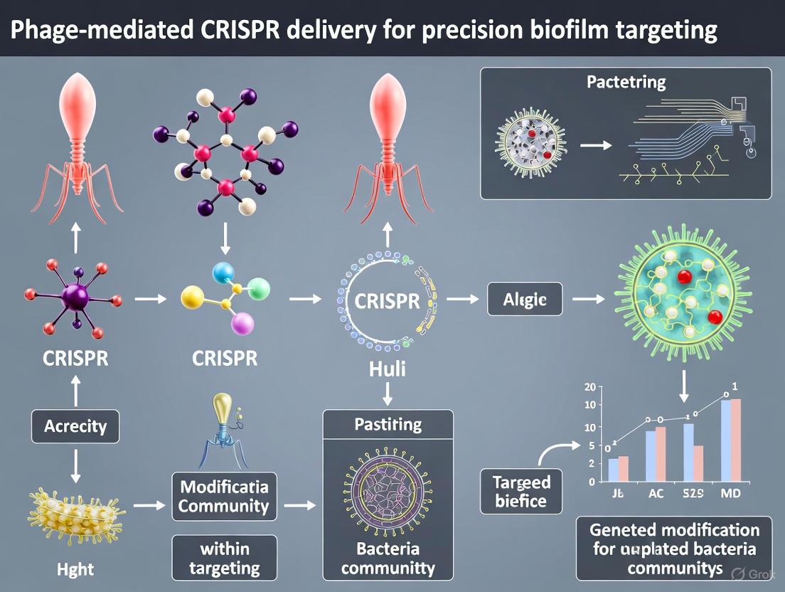
Diagram 1: Mechanism of Phage-Mediated CRISPR Delivery for Biofilm Targeting. This schematic illustrates how engineered bacteriophages deliver CRISPR-Cas systems specifically to biofilm-embedded bacteria, enabling precision disruption of genes involved in matrix production, antibiotic resistance, and persistence.
The Scientist's Toolkit: Essential Research Reagents
Table 3: Key Reagent Solutions for Biofilm and Persister Cell Research
| Reagent/Category | Specific Examples | Research Application |
|---|---|---|
| Biofilm Growth Systems | Calgary biofilm device; Flow cell systems; Modified Robbins device | Standardized biofilm cultivation under static or continuous flow conditions [6] |
| CRISPR-Cas Components | CGV-EcCas vector; PbolA promoter; Guide RNA libraries | Targeted gene disruption in biofilm-associated bacteria [9] [10] |
| Engineered Phage Vectors | SNIPR001 components; Tevenvirinae phages; Tail fiber-modified phages | Species-specific delivery of antimicrobial agents to biofilms [9] |
| Matrix Disruption Agents | DNase I; Dispersin B; Glycoside hydrolases; Alginate lyase | EPS degradation for enhanced antimicrobial penetration [3] [5] |
| Viability Stains | SYTO 9/propidium iodide; CTC; Resazurin | Differentiation of live/dead cells and metabolic activity assessment [4] |
| Antibiotic Conjugates | BODIPY-FL tagged tobramycin; Vancomycin fluorescein | Visualization and quantification of antibiotic penetration [3] |
| Persister Isolation Tools | Membrane potential dyes; Antibiotic selection protocols; FACS sorting reagents | Separation and enrichment of dormant persister cells [8] [4] |
The integration of these specialized reagents with advanced methodologies enables comprehensive investigation of biofilm architecture and resistance mechanisms, facilitating the development of novel anti-biofilm strategies that address the limitations of conventional antibiotic therapies.
Application Note: Phage-Mediated CRISPR Delivery for Biofilm Eradication
This application note provides a consolidated overview of engineered bacteriophages as precision delivery vehicles for CRISPR-based antimicrobial systems, specifically for targeting and eradicating bacterial biofilms. Biofilms are structured microbial communities embedded in extracellular polymeric substances (EPS) that exhibit dramatically enhanced resistance to conventional antibiotics—up to 1000-fold greater tolerance compared to planktonic cells [11]. Phage-mediated CRISPR delivery represents a paradigm shift in antimicrobial strategies by enabling sequence-specific targeting of resistance genes, virulence factors, and biofilm-regulatory elements within complex microbial communities.
Quantitative Efficacy of Phage-Delivered CRISPR Systems
Table 1: Editing Efficiency and Biofilm Reduction by Phage-Delivered CRISPR Systems
| System/Platform | Target Bacteria | Editing Efficiency/Biofilm Reduction | Experimental Context | Citation |
|---|---|---|---|---|
| λ-DART Phage | Escherichia coli | >50% population editing | Monoculture & mixed community | [12] |
| Liposomal Cas9 Formulation | Pseudomonas aeruginosa | >90% biofilm biomass reduction | In vitro | [11] |
| CRISPR-Gold Nanoparticle | Model Bacteria | 3.5x increase in editing efficiency | Non-carrier system comparison | [11] |
| SNIPR001 (CAP Cocktail) | Escherichia coli | Significant load reduction | Mouse gut model | [9] |
Table 2: Performance of Different Promoters in Phage-Delivered CRISPR Systems
| Promoter | System | Performance Context | Key Outcome | Citation |
|---|---|---|---|---|
| PbolA | SNIPR001 CAPs | Biofilm & restricted growth conditions | Significant killing; reduced metabolic activity | [9] |
| PrelB | SNIPR001 CAPs | Standard planktonic growth (LB, 37°C) | Lower performance vs. PbolA in biofilms | [9] |
Experimental Protocols
Protocol 1: Engineering Phage λ with CRISPR-Associated Transposases (DART System)
Background: This protocol details the modification of temperate phage λ to create a delivery vehicle for the DNA-editing all-in-one RNA-guided CRISPR-Cas transposase (DART) system, enabling large DNA insertions and targeted gene disruptions in situ [12].
Materials:
- Bacterial Strains: E. coli BW25113 (wild-type), E. coli LE392MP (amber-suppressor)
- Phage: λ cI857 Sam7 (amber mutation in lysis gene S, thermolabile repressor)
- Engineering Tools: Homologous recombination setup, Cas13a-based counterselection system
Methodology:
- Phage Engineering:
- Use homologous recombination to embed the entire DART system (including CRISPR, gene, and transposon components) into the λ phage genome.
- Employ Cas13a-based counterselection for precise, markerless selection of recombinant phages. Cas13a induces host dormancy upon targeting wild-type phage sequences, enriching for successfully engineered particles [12].
- Ensure the engineered λ-DART phages are rendered non-lysogenic by removing components essential for lysogeny to prevent persistent phage maintenance.
- Infection and Editing Assay:
- Grow the target E. coli culture to mid-log phase.
- Infect the culture with the engineered λ-DART phage at a predetermined Multiplicity of Infection (MOI). Note: Higher MOIs lead to more rapid population decline but may be followed by a rebound after approximately 8 hours [12].
- Incubate at 37°C to induce the lytic cycle via the cI857 mutation.
- Harvest samples post-infection to analyze editing efficiency via colony PCR, sequencing, or phenotypic assays.
Protocol 2: Construction and Application of CRISPR-Cas-Armed Phages (CAPs)
Background: This protocol describes the creation of a cocktail of CRISPR-Cas-Armed Phages (CAPs) for selective and potent killing of targeted pathogens, such as E. coli, in complex environments [9].
Materials:
- Wild-Type Phages: A library of lytic phages (e.g., from wastewater). Selected phages (e.g., α15, α17) from the Tevenvirinae subfamily.
- CRISPR System: Type I-E CRISPR-Cas system from E. coli (genes cas3, casA, casB, casC, casD, casE, and a customizable CRISPR array).
- Promoter: PbolA promoter for optimal expression under biofilm and restricted growth conditions [9].
Methodology:
- Phage Selection and Engineering:
- Screen a wild-type phage library against a panel of target pathogen strains to identify phages with broad, complementary host ranges and complementary receptor usage (e.g., LPS, Tsx, LamB) [9].
- Optionally engineer phage tail fibers to expand host range and reduce the emergence of resistant mutants. For example, the Tsx-binding adhesin from phage α17 was engineered into phage α15 to create a dual-affinity phage [9].
CRISPR-Cas Arming:
- Clone the Type I-E CRISPR-Cas system under the control of the PbolA promoter into the selected phage genomes.
- Design the CRISPR array spacer sequences to target essential genes or antibiotic resistance genes in the pathogen's genome or plasmids.
In Vitro and In Vivo Efficacy Testing:
- Lawn Kill Assay: Apply the engineered CAPs to a lawn of the target bacteria to assess the reduction in survivors compared to wild-type phage.
- Biofilm Assay: Grow biofilms of the target pathogen (e.g., on peg lids in 96-well plates). Treat with CAPs and quantify reduction in metabolic activity (e.g., via resazurin assay) or biofilm biomass [9].
- In Vivo Model: Administer a cocktail of the most complementary CAPs (e.g., SNIPR001) to animal models (e.g., mice). Monitor pathogen load (e.g., in the gut) and overall animal tolerance over time [9].
The Scientist's Toolkit: Research Reagent Solutions
Table 3: Essential Reagents for Phage-Delivered CRISPR-Cas Research
| Reagent/Category | Specific Examples | Function & Application | Citation |
|---|---|---|---|
| Engineered Phage Chassis | λ cI857 Sam7; α15, α17, α20 (Tevenvirinae) | Delivery vehicle; engineered for controlled lysis, broad host range, and cargo capacity. | [12] [9] |
| CRISPR-Cas Systems | Type I-F CAST (for DART); Type I-E (for CAPs); Cas9, Cas12a, Cas13a | Execution of DNA/RNA cleavage or insertion. CAST systems enable large DNA integrations. | [12] [9] [13] |
| Specialized Promoters | PbolA | Drives CRISPR-Cas expression under biofilm and nutrient-restricted conditions. | [9] |
| Delivery & Formulation Aids | Liposomal nanoparticles; Gold nanoparticles | Enhance delivery, cellular uptake, and stability of CRISPR-Cas components; can be co-delivered with phages. | [11] |
| Counterselection Tool | Cas13a | Enables precise, markerless selection of engineered phages during construction. | [12] |
Workflow and Pathway Visualizations
Diagram 1: Workflow of Phage-Delivered CRISPR Antimicrobials.
Diagram 2: Precision Targeting of Biofilm Resistance.
The escalating threat of Antimicrobial Resistance (AMR) represents one of the most critical challenges to global public health, projected to cause millions of deaths annually if left unaddressed [14] [15]. The discovery of new conventional antibiotics has stalled significantly, creating an urgent need for innovative therapeutic strategies [16]. In this landscape, CRISPR-Cas systems—an adaptive immune mechanism in prokaryotes—have emerged as a revolutionary tool for developing precision antimicrobials [17]. Unlike broad-spectrum antibiotics, CRISPR-Cas systems can be programmed to selectively target and eliminate specific bacterial pathogens or to resensitize them to traditional antibiotics by inactivating their resistance genes [16] [14]. This application note details the methodology for leveraging phage-mediated CRISPR-Cas delivery to combat biofilm-associated, multidrug-resistant infections.
Core Principles: Reprogramming Bacterial Immunity
The Native CRISPR-Cas Mechanism
CRISPR-Cas systems naturally protect bacteria from invasive genetic elements, such as viruses and plasmids, through a three-stage process: adaptation, crRNA biogenesis, and interference [17]. During adaptation, fragments of invader DNA are captured and integrated into the host's CRISPR array as "spacers." This array is then transcribed and processed into short CRISPR RNAs (crRNAs). During interference, these crRNAs guide Cas nucleases to recognize and cleave complementary DNA sequences of subsequent invaders, thereby neutralizing the threat [17].
CRISPR-Cas Systems as Programmable Antimicrobials
The programmability of this system allows it to be repurposed. By designing guide RNAs (gRNAs) to target essential bacterial genes, virulence factors, or antibiotic resistance genes (ARGs), the CRISPR-Cas machinery can be redirected to induce lethal double-strand breaks in the chromosome of a target pathogen or to cure it of its resistance-bearing plasmids [16]. The specificity of this approach minimizes damage to the beneficial microbiota, a significant advantage over conventional antibiotics [16] [17].
Table 1: Major CRISPR-Cas Systems Used in Antimicrobial Development
| System Type | Key Effector Nuclease | Mechanism of Action | Key Advantage for Antimicrobial Use |
|---|---|---|---|
| Type I (e.g., I-E) | Cas3 | Cascade complex recognizes DNA, recruits Cas3 for degradation [16]. | Potent, processive DNA degradation leading to high killing efficiency [17]. |
| Type II (e.g., II-A) | Cas9 | gRNA-guided nuclease introduces double-strand breaks in DNA [16] [11]. | High versatility and specificity; well-characterized system [11]. |
| Type VI (e.g., VI-A) | Cas13a | gRNA-guided nuclease targets and degrades RNA [16]. | Can target RNA-based functions and antibiotic resistance mRNAs [16]. |
Application Note: Phage-Mediated Delivery for Precision Biofilm Targeting
Rationale and Challenge
Bacterial biofilms are structured communities encased in an extracellular polymeric substance (EPS), which can reduce antibiotic penetration and increase tolerance by up to 1000-fold compared to planktonic cells [11]. This makes biofilm-associated infections particularly persistent and difficult to treat. Phage-mediated delivery offers a promising solution, as bacteriophages have evolved to naturally infect bacteria and penetrate biofilms [11] [9].
Workflow for Engineered Phage with Antibacterial CRISPR
The following diagram illustrates the core workflow for developing and applying a CRISPR-Cas armed phage (CAP) against a biofilm.
Experimental Protocols
Protocol: Construction of a CRISPR-Armed Phage (CAP)
Objective: To engineer a lytic bacteriophage to carry and deliver a CRISPR-Cas system targeting specific genes in a multidrug-resistant bacterial pathogen.
Materials:
- Bacterial Strains: Target pathogenic strain (e.g., E. coli, P. aeruginosa); non-target control strain.
- Phages: Selected broad-host-range, lytic phage from screening.
- Molecular Biology Reagents: PCR reagents, restriction enzymes (e.g., ApaI), T4 DNA ligase, Gibson Assembly mix.
- Plasmids: Vector backbone containing an inducible CRISPR-Cas system (e.g., Cas9 or Cas3 with gRNA cloning site).
- Culture Media: Lysogeny Broth (LB), LB agar, soft agar for plaques, appropriate antibiotics.
Methodology:
- Phage Genome Modification:
- Amplify the CRISPR-Cas expression cassette from the donor plasmid. The cassette should include:
- A constitutively active or environmentally responsive promoter (e.g.,
PbolAfor activity in biofilms and under restricted growth [9]). - The
casnuclease gene(s) (e.g.,cas9for Type II orcasA-Eandcas3for Type I-E [9]). - A gRNA expression scaffold targeting a specific bacterial gene (e.g., essential gene
ftsA, beta-lactamaseblaNDM-1, or colistin resistancemcr-1[16] [14]).
- A constitutively active or environmentally responsive promoter (e.g.,
- Insert this cassette into a non-essential region of the phage genome using homologous recombination or a Gibson Assembly-based approach, ensuring the packaging capacity of the phage is not exceeded.
- Purify the recombinant phage through several rounds of plating and PCR verification to ensure stability of the insert.
- Amplify the CRISPR-Cas expression cassette from the donor plasmid. The cassette should include:
Tail Fiber Engineering (Optional for Broader Host Range):
- To overcome phage resistance mediated by receptor mutation, engineer the phage tail fiber genes.
- Replace the native tail fiber/adhesin gene with one from a different phage that utilizes an alternative bacterial surface receptor (e.g., Tsx-binding adhesin from phage α17 engineered into phage α15 to add a second receptor affinity [9]).
- Verify the receptor usage and host range of the engineered phage using efficiency of plating (EoP) assays.
Phage Propagation and Purification:
- Amplify the validated CAP on a permissive propagation host in LB culture.
- Purify phage particles using polyethylene glycol (PEG) precipitation and subsequent cesium chloride density gradient centrifugation.
- Resuspend the purified phage stock in SM Buffer and determine the titer via double-layer agar plaque assay.
Protocol: Assessing CAP Efficacy Against In Vitro Biofilms
Objective: To quantify the ability of the CAP to reduce biofilm biomass and viability of target bacteria within a biofilm.
Materials:
- Biofilm Setup: 96-well peg lid plates or Calgary biofilm device.
- Staining Reagents: Crystal violet solution (0.1%), SYTO 9/propidium iodide live/dead stain.
- Equipment: Confocal Laser Scanning Microscope (CLSM), plate reader, sonication bath.
Methodology:
- Biofilm Formation:
- Grow the target bacterial strain in a suitable medium in 96-well plates or on peg lids for 24-48 hours to establish mature biofilms.
- Gently wash the biofilms with sterile saline to remove non-adherent planktonic cells.
CAP Treatment:
- Treat the established biofilms with the purified CAP at a defined Multiplicity of Infection (MOI, e.g., 10) in fresh medium. Include controls: wild-type phage, no phage (vehicle), and a non-targeting CAP.
- Incubate for 4-24 hours under conditions suitable for biofilm growth and phage activity.
Biofilm Analysis:
- Biomass Quantification (Crystal Violet Assay): Fix biofilms with methanol, stain with 0.1% crystal violet for 15 minutes, wash, solubilize the stain with acetic acid (33%), and measure absorbance at 595 nm. A >90% reduction in biomass is indicative of high efficacy [11].
- Viability Assessment (CFU Count): Sonicate pegs or scrape biofilms from wells to disaggregate cells. Serially dilute the suspension and plate on agar to enumerate Colony Forming Units (CFU). A 3–4 log₁₀ reduction in CFU/mL compared to the control demonstrates potent killing [9].
- Structural Integrity (CLSM): Stain the biofilm with a live/dead bacterial viability kit. Image using CLSM to visualize the architectural disruption of the biofilm and the spatial distribution of live vs. dead cells post-treatment.
Table 2: Quantitative Outcomes of CRISPR-Cas and Nanoparticle Antimicrobial Strategies
| Experimental Approach | Target Pathogen / Gene | Delivery System | Key Quantitative Result |
|---|---|---|---|
| CRISPR-Cas9 + Liposomal NPs [11] | P. aeruginosa biofilm | Lipid-based Nanoparticles | >90% reduction in biofilm biomass in vitro. |
| CRISPR-Cas + Gold NPs [11] | General gene editing | Gold Nanoparticles | 3.5-fold increase in editing efficiency vs. non-carrier systems. |
| Type I-E CRISPR-Cas (CGV-EcCas) [9] | Diverse E. coli panel | Conjugative Plasmid | Reduction of 1–6 log₁₀ CFU/mL; counts below LOD (200 CFU/mL) in susceptible strains. |
| CRISPR-Armed Phage (CAP) [9] | E. coli in mouse gut | Engineered Bacteriophage (SNIPR001) | Significant reduction in E. coli burden in the mouse gut model. |
The Scientist's Toolkit: Research Reagent Solutions
Table 3: Essential Reagents for Phage-Mediated CRISPR-Cas Antimicrobial Research
| Reagent / Material | Function / Purpose | Example & Notes |
|---|---|---|
| Type I-E CRISPR-Cas System | Provides DNA-targeting effector complex. | E. coli Cascade (CasA-E) + Cas3 [9]. Chosen for potent, processive degradation. |
| Constitutive/Inducible Promoters | Drives expression of Cas genes and gRNA in the target bacterium. | PbolA promoter provides robust expression in biofilms and under restricted growth [9]. |
| Lytic Bacteriophages | Natural vector for delivering CRISPR cargo into bacterial cells. | Tevenvirinae phages (e.g., α15, α17) are engineerable and have broad host ranges [9]. |
| Tail Fiber Adhesins | Determines host receptor specificity; enables engineering for broader coverage. | Tsx-binding or LamB-binding adhesins can be swapped between phages to combat resistance [9]. |
| Nanoparticle Carriers | Alternative delivery vehicle to protect and deliver CRISPR components. | Gold NPs enhance editing efficiency; Liposomal NPs co-deliver antibiotics and CRISPR for synergy [11]. |
| gRNA Design Tools | In silico design of specific guide RNAs for target genes. | Tools like Benchling are used to design gRNAs against AMR genes (e.g., mcr-1, blaKPC, mecA) [16] [14]. |
The fusion of CRISPR-Cas precision with the efficient delivery capabilities of bacteriophages represents a paradigm shift in antimicrobial therapy. This approach moves beyond traditional, broad-spectrum compounds to a programmable, sequence-specific strategy capable of targeting multidrug-resistant pathogens embedded in protective biofilms. While challenges remain—including optimizing delivery efficiency, pre-existing host immunity to phages, and potential off-target effects—the progress documented in recent studies underscores the immense translational potential of this technology [17] [9]. As these innovative platforms advance through preclinical and clinical development, they offer a powerful and precise new arsenal in the global fight against antimicrobial resistance.
Biofilm-associated infections represent a significant clinical challenge due to their inherent tolerance to antibiotics, with biofilms exhibiting up to 1000-fold greater resistance compared to their planktonic counterparts [11]. The extracellular polymeric substance (EPS) matrix of biofilms creates a formidable physical and functional barrier, limiting antimicrobial penetration and fostering bacterial persistence [11] [2]. CRISPR-Cas systems have emerged as powerful precision antimicrobial tools capable of selectively targeting antibiotic resistance genes, virulence factors, and biofilm-regulating elements [11] [18]. However, the efficacy of CRISPR-based antimicrobials is critically dependent on delivery systems that can successfully navigate the biofilm barrier and efficiently transduce bacterial cells. Bacteriophages (phages)—viruses that specifically infect bacteria—represent uniquely suited vectors for CRISPR delivery in biofilm environments, offering inherent mechanisms for biofilm penetration, targeted bacterial infection, and replication at the site of infection [2] [19] [9].
The Biofilm Challenge and Phage Advantages
The structured architecture of bacterial biofilms provides multiple mechanisms for enhanced antimicrobial resistance. The EPS matrix, primarily composed of exopolysaccharides, proteins, and extracellular DNA, physically restricts antibiotic diffusion and creates heterogeneous microenvironments with altered metabolic activity and elevated rates of horizontal gene transfer [11] [2]. Within this protective matrix, bacterial cells exist in various physiological states, including dormant persister cells that exhibit exceptional tolerance to conventional antibiotics [11].
Phages possess a suite of natural adaptations that overcome these biofilm-specific barriers:
- Depolymerase enzymes: Many phages encode virion-associated enzymes (depolymerases, lysins) that actively degrade key EPS components, creating physical channels for deeper biofilm penetration and facilitating access to underlying bacterial cells [2] [19].
- Self-replication and auto-dosing: Unlike static antimicrobials, phages replicate at the infection site, increasing their local concentration and creating a self-amplifying therapeutic effect that can overcome partial biofilm penetration limitations [19] [9].
- Biofilm-activated life cycles: Certain phages can infect dormant persister cells, remaining latent until these cells revert to metabolic activity, thereby targeting a population typically refractory to conventional antibiotics [19].
Table 1: Quantitative Comparison of Biofilm Elimination Strategies
| Strategy | Biofilm Reduction | Key Advantages | Limitations |
|---|---|---|---|
| Conventional Antibiotics | Variable (often limited) | Broad spectrum, well-established protocols | Poor penetration, increased resistance selection |
| Phage Monotherapy | Up to 3-4 log CFU reduction in optimized conditions [19] | Self-replicating, biofilm matrix degradation | Host range restrictions, resistance development |
| CRISPR-Cas Nanoparticles | >90% biomass reduction (liposomal Cas9) [11] | Precision targeting, modularity | Delivery challenges, stability issues |
| Phage-Delivered CRISPR | >4 log CFU reduction with targeted approach [9] | Combination of phage penetration & CRISPR precision | Engineering complexity, regulatory considerations |
Phage-Mediated CRISPR Delivery: Mechanisms and Workflows
The integration of CRISPR systems into phage genomes creates powerful synergistic platforms that combine the natural biofilm-penetrating abilities of phages with the sequence-specific targeting of CRISPR. These engineered phages, termed CRISPR-armed phages (CAPs), can be designed to deliver Cas nucleases and guide RNAs specifically to target bacteria within biofilms [9].
Molecular Mechanisms of Phage-CRISPR Synergy
The antibacterial activity of CAPs operates through two complementary mechanisms:
- Conventional phage lytic cycle: The phage injects its genetic material, hijacks bacterial machinery to produce new virions, and lyses the host cell, naturally disrupting biofilm architecture.
- CRISPR-mediated targeted killing: The delivered CRISPR-Cas system introduces double-strand breaks in chromosomal DNA or targets essential bacterial genes, resulting in lethal DNA damage regardless of bacterial metabolic state [9] [18].
This dual mechanism significantly reduces the emergence of escape mutants, as bacteria must simultaneously develop resistance to both phage infection and CRISPR targeting—a considerably less probable evolutionary scenario [9].
Diagram 1: Phage-CRISPR Synergy Mechanism in Biofilms
Protocol: Phage Engineering and CRISPR Loading
Objective: Engineer lytic phages to carry CRISPR-Cas systems targeting specific bacterial genes essential for biofilm formation or antibiotic resistance.
Materials:
- Wild-type lytic phages with known host range
- Target bacterial strains with sequenced genomes
- CRISPR plasmid constructs with Cas genes and guide RNA sequences
- Phage propagation host bacteria
- Molecular biology reagents: restriction enzymes, ligases, PCR reagents
- Electroporation system
- Plaque assay materials: agar plates, soft agar, culture media
Procedure:
- Phage Selection and Characterization:
- Screen phage library for broad host range and complementary receptor usage [9]
- Characterize phage genomes through sequencing to identify non-essential regions for cargo insertion
- Determine efficiency of plating (EoP) on target bacterial strains
CRISPR Construct Design:
- Identify essential bacterial genes for targeting (e.g., antibiotic resistance genes, quorum-sensing regulators, EPS synthesis genes)
- Design guide RNA sequences with minimal off-target potential using bioinformatics tools
- Select appropriate Cas nuclease (Cas9, Cas3) based on desired killing mechanism [9] [18]
- Clone CRISPR expression cassette into phage vector under constitutive or phage-specific promoters
Phage Engineering:
- For temperate phages: employ homologous recombination to integrate CRISPR cassette into phage genome
- For lytic phages: utilize in vitro assembly of phage genomes with CRISPR payload [9]
- Transfer engineered phage genomes into propagation hosts via electroporation or transfection
CAP Validation:
- Confirm CRISPR cargo stability through serial passage and PCR verification
- Assess killing efficiency against planktonic and biofilm-grown target bacteria
- Sequence potential escape mutants to confirm CRISPR-mediated selection
Experimental Models for Evaluating Phage-CRISPR Efficacy
In Vitro Biofilm Models and Assessment Methods
Static Biofilm Model Protocol:
- Grow biofilms in 96-well plates or on peg lids for 24-48 hours
- Treat with CAPs at varying multiplicities of infection (MOI)
- Assess biofilm reduction using:
Flow Cell Biofilm Model Protocol:
- Establish biofilms in flow cells with constant nutrient supply
- Treat with CAPs under static or flow conditions
- Monitor biofilm disruption in real-time using microscopy
- Compare efficacy against phage monotherapy and antibiotic controls
Table 2: Research Reagent Solutions for Phage-CRISPR Biofilm Studies
| Reagent/Category | Specific Examples | Function/Application | Key Considerations |
|---|---|---|---|
| CRISPR Components | Type I-E CRISPR-Cas (E. coli), Cas9, Cas3 [9] | Targeted bacterial killing | Select based on target bacteria & desired killing mechanism |
| Phage Vectors | Tevenvirinae phages (α15, α17, α20) [9] | CRISPR delivery vehicle | Choose phages with broad host range & engineerability |
| Biofilm Assessment | qPCR with species-specific primers [19] | Quantify bacterial load | More accurate than culture-based methods for biofilms |
| Metabolic Monitoring | Isothermal microcalorimetry [19] | Real-time metabolic activity | Detects biofilm responses hours before visible changes |
| Engineering Tools | Tail fiber engineering [9] | Expand phage host range | Combats LPS-based resistance; enables dual receptor use |
In Vivo Efficacy Assessment
Animal Model Protocol:
- Establish biofilm-associated infections in appropriate animal models (e.g., catheter-associated, wound, or pulmonary infection models)
- Administer CAPs via route appropriate to infection site (topical, systemic, inhalation)
- Include control groups: untreated, wild-type phage, CRISPR alone, conventional antibiotics
- Assess outcomes through:
- Bacterial burden quantification in tissues
- Histopathological analysis of infection sites
- In vivo imaging if reporter strains are used
- Host immune response monitoring
Advanced Applications and Engineering Strategies
Directed Evolution for Enhanced Phage Performance
Phages can be experimentally evolved to overcome biofilm-specific challenges through serial passage assays:
Directed Evolution Protocol:
- Incubate pre-established biofilms with phage mixture under controlled conditions
- Monitor phage activity using isothermal microcalorimetry to identify samples with >75% heat reduction compared to growth controls [19]
- Pool successful phage samples for subsequent rounds of evolution
- Perform 30+ rounds of selection to enhance host range, antimicrobial efficacy, and antibiofilm performance [19]
- Isolate individual evolved phages and characterize genomic changes responsible for improved efficacy
Diagram 2: Directed Evolution Workflow for Enhanced Phages
Cocktail Design to Prevent Resistance
Rational design of phage cocktails that target multiple bacterial receptors simultaneously can significantly reduce the emergence of resistant variants:
Resistance-Adapted Cocktail Design Protocol:
- Select phages with complementary host ranges and receptor usage patterns
- Include tail fiber-engineered phages capable of dual receptor recognition [9]
- Balance phage ratios based on kinetic parameters and biofilm penetration capabilities
- Validate cocktail efficacy against dual-resistant mutants in biofilm models
- Assess potential for horizontal gene transfer of CRISPR machinery to non-target bacteria
The integration of phage biology with CRISPR precision represents a paradigm shift in antimicrobial development for biofilm-associated infections. Phages provide the ideal vector system for CRISPR delivery in these challenging environments, leveraging natural mechanisms for biofilm penetration, bacterial targeting, and self-amplification. The experimental protocols outlined herein provide researchers with robust methodologies for developing, optimizing, and evaluating these innovative antimicrobial platforms. As resistance to conventional antibiotics continues to escalate, phage-delivered CRISPR systems offer a promising pathway for the development of precision antimicrobials capable of overcoming the unique challenges posed by bacterial biofilms. Future directions will likely focus on refining delivery efficiency, expanding host range through synthetic biology approaches, and addressing regulatory considerations for clinical translation of these innovative therapeutic platforms.
Engineering Phage Vectors and CRISPR Payloads for Targeted Biofilm Disruption
Within the broader scope of developing phage-mediated CRISPR delivery systems for precision targeting of biofilms, controlling the phage host range is a critical prerequisite. A phage must first successfully infect and transduce its target bacterial cell to deliver a CRISPR payload. Tail fibers, the intricate protein appendages of bacteriophages, are the primary determinants of host specificity, mediating the initial recognition and adsorption to bacterial surface receptors [20]. Engineering these structures to alter or expand host range is therefore a foundational step in creating effective, broad-spectrum antimicrobial agents.
This application note details two primary, experimentally validated strategies for tail fiber engineering: experimental evolution and structure-informed rational design. We provide detailed protocols and resources to enable researchers to implement these approaches for enhancing the efficacy of phage-based CRISPR delivery vehicles against diverse and resilient biofilm populations.
Key Engineering Strategies and Quantitative Outcomes
The following table summarizes the core strategies, their mechanistic bases, and key experimental results for host range expansion.
Table 1: Comparative Analysis of Phage Host Range Expansion Strategies
| Engineering Strategy | Phage Model | Target Bacterium | Key Mutations / Focus | Experimental Host Range Increase | Key Quantitative Findings |
|---|---|---|---|---|---|
| Experimental Evolution (Coevolution) | Phages Ace & APV [21] | Klebsiella pneumoniae (MDR/XDR) | Not specified (genomic analysis recommended) | APV: 27.1% → up to 61.0%Ace: 42.4% → up to 59.3% | Enhanced longitudinal growth suppression; trained phages superior in 10/12 assays over 72h [21] |
| Experimental Evolution (Biofilm-Adapted) | Phage PE1 (Pbunavirus) [22] | Pseudomonas aeruginosa (CF isolate) | gp78 (Tail fiber): T896Igp76 (Baseplate wedge): S324Fgp77 (Baseplate): S72P | EOP increased from 0.09 (WT) to ~1.16 (adapted) | >90% reduction in biofilm culturable cells in CF sputum medium; significant reduction in a 3-D lung epithelial model [22] |
| Structure-Informed Rational Design | Phage λ (lambda) [12] | Escherichia coli | CRISPR-guided transposase delivery (DART system) | N/A (Delivery vehicle engineering) | Achieved >50% editing efficiency in mixed bacterial communities for precise gene knockouts and insertions [12] |
Detailed Experimental Protocols
Protocol 1: Experimental Coevolution for Host Range Expansion
This protocol, adapted from recent work on Klebsiella pneumoniae phages, leverages continuous co-culture to drive the selection of phages with broadened host range [21].
Research Reagent Solutions
Table 2: Essential Reagents for Phage Coevolution
| Reagent / Material | Function / Application |
|---|---|
| Naïve Phage Stock | Starting viral population for evolution experiment. |
| Multi-Drug Resistant (MDR) Clinical Isolates | Bacterial hosts providing selective pressure for evolution. |
| Luria-Bertani (LB) Broth/Agar | Standard medium for bacterial growth and phage propagation. |
| Soft Agar (0.5%-0.7%) | For plaque assays to titer phage and isolate clones. |
| Phage Buffer (e.g., SM Buffer) | For phage storage and dilution, maintaining viral stability. |
| Synthetic Cystic Fibrosis Sputum Medium (SCFM2) | To mimic in-vivo conditions for biofilm adaptation [22]. |
Procedure
- Initial Setup: Inoculate 10 mL of liquid culture medium with a target clinical isolate (e.g., an MDR K. pneumoniae). Grow to mid-exponential phase (OD600 ≈ 0.4-0.6).
- Initial Infection: Infect the culture with the naïve phage stock at a low multiplicity of infection (MOI ~0.1) to ensure multiple infection cycles.
- Serial Passaging:
- Incubate the culture with shaking until complete lysis is observed or for a set period (e.g., 24 hours).
- Centrifuge the lysate (e.g., 10,000 × g for 10 min) and filter the supernatant through a 0.22 µm filter to remove bacterial debris.
- Use a small aliquot (e.g., 1%) of this filtered lysate to infect a fresh, mid-exponential phase culture of the same bacterial strain.
- Repeat this serial passaging daily for at least 15-30 cycles or 30 days [21].
- Plaque Purification and Isolation: After the evolution period, perform serial dilutions of the final lysate and conduct plaque assays. Pick well-isolated plaques and amplify them on the original host strain to create clonal populations of evolved phages.
- Host Range Assessment:
- Spot 10 µL of high-titer evolved phage lysates (≥10^8 PFU/mL) onto a lawn of various clinical isolates (including the original host and new, resistant strains) prepared in soft agar overlays.
- Incubate overnight and score for lytic activity (clearance or plaque formation). Compare the lytic profile of evolved phages to the ancestral phage to quantify host range expansion [21].
Diagram 1: Workflow for experimental coevolution of phages.
Protocol 2: Biofilm-Adapted Directed Evolution
This protocol is specifically designed to evolve phages with enhanced efficacy against bacterial biofilms, as demonstrated with Pseudomonas aeruginosa [22].
- Biofilm Formation: Grow the target bacterial strain (e.g., a CF isolate of P. aeruginosa) in 24-well plates for 24-48 hours to establish mature biofilms.
- Phage Infection and Adaptation:
- Inoculate the established biofilms with the ancestral phage (e.g., PE1).
- Incubate for a defined period (e.g., 24h) to allow phage infection and replication within the biofilm.
- Recover the phage-containing supernatant and use it to infect a fresh, planktonic culture of the same strain to amplify the phage population.
- Use this amplified phage population to infect a new, mature biofilm. Repeat this cycle of biofilm infection and planktonic amplification for multiple rounds (e.g., 10-15 cycles) [22].
- Isolation of Biofilm-Enhanced Mutants: Following the adaptation rounds, titer the phage population and isolate individual plaques. Screen these clones for enhanced biofilm disruption in a 24-well biofilm assay, selecting those that cause the most significant visual dispersion of biofilm aggregates.
- Genomic Analysis: Sequence the genomes of the selected biofilm-adapted phages. Identify mutations, particularly in genes encoding tail fiber (e.g., gp78) and baseplate wedge proteins (e.g., gp76, gp77), which are frequently implicated in adapted phenotypes [22].
Protocol 3: Phage Engineering for CRISPR Payload Delivery
This protocol outlines the engineering of a temperate phage like λ to deliver CRISPR-associated transposases (DART system) for precise genome editing within bacterial communities [12].
- Phage Genome Modification:
- Objective: Replace the phage's lysogeny control region and integrate the all-in-one DART system construct.
- Method: Use homologous recombination in E. coli with a donor plasmid containing the DART payload flanked by homology arms to the phage genome.
- Counterselection: Employ a Cas13a-based counterselection system to efficiently isolate recombinant phages that have successfully integrated the payload and lost the lysogeny genes [12].
- Screening for Non-Lysogenic, DART-Encoding Phage: Screen plaques for the desired genotype (presence of DART genes, absence of lysogeny genes) via PCR. Confirm the loss of lysogeny function by the inability to form lysogens.
- Validation of Editing Efficiency:
- Infect the target bacterial strain (in monoculture or a mixed community) with the engineered λ-DART phage.
- After infection, plate bacteria on selective media or use flow cytometry to quantify the frequency of successful gene knockouts or insertions. Editing efficiencies of >50% in mixed communities have been reported [12].
Integration with Phage-Mediated CRISPR Delivery
The successful engineering of phage tail fibers for an expanded host range is a critical enabling step for phage-mediated CRISPR delivery. A broad-host-range phage ensures that the CRISPR-Cas system is delivered to the maximum number of target cells within a heterogeneous biofilm. The synergy between these technologies is illustrated below.
Diagram 2: Integration of host range expansion with CRISPR delivery for biofilm targeting.
As shown, the engineered phage serves as a targeted delivery vehicle. Once inside the cell, the CRISPR payload can be deployed to precisely disrupt antibiotic resistance genes (e.g., bla genes), virulence factors, or genes essential for biofilm integrity, thereby resensitizing the entire bacterial population to conventional antibiotics and leading to effective biofilm clearance [12] [7].
Clustered Regularly Interspaced Short Palindromic Repeats (CRISPR) and CRISPR-associated (Cas) proteins constitute an adaptive immune system in bacteria and archaea that has been repurposed as a versatile genome engineering tool [23] [24]. The fundamental CRISPR system comprises two key components: a Cas nuclease and a guide RNA (gRNA) that directs the nuclease to specific DNA sequences [11] [24]. For research focused on phage-mediated delivery for precision biofilm targeting, strategic selection of CRISPR machinery is paramount. This application note delineates the distinct roles of catalytically active Cas nucleases for gene knockout and catalytically dead Cas9 (dCas9) for gene regulation, providing detailed protocols for implementing both approaches against bacterial biofilms.
The transformative potential of CRISPR technologies lies in their programmability and precision. Unlike previous genome editing tools such as zinc finger nucleases (ZFNs) and transcription activator-like effector nucleases (TALENs) that require complex protein engineering for each new target, CRISPR systems require only the synthesis of a new guide RNA to redirect the nuclease to a different genomic locus [23] [24]. This simplicity has enabled unprecedented scalability in genetic manipulation. In the context of antibacterial therapies, CRISPR systems can be engineered to target essential genes, antibiotic resistance determinants, or virulence factors in pathogenic bacteria, with particular promise for disrupting recalcitrant biofilms [11] [9].
Comparative Analysis of Cas Nuclease Systems
Cas Nucleases for Gene Knockout
Catalytically active Cas nucleases introduce double-strand breaks (DSBs) in DNA at sites complementary to the guide RNA and adjacent to a protospacer adjacent motif (PAM) [24]. In bacterial cells, which predominantly utilize the error-prone non-homologous end joining (NHEJ) pathway for DNA repair, these breaks result in insertions or deletions (indels) that disrupt the coding sequence of the target gene [25] [24]. For efficient gene knockout, the guide RNA should be designed to target the 5' end of the most conserved exon to maximize the likelihood of generating frameshift mutations that produce non-functional protein products [25].
Table 1: Cas Nuclease Variants for Gene Knockout Applications
| Nuclease | PAM Sequence | Editing Efficiency | Specificity | Primary Applications |
|---|---|---|---|---|
| SpCas9 | NGG | High (0-81%) [23] | Moderate | Standard gene knockouts, multiplexed editing |
| eSpCas9(1.1) | NGG | High | Increased fidelity [24] | Reduced off-target editing |
| SpCas9-HF1 | NGG | Moderate to High | Increased fidelity [24] | High-precision knockouts |
| HypaCas9 | NGG | High | Increased proofreading [24] | Enhanced discrimination against off-targets |
| evoCas9 | NGG | Moderate | High fidelity [24] | Applications requiring minimal off-target effects |
| SpCas9-NG | NG | Moderate | Moderate | Targeting AT-rich genomic regions |
| SpG | NGN | High | Moderate | Expanded targeting range |
| SpRY | NRN/NYN | Broad | Moderate | Near-PAMless targeting capability |
dCas9 Systems for Gene Regulation
Catalytically dead Cas9 (dCas9) is generated through point mutations (D10A and H840A for SpCas9) that inactivate the nuclease domains while preserving DNA-binding capability [26] [24]. When complexed with guide RNAs, dCas9 can be targeted to specific genomic loci without introducing DNA breaks, serving as a programmable DNA-binding platform [26] [27]. By fusing dCas9 to transcriptional repressors (CRISPR interference, CRISPRi) or activators (CRISPR activation, CRISPRa), researchers can precisely modulate gene expression levels [26].
CRISPRi functions by sterically hindering RNA polymerase binding or elongation when dCas9 is targeted to promoter regions [26]. This approach enables tunable gene knockdown without permanent genetic alterations, making it particularly valuable for studying essential genes where complete knockout would be lethal. CRISPRi systems have been successfully deployed in biofilm research to downregulate quorum sensing pathways, virulence factors, and antibiotic resistance genes [11] [26].
Table 2: dCas9-Based Systems for Gene Regulation
| System | Components | Mechanism of Action | Regulatory Effect | Biofilm Applications |
|---|---|---|---|---|
| CRISPRi | dCas9 + sgRNA | Blocks transcription initiation or elongation | Gene knockdown | Suppress quorum sensing, virulence factors, resistance genes [11] [26] |
| CRISPRa | dCas9 + activator domains | Recruits transcriptional machinery | Gene activation | Enhance antibiotic susceptibility, disrupt persistence |
| CRISPR | dCas9 + epigenetic modifiers | Modifies chromatin state | Epigenetic regulation | Alter biofilm formation pathways |
Quantitative Efficacy Data for Anti-Biofilm Applications
Recent advances in CRISPR-based antimicrobials have demonstrated promising efficacy against biofilm-associated infections. The integration of CRISPR systems with nanoparticle and phage delivery platforms has been particularly successful in enhancing biofilm penetration and bacterial uptake.
Table 3: Quantitative Efficacy of CRISPR-Based Anti-Biofilm Strategies
| Delivery System | CRISPR Payload | Target Bacteria | Biofilm Reduction | Key Findings |
|---|---|---|---|---|
| Liposomal nanoparticles [11] | Cas9 + gRNA (resistance genes) | Pseudomonas aeruginosa | >90% in vitro [11] | Enhanced antibiotic penetration, disruption of EPS matrix |
| Gold nanoparticles [11] | Cas9 + gRNA | P. aeruginosa | 3.5× editing efficiency [11] | Improved cellular uptake, controlled release within biofilm |
| Engineered phage (CAPs) [9] | Type I-E CRISPR-Cas | Escherichia coli | 1-6 log10 reduction [9] | Specific killing of target strains, reduced resistance emergence |
| CRISPR-Cas armed phage [9] | CRISPR with PbolA promoter | E. coli | Significant metabolic activity reduction [9] | Functional under restricted bacterial growth conditions in biofilms |
Experimental Protocols
Protocol for Gene Knockout in Bacterial Biofilms
This protocol outlines the procedure for achieving efficient gene knockout in biofilm-forming bacteria using CRISPR-Cas9 systems delivered via phage or nanoparticle vectors, adapted from established methodologies [11] [25] [9].
Guide RNA Design and Preparation
- Target Selection: Identify target sequences within the first exon of essential biofilm-related genes (e.g., antibiotic resistance genes, quorum sensing regulators) [25]. Prioritize the 5' end of conserved domains to maximize probability of functional knockout.
- gRNA Design: Design 2-3 gRNAs per target gene with the following criteria:
- 20-nucleotide spacer sequence complementary to target DNA
- 3' NGG protospacer adjacent motif (PAM) for SpCas9
- 40-60% GC content for optimal stability and activity [26]
- Minimal off-target potential (verify via BLAST against host genome)
- gRNA Construction: Synthesize crRNA and tracrRNA components separately, then anneal by heating to 95°C for 5 minutes and cooling slowly to room temperature in annealing buffer [25].
Ribonucleoprotein (RNP) Complex Formation
- Component Preparation: Dilute purified Cas9 nuclease to 10 µM in sterile buffer. Prepare annealed gRNA at 12 µM concentration (1.2:1 ratio to Cas9) [25].
- Complex Assembly: Combine Cas9 and gRNA in molar ratio of 1:1.2, incubate at room temperature for 15-30 minutes to form RNP complexes.
- Quality Control: Verify complex formation using gel shift assay before proceeding to delivery.
Delivery via Phage Vectors
- Phage Engineering: For phage-mediated delivery, integrate the CRISPR expression cassette into engineered phage genomes as described by [9]:
- Utilize lytic phage backbones (e.g., Tevenvirinae) with broad host range
- Incorporate CRISPR arrays targeting essential bacterial genes
- Use PbolA promoter for expression under biofilm conditions [9]
- Phage Propagation: Amplify CRISPR-armed phages in susceptible host strains, purify via polyethylene glycol precipitation, and resuspend in SM buffer.
- Biofilm Treatment: Apply phage suspension to pre-formed biofilms (24-48 hours old) at multiplicity of infection (MOI) of 10-100, incubate for 4-6 hours at 37°C.
Validation and Analysis
- Efficiency Assessment: Extract genomic DNA from treated biofilms, amplify target region by PCR, and analyze editing efficiency via T7E1 assay or sequencing.
- Phenotypic Screening: Evaluate biofilm biomass reduction using crystal violet staining or confocal microscopy.
- Antibiotic Resensitization: Test recovered bacteria for antibiotic susceptibility changes to confirm resistance gene disruption.
Protocol for Gene Regulation Using dCas9 Systems
This protocol describes the implementation of CRISPRi for targeted gene repression in bacterial biofilms, enabling modulation of gene expression without permanent genetic alterations.
dCas9-effector Fusion Construction
- dCas9 Vector Selection: Utilize plasmids encoding catalytically dead Cas9 (D10A and H840A mutations) with appropriate bacterial selection markers.
- Effector Domain Fusion: For CRISPRi, fuse dCas9 to transcriptional repressor domains (e.g., KRAB, ω subunit) if additional repression is required beyond steric hindrance [26].
- Promoter Selection: For biofilm applications, use promoters that remain active under biofilm conditions such as PbolA, which has demonstrated superior performance in biofilms compared to standard promoters [9].
Guide RNA Design for Regulation
- Targeting Strategy: Design gRNAs to target promoter regions (-10 to +50 relative to transcription start site) for optimal transcriptional repression [26].
- Multiplexing: For enhanced repression, design 2-3 gRNAs targeting different regions of the same promoter and express simultaneously from a multiplexed gRNA array.
- Specificity Controls: Include non-targeting gRNAs as negative controls and gRNAs targeting essential genes with known phenotypes as positive controls.
Delivery and Induction in Biofilms
- Phage Assembly: Package dCas9-effector and gRNA expression cassettes into engineered phage particles as described in section 4.1.3.
- Biofilm Treatment: Apply dCas9-phage constructs to mature biofilms at MOI 10-50, allow 2-4 hours for infection and expression.
- Induction Optimization: For inducible systems, add inducer (e.g., arabinose, anhydrotetracycline) at appropriate concentration after phage adsorption.
Efficacy Assessment
- Transcriptional Analysis: Measure target gene mRNA levels using RT-qPCR 4-8 hours post-treatment.
- Protein Quantification: Assess protein level reduction via Western blot or immunofluorescence 12-24 hours post-treatment.
- Phenotypic Characterization: Evaluate changes in biofilm architecture, metabolic activity, or antibiotic susceptibility.
The Scientist's Toolkit: Essential Research Reagents
Table 4: Key Reagents for CRISPR-Based Anti-Biofilm Research
| Reagent Category | Specific Examples | Function | Application Notes |
|---|---|---|---|
| Cas Nucleases | SpCas9, eSpCas9(1.1), HypaCas9 | Target DNA cleavage | Selection depends on specificity requirements and PAM availability [24] |
| dCas9 Effectors | dCas9-KRAB, dCas9-ω | Transcriptional repression | Fusion domains enhance repression efficiency [26] |
| Guide RNA Scaffolds | sgRNA, crRNA+tracrRNA | Target recognition | Two-part systems may offer better folding in some applications [25] |
| Delivery Vectors | Engineered Tevenvirinae phages, Liposomal/Gold nanoparticles | CRISPR component delivery | Phages offer species specificity; nanoparticles enhance biofilm penetration [11] [9] |
| Promoters | PbolA, PrelB | Expression in biofilms | PbolA shows superior performance under biofilm conditions [9] |
| Selection Markers | Kanamycin, Ampicillin resistance | Plasmid maintenance | Consider antibiotic susceptibility of target strains |
| Assembly Systems | GoldenGate, BsaI/BbsI restriction sites | Vector construction | Enable rapid multiplex gRNA cloning [28] |
The strategic selection of CRISPR-Cas machinery is fundamental to successful genetic manipulation of bacterial biofilms. Catalytically active Cas nucleases enable permanent gene knockout essential for eliminating antibiotic resistance genes, while dCas9-based systems offer precise temporal control over gene expression for functional studies and potential therapeutic interventions. The integration of these CRISPR systems with advanced delivery platforms, particularly engineered phages and nanoparticles, represents a promising avenue for overcoming the inherent resistance of biofilms to conventional antimicrobials. As these technologies continue to evolve, they hold immense potential for addressing the growing crisis of antibiotic-resistant biofilm-associated infections.
Designing Guide RNAs (gRNAs) to Target Virulence, Resistance, and Biofilm Genes
The rise of antimicrobial-resistant bacteria, particularly those residing in resilient biofilms, represents a critical challenge to global public health. Biofilms, which are structured communities of microorganisms embedded in a protective extracellular polymeric substance (EPS), can exhibit up to 1000-fold greater tolerance to antibiotics compared to their planktonic counterparts [11]. Within the context of a broader thesis on precision antimicrobials, this document outlines application notes and protocols for designing guide RNAs (gRNAs) for a phage-mediated CRISPR-Cas system. This approach aims to precisely target and disrupt key bacterial genes responsible for virulence, antibiotic resistance, and biofilm formation, offering a novel strategy to combat persistent infections [11] [9].
Target Gene Selection and gRNA Design Principles
The first critical step involves the strategic selection of target genes and the rational design of gRNAs. The efficacy of the entire system hinges on the precision and efficiency of this initial phase.
Key Target Gene Categories
For effective biofilm disruption and bacterial eradication, gRNAs should be designed against genes in the following functional categories:
- Virulence Genes: Target genes encoding toxins and adhesion factors.
- Biofilm-Regulatory Genes: Disrupt genes controlling biofilm formation and maintenance.
- Quorum Sensing (e.g., luxS): Knockout of luxS has been shown to reduce biofilm biomass by over 77% by interfering with cell-to-cell communication [30].
- Global Regulators (e.g., bolA): This gene influences curli production and biofilm architecture; its knockout leads to a significant reduction in EPS production [30].
- Antibiotic Resistance Genes: Directly target genetic elements conferring resistance.
- Enzymatic Resistance Genes (e.g., bla, ndm-1, mecA): Genes encoding for beta-lactamases or alternative penicillin-binding proteins can be disrupted to resensitize bacteria to antibiotics [11].
- Plasmid-Borne Resistance Genes: CRISPR-Cas systems can be programmed to cleave and eliminate resistance-harboring plasmids [31].
gRNA Design and Specificity Optimization
The gRNA must be meticulously designed to ensure high on-target activity and minimal off-target effects.
- Protospacer Adjacent Motif (PAM): The gRNA design is contingent on the PAM requirement of the specific Cas nuclease used. For the commonly used S. pyogenes Cas9 (SpCas9), the PAM sequence is 5'-NGG-3' located directly adjacent to the 3' end of the target DNA sequence [32].
- Seed Sequence: The 10-12 nucleotides proximal to the PAM sequence (the "seed" region) are critical for target binding. Mismatches in this region are generally not tolerated, ensuring high specificity [33].
- gRNA Length for Enhanced Specificity: Studies have demonstrated that extending the length of the gRNA spacer sequence can significantly improve specificity and efficacy. For instance, phagemids containing 60-base pair (bp) spacers targeting stx genes achieved significantly greater reductions (to 0.50 log CFU/reaction) in E. coli O157:H7 compared to those with standard 20-bp spacers (1.86 log CFU/reaction) [29].
- Multiplexing: Designing multiple gRNAs to target several genes or multiple sites within a crucial gene can enhance the killing efficiency and prevent escape through mutation. A pCRISPR construct with two gRNAs targeting stx1 and stx2 achieved significantly greater reductions of E. coli O157:H7 than constructs with a single gRNA due to simultaneous cleavage at two chromosomal locations [29].
Table 1: Validated Target Genes for Anti-Biofilm and Anti-Virulence gRNAs
| Gene | Gene Function | Phenotype of Knockout/Mutation | Efficacy of Intervention |
|---|---|---|---|
| fimH | Adhesin; mediates mannose-sensitive attachment | Significant reduction in initial cell attachment and biofilm formation [30] | ~78-84% reduction in biofilm biomass [30] |
| luxS | Quorum sensing; autoinducer-2 synthesis | Disruption of cell-cell signaling and biofilm maturation [30] | ~77% reduction in biofilm biomass [30] |
| bolA | Global transcription regulator; influences curli and EPS | Altered biofilm architecture and reduced EPS production [30] | ~75-78% reduction in biofilm biomass [30] |
| stx1/stx2 | Shiga toxins 1 and 2; key virulence factors | Elimination of toxin production, sequence-specific killing of pathogen [29] | Significant log reductions in pathogen load; efficacy enhanced with dual gRNAs [29] |
Experimental Protocol for gRNA Validation and Biofilm Assessment
This section provides a detailed, step-by-step protocol for validating gRNA efficacy and its functional impact on biofilm formation, based on established methodologies [30].
The diagram below outlines the key stages of the experimental protocol for developing and testing a phage-delivered CRISPR-Cas system.
Detailed Protocol Steps
Part A: gRNA Cloning and Phagemid Packaging
gRNA Design and Synthesis:
- Design sgRNAs targeting your gene of interest (e.g., fimH, luxS, bolA). Include the specific 20-60 nt spacer sequence, the gRNA scaffold, and a suitable promoter (e.g., U6 promoter).
- Synthesize a DNA fragment containing the tracrRNA, Cas9 gene (codon-optimized for the target bacterium), and the crRNA array with the designed spacer(s) [29].
Phagemid Construction:
- Clone the synthesized CRISPR-Cas9 fragment into a phagemid vector, such as pBluescript KS(+) [29].
- As a control, construct a phagemid carrying the tracrRNA and Cas9 but without any spacer sequences.
Phage Packaging:
- Introduce the constructed phagemids into a packaging cell line containing a helper phage (e.g., M13KO7).
- Harvest the phage particles, which now contain the phagemid DNA packaged within the capsid, and purify them using methods like polyethylene glycol (PEG) precipitation and cesium chloride gradient centrifugation [29].
Part B: Functional Validation in Target Bacteria
Bacterial Infection and Killing Assay:
- Grow the target bacterial strain (e.g., E. coli ATCC 25922) to mid-log phase.
- Infect the culture with the packaged phages at various Multiplicities of Infection (MOI). For example, significant killing has been demonstrated at MOIs ranging from 0.25 to 25 [29].
- Incubate to allow for phage infection, CRISPR-Cas delivery, and target gene cleavage.
- Plate serial dilutions of the culture on appropriate agar to enumerate surviving Colony Forming Units (CFU). Calculate the log reduction compared to a control treated with non-targeting phages.
Biofilm Quantification Assay (Crystal Violet Method):
- In a 96-well plate, incubate mutant strains (or wild-type strains infected with CRISPR-phage) in suitable biofilm-growing media for 24-48 hours.
- Carefully remove the planktonic cells and rinse the adhered biofilms gently with phosphate-buffered saline (PBS).
- Fix the biofilms with methanol or ethanol for 15 minutes, then stain with 0.1% crystal violet solution for 20 minutes.
- Rinse away excess stain and solubilize the bound dye with 33% acetic acid.
- Measure the absorbance of the solution at 570-595 nm. Compare the absorbance of the mutant/wild-type strains to quantify the percentage of biofilm reduction [30].
Morphological Analysis by Scanning Electron Microscopy (SEM):
- Grow biofilms on suitable substrates (e.g., glass coverslips, pieces of urinary catheter).
- Fix the samples with 2.5% glutaraldehyde, followed by dehydration in a graded ethanol series (e.g., 30%, 50%, 70%, 90%, 100%).
- Critical-point dry the samples and sputter-coat them with a thin layer of gold/palladium.
- Image the biofilms using SEM. Mutant strains (e.g., ΔfimH, ΔluxS, ΔbolA) are expected to show a lack of dense, structured EPS matrix and reduced cellular aggregation compared to the robust, matrix-embedded wild-type biofilm [30].
The Scientist's Toolkit: Research Reagent Solutions
Table 2: Essential Reagents for Phage-Delivered CRISPR-Cas Experiments
| Reagent / Tool | Function & Application | Example & Notes |
|---|---|---|
| Cas9 Nuclease | Creates double-strand breaks in the target DNA sequence. | Codon-optimized S. pyogenes Cas9 for expression in the target bacterial host [29]. |
| Phagemid Vector | Plasmid containing the CRISPR-Cas system and a phage packaging signal. | pBluescript KS(+) [29]. Allows for packaging of the vector into phage particles. |
| Helper Phage | Provides all necessary structural and replication proteins for phage assembly. | M13KO7 [29]. Essential for packaging the engineered phagemid DNA into infectious virions. |
| Bacterial Strains | Target pathogens and engineering hosts. | E. coli O157:H7 (for STEC targeting) [29], E. coli ATCC 25922 (for biofilm gene knockout) [30]. |
| Promoters for Cas/gRNA | Drives expression of Cas proteins and gRNAs in the target bacterium. | PbolA: Shown to provide significant killing in E. coli biofilms [9]. PrelB: An alternative promoter tested for biofilm applications [9]. |
Pathway and Logical Diagram: CRISPR-Cas Anti-Biofilm Mechanism
The following diagram illustrates the conceptual pathway and mechanism by which a phage-delivered CRISPR-Cas system precisely targets and disrupts biofilm formation.
The efficacy of phage-mediated CRISPR delivery for precision biofilm targeting is fundamentally challenged by the profound physiological heterogeneity found within these microbial communities. Biofilms are structured aggregates of bacterial cells encased in a self-produced extracellular polymeric substance (EPS) [11] [34]. A critical feature of mature biofilms is the presence of dormant or persister cells, which exhibit drastically reduced metabolic activity and are often located in nutrient-deprived regions of the biofilm core [11] [34]. This dormant state poses a significant barrier to genetic interventions, as many conventional, strong promoters used to drive CRISPR-Cas system expression rely on active bacterial metabolism and rapidly lose function in these non-growing or slow-growing cell populations.
Consequently, the selection of a promoter that remains active under the nutrient-limited, stressful conditions experienced by dormant cells is not merely an optimization step but a fundamental prerequisite for the success of any antimicrobial strategy aiming to eradicate biofilms completely. Failure to address this heterogeneity risks only targeting the metabolically active periphery, leaving a reservoir of persistent cells capable of regenerating the biofilm. This application note details the rationale, experimental validation, and implementation protocols for selecting and validating promoters for CRISPR-Cas expression in dormant bacterial cells, specifically within the context of phage-mediated delivery systems for precision biofilm targeting.
Promoter Performance in Biofilm and Dormant Cell Models
Quantitative Comparison of Candidate Promoters
The core of this application note is the identification of promoters that can function in the unique microenvironment of a biofilm, particularly in regions inhabited by dormant cells. A key study systematically evaluated promoter performance under conditions relevant to biofilm and dormant cell physiology [9]. The following table summarizes the quantitative findings from this research, which compared the activity of the growth-dependent PrelB promoter and the starvation-induced PbolA promoter.
Table 1: Quantitative Comparison of Promoter Activity in Biofilm and Dormant Cells
| Promoter | Primary Indication/Condition | Performance in Planktonic Cells (Standard Growth) | Performance in Biofilm | Performance Under Restricted Growth/Dormancy | Key Findings |
|---|---|---|---|---|---|
PrelB |
Growth-dependent, standard expression | Strong | Suboptimal | Poor | Effective in fast-growing cells but fails to drive sufficient expression in nutrient-limited biofilm environments. |
PbolA |
Starvation-induced, stress response | Moderate | Significantly Superior | Excellent | Shows robust activity under nutrient restriction and in biofilm conditions, making it ideal for targeting dormant cells. |
The data clearly demonstrates that PbolA is a superior choice for driving CRISPR-Cas systems intended to function throughout the entire biofilm architecture. Its induction under starvation and stress conditions allows it to remain active precisely in those biofilm regions where conventional promoters like PrelB fail, thereby enabling targeting of the hard-to-reach dormant cell populations [9].
The Scientist's Toolkit: Essential Research Reagents
The following table catalogs the key reagents and their functions for research on promoters for dormant cell activity, as featured in the cited studies.
Table 2: Research Reagent Solutions for Promoter Analysis in Biofilms
| Reagent/Resource | Function/Description | Application in Featured Research |
|---|---|---|
PbolA Promoter |
A native E. coli promoter induced under starvation and slow-growth conditions. | Driving CRISPR-Cas system expression in biofilms and dormant cells [9]. |
PrelB Promoter |
A strong, growth-dependent promoter derived from protein synthesis machinery. | Served as a control for growth-dependent promoter activity in comparative studies [9]. |
| CRISPR-guided Vector (CGV) | A delivery vector containing the Cas genes and a CRISPR array for targeted killing. | Used as a backbone to test the efficacy of different promoters [9]. |
| 96-Well Peg Lid Assay | A high-throughput system for growing standardized biofilms for analysis. | Used to cultivate E. coli biofilms for testing promoter-driven CRISPR killing efficacy [9]. |
| BiofilmQ Software | An image cytometry tool for 3D quantification and analysis of biofilm properties. | Enables high-throughput, spatially resolved quantification of promoter activity and its effects in 3D biofilms [35]. |
Experimental Protocol: Validating Promoter Activity in Biofilms
This protocol describes a methodology for comparing the efficacy of different promoters (e.g., PbolA vs. PrelB) for driving a functional CRISPR-Cas system against a target bacterium within an in vitro biofilm model.
Phase I: Vector Construction and Phage Engineering
Objective: To clone candidate promoters into a CRISPR-guided vector (CGV) and package them into engineered phages.
Materials:
- CRISPR-guided vector (CGV-EcCas) backbone [9]
- DNA sequences for
PbolAandPrelBpromoters - Competent E. coli cells for cloning
- Engineered bacteriophages (e.g., from the Tevenvirinae subfamily) [9] [36]
Procedure:
- Promoter Cloning: Isolate the
PbolAandPrelBpromoter sequences via PCR. Ligate each promoter sequence upstream of the Cas gene operon (e.g., cas3, casA-E) in the CGV-EcCas backbone to generate two distinct constructs: CGV-PbolA and CGV-PrelB [9]. - Phage Engineering: Integrate the complete CGV-PbolA and CGV-PrelB expression cassettes into the genomes of selected lytic phages using homologous recombination or other phage engineering techniques. This creates CRISPR-Cas-armed phages (CAPs) with different promoter drives [9] [36].
- Propagation and Purification: Amplify the engineered CAPs in a suitable propagation host. Purify the phage particles using polyethylene glycol (PEG) precipitation or cesium chloride gradient ultracentrifugation. Validate the titer via plaque assay.
Phase II: Biofilm Cultivation and Treatment
Objective: To grow standardized biofilms and treat them with the engineered CAPs to assess promoter-dependent killing.
Materials:
- Target bacterial strain (e.g., E. coli)
- 96-well plates with peg lids (e.g., Calgary Biofilm Device)
- Cation-adjusted Mueller Hinton Broth (CA-MHB)
- Phage suspension (CAP-PbolA and CAP-PrelB) in CA-MHB
Procedure:
- Biofilm Growth:
- Inoculate a 96-well plate containing CA-MHB with the target bacterium from an overnight culture.
- Incubate the plate with the peg lid attached for 24-48 hours at 37°C to allow robust biofilm formation on the pegs [9].
- Biofilm Treatment:
- Dilute the purified CAP-PbolA and CAP-PrelB to the same infectious titer (e.g., 10^8 PFU/mL) in CA-MHB.
- Transfer the peg lid with the established biofilms to a new 96-well plate containing the phage suspension.
- Incubate for 4-6 hours to allow for phage infection, CRISPR-Cas delivery, and system activation.
Phase III: Assessment of Promoter Efficacy
Objective: To quantitatively measure the killing efficiency driven by each promoter within the biofilm context.
Materials:
- Ultrasonic water bath
- Phosphate Buffered Saline (PBS)
- Agar plates for colony forming unit (CFU) counts
- Metabolic activity stain (e.g., resazurin)
Procedure:
- Biofilm Disruption and CFU Count:
- Post-treatment, rinse the pegs gently in PBS to remove non-adherent cells.
- Transfer the pegs to a microtube containing fresh PBS and sonicate in a water bath to disaggregate the biofilm.
- Perform serial dilutions of the resulting bacterial suspension and plate on agar.
- Incubate and count CFUs after 24 hours. Calculate the log reduction in viable bacteria for CAP-PbolA and CAP-PrelB compared to an untreated biofilm control [9] [37].
- Measurement of Metabolic Activity:
- As an alternative to CFU counting, transfer treated biofilms (on pegs) to a plate containing a metabolic dye like resazurin.
- Measure the fluorescence after a set incubation period. A lower signal indicates reduced metabolic activity and higher bacterial killing [9].
- Spatially Resolved Analysis (Advanced):
- For a detailed spatial analysis of promoter activity, use a fluorescent reporter (e.g., GFP) under the control of
PbolAorPrelBin the target strain. - Analyze the 3D biofilm architecture and fluorescence distribution using confocal laser scanning microscopy (CLSM) and software like BiofilmQ [35]. This allows for the quantification of promoter activity as a function of distance from the biofilm surface, directly correlating it with regions of suspected dormancy.
- For a detailed spatial analysis of promoter activity, use a fluorescent reporter (e.g., GFP) under the control of
Workflow Diagram: From Promoter Selection to Validation
The following diagram illustrates the logical workflow for the selection and validation of promoters for targeting dormant cells in biofilms.
The strategic selection of the PbolA promoter represents a critical advancement in overcoming the technical hurdle of physiological heterogeneity in biofilm research and treatment. By enabling efficient CRISPR-Cas system expression in dormant, nutrient-starved cells, this approach significantly enhances the potential of phage-mediated delivery systems to achieve complete biofilm eradication, a goal that has remained elusive for conventional antibiotics and non-optimized genetic tools.
Future work will focus on the discovery and characterization of additional novel promoters activated by other biofilm-specific stress signals, such as hypoxia or acidic pH. Furthermore, combining these specialized promoters with advanced nanoparticle carriers that enhance penetration through the extracellular polymeric substance (EPS) matrix [11] will pave the way for robust, clinically viable therapies against persistent biofilm-associated infections. The integration of these components—precision targeting via CRISPR, efficient delivery via engineered phages, and intelligent control via stress-induced promoters—forms the cornerstone of a new generation of antimicrobial strategies.
SNIPR001 represents a novel class of precision antimicrobials, under clinical development to prevent E. coli-induced bloodstream infections in hematological cancer patients with chemotherapy-induced neutropenia [38] [39]. This therapeutic addresses a critical unmet medical need: current fluoroquinolone prophylaxis is increasingly compromised by resistance, with up to 65% of E. coli isolates from hematopoietic stem cell transplant patients demonstrating resistance, contributing to a 15-20% mortality rate from such bloodstream infections [38].
SNIPR001 is a defined cocktail of four Cas-armed phages (CAPs)—naturally occurring bacteriophages engineered to deliver a CRISPR-Cas system specifically targeting the genome of E. coli [38] [39]. This approach synergizes the lethal activity of lytic phages with the precision of CRISPR-mediated DNA cleavage, creating a dual-mode antimicrobial strategy designed to selectively eliminate target bacteria within complex microbial communities like the gut microbiome, thereby reducing the risk of bacterial translocation [38] [40].
Discovery and Engineering of SNIPR001 Components
The development of SNIPR001 followed a systematic workflow from phage discovery to engineered candidate selection, designed to ensure broad coverage and efficacy against clinically relevant E. coli strains.
Phage Screening and Selection
The initial discovery phase involved screening a library of 162 wild-type (WT) lytic phages against a comprehensive panel of 429 phylogenetically diverse E. coli strains to identify lead candidates with optimal properties [38].
- Host Range and Potency: Phages were characterized using stringent in vitro growth kinetics assays to evaluate their ability to kill a broad spectrum of E. coli strains.
- Receptor Specificity: The adsorption mechanisms of selected phages were determined using efficiency of plating (EoP) assays on bacterial mutants. Receptors included lipopolysaccharide (LPS), the nucleoside transporter Tsx, and maltoporin LamB [38].
- Selection Criteria: Eight phages (α15, α17, α20, α31, α33, α46, α48, α51) from the Tevenvirinae subfamily were selected based on broad and complementary host range, complementary receptor binding, and capacity for genetic engineering [38].
Phage Engineering Strategies
The selected wild-type phages underwent two key engineering steps to enhance their efficacy and reduce the potential for bacterial resistance.
Tail Fiber Engineering
To expand the receptor repertoire and circumvent bacterial resistance via LPS mutation, the tail fiber of phage α15 was engineered.
- Objective: Consolidate two receptor affinities (LPS and Tsx) into a single phage particle.
- Methodology: The gene encoding a Tsx-binding adhesin from phage α17 was introduced into the α15 genome [38].
- Outcome: The engineered phage α15.2 produced virions with stochastic combinations of tail fibers, enabling infection via both receptors. This resulted in a substantial reduction in the number of bacterial survivors compared to the wild-type α15 in lawn kill assays [38].
CRISPR-Cas Arming
Selected phages were armed with a type I-E CRISPR-Cas system from E. coli to introduce a complementary killing modality [38].
- System Design: A CRISPR-guided vector (CGV-EcCas) was constructed, containing the cas3 gene and the cascade complex genes (casA-E), followed by a CRISPR array with spacers targeting conserved sequences in the E. coli genome [38].
- Promoter Optimization: The bolA promoter was selected over relB for driving CRISPR-Cas expression due to its superior performance, particularly under restricted growth conditions such as those found in biofilms [38].
- Mechanism of Action: Upon infection, the phage delivers the CRISPR-Cas system, which directs the Cas complex to homologous DNA sequences in the bacterial chromosome. This results in degradation of the E. coli genome and potent, sequence-specific killing [38].
The following diagram illustrates the logical workflow and major engineering steps in the creation of the CAPs used in SNIPR001.
Key Experimental Data and Efficacy
Rigorous in vitro and in vivo experiments were conducted to characterize the CAPs and select the final four-phage cocktail, SNIPR001.
In Vitro Characterization
Table 1: In Vitro Efficacy of CRISPR-Cas System and Engineered Phages
| Experiment | Strain/Model | Key Result | Significance |
|---|---|---|---|
| Conjugative Delivery of CGV-EcCas [38] | E. coli strain b52 | Average reduction of 3.5 log₁₀ CFU/ml | Validates potent killing by CRISPR-Cas system alone. |
| Spectrum of CGV-EcCas Killing [38] | Panel of 82 E. coli strains | Bacterial counts reduced below LOD (200 CFU/ml) in all conjugable strains. | Confirms broad-spectrum activity against diverse E. coli. |
| Tail Fiber Engineered Phage α15.2 [38] | Clinical E. coli strains (b1460, b1475, b1813) | Substantially reduced survivors vs. WT α15. Survivors remained sensitive to α15.2. | Engineered phage reduces frequency of escape mutants. |
| Biofilm Killing with PbolA [38] | E. coli biofilm model | Significant killing (reduction in metabolic activity). | Confirms efficacy in hard-to-treat biofilm environments. |
In Vivo Efficacy and Safety
Table 2: In Vivo and Preclinical Development Data for SNIPR001
| Parameter | Model | Outcome | Implication |
|---|---|---|---|
| In Vivo Efficacy [38] | Mouse gut colonization model | SNIPR001 cocktail reduced E. coli load better than individual CAPs. | Demonstrated synergistic/complementary action of the cocktail. |
| In Vivo Tolerability [38] | Mouse models and minipigs | The cocktail was well tolerated. | Supported safety profile for clinical trials. |
| Clinical Status [38] [39] [41] | Phase I clinical trial (NCT05277350) | SNIPR001 has entered clinical development. | First CRISPR-based drug candidate targeting the microbiome. |
Experimental Protocols
This section outlines detailed methodologies for key experiments cited in the SNIPR001 case study, providing a reproducible framework for researchers.
Protocol: Phage Host Range and Efficiency of Plating (EoP) Assay
Purpose: To determine the infectivity and lytic efficiency of phages across a panel of bacterial strains [38].
Bacterial Lawn Preparation:
- Grow target E. coli strains to mid-exponential phase (OD₆₀₀ ~0.5) in Lysogeny Broth (LB).
- Mix 100 µL of bacterial culture with 4 mL of soft agar (0.5% agar in LB), maintained at 45°C.
- Pour the mixture over a pre-set LB agar (1.5% agar) plate. Allow the soft agar to solidify.
Phage Spot Titration:
- Prepare ten-fold serial dilutions of phage lysate in phage buffer or SM buffer.
- Spot 5-10 µL of each phage dilution onto the prepared bacterial lawns. Allow spots to dry.
Incubation and Plaque Counting:
- Incubate plates upright at 37°C for 6-18 hours.
- Count plaque-forming units (PFU) at the highest countable dilution.
- EoP Calculation: Calculate EoP as (PFU/mL on test strain) / (PFU/mL on propagation host). An EoP ≥ 0.5 is considered high efficiency, while EoP < 0.1 indicates low efficiency or resistance.
Protocol: CRISPR-Cas Killing Efficiency Assay via Conjugation
Purpose: To quantify the bactericidal activity of the CRISPR-Cas system when delivered to target E. coli [38].
Donor and Recipient Preparation:
- Donor Strain: Use an E. coli donor strain (e.g., containing a conjugative plasmid with the CGV-EcCas construct).
- Recipient Strain: Grow the target E. coli strain to be tested.
Conjugation:
- Mix donor and recipient cultures at a defined ratio (e.g., 1:10 donor-to-recipient) on a sterile filter placed on an LB agar plate.
- Incubate at 37°C for a set period (e.g., 4-6 hours) to allow conjugation.
Selection and Enumeration:
- Resuspend the bacterial mixture from the filter in a known volume of buffer.
- Plate appropriate dilutions on selective agar containing antibiotics that select for transconjugants (recipients that received the plasmid) and count CFU after incubation.
- Killing Efficiency: Compare the CFU/ml of recipients receiving the targeting CRISPR-Cas system against those receiving an empty vector control. The log₁₀ reduction is calculated as: Log₁₀ Kill = Log₁₀ (CFUemptyvector) - Log₁₀ (CFU_CRISPR-Cas).
Protocol: Assessing Phage Efficacy in a Biofilm Model
Purpose: To evaluate the ability of CAPs to kill bacteria within a mature biofilm [38].
Biofilm Formation:
- Use a 96-well peg lid system or a standard 96-well plate.
- Add an overnight culture of E. coli (diluted 1:100 in fresh, low-nutrient media like M63 minimal media supplemented with glucose) to the wells.
- Incubate statically at 37°C for 24-48 hours, replacing the media every 24 hours to encourage biofilm formation.
Phage Treatment:
- Gently wash the formed biofilms on pegs or in wells with saline or buffer to remove non-adherent cells.
- Submerge the biofilms in treatment solutions: buffer (negative control), wild-type phage, or CAPs, typically at a high Multiplicity of Infection (MOI) in fresh, low-nutrient media.
Viability Assessment (Post-Treatment):
- Metabolic Activity (XTT Assay): After treatment incubation (e.g., 24h), measure the metabolic activity of the biofilm using an XTT assay kit according to the manufacturer's instructions. The reduction in absorbance compared to control indicates loss of viability.
- CFU Enumeration: Alternatively, dislodge biofilm cells from pegs/wells by sonication or vigorous vortexing into a recovery solution. Plate serial dilutions for CFU counting to obtain a direct measure of bacterial load reduction.
The Scientist's Toolkit: Research Reagent Solutions
Table 3: Essential Research Reagents for Phage-Mediated CRISPR Delivery
| Reagent / Tool | Function / Purpose | Example / Note |
|---|---|---|
| Lytic Phage Library | Source of natural phages for screening and engineering. | Library of 162 wild-type phages from wastewater/commercial cocktails [38]. |
| Diverse Bacterial Panel | For assessing host range and specificity of phages/CAPs. | Panel of 429 phylogenetically diverse E. coli strains [38]. |
| CRISPR-Cas System | Provides sequence-specific targeting and killing of bacteria. | Type I-E system from E. coli (genes cas3, casA-E) [38]. |
| Conditional Promoter | Drives CRISPR-Cas expression in specific environments (e.g., biofilms). | PbolA promoter showed superior performance in biofilms [38]. |
| Phage Engineering Tools | For precise genetic modification of phage genomes. | Homologous recombination paired with Cas13a-based counterselection [12]. |
| Amber-Suppressor Host | Enables controlled infection with engineered phages for safety. | E. coli LE392MP for use with λ phage containing Sam7 amber mutation [12]. |
| In Vivo Model | For evaluating efficacy and safety in a complex biological system. | Mouse gut colonization model; minipigs for toxicology [38]. |
SNIPR001 exemplifies a rational design approach to overcoming antibiotic resistance by integrating lytic phage therapy with the precision of CRISPR-Cas technology. Its progression from systematic phage discovery and engineering to clinical trials validates the potential of engineered phage cocktails as a new generation of precision antimicrobials. The strategies and protocols outlined—including tail fiber engineering to evade resistance, CRISPR arming for enhanced lethality, and efficacy testing in planktonic and biofilm states—provide a valuable blueprint for developing similar therapies against other recalcitrant bacterial pathogens.
Navigating Technical Hurdles: From Off-Target Effects to Delivery Efficiency
The therapeutic application of phage-mediated CRISPR delivery for precision biofilm targeting hinges on the system's ability to discriminate perfectly between target and non-target sequences. Off-target effects—unintended cleavage at sites with sequence similarity to the intended target—represent a significant safety concern that can compromise experimental validity and therapeutic safety [42]. These effects occur when the Cas nuclease tolerates mismatches between the guide RNA (gRNA) and genomic DNA, particularly in regions with high sequence homology [42]. In the context of complex biofilm microbiomes, where multiple bacterial species coexist, off-target activity could inadvertently harm non-pathogenic commensals or reduce the efficiency of targeting primary pathogens [18] [13].
Recent studies have revealed that CRISPR-induced genotoxicity extends beyond small insertions and deletions (indels) to include large structural variations (SVs), such as chromosomal translocations and megabase-scale deletions [43]. These undervalued genomic alterations raise substantial safety concerns for clinical translation, particularly when CRISPR systems are deployed in complex microbial communities. As phage-mediated CRISPR delivery advances toward clinical applications for biofilm eradication, implementing robust strategies to minimize off-target effects becomes paramount for both scientific accuracy and patient safety [9] [43].
High-Fidelity Cas Variants: Enhanced Specificity Through Protein Engineering
Protein engineering of Cas nucleases has yielded several high-fidelity variants with dramatically reduced off-target activity while maintaining robust on-target editing. These variants exploit distinct mechanistic strategies to enhance specificity, primarily by increasing the energy threshold for DNA cleavage. The table below summarizes key high-fidelity Cas9 variants and their characteristics:
Table 1: High-Fidelity Cas9 Variants and Their Properties
| Variant Name | Mutational Strategy | Specificity Enhancement | On-Target Efficiency | Primary Applications |
|---|---|---|---|---|
| HiFi Cas9 [43] | Point mutations (R691A) | Reduced non-specific DNA contacts | Near wild-type | Clinical therapies [43] |
| eSpCas9 | Enhanced specificity mutations | Weakened DNA binding | High | Microbial genome editing |
| SpCas9-HF1 | Structure-guided mutations | Requires more perfect gRNA:DNA pairing | High | Bacterial strain engineering |
| xCas9 | Phage-assisted evolution | Broad PAM compatibility + high fidelity | Variable | Complex microbiome editing |
The HiFi Cas9 variant has demonstrated particular promise for therapeutic applications, offering significantly reduced off-target effects while maintaining high on-target activity [43]. This variant contains strategic point mutations (including R691A) that reduce non-specific interactions with the DNA backbone, thereby increasing the stringency of gRNA:DNA complementarity required for cleavage activation.
For phage-mediated delivery to bacterial biofilms, high-fidelity variants help ensure that CRISPR systems selectively target pathogenic species without disrupting beneficial microbes within the biofilm matrix. This precision is crucial when targeting conserved genetic sequences across bacterial species, where single-nucleotide polymorphisms may represent the only distinguishing feature between pathogens and commensals [18] [13].
gRNA Design Principles: Computational Tools for Enhanced Specificity
Guide RNA design represents the most controllable factor in minimizing off-target effects. Sophisticated computational algorithms now enable researchers to select gRNA sequences with maximal target specificity and minimal potential for off-target activity. The following table compares prominent gRNA design tools:
Table 2: Comparison of gRNA Design and Off-Target Prediction Tools
| Tool Name | Primary Function | Algorithm Basis | Strengths | Limitations |
|---|---|---|---|---|
| Cas-OFFinder [42] | Off-target site identification | Exhaustive search with user-defined parameters | High tolerance for different PAMs, mismatch/bulge options | Does not incorporate epigenetic factors |
| DeepCRISPR [42] | Off-target prediction & gRNA design | Deep learning incorporating epigenetic features | Considers chromatin accessibility, DNA methylation | Requires substantial computational resources |
| CCTop [42] | gRNA design & off-target scoring | Distance of mismatches to PAM sequence | User-friendly interface, comprehensive output | Limited to pre-defined genome assemblies |
| FlashFry [42] | High-throughput gRNA analysis | Multiple scoring models (MIT, CFD) | Rapid analysis of thousands of targets | Command-line interface, less accessible |
| Elevation [42] | Off-target effect prediction | DNA accessibility + sequence alignment | Incorporates chromatin state information | Restricted to human genome applications |
Key gRNA design considerations for phage-delivered CRISPR systems in biofilm applications include:
Seed region optimization: The 10-12 nucleotides proximal to the Protospacer Adjacent Motif (PAM) sequence, known as the "seed region," exhibits minimal tolerance for mismatches [33]. Designing gRNAs with unique seed sequences significantly reduces off-target potential.
Specificity scores: Tools like FlashFry generate specificity scores (e.g., MIT and CFD scores) that predict off-target activity, with higher scores indicating greater specificity [42].
Genomic context: While most relevant for eukaryotic applications, some tools like DeepCRISPR consider epigenetic factors such as chromatin accessibility, which may have parallels in bacterial nucleoid organization [42].
For biofilm-targeting applications, gRNAs should be designed against unique genetic regions of the target pathogen, ideally spanning sequences that display high variability between species while maintaining conservation within the target species [18].
Experimental Protocols for Off-Target Assessment
Protocol 1: Genome-Wide Off-Target Detection Using CIRCLE-Seq
Purpose: To identify potential off-target sites of CRISPR-Cas systems through in vitro cleavage and high-throughput sequencing.
Materials:
- Purified genomic DNA from target bacterial strain
- Cas9 nuclease (high-fidelity variant recommended)
- In vitro transcribed gRNA
- CIRCLE-Seq kit or components for library preparation
- High-throughput sequencing platform (Illumina recommended)
Procedure:
- Genomic DNA Extraction and Fragmentation: Extract high-molecular-weight genomic DNA from the target bacterial strain. Fragment DNA to 1-5 kb fragments using controlled enzymatic or mechanical shearing.
- DNA Circularization: Incubate fragmented DNA with circligase to form single-stranded DNA circles, eliminating free ends that could be misinterpreted as cleavage sites.
- In Vitro Cleavage: Incubate circularized DNA with pre-formed Cas9-gRNA ribonucleoprotein (RNP) complex in appropriate reaction buffer. Include a no-RNP control to identify background cleavage.
- Library Preparation: Linearize cleaved DNA fragments, add sequencing adapters, and amplify using limited-cycle PCR.
- Sequencing and Analysis: Sequence libraries on a high-throughput platform. Align sequences to the reference genome and identify sites with significant read start clusters, indicating potential cleavage sites.
Interpretation: Sites showing significantly enriched read starts in the RNP-treated sample compared to control represent potential off-target cleavages. Validate top candidate sites using amplicon sequencing in actual treated samples [42].
Protocol 2: Validation of Off-Target Sites via Amplicon Sequencing
Purpose: To confirm suspected off-target editing in bacterial populations after phage-mediated CRISPR delivery.
Materials:
- Bacterial genomic DNA after phage-CRISPR treatment
- PCR primers flanking putative off-target sites
- High-fidelity DNA polymerase
- Barcoded sequencing library preparation kit
- Illumina MiSeq or similar platform
Procedure:
- Primer Design: Design primers to amplify 200-400 bp regions surrounding each putative off-target site identified through computational prediction or CIRCLE-seq.
- Library Preparation: Amplify each target region using barcoded primers in separate PCR reactions. Pool amplified products at equimolar ratios.
- Sequencing: Sequence pooled amplicons using paired-end sequencing (2x250 bp recommended) on an Illumina MiSeq platform.
- Variant Analysis: Use CRISPR-specific variant callers (CRISPResso2, ampliconDIVider) to identify insertion-deletion mutations (indels) at each putative off-target site.
Interpretation: Significant enrichment of indels at the on-target site with minimal indels (<0.1%) at off-target sites indicates high specificity. Sites showing >0.5% indels in treated samples but not controls represent confirmed off-target activity [42].
Integration with Phage Delivery Systems: Special Considerations
The deployment of high-fidelity CRISPR systems via phage delivery presents unique considerations for off-target minimization:
Promoter Selection: Cas expression driven by inducible or tissue-specific promoters can limit the duration of nuclease activity, reducing off-target effects. In biofilm applications, bacterial promoters activated in specific microenvironments (e.g., quorum-sensing inducible) can provide spatial and temporal control [9].
Dosage Optimization: Phage titers and multiplicity of infection (MOI) should be calibrated to deliver sufficient CRISPR components for efficient on-target editing while minimizing excessive nuclease concentrations that increase off-target risk [9] [33].
Modified Phage Genomes: Engineering phage genomes to eliminate integration capacity or persistence reduces the duration of CRISPR expression, limiting extended nuclease activity that contributes to off-target effects [12] [9].
Table 3: Research Reagent Solutions for Off-Target Minimization
| Reagent/Resource | Supplier Examples | Function | Application Notes |
|---|---|---|---|
| HiFi Cas9 Protein | IDT, Thermo Fisher | High-fidelity nuclease | Reduced off-target activity while maintaining on-target efficiency |
| Alt-R CRISPR-Cas9 System | Integrated DNA Technologies | Comprehensive gRNA + Cas9 system | Includes modified gRNAs with enhanced stability and specificity |
| CIRCLE-Seq Kit | Multiple vendors | Genome-wide off-target detection | Sensitive in vitro method for identifying potential off-target sites |
| Guide-it Mutagenesis Detection Kit | Takara Bio | Validation of editing efficiency | Enables detection of low-frequency indels at predicted off-target sites |
| CRISPResso2 Software | Open source | Analysis of CRISPR editing outcomes | Quantifies indels at specific genomic loci from sequencing data |
| Phi3 Polymerase | NEB | High-fidelity amplification | Accurate PCR amplification of target regions for off-target assessment |
Workflow Diagrams for Off-Target Minimization
Comprehensive Off-Target Assessment Pipeline
Phage-Mediated CRISPR Delivery with Safety Controls
The strategic integration of high-fidelity Cas variants, optimized gRNA design, and comprehensive off-target assessment represents a critical pathway for developing safe, effective phage-delivered CRISPR therapies against resistant biofilms. As these technologies advance toward clinical application, maintaining rigorous standards for specificity validation will be essential for regulatory approval and patient safety. The framework presented here provides a roadmap for researchers to minimize off-target effects while maximizing the therapeutic potential of precision antimicrobials for biofilm-associated infections.
The escalating global crisis of antibiotic resistance necessitates the development of innovative antimicrobial strategies. Phage therapy, which utilizes bacteriophages (viruses that infect and lyse bacteria), has re-emerged as a promising alternative to conventional antibiotics [44]. However, the therapeutic efficacy of phages is frequently challenged by the rapid evolution of bacterial resistance, a phenomenon observed in up to 82% of in vivo studies [45]. Within the broader context of phage-mediated CRISPR delivery for precision biofilm targeting, overcoming this resistance is paramount. Two of the most potent strategies to counter bacterial resistance are the use of rationally designed phage cocktails and the targeted engineering of phage tail fibers. Phage cocktails broaden the spectrum of activity and preempt resistance by targeting multiple bacterial receptors simultaneously, while tail fiber engineering directly modifies the phage's host recognition apparatus to counteract bacterial surface receptor modifications [45] [9]. This application note provides detailed protocols and frameworks for implementing these strategies, equipping researchers with the tools to enhance the robustness of phage-based therapeutics.
Theoretical Background and Key Concepts
Bacteria evolve resistance to phages through several key mechanisms, which in turn inform the strategies used to combat them. A primary defense is the modification or complete loss of the surface receptors that phages use for adsorption, such as lipopolysaccharides (LPS), outer membrane proteins, pili, and flagella [45]. Additionally, bacteria employ adaptive immune systems like CRISPR-Cas to degrade incoming phage DNA and can produce extracellular polymeric substances (EPS) in biofilms that shield receptors [45].
To address these barriers, the following conceptual approaches are employed:
Phage Cocktails: This approach involves formulating a mixture of multiple phages with complementary host ranges. The underlying principle is to target a single bacterial strain with phages that recognize different, independent surface receptors. This makes it evolutionarily costly—if not impossible—for the bacterium to simultaneously mutate all requisite receptors to evade infection [46]. The selection of phages for a cocktail is based on complementary binding to bacterial surface receptors and orthogonal, broad-spectrum effects against target pathogen panels [9].
Tail Fiber Engineering: The tail fiber of a phage is a specialized structure, often tipped with receptor-binding proteins (RBPs), that mediates the initial contact and binding to specific bacterial surface receptors [45] [33]. Tail fiber engineering involves genetically modifying these RBPs to alter or expand the phage's host range. A powerful application is the creation of phages with chimeric tail fibers, enabling a single virion to recognize two distinct receptors, thereby significantly reducing the population of bacterial "survivors" or "resisters" [9].
Table 1: Key Bacterial Resistance Mechanisms and Corresponding Phage Counter-Strategies
| Bacterial Resistance Mechanism | Description | Phage Counter-Strategy | Key References |
|---|---|---|---|
| Surface Receptor Modification | Alteration or loss of phage-binding receptors (e.g., LPS, OMPs). | Tail fiber engineering to recognize altered/alternative receptors; Phage cocktails targeting multiple receptors. | [45] [9] |
| CRISPR-Cas Immune Systems | Sequence-specific degradation of invading phage DNA. | Use of phages encoding Anti-CRISPR (Acr) proteins; Arming phages with anti-bacterial CRISPR-Cas systems. | [45] |
| Biofilm Formation | EPS matrix shielding cells and limiting phage penetration. | Phages encoding depolymerases (e.g., EPS-degrading enzymes); Phage-antibiotic synergy (PAS). | [47] [45] |
| Restriction-Modification Systems | Cleavage of non-methylated foreign DNA at specific sites. | Phage DNA methylation; Mutation of restriction enzyme recognition sites. | [45] |
The following workflow outlines the strategic process for developing a resistance-resistant phage therapeutic, integrating both cocktail design and tail fiber engineering.
Application Notes and Experimental Protocols
Protocol 1: Design and Validation of a Resistance-Evading Phage Cocktail
Objective: To develop and test a phage cocktail capable of effectively targeting a specific multidrug-resistant bacterial strain while minimizing the emergence of resistant mutants.
Materials:
- Bacterial Strains: Target pathogen (e.g., E. coli O157:H7, P. aeruginosa CRPA) and a panel of related strains for host range determination.
- Phage Library: A collection of characterized, strictly lytic phages known to infect the target pathogen.
- Culture Media: Lysogeny broth (LB) and appropriate solid agar plates.
- Equipment: Microplate readers, incubators, sterile 96-well plates.
Procedure:
Phage Isolation and Host Range Screening:
- Isolate potential phages from environmental sources (e.g., wastewater) or acquire from established phage banks [9].
- Using a spot test or efficiency of plating (EoP) assay, screen each phage against a panel of at least 80-100 clinically relevant strains of the target pathogen to determine individual host ranges [9]. Calculate the EoP as the ratio of plaque-forming units (PFU) on the test strain to PFU on the propagation host.
Receptor Binding Characterization:
- For the phages showing the broadest and most complementary host ranges, determine their primary bacterial surface receptors. This can be achieved by performing EoP assays on isogenic bacterial mutants with deletions in specific receptor genes (e.g., ∆rfaD for LPS, ∆lamB for maltoporin, ∆tsx for nucleoside transporter) [9].
- Select phages that demonstrate orthogonal and complementary receptor usage (e.g., one phage dependent on LPS, another on Tsx) for cocktail formulation [9].
Cocktail Formulation and In Vitro Validation:
- Formulate a cocktail by mixing the selected phages in equal proportions or in ratios optimized based on their individual potency and host range coverage.
- Evaluate the cocktail's efficacy using a bacterial growth kinetics assay in a 96-well plate format. Incubate the target bacteria with the individual phages and the formulated cocktail, monitoring optical density (OD600) over 12-24 hours.
- To assess the ability to suppress resistance, perform a lawn kill assay. Incubate a high-titer bacterial culture with the individual phages or the cocktail on agar plates. After incubation, count the number of surviving bacterial colonies (resisters). A well-designed cocktail should result in a statistically significant reduction in survivors compared to any single phage [9].
Table 2: Representative Quantitative Data from Phage Cocktail Efficacy Studies
| Phage / Cocktail | Target Pathogen | Host Range Coverage | Reduction in Bacterial Load (Log CFU mL⁻¹) | Frequency of Resistant Mutants | Citation |
|---|---|---|---|---|---|
| Phage α15 (LPS-dependent) | E. coli (clinical isolate b1460) | ~40% of 82-strain panel | ~3.0 | 1.2 x 10⁻⁵ | [9] |
| Phage α17 (Tsx-dependent) | E. coli (clinical isolate b1460) | ~35% of 82-strain panel | ~2.8 | 8.5 x 10⁻⁶ | [9] |
| Cocktail (α15 + α17) | E. coli (clinical isolate b1460) | ~65% of 82-strain panel | >4.0 | < 1.0 x 10⁻⁸ | [9] |
| SNIPR001 (4-CAP cocktail) | E. coli (mouse gut model) | Broad spectrum of clinically relevant strains | Significant reduction in gut load | Effectively suppressed | [9] |
Protocol 2: Tail Fiber Engineering via CRISPR-Cas9 and Homologous Recombination
Objective: To genetically engineer a phage by replacing its native receptor-binding protein gene with one from a different phage, thereby creating a chimera with an expanded host range.
Materials:
- Phages: Parent phage to be engineered (e.g., LPS-dependent phage α15), and donor phage (e.g., Tsx-binding phage α17) [9].
- Bacterial Host: A propagation strain susceptible to the parent phage and capable of expressing the CRISPR-Cas9 system.
- Plasmids: pCas9-CRISPR plasmid encoding Cas9 nuclease and a guide RNA (gRNA) targeting the parent phage's RBP gene; pDonor plasmid containing the donor RBP gene flanked by homology arms (500-800 bp) matching the sequences upstream and downstream of the parent phage's RBP locus.
- Reagents: PCR reagents, DNA purification kits, electroporation equipment.
Procedure:
gRNA and Donor Plasmid Construction:
- Design a gRNA sequence that specifically targets a conserved region within the tail fiber gene (e.g., gp37) of the parent phage. Clone this gRNA into the pCas9-CRISPR plasmid.
- Amplify the RBP gene (e.g., the adhesin from phage α17) from the donor phage's genomic DNA. Clone this gene into the pDonor plasmid, ensuring it is flanked by homology arms specific to the parent phage's genomic integration site.
Preparation of the Engineering Host:
- Co-transform the pCas9-CRISPR and pDonor plasmids into the competent bacterial propagation host. Select for transformants on appropriate antibiotic plates.
Phage Engineering and Selection:
- Infect the engineered bacterial host with the parent phage at a low multiplicity of infection (MOI ~0.1). The Cas9-gRNA complex will cleave the genome of the wild-type parent phage, while the donor plasmid provides the template for homologous recombination, leading to the replacement of the RBP gene.
- Allow the lytic cycle to proceed, which will produce a mix of progeny phages, including the desired recombinant.
- Harvest the lysate and use it to infect a bacterial strain that is resistant to the parent phage (due to a lack of its primary receptor) but susceptible to the donor phage. Any plaques formed indicate successful infection by a phage that has acquired the new RBP.
Validation of Engineered Phage:
- Purify the phage from the plaques and confirm the genetic modification via PCR and Sanger sequencing of the tail fiber locus.
- Characterize the new host range of the engineered phage (e.g., α15.2) using the EoP assay as described in Protocol 1. Compare its efficacy and the frequency of resistant mutant emergence against the wild-type parent phage using the lawn kill assay [9].
The Scientist's Toolkit: Research Reagent Solutions
Table 3: Essential Reagents for Phage Cocktail Development and Engineering
| Reagent / Kit | Function / Application | Specific Example / Note |
|---|---|---|
| Phage DNA Isolation Kit | Purification of high-quality, high-molecular-weight phage genomic DNA for sequencing and PCR. | Norgen Biotek's Phage DNA Isolation Kit (Cat. 46800) was used to sequence the 67,539 bp genome of phage Bm1 [46]. |
| CRISPR-Cas9 Plasmid System | A plasmid expressing Cas9 and a customizable gRNA for targeted cleavage of the wild-type phage genome. | Systems with inducible promoters offer better control over Cas9 expression, improving editing efficiency and reducing toxicity [33]. |
| pDonor Plasmid Vector | A template for homologous recombination, carrying the desired RBP gene flanked by homology arms. | Must be a replicative plasmid in the host strain. The insert should not exceed the packaging capacity of the phage capsid [33]. |
| Electrocompetent Cells | High-efficiency bacterial cells for the co-transformation of multiple plasmids (e.g., pCas9 and pDonor). | The host strain must be a permissive host for the parent phage and capable of expressing the CRISPR-Cas system. |
| Liposomal Encapsulation Reagents | Lipid-based nanovesicles to protect therapeutic phages from immune clearance and environmental degradation. | Liposomes improve phage stability in vivo, protect against gastric acid, and enable controlled release at the infection site [47] [48]. |
The extracellular polymeric substance (EPS) matrix is a formidable barrier in biofilm-associated infections, conferring up to a 1000-fold increase in antimicrobial resistance compared to planktonic cells [11] [49]. This matrix, composed of polysaccharides, proteins, and extracellular DNA (eDNA), creates a shielded microenvironment that limits the penetration and efficacy of conventional therapeutics [11] [50] [51]. Phage-mediated CRISPR delivery represents a paradigm shift in precision antimicrobial therapy. This approach synergistically combines the natural biofilm-penetrating ability of bacteriophages with the highly specific gene-targeting capability of the CRISPR-Cas system [36] [52]. The primary challenge lies in optimizing delivery vehicles to efficiently transport CRISPR components through the EPS and into bacterial cells while maintaining genome-editing activity [11] [53].
The following workflow diagrams illustrate the core mechanism of this combined therapeutic approach and a generalized protocol for developing these advanced antimicrobial agents.
Diagram 1: Mechanism of Phage-Delivered CRISPR for Biofilm Eradication. This illustrates the multi-step process where engineered phages first penetrate the biofilm EPS, then deliver CRISPR-Cas systems to bacterial cells, leading to precise genetic editing that disrupts biofilm integrity and antibiotic resistance.
Quantitative Comparison of Delivery Platforms
Selecting the optimal delivery vehicle requires a careful balance of efficiency, payload capacity, and penetration capability. The table below summarizes the key performance metrics of current leading platforms.
Table 1: Performance Metrics of CRISPR Delivery Vehicles for Biofilm Applications
| Delivery Vehicle | Max. Editing Efficiency | Biofilm Biomass Reduction | Key Advantages | Notable Limitations |
|---|---|---|---|---|
| Liposomal Nanoparticles [11] [54] | >90% protein knockdown (in vivo) | >90% (P. aeruginosa, in vitro) | High biocompatibility; suitable for in vivo delivery; can be redosed [54] | Limited tissue specificity without targeting moieties |
| Gold Nanoparticles [11] | 3.5x increase vs. non-carrier | Data not fully quantified | Enhanced cellular uptake; controlled release; photothermal properties | Potential long-term cytotoxicity concerns |
| Phage Vehicles [36] [55] [52] | High (bactericidal effect) | Significant degradation observed | Natural biofilm penetration; high specificity; self-replicating | Narrow host range; potential immune neutralization |
| Phage-Nanoparticle Hybrids [11] | Superior synergistic effect | Superior synergistic disruption | Combines penetration of phage with enhanced delivery of NPs | Complex fabrication and standardization |
Diagram 2: Generalized R&D Workflow for Anti-Biofilm Agents. This chart outlines the key stages in developing a phage-delivered CRISPR therapeutic, from target identification to efficacy evaluation.
Application Notes & Experimental Protocols
Protocol: Engineering a CRISPR-Phage Construct for Targeting Biofilm Genes
This protocol details the creation of a recombinant phage capable of delivering a CRISPR-Cas system to disrupt key biofilm-related genes (e.g., pelA for polysaccharide synthesis or lasI for quorum sensing) in Pseudomonas aeruginosa [11] [51].
I. Materials
- Bacterial Strains: Wild-type P. aeruginosa PAO1 (target pathogen), an susceptible laboratory strain (e.g., E. coli MG1655 for propagation).
- Phage Stock: Lytic phage specific to the target pathogen (e.g., Myoviridae or Podoviridae family).
- Molecular Biology Reagents: CRISPR plasmid backbone (with Cas9 and gRNA scaffold), primers for amplification of homology arms, high-fidelity DNA polymerase, restriction enzymes, T4 DNA ligase, electrocompetent E. coli.
- Culture Media: LB broth and agar, SM buffer.
II. Procedure
gRNA Cassette Design and Cloning:
- Design a gRNA sequence (20-nt) complementary to the target gene (e.g., lasI). Clone this sequence into a temperature-sensitive CRISPR plasmid under a bacterial promoter.
- Critical Step: Verify the sequence and functionality of the gRNA-Cas9 complex through in vitro cleavage assays before proceeding.
Phage Genome Preparation:
- Isolate the phage genomic DNA from a high-titer stock using a phenol-chloroform extraction method or a commercial kit.
- Note: For tailed phages, protect the DNA from physical shearing during handling.
Homology-Directed Vector Construction:
- Amplify ~500 bp homology arms from the phage genome flanking a non-essential region (e.g., a minor tail protein gene) using PCR.
- Assemble the final delivery vector by combining the CRISPR cassette with the phage homology arms into a suicide vector or using Gibson Assembly.
Phage Genome Recombineering:
- Electroporate the assembled vector into the target pathogen expressing recombinase proteins (e.g., RecET or Lambda Red).
- Infect these cells with the wild-type phage to allow for homologous recombination.
- Plate the lysate on a lawn of the target bacteria. Screen for recombinant plaques via PCR.
Purification and Amplification:
- Pick positive plaques and conduct three rounds of plaque purification to ensure clonality.
- Amplify the purified recombinant phage in a liquid culture of the target pathogen. Purify the final high-titer stock via polyethylene glycol (PEG) precipitation and cesium chloride gradient centrifugation.
III. Validation
- Titer Determination: Use the double-agar overlay plaque assay to determine the titer (PFU/mL) of the final stock.
- Efficiency of Plating (EOP): Compare the plating efficiency of the recombinant phage on the wild-type host versus the original phage to ensure viability has not been significantly compromised.
- DNA Sequencing: Confirm the integrity of the inserted CRISPR cassette and the absence of off-target mutations in the phage genome.
Protocol: Formulating Lipid Nanoparticles (LNPs) for CRISPR Delivery
This protocol describes the formulation of LNPs encapsulating CRISPR-Cas9 ribonucleoproteins (RNPs) or plasmid DNA, based on systems that have demonstrated success in clinical trials for hereditary diseases [54] and shown >90% biofilm biomass reduction in vitro [11].
I. Materials
- Lipids: Ionizable cationic lipid (e.g., DLin-MC3-DMA), phospholipid (DSPC), cholesterol, PEG-lipid (DMG-PEG 2000).
- Aqueous Phase: CRISPR payload (RNP or pDNA) in sodium acetate buffer (pH 4.0).
- Organic Phase: Ethanol.
- Equipment: Microfluidic mixer (e.g., NanoAssemblr), tangential flow filtration (TFF) system, dynamic light scattering (DLS) instrument.
II. Procedure
Lipid Solution Preparation:
- Dissolve the lipid mixture at a molar ratio (50:10:38.5:1.5 - ionizable lipid:DSPC:cholesterol:PEG-lipid) in ethanol to a final concentration of 10-20 mg/mL total lipids. Warm slightly to ensure complete dissolution.
Aqueous Phase Preparation:
- Dilute the CRISPR payload (RNP at 0.1 mg/mL or pDNA at 0.2 mg/mL) in a 25 mM sodium acetate buffer (pH 4.0).
Microfluidic Mixing:
- Load the lipid (organic) and aqueous phases into separate syringes.
- Set the total flow rate (TFR) to 12 mL/min and a flow rate ratio (FRR) of 3:1 (aqueous:organic) on the microfluidic mixer.
- Collect the effluent in a vessel containing a PBS buffer (pH 7.4) under continuous stirring to allow for immediate dilution and nanoparticle formation.
Dialysis and Concentration:
- Dialyze the crude LNP suspension against a large volume of PBS (pH 7.4) for at least 4 hours at 4°C to remove ethanol and exchange the buffer. Alternatively, use TFF with a 100 kDa molecular weight cut-off (MWCO) membrane.
- Concentrate the LNPs to the desired final concentration using centrifugal concentrators.
Sterile Filtration:
- Pass the concentrated LNP formulation through a sterile 0.22 µm polyethersulfone (PES) membrane filter. Aliquot and store at 4°C.
III. Validation
- Particle Size and Polydispersity (PDI): Measure by DLS. Aim for a diameter of 70-100 nm with a PDI < 0.2.
- Encapsulation Efficiency (EE): Quantify using a RiboGreen assay for nucleic acid payloads. EE should typically be >85%.
- In Vitro Potency: Treat established biofilms in a 96-well plate assay and quantify biomass reduction using crystal violet staining.
Protocol: Assessing Biofilm Penetration and Gene Editing Efficiency
This protocol outlines methods to quantitatively evaluate the delivery and functional efficacy of the developed therapeutic against bacterial biofilms.
I. Materials
- Fluorescently labeled CRISPR payload (e.g., Cy5-labeled gRNA), confocal laser scanning microscope (CLSM), 96-well polystyrene plates, crystal violet, qPCR equipment, primers for target gene.
II. Procedure
Biofilm Cultivation:
- Grow a static biofilm of the target pathogen in a 96-well plate or on a CLSM-compatible chamber slide for 24-48 hours to achieve maturity.
Treatment and Penetration Analysis:
- Treat the biofilm with the engineered therapeutic (phage, LNP, or hybrid) containing a fluorescent tag.
- Incubate for a set period (e.g., 4-6 hours).
- Gently wash the biofilm to remove non-adhered particles.
- Image using CLSM with Z-stacking to create 3D reconstructions and quantify fluorescence intensity at different biofilm depths.
Editing Efficiency Quantification:
- After treatment, disaggregate the biofilm via sonication/vortexing and plate serial dilutions on agar.
- Pick individual colonies for colony PCR and Sanger sequencing of the target locus to calculate the percentage of indels or gene modifications.
- Alternatively, use T7 endonuclease I (T7EI) or Tracking of Indels by Decomposition (TIDE) analysis on the bulk biofilm population.
Functional Efficacy Assessment:
- Biomass Reduction: Quantify total biofilm biomass post-treatment using crystal violet staining, measuring absorbance at 570 nm.
- Viability Assessment: Use a resazurin reduction assay (alamarBlue) or ATP-based luminescence assay on treated vs. untreated biofilms to measure metabolic activity.
The Scientist's Toolkit: Research Reagent Solutions
Table 2: Essential Reagents for Phage-CRISPR Anti-Biofilm Research
| Reagent / Material | Function / Application | Example & Notes |
|---|---|---|
| Lytic Phage Stocks | Natural delivery vector; provides host specificity and biofilm penetration. | Myoviridae phage JHP (broad host range: P. aeruginosa, A. baumannii, E. coli) [55]. Must be purified from lysogens. |
| CRISPR-Cas9 Plasmid Backbone | Provides the molecular machinery for targeted gene editing. | pCas9; must include a temperature-sensitive origin and a bacterial promoter for gRNA expression. |
| Ionizable Cationic Lipids | Key component of LNPs for efficient encapsulation and intracellular delivery of CRISPR payloads. | DLin-MC3-DMA; facilitates endosomal escape. Commercial LNP kits (e.g., from PreciGenome) can simplify initial testing. |
| Microfluidic Mixer | Essential equipment for the reproducible production of uniform, stable LNPs. | NanoAssemblr (Precision NanoSystems) or similar; allows precise control over particle size and PDI. |
| Depolymerase Enzymes | Degrades polysaccharides in the EPS matrix, enhancing phage and nanoparticle penetration. | Phage-derived enzymes (e.g., from S. aureus phages) [49]. Can be used as a pre-treatment or co-delivered. |
| Fluorescent Labels (e.g., Cy5) | Critical for tracking and visualizing the penetration and distribution of therapeutics within the biofilm. | Cy5-labeled gRNA or fluorescently tagged Cas9 protein. Allows for CLSM-based penetration assays. |
Addressing Host Immunity and Phage Pharmacokinetics for In Vivo Applications
The efficacy of phage-mediated delivery of CRISPR systems for precision biofilm targeting is constrained by two significant in vivo barriers: host immune responses against the therapeutic phage vector and complex phage pharmacokinetics (PK). These factors directly impact the bioavailability and subsequent therapeutic potential of the phage at the site of infection. A profound understanding of these dynamics is paramount for researchers in clinical pharmacology and PK/pharmacodynamics (PD) modeling, facilitating nuanced interpretation of dose-response relationships and optimization of phage cocktails for in vivo applications [56] [57]. This application note details protocols and analytical frameworks to address these challenges, ensuring robust experimental design for therapeutic development.
Understanding and Navigating Host Immune Responses
The host immune system can mount a robust response against therapeutic phages, potentially neutralizing the vector before it can deliver its CRISPR payload to the target bacterial population. A recent investigation demonstrated that bacteriophage T1 induced a profound ex vivo immune response in bovine primary blood cells, including dendritic cell activation and pro-inflammatory cytokine secretion (IL-1α, IL-1β, IL-6, MIP-1α, MIP-1β, TNF-α), whereas bacteriophage K was not recognized under identical conditions [58]. This highlights the necessity of pre-screening immune responses to candidate phages.
Protocol: Ex Vivo Immune Profiling of Therapeutic Phage Candidates
Objective: To characterize the immunostimulatory profile of bacteriophage candidates prior to in vivo use.
Materials:
- Purified Phage Stocks: ≥10^9 PFU/mL, purified via CsCl gradient ultracentrifugation and subjected to endotoxin removal (e.g., using EndoTrap HD kit) [58].
- Peripheral Blood Mononuclear Cells (PBMCs): Freshly isolated from the target organism (e.g., human, murine, bovine).
- Culture Medium: RPMI 1640 supplemented with antibiotics and serum.
- Multiparameter Flow Cytometry Assay: With antibodies for immune cell subsets (e.g., CD3, CD4, CD8, CD25, CD45RO, MHC-II) [58].
- Multiplex Immunoassay: For cytokine/chemokine quantification (e.g., IL-1β, IL-6, TNF-α, IFN-γ, IL-17).
Procedure:
- Phage Preparation: Confirm phage purity and endotoxin levels. Endotoxin concentration should be confirmed quantitatively (e.g., using Pierce Chromogenic Endotoxin Quant Kit) to be below the threshold for immune activation [58].
- PBMC Stimulation: Incubate PBMCs with phages at two titers (e.g., a "High" titer of 2.5e+6 PFU/mL and a "Low" titer of 2.5e+5 PFU/mL) for 6–24 hours. Include an untreated control and a positive control (e.g., standard endotoxin solution) [58].
- Immune Cell Activation Analysis:
- Harvest cells and stain with antibody panels for flow cytometry.
- Analyze activation markers (e.g., CD25, MHC-II) on key subsets: monocytes, dendritic cells (cDCs, pDCs), T cells (CD4+, CD8+, γδ), B cells, and NK cells.
- Cytokine Secretion Profiling:
- Collect cell culture supernatants post-incubation.
- Use a multiplex immunoassay to quantify pro-inflammatory, Th1, and Th17 cytokines.
- Viability Assessment: Perform a viability stain (e.g., via flow cytometry) to determine if phage exposure induces cell death in specific immune subsets.
Interpretation: Phages inducing minimal immune cell activation and low cytokine secretion are preferred candidates for in vivo delivery vectors. Phages like T1 that cause strong CD25 upregulation and pro-inflammatory cytokine release may be rapidly cleared or cause adverse effects [58].
Visualization of Phage-Immune System Interaction
The following diagram illustrates the key immune responses triggered by immunostimulatory phages, as identified in the ex vivo profiling protocol.
Diagram: Immunostimulatory Phage-Induced Host Responses. Phage recognition triggers dendritic, T, and B cell activation alongside cytokine release, potentially leading to rapid clearance and therapeutic failure.
Pharmacokinetics and Optimal Administration of Phage Vectors
Phage pharmacokinetics are distinct from traditional drugs due to their ability to replicate. Their effective transfer from the administration site to the target tissue is crucial for success and is influenced by administration route, physicochemical characteristics, and physical barriers [56]. Key PK concepts include the proliferative threshold (the bacterial density necessary for net phage increase) and the inundation threshold (the phage concentration required for net bacterial decrease), which is conceptually similar to the Minimum Inhibitory Concentration (MIC) of antibiotics [57].
Table 1: Comparison of Phage Administration Routes for In Vivo Applications
| Administration Route | Key Advantages | Key Challenges & Barriers | Key Pharmacokinetic Considerations |
|---|---|---|---|
| Oral [56] | Non-invasive, cost-effective, reduced patient discomfort. | Digestive enzymes (proteases), low stomach pH (can inactivate phages), bile salts, immune neutralization. | Low inherent bioavailability. Requires high initial titer or encapsulation (e.g., in chitosan/covalent chelating hydrogels) to shield phages and maintain therapeutically effective titers in the gut [56]. |
| Intravenous (IV) [56] | Efficient systemic absorption and dissemination; direct delivery to bloodstream infections. | Rapid clearance by the reticuloendothelial system (RES), neutralization by anti-phage antibodies. | Generally well-tolerated in studies. Efficient distribution to most organs. Phage clearance is dependent on the host immune response. The initial dose is less predictive of steady-state concentration than with antibiotics due to in vivo replication potential [56] [57]. |
| Inhalation / Pulmonary [56] | High local abundance at the infection site for respiratory pathogens. | Muco-obstructive lung diseases (e.g., cystic fibrosis) can impede phage penetration; particle size is critical for effective delivery. | Enables direct delivery to the respiratory tract. Optimal delivery requires stable phage formulations and appropriate inhaler devices to generate correctly sized aerosol particles [56]. |
| Local/Topical [56] | High local concentration, proximity to bacterial target, reduced systemic barriers. | Presence of serum antibodies at wound sites may interfere with activity. | Considered highly effective for localized infections (e.g., skin, wounds). Formulations combined with hydrogels or chitosan sponges have shown enhanced efficacy, accelerating wound regeneration in infected rats [56]. |
Protocol for Modeling In Vivo Phage-Bacteria Dynamics
Objective: To predict the PK/PD of therapeutic phages using a mathematical model that incorporates bacterial growth and phage replication.
Model Foundation: Ordinary Differential Equations (ODEs) are sufficient for most applications, as latent periods for therapeutic phages are typically short (<1 hour) [57].
Standard ODE Model (Model I):
Where:
- B = Bacterial density (CFU/mL)
- P = Phage density (PFU/mL)
- r = Bacterial growth rate (1/h)
- B_max = Carrying capacity (CFU/mL)
- k = Adsorption rate constant (mL/(CFU·h))
- b = Burst size (PFU/infected cell)
- w = Phage decay rate (1/h)
- P_old and B_old = Phage and bacteria densities at time of infection (for models with latent period)
Implementation Workflow:
- Parameter Estimation: Determine model parameters (r, k, b, w) from in vitro time-kill studies [57].
- Model Simulation: Use software (e.g., R, MATLAB) to simulate the system of ODEs over time for a given initial phage dose and bacterial load.
- Threshold Calculation:
- Inundation Threshold: Calculate as
P_inundation = r / k. The initial phage dose must exceed this to cause an immediate net decrease in bacteria [57]. - Proliferative Threshold: Calculate as
B_proliferative = w / ((b-1) * k). The bacterial load must exceed this for phages to replicate net-positive [57].
- Inundation Threshold: Calculate as
- Dose Optimization: Iterate simulations with different initial phage doses to identify the minimum dose that achieves bacterial clearance within a desired timeframe, considering both thresholds.
The Scientist's Toolkit: Research Reagent Solutions
Table 2: Essential Reagents for Phage Immunity and PK/PD Studies
| Research Reagent / Tool | Function & Application | Example Use Case |
|---|---|---|
| Endotoxin Removal Kit (e.g., EndoTrap HD) | Removes residual endotoxins from Gram-negative phage production batches to prevent false positive immune activation [58]. | Critical for purifying phages produced in E. coli before ex vivo immune profiling to ensure observed responses are phage-specific [58]. |
| CsCl Gradient Ultracentrifugation | High-purity phage purification method; often the first step in a multi-stage endotoxin removal process [58]. | Generating highly pure, concentrated phage stocks for in vivo PK and efficacy studies, minimizing confounding effects from bacterial lysate contaminants [58]. |
| Encapsulation Matrices (e.g., Chitosan, Hydrogels) | Shields phage virions from harsh external environments (e.g., low gastric pH), enabling controlled release and improving stability [56]. | Used in oral delivery protocols to significantly improve phage viability and gut colonization, maintaining therapeutically effective titers for extended periods [56]. |
| CRISPR-Armed Phage (CAP) | Engineered phage vector combining natural lytic activity with programmable, sequence-specific killing via CRISPR-Cas systems [9]. | Precision targeting of E. coli within biofilms and the gut microbiome, as demonstrated by the SNIPR001 cocktail, reducing pathogen load more effectively than wild-type phages [9]. |
| Engineered Phage with Tail Fiber Modifications | Expands the host range of a therapeutic phage and reduces the emergence of phage-resistant bacterial clones [9]. | Phage α15.2, engineered with a Tsx-binding adhesin, outperformed its wild-type ancestor (LPS-dependent) by selecting for fewer resistant survivors in clinical E. coli strains [9]. |
Integrated Application: A Workflow for In Vivo Translation
The following diagram synthesizes the concepts and protocols above into a coherent workflow for developing a phage-mediated CRISPR delivery system for in vivo use.
Diagram: Integrated R&D Workflow for In Vivo Phage-CRISPR Application. This workflow guides the development from initial phage selection through preclinical modeling to in vivo validation.
Ensuring Biocontainment and Addressing Regulatory Safety Concerns
The therapeutic application of phage-mediated CRISPR delivery for precision biofilm targeting represents a paradigm shift in combating antimicrobial resistance (AMR). However, the deliberate release of genetically modified organisms (GMOs), specifically bacteriophages engineered with CRISPR-Cas systems, necessitates rigorous biocontainment strategies to prevent unintended environmental persistence and horizontal gene transfer. These strategies are essential for ensuring environmental safety and meeting regulatory requirements for clinical translation. Phage-CRISPR biocontainment focuses on creating built-in molecular safeguards that restrict the survival and dissemination of engineered phages outside the intended therapeutic context, thereby addressing legitimate concerns from regulatory bodies about the environmental release of GMOs [59] [9].
The primary safety risks associated with phage-CRISPR antimicrobials include the potential for off-target gene editing in non-pathogenic commensal bacteria, the horizontal transfer of engineered DNA elements to environmental microbes, and the uncontrolled persistence of recombinant phages in the environment. A multi-layered biocontainment approach, combining physical containment procedures with inherent genetic safeguards in the engineered constructs, is critical for mitigating these risks and advancing these therapies from laboratory research to clinical application [11] [9].
Regulatory and Biosafety Framework
Core Principles and Hazard Identification
Research involving engineered bacteriophages must adhere to established biosafety guidelines. The foundational document is the Biosafety in Microbiological and Biomedical Laboratories (BMBL), which provides agent summary statements detailing hazards, recommended precautions, and appropriate containment levels [60]. A comprehensive biosafety/biocontainment plan must start with a thorough risk assessment that identifies the hazardous characteristics of the biological agents being used. For phage-CRISPR systems, this includes the specific bacterial target (e.g., ESKAPE pathogens), the nature of the CRISPR machinery (e.g., Cas nuclease used), and the engineered phage vector itself [60].
Key hazardous characteristics to document include:
- Mode of Transmission and Host Range: Understanding whether the engineered phage can infect non-target bacterial species is critical for evaluating environmental escape risk.
- Genetic Stability and Potential for Gene Transfer: Assessing the potential for the CRISPR cargo or antibiotic resistance markers to be transferred to other bacteria via lysogenic cycles or mobile genetic elements.
- Toxin Production or Virulence Factor Targeting: If the CRISPR system targets bacterial virulence genes, the potential for unintended effects during lysis must be considered [59] [60].
Recommended Containment Levels
The BMBL provides clear recommendations for containment levels based on the risk associated with the parent microorganism and the procedures being performed. For example, while diagnostic activities with certain bacterial pathogens may only require Biosafety Level 2 (BSL-2), any propagation of the agent or procedures with a risk of aerosol generation would necessitate BSafety Level 3 (BSL-3) practices, containment equipment, and facilities [60]. The introduction of recombinant DNA elements further mandates compliance with the NIH Guidelines for Research Involving Recombinant or Synthetic Nucleic Acid Molecules [60].
Table 1: Summary of Personal Protective Equipment (PPE) Requirements by Biosafety Level
| Biosafety Level (BSL) | Protective Laboratory Clothing | Eye and Face Protection | Gloves | Respiratory Protection |
|---|---|---|---|---|
| BSL-1 | Recommended to prevent contamination | For potential splashes | Must be worn | Not typically required |
| BSL-2 | Must be worn while working with hazards | Must be used for anticipated splashes | Must be worn | Required if procedures generate aerosols |
| BSL-3 | Required; often specific protective suits | Required | Required | Required, often via powered air-purifying respirators (PAPRs) |
| BSL-4 | Required; full-body, supplied-air suit | Integrated into suit | Required | Required via a life-support system |
Engineered Safety Features and Biocontainment Strategies
Genetic Safeguards in Phage Vectors
Incorporating genetic safeguards into the phage genome is a primary strategy for achieving biocontainment. These "kill-switches" are designed to prevent phage replication outside the specific conditions of the target infection.
- Auxotrophic Mutations and Metabolic Dependency: Engineering phages to be dependent on exogenously supplied metabolites or specific nucleotides not readily available in the environment can restrict their replication. For instance, a phage could be designed with a mutated essential gene that is only complemented by a transgene in the presence of a specific chemical inducer within the therapeutic context [9].
- Toxin-Antitoxin Systems: Incorporating a stable toxin and a degradable antitoxin into the phage genome can ensure that only actively infected bacteria producing the antitoxin survive. If the phage transfers its DNA to a non-target bacterium, the rapid degradation of the antitoxin would lead to toxin-mediated cell death, aborting the infection cycle [59].
- CRISPR-Based Self-Targeting: A powerful self-limitation strategy involves engineering the phage to carry a CRISPR array that targets its own essential genes. This system can be designed to be inactive during the initial manufacturing and therapeutic application (e.g., through the absence of a specific inducer) but become activated after a predetermined number of replication cycles, leading to the eventual self-destruction of the phage population and preventing long-term environmental persistence [9].
Control of Gene Expression and Host Range
Precision in the timing and target of CRISPR-Cas activity is another critical layer of containment.
- Tissue-Specific and Environment-Responsive Promoters: Using promoters that are only active in the presence of specific biofilm-associated cues (e.g., low oxygen, unique metabolic byproducts, or a specific pH) can restrict CRISPR expression to the infection site. For example, the bolA promoter, which is activated in stationary-phase cells and biofilms, has been shown to drive efficient CRISPR-Cas mediated killing in E. coli biofilms while likely having reduced activity in planktonic or non-biofilm environments [9].
- Tail Fiber Engineering for Narrowed Tropism: Modifying the phage's tail fibers, which determine bacterial host receptor recognition, can precision-target the phage to a specific pathogen while reducing the ability to infect non-target commensal bacteria. This was demonstrated in the development of SNIPR001, where phage α15 was engineered with a tail fiber from phage α17 to consolidate affinities and reduce the emergence of escape mutants, thereby enhancing specificity and safety [9].
Diagram 1: Multi-layered biocontainment strategy for phage-CRISPR therapeutics, integrating genetic, expression-based, and physical safeguards.
Experimental Protocol: Assessing Biocontainment Efficacy
This protocol outlines a standardized methodology to evaluate the environmental stability and containment efficacy of engineered phage-CRISPR constructs in a simulated laboratory setting.
Phase 1: Stability and Host Range Determination
Objective: To assess the genetic stability of the engineered phage and its ability to infect target vs. non-target bacterial species.
Materials:
- Bacterial Strains: Target pathogen (e.g., E. coli B52), non-target commensals (e.g., Lactobacillus spp.), and environmental isolates.
- Culture Media: Appropriate broths and agar plates for all strains.
- Engineered Phage Stock: Purified and titered CAP (CRISPR-Cas-armed phage) [9].
- Equipment: Biological safety cabinet (BSC), incubator, centrifuges.
Procedure:
- Serial Passage: In triplicate, infect a log-phase culture of the target pathogen with the CAP at a low multiplicity of infection (MOI ~0.1) in a BSL-appropriate laboratory. Allow the infection to proceed until lysis is observed.
- Harvest Progeny: Centrifuge the lysate to remove bacterial debris. Filter the supernatant through a 0.22 µm filter to obtain the progeny phage stock.
- Titer and Sequence: Determine the plaque-forming units (PFU/mL) of the progeny on the target pathogen. Extract genomic DNA from the progeny phage pool and perform Sanger or next-generation sequencing of key regions (e.g., CRISPR array, cargo insertion site, tail fiber genes) to check for mutations or deletions.
- Repeat: Perform at least 10 serial passages.
- Host Range Assay: Simultaneously, perform efficiency of plating (EoP) assays by spotting serial dilutions of the initial and passaged phage stocks onto lawns of non-target bacterial strains. A well-contained phage will show no or significantly reduced plating efficiency on non-targets.
Phase 2: Horizontal Gene Transfer Assay
Objective: To evaluate the potential for conjugative transfer of the CRISPR-carrying plasmid or phage genomic DNA to non-target bacteria.
Materials:
- Bacterial Strains: Donor strain (target pathogen infected with CAP), recipient strains (including non-pathogenic laboratory strains with a selectable marker, e.g., rifampicin resistance).
- Culture Media: LB broth and agar plates with/without appropriate antibiotics (e.g., rifampicin, kanamycin).
Procedure:
- Setup Mating: Co-culture the donor and recipient strains (1:1 ratio) in broth for 4-6 hours at 37°C.
- Selection and Screening: Plate appropriate dilutions onto selective agar that only allows the growth of transconjugants (recipients that have acquired the selectable marker from the donor AND the phage/plasmid cargo).
- Analysis: Count the number of transconjugant colonies after 24-48 hours. The negative control should be a pure culture of the recipient strain plated on the same selective media. A low or zero frequency of transconjugants indicates effective containment.
- PCR Verification: Verify putative transconjugants by PCR for the presence of the CRISPR-Cas genes.
Table 2: Key Reagents for Biocontainment Assessment Protocols
| Research Reagent / Material | Function / Explanation in Protocol | Example / Specification |
|---|---|---|
| CRISPR-Cas-armed Phage (CAP) | The engineered therapeutic agent being tested for safety and containment. | e.g., Phage α15.2 with Type I-E CRISPR system targeting E. coli genomic sites [9]. |
| Biological Safety Cabinet (BSC) | Primary engineering control to contain aerosols and prevent exposure during procedures. | Class II BSC, certified annually [60]. |
| Non-Target Bacterial Strains | Used to assess the specificity and host range of the engineered phage. | Commensals (e.g., Lactobacillus), environmental isolates, and lab strains (e.g., E. coli K-12). |
| Selective Culture Media | Allows for the growth of specific bacterial types while inhibiting others, crucial for selection in gene transfer assays. | Agar plates containing antibiotics like rifampicin, kanamycin, or others relevant to the donor/recipient markers. |
| Effluent Decontamination System | Treats liquid waste generated from biocontainment labs to prevent environmental release of GMOs. | Can be a chemical (e.g., bleach) or heat-based system integrated with autoclaving [60]. |
Data Presentation and Regulatory Documentation
Quantitative data from biocontainment experiments must be meticulously recorded and presented clearly to support regulatory submissions.
Table 3: Quantitative Biocontainment Efficacy Data from Model Study
| Biocontainment Assay | Test Construct | Key Parameter Measured | Result | Implication for Safety |
|---|---|---|---|---|
| Genetic Stability (10 passages) | CAP α15.2 | Mutation/Deletion Frequency in CRISPR Cargo | < 0.1% of progeny pools | High genetic stability reduces risk of functional loss of containment features. |
| Host Range (EoP) | CAP α15.2 | Plating Efficiency on E. coli B52 vs. L. lactis | 1.0 vs. < 1x10⁻⁹ | Effectively fails to replicate in non-target model commensal. |
| Horizontal Gene Transfer | CGV-EcCas Conjugative Plasmid | Transconjugants per Recipient Cell | < 2.0 x 10⁻⁹ (Below LOD) | Negligible transfer frequency of the CRISPR machinery under test conditions. |
| In Vivo Biocontainment (Mouse Model) | SNIPR001 Cocktail | Fecal Shedding of Phage (Days Post-Adm.) | Undetectable after 72 hours | Demonstrates self-limiting nature and clearance in vivo [9]. |
Diagram 2: Workflow for the experimental assessment of biocontainment efficacy, from initial testing to regulatory documentation.
Integrating robust biocontainment strategies is not an optional add-on but a fundamental component of the development pathway for phage-mediated CRISPR antimicrobials. A combination of engineered genetic safeguards, precise regulatory control of gene expression, and strict adherence to physical containment protocols forms a defensible safety profile that is essential for regulatory approval. The protocols and data presentation frameworks outlined here provide a template for researchers to systematically address safety concerns. As demonstrated by candidates like SNIPR001, which has progressed to clinical trials, a rigorous, data-driven approach to biocontainment is achievable and is the key to unlocking the immense therapeutic potential of precision biofilm targeting for public health benefit [9].
Efficacy Metrics and Competitive Landscape: Phage-CRISPR vs. Conventional Therapies
Within the broader scope of thesis research on phage-mediated CRISPR delivery for precision biofilm targeting, this application note provides detailed protocols for the essential in vitro validation phase. The primary objective is to quantify two key therapeutic outcomes: the reduction of established biofilm biomass and the resensitization of biofilm-protected bacteria to conventional antibiotics. This document outlines standardized methodologies for assessing the efficacy of CRISPR-Cas systems, delivered via engineered phages, against bacterial biofilms, providing a critical bridge between molecular tool development and potential therapeutic application.
The relationship between biofilm biomass and antibiotic susceptibility is complex and influenced by multiple factors, including microbial species and antibiotic type [61]. The following tables summarize key quantitative metrics essential for evaluating anti-biofilm strategies.
Table 1: Correlation between Biofilm Biomass and Antibiotic Susceptibility in S. aureus
| Antibiotic | Correlation Coefficient (r²) | Significance | Experimental Context |
|---|---|---|---|
| Tetracycline | 0.009 | Not Significant | Biofilm formation in presence vs. absence of antibiotic [61] |
| Amikacin | 0.150 | Not Significant | Biofilm formation in presence vs. absence of antibiotic [61] |
| Erythromycin | 0.167 | Not Significant | Biofilm formation in presence vs. absence of antibiotic [61] |
| Ciprofloxacin | 0.011 | Not Significant | Biofilm formation in presence vs. absence of antibiotic [61] |
| Linezolid (6h, CV) | 0.792 | Significant* | Biofilm biomass in presence vs. absence of antibiotic [61] |
Table 2: Efficacy of Advanced Anti-Biofilm Strategies
| Therapeutic Strategy | Target | Reported Efficacy | Context |
|---|---|---|---|
| Liposomal Cas9 Formulation | P. aeruginosa biofilm | >90% biomass reduction [7] | In vitro |
| CRISPR-Gold Nanoparticle Hybrid | Gene-editing efficiency | 3.5-fold increase [7] | In vitro |
| Phage λ-delivered Base Editor | β-lactamase gene in E. coli | 93% editing efficiency [62] | Mouse gut model |
Experimental Protocols
Biofilm Formation Inhibition Assay
This protocol assesses the ability of phage-delivered CRISPR systems to prevent biofilm formation.
- Procedure:
- Culture Preparation: Grow the target bacterial strain (e.g., Campylobacter jejuni) overnight under optimal conditions. Dilute the fresh culture in broth to an OD₆₀₀ of 0.05, representing approximately 10⁷ CFU/mL [63].
- Inoculation and Treatment: Dispense 180 µL of the bacterial suspension into wells of a 96-well plate. Add the engineered phage-CRISPR construct directly to the wells. Include controls: medium-only (negative control) and bacteria with non-targeting phage (positive control).
- Incubation: Incubate the plate under optimal static growth conditions for 24-48 hours to allow for biofilm formation under treatment pressure [63].
- Biofilm Quantification: Proceed to the standard biofilm assessment protocol.
Biofilm Dispersal Assay
This protocol evaluates the efficacy of treatments in eradicating pre-established biofilms.
- Procedure:
- Biofilm Establishment: Prepare and inoculate a 24-well or 96-well plate with bacterial suspension as in steps 1-2 of 3.1, but without the therapeutic agent. Incubate statically to form mature biofilms (typically 24-48 hours) [63].
- Treatment Application: Carefully remove the planktonic culture and rinse the adhered biofilm gently with PBS. Add the phage-CRISPR therapeutic in fresh broth or PBS to the wells.
- Incubation and Dispersal: Incubate the plate for a further 24 hours under appropriate conditions to allow the treatment to act on the established biofilm [63].
- Biofilm Quantification: Assess the remaining biofilm biomass.
Assessment of Biofilm Biomass
A standardized crystal violet staining method for quantifying total biofilm biomass.
- Procedure:
- Fixing: After the assay, remove the media and planktonic cells by inverting the plate over a waste container. Rinse the wells gently with distilled water twice to remove non-adherent cells. Air-dry the plates for 15 minutes [63].
- Staining: Add 125 µL (for 96-well plate) of 0.1% crystal violet solution to each well. Incubate for 10 minutes at room temperature [63].
- Destaining and Solubilization: Remove the crystal violet and rinse thoroughly with distilled water until the runoff is clear. Air-dry the plates. Add 200 µL of modified biofilm dissolving solution (MBDS, e.g., 10% SDS in 80% ethanol) to each well to solubilize the stained biofilm [63].
- Quantification: Transfer 125-200 µL of the solubilized crystal violet solution to a new flat-bottom 96-well plate. Measure the optical density at 570-600 nm using a plate reader [63].
Assessment of Bacterial Resensitization
This protocol measures the restoration of antibiotic susceptibility following CRISPR-mediated targeting of resistance genes.
- Procedure:
- Treatment and Recovery: Following the dispersal assay, collect the dispersed biofilm cells from the supernatant. Alternatively, treat pre-formed biofilms directly and then disaggregate them via sonication or vigorous pipetting. Plate the bacterial suspension on solid media and incubate to obtain single colonies.
- Antibiotic Susceptibility Testing (AST):
- Minimum Inhibitory Concentration (MIC) Determination: Prepare a 2-fold serial dilution of the target antibiotic in a 96-well plate. Inoculate each well with a standardized inoculum (~5x10⁵ CFU/mL) of the treated or control bacteria. The MIC is the lowest concentration that inhibits visible growth after 18-24 hours of incubation.
- Analysis: Compare the MIC values of the phage-CRISPR-treated bacteria against the control (untreated or treated with empty phage) bacteria. A significant (e.g., ≥4-fold) reduction in MIC indicates successful resensitization.
- Confirmation of Gene Editing: Use genomic DNA extraction from treated and control cells, followed by PCR amplification of the target resistance gene and Sanger sequencing to confirm the intended CRISPR-induced mutation [59].
Workflow and Pathway Diagrams
Experimental Workflow for In Vitro Validation
Mechanism of Phage-Delivered CRISPR for Resensitization
The Scientist's Toolkit: Research Reagent Solutions
Table 3: Essential Materials and Reagents for Biofilm and Resensitization Assays
| Item | Function/Description | Example/Reference |
|---|---|---|
| Engineered Phage λ | Delivery chassis for CRISPR payload; can be modified with chimeric tail fibers (e.g., A8, 1A2) to target specific bacterial receptors [12] [62]. | λ cI857 Sam7 variant for controlled infection [12]. |
| CRISPR-Cas Payload | Genetic construct for targeted gene editing; can be a base editor (ABE/CBE) or nuclease (Cas9) system targeting specific antibiotic resistance genes [62]. | Non-replicative cosmid with ABE8e for in situ editing [62]. |
| Crystal Violet Solution (0.1%) | Stains total biofilm biomass (cells and matrix), allowing for spectrophotometric quantification after solubilization [63]. | Standard laboratory reagent [61] [63]. |
| Modified Biofilm Dissolving Solution (MBDS) | Solubilizes crystal violet stain bound to the biofilm for accurate optical density measurement [63]. | 10% SDS in 80% Ethanol [63]. |
| Transwell Co-culture System | Facilitates interaction between therapeutic agents (e.g., secreted factors) and biofilms while maintaining spatial separation [64]. | Commercially available systems [64]. |
| Confocal Laser Scanning Microscope (CLSM) | Enables high-resolution 3D imaging of biofilm architecture and assessment of bacterial viability post-treatment using fluorescent stains [63]. | Instrument for image analysis [63]. |
| 96-well & 24-well Clear Flat-bottom Plates | Standard platform for high-throughput biofilm cultivation and staining assays [63]. | Cell culture-treated polystyrene plates [63]. |
| Nanoparticle Carriers | Enhances delivery and stability of CRISPR components; can synergize with phage delivery [7]. | Liposomal Cas9, Gold NPs [7]. |
Application Note: Efficacy of Phage-Mediated CRISPR Delivery in Animal Models
This application note synthesizes key in vivo findings on the use of bacteriophages for delivering CRISPR-Cas systems to combat bacterial infections in animal models of gut and wound infections. The data demonstrate the potential of this precision antimicrobial strategy for targeting antibiotic-resistant pathogens and biofilms.
Table 1: Summary of Key In Vivo Studies in Gut Infection Models
| Animal Model | Target Bacteria | Intervention | Key Efficacy Findings | Citation |
|---|---|---|---|---|
| Streptomycin-treated mice | Escherichia coli | M13 phage delivering pBluescript II phagemid | Successful plasmid delivery and conversion to carbenicillin resistance in the mouse GI tract. | [65] |
| Streptomycin-treated mice | E. coli (various strains) | SNIPR001 (cocktail of 4 CRISPR-Cas-armed phages) | Significant reduction in E. coli burden in the mouse gut compared to individual components. | [9] |
| Mouse GI tract model | GFP-marked isogenic E. coli | M13 phage delivering CRISPR-Cas9 | Strain-specific depletion of targeted E. coli and induction of genomic deletions at the target site. | [65] |
Table 2: Summary of Key In Vivo Studies in Wound Infection Models
| Wound Model | Target Bacteria | Intervention Type | Key Efficacy Findings | Citation |
|---|---|---|---|---|
| Burn wound/Chronic wound | Staphylococcus aureus, Pseudomonas aeruginosa | Monophage therapy | Significant decrease in bacterial load, reduction in wound area, and improved skin regeneration. | [66] |
| Porcine extracutaneous wound | S. aureus (in biofilm) | Monophage therapy | Significant reduction of bacterial cells within the biofilm. | [66] |
| Infected wound | Multiple pathogens | Phage cocktail therapy | Synergistic bactericidal activity and enhanced wound healing compared to monophage. | [66] |
Key Insights from Animal Studies
- Synergistic Cocktail Effects: The combination of multiple engineered phages, such as the SNIPR001 cocktail, demonstrates superior efficacy in reducing bacterial load in the gut compared to individual phages, highlighting the importance of broad and complementary host range coverage [9]. In wounds, phage cocktails overcome the limitations of monotherapy by synergistically targeting different bacterial receptors [66].
- Precision Targeting: The use of CRISPR-Cas systems delivered via phage enables sequence-specific targeting, allowing for the selective depletion of marked isogenic bacterial strains within the complex gut microbiota without broad-spectrum disruption [65].
- Biofilm Disruption: Phage therapy is effective at reducing bacterial counts in wound biofilms, a major challenge in chronic infections. Combining phages with surgical debridement further enhances biofilm clearance and wound healing [66].
- Bacterial Resistance Mitigation: Engineering phages to target multiple bacterial receptors reduces the emergence of phage-resistant bacterial mutants in vivo, a significant advantage over wild-type phages with single-receptor specificity [9].
Protocols for Key Animal Model Experiments
Protocol: Assessing Phage-Mediated Gene Delivery in the Mouse Gut
This protocol outlines the methodology for evaluating the efficiency of bacteriophage M13 in delivering genetic material to E. coli colonizing the gastrointestinal tract of mice [65].
Research Reagent Solutions
| Reagent/Material | Function/Description |
|---|---|
| Streptomycin-resistant (SmR) E. coli W1655 F+ | Model bacterium for gut colonization. |
| M13 phage carrying pBluescript II phagemid | Gene delivery vector containing a selectable marker (e.g., β-lactamase). |
| Streptomycin (Sm) | Antibiotic in drinking water to alter native microbiota and select for SmR E. coli. |
| Carbenicillin | β-lactam antibiotic in drinking water to select for phage-infected, plasmid-carrying E. coli. |
Procedure:
- Mouse Pre-treatment: Administer streptomycin in the drinking water to mice for several days to decrease microbial diversity and facilitate E. coli colonization.
- Bacterial Colonization: Orally gavage mice with a culture of SmR E. coli.
- Phage Dosing: Orally administer a high titer (e.g., 1 × 10^14 PFU) of M13(pBluescript II) phage to the colonized mice.
- Selection Pressure: Immediately transfer mice to drinking water containing carbenicillin to select for E. coli that have successfully received and expressed the phage-delivered β-lactamase gene.
- Monitoring and Analysis:
- Regularly collect fecal samples.
- Plate homogenized feces on selective agar plates (containing streptomycin) to quantify total E. coli.
- Plate on agar containing both streptomycin and carbenicillin to quantify the subpopulation of E. coli that has acquired the plasmid.
- The successful emergence of a CarbR population indicates effective phage-mediated gene delivery in the gut [65].
Protocol: Evaluating Phage Therapy in a Wound Infection Model
This protocol describes the general approach for testing the efficacy of phage therapy, including monophage and cocktails, in promoting the healing of infected wounds in animal models [66].
Research Reagent Solutions
| Reagent/Material | Function/Description |
|---|---|
| Specific pathogen (e.g., S. aureus, P. aeruginosa) | Causative agent of the wound infection. |
| Lytic bacteriophages (single or cocktail) | Therapeutic agents for targeted bacterial lysis. |
| Phage suspension vehicle (e.g., saline, buffer) | Carrier for topical application of phages to the wound. |
| Control treatments (e.g., antibiotics, vehicle) | Benchmarks for comparing therapeutic efficacy. |
Procedure:
- Wound Creation and Infection: Create full-thickness excisional or burn wounds on the animal's skin. Subsequently, inoculate the wound with a defined concentration of the target bacterium to establish an infection.
- Phage Treatment Application: After infection is established, topically apply a known titer of the phage preparation (monophage or cocktail) in the vehicle solution directly to the wound bed. Re-application may be performed at set intervals.
- Control Groups: Include control groups treated with the vehicle alone, a standard antibiotic regimen, or left untreated.
- Outcome Assessment:
- Bacterial Load: Regularly collect wound tissue or exudate for homogenization and plating to quantify bacterial CFU/g of tissue.
- Wound Healing: Digitally photograph the wound regularly and use planimetry software to calculate the wound area over time. The rate of wound closure is a key metric.
- Histological Analysis: Upon termination, collect healed wound tissue for histological staining (e.g., H&E) to assess scar quality, epidermal regeneration, and inflammatory cell infiltration [66].
- Biofilm Analysis: For biofilm-associated models, use techniques like confocal microscopy on stained tissue sections to visualize and quantify biofilm disruption.
Visualizing Pathways and Workflows
Engineered Phage Mechanism for Precision Targeting
In Vivo Wound Therapy Evaluation Workflow
Mouse Gut Colonization & Phage Delivery Model
The escalating crisis of antimicrobial resistance (AMR), projected to cause over 8 million annual deaths in the coming decades, necessitates a paradigm shift from conventional broad-spectrum antibiotics toward precision antimicrobial strategies [67]. Broad-spectrum antibiotics disrupt both pathogenic and commensal microbiota, contribute to the selection of multidrug-resistant organisms, and exhibit limited efficacy against biofilm-associated infections, which demonstrate up to 1000-fold greater tolerance to antibiotics compared to their planktonic counterparts [11] [45].
Precision antimicrobials, particularly those leveraging bacteriophage-mediated CRISPR-Cas delivery, represent a transformative approach. This strategy employs the innate specificity of bacteriophages to deliver programmable CRISPR-Cas systems directly to target bacterial pathogens, enabling selective eradication of antimicrobial resistance genes or precise pathogen targeting while preserving the commensal microbiome [68] [9]. This application note provides a comparative analysis of this emerging technology against traditional broad-spectrum antibiotics, supplemented with structured quantitative data and detailed experimental protocols for evaluating efficacy in biofilm targeting.
Quantitative Efficacy Comparison
Table 1: Comparative Efficacy Metrics of Precision Antimicrobials vs. Broad-Spectrum Antibiotics
| Parameter | Broad-Spectrum Antibiotics | Phage-Mediated CRISPR Delivery | References |
|---|---|---|---|
| Biofilm Elimination | Limited penetration; often ineffective | Liposomal Cas9 formulations reduce P. aeruginosa biofilm biomass by >90% in vitro | [11] |
| Resistance Gene Removal | Indirect selection pressure | 100% elimination of KPC-2 and IMP-4 carbapenem resistance genes demonstrated with multiple CRISPR systems | [69] |
| Bacterial Resensitization | N/A | CRISPRi resensitization reduces MIC >4-fold; restores susceptibility to last-resort antibiotics | [70] |
| Treatment Specificity | Non-selective; disrupts commensal flora | Engineered phage with antibacterial CRISPR–Cas selectively reduce E. coli burden in vivo | [9] |
| Resistance Emergence | Rapidly selected during treatment | CRISPR-Cas3 shows higher eradication efficiency than Cas9 and Cas12f1, reducing escape potential | [69] |
Table 2: Performance Metrics of Different CRISPR Systems Against Resistance Genes
| CRISPR System | Target Gene | Eradication Efficiency | Resensitization Outcome | Key Advantage | |
|---|---|---|---|---|---|
| CRISPR-Cas9 | KPC-2, IMP-4 | 100% | Resensitized to ampicillin | Well-established, broad application | [69] |
| CRISPR-Cas12f1 | KPC-2, IMP-4 | 100% | Resensitized to ampicillin | Compact size (half of Cas9) for easier delivery | [69] |
| CRISPR-Cas3 | KPC-2, IMP-4 | Highest among comparators | Resensitized to ampicillin | Processive DNA degradation; superior eradication | [69] |
| CRISPRi (dCas9) | blaTEM-116, tetA | >4-fold MIC reduction | Resensitization to ampicillin, tetracycline | No DNA cleavage; avoids new resistance mutations | [70] |
Experimental Protocols
Protocol 1: Assessing Phage-CRISPR Efficacy Against Planktonic Bacteria
Purpose: To quantify the efficacy of phage-delivered CRISPR-Cas systems in resensitizing antibiotic-resistant bacteria to conventional antibiotics [69] [70].
Materials:
- Bacterial strains: Recombinant E. coli DH5α carrying pKPC-2 or pIMP-4 resistance plasmids [69]
- CRISPR delivery system: Conjugative plasmids or engineered phages carrying CRISPR-Cas machinery [68] [9]
- Culture media: Luria-Bertani (LB) broth and agar
- Antibiotics: Ampicillin, meropenem, colistin, cefotaxime at clinical relevant concentrations
Procedure:
- Prepare competent cells of resistant E. coli strains using a commercial kit (e.g., TransEasy kit) [69].
- Transform competent cells with CRISPR plasmids (pCas9, pCas12f1, or pCas3) targeting specific resistance genes (KPC-2, IMP-4) via heat shock or electroporation.
- Plate transformants on selective media and incubate at 37°C for 16-24 hours.
- Verify gene eradication by colony PCR using gene-specific primers.
- Perform antibiotic susceptibility testing:
- Conduct broth microdilution assays in 96-well plates according to CLSI guidelines.
- Inoculate 5×10^5 CFU/mL of CRISPR-treated and control bacteria into media containing serial antibiotic dilutions.
- Incubate at 37°C for 16-20 hours with shaking.
- Measure optical density at 600 nm to determine minimum inhibitory concentration (MIC).
- Calculate resensitization efficiency as fold-reduction in MIC compared to untreated resistant controls.
Protocol 2: Biofilm Penetration and Disruption Assay
Purpose: To evaluate the penetration and biofilm disruption efficacy of nanoparticle-enhanced CRISPR-Cas systems [11].
Materials:
- Bacterial strain: Pseudomonas aeruginosa biofilm-forming strain
- CRISPR formulation: Liposomal Cas9 nanoparticles targeting quorum-sensing genes (lasI, rhlI) or biofilm matrix genes (pelA, pslA) [11]
- Culture media: Tryptic soy broth with 1% glucose for enhanced biofilm formation
- Assessment tools: Confocal laser scanning microscopy (CLSM), crystal violet staining
Procedure:
- Grow biofilms in flow cells or 96-well polystyrene plates for 48-72 hours at 37°C with medium replenishment every 24 hours.
- Treat mature biofilms with:
- Liposomal CRISPR-Cas9 formulations (test)
- Free CRISPR-Cas9 (control)
- Conventional antibiotics (e.g., tobramycin, control)
- Nanoparticle-only (vehicle control)
- Incubate for 24 hours at 37°C.
- Assess biofilm biomass:
- For crystal violet staining: Fix biofilms with methanol, stain with 0.1% crystal violet for 15 minutes, destain with ethanol, and measure absorbance at 595 nm.
- For CLSM: Stain with LIVE/DEAD BacLight Bacterial Viability Kit according to manufacturer's instructions.
- Image using 20× objective with appropriate filter sets.
- Analyze biomass thickness and viability using image analysis software (e.g., IMARIS, COMSTAT).
- Quantify biofilm disruption as percentage reduction in biomass compared to untreated controls.
Signaling Pathways and Workflows
Diagram Title: Phage-CRISPR Precision Antimicrobial Workflow
Diagram Title: Mechanism Comparison: Precision vs Broad-Spectrum
The Scientist's Toolkit: Research Reagent Solutions
Table 3: Essential Research Reagents for Phage-CRISPR Biofilm Studies
| Reagent / Material | Function | Example Specifications | References |
|---|---|---|---|
| Liposomal Nanoparticles | Enhance CRISPR delivery and biofilm penetration | Liposomal Cas9 formulations; >90% biofilm biomass reduction | [11] |
| Engineered Bacteriophages | Target-specific delivery vector for CRISPR components | Tevenvirinae phages (e.g., α15, α17) with modified tail fibers | [9] |
| CRISPR-Cas Systems | Targeted disruption of resistance or virulence genes | Cas9, Cas3, Cas12f1; type I-E CRISPR-Cas with PbolA promoter | [69] [9] |
| Conjugative Plasmids | Alternative delivery method for CRISPR components | pCas9 (Addgene #42876), pCas3cRh (Addgene #133773) | [69] [67] |
| Biofilm Assessment Tools | Quantify biofilm architecture and viability | Confocal laser scanning microscopy with LIVE/DEAD staining | [11] |
| Antibiotic Susceptibility Testing | Measure resensitization efficacy | Broth microdilution per CLSI guidelines; MIC determination | [70] |
The comparative analysis demonstrates that phage-mediated CRISPR delivery systems offer superior specificity and efficacy against biofilm-embedded and antibiotic-resistant bacteria compared to conventional broad-spectrum antibiotics. Quantitative data shows complete eradication of resistance genes, significant biofilm biomass reduction (>90%), and successful resensitization to last-resort antibiotics. The provided protocols and research toolkit enable rigorous evaluation of these precision antimicrobials, supporting their development as targeted therapeutic alternatives that mitigate resistance emergence while preserving commensal microbiota. This approach represents a paradigm shift from non-selective antimicrobial activity toward programmable, sequence-specific targeting of bacterial pathogens and their resistance mechanisms.
The escalating crisis of antimicrobial resistance (AMR), particularly the resilience offered by bacterial biofilms, necessitates a paradigm shift from conventional antibiotic therapies. Biofilms, structured communities of bacteria encased in a protective extracellular matrix, can exhibit up to 1000-fold greater tolerance to antibiotics compared to their planktonic counterparts [11]. Within the context of advanced phage-mediated CRISPR delivery research, it is crucial to benchmark this approach against other emerging therapeutic strategies. This application note provides a detailed comparative analysis and experimental protocols for two leading alternatives: CRISPR-nanoparticle hybrids and bacteriophage (phage) monotherapy. The focus is their application against biofilm-associated, multidrug-resistant (MDR) infections, providing researchers with a framework for evaluation and implementation.
Core Mechanisms of Action
The two therapies operate through fundamentally distinct mechanisms to combat bacterial biofilms.
CRISPR-Nanoparticle Hybrids: This approach combines the precision of gene editing with enhanced delivery. The CRISPR-Cas system is programmed to target and disrupt specific bacterial genes essential for antibiotic resistance, virulence, or biofilm integrity [11] [59]. Nanoparticles (NPs) serve as carriers, protecting the CRISPR components from degradation and facilitating their penetration through the dense biofilm matrix [11] [71]. For instance, liposomal Cas9 formulations have been shown to reduce Pseudomonas aeruginosa biofilm biomass by over 90% in vitro [11].
Phage Monotherapy: This approach utilizes the natural bactericidal activity of lytic bacteriophages—viruses that specifically infect and replicate within bacterial cells, ultimately causing host cell lysis [72]. Some phages also encode depolymerases that enzymatically degrade the extracellular polymeric substance (EPS) of the biofilm, facilitating penetration and access to embedded bacteria [72]. Clinical case reports have demonstrated efficacy rates of 50%-70% against various MDR infections [72].
Quantitative Benchmarking of Key Performance Metrics
The following table summarizes critical performance data for the two therapeutic strategies, based on current literature.
Table 1: Quantitative Benchmarking of CRISPR-Nanoparticles vs. Phage Monotherapy
| Performance Metric | CRISPR-Nanoparticle Hybrids | Phage Monotherapy |
|---|---|---|
| Reported Biofilm Reduction | >90% reduction in P. aeruginosa biofilm biomass in vitro [11] | Variable; highly dependent on phage strain and biofilm model |
| Editing/Efficacy Efficiency | Gold NPs enhanced editing efficiency 3.5-fold vs. non-carrier systems [11] | Clinical efficacy rates of 50%-70% reported in case studies [72] |
| Target Specificity | Very High (sequence-specific gRNA) [59] [73] | High (strain-specific receptor binding) [72] [74] |
| Biofilm Penetration Capacity | Enhanced via NP engineering (e.g., tunable surface charge/size) [11] | Moderate; can be limited by matrix density, enhanced by depolymerase-producing phages [72] |
| Resistance Development | Potential for target sequence mutation or NP evasion | Rapid emergence of phage-resistant mutants common, mitigated by cocktails [72] |
Analysis of Key Strengths and Limitations
A strategic understanding of each technology's profile is essential for selecting the appropriate tool for a given research or therapeutic goal.
CRISPR-Nanoparticle Hybrids:
- Strengths: Unparalleled precision in targeting specific genetic elements; potential to resensitize bacteria to conventional antibiotics; synergistic effect when NPs co-deliver antibiotics [11] [59].
- Limitations: Complex formulation and manufacturing; potential for off-target effects in bacteria; delivery efficiency can be variable; immune responses to NPs or Cas proteins [11] [71].
Phage Monotherapy:
- Strengths: Natural self-amplification at the infection site; ability to penetrate and disrupt biofilm structure; excellent safety profile in clinical use [72].
- Limitations: Narrow host range can limit applicability; rapid emergence of bacterial resistance; potential for neutralization by the human immune system; standardization and regulatory challenges [72] [74].
Experimental Protocols
Below are detailed protocols for evaluating the anti-biofilm efficacy of each therapy in a standardized laboratory setting.
Protocol: Anti-biofilm Efficacy of CRISPR-Nanoparticle Formulations
Objective: To assess the ability of CRISPR-loaded nanoparticles to disrupt pre-formed biofilms and target specific antibiotic resistance genes.
Materials:
- CRISPR-NP Formulation: Lipid or gold nanoparticles encapsulating Cas9/gRNA ribonucleoprotein (RNP) complex. gRNA should target a defined resistance gene (e.g., blaNDM-1* for carbapenem resistance).
- Bacterial Strain: MDR Gram-negative pathogen (e.g., Acinetobacter baumannii, Pseudomonas aeruginosa) with known resistance gene profile.
- Culture Media: Appropriate broth (e.g., Mueller Hinton Broth) and agar.
- 96-well Polystyrene Plates: For biofilm cultivation.
- Crystal Violet Stain: For biomass quantification.
- qPCR Equipment and Reagents: For assessing resistance gene copy number.
- Confocal Laser Scanning Microscopy (CLSM): With live/dead bacterial viability stains (e.g., SYTO9/propidium iodide).
Methodology:
- Biofilm Formation: Grow a standardized bacterial inoculum in 96-well plates for 24-48 hours at 37°C to form mature biofilms.
- Treatment:
- Test Group: Treat pre-formed biofilms with the CRISPR-NP formulation (e.g., 100 µL per well at the desired concentration in buffer).
- Control Groups:
- Untreated biofilms (media only).
- Biofilms treated with empty NPs.
- Biofilms treated with a scrambled gRNA-NP complex.
- Incubate plates for an additional 12-24 hours.
- Efficacy Assessment:
- Biomass Quantification: Aspirate treatment, gently wash wells, and stain biofilms with crystal violet. Elute stain and measure absorbance at 595 nm [11].
- Viability Assessment: Use CLSM to visualize biofilm architecture and bacterial viability post-treatment. Calculate the ratio of dead to live cells.
- Genetic Confirmation: Harvest biofilm cells, extract genomic DNA, and perform qPCR to quantify the reduction in the target resistance gene copy number relative to a housekeeping gene [59].
- Resensitization Assay: After treatment, challenge the biofilm with the antibiotic to which resistance was targeted and measure the resulting reduction in bacterial viability.
Protocol: Anti-biofilm Activity of Lytic Phage Cocktails
Objective: To evaluate the biofilm disruption and bactericidal activity of a characterized lytic phage cocktail.
Materials:
- Phage Cocktail: A minimum of 2-3 well-characterized, strictly lytic phages with activity against the target bacterial strain. Cocktails should be purified and titered (≥10^8 PFU/mL).
- Bacterial Strain: As in Protocol 3.1.
- Culture Media: Soft agar (0.4-0.7%) for plaque assays.
- 96-well Polystyrene Plates and Flow Cells: For biofilm models.
- Scanning Electron Microscopy (SEM) or CLSM: For structural analysis.
Methodology:
- Biofilm Formation: As in Step 1 of Protocol 3.1. For high-resolution imaging, form biofilms in flow cells with constant media perfusion.
- Treatment:
- Test Group: Treat biofilms with the phage cocktail at a high Multiplicity of Infection (MOI >10).
- Control Groups:
- Untreated biofilms.
- Biofilms treated with a single phage from the cocktail.
- Incubate for a defined period (e.g., 6-24 hours).
- Efficacy Assessment:
- Plaque Assay: Post-treatment, disrupt biofilms by sonication/vortexing, serially dilute the suspension, and perform a plaque assay to enumerate surviving bacterial cells and quantify phage titer amplification [72].
- Structural Analysis: Fix biofilms from flow cells and process for SEM or CLSM. Compare the structural integrity and thickness of treated vs. untreated biofilms.
- Resistance Monitoring: Isolate bacteria from the treated biofilm and challenge them with the individual phages from the cocktail to screen for the emergence of resistance phenotypes.
Visualizing Workflows and Mechanisms
Mechanism of CRISPR-Nanoparticle-Mediated Biofilm Disruption
The following diagram illustrates the multi-step mechanism by which CRISPR-nanoparticle hybrids target and disrupt biofilms.
Phage Engineering and Delivery Workflow for CRISPR Payloads
This diagram outlines a strategic workflow for engineering bacteriophages to deliver CRISPR-Cas systems, connecting to the broader research thesis.
The Scientist's Toolkit: Essential Research Reagents
Table 2: Key Reagents for Developing and Testing Novel Anti-Biofilm Therapies
| Research Reagent / Tool | Function & Application | Example / Specification |
|---|---|---|
| Lipid Nanoparticles (LNPs) | A primary non-viral vector for in vivo delivery of CRISPR-RNP or mRNA; biocompatible and scalable [11] [54]. | Commercially available ionizable lipids or pre-formed LNPs; size: 80-120 nm. |
| Gold Nanoparticles (AuNPs) | Metallic carrier for CRISPR; enhances stability and editing efficiency; surface easily functionalized [11]. | Spherical, 10-50 nm diameter, functionalized with thiolated DNA or peptides. |
| Lytic Phage Banks | Pre-characterized phage isolates for therapy or cocktail development; essential for de novo isolation. | Defined host range, high titer (>10^9 PFU/mL), genome-sequenced, free of toxin genes. |
| Cas9 Nuclease (with PAM specificity) | The effector protein for DNA cleavage in Type II CRISPR systems. | S. pyogenes Cas9 (SpCas9, PAM: NGG) is common; smaller variants (SaCas9) for viral delivery. |
| Synthetic Guide RNA (gRNA) | Provides targeting specificity to the CRISPR-Cas system by complementary base pairing. | 20-nt spacer sequence, chemically modified for enhanced stability in NPs. |
| Quorum Sensing Inhibitors (QSIs) | Small molecules that disrupt bacterial communication and biofilm maturation; used in combination therapies. | Synthetic AHL analogs or natural compounds (e.g., curcumin, berberine) [75]. |
Biofilm-associated infections represent a critical challenge in modern healthcare, contributing significantly to persistent treatment failures and escalating medical costs. The inherent resistance of biofilms to conventional antibiotics leads to prolonged illnesses, complex clinical management, and substantial economic burden [11] [34]. The global economic impact is immense, with biofilm-related losses across sectors estimated at approximately $324 billion annually [18]. Within healthcare, the complications arising from biofilm-based infections intensify this burden through extended hospital stays, additional interventions, and repeated treatment cycles [11].
Phage-mediated CRISPR delivery emerges as a transformative therapeutic paradigm that addresses the fundamental mechanisms of biofilm resilience. This approach synergistically combines the precision targeting of bacteriophages with the genetic editing capabilities of CRISPR-Cas systems, enabling specific disruption of antibiotic resistance genes and biofilm integrity factors within bacterial communities [9] [13]. This application note details the quantitative evidence, experimental protocols, and implementation frameworks supporting the economic and clinical potential of this technology, providing researchers and drug development professionals with practical tools for therapeutic advancement.
Quantitative Economic and Clinical Impact Assessment
Documented Efficacy Metrics of Anti-Biofilm Technologies
Table 1: Quantified efficacy of CRISPR-based biofilm disruption technologies
| Technology Platform | Target Pathogen | Key Efficacy Metric | Quantitative Result | Reference |
|---|---|---|---|---|
| Liposomal CRISPR-Cas9 Formulation | Pseudomonas aeruginosa | Reduction in biofilm biomass | >90% reduction in vitro | [11] |
| CRISPR-Gold Nanoparticle Hybrids | Bacterial biofilms | Gene-editing efficiency enhancement | 3.5-fold increase vs. non-carrier systems | [11] |
| Engineered Phage with CRISPR-Cas (SNIPR001) | Escherichia coli | Bacterial load reduction in mouse model | Significant reduction superior to component phages | [9] |
| Heterologous Effector Phage Therapeutics (HEPTs) | Uropathogenic E. coli | Control of bacteriuria in patient urine ex vivo | Superior control compared to wildtype phage | [76] |
| Phage-Antibiotic Combination Therapy | Various multidrug-resistant pathogens | Eradication rates in clinical cases | 70% superior eradication vs. monotherapy | [55] |
Healthcare Cost Implications of Biofilm-Related Infections
Table 2: Economic burden of biofilm-associated healthcare challenges
| Healthcare Challenge | Economic Impact | Contextual Factors | Reference |
|---|---|---|---|
| U.S. foodborne illnesses (biofilm-related) | ~$17.6 billion/year | Contamination from biofilm reservoirs | [18] |
| Average food recall costs | ~$10 million direct expenses | Biofilm-mediated batch failures | [18] |
| Urinary Tract Infections (US healthcare system) | Exceeds $2.8 billion/year | Complex etiology including biofilm formation | [76] |
| Antibiotic-resistant infections (global) | 700,000 deaths annually | Biofilms contribute significantly to resistance | [11] |
Experimental Protocols for Efficacy Validation
Protocol: Assessment of Biofilm Disruption Efficacy In Vitro
Purpose: To quantitatively evaluate the anti-biofilm activity of CRISPR-phage therapeutics against established bacterial biofilms.
Materials:
- Bacterial strains: Clinical isolates of target pathogens (e.g., P. aeruginosa, E. coli)
- CRISPR-phage formulation: Engineered phage carrying CRISPR payload targeting biofilm-specific genes
- Growth media: Appropriate broth (e.g., Lysogeny Broth) for biofilm formation
- Assessment tools: Confocal laser scanning microscopy (CLSM), crystal violet staining, colony forming unit (CFU) enumeration
Methodology:
- Biofilm formation: Grow biofilms on relevant surfaces (e.g., plastic, silicone, stainless steel) for 48-72 hours to establish mature structures [11] [18]
- Therapeutic application: Apply CRISPR-phage formulations at varying multiplicities of infection (MOI) to established biofilms
- Incubation: Maintain under physiological conditions (37°C) for 24 hours
- Biofilm quantification:
- Data analysis: Compare treatment groups to untreated controls and calculate percentage reduction
Validation Notes: Liposomal Cas9 formulations have demonstrated >90% reduction in P. aeruginosa biofilm biomass using similar protocols [11].
Protocol: In Vivo Assessment of Bacterial Burden Reduction
Purpose: To evaluate the efficacy of CRISPR-phage therapeutics in reducing pathogen load in animal infection models.
Materials:
- Animal model: Mice (e.g., C57BL/6) with established infections
- Formulation: SNIPR001-like cocktail of four CRISPR-armed phages [9]
- Administration route: Oral gavage for gut colonization models or localized delivery for tissue infections
- Assessment tools: Bacterial plating for CFU counts, histopathology, immunological assays
Methodology:
- Infection establishment: Colonize mouse gut with target pathogen (e.g., E. coli) via oral administration
- Therapeutic intervention: Administer CRISPR-phage cocktail after stable colonization is confirmed
- Dosage regimen: Multiple administrations over 3-5 days based on pharmacokinetic profile
- Sample collection:
- Fecal samples: Collect daily for CFU enumeration of target pathogen
- Tissue samples: Harvest at endpoint (e.g., intestinal tissue, organs for systemic models)
- Analysis:
- Quantitative culture on selective media
- Statistical comparison of bacterial loads between treatment and control groups
- Assessment of inflammatory markers and tissue damage
Validation Notes: Engineered phages with antibacterial CRISPR-Cas have demonstrated significant reduction of E. coli burden in mouse models, outperforming wild-type phages [9].
Visualization of Therapeutic Mechanisms and Workflows
Diagram 1: Therapeutic mechanism and impact pathway of CRISPR-phage biofilm targeting
Diagram 2: Development workflow for CRISPR-enhanced phage therapeutics
The Scientist's Toolkit: Research Reagent Solutions
Table 3: Essential research reagents for phage-CRISPR anti-biofilm development
| Reagent Category | Specific Examples | Function/Application | Implementation Notes |
|---|---|---|---|
| CRISPR Systems | Type I-E CRISPR-Cas (E. coli), Cas9, Cas12a | Genetic disruption of resistance genes | Cas3 in type I-E systems provides potent cleavage of bacterial chromosomes [9] [13] |
| Phage Scaffolds | Tevenvirinae phages (α15, α17, α20), E. coli phages E2 and CM001 | Delivery vehicles for CRISPR payload | Select for broad host range, complementary receptor binding [9] [76] |
| Nanoparticle Carriers | Liposomal formulations, gold nanoparticles | Enhanced delivery and stability | Gold nanoparticles show 3.5× editing efficiency improvement [11] |
| Promoter Systems | PbolA, PrelB | Control of CRISPR expression | PbolA shows superior performance in biofilm conditions [9] |
| Effector Payloads | Colicin E7, klebicin M, cell wall hydrolase EC300 | Heterologous antimicrobial activity | Enables cross-genus targeting in polymicrobial infections [76] |
| Engineering Tools | CRISPR-Cas9 assisted phage engineering, synthetic genome rebooting | Phage genome modification | Enables precise insertion of effector genes [76] |
Discussion and Implementation Framework
The integration of phage biology with CRISPR precision represents a paradigm shift in addressing biofilm-mediated treatment failures. The quantitative evidence demonstrates not only improved biological efficacy but also the potential for substantial healthcare cost reduction through decreased treatment failure rates, shortened hospitalization, and reduced need for secondary interventions.
For successful translation, developers should prioritize:
- Strain Coverage Expansion: Employ tail fiber engineering to broaden therapeutic host range and counter receptor-based resistance [9]
- Polymeric Biofilm Penetration: Utilize nanoparticle-phage hybrids to enhance diffusion through extracellular polymeric substances [11]
- Resistance Management: Implement combination approaches (phage-antibiotic synergy) to suppress resistance emergence [55]
- Diagnostic Integration: Develop companion diagnostics for patient stratification using CRISPR-based detection systems [18]
The ongoing clinical development of SNIPR001 exemplifies the translational pathway for these technologies, having progressed from systematic phage screening to demonstrated efficacy in animal models and current clinical evaluation [9]. This development roadmap provides a replicable framework for advancing similar therapeutics targeting other problematic biofilm-forming pathogens.
As antibiotic resistance continues to escalate, phage-mediated CRISPR delivery offers a precision medicine approach that aligns with sustainable antimicrobial stewardship principles. The documented efficacy in disrupting biofilm integrity and reducing bacterial burden positions this technology as a promising solution to the dual challenges of treatment failures and healthcare costs associated with persistent biofilm infections.
Conclusion
Phage-mediated CRISPR delivery represents a paradigm shift in antimicrobial therapy, moving from broad-spectrum eradication to precision genetic targeting of biofilms. The integration of phage biology with CRISPR engineering enables specific disruption of resistance genes and virulence factors while sparing the beneficial microbiome. While challenges in delivery efficiency, resistance management, and safety standardization remain, the robust preclinical success and entry of candidates like SNIPR001 into clinical trials underscore the immense therapeutic potential. Future directions must focus on refining delivery platforms, expanding the arsenal of CRISPR systems, and establishing regulatory frameworks to facilitate the translation of these programmable antimicrobials from the lab to the clinic, ultimately offering a powerful weapon in the ongoing fight against multidrug-resistant infections.
References
- 1. The Role of Bacterial Biofilms in Antimicrobial Resistance [https://asm.org/articles/2023/march/the-role-of-bacterial-biofilms-in-antimicrobial-re]
- 2. Bacteriophage-Mediated Control of Biofilm: A Promising ... [https://www.frontiersin.org/journals/microbiology/articles/10.3389/fmicb.2022.825828/full]
- 3. Mechanisms of antimicrobial resistance in biofilms [https://www.nature.com/articles/s44259-024-00046-3]
- 4. Bacterial persisters: molecular mechanisms and ... [https://www.nature.com/articles/s41392-024-01866-5]
- 5. Phages against Pathogenic Bacterial Biofilms and Biofilm ... [https://pmc.ncbi.nlm.nih.gov/articles/PMC8875263/]
- 6. Microbial biofilm: formation, architecture, antibiotic resistance ... [https://pmc.ncbi.nlm.nih.gov/articles/PMC8578483/]
- 7. integrating CRISPR/Cas9 and nanoparticles against biofilm ... [https://bmcmedicine.biomedcentral.com/articles/10.1186/s12916-025-04323-4]
- 8. Bacterial Persister Cells and Development of Antibiotic ... - PMC [https://pmc.ncbi.nlm.nih.gov/articles/PMC11335562/]
- 9. Engineered phage with antibacterial CRISPR–Cas ... [https://www.nature.com/articles/s41587-023-01759-y]
- 10. Precision targeting of food biofilm-forming genes by microbial ... [https://pmc.ncbi.nlm.nih.gov/articles/PMC9396135/]
- 11. integrating CRISPR/Cas9 and nanoparticles against biofilm ... [https://pmc.ncbi.nlm.nih.gov/articles/PMC12366227/]
- 12. Phage-based delivery of CRISPR-associated transposases ... [https://pmc.ncbi.nlm.nih.gov/articles/PMC12318184/]
- 13. CRISPR-Cas Systems: Transformative Precision Tools to ... [https://www.genesispub.org/crispr-cas-systems-transformative-precision-tools-to-combat-antimicrobial-resistance-in-multidrug-resistant-gram-negative-pathogens]
- 14. CRISPR-Cas Systems in the Fight Against Antimicrobial ... [https://pmc.ncbi.nlm.nih.gov/articles/PMC11608035/]
- 15. Antimicrobial resistance (AMR) management using ... [https://www.sciencedirect.com/science/article/pii/S2666388024000017]
- 16. CRISPR-Cas-Based Antimicrobials: Design, Challenges, ... [https://pmc.ncbi.nlm.nih.gov/articles/PMC10353011/]
- 17. Harnessing the CRISPR-Cas Systems to Combat ... [https://www.frontiersin.org/journals/microbiology/articles/10.3389/fmicb.2021.716064/full]
- 18. CRISPR–Cas systems as emerging tools for precision ... [https://www.sciencedirect.com/science/article/abs/pii/S0963996925021416]
- 19. Targeting Pseudomonas aeruginosa biofilm with an ... [https://www.nature.com/articles/s41467-024-52595-w]
- 20. Understanding Bacteriophage Tail Fiber Interaction with Host ... [https://pmc.ncbi.nlm.nih.gov/articles/PMC9603124/]
- 21. Experimental phage evolution results in expanded host ... [https://www.nature.com/articles/s41467-025-66062-7]
- 22. Directed evolution of phages in biofilms enhances ... [https://www.nature.com/articles/s41467-025-65014-5]
- 23. Exploring CRISPR-Cas: The transformative impact of gene ... [https://pmc.ncbi.nlm.nih.gov/articles/PMC12506487/]
- 24. CRISPR Guide [https://www.addgene.org/guides/crispr/]
- 25. CRISPR gene knock-out: Ensure efficient genome editing [https://www.idtdna.com/page/support-and-education/decoded-plus/crispr-knock-out-tips-and-tricks/]
- 26. CRISPR/dCas9-Based Systems: Mechanisms and ... [https://pmc.ncbi.nlm.nih.gov/articles/PMC8540305/]
- 27. CRISPR innovations in tissue engineering and gene editing [https://www.sciencedirect.com/science/article/pii/S0024320524007100]
- 28. CRISPR-Cas9 Protocol for Efficient Gene Knockout and ... [https://pmc.ncbi.nlm.nih.gov/articles/PMC11166535/]
- 29. Construct a Phage Mediated System to Deliver CRISPR- ... [https://www.beefresearch.org/resources/beef-safety/project-summaries/2016-2020/construct-a-phage-mediated-system-to-deliver-crispr-cas9]
- 30. Reduction of biofilm formation of Escherichia coli by ... [https://www.sciencedirect.com/science/article/pii/S1876034123001818]
- 31. CRISPR-Cas9 mediated phage therapy as an alternative to ... [https://animaldiseases.biomedcentral.com/articles/10.1186/s44149-023-00065-z]
- 32. Microbial Genome Editing with CRISPR–Cas9 [https://www.mdpi.com/2311-5637/11/7/410]
- 33. Phage engineering and phage‐assisted CRISPR‐Cas ... [https://pmc.ncbi.nlm.nih.gov/articles/PMC10013820/]
- 34. Beyond antibiotics: CRISPR/Cas9 triumph over biofilm- ... [https://www.frontiersin.org/journals/cellular-and-infection-microbiology/articles/10.3389/fcimb.2024.1408569/full]
- 35. Quantitative image analysis of microbial communities with ... [https://www.nature.com/articles/s41564-020-00817-4]
- 36. Phage delivered CRISPR-Cas system to combat multidrug ... [https://www.sciencedirect.com/science/article/pii/S075333222200511X]
- 37. Quantitative and Qualitative Assessment Methods for Biofilm ... [https://pmc.ncbi.nlm.nih.gov/articles/PMC6133255/]
- 38. Engineered phage with antibacterial CRISPR–Cas selectively ... [https://pmc.ncbi.nlm.nih.gov/articles/PMC10869271/]
- 39. Scientists create the first CRISPR-based drug candidate ... [https://www.dtu.dk/english/newsarchive/2023/05/scientists-create-the-first-crispr-based-drug-candidate-targeting-the-microbiome]
- 40. - Phage -Cas9 for strain-specific depletion and... delivered CRISPR [https://pubmed.ncbi.nlm.nih.gov/34731631/]
- 41. Armed Phages: A New Weapon in the Battle Against ... [https://www.mdpi.com/1999-4915/17/7/911]
- 42. Off-target effects in CRISPR/Cas9 gene editing [https://www.frontiersin.org/journals/bioengineering-and-biotechnology/articles/10.3389/fbioe.2023.1143157/full]
- 43. The hidden risks of CRISPR/Cas: structural variations and ... [https://www.nature.com/articles/s41467-025-62606-z]
- 44. Phage therapy: A targeted approach to overcoming ... [https://www.sciencedirect.com/science/article/pii/S0882401024005552]
- 45. Phage Therapy as a Novel Alternative to Antibiotics Through ... [https://pmc.ncbi.nlm.nih.gov/articles/PMC12562026/]
- 46. Phage Therapy: The Future of Fighting Antibiotic Resistance [https://norgenbiotek.com/blog/phage-therapy-future-fighting-antibiotic-resistance?srsltid=AfmBOorfndhkd4g0xpu__51MwMU9YrQ5Rrtv5MUPwh_ErP8V3UY12uJ0]
- 47. Engineered Phage-Guided Nanotherapeutic Systems for ... [https://pmc.ncbi.nlm.nih.gov/articles/PMC12567511/]
- 48. Phage Encapsulation and Delivery Technology: A Strategy ... [https://www.mdpi.com/1424-8247/18/11/1688]
- 49. Efficacy of phage therapy in controlling staphylococcal biofilms [https://eurjmedres.biomedcentral.com/articles/10.1186/s40001-025-02781-3]
- 50. Bacteriophage-mediated approaches for biofilm control [https://www.frontiersin.org/journals/cellular-and-infection-microbiology/articles/10.3389/fcimb.2024.1428637/full]
- 51. Beyond antibiotics: CRISPR/Cas9 triumph over biofilm ... - PMC [https://pmc.ncbi.nlm.nih.gov/articles/PMC11257871/]
- 52. Engineered bacteriophages for therapeutic and diagnostic ... [https://pmc.ncbi.nlm.nih.gov/articles/PMC12519544/]
- 53. Recent applications, future perspectives, and limitations of ... [https://www.sciencedirect.com/science/article/pii/S216225312500188X]
- 54. CRISPR Clinical Trials: A 2025 Update [https://innovativegenomics.org/news/crispr-clinical-trials-2025/]
- 55. Phage therapy as a revitalized weapon for treating clinical ... [https://www.oaepublish.com/articles/mrr.2025.31]
- 56. Invasive bacteriophages between a bell and a hammer: a comprehensive review of pharmacokinetics and bacterial defense systems | Discover Life [https://link.springer.com/article/10.1007/s11084-025-09686-5]
- 57. Phage-host-immune system dynamics in bacteriophage therapy [https://pmc.ncbi.nlm.nih.gov/articles/PMC10772058/]
- 58. Beware of host immune responses towards bacteriophages ... [https://veterinaryresearch.biomedcentral.com/articles/10.1186/s13567-025-01600-1]
- 59. CRISPR-Cas mediated targeting of resistance genes for ... [https://www.sciencedirect.com/science/article/pii/S0141813025097375]
- 60. Biosafety/Biocontainment Plan Guidance: Provision ... [https://www.selectagents.gov/compliance/guidance/biosafety/provision.htm]
- 61. Is Increased Biofilm Formation Associated with Decreased ... [https://pmc.ncbi.nlm.nih.gov/articles/PMC12566419/]
- 62. In situ targeted base editing of bacteria in the mouse gut [https://www.nature.com/articles/s41586-024-07681-w]
- 63. A Guideline for Assessment and Characterization of ... [https://pmc.ncbi.nlm.nih.gov/articles/PMC10632153/]
- 64. Utility of a commercially available transwell system for ... [https://www.sciencedirect.com/science/article/abs/pii/S0167701225002416]
- 65. Phage-delivered CRISPR-Cas9 for strain-specific ... [https://pmc.ncbi.nlm.nih.gov/articles/PMC8591988/]
- 66. Phage and enzyme therapies in wound infections [https://pmc.ncbi.nlm.nih.gov/articles/PMC12407172/]
- 67. Effectiveness of CRISPR-Cas in sensitizing bacterial ... [https://pmc.ncbi.nlm.nih.gov/articles/PMC12606438/]
- 68. CRISPR-Cas mediated targeting of resistance genes for ... [https://www.sciencedirect.com/science/article/abs/pii/S0141813025097375]
- 69. Comparison of CRISPR-Cas9, CRISPR-Cas12f1, and ... [https://pmc.ncbi.nlm.nih.gov/articles/PMC12131857/]
- 70. Harnessing CRISPR interference to resensitize laboratory ... [https://www.nature.com/articles/s41598-024-81989-5]
- 71. Challenges and Opportunities in the Application of CRISPR ... [https://pmc.ncbi.nlm.nih.gov/articles/PMC12503735/]
- 72. Phage therapy as a revitalized weapon for treating clinical ... [https://pmc.ncbi.nlm.nih.gov/articles/PMC12540056/]
- 73. Harnessing bacterial immunity: CRISPR-Cas system as a ... [https://www.frontiersin.org/journals/cellular-and-infection-microbiology/articles/10.3389/fcimb.2025.1588446/full]
- 74. Delivery of CRISPR-Cas systems using phage-based vectors [https://www.sciencedirect.com/science/article/abs/pii/S0958166920301798]
- 75. Biofilms and multidrug resistance: an emerging crisis ... [https://www.frontiersin.org/journals/bioengineering-and-biotechnology/articles/10.3389/fbioe.2025.1625356/full]
- 76. Enhancing bacteriophage therapeutics through in situ ... [https://www.nature.com/articles/s41467-023-39612-0]