Precision Guide RNA Design for CRISPR-Cas9 Targeting of Biofilm-Associated Genes: Strategies for Eradicating Resistant Infections
This article provides a comprehensive resource for researchers and drug development professionals on designing effective CRISPR-Cas9 guide RNAs (gRNAs) to combat biofilm-mediated antibiotic resistance.
Precision Guide RNA Design for CRISPR-Cas9 Targeting of Biofilm-Associated Genes: Strategies for Eradicating Resistant Infections
Abstract
This article provides a comprehensive resource for researchers and drug development professionals on designing effective CRISPR-Cas9 guide RNAs (gRNAs) to combat biofilm-mediated antibiotic resistance. It covers the foundational biology of biofilm structures and key genetic targets, detailed methodologies for gRNA design and delivery using advanced nanoparticle systems, strategies for optimizing specificity and overcoming efficiency challenges, and rigorous validation techniques. By synthesizing recent advances in precision antimicrobials, this guide aims to bridge the gap between computational gRNA design and practical application for disrupting resilient biofilm communities, ultimately informing the development of next-generation anti-biofilm therapies.
Understanding Biofilm Biology and Identifying Key Genetic Targets for CRISPR Intervention
Biofilms are structured communities of microbial cells embedded in a self-produced matrix of extracellular polymeric substances (EPS) and represent a primary mode of bacterial life in both natural and clinical settings [1] [2]. This EPS matrix has been metaphorically described as the "house of the biofilm cells," determining the immediate conditions of life for the resident microorganisms by affecting porosity, density, water content, charge, and mechanical stability [1]. The inherent resistance of biofilms to antimicrobial agents and host immune responses poses significant challenges in clinical practice, particularly in the treatment of chronic infections and medical device-related infections. The integration of CRISPR-Cas9 technology into biofilm research offers unprecedented precision in deconstructing the genetic foundations of biofilm architecture and resistance mechanisms. This application note provides a detailed framework for employing CRISPR-Cas9 guided RNA design to systematically target and analyze key structural and regulatory components of the biofilm EPS matrix, enabling researchers to develop novel anti-biofilm strategies with enhanced specificity and efficacy.
Biofilm EPS Matrix: Core Components and Functional Architecture
The biofilm EPS matrix is a complex, dynamic assemblage of biopolymers that provides structural integrity and protective functions for the embedded microbial cells. Contrary to early understanding, the matrix comprises more than just polysaccharides, including a diverse array of macromolecules with distinct functional roles [1].
Table 1: Core Components of the Biofilm EPS Matrix and Their Functions
| EPS Component | Chemical Nature | Primary Functions | Representative Organisms |
|---|---|---|---|
| Alginate | Polyanionic polysaccharide | Matrix structural integrity, water retention, antibiotic resistance | Pseudomonas aeruginosa (mucoid strains) |
| Psl Polysaccharide | Neutral polysaccharide | Cell-surface and intercellular adhesion, biofilm architecture maintenance | Pseudomonas aeruginosa (non-mucoid strains) [1] |
| Cellulose | Polysaccharide | Structural rigidity, resistance to desiccation | Escherichia coli, Agrobacteria [1] |
| Curli Fibers | Amyloid-like proteins | Structural scaffolding, adhesion to host proteins, surface attachment | Escherichia coli, Salmonella spp. [2] |
| Extracellular DNA (e-DNA) | DNA polymers | Structural network formation, intercellular connectivity, cation chelation | Pseudomonas aeruginosa, Staphylococcus aureus [1] |
| BslA | Hydrophobin protein | Surface hydrophobicity, water-resistant coating | Bacillus subtilis [2] |
| Membrane Vesicles | Lipid nanostructures | Enzyme delivery, genetic material transfer, biofilm communication | Various biofilm-forming bacteria [1] |
The functional organization of these components creates a sophisticated matrix system that can be categorized by its diverse roles within the biofilm community.
Table 2: Functional Classification of EPS Matrix Components
| Functional Category | EPS Components | Role in Biofilm |
|---|---|---|
| Constructive | Neutral polysaccharides, Amyloids | Primary structural components providing architectural framework |
| Sorptive | Charged or hydrophobic polysaccharides | Ion exchange, sorption of nutrients and signaling molecules |
| Active | Extracellular enzymes | Polymer degradation for nutrient acquisition |
| Surface-active | Amphiphilic compounds, Membrane vesicles | Interface interactions, export from cells |
| Informative | Lectins, Nucleic acids | Specificity in recognition, genetic information storage/transfer |
| Redox Active | Bacterial refractory polymers | Potential electron donor or acceptor functions |
| Nutritive | Various polymers | Source of carbon, nitrogen, phosphorus for community [1] |
The spatial organization of biofilms exhibits remarkable architectural complexity, characterized by heterogeneous structures such as cell clusters, towers, and interstitial voids (water channels) that facilitate nutrient distribution and waste removal [3] [4]. Advanced imaging techniques like Confocal Laser Scanning Microscopy (CLSM) and Scanning Electron Microscopy (SEM) have revealed these intricate three-dimensional arrangements, with quantitative analyses demonstrating significant increases in biofilm biovolume between early (3-day) and late (7-day) growth stages in Mycoplasma fermentans biofilms [4].
CRISPR-Cas9 Platform for Biofilm Gene Targeting
The CRISPR-Cas9 system has emerged as a powerful tool for precision genome engineering, offering targeted disruption of genes essential for biofilm formation, maintenance, and resistance. The system consists of two key components: the Cas9 nuclease, which introduces double-strand breaks in DNA, and a guide RNA (gRNA) that directs Cas9 to specific genomic sequences complementary to its targeting region [5].
Enhanced Specificity Guide RNA Designs
A significant challenge in therapeutic CRISPR applications is minimizing off-target effects while maintaining robust on-target activity. Recent advances in gRNA engineering have yielded several strategies to enhance targeting specificity:
- Extended gRNAs (x-gRNAs): Incorporation of short nucleotide extensions (∼6 to ∼16 nt) to the 5'-end of the gRNA spacer can increase gene editing specificity by orders of magnitude, with some designs demonstrating up to 200-fold improvement compared to standard gRNAs [6].
- SECRETS Protocol: The "Selection of Extended CRISPR RNAs with Enhanced Targeting and Specificity" protocol enables high-throughput screening of hundreds of thousands of gRNA variants with randomized 5' extensions to identify optimal sequences that maintain strong on-target activity while effectively blocking off-target effects [6].
- Hairpin-gRNAs (hp-gRNAs): Specifically designed 5' extensions that form secondary structures with the spacer sequence can interfere with gRNA interactions at specific off-target sites while preserving on-target efficiency [6].
Diagram 1: CRISPR gRNA Design Workflow for Biofilm Targets. This workflow outlines the process for designing and selecting high-specificity guide RNAs targeting biofilm-associated genes.
Nanoparticle-Mediated Delivery for Enhanced Biofilm Penetration
Efficient delivery of CRISPR-Cas9 components to bacterial cells within biofilms remains a significant challenge due to the protective barrier function of the EPS matrix. Nanoparticle-based delivery systems have shown remarkable promise in overcoming this limitation:
- Liposomal Cas9 Formulations: Demonstrated reduction of P. aeruginosa biofilm biomass by over 90% in vitro [3].
- Gold Nanoparticle Carriers: Enhanced editing efficiency up to 3.5-fold compared to non-carrier systems while promoting synergistic action with antibiotics [3].
- Hybrid Platforms: Enable co-delivery of CRISPR components with antibiotics or antimicrobial peptides, creating multifaceted approaches that attack biofilms through both genetic disruption and traditional antimicrobial mechanisms [3].
Experimental Protocols for CRISPR-Based Biofilm Analysis
Protocol: CRISPR-Cas9-Mediated Gene Editing in Biofilm-Forming Bacteria
This protocol details the methodology for targeted gene disruption in biofilm-forming bacteria using CRISPR-Cas9, adapted from established procedures with enhancements for biofilm applications [7].
Materials and Reagents
- pBECAb-apr plasmid or similar CRISPR-Cas9 expression vector
- Q5 High-Fidelity DNA Polymerase for amplification
- T4 Polynucleotide Kinase and T4 DNA Ligase
- BsaI-HFv2 restriction enzyme
- Apramycin antibiotic for selection
- LB broth and agar media
- Electroporation apparatus
- Biofilm growth surfaces (glass coverslips, PVC coupons)
Procedure
sgRNA Design and Cloning
- Design gene-specific sgRNAs targeting biofilm-associated genes using computational tools (e.g., CHOPCHOP).
- Synthesize oligonucleotides containing the targeting sequence with appropriate overhangs.
- Phosphorylate and anneal oligonucleotides using T4 Polynucleotide Kinase.
- Clone annealed oligos into CRISPR plasmid using Golden Gate assembly: 25 cycles at 37°C for 3 min, 16°C for 4 min; 50°C for 5 min; 80°C for 10 min.
Bacterial Transformation
- Transform ligation product into E. coli DH5α competent cells via heat shock.
- Plate on LB agar with apramycin (50 μg/mL); incubate at 37°C for 16 h.
- Verify successful cloning by colony PCR and DNA sequencing.
Delivery to Target Bacterium
- Prepare electrocompetent cells of target biofilm-forming bacterium.
- Transform verified plasmid via electroporation.
- Plate on selective media and incubate appropriately.
Mutant Selection and Verification
- Inoculate transformants into broth media; incubate overnight for plasmid curing.
- Streak onto media with 5% sucrose for counter-selection.
- Screen for antibiotic-sensitive colonies indicating successful plasmid curing.
- Verify gene editing by PCR amplification and sequencing of target locus.
Biofilm Phenotypic Characterization
- Grow biofilms of wild-type and mutant strains using appropriate substrates.
- Quantify biofilm formation via crystal violet staining or microscopic analysis.
- Assess architectural changes using CLSM or SEM.
Protocol: Biofilm Architecture Quantification Using Confocal Microscopy
This protocol standardizes the quantification of biofilm structural changes following CRISPR-mediated gene editing, enabling correlation between genetic modifications and phenotypic outcomes [4].
Materials
- Sterile glass coverslips (22 mm²)
- Appropriate biofilm growth medium
- Fixative: 4% formaldehyde solution in PBS
- Stain: Propidium iodide/PBS solution (1:9)
- Mounting medium: 90% glycerol, 10% PBS
- Confocal Laser Scanning Microscope (e.g., Leica TCS SP2)
- Image analysis software (e.g., Amira, ImageJ)
Procedure
Biofilm Growth and Preparation
- Place sterile coverslips vertically into culture tubes containing growth medium.
- Inoculate with 1:100 dilution of bacterial culture.
- Incubate at appropriate temperature and atmosphere for 3-7 days.
Sample Fixation and Staining
- Carefully remove coverslips from growth medium.
- Wash twice with PBS to remove non-adherent cells.
- Fix with 4% formaldehyde for 10 min at room temperature.
- Stain with propidium iodide solution for 15 min.
- Wash twice with PBS and mount with glycerol/PBS solution.
Image Acquisition
- Use 63x oil immersion objective with numerical aperture of 1.4.
- Set CLSM to generate 12-bit grey level resolution images.
- Use 514-nm laser for propidium iodide excitation with detection at 539-629 nm.
- Capture serial z-slices with 0.12 μm spacing through entire biofilm depth.
- Acquire multiple non-overlapping fields per sample (minimum 9 areas).
Image Processing and Quantification
- Apply median filter to each slice to reduce noise.
- Threshold images to define microcolonies.
- Generate 3D iso-surface visualizations.
- Quantify biovolume using image analysis software.
- Perform statistical analysis on volume data (e.g., using Minitab).
Diagram 2: Biofilm Architecture Analysis Workflow. This protocol outlines the key steps for preparing, imaging, and quantifying biofilm structures to assess changes following genetic interventions.
Research Reagent Solutions for Biofilm CRISPR Studies
Table 3: Essential Research Reagents for CRISPR-Biofilm Investigations
| Reagent/Category | Specific Examples | Function/Application | Key Considerations |
|---|---|---|---|
| CRISPR Plasmids | pBECAb-apr, Cas9-expression vectors | Delivery of CRISPR components to bacterial cells | Antibiotic resistance markers, host compatibility, curing efficiency [7] |
| sgRNA Synthesis | CHOPCHOP design tool, synthetic oligonucleotides | Target-specific guidance of Cas9 nuclease | Off-target potential, secondary structure, extension modifications [6] |
| Nanoparticle Carriers | Liposomal formulations, gold nanoparticles | Enhanced delivery through EPS matrix | Loading efficiency, stability, bacterial uptake, synergy with antibiotics [3] |
| Biofilm Growth Substrata | Glass coverslips, PVC coupons, flow cells | Controlled biofilm development and analysis | Surface properties, compatibility with imaging, reproducibility [4] |
| Imaging Reagents | Propidium iodide, formaldehyde, fluorescent lectins | Visualization of biofilm structures and components | Cell viability effects, EPS specificity, photostability [4] |
| Analysis Software | Amira, ImageJ, MATLAB-based tools | 3D reconstruction and biovolume quantification | Algorithm accuracy, processing speed, visualization capabilities [4] |
Applications and Future Directions
The integration of CRISPR-Cas9 technology with advanced biofilm research methodologies enables precise deconstruction of resistance mechanisms and identification of novel therapeutic targets. Promising applications include:
- Targeted Disruption of EPS Biosynthesis: Precision targeting of genes responsible for production of key matrix components such as alginate, Psl polysaccharide, and curli fibers [1] [2].
- Quorum Sensing Interference: Disruption of bacterial communication systems that regulate biofilm development and virulence factor production.
- Reservation of Antibiotic Efficacy: Targeted elimination of resistance genes to restore susceptibility to conventional antibiotics [3].
- Personalized Anti-Biofilm Strategies: Development of patient-specific CRISPR approaches accounting for individual bacterial strain variations and unique off-target profiles [6].
Future advancements will likely focus on improving delivery efficiency through engineered nanoparticles, enhancing specificity through optimized gRNA designs, and integrating multi-omics approaches to comprehensively understand biofilm biology following genetic interventions. The continued refinement of these technologies holds significant promise for addressing the persistent challenge of biofilm-associated infections in clinical settings.
In the context of developing CRISPR-Cas9 guided RNA (gRNA) strategies against biofilm-associated gene targets, distinguishing between genetic and phenotypic resistance is fundamentally important. Biofilms, which are structured communities of microorganisms encased in an extracellular polymeric substance (EPS), exhibit recalcitrance to antimicrobials through two distinct but often co-occurring mechanisms [8] [9]. Genetic resistance involves heritable genetic changes, such as the acquisition of antibiotic resistance genes (ARGs) via plasmids or mutations that allow bacteria to enzymatically degrade antibiotics or modify drug targets [10] [8]. In contrast, phenotypic resistance is a transient, non-heritable tolerance primarily driven by the biofilm's physical and physiological state, including the protective EPS matrix, reduced metabolic activity of persister cells, and quorum sensing-regulated efflux systems [8] [11] [9]. Biofilms can exhibit up to 1000-fold greater tolerance to antibiotics compared to their planktonic counterparts, with this resilience stemming from a complex interplay of genetic and phenotypic factors [8].
Understanding this distinction is critical for CRISPR-based therapeutic design. While CRISPR-Cas systems can be programmed to precisely disrupt genetic resistance determinants, their efficacy against phenotypic tolerance requires strategic targeting of the regulatory pathways and structural components that underpin the biofilm lifestyle [8] [12]. This application note details the core mechanisms and provides standardized protocols for their experimental characterization, thereby establishing a foundational framework for the rational design of gRNAs in anti-biofilm research.
Core Mechanisms: A Comparative Analysis
The following table systematically compares the fundamental attributes of genetic and phenotypic resistance in biofilms, highlighting key targets for CRISPR-Cas9 intervention.
Table 1: Comparative Analysis of Genetic versus Phenotypic Resistance Mechanisms in Biofilms
| Feature | Genetic Resistance | Phenotypic Resistance |
|---|---|---|
| Heritability | Heritable and stable across generations [11] | Transient and reversible; lost upon re-culture in planktonic state [11] [9] |
| Molecular Basis | Mutations in chromosomal genes or acquisition of mobile genetic elements (e.g., plasmids carrying ARGs like erm, cfxA, tet, nim) [10] [8] | Protective EPS matrix, metabolic dormancy, persistence, induction of efflux pumps, quorum sensing [8] [11] [9] |
| Key Mechanisms | Enzymatic inactivation, target site modification, efflux pump overexpression [8] | Limited antibiotic penetration, nutrient/oxygen gradients creating heterogeneous microenvironments, presence of persister cells [11] [9] |
| Primary Metrics | Elevated Minimum Inhibitory Concentration (MIC) [11] | Increased Minimum Duration for Killing (MDK) and Minimal Biofilm Eradication Concentration (MBEC) [11] |
| CRISPR-Cas9 Targeting Strategy | Direct cleavage and inactivation of acquired ARGs or mutated chromosomal alleles [8] [12] | Disruption of genes for biofilm regulation (e.g., quorum sensing, EPS production, stress response) to re-sensitize the population [12] [13] |
The Interplay and Emergence of Resistance
The relationship between genetic and phenotypic resistance is dynamic and synergistic. The biofilm environment itself promotes the emergence and fixation of genetic resistance [11]. Spatial structuring and nutrient gradients create "sanctuary sites" where antibiotic concentrations are sub-lethal, allowing populations to acquire resistance-conferring mutations stepwise [11]. Furthermore, mutation rates are substantially higher (4 to >100-fold) in biofilms compared to planktonic cultures, often linked to oxidative stress and DNA damage response mechanisms [11]. The biofilm matrix also facilitates efficient horizontal gene transfer (HGT), allowing ARGs to spread within the community [11]. Efflux pumps exemplify this interplay: their activity is heterogeneous within biofilms, contributes to tolerance by creating local antibiotic gradients, and mutations that increase their expression readily occur in this environment, leading to stable genetic resistance [14].
Quantitative Assessment and Experimental Protocols
Accurate distinction between these resistance types relies on complementary phenotypic and genotypic assays. The following workflow outlines a standardized experimental approach.
Diagram 1: Experimental workflow for characterizing biofilm resistance.
Protocol 1: Phenotypic Resistance Profiling via Minimum Duration for Killing (MDK)
This protocol quantifies biofilm tolerance by measuring the time required to kill a defined fraction of the biofilm population with a fixed antibiotic concentration [11].
Research Reagent Solutions: Table 2: Key reagents for phenotypic resistance profiling
| Item | Function/Description |
|---|---|
| Columbia Agar with 5% Sheep Blood | Rich medium for cultivating fastidious anaerobic bacteria from biofilm samples [10]. |
| Brain Heart Infusion (BHI) Broth | Liquid growth medium for sample transport, vortexing, and dilution [10]. |
| Antibiotic-Impregnated Microtiter Plates | Pre-prepared plates with breakpoint concentrations of antibiotics for MBEC/MDK assays [10] [11]. |
| Resazurin Cell Viability Stain | Metabolic dye used for quantitative assessment of cell viability; fluorescence/absorbance is proportional to the number of live cells [11]. |
Procedure:
- Biofilm Cultivation: Grow biofilms in 96-well microtiter plates for 48-72 hours under conditions relevant to your study (e.g., static, flow-cell).
- Antibiotic Exposure: Gently wash mature biofilms to remove non-adherent cells. Expose the biofilms to a high, fixed concentration of an antibiotic (e.g., 10x MIC of the planktonic population) in fresh medium [11].
- Time-Course Sampling: At predetermined time intervals (e.g., 0, 2, 4, 8, 24, 48 hours), remove replicate wells and disrupt the biofilm via sonication and vortexing.
- Viability Quantification: Prepare serial dilutions of the disrupted biofilm suspension and plate on non-selective agar for colony-forming unit (CFU) counting. Alternatively, use a metabolic stain like resazurin for higher throughput [11].
- Data Analysis: Plot the log10(CFU/mL) versus time. The MDK99 (time to kill 99% of the population) and MDK99.99 (time to kill 99.99%) are derived from this plot. An increased MDK, without a change in MIC, is indicative of phenotypic tolerance [11].
Protocol 2: Genotypic Resistance Profiling via Shotgun Metagenomic Sequencing
This protocol identifies the genetic basis of resistance, including ARGs and mutations, directly from a biofilm sample without the need for cultivation [10].
Research Reagent Solutions: Table 3: Key reagents for genotypic resistance profiling
| Item | Function/Description |
|---|---|
| DNA Extraction Kit (e.g., MagNA Pure 96) | Automated system for high-quality, high-throughput genomic DNA extraction from complex biofilm samples [10]. |
| NGS Library Preparation Kit | Kit for fragmenting DNA and attaching sequencing adapters compatible with platforms like Illumina. |
| PCR MasterMix (AmpliTaq) | Pre-mixed solution of Taq polymerase, dNTPs, and buffer for amplification of specific genes (e.g., 16S rRNA) [10]. |
| VITEK MS MALDI-TOF System | Matrix-assisted laser desorption/ionization time-of-flight mass spectrometer for rapid, accurate identification of microbial isolates to the species level [10]. |
Procedure:
- Sample Collection and DNA Extraction: Collect biofilm samples (e.g., using paper points from periodontal pockets or by scraping surface-grown biofilms) and pool them in a transport broth like BHI [10]. Extract high-molecular-weight genomic DNA using a commercial kit. Validate DNA quality and quantity using spectrophotometry and fluorometry.
- Library Preparation and Sequencing: Prepare a sequencing library from the extracted DNA using a commercial kit. Perform shotgun metagenomic sequencing on an Illumina or similar platform to achieve sufficient depth (e.g., 10-20 million reads per sample).
- Bioinformatic Analysis:
- Quality Control: Trim adapter sequences and low-quality bases using tools like Trimmomatic or Fastp.
- Taxonomic Profiling: Assign reads to taxonomic units using tools like Kraken2 or MetaPhlAn.
- ARG Identification: Align reads to curated ARG databases (e.g., CARD, ARDB) using tools like Diamond or DeepARG. Calculate abundance as Reads Per Kilobase per Million (RPKM).
- Correlation with Phenotype: Compare the ARG profile with phenotypic resistance data. A high positive predictive value (PPV) for a gene (e.g., erm genes for clindamycin resistance had a PPV of 1.00 [10]) indicates it is a strong candidate for a functional, CRISPR-targetable resistance determinant.
Application in CRISPR-Cas9 gRNA Design
The data generated from the above protocols directly informs a rational gRNA design strategy. The following diagram illustrates the decision-making pathway for target selection based on the characterized resistance mechanism.
Diagram 2: CRISPR-Cas9 gRNA design logic based on resistance type.
gRNA Design and Delivery Considerations
- For Genetic Resistance: Design gRNAs with high specificity to conserved regions of identified ARGs (e.g., bla for β-lactams, erm for macrolides) [10] [8]. The goal is to introduce double-strand breaks that lead to gene knockout via error-prone non-homologous end joining (NHEJ) [15].
- For Phenotypic Resistance: Employ CRISPR interference (CRISPRi) using a catalytically dead Cas9 (dCas9) fused to repressor domains. Design gRNAs to target the promoter or coding regions of key regulatory genes (e.g., lasI/rhlI in P. aeruginosa quorum sensing, eps operon for polysaccharide synthesis) to suppress transcription without killing the cell, thereby preventing biofilm maturation and restoring antibiotic susceptibility [12].
- Delivery: Efficient delivery of CRISPR machinery into biofilms remains a challenge. Promising strategies include the use of engineered nanoparticles (e.g., gold or lipid nanoparticles) or bacteriophages as carriers, which can enhance penetration through the EPS matrix and increase editing efficiency [8] [12].
A precise understanding of the distinction between genetic and phenotypic resistance is not merely academic; it is the cornerstone of effective, CRISPR-based anti-biofilm strategies. The integrated application of the phenotypic (MDK) and genotypic (shotgun metagenomics) protocols detailed herein enables researchers to deconvolute the contributions of each mechanism within a specific biofilm. This empirical foundation is critical for moving beyond speculative gRNA design to the targeted disruption of key resistance nodes, ultimately paving the way for next-generation antimicrobials that can overcome the formidable defenses of biofilm-associated infections.
Bacterial biofilms are structured communities of microorganisms embedded in a self-produced extracellular polymeric substance (EPS) matrix, which represents a primary mechanism of antibiotic resistance and chronic infection persistence [16] [17]. The biofilm lifestyle renders bacteria up to 1,000 times more resistant to antimicrobial agents compared to their planktonic counterparts, creating formidable challenges across clinical medicine and industrial settings [8]. This application note examines three high-value genetic target categories—quorum sensing (QS), adhesion, and EPS production genes—within the context of CRISPR-Cas9 guided RNA design for biofilm research and therapeutic development. Targeting these systems offers a precision approach to disrupting biofilm formation and maintenance, potentially overcoming the limitations of conventional broad-spectrum antimicrobials [12].
The complex process of biofilm development proceeds through defined stages: initial attachment, microcolony formation, maturation, and dispersal [16] [17]. Each stage presents distinct molecular targets for genetic intervention. Quorum sensing enables cell-density coordinated gene expression through chemical signaling molecules, directly regulating virulence factors and biofilm development [16] [18]. Adhesion genes facilitate the initial attachment of bacterial cells to surfaces, while EPS production genes generate the protective matrix that constitutes the biofilm's structural foundation [17]. The following sections detail specific targets within these categories, quantitative data supporting their relevance, and practical protocols for CRISPR-based investigation and intervention.
Quantitative Data on High-Value Genetic Targets
Table 1: Key Quorum Sensing System Components as Genetic Targets
| Bacterial Species | QS System Components | Function | Impact of Disruption |
|---|---|---|---|
| Pseudomonas aeruginosa | lasI/lasR, rhlI/rhlR, pqsA/pqsR | AHL synthesis and response; Pseudomonas quinolone signal system [17] | Reduced virulence, impaired biofilm maturation [12] |
| Staphylococcus aureus | agrBDCA | Autoinducing peptide signaling circuit [19] | Attenuated pathogenicity, altered biofilm development |
| Gram-negative bacteria | luxS | Autoinducer-2 (AI-2) synthesis for interspecies communication [19] | Disrupted community coordination |
Table 2: Adhesion and EPS Production Genes as Anti-Biofilm Targets
| Gene Category | Representative Genes | Bacterial Species | Gene Function | CRISPR Intervention Effect |
|---|---|---|---|---|
| Adhesion | atlE, fbe, fimABCDEFGH, lpf [17] | S. aureus, E. coli | Mediates initial surface attachment and accumulation | Inhibits biofilm initiation; reduces bacterial attachment by 10-fold in QS mutants [20] |
| EPS Production | pelA, psl, icaADBC [17] | P. aeruginosa, S. aureus | Synthesizes polysaccharide matrix components | Restores susceptibility; liposomal Cas9 formulations reduce biofilm biomass by >90% [8] |
| Transcriptional Regulators | esaR [20] | Pantoea stewartii | Represses EPS production at low cell density | Constitutive EPS production, loss of adhesion [20] |
Experimental Protocols for CRISPR-Based Biofilm Research
Protocol 1: Guide RNA Design for Biofilm-Associated Gene Knockouts
Principle: This protocol outlines the design of guide RNAs (gRNAs) for generating gene knockouts via the non-homologous end joining (NHEJ) repair pathway, applicable to quorum sensing, adhesion, and EPS production genes [21].
Procedure:
- Target Site Selection: Identify target sequences within early exons of essential protein domains for the gene of interest (e.g., lasR, icaA, atlE). Avoid regions close to the N- or C-terminus to prevent functional truncated protein variants [21].
- gRNA Design Parameters: Utilize established design tools (e.g., Synthego CRISPR Design Tool, Benchling) that incorporate algorithms like the "Doench rules" to predict on-target activity [21].
- Off-Target Assessment: The design tool will analyze the genome of the target bacterium to identify and minimize gRNAs with potential off-target activity. Select gRNAs with the highest specificity scores [21].
- Multi-guRNA Strategy (Optional): For enhanced knockout efficiency, design 2-3 gRNAs targeting different regions of the same gene to maximize the probability of disruptive frameshift mutations [21].
- Synthesis and Validation: Procize synthetic single-guide RNAs (sgRNAs) corresponding to the top-designed sequences. Validate the knockout efficiency through sequencing and phenotypic assays post-delivery.
Protocol 2: Nanoparticle-Mediated Delivery of CRISPR-Cas9 Components
Principle: This protocol describes the use of nanoparticle (NP) carriers to deliver CRISPR-Cas9 plasmids or ribonucleoprotein (RNP) complexes into bacterial biofilms, overcoming the barrier presented by the EPS matrix [8] [17].
Procedure:
- Nanocarrier Preparation: Select appropriate nanocarriers such as:
- Gold Nanoparticles (AuNPs): Functionalize with cationic polymers to complex with CRISPR-Cas9 RNPs. AuNPs can enhance gene-editing efficiency up to 3.5-fold compared to non-carrier systems [8].
- Liposomal Nanoparticles: Prepare lipid-based nanoparticles to encapsulate Cas9-sgRNA plasmid DNA. These formulations have demonstrated >90% reduction of P. aeruginosa biofilm biomass in vitro [8].
- Complex Formation: Incubate the pre-assembled Cas9-sgRNA RNP complexes or plasmid DNA with the nanoparticles at an optimized mass ratio to allow for stable complex formation. This typically involves 30-60 minutes of incubation at room temperature.
- Biofilm Treatment: Apply the NP-CRISPR formulations to pre-established biofilms (e.g., 24-48 hour mature biofilms) grown in relevant media. A typical treatment might use a concentration range of 10-100 µg/mL of NPs containing CRISPR constructs.
- Incubation and Analysis: Incubate the treated biofilms for 24-72 hours under optimal growth conditions. Assess the outcome through:
Protocol 3: Assessing Anti-Biofilm Efficacy of CRISPR Interventions
Principle: A standardized workflow to quantify the functional impact of CRISPR-based genetic targeting on biofilm formation and stability [20] [17].
Procedure:
- Adhesion Assay (for adhesion gene targets):
- Inoculate bacterial cultures, treated with NP-CRISPR constructs, into PVC microtiter dishes.
- After 12-hour growth, measure planktonic cell density (OD₆₀₀).
- Remove planktonic cells and stain adherent cells with 0.1% crystal violet for 15 minutes.
- Solubilize bound stain with ethanol and measure absorbance at 570 nm. A 10-fold reduction in CV staining indicates significant inhibition of adhesion, as observed in QS mutants [20].
- EPS Quantification (for EPS gene targets):
- Harvest biofilm cells from treated and control groups.
- Extract EPS using a cation exchange resin method or thermal treatment.
- Quantify polysaccharide content using the phenol-sulfuric acid method with glucose as a standard.
- Quantify protein content using the bicinchoninic acid (BCA) assay.
- A significant reduction in EPS components confirms successful disruption of matrix production pathways [18] [22].
- QS Inhibition Bioassay (for QS gene targets):
- Utilize a bioreporter strain (e.g., an AHL-sensitive strain that produces a detectable signal like bioluminescence or pigment) [20].
- Co-culture the bioreporter with the supernatant from CRISPR-treated target bacteria.
- Measure the reduction in signal output compared to controls, indicating a decrease in QS signal molecule production due to genetic disruption.
Visualization of Pathways and Workflows
Diagram 1: Logical relationship map between biofilm formation stages, key genetic targets, and the CRISPR intervention strategy. Arrows in red indicate the points at which disrupting a specific genetic target impacts the biofilm lifecycle.
Diagram 2: A generalized workflow for conducting CRISPR-Cas9 experiments against biofilm-associated genes, from target selection to phenotypic and genotypic validation.
The Scientist's Toolkit: Research Reagent Solutions
Table 3: Essential Reagents for CRISPR-Based Biofilm Gene Targeting
| Reagent / Material | Function / Application | Example Use Case |
|---|---|---|
| Synthego CRISPR Design Tool | In silico gRNA design and optimization for gene knockouts [21] | Designing high-efficiency gRNAs with minimal off-target effects for icaADBC operon disruption. |
| Gold Nanoparticles (AuNPs) | Non-viral delivery vector for Cas9 RNP complexes [8] | Enhancing editing efficiency in P. aeruginosa biofilms; shown to boost efficiency 3.5-fold. |
| Liposomal Nanoparticles | Encapsulation and delivery of CRISPR-Cas9 plasmid DNA [8] | Achieving >90% reduction of P. aeruginosa biofilm biomass in vitro. |
| dCas9 (CRISPRi/a) | Transcriptional modulation without DNA cleavage (interference/activation) [12] | Fine-tuning QS gene expression (e.g., lasI) to study its role in biofilm maturation. |
| Crystal Violet (CV) | Dye for quantitative assessment of bacterial adhesion and total biofilm biomass [20] | Measuring the anti-adhesion effect of targeting fim genes in E. coli. |
| AHL Bioreporter Strains | Biological sensors for detecting and quantifying quorum sensing activity [20] | Confirming the functional knockdown of AHL production after targeting lasI or rhlI. |
The ESKAPE pathogen consortium—comprising Enterococcus faecium, Staphylococcus aureus, Klebsiella pneumoniae, Acinetobacter baumannii, Pseudomonas aeruginosa, and Enterobacter species—represents a critical group of multidrug-resistant organisms that significantly contribute to the global antimicrobial resistance crisis [23]. These pathogens are notorious for their ability to "escape" the biocidal effects of conventional antibiotics, largely due to their remarkable capacity for biofilm formation [24]. Biofilms are structured microbial communities encased in a self-produced extracellular polymeric substance (EPS) matrix that confers inherent resistance to antimicrobial agents and host immune responses [8]. Within biofilm structures, bacteria can exhibit up to 1000-fold greater tolerance to antibiotics compared to their planktonic counterparts [8]. The World Health Organization has classified ESKAPE pathogens as priority targets for novel therapeutic development, underscoring their clinical significance [25]. This Application Note delineates the unique biofilm gene profiles of ESKAPE pathogens and provides a framework for CRISPR-Cas9 guided RNA design to target these virulence determinants, offering novel strategic approaches for combating biofilm-associated infections.
Biofilm Gene Profiles of ESKAPE Pathogens
The formation of biofilms is a complex, multifactorial process governed by distinct genetic determinants across ESKAPE pathogens. Understanding these unique genetic profiles is fundamental to developing targeted anti-biofilm strategies. The table below summarizes key biofilm-associated genes and their molecular functions in ESKAPE pathogens.
Table 1: Unique Biofilm Gene Profiles of ESKAPE Pathogens
| Pathogen | Key Biofilm Genes | Gene Functions | CRISPR Target Potential |
|---|---|---|---|
| Enterococcus faecium | agg, asa1 | Promote aggregation and binding to epithelial cells/abiotic surfaces [23] | High |
| esp | Facilitates cell-cell adhesion, abiotic surface attachment, and immune evasion [23] | High | |
| ace, acm | Collagen-binding proteins involved in host tissue binding [23] | Medium | |
| Staphylococcus aureus | mecA | Confers methicillin resistance; frequently detected in MRSA biofilms [26] | Critical |
| icaADBC | Synthesizes polysaccharide intercellular adhesin (PIA) for biofilm accumulation [27] | High | |
| Klebsiella pneumoniae | mrkH | Regulates type 3 fimbriae expression crucial for surface attachment [27] | High |
| fimH | Encodes type 1 fimbrial adhesin for initial surface attachment [27] | Medium | |
| Acinetobacter baumannii | ompA | Outer membrane protein A; critical for adhesion and biofilm formation [25] | High |
| csuA/BABCDE | Forms pilus assembly system for attachment to abiotic surfaces [27] | High | |
| blaOXA-51 | Carbapenemase gene; associated with biofilm-enhanced resistance [26] | Critical | |
| Pseudomonas aeruginosa | pel, psl | Synthesizes Pel and Psl polysaccharides for EPS matrix structure [12] | High |
| lasI, rhlI | Quorum-sensing autoinducer synthases regulating biofilm maturation [24] | High | |
| algD | Alginate biosynthesis gene for mucoid biofilm production [12] | Medium |
The genetic determinants outlined in Table 1 represent high-value targets for CRISPR-Cas9 based interventions. For instance, targeting the icaADBC operon in S. aureus disrupts the production of polysaccharide intercellular adhesin, a key structural component of staphylococcal biofilms [27]. Similarly, in P. aeruginosa, directing CRISPR systems against quorum-sensing regulators like lasI and rhlI can impede cell-to-cell communication essential for biofilm maturation without inducing bacterial lysis [24].
Experimental Protocols for Biofilm Gene Analysis
Protocol: CRISPR-Cas9 Guide RNA Design for Biofilm Gene Targets
Principle: This protocol outlines a systematic approach for designing and validating guide RNAs (gRNAs) that direct the CRISPR-Cas9 system to specifically disrupt biofilm-associated genes in ESKAPE pathogens [25].
Materials:
- Target Bacterial Strains: Clinical isolates of ESKAPE pathogens with confirmed biofilm phenotypes [26]
- CRISPR-Cas9 System: Plasmid vectors expressing S. pyogenes Cas9 nuclease and gRNA scaffold
- Bioinformatics Software: BLAST, Cas-Designer, CHOPCHOP
- Cloning Reagents: T4 DNA ligase, restriction enzymes, competent E. coli cells
- Delivery Vehicle: Conjugative plasmids, engineered bacteriophages, or nanoparticles [25]
Procedure:
- Target Selection: Identify protospacer adjacent motif (PAM) sites (5'-NGG-3' for SpCas9) within 100-200 bp downstream of the biofilm gene start codon [25].
- gRNA Design:
- Design 3-5 gRNA candidates (19-20 nt sequences) complementary to the target gene's coding strand.
- Validate specificity using BLAST against the host genome to minimize off-target effects.
- Select gRNAs with high on-target efficiency scores (>80%) predicted by CHOPCHOP.
- gRNA Cloning:
- Synthesize oligonucleotides encoding the gRNA sequence with appropriate overhangs.
- Anneal and phosphorylate oligonucleotides using T4 polynucleotide kinase.
- Ligate into the Cas9 expression plasmid downstream of the U6 promoter.
- Transform into competent E. coli and verify constructs by Sanger sequencing.
- Delivery System Preparation:
- Efficiency Validation:
- Transfer CRISPR constructs into target ESKAPE pathogens via conjugation, electroporation, or nanoparticle treatment.
- Assess gene editing efficiency via T7E1 assay or targeted deep sequencing (minimum 1000x coverage).
- Quantify biofilm disruption using crystal violet microtiter assays [26].
Protocol: Quantitative Assessment of Biofilm Formation
Principle: This standardized microtiter plate assay quantifies biofilm formation capacity of ESKAPE pathogens and evaluates the efficacy of CRISPR-based interventions [26].
Materials:
- Biofilm Media: Tryptic soy broth (TSB) with 1% glucose for enhanced biofilm formation
- Staining Reagents: 0.1% crystal violet solution, 33% glacial acetic acid
- Equipment: 96-well flat-bottom polystyrene plates, microplate reader
Procedure:
- Inoculum Preparation: Adjust bacterial suspensions to 1×10^6 CFU/mL in biofilm media.
- Biofilm Growth: Aliquot 200 µL of bacterial suspension per well in a 96-well plate. Include media-only wells as negative controls. Incubate statically for 24-48 hours at 37°C.
- Biofilm Staining:
- Carefully remove planktonic cells by inverting and tapping the plate.
- Wash adhered biofilms twice with 200 µL phosphate-buffered saline (PBS).
- Fix biofilms with 200 µL of 99% methanol for 15 minutes.
- Air-dry plates and stain with 200 µL of 0.1% crystal violet for 15 minutes.
- Rinse thoroughly under running tap water to remove unbound dye.
- Quantification:
- Solubilize bound dye with 200 µL of 33% glacial acetic acid.
- Measure optical density at 570 nm (OD₅₇₀) using a microplate reader.
- Classify biofilm formation as: non-biofilm producer (OD < ODc), weak (ODc < OD ≤ 2×ODc), moderate (2×ODc < OD ≤ 4×ODc), or strong (OD > 4×ODc), where ODc is the mean OD of negative control [26].
Research Reagent Solutions
Table 2: Essential Research Reagents for ESKAPE Biofilm and CRISPR Studies
| Reagent Category | Specific Examples | Research Application |
|---|---|---|
| CRISPR-Cas Systems | S. pyogenes Cas9 expression plasmids, dCas9 repression systems [12] | Targeted gene editing and transcriptional control of biofilm genes |
| Delivery Vehicles | Lipid nanoparticles (LNPs), engineered bacteriophages, conjugative plasmids [8] [25] | Efficient transport of CRISPR components into bacterial cells |
| Biofilm Assay Kits | Crystal violet staining kits, Calgary biofilm device, SYTO 9 live-cell stains | Quantification of biofilm formation and assessment of anti-biofilm efficacy |
| Molecular Cloning Tools | T4 DNA ligase, U6 promoter plasmids, Gibson assembly master mixes | Construction of CRISPR gRNA expression vectors |
| Antibiotic Susceptibility Testing | Mueller-Hinton agar, MIC test strips, β-lactamase detection reagents | Correlation of biofilm disruption with antibiotic resensitization |
Workflow Diagram
Figure 1: CRISPR Anti-Biofilm Workflow: This diagram outlines the systematic approach for targeting ESKAPE biofilm genes, from initial identification through efficacy assessment.
The precise targeting of unique biofilm gene profiles in ESKAPE pathogens represents a paradigm shift in combating antimicrobial resistance. CRISPR-Cas9 technology offers unprecedented specificity in disrupting critical virulence determinants without affecting commensal microbiota—a significant advantage over broad-spectrum antibiotics [25]. The integration of advanced delivery systems, particularly engineered nanoparticles and bacteriophages, enhances the practical implementation of these strategies by improving stability and target specificity [8]. As research progresses, the combination of CRISPR-based biofilm disruption with conventional antibiotics holds promise for resensitizing resistant pathogens and extending the therapeutic lifespan of existing antimicrobial agents [28]. This Application Note provides a foundational framework for developing targeted interventions against these priority pathogens, contributing to the broader objective of overcoming the global AMR crisis.
The transition from free-swimming planktonic cells to a surface-associated, multicellular biofilm community represents a fundamental shift in the bacterial lifestyle. This complex developmental process is underpinned by dynamic and precise reprogramming of gene expression, leading to the production of extracellular polymeric substances (EPS), increased antibiotic tolerance, and phenotypic heterogeneity [29] [30]. Understanding these transcriptional rearrangements is crucial for developing novel anti-biofilm strategies, including CRISPR-Cas9-based interventions aimed at disrupting key genetic regulatory nodes.
This Application Note provides a detailed experimental framework for quantifying spatiotemporal gene expression patterns during biofilm maturation. The protocols and data presented herein are designed to inform the selection of high-value targets for CRISPR-Cas9 guide RNA (gRNA) design, enabling precise disruption of biofilm integrity and resilience.
Quantitative Analysis of Gene Expression Dynamics
Tracking gene expression across the stages of biofilm development reveals distinct transcriptional waves. The following table summarizes key regulatory and structural genes and their expression patterns during maturation, providing critical quantitative data for target prioritization in CRISPR-based strategies.
Table 1: Temporal Expression Patterns of Key Biofilm-Associated Genes
| Gene / Operon | Function | Expression Peak | Expression Trend During Maturation | Quantitative Change (Representative) |
|---|---|---|---|---|
| VPS Operons (vpsI, vpsII) | Vibrio polysaccharide (VPS) synthesis; primary matrix structural component [31] | Early-Mid Maturation | Overall decrease as biofilm matures; becomes confined to periphery [31] | N/A |
| rbmA | Matrix protein; promotes cell-cell adhesion [31] | Early Attachment | Decreases during maturation [31] | N/A |
| bap1 | Matrix protein; critical for cell-surface attachment [31] | Early Attachment | Decreases during maturation [31] | N/A |
| rbmC | Matrix protein; forms protective envelopes around cell clusters [31] | Early-Mid Maturation | Decreases during maturation [31] | N/A |
| csgD | Master regulator for curli and cellulose production in E. coli [30] | Irreversible Attachment | Up-regulated post-attachment [30] | N/A |
| hapR | Master high-cell-density (HCD) regulator; represses matrix production [31] | Dispersion | Low at LCD, high at HCD [31] | N/A |
| vpsT | Transcription factor activated by c-di-GMP; drives matrix gene expression [31] | Early-Mid Maturation | Activated by c-di-GMP, repressed by HapR [31] | N/A |
The spatial organization of gene expression is equally critical. A recent single-molecule fluorescence in situ hybridization (smFISH) study in Vibrio cholerae demonstrated that as biofilms mature, the expression of core matrix genes (e.g., vpsI, vpsII, rbmC) becomes spatially restricted to the peripheral cells of the biofilm, while expression in the interior is significantly down-regulated [31]. This heterogeneity creates distinct subpopulations with different physiological roles, a key consideration when designing targeting strategies.
Core Signaling Pathways Governing Transcriptional Reprogramming
The gene expression shifts detailed above are orchestrated by integrated sensory systems. The following pathway diagram delineates the primary regulatory network controlling biofilm maturation in model organisms like V. cholerae.
Biofilm Maturation Regulatory Pathway
This integrated signaling network reveals multiple high-value targets for CRISPR-Cas9. The system can be disrupted by targeting the genes encoding key regulatory proteins (e.g., vpsR, vpsT, hapR) or the synthesis of structural matrix components (e.g., vps genes) [8] [32].
Experimental Protocol: smFISH for Spatial Gene Expression Mapping
To empirically validate gene expression patterns and the efficacy of CRISPR interventions, single-molecule fluorescence in situ hybridization (smFISH) provides quantitative, cell-resolution data within the intact biofilm architecture [31].
Materials and Reagents
- Bacterial Strain(s): Relevant biofilm-forming strain (e.g., V. cholerae, P. aeruginosa).
- Growth Chamber: µ-Slide or flow cell for controlled biofilm growth [31].
- Fixative: 4% paraformaldehyde (PFA) in PBS.
- Permeabilization Buffer: 70% Ethanol or buffer containing 0.1% Triton X-100.
- smFISH Probes: A set of ~30-50 fluorescently labeled DNA oligonucleotides (20-25 nt each) targeting the mRNA of interest. Probes are designed using online algorithms (e.g., Stellaris Probe Designer).
- Hybridization Buffer: Contains formamide, SSC, and dextran sulfate. Formamide concentration is optimized based on probe GC content.
- Wash Buffer: SSC buffer with appropriate formamide concentration.
- Mounting Medium: Antifade mounting medium with DAPI for nucleic acid counterstaining.
- Microscope: Confocal or epifluorescence microscope with a high-sensitivity camera and a 100x oil immersion objective.
Step-by-Step Procedure
Biofilm Growth and Fixation:
- Grow biofilms to the desired maturation stage (e.g., 24h, 48h, 72h) under optimal conditions in the growth chamber.
- Carefully remove growth medium and immediately add 4% PFA. Incubate for 30-60 minutes at room temperature.
- Aspirate PFA and wash the biofilm three times with 1x PBS.
Permeabilization:
- Gently add 70% ethanol to the fixed biofilm and incubate for at least 1 hour at 4°C. This step permeabilizes the cell membrane and the extracellular matrix, allowing probe access.
Hybridization:
- Prepare the hybridization buffer containing the smFISH probe set (final concentration ~1-10 nM per probe).
- Remove the ethanol and add the hybridization mix to the biofilm.
- Incubate in a dark, humidified chamber at 37°C for 12-16 hours.
Washing:
- Carefully remove the hybridization solution.
- Wash the biofilm with pre-warmed wash buffer for 20-30 minutes at 37°C to remove non-specifically bound probes.
- Perform a final brief wash with 1x PBS to remove salts.
Mounting and Imaging:
- Add a small volume of antifade mounting medium with DAPI to the biofilm.
- Image immediately using a confocal microscope. Acquire z-stacks through the entire biofilm thickness with high resolution.
Image Analysis and Quantification:
- Use image analysis software (e.g., ImageJ/FIJI, custom MATLAB or Python scripts) to identify individual bacterial cells (DAPI channel) and count the number of fluorescent mRNA spots within each cell (probe channel).
- Correlate mRNA counts with cell position (e.g., distance from biofilm base, interior vs. periphery) to generate spatiotemporal expression maps.
The Scientist's Toolkit: Essential Research Reagents
Table 2: Key Reagent Solutions for Biofilm Gene Expression Studies
| Research Reagent / Material | Function / Application | Example Use-Case in Protocol |
|---|---|---|
| CRISPR-Cas9 Ribonucleoprotein (RNP) | Complex of Cas9 protein and sgRNA; for precise gene knockout without permanent DNA integration [8] [33]. | Disruption of target genes (e.g., vpsT, csgD) in planktonic cells prior to biofilm studies to validate target necessity. |
| Lipid or Gold Nanoparticles | Non-viral delivery vectors for CRISPR components; enhance stability and cellular uptake [8]. | Delivery of CRISPR-Cas9 RNP complexes into established biofilms to assess therapeutic disruption. |
| smFISH Probe Sets | Fluorescently labeled DNA oligos for detecting and quantifying specific mRNA transcripts in situ. | Visualizing and quantifying the spatial expression of matrix genes (e.g., vps operons) pre- and post-CRISPR treatment [31]. |
| Conjugated IncF Plasmids | Self-transmissible plasmids; can influence and enhance biofilm architecture in some species [34]. | Tool for modulating biofilm formation capacity in E. coli models to study genetic effects. |
| Norspermidine (Nspd) | Polyamine that modulates c-di-GMP levels via the NspS-MbaA system [31]. | Chemical intervention to artificially elevate intracellular c-di-GMP and induce matrix gene expression. |
| dCas9-effector fusions (CRISPRi/a) | Catalytically "dead" Cas9 fused to transcriptional repressors/activators; allows precise gene regulation without cutting DNA [12]. | For dynamic, knockdown (CRISPR interference) or upregulation (CRISPR activation) of key regulatory genes during biofilm development. |
The systematic mapping of gene expression from the planktonic to the sessile state provides a high-resolution blueprint of biofilm development. The quantitative data and detailed protocols outlined in this Application Note empower researchers to move from observation to intervention. By identifying the critical genetic checkpoints—such as the c-di-GMP/VpsR-VpsT axis and the spatial regulators of matrix production—this framework enables the rational design of CRISPR-Cas9 gRNAs for precision anti-biofilm therapies. The combination of spatial transcriptomics (smFISH) with targeted genetic disruption represents a powerful approach for validating novel targets and developing next-generation antibacterial strategies.
A Step-by-Step Framework for gRNA Design, Selection, and Delivery Against Biofilm Genes
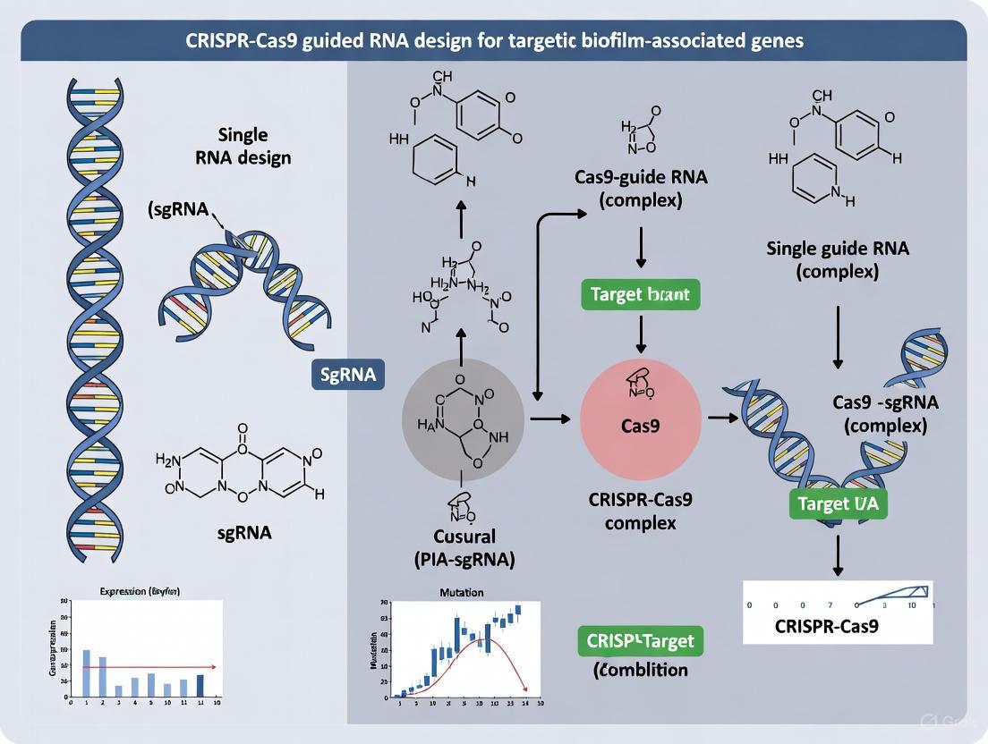
The Clustered Regularly Interspaced Short Palindromic Repeats (CRISPR) and CRISPR-associated (Cas) system has revolutionized genetic engineering, offering unprecedented precision for modifying bacterial genomes. For research focused on biofilm-associated gene targets—where overcoming antibiotic resistance and disrupting complex microbial communities is paramount—optimal single guide RNA (sgRNA) design is the cornerstone of success. An effective sgRNA must achieve three critical objectives: specific binding adjacent to the appropriate Protospacer Adjacent Motif (PAM), high on-target efficiency to ensure effective cleavage or gene repression, and minimal off-target effects to maintain specificity and avoid unintended genomic alterations [35] [5]. This application note provides a detailed protocol for designing sgRNAs against bacterial genomic targets, with a specific emphasis on applications within biofilm research.
Theoretical Foundation: PAM Specificity and gRNA Function
The Indispensable PAM Sequence
The Protospacer Adjacent Motif (PAM) is a short, specific DNA sequence (typically 2-6 base pairs) located directly downstream of the DNA region targeted for cleavage by the CRISPR system. Its primary function is to serve as a binding signal for the Cas nuclease, enabling the distinction between self and non-self DNA, which in natural bacterial immunity prevents the CRISPR system from attacking the bacterium's own genome [35].
The PAM sequence is an absolute requirement for Cas nuclease activity; without it, cleavage will not occur. The sequence of the PAM is strictly dependent on the specific Cas protein used in the experiment. For example, the most commonly used nuclease, SpCas9 from Streptococcus pyogenes, requires a 5'-NGG-3' PAM, where "N" can be any nucleotide base [35] [36].
Mechanism of gRNA-Guided DNA Targeting
In engineered CRISPR systems, the sgRNA is a chimeric RNA molecule comprising two functionally distinct parts: the crRNA-derived segment, which contains the user-defined 17-20 nucleotide spacer sequence complementary to the target DNA, and the tracrRNA scaffold, which is essential for Cas nuclease binding [37]. This sgRNA forms a ribonucleoprotein complex with the Cas nuclease, guiding it to the specific genomic locus. Upon recognizing the correct PAM sequence, the Cas protein unwinds the DNA duplex, allowing the spacer sequence of the sgRNA to anneal to the target DNA strand. Successful base-pairing leads to a conformational change in Cas9, activating its nuclease domains to create a double-strand break (DSB) approximately 3-4 nucleotides upstream of the PAM site [35] [36].
Diagram 1: The foundational workflow for designing a gRNA, beginning with PAM identification and culminating in experimental validation.
Comprehensive gRNA Design Protocol for Bacterial Genomes
Step 1: PAM Selection and Cas Nuclease Choice
The initial and most critical step is selecting a Cas nuclease whose PAM requirement is present near your target site within the bacterial genome. While SpCas9 (PAM: 5'-NGG-3') is widely used, its PAM abundance may be limiting in AT-rich bacterial genomes. Fortunately, a diverse toolkit of Cas nucleases with varying PAM specificities is available [35].
Table 1: Common CRISPR Nucleases and Their PAM Sequences
| CRISPR Nuclease | Organism Isolated From | PAM Sequence (5' to 3') | Advantages for Bacterial Targeting |
|---|---|---|---|
| SpCas9 | Streptococcus pyogenes | NGG | Broadly validated, high activity |
| SaCas9 | Staphylococcus aureus | NNGRR(T) | Smaller size, good for delivery |
| NmeCas9 | Neisseria meningitidis | NNNNGATT | Longer PAM, potentially higher specificity |
| Cas12a (Cpf1) | Lachnospiraceae bacterium | TTTV | Creates staggered ends, no tracrRNA needed |
| hfCas12Max | Engineered from Cas12i | TN and/or TNN | Increased fidelity, flexible PAM |
| AacCas12b | Alicyclobacillus acidiphilus | TTN | Thermostable, useful for certain conditions |
Protocol Recommendation: For biofilm research targeting genes involved in quorum sensing (e.g., luxS) or extracellular polymeric substance (EPS) production (e.g., algD in P. aeruginosa), first identify all available PAM sites within a 200 bp window surrounding the start codon of your target gene. If no suitable PAM is found for SpCas9, consider alternative nucleases like SaCas9 or Cas12a variants [8] [12].
Step 2: gRNA Spacer Design for Optimal On-Target Efficiency
The 17-20 nucleotide spacer sequence directly upstream of the PAM is the determinant of specificity. The following principles, derived from large-scale screens, should guide its selection [38] [39] [40].
Table 2: Features Influencing gRNA On-Target Efficiency
| Feature Category | Efficient Features (Prefer) | Inefficient Features (Avoid) |
|---|---|---|
| Overall Nucleotide Usage | High 'A' count; 'A' in the middle; AG, CA, AC, UA dinucleotides | High 'U', 'G' count; GG, GGG repeats; UU, GC dinucleotides |
| Position-Specific Nucleotides | 'G' at position 20 (adjacent to PAM); 'G' or 'A' at position 19; 'C' at position 18 | 'C' at position 20; 'U' in positions 17–20; 'G' at position 16; 'T' in PAM (TGG) |
| GC Content | 40% - 60% | GC > 80% or < 20% |
| Thermodynamic Stability | Moderate gRNA:DNA hybridization energy | Extremely stable gRNA secondary structures (MFE < -7.5 kcal/mol) |
| Target Gene Context (CRISPRi) | High target gene expression level; proximity to Transcription Start Site (TSS) | Essential genes in the same operon (polar effects) |
Experimental Protocol: In Silico gRNA Design
- Input Sequence: Extract a 300-500 bp genomic sequence centered on your target region (e.g., the promoter or early coding sequence of a biofilm-related gene).
- Identify PAM Sites: Scan the sequence for all instances of the PAM corresponding to your chosen nuclease (e.g., 'GG' for SpCas9) on both strands.
- Extract Spacers: For each PAM, extract the 20 nucleotides immediately upstream as a candidate spacer sequence.
- Initial Filtering: Discard spacers with:
- GC content < 20% or > 80%.
- Homopolymer runs (e.g., AAAA, GGGG) ≥ 4 nt.
- Obvious self-complementarity that could form stable secondary structures.
- Efficiency Scoring: Input the remaining candidate spacer sequences into a predictive algorithm such as CRISPRon (for Cas9 editing) or the mixed-effect random forest model described by Gussomes et al. (for bacterial CRISPRi) [39] [40]. These tools integrate sequence features and thermodynamic properties to generate a normalized efficiency score.
- Final Selection: Proceed with the top 2-3 ranked gRNAs for experimental validation to account for potential unpredictability in cellular contexts.
Step 3: Rigorous Specificity and Off-Target Assessment
A gRNA with even partial complementarity to non-target genomic sites can cause off-target effects, which is a critical concern in bacterial biofilm studies where related strains may share homologous sequences [5].
Protocol for Specificity Checks:
- Genome-Wide Alignment: Perform a BLASTN or use dedicated tools like Cas-OFFinder to search the entire bacterial genome for sites with significant homology to your candidate gRNA spacer.
- Analyze Mismatches: Pay close attention to potential off-target sites with:
- Fewer than 4 mismatches to the gRNA spacer.
- Mismatches concentrated in the 5' end of the gRNA (distal to the PAM). The "seed sequence" (8-12 bases proximal to the PAM) is less tolerant of mismatches [36].
- Bulges or indels in the potential DNA-RNA hybrid.
- Filter and Select: Discard any gRNA that has a near-perfect match (≥17/20 nt identity) elsewhere in the genome, especially within another coding sequence. For applications requiring extreme specificity, such as targeting a specific allele within a mixed biofilm community, consider using high-fidelity Cas9 variants (e.g., eSpCas9, SpCas9-HF1) [36].
Diagram 2: The key factor groups that collectively determine the final on-target efficiency of a gRNA.
Experimental Validation and Application in Biofilm Research
Protocol: Validating gRNA Efficiency and Specificity
Validation is non-negotiable. The following methods are used to confirm editing outcomes.
Table 3: Key Reagent Solutions for Bacterial CRISPR Experiments
| Research Reagent | Function/Explanation | Example Use Case |
|---|---|---|
| High-Fidelity DNA Polymerase | Amplifies the target genomic locus with minimal error rates for downstream sequencing. | Preparing amplicons for NGS validation of indels. |
| Next-Generation Sequencing (NGS) | Provides a quantitative and comprehensive profile of all induced mutations at the target site. | Gold-standard method for quantifying on-target indel frequency and characterizing off-target effects [41]. |
| T7 Endonuclease I (T7E1) Assay | A mismatch-cleavage enzyme that detects heteroduplex DNA formed by indel mutations. | A cost-effective, rapid initial check for activity. Note: Can underestimate efficiency, especially when >30% [41]. |
| Tracking of Indels by Decomposition (TIDE) | A computational method that uses Sanger sequencing traces to deconvolute a mixture of indels. | A simple, accessible method for estimating editing efficiency in pooled populations without NGS [41]. |
Detailed NGS Validation Workflow:
- PCR Amplification: Design primers flanking the target site (amplicon size: 250-400 bp). Perform PCR on genomic DNA extracted from both control and CRISPR-treated bacterial cultures.
- Library Preparation and Sequencing: Purify the PCR products and prepare a sequencing library using a platform like Illumina MiSeq (2x250 bp paired-end reads is typical).
- Data Analysis: Process the sequencing data through a pipeline like CRISPResso2 to align reads to the reference sequence and precisely quantify the spectrum and frequency of insertion/deletion (indel) mutations at the target site. A successful experiment typically shows >50% indel frequency for a highly efficient gRNA.
Application: gRNA Design for Biofilm-Associated Gene Targets
Biofilms present a unique challenge due to their genetic heterogeneity and protective matrix. CRISPR-Cas can be deployed not only for gene knockout but also for transcriptional repression using a catalytically dead Cas9 (dCas9) in CRISPR interference (CRISPRi) systems [12] [40].
Case Study: Targeting a Quorum Sensing Gene
- Target Selection: Select a key regulator like lasI in P. aeruginosa, which is essential for producing acyl-homoserine lactone (AHL) signaling molecules.
- gRNA Design for CRISPRi: For repression, design gRNAs to target the non-template strand within the promoter or early coding region, as close to the Transcription Start Site (TSS) as possible. This position maximally interferes with RNA polymerase binding or progression [40].
- Efficiency Prediction: Use a bacterial-specific model that incorporates gene expression data, as high-expression targets like lasI may show stronger depletion phenotypes in CRISPRi screens [40].
- Delivery: Co-express dCas9 and the validated sgRNA in P. aeruginosa using a plasmid system. The repression of lasI should disrupt quorum sensing, potentially reducing biofilm formation and virulence, which can be assayed by crystal violet staining or AHL biosensors.
Table 4: Computational Tools for gRNA Design and Analysis
| Tool Name | Primary Function | Relevant Context |
|---|---|---|
| CRISPRon | Deep learning model for predicting on-target gRNA activity for SpCas9. | Shown to outperform other tools; incorporates binding energy (ΔGB) [39]. |
| CHOPCHOP | User-friendly web tool for designing gRNAs for various nucleases and species. | Supports alternative Cas nucleases and PAM recognition [37]. |
| Cas-Offinder | Searches for potential off-target sites across a genome. | Essential for specificity checks; allows for bulged and mismatched off-target prediction [37]. |
| Mixed-Effect Random Forest (ML Model) | Predicts guide depletion in bacterial CRISPRi screens by integrating guide and gene-specific features. | Crucial for bacterial CRISPRi; accounts for target gene expression and operon context [40]. |
Precise gRNA design is the foundation of effective CRISPR experiments in bacterial systems, especially for complex targets like biofilm-associated genes. By systematically selecting the appropriate Cas nuclease and PAM, designing spacers using state-of-the-art efficiency predictors, and conducting rigorous specificity checks, researchers can significantly increase their chances of success. Finally, employing robust validation methods like NGS is critical to confirm high on-target activity and rule out significant off-target effects, thereby ensuring the reliability of subsequent phenotypic analyses in biofilm research.
The challenge of combating biofilm-associated infections is a pressing issue in modern therapeutics, as biofilms confer up to 1000-fold greater tolerance to antibiotics compared to planktonic cells [8]. CRISPR-Cas9 technology presents a transformative approach for the precise disruption of genes essential for biofilm formation, stability, and antibiotic resistance [12]. However, the success of these interventions hinges on the selection of highly efficient and specific guide RNAs (gRNAs). The integration of bioinformatics tools and artificial intelligence (AI) has become indispensable for moving beyond empirical gRNA design toward predictive modeling, enabling researchers to systematically identify optimal gRNA sequences for targeting biofilm-associated genes with enhanced precision and efficacy [42] [38]. This protocol details a comprehensive methodology for leveraging these computational advances within the specific context of biofilm research.
Bioinformatics and AI Tools for Predictive gRNA Modeling
The initial design phase relies on computational tools to predict gRNA on-target activity and off-target effects. The following table summarizes the key categories and examples of such tools.
Table 1: Categories of Bioinformatics and AI Tools for gRNA Design
| Tool Category | Description | Key Tools & Models | Relevance to Biofilm Research |
|---|---|---|---|
| AI-Driven On-Target Predictors | Deep learning models trained on large-scale gRNA activity datasets to forecast cleavage efficiency. | CRISPRon [39], DeepSpCas9 [42], CRISPR-Net [43] | Identifies gRNAs with high predicted activity against biofilm regulator genes (e.g., quorum sensing, EPS production). |
| Off-Target Effect Predictors | Models that score potential off-target cleavage at genomic sites with sequence similarity. | Cutting Frequency Determination (CFD) score [42], DeepCRISPR [42] | Ensures specificity, minimizing unintended edits in bacterial genomes during anti-biofilm interventions. |
| Multitask & Integrated AI Models | Models that jointly predict on-target and off-target activities to balance efficiency and specificity. | Models by Vora et al. [43], Hybrid deep learning models [43] | Provides a holistic gRNA scoring system for designing safe and effective biofilm-targeting strategies. |
| Generative AI for Novel Editors | Large language models (LLMs) used to design novel Cas proteins with optimal properties. | OpenCRISPR-1 (AI-generated editor) [44] | Offers potential for developing bespoke editors optimized for targeting specific biofilm-forming pathogens. |
The field has evolved from hypothesis-driven rules to sophisticated deep learning models. Early models like Rule Set 1 and 2 identified simple sequence features associated with gRNA activity, such as GC content and position-specific nucleotide preferences [42] [38]. The current state-of-the-art leverages deep learning architectures, such as convolutional neural networks (CNNs) and recurrent neural networks (RNNs), which automatically extract relevant features from gRNA and target DNA sequences [38] [43]. For instance, CRISPRon integrates sequence information with thermodynamic properties like gRNA-DNA binding energy (ΔGB) and epigenomic data, achieving superior performance by training on a dataset of 23,902 gRNAs [39]. Furthermore, models like CRISPR-Net employ a combination of CNNs and bidirectional GRUs to analyze sequences with mismatches, enhancing off-target prediction [43].
Table 2: Key Features Influencing gRNA Efficiency as Identified by AI Models
| Feature Category | Efficient Features | Inefficient Features |
|---|---|---|
| Nucleotide Composition | High adenine (A) count; AG, CA, AC, UA dinucleotides [38] | High uracil (U) and guanine (G) count; GG, GGG motifs; GGGG repeats [38] |
| Position-Specific Nucleotides | Guanine (G) at position 20; Adenine (A) at position 19; Cytosine (C) at positions 16 & 18 [38] | Cytosine (C) at position 20; Uracil (U) in positions 17-20; Thymine (T) in PAM [38] |
| Structural & Energetic | GC content between 40-60%; Stable gRNA secondary structure (MFE > -7.5 kcal/mol) [39]; Favorable gRNA-DNA binding energy (ΔGB) [39] | GC content >80%; Unstable gRNA structures [38] [39] |
Experimental Protocol for gRNA Design and Validation Against Biofilm Targets
This section provides a detailed, step-by-step protocol for designing and validating gRNAs targeting biofilm-associated genes.
In Silico gRNA Design and Selection Workflow
Step 1: Target Gene and Locus Identification
- Objective: Identify critical genes involved in biofilm formation (e.g., genes for quorum sensing, adhesion, extracellular polymeric substance (EPS) production, or antibiotic resistance) in your target bacterial pathogen [12] [32].
- Procedure: Use genomic databases (e.g., NCBI, UniProt) to obtain the DNA sequence of the target gene. Identify a specific protospacer adjacent motif (PAM) site (e.g., 5'-NGG-3' for SpCas9) within the coding or regulatory sequence of the gene.
Step 2: Candidate gRNA Retrieval
- Objective: Generate a list of all possible gRNA sequences adjacent to the PAM site.
- Procedure: Input the target gene sequence into an alignment-based bioinformatics tool such as CHOPCHOP or E-CRISP [45] [46]. These tools will scan the input sequence and return a list of candidate gRNA sequences (typically 20 nucleotides upstream of the PAM).
Step 3: On-target and Off-target Scoring
- Objective: Rank candidate gRNAs based on predicted efficiency and specificity.
- Procedure:
- On-target Activity Prediction: Submit the candidate gRNA sequences to an AI-based predictor like CRISPRon or DeepSpCas9 [42] [39]. These tools will provide a quantitative score predicting the gRNA's cleavage efficiency.
- Off-target Effect Prediction: Use the same candidate list in tools that compute the Cutting Frequency Determination (CFD) score or use deep learning models like DeepCRISPR to identify and score potential off-target sites across the genome [42] [45].
- Output: A ranked list of gRNAs with high on-target and low off-target scores.
Step 4: Final gRNA Selection
- Objective: Select the top 3-5 gRNA candidates for experimental validation.
- Procedure: Prioritize gRNAs that exhibit a balance of high on-target activity (e.g., CRISPRon score > 0.6), low off-target potential (CFD score for off-targets < 0.1), and target key functional domains of the biofilm-associated gene.
Experimental Validation of gRNA Efficacy
Step 1: gRNA Cloning and Delivery Vector Preparation
- Objective: Clone the selected gRNA sequences into an appropriate CRISPR-Cas9 delivery vector.
- Materials:
- Plasmid Vector: A plasmid encoding SpCas9 and a scaffold for gRNA insertion (e.g., pSpCas9(BB)).
- Oligonucleotides: Designed sense and antisense oligonucleotides corresponding to the target gRNA sequence, with 5' overhangs compatible with the vector's restriction sites (e.g., BsmBI or BsaI).
- Procedure:
- Phosphorylate and anneal the oligonucleotides.
- Digest the plasmid vector with the appropriate restriction enzyme.
- Ligate the annealed oligonucleotide duplex into the digested vector.
- Transform the ligation product into competent E. coli, then plate on selective media.
- Select colonies, culture, and extract plasmid DNA. Verify the insert by Sanger sequencing.
Step 2: Delivery into Bacterial Pathogens
- Objective: Introduce the CRISPR-Cas9 construct into the target biofilm-forming bacteria.
- Materials:
- Nanoparticles: Lipid-based or gold nanoparticles (AuNPs) can be complexed with the CRISPR plasmid or ribonucleoprotein (RNP) complex to enhance delivery, especially through biofilm matrices [8]. For instance, gold nanoparticle carriers have been shown to enhance editing efficiency up to 3.5-fold [8].
- Electroporation Equipment: For physical delivery of plasmids or RNPs.
- Procedure:
- For nanoparticle delivery: Mix the CRISPR construct with the nanoparticles according to the manufacturer's protocol and incubate with the bacterial culture.
- For electroporation: Wash and concentrate bacterial cells in an electroporation buffer, mix with the plasmid DNA or RNP, and electroporate using optimized parameters for the specific bacterium.
Step 3: Quantification of Gene Editing and Biofilm Disruption
- Objective: Assess the functional outcome of the CRISPR intervention on gene editing and biofilm integrity.
- Materials:
- Deep Sequencing Platform (e.g., Illumina): To precisely quantify indel frequencies at the target locus.
- PCR Reagents: For amplifying the target genomic region for sequencing.
- Biofilm Assay Kits: (e.g., crystal violet staining kit) to quantify total biofilm biomass.
- Confocal Laser Scanning Microscopy (CLSM): For high-resolution imaging of biofilm architecture [8] [32].
- Procedure:
- On-target Efficiency Analysis: Harvest bacterial cells 48-72 hours post-transfection. Extract genomic DNA, PCR-amplify the target region, and subject the product to deep sequencing. Analyze the sequencing data with a tool like CRISPResso2 to quantify the percentage of indels.
- Biofilm Phenotypic Assay:
- Culture the CRISPR-treated and control bacteria in biofilm-promoting conditions (e.g., in microtiter plates).
- Stain the adherent biofilm with crystal violet, elute the dye, and measure its absorbance at 595 nm to quantify biomass.
- For imaging, grow biofilms on coverslips, stain with fluorescent dyes (e.g., SYTO 9 for cells), and visualize using CLSM to observe structural disintegration.
The Scientist's Toolkit: Research Reagent Solutions
Table 3: Essential Research Reagents and Materials for CRISPR Biofilm Experiments
| Item Name | Function/Application | Example Use Case |
|---|---|---|
| SpCas9 Plasmid Vector | Provides the genetic code for the Cas9 nuclease and gRNA scaffold. | Backbone for cloning designed gRNAs targeting biofilm genes. |
| Lipid-Based Nanoparticles | Enhances cellular uptake of CRISPR constructs; protects genetic material. | Delivery of plasmid or RNP into bacterial pathogens through biofilm EPS [8]. |
| Gold Nanoparticles (AuNPs) | Serves as a carrier for CRISPR-Cas9 components; enables controlled release. | Co-delivery of Cas9 protein and gRNA, shown to increase editing efficiency [8]. |
| CRISPResso2 Software | Computational tool for analyzing next-generation sequencing data from CRISPR experiments. | Quantification of indel frequencies and mapping of repair outcomes at the target locus. |
| Crystal Violet Staining Kit | Dye-based assay for quantifying total biofilm biomass. | High-throughput assessment of biofilm reduction post-CRISPR treatment. |
The integration of predictive AI models with robust experimental protocols creates a powerful pipeline for designing gRNAs against biofilm-associated genes. This approach moves beyond trial-and-error, enabling the rational selection of gRNAs that are both highly active and specific. As AI models continue to evolve, incorporating factors like genomic variation and epigenetic context, and as delivery systems like nanoparticles become more sophisticated, the precision and efficacy of CRISPR-based biofilm control will be further enhanced, paving the way for novel anti-biofilm therapies [43] [12].
Biofilm-associated infections represent a significant challenge in therapeutic treatment due to their inherent resistance to conventional antibiotics. The extracellular polymeric substance (EPS) matrix of biofilms acts as a formidable physical and chemical barrier, severely limiting the penetration and efficacy of antimicrobial agents [8] [47]. CRISPR-Cas9 technology has emerged as a powerful tool for precision targeting of biofilm-forming pathogens, capable of disrupting virulence genes, antibiotic resistance determinants, and quorum-sensing pathways [12]. However, the efficient delivery of CRISPR components through the biofilm matrix to target bacterial cells remains a major translational hurdle. Engineered lipid and gold nanoparticles have shown exceptional promise as advanced delivery platforms to overcome these penetration barriers, enabling effective CRISPR-based antimicrobial strategies against resilient biofilm communities [48] [8].
Nanoparticle Engineering and Design Specifications
Lipid Nanoparticles (LNPs) for CRISPR Delivery
Lipid nanoparticles represent a versatile non-viral delivery platform for CRISPR-Cas9 components, with well-defined compositional parameters that govern their efficacy [49]. The modular nature of LNP design allows for systematic optimization to enhance biofilm penetration and cellular delivery.
Table 1: Composition and Characteristics of CRISPR-Loaded Lipid Nanoparticles
| Component Category | Specific Examples | Molar Ratio Range | Function in Formulation |
|---|---|---|---|
| Ionizable Lipids | ALC-0315, ALC-0307, C12-200, CKK-E12 | 35-50% | pH-responsive encapsulation and endosomal escape |
| Structural Lipids | DSPC, DOPE | 10-20% | Define bilayer structure and stability |
| Sterols | Cholesterol, Beta-sitosterol | 38.5-40% | Regulate membrane fluidity and integrity |
| PEG-Lipids | ALC-0159, C14-PEG2000 | 1.5-2.5% | Enhance stability, reduce opsonization, control biodistribution |
| Performance Metrics | Value Range | Measurement Conditions | Impact on Delivery |
| Hydrodynamic Diameter | 50-150 nm | Dynamic light scattering | Optimal for biofilm penetration and cellular uptake |
| Polydispersity Index | <0.2 | Dynamic light scattering | Uniformity of nanoparticle population |
| Zeta Potential | Slightly negative to neutral | Physiological pH | Reduced non-specific binding |
| Encapsulation Efficiency | >90% | RIBOGreen assay | Payload protection and delivery capacity |
LNPs have demonstrated remarkable versatility in accommodating different forms of CRISPR cargo, including plasmid DNA, mRNA, and preassembled ribonucleoprotein (RNP) complexes [50]. The transient nature of LNP-mediated CRISPR delivery is particularly advantageous for antimicrobial applications, as it reduces the likelihood of off-target effects while maintaining high editing efficiency [49]. Recent advances in LNP formulation have employed artificial intelligence-driven approaches, such as transformer-based neural networks (COMET), to optimize lipid compositions and ratios for specific delivery applications, potentially accelerating the development of LNPs tailored for biofilm penetration [51].
Gold Nanoparticles (AuNPs) for CRISPR Delivery
Gold nanoparticles offer unique advantages for CRISPR delivery, including precise size control, facile surface functionalization, and tunable optical properties [48]. Their versatility in structural configurations enables tailored approaches for biofilm penetration and intracellular delivery.
Table 2: Gold Nanoparticle Platforms for CRISPR Delivery
| Nanoparticle Type | Size Range | Surface Functionalization | Editing Efficiency | Key Advantages |
|---|---|---|---|---|
| Gold Nanorods | 60-150 nm | Cationic polymers, cancer cell membranes | 15-33% indel formation | Enhanced cellular uptake, photothermal properties |
| Spherical Gold NPs | 3-500 nm | Protamine, polyethylenimine (PEI) | 30-60% knock-out efficiency | High stability, versatile chemistry |
| Gold-Loaded Core-Shell Tecto Dendrimers | 108-131 nm | Anionic/cationic layering | ~60% PD-L1 knock-out | Controlled release properties |
| Performance Aspect | Optimization Strategy | Impact on Biofilm Penetration | Experimental Evidence | |
| Cellular Uptake | Surface charge modulation (+10 to +30 mV) | Enhanced diffusion through EPS matrix | 3.5-fold increase compared to non-carrier systems [8] | |
| Stability in Biofilm Environment | PEGylation, biomimetic coatings | Reduced non-specific binding | Maintained structural integrity for >72 hours [48] | |
| Targeting Specificity | Ligand conjugation (antibodies, peptides) | Selective pathogen targeting within multispecies biofilms | 90% binding to target bacterial species [49] |
Gold nanoparticles can be engineered to exploit their inherent antimicrobial properties while serving as efficient carriers for CRISPR components [48]. The surface plasmon resonance properties of certain gold nanostructures can be leveraged for triggered release mechanisms or synergistic photothermal therapy to enhance biofilm disruption [8]. Furthermore, the ability to functionalize AuNPs with targeting ligands enables specific binding to pathogen surfaces within complex biofilm communities, improving the precision of CRISPR delivery while minimizing off-target effects on commensal microorganisms [48].
Experimental Protocols
LNP Formulation and CRISPR Encapsulation Protocol
Title: LNP Formulation Workflow
Materials:
- Ionizable lipid (e.g., ALC-0315 or C12-200): Primary component for nucleic acid complexation and endosomal release
- Distearoylphosphatidylcholine (DSPC): Structural lipid for bilayer stability
- Cholesterol: Membrane rigidity modulator
- PEG-lipid (e.g., DMG-PEG2000): Surface stabilization and pharmacokinetics
- CRISPR cargo: sgRNA and Cas9 mRNA or RNP complex
- Microfluidic device: Precision mixing chamber (e.g., NanoAssemblr)
- Dialysis membranes: Molecular weight cutoff 10-20 kDa
Procedure:
- Lipid Stock Preparation: Dissolve lipid components in ethanol at the following molar ratios: ionizable lipid (50%), DSPC (10%), cholesterol (38.5%), and PEG-lipid (1.5%). Maintain total lipid concentration at 10 mg/mL [49].
- Aqueous Phase Preparation: Dilute CRISPR cargo (mRNA at 0.2 mg/mL or RNP at 0.5 mg/mL) in 25 mM sodium acetate buffer (pH 4.0). For RNP complexes, pre-incubate Cas9 protein with sgRNA at 1:1.5 molar ratio for 10 minutes at room temperature prior to formulation [48].
- Nanoparticle Formation: Utilize microfluidic mixing with total flow rate of 12 mL/min and aqueous-to-organic flow rate ratio of 3:1. Collect effluent in sterile containers [51].
- Buffer Exchange: Dialyze formed LNPs against phosphate-buffered saline (PBS, pH 7.4) for 24 hours at 4°C using 10-20 kDa molecular weight cutoff dialysis membranes.
- Characterization: Determine particle size (Z-average), polydispersity index (PDI), and zeta potential using dynamic light scattering. Measure encapsulation efficiency via RIBOGreen assay following Triton X-100 disruption [49].
Gold Nanoparticle Functionalization and CRISPR Loading
Title: AuNP Functionalization Process
Materials:
- Gold nanorods: 60 nm length, 15 nm width, synthesized via seed-mediated growth
- Polyethylenimine (PEI): Branched, 25 kDa molecular weight for surface coating
- CRISPR RNP complexes: Cas9 protein pre-complexed with target-specific sgRNA
- Crosslinkers: SM(PEG)8 or EDC/NHS chemistry for covalent attachment
- Targeting ligands: Antibody fragments or bacterial-specific peptides
- Purification equipment: Ultracentrifugation system with sucrose density gradients
Procedure:
- Gold Nanoparticle Synthesis: Prepare gold nanorods via seed-mediated growth method. Characterize using transmission electron microscopy (TEM) and UV-Vis spectroscopy to ensure uniform morphology and surface plasmon resonance peaks [48].
- Surface Modification: Incubate AuNPs with 2 mg/mL PEI solution (1:10 volume ratio) for 2 hours at room temperature with gentle agitation. Purify via centrifugation at 14,000 × g for 15 minutes and resuspend in nuclease-free water [48].
- CRISPR Loading: Complex CRISPR RNP (at 1 μM concentration) with PEI-coated AuNPs at N:P ratio of 10:1 for 30 minutes at room temperature. For covalent conjugation, utilize SM(PEG)8 crosslinkers according to manufacturer's protocol [48].
- Targeting Functionalization: Incubate CRISPR-loaded AuNPs with targeting ligands (e.g., anti-Pseudomonas antibodies at 50 μg/mL) for 1 hour at 4°C. Use EDC/NHS chemistry for stable amine coupling where necessary.
- Purification and Validation: Purify functionalized nanoparticles via sucrose density gradient centrifugation (10-40% w/v). Characterize final formulation using dynamic light scattering, TEM, and gel electrophoresis to confirm CRISPR loading efficiency [48].
Biofilm Penetration and Gene Editing Assessment
Title: Biofilm Penetration Assay
Materials:
- Biofilm model systems: CDC biofilm reactor or flow-cell systems
- Confocal laser scanning microscopy (CLSM): With Z-stack imaging capability
- Fluorescent reporters: FITC-labeled nanoparticles or Cy5-tagged CRISPR components
- Molecular biology reagents: T7 endonuclease I, PCR components, Sanger sequencing
- Viability assays: Resazurin, propidium iodide, colony counting materials
Procedure:
- Biofilm Establishment: Culture biofilm-forming bacteria (e.g., Pseudomonas aeruginosa PAO1 or Staphylococcus aureus) on relevant substrates (silicone, polystyrene, or glass) for 72 hours in appropriate growth media to establish mature biofilms with characteristic EPS matrix [8] [47].
- Nanoparticle Treatment: Apply fluorescently labeled LNP or AuNP formulations (100 μL containing 1 μg CRISPR payload) to established biofilms. Incubate for predetermined timepoints (2, 6, 12, and 24 hours) under appropriate culture conditions [8].
- Penetration Analysis: Fix biofilms with 4% paraformaldehyde for 30 minutes. Image using confocal laser scanning microscopy with Z-stack sectioning (1 μm intervals). Quantify fluorescence intensity at different biofilm depths using ImageJ or similar software [8].
- Gene Editing Assessment: Following nanoparticle treatment (24-48 hours), extract genomic DNA from biofilm cells. Amplify target regions via PCR and analyze editing efficiency using T7 endonuclease I assay or tracking of indels by decomposition (TIDE) analysis [12] [8].
- Functional Efficacy Evaluation: Assess biofilm viability using resazurin reduction assays and quantify colony-forming units following nanoparticle treatment. For combination approaches, evaluate synergy with conventional antibiotics where applicable [8].
The Scientist's Toolkit: Essential Research Reagents
Table 3: Key Research Reagent Solutions for CRISPR-Nanoparticle Biofilm Studies
| Reagent Category | Specific Product Examples | Function in Experimental Workflow | Key Considerations for Selection |
|---|---|---|---|
| CRISPR Core Components | Alt-R S.p. Cas9 Nuclease V3, Sigma-Aldrich sgRNA, IDT Cas9 mRNA | Genome editing machinery | Purity, modification status, nuclease-free formulation |
| Lipid Nanoparticle Components | Avanti Polar Lipids ionizable lipids, PreciChrom cholesterol, NOF PEG-lipids | LNP structural formation | Batch-to-batch consistency, pharmaceutical grade |
| Gold Nanoparticle Materials | Cytodiagnostics gold nanorods, Sigma-Aldrich HAuCl4, Thermo Fisher PEI | AuNP synthesis and functionalization | Size uniformity, surface reactivity, endotoxin levels |
| Biofilm Assay Reagents | Corning biofilm substrates, Invitrogen LIVE/DEAD BacLight, Promega CellTiter-Glo | Biofilm growth and viability assessment | Compatibility with imaging systems, sensitivity range |
| Analytical Tools | Malvern Zetasizer, Molecular Devices SpectraMax, Leica confocal microscopes | Nanoparticle and biofilm characterization | Measurement precision, software capabilities |
The integration of engineered lipid and gold nanoparticles with CRISPR-Cas9 technology represents a paradigm shift in our approach to combating biofilm-associated infections. The precise design parameters outlined in this protocol—including optimal size ranges, surface functionalization strategies, and composition ratios—enable researchers to develop advanced delivery platforms capable of penetrating the complex biofilm matrix. These nanoparticle systems address the critical challenge of delivering functional CRISPR components to target pathogens within biofilms, opening new avenues for precision antimicrobial therapy. As research in this field advances, further optimization of targeting specificity and payload release kinetics will enhance the therapeutic potential of these innovative platforms for clinical application against persistent biofilm-mediated infections.
Multiplexed CRISPR-Cas9 technology represents a significant advancement in genetic engineering, enabling researchers to target multiple genomic loci simultaneously. This approach involves the design and delivery of multiple guide RNAs (gRNAs) that work in concert with the Cas9 nuclease to disrupt several genes in a single experiment. In the context of biofilm-associated gene research, where complex genetic networks govern bacterial persistence and antibiotic resistance, multiplexing offers a powerful tool for dissecting polygenic traits and functional pathways [52] [53].
The fundamental advantage of multiplexing lies in its ability to ensure that each cell receiving the CRISPR components contains all desired gRNAs, dramatically increasing the probability of generating the complete set of desired genetic modifications. This is particularly valuable for addressing multifactorial biological processes such as biofilm formation, which involves coordinated expression of numerous genes related to adhesion, matrix production, quorum sensing, and stress adaptation [52] [12].
Strategic Approaches to gRNA Multiplexing
Platform Selection for Multiplexed gRNA Expression
Table 1: Comparison of Major Multiplexed gRNA Expression Systems
| System Type | Mechanism | Maximum gRNA Capacity | Key Advantages | Limitations | Example Applications |
|---|---|---|---|---|---|
| Tandem Promoters | Multiple individual Pol III promoters (U6, H1) each driving single gRNA | Typically 2-4 gRNAs | Simplified validation; predictable expression levels | Promoter cross-talk; larger plasmid size | Dual nickase systems; small-scale knockouts [52] |
| Polycistronic tRNA-gRNA | tRNA-processing machinery cleaves gRNAs from single transcript | Up to 8 gRNAs | Compact size; uses endogenous enzymes; no additional co-factors | Processing efficiency varies; possible improper cleavage | Plant genome engineering; metabolic pathway manipulation [52] [53] |
| Csy4-Recognized Array | Csy4 endoribonuclease cleaves at specific 28-nt sequences flanking gRNAs | 10+ gRNAs | High processing efficiency; precise cleavage | Requires Csy4 co-expression; potential cytotoxicity | Large-scale genome engineering; genetic circuits [52] [54] |
| Cas12a Processed Array | Cas12a itself processes pre-crRNA from a single transcript | 5+ gRNAs | No additional factors needed; self-processing | Limited to Cas12a systems; PAM requirements | Transcriptional regulation; multiple gene activation/repression [53] [55] |
| Ribozyme-Processed Array | Hammerhead and HDV ribozymes flank gRNAs for self-cleavage | 5+ gRNAs | No protein co-factors needed; works with Pol II promoters | Variable processing efficiency; larger construct size | Inducible systems; tissue-specific multiplexing [53] |
gRNA Design Considerations for Biofilm Targets
When designing gRNA cocktails for disrupting biofilm-associated genes, several critical factors must be considered to ensure maximum efficiency and specificity:
On-target efficiency prediction: Utilize established algorithms that incorporate sequence features such as GC content, position-specific nucleotide preferences, and secondary structure predictions. Tools like the IDT Custom Alt-R CRISPR-Cas9 guide RNA design tool employ machine learning models trained on thousands of gRNAs to predict editing efficiency [56].
Off-target minimization: Carefully evaluate potential off-target sites across the genome by assessing mismatch tolerance, especially in the seed region adjacent to the PAM sequence. Cross-referencing with biofilm-specific gene databases can help identify and avoid potential off-targets within related gene families [57] [56].
Functional redundancy: For essential biofilm processes, design multiple gRNAs targeting the same gene or pathway to ensure complete disruption, as this can compensate for variations in individual gRNA efficiency [52] [12].
PAM sequence requirements: Consider the PAM requirements of your specific Cas variant (5'-NGG-3' for standard S. pyogenes Cas9) and ensure adequate target site availability within your genes of interest. For biofilm targets with limited PAM sites, consider alternative Cas enzymes such as Cas12a (recognizes TTTV PAM) [56] [55].
Experimental Protocols for Multiplexed gRNA Implementation
Golden Gate Assembly for gRNA Array Construction
The Golden Gate assembly method has emerged as a particularly robust approach for constructing gRNA arrays due to its high efficiency and modularity [52] [54]. Below is a detailed protocol for assembling multiplexed gRNA constructs using the Golden Gate system:
Materials Required:
- BsaI or BsmBI restriction enzyme (Type IIS)
- T4 DNA Ligase and buffer
- Plasmid backbone containing destination vector
- Entry clones with individual gRNA expression cassettes
- Chemically competent E. coli cells
- Appropriate antibiotic selection plates
Step-by-Step Procedure:
gRNA Oligonucleotide Design and Cloning:
- Design oligonucleotides for each gRNA target sequence, including appropriate overhangs for BbsI cloning (forward oligo: 5'-CACCg[20nt target sequence]-3'; reverse oligo: 5'-AAAC[20nt target reverse complement]C-3').
- Phosphorylate and anneal oligonucleotides using T4 PNK in T4 ligation buffer with the following thermal cycling program: 37°C for 30 minutes, 95°C for 5 minutes, then ramp down to 25°C at 5°C/minute.
- Ligate annealed oligos into BbsI-digested entry vector using T4 DNA ligase (incubate at room temperature for 1 hour).
- Transform into competent E. coli, plate on selective media, and verify clones by Sanger sequencing [54].
Golden Gate Assembly Reaction:
- Set up the assembly reaction containing:
- 50-100 ng destination vector
- Equimolar amounts of each gRNA entry clone (typically 20-50 ng each)
- 1μL BsaI or BsmBI restriction enzyme
- 1μL T4 DNA ligase
- 1X T4 DNA ligase buffer
- Nuclease-free water to 20μL total volume
- Run the following thermal cycling program:
- Set up the assembly reaction containing:
Transformation and Verification:
- Transform 2-5μL of the Golden Gate reaction into chemically competent E. coli.
- Select on appropriate antibiotic plates and screen multiple colonies by colony PCR or restriction digest.
- Confirm final assembly by Sanger sequencing across all gRNA insertion sites.
Quantitative Assessment of Editing Efficiency
Validating the efficiency of each gRNA in your multiplexed cocktail is essential for interpreting experimental outcomes. The qEva-CRISPR method provides a robust quantitative approach for evaluating editing efficiency at multiple target sites simultaneously [57].
qEva-CRISPR Protocol:
Genomic DNA Extraction:
- Harvest cells 72-96 hours post-transfection using your preferred method (e.g., GeneJET Genomic DNA Purification Kit)
- Quantify DNA concentration and quality using spectrophotometry
Probe Design and Hybridization:
- Design specific probes for each target site and internal reference genes
- Set up hybridization reaction:
- 100-200ng genomic DNA
- 1μL probe mix (0.5-1fmol/μL each probe)
- Nuclease-free water to 10μL total
- Denature at 98°C for 5 minutes, then hybridize at 60°C for 16-20 hours
Ligation and PCR Amplification:
- Add 10μL ligation mixture (containing ligase in appropriate buffer) to hybridized samples
- Incubate at 54°C for 15 minutes, then inactivate at 98°C for 5 minutes
- Add 20μL PCR master mix with fluorescently labeled primers
- Amplify with the following program:
- 95°C for 2 minutes
- 35 cycles of: (95°C for 30s + 60°C for 30s + 72°C for 60s)
- 72°C for 20 minutes
Fragment Analysis and Quantification:
- Separate PCR products by capillary electrophoresis
- Analyze peak areas for target and reference amplicons
- Calculate editing efficiency using the formula: % Editing = [1 - (Target peak area/Reference peak area)treated / (Target peak area/Reference peak area)control] × 100% [57]
Table 2: Expected Editing Efficiencies for Different Multiplexing Applications
| Application | Target Organism | Expected Efficiency Range | Key Influencing Factors | Validation Methods |
|---|---|---|---|---|
| Dual-gene knockout | P. pastoris | 60-100% | gRNA efficiency, delivery method | Phenotypic screening, sequencing [58] |
| Multi-gene integration | P. pastoris | 30-70% | Homology arm length, repair template design | PCR verification, functional assays [58] |
| Biofilm gene disruption | Bacterial pathogens | 40-90% | gRNA design, delivery efficiency | qEva-CRISPR, biofilm formation assays [12] [57] |
| Metabolic pathway engineering | E. coli, yeast | 50-95% | gRNA cocktail balance, Cas9 expression level | Metabolite profiling, growth assays [52] [53] |
| Transcriptional regulation | Mammalian cells | 60-85% | dCas9-effector fusions, gRNA positioning | RNA-seq, qPCR [53] [55] |
The Scientist's Toolkit: Essential Research Reagents
Table 3: Key Research Reagent Solutions for Multiplexed CRISPR Experiments
| Reagent Category | Specific Product/System | Function | Application Notes |
|---|---|---|---|
| Cloning Systems | Gersbach Lab Multiplexing Plasmids | Express 2-4 gRNAs with different promoters | Includes kanamycin-resistant entry vectors; optimized for human cells [52] |
| Yamamoto Lab CRISPR Assembly Kit | Express up to 7 gRNAs | Custom destination vectors for different gRNA numbers; no filler sequences needed [52] | |
| Frew Lab MuLE System | Lentiviral delivery of 3 gRNAs + Cas9 | Gateway-compatible; enables stable integration [52] | |
| Assembly Kits | Golden Gate Assembly Kit (BsaI/BsmBI) | Type IIS restriction enzyme-based assembly | Enables ordered assembly of multiple gRNA units [52] [54] |
| Gibson Assembly Master Mix | Isothermal assembly of multiple fragments | Alternative to Golden Gate; useful for larger constructs [52] | |
| Delivery Tools | Liposomal CRISPR-Cas9 Formulations | Nanoparticle-mediated delivery | >90% biofilm reduction in P. aeruginosa; enhances penetration [3] |
| Gold Nanoparticle Carriers | Enhanced delivery of CRISPR components | 3.5× increased editing efficiency; suitable for bacterial systems [3] | |
| Validation Assays | qEva-CRISPR Kit | Quantitative evaluation of editing efficiency | Detects all mutation types; enables multiplexed target analysis [57] |
| IDT Alt-R CRISPR-Cas9 System | Comprehensive gRNA design and synthesis | Includes design tools with on-target and off-target scoring [56] |
Applications in Biofilm Research and Troubleshooting
Implementing Multiplexed Approaches for Biofilm Control
The application of multiplexed gRNA cocktails to biofilm research enables simultaneous targeting of multiple genetic pathways essential for biofilm formation and maintenance. Key strategies include:
Targeting quorum sensing systems: Design gRNAs against luxS, agr, and other quorum-sensing genes to disrupt bacterial communication and biofilm coordination [12].
Matrix disruption: Simultaneously target eps, psl, and alg genes encoding exopolysaccharide production to compromise biofilm structural integrity [12] [3].
Resistance gene elimination: Co-target antibiotic resistance genes (e.g., bla, mecA) with biofilm formation genes to restore antibiotic sensitivity while preventing biofilm protection [12] [3].
Recent advances have demonstrated that combining CRISPR-based genetic disruption with nanoparticle delivery systems can enhance efficacy against biofilms. Liposomal Cas9 formulations have shown over 90% reduction in Pseudomonas aeruginosa biofilm biomass in vitro, while gold nanoparticle carriers improve editing efficiency up to 3.5-fold compared to non-carrier systems [3].
Troubleshooting Common Challenges
Even with carefully designed multiplexed systems, researchers may encounter specific challenges:
Variable editing efficiencies: If some gRNAs in the cocktail show significantly lower efficiency, consider redesigning with improved on-target scores, adjusting their position in the array, or including additional gRNAs targeting the same gene [52] [56].
Toxicity and cellular stress: High levels of Cas9 expression or certain gRNA combinations can cause cellular stress. Implement inducible expression systems, optimize delivery amounts, or switch to high-fidelity Cas9 variants to mitigate toxicity [53] [54].
Incomplete biofilm disruption: When targeting complex biofilm processes, consider expanding your gRNA cocktail to include genes involved in multiple aspects of biofilm formation (adhesion, maturation, dispersion) and combine CRISPR approaches with conventional antimicrobials at sub-inhibitory concentrations [12] [3].
Multiplexed gRNA strategies represent a transformative approach for sophisticated genetic manipulation in biofilm research. The methodologies outlined in this application note provide a solid foundation for designing and implementing effective gRNA cocktails that can simultaneously disrupt multiple genetic targets. As the field advances, integration of multiplexed CRISPR systems with emerging delivery platforms and computational design tools will further enhance our ability to dissect and manipulate complex biological systems like bacterial biofilms, ultimately accelerating the development of novel anti-biofilm therapeutics.
Application Notes
The escalating crisis of antibiotic resistance underscores the critical need for novel therapeutic strategies against persistent bacterial infections. Biofilms, structured communities of bacteria encased in an extracellular polymeric substance, are a primary factor in the resilience of opportunistic pathogens like Pseudomonas aeruginosa and Acinetobacter baumannii. These matrices provide significant protection against antimicrobial treatments and host immune responses [59]. CRISPR-Cas9 genome editing has emerged as a revolutionary tool for precision targeting of bacterial virulence. This case study details successful guide RNA (gRNA) designs and protocols developed to disrupt biofilm formation in these high-priority pathogens, offering a genetic blueprint for potential therapeutic intervention.
gRNA Design and Selection forP. aeruginosaLasR Gene
The LasR gene is a central regulator of quorum sensing and virulence in P. aeruginosa. Knocking it out disrupts the expression of virulence-associated genes and biofilm formation [60].
Computational Design & Selection Workflow: The in silico selection of high-efficacy gRNAs involved a multi-step screening process using several computational tools, as outlined below.
Table 1: gRNA Hits Identified by Computational Tools for P. aeruginosa LasR
| Computational Tool | Initial gRNA Pool Screened | High-Quality gRNA Hits Identified |
|---|---|---|
| ChopChop | 102 | 18 |
| Cas-Designer | 115 | 39 |
| Crispor | 115 | 6 |
| Benchling | 115 | 15 |
The 19 gRNAs that satisfied key parameters (e.g., GC content, off-target potential, and specificity) in more than one tool were selected for further analysis. From these, six top hits (gRNAs 1, 8, 14, 16, 17, and 19) were shortlisted. Subsequent secondary structure analysis using the RNAfold server was critical for assessing gRNA stability and accessibility, ultimately identifying gRNAs 1 and 16 as the best leads due to their favorable structural properties [60].
gRNA Design and Application forA. baumannii
For A. baumannii, research has successfully employed gRNAs to target specific genes to study and disrupt virulence mechanisms.
Table 2: Successful gRNA Targets in A. baumannii for Biofilm and Virulence Study
| Target Gene | Function and Phenotype of Mutant | gRNA Design Tool & Key Metric | Experimental Validation |
|---|---|---|---|
| smpB | Regulates trans-translation; mutant showed significantly reduced biofilm formation (p=0.0079), impaired twitching motility, and altered antibiotic susceptibility [7]. | CHOPCHOPSpacer Sequence: 5'-TTTCGTGTACGTGTAGCTTC-3' [7] | CRISPR-Cas9 editing introduced a C212T (A89G) mutation. Phenotypes confirmed via crystal violet staining, motility assays, and antibiotic susceptibility testing [7]. |
| AbaI | Autoinducer synthase for quorum sensing; inactivation results in reduced Acyl-homoserine lactone production and decreased biofilm production [61]. | CHOPCHOPTop gRNA GC content: 48-57% [61] | In silico design provides a validated starting point for wet-lab analysis of biofilm reduction. |
| cas3 (Type I-Fa) | Component of CRISPR-Cas system; deletion mutant showed significantly reduced biofilm formation, virulence, and pathogenicity in mouse models [62]. | Not Specified | The ∆cas3 strain demonstrated reduced fluorescence intensity and thickness in CLSM analysis of biofilms [62]. |
The logical relationship between gRNA-targeted genes and the resulting anti-biofilm effects is summarized in the pathway below.
Protocols
Protocol 1: In Silico Design and Selection of gRNAs for a Target Gene
This protocol is adapted from the methodologies used to target the P. aeruginosa LasR and A. baumannii AbaI genes [60] [61].
1.1. Input Sequence Preparation:
- Obtain the complete coding DNA sequence (CDS) of your target gene (e.g., LasR, AbaI) from a reliable database like NCBI Nucleotide.
- Save the sequence in FASTA format.
1.2. Multi-Tool gRNA Screening:
- Submit the FASTA sequence to several gRNA design web servers. Key tools include:
- CHOPCHOP (https://chopchop.rc.fas.harvard.edu)
- Cas-Designer
- Crispor
- Benchling
- For each tool, specify the organism and the nuclease (e.g., S. pyogenes Cas9 with NGG PAM).
1.3. Primary Hit Identification:
- From the results, export the list of suggested gRNAs from each tool.
- Cross-reference the lists to identify gRNAs that are ranked highly across multiple tools.
- Apply initial filters based on:
- GC Content: Ideally between 40% and 80% [61].
- Off-target count: Prefer gRNAs with zero or minimal off-target sites with perfect or near-perfect matches.
- On-target efficiency score: Use the tool-specific predictive scores.
1.4. Secondary Structure Validation:
- Input the sequence of the shortlisted gRNAs (typically the 20-nt spacer sequence) into the RNAfold WebServer (http://rna.tbi.univie.ac.at//cgi-bin/RNAWebSuite/RNAfold.cgi).
- Analyze the predicted secondary structures and their minimum free energy (MFE).
- Select lead gRNAs that show minimal tendency to form stable secondary structures, as these could hinder the gRNA's ability to bind the Cas9 protein and its DNA target [60].
Protocol 2: CRISPR-Cas9-Mediated Gene Editing inA. baumannii(smpB Gene)
This protocol details the experimental steps for creating a targeted mutation in the smpB gene of A. baumannii ATCC17978, as described in the search results [7].
2.1. sgRNA Cloning into Plasmid Vector:
- Design and synthesize oligonucleotides corresponding to the chosen spacer sequence for the smpB gene.
- Example spacer sequence: 5'-tagtTTTCGTGTACGTGTAGCTTC-3' (forward) and 5'-aaacGAAGCTACACGTACACGAAA-3' (reverse) [7].
- Phosphorylate and anneal the oligonucleotides using T4 Polynucleotide Kinase and a standard annealing buffer.
- Clone the annealed product into the pBECAb-apr plasmid (Addgene #122001) using a Golden Gate ligation reaction with BsaI-HFv2 and T4 DNA ligase.
- Transform the ligation product into E. coli DH5α competent cells via heat shock and plate on LB agar containing 50 μg/mL apramycin (LB-apr).
- Verify successful cloning by performing colony PCR on transformants using specific primers (e.g., Spacer-F' and M13R) and confirm by DNA sequencing.
2.2. Plasmid Transformation and Gene Editing in A. baumannii:
- Extract the verified spacer-introduced pBECAb-apr plasmid from E. coli.
- Electroporate the plasmid into competent A. baumannii cells.
- Plate the transformed cells on LB-apr agar and incubate to select for transformants.
- Inoculate a positive colony into LB broth and incubate overnight to allow for plasmid curing.
- Streak the culture onto LB agar containing 5% sucrose to counter-select against the plasmid, resulting in the isolation of the desired smpB mutant strain.
2.3. Mutant Phenotype Validation:
- Biofilm Assay: Quantify biofilm formation of the mutant versus wild-type strain using crystal violet staining [7] [62].
- Motility Assay: Assess twitching, swimming, and swarming motility on appropriate agar plates [7].
- Antibiotic Susceptibility Testing: Perform disk diffusion or MIC assays to profile changes in sensitivity [7].
The Scientist's Toolkit: Research Reagent Solutions
Table 3: Essential Reagents and Resources for CRISPR-based Biofilm Research
| Reagent / Resource | Function and Application | Example or Source |
|---|---|---|
| CRISPR-Cas9 Plasmid | Vector for expressing Cas9 and the sgRNA in the target bacterium. | pBECAb-apr (for A. baumannii, Addgene #122001) [7]. |
| gRNA Design Web Tools | In silico design and initial screening of potential gRNA sequences. | CHOPCHOP, Cas-Designer, Crispor, Benchling [60] [61]. |
| Secondary Structure Prediction Tool | Assess gRNA stability and functionality. | RNAfold WebServer [60]. |
| Oligonucleotide Synthesis | Source for custom sgRNA spacer sequences. | Commercial suppliers (e.g., Integrated DNA Technologies) [7]. |
| Restriction Enzymes & Ligases | Molecular cloning of sgRNA spacers into the plasmid backbone. | BsaI-HFv2, T4 DNA Ligase, T4 Polynucleotide Kinase (NEB) [7]. |
| Selection Antibiotics | Selection and maintenance of the CRISPR plasmid in bacteria. | Apramycin (for pBECAb-apr system) [7]. |
| Biofilm Quantification Reagent | Standardized staining for measuring biofilm biomass. | Crystal Violet solution [7] [62]. |
| Confocal Laser Scanning Microscopy (CLSM) | High-resolution 3D imaging of biofilm architecture and matrix components. | Used with fluorescent stains (e.g., SYTO9, dextran conjugates) [62]. |
Overcoming Design and Delivery Hurdles: Enhancing gRNA Specificity and Efficacy
The CRISPR-Cas9 system has revolutionized genome engineering by providing an unprecedented ability to modify specific genetic sequences across diverse organisms. However, the widespread adoption of this technology has revealed a significant challenge: off-target effects, where the Cas9 nuclease cleaves DNA at unintended genomic sites. This is particularly problematic in research applications requiring high precision, such as the functional study of biofilm-associated genes, where inaccurate editing can lead to erroneous conclusions about gene function in biofilm formation and antibiotic resistance.
Off-target activity occurs because the Cas9 nuclease from Streptococcus pyogenes (SpCas9) can tolerate certain mismatches between the guide RNA (gRNA) and the target DNA sequence, especially in the PAM-distal region [63]. The stringency of complementarity is not absolute, and multiple factors—including the quantity, position, and base identity of mismatches—influence whether off-target cleavage occurs [63]. For research on biofilm-associated gene targets, where the goal is often to precisely disrupt specific virulence, quorum sensing, or antibiotic resistance genes, these off-target effects can compromise experimental integrity and confound the interpretation of results.
This Application Note details two powerful and complementary strategies to enhance editing specificity: double nicking using Cas9 nickase mutants and the application of high-fidelity Cas variants. We provide explicit protocols for their implementation in the context of biofilm research, enabling researchers to achieve the precision required for reliable genetic investigations.
Strategy 1: Double Nicking with Cas9 Nickase
Principle and Mechanism
The double nicking strategy employs a engineered Cas9 nickase mutant (Cas9n) that creates single-strand breaks (nicks) in DNA instead of double-strand breaks (DSBs). Wild-type Cas9 utilizes two nuclease domains, HNH and RuvC, to cleave both strands of the DNA duplex. A point mutation (D10A) in the RuvC domain converts the enzyme into a nickase that only cleaves the DNA strand complementary to the guide RNA [63]. Simultaneous nicking of both DNA strands by a pair of Cas9n complexes, guided by two offset gRNAs, generates a DSB with overhangs. Since single-strand nicks are predominantly repaired with high fidelity by the base excision repair (BER) pathway, individual off-target nicks are unlikely to result in mutagenic indels. This effectively increases the number of base pairs that must be recognized for a DSB to occur, thereby dramatically enhancing specificity [63].
Table 1: Key Advantages of the Double Nicking Strategy
| Feature | Benefit | Impact on Specificity |
|---|---|---|
| Dual Guide Requirement | A DSB requires two proximal, target-specific binding events. | Extends the effectively recognized target sequence, reducing the probability of off-target DSBs. |
| High-Fidelity Nick Repair | Single nicks are repaired via the BER pathway. | Isolated off-target nicks are corrected without introducing mutations. |
| Overhang Generation | Creates cohesive ends with defined overhangs. | Can be designed for specific cloning or editing outcomes. |
Protocol for Implementing Double Nicking
This protocol outlines the process for using the double nicking strategy to target a biofilm-related gene (e.g., a quorum sensing regulator) in a bacterial model.
Step 1: Design and Selection of gRNA Pairs
- Identify Target Region: Select a ~40 bp target region within your gene of interest.
- Design gRNA Pairs: Design two gRNAs targeting the opposite strands of the DNA within this region. The gRNAs must be oriented such that their PAM sequences (5'-NGG-3' for SpCas9n) face outward from the site to be cut.
- Determine Optimal Offset: The distance (offset) between the two nicking sites is critical for efficient DSB formation. Empirical data show that robust non-homologous end joining (NHEJ) mediated indel formation occurs with offsets between -4 bp and 20 bp (where a negative offset indicates overlap) [63]. Pairs with offsets greater than -8 bp (i.e., less than 8 bp of overlap) are most effective. Test 2-3 different pairs with offsets in this range for optimal activity.
Step 2: Experimental Setup and Transfection
- Plasmid Preparation: Clone each gRNA into a suitable expression vector (e.g., with a U6 promoter). Express the Cas9n (D10A mutant) from a compatible vector.
- Delivery: Co-transfect the three plasmids (Cas9n + gRNA-1 + gRNA-2) into your target cells. A wild-type Cas9 group and a non-targeting gRNA group should be included as controls.
- Selection and Expansion: Allow sufficient time (e.g., 48-72 hours) for the expression of components and the occurrence of editing events before analysis.
Step 3: Validation and Analysis
- Assess Editing Efficiency: Extract genomic DNA from transfected cells. Amplify the target region by PCR and analyze editing efficiency using a method such as T7 Endonuclease I assay or tracking of indels by decomposition (TIDE). For precise quantification and characterization of indel sequences, Sanger sequencing followed by analysis with the ICE (Inference of CRISPR Edits) tool is highly recommended [64].
- Evaluate Specificity: To confirm reduced off-target effects, perform deep sequencing on the top 5-10 computationally predicted off-target sites for the standard Cas9 gRNA and compare the indel frequencies with the double nicking approach.
The workflow for this strategy is outlined in Figure 1 below.
Strategy 2: High-Fidelity Cas Variants
An alternative to the nickase-based strategy is the use of engineered high-fidelity Cas variants. These proteins are designed through mutations that reduce their affinity for non-specific DNA, thereby enforcing more stringent verification of the gRNA:DNA match before cleavage occurs. They are particularly valuable for experiments where the simplicity of a single gRNA system is desired without sacrificing specificity.
Table 2: Comparison of Select High-Fidelity Cas Variants
| Nuclease | Origin/Type | PAM Sequence | Key Features and Applications |
|---|---|---|---|
| eSpOT-ON (ePsCas9) | Engineered from Parasutterella secunda [65] | Not specified | Exceptionally low off-target editing while retaining robust on-target activity; available as recombinant protein or mRNA. |
| hfCas12Max | Engineered from Cas12i (Type V) [65] | 5'-TN [65] | Enhanced editing efficiency with reduced off-targets; small size (1080 aa) suitable for viral delivery (e.g., AAV, LNP). |
| SaCas9-HF | High-fidelity variant of Staphylococcus aureus Cas9 [65] | 5'-NNGRRT [65] | High-fidelity version of the compact SaCas9; useful for applications requiring AAV delivery. |
| SpCas9-HF1 | Engineered Streptococcus pyogenes Cas9 [63] | 5'-NGG | An early high-fidelity variant with reduced off-target activity for standard SpCas9 applications. |
Protocol for Using High-Fidelity Variants
This protocol is adaptable for various high-fidelity nucleases and is framed here for use with hfCas12Max due to its broad PAM recognition and high specificity.
Step 1: gRNA Design for the Specific Variant
- Confirm PAM Requirement: The PAM sequence is nuclease-specific. For hfCas12Max, the PAM is 5'-TN (where T is thymine and N is any nucleotide), which offers a broader targeting range compared to SpCas9's NGG [65].
- Design gRNA: Design a gRNA sequence that is complementary to your target DNA immediately adjacent to a valid PAM sequence.
Step 2: Delivery and Expression
- Component Preparation: Obtain the hfCas12Max nuclease and your designed gRNA. These can be delivered as plasmid DNA, mRNA/synthetic gRNA, or as a pre-complexed ribonucleoprotein (RNP) complex.
- Transfection/Electroporation: Deliver the components into your target bacterial or eukaryotic cells. RNP delivery is often the fastest and most effective method, reducing off-target effects further by minimizing the time the nuclease is active in the cell.
Step 3: Analysis and Validation
- On-target Efficiency Check: As with the double nicking protocol, assess on-target editing efficiency via PCR and sequencing analysis (e.g., using the ICE tool [64]).
- Specificity Confirmation: Compare the off-target profile of the high-fidelity variant to that of the wild-type nuclease at several computationally predicted off-target sites using amplicon sequencing.
The Scientist's Toolkit: Research Reagent Solutions
Table 3: Essential Reagents for High-Specificity CRISPR Editing
| Reagent / Tool | Function | Example Use Case |
|---|---|---|
| Cas9 Nickase (D10A) | Catalyzes single-strand DNA breaks for double nicking strategy. | Paired with two offset gRNAs to create a specific DSB in a biofilm regulator gene. |
| High-Fidelity Cas Variants (e.g., hfCas12Max) | Engineered nucleases with reduced off-target activity. | Single gRNA knockout of an antibiotic resistance gene (e.g., ndm-1) with minimal collateral editing. |
| Sanger Sequencing & ICE Tool | Computational tool for analyzing Sanger data to quantify editing efficiency and indel profiles. | Rapid, cost-effective validation of CRISPR edits without the need for NGS. |
| Genome-Wide sgRNA Library | Library for pooled loss-of-function screens to identify gene essentiality. | CRISPR screen to identify genes essential for biofilm formation under antibiotic stress. |
| CRISPR-Cas9 Biosensors | Cas12/Cas13-based diagnostic tools for pathogen detection. | Rapid detection of specific biofilm-forming pathogens in a clinical sample. |
The pursuit of genetic precision in functional genomics, especially in complex systems like bacterial biofilms, demands rigorous control over CRISPR-Cas9 activity. The strategies detailed here—double nicking with Cas9 nickase and the deployment of engineered high-fidelity Cas variants—provide robust, experimentally validated pathways to achieve this goal. The double nicking approach leverages a cooperative system to enhance recognition length, while high-fidelity variants intrinsically tighten the binding requirements for cleavage. By adopting the application notes and protocols outlined in this document, researchers can significantly mitigate the risk of off-target effects, thereby generating more reliable and interpretable data in their investigations of biofilm-associated gene targets.
The extracellular polymeric substance (EPS) matrix of bacterial biofilms represents a formidable physical and chemical barrier that severely limits the efficacy of antimicrobial agents, including the emerging class of CRISPR-Cas9 therapeutics [8] [66]. This dense, gel-like matrix is composed of polysaccharides, proteins, lipids, and extracellular DNA (eDNA), which together create a protective environment for embedded bacterial cells [66]. The matrix functions as a dynamic filter, hindering the penetration of therapeutic molecules through mechanisms such as molecular sieving, charge interactions, and enzymatic degradation [66]. For CRISPR-Cas9 systems, successful gene editing requires the efficient co-delivery of both the Cas nuclease and guide RNA (gRNA) to the bacterial cytoplasm, a process fundamentally challenged by the EPS barrier. This application note outlines validated strategies and detailed protocols for overcoming these delivery challenges, enabling effective gRNA transport through biofilm matrices for precise genetic targeting.
Table 1: Key Components of the Biofilm EPS Matrix that Hinder gRNA Delivery
| EPS Component | Primary Function | Impact on gRNA Delivery |
|---|---|---|
| Exopolysaccharides (e.g., Alginate, Cellulose) | Structural scaffolding, hydration retention, molecular sieving [66] | Creates a dense mesh that physically blocks nanoparticle diffusion [66] |
| Extracellular DNA (eDNA) | Structural integrity, cation exchange [66] | Can bind and sequester cationic delivery vehicles, trapping them in the matrix [66] |
| Proteins & Enzymes | Structural support, nutrient acquisition, matrix remodeling [66] | Nucleases can degrade unprotected gRNA; proteases can degrade protein-based Cas systems [8] |
| Water Channels | Nutrient/waste transport [66] | Defines heterogeneous penetration routes; delivery must leverage this natural plumbing [66] |
Nanoparticle-Mediated Delivery Strategies
Nanoparticles (NPs) have emerged as the most promising solution for delivering CRISPR-Cas9 components through EPS, thanks to their tunable size, surface charge, and functionalizability [8]. The following table summarizes the key nanocarrier types and their documented efficacy.
Table 2: Nanoparticle Platforms for gRNA Delivery Through Biofilm EPS
| Nanoparticle Type | Key Features & Modifications | Documented Efficacy Against Biofilms |
|---|---|---|
| Lipid-Based NPs (e.g., Liposomes, LNPs) | Fusogenic lipid bilayers; can be coated with DNA (SNA architecture) or EPS-disrupting agents [8] [67] | Liposomal Cas9/gRNA reduced P. aeruginosa biofilm biomass by >90% in vitro [8]. LNP-SNAs boosted gene-editing efficiency threefold with low toxicity [67]. |
| Gold Nanoparticles (AuNPs) | Biocompatible, facile surface conjugation with thiolated linkers; tunable size and shape [8] | CRISPR-gold nanoparticle hybrids demonstrated a 3.5-fold increase in gene-editing efficiency compared to non-carrier systems [8]. |
| Polymeric Nanoparticles (e.g., Chitosan, PLGA) | Biodegradable, controllable release kinetics; cationic polymers (e.g., chitosan) can disrupt EPS [66] | Effective for co-delivery of antibiotics and biofilm-dispersing enzymes (e.g., DNase), showing synergistic effects [66]. |
| Spherical Nucleic Acids (SNAs) | Dense shell of oligonucleotides (e.g., DNA or RNA) around a nanoparticle core [67] | A novel LNP-SNA platform enabled co-delivery of Cas9 mRNA, gRNA, and repair template, tripling editing efficiency and improving precision [67]. |
The following diagram illustrates the strategic design of these multifunctional nanoparticles and their journey through the EPS to target bacterial cells.
Detailed Experimental Protocol: gRNA Delivery via LNP-SNAs
This protocol details the synthesis, characterization, and application of Lipid Nanoparticle Spherical Nucleic Acids (LNP-SNAs) for the delivery of CRISPR ribonucleoprotein (RNP) complexes through a biofilm EPS, based on a recent groundbreaking study [67].
Reagents and Equipment
Table 3: Research Reagent Solutions for LNP-SNA Delivery
| Item | Function/Description | Example/Supplier Note |
|---|---|---|
| Ionizable Lipids | Forms core of LNP; encapsulates RNP and enables endosomal escape [67] [68] | e.g., A4B4-S3 (biodegradable) or SM-102; critical for efficacy and safety [68]. |
| Cas9 Protein | The CRISPR-associated nuclease. | High-purity, endotoxin-free S. pyogenes Cas9 is recommended. |
| sgRNA | Single-guide RNA targeting the gene of interest. | Chemically modified (2'-O-methyl) for nuclease resistance [8]. |
| Thiolated DNA Strands | Forms the SNA shell; enables cellular uptake and targeting [67]. | Designed with complementary sequences for surface conjugation. |
| Microfluidics Device | For controlled, reproducible LNP formation. | e.g., NanoAssemblr or similar. |
| Dialysis Membranes (MWCO 100kDa) | Purifies formed LNP-SNAs from unencapsulated components. | Standard laboratory supplier. |
| Dynamic Light Scattering (DLS) | Instrument for measuring nanoparticle size and polydispersity. | Standard laboratory equipment. |
Step-by-Step Procedure
Part A: Preparation of CRISPR RNP Complex
- Complex Formation: In a nuclease-free microcentrifuge tube, combine purified Cas9 protein with sgRNA at a molar ratio of 1:1.2 (Cas9:sgRNA) in a suitable buffer (e.g., 20 mM HEPES, 150 mM KCl, pH 7.5).
- Incubation: Incubate the mixture at 25°C for 10-15 minutes to allow for stable RNP complex formation.
- Validation: Optional step: Run an electrophoretic mobility shift assay (EMSA) to confirm complete complex formation.
Part B: Formulation of LNP-SNAs via Microfluidics
- Prepare Lipid Mixture (Organic Phase): Dissolve the ionizable lipid (e.g., A4B4-S3), phospholipid, cholesterol, and PEG-lipid at a defined molar ratio (e.g., 50:10:38.5:1.5) in ethanol. The total lipid concentration should be ~10 mg/mL.
- Prepare Aqueous Phase: Dilute the pre-formed RNP complex from Part A into a citrate buffer (pH 4.0). The nitrogen-to-phosphate (N:P) ratio between the ionizable lipid and nucleic acid should be optimized (typically 3:1 to 6:1).
- Mixing via Microfluidics: Use a microfluidics device to mix the organic and aqueous phases at a controlled flow rate ratio (e.g., 3:1 aqueous-to-organic) and a total flow rate of ~12 mL/min. This rapid mixing initiates LNP self-assembly, encapsulating the RNP complex.
- Buffer Exchange and Purification: Immediately after formation, dialyze the crude LNP solution against a large volume of 1X PBS (pH 7.4) for at least 4 hours at 4°C to remove ethanol and exchange the buffer. Alternatively, use tangential flow filtration.
- Conjugation of DNA Shell: Incubate the purified LNPs with an excess of thiolated single-stranded DNA (ssDNA) in PBS for 16-24 hours at room temperature with gentle agitation. The thiol group covalently attaches to the gold or maleimide-headgroup PEG-lipid present on the LNP surface, forming the dense SNA shell.
- Final Purification: Purify the LNP-SNAs from unreacted DNA strands via size-exclusion chromatography (e.g., Sepharose CL-4B) or dialysis.
Part C: Characterization of LNP-SNAs
- Size and Zeta Potential: Use Dynamic Light Scattering (DLS) to determine the hydrodynamic diameter and polydispersity index (PDI). Use Laser Doppler Microelectrophoresis to measure zeta potential. Expected diameter: ~50-100 nm [67].
- Encapsulation Efficiency (EE): Quantify the amount of encapsulated sgRNA using a Ribogreen assay. Compare fluorescence with and without a detergent (e.g., Triton X-100) to distinguish between encapsulated and free RNA. Target EE > 90%.
- Structural Confirmation: Confirm the presence of the DNA shell using techniques such as transmission electron microscopy (TEM) or UV-Vis spectroscopy.
Part D: Application to Biofilm and Analysis
- Biofilm Cultivation: Grow a relevant bacterial biofilm (e.g., P. aeruginosa) to maturity (typically 48-72 hours) in a suitable medium using a standardized model (e.g., Calgary biofilm device, flow cell).
- Treatment with LNP-SNAs: Gently wash the mature biofilm with buffer to remove non-adherent cells. Apply the LNP-SNA formulation, resuspended in a minimal volume of buffer or fresh medium, to the biofilm surface.
- Incubation and Penetration: Incubate the biofilms with the nanoparticles for a defined period (e.g., 4-24 hours) under appropriate conditions to allow for matrix penetration and cellular uptake.
- Efficacy Assessment:
- Genetic Analysis: Extract genomic DNA from treated and control biofilms. Use T7E1 assay or next-generation sequencing to quantify the frequency of indel mutations at the target locus.
- Functional Assessment: Measure phenotypic outcomes, such as reduction in biofilm biomass (via crystal violet staining), disruption of quorum sensing, or re-sensitization to antibiotics [8] [69].
- Imaging: Use confocal laser scanning microscopy (CLSM) with fluorescently labeled LNP-SNAs to visually confirm penetration depth and distribution within the biofilm architecture.
The Scientist's Toolkit: Essential Reagents and Materials
This table consolidates the key materials required for implementing the gRNA delivery strategies described in this note.
Table 4: Essential Research Reagents for gRNA Delivery Through EPS
| Category / Item | Critical Function | Application Notes |
|---|---|---|
| CRISPR Components | ||
| Cas9 Nuclease (High Purity) | Executes DNA double-strand break at the target site. | Use HiFi Cas9 variants to minimize off-target effects. |
| Chemically Modified sgRNA | Guides Cas9 to the specific genomic target; modifications enhance stability. | 2'-O-methyl, phosphorothioate backbones resist nucleases in the EPS/bacterial milieu [8]. |
| Nanocarrier Core | ||
| Biodegradable Ionizable Lipids | Key component of LNPs; enables RNA encapsulation and endosomal escape. | A4B4-S3 is a novel lipid shown to outperform SM-102 in murine liver delivery [68]. |
| Gold Nanoparticle (AuNP) Core | Inert, versatile core for SNA construction and RNP conjugation. | 10-15 nm diameter is typical for efficient cellular uptake. |
| Surface Functionalization | ||
| Thiolated DNA/Oligonucleotides | Creates the SNA shell; dictates cellular recognition and uptake [67]. | Sequence can be tuned for specific targeting. |
| Cationic Polymers (e.g., Chitosan) | Promotes interaction with and disruption of anionic EPS components [66]. | Used as a coating to enhance penetration. |
| Matrix-Disrupting Enzymes | Degrades specific EPS components to create penetration paths. | DNase I (targets eDNA), Dispersin B (targets polysaccharides) [66]. |
| Analytical Tools | ||
| Microfluidics Mixer | Enables reproducible, scalable synthesis of uniform LNPs. | Essential for clinical translation. |
| Dynamic Light Scattering (DLS) | Characterizes nanoparticle size, distribution, and zeta potential. | Critical for Quality Control (QC) of synthesized NPs. |
| Confocal Laser Scanning Microscope (CLSM) | Visualizes nanoparticle penetration and distribution within the 3D biofilm structure. | Use with fluorescently labeled NPs or sgRNA. |
Biofilm-associated infections represent a significant challenge in therapeutic development due to their inherent heterogeneity, which is characterized by the coexistence of metabolically active cells and dormant persister cells within the same protective extracellular polymeric substance (EPS) matrix [8]. This phenotypic heterogeneity contributes substantially to antibiotic treatment failures, as conventional therapies typically target active cellular processes, leaving dormant populations unaffected and capable of causing biofilm regeneration [8]. The CRISPR-Cas9 system has emerged as a promising precision tool for addressing this challenge through its ability to target specific genetic determinants of biofilm formation, antibiotic resistance, and persistence [12].
This application note provides a comprehensive framework for designing guide RNAs (gRNAs) that effectively target both dormant persister cells and metabolically active populations within biofilms. By accounting for the distinct transcriptional and metabolic profiles of these subpopulations, researchers can develop more effective CRISPR-based strategies to combat persistent biofilm-associated infections.
Understanding Biofilm Heterogeneity and Implications for gRNA Design
Structural and Metabolic Heterogeneity in Biofilms
Biofilms exhibit a highly organized architecture characterized by microcolonies interspersed with water channels that facilitate nutrient distribution and waste removal [8]. This complex structure creates heterogeneous microenvironments with varying levels of nutrient availability, pH, oxygen, and metabolic activity [8]. The stratification within biofilms typically includes:
- Basal Layer: Composed of densely packed cells forming strong surface adhesions
- Middle Regions: Featuring metabolically active cells within microcolonies
- Upper Layers: Containing less densely packed cells with phenotypic heterogeneity, including persister cells [8]
This structural organization directly influences cellular metabolic states, with cells in nutrient-rich areas exhibiting active metabolism while those in nutrient-poor regions enter dormant states characterized by reduced transcriptional and translational activity [8].
Persister Cells: Characteristics and Challenges
Persister cells represent a small subpopulation of dormant, non-dividing bacterial cells that exhibit exceptional tolerance to conventional antibiotics without undergoing genetic resistance mutations [8]. These cells are characterized by:
- Greatly reduced metabolic activity
- Downregulated gene expression
- Activation of toxin-antitoxin systems and stress response pathways
- Enhanced efflux pump expression
- Altered cell wall composition
The dormant state of persister cells presents significant challenges for CRISPR-Cas9 targeting, as efficient genome editing requires cellular machinery for gRNA expression, Cas protein activity, and DNA repair mechanisms—all of which are compromised in dormant cells [12].
Table 1: Key Characteristics of Biofilm Subpopulations Influencing gRNA Design
| Characteristic | Metabolically Active Cells | Dormant Persister Cells |
|---|---|---|
| Metabolic State | High metabolic activity | Greatly reduced metabolic activity |
| Gene Expression | Active transcription and translation | Global transcriptional downregulation |
| Cellular Machinery | Functional DNA repair systems | Compromised DNA repair mechanisms |
| Membrane Permeability | Variable, often enhanced in active cells | Reduced, with altered composition |
| Target Accessibility | High for constitutively expressed genes | Requires stress-responsive promoters |
| Ideal gRNA Targets | Essential genes, virulence factors | Persistence genes, toxin-antitoxin systems |
gRNA Design Strategies for Heterogeneous Biofilm Populations
Target Selection for Diverse Metabolic States
Effective gRNA design for heterogeneous biofilm populations requires strategic target selection that accounts for differential gene expression across metabolic states:
For Metabolically Active Cells:
- Target constitutively expressed essential genes (e.g., DNA gyrase, RNA polymerase)
- Focus on virulence factors actively expressed during growth (e.g., adhesion proteins, quorum-sensing genes)
- Consider genes involved in EPS production and biofilm maturation [12]
For Dormant Persister Cells:
- Prioritize targets involved in persistence mechanisms (e.g., toxin-antitoxin systems, stress response regulators)
- Identify genes with sustained expression during dormancy (e.g., ribosomal protection proteins)
- Target genes essential for resuscitation from dormant state [8]
Recent studies have demonstrated that targeting multiple genes simultaneously using dual-sgRNA approaches can achieve deletion efficiencies exceeding 90% for certain loci, significantly improving the eradication of heterogeneous bacterial populations [70].
gRNA Design Parameters for Heterogeneous Populations
The following design parameters optimize gRNA efficacy across diverse metabolic states:
Sequence-Specific Considerations:
- GC content: 40-60% to balance stability and specificity
- Length: 20 nt protospacer for standard Cas9 systems
- Avoidance of self-complementarity to prevent secondary structure formation
- Minimal off-target potential, verified through genome-wide specificity analysis
Metabolic-State-Specific Modifications:
- For persister cells: Incorporate chemical modifications to enhance stability during extended delivery timeframes
- For active cells: Utilize unmodified gRNAs with precise temporal expression control
- Consider truncated gRNAs with reduced length (17-18 nt) for potentially enhanced specificity in dormant cells [70]
Table 2: gRNA Design Optimization for Biofilm Subpopulations
| Design Parameter | Metabolically Active Cells | Dormant Persister Cells | Rationale |
|---|---|---|---|
| Promoter Selection | Constitutive promoters (J23100, U6) | Stress-responsive promoters (recA, katG) | Matches transcriptional activity of target population |
| Delivery System | Plasmid-based expression | Precomplexed RNP formulations | Bypasses need for cellular transcription/translation in dormant cells |
| Chemical Modifications | Standard RNA composition | 2'-O-methyl, 2'-fluoro modifications | Enhances stability in low-activity environments |
| Target Region | Open reading frames | Regulatory regions, persistence genes | Addresses different vulnerabilities |
| Validation Approach | Editing efficiency in log-phase cultures | Resuscitation inhibition assays | Confirms activity against specific subpopulation |
Experimental Protocols for gRNA Validation in Heterogeneous Populations
Protocol 1: Dual-Fluorescence Reporter System for gRNA Activity Assessment
Purpose: To simultaneously evaluate gRNA editing efficiency in metabolically active and dormant bacterial populations within a single biofilm.
Materials:
- pCas9-mScarlet plasmid (Addgene #115162) [70]
- pQL033-X-sg plasmid with zeocin resistance [70]
- Anhydrotetracycline (aTc) for inducible expression [70]
- Middlebrook 7H9 broth and 7H10 agar for mycobacterial culture [70]
- Custom-designed gRNA sequences targeting genes of interest
Methodology:
- Clone target-specific gRNAs into the pQL033-X-sg plasmid using Golden Gate assembly with SapI restriction sites [70].
- Transform pCas9-mScarlet plasmid into competent Mycobacterium abscessus cells via electroporation (2.5 kV, 25 μF, 1000 Ω) [70].
- Select transformants on 7H11 plates containing kanamycin (100 μg/mL) and screen for mScarlet fluorescence.
- Introduce pQL033-X-sg plasmids into the pCas9-mScarlet strain and plate on dual-selection media (kanamycin 100 μg/mL + zeocin 20 μg/mL).
- Induce CRISPR system with 500 ng/mL aTc during mid-log phase (OD₆₀₀ = 0.6-0.8) [70].
- Assess editing efficiency through:
- Sequencing of target loci in pooled colonies
- Quantitative PCR for gene deletion verification
- Fluorescence quantification to distinguish population subsets
Expected Outcomes: This protocol enables precise quantification of editing efficiencies in distinct subpopulations, with successful systems typically achieving >90% deletion at specific loci [70].
Protocol 2: gRNA Efficacy Testing in Biofilm Persister Cells
Purpose: To specifically evaluate gRNA performance against antibiotic-tolerant persister populations.
Materials:
- High-purity Cas9 protein
- Chemically modified sgRNAs (2'-O-methyl 3' ends)
- Biofilm disruption equipment (sonicator, homogenizer)
- Fluorescent viability stains (SYTO 9, propidium iodide)
- Culture media with appropriate antibiotics
Methodology:
- Establish mature biofilms (5-7 days) on relevant substrates.
- Treat biofilms with high-dose antibiotics (e.g., 100× MIC of ciprofloxacin) for 4-6 hours to eliminate metabolically active cells while enriching for persisters.
- Harvest persister cells through gentle scraping or enzymatic biofilm disruption.
- Deliver precomplexed Cas9-sgRNA ribonucleoproteins (RNPs) via electroporation or nanoparticle transfection.
- Plate treated persister cells on recovery media without antibiotics to allow resuscitation.
- Quantify colony-forming units (CFUs) after 24-48 hours and compare to non-targeting gRNA controls.
- Verify target modification through targeted sequencing of resuscitated colonies.
Validation Parameters:
- Persister cell eradication efficiency
- Prevention of biofilm regeneration
- Specificity of target modification
- Absence of off-target effects in resuscitated populations
Research Reagent Solutions for gRNA Design in Heterogeneous Biofilms
Table 3: Essential Research Reagents for gRNA Design and Validation
| Reagent/Category | Specific Examples | Function/Application |
|---|---|---|
| CRISPR Plasmids | pCas9-mScarlet, pQL033-X-sg [70] | CRISPR component expression and selection |
| Induction Systems | Anhydrotetracycline (aTc)-inducible promoters [70] | Controlled temporal activation of CRISPR systems |
| Selection Markers | Kanamycin resistance, Zeocin resistance [70] | Strain selection and plasmid maintenance |
| Fluorescent Reporters | mScarlet, mCherry, GFP [70] | Visualization of transformation efficiency and population tracking |
| Culture Media | Middlebrook 7H9 broth, 7H10 agar [70] | Optimal growth conditions for biofilm-forming pathogens |
| Delivery Vehicles | Liposomal nanoparticles, Gold nanoparticles [8] | Enhanced cellular uptake and biofilm penetration |
| gRNA Modifications | 2'-O-methyl, 2'-fluoro phosphorothioate [12] | Improved nuclease resistance and persistence in dormant cells |
| Validation Tools | Sanger sequencing, Next-generation sequencing [70] | Confirmation of target modification and off-target assessment |
Conceptual Framework for Targeting Heterogeneous Biofilms
The following diagram illustrates the strategic approach to gRNA design for heterogeneous biofilm populations:
gRNA Design Strategy for Biofilm Heterogeneity
Addressing microbial heterogeneity in biofilms requires sophisticated gRNA design strategies that account for the distinct biological characteristics of metabolically active and dormant persister cells. By implementing the dual-targeting approaches, delivery optimization methods, and validation protocols outlined in this application note, researchers can significantly enhance the efficacy of CRISPR-Cas9 systems against resilient biofilm-associated infections. The integration of state-specific gRNA designs with advanced delivery platforms represents a promising pathway toward overcoming the challenges posed by bacterial persistence and achieving more complete biofilm eradication.
In the pursuit of effective CRISPR-Cas9 therapies against biofilm-associated infections, optimizing guide RNA (gRNA) design is paramount. Biofilms, with their complex extracellular polymeric substance (EPS) matrices, present significant barriers to conventional antimicrobials and gene-editing tools [8] [32]. The efficiency of CRISPR-Cas9 systems in this context is heavily dependent on the functional integrity of the gRNA, which can be compromised by inherent sequence-dependent misfolding [71]. Such misfolding reduces the availability of active gRNAs by competing for Cas9 binding and can render some biofilm-associated genetic targets completely resistant to cleavage [71]. This application note details validated strategies—focusing on advanced gRNA secondary structure engineering and refined expression cassettes—to overcome these limitations and achieve robust editing efficiency for disrupting antibiotic resistance genes, quorum sensing pathways, and biofilm-regulating factors in challenging biofilm environments.
The Critical Role of gRNA Optimization in Biofilm Research
Bacterial biofilms are structured communities encased in a protective matrix, exhibiting tolerance to antibiotics up to 1000-fold greater than their planktonic counterparts [8]. The EPS matrix limits the penetration of antimicrobial agents and gene-editing tools, making precision targeting essential [8] [32]. The CRISPR-Cas9 system offers a promising approach by enabling the specific disruption of genes critical for biofilm formation, maintenance, and antibiotic resistance [12].
However, the success of this strategy hinges on the performance of the gRNA. In the context of biofilm-targeting, where delivery efficiency is already hampered by physical barriers, maximizing the intrinsic activity of each gRNA molecule is crucial. Unoptimized gRNAs are susceptible to misfolding and degradation, leading to inconsistent and low editing efficiencies that can jeopardize experimental and therapeutic outcomes [71] [38]. Therefore, implementing robust gRNA optimization protocols is not merely an enhancement but a necessity for successful anti-biofilm research.
Strategies for Enhancing gRNA Secondary Structure
The secondary structure of a gRNA is a principal determinant of its functionality. Strategic engineering of this structure can dramatically improve genome editing efficiency.
GOLD-gRNA: A Locked Design for Enhanced Stability
The "Genome-editing Optimized Locked Design" (GOLD) represents a significant advance in gRNA engineering. This design incorporates a highly stable, non-canonical hairpin (with a melting temperature of 71°C) into the first hairpin of the tracrRNA sequence [71]. This engineered hairpin acts as a nucleation site, promoting correct folding of the entire gRNA and preventing misfolding that occludes the spacer sequence, irrespective of its nucleotide composition [71].
Key Performance Data: In human induced pluripotent stem cells (hiPSCs), the GOLD-gRNA design increased editing efficiency up to 1000-fold (from 0.08% to 80.5%) for targets previously resistant to cleavage, with a mean 7.4-fold increase across a panel of diverse targets [71].
Table 1: Comparison of gRNA Format Efficiencies
| gRNA Format | Key Feature | Reported Increase in Efficiency | Best Application Context |
|---|---|---|---|
| Standard gRNA | Unmodified tracrRNA | Baseline | General use for high-activity spacer sequences |
| Chemically Modified | Proprietary stabilization (e.g., from IDT) | ~31% average increase [71] | Experiments requiring enhanced nuclease resistance |
| HEAT-sgRNA | Extended complementarity, A-T inversion | Varies by target [71] | In vivo transcription from U6 promoter |
| GOLD-gRNA | Stable hairpin in tracrRNA + optimized chemistry | Up to 1000-fold; 7.4-fold mean increase [71] | Stubborn targets, PAM-proximal GCC motifs, biofilm applications |
Optimized Chemical Modifications
Chemical modifications enhance gRNA stability against cellular nucleases without interfering with its biological function. The most effective strategy combines:
- Phosphorothioate bonds at the terminal nucleotides for end-protection.
- Internal 2'-O-methyl (2'OMe) modifications at specific positions [71].
A critical consideration is to avoid modifying the nexus loop of the gRNA, as the 2'OH groups of nucleotides in this region form polar contacts that stabilize the active state of the Cas9 ribonucleoprotein complex [71]. Excluding the nexus loop from 2'OMe modifications has been shown to increase absolute editing efficiency from 62% to 75% compared to a more broadly modified version [71].
Sequence-Based Spacer Design Rules
While structural optimizations are universal, the initial 20-nucleotide spacer sequence remains foundational. The following rules, derived from large-scale studies, help select spacers with high inherent activity [72] [38]:
- The 3' end of the genomic target must be adjacent to a Protospacer Adjacent Motif (PAM) sequence (5'-NGG-3' for SpCas9). The PAM is not part of the gRNA sequence [72].
- Position-specific nucleotide preferences:
- Overall sequence composition:
Diagram 1: A comparison of standard gRNA structure versus the optimized GOLD-gRNA design, highlighting the key stabilization features.
Designing and Delivering High-Efficiency Expression Cassettes
The method of gRNA delivery into cells significantly impacts the outcome of gene-editing experiments.
Choosing the Right Delivery Method
The choice between synthetic RNA and DNA-based templates depends on the experimental goals, target cells, and required precision.
Table 2: gRNA Delivery Method Comparison
| Delivery Method | Description | Advantages | Disadvantages | Compatible Optimizations |
|---|---|---|---|---|
| Synthetic crRNA:tracrRNA Duplex | Chemically synthesized RNAs complexed in vitro [71] | Rapid action, high consistency, suitable for difficult-to-transfect cells [71] | Higher cost, transient activity | GOLD design, full chemical modification (phosphorothioate, 2'OMe) |
| In Vitro Transcribed (IVT) sgRNA | T7 promoter-driven transcription from a DNA template [71] | Cost-effective for screening, flexible sequence design | Potential 5' heterogeneity, requires purification | GOLD design, 5' end sequence optimization |
| In Vivo Transcribed sgRNA from DNA | U6 or other Pol III promoter in plasmids or linear templates [71] | Sustained expression, suitable for long experiments, easy to store | Slower onset, potential for genomic integration | HEAT modifications, GOLD design for stubborn targets |
Advanced Expression Cassette Engineering
For DNA-based delivery, the expression cassette itself can be engineered for higher performance:
- HEAT Modifications: The "Hybridization Extended A-T inversion" (HEAT) sgRNA design extends the length of complementary sequences between the crRNA and tracrRNA parts and swaps a T-A base pair to prevent premature transcription termination from the U6 promoter [71]. This is particularly useful for in vivo transcribed gRNAs.
- Promoter Considerations: The U6 promoter is commonly used for sgRNA expression. Ensure the sgRNA sequence does not contain a TTTT motif (a transcription termination signal for Pol III) in the stem. If present, use the HEAT workaround [71].
Application Protocol: A Workflow for Anti-Biofilm gRNA Testing
The following step-by-step protocol is designed for developing and testing gRNAs against biofilm-associated bacterial targets, incorporating the optimization strategies discussed.
Protocol Part 1: In Silico Design and Selection
Objective: To design highly efficient and specific gRNAs targeting biofilm-related genes (e.g., quorum sensing, EPS production, antibiotic resistance genes).
Materials:
- Gene sequence of the biofilm-associated target.
- gRNA design software (e.g., WheatCRISPR [73], other specific design tools).
- BLAST software for off-target analysis [73].
Procedure:
- Target Gene Verification: Identify the precise genomic sequence of the target gene across all relevant sub-genomes or strains, especially for polyploid organisms or diverse bacterial isolates [73].
- Spacer Sequence Design: a. Input the target sequence into the gRNA design software to locate all possible PAM (5'-NGG-3') sites [72]. b. For each PAM site, extract the 20 nucleotides immediately upstream as the potential spacer sequence [72]. c. Rank the potential spacers using the tool's prediction score for on-target efficiency.
- Specificity and Off-Target Analysis: a. Perform a BLAST search of each candidate spacer sequence against the appropriate genome database [73]. b. Reject any spacer with significant sequence similarity (especially in the "seed" region near the PAM) to other genomic loci to minimize off-target effects [73].
- Final Selection: Select 2-3 top-ranking spacer sequences that fulfill the sequence composition rules (see 3.3) and show no significant off-targets.
Protocol Part 2: Experimental Validation of gRNA Efficiency
Objective: To experimentally test and compare the editing efficiency of the designed gRNAs, ideally in a biofilm model.
Materials:
- Chemically synthesized GOLD-tracrRNA (or standard for comparison) and crRNAs for each spacer [71].
- Purified Cas9 protein or a Cas9-expressing bacterial strain.
- Appropriate biofilm growth media and materials (e.g., microtiter plates, flow cells).
- DNA extraction kit and PCR reagents.
- Sequencing platform (e.g., for NGS or TIDE analysis).
Procedure:
- gRNA Complex Formation: For each spacer, complex the crRNA with the GOLD-tracrRNA (or alternative) in nuclease-free duplex buffer according to the manufacturer's instructions [71].
- Co-delivery with Cas9: a. For Planktonic Assays: Electroporate or transform the pre-formed crRNA:tracrRNA Cas9 ribonucleoprotein (RNP) complex into the target bacterial strain. b. For Biofilm Assays: If using nanoparticle carriers, formulate the RNP complex with lipid or gold nanoparticles to enhance penetration into the biofilm matrix [8]. For example, liposomal Cas9 formulations have been shown to reduce Pseudomonas aeruginosa biofilm biomass by over 90% in vitro [8].
- Incubation and Biofilm Harvest: Allow the bacteria to form a biofilm under suitable conditions for 24-48 hours. Gently wash and harvest the biofilm cells.
- Efficiency Analysis: a. Extract genomic DNA from the harvested biofilm cells. b. Amplify the target genomic region by PCR. c. Quantify the editing efficiency via next-generation sequencing (NGS) of the amplicons or the TIDE (Tracking of Indels by DEcomposition) method [71].
- Phenotypic Confirmation: Couple the genotypic analysis with phenotypic assays relevant to the targeted gene, such as quantification of EPS production, antibiotic susceptibility testing, or biofilm biomass staining, to confirm the functional impact of the gene edit.
Diagram 2: A complete experimental workflow for designing, testing, and validating anti-biofilm gRNAs.
The Scientist's Toolkit: Research Reagent Solutions
Table 3: Essential Reagents for High-Efficiency gRNA Workflows
| Reagent / Material | Function / Description | Example Use Case |
|---|---|---|
| GOLD-tracrRNA | Chemically synthesized tracrRNA with a stable engineered hairpin to prevent misfolding [71] | Boosting efficiency for low-activity spacers; essential for targets with PAM-proximal GCC motifs. |
| Guide-it sgRNA In Vitro Transcription Kit | Kit for producing sgRNAs from a PCR-generated T7 promoter template [72] | Cost-effective synthesis of in vitro transcribed sgRNAs for initial screening. |
| Guide-it sgRNA Screening Kit | Provides a simple in vitro method to assess sgRNA efficiency before cell transduction [72] | Rapid pre-validation of multiple gRNA designs, saving time and resources on cell culture. |
| Lipid or Gold Nanoparticles (NPs) | Carrier systems for RNP complex delivery; enhance cellular uptake and biofilm penetration [8] | Co-delivery of CRISPR components into bacterial biofilms; gold NPs can increase editing efficiency up to 3.5-fold [8]. |
| DNABERT-Epi Computational Model | A deep learning model that integrates sequence and epigenetic data for off-target prediction [74] | Advanced, high-specificity in silico prediction of potential off-target sites during the design phase. |
Optimizing gRNA secondary structure and expression cassettes is a critical step in harnessing the full potential of CRISPR-Cas9 technology for combating resilient biofilm-associated infections. The integration of the GOLD-gRNA design, strategic chemical modifications, and careful selection of delivery methods provides a robust framework to overcome the inherent inefficiencies and stability issues of conventional gRNAs. By following the detailed application notes and protocols outlined herein, researchers can significantly enhance editing efficiencies, thereby accelerating the development of precise genetic interventions aimed at dismantling biofilms and resensitizing resistant pathogens to conventional antibiotics.
The targeting scope of CRISPR-Cas9 systems is fundamentally constrained by protospacer adjacent motif (PAM) requirements, presenting a significant challenge for comprehensive genome editing, particularly in specialized applications such as targeting biofilm-associated genes. This application note explores the strategic utilization of naturally diverse Cas9 orthologs to overcome PAM limitations. We detail the substantial variation in PAM preferences across bacterial species, catalog orthologs with non-canonical PAM recognition, and provide validated experimental protocols for their implementation. Within the context of biofilm research, where target sites may be restricted by sequence context, this resource empowers researchers to select optimal Cas9 variants, dramatically expanding accessible genomic targets for combating antibiotic-resistant infections.
The CRISPR-Cas9 system has revolutionized genome editing by providing a programmable platform for precise DNA manipulation. A critical component for target recognition is the protospacer adjacent motif (PAM), a short, specific DNA sequence adjacent to the target site that the Cas9 nuclease requires to initiate DNA binding and cleavage [35] [33]. The PAM functions as a distinguishing signature, allowing the system to differentiate between invader DNA (non-self) and the bacterial CRISPR locus (self) [35].
For the commonly used Streptococcus pyogenes Cas9 (SpCas9), the PAM sequence is 5'-NGG-3', where "N" is any nucleotide [35] [75]. This requirement means that, statistically, an NGG PAM occurs approximately every 8-12 base pairs in a random DNA sequence. While this frequency seems adequate for many applications, it becomes a significant limitation when targeting specific genomic loci—such as essential biofilm-associated genes—where the target sequence may not be flanked by a suitable PAM. This constraint is particularly problematic in therapeutic contexts that require precise editing of specific sequences, like disease-associated alleles [33].
Fortunately, the evolutionary arms race between bacteria and their viruses has resulted in a remarkable diversity of natural CRISPR-Cas systems. Different bacterial species encode Cas9 orthologs with distinct PAM requirements, providing a rich repository of tools to overcome the targeting limitations of SpCas9 [76] [77]. By strategically deploying these orthologs, researchers can dramatically expand the accessible "target space" within bacterial genomes, a crucial advantage when designing strategies to disrupt biofilm formation or antibiotic resistance genes.
Systematic studies have revealed an extensive landscape of Cas9 proteins with widely varying PAM requirements, offering researchers a "toolbox" for diverse targeting needs.
Catalog of Cas9 Orthologs and PAM Specificities
A comprehensive study characterizing 79 phylogenetically distinct Cas9 orthologs identified at least seven distinct guide RNA (gRNA) classes and 50 different PAM sequences [76]. This diversity spans the entire spectrum of nucleotide preferences, from T-rich, A-rich, and C-rich PAMs to the more common G-rich PAM of SpCas9. The length of these required PAM sequences also varies considerably, ranging from a single nucleotide to strings longer than four nucleotides [76].
Table 1: Selected Cas9 Orthologs and Their PAM Requirements
| Cas9 Ortholog | Source Organism | PAM Sequence (5' to 3') | PAM Length | Key Characteristics |
|---|---|---|---|---|
| SpCas9 | Streptococcus pyogenes | NGG | 3 bp | Standard nuclease; most widely used [35]. |
| SaCas9 | Staphylococcus aureus | NNGRRT (R = A/G) | 6 bp | Smaller size than SpCas9; good for viral delivery [35] [77]. |
| CjCas9 | Campylobacter jejuni | NNNNRYAC (Y = C/T) | 8 bp | Very compact size; high specificity [35] [77]. |
| Nme2Cas9 | Neisseria meningitidis | NNNNCC | 6 bp | Compact, high-fidelity enzyme [77]. |
| BlatCas9 | Brevibacillus laterosporus | NNNNCNAA | 8 bp | Long PAM recognition [76] [77]. |
| FnCas12a | Francisella novicida | YYN (5' PAM, Y = T/C) | 3 bp | Creates staggered ends; 5' PAM location [78]. |
| SauCas9 | Streptococcus aureus | NNGGV (V = A/C/G) | 5 bp | Closely related to SaCas9 [77]. |
| AacCas12b | Alicyclobacillus acidiphilus | TTN | 3 bp | Thermostable [35]. |
Diversity Within Related Orthologs
Significant PAM diversity exists even among closely related Cas9 proteins. For instance, an analysis of 29 orthologs closely related to Neisseria meningitidis Cas9 (Nme1Cas9) revealed that 25 were active in human cells and recognized a wide array of PAMs [77]. These included purine-rich, pyrimidine-rich, and mixed PAMs of variable lengths. This finding is crucial as it demonstrates that researchers can source multiple tools with different PAM specificities from a single phylogenetic group, simplifying the experimental workflow as these related orthologs often share similar gRNA designs and operational parameters [77].
More recent bioinformatic mining of bacterial genera like Streptococcus and Lactobacillus has identified additional functional orthologs, such as S. uberis Cas9, which perform competitively in mammalian cells and possess distinct PAMs, further enriching the toolkit [79].
Experimental Protocols for Identifying and Validating Novel Cas9 Orthologs
Protocol 1: Rapid PAM Determination Using Cell-Free Transcription-Translation (TXTL) Systems
This method allows for high-throughput screening of Cas9 PAM specificity without the need for protein purification [76].
Research Reagent Solutions:
- DNA Template: Plasmid library containing a randomized PAM region (e.g., NNNNNNN) adjacent to a fixed protospacer target site.
- Cell-Free TXTL System: Commercial in vitro transcription-translation system (e.g., E. coli TXTL kit).
- Cas9 Expression Construct: DNA plasmid encoding the candidate Cas9 ortholog.
- gRNA Expression Construct: DNA plasmid encoding the corresponding guide RNA targeting the fixed protospacer.
- Analysis Reagents: PCR reagents and sequencing primers.
Procedure:
- Incubation: Combine the Cas9 plasmid, gRNA plasmid, and PAM library plasmid in the TXTL reaction mixture.
- Dilution Series: Perform a dilution series (e.g., 10-fold to 1000-fold) of the crude reaction mixture to find the optimal concentration for cleavage activity.
- Cleavage Reaction: Incubate the diluted ribonucleoprotein (RNP) mixtures with the PAM library plasmid to allow for DNA cleavage.
- PCR Amplification: Amplify the cleaved plasmid products using specific primers.
- Sequencing and Analysis: Subject the PCR products to high-throughput sequencing. The PAM sequences enriched in the cleaved products, compared to the initial library, represent the functional PAMs for the ortholog.
Protocol 2: GenomePAM for Direct PAM Characterization in Mammalian Cells
GenomePAM leverages highly repetitive sequences in the mammalian genome, which are flanked by diverse sequences, to characterize PAM requirements directly in a cellular context [78].
Research Reagent Solutions:
- Cell Line: HEK293T or other readily transfectable mammalian cell line.
- gRNA Plasmid: Vector expressing a gRNA targeting a specific genomic repeat (e.g., Rep-1: 5′-GTGAGCCACTGTGCCTGGCC-3′).
- Cas9 Plasmid: Vector expressing the candidate Cas9 nuclease.
- dsODN Donor: Double-stranded oligodeoxynucleotide for capture of double-strand breaks via GUIDE-seq.
- Sequencing Kits: Next-generation sequencing library preparation kits.
Procedure:
- Transfection: Co-transfect mammalian cells with the candidate Cas9 plasmid, the repeat-targeting gRNA plasmid, and the GUIDE-seq dsODN donor using a standard method (e.g., lipofection).
- Genomic DNA Extraction: Harvest cells 48-72 hours post-transfection and isolate genomic DNA.
- GUIDE-seq Library Preparation: Perform GUIDE-seq to enrich for genomic sites that have undergone Cas9 cleavage and incorporated the dsODN.
- High-Throughput Sequencing: Sequence the amplified fragments.
- Bioinformatic Analysis: Identify the sequences flanking the target repeats at all cleaved genomic sites. These flanking sequences constitute the functional PAMs for the nuclease in a live-cell environment.
Diagram 1: Workflow for PAM characterization of novel Cas9 orthologs.
Application in Biofilm-Associated Gene Targeting
The strategic use of diverse Cas9 orthologs is particularly impactful in the fight against biofilm-associated antibiotic resistance. Biofilms, which are structured communities of bacteria encased in an extracellular matrix, can exhibit up to 1000-fold greater tolerance to antibiotics compared to free-floating (planktonic) cells [8] [32]. CRISPR-based antimicrobials offer a precision approach to disrupt these resilient communities by targeting essential resistance genes, quorum-sensing pathways, or biofilm-regulating factors [8] [32].
Exploiting PAM Diversity for Targeting Biofilm Genes
Many critical genes involved in biofilm formation and maintenance may not be flanked by an NGG PAM, rendering them inaccessible to SpCas9. By employing a suite of Cas9 orthologs, researchers can overcome this limitation.
- Targeting Quorum Sensing: Genes like lasI and rhlI in Pseudomonas aeruginosa, which are crucial for cell-to-cell communication in biofilms, can be targeted using orthologs that recognize AT-rich PAMs common in their promoter regions.
- Disrupting Resistance Genes: The horizontal transfer of plasmid-borne resistance genes (e.g., bla, mecA, ndm-1) within biofilms can be countered using compact orthologs like CjCas9 or Nme2Cas9 to cleave these genes, regardless of their GC-content or PAM context [8].
- Eliminating Persister Cells: Precision disruption of genes responsible for the dormant "persister" state in biofilms can be achieved by choosing a Cas9 ortholog whose PAM is located optimally within the target gene.
Table 2: Selecting Cas9 Orthologs for Biofilm Target Types
| Biofilm Target Category | Example Genes | Recommended Cas9 Ortholog(s) | Rationale |
|---|---|---|---|
| AT-Rich Promoter Regions | Quorum-sensing regulators (e.g., lasR, luxS) | FnCas12a (TTTV PAM), AacCas12b (TTN PAM) | T-rich PAMs ideal for targeting AT-rich regions [35] [76]. |
| GC-Rich Resistance Genes | Antibiotic-inactivating enzymes (e.g., blaTEM-1) | SpCas9 (NGG PAM), SaCas9 (NNGRRT PAM) | G-rich PAMs are well-suited for high GC-content targets. |
| Genes with Restricted PAMs | Essential biofilm structural genes | Nme2Cas9 (NNNNCC PAM), S. uberis Cas9 (AT-rich PAM) | Non-canonical PAMs provide access to otherwise untargetable sites [79] [77]. |
| Multiplexed Gene Disruption | Simultaneously targeting multiple pathways | Combination of SaCas9, CjCas9, Nme2Cas9 | Orthogonal gRNAs and PAMs prevent cross-talk and enable concurrent targeting [79]. |
Workflow for Targeting Biofilm-Associated Genes
Diagram 2: Experimental workflow for targeting biofilm genes using diverse Cas9 orthologs.
Table 3: Key Research Reagent Solutions for Cas9 Ortholog Work
| Reagent / Resource | Function | Examples & Notes |
|---|---|---|
| Cas9 Expression Plasmids | Mammalian codon-optimized expression of the Cas9 ortholog. | Addgene repository is a primary source for many wild-type and engineered Cas9 variants (e.g., SpCas9, SaCas9, Nme2Cas9). |
| gRNA Cloning Vectors | Backbone for synthesizing and expressing ortholog-specific guide RNAs. | Must be compatible with the tracrRNA sequence of the specific Cas9 ortholog. Custom synthesis is often required [76]. |
| Cell-Free TXTL System | For high-throughput, in vitro characterization of nuclease activity and PAM specificity. | Commercial systems (e.g., from Arbor Biosciences) provide a rapid, reproducible platform [76]. |
| GUIDE-seq Kit | For genome-wide profiling of off-target effects and PAM validation in mammalian cells. | Critical for assessing the specificity and safety of novel orthologs for therapeutic applications [78]. |
| Nanoparticle Delivery Systems | For efficient co-delivery of Cas9-gRNA complexes, especially in biofilm environments. | Lipid nanoparticles (LNPs) or gold nanoparticles can enhance penetration and protect genetic material [8]. |
| Bioinformatic Databases | For identifying putative Cas9 orthologs and their associated CRISPR repeats and tracrRNAs. | CRISPRdisco pipeline; NCBI and UniProt databases for mining bacterial genomes [79] [77]. |
The constraint imposed by PAM sequences is a surmountable challenge. The natural diversity of Cas9 orthologs provides a powerful and extensive genetic toolkit that, when systematically characterized and deployed, can massively expand the targeting range of CRISPR technology. For researchers focused on combating biofilm-associated infections, leveraging this diversity is not merely an option but a necessity. It enables the precise targeting of previously inaccessible but therapeutically crucial genes involved in antibiotic resistance, quorum sensing, and biofilm integrity. As the field progresses, the continued discovery and engineering of novel orthologs, combined with advanced delivery systems like nanoparticles, will further solidify CRISPR-based strategies as a cornerstone in the fight against resilient bacterial biofilms.
Benchmarking gRNA Efficacy: From In Vitro Validation to Preclinical Models
Within the broader scope of a thesis on CRISPR-Cas9 guided RNA design for biofilm-associated gene targets, this application note provides detailed protocols for the essential in vitro assessments. The resilience of biofilms is a major challenge in therapeutic development, primarily due to their protective extracellular polymeric substance (EPS) matrix [80] [8]. This document details integrated methodologies to quantitatively measure the reduction in biofilm biomass resulting from CRISPR-Cas9-mediated knockout of key biofilm-related genes. The protocols are designed for researchers and drug development professionals aiming to validate gene targets and optimize antibiofilm strategies.
The Scientist's Toolkit: Research Reagent Solutions
The following table catalogues essential materials and reagents required for the experiments described in this protocol.
Table 1: Key Research Reagents and Their Functions
| Reagent/Material | Primary Function | Application Notes |
|---|---|---|
| CRISPR-Cas9 System | Precision gene editing | Use Cas9 nuclease with sgRNAs targeting specific biofilm genes (e.g., quorum sensing, EPS production) [8] [81]. |
| Lipid-based Nanoparticles | Delivery vector for CRISPR components | Enhances cellular uptake and protects genetic material; demonstrated >90% biofilm biomass reduction in P. aeruginosa [8]. |
| Crystal Violet (CV) | Total biofilm biomass staining | Binds polysaccharides and proteins in the EPS matrix; provides a quantitative measure of adhered biomass [80] [82]. |
| Resazurin Assay | Metabolic activity measurement | Measures cell viability within the biofilm; serves as an indicator of functional cell reduction post-knockout [80]. |
| Microtiter Plate (96-well) | Platform for static biofilm cultivation | Enables high-throughput, parallel screening of multiple conditions with minimal reagents [80]. |
| Magnetic Beads (BioFilm Ring Test) | Early-stage biofilm formation assessment | Bead immobilization by nascent biofilm provides a rapid (5h) readout of biofilm formation capacity [83]. |
| Guide RNA (gRNA) | Targets Cas9 nuclease to specific genomic loci | Design focuses on the 5' end of conserved exons to maximize frameshift mutations and knockout efficiency [84] [81]. |
Quantitative Assessment of Biofilm Biomass
Reliable quantification of biofilm biomass is fundamental to evaluating the efficacy of any intervention. Below are three established methods, ranging from high-throughput to rapid analysis.
Crystal Violet Staining Assay
The Crystal Violet (CV) staining method is a widely used, cost-effective quantitative assay for total biofilm biomass (live and dead cells) [80] [82].
Protocol:
- Biofilm Growth: In a sterile 96-well flat-bottom plate, add 200 µL of bacterial suspension (e.g., ~10^6 CFU/mL in Tryptic Soy Broth with 1% glucose) to desired wells. Include broth-only wells as negative controls. Incubate statically under optimal conditions for the strain (e.g., 37°C for 24-48 h) [80].
- Washing: Gently remove the planktonic culture by inverting the plate. Wash the adhered biofilm twice with 200 µL of phosphate-buffered saline (PBS, pH 7.2) to remove non-adherent cells.
- Fixation and Staining: Add 200 µL of 99% methanol to each well to fix the biofilm for 15 minutes. Discard methanol, air-dry the plate, and then add 200 µL of 0.1% (w/v) crystal violet solution to each well. Stain for 15 minutes at room temperature.
- Destaining: Carefully remove the stain and rinse the plate under running tap water until the runoff is clear. Air-dry the plate.
- Elution and Quantification: Add 200 µL of 33% glacial acetic acid (or 95% ethanol) to each well to solubilize the bound dye. Incubate for 15-30 minutes with gentle shaking. Measure the optical density (OD) of the solution at 570 nm (or 595 nm) using a microplate reader [80] [82].
Metabolic Activity Assay (Resazurin)
The resazurin assay measures the metabolic activity of biofilm cells, providing an indirect measure of viable cell count [80].
Protocol:
- Biofilm Growth and Washing: Grow and wash biofilms in a 96-well plate as described in steps 1-2 of the CV protocol.
- Dye Incubation: Add 200 µL of a fresh resazurin solution (e.g., 0.15 mg/mL in PBS or fresh growth medium) to each well.
- Incubation and Measurement: Incubate the plate in the dark under optimal growth conditions for 30-60 minutes. The metabolic activity of viable cells reduces the blue, non-fluorescent resazurin to pink, fluorescent resorufin. Measure the fluorescence (Excitation: 560 nm, Emission: 590 nm) or absorbance (600 nm) using a plate reader [80].
BioFilm Ring Test (BRT)
The BRT is a rapid method that measures the early-stage biofilm formation capacity by assessing the immobilization of magnetic beads within the growing biofilm matrix [83].
Protocol:
- Preparation: In a specific BRT plate or microtube, mix a bacterial suspension with magnetic beads and a diluted growth medium.
- Incubation and Magnet Application: Incubate the mixture for a set period (e.g., 5 hours). After incubation, apply a magnetic field to the plate for a short time.
- Analysis: In non-biofilm-forming wells, the beads are pulled to the center by the magnet, forming a dark spot. In biofilm-forming wells, the EPS matrix immobilizes the beads, resulting in a diffuse appearance. The biofilm formation index (BFI) is automatically calculated by the analysis software [83].
Table 2: Comparison of Biofilm Quantification Methods
| Method | What It Measures | Key Advantage | Key Limitation | Typical Duration |
|---|---|---|---|---|
| Crystal Violet | Total adhered biomass | Simple, inexpensive, high-throughput | Does not distinguish live/dead cells | 24-48 hours |
| Resazurin Assay | Metabolic activity of biofilm cells | Indicates viability; can be used sequentially with CV | Affected by bacterial metabolic rate | 24-48 hours + 1 hour |
| BioFilm Ring Test | Early biofilm formation capacity | Rapid results (e.g., 5h); minimal handling [83] | Requires specialized beads and apparatus | ~5 hours |
Assessing CRISPR-Cas9 Gene Knockout Efficiency
A successful knockout is the foundation for interpreting subsequent phenotypic changes in biofilm formation.
Protocol for Generating Knockouts via CRISPR-Cas9
This protocol outlines the key steps for creating gene knockouts in bacteria, utilizing a ribonucleoprotein (RNP) complex for delivery to enhance efficiency and reduce off-target effects [81].
Experimental Workflow: The following diagram outlines the key steps for creating gene knockouts using CRISPR-Cas9.
Detailed Steps:
- gRNA Design and Synthesis: Design 2-3 sgRNAs targeting the 5' end of the most conserved exon of your target biofilm gene (e.g., genes for EPS production, quorum sensing) to maximize the likelihood of a frameshift mutation [81]. Use established design tools to predict on-target activity and minimize off-target risks. Synthesize the selected crRNA and tracrRNA (or a single-guide RNA).
- RNP Complex Formation: In vitro, combine the purified Cas9 nuclease with the annealed gRNA at a molar ratio (e.g., 1:1.2) to form the RNP complex. Incubate at room temperature for 10-20 minutes to allow complex assembly [81].
- Delivery into Cells: Deliver the RNP complex into your target bacterial strain. Electroporation is a highly efficient method for many bacteria. Optimize the electroporation parameters (voltage, resistance, capacitance) for your specific strain.
- Incubation and Repair: After delivery, incubate the cells in a rich recovery medium without antibiotics for 1-2 hours. Then, plate them on selective media if a selection marker was co-delivered, or proceed to outgrowth for 24-48 hours to allow for the expression of the knockout and turnover of pre-existing proteins.
- Efficiency Analysis: Determine knockout efficiency using methods like T7 Endonuclease I assay or Tracking of Indels by Decomposition (TIDE) analysis 48-72 hours post-delivery. Confirm successful knockout by sequencing the target genomic region [84] [81].
Key Parameters for Knockout Optimization
- gRNA Efficiency: Always screen multiple gRNAs empirically; do not rely solely on predictive algorithms [81].
- Delivery Efficiency: The delivery method is critical. Electroporation often yields higher editing efficiencies in bacteria compared to chemical methods [81].
- Cell Density: The initial cell density during electroporation can significantly impact editing efficiency and needs to be optimized for each bacterial strain.
Integrated Data Analysis: Linking Genotype to Phenotype
The ultimate goal is to correlate the genetic knockout with the observed phenotypic reduction in biofilm.
Table 3: Integrated Analysis of Knockout Efficiency and Biofilm Phenotype
| Target Gene (Example) | Knockout Efficiency (%) | CV Biomass (OD570) [Mean ± SD] | Resazurin Activity (RFU) [Mean ± SD] | BRT Index (BFI) | Biological Interpretation |
|---|---|---|---|---|---|
| Wild-Type Control | N/A | 1.00 ± 0.15 | 10,000 ± 1,200 | >25 (Non-Former) | Baseline biofilm formation |
| icaA/D (PIA Synthesis) | >80% | 0.15 ± 0.05 | 1,500 ± 400 | <10 (Strong Former) | Deficient matrix production; strong reduction in biomass and viability [82]. |
| luxS (Quorum Sensing) | >75% | 0.45 ± 0.08 | 4,000 ± 600 | 15 (Moderate Former) | Disrupted cell communication; moderate reduction in biofilm [12]. |
| Non-Targeting gRNA | N/A | 0.95 ± 0.12 | 9,800 ± 1,100 | >25 (Non-Former) | Confirms phenotypic effect is due to specific gene knockout. |
Interpreting the Data:
- High knockout efficiency with a strong reduction in both CV and Resazurin signals indicates the target gene is critical for both biofilm structural integrity and cell viability within the biofilm.
- A significant reduction in CV staining but a modest reduction in Resazurin signal might suggest the gene is primarily involved in EPS production or adhesion, but not immediately lethal to the cells.
- Using the BRT assay can quickly confirm if the knockout strain has a inherent defect in early-stage biofilm formation capacity [83].
- The inclusion of a non-targeting gRNA control is crucial to rule out non-specific effects of the CRISPR-Cas9 system itself.
The synergistic application of robust biofilm quantification assays and efficient CRISPR-Cas9 gene knockout protocols provides a powerful framework for validating anti-biofilm gene targets. By systematically following these protocols, researchers can generate quantitative, high-quality data that directly links the disruption of a specific genetic target to a measurable decrease in biofilm biomass and viability. This integrated approach is indispensable for advancing the development of precise, gene-targeted strategies to combat persistent biofilm-associated infections.
Clustered Regularly Interspaced Short Palindromic Repeats (CRISPR) and CRISPR-associated (Cas) systems have revolutionized genetic engineering, offering unprecedented precision in genome editing. The guide RNA (gRNA) is a critical determinant of editing success, as its sequence dictates the specificity and efficiency of the Cas nuclease. For research targeting biofilm-associated genes—a key virulence factor in many pathogenic bacteria—evaluating gRNA performance across diverse bacterial strains and species is paramount. Biofilms are structured communities of microorganisms embedded in an extracellular polymeric substance, contributing significantly to antibiotic resistance and chronic infections [85] [3]. This application note provides a systematic protocol for the comparative analysis of gRNA performance, enabling researchers to design and validate gRNAs for effective targeting of biofilm-forming genes across different bacterial contexts. The principles outlined are essential for developing targeted antimicrobial therapies and advancing fundamental research into biofilm regulation.
Key Concepts and gRNA Design Fundamentals
The CRISPR-Cas9 system functions as a programmable RNA-guided DNA endonuclease. Its core components are the Cas9 nuclease and a single-guide RNA (sgRNA), which is a fusion of CRISPR RNA (crRNA) and trans-activating crRNA (tracrRNA) [15] [73]. The sgRNA's 20-nucleotide spacer sequence confers target specificity by complementary base pairing to genomic DNA, while the Cas9 nuclease induces a double-strand break at the target site, which is adjacent to a Protospacer Adjacent Motif (PAM) [15] [86].
Efficient gRNA design must prioritize both on-target efficacy and specificity to minimize off-target effects. Key parameters include:
- GC Content: An optimal range of 40-60% enhances gRNA stability and binding [73].
- Folding Barrier: This refers to the activation energy required for the gRNA to refold from its most stable structure into its active conformation. A lower folding barrier promotes functionality and is a critical parameter for forward design of gRNA spacer sequences [87].
- PAM Compatibility: The PAM sequence requirement varies with the Cas protein used (e.g., 5'-NGG-3' for SpCas9) and must be present immediately downstream of the target site [15] [86].
- Off-Target Potential: The gRNA sequence should be unique within the genome to prevent unintended cleavage at homologous sites. This is particularly crucial in polyploid species or bacteria with highly repetitive genomes [73].
Experimental Protocol for Comparative gRNA Evaluation
In Silico gRNA Design and Selection
- Gene Identification: Conduct an extensive literature review to identify a negative regulator gene involved in biofilm formation (e.g., quorum-sensing regulators, adhesion genes). Verify the gene's nature, chromosomal location, and homologs across target species using databases like Ensembl Plants or KnetMiner [73].
- Sequence Retrieval and Alignment: Obtain the coding sequences for the target gene from all bacterial strains and species to be studied. Use Clustal Omega or similar software to perform a multiple sequence alignment to assess conservation across species and sub-genomes [73].
- gRNA Design: Utilize specialized software such as WheatCRISPR (adaptable for bacteria) to generate a list of potential gRNAs targeting conserved regions of the gene [73]. For each candidate gRNA, the software will provide:
- The specific genomic target locus.
- The associated PAM sequence.
- Potential off-target sites.
- gRNA Analysis and Selection: Analyze the candidate gRNAs based on:
- Secondary Structure and Free Energy: Use RNA folding prediction tools (e.g., UNAFold, Mfold) to model gRNA secondary structure. Select gRNAs with lower Gibbs free energy (ΔG) for the active conformation and a low folding barrier to ensure they remain accessible for Cas9 binding [73] [87].
- Specificity Validation: Perform a BLAST search of the gRNA spacer sequence against the respective bacterial genomes to identify and minimize off-target hits [73].
- Sequence Similarity Check: Ensure the gRNA sequence has no significant similarity to the cloning binary vector to be used in the study [73].
Laboratory Workflow for gRNA Validation
- Vector Construction: Clone the selected gRNA sequences into an appropriate CRISPR-Cas9 expression plasmid suitable for the target bacteria (e.g., pCas9). Use restriction digestion (e.g., with BsaI) and ligation to insert the annealed oligonucleotides corresponding to the gRNA spacer into the vector backbone [86].
- Bacterial Transformation: Introduce the constructed CRISPR plasmids into the target bacterial strains. For Escherichia coli, prepare competent cells and use heat shock or electroporation for transformation [86]. For other species, optimize delivery methods, which may include conjugative plasmids [88] or nanoparticle-mediated delivery [3].
- Editing Efficiency Analysis:
- Genotypic Validation: Extract genomic DNA from transformants. Use PCR to amplify the target region and perform Sanger sequencing or next-generation sequencing to confirm the presence of intended edits (indels or precise mutations) and to screen for potential off-target effects [89].
- Phenotypic Screening: For biofilm-associated genes, quantify editing outcomes using phenotypic assays:
The following workflow diagram illustrates the complete experimental pipeline from design to validation.
Data Presentation and Analysis
Quantitative Comparison of gRNA Efficacy
The following tables consolidate quantitative data from published studies to illustrate how gRNA performance and CRISPR system efficacy can vary.
Table 1: Comparative Editing Efficiencies in Different Bacterial Species Targeting Biofilm or Resistance Genes
| Bacterial Species | Target Gene | Gene Function | Editing Efficiency | Key Phenotypic Outcome | Citation |
|---|---|---|---|---|---|
| Acinetobacter baumannii | smpB | Ribosome rescue, biofilm regulation | Successful mutation (C212T) | Significant biofilm reduction (p=0.0079) | [89] |
| Escherichia coli | blaKPC-2 | β-lactam antibiotic resistance | 100% eradication | Resensitization to ampicillin | [86] |
| Escherichia coli | blaIMP-4 | β-lactam antibiotic resistance | 100% eradication | Resensitization to ampicillin | [86] |
| Pseudomonas aeruginosa | Biofilm genes | Biofilm formation | >90% biomass reduction | Enhanced biofilm disruption | [3] |
Table 2: Comparison of Different CRISPR Systems for Eliminating Resistance Genes in E. coli
| CRISPR System | Target Gene | PAM Sequence | Spacer Length | Eradication Efficiency | Relative Performance | Citation |
|---|---|---|---|---|---|---|
| CRISPR-Cas9 | KPC-2 / IMP-4 | 5'-NGG-3' | 30 nt | 100% | Baseline | [86] |
| CRISPR-Cas12f1 | KPC-2 / IMP-4 | 5'-TTTN-3' | 20 nt | 100% | Compact size, easier delivery | [86] |
| CRISPR-Cas3 | KPC-2 / IMP-4 | 5'-GAA-3' | 34 nt | 100% | Highest eradication efficiency | [86] |
Visualization of gRNA Binding and Bacterial Targeting
The following diagram illustrates the molecular mechanism of gRNA-guided Cas9 targeting a bacterial biofilm-associated gene and the subsequent outcome on a cellular level.
The Scientist's Toolkit: Research Reagent Solutions
Table 3: Essential Reagents and Tools for gRNA Evaluation in Bacteria
| Reagent / Tool | Function / Description | Example Use Case |
|---|---|---|
| CRISPR Plasmid Backbone | Vector for expressing Cas9 and gRNA in target bacteria. | pCas9 (Addgene #42876) for E. coli [86]. |
| gRNA Cloning Oligos | Synthesized DNA oligonucleotides encoding the gRNA spacer sequence. | Designed with sticky ends (e.g., AAAC/G) for BsaI digestion and ligation [86]. |
| Bioinformatic Software | Computational tools for gRNA design and analysis. | WheatCRISPR for design; BLAST for specificity check; RNA folding tools [73]. |
| Delivery Vectors | Methods to introduce CRISPR constructs into bacteria. | Conjugative plasmids, electroporation, or nanoparticle carriers [88] [3]. |
| Cas Variants | Alternative Cas nucleases with different PAM requirements. | Cas12f1 (small size), Cas3 (high degradation efficiency) to expand target range [86]. |
| Selection Markers | Antibiotic resistance genes for selecting transformed bacteria. | Chloramphenicol, kanamycin, or spectinomycin resistance cassettes on the plasmid [90]. |
Discussion and Concluding Remarks
This application note outlines a standardized framework for the comparative evaluation of gRNA performance across diverse bacterial strains. The integrated approach—combining rigorous in silico design with empirical validation—is critical for success, especially when targeting complex phenotypes like biofilm formation. Key findings from the literature underscore that while high-efficiency editing (up to 100%) is achievable, the optimal choice of CRISPR system (e.g., Cas9, Cas12f1, Cas3) and gRNA design parameters must be tailored to the specific bacterial host and target gene [86].
The implications for biofilm research are profound. The ability to precisely disrupt genes encoding quorum-sensing systems, adhesion proteins, or extracellular matrix components allows for direct functional validation of their roles in virulence and resistance. Furthermore, combining CRISPR with emerging delivery technologies, such as engineered nanoparticles or bacteriophages, promises to enhance the efficacy and specificity of these genetic tools, potentially paving the way for novel "precision antimicrobials" that can selectively eradicate pathogenic strains based on their genetic signature [3] [90]. By adhering to the detailed protocols and considerations presented here, researchers can systematically overcome challenges in cross-species and cross-strain editing, accelerating both basic science and therapeutic development in the fight against biofilm-associated infections.
Within the broader thesis on CRISPR-Cas9 guided RNA design for biofilm-associated gene targets, this application note details standardized protocols for quantifying changes in antibiotic susceptibility following genetic interventions. Biofilms contribute significantly to antibiotic treatment failure; their extracellular polymeric substance (EPS) matrix limits antibiotic penetration and fosters bacterial persistence [8]. The precision of CRISPR-Cas9 system allows for the targeted disruption of specific genes implicated in biofilm formation, virulence, and antibiotic resistance [12] [89]. Measuring the functional outcome of these genetic modifications—specifically, the subsequent change in bacterial susceptibility to antimicrobial agents—is critical for validating target genes and assessing the potential of novel anti-biofilm strategies. This document provides detailed methodologies for these measurements, tailored for research scientists and drug development professionals.
Background and Key Concepts
Biofilms and Antibiotic Resistance
Biofilms are structured microbial communities embedded in a self-produced EPS matrix. This matrix creates a physical and physiological barrier that can reduce antibiotic efficacy by up to 1000-fold compared to planktonic cells [8]. Key mechanisms include:
- Reduced Penetration: The EPS acts as a physical barrier, limiting antibiotic diffusion.
- Altered Microenvironments: Gradients of nutrients, oxygen, and waste products create heterogeneous bacterial subpopulations, including dormant persister cells with low metabolic activity [8].
- Enhanced Horizontal Gene Transfer: The dense, structured environment facilitates the spread of antibiotic resistance genes [8].
CRISPR-Cas9 as a Precision Tool for Biofilm Research
CRISPR-Cas9 technology enables the specific targeting and disruption of genes essential for biofilm integrity, such as those involved in:
- Quorum Sensing: Cell-to-cell communication systems that regulate biofilm formation and virulence.
- EPS Production: Genes responsible for synthesizing the polysaccharide, protein, and extracellular DNA (eDNA) components of the matrix.
- Virulence Factors: Genes that contribute to pathogenicity and stress adaptation [12] [89]. By selectively knocking out these genes, researchers can dissect their individual contributions to the antibiotic-resistant biofilm phenotype and identify promising therapeutic targets.
Experimental Protocol: From Genetic Intervention to Susceptibility Phenotyping
The following integrated protocol outlines the process from designing a CRISPR-Cas9 experiment to measuring the resulting changes in antibiotic susceptibility. A complete workflow is provided in [fig:workflow-diagram].
Phase 1: CRISPR-Cas9-Mediated Gene Targeting
This phase involves the design and delivery of CRISPR-Cas9 components to target specific biofilm-associated genes in the bacterial strain of interest.
Guide RNA (gRNA) Design and Preparation
- Objective: Design gRNAs with high specificity and efficiency for the target gene.
- Procedure:
- Target Selection: Identify a specific sequence within the target gene (e.g., a biofilm regulator, quorum-sensing gene, or virulence factor).
- gRNA Design: Use established web tools (e.g., CHOPCHOP) to design a 20-nucleotide spacer sequence complementary to the target site, ensuring it is adjacent to a Protospacer Adjacent Motif (PAM, e.g., 5'-NGG-3' for SpCas9) [89].
- Oligonucleotide Synthesis: Synthesize the gRNA oligonucleotides commercially.
- Cloning into Plasmid: Phosphorylate and anneal the oligonucleotides, then clone them into a CRISPR-Cas9 plasmid (e.g., pBECAb-apr for Acinetobacter baumannii) using a Golden Gate ligation protocol with enzymes like BsaI-HFv2 and T4 DNA ligase [89].
- Transformation and Verification: Transform the ligated product into competent E. coli cells (e.g., DH5α). Select transformants on antibiotic-containing agar plates and verify successful cloning by colony PCR and sequencing.
Delivery of CRISPR-Cas9 System and Mutant Generation
- Objective: Introduce the CRISPR-Cas9 construct into the target bacterial strain and isolate mutant clones.
- Procedure:
- Transformation/Conjugation: Introduce the verified plasmid into the target bacterial strain (e.g., Acinetobacter baumannii ATCC17978) via electrotransformation or conjugation.
- Selection and Screening: Plate transformed cells on selective media. Isolate individual colonies and screen for desired genetic modifications. The CRISPR-Cas9-induced double-strand break is typically lethal unless repaired by homology-directed repair (HDR) when a donor template is provided, leading to precise edits [89] [15].
- Mutant Verification: Confirm the genetic mutation (e.g., nucleotide substitution, deletion) via DNA sequencing of the target locus.
Phase 2: Phenotypic Assessment of Susceptibility
This phase quantifies the changes in antibiotic susceptibility and other virulence traits in the generated mutant compared to the wild-type strain.
Biofilm Biomass Quantification (Crystal Violet Staining)
- Objective: Measure the impact of the gene knockout on biofilm formation.
- Procedure:
- Culture Growth: Grow wild-type and mutant strains in appropriate broth (e.g., Tryptic Soy Broth) to mid-log phase.
- Biofilm Formation: Transfer cultures to a 96-well plate and incubate statically for 24-48 hours at the optimal growth temperature to allow biofilm formation on the well walls.
- Staining and Quantification:
- Aspirate planktonic cells and gently wash the wells with phosphate-buffered saline (PBS).
- Fix biofilms with methanol for 15 minutes, then air-dry.
- Stain with 0.1% crystal violet solution for 15-20 minutes.
- Wash away excess stain and solubilize the bound crystal violet with 33% acetic acid.
- Measure the optical density (OD) at 570-600 nm using a plate reader. A significant reduction in OD indicates impaired biofilm formation in the mutant [89].
Minimum Inhibitory Concentration (MIC) Determination (Broth Microdilution)
- Objective: Determine the lowest concentration of an antibiotic that inhibits visible growth of the bacteria, following Clinical and Laboratory Standards Institute (CLSI) guidelines.
- Procedure:
- Antibiotic Preparation: Prepare two-fold serial dilutions of the target antibiotic in a suitable broth medium (e.g., Mueller-Hinton Broth) in a 96-well microtiter plate.
- Inoculum Preparation: Dilute overnight cultures of wild-type and mutant strains to a standard density (e.g., 0.5 McFarland) and further dilute in broth to achieve a final inoculum of ~5 x 10^5 CFU/mL in each well.
- Incubation and Reading: Incub the plate at 35±2°C for 16-20 hours. The MIC is the lowest antibiotic concentration that completely prevents visible turbidity [91] [92].
Disk Diffusion Assay
- Objective: Provide a qualitative and semi-quantitative measure of antibiotic susceptibility.
- Procedure:
- Lawn Preparation: Spread a standardized bacterial suspension evenly onto the surface of a Mueller-Hinton agar plate.
- Disk Application: Aseptically place antibiotic-impregnated disks onto the inoculated agar surface.
- Incubation and Measurement: Incubate the plate at 35±2°C for 16-18 hours. Measure the diameter of the zone of inhibition (including the disk diameter) in millimeters. Compare the zone sizes for the mutant versus the wild-type strain according to CLSI breakpoints. An increased zone size indicates enhanced susceptibility [89].
Key Reagent Solutions
Table 1: Essential Research Reagents for CRISPR-Cas9 Biofilm Susceptibility Studies
| Reagent / Material | Function / Application | Example / Specification |
|---|---|---|
| CRISPR-Cas9 Plasmid | Vector for expressing Cas9 nuclease and gRNA. | pBECAb-apr (for A. baumannii); contains apramycin resistance marker [89]. |
| Guide RNA (gRNA) Oligos | Synthesized DNA oligonucleotides that define CRISPR target specificity. | 20-nt spacer sequence designed via CHOPCHOP; requires PAM site [89]. |
| Restriction & Ligase Enzymes | Molecular tools for cloning gRNA into plasmid backbone. | BsaI-HFv2, T4 DNA Ligase, T4 Polynucleotide Kinase [89]. |
| Competent Cells | For plasmid propagation and cloning. | E. coli DH5α [89]. |
| Selective Growth Media | For selection and cultivation of transformed bacteria. | LB Agar/Broth supplemented with appropriate antibiotic (e.g., Apramycin 50 μg/mL) [89]. |
| Crystal Violet Solution | Staining agent for quantifying adherent biofilm biomass. | 0.1% (w/v) aqueous solution [89]. |
| Cation-Adjusted Mueller-Hinton Broth (CAMHB) | Standardized medium for antimicrobial susceptibility testing (AST). | CLSI-recommended for broth microdilution MIC assays [91] [92]. |
| Antibiotic Disks & Powder | For disk diffusion assays and MIC determination. | Commercially available, CLSI-approved disks (e.g., gentamicin, cefepime); pure powder for MIC dilutions [89]. |
| Microtiter Plates | Platform for high-throughput biofilm and MIC assays. | Sterile 96-well plates with flat-bottom for optical reading [89]. |
Expected Outcomes and Data Interpretation
Targeting key biofilm-associated genes typically results in two major phenotypic shifts: a reduction in biofilm-forming capacity and an alteration in antibiotic susceptibility profiles.
Quantitative Data from a Model Study
Research disrupting the smpB gene in Acinetobacter baumannii via CRISPR-Cas9 provides a representative example of expected outcomes [89].
Table 2: Exemplar Functional Outcomes Post smpB Gene Disruption in A. baumannii
| Phenotypic Assay | Wild-Type Strain Result | smpB Mutant Result | Functional Outcome & Interpretation |
|---|---|---|---|
| Biofilm Formation (OD570) | Baseline high OD (e.g., ~2.0) | Significant reduction (p=0.0079) | Mutant has impaired capacity to form mature biofilms. |
| Twitching Motility | Present | Impaired | Mutant exhibits reduced surface translocation, a key virulence trait. |
| MIC - Gentamicin | Higher MIC (Resistant) | Lower MIC (Increased Susceptibility) | Mutant becomes more sensitive to aminoglycosides. |
| MIC - Cefepime | Lower MIC (Susceptible) | Higher MIC (Decreased Susceptibility) | Mutant develops unexpected resistance, highlighting complex, multi-faceted resistance mechanisms. |
| Disk Diffusion - Piperacillin/Tazobactam | Smaller inhibition zone | Larger inhibition zone | Confirms increased susceptibility to certain beta-lactam/beta-lactamase inhibitors. |
| Virulence (G. mellonella survival) | 72% survival | 84% survival | Mutant is attenuated in vivo, indicating reduced pathogenicity. |
Data Analysis and Integration
- Correlating Genotype and Phenotype: A successful experiment demonstrates a direct link between the specific genetic modification and the observed phenotypic changes. For instance, reduced biofilm formation and altered motility confirm the role of the targeted gene in these processes.
- Interpreting Susceptibility Changes: The data often reveals a "resensitization" effect, where the mutant shows increased susceptibility to antibiotics to which the wild-type was resistant. This is a key finding, suggesting that targeting the gene could restore the efficacy of conventional antibiotics [89]. However, as seen with cefepime, susceptibility can sometimes decrease, underscoring the importance of testing a panel of antibiotics.
- Proteomic Analysis: To gain mechanistic insights, follow-up proteomic analyses can be conducted. In the smpB mutant study, this revealed downregulation of stress response proteins (GroEL, DnaK) and virulence-associated factors, providing a molecular explanation for the observed phenotypic changes [89].
Visualization of Experimental Workflow and Biofilm Resistance Mechanisms
The following diagrams illustrate the complete experimental journey and the core concepts of biofilm-mediated resistance that this protocol aims to investigate.
Fig 1. Experimental workflow for assessing antibiotic susceptibility post-CRISPR intervention. This diagram outlines the two main phases of the protocol, from genetic modification to comprehensive phenotypic analysis.
Fig 2. Biofilm-mediated antibiotic resistance mechanisms. This diagram visualizes how the biofilm structure and microbial physiology contribute to treatment failure, highlighting potential targets for CRISPR-Cas9 interventions like quorum sensing (QS) and EPS production genes.
Within the framework of a thesis investigating CRISPR-Cas9 guided RNA design for biofilm-associated gene targets, advanced imaging serves as a critical validation tool. While genetic tools can knock out genes responsible for biofilm formation and integrity, Confocal Laser Scanning Microscopy (CLSM) and Scanning Electron Microscopy (SEM) are indispensable for qualitatively and quantitatively assessing the resulting structural disruption. This Application Note provides detailed protocols for using CLSM and SEM to visualize and quantify changes in biofilm architecture following genetic interventions, thereby bridging the gap between genotypic modification and phenotypic confirmation [93] [94].
Protocol: CLSM for 3D Architecture and Viability Analysis
CLSM is used to generate high-resolution, three-dimensional images of biofilms, allowing for the analysis of biofilm volume, thickness, and bacterial viability without the need for physical sectioning.
2.1 Sample Preparation
- Biofilm Growth: Grow biofilms on suitable substrates (e.g., glass coverslips, catheter-mimicking surfaces) placed in multi-well plates. For E. coli, a common protocol involves incubating the substrate in YESCA media with a bacterial inoculum under static conditions at 25°C for 4 days to facilitate mature biofilm development [93].
- Staining: After genetic intervention (e.g., CRISPR-Cas9 treatment), carefully rinse the biofilm with 1X Phosphate Buffered Saline (PBS), pH 7.4, to remove non-adherent cells.
- Use a fluorescent viability stain, such as a mixture of SYTO 9 and propidium iodide, to distinguish between live and dead cells.
- Alternatively, use specific fluorescent dyes to stain extracellular polymeric substances (EPS), such as Concanavalin A conjugated with a fluorophore for polysaccharides.
- Incubate the stains in the dark according to manufacturer specifications.
- Fixation: Fix the stained biofilms by immersing them in a 4% formaldehyde solution (prepared in distilled water) for 15-30 minutes at room temperature to preserve the 3D structure [95].
2.2 Image Acquisition and Analysis
- Microscopy: Use a confocal laser scanning microscope. Image the biofilms using appropriate laser lines and emission filters for the chosen fluorescent dyes.
- Z-Stack Acquisition: Capture images at successive focal planes (z-stacks) through the entire depth of the biofilm. Set the step size between slices to 1-5 µm, depending on the required resolution.
- 3D Reconstruction and Quantification: Use image analysis software (e.g., ImageJ/Fiji with plugins like BiofilmQ) to process the z-stacks. Key quantitative metrics include:
- Biovolume: The total volume of the biofilm (µm³/µm²).
- Average Thickness: The mean height of the biofilm.
- Substrate Coverage: The percentage of the surface area covered by the biofilm.
- Viability Ratio: The ratio of live to dead cells based on fluorescence intensity.
Protocol: SEM for High-Resolution Surface Ultrastructure
SEM provides topographical and morphological information of biofilms at nanometer-scale resolution, revealing details of cell arrangement and matrix integrity.
3.1 Sample Preparation and Fixation
- Primary Fixation: Gently rinse the biofilm-grown substrate with 0.1 M PBS. Fix the biofilm by immersion in 2.5% glutaraldehyde in 0.1 M PBS for a minimum of 2 hours at 4°C [95] [96].
- Dehydration: Subject the fixed samples to a graded ethanol series (e.g., 50%, 70%, 80%, 90%, and 100%) with each step lasting approximately 10-15 minutes to gradually remove all water [95].
- Drying and Mounting: Critical Point Dry (CPD) the samples using liquid CO₂ to prevent structural collapse caused by surface tension. Alternatively, for a simpler method, chemical drying with Hexamethyldisilazane (HMDS) can be used: treat samples with ethanol:HMDS mixtures (3:1, 1:1, 1:3) followed by pure HMDS, and air-dry overnight in a fume hood [94].
- Sputter-Coating: Mount the dried samples on SEM stubs and coat them with a thin (5-20 nm) layer of a conductive material like gold or gold/palladium using a sputter coater to prevent charging under the electron beam [95] [94].
3.2 Image Acquisition and Analysis
- Microscopy: Use a Field Emission Scanning Electron Microscope (FE-SEM) for superior resolution. Operate at an accelerating voltage of 5-10 kV for optimal surface detail [94].
- Quantitative Analysis: For robust quantification of biofilm disruption, leverage machine learning tools. The Trainable Weka Segmentation (TWIST) plugin in Fiji (ImageJ) can be trained to differentiate biofilm aggregates from the background and host tissue, providing an objective measure of biofilm abundance from SEM images [94].
Quantitative Data Analysis and Presentation
The following table summarizes key quantitative metrics derived from CLSM and SEM analyses that can be used to validate the efficacy of CRISPR-Cas9-mediated biofilm disruption.
Table 1: Key Quantitative Metrics for Biofilm Structural Analysis
| Imaging Modality | Quantifiable Metric | Significance in CRISPR Validation | Exemplary Data Post-Treatment |
|---|---|---|---|
| CLSM | Total Biofilm Biovolume (µm³/µm²) | Indicates overall biomass reduction. | Reduction of up to 95% in viable biomass [93]. |
| CLSM | Average Thickness (µm) | Reveals collapse of 3D structure. | Significant decrease, indicating structural disintegration [93]. |
| CLSM | Live/Dead Cell Ratio | Measures bactericidal effect and membrane integrity. | Increase in dead cell population; up to 95% reduction in viability [93]. |
| SEM | Biofilm Abundance (%) | Quantifies surface area coverage by biofilm aggregates. | Objective scoring (None, Low, Intermediary, High) via ML tools like SEMTWIST [94]. |
| SEM | Structural Integrity (Qualitative) | Shows physical disruption of the EPS matrix and microcolonies. | Observations of marked structural disruption and loss of complex architecture [93]. |
The Scientist's Toolkit: Research Reagent Solutions
Table 2: Essential Reagents and Materials for Biofilm Imaging
| Item | Function/Application |
|---|---|
| Glass Coverslips / Catheter Mimics | Abiotic surfaces for standardized biofilm growth [93]. |
| YESCA Broth / Tryptic Soy Broth (TSB) | Culture media for promoting robust biofilm formation [93] [96]. |
| SYTO 9 / Propidium Iodide | Fluorescent stains for simultaneous determination of cell viability in CLSM [97]. |
| Concanavalin A, Tetramethylrhodamine conjugate | Fluorescently labels α-D-mannosyl and α-D-glucosyl residues in EPS polysaccharides for CLSM [96]. |
| Glutaraldehyde (2.5%) & Formaldehyde (4%) | Primary fixatives for cross-linking and preserving biofilm structure for SEM and CLSM [95] [94]. |
| Hexamethyldisilazane (HMDS) | Chemical agent for sample dehydration as an alternative to critical point drying for SEM [94]. |
| Fiji/ImageJ with BiofilmQ & TWIST Plugins | Open-source software for 3D biofilm analysis and machine-learning-based segmentation of SEM images [94]. |
Experimental Workflow Diagram
The following diagram illustrates the integrated workflow from genetic intervention to imaging validation.
Integrated Workflow from CRISPR Intervention to Imaging Validation
The combined application of CLSM and SEM, as outlined in these protocols, provides a powerful and comprehensive framework for validating the structural disruption of biofilms in CRISPR-Cas9 research. By systematically implementing these imaging and analysis strategies, researchers can robustly quantify the phenotypic outcomes of their genetic designs, strengthening the conclusions of their thesis work and contributing to the development of novel anti-biofilm therapies.
Application Notes
The escalating crisis of antimicrobial resistance (AMR) necessitates a paradigm shift from broad-spectrum conventional antibiotics to precision-targeted antimicrobial strategies. The CRISPR-Cas9 system, a programmable gene-editing tool, has emerged as a powerful platform for developing sequence-specific antimicrobials. When deployed against biofilm-associated infections—which exhibit up to 1000-fold greater tolerance to antibiotics than their planktonic counterparts—these tools offer a unique potential to dismantle the protective mechanisms of biofilms and resensitize pathogens to conventional drugs [8]. This application note details the framework for benchmarking CRISPR-guided RNA (gRNA) strategies, both as standalone therapeutics and in synergy with traditional antibiotics, to combat resilient biofilm-mediated infections. The core advantage lies in the system's programmability; by designing gRNAs to target essential genes for viability, antibiotic resistance, or biofilm integrity, researchers can achieve precise killing of pathogen populations or reverse their drug-resistant phenotype [98] [99].
Key Target Genes for Biofilm Disruption and Resensitization
The effectiveness of a CRISPR-antibiotic synergistic approach hinges on the selection of optimal genetic targets. The table below summarizes high-value target genes for gRNA design, categorized by their function in biofilm formation and antimicrobial resistance.
Table 1: Key Gene Targets for CRISPR-gRNA Design in Biofilm-Associated Pathogens
| Target Category | Example Genes | Functional Role | Expected Outcome of Disruption |
|---|---|---|---|
| Antibiotic Resistance | mcr-1, blaNDM-5, mecA, ermB |
Confers resistance to colistin, carbapenems, methicillin, and erythromycin, respectively [98]. | Plasmid "curing" or chromosomal inactivation; resensitization to the corresponding antibiotic [98] [99]. |
| Biofilm Formation & Integrity | pbpB, cwIM [98], Quorum Sensing (e.g., agrA [98]) |
Involved in peptidoglycan biosynthesis, extracellular polymeric substance (EPS) production, and cell-cell communication. | Weakened biofilm structure, increased antibiotic penetration, and reduced biofilm biomass [8] [12]. |
| Bacterial Viability | lacL [98], glmS [98] |
Encodes essential metabolic functions. | Direct and selective killing of the target bacterial population [98]. |
| Virulence | hly [98] |
Encodes toxins and other virulence factors. | Attenuation of pathogenicity without directly affecting bacterial growth [98]. |
Quantitative Benchmarking of Combination Therapy Efficacy
Benchmarking the synergistic effect requires quantifying the enhancement of antibacterial activity when CRISPR-Cas9 and antibiotics are combined, compared to each treatment alone. The following table outlines key metrics and reported outcomes from seminal studies.
Table 2: Metrics for Benchmarking CRISPR-Antibiotic Synergy Against Biofilms
| Benchmarking Metric | CRISPR Monotherapy Example | Conventional Antibiotic Monotherapy | Observed Synergistic Outcome |
|---|---|---|---|
| Reduction in Biofilm Biomass | Liposomal Cas9-gRNA reduced P. aeruginosa biofilm by >90% in vitro [8]. | Varies; often minimal reduction at sub-MIC concentrations. | Superior biofilm eradication compared to either agent alone; near-complete clearance. |
| Editing Efficiency & Delivery | Gold nanoparticle carriers enhanced editing efficiency up to 3.5-fold [8]. | Not Applicable. | Nanoparticles facilitate co-delivery, ensuring simultaneous action of CRISPR and antibiotics within the biofilm [8]. |
| Minimum Inhibitory Concentration (MIC) | Not Applicable. | The baseline MIC for a given antibiotic against a resistant strain. | Significant reduction (e.g., 4 to 16-fold) in the MIC of the co-administered antibiotic post-CRISPR treatment [12]. |
| Bacterial Log Reduction | Specific gRNAs can achieve a ~3-log reduction of the target pathogen [12]. | Limited log reduction against resistant strains in biofilms. | Synergy often results in a >5-log reduction, meeting criteria for bactericidal efficacy [12]. |
Experimental Protocols
Protocol 1: In Vitro Assessment of CRISPR-gRNA and Antibiotic Synergy on Biofilm
This protocol describes a standardized method for quantifying the synergistic effects of CRISPR-based antimicrobials and conventional antibiotics on pre-established biofilms in a 96-well plate format.
Research Reagent Solutions
Table 3: Essential Reagents for Synergy Testing
| Reagent/Material | Function/Application |
|---|---|
| CRISPR-Cas9 System | Precision targeting of bacterial genes. Can be delivered as a ribonucleoprotein (RNP) complex or encoded via plasmid [98]. |
| Nanoparticle Carrier (e.g., AuNP, liposomes) | Enhances delivery and stability of CRISPR components through the biofilm matrix [8] [98]. |
| Conventional Antibiotics | The partner therapeutic agent (e.g., colistin, carbapenem). Use a range of concentrations around the MIC. |
| Crystal Violet Stain | High-throughput quantification of total biofilm biomass. |
| Resazurin (AlamarBlue) | Metabolic assay to determine the viability of biofilm-associated cells. |
| qPCR with Specific Primers | Validates the efficiency of gene editing (e.g., indel frequency, plasmid loss) within the biofilm population. |
Procedure
Biofilm Formation:
- Grow the target bacterial strain (e.g., a clinical isolate of E. coli carrying
mcr-1) to mid-log phase. - Dispense 200 µL of bacterial suspension (~10^6 CFU/mL) into the wells of a 96-well flat-bottom polystyrene plate. Include negative control wells (media only).
- Incubate under static conditions for 24-48 hours at the organism's optimal growth temperature to form a mature biofilm.
- Grow the target bacterial strain (e.g., a clinical isolate of E. coli carrying
Treatment Application:
- Carefully remove the planktonic culture and gently wash the biofilms twice with sterile phosphate-buffered saline (PBS).
- Divide wells into four treatment groups:
- Group A (Control): PBS or delivery vehicle only.
- Group B (CRISPR only): Apply 100 µL of CRISPR-Cas9 formulation (e.g., gold nanoparticles conjugated with Cas9-gRNA targeting
mcr-1[8]). - Group C (Antibiotic only): Apply 100 µL of antibiotic solution at 1x MIC and 0.5x MIC.
- Group D (Combination): Apply 100 µL of the CRISPR-Cas9 formulation containing the antibiotic at 0.5x MIC.
- Incubate the plate for 4-6 hours to allow for gene editing and antibiotic action.
Post-Treatment Analysis:
- Biofilm Biomass (Crystal Violet Assay): Wash wells to remove non-adherent cells, fix with methanol, and stain with 0.1% crystal violet for 15 minutes. Wash again, solubilize the stain in acetic acid, and measure the absorbance at 595 nm.
- Cell Viability (Resazurin Assay): After treatment, add resazurin solution to wells and incubate for 1-2 hours. Measure the fluorescence (Ex560/Em590). The reduction of resazurin (blue, non-fluorescent) to resorufin (pink, fluorescent) is proportional to metabolic activity.
- Viable Counts: Scrape biofilms from replicate wells, homogenize, serially dilute, and plate on agar to determine Colony Forming Units (CFU/mL).
Data Analysis:
- Calculate the percentage reduction in biofilm biomass and viability for each treatment compared to the control.
- Assess synergy by comparing the log reduction in the combination group to the sum of the log reductions in the monotherapy groups. A significantly greater reduction in the combination group indicates a synergistic interaction.
Protocol 2: Validating gRNA Efficiency and Genetic Knockout
Accurate benchmarking requires confirmation that the observed phenotypic effects are due to the intended genetic perturbation.
Procedure
- DNA Extraction: Post-treatment, harvest biofilm cells and extract genomic and plasmid DNA.
- PCR Amplification: Design primers flanking the gRNA target site (for chromosomal edits) or specific to the resistance gene (for plasmid curing). Amplify the target region.
- Analysis of Editing Efficiency:
- Sanger Sequencing & TIDE Analysis: For chromosomal edits, sequence the PCR products and use the TIDE (Tracking of Indels by DEcomposition) web tool to quantify the spectrum and frequency of induced indels.
- Plasmid Curing Check: For targets like
mcr-1, perform PCR on the extracted plasmid fraction. The loss of an amplification band in treated samples, compared to a control, indicates successful plasmid elimination [98]. - qPCR for Gene Expression: If using CRISPR interference (CRISPRi) with a dead Cas9 (dCas9), perform RT-qPCR to quantify the knockdown of the target mRNA [98] [12].
Visualization of Workflows and Mechanisms
Diagram 1: Experimental Workflow for Synergy Screening
Diagram 2: Mechanism of CRISPR-Antibiotic Synergy
Conclusion
The strategic design of CRISPR-Cas9 guide RNAs represents a paradigm shift in targeting biofilm-associated antibiotic resistance. By moving beyond broad-spectrum approaches to precise genetic disruption of quorum sensing, EPS production, and resistance genes, this technology offers a path to resensitizing persistent infections. The integration of robust bioinformatics for gRNA design with innovative nanoparticle delivery systems has demonstrated remarkable efficacy, such as over 90% biofilm reduction in model systems. Future directions must focus on translating these validated designs into safe and effective in vivo applications, tackling the challenges of delivery in complex infection environments, and navigating the regulatory pathway for CRISPR-based antimicrobials. The continued convergence of AI-driven target discovery, advanced materials for delivery, and refined gRNA design rules holds the promise of creating a new arsenal against the global crisis of multidrug-resistant biofilm infections.
References
- 1. The EPS Matrix: The “House of Biofilm Cells” - PMC [https://pmc.ncbi.nlm.nih.gov/articles/PMC2168682/]
- 2. Giving structure to the biofilm matrix: an overview of individual ... [https://pmc.ncbi.nlm.nih.gov/articles/PMC4551309/]
- 3. integrating CRISPR/Cas9 and nanoparticles against biofilm ... [https://bmcmedicine.biomedcentral.com/articles/10.1186/s12916-025-04323-4]
- 4. Visualisation and biovolume quantification in the ... [https://www.nature.com/articles/s41598-021-90455-5]
- 5. Target specificity of the CRISPR-Cas9 system - PMC [https://pmc.ncbi.nlm.nih.gov/articles/PMC4338555/]
- 6. Selection of extended CRISPR RNAs with enhanced ... [https://www.nature.com/articles/s42003-024-05776-8]
- 7. CRISPR/Cas9-targeted smpB mutation revealing roles in ... [https://pmc.ncbi.nlm.nih.gov/articles/PMC12321068/]
- 8. integrating CRISPR/Cas9 and nanoparticles against biofilm ... [https://pmc.ncbi.nlm.nih.gov/articles/PMC12366227/]
- 9. Mechanisms of antimicrobial resistance in biofilms [https://www.nature.com/articles/s44259-024-00046-3]
- 10. Relationship between Phenotypic and Genotypic ... [https://pmc.ncbi.nlm.nih.gov/articles/PMC9854985/]
- 11. Biofilm antimicrobial susceptibility through an experimental ... [https://pmc.ncbi.nlm.nih.gov/articles/PMC9579162/]
- 12. CRISPR–Cas systems as emerging tools for precision ... [https://www.sciencedirect.com/science/article/abs/pii/S0963996925021416]
- 13. Precision targeting of food biofilm-forming genes by ... [https://www.frontiersin.org/journals/microbiology/articles/10.3389/fmicb.2022.964848/full]
- 14. Antimicrobial efflux and biofilms: an interplay leading to ... [https://www.sciencedirect.com/science/article/pii/S0966842X25001234]
- 15. Microbial Genome Editing with CRISPR–Cas9 [https://www.mdpi.com/2311-5637/11/7/410]
- 16. biofilms, quorum sensing, formation and prevention [https://pmc.ncbi.nlm.nih.gov/articles/PMC7086079/]
- 17. Targeting bacterial biofilm-related genes with nanoparticle ... [https://www.frontiersin.org/journals/microbiology/articles/10.3389/fmicb.2024.1387114/full]
- 18. Review Regulation of extracellular polymers based on ... [https://www.sciencedirect.com/science/article/abs/pii/S0043135423014975]
- 19. Anti-biofilm mechanisms of action of essential oils by ... [https://www.sciencedirect.com/science/article/pii/S0168160524003180]
- 20. Quorum-sensing regulation governs bacterial adhesion ... [https://pmc.ncbi.nlm.nih.gov/articles/PMC1458684/]
- 21. How To Design Guide RNA for CRISPR [https://www.synthego.com/guide/how-to-use-crispr/design-grna-crispr/]
- 22. The role of quorum sensing signalling in EPS production ... [https://www.nature.com/articles/ismej2013240]
- 23. Foodborne ESKAPE Biofilms and Antimicrobial Resistance [https://pmc.ncbi.nlm.nih.gov/articles/PMC8592604/]
- 24. Editorial: ESKAPE biofilm: challenges and solutions - PMC [https://pmc.ncbi.nlm.nih.gov/articles/PMC10434215/]
- 25. CRISPR-Cas mediated targeting of resistance genes for ... [https://www.sciencedirect.com/science/article/pii/S0141813025097375]
- 26. Comparative Analysis of Biofilm Formation and Antibiotic ... [https://www.mdpi.com/2079-6382/14/8/842]
- 27. Escaping the ESKAPE pathogens: A review on antibiofilm ... [https://www.sciencedirect.com/science/article/abs/pii/S0882401024003097]
- 28. Application of CRISPR-Cas system in the diagnosis and ... [https://pmc.ncbi.nlm.nih.gov/articles/PMC10470840/]
- 29. A comprehensive review on biofilm-associated infections [https://www.sciencedirect.com/science/article/pii/S2950194625002043]
- 30. Insights into biogenesis and architecture of bacterial biofilms [https://pmc.ncbi.nlm.nih.gov/articles/PMC11750278/]
- 31. Analysis of gene expression within individual cells reveals ... [https://journals.plos.org/plosbiology/article?id=10.1371/journal.pbio.3003187]
- 32. Beyond antibiotics: CRISPR/Cas9 triumph over biofilm- ... [https://www.frontiersin.org/journals/cellular-and-infection-microbiology/articles/10.3389/fcimb.2024.1408569/full]
- 33. CRISPR/Cas9 therapeutics: progress and prospects [https://www.nature.com/articles/s41392-023-01309-7]
- 34. Biofilm Maturation and Resistance Quorum Sensing Connection [https://www.biolscigroup.us/articles/OJB-9-129.php]
- 35. What is the PAM Sequence in CRISPR Experiments, and Why is it Important? [https://www.synthego.com/guide/how-to-use-crispr/pam-sequence/]
- 36. CRISPR Guide [https://www.addgene.org/guides/crispr/]
- 37. The Complete Guide to Understanding CRISPR sgRNA [https://www.synthego.com/guide/how-to-use-crispr/sgrna/]
- 38. CRISPR–Cas9 gRNA efficiency prediction: an overview of ... [https://pmc.ncbi.nlm.nih.gov/articles/PMC9023298/]
- 39. Enhancing CRISPR-Cas9 gRNA efficiency prediction by ... [https://www.nature.com/articles/s41467-021-23576-0]
- 40. Improved prediction of bacterial CRISPRi guide efficiency from ... [https://genomebiology.biomedcentral.com/articles/10.1186/s13059-023-03153-y]
- 41. A Survey of Validation Strategies for CRISPR-Cas9 Editing [https://www.nature.com/articles/s41598-018-19441-8]
- 42. Revolutionizing CRISPR technology with artificial intelligence [https://www.nature.com/articles/s12276-025-01462-9]
- 43. Artificial Intelligence for CRISPR Guide RNA Design [https://arxiv.org/html/2508.20130v1]
- 44. Design of highly functional genome editors by modelling ... [https://www.nature.com/articles/s41586-025-09298-z]
- 45. The Bioinformatics of CRISPR gRNA Design [https://ardigen.com/the-bioinformatics-of-crispr-grna-design/]
- 46. CRISPR-Cas9 and Its Bioinformatics Tools: A Systematic ... [https://pmc.ncbi.nlm.nih.gov/articles/PMC12109910/]
- 47. Biofilms: structure, resistance mechanism, emerging control ... [https://pubs.rsc.org/en/content/articlehtml/2025/pm/d5pm00094g]
- 48. Precisely Targeted Nanoparticles for CRISPR-Cas9 Delivery ... [https://pmc.ncbi.nlm.nih.gov/articles/PMC11990453/]
- 49. Advancing gene editing: the role of lipid nanoparticles in ... [https://www.drugtargetreview.com/article/188056/advancing-gene-editing-the-role-of-lipid-nanoparticles-in-crispr-delivery/]
- 50. CRISPR Delivery Methods: Cargo, Vehicles, and Challenges [https://www.synthego.com/blog/delivery-crispr-cas9/]
- 51. Designing lipid nanoparticles using a transformer-based ... [https://www.nature.com/articles/s41565-025-01975-4]
- 52. CRISPR 101: Multiplex Expression of gRNAs [https://blog.addgene.org/crispr-101-multiplex-expression-of-grnas]
- 53. Multiplexed CRISPR technologies for gene editing and ... [https://www.nature.com/articles/s41467-020-15053-x]
- 54. Highly multiplexed genome engineering using CRISPR/Cas9 gRNA arrays - PMC [https://pmc.ncbi.nlm.nih.gov/articles/PMC6141065/]
- 55. Multiplexed CRISPR technologies for gene editing and transcriptional regulation - PMC [https://pmc.ncbi.nlm.nih.gov/articles/PMC7062760/]
- 56. How to Design Guide RNA for CRISPR | IDT [https://www.idtdna.com/page/support-and-education/decoded-plus/how-to-design-guide-rnas-for-your-research/]
- 57. qEva-CRISPR: a method for quantitative evaluation of ... [https://pmc.ncbi.nlm.nih.gov/articles/PMC6158505/]
- 58. CRISPR-Cas9-based one-step multiplexed genome ... [https://pubmed.ncbi.nlm.nih.gov/39995681/]
- 59. Pseudomonas aeruginosa Biofilms - PMC [https://pmc.ncbi.nlm.nih.gov/articles/PMC7698413/]
- 60. Computational design of guide RNAs and vector to ... [https://www.sciencedirect.com/science/article/pii/S2666388023000047]
- 61. In silico sgRNA tool design for CRISPR control of quorum ... [https://pmc.ncbi.nlm.nih.gov/articles/PMC6146548/]
- 62. Cas3 of type I-Fa CRISPR-Cas system upregulates ... [https://www.nature.com/articles/s42003-025-08124-6]
- 63. Double nicking by RNA-guided CRISPR Cas9 for enhanced genome editing specificity - PMC [https://pmc.ncbi.nlm.nih.gov/articles/PMC3856256/]
- 64. How To Use ICE: A Detailed Guide for Analyzing CRISPR ... [https://www.synthego.com/guide/how-to-use-crispr/ice-analysis-guide/]
- 65. How to Choose the Right Cas Variant for Every CRISPR ... [https://www.synthego.com/guide/how-to-use-crispr/cas-nuclease-variants/]
- 66. Innovative Strategies Toward the Disassembly of the EPS ... [https://pmc.ncbi.nlm.nih.gov/articles/PMC7264105/]
- 67. Scientists just made CRISPR three times more effective [https://www.sciencedaily.com/releases/2025/09/250907024543.htm]
- 68. The CRISPR therapeutics landscape in 2025 [https://www.kilburnstrode.com/knowledge/european-ip/the-crispr-therapeutics-landscape-in-2025]
- 69. CRISPR/Cas9 triumph over biofilm-associated antibiotic ... [https://pubmed.ncbi.nlm.nih.gov/39035353/]
- 70. A CRISPR/Cas9-based system using dual-sgRNAs for ... [https://www.frontiersin.org/journals/microbiology/articles/10.3389/fmicb.2025.1608274/full]
- 71. Improved gRNA secondary structures allow editing of ... [https://www.nature.com/articles/s41467-022-28137-7]
- 72. How to design sgRNA sequences [https://www.takarabio.com/learning-centers/gene-function/gene-editing/gene-editing-tools-and-information/how-to-design-sgrna-sequences?srsltid=AfmBOor9SOTXNcy3-MtHtPnanRD9kXXirFvxkH_g1jarTerTvtiLc398]
- 73. Comprehensive approaches to design efficient gRNA for ... [https://pmc.ncbi.nlm.nih.gov/articles/PMC12259558/]
- 74. Improved CRISPR/Cas9 off-target prediction with DNABERT ... [https://pmc.ncbi.nlm.nih.gov/articles/PMC12611124/]
- 75. PAM sequences and considerations for CRISPR | IDT [https://www.idtdna.com/page/support-and-education/decoded-plus/importance-of-the-pam-sequences-in-crispr-cas9-gene-editing/]
- 76. A catalogue of biochemically diverse CRISPR-Cas9 ... [https://www.nature.com/articles/s41467-020-19344-1]
- 77. Closely related type II-C Cas9 orthologs recognize diverse ... [https://pmc.ncbi.nlm.nih.gov/articles/PMC9433092/]
- 78. GenomePAM directs PAM characterization and ... [https://www.nature.com/articles/s41551-025-01464-y]
- 79. Characterization of diverse Cas9 orthologs for genome and ... [https://pmc.ncbi.nlm.nih.gov/articles/PMC11929499/]
- 80. An overview of various methods for in vitro biofilm formation [https://pmc.ncbi.nlm.nih.gov/articles/PMC10533769/]
- 81. CRISPR gene knock-out: Ensure efficient genome editing [https://www.idtdna.com/page/support-and-education/decoded-plus/crispr-knock-out-tips-and-tricks/]
- 82. Evaluation of methods to detect in vitro biofilm formation by ... [https://bmcresnotes.biomedcentral.com/articles/10.1186/s13104-018-3820-9]
- 83. Development of an in vitro Assay, Based on the BioFilm ... [https://www.frontiersin.org/journals/microbiology/articles/10.3389/fmicb.2016.01429/full]
- 84. CRISPR-Cas9 Protocol for Efficient Gene Knockout and ... [https://pmc.ncbi.nlm.nih.gov/articles/PMC11166535/]
- 85. Precision targeting of food biofilm-forming genes by microbial ... [https://pmc.ncbi.nlm.nih.gov/articles/PMC9396135/]
- 86. Comparison of CRISPR-Cas9, CRISPR-Cas12f1, and ... [https://pmc.ncbi.nlm.nih.gov/articles/PMC12131857/]
- 87. How smarter guide RNA design boosted bacterial production ... [https://communities.springernature.com/posts/how-smarter-guide-rna-design-boosted-bacterial-production-of-useful-chemicals]
- 88. Unravelling the advances of CRISPR-Cas9 as a precise ... [https://www.sciencedirect.com/science/article/pii/S2213716525000360]
- 89. CRISPR/Cas9-targeted smpB mutation revealing roles in ... [https://journals.plos.org/plosone/article?id=10.1371/journal.pone.0329638]
- 90. Precision targeting of genetic variations in mixed bacterial ... [https://www.frontiersin.org/journals/microbiology/articles/10.3389/fmicb.2025.1575339/full]
- 91. Next-generation rapid phenotypic antimicrobial ... [https://www.nature.com/articles/s41467-024-53930-x]
- 92. Microbiological methodologies: Comparative evaluation of ... [https://www.sciencedirect.com/science/article/pii/S0717345825000053]
- 93. Breaking the barrier: disruption of bacterial biofilms using ... [https://www.frontiersin.org/journals/cellular-and-infection-microbiology/articles/10.3389/fcimb.2025.1670237/full]
- 94. SEMTWIST Quantification of Biofilm Infection in Human ... [https://pmc.ncbi.nlm.nih.gov/articles/PMC12359142/]
- 95. A novel dual-staining method for cost-effective visualization ... [https://www.nature.com/articles/s41598-024-80644-3]
- 96. Biofilm formation and analysis of EPS architecture ... [https://www.sciencedirect.com/science/article/abs/pii/S0963996925006118]
- 97. Biofilms Exposed: Innovative Imaging and Therapeutic ... [https://pmc.ncbi.nlm.nih.gov/articles/PMC12466655/]
- 98. CRISPR-Cas-Based Antimicrobials: Design, Challenges, ... [https://pmc.ncbi.nlm.nih.gov/articles/PMC10353011/]
- 99. Antimicrobial resistance (AMR) management using ... [https://www.sciencedirect.com/science/article/pii/S2666388024000017]