Sanger Sequencing for NGS Validation: A 2025 Guide for Researchers and Clinicians
This article provides a comprehensive guide for researchers and drug development professionals on the critical role of Sanger sequencing in validating Next-Generation Sequencing (NGS) findings.
Sanger Sequencing for NGS Validation: A 2025 Guide for Researchers and Clinicians
Abstract
This article provides a comprehensive guide for researchers and drug development professionals on the critical role of Sanger sequencing in validating Next-Generation Sequencing (NGS) findings. Covering foundational principles, established methodologies, and advanced troubleshooting, it details why Sanger remains the gold standard for confirmatory testing despite the rise of high-throughput technologies. Drawing on the most recent 2025 studies, we explore practical workflows for orthogonal verification, address common challenges in variant confirmation, and present comparative data on accuracy and sensitivity. The guide also synthesizes current best practices for optimizing validation pipelines to enhance reliability in clinical diagnostics and biomedical research, ensuring the highest confidence in reported genetic variants.
Why Sanger Sequencing Remains the Gold Standard for NGS Verification
In the era of next-generation sequencing (NGS), Sanger sequencing maintains a critical role in genomic verification and validation. Its reputation rests on a well-established benchmark: 99.99% single-base accuracy. This gold-standard status is not merely historical but is actively maintained in contemporary research and clinical pipelines, particularly for confirming critical genetic findings. This guide explores the experimental and technical foundations of this accuracy benchmark, objectively compares it with NGS performance, and details its indispensable application in verifying NGS-derived results within modern research and drug development.
The Technical Foundation of Sanger Accuracy
The exceptional accuracy of Sanger sequencing is a direct result of its refined methodology and unique approach to base calling.
Principle of Operation: Sanger sequencing, or chain-termination sequencing, operates by incorporating fluorescently-labeled dideoxynucleotides (ddNTPs) during DNA synthesis. Each ddNTP halts the elongation of the DNA strand at a specific nucleotide, producing DNA fragments of varying lengths. These fragments are then separated via capillary electrophoresis, a high-resolution process that determines the sequence by reading the fluorescently tagged terminal bases in order of fragment size [1] [2] [3]. This direct physical separation contributes significantly to its precision.
The Role of Phred Quality Scoring: The accuracy of each base call is quantitatively assessed using Phred quality scores (Q-score), a critical metric for evaluating sequencing data. A Phred score of 30, which is standard for high-quality Sanger data, indicates a 1 in 1,000 probability of an error, translating to a base-call accuracy of 99.9%. Notably, Sanger sequencing often achieves stretches of data with Q-scores of 40 or higher, equating to a phenomenal 99.99% accuracy (1 error in 10,000 bases) [4] [1]. This consistent, high-quality output across long reads (typically 500-1000 bp) is a key differentiator.
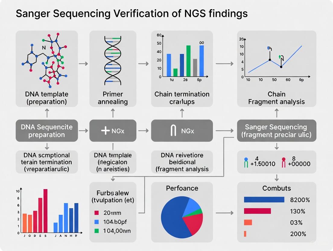
The following diagram illustrates the core workflow that enables this high accuracy:
Sanger Sequencing Workflow
Quantitative Comparison: Sanger Sequencing vs. NGS
While NGS offers unparalleled throughput, Sanger sequencing remains superior for targeted applications requiring maximum accuracy. The table below summarizes the core performance differences.
Table 1: Performance Comparison Between Sanger and Next-Generation Sequencing
| Aspect | Sanger Sequencing | Next-Generation Sequencing (NGS) |
|---|---|---|
| Single-Base Accuracy | 99.99% (QV ≥40) [4] | ~99.9% (Often requires Sanger validation) [4] |
| Read Length | 500 - 1,000 bp (high-quality region) [4] [2] | 150 - 300 bp (for Illumina) [2] [3] |
| Detection Limit for Variants | ~15-20% of the viral quasispecies [5] [3] | As low as 1% [6] [3] |
| Ideal Use Cases | Mutation verification, plasmid validation, single-gene studies [4] [6] | Whole-genome sequencing, variant discovery, transcriptomics [6] [3] |
| Best for Number of Targets | Cost-effective for 1-20 targets [6] [2] | Cost-effective for >20 targets [6] [2] |
This comparison highlights a key distinction: Sanger sequencing provides superior accuracy for a single DNA fragment, whereas NGS provides greater sensitivity for detecting rare, low-frequency variants in a mixed sample due to its deep sequencing capability [6] [3].
Sanger Sequencing for NGS Verification: Experimental Evidence
The practice of using Sanger sequencing to validate NGS findings is a cornerstone of rigorous genomic research. The evidence for its utility comes from large-scale, systematic studies.
Key Experimental Protocol: Large-Scale Validation
A seminal study from the ClinSeq project systematically evaluated the need for Sanger validation of NGS variants [7].
- Methodology: Researchers compared NGS variants in five genes across 684 participants against data generated from high-throughput Sanger sequencing of the same samples. Over 5,800 NGS-derived variants were checked against Sanger data.
- Findings: Of the 5,800+ variants, only 19 were not initially validated by the first-pass Sanger data. Upon re-sequencing with newly designed primers, 17 of these 19 NGS variants were confirmed, revealing that the Sanger method was more likely to incorrectly refute a true positive than to correctly identify a false positive.
- Conclusion: The study calculated a NGS validation rate of 99.965% using Sanger sequencing and questioned the routine necessity of this orthogonal validation, given that NGS data are at least as accurate as a single round of Sanger sequencing [7].
Application in Infectious Disease Research
In clinical settings, the choice between technologies depends on the required sensitivity.
- HIV Drug Resistance Study: A 2022 study on HIV-1 drug resistance in children and adolescents compared Sanger sequencing with NGS at multiple sensitivity thresholds (1% to 20%) [5].
- Findings: The agreement between the two technologies was high (up to 88%) for detecting any drug resistance mutation when a high NGS threshold (15-20%) was used. However, NGS detected substantially more mutations at lower thresholds (1-5%). This demonstrates that while Sanger is highly reliable for detecting dominant variants, NGS offers superior sensitivity for identifying minor populations [5].
The logical relationship in the verification workflow is outlined below:
NGS Verification Workflow
The Scientist's Toolkit: Essential Reagents for Sanger Sequencing
Successful Sanger sequencing relies on a set of core reagents and materials, each with a specific function.
Table 2: Key Research Reagent Solutions for Sanger Sequencing
| Reagent/Material | Function | Key Considerations |
|---|---|---|
| Template DNA | The target DNA to be sequenced (e.g., plasmid, PCR product). | Requires high purity and specific concentration (e.g., plasmids ≥100 ng/μL; PCR products ≥20 ng/μL) [4]. |
| Sequencing Primer | A short, single-stranded DNA fragment that anneals to a specific region to initiate sequencing. | Can be client-supplied or selected from universal primers (M13, T7, SP6). Specificity is critical [4] [8]. |
| Fluorescent ddNTPs | Dideoxynucleotides labeled with fluorescent dyes; each base (A, T, C, G) has a unique dye. | Incorporated by DNA polymerase to terminate chain elongation, generating labeled fragments [2]. |
| DNA Polymerase | Enzyme that catalyzes the template-dependent addition of nucleotides. | High-fidelity polymerase is essential for low error rates and robust performance through difficult templates [9]. |
| Capillary Electrophoresis System | Instrumentation that separates DNA fragments by size. | Systems like the Applied Biosystems 3730xl provide the high resolution needed for long, accurate reads [4]. |
Sanger sequencing's 99.99% accuracy benchmark is not a historical artifact but a living standard underpinned by a robust and reliable biochemical process. Its strength lies in delivering long-read, single-molecule resolution with exceptional precision, making it the undisputed gold standard for targeted validation work. In the context of a world increasingly dominated by NGS, its role has evolved rather than diminished. For confirming critical mutations, verifying gene edits, validating NGS-derived variants, and ensuring the integrity of plasmid constructs, Sanger sequencing provides a final, authoritative layer of confidence that remains unmatched for specific, high-stakes applications in research and drug development.
Orthogonal validation, the practice of confirming next-generation sequencing (NGS) findings with an independent method, traditionally Sanger sequencing, has been a cornerstone of clinical genetic testing to ensure maximal accuracy and reliability. As NGS technologies have matured, the necessity of validating every variant has been questioned, prompting a shift towards risk-based approaches. This guide examines the current regulatory and best practice landscape, defining the requirements for orthogonal confirmation and providing a comparative analysis of validation strategies. The core challenge lies in balancing the impeccable accuracy demanded of clinical diagnostics with the practical realities of throughput, cost, and turnaround time in the era of large-scale genomic testing.
Regulatory Frameworks and Professional Guidelines
Regulatory requirements for NGS validation are evolving, with professional societies leading the development of standards in the absence of universally mandated regulations.
- Professional Society Leadership: The Association for Molecular Pathology (AMP) and the National Society of Genetic Counselors have convened working groups to address the variability in laboratory validation practices, establishing a common framework for clinical laboratories to develop individualized policies [10].
- The CLEP Standard: The New York State Department of Health's Clinical Laboratory Evaluation Program (CLEP) requirements for analytical validation are widely recognized as a national standard. CLEP approval mandates detailed documentation, quality control metrics, validation studies (accuracy, precision, reproducibility), and orthogonal validation of variants [11].
- Shifting Perspectives: Recent guidelines reflect a nuanced approach. The European Society of Human Genetics (ESHG) acknowledges the challenges of NGS but also the imperative for standardized validation, while other studies conclude that routine orthogonal Sanger validation of NGS variants has "limited utility" once appropriate quality thresholds are established [7] [12].
Orthogonal Validation in Practice: A Data-Driven Comparison
The decision to implement universal or selective orthogonal validation hinges on quantitative performance data. The table below summarizes key metrics from recent large-scale studies evaluating NGS accuracy against Sanger sequencing.
Table 1: Comparative Performance of NGS versus Sanger Sequencing in Validation Studies
| Study and Technology | Cohort and Variant Numbers | Concordance Rate with Sanger | Key Factors Influencing Concordance |
|---|---|---|---|
| WGS Variant Analysis [13] | 1,756 variants from 1,150 WGS samples | 99.72% (5 discrepancies) | Quality (QUAL) score, read depth (DP), allele frequency (AF) |
| Large-Scale Exome Study [7] | ~5,800 NGS-derived variants from 684 exomes | 99.965% (initially 19 failures, 17 confirmed with new primers) | Variant quality score; primer design in Sanger |
| Target-Capture Gene Panels [14] | 1,080 SNVs and 124 Indels across 117 genes | 100% for SNVs (919 comparisons) | Sufficient depth of coverage (>100x); variant type (SNV vs. Indel) |
| Targeted Gene Panels [12] | 945 rare variants from 218 patients | >99.6% (3 discrepancies resolved in favor of NGS) | Allele dropout (ADO) in Sanger PCR; primer-binding variants |
Key Experimental Protocols from Cited Studies
The data in Table 1 are derived from rigorously validated clinical workflows:
- WGS Variant Confirmation Protocol [13]: DNA was sequenced using a PCR-free WGS protocol on a BGI platform with a mean coverage of 34.1x. Variants were called using GATK HaplotypeCaller. All 1,756 selected variants underwent Sanger sequencing, and the quality metrics (DP, AF, QUAL) of the 5 discordant variants were analyzed to establish robust filtering thresholds.
- High-Throughput Sanger Comparison Protocol [7]: This study leveraged the unique ClinSeq cohort, where exome sequencing (via Illumina platforms) was performed on samples with existing high-throughput Sanger data. Variants were called using the Most Probable Genotype (MPG) caller. Discrepant variants were investigated with re-designed Sanger primers, which often resolved the issue.
- Targeted Panel Validation Protocol [12]: Targeted NGS was performed on Illumina MiSeq using Agilent SureSelect or Haloplex for enrichment. Variants were called with GATK HaplotypeCaller and filtered for Phred quality ≥30 and coverage depth ≥30x. Sanger validation included careful primer design and checking for SNPs in primer-binding sites to prevent allelic dropout.
Evolving Best Practices: From Universal to Selective Validation
Best practices are moving away from universal Sanger validation towards a more strategic, data-driven approach. The diagram below illustrates the logical decision-making workflow for modern orthogonal validation.
Defining "High-Quality" Variants for Validation Bypass
The decision to bypass validation is guided by specific, measurable quality metrics established through large-scale Sanger confirmation studies.
- Caller-Agnostic vs. Caller-Specific Metrics: Quality parameters can be divided into two groups. Caller-agnostic metrics like read depth (DP ≥ 15) and allele frequency (AF ≥ 0.25) are universally applicable across technologies. In contrast, caller-specific metrics like the quality score (QUAL ≥ 100 for GATK HaplotypeCaller) depend on the variant-calling algorithm and require laboratory-specific validation [13].
- The Critical Role of Genomic Context: Even high-quality metrics can be misleading in complex genomic regions. Segment duplications, GC-rich areas, and repetitive sequences are notorious for causing false positives. Tools like ClinRay are being developed to predict probe reproducibility in these difficult regions, helping to identify variants that still require validation despite good nominal quality scores [15].
- Variant-Type Specific Considerations: The validation policy must be stratified by variant type. Multiple studies have demonstrated that single nucleotide variants (SNVs) passing quality thresholds have concordance rates approaching 100%, making their validation redundant. In contrast, insertion-deletion (indel) variants more frequently require Sanger confirmation due to complexities in alignment and calling, even with sufficient depth of coverage [14].
Emerging Strategies and Advanced Solutions
The field is advancing beyond simple quality thresholds towards more sophisticated, automated methods for ensuring variant accuracy.
Machine Learning and Bioinformatics Innovation
Supervised machine learning models are now being trained to differentiate high-confidence from low-confidence variants with high precision, reducing the need for wet-lab confirmation.
- Model Training and Performance: Studies have used variant calls from Genome in a Bottle (GIAB) reference samples to train models like Logistic Regression, Random Forest, and Gradient Boosting. These models use features such as allele frequency, read depth, mapping quality, and homopolymer context. One such model achieved 99.9% precision and 98% specificity in identifying true positive heterozygous SNVs [16].
- The "Digital Twin" Approach: Methods like ClinRay create synthetic data enhancements to predict the reproducibility of NGS probes in difficult-to-sequence regions. This bioinformatic approach, which achieved an AUC of 0.85-0.89, can flag variants with likely poor reproducibility for orthogonal validation without the need for costly wet-lab replicates [15].
The Scientist's Toolkit: Essential Reagents and Metrics
Table 2: Key Research Reagent Solutions and Quality Metrics for Orthogonal Validation
| Tool/Reagent | Primary Function in Validation | Application Notes |
|---|---|---|
| GIAB Reference Materials | Benchmark "truth set" for validating NGS pipelines and training ML models. | Essential for establishing lab-specific quality thresholds and for bioinformatics pipeline validation [16] [17]. |
| PCR-Free WGS Libraries | Reduces library preparation artifacts that can mimic true variants. | Critical for minimizing false positives in whole-genome studies; explained absence of certain artifacts in one study [13]. |
| Multiple Bioinformatics Callers | Provides computational orthogonal confirmation (e.g., DeepVariant). | Can be used for low-quality variants, though performance varies (F1-score 0.76 in one assessment) [13]. |
| Quality Metric: Depth (DP) | Measures number of reads covering a variant; indicates confidence. | Caller-agnostic; DP ≥ 15 suggested for WGS [13]. |
| Quality Metric: Allele Frequency (AF) | Proportion of reads supporting the variant; should be ~0.5 for germline heterozygotes. | Caller-agnostic; AF ≥ 0.25 suggested as threshold [13]. |
| Quality Metric: QUAL Score | Phred-scaled confidence that a variant exists at a given site. | Caller-specific (e.g., QUAL ≥ 100 for GATK); highly effective but not transferable between pipelines [13]. |
The paradigm for orthogonal validation is shifting decisively from a universal requirement to a targeted, evidence-based strategy. The consensus emerging from recent research and guideline development is that clinical laboratories should establish and validate internal quality thresholds based on their specific NGS and bioinformatics pipelines. For variants exceeding these thresholds—particularly SNVs in uniquely mappable regions—orthogonal Sanger validation is increasingly seen as redundant. Future efforts will focus on standardizing these quality metrics across platforms, refining machine learning models for variant triage, and integrating long-read sequencing technologies as a more comprehensive orthogonal method. The ultimate goal is a streamlined, cost-effective validation process that maintains the highest standards of clinical accuracy while fully leveraging the power of modern high-throughput genomics.
Sanger sequencing has long been regarded as the "gold standard" for confirming DNA sequence variants. However, with the maturation of Next-Generation Sequencing (NGS) technologies, the practice of universally validating NGS findings with Sanger sequencing is being re-evaluated. This guide objectively compares the performance of these technologies and outlines the specific scenarios where orthogonal Sanger verification remains indispensable in clinical diagnostics and research publications, supported by experimental data.
Next-Generation Sequencing has revolutionized genetics, enabling the simultaneous analysis of millions of DNA fragments. Despite its high throughput, the initial standards, including those from the American College of Medical Genetics (ACMG), often mandated that variants identified by NGS be confirmed by the orthogonal method of Sanger sequencing before reporting [13]. This practice was rooted in concerns over NGS errors related to sequencing artifacts, bioinformatics pipeline inaccuracies, and challenges in regions with complex architecture (e.g., high GC content) [18].
Recent large-scale studies have demonstrated that NGS data, when subjected to appropriate quality filters, can achieve exceptionally high accuracy, calling into question the utility of routine Sanger validation. One systematic evaluation of over 5,800 NGS-derived variants found a validation rate of 99.965% using Sanger sequencing, concluding that a single round of Sanger sequencing is more likely to incorrectly refute a true positive NGS variant than to correctly identify a false positive [7]. This guide synthesizes current evidence to define the key scenarios where Sanger verification is still required, providing a data-driven framework for researchers and clinicians.
Performance Comparison: NGS vs. Sanger Sequencing
The decision to use Sanger verification hinges on a clear understanding of the performance characteristics of both technologies. The following table summarizes key comparative metrics based on recent literature.
Table 1: Performance Comparison of NGS and Sanger Sequencing for Variant Detection
| Metric | Next-Generation Sequencing (NGS) | Sanger Sequencing |
|---|---|---|
| Throughput | High (Millions of parallel sequences) | Low (Single amplicons per reaction) |
| Cost per Base | Very Low | High |
| Single-Base Accuracy | High (Exceeding 99.9% for high-quality calls) [13] | Very High (Error rate ~0.01% or lower) [9] |
| Ideal Application | Interrogating multiple genes or the entire genome/exome | Targeted confirmation of specific variants |
| Key Strengths | Discovery of novel variants, detection of mosaicism (at sufficient depth), comprehensive profiling | Long read lengths, high consensus accuracy, single-molecule resolution |
| Key Limitations | False positives/negatives can occur in low-complexity or low-coverage regions [18] | Low throughput, inefficient for screening, prone to allelic dropout (ADO) from primer-binding SNPs [18] |
Key Scenarios Mandating Sanger Verification
Scenario 1: Clinical Diagnostics and Reporting of Pathogenic Variants
In clinical diagnostics, where a result directly impacts patient management, the highest level of certainty is required. While the trend is to move away from universal validation, Sanger confirmation remains critical in specific diagnostic contexts.
- Evidence and Data: A 2025 study on Whole Genome Sequencing (WGS) data found that while overall concordance with Sanger was 99.72%, certain variant quality parameters could identify false positives. The study demonstrated that applying caller-agnostic filters (Depth of coverage (DP) ≥ 15 and Allele Frequency (AF) ≥ 0.25) successfully isolated all unconfirmed variants into a "low-quality" bin, suggesting that variants failing these thresholds require Sanger verification before clinical reporting [13].
- Experimental Protocol: The standard protocol involves:
- Variant Identification: NGS is performed on a patient sample using a targeted panel, exome, or genome sequencing.
- Variant Filtering: Variants are filtered using established bioinformatic pipelines. Variants with a depth of coverage (DP) below 15-20x or an allele frequency (AF) below 20-25% are flagged for validation [13] [18].
- PCR Amplification: Specific primer pairs are designed to flank the variant of interest. These primers must be checked for common SNPs at their binding sites to prevent allelic dropout [18].
- Sanger Sequencing: PCR amplicons are purified and sequenced using fluorescent dye-terminator chemistry on a capillary electrophoresis instrument [19].
- Analysis: The resulting chromatograms are manually inspected for the presence of the variant, confirming both the zygosity and the base change.
Scenario 2: Validation of Critical Research Findings for Publication
In research publications, particularly those reporting novel, high-impact genetic findings, Sanger validation is often a requirement from journal reviewers to ensure the robustness of the data.
- Evidence and Data: The credibility of a research claim is paramount. Sanger sequencing provides an orthogonal method to confirm that a variant is not an NGS-specific artifact. This is especially true for novel mutations that constitute the central evidence of a study. Research has shown that discrepancies between NGS and Sanger can sometimes originate from Sanger-specific issues like allelic dropout (ADO); therefore, careful experimental design is essential [18].
- Experimental Protocol: The methodology is similar to the clinical diagnostic protocol but is often applied to a wider range of variant types, including those in complex genomic regions. Key steps include:
- Primer Design: Using tools like Primer3 to design primers that amplify a 500-800bp region encompassing the variant [7] [18].
- Template Preparation: Using high-quality, purified DNA (e.g., from plasmid minipreps or gel-extracted PCR products) as a template for the sequencing reaction [19].
- Bidirectional Sequencing: Sequencing the same amplicon from both forward and reverse primers to achieve comprehensive coverage and confirm the variant call [7].
Scenario 3: Verification of Genome Editing Outcomes
The field of genome editing (e.g., with CRISPR-Cas9) relies heavily on Sanger sequencing to confirm the precise nature of induced mutations, such as insertions or deletions (indels).
- Evidence and Data: Specialized computational tools (TIDE, ICE, DECODR) have been developed to deconvolute complex Sanger sequencing chromatograms from edited heterogeneous cell populations into quantitative indel frequencies [20]. A 2024 study systematically compared these tools and found that while they perform well with simple indels, their estimates can vary with more complex edits, underscoring the need for careful tool selection and interpretation, with Sanger as the foundational data source [20].
- Experimental Protocol:
- PCR Amplification: The genomic target site surrounding the CRISPR-Cas cut site is amplified from a pool of edited cells.
- Sanger Sequencing: The PCR products are directly subjected to Sanger sequencing, which produces mixed-base chromatograms at the site of editing due to heterogeneity.
- Computational Analysis: The sequencing trace data (.ab1 files) from the edited pool and a wild-type control are uploaded to a web tool (e.g., ICE or DECODR). The algorithm decomposes the complex trace to estimate the percentage of indels and their size distribution [20].
Scenario 4: Resolving Discrepancies or Ambiguous NGS Data
When NGS data is ambiguous, has low quality scores, or conflicts with phenotypic or other molecular data, Sanger sequencing is the definitive method for resolution.
- Evidence and Data: A 2020 study reported three cases of discrepancy between high-quality NGS calls and the initial Sanger sequencing result. Upon reinvestigation, all three NGS variants were confirmed to be correct, with the Sanger errors attributed to allelic dropout (ADO) caused by unknown SNPs in the primer-binding region. This highlights that in cases of discrepancy, the NGS result should not be automatically assumed false, and re-designing Sanger primers is a critical step [18].
- Experimental Protocol: The process involves:
- Re-examination of NGS Data: Inspecting the BAM file for alignment issues, strand bias, and low mapping quality at the variant site.
- Re-design of Sanger Primers: Designing new primers that bind to a different, unique genomic region to avoid suspected SNPs that cause ADO [18].
- Re-sequencing: Performing Sanger sequencing with the new primers to obtain a clear, unambiguous result.
Decision Workflows and Visualization
The following diagram illustrates the decision-making process for determining when Sanger verification is necessary, integrating the scenarios and quality thresholds discussed.
Decision Workflow for Sanger Verification
The Scientist's Toolkit: Essential Reagents and Materials
Successful Sanger verification relies on high-quality reagents and careful experimental preparation. The following table details key solutions and their functions.
Table 2: Essential Research Reagent Solutions for Sanger Verification
| Item | Function | Key Considerations |
|---|---|---|
| High-Purity Template DNA | Serves as the substrate for the sequencing reaction. | Plasmid, PCR product, or genomic DNA with OD260/OD280 ratio of ~1.8-2.0 [19]. Concentration: 10-100 ng/μL depending on type [19]. |
| Optimized Sequencing Primers | Provides the starting point for DNA polymerase. | 18-25 bases in length; designed to avoid secondary structures and SNPs in binding sites [18] [19]. |
| BigDye Terminator Kit | Fluorescently labeled dideoxynucleotides (ddNTPs) for chain termination. | The core chemistry for cycle sequencing. Requires optimization of dilution and usage to reduce costs [18]. |
| DNA Polymerase | Enzyme that catalyzes the template-dependent DNA synthesis. | High-fidelity enzymes are preferred. Typical use: 0.5-1.0 U per 10 μL reaction [19]. |
| Capillary Electrophoresis Instrument | Separates DNA fragments by size and detects fluorescent labels. | Instruments like ABI 3500 Series perform automated electrophoresis, data collection, and base calling [21]. |
The paradigm for Sanger verification of NGS findings is shifting from a routine, blanket practice to a targeted, evidence-based one. Data shows that NGS is highly accurate, and its false positives can be effectively predicted using quality metrics like depth of coverage and allele frequency. Sanger sequencing remains an indispensable tool in the molecular biologist's arsenal, but its application is now focused on key scenarios: clinical reporting of low-quality variants, cornerstone research findings, genome editing verification, and resolution of technical discrepancies. By adopting this refined approach, researchers and diagnosticians can optimize resources while maintaining the highest standards of data integrity.
The Complementary Roles of NGS and Sanger in Modern Genomics Workflows
Next-generation sequencing (NGS) and Sanger sequencing represent complementary technological pillars in modern genomic analysis. While NGS provides unprecedented throughput for discovering genetic variants across entire genomes or targeted gene panels, Sanger sequencing remains the gold standard for confirming these findings with exceptional accuracy [22] [23]. This complementary relationship is particularly crucial in clinical diagnostics and drug development, where verifiable accuracy is paramount for patient care and regulatory approval. The integration of both technologies creates a powerful workflow that leverages the discovery power of NGS with the verification reliability of Sanger sequencing, establishing a robust framework for genomic analysis across research and clinical applications.
Technical Comparison: NGS vs. Sanger Sequencing
The fundamental differences between NGS and Sanger sequencing technologies dictate their respective roles in genomic workflows. Understanding their technical specifications, advantages, and limitations enables researchers to deploy each method strategically.
Table 1: Technical Specifications and Performance Comparison
| Parameter | Sanger Sequencing | Next-Generation Sequencing (NGS) |
|---|---|---|
| Sequencing Principle | Chain termination with dideoxynucleotides (ddNTPs) [22] | Massively parallel sequencing [23] |
| Throughput | Low (single fragment per reaction) [23] | High (millions of fragments simultaneously) [23] [24] |
| Read Length | 500-1000 base pairs [25] [9] | 50-400 base pairs (short-read platforms) [26] |
| Accuracy | >99.9% (gold standard) [22] [23] | >99% (with sufficient coverage) [23] |
| Variant Detection Sensitivity | 15-20% [26] | <1% [26] |
| Cost-Effectiveness | Ideal for small projects, individual genes [23] [24] | Cost-effective for large projects, entire genomes [23] [24] |
| Time per Run | 20 minutes - 3 hours [26] | 48 hours (for standard NGS workflows) [26] |
| Key Applications | Variant confirmation, single gene testing, plasmid verification [22] [27] | Whole genome/exome sequencing, gene panel testing, transcriptomics [23] |
| Data Analysis Complexity | Minimal bioinformatics required [23] | Complex, requires specialized bioinformatics [23] |
Sanger sequencing operates on the principle of chain termination using fluorescently labeled dideoxynucleotides (ddNTPs) that lack the 3'-hydroxyl group necessary for DNA strand elongation [22]. When incorporated during DNA synthesis, these ddNTPs randomly terminate growing DNA strands, producing fragments of varying lengths that are separated by capillary electrophoresis to determine the nucleotide sequence [22] [24].
In contrast, NGS technologies employ massively parallel sequencing, simultaneously determining the sequence of millions to billions of DNA fragments [23] [24]. This high-throughput approach enables comprehensive genomic analysis but generates shorter reads than Sanger sequencing. The most commonly used NGS technique, sequencing by synthesis (SBS), involves amplifying and sequencing DNA fragments on a solid surface or in emulsion droplets, with nucleotide incorporation detected through various signal detection methods [24].
The exceptional accuracy of Sanger sequencing (>99.9%) establishes it as the reference standard for validating genetic variants, particularly for clinical applications [22] [23]. Meanwhile, NGS provides superior sensitivity for detecting low-frequency variants present in minor cell populations, with variant detection thresholds below 1% compared to Sanger's 15-20% sensitivity limit [26]. This makes NGS particularly valuable for oncology applications where detecting somatic mutations in heterogeneous tumor samples is critical.
Experimental Evidence: Validation Studies and Performance Metrics
Robust experimental studies have quantified the concordance between NGS and Sanger sequencing, providing empirical evidence to guide their complementary implementation in genomic workflows.
Table 2: Experimental Validation Studies of NGS-Sanger Concordance
| Study Focus | Sample Size | Key Findings | Concordance Rate |
|---|---|---|---|
| Targeted NGS Panel Validation [14] | 77 patient samples, 1080 SNVs, 124 indels | 100% concordance for recurrent variants in unrelated samples | 100% for SNVs |
| Large-Scale Exome Sequencing Validation [7] | 684 exomes, over 5,800 NGS-derived variants | Only 19 NGS variants not initially validated by Sanger; 17 confirmed with redesigned primers | 99.965% overall |
| Nanopore vs Sanger in Oncohematology [26] | 164 samples, 174 analyzed regions across 15 genes | Supported implementation of MinION technology for routine variant detection | 99.43% |
| NGS-Sanger Comparison in Clinical Context [14] | 7 1000 Genomes Project samples, 762 unique variants | High concordance with 1000 Genomes phase 1 data; all discrepancies resolved with additional data | 97.1% |
The remarkably high concordance rates demonstrated in these studies, particularly for single nucleotide variants (SNVs), question the utility of routine orthogonal Sanger validation for all NGS findings. The large-scale evaluation by the ClinSeq project, which analyzed over 5,800 NGS-derived variants, revealed an exceptional validation rate of 99.965% [7]. The authors concluded that "a single round of Sanger sequencing is more likely to incorrectly refute a true positive variant from NGS than to correctly identify a false positive variant from NGS," suggesting that routine Sanger validation of NGS variants has limited utility [7].
However, this does not eliminate the need for Sanger confirmation in specific scenarios. The same study found that when NGS variants failed Sanger validation, the issues were typically resolved by redesigning sequencing primers, indicating that primer design and genomic context can impact verification success [7]. Insertion-deletion variants (indels) may also require Sanger sequencing for precise characterization of their genomic location, even when initially detected by NGS [14].
Methodological Guide: Experimental Workflows and Protocols
Implementing an effective NGS-Sanger validation workflow requires careful experimental design and execution. The following section outlines standard protocols for orthogonal verification of NGS findings.
NGS Variant Detection Workflow
Diagram 1: NGS Variant Detection Workflow
The NGS variant detection process begins with sample preparation and DNA extraction using standardized methods such as salting-out protocols or column-based kits [7]. For targeted sequencing approaches, solution-hybridization capture systems (e.g., SureSelect, TruSeq) enrich specific genomic regions of interest [7]. Following library preparation, massively parallel sequencing occurs on platforms such as Illumina GAIIx or HiSeq series, generating millions to billions of short reads [7]. Image analysis and base calling transform raw signals into sequence data, which is then aligned to reference genomes (e.g., hg19) using tools like NovoAlign [7]. Variant calling identifies potential mutations, with quality thresholds such as minimum depth of coverage (>100x) and quality scores (e.g., MPG score ≥10) ensuring reliable variant detection [7].
Sanger Sequencing Validation Protocol
Diagram 2: Sanger Validation of NGS Variants
The Sanger validation workflow initiates with selecting NGS-derived variants requiring confirmation, prioritizing clinically significant mutations or those with borderline quality metrics [28]. PCR and sequencing primers are designed using specialized tools (e.g., PrimerTile, Primer3) that avoid known polymorphisms and optimize annealing conditions [7]. For the sequencing reaction, template DNA is amplified with fluorescently labeled ddNTPs using DNA polymerase, generating chain-terminated fragments [22]. Post-amplification cleanup removes excess primers and unincorporated nucleotides before capillary electrophoresis separates fragments by size [22] [24]. Fluorescence detection identifies the terminal ddNTP at each position, generating chromatograms for sequence determination [22]. Finally, sequence alignment tools compare results with reference sequences and the original NGS data to confirm variants [28].
Table 3: Essential Research Reagent Solutions for NGS-Sanger Workflows
| Reagent/Kit | Application | Key Features | Representative Examples |
|---|---|---|---|
| DNA Extraction Kits | Nucleic acid purification from various sample types | High purity, yield, and integrity | Salting-out method (Qiagen) [7] |
| Target Enrichment Systems | Selective capture of genomic regions for NGS | Comprehensive coverage, uniformity | SureSelect (Agilent), TruSeq (Illumina) [7] |
| NGS Library Prep Kits | Preparation of sequencing libraries | Efficiency, minimal bias | Illumina library prep kits [7] |
| DNA Polymerase | PCR amplification for Sanger sequencing | High fidelity, processivity | Optimized enzymes with proofreading activity [9] |
| Cycle Sequencing Kits | Sanger sequencing reactions | Fluorescent ddNTP incorporation | BigDye Terminator kits [7] |
| Capillary Electrophoresis Kits | Fragment separation for Sanger sequencing | High resolution, sensitivity | Applied Biosystems kits [28] |
Quality Control and Troubleshooting
Successful implementation of NGS-Sanger workflows requires rigorous quality control measures. For NGS data, ensure sufficient depth of coverage (>100x for heterozygous variants) and high-quality scores at variant positions [7] [14]. For Sanger validation, examine chromatograms for clean baseline separation between peaks and strong signal intensity throughout the sequence [22]. When Sanger fails to confirm NGS variants, consider redesigning sequencing primers to avoid problematic genomic regions, increasing template DNA concentration, or verifying NGS read alignment and variant calling parameters [7]. For indels, careful inspection of both forward and reverse Sanger sequences is essential to determine exact breakpoints [14].
Application Scenarios and Decision Framework
The complementary use of NGS and Sanger sequencing varies across research and clinical contexts, with specific applications benefiting from their integrated implementation.
Clinical Diagnostics and Genetic Testing
In clinical settings, NGS enables comprehensive testing for heterogeneous conditions through multi-gene panels, whole exome sequencing, or whole genome sequencing [26] [23]. For definitive diagnosis of monogenic disorders or confirmation of pathogenic variants, Sanger sequencing provides the requisite verification [22] [27]. This is particularly important for heritable conditions like BRCA-related cancers or cystic fibrosis, where diagnostic accuracy directly impacts patient management [22] [23]. In oncology, NGS identifies low-frequency somatic mutations in tumor samples, while Sanger confirms key therapeutic markers [26] [27].
Research Applications
In basic research, NGS facilitates discovery-based studies including novel variant identification, transcriptomic profiling, and epigenomic characterization [23]. Sanger sequencing then verifies key findings through orthogonal validation [27] [28]. Additional research applications include confirming genome editing outcomes (e.g., CRISPR-Cas9 modifications), validating plasmid constructs, and verifying synthetic biology constructs [27] [9]. The high accuracy of Sanger sequencing makes it indispensable for these applications where sequence precision is critical.
Decision Framework for Method Selection
Selecting the appropriate sequencing method or combination depends on multiple factors:
- Project Scale: For sequencing individual genes or limited targets (<20), Sanger sequencing is typically more practical and cost-effective. For analyzing hundreds to thousands of genes or entire genomes, NGS is preferable [22] [23].
- Variant Frequency: For detecting low-frequency variants (<15-20%) in heterogeneous samples, NGS provides superior sensitivity [26].
- Accuracy Requirements: For clinical applications requiring the highest possible accuracy for specific variants, Sanger confirmation remains essential [22] [28].
- Turnaround Time: For urgent clinical decisions (e.g., NPM1 mutational status in AML), targeted approaches like Sanger or third-generation sequencing (e.g., Nanopore) may provide faster results than standard NGS workflows [26].
- Resource Availability: Sanger requires minimal bioinformatics infrastructure, while NGS demands significant computational resources and expertise [23].
Emerging Trends and Future Directions
Sequencing technologies continue to evolve, with new developments enhancing their complementary roles. Third-generation sequencing technologies, such as Oxford Nanopore MinION, offer potential alternatives with real-time sequencing, long reads, and rapid turnaround times [26]. Recent studies demonstrate 99.43% concordance between MinION and Sanger sequencing in oncohematological diagnostics, suggesting potential for replacing Sanger in some verification scenarios [26].
Technical improvements in Sanger sequencing continue to optimize its performance, with developments in capillary array design, fluorescent detection systems, and DNA polymerase engineering enhancing throughput, accuracy, and read length [9]. Microfluidic chip technologies enable miniaturization and automation of Sanger sequencing, potentially increasing its efficiency for validation workflows [9].
Bioinformatics advances are streamlining the validation process through automated primer design, integrated data analysis platforms, and visualization tools that compare NGS and Sanger results [28]. Software solutions like Minor Variant Finder enhance Sanger's sensitivity for detecting low-frequency mutations, bridging one of the key sensitivity gaps between Sanger and NGS [28].
NGS and Sanger sequencing maintain complementary rather than competitive roles in modern genomics. NGS provides unparalleled discovery power for comprehensive genomic analysis, while Sanger delivers verifiable accuracy for confirmatory testing. This synergistic relationship creates a robust framework for genomic analysis across basic research, clinical diagnostics, and therapeutic development. As sequencing technologies evolve, their core strengths will likely maintain this complementary dynamic, with verification standards adapting to technological improvements rather than abandoning the fundamental principle of orthogonal validation that ensures reliability in genomic medicine.
The field of genomic sequencing has undergone a remarkable transformation over the past two decades, driven primarily by the advent of next-generation sequencing (NGS) technologies. As these high-throughput methods entered clinical and research laboratories, a critical question emerged: how should variants detected by NGS be validated to ensure accuracy? For years, Sanger sequencing served as the undisputed "gold standard" for orthogonal confirmation of NGS findings. However, as NGS technologies have matured, with demonstrated error rates below 0.1% under optimal conditions, the practice of reflexive Sanger validation has faced increasing scrutiny [7] [12]. This guide examines the evolving standards for validation practices, presenting comparative experimental data that inform current professional guidelines and laboratory practices.
The Era of Mandatory Sanger Validation
When NGS first transitioned from research to clinical applications, regulatory uncertainty and a natural caution regarding new technologies made Sanger confirmation a standard practice. This approach was rooted in Sanger's long-established reputation for accuracy, with a documented single-base sequencing error rate below 0.001% [26].
The fundamental rationale for this practice included:
- Error Prevention: Bioinformatic pipelines for NGS were initially novel and required verification
- Regulatory Compliance: Laboratories sought to minimize risk in clinical reporting
- Technical Limitations: Early NGS platforms had higher error rates in challenging genomic regions
During this period, Sanger validation was considered an essential quality control measure, particularly for variants with potential clinical significance [12].
Paradigm Shift: Evidence Challenging Routine Sanger Validation
Landmark Validation Studies
Comprehensive studies began questioning the utility of reflexive Sanger validation as NGS technology matured. A systematic evaluation published in Clinical Chemistry examined this practice using data from the ClinSeq project, which provided a unique opportunity to compare NGS variants with high-throughput Sanger sequencing on the same samples [7].
Table 1: Large-Scale Comparison of NGS vs. Sanger Sequencing
| Study Parameter | 19-Gene Analysis | 5-Gene Analysis (684 participants) |
|---|---|---|
| Total NGS Variants | 234 variants | >5,800 variants |
| Discrepant Variants | 0 | 19 initially |
| Resolution | N/A | 17 confirmed by redesigned Sanger primers, 2 had low NGS quality scores |
| Final Validation Rate | 100% | 99.965% |
This study demonstrated that a single round of Sanger sequencing was statistically more likely to incorrectly refute a true positive NGS variant than to correctly identify a false positive [7]. The authors concluded that routine orthogonal Sanger validation has limited utility and should not be considered a best practice standard.
Case Studies Highlighting Sanger Limitations
Further evidence emerged from smaller, focused studies. An analysis of 945 NGS variants from 218 patients revealed only three discrepancies with Sanger sequencing [12]. Upon deeper investigation, all three NGS calls were validated, with the discrepancies attributed to allelic dropout (ADO) during polymerase chain reaction or Sanger sequencing reactions. This phenomenon, often related to unpredictable private variants on primer-binding regions, highlighted that Sanger sequencing itself is not error-free [12].
Current Guidelines and Evolving Standards
Professional organizations have responded to this accumulating evidence by refining their recommendations regarding validation practices.
Key Guideline Updates
The Association for Molecular Pathology (AMP) and the College of American Pathologists have developed frameworks that emphasize an error-based approach to validation rather than reflexive Sanger confirmation [29]. These guidelines encourage laboratories to:
- Identify potential sources of errors throughout the analytical process
- Address these potential errors through test design, method validation, or quality controls
- Focus validation efforts on specific variant types or genomic regions where NGS performance is known to be challenging
The AMP and National Society of Genetic Counselors have further addressed this issue through a joint working group, establishing recommendations for standardizing orthogonal confirmation practices [10]. While specific guidelines vary between organizations, the overall trend is toward limiting Sanger validation to specific circumstances rather than applying it universally.
Emerging Technologies and Validation Paradigms
The Rise of Third-Generation Sequencing
New sequencing technologies continue to reshape the validation landscape. Oxford Nanopore Technology (ONT) represents a promising approach that combines long-read capabilities with decreasing turnaround times [26].
Table 2: Performance Comparison of Sequencing Technologies
| Parameter | Sanger Sequencing | NGS (Illumina) | Nanopore (MinION) |
|---|---|---|---|
| Single-Read Accuracy | >99% [26] | >99% [26] | >99% [26] |
| Read Length | 400-900 bp [26] | 50-500 bp [26] | Up to megabase scales [26] |
| Sensitivity | 15-20% [26] | 1% [26] | <1% [26] |
| Error Rate | 0.001% [26] | 0.1-1% [26] | ~5% (platform-dependent) [26] |
| Main Applications | SNVs, INDELs [26] | SNVs, INDELs [26] | SNVs, INDELs, complex structural variants [26] |
A 2025 study comparing Sanger sequencing with MinION technology for oncohematological diagnostics demonstrated 99.43% concordance, supporting the implementation of this technology as a viable alternative to Sanger for validation purposes [26].
Methodological Advancements Reducing Validation Needs
Technical improvements across the NGS workflow have substantially enhanced reliability:
- Hybrid capture-based enrichment methods using longer probes that tolerate mismatches better, reducing allelic dropout [29]
- Enhanced bioinformatics pipelines with improved alignment algorithms and variant calling
- Quality metrics such as Phred scores (>Q30), depth of coverage (>30x), and allele balance thresholds (>0.2) that reliably predict validation success [12]
Modern Validation Framework: A Risk-Based Approach
Contemporary best practices employ a nuanced, context-dependent strategy for variant validation:
Scenarios Warranting Orthogonal Confirmation
- Borderline quality metrics: Variants with coverage depth, quality scores, or allele frequencies near established thresholds
- Clinically critical findings: Variants with significant therapeutic implications
- Technically challenging regions: GC-rich areas, homopolymer stretches, or regions with pseudogenes
- Novel variant types: When laboratories first implement detection of complex variants
Circumstances Where Sanger Validation Adds Limited Value
- High-quality NGS variants: Those with strong quality metrics in well-validated assays
- Established variant types: In genes and variant classes with previously demonstrated high concordance
- High-throughput research settings: Where the 99.9%+ validation rate makes reflexive Sanger confirmation impractical
Evolution of Validation Standards: This diagram illustrates the transition from reflexive Sanger confirmation to a modern, risk-based approach that uses quality metrics to determine when orthogonal validation is necessary.
The Scientist's Toolkit: Essential Research Reagent Solutions
Table 3: Key Reagents and Materials for Sequencing Validation Studies
| Reagent/Material | Function | Application Notes |
|---|---|---|
| SureSelect Target Enrichment | Hybrid capture-based library preparation | Used in large-scale validation studies; tolerates mismatches better than amplification-based methods [7] |
| TruSeq / SureSelect Exome Capture | Solution-hybridization exome capture | Provides comprehensive coverage of coding regions for variant discovery [7] |
| BigDye Terminator v3.1 | Sanger sequencing chemistry | Standard for cycle sequencing reactions; used in discrepant resolution [7] |
| Primer3 Algorithm | Primer design for Sanger validation | Critical for avoiding variants in primer-binding sites that cause allelic dropout [12] |
| ForenSeq mtDNA Kits | Targeted NGS for mitochondrial DNA | Enables comparison of NGS vs. Sanger for forensic applications [30] |
| MiniON Flow Cells | Nanopore-based sequencing | Allows third-generation sequencing validation with long-read capabilities [26] |
The evolution of validation standards from reflexive Sanger confirmation to a nuanced, evidence-based framework reflects the maturation of NGS technologies. Current practices emphasize that validation strategies should be driven by performance data rather than tradition. As one comprehensive study concluded, "Validation of NGS-derived variants using Sanger sequencing has limited utility, and best practice standards should not include routine orthogonal Sanger validation of NGS variants" [7]. This paradigm shift enables more efficient resource allocation in both clinical and research settings while maintaining the rigorous standards necessary for accurate genomic interpretation.
Implementing a Robust Sanger Verification Workflow for NGS Findings
Next-generation sequencing (NGS) has revolutionized genomic research and clinical diagnostics, enabling the simultaneous analysis of millions of DNA fragments. However, the transition from massive parallel sequencing to clinically actionable results requires a robust validation framework to ensure accuracy and reliability. Sanger sequencing has long served as the gold standard for orthogonal confirmation of NGS-derived variants, but blanket validation of all variants is inefficient and costly. This guide provides a comprehensive, evidence-based framework for designing an effective validation pipeline that strategically employs Sanger confirmation where most needed, comparing this approach with emerging alternatives to optimize resource allocation while maintaining stringent quality standards in genomic research and drug development.
NGS and Sanger Sequencing: A Comparative Technological Foundation
Understanding the fundamental differences between NGS and Sanger sequencing technologies is crucial for designing an effective validation pipeline. Each method has distinct strengths and limitations that inform their complementary roles in variant verification.
Table 1: Fundamental Comparison of NGS and Sanger Sequencing Technologies
| Feature | Next-Generation Sequencing (NGS) | Sanger Sequencing |
|---|---|---|
| Throughput | Millions to billions of fragments simultaneously [31] | One DNA fragment at a time [31] |
| Speed | Entire human genome in hours [31] | Slow, suitable for single genes [31] |
| Cost | Under $1,000 per whole human genome [31] | High for large-scale applications [31] |
| Read Length | Short (50-600 base pairs, typically) [31] | Long (500-1000 base pairs) [31] |
| Best Applications | Whole genomes, exomes, large panels, novel discovery [31] [32] | Targeted validation, confirmation of specific variants [31] |
| Accuracy | Excellent for high-quality variants (≥99.72% concordance with Sanger) [13] | Considered gold standard [13] [18] |
The massively parallel approach of NGS creates unprecedented throughput but introduces specific error profiles that necessitate validation, particularly for clinical applications. Sanger sequencing provides precise, long-read accuracy but lacks the scalability for comprehensive genomic analysis [31]. Effective pipeline design leverages the strengths of both technologies while minimizing their respective limitations.
Establishing Quality Thresholds for Strategic Sanger Validation
Recent evidence demonstrates that establishing quality thresholds for NGS variants can dramatically reduce the need for Sanger confirmation while maintaining exceptional accuracy. Key quality metrics have emerged as reliable predictors of variant authenticity.
Table 2: Evidence-Based Quality Thresholds for Reducing Sanger Validation
| Quality Parameter | Proposed Threshold | Concordance with Sanger | Application Considerations |
|---|---|---|---|
| Coverage Depth (DP) | ≥15-20x [13] | 100% [13] | PCR-free protocols reduce bias; lower may be sufficient with high AF [13] |
| Allele Frequency (AF) | ≥0.25-0.30 [13] [18] | 100% [13] | Higher thresholds reduce false positives; consider tumor purity in oncology [13] |
| Variant Quality (QUAL) | ≥100 [13] | 100% [13] | Caller-specific (GATK HaplotypeCaller); not directly transferable between pipelines [13] |
| Filter Status | PASS [13] | High (99.72% overall) [13] | Variants failing FILTER should always be validated [13] |
Research on 1756 WGS variants demonstrated that implementing caller-agnostic thresholds (DP ≥15, AF ≥0.25) reduced the validation burden to just 4.8% of variants while maintaining 100% sensitivity for false positives. Using caller-specific QUAL scores (≥100) further reduced necessary validation to only 1.2% of variants [13]. These thresholds provide a robust framework for prioritizing Sanger confirmation while recognizing that laboratory-specific validation may be necessary to account for pipeline-specific characteristics.
Diagram 1: Variant validation decision workflow showing how quality thresholds dramatically reduce Sanger confirmation burden.
Comparative Analysis of Validation Approaches
Traditional vs. Threshold-Based Sanger Validation
The evolution from blanket Sanger confirmation to strategic, quality-based validation represents a significant advancement in NGS pipeline efficiency.
Table 3: Traditional vs. Modern Validation Approaches
| Validation Approach | Sanger Utilization | Advantages | Limitations |
|---|---|---|---|
| Traditional Blanket Validation | 100% of NGS variants | Maximum accuracy, compliance with early ACMG guidelines [18] | Resource-intensive, time-consuming, cost-ineffective [13] [18] |
| Quality-Threshold Approach | 1.2-4.8% of NGS variants [13] | Efficient resource allocation, faster turnaround, maintained accuracy [13] | Requires initial validation to establish lab-specific thresholds [13] |
| Variant-Type Specific Approach | All indels + low-quality SNVs | Balances comprehensive indel validation with SNV efficiency [14] | Still requires significant Sanger resources for indel-rich regions [14] |
Multiple studies have demonstrated that quality-focused approaches maintain exceptional accuracy while dramatically improving efficiency. One analysis of 919 comparisons between NGS and Sanger showed 100% concordance for high-quality variants [14], while another study of 1756 WGS variants demonstrated 99.72% overall concordance [13].
Alternative Validation Methods
While Sanger sequencing remains the established validation standard, several alternative approaches are emerging:
- Orthogonal NGS Methods: Using a different NGS platform or bioinformatics pipeline for confirmation provides scalability but may perpetuate systematic errors [13].
- Consensus Calling: Employing multiple variant callers (GATK, SAMtools, DeepVariant) increases specificity but requires sophisticated bioinformatics infrastructure [33].
- Array-Based Validation: Microarray genotyping offers high-throughput confirmation but is limited to known variants and cannot detect novel findings [33].
Research indicates that while these alternatives show promise, they have limitations. One evaluation found that using DeepVariant to validate low-quality variants (QUAL <100) achieved only an F1-score of 0.76, indicating significant limitations compared to Sanger [13].
Implementing the Validation Pipeline: Protocols and Methodologies
NGS Variant Calling with GATK Best Practices
The foundation of an effective validation pipeline begins with optimized NGS data processing:
- Read Mapping and Alignment: Map raw sequencing reads (FASTQ) to the reference genome (e.g., GRCh37/hg19, GRCh38/hg38) using BWA-MEM [18].
- Duplicate Marking: Identify and mark PCR duplicates using tools like Picard to prevent artificial inflation of variant support [33].
- Local Realignment and Base Quality Recalibration: Perform local realignment around indels and base quality score recalibration (BQSR) using GATK. This critical step significantly improves variant calling accuracy, with one study showing it improved positive predictive value from 35.25% to 88.69% for certain variant types [33].
- Variant Calling: Call variants using GATK HaplotypeCaller, which outperforms UnifiedGenotyper and SAMtools mpileup, demonstrating positive predictive values of 95.37% versus 69.89% for tool-specific variants [33].
- Variant Quality Score Recalibration (VQSR): Apply VQSR to filter variants based on a Gaussian mixture model of annotation values, which provides slightly better specificity (99.79% vs. 99.56%) compared to hard filtering [33].
Sanger Sequencing Validation Protocol
For variants requiring orthogonal confirmation, implement a rigorous Sanger sequencing protocol:
- Primer Design: Design primers flanking the target variant using Primer3, with optimal length of 18-25 bases, annealing temperature of 50-65°C, and GC content of 40-60%. Verify specificity with BLAST and check for polymorphisms in primer-binding sites [18] [19].
- PCR Amplification: Perform PCR in 25μL reactions containing 50ng genomic DNA, 10pmol of each primer, 2.5mM dNTPs, and FastStart Taq DNA Polymerase. Use touchdown PCR or optimized annealing temperatures for challenging regions [18] [19].
- PCR Product Purification: Treat amplicons with Exonuclease I and FastAP Thermosensitive Alkaline Phosphatase to remove excess primers and nucleotides [18].
- Sequencing Reaction and Analysis: Perform sequencing using BigDye Terminator chemistry on an ABI 3500xL Genetic Analyzer or similar platform. Analyze chromatograms using specialized software (e.g., Variant Reporter, Sequencher) with particular attention to indel regions where strand slippage can cause artifacts [18].
Resolution of Discrepant Results
Despite rigorous quality control, occasional discrepancies between NGS and Sanger results occur. A 2020 study analyzing 945 validated variants identified three discrepancies, all attributable to Sanger limitations rather than NGS errors [18]. Systematic troubleshooting should include:
- Allelic Dropout (ADO): Caused by polymorphisms in primer-binding sites preventing amplification of one allele. Resolution: Redesign primers or use different amplification conditions [18].
- Strand-Bias Artifacts: Uneven representation of forward and reverse strands in NGS data. Resolution: Examine alignment metrics and consider positional effects [33].
- Mapping Errors: Misalignment of reads in complex genomic regions. Resolution: Visualize reads in IGV and consider local assembly approaches [33].
Essential Research Reagent Solutions
Table 4: Key Reagents for NGS and Sanger Validation Workflows
| Reagent/Category | Specific Examples | Function | Considerations |
|---|---|---|---|
| NGS Library Prep | Illumina SureSelect, Haloplex, Agilent Magnis | Target enrichment, library construction | SureSelect for exome, Haloplex for panels; PCR-free preferred [13] [18] |
| NGS Sequencing | Illumina MiSeq, NextSeq; MGI DNBSEQ | Sequencing platform | MiSeq for panels, NextSeq for exomes; DNBSEQ as alternative [18] [34] |
| Variant Callers | GATK HaplotypeCaller, DeepVariant | Identify variants from aligned reads | GATK gold standard; DeepVariant for challenging variants [13] [33] |
| Sanger Sequencing | BigDye Terminator, FastStart Taq | Dideoxy sequencing reaction | BigDye standard for capillary electrophoresis [18] [19] |
| Nucleic Acid Purification | Tecan Freedom EVO, phenol-chloroform, column kits | DNA extraction and purification | Automated platforms increase throughput; manual methods for challenging samples [18] [19] |
Diagram 2: Systematic troubleshooting protocol for resolving discrepancies between NGS and Sanger sequencing results.
The evolution of NGS validation strategies from comprehensive Sanger confirmation to targeted, quality-driven approaches represents a maturation of genomic technologies. Evidence consistently demonstrates that implementing evidence-based thresholds for depth of coverage (≥15x), allele frequency (≥0.25), and variant quality (QUAL ≥100) can reduce Sanger validation to just 1.2-4.8% of variants while maintaining exceptional accuracy (99.72-100% concordance) [13].
Future developments will likely continue to reduce reliance on orthogonal validation through improved sequencing chemistries, enhanced bioinformatics algorithms, and standardized quality metrics. Emerging technologies like single-molecule sequencing and advanced computational methods may eventually obviate the need for Sanger confirmation entirely, but currently, a strategic, evidence-based validation pipeline remains essential for clinical-grade genomic analysis.
For research and drug development applications, implementing the tiered validation framework outlined in this guide provides an optimal balance of accuracy and efficiency, ensuring reliable variant confirmation while maximizing resource utilization in precision medicine initiatives.
Primer Design Best Practices for Sanger Verification of Variants
Next-Generation Sequencing (NGS) has revolutionized genomics by enabling the simultaneous analysis of millions of DNA fragments, providing unprecedented scale for variant discovery across entire genomes, exomes, and targeted panels [35] [23]. Despite its transformative impact, the American College of Medical Genetics (ACMG) has historically recommended orthogonal validation of NGS-identified variants before reporting, a role predominantly filled by Sanger sequencing [13]. This verification process is crucial in clinical diagnostics and drug development, where reporting accuracy directly impacts patient care and research conclusions.
Sanger sequencing remains the gold standard for confirmatory testing due to its exceptional per-base accuracy, which exceeds 99.99% for short reads [35] [23]. This verification process typically targets specific variants initially identified through NGS screening, leveraging Sanger's reliability for definitive confirmation of single-nucleotide variants (SNVs) and small insertions/deletions (indels) [35] [36]. The foundation of successful Sanger verification lies in optimal primer design, which ensures specific amplification and accurate sequencing of the target region containing the putative variant.
Comparative Analysis: NGS and Sanger Sequencing Performance
Technical and Performance Characteristics
The decision to use Sanger sequencing for NGS validation stems from their complementary strengths. While NGS offers superior throughput and sensitivity for variant discovery, Sanger provides unparalleled accuracy for confirming individual variants [23]. This synergy creates a powerful workflow: NGS enables broad discovery across thousands of targets, while Sanger delivers definitive verification of critical findings.
Table 1: Performance Comparison Between NGS and Sanger Sequencing
| Feature | Next-Generation Sequencing (NGS) | Sanger Sequencing |
|---|---|---|
| Fundamental Method | Massively parallel sequencing [35] | Chain termination with ddNTPs [35] |
| Single-Read Accuracy | >99% [26] | >99.99% (Gold Standard) [35] [23] |
| Typical Read Length | 50-500 base pairs [26] | 500-1000 base pairs [35] [36] |
| Variant Detection Sensitivity | 1-5% (can be <1% with deep sequencing) [26] [23] | 15-20% [26] |
| Primary Role in Verification | Variant discovery [23] | Orthogonal confirmation [13] |
| Best Applications | Whole genomes, exomes, transcriptomes, targeted panels [35] [23] | Single-gene targets, validation of NGS findings, plasmid sequencing [35] [23] |
Establishing Quality Thresholds for Sanger Validation
Recent studies indicate that not all NGS-identified variants require Sanger confirmation. Establishing quality thresholds allows laboratories to define "high-quality" variants that can be reported without orthogonal validation, significantly reducing time and cost [13]. Research on Whole Genome Sequencing (WGS) data demonstrates that applying specific quality filters can drastically reduce the validation burden while maintaining accuracy.
Table 2: Quality Thresholds for Filtering NGS Variants for Sanger Validation
| Parameter Type | Quality Threshold | Effect on Variant Set | Concordance with Sanger |
|---|---|---|---|
| Caller-Agnostic (DP & AF) | Depth of Coverage (DP) ≥ 15Allele Frequency (AF) ≥ 0.25 [13] | Reduces variants requiring validation to ~4.8% of initial set [13] | 100% [13] |
| Caller-Specific (QUAL) | QUAL ≥ 100 (for HaplotypeCaller) [13] | Reduces variants requiring validation to ~1.2% of initial set [13] | 100% [13] |
| Combined Threshold | FILTER = PASS, QUAL ≥ 100, DP ≥ 20, AF ≥ 0.2 [13] | Filters out 210 "low-quality" variants from a set of 1756 [13] | 100% for high-quality bin [13] |
Primer Design Fundamentals for Sanger Verification
Core Parameters for Optimal Primer Design
Proper primer design is arguably the most critical factor in successful Sanger sequencing, as even advanced sequencers cannot compensate for poorly designed primers [37]. The following parameters represent the consensus best practices from major sequencing centers and peer-reviewed literature [37] [38].
Table 3: Critical Parameters for Sanger Sequencing Primer Design
| Parameter | Optimal Range | Rationale & Practical Considerations |
|---|---|---|
| Primer Length | 18-24 nucleotides [37] [38] | Balances specificity with binding efficiency [37]. |
| GC Content | 40-60% [37] | Ideal for stable hybridization; extremes risk instability [37]. |
| GC Clamp | 1-2 G/C bases at the 3' end [38] | Promotes stable binding; avoid >3 G/C in final five bases [37]. |
| Melting Temperature (Tₘ) | 50-65°C (sweet spot: 60-64°C) [37] | Critical for binding specificity; primer pairs should have Tₘ within 2°C [37]. |
| Amplicon Size | 200-500 bp [37] | Optimal for Sanger sequencing; can extend to ~1000 bp [23]. |
| 3' End Placement | ≥50-60 bp upstream of variant [38] | Ensures the variant falls within high-quality sequencing read. |
| Avoid | Homopolymeric runs, repetitive elements, SNPs in primer site [37] | Prevents mispriming and amplification failures [37]. |
Avoiding Common Structural Pitfalls
Primer sequences must be screened for structural problems that compromise sequencing results:
- Secondary Structures: Hairpins and internal folding prevent primer binding to the target DNA. Avoid primers with strong intramolecular folding, particularly with competitive ΔG values [37].
- Self-Dimers and Cross-Dimers: Self-dimers occur when two copies of the same primer anneal, while cross-dimers form between forward and reverse primers. Both reduce primer availability and may generate non-specific products [37]. Use thermodynamic tools to screen designs, preferring ΔG values less negative than -9 kcal/mol for potential dimers [37].
- Runs and Repeats: Avoid long runs of the same nucleotide (e.g., "AAAA") and long di-nucleotide repeats (e.g., "ATATAT"), which can cause mispriming or polymerase slippage [37].
Experimental Protocol: A Workflow for Primer Design and Validation
Step-by-Step Primer Design Methodology
The following workflow provides a robust, reproducible protocol for designing primers for Sanger verification of NGS variants, grounded in best practices from NCBI Primer-BLAST, Primer3, and published guidelines [37].
Workflow Diagram Title: Sanger Verification Primer Design Protocol
Step 1: Define Target Region
- Select a target region of 100-200 base pairs flanking the NGS-identified variant [37]. Obtain the reference sequence from a curated database like NCBI or Ensembl using FASTA format or accession numbers [37].
Step 2: Utilize Primer Design Tools
- Use NCBI Primer-BLAST, which integrates Primer3's design engine with BLAST-based specificity checking [37] [39]. Input your target sequence or accession number and set constraints including product size (200-500 bp), Tₘ limits (58-62°C), and organism specificity [37].
Step 3: Evaluate Candidate Primers
- Screen candidate primers for GC content (40-60%), Tₘ values (within 2°C for paired primers), and specificity using Primer-BLAST reports [37]. Eliminate primers with potential secondary structures, self-dimers, or cross-dimers using tools like OligoAnalyzer [37].
Step 4: In Silico Validation
- Confirm primer specificity using Primer-BLAST against the appropriate genome background [37] [40]. Simulate amplicons via in silico PCR tools (e.g., UCSC in silico PCR) to verify expected product size and absence of spurious products [37] [39].
Step 5: Wet-Lab Testing and Optimization
- Order small-scale test primers and optimize PCR conditions using touchdown PCR if necessary [37]. Validate both primers individually before combined use, and sequence the resulting amplicon to confirm specific amplification of the target region containing the variant [37].
Computational Tools for Large-Scale Primer Design
For laboratories validating numerous NGS variants, automated primer design tools provide significant advantages. Tools like CREPE (CREate Primers and Evaluate) leverage Primer3 for design and In-Silico PCR (ISPCR) for specificity analysis, enabling parallelized primer design with integrated off-target assessment [39]. These pipelines are particularly valuable for clinical laboratories developing standardized protocols for verifying NGS findings across multiple disease genes.
Essential Reagents and Research Solutions
Successful Sanger verification requires high-quality reagents and materials throughout the workflow. The following table details key research solutions essential for robust primer design and validation.
Table 4: Essential Research Reagent Solutions for Sanger Verification
| Reagent/Material | Function in Workflow | Application Notes |
|---|---|---|
| High-Fidelity DNA Polymerase | PCR amplification of target region from genomic DNA | Reduces amplification errors; essential for accurate variant verification [37]. |
| Primer Design Software (Primer-BLAST, Primer3) | In silico primer design and parameter optimization | Automates design process; ensures adherence to thermodynamic parameters [37] [39]. |
| Specificity Checking Tools (OligoAnalyzer, BLAST) | Screening for secondary structures and off-target binding | Identifies potential dimers and hairpins; confirms target specificity [37]. |
| Capillary Electrophoresis System | Separation and detection of Sanger sequencing fragments | Industry standard for sequencing; provides high-quality trace data [35] [23]. |
| Template DNA Purification Kits | Isolation of high-quality genomic DNA | Pure template essential for efficient amplification and sequencing [23]. |
| Sequence Analysis Software | Analysis of chromatograms and variant calling | Enables base calling and visualization of sequence traces [41]. |
Sanger sequencing remains an indispensable tool for verifying NGS-derived variants, particularly in clinical diagnostics and precision medicine applications. The reliability of this verification process hinges on meticulous primer design that adheres to established thermodynamic parameters and incorporates comprehensive specificity checking. By implementing the quality thresholds and design principles outlined in this guide, researchers can establish robust, efficient workflows for validating NGS findings. The continued importance of Sanger verification in the NGS era underscores the enduring value of this foundational technology in ensuring the accuracy and reproducibility of genomic data.
Sample Preparation and PCR Amplification for Reliable Results
In genomic research and clinical diagnostics, the reliability of next-generation sequencing (NGS) findings hinges upon the initial steps of sample preparation and PCR amplification. Within the broader thesis of Sanger sequencing verification of NGS findings, this foundation becomes paramount. The accuracy and reproducibility of sequencing data are directly influenced by the quality of the starting material, the methods used for nucleic acid extraction, and the subsequent library construction [42]. Proper sample preparation minimizes artifacts and biases that could otherwise lead to false positives or negatives, thereby determining whether Sanger confirmation remains a necessary safeguard or becomes redundant validation [7] [12].
The central role of sample preparation has been demonstrated through large-scale studies comparing NGS results with traditional Sanger sequencing. When NGS variants meet specific quality thresholds—including sufficient coverage depth and allele frequency—their validation rate by Sanger sequencing can reach 99.965%, challenging the routine necessity of orthogonal confirmation for high-quality data [7] [13]. This guide systematically compares sample preparation methods and their performance impacts, providing researchers with evidence-based protocols to maximize data reliability from the outset.
Sample Preparation Methods: A Comparative Analysis
The journey toward reliable sequencing results begins with the extraction of high-quality nucleic acids from biological samples. Significant methodological variations exist at this critical first step, each with distinct implications for downstream applications and result verification.
DNA Extraction and Sample Type Considerations
The choice of DNA extraction method and source material significantly impacts PCR sensitivity, especially when targeting low-abundance targets. A comparative study on visceral leishmaniasis diagnosis demonstrated that proteinase K-based lysis methods clearly outperformed guanidine-EDTA-based methods at low parasite concentrations (≤100 parasites/ml) [43]. Furthermore, the use of buffy coat (the leukocyte fraction) over whole blood provided a tenfold increase in sensitivity, with the most sensitive combination reliably detecting 10 parasites/ml [43].
For human African trypanosomosis diagnosis, research indicated that DNA purification from whole blood performed better than methods using buffy coat prepared in two different ways, achieving 100% sensitivity on parasitologically confirmed patients and 92% specificity [44]. This highlights that optimal sample preparation depends on the specific pathogen and biological matrix, requiring careful validation for each application.
NGS Library Preparation: Fragmentation and Size Selection
For next-generation sequencing, library preparation represents a crucial step where methodological choices introduce specific artifacts and biases. The core steps typically include: (1) fragmenting and sizing target sequences to desired length, (2) converting target to double-stranded DNA, (3) attaching oligonucleotide adapters to fragment ends, and (4) quantitating the final library product for sequencing [42].
Modern approaches to DNA fragmentation include physical, enzymatic, and chemical methods. A comprehensive 2022 comparison of library preparation methods for Illumina sequencing evaluated four enzymatic fragmentation-based kits against a tagmentation-based kit (Illumina Nextera DNA FLEX) [45]. While all kits produced high-quality sequence data, libraries with longer insert fragments consistently performed better in terms of coverage and variant detection. Researchers noted that insert sizes longer than the cumulative sum of both read lengths avoid read overlap, producing more informative data that leads to strongly improved genome coverage and consequently increased sensitivity and precision of SNP and indel detection [45].
Table 1: Comparison of DNA Library Preparation Methods
| Fragmentation Method | Hands-on Time | Input DNA Flexibility | Potential Biases | Best Applications |
|---|---|---|---|---|
| Physical Shearing (sonication) | Longer | Moderate | Low | PCR-free workflows, reference standards |
| Enzymatic Fragmentation | Short | High | Moderate (sequence-dependent) | High-throughput, automated workflows |
| Tagmentation (Nextera) | Shortest | Moderate | High (insert size constraints) | Rapid turnaround, transcriptomics |
PCR Amplification in Library Preparation
The role of PCR amplification in library preparation presents a critical decision point with significant implications for data quality. PCR allows researchers to sequence samples with low DNA content but may introduce GC bias, amplification bias, and duplicates that can hinder downstream genome assembly or data analysis [46]. To counteract these problems, many vendors have created PCR-free kits that offer reduced assay times and increased coverage across genomic regions that are traditionally challenging to sequence, such as G-rich, high GC, and promoter regions [46].
A 2022 study confirmed that libraries prepared with minimal or no PCR performed best with regard to indel detection [45]. However, PCR-free protocols typically require higher DNA input amounts (e.g., 100ng-1μg), creating a practical trade-off between input requirements and data quality that researchers must balance based on their specific sample availability and application needs [42] [45].
Sanger Verification of NGS Findings: Establishing Quality Thresholds
The relationship between sample preparation quality and the need for Sanger verification represents an evolving paradigm in genomic science. Large-scale studies have begun to establish clear quality thresholds that predict when NGS variants require orthogonal confirmation.
Evidence for Reconsidering Routine Sanger Validation
A systematic evaluation of Sanger validation of NGS variants using data from the ClinSeq project measured a validation rate of 99.965% for NGS variants using Sanger sequencing, which was higher than many existing medical tests that do not necessitate orthogonal validation [7]. The authors concluded that a single round of Sanger sequencing is more likely to incorrectly refute a true positive variant from NGS than to correctly identify a false positive variant from NGS, suggesting that routine orthogonal Sanger validation has limited utility [7].
Further supporting this position, a 2020 study reported that nearly 100% of "high quality" NGS variants were confirmed by Sanger sequencing [12]. In cases of discrepancy between high-quality NGS data and Sanger validation, the researchers demonstrated that the NGS call should not be a priori assumed to represent the source of the error [12]. Instead, difficulties with Sanger sequencing itself—including allelic dropout (ADO) during polymerase chain or sequencing reaction, often related to incorrect variant zygosity or unpredictable presence of private variants on primer-binding regions—should be considered [12].
Establishing Quality Thresholds for High-Confidence Variants
Recent research has focused on defining specific quality metrics that distinguish variants requiring Sanger confirmation from those that can be reliably reported without orthogonal verification. A 2025 study analyzing concordance for 1756 WGS variants established that caller-agnostic thresholds of DP ≥ 15 (depth of coverage) and AF ≥ 0.25 (allele frequency) achieved 100% sensitivity, filtering all unconfirmed variants into the low-quality bin while significantly reducing the need for confirmatory testing [13].
For caller-specific parameters, the QUAL metric (quality score) showed strong predictive value. The study found that all variants with QUAL ≥ 100 demonstrated 100% concordance with Sanger data, while 5 out of 21 variants with QUAL below 100 were unconfirmed, resulting in 23.8% precision [13]. Importantly, the authors caution that QUAL thresholds are caller-dependent and not directly transferable between different bioinformatic pipelines [13].
Table 2: Quality Thresholds for Sanger Validation of NGS Variants
| Study | Sequencing Type | Recommended Thresholds | Concordance Rate | Reduction in Sanger Validation |
|---|---|---|---|---|
| Zheng et al. | Exome/Panel | DP ≥ 35, AF ≥ 0.35 | 100% | Not specified |
| PMC4878677 | Exome/Genome | High-quality variants | 99.965% | Recommended elimination of routine validation |
| Scientific Reports (2025) | WGS | DP ≥ 15, AF ≥ 0.25 | 100% | To 4.8% of initial set |
| Scientific Reports (2025) | WGS | QUAL ≥ 100 | 100% | To 1.2% of initial set |
Experimental Protocols for Reliable Results
DNA Extraction Protocol for Whole Blood
This protocol, adapted from comparative studies, optimizes yield and purity for downstream sequencing applications [43] [44] [47]:
- Sample Collection: Collect peripheral blood in EDTA-coated tubes (superior to heparin for PCR applications based on higher detection rates) [44].
- Cell Lysis: Transfer 500μl of whole blood or buffy coat to a clean tube. Add 2 volumes of TNNT buffer (0.5% Tween 20, 0.5% Nonidet P-40, 10 mM NaOH, 10 mM Tris pH 7.2) and 320μg/ml proteinase K [43].
- Incubation: Incubate between 2 and 24 hours at 56°C, then boil for 10 minutes [43].
- DNA Purification:
- Add an equal volume of phenol:chloroform:isoamyl alcohol (25:24:1), mix thoroughly, and centrifuge at 12,000 × g for 10 minutes [47].
- Transfer the aqueous upper phase to a new tube and add 0.1 volume of 3M sodium acetate and 0.7 volumes of isopropanol [47].
- Incubate at -20°C for 1 hour, then centrifuge at maximum speed for 10 minutes at 4°C to pellet DNA [47].
- DNA Washing: Wash the pellet with 500μl of 70% ethanol, centrifuge for 5 minutes, air-dry the pellet, and resuspend in ultrapure DEPC H₂O [47].
- Quality Assessment: Use Nanodrop equipment to assess DNA concentration and quality. Optimal A260/280 ratios should be approximately 1.8 for DNA [47].
NGS Library Preparation Protocol
This generalized protocol for fragmented DNA incorporates best practices from multiple comparative studies [42] [46] [45]:
- DNA Fragmentation:
- End Repair and A-Tailing: Treat fragmented DNA with T4 polynucleotide kinase, T4 DNA polymerase, and Klenow Large Fragment to blunt ends and phosphorylate 5' ends. Then add A-tails using either Taq polymerase or Klenow Fragment (exo-) [42].
- Adapter Ligation: Ligate oligonucleotide adapters to ends of target fragments using a ~10:1 molar ratio of adapter to fragment to minimize adapter dimer formation [42].
- Library Amplification (if required): For low-input samples, amplify with limited PCR cycles (5-10 cycles) using high-fidelity polymerase to minimize amplification bias [46].
- Size Selection: Perform rigorous size selection via magnetic bead-based cleanup or agarose gel electrophoresis to remove adapter dimers and select optimal insert size [42].
- Library Quantification: Use fluorometric methods (e.g., Qubit) for accurate concentration measurement prior to sequencing [42].
Sanger Sequencing Validation Protocol
For variants that do not meet high-quality thresholds, this protocol ensures reliable orthogonal verification [12]:
- Primer Design:
- PCR Amplification:
- PCR Product Purification: Purify amplicons using exonuclease I and shrimp alkaline phosphatase treatment or column-based purification [12].
- Sequencing Reaction and Cleanup: Perform sequencing reaction with BigDye Terminator chemistry, followed by cleanup to remove unincorporated dyes [7].
- Capillary Electrophoresis: Run on ABI sequencers and analyze chromatograms by manual observation of fluorescence peaks to verify genotypes [7].
Visualization of Workflows and Relationships
Sample Preparation to Verification Workflow
Decision Workflow for Sanger Validation of NGS Findings
Methodological Comparison for Library Preparation
Library Preparation Method Trade-offs
Essential Research Reagent Solutions
Table 3: Key Reagents for Sample Preparation and PCR Amplification
| Reagent/Category | Specific Examples | Function & Importance | Performance Considerations |
|---|---|---|---|
| Lysis Buffers | Proteinase K-based buffers, Guanidine-EDTA, Tween 20/Nonidet P-40 | Cell membrane disruption, protein degradation, nucleic acid release | Proteinase K superior for low-target samples [43] |
| DNA Polymerases | High-fidelity enzymes (Q5, Phusion), Standard Taq, Hot-start variants | PCR amplification with varying fidelity and efficiency | High-fidelity reduces errors; hot-start improves specificity |
| Fragmentation Reagents | Fragmentase (NEB), Transposases (Nextera), Acoustic shearing (Covaris) | DNA shearing to appropriate library insert sizes | Enzymatic methods faster; physical methods less biased [42] [45] |
| Library Prep Kits | Illumina DNA Prep, NEBNext Ultra II, KAPA HyperPlus | End-repair, A-tailing, adapter ligation in optimized workflows | Vary in input requirements, hands-on time, and cost [46] [45] |
| Cleanup Materials | SPRI beads, Silica membranes, Phenol-chloroform | Remove enzymes, salts, primers, and unwanted fragments | Bead-based methods preferred for automation; phenol-chloroform for challenging samples |
| Quantification Tools | Qubit fluorometer, Nanodrop, TapeStation, qPCR | Accurate nucleic acid concentration and quality assessment | Fluorometry more accurate than spectrophotometry for NGS |
The integration of robust sample preparation methods with evidence-based quality thresholds creates a foundation for reliable genomic analysis that can potentially minimize the need for resource-intensive Sanger verification. Current research demonstrates that when NGS variants meet specific quality parameters—including coverage depth ≥15x, allele frequency ≥0.25, and quality scores ≥100—they exhibit nearly perfect concordance with Sanger sequencing results [13] [12]. These thresholds provide concrete criteria for laboratories to establish confirmatory testing policies that balance reliability with efficiency.
The evolving consensus suggests that routine orthogonal Sanger validation of all NGS findings represents an unnecessary redundancy for high-quality data, particularly as NGS technologies continue to mature [7] [14]. Instead, researchers should focus on optimizing initial sample preparation—selecting appropriate extraction methods, minimizing PCR amplification biases, and implementing rigorous quality control measures—to generate NGS data of sufficient quality to stand without mandatory verification. This approach, framed within our broader thesis on Sanger verification, redirects resources from redundant confirmation to enhanced initial quality, advancing the field toward more efficient and cost-effective genomic analysis while maintaining rigorous reliability standards.
Within the framework of Sanger sequencing verification of Next-Generation Sequencing (NGS) findings, the interpretation of chromatograms stands as a critical, gold-standard validation step. Despite the proliferation of NGS technologies, Sanger sequencing remains indispensable for orthogonal confirmation of discovered variants due to its exceptional accuracy, capable of achieving base-calling accuracies as high as 99.999% [48]. The precision and robustness of Sanger sequencing contribute significantly to the scientific foundation of clinical investigations and genetic research, serving as a final checkpoint before reporting variants[cite:1][cite:8]. This guide provides a comprehensive overview of interpreting Sanger chromatograms for single nucleotide variants (SNVs) and insertions/deletions (indels), enabling researchers and drug development professionals to effectively validate NGS-generated hypotheses.
Fundamentals of Sanger Chromatogram Analysis
Understanding the Chromatogram
A Sanger sequencing chromatogram, or electropherogram, visually illustrates the DNA sequence data generated by sequencing machinery, representing fluorescence intensity across four color channels (A, C, G, T) over time, which correlates with base position [49]. The base-calling software provides an initial interpretation, but manual verification is crucial as automated algorithms can make errors, particularly in regions with technical challenges [48].
Key Chromatogram Regions:
- Start of Trace (bases 1-40): Often poorly resolved due to unpredictable migration of short sequencing products; primers should bind 60-100 bp away from key regions of interest [49].
- Middle of Trace (bases 100-500): Peak resolution is optimal, with sharp, well-spaced peaks and most reliable base calling [49].
- End of Trace: Peaks become less defined with lower intensity; base calling reliability decreases due to reduced production of larger sequencing fragments and resolution limitations [49].
Essential Quality Metrics
When analyzing chromatograms, several quality metrics provide objective assessment of data reliability [49]:
- Quality Value (QV): Logarithmically related to base-calling error probability (QV = -10 × log(error probability)); QV ≥ 20 indicates high confidence.
- Quality Score (QS): Average QV for all bases in the trace; QS ≥ 40 indicates good overall sequence quality.
- Continuous Read Length (CRL): The longest stretch of bases with a running 20-base QV average ≥ 20; CRL > 500 is associated with high-quality data for plasmid samples and PCR products >500 bp.
- Signal Intensity: Measured in relative fluorescence units; robust reactions typically have average intensities >1,000 RFU.
Identifying True Genetic Variants vs. Technical Artifacts
Confirming Single Nucleotide Variants (SNVs)
True heterozygous SNVs appear as overlapping peaks of approximately equal height and different colors at a single position, with a relative signal intensity reduction of approximately 50% compared to adjacent homozygous peaks [50]. In contrast, sequence noise typically appears as random, low-height peaks without the characteristic 50% intensity pattern.
Table 1: Characteristics of True SNVs vs. Artifacts
| Feature | True Heterozygous SNV | Sequencing Artifact |
|---|---|---|
| Peak Pattern | Two distinct colored peaks at same position | Irregular, random peak patterns |
| Peak Height | Approximately equal height for both bases | Variable, often unequal heights |
| Signal Intensity | ~50% reduction compared to homozygous peaks | No consistent intensity pattern |
| Reproducibility | Consistent across forward/reverse sequencing | Not reproducible |
| Context | Located within otherwise high-quality sequence | Often near problematic regions |
Detecting Insertions and Deletions (Indels)
Heterozygous indels present a more complex chromatogram pattern, characterized by overlapping peaks beginning at the mutation site and continuing to the end of the sequence read [50]. The key distinguishing feature is that these overlapping peaks resolve into two distinct sequences when properly aligned with a reference, with one allele shifted relative to the other.
Challenges in Indel Interpretation:
- Homopolymer Regions: Polymerase errors frequently occur in homopolymer runs (stretches of identical nucleotides), making it difficult to distinguish true biological indels from technical artifacts [50]. Sequences with homopolymers longer than 10 bases are particularly problematic.
- Multiple Peaks: The presence of three or more peaks at a single position may indicate multiple variant alleles, contamination, or complex artifacts [50].
Table 2: Distinguishing True Indels from Artifacts
| Feature | True Heterozygous Indel | PCR/Sequencing Artifact |
|---|---|---|
| Pattern Onset | Begins at specific position in otherwise clean sequence | Often associated with problematic regions |
| Peak Alignment | Peaks align into two distinct sequences when shifted | No clear alignment pattern |
| Peak Continuity | Continues consistently to end of sequence | May be intermittent or discontinuous |
| Signal Intensity | ~50% reduction in overall signal intensity after indel | No consistent intensity pattern |
| Confirmation | Verifiable by bidirectional sequencing | Not confirmed by reverse sequencing |
Computational Tools for Automated Variant Detection
Several computational tools have been developed to assist with variant detection in Sanger chromatograms, particularly for deconvoluting complex indel patterns:
Tracy: A versatile tool that enables basecalling, alignment, assembly, and deconvolution of Sanger chromatograms. It specializes in disentangling overlaying signals from heterozygous indels into two distinct alleles using a reference sequence [51].
Indigo: A rapid SNV and indel discovery method that can separate mutated and wildtype alleles using a reference sequence. It estimates allelic fractions based on mixed traces, which is particularly valuable for genome editing validation where mutation rates may vary [52].
Polyphred: Specialized for calling heterozygous SNPs and indels, this tool automatically identifies regions where traces can be separated into distinct allelic sequences [50].
Table 3: Comparison of Sanger Chromatogram Analysis Tools
| Tool | Primary Function | Strengths | Limitations |
|---|---|---|---|
| Tracy | Basecalling, alignment, decomposition | Handles genome-scale references; outputs standard VCF/BCF formats | Requires computational expertise for command-line use |
| Indigo | SNV and indel discovery with allelic fraction estimation | Web-based interface; rapid analysis | Limited to targeted analysis |
| Polyphred | Heterozygous SNP and indel detection | Specialized for variant detection | Less functionality for general trace analysis |
| Commercial Software (e.g., ThermoFisher, Qiagen) | Comprehensive trace visualization and analysis | User-friendly interfaces; integrated workflows | Often proprietary and licensed |
Experimental Protocols for Reliable Variant Confirmation
Sample Preparation and Sequencing Protocol
Template Quality Control:
- Use well-preserved, high-quality DNA samples and implement quality control measures including quantification and gel electrophoresis to ensure template integrity and purity [48].
- For PCR products, optimize conditions including annealing temperature, extension time, and primer concentrations to ensure efficient and accurate amplification [48].
Sequencing Reaction:
- Utilize high-fidelity DNA polymerases with proofreading capabilities to minimize errors during DNA synthesis [48].
- Implement bidirectional sequencing using both forward and reverse primers to confirm variants, with consistent results across both directions providing strong validation evidence [50].
Purification and Electrophoresis:
- Employ effective cleanup protocols to remove unincorporated dye terminators, minimizing "dye blob" artifacts that typically appear around position 80 [49].
- Use capillary electrophoresis systems for superior fragment separation and fluorescence detection.
Systematic Chromatogram Analysis Workflow
The following workflow provides a methodological approach for confirming NGS-derived variants using Sanger sequencing:
Troubleshooting Common Technical Challenges
Various technical issues can compromise Sanger sequencing quality and lead to misinterpretation of variants:
Poor Template Quality: Degraded DNA or suboptimal templates manifest as low-quality sequencing traces with high background noise and erroneous base calls [48]. Solution: Implement rigorous quality control measures for DNA templates and optimize PCR conditions.
Non-Specific Amplification: Multiple template sequences amplified by the same primer cause mixed signals throughout the chromatogram [48]. Solution: Use dedicated PCR workstations, sterile disposable tips, and optimize annealing temperatures to improve specificity.
Dye Blobs: Broad peaks of unincorporated dye terminators around position 80 can interfere with base calling [49]. Solution:
- Improve post-sequencing cleanup protocols
- Design primers to place critical regions >100bp from sequencing start
Heterozygous Indel Complexity: Overlapping sequences from different alleles create challenging patterns that are difficult to interpret manually [51]. Solution: Use decomposition tools like Tracy to computationally separate alleles and confirm variants.
The Scientist's Toolkit: Essential Research Reagents and Materials
Table 4: Key Research Reagents for Sanger Sequencing Validation
| Reagent/Material | Function | Application Notes |
|---|---|---|
| High-Fidelity DNA Polymerase | Amplifies target regions with minimal errors | Essential for accurate template generation; proofreading activity reduces mismatches [48] |
| Capillary Sequencer | Separates DNA fragments by size and detects fluorescence | Automated systems provide consistent electrophoresis and detection [49] |
| BigDye Terminators | Fluorescently labeled ddNTPs for chain termination | Modern chemistry provides strong signal with minimal background [53] |
| Purification Kits | Remove unincorporated dyes and salts | Critical for reducing artifacts and improving signal-to-noise ratio [49] |
| Quality Control Reagents | Assess DNA quantity and quality | Gel electrophoresis and quantification ensure template suitability [48] |
| Computational Tools (Tracy, Indigo, Polyphred) | Automated basecalling and variant detection | Essential for deconvoluting complex indel patterns [51] [52] [50] |
Within the broader context of Sanger sequencing verification of NGS findings, meticulous interpretation of chromatograms remains fundamental for confirming SNVs and indels with the high accuracy required for clinical diagnostics and research applications. By combining systematic visual inspection with quality metrics, bidirectional confirmation, and computational tools, researchers can effectively distinguish true genetic variants from technical artifacts. As NGS continues to generate increasingly complex variant datasets, the role of Sanger sequencing as a gold-standard validation method remains secure, particularly when performed with the rigorous methodologies outlined in this guide.
Next-generation sequencing has revolutionized genomics research by enabling the parallel analysis of millions of DNA fragments, providing unprecedented insights into genome structure, genetic variations, and gene expression profiles [54]. Despite its transformative impact, the verification of NGS-derived variants by Sanger sequencing remains a critical step in ensuring data accuracy across multiple fields, including oncology, inherited disease research, and microbiology [13] [7]. This guide objectively compares the performance of these sequencing technologies, providing experimental data and protocols that support a rigorous framework for genomic validation.
The persistent requirement for Sanger verification stems from the need for maximum accuracy in clinical and research reporting. As one study notes, "it is generally still required to confirm the variants before reporting" NGS findings [13]. However, recent developments have established that carefully defined quality thresholds can identify "high quality" NGS variants that may not require orthogonal validation, potentially streamlining workflows while maintaining confidence in results [13] [7].
Technology Comparison: Sanger Sequencing vs. Next-Generation Sequencing
Fundamental Differences and Complementary Applications
The core distinction between these technologies lies in their sequencing volume. Sanger sequencing processes a single DNA fragment per reaction, while NGS is "massively parallel, sequencing millions of fragments simultaneously per run" [6]. This fundamental difference dictates their respective applications: Sanger sequencing remains the preferred method for targeted analysis of specific genomic regions, while NGS provides comprehensive coverage for whole genomes, exomes, or transcriptomes [6] [23].
Both technologies employ DNA polymerase to add fluorescently-labeled nucleotides to a growing DNA strand, but they differ significantly in their implementation. Sanger sequencing uses capillary electrophoresis to separate DNA fragments by size, while NGS platforms like Illumina utilize sequencing-by-synthesis with reversible dye terminators [54] [6]. These methodological differences result in complementary strengths that make the technologies ideally suited for verification workflows where NGS enables broad variant discovery and Sanger provides definitive confirmation.
Performance Characteristics Across Key Metrics
Table 1: Comparative Analysis of Sanger Sequencing and NGS Technologies
| Performance Metric | Sanger Sequencing | Next-Generation Sequencing |
|---|---|---|
| Throughput | Low: single fragment per reaction [23] | High: millions of fragments simultaneously [6] |
| Read Length | 400-900 base pairs [26] | 50-500 base pairs (short-read platforms) [54] |
| Accuracy | >99.99% for individual bases [9] | >99% with sufficient coverage [26] |
| Sensitivity | 15-20% variant allele frequency [26] | <1% variant allele frequency [26] |
| Cost-Effectiveness | Optimal for small targets (<20 genes) [6] | Cost-efficient for large gene panels/whole genomes [23] |
| Turnaround Time | 3-4 days for routine analysis [26] | Approximately 14 days for comprehensive analysis [26] |
| Key Applications | Variant validation, single gene analysis, plasmid sequencing [9] | Whole genome/exome sequencing, novel variant discovery, transcriptomics [54] |
Experimental Approaches for Orthogonal Validation
Establishing Quality Thresholds for NGS Variant Confirmation
Recent research has systematically evaluated parameters for identifying high-quality NGS variants that may not require Sanger validation. One comprehensive study of 1,756 WGS variants established that caller-agnostic thresholds of DP ≥ 15 (depth of coverage) and AF ≥ 0.25 (allele frequency) successfully filtered all false positive variants into the "low quality" bin while dramatically reducing the number of variants requiring confirmation [13]. Caller-dependent thresholds using quality scores (QUAL ≥ 100) demonstrated even greater precision, potentially reducing necessary Sanger validation to just 1.2% of the initial variant set [13].
The validation workflow typically begins with NGS identification of potential variants, followed by application of quality filters to determine which variants require orthogonal confirmation. For research applications with potentially lower stakes, laboratories might choose to forego Sanger validation for variants meeting all quality thresholds, while clinical applications often maintain stricter verification requirements regardless of quality metrics.
Standard Protocol for Sanger Sequencing Verification
Materials Required:
- Purified DNA sample (10-50 ng/μL)
- Sequence-specific primers (10 μM working solution)
- PCR master mix (including DNA polymerase, dNTPs, buffer)
- Thermal cycler
- Capillary sequencer
- Sequence analysis software
Methodology:
- Primer Design: Design primers flanking the variant of interest using tools like Primer3, ensuring amplicons of 300-500 bp for optimal sequencing performance [7].
- PCR Amplification: Amplify the target region using standard PCR conditions with annealing temperatures optimized for the specific primer pair.
- Purification: Clean PCR products to remove excess primers and dNTPs using enzymatic or column-based purification methods.
- Sequencing Reaction: Prepare sequencing reactions with fluorescently-labeled dideoxynucleotides using both forward and reverse primers for bidirectional coverage.
- Capillary Electrophoresis: Separate extension products by size using capillary electrophoresis systems.
- Variant Analysis: Compare resulting chromatograms to reference sequences, manually verifying any potential variants identified by NGS.
This protocol typically achieves single-base resolution with error rates below 0.01% when performed under optimal conditions [9]. The process requires 1-2 days for completion after PCR amplification, making it considerably faster than full NGS workflows for targeted verification [26].
NGS Verification Workflow: Decision pathway for orthogonal validation of NGS findings based on quality metrics.
Case Studies Across Disease Areas
Oncology: Hematological Malignancies
Study Overview: A 2025 study directly compared Sanger sequencing with Oxford Nanopore MinION technology for detecting variants in 164 patients with hematological malignancies, including myeloproliferative neoplasms (MPN), myelodysplastic syndromes (MDS), acute myeloid leukemia (AML), and chronic myeloid leukemia (CML) [26]. The research focused on 15 genes with diagnostic, prognostic, or therapeutic relevance, including CALR, JAK2, NPM1, and TP53.
Experimental Protocol: Researchers analyzed 174 previously characterized regions using MinION technology, with all variants having been previously identified by either Sanger sequencing or NGS panels. The methodology included:
- DNA extraction from peripheral blood or bone marrow samples
- Target-specific PCR amplification of relevant gene regions
- Library preparation using Oxford Nanopore protocols
- Sequencing on MinION flow cells
- Variant calling and comparison with existing data
Results and Concordance: The study demonstrated 99.43% concordance between MinION and established methods, supporting the implementation of newer technologies in routine diagnostics. Notably, MinION offered advantages over Sanger sequencing in sensitivity (<1% vs. 15-20%) and turnaround time, potentially delivering diagnostic results within 24 hours [26]. This enhanced sensitivity is particularly valuable in oncology for detecting minimal residual disease or heterogeneous tumor populations.
Table 2: Performance Comparison in Hematological Malignancy Testing
| Parameter | Sanger Sequencing | Nanopore Technology | NGS Panels |
|---|---|---|---|
| Concordance Rate | Reference Standard | 99.43% [26] | >99% (est.) |
| Sensitivity | 15-20% [26] | <1% [26] | 1% [26] |
| Turnaround Time | 3-4 days [26] | <24 hours (potential) [26] | ~14 days [26] |
| Key Applications | Single gene mutational analysis | Rapid diagnostics, resistance monitoring | Comprehensive profiling |
Inherited Disease: The ClinSeq Cohort Study
Study Design: A landmark investigation systematically evaluated Sanger-based validation of NGS variants using data from the ClinSeq project, which initially utilized high-throughput Sanger sequencing before transitioning to NGS [7]. The study compared variants in five genes (APOA5, LDLRAP1, MMP9, PDGFRB, and VEGFA) across 684 participants, representing one of the most comprehensive comparisons of these technologies.
Methodological Approach: The research team analyzed over 5,800 NGS-derived variants against Sanger sequencing data generated from the same samples. The NGS data was generated using solution-hybridization exome capture (SureSelect or TruSeq systems) followed by sequencing on Illumina GAIIx or HiSeq 2000 platforms. Variants were called using the Most Probable Genotype (MPG) algorithm, with a minimum score threshold of 10 [7].
Key Findings: Of the 5,800+ NGS-derived variants examined, only 19 were not initially validated by Sanger data. Upon further investigation with newly-designed sequencing primers, 17 of these variants were confirmed by Sanger sequencing, while the remaining two exhibited low quality scores in the exome data [7]. This resulted in an overall validation rate of 99.965% for NGS variants – exceeding the accuracy of many established medical tests that do not require orthogonal verification.
The implications of this study are significant for inherited disease testing. The exceptionally high validation rate suggests that NGS data, particularly when using appropriate quality thresholds, may not routinely require Sanger confirmation, potentially reducing costs and turnaround times for clinical genetic testing.
Microbiology and CRISPR Editing Verification
Application in Genome Editing: Sanger sequencing serves as the gold standard for verifying CRISPR-Cas9 and other programmable nuclease editing efficiency [20] [9]. After introducing double-strand breaks with CRISPR systems, researchers amplify the target region by PCR and sequence using Sanger technology to precisely characterize induced indels and calculate editing efficiency.
Computational Tool Evaluation: A 2024 systematic comparison evaluated four computational tools (TIDE, ICE, DECODR, and SeqScreener) for analyzing Sanger sequencing traces of CRISPR-edited samples [20]. Using artificial sequencing templates with predetermined indels, researchers found these tools could estimate indel frequency with acceptable accuracy for simple indels, though variability increased with more complex edits. Among the tools evaluated, DECODR provided the most accurate estimations for most samples [20].
Microbial Identification: In clinical microbiology, Sanger sequencing of specific marker genes (such as 16S rRNA for bacteria or ITS regions for fungi) remains a cornerstone for pathogen identification, particularly for organisms that are difficult to culture or identify using conventional methods. While metagenomic NGS approaches are increasingly applied to microbial community analysis, Sanger sequencing continues to provide definitive identification for specific isolates.
Essential Research Reagents and Materials
Successful implementation of NGS verification workflows requires specific reagent systems optimized for each technological platform.
Table 3: Essential Research Reagents for Sequencing Verification Workflows
| Reagent/Material | Application | Function | Example Products |
|---|---|---|---|
| DNA Polymerase (High-Fidelity) | PCR amplification for Sanger sequencing | Catalyzes DNA synthesis with minimal error rates | Platinum SuperFi II, Q5 High-Fidelity |
| Capillary Electrophoresis Kits | Fragment separation in Sanger sequencing | Size-based separation of DNA fragments | BigDye Terminator v3.1, Spectrum Compact CE System |
| Sequence Capture Kits | Target enrichment for NGS | Hybridization-based selection of genomic regions | SureSelect (Agilent), TruSeq (Illumina) |
| NGS Library Prep Kits | Library construction for massively parallel sequencing | Fragment end-repair, adapter ligation, and amplification | Illumina DNA Prep, Nextera Flex |
| CRISPR-Cas9 RNP Complex | Genome editing verification | Targeted DNA cleavage for functional studies | Alt-R CRISPR-Cas9 (IDT) |
| Computational Analysis Tools | Indel characterization from Sanger traces | Deconvolution of complex sequencing chromatograms | TIDE, ICE, DECODR [20] |
The case studies presented demonstrate that both Sanger sequencing and NGS technologies have distinct yet complementary roles in genomic verification workflows across oncology, inherited disease, and microbiology. The decision to implement orthogonal validation should be guided by specific application requirements, with key considerations including:
Clinical vs. Research Context: Clinical applications typically demand stricter verification protocols, while research settings might appropriately leverage quality thresholds to reduce unnecessary Sanger confirmation [13] [7].
Variant Characteristics: Complex genomic regions, low-quality variants, and mosaic mutations often benefit from orthogonal verification, while high-quality variants in straightforward genomic contexts may not require additional confirmation [13].
Technology Advancements: Emerging technologies like Oxford Nanopore sequencing offer promising alternatives that combine the throughput of NGS with the rapid turnaround times valuable for clinical decision-making [26].
As sequencing technologies continue to evolve, the verification paradigm will likely shift toward computational validation using established quality metrics rather than universal experimental confirmation. However, Sanger sequencing remains an essential component of the genomic verification toolkit, particularly for clinical applications where maximum accuracy is paramount.
Troubleshooting Sanger Validation: Overcoming Common Challenges and Optimizing Protocols
In the context of verifying next-generation sequencing (NGS) findings, Sanger sequencing remains the uncontested "gold standard" for orthogonal confirmation. [13] [2] However, its reliability is contingent upon high-quality results, which are frequently compromised by reaction failures stemming from contaminants and problematic DNA templates. This guide objectively compares the impact of these issues on Sanger sequencing performance and outlines established protocols for mitigation, ensuring data integrity in critical research and drug development applications.
Contaminants introduced during sample preparation are a primary cause of failed or poor-quality Sanger sequencing reactions. They inhibit polymerase activity, leading to low signal intensity, noisy baselines, or complete reaction failure. [55] [56] The following table summarizes common contaminants, their effects, and proven solutions.
Table 1: Common Contaminants in Sanger Sequencing
| Contaminant | Observed Effect on Sequencing Data | Recommended Solution | Supporting Experimental Data |
|---|---|---|---|
| Ethanol | Inhibition of polymerase; failed reactions or dramatically reduced signal strength. [56] | Ensure complete drying of precipitated DNA samples; thorough washes during purification. [57] [56] | A final concentration of 10% ethanol almost entirely inhibits polymerase. Signal strength decreases measurably with 2.5% and 5% ethanol. [56] |
| Salts | Reduced signal strength, shorter read lengths, and incorrect base calls. [55] [56] | Use spin columns with proper technique; ensure adequate washing during ethanol precipitation. | Addition of 40mM NaCl to a reaction reduced accurate read length by 220 bases. [56] |
| EDTA | Severe reaction inhibition by chelating magnesium ions essential for polymerase. [57] [56] | Use elution buffers without EDTA (e.g., TE buffer is not recommended). [57] | A final EDTA concentration of 2.5mM leads to a complete failure with no discernable sequence data. [56] |
| Phenol/Guanidine | Low 260/230 ratio; poor-quality data or failure. [57] | Perform additional cleanup steps; ensure proper sample purification. | A 260/230 ratio below 1.6 suggests organic contaminants that impact quality. [57] |
# Experimental Protocol: Verifying Template Purity
A standard method to rule out contaminants as the source of a failed reaction is to sequence a well-characterized control template alongside your samples. [58]
Methodology:
- Control Reaction Setup: Use a control DNA template (e.g., pGEM) and primer (e.g., M13 Forward) provided in many sequencing kits.
- Spike Test: The suspected contaminated sample DNA can be added to a separate control reaction to observe inhibition.
- Analysis: If the control reaction fails, the issue likely lies with the reagents or sequencer. If the control passes but the sample fails, the problem is with the sample template itself (either contaminants or template issues). [58]
# Troubleshooting Difficult DNA Templates
Certain DNA template characteristics can cause specific failure patterns, such as sudden sequence termination or messy chromatograms. The table below contrasts the performance of Sanger sequencing and NGS when dealing with such challenging templates.
Table 2: Sanger vs. NGS Performance with Difficult Templates
| Template Issue | Effect on Sanger Sequencing | Effect on NGS | Sanger Sequencing Solution |
|---|---|---|---|
| GC-Rich Regions/Secondary Structures | Polymerase cannot pass through; causes sudden "hard stops" or dramatic signal drop-offs. [57] [55] | Library preparation can be challenging, but massively parallel sequencing can often overcome this. [6] | Use specialized kits or proprietary protocols for "difficult templates"; re-sequence with a primer on the other side of the structure. [57] [55] |
| Homopolymer Repeats | Polymerase slippage causes mixed and unreadable sequence after the repeat. [55] | Short-read technologies can struggle with accurate base calling in long homopolymers. [59] | Design a primer just after the repeat region or sequence toward it from the reverse direction. [55] |
| Mixed Templates | Double peaks from the beginning of the sequence, leading to uninterpretable data. [55] | Designed to handle mixed templates; bioinformatics tools can deconvolute signals from different organisms. [60] | Ensure colony purity; use clean PCR products; verify a single priming site on the template. [55] |
| Low-Frequency Variants | Limited sensitivity; variants must be present in ~15-20% of the sample to be detectable. [6] [2] | High sensitivity; can detect variants at frequencies of 1% or lower due to deep, clonal sequencing. [6] | Not applicable. Sanger is not suitable for detecting low-frequency variants; NGS is the required method. |
# Experimental Protocol: Overcoming Secondary Structures
For Sanger sequencing, a direct protocol to address secondary structures involves using a different sequencing chemistry.
Methodology:
- Identification: Confirm the issue by observing a chromatogram with high-quality data that terminates abruptly. [55]
- Alternative Chemistry: Use a dye-terminator chemistry specifically designed for difficult templates (e.g., BigDye XTerminator kits). These kits contain enhanced polymers that can disrupt secondary structures. [55] [58]
- Validation: Test the protocol on a few samples before large-scale use, as efficacy can vary. [55]
# A Systematic Workflow for Troubleshooting
The following diagram maps out a logical pathway for diagnosing and addressing failed Sanger sequencing reactions, integrating the concepts of contaminants and template issues.
# The Scientist's Toolkit: Essential Research Reagents
Successful sequencing relies on key reagents and materials. The following table details essential items for troubleshooting contaminants and template issues.
Table 3: Research Reagent Solutions for Sequencing Troubleshooting
| Reagent/Material | Function in Troubleshooting |
|---|---|
| Control DNA & Primer | Provides a known positive control to distinguish between sample-specific and process-related failures. [58] |
| "Difficult Template" Kits | Specialized sequencing chemistry containing enhanced polymers and additives to overcome secondary structures and GC-rich regions. [57] [55] |
| PCR Purification Kits | Essential for removing excess salts, dNTPs, and primers from PCR products before sequencing. Critical for clean results. [55] |
| Spin Columns / Plates | Used for efficient cleanup of cycle sequencing reactions to remove unincorporated dye terminators, which cause dye blobs. [58] |
| Hi-Di Formamide | Used to resuspend sequencing products for capillary electrophoresis; ensures proper sample denaturation and loading. [58] |
| Nanodrop Spectrophotometer | Allows measurement of DNA concentration and assessment of purity via 260/280 and 260/230 ratios, identifying contaminants. [55] |
Within the critical workflow of NGS findings verification, the reliability of Sanger sequencing is paramount. Failed reactions due to contaminants or difficult templates represent a significant bottleneck. By systematically identifying the root cause—whether through purity metrics, control experiments, or chromatogram analysis—researchers can apply targeted solutions. While NGS offers superior throughput and sensitivity for variant discovery, the simplicity, long read length, and established gold-standard status of Sanger sequencing ensure its continued indispensable role in data validation. Adherence to rigorous sample preparation and a structured troubleshooting protocol, as outlined above, guarantees the generation of high-quality, reliable data essential for confident scientific conclusions and drug development milestones.
Sanger sequencing remains the cornerstone for validating Next-Generation Sequencing (NGS) findings, providing an orthogonal method of confirmation with accuracy that is often unsurpassed. Despite the high-throughput capabilities of NGS platforms, the American College of Medical Genetics (ACMG) guidelines have historically required important variants to be validated by an orthogonal method, typically Sanger sequencing, before reporting [13]. While this requirement is relaxing for high-quality NGS variants, Sanger sequencing's role in confirming critical results, particularly those with potential clinical implications, is firmly entrenched in molecular biology workflows. Its unparalleled accuracy is especially crucial when dealing with technically challenging genomic regions, such as those with high GC-content or proneness to secondary structures, which can confound both NGS analysis and subsequent verification attempts.
This guide objectively compares the performance of specialized Sanger sequencing protocols and reagents against standard methods for verifying NGS-derived variants from difficult genomic contexts. We present supporting experimental data and detailed methodologies to equip researchers and drug development professionals with the tools to enhance their verification success rates.
Comparative Analysis: Standard vs. Specialized Sanger Sequencing
The challenges posed by GC-rich regions and secondary structures are well-documented in Sanger sequencing. Standard protocols often produce characteristic failure patterns, including abrupt signal termination, rapid signal decay, and elevated background noise [61] [62]. The table below summarizes the performance differences between standard and specialized approaches for troubleshooting these difficult templates.
Table 1: Performance Comparison of Standard and Specialized Sanger Sequencing Protocols for Difficult Templates
| Aspect | Standard Protocol | Specialized Protocol (dGTP Kit) | Specialized Protocol (with Additives) |
|---|---|---|---|
| Typical Read Length in GC-rich regions | Often shortened (e.g., 300-500 bp) [61] | Improved, up to full read length (700-1000 bp) [62] | Improved, up to full read length [63] |
| Signal Quality after Homopolymer stretches | Poor; often "stutter" with mixed signals downstream [64] | Moderate improvement | Moderate improvement |
| Ability to Sequence through Hairpins | Low; often hard stops [62] | High; can often polymerize through [62] [63] | Moderate to High [63] |
| Base-Calling Accuracy in Problematic Regions | Low due to signal deterioration | High [62] | High [63] |
| Common Indicators in Chromatogram | Sharp signal drop-off, compressed peaks, high background noise [61] [64] | Clean, well-spaced peaks with low background [62] | Clean, well-spaced peaks with low background [63] |
| Approximate Cost per Reaction | Base cost [63] | ~$5 extra [63] | ~$5 extra (if core facility service) [63] |
Key Experimental Findings from NGS Validation Studies
Recent research underscores the critical importance of stringent quality thresholds for NGS variants requiring Sanger confirmation. A 2025 study analyzing 1,756 whole-genome sequencing (WGS) variants found that applying caller-agnostic filters (depth of coverage (DP) ≥ 15 and allele frequency (AF) ≥ 0.25) successfully identified all false positive variants, achieving 100% sensitivity in their dataset [13]. This suggests that NGS variants falling below these quality metrics, particularly those in difficult-to-sequence regions, are prime candidates for optimized Sanger verification protocols.
Table 2: NGS Variant Quality Metrics and Sanger Validation Outcomes
| Quality Filter | Threshold | Sanger Concordance | Precision for Identifying False Positives | Recommended Use Case |
|---|---|---|---|---|
| Caller-Agnostic (DP) | ≥ 15 | 100% [13] | 6.0% [13] | Standard verification |
| Caller-Agnostic (AF) | ≥ 0.25 | 100% [13] | 6.0% [13] | Standard verification |
| Caller-Specific (QUAL) | ≥ 100 | 100% [13] | 23.8% [13] | Internal pipeline validation |
| Combined (DP + AF) | DP ≥ 20, AF ≥ 0.2 | 100% [13] | 2.4% [13] | High-stringency verification |
Experimental Protocols for Difficult Templates
Specialized Sequencing Chemistry Protocol
The dGTP BigDye Terminator kit (Applied Biosystems) replaces dITP with dGTP to reduce secondary structure formation during sequencing. The following protocol is adapted from core facility methodologies [62] [63]:
- Reaction Setup:
- Use 20-40 ng of plasmid DNA or 5-10 ng of PCR product as template.
- Maintain primer concentration at 3.2 pmol/reaction.
- Substitute standard BigDye Terminator v3.1 with dGTP BigDye Terminator kit.
- Thermal Cycling Conditions:
- Initial Denaturation: 96°C for 2 minutes.
- Cycling (25 cycles): 96°C for 30 seconds, 50°C for 15 seconds, 60°C for 4 minutes.
- Final Hold: 4°C.
- Post-Reaction Purification: Perform ethanol/sodium acetate precipitation to remove unincorporated terminators.
- Capillary Electrophoresis: Resuspend in Hi-Di formamide and run on an appropriate genetic analyzer.
This protocol leverages the reduced stability of dGTP-cDNA hybrids, which facilitates polymerase processivity through regions prone to secondary structure formation [62].
PCR Amplification with Additives for GC-Rich Templates
Successful Sanger sequencing of GC-rich regions (typically >70% GC content) often requires optimization of the preceding PCR amplification [65]:
- PCR Reaction Composition:
- Template DNA: 10-50 ng.
- Primers: 0.2-0.5 µM each, designed with melting temperatures (Tm) between 50-60°C.
- PCR Buffer: Use standard buffer supplied with the polymerase.
- Critical Additives:
- Betaine (1-1.5 M final concentration): Destabilizes GC-rich base pairing, reducing secondary structure.
- DMSO (3-10% v/v): Lowers DNA melting temperature, facilitating strand separation.
- DNA Polymerase: Use a high-fidelity, hot-start polymerase (e.g., AmpliTaq Gold) to minimize nonspecific amplification.
- Thermal Cycling Profile:
- Initial Denaturation: 95°C for 5 minutes.
- Amplification (35 cycles):
- Denaturation: 95°C for 30 seconds.
- Annealing: Temperature 5°C below primer Tm for 30 seconds.
- Extension: 72°C for 1 minute per kb.
- Final Extension: 72°C for 7 minutes.
- Product Purification: Clean amplified products using column-based purification or enzymatic treatment (Shrimp Alkaline Phosphatase and Exonuclease I) to remove residual primers and dNTPs [65].
Diagram 1: Decision workflow for verifying NGS variants from difficult genomic regions.
The Scientist's Toolkit: Essential Reagents and Solutions
The following reagents are critical for successfully sequencing through GC-rich regions and secondary structures.
Table 3: Essential Research Reagents for Troubleshooting Difficult Sanger Sequencing Templates
| Reagent/Chemical | Function | Optimal Concentration | Considerations |
|---|---|---|---|
| dGTP BigDye Terminator Kit | Replaces dGTP with dITP, reducing secondary structure stability during sequencing [62] [63] | Full kit replacement | ~$5 extra per reaction; requires specialized protocol [63] |
| Betaine | PCR additive that destabilizes GC-rich base pairing, equalizing Tm of GC and AT pairs [63] | 1-1.5 M | Added directly to PCR master mix; compatible with most polymerases |
| Dimethyl Sulfoxide (DMSO) | Lowers nucleic acid melting temperature, facilitating denaturation of secondary structures [65] | 3-10% (v/v) | Higher concentrations may inhibit polymerase activity |
| High-Fidelity Hot-Start Polymerase | Reduces nonspecific amplification and pre-PCR mispriming; essential for complex templates [65] | As per manufacturer | Enzymes with proofreading activity enhance accuracy but require 3' A-tailing for TA cloning |
| Shrimp Alkaline Phosphatase (SAP) & Exonuclease I (Exo I) | Enzymatic cleanup of PCR products; degrades excess dNTPs and primers post-amplification [65] | As per manufacturer | More convenient than column purification for high-throughput applications |
Optimizing primer design and sequencing protocols for GC-rich regions and secondary structures is not merely a technical exercise—it is fundamental to ensuring the fidelity of genetic verification in the NGS era. While standard Sanger sequencing achieves 99.72% concordance with high-quality NGS variants [13], problematic genomic contexts demand specialized approaches. The experimental data and protocols presented here demonstrate that specialized chemistries and additives can significantly improve sequencing success, enabling researchers to confidently verify critical genetic findings. As drug development increasingly relies on precise genetic information, these optimized Sanger sequencing methods will continue to play a vital role in validating NGS discoveries, ensuring the accuracy of genetic diagnoses, and supporting the development of targeted therapies.
In the contemporary genomics landscape, Next-Generation Sequencing (NGS) delivers unprecedented throughput for variant discovery, yet Sanger sequencing maintains its critical role as the gold standard for verification. This orthogonal validation process is essential for confirming variants before clinical reporting, ensuring the high accuracy required for diagnostic and therapeutic decisions [13] [2]. However, the reliability of this verification step is entirely dependent on the quality of the underlying Sanger sequencing data. Poor chromatograms and weak signals represent significant failure points that can compromise variant confirmation, potentially leading to false positives or false negatives in final reports.
The American College of Medical Genetics (ACMG) guidelines have historically required orthogonal validation of NGS discoveries, typically via Sanger sequencing [13]. While recent recommendations have relaxed to allow laboratories to establish their own confirmatory testing policies, the practice remains widespread. The fundamental challenge is that Sanger sequencing, while highly accurate, is susceptible to specific technical failures that manifest as poor chromatogram quality or weak signals. Understanding these failure modes—and their solutions—is therefore essential for maintaining the integrity of the NGS verification pipeline, particularly in clinical and drug development contexts where data accuracy directly impacts patient care and research outcomes.
Understanding Chromatogram Quality and Signal Strength
Fundamentals of Sanger Sequencing Data Quality
A Sanger sequencing chromatogram, or trace file, provides the primary data for assessing sequencing reaction quality. The chromatogram represents the migration of fluorescently labeled DNA fragments via capillary electrophoresis, with signal intensity plotted against migration time [49]. The quality of this data is not uniform across the entire read; it typically follows a predictable pattern where the most reliable base calling occurs between approximately 100 and 500 bases [49]. The initial 20-40 bases are often poorly resolved due to unpredictable migration of very short sequencing products, while the end of the trace shows decreased signal intensity and resolution as DNA fragments become larger and more difficult to separate [49].
Several key metrics enable objective assessment of chromatogram quality. The Quality Value (QV) assigned to each base is logarithmically related to the base-calling error probability (QV = -10 × log10(error probability)) [49]. A QV of 20 corresponds to a 1% error rate (99% accuracy), while a QV of 30 indicates a 0.1% error rate (99.9% accuracy) [49] [66]. The Quality Score (QS) represents the average QV for all assigned bases in the trace, providing an overall quality metric, with values ≥40 generally indicating good quality data [49]. Signal Intensity, measured in relative fluorescence units (RFU), reflects the robustness of the sequencing reaction, with values below 100 typically indicating noisy traces and values above 10,000 potentially causing sensor oversaturation [49].
Comparative Performance: Sanger Sequencing vs. NGS
Table 1: Key Technical Characteristics of Sanger Sequencing and NGS
| Parameter | Sanger Sequencing | Next-Generation Sequencing (NGS) |
|---|---|---|
| Read Length | 500-1000 bp [2] [9] | 150-300 bp (short-read); >10,000 bp (long-read) [3] |
| Throughput | Single DNA fragment per reaction [6] | Millions of fragments simultaneously [6] |
| Detection Limit | ~15-20% [6] [2] | As low as 1% [6] [2] |
| Accuracy | >99% [2] [9] | >99.9% (Q30) [66] |
| Primary Applications in Verification | Validation of NGS variants [13] [2], plasmid sequencing [9], mutation confirmation [9] | Variant discovery [6], comprehensive genomic analysis [6], detection of novel variants [6] |
Troubleshooting Poor Chromatograms and Weak Signals
Common Chromatogram Issues and Experimental Solutions
Failed Reactions and Weak Signal Intensity
Identification: Messy traces with no discernable peaks or low signal intensity with high background noise [62]. Low quality scores (QS < 20) and average signal intensity below 100 RFU [49].
Causes and Solutions:
- Low template concentration: This is the most common cause of reaction failure [62]. Ensure template DNA concentration is between 100-200 ng/μL for plasmid DNA, using accurate quantification methods (e.g., NanoDrop) [62]. For PCR products, use 1-40 ng depending on product size [58].
- Poor DNA quality: Contaminants such as salts, proteins, or organic compounds can inhibit the sequencing reaction [62]. Implement rigorous cleanup protocols using spin columns or ethanol precipitation. Verify DNA purity via A260/A280 ratio (target ≥1.8) [62].
- Excessive template DNA: Too much template can also cause reaction failure or premature termination [62]. Precisely optimize template concentration using the recommended ranges for different template types [58].
- Bad primer or primer-dimer formation: Ensure primers are high quality, specific, and properly designed to avoid self-hybridization [62]. Use primer design software to check for secondary structures and potential dimer formation.
Sequence Termination and Peak Shape Abnormalities
Identification: Good quality data that terminates abruptly [62], poorly resolved peaks that appear broad instead of sharp [62], or shoulder peaks adjacent to main peaks [58].
Causes and Solutions:
- Secondary structures: Hairpin formations in GC-rich regions can block polymerase progression [62]. Use specialized polymerase mixes formulated for difficult templates or sequence from the opposite direction. Alternative dye chemistries (e.g., "difficult template" protocols) may improve results [62].
- Polymerase slippage on homopolymers: Stretches of single nucleotides (e.g., poly-A tracts) cause polymerase stutter, resulting in mixed signals after the homopolymer region [62] [58]. Design primers to sequence through homopolymers from both directions or place primers just after problematic regions.
- Dye blobs: Broad peaks of unincorporated dye terminators, typically around position 70-80, interfere with base calling [49] [62]. Optimize purification protocols to remove unincorporated dyes thoroughly. Ensure proper vortexing when using bead-based cleanup methods [58].
- Capillary array issues: Degraded capillaries or polymer can cause broad peaks and poor resolution [58]. Replace capillary arrays according to manufacturer recommendations and run appropriate standards to monitor system performance.
Table 2: Troubleshooting Guide for Common Sanger Sequencing Problems
| Problem | Possible Causes | Recommended Solutions |
|---|---|---|
| High background noise | Multiple priming sites [58], residual PCR primers [58], low signal intensity [62] | Redesign primer for unique binding site [58], implement rigorous PCR cleanup [58], increase template concentration [62] |
| Dye blobs (~70-80 bp) | Unincorporated dye terminators [49] [62], inefficient cleanup [58] | Optimize purification protocol [58], ensure proper vortexing with bead-based methods [58], avoid primer binding near critical regions [49] |
| Early termination | Secondary structures [62], excessive template [62] | Use difficult template protocol [62], redesign primer [62], optimize template concentration [62] [58] |
| Double peaks | Mixed templates [62], multiple priming sites [62] | Sequence single colonies only [62], ensure primer specificity [62] [58] |
| Peak shoulders | Degraded capillary array [58], sample overload [58], impure primers [58] | Replace capillary array [58], reduce template amount or injection time [58], use HPLC-purified primers [58] |
Systematic Workflow for Quality Assurance
Sanger Sequencing Quality Control Workflow
Experimental Protocols for Quality Optimization
Standardized Template Preparation and Quantification
Objective: Ensure optimal template quality and quantity for robust sequencing reactions.
Materials:
- Template DNA (plasmid, PCR product, genomic DNA)
- Appropriate purification kit (spin columns, bead-based, or ethanol precipitation)
- Accurate quantification instrument (NanoDrop, Qubit, or similar)
- Nuclease-free water
- Appropriate buffers
Protocol:
- Purify template DNA using a method appropriate for your sample type. For PCR products, use PCR purification kits or enzymatic cleanup to remove primers, dNTPs, and enzymes [62] [58].
- Quantify DNA using a spectrophotometer. For low-concentration samples, use fluorometric methods for greater accuracy [62].
- Assess purity by measuring A260/A280 ratio (target 1.8-2.0) and A260/A230 ratio (target >2.0) to detect contaminants [62].
- Dilute template to the appropriate concentration in nuclease-free water:
- Store prepared templates at -20°C until use to prevent degradation.
Sequencing Reaction Setup and Purification
Objective: Perform optimal sequencing reactions and remove interfering components.
Materials:
- BigDye Terminator mix or equivalent
- Sequencing primer (3.2 pmol per reaction)
- Template DNA (prepared as above)
- Thermal cycler
- Purification method (ethanol precipitation, spin columns, or bead-based)
Protocol:
- Prepare reaction mix:
- 8 μL BigDye Terminator Ready Reaction Mix
- 3.2 pmol sequencing primer
- Recommended amount of template DNA (see 4.1)
- Nuclease-free water to 20 μL final volume [58]
- Perform thermal cycling:
- 96°C for 1 minute (initial denaturation)
- 25 cycles of: 96°C for 10 seconds, 50°C for 5 seconds, 60°C for 4 minutes
- Hold at 4°C [58]
- Purify sequencing products to remove unincorporated dye terminators:
- For bead-based methods: Add recommended volume of beads, mix thoroughly by vortexing for 30 minutes, separate beads, wash, and elute in appropriate buffer [58].
- For ethanol precipitation: Add EDTA, sodium acetate, and ethanol, incubate, centrifuge, wash with 70% ethanol, and resuspend in Hi-Di formamide [58].
- Store purified products at 4°C or -20°C protected from light until capillary electrophoresis.
The Scientist's Toolkit: Essential Research Reagents and Materials
Table 3: Key Research Reagent Solutions for Sanger Sequencing
| Reagent/Material | Function | Application Notes |
|---|---|---|
| BigDye Terminator Mix | Fluorescent dye-terminator sequencing chemistry | Contains dNTPs, dye-labeled ddNTPs, and polymerase in optimized buffer [58] |
| Hi-Di Formamide | Denaturing agent for sample loading | Denatures DNA and maintains single-stranded state during electrophoresis [58] |
| POP-7 Polymer | Capillary separation matrix | Provides sieving matrix for DNA fragment separation [58] |
| pGEM Control DNA | Positive control template | Verifies reaction performance with known sequence [58] |
| BigDye XTerminator Purification Kit | Rapid cleanup of sequencing reactions | Removes unincorporated dye terminators and salts via bead-based method [58] |
| MicroAmp Optical Reaction Plates | Thermal cycling compatible plates | Withstand thermal cycling without deformation or evaporation [58] |
| DNA Polymerase (Alternative) | Specialized enzymes for difficult templates | High-processivity enzymes for GC-rich regions or secondary structures [9] |
NGS Verification: Establishing Quality Thresholds to Reduce Sanger Validation
The paradigm for Sanger validation of NGS findings is evolving as NGS technologies mature. Recent research demonstrates that establishing stringent quality thresholds for NGS variants can drastically reduce—though not eliminate—the need for orthogonal Sanger validation [13]. A 2025 study analyzing concordance between WGS variants and Sanger sequencing established that caller-agnostic thresholds of depth of coverage (DP) ≥ 15 and allele frequency (AF) ≥ 0.25 effectively separated false positive variants with 100% sensitivity in their dataset [13]. For caller-specific parameters, a QUAL score ≥ 100 achieved similar performance [13].
Implementation of these quality thresholds reduced the number of variants requiring Sanger validation to just 1.2-4.8% of the initial variant set in WGS data [13]. This represents a significant efficiency improvement for clinical genomics workflows. However, the study authors emphasize that variants falling below these quality thresholds (the "low quality bin") still require validation, as this bin contained more than 75% of the validated variants in their dataset [13]. This underscores the continued importance of Sanger sequencing for verifying borderline NGS calls, particularly in clinical diagnostics where accuracy is paramount.
NGS Variant Validation Decision Tree
Sanger sequencing remains an indispensable tool for verifying NGS findings, particularly in clinical diagnostics and drug development where data accuracy directly impacts patient outcomes. The persistent challenges of poor chromatograms and weak signals necessitate systematic troubleshooting approaches focused on template quality, reaction optimization, and proper purification. By implementing the protocols and quality thresholds outlined in this guide, researchers can significantly improve their Sanger sequencing success rates while establishing efficient workflows for NGS verification.
The future of genomic verification lies in the strategic integration of both technologies: leveraging NGS for comprehensive variant discovery while reserving Sanger sequencing for targeted validation of clinically significant or low-quality variants. As NGS quality continues to improve, the specific applications requiring orthogonal confirmation may diminish, but the role of Sanger sequencing as the gold standard for verification will remain critical for the foreseeable future in contexts demanding the highest possible data accuracy.
Next-generation sequencing (NGS) has revolutionized genomic analysis in research and clinical diagnostics, offering unprecedented throughput for detecting genetic variants. Despite its advanced capabilities, Sanger sequencing has remained the trusted "gold standard" for orthogonal validation of NGS findings in many laboratories [67]. While recent large-scale studies demonstrate that high-quality NGS variants show exceptionally high concordance (99.72-100%) with Sanger sequencing [13] [68], discordant results still occur. These discrepancies present significant challenges for researchers and clinicians who must determine which technology reflects the true biological reality. This guide provides a systematic framework for investigating discordant results between Sanger and NGS methodologies, empowering scientists to resolve these conflicts with confidence.
Understanding the Technologies and Their Limitations
The first step in investigating discordant results requires a fundamental understanding of the technical strengths and limitations inherent to each sequencing method.
Sanger Sequencing: The Established Gold Standard
Sanger sequencing operates on the principle of dideoxy chain termination, generating a single, long read (up to 1,000 bp) through capillary electrophoresis [6] [59]. Its established advantages include:
- Exceptional accuracy (99.99%) for targeted regions [59]
- Straightforward data analysis with minimal bioinformatics requirements [3]
- Superior performance in regions with repetitive sequences compared to short-read NGS [59]
However, Sanger sequencing has notable limitations, including:
- Low sensitivity, with a typical detection limit of 15-20% for minor alleles [6] [3]
- Low throughput, processing only a single DNA fragment per run [6]
- Potential for preferential amplification during PCR, which can skew results [68]
Next-Generation Sequencing: The Power of Parallelism
NGS technologies employ massively parallel sequencing, simultaneously processing millions of DNA fragments [69] [6]. Key advantages include:
- High sensitivity with detection limits as low as 1% for minor variants [6] [3]
- Comprehensive genomic coverage across hundreds to thousands of genes [6]
- Superior discovery power for identifying novel variants [6] [3]
NGS limitations encompass:
- Shorter read lengths (50-300 bp for Illumina platforms) [69] [59]
- Complex data analysis requiring sophisticated bioinformatics pipelines [67]
- Susceptibility to mapping errors in complex genomic regions [70]
Table 1: Fundamental Differences Between Sanger and NGS Technologies
| Parameter | Sanger Sequencing | Next-Generation Sequencing |
|---|---|---|
| Throughput | Single fragment per run | Millions of fragments simultaneously [6] |
| Read Length | Up to 1,000 bp [59] | 50-300 bp (Illumina) [69] |
| Sensitivity | ~15-20% [6] [3] | As low as 1% [6] |
| Primary Strength | Validation of known variants | Discovery of novel variants [3] |
| Data Analysis | Straightforward [3] | Complex bioinformatics required [67] |
Establishing Quality Thresholds for NGS Data
Before investigating discordancies, researchers must establish robust quality metrics to identify truly reliable NGS variants. Multiple large-scale validation studies have defined parameters that predict high concordance with Sanger sequencing.
Table 2: Established Quality Thresholds for High-Confidence NGS Variants
| Study | Sample Size | Concordance Rate | Recommended Quality Thresholds |
|---|---|---|---|
| Beck et al. (2025) [13] | 1,756 WGS variants | 99.72% | QUAL ≥100, DP ≥15, AF ≥0.25 |
| Muñoz et al. (2021) [68] | 1,109 exome variants | 100% (HQ variants) | FILTER=PASS, QUAL≥100, Depth≥20×, VF≥20% |
| Zheng et al. (2016) [7] | Over 5,800 variants | 99.965% | MPG score ≥10 |
| Beck et al. (2016) [14] | 1,204 variants | 100% (SNVs) | Depth >100× in >99.7% of target bases |
Key quality parameters include:
- Depth of coverage (DP): The number of times a nucleotide is read. Higher depth increases confidence. WGS requires ≥15×, while targeted panels require ≥20× [13] [68].
- Variant allele frequency (AF): The proportion of reads supporting the variant. Thresholds typically range from 20-25% for heterozygous calls [13] [68].
- Quality score (QUAL): A phred-scaled value representing call confidence. QUAL ≥100 provides high confidence [68].
- Filter status: Variants should pass all variant-calling filters (FILTER=PASS) [68].
Systematic Investigation of Discordant Results
When Sanger and NGS results disagree despite meeting quality thresholds, a systematic investigation is required. The following workflow provides a logical pathway for resolving these discrepancies.
Investigating NGS-Related Issues
When evidence suggests the NGS result may be erroneous, consider these specific investigation protocols:
Protocol 1: Mapping Quality Assessment
- Objective: Evaluate whether sequencing reads are correctly mapped to the reference genome.
- Methodology:
- Examine mapping quality scores (MAPQ) in the BAM file [70]
- Visualize the region with tools like Integrative Genomics Viewer (IGV)
- Check for split reads or discordant read pairs indicating structural variants [69]
- Verify uniqueness of the genomic region using resources like UCSC Genome Browser mappability tracks [70]
- Interpretation: Low mapping quality scores (<50) or regions with known homology to pseudogenes may cause false positives [70].
Protocol 2: Strand Bias Evaluation
- Objective: Determine if variant calls are supported by reads from only one DNA strand.
- Methodology:
- Calculate the Fisher's exact test for strand bias using variant call data
- Examine the ratio of forward vs. reverse strand reads supporting the variant
- Check for correlation with sequence context (e.g., homopolymer regions)
- Interpretation: Significant strand bias (p-value < 0.05) suggests a sequencing or amplification artifact rather than a true biological variant.
Investigating Sanger Sequencing Issues
When evidence suggests the Sanger result may be unreliable, implement these protocols:
Protocol 3: Sanger Primer Evaluation and Redesign
- Objective: Identify and resolve primer-related issues causing amplification failure or bias.
- Methodology [68]:
- Check for common SNPs within primer binding sites using tools like SNPchecker
- Verify primer specificity with In-silico PCR tool of UCSC Genome Browser
- Redesign primers using tools like Primer3 or ExonPrimer
- Amplify and sequence from the opposite strand
- Interpretation: Primer-binding SNPs or non-specific amplification can cause preferential amplification of one allele, leading to false homozygous calls or failure to detect variants [68].
Protocol 4: Chromatogram Analysis for Preferential Amplification
- Objective: Identify unequal amplification of alleles in heterozygous variants.
- Methodology:
- Examine peak height balance in forward and reverse chromatograms
- Compare results from multiple primer sets
- Use increasing cycle numbers in PCR to detect amplification differences
- For clinical variants, confirm with buccal cell analysis or familial segregation [68]
- Interpretation: Consistent imbalance across multiple primer sets suggests true allelic imbalance, while inconsistency indicates technical artifact.
Essential Research Reagent Solutions
Successful resolution of discordant results requires specific laboratory reagents and bioinformatics tools. The following table outlines essential solutions for these investigations.
Table 3: Essential Research Reagents and Tools for Investigating Discordant Results
| Reagent/Tool | Function | Application Example |
|---|---|---|
| High-Fidelity DNA Polymerase | PCR amplification with minimal bias | Amplifying problematic regions for Sanger sequencing [68] |
| Multiple Primer Sets | Alternative binding sites for amplification | Overcoming primer-binding SNPs in Sanger validation [68] |
| SureSelect Target Enrichment | Solution-phase capture for NGS | Targeted sequencing of genes of interest [70] |
| TruSeq Library Prep Kits | NGS library preparation | Whole exome sequencing applications [68] |
| Integrative Genomics Viewer (IGV) | Visualization of NGS alignments | Manual inspection of variant calls and read mapping [68] |
| Genome Analysis Toolkit (GATK) | Variant discovery and call refinement | NGS variant calling pipeline [68] |
| Burrows-Wheeler Aligner (BWA) | Read alignment to reference genome | Mapping NGS reads to reference sequences [68] |
Clinical Implications and Best Practices
The resolution of discordant results has significant implications for clinical practice and research applications. Recent evidence suggests that routine Sanger validation of high-quality NGS variants has limited utility and may be unnecessarily redundant [7]. One large-scale study found that a single round of Sanger sequencing is more likely to incorrectly refute a true positive variant from NGS than to correctly identify a false positive [7].
Based on current evidence, laboratories should consider the following best practices:
- Establish laboratory-specific quality thresholds for foregoing Sanger confirmation based on validation studies [13] [68]
- Focus Sanger resources on variants that fail quality metrics or reside in problematic genomic regions [14]
- Maintain Sanger capability for orthogonal confirmation of clinically critical variants and those with potential technical artifacts [67]
- Implement additional NGS-based confirmation for low-quality variants, such as using a different variant caller (e.g., DeepVariant)[ccitation:5]
Discordant results between Sanger and NGS technologies present complex challenges that require systematic investigation. By understanding the technical limitations of each method, establishing rigorous quality thresholds, and implementing structured investigation protocols, researchers can resolve these discrepancies with confidence. The growing evidence demonstrating high concordance for quality-filtered NGS variants suggests that routine Sanger confirmation may be unnecessarily redundant in many cases [14] [7] [68]. Instead, laboratories should focus validation efforts on variants that fail quality metrics or reside in technically challenging regions, optimizing resource allocation while maintaining diagnostic accuracy. As NGS technologies continue to evolve and improve, the framework for investigating discordant results will remain essential for ensuring the highest standards of genomic analysis in both research and clinical settings.
Next-generation sequencing (NGS) has revolutionized biological research and clinical diagnostics by enabling the comprehensive analysis of genetic variations. However, the verification of NGS findings, particularly low-frequency variants, remains a significant challenge that often requires orthogonal confirmation through Sanger sequencing [71]. As the volume and complexity of NGS data grow, researchers increasingly rely on sophisticated software tools to accurately detect minor variants present at low allele frequencies. These specialized bioinformatics tools are essential for distinguishing true biological variants from sequencing artifacts, which is crucial for applications in cancer research, infectious disease monitoring, and rare genetic disorder diagnosis.
The integration of specialized variant calling software has become a fundamental component of the NGS validation workflow. These tools employ diverse computational approaches, from traditional statistical models to advanced artificial intelligence (AI) algorithms, to improve detection sensitivity and specificity. This guide provides an objective comparison of current software tools for minor variant detection, presents experimental data on their performance, and outlines methodologies that researchers can implement within the context of Sanger sequencing verification of NGS findings.
Comparison of Minor Variant Detection Tools
Tool Classifications and Core Technologies
Variant detection tools can be broadly categorized based on their underlying technologies and approaches. Raw-reads-based callers analyze sequencing reads directly using statistical models to differentiate true variants from background noise, while UMI-based callers utilize unique molecular identifiers to label individual DNA molecules, enabling error correction by comparing reads originating from the same original molecule [72]. More recently, AI-based callers leverage machine learning and deep learning algorithms to identify complex patterns in sequencing data that may be challenging for traditional methods [73].
The following table summarizes the key characteristics of currently available variant calling tools:
Table 1: Classification and Key Features of Variant Calling Tools
| Tool Name | Classification | Core Technology/Methodology | Detection Limit | Primary Applications |
|---|---|---|---|---|
| LoFreq | Raw-reads-based | Bernoulli trial with quality scores | ~0.05% | SNVs, indels in deep sequencing [72] |
| SiNVICT | Raw-reads-based | Poisson model | 0.5% | SNVs, indels, time-series analysis [72] |
| outLyzer | Raw-reads-based | Thompson Tau test for background noise | 1% (SNVs), 2% (indels) | SNV and indel detection [72] |
| Pisces | Raw-reads-based | Q-score based on Poisson model | Not specified | Amplicon sequencing data [72] |
| DeepSNVMiner | UMI-based | SAMtools calmd with UMI support filtering | 0.025% | Low-frequency SNVs with UMI support [72] |
| UMI-VarCal | UMI-based | Poisson statistical test per position | 0.1% | Low-frequency variants with high specificity [72] |
| MAGERI | UMI-based | Beta-binomial modeling of UMI groups | 0.1% | Low-frequency variant calling [72] |
| smCounter2 | UMI-based | Beta and Beta-binomial distributions | 0.5%-1% | Low-frequency variant detection [72] |
| DeepVariant | AI-based | Deep convolutional neural networks | Varies by coverage | SNPs, indels across technologies [73] |
| DNAscope | AI-based | Machine learning-enhanced HaplotypeCaller | Varies by coverage | SNPs, small indels with high efficiency [73] |
| Clair3 | AI-based | Deep learning optimized for long-reads | Varies by coverage | SNPs, indels in long-read data [73] |
| Minor Variant Finder | Specialized | Noise-canceling algorithm with control | 5% | Sanger sequencing confirmation [71] |
Performance Comparison Across Tool Types
Comprehensive evaluations of variant calling tools have revealed significant differences in their performance characteristics, particularly at low variant allele frequencies (VAFs). A 2023 systematic comparison of eight low-frequency variant callers using simulated datasets with varying VAFs demonstrated clear performance patterns across tool types [72].
Table 2: Performance Comparison of Low-Frequency Variant Callers at 20,000x Sequencing Depth [72]
| Tool | True Positives at 2.5% VAF | Detection Limit | Key Strengths | Key Limitations |
|---|---|---|---|---|
| outLyzer | 50 | 1% | Highest sensitivity at 2.5% VAF | Limited to SNVs and indels |
| smCounter2 | 49 | 0.5%-1% | Good sensitivity | Longest processing time |
| Pisces | 49 | Not specified | Tuned for amplicon data | Limited to amplicon sequencing |
| SiNVICT | 49 | 0.5% | Time-series analysis capability | High false positive rate |
| LoFreq | 48 | 0.05% | Very low detection limit | Performance affected by sequencing depth |
| UMI-VarCal | 48 | 0.1% | High sensitivity and precision | Requires UMI incorporation |
| DeepSNVMiner | 44 | 0.025% | Lowest detection limit | Potential false positives without filters |
| MAGERI | 41 | 0.1% | Fast analysis | High memory consumption |
The data demonstrates that UMI-based callers generally achieve lower detection limits compared to raw-reads-based tools. DeepSNVMiner and UMI-VarCal showed particularly strong performance with high sensitivity (88% and 84% respectively) and precision (100% for both) in reference dataset evaluations [72]. Sequencing depth significantly affected the performance of raw-reads-based callers but had minimal impact on UMI-based callers, highlighting the advantage of molecular barcoding for low-frequency variant detection.
Experimental Protocols for Tool Evaluation
Benchmarking Workflow for Variant Caller Performance
Rigorous evaluation of variant calling tools requires standardized experimental designs and benchmarking workflows. The following diagram illustrates a comprehensive workflow for assessing tool performance:
Detailed Methodologies from Key Studies
Low-Frequency Variant Caller Assessment Protocol
A comprehensive 2023 evaluation study implemented the following methodology to assess eight low-frequency variant callers [72]:
Sample Preparation and Sequencing:
- Utilized reference standard samples (Horizon Tru-Q) and simulated datasets
- Generated 54 simulated datasets with precisely controlled VAFs (5% to 0.025%)
- Conducted ultra-deep sequencing at 20,000x depth using Illumina platforms
- For UMI-based protocols: Incorporated molecular barcodes during library preparation
Data Analysis Pipeline:
- Processed raw sequencing data through standard quality control (FastQC)
- Performed alignment to reference genome (GRCh38) using BWA-MEM
- For UMI-based tools: Implemented UMI grouping and consensus sequence generation
- Executed variant calling with each tool using default parameters
- Compared results against known variants in reference standards
Performance Metrics:
- Calculated sensitivity (recall), precision, and F1-score
- Determined detection limits for each tool
- Assessed computational requirements (runtime, memory usage)
- Evaluated concordance between tools across VAF ranges
Whole Genome Sequencing Validation Protocol
A 2025 study established a rigorous protocol for validating WGS variants with Sanger sequencing [74]:
Sample and Data Collection:
- Analyzed 1,150 WGS samples with mean coverage of 34.1x
- Selected 1,756 variants (1,555 SNVs and 201 INDELs) for validation
- Collected quality parameters including depth of coverage (DP), allele frequency (AF), and quality scores (QUAL)
Orthogonal Validation:
- Designed PCR primers for each variant using Primer Designer Tool
- Performed Sanger sequencing using Applied Biosystems genetic analyzers
- Compared NGS and Sanger results to determine concordance
- Established quality thresholds for "high-quality" variants not requiring validation
Threshold Optimization:
- Evaluated caller-agnostic (DP, AF) and caller-dependent (QUAL) parameters
- Determined optimal thresholds for minimizing false positives
- Achieved 99.72% concordance between WGS and Sanger sequencing
Analysis of Key Performance Factors
Impact of Sequencing Depth and Variant Allele Frequency
Sequencing depth significantly influences variant detection performance, particularly for low-frequency variants. The 2023 evaluation study demonstrated that raw-reads-based callers show improved sensitivity with higher sequencing depths, while UMI-based callers maintain consistent performance across depth variations [72]. This distinction is crucial for designing cost-effective sequencing experiments.
Variant allele frequency remains the primary factor affecting detection capability. The study revealed that all tools performed well at VAFs ≥2.5%, but performance diverged substantially at frequencies below 1%. At the challenging 0.1% VAF level, only DeepSNVMiner and UMI-VarCal maintained high sensitivity (88% and 84% respectively) while achieving 100% precision [72].
The Role of Molecular Barcoding in Error Correction
UMI-based tools demonstrate superior performance for low-frequency variant detection due to their error correction capabilities. The molecular barcoding approach enables the distinction of true biological variants from PCR amplification errors and sequencing artifacts by tracking individual molecules through the sequencing process [72]. This methodology is particularly valuable for applications requiring detection of variants below 1% VAF, such as liquid biopsy analysis in cancer or detection of emerging drug-resistant pathogens.
The computational workflow for UMI-based variant calling involves multiple specialized steps:
Integration with Sanger Sequencing Verification
Establishing Validation Thresholds for NGS Findings
Despite advances in NGS technologies, Sanger sequencing remains the gold standard for orthogonal validation of genetic variants. Current best practices in many laboratories include confirming NGS findings with Sanger sequencing, particularly for clinical decision-making where validation has real-world implications [71]. A 2025 comprehensive study established data-driven thresholds to determine which variants require Sanger confirmation [74].
The research analyzed 1,756 WGS variants validated by Sanger sequencing and established that caller-agnostic parameters (depth of coverage ≥15, allele frequency ≥0.25) effectively filtered out false positives while maintaining high sensitivity. For caller-specific parameters, a quality score (QUAL) threshold of ≥100 achieved 100% concordance with Sanger data [74]. Implementation of these thresholds reduced the number of variants requiring validation to 1.2-4.8% of the initial dataset, significantly decreasing the time and cost of clinical WGS analysis.
Specialized Tools for Sanger Sequencing Confirmation
Thermo Fisher's Minor Variant Finder represents a specialized tool designed specifically for confirming NGS findings using Sanger sequencing data. This software employs an innovative algorithm that neutralizes background noise using a control sample, enabling detection of minor variants at levels as low as 5% [71]. The tool integrates with NGS confirmation workflows through visualization features such as variant Venn diagrams that show the overlap between NGS-called variants and Sanger-verified variants.
Emerging AI-Based Approaches in Variant Calling
Deep Learning Tools and Their Applications
Artificial intelligence has revolutionized variant calling through tools that leverage deep learning algorithms to improve accuracy, especially in challenging genomic regions. DeepVariant, developed by Google Health, uses convolutional neural networks to analyze pileup images of aligned reads, achieving accuracy that surpasses traditional statistical methods [73]. Similarly, Clair3 provides optimized performance for long-read sequencing data, demonstrating particular strength in calling variants at lower coverage levels [73].
These AI-based tools represent a significant advancement in handling complex variant types and reducing both false positive and false negative rates. DeepVariant's approach of automatically producing filtered variants eliminates the need for post-calling refinement steps required by traditional pipelines [73]. However, these tools typically demand greater computational resources, which may present challenges for some research settings.
Performance Comparison of AI-Based Callers
Table 3: Comparison of AI-Based Variant Calling Tools [73]
| Tool | Technology Supported | Key Features | Computational Requirements | Best Suited Applications |
|---|---|---|---|---|
| DeepVariant | Short-read, PacBio HiFi, ONT | Pileup image analysis with CNN, automatic filtering | High (GPU/CPU compatible) | Large-scale genomic studies |
| DeepTrio | Short-read, PacBio HiFi, ONT | Family trio analysis, improved de novo mutation detection | High | Familial genetic studies |
| DNAscope | Short-read, PacBio HiFi, ONT | ML-enhanced HaplotypeCaller, high efficiency | Moderate (no GPU required) | General purpose variant detection |
| Clair3 | Short-read, long-read | Fast processing, better low-coverage performance | Moderate | Long-read sequencing projects |
| Medaka | ONT long-read | ONT-optimized, variant consensus calling | Low to moderate | ONT-specific applications |
Essential Research Reagent Solutions
Successful implementation of minor variant detection workflows requires specific laboratory reagents and materials. The following table details essential research reagent solutions and their functions in variant detection and validation experiments:
Table 4: Essential Research Reagents for Minor Variant Detection Workflows
| Reagent/Material | Function | Application Context |
|---|---|---|
| MGIEasy UDB Universal Library Prep Set | Library construction with unique dual indexes | Whole exome sequencing studies [75] |
| TargetCap Core Exome Panel v3.0 (BOKE) | Exome capture using hybridization probes | Comparative exome platform evaluations [75] |
| IDT's xGen Exome Hyb Panel v2 | Comprehensive exome capture | Target enrichment for WES [75] |
| Twist Exome 2.0 | Efficient exome targeting | Hybridization-based exome capture [75] |
| Horizon Tru-Q Reference Standard | Quality control with known variants | Tool performance benchmarking [72] |
| MGIEasy Fast Hybridization and Wash Kit | Streamlined target enrichment | Exome capture protocol optimization [75] |
| Unique Molecular Identifiers (UMIs) | Molecular barcoding for error correction | Low-frequency variant detection studies [72] |
| Applied Biosystems Genetic Analyzers | Capillary electrophoresis sequencing | Sanger sequencing validation [71] [76] |
| PCR Primers (Predesigned Sets) | Target amplification for validation | Orthogonal confirmation of NGS variants [71] |
The landscape of software tools for minor variant detection continues to evolve, with clear trends toward molecular barcoding approaches for very low-frequency variants and AI-based methods for improved accuracy across diverse genomic contexts. The experimental data presented in this comparison guide demonstrates that tool selection must be guided by specific research needs, including required detection sensitivity, available sequencing depth, and computational resources.
For researchers engaged in Sanger sequencing verification of NGS findings, establishing laboratory-specific quality thresholds based on validation studies can significantly reduce unnecessary Sanger confirmation while maintaining high accuracy. Integration of specialized tools like Minor Variant Finder can further enhance the efficiency of orthogonal validation workflows.
As sequencing technologies advance and computational methods become more sophisticated, the integration of multiple complementary approaches—combining the sensitivity of UMI-based methods with the accuracy of AI-based callers—will likely provide the most robust solution for minor variant detection across diverse research and clinical applications.
Sanger vs. NGS vs. Emerging Technologies: A 2025 Comparative Analysis
The emergence of next-generation sequencing (NGS) has transformed genomic analysis, yet traditional Sanger sequencing maintains a critical role in modern laboratories, particularly for verifying NGS findings. This guide provides an objective, data-driven comparison of these technologies, focusing on the metrics of accuracy, sensitivity, and throughput that are essential for research and clinical decision-making. Understanding their complementary strengths is fundamental to developing robust genomic verification protocols. While NGS provides unparalleled throughput for discovering variants, Sanger sequencing remains the gold standard for confirming specific genetic variations, especially in clinical diagnostics and critical research validations [9] [35]. This relationship frames a persistent dichotomy in genomics: the need for high-volume screening versus the necessity for definitive, high-fidelity confirmation.
Quantitative Technology Comparison
The following table summarizes the core performance metrics of Sanger and NGS technologies, highlighting their distinct operational profiles.
Table 1: Key Performance Metrics for Sanger and Next-Generation Sequencing
| Feature | Sanger Sequencing | Next-Generation Sequencing (NGS) |
|---|---|---|
| Fundamental Method | Chain termination using dideoxynucleotides (ddNTPs) [35] | Massively parallel sequencing (e.g., Sequencing by Synthesis) [31] [35] |
| Single-Read Accuracy | >99.9% (Error rate ~0.001%) [77] [26] | Varies by platform: ~0.1-1% [77] [26] |
| Typical Read Length | 500 - 1000 base pairs [9] [35] | 50 - 600 base pairs (short-read platforms) [31] [35] |
| Sensitivity (Variant Detection) | 15-20% allele frequency [26] | Can detect variants at 1-5% allele frequency, or lower with sufficient depth [26] [78] |
| Throughput per Run | Single to few DNA fragments [35] | Millions to billions of fragments simultaneously [31] [35] |
| Primary Application in Verification | Gold standard for orthogonal confirmation of variants, especially INDELs [14] [13] | Discovery tool; variants often require confirmation by a second method like Sanger [14] [13] |
Experimental Protocols for Method Comparison
To ensure reliable comparisons between Sanger and NGS, specific experimental protocols must be followed. These methodologies are designed to cross-validate results and quantify the performance of each technology.
Protocol for Orthogonal Validation of NGS Variants
This protocol is used to confirm the accuracy of variants, particularly single-nucleotide variants (SNVs) and insertions/deletions (INDELs), identified by NGS.
- Variant Calling from NGS Data: Perform whole-genome, exome, or panel sequencing using a standard NGS platform (e.g., Illumina). Identify potential SNVs and INDELs using a bioinformatics pipeline (e.g., GATK HaplotypeCaller) [13].
- PCR Amplification of Target Loci: Design primers flanking the genomic region of each identified variant. Perform PCR amplification using high-fidelity DNA polymerase on the original patient DNA sample [9].
- Sanger Sequencing: Purify the PCR amplicons and perform Sanger sequencing using fluorescently labeled ddNTPs and capillary electrophoresis [9] [35].
- Sequence Alignment and Comparison: Align the resulting Sanger sequencing chromatograms to the reference genome. Manually inspect the variant position in the chromatogram for a clear, unambiguous base call and compare it to the base called by NGS [14] [13].
- Concordance Analysis: A variant is considered "confirmed" if the base call and zygosity from Sanger sequencing match the NGS result. Discrepancies are investigated by repeating the PCR and Sanger sequencing or by using an alternative NGS pipeline [14].
Protocol for Assessing Sensitivity in Variant Detection
This methodology is used to determine the lower limit of detection for low-abundance variants, which is critical for applications like cancer and microbiology.
- Sample Preparation with Known Variant Frequencies: Create samples with known low-frequency variants. This can be achieved by mixing DNA from a cell line with a known mutation into wild-type DNA at precise ratios (e.g., 1%, 5%, 10%, 20%) [78].
- Parallel Sequencing with Both Technologies: Process the mixed samples simultaneously using both NGS and Sanger sequencing.
- NGS Data Processing with Low-Frequency Filters: Analyze NGS data using pipelines designed for detecting low-frequency variants (e.g., HyDRA, MiCall). Apply specific filters for read quality and mapping [78].
- Sanger Sequencing Chromatogram Analysis: Inspect Sanger chromatograms for the presence of secondary peaks (indicative of heteroplasmy or heterozygosity) at the target position. The variant allele frequency (VAF) is often estimated from the peak height ratio [79].
- Quantification and Threshold Determination: For each technology and dilution, record whether the variant was detected. The sensitivity is defined as the lowest variant allele frequency at which the variant is consistently and reliably detected. Studies show NGS can robustly detect variants at frequencies of 1-2%, while Sanger sequencing typically has a sensitivity threshold of 15-20% [26] [78].
Establishing a Framework for Sanger Validation of NGS Data
Given the high operational burden of Sanger sequencing, a key focus of recent research is to define criteria for when orthogonal validation is necessary. Data suggests that not all NGS-derived variants require confirmation.
Table 2: Quality Thresholds for Determining the Need for Sanger Validation of NGS Variants
| Parameter | High-Quality (HQ) Threshold | Rationale and Implication |
|---|---|---|
| Coverage Depth (DP) | ≥ 15x - 20x [13] | Lower coverage increases the chance of stochastic sampling error. Variants with DP below this threshold should be validated. |
| Allele Frequency (AF) | ≥ 0.25 (25%) [13] | For heterozygous variants in pure diploid samples, AF is expected to be ~0.5. Significant deviation may indicate a false positive, especially in WGS. |
| Variant Quality (QUAL) | ≥ 100 (caller-dependent) [13] | This Phred-scaled score represents call confidence. Lower scores indicate higher probability of a false positive. |
| Variant Type | All INDELs may require validation [14] | INDELs are more challenging to call accurately than SNVs with most NGS short-read technologies and often benefit from Sanger confirmation. |
Application of these thresholds can drastically reduce the number of variants requiring Sanger validation. One study of 1756 WGS variants demonstrated that using a QUAL ≥ 100 threshold reduced the need for Sanger confirmation to just 1.2% of the initial variant set while maintaining 100% concordance for the high-quality variants [13]. Another study found 100% concordance between NGS and Sanger for single-nucleotide variants (SNVs) when appropriate quality thresholds were met, suggesting that Sanger confirmation for such SNVs is "unnecessarily redundant" [14].
Workflow and Decision Pathway
The following diagram illustrates the logical workflow for integrating Sanger sequencing and NGS in a research or clinical setting, from initial sequencing to final validation.
Technology Selection Decision Pathway
For researchers deciding which technology to employ for a given application, the following decision pathway provides a structured guide.
The Scientist's Toolkit: Essential Research Reagents and Materials
Successful execution of the described experimental protocols requires specific, high-quality reagents. The following table details key solutions and their functions.
Table 3: Essential Research Reagent Solutions for Sequencing and Validation
| Research Reagent | Function in Experiment |
|---|---|
| High-Fidelity DNA Polymerase | Ensures accurate amplification of target DNA regions for both NGS library preparation and Sanger sequencing PCR, minimizing introduction of errors during amplification [9]. |
| Fluorescently-Labeled ddNTPs | The core reagents for Sanger sequencing. They terminate DNA synthesis at specific bases (A, T, C, G) and provide the fluorescent signal detected during capillary electrophoresis [35]. |
| NGS Library Preparation Kit | A suite of enzymes and buffers for fragmenting DNA, attaching adapter sequences, and amplifying the library to create a pool of templates ready for massively parallel sequencing [31]. |
| Capillary Array & Polymer | The physical component of Sanger sequencers where size-based separation of DNA fragments occurs, directly impacting read length and quality [9]. |
| Variant Calling Software | Bioinformatics tools (e.g., GATK, DeepVariant) that analyze raw NGS sequence data to identify genetic variants compared to a reference genome [78] [13]. |
The head-to-head comparison between Sanger and NGS technologies reveals a clear paradigm: NGS is the powerful engine for genomic discovery, while Sanger sequencing remains the indispensable tool for verification. The quantitative data shows that NGS offers superior sensitivity for low-frequency variants and unmatched throughput, whereas Sanger provides exceptional single-read accuracy and read length for targeted regions. By implementing the experimental protocols and quality frameworks outlined in this guide, researchers and drug development professionals can optimize their workflows. This ensures the delivery of data that is both comprehensive and unequivocally accurate, thereby upholding the highest standards of scientific rigor in the era of next-generation genomics.
The rapid integration of next-generation sequencing (NGS) into research and clinical diagnostics has necessitated robust validation protocols to ensure data accuracy. For years, Sanger sequencing has served as the undisputed "gold standard" for orthogonally confirming NGS-derived variants, a practice embedded in guidelines from professional societies like the American College of Medical Genetics (ACMG) [13]. However, as NGS technologies have matured, yielding ever-higher quality data, the imperative to validate every single variant has been called into question. This guide objectively analyzes recent evidence on the concordance between Whole Genome Sequencing (WGS) and Sanger sequencing, focusing on a landmark 2025 study that reported 99.72% agreement [13]. We will compare this finding against other benchmarks, detail the experimental methodologies that underpin these results, and provide a scientific toolkit for researchers and drug development professionals to optimize their validation strategies in the context of a broader thesis on Sanger verification of NGS findings.
Quantitative Concordance Analysis
Recent large-scale studies demonstrate exceptionally high concordance rates between NGS and Sanger sequencing, challenging the necessity of universal orthogonal validation.
Table 1: Key Recent Studies on NGS and Sanger Concordance
| Study | Sequencing Type | Cohort/Variant Size | Reported Concordance | Key Findings |
|---|---|---|---|---|
| Sanger validation of WGS variants (2025) [13] | Whole Genome Sequencing (WGS) | 1,756 variants from 1,150 patients | 99.72% | 5 out of 1,756 variants were not confirmed by Sanger. |
| Systematic Evaluation of Sanger Validation (2016) [7] | Exome Sequencing | Over 5,800 variants from 684 participants | 99.965% | A validation rate of 99.965% was measured; 19 initial discrepancies were largely resolved, confirming NGS data. |
| Front. Genet. (2020) [80] | Whole Genome Sequencing (WGS) | 81 datasets from GIAB references | > 98.9% Sensitivity | All pipelines showed high sensitivity (>98.9%) and precision against GIAB benchmarks across multiple sequencing centers. |
| BMC Genomics (2025) [81] | Whole Exome Sequencing (WES) | GIAB benchmark regions | High Precision with ML | Machine learning models achieved 99.9% precision and 98% specificity in identifying true positives, reducing need for confirmation. |
The 99.72% concordance from the 2025 WGS study signifies a minuscule false-positive rate, with only 5 variants out of 1,756 failing Sanger confirmation [13]. This finding is consistent with earlier, larger studies. A 2016 systematic evaluation of exome sequencing reported an even higher validation rate of 99.965% [7]. The authors of that study concluded that "validation of NGS-derived variants using Sanger sequencing has limited utility, and best practice standards should not include routine orthogonal Sanger validation of NGS variants" [7]. A key insight from these studies is that discrepancies are not always due to NGS errors. A 2020 analysis of 945 validated variants found that in cases of discrepancy, a deep methodological review often confirmed the NGS result, with issues like allelic dropout (ADO) during Sanger sequencing being a potential culprit [18].
Experimental Protocols & Methodologies
The high concordance rates reported in recent literature are achieved through rigorous and well-defined experimental protocols for both NGS and subsequent Sanger validation.
Next-Generation Sequencing Workflow
The foundational NGS process begins with library preparation, where genomic DNA is fragmented and platform-specific adapters are ligated [82] [80]. For WGS, many centers employ PCR-free library protocols to avoid associated amplification biases [13] [80]. The libraries are then sequenced on high-throughput platforms like the Illumina HiSeq X or NovaSeq 6000, generating millions of short, paired-end reads [13] [81] [80].
Critical quality control (QC) parameters assessed post-sequencing include:
- Sequencing Depth: The average number of reads covering a base (e.g., mean coverage of ~34x in the 2025 WGS study) [13].
- Coverage Ratio: The percentage of the target genome covered by at least one read [82].
- Mapping Rate: The proportion of reads that successfully align to the reference genome (e.g., GRCh37/hg19) using aligners like BWA-MEM [82] [18].
Bioinformatic processing is a critical final NGS step. The standard pipeline includes variant calling with tools like GATK's HaplotypeCaller or Strelka2, followed by variant filtering based on quality metrics [13] [80] [18]. Variants selected for Sanger validation are typically those that pass initial filters (e.g., FILTER=PASS) and meet thresholds for metrics like quality score (QUAL), read depth (DP), and allele frequency (AF) [13] [18].
Figure 1: Integrated NGS and Sanger Validation Workflow. This diagram outlines the key steps from DNA extraction to final high-confidence variant calling, highlighting the integration of Sanger sequencing for confirmation [13] [80] [18].
Sanger Sequencing Validation Protocol
The standard protocol for Sanger validation involves:
- Primer Design: Design flanking intronic primers using tools like Primer3, ensuring they are checked for SNPs to prevent allelic dropout [18].
- PCR Amplification: Amplify the target region from genomic DNA.
- Sequencing and Analysis: Perform cycle sequencing with fluorescent dye-terminators, capillary electrophoresis, and analyze the resulting chromatograms using software like UGENE or GeneStudio Pro [81] [18].
Discrepancies between NGS and Sanger results trigger a troubleshooting protocol that includes repeating Sanger sequencing with newly designed primers and re-examining the NGS raw data (BAM files) around the variant site [7] [18].
Quality Thresholds for High-Confidence Variants
Research shows that applying specific quality filters can distinguish "high-quality" variants that do not require Sanger validation. The 2025 WGS study evaluated and refined such thresholds.
Table 2: Variant Quality Filter Thresholds for Sanger Bypass
| Filter Type | Published Thresholds (Exome/Panel) | Refined WGS Thresholds (2025 Study) | Performance on WGS Data |
|---|---|---|---|
| Caller-Agnostic (DP) | DP ≥ 20 [13] [13] | DP ≥ 15 | 100% sensitivity for unconfirmed variants, increased precision [13]. |
| Caller-Agnostic (AF) | AF ≥ 0.2 [13] [13] | AF ≥ 0.25 | 100% sensitivity for unconfirmed variants, increased precision [13]. |
| Caller-Specific (QUAL) | QUAL ≥ 100 [13] [13] | QUAL ≥ 100 (HaplotypeCaller) | 100% concordance for variants above threshold; 23.8% precision in low-quality bin [13]. |
| Combined (Agnostic) | FILTER=PASS, QUAL≥100, DP≥20, AF≥0.2 [13] [13] | DP≥15 & AF≥0.25 | Filtered all unconfirmed variants, drastically reducing validation burden [13]. |
The data demonstrates that while previously suggested thresholds perform well, they can be optimized for WGS. Lowering the depth of coverage (DP) requirement to 15x and slightly raising the allele frequency (AF) threshold to 0.25 maintained 100% sensitivity for catching false positives while significantly reducing the number of variants requiring validation [13]. The quality score (QUAL) is a powerful caller-specific parameter; in the studied pipeline, all variants with QUAL ≥ 100 showed 100% concordance with Sanger [13].
Figure 2: Decision Logic for Sanger Sequencing Bypass. This flowchart illustrates the application of caller-agnostic (DP, AF) and caller-specific (QUAL) quality filters to identify high-confidence variants that may not require orthogonal confirmation [13].
The Scientist's Toolkit: Key Reagents & Materials
Table 3: Essential Research Reagents and Solutions for NGS Validation Studies
| Item | Function | Example Products/Protocols |
|---|---|---|
| PCR-free Library Prep Kit | Prepares DNA for sequencing without PCR amplification bias, crucial for accurate variant calling. | Illumina PCR-Free Library Prep, SureSelectQXT [80] [18] |
| Target Enrichment Probes | Biotinylated oligonucleotides that capture specific genomic regions (e.g., exomes, gene panels) for sequencing. | Agilent SureSelect, Twist Biosciences Custom Panels [81] [18] |
| NGS Platform & Chemistry | High-throughput sequencer and corresponding reagent kits for generating short-read data. | Illumina NovaSeq 6000 S4 Flowcell, MiSeq V3 Chemistry [81] [83] |
| Variant Caller Software | Bioinformatic tool that identifies genetic variants from aligned sequence data. | GATK HaplotypeCaller, DeepVariant, Strelka2 [13] [80] |
| Sanger Sequencing Kit | Reagents for dye-terminator cycle sequencing, the core of the orthogonal validation method. | BigDye Terminator v1.1/v3.1 [18] [28] |
| Capillary Electrophoresis Sequencer | Instrument for separating and detecting fluorescently-labeled Sanger sequencing fragments. | Applied Biosystems 3500xL Genetic Analyzer [18] |
The compelling evidence of 99.72% concordance between WGS and Sanger sequencing signals a pivotal moment for genetic research and diagnostics [13]. The collective findings from recent studies indicate that routine Sanger validation of all NGS variants is an practice that can be optimized. The research community is moving towards a more nuanced, data-driven validation policy. By establishing and adhering to laboratory-specific quality thresholds for metrics like depth of coverage, allele frequency, and variant quality, researchers can define a set of high-confidence variants that bypass Sanger confirmation. This approach, potentially augmented by machine learning models [81], drastically reduces the time and cost of WGS analysis while maintaining rigorous accuracy standards, thereby accelerating the pace of scientific discovery and its translation into clinical applications.
Next-generation sequencing (NGS) has revolutionized genetic analysis, enabling the simultaneous examination of millions of variants across the genome. However, this technological advancement has brought forth a critical methodological question: when should researchers validate all NGS-derived variants versus implementing quality-based thresholds to minimize confirmation testing? The established best practice in many laboratories, particularly in clinical settings where results impact patient care, has been to confirm all NGS-discovered variants using orthogonal Sanger sequencing [71]. This conservative approach ensures maximum accuracy but incurs significant time and resource expenditures.
The paradigm is shifting as evidence accumulates regarding the exceptional accuracy of NGS for high-quality variants. This analysis systematically compares these two approaches—comprehensive validation versus threshold-based selective validation—by examining experimental data, cost considerations, and practical implementation guidelines. The synthesis of recent research presented here provides a framework for researchers to optimize their validation strategies without compromising data integrity, ultimately enabling more efficient allocation of scientific resources in genomics research and drug development.
Quantitative Comparison of Validation Approaches
Performance Metrics of NGS vs. Sanger Sequencing
Table 1: Comparative Performance of NGS with Sanger Validation from Key Studies
| Study Reference | Sample Size & Variants | Concordance Rate | Key Findings | Recommended Application |
|---|---|---|---|---|
| ClinSeq (2016) [7] | 5,800+ NGS variants from 684 exomes | 99.965% | Single-round Sanger more likely to incorrectly refute true positive NGS variants than identify false positives | Routine orthogonal Sanger validation has limited utility |
| Scientific Reports (2025) [13] | 1,756 WGS variants from 1,150 patients | 99.72% | Caller-agnostic thresholds (DP≥15, AF≥0.25) achieved 100% concordance for HQ variants | Quality thresholds can reduce Sanger validation to 1.2-4.8% of variant sets |
| Mayo Clinic Study [14] | 1,080 SNVs and 124 indels from 77 patients | 100% for recurrent variants | Sanger confirmation redundant for SNVs meeting quality thresholds; beneficial for indel characterization | Maintain Sanger for indels; discontinue for quality-filtered SNVs |
Economic Considerations in Validation Strategies
Table 2: Cost and Efficiency Comparison of Validation Approaches
| Parameter | Validate-All Approach | Quality-Threshold Approach | Economic Impact |
|---|---|---|---|
| Sequencing Cost | Sanger sequencing: ~$500/Mb [84] | NGS: <$0.50/Mb [84] | 1000-fold cost difference per megabase |
| Personnel Time | Significant time investment in primer design, PCR, and analysis | Minimal additional time beyond initial NGS QC | Estimated 40-60% reduction in hands-on time |
| Healthcare System Cost | Higher overall diagnostic costs [85] | WGS-first approach associated with $2,339 lower mean healthcare cost per patient [85] | Significant system-wide savings |
| Diagnostic Yield | Unchanged | 23% higher yield with WGS-first approach [85] | Improved patient outcomes |
Experimental Evidence and Protocol Design
Establishing Quality Thresholds: Methodological Framework
Recent large-scale studies have established robust methodological frameworks for determining optimal quality thresholds. The 2025 WGS validation study utilized a cohort of 1,150 whole genome sequencing samples with mean coverage of 34.1×, where all 1,756 selected variants underwent Sanger confirmation [13]. The experimental protocol involved:
- Variant Calling: Implementation using HaplotypeCaller v.4.2 with quality parameter recording
- Threshold Optimization: Systematic comparison of variant parameters including depth of coverage (DP), allele frequency (AF), and quality scores (QUAL)
- Confirmation Testing: All variants underwent Sanger sequencing regardless of quality metrics
- Concordance Analysis: Comparison of NGS and Sanger results to determine optimal thresholds
This study established that caller-agnostic thresholds of DP ≥ 15 and AF ≥ 0.25 achieved 100% concordance with Sanger sequencing while reducing the validation burden by 95.2% [13]. For caller-specific parameters, a QUAL ≥ 100 threshold achieved similar performance, though the authors caution against direct transfer of this threshold to different bioinformatic pipelines.
Limitations of Comprehensive Validation
The ClinSeq study demonstrated a critical limitation of routine Sanger validation through systematic evaluation of over 5,800 NGS-derived variants [7]. Their experimental approach included:
- Multi-platform Sequencing: Solution-hybridization exome capture using SureSelect and TruSeq systems
- High-Throughput Sanger: Comparison with 2,793,321 Sanger sequencing reads from the same samples
- Discrepancy Resolution: Re-sequencing of non-validated variants with newly-designed primers
Their findings revealed that single-round Sanger sequencing was more likely to incorrectly refute true positive variants (17 of 19 initially non-validated NGS variants were confirmed with optimized primers) than to correctly identify false positives [7]. This demonstrates that Sanger sequencing itself has limitations and may not be infallible as a validation method, particularly when primer design or PCR amplification is suboptimal.
Figure 1: Decision Framework for NGS Variant Validation. This workflow illustrates the pathway for determining when Sanger validation is necessary based on quality metrics and application context.
Implementation Guidelines
Context-Dependent Validation Strategies
The decision to implement quality thresholds versus comprehensive validation must account for the specific research or diagnostic context:
Clinical Diagnostic Settings: For variants impacting patient care decisions, the most recent ACMG guidelines suggest laboratories either establish confirmatory testing policies for variants meeting specific quality metrics or continue with orthogonal confirmation [13]. Specific considerations include:
- Indel Characterization: Sanger sequencing remains valuable for defining correct genomic location of insertion/deletion variants [14]
- Complex Genomic Regions: Variants in AT-rich regions, GC-rich sequences, or areas with pseudogenes often require confirmation [67]
- Novel Pathogenic Variants: Previously unreported likely pathogenic variants should be confirmed regardless of quality metrics
Research Settings: Large-scale genomic studies can implement quality thresholds more aggressively to preserve resources:
- Threshold Implementation: Apply caller-agnostic thresholds (DP≥15, AF≥0.25) as primary filter [13]
- Random Sampling: Periodically validate a random subset of high-quality variants to monitor pipeline performance
- Variant-Specific Policies: Establish different thresholds for different variant types (e.g., higher thresholds for indels than SNVs)
Technological and Reagent Solutions
Table 3: Essential Research Reagents and Platforms for NGS Validation
| Reagent/Platform | Function | Implementation Considerations |
|---|---|---|
| PCR Primers for Sanger | Amplification of specific targets for validation | Design to avoid genomic complexities; optimize annealing temperatures |
| BigDye Terminator Chemistry [7] | Sanger sequencing chain termination | Standardized protocols for consistent results |
| Illumina NGS Platforms [85] | High-throughput variant discovery | Platform-specific error profiles affect quality thresholds |
| Hybrid Capture Kits (SureSelect, TruSeq) [7] | Target enrichment for NGS | Impact uniformity of coverage and variant calling quality |
| Bioinformatics Pipelines (HaplotypeCaller, DeepVariant) [13] | Variant calling and quality metrics | Caller-specific parameters require individual optimization |
The prevailing evidence demonstrates that a rigid validate-all approach for NGS findings is neither scientifically necessary nor economically justified for most research applications. The implementation of quality thresholds for variant validation represents a more sophisticated approach that aligns with the maturity of NGS technologies while conserving substantial resources.
Based on the synthesized research, the following recommendations emerge:
Establish Laboratory-Specific Thresholds: Each laboratory should validate their own quality thresholds based on their specific NGS workflows, using the published thresholds (DP≥15, AF≥0.25, QUAL≥100) as starting points [13].
Adopt Application-Specific Policies: Research studies can confidently implement quality thresholds to minimize Sanger validation, while clinical diagnostics should maintain more conservative policies, particularly for novel pathogenic variants and indels [14].
Monitor Evolving Standards: As NGS technologies and bioinformatic pipelines continue to improve, the need for orthogonal validation will further decrease. Regular reassessment of validation protocols is essential.
The transition from reflexive comprehensive validation to evidence-based selective validation represents an important maturation in genomic science, enabling more efficient discovery while maintaining rigorous standards for data quality.
The Rising Role of Oxford Nanopore and Other Third-Generation Technologies in Verification
Genomic verification, the process of confirming the accuracy of genetic data, has long relied on established technologies. Sanger sequencing remains the historical gold standard for validating variants identified by next-generation sequencing (NGS), prized for its exceptional accuracy of over 99.99% [26] [59]. However, the increasing demand for faster turnaround times, higher sensitivity, and more comprehensive data is driving a significant shift. Third-generation sequencing (TGS) technologies, particularly those from Oxford Nanopore Technologies (ONT) and Pacific Biosciences (PacBio), are now emerging as powerful tools that can supplement or even supplant Sanger in verification workflows [26] [86]. This transition is critical in fields like drug development, where rapid and reliable genetic data can inform target identification, patient stratification, and companion diagnostic development [87]. This guide objectively compares the performance of these verification technologies, providing the experimental data and protocols needed for researchers to evaluate their applications.
Technology Comparison: Performance Metrics and Experimental Data
The following tables summarize key performance metrics and cost-effectiveness data for Sanger, NGS, and TGS platforms, based on recent comparative studies.
Table 1: Comparative Performance of Sequencing Technologies for Verification Applications
| Technology | Single-Read Accuracy | Typical Read Length | Detection Limit (VAF) | Key Strengths in Verification | Primary Limitations |
|---|---|---|---|---|---|
| Sanger Sequencing | >99.99% [26] [59] | 400–900 bp [26] | 15–20% [26] [59] | Simple data analysis; excellent for single genes; low instrument cost [59]. | Low throughput; high cost per base for large-scale work [59]. |
| Illumina (NGS) | >99% [26] | 50–500 bp [26] | ~1% [26] [59] | High throughput and low cost per base; ideal for validating many variants simultaneously [59]. | Short reads struggle with repeats and structural variants; PCR bias [86]. |
| PacBio (TGS) | >99.9% (HiFi mode) [86] | 15,000–20,000 bp [59] | <1% (informed by coverage) | Random errors are easily corrected; detects base modifications; excellent for complex regions [88] [59]. | Lower throughput than Illumina; higher cost per base; potential PCR bias in library prep [86] [59]. |
| Oxford Nanopore (TGS) | >99% (Q20+ chemistry) [26] | Up to 2.3 Mb [59] | <1% (informed by coverage) [26] | Real-time sequencing; longest reads; detects base modifications; portable; no PCR amplification bias [59]. | Higher error rate in homopolymer regions [59]. |
Table 2: Cost-Effectiveness and Application-Based Accuracy in Recent Studies
| Study Context | Technology Compared | Key Finding for Verification | Quantitative Result |
|---|---|---|---|
| HIV-1 T/F Virus ID [89] | ONT (MinION) vs. Sanger | ONT reliably identifies viral sequences with high similarity to Sanger. | 89.7% sensitivity; 99.81% mean sequence similarity [89]. |
| Oncohematology [26] | ONT (MinION) vs. Sanger/NGS | ONT detects variants with very high concordance to existing methods. | 99.43% overall concordance with previous methods [26]. |
| DNA Barcoding [90] | ONT vs. PacBio vs. Sanger | ONT with R10 & Q20+ chemistry sequenced the highest number of samples successfully. | ONT protocols were the quickest for library preparation [90]. |
| Bacterial 6mA Detection [88] | ONT (Dorado) vs. PacBio (SMRT) | Both SMRT and Dorado consistently delivered strong performance for epigenetic marker detection. | Tools struggled with low-abundance sites; newer tools (Dorado) showed higher accuracy [88]. |
Analysis of Comparative Data
The data reveals a clear trend: while Sanger sequencing remains the benchmark for raw single-read accuracy, TGS technologies offer compelling advantages for modern verification challenges. ONT excels in speed and portability, with library preparation times as quick as 10 minutes for a DNA barcoding application, significantly faster than other methods [90]. Its growing accuracy, now exceeding 99% with Q20+ chemistry, makes it a viable verification tool [26]. PacBio's key strength lies in its high-fidelity (HiFi) reads, which provide both length and accuracy, making it superior for verifying sequences in complex genomic regions.
For sensitivity, NGS and TGS platforms both outperform Sanger by analyzing each molecule individually, enabling the detection of low-frequency variants (<1% variant allele frequency) that Sanger would miss [26] [59]. This is critical in cancer research for detecting minor subclones. Furthermore, a unique capability of TGS is direct epigenetic verification. ONT and PacBio can detect base modifications like 5mC and 6mA without additional chemical treatment, allowing researchers to confirm epigenetic patterns alongside sequence data [88] [91] [92].
Experimental Protocols for Verification
To ensure reliable verification, standardized experimental protocols are essential. Below are detailed methodologies for key applications cited in this guide.
Protocol 1: Verification of HIV-1 Transmitted/Founder Viruses using ONT
This protocol, adapted from a 2025 study, uses ONT to verify and identify HIV-1 strains [89].
- Sample Preparation: Begin with archived HIV-1 single-genome amplicons (SGAs). This method uses end repair, native barcoding, and adapter ligation for library preparation.
- Sequencing: Load the library onto a MinION MK1C device with R9.4 flow cells. Sequencing runs typically last up to 72 hours, but data analysis can begin in real-time.
- Data Processing & Verification:
- Basecalling & Filtering: Use the Dorado basecaller for accurate signal-to-sequence conversion. Filter reads for quality and length.
- Error Correction & Assembly: Apply bioinformatic tools for read error correction. Reconstruct haplotypes from the corrected reads.
- Variant Calling & Comparison: Identify variants and compare the resulting sequences to a Sanger-derived reference.
- Analysis: Use Highlighter plots and phylogenetic analysis (e.g., with 1000 bootstrap replicates) to confirm strong concordance with Sanger sequences. The transmitted/founder virus is identified as the sequence most closely related to the most recent common ancestor.
Protocol 2: Verification of Bacterial DNA 6mA Modifications using TGS
This protocol, based on a benchmark study, compares tools for verifying the epigenetic marker 6mA in bacteria [88].
- Sample Preparation: Extract native DNA from bacterial wild-type (WT) and methyltransferase-knockout (ΔhsdMSR) strains. As a control, prepare whole genome amplification (WGA) DNA, which lacks modifications.
- Sequencing: Sequence the DNA samples using both ONT (R9.4.1 and R10.4.1 flow cells) and PacBio SMRT sequencing. Aim for high coverage (e.g., >200x).
- Data Processing & Verification:
- Tool Selection: Run data through multiple 6mA detection tools. For ONT R9 data, use tools like mCaller, Tombo, or Nanodisco. For ONT R10 data, use Dorado or Hammerhead. For PacBio data, use SMRT Link's modification detection.
- Ground Truth Establishment: Define verified methylation sites using the known motif specificity of the methyltransferase (e.g., type 1 motif GAG-N6-GCTG) in the WT strain. The knockout strain serves as a negative control.
- Performance Assessment: Evaluate tools based on motif discovery accuracy, single-base resolution, and false positive rates against the ground truth. The study found that SMRT and Dorado consistently delivered strong performance [88].
Workflow Visualization
The following diagram illustrates the key decision-making workflow for selecting a sequencing technology for verification purposes, based on common experimental goals.
The Scientist's Toolkit: Essential Reagents and Materials
Successful verification experiments depend on key laboratory materials and reagents. The following table details essential solutions for the protocols discussed.
Table 3: Key Research Reagent Solutions for TGS Verification Workflows
| Reagent / Material | Function | Example Application |
|---|---|---|
| Native Barcoding Kits | Allows multiplexing of samples by adding unique DNA barcodes to each one during library prep. | Essential for cost-effective verification of multiple samples (e.g., HIV SGAs) in a single ONT run [89]. |
| SMRTbell Prep Kits | Prepares DNA libraries by ligating hairpin adapters to create circular templates for PacBio sequencing. | Required for generating high-fidelity (HiFi) reads to verify sequences in complex regions [86]. |
| Whole Genome Amplification (WGA) Kits | Generates amplification-based DNA with all native modifications removed. | Serves as a critical negative control in methylation detection experiments (e.g., bacterial 6mA profiling) [88]. |
| Flow Cells (R10.4.1 for ONT) | The consumable containing nanopores for sequencing. Newer chemistries (R10.4.1, Q20+) significantly improve accuracy. | Key for achieving high single-base accuracy in verification workflows using ONT [88] [90]. |
| High-Fidelity DNA Polymerase | Enzymes with proofreading activity for accurate amplification of target regions before sequencing. | Crucial for generating high-quality amplicons for any targeted verification approach, minimizing PCR errors [26]. |
The landscape of genomic verification is undergoing a profound transformation. While Sanger sequencing maintains its role for straightforward, single-variant confirmation, the data and protocols presented here demonstrate that third-generation sequencing technologies have matured into powerful, and often superior, verification tools. Oxford Nanopore Technologies offers unparalleled speed, portability, and the ability to directly detect epigenetic modifications, making it ideal for rapid in-field verification and complex epigenetic studies. Pacific Biosciences provides a compelling solution for verifying sequences in difficult genomic regions thanks to its long, highly accurate HiFi reads.
The choice of technology is no longer about finding a single "best" option but about matching the tool's strengths to the verification challenge. As TGS platforms continue to evolve, with accuracy and throughput increasing while costs fall, their role in the verification workflows of researchers and drug development professionals is poised to become standard practice.
Next-Generation Sequencing (NGS) has revolutionized genomic analysis, enabling researchers to simultaneously analyze millions of DNA fragments. However, the inherent complexity of NGS technologies necessitates rigorous validation to ensure data accuracy and reliability. For decades, Sanger sequencing has served as the gold standard for orthogonal validation of NGS findings, but recent evidence suggests a more nuanced approach is needed. This guide examines current evidence comparing these technologies and provides a framework for establishing data-driven, laboratory-specific validation policies that balance thoroughness with operational efficiency.
The American College of Medical Genetics (ACMG) guidelines have historically required validation of NGS variants with an orthogonal method, typically Sanger sequencing [13]. As NGS technologies have matured, the scientific community has questioned whether all variants require this costly and time-consuming confirmation. Several studies have reported that "high quality" (HQ) variants demonstrate nearly 100% concordance with Sanger sequencing, suggesting that well-defined quality thresholds could eliminate unnecessary validation while maintaining accuracy [13] [18].
Technology Comparison: NGS vs. Sanger Sequencing
Key Technical Differences
Understanding the fundamental differences between NGS and Sanger sequencing is essential for developing appropriate validation strategies. Each technology has distinct strengths and limitations that make them suitable for different applications.
Table 1: Technical comparison of NGS and Sanger sequencing
| Feature | Next-Generation Sequencing (NGS) | Sanger Sequencing |
|---|---|---|
| Fundamental Method | Massively parallel sequencing (e.g., Sequencing by Synthesis) [35] | Chain termination using dideoxynucleotides (ddNTPs) [35] |
| Throughput | High: millions to billions of reads per run [23] | Low: single fragment per reaction [23] |
| Read Length | Short reads: 50-300 bp [35] | Long contiguous reads: 500-1,000 bp [35] |
| Accuracy | High with sufficient coverage; errors possible in repetitive regions [23] | Gold standard for short reads; >99.999% (Phred Q50) [35] [23] |
| Cost Structure | Low cost per base; high initial instrument investment [35] [23] | High cost per base; low initial instrument cost [35] [23] |
| Bioinformatics Demand | Sophisticated pipelines required for alignment and variant calling [35] | Minimal bioinformatics requirements [23] |
| Optimal Application | Whole genomes, exomes, transcriptomes, variant discovery [35] [23] | Targeted confirmation, single-gene testing, validation [35] [23] |
Economic and Operational Considerations
The economic implications of sequencing technology choice significantly impact laboratory efficiency and resource allocation. While NGS platforms require substantial capital investment, their massively parallel architecture dramatically reduces the cost per base pair, making large-scale genomic projects financially viable [35]. This economy of scale particularly benefits high-volume laboratories processing numerous samples or conducting comprehensive genomic analyses.
Conversely, Sanger sequencing maintains advantages for low-throughput applications. Its operational simplicity and minimal bioinformatics requirements make it ideal for focused validation work [23]. For projects requiring sequencing of limited targets or confirmation of specific variants, Sanger sequencing provides a cost-effective solution despite its higher cost per base [35]. Many laboratories implement a hybrid approach, using NGS for primary discovery and Sanger for confirmatory testing, thereby optimizing both throughput and accuracy [23].
Recent Evidence on NGS and Sanger Concordance
Validation Studies in Whole Genome Sequencing
A 2025 study published in Scientific Reports addressed a significant evidence gap by analyzing concordance between Whole Genome Sequencing (WGS) and Sanger sequencing for 1,756 variants across 1,150 patients [13]. This research is particularly relevant as previous validation studies focused primarily on panels and exomes, with limited data available for WGS.
The study demonstrated 99.72% overall concordance between WGS and Sanger sequencing, with only 5 discordant results out of 1,756 variants analyzed [13]. More importantly, the research established that specific quality thresholds could identify variants requiring validation. The findings indicate that previously suggested thresholds (DP ≥ 20, AF ≥ 0.2, QUAL ≥ 100) work reasonably well for WGS data, successfully filtering all false positives into the "low quality" bin with 100% sensitivity [13].
Table 2: Performance of different quality threshold sets for WGS variant validation
| Threshold Type | Specific Thresholds | Sensitivity | Precision | LQ Bin Size Reduction |
|---|---|---|---|---|
| Previously Suggested | FILTER=PASS, QUAL≥100, DP≥20, AF≥0.2 [13] | 100% | 2.4% | 210 variants (12.0%) |
| Caller-Agnostic | DP≥15, AF≥0.25 [13] | 100% | 6.0% | 84 variants (4.8%) |
| Caller-Dependent | QUAL≥100 (HaplotypeCaller v.4.2) [13] | 100% | 23.8% | 21 variants (1.2%) |
The research further revealed that caller-agnostic parameters (DP ≥ 15, AF ≥ 0.25) achieved similar sensitivity while substantially reducing the number of variants requiring validation [13]. This approach shrunk the "low quality" bin by 2.5 times compared to previously suggested thresholds, potentially reducing confirmatory testing costs accordingly.
Targeted Panel Sequencing and Discrepancy Analysis
A 2020 study provided additional insights through analysis of 945 rare genetic variants identified in 218 patients using targeted NGS panels [18]. This research revealed three cases of discrepancy between NGS and Sanger sequencing, with allelic dropout (ADO) during polymerase chain reaction or sequencing reaction identified as the primary cause [18].
Notably, upon deep evaluation of these discrepant variants, the NGS data was confirmed correct in all three cases [18]. This finding challenges the conventional assumption that Sanger sequencing always represents the ground truth and highlights that methodological limitations can affect either technology. The study emphasizes that in cases of discrepancy between a high-quality NGS variant and Sanger validation, the NGS call should not be automatically assumed erroneous [18].
Specialized Applications: HLA Typing and 16S rRNA Sequencing
Beyond variant detection, comparative studies have examined NGS performance in specialized applications. In HLA typing, NGS demonstrated 99.8% overall accuracy compared to Sanger sequencing while reducing ambiguities and achieving significant cost savings of approximately $6,000 per run [93].
For 16S rRNA sequencing in microbiological diagnostics, Oxford Nanopore Technologies (ONT) NGS demonstrated superior performance compared to Sanger sequencing, particularly for polymicrobial samples [60]. ONT sequencing achieved a 72% positivity rate for identifying clinically relevant pathogens versus 59% for Sanger sequencing, and detected more samples with polymicrobial presence (13 vs. 5) [60]. In one notable case, ONT identified Borrelia bissettiiae in a synovial fluid sample that Sanger sequencing missed [60].
Experimental Protocols for Validation Studies
WGS Variant Validation Methodology
The 2025 Scientific Reports study employed a rigorous methodology for assessing variant concordance [13]. Researchers analyzed 1,756 variants (1,555 SNVs and 201 INDELs) from 1,150 WGS samples with mean coverage of 34.1x [13]. The variant calling was performed using HaplotypeCaller v.4.2, and all selected variants underwent Sanger validation regardless of quality metrics [13].
For Sanger sequencing, specific flanking intronic primer pairs were designed using the Primer3 algorithm, followed by PCR amplification and sequencing using the BigDye Terminator Kit v1.1 on an ABI 3500Dx Sequencer [13] [18]. The comparison between WGS and Sanger results enabled researchers to calculate concordance rates and establish optimal quality thresholds for distinguishing high-quality variants requiring no validation from lower-quality variants needing confirmation.
Targeted NGS Panel Validation Approach
The methodology for targeted panel validation involved NGS using Illumina MiSeq and Haloplex/SureSelect protocols targeting 97, 57, or 10 gene panels [18]. Variant calling was performed using GATK4 HaplotypeCaller in GVCF mode with quality filtering at Phred score (Q) ≥30 and minimum coverage depth of 30x [18].
Variants were selected for Sanger validation based on multiple criteria including MAF <0.01, potential pathogenetic role, and allele balance >0.2 [18]. This comprehensive approach ensured thorough assessment of variant concordance across different genomic contexts and quality parameters.
Data-Driven Validation Workflow: This diagram illustrates the decision process for determining when Sanger validation is necessary based on established quality thresholds.
Essential Research Reagents and Materials
Successful implementation of NGS validation protocols requires specific laboratory reagents and materials. The following table details key solutions and their functions in the validation workflow.
Table 3: Essential research reagents for NGS validation workflows
| Reagent/Material | Function | Application Examples |
|---|---|---|
| TruSight HLA Assay | High-resolution HLA typing by NGS [93] | Specialized immunogenetics applications |
| BigDye Terminator Kit | Fluorescent dye-terminator sequencing [18] | Sanger sequencing confirmation |
| Haloplex/SureSelect | Target enrichment for NGS panels [18] | Focused sequencing of gene regions |
| Micro-Dx Kit | 16S rRNA gene amplification [60] | Microbiological pathogen detection |
| Primer3 Algorithm | Design of flanking primers [18] | Sanger sequencing assay development |
| Exonuclease I/FastAP | PCR product purification [18] | Sample preparation for sequencing |
Establishing Data-Driven Laboratory Validation Policies
Developing Laboratory-Specific Quality Thresholds
Based on recent evidence, laboratories should establish customized quality thresholds that account for their specific NGS methodologies, variant callers, and intended applications. The 2025 WGS study demonstrated that generic thresholds may be suboptimal, with caller-agnostic parameters (DP ≥ 15, AF ≥ 0.25) providing better performance for their specific dataset [13].
For laboratories using different variant callers, establishing QUAL thresholds requires internal validation rather than adopting published values directly. The study noted they "would not recommend a direct transfer of this threshold to different callers apart from the one used in this work (HaplotypeCaller v.4.2)" [13]. This highlights the importance of laboratory-specific threshold determination rather than universal application of published values.
Implementing a Tiered Validation Approach
A tiered approach to NGS validation optimizes resource allocation while maintaining accuracy. This strategy involves:
- Defining High-Quality Variants: Establishing laboratory-specific thresholds for depth of coverage, allele frequency, and quality scores to identify variants requiring no orthogonal validation [13].
- Prioritizing Validation Efforts: Focusing Sanger sequencing on variants below quality thresholds, in clinically critical genes, or with unexpected findings [13] [67].
- Addressing Technical Limitations: Recognizing scenarios where Sanger sequencing may produce erroneous results due to allelic dropout or primer-binding issues [18].
This approach significantly reduces the validation burden while maintaining high accuracy. The WGS study achieved reduction of required Sanger validation to 4.8% and 1.2% of the initial variant set using caller-agnostic and caller-dependent thresholds, respectively [13].
Continuous Monitoring and Policy Refinement
Validation policies should not remain static but rather evolve with technological advancements and accumulating laboratory experience. Regular reassessment of quality thresholds using internal concordance data ensures ongoing optimization [13]. Additionally, laboratories should monitor emerging alternative validation approaches, such as using a second variant caller, though initial assessments show mixed results with 40% of unconfirmed variants still called by DeepVariant [13].
Implementing a data-driven culture with regular review of key performance metrics enables laboratories to refine validation policies based on empirical evidence rather than assumptions [94] [95]. This approach ensures continuous improvement in both operational efficiency and analytical quality.
The established paradigm of universally applying Sanger validation to all NGS findings is no longer necessary or efficient. Recent evidence demonstrates that data-driven, laboratory-specific validation policies can maintain the highest accuracy standards while significantly reducing time and resource expenditures. By implementing tailored quality thresholds based on robust internal validation data, laboratories can confidently classify high-quality variants requiring no orthogonal confirmation while focusing Sanger sequencing resources where they provide greatest value.
This evidence-based approach represents the future of genomic sequencing quality management, balancing thorough verification with operational practicality in an era of expanding genomic testing.
Conclusion
Sanger sequencing continues to be an indispensable tool for verifying NGS findings, providing an unmatched level of accuracy that is crucial for clinical diagnostics and high-impact research. As the field evolves, the practice of validation is becoming more refined, with recent 2025 studies enabling labs to implement data-driven quality thresholds that dramatically reduce, but do not eliminate, the need for orthogonal confirmation. The future of genomic verification lies in leveraging the respective strengths of each technology: utilizing NGS for broad discovery and Sanger for definitive confirmation of critical variants. For researchers and drug development professionals, maintaining a robust Sanger validation protocol is not a relic of the past but a necessary component of rigorous genomic science, ensuring the reliability of findings that inform medical treatments and scientific understanding. Emerging technologies like Oxford Nanopore show promise for faster turnaround times, but Sanger's proven track record and regulatory acceptance cement its role for the foreseeable future.
References
- 1. Sanger DNA Sequencing | Services [https://sourcebioscience.com/genomics/sanger-sequencing/]
- 2. Sanger Sequencing vs. Next-Generation Sequencing (NGS) [https://www.genscript.com/gene-news/sanger-sequencing-vs-next-generation-sequencing.html]
- 3. Sanger Sequencing vs. Next-Generation Sequencing (NGS) [https://www.cd-genomics.com/resource-sanger-sequencing-vs-next-generation-sequencing.html]
- 4. Sanger Sequencing Services – 99.99% Accuracy DNA ... [https://www.cd-genomics.com/sanger-sequencing.html]
- 5. Added Value of Next Generation over Sanger Sequencing ... [https://pmc.ncbi.nlm.nih.gov/articles/PMC9769539/]
- 6. NGS vs Sanger Sequencing [https://www.illumina.com/science/technology/next-generation-sequencing/beginners/advantages/ngs-vs-sanger.html]
- 7. Systematic Evaluation of Sanger Validation of NextGen ... [https://pmc.ncbi.nlm.nih.gov/articles/PMC4878677/]
- 8. Sanger Sequencing | GENEWIZ from Azenta [https://www.genewiz.com/public/services/sanger-sequencing]
- 9. The Future of Sanger Sequencing: Technical Development ... [https://www.cd-genomics.com/blog/sanger-sequencing-technology-innovation/]
- 10. Recommendations for Next-Generation Sequencing ... [https://www.sciencedirect.com/science/article/pii/S1525157823001034]
- 11. A Comprehensive Guide to Achieving New York State ... [https://www.sciencedirect.com/science/article/pii/S1525157825000674]
- 12. Sanger Validation of High-Throughput Sequencing in ... [https://www.frontiersin.org/journals/genetics/articles/10.3389/fgene.2020.592588/full]
- 13. Sanger validation of WGS variants | Scientific Reports [https://www.nature.com/articles/s41598-025-87814-x]
- 14. Confirming Variants in Next-Generation Sequencing Panel ... [https://www.sciencedirect.com/science/article/pii/S1525157815000768]
- 15. Clinical Assessment of Next-Generation Sequencing Probe ... [https://www.sciencedirect.com/science/article/abs/pii/S1525157825001667]
- 16. Determination of high-confidence germline genetic variants ... [https://pmc.ncbi.nlm.nih.gov/articles/PMC12326712/]
- 17. Recommendations for bioinformatics in clinical practice [https://genomemedicine.biomedcentral.com/articles/10.1186/s13073-025-01543-4]
- 18. Sanger Validation of High-Throughput Sequencing in ... [https://pmc.ncbi.nlm.nih.gov/articles/PMC7738558/]
- 19. Sanger Sequencing: Experimental Design and Sample ... [https://www.cd-genomics.com/blog/sanger-sequencing-sample-prepation-experimental-design/]
- 20. Systematic Comparison of Computational Tools for Sanger ... [https://pmc.ncbi.nlm.nih.gov/articles/PMC10854981/]
- 21. Sanger Sequencing Publications & Literature [https://www.thermofisher.com/us/en/home/life-science/sequencing/sanger-sequencing/sanger-sequencing-publications-literature.html]
- 22. Sanger sequencing: Principles, process and applications [https://www.abcam.com/en-us/knowledge-center/dna-and-rna/sanger-sequencing]
- 23. Next-Generation Sequencing (NGS) vs. Sanger ... [https://www.labmanager.com/next-generation-sequencing-ngs-vs-sanger-sequencing-which-dna-sequencing-method-is-right-for-you-33659]
- 24. NGS vs Sanger Sequencing [https://sourcebioscience.com/ngs-v-sanger-sequencing/]
- 25. Next-Generation Sequencing Technologies and their ... [https://pmc.ncbi.nlm.nih.gov/articles/PMC5857210/]
- 26. From Sanger to Oxford Nanopore MinION Technology [https://pmc.ncbi.nlm.nih.gov/articles/PMC12153771/]
- 27. Sanger Sequencing Applications [https://www.thermofisher.com/us/en/home/life-science/sequencing/sanger-sequencing/applications.html]
- 28. Next-Generation Sequencing Validation [https://www.thermofisher.com/us/en/home/life-science/sequencing/sanger-sequencing/applications/next-generation-sequencing-validation.html]
- 29. Guidelines for Validation of Next-Generation Sequencing– ... [https://pmc.ncbi.nlm.nih.gov/articles/PMC6941185/]
- 30. Comparative analysis of ForenSeq mtDNA Control Region ... [https://www.sciencedirect.com/science/article/abs/pii/S1344622325001671]
- 31. Next-generation sequencing: 2025 Revolution [https://lifebit.ai/blog/next-generation-sequencing/]
- 32. An NGS Workflow Blueprint for DNA Sequencing Data and Its ... [https://pmc.ncbi.nlm.nih.gov/articles/PMC4827795/]
- 33. and assessment of variant calling Validation for... pipelines [https://humgenomics.biomedcentral.com/articles/10.1186/1479-7364-8-14]
- 34. The Best of NGS: Instrument Companies to Watch in 2025 [https://www.genengnews.com/topics/omics/the-best-of-ngs-instrument-companies-to-watch-in-2025/]
- 35. Next-Generation Sequencers vs. Sanger ... [https://www.labx.com/resources/next-generation-sequencers-vs-sanger-sequencers-key-differences/5597]
- 36. NGS, qPCR, or Sanger Sequencing: An Assay Selection ... [https://blog.genewiz.com/ngs-qpcr-or-sanger-sequencing-an-assay-selection-guide]
- 37. How to Design Primers for DNA Sequencing | Practical Lab ... [https://www.cd-genomics.com/resource-how-to-design-primers-for-dna-sequencing.html]
- 38. Sanger DNA Sequencing: Primer Design [https://dnacore.mgh.harvard.edu/new-cgi-bin/site/pages/sequencing_pages/primer_design.jsp]
- 39. A Computational Tool for Large-Scale Primer Design and ... [https://pmc.ncbi.nlm.nih.gov/articles/PMC12469620/]
- 40. a targeted next-generation sequencing primer design workflow [https://virologyj.biomedcentral.com/articles/10.1186/s12985-025-02831-6]
- 41. Systematic Comparison of Computational Tools for Sanger ... [https://www.mdpi.com/2073-4409/13/3/261]
- 42. Library construction for next-generation sequencing [https://pmc.ncbi.nlm.nih.gov/articles/PMC4351865/]
- 43. Comparison of Various Sample Preparation Methods for ... [https://pmc.ncbi.nlm.nih.gov/articles/PMC87785/]
- 44. Comparison of different DNA preparation protocols for PCR ... [https://www.sciencedirect.com/science/article/abs/pii/S0001706X02000293]
- 45. Optimization of enzymatic fragmentation is crucial to maximize ... [https://bmcgenomics.biomedcentral.com/articles/10.1186/s12864-022-08316-y]
- 46. Which NGS DNA Library Prep Kit should you choose? [https://frontlinegenomics.com/which-ngs-dna-library-prep-kit-should-you-choose/]
- 47. PCR Sample Preparation Guide | Boster Bio [https://www.bosterbio.com/protocol-and-troubleshooting/pcr-sample-preparation?srsltid=AfmBOor5eIgdl0SF72QV-sHvTgSYg53K3eSozl-p7y0eaeXlM6vmhP69]
- 48. Mastering DNA chromatogram analysis in Sanger ... [https://pmc.ncbi.nlm.nih.gov/articles/PMC10643650/]
- 49. Analyzing Sanger Sequencing Data [https://blog.genewiz.com/analyzing-sanger-sequencing-data]
- 50. Identifying Indels from Chromatograms [https://bioinformatics.stackexchange.com/questions/6672/identifying-indels-from-chromatograms]
- 51. Tracy: basecalling, alignment, assembly and deconvolution of ... [https://bmcgenomics.biomedcentral.com/articles/10.1186/s12864-020-6635-8]
- 52. Indigo: Rapid InDel Discovery in Sanger Chromatograms [https://www.gear-genomics.com/indigo/]
- 53. Sanger Sequencing - Embryo Project Encyclopedia [https://embryo.asu.edu/pages/sanger-sequencing]
- 54. Next-Generation Sequencing Technology: Current Trends ... [https://pmc.ncbi.nlm.nih.gov/articles/PMC10376292/]
- 55. Sanger sequencing troubleshooting guide - Research at Yale [https://research.yale.edu/sanger-sequencing-troubleshooting-guide]
- 56. Sanger Sequencing Services | Troubleshooting Guide [https://mugenomicscore.missouri.edu/commoncontaminants.html]
- 57. Troubleshooting DNA Templates with Sanger Sequencing [https://blog.genewiz.com/troubleshooting-dna-templates-with-sanger-sequencing]
- 58. Sanger Sequencing Support—Troubleshooting [https://www.thermofisher.com/us/en/home/technical-resources/technical-reference-library/capillary-electrophoresis-applications-support-center/sanger-sequencing-support/sanger-sequencing-support-troubleshooting.html]
- 59. Sanger sequencing vs NGS - INTEGRA Biosciences [https://www.integra-biosciences.com/united-states/en/blog/article/sanger-sequencing-vs-ngs]
- 60. Next Generation Sequencing Improves Diagnostic 16S ... [https://pmc.ncbi.nlm.nih.gov/articles/PMC12418051/]
- 61. Sanger Sequencing Services | Troubleshooting Guide [https://mugenomicscore.missouri.edu/difficulttemplates.html]
- 62. Sanger DNA Sequencing: Troubleshooting [https://dnacore.mgh.harvard.edu/new-cgi-bin/site/pages/sequencing_pages/seq_troubleshooting.jsp]
- 63. Sanger sequencing: troubleshooting [https://www.biotech.cornell.edu/core-facilities-brc/facilities/genomics-facility/qc-troubleshooting/sanger-sequencing]
- 64. Sanger Sequencing Support [https://eurofinsgenomics.com/en/products/dna-sequencing/sequencing-support/]
- 65. PCR for Sanger Sequencing [https://www.thermofisher.com/us/en/home/life-science/sequencing/sanger-sequencing/sanger-dna-sequencing/pcr-sanger-sequencing.html]
- 66. Sequencing Quality Scores [https://www.illumina.com/science/technology/next-generation-sequencing/plan-experiments/quality-scores.html]
- 67. Sanger Sequencing for Validation of Next-Generation ... [https://www.cd-genomics.com/resource-sanger-sequencing-for-next-generation-sequencing-validation.html]
- 68. Sanger sequencing is no longer always necessary based ... [https://www.nature.com/articles/s41598-021-85182-w]
- 69. Detection of structural DNA variation from next generation ... [https://pmc.ncbi.nlm.nih.gov/articles/PMC4441822/]
- 70. Validation of a Next-Generation Sequencing Assay for ... [https://pmc.ncbi.nlm.nih.gov/articles/PMC5762937/]
- 71. Next-Generation Sequencing Validation | Thermo Fisher Scientific - ZA [https://www.thermofisher.com/za/en/home/life-science/sequencing/sanger-sequencing/applications/next-generation-sequencing-validation.html]
- 72. Evaluating the performance of low-frequency variant ... [https://www.nature.com/articles/s41598-023-47135-3]
- 73. Artificial intelligence in variant calling: a review [https://www.frontiersin.org/journals/bioinformatics/articles/10.3389/fbinf.2025.1574359/full]
- 74. validation of WGS variants | Scientific Reports Sanger [https://www.nature.com/articles/s41598-025-87814-x?error=cookies_not_supported&code=0363c999-33a9-4517-8050-5c70d5bf1c5f]
- 75. Performance comparison of four exome capture platforms ... [https://pmc.ncbi.nlm.nih.gov/articles/PMC12551171/]
- 76. New to Sanger and NGS Technology [https://www.thermofisher.com/us/en/home/life-science/sequencing/sequencing-learning-center/sequencing-basics/sanger-next-generation-technology.html]
- 77. Methods to improve the accuracy of next-generation sequencing [https://pmc.ncbi.nlm.nih.gov/articles/PMC9895957/]
- 78. Performance comparison of next generation sequencing ... [https://www.nature.com/articles/s41598-020-58544-z]
- 79. Comparison of classic Sanger and next generation ... [https://link.springer.com/article/10.1007/s00414-025-03603-1]
- 80. A Distributed Whole Genome Sequencing Benchmark Study [https://www.frontiersin.org/journals/genetics/articles/10.3389/fgene.2020.612515/full]
- 81. Determination of high-confidence germline genetic variants in ... [https://bmcgenomics.biomedcentral.com/articles/10.1186/s12864-025-11889-z]
- 82. Advances in Whole Genome Sequencing: Methods, Tools ... [https://pmc.ncbi.nlm.nih.gov/articles/PMC11719799/]
- 83. Press Release [https://investor.illumina.com/news/press-release-details/2025/Illumina-whole-genome-sequencing-provides-greater-insight-into-genetic-signals-behind-common-diseases--according-to-Nature-study/default.aspx]
- 84. cost-effectiveness of next generation sequencing - NCBI - NIH [https://www.ncbi.nlm.nih.gov/books/NBK274079/]
- 85. The cost-effectiveness of whole genome sequencing in ... [https://www.nature.com/articles/s41598-023-33787-8]
- 86. The Third-Generation Sequencing Challenge: Novel ... [https://pmc.ncbi.nlm.nih.gov/articles/PMC11117673/]
- 87. Next-Generation Sequencing (NGS) in Drug Discovery & ... [https://www.corning.com/worldwide/en/products/life-sciences/resources/stories/the-cutting-edge/integrating-next-generation-sequencing-into-drug-discovery.html]
- 88. Comprehensive comparison of the third-generation ... [https://www.nature.com/articles/s41467-025-59187-2]
- 89. Validation of high-throughput oxford nanopore technology ... [https://www.sciencedirect.com/science/article/pii/S1201971225003601]
- 90. Comparing the accuracy and efficiency of third generation ... [https://www.sciencedirect.com/science/article/pii/S2405985423000228]
- 91. Multidimensional third-generation sequencing of modified ... [https://www.nature.com/articles/s41467-025-60896-x]
- 92. Science unlocked: publication picks from August 2025 [https://nanoporetech.com/blog/science-unlocked-publication-picks-from-august-2025]
- 93. Performance Characteristics and Validation of Next ... [https://www.sciencedirect.com/science/article/pii/S1525157816300654]
- 94. Data-Driven Problem Solving: Empowering Your Lab Team ... [https://labmanageracademy.com/blog/data-driven-problem-solving]
- 95. Making Data-Driven Decisions: Using a LIMS as Your ... [https://www.labmanager.com/making-data-driven-decisions-using-a-lims-as-your-source-of-truth-32981]