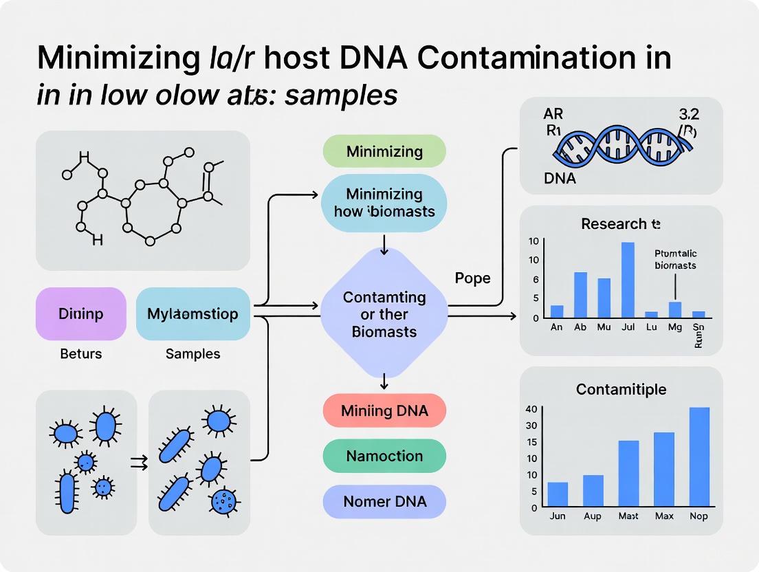
Strategies for Minimizing Host DNA Contamination in Low-Biomass Microbiome Studies: A Guide for Researchers
Metagenomic sequencing of low-biomass samples, such as urine, respiratory fluids, and tissues, is critically hampered by overwhelming host DNA, which can obscure microbial signals and lead to spurious results.
Strategies for Minimizing Host DNA Contamination in Low-Biomass Microbiome Studies: A Guide for Researchers
Abstract
Metagenomic sequencing of low-biomass samples, such as urine, respiratory fluids, and tissues, is critically hampered by overwhelming host DNA, which can obscure microbial signals and lead to spurious results. This article provides a comprehensive framework for researchers and drug development professionals to navigate the challenges of host DNA contamination. Drawing on the latest evidence, we detail foundational concepts, compare methodological approaches for host depletion, outline optimization and troubleshooting strategies, and establish rigorous validation standards. Implementing these guidelines is essential for achieving accurate, reproducible, and biologically meaningful insights into low-biomass microbial communities in biomedical and clinical research.
Understanding the Critical Challenge of Host DNA in Low-Biomass Samples
Low-biomass samples are characterized by exceptionally low levels of microbial DNA, which approach the detection limits of standard molecular techniques. These samples are disproportionately affected by contaminating DNA, as the target signal can be easily overwhelmed by contaminant noise [1] [2]. The defining challenge in low-biomass research is that even minute amounts of externally introduced DNA can generate spurious results, potentially leading to incorrect biological conclusions [3].
These samples originate from diverse environments. In human pathology, they include blood, urine, respiratory tract samples (such as nasopharyngeal aspirates and bronchoalveolar lavage fluid), deep tissues, and intratumoral environments [1] [4] [5]. Beyond the human body, low-biomass environments encompass the atmosphere, hyper-arid soils, treated drinking water, and the deep subsurface [2]. This article focuses on human-derived samples, framing the discussion within the critical context of minimizing host DNA contamination to ensure research validity.
Low-biomass samples are not a homogeneous group; they vary significantly in their origin, typical microbial load, and primary contaminants. Understanding these differences is essential for tailoring appropriate handling and analysis protocols.
Table 1: Characteristics of Common Low-Biomass Sample Types
| Sample Type | Typical Microbial Load & Context | Dominant Contaminant Challenges | Key Research Associations |
|---|---|---|---|
| Urine & Genitourinary Tract [6] | Low biomass; dogma of sterile urine disproven. | Sample collection contamination (midstream vs. catheterized); high host DNA. | Benign prostatic hyperplasia (BPH), chronic prostatitis/chronic pelvic pain syndrome (CP/CPPS), overactive bladder (OAB) [6]. |
| Respiratory Tract [4] [7] | Low bacterial biomass; healthy lung microbiota largely reflects upper respiratory tract entry via microaspiration. | Upper respiratory tract carryover during collection; reagent contaminants; very high host DNA content. | Respiratory disorders in premature infants; chronic lung allograft dysfunction; interstitial pulmonary fibrosis [4] [7]. |
| Blood [1] [2] | Very low microbial biomass in healthy state. | Reagent and kit contaminants; environmental DNA during phlebotomy. | Potential role in inflammatory and metabolic diseases; source of controversy [2] [3]. |
| Tumors (Intratumoral Microbiota) [5] [8] | Low-biomass microbial communities found in at least 33 cancer types. | Contamination from adjacent tissues, reagents, and sample handling. | Tumor initiation, progression, metastasis, and response to therapy (e.g., immunotherapy) [5]. |
The intratumoral microbiota, or "oncomicrobiome," presents a particularly complex low-biomass system. Microorganisms can colonize tumors through three primary routes: mucosal barrier invasion (e.g., from the gut to the pancreas), adjacent tissue invasion, and hematogenous invasion (via the bloodstream) [5]. For instance, Fusobacterium nucleatum can travel from the oral cavity to colonize colorectal tumors through the blood [5]. The structure and abundance of these intratumoral microbial populations vary substantially across cancer types, subtypes, and stages, influencing the tumor microenvironment and patient outcomes [5] [8].
Critical Challenges in Low-Biomass Microbiome Research
Contamination and Host DNA Interference
The foremost challenge in low-biomass research is contamination. Contaminant DNA can originate from a multitude of sources, including sampling equipment, laboratory reagents, kits, personnel, and the laboratory environment itself [2] [3]. In metagenomic analyses of low-biomass samples, host DNA can constitute over 99% of the sequenced material, drastically reducing the reads available for microbial characterization and increasing sequencing costs [9] [4]. This high host DNA content also creates a risk of host DNA being misclassified as microbial during bioinformatic analysis, potentially generating artifactual signals [9].
Cross-Contamination and Batch Effects
Another significant challenge is cross-contamination, also known as "well-to-well leakage" or the "splashome," where DNA is transferred between samples processed concurrently, such as in adjacent wells on a 96-well plate [2] [9]. Furthermore, batch effects—differences arising from different laboratories, personnel, or reagent lots—can introduce technical variation that confounds biological signals, especially when batch is correlated with the phenotype of interest [9].
The relationship between these challenges and their impact on data integrity is summarized below.
Essential Protocols for Reliable Low-Biomass Analysis
Experimental Design and Contamination Mitigation
Robust study design is the first line of defense against contamination. Key recommendations include:
- Decontaminate Sources: Thoroughly decontaminate equipment, tools, and vessels using 80% ethanol (to kill organisms) followed by a nucleic acid-degrading solution like sodium hypochlorite (bleach) to remove residual DNA [2].
- Use Personal Protective Equipment (PPE): Operators should wear gloves, masks, and cleansuits to limit the introduction of human-associated contaminants from skin, hair, or aerosols [2].
- Employ Single-Use, DNA-Free Materials: Use single-use, DNA-free consumables (e.g., swabs, collection vessels) whenever possible to minimize contamination [2].
- Avoid Batch Confounding: A critical step is to ensure that the biological groups being compared (e.g., cases vs. controls) are processed across the same batches (e.g., DNA extraction, sequencing runs) in a balanced, randomized manner. This prevents technical batch effects from being misinterpreted as biological signals [9].
Mandatory Control Samples
The inclusion of various control samples is non-negotiable for identifying contaminants and validating results [2] [9].
- Negative Controls: These include "field blanks" (an empty collection vessel processed alongside real samples), "extraction blanks" (reagents without a sample), and "PCR blanks" (water instead of template DNA) [2] [3].
- Positive Controls: Using a mock community (a defined mix of microbial cells or DNA) of known composition verifies that the entire workflow, from DNA extraction to sequencing, is functioning correctly and can detect expected taxa [4].
Host DNA Depletion and Microbial DNA Extraction
Effective host DNA depletion is paramount for maximizing microbial sequence recovery in shotgun metagenomic studies. A comparative study on nasopharyngeal aspirates from premature infants evaluated several combined depletion and extraction protocols [4].
Table 2: Evaluation of Host DNA Depletion and DNA Extraction Protocols for Respiratory Samples
| Protocol Name | Host DNA Depletion Method | DNA Extraction Kit | Key Findings and Efficacy |
|---|---|---|---|
| MasterPure [4] | None | MasterPure Gram Positive DNA Purification Kit | Retrieved expected DNA yield from mock communities but resulted in 99% host DNA in non-depleted patient samples. |
| Mol_MasterPure [4] | MolYsis Basic5 | MasterPure Gram Positive DNA Purification Kit | Most effective protocol. Showed varied but satisfactory host DNA reduction (down to 15-98% in patient samples), increasing bacterial reads by 7.6 to 1,725.8-fold. |
| Mol_MagMax [4] | MolYsis Basic5 | MagMAX Microbiome Ultra Nucleic Acid Isolation Kit | Failed to reduce host DNA content adequately in the tested samples. |
| QIA_QIAamp [4] | QIAamp | QIAamp DNA Microbiome Kit | Retrieved DNA yields that were too low for further analysis. |
The workflow for optimizing microbial DNA recovery from a high-host-content sample, based on this study, involves a critical decision point regarding host DNA depletion.
The Scientist's Toolkit: Key Research Reagent Solutions
Selecting the appropriate reagents and kits is fundamental to the success of low-biomass studies. The following table details essential materials and their functions, based on protocols cited in this review.
Table 3: Essential Reagents and Kits for Low-Biomass Research
| Reagent/Kits | Specific Function | Application Context |
|---|---|---|
| MolYsis Basic5 [4] | Selective host cell lysis and degradation of the released DNA, enriching for intact microbial cells. | Host DNA depletion from nasopharyngeal aspirates and other high-host-content samples prior to microbial DNA extraction. |
| MasterPure Gram Positive DNA Purification Kit [4] | Efficient DNA extraction using a lytic method that improves recovery from tough-to-lyse Gram-positive bacteria. | DNA extraction following host depletion; identified as the most effective extraction method in a comparative study. |
| ZymoBIOMICS Microbial Community Standard (D6300) [4] | Defined mock community of known microbial composition; serves as a positive control for the entire workflow. | Verifying accuracy, precision, and bias of the entire workflow from DNA extraction to sequencing and bioinformatics. |
| ZymoBIOMICS Spike-in Control II (D6321) [4] | Low-abundance spike-in control containing species not found in the human microbiome (e.g., Imtechella halotolerans). | Added to samples to quantitatively assess microbial load and account for variation in sample processing efficiency. |
| Sodium Hypochlorite (Bleach) [2] | Degrades contaminating DNA on surfaces and equipment; critical for achieving a DNA-free state beyond sterility. | Decontamination of work surfaces and reusable laboratory equipment before and during sample processing. |
The study of low-biomass samples, from genitourinary and respiratory tracts to tumors and blood, holds immense promise for advancing our understanding of human health and disease. However, realizing this potential requires unwavering diligence in addressing the unique challenges these samples present. Contamination, high host DNA content, and technical biases are not merely nuisances but fundamental issues that can invalidate research findings. By adopting a rigorous, contamination-aware mindset—implementing stringent experimental designs, mandatory controls, optimized wet-lab protocols for host DNA depletion, and careful bioinformatic decontamination—researchers can confidently navigate the complexities of low-biomass environments. The protocols and guidelines outlined here provide a foundational framework for generating robust, reliable, and reproducible data in this demanding but highly rewarding field.
The investigation of low-biomass microbial environments, such as human tissues, blood, and certain environmental niches, represents a frontier in microbiome science with great potential for discovery [9]. However, these studies face a formidable obstacle: the overwhelming presence of host DNA. In samples like tumors or blood, microbial DNA can constitute as little as 0.01% of sequenced reads, with the remainder being host-derived [9]. This imbalance severely compromises data quality and analytical sensitivity. Contrary to being merely "background noise," host DNA actively interferes with sequencing efficiency, reduces microbial sequencing depth, and can be misclassified as microbial signal, leading to spurious conclusions and controversial findings [9]. Addressing host DNA contamination is therefore not a peripheral concern but a fundamental requirement for generating reliable data in low-biomass microbiome research.
Quantitative Impact: How Host DNA Compromises Data Analysis
The presence of excessive host DNA has tangible, measurable impacts on sequencing outcomes and data quality. The following table summarizes the primary consequences and their mechanistic causes.
Table 1: Consequences and Mechanisms of Host DNA Contamination in Sequencing Studies
| Consequence | Underlying Mechanism | Impact on Data Analysis |
|---|---|---|
| Reduced Microbial Sequencing Depth | Fixed sequencing capacity is dominated by host reads, drastically undersampling the microbial community [9]. | Compromised detection sensitivity for low-abundance microbes; reduced statistical power. |
| Increased Sequencing Costs | Requires deeper sequencing to achieve sufficient coverage of the target microbial genome [10]. | Inefficient use of resources; cost for one lane can shift from 50+ microbial genomes to just a few [10]. |
| Misclassification of Host DNA as Microbial | Computational pipelines may incorrectly assign host DNA sequences to microbial taxa due to evolutionary similarities or database gaps [9]. | Introduction of false-positive microbial signals; distortion of reported microbial community composition. |
| Obscured Ecological Patterns | The proportional nature of sequence data means host DNA dilution alters the apparent relative abundance of microbes [2]. | Inaccurate representation of microbial community structure and dynamics. |
The economic impact is particularly stark. One lane of an Illumina HiSeq that could sequence over 50 multiplexed pure pathogen genomes may be reduced to sequencing only a single sample when host contamination is high, merely to achieve adequate microbial coverage [10]. This represents a catastrophic decrease in experimental efficiency and cost-effectiveness.
Methodological Solutions for Host DNA Depletion
Several strategies have been developed to mitigate host DNA contamination, falling into two broad categories: laboratory-based depletion and computational subtraction. The choice of method depends on sample type, research question, and available resources.
Laboratory-Based Depletion Techniques
These methods physically or enzymatically remove host DNA prior to sequencing.
Table 2: Laboratory-Based Methods for Host DNA Depletion
| Method | Principle | Advantages | Limitations |
|---|---|---|---|
| Enzymatic Methylation-Dependent Depletion | Utilizes restriction endonucleases (e.g., MspJI) that selectively cleave DNA at methylated cytosines, abundant in host DNA but largely absent in microbial genomes [10]. | - Can achieve ~9-fold enrichment of pathogen DNA.- Simple protocol integrated into library prep.- Non-biased recovery of microbial sequences [10]. | - Efficiency depends on host methylation status.- May affect microbes with methylated genomes.- Requires optimized reaction conditions. |
| Probe-Based Hybridization Capture | Uses complementary biotinylated probes (e.g., against human rRNA genes) to bind host DNA, which is then removed with streptavidin-coated beads. | - Highly specific and efficient depletion.- Can be tailored for different host species. | - High cost and procedural complexity.- Requires sufficient input DNA.- Potential for non-specific removal of microbial DNA. |
| Differential Lysis and Centrifugation | Selectively lyse host cells with milder agents, leaving microbial cells intact for subsequent separation. | - Preserves viability of microbes for downstream culture.- No enzymatic or chemical bias. | - Inefficient for intracellular microbes or biofilms.- Risk of incomplete host lysis or microbial loss. |
Detailed Protocol: Enzymatic Depletion of Human DNA Using MspJI
This protocol is adapted from a method proven to enrich Plasmodium falciparum DNA from highly contaminated clinical samples [10].
A. Reagents and Equipment
- Methylation-Dependent Restriction Endonuclease (e.g., MspJI, LpnPI, FspEI)
- 10x NEB Buffer 4 (or supplier-recommended buffer)
- Molecular biology-grade Bovine Serum Albumin (BSA)
- Activator oligonucleotide (sequence: CTGCmCAGGATCTTTTTTGATCmCTGGCAG) [10]
- Agencourt Ampure XP beads or equivalent
- Thermal cycler
- Covaris S2 sonicator or equivalent DNA shearing device
B. Procedure (Gel-Free Method)
- DNA Shearing: Shear 0.1-2 μg of total genomic DNA to an average fragment size of ~350 bp using a Covaris S2 sonicator with the following settings: 10% duty cycle, intensity 4, 200 cycles per burst for 70 seconds [10].
- End-Repair: Perform end-repair on the sheared DNA fragments using a standard library preparation kit (e.g., NEBNext DNA Sample Preparation Kit).
- Enzymatic Digestion: Set up the digestion reaction in a 0.2 mL PCR tube:
- End-repaired DNA: Entire sample
- 10x NEB Buffer 4: 3 μL
- BSA (10 μg/μL): 1 μL
- Activator Oligonucleotide (0.05 μM): 0.05 μM final concentration
- MspJI Enzyme: 6 units
- Nuclease-free water to a final volume of 30 μL Incubate in a thermocycler at 37°C for 16 hours, followed by 65°C for 20 minutes to inactivate the enzyme [10].
- Size Selection: Purify the digested sample using Agencourt Ampure XP beads to remove small digestion fragments.
- Mix the digested sample with an equal volume of beads and incubate for 5 minutes at room temperature.
- Place on a magnetic rack to capture beads. Discard the supernatant.
- Wash the beads twice with 80% ethanol. Air dry and elute DNA in elution buffer (EB).
- Library Preparation: Proceed with standard Illumina paired-end library construction (A-tailing, adapter ligation, and PCR enrichment with 12 cycles) using the purified, host-depleted DNA [10].
Computational Subtraction
This bioinformatic approach involves aligning sequencing reads to a host reference genome (e.g., GRCh38) and discarding those that match. While this method does not improve the depth of microbial sequencing, it prevents the misclassification of host reads as microbial and reduces downstream analysis noise [9]. Its success is highly dependent on the quality and completeness of the reference genome.
Integrated Experimental Workflow for Low-Biomass Studies
A robust study of low-biomass microbiomes requires an integrated workflow that combines host DNA depletion with stringent contamination control throughout the process. The following diagram visualizes this comprehensive approach.
Diagram 1: Integrated workflow for low-biomass microbiome studies, combining wet-lab and computational steps to mitigate host DNA and contamination.
Critical Step: Incorporating Process Controls
As visualized in the workflow, the use of process controls is non-negotiable in low-biomass research [2] [9]. These controls are essential for distinguishing true microbial signal from contamination introduced during sampling or laboratory processing.
- Types of Controls: Include blank extraction controls (no sample added during DNA extraction), no-template PCR controls (water instead of DNA during amplification), and sampling controls (e.g., swabs of air or sterile collection tubes) [2] [9].
- Execution: These controls must be processed concurrently with actual samples through every step, from DNA extraction to sequencing, to accurately capture the contaminant profile of the specific experimental batch [9].
The Scientist's Toolkit: Essential Reagents and Materials
The following table details key reagents and materials required for implementing the host DNA depletion and control strategies described in this protocol.
Table 3: Research Reagent Solutions for Host DNA Depletion and Contamination Control
| Item | Function/Description | Application Note |
|---|---|---|
| MspJI Restriction Endonuclease | A methylation-dependent enzyme that cleaves DNA at methylated cytosine sites, preferentially digesting host DNA [10]. | Core reagent for enzymatic host DNA depletion. Requires optimized buffer conditions and optional activator oligonucleotide for enhanced activity [10]. |
| Biotinylated Host DNA Probes | Single-stranded DNA probes designed to target repetitive host elements (e.g., ALU, LINE, rDNA) for capture and removal. | Essential for probe-based hybridization capture methods. Specificity and design are critical for efficiency. |
| Agencourt Ampure XP Beads | Magnetic silica beads for post-digestion size selection and clean-up, removing small fragments of digested host DNA. | Enables gel-free sample preparation after enzymatic treatment, streamlining the workflow [10]. |
| DNA-Free Collection Kits | Pre-sterilized, DNA-free swabs, tubes, and reagents specifically designed for low-biomass sample collection. | Minimizes the introduction of contaminating DNA at the first step of the workflow, a foundational best practice [2]. |
| Commercial Host Depletion Kits | Integrated kits (e.g., based on probe capture) that provide a standardized protocol and reagents for depleting host DNA from specific sample types. | Reduces optimization time but can be costly. Suited for studies processing a large number of similar samples. |
The impact of host DNA on low-biomass microbiome studies is profound, affecting everything from sequencing costs and efficiency to the fundamental validity of biological conclusions. Success in this challenging field relies on a multi-layered strategy that integrates wet-lab depletion methods like enzymatic treatment, rigorous experimental design with extensive controls, and robust bioinformatic cleaning. By systematically implementing the protocols and workflows outlined in this document, researchers can significantly reduce the obscuring effect of host DNA, revealing the true and often subtle microbial signals in low-biomass environments.
In low-biomass microbiome research, where microbial DNA is minimal, contaminants from reagents, sampling equipment, and cross-contamination can disproportionately affect results, leading to erroneous conclusions [2]. These environments, which include human tissues like the placenta and respiratory tract, as well as various environmental niches, are particularly vulnerable because the target DNA signal is often dwarfed by contaminant noise [2] [9]. Effectively minimizing and identifying these contaminants is critical for data integrity, requiring stringent controls at every stage from sample collection to data analysis [2] [11]. This document outlines the major sources of contamination and provides detailed protocols to mitigate their impact, specifically framed within the challenge of minimizing host DNA contamination.
The table below summarizes the primary contamination sources, their origins, and the recommended mitigation strategies.
Table 1: Major Contamination Sources and Control Strategies
| Contamination Source | Description and Origin | Key Mitigation Strategies |
|---|---|---|
| Reagents & Kits | Microbial DNA inherent in DNA extraction kits and laboratory reagents, known as "kitomes" [11]. Profiles vary by brand and manufacturing lot [11]. | - Use multiple extraction blanks [11] [9].- Employ computational decontamination tools (e.g., Decontam) [11].- Request lot-specific contamination profiles from manufacturers [11]. |
| Sampling Equipment | Contaminants introduced from collection vessels, swabs, and personal protective equipment (PPE) during sample collection [2]. | - Use single-use, DNA-free equipment [2].- Decontaminate surfaces with 80% ethanol followed by a nucleic acid-degrading solution (e.g., bleach) [2].- Utilize appropriate PPE and sterilize tools with autoclaving or UV-C light [2]. |
| Cross-Contamination (Well-to-Well Leakage) | Transfer of DNA between samples processed concurrently, often in adjacent wells on a plate [2] [12]. This is distinct from index hopping [12]. | - Randomize or strategically balance sample placement across plates to avoid confounding with phenotypes [9].- Maintain physical distance between samples during liquid handling [12].- Use strain-resolved bioinformatic analyses to detect contamination patterns [12]. |
| Host DNA | Abundant human DNA in samples can be misclassified as microbial during analysis, overwhelming sequencing depth [13] [9] [14]. | - Apply wet-lab host depletion methods before sequencing [14].- Use bioinformatic tools (e.g., bwa-mem) with a comprehensive reference genome (e.g., CHM13-T2T) for post-sequencing removal [13]. |
Experimental Protocols for Contamination Control
Protocol: Profiling Reagent-Derived Contamination
Objective: To characterize the background microbiota ("kitome") in DNA extraction reagents and account for batch variability [11].
Materials:
- DNA extraction kits from various brands (e.g., Q, M, R, Z) [11]
- Molecular biology-grade (DNA-free) water [11]
- ZymoBIOMICS Spike-in Control I (optional, for positive control) [11]
- Unison Ultralow DNA NGS Library Preparation Kit [11]
Method:
- Preparation of Extraction Blanks: For each brand and lot of DNA extraction kit, prepare triplicate blank extractions using molecular-grade water as input instead of a sample [11].
- DNA Extraction: Perform DNA extraction strictly following the manufacturer's instructions. Process kits and lots on separate days to prevent cross-contamination [11].
- Library Preparation and Sequencing: Construct sequencing libraries from the eluted DNA using an ultralow-DNA-input library preparation kit. Sequence the libraries, for example, using an Illumina MiSeq platform with 150 bp single-end reads [11].
- Data Analysis: Process the raw sequences through a standard microbiome analysis pipeline (e.g., using QIIME2). Identify putative contaminant sequences by comparing their prevalence and abundance in blank controls versus true biological samples, using tools like the
decontamR package [11] [14].
Protocol: Implementing a Comprehensive Control Strategy
Objective: To identify contaminants introduced throughout the entire experimental workflow, from sampling to sequencing [2] [9].
Materials:
- Sterile, DNA-free collection vessels (e.g., empty tubes, swabs) [2] [9]
- Sample preservation solution
- DNA extraction kits and library preparation reagents
Method:
- Sample Collection Controls:
- Laboratory Process Controls:
- Processing: Include multiple replicates of each control type and process them alongside the biological samples through every subsequent step (DNA extraction, library prep, sequencing) [2] [9].
- Analysis: The collective data from these controls provides a comprehensive profile of contamination. This profile is essential for informing computational decontamination and for judging the validity of taxa detected in low-biomass samples [9].
Protocol: Optimizing Host DNA Depletion for Urine Samples
Objective: To evaluate and implement host DNA depletion methods for low-microbial-biomass, high-host-DNA samples like urine [14].
Materials:
- Urine sample (≥3.0 mL recommended for consistent profiling) [14]
- Commercial host depletion kits (e.g., QIAamp DNA Microbiome, Molzym MolYsis, NEBNext Microbiome DNA Enrichment, Zymo HostZERO) [14]
- Propidium monoazide (PMA) solution [14]
- QIAamp BiOstic Bacteremia Kit (or similar kit without host depletion) [14]
Method:
- Sample Preparation: Collect and centrifuge urine samples at 20,000 × g for 30 minutes at 4°C. Discard the supernatant and retain the pellet [14].
- Host Depletion and DNA Extraction: Divide the pellet for processing with different methods:
- No Depletion (Control): Extract DNA using the Bacteremia kit per manufacturer protocol [14].
- Commercial Kits: Extract DNA using the various host depletion kits, following their respective protocols [14].
- PMA Treatment: Resuspend the pellet in PBS and add PMA to a final concentration of 50 µM. Incubate in the dark for 10 minutes, then expose to a 500-watt halogen light source for 10 minutes. Proceed with DNA extraction using the Bacteremia kit [14].
- Downstream Analysis: Quantify DNA yield. Perform 16S rRNA gene and shotgun metagenomic sequencing. Compare microbial diversity, host DNA depletion efficiency, and metagenome-assembled genome (MAG) recovery across methods [14].
Workflow for Contamination Management
The following diagram illustrates a comprehensive workflow for managing contamination in low-biomass studies, integrating the protocols and strategies detailed above.
The Scientist's Toolkit: Essential Research Reagents & Materials
Table 2: Key Reagents and Materials for Contamination Control
| Item | Function / Application | Key Considerations |
|---|---|---|
| Molecular-Grade Water | Serves as input for extraction blank controls to profile reagent-derived contamination [11]. | Must be certified DNA-free and nuclease-free; filter-sterilized (0.1 µm) [11]. |
| DNA Decontamination Solutions | Decontaminates sampling equipment and surfaces to remove microbial cells and trace DNA [2]. | Use 80% ethanol to kill cells, followed by sodium hypochlorite (bleach) or UV-C light to degrade DNA [2]. |
| ZymoBIOMICS Spike-in Control | Serves as an internal positive control for the DNA extraction and sequencing process [11]. | Consists of known, non-native bacterial strains (e.g., I. halotolerans, A. halotolerans) to monitor efficiency [11]. |
| Host Depletion Kits | Selectively depletes host (e.g., human/canine) cells or DNA from a sample to increase microbial sequencing depth [14]. | Kits vary in efficacy (e.g., QIAamp DNA Microbiome Kit showed strong performance for urine) [14]. |
| CHM13-T2T Reference Genome | A complete, telomere-to-telomere human genome used for bioinformatic removal of host-derived sequences from metagenomic data [13]. | More effective than previous references (e.g., GRCh38) due to 216 Mbp of additional sequence, reducing false positives [13]. |
| Decontam (R package) | A statistical tool to identify and remove contaminant sequences from microbiome data based on prevalence in negative controls [11] [14]. | Relies on the inclusion of proper negative controls; uses prevalence or frequency to classify contaminants [11]. |
The Amplified Signal of Contamination in Low-Biomass Research
In microbiome studies, "low-biomass" refers to samples containing minimal microbial material, often hovering near the detection limits of standard DNA-based sequencing methods [2]. These samples are common in a wide range of research contexts, including certain human tissues (respiratory tract, placenta, blood), environmental samples (drinking water, cleanroom surfaces, hyper-arid soils), and host-associated systems like fish gills or marine invertebrate symbionts [2] [15] [16]. The fundamental vulnerability of low-biomass research lies in the proportional nature of sequence-based data; even minute amounts of contaminating DNA, which would be statistically negligible in high-biomass samples like stool or soil, can constitute a substantial proportion of, or even exceed, the true biological signal [2] [3].
This contamination arises from multiple sources throughout the experimental workflow. The table below summarizes the core challenges that distinguish low-biomass research from standard microbiome workflows.
Table 1: Core Challenges in Low-Biomass Microbiome Research
| Challenge | Impact on Low-Biomass Samples | Consequence |
|---|---|---|
| External Contamination [2] [9] | Reagent "kitome," sampling equipment, and personnel DNA can dominate the signal. | Distorts true microbial community structure, leading to false positives and incorrect ecological conclusions [3]. |
| Host DNA Misclassification [9] | Host DNA can constitute >99.9% of sequenced material (e.g., in tumors) [9]. | Obscures microbial signal; misclassified host reads can be misinterpreted as microbes, generating noise or artifactual signals [9]. |
| Well-to-Well Leakage [2] [9] | Cross-contamination between samples on a processing plate is disproportionately impactful. | Can violate the assumptions of decontamination algorithms, leading to faulty data interpretation [9]. |
| Batch Effects & Processing Bias [9] | Technical variations between processing batches can be confounded with the phenotype of interest. | Introduces artifactual signals that are falsely associated with experimental groups rather than technical noise [9]. |
The failure of standard practices is starkly illustrated by historical controversies in the field. For instance, initial claims of a distinct placental microbiome were later refuted when follow-up studies demonstrated that the microbial signals detected were indistinguishable from those found in negative control samples [2] [9]. Similar debates have surrounded studies of the blood microbiome and certain extreme environments, underscoring the critical need for specialized workflows [2].
Building a Contamination-Aware Workflow: From Sampling to Analysis
A robust low-biomass workflow requires integrated strategies across all stages of research, from experimental design and sample collection to laboratory processing and data analysis. The following diagram outlines the core pillars of a contamination-aware workflow.
Foundational Step: Contamination-Informed Sampling
The first line of defense is to minimize the introduction of contaminants during sample collection.
- Decontaminate Sources of Contaminant DNA: Equipment, tools, and collection vessels should be decontaminated. A recommended protocol involves treatment with 80% ethanol to kill contaminating organisms, followed by a nucleic acid-degrading solution like sodium hypochlorite (bleach) or UV-C light to remove residual DNA [2]. It is critical to note that sterility (absence of viable cells) is not synonymous with being DNA-free [2].
- Use Physical Barriers: Personnel should wear appropriate personal protective equipment (PPE), including gloves, masks, cleansuits, and hairnets, to limit the introduction of human-associated microbial cells and DNA via aerosol droplets or skin shedding [2] [17].
- Maximize Microbial Yield, Minimize Host Content: The collection method itself can be optimized. In fish gill studies, for example, swabbing the gill filter was shown to yield significantly more 16S rRNA gene copies and less host DNA compared to processing whole gill tissue [16]. This approach directly increases the target signal relative to background interference.
Laboratory Processing: Host Depletion and Low-Bioburden Reagents
Once a sample enters the lab, the focus shifts to preserving the microbial signal while minimizing background.
- Utilize Certified Low-Bioburden Reagents: Standard molecular biology reagents can be significant sources of contaminating DNA. Using commercially available, certified low-bioburden DNA extraction kits and DNase/RNase-free water is crucial. These reagents are manufactured and aliquoted in clean, HEPA-filtered environments to minimize background DNA [17].
- Implement Host DNA Depletion: When host DNA is expected to overwhelm the microbial signal (e.g., in tissue biopsies or BALF), specific depletion methods can be applied. These are broadly categorized as pre-extraction (physical removal or lysis of host cells) and post-extraction (enzymatic degradation of host DNA) methods [18]. A 2025 benchmarking study of seven pre-extraction methods for respiratory samples highlighted a key trade-off: while all methods significantly increased microbial read proportions, they also introduced varying degrees of bias, contamination, and microbial DNA loss [18]. The table below compares the performance of several common host depletion methods.
Table 2: Comparison of Host DNA Depletion Methods for Low-Biomass Respiratory Samples
| Method (Abbreviation) | Principle | Host Depletion Efficiency | Key Trade-offs / Performance Notes |
|---|---|---|---|
| Saponin + Nuclease (S_ase) [18] | Lyses human cells with saponin; degrades DNA with nuclease. | High (to ~0.01% of original) | High bacterial DNA loss; can diminish specific pathogens like Mycoplasma pneumoniae. |
| HostZERO Kit (K_zym) [18] | Commercial pre-extraction kit. | High (to ~0.01% of original) | High bacterial DNA loss. |
| Nuclease Only (R_ase) [18] | Degrades exposed (e.g., cell-free) DNA with nuclease. | Moderate | Highest bacterial retention rate; lower increase in microbial reads. |
| Filtration + Nuclease (F_ase) [18] | Filters host cells; treats with nuclease. | Moderate | Most balanced performance in the benchmarked study. |
| Osmotic Lysis + PMA (O_pma) [18] | Lyses human cells osmotically; PMA inhibits DNA from dead cells. | Low | Least effective at increasing microbial reads (2.5-fold). |
The Critical Role of Comprehensive Controls
Contamination cannot be fully eliminated, so it must be documented and accounted for via rigorous controls.
- Types of Controls: It is essential to process multiple types of negative controls alongside your true samples from collection to sequencing. These include empty collection vessels, swabs of the air, aliquots of preservation solutions, DNA extraction blanks (no sample added), and no-template PCR controls [2] [9] [19].
- Avoiding Batch Confounding: A critical aspect of experimental design is to ensure that case and control samples are distributed across all processing batches (e.g., DNA extraction plates, sequencing runs). If all cases are processed in one batch and all controls in another, technical artifacts from that batch can be falsely interpreted as biological signals [9]. Active randomization or tools like BalanceIT should be used to de-confound batches from the phenotypes of interest [9].
The Scientist's Toolkit: Essential Reagents & Materials
The following table details key reagents and materials that form the foundation of a reliable low-biomass research pipeline.
Table 3: Research Reagent Solutions for Low-Biomass Workflows
| Item | Function | Considerations & Examples |
|---|---|---|
| Certified Low-Bioburden Kits [17] | DNA extraction with minimal contaminating bacterial background DNA. | Kits are certified using qPCR to quantify background 16S rRNA. Example: ZymoBIOMICS DNA Miniprep Kit. |
| DNase/RNase-Free Water [17] | Elution and reagent preparation without introducing contaminating DNA. | Should be DEPC-treated and autoclaved. Aliquoting in a clean environment is recommended. |
| Nucleic Acid Degrading Solutions [2] | Decontaminate surfaces and equipment to remove trace DNA. | Sodium hypochlorite (bleach), UV-C light, hydrogen peroxide, or commercial DNA removal solutions. |
| Personal Protective Equipment (PPE) [2] [17] | Create a physical barrier to human-derived contamination. | Gloves, masks, dedicated lab coats, bouffant caps, and shoe covers. |
| Internal Standard [18] [20] | Spike-in control for quantifying absolute abundance and assessing bias. | Defined microbial communities (e.g., ZymoBIOMICS Microbial Community Standard) added to the sample at lysis. |
| Propidium Monoazide (PMA) [20] | Viability dye that penetrates compromised membranes, suppressing DNA from dead cells. | Used pre-extraction to better profile intact, potentially viable cells. Effectiveness varies by sample type. |
From Data to Discovery: Analysis, Reporting, and Future Directions
Bioinformatics Decontamination and Quantitative Analysis
After sequencing, bioinformatic tools are necessary to identify and remove contaminant sequences.
- Employ Contamination Removal Algorithms: Computational tools such as
decontam(R package) can use the prevalence or frequency of sequence variants in negative controls to identify and subtract contaminants from the true sample data [2] [20]. However, these methods rely on well-designed control experiments to function correctly. - Pursue Quantitative Profiling: Relative abundance data can be misleading. Incorporating quantitative data is powerful. This can be achieved by:
- Spiking Internal Standards: Adding a known quantity of an artificial or exotic microbial community to each sample during lysis allows for the estimation of absolute microbial loads in the original sample [20].
- qPCR: Quantifying 16S rRNA gene copies or a specific pathogen gene prior to sequencing provides an absolute metric that can contextualize sequencing results [16] [20].
Transparent Reporting and a Path Forward
To ensure the integrity and reproducibility of low-biomass research, the field is moving towards adopting minimal reporting standards. Researchers are urged to clearly document the following in their publications [2]:
- Types and number of negative controls used.
- The bioinformatic decontamination strategies applied.
- Any quantitative assessments performed (e.g., qPCR data, spike-in controls).
Future advancements will likely come from continued refinement of host depletion methods to reduce bias, the development of more efficient sample concentration technologies, and the creation of more comprehensive reference databases for accurate taxonomic classification in understudied environments [9] [19]. By adopting these contamination-aware practices, researchers can overcome the unique vulnerabilities of low-biomass workflows and generate robust, reliable data that drives meaningful scientific discovery.
The investigation of microbial communities in environments with minimal microbial life, known as low-biomass microbiomes, represents one of the most technically challenging frontiers in microbial ecology. Research on purported microbial communities in tissues such as the placenta and internal tumors has been marked by significant controversy, primarily revolving around the critical issue of distinguishing true biological signal from contamination. The central thesis framing this application note is that minimizing host DNA contamination and exogenous microbial contamination is not merely a technical consideration but a fundamental prerequisite for generating valid data in low-biomass microbiome studies. Failures in adequate contamination control have led to the publication of findings that could not be replicated, sparking vigorous debates within the scientific community about the very existence of microbiomes in certain human tissues [21] [2] [22].
The core challenge stems from the fact that in low-biomass samples, the target microbial DNA signal can be exponentially smaller than contamination introduced from reagents, laboratory environments, sampling procedures, and the host organism's own DNA [2] [23]. Even minute contamination levels that would be negligible in high-biomass samples (like stool) can completely dominate and distort the microbial profile of low-biomass samples. This application note synthesizes critical lessons from two key case studies—placental and tumor microbiome research—to provide a structured framework of protocols, controls, and analytical strategies designed to safeguard research integrity in this demanding field.
Case Study 1: The Placental Microbiome Controversy
Review of Key Evidence and Methodological Pitfalls
The long-standing dogma of uterine sterility during healthy pregnancy was challenged by next-generation sequencing studies that reported detectable bacterial DNA in placental tissue. However, a comprehensive re-analysis of fifteen publicly available 16S rRNA gene datasets concluded that contemporary DNA-based evidence does not support the existence of a placental microbiota [21]. The analysis demonstrated that bacterial signals observed in placental samples were indistinguishable from those found in technical controls and were profoundly influenced by the mode of delivery [21]. For instance, Lactobacillus sequences—typical vaginal bacteria—were highly prevalent in placental samples from vaginal deliveries but disappeared from samples obtained through term cesarean deliveries after rigorous contaminant removal [21].
A separate cross-sectional study of 76 term pregnancies comparing placental tissues, amniotic fluid, and maternal samples found no evidence of a placental microbiome using both PCR-based methods and bacterial culture. Quantitative measurements of bacterial content in all three placental layers showed no significant difference from negative controls [22]. This study also highlighted that bacterial cultures from placentas delivered vaginally showed substantially more bacteria than those from cesarean deliveries, with most identified bacteria representing genera commonly found on human skin or in the vagina [22].
Table 1: Key Studies in the Placental Microbiome Debate
| Study Focus | Key Findings | Methodological Limitations |
|---|---|---|
| Re-analysis of 15 datasets [21] | No distinct placental microbiota after accounting for contaminants; signals clustered by study origin and delivery mode. | Inconsistent processing pipelines across studies; insufficient controls in original studies. |
| Term pregnancy study [22] | No significant difference between placental bacteria and negative controls; culture growth was delivery-associated. | Cannot rule out extremely low-biomass signals below detection limits. |
| Expert consensus [24] | Majority opinion favors 'sterile womb' hypothesis; any bacterial DNA likely from contamination or transient presence. | Burden of proof remains high for demonstrating a true microbiota. |
Detailed Experimental Protocol: Controlled Placental Sampling and DNA Analysis
The following protocol outlines a rigorous approach for placental tissue collection and processing designed to minimize contamination, suitable for investigating potential microbial signals.
Materials and Equipment
- Personal Protective Equipment (PPE): Sterile gloves, mask, hair net, cleanroom suit (or fresh lab coat)
- Sampling Equipment: Autoclaved surgical instruments (scalpels, forceps), DNA-free collection vessels, sterile swabs
- Preservation Solution: DNA-free buffer (e.g., TE) or preservation kit (e.g., DNA/RNA Shield)
- DNA Extraction Kit: Selected for efficiency with low-biomass samples (e.g., DNeasy PowerSoil Pro Kit)
- Quantitation Equipment: Qubit fluorometer, qPCR system
- Sequencing Reagents: 16S rRNA gene primers, library preparation kit
Step-by-Step Procedure
Pre-sampling Preparation:
- Decontaminate all work surfaces and equipment with 80% ethanol followed by DNA-degrading solution (e.g., 0.5% sodium hypochlorite) and UV irradiation.
- Set up multiple negative controls: an empty collection vessel, a swab exposed to the air in the sampling environment, and an aliquot of preservation solution [2].
Patient and Sample Collection:
- For cesarean deliveries, clean the abdominal skin with an antiseptic protocol. For vaginal deliveries, clean the perineum thoroughly.
- Upon placental delivery, immediately transfer the organ to a sterile container without manual contact.
- Using sterile instruments, collect tissue samples from multiple sites: maternal side, fetal side, and a central villous core.
- For each sample, use a fresh set of sterilized instruments. Collect tissue pieces of standardized weight (e.g., 100 mg).
- Place samples immediately into sterile, DNA-free tubes containing preservation solution and freeze at -80°C.
DNA Extraction with Controls:
- Process samples in a laminar flow hood to minimize air contamination.
- Include multiple extraction negative controls (reagents only) alongside samples and pre-collection negative controls.
- Use an extraction kit validated for low-biomass samples. Incorporate an internal DNA standard (spike-in) into a subset of samples to assess extraction efficiency and PCR inhibition [24].
- Elute DNA in a low EDTA elution buffer and store at -20°C.
Quantitative Analysis and Sequencing:
- Quantify total DNA and bacterial 16S rRNA gene copies using qPCR with universal bacterial primers.
- Samples with bacterial DNA levels not significantly exceeding those in the negative controls should be treated with extreme caution [22].
- For sequencing, normalize inputs based on 16S rRNA gene copy number rather than total DNA to create "equicopy libraries," improving diversity detection [25].
- Use a targeted amplicon approach (e.g., 16S rRNA gene V4 region) with sufficient sequencing depth.
Diagram 1: Rigorous Placental Sampling Workflow. This workflow emphasizes contamination control at every stage, from pre-sampling preparation to final analysis.
Case Study 2: The Tumor Microbiome Debate
Methodological Challenges and Contamination Controls
The proposal that tumors harbor low-biomass microbial ecosystems has been similarly contentious. A pivotal review highlighted that recent reports suggesting a distinctive cancer microbiome were based on flawed data, with re-analysis completely overturning the original findings [26]. The major issues identified included susceptibility of low-biomass samples to exogenous contamination, undetermined microbial viability from NGS data, and insufficient attention to host DNA depletion [27] [28].
In tumor samples, the overwhelming abundance of host DNA presents a distinct challenge. In milk samples (another low-biomass, host-rich matrix), the ratio of somatic cells to bacteria ultimately impacts microbial DNA yield, with samples having lower somatic cell counts being the most problematic for analysis [23]. This directly parallels the tumor context, where the balance between human and microbial DNA is critical.
Table 2: Key Considerations for Host DNA Depletion in Low-Biomass Samples
| Method/Strategy | Principle | Considerations for Tumor/Placental Samples |
|---|---|---|
| Commercial Host Depletion Kits (e.g., NEBNext Microbiome DNA Enrichment Kit) | Selective digestion of methylated host DNA (common in mammalian genomes). | Can significantly reduce host DNA but may also reduce microbial DNA yield, impacting already low-biomass samples [23]. |
| Multiple Displacement Amplification (MDA) | Isothermal whole-genome amplification using phi29 polymerase. | Can recover microbial genomes from low-input samples; successful for high SCC milk samples [23]. Risk of amplification bias and contaminant sequences. |
| Optimized DNA Extraction Kits | Kits designed for low-biomass, inhibitor-rich samples (e.g., Dneasy PowerFood Microbial Kit). | Maximizes microbial lysis and DNA recovery while minimizing co-extraction of inhibitors. Performance varies by sample type [23]. |
| qPCR Pre-screening | Quantification of 16S rRNA genes vs. total DNA. | Allows for screening of samples prior to costly sequencing; identifies samples with signal potentially below reliable detection [25]. |
Detailed Experimental Protocol: Host DNA Depletion and Metagenomic Analysis from Tumors
This protocol focuses on maximizing microbial signal and depleting host DNA from tumor tissue samples for metagenomic sequencing.
Materials and Equipment
- Tissue Homogenizer: Bead-beater or similar, with sterile, DNA-free beads
- Host DNA Depletion Kit: e.g., NEBNext Microbiome DNA Enrichment Kit
- DNA Extraction Kit: e.g., DNeasy PowerSoil Pro Kit or QIAamp DNA Micro Kit
- Whole Genome Amplification Kit: e.g., REPLI-g Single Cell Kit (MDA)
- Quantitation and QC: Qubit, Bioanalyzer/TapeStation, qPCR system
- Sequencing Reagents: For shotgun metagenomic library preparation
Step-by-Step Procedure
Sample Processing and Homogenization:
- Perform all steps in a dedicated laminar flow hood.
- Weigh tumor tissue (e.g., 25 mg) and place in a sterile tube with lysis buffer and beads.
- Homogenize using a bead beater. Include a process control (e.g., a known mock microbial community) and negative controls (lysis buffer only).
- Centrifuge briefly to pellet large debris and transfer the supernatant to a new tube.
Host DNA Depletion:
- Follow the protocol of a host DNA depletion kit, such as the NEBNext Microbiome DNA Enrichment Kit, which exploits the differential methylation patterns between host and microbial DNA.
- After enzymatic treatment, purify the DNA. Expect a significant reduction in total DNA yield.
DNA Extraction and Optional Amplification:
- Proceed with DNA extraction from the depleted lysate using a kit designed for maximal microbial recovery.
- Quantify DNA using a fluorometer. For samples with very low DNA yield (<0.5 ng/µL), consider Multiple Displacement Amplification (MDA).
- For MDA: Use 1-10 µL of extracted DNA following the manufacturer's instructions. Purify the amplified product.
Library Preparation and Sequencing:
- Quantify the final DNA and assess fragment size distribution.
- Prepare metagenomic sequencing libraries. For MDA-treated samples, use kits compatible with amplified DNA.
- Sequence using an appropriate platform (e.g., Illumina for short-read; if DNA quantity and quality are sufficient, consider long-read sequencing for improved assembly).
Diagram 2: Tumor Microbiome Analysis with Host Depletion. This workflow includes a critical decision point (red node) for whole-genome amplification when microbial DNA yield is insufficient for direct sequencing.
The Scientist's Toolkit: Essential Reagents and Controls
The following table compiles key reagents, controls, and their critical functions based on the lessons learned from the placental and tumor microbiome controversies.
Table 3: Essential Research Reagents and Controls for Low-Biomass Studies
| Item Category | Specific Examples | Function & Importance |
|---|---|---|
| DNA Extraction Kits | DNeasy PowerSoil Pro Kit, QIAamp DNA Micro Kit | Optimized for lysing tough microbial cells and removing PCR inhibitors common in tissue samples. |
| Host Depletion Kits | NEBNext Microbiome DNA Enrichment Kit | Selectively depletes methylated host DNA, increasing the relative proportion of microbial reads. |
| Whole Genome Amplification | REPLI-g Single Cell Kit (MDA) | Amplifies minimal microbial DNA for sequencing; crucial for very low-biomass samples but requires careful control for bias [23]. |
| Negative Controls | Extraction blanks, reagent-only controls, sterile swab/air controls | Identifies contaminating DNA from reagents and the laboratory environment; essential baseline for data interpretation [2] [22]. |
| Positive/Mock Controls | Defined microbial mock communities, internal spike-ins (e.g., S. thermophilus) | Assesses extraction efficiency, PCR bias, and bioinformatic pipeline performance; verifies detection capability [24] [23]. |
| qPCR Reagents | Assays for universal 16S rRNA genes and a host gene (e.g., β-actin) | Pre-screens sample quality and bacterial load; allows normalization and identifies samples unsuitable for sequencing [25]. |
The controversies surrounding the placental and tumor microbiomes underscore a critical paradigm for all low-biomass microbiome research: the imperative for stringent contamination control throughout the entire research workflow, from experimental design through data analysis. The following consolidated framework is proposed for future studies:
- Design with Controls from the Start: Integrate multiple negative controls (collection, extraction, and sequencing) and positive controls (mock communities/spike-ins) into the experimental design, not as an afterthought [2].
- Standardize Pre-analytical Steps: Implement rigorous, standardized protocols for sample collection, using PPE and decontaminated equipment to minimize introduction of exogenous DNA [2] [22].
- Quantify and Normalize Strategically: Use qPCR to quantify both microbial and host DNA, and normalize sequencing inputs based on microbial load where appropriate to improve resolution [25].
- Apply Robust Bioinformatic Decontamination: Process sequence data using pipeline tools (e.g., DADA2) and contaminant identification packages (e.g., DECONTAM) that can identify and remove sequences likely originating from contaminants [21] [2].
- Transparently Report Methods and Controls: Adhere to emerging reporting guidelines for low-biomass studies to ensure reproducibility and allow the scientific community to critically evaluate the validity of findings [2].
By learning from the methodological pitfalls revealed in these case studies and adopting the detailed protocols and toolkit provided herein, researchers can advance the field with greater confidence, ensuring that future discoveries of low-biomass microbiomes are built upon a foundation of rigorous and reproducible science.
A Practical Guide to Host Depletion and Contamination Control Methods
Metagenomic next-generation sequencing (mNGS) has revolutionized the detection and characterization of microbial communities in clinical and research settings. However, the accuracy and sensitivity of this powerful technique are significantly hampered when applied to low-biomass samples with overwhelming amounts of host-derived nucleic acids, particularly from respiratory tract samples such as bronchoalveolar lavage fluid (BALF) and oropharyngeal swabs (OP) [18]. In these challenging samples, host DNA can constitute over 99% of the total sequenced genetic material, drastically reducing the microbial sequencing depth and compromising pathogen detection resolution [29].
Pre-extraction host DNA depletion methods have emerged as a critical solution to increase the yield of microbial sequences by selectively removing host DNA while preserving microbial DNA. These methods employ physical, chemical, and enzymatic approaches to lyse host cells and degrade the released DNA before the extraction of intact microbial genetic material [18]. Among these techniques, three prominent methods—Sase (saponin lysis with nuclease digestion), Rase (nuclease digestion only), and O_ase (osmotic lysis with nuclease digestion)—have demonstrated varying efficiencies and applications across different sample types.
This application note provides a comprehensive comparison of these three pre-extraction host DNA depletion methods, detailing their protocols, performance metrics, and optimal applications within the broader context of minimizing host DNA contamination in low-biomass sample research.
Performance Comparison
The effectiveness of host DNA depletion methods varies significantly depending on the sample type and specific protocol employed. The table below summarizes key performance metrics for the Sase, Rase, and O_ase methods based on recent comparative studies utilizing respiratory samples.
Table 1: Performance Comparison of Pre-extraction Host DNA Depletion Methods
| Method | Host DNA Reduction | Microbial DNA Retention | Fold Increase in Microbial Reads | Species Richness Impact | Best Suited Sample Types |
|---|---|---|---|---|---|
| S_ase | 99.99% (to 493.82 pg/mL in BALF) [18] | Moderate | 55.8-fold (BALF) [18] | Moderate increase [18] | BALF, high-host content samples [18] |
| R_ase | 1-2 orders of magnitude [18] | High (median 31% in BALF) [18] | 16.2-fold (BALF) [18] | Limited increase [18] | Samples with high cell-free microbial DNA [18] |
| O_ase | 1-4 orders of magnitude [18] | Variable | 25.4-fold (BALF) [18] | Moderate increase [18] | Various respiratory samples [18] |
The data reveal significant methodological trade-offs. While Sase demonstrates exceptional host DNA removal efficiency, it results in moderate microbial DNA retention. Conversely, Rase preserves microbial DNA effectively but provides less host depletion. These performance characteristics must be carefully considered when selecting an appropriate method for specific research applications and sample types.
Detailed Methodologies
S_ase Method (Saponin Lysis with Nuclease Digestion)
The S_ase method utilizes saponin, a plant-derived surfactant, to selectively lyse mammalian cells through cholesterol complexation in cell membranes, followed by nuclease digestion of released host DNA.
Optimized Protocol:
- Sample Preparation: Thaw frozen respiratory samples (BALF or OP) on ice. Aliquot 500 μL into sterile low-DNA-binding microcentrifuge tubes.
- Saponin Treatment: Add 0.025% saponin (w/v) final concentration to each sample. Vortex thoroughly and incubate at room temperature for 15 minutes with gentle agitation [18].
- Nuclease Digestion: Add 5 μL of Benzonase (25 U/μL) or similar endonuclease. Mix gently and incubate at 37°C for 30 minutes [30].
- Reaction Termination: Add 10 μL of 0.5 M EDTA (pH 8.0) to chelate magnesium ions and inactivate the nuclease.
- Microbial Pellet Collection: Centrifuge at 14,000 × g for 10 minutes to pellet intact microbial cells. Carefully discard supernatant.
- Wash Step: Resuspend pellet in 1 mL of PBS (pH 7.4) and repeat centrifugation. Proceed to DNA extraction using preferred microbial DNA extraction kit.
Critical Considerations:
- Saponin concentration optimization is essential; higher concentrations (>0.5%) may damage microbial cell walls [18].
- This method is particularly effective for BALF samples with extremely high host DNA content (≥99%) [18].
- Include negative controls to monitor potential contamination introduced during processing [9].
R_ase Method (Nuclease Digestion Only)
The R_ase method employs direct nuclease digestion of unprotected DNA in samples without prior selective lysis, primarily targeting cell-free DNA while preserving intact microbial cells.
Optimized Protocol:
- Sample Preparation: Thaw samples on ice and aliquot 500 μL into nuclease-free tubes.
- Digestion Buffer Preparation: Prepare digestion buffer containing 10 mM Tris-HCl (pH 7.5), 2.5 mM MgCl₂, and 0.5 mM CaCl₂.
- Nuclease Addition: Add 10 μL of Benzonase (25 U/μL) or 5 μL of DNase I (10 U/μL) to each sample. Mix gently by inversion [30].
- Digestion Incubation: Incubate at 37°C for 45 minutes with intermittent gentle mixing.
- Enzyme Inactivation: Add 15 μL of 0.5 M EDTA (pH 8.0) and incubate at 75°C for 10 minutes.
- Microbial Collection: Centrifuge at 14,000 × g for 15 minutes. Discard supernatant containing digested DNA fragments.
- Pellet Wash: Resuspend microbial pellet in 1 mL of TE buffer (10 mM Tris-HCl, 1 mM EDTA, pH 8.0) and repeat centrifugation.
- DNA Extraction: Proceed with standard microbial DNA extraction from the pellet.
Critical Considerations:
- This method preserves both intracellular and cell-wall protected microbial DNA effectively [18].
- Optimal for samples with high proportions of cell-free microbial DNA (up to 79.6% in OP samples) [18].
- Magnesium concentration must be optimized for specific nuclease activity [30].
O_ase Method (Osmotic Lysis with Nuclease Digestion)
The O_ase method utilizes hypotonic conditions to osmotically lyse host cells, followed by nuclease digestion of released host DNA.
Optimized Protocol:
- Sample Preparation: Aliquot 500 μL of sample into sterile centrifuge tubes.
- Hypotonic Treatment: Add 1 mL of sterile molecular-grade water to create hypotonic conditions. Vortex thoroughly and incubate at room temperature for 20 minutes [18].
- Nuclease Digestion: Add 5 μL of Benzonase (25 U/μL) and 10 μL of proteinase K (20 mg/mL). Mix gently and incubate at 56°C for 30 minutes.
- Osmolarity Restoration: Add 100 μL of 10× PBS to restore isotonic conditions and protect microbial cells from lysis.
- Microbial Collection: Centrifuge at 14,000 × g for 15 minutes. Discard supernatant.
- Pellet Wash: Resuspend pellet in 1 mL of PBS and repeat centrifugation.
- DNA Extraction: Proceed with standard DNA extraction protocols from the intact microbial pellet.
Critical Considerations:
- Incubation time in hypotonic conditions must be carefully controlled to minimize microbial damage [18].
- Effectiveness varies significantly between sample types; optimization may be required for different matrices [29].
- Temperature control during incubation is critical for maintaining microbial integrity.
Workflow Integration
The following diagram illustrates the strategic position of these pre-extraction methods within the complete metagenomic sequencing workflow for low-biomass samples:
Research Reagent Solutions
Successful implementation of pre-extraction host DNA depletion methods requires carefully selected reagents and controls. The following table outlines essential materials and their specific functions in the experimental workflow.
Table 2: Essential Research Reagents for Pre-extraction Host DNA Depletion
| Reagent Category | Specific Examples | Function & Application Notes |
|---|---|---|
| Lysis Reagents | Saponin (0.025%), Hypotonic solutions (sterile H₂O) | Selective lysis of mammalian cells while preserving microbial integrity [18]. |
| Nucleases | Benzonase, DNase I, MNase | Digestion of unprotected host DNA post-lysis; Benzonase offers broad specificity [30]. |
| Cryoprotectants | 20-25% Glycerol solution | Preserves microbial viability in frozen samples; critical for biobanked specimens [18] [31]. |
| Inactivation Reagents | EDTA (0.5 M, pH 8.0), Heat inactivation | Chelates Mg²⁺ ions or uses heat to terminate nuclease activity post-digestion [30]. |
| Process Controls | Mock communities (Zymo D6300), Negative extraction controls | Monitors contamination, validates efficiency, and ensures reproducibility [31] [9]. |
| Buffer Components | Tris-HCl, MgCl₂, CaCl₂, PBS | Provides optimal enzymatic activity conditions and maintains microbial integrity [30]. |
Experimental Design Considerations
Implementing these methods requires careful consideration of several experimental factors to ensure reliable and reproducible results:
Sample Preparation and Storage
The integrity of starting material significantly impacts method performance. Studies demonstrate that cryopreservation with 25% glycerol before freezing improves microbial recovery from frozen respiratory samples [18]. Furthermore, sample matrix differences (BALF vs. OP vs. sputum) substantially affect method efficiency, necessitating sample-type-specific protocol optimization [29]. For nasopharyngeal aspirates and similar challenging samples, the addition of sterile 20% glycerol as a cryoprotectant before storage at -80°C has proven effective for preserving microbial content [31].
Contamination Control and Biases
Low-biomass microbiome studies are particularly vulnerable to contamination and technical artifacts. Several critical controls must be incorporated:
- Process Controls: Include mock communities with known composition to validate depletion efficiency and identify technical biases [31].
- Negative Controls: Incorporate extraction blanks and no-template controls to identify contamination sources [9].
- Batch Balancing: Distribute experimental and control samples across processing batches to avoid confounding technical effects with biological signals [9].
Additionally, researchers should be aware that all host depletion methods introduce some degree of taxonomic bias. Some commensals and pathogens, including Prevotella spp. and Mycoplasma pneumoniae, may be significantly diminished during processing [18]. These biases must be accounted for during data interpretation.
Method Selection Guidelines
Choosing the appropriate method depends on sample characteristics and research goals:
- S_ase is optimal for samples with extremely high host DNA content (>99%) such as BALF, where maximal host depletion is prioritized [18].
- R_ase is suitable for samples with substantial cell-free microbial DNA (e.g., OP samples with ~80% cell-free DNA) where preserving this fraction is important [18].
- O_ase provides a balanced approach for various respiratory samples, though it requires careful optimization of osmotic conditions [18].
For all methods, viability assessment using culture-based methods or molecular viability assays is recommended to confirm that the depletion process maintains the integrity of living microorganisms, which is particularly important for functional studies [29].
Pre-extraction host DNA depletion methods represent powerful tools for enhancing microbial detection in low-biomass respiratory samples. The Sase, Rase, and O_ase methods each offer distinct advantages and limitations, with performance highly dependent on sample type and specific application requirements. As metagenomic sequencing continues to advance clinical diagnostics and microbial ecology research, optimizing these front-end sample preparation techniques will be crucial for generating accurate, reproducible results. By implementing these protocols with appropriate controls and validation measures, researchers can significantly improve the resolution and reliability of microbiome studies in challenging, host-dominated sample types.
In the study of low-biomass environments, such as the upper respiratory tract, reverse osmosis-produced drinking water, and other host-associated tissues, the overwhelming presence of host DNA poses a formidable challenge to microbial detection and characterization. Metagenomic sequencing for respiratory pathogen detection faces significant challenges due to efficient host DNA depletion requirements and the representativeness of upper respiratory samples for lower tract infections [18]. In respiratory samples like bronchoalveolar lavage fluid (BALF), the microbe-to-host read ratio can be as low as 1:5263, meaning microbial signals are vastly outnumbered by host-derived nucleic acids [18]. This imbalance severely compromises the accuracy and sensitivity of downstream metagenomic analyses, potentially leading to missed detections of pathogens or distorted microbial community profiles.
Pre-extraction host depletion methods have emerged as a promising solution to increase the yield of microbial sequences from metagenomic sequencing. These methods operate by selectively removing host material before DNA extraction takes place, thereby preserving the often-fragile microbial DNA and increasing its proportional representation in sequencing libraries. Unlike post-extraction methods that selectively eliminate host DNA based on methylation patterns, pre-extraction methods involve a two-step procedure that eliminates mammalian cells and cell-free DNA, leaving primarily intact microbial cells for downstream DNA extraction [18]. This approach is particularly valuable for low-biomass research where the target DNA 'signal' is far smaller than the contaminant 'noise' [2].
Among the various pre-extraction methods available, three approaches show particular promise: Fase (a filtration-based method), Kzym (the HostZERO Microbial DNA Kit from Zymo Research), and K_qia (the QIAamp DNA Microbiome Kit from Qiagen). Each method employs distinct mechanisms for host depletion and exhibits different performance characteristics in terms of efficiency, microbial DNA retention, and taxonomic bias. Understanding these methods' comparative advantages and limitations is essential for researchers designing studies of low-biomass environments, where proper methodological choices can mean the difference between biologically meaningful results and technical artifacts.
Performance Comparison of Host Depletion Methods
The effectiveness of pre-extraction methods is typically evaluated through multiple metrics, including host DNA removal efficiency, microbial DNA retention rate, and the resulting improvement in microbial read recovery after sequencing. A comprehensive 2025 benchmarking study compared seven host depletion methods using bronchoalveolar lavage fluid (BALF) and oropharyngeal swab (OP) samples, providing valuable quantitative data for comparing Fase, Kzym, and K_qia [18].
Table 1: Performance Metrics of Host Depletion Methods for BALF Samples
| Method | Host DNA Removal Efficiency | Bacterial DNA Retention Rate | Microbial Read Proportion | Fold Increase in Microbial Reads |
|---|---|---|---|---|
| F_ase | Significant reduction (1-4 orders of magnitude) | Moderate retention | 1.57% | 65.6-fold |
| K_zym | Highest efficiency (0.9‱ of original concentration) | Lower retention | 2.66% | 100.3-fold |
| K_qia | Significant reduction (1-4 orders of magnitude) | Highest retention in OP samples (21%, IQR 11%-72%) | 1.39% | 55.3-fold |
Table 2: Performance Metrics of Host Depletion Methods for Oropharyngeal Swab Samples
| Method | Host DNA Removal Efficiency | Bacterial DNA Retention Rate | Microbial Read Proportion | Key Advantages |
|---|---|---|---|---|
| F_ase | Significant reduction (1-4 orders of magnitude) | Data not specifically reported | Data not specifically reported | Most balanced overall performance |
| K_zym | 70.59% of samples below detection limit | Lower retention | Data not specifically reported | Excellent host depletion |
| K_qia | Significant reduction (1-4 orders of magnitude) | Highest retention alongside R_ase | Data not specifically reported | Superior bacterial DNA preservation |
The benchmarking study revealed that all methods significantly decreased host DNA load by one to four orders of magnitude [18]. However, important differences emerged in their specific performance characteristics. The Kzym method demonstrated the best performance in increasing microbial reads in BALF samples (2.66% of total reads, representing a 100.3-fold increase), followed by Fase (1.57%, 65.6-fold) and Kqia (1.39%, 55.3-fold) [18]. Notably, the Kqia method showed the highest bacterial retention rate in oropharyngeal samples (median 21%, IQR 11%-72%), indicating its particular effectiveness for preserving microbial DNA in certain sample types [18].
A critical finding across methods was that the host depletion process introduced varying degrees of taxonomic bias. Some commensals and pathogens, including Prevotella spp. and Mycoplasma pneumoniae, were significantly diminished by certain methods, highlighting the importance of method selection based on specific research questions [18]. Importantly, the study identified F_ase as demonstrating the most balanced performance across evaluation metrics, though the optimal choice depends on sample type and research objectives [18].
Detailed Experimental Protocols
F_ase (Filtration-based Host Depletion) Protocol
The F_ase method represents a newly developed approach that combines mechanical filtration with nuclease digestion to effectively separate microbial cells from host material [18]. The protocol leverages the size difference between human cells and microbial cells, allowing physical separation through filtration.
Materials Required:
- 10 μm pore size filters (material not specified in study)
- Nuclease enzyme (type not specified)
- Appropriate buffer solutions
- Centrifuge and refrigeration system
- DNA extraction kit suitable for low-biomass samples
Step-by-Step Procedure:
Sample Preparation: Begin with fresh or properly stored respiratory samples (BALF or oropharyngeal swabs in preservation buffer). For BALF samples, initial centrifugation at low speed (300-500 × g for 10 minutes) can help pellet larger host cells while leaving many microbial cells in suspension.
Filtration Setup: Assemble the filtration apparatus with 10 μm pore size filters. The exact filter material was not specified in the benchmarking study, but polyethersulfone (PES) membranes are commonly used in filtration-based microbial concentration methods [32].
Primary Filtration: Pass the sample supernatant through the 10 μm filter. This step retains larger host cells and debris while allowing microbial cells to pass through or remain in the filtrate.
Secondary Concentration: Collect the filtrate and concentrate microbial cells using higher-speed centrifugation (10,000 × g for 15-20 minutes at 4°C) or through a secondary filtration with a smaller pore size (typically 0.22 μm) to capture microbial cells.
Nuclease Treatment: Resuspend the microbial pellet or the final filter in an appropriate buffer containing nuclease enzyme. Incubate according to the manufacturer's specifications to digest any residual free-floating host DNA that may have co-concentrated with microbial cells.
DNA Extraction: Proceed with standard DNA extraction protocols suitable for low-biomass samples. Mechanical lysis methods including bead beating are recommended for comprehensive lysis of diverse microbial taxa [33].
Quality Control: Assess DNA quantity and quality using fluorometric methods (e.g., Qubit) and quality metrics (e.g., TapeStation). The expected host DNA concentration after processing should be significantly reduced—typically by 1-4 orders of magnitude compared to untreated samples [18].
Figure 1: F_ase Method Workflow. This diagram illustrates the sequential steps in the filtration-based host depletion protocol.
K_zym (HostZERO Microbial DNA Kit) Protocol
The HostZERO Microbial DNA Kit from Zymo Research employs a proprietary method for selective host cell lysis followed by degradation of released host DNA, while maintaining integrity of microbial cells for subsequent extraction.
Materials Required:
- HostZERO Microbial DNA Kit (Zymo Research) containing:
- Host Lysis Buffer
- Host DNase Solution
- Microbial Lysis Buffer
- Proteinase K
- DNA Binding Buffer
- Wash Buffers
- Elution Buffer
- Collection Tubes/Zymo-Spin IC Columns
- Centrifuge
- Water bath or incubator (37°C, 55-70°C)
- Ethanol (96-100%)
Step-by-Step Procedure:
Sample Preparation: Transfer up to 500 μL of sample (BALF, oropharyngeal swab suspension, or other respiratory sample) to a microcentrifuge tube. For larger volumes, process sequentially or concentrate first by centrifugation.
Host Cell Lysis: Add 200 μL of Host Lysis Buffer to the sample and mix thoroughly by vortexing. Incubate at room temperature for 10 minutes. This step selectively lyses mammalian cells while leaving microbial cells intact.
Host DNA Degradation: Add 20 μL of Host DNase Solution to the mixture and incubate at 37°C for 15-30 minutes. This enzymatically degrades the released host DNA into small fragments.
Microbial Cell Lysis: Add 800 μL of Microbial Lysis Buffer and 50 μL of Proteinase K to the sample. Mix thoroughly and incubate at 55-70°C for 30-60 minutes. This step lyses the microbial cells to release DNA.
DNA Binding: Add the lysate to a Zymo-Spin IC Column placed in a collection tube. Centrifuge at 12,000 × g for 1 minute. Discard the flow-through.
Wash Steps: Add 400 μL of Wash Buffer to the column and centrifuge at 12,000 × g for 1 minute. Repeat with a second wash using 500 μL of Wash Buffer. Centrifuge again for an additional 2 minutes to ensure complete ethanol removal.
DNA Elution: Transfer the column to a clean microcentrifuge tube. Add 20-100 μL of Elution Buffer directly to the column matrix and incubate at room temperature for 2 minutes. Centrifuge at 12,000 × g for 1 minute to elute the DNA.
Quality Assessment: Quantify DNA using fluorometric methods and assess quality. The K_zym method typically achieves the highest host DNA removal efficiency, with 70.59% of oropharyngeal samples showing human DNA concentration below the detection limit (8.34 pg/swab) [18].
K_qia (QIAamp DNA Microbiome Kit) Protocol
The QIAamp DNA Microbiome Kit from Qiagen utilizes enzymatic and mechanical methods for selective host depletion followed by microbial DNA purification, demonstrating particularly high bacterial DNA retention in oropharyngeal samples [18].
Materials Required:
- QIAamp DNA Microbiome Kit (Qiagen) containing:
- Enzymatic Lysis Buffer
- Benzonase
- Microbial Lysis Buffer
- Proteinase K
- AL Buffer
- AW1 and AW2 Wash Buffers
- ATE Elution Buffer
- UCP Pathogen Lysis Tubes
- QIAamp UCP Mini Columns
- Centrifuge
- Thermo-mixer or water bath (30°C, 56°C)
- Ethanol (96-100%)
Step-by-Step Procedure:
Sample Preparation: Transfer up to 500 μL of sample to a UCP Pathogen Lysis Tube. For viscous samples, pre-treat with enzymatic lysis buffer to reduce viscosity.
Host Cell Lysis and DNA Digestion: Add 25 μL of Enzymatic Lysis Buffer and 2.5 μL of Benzonase to the sample. Mix by pulse-vortexing and incubate at 30°C for 10-30 minutes. This step selectively lyses host cells and degrades the released DNA.
Microbial Cell Lysis: Add 500 μL of Microbial Lysis Buffer and 25 μL of Proteinase K to the sample. Mix thoroughly by vortexing and incubate at 56°C with shaking for 30-60 minutes.
Mechanical Lysis: Secure the UCP Pathogen Lysis Tube in a vortex adapter and vortex at maximum speed for 10-15 minutes. This mechanical disruption enhances lysis of tough microbial cell walls.
DNA Binding: Add 650 μL of ethanol (96-100%) to the lysate and mix by vortexing. Transfer the mixture to a QIAamp UCP Mini Column placed in a collection tube and centrifuge at 12,000 × g for 1 minute. Discard flow-through.
Wash Steps: Add 500 μL of AW1 buffer to the column and centrifuge at 12,000 × g for 1 minute. Transfer the column to a new collection tube, add 500 μL of AW2 buffer, and centrifuge at 12,000 × g for 3 minutes.
Final Spin and Elution: Transfer the column to a clean elution tube and centrifuge at full speed for 1 minute to eliminate residual ethanol. Add 20-100 μL of ATE Elution Buffer to the column membrane and incubate at room temperature for 5 minutes. Centrifuge at 12,000 × g for 1 minute to elute the DNA.
Quality Control: Assess DNA concentration and quality. The K_qia method demonstrates particularly high bacterial retention rates in oropharyngeal samples (median 21%, IQR 11%-72%) while significantly reducing host DNA [18].
The Scientist's Toolkit: Essential Research Reagents and Materials
Successful implementation of pre-extraction host depletion methods requires careful selection of reagents and materials optimized for low-biomass research. The following toolkit compiles essential solutions based on performance data from recent studies.
Table 3: Research Reagent Solutions for Pre-extraction Host Depletion
| Category | Specific Product/Type | Key Function | Performance Notes |
|---|---|---|---|
| Commercial Kits | HostZERO Microbial DNA Kit (Zymo Research) | Selective host depletion & microbial DNA extraction | Highest host DNA removal efficiency; 70.59% of OP samples below detection limit [18] |
| QIAamp DNA Microbiome Kit (Qiagen) | Selective host depletion & microbial DNA extraction | Superior bacterial DNA preservation in OP samples (median 21% retention) [18] | |
| Filtration Materials | 10 μm pore size filters | Size-based separation of host cells | Critical component of F_ase method; enables balanced performance [18] |
| 0.22 μm pore size membranes | Microbial concentration | Common secondary filtration step; PES membranes frequently used [32] [34] | |
| Nuclease Reagents | Benzonase (Qiagen kits) | Host DNA degradation | Digests host DNA after selective lysis [18] |
| Host DNase Solution (Zymo kits) | Host DNA degradation | Proprietary formulation for selective host DNA removal [18] | |
| Lysis Components | Proteinase K | Microbial cell lysis | Standard component for efficient microbial DNA release [18] |
| Bead beating matrix | Mechanical cell disruption | Enhances lysis of difficult-to-break microbial cells [33] | |
| Quality Assessment | Fluorometric DNA quantification (Qubit) | Accurate DNA quantification | Superior to spectrophotometry for low-concentration samples [35] |
| Capillary electrophoresis (TapeStation) | DNA quality assessment | Evaluates DNA integrity for sequencing suitability [35] |
Implementation Considerations for Low-Biomass Research
Contamination Prevention and Controls
Working with low-biomass samples requires exceptional vigilance against contamination, as contaminants can disproportionately impact results when target microbial DNA is minimal. Eisenhofer et al. (2025) emphasize that contamination in low-biomass samples "will generally account for a greater proportion of the observed data" and can generate both noise and artifactual signals if confounded with experimental groups [9]. Essential contamination prevention strategies include:
Comprehensive Controls: Implement multiple negative controls throughout the experimental workflow, including sample collection controls (e.g., empty collection vessels, exposed swabs), extraction blanks, and no-template PCR controls [2] [9]. These controls are essential for distinguishing contamination from true signals.
Laboratory Practices: Use DNA-free reagents and consumables, decontaminate work surfaces with both ethanol and DNA-degrading solutions (e.g., bleach), and employ dedicated equipment for pre- and post-PCR workflows [2]. Personal protective equipment including gloves, lab coats, and masks can reduce human-derived contamination [2].
Sample Processing Design: Avoid batch confounding by ensuring that case and control samples are distributed across processing batches rather than processed in separate groups [9]. This prevents technical artifacts from being misinterpreted as biological signals.
Method Selection Guidelines
Choosing the appropriate host depletion method requires careful consideration of research goals, sample types, and practical constraints:
For Maximum Host Depletion: The K_zym (HostZERO) method demonstrates superior host DNA removal, making it ideal for samples with extremely high host-to-microbe ratios, such as BALF where host DNA content can reach 4446.16 ng/ml [18].
For Microbial Diversity Preservation: The K_qia (QIAamp DNA Microbiome Kit) shows superior bacterial DNA retention in oropharyngeal samples, making it preferable when preserving maximal microbial diversity is the priority [18].
For Balanced Performance: The F_ase method offers the most balanced performance across metrics, providing substantial host depletion while maintaining reasonable microbial recovery and minimizing taxonomic bias [18].
For Specific Taxonomic Groups: Consider potential methodological biases, as some commensals and pathogens including Prevotella spp. and Mycoplasma pneumoniae may be significantly diminished by certain host depletion methods [18].
Regardless of the method selected, researchers should validate their chosen protocol using mock communities and sample-specific controls to quantify technical biases and ensure the method aligns with their specific research objectives. The rapid evolution of host depletion technologies warrants periodic re-evaluation of available methods as new innovations continue to emerge in this critical field of low-biomass research.
In the study of low-biomass samples, such as specific human tissues or environmental specimens, the overwhelming presence of host DNA poses a significant challenge for sequencing microbial or other target DNA. Methylation-based enrichment has emerged as a powerful post-extraction method to address this issue by exploiting the fundamental epigenetic differences between host and contaminating DNA. Eukaryotic host DNA is characterized by a high frequency of methylated cytosine residues, particularly at CpG dinucleotides, which are involved in critical gene regulation and cellular differentiation processes [36]. In contrast, bacterial genomes generally lack this dense CpG methylation patterning [37].
This application note evaluates the leading methylation-based enrichment kits and protocols, focusing on their efficacy in reducing host DNA background to improve the detection and analysis of target sequences in low-biomass research. We provide a structured comparison of available technologies, detailed experimental protocols, and practical guidance for researchers aiming to implement these methods in studies of microbiomes, ancient DNA, and other challenging sample types where host DNA contamination is a predominant concern.
Comparison of Methylation-Based Enrichment Methods
Methylation-dependent enrichment strategies primarily fall into two categories: those utilizing methyl-binding domain (MBD) proteins and those based on immunoprecipitation with anti-5-methylcytosine antibodies (MeDIP). The choice between them depends on the specific research requirements, including desired resolution, DNA input, and available resources.
Table 1: Comparison of Key Methylation-Based Enrichment Methods
| Method | Principle | Genomic Coverage | Resolution | Key Advantages | Key Limitations |
|---|---|---|---|---|---|
| MBD-Based Enrichment [37] [38] | Uses MBD2 protein to bind methylated CpG sites. | Genome-wide, biased towards CpG-rich regions. | Low (enriched fragments) | Does not require DNA denaturation; more specific for CpG methylation. | May require high DNA input; does not provide single-base resolution. |
| MeDIP (Methylated DNA Immunoprecipitation) [39] [40] | Immunoprecipitation with anti-5-methylcytosine antibody. | Genome-wide, can cover non-CpG contexts. | Low (enriched fragments) | Robust enrichment (>100-fold); compatible with various downstream analyses. | Requires DNA denaturation; antibody specificity is critical. |
| Enzymatic Methyl-seq (EM-seq) [36] [39] | Enzymatic conversion; detects both 5mC and 5hmC. | Nearly whole-genome, uniform coverage. | Single-base resolution | Gentle on DNA; low input (from 10 ng); uniform coverage. | Does not distinguish between 5mC and 5hmC. |
Performance in host depletion varies significantly. One study comparing commercial kits for frozen intestinal biopsies found that an MBD-based method (NEB) provided approximately 5-fold enrichment of microbial DNA in human samples, while a Chromatin Immunoprecipitation (ChIP)-based method, which shares similarities with MeDIP by targeting host-bound DNA, achieved ~10-fold enrichment [38]. Critically, these methods that rely on pulldown of host DNA (MBD and ChIP) introduced less taxonomic bias compared to methods that physically remove microbial cells [38].
Detailed Experimental Protocols
MBD-Based Enrichment for Fecal Samples (FecalSeq)
The following protocol, adapted from the "FecalSeq" method, is designed for enriching host DNA from complex fecal samples where host DNA is a minor component [37].
Workflow Overview:
- Input: Sheared genomic DNA from fecal extraction.
- Enrichment: Incubate DNA with MBD2-Fc fusion protein bound to Protein A paramagnetic beads.
- Wash & Elute: Wash away unbound (non-host) DNA and elute the bound methylated (host) DNA.
- Downstream Application: Prepare sequencing libraries (e.g., ddRADseq, WGS).
Required Reagents and Equipment:
- MBD2-Fc Fusion Protein: Recombinant human MBD2 fused to Fc fragment of IgG1.
- Paramagnetic Protein A Beads
- Binding Buffer: 20 mM Tris-HCl (pH 8.0), 150 mM NaCl, 0.1% Triton X-100, 1 mM DTT.
- Wash Buffers: Binding buffer with increasing salt concentrations (e.g., 200 mM, 500 mM NaCl).
- Elution Buffer: Tris-EDTA buffer or commercial elution buffer.
- Magnetic rack, Thermo-mixer, Qubit Fluorometer.
Step-by-Step Protocol:
- DNA Preparation: Shear high-molecular-weight DNA to ~300 bp fragments. Quantify using a fluorometric method. Input: 25 ng–1 μg.
- Bead Preparation: Wash Protein A beads twice with Binding Buffer. Resuspend beads in original volume.
- Complex Assembly: Combine the following in a nuclease-free tube:
- 1 μg of sheared DNA (in a volume ≤ 50 μL).
- 5 μg of MBD2-Fc protein.
- 50 μL of washed Protein A bead slurry.
- Binding Buffer to a final volume of 500 μL.
- Binding Reaction: Incubate the mixture for 1 hour at room temperature with end-over-end rotation.
- Capture and Washes:
- Place tube on a magnetic rack until solution clears. Carefully remove and discard the supernatant (contains unbound, non-methylated DNA).
- Wash beads once with 500 μL of Low Salt Wash Buffer (200 mM NaCl). Incubate for 5 minutes, capture on magnet, and discard supernatant.
- Wash once with 500 μL of High Salt Wash Buffer (500 mM NaCl) to remove weakly bound DNA. Capture and discard supernatant.
- Elution: Resuspend beads in 50 μL of Elution Buffer. Incubate at 65°C for 10 minutes. Capture on magnet and transfer the supernatant containing the enriched methylated DNA to a new tube.
- Purification: Purify the eluted DNA using a commercial PCR cleanup kit. Elute in 20-30 μL of elution buffer.
- Quality Control: Quantify the recovered DNA and assess fragment size using a Bioanalyzer or TapeStation. The efficiency of enrichment can be evaluated by qPCR of host-specific genes pre- and post-enrichment.
MeDIP Protocol for Low-Biomass Tissue Samples
This protocol is adapted from commercial MeDIP kits and is suitable for frozen tissue specimens where microbial DNA is the target [38] [40].
Workflow Overview:
- Input: Fragmented genomic DNA from tissue homogenate.
- Denaturation: Heat DNA to produce single strands.
- Immunoprecipitation: Incubate with anti-5-methylcytosine antibody.
- Capture & Elute: Add magnetic beads to capture antibody-DNA complexes; elute enriched DNA.
Required Reagents and Equipment:
- Anti-5-Methylcytosine Monoclonal Antibody [40]
- Magnetic Beads (e.g., Protein A or G)
- DNA Denaturing Buffer (e.g., Zymo Research)
- IP Buffer (with Triton X-100 and BSA)
- Elution Buffer
- Thermal cycler, Magnetic rack.
Step-by-Step Protocol:
- DNA Fragmentation and Quantification: Shear DNA to ~200 bp fragments via sonication or enzymatic digestion. Quantify. Input: 50-500 ng.
- DNA Denaturation: In a PCR tube, combine 100 ng of fragmented DNA with DNA Denaturing Buffer to a final volume of 50 μL. Denature at 95°C for 5 minutes, then immediately place on ice.
- Immunoprecipitation Setup:
- Add 50 μL of the denatured DNA to 450 μL of IP Buffer.
- Add 5 μL of anti-5-methylcytosine antibody. Mix thoroughly.
- Incubation: Incubate the mixture for 2 hours at 4°C with rotation.
- Bead Capture:
- Pre-wash magnetic beads with IP Buffer.
- Add 50 μL of bead slurry to the IP reaction. Incubate for 1 hour at 4°C with rotation.
- Bead Washes:
- Capture beads on a magnet and discard supernatant.
- Wash beads 3 times with 500 μL of IP Buffer, incubating for 5 minutes per wash.
- DNA Elution:
- After the final wash, resuspend beads in 150 μL of Elution Buffer.
- Incubate at 55°C for 15 minutes with occasional mixing.
- Capture beads on magnet and transfer the supernatant (enriched DNA) to a new tube.
- DNA Purification: Purify the eluate with a DNA cleanup kit. Elute in 20 μL.
- Quality Control: Measure DNA concentration. Success can be monitored using included control DNA and primers or by qPCR for known methylated and unmethylated loci.
Workflow Visualization
The Scientist's Toolkit: Essential Reagents and Kits
Table 2: Key Research Reagent Solutions for Methylation-Based Enrichment
| Product Name | Supplier | Principle | Key Features | Suitable For |
|---|---|---|---|---|
| EpiXplore Meth-Seq DNA Enrichment Kit | Takara Bio | MBD-based enrichment using his-tagged MBD2 protein and columns. | Rapid protocol (~2 hrs enrichment); ligation-independent library prep; input 25 ng–1 μg. | Preparing sequencing libraries from low-input, sheared DNA [41]. |
| NEBNext Microbiome DNA Enrichment Kit | New England Biolabs | MBD-based enrichment for host DNA depletion. | Designed specifically for enriching microbial DNA from host-dominated samples [38]. | |
| Methylated-DNA IP Kit | Zymo Research | Immunoprecipitation with anti-5-methylcytosine monoclonal antibody. | >100-fold enrichment of methylated DNA; processing time ~4 hours; input 50-500 ng. | Genome-wide methylation analysis via PCR, sequencing, or microarrays [40]. |
| MBD2-Fc Fusion Protein | Various | Recombinant protein for custom MBD protocols. | High affinity for methylated CpG DNA; requires coupling to Protein A/G beads. | Customizable in-house protocol development [37]. |
| Anti-5-Methylcytosine Monoclonal Antibody | Various (e.g., Zymo Research) | Antibody for specific recognition of 5mC in DNA. | High specificity; essential for MeDIP protocols. | Immunoprecipitation of methylated DNA in custom or kit-based workflows [40]. |
Methylation-based enrichment kits represent a critical technological advancement for mitigating host DNA contamination in low-biomass research. The choice between MBD and MeDIP methodologies hinges on specific experimental needs: MBD-based methods offer specificity for CpG methylation without denaturation, while MeDIP can provide robust enrichment and access to different methylation contexts. As the field progresses, integrating these post-extraction enrichment protocols with careful experimental design—including the use of appropriate controls as highlighted in recent contamination guidelines [2]—will be paramount for generating reliable and interpretable data from the most challenging samples.
In low-biomass microbiome research—encompassing environments such as human tissues (e.g., placenta, blood, tumors, respiratory tract), the atmosphere, and hyper-arid soils—the minimal microbial signal can be easily obscured by contamination introduced during sampling and DNA extraction [2] [9]. The overwhelming presence of host DNA in such samples further complicates the analysis, as it can drastically reduce the sequencing depth available for microbial reads and lead to misclassification [9] [31]. Therefore, an integrated approach that combines stringent decontamination and sterile techniques from the point of sampling through to DNA extraction and data analysis is paramount for generating reliable and reproducible results. This protocol details methods to minimize contamination and host DNA, thereby enhancing the signal-to-noise ratio in low-biomass studies.
Core Principles for Low-Biomass Workflows
Working with low-biomass samples requires a paradigm shift from standard microbiological practices. The following core principles must underpin all experimental activities:
- Proactive Contamination Control: Contamination cannot be removed computationally after the fact; its introduction must be prevented at every stage [2]. The assumption should be that every surface, reagent, and piece of equipment is a potential source of contaminating DNA until proven otherwise.
- Comprehensive Process Controls: The use of negative controls is non-negotiable. These include field blanks (e.g., an empty collection vessel), extraction blanks (reagents only), and no-template PCR controls [2] [9]. These controls are essential for identifying the source and composition of contamination and for informing downstream computational decontamination.
- Batch Awareness and Randomization: Processing batches can introduce significant technical variation [9]. To prevent batch effects from confounding biological signals, sample groups (e.g., case and control) must be randomized across DNA extraction and library preparation batches.
Integrated Sampling and Preservation Protocol
Pre-Sampling Preparation
- Personal Protective Equipment (PPE): Personnel must wear gloves, masks, goggles, and cleanroom suits or lab coats to minimize contamination from skin, hair, and aerosols [2]. Gloves should be decontaminated with 80% ethanol and a DNA-degrading solution (e.g., 0.5-1% sodium hypochlorite) between samples if multiple samples are handled.
- Equipment Decontamination: All sampling tools (forceps, scalpels, etc.) and collection vessels (e.g., cryovials) should be sterilized by autoclaving and treated with UV-C light or a DNA removal solution to degrade residual extracellular DNA [2]. Single-use, DNA-free consumables are preferred.
- Reagent Verification: All preservation solutions (e.g., PBS, glycerol) must be confirmed DNA-free by processing an aliquot as a negative control.
During Sampling
- Minimize Exposure: Samples should be exposed to the open environment for the shortest time possible.
- Collect Control Samples: Concurrently collect several types of control samples [2] [9]:
- Field/Kit Blank: Pass a sterile swab or aliquot of preservation solution through the sampling procedure without contacting the sample.
- Environmental Swabs: Swab the surfaces near the sampling site (e.g., skin adjacent to a tissue biopsy, operating theatre air).
- Sampling Equipment Blanks: Swab the decontaminated sampling equipment after processing.
Sample Storage
- Immediately place samples in a pre-cooled, DNA-free container.
- Flash-freeze samples in liquid nitrogen or on dry ice and transfer to -80°C for long-term storage to prevent microbial community shifts.
Integrated DNA Extraction and Host DNA Depletion
DNA extraction from low-biomass, high-host-content samples requires strategies that maximize microbial DNA yield while minimizing co-extraction of host DNA. The following section compares different methodological approaches.
Comparison of Host DNA Depletion Methods
The following table summarizes the performance of various host depletion methods as benchmarked in recent studies on respiratory samples [18].
Table 1: Benchmarking of Host DNA Depletion Methods for Respiratory Samples
| Method Name | Method Category | Key Principle | Reported Host Depletion Efficiency | Reported Bacterial DNA Retention | Noted Taxonomic Biases |
|---|---|---|---|---|---|
| Saponin + Nuclease (S_ase) | Pre-extraction | Lyses human cells with saponin; degrades DNA with nuclease. | High (to ~0.01% of original) [18] | Moderate | Diminishes Prevotella spp. and Mycoplasma pneumoniae [18] |
| HostZERO Kit (K_zym) | Pre-extraction | Commercial kit; selective lysis. | High (to ~0.01% of original) [18] | Low to Moderate | Not specified |
| Filtration + Nuclease (F_ase) | Pre-extraction | Filters microbial cells; nuclease degrades host DNA. | Moderate (~1.57% microbial reads) [18] | High | Shows more balanced performance [18] |
| QIAamp Microbiome Kit (K_qia) | Pre-extraction | Commercial kit; enzymatic digestion. | Moderate (~1.39% microbial reads) [18] | High (in OP samples) [18] | Not specified |
| Nuclease Only (R_ase) | Pre-extraction | Degrades free DNA without prior lysis. | Low (~0.32% microbial reads in BALF) [18] | High (best in BALF: 31% median) [18] | Targets cell-free DNA; may miss intracellular host DNA. |
| Osmotic Lysis + PMA (O_pma) | Pre-extraction | Hypotonic lysis of human cells; PMA degrades DNA. | Least Effective (~0.09% microbial reads) [18] | Low | Not specified |
| MolYsis Basic + MasterPure (Mol_MasterPure) | Pre-extraction | Commercial MolYsis system for selective lysis; MasterPure kit for DNA extraction. | Varied, but significant (host DNA 15%-98% in depleted samples vs. >99% in non-depleted) [31] | Successful for microbiome/resistome profiling [31] | Effective for Gram-positive recovery [31] |
Comparison of DNA Extraction Methods Without Dedicated Depletion
The choice of DNA extraction method itself can influence the amount of host DNA co-extracted, as demonstrated in a study on breast tissue and fecal samples [42] [43].
Table 2: Impact of DNA Extraction Method on Host DNA Content
| Extraction Method | Lysis Principle | Average Eukaryotic (Host) DNA Content in Breast Tissue | Recommendation |
|---|---|---|---|
| Mechanical Lysis | Bead-beating | 89.11% ± 2.32% [42] [43] | Not ideal for low-biomass, high-host-content tissues. |
| Trypsin Treatment | Enzymatic (protease) | 82.63% ± 1.23% [42] [43] | Most convenient for tissues other than stool. |
| Saponin Treatment | Chemical (detergent) | 80.53% ± 4.09% [42] [43] | Viable alternative to trypsin. |
Recommended Integrated Protocol: MolYsis + MasterPure
Based on the benchmarking data, the following protocol, adapted from [31], is effective for nasopharyngeal-type samples and can be optimized for other low-biomass tissues.
- Goal: To extract microbial DNA with significant reduction of host DNA contamination.
- Sample Type: Validated for nasopharyngeal aspirates; applicable to other low-biomass tissues.
- Workflow Summary: The process involves an initial pre-extraction step to selectively remove host DNA, followed by mechanical and chemical lysis of microbial cells, and finally DNA purification.
Procedure:
- Host DNA Depletion: Add 2 ml of sample to a tube containing MolYsis buffer. Incubate according to the manufacturer's instructions to selectively lyse mammalian cells. Add a DNase enzyme to degrade the released host DNA. Centrifuge at high speed (e.g., 13,000 × g for 10 min) to pellet the intact microbial cells. Discard the supernatant containing degraded host DNA.
- Microbial DNA Extraction: Resuspend the microbial pellet in 300 μL of MasterPure DNA Extraction Lysis Buffer containing Proteinase K. Vortex thoroughly. Add 150 mg of sterile zirconia/silica beads (0.1 mm diameter) and perform bead-beating for 3-5 minutes to mechanically disrupt microbial cell walls.
- DNA Purification: Follow the standard MasterPure protocol for protein precipitation and DNA isolation. This typically involves adding a protein precipitation reagent, centrifuging, and then precipitating the DNA from the supernatant with isopropanol. Wash the DNA pellet with 70% ethanol and air-dry. Resuspend the final DNA pellet in 30 μL of nuclease-free water.
- Quality Control: Quantify DNA yield using a fluorometric method (e.g., Qubit dsDNA HS Assay). Assess the degree of host DNA depletion by quantitative PCR (qPCR) with universal bacterial primers (e.g., targeting the 16S rRNA gene) and human-specific primers (e.g., targeting the β-actin gene) [31].
The Scientist's Toolkit: Essential Research Reagents
The following reagents and kits are critical for implementing the protocols described above.
Table 3: Essential Reagents for Low-Biomass Microbiome Research
| Reagent / Kit | Function | Key Feature for Low-Biomass |
|---|---|---|
| MolYsis Basic Kit | Selective host cell lysis and DNA depletion. | Designed to lyse eukaryotic cells while leaving microbial cells intact for pelleting [31]. |
| HostZERO Microbial DNA Kit | Integrated host DNA removal and microbial DNA extraction. | A commercial solution for depleting host DNA from difficult samples [18]. |
| MasterPure Complete DNA & RNA Purification Kit | Nucleic acid extraction from a wide range of sample types. | Validated for efficient recovery of microbial DNA, including from Gram-positive bacteria, after host depletion [31]. |
| QIAamp DNA Microbiome Kit | Enrichment of microbial DNA. | Uses enzymatic digestion to remove host DNA and purify microbial DNA [18]. |
| Zirconia/Silica Beads (0.1 mm) | Mechanical cell disruption. | Essential for efficient lysis of tough microbial cell walls (e.g., Gram-positive bacteria) during DNA extraction [31]. |
| Spike-in Control (e.g., Zymo D6321) | Internal process control. | Contains known, non-human microbes to quantify extraction efficiency, microbial load, and identify technical biases [31]. |
Downstream Computational Decontamination
Even with optimal wet-lab techniques, computational cleanup is a necessary final step. Tools like the micRoclean R package can be applied to 16S rRNA data to remove contaminant sequences identified from negative controls [44]. It offers two pipelines:
- "Orig.Composition" Pipeline: Best for estimating the original sample composition, especially when well-to-well contamination is a concern.
- "Biomarker" Pipeline: More aggressive, designed to remove all likely contaminants to minimize false positives in biomarker discovery.
These tools use the data from your negative controls to statistically identify and subtract contaminating sequences from your biological samples [44].
Achieving reliable results in low-biomass microbiome research hinges on a fully integrated strategy that marries rigorous sterile technique during sampling with optimized laboratory methods for host DNA depletion and DNA extraction. There is no single "perfect" method; the optimal choice depends on sample type, budget, and research goals. However, the consistent use of comprehensive negative controls, combined with validated wet-lab and computational decontamination protocols as outlined in this application note, provides a robust framework for distinguishing true biological signal from technical artifact. This integrated approach is fundamental for generating credible data that can advance our understanding of microbial communities in low-biomass environments.
In low-biomass microbiome research, where microbial signals approach the limits of detection, the implementation of a rigorous control strategy is not merely a best practice but an absolute necessity. Environments such as human tissues (tumors, placenta, blood), treated drinking water, and hyper-arid soils harbor minimal microbial biomass, making them particularly vulnerable to contamination and misleading results [2]. The fundamental challenge stems from the proportional nature of sequence-based data, where even minute amounts of contaminating DNA from reagents, kits, laboratory environments, or cross-contamination between samples can drastically distort biological interpretations and generate false conclusions [2] [9].
The controversial history of placental microbiome research exemplifies these risks, where initial findings of a resident microbiome were later attributed to contamination [9]. Similarly, studies of blood microbiota and certain tumor microbiomes have faced scrutiny due to potential contamination issues [11] [9]. These controversies highlight that without appropriate controls, distinguishing true biological signal from technical artifacts becomes impossible. This document establishes detailed protocols for implementing negative and process-specific controls specifically designed to safeguard the integrity of low-biomass microbiome studies throughout the entire experimental workflow, from sample collection to data analysis.
Experimental Design and Planning
Fundamental Principles for Control Implementation
Effective contamination control begins with strategic experimental design. The overarching goal is to minimize contamination introduction and enable its detection through comprehensive controls. Several core principles should guide this process:
Contamination Source Identification: Researchers must systematically identify all potential contamination sources the sample will encounter, including human operators, sampling equipment, collection vessels, preservation solutions, laboratory environments, and molecular biology reagents [2]. Each represents a potential vector for contaminating DNA that could compromise low-biomass samples.
Decontamination Protocols: Equipment, tools, vessels, and gloves require thorough decontamination. Ideal practice involves using single-use DNA-free objects where possible. When reusables are necessary, a two-step decontamination process is recommended: first with 80% ethanol to kill contaminating organisms, followed by a nucleic acid degrading solution (e.g., sodium hypochlorite, UV-C exposure, or commercial DNA removal solutions) to eliminate residual DNA [2]. Note that sterility (absence of viable cells) does not equate to being DNA-free.
Personal Protective Equipment (PPE): Appropriate PPE acts as a critical barrier against human-derived contamination. This includes gloves, goggles, coveralls or cleansuits, and shoe covers as appropriate. For extreme low-biomass scenarios (e.g., ancient DNA studies), more extensive PPE such as face masks, full suits, visors, and multiple glove layers may be necessary to minimize skin exposure and aerosol contamination [2].
Batch Confounding Avoidance: A critical design consideration involves ensuring that phenotypes and covariates of interest are not confounded with batch structure (e.g., DNA extraction batches, sequencing runs). Randomization helps, but active approaches like BalanceIT that systematically de-confound batches are more effective [9]. When batch confounding is unavoidable, analyze batches separately and assess result generalizability across them.
Types of Essential Controls
A comprehensive control strategy incorporates multiple control types designed to capture contamination from different sources. The table below summarizes the essential controls for low-biomass studies.
Table 1: Types of Process Controls for Low-Biomass Microbiome Studies
| Control Type | Description | Purpose | Implementation Examples |
|---|---|---|---|
| Extraction Blanks | Tubes containing molecular-grade water or buffer processed alongside samples through DNA extraction | Identifies contamination originating from DNA extraction kits and reagents | Use molecular-grade water as input; process identical to samples [11] |
| Sampling Blanks | Sterile collection devices exposed to the sampling environment but without actual sample collection | Captures contamination from collection devices, air, and sampling environment | Empty collection vessel; swab exposed to air; swab of PPE or sampling surfaces [2] |
| Negative Template Controls (NTCs) | Water or buffer included during PCR or library preparation steps | Detects contamination in amplification reagents and cross-contamination during plate setup | Include in all PCR plates or library preparation batches [9] |
| Positive Controls | Known microbial communities or synthetic spikes processed alongside samples | Verifies assay sensitivity and detects inhibition issues | ZymoBIOMICS Spike-in Control [11] |
| Process-Specific Controls | Controls targeting specific contamination sources throughout workflow | Identifies particular contamination vectors for targeted bioinformatic removal | Swab of sampling fluid; drilling fluid; laboratory surfaces [2] [9] |
Determining Control Replication and Distribution
The number and distribution of controls significantly impact their effectiveness. While no universal standard exists for replication, these principles should guide implementation:
Minimum Replication: At least two control replicates per type are preferable to one, as they help account for stochastic variation in contamination detection [9].
Batch Representation: Each processing batch (extraction, PCR, sequencing) should contain its own full set of controls to account for batch-to-batch variability in reagents and conditions [9].
Longitudinal Studies: For studies conducted over extended periods, include controls in each processing session to monitor temporal variation in contamination profiles.
Source-Specific Considerations: Certain contamination sources may require additional replication. For example, when using different manufacturing lots of collection swabs or extraction kits, include separate controls for each lot [9].
Detailed Methodologies and Protocols
Sample Collection and Preservation Protocol
Proper sample collection is the first defense against contamination. The following protocol outlines specific procedures for low-biomass samples:
Pre-Sampling Preparation:
- Verify that all sampling reagents and preservation solutions are DNA-free through prior testing.
- Conduct test runs with control samples to identify and optimize procedures.
- Pre-treat plasticware or glassware with autoclaving or UV-C light sterilization, keeping sealed until moment of use [2].
Sampling Procedure:
- Don appropriate PPE (fresh gloves, lab coat, face mask if needed) immediately before sampling.
- Decontaminate sampling equipment and surfaces with 80% ethanol followed by DNA removal solution if safe and practical.
- Collect samples with minimal handling exposure.
- For clinical samples, collect adjacent tissue or skin swabs as process-specific controls [9].
- Immediately place samples in pre-sterilized containers with appropriate preservation buffer.
- Process sampling controls simultaneously:
- Air exposure control: Open sterile swab or container to air for duration of sampling.
- Equipment control: Swab sampling equipment after decontamination.
- Vessel control: Include empty collection vessel [2].
Post-Sampling Handling:
- Label all samples and controls clearly with unique identifiers.
- Immediately freeze at -80°C or place in appropriate preservative.
- Document all deviations from protocol.
DNA Extraction and Library Preparation Protocol
This phase introduces significant contamination risk from reagents and laboratory environments:
Extraction Procedure:
- Include extraction blanks (molecular-grade water) alongside samples – at least one per extraction batch, ideally more for large batches [11].
- Use positive control spikes (e.g., ZymoBIOMICS Spike-in Control) in separate reactions to monitor extraction efficiency [11].
- Process samples in randomized order to avoid confounding biological groups with processing batches.
- When possible, use automated extraction systems to reduce manual handling contamination [11].
Library Preparation:
- Include negative template controls (NTCs) in each PCR plate or library preparation batch.
- Use unique dual-indexed primers to mitigate index hopping between samples [11].
- Maintain physical separation between pre- and post-amplification samples to prevent amplicon contamination.
- Implement UV decontamination of workstations and equipment between procedures.
Quality Control:
- Quantify DNA yield using fluorometric methods (e.g., Qubit) rather than spectrophotometry for better accuracy with low concentrations.
- Record extraction efficiencies for positive controls.
- Verify that extraction blanks and NTCs show minimal DNA concentration.
Experimental Workflow Visualization
The following diagram illustrates the complete experimental workflow with integrated control points:
Experimental Workflow with Control Points
Data Analysis and Computational Decontamination
Bioinformatics Tools for Contamination Identification
After sequencing, computational methods help identify and remove contaminating sequences. Several tools have been developed specifically for this purpose:
Table 2: Computational Tools for Contamination Detection and Removal
| Tool Name | Methodology | Input Requirements | Strengths | Limitations |
|---|---|---|---|---|
| Decontam | Statistical classification based on prevalence in low-concentration samples and negative controls [11] | Feature table, metadata with sample type designation | User-friendly, effective for reagent contamination | Struggles with cross-contamination between samples [9] |
| SourceTracker | Bayesian approach to estimate proportion of contaminants from source samples [11] | Feature table, designated source/sink samples | Quantifies contamination sources | Requires comprehensive control dataset |
| microDecon | Uses negative controls to subtract contaminant sequences [11] | Abundance table, negative control data | Direct subtraction method | May over-correct if controls are overly contaminated |
| Conpair | Specifically designed for cross-sample contamination in NGS data [45] | BAM files from samples | Best performance for cross-contamination in cancer NGS [45] | Limited to human genomic studies |
Implementing a Computational Decontamination Pipeline
A robust bioinformatic decontamination workflow involves multiple steps:
Sequence Processing:
- Process all samples and controls through the same bioinformatic pipeline (quality filtering, denoising, ASV/OTU clustering).
- Use amplicon sequence variants (ASVs) rather than OTU clustering for higher sensitivity in contamination detection [46].
Control Assessment:
- Examine control samples to identify contaminant sequences.
- Apply abundance thresholds (e.g., ≥1% relative abundance in controls) to distinguish significant contaminants from low-level noise [46].
Contaminant Removal:
- Apply Decontam in "prevalence" mode using the frequency of features in negative controls versus samples.
- For cross-contamination, use Conpair to identify sample-to-sample contamination [45].
- For studies with comprehensive control data, apply SourceTracker to estimate contamination proportions.
Validation:
- Verify that positive control samples retain expected sequences after decontamination.
- Confirm that negative controls contain minimal sequences post-decontamination.
- Assess whether biological patterns strengthen after decontamination.
Data Analysis Workflow Visualization
The computational decontamination process follows a structured workflow:
Computational Decontamination Workflow
Essential Research Reagents and Materials
Successful implementation of contamination controls requires specific reagents and materials designed for low-biomass research:
Table 3: Essential Research Reagents for Low-Biomass Studies
| Reagent/Material | Function | Application Notes | Example Products |
|---|---|---|---|
| Molecular Grade Water | Solvent for extraction blanks and negative controls; must be DNA-free | Verify DNA-free status; filter through 0.1µm membrane; test for absence of nucleases and proteases | Sigma-Aldrich W4502 [11] |
| DNA Extraction Kits | Isolation of microbial DNA from samples | Different brands show distinct contamination profiles; test multiple lots; prefer automated systems | QIAamp DNA Microbiome Kit; ZymoBIOMICS DNA Miniprep Kit [11] |
| Positive Control Spikes | Verification of extraction efficiency and sequencing sensitivity | Use non-native species to distinguish from samples; add at consistent concentrations | ZymoBIOMICS Spike-in Control I [11] |
| UV-C Decontamination Equipment | DNA degradation on surfaces and equipment | Effective for workstations and tools; does not remove all DNA so combine with chemical methods | Various UV crosslinkers and cabinets |
| DNA Decontamination Solutions | Removal of contaminating DNA from surfaces and equipment | Sodium hypochlorite (bleach) effective but corrosive; commercial solutions available | DNA-ExitusPlus; DNA-Zap |
| Unique Dual-Indexed Primers | Prevention of index hopping and cross-sample contamination during sequencing | Essential for multiplexed sequencing; reduce misassignment of reads | Illumina TruSeq; IDT for Illumina |
Implementing a rigorous control strategy for low-biomass microbiome research requires meticulous attention throughout the entire experimental process, from study design through computational analysis. The following integrated best practices emerge from current methodologies:
First, control implementation must be comprehensive and process-specific. Rather than relying on a single control type, employ multiple controls targeting different contamination sources, including extraction blanks, sampling blanks, and negative template controls. These should be replicated within each processing batch and distributed throughout experimental runs to capture batch-to-bat
Optimizing Your Workflow and Solving Common Host Depletion Problems
In low-biomass microbiome research, such as studies of the urobiome, respiratory tract, and tissues, the overwhelming abundance of host DNA presents a fundamental technical challenge [14] [9]. This host DNA can constitute over 99% of the genetic material in a sample, severely limiting the sequencing depth available for microbial reads and compromising the sensitivity and accuracy of metagenomic analysis [31] [47]. Host depletion methods are therefore critical for enriching microbial DNA, but their performance varies significantly, necessitating rigorous benchmarking to guide methodological selection [48] [38]. The selection of an appropriate host depletion strategy must be informed by key metrics that holistically evaluate efficiency, bias, and practical utility. This application note details the essential metrics and controlled experimental designs required to benchmark host depletion methods, ensuring reliable and interpretable results in low-biomass microbiome studies.
Core Metrics for Benchmarking Host Depletion Methods
A comprehensive benchmarking study should evaluate methods across three primary dimensions: (1) efficiency of host DNA removal and microbial DNA recovery, (2) impact on the fidelity of microbial community composition, and (3) practical considerations for implementation. The following metrics are indispensable for a complete performance profile.
Metrics of Efficiency
These metrics quantify the fundamental effectiveness of a method in removing host DNA and retaining microbial DNA.
- Host DNA Depletion Ratio: This is a primary measure of success. It is calculated by comparing the ratio of host DNA to microbial DNA before and after depletion, often using qPCR targets such as the host 18S rRNA gene and the bacterial 16S rRNA gene [47]. A high fold-reduction (e.g., 32 to 57-fold as reported in one study) indicates effective host depletion [47].
- Microbial DNA Retention Rate: This measures the proportion of microbial DNA retained through the depletion process. It is critical for low-biomass samples where DNA loss can be detrimental. Metrics include the percentage of bacterial DNA in the final sequencing library and the fold-increase in microbial reads [14] [48]. For example, methods can increase microbial reads from 2.5-fold to over 100-fold compared to non-depleted controls [48].
- Percentage of Microbial Reads in Final Library: This is a direct output from shotgun metagenomic sequencing. Effective methods can raise the proportion of microbial reads from less than 0.1% in non-depleted samples to over 70% in some cases [48] [47].
Metrics of Fidelity and Bias
Depletion methods can distort the apparent microbial community. Assessing this bias is crucial for ecological and clinical interpretation.
- Taxonomic Bias and Community Composition Shifts: This is evaluated by comparing the microbial community profile after depletion to a non-depleted control or a mock community of known composition. Techniques include 16S rRNA gene amplicon sequencing or shallow shotgun metagenomics. The Bray-Curtis dissimilarity index and correlation coefficients (e.g., Spearman's) between pre- and post-depletion taxon abundances are standard measures [38]. Methods that introduce less bias will have lower Bray-Curtis distances (closer to 0) and higher correlation coefficients (closer to 1) [38].
- Impact on Metagenome-Assembled Genomes (MAGs): For shotgun metagenomics, the quality and quantity of recovered MAGs is a key functional metric. Methods that maximize MAG recovery while effectively depleting host DNA enable more comprehensive functional profiling [14].
- Differential Abundance Analysis: Statistical methods like ANCOM-BC can identify specific taxa that are significantly enriched or depleted by the method, revealing systematic biases against microbes with fragile cell walls or other specific characteristics [48] [38].
Practical Metrics
- Cost and Turnaround Time: The financial cost per sample and the hands-on time required are practical constraints that influence scalability [48].
- DNA Yield and Input Requirements: The total DNA yield after processing and the minimum required input sample volume are critical, especially for precious low-biomass samples [14] [31].
- Contamination Introduction: All methods should be tested alongside negative controls (e.g., no-template and extraction controls) to monitor the introduction of contaminating microbial DNA during the multi-step process [2] [48].
Quantitative Benchmarking Data from Recent Studies
Table 1: Performance of Host Depletion Methods Across Sample Types
| Method (Kit/Protocol) | Mechanism of Action | Reported Host Depletion Efficiency | Reported Microbial Retention / Bias | Sample Types Tested |
|---|---|---|---|---|
| QIAamp DNA Microbiome (QIA) [47] [38] | Differential lysis, nuclease treatment, centrifugal enrichment | 32-fold reduction in host (18S/16S) ratio [47]; ~100-fold microbial enrichment (tissue) [38] | ~71% bacterial DNA in final library [47]; Introduces high taxonomic bias [38] | Infected tissue [47]; Frozen intestinal biopsies [38]; Urine [14]; Respiratory samples [48] |
| HostZERO (ZYM) [47] [38] | Differential lysis, nuclease treatment, centrifugal enrichment | 57-fold reduction in host (18S/16S) ratio [47]; >100-fold microbial enrichment (tissue) [38] | ~80% bacterial DNA in final library [47]; Introduces high taxonomic bias [38] | Infected tissue [47]; Frozen intestinal biopsies [38]; Respiratory samples [48] |
| MolYsis (MOL) [31] [38] | Differential lysis, nuclease treatment, centrifugal enrichment | Satisfactory but varied reduction; host DNA 15%-98% in nasopharyngeal aspirates [31]; ~100-fold microbial enrichment (tissue) [38] | Enabled microbiome/resistome characterization [31]; Introduces high taxonomic bias [38] | Frozen intestinal biopsies [38]; Nasopharyngeal aspirates [31]; Urine [14] |
| NEBNext Microbiome (NEB) [47] [38] | CpG methylation-based pulldown | Lower performance in respiratory samples [48]; ~5-fold microbial enrichment (human tissue) [38] | Community composition similar to control in infected tissue [47]; Lower taxonomic bias (tissue) [38] | Infected tissue [47]; Frozen intestinal biopsies [38]; Urine [14] |
| Chromatin Immunoprecipitation (ChIP) [38] | Antibody-based pulldown of host histone-bound DNA | ~10-fold microbial enrichment (frozen tissue) [38] | Lowest taxonomic bias among tested methods (tissue) [38] | Frozen intestinal biopsies [38] |
| Saponin Lysis + Nuclease (S_ase) [48] | Lysis with saponin, nuclease treatment | Highest host DNA removal in respiratory samples (to 0.01% original) [48] | Diminishes certain commensals/pathogens (e.g., Prevotella, M. pneumoniae) [48] | Bronchoalveolar Lavage Fluid, Oropharyngeal swabs [48] |
| Nuclease Digestion Only (R_ase) [48] | Digestion of free DNA (host & microbial) | Moderate increase in microbial reads (16.2-fold in BALF) [48] | Highest bacterial retention rate in BALF (median 31%) [48] | Bronchoalveolar Lavage Fluid, Oropharyngeal swabs [48] |
Table 2: Key Metrics and Typical Ranges from Benchmarking Studies
| Metric Category | Specific Metric | Typical Range / Observation | Measurement Technique |
|---|---|---|---|
| Efficiency | Host DNA Depletion (Fold-Reduction) | 10-fold to >100-fold [47] [38] | qPCR (e.g., 18S/16S ratio) [47] |
| Microbial Read Proportion in Library | <0.1% (non-depleted) to >70% (depleted) [48] [47] | Shotgun Metagenomic Sequencing [48] | |
| Microbial DNA Retention Rate | 5% to 100% (highly variable by method and sample) [48] | qPCR or spike-in controls [48] [31] | |
| Fidelity | Bray-Curtis Dissimilarity | 0.25 (low bias) to >0.8 (high bias) vs. non-depleted control [38] | 16S rRNA or Shotgun Sequencing [38] |
| Taxon Abundance Correlation | Spearman's ρ: ~0.3 (high bias) to ~0.8 (low bias) vs. non-depleted control [38] | 16S rRNA or Shotgun Sequencing [38] | |
| Practical Output | MAG Recovery | Maximized by methods with balanced depletion and retention [14] | Shotgun Metagenomic Assembly [14] |
Detailed Experimental Protocol for Benchmarking
This protocol provides a framework for comparing host depletion methods in a specific low-biomass sample type (e.g., urine, tissue, respiratory samples).
Experimental Design and Sample Preparation
- Sample Selection and Aliquoting: Select representative low-biomass samples. For a within-subject comparison, pool samples to create a homogeneous mixture and divide into equal aliquots for each host depletion method and a non-depleted control [31]. This minimizes inter-sample variability.
- Incorporation of Controls:
- Non-depleted Control: An aliquot processed with a standard DNA extraction without host depletion.
- Negative Controls: Include extraction blanks (reagents only) and sampling controls (e.g., sterile swabs, saline) to monitor contamination [2] [9] [31].
- Positive Controls: Use a mock microbial community of known composition and abundance, spiked into a sterile matrix or a host-dominated sample, to assess recovery and bias [48] [31].
- Batch Confounding Avoidance: Process samples from different experimental groups (e.g., case and control) in the same batch to prevent batch effects from being confounded with biological signals [9].
Host Depletion and DNA Extraction
- Method Testing: Apply the host depletion methods to be benchmarked (e.g., QIA, ZYM, MOL, NEB, laboratory-developed protocols) to the prepared aliquots according to their respective manufacturer instructions or established protocols.
- DNA Extraction: Following host depletion, proceed with DNA extraction using a consistent kit and protocol across all samples to isolate the final DNA [14] [31].
- DNA Quantification: Quantify total DNA yield using a fluorescence-based assay (e.g., Qubit). Assess host and microbial DNA concentration specifically via qPCR targeting a single-copy host gene (e.g., 18S rRNA) and a bacterial gene (16S rRNA) [31] [47].
Downstream Sequencing and Analysis
- Library Preparation and Sequencing: Prepare sequencing libraries for both 16S rRNA gene amplicon sequencing and shotgun metagenomics from the same DNA extracts. Sequence all libraries to a sufficient depth on a common platform [14] [48].
- Bioinformatic Processing:
- Metric Calculation: Calculate the metrics outlined in Section 2 for each method.
The Scientist's Toolkit: Research Reagent Solutions
Table 3: Key Reagents and Kits for Host Depletion Research
| Reagent / Kit Name | Primary Function | Key Characteristics / Mechanism |
|---|---|---|
| MolYsis Basic / Complete5 [14] [31] [38] | Host DNA Depletion | Series of reagents for selective host cell lysis, DNase degradation of released DNA, and subsequent microbial DNA isolation. |
| QIAamp DNA Microbiome Kit [14] [47] [38] | Host DNA Depletion & Microbial DNA Extraction | Integrated kit for host cell lysis, nuclease treatment, and silica-membrane-based purification of microbial DNA. |
| HostZERO Microbial DNA Kit [14] [47] [38] | Host DNA Depletion & Microbial DNA Extraction | Uses proprietary reagents to degrade host cells and DNA, followed by microbial DNA binding to a column. |
| NEBNext Microbiome DNA Enrichment Kit [14] [47] [38] | Host DNA Depletion | Selective enrichment of microbial DNA using magnetic beads that bind to CpG methylated host DNA (post-extraction method). |
| Propidium Monoazide (PMA) [14] [48] | Selective DNA Dye | Penetrates compromised (host) cells, cross-links DNA upon light exposure, rendering it non-amplifiable. Used in some custom protocols. |
| Saponin [48] | Host Cell Lysis Agent | Detergent used at low concentrations (e.g., 0.025%) to selectively lyse eukaryotic host cells in custom pre-extraction protocols. |
| Mock Microbial Communities [31] | Process Control | Defined mixes of microbial cells (e.g., from ZymoBIOMICS) with known genomic composition to assess bias and recovery efficiency. |
| Spike-in Controls [31] | Process Control | Exogenous DNA or cells added to samples to quantitatively track DNA loss and normalize across samples. |
Rigorous benchmarking using a multi-faceted metrics framework is non-negotiable for selecting an appropriate host depletion method. No single method is universally superior; the choice involves a trade-off between depletion efficiency, microbial retention, and taxonomic fidelity [38]. For discovery-based studies where detecting any microbe is paramount, high-depletion methods like the Zymo HostZERO or MolYsis kits may be preferred, despite their higher bias. Conversely, for ecological studies requiring accurate representation of community structure, lower-bias methods like ChIP or NEBNext may be more suitable, even with modest enrichment [38]. Ultimately, the experimental question and sample type must drive the choice, guided by empirical benchmarking data generated under controlled conditions that reflect the specific challenges of the researcher's low-biomass system.
In the field of low-biomass microbiome research, effective host DNA depletion is a critical preprocessing step to enhance the detection and resolution of microbial signals. However, these methods are not without their own artifacts. A growing body of evidence demonstrates that host depletion techniques can significantly alter microbial community profiles, introducing method-specific biases that distort the true biological picture. This application note examines how different depletion strategies impact microbial community representation and provides protocols for identifying and mitigating these biases in experimental workflows.
The Impact of Host Depletion Methods on Microbial Profiles
Host DNA depletion methods, while essential for improving microbial sequencing depth, can significantly alter the apparent composition of microbial communities. A comprehensive benchmarking study evaluating seven host depletion methods on respiratory samples revealed consistent patterns of bias across methodologies.
Table 1: Performance Metrics of Host Depletion Methods in Respiratory Samples [18]
| Method | Host DNA Removal Efficiency | Microbial Read Increase (Fold) | Bacterial DNA Retention | Notable Taxonomic Biases |
|---|---|---|---|---|
| S_ase (Saponin + Nuclease) | Highest (to 1.1‱ of original in BALF) | 55.8× | Moderate | Diminishment of commensals and pathogens |
| K_zym (HostZERO Kit) | Highest (to 0.9‱ of original in BALF) | 100.3× | Low | Not specified |
| F_ase (Filter + Nuclease) | Significant | 65.6× | Moderate | Most balanced performance |
| K_qia (QIAamp Microbiome Kit) | Significant | 55.3× | High (21% in OP) | Not specified |
| R_ase (Nuclease Digestion) | Moderate | 16.2× | Highest (31% in BALF) | Not specified |
| O_ase (Osmotic Lysis + Nuclease) | Significant | 25.4× | Moderate | Not specified |
| O_pma (Osmotic Lysis + PMA) | Least Effective | 2.5× | Low | Not specified |
All tested methods significantly increased microbial reads, species richness, gene richness, and genome coverage while simultaneously reducing bacterial biomass, introducing contamination, and altering microbial abundance patterns. [18] Critically, the study found that certain commensals and pathogens, including Prevotella spp. and Mycoplasma pneumoniae, were significantly diminished by some depletion methods, highlighting the potential for false negatives in clinical diagnostics. [18]
Mechanisms of Method-Specific Bias
The biases introduced by host depletion methods stem from their fundamental mechanisms of action, which can be categorized into pre-extraction and post-extraction approaches.
The diagram above illustrates how different depletion methodologies link to specific bias mechanisms. Pre-extraction methods physically separate microbial cells from host cells or DNA but exhibit biases based on microbial cell wall properties. For instance, methods relying on saponin concentration (typically 0.025%-2.50%) or osmotic lysis selectively affect microorganisms with varying cell wall integrity. [18] Post-extraction methods like methylation-based enrichment target epigenetic signatures but have shown poor performance in respiratory samples. [18]
Nanopore's adaptive sequencing represents an emerging alternative that operates during sequencing itself, though it still requires sufficient read lengths (≥400 bp) for effective decision-making. [49]
Experimental Protocols for Bias Assessment
Protocol 1: Mock Community Validation for Depletion Bias Evaluation
Purpose: To quantify taxonomic biases introduced by host depletion methods using a standardized microbial community.
Materials:
- ZymoBIOMICS Microbial Community Standard or similar reference material [49]
- Selected host depletion kits/methods
- DNA extraction kits (both PCR-based and PCR-free)
- Sequencing platform (Illumina, Nanopore, etc.)
- Computational tools for abundance analysis (Minimap2, SAMtools) [49]
Procedure:
- Sample Preparation: Reconstitute the mock community according to manufacturer specifications.
- Host Depletion Application: Divide the mock community into aliquots and apply each host depletion method to parallel samples.
- DNA Extraction: Perform nucleic acid extraction using both amplification-based and PCR-free protocols to assess amplification biases. [49]
- Library Preparation & Sequencing: Prepare libraries using consistent protocols across samples and sequence to sufficient depth.
- Bioinformatic Analysis:
Expected Results: Gram-positive bacteria and yeast are typically underrepresented (0.34-0.79 fold) while Gram-negative bacteria are overrepresented (1.80-1.88 fold) due to differential lysis efficiency. [49]
Protocol 2: Low-Biomass Sample Processing with 16S rRNA Quantification
Purpose: To optimize host depletion for low-biomass samples while monitoring community representation.
Materials:
- Quantitative PCR system
- 16S rRNA gene primers
- Host-specific gene primers
- Sample collection materials (DNA-free swabs, containers)
- Personal protective equipment (PPE) to minimize contamination [2]
Procedure:
- Sample Collection: Use DNA-free collection materials and PPE to minimize exogenous contamination. [2]
- Dual qPCR Assay:
- Perform qPCR for both 16S rRNA genes and host-specific genes
- Use the ratio to determine optimal input material for library preparation [25]
- Equicopy Library Construction: Normalize samples based on 16S rRNA gene copies rather than total DNA to improve diversity representation [25]
- Sequencing & Analysis:
- Sequence using appropriate platform
- Compare alpha and beta diversity metrics across depletion methods
Key Considerations: Sample preservation method (e.g., cryopreservation with 25% glycerol) significantly impacts bacterial recovery after host depletion. [18]
The Scientist's Toolkit: Research Reagent Solutions
Table 2: Essential Reagents for Host Depletion Studies [18] [49]
| Reagent/Category | Specific Examples | Function & Application Notes |
|---|---|---|
| Commercial Host Depletion Kits | QIAamp DNA Microbiome Kit, HostZERO Microbial DNA Kit, Molzym MolYsis Basic kit | Selective removal of host DNA through various mechanisms; show varying effectiveness across sample types |
| Chemical Lysis Agents | Saponin (0.025%-0.50%), Propidium Monoazide (PMA, 10-50 μM) | Selective disruption of human cell membranes; concentration must be optimized for specific sample types |
| Nucleic Acid Modification Enzymes | CpG methylation-sensitive enzymes, DNases | Target epigenetic signatures in host DNA; may have limited effectiveness in respiratory samples |
| Reference Materials | ZymoBIOMICS Microbial Community Standard | Validate depletion method performance and quantify taxonomic biases |
| Library Preparation Kits | ONT RPB004 (PCR-based), ONT LSK109 (PCR-free) | Assess and minimize amplification biases introduced during library prep |
| Contamination Control Reagents | DNA decontamination solutions (bleach, UV-C, DNA removal solutions) | Eliminate contaminating DNA from equipment and surfaces |
Strategies for Mitigating Depletion-Induced Biases
Computational Correction Approaches
After implementing wet-lab protocols, computational methods can further refine microbial community profiles:
- Contamination Identification: Utilize experimental controls and background subtraction to identify reagent-derived contaminants [2]
- Cross-Contamination Assessment: Monitor for well-to-well leakage during high-throughput processing [2]
- Abundance Normalization: Apply statistical correction factors based on mock community validation data
Method Selection Framework
Choosing the appropriate host depletion method requires balancing efficiency with bias concerns:
- Sample-Type Specificity: Consider the expected microbial biomass and host content (e.g., BALF vs. OP samples) [18]
- Target Microorganisms: Account for potential sensitivity of relevant taxa to specific depletion methods
- Downstream Applications: Align method choice with analytical goals (pathogen detection vs. community ecology)
Host DNA depletion methods inevitably introduce taxonomic biases that can alter microbial community profiles, potentially leading to erroneous biological conclusions. The F_ase (filtration + nuclease) method has demonstrated the most balanced performance for respiratory samples, but optimal method selection remains context-dependent. [18] By implementing rigorous validation protocols using mock communities, applying appropriate normalization strategies, and transparently reporting methodological limitations, researchers can mitigate the impact of depletion-induced biases and generate more reliable microbial community data.
In low-biomass microbiome research, where microbial signals are faint and easily overwhelmed by technical artifacts, batch confounding represents one of the most significant threats to data integrity. Batch confounding occurs when technical processing groups (batches) are perfectly or partially aligned with the biological groups of interest, making it impossible to distinguish true biological signals from technical artifacts [9]. This alignment can create the illusion of robust biological findings where none exist, potentially derailing research programs and clinical applications.
The challenges are particularly acute in low-biomass environments such as human tissues (tumors, placenta, lungs, blood), certain environmental samples (deep biosphere, glaciers), and built environments [9] [2]. In these systems, the microbial DNA represents only a tiny fraction of the total genetic material present, sometimes accounting for as little as 0.01% of sequenced reads [9]. When batch effects become confounded with biological variables, the resulting artifactual signals can lead to dramatic controversies, such as the debated existence of a placental microbiome [9] [2] or retractions of tumor microbiome studies [9]. Understanding and preventing these artifacts through rigorous experimental design is therefore not merely a technical consideration but a fundamental requirement for generating meaningful scientific insights.
Understanding Batch Effects and Confounding in Low-Biomass Contexts
In microbiome research, batch effects refer to technical variations introduced during sample processing, including differences in reagents, equipment, personnel, protocols, or sequencing runs [9] [50]. These effects become confounded when they align systematically with the biological variables under investigation. For example, if all case samples are processed in one batch and all control samples in another, any technical differences between batches will be indistinguishable from true case-control differences [9].
The major sources of variation that can contribute to batch effects in low-biomass studies include:
- External contamination: DNA introduced from sources other than the sample itself, including reagents, kits, laboratory environments, and personnel [9] [2] [3]
- Host DNA misclassification: Host sequences mistakenly identified as microbial, particularly problematic when host DNA depletion methods are employed [9]
- Well-to-well leakage: Cross-contamination between samples processed in proximity, sometimes called the "splashome" [9] [2]
- Processing bias: Variable efficiency across different experimental processing stages for different microbial taxa [9]
- Reagent lot variation: Differences between manufacturing batches of DNA extraction kits, PCR reagents, and other consumables [51] [52]
The Impact of Biomass on Susceptibility to Batch Effects
The relationship between microbial biomass and vulnerability to batch effects follows an inverse pattern: as biomass decreases, the proportional impact of technical artifacts increases. In high-biomass samples like stool, the biological signal typically dwarfs technical noise. However, in low-biomass samples, contaminating DNA can comprise most or even all of the observed microbial community [2] [51] [3].
Table 1: Impact of Input Biomass on Data Quality in 16S rRNA Gene Sequencing
| Biomass Level | 16S rRNA Copy Number | Expected Pairwise Distance | Data Reliability | Primary Concerns |
|---|---|---|---|---|
| High | >10,000 copies/μL | 0.11 (intra-assay) | High | Biological variation |
| Medium | 1,000-10,000 copies/μL | 0.31 (inter-assay) | Moderate | Technical variation |
| Low | <100 copies/μL | >0.38 | Low | Contamination dominance |
Data adapted from [52] demonstrates that below approximately 100 copies of the 16S rRNA gene per microliter, estimates of relative abundance become unreliable, and pairwise distances between technical replicates increase substantially, indicating poor reproducibility.
A Hypothetical Case Study: How Batch Confounding Generates Artifacts
Consider a simulated case-control study with 54 cases and 54 controls, where 53 samples from each group have identical microbial compositions consisting of two taxa, with one extra sample per group containing monocultures of a third and fourth taxon [9]. In an unconfounded design where cases and controls are randomly distributed across processing batches, technical artifacts would likely manifest as increased noise rather than systematic bias.
However, if all case samples are processed in one batch and all controls in another, with each batch having distinct contamination profiles, well-to-well leakage patterns, and processing biases, the resulting observed datasets would appear dramatically different between cases and controls [9]. Analysis of these confounded datasets could identify six taxa apparently associated with case-control status—two from contamination, two from well-to-well leakage, and two from processing bias—despite 98% of samples having identical true compositions [9].
This hypothetical scenario illustrates the profound risk of batch confounding: it can generate entirely artifactual "discoveries" that bear no relationship to the underlying biology. The following diagram visualizes this critical concept:
Core Principles for Preventing Batch Confounding
Active De-confounding Through Experimental Design
The most powerful approach to batch confounding is prevention through careful experimental design. While randomization provides some protection, active de-confounding through strategic sample distribution is significantly more effective [9]. This involves deliberately distributing samples across processing batches to ensure that biological groups of interest are proportionally represented in every batch.
For a study comparing cases and controls, this means ensuring that each DNA extraction plate, sequencing run, and processing day includes a similar ratio of case and control samples. Tools like BalanceIT can help optimize these distributions to minimize confounding [9]. When complete de-confounding is impossible (e.g., when samples are collected at different sites with different case-control ratios), researchers should explicitly assess result generalizability across batches rather than pooling all data [9].
Comprehensive Process Controls
Effective contamination tracking requires multiple types of controls collected throughout the experimental workflow [9] [2]. Different controls capture different contamination sources, and a comprehensive approach uses multiple control types:
Table 2: Essential Process Controls for Low-Biomass Microbiome Studies
| Control Type | Collection Method | Contamination Sources Detected | Recommended Frequency |
|---|---|---|---|
| Field/Collection Blanks | Empty collection devices processed identically to samples | Sampling equipment, collection environment, personnel | Every 10-20 samples |
| Extraction Blanks | Tubes with no sample carried through DNA extraction | DNA extraction kits, laboratory environment, reagents | Every extraction batch (minimum 2 per batch) |
| Library Preparation Controls | Water or buffer used in library preparation | PCR reagents, cross-contamination during library prep | Every library prep batch |
| Mock Communities | Samples with known microbial composition | Technical bias, quantification accuracy | Every sequencing run |
Recent evidence suggests that process-specific controls (profiling individual contamination sources separately) provide superior contamination identification compared to single controls meant to represent all contamination sources [9]. The number of controls should be sufficient to capture variability within contamination sources, with two controls generally representing a minimum rather than an optimum [9].
Detailed Protocols for Batch-Unconfounded Studies
Sample Collection and Storage Protocol
Objective: To collect low-biomass samples while minimizing contamination introduction and ensuring batch structure does not align with biological variables.
Materials Needed:
- DNA-free collection devices (pre-sterilized or treated with DNA removal solutions)
- Personal protective equipment (gloves, masks, clean lab coats or cleanroom suits)
- DNA decontamination solutions (sodium hypochlorite, UV-C light, DNA removal commercial products)
- Sample storage containers (pre-treated with UV-C or autoclaved)
- Multiple control types (see Table 2)
Procedure:
- Pre-collection decontamination: Treat all surfaces, equipment, and collection devices with 80% ethanol followed by a DNA degrading solution (e.g., 0.5-1% sodium hypochlorite) [2].
- Personnel protection: Researchers should wear appropriate PPE, including gloves that are changed between samples, masks to prevent aerosol contamination, and clean lab coats [2].
- Sample collection: Use aseptic technique with minimal handling. For human tissue samples, consider collecting adjacent tissue or surface swabs as controls [9].
- Control collection: Simultaneously collect field blanks (empty collection devices exposed to the sampling environment), equipment blanks (swabs of sampling equipment), and environmental samples (air swabs, surface swabs) [2].
- Storage: Immediately freeze samples at -80°C or preserve using appropriate preservatives (95% ethanol, OMNIgene Gut kit, or other DNA-stabilizing solutions) [51]. Maintain consistent storage conditions for all samples.
- Batch assignment: Assign samples to processing batches ensuring proportional representation of all biological groups in each batch.
Troubleshooting Tips:
- If sample biomass is suspected to be extremely low, perform quantitative PCR screening prior to full processing to assess sufficiency [25] [52].
- For difficult-to-lyse bacteria, consider incorporating mechanical lysis methods (bead beating) alongside chemical lysis [31].
DNA Extraction and Library Preparation Without Batch Confounding
Objective: To isolate microbial DNA and prepare sequencing libraries while maintaining batch structure that does not confound biological variables.
Materials Needed:
- DNA extraction kits with low microbial biomass background
- Host DNA depletion reagents (optional, with caution for bias introduction)
- PCR reagents with low bacterial DNA content
- Unique barcoded primers for each sample
- Multiple extraction and library preparation controls
Procedure:
- Sample randomization: Within the constraint of maintaining proportional biological group representation, randomize sample processing order to avoid temporal confounding.
- DNA extraction: a. Process all samples using the same lot of extraction reagents to minimize lot-to-lot variation [51]. b. Include extraction blank controls (minimum 2 per batch) containing no sample [9] [2]. c. For high-host content samples, consider selective host DNA depletion methods (e.g., MolYsis system) but validate carefully for taxonomic bias [31].
- Biomass assessment: Quantify both total DNA and bacterial DNA (via 16S rRNA qPCR) to identify samples potentially dominated by contamination [25] [52].
- Library preparation: a. Use the same master mix for all samples within a study when possible. b. Include library preparation controls (water blanks) to detect reagent contamination. c. Use unique barcodes for all samples to enable identification of well-to-well leakage [9] [2].
- Batch recording: Meticulously document all batch information, including DNA extraction dates, reagent lot numbers, personnel, and sequencing runs.
Critical Considerations:
- Purchasing all extraction kits from the same manufacturing lot at the study outset minimizes one significant source of technical variation [51].
- For 16S rRNA gene sequencing, the choice of gene region (V4 vs. V1-V3, etc.) influences taxonomic resolution and should be consistent across all samples in a study [51].
Analytical Approaches to Identify and Correct for Residual Batch Effects
Statistical Methods for Batch Effect Correction
Even with careful experimental design, some batch effects may persist. Several computational approaches can help identify and correct these residual effects:
Percentile Normalization: For case-control studies, this model-free approach converts case abundance distributions to percentiles of the equivalent control abundance distributions within each batch before pooling data across studies [50] [53]. This method places data from separate studies onto a standardized axis, facilitating cross-study comparison without parametric assumptions.
ComBat and limma: These established batch-correction methods, originally developed for gene expression data, use empirical Bayes (ComBat) or linear models (limma) to adjust for batch effects [50] [53]. Both require careful parameterization to avoid removing biological signal along with technical noise.
Traditional Meta-analysis: Methods like Fisher's and Stouffer's approaches for combining independent p-values avoid batch effects by analyzing studies separately before combining results [50]. These are robust to batch effects but have reduced statistical power compared to pooled analyses.
Visualization and Diagnostic Tools
Prior to statistical batch correction, researchers should employ visualization techniques to assess the magnitude and structure of batch effects:
- Ordination plots: Non-metric multidimensional scaling (NMDS) or principal coordinates analysis (PCoA) colored by batch and biological group can reveal problematic confounding [50] [52]
- Hierarchical clustering: Dendrograms of samples may show clustering by batch rather than biological group when batch effects are severe
- PERMANOVA: Statistical testing of the proportion of variance explained by batch versus biological variables [52]
The following workflow provides a systematic approach for diagnosing and addressing batch effects in low-biomass studies:
The Scientist's Toolkit: Essential Reagents and Controls
Table 3: Research Reagent Solutions for Low-Biomass Microbiome Studies
| Reagent/Control Type | Specific Product Examples | Function/Purpose | Critical Considerations |
|---|---|---|---|
| DNA Depletion Reagents | MolYsis Complete5, NEBNext Microbiome DNA Enrichment Kit | Selective removal of host DNA to increase microbial sequencing depth | Can introduce taxonomic bias; requires validation with mock communities |
| Low-Biomass Extraction Kits | MasterPure Complete DNA & RNA Purification Kit, DNeasy PowerSoil Pro Kit | Efficient lysis of difficult-to-break bacterial cells while minimizing contamination | Verify kit background contamination with extraction blanks |
| DNA-Free Reagents | Ultrapure water, DNA-free PCR components, UV-irradiated plastics | Minimize introduction of contaminant DNA from reagents | Test all reagent lots for bacterial DNA contamination before use |
| Process Controls | ZymoBIOMICS Microbial Community Standards, DNA extraction blanks | Identify contamination sources and quantify technical variation | Include multiple types throughout workflow (see Table 2) |
| Sample Preservation Solutions | DNA/RNA Shield, RNAlater, 95% ethanol | Stabilize microbial community composition between collection and processing | Compare preservation methods for your specific sample type |
Avoiding batch confounding in low-biomass microbiome research requires a comprehensive approach integrating both experimental design and analytical strategies. The most sophisticated statistical corrections cannot rescue a study where biological variables are perfectly confounded with technical batches. Therefore, prevention through careful experimental design must be the primary defense.
Key principles include: (1) active de-confounding by distributing biological groups proportionally across all processing batches; (2) comprehensive control strategies using multiple control types throughout the experimental workflow; (3) meticulous documentation of all batch information; and (4) application of appropriate analytical methods to identify and correct for residual batch effects when necessary.
By adopting these practices, researchers can generate low-biomass microbiome data that withstands scrutiny and contributes meaningfully to our understanding of microbial communities in challenging environments. The field must continue to develop and embrace standards that prioritize rigorous design over convenience, ensuring that the growing interest in low-biomass microbiomes yields robust, reproducible insights rather than controversial artifacts.
The analysis of low microbial biomass samples, such as certain human tissues, respiratory specimens, and environmental samples, presents unique challenges for accurate microbiome characterization. Among these challenges, well-to-well leakage (also termed cross-contamination or "splashome") has been identified as a significant and previously underestimated source of contamination that can compromise data integrity [54] [9]. This phenomenon occurs when genetic material from one sample inadvertently transfers to adjacent wells during laboratory processing, particularly in plate-based workflows. Within the broader context of minimizing host DNA contamination in low-biomass research, controlling well-to-well leakage is paramount, as its impact is most pronounced in samples where the target microbial signal is faint and easily overwhelmed by contamination [54] [3]. Failure to address this issue can lead to false positives, distorted ecological patterns, and ultimately, spurious biological conclusions [2] [9]. This application note synthesizes current evidence to provide detailed strategies for sample layout and processing to minimize well-to-well leakage, thereby enhancing the validity of low-biomass microbiome studies.
Understanding Well-to-Well Leakage
Mechanisms and Impact
Well-to-well contamination is defined by the transfer of microbial DNA sequences between samples processed concurrently in multi-well plates [54]. Empirical studies demonstrate that this leakage:
- Primarily occurs during DNA extraction rather than during PCR amplification or library preparation [54] [55].
- Is highest in plate-based extraction methods compared to single-tube extraction protocols [54].
- Displays a distance-decay relationship, with the highest contamination rates occurring in immediately adjacent wells and rare events detected up to 10 wells apart [54].
- Disproportionately affects low-biomass samples, where contaminating sequences can constitute a substantial proportion of the final microbial profile [54] [3].
This form of contamination negatively impacts both alpha and beta diversity metrics and violates the core assumption of many computational decontamination tools that contaminants originate only from reagents or the laboratory environment [54] [9].
Experimental Evidence and Quantification
Rigorous experimental designs utilizing unique bacterial "source" isolates in specific well positions have quantified well-to-well leakage. The following table summarizes key quantitative findings from controlled studies:
Table 1: Quantitative Findings on Well-to-Well Leakage from Experimental Studies
| Experimental Factor | Finding | Impact/Note |
|---|---|---|
| Extraction Method | Plate-based methods showed ~2x higher contamination than single-tube methods [54] [55]. | Single-tube methods had higher background (reagent) contaminants [54]. |
| Sample Biomass | Low-biomass "sink" samples showed significantly higher rates of well-to-well contamination [54]. | High-biomass samples are more resistant to contamination effects [54]. |
| Spatial Pattern | Strongest contamination signal in immediately proximate wells [54]. | Contamination follows a visible plate pattern, not a random distribution [54]. |
| Barcode Leakage | Negligible with 12-bp error-correcting barcodes [54]. | Not a major contributor under these specific conditions [54]. |
Strategic Sample Layout and Processing Protocols
Sample Layout and Plate Design Strategies
Strategic plate layout design is a critical first line of defense against the confounding effects of well-to-well leakage.
- Randomization of Sample Groups: Avoid processing all samples from a single experimental group (e.g., cases versus controls) on the same plate. Instead, randomize samples across plates to ensure that biological groups are distributed throughout the plate [9]. This prevents group-specific signals from being confounded with contamination patterns that often follow spatial gradients.
- Grouping by Biomass: Whenever possible, process samples with similar microbial biomass concentrations together on the same plate [54]. This practice reduces the risk of high-biomass samples acting as "donors" that overwhelm adjacent low-biomass "sink" samples.
- Strategic Placement of Controls: Position negative controls (e.g., blank extraction controls) throughout the plate, not just clustered in a single row or column [9]. Placing controls in various locations helps capture spatial variation in contamination across the entire plate.
- Physical Spacing of Critical Samples: For particularly critical low-biomass samples, consider leaving intervening blank wells between them to create a physical buffer that reduces the risk of cross-contamination [54].
The following diagram illustrates the logical relationship between the core problem of well-to-well leakage and the resulting experimental requirements and strategies to mitigate it.
DNA Extraction and Laboratory Processing Protocols
The choice of DNA extraction methodology is a major determinant of well-to-well leakage.
- Single-Tube vs. Plate-Based Extraction: Evidence strongly indicates that manual single-tube extraction protocols result in significantly lower well-to-well contamination compared to automated plate-based systems [54] [55]. The enclosed nature of single tubes minimizes the opportunity for aerosol-mediated transfer between samples.
- Hybrid Extraction Protocol: A recommended approach to balance throughput and contamination control is a hybrid method that performs the initial lysis step in single tubes, followed by a transfer to a plate for downstream magnetic bead-based cleanup [55]. This protocol capitalizes on the contained lysis of single-tube methods while maintaining the efficiency of plate-based automation for less risky steps.
- Detailed Hybrid Protocol:
- Lysis in Individual Tubes: Perform cell lysis in individual, physically separated microcentrifuge tubes.
- Transfer to Plate: After lysis is complete, combine supernatants with binding reagents in a new, clean 96-well plate.
- Automated Cleanup: Transfer the plate to an automated liquid handling system (e.g., KingFisher robot) for magnetic bead-based washing and elution [54] [55].
- Antifoam Considerations: Experimental testing has shown that the supplementation of antifoam A to wells during DNA extraction does not significantly reduce well-to-well contamination [54]. Therefore, this strategy is not recommended as a primary mitigation tactic.
Table 2: Comparison of DNA Extraction Methodologies for Contamination Control
| Methodology | Well-to-Well Contamination Risk | Background/Reagent Contamination | Throughput | Key Recommendation |
|---|---|---|---|---|
| Full Plate-Based | High [54] | Lower | High | Not recommended for critical low-biomass samples. |
| Single-Tube | Low [54] | Higher [54] | Low | Gold standard for contamination-sensitive work. |
| Hybrid (Single-tube lysis + Plate cleanup) | Low [55] | Moderate | Medium | Optimal balance for most studies. |
Integration with Host DNA Depletion
In low-biomass, high-host-content samples, host DNA depletion is often necessary for effective metagenomic sequencing. These procedures must be integrated with contamination-aware practices.
- Depletion Method Efficacy: Various host DNA depletion methods exist, including physical separation (e.g., filtration, centrifugation), enzymatic digestion (e.g., Benzonase), and commercial kits (e.g., MolYsis, HostZERO) [4] [56] [57]. The effectiveness of these methods varies by sample type.
- Interaction with Leakage Risks: Host depletion steps, which often involve additional sample manipulation, can potentially introduce further opportunities for cross-contamination. Therefore, the same principles of single-tube processing for critical steps and strategic plate layout should be applied during depletion protocols.
- Method Selection: The optimal host depletion method is sample-specific. For instance, one study on respiratory samples found the HostZERO and MolYsis kits most effective at reducing host DNA in BALF samples, while saponin-based lysis (S_ase) worked best for oropharyngeal swabs [48]. Researchers should pilot methods on their specific sample type.
The Scientist's Toolkit: Essential Reagents and Materials
Table 3: Research Reagent Solutions for Minimizing Well-to-Well Leakage
| Item | Function/Application | Key Considerations |
|---|---|---|
| Single-Tube DNA Extraction Kits | To perform critical lysis steps in isolated containers. | Prefer kits validated for low-biomass samples; reduces aerosol generation. |
| Magnetic Bead Cleanup Kits | For DNA purification in the hybrid or plate-based protocol. | Compatible with automated systems like KingFisher for throughput. |
| MolYsis or HostZERO Kits | For host DNA depletion in high-host-content samples. | Efficiency varies by sample type (e.g., BALF vs. swab) [4] [48]. |
| DNA-Free Plasticware & Reagents | Standard for all preparation steps. | UV-treated or pre-sterilized tubes/plates reduce background contamination. |
| Unique Bacterial Isolates | For use as positive controls and to trace contamination. | Essential for empirical quantification of well-to-well leakage in a lab [54]. |
Minimizing well-to-well leakage is not merely a technical refinement but a fundamental requirement for generating reliable data in low-biomass microbiome research. The strategies outlined herein—strategic sample randomization, grouping by biomass, employing single-tube or hybrid extraction protocols, and judicious use of controls—provide a robust framework to mitigate this hidden source of contamination. By integrating these sample layout and processing protocols with appropriate host DNA depletion methods, researchers can significantly enhance the accuracy and interpretability of their studies, ensuring that biological signals are not obscured by technical artifacts.
In low-biomass microbiome research—encompassing environments like human tissues, treated drinking water, and the deep subsurface—the dual challenges of low microbial DNA yield and high host DNA contamination represent significant technical bottlenecks [2] [9]. These issues can distort ecological patterns, lead to false positives, and compromise the validity of downstream sequencing analyses [2] [9]. This guide provides a structured framework for researchers to diagnose, address, and prevent these common problems, ensuring the generation of reliable and interpretable data from challenging sample types.
Understanding the Core Challenges
Low-biomass environments, characterized by minimal microbial cells, present unique methodological hurdles. The key challenges include:
- External Contamination: At low microbial levels, DNA from reagents, kits, and laboratory environments can constitute a substantial proportion of the sequenced material, potentially obscuring the true biological signal [2] [9].
- Host DNA Misclassification: In host-associated samples (e.g., tissues, blood), the overwhelming abundance of host DNA can be misidentified as microbial during sequencing analysis, creating false signals, especially if host DNA levels correlate with a phenotype of interest [9].
- Well-to-Well Leakage: Cross-contamination between adjacent samples on processing plates (e.g., 96-well plates) can transfer DNA, violating the independence of samples and controls [9].
- Processing Bias and Batch Effects: Variations in protocols, reagent batches, or personnel can introduce technical artifacts that are confounded with biological groups, leading to spurious conclusions [9].
Optimizing Sample Collection and Handling
Proper technique during the initial stages of an experiment is crucial for minimizing the introduction of contaminants and preserving the native microbial signal.
Key Strategies for Sample Collection
- Decontaminate Equipment: Thoroughly decontaminate all sampling tools and surfaces. A recommended sequence is decontamination with 80% ethanol to kill organisms, followed by a nucleic acid degrading solution (e.g., sodium hypochlorite, UV-C light) to remove residual DNA [2].
- Use Personal Protective Equipment (PPE): Researchers should wear gloves, masks, cleansuits, and other appropriate PPE to reduce contamination from skin, hair, and aerosols [2].
- Employ Single-Use, DNA-Free Materials: Whenever possible, use single-use, sterile consumables for sample collection and storage [2].
Essential Experimental Controls
Including the correct controls is a non-negotiable standard for interpreting low-biomass data.
- Sampling Controls: Expose an empty collection vessel or a swab to the air in the sampling environment to account for contaminants introduced during collection [2].
- Process Controls: Include negative controls (e.g., blank extractions with no sample) that undergo the entire DNA extraction and sequencing workflow alongside your samples. These are critical for identifying reagent-derived and laboratory-introduced contaminants [9].
- Positive Controls: Use mock microbial communities with known composition to verify that your entire workflow, from DNA extraction to sequencing and bioinformatics, is functioning correctly and without significant bias [31].
Improving Microbial DNA Yield
For samples with inherently low microbial cell density, maximizing DNA recovery is a primary concern. The following table summarizes key factors to optimize.
Table 1: Strategies for Enhancing Microbial DNA Yield from Low-Biomass Samples
| Factor | Consideration | Recommendation |
|---|---|---|
| Sampling Volume | Increasing volume may not be feasible or effective for very low-biomass water [58]. | Test practical volume increases; for water, 1-liter filtration is a common starting point [58]. |
| Filtration Membrane | DNA yield is substantially dependent on membrane material and pore size [58]. | Polycarbonate (0.2 µm) is recommended based on performance for DNA yield and quality from low-biomass water; avoid assuming smaller pores are always better [58]. |
| Cell Lysis Efficiency | Standard lysis may not efficiently break tough microbial cell walls. | Incorporate a mechanical lysis step (e.g., bead beating) alongside chemical lysis, especially for Gram-positive bacteria [31]. |
| Post-Collection Incubation | An incubation step (without nutrient addition) can increase biomass. | For water samples, incubation enhanced DNA yield and enabled identification of core community members like Porphyrobacter and Blastomonas [58]. |
Reducing Host DNA Contamination
When samples are overwhelmed by host DNA, specific depletion strategies are required. The choice between pre-and post-extraction methods depends on your sample type and research goals.
Table 2: Methods for Depleting Host DNA in High-Host-Content Samples
| Method | Principle | Pros & Cons | Example Protocol/Product |
|---|---|---|---|
| Pre-Extraction (Physical) | Selective lysis of host cells (e.g., using saponin) followed by degradation of released host DNA with enzymes like Benzonase nuclease or PMA [59] [31]. | Pro: Can be very effective.Con: Requires fresh/frozen samples; can cause microbial DNA loss; may not work on frozen samples [59] [31]. | MolYsis kit: Designed to lyse human/eukaryotic cells and degrade the released DNA, enriching for intact bacterial cells [31]. |
| Post-Extraction (Biochemical) | Exploits differential CpG methylation between host (methylated) and microbial (largely unmethylated) DNA [60]. | Pro: Works on extracted DNA; no live cells needed.Con: Potential bias if microbial genomes have unusual methylation density [60]. | NEBNext Microbiome DNA Enrichment Kit: Uses MBD2-Fc protein bound to beads to selectively remove methylated host DNA [60]. |
Workflow: Integrating Host DNA Depletion
The following diagram illustrates a decision pathway for incorporating host DNA depletion methods into a typical microbiome study workflow.
Alternative Profiling Methods & Data Analysis
Innovative Sequencing Approaches
Alternative methods can circumvent the host DNA problem by design.
- 2bRAD-M: This is a reduced-representation metagenomic sequencing method that leverages the higher density of restriction enzyme sites in microbial genomes compared to the human genome. It efficiently captures microbial signals without prior host depletion, even in samples with >90% human DNA, requiring only 5-10% of the sequencing effort of whole metagenomic shotgun sequencing (WMS) to achieve comparable profiles [59].
- Spatial Host-Microbiome Profiling: Advanced techniques now allow for simultaneous sequencing of the host transcriptome and species-level identification of bacteria within tissue sections, providing spatial context to host-microbe interactions. This method uses in-situ polyadenylation to capture host mRNA and specialized bioinformatics pipelines to identify bacterial RNA [61].
Essential Bioinformatic Decontamination
Wet-lab efforts must be complemented by robust computational cleanup.
- Identify and Remove Contaminants: Use data from your negative controls to identify contaminating sequences present in your samples. Tools like Decontamer or similar methods can statistically identify and remove these taxa [2] [9].
- Address Well-to-Well Leakage: Be aware that standard decontamination tools assume contaminants in samples are the same as in controls. However, well-to-well leakage violates this assumption, as contaminants can come from adjacent samples. Specific analytical strategies are needed to account for this [9].
- Implement a Stepwise Filtering Pipeline: As demonstrated in spatial profiling studies, a rigorous pipeline is key. This can involve: 1) Removing species with extremely low total read counts across all samples. 2) Cross-referencing with bulk metagenome data from the same sample (if available). 3) Applying multiple human read removal steps to minimize host sequence misclassification [61].
The Scientist's Toolkit: Key Research Reagent Solutions
Table 3: Essential Reagents and Kits for Low-Biomass Microbiome Research
| Reagent/Kits | Primary Function | Specific Application Note |
|---|---|---|
| MolYsis Kits | Pre-extraction host DNA depletion. Selectively lyses eukaryotic cells and degrades the released DNA. | Validated on nasopharyngeal aspirates; showed varied but satisfactory host DNA reduction (down to 15% host DNA) [31]. |
| NEBNext Microbiome DNA Enrichment Kit | Post-extraction host DNA depletion. Uses MBD2-Fc protein to bind and remove methylated host DNA. | Effective for saliva samples; retains microbial diversity post-enrichment. Caution with certain bacteria like Neisseria flavescens that may bind to the beads [60]. |
| MasterPure DNA Extraction Kit | DNA extraction with efficient lysis for Gram-positive bacteria. | Successfully retrieved expected DNA yield from mock communities and, when combined with MolYsis, enabled analysis of high-host-content nasopharyngeal samples [31]. |
| Polycarbonate Filter Membranes (0.2 µm) | Biomass filtration for liquid samples. | Outperformed other membranes (PES, PVDF) for DNA yield and quality from low-biomass chlorinated drinking water [58]. |
| Mock Microbial Communities (e.g., ZymoBIOMICS) | Positive process control. verifies lysis efficiency, and checks for PCR and sequencing biases. | Crucial for validating the entire workflow from DNA extraction to bioinformatics in low-biomass contexts [31]. |
Successfully navigating the challenges of low microbial DNA yield and high host contamination requires a holistic and vigilant approach. There is no single solution; rather, robustness is achieved by integrating meticulous sample handling, appropriate physical and biochemical enrichment strategies, innovative profiling methods where applicable, and rigorous bioinformatic decontamination. By adhering to these best practices and systematically employing the recommended controls, researchers can confidently produce high-quality, reliable data from even the most challenging low-biomass samples, thereby unlocking deeper insights into these critical microbial environments.
Validating Your Results and Comparing Host Depletion Method Performance
In the study of low-biomass microbial communities, such as those found in the respiratory tract, blood, urine, and other host-associated environments, the overwhelming abundance of host DNA presents a fundamental challenge for metagenomic next-generation sequencing (mNGS) [48] [2]. Host DNA can constitute over 99% of the sequenced material in samples like bronchoalveolar lavage fluid (BALF), drastically reducing the sensitivity for detecting microbial pathogens and characterizing microbiota [48] [9]. This high background of host material consumes valuable sequencing resources, obscures microbial signals, and can lead to misclassification of host DNA as microbial, thereby compromising biological conclusions [62] [9]. The need for effective host depletion strategies is therefore critical for advancing research and clinical diagnostics in infectious diseases, oncology, and microbiome science.
This application note provides a comparative analysis of current host depletion methodologies, evaluating their performance based on effectiveness, cost, and operational efficiency. We focus on the application of these methods within low-biomass research contexts, where minimizing host contamination is paramount for obtaining reliable data. By synthesizing recent validation studies and providing detailed protocols, we aim to equip researchers with the information necessary to select and implement optimal host depletion workflows for their specific sample types and research objectives.
Host Depletion Methodologies: Principles and Classification
Host depletion methods can be broadly categorized into pre-extraction and post-extraction techniques, each with distinct mechanisms and applications. A third category, integrated physical separation technologies, represents emerging advancements in the field.
Pre-extraction Methods
Pre-extraction methods physically separate or lyse host cells prior to DNA extraction, preserving microbial DNA for downstream analysis. These methods typically target the cellular properties of host material.
- Differential Lysis: Utilizes selective reagents like saponin to lyse host cells, followed by nuclease digestion to degrade the released host DNA. Microbial cells, protected by their cell walls, remain intact [48]. Commercial kits employing this principle include the QIAamp DNA Microbiome Kit and the Zymo HostZERO Microbial DNA Kit [48] [14].
- Osmotic Lysis: Exploites differences in osmotic pressure to burst host cells, often coupled with nuclease digestion or Propidium Monoazide (PMA) treatment to degrade host DNA [48]. PMA is a light-activated dye that penetrates compromised host membranes and cross-links DNA, inhibiting its amplification.
- Filtration-based Methods: Rely on physical size exclusion or charge-based interactions to separate host cells from microbes. The novel ZISC (Zwitterionic Interface Ultra-Self-assemble Coating) filter uses a charge-mediated mechanism to retain host nucleated cells while allowing bacteria and viruses to pass through unaltered [63] [64].
Post-extraction Methods
These methods selectively remove or degrade host DNA after nucleic acid extraction has been performed.
- Methylation-Based Enrichment: Targets the differential methylation patterns between host and microbial genomes. The NEBNext Microbiome DNA Enrichment Kit uses antibodies to capture CpG-methylated host DNA, leaving microbial DNA in the supernatant [14] [62]. However, its performance can be variable, showing poor efficiency in some respiratory and urine samples [48] [14].
- Computational Subtraction: A bioinformatic approach where sequenced reads aligning to the host genome are removed from analysis post-sequencing. While this method salvages some microbial data, it does not prevent the wasteful consumption of sequencing resources on host DNA [64].
The following diagram illustrates the decision-making workflow for selecting an appropriate host depletion method based on sample type and research goals.
Comparative Performance Analysis
Evaluating host depletion methods requires a multi-faceted approach, considering not only their efficiency in removing host DNA but also their impact on microbial community fidelity, operational complexity, and cost.
Quantitative Effectiveness
A benchmark study evaluating seven pre-extraction methods on respiratory samples (BALF and oropharyngeal swabs) revealed significant differences in performance. The methods tested included nuclease digestion (Rase), osmotic lysis with PMA (Opma) or nuclease (Oase), saponin lysis with nuclease (Sase), filtration with nuclease (Fase), and two commercial kits (Kqia and K_zym) [48].
Table 1: Performance Metrics of Host Depletion Methods in Respiratory Samples
| Method Category | Specific Method | Host DNA Removal Efficiency | Microbial Read Increase (Fold) | Bacterial DNA Retention | Key Taxonomic Biases |
|---|---|---|---|---|---|
| Pre-extraction | Saponin + Nuclease (S_ase) | Highest (to 0.9-1.1‱ of original) [48] | 55.8x (BALF), 5.9x (OP) [48] | Moderate [48] | Diminishes Prevotella spp. and Mycoplasma pneumoniae [48] |
| Pre-extraction | HostZERO Kit (K_zym) | Highest (to 0.9‱ of original) [48] | 100.3x (BALF) [48] | Low [48] | Diminishes Prevotella spp. and Mycoplasma pneumoniae [48] |
| Pre-extraction | DNA Microbiome Kit (K_qia) | High [48] | 55.3x (BALF), 4.2x (OP) [48] | High (21% in OP) [48] | Not Specified |
| Pre-extraction | Filtration + Nuclease (F_ase) | High [48] | 65.6x (BALF) [48] | Moderate [48] | Most balanced performance [48] |
| Pre-extraction | ZISC Filtration (Novel) | >99% WBC removal [63] [64] | >10x (Blood, to 9351 RPM) [63] | High (unimpeded microbial passage) [63] | Preserves microbial composition [63] [64] |
| Pre-extraction | Osmotic Lysis + Nuclease (O_ase) | Significant [48] | 25.4x (BALF) [48] | Moderate [48] | Not Specified |
| Pre-extraction | Nuclease Only (R_ase) | Significant [48] | 16.2x (BALF) [48] | High (31% in BALF) [48] | Not Specified |
| Pre-extraction | Osmotic Lysis + PMA (O_pma) | Least Effective [48] | 2.5x (BALF) [48] | Low [48] | Not Specified |
| Post-extraction | Methylation-Based (NEB) | Variable; poor in respiratory/urine samples [48] [14] | Not Specified | High (no physical loss) | Potential bias based on lysis efficiency [62] |
In blood samples, the novel ZISC-based filtration device demonstrated a microbial read count of 9,351 reads per million (RPM) after filtration, a more than tenfold enrichment compared to unfiltered samples (925 RPM) and outperforming cfDNA-based approaches [63]. Furthermore, this method preserved the native microbial composition, which is crucial for accurate pathogen profiling and ecological studies [63] [64].
Operational and Economic Considerations
The choice of a host depletion method is also governed by practical constraints in the laboratory, including time, cost, and workflow integration.
Table 2: Operational and Economic Comparison of Host Depletion Methods
| Method Category | Example Method | Estimated Hands-on Time | Relative Cost | Throughput & Scalability | Key Limitations |
|---|---|---|---|---|---|
| Pre-extraction | ZISC Filtration | < 2 minutes [64] | Low per-test cost [64] | High (automation compatible) [64] | New technology, limited independent validations |
| Pre-extraction | Saponin/Osmotic Lysis | High (multiple steps) [48] | Moderate (reagent-intensive) [48] | Moderate | Complex protocol; potential for bias [48] |
| Pre-extraction | Commercial Kits (Kqia, Kzym) | Moderate [48] | High (kit cost) [48] | Moderate | Cost can be prohibitive for large studies [48] |
| Post-extraction | Methylation-Based (NEB) | Moderate [14] | Moderate (kit cost) [14] | High | Variable performance across sample types [48] [14] |
| Bioinformatic | Computational Subtraction | Minimal (computational time) | Low (no wet-lab cost) | High | Wastes sequencing resources; requires deep coverage [64] |
The ZISC filter significantly reduces turnaround time by eliminating enzymatic steps, incubations, and wash buffers, making it suitable for time-sensitive clinical diagnostics [64]. Furthermore, by depleting host DNA prior to sequencing, it reduces the required sequencing depth (often to <5 million reads/sample), thereby lowering overall consumable costs [64].
Detailed Experimental Protocols
Protocol 1: Host Depletion using ZISC-Based Filtration for Blood Samples
This protocol is adapted from validation studies for sepsis diagnostics and is designed for 3-13 mL of whole blood [63].
Workflow Overview:
- Sample Preparation: Collect whole blood using aseptic technique. If processing cannot begin immediately, store at 4°C for short-term holding.
- Filtration Assembly: Aseptically connect a sterile syringe to the Devin Host Depletion filter (Micronbrane Medical).
- Host Depletion: Transfer the blood sample into the syringe. Gently depress the plunger to pass the blood through the filter into a sterile 15 mL collection tube.
- Plasma Separation: Centrifuge the filtered blood at 400g for 15 minutes at room temperature to separate plasma.
- Microbial Pellet Formation: Transfer the plasma to a new tube and centrifuge at 16,000g for 30 minutes to pellet microbial cells. Discard the supernatant.
- DNA Extraction: Proceed with DNA extraction from the pellet using a dedicated microbial DNA extraction kit, such as the ZISC-based Microbial DNA Enrichment Kit or equivalent.
Protocol 2: Host Depletion using Saponin Lysis and Nuclease Treatment for Respiratory Samples
This protocol is optimized for BALF and oropharyngeal swab samples, based on the S_ase method from the benchmark study [48].
Workflow Overview:
- Sample Pre-treatment: Add 25% glycerol to the respiratory sample (BALF or swab media) for cryopreservation if not processed immediately [48].
- Host Cell Lysis: Add saponin to the sample at a final concentration of 0.025%. Vortex thoroughly and incubate at room temperature for 15 minutes to lyse host cells.
- Nuclease Digestion: Add a nuclease enzyme (e.g., Benzonase) according to the manufacturer's instructions, along with the required buffer and MgCl₂. Incubate at 37°C for 30-60 minutes to digest free-floating DNA, including released host DNA.
- Reaction Termination: Add a nuclease inactivation agent (e.g., EDTA) to stop the reaction.
- Microbial Collection: Centrifuge the sample at high speed (e.g., 16,000g) for 10 minutes to pellet intact microbial cells. Carefully discard the supernatant containing digested host DNA.
- DNA Extraction: Wash the pellet and proceed with standard DNA extraction from the microbial pellet using a bead-beating lysis protocol to ensure rupture of tough microbial cell walls.
The Scientist's Toolkit: Essential Research Reagents and Materials
Table 3: Key Reagents and Kits for Host Depletion Workflows
| Product Name | Manufacturer | Function / Principle | Key Applications |
|---|---|---|---|
| Devin Host Depletion Filter | Micronbrane Medical | Pre-extraction; charge-based (ZISC) retention of host nucleated cells [63] [64] | Blood, other liquid biopsies |
| QIAamp DNA Microbiome Kit | Qiagen | Pre-extraction; differential lysis of host cells followed by nuclease digestion [48] [14] | Respiratory samples, urine, tissue |
| HostZERO Microbial DNA Kit | Zymo Research | Pre-extraction; differential lysis and nuclease digestion [48] [14] | Respiratory samples, saliva, milk |
| MolYsis Basic Kit | Molzym | Pre-extraction; selective lysis of human cells and degradation of DNA [14] | Urine, other body fluids |
| NEBNext Microbiome DNA Enrichment Kit | New England Biolabs | Post-extraction; immunoprecipitation of CpG-methylated host DNA [14] [62] | Various sample types (variable efficacy) |
| Propidium Monoazide (PMA) | Various Suppliers | Pre-extraction; light-activated dye that cross-links DNA from membrane-compromised (host) cells [48] [14] | Used in osmotic lysis workflows |
Critical Considerations for Low-Biomass Studies
The implementation of host depletion methods in low-biomass research must be accompanied by rigorous controls to ensure the validity of results.
- Contamination Control: Low-biomass samples are exceptionally vulnerable to contamination from reagents, kits, and the laboratory environment [2] [9]. It is essential to include multiple negative controls throughout the workflow, such as blank extraction controls and no-template PCR controls. These controls are necessary for identifying contaminating sequences, which can be bioinformatically removed using tools like
decontam[14] [9]. - Avoiding Batch Confounding: Experimental design must ensure that phenotypes of interest (e.g., disease status) are not confounded with processing batches (e.g., DNA extraction date or sequencing run) [9]. Randomization of sample processing or balanced batch design is critical to prevent technical artifacts from being misinterpreted as biological signals.
- Method-Specific Biases: All host depletion methods can introduce taxonomic biases. For instance, saponin-based lysis and nuclease treatments have been shown to significantly diminish the recovery of commensals and pathogens like Prevotella spp. and Mycoplasma pneumoniae [48]. Researchers must consider these potential biases when interpreting microbial community data.
The selection of an optimal host depletion strategy is a cornerstone of robust metagenomic analysis in low-biomass environments. While traditional methods like differential lysis and methylation-based enrichment are widely used, emerging technologies such as ZISC filtration offer compelling advantages in speed, cost-effectiveness, and preservation of microbial integrity. The choice of method should be guided by a triage of research priorities: maximizing microbial read depth for sensitive pathogen detection, preserving true microbial community structure for ecological studies, or optimizing for high-throughput and cost-efficient operation. By integrating these tailored wet-lab methodologies with stringent experimental controls and informed bioinformatic processing, researchers can significantly enhance the reliability and translational impact of their low-biomass microbiome studies.
The expansion of microbiome research into low-biomass environments has revealed profound methodological challenges that threaten the validity and reproducibility of scientific findings. Low-biomass samples—from human tissues like tumors, placenta, and blood to environmental samples like the deep subsurface and hyper-arid soils—are particularly vulnerable to contamination and host DNA interference [2] [9]. These challenges have fueled several scientific controversies, most notably in placental microbiome research where initial findings of resident microbes were later attributed to contamination [9]. The establishment of rigorous reporting standards and minimal information guidelines is therefore essential to ensure that research in this rapidly evolving field produces reliable, reproducible, and biologically meaningful results.
The fundamental vulnerability of low-biomass studies stems from working near the limits of detection for standard DNA-based approaches. When target microbial DNA is minimal, contaminants from reagents, sampling equipment, laboratory environments, and even other samples can constitute a substantial proportion of the observed data [2]. Furthermore, these samples often contain abundant host DNA that can be misclassified as microbial in origin if not properly accounted for [9]. Without transparent reporting of all experimental details and comprehensive contamination controls, the scientific community cannot properly evaluate the validity of research conclusions, leading to potential misinformation and wasted research resources.
Minimal Information Standards for Publication
Core Reporting Requirements
Transparent, clear, and comprehensive description of all experimental details is necessary to ensure the repeatability and reproducibility of experimental results, especially in methodologically challenging fields like low-biomass research [65]. The Minimum Information for Publication of Quantitative Real-Time PCR Experiments (MIQE) guidelines, recently updated to version 2.0, establish a valuable framework for the level of detail required, though similar standards are needed specifically for low-biomass microbiome studies [65]. Researchers should provide all necessary information without undue burden, thereby promoting more rigorous and reproducible research.
Table 1: Essential Reporting Elements for Low-Biomass Microbiome Studies
| Category | Specific Element | Details Required |
|---|---|---|
| Sample Characteristics | Biomass level | Quantitative estimation (e.g., cell count/mL, DNA concentration) |
| Sample origin | Detailed description of tissue/environment source | |
| Collection method | Specific equipment and containment vessels used | |
| Experimental Design | Batch structure | How samples were grouped for processing |
| Randomization | Methods used to avoid batch confounding | |
| Control samples | Types, numbers, and placement of controls | |
| Contamination Prevention | Decontamination procedures | Specific methods (UV, bleach, etc.) for equipment |
| Personal protective equipment | Type of PPE used during sampling and processing | |
| DNA removal | Methods for eliminating DNA from reagents/surfaces | |
| Laboratory Processing | DNA extraction method | Specific kit/protocol and any modifications |
| Amplification conditions | Primer sequences, cycle numbers, reaction volumes | |
| Quantification method | How DNA and library concentrations were measured | |
| Data Analysis | Decontamination approaches | Specific algorithms and parameters used |
| Host DNA depletion | Methods for identifying and removing host sequences | |
| Negative control processing | How control data were incorporated in analysis |
Sample Collection and Handling Protocols
Proper sample collection and handling are critical first steps in minimizing contamination in low-biomass research. The following protocols represent best practices for ensuring sample integrity from the initial collection phase:
Decontaminate all potential sources of contaminant cells or DNA: This applies to equipment, tools, vessels, and gloves. Ideally, single-use DNA-free objects should be used, but when this is not practical, thorough decontamination is required. A two-step process of decontamination with 80% ethanol (to kill contaminating organisms) followed by a nucleic acid degrading solution (to remove traces of their DNA) is recommended. Plasticware or glassware should be pre-treated by autoclaving or UV-C light sterilization and remain sealed until sample collection [2].
Use appropriate personal protective equipment (PPE) or other barriers: Samples should not be handled more than necessary. Researchers should cover exposed body parts with PPE (including gloves, goggles, coveralls or cleansuits, and shoe covers) appropriate for the sampling environment. PPE protects samples from human aerosol droplets generated while breathing or talking, as well as from cells shed from clothing, skin, and hair. For extremely sensitive applications, the stringent PPE protocols used in cleanroom studies and ancient DNA laboratories should be adopted [2].
Collect and process controls for potential contamination sources: The inclusion of sampling controls is essential for determining the identity and sources of potential contaminants. Sampling controls may include an empty collection vessel, a swab exposed to the air in the sampling environment, swabs of PPE, or a swab of surfaces that the sample may contact during collection. These controls should be included alongside samples through all processing steps to account for contaminants introduced during both sample collection and downstream processing [2].
Experimental Design and Workflow Visualization
Comprehensive Workflow for Low-Biomass Studies
The diagram below illustrates a standardized workflow for low-biomass microbiome studies, integrating contamination prevention measures at each stage and emphasizing critical reporting requirements.
Low-Biomass Microbiome Study Workflow
Key Experimental Design Considerations
Optimal experimental design is essential for low-biomass microbiome studies, with several critical considerations that must be addressed before sample collection begins:
Avoid batch confounding by optimizing study design: A critical step to reducing the impact of low-biomass challenges is ensuring that phenotypes and covariates of interest are not confounded with the batch structure at any experimental stage (e.g., sample shipment batches or DNA extraction batches). Rather than relying solely on randomization, researchers should take a more active approach in generating unconfounded batches. If batches cannot be de-confounded from a covariate, the generalizability of results should be assessed explicitly across batches rather than analyzing data from all batches together [9].
Use process controls that represent all contamination sources: While best laboratory practices can reduce contamination, they cannot eliminate it. It has therefore become standard to collect process controls whose contents represent contamination introduced throughout the study. Researchers should focus not only on control samples that pass through the entire experiment but also on identifying contamination sources and profiling them separately using process-specific controls. The types of controls collected should be tailored to each study and may include surface or adjacent tissue samples, empty collection kits, blank extraction controls, no-template controls, or library preparation controls [9].
Minimize well-to-well leakage and account for it in experimental design: Well-to-well leakage (also termed "cross-contamination" or the "splashome") can compromise the inferred composition of every sample. This phenomenon occurs when DNA from one sample contaminates adjacent samples, typically during DNA extraction rather than PCR, and is highest with plate-based methods compared to single-tube extraction. Researchers should implement physical barriers between samples, use careful pipetting techniques, and consider sample layout strategies that minimize the potential for cross-contamination between critical samples [9].
Essential Research Reagents and Materials
Table 2: Key Research Reagent Solutions for Low-Biomass Studies
| Reagent/Material | Function/Purpose | Implementation Considerations |
|---|---|---|
| DNA-free Collection Swabs | Sample collection without introducing contaminants | Verify DNA-free certification; use single-use packages |
| Nucleic Acid Degrading Solutions | Eliminate contaminating DNA from equipment | Sodium hypochlorite (bleach), UV-C, hydrogen peroxide, or commercial DNA removal solutions |
| DNA Extraction Kits | Isolation of microbial DNA from samples | Select kits with demonstrated low contamination; include extraction blanks |
| Host DNA Depletion Reagents | Selective removal of host DNA from samples | Assess efficiency and potential bias in microbial recovery |
| PCR Reagents | Amplification of target genes | Use high-fidelity enzymes; optimize cycle numbers to minimize contamination amplification |
| Negative Control Materials | Identification of contamination sources | Sterile water, empty collection tubes, or DNA-free buffers processed alongside samples |
| Positive Control Materials | Verification of protocol efficiency | Mock communities with known composition; assess potential cross-contamination |
Data Analysis and Reporting Standards
Computational Approaches and Reporting
Robust data analysis strategies are essential for distinguishing true biological signals from contamination and artifacts in low-biomass studies. The analysis phase must incorporate specific approaches to address the unique challenges of these samples:
Implement appropriate decontamination algorithms: Various computational approaches have been developed to identify and remove contaminants from sequence datasets, though such approaches often struggle to accurately distinguish signal from noise in extensively and variably contaminated datasets. These tools typically use different statistical approaches to identify taxa that are overrepresented in negative controls or that follow patterns indicative of contamination rather than biological origin. When applying these tools, researchers should report the specific algorithm used, all parameters employed, and the impact of the decontamination on the final dataset [2].
Address host DNA misclassification: In metagenomic or transcriptomic data from low-biomass human microbiome studies, the majority of sequences typically originate from the host. When this host DNA is not properly accounted for, it can be misidentified as microbial, generating noise that impedes the ability to identify true signals. Researchers should implement and report specific bioinformatic strategies for identifying and removing host sequences, using well-curated host reference databases to minimize misclassification [9].
Report contamination assessment transparently: The results of contamination controls and decontamination procedures should be fully reported, including the taxonomic composition of negative controls, the proportion of sequences removed during decontamination, and the impact of these procedures on sample composition and diversity metrics. This transparency allows readers to assess the potential impact of contamination on the study's conclusions and facilitates appropriate interpretation of the findings [2].
Quantitative Data Reporting Standards
The accurate reporting of quantitative data is essential for assessing the validity and reproducibility of low-biomass research. Following the principles established in updated MIQE guidelines for qPCR research, quantification cycle (Cq) values should be converted into efficiency-corrected target quantities and reported with prediction intervals, along with detection limits and dynamic ranges for each target, based on the chosen quantification method [65]. Similar standards should be applied to sequencing-based approaches:
Table 3: Quantitative Data Reporting Requirements
| Metric | Reporting Standard | Purpose |
|---|---|---|
| DNA Yield | Report concentration and quality metrics for all samples and controls | Assess sample quality and potential contamination |
| Sequencing Depth | Provide raw read counts per sample before and after quality filtering | Evaluate sequencing adequacy and potential sampling bias |
| Control Contamination Levels | Quantify total reads and microbial diversity in all control samples | Assess contamination burden and identify potential sources |
| Host DNA Proportion | Report percentage of reads identified as host origin | Evaluate efficiency of host depletion and potential for misclassification |
| Detection Limits | Define the minimum biomass or read count thresholds for detection | Establish confidence levels for identified taxa |
| Decontamination Impact | Quantify reads/taxa removed during decontamination | Document the effect of cleaning procedures on dataset |
The establishment and consistent implementation of minimal information standards are fundamental to advancing reproducible research in low-biomass microbiome studies. By adopting the comprehensive guidelines outlined in this document—including rigorous experimental design, appropriate contamination controls, transparent reporting, and robust data analysis—researchers can significantly improve the reliability and interpretability of their findings. The scientific community should work toward broader adoption of these standards through journal requirements, reviewer education, and shared computational tools that facilitate compliance. Only through such concerted efforts can we ensure that this promising field fulfills its potential to reveal meaningful biological insights in challenging low-biomass environments.
Using Mock Communities and Spike-Ins to Assess Fidelity and Taxonomic Bias
In low-biomass microbiome research—encompassing environments such as human tissues, atmospheric samples, and treated drinking water—the accurate characterization of microbial communities presents substantial challenges. The relative scarcity of microbial DNA in these samples means that even minimal contamination from external sources or technical biases can disproportionately distort results, potentially leading to spurious biological conclusions [2] [66] [9]. These technical artifacts have fueled controversies in fields investigating the placental microbiome, tumor microbiomes, and other low-biomass environments [9]. To address these challenges, mock communities and spike-in controls provide a powerful framework for quantifying technical biases and improving data fidelity, enabling researchers to distinguish true biological signals from methodological artifacts.
Mock communities are defined as synthetic mixtures of known microorganisms combined in specified proportions, serving as internal positive controls that undergo the entire experimental workflow alongside test samples [67] [68]. Spike-ins typically consist of synthetic DNA sequences or foreign microbial cells added at known concentrations to facilitate absolute quantification [69] [67]. When properly implemented, these controls allow researchers to identify and correct for biases introduced during DNA extraction, amplification, and sequencing, thereby providing a more accurate representation of the true microbial composition in low-biomass samples where host DNA contamination remains a significant concern [69] [9].
In low-biomass microbiome studies, multiple technical challenges can compromise data integrity. External contamination originates from reagents, sampling equipment, laboratory environments, and personnel, introducing exogenous DNA that can dominate the sequencing results when the target biomass is minimal [2] [66]. Cross-contamination (or "well-to-well leakage") occurs when DNA transfers between samples processed concurrently, potentially introducing false positives from adjacent wells [2] [9]. Additionally, protocol-dependent biases during DNA extraction, PCR amplification, and sequencing can significantly alter the observed microbial composition compared to the true biological profile [70] [69].
The impact of these biases is particularly pronounced in low-biomass environments. Studies have demonstrated that in serially diluted mock communities, contaminant sequences can comprise over 80% of the most diluted samples [66]. These technical artifacts lead to overinflated diversity metrics, distorted microbial composition, and potentially erroneous biological conclusions if not properly addressed [66] [9]. The use of mock communities and spike-ins provides an empirical foundation for identifying, quantifying, and correcting these biases, serving as essential controls for studies where microbial signals approach the limits of detection.
Table 1: Common Technical Challenges in Low-Biomass Microbiome Studies
| Challenge Type | Description | Primary Sources | Impact on Data |
|---|---|---|---|
| External Contamination | Introduction of exogenous DNA | Reagents, equipment, personnel, laboratory environment | False positives, inflated diversity, distorted community structure |
| Cross-Contamination | Transfer of DNA between samples | Adjacent wells during processing, index hopping | Spurious signals unrelated to actual sample composition |
| Extraction Bias | Differential lysis efficiency among taxa | Cell wall structure, extraction protocols | Underrepresentation of difficult-to-lyse organisms |
| Amplification Bias | Variable PCR efficiency | Primer specificity, polymerase fidelity, GC content | Skewed abundance measurements |
| Sequencing Bias | Platform-specific artifacts | Read length, error rates, coverage depth | Inaccurate taxonomic assignment and abundance estimation |
Experimental Design and Implementation
Selection and Preparation of Mock Communities
The strategic selection of appropriate mock communities is fundamental to their effectiveness as controls. Well-characterized commercial standards such as the ZymoBIOMICS series provide consistent composition and reliable performance benchmarks [69] [68]. These typically include bacterial species with diverse cell wall structures (Gram-positive vs. Gram-negative) and GC content, enabling researchers to evaluate extraction efficiency and amplification bias across different morphological types [69]. When designing custom mock communities, researchers should include taxa that are absent from the study ecosystem to facilitate clear distinction between control and sample sequences during bioinformatic analysis [67].
The ratio of mock community to sample biomass represents a critical experimental consideration. Studies demonstrate that when mock communities constitute less than 10% of total sequence reads, they do not significantly distort sample diversity estimates [67]. This threshold serves as a valuable guideline for determining appropriate spiking concentrations. For absolute quantification, spike-in communities containing species alien to the study ecosystem (e.g., Truepera radiovictrix, Allobacillus halotolerans, and Imtechella halotolerans for human microbiome studies) are particularly valuable as they enable precise normalization without confounding biological interpretation [69].
Integration with Experimental Workflow
The strategic placement of controls throughout the experimental workflow is essential for accurate bias assessment. Mock communities should be incorporated prior to DNA extraction to evaluate biases introduced during cell lysis and DNA purification [69] [67]. In contrast, synthetic spike-ins are typically added immediately before PCR amplification to specifically assess amplification efficiency and sequencing artifacts [67]. This multi-point approach enables researchers to pinpoint the specific stages where biases are introduced.
A comprehensive experimental design should include multiple control types processed alongside test samples. Essential controls include:
- Process controls that represent all potential contamination sources [9]
- Extraction blanks containing only reagents to identify kit-derived contaminants [66] [9]
- Sample-specific negative controls such as empty collection vessels or swabs exposed to sampling environment air [2]
- Multiple mock community replicates across different biomass levels to establish dose-response relationships [66] [67]
Table 2: Research Reagent Solutions for Bias Assessment
| Reagent Type | Examples | Primary Function | Key Considerations |
|---|---|---|---|
| Even Whole-Cell Mock Communities | ZymoBIOMICS D6300 | Assess extraction efficiency and overall protocol bias | Contains equal cell counts of 8 bacterial species; evaluates lysis bias |
| Staggered Whole-Cell Mock Communities | ZymoBIOMICS D6310 | Quantify detection limits and dynamic range | Contains uneven ratios of bacterial species; identifies abundance-dependent biases |
| DNA Mock Communities | ZymoBIOMICS D6305, D6311 | Control for extraction-independent steps | Bypasses cell lysis; evaluates amplification and sequencing biases |
| Spike-in Communities | ZymoBIOMICS D6321 | Enable absolute quantification | Contains species alien to study ecosystem; facilitates normalization |
| Synthetic Nucleic Acid Spike-ins | Custom sequences LC140931.1, LC140933.1 | Precisely quantify amplification efficiency | Synthetic sequences with negligible identity to natural 16S rRNA genes |
Diagram 1: Integrated experimental workflow for mock communities and spike-ins
DNA Extraction and Sequencing Considerations
The choice of DNA extraction methodology significantly impacts bias profiles. Studies comparing different extraction kits, lysis conditions, and buffers have demonstrated marked differences in microbial composition results, primarily due to variable lysis efficiency across bacterial taxa with different cell wall structures [69]. For comprehensive bias assessment, researchers should employ the same DNA extraction protocol for both mock communities and test samples to ensure comparable performance [69] [67].
During library preparation and sequencing, balanced multiplexing of samples and controls across sequencing runs is essential to minimize batch effects. Researchers should avoid processing all low-biomass samples or all high-biomass samples in the same batch, as this can confound biological differences with technical artifacts [9]. Additionally, incorporating negative controls in every processing batch enables detection of contamination that may vary between runs [2] [9].
Data Analysis and Bias Correction Methods
Bioinformatic Processing of Control Sequences
The initial step in analyzing data from mock communities and spike-ins involves sequence quality control and preprocessing using standard tools such as DADA2 or deblur to correct sequencing errors and reduce amplicon sequence variants (ASVs) [66] [69]. Following quality control, identification of control sequences enables their separation from sample-derived sequences. For mock communities, this involves mapping sequences to reference genomes of the constituent species, while spike-in sequences are typically identified through exact matching to their known synthetic sequences [67].
An important consideration in this process is the potential for multiple sequence variants originating from a single mock community organism. Studies have observed that even well-characterized mock communities can generate several ASVs per expected organism due to intragenomic heterogeneity in the 16S rRNA gene or sequencing errors [67]. Establishing appropriate thresholds for matching expected sequences (e.g., ≥98% identity) helps distinguish true positive signals from artifacts while accounting for legitimate biological variation.
Computational Decontamination Strategies
Several computational approaches have been developed to identify and remove contaminant sequences based on control data:
Frequency-based methods implemented in tools like Decontam identify contaminants as sequences with higher prevalence in negative controls or an inverse correlation with sample DNA concentration [66]. This approach has demonstrated effectiveness in removing 70-90% of contaminants without eliminating expected sequences [66].
SourceTracker uses a Bayesian approach to estimate the proportion of sequences in each sample that originated from defined contaminant sources [66]. While highly effective when contamination sources are well-characterized, performance decreases when experimental environments are unknown [66].
Reference-based bias correction models leverage mock community data to correct for protocol-specific biases. These models use PCR efficiency measurements from reference communities to adjust observed abundances, significantly improving accuracy across different sequencing platforms and 16S rRNA target regions [70].
Table 3: Performance Comparison of Decontamination Methods
| Method | Mechanism | Advantages | Limitations | Reported Efficacy |
|---|---|---|---|---|
| Negative Control Filtering | Removes sequences present in controls | Simple implementation | Overly aggressive; removes true signals | Can erroneously remove >20% of expected sequences [66] |
| Abundance Filtering | Removes low-abundance sequences | Reduces rare contaminants | Assumes contaminants are always rare; removes rare true taxa | Varies substantially with threshold settings [66] |
| Decontam (Frequency) | Identifies inverse abundance-DNA concentration correlation | Preserves expected sequences; does not require prior knowledge of contaminants | Requires DNA concentration measurements | Removes 70-90% of contaminants [66] |
| SourceTracker | Bayesian source estimation | Highly effective with well-defined sources | Performance declines with unknown sources | Removes >98% of contaminants with known sources; <3% with unknown sources [66] |
| Reference-based Correction | Uses mock community efficiencies to correct biases | Corrects rather than removes sequences; transferable between studies | Requires comprehensive mock community data | Effectively corrects biases across platforms and regions [70] |
Bias Quantification and Correction
Mock communities enable precise quantification of technical biases by comparing observed abundances to expected compositions. The bias factor for each taxon can be calculated as the log-ratio of observed to expected relative abundance [70] [68]. These bias factors can then be applied to correct abundances in experimental samples, significantly improving accuracy [70].
Recent advances have demonstrated that extraction bias correlates with bacterial cell morphology, enabling morphology-based correction even for non-mock taxa [69]. This approach uses mock community data to establish a relationship between cell characteristics (e.g., Gram status, cell size) and extraction efficiency, then applies this model to correct biases in environmental samples [69]. Similarly, PCR amplification biases can be quantified using synthetic spike-ins and corrected based on sequence characteristics [67].
Diagram 2: Bioinformatic workflow for bias quantification and correction
Applications in Low-Biomass Research
Absolute Quantification and Microbial Load Assessment
In low-biomass environments, determining the absolute abundance of microorganisms provides crucial context for interpreting ecological and clinical findings. Mock communities and spike-ins enable the conversion of relative sequence abundances to absolute counts by providing an internal standard for normalization [67]. The underlying principle involves comparing the number of sample-derived sequences to spike-in sequences added at known concentrations, allowing calculation of absolute 16S rRNA gene copy numbers in the original sample [67].
This approach has particular value in clinical low-biomass settings where microbial load may correlate with disease states or treatment efficacy. For example, in studies of tumor microbiomes or respiratory tract microbiota, absolute quantification helps distinguish true colonization from background contamination [9]. Importantly, while 16S rRNA gene copy numbers do not directly equate to bacterial cell counts due to variation in copy number across taxa, they provide a valuable proxy for total bacterial load when interpreted appropriately [67].
Interstudy Comparisons and Method Standardization
The use of standardized mock communities facilitates meaningful comparisons across different studies and laboratories, addressing a significant challenge in microbiome research [67] [68]. By quantifying and correcting for protocol-specific biases, researchers can normalize datasets generated using different experimental methods, enhancing reproducibility and meta-analytic capabilities [70] [69].
This standardization is particularly valuable for multi-center clinical trials or large-scale ecological studies where samples are processed in multiple batches or locations. The implementation of shared reference materials allows for calibration across platforms, enabling robust cross-study comparisons that would otherwise be confounded by technical variation [70] [68]. As the field moves toward improved reproducibility, such standardized controls are increasingly recognized as essential components of rigorous study design.
Mock communities and spike-in controls represent powerful tools for assessing and correcting technical biases in low-biomass microbiome studies. When strategically implemented throughout the experimental workflow—from sample collection to data analysis—these controls enable researchers to distinguish true biological signals from methodological artifacts, significantly improving data fidelity [70] [69] [67]. The development of standardized reference materials and computational methods for bias correction continues to enhance the reliability and reproducibility of microbiome research, particularly in challenging low-biomass environments where technical artifacts can easily obscure biological truth.
Future methodological advances will likely focus on expanded mock community compositions encompassing more diverse taxa, including anaerobic and fastidious organisms that present particular challenges for DNA extraction [69]. Similarly, the integration of machine learning approaches with mock community data may enable more sophisticated bias prediction and correction based on genomic features [70] [69]. As these tools evolve, their widespread adoption across the research community will be essential for establishing robust standards and advancing our understanding of microbial communities in low-biomass environments.
The analysis of low-biomass microbial communities, characterized by a small amount of microbial DNA, presents unique challenges in microbiome research. Samples from environments such as blood, plasma, skin, the nasopharynx, and internal organs like the brain or placenta inherently contain minimal microbial content [44] [71]. In these samples, contaminant DNA from laboratory reagents, the environment, or cross-contamination between samples can constitute a substantial proportion, or even the majority, of the sequenced genetic material [9] [71]. This contamination obscures true biological signals and has led to several high-profile controversies and retractions in the field when artifactual signals were misinterpreted as genuine findings [9] [71]. Consequently, rigorous bioinformatic decontamination is not merely a supplementary step but a fundamental requirement for ensuring the validity of any study investigating low-biomass ecosystems.
The primary sources of non-biological signals in sequencing data can be categorized into three main types. External contamination includes DNA introduced during sample collection, DNA extraction, or library preparation from reagents, kits, and the laboratory environment [9]. Host DNA misclassification occurs when abundant host DNA (e.g., human DNA in clinical samples) is incorrectly identified as microbial during bioinformatic analysis, a significant risk in metagenomic studies where host reads can exceed 99.99% of the data [9]. Well-to-well leakage or "cross-contamination" happens when DNA from one sample leaches into adjacent wells on a processing plate, violating the assumption of sample independence [44] [9]. Bioinformatic decontamination strategies are specifically designed to identify and remove these non-biological signals, thereby revealing the true underlying microbiome structure.
A Landscape of Bioinformatic Decontamination Tools
A variety of computational tools and packages have been developed to address the challenge of contamination in microbiome data. These methods can be broadly classified into three categories based on their underlying approach: blocklist methods, sample-based methods, and control-based methods [44].
Blocklist methods involve the complete removal of microbial features previously identified in the literature as common contaminants. Sample-based methods identify contaminant features based on their distribution and abundance patterns across the sample set, for instance, by assuming contaminants are distributed differently across batches. Control-based methods identify contaminant features based on their higher relative abundance in negative control samples compared to true biological samples [44]. Some tools integrate multiple approaches for more robust performance.
Table 1: Key Bioinformatic Decontamination Tools and Their Characteristics
| Tool/Package | Primary Method | Key Functionality | Removal Strategy |
|---|---|---|---|
| micRoclean (R) | Control & Sample-based | Two specialized pipelines for different research goals; quantifies filtering impact. | Partial or full feature removal [44]. |
| decontam (R) | Control & Sample-based | Identifies contaminants using prevalence or frequency in controls vs. samples. | Full feature removal [44]. |
| SCRuB (R/Python) | Control-based | Models and subtracts contamination; accounts for well-to-well leakage. | Partial feature removal [44]. |
| MicrobIEM | Control-based | User-friendly tool for identifying and removing contaminants from controls. | Partial feature removal [44]. |
| microDecon (R) | Control-based | Uses ablation-based subtraction to remove contamination. | Partial feature removal [44]. |
| GRIMER | Blocklist | Implements MGnify tool to identify known common contaminants. | Full feature removal [44]. |
An In-Depth Look at the micRoclean R Package
The micRoclean R package, introduced in 2025, addresses two significant gaps in the field: the lack of situational guidance on tool selection and the need to quantify the impact of decontamination to avoid over-filtering [44]. It integrates and expands upon existing methods, providing users with two distinct pipelines selected based on the downstream research goal.
The package requires standard input data: a sample-by-feature count matrix from 16S-rRNA sequencing and a corresponding metadata file. The metadata must specify which samples are negative controls and their group names, with optional columns for batch and well location information [44].
A key innovation in micRoclean is the implementation of a Filtering Loss (FL) statistic. This value quantifies the impact of contaminant removal on the overall covariance structure of the data. The FL statistic is calculated as:
FLJ = 1 - ( ||YᵀY||²_F / ||XᵀX||²_F )
where X is the pre-filtering count matrix and Y is the post-filtering count matrix. An FL value closer to 0 indicates that the removed features contributed little to the overall sample covariance, while a value closer to 1 suggests high contribution and potential over-filtering, alerting the researcher to re-evaluate their parameters [44].
Pipeline 1: Original Composition Estimation
- Goal: To estimate the original microbiome composition as closely as possible prior to contamination.
- Underlying Method: This pipeline leverages the SCRuB method for decontamination [44].
- Best For:
- Studies concerned with well-to-well contamination, provided well location data is available.
- Studies with only a single batch of samples.
- Research aiming to characterize the complete microbial community structure.
- Technical Note: The
micRocleanimplementation extends SCRuB's functionality by enabling convenient, proper decontamination of multiple batches within a single line of code, preventing a common user error [44].
Pipeline 2: Biomarker Identification
- Goal: To strictly remove all likely contaminant features to ensure downstream biomarker identification is not confounded.
- Underlying Method: This pipeline is based on a multi-step method first introduced by Zozaya-Valdés et al. for decontaminating low-biomass cell-free microbial data [44].
- Best For:
- Case-control or differential abundance studies.
- Research where identifying a specific, strong microbial signal is more critical than recovering the full community.
- Study designs with multiple batches, which this pipeline requires [44].
Experimental Protocols and Workflows
Protocol: Implementing the micRoclean Package for 16S-rRNA Data
Application: Decontamination of 16S-rRNA microbiome data from low-biomass samples.
Primary Citation: Griffard et al., 2025 [44].
1. Input Data Preparation: - Count Matrix: Prepare a sample (n) by features (p) count matrix (e.g., ASV or OTU table) from 16S-rRNA sequencing. - Metadata Matrix: Create a metadata file with n rows. Must include: - A column identifying negative control samples. - A column specifying sample groups. - Optional but recommended: Batch ID and well location on the processing plate.
2. Package Installation:
3. Decontamination Execution: - For Original Composition Estimation:
- For Biomarker Identification:4. Output Interpretation: - The function returns a filtered count matrix. - critically examine the Filtering Loss (FL) value. An FL > 0.5 may indicate over-filtering, requiring parameter adjustment or pipeline re-evaluation [44].
Protocol: A Framework for Rigorous Low-Biomass Study Design
Application: Planning and executing a low-biomass microbiome study to minimize confounding factors from the outset.
Primary Citation: "Planning and analyzing a low-biomass microbiome study," 2024 [9].
1. Avoid Batch Confounding:
- Do NOT process all case samples in one batch and all control samples in another. This inextricably links biological groups with technical artifacts, making true signals impossible to distinguish from bias [9].
- DO randomize samples across processing batches. Use tools like BalanceIT to actively design unconfounded batches, ensuring each batch contains a similar ratio of cases and controls [9].
2. Implement Comprehensive Process Controls: - Collect multiple types of control samples to represent different contamination sources [9]: - Kit Controls: Extract DNA from empty collection kits. - Extraction Blanks: Include samples with no biological material taken through the DNA extraction process. - No-Template Controls (NTCs): Use water instead of sample in library preparation. - Critical: Include these controls in every processing batch, not just a subset, to capture batch-specific contamination [9].
3. Minimize and Account for Well-to-Well Leakage:
- When plating samples, avoid placing high-biomass samples (e.g., stool) immediately adjacent to low-biomass samples or negative controls.
- Record well locations meticulously for use with decontamination tools like micRoclean or SCRuB that can model and correct for this spatial leakage [44] [9].
The following workflow diagram integrates both experimental and computational decontamination steps for a comprehensive low-biomass study.
The Scientist's Toolkit: Essential Reagents and Materials
Successful decontamination relies on a combination of computational tools and carefully selected experimental reagents. The following table details key solutions used in the featured protocols and the broader field.
Table 2: Research Reagent Solutions for Low-Biomass Microbiome Studies
| Reagent / Kit | Function / Application | Key Features / Considerations |
|---|---|---|
| MolYsis kits | Host DNA depletion in low-biomass, high-host content samples (e.g., nasopharynx). | Selective lysis of human cells; retains intact microbial cells for DNA extraction [31]. |
| QIAamp DNA Microbiome Kit | Host DNA depletion. | Commercial kit for removing host DNA; performance varies by sample type [48]. |
| HostZERO Microbial DNA Kit | Host DNA depletion. | Commercial kit for removing host DNA; performance varies by sample type [48]. |
| Saponin-based Lysis Buffers | Host cell lysis in pre-extraction depletion methods. | Concentration is critical (e.g., 0.025-0.50%); requires optimization for sample type [48]. |
| Propidium Monoazide (PMA) | Treatment to degrade cell-free DNA in pre-extraction methods. | Can introduce taxonomic bias; concentration must be optimized (e.g., 10 μM) [48]. |
| Mock Communities (e.g., Zymo) | Positive controls for quantifying bias and DNA loss. | Composed of known microbes; essential for validating entire workflow from extraction to bioinformatics [31]. |
| SPRI Beads | PCR product cleanup prior to sequencing. | Magnetic bead-based purification; removes primers, dNTPs, and salts [72]. |
| BigDye Terminator Kits | Sanger sequencing reaction setup. | Includes reagents for cycle sequencing; unincorporated dyes must be removed post-reaction [73]. |
| ExoSAP-IT Express Reagent | Rapid enzymatic cleanup of PCR products. | Fast (5 min) one-tube method to degrade unused primers and dNTPs [73]. |
Bioinformatic decontamination is a non-negotiable component of the analytical pipeline for low-biomass microbiome research. The choice of tool and strategy, whether it is the dual-pipeline micRoclean package, the well-established decontam, or the leakage-correcting SCRuB, must be guided by the specific research question and study design [44] [9]. However, even the most sophisticated computational method cannot fully compensate for a poorly designed experiment. The path to robust, reproducible results in low-biomass environments requires an integrated approach: meticulous experimental design that avoids batch confounding, the collection of comprehensive process controls, and the judicious application of validated bioinformatic decontamination protocols [9] [71]. By adhering to this rigorous framework, researchers can confidently navigate the pitfalls of contamination and uncover the genuine biological signals within these challenging yet scientifically rewarding ecosystems.
The application of genome-resolved metagenomics to urine samples, or urobiome research, presents a unique set of challenges and opportunities for understanding urinary tract health and disease. Urine is typically a low microbial biomass environment, making its study particularly vulnerable to contamination and technical artifacts [14]. These challenges are compounded by a high burden of host DNA, which can overwhelm sequencing efforts and obscure the microbial signal [14]. The need for robust, contamination-aware protocols is therefore critical for generating reliable and reproducible data. This case study applies contemporary best practices for low-biomass microbiome research, as outlined in recent consensus statements [2], to a genome-resolved metagenomic investigation of the urobiome, with a focus on minimizing the impact of host DNA.
Methodologies & Experimental Protocols
Sample Collection and Contamination Control
Adhering to stringent contamination control measures during sampling is the first and most critical step for reliable urobiome analysis [2].
- Sample Volume: Based on empirical data, a minimum urine volume of 3.0 mL is recommended for consistent microbial profiling, with larger volumes (e.g., 5.0 mL) being preferable when feasible [14].
- Personal Protective Equipment (PPE): Personnel collecting samples should wear gloves, and consideration should be given to masks and clean suits to reduce contamination from human skin and aerosols [2].
- Sample Handling: Single-use, DNA-free collection vessels should be employed. If equipment must be re-used, it should be decontaminated with 80% ethanol followed by a nucleic acid-degrading solution like sodium hypochlorite (bleach) [2].
- Controls: The inclusion of negative controls is non-negotiable. These should include an empty collection vessel, a swab of the air in the sampling environment, and an aliquot of any preservation solution used. These controls are essential for identifying contaminating DNA introduced during the sampling and processing workflow [2].
DNA Extraction and Host DNA Depletion
The choice of DNA extraction method is pivotal for success in low-biomass, high-host-DNA contexts. A comparative evaluation of several commercially available kits reveals distinct performance characteristics [14].
Table 1: Evaluation of Host DNA Depletion Methods for Urine Metagenomics
| Method | Technology / Principle | Performance in Microbial Diversity (16S rRNA) | Performance in Shotgun Metagenomics (MAG recovery) | Efficacy in Host DNA Depletion |
|---|---|---|---|---|
| QIAamp DNA Microbiome | Enzymatic & mechanical lysis; differential binding | Highest microbial diversity | Maximized MAG recovery | Effective |
| Molzym MolYsis | Selective lysis of host cells | Not specified | Not specified | Not specified |
| NEBNext Microbiome DNA Enrichment | Enzymatic digestion of unprotected (host) DNA | Not specified | Not specified | Not specified |
| Zymo HostZERO | Not specified | Not specified | Not specified | Not specified |
| Propidium Monoazide (PMA) | Light-activated dye penetrates compromised cells; binds DNA | Not specified | Not specified | Not specified |
| QIAamp BiOstic Bacteremia (No depletion) | Standard mechanical lysis | Baseline (lowest) diversity | Limited MAG recovery | Ineffective |
Based on this evaluation, the QIAamp DNA Microbiome Kit yielded the greatest microbial diversity in 16S rRNA sequencing data and maximized the recovery of Metagenome-Assembled Genomes (MAGs) while effectively depleting host DNA [14]. The protocol involves:
- Pelleting: Centrifuging a minimum 3.0 mL urine aliquot at 20,000 x g for 30 minutes at 4°C.
- Lysis: Resuspending the pellet in a lysis buffer and performing mechanical disruption via bead beating (e.g., two rounds of 60 seconds at 6 m/s in an MP FastPrep-24) [14].
- DNA Purification: Following the kit's protocol for inhibitor removal and DNA binding to a silica membrane, with a final elution step performed twice to maximize DNA yield.
Sequencing and Bioinformatic Analysis
- Library Preparation and Sequencing: For shotgun metagenomics, standard library prep kits are used, followed by sequencing on platforms such as Illumina to generate high-quality paired-end reads (e.g., 2x125 bp) [74].
- Bioinformatic Processing and MAG Reconstruction:
- Quality Control & Host Read Removal: Raw sequencing reads are quality-filtered using tools like Trimmomatic or Fastp. Subsequently, reads aligning to the host genome (e.g., human or canine) are identified and removed using classifiers like Kraken2 or by alignment with BWA/Bowtie2.
- Assembly and Binning: High-quality non-host reads are assembled into contigs using metagenomic assemblers (e.g., MEGAHIT, metaSPAdes). Contigs are then binned into draft genomes using tools like MetaBAT2, MaxBin2, and CONCOCT, with consolidation performed via DAS Tool.
- MAG Refinement and Analysis: Draft MAGs are assessed for quality (completeness and contamination) using CheckM. High-quality MAGs can then be taxonomically classified with GTDB-Tk and functionally annotated with tools like PROKKA or through the MG-RAST pipeline.
Results and Practical Implementation
Impact of Protocol Choices on Data Output
The methodological choices detailed above have a direct and quantifiable impact on the outcomes of a urobiome study.
Table 2: Impact of Sample Volume and DNA Extraction on Metagenomic Data Quality
| Parameter | Low Volume (e.g., 0.1-1.0 mL) | High Volume (≥ 3.0 mL) | QIAamp DNA Microbiome Kit (with host depletion) | Kit without Host Depletion |
|---|---|---|---|---|
| Data Consistency | Low | High (Recommended) | High | Low |
| Host DNA Proportion in Sequencing Reads | Variable, often high | Variable, often high | Effectively depleted | Very high (can be >99.9%) [74] |
| Microbial Diversity (Species Richness) | Underestimated | Most consistent | Highest | Lower |
| MAG Recovery (Quantity & Quality) | Poor | Good | Maximized | Limited |
| Risk of Contaminant Dominance | High | Lower | Managed via controls | High |
Successful application of these protocols enables the recovery of a substantial number of MAGs from urine. For instance, one study reported a median of 41 bacterial genera per sample from metagenomic sequencing [74]. Another demonstrated the reconstruction of 27 bacterial strains with >90% genome coverage and 411 strains with >50% coverage from urine metagenomes, allowing for high-resolution functional analysis [74].
Functional Insights from MAGs
The primary advantage of genome-resolved metagenomics is the ability to move beyond community composition to infer functional potential. Mining MAGs reconstructed from urine samples can reveal genes and pathways relevant to urinary health, such as:
- Central metabolism and nutrient acquisition pathways.
- Virulence factors associated with pathogens.
- Genes involved in the degradation of environmental chemicals linked to diseases like bladder cancer [14]. This functional profiling provides a mechanistic foundation for exploring the role of the urobiome in health and disease.
The Scientist's Toolkit
Table 3: Essential Research Reagents and Materials for Urobiome Metagenomics
| Item | Function | Example Brands / Notes |
|---|---|---|
| DNA-free Urine Collection Cup | Sample collection while minimizing exogenous DNA contamination | Single-use, sterile, pre-treated with UV or autoclaved |
| QIAamp DNA Microbiome Kit | DNA extraction with integrated host DNA depletion | Qiagen |
| MolYsis Complete5 Kit | Selective chemical lysis of host cells for host DNA depletion | Molzym |
| NEBNext Microbiome DNA Enrichment Kit | Enzymatic digestion of host DNA for enrichment of microbial DNA | New England Biolabs |
| Bead Beater | Mechanical lysis of microbial cells for DNA extraction | MP FastPrep-24 |
| Sodium Hypochlorite (Bleach) | Decontamination of surfaces and reusable equipment to degrade DNA | Diluted solution [2] |
| Propidium Monoazide (PMA) | Treatment to inhibit amplification of DNA from non-viable/dead cells | Optional step for viability assessment |
| CheckM | Bioinformatic tool to assess completeness/contamination of MAGs | Requires a marker gene set |
Workflow Visualization
The following diagram summarizes the comprehensive end-to-end protocol for genome-resolved metagenomics of urine samples, from collection to functional analysis.
End-to-End Workflow for Urobiome Metagenomics
This case study demonstrates that robust, genome-resolved metagenomics of the urobiome is achievable by systematically addressing the technical challenges of low microbial biomass and high host DNA. The key to success lies in integrating rigorous contamination-aware sampling, the use of optimized urine volumes and host DNA depletion methods, and sophisticated bioinformatic analysis. By adhering to these best practices, researchers can reliably generate high-quality MAGs from urine, unlocking the functional potential of the urobiome and paving the way for a deeper understanding of its role in urinary tract health and disease.
Conclusion
Minimizing host DNA contamination is not a single-step fix but a comprehensive strategy that must be integrated from experimental design through data analysis. Success hinges on selecting the appropriate host depletion method for the sample type, implementing a rigorous system of controls, and maintaining vigilant contamination prevention at every stage. The future of low-biomass microbiome research depends on the widespread adoption of these standardized, rigorous practices. This will enable reliable discoveries in human health and disease, paving the way for clinical applications in diagnostics and therapeutic development. Future efforts should focus on developing even more efficient and unbiased depletion technologies and establishing universal benchmarking standards.
References
- 1. Microbiome applications for pathology: challenges of low ... [https://pmc.ncbi.nlm.nih.gov/articles/PMC7164373/]
- 2. Guidelines for preventing and reporting contamination in ... [https://www.nature.com/articles/s41564-025-02035-2]
- 3. Contamination in Low Microbial Biomass Microbiome Studies [https://www.sciencedirect.com/science/article/abs/pii/S0966842X18302531]
- 4. Microbial DNA extraction of high-host content and low ... [https://pmc.ncbi.nlm.nih.gov/articles/PMC9811202/]
- 5. Intratumoural microbiota: a new frontier in cancer ... [https://www.nature.com/articles/s41392-023-01693-0]
- 6. Possibilities and limitations of using low biomass samples ... [https://pmc.ncbi.nlm.nih.gov/articles/PMC9747588/]
- 7. The lung microbiome: progress and promise [https://www.jci.org/articles/view/150473]
- 8. The oncomicrobiome: New insights into microorganisms in ... [https://www.sciencedirect.com/science/article/pii/S0882401024005588]
- 9. Planning and analyzing a low-biomass microbiome study [https://pmc.ncbi.nlm.nih.gov/articles/PMC12445924/]
- 10. Efficient Depletion of Host DNA Contamination in Malaria ... [https://pmc.ncbi.nlm.nih.gov/articles/PMC3592063/]
- 11. Deciphering the impact of contaminating microbiota in DNA ... [https://pmc.ncbi.nlm.nih.gov/articles/PMC12502690/]
- 12. Using strain-resolved analysis to identify contamination in ... [https://microbiomejournal.biomedcentral.com/articles/10.1186/s40168-023-01477-2]
- 13. Use of the CHM13-T2T genome improves metagenomic ... [https://pmc.ncbi.nlm.nih.gov/articles/PMC12542756/]
- 14. Evaluating urine volume and host depletion methods to ... [https://microbiomejournal.biomedcentral.com/articles/10.1186/s40168-025-02191-x]
- 15. Protocol for efficient recovery of high-quality DNA from ... [https://pubmed.ncbi.nlm.nih.gov/41025248/]
- 16. Optimization of Low-Biomass Sample Collection and ... [https://pmc.ncbi.nlm.nih.gov/articles/PMC9769501/]
- 17. Tips & Tricks to Reduce Bioburden for Microbiome Analyses [https://www.zymoresearch.de/blogs/blog/tips-tricks-to-reduce-bioburden]
- 18. Benefits and challenges of host depletion methods in ... [https://pmc.ncbi.nlm.nih.gov/articles/PMC12234985/]
- 19. Analysis of Microbiomes from Ultra-Low Biomass Surfaces ... [https://pmc.ncbi.nlm.nih.gov/articles/PMC10644977/]
- 20. An improved workflow for accurate and robust healthcare ... [https://microbiomejournal.biomedcentral.com/articles/10.1186/s40168-022-01412-x]
- 21. Is there a placental microbiota? A critical review and re ... [https://pmc.ncbi.nlm.nih.gov/articles/PMC10024458/]
- 22. No evidence for a placental microbiome in human ... [https://www.sciencedirect.com/science/article/pii/S0002937820309844]
- 23. Host DNA depletion methods and genome-centric ... [https://pmc.ncbi.nlm.nih.gov/articles/PMC10826364/]
- 24. Lessons learned from the prenatal microbiome controversy [https://microbiomejournal.biomedcentral.com/articles/10.1186/s40168-020-00946-2]
- 25. Optimization of Low-Biomass Sample Collection and ... [https://pubmed.ncbi.nlm.nih.gov/36377933/]
- 26. Major data analysis errors invalidate cancer microbiome findings [https://pubmed.ncbi.nlm.nih.gov/37811944/]
- 27. The Blood Microbiome and Health: Current Evidence ... [https://pmc.ncbi.nlm.nih.gov/articles/PMC10059777/]
- 28. Intracellular bacteria in cancer—prospects and debates [https://www.nature.com/articles/s41522-023-00446-9]
- 29. Host DNA depletion on frozen human respiratory samples ... [https://www.nature.com/articles/s42003-024-07290-3]
- 30. Pre-digest of unprotected DNA by Benzonase improves the ... [https://pmc.ncbi.nlm.nih.gov/articles/PMC8157445/]
- 31. Microbial DNA extraction of high-host content and low ... [https://www.frontiersin.org/journals/microbiology/articles/10.3389/fmicb.2022.1038120/full]
- 32. The Latest Practices in Culture‐Free Detection of Bacteria in ... [https://pmc.ncbi.nlm.nih.gov/articles/PMC12586351/]
- 33. Protocol for microbial profiling of low-biomass upper ... [https://www.sciencedirect.com/science/article/pii/S2666166725001467]
- 34. Enhancing the DNA yield intended for microbial ... [https://pmc.ncbi.nlm.nih.gov/articles/PMC11157071/]
- 35. Comparison of three commercial DNA extraction kits and ... [https://www.sciencedirect.com/science/article/pii/S0167701225002337?dgcid=rss_sd_all]
- 36. Comparison of current methods for genome-wide DNA ... [https://epigeneticsandchromatin.biomedcentral.com/articles/10.1186/s13072-025-00616-3]
- 37. Methylation-based enrichment facilitates low-cost ... [https://www.nature.com/articles/s41598-018-20427-9]
- 38. ChIP provides 10-fold microbial DNA enrichment from ... [https://pmc.ncbi.nlm.nih.gov/articles/PMC11845529/]
- 39. Comparison of Methods for DNA Methylation Analysis [https://www.neb.com/en-us/tools-and-resources/selection-charts/dna-methylation-table?srsltid=AfmBOooOQ_iAHaAe6BoOspBRbPGqQocFC4iOsz6a8ANEmd8x3B4hCrzz]
- 40. Methylated-DNA IP Kit [https://www.zymoresearch.com/products/methylated-dna-ip-kit?srsltid=AfmBOoo1ejibiMTmQxME2gSyFn4HZf_LkF3uZAr6Hm2V4iGHmT3azIW6]
- 41. EpiXplore Meth-Seq DNA Enrichment Kit [https://www.takarabio.com/products/next-generation-sequencing/dna-seq/legacy-kits/meth-seq-kit?srsltid=AfmBOopZq7rb6cTQ0ha1AYLlovAQDrExxIxrOCSjGqpeUqd070kfCqB1]
- 42. Comparison of Three DNA Isolation Methods and Two ... [https://pmc.ncbi.nlm.nih.gov/articles/PMC12028492/]
- 43. Comparison of Three DNA Isolation Methods and Two ... [https://www.mdpi.com/2075-1729/15/4/599]
- 44. an R package for decontaminating low-biomass 16S-rRNA ... [https://www.frontiersin.org/journals/bioinformatics/articles/10.3389/fbinf.2025.1556361/full]
- 45. A comprehensive performance evaluation, comparison ... [https://www.sciencedirect.com/science/article/pii/S1532046424000431]
- 46. Identification and Removal of Potential Contaminants in ... [https://pmc.ncbi.nlm.nih.gov/articles/PMC8265668/]
- 47. Host DNA depletion efficiency of microbiome ... [https://pubmed.ncbi.nlm.nih.gov/32007505/]
- 48. Benefits and challenges of host depletion methods in ... [https://www.nature.com/articles/s41522-025-00762-2]
- 49. Evaluation of microbiome enrichment and host DNA ... [https://www.nature.com/articles/s41598-022-08003-8]
- 50. Correcting for batch effects in case-control microbiome studies [https://journals.plos.org/ploscompbiol/article?id=10.1371/journal.pcbi.1006102]
- 51. Optimizing methods and dodging pitfalls in microbiome research [https://microbiomejournal.biomedcentral.com/articles/10.1186/s40168-017-0267-5]
- 52. Quantification of variation and the impact of biomass in ... [https://microbiomejournal.biomedcentral.com/articles/10.1186/s40168-018-0543-z]
- 53. Correcting for batch effects in case-control microbiome studies [https://pmc.ncbi.nlm.nih.gov/articles/PMC5940237/]
- 54. Quantifying and Understanding Well-to- ... [https://pmc.ncbi.nlm.nih.gov/articles/PMC6593221/]
- 55. News Release [https://jacobsschool.ucsd.edu/news/release/2819]
- 56. Metagenomic Sequencing: Strategies for Host DNA Removal [https://www.cd-genomics.com/microbioseq/resource-metagenomic-sequencing-host-dna-removal.html]
- 57. Host DNA depletion on frozen human respiratory samples ... [https://pmc.ncbi.nlm.nih.gov/articles/PMC10854296/]
- 58. Enhancing the DNA yield intended for microbial ... [https://www.frontiersin.org/journals/microbiology/articles/10.3389/fmicb.2024.1339844/full]
- 59. High-resolution microbiome analysis of host-rich samples ... [https://www.nature.com/articles/s41522-025-00851-2]
- 60. Addressing Challenges in Microbiome DNA Analysis [https://www.neb.com/en-us/tools-and-resources/feature-articles/addressing-challenges-in-microbiome-dna-analysis?srsltid=AfmBOorVpZXjNu1YyHTxcl51GrXnVoGyPpvUb3upO6l4sIyU4vDovFHj]
- 61. Spatial host-microbiome profiling demonstrates bacterial ... [https://microbiomejournal.biomedcentral.com/articles/10.1186/s40168-025-02178-8]
- 62. Advances in Host Depletion and Pathogen Enrichment ... [https://www.sciencedirect.com/science/article/pii/S1525157824001284]
- 63. Optimization of Metagenomic Next-Generation Sequencing ... [https://pmc.ncbi.nlm.nih.gov/articles/PMC12436562/]
- 64. Devin Host Depletion Filters - Improve Metagenomics Results [https://micronbrane.com/devin-mngs-host-depletion-filters/]
- 65. MIQE 2.0: Revision of the Minimum Information for ... [https://pubmed.ncbi.nlm.nih.gov/40272429/]
- 66. Controlling for Contaminants in Low-Biomass 16S rRNA ... [https://pmc.ncbi.nlm.nih.gov/articles/PMC6550369/]
- 67. Mock community as an in situ positive control for amplicon ... [https://www.nature.com/articles/s41598-023-30916-1]
- 68. What Can We Learn about the Bias of Microbiome Studies ... [https://pure.johnshopkins.edu/en/publications/what-can-we-learn-about-the-bias-of-microbiome-studies-from-analy/]
- 69. De-biasing microbiome sequencing data - PubMed Central [https://pmc.ncbi.nlm.nih.gov/articles/PMC11792448/]
- 70. Development of reference-based model for improved ... [https://www.sciencedirect.com/science/article/pii/S0963996925007185]
- 71. Ensuring rigour of low-biomass microbiome research [https://www.nature.com/articles/s41564-025-01939-3]
- 72. PCR REACTION CLEANUP [https://ucberkeleydnasequencing.com/pcr-reaction-cleanup]
- 73. Sanger Sequencing Workflow [https://www.thermofisher.com/us/en/home/life-science/sequencing/sanger-sequencing/sanger-sequencing-workflow.html]
- 74. Microbial metagenome of urinary tract infection [https://www.nature.com/articles/s41598-018-22660-8]