Synergistic Force: How Rheology and AFM Are Redefining Biofilm Characterization in Biomedical Research
This article provides a comprehensive overview of the combined application of rheology and Atomic Force Microscopy (AFM) for the advanced characterization of microbial biofilms.
Synergistic Force: How Rheology and AFM Are Redefining Biofilm Characterization in Biomedical Research
Abstract
This article provides a comprehensive overview of the combined application of rheology and Atomic Force Microscopy (AFM) for the advanced characterization of microbial biofilms. Aimed at researchers, scientists, and drug development professionals, it explores the foundational principles of biofilm viscoelasticity and nanomechanics. The content details methodological protocols for integrated mechanical-structural analysis, addresses common troubleshooting and optimization challenges, and validates the approach through comparative analysis with other techniques. By synthesizing insights from current literature, this review underscores the transformative potential of this combined methodology for developing effective anti-biofilm strategies and therapeutics, ultimately aiming to bridge the gap between fundamental research and clinical application in managing biofilm-associated infections.
The Mechanical World of Biofilms: Understanding Viscoelasticity and Nanostructure
Biofilms are complex, structured communities of microbial cells enclosed in a self-produced extracellular polymeric substance (EPS) matrix and adherent to abiotic or biotic surfaces [1]. This matrix is a critical determinant of the biofilm's physical properties and functional integrity, accounting for up to 90% of the dry mass in many biofilms [2]. The transition from planktonic (free-swimming) to sessile (surface-attached) biofilm lifestyle represents the default mode of bacterial growth in most environments, offering significant survival advantages [2].
The development of a mature biofilm follows a defined developmental cycle, illustrated in Figure 1 below.
Figure 1. The Biofilm Development Cycle. The process begins with initial reversible attachment via weak interactions, transitions to irreversible attachment through EPS production, develops into complex three-dimensional structures, and culminates in dispersal phases that colonize new surfaces [1].
The EPS matrix represents a biological barrier with profound clinical significance, implicated in approximately 80% of persistent clinical infections in humans [3]. This matrix creates a protected environment where microorganisms exhibit dramatically increased tolerance to antimicrobial agents—sometimes requiring up to 1000 times higher antibiotic concentrations for eradication compared to their planktonic counterparts [4].
Mechanical Significance of the Biofilm Matrix
The mechanical properties of biofilms, derived from their EPS matrix composition, play crucial roles in their ecological success, persistence, and resistance to removal strategies. Biofilms demonstrate viscoelastic characteristics, meaning they exhibit both solid-like (elastic) and fluid-like (viscous) mechanical behaviors [2] [5]. This viscoelasticity enables biofilms to dissipate energy from external forces and withstand mechanical stresses in their environment [2].
The mechanical behavior of biofilms has direct implications for their persistence and removal. In medical contexts, understanding biofilm mechanics helps optimize cleaning procedures and fluid flow parameters in systems like water distribution pipelines [2]. The cohesive strength of biofilms—primarily influenced by EPS composition and specific compounds like calcium that fill spaces between microbial cells—is a fundamental factor affecting biofilm detachment and sloughing [6].
Recent research has revealed that biofilm streamers exhibit stress-hardening behavior, where both their differential elastic modulus and effective viscosity increase linearly with external stress [7]. This adaptive mechanical response, conserved across various bacterial species and growth conditions, originates from the properties of extracellular DNA (eDNA) molecules that form the structural backbone of many streamers [7].
Table 1: Key Mechanical Properties of Biofilms and Their Functional Significance
| Mechanical Property | Functional Significance | Governing Matrix Components |
|---|---|---|
| Viscoelasticity | Enables energy dissipation and withstands mechanical stress | eDNA, polysaccharides (Pel, Psl, cellulose), proteins |
| Cohesive Strength | Determines resistance to detachment and sloughing | Cross-linked polymer networks, calcium ions |
| Elastic Modulus (Stiffness) | Influences structural integrity and resistance to deformation | Curli fibers, pEtN-cellulose, amyloid fibers |
| Stress-Hardening | Provides adaptive response to increasing hydrodynamic stress | eDNA backbone, eRNA modulators |
| Adhesive Strength | Affects attachment to biotic and abiotic surfaces | Adhesins, pili, surface proteins |
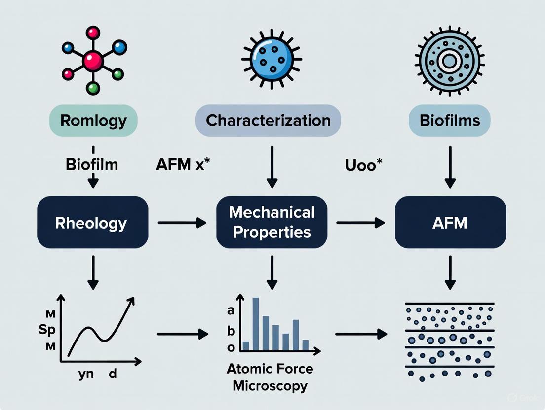
Combined Rheology and AFM Characterization: An Integrated Approach
The combination of rheology and atomic force microscopy (AFM) provides complementary insights into biofilm mechanical properties across different length scales. While rheology characterizes bulk viscoelastic properties, AFM enables visualization of biofilm morphology, quantification of surface roughness, and probing of mechanical interactions at the nanoscale [5].
Rheological Characterization
Rheological assessments typically employ oscillatory shear tests to measure viscoelastic parameters such as storage modulus (G', representing solid-like behavior), loss modulus (G", representing fluid-like behavior), and complex viscosity [5]. These bulk measurements help monitor and predict biofilm behavior under diverse environmental conditions and are particularly useful for evaluating anti-biofilm treatments [5].
Sample preparation remains challenging for rheological analysis. While cohesive biofilms can sometimes be removed intact from substrates, this process may destroy delicate EPS architecture [8]. Alternative approaches include growing biofilms directly on rheometer plates or using semipermeable membranes for transfer [8].
Atomic Force Microscopy (AFM)
AFM provides high-resolution topographical imaging and nanomechanical mapping under physiological conditions with minimal sample preparation [5] [6]. Advanced AFM techniques can measure cohesive energy within biofilms by determining the volume of displaced biofilm and corresponding frictional energy dissipation as a function of biofilm depth [6].
A novel AFM method has been developed to measure cohesive energy levels in moist biofilms by quantifying scan-induced abrasion. This approach has demonstrated that cohesive energy increases with biofilm depth (from 0.10 ± 0.07 nJ/μm³ to 2.05 ± 0.62 nJ/μm³) and is enhanced by the presence of calcium ions [6].
The integrated workflow for combined rheology-AFM characterization is illustrated in Figure 2 below.
Figure 2. Combined Rheology-AFM Characterization Workflow. This integrated approach correlates bulk viscoelastic properties from rheology with localized structural and mechanical data from AFM to establish comprehensive structure-function relationships in biofilms.
Experimental Protocols for Combined Characterization
Protocol: Macrorheology of Homogenized Biofilm Material
Application: Bulk viscoelastic characterization of biofilm EPS components [8]
Materials and Reagents:
- Biofilm samples (7-day old macrocolonies)
- Sterile phosphate-buffered saline (PBS)
- Parallel plate rheometer (e.g., 20-mm diameter)
- Temperature control unit
- Spatula and weighing boats
Procedure:
- Grow macrocolony biofilms on appropriate agar substrates for 7 days at desired temperature
- Carefully scrape biofilm material from agar surface using spatula
- Homogenize biofilm material by gentle mixing to create uniform consistency
- Transfer approximately 0.5 mL of homogenized biofilm to rheometer measuring plate
- Lower upper plate to measuring gap (typically 0.5-1.0 mm)
- Perform amplitude sweep (0.01-100% strain) at constant frequency (e.g., 1 Hz) to determine linear viscoelastic region
- Conduct frequency sweep (0.01-100 rad/s) at constant strain within linear region
- Perform time sweep measurements to monitor viscoelastic stability
- Analyze storage modulus (G'), loss modulus (G"), and complex viscosity
Notes: Homogenization destroys the native biofilm architecture but enables assessment of the intrinsic mechanical properties of EPS components [8].
Protocol: AFM-Based Cohesive Energy Measurement
Application: Quantification of local cohesive energy in hydrated biofilms [6]
Materials and Reagents:
- Hydrated biofilm samples on growth substrate
- Saturated NaCl solution (for 90% humidity control)
- AFM with humidity control chamber
- Silicon nitride cantilevers (spring constant: 0.58 N/m)
- Atomic force microscope with M scanner (30 μm lateral range, 7 μm vertical range)
Procedure:
- Grow biofilms on appropriate substrates (e.g., membrane test modules)
- Cut 1 × 1 cm samples and equilibrate in 90% humidity chamber for 1 hour
- Mount sample in AFM humidity chamber maintained at 90% relative humidity
- Collect non-perturbative topographic image of 5 × 5 μm region at minimal applied load (~0 nN)
- Select 2.5 × 2.5 μm subregion for abrasion testing
- Perform repeated raster scanning (4 scans) at elevated load (40 nN)
- Return to low load and collect post-abrasion 5 × 5 μm topographic image
- Calculate displaced volume from height difference between pre- and post-abrasion images
- Determine frictional energy dissipation from lateral deflection signals during abrasion
- Calculate cohesive energy as frictional energy divided by displaced volume
- Repeat measurements at different depths by successive abrasion cycles
Notes: This method has shown cohesive energy increases with biofilm depth from 0.10 ± 0.07 nJ/μm³ to 2.05 ± 0.62 nJ/μm³ and increases significantly with calcium addition [6].
Protocol: In Situ Viscoelastic Characterization of Biofilm Streamers
Application: Mechanical assessment of biofilm streamers under flow conditions [7]
Materials and Reagents:
- Microfluidic platform with pillar obstacles
- Bacterial suspension in appropriate growth medium
- Propidium iodide stain (for eDNA visualization)
- Epifluorescence microscopy system
- Computational fluid dynamics software
Procedure:
- Fabricate microfluidic device with pillar-shaped obstacles in main channel
- Introduce diluted bacterial suspension at controlled flow rates (Re 0.02-0.20)
- Allow streamers to develop over 15 hours until steady state
- Stain with propidium iodide (10 μg/mL) for eDNA visualization
- Acquire 3D epifluorescence images of streamer morphology
- Reconstruct 3D geometry for CFD simulations
- Estimate forces exerted by flow on streamers using Eq. 1 (see reference [7])
- Apply controlled flow perturbations to impose extensional stress increments
- Measure resulting strain increments to calculate differential Young's modulus and effective viscosity
- Correlate mechanical properties with prestress state and biochemical composition
Notes: This method revealed stress-hardening behavior in biofilm streamers, with mechanical properties increasing linearly with external stress [7].
Table 2: Research Reagent Solutions for Biofilm Mechanical Characterization
| Reagent/Equipment | Function | Application Examples |
|---|---|---|
| Silicon nitride AFM cantilevers | Nanomechanical probing | Cohesive energy measurement, force mapping [6] |
| Parallel plate rheometer | Bulk viscoelastic characterization | Oscillatory shear testing of biofilm material [8] |
| Propidium iodide | eDNA staining | Visualization of streamer backbone structure [7] |
| Microfluidic platforms | Controlled biofilm growth under flow | Streamer formation and in situ characterization [7] |
| Calcium chloride (10 mM) | Ionic cross-linking of EPS | Cohesive strength enhancement studies [6] |
| Targeted ultrasound contrast agents | Acoustic biofilm detection | Mechanoelastic property assessment [4] |
| DNase I | eDNA degradation | Matrix structural integrity studies [7] |
Application Notes: From Characterization to Anti-Biofilm Strategies
The mechanical characterization of biofilms provides critical insights for developing anti-biofilm strategies, particularly for combating antimicrobial resistance. The mechanical properties of biofilms serve as valuable biomarkers for assessing the efficacy of anti-biofilm treatments, with changes in viscoelastic parameters often correlating with disrupted matrix integrity [2].
Chemical treatments can be designed to specifically reduce biofilm cohesiveness or stiffness, thereby decreasing the force required for mechanical removal or enhancing biocide penetration [2]. Combined chemical-mechanical approaches represent a promising paradigm for biofilm control, where chemical treatment weakens the EPS matrix, making it more susceptible to mechanical eradication [2].
The relationship between matrix composition and mechanical properties offers multiple intervention targets, illustrated in Figure 3 below.
Figure 3. Matrix Component – Mechanical Property – Intervention Relationships. Specific matrix components contribute distinct mechanical properties, enabling targeted intervention strategies that disrupt matrix integrity and facilitate biofilm removal.
Understanding the contribution of specific EPS components to overall mechanical properties enables targeted disruption strategies. For example:
- eDNA degradation with DNase I disrupts streamer integrity and reduces stress-hardening capacity [7]
- Cellulose and curli interactions provide structural stability in E. coli biofilms, with pEtN-modified cellulose playing a crucial role in maintaining stiffness and structural stability [8]
- Calcium chelation reduces cohesive energy by interfering with ionic cross-linking within the EPS matrix [6]
The integrated rheology-AFM characterization approach provides comprehensive structure-property relationships that guide the development of more effective biofilm control strategies across medical, industrial, and environmental applications.
Biofilms are complex, three-dimensional microbial communities that grow at interfaces and are embedded in a self-produced matrix of extracellular polymeric substances (EPS) [9] [10]. This matrix, composed of polymers, proteins, extracellular DNA, and various biomolecules, provides the biofilm with its distinctive mechanical properties [10]. A defining characteristic of biofilms is their viscoelasticity, meaning they exhibit both solid-like (elastic) and fluid-like (viscous) mechanical behaviors [10]. This combination is an emergent property resulting from intercellular cohesion, a feature not present in their planktonic counterparts [10].
Understanding the bulk viscoelastic properties of biofilms is crucial for both fundamental research and applied science. These properties mediate the biofilm's structural integrity, determine its resistance to environmental stresses (such as fluid shear forces), and control the ease of dispersion for daughter cells [11]. Furthermore, the viscoelastic character of biofilms has been linked to their recalcitrance toward immune system clearance, particularly by impeding phagocytosis by neutrophils [10]. Consequently, probing these properties provides critical insights for developing control strategies in industrial, medical, and environmental contexts [5].
This document, framed within a broader thesis on the combined characterization of biofilms via rheology and Atomic Force Microscopy (AFM), details the fundamental principles, quantitative data, and standardized protocols for assessing the bulk viscoelastic properties of biofilms. The complementary nature of rheology, which measures bulk material properties, and AFM, which probes mechanical interactions at the nanoscale, offers a comprehensive picture of biofilm mechanics [5] [12].
Key Viscoelastic Properties and Quantitative Values
The viscoelastic behavior of biofilms is typically characterized using oscillatory shear rheology. This method involves applying a sinusoidal stress and measuring the resulting strain, which allows for the decomposition of the mechanical response into elastic and viscous components. The table below summarizes the key parameters used to quantify biofilm viscoelasticity.
Table 1: Key Parameters for Characterizing Biofilm Viscoelasticity via Rheology
| Parameter | Symbol | Description | Interpretation |
|---|---|---|---|
| Elastic (Storage) Modulus | G′ | Quantifies the energy stored and recovered per deformation cycle; represents the solid-like, elastic component. | A higher G′ indicates a more rigid, structured, and solid-like biofilm. |
| Viscous (Loss) Modulus | G″ | Quantifies the energy dissipated as heat per deformation cycle; represents the fluid-like, viscous component. | A higher G″ indicates a more fluid and liquid-like biofilm. |
| Complex Modulus | G* | |G*| = √(G′² + G″²). A overall measure of the material's resistance to deformation. | A higher G* indicates a stiffer material overall. |
| Loss Tangent | tan δ = G″/G′ | The ratio of the viscous to elastic modulus. | tan δ < 1: Solid-like, elastic behavior dominates (G′ > G″).tan δ > 1: Fluid-like, viscous behavior dominates (G″ > G′). |
The mechanical properties of a biofilm are not fixed; they are dynamically influenced by genetic makeup, environmental conditions, and the age of the biofilm. The following table compiles quantitative values from scientific literature to illustrate this variability.
Table 2: Reported Viscoelastic Properties of Various Biofilms
| Biofilm Organism / Condition | Elastic Modulus (G′) | Viscous Modulus (G″) | Loss Tangent (tan δ) | Notes | Source Technique |
|---|---|---|---|---|---|
| P. aeruginosa PAO1 (Early Biofilm) | --- | --- | --- | Adhesive pressure: 34 ± 15 Pa | Microbead Force Spectroscopy [11] |
| P. aeruginosa PAO1 (Mature Biofilm) | --- | --- | --- | Adhesive pressure: 19 ± 7 Pa; Reduced elastic moduli vs. early biofilm. | Microbead Force Spectroscopy [11] |
| P. aeruginosa LPS Mutant (Early) | --- | --- | --- | Adhesive pressure: 332 ± 47 Pa; Drastically reduced elastic moduli vs. wild-type. | Microbead Force Spectroscopy [11] |
| P. fluorescens (with CaCl₂) | --- | --- | --- | Creep compliance primarily influenced by void zones; altered with ionic environment. | Particle-Tracking Microrheology [13] |
Standardized Experimental Protocols
Robust and reproducible measurement of biofilm viscoelasticity requires careful adherence to standardized protocols. The following section outlines a general workflow for bulk rheological characterization.
Diagram 1: Rheology Experimental Workflow
Protocol: Oscillatory Rheology for Bulk Biofilm Viscoelasticity
Principle: This protocol uses a parallel plate rheometer to apply a controlled oscillatory shear stress to a biofilm sample and measure its viscoelastic response, determining G′, G″, and tan δ [10].
Materials & Equipment:
- Rheometer: Rotational rheometer with parallel plate geometry (e.g., 20-50 mm diameter).
- Biofilm Reactor: Standardized system for reproducible biofilm growth (e.g., CDC Biofilm Reactor, Drip Flow Reactor, Rotating Disk Reactor) [14].
- Growth Medium: Appropriate sterile nutrient broth for the target microorganisms.
- Inoculum: Pure or mixed culture of microbial strain(s) at a standardized concentration.
Procedure:
- Biofilm Cultivation:
- Grow biofilms in a relevant biofilm reactor under conditions that mimic the system of interest (e.g., flow rate, nutrient composition, temperature) [14].
- Ensure consistent growth time across replicates to minimize age-related variability.
Sample Loading:
- Carefully harvest the mature biofilm from the reactor substratum using a sterile spatula or scalpel.
- Transfer the intact biofilm aggregate onto the lower plate of the rheometer.
- Lower the upper parallel plate to a defined gap height (e.g., 1.0 mm), ensuring contact with the biofilm without squeezing out the sample. Trim excess material from the edges.
Amplitude Sweep Test:
- At a constant, physiologically relevant frequency (e.g., 1 Hz), perform an amplitude sweep by incrementally increasing the oscillatory strain (e.g., from 0.01% to 100%).
- Objective: To determine the Linear Viscoelastic Region (LVR), where G′ and G″ are independent of the applied strain. This ensures subsequent measurements probe the intrinsic structure without causing damage.
- Identify the critical strain, γc, where G′ begins to decrease significantly, indicating structural yielding.
Frequency Sweep Test:
- Within the LVR (at a strain value below γc), perform a frequency sweep across a relevant range (e.g., 0.1 to 100 rad/s).
- Objective: To characterize the time-dependent nature of the biofilm's viscoelasticity. The evolution of G′ and G″ with frequency reveals how the material behaves under different deformation rates.
Data Analysis:
- Plot G′ and G″ as a function of strain (amplitude sweep) and angular frequency (frequency sweep).
- Report the plateau values of G′ and G″ within the LVR.
- Calculate tan δ to classify the dominant mechanical behavior of the biofilm under the tested conditions.
Complementary and Advanced Techniques
While bulk rheology provides essential macroscopic properties, biofilms are structurally and mechanically heterogeneous. Advanced techniques are required to resolve this complexity and to link bulk properties with nanoscale interactions, which is a core theme of combined rheology-AFM research.
Atomic Force Microscopy (AFM) and Force Spectroscopy
AFM serves as a powerful complementary technique to rheology. It can image biofilm topography at the nanoscale and, through force spectroscopy, quantify local mechanical properties and interaction forces [5] [12].
- Imaging: Tapping mode AFM in fluid allows visualization of the topographical landscape of hydrated biofilms and individual cells with minimal disruption [12].
- Nanoindentation: The AFM tip can be used as a nanoindenter to measure local elastic moduli and turgor pressure by analyzing force-distance curves, often using Hertzian contact mechanics models [12].
- Microbead Force Spectroscopy (MBFS): A specialized AFM technique where a microbead attached to the cantilever is coated with biofilm cells. This allows for simultaneous quantification of adhesion (from retraction curves) and viscoelasticity (from creep compliance during hold periods) under standardized conditions [11].
Particle-Tracking Microrheology
This in-situ technique involves embedding fluorescent tracer particles (e.g., 1 μm diameter) within the biofilm matrix [13]. Using confocal laser scanning microscopy (CLSM), the Brownian motion of these particles is tracked over time.
- Principle: The mean square displacement (MSD), 〈Δr²(τ)〉, of the particles is calculated from their trajectories. The creep compliance, J(t), is then derived from the MSD, providing a measure of local, region-specific viscoelasticity [13].
- Advantage: It can map mechanical properties in 3D, differentiating between regions like voids and cell clusters, thus directly addressing biofilm heterogeneity [13].
Diagram 2: Particle-Tracking Microrheology
The Scientist's Toolkit: Essential Research Reagents and Materials
Successful characterization of biofilm rheology depends on the use of specific, well-defined materials and reagents. The following table lists key solutions and items essential for the experiments described in this protocol.
Table 3: Key Research Reagent Solutions and Materials
| Item / Solution | Function / Role | Example / Notes |
|---|---|---|
| CDC Biofilm Reactor | Standardized system for growing reproducible, high-throughput biofilms in suspension. | An ASTM-standard method (E2562) for growing a homogenous biofilm sample ideal for rheological testing [14]. |
| Trypticase Soy Broth (TSB) | A common nutrient-rich growth medium for cultivating a wide variety of bacterial biofilms. | Used for growing Pseudomonas aeruginosa and other relevant species to mature biofilms for mechanical testing [11]. |
| Polystyrene Microbeads (1 μm) | Tracer particles for particle-tracking microrheology. | Fluorescent carboxylate beads are embedded in the biofilm to track local matrix mobility [13]. |
| Carboxylated Magnetic Beads | Functionalized probes for force spectroscopy and magnetic tweezer microrheology. | Used in AFM-MBFS and magnetic tweezers to apply force and measure creep compliance [11] [13]. |
| Phosphate Buffered Saline (PBS) | Ionic buffer for rinsing and re-suspending biofilms. | Used to remove planktonic cells and non-adherent material before testing without altering ionic strength drastically [13]. |
| Calcium Chloride (CaCl₂) Supplement | Modifies the ionic environment to study the effect of divalent cations on biofilm mechanics. | Divalent cations like Ca²⁺ can cross-link EPS components, significantly increasing biofilm stiffness and cohesion [13]. |
The accurate determination of biofilm viscoelastic properties through rheological methods is a cornerstone of understanding biofilm persistence and developing effective control strategies. The protocols and data outlined herein provide a framework for standardized, quantitative assessment of these critical mechanical properties. When these bulk measurements are integrated with nanoscale techniques like AFM and high-resolution mapping via particle-tracking microrheology, researchers can achieve a multi-scale understanding of biofilm mechanics. This interdisciplinary approach is essential for linking biofilm material properties to their physiological functions and their recalcitrance to both mechanical and chemical challenges.
Atomic Force Microscopy (AFM) is a high-resolution scanning probe microscopy technique that achieves nanometer-scale resolution by measuring the forces between a sharp tip and the sample surface [15]. Unlike electron microscopes, AFM requires no special sample preparation such as conductive coatings and can operate in various environments, including liquid mediums, making it particularly valuable for characterizing soft biological samples such as biofilms [16] [17]. The fundamental principle of AFM involves physically "feeling" the sample surface with a sharp probe, providing three-dimensional topographic information while simultaneously mapping local material properties [15]. This dual capability for topographic and nanomechanical characterization makes AFM an indispensable tool in the expanding field of biofilm research, where understanding the relationship between structure, mechanical properties, and function is crucial for developing effective anti-biofilm strategies [5].
The particular challenge of biofilm-related infections lies in their enhanced antibiotic resistance, which is intimately connected to their structural integrity and viscoelastic properties [5]. When AFM is combined with rheological measurements, researchers can obtain comprehensive insights into biofilm behavior under mechanical stress, informing strategies for biofilm disruption in medical and industrial contexts [5] [18]. This application note details the principles, methodologies, and practical protocols for implementing AFM in the characterization of biofilms, with emphasis on connecting topographic features with nanomechanical properties within a multidisciplinary research framework.
Fundamental Principles of AFM Operation
Core Components and Sensing Mechanism
The atomic force microscope consists of three primary subsystems that work in coordination: the sensing system, detection system, and positioning system [15]. The sensor is a flexible cantilever with a sharp tip at its free end, typically with a radius of curvature between 1-15 nanometers [17]. When this tip approaches the sample surface, it experiences forces that cause the cantilever to bend. This bending is detected using an optical system consisting of a laser beam reflected from the back of the cantilever onto a position-sensitive photodetector [15]. The positioning system uses piezoelectric actuators to move the tip relative to the sample with sub-nanometer precision in three dimensions [15]. This combination of components enables the AFM to achieve exceptional resolution, with lateral resolution as small as the tip radius and vertical resolution on the order of angstroms [17].
Primary Operational Modes
AFM operates in several distinct modes, each optimized for specific sample types and measurement requirements. The selection of an appropriate operational mode is critical for successful biofilm characterization, as these delicate structures can be easily damaged by inappropriate forces.
Table 1: Key AFM Operational Modes for Biofilm Characterization
| Operation Mode | Principle | Best For | Biofilm Application Examples |
|---|---|---|---|
| Contact Mode [19] | Tip dragged across surface at constant cantilever deflection | Stiff, robust materials | Limited use for soft biofilms due to potential damage |
| Tapping Mode [19] | Tip oscillated at resonance frequency with amplitude feedback | Soft, fragile, adhesive samples | Imaging delicate biofilm structures in liquid environments |
| PeakForce Tapping [20] | Oscillating tip contacts surface at controlled maximum force | Quantitative nanomechanical mapping | Measuring biofilm elasticity and adhesion without damage |
| Force Spectroscopy [15] | Force-distance curves acquired at fixed positions | Local mechanical properties and single-molecule interactions | Probing ligand-receptor binding on bacterial surfaces |
The following diagram illustrates the fundamental working principle of AFM and its primary operational modes:
AFM for Topographical and Nanomechanical Characterization
Quantitative Topography and Surface Roughness
AFM provides quantitative three-dimensional topographic information with exceptional resolution, typically achieving 5-10 nm laterally and sub-nanometer vertically [19]. This precise height measurement capability enables accurate surface roughness quantification, which is essential for characterizing biofilm formation and substrate interactions [5]. Unlike qualitative methods that merely provide visual impressions of texture, AFM generates numerical data that can be statistically analyzed using parameters such as arithmetical mean deviation (Sa) and root mean square deviation (Sq) [19]. For biofilms, surface roughness measurements can reveal structural heterogeneity, porosity, and the distribution of extracellular polymeric substances (EPS) that comprise the biofilm matrix [5]. Studies have demonstrated that surface roughness parameters can correlate with biofilm adhesion strength and resistance to mechanical disruption, providing critical insights for anti-fouling surface design [5].
Nanomechanical Property Mapping
Beyond topography, AFM excels at characterizing mechanical properties at the nanoscale through techniques such as force spectroscopy and PeakForce Quantitative Nanomechanical Mapping (QNM) [20] [15]. These methods measure tip-sample interaction forces to determine properties including elastic modulus, adhesion, deformation, and energy dissipation [15]. For biofilm research, this capability is transformative, as the mechanical properties of biofilms directly influence their persistence and resistance to removal [5] [21]. AFM-based nanomechanical mapping has revealed significant heterogeneity within biofilm structures, with elastic modulus values varying by orders of magnitude across different regions of the same biofilm [5]. This mechanical characterization, when combined with rheological measurements, provides a comprehensive understanding of biofilm viscoelasticity across length scales, from bulk responses to local nanomechanical properties [5] [21].
Table 2: AFM Measurements for Biofilm Characterization
| Measurement Type | Parameters | Typical Values for Biofilms | Significance in Biofilm Research |
|---|---|---|---|
| Surface Roughness [19] | Sa (Arithmetical mean height)Sq (Root mean square)SkewnessKurtosis | Varies with biofilm type and age; Sa values from nanometers to micrometers | Influences bacterial adhesion, structural complexity, and fluid interactions |
| Elastic Modulus [5] [21] | Young's modulus from force curves or QNM | 0.1 kPa - 1000 kPa (highly dependent on biofilm type and hydration) | Determines resistance to mechanical disruption and penetration of antimicrobials |
| Adhesion Forces [5] [22] | Pull-off forces measured in force curves | 0.1 - 10 nN (varies with tip functionalization) | Quantifies cohesion within biofilm matrix and adhesion to substrates |
| Viscoelastic Parameters [5] [21] | Storage/loss moduli, relaxation times | G': 1 - 1000 Pa, G": 0.5 - 500 Pa (from AFM-nDMA) | Predicts biofilm response to shear flows and cleaning stresses |
Application Notes: AFM in Biofilm Research
Integrated Rheology-AFM Characterization
The combination of rheology and AFM provides a powerful multimodal approach for understanding biofilm mechanics across different scales [5]. While rheology measures the bulk viscoelastic properties of biofilms, AFM probes local nanomechanical behavior, revealing heterogeneity that bulk measurements may average out [5]. This integrated approach has demonstrated that biofilm mechanical properties are significantly influenced by environmental conditions, including nutrient availability, flow conditions, and the composition of surrounding fluids [5] [18]. For instance, research has shown that the viscoelastic properties of biofilms measured by rheology correlate with their recalcitrance to mechanical and chemical challenges, while AFM can identify specific structural features responsible for this robustness [5]. This multiscale mechanical profiling is essential for developing effective biofilm control strategies, as it identifies both bulk and local weaknesses that can be targeted for removal.
Antimicrobial Efficacy Assessment
AFM serves as a sensitive tool for evaluating the efficacy of antimicrobial agents and anti-biofilm strategies by detecting structural and mechanical changes before and after treatment [5]. Time-resolved AFM imaging can track the degradation of biofilm architecture, changes in surface roughness, and alterations in mechanical integrity following antimicrobial application [5]. Force spectroscopy measurements can quantify changes in adhesion forces between functionalized AFM tips and biofilm components, revealing how anti-biofilm agents affect cohesive and adhesive properties [22]. This application is particularly valuable in drug development, where understanding the mechanism of action at the nanoscale can guide compound optimization. Furthermore, AFM can be combined with fluorescence microscopy to correlate structural and mechanical changes with biological activity, such as membrane disruption or metabolic inhibition [22] [17].
The following workflow illustrates the integrated approach for combining AFM with rheology in biofilm research:
Experimental Protocols
Protocol: AFM Analysis of Biofilm Mechanical Properties
This protocol describes the procedure for preparing biofilm samples and performing nanomechanical characterization using Atomic Force Microscopy, adapted from established methodologies for biological AFM [22] and biofilm characterization [5].
Sample Preparation
- Substrate Selection: Use appropriate substrates for biofilm growth, such as 2B cold-rolled stainless-steel plates for food industry-relevant studies [21] or glass bottom dishes for optical microscopy correlation [22].
- Biofilm Growth: Cultivate biofilms under controlled conditions relevant to the research context. For Microbacterium lacticum, follow established cultivation procedures using appropriate growth media [21].
- Fixation (if required): For high-resolution imaging, slight fixation with 0.5-2% glutaraldehyde may be necessary, though living biofilms can be analyzed in liquid environments [5].
- Mounting: Secure the biofilm substrate to the AFM specimen disk using double-sided adhesive tape or magnetic holders. Ensure the surface is level to prevent tilt artifacts.
AFM Configuration
- Cantilever Selection: Choose appropriate cantilevers based on the measurement mode:
- For tapping mode in liquid: Soft cantilevers with spring constants of 0.1-1 N/m and resonant frequencies of 10-30 kHz [22].
- For force spectroscopy: Sharp tips with spring constants of 0.01-0.5 N/m for minimal sample damage [22].
- For quantitative nanomechanical mapping: Tips with well-characterized geometry and spring constants, calibrated before measurement [20].
- Laser Alignment: Align the laser beam to reflect off the cantilever end onto the position-sensitive photodetector center.
- Photodetector Adjustment: Adjust the photodetector to obtain a sum signal of 2-5 V, indicating optimal reflection.
Measurement Procedure
- Engagement: Approach the tip to the surface slowly using the automated engagement routine, monitoring the deflection signal.
- Topography Imaging: First capture large-scale (e.g., 20×20 µm) topographic images to identify regions of interest, then higher-resolution (e.g., 3×3 µm) scans for detailed analysis [23].
- Force Curve Acquisition: Acquire force curves at multiple locations (minimum 3 different areas, with 10×10 force curves in each array) using consistent parameters (approach velocity: 0.5-1 µm/s, force trigger: 0.5-2 nN) [22].
- Nanomechanical Mapping: Perform PeakForce QNM scans with optimized parameters to simultaneously map topography, elastic modulus, adhesion, and dissipation [20].
- Environmental Control: For live biofilm imaging, maintain temperature and fluid environment throughout the measurement using appropriate environmental chambers [17].
Protocol: Combined AFM-Rheology Workflow
This protocol outlines the procedure for correlating AFM nanomechanical data with bulk rheological measurements of biofilms, based on integrated characterization approaches [5] [21].
Sample Preparation for Correlative Measurements
- Parallel Sample Preparation: Prepare identical biofilm samples on both AFM-compatible substrates (e.g., glass discs) and rheometry fixtures (e.g., parallel plates).
- Growth Condition Control: Ensure biofilms are grown under identical conditions for both measurement types, with the same age, temperature, and nutrient availability.
- Hydration Maintenance: Prevent dehydration during transfer by using humidity chambers or performing measurements in liquid environments.
Rheological Characterization
- Fixture Selection: Use parallel plate geometry with appropriate surface roughness to prevent slippage (sandblasted or serrated plates recommended).
- Strain Sweep: Perform amplitude sweep tests (0.01-10% strain) at constant frequency (1 Hz) to determine the linear viscoelastic region (LVR) [21].
- Frequency Sweep: Conduct frequency sweep tests (0.1-100 rad/s) at a strain within the LVR to characterize viscoelastic modulus (G', G") dependence on timescale.
- Flow Properties: Measure flow curves to determine yield stress and apparent viscosity as a function of shear rate.
Correlative Analysis
- Spatial Correlation: Compare AFM nanomechanical maps with rheological data, noting how local heterogeneities observed by AFM might influence bulk measurements.
- Mechanical Property Correlation: Establish relationships between local stiffness (from AFM force curves) and bulk modulus (from rheology).
- Time-Dependent Studies: Perform time-series measurements with both techniques to track mechanical property evolution during biofilm development or treatment.
The Scientist's Toolkit: Essential Materials and Reagents
Table 3: Research Reagent Solutions for AFM Biofilm Characterization
| Item | Specifications | Function | Example Application |
|---|---|---|---|
| AFM Cantilevers [22] | Silicon nitride, pyramidal tipSpring constant: 0.01-1 N/mTip radius: <40 nm | Sensing surface topography and forces | High-resolution imaging of biofilm structure |
| Functionalized Tips [22] | Borosilicate beads labeled with biotinSpring constant: 0.01 N/mDiameters: 2-5 μm | Specific molecular interactions | Ligand-receptor binding studies on bacterial surfaces |
| Biofilm Substrates [21] | 2B cold-rolled stainless-steel platesGlass bottom dishes | Controlled biofilm growth | Food industry-relevant biofilm studies |
| Liquid Cells [22] | Sealed fluid chambers with O-ringsTemperature control capability | Hydrated biofilm imaging | Live biofilm analysis under physiological conditions |
| Calibration Samples [19] | Gratings with known pitch and heightReference roughness samples | AFM calibration and validation | Verification of instrument performance before biofilm measurements |
| Extracellular Matrix Proteins [22] | Fibronectin, collagen, laminin | Tip functionalization | Studying integrin-ECM interactions in biofilms |
| Cell Isolation Reagents [22] | Protease XXIII, kynurenic acid, PEG | Dissociation of biofilm cells | Single-cell mechanics studies within biofilms |
| Imitation Biofilm Materials [21] | Alginate-based or gellan-based hydrogels | Biofilm model systems | Standardized testing of anti-biofilm strategies |
Data Analysis and Interpretation
Topographical Data Processing
AFM topographic data requires careful processing to extract meaningful quantitative information. The essential steps include:
- Flattening/Leveling: Apply plane fitting algorithms to correct for sample tilt and scanner bow [16]. Use polynomial or plane fit functions to remove background curvature while preserving surface features.
- Noise Filtering: Implement appropriate digital filters to reduce noise without distorting genuine features. Low-pass filters remove high-frequency noise, while median filters effectively eliminate spike noise [16].
- Roughness Analysis: Calculate standard roughness parameters including Sa (arithmetical mean height), Sq (root mean square height), skewness (asymmetry of height distribution), and kurtosis (peakedness of height distribution) [19].
- Particle/Aggregate Analysis: Use threshold-based detection algorithms to identify and characterize discrete features within biofilms, measuring parameters such as diameter, height, volume, and surface coverage [16].
Force Curve Analysis
Nanomechanical properties are extracted from force curves through theoretical modeling:
- Elastic Modulus Calculation: Fit the retraction portion of force curves with appropriate contact mechanics models. The Hertz model is commonly used for purely elastic materials, while more complex models (Sneddon, Johnson-Kendall-Roberts) account for adhesion and plasticity [15].
- Adhesion Force Measurement: Identify the minimum force in the retraction curve as the adhesion force. Statistical analysis of multiple curves provides mean adhesion and binding probability [22].
- Energy Dissipation Calculation: Integrate the area between approach and retraction curves to determine energy dissipation, indicative of viscoelastic behavior [15].
Correlation with Rheological Data
Integrating AFM nanomechanical data with bulk rheological measurements enables comprehensive understanding of biofilm mechanics:
- Multi-scale Mechanical Profiling: Compare local stiffness from AFM with bulk modulus from rheology to identify how nanoscale properties influence macroscopic behavior [5].
- Heterogeneity Assessment: Use the spatial distribution of mechanical properties from AFM to interpret the non-linear responses observed in rheological measurements [5] [21].
- Structure-Function Relationships: Correlate topographic features with mechanical properties to understand how biofilm architecture contributes to mechanical robustness [5].
The following diagram illustrates the pathway from raw AFM data to integrated mechanical understanding of biofilms:
The Synergistic Value of Combining Rheology and AFM for a Multi-Scale View
In the study of microbial biofilms, the complex and heterogeneous nature of these structures demands analytical techniques that can capture a full spectrum of physical properties. No single method can fully characterize the viscoelastic properties and cohesive strength that govern biofilm resilience and detachment. The combination of rheology, which probes the bulk mechanical response, and Atomic Force Microscopy (AFM), which investigates nanoscale surface properties and interactions, provides a powerful, multi-scale analytical framework [5] [24]. This synergistic approach is pivotal for understanding biofilm behavior, from the initial stages of bacterial adhesion to the mechanical stability of mature structures, thereby informing the development of effective anti-biofilm strategies in clinical and industrial settings [5].
This Application Note delineates the quantitative data, detailed protocols, and essential reagents for the integrated use of rheology and AFM in biofilm research. By correlating macro-scale mechanical behavior with nano-scale structural and force interactions, researchers can achieve a comprehensive understanding of biofilm mechanics, crucial for applications ranging from antimicrobial screening to the optimization of biofilm-based bioprocesses [24].
Quantitative Data Comparison
The following tables summarize key mechanical parameters obtainable through rheology and AFM, highlighting the complementary nature of the data generated by each technique.
Table 1: Bulk Mechanical Properties from Rheological Analysis
| Mechanical Parameter | Typical Value/Behavior for Biofilms | Significance in Biofilm Function | Common Experimental Method |
|---|---|---|---|
| Elastic Modulus (G') | 10 - 10,000 Pa [24] | Quantifies solid-like character and structural rigidity; dominant G' indicates a solid material. | Oscillatory shear testing |
| Viscous Modulus (G") | 1 - 1,000 Pa [24] | Quantifies liquid-like, energy-dissipating behavior. | Oscillatory shear testing |
| Complex Modulus (G*) | Derived from G' and G" | Represents overall mechanical resistance to deformation. | Oscillatory shear testing |
| Cohesive Energy | N/A (Bulk property) | Energy required to disrupt the bulk biofilm structure. | Flow-induced detachment assays |
| Viscoelasticity | Yes (G' > G") [24] | Allows biofilms to withstand and dissipate mechanical stress from fluid flow. | Frequency sweep, creep-recovery |
Table 2: Localized Nanomechanical and Adhesive Properties from AFM Analysis
| Mechanical Parameter | Typical Value/Behavior for Biofilms | Significance in Biofilm Function | Common Experimental Mode |
|---|---|---|---|
| Elastic Modulus (Young's Modulus) | 0.1 - 1000 kPa [12] | Measures local cell/EPS stiffness; varies with biofilm depth and composition. | Force Spectroscopy (Nanoindentation) |
| Adhesion Force | Varies (pN to nN) [12] | Measures binding strength between cells, EPS, and surfaces. | Single-Molecule/Cell Force Spectroscopy |
| Cohesive Energy | 0.10 to 2.05 nJ/μm³ [6] | Nanoscale work required to separate biofilm components; can be depth-dependent. | Friction/Abrasion experiments |
| Surface Roughness | Topographical maps | Influences initial bacterial attachment and biofilm architecture. | Tapping Mode Imaging |
| Turgor Pressure | Varies by cell type | Internal cell pressure contributing to biofilm mechanics. | Force Spectroscopy |
Experimental Protocols
Protocol 1: Macro-Rheological Assessment of Biofilm Viscoelasticity
This protocol characterizes the bulk viscoelastic properties of a mature biofilm.
- Biofilm Cultivation: Grow biofilms under controlled conditions on appropriate substrates (e.g., stainless-steel coupons for food industry studies, or in Petri dishes for clinical isolates) using relevant growth media [21]. Ensure consistent age and growth conditions across replicates.
- Sample Loading: Carefully harvest the biofilm and transfer it onto the measuring geometry of a stress- or strain-controlled rheometer. A parallel plate geometry is often suitable. To prevent dehydration, use a solvent trap to maintain a humid environment [21] [24].
- Strain Sweep Test: Perform an oscillatory strain amplitude sweep (e.g., 0.1% - 100%) at a fixed frequency to determine the Linear Viscoelastic Region (LVR), where the microstructure remains intact. This identifies the maximum strain (
γ_max) applicable for subsequent tests without causing structural damage. - Frequency Sweep Test: Within the LVR, conduct an oscillatory frequency sweep (e.g., 0.1 - 100 rad/s) at a constant strain (
γ < γ_max). This measures the evolution of the elastic modulus (G') and viscous modulus (G") as a function of timescale, revealing the material's relaxation mechanisms [24]. - Data Analysis: Plot G' and G" against frequency. A biofilm typically exhibits G' > G" across a wide frequency range, confirming its dominant elastic, solid-like behavior. The complex modulus (G*) can be calculated to represent the overall stiffness [24].
Protocol 2: Nanoscale Cohesive Energy Measurement via AFM
This protocol measures the depth-dependent cohesive energy within a hydrated biofilm, providing nanoscale resolution of biofilm mechanical properties [6].
- Biofilm Preparation and Immobilization: Grow a 1-day-old biofilm on a suitable substrate (e.g., a gas-permeable membrane). For AFM analysis, cut a small piece (~1 cm²) and equilibrate it in a humidity chamber (~90% relative humidity) for one hour to maintain consistent hydration without excess surface water [6].
- Topographical Imaging: Mount the sample in the AFM liquid cell or humidity chamber. Using a sharp AFM probe (e.g., silicon nitride tip), first acquire a non-perturbative, high-resolution topographic image of a 5x5 μm area at a minimal applied load (~0 nN) [6].
- Controlled Abrasion: Zoom into a 2.5x2.5 μm sub-region of the scanned area. Set the AFM to repeatedly raster scan this smaller area for a defined number of cycles (e.g., 4 scans) under a high applied load (e.g., 40 nN). This scanning abrades and displaces biofilm material [6].
- Post-Abrasion Imaging: Reduce the applied load back to ~0 nN and capture a new non-perturbative 5x5 μm topographic image of the same initial location, which now includes the abraded region.
- Data Analysis:
- Volume Calculation: Subtract the post-abrasion height image from the pre-abrasion image to calculate the volume of biofilm displaced (
V_displaced). - Frictional Energy: Calculate the total frictional energy dissipated (
E_friction) during abrasive scanning from the lateral deflection signals of the AFM cantilever. - Cohesive Energy: The cohesive energy (
Γ) is then calculated asΓ = E_friction / V_displaced(units: nJ/μm³). This process can be repeated at different biofilm depths to profile depth-dependent cohesion [6].
- Volume Calculation: Subtract the post-abrasion height image from the pre-abrasion image to calculate the volume of biofilm displaced (
Workflow for Combined Rheology-AFM Analysis
The diagram below illustrates the integrated experimental workflow.
The Scientist's Toolkit: Research Reagent Solutions
Table 3: Essential Materials for Combined Rheology-AFM Biofilm Studies
| Reagent/Material | Function/Description | Application in Protocols |
|---|---|---|
| Stainless Steel Coupons (2B finish) | Industrially relevant substrate for biofilm growth. | Biofilm cultivation for both rheology and AFM [21]. |
| Microporous Polyolefin Membrane | Supports biofilm growth with aeration from below. | AFM substrate, especially for aerobic biofilms [6]. |
| Silicon Nitride AFM Probes | Sharp tips for high-resolution imaging and force measurement. | AFM topographic imaging and nanoindentation [6] [12]. |
| Polydimethylsiloxane (PDMS) Stamps | Micro-patterned surfaces for secure cell immobilization. | Immobilizing microbial cells for AFM in aqueous conditions [12]. |
| Alginate or Gellan Gum | Polysaccharides for formulating hydrogel-based biofilm imitations. | Creating reference/control samples with tunable mechanical properties [21]. |
| Calcium Chloride (CaCl₂) | Divalent cation that cross-links EPS, increasing cohesion. | Studying the effect of specific ions on biofilm mechanics [6]. |
| Humidity Controller | Maintains constant relative humidity (e.g., ~90%) during AFM. | Prevents biofilm dehydration during AFM measurements without submersion [6]. |
Synergistic Data Interpretation
The synergy between rheology and AFM becomes evident when data from both techniques are correlated. For instance, a bulk rheological measurement might show a significant decrease in the Elastic Modulus (G') after treatment with an enzyme targeting extracellular DNA [24]. AFM can complement this finding by revealing a corresponding reduction in nanoscale cohesive energy and adhesion forces, directly visualizing the disruption of the EPS matrix that underpins the macroscopic mechanical change [5] [12]. This multi-scale validation is powerful for confirming the mechanism of action of anti-biofilm agents.
Furthermore, the heterogeneous nature of biofilms means that bulk rheology provides an average property, which might mask critical local variations. AFM can map this heterogeneity, identifying stiffer microcolonies or weaker regions of predominantly EPS, thereby explaining the standard deviations observed in rheological data and leading to more sophisticated biofilm models [24]. The conceptual relationship between these techniques is illustrated below.
Biofilms are structured microbial communities embedded in a self-produced matrix of extracellular polymeric substances (EPS). Quantifying their mechanical properties—cohesive energy, stiffness, and adhesion forces—is essential for understanding biofilm development, stability, and removal in contexts ranging from medical infections to industrial biofouling [6] [7] [8]. This Application Note details protocols for measuring these key parameters via atomic force microscopy (AFM) and rheology, providing a standardized framework for researchers aiming to correlate biofilm's mechanical behavior with its structural composition and function.
Table 1: Experimentally Measured Mechanical Parameters of Biofilms
| Parameter | Measurement Technique | Biofilm System / Condition | Reported Values | Reference |
|---|---|---|---|---|
| Cohesive Energy | AFM-based abrasion & friction measurement | Mixed culture (activated sludge), 1-day biofilm, depth profile | 0.10 ± 0.07 to 2.05 ± 0.62 nJ/μm³ | [6] |
| AFM-based abrasion & friction measurement | Mixed culture with 10 mM Ca²⁺ added | 0.10 ± 0.07 to 1.98 ± 0.34 nJ/μm³ | [6] | |
| Stiffness (Elastic Modulus) | Microindentation | E. coli AR3110 (produces curli and pEtN-cellulose) | ~140 kPa | [8] |
| Microindentation | E. coli W3110 (produces curli only) | ~20 kPa | [8] | |
| Microindentation | E. coli AR198 (no curli, no cellulose) | ~10 kPa | [8] | |
| Differential Young's Modulus | Extensional Rheology | P. aeruginosa PA14 biofilm streamers | Increases linearly with external prestress (Stress-hardening behavior) | [7] |
Table 2: Key Research Reagent Solutions
| Item | Function/Application | Specific Example / Notes |
|---|---|---|
| Microporous Polyolefin Membrane | Substrate for growing membrane-aerated biofilms. | Treated with a fluorocarbon polyurethane coating; 0.1-μm mean pore diameter, 34% porosity [6]. |
| PFOTS-Treated Glass Coverslips | Hydrophobic surface for studying initial bacterial attachment and biofilm assembly. | Used for high-resolution AFM studies of Pantoea sp. YR343 biofilm formation [25]. |
| Si₃N₄ AFM Tips | Nanoscale imaging and force measurement. | Pyramidal, oxide-sharpened tips on V-shaped cantilevers (0.58 N/m spring constant) for cohesive energy measurements [6]. |
| Calcium Chloride (CaCl₂) | Modifies biofilm cohesiveness by interacting with EPS. | Added at 10 mM to the reactor during cultivation to increase cohesive strength [6]. |
| DNase I | Enzyme that degrades extracellular DNA (eDNA). | Used to interrogate the structural and mechanical role of eDNA in biofilm streamers [7]. |
| Propidium Iodide (PI) | Fluorescent nucleic acid stain for 3D structural visualization. | Used to stain and reconstruct the 3D geometry of biofilm streamers for CFD simulations [7]. |
Experimental Protocols
Protocol: In Situ Measurement of Biofilm Cohesive Energy Using AFM
This protocol quantifies the cohesive energy of moist biofilms by correlating the volume of material displaced by an AFM tip with the frictional energy dissipated during the abrasion process [6].
Key Materials:
- Biofilm Sample: 1-day-old biofilm grown from an undefined mixed culture (e.g., from activated sludge) on a gas-permeable membrane [6].
- AFM Setup: A PicoSPM or equivalent system equipped with a humidity control chamber (~90% RH).
- AFM Probes: V-shaped Si₃N₄ cantilevers with a pyramidal, oxide-sharpened tip (nominal spring constant of 0.58 N/m).
Procedure:
- Sample Equilibration: After growth, excise a ~1 cm x 1 cm piece of the biofilm-coated membrane. Place it in a chamber with a saturated NaCl solution for 1 hour to maintain a constant humidity of ~90%.
- Mounting: Transfer the equilibrated sample to the AFM stage within the humidity-controlled chamber.
- Initial Topography Imaging:
- Select a 5 μm x 5 μm region of interest.
- Obtain a baseline topographic image using a low applied load (~0 nN) to avoid sample perturbation.
- Abrasion Phase:
- Zoom into a 2.5 μm x 2.5 μm sub-region within the initially scanned area.
- Set the AFM to perform repeated raster scans (4 scans per cycle) at an elevated load of 40 nN. This abrasive scanning displaces biofilm material.
- Post-Abrasion Imaging:
- Reduce the applied load back to ~0 nN.
- Acquire a new non-perturbative 5 μm x 5 μm topographic image of the abraded region.
- Data Analysis:
- Subtract the post-abrasion height image from the pre-abrasion image to determine the volume of displaced biofilm.
- Calculate the frictional energy dissipated during abrasion from the lateral (friction) force signals recorded by the AFM photodiode.
- Compute the cohesive energy (Γ) using the formula: Γ = (Dissipated Frictional Energy) / (Displaced Biofilm Volume). The unit is nJ/μm³.
Diagram 1: AFM cohesive energy measurement protocol (nJ/μm³).
Protocol: Microindentation for Local Stiffness Mapping
This protocol measures the local compressive stiffness of native, non-homogenized macrocolony biofilms, preserving their original ECM architecture [8].
Key Materials:
- Biofilm Sample: Macrocolony biofilms (e.g., of E. coli K-12 strains) grown for 7 days on nutritive agar plates.
- Microindenter: System equipped with a spherical or flat-ended indenter tip (diameter in the tens of micrometers).
Procedure:
- Sample Preparation: Grow biofilms directly on the agar substrate. For measurement, use the biofilm in its native state; do not homogenize or remove it from the substrate.
- Instrument Calibration: Calibrate the indenter's load and displacement sensors. Select an indenter tip size appropriate for the biofilm's heterogeneity.
- Positioning: Bring the indenter tip into proximity with the biofilm surface at a predetermined approach rate.
- Indentation Cycle:
- Loading: Drive the tip into the biofilm surface at a constant rate until a predefined maximum force or depth is reached.
- Hold (Optional): Maintain the maximum load for a period to study stress relaxation (viscoelastic behavior).
- Unloading: Retract the tip from the surface.
- Data Collection: Record the force (F) and displacement (h) data throughout the indentation cycle.
- Data Analysis:
- Plot the force-displacement (F-h) curve from the unloading segment.
- Fit the curve with an appropriate contact mechanics model (e.g., Hertz, Oliver-Pharr) to calculate the Effective Elastic Modulus (E), reported in kPa or MPa.
Protocol: Extensional Rheology of Biofilm Streamers
This protocol characterizes the viscoelastic properties and stress-hardening behavior of biofilm streamers in a fluid flow environment [7].
Key Materials:
- Microfluidic Platform: A channel with pillar-shaped obstacles to nucleate reproducible biofilm streamers.
- Syringe Pump: For controlled flow of bacterial suspension and medium.
- Epifluorescence Microscope: For imaging streamer morphology and deformation.
Procedure:
- Streamer Growth:
- Introduce a diluted bacterial suspension (e.g., of P. aeruginosa PA14) into the microfluidic channel at a controlled flow rate.
- Allow streamers to form and grow tethered to the pillars until they reach a steady-state length (typically over several hours).
- Morphological Characterization:
- Stain the streamers with a fluorescent dye like Propidium Iodide (PI).
- Acquire 3D image stacks to reconstruct the streamer geometry.
- Mechanical Testing:
- Prestress State (σ₀): The background flow exerts a constant extensional axial stress on the streamer, calculated using Computational Fluid Dynamics (CFD) simulations based on the streamer's 3D geometry.
- Differential Testing: Apply a controlled flow perturbation to impose a small stress increment (Δσ) on top of the prestress σ₀.
- Measure the resulting strain increment (Δε) from the streamer's deformation.
- Data Analysis:
- Calculate the Differential Young's Modulus as Ediff = Δσ / Δε.
- Observe the relationship between Ediff and the prestress σ₀. A linear increase confirms stress-hardening behavior.
Diagram 2: Extensional rheology protocol for biofilm streamers.
Interplay of Parameters and Matrix Composition
The mechanical parameters are not independent; they are intrinsically linked through the composition and molecular interactions within the EPS.
- Cohesive Energy & eDNA: The cohesive strength is heavily influenced by extracellular DNA (eDNA), which can form a structural backbone and interact with other EPS components like polysaccharides and proteins [7]. The addition of calcium ions (Ca²⁺) can bridge negatively charged polymers, further increasing cohesion [6].
- Stiffness & EPS Fibers: In E. coli biofilms, the amyloid protein curli and modified cellulose (pEtN-cellulose) form a synergistic network that confers tissue-like stiffness. The absence of either component significantly reduces the elastic modulus measured by microindentation [8].
- Stress-Hardening & eNA: The stress-hardening behavior observed in streamers—where stiffness increases linearly with applied stress—is primarily attributed to the physical properties of eDNA and can be modulated by extracellular RNA (eRNA) [7]. This provides a mechanism for biofilms to adapt and strengthen in response to high-shear environments.
The combined application of AFM and rheology provides a powerful toolkit for dissecting the mechanical behavior of biofilms from the nanoscale to the macroscale. The protocols outlined herein for measuring cohesive energy, stiffness, and viscoelasticity enable a quantitative understanding of how EPS composition dictates mechanical function. This knowledge is critical for designing effective strategies to either disrupt resilient pathogenic biofilms or engineer robust beneficial ones, ultimately informing research in antimicrobial development, materials science, and environmental engineering.
A Practical Guide to Combined Rheology-AFM Biofilm Analysis
This application note details a structured protocol for the integrated characterization of biofilms, correlating their macroscopic viscoelastic (rheological) properties with nanoscale structural organization. Biofilms are complex microbial communities whose functional integrity, including resilience to fluid shear stress, is governed by their structural composition and organization at multiple scales [25]. A comprehensive understanding of biofilm mechanics requires linking bulk material properties, measured via rheology, with high-resolution architectural data provided by Atomic Force Microscopy (AFM) [6] [26]. This protocol provides a methodology for this correlated analysis, enabling insights crucial for designing anti-biofilm strategies in medical and industrial contexts.
Experimental Workflow and Data Integration
The following diagram outlines the core sequential workflow for the correlated rheology-AFM analysis of biofilms, highlighting the key stages from sample preparation to data synthesis.
Detailed Experimental Protocols
Substrate Preparation and Biofilm Cultivation
Objective: To grow standardized, reproducible biofilms on substrates suitable for subsequent rheological and AFM analysis.
Materials:
- Microbial Strain: e.g., Pantoea sp. YR343 [25] or Pseudomonas aeruginosa [26].
- Growth Medium: Tryptic Soy Broth (TSB) or other suitable culture medium [26].
- Substrates: Glass coverslips, Polyvinyl Chloride (PVC), steel, aluminum, or polypropylene sheets (1.5 cm x 1.5 cm) [27] [26].
- Surface Treatment: Perfluorooctyltrichlorosilane (PFOTS)-treated glass or other functionalizations to modulate adhesion [25].
Protocol:
- Substrate Cleaning: Clean all substrates (e.g., with ethanol or plasma cleaning) to remove organic contaminants.
- Surface Modification (Optional): Treat substrates (e.g., with PFOTS) to create defined surface chemistries that influence initial bacterial attachment [25].
- Inoculation: Place sterile substrates in a Petri dish and inoculate with a bacterial suspension (e.g., ~10^9 CFU/mL in TSB) [26].
- Biofilm Growth: Incubate under appropriate conditions (e.g., 25-30°C) for a defined period (e.g., from 30 minutes for initial attachment studies to 24-48 hours for mature biofilms) [25] [6].
- Sample Retrieval: Gently rinse the biofilm-coated substrates with a buffer solution (e.g., filtered stream water or PBS) to remove non-adherent planktonic cells [25] [28].
Macroscopic Rheological Characterization
Objective: To quantify the bulk viscoelastic properties and cohesive strength of the biofilm.
Materials:
- Rheometer: with parallel plate or cone-and-plate geometry.
- Humidity Chamber: to prevent sample dehydration during testing.
- Biofilm Samples: grown as described in Section 3.1.
Protocol:
- Loading: Carefully transfer the biofilm-coated substrate to the rheometer base plate. Lower the measuring geometry (plate or cone) until it makes full contact with the biofilm surface at a defined, low normal force.
- Strain Sweep: Perform an oscillatory strain amplitude sweep (e.g., 0.1% - 10% strain) at a fixed frequency (e.g., 1 Hz) to determine the linear viscoelastic region (LVR) of the biofilm.
- Frequency Sweep: Within the LVR, conduct an oscillatory frequency sweep (e.g., 0.1 - 100 rad/s) to measure the elastic (G') and viscous (G") moduli as a function of timescale.
- Flow Curve: Perform a steady-state shear rate sweep to measure the biofilm's apparent viscosity and yield stress, the critical stress required to initiate flow.
- Cohesive Energy Measurement (AFM-based): As an alternative nanoscale measure, an AFM tip can be used to abrade the biofilm under controlled loads. The cohesive energy (nJ/μm³) is calculated from the volume of displaced biofilm and the frictional energy dissipated during scanning [6].
Nanoscale Structural Characterization by AFM
Objective: To image biofilm topography and quantify structural parameters at the cellular and macromolecular scale.
Materials:
- Atomic Force Microscope: Capable of both contact mode and high-resolution imaging in air or liquid [25] [27].
- Cantilevers: MLCT-D silicon nitride cantilevers (nominal tip radius ~20 nm) for contact mode in air [27], or other appropriate probes for liquid imaging.
- Sample Mounting: Magnetic disks or specific sample holders for the AFM.
Protocol:
- Sample Fixation (for air imaging): If imaging in air, gently fix the rinsed biofilm using paraformaldehyde/glutaraldehyde to preserve structure, though this may alter mechanical properties. For native condition imaging, proceed to step 2 without fixation [6] [28].
- Mounting: Secure the biofilm sample onto the AFM sample stage.
- Large-Area Scanning:
- Use an automated large-area AFM system to capture multiple adjacent high-resolution images over millimeter-scale areas [25].
- Machine Learning for Stitching: Employ ML-based algorithms to automatically stitch the individual images into a seamless, high-resolution mosaic with minimal overlap, maximizing acquisition speed [25].
- High-Resolution Imaging:
- Select regions of interest within the large-area map for detailed scanning.
- Image in contact mode (in air) or tapping mode (in liquid) at a high resolution (e.g., 512 x 512 pixels) and slow scan rate (e.g., 0.5 Hz) to visualize fine features like individual cells, flagella, and EPS fibers [25] [27].
- Image Analysis:
- Surface Parameters: Use AFM software (e.g., NanoScope Analysis) to calculate surface parameters like RMS Roughness (Rq), Average Height, and Surface Area Difference [27] [26].
- Morphological Quantification: Implement machine learning-based image segmentation to automatically extract parameters such as cell count, confluency, cell shape, and orientation from the large-area stitched images [25].
Data Integration and Analysis
Objective: To correlate rheological data with nanoscale structural features.
Tools:
- BiofilmQ Software: An image cytometry tool for quantifying 3D biofilm properties [29].
- Machine Learning Classifiers: For analyzing high-content AFM data [25] [30].
- Statistical Software: For performing regression and correlation analysis.
Protocol:
- Parameter Extraction from Images: Use BiofilmQ to analyze AFM and/or confocal microscopy images. The software can quantify hundreds of global (whole-biofilm) and internal (spatially resolved) parameters [29].
- Data Cross-Correlation: Statistically correlate quantitative AFM parameters (e.g., surface roughness, cell density, EPS distribution) with rheological measurements (e.g., elastic modulus G', yield stress, cohesive energy).
- Model Building: Develop predictive models that link nanoscale structural descriptors to macroscopic mechanical performance.
Quantitative Data Presentation
Table 1: Key Parameters from Rheology and AFM Characterization
| Method | Measured Parameter | Typical Values/Units | Biological/Physical Significance |
|---|---|---|---|
| Rheology | Elastic Modulus (G') | Variable, e.g., 10 - 10,000 Pa | Solid-like strength & structural integrity of the biofilm [6]. |
| Viscous Modulus (G") | Variable, e.g., 10 - 10,000 Pa | Liquid-like, dissipative response of the biofilm [6]. | |
| Yield Stress | Variable, e.g., 1 - 1000 Pa | Critical stress to induce structural failure and flow [6]. | |
| AFM (Nano-mechanical) | Cohesive Energy | 0.10 to 2.05 nJ/μm³ | Energy required to displace a unit volume of biofilm; increases with depth and calcium addition [6]. |
| AFM (Topographical) | RMS Roughness (Rq) | Nanometers (nm) | Surface heterogeneity; influences bacterial adhesion and biofilm structure [27] [26]. |
| Average Height | Nanometers to Micrometers | Overall thickness and topography of the biofilm [27]. | |
| Surface Area Difference | Percentage (%) | Increase in true surface area vs. projected area; indicates surface complexity [27]. | |
| Large-Area AFM | Cellular Orientation | Degrees (°) | Preferred alignment of surface-attached cells (e.g., honeycomb pattern) [25]. |
| Flagellar Density & Length | Number/μm, Micrometers (μm) | Indicates role in surface attachment and cell-cell coordination beyond initial adhesion [25]. |
Table 2: The Scientist's Toolkit: Essential Research Reagent Solutions
| Item | Function/Description | Application Note |
|---|---|---|
| PFOTS-Treated Glass | Creates a hydrophobic, low-energy surface to study the effects of surface chemistry on initial bacterial attachment and biofilm assembly [25]. | Useful for probing the interplay between surface energy, cellular morphology, and spatial organization [25] [26]. |
| Calcium Chloride (CaCl₂) | Divalent cation that cross-links anionic groups in EPS, significantly increasing biofilm cohesive strength [6]. | Adding 10 mM CaCl₂ during cultivation is a proven method to enhance cohesion, measurable via AFM abrasion or rheology [6]. |
| Paraformaldehyde/Glutaraldehyde | Fixative cocktail that cross-links proteins and other biomolecules, preserving biofilm structure for AFM imaging in air [6] [28]. | Essential for maintaining structural integrity when imaging in air; note that fixation may alter native mechanical properties [6]. |
| BiofilmQ Software | Comprehensive image cytometry software for automated, high-throughput quantification of 3D biofilm architecture from microscopy data [29]. | Enables extraction of hundreds of structural and fluorescence-based parameters, facilitating correlation with rheological data [29]. |
| MLCT-D Cantilever | Silicon nitride cantilever with a sharp tip (nominal radius ~20 nm) for high-resolution topographical imaging in contact mode [27]. | A standard choice for contact mode AFM in air, providing reliable data on biofilm surface morphology [27] [26]. |
Concluding Remarks
This integrated protocol provides a robust framework for linking the macroscopic flow properties of biofilms to their underlying nanoscale architecture. The combination of rheology for bulk property measurement and AFM for structural dissection, enhanced by machine learning and automated image analysis, offers a powerful approach to deconstruct the complex structure-function relationships in these microbial communities. The quantitative data generated can inform the development of targeted strategies to disrupt biofilm integrity in clinical and industrial settings.
Atomic Force Microscopy (AFM) has established itself as a powerful, multifunctional platform for elucidating the nanoscale world of microbial biofilms. This technique provides unique capabilities for interrogating both structural and mechanical properties of these complex microbial communities under physiologically relevant conditions [12]. The resilience of biofilms in clinical, industrial, and environmental contexts is intimately tied to their physical architecture and material properties, necessitating techniques that can probe beyond mere topology [25] [31]. AFM addresses this need by operating as a truly multiparametric tool, enabling researchers to correlate topographical features with quantitative mechanical data and interaction forces [12].
Traditional AFM applications in biofilm research were limited by small imaging areas (<100 µm) that struggled to capture the inherent heterogeneity of these communities, along with labor-intensive operation that hindered statistical robustness [25]. Recent technological revolutions, particularly in automation and machine learning integration, have overcome these limitations. The development of automated large-area AFM approaches now enables high-resolution imaging over millimeter-scale areas, revealing previously obscured spatial patterns and heterogeneity [25] [32] [33]. When framed within the broader context of combined rheology and AFM characterization, these advanced operational modes provide unprecedented insight into the structure-function relationships that govern biofilm behavior and resistance mechanisms.
Key AFM Operational Modes for Biofilm Analysis
Topographical Imaging
Topographical imaging forms the foundation of AFM analysis, providing high-resolution visualization of biofilm surface architecture. In biofilm research, tapping mode (also known as intermittent contact mode) has emerged as the preferred imaging technique because it minimizes lateral forces that could damage soft, hydrated biological samples [12]. This mode operates by vibrating the cantilever near its resonant frequency while scanning, causing the tip to intermittently contact the surface. The feedback system maintains constant oscillation amplitude by adjusting the scanner height, generating topographical data [12]. Simultaneously, phase imaging captures contrasts in mechanical properties, often revealing the distribution of extracellular polymeric substances (EPS) and cellular components within the heterogeneous biofilm matrix without requiring staining or fixation [12].
The application of large-area automated topographical imaging has revealed remarkable organizational patterns in biofilms, such as the distinctive honeycomb arrangement observed in Pantoea sp. YR343 biofilms during early assembly stages [25] [33]. This mode also enables visualization of delicate structural features like flagella and pili, measuring approximately 20-50 nm in height and extending tens of micrometers across surfaces [25]. These appendages, crucial for initial attachment and surface colonization, are typically beyond the resolution of optical microscopy but are clearly resolved via AFM topography, providing insights into their role in biofilm development beyond mere surface attachment [25].
Force Volume
Force Volume (FV) mode generates quantitative maps of mechanical properties by acquiring force-distance curves (FDCs) at each pixel across a defined surface area [34] [12]. In this mode, the AFM tip approaches the surface until contact is established, indents the sample, and then retracts while recording cantilever deflection as a function of vertical position. Each force curve contains rich information about the sample's mechanical response, including elasticity, adhesion, and deformation characteristics [34].
For biofilm characterization, FV mode enables researchers to spatially correlate mechanical properties with topological features, mapping variations in stiffness and adhesion across different regions of a biofilm [12]. This is particularly valuable for understanding the heterogeneous nature of biofilms, where EPS-rich regions may exhibit significantly different mechanical behavior from cellular zones. The Hertzian contact model is commonly applied to extract quantitative mechanical parameters from the approach portion of the force curve, relating applied force to indentation depth through the sample's elastic modulus [12]. Modern implementations utilizing sinusoidal waveforms for tip-sample distance modulation have significantly improved imaging rates, making FV more practical for studying larger biofilm areas [34].
Nanomechanical Mapping
Nanomechanical mapping represents an evolution beyond traditional Force Volume, emphasizing higher-speed acquisition of mechanical property data through advanced operational modes. These include force volume with sinusoidal excitations, nano-Dynamic Mechanical Analysis (nano-DMA), and parametric methods such as bimodal AFM [34]. Each approach offers distinct advantages for specific biofilm characterization scenarios.
Nano-DMA techniques are particularly valuable for probing the viscoelastic properties of biofilms, which exhibit both solid-like (elastic) and liquid-like (viscous) characteristics [34] [21]. In this mode, the tip is brought into contact with the sample at a predefined setpoint force, after which an oscillatory signal is applied to either the cantilever or the z-piezo. The resulting phase lag between the applied oscillation and the tip's response provides quantitative data on storage and loss moduli, key parameters for understanding biofilm deformation and recovery behavior [34]. Parametric methods like bimodal AFM excite multiple cantilever eigenmodes simultaneously, deriving mechanical properties from changes in oscillation parameters without requiring full force-distance curves at each pixel, thereby significantly increasing mapping speed [34].
Table 1: Comparison of AFM Operational Modes for Biofilm Characterization
| Operational Mode | Key Measured Parameters | Spatial Resolution | Temporal Resolution | Primary Applications in Biofilm Research |
|---|---|---|---|---|
| Tapping Mode Topography | Surface height, Phase shift | Nanoscale (sub-5 nm) | Medium-High | Visualization of biofilm architecture, EPS distribution, cellular organization |
| Force Volume | Elastic modulus, Adhesion forces, Deformation | ~20-50 nm | Low | Mapping mechanical heterogeneity, cell vs. EPS properties, adhesion strength |
| Nano-DMA | Storage/loss moduli, Complex modulus, Tan δ | ~50-100 nm | Medium | Viscoelastic characterization, time-dependent mechanical behavior |
| Bimodal AFM | Elastic modulus, Dissipation | ~10-30 nm | High | High-speed nanomechanical mapping of soft, hydrated biofilms |
Experimental Protocols
Sample Preparation for Biofilm AFM
Proper sample preparation is critical for successful AFM analysis of biofilms, requiring careful consideration of immobilization strategies that maintain structural integrity while allowing reliable probe interaction.
Substrate Selection and Functionalization: For single-cell analyses, use freshly cleaved mica or glass substrates functionalized with adhesion-promoting coatings such as poly-L-lysine (0.01% w/v aqueous solution, applied for 30 minutes followed by rinsing) or aminosilanes (e.g., 3-aminopropyltriethoxysilane) to enhance cell attachment [12]. For larger biofilm studies, PFOTS-treated glass coverslips provide suitable hydrophobicity to simulate industrial or clinical surfaces while promoting biofilm formation [25].
Cell Immobilization: Mechanical confinement methods using porous membranes or micropatterned polydimethylsiloxane (PDMS) stamps with feature dimensions tailored to cell size (typically 1.5-6 µm wide, 1-4 µm deep) provide effective immobilization without chemical modification that might alter surface properties [12]. Chemical fixation with low concentrations of glutaraldehyde (0.1-0.25% in buffer) can be employed but may affect nanomechanical properties and should be used judiciously [12].
Hydration Maintenance: For measurements under physiological conditions, utilize liquid cells or environmental chambers that maintain hydration with appropriate buffers (e.g., PBS or growth medium). For delicate features like flagella, gentle rinsing with deionized water followed by air-drying may be necessary to preserve ultrastructure while reducing capillary forces during imaging [25].
Protocol for Large-Area Topographical Mapping
The following protocol outlines the procedure for automated large-area topographical mapping of early-stage biofilms, adapted from Millan-Solsona et al. [25]:
Instrument Setup: Configure the AFM with a silicon cantilever (typical spring constant 0.1-5 N/m, resonant frequency ~70 kHz in air, tip radius <10 nm). For large-area scans, ensure the instrument is equipped with a long-range scanner (capable of ≥100 µm motion in x,y) and automated stage.
Region Selection: Using integrated optical microscopy, identify representative regions of interest on the substrate containing distributed surface-attached cells.
Scan Parameters: Program an automated multi-region scan with individual scan sizes of 50×50 µm to 100×100 µm, overlap of 10-15% between adjacent scans, resolution of 512×512 pixels per scan, and scan rate of 0.5-1.0 Hz.
Image Acquisition: Execute automated sequential scanning with real-time monitoring of image quality. The system should automatically move between adjacent regions, engage, scan, retract, and transition to the next position.
Data Processing: Apply machine learning-assisted stitching algorithms to merge individual scans into a seamless millimeter-scale topographical map. Implement flat-plane correction and line-leveling to remove background tilt while preserving biological features.
Morphological Analysis: Utilize automated cell detection and classification algorithms to extract quantitative parameters including bacterial density, cellular orientation, aspect ratio, and surface coverage from the stitched large-area map.
Protocol for Nanomechanical Mapping via Force Volume
This protocol describes the acquisition of nanomechanical maps to characterize the mechanical heterogeneity of mature biofilms:
Cantilever Selection and Calibration: Select a cantilever with appropriate spring constant (typically 0.1-0.6 N/m for hydrated biofilms). Precisely calibrate the spring constant using thermal tune or Sader method, and determine the optical lever sensitivity on a rigid reference surface (e.g., clean silicon wafer).
Experimental Parameters: Set force volume parameters including maximum applied force (0.5-5 nN to avoid sample damage), approach/retraction velocity (0.5-2 µm/s), z-length (500-1000 nm to ensure full approach-retraction cycle), and pixel resolution (64×64 to 128×128 for reasonable acquisition times).
Map Acquisition: Engage the AFM in force volume mode over the region of interest. The system will automatically acquire a complete force-distance curve at each pixel position, recording both approach and retraction data.
Data Processing: For each force curve, identify the contact point and fit the approach curve with appropriate contact mechanics models (Hertz, Sneddon, or JKR depending on tip geometry and adhesion characteristics). Extract spatial maps of reduced modulus, adhesion force, and deformation.
Correlation with Topography: Register the mechanical property maps with simultaneous topographical data to correlate mechanical properties with specific biofilm features (e.g., cells versus EPS matrix).
The Scientist's Toolkit: Research Reagent Solutions
Table 2: Essential Research Reagents and Materials for AFM Biofilm Studies
| Item | Specifications | Function/Application | Key Considerations |
|---|---|---|---|
| Functionalized Substrates | PFOTS-treated glass, Aminosilane-coated mica, Poly-L-lysine coated surfaces | Provides controlled surface chemistry for biofilm growth and attachment | Surface hydrophobicity and charge significantly influence initial cell attachment and biofilm architecture [25] [12] |
| Immobilization Materials | Polydimethylsiloxane (PDMS) stamps, Polycarbonate membranes with 0.1-1.0 µm pores, Low-concentration glutaraldehyde (0.1-0.25%) | Secures cells/biofilms for stable AFM imaging without structural damage | Mechanical confinement preferred over chemical fixation when preserving native mechanical properties is critical [12] |
| AFM Probes | Silicon cantilevers (k=0.1-5 N/m), Silicon nitride cantilevers (k=0.06-0.6 N/m), Sharp tips (radius <10 nm) | Physical probe for surface interaction and force sensing | Softer cantilevers (0.1-0.6 N/m) essential for accurate nanomechanical mapping of soft biofilms without damage [34] [12] |
| Calibration References | Gratings (periodic structures), Clean silicon wafers, Colloidal standards | Instrument calibration and verification of mechanical property measurements | Essential for quantitative accuracy in both topographical and nanomechanical measurements [34] |
| ML-Assisted Analysis Software | Custom Python scripts, Commercial image analysis packages with ML capabilities | Automated image stitching, cell detection, and classification | Dramatically reduces analysis time for large-area datasets and improves statistical robustness [25] |
Data Interpretation and Integration with Rheology
Interpreting AFM data for biofilms requires careful consideration of the complex, heterogeneous nature of these biological systems. When analyzing nanomechanical maps, researchers should recognize that biofilm mechanical properties typically span a wide range (elastic moduli from ~1 kPa to several MPa), reflecting the structural heterogeneity between cellular regions and the surrounding EPS matrix [5] [12]. This mechanical heterogeneity is functionally significant, potentially influencing nutrient transport, resistance mechanisms, and detachment behavior.
The integration of AFM nanomechanical data with bulk rheological measurements creates a powerful multiscale characterization framework. While AFM provides local mechanical properties at micron and sub-micron scales, rheology captures the ensemble mechanical behavior of the entire biofilm construct [5] [21]. This combination reveals how local structural features contribute to macroscopic mechanical responses, including viscoelastic relaxation, yield stress, and recovery behavior—properties critically important for understanding biofilm removal and control strategies [5] [21].
When applying contact mechanics models to force spectroscopy data, the Hertz model provides a reasonable first approximation for bacterial cell mechanics, but more sophisticated models (Sneddon, Johnson-Kendall-Roberts) may be necessary for accurate quantification, particularly when significant adhesion or large deformations are present [12]. For viscoelastic characterization, nano-DMA data can be modeled using standard linear solid models or power-law rheology to extract meaningful parameters that connect to bulk measurements [34].
Troubleshooting and Technical Considerations
Successful AFM analysis of biofilms requires addressing several technical challenges inherent to these soft, hydrated, and heterogeneous systems:
Tip Contamination and Biofouling: The organic nature of biofilms makes AFM tips particularly susceptible to contamination during extended scanning. Implement regular tip check procedures using reference samples and consider using antifouling coatings on cantilevers for long-term experiments. When contamination occurs, carefully clean tips with organic solvents (ethanol, acetone) or plasma cleaning.
Sample Deformation and Damage: Excessive imaging forces can compress or disrupt delicate biofilm structures. Optimize setpoint forces to minimize deformation while maintaining stable feedback. For particularly soft samples, utilize the Q-Control feature if available to enhance effective quality factor and improve imaging stability in liquid environments.
Environmental Control: Maintain constant temperature and hydration during measurements to prevent artifacts from sample drying or temperature-induced drift. For extended experiments, use environmental chambers with active temperature control and fluid reservoirs to compensate for evaporation.
Data Interpretation Challenges: Recognize that mechanical properties obtained from indentation experiments represent apparent values influenced by substrate effects, especially for thin biofilms. Apply appropriate corrections when the indentation depth exceeds 10% of the biofilm thickness. For heterogeneous samples, ensure sufficient sampling statistics to account for property variations.
The field of AFM for biofilm characterization continues to evolve rapidly, with emerging trends including high-speed nanomechanical mapping for capturing dynamic processes, correlative microscopy combining AFM with optical techniques, and increasingly sophisticated machine learning applications for automated data analysis and experimental control [25] [34]. These advancements promise to further enhance our understanding of the fundamental structure-property relationships in biofilms, ultimately contributing to improved strategies for biofilm control and management across clinical, industrial, and environmental contexts.
Biofilms pose significant challenges across various fields, including food, healthcare, and environmental industries, where they compromise safety, quality, and operational efficiency [5]. Understanding their mechanical behavior is crucial for developing effective control strategies. The viscoelastic properties of biofilms, which exhibit both liquid-like and solid-like characteristics, play a pivotal role in their stability, resistance to removal, and response to environmental stresses [5] [13]. This application note details integrated methodologies employing rheology and atomic force microscopy (AFM) for comprehensive characterization of biofilm viscoelasticity across multiple scales, supporting advanced research and therapeutic development.
Experimental Approaches: Bridging Scale-Dependent Properties
The mechanical characterization of biofilms requires complementary techniques to capture properties from bulk to nanoscale. The heterogeneous nature of biofilms, with their complex structural organization and region-specific material properties, necessitates this multi-scale approach [13].
Comparative Analysis of Characterization Techniques
Table 1: Techniques for Biofilm Viscoelasticity Characterization
| Technique | Measurement Scale | Key Parameters | Applications | Limitations |
|---|---|---|---|---|
| Bulk Rheology | Macroscopic (mm-cm) | Viscoelastic moduli (G', G"), complex viscosity | Monitoring biofilm behavior under different conditions, evaluating antimicrobial efficacy [5] | Provides only bulk average properties, requires substantial sample volume [13] |
| Atomic Force Microscopy (AFM) | Nanoscopic (nm-μm) | Cohesive energy, adhesion forces, surface roughness, nanomechanical properties | Visualization of biofilm morphology, quantification of surface interactions, probing mechanical properties at nanoscale [5] [6] | Limited to surface and near-surface regions, small sampling area [13] |
| Particle-Tracking Microrheology | Microscopic (μm) | Mean square displacement (MSD), creep compliance, localized viscoelastic properties | Region-specific material properties at any biofilm location, can be combined with confocal microscopy [13] | Requires particle embedding, complex data analysis |
Figure 1: Integrated Workflow for Multi-Scale Biofilm Viscoelasticity Characterization. This approach combines macroscopic, nanoscale, and microscale techniques to provide comprehensive mechanical profiling.
Protocol 1: Bulk Rheological Characterization
Sample Preparation and Experimental Setup
Biofilms for bulk rheology are typically cultivated in flow cells or bioreactors to ensure sufficient biomass. For reproducible results:
- Culture Conditions: Grow biofilms from defined or mixed cultures under controlled conditions. For Pseudomonas fluorescens biofilms, use King B broth with appropriate antibiotics at 28°C with shaking at 75 rpm for 24-48 hours [13].
- Environmental Modification: To investigate ionic effects, supplement growth media with CaCl₂ (10-15 mM concentration) to examine calcium-mediated strengthening of biofilm matrix [6] [13].
- Harvesting: Carefully scrape biofilm biomass from substrate surfaces using sterile implements, ensuring minimal structural damage.
Rheological Measurement Parameters
Table 2: Standard Parameters for Biofilm Rheological Analysis
| Parameter | Recommended Setting | Purpose |
|---|---|---|
| Geometry | Parallel plate (20-40 mm diameter) | Accommodates heterogeneous biofilm structure |
| Gap Size | 0.5-1.0 mm | Prevents wall slip effects while maintaining sufficient normal force |
| Temperature | 25-37°C (depending on growth conditions) | Maintains physiological relevance |
| Strain Sweep | 0.01-10% strain | Determines linear viscoelastic region |
| Frequency Sweep | 0.1-100 rad/s | Characterizes time-dependent mechanical response |
| Time Sweep | 2-24 hours | Monitors structural evolution over time |
Data Interpretation and Analysis
The viscoelastic character of biofilms is revealed through several key measurements:
- Elastic (G') and Viscous (G") Moduli: Most mature biofilms exhibit G' > G", indicating solid-like dominant behavior [5].
- Complex Viscosity: Quantifies resistance to flow under dynamic conditions.
- Thixotropy: Measures time-dependent recovery after shear-induced structural breakdown.
For accurate interpretation, conduct minimum triplicate measurements and account for batch-to-batch variability in biofilm cultivation.
Protocol 2: Atomic Force Microscopy for Nanomechanical Properties
Sample Preparation for AFM Analysis
Proper sample preparation is critical for reliable AFM measurements:
- Substrate Selection: Use appropriate substrates such as stainless steel, copper alloys, or glass coverslips, depending on research focus [35].
- Biofilm Growth: Grow biofilms directly on substrates submerged in growth media. For 1-day biofilms from activated sludge, use membrane-aerated biofilm reactors with complete mixed reactors fed with sodium acetate, ammonium chloride, and nutrients [6].
- Hydration Control: For measurements in humid environments, equilibrate samples for 1 hour in ~90% humidity chamber using saturated NaCl solution before AFM analysis [6].
AFM Operational Modes and Parameters
- Imaging Modes: Use tapping mode for topological imaging to minimize sample damage. Contact mode can be employed for force measurements [35].
- Cantilever Selection: V-shaped microfabricated cantilevers with pyramidal, oxide-sharpened Si₃N₄ tips (spring constant ~0.58 N/m) are suitable for biofilm characterization [6].
- Scan Parameters: Set scan velocity in the range of 50-100 μm/s with applied loads from 0-40 nN for non-perturbative imaging and abrasive scanning, respectively [6].
Cohesive Energy Measurements via AFM Abrasion
This novel AFM method quantifies biofilm cohesion through controlled abrasion:
- Initial Topography: Collect non-perturbative topographic images of a 5×5 μm biofilm region at low applied load (~0 nN) [6].
- Abrasion Phase: Zoom into a 2.5×2.5 μm subregion and abrade under repeated raster scanning at elevated load (40 nN) for four scans [6].
- Post-Abrasion Imaging: Return to low load and recapture 5×5 μm image of abraded region.
- Volume Calculation: Subtract consecutive height images to determine volume of displaced biofilm.
- Energy Calculation: Calculate frictional energy dissipated during abrasion from lateral force signals.
- Cohesive Energy: Determine cohesive energy (nJ/μm³) as the ratio of frictional energy dissipated to volume displaced [6].
Region-Specific Mechanical Properties
AFM enables mapping of mechanical heterogeneity within biofilms:
- Force Volume Mapping: Collect force-distance curves at multiple locations to create spatial maps of elastic modulus, adhesion, and deformation.
- Nanoscale Indentation: Perform approach-retract cycles to probe local mechanical properties at the biofilm-liquid interface [13].
Protocol 3: Particle-Tracking Microrheology
Experimental Setup and Bead Preparation
Particle-tracking microrheology provides region-specific mechanical properties within intact biofilms:
- Bead Selection: Use fluorescent carboxylate microbeads of 1 μm diameter [13].
- Bead Preparation: Dilute concentrated bead solution (1:30) in MilliQ water, centrifuge at 10,000 RPM for 10 minutes, discard supernatant, and repeat washing three times to remove surfactants. Resuspend final pellet in sterile PBS-buffer solution [13].
- Bead Implantation: Add prepared beads to growth medium at final concentration of 5×10⁵ beads mL⁻¹ before biofilm cultivation [13].
Image Acquisition and Particle Tracking
- Microscopy: Use confocal laser scanning microscopy (CLSM) with appropriate excitation/emission wavelengths (e.g., 488 nm/519 nm for green fluorescent beads) [13].
- Time-Series Acquisition: Collect xyt-stacks with 2.25 s scanning time-increments for approximately 135 s (60 slices total) [13].
- Trajectory Analysis: Obtain bead trajectories using particle tracking software (e.g., Diatrack version 3.04 Pro) [13].
Data Analysis and Creep Compliance Calculation
The mean square displacement (MSD) analysis reveals local mechanical properties:
Calculate MSD: [ \text{MSD} = \langle \Delta r^2(\tau) \rangle = \langle r(t + \tau) - r(t) \rangle^2 ] where (r) represents bead position at time (t), and (\tau) is lag time [13].
Compute Creep Compliance: [ J = \frac{3\pi d}{4kBT} \langle \Delta r^2(t) \rangle ] where (d) is bead diameter, (T) is temperature, and (kB) is Boltzmann constant [13].
Regional Classification: Classify beads into populations (mobile vs. confined) based on trajectory statistics, and associate with biofilm structures (voids vs. clusters) [13].
Quantitative Data Analysis
Representative Viscoelastic Properties of Biofilms
Table 3: Experimentally Determined Mechanical Properties of Biofilms
| Biofilm Type | Technique | Measured Property | Value | Conditional Factors |
|---|---|---|---|---|
| Mixed culture (activated sludge) | AFM Cohesive Energy | Cohesive Energy | 0.10 ± 0.07 to 2.05 ± 0.62 nJ/μm³ | Increases with biofilm depth [6] |
| Mixed culture (activated sludge) | AFM Cohesive Energy | Cohesive Energy | 0.10 ± 0.07 to 1.98 ± 0.34 nJ/μm³ | Increases with Ca²⁺ supplementation (10 mM) [6] |
| Pseudomonas fluorescens | Particle-Tracking Microrheology | Creep Compliance | Region-specific values | Varies between void and cluster regions, higher in voids [13] |
| General Biofilms | Bulk Rheology | Elastic Modulus (G') | Typically > Viscous Modulus (G″) | Indicative of solid-dominated viscoelastic character [5] |
Structural and Mechanical Correlation Data
Table 4: Structural-Mechanical Relationships in Biofilms
| Structural Feature | Mechanical Property | Impact | Experimental Evidence |
|---|---|---|---|
| EPS Matrix Composition | Cohesive strength | Increased by cross-linking cations (e.g., Ca²⁺) | Calcium addition increases cohesive energy [6] |
| Void Zones | Creep compliance | Primary contributor to mechanical properties | Higher compliance in void regions [13] |
| Biofilm Depth | Cohesive energy | Increases with depth from surface | 20-fold increase from top to bottom layers [6] |
| Cluster Regions | Bead mobility | Reduced mobility in dense clusters | Lower MSD values in cluster regions [13] |
The Scientist's Toolkit: Essential Research Reagents and Materials
Table 5: Key Research Reagents and Materials for Biofilm Viscoelasticity Studies
| Item | Specifications | Application | Research Function |
|---|---|---|---|
| AFM Cantilevers | V-shaped Si₃N₄, spring constant ~0.58 N/m | Nanomechanical characterization | Force application and detection at nanoscale [6] |
| Fluorescent Microbeads | Carboxylate-modified, 1 μm diameter | Particle-tracking microrheology | Probes for local mechanical properties [13] |
| Calcium Chloride (CaCl₂) | 10-15 mM concentration | Matrix modification | Investigates ionic cross-linking in EPS [6] [13] |
| Membrane Substrates | Microporous polyolefin flat sheet | Biofilm growth support | Provides surface for controlled biofilm development [6] |
| King B Broth | With gentamicin (10 μg mL⁻¹) | Pseudomonas biofilm cultivation | Standardized growth medium for consistent biofilm formation [13] |
Integrated Data Interpretation and Application
The complementary nature of these techniques provides unprecedented insights into biofilm mechanics. Bulk rheology offers macroscopic behavior relevant to industrial removal processes, while AFM and microrheology reveal nanoscale and microscale heterogeneities that underlie bulk properties [5] [6] [13].
The correlation between structural features and mechanical properties enables predictive modeling of biofilm behavior. For instance, the increasing cohesive energy with depth explains the resistance of basal biofilm layers to removal, while region-specific creep compliance informs targeted control strategies [6] [13].
These methodologies support diverse applications including antimicrobial efficacy testing, biofilm control strategy design, and monitoring of biofilm contamination across industries [5]. The integration of rheological models with nanoscale characterization techniques continues to advance our understanding of biofilm persistence and informs the development of more effective interventions for safeguarding product quality and human health.
Figure 2: Experimental Workflow for Integrated Biofilm Characterization. This logical pathway guides researchers from objective definition through integrated data analysis for comprehensive mechanical understanding.
The combined characterization of biofilms using rheology and atomic force microscopy (AFM) provides a powerful framework for understanding the complex structure-function relationships in these microbial communities. While rheology probes the bulk viscoelastic properties of biofilms, AFM offers nanoscale resolution of structural and mechanical properties [5] [24]. However, conventional AFM is limited by small scan areas (typically <100 μm), restricting analysis to localized regions that may not represent heterogeneous biofilm architectures [25]. This protocol details the integration of large-area automated AFM with machine learning (ML) to overcome these limitations, enabling comprehensive characterization of biofilm organization across multiple scales. The automated approach captures high-resolution images over millimeter-scale areas, providing unprecedented insights into spatial heterogeneity, cellular orientation, and the role of appendages in biofilm assembly [25]. When correlated with rheological measurements, this multiscale analysis platform offers researchers a more complete understanding of how microscale structural features influence macroscale mechanical behavior in biofilm systems.
Research Reagent Solutions and Essential Materials
Table 1: Key reagents and materials for large-area AFM of biofilms
| Item | Function/Application | Specifications/Notes |
|---|---|---|
| Pantoea sp. YR343 | Model biofilm-forming bacterium | Gram-negative, rod-shaped, peritrichous flagella; isolated from poplar rhizosphere [25] |
| PFOTS-treated glass coverslips | Hydrophobic substrate for bacterial attachment | (1H,1H,2H,2H-Perfluorooctyl)trichlorosilane treatment creates uniform surface chemistry [25] |
| Synthetic Cystic Fibrosis Sputum Medium (SCFM2) | Physiologically relevant growth medium | Used for cultivating P. aeruginosa aggregates; mimics in vivo conditions [36] |
| Liquid growth medium | Standard biofilm culture | Composition depends on bacterial strain; used for Pantoea sp. YR343 cultivation [25] |
| AFM probes | Nanomechanical probing | Thermal-calibrated probes; specific spring constants depend on application (e.g., 0.1-1 N/m for biofilms) [37] |
Automated Large-Area AFM Imaging Protocol
Sample Preparation
Surface Treatment: Prepare PFOTS-treated glass coverslips to create a hydrophobic surface with controlled surface energy [25].
- Clean glass coverslips thoroughly using oxygen plasma treatment
- Immerse in PFOTS solution (0.1% v/v in organic solvent) for 1 hour at room temperature
- Rinse with appropriate solvent and dry under nitrogen stream
Biofilm Cultivation:
- Inoculate Petri dishes containing treated coverslips with Pantoea sp. YR343 suspended in liquid growth medium [25]
- Incubate at appropriate temperature (e.g., 30°C) for selected time points (30 minutes for initial attachment studies; 6-8 hours for cluster formation)
- Gently rinse coverslips with buffer solution to remove unattached cells
- Air-dry samples before AFM imaging [25]
Automated Large-Area AFM Setup and Imaging
Instrument Configuration:
- Utilize an AFM system with large-range piezoelectric actuators capable of millimeter-scale travel
- Implement automated stage control for precise positioning across large areas
- Configure environmental control if imaging under physiological conditions is required
Scanning Parameters:
- Set scan size to individual images of 100 × 100 μm
- Program overlapping regions of 10-15% between adjacent images for accurate stitching
- Optimize resolution (512 × 512 pixels recommended for cellular features)
- Apply appropriate scanning mode (tapping mode recommended for delicate biofilm structures)
Large-Area Acquisition:
- Define the total area to be imaged (e.g., 1 × 1 mm)
- Implement automated sequential imaging with predetermined overlap
- Use low-invasiveness settings to prevent sample damage during extended acquisition
Table 2: Key parameters for large-area AFM of bacterial biofilms
| Parameter | Recommended Setting | Purpose/Rationale |
|---|---|---|
| Total Imaging Area | 1 × 1 mm | Captures structural heterogeneity representative of biofilm architecture [25] |
| Individual Scan Size | 100 × 100 μm | Balances resolution with practical acquisition time [25] |
| Image Overlap | 10-15% | Ensures reliable stitching while minimizing redundant data acquisition [25] |
| Resolution | 512 × 512 pixels | Resolves cellular features (~2 μm) and flagella (20-50 nm height) [25] |
| Scan Rate | 0.5-1 Hz | Optimizes image quality while maintaining tip integrity [25] |
| Cantilever Spring Constant | 0.1-1 N/m | Suitable for biological samples without causing deformation [37] |
Computational Analysis Pipeline
Image Stitching and Preprocessing
Stitching Algorithm:
- Implement feature-based registration to align overlapping regions
- Use intensity-based refinement for seamless blending
- Apply minimal overlap approach (10-15%) to maximize acquisition speed [25]
Quality Control:
- Remove imaging artifacts using median filtering
- Correct for background tilt and bow using polynomial fitting
- Normalize image intensities across stitched regions
Machine Learning-Based Segmentation and Classification
Data Preparation:
- Manually annotate training datasets for cellular features, flagella, and extracellular matrix
- Augment data through rotation, scaling, and intensity variations
- Split data into training (70%), validation (15%), and test sets (15%)
Model Architecture:
- Implement convolutional neural networks (CNNs) for feature extraction
- Utilize U-Net or similar encoder-decoder architecture for semantic segmentation
- Apply recurrent layers for contextual understanding in sequential data [37]
Cell Detection and Morphological Analysis:
- Train models to identify individual bacterial cells with bounding boxes
- Extract morphological parameters: length, width, surface area, orientation
- Quantify spatial distribution and density metrics [25]
Specialized Detection:
Figure 1: Workflow for large-area AFM and ML analysis of biofilms.
COBRA Framework for Force Curve Analysis
For studies integrating nanomechanical properties with structural data:
Data Collection:
- Acquire force curves across biofilm surface using thermal-calibrated probes
- Set indentation velocity to 9.92 μm/s with constant extension of 5000 nm [37]
- Collect reference data on multiple cell types for model training
Model Implementation:
- Apply Convolutional Bidirectional Recurrent Architecture (COBRA) for contact point identification
- Process raw AFM elastography data without a priori knowledge of material properties
- Triage poor-quality curves automatically using classification algorithms [37]
Mechanical Property Mapping:
- Compute elastic modulus using Hertzian or non-Hertzian pointwise methods
- Generate spatial stiffness maps correlated with structural features
- Identify regional variations in mechanical properties within biofilm architecture
Table 3: Machine learning models for AFM data analysis
| ML Model | Application | Performance Metrics | Reference |
|---|---|---|---|
| COBRA (Convolutional Bidirectional RNN) | Contact point identification in force curves | Absolute error: 28 ± 3 nm; MAPE: 5.3% ± 0.7% | [37] |
| CNN-based Classifier | Biofilm maturity classification | Accuracy: 0.66 ± 0.06; Recall: comparable to human experts | [38] |
| Segmentation CNN | Cell detection and morphological analysis | Enables automated quantification of spatial parameters | [25] |
| Random Forest | Flagella identification and quantification | Classifies based on height (20-50 nm) and morphology | [25] |
Applications in Biofilm Research
Structural Organization Analysis
The integrated large-area AFM and ML approach reveals distinctive organizational patterns in biofilms:
Cellular Orientation Mapping:
- Quantify preferred orientation of surface-attached cells using vector analysis
- Identify honeycomb pattern formation in Pantoea sp. YR343 after 6-8 hours of growth [25]
- Correlate cellular alignment with rheological properties of the biofilm
Flagellar Coordination Studies:
- Visualize flagellar structures bridging gaps between cells during early attachment
- Measure flagella dimensions: 20-50 nm in height, extending tens of micrometers [25]
- Analyze role of flagellar interactions in biofilm assembly beyond initial attachment
Correlation with Rheological Properties
Structure-Function Relationships:
Intervention Assessment:
- Monitor structural changes following antibacterial treatments
- Quantify alterations in spatial organization correlated with changes in viscoelastic properties [24]
- Develop structure-based predictors of biofilm susceptibility to mechanical disruption
Figure 2: Relationship between AFM data, ML methods, and quantified properties.
Troubleshooting and Optimization
Common Challenges and Solutions
Table 4: Troubleshooting guide for large-area AFM of biofilms
| Challenge | Potential Cause | Solution | Prevention |
|---|---|---|---|
| Poor image stitching | Insufficient overlap between scans | Increase overlap to 15-20%; improve feature detection algorithm | Program systematic overlap during acquisition planning |
| Cell damage during imaging | Excessive force application | Reduce setpoint; optimize feedback parameters | Use softer cantilevers (0.1 N/m); implement force mapping |
| Flagella not visible | Detachment during rinsing; low resolution | Gentler rinsing protocol; higher resolution scans | Minimal sample preparation; height thresholding in analysis |
| Low ML classification accuracy | Insufficient training data; poor annotations | Data augmentation; review ground truth labels | Collect diverse dataset; multiple expert annotators |
| Drift in large-area scans | Thermal instability; piezoelectric creep | Environmental isolation; longer settling times | Temperature stabilization; implement drift compensation algorithms |
Within the field of biofilm research, the combination of rheology and atomic force microscopy (AFM) has emerged as a powerful interdisciplinary approach for understanding the fundamental mechanics and structure of these complex microbial communities. Biofilms, which are structured consortia of microorganisms embedded in an extracellular polymeric substance (EPS), exhibit a three-dimensional architecture that provides significant protection against antimicrobial agents and environmental stresses [9] [1]. The viscoelastic properties of biofilms, characterized through rheological methods, and their nanoscale surface morphology, revealed through AFM, are now recognized as critical parameters for evaluating the efficacy of antimicrobial treatments and designing effective control strategies [5] [24].
This application note presents detailed case studies and protocols that leverage rheological and AFM characterization to advance antimicrobial screening and biofilm control strategy design. By quantifying how mechanical properties correlate with biofilm susceptibility to therapeutic interventions, researchers can develop more predictive models for treatment efficacy and identify novel targets for biofilm disruption.
Theoretical Framework: Rheology and AFM in Biofilm Characterization
Rheological Properties of Biofilms
Biofilms exhibit complex viscoelastic behavior, meaning they demonstrate both solid-like and liquid-like mechanical properties depending on the applied stress and timescale of observation [24]. This behavior arises from the intricate network of biopolymers, cells, and water that constitutes the EPS matrix. Rheological measurements provide quantitative parameters that describe this behavior, including:
- Elastic modulus (G'): A measure of the solid-like, energy-storing component of the material
- Viscous modulus (G″): A measure of the liquid-like, energy-dissipating component
- Complex viscosity (η*): A comprehensive measure of flow resistance
- Yield stress (τy): The critical stress required to initiate flow or structural failure
The viscoelastic nature of biofilms enables them to withstand external mechanical stresses while maintaining structural integrity, contributing significantly to their recalcitrance to mechanical removal and antimicrobial penetration [24].
AFM for Nanostructural Analysis
AFM provides high-resolution imaging and force measurement capabilities at the nanoscale, allowing researchers to:
- Visualize surface topography of biofilms under physiological conditions
- Quantify surface roughness and heterogeneity
- Measure adhesive forces between the AFM tip and biofilm components
- Characterize local mechanical properties through force-distance curves
The combination of bulk rheological measurements with nanoscale AFM characterization provides a comprehensive understanding of structure-function relationships in biofilms, enabling researchers to correlate mechanical properties with structural features and compositional changes [5].
Correlation with Antimicrobial Efficacy
Changes in biofilm mechanical properties following antimicrobial treatment can serve as quantitative biomarkers for treatment efficacy. Treatments that successfully disrupt the EPS matrix typically result in measurable reductions in viscoelastic moduli and alterations in nanoscale adhesion forces [24] [31]. This mechanical signature of biofilm disruption provides a valuable complement to traditional viability assays, offering insights into the mechanism of action of anti-biofilm agents.
Case Study 1: Antimicrobial Susceptibility Testing Enhanced by Mechanical Characterization
Background and Objective
Traditional antimicrobial susceptibility testing (AST) methods, developed for planktonic bacteria, often fail to predict efficacy against biofilm-associated infections [39]. This case study demonstrates how rheological and AFM characterization can enhance AST by quantifying changes in biofilm mechanical and structural properties following antibiotic exposure, providing more clinically relevant assessment of antimicrobial efficacy.
Experimental Protocol
Materials and Reagents
Table 1: Key research reagents and materials for biofilm antimicrobial susceptibility testing
| Reagent/Material | Function/Application | Specifications |
|---|---|---|
| Mueller Hinton Broth (MHB) | Standard bacteriological growth medium for AST | Prepared according to CLSI guidelines [39] |
| RPMI 1640 Medium | Physiological culture medium mimicking host conditions | Contains bicarbonate, glutathione for physiological relevance [39] |
| Tryptic Soy Broth (TSB) | Nutrient-rich medium for biofilm growth | Contains 1% glucose for enhanced EPS production [39] |
| Crystal Violet | Histological stain for biofilm biomass quantification | 0.1% solution in distilled water [31] |
| Phosphate Buffered Saline (PBS) | Washing buffer for removing non-adherent cells | pH 7.4, sterile filtered |
| Test Antimicrobials | Compounds for efficacy screening | Serial dilutions prepared in relevant solvent |
Biofilm Cultivation and Treatment
- Inoculum Preparation: Grow test organisms (e.g., Pseudomonas aeruginosa ATCC 15442, Staphylococcus aureus ATCC 6538) to mid-log phase in appropriate medium [40].
- Biofilm Formation: Inoculate sterile growth surfaces (e.g., polystyrene pegs, glass coupons, or AFM substrates) with bacterial suspension (approximately 10^6 CFU/mL) and incubate for 24-48 hours at appropriate temperature (e.g., 37°C for human pathogens) to establish mature biofilms [40].
- Antimicrobial Treatment: Explicate biofilms to serial dilutions of test antimicrobials for specified contact times (typically 2-24 hours, not exceeding 10 minutes for disinfectant efficacy testing per EPA guidelines) [40].
- Post-treatment Processing: Gently rinse treated biofilms with PBS to remove non-adherent cells and antimicrobial residue.
Rheological Characterization
- Sample Preparation: Carefully harvest intact biofilm sheets using sterile spatula or scalpel for bulk rheological measurements. For in situ measurements, grow biofilms directly on rheometer measuring surfaces.
- Oscillatory Rheometry: Perform amplitude sweep tests to determine the linear viscoelastic region (0.01-100% strain). Conduct frequency sweep tests (0.1-100 rad/s) at a strain within the linear region to characterize viscoelastic moduli (G', G″) [24].
- Creep-Recovery Testing: Apply constant stress (below yield stress) for 60 seconds, followed by recovery period to characterize time-dependent mechanical behavior.
- Yield Stress Determination: Perform controlled stress ramp experiments to identify the critical stress at which biofilm structure fails (evidenced by significant decrease in G').
AFM Characterization
- Sample Preparation: For AFM imaging, grow biofilms directly on appropriate substrates (e.g., glass coverslips, mica) [5]. Gently rinse with ultrapure water to remove soluble media components while preserving biofilm structure.
- Topographical Imaging: Acquire height, amplitude, and phase images in tapping mode under physiological buffer (e.g., PBS) to minimize sample disturbance. Use standard silicon cantilevers with nominal spring constant of 40 N/m and resonant frequency of approximately 300 kHz [5].
- Force Spectroscopy: Collect force-distance curves at multiple locations (minimum 64 points per location) to map local adhesive properties and mechanical stiffness. Approach and retract speeds of 0.5-1.0 μm/s are typically used.
- Data Analysis: Calculate root-mean-square (RMS) roughness from height images. Determine adhesion forces from the minimum of retraction curves and Young's modulus from the approach curves using appropriate contact mechanics models (e.g., Hertz, Sneddon).
Results and Data Interpretation
Table 2: Representative rheological and AFM data for biofilms before and after antimicrobial treatment
| Parameter | Untreated Control | After Biocide A | After Antibiotic B | After Enzyme C |
|---|---|---|---|---|
| Elastic Modulus, G' (Pa) | 550 ± 45 | 85 ± 12 | 480 ± 38 | 120 ± 15 |
| Viscous Modulus, G″ (Pa) | 180 ± 22 | 65 ± 8 | 165 ± 20 | 95 ± 11 |
| Yield Stress, τ_y (Pa) | 42 ± 5 | 8 ± 2 | 35 ± 4 | 12 ± 3 |
| AFM Adhesion Force (nN) | 3.8 ± 0.6 | 1.2 ± 0.3 | 3.5 ± 0.5 | 0.9 ± 0.2 |
| Surface Roughness, R_q (nm) | 285 ± 35 | 650 ± 85 | 310 ± 40 | 520 ± 65 |
| Local Stiffness (kPa) | 125 ± 15 | 35 ± 8 | 115 ± 12 | 45 ± 9 |
The data demonstrates distinct mechanical signatures for different antimicrobial mechanisms:
- Biocide A shows significant reduction in both viscoelastic moduli and adhesion, suggesting extensive matrix disruption.
- Antibiotic B exhibits minimal change in mechanical properties despite reducing viability, indicating a primarily bactericidal mechanism without substantial matrix degradation.
- Enzyme C shows moderate reduction in stiffness but substantial decrease in adhesion, consistent with specific matrix component targeting.
These mechanical profiles provide valuable insights into antimicrobial mechanisms that complement traditional viability measures (e.g., CFU counts) [24].
Case Study 2: Designing Biofilm Control Strategies Based on Mechanical Properties
Background and Objective
This case study demonstrates how rheological and AFM characterization can inform the design of effective biofilm control strategies, particularly for industrial and clinical settings where biofilms cause significant problems including equipment damage, product contamination, and persistent infections [1].
Experimental Protocol
Biofilm Growth Under Different Conditions
Condition Optimization: Cultivate biofilms under various environmental conditions known to influence mechanical properties:
- Nutrient availability (high vs. low carbon source)
- Fluid shear stress (static vs. flow conditions at 0.1-10 dyn/cm²)
- Surface properties (varying substrate hydrophobicity and roughness)
- Multi-species vs. mono-species cultures
Mechanical Characterization: Perform rheological and AFM analysis as described in Section 3.2 to establish correlations between growth conditions and mechanical properties.
Testing Mechanical Removal Strategies
- Shear Stress Resistance: Subject biofilms to controlled fluid shear in flow cells or rotational devices while monitoring detachment via microscopy or effluent cell counts.
- Mechanical Scouring Evaluation: Develop standardized methods for testing biofilm removal by mechanical means (brushing, scraping, swirling) with quantitative assessment of residual biomass.
- Synergistic Chemical-Mechanical Treatments: Pre-treat biofilms with matrix-targeting agents (enzymes, surfactants, chelators) followed by mechanical challenge to assess enhancement of removal efficacy.
Data Correlation Analysis
- Regression Modeling: Establish mathematical relationships between rheological parameters (particularly yield stress and elastic modulus) and resistance to removal methods.
- Failure Mode Analysis: Correlate AFM structural data with observed failure mechanisms (cohesive vs. adhesive failure, erosion vs. sloughing).
Application in Control Strategy Design
Table 3: Biofilm control strategies informed by mechanical characterization
| Control Strategy | Target Mechanical Property | Implementation Example | Expected Outcome |
|---|---|---|---|
| Matrix-Targeting Enzymes | Reduce elastic modulus and adhesion | DNase, dispersin B, proteases | Weakened structural integrity, enhanced antimicrobial penetration [31] |
| Quorum Sensing Inhibitors | Alter viscoelastic properties | Natural compounds, synthetic analogs | Modified matrix architecture, reduced cohesion [1] |
| Surface Modification | Control initial adhesion forces | Nanopatterned surfaces, low-fouling coatings | Reduced biofilm adhesion strength, easier removal [5] |
| Fluid Dynamics Optimization | Exploit shear-thinning behavior | Pulsed flow, high-shear regions | Enhanced biofilm detachment without system damage [24] |
| Combination Treatments | Lower yield stress for removal | Chemical pretreatment + mechanical cleaning | Synergistic efficacy, reduced cleaning intensity requirements [24] |
The experimental data reveals that biofilms with higher elastic moduli (G' > 400 Pa) and yield stresses (τ_y > 30 Pa) require more aggressive mechanical or chemical interventions for removal. Biofilms grown under high shear conditions typically exhibit more robust mechanical properties and greater resistance to fluid shear removal, informing the design of flow conditions in industrial systems to minimize problematic biofilm accumulation [24].
Advanced Applications and Future Directions
Integration with Omics Technologies
The combination of mechanical characterization with genomic, transcriptomic, and proteomic analyses offers powerful insights into the molecular basis of biofilm mechanical properties. By correlating mechanical parameters with gene expression profiles, researchers can identify specific genetic determinants of biofilm viscoelasticity and target them for control strategies [31].
High-Throughput Screening Platforms
Recent advances in miniaturized rheometry and automated AFM enable mechanical characterization of multiple biofilm samples in parallel, facilitating high-throughput screening of anti-biofilm compounds. These platforms can rapidly identify agents that effectively disrupt biofilm mechanical integrity, accelerating the discovery of new therapeutic and anti-fouling agents [41].
3In SituMechanical Monitoring
Development of non-destructive techniques for monitoring biofilm mechanical properties in situ during growth and treatment provides dynamic information about biofilm development and response to interventions. These approaches minimize artifacts associated with sample manipulation and offer real-time assessment of control strategy efficacy [24].
Personalized Medicine Applications
In clinical settings, mechanical characterization of patient-derived biofilms could inform personalized treatment strategies for chronic infections. By identifying the specific mechanical properties of infecting biofilms, clinicians could select targeted interventions that exploit specific mechanical vulnerabilities [31] [39].
The integration of rheological and AFM characterization provides powerful insights for antimicrobial screening and biofilm control strategy design. By quantifying the mechanical signatures of biofilm disruption and resistance, researchers and clinicians can develop more effective, mechanism-based approaches for managing biofilm-associated problems across healthcare, industrial, and environmental sectors. The protocols and case studies presented here offer a framework for implementing these characterization methods to advance both fundamental understanding and practical applications in biofilm research.
Overcoming Technical Hurdles in Combined Biofilm Characterization
Addressing Biofilm Heterogeneity and Sample-to-Sample Variability
Biofilms are complex, three-dimensional microbial communities that exhibit significant spatial and temporal heterogeneity in their structure, composition, and function. This inherent variability presents substantial challenges for reproducible scientific research and effective drug development. Biofilm heterogeneity manifests as variations in bacterial cell concentrations, community composition, and physical architecture across different spatial scales, from micrometers to meters [42]. Understanding and addressing this variability is particularly crucial when employing advanced characterization techniques such as rheology and atomic force microscopy (AFM), as their measurements are highly sensitive to sample consistency. This Application Note provides standardized protocols and analytical frameworks to minimize sampling artifacts and generate reliable, reproducible data in combined rheology-AFM biofilm studies.
Quantitative Characterization of Biofilm Heterogeneity
Documented Spatial Variability in Biofilms
The heterogeneous nature of biofilms has been quantitatively demonstrated through high-resolution sampling studies. For instance, research on drinking water biofilms grown in shower hoses revealed substantial small-scale variability even under controlled laboratory conditions. The table below summarizes the degree of heterogeneity observed in key biofilm parameters:
Table 1: Quantified Heterogeneity in Drinking Water Biofilms Over 12-Month Growth Period
| Parameter Measured | Scale of Measurement | Observed Variability | Experimental Conditions |
|---|---|---|---|
| Biofilm Thickness | 1.2 cm sections | Up to 4-fold variation | Controlled laboratory conditions [42] |
| Total Cell Concentrations (TCC) | 1.2 cm sections | Up to 3-fold variation | Controlled laboratory conditions [42] |
| Relative Abundance of Dominant Taxa | 1.2 cm sections | Up to 5-fold variation | Controlled laboratory conditions [42] |
| All Parameters | 1.2 cm sections | Significantly more heterogeneity | Real-use (uncontrolled) conditions [42] |
Impact of Substratum on Microbial Community Structure
The choice of substratum significantly influences biofilm community composition, adding another dimension to sample variability. Research comparing natural and artificial substrata has shown that:
- No single substratum optimally captures the full diversity of natural biofilm communities [43].
- Pooling samples from multiple substrata types (e.g., pebbles, plants, wood, and artificial materials) significantly increases the recovery of both bacterial and fungal biodiversity [43].
- Artificial substrata like Plexiglas are not inherently inferior to natural materials and can be effectively used for standardized sampling [43].
Integrated Rheology-AFM Protocol for Heterogeneity-Informed Characterization
This protocol provides a systematic approach for characterizing the viscoelastic and nanomechanical properties of biofilms while accounting for inherent heterogeneity.
Sample Preparation and Substratum Selection
Materials:
- Reactor System: Suitable biofilm reactor (e.g., drip-flow, rotating disk, or flow-cell reactor).
- Growth Medium: Appropriate nutrient broth for target microorganisms.
- Substrata: A selection of relevant substrata. For a comprehensive analysis, include at least four different types (e.g., glass, PVC, Plexiglas, and a natural material relevant to the study context) [43].
- Inoculum: Planktonic culture of target microorganism(s).
Procedure:
- Substratum Preparation: Clean all substrata thoroughly (e.g., via sonication in ethanol and UV sterilization) to ensure uniform initial surface conditions.
- Biofilm Growth: Inoculate the reactor system and grow biofilms under defined conditions (flow rate, temperature, nutrient concentration, and time). For the most representative assessment, include multiple substratum types in the same reactor run.
- Sampling Strategy: Employ a high-resolution spatial sampling strategy. For a given analysis, sample from at least three different locations on each substratum type (e.g., inlet, middle, and outlet regions of a flow cell) [42].
- Sample Pre-screening: If possible, use non-destructive methods like Optical Coherence Tomography (OCT) to pre-screen samples and document initial biofilm thickness and structural heterogeneity before rheology or AFM analysis [42].
Correlative Rheological and Nanomechanical Analysis
Equipment:
- Rheometer: with parallel plate or cone-and-plate geometry, and an environmental control system.
- Atomic Force Microscope: capable of operating in fluid and performing force spectroscopy.
Workflow:
Diagram 1: Integrated Rheology-AFM Workflow for Heterogeneous Biofilms
Macro-scale Rheological Characterization
Objective: To quantify the bulk viscoelastic properties of the biofilm, which are indicative of its mechanical stability and integrity.
Procedure:
- Sample Loading: Carefully transfer a section of the biofilm-grown substratum to the rheometer plate. Use a solvent trap or humidified chamber to prevent dehydration.
- Strain Sweep Test: Perform an oscillatory strain sweep (e.g., 0.1% to 10% strain at a fixed frequency of 1 rad/s) to determine the linear viscoelastic region (LVR) where properties are strain-independent.
- Frequency Sweep Test: Within the LVR, conduct a frequency sweep (e.g., 0.1 to 100 rad/s) to measure the elastic (G') and viscous (G'') moduli as a function of timescale.
- Data Recording: For each sample, record G' and G'' at a reference frequency (e.g., 1 rad/s). Perform these measurements on multiple biological replicates (n ≥ 3) from each substratum and location.
Nano-scale AFM Characterization
Objective: To map the nanomechanical properties and cohesive forces within the biofilm at the micro- to nanoscale, complementing the bulk rheology data.
Procedure:
- Sample Mounting: Mount a separate section from the same biofilm/substratum type on the AFM stage. Maintain hydration with an appropriate liquid medium.
- Imaging: Acquire topographic images in contact mode or quantitative imaging (QI) mode to visualize the surface morphology and heterogeneity. Large-area automated AFM approaches, stitching multiple images over millimeter-scale areas, are highly recommended to capture spatial heterogeneity [25].
- Force Mapping: Perform force-volume or peak-force QI measurements across a grid (e.g., 32x32 points) over a representative area (e.g., 50x50 μm²).
- Cohesive Energy Measurement (Optional): To directly measure local cohesion, use an AFM-based abrasion method [6]:
- Acquire a baseline topographic image at low force.
- Scan a smaller sub-region repeatedly at high load (e.g., 40 nN) to abrade the biofilm.
- Re-image the original area at low force.
- Calculate the volume of displaced biofilm from the height difference and the frictional energy dissipated during abrasion. The cohesive energy (nJ/μm³) is the ratio of energy to volume [6].
- Data Extraction: Fit the retraction curves of force maps with appropriate models (e.g., Hertz, Sneddon, or Johnson-Kendall-Roberts) to extract the Young's modulus (Elasticity) and adhesion force for each point.
Data Integration and Heterogeneity Assessment
- Data Collation: Compile all rheological (G', G'') and nanomechanical (Elasticity, Adhesion) data into a single dataset, tagged with their corresponding substratum type and sampling location.
- Statistical Analysis: Calculate the mean, standard deviation, and coefficient of variation (CV) for each measured property grouped by substratum and by sampling location. A high CV indicates significant heterogeneity.
- Correlation Analysis: Investigate correlations between bulk rheological moduli (G') and nanomechanical properties (Average Young's Modulus). The absence of a strong correlation often highlights the multi-scale nature of biofilm mechanics.
Table 2: Essential Research Reagent Solutions for Biofilm Characterization
| Category | Item | Function/Application | Key Considerations |
|---|---|---|---|
| Substrata | Plexiglas | Artificial substratum for standardized biofilm growth | Shows good correlation with natural biofilm diversity in some systems [43] |
| Glass Coverslips | Ideal for high-resolution AFM imaging | Requires surface functionalization (e.g., PFOTS) for controlled bacterial attachment [25] | |
| PVC | Common plumbing material, relevant for industrial/medical biofilms | Plasticizers can leach and act as a selective carbon source, influencing community structure [42] | |
| AFM Consumables | MLCT-D Cantilever (Si₃N₄) | For contact mode imaging in liquid | Nominal spring constant required for quantitative force measurements [27] |
| Sharpened Si₃N₄ Tips (e.g., NPS) | For friction/abrasion experiments | Used for cohesive energy measurements on moist biofilms [6] | |
| Molecular Biology Kits | FastDNA SPIN Kit for Soil | Efficient DNA extraction from complex biofilm matrices | Critical for downstream community structure analysis via 16S/28S rRNA sequencing [43] |
| Software & Analysis | NanoScope Analysis | Primary software for AFM data analysis | Used for calculating roughness (Rq), surface area difference, and processing force curves [27] |
| ImageJ / FIJI | Open-source image analysis | Can be used for colony counting (CFU) and analysis of biofilm images [9] |
Addressing biofilm heterogeneity is not about eliminating it, but rather about understanding, quantifying, and accounting for it in experimental design. The combined use of rheology and AFM, guided by the protocols in this document, provides a powerful framework to bridge scales from bulk material properties to local nanomechanics. By adopting a rigorous, heterogeneity-informed sampling strategy and utilizing standardized reagents, researchers can significantly reduce sample-to-sample variability, thereby enhancing the reliability and reproducibility of their data in both fundamental research and applied drug development.
Optimizing AFM Probe Selection and Scan Parameters for Soft, Hydrated Samples
Atomic force microscopy (AFM) is an indispensable tool in biofilm research, providing unparalleled nanoscale resolution of structural and mechanical properties under physiological conditions. The characterization of soft, hydrated biofilm samples presents unique challenges, as their native viscoelastic properties and complex extracellular polymeric substance (EPS) matrix can be easily altered by inappropriate probe selection or scanning forces. This protocol details the optimization of AFM for studying biofilms, framing the methodology within the broader context of combining rheological and AFM characterization to understand biofilm mechanical behavior [5] [8]. We provide researchers with a structured framework for selecting probes, optimizing parameters, and executing measurements that preserve sample integrity while generating quantitatively accurate nanomechanical data.
Theoretical Background: AFM in Biofilm Research
Biofilms are complex microbial communities embedded in a self-produced EPS matrix, exhibiting heterogeneous viscoelastic properties that are fundamental to their function and resistance [8]. AFM enables researchers to correlate this mechanical heterogeneity with structural features by providing:
- Topographical imaging at nanoscale resolution, revealing the spatial organization of bacterial cells and EPS [25] [44].
- Nanomechanical mapping through force-distance curves, quantifying local elastic modulus, adhesion forces, and viscoelastic parameters [45] [46].
- Operational flexibility for measurements in liquid environments, preserving native biofilm physiology and eliminating capillary forces that dominate in air [46] [47].
The integration of AFM with rheological approaches creates a powerful multimodal characterization framework. While bulk rheology measures ensemble viscoelastic properties, AFM probes local mechanical variations at the micro- and nanoscale, enabling correlation of local matrix composition with macroscopic mechanical behavior [5] [8].
AFM Probe Selection Guide
Probe Characteristics and Selection Criteria
The following table summarizes key parameters for AFM probe selection for biofilm characterization:
Table 1: AFM Probe Selection Guide for Soft, Hydrated Biofilms
| Probe Characteristic | Recommended Specification | Rationale | Application Examples |
|---|---|---|---|
| Spring Constant (k) | 0.01 - 0.5 N/m [46] [48] | Minimizes indentation force, prevents sample damage | High-resolution imaging of EPS [47] |
| Resonant Frequency (in liquid) | 1 - 100 kHz [49] [48] | Enables stable operation in damping liquid environment | Force mapping in physiological buffers [46] |
| Tip Geometry | Sharp tips (nominal radius < 10 nm) for imaging; spherical colloid probes (1-5 µm) for mechanics [46] [48] | Sharp tips resolve fine structures; spherical probes provide well-defined contact for reliable mechanics | Imaging bacterial cell surfaces [25]; Nanomechanical mapping of biofilm matrix [8] |
| Tip Material | Silicon nitride (Si₃N₄) [46] [48] | Hydrophilic surface, reduced adhesion in liquids | Quantitative force spectroscopy in aqueous media [46] |
| Cantilever Length | Short cantilevers (≤ 30 µm) for HS-AFM [49] | Higher resonant frequencies enable faster scanning | Capturing dynamic biofilm processes [49] |
Probe Selection Workflow
The logical sequence for probe selection is visualized below:
Scan Parameter Optimization
Parameter Configuration for Different Operational Modes
Optimal scan parameters depend on the AFM operational mode and the specific biofilm property being investigated:
Table 2: Optimized Scan Parameters for Different AFM Modes on Biofilms
| Operating Mode | Setpoint Ratio (A/A₀) | Oscillation Amplitude | Scan Rate | Remarks |
|---|---|---|---|---|
| Intermittent Contact (AC) | 0.8 - 0.9 [49] | 1 - 10 nm (in liquid) [48] | 0.5 - 2 Hz [48] | Balance between force control and tracking capability |
| Force Volume | N/A | N/A | 1 - 10 Hz per curve [45] [46] | 32x32 to 128x128 pixel resolution provides sufficient spatial mapping |
| High-Speed AFM | ~0.9 [49] | 1 - 5 nm [49] | 10 - 20 fps [49] | Requires specialized small cantilevers and high-speed scanners |
Key Optimization Principles
- Minimize Imaging Force: Maintain the cantilever oscillation energy (Eₐ) close to a few kBT to avoid disrupting the native biofilm structure [49]. The setpoint should be as high as possible while maintaining stable feedback.
- Optimize Scan Speed: Balance between temporal resolution and sample disturbance. For force mapping, acquisition rates of 1-10 Hz per curve typically provide a good compromise between spatial resolution and measurement time [45] [46].
- Liquid Environment: Always perform measurements in appropriate physiological buffers to maintain biofilm hydration and native mechanical properties. This eliminates capillary forces and reduces adhesive interactions [46] [47].
Experimental Protocol for Nanomechanical Characterization
Sample Preparation
- Substrate Selection: Use freshly cleaved mica, glass coverslips, or relevant material surfaces (e.g., food processing surface mimics) [47]. Surfaces may be functionalized based on research questions.
- Biofilm Growth: Grow biofilms directly on substrates using appropriate media and incubation times (e.g., 1-7 days for mature biofilms) [8]. For in situ studies, use liquid flow cells mounted on the AFM stage.
- Hydration Maintenance: Never allow samples to air-dry. Transfer biofilm samples fully submerged in buffer solution to maintain native conditions [47].
Cantilever Preparation and Calibration
- Cleaning: Plasma clean cantilevers for 5-10 minutes to remove organic contaminants before use.
- Spring Constant Calibration: Apply thermal tune method in fluid to accurately determine the spring constant using the equipartition theorem [48].
- Sensitivity Calibration: Perform on a clean, rigid surface (e.g., sapphire) in the same buffer used for experiments to determine optical lever sensitivity.
Experimental Workflow
The comprehensive experimental procedure is outlined below:
Data Acquisition and Analysis
- Topographical Imaging: First acquire large-area scans (when possible with automated stitching [25]) to identify regions of interest, then higher-resolution images of specific features.
- Force Volume Acquisition: Program the AFM to acquire a force-distance curve at each pixel in a defined array. Use a maximum force threshold of 0.5-5 nN to avoid sample damage [46].
- Model Fitting: Fit the approach portion of force curves with appropriate contact mechanics models:
- Spatial Mapping: Generate elastic modulus maps by representing the calculated Young's modulus at each pixel location [45] [46].
The Scientist's Toolkit: Essential Research Reagents and Materials
Table 3: Essential Materials for AFM Biofilm Characterization
| Item | Function/Application | Examples/Specifications |
|---|---|---|
| Soft Cantilevers | Nanomechanical mapping of delicate biofilm structures | Silicon nitride probes with spring constant 0.01-0.5 N/m [46] [48] |
| Spherical Colloidal Probes | Quantitative mechanical characterization | Probes with 1-5 μm diameter spheres for well-defined contact geometry [46] [48] |
| Liquid Cell | Maintenance of hydrated conditions during imaging | Closed fluid cells with O-rings for sealed operation [47] |
| Physiological Buffers | Maintenance of biofilm viability and native structure | PBS, LB medium, or specific bacterial growth media [47] |
| Freshly Cleaved Mica | Atomically flat substrate for high-resolution imaging | Muscovite mica sheets for easy cleavage to fresh surface [47] |
| Calibration Grids | Verification of lateral and vertical scanner accuracy | TGQ1, TGZ1, or similar grids with periodic features [48] |
Advanced Applications and Integration with Rheology
The combination of AFM with bulk rheological measurements provides a comprehensive mechanical characterization of biofilms across length scales. AFM reveals how local variations in EPS composition (e.g., curli fibers, cellulose) contribute to macroscopic mechanical properties measured by rheology [8]. For instance, AFM has demonstrated that biofilms containing curli fibers and phosphoethanolamine-modified cellulose (pEtN-cellulose) form stiffer, more structurally stable networks [8].
Advanced AFM applications in biofilm research include:
- Large-area automated AFM: Uses machine learning-assisted stitching to connect nanoscale features with millimeter-scale biofilm organization [25].
- High-speed AFM (HS-AFM): Captures dynamic processes like biofilm formation and response to antimicrobial agents at temporal resolution of seconds [49].
- Nanomechanical tomography: Generates 3D mechanical property maps by combining indentation maps at different depths [45].
When correlating AFM with rheology, note that sample preparation significantly affects results. Homogenizing biofilms for rheology destroys the native architecture preserved in AFM measurements, leading to different mechanical parameters [8]. Therefore, report sample preparation methods alongside mechanical data for proper interpretation.
The combined characterization of microbial biofilms using rheology and atomic force microscopy (AFM) provides profound insights into their structural and mechanical properties. However, the physiological relevance of the data obtained is highly dependent on the measurement condition—in-situ (in the native hydrated state) or ex-situ (on dried samples). This application note details the specific challenges associated with each approach, provides protocols for conducting mechanically relevant measurements, and offers standardized methodologies for the coupled use of rheology and AFM in biofilm research. The guidance is tailored for researchers and scientists aiming to design robust experiments for drug development and antimicrobial screening.
Biofilms are viscoelastic, three-dimensional microbial communities embedded in an extracellular polymeric substance (EPS). Their mechanical properties, such as cohesiveness, stiffness, and adhesion, are critical for understanding biofilm stability, drug resistance, and dispersal. Rheology provides bulk mechanical properties, while AFM offers nanoscale topographical and mechanical mapping. Combining these techniques yields a multi-scale understanding of biofilm mechanics [5] [24]. However, a significant challenge lies in the choice of measurement environment. Ex-situ characterization, often involving dried samples, can introduce artifacts that alter the biofilm's native architecture and mechanics. In contrast, in-situ measurements, performed under physiological conditions (in liquid), preserve the native state of the biofilm but present technical difficulties in handling and data acquisition [6] [24]. This document outlines the core challenges and provides protocols to ensure data quality and physiological relevance.
Comparative Analysis:In-Situvs.Ex-SituCharacterization
The following table summarizes the key differences, challenges, and appropriate applications for in-situ and ex-situ measurement strategies.
Table 1: Comparison of In-Situ and Ex-Situ Biofilm Characterization Methods
| Aspect | In-Situ Characterization | Ex-Situ Characterization |
|---|---|---|
| Physiological State | Hydrated, near-native conditions | Dehydrated, altered state |
| Technical Challenges | Requires humidity/temperature control; more complex data analysis | Simpler sample handling and imaging |
| Key Artifacts | Potential for biofilm growth/evolution during measurement | Collapse of EPS structure; overestimation of stiffness and roughness |
| AFM Topography | Accurate 3D structure with hydrated EPS | Shriveled, flattened structures; increased measured roughness [6] |
| AFM Mechanics | Softer, more viscoelastic response | Artificially stiff and brittle response [6] [24] |
| Rheology Data | Authentic viscoelastic moduli; time-dependent flow | Primarily elastic response; loss of viscous properties |
| Ideal Use Cases | Screening antimicrobial efficacy; studying biofilm growth dynamics | High-resolution surface morphology; when the biofilm matrix is not the primary focus |
Experimental Protocols for Physiologically Relevant Measurements
Protocol forIn-SituAFM Cohesion Measurement
This protocol measures the cohesive energy of a hydrated biofilm using an AFM abrasion method, adapted from a foundational study [6].
1. Sample Preparation
- Grow a 1-day-old biofilm on a suitable substrate (e.g., a gas-permeable membrane) in a reactor.
- To maintain hydration during measurement, equilibrate the biofilm-coated sample in a chamber with 90% relative humidity for 1 hour. Do not allow the sample to dry.
2. AFM Setup and Non-Perturbative Imaging
- Mount the sample in an AFM equipped with an environmental chamber capable of controlling humidity.
- Use a sharp silicon nitride tip (e.g., nominal spring constant of 0.58 N/m).
- On a 5x5 µm region of interest, collect an initial topographic image at a low applied load (~0 nN) to establish a baseline without disturbing the biofilm.
3. Abrasion Phase and Cohesive Energy Calculation
- Zoom into a 2.5x2.5 µm sub-region within the initially scanned area.
- Set the AFM to perform repeated raster scans (e.g., 4 scans) at an elevated load (e.g., 40 nN) to abrade the biofilm surface.
- Return to the low load and capture a new 5x5 µm topographic image of the abraded region.
- Use the AFM software to subtract the post-abrasion height image from the pre-abrasion image. The volume of the displaced biofilm is calculated from this height difference.
- The frictional energy dissipated during abrasion is determined from the lateral (friction) signal.
- The cohesive energy (nJ/µm³) is calculated as the ratio of frictional energy dissipated to the volume of biofilm displaced [6].
Protocol forIn-SituRheological Characterization
This protocol outlines the procedure for measuring the bulk viscoelastic properties of hydrated biofilms.
1. Biofilm Growth and Loading
- Grow biofilms directly on the rheometer geometry (e.g., parallel plate or cone) or on a compatible substrate that can be transferred.
- Carefully load the biofilm sample into the rheometer, ensuring it remains hydrated with a solvent trap or by submerging in the growth medium.
2. Oscillatory Rheology Measurement
- Perform a strain amplitude sweep (e.g., 0.1% - 10% strain) at a fixed frequency (e.g., 1 Hz) to identify the linear viscoelastic region (LVR).
- Conduct a frequency sweep (e.g., 0.1 - 100 rad/s) within the LVR to measure the storage modulus (G', elasticity) and loss modulus (G", viscosity) as a function of timescale.
- To probe mechanical stability, perform a creep-recovery test by applying a constant shear stress within the LVR and monitoring the deformation over time, followed by recovery once the stress is removed.
3. Data Interpretation
- A predominantly elastic biofilm will have G' > G" across the frequency spectrum.
- The complex modulus G* (√(G'² + G"²)) provides a measure of the overall mechanical strength.
- Changes in these parameters after antimicrobial treatment can serve as biomarkers for the treatment's efficacy, indicating whether it disrupts the matrix (reduced G') or kills cells (reduced metabolic activity) [24].
Workflow for Combined Rheology-AFM Analysis
The following diagram illustrates a standardized workflow for the coupled characterization of biofilms, integrating both in-situ and ex-situ elements to maximize information yield.
The Scientist's Toolkit: Essential Reagents and Materials
Table 2: Key Research Reagent Solutions for Biofilm Rheology and AFM
| Item | Function/Description | Example Application |
|---|---|---|
| Crystal Violet (CV) | Stains polysaccharides in the EPS matrix; quantifies total biofilm biomass. | Initial, low-cost screening of biofilm formation under different conditions [50]. |
| XTT Assay Kit | Measures metabolic activity of cells via reduction of tetrazolium salt to formazan. | Assessing viability and metabolic state of cells within the biofilm; used in antimicrobial screening [50]. |
| Live/Dead BacLight Kit | Fluorescent stains (Syto9/propidium iodide) differentiate live vs. dead cells via CLSM. | Quantifying biofilm viability and 3D structure using confocal laser scanning microscopy [51]. |
| pH-Sensitive Dyes (e.g., C-SNARF-4) | Ratiometric dye that shifts fluorescence emission with pH changes. | Mapping extracellular pH gradients within the biofilm, crucial for studying acidification-related virulence [51]. |
| Functionalized AFM Tips | Tips coated with specific molecules (e.g., polymers, antibodies) to probe specific interactions. | Measuring specific ligand-receptor binding forces or cell-surface interactions at the nanoscale [5] [6]. |
| Silicon Nitride AFM Cantilevers | Soft, biocompatible cantilevers for contact mode imaging in liquid. | Essential for in-situ AFM topographical and mechanical mapping without damaging the soft biofilm [27]. |
The synergistic use of rheology and AFM is a powerful approach for understanding biofilm mechanics. To ensure physiological relevance, in-situ measurements should be the gold standard for studies aimed at predicting biofilm behavior in real-world environments, such as during antimicrobial drug screening. Ex-situ methods can be used complementarily for high-resolution structural analysis when the potential artifacts are understood and accounted for. Researchers should consistently report measurement conditions (hydration, temperature, load) in detail to improve reproducibility and data comparison across the field. By adhering to the protocols and considerations outlined in this document, scientists can generate more reliable and meaningful data to advance both fundamental biofilm research and applied therapeutic development.
Atomic force microscopy (AFM) has become the dominant technique for characterizing mechanical properties at the nanoscale, transforming the interaction force between a tip and sample surface into quantifiable mechanical parameters [45]. In biofilm research, understanding viscoelastic properties is crucial for developing control strategies across medical, industrial, and environmental contexts [5]. AFM-based nanomechanical mapping generates spatially resolved property maps by performing sequential measurements across a sample surface, expressing experimental force data through contact mechanics models to reveal structure-function relationships in microbial communities [45].
The integration of rheology and AFM provides powerful tools for probing biofilm mechanical behavior under diverse environmental conditions [5]. This application note details the methodologies for converting raw AFM force curves into meaningful mechanical properties specific to biofilm systems, providing researchers with standardized protocols for quantitative analysis of biofilm resilience, adhesion, and structural integrity.
Theoretical Foundations of Force Curve Analysis
Contact Mechanics Models
AFM nanomechanical property mapping relies on expressing force measurements through contact mechanics models. The transformation of raw force curves into mechanical properties requires selecting appropriate models based on sample properties and experimental conditions.
Table 1: Contact Mechanics Models for Biofilm Characterization
| Model Name | Fundamental Principle | Applicable Biofilm Properties | Limitations for Biofilms |
|---|---|---|---|
| Hertz Model | Elastic deformation of two contacting bodies | Young's modulus, Stiffness | Ignores adhesion, plastic deformation |
| Johnson-Kendall-Roberts (JKR) | Includes adhesive forces in elastic contact | Work of adhesion, Pull-off forces | Assumes short-range adhesion forces |
| Derjaguin-Muller-Toporov (DMT) | Accounts for adhesive forces outside contact area | Surface energy, Stiffness of adhesive samples | Complex calculation for heterogeneous biofilms |
| Maxwell Model | Viscoelastic stress relaxation | Relaxation times, Fluid characteristics | Oversimplifies complex biofilm rheology |
| Generalized Maxwell | Multiple relaxation elements in parallel | Spectrum of relaxation times | Requires multiple parameter optimization |
For biofilm characterization, the generalized Maxwell model has proven particularly valuable for quantifying viscoelastic characteristics, as it accurately fits stress relaxation behavior through multiple parallel Maxwell units [52]. This model successfully characterizes the complex rheological properties of biological tissues including fruit berries, providing a framework applicable to biofilm systems with similar structural complexity.
Key Mechanical Parameters in Biofilm Research
Biofilms exhibit viscoelastic properties that enable resistance to mechanical and chemical challenges [5]. The following parameters are most relevant for understanding biofilm mechanical behavior:
- Young's Modulus (Elastic Modulus): Quantifies stiffness or resistance to elastic deformation. Softer biofilms typically exhibit values in the kPa range.
- Adhesion Force: The maximum force required to separate the AFM tip from the biofilm surface.
- Viscoelastic Parameters: Including complex modulus (G*), storage modulus (G'), and loss modulus (G"), which describe solid-like and liquid-like mechanical responses.
- Deformation: The degree of sample indentation under applied force, indicating sample compliance.
- Relaxation Time Constants: Parameters from Maxwell models that characterize stress dissipation over time.
Experimental Protocols for Biofilm AFM Nanomechanics
Biofilm Preparation and AFM Imaging
Materials and Reagents:
- MLCT-D silicon nitride cantilever (nominal tip apex radius: 20 nm) for contact mode imaging [27]
- Glass or PVC surfaces for biofilm growth substrates [27]
- Bioscope II AFM with NanoScope V controller or equivalent system [27]
- Liquid growth media appropriate for target microorganisms (e.g., Pantoea sp. YR343) [25]
Procedure:
- Inoculate sterile growth surfaces with microbial culture in a petri dish containing appropriate liquid growth medium.
- Incubate under optimal conditions for target biofilm formation period (e.g., 30 minutes for initial attachment studies, up to 48 hours for mature biofilms).
- Remove coverslips at selected time points and gently rinse with appropriate buffer to remove unattached cells.
- For imaging in air, allow samples to dry before AFM measurement [25].
- Mount sample in AFM and select randomly distributed 30 × 30 µm² areas for analysis [27].
- Perform measurements in contact mode at scan rate of 0.5 Hz with resolution of 512 pixels per line [27].
- Obtain simultaneous height and deflection images for topographical analysis.
- Flatten and plane-fit AFM images prior to analysis using appropriate software (e.g., NanoScope Analysis) [27].
Force Volume Mapping Protocol
Materials and Reagents:
- Sharp cantilevers with well-characterized spring constants (typically 0.01-1 N/m)
- Calibration samples (e.g., clean glass substrate for reference measurements)
- Liquid cell if performing measurements under physiological conditions
Procedure:
- Calibrate cantilever sensitivity on rigid reference sample (e.g., clean glass surface).
- Determine cantilever spring constant using thermal tune or other appropriate method.
- Approach biofilm surface and select mapping area with appropriate pixel density (typically 64×64 to 128×128 pixels).
- Set maximum indentation force (typically 1-20 nN) to avoid sample damage [45].
- Configure z-modulation parameters:
- For triangular waveforms: Set approach/retract velocity constant
- For sinusoidal waveforms: Set frequency significantly lower than cantilever resonance (off-resonance excitation) [45]
- Acquire force-distance curves at each pixel with sufficient sampling rate to capture curve features.
- Verify data quality through repeat measurements and approach-retract curve consistency checks.
- Process raw curves through baseline correction and contact point determination.
Nano-DMA (Nanorheology) Measurements
Materials and Reagents:
- Appropriate cantilevers with known spring constants
- Environmental chamber if temperature control required
- Liquid cell with appropriate buffer for hydrated measurements
Procedure:
- Approach cantilever toward biofilm surface to reach predefined set point force value (1-20 nN) [45].
- Establish indentation depth I0 (typically 100-500 nm) defined by the set point force value [45].
- Apply oscillatory signal to either cantilever or z-piezo while tip maintains contact with sample.
- Set oscillation amplitude to 10-50 nm to ensure linear viscoelastic response.
- Sweep frequency range from minimal achievable (few Hz) to hundreds of Hz.
- Record resulting oscillating motion of tip and transform into force as function of time.
- Analyze viscoelastic properties through phase lag between tip indentation and applied force.
- Repeat at multiple surface locations to assess heterogeneity.
Data Processing and Analysis Workflow
From Raw Curves to Mechanical Properties
The transformation of raw force curves into quantitative mechanical parameters requires systematic processing and appropriate model fitting.
Figure 1: Workflow for transforming raw AFM force curves into mechanical property maps.
Data Processing Steps
Data Preprocessing:
- Baseline correction to remove cantilever tilt effects
- Zero deflection and zero distance alignment
- Noise reduction through filtering if necessary
Contact Point Detection:
- Identify point of initial tip-sample contact
- Use automated algorithms (threshold, regression, or correlation-based)
- Visually verify subset of curves for accuracy
Indentation Calculation:
- Convert piezo displacement to true indentation: δ = (z - z₀) - (d - d₀)
- Where z is piezo position, z₀ is contact point, d is deflection, d₀ is baseline deflection
Force Calculation:
- Calculate applied force: F = k × d
- Where k is cantilever spring constant, d is deflection
Model Fitting:
- Select appropriate contact mechanics model
- Implement fitting algorithm (least squares, maximum likelihood)
- Extract mechanical parameters with confidence intervals
Advanced Analysis Techniques
Machine Learning Integration: Recent advances incorporate machine learning for automated segmentation, classification, and analysis of AFM images and force curves [25]. These approaches enable efficient processing of large datasets common in biofilm studies.
Large-Area AFM Analysis: Traditional AFM imaging is limited to small areas (<100 µm). Automated large-area AFM approaches now enable high-resolution imaging over millimeter-scale areas, providing better representation of heterogeneous biofilm structures [25].
Quantitative Parameters and Interpretation
Key Mechanical Properties Table
Table 2: Quantitative Mechanical Parameters from AFM Force Curves
| Parameter | Symbol | Units | Typical Biofilm Range | Physical Interpretation | Experimental Considerations |
|---|---|---|---|---|---|
| Young's Modulus | E | kPa | 1-1000 [5] | Stiffness/resistance to elastic deformation | Highly dependent on loading rate, indentation depth |
| Adhesion Force | F_ad | nN | 0.1-100 | Work required to separate tip from sample | Sensitive to surface chemistry, tip functionalization |
| Adhesion Energy | W_ad | aJ | 1-1000 | Total energy dissipated during separation | Calculated from retraction curve integral |
| Relaxation Time | τ | s | 0.1-100 [52] | Characteristic time for stress relaxation | Multi-relaxation times common in biofilms |
| Storage Modulus | G' | kPa | 10-500 | Elastic (energy storage) component | Frequency-dependent, dominates in structured biofilms |
| Loss Modulus | G" | kPa | 5-250 | Viscous (energy dissipation) component | Frequency-dependent, indicates fluid-like behavior |
| Loss Tangent | tan(δ) | - | 0.1-1.0 | Ratio of viscous to elastic properties (G"/G') | Values <1: solid-like; >1: liquid-like behavior |
Research Reagent Solutions
Table 3: Essential Materials for AFM Biofilm Mechanobiology
| Item | Specification | Function/Application | Example Sources/References |
|---|---|---|---|
| AFM Cantilevers | MLCT-D, silicon nitride, nominal tip radius 20nm [27] | Contact mode imaging and force mapping | Bruker, Olympus, NanoWorld |
| Biofilm Substrates | Glass, PVC, PFOTS-treated surfaces [25] [27] | Controlled surface for biofilm growth | Various manufacturers |
| Calibration Samples | Clean glass, polystryrene, reference grids | Cantilever sensitivity and scanner calibration | Ted Pella, Bruker |
| Liquid Cells | Fluid immersion cells with O-rings | Hydrated measurements under physiological conditions | Bruker, Asylum Research |
| Buffer Solutions | Phosphate buffers, growth media | Maintain biofilm viability during measurement | Various manufacturers |
| Software Tools | NanoScope Analysis, SPIP, custom MATLAB | Data processing and analysis | Commercial and open-source |
Case Study: Pantoea sp. YR343 Biofilm Analysis
Application of Large-Area AFM
Recent research with Pantoea sp. YR343 demonstrates the power of large-area AFM in biofilm characterization. Using automated large-area AFM approaches, researchers observed:
- Preferred cellular orientation among surface-attached cells forming distinctive honeycomb patterns [25]
- Flagellar structures measuring 20-50 nm in height extending tens of micrometers across surfaces [25]
- Flagellar coordination potentially facilitating biofilm assembly beyond initial attachment [25]
This approach revealed spatial heterogeneity and cellular morphology during early biofilm formation stages previously obscured by conventional AFM limitations [25].
Workflow for Combined Rheology-AFM Biofilm Characterization
Figure 2: Complete workflow for combined rheology-AFM characterization of biofilms.
Technical Considerations and Validation
Methodological Precautions
- Tip Selection: Cantilever spring constant and tip geometry significantly influence results. Colloidal probes may provide more reproducible contact mechanics for heterogeneous biofilms.
- Indentation Depth: Limit indentation to 10-20% of sample height to minimize substrate effects.
- Loading Rate: Viscoelastic properties are rate-dependent; standardize loading rates for comparative studies.
- Hydration State: Mechanical properties differ significantly between hydrated and dry biofilms; control hydration carefully.
- Temperature Effects: Biochemical processes influence mechanical properties; maintain constant temperature during measurements.
- Statistical Sampling: Biofilm heterogeneity requires sufficient sampling points and locations for representative data.
Data Validation Approaches
- Repeat Measurements: Assess reproducibility through repeated curves at same location.
- Approach-Retract Consistency: Evaluate hysteresis between approach and retract curves.
- Multiple Models: Compare results using different contact mechanics models.
- Reference Materials: Validate using samples with known mechanical properties.
- Complementary Techniques: Correlate with bulk rheology measurements where possible.
The transformation of raw AFM force curves into meaningful mechanical properties provides critical insights into biofilm behavior and resilience. Through standardized protocols for data acquisition, processing, and interpretation, researchers can quantitatively relate nanomechanical properties to biofilm function and response to environmental challenges. The integration of advanced techniques including large-area AFM, machine learning, and combined rheological approaches continues to expand capabilities for understanding these complex microbial systems.
Standardization Efforts and Best Practices for Reproducible Results
The characterization of microbial biofilms using a combination of rheology and atomic force microscopy (AFM) provides critical insights into the mechanical properties that govern biofilm behavior in medical, industrial, and environmental contexts. The inherent structural heterogeneity and dynamic nature of biofilms, coupled with the method-dependent results from different mechanical testing approaches, have created a significant reproducibility crisis in the field [24]. Literature values for mechanical parameters of identical bacterial strains can vary by several orders of magnitude, complicating direct comparison of research findings and hindering the development of effective anti-biofilm strategies [24]. This application note addresses these challenges by providing standardized protocols and best practices for the combined characterization of biofilms using rheological and AFM techniques, with emphasis on cross-method validation and data correlation.
The urgent need for standardization in biofilm research is increasingly recognized across academic and industrial sectors. International consortia, including the National Biofilms Innovation Centre (NBIC), Center for Biofilm Engineering (CBE), and the International Biofilm Standards Task Group (IBSTG), are working to develop consensus-based guidelines and standardized testing methodologies [53] [54]. These efforts aim to bridge the gap between industrial practices and academic research, facilitating the translation of fundamental biofilm science into practical applications for healthcare, food safety, and industrial biofilm management [54]. The Biofilm Research-Industrial Engagement Framework (BRIEF) has been proposed as a tool for classifying biofilm technologies according to their scientific insight and industrial utility, providing guidance for translational research development [54].
Current Challenges in Biofilm Characterization
Methodological Variability and Reproducibility Issues
The mechanical characterization of biofaces numerous challenges related to methodological variability and insufficient reporting standards. Different research groups employ varied experimental setups, growth conditions, and analysis methods, leading to difficulties in reproducing findings across laboratories [24]. The lack of standardized protocols for sample preparation, measurement parameters, and data interpretation contributes significantly to the observed discrepancies in mechanical property reporting [24]. Furthermore, biofilms are living structures with inherent biological variability, requiring careful control of environmental factors and sufficient replication to generate statistically meaningful results.
The Minimum Information About a Biofilm Experiment (MIABiE) initiative has emerged to address these challenges by establishing guidelines for the minimum information that should be documented in biofilm research publications [24]. Similarly, the BiofOmics database provides a platform for collecting and sharing biofilm experimental data on a systematic and standardized basis [24]. These efforts represent important steps toward improving reproducibility and enabling meaningful comparisons between studies across different research groups and laboratories.
Technical Limitations of Characterization Techniques
Both rheology and AFM present specific technical limitations that must be considered when designing biofilm characterization experiments. Traditional AFM is limited by small imaging areas (typically <100 μm) that may not capture the full spatial heterogeneity of biofilm structures [25]. This restriction creates a scale mismatch that makes it difficult to relate nanoscale mechanical properties to macroscopic biofilm behavior [25]. Additionally, conventional AFM imaging is slow and labor-intensive, hindering the capture of dynamic structural changes over extended time periods [25].
Rheological measurements face their own challenges, including potential sample disruption during loading, edge effects, and difficulties in maintaining biofilm viability during testing [24]. The interpretation of rheological data is further complicated by the time-dependent, viscoelastic nature of biofilms and their complex, heterogeneous structure [5]. These technical limitations underscore the importance of complementary characterization approaches and standardized testing methodologies to generate reliable, reproducible data.
Standardized Experimental Protocols
Biofilm Cultivation and Sample Preparation
Protocol 3.1.1: Standardized Biofilm Growth using Drip-Flow Reactor
- Objective: To generate reproducible, uniform biofilms under low-shear conditions that mimic natural environments.
- Materials:
- Drip-flow reactor system
- Sterile growth medium appropriate for target microorganisms
- Inoculum of target microorganism(s) in mid-logarithmic growth phase
- Substrate surfaces (e.g., glass, polycarbonate, medical-grade materials)
- Sterile tubing and peristaltic pump (if using continuous flow)
- Procedure:
- Clean and sterilize all reactor components and substrate surfaces.
- Place substrate surfaces in reactor channels and assemble reactor under sterile conditions.
- Inoculate each channel with standardized microbial suspension (e.g., 10⁶ CFU/mL in growth medium).
- Incubate without flow for 2-4 hours to allow initial attachment under static conditions.
- Initiate continuous medium flow at 0.2 mL/min per channel, maintaining temperature at appropriate growth conditions (e.g., 37°C for human pathogens).
- Continue flow for 24-72 hours to establish mature biofilms.
- Harvest biofilms for analysis using standardized sampling techniques.
- Quality Control: Include control surfaces and validate biofilm density using crystal violet staining or colony forming unit (CFU) counts [31] [9].
Protocol 3.1.2: Sample Preparation for Combined Rheology-AFM Analysis
- Objective: To prepare biofilm samples for sequential rheological and nanomechanical characterization while maintaining structural integrity.
- Materials:
- Custom-designed biofilm growth substrates compatible with both rheometry and AFM
- Sterile phosphate-buffered saline (PBS)
- Environmental chamber for AFM
- Micro-tools for careful substrate transfer
- Procedure:
- Grow biofilms following Protocol 3.1.1 on appropriate substrates.
- Gently rinse with sterile PBS to remove non-adherent cells, maintaining hydration.
- For rheological measurement, carefully transfer substrate to rheometer and perform tests.
- For AFM analysis, transfer identical biofilm samples to AFM fluid cell immediately after rheological testing.
- Maintain hydration and physiological temperature throughout transfer and measurement processes.
- Perform AFM measurements within 30 minutes of rheological testing to minimize biological changes.
- Note: Whenever possible, perform rheology and AFM on replicate samples from the same biofilm growth batch to enable direct correlation of mechanical properties.
Rheological Characterization Protocol
Protocol 3.2.1: Viscoelastic Property Assessment via Oscillatory Shear Rheometry
- Objective: To quantitatively characterize the viscoelastic properties of biofilms through small-amplitude oscillatory shear (SAOS) testing.
- Materials:
- Controlled-stress or strain-controlled rheometer with environmental control
- Parallel plate geometry (8-20 mm diameter) with roughened surfaces to prevent slip
- Temperature-controlled hood or chamber to maintain hydration
- Procedure:
- Load biofilm sample onto rheometer lower plate and lower upper geometry to appropriate gap height (typically 0.5-1.0 mm).
- Perform amplitude sweep tests (0.01-100% strain) at constant frequency (1 Hz) to determine the linear viscoelastic region (LVR).
- Conduct frequency sweep tests (0.01-100 rad/s) at strain amplitude within the LVR to characterize time-dependent behavior.
- Perform time sweep tests at constant frequency and strain within LVR to monitor structural evolution.
- Conduct flow ramp tests (0.01-100 s⁻¹) to characterize viscous flow properties.
- Data Analysis:
- Extract storage modulus (G'), loss modulus (G"), complex viscosity (η*), and yield stress from rheological measurements.
- Report mean values with standard deviations from minimum of three biological replicates.
Atomic Force Microscopy Characterization Protocol
Protocol 3.3.1: Nanomechanical Mapping via AFM
- Objective: To characterize biofilm surface topography, adhesion properties, and nanoscale mechanical heterogeneity.
- Materials:
- Atomic force microscope with environmental control capability
- Cantilevers with appropriate spring constants (typically 0.01-0.5 N/m for biofilms)
- Colloidal probes or sharp tips depending on measurement requirements
- Fluid cell for measurements in physiological buffers
- Procedure:
- Calibrate cantilever spring constant and sensitivity using thermal tune method.
- Mount biofilm sample in fluid cell and immerse in appropriate buffer.
- Engage cantilever with surface using minimal force.
- Acquire topographic images in tapping or contact mode over multiple scan sizes (5×5 μm to 50×50 μm).
- Perform force mapping over representative areas (32×32 to 64×64 points).
- Collect force-distance curves at each point with appropriate loading rates.
- Data Analysis:
- Process force curves to extract adhesion force, Young's modulus, and deformation.
- Generate spatial maps of mechanical properties correlated with topography.
- Use automated analysis algorithms for consistent processing of large datasets [25].
Protocol 3.3.2: Large-Area Automated AFM Imaging
- Objective: To overcome traditional AFM field-of-view limitations and capture structural heterogeneity across millimeter-scale areas.
- Materials:
- AFM system with automated stage and large scan range
- Software for automated image tiling and stitching
- Machine learning algorithms for image segmentation and analysis [25]
- Procedure:
- Define large-area scan region of interest (up to several mm²).
- Automatically acquire multiple adjacent high-resolution AFM images with minimal overlap.
- Stitch images using feature-based or stage-position-based algorithms.
- Apply machine learning-based segmentation to identify cellular features, extracellular polymeric substances, and void spaces.
- Quantify spatial parameters including coverage, orientation, and heterogeneity.
- Applications: This approach enables correlation between local nanomechanical properties and macroscopic biofilm architecture, revealing structural patterns such as the honeycomb organization observed in Pantoea sp. YR343 biofilms [25].
Quantitative Comparison of Biofilm Mechanical Properties
Table 1: Comparison of Mechanical Properties for Different Biofilm Types Measured by Rheology and AFM
| Biofilm Species | Growth Method | Storage Modulus G' (Pa) | Loss Modulus G" (Pa) | Young's Modulus (kPa) | Adhesion Force (nN) | Characterization Techniques |
|---|---|---|---|---|---|---|
| Pseudomonas aeruginosa | Drip-flow reactor | 10-500 | 5-50 | 1-100 | 0.5-5 | Oscillatory rheology, AFM force mapping |
| Staphylococcus epidermidis | CDC biofilm reactor | 50-1000 | 20-200 | 10-500 | 1-10 | Rheometry, nanomechanical AFM |
| Pantoea sp. YR343 | Static culture | N/R | N/R | N/R | N/R | Large-area AFM, morphological analysis [25] |
| Mixed-species (wastewater) | Rotating disk reactor | 100-2000 | 50-500 | 0.5-50 | 0.2-2 | Bulk rheology, colloidal probe AFM |
Table 2: Standardized Experimental Parameters for Combined Rheology-AFM Characterization
| Parameter | Rheological Testing | AFM Characterization | Integrated Approach |
|---|---|---|---|
| Temperature Control | 25°C or 37°C (±0.1°C) | 25°C or 37°C (±1°C) | Maintain constant temperature across measurements |
| Hydration Maintenance | Solvent trap, humidified chamber | Fluid cell measurements | Minimize air exposure during transfer |
| Sample Size | 8-20 mm diameter, 0.5-1 mm thickness | 5×5 μm to 100×100 μm scan areas | Multiple AFM scans across rheology sample region |
| Measurement Time | 30-60 minutes per sample | 2-4 hours for detailed mapping | Coordinate sequential analysis within 4-hour window |
| Replication | Minimum 3 biological replicates | 3+ different locations per sample | Correlated data from matched samples |
| Data Reporting | G', G", δ, η*, yield stress | E, adhesion, topography, deformation | Cross-correlated mechanical parameters |
Research Reagent Solutions and Essential Materials
Table 3: Essential Research Reagents and Materials for Biofilm Rheology-AFM Characterization
| Category | Item | Specification/Function | Application Notes |
|---|---|---|---|
| Growth Systems | Drip-flow reactor | Mimics low-shear environments; produces uniform biofilms | ASTM E2647-20 standard method [54] |
| CDC biofilm reactor | Generates high-shear, reproducible biofilms | ASTM E2562-17 standard method [54] | |
| MBEC assay system | High-throughput biofilm cultivation | For antimicrobial screening applications | |
| Rheology | Parallel plate geometry, 8-20 mm | Controlled shear stress/strain application | Use roughened surfaces to prevent slip |
| Biofilm-specific substrates | Customized surfaces for growth and direct testing | Ensure compatibility with both growth and measurement | |
| AFM | Soft cantilevers | 0.01-0.5 N/m spring constant | Suitable for delicate biofilm structures |
| Colloidal probes | 2-10 μm spheres for mechanical mapping | Provides well-defined contact geometry | |
| Environmental chamber | Maintains hydration during measurement | Essential for physiological conditions | |
| Analysis | Image stitching software | Combines multiple AFM images into large-area maps | Enables millimeter-scale analysis [25] |
| Machine learning algorithms | Automated feature detection and classification | Reduces analysis bias; handles large datasets [25] |
Data Integration and Correlation Framework
The combination of rheology and AFM provides complementary information across different length scales, enabling comprehensive characterization of biofilm mechanical properties. Rheology measures bulk mechanical response, reflecting the average behavior of the entire biofilm, while AFM probes local nanomechanical properties, revealing spatial heterogeneity. Integrating data from these techniques requires careful experimental design and standardized analysis protocols.
Workflow 6.1: Correlated Rheology-AFM Analysis
- Sample Matching: Use replicate biofilms from identical growth conditions for rheological and AFM characterization.
- Spatial Mapping: Perform AFM measurements at multiple locations to capture structural heterogeneity.
- Statistical Correlation: Relate local AFM mechanical properties to bulk rheological parameters through statistical analysis.
- Model Validation: Use combined datasets to validate mechanical models of biofilm behavior.
The following diagram illustrates the integrated experimental workflow for combined rheology-AFM characterization:
Diagram 1: Integrated workflow for standardized rheology-AFM characterization of biofilms (Title: Biofilm Mechanical Characterization Workflow)
The correlation between rheological and AFM data can be visualized through the following relationship mapping:
Diagram 2: Correlation framework linking rheological and AFM mechanical properties (Title: Rheology-AFM Data Correlation Framework)
Standardization of biofilm mechanical characterization through combined rheology and AFM approaches is essential for generating reproducible, comparable data across research laboratories. The protocols and guidelines presented in this application note provide a framework for consistent sample preparation, measurement, and data analysis, facilitating more reliable comparison of research findings. Future developments in this field will likely include increased automation through machine learning algorithms, enhanced large-area imaging capabilities, and the development of international standards for biofilm mechanical testing [25]. The ongoing work of organizations such as the International Biofilm Standards Task Group will be critical for establishing consensus-based methodologies that bridge the gap between academic research and industrial applications [53] [54].
As standardization efforts progress, researchers should prioritize the adoption of minimum information guidelines, implementation of validated control strains and reference materials, and participation in interlaboratory studies to validate methodological approaches. Through these collective efforts, the biofilm research community will overcome current reproducibility challenges and accelerate the development of effective biofilm management strategies across healthcare, industrial, and environmental sectors.
Benchmarking Performance: How Rheology-AFM Stacks Up Against Other Techniques
Biofilms are structured microbial communities encased in a self-produced extracellular polymeric substance (EPS) matrix that confers remarkable resistance to antimicrobials and environmental stresses [31]. The characterization of these complex structures requires a multifaceted approach, as no single technique can fully elucidate their architectural, mechanical, and compositional heterogeneity. This application note provides a comparative analysis of several key biofilm characterization methodologies, with a specific focus on the emerging integration of rheology and Atomic Force Microscopy (AFM). We detail standardized protocols for Confocal Laser Scanning Microscopy (CLSM), Scanning Electron Microscopy (SEM), and Crystal Violet (CV) Staining, and present a framework for their complementary use with rheological assessments to advance biofilm research and therapeutic development.
Core Techniques: Principles and Applications
Rheology and Atomic Force Microscopy (AFM)
Principle: Rheology quantitatively measures the mechanical properties of biofilms, such as viscoelasticity, yield stress, and compliance, under controlled deformation. AFM complements this by providing nanoscale topographical imaging and localized mechanical probing (e.g., stiffness, adhesion forces) [25]. The combination of these techniques, termed "Rheology-AFM" in this context, links macroscopic material behavior with microscopic structural determinants.
Applications:
- Quantifying the contribution of EPS composition and cross-linking to biofilm mechanical integrity.
- Mapping spatial heterogeneity in mechanical properties across a biofilm.
- Evaluating the efficacy of biofilm-disrupting agents that target the matrix structure.
Confocal Laser Scanning Microscopy (CLSM)
Principle: CLSM uses laser light to excite fluorescent dyes and creates high-resolution, optical sections through a biofilm, enabling non-destructive 3D reconstruction of its architecture [55] [56].
Applications:
- 3D visualization of biofilm topography, thickness, and biovolume.
- Differentiation of live/dead cells and spatial mapping of metabolic activity using viability stains (e.g., SYTO 9/PI) [56].
- Localization of specific EPS components using conjugated lectins (e.g., ConA-Alexa Fluor for polysaccharides) [55].
Scanning Electron Microscopy (SEM)
Principle: SEM generates high-resolution, topographical images of biofilm surfaces by scanning with a focused electron beam. It reveals ultrastructural details of microbial cells and the EPS matrix [57] [58].
Applications:
- Visualizing surface colonization patterns, cell morphology, and arrangement (e.g., cocci, rods, filaments) [57].
- Examining biofilm-surface interactions and the physical structure of the matrix.
- Qualitative and semi-quantitative analysis of biofilm coverage on surfaces [58].
Crystal Violet (CV) Staining
Principle: This colorimetric assay uses crystal violet dye, which binds to negatively charged molecules on cell surfaces and in the EPS. The bound dye is solubilized and quantified spectrophotometrically, providing a measure of total adhered biomass [59] [60].
Applications:
- High-throughput screening of biofilm formation capacity under different conditions.
- Rapid, quantitative assessment of total biofilm biomass.
- Evaluating the efficacy of anti-biofilm agents in inhibition or eradication assays [31].
Comparative Analysis of Technical Capabilities
The table below provides a direct comparison of the key parameters and outputs of the four characterization techniques.
Table 1: Technical comparison of biofilm characterization methods.
| Parameter | Rheology-AFM | CLSM | SEM | Crystal Violet Staining |
|---|---|---|---|---|
| Primary Output | Mechanical properties (elastic modulus, viscosity), nanoscale topography | 3D structural architecture, chemical composition via fluorescence | High-resolution 2D surface topography | Total adhered biomass |
| Resolution | Nanoscale (AFM); Macroscale (Bulk Rheology) | Sub-micrometer (lateral), micrometer (axial) | Nanometer | N/A (bulk measurement) |
| Throughput | Low to Medium | Medium | Low | High |
| Viability Compatibility | Yes (under physiological conditions) | Yes (with live-cell stains) | No (requires fixation) | No (end-point assay) |
| Quantification | Quantitative (mechanical properties) | Semi-Quantitative (biovolume, thickness) | Qualitative / Semi-Quantitative | Quantitative (biomass) |
| Key Advantage | Links microstructure to macroscale mechanics; nanoscale resolution | Non-destructive 3D imaging; viability assessment | Exceptional surface detail | Simplicity, cost-effectiveness, high-throughput |
Detailed Experimental Protocols
Crystal Violet Staining for Biofilm Quantification
This protocol is adapted from standardized methods for quantifying biofilm biomass [59].
Research Reagent Solutions:
- Crystal Violet Solution (0.1%): Dissolve 0.1 g crystal violet powder in 100 mL PBS or distilled water. Store in a dark container [59].
- Fixative Solution: Absolute methanol or ethanol.
- Solubilization Solution: 95% ethanol, 1% acetic acid, or 30% sodium citrate solution.
Procedure:
- Biofilm Growth: Inoculate sterile multi-well plates (e.g., 96-well) with bacterial culture and incubate under static conditions to allow biofilm formation on the well surfaces.
- Washing: Gently remove the planktonic culture and wash the biofilm twice with 1X Phosphate Buffered Saline (PBS) to remove non-adherent cells.
- Fixation (Optional but recommended): Add enough methanol or ethanol to cover the biofilm and incubate for 15-20 minutes at room temperature. Remove the fixative and let the plate air dry completely.
- Staining: Add 0.1% crystal violet solution to cover the biofilm. Incubate for 15-30 minutes at room temperature.
- Washing: Remove the stain and rinse the wells thoroughly 4 times with water to remove unbound dye. Invert the plate and tap dry.
- Solubilization: Add an appropriate solvent (e.g., 95% ethanol) to the stained biofilm. Incubate for 10-15 minutes with shaking to elute the bound dye.
- Quantification: Transfer 100-200 µL of the solubilized dye solution to a new microplate and measure the absorbance at 590 nm using a spectrophotometer. Higher absorbance correlates with greater biofilm biomass [59].
CLSM for 3D Biofilm Architecture and Viability
This protocol details the use of fluorescent stains to visualize biofilm structure and cell viability [55] [56].
Research Reagent Solutions:
- SYTO 9 Stain: A cell-permeant nucleic acid stain labeling all cells (green fluorescence).
- Propidium Iodide (PI) Stain: A cell-impermeant stain that labels only cells with compromised membranes (red fluorescence).
- Concanavalin A (ConA) Conjugate: A lectin (e.g., ConA-Alexa Fluor 647) that binds to α-mannopyranosyl and α-glucopyranosyl residues in EPS.
Procedure:
- Biofilm Growth: Grow biofilms on suitable substrates (e.g., glass coverslips, polycarbonate coupons).
- Staining: Prepare a staining solution in PBS containing SYTO 9 and PI per manufacturer's instructions. For EPS staining, include ConA-Alexa Fluor 647. Gently apply the stain to the biofilm and incubate in the dark for 30-45 minutes.
- Washing: Rinse the stained biofilm gently with PBS to remove excess dye.
- Imaging: Mount the sample on the CLSM stage. Using appropriate laser lines and emission filters, collect z-stack images through the entire biofilm depth at multiple random locations.
- Image Analysis: Use image analysis software (e.g., ImageJ, IMARIS) to reconstruct 3D images, calculate biovolume, determine average thickness, and quantify the ratio of live (green) to dead (red) cells or the amount of EPS (blue) [55].
SEM for High-Resolution Surface Topography
This protocol outlines sample preparation for SEM examination of biofilms [57].
Research Reagent Solutions:
- Fixative: Modified Karnovsky's solution (e.g., 2.5% glutaraldehyde and 2% paraformaldehyde in buffer).
- Dehydration Series: A graded ethanol series (e.g., 30%, 50%, 70%, 80%, 90%, 100%).
Procedure:
- Primary Fixation: Gently wash the biofilm sample with a saline or buffer solution. Immerse the sample in the primary fixative solution for several hours at 4°C.
- Washing: Rinse the sample 3-4 times with the same buffer to remove the fixative.
- Dehydration: Sequentially pass the sample through a graded ethanol series (e.g., 30% to 100%), allowing sufficient time at each step for complete dehydration.
- Drying/Critical Point Drying: Air-dry or, for superior preservation of structure, use critical point drying.
- Sputter Coating: Mount the sample on a stub and coat with a thin layer of conductive material (e.g., gold, platinum) using a sputter coater.
- Imaging: Examine the sample under low-vacuum or high-vacuum SEM at various magnifications to visualize surface details, cell types, and matrix structures [57].
Diagram 1: Biofilm analysis workflow. The path branches after initial sample preparation into distinct protocols for SEM, CLSM, and Crystal Violet staining.
Integrated Data Interpretation
An effective biofilm characterization strategy employs these techniques synergistically. For instance, a research workflow might begin with high-throughput Crystal Violet screening to identify conditions that modulate biofilm formation. CLSM can then be used to validate these findings and provide 3D structural context, revealing whether a reduction in biomass is due to thinner biofilms or less dense colonization. SEM can offer ultra-structural details of the cells and matrix under these conditions. Finally, Rheology-AFM can determine if the observed structural changes translate into significant alterations in the biofilm's mechanical robustness, which is critical for understanding its stability and resistance to mechanical removal.
Table 2: Suitability of techniques for addressing specific biofilm research questions.
| Research Question | Recommended Technique(s) | Rationale |
|---|---|---|
| Which chemical treatment most effectively prevents biofilm attachment? | Crystal Violet Staining | Ideal for high-throughput, quantitative comparison of total biomass across many conditions [31]. |
| Does an antibiotic cause cell death within an established biofilm? | CLSM with Viability Stains | Directly visualizes and quantifies the spatial distribution of live vs. dead cells in 3D [56]. |
| What is the nanoscale morphology of individual cells and the surrounding matrix? | SEM | Provides the highest resolution surface images to visualize ultrastructural details [57]. |
| How does EPS composition alter biofilm stiffness and viscoelasticity? | Rheology-AFM | Directly measures mechanical properties, linking composition to function [25]. |
| Is the biofilm structure and mechanical strength heterogeneous? | CLSM + Rheology-AFM | CLSM shows 3D structural heterogeneity; AFM maps correlated mechanical property variations [25]. |
The combination of rheological assessment and AFM with established imaging and staining techniques provides a powerful, multi-scale toolkit for comprehensive biofilm analysis. While CLSM, SEM, and Crystal Violet staining each offer unique and valuable insights into biofilm mass, architecture, and composition, integrating them with mechanical property data from Rheology-AFM bridges a critical knowledge gap. This integrated approach allows researchers to move beyond correlative observations and establish causative links between the structural, chemical, and mechanical properties that define biofilm resilience, ultimately accelerating the development of novel anti-biofilm strategies.
Within the broader research on the combined characterization of biofilms, correlating nanoscale and macroscale mechanical properties is a fundamental challenge. Biofilms exhibit complex, heterogeneous viscoelastic behaviors that dictate their functionality and resistance to treatment [5]. Atomic force microscopy (AFM) nanoindentation and macroscopic rheology provide complementary insights: AFM probes local mechanical properties at the single-cell or even macromolecular scale, while bulk rheology measures the averaged mechanical response of the entire biofilm community [5] [61]. This application note details the principles, protocols, and analytical frameworks for validating AFM nanoindentation data against macroscopic rheological measurements, creating a comprehensive characterization toolkit for researchers and drug development professionals.
Theoretical Foundations
AFM Nanoindentation Principles
AFM-based nanoindentation determines mechanical properties by analyzing the force resulting from a controlled deformation applied to the sample surface. The key measurable is the force-distance curve (FDC), which records the cantilever deflection as a function of the tip-sample distance [45] [34].
The analysis hinges on contact mechanics models to extract quantitative properties from FDCs. The choice of model is critical and is guided by the sample properties and interaction forces [61]:
- Hertz model: Applies to purely elastic, non-adhesive contacts.
- Johnson-Kendall-Roberts (JKR) model: Suitable for soft samples with high surface energy and strong adhesive forces.
- Derjaguin-Muller-Toporov (DMT) model: Applies to stiffer samples with lower, yet non-negligible, surface energy.
The Tabor parameter helps distinguish between JKR and DMT regimes [61]. For biofilms, which are soft and adhesive, the JKR model is often appropriate. These models output nanomechanical parameters such as Young's modulus (E) and adhesion force.
Advanced AFM modes extend these basic measurements:
- Force Volume: Acquires a full FDC at every pixel in a scan, generating spatially resolved maps of mechanical properties [45] [34].
- Nanoscale Dynamic Mechanical Analysis (Nano-DMA): The tip is held at a set indentation while an oscillatory signal is applied. The time lag between the indentation and the applied force quantifies viscoelastic properties like storage and loss moduli [45] [34].
Macroscopic Rheology Principles
Macroscopic rheology characterizes the flow and deformation of materials under stress, providing bulk measurements of viscoelasticity. Key measurements for biofilms include [5]:
- Storage Modulus (G'): Quantifies the elastic, solid-like response representing energy storage.
- Loss Modulus (G''): Quantifies the viscous, liquid-like response representing energy dissipation.
- Complex Modulus (G): The overall resistance to deformation, defined as ( |G| = \sqrt{(G')^2 + (G'')^2} ).
These properties are typically measured using rotational or oscillatory rheometers that apply controlled shear stresses or strains to a bulk biofilm sample [5].
Correlation Methodology and Experimental Workflow
Bridging the nanoscale (AFM) and macroscale (rheology) data requires a rigorous, multi-step experimental approach. The following workflow outlines the key stages for obtaining correlative data.
Sample Preparation Protocol
Consistent sample preparation is the most critical factor for successful correlation.
- Biofilm Growth: Grow standardised biofilms in a flat-bottomed vessel or directly on suitable substrates (e.g., glass coverslips, Petri dishes) under controlled and reproducible conditions [62]. Key parameters to control include bacterial strain, growth medium, temperature, incubation time, and agitation.
- Substrate for AFM: For AFM imaging and nanoindentation, biofilms must be securely immobilized. Methods include:
- Substrate for Rheology: For bulk rheology, grow a thick, uniform biofilm directly on the rheometer plate or transfer a pre-grown biofilm pellet to the plate [5].
- Hydration: Perform all AFM measurements in liquid to maintain the native state of the biofilm [12]. For rheology, use a solvent trap to prevent evaporation during measurement.
AFM Nanoindentation Protocol
This protocol is adapted from established methods for soft matter characterization [48] [11].
Cantilever Selection and Calibration:
- Use soft, rectangular cantilevers with nominal spring constants of 0.01 - 0.1 N/m to prevent sample damage [11] [61].
- Calibrate the exact spring constant of each cantilever using the thermal tune method [11].
- Use spherical colloidal probes (diameter 2-50 µm) where possible. The defined geometry simplifies contact mechanics analysis and better mimics bulk contact [11] [61].
Data Acquisition:
- Mode Selection: Use force spectroscopy mode to collect FDCs.
- Mapping: Acquire force volume maps by collecting FDCs on a grid (e.g., 64x64 points) over the region of interest to capture spatial heterogeneity [45].
- Parameters: Set approach/retraction velocity between 0.5 - 2 µm/s. Use a maximum trigger force typically between 0.5 - 10 nN, ensuring indentation does not exceed 10-20% of the sample thickness to avoid substrate effects [48].
- Environment: Perform measurements in a liquid cell filled with the appropriate buffer or growth medium.
Data Analysis:
- Convert cantilever deflection (V) to force (nN) using the calibrated spring constant.
- Fit the approach segment of the FDC to a contact mechanics model (e.g., JKR model for biofilms) to extract the Young's Modulus (E) [11] [61].
- Report the reduced modulus (Er) and convert to the sample Young's modulus (Es) using the formula: ( \frac{1}{Er} = \frac{1-\nus^2}{Es} + \frac{1-\nui^2}{E_i} ) where ν is Poisson's ratio (typically assumed to be 0.5 for hydrated, incompressible biofilms), and subscripts s and i denote sample and indenter, respectively [61].
Macroscopic Rheology Protocol
Instrument Setup:
- Use a rotational rheometer with a parallel plate or cone-and-plate geometry.
- Select a plate diameter appropriate for the biofilm size (e.g., 20-40 mm).
- Set the measurement gap to slightly compress the biofilm, ensuring full contact without excessive deformation.
Data Acquisition:
- Strain Sweep: First, perform an oscillatory strain sweep (e.g., 0.01% - 10% strain) at a fixed frequency (e.g., 1 Hz) to identify the linear viscoelastic region (LVR) where moduli are independent of strain.
- Frequency Sweep: Within the LVR, perform a frequency sweep (e.g., 0.1 - 100 rad/s) at a fixed strain amplitude to measure the storage modulus (G') and loss modulus (G'') as a function of frequency [5].
- Temperature: Maintain a constant, physiologically relevant temperature throughout the experiment.
Data Correlation and Validation
The primary challenge is relating the elastic modulus (E) from AFM to the shear moduli (G', G'') from rheology. For an isotropic, linear elastic material, the simplified conversion is: E ≈ 2G(1+ν) where G is the shear modulus (often taken as |G*| for viscoelastic materials) and ν is Poisson's ratio [61].
Table 1: Key Parameters for Cross-Technique Correlation
| AFM Nanoindentation | Macroscopic Rheology | Theoretical Relationship |
|---|---|---|
| Young's Modulus (E) | Complex Modulus (|G*|) | E ≈ 2|G*|(1+ν) |
| (from Hertz/JKR model fit) | (from oscillatory shear) | |
| Loss Tangent (tan δ) from AFM-based nanorheology | Loss Tangent (tan δ = G''/G') | Direct comparison of trends |
| (from phase lag in nano-DMA) | (from oscillatory shear) | |
| Spatial map of adhesion force | Bulk yield stress inference | Qualitative correlation of cohesive strength |
For biofilms, which are porous, heterogeneous, and often non-linear, this conversion provides an estimate. A robust validation strategy includes:
- Trend Validation: Confirm that both techniques detect the same relative changes in mechanical properties. For example, both should show a significant decrease in stiffness after treatment with a matrix-degrading enzyme [5].
- Order-of-Magnitude Agreement: While exact numerical agreement is challenging, the calculated E from AFM and the derived E from rheology should be consistent within an order of magnitude.
- Reporting: Always explicitly state the contact model, assumed Poisson's ratio, and measurement parameters to enable meaningful comparison.
The Scientist's Toolkit: Research Reagent Solutions
Table 2: Essential Materials for Correlative AFM-Rheology Studies
| Item | Function/Description | Example/Notes |
|---|---|---|
| Soft Cantilevers | Transducer for force application in AFM. | CSC12 tipless cantilevers (Mikromasch) for attaching colloidal probes [11]. |
| Colloidal Probes | AFM tips with spherical geometry for well-defined contact. | Silica or glass microspheres (2-50 µm diameter) glued to tipless cantilevers [11]. |
| PDMS Stamps | Micro-patterned surfaces for secure cell immobilization for AFM. | Stamps with 1.5-6 µm wide pits, created via soft lithography [12]. |
| Poly-L-Lysine | Coating substance to improve adhesion of biofilms to substrates for AFM. | 0.1% w/v aqueous solution [12]. |
| Rheometer with Peltier Plate | Applies controlled shear deformation and measures stress for bulk rheology. | Provides temperature control (e.g., 37°C) during measurement [5]. |
The correlation of AFM nanoindentation and macroscopic rheology provides a powerful, multi-scale perspective on the mechanical properties of biofilms. While technical challenges exist due to the inherent differences in sampling volume and physical principles, a rigorous approach to sample preparation, measurement execution, and data analysis enables meaningful validation. This correlative framework allows researchers to bridge the gap between local molecular interactions, probed by AFM, and the emergent bulk mechanical behavior, measured by rheology. This is essential for advancing fundamental understanding and developing effective anti-biofilm strategies in therapeutic and industrial contexts.
Biofilm-associated infections represent a significant challenge in healthcare, compromising medical device functionality and driving antimicrobial resistance. The inherent resistance of biofilms to conventional antibiotics is multi-factorial, arising from physical barrier functions, metabolic heterogeneity, and the presence of persistent cells [63] [64]. Traditional antimicrobial susceptibility testing, developed for planktonic bacteria, often fails to predict treatment outcomes for biofilm-based infections, creating an urgent need for new diagnostic and prognostic biomarkers [65] [66].
This Application Note proposes the mechanical properties of biofilms as novel, functional biomarkers for assessing anti-biofilm treatment efficacy. The viscoelastic character of biofilms, primarily governed by the extracellular polymeric substance (EPS), is crucial for their structural integrity and stability [5] [24]. When anti-biofilm agents disrupt the EPS matrix, this disruption manifests as quantifiable changes in mechanical properties, such as reduced stiffness or enhanced fluid-like behavior [67] [24]. This protocol details the combined application of rheology and Atomic Force Microscopy (AFM) to characterize these mechanical shifts, establishing a correlative framework between a treatment's mechanical impact and its biological efficacy.
Theoretical Background: Biofilm Mechanics as a Functional Biomarker
The biofilm matrix is a complex hydrogel-like structure composed of polysaccharides, proteins, nucleic acids, and lipids [67] [64]. This EPS confers distinct viscoelastic properties—exhibiting both solid-like (elastic) and liquid-like (viscous) behaviors—which enable biofilms to withstand external mechanical stresses [24]. The matrix is not a passive barrier; its mechanical properties are dynamically regulated by the residing microbial community in response to environmental cues, including chemical threats [5].
Anti-biofilm agents target different matrix components and cellular processes, leading to measurable mechanical alterations:
- Matrix-Targeting Enzymes (e.g., Dispersin B, glycosidases, proteases, DNases) directly degrade EPS components, typically reducing overall cohesion and stiffness [67].
- Quorum Sensing Inhibitors disrupt cell-to-cell communication, often leading to poorly structured, mechanically weaker biofilms [63] [64].
- Antibiotics at Sub-Inhibitory Concentrations can unexpectedly enhance matrix production, potentially increasing biofilm stiffness and toughness, which may explain the failure of some conventional treatments [24].
Therefore, tracking mechanical properties provides a direct, functional readout of a treatment's ability to compromise the biofilm's structural integrity, offering a powerful biomarker that is agnostic to the specific molecular target.
Experimental Design and Workflow
The following integrated workflow outlines the process for correlating the mechanical effects of anti-biofilm treatments with their efficacy.
Detailed Methodologies
Biofilm Cultivation and Treatment
Materials:
- Bacterial strains of interest (e.g., Pseudomonas aeruginosa, Staphylococcus aureus)
- Appropriate culture media (e.g., Tryptic Soy Broth, Luria-Bertani Broth)
- Sterile polystyrene plates or flow cells for cultivation
- Test anti-biofilm agents (e.g., microbial enzymes, natural compounds, synthetic molecules)
Procedure:
- Cultivation: Grow biofilms in 96-well plates for high-throughput screening or on specific substrates (e.g., glass slides, medical-grade silicone) for rheology/AFM. Incubate under optimal conditions until mature (typically 24-72 hours) [9] [47].
- Treatment: Gently replace the spent medium with fresh medium containing the anti-biofilm agent at the desired concentration. Include negative (vehicle-only) and positive (biocidal agent) controls.
- Incubation: Incubate for a predetermined treatment period (e.g., 2-24 hours).
Protocol A: Macro-Rheological Characterization
This protocol assesses the bulk viscoelastic properties of biofilm, which are crucial for understanding its stability and resistance to fluid shear forces [5] [24].
Materials & Equipment:
- Rheometer with parallel plate or cone-and-plate geometry
- Temperature-controlled chamber
- Biofilms cultivated on appropriate, non-porous substrates (e.g., Petri dishes)
Procedure:
- Sample Loading: Carefully harvest the biofilm and transfer it to the rheometer base plate. For biofilms grown in dishes, measurements can be performed in situ by lowering the measuring geometry onto the biofilm surface.
- Stress Relaxation Test:
- Apply a rapid, small strain (e.g., 1-5%, within the linear viscoelastic region) and hold.
- Monitor the resulting shear stress (σ(t)) over time (typically 5-10 minutes).
- Fit the stress decay curve to a model (e.g., a generalized Maxwell model) to extract relaxation time spectra.
- Oscillatory Amplitude Sweep:
- Apply an oscillatory strain with increasing amplitude (e.g., from 0.1% to 100%) at a fixed frequency.
- Record the storage modulus (G'), loss modulus (G"), and complex viscosity (η*).
- Identify the critical strain (γ_c) where G' = G", indicating the yield point.
Data Analysis:
- Complex Modulus |G*|: A decrease indicates a general loss of mechanical integrity.
- Relaxation Time (λ): A shorter λ suggests a shift toward more fluid-like behavior.
- Yield Strain (γc) and Yield Stress (σy): Reduction signifies easier breakdown under flow.
Protocol B: Nanoscale Characterization via Atomic Force Microscopy (AFM)
AFM complements rheology by providing high-resolution topographical data and mapping local mechanical properties [5] [47].
Materials & Equipment:
- Atomic Force Microscope with capability for force spectroscopy
- AFM probes (cantilevers) with appropriate spring constants (typically 0.01-0.1 N/m for soft materials)
- Liquid cell for in situ measurements in physiological buffer
Procedure:
- Sample Preparation: Grow biofilms directly on sterile glass or mica slides. After treatment, keep the biofilm hydrated in a suitable buffer.
- Topographical Imaging:
- Operate in contact mode or quantitative imaging mode (e.g., PeakForce Tapping).
- Scan multiple areas (e.g., 10x10 μm² to 50x50 μm²) to assess heterogeneity.
- Obtain surface roughness parameters (e.g., RMS roughness, R_q).
- Force Spectroscopy:
- Acquire force-distance curves at multiple (e.g., 64x64) points on a grid over the biofilm surface.
- Fit the retraction curve to an appropriate contact mechanics model (e.g., Hertz, Sneddon, or DMT models) to calculate the local Young's Modulus (E).
- For adhesion mapping, analyze the maximum adhesive force from the retraction curve.
Data Analysis:
- Young's Modulus (E): Generate stiffness maps. A decrease in average E suggests matrix disruption.
- Surface Roughness: Changes can indicate erosion, dispersion, or collapse of the biofilm architecture.
- Adhesion Force: Altered adhesion can reflect changes in surface macromolecules or hydrophobicity.
Protocol C: Correlative Biological Efficacy Assays
C1. Colony Forming Unit (CFU) Count [9]
- Gently wash the treated biofilm to remove non-adherent cells.
- Dissociate the biofilm by vortexing or sonication in a known volume of saline.
- Perform serial dilutions and plate on solid agar media.
- Incubate and count colonies to determine viable cells (CFU/mL).
C2. Crystal Violet (CV) Staining for Biomass [9]
- Fix the biofilm with methanol or ethanol.
- Stain with 0.1% crystal violet solution for 15-20 minutes.
- Wash to remove unbound dye.
- Elute the bound dye with acetic acid or ethanol.
- Measure the absorbance at 590-595 nm, which correlates with total biofilm biomass.
Data Integration and Interpretation
The following table summarizes key mechanical parameters and their correlation with biological efficacy, based on representative data from the literature.
Table 1: Mechanical Parameters as Biomarkers for Anti-Biofilm Efficacy
| Anti-Biofilm Agent | Mechanical Parameter | Observed Shift Post-Treatment | Correlated Biological Effect | Implied Mechanism of Action |
|---|---|---|---|---|
| Dispersin B [67] | Storage Modulus (G') | > 50% Decrease | Dispersal & reduced viability | Degradation of PNAG polysaccharide |
| Cellulase [67] | Young's Modulus (E) | > 40% Decrease | Increased antibiotic susceptibility | Degradation of exopolysaccharides (EPS) |
| Protease [67] | Adhesion Force (AFM) | > 60% Decrease | Reduced surface attachment | Cleavage of proteinaceous adhesins |
| DNase [67] | Yield Stress (σ_y) | > 30% Decrease | Inhibition of initial biofilm formation | Hydrolysis of eDNA in the matrix |
| Sub-MIC Antibiotic [24] | Storage Modulus (G') | Variable (Can Increase) | Persistent or increased biomass | Stress-induced EPS overproduction |
Correlation Logic and Decision Framework
The relationship between mechanical changes and treatment success is conceptualized below. A successful treatment typically moves a biofilm from a robust, solid-like state to a weakened, fluid-like state that is more susceptible to clearance.
Application Notes and Troubleshooting
- Sample Heterogeneity: Biofilms are inherently variable. Always perform technical replicates (multiple measurements per sample) and biological replicates (multiple independently grown biofilms) [24].
- Linear Viscoelastic Region (LVR): Ensure all rheological oscillatory tests are initiated within the LVR by performing an amplitude sweep first. Applying strains outside the LVR can destructively alter the biofilm structure.
- AFM Tip Selection: Use sharp, non-functionalized tips for topography. For reliable nanomechanical mapping, use spherical tips if possible, and always calibrate the cantilever's spring constant and sensitivity immediately before measurements.
- Data Normalization: Express mechanical data from treated biofilms as a percentage of the values from the untreated control biofilm grown and measured in the same batch to account for day-to-day variability.
- Combined Efficacy Metric: Develop a combined score that integrates key mechanical (e.g., % reduction in G') and biological (e.g., % reduction in CFU) parameters to rank the performance of different anti-biofilm agents.
The Scientist's Toolkit: Essential Reagents & Materials
Table 2: Key Research Reagent Solutions for Biofilm Mechanics
| Reagent/Material | Function/Description | Example Application in Protocol |
|---|---|---|
| Dispersin B [67] | Glycoside hydrolase enzyme that degrades poly-N-acetylglucosamine (PNAG) in the biofilm matrix. | Positive control agent for matrix disruption; induces a clear reduction in G' and biofilm stability. |
| Crystal Violet Stain [9] | A simple dye that binds to biomass, used for high-throughput, quantitative assessment of total biofilm. | Standard method for initial screening of anti-biofilm agents (Protocol C2). |
| Polystyrene Microplates [9] | Standard tissue culture-treated plates for high-throughput cultivation of biofilms under static conditions. | Biofilm cultivation for CV staining and CFU counts. |
| Rheometer with Peltier Plate | Instrument for applying controlled shear stress/strain to measure viscoelastic properties (G', G"). | Bulk mechanical characterization of biofilm integrity (Protocol A). |
| AFM with Liquid Cell [5] [47] | Instrument for high-resolution 3D topography and nanomechanical mapping in fluid. | Nanoscale imaging and measurement of local Young's Modulus (Protocol B). |
| Functionalized AFM Probes | Cantilevers with specific chemical groups (e.g., NH₂, COOH) to probe specific molecular interactions. | Used in advanced studies to quantify specific ligand-receptor binding forces within the matrix. |
The integration of rheology and AFM provides a powerful, multi-scale framework for quantifying the mechanical properties of biofilms. These mechanical parameters serve as highly informative functional biomarkers that directly report on the structural efficacy of anti-biofilm interventions. By adopting the standardized protocols outlined in this Application Note, researchers can robustly link mechanical shifts to treatment efficacy, accelerating the development and screening of next-generation anti-biofilm strategies for therapeutic and industrial applications.
The Role in Quantifying the Impact of Surface Modifications on Bacterial Adhesion
Bacterial adhesion to surfaces is the critical first step in biofilm formation, a process with profound implications in healthcare, industry, and environmental science. Surface modifications present a promising strategy for controlling this adhesion, but their effectiveness must be precisely quantified to guide rational design. Within the broader context of combining rheology and atomic force microscopy (AFM) for biofilm characterization, this application note details how AFM-based techniques provide direct, quantitative measurements of how surface modifications alter bacterial adhesion forces. We present standardized protocols and data analysis workflows that enable researchers to systematically evaluate engineered surfaces, thereby contributing to the development of advanced anti-biofilm materials.
Theoretical Background: Adhesion Forces and Surface Properties
Bacterial adhesion is governed by a complex interplay of physicochemical forces. The thermodynamic adhesion energy (ΔFadh) provides a foundational model, where adhesion is favored when the energy is negative. This model highlights that the degree of bacterial adhesion increases as the surface free energy (SFE) difference between the bacterial cells and the substratum surface decreases [68]. This relationship offers a predictive framework for designing surfaces that minimize adhesion.
The initial attachment of bacteria to a surface involves multiple stages. Long-range, non-specific forces (e.g., van der Waals, electrostatic) first bring cells near the surface, followed by short-range, specific interactions (e.g., hydrogen bonding, receptor-ligand binding) that facilitate irreversible attachment [69] [70]. The contact time between the bacterium and the surface is a critical parameter, as adhesive bonds mature and strengthen over time, transitioning from reversible to irreversible adhesion [69]. Surface modifications aim to disrupt these interactions at various stages, and AFM provides the nanoscale force sensitivity needed to quantify their success.
Key AFM Methodologies for Quantifying Adhesion
Atomic Force Microscopy has evolved into a versatile toolkit for probing bacterium-surface interactions. The table below summarizes the primary AFM techniques used for this purpose.
Table 1: Key AFM Techniques for Quantifying Bacterial Adhesion to Modified Surfaces
| Technique | Core Principle | Measured Parameters | Key Advantages |
|---|---|---|---|
| Single-Cell Force Spectroscopy (SCFS) [69] [70] | A single bacterial cell is immobilized on an AFM cantilever and pressed against a surface. | Adhesion force (nN), adhesion energy, rupture event length. | Probes interaction from the perspective of a single cell; reveals population heterogeneity. |
| Microbead Force Spectroscopy (MBFS) [11] | A glass microbead coated with a biofilm is attached to a tipless cantilever. | Adhesive pressure (Pa) over a defined contact area. | Standardizes contact geometry; suitable for studying biofilm-level properties. |
| Large-Area Automated AFM [25] [71] | Automated scanning and stitching of multiple high-resolution AFM images over mm-sized areas. | Spatial distribution of adhered cells, surface coverage, preferred orientation. | Links nanoscale adhesion events to macroscale organization and pattern formation. |
| Surface Characterization [27] | AFM tip scans the surface topography of a modified substrate. | Root mean square (RMS) roughness (Rq), average height. | Quantifies the topographical changes introduced by the surface modification. |
Experimental Workflow for Adhesion Quantification
The following diagram illustrates the integrated experimental workflow for quantifying the impact of surface modifications on bacterial adhesion, combining surface characterization, adhesion force measurement, and data analysis.
Diagram 1: Workflow for adhesion quantification.
Quantitative Data from AFM Adhesion Studies
AFM studies have successfully quantified how specific surface properties influence bacterial adhesion. The following table consolidates key findings from recent research.
Table 2: Quantified Impact of Surface Properties on Bacterial Adhesion Forces
| Surface Material / Modification | Bacterial Strain | Key Surface Property Altered | Adhesion Force Measurement | Citation |
|---|---|---|---|---|
| 58S Bioactive Glass (Amorphous) | E. coli (Gram-negative) | Chemical composition / Reactivity | ~6 nN | [69] |
| 58S Bioactive Glass (Amorphous) | S. aureus (Gram-positive) | Chemical composition / Reactivity | ~3 nN | [69] |
| PEG-coated Titanium | S. aureus | Hydrophilicity / Anti-fouling chemistry | Significant reduction | [70] |
| LIPSS-treated Titanium | S. aureus, E. coli | Nanoscale roughness / Hydrophilicity | Significant reduction | [70] |
| Wild-type P. aeruginosa PAO1 (Early Biofilm) | P. aeruginosa (on glass) | Native biofilm viscoelasticity | 34 ± 15 Pa (adhesive pressure) | [11] |
| PFOTS-treated Glass | Pantoea sp. YR343 | Hydrophobicity / Nanoscale patterning | Aligned honeycomb pattern; reduced density | [25] |
Detailed Experimental Protocols
Protocol: Single-Cell Force Spectroscopy (SCFS) on Modified Surfaces
This protocol details the measurement of adhesion forces between a single bacterial cell and a modified surface [69] [70].
5.1.1 Research Reagent Solutions
Table 3: Essential Materials for SCFS
| Item | Function / Specification | Notes |
|---|---|---|
| AFM with Liquid Cell | Must be capable of force-distance spectroscopy. | A closed-loop system is recommended for accuracy [11]. |
| Tipless Cantilevers | For bacterial probe fabrication. | CSC12/Tipless/No Al Type E or equivalent [11]. |
| Poly-L-Lysine (PLL) | 0.01% w/v solution. | Used as a non-specific adhesive for cell immobilization [70]. |
| Phosphate Buffer Saline (PBS) | For washing and resuspending cells. | Maintains physiological pH and osmolarity. |
| Bacterial Culture | Harvested at stationary phase (OD600 ~2.0). | Wash cells 2-3 times in PBS to remove media [11]. |
5.1.2 Procedure
- Cantilever Functionalization: Incubate a tipless cantilever in a 0.01% PLL solution for 30 minutes. Rinse gently with deionized water and air-dry.
- Single-Cell Probe Preparation: Under optical control, approach the PLL-coated cantilever towards a single bacterial cell deposited on a rigid substrate (e.g., glass). Apply a slight contact force (1-2 nN) for 1-2 seconds to immobilize the cell. Retract the cantilever. Verify successful pickup visually.
- Surface Engagement: Mount the modified surface of interest in the AFM liquid cell containing PBS. Approach the single-cell probe towards the surface at a constant velocity (e.g., 500 nm/s).
- Force Curve Acquisition: Record force-distance curves using the following standardized parameters [11]:
- Applied Load: 5 nN (to ensure sufficient contact without cell damage).
- Contact Time: 0-1 second (to probe early-stage transient adhesion) [69].
- Retraction Speed: 500-1000 nm/s.
- Acquire a minimum of 100-200 curves at different locations on the surface.
- Data Analysis: Use the AFM software or custom scripts (e.g., in Igor Pro) to analyze retraction curves. The maximum adhesion force is the minimum force value during retraction. The work of adhesion is calculated as the integral of the area under the retraction curve.
Protocol: Large-Area AFM Imaging of Bacterial Organization
This protocol describes automated imaging to assess how surface modifications influence the spatial distribution and density of adhered bacteria [25] [71].
5.2.1 Procedure
- Sample Preparation: Incubate the modified surface in a bacterial suspension for a set time (e.g., 30 minutes for initial attachment). Gently rinse with buffer to remove non-adhered cells and air-dry.
- Automated Setup Configuration: Define a large-area scan grid (e.g., 1 mm x 1 mm) within the AFM software. Set the overlap between adjacent images (e.g., 10-15%) to facilitate stitching.
- High-Resolution Imaging: Perform contact or tapping mode AFM across the defined grid using a sharp tip (nominal radius < 10 nm).
- Image Stitching and Analysis: Use integrated or external software (often ML-powered) to stitch individual images into a seamless mosaic. Apply machine learning algorithms for automated cell detection, counting, and analysis of parameters like:
- Bacterial Density: Number of cells per unit area.
- Spatial Distribution: Homogeneous vs. clustered.
- Morphological Parameters: Cell orientation, length, width.
Integration with Rheological Characterization
The quantification of adhesion forces via AFM provides critical input parameters for understanding and modeling the bulk mechanical (rheological) behavior of biofilms. Adhesion at the single-cell and substratum level directly influences the cohesive strength of the mature biofilm [24]. A surface that reduces initial adhesion is likely to result in a biofilm with altered viscoelastic properties, making it more susceptible to mechanical removal.
Standardization of mechanical characterization, including adhesion measurements, is essential for meaningful comparison across studies. Initiatives like MIABiE (Minimum Information About a BIofilm Experiment) provide guidelines for documenting experiments to ensure reproducibility and data sharing [24]. The AFM protocols outlined herein are designed to align with these standardization efforts.
Data Integration and Analysis Workflow
The following diagram illustrates how AFM-derived adhesion data integrates with rheological analysis to provide a comprehensive understanding of biofilm mechanics.
Diagram 2: Data integration for biofilm mechanics.
Atomic Force Microscopy provides an indispensable suite of tools for moving beyond qualitative assessments of bacterial adhesion. The protocols for SCFS, MBFS, and large-area AFM detailed in this application note enable researchers to precisely quantify the nanoscale forces that dictate bacterial attachment to modified surfaces. By integrating this adhesion data with rheological studies and adhering to emerging standardization guidelines, the scientific community can accelerate the development of advanced surfaces capable of mitigating biofilm-related challenges across healthcare and industry.
The structural and mechanical characterization of bacterial biofilms is critical for addressing their challenges in medical, industrial, and environmental contexts. While individual analytical techniques provide valuable insights, the inherent complexity and heterogeneity of biofilms necessitate an integrated methodological approach. This application note delineates the advantages and limitations of atomic force microscopy (AFM) and rheology as core techniques in biofilm research. We demonstrate how their synergistic combination overcomes the limitations of either standalone method, providing a multiscale understanding from single-cell interactions to bulk viscoelastic properties. Detailed protocols for combined AFM-rheology workflows are presented, alongside structured decision frameworks to guide researchers in selecting appropriate characterization strategies based on specific experimental objectives related to biofilm assembly, matrix composition, and antimicrobial resistance.
Biofilms are complex microbial communities encased in a self-produced extracellular polymeric substance (EPS) matrix, exhibiting sophisticated spatial organization and mechanical robustness. Their characterization presents unique challenges due to their inherent heterogeneity, dynamic nature, and hierarchical structure spanning nanoscale cellular interactions to millimeter-scale community architecture [72] [25]. While Atomic Force Microscopy (AFM) excels in mapping nanoscale topography and quantifying local mechanical properties, and rheology provides bulk viscoelastic parameters, neither technique alone captures the full spectrum of biofilm physicochemical properties [5]. The integration of these techniques creates a powerful synergistic framework, correlating localized structural features with macroscopic mechanical behavior. This combined approach is particularly valuable for elucidating structure-function relationships in biofilm development, assessing antimicrobial efficacy, and designing targeted control strategies [5] [73]. This document provides a comprehensive framework for implementing integrated AFM-rheology approaches, detailing practical methodologies, analytical considerations, and scenario-based guidance for biofilm researchers.
Individual Technique Profiles: Advantages and Limitations
Atomic Force Microscopy (AFM) in Biofilm Research
AFM provides high-resolution imaging and nanomechanical characterization under physiological conditions, making it indispensable for probing biofilm ultrastructure and surface properties.
Key Advantages:
- Nanoscale Resolution: Visualizes biofilm morphology, individual cells, and extracellular components like flagella and EPS with nanometer resolution [25] [47].
- Nanomechanical Mapping: Quantifies local mechanical properties including stiffness (elastic modulus), adhesion, and viscoelasticity via force spectroscopy [11].
- Native Conditions: Operates in liquid environments, enabling real-time observation of biofilm development and responses to stressors without fixation or dehydration artifacts [25] [11].
Inherent Limitations:
- Limited Field of View: Conventional AFM scan ranges (<100 µm) can miss larger-scale heterogeneity, making it difficult to relate local measurements to overall biofilm architecture [25].
- Surface-Sensitive Data: Primarily probes surface and near-surface properties, with limited capacity to inform on the bulk mechanical behavior of thick biofilms [5].
- Throughput Constraints: High-resolution imaging and force mapping are typically slow, limiting statistical sampling across large, heterogeneous biofilm samples [25].
Rheology in Biofilm Research
Rheology characterizes the bulk viscoelastic response of biofilms, treating them as complex materials to understand their mechanical integrity and flow behavior.
Key Advantages:
- Bulk Property Measurement: Provides averaged viscoelastic parameters (e.g., storage modulus G', loss modulus G", complex viscosity) that define the biofilm's overall mechanical strength and deformation resistance [5] [73].
- Probing Matrix Integrity: Directly assesses the contribution of the EPS matrix to biofilm stability, independent of cellular components, by measuring responses to shear and compressive stresses [73].
- Relevance to Applications: Bulk properties are critical for predicting biofilm behavior in industrial flow conditions, mechanical removal, and understanding biofilm-coating interactions [5].
Inherent Limitations:
- Lack of Spatial Resolution: Provides no information on local heterogeneity, structural features, or property variations within the biofilm [5].
- Sample Disruption: Often requires sample extraction and loading, which can disrupt the native biofilm structure and alter mechanical measurements [73].
- Indirect Correlation: Bulk measurements cannot directly identify the nanoscale structural or compositional origins of observed mechanical properties [5].
Table 1: Quantitative Comparison of AFM and Rheology for Biofilm Characterization
| Characteristic | Atomic Force Microscopy (AFM) | Rheology |
|---|---|---|
| Spatial Resolution | Nanoscale (sub-cellular) [25] | Macroscopic (bulk average) [5] |
| Property Measurement | Local Elastic Modulus, Adhesion, Surface Roughness [11] [47] | Bulk Storage/Loss Modulus (G', G"), Viscosity [5] [73] |
| Typical Sample Area | < 100 µm (standard); up to mm with automation [25] | Several mm² to cm² [5] |
| Measurement Depth | Surface and near-surface (nm to µm) [5] | Entire sample thickness (µm to mm) [5] |
| Key Biofilm Insights | Cell-appendage interactions, EPS nanostructure, localized stiffness [25] [11] | Matrix-dominated mechanical stability, flow resistance, cohesiveness [5] [73] |
Synergistic Integration: Combining AFM and Rheology
The integration of AFM and rheology creates a multiscale analytical platform that correlates localized structural and mechanical heterogeneities with bulk material properties.
Conceptual Workflow for Integrated Analysis
The following diagram illustrates the logical workflow for designing an experiment that integrates AFM and rheology, ensuring data from both techniques inform a comprehensive biological conclusion.
Key Synergistic Applications
- Elucidating Matrix Composition-Function Relationships: Rheology quantifies how different EPS components (e.g., alginate, Psl, eDNA) contribute to overall biofilm viscoelasticity [73]. AFM correlates this with nanoscale imaging, revealing how specific polymers alter matrix ultrastructure and local adhesion forces. For instance, a rheologically measured increase in stiffness after alginate overproduction can be linked to AFM-observed changes in fiber density and network morphology [73].
- Evaluating Antimicrobial Mechanisms: Rheology assesses whether a treatment alters the biofilm's bulk mechanical integrity—a key factor in stability and dispersal. AFM complements this by determining if the action is at the cellular level (e.g., cell wall disruption) or the matrix level (e.g., EPS degradation), and can map the spatial distribution of damage [72] [73].
- Linking Surface Attachment to Community Mechanics: AFM is ideal for studying initial bacterial adhesion, exploring the role of appendages like flagella, and measuring single-cell adhesion forces on different surfaces [25] [11]. Rheology then characterizes how these initial adhesion events and subsequent microcolony development translate into the macroscopic mechanical properties of a mature biofilm [5].
Experimental Protocols
Protocol: Combined AFM-Rheology for Assessing Anti-biofilm Agents
This protocol details a methodology for evaluating the efficacy of anti-biofilm treatments using an integrated AFM-Rheology approach.
Objective: To determine the mechanistic action of an antimicrobial agent (e.g., N-Acetyl Cysteine) by correlating changes in bulk biofilm viscoelasticity with nanoscale structural and mechanical alterations.
Materials & Reagents:
- Bacterial Strain: e.g., Pseudomonas aeruginosa PAO1 and isogenic mutants (e.g., ΔmucA for alginate overproduction) [73].
- Growth Medium: Trypticase Soy Broth (TSB) or other appropriate chemically defined medium [11].
- Substrate: Glass coverslips or flow cell chambers for AFM; parallel-plate or cone-and-plate geometry for rheometry [5] [25].
- Antimicrobial Agent: e.g., N-Acetyl Cysteine (NAC) solution, pH-adjusted [73].
- AFM Cantilevers: Sharp tips for imaging (e.g., silicon nitride); spherical colloidal probes for force spectroscopy [25] [11].
- Rheometer: Controlled-stress or controlled-strain rheometer with environmental control.
Procedure:
- Biofilm Cultivation:
- Grow biofilms under standardized conditions to a desired maturity (e.g., 3-5 days) in reactors compatible with subsequent sample harvesting for rheology or direct analysis on AFM substrates [73].
- For consistent results, control temperature, hydrodynamic conditions, and nutrient availability.
Pre-treatment Baseline Characterization (Optional but Recommended):
- Rheology: Carefully harvest a portion of the biofilm and transfer to the rheometer plate. Perform a oscillatory time sweep at a fixed frequency and strain within the linear viscoelastic region to measure baseline G' and G" [5] [73].
- AFM: Image untreated biofilm areas and perform force spectroscopy to map local stiffness and adhesion.
Antimicrobial Treatment:
- Expose biofilms to the selected agent (e.g., 10 mg/mL NAC) for a defined period under physiologically relevant conditions [73].
Post-treatment Analysis:
- Macroscopic Rheological Assessment:
- Transfer the treated biofilm to the rheometer.
- Repeat the oscillatory time sweep to measure changes in G' and G".
- Conduct a frequency sweep to assess the relaxation dynamics of the treated matrix.
- Nanoscale AFM Characterization:
- For the same treated biofilm (or one cultured and treated in parallel), perform large-area AFM imaging to visualize structural integrity, cell debris, and matrix remnants [25].
- Use force spectroscopy to quantify changes in the elastic modulus and adhesion of the residual matrix and any remaining cells.
- Macroscopic Rheological Assessment:
Data Correlation:
- Correlate a macroscopic reduction in G' (from rheology) with AFM observations of matrix disintegration and a decrease in local stiffness.
- Interpret an increase in G' post-treatment alongside AFM data showing matrix condensation or swelling, as seen in alginate-rich biofilms [73].
Table 2: Research Reagent Solutions for Integrated Biofilm Characterization
| Item | Function/Application | Example & Notes |
|---|---|---|
| Tipless Cantilevers | AFM force spectroscopy; can be functionalized with beads or bacterial coatings. | CSC12/Tipless/No Al; used with 50 µm glass beads for defined contact area [11]. |
| Spherical Colloidal Probe | AFM adhesion & nanomechanics; provides defined contact geometry for quantifiable measurements. | Glass or polystyrene microbead (~50 µm diameter) attached to tipless cantilever [11]. |
| N-Acetyl Cysteine (NAC) | Matrix-penetrating antimicrobial; treats biofilms without fully removing the EPS structure. | Use at 10 mg/mL, pH adjusted to below pKa for efficacy; kills cells, leaves matrix for study [73]. |
| PFOTS-Treated Glass | Hydrophobic substrate for AFM; used to study attachment on modified surfaces. | (Tridecafluoro-1,1,2,2-tetrahydrooctyl)trichlorosilane treated coverslips [25]. |
| Fluorescent Labels (GFP, mCherry) | Distinguishing pre-formed biofilms from recolonizing bacteria in confocal assays. | Used to tag different bacterial populations for visualization [73]. |
Advanced Method: Large-Area Automated AFM with Machine Learning
To overcome the limited field of view of conventional AFM, implement an automated large-area AFM approach for contextualizing rheological data.
Objective: To acquire high-resolution topological and mechanical maps over millimeter-scale areas, bridging the gap to bulk rheology measurements.
Procedure:
- Automated Scanning: Program the AFM to acquire multiple contiguous high-resolution images over a large area (e.g., >1 mm²) of the biofilm surface [25].
- Image Stitching: Use integrated software or algorithms to seamlessly stitch individual images into a single, large-area map.
- Machine Learning Analysis: Apply machine learning (ML) models for automated segmentation, cell detection, and classification within the large-area dataset. This quantifies parameters like cell density, confluency, and spatial distribution of morphological features [25].
- Correlation with Rheology: Overlay the spatial data from AFM (e.g., regions of high cell density vs. EPS-rich zones) with the bulk viscoelastic profile from rheology. This identifies which structural features dominate the macroscopic mechanical response.
Scenarios for Choosing the Right Approach
The choice between standalone techniques, their sequential use, or full integration depends on the specific research question. The following decision tree provides a guideline for selecting the optimal characterization strategy.
Scenario-Based Guidance:
- Choose Standalone AFM When: The research question is fundamentally about nanoscale processes. Examples include: visualizing the role of flagella in initial attachment [25], quantifying the adhesion force of a single bacterial cell to a novel coating [11], or mapping the nanomechanical properties of a mono-species biofilm with low heterogeneity.
- Choose Standalone Rheology When: The objective is to evaluate the bulk performance of a biofilm or the effectiveness of a treatment at a macro-scale. Examples include: screening the efficacy of different chemical disinfectants on biofilm removal [5], optimizing flow conditions in industrial pipes to prevent biofilm accumulation, or comparing the overall cohesiveness of biofilms formed by different bacterial strains [73].
- Choose a Sequential AFM + Rheology Approach When: A correlative understanding is needed but extreme spatial resolution is not required across the entire sample. This is effective for testing hypotheses where a bulk property change has a suspected localized origin. For instance, after rheology shows that a mutant strain forms weaker biofilms, AFM can be used to image and probe specific areas to confirm suspected defects in EPS matrix production or cell-surface attachment [11] [73].
- Choose a Fully Integrated Approach (with Automated AFM) When: The system is highly heterogeneous, and the research goal demands a direct, quantitative correlation between local structure and bulk function across a large area. This is crucial for: understanding the mechanical resilience of polymicrobial biofilms [72], identifying specific microcolony architectures that contribute most to antibiotic tolerance, or validating computational models of biofilm mechanics that require input parameters across multiple length scales [5] [25].
The combined application of AFM and rheology provides a more powerful and insightful framework for biofilm research than either technique could achieve independently. AFM brings unparalleled nanoscale resolution for probing structure and local mechanics, while rheology delivers essential macroscopic viscoelastic parameters defining bulk behavior and stability. By strategically selecting a standalone, sequential, or fully integrated approach as guided by the research objective, scientists can unravel the complex structure-function relationships that underpin biofilm resilience, leading to more effective strategies for biofilm control and eradication in healthcare, industry, and environmental management.
Conclusion
The synergistic combination of rheology and AFM provides an unparalleled, multi-scale toolkit for deciphering the complex mechanical and structural nature of biofilms. This integrated approach successfully bridges the gap between the bulk viscoelastic behavior, critical for predicting biofilm persistence under fluid shear, and the nanoscale architectural and adhesive properties that underpin this resilience. For biomedical research and drug development, this methodology offers a powerful means to screen anti-biofilm therapeutics, understand the mechanistic action of antibiotics, and design surface modifications to resist colonization. Future directions will be shaped by increased automation, the standardization of protocols to enable direct cross-study comparisons, and the deeper integration of AI and machine learning for analyzing the vast, information-rich datasets these techniques generate. Ultimately, embracing this combined characterization strategy is pivotal for translating fundamental biofilm research into effective clinical interventions against persistent infections.
References
- 1. Biofilms: structure, resistance mechanism, emerging control ... [https://pubs.rsc.org/en/content/articlehtml/2025/pm/d5pm00094g]
- 2. Towards standardized mechanical characterization of ... [https://pmc.ncbi.nlm.nih.gov/articles/PMC6102240/]
- 3. Biofilm Matrix Formation in Human: Clinical Significance, ... [https://brieflands.com/journals/archcid/articles/107919]
- 4. Detection and quantification of bacterial biofilms combining ... [https://jnanobiotechnology.biomedcentral.com/articles/10.1186/1477-3155-12-24]
- 5. Advances in biofilm characterization: utilizing rheology and ... [https://link.springer.com/article/10.1007/s42114-024-00950-2]
- 6. Biofilm Cohesiveness Measurement Using a Novel Atomic ... [https://pmc.ncbi.nlm.nih.gov/articles/PMC1892862/]
- 7. Stress-hardening behaviour of biofilm streamers [https://www.nature.com/articles/s41467-025-64557-x]
- 8. Mechanical Comparison of Biofilms with Altered Matrix ... [https://pmc.ncbi.nlm.nih.gov/articles/PMC12264770/]
- 9. Quantitative and Qualitative Assessment Methods for Biofilm ... [https://pmc.ncbi.nlm.nih.gov/articles/PMC6133255/]
- 10. Perspective: The viscoelastic properties of biofilm ... [https://pmc.ncbi.nlm.nih.gov/articles/PMC9978752/]
- 11. Absolute Quantitation of Bacterial Biofilm Adhesion and ... [https://pmc.ncbi.nlm.nih.gov/articles/PMC2711294/]
- 12. Atomic Force Microscopy of Biofilms—Imaging, Interactions ... [https://www.intechopen.com/chapters/50739]
- 13. Revealing region-specific biofilm viscoelastic properties by ... [https://www.nature.com/articles/s41522-016-0005-y]
- 14. Standardized Biofilm Methods Laboratory (SBML) [https://biofilm.montana.edu/research-program/standardized_biofilm_methods_lab.html]
- 15. Atomic Force Microscopy - An Overview from Asylum Research [https://afm.oxinst.com/outreach/atomic-force-microscopy]
- 16. AFM image analysis: a comprehensive guide [https://www.digitalsurf.com/blog/demystifying-afm-image-analysis-a-comprehensive-guide/]
- 17. Atomic Force Microscopy [https://www.mri.psu.edu/materials-characterization-lab/characterization-techniques/atomic-force-microscopy]
- 18. The influence of bodily fluid rheology on biofilm formation [https://pubmed.ncbi.nlm.nih.gov/40931650/]
- 19. Topography and Surface Roughness Measurements [https://www.nanosurf.com/en/support/afm-modes/topography-and-surface-roughness-measurements]
- 20. Quantitative AFM Nanomechanics Data for Machine ... [https://www.bruker.com/en/news-and-events/webinars/2021/quantitative-afm-nanomechanics-data-for-machine-learning-and-mat.html]
- 21. Rheological and tribological characterisation of ... [https://www.sciencedirect.com/science/article/pii/S2772502224002622]
- 22. Atomic Force Microscopy Protocol for Measurement of ... [https://pmc.ncbi.nlm.nih.gov/articles/PMC4854888/]
- 23. Quantitative nano-mechanical mapping AFM-based ... [https://www.sciencedirect.com/science/article/pii/S0109564122000665]
- 24. analysis and critical review | npj Biofilms and Microbiomes [https://www.nature.com/articles/s41522-018-0062-5]
- 25. Analysis of biofilm assembly by large area automated AFM [https://www.nature.com/articles/s41522-025-00704-y]
- 26. Effects of substrates on biofilm formation observed by ... [https://www.sciencedirect.com/science/article/abs/pii/S0304399109000588]
- 27. 4.4. Biofilm Surface Characterization by AFM [https://bio-protocol.org/exchange/minidetail?id=8353093&type=30]
- 28. Characterization of Aquatic Biofilms with Flow Cytometry - PMC [https://pmc.ncbi.nlm.nih.gov/articles/PMC6101579/]
- 29. Quantitative image analysis of microbial communities with ... [https://www.nature.com/articles/s41564-020-00817-4]
- 30. Imaging Flow Cytometry to Study Biofilm-Associated ... [https://pmc.ncbi.nlm.nih.gov/articles/PMC8659131/]
- 31. Biofilms Exposed: Innovative Imaging and Therapeutic ... [https://pmc.ncbi.nlm.nih.gov/articles/PMC12466655/]
- 32. Analysis of Biofilm Assembly by Large Area Automated AFM [https://www.nanosurf.com/en/en/analysis-of-biofilm-assembly-by-large-area-automated-afm]
- 33. Analysis of biofilm assembly by large area automated AFM [https://pubmed.ncbi.nlm.nih.gov/40341406/]
- 34. Advances in nanomechanical property mapping by atomic ... [https://pmc.ncbi.nlm.nih.gov/articles/PMC12379807/]
- 35. The use of atomic force microscopy for studying ... [https://www.sciencedirect.com/science/article/abs/pii/S0927776501002338]
- 36. AFM characterization of early P. aeruginosa aggregates ... [https://pubmed.ncbi.nlm.nih.gov/41070956/]
- 37. Automated atomic force microscopy analysis using ... [https://www.sciencedirect.com/science/article/pii/S0006349525002838]
- 38. Machine learning assisted classification of staphylococcal ... [https://www.sciencedirect.com/science/article/pii/S2590207525000310]
- 39. Enhancing Antimicrobial Susceptibility Testing for ... [https://pmc.ncbi.nlm.nih.gov/articles/PMC12452809/]
- 40. Efficacy Test Methods, Test Criteria, and Labeling ... [https://www.epa.gov/pesticide-analytical-methods/efficacy-test-methods-test-criteria-and-labeling-guidance]
- 41. Rapid and sensitive antimicrobial susceptibility testing of ... [https://www.sciencedirect.com/science/article/pii/S2589004225005735]
- 42. Small-Scale Heterogeneity in Drinking Water Biofilms - PMC [https://pmc.ncbi.nlm.nih.gov/articles/PMC6828615/]
- 43. Representative sampling of natural biofilms : influence of substratum... [https://springerplus.springeropen.com/articles/10.1186/s40064-016-2448-2]
- 44. Atomic force microscopy in biofilm study [https://pubmed.ncbi.nlm.nih.gov/24793174/]
- 45. Advances in nanomechanical property mapping by atomic ... [https://pubs.rsc.org/en/content/articlehtml/2025/na/d5na00702j]
- 46. Nanomechanical characterization of soft nanomaterial ... [https://www.sciencedirect.com/science/article/pii/S259000642500064X]
- 47. Methods to probe the formation of biofilms: applications in ... [https://pubs.rsc.org/en/content/articlehtml/2020/ay/c9ay02214g]
- 48. A guide for nanomechanical characterization of soft matter ... [https://www.sciencedirect.com/science/article/pii/S2666166725002151]
- 49. High-speed atomic force microscopy and its future prospects [https://pmc.ncbi.nlm.nih.gov/articles/PMC5899716/]
- 50. Biofilm Specific Activity: A Measure to Quantify Microbial ... [https://pmc.ncbi.nlm.nih.gov/articles/PMC6463164/]
- 51. Biofilm volume and acidification within initial ... [https://pmc.ncbi.nlm.nih.gov/articles/PMC12181129/]
- 52. Contact force modeling and analysis of rheological ... [https://www.sciencedirect.com/science/article/abs/pii/S0168169925008786]
- 53. The role of standards in biofilm research and industry ... [https://www.sciencedirect.com/science/article/abs/pii/S0964830522001603]
- 54. Translational challenges and opportunities in biofilm science [https://pmc.ncbi.nlm.nih.gov/articles/PMC9424220/]
- 55. 2.8. Confocal Laser Scanning Microscopy (CLSM) of Biofilms [https://bio-protocol.org/exchange/minidetail?id=9068236&type=30]
- 56. Visualization of Biofilms Using Confocal Laser Scanning ... [https://bio-protocol.org/exchange/minidetail?id=4750315&type=30]
- 57. A qualitative and semiquantitative SEM study of the morphology of the... [https://pubmed.ncbi.nlm.nih.gov/33133249/]
- 58. (PDF) A qualitative and semiquantitative SEM study of the... [https://www.academia.edu/144799602/A_qualitative_and_semiquantitative_SEM_study_of_the_morphology_of_the_biofilm_on_root_surfaces_of_human_teeth_with_endodontic_periodontal_lesions]
- 59. protocol | Abcam Crystal violet staining [https://www.abcam.com/en-us/knowledge-center/cell-biology/crystal-violet-staining-protocol]
- 60. Quantitative Analyses of Biofilm by Using Crystal and... Violet Staining [https://pmc.ncbi.nlm.nih.gov/articles/PMC9571847/]
- 61. BJNANO - Dynamic nanoindentation by instrumented ... [https://www.beilstein-journals.org/bjnano/articles/4/93]
- 62. Analysis of biofilm assembly by large area automated AFM [https://pmc.ncbi.nlm.nih.gov/articles/PMC12062311/]
- 63. Strategies for combating bacterial biofilms: A focus on anti - biofilm ... [https://pmc.ncbi.nlm.nih.gov/articles/PMC5955472/]
- 64. Frontiers | Natural Anti - biofilm : Strategies to Control... Agents [https://www.frontiersin.org/journals/microbiology/articles/10.3389/fmicb.2020.566325/full]
- 65. Novel approaches to the treatment of bacterial biofilm infections [https://pmc.ncbi.nlm.nih.gov/articles/PMC5481657/]
- 66. Current strategies for monitoring and controlling bacterial ... [https://www.sciencedirect.com/science/article/pii/S0147651324007851]
- 67. Microbial enzymes as powerful natural anti - biofilm candidates [https://microbialcellfactories.biomedcentral.com/articles/10.1186/s12934-024-02610-y]
- 68. Quantitatively Predicting Bacterial Adhesion Using Surface ... [https://pmc.ncbi.nlm.nih.gov/articles/PMC4854535/]
- 69. Quantitative study of early-stage transient bacterial ... [https://www.nature.com/articles/s41598-024-67716-0]
- 70. Bacterial Adhesion Strength on Titanium Surfaces ... [https://www.mdpi.com/2079-6382/12/6/994]
- 71. New imaging approach transforms study of bacterial biofilms [https://www.eurekalert.org/news-releases/1093537]
- 72. New Methodologies as Opportunities in the Study of Bacterial ... [https://pmc.ncbi.nlm.nih.gov/articles/PMC12114119/]
- 73. Alginate exopolymer significantly modulates the ... [https://pmc.ncbi.nlm.nih.gov/articles/PMC12149293/]