The Calgary Biofilm Device: A Comprehensive Guide to High-Throughput Antimicrobial Susceptibility Testing
This article provides a detailed overview of the Calgary Biofilm Device (CBD), an innovative technology designed for the rapid and reproducible assay of microbial biofilm susceptibilities to antibiotics.
The Calgary Biofilm Device: A Comprehensive Guide to High-Throughput Antimicrobial Susceptibility Testing
Abstract
This article provides a detailed overview of the Calgary Biofilm Device (CBD), an innovative technology designed for the rapid and reproducible assay of microbial biofilm susceptibilities to antibiotics. Aimed at researchers, scientists, and drug development professionals, the content covers the foundational principles of the CBD, its advantages over traditional planktonic susceptibility testing (MIC), and standardized methodological protocols for its application. It further delves into troubleshooting common challenges, optimizing assays for various pathogens, and validating the device's performance through comparative analyses with standard methods. The scope also includes the critical interpretation of results such as the Minimum Biofilm Eradication Concentration (MBEC), positioning the CBD as an essential tool for the rational selection of antibiotics and the screening of new compounds against resilient biofilm-associated infections.
Understanding Biofilm Resistance and the Genesis of the Calgary Biofilm Device
Microbial biofilms are aggregated communities of cells encased in a self-produced extracellular polymeric substance (EPS) and represent a default mode of bacterial growth in nature [1]. This lifestyle confers a profound survival advantage, with biofilm-associated cells exhibiting 100 to 1,000-fold increase in antibiotic resistance compared to their planktonic (free-floating) counterparts [2]. This inherent tolerance presents a critical clinical challenge, contributing significantly to persistent infections that are difficult to eradicate with conventional antibiotic therapies [3].
The Calgary Biofilm Device (CBD) has emerged as a pivotal technology for studying this phenomenon. It enables the rapid and reproducible generation of equivalent biofilms for standardized antibiotic susceptibility testing, providing researchers with a tool to bridge the gap between conventional planktonic testing and the realities of biofilm physiology [4]. Understanding the mechanisms behind biofilm tolerance is essential for developing more effective therapeutic strategies against chronic infections.
Molecular Mechanisms of Biofilm-Mediated Tolerance and Resistance
Biofilm tolerance is a multifactorial phenomenon arising from the complex, structured nature of the biofilm community. The mechanisms can be categorized into four primary, interconnected pillars that operate in concert to protect the bacterial community.
Table 1: Core Mechanisms of Biofilm Tolerance and Resistance
| Mechanism | Key Components | Impact on Antimicrobial Efficacy |
|---|---|---|
| Physical Barrier & Inactivation | EPS Matrix (Polysaccharides, eDNA, Proteins) [1] [5] | Restricts antibiotic penetration; cationic antibiotics (e.g., aminoglycosides) bind to anionic eDNA, slowing diffusion and enabling inactivation [1] [2]. |
| Altered Microenvironment | Nutrient/Oxygen Gradients, Low pH, Waste Accumulation [6] [2] | Creates heterogeneous conditions; low oxygen and nutrient limitation reduce metabolic activity, diminishing the efficacy of ciprofloxacin, tobramycin, and other drugs targeting active cellular processes [2]. |
| Metabolic & Physiological Heterogeneity | Slow Growth, Dormant Subpopulations, Persister Cells [1] [7] | Reduced growth rate and metabolic activity render many antibiotics less effective; a small subpopulation of dormant persister cells exhibits extreme multidrug tolerance without genetic change [7] [2]. |
| Enhanced Evolutionary Capacity | Horizontal Gene Transfer (HGT), High Mutation Rates, SOS Responses [1] [5] | Close cell proximity in the matrix facilitates plasmid exchange, spreading classic resistance genes (e.g., for enzymes, efflux pumps); stress conditions can induce mutagenesis, accelerating resistance development [1]. |
The following diagram synthesizes these core mechanisms and their interactions within the biofilm architecture.
The Calgary Biofilm Device: A Tool for AST
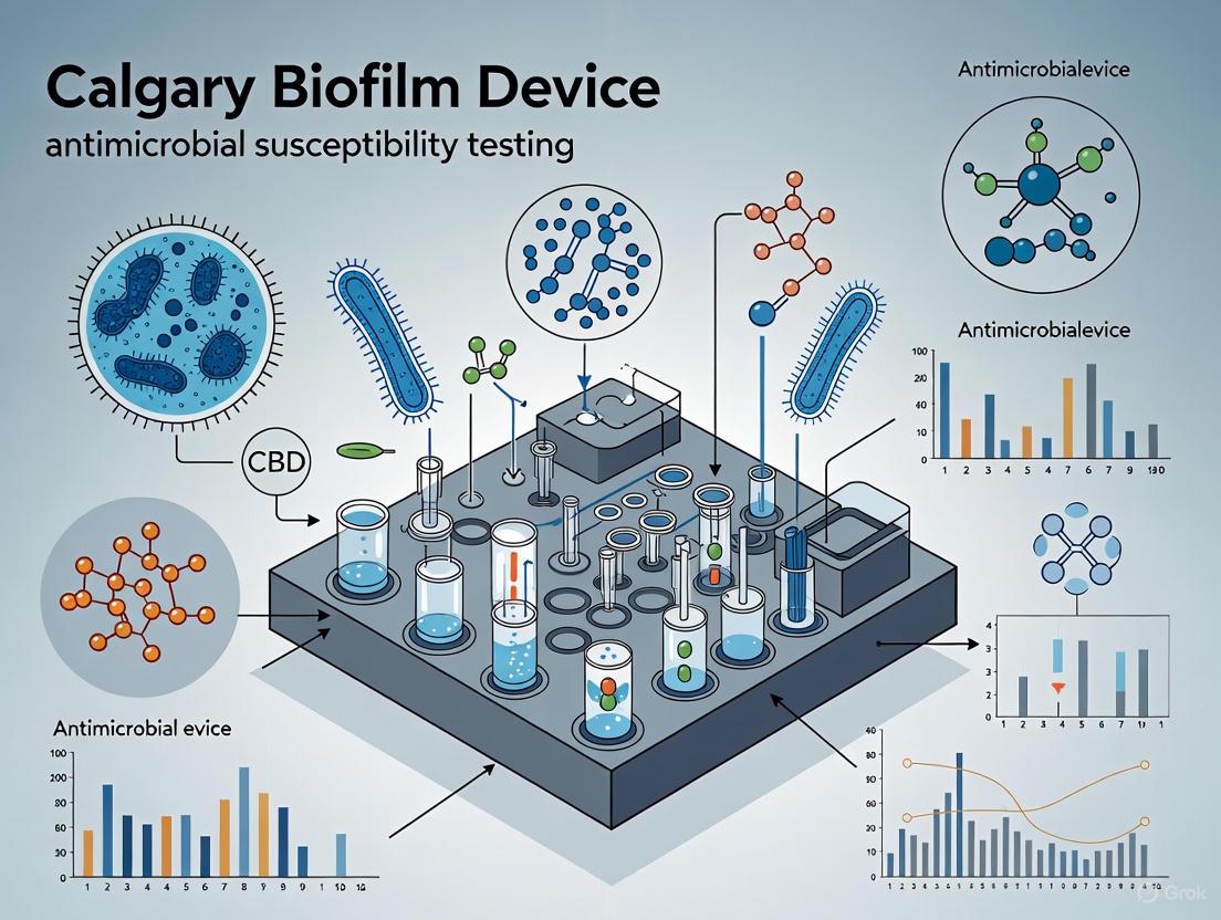
The Calgary Biofilm Device (CBD) was developed to address the critical need for standardized antimicrobial susceptibility testing (AST) of biofilms [4]. This technology consists of a two-part system: a standard 96-well plate containing growth media and antibiotics in serial dilutions, and a lid with 96 pegs that sits inside the wells. Biofilms form on the pegs, enabling high-throughput generation of 96 equivalent, reproducible biofilms ideal for robust susceptibility assays [4].
The key metrics derived from CBD testing are:
- Minimal Inhibitory Concentration (MIC): The lowest concentration that prevents planktonic growth in the wells.
- Minimal Biofilm Eradication Concentration (MBEC): The lowest concentration that eradicates the biofilm grown on the pegs [4].
The MBEC value is clinically paramount, as studies using the CBD consistently demonstrate that biofilms often require 100 to 1,000 times the antibiotic concentration for eradication compared to the MIC needed to inhibit planktonic cells [4] [2]. This quantitative difference underscores the therapeutic challenge posed by biofilm infections.
Application Note: Protocol for MBEC Assay Using the CBD
This protocol details the procedure for determining the MBEC of antibiotics against a bacterial biofilm using the Calgary Biofilm Device, following the established methodology [4].
Materials and Reagents
Table 2: Research Reagent Solutions for CBD-MBEC Assay
| Item | Function/Description | Application Note |
|---|---|---|
| Calgary Biofilm Device (CBD) | A 96-peg lid and matching trough; provides a surface for high-throughput, reproducible biofilm formation. | Pegs can be pre-coated with materials like hydroxyapatite, cellulose, or titanium dioxide to mimic specific environmental or clinical surfaces [8]. |
| Cation-Adjusted Mueller-Hinton Broth (CAMHB) | Standardized growth medium for susceptibility testing. | Ensures reliable and reproducible bacterial growth and antibiotic activity. |
| Antibiotic Stock Solutions | Solutions of antimicrobial agents for susceptibility testing. | Prepare serial two-fold dilutions in CAMHB in the 96-well plate according to CLSI/EUCAST guidelines. |
| Sterile 96-Well Microtiter Plate | Serves as a trough for the peg lid during biofilm formation and challenge. | Must be sterile and compatible with the CBD lid. |
| Neutralization Buffer (e.g., D/E Buffer) | Halts antibiotic action during processing. | Critical for accurate viability counts after antibiotic exposure. |
| Sonication Bath | Releases biofilm cells from pegs into recovery media. | Standardized sonication conditions (e.g., time, power) are vital for reproducible biofilm harvesting. |
| Tryptic Soy Agar (TSA) Plates | Solid medium for enumerating viable bacteria. | Used for spot-plating or spreading the sonicated biofilm suspension. |
Step-by-Step Procedure
Part 1: Biofilm Formation (24-48 hours)
- Inoculum Preparation: Grow the bacterial strain of interest to mid-log phase and dilute in CAMHB to a concentration of approximately 1 x 10^6 CFU/mL [4].
- Loading: Dispense 150 µL of the bacterial inoculum into each well of a sterile 96-well plate. For controls, fill wells with sterile broth.
- Incubation: Place the peg lid onto the plate, ensuring each peg is submerged in the inoculum. Incubate the assembly statically for 24-48 hours at the appropriate temperature (e.g., 37°C) to allow for mature biofilm development on the pegs.
Part 2: Antibiotic Challenge (24 hours)
- Biofilm Normalization: After incubation, carefully remove the peg lid from the growth plate. Rinse it gently by immersing it in a fresh trough of sterile saline or phosphate-buffered saline (PBS) to remove loosely adherent planktonic cells.
- Challenge Plate Preparation: In a new 96-well plate, prepare serial two-fold dilutions of the test antibiotic(s) in CAMHB. Include antibiotic-free control wells for growth and sterility.
- Exposure: Transfer the rinsed peg lid into the challenge plate, ensuring each biofilm-coated peg is immersed in an antibiotic solution. Incubate the assembly for 24 hours at the appropriate temperature.
Part 3: Biofilm Recovery and MBEC Determination (24-48 hours)
- Neutralization and Harvesting: Remove the peg lid from the challenge plate. Rinse it again in sterile PBS to remove residual antibiotic. Place the lid into a new trough containing neutralization buffer.
- Sonication: Sonicate the peg lid to dislodge and disperse the biofilm cells from the pegs into the neutralization buffer. This step is critical for obtaining an accurate cell count.
- Viability Quantification: Serially dilute the resulting biofilm suspension and spot-plate or spread onto TSA plates. Incubate the plates for 24-48 hours and enumerate the colony-forming units (CFU).
- MBEC Analysis: The MBEC is defined as the lowest antibiotic concentration that results in no growth (or a pre-defined log reduction, e.g., ≥99.9% kill) on the agar plates [4]. Compare this value to the MIC obtained from the planktonic cells in the challenge plate wells.
The workflow for this protocol is illustrated below.
Discussion and Research Implications
The data generated by the CBD unequivocally quantifies the stark contrast between planktonic and biofilm susceptibility, validating the clinical observation of recalcitrant infections [6] [4]. For drug development, this means compounds showing efficacy only in standard planktonic AST may fail against biofilm-related infections. The CBD enables screening for "biofilm-active" agents, such as rifampicin for prosthetic joint infections, which has shown success in clinical practice [6].
A primary research bottleneck is the translation of biofilm AST into routine clinical microbiology. While the CBD is a powerful research tool, current clinical diagnostics still predominantly rely on planktonic AST [6]. Future work must focus on developing rapid, standardized biofilm AST methods that can be integrated into clinical workflows to guide targeted therapy. Promising avenues include refining devices like the CBD for clinical specimens, exploring genetic markers for biofilm-specific resistance, and developing combination therapies that disrupt the EPS matrix to enhance antibiotic penetration [6] [1] [5]. Understanding and targeting the specific mechanisms of biofilm tolerance, rather than just the resident bacteria, is the key to overcoming this formidable clinical challenge.
Conventional antimicrobial susceptibility testing (AST), based on minimum inhibitory concentration (MIC) assays against free-floating planktonic bacteria, has long served as the standard for guiding antibiotic therapy. However, this approach fails to accurately represent the majority of clinical infections involving surface-attached, biofilm-forming bacterial communities. Biofilm-associated bacteria demonstrate dramatically increased tolerance to antimicrobial agents, often requiring concentrations 100-1000 times higher than those effective against their planktonic counterparts. This Application Note examines the critical limitations of planktonic susceptibility testing and presents the Calgary Biofilm Device (CBD) as a standardized methodology for the rapid and reproducible assessment of biofilm susceptibilities, enabling more effective selection of antibiotics against chronic and device-related infections.
The Minimum Inhibitory Concentration (MIC) has served as the cornerstone of antibiotic susceptibility testing for decades. As the standard assay endorsed by the Clinical and Laboratory Standards Institute (CLSI) and the European Committee on Antimicrobial Susceptibility Testing (EUCAST), MIC determination measures antibiotic efficacy against planktonic bacterial populations and provides critical guidance for treating acute infections [9]. However, this planktonic-centric paradigm presents substantial limitations when applied to the management of chronic or device-related infections, where bacteria predominantly exist in structured biofilm communities.
Biofilms are surface-adherent microbial communities encased within an extracellular polymeric substance (EPS) matrix. These structured communities represent a fundamental mode of bacterial growth that differs profoundly from planktonic existence. Numerous studies have demonstrated that biofilm-grown microorganisms exhibit inherent tolerance to antimicrobial agents not observed in planktonic cultures of the same organisms [9] [10]. This recalcitrance is multifactorial, arising from physical barriers created by the EPS matrix, reduced metabolic activity of subpopulations within the biofilm, and the expression of distinct biofilm-specific phenotypes [10].
The clinical impact of this discrepancy is profound. Approximately 80% of chronic human infections are estimated to be biofilm-associated, including those affecting wounds, the respiratory tract in cystic fibrosis patients, and indwelling medical devices [11]. When clinicians prescribe antibiotics based on conventional MIC data for these infections, treatment failures frequently occur, leading to chronic persistence, recurrent exacerbations, and increased healthcare costs. Patients harboring biofilm infections often require higher antibiotic doses and prolonged treatment courses not predicted by planktonic susceptibility testing [12].
The Biofilm Challenge: Quantifying the MIC-MBEC Disparity
The critical limitation of planktonic AST becomes evident when comparing MIC values with Minimal Biofilm Eradication Concentrations (MBEC) – the lowest concentration of an antimicrobial that eradicates a biofilm population. Research utilizing the Calgary Biofilm Device has systematically quantified this disparity across multiple bacterial species and antibiotic classes.
Table 1: Comparative Analysis of MIC vs. MBEC Values for Reference Bacterial Strains
| Bacterial Strain | Antibiotic | MIC (μg/mL) | MBEC (μg/mL) | Fold Increase |
|---|---|---|---|---|
| E. coli ATCC 25922 | Ampicillin | 4 | 512 | 128x |
| Ciprofloxacin | 0.125 | 32 | 256x | |
| P. aeruginosa ATCC 27853 | Gentamicin | 2 | 512 | 256x |
| Ceftazidime | 1 | 256 | 256x | |
| S. aureus ATCC 29213 | Oxacillin | 0.5 | 64 | 128x |
| Vancomycin | 2 | 128 | 64x |
Data derived from Ceri et al. (1999) demonstrating 100-1000 fold increases in antibiotic concentrations required to eradicate biofilms compared to planktonic cells [9].
This profound tolerance is not limited to conventional antibiotics. Recent research on antimicrobial phytochemicals reveals similar patterns. For instance, cannabidiol (CBD) demonstrated a MIC of 5 μg/mL against planktonic Streptococcus mutans, but required 7.5-20 μg/mL to significantly reduce the viability of preformed biofilms [13]. Similar findings extend to disinfectants and sanitizers used in industrial and food processing environments, where biofilms demonstrate significantly enhanced survival compared to planktonic cells [14].
Table 2: Methodological Comparison: Planktonic AST vs. Biofilm AST
| Parameter | Planktonic AST (MIC) | Biofilm AST (MBEC) |
|---|---|---|
| Inoculum | Planktonic cells | Surface-attached biofilm |
| Growth Phase | Logarithmic | Stationary/Mature |
| Matrix Presence | No | Yes (EPS) |
| Metabolic State | Uniform | Heterogeneous |
| Antibiotic Exposure | Direct | Diffusion-limited |
| Endpoint | Inhibition of growth | Eradication of biofilm |
| Clinical Relevance | Acute infections | Chronic/device-related infections |
The Calgary Biofilm Device: Principles and Applications
The Calgary Biofilm Device (CBD), commercially available as the MBEC Assay System, was developed specifically to address the limitations of planktonic susceptibility testing. This innovative technology enables high-throughput production of 96 equivalent biofilms suitable for antibiotic susceptibility screening using standard 96-well microtiter plate methodology [9] [4].
The device consists of a two-part reaction vessel: a lid with 96 pegs that sits in a correspondingly channeled base. The design channels medium flow across all pegs, creating consistent shear force that results in the formation of highly reproducible equivalent biofilms at each peg site. This standardization is critical for reliable susceptibility testing, as demonstrated by studies showing no significant difference (P > 0.1) between biofilms formed on different pegs across the device [9].
Key Advantages for Research and Diagnostics
The CBD offers several distinct advantages for biofilm research and potential diagnostic applications:
- Reproducibility: The device generates 96 equivalent biofilms in a single run, with statistical analysis confirming no significant differences between biofilms on different pegs [9] [14].
- High-throughput capacity: Multiple antibiotics can be tested against standardized biofilms simultaneously, significantly increasing screening efficiency.
- Biomass quantification: Biofilm formation can be precisely monitored through quantitative microbiology and scanning electron microscopy [9].
- Flexibility: The system accommodates various bacterial species, including E. coli, P. aeruginosa, S. aureus, Mycoplasma species, and fungal pathogens such as Candida albicans [15] [16].
- Clinical relevance: MBEC values derived from the CBD provide more accurate predictors of antibiotic efficacy for biofilm-associated infections than traditional MIC values [12].
Experimental Protocols: CBD Workflow and Methodologies
Calgary Biofilm Device Standard Operating Procedure
Figure 1: Calgary Biofilm Device Experimental Workflow. The standardized protocol for MBEC determination using the CBD system.
Biofilm Formation and Harvesting
Inoculum Preparation: Prepare standardized bacterial suspensions (approximately 10^7 CFU/mL) in appropriate media such as Trypticase Soy Broth (TSB) or Cation-Adjusted Mueller-Hinton Broth (CAMHB) using the direct colony suspension method from 18-24 hour cultures [9].
Device Assembly and Inoculation: Transfer 150-200 μL of standardized inoculum to each well of the CBD base. Secure the peg lid and incubate at 35°C with 95% relative humidity on a rocking platform to generate consistent shear force across all pegs.
Biofilm Maturation: Incubate for species-specific duration (typically 4-24 hours) to achieve mature biofilms. P. aeruginosa typically forms established biofilms within 4 hours, while S. aureus may require up to 7 hours [9].
Biofilm Quantification (Quality Control): Select representative pegs, transfer to microcentrifuge tubes containing 200 μL of fresh media, and sonicate for 5 minutes to disrupt biofilms. Perform viable counts on appropriate agar plates to verify consistent biofilm formation across the device [9].
Antibiotic Susceptibility Testing
Antibiotic Preparation: Prepare serial two-fold dilutions of antibiotics in CAMHB in a 96-well plate, typically ranging from 1024 μg/mL to 0.5 μg/mL.
Antibiotic Exposure: Transfer the CBD lid with established biofilms to the antibiotic dilution plate. Incubate for 24 hours at 35°C to assess biofilm eradication.
Viability Assessment: Remove the lid, rinse gently in phosphate-buffered saline to remove non-adherent cells, and transfer to a recovery plate containing fresh media. Sonicate to disrupt surviving biofilm cells and assess viability through:
MBEC Determination: The MBEC is defined as the lowest antibiotic concentration that prevents biofilm recovery, indicated by no growth in the recovery medium [9].
Alternative and Emerging Biofilm Susceptibility Methods
While the CBD represents a well-established approach, several emerging technologies offer complementary capabilities for biofilm susceptibility testing:
Live/Dead Antimicrobial Susceptibility Test (LD-AST): This flow cytometry-based method measures the proportion of live bacteria upon antibiotic exposure using viability staining, effectively working with both planktonic and biofilm cultures. LD-AST has proven particularly valuable for fastidious organisms like Mycoplasma species, where traditional metabolic assays may be unreliable due to biofilm-related metabolic dormancy [15].
Resazurin-Based Fluorometric Assay: This method utilizes the metabolic reduction of resazurin (a blue, non-fluorescent compound) to resorufin (pink, highly fluorescent) to quantify viable cells within biofilms. Studies have demonstrated strong correlation between this approach and the CBD, with the advantage of real-time monitoring capability [12].
BiofilmChip Technology: This microfluidic platform with integrated interdigitated sensors enables irreversible attachment of bacterial cells and real-time monitoring of biofilm formation and treatment response through electrical impedance spectroscopy or confocal microscopy. The system better mimics in vivo flow conditions and supports polymicrobial biofilm studies [11].
The Scientist's Toolkit: Essential Research Reagents and Materials
Table 3: Essential Research Reagents for Biofilm Susceptibility Testing
| Reagent/Equipment | Function/Application | Specifications/Alternatives |
|---|---|---|
| Calgary Biofilm Device | High-throughput biofilm production | MBEC Assay System (Innovotech) |
| Culture Media | Biofilm growth and antibiotic dilution | TSB, CAMHB, BHI (species-specific) |
| Sonication Device | Biofilm harvesting and disruption | 5 min at high setting (Aquasonic model) |
| Viability Stains | Cell viability assessment | LIVE/DEAD BacLight, SYTO 9/propidium iodide |
| Metabolic Indicators | Metabolic activity quantification | PrestoBlue, resazurin, MTT |
| Microtiter Plates | Antibiotic serial dilutions | Standard 96-well plates |
| Rocking Platform | Shear force generation during biofilm formation | Red Rocker model or equivalent |
| Imaging Systems | Biofilm structure analysis | Scanning Electron Microscopy, Confocal Laser Scanning Microscopy |
Data Interpretation and Clinical Implications
MBEC Determination and Breakpoints
A critical challenge in biofilm susceptibility testing remains the establishment of standardized breakpoints for MBEC values. Unlike MIC breakpoints, which are well-defined by CLSI and EUCAST for planktonic bacteria, MBEC interpretive criteria are still evolving. Current approaches include:
- Comparative Assessment: MBEC values are compared to achievable tissue/serum antibiotic concentrations, with MBEC values below attainable concentrations considered potentially effective.
- Multiple of MIC: Some researchers propose that antibiotics with MBEC values <10x the MIC may have better anti-biofilm efficacy, though this varies substantially by drug class and bacterial species.
- Species-Drug Specific Guidelines: Preliminary guidelines are emerging for specific pathogen-drug combinations, particularly for device-related infections.
Correlation with Treatment Outcomes
Evidence increasingly supports the clinical relevance of biofilm susceptibility testing. Studies have demonstrated that patients with chronic infections treated with antibiotic regimens based on biofilm susceptibility testing have better clinical outcomes than those treated with regimens based solely on planktonic susceptibility profiles [12]. This is particularly evident in:
- Cystic fibrosis airway infections
- Chronic wound management
- Medical device-associated infections
- Orthopedic implant-related infections
The limitations of conventional planktonic susceptibility testing are no longer speculative but are well-documented and quantitatively significant. The MIC, while valuable for acute infections, provides dangerously misleading information when applied to biofilm-associated chronic infections. The Calgary Biofilm Device addresses this critical gap by providing a standardized, reproducible platform for determining the Minimal Biofilm Eradication Concentration - a clinically relevant metric that accounts for the profound tolerance of biofilm communities.
As antimicrobial resistance continues to escalate, embracing biofilm-specific susceptibility testing represents an essential evolution in our approach to managing persistent infections. The protocols and methodologies outlined in this Application Note provide researchers with robust tools to advance this critical field, ultimately contributing to more effective therapeutic strategies for the challenging spectrum of biofilm-associated diseases.
The Calgary Biofilm Device (CBD) represents a groundbreaking technology for the rapid and reproducible assay of biofilm susceptibilities to antibiotics. Developed to address the innate lack of antibiotic susceptibility observed in adherent bacterial populations, the CBD enables high-throughput screening of antimicrobial compounds against biofilm-grown microorganisms. This technology facilitates the determination of the Minimal Biofilm Eradication Concentration (MBEC), a critical parameter for evaluating antibiotic efficacy against biofilms, which often requires 100 to 1,000 times the concentration needed to eradicate planktonic bacteria [9] [4]. This application note details the design principles, core technology, and standard protocols for utilizing the CBD in antimicrobial susceptibility research.
Design Principles and Core Technology
Rationale and Innovation
Traditional antibiotic susceptibility testing, based on the Minimum Inhibitory Concentration (MIC) against free-floating (planktonic) bacteria, fails to predict therapeutic success for chronic or device-related infections involving bacterial biofilms [9] [17]. Biofilms are structured communities of microbial cells embedded in a protective extracellular matrix, demonstrating inherent tolerance to antimicrobial agents [18]. The CBD was innovated to address this technological gap, providing a standardized method for growing multiple equivalent biofilms for susceptibility testing [9].
Device Architecture
The CBD is a two-part reaction vessel engineered for use with standard 96-well microtiter plate technology [9]. Its design consists of:
- A Lid with 96 Pegs: The top component forms a sealed lid with 96 pegs, designed to fit into the wells of a standard microtiter plate. These pegs serve as the substrate for biofilm growth.
- A Channeled Base: The bottom component features channels that guide the flow of growth medium. When placed on a rocking table, this design creates consistent shear force across all pegs, which is essential for the uniform development of biofilms on each peg [9].
This architecture allows for the simultaneous formation of 96 equivalent biofilms, enabling high-throughput screening of antibiotic compounds under conditions that mimic the shear forces found in natural environments [9]. The device is commercially available as the MBEC Assay System [19].
Key Research Findings and Quantitative Data
Initial validation studies with NCCLS reference strains demonstrated the device's capability to produce highly reproducible biofilms and quantify dramatically increased antibiotic tolerance.
Table 1: Biofilm Growth Kinetics on the Calgary Biofilm Device [9]
| Bacterial Strain | Time to Reach ~10⁵ CFU/peg | Maximum Density (after 24 h) |
|---|---|---|
| Escherichia coli ATCC 25922 | 6 hours | 3 × 10⁷ to 5 × 10⁷ CFU/peg |
| Pseudomonas aeruginosa ATCC 27853 | 4 hours | 3 × 10⁷ to 5 × 10⁷ CFU/peg |
| Staphylococcus aureus ATCC 29213 | 7 hours | 1 × 10⁵ to 2 × 10⁵ CFU/peg |
Statistical analysis confirmed no significant difference (P > 0.1) between the biofilms formed on each of the 96 pegs, validating the device's ability to produce equivalent biofilms for highly reproducible screening [9].
Table 2: Comparison of Planktonic MIC vs. Biofilm MBEC for Selected Antibiotics [9] [20]
| Organism | Antibiotic | MIC (µg/mL) | MBEC (µg/mL) | Fold-Increase |
|---|---|---|---|---|
| Pseudomonas aeruginosa | Ciprofloxacin | Not specified | Not specified | 100 - 1,000 |
| Staphylococcus aureus | Clindamycin | Not specified | Not specified | 100 - 1,000 |
| Escherichia coli | Ampicillin | Not specified | Not specified | 100 - 1,000 |
The data confirmed that while some antibiotics remained effective at the MIC, for many others, a 100 to 1,000-fold increase in concentration was required to eradicate biofilm-grown bacteria [9] [20].
Experimental Protocols
Biofilm Formation Protocol
Principle: To establish robust and equivalent biofilms on all 96 pegs of the device [9].
Workflow Overview:
Materials:
- Calgary Biofilm Device (with 96-peg lid and trough base) [19]
- Trypticase Soy Broth (TSB) or other appropriate culture medium [9]
- Bacterial strains grown on Trypticase Soy Agar (TSA) plates for 18-24 hours [9]
- Phosphate-Buffered Saline (PBS)
- Rocking table (e.g., Red Rocker model) placed in a 35°C incubator with 95% relative humidity [9]
Procedure:
- Inoculum Preparation: Create a bacterial suspension in TSB directly from colonies on a TSA plate. Standardize the suspension to a density of approximately 10⁷ - 10⁸ CFU/mL using McFarland standards [9].
- Device Inoculation: Pipette the standardized inoculum into the channeled base of the CBD. Ensure the channels are filled to allow the pegs to be submerged.
- Biofilm Growth: Place the peg lid onto the inoculated base. Incubate the assembled device on a rocking table at 35°C for a duration determined by the microbial growth kinetics (see Table 1). The rocking motion generates shear force, promoting uniform biofilm formation on the pegs [9].
- Quality Control: To confirm biofilm formation and density, remove select pegs, place them in microcentrifuge tubes containing recovery broth (e.g., TSB), and sonicate to dislodge the biofilm. Perform viable cell counts on TSA plates to determine the CFU/peg [9].
Biofilm Susceptibility Testing (MBEC Assay) Protocol
Principle: To determine the minimal concentration of an antimicrobial agent required to eradicate a mature biofilm [9] [8].
Workflow Overview:
Materials:
- CBD lid with mature biofilm
- Cation-Adjusted Mueller-Hinton Broth (CAMHB) [9]
- Standard 96-well microtiter plate
- Antimicrobial stock solutions and working dilutions
- Sonicating water bath
Procedure:
- Antibiotic Plate Preparation: In a 96-well plate, prepare serial twofold dilutions of the test antibiotics in CAMHB, covering a concentration range (e.g., 0.5 to 1,024 µg/mL) [9].
- Antibiotic Exposure: Carefully remove the peg lid from the growth base, rinse it gently in PBS to remove loosely adherent planktonic cells, and transfer it into the antibiotic-containing plate. Ensure each peg is submerged in a different antibiotic concentration.
- Incubation: Incubate the plate at 35°C for 18-20 hours.
- Biofilm Recovery and Viability Assessment:
- Remove the lid from the antibiotic plate and rinse it again in PBS.
- Transfer the lid to a new 96-well "recovery" plate containing fresh, antibiotic-free CAMHB.
- Sonicate the entire lid to disrupt the biofilms and release viable cells into the recovery plate [9].
- MBEC Determination: Incubate the recovery plate for 24 hours at 35°C. The MBEC is defined as the lowest concentration of antibiotic in the challenge plate that results in no visible growth (or no detectable turbidity at 650 nm) in the corresponding well of the recovery plate [9]. The MIC for planktonic cells can also be determined from the challenge plate by measuring turbidity after incubation [9].
The Scientist's Toolkit: Essential Research Reagents and Materials
Table 3: Key Research Reagent Solutions for CBD Experiments
| Item | Function/Description |
|---|---|
| CBD/MBEC Assay Device | The core hardware, consisting of a peg lid and a channeled trough base, for high-throughput biofilm cultivation [9] [19]. |
| Cation-Adjusted Mueller-Hinton Broth (CAMHB) | Standardized medium for performing antimicrobial susceptibility tests in the 96-well plate [9]. |
| Trypticase Soy Broth (TSB) | General nutrient medium used for the initial growth of the inoculum and for promoting biofilm formation in the device base [9]. |
| Coated Pegs | Commercially available peg lids can be coated with materials like hydroxyapatite, titanium dioxide, or cellulose to mimic specific surface properties of medical devices or natural environments [19] [8]. |
| Sonicating Water Bath | Used to efficiently and reproducibly dislodge biofilms from the pegs for quantitative viability counts after antimicrobial exposure [9]. |
Advanced Applications and Recent Developments
The utility of the CBD platform continues to expand in scientific research. A recent innovative application combines the CBD with Ionized Jet Deposition (IJD) technology for the high-throughput screening of novel nanostructured silver and zinc coatings [18]. In this setup, different amounts of metal are deposited directly onto the wells/pegs of the CBD, creating a gradient of coating properties that can be simultaneously tested for their antibacterial and antibiofilm efficacy against multiple bacterial strains [18]. This approach validates the CBD's role as a powerful tool for developing next-generation antibacterial materials for medical devices.
The Calgary Biofilm Device provides a robust, standardized, and high-throughput platform for assessing antimicrobial susceptibility in biofilm-grown bacteria. By enabling the rational selection of antibiotics and the screening of new compounds against clinically relevant biofilms, the CBD addresses a critical need in both basic research and therapeutic development. Its well-defined protocols and commercial availability make it an indispensable tool for researchers and scientists combating biofilm-related infections.
The Calgary Biofilm Device (CBD), also known as the MBEC (Minimum Biofilm Eradication Concentration) Assay System, represents a groundbreaking technological advancement in antimicrobial susceptibility testing for bacterial biofilms [9]. This innovative device addresses a critical limitation in traditional microbiology: standard antibiotic susceptibility tests, like the Minimum Inhibitory Concentration (MIC), are performed on free-floating (planktonic) bacteria, which often do not correlate with the efficacy of antibiotics against surface-adherent, biofilm populations [9] [12]. Bacterial biofilms are structured communities of microorganisms encased in an extracellular polymeric substance that exhibit innate tolerance to antimicrobial agents, sometimes requiring antibiotic concentrations 100 to 1,000 times higher than those needed to eradicate their planktonic counterparts for effective treatment [9]. The CBD was specifically designed to provide a rapid, reproducible, and high-throughput method for assaying biofilm susceptibilities, enabling the rational selection of antibiotics for treating chronic device-related infections and screening new antimicrobial compounds [9] [4].
Key Advantages of the 96-Well Platform
The CBD leverages the standardization of 96-well technology to overcome the limitations of previous biofilm models, such as the Modified Robbins Device, which were not suited for rapid, high-throughput screening in a clinical laboratory setting [9]. The core advantages of this platform are its high-throughput capability, exceptional reproducibility, and seamless integration with standardized laboratory equipment and protocols.
High-Throughput Capacity
- Simultaneous Biofilm Production: The device features a lid with 96 pegs that sits atop a channeled base, allowing for the simultaneous formation of 96 equivalent biofilms in a single experiment [9] [21]. This design subjects all pegs to consistent hydrodynamic conditions, ensuring uniform biofilm growth [9].
- Efficient Antibiotic Screening: Once biofilms are established, the peg lid can be transferred to a standard 96-well plate containing serial dilutions of antimicrobial agents. This allows for the efficient screening of multiple antibiotics or compound concentrations against a single biofilm-forming strain, or the testing of one antibiotic against multiple bacterial strains in a single run [9] [22].
- Automation Compatibility: The 96-well format is compatible with automated liquid handling systems, plate readers, and data analysis software, significantly reducing hands-on time and increasing analytical throughput [23]. This automation potential minimizes inter-operator variability and is a key step towards standardized biofilm susceptibility testing [23].
Reproducibility and Reliability
- Equivalent Biofilm Formation: Quantitative microbiology and scanning electron microscopy have confirmed that the CBD produces highly consistent biofilms across all 96 pegs. Statistical analyses, including one-way analysis of variance, have demonstrated no significant difference (P > 0.1) between biofilms formed on different pegs [9]. For instance, a study with Pseudomonas aeruginosa showed minimal variation, with mean log10 counts per peg of 5.725 at 4 hours and 7.202 at 24 hours [9].
- Quantitative and Robust Endpoints: The system provides clear, quantitative endpoints such as the Minimum Biofilm Eradication Concentration (MBEC)—the lowest concentration of an antimicrobial required to eradicate a biofilm [9] [21]. This offers a more clinically relevant measure than the MIC for biofilm-associated infections.
- Validation Against Reference Methods: The antibiotic susceptibility profiles for planktonic populations derived from the CBD have been shown to be similar to those obtained by the reference broth microdilution method set by the National Committee for Clinical Laboratory Standards (NCCLS, now CLSI), validating its reliability [9].
Standardization and Integration
- Consistency with 96-Well Technology: By utilizing the ubiquitous 96-well microtiter plate format, the CBD integrates seamlessly into existing laboratory workflows. This compatibility extends to standard plate shakers for incubation and plate readers for measuring turbidity or fluorescence to determine bacterial viability [9] [12].
- Standardized Protocols: The device enables the establishment of standardized protocols for biofilm growth and susceptibility testing. For example, growth curves can determine the precise incubation time needed to form a biofilm of a predetermined density, ensuring consistency across experiments [9].
- Foundation for Automated Workflows: The 96-well platform is the foundation for developing fully automated workflows, as evidenced in other fields like liquid chromatography-mass spectrometry (LC-MS/MS) for therapeutic drug monitoring [23]. This principle is directly applicable to biofilm susceptibility testing, promising enhanced precision and scalability.
Table 1: Quantitative Validation of Biofilm Reproducibility on the CBD (Data for Pseudomonas aeruginosa ATCC 27853) [9]
| Parameter | 4 Hours of Growth (Log10 count/peg) | 24 Hours of Growth (Log10 count/peg) |
|---|---|---|
| Mean | 5.725 | 7.202 |
| Median | 5.778 | 7.204 |
| Standard Deviation | 0.448 | 0.383 |
| Lower 95% CI | 5.634 | 7.124 |
| Upper 95% CI | 5.816 | 7.280 |
Comparative Data: MIC vs. MBEC
The critical value of the CBD is its ability to reveal the profound tolerance of biofilms to antibiotics, a phenomenon that is missed by conventional planktonic testing.
Table 2: Illustrative Comparison of MIC vs. MBEC Values for Reference Strains [9]
| Organism | Antibiotic | MIC (µg/mL) | MBEC (µg/mL) | Fold Increase (MBEC/MIC) |
|---|---|---|---|---|
| Escherichia coli ATCC 25922 | Ampicillin | 4 | >1024 | >256 |
| Escherichia coli ATCC 25922 | Ciprofloxacin | 0.015 | 4 | 267 |
| Pseudomonas aeruginosa ATCC 27853 | Gentamicin | 2 | 512 | 256 |
| Staphylococcus aureus ATCC 29213 | Oxacillin | 0.25 | 128 | 512 |
This data underscores that while some antibiotics may be effective against planktonic cells, they can be remarkably ineffective against biofilms of the same organism. Conversely, the CBD can also identify antibiotics that remain effective at or near the MIC, enabling data-driven antibiotic selection [9].
Detailed Experimental Protocols
Protocol 1: Biofilm Formation and Growth Curve Analysis
This protocol describes how to establish and quantify biofilm formation on the CBD.
Research Reagent Solutions
- Growth Medium: Trypticase Soy Broth (TSB) or Cation-Adjusted Mueller-Hinton II Broth (MHIIB) [9] [12].
- Agar Plates: Trypticase Soy Agar (TSA) for viable counts [9].
- Saline Solution: Phosphate-buffered saline (PBS) for rinsing [9].
Procedure
- Inoculum Preparation: Create a bacterial suspension from fresh colonies on a TSA plate. Adjust the turbidity to a 0.5 McFarland standard in saline or growth medium, which corresponds to approximately 1-2 x 10^8 CFU/mL [9] [12].
- Dilution and Loading: Dilute the standardized suspension 1:30 in growth medium. Pipette 150 µL of the diluted inoculum into each well of the CBD base channeled plate [9].
- Incubation with Agitation: Place the peg lid onto the base and incubate the entire assembly on a rocking table (e.g., Red Rocker model) at 35°C and 95% relative humidity for a desired period (e.g., 4-24 hours). The rocking motion creates shear force necessary for consistent biofilm formation across all pegs [9].
- Growth Curve Analysis:
- At selected time points (e.g., 2, 4, 6, 8, 24 hours), remove two pegs from different locations on the lid.
- Place each peg in a microcentrifuge tube containing 200 µL of TSB and sonicate for 5 minutes in a bath sonicator (e.g., Aquasonic model 250HT) to dislodge the biofilm [9].
- Perform serial dilutions of the sonicate and plate on TSA plates to determine viable counts (CFU/peg).
- Plot log CFU/peg versus time to generate a biofilm growth curve and determine the optimal incubation time for a mature biofilm.
Protocol 2: MBEC Assay for Antibiotic Susceptibility
This protocol outlines the steps to determine the Minimum Biofilm Eradication Concentration of antibiotics against a pre-formed biofilm.
Research Reagent Solutions
- Antibiotic Stock Solutions: Prepare high-concentration stocks (e.g., 6,200 µg/mL) in solvent, filter-sterilize, and store at -80°C [9].
- Antibiotic Working Solutions: Prepare two-fold serial dilutions of antibiotics in CAMHB in a standard 96-well plate, with a typical concentration range from 1,024 µg/mL and downward [9].
- Viability Indicator (Alternative): PrestoBlue (a resazurin-based solution) can be used as a fluorometric indicator of metabolic activity for a non-destructive readout [12].
Procedure
- Biofilm Formation: Grow a mature biofilm on the CBD peg lid as described in Protocol 1.
- Rinsing: Carefully remove the peg lid from the growth base, and rinse it gently in PBS to remove non-adherent planktonic cells [9] [21].
- Antibiotic Exposure: Transfer the rinsed peg lid to the 96-well plate containing the serial dilutions of antibiotics. Ensure each peg is immersed in a different antibiotic concentration.
- Incubation: Incub the plate at 35°C for 18-24 hours [9].
- Viability Assessment:
- Rinse and Sonicate: Rinse the peg lid again in PBS to remove residual antibiotic, and then transfer it to a new 96-well plate (a "recovery plate") containing fresh CAMHB. Sonicate the entire lid for 5 minutes to disrupt the biofilms and release any remaining viable bacteria into the recovery plate [9].
- Incubate and Measure: Incubate the recovery plate for 24 hours at 35°C. The MBEC is defined as the lowest antibiotic concentration in the challenge plate that results in no visible growth (turbidity) in the corresponding well of the recovery plate. Alternatively, measure turbidity at OD650 with a plate reader, where an OD of <0.1 indicates eradication [21]. The metabolic activity can also be measured fluorometrically using PrestoBlue to determine the percentage of non-viable cells [12].
Workflow Visualization
Diagram 1: MBEC Assay Workflow. This flowchart outlines the key steps in forming a biofilm on the CBD and testing its susceptibility to antimicrobial agents.
The Scientist's Toolkit: Essential Research Reagent Solutions
Table 3: Key Reagents and Materials for CBD Experiments
| Item | Function/Description | Example/Specification |
|---|---|---|
| Calgary Biofilm Device (CBD) | The core reaction vessel with a peg lid and channeled base for growing 96 equivalent biofilms. | MBEC Assay System (commercially available) [9]. |
| Growth Media | Supports bacterial growth and biofilm formation. | Trypticase Soy Broth (TSB), Cation-Adjusted Mueller-Hinton Broth (CAMHB) [9]. |
| Cation-Adjusted Mueller-Hinton II Broth (CAMHB) | The standardized medium for antibiotic susceptibility testing, ensuring consistent cation concentrations [9] [12]. | |
| Antibiotic Stock Solutions | High-concentration stocks for preparing serial dilutions. | Prepared in solvent or water, filter-sterilized, stored at -80°C [9]. |
| Phosphate-Buffered Saline (PBS) | Used for rinsing pegs to remove non-adherent cells and residual antibiotics without harming the biofilm. | Sterile, pH 7.4 [9]. |
| Sonication Device | To disrupt the biofilm from the pegs for quantitative viable counting. | Bath sonicator (e.g., Aquasonic 250HT), 5 min sonication [9]. |
| Microplate Reader | To measure turbidity (OD650) or fluorescence for high-throughput determination of bacterial growth/viability after antibiotic challenge [9] [12]. | |
| Viability Stain (Optional) | Fluorometric indicator of metabolic activity for alternative endpoint determination. | PrestoBlue (resazurin-based) [12]. |
The Calgary Biofilm Device successfully harnesses the power of the 96-well platform to address a significant challenge in clinical microbiology and antimicrobial drug development. Its core advantages of high-throughput screening, reproducible biofilm production, and straightforward standardization make it an indispensable tool for researchers and scientists. By providing clinically relevant MBEC data that often differs drastically from conventional MIC results, the CBD enables a more rational approach to selecting and developing effective treatments for persistent biofilm-associated infections. Its compatibility with automated systems further positions it as a cornerstone technology for advancing standardized antimicrobial susceptibility testing for biofilms.
The Minimum Inhibitory Concentration (MIC) has long been the standard reference for antimicrobial susceptibility testing, guiding clinical treatment decisions for decades. However, a critical limitation of MIC determination is that it measures antibiotic activity against planktonic (free-floating) bacteria, which represents only one mode of bacterial existence [9]. In contrast, the Minimum Biofilm Eradication Concentration (MBEC) quantifies the concentration required to eradicate bacteria growing in structured, surface-attached communities known as biofilms [24]. This distinction is clinically paramount, as biofilms exhibit innate tolerance to antimicrobial agents, often requiring concentrations 100 to 1000 times higher than those needed to inhibit their planktonic counterparts [9] [25]. This protocol details the methodology for determining MBEC using the Calgary Biofilm Device (CBD), a technology specifically designed to address the challenges of biofilm-related antimicrobial resistance [9].
The CBD, commercially available as the MBEC Assay System, enables the rapid and reproducible generation of multiple equivalent biofilms for high-throughput susceptibility screening [9] [26]. Its application is particularly relevant for investigating device-related and chronic infections, where biofilms play a definitive pathogenic role and conventional antibiotic therapies based on MIC frequently fail [9] [24]. The following sections provide comprehensive application notes and standardized protocols for implementing this essential technology in antimicrobial research and development.
Theoretical Foundation: MIC vs. MBEC
Conceptual Definitions and Clinical Implications
Minimum Inhibitory Concentration (MIC) is defined as the lowest concentration of an antimicrobial agent that prevents the visible growth of a planktonic bacterial population after a standard incubation period (typically 16-20 hours) [27]. MIC testing is standardized by organizations such as the Clinical and Laboratory Standards Institute (CLSI) and the European Committee on Antimicrobial Susceptibility Testing (EUCAST) [28] [27]. While MIC values are essential for treating acute infections with planktonic bacteria, they have limited utility for biofilm-associated infections [24].
Minimum Biofilm Eradication Concentration (MBEC) represents the lowest concentration of an antimicrobial agent that eradicates a biofilm population, including the more resilient "persister" cells within the biofilm structure [9] [25]. The MBEC assay more accurately models the clinical scenario of biofilm infections associated with medical implants and chronic conditions, providing data that better predicts the concentrations required for successful treatment [24].
Table 1: Fundamental Differences Between MIC and MBEC
| Characteristic | Minimum Inhibitory Concentration (MIC) | Minimum Biofilm Eradication Concentration (MBEC) |
|---|---|---|
| Target Population | Planktonic (free-floating) bacteria | Biofilm-associated bacteria |
| Physiological State | Log-phase growth | Stationary-phase with heterogeneous metabolism |
| Defined Endpoint | Inhibition of visible growth | Complete eradication of viable cells |
| Typical Assay Duration | 16-24 hours | 24 hours to several days |
| Clinical Correlation | Acute infections | Chronic, device-related infections |
| Antibiotic Concentration | Generally lower (µg/mL range) | Often 100-1000x higher than MIC |
The Biofilm Resistance Phenomenon
Biofilms demonstrate inherent lack of susceptibility to antimicrobials through multiple mechanisms: the extracellular polymeric substance (EPS) matrix acts as a physical diffusion barrier; metabolic heterogeneity within the biofilm creates dormant persister cells; and biofilm-specific phenotypes activate stress response pathways [9] [24]. This resistance is transient—when biofilm cells are dispersed and revert to planktonic growth, they regain the susceptibility profile of their planktonic counterparts [9].
System Design and Operating Principles
The Calgary Biofilm Device consists of a two-part reaction vessel: a lid with 96 pegs and a corresponding channeled base plate [9] [26]. This innovative design allows for the simultaneous formation of 96 equivalent biofilms on the peg lids when placed on a rocking table. The rocking motion generates consistent laminar flow and shear force across all pegs, resulting in highly reproducible biofilm formation at each site [9]. The device is compatible with standard 96-well microtiter plate technology, enabling high-throughput antibiotic susceptibility screening against biofilm-grown microorganisms [9].
Key Advantages for Antimicrobial Research
The CBD addresses significant limitations of previous biofilm models, such as the modified Robbin's device, which was not suited for rapid susceptibility testing in a clinical laboratory setting [9]. Key advantages include:
- Standardization: Produces 96 equivalent biofilms with no significant difference (P > 0.1) in biomass between pegs [9]
- High-Throughput Capacity: Enables screening of multiple antibiotic concentrations and combinations against biofilm-grown organisms
- Reproducibility: Minimizes experimental variability through standardized shear forces and growth conditions
- Flexibility: Accommodates various bacterial species and antifungal testing
- Validation: Correlates with in vivo biofilm susceptibility patterns [9]
Experimental Protocols for MBEC Determination
Protocol 1: Biofilm Formation and MBEC Assay
This protocol describes the standard procedure for growing biofilms and determining MBEC using the Calgary Biofilm Device [9] [24].
Materials and Equipment
- Calgary Biofilm Device (MBEC Assay System)
- Mueller-Hinton Broth (cation-adjusted for antibiotic testing)
- Trypticase Soy Broth or appropriate growth medium
- Antibiotic stock solutions
- 96-well microtiter plates
- Sonicator (e.g., Aquasonic 250HT)
- Rocking table (e.g., Red Rocker model)
- Incubator (35°C ± 2°C)
Procedure
Inoculum Preparation
- Harvest bacteria from fresh agar plates (18-24 hour growth)
- Prepare bacterial suspension in appropriate broth medium
- Standardize suspension to approximately 1 × 10^7 CFU/mL using McFarland standards [9]
Biofilm Formation
- Add 150 µL of standardized inoculum to each well of the CBD base plate
- Place peg lid into base plate ensuring pegs are submerged in inoculum
- Incubate on rocking table (125 rpm) at 35°C and 95% relative humidity for specific time period (varies by organism):
- Confirm biofilm formation by quantitative microbiology or scanning electron microscopy [9]
Antibiotic Exposure
- Prepare serial twofold dilutions of antibiotics in 96-well plates
- Transfer peg lid with established biofilms to antibiotic dilution plate
- Incubate static for 24 hours at 35°C [9]
MBEC Determination
- Remove peg lid from antibiotic plate and rinse in phosphate-buffered saline
- Transfer to recovery plate containing fresh medium
- Remove biofilm by sonication for 5 minutes [9]
- Incubate recovery plate for 24 hours at 35°C
- Assess viability by turbidity measurement (OD650) or plate counts [9]
- MBEC defined as the lowest antibiotic concentration showing no growth [9]
Figure 1: MBEC Assay Workflow. The standard procedure for determining Minimum Biofilm Eradication Concentration using the Calgary Biofilm Device.
Protocol 2: Biofilm Dispersal Assay for Anti-Biofilm Compounds
This protocol assesses the ability of test compounds to disperse established biofilms, adapted for natural compounds and anti-biofilm agents [29].
Materials and Equipment
- 24-well or 96-well clear flat-bottom plates
- Test compounds (e.g., natural products, synthetic anti-biofilm agents)
- Phosphate-buffered saline (PBS, pH 7.4)
- Crystal violet solution (0.1%)
- Modified biofilm dissolving solution (SDS in ethanol)
- Plate reader for optical density measurement
Procedure
Biofilm Establishment
- Grow biofilms as described in Protocol 1 for 24-48 hours
- Remove media and gently rinse with distilled water to remove planktonic cells
Compound Exposure
- Add PBS containing appropriate concentration of test compound to each well
- Incubate plates under appropriate conditions for 24 hours [29]
- Include PBS-only control wells
Biofilm Quantification
- Remove supernatants and measure OD600 for dispersed cells
- Remove media from plates by inverting over absorbent paper
- Rinse gently with distilled water twice to remove remaining planktonic cells
- Air-dry plates for 15 minutes in laminar flow cabinet
- Stain attached biofilm with 0.1% crystal violet solution for 10 minutes
- Remove unbound crystal violet with distilled water rinses
- Air-dry plates overnight at room temperature
- Solubilize crystal violet in modified biofilm dissolving solution
- Quantify OD570-600 in plate reader [29]
Quantitative Data Analysis and Interpretation
Comparative MIC and MBEC Values for Reference Strains
Research using the CBD has established critical baseline data for MBEC values of common reference strains, demonstrating the profound tolerance of biofilm populations compared to their planktonic counterparts.
Table 2: Representative MIC and MBEC Values for NCCLS/CLSI Reference Strains [9] [25]
| Organism | Antibiotic | MIC (µg/mL) | MBEC (µg/mL) | Fold Increase |
|---|---|---|---|---|
| Staphylococcus aureus ATCC 29213 | Ciprofloxacin | 0.25 | 8 | 32x |
| Vancomycin | 2 | >128 | >64x | |
| Pseudomonas aeruginosa ATCC 27853 | Gentamicin | 1 | 256 | 256x |
| Tobramycin | 1 | 128 | 128x | |
| Ciprofloxacin | 0.25 | 16 | 64x | |
| Escherichia coli ATCC 25922 | Amikacin | 1 | 64 | 64x |
| Ampicillin | 4 | >512 | >128x |
Impact of Exposure Time on MBEC
A critical factor in MBEC determination is exposure duration. Contrary to MIC testing which uses standardized 24-hour exposure, MBEC values are significantly influenced by treatment duration, with longer exposures generally resulting in lower MBEC values [25].
Table 3: MBEC Variation with Antimicrobial Exposure Time for S. aureus Strains [25]
| Antimicrobial | Strain | MBEC Day 1 (µg/mL) | MBEC Day 3 (µg/mL) | MBEC Day 5 (µg/mL) |
|---|---|---|---|---|
| Tobramycin | MSSA | >8000 | 4000 | 1000 |
| MRSA | >8000 | >8000 | 8000 | |
| Vancomycin | MSSA | >8000 | >8000 | 2000 |
| MRSA | >8000 | >8000 | 4000 | |
| Tobramycin:Vancomycin (3:1) | MSSA | 8000 | 500 | 500 |
| MRSA | >8000 | 4000 | 1000 |
Figure 2: Factors Influencing MBEC. Key parameters that affect the Minimum Biofilm Eradication Concentration in experimental and clinical settings.
Advanced Applications and Modifications
In Vivo Biofilm Models and Clinical Correlation
Recent advancements have extended MBEC testing to in vivo-formed biofilms, providing more clinically relevant susceptibility data. A 2022 study developed a novel MBEC assay using biofilms formed on stainless-steel implants in a rat femoral infection model, demonstrating that MBEC values derived from in vivo biofilms were substantially higher than those from in vitro models [24]. For example, against Staphylococcus aureus biofilms:
- Gentamicin MBEC100 (100% eradication) ranged from 256–1024 µg/mL for in vivo MBEC
- Vancomycin and cefazolin MBEC100 ranged from 2048–4096 µg/mL
- The in vivo implant MBEC was much higher, ranging from 2048 µg/mL to >4096 µg/mL [24]
This model also demonstrated that combination therapy with rifampicin significantly reduced MBEC values, highlighting the importance of antibiotic combinations for biofilm eradication [24].
Specialty Biofilm Models for Specific Applications
The CBD platform has been adapted for various research applications:
Oral Biofilm Models: The device has been used to study oral cariogenic biofilms, with modifications including coating pegs with hydroxyapatite and collagen to better mimic tooth surfaces [26]. These models have been used to test natural compounds like cranberry extract and cashew nutshell liquid for their anti-biofilm properties [26].
Combination Therapy Screening: The system enables efficient screening of synergistic combinations. For instance, research has demonstrated that combined triclosan/cannabidiol (CBD) treatment provides enhanced anti-biofilm effects against Streptococcus mutans compared to individual compounds [30].
Dual-Species and Multi-Species Biofilms: Protocols have been developed for growing mixed-species biofilms to study interspecies interactions and more complex community dynamics [29].
The Scientist's Toolkit: Essential Research Reagents and Materials
Table 4: Key Research Reagent Solutions for CBD Experiments
| Reagent/Equipment | Function/Application | Specifications/Notes |
|---|---|---|
| Calgary Biofilm Device | High-throughput biofilm production | MBEC Assay System with 96-peg lid [9] |
| Cation-Adjusted Mueller-Hinton Broth | Standard medium for antibiotic susceptibility testing | Required for accurate MIC/MBEC comparison [9] |
| Mueller-Hinton Agar | Quality control and viability counting | Standardized growth medium [9] |
| Rocking Table | Biofilm formation with consistent shear | ~125 rpm; creates laminar flow in channels [9] |
| Sonicator | Biofilm removal from pegs | 5 min sonication; model-specific settings vary [9] |
| Microtiter Plate Reader | Turbidity measurement for viability | OD650 nm for bacterial growth assessment [9] |
| 96-Well Microtiter Plates | Antibiotic dilution and incubation | Standard format for high-throughput screening [9] |
| Crystal Violet Solution | Biofilm biomass quantification | 0.1% solution for staining [29] |
| Collagen Coating Solution | Implant surface modification | For creating relevant surface interfaces [26] |
| Test Compounds | Anti-biofilm agent screening | Natural/synthetic compounds; antibiotic libraries [29] [30] |
Quality Control and Standardization
Implementing robust quality control measures is essential for reliable MBEC determination:
- Reference Strains: Include quality control strains such as Escherichia coli ATCC 25922, Pseudomonas aeruginosa ATCC 27853, and Staphylococcus aureus ATCC 29213 in each run [9]
- Inoculum Standardization: Use McFarland standards (0.5) for consistent inoculum density [27]
- Growth Validation: Confirm biofilm formation by quantitative microbiology and scanning electron microscopy [9]
- Replication: Perform tests in triplicate with appropriate controls
- Data Interpretation: Establish clear endpoints for eradication (no growth in recovery media)
Adherence to these quality control protocols ensures reproducible and clinically relevant MBEC data that can reliably inform treatment strategies for biofilm-associated infections.
The transition from MIC to MBEC represents a paradigm shift in antimicrobial susceptibility testing, acknowledging the critical differences between planktonic and biofilm phenotypes. The Calgary Biofilm Device provides a standardized, high-throughput platform for generating reproducible MBEC data that more accurately predicts antibiotic efficacy against biofilm-associated infections. As research continues to refine MBEC testing protocols and establish clinical correlations, this approach promises to enhance therapeutic strategies for the challenging clinical problem of biofilm-mediated resistance. The protocols and applications detailed in this document provide researchers with comprehensive guidance for implementing this essential technology in antimicrobial development and resistance management.
Standardized Protocols: From Biofilm Cultivation to MBEC Determination
The Calgary Biofilm Device (CBD), also known as the MBEC (Minimum Biofilm Eradication Concentration) Assay System, represents a groundbreaking technology in antimicrobial susceptibility testing. It addresses a critical limitation of traditional methods, which are based on the activity of antibiotics against free-floating (planktonic) bacteria, by enabling the rapid and reproducible formation and assay of microbial biofilms [9]. Bacterial biofilms, which are structured communities of microbial cells encased in an extracellular polymeric matrix, demonstrate a profound and inherent tolerance to antimicrobial agents that is not observed in their planktonic counterparts [9]. This innate resistance makes biofilm-associated infections, particularly those related to medical devices, challenging to treat and a significant concern in clinical settings. The CBD was specifically designed to produce 96 equivalent biofilms in a single run, making it compatible with standard 96-well microtiter technology and allowing for the high-throughput screening of antibiotic efficacy against biofilm populations [9].
Principle of the Method
The core principle of the CBD is the cultivation of standardized, highly reproducible biofilms under controlled hydrodynamic conditions. The device consists of a two-part reaction vessel: a lid with 96 pegs and a corresponding base channeled to serve as a trough. When the lid is placed onto the base, the pegs sit within the channels. The device is then placed on a rocking platform, which generates a consistent, laminar flow of the inoculated growth medium over the surface of each peg. This controlled shear force is essential for the uniform development of structured biofilms across all 96 pegs, mimicking conditions that promote biofilm formation in natural and clinical environments [9]. Following the incubation period, the lid with the mature biofilms attached to the pegs can be transferred to a 96-well plate containing serial dilutions of antimicrobial agents. This allows for the determination of the Minimum Biofilm Eradication Concentration (MBEC), defined as the lowest concentration of antimicrobial required to eradicate the biofilm, which is often 100 to 1000 times higher than the Minimum Inhibitory Concentration (MIC) for the planktonic population of the same organism [9].
Materials and Equipment
Research Reagent Solutions
Table 1: Essential materials and reagents for operating the Calgary Biofilm Device.
| Item | Function/Description |
|---|---|
| Calgary Biofilm Device (CBD) | The main reaction vessel, commercially available as the MBEC Assay System. It features a lid with 96 pegs and a channeled base [9]. |
| Cation-Adjusted Mueller-Hinton Broth (CAMHB) | Standard medium used for antibiotic susceptibility screening and recovery of viable biofilm organisms [9]. |
| Trypticase Soy Broth (TSB) / Trypticase Soy Agar (TSA) | General growth medium used for initiating biofilm formation in the reaction vessel and for performing viable cell counts [9]. |
| Rocking Platform (e.g., Red Rocker) | Provides the consistent rocking motion necessary to create uniform shear force across all pegs, enabling the formation of equivalent biofilms [9]. |
| Sonicator (e.g., Aquasonic model 250HT) | Used to disrupt the biofilm on the pegs and release the embedded cells for quantitative microbiology (e.g., viable counts) after exposure to antimicrobials [9]. |
Experimental Organisms
Standard reference strains, such as those recommended by the Clinical and Laboratory Standards Institute (CLSI), are commonly used for quality control and method validation. Examples include:
- Escherichia coli ATCC 25922
- Pseudomonas aeruginosa ATCC 27853
- Staphylococcus aureus ATCC 29213 [9]
Step-by-Step Protocol
Preparation and Inoculation
- Prepare the Inoculum: Harvest fresh bacterial colonies from an 18-24 hour Trypticase Soy Agar (TSA) plate. Suspend the colonies in a suitable broth (e.g., Trypticase Soy Broth) and adjust the turbidity to a 0.5 McFarland standard, which corresponds to approximately 1-2 x 10^8 CFU/mL. Validate the inoculum concentration by performing viable counts on TSA plates [9].
- Load the Inoculum: Pipette 150-200 µL of the standardized inoculum into each channel of the sterilized CBD base. It is critical to avoid introducing air bubbles into the channels, as they can disrupt the uniform flow of medium over the pegs.
- Assemble the Device: Carefully place the peg lid onto the base, ensuring that each peg is fully submerged in the inoculum within the channels.
Incubation and Biofilm Growth
- Initiate Biofilm Formation: Transfer the fully assembled CBD to an incubator maintained at 35°C ± 1°C and 95% relative humidity.
- Apply Shear Force: Place the CBD onto a rocking platform set at a specific speed and angle. For the standard Red Rocker model, a rocking rate that creates a 120° tilt with a period of 3 seconds is typically used. This rocking motion is the critical step that generates the required laminar flow and shear force for consistent biofilm formation across all 96 pegs [9].
- Incubate to Desired Maturity: Incubate the device for a predetermined time based on the organism's growth characteristics and the desired biofilm density. Biofilm growth can be monitored by generating a growth curve, as illustrated in the diagram below. For instance, P. aeruginosa can reach densities of ~10^5 CFU/peg within 4 hours, while S. aureus may require 7 hours to achieve a similar density [9].
Biofilm Harvesting and Quantification
- Rinse Biofilms: After incubation, aseptically remove the peg lid from the base. Gently rinse the biofilm-covered pegs by immersing the lid in a wash container filled with sterile phosphate-buffered saline (PBS) to remove loosely adherent, non-biofilm cells.
- Disrupt Biofilm for Analysis: To harvest the biofilm for quantification, transfer the peg lid to a 96-well plate containing 200 µL of recovery broth (e.g., TSB or CAMHB) per well. Sonicate the entire lid for 5 minutes on a high setting to dislodge the biofilm cells into the broth [9].
- Determine Biofilm Density: Perform serial dilutions of the recovered cell suspension and plate onto Trypticase Soy Agar (TSA). After incubation, count the colony-forming units (CFU) to determine the CFU/peg. This quantitative measure validates the reproducibility and density of the biofilms formed.
Quality Control and Data Interpretation
Validation of Biofilm Equivalence
A key strength of the CBD is its ability to generate highly reproducible and equivalent biofilms across all 96 pegs. This equivalence must be validated for critical experiments. As demonstrated in the original research, statistical analysis (e.g., one-way analysis of variance, Bartlett's test for homogeneity of variances) shows no significant difference (P > 0.1) between the biofilms formed on different pegs [9]. The quantitative data below from a study with P. aeruginosa confirms the low variability in biofilm formation across the device.
Table 2: Validation of biofilm equivalence across 96 pegs of the CBD for Pseudomonas aeruginosa after 4 and 24 hours of incubation. Data adapted from [9].
| Statistical Parameter | Log₁₀ Count per Peg (4 hours) | Log₁₀ Count per Peg (24 hours) |
|---|---|---|
| Mean | 5.725 | 7.202 |
| Median | 5.778 | 7.204 |
| Standard Deviation | 0.448 | 0.383 |
| Lower 95% CI | 5.634 | 7.124 |
| Upper 95% CI | 5.816 | 7.280 |
Determining MBEC
- Transfer to Challenge Plate: After rinsing, transfer the peg lid with mature biofilms to a new 96-well "challenge plate" containing serial two-fold dilutions of the antimicrobial agent in CAMHB.
- Incubate with Antimicrobial: Incubate the challenge plate (with the lid seated) for a standardized period (e.g., 18-20 hours at 35°C).
- Assess for Eradication: Remove and rinse the lid again to stop the antimicrobial action. Transfer it to a "recovery plate" containing fresh broth and sonicate to disrupt any surviving biofilm.
- Determine MBEC: Incubate the recovery plate to allow any remaining viable cells to proliferate. The MBEC is defined as the lowest concentration of antimicrobial in the challenge plate that results in no visible growth (or below a predetermined turbidity threshold) in the corresponding well of the recovery plate [9].
Applications in Antimicrobial Research
The CBD protocol is instrumental in advancing biofilm research and developing new therapeutic strategies. Its primary applications include:
- Rational Antibiotic Selection: The MBEC value provides clinicians and researchers with critical data to select antibiotics that are effective against biofilm-associated infections, moving beyond the limitations of the planktonic MIC [9].
- High-Throughput Compound Screening: The device's 96-peg format enables the efficient screening of novel anti-biofilm compounds, natural products, and synthetic molecules for their ability to prevent biofilm formation or eradicate pre-formed biofilms [9] [31].
- Studying Synergistic Combinations: The system is ideal for testing the efficacy of combination therapies, such as CBD with polymyxin B, which has shown promise in overcoming resistance in Gram-negative bacilli [32].
- Fundamental Biofilm Research: The CBD facilitates investigations into biofilm physiology, architecture (e.g., via SEM), and the mechanisms underlying their enhanced antimicrobial tolerance [9].
The Calgary Biofilm Device (CBD) has emerged as a seminal tool for generating reproducible and robust biofilms for antimicrobial susceptibility testing (AST). Biofilms are recognized as a primary driver of persistent and chronic infections, conferring upon embedded bacteria a level of resistance that can be 100 to 1000 times higher than their planktonic counterparts [33]. Effective use of the CBD requires careful optimization of critical parameters, including hydrodynamic shear force, growth media composition, and incubation time. This application note provides detailed, evidence-based protocols for optimizing these conditions, framed within the broader context of developing novel therapeutic strategies against biofilm-forming, multidrug-resistant pathogens.
The Calgary Biofilm Device revolutionized biofilm research by enabling the high-throughput generation of multiple, identical biofilms under standardized shear conditions [33]. The formation of a biofilm begins with the attachment of planktonic cells to a surface. These cells proliferate and initiate the secretion of a protective extracellular matrix (ECM), a complex hydrated network of polysaccharides, nucleic acids, lipids, and proteins that can constitute up to 98% of the total biofilm biomass [33]. This ECM acts as a barrier, inhibiting or retarding the diffusion of antimicrobial factors, thereby exposing bacteria within the biofilm to sub-inhibitory and often ineffective drug concentrations [33].
Beyond the physical barrier, the biofilm phenotype involves a subpopulation of dormant, non-dividing persister cells that exhibit high tolerance to antibiotics [34]. Consequently, conventional AST, which uses planktonically growing bacteria, often fails to predict therapeutic success for biofilm-associated infections [35]. The CBD model addresses this gap by providing a reliable platform for evaluating antimicrobial activity against biofilm-growing bacteria, using defined parameters such as the Minimum Biofilm Eradication Concentration (MBEC) [35].
The following diagram illustrates the core workflow of the CBD and the key parameters optimized in this protocol.
A summary of key quantitative findings from recent literature informs the optimization strategies outlined in this protocol.
Table 1: Evidence-Based Parameters for AST Optimization
| Optimization Parameter | Key Evidence | Quantitative Finding | Pathogen/Context | Source |
|---|---|---|---|---|
| Incubation Time | Shortened disk diffusion AST | Reliable reading feasible at 10 hours; 95.8% of plates readable at 6 hours. | Enterobacteriaceae | [36] |
| Incubation Time | Shortened incubation for ID/AST | ID accuracy of 96.1% (GP) and 97.4% (GN) after 4.5h and 3.5h incubation, respectively. | Bloodstream infections | [37] |
| Novel Technology | Microfluidic AST platform | Reduced standard AST time from 16–20 hours to 4–5 hours. | Canine UTI isolates | [38] |
| Anti-Biofilm Activity | Cannabidiol (CBD) efficacy | Demonstrated synergy with gentamicin, meropenem, and colistin, reducing effective concentrations by up to 1000-fold. | XDR Acinetobacter baumannii | [39] |
| Anti-Biofilm Activity | Cannabidiol (CBD) efficacy | Rapid, concentration-dependent killing with complete bacterial clearance at 4× MIC within 2 hours. | XDR Acinetobacter baumannii | [39] |
Table 2: Standard vs. Optimized AST Timeframes
| Testing Stage | Conventional Timeframe | Optimized/Novel Timeframe | Method |
|---|---|---|---|
| Pathogen Identification | ~24 hours | 3.5 - 4.5 hours | Short-term subculture + MALDI-TOF MS [37] |
| Phenotypic AST (Planktonic) | 16-24 hours | 10 hours | Shortened disk diffusion [36] |
| Phenotypic AST (Planktonic) | 16-20 hours | 4-5 hours | Ladder-shaped microfluidic system [38] |
| Total Turnaround (ID + AST) | 48-72 hours | ~28 hours | Integrated rapid protocol [37] |
Detailed Experimental Protocols
Protocol 1: Optimizing Shear Force and Media for Biofilm Formation in the CBD
Objective: To establish robust biofilms in the Calgary Biofilm Device by systematically varying shear force (rpm) and growth media.
Background: Shear force, induced by agitation, is critical for nutrient distribution and waste removal, directly influencing biofilm architecture and density. Media composition provides the essential nutrients that dictate growth rates and matrix production.
Materials:
- The Scientist's Toolkit: Key Research Reagent Solutions:
- Calgary Biofilm Device (CBD): 96-well plate with peg lid for high-throughput biofilm growth [33].
- Cation-Adjusted Mueller-Hinton Broth (CAMHB): Standard medium for AST for many non-fastidious organisms [40].
- Tryptic Soy Broth (TSB): A nutrient-rich general growth medium suitable for a wide range of bacteria.
- Artificial Sputum Medium or Wound Fluid Mimic: Specialized media to simulate in vivo conditions for P. aeruginosa or wound pathogens [33].
- Sterile 96-well microtiter plates: Used as the challenge plate for AST.
- Phosphate Buffered Saline (PBS): For washing and diluting biofilms.
- Plate reader or spectrophotometer: For measuring planktonic growth (OD~600nm~).
Method:
- Preparation: Dispense 150 µL of the selected growth media into all wells of a sterile 96-well plate. For a systematic optimization test, assign different media types (e.g., CAMHB, TSB, ASM) to different columns or rows.
- Inoculation: Prepare a bacterial suspension in each medium to a density of 0.5 McFarland standard (~1.5 x 10^8 CFU/mL). Dilute the suspension 1:30 in the respective medium to achieve a final working inoculum. Transfer 150 µL of this diluted inoculum to the wells of the prepared plate.
- Applying Shear Force: Carefully place the sterile CBD peg lid into the inoculated plate. Incubate the assembled plate on an orbital shaker at a controlled temperature (e.g., 37°C). To optimize shear, test a range of agitation speeds (e.g., 0, 50, 100, 150 rpm) in separate plates.
- Incubation and Growth Monitoring: Incubate for the desired period (e.g., 24 hours or as optimized in Protocol 2). After incubation, measure the optical density (OD~600nm~) of the planktonic phase in the wells to assess overall bacterial growth.
- Biofilm Harvesting and Quantification:
- Transfer the peg lid to a new microtiter plate containing 200 µL of PBS per well to wash off non-adherent cells.
- Subsequently, transfer the peg lid to a second plate containing 200 µL of fresh PBS (or a suitable solvent for sonication/vortexing).
- Vortex or sonicate the plate to dislodge the biofilm from the pegs.
- Quantify the biofilm by serial dilution and plating for viable counts (CFU/peg) or using a crystal violet staining protocol.
Data Analysis: Plot biofilm density (CFU/peg or OD from crystal violet) and planktonic density (OD~600nm~) against the different agitation speeds and media types. The optimal condition is the one that yields a consistent, high-density biofilm without excessively depleting the planktonic population, which could indicate dispersal or starvation.
Protocol 2: Determining Minimum Incubation Time for Reliable Biofilm Formation
Objective: To identify the shortest incubation time that produces mature, treatment-resistant biofilms, thereby accelerating the AST workflow.
Background: Standard biofilm formation in the CBD often uses 24-48 hours of incubation. However, studies on planktonic AST demonstrate that significant time savings are possible without sacrificing accuracy [37] [36]. This protocol applies a similar logic to biofilm growth.
Materials:
- Materials from Protocol 1.
- Timer.
Method:
- Setup: Inoculate multiple identical CBD plates as described in Protocol 1, using the optimal media and shear force conditions identified.
- Time-Course Harvesting: Remove one peg lid at defined time points (e.g., 4h, 6h, 8h, 10h, 12h, 18h, 24h). For each time point:
- Rinse the peg lid in a PBS-filled plate.
- Transfer the lid to a challenge plate containing a positive control medium (no antibiotic).
- Sonicate/vortex to dislodge the biofilm and perform viable cell counting to determine the biofilm density at that time point.
- Establishing Growth Kinetics: Plot the biofilm density (log~10~ CFU/peg) against incubation time. The biofilm is considered mature when the density reaches a stable plateau, indicating no further net growth.
Data Analysis: The minimal incubation time for reliable testing is the earliest point at which the biofilm density reaches this plateau. Using this optimized time can reduce the initial setup phase of a CBD assay by up to 50%, aligning with trends in rapid AST development [38].
Protocol 3: Rapid Antimicrobial Susceptibility Testing Against Biofilms
Objective: To determine the Minimum Biofilm Eradication Concentration (MBEC) using biofilms formed under optimized conditions.
Background: The MBEC is the lowest concentration of an antimicrobial that eradicates a biofilm, distinct from the MIC for planktonic cells [35].
Materials:
- Biofilm-coated peg lids from Protocol 1/2.
- Two-fold serially diluted antibiotics in growth medium, dispensed in a 96-well challenge plate.
- Positive growth control wells (medium only).
- Sterile PBS.
Method:
- Preparation of Challenge Plate: Prepare a 96-well "challenge plate" containing a two-fold serial dilution of the target antibiotic(s) across the rows. Include a column of media-only wells for positive growth controls.
- Biofilm Challenge: After incubation and a brief rinse in PBS, transfer the peg lid with the mature biofilm from the growth plate to the antibiotic challenge plate.
- Exposure and Recovery: Incubate the challenge plate for a predetermined period (e.g., 20-24 hours) at the appropriate temperature. After incubation, remove the peg lid, rinse it in a PBS plate, and then transfer it to a recovery plate containing a non-selective growth medium.
- Viability Assessment: Incubate the recovery plate to allow any remaining viable bacteria from the biofilm to proliferate. The MBEC is defined as the lowest antibiotic concentration in the challenge plate that results in no growth in the corresponding well of the recovery plate.
Data Analysis: Compare the MBEC values obtained from biofilms formed under standard versus optimized conditions. The optimized protocol should yield MBEC values that are consistent and reproducible, potentially with a significantly reduced total assay time.
The Scientist's Toolkit: Essential Materials for CBD Research
Table 3: Key Research Reagent Solutions and Their Functions
| Item | Function/Application in CBD Research | Example/Notes |
|---|---|---|
| Calgary Biofilm Device (CBD) | High-throughput generation of reproducible biofilms on multiple identical pegs. | The foundational tool for all protocols described [33]. |
| Cation-Adjusted Mueller-Hinton Broth (CAMHB) | Standardized medium for antimicrobial susceptibility testing. | Recommended by CLSI for many AST methods [40]. |
| Specialized In Vivo Mimic Media | Simulates host conditions to grow more clinically relevant biofilms. | e.g., Artificial Sputum Medium for P. aeruginosa lung infections [33]. |
| Orbital Shaker | Provides consistent shear force across the CBD plate during incubation. | Critical for controlling biofilm growth and architecture. |
| Matrix-Disrupting Agents | Aids in the recovery of biofilm cells for quantification. | e.g., Sonication equipment, Dithiothreitol (DTT). |
| Ladder-Shaped Microfluidic System | Rapid phenotypic AST, cutting testing time to 4-5 hours. | Emerging technology for accelerated planktonic AST [38]. |
| Cannabidiol (CBD) | Investigational anti-biofilm agent with membrane-disruptive properties. | Shows synergy with conventional antibiotics against XDR pathogens [39] [41]. |
Visualization of Biofilm Resistance and Testing Strategy
The following diagram outlines the primary mechanisms that contribute to the high resistance observed in biofilms and the corresponding testing strategy employed by the CBD.
The reproducibility and clinical relevance of data generated using the Calgary Biofilm Device are profoundly influenced by the initial conditions of biofilm growth. The systematic optimization of shear force, media composition, and incubation time, as detailed in these protocols, is not merely a procedural refinement but a fundamental requirement for meaningful AST. By integrating these optimized conditions with emerging rapid technologies and novel anti-biofilm agents like cannabidiol, researchers can significantly accelerate the development of effective therapeutic strategies against the formidable challenge of biofilm-mediated antimicrobial resistance.
Bacterial biofilms are aggregated communities of microorganisms encased in a self-produced matrix of extracellular polymeric substances (EPS) [42] [1]. This structured mode of growth presents a significant challenge in clinical and industrial settings due to its innate tolerance to antimicrobial agents. Cells within a biofilm can exhibit resistance levels 100 to 1,000 times greater than their planktonic counterparts [9] [43]. This application note details protocols utilizing the Calgary Biofilm Device (CBD) for assessing biofilm susceptibility to antimicrobials, providing researchers with standardized methodologies to evaluate treatment efficacy against these resilient structures.
The intrinsic resistance mechanisms of biofilms are multifaceted. The EPS matrix, comprising polysaccharides, proteins, and extracellular DNA (eDNA), acts as a barrier that can hinder antibiotic penetration [1]. Furthermore, metabolic heterogeneity within the biofilm community results in subpopulations of metabolically dormant or slow-growing "persister" cells that exhibit heightened tolerance to antimicrobials [1] [43]. The close proximity of cells within the biofilm also facilitates efficient horizontal gene transfer, accelerating the dissemination of antibiotic resistance genes [44].
The Calgary Biofilm Device (CBD): Principles and Applications
The Calgary Biofilm Device (CBD), commercially available as the MBEC Assay System, represents a technological advancement for high-throughput biofilm susceptibility testing [9]. This system generates 96 equivalent biofilms on a proprietary peg lid, enabling the assessment of multiple antimicrobial concentrations or compounds against standardized biofilm populations using standard 96-well microtiter technology.
- Device Configuration: The CBD consists of a two-part reaction vessel. The top component is a sealed lid with 96 pegs, which is seated onto a channeled base plate. This design channels the flow of growth medium across all pegs simultaneously when placed on a rocking platform, creating consistent shear force essential for uniform biofilm development at each peg site [9].
- Quantitative Output: The primary quantitative measure derived from CBD assays is the Minimal Biofilm Eradication Concentration (MBEC), defined as the minimal concentration of antimicrobial required to eradicate the biofilm. This value provides a more clinically relevant measure for treating biofilm-associated infections compared to the Minimum Inhibitory Concentration (MIC) established for planktonic cells [9].
Experimental Protocols
Protocol 1: Biofilm Cultivation using the CBD
This protocol outlines the procedure for growing reproducible, high-density biofilms on the CBD.
Research Reagent Solutions
| Reagent/Material | Function in Protocol |
|---|---|
| Calgary Biofilm Device (MBEC Assay) | Primary platform for growing 96 equivalent biofilms. |
| Trypticase Soy Broth (TSB) | Standard growth medium for initiating biofilm formation. |
| Cation-Adjusted Mueller-Hinton Broth (CAMHB) | Standard medium for antibiotic dilution and susceptibility testing. |
| Phosphate-Buffered Saline (PBS) | Rinsing solution to remove non-adherent cells. |
| Sonicator (e.g., Aquasonic 250HT) | To disrupt the biofilm and harvest viable cells from pegs. |
Methodology
- Inoculum Preparation: Establish the inoculum from 18-24 hour Trypticase Soy Agar (TSA) plates using the direct colony suspension method. Standardize the bacterial suspension to a 0.5 McFarland standard (approximately 1-2 x 10⁸ CFU/mL) [9].
- Device Setup: Dilute the standardized suspension in TSB to the desired working concentration. Add the diluted inoculum to the channels of the CBD base plate.
- Biofilm Growth: Seal the CBD with the peg lid and incubate at 35°C ± 2°C with 95% relative humidity on a rocking platform (e.g., Red Rocker model) set to create a steady flow of medium along the channels. The growth time is strain-dependent and should be predetermined to achieve a desired biofilm density (e.g., 4-6 hours for P. aeruginosa, 6-8 hours for E. coli, 7-9 hours for S. aureus) [9].
- Biofilm Quality Control: To validate equivalent growth across pegs, break off two pegs from different locations at a predetermined time. Place each in a microcentrifuge tube with 200 µL of TSB, sonicate for 5 minutes to dislodge the biofilm, and perform viable cell counts on TSA plates. No significant difference (P > 0.1) should be observed between biofilms from different peg sites [9].
Protocol 2: MBEC Assay for Antimicrobial Susceptibility Testing
This protocol describes the process for challenging pre-formed biofilms with antimicrobial agents to determine the MBEC.
Research Reagent Solutions
| Reagent/Material | Function in Protocol |
|---|---|
| Antimicrobial Stock Solutions | Prepared at high concentration (e.g., 6,200 µg/mL) and stored at -80°C. |
| 96-well Microtiter Plate | Platform for preparing serial dilutions of antimicrobials. |
| Plate Reader (Molecular Devices) | To measure turbidity at 650 nm as an indicator of cell viability. |
Methodology
- Antimicrobial Preparation: Prepare working solutions of antimicrobials in CAMHB at a concentration of 1,024 µg/mL. Perform serial two-fold dilutions in the wells of a sterile 96-well plate using CAMHB as the diluent [9].
- Biofilm Challenge: After the biofilm growth phase, carefully remove the peg lid from the CBD growth base. Rinse the lid gently by dipping it in a bath of sterile PBS to remove loosely adherent planktonic cells. Transfer the peg lid to the 96-well plate containing the antimicrobial dilutions, ensuring each peg is immersed in a different well. Incubate the plate for 18-24 hours at 35°C [9].
- Viability Assessment:
- After incubation, remove the peg lid and rinse again in PBS.
- Transfer the lid to a new "recovery" plate containing CAMHB in each well.
- Remove the biofilm from the pegs by sonicating the entire lid for 5 minutes in the recovery plate.
- Incubate the recovery plate for 24 hours at 35°C. The MBEC is determined as the lowest antimicrobial concentration in the challenge plate that results in no visible growth (or optical density below a predetermined threshold at 650 nm) in the corresponding well of the recovery plate [9].
Protocol 3: Determination of Planktonic MIC using CBD Effluent
This optional protocol allows for the concurrent determination of the MIC for planktonic cells shed from the biofilm during the initial growth phase.
Methodology
- Following biofilm growth, the effluent in the channels of the CBD base plate will contain planktonic cells shed from the developing biofilm.
- Use this effluent to inoculate a standard broth microdilution MIC test in a 96-well plate, following National Committee for Clinical Laboratory Standards (NCCLS, now CLSI) guidelines [9].
- The MIC (CBD) is defined as the lowest concentration of antibiotic that prevents the establishment of a planktonic population from these shed cells after 24 hours of incubation, as measured by turbidity [9].
Data Presentation and Analysis
Table 1 summarizes representative quantitative data obtained from CBD assays for NCCLS reference strains, highlighting the profound tolerance of biofilm populations.
Table 1: Comparative Susceptibility Profiles of Planktonic and Biofilm Populations for Reference Strains [9]
| Bacterial Strain | Planktonic MIC (µg/mL) | MBEC (µg/mL) | Fold-Increase (MBEC/MIC) |
|---|---|---|---|
| Staphylococcus aureus ATCC 29213 | 1 - 4 (Oxacillin) | >256 (Oxacillin) | >64 to >256 |
| Pseudomonas aeruginosa ATCC 27853 | 1 - 2 (Tobramycin) | 64 - 128 (Tobramycin) | 32 to 128 |
| Escherichia coli ATCC 25922 | 4 - 8 (Ampicillin) | 512 - 1024 (Ampicillin) | 64 to 128 |
Advanced Applications and Novel Approaches
The standard CBD assay provides a robust platform for biofilm susceptibility testing. However, the field is evolving with the integration of novel technologies and models.
- Physiologically Relevant Models: Growing biofilms in media that mimic in vivo environments, such as Synthetic Cystic Fibrosis Medium 2 (SCFM2) for P. aeruginosa, results in biofilm microaggregates that more closely resemble those found in clinical infections and can exhibit altered susceptibility profiles [43].
- Machine Learning for Susceptibility Prediction: Emerging techniques leverage machine learning models trained on data from analytical methods like Whole-Genome Sequencing (WGS), MALDI-TOF Mass Spectrometry, and Raman Spectroscopy to predict antibiotic susceptibility in biofilms. One study achieved up to 97.83% accuracy in predicting tobramycin MIC for P. aeruginosa using MALDI-TOF MS data [43].
Workflow and Resistance Mechanism Visualization
The following diagrams, generated using Graphviz and adhering to the specified color and contrast guidelines, illustrate the core experimental workflow and the multi-faceted nature of biofilm resistance.
Diagram 1: CBD-MBEC Assay Workflow. This chart outlines the key steps for growing and challenging biofilms in the Calgary Biofilm Device.
Diagram 2: Biofilm Antimicrobial Resistance. This chart illustrates the key intrinsic mechanisms that contribute to high-level antimicrobial resistance in biofilms.
Within antimicrobial susceptibility testing research, the Calgary Biofilm Device (CBD) has emerged as a pivotal technology for generating reproducible and high-throughput bacterial biofilms [9]. A critical step in utilizing this platform is the accurate assessment of biofilm viability following antimicrobial challenge. This application note details a standardized protocol for the sonication and recovery of biofilm samples from the CBD, leading to the reliable quantification of eradication. The procedures outlined are essential for determining key pharmacodynamic parameters like the Minimum Biofilm Eradication Concentration (MBEC), which defines the lowest concentration of an antimicrobial required to eradicate a biofilm [35] [4]. Standardizing this viability assessment is crucial for the rational selection of antibiotics and for screening novel anti-biofilm compounds in drug development [9].
Experimental Principles and Workflow
The fundamental principle of this assay is the mechanical disruption of the biofilm matrix from the CBD pegs via sonication, followed by the quantification of viable cells. Sonication uses sound energy to agitate particles in a liquid, leading to acoustic cavitation—the formation, growth, and implosive collapse of microbubbles. This process generates localized shear forces and micro-jets that effectively dislodge bacteria embedded in the protective extracellular polymeric substance (EPS) without resorting to chemical agents [45]. The resulting suspension can then be serially diluted and plated to determine the number of colony-forming units (CFU), providing a direct measure of viable bacteria remaining post-treatment.
The workflow below illustrates the key stages from biofilm growth to quantification of eradication.
Key Research Reagent Solutions
The following table catalogues essential materials and reagents required for the sonication, recovery, and quantification processes described in this protocol.
Table 1: Essential Research Reagents and Materials
| Item | Function/Application | Example Specifications |
|---|---|---|
| Calgary Biofilm Device (CBD) | Platform for growing standardized, equivalent biofilms on 96 pegs for high-throughput susceptibility testing [9]. | Also known as the MBEC Assay System [8]. |
| Sonication Bath | Provides ultrasonic energy to disrupt the biofilm matrix and dislodge cells from the CBD pegs [9] [46]. | e.g., Aquasonic model 250HT; 5-minute sonication at high setting [9]. |
| Cation-Adjusted Mueller-Hinton Broth (CAMHB) | Standardized medium for antibiotic dilution and susceptibility testing of recovered biofilm cells [9]. | Prepared according to CLSI guidelines. |
| General and Selective Agar Media | Supports growth of viable bacteria from the sonicate for CFU enumeration; selective media can be used for polymicrobial biofilms [47]. | Tryptic Soy Agar (TSA), CLED Agar, CHROMagar [46]. |
| Phosphate-Buffered Saline (PBS) | Isotonic rinsing solution to remove planktonic cells and antibiotic residues before sonication [46]. | Sterile, 1X concentration. |
Detailed Experimental Protocol
Biofilm Growth and Treatment
Grow biofilms on the CBD according to established methods [9]. Briefly, dilute a standardized microbial inoculum in a suitable broth like Trypticase Soy Brod (TSB) and add it to the CBD trough. Incubate the sealed device on a rocking platform to generate consistent shear force across all pegs, facilitating equivalent biofilm formation at each peg site. Grow biofilms to a desired density (e.g., for Pseudomonas aeruginosa, ~10⁷ CFU/peg can be achieved in 24 hours). For susceptibility testing, transfer the lid with mature biofilms to a 96-well plate containing serial two-fold dilutions of the antimicrobial agent in CAMHB. Incubate the plate for a predetermined period (e.g., 18-24 hours) [9].
Sonication and Recovery for Viability Assessment
This core protocol for biofilm disaggregation is adapted from established methodologies [9] [46] [47].
- Rinsing: After antimicrobial treatment, carefully remove the CBD lid from the challenge plate. Gently rinse the biofilm-covered pegs by immersing them in a neutral buffer, such as sterile phosphate-buffered saline (PBS), to remove any remaining planktonic cells or antimicrobial agent [9] [46].
- Sonication: Transfer the rinsed CBD lid to a new 96-well plate containing a recovery medium (e.g., CAMHB, TSB, or PBS) in each well. Ensure the pegs are fully submerged. Sonicate the entire plate using an ultrasonic water bath (e.g., Aquasonic 250HT) for 5 minutes on a high setting to dislodge the biofilm [9]. This step is critical for breaking down the EPS matrix and freeing embedded cells.
- Vortexing (Optional but Recommended): To further homogenize the bacterial suspension and break down remaining cell clusters—particularly in mature or robust biofilms—a brief vortexing step can be incorporated. A protocol of two rounds of vortexing at 900 rpm for 5 minutes each, potentially combined with sonication, has been shown to enhance yield from complex biofilms [46] [47].
- Final Suspension: The fluid in the recovery plate now contains a homogenized suspension of bacteria dislodged from the biofilms and is ready for quantitative analysis.
Quantification of Eradication
- Serial Dilution and Plating: Perform serial 10-fold dilutions of the sonication fluid in a neutral buffer. Plate appropriate volumes (e.g., 10 µL) of the original suspension and each dilution onto general or selective agar plates suitable for the target microorganism(s) [46] [47].
- Incubation and Enumeration: Incubate the agar plates at the optimal temperature for the organism (e.g., 37°C) until colonies are of sufficient size for counting (typically 16-48 hours). Count the colonies on plates with 30-300 CFUs and calculate the number of viable bacteria (CFU) per peg or per mL of sonication fluid.
- Data Analysis and MBEC Determination: The Minimum Biofilm Eradication Concentration (MBEC) is defined as the lowest concentration of antimicrobial that results in no growth on the agar plates, indicating complete eradication of the biofilm [9] [35]. Data can also be expressed as log reduction in CFU compared to an untreated control biofilm, providing a quantitative measure of the antimicrobial's efficacy.
Table 2: Key Pharmacodynamic Parameters for Biofilm Susceptibility Testing
| Parameter | Name | Definition | Significance |
|---|---|---|---|
| MBEC | Minimum Biofilm Eradication Concentration | The lowest antimicrobial concentration that eradicates the biofilm (no viable cells recovered) [9] [35]. | Indicates concentration required for a curative effect against biofilm-resident bacteria. |
| Log Reduction | – | The log₁₀ reduction in viable CFU compared to an untreated control. | Quantifies the bactericidal activity of an antimicrobial against a biofilm. |
Critical Technical Considerations and Troubleshooting
- Sonication Variability: The efficiency of sonication for dislodging biofilm can be highly variable and is influenced by factors such as biofilm maturity, species, and exopolysaccharide matrix composition [48]. For instance, a study on Staphylococcus epidermidis biofilms demonstrated significant and unpredictable variation in biofilm removal after a standard 5-minute sonication [48]. Researchers should optimize and consistently report their sonication parameters (time, power, frequency).
- Combined Physical Disruption: For particularly resilient biofilms, such as those found on explanted medical devices, a combination of vortexing and sonication may be superior to either method alone. One optimized protocol for urinary catheters uses vortexing before and after a limited sonication step to dislodge loosely attached layers and break down bacterial clusters into individual cells for more accurate quantification [46].
- Biofilm Age and Resilience: Biofilm maturity significantly impacts dispersal efficacy. Ultrasonic and physical disruption strategies are generally more effective against early-stage and intermediate-stage biofilms than against dense, mature structures [45]. Establishing growth curves for each model organism is essential for standardizing the biofilm age used in assays [9].
- Validation of Recovery: To ensure the recovery method is not causing significant bacterial cell death, the number of CFU on general media can be compared to that on selective media. Recovery is considered quantitative when no significant difference is observed between them [47].
Within the context of antimicrobial susceptibility testing (AST) research, the transition from planktonic to biofilm-based methodologies is a critical frontier [6]. The Minimal Biofilm Eradication Concentration (MBEC) has emerged as a key parameter for quantifying the concentration of an antimicrobial required to eradicate a mature biofilm, a phenotype profoundly different from its planktonic counterpart [9] [49]. This document provides a detailed application note for establishing, interpreting, and reporting the MBEC value using the Calgary Biofilm Device (CBD), a technology designed for the rapid and reproducible assay of biofilm susceptibilities [9].
Background and Definitions
The Challenge of Biofilm Antimicrobial Susceptibility
It is generally accepted that the biofilm lifestyle has a tremendous impact on antibiotic susceptibility, yet standard AST is typically still carried out with planktonic cells [6]. Biofilm-grown microorganisms exhibit an inherent lack of susceptibility to antibiotics, with some studies demonstrating that 100 to 1,000 times the concentration of a certain antibiotic is required for efficacy compared to the Minimal Inhibitory Concentration (MIC) for planktonic cells [9]. This innate tolerance leads to frequent treatment failures in device-related and chronic infections [9] [35].
Defining the MBEC
The MBEC is defined as the minimal concentration of antibiotic required to eradicate a biofilm [9]. In practice, researchers often define it as the lowest concentration that results in a 99.9% reduction (3 log10) in the number of viable biofilm-embedded colony-forming units (CFU) compared to the pre-treatment biofilm [49]. It is crucial to differentiate this from the Minimal Biofilm Inhibitory Concentration (MBIC), which is the concentration that inhibits biofilm growth over time without necessarily reducing the established biomass [49].
Table 1: Key Pharmacodynamic Parameters in Biofilm Susceptibility Testing
| Parameter | Acronym | Definition | Key Differentiator |
|---|---|---|---|
| Minimal Inhibitory Concentration | MIC | Lowest concentration inhibiting planktonic growth | Planktonic cell reference standard |
| Minimal Biofilm Eradication Concentration | MBEC | Lowest concentration eradicating a mature biofilm (e.g., 99.9% kill) | Biofilm reduction from pre-treatment baseline |
| Minimal Biofilm Inhibitory Concentration | MBIC | Lowest concentration preventing time-dependent increase in biofilm viable cells | Inhibition of further biofilm growth |
Materials and Reagents
The Calgary Biofilm Device (CBD)
The CBD, commercially available as the MBEC Assay System, consists of a two-part reaction vessel [9]. The lid features 96 pegs that sit in the channels of the bottom component. This design channels the flow of growth medium across all pegs, creating consistent shear force and resulting in the formation of equivalent biofilms at each peg site [9].
Research Reagent Solutions
Table 2: Essential Materials and Reagents for the CBD Protocol
| Reagent/Material | Function/Description | Example/Specification |
|---|---|---|
| CBD Device | Provides a reproducible platform for growing 96 equivalent biofilms. | MBEC Assay System (MBEC Biofilms) |
| Cation-Adjusted Mueller-Hinton Broth (CAMHB) | Standard medium for antibiotic dilution and biofilm susceptibility testing. | Conforms to CLSI standards [9] |
| Trypticase Soy Broth (TSB) / Agar (TSA) | General growth medium for inoculum preparation and viable count determination. | [9] |
| Antibiotic Stock Solutions | Prepared at high concentration (e.g., 6,200 µg/mL) for serial dilution. | Stored at -80°C; working solutions at 1,024 µg/mL [9] |
| Phosphate-Buffered Saline (PBS) | Used for rinsing biofilms to remove non-adherent cells and antibiotic carryover. | [9] |
| Sonication Device | Critical for disaggregating and removing biofilms from peg surfaces for viable counting. | e.g., Aquasonic sonicator, 5 min on high setting [9] |
Experimental Protocol
Biofilm Formation on the CBD
- Inoculum Preparation: Establish the inoculum from 18-24 hour TSA plates using the direct colony suspension method. Standardize the suspension to a 0.5 McFarland standard and validate via viable counts on TSA plates [9].
- Loading and Incubation: Add the standardized inoculum to the bottom channeled component of the CBD. Seal the system with the peg lid and place it on a rocking table (e.g., Red Rocker model) at 35°C and 95% relative humidity to generate the required laminar flow and shear force across all pegs [9].
- Growth Duration: Incubate for a predetermined time to form a mature biofilm. Growth curves must be established for each strain. For example, reference strains like Pseudomonas aeruginosa ATCC 27853 can form biofilms of ~10^5 CFU/peg within 4 hours and reach ~10^7 CFU/peg after 24 hours [9].
Antibiotic Exposure and MBEC Determination
- Antibiotic Plate Preparation: Prepare serial twofold dilutions of the antimicrobial agents in CAMHB in a standard 96-well plate. A typical working solution starts at 1,024 µg/mL [9].
- Biofilm Challenge: After biofilm formation, carefully remove the peg lid from the growth vessel, rinse it gently in PBS to remove loosely adherent cells, and transfer it to the 96-well plate containing the antibiotic dilutions.
- Incubation: Incubate the antibiotic challenge plate with the biofilm pegs for a specified period (e.g., overnight at 35°C).
- Biofilm Viability Assessment:
- Rinsing: Remove the lid from the antibiotic plate and rinse again in PBS.
- Sonication: Transfer the lid to a new 96-well plate containing fresh CAMHB. Sonicate the entire lid for 5 minutes on a high setting to dislodge and disaggregate the biofilms into the recovery medium [9].
- Viable Count Determination: The resulting suspension can be assessed by either:
Diagram 1: MBEC Assay Workflow. This diagram outlines the key steps for determining the MBEC using the Calgary Biofilm Device, from biofilm growth to viability assessment.
Data Interpretation and Reporting
Calculating the MBEC
The MBEC is determined as the lowest concentration of antimicrobial in the challenge plate where no viable bacteria are recovered from the corresponding biofilm peg [9]. When using turbidity, this is the first clear well. When using CFU counts, it is the first concentration that yields a ≥3 log10 reduction compared to the pre-treatment biofilm control.
Critical Considerations for Interpretation
- Pre-treatment Quantification is Essential: The magnitude of the anti-biofilm effect (reduction vs. inhibition) can only be determined by quantifying the mature biofilm immediately before antimicrobial exposure (CFUpre) and comparing the treated biofilm (CFUtreated) to both CFUpre and the untreated control at the endpoint (CFUcontrol_end) [49].
- Differentiating MBEC from MBIC: The interpretation depends on the stability of the untreated control biofilm, as Artz et al. (2019) demonstrated [49]. If the untreated biofilm remains stable from pre-treatment to endpoint, a reduction in CFU_treated indicates a biofilm reduction (MBEC). If the untreated biofilm grows significantly, preventing growth in the treated biofilm indicates a biofilm inhibition (MBIC).
Diagram 2: Differentiating MBEC and MBIC. This decision logic highlights the importance of pre-treatment biofilm quantification for accurate parameter reporting.
Table 3: Example MBEC and MIC Data for NCCLS Reference Strains
| Bacterial Strain | Antibiotic | Planktonic MIC (µg/mL) | Biofilm MBEC (µg/mL) | Fold Increase (MBEC/MIC) |
|---|---|---|---|---|
| Staphylococcus aureus ATCC 29213 | Ciprofloxacin | 0.5 | 4 | 8 |
| Staphylococcus aureus ATCC 29213 | Oxacillin | 0.5 | >256 | >512 |
| Pseudomonas aeruginosa ATCC 27853 | Gentamicin | 4 | 256 | 64 |
| Pseudomonas aeruginosa ATCC 27853 | Tobramycin | 1 | 64 | 64 |
| Escherichia coli ATCC 25922 | Ampicillin | 8 | 128 | 16 |
| Escherichia coli ATCC 25922 | Cefazolin | 4 | >256 | >64 |
Data adapted from Ceri et al. (1999) [9]. Note the strain- and antibiotic-dependent nature of the MBEC, with some antibiotics retaining activity while others show massive tolerance.
The Calgary Biofilm Device provides a robust, high-throughput methodology for assessing antimicrobial efficacy against biofilms. Establishing and accurately reporting the MBEC requires strict adherence to a standardized protocol, with particular emphasis on the critical step of pre-treatment biofilm quantification. Proper interpretation differentiates between biofilm eradication (MBEC) and inhibition (MBIC), providing more clinically relevant data for tackling persistent biofilm-associated infections. The adoption of such standardized biofilm AST is a crucial step toward translating our growing understanding of biofilm biology into improved patient outcomes [6] [35].
Optimizing CBD Assays and Troubleshooting Common Technical Challenges
Within antimicrobial susceptibility testing research, the Calgary Biofilm Device (CBD) has established itself as a standard tool for generating reproducible biofilms for the evaluation of antibiotic efficacy [4]. A core principle that underpins the reliability of data generated using the CBD is the presumption of biofilm equivalence across all its pegs. The device is designed to produce 96 equivalent biofilms, enabling the assay of antibiotic susceptibilities using standard 96-well technology [4]. This application note details the experimental validation of this critical feature, providing protocols to confirm that biofilms formed across the device are statistically indistinguishable in key metrics such as bacterial density and biomass. Ensuring this reproducibility is fundamental for the rational selection of antibiotics effective against microbial biofilms and for the accurate screening of new antibiotic compounds [4].
Validating Peg Equivalence: Core Data and Metrics
The foundational validation of the CBD demonstrated that biofilms of a predetermined size could be formed at specific time points with no significant difference (P > 0.1) between biofilms on each of the 96 pegs [4]. Subsequent research has consistently reinforced this finding. The following table summarizes key quantitative evidence from validation studies supporting peg-to-peg equivalence.
Table 1: Quantitative Evidence of Biofilm Equivalence Across CBD Pegs
| Study Focus / Organism | Key Metric of Equivalence | Result | Citation |
|---|---|---|---|
| General CBD Validation (E. coli, P. aeruginosa, S. aureus) | Quantitative microbiology & statistical analysis | No significant difference (P > 0.1) between 96 pegs | [4] |
| Suitability for antimicrobial efficacy testing (L. innocua, E. coli) | Cell density (CFU/peg) and visual confirmation (SEM) | Pegs acquired similar biofilms irrespective of location; minimal batch-to-batch variability | [14] |
| Evaluation of a modified peg-lid (P. aeruginosa PA14) | OD600 and CFU counts between inner and outer pegs | Negligible mean difference in growth between peg locations | [50] |
The consistency reported across these studies confirms that the CBD provides a robust platform for high-throughput biofilm assays, allowing researchers to confidently attribute observed effects, such as changes in biofilm viability after antimicrobial challenge, to the experimental treatment rather than to positional variability on the device.
Experimental Protocols for Validation
To independently verify biofilm equivalence within a specific laboratory setting, the following protocols can be employed. These methodologies are adapted from standard procedures used in CBD validation [4] [14] and related biofilm assays [29].
Protocol: Viable Cell Count for Bacterial Density
This protocol determines the number of viable bacteria attached to pegs across the device, providing a direct measure of biofilm cellular density.
- Biofilm Growth: Inoculate a standard 96-well plate containing growth medium (e.g., Mueller-Hinton Broth) with a bacterial suspension of a reference strain (e.g., E. coli ATCC 25922) adjusted to an OD600 of 0.05. Cover the plate with the sterile CBD lid and incubate under appropriate conditions (e.g., 37°C for 24-48 hours) without shaking [29].
- Peg Sampling and Processing: After incubation, carefully remove the lid from the plate. Using sterile pliers, break off a representative number of pegs (e.g., n=8-12) from various locations on the lid (e.g., four corners and center). Transfer each individual peg to a separate tube containing 1-5 mL of sterile saline or a neutralizer solution.
- Biofilm Dispersal: Sonicate the tubes for 5-15 minutes to dislodge and disperse the biofilm from the peg surface. Vortex the tubes vigorously for 1-2 minutes to further break up any remaining aggregates [51].
- Serial Dilution and Plating: Perform a serial dilution (e.g., 10-fold dilutions in saline) of the resulting bacterial suspension from each peg. Plate appropriate dilutions onto agar plates (e.g., Mueller-Hinton Agar) and incubate for 18-24 hours.
- Enumeration and Analysis: Count the resulting colony-forming units (CFU) on plates with 30-300 colonies. Calculate the CFU/peg for each sampled location. Perform statistical analysis (e.g., one-way ANOVA) to confirm no significant difference (P > 0.05) in the mean CFU counts between the different peg locations.
Protocol: Biomass Staining with Crystal Violet
This protocol assesses the total attached biofilm biomass, which includes bacterial cells and the extracellular matrix, across the pegs.
- Biofilm Growth and Fixation: Grow biofilms as described in Step 1 of the previous protocol. After incubation, remove the CBD lid and rinse the peg-bearing lid gently by immersing it in a container of distilled water to remove non-adherent planktonic cells. Air-dry the lid for 15-30 minutes in a laminar flow cabinet [29].
- Staining: Immerse the entire peg-lid assembly in a tray containing a 0.1% crystal violet solution for 10-15 minutes at room temperature.
- Destaining and Solubilization: Remove the lid from the stain and rinse off excess dye by gently immersing it in distilled water until the water runs clear. Air-dry the lid. To solubilize the crystal violet bound to the biofilms, place the lid in a second tray containing a modified biofilm dissolving solution (MBDS), such as 10% sodium dodecyl sulfate (SDS) in 80% ethanol, for 10-15 minutes [29].
- Quantification: Transfer 125-200 µL of the solubilized crystal violet solution from the tray into a corresponding well of a flat-bottomed 96-well plate. Measure the optical density (OD) of each well at a wavelength of 570-600 nm using a plate reader. Compare the OD values corresponding to different peg locations to confirm uniformity.
Workflow and Quality Control Diagram
The following diagram illustrates the logical workflow for ensuring and validating biofilm equivalence in a CBD experiment, integrating the protocols above into a cohesive quality control process.
The Scientist's Toolkit: Essential Research Reagents and Materials
The following table details key materials and reagents essential for conducting reproducible biofilm studies using the Calgary Biofilm Device.
Table 2: Essential Research Reagent Solutions for CBD Experiments
| Item Name | Function / Application in CBD Protocol | Example Specification / Note |
|---|---|---|
| Calgary Biofilm Device (CBD) | The core platform for growing multiple equivalent biofilms; also known as MBEC Assay. | Commercially available; consists of a lid with 96 pegs that fit into a 96-well plate. [4] |
| Reference Bacterial Strains | Quality control and standardization of growth conditions across experiments. | Examples: E. coli ATCC 25922, P. aeruginosa ATCC 27853, S. aureus ATCC 29213. [4] |
| Mueller-Hinton Broth (MHB) | A standard, well-defined growth medium for antimicrobial susceptibility testing. | Prepared according to manufacturer instructions; used for diluting inoculum and filling wells. [29] |
| Crystal Violet Solution (0.1%) | A dye that stains total attached biofilm biomass (cells and matrix). | Used for quantifying overall biofilm formation; requires solubilization (e.g., with ethanol/SDS) for OD reading. [29] |
| Modified Biofilm Dissolving Solution (MBDS) | Solubilizes crystal violet stain from pegs/biofilms for spectrophotometric quantification. | Example: 10% Sodium Dodecyl Sulfate (SDS) in 80% Ethanol. [29] |
| Phosphate Buffered Saline (PBS) | Washing and dilution buffer; used to rinse off planktonic cells and prepare serial dilutions. | pH 7.4, sterile. [29] |
| Sonicating Water Bath | Equipment for disaggregating and dislodging biofilms from pegs for viable cell counts. | Critical for accurate CFU enumeration; typically 5-15 minutes sonication is used. [51] |
The Calgary Biofilm Device's power as a research tool hinges on its ability to generate highly reproducible and equivalent biofilms across all 96 pegs. This equivalence, validated through quantitative microbiology and statistical analysis in foundational studies, allows for the reliable and high-throughput screening of antimicrobial compounds [4]. By adhering to the standardized protocols for viable count and biomass assessment, researchers can independently verify this critical feature within their own laboratories, thereby ensuring the integrity and reproducibility of their data in the critical fight against antimicrobial resistance.
Inconsistent biofilm formation presents a significant obstacle in antimicrobial susceptibility testing (AST) research, potentially compromising data reliability and experimental reproducibility. The innate heterogeneity of microbial communities, combined with subtle variations in experimental conditions, can lead to substantial variability in biofilm architecture, cellular density, and metabolic activity [52] [53]. Within the context of Calgary Biofilm Device (CBD) research, this variability directly impacts the interpretation of minimal biofilm eradication concentrations (MBECs) and challenges the standardization of biofilm-specific AST protocols [9] [35]. This application note provides detailed strategies and standardized protocols to identify, control, and minimize sources of variability, enabling researchers to achieve consistent, high-quality biofilms essential for robust antimicrobial screening.
The biofilm lifestyle contributes tremendously to reduced antimicrobial susceptibility, with cells in biofilms exhibiting phenotypic differences from their planktonic counterparts [6]. However, the translation of biofilm AST to clinical practice has been hampered by the lack of standardized tools, making protocol standardization increasingly critical for both research and potential future diagnostic applications [6].
Root Causes of Variability in Biofilm Formation
Understanding the fundamental sources of variability is the first step toward achieving reproducible biofilm experiments. These factors can be broadly categorized into biological, procedural, and equipment-related sources.
Biological and Metabolic Factors
Microbial biofilms are highly structured communities attached to surfaces and embedded in an extracellular polymeric substance (EPS) [52] [53]. The process of biofilm development occurs through a well-defined sequence of stages: initial attachment, irreversible adhesion, microcolony formation, maturation, and dispersion [53]. This complex developmental process is influenced by numerous biological factors:
- Inoculum Preparation: The physiological state of the inoculum culture significantly impacts attachment efficiency. Cultures in different growth phases (exponential vs. stationary) exhibit varying expression of adhesion molecules and metabolic activity [9].
- Strain Phenotype: Isogenic populations can contain genetic variants with altered biofilm-forming capabilities, leading to inconsistent results even with the same strain [6].
- Quorum Sensing Regulation: Cell-to-cell communication via quorum sensing (QS) plays a central role in orchestrating transcriptional regulation and phenotypic heterogeneity within maturing biofilm consortia [53]. QS enables microbial populations to sense cell density and coordinate gene expression collectively, governing key biofilm-associated processes such as EPS production, motility, and virulence factor expression.
Procedural and Technical Influences
Technical execution introduces multiple potential variability sources throughout the experimental workflow:
- Surface preconditioning: Inconsistent well or peg conditioning affects initial bacterial attachment [9].
- Growth medium composition: Lot-to-lot variations in complex media components introduce unintended variables [54].
- Incubation parameters: Temperature fluctuations and improper humidity control during incubation impact growth kinetics [9].
- Shear force inconsistencies: In the CBD, the flow of fluid channels creates shear force across pegs, contributing to equivalent biofilm formation at each peg site [9]. Inconsistent shear forces, potentially from improper rocking speed or volume miscalculations, lead to heterogeneous biofilm development.
Standardized Protocols for Reproducible CBD Biofilms
Optimized Inoculum Preparation Protocol
Principle: Standardize the initial microbial population to ensure consistent attachment and biofilm development.
Materials:
- Calgary Biofilm Device (CBD); commercially available as the MBEC Assay System [9]
- Cation-adjusted Mueller-Hinton broth (CAMHB) or other appropriate growth medium
- Maximum Recovery Diluent [54]
- Fresh agar plates (appropriate for test organism)
- McFarland standard or spectrophotometer
Procedure:
- Day 1 - Colony Selection: Pick 3-5 well-isolated colonies from an overnight agar plate (18-24 hours old) [54].
- Inoculum Standardization: Suspend colonies in 10 mL Maximum Recovery Diluent to a turbidity equivalent to 0.5 McFarland standard [54].
- Medium Mixture: Mix the standardized inoculum 1:1 with double-strength growth medium (e.g., LB broth, Trypticase soy broth, or CAMHB) [9] [54].
- CBD Inoculation: Pipette 150 μL of the bacterial suspension into each well of a sterile 96-well microtiter plate [54].
- Device Assembly: Carefully attach the CBD lid with 96 pegs to the microtiter plate, ensuring complete immersion of all pegs.
- Biofilm Growth: Incubate the assembled CBD at 35°C ± 1°C for 24-48 hours on a platform shaker set at 110 rpm [54]. Maintain 95% relative humidity to prevent evaporation.
Critical Steps:
- Use fresh cultures (no more than 2-3 subcultures from original isolate)
- Standardize inoculum density precisely (0.5 McFarland = ~1.5 × 10^8 CFU/mL)
- Verify mixing uniformity before plate inoculation
- Document incubation time and conditions precisely for replication
Biofilm Quantification and Quality Control Protocol
Principle: Assess biofilm consistency across the device to identify and exclude unreliable experimental runs.
Materials:
- Sonicator (e.g., Aquasonic model 250HT) [9]
- Microtiter plate reader
- Phosphate-buffered saline (PBS)
- Sterile microcentrifuge tubes
Procedure:
- Post-incubation Processing: After incubation, carefully remove the CBD lid from the growth plate.
- Washing: Gently rinse the peg lid by immersing in sterile PBS to remove loosely adherent planktonic cells.
- Biofilm Recovery: Place individual pegs (broken from the lid) into microcentrifuge tubes containing 200 μL of recovery broth [9].
- Sonication: Sonicate for 5 minutes on high setting to dislodge biofilm cells [9].
- Viable Count Determination: Perform serial dilutions and plate on appropriate agar media for colony forming unit (CFU) enumeration [9].
- Quality Control Criteria: Accept the experimental run if:
- Coefficient of variation (CV) between replicate pegs is <20%
- No statistical difference (P > 0.1) between biofilms formed on each peg [9]
- Biomass density falls within expected range for the strain
Table 1: Expected Biofilm Formation for QC Strains in CBD
| Organism | ATCC # | Time to Detectable Biofilm (h) | Expected Density at 24h (log10 CFU/peg) | Acceptable Range (log10 CFU/peg) |
|---|---|---|---|---|
| E. coli | 25922 | 6 | 7.0-7.7 | 6.5-8.0 |
| P. aeruginosa | 27853 | 4 | 7.2-7.8 | 6.8-8.2 |
| S. aureus | 29213 | 7 | 5.0-5.5 | 4.5-5.8 |
Data adapted from [9]
Advanced Monitoring and Troubleshooting Strategies
Real-time Quality Control Monitoring
Implementing rigorous QC monitoring throughout the biofilm growth process enables early detection of variability:
Growth Curve Establishment:
- Sample pegs from different positions at multiple time points (e.g., 4, 8, 12, 24, 48h)
- Generate strain-specific growth curves to establish normal development patterns
- Identify optimal harvest time for consistent maturity across replicates
Spatial Consistency Verification:
- Compare biofilm density from pegs in different device locations (edges vs. center)
- Use statistical analysis (e.g., one-way ANOVA) to confirm homogeneity [9]
- Document any positional effects for future experimental design
Table 2: Troubleshooting Common Variability Issues
| Problem | Potential Causes | Solutions |
|---|---|---|
| High inter-peg variability | Inconsistent inoculum mixing, improper device assembly | Vortex inoculum before plating, verify secure lid attachment |
| Edge effects | Evaporation in outer wells, temperature gradients | Use humidity chambers, incubate in thermally stable environments |
| Decreasing biomass over time | Contamination, nutrient depletion | Practice sterile technique, optimize incubation duration |
| Strain-dependent inconsistency | Genetic drift, improper culture maintenance | Use low-passage cultures, implement proper strain preservation |
Quantitative Assessment Methods
Multiple complementary methods are available for quantifying biofilm formation:
Classical Methods:
- CFU Enumeration: The reference standard for viable cell quantification [52] [9]
- Crystal Violet Staining: Measures total attached biomass (live and dead cells) [52]
- ATP Bioluminescence: Assess metabolic activity via cellular ATP content [52]
Modern Approaches:
- Electrical Impedance Spectroscopy (EIS): Non-destructive, real-time monitoring of biofilm growth [11]
- Software-assisted Image Analysis: Tools like BiofilmQ enable comprehensive quantification of 3D biofilm architecture and spatial heterogeneity [55]
- Confocal Microscopy with Vital Stains: Visualizes 3D structure and differentiates live/dead cells [11]
Research Reagent Solutions
Table 3: Essential Materials for Reproducible CBD Biofilm Research
| Item | Function | Application Notes |
|---|---|---|
| Calgary Biofilm Device (MBEC Assay System) | High-throughput biofilm cultivation | Provides 96 equivalent biofilms for standardized testing [9] |
| Cation-Adjusted Mueller-Hinton Broth (CAMHB) | Standardized antimicrobial susceptibility testing medium | Recommended for AST by CLSI/EUCAST standards [9] |
| Maximum Recovery Diluent | Biofilm recovery and dilution | Maintains cell viability during processing [54] |
| LIVE/DEAD BacLight Bacterial Viability Kit | Differentiation of live/dead cells in biofilms | Confocal microscopy validation of biofilm viability [11] |
| Microtiter Plate Reader with 650 nm filter | Turbidity measurements for planktonic growth assessment | Enables MIC determinations alongside MBEC assays [9] |
| Platform Shaker with Temperature/Humidity Control | Consistent incubation conditions | Maintains uniform shear force and prevents evaporation [9] [54] |
Workflow Visualization
Standardized CBD Workflow for Reproducible Biofilms
Addressing variability in biofilm formation requires systematic attention to biological, technical, and environmental factors throughout the experimental workflow. By implementing these standardized protocols, quality control measures, and troubleshooting strategies, researchers can significantly improve the reproducibility and reliability of biofilm AST using the Calgary Biofilm Device. Consistent biofilm formation enables more accurate determination of MBEC values and enhances cross-study comparisons, ultimately advancing our understanding of biofilm-specific antimicrobial resistance and supporting the development of more effective anti-biofilm therapies.
Selecting Appropriate Controls for Robust Assay Interpretation
Within antimicrobial susceptibility testing research, the Calgary Biofilm Device (CBD) has emerged as a foundational technology for generating reproducible, high-throughput bacterial biofilms for the evaluation of antibiotic efficacy [4]. A critical yet often underestimated component of robust CBD experimentation is the strategic selection and implementation of appropriate controls. Controls form the backbone of reliable assay interpretation, enabling researchers to distinguish true antibacterial effects from artifacts, validate methodological integrity, and ensure the reproducibility of findings. This application note provides a detailed framework for selecting and utilizing controls in CBD-based assays, specifically tailored for research on antimicrobial agents, including emerging compounds such as cannabidiol (CBD) and other phytocannabinoids with demonstrated antibacterial and anti-biofilm properties [39] [32] [41].
The innate resistance of bacterial biofilms to antimicrobials necessitates specialized testing protocols. The CBD produces 96 equivalent biofilms on plastic pegs, allowing for the direct determination of Minimum Biofilm Eradication Concentrations (MBECs) alongside the standard Minimum Inhibitory Concentrations (MICs) for planktonic cells [4]. Proper controls are indispensable for accurately quantifying these values, especially when investigating novel antibacterial agents or combination therapies where mechanisms of action may include membrane disruption [39] [41], biofilm inhibition, or synergy with conventional antibiotics [39] [32].
Defining Control Types and Their Roles
A comprehensive control strategy for CBD assays encompasses several distinct types of controls, each serving a unique purpose in validating different aspects of the experimental system. The following table summarizes the core controls, their components, and their specific roles in assay interpretation.
Table 1: Essential Controls for CBD Biofilm Assays
| Control Type | Purpose | Key Components | Interpretation of Expected Result |
|---|---|---|---|
| Sterility Control | Verifies aseptic technique and confirms media sterility. | Growth media only. | No microbial growth after incubation. |
| Planktonic Growth Control (Inoculum Viability) | Confirms viability and adequate growth of the planktonic inoculum in the CBD plate. | Growth media + bacterial inoculum. | Turbid growth in wells, indicating a healthy, growing culture. |
| Biofilm Growth Control (Untreated Biofilm) | Serves as the baseline for calculating biofilm eradication and metabolic activity reduction. | Pegs exposed to media + inoculum. | Robust biofilm formation on pegs, confirmed by CV staining or viability assays. |
| Vehicle/Solvent Control | Accounts for any antimicrobial effect of the compound solvent (e.g., methanol, DMSO). | Pegs exposed to media + inoculum + highest solvent concentration used. | Biofilm density comparable to the Biofilm Growth Control. |
| Reference Antibiotic Control | Validates assay performance and provides a benchmark for novel agent activity. | Pegs exposed to media + inoculum + a standard antibiotic (e.g., gentamicin, colistin). | Demonstrates a known, characterized MBEC and MIC profile. |
Quantitative Data from Controlled CBD Experiments
The integration of proper controls enables the generation of quantitative, reliable data. The table below illustrates representative data from a controlled CBD experiment, highlighting how controls contextualize the activity of a novel antibacterial agent against both planktonic and biofilm populations of a reference strain like Staphylococcus aureus ATCC 29213.
Table 2: Representative Quantitative Data from a Controlled CBD Experiment
| Test Condition | Planktonic MIC (µg/mL) | Biofilm MBEC (µg/mL) | Typical Fold-Change (MBEC/MIC) |
|---|---|---|---|
| Sterility Control | N/A | N/A | N/A |
| Growth Control (Untreated) | N/A | N/A | N/A |
| Vehicle Control (e.g., 1% Methanol) | >256 | >256 | N/A |
| Reference Antibiotic (e.g., Gentamicin) | 1 | 64 | 64-fold |
| Novel Antibacterial Agent (e.g., CBD) | 4 [41] | >256 [41] | >64-fold |
The data starkly demonstrates the critical importance of testing anti-biofilm activity directly. While a novel agent like CBD may show promising activity against planktonic cells (MIC = 4 µg/mL), it may require concentrations over 64 times higher to eradicate a mature biofilm (MBEC > 256 µg/mL) [4] [41]. The vehicle and growth controls are essential for confirming that the observed effects are due to the agent itself and not the solvent or a lack of biofilm formation.
Detailed Experimental Protocols
Protocol 1: Basic CBD Setup with Essential Controls
This protocol outlines the standard procedure for growing biofilms in the CBD and incorporating the fundamental controls required for any subsequent susceptibility testing.
Materials:
- Calgary Biofilm Device (e.g., MBEC Device)
- Cation-Adjusted Mueller Hinton II Broth (CAMHB) or other appropriate media
- Bacterial inoculum, adjusted to 1 x 10^6 CFU/mL in media
- Sterile 96-well microtiter plate with lid
- Orbital shaker incubator
Method:
- Inoculation: Dispense 150 µL of the standardized bacterial inoculum into all wells of the challenge plate that will be used for biofilm formation. For sterility controls, dispense 150 µL of sterile media only into designated wells.
- Device Assembly: Carefully place the CBD lid with its plastic pegs into the challenge plate, ensuring each peg is submerged in the inoculum.
- Biofilm Growth: Incub the assembled device for a predetermined time (e.g., 24-48 hours) at 37°C with constant agitation (e.g., 125 rpm) on an orbital shaker to allow for robust and reproducible biofilm formation on the pegs [4].
- Planktonic Control: After incubation, remove the CBD lid. The broth in the challenge plate now represents the planktonic population. Measure the optical density (OD600) of the planktonic growth control wells to confirm adequate bacterial growth.
- Biofilm Washing: Gently rinse the biofilm-coated pegs by immersing the CBD lid into a sterile wash plate containing 200 µL of sterile saline or phosphate-buffered saline (PBS) per well to remove non-adherent cells.
Protocol 2: Biofilm Susceptibility Testing and Analysis
This protocol describes the steps for challenging pre-formed biofilms with antimicrobial agents and quantifying the outcome, utilizing controls for accurate interpretation.
Materials:
- CBD lid with mature biofilms from Protocol 1
- Two-fold serial dilutions of antimicrobial agents (e.g., CBD, antibiotics) in CAMHB, prepared in a new 96-well plate
- Sterile vehicle control solutions
- Positive control wells containing a known bactericidal antibiotic
- Recovery media: Tryptic Soy Agar (TSA) plates or CAMHB
- Sonicating water bath
Method:
- Challenge Plate Preparation: Prepare a "challenge plate" containing serial dilutions of the test agents, vehicle controls, and growth control (media only) in a final volume of 150 µL.
- Biofilm Challenge: Transfer the washed CBD lid from Protocol 1 to the challenge plate. Incubate for a further 24 hours at 37°C with agitation.
- Biofilm Recovery and Viability Assessment:
- For MBEC Determination: After challenge, remove the lid and wash the pegs as before. Transfer the lid to a "recovery plate" containing 150 µL of fresh, sterile media per well. Sonicate the plate for 30 minutes to dislodge the biofilms from the pegs. Vortex the recovery plate thoroughly and spot-plate serial dilutions onto TSA to determine viable counts. The MBEC is the lowest concentration that results in no growth on agar [4].
- For Metabolic Activity (Alternate Method): As an alternative to viability counting, biofilms can be assessed metabolically. Following challenge and washing, transfer pegs to a new plate containing a redox dye (e.g., 5× dye E with menadione sodium bisulphite in Phenotype Microarray assays) and measure colorimetric change over time [56].
Experimental Workflow and Control Integration
The following diagram illustrates the logical flow of a complete CBD experiment, integrating all critical control points from setup to data analysis.
The Scientist's Toolkit: Key Research Reagent Solutions
Successful execution of CBD assays relies on specific, high-quality materials and reagents. The following table details essential components and their critical functions.
Table 3: Essential Research Reagents for CBD Assays
| Reagent/Material | Function in Assay | Key Considerations |
|---|---|---|
| Calgary Biofilm Device (MBEC) | High-throughput platform for growing 96 identical biofilms. | Ensure peg integrity and plate compatibility. |
| Cation-Adjusted Mueller Hinton II Broth | Standardized growth medium for antimicrobial susceptibility testing. | Essential for reproducible MIC/MBEC results per CLSI guidelines. |
| Analytical-Grade Antimicrobials | Reference agents for assay validation and benchmarking. | Use reference strains (e.g., S. aureus ATCC 29213) for quality control. |
| High-Purity Test Compounds | Investigational agents (e.g., Cannabidiol ≥98% purity). | Purity is critical; use validated suppliers (e.g., Sigma-Aldrich, Cerilliant) [39] [57]. |
| Vehicle Solvents | Dissolve lipophilic compounds (e.g., CBD). | Use the lowest possible concentration; include solvent-only controls. |
| Viability Assay Reagents | Quantify viable bacteria (CFU/mL). | Tryptic Soy Agar (TSA) for plating; crystal violet for total biomass. |
| Metabolic Assay Dyes | Assess biofilm metabolic activity as an alternative to CFU. | Redox dyes (e.g., for Phenotype Microarray) require chemistry optimization [56]. |
The path to reliable and interpretable data in biofilm research is paved with meticulously selected controls. By systematically implementing the sterility, viability, vehicle, and reference controls outlined in this document, researchers can confidently use the Calgary Biofilm Device to dissect the activity of novel antimicrobials. This rigorous approach is indispensable for advancing our understanding of biofilm biology and for developing the next generation of anti-biofilm therapies.
Adapting Protocols for Challenging or Slow-Growing Microbial Species
Within antimicrobial susceptibility testing (AST) research, a significant challenge is presented by challenging or slow-growing microbial species, including bacterial biofilms, isolates from cystic fibrosis patients, and other persistent forms. These populations exhibit innate tolerance to antibiotics at concentrations that are effective against their planktonic, fast-growing counterparts [9]. The Calgary Biofilm Device (CBD) is an established technology for the rapid and reproducible production of 96 equivalent biofilms for high-throughput antibiotic susceptibility assays [9]. However, standard CBD and other automated AST protocols, such as those performed on systems like VITEK 2, may lack the necessary sensitivity for accurate endpoint detection with slow-growing or biofilm-derived organisms [58]. This application note details adapted methodologies for the CBD and related systems to overcome these limitations, ensuring reliable AST results for these recalcitrant populations. The protocols are framed within the broader thesis that accurate susceptibility profiling of all bacterial physiological states is crucial for effectively managing chronic and device-related infections.
Adapted Protocols for the Calgary Biofilm Device (CBD) and Automated Systems
Core CBD Protocol for Biofilm Susceptibility Testing
The standard CBD protocol provides a foundation for biofilm susceptibility testing, which can be further refined for challenging species [9].
- Biofilm Formation: The CBD consists of a lid with 96 pegs seated in a channeled base. An overnight culture of the target microorganism, standardized to a 0.5 McFarland standard, is used to inoculate the base containing cation-adjusted Mueller-Hinton broth (CAMHB) or another suitable growth medium. The device is then incubated at 35°C and 95% relative humidity on a rocking table for a specific period to form biofilms of a predetermined density [9]. Growth curves must be established for each species to determine the optimal incubation time.
- Biofilm Susceptibility Assay: After incubation, the lid with established biofilms is transferred to a 96-well plate containing serial two-fold dilutions of antibiotics in CAMHB. The plate is incubated overnight at 35°C [9].
- Assessment of Biofilm Viability: Following antibiotic exposure, the lid is rinsed in phosphate-buffered saline and transferred to a fresh 96-well plate containing CAMHB. Biofilms are disrupted from the pegs via sonication (e.g., 5 minutes in an Aquasonic sonicator). The viability of the biofilm is then determined after a further 24-hour incubation at 35°C, either by measuring turbidity at 650 nm or by performing viable plate counts [9].
- Endpoint Determination: The Minimal Biofilm Eradication Concentration (MBEC) is defined as the lowest concentration of antibiotic that eradicates the biofilm, demonstrating no regrowth after the treatment period [9].
Enhanced Protocol for Slow-Growing and Biofilm-Forming Isolates
Slow-growing and mucoid isolates, such as Pseudomonas aeruginosa from cystic fibrosis patients, often yield weak signals in automated systems, leading to unreliable AST results. The following adaptation significantly improves growth detection sensitivity [58].
- Principle: The water-soluble tetrazolium salt WST-1 (4-[3-(4-iodophenyl)-2-(4-nitrophenyl)-2H-5-tetrazolio]-1,3-benzene disulfonate), in combination with menadione, is used to enhance the detection of microbial metabolic activity. Metabolically active bacteria reduce WST-1 to a formazan dye, producing a strong colorimetric signal that can be measured spectrophotometrically, even with low levels of growth [58].
- Methodology:
- Prepare the WST-1/menadione solution according to manufacturer specifications.
- Integrate this solution into the growth medium of the AST system, such as the cards or wells of a VITEK 2 system.
- Proceed with the standard inoculation and incubation protocol for the automated system.
- The increased absorbance signal from formazan production allows for more sensitive detection of growth in the presence of antibiotics, improving the accuracy of MIC determinations for slow-growers [58].
Studies have shown that this adaptation can increase the detectable absorbance signal intensity for over 30% of P. aeruginosa isolates from CF patients, thereby enhancing the sensitivity of the VITEK 2 system while maintaining accuracy [58].
Dilution-Regrowth Assay for Non-Growing Bacteria
This protocol is designed to identify compounds active against non-growing, stationary-phase bacteria, which model tolerant populations in chronic infections [59].
- Principle: A stationary-phase culture is treated with an antimicrobial compound. After treatment, the culture is diluted into fresh, drug-free medium, and the time to bacterial regrowth is monitored. A delay in regrowth indicates that the compound has either killed a portion of the non-growing population or suppressed its recovery [59].
- Methodology:
- Culture Preparation: Grow the bacterial strain (e.g., uropathogenic E. coli CFT073) to stationary phase in an appropriate medium (e.g., 1:4 diluted CAMHB at pH 7.4 or acidic, low-phosphate, low-magnesium medium (LPM) at pH 5.5 to mimic intravacuolar conditions). A 24-hour cultivation period is recommended to minimize variability [59].
- Compound Treatment: Treat the stationary-phase culture with the test compound at a desired concentration (e.g., 20 µM) for 24 hours.
- Dilution and Regrowth Monitoring: Perform a high-dilution (e.g., 2500-fold) of the treated culture into fresh, drug-free growth medium. This dilution reduces the compound concentration to a level presumed to be sub-inhibitory.
- Endpoint Measurement: Monitor the optical density (OD600) of the regrowing culture. A well is typically defined as a "hit" if the OD600 remains below a threshold (e.g., 0.1) at a specific time point post-dilution (e.g., 6 hours), indicating a significant delay in regrowth compared to a drug-free control [59].
Quantitative Data and Analysis
Comparative Efficacy of Antibiotics Against Planktonic vs. Biofilm Populations
Table 1: MBEC and MIC Values for Reference Strains [9]
| Organism | Antibiotic Class (Example) | MIC for Planktonic Cells (µg/mL) | MBEC for Biofilm Cells (µg/mL) | Fold Increase (MBEC/MIC) |
|---|---|---|---|---|
| Escherichia coli ATCC 25922 | Aminoglycoside (e.g., Tobramycin) | ≤2 | ≥1000 | ≥500 |
| Pseudomonas aeruginosa ATCC 27853 | Fluoroquinolone (e.g., Ciprofloxacin) | ≤0.5 | ≥100 | ≥200 |
| Staphylococcus aureus ATCC 29213 | Beta-lactam (e.g., Oxacillin) | ≤0.25 | ≥100 | ≥400 |
Compounds with Enhanced Activity Against Non-Growing Bacteria
Table 2: Selected Hits from a Drug-Repurposing Screen Against Non-Growing Bacteria [59]
| Compound Class | Example Compound | Activity Against Non-Growing UPEC | Activity Against Non-Growing P. aeruginosa | Notes |
|---|---|---|---|---|
| Fluoroquinolones | Clinafloxacin, Gatifloxacin | Strongly bactericidal | Strongly bactericidal (e.g., >4 log10 kill at 2.5 µM) | Known to be active against persisters |
| Macrolide | Solithromycin | Delays regrowth | Strongly bactericidal | Selective activity against non-growers; poor efficacy against growing cells |
| Rifamycin | Rifabutin | Delays regrowth | Strongly bactericidal | - |
| Anti-cancer Agent | Mitomycin C | Delays regrowth | Strongly bactericidal | Causes DNA cross-linking |
The Scientist's Toolkit: Key Research Reagent Solutions
Table 3: Essential Materials for Adapted AST Protocols
| Reagent / Material | Function | Application Note |
|---|---|---|
| Calgary Biofilm Device (CBD) | High-throughput production of 96 equivalent biofilms on pegs for susceptibility testing. | Commercially available as the MBEC Assay System. Essential for standard biofilm AST [9]. |
| Cation-Adjusted Mueller-Hinton Broth (CAMHB) | Standardized growth medium for antimicrobial susceptibility testing. | The default medium for broth microdilution and CBD protocols, as it ensures consistent cation concentrations [9]. |
| Water-soluble Tetrazolium Salt (WST-1) | A reagent that is reduced by metabolically active cells to a colored formazan, enabling sensitive colorimetric growth detection. | Critical for enhancing signal detection in automated AST of slow-growing or biofilm-forming isolates (e.g., in VITEK 2) [58]. |
| Menadione | An electron-coupling agent that enhances the efficiency of the WST-1 reduction process. | Used in combination with WST-1 to improve the sensitivity of the metabolic activity assay [58]. |
| Acidic, Low-Phosphate, Low-Magnesium Medium (LPM) | A specialized medium designed to mimic the conditions within host cell vacuoles where pathogens can persist. | Used in dilution-regrowth assays to model the activity of compounds against intracellular, non-growing bacterial reservoirs [59]. |
Workflow and Signaling Pathway Diagrams
Biofilm Susceptibility Testing Workflow
Assay for Non-Growing Bacteria
Best Practices for Data Analysis and Quality Control in High-Throughput Screening
High-throughput screening (HTS) represents a cornerstone technology in modern antimicrobial susceptibility testing (AST), enabling the rapid evaluation of compound libraries against bacterial pathogens. Within the specific context of research utilizing the Calgary Biofilm Device (CBD), HTS methodologies are particularly valuable for assessing the efficacy of novel antimicrobial agents against biofilm-embedded microorganisms. Biofilms are responsible for approximately 80% of all microbial infections and exhibit significantly enhanced resistance to conventional antibiotics, sometimes by factors of 10-1000 times compared to planktonic cells [60]. The CBD (also referred to as the MBEC Assay System) provides a standardized platform for growing biofilms in a high-throughput format, typically with 96-peg lids, making it ideally suited for screening applications.
The integration of HTS with CBD methodologies addresses a critical need in microbiology: the ability to efficiently identify promising compounds against therapeutically challenging biofilm-associated infections. This approach aligns with the urgent global priority to combat antimicrobial resistance (AMR), which the World Health Organization has classified as one of the top ten threats to global health [61]. Recent advancements in AST emphasize bridging in vitro results with in vivo efficacy, particularly through testing within biofilm models that more accurately represent clinical settings where pathogens behave in complex, often unpredictable ways [60].
Experimental Design and Workflow
Core Principles for HTS Experimental Design
Robust HTS experiments for antimicrobial susceptibility testing against biofilms require careful consideration of several key factors to ensure reliable, reproducible results:
- Assay Miniaturization and Automation: The 96-well format of the CBD enables parallel processing of multiple treatments and controls. Automation of liquid handling and data acquisition is essential for maintaining consistency and throughput.
- Appropriate Controls: Each screening plate must include positive (biofilm growth without antimicrobial) and negative (sterility) controls for data normalization and quality assessment.
- Replication and Randomization: Technical and biological replicates are necessary to account for experimental variability. Plate randomization minimizes position effects that could introduce bias.
- Z'-Factor Calculation: This statistical parameter assesses the quality and robustness of HTS assays by comparing the separation between positive and negative controls to the data variation. A Z'-factor > 0.5 indicates an excellent assay suitable for screening.
Calgary Biofilm Device Workflow
The standard workflow for HTS using the Calgary Biofilm Device involves several critical stages, each requiring optimization for screening applications:
Diagram 1: CBD screening workflow for antimicrobial susceptibility testing.
Key Experimental Parameters
Table 1: Critical Experimental Parameters for CBD HTS
| Parameter | Optimal Condition | Quality Control Consideration |
|---|---|---|
| Inoculum density | 1.0 × 10^6 CFU/mL (1.0 McFarland standard) | Verify by spectrophotometry and viable count |
| Growth medium | Cation-adjusted Mueller Hinton Broth or appropriate culture medium | Test for support of robust biofilm formation |
| Incubation time | 48 hours for mature biofilm development | Standardize across all experimental runs |
| Incubation temperature | 35°C ± 2°C | Monitor with independent thermometer |
| Agitation speed | 125 rpm (if using orbital shaker) | Calibrate equipment regularly |
| Challenge period | 24 hours for antimicrobial exposure | Maintain consistent duration across plates |
| Recovery method | Sonication (5-10 min) followed by vortexing (2 min) | Validate efficiency of biofilm removal |
Data Acquisition and Quality Control
Minimum Inhibitory/Bactericidal Concentration (MIC/MBC) Determination
The minimum inhibitory concentration (MIC) represents the lowest concentration of an antimicrobial agent that completely inhibits visible growth of a microorganism. In CBD assays, the minimum biofilm eradication concentration (MBEC) is the critical parameter, defined as the lowest concentration that eradicates the biofilm. Recent systematic reviews have demonstrated that cannabinoids like cannabidiol (CBD) show promising MIC values against a range of Gram-positive pathogens, including methicillin-resistant Staphylococcus aureus (MRSA) with MICs ranging from 0.5-4 mg/L [62]. Similar methodologies can be applied to evaluate novel compounds in HTS formats.
Quality control for MIC/MBEC determination includes:
- Reference strains: Include quality control strains with known MIC ranges (e.g., S. aureus ATCC 29213, E. coli ATCC 25922, P. aeruginosa ATCC 27853) in each run.
- Medium sterility checks: Verify that growth media supports microbial growth without contamination.
- Endpoint determination standardization: Use consistent criteria for reading endpoints across all plates.
Advanced Detection Methodologies
Innovative detection technologies are enhancing HTS capabilities for antimicrobial screening:
Electrochemical Impedance Spectroscopy: Recent advances have demonstrated the utility of electrochemical microfluidic devices (ε-µD) employing low-cost carbon screen-printed electrodes for rapid AST. This method monitors bacterial growth through changes in charge transfer resistance, enabling sensitive detection of bacteria at low densities (84/mm²) within three hours of incubation time [61]. This approach could be adapted for biofilm susceptibility testing in a high-throughput format.
Optical Density and Fluorescence Methods: Metabolic indicators such as resazurin (Alamar Blue) or tetrazolium salts (XTT, MTT) provide quantitative measures of biofilm viability. Fluorescent stains including SYTO 9/propidium iodide (Live/Dead staining) enable assessment of membrane integrity.
Table 2: Comparison of Detection Methods for CBD HTS
| Method | Principle | Throughput | Advantages | Limitations |
|---|---|---|---|---|
| CFU Enumeration | Viable cell counting | Low | Direct measure of cell viability | Labor-intensive, time-consuming |
| Crystal Violet | Biomass staining | High | Simple, inexpensive | Does not distinguish live/dead cells |
| Metabolic Assays | Metabolic activity | High | Sensitive, quantitative | Indirect measure of viability |
| ATP Bioluminescence | Cellular ATP content | High | Very sensitive, rapid | Requires specialized equipment |
| Impedance Spectroscopy | Electrical properties | Medium | Label-free, continuous monitoring | Developing technology for biofilms |
Quality Control Framework
Implementing a robust quality control framework is essential for generating reliable HTS data:
Pre-screening Validation:
- Verify equipment calibration (pipettes, incubators, plate readers)
- Test growth support of all media lots
- Confirm identity and purity of all microbial strains
Intra-assay Quality Metrics:
- Calculate Z'-factor for each plate (acceptable > 0.4)
- Monitor positive and negative control values
- Track background signal variability
Inter-assay Standardization:
- Include reference compounds with known activity
- Monitor assay drift over time
- Establish historical performance benchmarks
Data Analysis and Interpretation
Primary Data Processing
Raw data from CBD HTS requires systematic processing to generate meaningful biological insights:
Normalization Approaches:
- Positive control normalization: (Test value - Negative control) / (Positive control - Negative control)
- Percent inhibition: (1 - (Test value / Positive control)) × 100%
- Z-score normalization: (Test value - Plate mean) / Plate standard deviation
Hit Identification Criteria:
- Establish statistically significant thresholds (typically >3 standard deviations from control mean)
- Apply percentage-based cutoffs (e.g., >50% inhibition at test concentration)
- Implement dose-response confirmation for primary hits
Data Visualization Principles
Effective data visualization enhances interpretation of complex HTS datasets. The following principles should guide visualization choices:
- Color Selection: Use distinct hues for categorical data and sequential palettes for concentration-response data. Ensure sufficient contrast (≥3:1 ratio) for accessibility [63].
- Palette Types: Implement qualitative palettes for different bacterial strains, sequential palettes for concentration gradients, and diverging palettes to highlight deviations from controls [64].
- Visual Hierarchy: Use grey for contextual elements and reserve highlight colors for key data points [65].
Diagram 2: HTS data analysis workflow for CBD antimicrobial screening.
Statistical Analysis and Hit Confirmation
Robust statistical analysis is essential for distinguishing true activity from experimental noise:
- Dose-Response Modeling: Fit data to four-parameter logistic curves (4PL) to determine IC50/MBEC values
- Structure-Activity Relationships (SAR): Identify chemical features associated with antimicrobial activity
- Selectivity Indication: Compare antibacterial activity against mammalian cell cytotoxicity
Table 3: Statistical Parameters for HTS Data Analysis
| Parameter | Calculation | Interpretation | ||
|---|---|---|---|---|
| Z'-Factor | 1 - (3×σpositive + 3×σnegative) / | μpositive - μnegative | >0.5: Excellent assay0.5-0: Marginal assay<0: Poor assay | |
| Signal-to-Noise Ratio | (μpositive - μnegative) / σ_negative | >3: Acceptable for screening | ||
| Coefficient of Variation | (σ / μ) × 100% | <20%: Acceptable variability | ||
| Z-Score | (x - μplate) / σplate | Z | > 3: Potential hit |
Research Reagent Solutions
Table 4: Essential Materials for CBD Antimicrobial Susceptibility Screening
| Reagent/Material | Function | Application Notes |
|---|---|---|
| Calgary Biofilm Device | High-throughput biofilm cultivation | 96-peg lid format enables parallel processing |
| Cation-Adjusted Mueller Hinton Broth | Standardized growth medium | Ensures reproducible cation concentrations for antibiotic activity |
| Quality Control Strains | Assay performance verification | Include ATCC reference strains with known susceptibility profiles |
| Resazurin Solution | Metabolic activity indicator | 0.02% w/v in PBS; incubate 2-4 hours before reading fluorescence |
| SYPRO Ruby Biofilm Matrix Stain | Extracellular polymeric substance quantification | Specific for biofilm matrix proteins |
| Tween 20 (0.1% v/v) | Washing solution additive | Reduces surface tension for improved peg washing |
| Polysorbate 80 | Compound solubility enhancement | 0.01-0.05% final concentration for hydrophobic compounds |
| DMSO (Pharmaceutical Grade) | Compound solubilization | Maintain concentration <1% to avoid biofilm effects |
| Poly-L-lysine Solution | Surface modification for bacterial immobilization in electrochemical sensors | Facilitates electrostatic interaction with bacterial cells [61] |
Troubleshooting and Optimization
Common challenges in CBD HTS and recommended solutions:
- Inconsistent Biofilm Formation: Optimize incubation time, agitation speed, and inoculum density for each strain. Verify medium support for biofilm growth.
- High Background Signal: Increase wash stringency, optimize detection reagent concentration, and include appropriate blank controls.
- Edge Effects in Plates: Use plate seals during incubation, ensure consistent incubator temperature, and consider plate randomization.
- Compound Interference with Detection: Include compound-only controls and consider alternative detection methods.
- Poor Z'-Factor Values: Troubleshoot control variability, check reagent consistency, and verify equipment performance.
Implementation of robust HTS methodologies within Calgary Biofilm Device research requires integration of standardized protocols, rigorous quality control measures, and appropriate data analysis frameworks. The systematic approach outlined in this application note provides researchers with a foundation for generating reliable, reproducible data on antimicrobial activity against biofilm-embedded microorganisms. As antimicrobial resistance continues to pose significant therapeutic challenges, these optimized HTS approaches will be increasingly valuable for identifying novel therapeutic candidates with activity against resistant biofilm-associated infections.
The continuing evolution of HTS technologies, including the development of more sensitive detection methods like electrochemical impedance spectroscopy and advanced data visualization techniques, promises to further enhance the efficiency and predictive value of antimicrobial screening against biofilms. By adhering to these best practices for data analysis and quality control, researchers can accelerate the discovery of novel anti-biofilm agents to address unmet medical needs in the treatment of persistent microbial infections.
Validating CBD Performance: Comparative Data and Clinical Relevance
Within antimicrobial susceptibility testing (AST) research, the Calgary Biofilm Device (CBD) has emerged as a standardizable tool for the rapid and reproducible generation of microbial biofilms for antibiotic challenge [9]. A critical step in validating any new technology is correlating its results with established benchmark systems. The Modified Robbins Device (MRD) represents one such early and influential model for studying biofilm physiology and antibiotic susceptibility [9]. This application note provides a detailed protocol for conducting a correlation study between the CBD and the MRD, framed within the broader context of a thesis on advancing biofilm-specific AST. We summarize comparative data, provide step-by-step experimental methodologies, and outline essential tools for researchers and drug development professionals.
Comparative Analysis of Biofilm Models
The CBD and MRD represent different design philosophies for biofilm cultivation. The CBD utilizes a 96-peg lid that fits into standard microtiter plates, enabling high-throughput screening under consistent hydrodynamic conditions on a rocking table [9]. In contrast, the MRD typically features a flat flow cell with multiple sampling ports, allowing for the development of biofilms under continuous laminar flow, which more closely mimics certain in vivo conditions like those in catheters or pipes [9].
Table 1: Key Characteristics of the Calgary Biofilm Device (CBD) and Modified Robbins Device (MRD)
| Feature | Calgary Biofilm Device (CBD) | Modified Robbins Device (MRD) |
|---|---|---|
| Principle & Design | 96-peg lid in a channeled vessel; works with standard 96-well technology [9] | Flow cell with multiple sampling ports; inline system for continuous flow [9] |
| Hydrodynamics | Controlled shear via rocking platform [9] | Laminar flow with controlled rate and pressure [9] |
| Throughput | High (96 equivalent biofilms per run) [9] | Low to Medium (Limited by number of sampling ports) |
| Reproducibility | High; no significant difference (P > 0.1) between biofilms on 96 pegs [9] | Subject to port position and flow dynamics |
| Primary Application | High-throughput screening of antibiotic susceptibilities (MBEC assay) and anti-biofilm compounds [9] | Study of biofilm physiology, architecture, and efficacy of anti-biofilm treatments in a flow system [9] |
| Key Readout | Minimum Biofilm Eradication Concentration (MBEC) [9] | CFU/cm² from coupons, microscopy analysis |
| Quantitative Data (e.g., P. aeruginosa) | ~10⁷ CFU/peg after 24h [9] | Varies based on flow rate and nutrient conditions; often reported as CFU/cm² |
Early validation work demonstrated that the MRD provided important information that correlated with in vivo antibiotic efficacy [9]. However, its design was not suited for rapid susceptibility testing in a clinical laboratory setting, a niche the CBD was specifically designed to fill [9].
Experimental Protocol for Model Correlation
This protocol outlines a direct comparison of biofilm formation and antibiotic susceptibility testing between the CBD and an MRD using Pseudomonas aeruginosa as a model organism.
Materials and Equipment
- Biofilm Models: Calgary Biofilm Device (MBEC Assay System) [9] and a Modified Robbins Device (e.g., with 20 sampling ports).
- Microbial Strain: Pseudomonas aeruginosa ATCC 27853 [9].
- Growth Media: Trypticase Soy Broth (TSB), Cation-Adjusted Mueller-Hinton Broth (CAMHB) [9].
- Antibiotics: A selection of antibiotics with known activity against P. aeruginosa (e.g., tobramycin, ceftazidime, ciprofloxacin) prepared as stock solutions [9].
- Equipment: Rocking table, incubator (35°C), sonicator (e.g., Aquasonic model 250HT), spectrophotometer or 96-well plate reader, vortex mixer [9].
- Consumables: 96-well plates, microcentrifuge tubes, sterile containers.
Methodology: Biofilm Cultivation and Harvesting
Part A: Biofilm Growth in the Calgary Biofilm Device
- Inoculum Preparation: Prepare a bacterial suspension in TSB, standardized to a 0.5 McFarland standard (~1-2 x 10⁸ CFU/mL) from an 18-24 hour culture plate [9].
- Device Setup: Pipette 150 µL of the standardized inoculum into each well of a sterile 96-well plate. Seal the CBD lid with its 96 pegs onto the channeled base containing the bacterial suspension.
- Biofilm Formation: Incubate the assembled CBD for 24 hours at 35°C and 95% relative humidity on a rocking table to generate consistent shear force across all pegs [9].
- Biofilm Harvesting (for quantification):
- Carefully remove the peg lid from the base.
- Rinse pegs gently by dipping them in a separate 96-well plate containing sterile phosphate-buffered saline (PBS) to remove non-adherent planktonic cells.
- To harvest biofilm from the pegs for quantification, place the entire peg lid or individual pegs into a microcentrifuge tube with 200 µL of PBS and sonicate for 5 minutes to dislodge the biofilm [9].
- Vortex the tube vigorously for 1 minute to homogenize the biofilm suspension.
- Perform serial dilutions and plate on Trypticase Soy Agar (TSA) for viable cell counting (CFU/peg).
Part B: Biofilm Growth in the Modified Robbins Device
- System Setup and Inoculation: Assemble the MRD according to the manufacturer's instructions. Sterilize the system by autoclaving or flowing sterile buffer. Fill the system with TSB and inoculate with the standardized bacterial suspension. Allow for a static incubation period (e.g., 2 hours) for initial attachment.
- Biofilm Formation: Initiate a continuous flow of fresh TSB through the device at a defined rate (e.g., 10 mL/min) for 24-48 hours at room temperature or 35°C to promote biofilm growth under shear stress [9].
- Biofilm Harvesting: At designated time points, aseptically remove sampling coupons from the ports. Place each coupon into a tube containing 10 mL of PBS.
- Sonicate the tubes for 5-10 minutes to dislodge biofilm.
- Vortex for 1-2 minutes to homogenize the suspension.
- Perform serial dilutions and plate on TSA for viable cell counting (CFU/coupon).
Methodology: Susceptibility Testing and Correlation Analysis
Part C: Antibiotic Susceptibility Testing in the CBD
- Challenge Plate Preparation: Prepare serial two-fold dilutions of selected antibiotics in CAMHB in a 96-well plate, with a concentration range from 1,024 µg/mL downward [9].
- Biofilm Challenge: Transfer the CBD lid with established 24-hour biofilms from the growth plate to the antibiotic challenge plate.
- Incubation: Incubate the plate for 20-24 hours at 35°C.
- Determining MBEC: After incubation, remove the lid, rinse it in PBS, and transfer it to a recovery plate containing CAMHB. Eradicate the biofilm by sonication and incubate the recovery plate for 24 hours. The Minimum Biofilm Eradication Concentration (MBEC) is defined as the lowest antibiotic concentration that prevents biofilm regrowth, as determined by visual turbidity or OD650 measurement [9].
Part D: Antibiotic Susceptibility Testing in the MRD
- Antibiotic Exposure: After biofilm formation, switch the medium reservoir to one containing a specific concentration of an antibiotic in CAMHB and circulate for 20-24 hours.
- Biofilm Recovery and Analysis: Harvest coupons post-exposure as described in Part B. Determine the remaining viable biofilm cells (CFU/coupon) and calculate the log reduction compared to an untreated control.
Part E: Data Correlation
- Quantitative Comparison: Plot the MBEC values obtained from the CBD against the log-reduction values from the MRD for each antibiotic. Use statistical methods (e.g., Pearson correlation) to determine the strength of the relationship.
- Qualitative Comparison: Compare the rank order of efficacy of the tested antibiotics between the two models. A strong correlation supports the use of the high-throughput CBD for reliably predicting antibiotic efficacy in a more complex flow system.
The Scientist's Toolkit: Research Reagent Solutions
Table 2: Essential Research Reagents and Materials
| Item | Function/Application |
|---|---|
| Calgary Biofilm Device (MBEC Assay System) | Core hardware for high-throughput, reproducible biofilm cultivation and susceptibility testing [9]. |
| Modified Robbins Device | Benchmark flow cell model for studying biofilms under continuous laminar flow conditions [9]. |
| Cation-Adjusted Mueller-Hinton Broth (CAMHB) | Standardized medium for antibiotic susceptibility testing, ensuring consistent cation concentrations for reliable results [9]. |
| Sonicator (e.g., Aquasonic 250HT) | Critical for the standardized and efficient removal of biofilms from pegs (CBD) or coupons (MRD) for quantitative analysis [9]. |
| Rocking Platform | Provides the necessary shear force for consistent biofilm formation across all pegs of the CBD [9]. |
| Crystal Violet Stain | A basic dye for quantifying total biofilm biomass by staining cells and extracellular matrix components [66]. |
| Resazurin (Alamar Blue) | A fluorometric/colorimetric viability indicator used to quantify metabolically active cells in a biofilm [66]. |
| Syto9 Nucleic Acid Stain | A fluorescent dye that stains DNA of all cells (viable and dead), useful for quantifying total biofilm biomass via fluorescence [66]. |
This application note provides a detailed protocol for using the Calgary Biofilm Device (CBD) to evaluate the susceptibility of bacterial biofilms to antimicrobial agents. Biofilms, characterized by adherent bacterial communities, demonstrate innate resistance to antibiotics at concentrations that are effective against their planktonic (free-floating) counterparts. The CBD standardizes the production of multiple equivalent biofilms for high-throughput susceptibility testing. Herein, we present case studies utilizing reference strains of Escherichia coli (ATCC 25922), Pseudomonas aeruginosa (ATCC 27853), and Staphylococcus aureus (ATCC 29213), detailing protocols for biofilm cultivation, susceptibility testing, and data analysis to determine the Minimum Biofilm Eradication Concentration (MBEC). This methodology is critical for the rational selection of antibiotics in the treatment of device-related and chronic infections.
The Calgary Biofilm Device (CBD), also known as the MBEC (Minimum Biofilm Eradication Concentration) device, was developed to address the critical challenge of biofilm-specific antibiotic resistance [4]. Traditional antimicrobial susceptibility testing (AST), such as broth microdilution, determines the Minimum Inhibitory Concentration (MIC) for planktonic bacteria. However, biofilms can exhibit tolerance to antibiotic concentrations 100 to 1000 times higher than the MIC for planktonic cells [4] [67]. This innate resistance makes infections like those associated with medical implants (catheters, prostheses) and chronic wounds particularly difficult to treat.
The CBD technology enables the rapid and reproducible generation of 96 identical biofilms on plastic pegs arrayed on a lid, which is seated onto a standard 96-well microtiter plate [4]. This design allows for the simultaneous screening of multiple antibiotics or compound libraries against mature biofilms. The subsequent challenge assay quantifies the MBEC, defined as the lowest concentration of an antimicrobial that eradicates the biofilm. This protocol outlines the use of the CBD with common AST reference strains, providing a standardized model for evaluating novel anti-biofilm therapies.
Materials and Methods
Research Reagent Solutions
The following table lists the essential materials required for the successful execution of biofilm susceptibility testing with the Calgary Biofilm Device.
Table 1: Key Research Reagents and Materials
| Item | Function/Description |
|---|---|
| Calgary Biofilm Device (CBD) | A specialized lid with 96 pegs that fits a standard 96-well plate, used for growing equivalent biofilms [4]. |
| Cation-Adjusted Mueller Hinton Broth (CAMHB) | Standardized growth medium for antimicrobial susceptibility testing [40]. |
| Reference Strains | E. coli ATCC 25922, P. aeruginosa ATCC 27853, S. aureus ATCC 29213. Quality-controlled strains for protocol standardization [4]. |
| Antibiotic Stock Solutions | Prepared in appropriate solvents (e.g., water, DMSO) at high concentration (e.g., 1024 µg/mL) and stored at -80°C [40]. |
| Sterile 96-Well Microtiter Plates | Used as a trough for inoculum and for performing the challenge phase of the assay. |
| Microtiter Plate Reader | For measuring optical density (OD) to standardize inocula and assess planktonic growth. |
| Sonication Bath & Microplate Shaker | For the disaggregation and release of biofilm cells from pegs into a recovery medium for quantification. |
Experimental Workflow
The following diagram illustrates the complete experimental workflow for antimicrobial susceptibility testing of biofilms using the Calgary Biofilm Device.
Detailed Experimental Protocol
Part A: Biofilm Formation
Inoculum Preparation:
Loading the CBD:
- Dispense 150 µL of the standardized bacterial suspension into each well of a sterile 96-well microtiter plate.
- Carefully place the CBD lid onto the plate, ensuring all pegs are submerged in the inoculum.
Biofilm Growth:
- Incubate the assembled CBD for 24-48 hours at 37°C under static conditions or with gentle agitation to promote uniform biofilm formation on the pegs [4]. Planktonic growth in the wells can be monitored with a plate reader.
Part B: Antibiotic Challenge and MBEC Assay
Biofilm Rinsing:
- After incubation, remove the CBD lid from the growth plate.
- Gently rinse the biofilm-covered pegs by immersing the lid in a trough containing 200 µL of sterile saline per well to remove non-adherent cells. Repeat this step once.
Preparation of Antibiotic Plates:
- In a new 96-well plate, prepare two-fold serial dilutions of the test antibiotics in CAMHB, typically ranging from 1024 µg/mL to 0.5 µg/mL (or relevant range). Include antibiotic-free medium as a growth control.
Challenge Phase:
- Transfer the rinsed CBD lid onto the plate containing the antibiotic dilutions.
- Incubate the plate for 20-24 hours at 37°C [4].
Biofilm Recovery and Viability Assessment:
- After challenge, remove the CBD lid and rinse the pegs again in a sterile saline trough.
- To release and disaggregate biofilm cells, place the CBD lid into a new "recovery" plate containing 150 µL of fresh CAMHB per well.
- Vortex or sonicate the entire assembly to dislodge the biofilm cells into the recovery medium [4].
- Quantify viable cells by either:
- Serial dilution and CFU counting: Spot plate or spread plate the recovery medium onto agar plates to determine the number of surviving bacteria.
- OD measurement: Incubate the recovery plate (without the lid) for a further 24 hours at 37°C, then measure the optical density at 600 nm to assess growth.
Results and Data Analysis
Quantitative Data from Reference Strains
The application of the CBD protocol to reference strains demonstrates the pronounced tolerance of biofilms compared to planktonic cells. The table below summarizes exemplary data for common antibiotics.
Table 2: Comparative Susceptibility Profiles of Planktonic vs. Biofilm Cells of Reference Strains
| Bacterial Strain | Antibiotic | Planktonic MIC (µg/mL) | Biofilm MBEC (µg/mL) | Fold Increase (MBEC/MIC) |
|---|---|---|---|---|
| S. aureus ATCC 29213 | Oxacillin | 0.25 | 128 | 512 |
| Vancomycin | 2 | 64 | 32 | |
| Clindamycin | 0.125 | >128 | >1024 | |
| E. coli ATCC 25922 | Ciprofloxacin | 0.015 | 4 | ~267 |
| Ampicillin | 4 | >1024 | >256 | |
| Gentamicin | 1 | 64 | 64 | |
| P. aeruginosa ATCC 27853 | Piperacillin | 2 | 512 | 256 |
| Tobramycin | 1 | 128 | 128 | |
| Ciprofloxacin | 0.5 | 32 | 64 |
Data is representative and adapted from studies utilizing the CBD [4] [40]. Actual values may vary based on specific experimental conditions.
Data Interpretation
- MBEC Determination: The MBEC is identified as the lowest concentration of antibiotic in the challenge plate that results in no visible growth in the corresponding well of the recovery plate (or a >99.9% reduction in CFU compared to the growth control).
- Comparative Analysis: The data typically reveals a significant increase (often 100 to 1000-fold) in the antibiotic concentration required to eradicate biofilm cells compared to the MIC for planktonic cells [4]. This highlights the critical importance of testing anti-biofilm efficacy directly.
- Strain and Antibiotic Variability: The degree of biofilm resistance is highly dependent on the bacterial species and the mechanism of action of the antibiotic. As shown in Table 2, some antibiotics retain marginal activity against biofilms, while others become completely ineffective at clinically achievable concentrations.
The case studies presented with standard reference strains validate the Calgary Biofilm Device as a robust and reproducible platform for assessing antimicrobial efficacy against bacterial biofilms. The ability to generate 96 equivalent biofilms simultaneously makes the CBD an invaluable tool for high-throughput screening in both academic research and pharmaceutical development [4].
The data unequivocally demonstrates that susceptibility data from planktonic cells (MIC) is not predictive of efficacy against biofilms (MBEC). Relying solely on MIC data can lead to the selection of inadequate therapies for biofilm-associated infections. The CBD protocol provides a more clinically relevant model for such infections, enabling the rational selection of antibiotics and the identification of promising novel anti-biofilm compounds [4].
In conclusion, this application note provides a standardized framework for utilizing the Calgary Biofilm Device. By employing this protocol with quality-controlled reference strains, researchers can reliably generate critical data on biofilm-specific antibiotic resistance, thereby accelerating the development of effective therapeutic strategies against persistent bacterial infections.
A fundamental challenge in modern clinical microbiology is addressing the profound antibiotic tolerance of biofilm-resident bacteria compared to their planktonic (free-floating) counterparts. The Minimum Inhibitory Concentration (MIC), determined through standardized assays, measures antibiotic effectiveness against planktonic bacteria and serves as the cornerstone for conventional antibiotic susceptibility testing. However, this measurement frequently proves inadequate for predicting clinical success against biofilm-mediated infections. The Minimum Biofilm Eradication Concentration (MBEC) represents the minimum antibiotic concentration required to eradicate a mature biofilm, typically ranging from 100 to 1000 times higher than the MIC for the same bacterial strain and antibiotic [9] [68]. This quantitative gap underscores the critical limitation of relying solely on MIC data for treating chronic infections and highlights the necessity for biofilm-specific susceptibility testing methods like the Calgary Biofilm Device (CBD).
The Calgary Biofilm Device (CBD): Core Technology
The CBD was developed as a robust, high-throughput technology to rapidly and reproducibly determine the antibiotic susceptibility of microbial biofilms [9] [4]. Its key innovation lies in its ability to generate 96 equivalent biofilms simultaneously, making it compatible with standard 96-well microtiter plate technology for efficient antibiotic screening [9].
- Reaction Vessel Design: The device consists of a two-part reaction vessel. The lid features 96 pegs that sit in the channels of the bottom component. This design channels the flow of growth medium across all pegs, creating consistent shear force that promotes the formation of equivalent biofilms on each peg [9].
- Workflow Efficiency: After a standardized incubation period to form biofilms on the pegs, the entire lid can be transferred to a 96-well plate containing serial dilutions of antibiotics. Following exposure, biofilm viability is assessed by removing the bacteria from the pegs via sonication and measuring viability through plate counts or turbidity measurements [9].
This technology enables researchers to systematically quantify the MBEC, providing a more clinically relevant measure of antibiotic efficacy for device-related and other chronic infections where biofilms are implicated.
Quantitative Evidence of the MBEC-MIC Gap
Data obtained using the CBD and related methodologies consistently demonstrate a significant increase in the antibiotic concentrations required to eradicate biofilms compared to those needed to inhibit planktonic growth.
Table 1: Documented MBEC to MIC Ratios for Various Antibiotics and Organisms
| Bacterial Strain | Antibiotic | MIC (μg/mL) | MBEC (μg/mL) | Fold Increase (MBEC/MIC) | Citation |
|---|---|---|---|---|---|
| Pseudomonas aeruginosa (ATCC 27853) | Various Antibiotics | Not Specified | Not Specified | 100 to 1000 | [9] |
| Staphylococcus aureus (ATCC 29213) | Various Antibiotics | Not Specified | Not Specified | 100 to 1000 | [9] |
| Escherichia coli (ATCC 25922) | Various Antibiotics | Not Specified | Not Specified | 100 to 1000 | [9] |
| Clinical Isolates from AECRS* | Amoxicillin | Variable | Variable | Significant Increase Reported | [68] |
| Clinical Isolates from AECRS* | Amoxicillin/Clavulanic Acid | Variable | Variable | Significant Increase Reported | [68] |
| Clinical Isolates from AECRS* | Clarithromycin | Variable | Variable | Significant Increase Reported | [68] |
| Pseudomonas aeruginosa (PJI Isolate) | Levofloxacin | Achieved in Serum | 110.20 ± 39.20 (Synovial) | Supratherapeutic Synovial Concentration Achieved | [69] |
*Acute Exacerbation of Chronic Rhinosinusitis
Beyond the foundational data, a study on periprosthetic joint infection (PJI) caused by Pseudomonas aeruginosa demonstrated that achieving synovial fluid concentrations sufficient to exceed the MBEC is possible with local antibiotic delivery. While serum concentrations of levofloxacin remained at a safe level of 1.76 ± 0.37 mg/L, the synovial concentration reached 110.20 ± 39.20 mg/L, a level capable of eradicating biofilms for 80% of isolates [69]. This finding highlights the clinical relevance of quantifying the MBEC-MIC gap for designing effective treatment strategies.
Detailed Experimental Protocols
Protocol for MBEC Determination Using the CBD
This protocol outlines the standardized procedure for determining the MBEC of antibiotics against bacterial biofilms.
Diagram 1: Workflow for Minimum Biofilm Eradication Concentration (MBEC) Assay.
Procedure:
- Inoculum Preparation: Prepare a standardized bacterial inoculum from 18-24 hour culture plates using the direct colony suspension method, adjusted to a 0.5 McFarland standard and validated by viable counts on Trypticase Soy Agar (TSA) [9].
- Biofilm Formation: Add the standardized inoculum to the wells of the CBD. Seal the device and incubate on a rocking table (to generate shear force) at 35°C and 95% relative humidity for 20 hours to form mature biofilms on the pegs [9] [68].
- Antibiotic Exposure: Gently rinse the peg lid with sterile phosphate-buffered saline (PBS) to remove planktonic cells. Transfer the lid to a new 96-well plate containing serial two-fold dilutions of the test antibiotic in cation-adjusted Mueller-Hinton broth (CAMHB). Incubate the plate for 20 hours at 35°C [9].
- Biofilm Recovery and Viability Assessment: Remove the peg lid from the antibiotic plate, rinse again with PBS, and transfer it to a 'recovery' plate containing fresh CAMHB without antibiotics. Sonicate the lid for 5 minutes to dislodge the biofilm from the pegs into the recovery medium. Incubate the recovery plate for 6 hours at 35°C to allow any remaining viable cells to proliferate [68].
- MBEC Determination: Measure the optical density at 650 nm (OD650) of each well in the recovery plate. The MBEC is defined as the lowest concentration of antibiotic that prevents regrowth, resulting in an OD similar to the negative control (sterile broth), indicating eradication of ≥99.9% of the biofilm [9] [68].
Protocol for MIC Determination (Reference Method)
To quantify the MBEC-MIC gap, the MIC for the planktonic population of the same strain must be determined in parallel.
Procedure (NCCLS/NCCLS Method):
- Prepare the same bacterial inoculum as for the CBD assay.
- In a standard 96-well plate, prepare serial two-fold dilutions of the antibiotic in CAMHB.
- Add the standardized inoculum to each well.
- Incubate the plate for 20 hours at 35°C.
- The MIC is the lowest concentration of antibiotic that visually inhibits visible growth of the planktonic bacteria [9].
An alternative MIC (CBD) can be derived directly from the CBD assay. After biofilm formation, the optical density of the wells in the original growth plate (containing shedded planktonic cells) is measured. The MIC (CBD) is the lowest concentration of antibiotic that prevents the establishment of a planktonic population from the biofilm [9].
The Scientist's Toolkit: Essential Research Reagents
Table 2: Key Reagents and Equipment for CBD Biofilm Research
| Item | Function/Description | Example/Standard |
|---|---|---|
| Calgary Biofilm Device (CBD) | Reaction vessel with 96-peg lid for standardized, high-throughput biofilm formation. | MBEC Assay System [9] |
| Cation-Adjusted Mueller-Hinton Broth (CAMHB) | Standardized medium for antibiotic susceptibility testing and biofilm recovery. | As per CLSI guidelines [9] |
| Trypticase Soy Broth/Agar | General growth medium for inoculum preparation and viable count determination. | TSB/TSA from standard suppliers [9] |
| Sonication Device | Critical for dislodging biofilms from pegs into recovery medium for viability assessment. | Aquasonic sonicator (e.g., model 250HT) [9] |
| Microplate Reader | For measuring turbidity (OD650) of recovery plates to determine bacterial growth. | Spectrophotometer compatible with 96-well plates [68] |
| Quality Control Strains | Essential for validating assay performance. | P. aeruginosa ATCC 27853, S. aureus ATCC 29213 [9] [68] |
The quantitative documentation of the 100 to 1000-fold increase in MBEC over MIC is not merely an observational finding; it is a critical data point that explains clinical failure in biofilm-associated infections and guides the development of more effective therapeutic regimens. The Calgary Biofilm Device provides the methodological foundation for generating this essential data, enabling researchers to move beyond planktonic microbiology. Integrating MBEC testing into the antimicrobial development pipeline and clinical diagnostics is paramount for advancing the treatment of resilient biofilm-mediated diseases.
Within antimicrobial resistance research, bacterial biofilms represent a significant challenge, demonstrating innate tolerance to concentrations of antibiotics that are effective against their planktonic counterparts. The Calgary Biofilm Device (CBD), also known as the MBEC (Minimum Biofilm Eradication Concentration) Assay System, was developed to provide a rapid, reproducible, and high-throughput method for profiling biofilm susceptibilities [9]. This application note details the integration of the CBD with contemporary computational and experimental methodologies to establish a robust pipeline for screening and identifying novel anti-biofilm compounds, thereby contributing to the broader thesis of enhancing antimicrobial susceptibility testing.
The CBD is a two-part reaction vessel designed to generate 96 equivalent biofilms for standardized susceptibility testing. Its key innovation is the production of highly reproducible biofilms on an array of pegs, which are then exposed to antimicrobial agents in a standard 96-well plate format [9]. This system enables the derivation of the Minimum Biofilm Eradication Concentration (MBEC), a critical metric which often reveals that biofilms can require 100 to 1,000 times the concentration of an antibiotic for effective eradication compared to the Minimum Inhibitory Concentration (MIC) for planktonic cells [9].
Biofilm growth on the CBD is characterized by predictable growth kinetics. For standard reference strains, biofilm densities of approximately 10^5 CFU/peg are typically reached within 4 to 7 hours, maturing to higher densities (e.g., 10^7 CFU/peg for E. coli and P. aeruginosa) after 24 hours of incubation under steady shear force provided by a rocking table [9]. Statistical analyses, such as one-way ANOVA, have confirmed no significant difference between the biofilms formed on each of the 96 pegs, validating the device's reliability for high-throughput screening [9].
Experimental Protocols
Protocol 1: CBD-Based Biofilm Cultivation and MBEC Assay
This protocol describes the standard procedure for growing biofilms and determining their antibiotic susceptibility using the CBD [9].
Key Materials:
- Calgary Biofilm Device (MBEC Assay System)
- Cation-Adjusted Mueller-Hinton Broth (CAMHB)
- Trypticase Soy Broth (TSB) and Trypticase Soy Agar (TSA)
- Rocking table (e.g., Red Rocker model)
- Sonicator (e.g., Aquasonic model 250HT)
- Microplate reader
Methodology:
- Inoculum Preparation: Establish a bacterial inoculum from 18-24 hour TSA plates using the direct colony suspension method, standardized to a 0.5 McFarland standard.
- Biofilm Formation: Dilute the standardized inoculum in TSB and add it to the CBD channels. Incubate the sealed device for a predetermined time (e.g., 4-24 hours, based on growth curves) at 35°C and 95% relative humidity on a rocking table to create consistent shear force across all pegs.
- Antibiotic Challenge: a. Prepare serial twofold dilutions of the test antibiotic in CAMHB in a 96-well plate. b. Rinse the CBD lid with the mature biofilms in sterile saline to remove planktonic cells. c. Transfer the peg lid to the antibiotic plate and incub for 18-24 hours at 35°C.
- MBEC Determination: a. After incubation, remove the lid, rinse again in phosphate-buffered saline. b. Transfer the lid to a new "recovery" plate containing CAMHB in each well. c. Remove the biofilm from the pegs by sonicating the entire lid for 5 minutes. d. Incubate the recovery plate for 24 hours at 35°C. e. The MBEC is defined as the lowest concentration of antibiotic in the challenge plate that results in no visible growth in the corresponding well of the recovery plate, as determined by turbidity measurement at 650 nm or viable plate counts [9].
Protocol 2: Advanced Screening with Reporter Strain Biofilms
For enhanced throughput and non-destructive monitoring, reporter strains can be employed. This protocol adapts the CBD principle for use with luminescent and fluorescent reporter strains [70].
Key Materials:
- Double-reporter bacterial strain (e.g., luminescent for viability, fluorescent for biomass).
- Appropriate selective media.
- Microplate reader or imaging system capable of detecting luminescence and fluorescence.
- Crystal violet solution for endpoint biomass staining.
Methodology:
- Biofilm Growth: Grow biofilms of the reporter strain in a suitable microtiter plate format under conditions optimized for the specific organism.
- Compound Exposure: Add serial dilutions of novel compounds to the wells containing mature biofilms.
- Non-Destructive Readout: a. Luminescence: Measure luminescence as a direct indicator of metabolic activity and viability within the intact biofilm. b. Fluorescence: Measure fluorescence as an indicator of total bacterial load without disrupting the biofilm structure.
- Endpoint Validation: Following signal measurement, fix the biofilms and quantify total biomass using crystal violet staining to correlate signals with physical biofilm amount [70].
Integrated Workflow for Compound Screening
The following diagram illustrates a comprehensive pipeline that combines in silico predictions with experimental validation using the CBD.
Computational Pre-Screening and Target Identification
To improve the efficiency of CBD testing, machine learning (ML) and computational methods can prioritize the most promising candidates from vast virtual libraries.
- Machine Learning Models: A typical approach involves training ML models, such as Random Forest (RF), on datasets of known active and inactive compounds against a specific biofilm-related target. RF models have demonstrated high accuracy (>0.98) in classifying potential inhibitors [71].
- Virtual Screening Workflow:
- A library of phytochemicals or synthetic compounds (e.g., 9000 molecules) is screened using the trained RF model.
- Predicted actives (e.g., 367 compounds) are filtered for drug-likeness using rules like Lipinski's Rule of Five.
- The resulting shortlist (e.g., 155 compounds) undergoes molecular docking against a key biofilm regulatory target, such as the LasR quorum-sensing receptor in Pseudomonas aeruginosa [71].
- Top-ranking candidates based on binding energy (e.g., -11.8 to -12.0 kcal/mol) are selected for further analysis using molecular dynamics simulations to confirm binding stability [71].
Quantitative Data from Representative Studies
The tables below summarize key quantitative data from biofilm and anti-biofilm studies, providing benchmarks for research.
Table 1: Biofilm Growth Kinetics on the Calgary Biofilm Device [9]
| Organism | Time to Reach ~10^5 CFU/peg | Max Density (CFU/peg) after 24h |
|---|---|---|
| Escherichia coli ATCC 25922 | 6 hours | 3 × 10^7 to 5 × 10^7 |
| Pseudomonas aeruginosa ATCC 27853 | 4 hours | 3 × 10^7 to 5 × 10^7 |
| Staphylococcus aureus ATCC 29213 | 7 hours | 1 × 10^5 to 2 × 10^5 |
Table 2: Example Anti-biofilm Screening Data for S. aureus [72]
| Compound ID | Planktonic MIC (µg/mL) | Biofilm Inhibition (IC50, µg/mL) | Docking Score (kcal/mol) |
|---|---|---|---|
| Analog A | 8 | 32 | -9.5 |
| Analog B | 16 | >64 | -7.8 |
| Drug C (Repurposed) | 4 | 16 | -10.2 |
Table 3: Top Docked Phytochemicals against P. aeruginosa LasR [71]
| PubChem CID | Binding Energy (kcal/mol) | Key Interactions |
|---|---|---|
| 3,795,981 | -12.0 | Hydrogen bonding, π-π stacking |
| 42,607,867 | -12.0 | Hydrogen bonding, π-π stacking |
| 6,971,066 | -11.8 | Hydrogen bonding, π-π stacking |
The Scientist's Toolkit: Essential Research Reagents and Materials
Table 4: Key Reagents and Materials for CBD and Anti-biofilm Research
| Item | Function/Description |
|---|---|
| Calgary Biofilm Device (MBEC) | High-throughput system for growing 96 equivalent biofilms and assaying antibiotic susceptibility [9]. |
| Cation-Adjusted Mueller-Hinton Broth (CAMHB) | Standardized medium for antibiotic susceptibility testing in the CBD challenge plate [9]. |
| Reporter Strains | Genetically modified bacteria (e.g., luminescent/fluorescent) enabling non-destructive monitoring of biofilm viability and biomass [70]. |
| Quorum Sensing Targets | Purified proteins (e.g., LasR) used as targets for in silico docking studies to identify potential anti-virulence compounds [71]. |
| Molecular Dynamics Software | Software suites (e.g., GROMACS, AMBER) used to simulate the stability of compound-target complexes over time, validating docking results [71]. |
The Calgary Biofilm Device remains a cornerstone technology for advancing anti-biofilm drug discovery. Its power is significantly amplified when integrated into a multidisciplinary pipeline that incorporates machine learning-driven virtual screening, molecular docking, and advanced reporter systems. This combined approach provides a rational and efficient strategy for identifying and characterizing novel therapeutic agents capable of overcoming the profound resistance mechanisms inherent to bacterial biofilms.
Device-related infections are a major challenge in modern healthcare, constituting 50–70% of the nearly 2 million healthcare-associated infections reported by the CDC. Attributable mortality is highly device-dependent, ranging from <5% for devices such as Foley catheters to >25% for mechanical heart valves [73]. A central pathophysiological event in these infections is the formation of complex bacterial communities known as biofilms, which are implicated in 65–80% of pathogenic encounters [74] [73].
Biofilms exhibit significantly higher resistance to antibiotics compared to their planktonic counterparts, rendering minimum inhibitory concentration (MIC) values determined for planktonic cells largely subtherapeutic for biofilm-embedded bacteria [74]. This resistance, combined with a lack of clear diagnostic definitions and specific biomarkers for device infection, creates a critical gap in current clinical management [73]. The Calgary Biofilm Device (CBD), commercially known as the MBEC Assay, provides a standardized, high-throughput system to address this gap by enabling antimicrobial susceptibility testing (AST) of microbial biofilms, thereby helping to bridge in vitro findings with meaningful therapeutic outcomes [8].
The MBEC Assay (Calgary Biofilm Device) System
The MBEC Assay system is designed to rapidly grow uniform, high-density biofilms in a 96-well plate format for subsequent AST. The device consists of a lid with 96 pegs that fits into a standard microtiter plate. Biofilms form on the pegs, which can then be transferred to challenge plates containing antimicrobial agents to determine the minimum biofilm eradication concentration (MBEC) [8].
Key Features and Specifications
Table 1: MBEC Assay System Specifications [8]
| Feature | Description |
|---|---|
| Technology | High-throughput biofilm formation and susceptibility testing |
| Format | 96-peg lid and microtiter plate tray |
| Primary Outputs | MIC, MBC (Minimum Bactericidal Concentration), MBEC |
| Quantitative Output | Log reduction data |
| Approved Method | ASTM (E2799-17) |
| Testing Capabilities | Aerobic, anaerobic, and microaerophilic conditions; bacterial and fungal strains |
| Customization | Pegs can be coated with poly-L-lysine, hydroxyapatite, cellulose, titanium dioxide, etc. |
Application Note: Protocol for Biofilm Susceptibility Testing
This protocol details the procedure for using the Calgary Biofilm Device to determine the MBEC of an antimicrobial agent against a bacterial biofilm.
Research Reagent Solutions and Essential Materials
Table 2: Essential Materials for MBEC Assay [8]
| Item | Function/Description |
|---|---|
| MBEC Assay Device | The 96-peg lid and corresponding microtiter plate for growing biofilms. |
| Cation-Adjusted Mueller Hinton Broth (CAMHB) | Standard growth medium for the assay. |
| Sterile Phosphate Buffered Saline (PBS) | Used for washing and diluting biofilms. |
| 96-Well "Challenge Plates" | Plates containing serial dilutions of antimicrobials for MBEC determination. |
| Sonication Bath | For harvesting biofilms from the pegs into a recovery plate. |
| Microtiter Plate Reader | To measure optical density (OD) for viability assessment. |
Detailed Experimental Protocol
Part A: Biofilm Formation on the CBD Peg Lid
- Inoculum Preparation: Prepare a suspension of the test organism(s) in CAMHB, standardized to approximately 1 x 10^7 CFU/mL [8].
- Loading the Tray: Dispense 150 µL of the bacterial suspension into each well of the MBEC microtiter plate.
- Assembly and Incubation: Place the sterile peg lid into the inoculated tray, ensuring each peg is submerged in the suspension. Incub the assembly for 24-48 hours at 37°C under static conditions to allow for mature biofilm formation on the pegs [8].
- Washing: After incubation, carefully remove the peg lid and gently rinse it in a separate tray containing sterile PBS to remove non-adherent planktonic cells.
Part B: Antimicrobial Challenge and MBEC Determination
- Preparation of Challenge Plates: In a new 96-well "challenge plate," prepare a two-fold serial dilution of the antimicrobial agent in CAMHB.
- Biofilm Exposure: Transfer the rinsed peg lid into the challenge plate, ensuring each peg is submerged in an antimicrobial solution. Incubate for 24 hours at 37°C [8].
- Final Washing and Recovery: After exposure, remove the peg lid and rinse again with PBS to remove residual antimicrobial. To harvest the biofilm, transfer the lid to a "recovery plate" containing a fresh medium and sonicate for 30-60 minutes to dislodge the biofilm cells into the solution [8].
- Viability Assessment: Measure the turbidity (OD) of the recovery plate wells or spot-plate the suspensions to determine viable counts. The MBEC value is defined as the lowest concentration of antimicrobial that prevents detectable growth in the recovery well, indicating eradication of the biofilm [8] [75].
The following workflow diagram illustrates this multi-stage experimental process:
Data Interpretation and Key Metrics
The MBEC Assay generates several critical quantitative endpoints for evaluating biofilm susceptibility.
Table 3: Key Susceptibility Metrics from the MBEC Assay [8] [75]
| Metric | Definition | Clinical/Experimental Significance |
|---|---|---|
| Minimum Inhibitory Concentration (MIC) | The lowest concentration that inhibits visible growth of planktonic cells. | Serves as a baseline reference for standard susceptibility. |
| Minimum Bactericidal Concentration (MBC) | The lowest concentration that kills ≥99.9% of planktonic cells. | Indicates bactericidal activity against free-floating bacteria. |
| Minimum Biofilm Eradication Concentration (MBEC) | The lowest concentration that eradicates the biofilm (no viable cells recovered). | Primary metric for biofilm susceptibility; often significantly higher than MIC. |
| Log Reduction | The log₁₀ reduction in viable cells within the biofilm after antimicrobial exposure. | Provides a quantitative measure of the antimicrobial's killing effect on the biofilm. |
Advanced and Emerging Methodologies
While the CBD is a well-established tool, the field of biofilm susceptibility testing is rapidly evolving. Emerging technologies aim to provide faster, more sensitive, or more clinically representative data.
The BiofilmChip: A Microfluidic Approach
The BiofilmChip is a microfluidic platform with an integrated interdigitated sensor designed for dynamic biofilm growth and analysis. It allows for homogeneous, irreversible attachment of bacterial cells under flow conditions, better mimicking in vivo environments. A key advantage is the ability to monitor biofilm formation and treatment response in real-time using techniques like Electrical Impedance Spectroscopy (EIS), without destructive sampling [11].
Paper-Based Organic Electrochemical Transistors
A recent innovation is a paper-based organic field-effect transistor platform for rapid AST of biofilm-forming pathogens. This system tracks protons generated by bacterial metabolism within the biofilm as an indicator of viability under antibiotic exposure. The platform can be integrated with a microcontroller and machine learning algorithm to classify antibiotic concentration relative to the MIC with over 85% accuracy, offering a potential point-of-care solution [74].
The diagram below illustrates the signaling pathway and operational principle of this proton-sensing technology:
The Calgary Biofilm Device and similar advanced platforms represent a critical step toward personalized and effective management of device-related infections. By providing clinically relevant data on biofilm susceptibility through metrics like the MBEC, these tools empower clinicians to move beyond traditional AST results derived from planktonic cells. The translational potential of this research lies in its ability to bridge the gap between in vitro findings and therapeutic outcomes, ultimately guiding the selection of effective antibiotic regimens, improving patient outcomes, and combating the global threat of antimicrobial resistance [74] [73] [8].
Conclusion
The Calgary Biofilm Device represents a paradigm shift in antimicrobial susceptibility testing by providing a standardized, high-throughput platform to address the profound resistance of biofilm-associated infections. This technology moves beyond traditional MIC determinations, which are often irrelevant for chronic infections, by enabling the direct measurement of the Minimum Biofilm Eradication Concentration (MBEC). The validated performance and reproducibility of the CBD make it an indispensable tool for both clinical microbiology and antibacterial drug development. Future directions should focus on further integrating the CBD into clinical trial frameworks for new antibiotics, expanding its use for a wider range of fastidious pathogens, and correlating specific MBEC values with patient treatment outcomes to ultimately improve the management of the most persistent bacterial infections.
References
- 1. Mechanisms of antimicrobial resistance in biofilms [https://www.nature.com/articles/s44259-024-00046-3]
- 2. The Role of Bacterial Biofilms in Antimicrobial Resistance [https://asm.org/articles/2023/march/the-role-of-bacterial-biofilms-in-antimicrobial-re]
- 3. Molecular mechanisms of biofilm -based antibiotic resistance and... [https://pubmed.ncbi.nlm.nih.gov/28369412/]
- 4. The Calgary Biofilm Device: new technology for rapid ... [https://pubmed.ncbi.nlm.nih.gov/10325322/]
- 5. Biofilm and Antimicrobial Resistance: Mechanisms, ... [https://www.mdpi.com/2036-7481/16/8/183]
- 6. Biofilm antimicrobial susceptibility testing: where are we ... [https://pmc.ncbi.nlm.nih.gov/articles/PMC10732061/]
- 7. and resistance of microbial Tolerance biofilms [https://www.nature.com/articles/s41579-022-00682-4?error=cookies_not_supported&code=4215227f-27c6-4fc6-a055-9d880bf67864]
- 8. MBEC Assay® Testing [https://innovotech.ca/services/in-vitro-antimicrobial-testing/mbec-assay-testing/]
- 9. The Calgary Biofilm Device: New Technology for Rapid ... [https://pmc.ncbi.nlm.nih.gov/articles/PMC84946/]
- 10. In Vitro Antimicrobial Susceptibility of Biofilm-Growing... Testing [https://pubmed.ncbi.nlm.nih.gov/33963526/]
- 11. A new BiofilmChip device for testing biofilm formation and ... [https://www.nature.com/articles/s41522-021-00236-1]
- 12. A rapid and simple method for routine determination of ... [https://ann-clinmicrob.biomedcentral.com/articles/10.1186/s12941-020-00350-6]
- 13. Anti-Bacterial Effect of Cannabidiol against the Cariogenic ... [https://pmc.ncbi.nlm.nih.gov/articles/PMC9782013/]
- 14. Investigating the suitability of the Calgary Biofilm Device for ... [https://www.sciencedirect.com/science/article/abs/pii/S0960852405004207]
- 15. Innovative Methodology for Antimicrobial Susceptibility ... [https://pmc.ncbi.nlm.nih.gov/articles/PMC11728330/]
- 16. Comparison of two different methods for the investigation of in vitro... [https://extranet.who.int/fctcapps/fctcapps/fctc/kh/wts/wts-database/comparison-two-different-methods-investigation-vitro]
- 17. Antimicrobial susceptibility testing in biofilm-growing bacteria [https://pubmed.ncbi.nlm.nih.gov/24766583/]
- 18. Customized biofilm device for antibiofilm and antibacterial ... [https://jbioleng.biomedcentral.com/articles/10.1186/s13036-023-00326-y]
- 19. Calgary Biofilm Device [https://innovotech.ca/calgary-biofilm-device/]
- 20. [PDF] The Calgary Biofilm Device: New Technology for ... [https://www.semanticscholar.org/paper/The-Calgary-Biofilm-Device%3A-New-Technology-for-of-Ceri-Olson/c9dda898f3a9a5dab2ef877f796d70095cfbf95a]
- 21. Testing Antimicrobials Using Minimum Biofilm Eradication ... [https://emerypharma.com/blog/testing-antimicrobials-using-minimum-biofilm-eradication-concentration-mbec/]
- 22. Different antimicrobial susceptibility test methods | INTEGRA [https://www.integra-biosciences.com/united-states/en/blog/article/antimicrobial-susceptibility-tests]
- 23. Automated Workflow for High-Throughput LC–MS/MS ... [https://pmc.ncbi.nlm.nih.gov/articles/PMC12295711/]
- 24. Estimation of Minimum Biofilm Eradication Concentration ... [https://pmc.ncbi.nlm.nih.gov/articles/PMC9285014/]
- 25. Biofilm Antimicrobial Susceptibility Increases With ... [https://pmc.ncbi.nlm.nih.gov/articles/PMC4887352/]
- 26. Engineering a dysbiotic biofilm model for testing root caries ... [https://microbiomejournal.biomedcentral.com/articles/10.1186/s40168-024-01862-5]
- 27. Antimicrobial Susceptibility Testing - StatPearls - NCBI - NIH [https://www.ncbi.nlm.nih.gov/books/NBK539714/]
- 28. M100 | Performance Standards for Antimicrobial ... [https://clsi.org/shop/standards/m100/]
- 29. A Guideline for Assessment and Characterization of ... [https://pmc.ncbi.nlm.nih.gov/articles/PMC10632153/]
- 30. Improved Anti-Biofilm Effect against the Oral Cariogenic ... [https://www.mdpi.com/2227-9059/11/2/521]
- 31. Cannabidiol as a potential anti-biofilm and anti-quorum ... [https://www.sciencedirect.com/science/article/abs/pii/S0254629925003060]
- 32. Potential cannabidiol (CBD) repurposing as antibacterial ... [https://www.nature.com/articles/s41598-022-10393-8]
- 33. Current Status of In Vitro Models and Assays for ... [https://www.mdpi.com/2227-9059/7/2/34]
- 34. Antimicrobial and Antiviral (SARS-CoV-2) Potential of ... [https://www.mdpi.com/1420-3049/26/23/7216]
- 35. Antimicrobial susceptibility testing in biofilm-growing bacteria [https://www.sciencedirect.com/science/article/pii/S1198743X14653647]
- 36. how short can it be and are the results accurate? [https://pubmed.ncbi.nlm.nih.gov/28263895/]
- 37. Shortening the Time of the Identification and Antimicrobial ... [https://pmc.ncbi.nlm.nih.gov/articles/PMC8391154/]
- 38. Ladder-shaped microfluidic system for rapid antibiotic ... [https://www.nature.com/articles/s44172-023-00064-5]
- 39. Potential of Cannabidiol (CBD) to overcome extensively ... [https://pmc.ncbi.nlm.nih.gov/articles/PMC12357442/]
- 40. Antimicrobial Susceptibility Testing: A Comprehensive Review ... [https://pmc.ncbi.nlm.nih.gov/articles/PMC9024665/]
- 41. The antimicrobial potential of cannabidiol [https://www.nature.com/articles/s42003-020-01530-y]
- 42. Biofilms: structure, resistance mechanism, emerging control ... [https://pubs.rsc.org/en/content/articlehtml/2025/pm/d5pm00094g]
- 43. Harnessing machine learning to predict antibiotic ... [https://www.nature.com/articles/s41522-025-00833-4]
- 44. Biofilms as potential reservoirs of antimicrobial resistance ... [https://www.frontiersin.org/journals/public-health/articles/10.3389/fpubh.2025.1568463/full]
- 45. Ultrasonic strategies for mitigating microbial adhesion and ... [https://www.frontiersin.org/journals/microbiology/articles/10.3389/fmicb.2025.1558035/full]
- 46. Extraction and quantification of biofilm bacteria: Method ... [https://www.nature.com/articles/s41598-018-26342-3]
- 47. Biofilm quantification and recovery testing [https://bio-protocol.org/exchange/minidetail?id=2761196&type=30]
- 48. Highly variable effect of sonication to dislodge biofilm ... [https://josr-online.biomedcentral.com/articles/10.1186/s13018-020-02052-3]
- 49. MBEC Versus MBIC: the Lack of Differentiation between ... [https://biologicalproceduresonline.biomedcentral.com/articles/10.1186/s12575-019-0106-0]
- 50. A microtiter peg lid with ziggurat geometry for medium- ... [https://pmc.ncbi.nlm.nih.gov/articles/PMC10700155/]
- 51. Modular 3D-Printed Peg Biofilm Device for Flexible Setup ... [https://www.frontiersin.org/journals/cellular-and-infection-microbiology/articles/10.3389/fcimb.2021.802303/full]
- 52. Quantitative and Qualitative Assessment Methods for Biofilm Growth: A Mini-review - PMC [https://pmc.ncbi.nlm.nih.gov/articles/PMC6133255/]
- 53. New Methodologies as Opportunities in the Study of Bacterial Biofilms, Including Food-Related Applications - PMC [https://pmc.ncbi.nlm.nih.gov/articles/PMC12114119/]
- 54. 2.4. Biofilm Formation [https://bio-protocol.org/exchange/minidetail?id=7329857&type=30]
- 55. Quantitative image analysis of microbial communities with ... [https://www.nature.com/articles/s41564-020-00817-4]
- 56. A novel approach combining the Calgary Biofilm Device and Phenotype MicroArray for the characterization of the chemical sensitivity of bacterial biofilms - PubMed [https://pubmed.ncbi.nlm.nih.gov/23004019/]
- 57. Analytical method validation for assay determination of ... [https://pmc.ncbi.nlm.nih.gov/articles/PMC9304360/]
- 58. Automated antimicrobial susceptibility testing of slow- ... [https://www.sciencedirect.com/science/article/pii/S0167701221001202]
- 59. Antibacterial compounds against non-growing and ... [https://www.nature.com/articles/s44259-025-00097-0]
- 60. Antimicrobial Testing: Advancing Methodologies and ... [https://pmc.ncbi.nlm.nih.gov/articles/PMC12566307/]
- 61. Rapid antimicrobial susceptibility testing using carbon ... [https://www.nature.com/articles/s41598-024-84286-3]
- 62. Cannabinoids as Antibacterial Agents: A Systematic and ... [https://pmc.ncbi.nlm.nih.gov/articles/PMC11591022/]
- 63. Color palettes and accessibility features for data ... - Medium [https://medium.com/carbondesign/color-palettes-and-accessibility-features-for-data-visualization-7869f4874fca]
- 64. Your Data Visualization Color Guide: 7 Best Practices | Sigma [https://www.sigmacomputing.com/blog/7-best-practices-for-using-color-in-data-visualizations]
- 65. What to consider when choosing colors for data visualization [https://academy.datawrapper.de/article/140-what-to-consider-when-choosing-colors-for-data-visualization]
- 66. Comparison of multiple methods for quantification ... [https://www.sciencedirect.com/science/article/abs/pii/S0167701207003909]
- 67. (PDF) The Calgary : new technology for rapid... Biofilm Device [https://www.academia.edu/17284675/The_Calgary_Biofilm_Device_new_technology_for_rapid_determination_of_antibiotic_susceptibilities_of_bacterial_biofilms]
- 68. Biofilm and Planktonic Antibiotic Resistance in Patients ... [https://www.frontiersin.org/journals/cellular-and-infection-microbiology/articles/10.3389/fcimb.2021.813076/full]
- 69. Dual-route levofloxacin delivery following revision surgery [https://pmc.ncbi.nlm.nih.gov/articles/PMC12430943/]
- 70. The use of double-reporter Mycobacterium abscessus ... [https://pubmed.ncbi.nlm.nih.gov/41076142/]
- 71. Identification of potential anti-biofilm agents targeting LasR ... [https://www.nature.com/articles/s41598-025-29151-7]
- 72. Integrative Experimental and Computational Dataset for ... [https://www.sciencedirect.com/science/article/pii/S2352340925007474]
- 73. Implantable Device Related Infection - PMC - PubMed Central [https://pmc.ncbi.nlm.nih.gov/articles/PMC5110396/]
- 74. Rapid and sensitive antimicrobial susceptibility testing of ... [https://pmc.ncbi.nlm.nih.gov/articles/PMC12013490/]
- 75. Sensor system for analysis of biofilm sensitivity to ampicillin [https://link.springer.com/article/10.1007/s00253-023-12831-7]