The Stringent Response and ppGpp: Master Regulators of Bacterial Persister Cell Formation and Novel Therapeutic Targets
This article provides a comprehensive analysis of the pivotal role played by the alarmone (p)ppGpp and the stringent response in the formation of bacterial persister cells, a major cause of...
The Stringent Response and ppGpp: Master Regulators of Bacterial Persister Cell Formation and Novel Therapeutic Targets
Abstract
This article provides a comprehensive analysis of the pivotal role played by the alarmone (p)ppGpp and the stringent response in the formation of bacterial persister cells, a major cause of chronic and relapsing infections. Aimed at researchers, scientists, and drug development professionals, it synthesizes foundational knowledge, explores advanced methodological approaches for studying persistence, and investigates emerging therapeutic strategies that target the stringent response to combat antibiotic tolerance. By integrating foundational mechanisms with cutting-edge research on metabolic reprogramming and synthetic alarmone analogs, this review offers a roadmap for developing effective anti-persister agents and combination therapies to address the global challenge of antibiotic treatment failure.
Decoding the Magic Spot: How ppGpp Orchestrates Bacterial Dormancy and Survival
Guanosine pentaphosphate and tetraphosphate, collectively known as (p)ppGpp, function as universal bacterial alarmones that orchestrate the stringent response—a fundamental adaptation mechanism to nutrient limitation and environmental stress. This in-depth technical review explores the molecular mechanisms of (p)ppGpp signaling, with particular emphasis on its central role in persister cell formation, a phenomenon of critical importance in antibiotic treatment failure. We examine the synthesis and degradation of (p)ppGpp by RelA/SpoT homolog (RSH) enzymes, its allosteric regulation of transcriptional networks and metabolic pathways, and the experimental methodologies enabling its study. The emerging understanding of (p)ppGpp-mediated persistence provides a compelling framework for developing novel therapeutic strategies against recalcitrant bacterial infections.
Core Concepts and Biochemical Identity
(p)ppGpp represents two hyperphosphorylated nucleotide derivatives: guanosine pentaphosphate (pppGpp) and guanosine tetraphosphate (ppGpp). These molecules function as bacterial alarmones, serving as intracellular danger signals that activate survival programs in response to environmental challenges [1]. Initially discovered as the mysterious "magic spot" compounds in nutrient-starved bacteria, (p)ppGpp is now recognized as a master regulator of bacterial physiology that coordinates cell growth with resource availability [2] [1].
The stringent response, governed by (p)ppGpp, represents one of the most conserved regulatory systems throughout the bacterial domain [3]. This response enables bacteria to survive "feast and famine" cycles by dynamically reprogramming cellular processes from growth-oriented to stress-responsive states [3]. Beyond its classical role in nutrient starvation, (p)ppGpp signaling has been implicated in diverse phenomena including virulence regulation, antibiotic tolerance, biofilm formation, and bacterial persistence [2] [4].
Table: Fundamental Characteristics of (p)ppGpp
| Property | Description | Functional Significance |
|---|---|---|
| Chemical Identity | ppGpp: guanosine 3',5'-bispyrophosphatepppGpp: guanosine 5'-triphosphate, 3'-diphosphate | Nucleotide-derived second messengers [1] |
| Collective Term | (p)ppGpp | Encompasses both tetraphosphate and pentaphosphate forms [1] |
| Historical Term | "Magic Spot" | Original designation based on chromatographic migration [1] |
| Primary Role | Stringent Response Orchestrator | Coordinates adaptation to nutrient starvation and stress [2] [3] |
| Regulatory Scope | Pleiotropic | Modulates transcription, translation, replication, metabolism [1] [5] |
Molecular Mechanisms of (p)ppGpp Metabolism and Signaling
The RSH Enzyme Family: Synthetases and Hydrolases
(p)ppGpp homeostasis is maintained by enzymes belonging to the RelA/SpoT Homologue (RSH) family, which are highly conserved across bacterial species with few exceptions [1]. These enzymes can be categorized based on their domain architecture and functional properties:
- Long RSH Enzymes: Multi-domain proteins that typically contain both synthetase (SYNTH) and hydrolase (HD) activities. In Escherichia coli, these are represented by RelA and SpoT, which originated through gene duplication in β- and γ-proteobacteria. Their structure comprises six domains: the N-terminal hydrolase domain (HD), synthetase domain (SYNTH), and the C-terminal regulatory region containing TGS, CC, and ACT domains [1] [6].
- Short RSH Enzymes: Single-domain monofunctional enzymes including Small Alarmone Synthetases (SAS) and Small Alarmone Hydrolases (SAH). These typically exist in organisms that also encode a long RSH and may be transcriptionally regulated in response to specific stresses [1].
RelA primarily responds to amino acid starvation by detecting uncharged tRNA in the ribosomal A-site, while SpoT synthesizes (p)ppGpp in response to diverse signals including carbon starvation, iron limitation, and fatty acid deprivation [1] [5]. SpoT also possesses potent hydrolase activity critical for (p)ppGpp turnover [1].
Allosteric Regulation of RelA: A Positive Feedback Mechanism
Recent structural insights have revealed sophisticated regulatory mechanisms governing RSH enzymes. RelA and its Bacillus subtilis homolog Rel are subject to autoinhibition by their HD/pseudo-HD domains, which repress synthetase activity under non-starvation conditions [7]. This autoinhibition is relieved through a remarkable positive feedback mechanism where the product pppGpp binds to an allosteric site at the interface between the SYNTH and HD/pseudo-HD domains, stimulating further (p)ppGpp production [7]. This regulatory circuit ensures that once a starvation signal is detected, RelA becomes fully activated to mount a robust stringent response. Notably, the weak synthetase SpoT lacks this allosteric pppGpp site, explaining its differential regulation [7].
Structural studies have further elucidated that in the absence of stress, the TGS domain of Rel associates with and represses the synthetase while concomitantly activating the hydrolase [6]. Additionally, Rel forms homodimers that appear to control interaction with deacylated tRNA without directly affecting enzymatic activity [6].
Transcriptional Reprogramming and Metabolic Control
(p)ppGpp exerts its pleiotropic effects through both direct and indirect mechanisms. The primary molecular target in γ-proteobacteria is the RNA polymerase (RNAP), where (p)ppGpp binds together with its cofactor DksA to dramatically alter promoter selection [1] [4]. This interaction leads to:
- Downregulation of stable RNA (rRNA, tRNA) genes and ribosomal protein operons [1] [5]
- Upregulation of amino acid biosynthesis genes and stress response regulons [1] [5]
Beyond transcription, (p)ppGpp directly binds to and modulates numerous metabolic enzymes, including those involved in GTP biosynthesis, purine metabolism, and polyphosphate metabolism [1]. This coordinated regulation ensures optimal resource allocation during stress, with proteomic studies demonstrating that increased (p)ppGpp levels trigger proteome resource re-allocation from ribosome synthesis to amino acid biosynthesis and other catabolic functions [3].
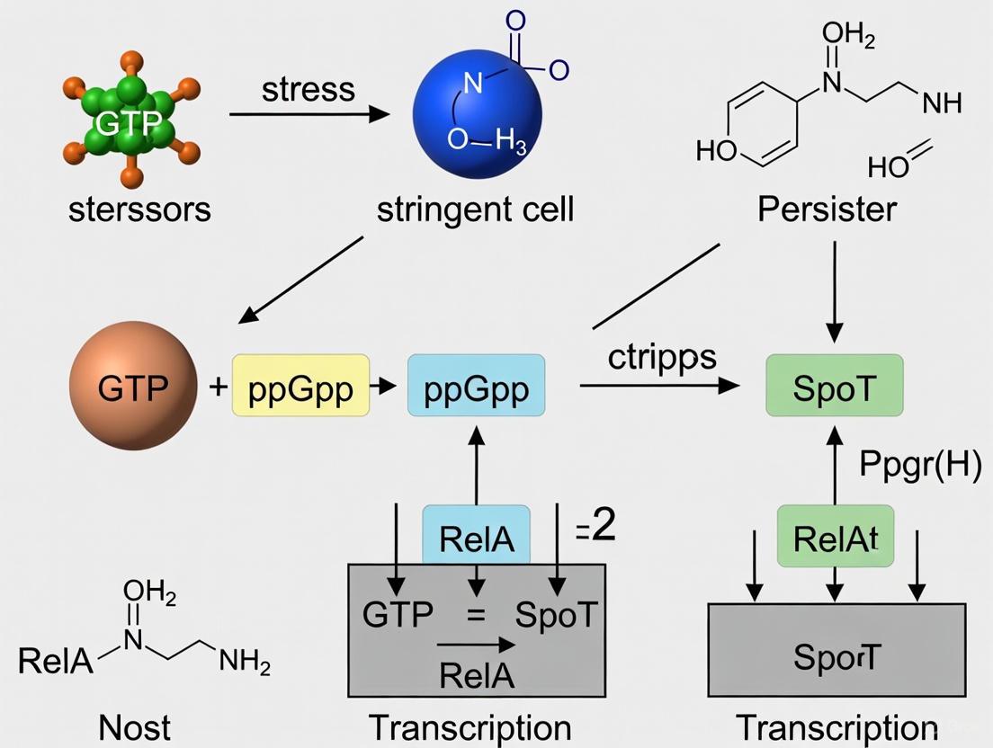
(Figure: The (p)ppGpp Signaling Pathway. This diagram illustrates the molecular events from stress sensing to persister formation, highlighting the positive feedback regulation of RelA activation.)
Methodologies for Studying (p)ppGpp and Stringent Response
Experimental Systems for Inducing Stringent Response
Several well-established experimental approaches enable controlled induction of (p)ppGpp accumulation to study the stringent response:
Amino Acid Analogue Treatment: Serine hydroxamate (SHX) inhibits seryl-tRNA synthetase, triggering RelA-dependent (p)ppGpp accumulation. This system produces dose-dependent effects, with higher SHX concentrations (100-1000 μM) generating graded increases in (p)ppGpp levels and corresponding transcriptional changes [4] [8]. SHX treatment at 500 μM typically induces an "intermediate" stringent response affecting approximately 20% of the genome [4].
Valine-Induced Isoleucine Starvation: In E. coli K-12 strains (which harbor an ilvG mutation), excess valine inhibits acetohydroxy acid synthase, specifically blocking isoleucine biosynthesis. This approach enables study of proteome remodeling during starvation as protein synthesis remains possible [5].
Temperature-Sensitive tRNA Synthetase Mutants: Strains carrying valS(ts) alleles exhibit defective valyl-tRNA synthetase activity at elevated temperatures, causing accumulation of uncharged tRNA and RelA-dependent (p)ppGpp synthesis. Shifting cultures from 30°C to 36.6-42°C induces (p)ppGpp increases of approximately 9-16 fold [9].
Inducible RelA Expression: Constitutively active RelA* mutants can be induced (e.g., with IPTG) to directly stimulate (p)ppGpp synthesis independent of starvation signals, enabling precise control over alarmone levels [3].
Quantitative Measurement and Single-Cell Analysis
Advanced methodologies enable comprehensive analysis of stringent response dynamics:
Nucleotide Extraction and Chromatography: Traditional thin-layer chromatography (TLC) or modern HPLC approaches quantitatively measure intracellular (p)ppGpp pools. Studies demonstrate basal (p)ppGpp levels increase 1.3-1.5 fold during mild to acute stringent response [4].
Quantitative Proteomics: 4D-label-free proteomic approaches capture approximately 2600 E. coli proteins, enabling quantification of proteome resource re-allocation during stress adaptation [3].
Single-Cell Fluorescence Microscopy: Reporter systems enable real-time tracking of stringent response parameters in individual cells:
- RpoS-mCherry fusions report (p)ppGpp induction (approximately 10-fold increase under non-permissive conditions) [9]
- QUEEN-7μ biosensor monitors ATP concentrations (dynamic range: 0.05-10 mM) [9]
- Unstable fluorescent proteins (e.g., YFPunstable) under control of TA module promoters track toxin-antitoxin system activation [9]
Table: Quantitative Effects of (p)ppGpp Accumulation on Bacterial Physiology
| (p)ppGpp Level | Growth Rate Reduction | Transcriptomic Changes | Proteomic Reallocation | Phenotypic Outcomes |
|---|---|---|---|---|
| Mild Increase (~1.3-fold) | ~60% of maximal rate [4] | ~4% of genome (227 DEGs) [4] | Initial shift from ribosomes to metabolism [3] | Motility suppression, reduced pyocyanin [4] |
| Intermediate Increase (~1.4-fold) | ~30% of maximal rate [4] | ~20% of genome (1197 DEGs) [4] | Significant ribosome downregulation, amino acid biosynthesis upregulation [3] | Biofilm promotion, virulence downregulation [4] |
| Acute Increase (>1.5-fold) | ~20% of maximal rate [4] | ~25% of genome (1508 DEGs) [4] | Profound metabolic restructuring [3] [5] | Antibiotic tolerance, persistence [2] [4] |
(p)ppGpp and Persister Cell Formation
Molecular Connections to Bacterial Persistence
Persister cells represent a transient, non-growing subpopulation that exhibits remarkable tolerance to antibiotic treatment without genetic resistance. Substantial evidence implicates (p)ppGpp as a central regulator of persister formation through multiple interconnected mechanisms:
Growth Rate Control: (p)ppGpp-mediated growth arrest is a fundamental prerequisite for persistence. By inhibiting rRNA transcription and ribosomal biogenesis, (p)ppGpp actively suppresses growth, creating a state less vulnerable to bactericidal antibiotics [2] [3].
Toxin-Antitoxin System Regulation: Early models proposed that (p)ppGpp activates TA modules through transcriptional and post-translational mechanisms. However, recent single-cell studies question whether previously implicated TA modules (e.g., RelBE) are critical for persistence under natural conditions [9].
Transcriptional Reprogramming: Graded increases in (p)ppGpp levels produce layered transcriptional changes, with up to 25% of the P. aeruginosa genome differentially regulated at maximal levels. This reprogramming suppresses virulence factors and motility while enhancing stress adaptation and survival networks [4] [8].
Biofilm Enhancement: (p)ppGpp promotes biofilm formation through upregulation of exopolysaccharide production and adhesion factors, creating protected environments where persister cells are enriched [4].
Ribosome Dimerization: The "ppGpp ribosome dimerization persister (PRDP) model" proposes that (p)ppGpp contributes to ribosome hibernation through dimerization, reducing protein synthesis and antibiotic target availability [10].
Single-Cell Dynamics and Stochasticity
Cutting-edge single-cell analyses have revealed the stochastic nature of persister formation. When E. coli populations experience valS(ts)-induced tRNA charging limitation, only a small fraction of cells (3-4 orders of magnitude higher than baseline) become antibiotic-tolerant despite uniform stress exposure [9]. Notably, these persisters do not exhibit markedly higher (p)ppGpp levels than their non-persister siblings, suggesting that the transition involves molecular noise in the downstream regulatory circuits rather than differential alarmone accumulation [9].
This stochasticity may be explained by bet-hedging strategies, where clonal populations diversify phenotypically to ensure some members survive unpredictable environmental challenges [9]. The graded transcriptional response to (p)ppGpp creates a continuum of physiological states, with persisters representing an extreme along this spectrum [4].
The Scientist's Toolkit: Essential Research Reagents
Table: Key Reagents for (p)ppGpp and Persistence Research
| Reagent / Tool | Function / Application | Key Characteristics & Examples |
|---|---|---|
| Serine Hydroxamate (SHX) | Chemical inducer of stringent response | Inhibits seryl-tRNA synthetase; Dose-dependent effects (100-1000 μM) [4] [8] |
| ValS(ts) Strains | Genetic system for stringent response | Temperature-sensitive valyl-tRNA synthetase; Induces (p)ppGpp at 36.6-42°C [9] |
| RelA* Overexpression | Direct (p)ppGpp synthesis | Constitutively active RelA mutant; IPTG-inducible [3] |
| relA/spoT Deletion | Stringent response deficiency | (p)ppGpp-null strains (ppGpp0); Multiple amino acid auxotrophy [3] [5] |
| RpoS-mCherry Reporter | (p)ppGpp signaling reporter | ~10-fold fluorescence increase under stringent conditions [9] |
| QUEEN-7μ Biosensor | ATP concentration monitoring | FRET-based ATP sensor; Range: 0.05-10 mM [9] |
| Promoter-YFPunstable | TA module activation tracking | Reports promoter activity with minimal signal persistence [9] |
| 4D-Label-Free Proteomics | Global protein quantification | Captures ~2600 E. coli proteins; Measures resource allocation [3] |
Therapeutic Implications and Future Perspectives
The central role of (p)ppGpp in bacterial persistence makes it an attractive target for novel antimicrobial strategies. Innovative treatments targeting (p)ppGpp metabolism are emerging as candidates for effective anti-persistence agents [2]. Potential approaches include:
RSH Enzyme Inhibitors: Small molecules targeting the synthetase or hydrolase activities of RelA/SpoT could modulate (p)ppGpp levels, potentially sensitizing bacteria to conventional antibiotics [2].
Stringent Response Disruptors: Compounds that interfere with (p)ppGpp signaling effectors, particularly its interaction with RNAP, could prevent the transcriptional reprogramming essential for persistence [4].
Combination Therapies: Adjuvants that suppress (p)ppGpp-mediated survival pathways alongside traditional antibiotics could potentially eradicate persistent infections [2] [4].
The graded nature of (p)ppGpp signaling reveals a sophisticated regulatory system where response intensity matches stress severity [4]. This layered control mechanism ensures appropriate resource investment in survival strategies, highlighting the evolutionary optimization of bacterial stress adaptation. Future research delineating the molecular basis of stochastic persister formation within heterogeneous populations will be crucial for developing effective countermeasures against antibiotic tolerance.
Experimental data and citations derived from provided search results.
The RelA/SpoT Homolog (RSH) superfamily comprises the essential enzymes that govern the bacterial stringent response, a universal adaptative mechanism to stress and nutrient limitation. These enzymes regulate cellular levels of the alarmones guanosine tetraphosphate and pentaphosphate, collectively known as (p)ppGpp, which act as master regulators of bacterial physiology [11] [12]. When faced with stressors such as amino acid starvation, fatty acid limitation, or osmotic shock, a rapid increase in (p)ppGpp concentration rewires the bacterial transcriptome and metabolism. This re-prioritization halts growth-intensive processes like ribosome biogenesis and division, and activates survival pathways, enabling the bacterium to endure the hostile condition [11] [4]. Beyond survival, this response is critically linked to virulence, biofilm formation, and—most importantly in the context of therapeutic challenges—the formation of antibiotic-tolerant persister cells [11] [13] [12]. Persisters are a sub-population of genetically susceptible, non-growing or slow-growing bacteria that survive antibiotic exposure and can lead to chronic, relapsing infections [13]. Understanding the RSH-mediated stringent response is therefore paramount for developing novel strategies to combat persistent infections.
Classification and Genomic Distribution of RSH Proteins
RSH enzymes are categorized based on their domain architecture and functionality. A comprehensive phylogenetic analysis classifies them into three main groups comprising 30 subgroups, providing a unifying terminology for the field [14].
Table 1: Classification of Major RSH Enzymes
| RSH Category | Key Domains | Functionality | Representative Examples | Genomic Distribution |
|---|---|---|---|---|
| Long RSHs | SYNTH, HD, TGS, ACT | Bifunctional (synthesis & hydrolysis) or monofunctional | Rel (e.g., in B. subtilis), RelA (synthase-only in E. coli), SpoT (hydrolysis-predominant in E. coli) | Nearly all bacteria; plant chloroplasts [14] [15] |
| Small Alarmone Synthetases (SAS) | SYNTH | Monofunctional (synthesis only) | RelP (SAS2), RelQ (SAS1) in Firmicutes; ToxSAS in TA modules | Widespread across disparate bacteria [16] [14] |
| Small Alarmone Hydrolases (SAH) | HD | Monofunctional (hydrolysis only) | Mesh1 (metazoans) | Animals; some bacteria [14] |
Long RSHs, such as the bifunctional Rel protein found in Staphylococcus aureus and Bacillus subtilis, contain both synthetase (SYNTH) and hydrolase (HD) domains, alongside regulatory TGS and ACT domains in their C-terminal region [15] [14]. In contrast, β- and γ-proteobacteria like Escherichia coli and Pseudomonas aeruginosa possess two long RSHs resulting from a gene duplication event: the monofunctional synthetase RelA and the bifunctional SpoT, which primarily performs hydrolysis [11] [14]. Small RSHs are single-domain proteins that specialize in either synthesis (SASs) or hydrolysis (SAHs). SASs, including RelP and RelQ, allow bacteria to fine-tune (p)ppGpp production in response to specific, localized stresses [14]. Notably, some SASs are encoded in toxin-antitoxin (TA) operons, termed ToxSAS, where their uncontrolled alarmone synthesis acts as a toxin, inhibiting growth and being neutralized by a cognate antitoxin [16].
Molecular Mechanisms of (p)ppGpp Synthesis and Signaling
The core reaction of alarmone synthesis is conserved across RSH synthetases. These enzymes use ATP as a pyrophosphate donor, transferring it to the 3' hydroxyl group of GDP or GTP to produce ppGpp or pppGpp, respectively [12]. pppGpp is often rapidly converted to ppGpp by specific phosphatases. The molecular regulation of this activity, however, differs between long and small RSHs. Long RSHs are allosterically regulated. For instance, RelA from E. coli is directly activated by binding to the ribosome when uncharged tRNA accumulates in the A-site during amino acid starvation [11]. In Firmicutes like S. aureus, the bifunctional RSH enzyme's activity is regulated by conformational shifts between synthase-ON/hydrolase-OFF and synthase-OFF/hydrolase-ON states [15]. Small SASs like RelP and RelQ provide a secondary, ribosome-independent layer of (p)ppGpp production, allowing for a nuanced and robust stress response [14].
Once synthesized, (p)ppGpp exerts its pleiotropic effects through two primary mechanisms, depending on the bacterial phylum. In Gammaproteobacteria like E. coli and P. aeruginosa, (p)ppGpp binds directly to the RNA polymerase in concert with the cofactor DksA, profoundly rewiring transcription to repress growth-related genes (e.g., for ribosome biogenesis) and activate stress survival genes [11] [4]. In Firmicutes and Actinobacteria (e.g., B. subtilis, S. aureus, M. tuberculosis), the primary effect is through indirect transcriptional regulation. (p)ppGpp synthesis leads to a drastic reduction in the cellular GTP pool by inhibiting GTP synthesis. Since many promoters, including those for rRNA genes, require GTP for initiation, this results in growth arrest. The drop in GTP also inactivates GTP-binding repressors like CodY, leading to derepression of amino acid biosynthesis and virulence genes [15].
The Critical Role of RSH and (p)ppGpp in Persister Cell Formation
Persister cells are non-growing or slow-growing, genetically susceptible bacterial cells that exhibit transient, high-level tolerance to antibiotics. They are a major culprit in chronic and relapsing infections, as they can resume growth after antibiotic treatment ends [13]. The RSH-mediated stringent response is one of the most important molecular mechanisms underlying persistence.
Accumulation of (p)ppGpp triggers a global slowdown of bacterial metabolism and growth, which is the fundamental basis for antibiotic tolerance since most antibiotics target active cellular processes [11] [13]. In P. aeruginosa, (p)ppGpp production is not a binary switch but a graded response relative to stress severity. Transcriptomic studies show that as (p)ppGpp levels rise, an increasing number of genes are differentially regulated, initially repressing motility and metabolism and, at higher levels, upregulating biofilm-associated genes and impairing antibiotic efficacy [4]. This graded response directly translates to increased tolerance, particularly in biofilms where nutrient limitation naturally induces the stringent response [11] [4]. Furthermore, (p)ppGpp is crucial for the survival of intracellular pathogens. For example, Salmonella enterica residing within acidified macrophage vacuoles requires (p)ppGpp production to persist [11]. In S. aureus, the SAS enzymes RelP and RelQ are key contributors to persister formation, as their genetic knockout or pharmacological inhibition significantly reduces persister counts under antibiotic stress [17].
Quantitative Data and Experimental Analysis of the Stringent Response
Quantifying the Graded (p)ppGpp Response
Research on P. aeruginosa has demonstrated that the stringent response is a finely tuned, dose-dependent system. The following table summarizes key quantitative findings on how graded (p)ppGpp levels correlate with transcriptional changes and phenotypic outcomes [4].
Table 2: Graded (p)ppGpp Response in Pseudomonas aeruginosa PA14
| Parameter | Mild Stringent Response | Intermediate Stringent Response | Acute Stringent Response |
|---|---|---|---|
| Inducing Signal | 100 µM SHX | 500 µM SHX | 1000 µM SHX |
| Growth Rate (doublings/h) | 0.4 | 0.26 | Severe perturbation |
| (p)ppGpp Increase (fold) | 1.33 | 1.39 | 1.48 |
| Differentially Expressed Genes (DEGs) | 227 (~4% of genome) | 1197 (~20% of genome) | 1508 (~25% of genome) |
| Key Affected Processes | Motility suppression, metabolic slowdown | Virulence gene downregulation, quorum sensing upregulation | Ribosome biogenesis downregulation, compact biofilm formation |
| Antibiotic Tolerance | Induced | Induced | Highly induced |
Key Experimental Protocol: Assessing the Role of RSH in Persistence
To investigate the role of specific RSH enzymes, such as the SAS RelP in S. aureus, researchers often employ genetic and pharmacological approaches coupled with persister assays [17].
Objective: To determine the effect of a relP knockout or inhibition on persister cell formation in S. aureus under antibiotic stress.
Methodology:
- Strain Construction: Create an isogenic relP knockout mutant (e.g., ΔrelP) from a wild-type S. aureus strain (e.g., HG001) using homologous recombination with an allelic replacement vector.
- Compound Treatment (Pharmacological inhibition): Prepare cultures of wild-type and mutant strains. Pre-treat wild-type cultures with a candidate inhibitor (e.g., 80-160 µM diosgenin) or a vehicle control for 3 hours during mid-exponential growth [17].
- Persister Assay: Expose the pre-treated cultures and the ΔrelP mutant to a high concentration (e.g., 10x MIC) of different classes of bactericidal antibiotics (e.g., oxacillin, ciprofloxacin, gentamicin) for a set period (e.g., 3-24 hours).
- Viability Quantification: After antibiotic exposure, wash the cells to remove the drug, serially dilute, and spot them onto drug-free agar plates. Count the resulting Colony-Forming Units (CFU) after incubation.
- Data Analysis: The persister fraction is calculated as (CFU after antibiotic treatment / CFU before antibiotic treatment) × 100%. A significant reduction in the persister fraction in the ΔrelP mutant or diosgenin-treated wild-type compared to the untreated wild-type control implicates RelP in persister formation.
Research Toolkit: Reagents and Therapeutic Strategies
Essential Research Reagents
The following table lists key reagents used in stringent response and persister research, as identified from the cited literature.
Table 3: Key Research Reagents for Stringent Response and Persister Studies
| Reagent / Tool | Function / Description | Example Application |
|---|---|---|
| Serine Hydroxamate (SHX) | Serine analogue that inhibits seryl-tRNA synthetase, inducing amino acid starvation and RelA-dependent (p)ppGpp synthesis. | Used to experimentally induce a graded stringent response in P. aeruginosa for transcriptomic studies [4]. |
| Diosgenin | A natural steroidal saponin that inhibits (p)ppGpp synthesis by downregulating expression of relP and relQ in S. aureus. | Used as a pre-treatment (80-160 µM) to suppress persister cell formation by disrupting the metabolic pathway to dormancy [17]. |
| Relacin | A ppGpp analogue designed to competitively inhibit (p)ppGpp synthetases. | Shown to limit (p)ppGpp production, impede entry into stationary phase, and inhibit biofilm formation in Gram-positive bacteria like B. subtilis [12]. |
| ppGpp Analogues (AC/AB) | Synthetic ppGpp analogues with modified phosphate and base groups. | Demonstrated to inhibit RelMsm enzyme activity in M. smegmatis in vitro and reduce bacterial survival under stress [12]. |
| S. aureus HG001 | A well-characterized laboratory strain with a restored RsbU factor, making it a standard model for S. aureus research. | Used to generate isogenic RSH mutants (e.g., rshsyn) for studying the role of RSH synthase activity in virulence and persistence [15]. |
Strategies for Targeting the Stringent Response
The pivotal role of (p)ppGpp in persistence and virulence makes the RSH system an attractive therapeutic target. Current strategies focus on inhibiting its synthesis to disarm bacterial survival mechanisms [12].
- Direct Synthetase Inhibition: The most straightforward approach is developing small-molecule inhibitors that target the active site of (p)ppGpp synthetases. Relacin and other ppGpp analogues (e.g., AC/AB compounds) compete with GDP/GTP for binding, showing efficacy primarily in Gram-positive bacteria [12].
- Indirect Inhibition via Metabolic Disruption: Compounds like diosgenin exhibit a dual mechanism. They downregulate the expression of SAS genes (relP and relQ) and reduce intracellular ATP levels, which is a required substrate for (p)ppGpp synthesis. This metabolic suppression prevents the induction of the dormant persister state [17].
- Combination Therapies: Given that persisters lead to treatment failure, combining conventional antibiotics with an anti-(p)ppGpp agent is a promising strategy. This approach aims to kill the actively growing population with the antibiotic while simultaneously preventing the surviving sub-population from entering a protected, dormant state by blocking the stringent response. This has been demonstrated with relacin in combination with metronidazole against Clostridioides difficile [12].
The bacterial stringent response is a universal adaptive mechanism for survival under stress, centrally governed by the alarmone (p)ppGpp. This in-depth technical review elucidates the diverse environmental cues—extending beyond canonical amino acid starvation—that trigger this response, detailing the molecular machinery involved and its profound implications for bacterial persistence. Within the broader context of persister cell formation research, we frame the (p)ppGpp-mediated stringent response as a critical regulator of the dormant, multidrug-tolerant state that complicates the treatment of chronic infections. This whitepaper provides a synthesis of current mechanistic understanding, complete with structured quantitative data, experimental methodologies for key assays, and visualizations of core signaling pathways, serving as a resource for researchers and drug development professionals aiming to overcome antibiotic tolerance.
The stringent response is a highly conserved global regulatory network that allows bacteria to rapidly reprogram their physiology in response to perceived stress or nutrient limitation [11]. This response is orchestrated by the rapid intracellular accumulation of the alarmones guanosine tetraphosphate (ppGpp) and guanosine pentaphosphate (pppGpp), collectively known as (p)ppGpp or "magic spot" [11] [18]. The fundamental outcome of this signaling cascade is a dramatic shift in gene expression, favoring stress survival and repair pathways while repressing energy-costly processes related to growth and replication, such as rRNA and tRNA synthesis [11]. This reallocation of resources enables bacteria to withstand adverse conditions.
Critically, within the framework of persister cell research, this same physiological shift into a state of reduced metabolic activity and growth arrest is a primary mechanism of antibiotic tolerance. Persisters are defined as a subpopulation of genetically susceptible cells that enter a transient, dormant state, allowing them to survive exposure to high concentrations of bactericidal antibiotics [19] [13]. Upon removal of the antibiotic pressure, these cells can resume growth and repopulate a susceptible community, leading to recurrent and chronic infections [20] [13]. The stringent response is not merely one of several pathways to persistence; substantial evidence positions (p)ppGpp as a central regulator of multidrug tolerance, acting as a master switch that integrates various environmental and internal signals to induce the persistent phenotype [11] [18]. Understanding the triggers and molecular execution of the stringent response is therefore paramount to developing novel therapeutic strategies against persistent infections.
Molecular Mechanisms: The Core Players of the Stringent Response
The synthesis and degradation of (p)ppGpp are managed by enzymes belonging to the RelA/SpoT homolog (RSH) family. The composition and regulation of this system differ notably between Gram-negative and Gram-positive bacteria, a key consideration for targeted drug development.
RelA and SpoT in Gram-Negative Bacteria (e.g., E. coli): In model organisms like E. coli, the system is characterized by two principal enzymes. RelA is a ribosome-associated (p)ppGpp synthetase I that is directly activated by the presence of uncharged tRNA in the A-site—a direct signal of amino acid starvation [11]. SpoT, a bifunctional enzyme, possesses weak (p)ppGpp synthetase II activity but primarily functions as a hydrolase, degrading (p)ppGpp to maintain homeostasis. SpoT responds to a broader range of stresses, including fatty acid and carbon starvation [11].
Rel and SAS/SAH in Gram-Positive Bacteria (e.g., Staphylococcus aureus): Many Gram-positive bacteria possess a single, long RSH protein, Rel, which contains both synthetase and hydrolase domains [11] [17]. Additionally, they often encode small, single-function enzymes known as Small Alarmone Synthetases (SAS), such as RelP and RelQ in S. aureus [17]. These SAS proteins lack hydrolase activity and contribute to (p)ppGpp production in response to distinct, often non-nutritional, stresses.
The primary molecular target of (p)ppGpp is the RNA polymerase (RNAP). By binding directly to the RNAP, (p)ppGpp induces an allosteric change that severely dampens the transcription of genes related to rapid growth, including those for ribosome biogenesis [11]. Concurrently, it upregulates transcription of stress response and amino acid biosynthesis genes. This global rewiring of gene expression, frequently in conjunction with the activation of toxin-antitoxin (TA) modules that further inhibit cellular processes, leads to metabolic quiescence and dormancy—the hallmarks of a persister cell [11] [18].
Signaling Pathway Visualization
The following diagram illustrates the core molecular pathway of the stringent response, from initial stress cues to the formation of a persister cell.
Environmental Triggers of the Stringent Response
While amino acid starvation is the canonical trigger for (p)ppGpp production via RelA, research has revealed a much wider spectrum of inducers. The table below categorizes and quantifies the diverse environmental cues that can activate the stringent response, contributing to persister formation.
Table 1: Environmental Cues Triggering the Stringent Response and Persister Formation
| Trigger Category | Specific Cue | Key Sensor/Effector | Impact on (p)ppGpp & Persistence |
|---|---|---|---|
| Nutrient Starvation | Amino Acid Deprivation | RelA (on ribosome) | Rapid, strong (p)ppGpp surge; core persister trigger [11]. |
| Carbon Source/Fatty Acid Limitation | SpoT / Rel | Induces (p)ppGpp synthesis; linked to biofilm persistence [11]. | |
| Physicochemical Stresses | pH Downshift (Acid Stress) | SpoT / SAS | (p)ppGpp accumulation; promotes survival in macrophages [11]. |
| Osmotic Shock | SpoT / Rel | Induces (p)ppGpp; associated with general stress tolerance [11]. | |
| Temperature Shift (Heat/Cold Shock) | SpoT / Rel | Alarmsone accumulation; increases persister frequency [11]. | |
| Host-Associated & Other Stresses | Intracellular Environment (e.g., within macrophage vacuoles) | Rel / SAS | (p)ppGpp required for Salmonella persistence in macrophages [11]. |
| Sub-inhibitory Antibiotic Exposure | Multiple | Various antibiotics can indirectly induce the stringent response [20]. | |
| Oxygen Variation / Oxidative Stress | SpoT / SAS | (p)ppGpp accumulation in response to redox changes [11]. |
The diversity of these inducers underscores the role of the stringent response as a general stress alarm system. For instance, the ability to respond to the acidic environment within a macrophage phagosome is a key virulence feature for intracellular pathogens like Salmonella enterica and Mycobacterium tuberculosis [11] [13]. Furthermore, the nutrient gradients and microenvironments within a biofilm create constant, localized triggers for the stringent response, explaining the high frequency of persisters in these structured communities [11] [19].
Experimental Analysis: Measuring the Stringent Response and Its Outcomes
Studying the stringent response and its link to persistence requires robust, quantitative methodologies. Below is a detailed protocol for a key experiment demonstrating the induction of the stringent response and its functional consequence—antibiotic tolerance.
Detailed Protocol: Inducing and Quantifying (p)ppGpp-Mediated Persistence
Objective: To trigger the stringent response via serine hydroxamate (a non-metabolizable analog that causes serine starvation and uncharged tRNA accumulation) and quantify the resulting tolerance to a fluoroquinolone antibiotic (e.g., ciprofloxacin) in E. coli.
Principle: Serine hydroxamate induces amino acid starvation, activating RelA and causing (p)ppGpp accumulation. This reprograms cells into a dormant, persistent state. Subsequent treatment with ciprofloxacin, which targets DNA gyrase in growing cells, will kill the normal population but spare the non-growing persisters.
Materials & Reagents:
- Bacterial Strain: Wild-type E. coli (e.g., MG1655) and an isogenic ΔrelAΔspoT double mutant as a negative control.
- Inducer: L-Serine hydroxamate (Sigma-Aldrich), prepared as a sterile-filtered stock solution in water.
- Antibiotic: Ciprofloxacin hydrochloride, prepared as a stock solution in water or DMSO.
- Growth Media: M9 minimal glucose medium to control nutrient availability.
- Equipment: Spectrophotometer for measuring optical density at 600 nm (OD₆₀₀), shaking incubator, water bath, microcentrifuge, and equipment for serial dilution and plating (CFU assay).
Procedure:
- Culture Preparation: Inoculate E. coli strains from a single colony into 5 mL of M9 minimal medium and grow overnight (~16 hours) at 37°C with shaking.
- Sub-culture Dilution: Dilute the overnight culture 1:100 into fresh, pre-warmed M9 medium and grow to mid-exponential phase (OD₆₀₀ ≈ 0.4-0.5).
- Stringent Response Induction: Split the exponential-phase culture into two flasks.
- Experimental Group: Add serine hydroxamate to a final concentration of 0.5 - 1.0 mg/mL.
- Control Group: Add an equivalent volume of sterile water.
- Incubate both cultures for 1 hour at 37°C with shaking.
- Antibiotic Challenge:
- After induction, take a sample (time = 0 h) for CFU determination.
- Add ciprofloxacin to both cultures at a final concentration of 10x the MIC (e.g., 5-10 µg/mL for E. coli).
- Continue incubation. Take 1 mL samples at predetermined time points (e.g., 1h, 2h, 3h, 5h) post-antibiotic addition.
- Viability Assessment (CFU Count):
- Wash the samples 1x in sterile phosphate-buffered saline (PBS) to remove residual antibiotic.
- Perform serial 10-fold dilutions in PBS.
- Spot plate appropriate dilutions onto LB agar plates without antibiotics.
- Incubate plates overnight at 37°C and count colonies the next day.
- Data Analysis:
- Calculate the survival fraction at each time point as (CFU/mL at time t) / (CFU/mL at time 0 of antibiotic challenge).
- Plot the survival fraction over time to generate a biphasic killing curve, characteristic of persister cells. The initial rapid kill represents the normal population, while the plateau indicates the surviving persister subpopulation.
- Expected Outcome: The wild-type strain pre-treated with serine hydroxamate will show a significantly higher persister fraction (survival plateau) compared to the un-induced control and the ΔrelAΔspoT mutant, which will be largely eradicated.
The Scientist's Toolkit: Key Research Reagents
The following table lists essential materials and their functions for researching the stringent response and bacterial persistence.
Table 2: Essential Reagents and Tools for Stringent Response Research
| Reagent / Tool | Function / Utility in Research | Example |
|---|---|---|
| Amino Acid Analogs | Chemically induces amino acid starvation by causing tRNA uncharging; a direct, RelA-dependent trigger. | L-Serine Hydroxamate [11] |
| relA/spoT Mutants | Genetic controls to dissect the contribution of specific synthases/hydrolases to the response. | E. coli ΔrelA, ΔspoT, or ΔrelAΔspoT [11] |
| (p)ppGpp Antibodies | Enable detection and semi-quantification of intracellular alarmone levels via immunoassays. | Commercial monoclonal antibodies |
| Thin-Layer Chromatography (TLC) | The gold-standard method for direct separation, visualization, and quantification of radiolabeled (p)ppGpp. | P³² or H³-labeled nucleotide precursors |
| ATP Assay Kits (Luminescence) | Quantify intracellular ATP as a proxy for metabolic activity and the dormant state of persisters. | Commercial kits (e.g., BacTiter-Glo) [17] |
| Microfluidic Systems | Enable single-cell analysis and real-time observation of persister formation and resuscitation. | CellASIC ONIX system [13] |
Therapeutic Targeting of the Stringent Response to Combat Persistence
Given its central role in persistence, the (p)ppGpp-mediated stringent response represents a promising target for novel antimicrobial adjuvants. The goal is not necessarily to kill bacteria but to disrupt the dormancy program, thereby re-sensitizing persisters to conventional antibiotics.
One innovative strategy is the "wake-and-kill" approach, which involves using metabolites or other compounds to reactivate the metabolism of dormant persisters, making them vulnerable again to antibiotics [20]. For example, exogenous sugars like mannitol or metabolites like pyruvate have been shown to rejuvenate bacterial metabolism and restore the efficacy of aminoglycoside antibiotics [20].
A more direct approach involves identifying inhibitors of the (p)ppGpp synthetases themselves. Recent research on natural compounds has shown promise; for instance, the plant-derived saponin diosgenin was found to significantly reduce persister formation in Staphylococcus aureus by downregulating the expression of the SAS genes relP and relQ, leading to reduced (p)ppGpp synthesis [17]. In this study, pre-treatment with 160 µM diosgenin reduced persister fractions by 87-94% under antibiotic stress, demonstrating the therapeutic potential of this targeted inhibition [17].
Therapeutic Strategy Visualization
The following diagram outlines the logical flow of therapeutic strategies that target the stringent response to eradicate bacterial persisters.
The bacterial stringent response, initiated by a diverse array of environmental cues and centrally mediated by (p)ppGpp, is a master regulator of the persister phenotype—a major clinical obstacle in treating chronic and biofilm-associated infections. This technical guide has detailed the molecular mechanisms, key triggers, and essential experimental approaches for investigating this critical survival pathway. The ongoing research into therapeutic interventions, particularly the development of (p)ppGpp synthesis inhibitors and metabolite-based "wake-and-kill" adjuvants, holds significant promise for the future of antimicrobial therapy. By preventing or reversing the dormant state of persisters, these strategies aim to enhance the efficacy of existing antibiotics and ultimately improve outcomes for patients suffering from recalcitrant bacterial infections.
The bacterial stringent response, mediated by the alarmones guanosine tetraphosphate (ppGpp) and guanosine pentaphosphate (pppGpp), collectively known as (p)ppGpp, represents a fundamental survival mechanism that orchestrates global physiological rewiring in response to environmental stress. This sophisticated regulatory system enables bacteria to transition from active growth to a protected, dormant state by implementing coordinated transcriptional shifts, growth arrest, and metabolic downregulation [4] [12]. As research into antibiotic tolerance intensifies, understanding the role of (p)ppGpp in persister cell formation has become paramount. Persisters constitute a subpopulation of metabolically dormant bacterial cells that exhibit transient antibiotic tolerance without genetic resistance, contributing significantly to chronic and recurrent infections that evade conventional treatments [20] [17]. The (p)ppGpp-mediated stringent response serves as the central molecular switch that reprogrammes cellular physiology toward this persistent state, making it a critical focus for therapeutic interventions aimed at combating recalcitrant bacterial infections [12].
Molecular Mechanisms of (p)ppGpp Signaling
Alarmone Synthesis and Turnover
The stringent response is governed by the RelA-SpoT Homologue (RSH) family of enzymes, which control the synthesis and degradation of (p)ppGpp. In β- and γ-proteobacteria like Escherichia coli and Pseudomonas aeruginosa, this system typically consists of the synthetase RelA and the bifunctional synthetase/hydrolase SpoT [4] [21]. RelA is primarily activated by binding deacylated tRNA during amino acid starvation, while SpoT responds to diverse stresses including nutrient limitation, oxidative stress, and membrane damage [4] [12]. In Firmicutes such as Staphylococcus aureus and Bacillus subtilis, (p)ppGpp metabolism involves bifunctional RSH enzymes along with small alarmone synthetases (SASs) like RelP and RelQ [17] [12]. These enzymes maintain precise cellular (p)ppGpp levels, which determine the extent of physiological rewiring and growth modulation.
Downstream Molecular Targets
(p)ppGpp exerts its pleiotropic effects through two primary mechanisms. In proteobacteria, it directly binds to RNA polymerase together with its co-factor DksA, dramatically altering transcriptional profiles by both inhibiting and activating distinct sets of genes [4] [12]. Concurrently, across bacterial species, (p)ppGpp directly binds and modulates numerous metabolic enzymes, particularly those involved purine biosynthesis, to redirect metabolic flux [22] [12]. This dual regulatory strategy enables simultaneous control of gene expression and metabolic activity, ensuring coordinated physiological adaptation to stress conditions.
Quantitative Transcriptional Reprogramming
Graded Response to Stress Severity
Research has revealed that the (p)ppGpp-mediated transcriptional response is not a binary on/off switch but rather a graded system that proportionally adjusts gene expression based on stress severity. In Pseudomonas aeruginosa, exposure to increasing concentrations of serine hydroxamate (SHX), which induces amino acid starvation, results in corresponding increases in (p)ppGpp accumulation and progressively extensive transcriptional reprogramming [4]. Under mild stringent response conditions (100 μM SHX), approximately 4% of the genome (227 genes) shows differential expression. This expands to 20% (1,197 genes) under intermediate conditions (500 μM SHX), and reaches 25% of the genome (1,508 genes) under acute stringent response (1,000 μM SHX) [4]. This demonstrates a layer-by-layer engagement of the transcriptome, where both the number of regulated genes and the magnitude of expression changes scale with alarmone levels.
Functional Enrichment of Regulated Genes
The transcriptional shifts orchestrated by (p)ppGpp consistently downregulate growth-related processes while activating survival pathways. Analysis of differentially expressed genes reveals suppression of ribosome biogenesis, flagellar assembly, multiple secretion systems (types I, II, III, and VI), oxidative phosphorylation, the TCA cycle, and biosynthesis pathways for fatty acids, peptidoglycan, and lipopolysaccharides [4]. Conversely, upregulated pathways include those involved in stress management, such as alginate and polysaccharide biosynthesis, fatty acid degradation, and aminoacyl-tRNA biosynthesis [4]. This systematic reallocation of cellular resources from growth to maintenance constitutes the fundamental transcriptional basis for persister cell formation.
Table 1: Transcriptional Reprogramming in P. aeruginosa Under Varying Stringent Response Conditions
| Stringent Response Level | SHX Concentration | Differentially Expressed Genes | Functional Categories Downregulated | Functional Categories Upregulated |
|---|---|---|---|---|
| Mild | 100 μM | 227 (∼4% of genome) | Motility systems, Pyocyanin production | Serine metabolism |
| Intermediate | 500 μM | 1,197 (∼20% of genome) | Metabolic pathways, Secretion systems | Stress response pathways |
| Acute | 1,000 μM | 1,508 (∼25% of genome) | Ribosome biogenesis, Virulence factors | Biofilm-related genes, Alginate production |
Metabolic Rewiring and Downregulation
Central Carbon Metabolism and Energy Management
The stringent response implements comprehensive metabolic rewiring to reduce energy consumption while maintaining essential functions. In Pseudomonas putida, (p)ppGpp accumulation triggers significant alterations in central carbon metabolism, characterized by increased concentrations of central carbon metabolites alongside sharply decreased intermediates in the purine biosynthesis pathway [22]. This metabolic reorganization facilitates redirection of resources from nucleotide synthesis for growth to maintenance activities. Extracellular accumulation of pyruvate and acetate observed during stringent response activation indicates a fundamental shift in carbon flux patterns [22]. These metabolic changes are directly linked to reduced intracellular ATP levels, as demonstrated in Staphylococcus aureus, where diosgenin treatment reduced ATP levels by 36-38% while simultaneously suppressing persister formation [17]. The diminished energy charge contributes directly to the metabolically quiescent state characteristic of persister cells.
Nucleotide Metabolism and Purine Pathway Regulation
A conserved feature of the stringent response across bacterial species is the targeted downregulation of purine biosynthesis. (p)ppGpp directly binds to and inhibits multiple enzymes in the purine pathway, including glutamine amidophosphoribosyltransferase (PurF), hypoxanthine phosphoribosyltransferase (Hpt), guanine phosphoribosyltransferase (Gpt), and inosine-guanosine kinase (Gsk) [22] [12]. This allosteric regulation rapidly constricts nucleotide precursor availability, contributing to growth arrest. Studies in P. putida confirm that (p)ppGpp is essential for purine pathway downregulation, with ΔrelA and ppGpp0 mutant strains failing to suppress purine metabolites under SHX-induced stress [22]. This targeted metabolic control complements transcriptional reprogramming to enforce the dormant state.
Table 2: Key Metabolic Alterations During Stringent Response-Induced Growth Arrest
| Metabolic Parameter | Observed Change | Functional Significance | Experimental System |
|---|---|---|---|
| Purine pathway intermediates | Sharp decrease | Limits nucleotide availability for replication and transcription | Pseudomonas putida [22] |
| Intracellular ATP levels | 36-38% reduction | Decreases energy charge, promotes metabolic quiescence | Staphylococcus aureus [17] |
| Central carbon metabolites | Increased concentration | Redirects carbon flux toward maintenance | Pseudomonas putida [22] |
| Pyruvate and acetate excretion | Extracellular accumulation | Indicates redirection of carbon flux | Pseudomonas putida [22] |
| Glucose uptake and utilization | Enhanced | Meets increased energy demands for stress adaptation | General bacterial response [23] |
Experimental Models and Methodologies
Inducing and Quantifying the Stringent Response
Serine Hydroxamate (SHX) Treatment Protocol
A widely established method for stringent response induction involves using serine hydroxamate (SHX), a serine analog that inhibits seryl-tRNA acylation. This leads to accumulation of deacylated tRNA, activating RelA-dependent (p)ppGpp synthesis [4] [22]. Standardized protocol: (1) Grow P. aeruginosa or E. coli cultures to mid-exponential phase (OD600 ≈ 0.4-0.6); (2) Add SHX at concentrations ranging from 100-1000 μM to create mild, intermediate, or acute stringent response; (3) Incubate for 30 minutes to several hours depending on experimental requirements; (4) Monitor growth arrest via optical density measurements and quantify (p)ppGpp accumulation using chromatographic methods [4]. The concentration-dependent effect of SHX yields half-maximal growth inhibition (IC50) at approximately 128 μM in P. aeruginosa PA14, providing a standardized framework for reproducible induction of graded stringent response [4].
Metabolomic Analysis of Stringent Response
Comprehensive metabolomic profiling provides insights into metabolic rewiring during stringent response. Methodology: (1) Rapid sampling of bacterial cultures (e.g., P. putida) during exponential growth and at specified intervals post-SHX treatment; (2) Immediate quenching of metabolism using cold methanol or specialized buffers like RNAprotect; (3) Intracellular metabolite extraction using appropriate solvent systems; (4) Quantitative analysis employing complementary platforms - NMR spectroscopy for extracellular metabolites and quantitative mass spectrometry for intracellular metabolites; (5) Data integration to identify significantly altered metabolic pathways [22]. This approach has revealed crucial (p)ppGpp-mediated metabolic shifts, particularly in purine and central carbon metabolism.
Assessing Persister Cell Formation
Persister cell levels are quantified by exposing bacterial populations to high concentrations of bactericidal antibiotics (typically 10× MIC) and determining surviving colony-forming units (CFUs). Standard procedure: (1) Pre-treat cultures with stringent response inducers or potential inhibitors; (2) Harvest cells during transition to stationary phase when persister formation peaks; (3) Challenge with antibiotics such as ciprofloxacin, oxacillin, or gentamicin for extended periods (3-24 hours); (4) Wash cells to remove antibiotics and plate on fresh media for CFU enumeration; (5) Calculate persister fractions as percentage of initial population surviving antibiotic exposure [17]. This method has demonstrated that diosgenin pre-treatment reduces S. aureus persister formation by 82-94% across different antibiotic classes [17].
The Scientist's Toolkit: Key Research Reagents
Table 3: Essential Research Reagents for Stringent Response and Persister Studies
| Reagent/Chemical | Function in Research | Example Application |
|---|---|---|
| Serine Hydroxamate (SHX) | Induces amino acid starvation by inhibiting seryl-tRNA acylation | Activation of RelA-dependent (p)ppGpp synthesis in P. aeruginosa and E. coli [4] [22] |
| Diosgenin | Natural compound that inhibits (p)ppGpp synthesis by downregulating relP/relQ | Suppression of persister cell formation in S. aureus [17] |
| Relacin | (p)ppGpp analog that inhibits (p)ppGpp synthetases | Limiting (p)ppGpp production in Gram-positive bacteria including B. subtilis [12] |
| ΔrelA/ppGpp0 mutant strains | Engineered strains unable to produce (p)ppGpp | Disruption of stringent response for mechanistic studies [22] |
| RNAprotect Bacteria Reagent | Stabilizes cellular RNA profiles immediately upon sampling | Transcriptomic analysis during bacterial stress response [24] |
Therapeutic Targeting of the Stringent Response
Anti-Persister Compounds and Strategies
The central role of (p)ppGpp in persister formation makes the stringent response an attractive therapeutic target. Multiple strategies have emerged to disrupt this survival pathway: (1) Direct inhibition of (p)ppGpp synthetases using analogs like relacin and related compounds that compete with GDP/GTP for active site binding [12]; (2) Modulation of alarmone hydrolysis to deplete (p)ppGpp pools; (3) Combinatorial approaches that pair conventional antibiotics with stringent response inhibitors [17] [12]. The natural compound diosgenin exemplifies this approach, demonstrating dual-action inhibition through downregulation of relP and relQ expression (reducing (p)ppGpp synthesis by up to 60%) and reduction of membrane fluidity, ultimately suppressing S. aureus persister formation by 82-94% across multiple antibiotic classes [17].
Metabolite-Induced Persister Resensitization
An alternative therapeutic strategy involves metabolic reactivation of persister cells to re-sensitize them to conventional antibiotics. Exogenous metabolites such as sugars, amino acids, nucleic acid precursors, and central carbon intermediates can stimulate metabolic activity in dormant cells, restoring their susceptibility to bactericidal antibiotics [20]. For example, supplementation with specific metabolites like pyruvate, adenosine, or guanosine has been shown to enhance antibiotic uptake and efficacy against persistent pathogens including Vibrio alginolyticus and M. tuberculosis [20]. This "wake-and-kill" approach leverages the established correlation between bacterial metabolic activity and antibiotic efficacy, offering a promising avenue for combating persistent infections.
The (p)ppGpp-mediated stringent response represents a master regulatory system that coordinates global physiological rewiring through integrated transcriptional and metabolic reprogramming. The graded nature of this response enables precise adaptation to stress severity, with progressively extensive transcriptional changes and metabolic downregulation scaling with (p)ppGpp accumulation. This sophisticated survival mechanism directly contributes to bacterial persistence by orchestrating the transition to a metabolically quiescent state that tolerates antibiotic exposure. Understanding these fundamental mechanisms provides crucial insights for developing novel therapeutic strategies that target the stringent response directly or exploit metabolic pathways to reactivate and eliminate persistent bacterial populations. As antibiotic resistance continues to escalate, innovative approaches that disrupt bacterial persistence through modulation of the stringent response offer promising avenues for combating recalcitrant infections.
Linking Stringent Response to Phenotypic Heterogeneity and Bet-Hedging
The alarmone guanosine tetraphosphate or pentaphosphate, collectively known as (p)ppGpp, serves as the master regulator of bacterial stress responses, orchestrating cellular physiology through the stringent response to promote survival and adaptation [4]. This evolutionary conserved signaling system plays a pivotal role in phenotypic heterogeneity, enabling isogenic bacterial populations to generate subpopulations with distinct characteristics, including antibiotic-tolerant persister cells [25] [26]. Persister cells represent a dormant subpopulation that survives lethal antibiotic exposure without genetically heritable resistance, contributing significantly to recurrent and chronic infections [27] [28]. The stochastic emergence of these cells exemplifies bet-hedging strategies, where microbial populations pre-adapt to potential future stressors through phenotypic diversification [25] [29]. Understanding the molecular mechanisms connecting (p)ppGpp signaling to persistence is therefore crucial for addressing the global challenge of treatment-resistant infections.
This technical guide synthesizes current research on how graded (p)ppGpp signaling imposes transcriptional and physiological changes that drive phenotypic heterogeneity. We present quantitative data from key studies, detailed experimental methodologies for investigating these phenomena, and visualization of the core regulatory networks. The framework presented here aims to equip researchers with the foundational knowledge and technical approaches needed to advance both basic science and therapeutic development in this critical area.
Quantitative Evidence: Graded (p)ppGpp Responses and Phenotypic Outcomes
Dose-Dependent Transcriptional Reprogramming by (p)ppGpp
Research demonstrates that (p)ppGpp production in Pseudomonas aeruginosa is gradual and proportionate to stress severity rather than a binary on/off switch [4]. Transcriptomic analysis reveals that (p)ppGpp ensures proportionate cellular responses to stress by imposing layer-by-layer regulation of gene expression, with the number of differentially expressed genes escalating dramatically with increasing (p)ppGpp levels.
Table 1: Graded Transcriptional Response to Increasing (p)ppGpp Levels in P. aeruginosa
| Stringent Response Level | SHX Concentration (µM) | Differentially Expressed Genes | Percentage of Genome | Primary Functional Consequences |
|---|---|---|---|---|
| Mild | 100 | 227 | ~4% | Reduced growth and metabolism; suppressed motility and pyocyanin production |
| Intermediate | 500 | 1,197 | ~20% | Downregulation of ribosome biogenesis and virulence genes |
| Acute | 1000 | 1,508 | ~25% | Upregulation of biofilm-related genes; promotion of antimicrobial tolerance |
This graded response generates functional heterogeneity within bacterial populations, with varying (p)ppGpp levels driving distinct physiological states appropriate for different environmental conditions [4].
Correlation Between (p)ppGpp Levels and Persister Formation
Multiple studies establish a quantitative relationship between (p)ppGpp levels, metabolic states, and persistence frequency. Single-cell analyses reveal that persister formation remains stochastic even under conditions of high (p)ppGpp induction, with the majority of cells remaining antibiotic-sensitive despite uniform stress exposure [9].
Table 2: Experimental Models Linking (p)ppGpp to Persister Formation
| Experimental System | Induction Method | Key Findings | Impact on Persistence |
|---|---|---|---|
| E. coli MG1655 valSts [9] | Temperature-sensitive valyl-tRNA synthetase | 16-fold ppGpp increase at semi-permissive temperature; stochastic persister formation | 3-4 orders of magnitude increase in antibiotic-tolerant cells |
| E. coli bioenergetic stress model [30] | Constitutive ATP hydrolysis (pF1) or NADH oxidation (pNOX) | Decreased ATP/ADP and NADH/NAD+ ratios; enhanced respiration | Significantly increased persister fractions for ciprofloxacin, gentamicin, and ampicillin |
| M. smegmatis nutrient depletion [25] | Nutrient starvation; rel promoter monitoring | Bimodal distribution of rel expression; bistability in stringent response pathway | Phenotypic heterogeneity with distinct subpopulations |
Notably, research shows that slow growth per se does not induce persistence in the absence of toxin-antitoxin (TA)-encoded mRNases, placing these genes as central effectors of bacterial persistence downstream of (p)ppGpp signaling [26].
Core Mechanisms: Molecular Pathways from Stringent Response to Persistence
The RelA-SpoT Homologue (RSH) Family and (p)ppGpp Synthesis
In Beta- and Gammaproteobacteria, the synthesis and hydrolysis of (p)ppGpp are mediated by the enzymes RelA and SpoT, namesakes of the widely distributed RelA-SpoT Homologue (RSH) family [4]. The most studied member, RelA, has (p)ppGpp synthetic activity that depends on the accumulation of deacylated tRNAs triggered by direct amino acid starvation [4]. Under stress conditions, (p)ppGpp coordinates diverse adaptations by directly binding to multiple target enzymes and modifying their activity, with RNA polymerase (RNAP) being one of the best-studied targets [4].
Integrated Signaling Network
The stringent response connects to persistence through several integrated pathways:
- TA Module Activation: (p)ppGpp competitively inhibits exopolyphosphatase (PPX), leading to polyphosphate (Poly(P)) accumulation, which activates Lon protease to degrade type II antitoxins, thereby freeing TA-encoded toxins (mRNases) that inhibit translation and induce dormancy [26].
- HipA-Mediated Pathway: The HipA toxin phosphorylates glutamyl-tRNA synthetase (GltX), resulting in uncharged tRNAGlu accumulation that stimulates RelA-dependent (p)ppGpp synthesis, creating a self-reinforcing cycle that amplifies the stringent response [26].
- Transcriptional Reprogramming: (p)ppGpp, together with its cofactor DksA, binds to RNAP to rewire the transcriptome, downregulating energy-intensive processes (ribosome biogenesis, motility) while upregulating stress adaptation and survival pathways [4].
(Diagram 1: Integrated signaling network from stringent response to persistence. The core (p)ppGpp-mediated pathways connecting environmental stress to phenotypic heterogeneity through TA module activation and transcriptional reprogramming.)
Bistability and Bet-Hedging
The mycobacterial stringent response demonstrates how bistability emerges from regulatory architecture, resulting in phenotypic heterogeneity [25]. Quantitative characterization of single-cell promoter activity for key genes (mprA, sigE, and rel) reveals a bimodal distribution with two stable expression states. This bistability originates from a combination of positive feedback in the stringent response pathway and circuit-induced growth retardation [25]. The resulting population structure represents a classic bet-hedging strategy, where a subpopulation pre-adapts to potential stress conditions even before they occur, enhancing overall population fitness in fluctuating environments.
Experimental Approaches: Methodologies for Investigating Stringent Response and Persistence
Inducing and Monitoring Stringent Response in Model Systems
Chemical Induction with Serine Hydroxamate (SHX)
SHX is a serine analog that inhibits the acylation of seryl-tRNA, causing accumulation of deacylated seryl-tRNA that activates the RelA-dependent stringent response [4].
Protocol:
- Grow P. aeruginosa PA14 cultures to exponential phase (OD~600~ ≈ 0.3-0.5)
- Add SHX at varying concentrations (10-1000 µM) to establish mild (100 µM), intermediate (500 µM), or acute (1000 µM) stringent response
- Incubate for 30 minutes for (p)ppGpp accumulation and transcriptomic changes
- Measure growth inhibition via optical density and (p)ppGpp levels via chromatographic techniques
- Perform RNA sequencing for transcriptomic analysis [4]
Genetic Induction Systems
Temperature-Sensitive valS Allele:
- Incorporate valS~ts~ mutation into E. coli K-12 strains (e.g., MG1655)
- Grow cultures at permissive temperature (30°C) then shift to semi-permissive (36.6°C) or non-permissive (42°C) conditions
- Monitor ppGpp levels, which increase approximately 16-fold after temperature shift [9]
Bioenergetic Stress Induction:
- Engineer E. coli with constitutive over-expression of ATP synthase F1 complex (atpAGD; pF1) or NADH oxidase (nox; pNOX) on low-copy plasmids
- Validate system by quantifying decreased ATP/ADP and NADH/NAD+ ratios via LC-MS/MS
- Assess impact on antibiotic persistence through time-kill experiments [30]
Advanced microfluidic approaches enable direct observation of the stochastic appearance, antibiotic tolerance, and resuscitation of persister cells [9] [29].
Protocol for Membrane-Covered Microchamber Array (MCMA):
- Etch 0.8-µm deep microchambers on glass coverslip
- Enclose E. coli cells in microchambers by covering with cellulose semipermeable membrane via biotin-streptavidin bonding
- Control medium conditions by flow above membrane (exchange occurs within 5 minutes)
- Grow cells to form two-dimensional microcolonies
- Treat with lethal antibiotic doses (e.g., 200 µg/mL ampicillin or 1 µg/mL ciprofloxacin)
- Monitor single-cell histories before, during, and after antibiotic exposure via time-lapse microscopy [29]
Fluorescent Reporter Systems:
- (p)ppGpp levels: RpoS-mCherry fusion protein
- TA module activation: unstable YFP or mCherry variants under control of TA promoters (e.g., relB promoter)
- ATP levels: QUEEN-7µ ATP sensor
- Caspase activity: Fluorescent caspase 3/7 activity sensor [9] [31]
(Diagram 2: Experimental workflow for single-cell analysis of persister formation, from culture preparation through data analysis.)
The Scientist's Toolkit: Essential Research Reagents
Table 3: Key Reagents for Investigating Stringent Response and Persistence
| Reagent Category | Specific Examples | Function/Application | Key Findings Enabled |
|---|---|---|---|
| Chemical Inducers | Serine hydroxamate (SHX) | Inhibits seryl-tRNA synthetase; induces amino acid starvation | Established graded (p)ppGpp response and transcriptional reprogramming [4] |
| Genetic Tools | valS~ts~ allele | Temperature-sensitive valyl-tRNA synthetase; controls (p)ppGpp production | Demonstrated stochastic persister formation independent of TA modules [9] |
| pF1 (atpAGD) & pNOX (nox) plasmids | Constitutive ATP hydrolysis or NADH oxidation; induces bioenergetic stress | Linked bioenergetic stress to persistence via stringent response [30] | |
| Fluorescent Reporters | RpoS-mCherry | Reports (p)ppGpp levels indirectly via RpoS expression | Revealed lack of correlation between single-cell (p)ppGpp levels and persistence [9] |
| relB promoter-YFP~unstable~ | Monitors TA module activation | Showed frequent but non-essential TA activation in persister precursors [9] | |
| QUEEN-7µ | Measures absolute ATP concentrations | Demonstrated that low ATP alone doesn't predict persistence [9] | |
| Inhibitors | Quinoline-Val-Asp-difluorophenoxymethylketone (QVD) | Pan-caspase inhibitor | Identified caspase-dependent DNA damage in cancer persister cells [31] |
Research Implications and Future Directions
The mechanistic link between stringent response and phenotypic heterogeneity represents a paradigm shift in understanding bacterial survival strategies. The graded nature of (p)ppGpp signaling enables populations to deploy proportionate responses to stress severity, while the inherent stochasticity in downstream effects generates functional heterogeneity that serves as a bet-hedging strategy [4] [25] [29]. This knowledge has profound implications for antimicrobial development, suggesting that effective strategies must address both genetic resistance and non-genetic tolerance mechanisms.
Future research should focus on quantifying the switching rates between phenotypic states, identifying key nodes in the regulatory network that control entry into and exit from persistence, and exploring evolutionary conservation of these mechanisms across bacterial species. The development of high-throughput screening platforms for antifungal persistence [27] and membrane-active compounds that target dormant cells [28] represent promising avenues for therapeutic innovation. Additionally, the discovery that apoptotic signaling promotes cancer persister cell regrowth through DFFB-mediated suppression of interferon signaling [31] suggests possible parallels between bacterial and eukaryotic persistence mechanisms that warrant further investigation.
As single-cell technologies continue to advance, our ability to correlate molecular events with phenotypic outcomes across millions of individual cells will dramatically enhance understanding of the stringent response's role in phenotypic heterogeneity and guide development of novel approaches to combat persistent infections and treatment-resistant cancers.
From Single Cells to Systems: Advanced Methods to Probe ppGpp-Driven Persistence
Single-Cell Microscopy and Fluorescent Reporters for Real-Time Persister Tracking
Antibiotic persistence, a phenomenon where a small subpopulation of genetically susceptible bacteria survives lethal antibiotic treatment, represents a significant challenge in treating chronic and recurrent infections. The ability of these persister cells to tolerate antibiotics is intrinsically linked to non-genetic, phenotypic heterogeneity within bacterial populations. Central to the formation of these persisters is the stringent response, a universal bacterial stress adaptation mechanism governed by the alarmone guanosine tetra- or pentaphosphate, collectively known as (p)ppGpp. This in-depth technical guide explores how the integration of single-cell microscopy with advanced fluorescent reporter systems enables real-time tracking of persister cell formation, behavior, and resuscitation, providing unprecedented insight into the stochastic cellular events underlying this phenotype.
The Role of (p)ppGpp in Bacterial Persistence
The alarmone (p)ppGpp functions as a master regulator of bacterial stress physiology, orchestrating a transcriptional reprogramming that shifts resources from growth to survival. In Pseudomonas aeruginosa, (p)ppGpp production is graded and proportional to stress severity, leading to a layer-by-layer alteration of the transcriptome where up to a quarter of the genome can be differentially regulated at maximal (p)ppGpp levels [4]. This rewiring impairs motility, promotes biofilm formation, and induces antimicrobial tolerance [4].
Crucially, (p)ppGpp accumulation is a key mediator of antibiotic persistence. In Bacillus subtilis, (p)ppGpp promotes persistence primarily through the depletion of intracellular GTP levels. A rapid, switch-like drop in GTP beneath a critical threshold in single cells triggers a transition from growth to dormancy, enabling survival against antibiotics like vancomycin, ciprofloxacin, and kanamycin [32]. This alarmone–GTP switch constitutes a common pathway for multiple persistence routes—starvation-triggered, spontaneous, and antibiotic-induced [32]. The following diagram illustrates this core pathway and the experimental approach for its single-cell observation.
Single-Cell Microscopy Platforms for Persister Research
Single-cell technologies are indispensable for studying persistence because they resolve rare, transient cellular states that population-level assays inevitably obscure.
Microfluidic Devices for Long-Term Imaging
Microfluidic devices facilitate continuous, high-resolution imaging of individual bacteria under controlled fluid conditions, allowing for the precise administration and removal of antibiotics.
- Membrane-Covered Microchamber Array (MCMA): This system confines E. coli cells in 0.8-µm deep microchambers covered by a semipermeable membrane, enabling rapid medium exchange and the formation of two-dimensional microcolonies ideal for imaging. This setup allows researchers to track over one million individual cells to identify rare persisters and reconstruct their lineage history before, during, and after antibiotic treatment [33].
- Classical Microfluidic Plates: Used to observe the division history of cells before antibiotic exposure. Studies using this platform have demonstrated that E. coli persisters to ofloxacin often originate from metabolically active cells that were dividing before antibiotic addition, challenging the notion that persistence is exclusively linked to pre-existing dormancy [34].
Table 1: Key Microfluidic Platforms for Persister Tracking
| Platform Type | Key Features | Application Example | Considerations |
|---|---|---|---|
| Membrane-Covered Microchamber Array (MCMA) [33] | - 0.8 µm deep chambers- Rapid medium exchange (<5 min)- Monolayer cell growth | Tracking of >10^6 E. coli MG1655 cells to identify rare persister lineages and their resuscitation dynamics. | Ideal for long-term, high-resolution lineage tracking. |
| Classical Microfluidic Plates [34] | - Continuous perfusion of medium- Controlled chemical environment | Single-cell observation of E. coli persistence to ofloxacin, revealing origins in dividing cells. | Well-established protocol; requires optimization for long-term imaging. |
Fluorescent Reporter Systems for Live-Cell Tracking
Fluorescent reporters are the cornerstone of live-cell imaging, allowing for the real-time visualization of key physiological parameters and genetic circuits in persister cells.
Reporters for Cellular Physiology and Stress
- Metabolic and Stress Reporters:
- QUEEN is a genetically encoded biosensor that measures physiologically relevant intracellular ATP concentrations (0.05–10 mM) via a ratiometric fluorescence signal [9] [35]. This is crucial for investigating the debated link between ATP depletion and persistence.
- RpoS-mCherry serves as an indirect reporter for (p)ppGpp accumulation, as the stress sigma factor RpoS is upregulated during the stringent response [9]. However, a functional defect of the fluorescent fusion protein has been reported, which can compromise RpoS activity and requires careful interpretation of results [33].
- Genetic Circuit Reporters:
- Toxin-Antitoxin (TA) Activation: The activity of TA modules like relBE can be monitored using unstable fluorescent proteins (e.g., YFP~unstable~) expressed from a TA promoter (e.g., P~relB~). Derepression of this promoter indicates activation of the toxin and growth arrest [9].
Reporters for Cell Structure and Synthesis
- Nucleoid Visualization: Fluorescently tagged nucleoid-associated proteins like HU-GFP allow for the visualization of chromosome organization and segregation. Persister cells recovering from fluoroquinolone treatment have been observed to form long polynucleoid filaments before resuming division [34].
- Biosynthetic Activity Probes:
Table 2: Essential Fluorescent Reporters and Biosensors
| Target / Process | Reporter/Biosensor | Measurement Principle | Key Insight in Persistence |
|---|---|---|---|
| Stringent Response | RpoS-mCherry [9] | Indirect reporter of (p)ppGpp via stress sigma factor. | Correlates with stress induction; fusion protein may be dysfunctional [33]. |
| Intracellular ATP | QUEEN [9] [35] | Ratiometric fluorescence based on ATP-induced conformational change. | Enables testing of the hypothesis that persisters have low ATP levels. |
| TA Module Activation | P~relB~-YFP~unstable~* [9] | Promoter activity reports toxin-antitoxin system derepression. | Persister formation can be preceded by TA activation, but causality is complex. |
| Nucleoid Structure | HU-GFP [34] | Fluorescent fusion protein binds DNA. | Reveals formation of polynucleoid filaments in recovering persisters. |
| Protein Synthesis | OPP [35] | Puromycin analog incorporated into nascent peptides, detected via click chemistry. | Reports on heterogeneous translation shutdown/resumption in persisters. |
| GTP Levels | Fluorescent GTP Reporter [32] | Specifically designed sensor for intracellular GTP. | Directly visualizes the critical GTP drop triggering the persistent state. |
A Protocol for Single-Cell Tracking of (p)ppGpp-Dependent Persisters
The following integrated protocol outlines a representative workflow for studying persister formation triggered by the stringent response in E. coli.
Strain Engineering and Preparation
- Genetic Manipulation: Introduce a temperature-sensitive allele of valS (valS^ts^), which compromises valyl-tRNA synthetase activity, into your E. coli background (e.g., MG1655). This allows for controlled induction of (p)ppGpp synthesis by shifting cultures to a semi-permissive temperature (e.g., 36.6–37°C) [9].
- Reporter Integration:
- Construct a strain harboring the chromosomal rpoS-mCherry fusion to monitor the stringent response.
- Introduce a plasmid carrying an unstable fluorescent protein (e.g., YFP~unstable~) under the control of a TA promoter like P~relB~ [9].
- For direct metabolic insight, express a QUEEN sensor for ATP or a specific GTP reporter [32].
Microfluidic Experiment and Image Acquisition
- Device Loading: Inoculate the engineered strain into the microfluidic device (e.g., MCMA or a commercial plate) and perfuse with rich medium at a permissive temperature (e.g., 30°C) for several hours to establish microcolonies from single cells [9] [33].
- Stress Induction and Antibiotic Challenge:
- Switch the perfusion medium to pre-warmed medium at the semi-permissive temperature (e.g., 36.6°C) to induce (p)ppGpp accumulation via valS^ts^.
- After a defined period (e.g., 30-60 minutes), add a lethal dose of a bactericidal antibiotic (e.g., ampicillin at 10-20x MIC) to the medium flowing into the device.
- Maintain antibiotic perfusion for several hours (e.g., 5-7 hours) [34].
- Recovery and Resuscitation: Switch the medium flow back to fresh, antibiotic-free medium at the permissive temperature and continue imaging for up to 24 hours to capture the resuscitation of persister cells [34] [33].
- Image Acquisition: Acquire phase-contrast and fluorescence images at regular intervals (e.g., every 15 minutes) throughout all phases of the experiment [34].
Data Analysis and Persister Identification
- Cell Tracking: Use automated cell-tracking software to reconstruct lineages, quantifying parameters such as growth rate, cell area, and fluorescence intensity over time for every cell.
- Persister Identification: Persisters are defined as cells that (i) survive the entire antibiotic exposure phase and (ii) resume growth and division during the recovery phase [33].
- Correlation Analysis: Correlate the pre-antibiotic and intra-antibiotic history of persister cells—including (p)ppGpp reporter signal, TA activation, and metabolic reporter levels—with that of their non-surviving siblings to identify predictive markers of persistence.
The workflow and the key cellular parameters tracked in this protocol are summarized in the diagram below.
The Scientist's Toolkit: Essential Research Reagents
Table 3: Key Reagent Solutions for Single-Cell Persister Tracking
| Reagent / Tool | Category | Function in Research | Example Use Case |
|---|---|---|---|
| valS temperature-sensitive allele [9] | Genetic Tool | Enables controlled, RelA-dependent induction of (p)ppGpp via shifted incubation temperature. | Studying stochastic persister formation in E. coli under defined stringent response. |
| Membrane-Covered Microchamber Array (MCMA) [33] | Microfluidic Device | Enables long-term monolayer growth and high-resolution lineage tracking of >10^6 cells. | Observing heterogeneous persister resuscitation dynamics (e.g., L-form transitions). |
| QUEEN ATP Sensor [9] [35] | Fluorescent Biosensor | Ratiometrically quantifies intracellular ATP concentration (0.05-10 mM) in single, live cells. | Testing correlation between metabolic dormancy (low ATP) and persistence. |
| Unstable Fluorescent Protein Reporters [9] | Transcriptional Reporter | Short-lived FPs (e.g., YFP~unstable~) report real-time promoter activity of stress genes (e.g., TA systems). | Monitoring transient, stochastic activation of toxin-antitoxin modules. |
| HU-GFP Fusion Protein [34] | Structural Reporter | Labels the nucleoid to visualize chromosome organization and integrity. | Identifying persister-specific traits like polynucleoid filamentation during recovery. |
| Fluorescent GTP Reporter [32] | Fluorescent Biosensor | Directly visualizes intracellular GTP levels in single cells. | Confirming the critical GTP threshold drop associated with the persister switch. |
The synergistic application of single-cell microscopy and sophisticated fluorescent reporters has fundamentally advanced our understanding of bacterial persistence. This technical guide has outlined how these methods can be deployed to dissect the role of the (p)ppGpp-mediated stringent response, revealing it to be a graded, stochastic driver of a dormancy switch linked to GTP pool depletion. Moving forward, these technologies will be crucial for validating novel anti-persister strategies, such as compounds that inhibit (p)ppGpp synthesis or disrupt the associated GTP-mediated switch, ultimately aiming to overcome the challenge of recalcitrant bacterial infections.
Bacterial persistence represents a phenomenon of profound clinical importance, describing a state in which a subpopulation of genetically susceptible cells enters a transient, non-growing or slow-growing state, allowing them to survive exposure to lethal concentrations of antibiotics [13] [36]. These persister cells are increasingly recognized as a critical factor in chronic and recurrent infections, as they can resume growth once antibiotic pressure is removed, leading to treatment failure and disease relapse [11] [13]. The alarmone guanosine pentaphosphate/tetraphosphate [(p)ppGpp] serves as the central regulator of the stringent response, a complex adaptation network that coordinates bacterial physiology in response to nutrient limitation and other environmental stresses [11]. This in-depth technical guide examines three core experimental approaches for inducing the persistent state—amino acid starvation, antibiotic pretreatment, and toxin overexpression—with a specific focus on their interconnected relationships with (p)ppGpp signaling. Aimed at researchers and therapeutic developers, this document provides detailed methodologies, quantitative data summaries, and visual tools to advance the study of bacterial persistence.
The Central Role of (p)ppGpp in Persister Formation
The stringent response, controlled by the alarmone (p)ppGpp, orchestrates a massive transcriptional reprogramming in bacteria, shifting resources from growth-oriented processes to stress survival pathways [11]. In Escherichia coli, accumulation of (p)ppGpp leads to the differential expression of approximately 500 genes, activating stress response sigma factors (RpoS, RpoE) while repressing genes involved in rapid growth, including those for rRNA and tRNA synthesis [11]. This reallocation of cellular resources results in dramatic physiological changes, including growth arrest and metabolic dormancy, which are hallmarks of the persister phenotype [11] [9].
(p)ppGpp is synthesized by proteins belonging to the RelA/SpoT homolog (RSH) family. During amino acid starvation, uncharged tRNAs activate RelA, which rapidly produces (p)ppGpp [11] [9]. The bifunctional SpoT protein both synthesizes and degrades (p)ppGpp in response to other stress signals, including fatty acid starvation, carbon source limitation, and oxidative stress [11]. The intricate relationship between (p)ppGpp and persistence is exemplified by research demonstrating that relA spoT double null mutants of E. coli, completely depleted of (p)ppGpp, show altered persistence profiles, while impaired tRNA charging through a temperature-sensitive valyl-tRNA synthetase (valS) mutation increases (p)ppGpp levels and persister formation by 3-4 orders of magnitude [11] [9].
The diagram below illustrates the central role of (p)ppGpp in integrating different persistence induction methods.
Induction Method 1: Amino Acid Starvation
Experimental Basis and Mechanistic Insights
Amino acid starvation represents a physiologically relevant approach to induce persistence through the natural activation of the stringent response. This method directly triggers (p)ppGpp accumulation via the RelA enzyme, which detects uncharged tRNAs in the ribosomal A-site [11] [9]. The resulting (p)ppGpp alarmone binds to RNA polymerase, fundamentally reprogramming gene expression by downregulating transcription of ribosomal RNAs and growth-related genes while upregulating stress response and amino acid biosynthesis genes [11]. This transcriptional shift promotes a dormant state characterized by reduced metabolic activity and antibiotic tolerance.
Research using a temperature-sensitive valyl-tRNA synthetase mutant (valS) in E. coli has demonstrated that impaired tRNA charging increases intracellular (p)ppGpp concentrations approximately 16-fold within 10 minutes of induction, resulting in a corresponding 3-4 order of magnitude increase in persister cell formation [9]. This persister formation is strictly dependent on RelA, highlighting the essential role of (p)ppGpp synthesis in this process [9]. Furthermore, amino acid starvation has been shown to induce persistence in intracellular bacterial pathogens, as demonstrated by Salmonella enterica residing within acidified vacuoles of macrophages, where (p)ppGpp production was essential for bacterial survival and persistence [11].
Protocol: Induction of Persistence via Amino Acid Starvation
Principle: Limit availability of specific amino acids to activate RelA-mediated (p)ppGpp synthesis through accumulation of uncharged tRNAs.
Materials:
- Bacterial strain (e.g., E. coli K-12 MG1655 or appropriate pathogen)
- Complete growth medium (e.g., LB, M9 minimal medium with full amino acid complement)
- Starvation medium (lacking specific amino acids, e.g., M9 minimal medium without required amino acids)
- Phosphate buffered saline (PBS) for washing
- Antibiotics for persister assessment
Procedure:
- Grow bacterial culture overnight in complete medium under appropriate conditions.
- Subculture (1:100 dilution) into fresh complete medium and grow to mid-exponential phase (OD600 ≈ 0.3-0.5).
- Harvest cells by centrifugation (3,500 × g, 10 min, 25°C).
- Wash cell pellet twice with prewarmed starvation medium to remove residual amino acids.
- Resuspend cells in prewarmed starvation medium at original volume.
- Incubate with aeration for 1-4 hours to induce stringent response.
- Assess persister levels by challenging with lethal antibiotic concentrations (e.g., 5-10× MIC of fluoroquinolones or β-lactams) for 3-5 hours.
- Determine viable counts by plating serial dilutions on complete medium before and after antibiotic exposure.
Technical Notes:
- For controlled induction, use E. coli strains with temperature-sensitive aminoacyl-tRNA synthetase alleles (e.g., valS). Shift cultures from permissive (30°C) to semi-permissive (36.6-37°C) temperatures to gradually induce (p)ppGpp accumulation [9].
- Monitor (p)ppGpp induction using RpoS-mCherry transcriptional fusions, which serve as reliable reporters of (p)ppGpp-mediated signaling [9].
- Include appropriate controls: relA deletion mutants or relA spoT double mutants to confirm (p)ppGpp-dependent effects.
Table 1: Quantitative Outcomes of Amino Acid Starvation-Induced Persistence
| Experimental System | Induction Condition | (p)ppGpp Increase | Persister Increase | Key Dependencies |
|---|---|---|---|---|
| E. coli valS ts mutant | Shift to 36.6°C | ~16-fold at 10 min; ~9-fold at 80 min | 10^3-10^4 fold | RelA, (p)ppGpp synthesis [9] |
| Salmonella enterica in macrophages | Intracellular vacuole environment | Significant accumulation detected | Essential for persistence | (p)ppGpp production [11] |
| E. coli biofilms | Nutrient limitation in biofilm | Elevated levels | Multidrug tolerance | (p)ppGpp synthesis [11] |
Induction Method 2: Antibiotic Pretreatment
Experimental Basis and Mechanistic Insights
Subinhibitory antibiotic exposure can serve as an environmental cue that triggers persistence through activation of cellular stress pathways, including the stringent response. Different antibiotic classes induce persistence through distinct but interconnected mechanisms, with DNA-damaging agents such as fluoroquinolones particularly effective due to their activation of the SOS response [37]. This response leads to LexA cleavage and derepression of SOS genes, including the type I toxin-antitoxin system tisB/istR-1 [37] [38].
The TisB toxin, a small membrane-targeting peptide, integrates into the cytoplasmic membrane and disrupts the proton motive force (PMF), leading to membrane depolarization and ATP depletion [37] [38]. This bioenergetic collapse induces a state of metabolic quiescence that protects cells from killing by diverse antibiotic classes. Research has demonstrated that TisB-dependent depolarization occurs in a fraction of cells (approximately 20% after 4 hours and 50% after 6 hours of ciprofloxacin treatment), and deletion of tisB significantly reduces persister levels following exposure to DNA-damaging antibiotics [37].
Notably, connections exist between antibiotic-induced persistence and (p)ppGpp signaling, though these relationships can be complex and context-dependent. While some antibiotics may directly or indirectly influence (p)ppGpp accumulation, TisB-mediated persistence can occur through both (p)ppGpp-dependent and independent pathways, suggesting multiple routes to the persistent state [37] [38].
Protocol: Induction of Persistence via Antibiotic Pretreatment
Principle: Use subinhibitory concentrations of DNA-damaging antibiotics to activate the SOS response and induce expression of persistence-promoting toxin genes.
Materials:
- Bacterial strain (wild-type and appropriate mutants, e.g., ΔtisB)
- Appropriate growth medium
- DNA-damaging antibiotic stock solution (e.g., ciprofloxacin, ofloxacin)
- Lethal antibiotic concentrations for persister assessment
Procedure:
- Grow bacterial culture to mid-exponential phase (OD600 ≈ 0.3-0.5).
- Add subinhibitory concentration of DNA-damaging antibiotic (e.g., 0.1-0.5× MIC of ciprofloxacin).
- Incubate with aeration for 2-4 hours to induce SOS response and toxin expression.
- Harvest cells by centrifugation (3,500 × g, 10 min).
- Wash twice with fresh medium to remove inducer antibiotic.
- Resuspend in fresh medium and challenge with lethal antibiotic concentration (5-10× MIC of target antibiotic) for 3-5 hours.
- Determine viable counts before and after lethal antibiotic exposure.
Technical Notes:
- Include ΔtisB mutant controls to confirm TisB-dependent persistence.
- Monitor membrane depolarization using potential-sensitive fluorescent dyes (e.g., DiBAC₄(3)) to correlate physiological state with persistence [38].
- For single-cell analysis, employ fluorescent reporters for SOS response activation (e.g., PtisB-GFP) or toxin activity (e.g., PMF-sensitive dyes) [37].
- Consider strain-specific differences, as TisB-dependent persistence phenotypes may vary between genetic backgrounds.
Table 2: Antibiotic Pretreatment-Induced Persistence Mechanisms
| Antibiotic Class | Inducing Concentration | Primary Mechanism | Key Effectors | Connection to (p)ppGpp |
|---|---|---|---|---|
| Fluoroquinolones (e.g., ciprofloxacin) | 0.1-0.5× MIC | SOS response activation, LexA cleavage | TisB toxin, membrane depolarization | Variable/context-dependent [37] [38] |
| β-lactams | Sub-MIC | Cell wall stress, potential SOS induction | Multiple TA systems, growth arrest | Can trigger (p)ppGpp accumulation via multiple stress responses [11] |
Induction Method 3: Toxin Overexpression
Experimental Basis and Mechanistic Insights
Direct overexpression of toxin genes from toxin-antitoxin (TA) systems represents a potent and controlled method for inducing bacterial persistence. These TA modules, abundantly encoded in bacterial chromosomes, typically consist of a stable toxin protein that disrupts essential cellular processes and a labile antitoxin that neutralizes toxin activity [39]. Under stress conditions, antitoxins are degraded or outnumbered, allowing toxins to act on their cellular targets [39] [36].
Multiple TA systems have been linked to persistence, with toxins employing diverse mechanisms to induce growth arrest:
- Type I toxins (e.g., TisB, HokB): Integrate into cytoplasmic membrane, disrupting proton motive force and ATP levels [37] [38]
- Type II toxins (e.g., MqsR, MazF): Function as sequence-specific endoribonucleases that cleave cellular mRNAs, globally inhibiting translation [39] [36]
These toxins induce a state of metabolic quiescence that protects bacteria from antibiotic killing. The connection between TA systems and (p)ppGpp is well-established, as (p)ppGpp can stimulate TA system activation through multiple pathways, including direct transcriptional effects and regulation of protease activity that controls antitoxin stability [11] [36].
Research has demonstrated that artificial overexpression of various toxins (e.g., RelE, MazF, MqsR) can increase persister formation by up to 10,000-fold, while deletion of multiple TA systems collectively reduces persistence [39] [36]. However, redundancy among TA systems often means that deletion of single systems produces minimal phenotypes, complicating genetic analysis of their physiological roles [39] [37].
Protocol: Induction of Persistence via Controlled Toxin Overexpression
Principle: Use inducible expression systems to directly produce toxin proteins, bypassing natural regulatory mechanisms to induce growth arrest and persistence.
Materials:
- Bacterial strain with chromosomal TA deletion (if studying specific system)
- Plasmid with toxin gene under inducible promoter (e.g., pBAD, pTet)
- Appropriate inducing agent (e.g., L-arabinose for pBAD, anhydrotetracycline for pTet)
- Antibiotics for plasmid selection and persister assessment
Procedure:
- Transform bacterial strain with toxin expression plasmid and appropriate empty vector control.
- Grow cultures overnight with antibiotic selection.
- Subculture (1:100) into fresh medium with selection and grow to mid-exponential phase.
- Add inducer at predetermined concentration (e.g., 0.1-0.2% L-arabinose for pBAD).
- Incubate for 1-3 hours to allow toxin expression and growth arrest.
- Assess persister levels by challenging with lethal antibiotic concentrations (5-10× MIC) for 3-5 hours.
- Determine viable counts before and after antibiotic exposure.
Technical Notes:
- Optimize inducer concentration and induction time to achieve partial growth inhibition without complete bacterostasis.
- Include controls with catalytically inactive toxin mutants to confirm specificity.
- For type I toxins, monitor membrane depolarization using DiBAC₄(3) or similar dyes [38].
- For type II toxins, assess global translation inhibition using fluorescent protein reporters or metabolic labeling.
- Use appropriate containment for strains expressing potent toxins.
Table 3: Toxin Overexpression in Persistence Induction
| Toxin | TA System Type | Primary Mechanism | Induction System | Persister Increase | (p)ppGpp Connection |
|---|---|---|---|---|---|
| TisB | Type I | Membrane depolarization, PMF disruption, ATP depletion | SOS-induced or plasmid-based | ~10-100 fold (ciprofloxacin) | Enhanced by (p)ppGpp [37] [38] |
| HokB | Type I | Pore formation, membrane depolarization, ATP leakage | ppGpp-dependent or plasmid-based | Significant (varies by condition) | Transcription depends on (p)ppGpp [38] |
| MqsR | Type II | mRNA cleavage at GCU sites | Plasmid-based inducible expression | ~10-1,000 fold | Regulated by (p)ppGpp [39] [36] |
| MazF | Type II | mRNA cleavage at ACA sites | Plasmid-based inducible expression | Up to 10,000 fold | Enhanced by extracellular death factor [39] |
| RelE | Type II | Ribosome-dependent mRNA cleavage | Plasmid-based inducible expression | Up to 10,000 fold | Activated by amino acid starvation [39] |
The Scientist's Toolkit: Essential Research Reagents
Table 4: Key Reagents for Persistence Research
| Reagent Category | Specific Examples | Research Application | Technical Considerations |
|---|---|---|---|
| Genetic Tools | valS ts mutant strains | Controlled induction of stringent response via temperature shift | Use semi-permissive temperatures (36.6-37°C) for gradual induction [9] |
| relA and relA spoT mutants | Confirm (p)ppGpp-dependent mechanisms | Essential controls for stringent response studies [11] [9] | |
| TA system deletion mutants (e.g., ΔtisB, ΔmqsR) | Determine specific toxin contributions | Consider redundancy; multiple deletions may be needed [39] [37] | |
| Fluorescent Reporters | RpoS-mCherry fusions | Monitor (p)ppGpp signaling at single-cell level | Correlates with (p)ppGpp levels but is an indirect reporter [9] |
| PrelB-YFP/mCherry unstable variants | Detect TA system activation | Short protein half-life enables dynamic response monitoring [9] | |
| QUEEN-7µ | Quantify intracellular ATP concentrations | FRET-based sensor with 0.05-10 mM dynamic range [9] | |
| Physiological Probes | DiBAC₄(3) | Measure membrane potential/depolarization | Increased fluorescence indicates depolarization [38] |
| H2DCFDA | Detect reactive oxygen species (ROS) | Oxidized to fluorescent DCF by various ROS [38] | |
| Induction Systems | pBAD vectors (arabinose-inducible) | Controlled toxin overexpression | Titrate arabinose concentration for moderate expression [38] |
Integrated Experimental Workflow and Technical Considerations
The diagram below presents a comprehensive workflow for comparing persistence induction methods, incorporating appropriate controls and validation steps.
Critical Technical Considerations
When designing persistence induction experiments, several factors require careful attention:
Strain and Genetic Background: Persistence mechanisms show significant strain-specific variation. Use appropriate isogenic mutants for controls and validate findings across multiple genetic backgrounds when possible [37].
Growth Phase and Inoculum Effects: Persister frequencies are highly dependent on growth phase, with stationary phase cultures typically containing higher persister proportions. Use standardized growth conditions and consider inoculum age, as older inocula contain more persisters, potentially masking differences between strains [36].
Antibiotic Selection for Persistence Assessment: Different antibiotic classes kill persisters with varying efficiency. Include multiple antibiotic classes in persistence assays, as tolerance mechanisms may be antibiotic-specific [36] [37]. Fluoroquinolones and aminoglycosides generally kill both growing and non-growing cells, while β-lactams primarily target actively growing cells.
Single-Cell Heterogeneity: Persistence is inherently a heterogeneous phenomenon at the single-cell level. Employ flow cytometry, microfluidics, or time-lapse microscopy to resolve population heterogeneity and identify distinct persister subpopulations [13] [9].
The experimental approaches detailed in this technical guide—amino acid starvation, antibiotic pretreatment, and toxin overexpression—provide robust methodologies for investigating bacterial persistence within the conceptual framework of (p)ppGpp-mediated stringent response. Each method engages distinct but interconnected pathways that converge on a common phenotype of transient growth arrest and multidrug tolerance. As persistence research advances, the integration of single-cell analysis, defined genetic systems, and appropriate control experiments will be essential for elucidating the complex regulatory networks governing this clinically significant phenomenon. The tools and methodologies presented here offer a foundation for developing novel therapeutic strategies that target persistent cells, potentially addressing the significant challenge of chronic and recurrent bacterial infections.
Bacterial persisters are a subpopulation of cells characterized by transient, non-genetic tolerance to high concentrations of antibiotics. These phenotypically variant cells are not mutants but exist in a state of slowed or halted metabolism, allowing them to survive lethal stressors that eradicate their genetically identical siblings [13] [28]. Upon removal of the antibiotic pressure, persisters can resume growth, leading to relapse of infections and contributing to chronic and recalcitrant diseases. This phenomenon poses a significant challenge in clinical settings, underlying treatment failures in infections such as cystic fibrosis, tuberculosis, and those associated with medical implants [13] [28].
Central to the formation and maintenance of the persister phenotype is the bacterial stringent response, a universal stress adaptation mechanism. This response is mediated by the alarmones guanosine tetraphosphate (ppGpp) and guanosine pentaphosphate (pppGpp), collectively known as (p)ppGpp [4] [12]. These molecules act as master regulators, extensively rewiring cellular physiology in response to nutrient limitation and other environmental stresses. In the context of persistence, (p)ppGpp orchestrates a dramatic downshift in metabolic activity, promoting the dormancy that protects cells from antibiotic killing [4] [20]. This technical guide details how modern transcriptomic and proteomic approaches are used to dissect the molecular profile of persister cells, with a specific focus on the central role of the (p)ppGpp-mediated stringent response.
The Central Role of the (p)ppGpp-Stringent Response in Persistence
The (p)ppGpp-mediated stringent response is a cornerstone of bacterial persistence research. The alarmone (p)ppGpp is synthesized by enzymes of the RelA-SpoT Homologue (RSH) family, such as RelA in response to amino acid starvation, and by small alarmone synthetases (SASs) like RelP and RelQ in Staphylococcus aureus [17] [12]. Its primary function is to redirect cellular resources from proliferation to survival.
Recent research has revealed that the (p)ppGpp response is not a simple binary switch but is graded and proportionate to stress severity. In Pseudomonas aeruginosa, increasing levels of (p)ppGpp, induced by varying degrees of amino acid starvation, result in a layer-by-layer alteration of the transcriptome [4]. Initial increases in (p)ppGpp suppress motility and reduce growth, while higher levels upregulate biofilm-related genes and potently induce antimicrobial tolerance, often independently of growth effects [4]. This graded mechanism allows bacteria to fine-tune their survival strategy to the intensity of the encountered stress.
The molecular effects of (p)ppGpp are pleiotropic. It directly binds to RNA polymerase in Gammaproteobacteria, together with its cofactor DksA, to massively rewire the transcriptome, repressing genes for protein synthesis, ribosome assembly, and metabolism, while activating stress survival and virulence pathways [4] [12]. By inhibiting anabolic processes and promoting a dormant state, (p)ppGpp creates a cellular environment where antibiotics that target active growth processes become ineffective, thereby establishing the persister phenotype [20].
Signaling Pathway Diagram
The following diagram illustrates the core (p)ppGpp-mediated stringent response pathway that leads to persister cell formation, integrating key triggers, molecular players, and functional outcomes.
Transcriptomic Profiling of Persister Cells
Transcriptomics provides a global view of gene expression, revealing the specific mRNA landscape that defines the persister state. Early studies relied on microarrays and bulk RNA sequencing of persisters isolated via antibiotic selection. However, the rarity and heterogeneity of persisters have driven the development of more sophisticated techniques, particularly single-cell RNA sequencing (scRNA-seq), which can capture the transcriptional states of individual cells without the need for pre-isolation.
Key Methodologies and Workflows
A leading-edge protocol is PETRI-seq (Prokaryotic Expression Profiling by Tagging RNA in situ and sequencing), which has been applied to model organisms like Escherichia coli [40]. This method allows for the identification of rare persister cell states within a larger, heterogeneous population. The workflow involves:
- Cell Fixation and Permeabilization: Cells from cultures at critical timepoints (e.g., during transition to stationary phase or after dilution into fresh medium) are fixed and permeabilized to preserve RNA and allow probe access.
- Ribosomal RNA Depletion: A Cas9-driven rRNA depletion step is incorporated to increase the sequencing depth of informative mRNAs.
- In situ Hybridization and Barcoding: A pool of DNA probes with gene-specific barcodes hybridizes to target mRNAs within the fixed cells. The cells are then encapsulated into droplets for amplification.
- Library Preparation and Sequencing: The barcoded transcripts are amplified, and sequencing libraries are constructed.
- Data Analysis and Clustering: Computational analysis, including Uniform Manifold Approximation and Projection (UMAP), is used to visualize single-cell transcriptomes and identify distinct clusters, such as a unique persister cluster [40].
Key Transcriptomic Findings
Transcriptomic studies have consistently highlighted the role of the stringent response. Research in P. aeruginosa demonstrated that (p)ppGpp imposes a graded transcriptional response. The number of differentially expressed genes (DEGs) increases with stress severity, engaging more of the genome in a layer-by-layer manner [4].
Table 1: Graded Transcriptional Response to Increasing (p)ppGpp in P. aeruginosa
| Stringent Response Condition | SHX Concentration | Differentially Expressed Genes (DEGs) | % of Genome | Key Functional Pathways Affected |
|---|---|---|---|---|
| Mild | 100 µM | 227 | ~4% | Initial reduction in growth and metabolism; suppression of motility and pyocyanin production. |
| Intermediate | 500 µM | 1,197 | ~20% | Downregulation of ribosome biogenesis, oxidative phosphorylation, TCA cycle, and secretion systems. |
| Acute | 1000 µM | 1,508 | ~25% | Upregulation of alginate and polysaccharide biosynthesis; enhanced biofilm formation and antibiotic tolerance. |
A landmark single-cell transcriptomics study of E. coli revealed that persisters from diverse genetic models (e.g., metG, hipA7) converge to a distinct transcriptional state defined by a signature of translational deficiency [40]. This persister state is separate from standard growth phases like exponential or stationary phase. Key markers upregulated in this state include rmf (ribosome modulation factor), mdtK (a drug efflux pump), and yhaM (involved in cysteine detoxification) [40]. This suggests that persisters are not merely dormant but are in a unique, programmed state of physiology.
The following workflow diagram outlines the major steps for transcriptomic and proteomic profiling of persisters, from initial culture to data analysis.
Proteomic Profiling of Persister Cells
While transcriptomics reveals the RNA blueprint, proteomics directly characterizes the functional effectors of the persister state—the proteins. Mass spectrometry (MS)-based proteomics is the primary tool for this, allowing for the identification and quantification of hundreds to thousands of proteins in persister cells.
Key Methodologies and Workflows
A powerful method for studying dynamic changes in the persister proteome is pulsed-SILAC (Stable Isotope Labeling by Amino Acids in Cell Culture). This technique involves:
- Heavy Label Incorporation: Growing cells are transferred to a medium containing heavy isotope-labeled forms of essential amino acids (e.g., 13C6-Lysine and 13C6-Arginine).
- Pulse and Chase: Cells are pulsed with the heavy label, after which the medium is replaced with a standard "light" medium. Antibiotic treatment is applied during this chase phase to isolate persisters.
- Sample Preparation and MS Analysis: Proteins are extracted, digested into peptides, and analyzed by Liquid Chromatography-Tandem Mass Spectrometry (LC-MS/MS).
- Data Interpretation: Newly synthesized proteins incorporated during the pulse period will have a higher mass, allowing them to be distinguished from pre-existing proteins. This reveals which proteins are actively synthesized by persisters even during antibiotic stress [41].
Label-free quantitative proteomics is another widely used approach, particularly for comparing the static proteome of persisters under different conditions or during the recovery ("awakening") phase.
Key Proteomic Findings
Proteomic studies have provided critical insights that complement transcriptomic data. For example, a pulsed-SILAC study on E. coli TisB persisters showed that these supposedly dormant cells mount an active translational response to ampicillin, synthesizing several stress-related proteins, including RpoS-dependent factors [41]. This challenges the notion that persisters are entirely metabolically inert and suggests a role for active stress management in persistence [42].
Furthermore, proteomic analysis of persisters during post-antibiotic recovery has identified specific proteins crucial for the "awakening" process. In TisB-dependent E. coli persisters, proteins like AhpF (a component of alkyl hydroperoxide reductase) and the outer membrane porin OmpF were found to be important for recovery. Deletion of these genes prolonged the persistence time, indicating their role in exiting the dormant state [41]. Importantly, the study also demonstrated that the importance of a specific protein for recovery depends on the physiological state of the persister, highlighting the mechanistic heterogeneity underlying the phenomenon.
The Scientist's Toolkit: Key Reagents and Experimental Solutions
This section details essential reagents, compounds, and genetic tools used in persister research, particularly those pertinent to studying the stringent response.
Table 2: Key Research Reagents for Persister and Stringent Response Studies
| Reagent / Tool | Function / Target | Application in Research | Example Use Case |
|---|---|---|---|
| Serine Hydroxamate (SHX) | Inhibits seryl-tRNA synthetase | Induces amino acid starvation and RelA-dependent (p)ppGpp accumulation. | Used to create a graded stringent response in P. aeruginosa for transcriptomics [4]. |
| Diosgenin | Natural steroidal saponin | Downregulates relP and relQ genes, inhibiting (p)ppGpp synthesis in Firmicutes. | Prevents persister formation in S. aureus; used to study link between membrane fluidity, (p)ppGpp, and persistence [17]. |
| Relacin | ppGpp analogue | Inhibits (p)ppGpp synthetases. | Used in Gram-positive bacteria (e.g., B. subtilis) to limit persistence, biofilm formation, and sporulation [12]. |
| CRISPR Interference (CRISPRi) | Targeted gene knockdown | Enables high-throughput screening of gene contributions to persistence. | Used in E. coli to identify critical persistence genes like lon protease and yqgE [40]. |
| pulsed-SILAC Media | Contains heavy isotope-labeled amino acids (e.g., 13C6-Lysine) | Labels newly synthesized proteins during a specific time window for MS-based proteomics. | Identified proteins actively synthesized by E. coli TisB persisters during ampicillin treatment [41]. |
| PETRI-seq Reagents | Probes for in situ RNA tagging | Enables high-throughput single-cell RNA sequencing in prokaryotes. | Mapped the convergence of different E. coli persister mutants to a common transcriptional state [40]. |
Transcriptomic and proteomic profiling have fundamentally advanced our understanding of the bacterial persister phenotype, moving beyond the simplistic model of total metabolic dormancy. A key consensus emerging from these studies is the central, graded role of the (p)ppGpp-mediated stringent response in orchestrating the complex physiological reprogramming required for persistence. The integration of these omics technologies has revealed that persisters can occupy a distinct, heterogeneous state characterized by translational deficiency and active stress response pathways, rather than being completely inert.
Future research will likely focus on deeper integration of multi-omics datasets to build comprehensive models of persister physiology. The application of single-cell proteomics, though technically challenging, will be crucial for directly linking transcriptional programs to protein-level functional outputs. Furthermore, translating these mechanistic insights into therapeutic strategies, such as combining antibiotics with (p)ppGpp synthesis inhibitors like diosgenin or metabolites that force metabolic awakening, represents a promising frontier for overcoming persistent infections [17] [20] [28]. By continuing to decode the molecular profile of persisters, researchers aim to develop novel treatments that effectively target this resilient subpopulation, thereby addressing a root cause of chronic and relapsing bacterial infections.
In the study of bacterial persistence, a subpopulation of cells capable of surviving antibiotic treatment without genetic resistance, the ability to quantitatively measure key physiological parameters is paramount. The stringent response, orchestrated by the alarmone (p)ppGpp, is a master regulator of this dormant state. Research into persister cell formation relies on dissecting the complex interplay between this alarmone, cellular energy levels (ATP), and global metabolic activity. This technical guide provides an in-depth overview of the current methodologies for quantifying intracellular (p)ppGpp, ATP, and metabolic flux, framing them within the context of persister cell research. Accurate measurement of these parameters provides crucial insights into the metabolic state of persisters and can inform the development of novel therapeutic strategies to combat chronic and persistent infections [11] [13].
Biological Context and Significance in Persistence
The Stringent Response and Persister Cell Formation
The alarmones guanosine tetraphosphate (ppGpp) and guanosine pentaphosphate (pppGpp), collectively known as (p)ppGpp, are fundamental mediators of bacterial stress adaptation. Initially discovered as "magic spots," these nucleotides trigger the stringent response, a global reprogramming of cellular physiology that promotes survival under adverse conditions [43] [11]. This response is characterized by a dramatic shift in gene expression: downregulation of energy-intensive processes like rRNA and tRNA synthesis, and upregulation of stress response and amino acid biosynthesis genes [11]. The net effect is a sharp reduction in growth rate and a transition to a dormant, or persister, state.
In the context of persistence, (p)ppGpp accumulation is a critical event. It directly contributes to antibiotic tolerance by inhibiting DNA primase and, via its interaction with RNA polymerase, repressing the transcription of growth-related genes [11]. This leads to a multi-faceted survival strategy:
- Growth Arrest: Halting replication and translation renders cells insensitive to many bactericidal antibiotics that target active cellular processes.
- Metabolic Remodeling: Central metabolism, including the TCA cycle and pentose phosphate pathway, is significantly slowed down, as revealed by 13C-metabolic flux analysis [44].
- Biofilm Association: The stringent response is a key driver of biofilm formation, a protected environment where persister cells are commonly found [11].
The diagram below illustrates the central role of (p)ppGpp in integrating stress signals to drive the formation of persister cells.
The Energetic Dimension: ATP and Bioenergetic Stress
While persistence is associated with dormancy, it is not a state of complete metabolic inactivity. Maintaining cellular integrity and homeostasis requires energy, making Adenosine Triphosphate (ATP) a key parameter. The relationship between ATP and persistence is complex. Some studies report that persister cells have diminished cellular energy levels [44] [13], while others have shown that bioenergetic stress—a state where ATP consumption outpaces production—can actually potentiate the evolution of antibiotic resistance and persistence [30].
Bioenergetic stress is characterized by a decreased ATP/ADP ratio and a reduction in the adenylate energy charge (AEC) [30]. This stress can be induced by various conditions, including constitutive ATP hydrolysis, which leads to hyper-respiratory activity and increased production of reactive oxygen species (ROS). ROS, in turn, can cause oxidative DNA damage and stimulate stress-induced mutagenesis, creating a pathway for resistance evolution. Furthermore, bioenergetic stress has been shown to potentiate persister cell formation via the stringent response, creating a direct link between energy homeostasis and the (p)ppGpp-mediated persistence pathway [30].
Quantitative Measurement Techniques
Detecting and Quantifying (p)ppGpp
The quantitative analysis of (p)ppGpp is methodologically challenging due to its dynamic metabolism, structural similarity to other nucleotides, and sometimes low intracellular concentrations. The effects of (p)ppGpp are often concentration-dependent, making precise quantitation essential for understanding its role in stress responses [43]. The table below summarizes the two primary chromatographic methods used for its detection.
Table 1: Methods for (p)ppGpp Detection and Quantification
| Method | Principle | Key Technical Points | Applications & Advantages | Limitations |
|---|---|---|---|---|
| Thin Layer Chromatography (TLC) | Separates nucleotides based on affinity to a PEI-cellulose stationary phase and a mobile solvent phase [43]. | - In vivo labeling with P32-/P33-orthophosphate [43].- 1D or 2D separation with buffers like 1.5 M KH₂PO₄ (pH 3.4) [43].- Detection via autoradiography (phosphor-storage) and densitometry [43]. | - Low cost and simplicity [43].- Ideal for time-course studies of alarmone accumulation.- Enables separation of (p)ppGpp from (p)ppApp in 2D systems [43]. | - Comigration of ppGpp with pppApp in 1D systems [43].- Requires radioactive materials. |
| High-Performance Liquid Chromatography (HPLC) | Uses strong anion-exchange (SAX) or ion-pair reverse-phase columns for high-resolution separation [43] [45]. | - Isocratic elution with 0.85 M ammonium phosphate (pH 2.1) on a Phenomenex Luna NH2 column [45].- UV detection at 254 nm [45].- Quantification against a pure ppGpp standard [45]. | - High specificity and sensitivity (detection limit ~1 μM) [45].- Allows estimation of intracellular concentration (e.g., using cell volume and extraction efficiency) [45]. | - Difficulty separating pppGpp and ppGpp in some systems [45].- ppGpp can comigrate with pppApp in SAX-HPLC [43]. |
A typical protocol for quantifying ppGpp via HPLC involves cell lysis in cold formic acid, followed by clarification and filtration of the extract. The sample is then injected onto a pre-equilibrated anion-exchange column. The intracellular concentration can be estimated using the following equation, which accounts for extraction efficiency and cell volume [45]:
Intracellular [ppGpp] = ( [Integrated Peak Area (mAU·s)] × [Slope from Std Curve (moles/mAU·s)] × [Dilution Factor] × [Extraction Efficiency Factor] ) / ( [Number of Cells] × [Average Cell Volume (L)] )
Measuring ATP Levels and Energy Charge
ATP is a universal energy currency, and its levels serve as a direct indicator of cellular metabolic activity. In persistence research, ATP quantification helps characterize the bioenergetic state of dormant cells.
Table 2: Methods for ATP Quantification
| Method | Principle | Key Technical Points | Applications in Persistence Research |
|---|---|---|---|
| ATP Bioluminescence Assay | Measures light produced by the luciferase-catalyzed reaction between ATP, luciferin, and oxygen [46] [47]. | - Use of commercial swabs or kits (e.g., LuciPac pen swabs with a Lumitester) [46].- Results are in Relative Light Units (RLU), convertible to ATP moles via a standard curve (e.g., 100 fmol ATP = 174 RLU for one system) [46]. | - Rapid assessment of cellular contamination and viability on surfaces [46].- Evaluation of cleaning efficacy for medical equipment [46]. |
| Liquid Chromatography with Tandem Mass Spectrometry (LC-MS/MS) | Physically separates metabolites (LC) followed by highly specific and sensitive mass-based detection (MS/MS) [30]. | - Requires metabolite extraction, typically with quenching in cold methanol [30] [44].- Provides absolute quantification of ATP, ADP, and AMP [30].- Allows calculation of ATP/ADP ratio and Adenylate Energy Charge (AEC = [ATP + 0.5ADP] / [ATP+ADP+AMP]) [30]. | - Directly measures bioenergetic stress in cells [30].- Part of broader metabolomic profiling to understand the physiological state of persisters [30]. |
Mapping Metabolic Activity with Metabolic Flux Analysis (MFA)
Metabolic Flux Analysis (MFA) is a powerful fluxomics technique that quantitatively describes the flow of metabolites through a metabolic network, thereby revealing the metabolic phenotype of cells under specific conditions, such as persistence [48] [49]. The most informative approach is 13C-MFA, which uses 13C-labeled substrates (e.g., glucose or acetate) to trace the fate of carbon atoms through central metabolism [48] [44].
The general workflow for 13C-MFA is as follows:
- Cell Culture and Labeling: Cells are cultivated in a defined medium containing a 13C-labeled carbon source (e.g., [U-13C] glucose) until they reach both metabolic and isotopic steady state [48] [49].
- Rapid Quenching and Metabolite Extraction: Metabolism is rapidly halted by quenching cells in cold methanol. Intracellular metabolites are extracted using solvent systems like 80:20 methanol-water [48] [44].
- Analysis: The extraction supernatant is analyzed by either Liquid Chromatography-Mass Spectrometry (LC-MS) or Gas Chromatography-Mass Spectrometry (GC-MS) to determine the mass isotopomer distribution of metabolic intermediates [48] [44].
- Computational Modeling: The labeling data is integrated into a stoichiometric model of the metabolic network. Software tools (e.g., INCA, 13CFLUX2) are used to calculate the flux distribution that best fits the experimental data [48] [49].
Application of 13C-MFA to persister cells has revealed profound metabolic alterations. For example, in E. coli persisters, labeling with 13C-glucose or 13C-acetate showed delayed labeling dynamics and reduced incorporation into intermediates of the pentose phosphate pathway and TCA cycle, indicating a global slowdown of central metabolism [44]. This technique can distinguish between the metabolic states of normal and persister cells with high resolution.
The following diagram outlines the key stages of a 13C-MFA workflow, from cell preparation to flux calculation.
Table 3: Key Research Reagent Solutions for Measuring Persistence Parameters
| Reagent / Material | Function / Application | Examples / Specifications |
|---|---|---|
| ppGpp Standard | Quantitative calibration standard for HPLC analysis. | - TriLink Biotechnologies [45]. |
| P32- or P33-Orthophosphate | Radioactive label for in vivo metabolic labeling and detection of (p)ppGpp via TLC autoradiography. | - Requires facilities for safe handling and disposal of radioactive materials [43]. |
| 13C-Labeled Substrates | Tracers for Metabolic Flux Analysis (MFA) to determine pathway activities. | - [1,2-13C] glucose, [U-13C] glucose, 13C-acetate (e.g., from Cambridge Isotope Laboratories) [48] [44]. |
| ATP Assay Kits | Ready-to-use reagents for bioluminescence-based ATP quantification. | - Kikkoman Lumitester PD-30 with LuciPac swabs [46].- Promega NAD/NADH-Glo Assay [30]. |
| Anion-Exchange HPLC Column | Stationary phase for separation of nucleotides like (p)ppGpp. | - Phenomenex Luna NH2, 50 x 4.60 mm [45]. |
| Quenching Solution | To instantly halt cellular metabolism for accurate snapshots of metabolite levels. | - Cold methanol (e.g., 80:20 methanol-water) [48] [44]. |
| MFA Software | Computational modeling of isotopic labeling data to calculate metabolic fluxes. | - INCA (for INST-MFA) [48] [49].- 13CFLUX2, OpenFLUX [48] [49]. |
The precise measurement of intracellular (p)ppGpp, ATP, and metabolic flux is fundamental to advancing our understanding of bacterial persistence. The stringent response, mediated by (p)ppGpp, initiates a cascade of events that lead to metabolic dormancy and antibiotic tolerance. By applying the techniques detailed in this guide—ranging from classic chromatography and luminescence assays to advanced isotope tracing and computational modeling—researchers can deconstruct the physiological state of persister cells. Integrating these quantitative data will not only refine our mechanistic models of persistence but also illuminate novel molecular targets for therapeutic intervention. The development of anti-persister compounds that disrupt (p)ppGpp signaling or target the unique metabolic environment of dormant cells holds great promise for eradicating recalcitrant, chronic infections.
High-Throughput Screening for Anti-Persister Compounds Targeting the Stringent Response
Bacterial persistence represents a significant challenge in clinical settings, contributing to chronic and relapsing infections that are notoriously difficult to eradicate. This phenotypic tolerance allows a small subpopulation of bacteria to survive lethal concentrations of antibiotics without genetic resistance mutations. Within this complex physiological adaptation, the stringent response, governed by the signaling molecule (p)ppGpp, has emerged as a master regulatory switch that coordinates bacterial survival under stress. Often called the "magic spot," (p)ppGpp orchestrates widespread transcriptional reprogramming that promotes dormancy and antibiotic tolerance [11]. The clinical relevance of this connection is substantial, as persister cells have been directly implicated in recurrent infections associated with cystic fibrosis, tuberculosis, and biofilm-based infections [11] [50].
Recent research has revealed that the stringent response operates not as a simple binary switch but as a graded physiological system that proportionally adjusts cellular processes based on stress severity. In Pseudomonas aeruginosa, (p)ppGpp production increases gradually relative to stress intensity, with mild stress (100 µM SHX) causing a 1.33-fold increase in (p)ppGpp levels, intermediate stress (500 µM SHX) a 1.39-fold increase, and acute stress (1000 µM SHX) a 1.48-fold increase [4]. This dose-dependent response enables bacteria to implement layered survival strategies, making the stringent response pathway an attractive target for anti-persister therapeutic development.
Molecular Mechanisms: (p)ppGpp-Mediated Persistence Pathways
The Graded Transcriptional Response to (p)ppGpp
The stringent response exerts its effects through extensive transcriptional rewiring that scales with (p)ppGpp concentration. Transcriptomic analyses of P. aeruginosa reveal that increasing (p)ppGpp levels engage cellular processes in a layer-by-layer manner [4]. Under mild stringent response conditions (100 µM SHX), approximately 4% of the genome (227 genes) shows differential expression. As stress intensifies to intermediate levels (500 µM SHX), 20% of the genome (1,197 genes) becomes differentially regulated. Under acute stringent response conditions (1,000 µM SHX), a remarkable 25% of the genome (1,508 genes) demonstrates significant expression changes [4].
This gradual transcriptomic restructuring follows a consistent pattern: initial (p)ppGpp increases suppress motility and pyocyanin production while reducing growth and metabolic activity. At higher concentrations, biofilm-related genes become upregulated while virulence factors are downregulated, promoting the formation of dense, antibiotic-tolerant communities [4]. The functional enrichment of these transcriptional changes reveals a systematic shutdown of energy-intensive processes, including ribosome biogenesis, flagellar assembly, and multiple secretion systems, while activating stress survival pathways [4].
Integrated Molecular Pathways of Persistence
The development of persistence through the stringent response involves an interconnected network of physiological adaptations. The diagram below illustrates the core signaling pathway and its phenotypic consequences.
This integrated pathway demonstrates how diverse environmental stresses converge on (p)ppGpp signaling, which directly binds RNA polymerase in Gammaproteobacteria to redirect cellular resources from growth to survival [4] [11]. The resulting physiological state exhibits multidrug tolerance through reduced metabolic activity, decreased membrane permeability, and enhanced stress defense mechanisms.
Bioenergetic stress further potentiates this relationship by creating a self-reinforcing cycle. Research in E. coli has demonstrated that constitutive ATP hydrolysis decreases the ATP/ADP ratio and adenylate energy charge, which enhances persistence via the stringent response [30]. This connection between energy status and antibiotic tolerance provides an additional layer of regulation to the persistence phenomenon.
Quantitative Landscape of Bacterial Persistence
The quantitative assessment of bacterial persistence reveals substantial variation across antibiotic classes, bacterial species, and growth conditions. Analysis of persistence data from 36 bacterial species and 54 antibiotics provides crucial context for screening campaign design.
Table 1: Persistence Variation Across Antibiotic Classes
| Antibiotic Class | Representative Antibiotics | Typical Persistence Frequency | Key Influencing Factors |
|---|---|---|---|
| Membrane-Targeting | Colistin, Polymyxin B, Daptomycin | 0.001% (lowest) | Direct membrane disruption less affected by dormancy |
| Protein Synthesis Inhibitors | Aminoglycosides, Macrolides | Variable (0.01-1%) | Growth phase, energy status |
| DNA Synthesis Inhibitors | Fluoroquinolones | 0.1-1% | SOS response, nutrient availability |
| Antimetabolites | Antifolates | Up to 63% (highest) | Metabolic state, nutrient conditions |
Membrane-active antibiotics demonstrate the lowest persistence frequencies because their mechanism of action requires minimal metabolic activity for efficacy [50]. In contrast, antibiotics targeting metabolic processes exhibit higher persistence rates, as dormant cells naturally evade these mechanisms.
Table 2: Species-Specific Persistence Characteristics
| Bacterial Species | Persistence Range | Notable Features | Stringent Response Role |
|---|---|---|---|
| Escherichia coli | 0.01-10% | Model organism, well-characterized | (p)ppGpp essential for tolerance [11] |
| Pseudomonas aeruginosa | 0.1-5% | Biofilm-associated infections | Graded response to stress [4] |
| Staphylococcus aureus | 0.001-1% | Acute and chronic infections | Maintained in starvation [51] |
| Acinetobacter baumannii | ~0.01% (lowest) | Multidrug-resistant pathogen | -- |
| Enterococcus faecium | Up to 100% | High innate tolerance | -- |
The data reveal that persistence is an almost universal bacterial phenomenon, though its magnitude varies substantially between species [50]. This variation underscores the importance of including multiple bacterial species in screening campaigns to identify broad-spectrum anti-persister compounds.
High-Throughput Screening Methodologies
Experimental Workflow for Anti-Persister Compound Screening
A robust high-throughput screening approach for identifying anti-persister compounds requires careful optimization at each stage to avoid common pitfalls. The following diagram outlines a comprehensive screening workflow.
Key Protocol: Generating a Homogeneous Persister Population
A critical challenge in anti-persister screening is obtaining a consistent, high-persistence population without genetic resistance. A validated method involves transferring stationary-phase cultures to carbon-free minimal medium before antibiotic exposure [51]. This approach maintains the persister phenotype throughout the screening window by preventing metabolic resuscitation.
Step-by-step protocol:
- Grow bacterial cultures to stationary phase in appropriate medium
- Transfer to carbon-free minimal medium (e.g., M9 without glucose)
- Add antibiotics at concentrations significantly above MIC (typically 10-50× MIC)
- Incubate for predetermined period (typically 24 hours)
- Assess viability through colony-forming unit (CFU) counts
This starvation-based method generates populations where most cells tolerate high antibiotic concentrations, creating a suitable baseline for identifying compounds that specifically reverse persistence [51]. For S. aureus, this protocol produces populations where 100% of cells maintain the persister phenotype for up to 7 hours before gradual resuscitation begins [51].
Protocol: Eliminating Pre-existing Persisters
To study induced rather than innate persistence, a dilution/growth cycle method effectively eliminates pre-existing persisters from experimental cultures [52]:
- Inoculate 2 mL of modified LB medium (without sodium chloride) with cells from a frozen stock
- Culture for 12 hours at 37°C with shaking at 250 rpm
- Transfer 250 μL of overnight culture to 25 mL fresh medium in a 250 mL baffled flask
- Grow to mid-exponential phase (OD₆₀₀ = 0.5)
- Repeat the dilution and growth cycle a second time
- Validate persister elimination by comparing OFX survival before and after propagation
This method reduces the background persister population, enabling clearer detection of compounds that specifically modulate the stringent response pathway [52].
The Scientist's Toolkit: Essential Research Reagents and Solutions
Table 3: Key Reagents for Stringent Response and Persistence Research
| Reagent/Category | Specific Examples | Function/Application | Technical Notes |
|---|---|---|---|
| Stringent Response Inducers | Serine Hydroxamate (SHX) | Artificial serine starvation; RelA activation | Dose-dependent: 100μM (mild) to 1000μM (acute) [4] |
| Model Organisms | E. coli MG1655, P. aeruginosa PA14, S. aureus | Well-characterized persistence models | Genetic tools available; known SR components |
| Antibiotics for Selection | Ofloxacin, Ciprofloxacin, Ampicillin, Gentamicin | Persister cell selection and screening | Use at 5-50× MIC concentrations [52] |
| Specialized Media | Carbon-free minimal medium, Modified LB (no NaCl) | Maintain persister phenotype during screening | Prevents metabolic resuscitation [51] |
| Detection Methods | Colony forming unit (CFU) counts, LC-MS/MS for nucleotides | Quantify persistence, measure (p)ppGpp levels | Biphasic kill curves confirm persistence [52] |
| Genetic Tools | relA/spoT mutants, Overexpression plasmids | Mechanistic studies of SR pathway | pF1 (ATP hydrolysis), pNOX (NADH oxidation) [30] |
Current and Emerging Screening Approaches
Fragment-Based Screening Strategies
Conventional high-throughput screening often fails against non-growing persister cells because most assays prioritize growth inhibition rather than bactericidal activity against dormant cells [51]. Fragment-based screening offers a promising alternative by identifying molecular motifs with intrinsic activity against persister cells. One such approach screened compound fragments against S. aureus persisters and identified seven compounds from four structural clusters with verified activity [51]. While most hits showed significant cytotoxicity, this validated the screening methodology for identifying persister-active scaffolds.
Bioenergetic Profiling in Screening
The connection between bioenergetic status and persistence provides additional screening parameters. Monitoring oxygen consumption rates (OCR) and extracellular acidification rates (ECAR) can identify compounds that disrupt the metabolic adaptations associated with persistence [30]. Engineered E. coli strains with constitutive ATP hydrolysis (pF1) or NADH oxidation (pNOX) exhibit enhanced respiration, glycolysis, and persistence, offering valuable tools for compound screening [30].
Targeting the stringent response represents a promising strategy for combating bacterial persistence and addressing the growing crisis of chronic, recalcitrant infections. The graded nature of (p)ppGpp signaling and its central role in coordinating bacterial dormancy make it an attractive, albeit challenging, target for therapeutic intervention. Successful high-throughput screening campaigns must incorporate several key design elements: standardized persistence induction methods, appropriate model systems that reflect clinical persistence, and detection assays that specifically measure bactericidal activity against non-growing cells.
Future directions in this field should include the development of more sophisticated screening platforms that simultaneously monitor multiple persistence-associated parameters, including (p)ppGpp levels, metabolic activity, and resuscitation kinetics. Additionally, expanding screening efforts to include bacterial species with high clinical relevance in chronic infections will improve the translational potential of identified hits. As our understanding of the connection between bioenergetic stress and persistence deepens, compounds that specifically disrupt this relationship may offer novel approaches to eradicating persistent infections. The integration of stringent response targeting with conventional antibiotics holds particular promise for developing combination therapies that address both growing and dormant bacterial populations.
Overcoming Therapeutic Hurdles: Targeting the Stringent Response to Eradicate Persisters
Challenges in Eradicating Metabolically Dormant Cells
Metabolically dormant bacterial cells, known as persister cells, represent a significant challenge in the treatment of chronic and recurrent infections. Unlike antibiotic-resistant bacteria, persisters are not genetically distinct but are phenotypic variants that survive antibiotic treatment by entering a dormant state. This whitepaper examines the core molecular mechanisms underlying persister cell formation and survival, with a specific focus on the central role of the stringent response and the alarmone (p)ppGpp. We provide a detailed analysis of the current understanding of persistence, summarize key experimental methodologies for studying this phenotype, and discuss emerging therapeutic strategies that target persister cells. The information is presented for researchers, scientists, and drug development professionals working to overcome the challenges posed by bacterial persistence.
Persister cells are a subpopulation of bacteria that exhibit transient, non-inherited tolerance to high doses of bactericidal antibiotics without undergoing genetic mutation [36] [53]. These cells were first identified in 1944 by Joseph Bigger, who observed that penicillin could not completely sterilize a Staphylococcus aureus culture, with a small fraction of cells "persisting" after treatment [54]. These pioneer researchers concluded that the surviving cells must be in a "dormant, non-dividing state" [54]. This phenotype is distinct from antibiotic resistance, as persister cells do not grow in the presence of antibiotics and the resulting population after regrowth remains fully susceptible to the same antibiotics [55] [36].
The clinical significance of persister cells is profound. They are strongly implicated in the recalcitrance and recurrence of chronic bacterial infections [54] [28]. Persisters play important roles in chronic lung infections in cystic fibrosis patients, medical device-associated infections, Lyme disease, and tuberculosis [28] [53]. Critically, persister cells provide a reservoir from which antibiotic-resistant strains may evolve over time [28]. The ability of persisters to survive antibiotic therapy represents a major factor in treatment failure, as conventional antibiotics typically target active cellular processes like cell wall synthesis, DNA replication, and protein synthesis—functions that are largely suspended in dormant persister cells [28].
A key concept in persistence research is the critical distinction between true persistence and related phenomena such as antibiotic tolerance. True persister cells are characterized by dormancy and lack of metabolic activity prior to antibiotic exposure [55]. In contrast, some research groups have studied metabolically active and growing cell populations (e.g., as a result of nutrient shifts) and attributed the phenotypes they discern to persister cells [55]. These actively growing populations should more accurately be considered tolerant cells, while the dormant cells represent the true persister population [55]. This distinction is crucial for proper experimental design and interpretation of results in persistence research.
The Central Role of (p)ppGpp and the Stringent Response
Molecular Mechanisms of the Stringent Response
The stringent response is a ubiquitous bacterial reaction to various stress conditions, including nutrient deprivation, and is mediated by the intracellular signaling molecules guanosine tetraphosphate (ppGpp) and guanosine pentaphosphate (pppGpp), collectively referred to as (p)ppGpp [22] [11]. These "alarmones" act as master regulators that profoundly reprogram cellular physiology from a state of active growth to one of survival and maintenance [11] [56]. The synthesis and degradation of (p)ppGpp are primarily controlled by enzymes of the RelA/SpoT homolog (RSH) family [11]. In Escherichia coli, RelA functions as a (p)ppGpp synthetase, while SpoT possesses both synthetase and hydrolase activities [11].
(p)ppGpp exerts its regulatory effects through multiple mechanisms operating at different levels of cellular organization. At the hierarchical regulation level, (p)ppGpp binds directly to RNA polymerase, dramatically altering global transcription profiles [22] [11]. This interaction reduces expression of genes involved in rRNA synthesis and macromolecule production while increasing expression of genes involved in amino acid biosynthesis and stress survival [22]. At the metabolic regulation level, (p)ppGpp directly binds to and modulates the activity of key metabolic enzymes [22]. A particularly important target is the purine biosynthesis pathway, where (p)ppGpp inhibits multiple enzymes including glutamine amidophosphoribosyltransferase (PurF) in E. coli [22]. This dual-level regulation allows bacteria to rapidly adjust their metabolism in response to environmental stresses.
Table 1: Key Regulatory Targets of (p)ppGpp
| Regulatory Level | Molecular Target | Cellular Consequence |
|---|---|---|
| Transcriptional | RNA polymerase | Global reprogramming of gene expression |
| Translational | Ribosome maturation factors | Inhibition of protein synthesis |
| Metabolic | Purine biosynthesis enzymes (e.g., PurF) | Downregulation of nucleotide synthesis |
| Metabolic | GTP pool | Reduction in cellular growth rate |
The ppGpp Ribosome Dimerization Persister (PRDP) Model
The ppGpp Ribosome Dimerization Persister (PRDP) model provides a comprehensive framework for understanding how persister cells enter and exit the dormant state [10] [57]. According to this model, accumulated (p)ppGpp stimulates the production of factors that promote ribosome dimerization and inactivation, including Ribosome Modulation Factor (RMF), hibernation promoting factor (HPF), and ribosome-associated inhibitor (RaiA) [54]. These factors collectively convert active 70S ribosomes into inactive 100S ribosomes or other inactive forms, effectively shutting down protein synthesis and inducing a dormant state [54].
The following diagram illustrates the core signaling pathway of the PRDP model:
Figure 1: The ppGpp Ribosome Dimerization Persister (PRDP) Model. Cellular stress triggers (p)ppGpp accumulation, which stimulates production of ribosome inactivation factors (RMF, HPF, RaiA) that collectively shut down protein synthesis, leading to dormancy.
The resuscitation of persister cells is initiated when cells sense improved environmental conditions, particularly nutrient availability, through their chemotaxis systems [54]. This sensing leads to a reduction in secondary messenger proteins, including cAMP, which allows the ribosomal resuscitation factor HflX to reactivate ribosomes and reinstate protein synthesis, enabling the cells to resume growth and repopulate the environment [54].
Toxin-Antitoxin Systems and Their Connection to (p)ppGpp
Types and Mechanisms of Toxin-Antitoxin Systems
Toxin-antitoxin (TA) systems are genetic modules that consist of a stable toxin protein and a corresponding labile antitoxin (either protein or RNA) that prevents the toxin's activity [36] [54]. These systems are currently classified into six types (I-VI) based on the nature of the antitoxin and its mechanism of action [36]. Type I and III systems utilize RNA antitoxins that inhibit toxin translation or bind directly to toxin proteins, respectively [36]. Type II, IV, V, and VI systems employ protein antitoxins that either directly bind to toxins, prevent toxins from binding their targets, or cleave toxin mRNAs [36].
TA systems were first linked to persistence in 1983 through the identification of hip (high persistence) mutants in E. coli [36]. The hipBA locus constitutes a type II TA system where the HipA toxin inactivates the translation factor EF-Tu through phosphorylation [36]. Subsequent research has identified numerous TA systems in E. coli, with toxins typically functioning as mRNA endonucleases (mRNases) that can be categorized into superfamilies based on their cleavage targets [54]. Six mRNases (RelE, YoeB, HigB, YhaV, YafO, and YafQ) cleave mRNA at the ribosomal A site, while four others (MazF, ChpB, MqsR, and HicA) cleave RNA site-specifically and independently of the ribosome [54]. All have the net effect of downregulating protein translation.
Integration of TA Systems with the Stringent Response
TA systems and the (p)ppGpp-mediated stringent response are intricately connected in a regulatory network that controls persister formation. The model proposed by Semanjski et al. suggests that under stress conditions, elevated HipA levels lead to phosphorylation of glutamate-tRNA-ligase (GltX), preventing the transfer of glutamate to tRNAGlu [54]. The resulting accumulation of uncharged tRNAGlu in the ribosomal A site activates the ribosome-associated (p)ppGpp synthase RelA [54]. The ensuing increase in (p)ppGpp then acts as an alarmone that triggers the release of toxins from other TA systems through Lon protease-mediated degradation of antitoxins [36] [54].
This relationship between TA systems and persistence has been experimentally validated through successive deletion of ten type II TA systems in E. coli (creating the Δ10 strain), which resulted in significantly reduced persister levels following antibiotic exposure without affecting exponential growth [54]. Furthermore, the availability of Lon protease has been shown to be crucial for persister formation, as cells with Lon deletion display drastically decreased persister levels compared to strains with deficiencies in other proteases [54]. This evidence supports a model where stochastic variation in TA system activation and (p)ppGpp levels creates a subpopulation of dormant cells that can survive antibiotic treatment.
Experimental Models and Methodologies
Generating and Isolating Persister Cells
Several well-established methodologies exist for generating and isolating persister cells for experimental study. One common approach involves antibiotic treatment and lysis of the majority population, leaving persisters as survivors. For example, one protocol treats exponential-phase E. coli cultures with ampicillin (at 10× MIC) for 3-5 hours to lyse non-persister cells, then collects the surviving persister cells by centrifugation and washes them to remove antibiotic traces [36].
A more sophisticated method utilizes fluorescence-activated cell sorting (FACS) based on diminished fluorescence from reporters under control of ribosomal promoters [36] [53]. In this approach, a strain expressing green fluorescent protein (GFP) under a ribosomal promoter is used. Metabolically inactive persister cells exhibit weak fluorescence and can be isolated via FACS [36]. However, this method has limitations, as sorting cells necessitates dilution into buffer, which changes medium composition and may promote resuscitation, potentially decreasing persister levels [54].
Nutrient shift approaches have also been employed, where cells are subjected to transitions between carbon sources (e.g., from glucose to fumarate) to induce a slow-growing, tolerant state [55]. However, there is controversy regarding whether these cells represent true persisters or merely tolerant cells, as they may be growing prior to antibiotic addition [55].
Table 2: Comparison of Persister Generation and Isolation Methods
| Method | Key Steps | Advantages | Limitations |
|---|---|---|---|
| Antibiotic Lysis | Treat culture with bactericidal antibiotic (e.g., ampicillin at 10× MIC) for 3-5 hours; collect survivors by centrifugation | Simple, high yield; mimics therapeutic treatment | Potential for carryover effects; may not isolate pure persister population |
| FACS Sorting | Use GFP reporter under ribosomal promoter; sort low-fluorescence cells | Enriches for metabolically inactive cells; enables single-cell analysis | Buffer dilution may alter physiology; requires specialized equipment |
| Nutrient Shift | Transition cells between carbon sources (e.g., glucose to fumarate) | Generates large numbers of tolerant cells; models in vivo nutrient limitation | May produce tolerant rather than true persister cells |
Key Experimental Protocols
Protocol 1: Assessing Persister Levels in Stationary Phase Cultures
- Grow bacterial culture in appropriate medium for 24-48 hours to reach stationary phase
- Treat with bactericidal antibiotic (e.g., 100μg/ml ofloxacin or 50μg/ml ampicillin) for 3-5 hours
- Remove antibiotic by centrifugation and washing
- Serially dilute and plate on fresh medium to determine viable cell counts
- Calculate persister frequency as (CFU/ml after treatment)/(CFU/ml before treatment) [36]
Protocol 2: Triggering Stringent Response with Serine Hydroxamate (SHX)
- Grow Pseudomonas putida or other target bacteria in M9 minimal medium with glucose to mid-exponential phase (OD600 ≈ 0.5)
- Add SHX to final concentration of 0.5-1.0 mg/mL to induce amino acid starvation
- Monitor (p)ppGpp accumulation using analytical methods (e.g., mass spectrometry)
- Track dynamics of intra- and extracellular metabolites using untargeted quantitative MS and NMR-based metabolomics [22]
Protocol 3: Metabolomic Analysis of Stringent Response
- Culture wild-type and ΔrelA mutant strains under identical conditions
- Induce stringent response with SHX
- Quench metabolism rapidly at multiple time points (e.g., 0, 5, 15, 30, 60 minutes)
- Extract intracellular metabolites using cold methanol/water extraction
- Analyze extracts using LC-MS/MS for quantitative metabolomics
- Identify significant changes in metabolite levels between strains [22]
The Scientist's Toolkit: Essential Research Reagents
Table 3: Key Research Reagent Solutions for Persistence Studies
| Reagent/Category | Specific Examples | Function/Application |
|---|---|---|
| Stringent Response Inducers | Serine hydroxamate (SHX) | Mimics amino acid starvation to trigger (p)ppGpp accumulation |
| Bacterial Strains | E. coli Δ10 (deficient in 10 TA systems); P. putida ΔrelA; E. coli ppGpp0 | Study molecular mechanisms in defined genetic backgrounds |
| Analytical Tools | LC-MS/MS for (p)ppGpp quantification; NMR-based metabolomics | Measure alarmone levels and metabolic changes |
| Fluorescent Reporters | GFP under ribosomal promoters (e.g., rrnB P1) | Identify and sort metabolically inactive cells via FACS |
| TA System Components | Plasmids for controlled toxin expression (e.g., HipA, RelE, MazF) | Induce dormancy and study persistence mechanisms |
| Membrane-Active Compounds | 2D-24, XF-70, XF-73, SA-558 | Directly target and kill persister cells via membrane disruption |
| Metabolic Modulators | CSE inhibitors; H2S scavengers; nitric oxide (NO) | Prevent persister formation by targeting metabolic pathways |
Therapeutic Strategies Targeting Persister Cells
Direct Killing Approaches
Direct killing strategies focus on targeting growth-independent cellular structures, with the bacterial membrane being a primary target. Multiple agents have demonstrated efficacy against persister cells through membrane disruption, including:
- 2D-24, AM-0016, XF-70, and XF-73: These compounds effectively kill non-dividing and slow-growing cells of Staphylococcus aureus by disrupting cell membranes [28]. XF-73 additionally generates reactive oxygen species (ROS) upon light activation, oxidizing essential cellular components [28].
- SA-558: A synthetic cation transporter that disrupts bacterial homeostasis, leading to autolysis [28].
- Thymol triphenylphosphine conjugates (TPP-Thy3) and tea tree essential components: Natural product-derived compounds with demonstrated anti-persister activity [28].
Other direct killing approaches include:
- Pyrazinamide: A prodrug effective against Mycobacterium tuberculosis persisters whose active form, pyrazinoic acid, disrupts membrane energetics and binds to PanD, triggering its degradation by ClpC1-ClpP [28].
- ADEP4: A semi-synthetic acyldepsipeptide that binds to ClpP protease, causing conformational changes that enable ATP-independent protein degradation. This results in the breakdown of over 400 intracellular proteins, including metabolic enzymes essential for persister wake-up [28].
Indirect Approaches and Combination Therapies
Indirect strategies aim to prevent persister formation or resuscitate dormant cells to sensitize them to conventional antibiotics:
- Inhibiting persister formation: The pheromone cCf10 inhibits Enterococcus faecalis persister formation by reducing (p)ppGpp alarmone accumulation [28]. Similarly, inhibitors of H2S biogenesis (e.g., CSE inhibitors) and synthetic H2S scavengers reduce persister formation and potentiate antibiotics against S. aureus, P. aeruginosa, and E. coli [28].
- Combination therapies: Increasing membrane permeability sensitizes persister cells to antibiotics. MB6-a potent methylazanediyl bisacetamide derivative-and synthetic retinoids (CD437 and CD1530) bind to and embed in the MRSA lipid bilayer, disrupting membrane integrity and increasing antibiotic uptake [28]. Combined treatment with these compounds and gentamicin shows strong anti-persister activity [28].
- Quorum sensing interference: Compounds with a benzamide-benzimidazole backbone bind to the QS regulator MvfR and inhibit the MvfR regulon in P. aeruginosa, reducing persister formation without affecting growth [28].
Metabolically dormant persister cells represent a significant challenge in clinical practice due to their tolerance to conventional antibiotic treatments. The central role of (p)ppGpp and the stringent response in persister formation provides a molecular framework for understanding this phenotype. Through its regulation of toxin-antitoxin systems, ribosomal function, and cellular metabolism, (p)ppGpp serves as a master switch that reprograms bacterial physiology toward dormancy and survival under adverse conditions.
Future research directions should focus on developing more precise experimental models that distinguish between true persistence and tolerance, identifying species-specific variations in (p)ppGpp regulation, and translating basic research on persistence mechanisms into clinically effective therapeutic strategies. The development of compounds that target (p)ppGpp synthesis or activity, disrupt TA system function, or directly kill dormant cells holds promise for addressing the significant clinical challenge posed by persistent infections. As our understanding of the molecular mechanisms underlying persistence continues to evolve, so too will our ability to combat these elusive bacterial subpopulations.
The bacterial stringent response, orchestrated by the alarmone nucleotide (p)ppGpp, is a master regulator of bacterial stress survival, virulence, and antibiotic tolerance [11] [56]. Within the context of persister cell formation—a dormant, multidrug-tolerant subpopulation responsible for chronic and relapsing infections—(p)ppGpp has emerged as a central regulator [13] [18]. This whitepaper focuses on the strategic approach of direct inhibition of (p)ppGpp synthesis. By targeting the RelA/SpoT homolog (RSH) enzymes responsible for (p)ppGpp production, this strategy aims to disarm the bacterial defense mechanisms that lead to persistence, thereby sensitizing bacteria to conventional antibiotics [58] [59]. The development of ppGpp analogs and synthetase-specific blockers, such as Relacin and its derivatives, represents a promising frontier in the development of novel anti-persister therapies.
The Scientific Rationale for Targeting (p)ppGpp Synthesis
The Central Role of (p)ppGpp in Persistence and Virulence
(p)ppGpp is a key signaling molecule that reprograms cellular physiology from active growth to a stress-resistant, dormant state in response to nutrient limitation and other environmental insults [11] [56]. This reprogramming involves the massive rewiring of the transcriptome, leading to the downregulation of energy-intensive processes like DNA replication and ribosome biogenesis, and the upregulation of stress response and survival pathways [4]. Critically, this state is intrinsically linked to antibiotic tolerance.
Elevated (p)ppGpp levels have been consistently shown to induce the formation of persister cells that can survive lethal doses of antibiotics [11] [9] [18]. The mechanisms are multifaceted, including:
- Growth Arrest: (p)ppGpp directly inhibits key enzymes like DNA primase, halting replication and growth, which is a cornerstone of antibiotic tolerance [11].
- Biofilm Formation: The stringent response is crucial for the development of biofilms, which are protective structures with high persister frequencies [11] [58] [4].
- Toxin-Antitoxin (TA) Module Activation: (p)ppGpp can act as a central regulator that triggers the activity of TA modules, which in turn induce dormancy [11] [18].
- Virulence Control: (p)ppGpp regulates the expression of virulence factors in many pathogens, facilitating host invasion and immune evasion [56].
Given its pleiotropic role in controlling these pathways, (p)ppGpp synthetases present a high-value target for disrupting the bacterial capacity to survive treatment and establish persistent infections.
Mechanism of (p)ppGpp Synthesis and Its Inhibition
The synthesis of (p)ppGpp is primarily catalyzed by RSH enzymes. In Gram-negative bacteria like E. coli, RelA is a ribosome-associated synthetase that is activated by binding to stalled ribosomes with uncharged tRNA—a signal of amino acid starvation [11] [58]. RelA then synthesizes (p)ppGpp by transferring a pyrophosphate group from ATP to the 3' hydroxyl of GDP or GTP [60]. Gram-positive bacteria often possess a single bifunctional Rel enzyme with both synthetase and hydrolase activities [58] [56].
Inhibitors like Relacin are designed to mimic the ppGpp substrate and compete for the synthetase active site, thereby preventing the production of (p)ppGpp and short-circuiting the stringent response [58]. The following diagram illustrates the native synthesis pathway and the proposed mechanism of inhibition by Relacin.
Key Inhibitor Classes and Quantitative Profiling
Relacin: The Foundational ppGpp Analog
Relacin is a novel 2'-deoxyguanosine-based analogue of ppGpp where the original 3' and 5' pyrophosphate moieties are replaced by glycyl-glycine dipeptides linked via a carbamate bridge [58]. This design was intended to create a stable, non-hydrolysable mimic that could effectively bind the synthetase active site while improving drug-like properties.
Key Characteristics:
- Mechanism: Directly inhibits the (p)ppGpp synthetase activity of Rel proteins by occupying the substrate-binding pocket, forming hydrogen bonds and hydrophobic interactions [58].
- Ribosome Interaction: Increases the amount of Rel enzyme locked on ribosomes, potentially reducing the pool of free enzyme available for (p)ppGpp synthesis [58].
- Gram-Positive Specificity: Demonstrates potent activity against Gram-positive bacteria (e.g., Bacillus subtilis, Deinococcus radiodurans) but shows no efficacy against Gram-negative E. coli, likely due to permeability barriers [58].
Advanced Analogs and Structure-Activity Relationship (SAR)
Subsequent medicinal chemistry efforts have generated a series of Relacin analogs to explore the structure-activity relationship (SAR) and improve potency. These efforts involve symmetrical and asymmetrical modifications of the substituents at the 3' and 5' positions of the deoxyguanosine core [59].
A significant advancement came with the development of bis(phosphonomethyl) derivatives (e.g., DR-6331), which replace the phosphate groups with more stable and less charged phosphonomethyl functions [60]. These compounds showed markedly improved inhibitory potency in biochemical assays compared to Relacin and earlier bisphosphonate analogs [60].
Table 1: Quantitative Profile of Key (p)ppGpp Synthetase Inhibitors
| Compound | Core Structure | Key Modifications | Reported IC₅₀ (vs. E. coli RelA) | Cellular Efficacy |
|---|---|---|---|---|
| Relacin | 2'-deoxyguanosine | 3',5'-di(glycyl-glycine) | ~1 mM [58] | Effective in Gram-positive bacteria (IC₅₀ ~200 µM in B. subtilis); not effective in E. coli [58]. |
| Compound (10) (Bisphosphonate) | 2'-deoxyguanosine | 3',5'-di(methylene bisphosphonate) | ~1 mM [60] | Not reported for live bacteria. |
| DR-4250 | Guanosine | Methylenebis(phosphonate) with PCOP linkage | 54 ± 3 µM [60] | Not reported for live bacteria. |
| DR-6331 | Guanosine | 3',5'-bis(phosphonomethyl) | 76 ± 6 µM [60] | Not reported for live bacteria; promising for prodrug development. |
| 2d | 2'-deoxyguanosine | Symmetrically substituted dipeptide analog | More potent than Relacin [59] | Improved activity in both Gram-positive and Gram-negative bacteria [59]. |
Experimental Protocols for Inhibitor Evaluation
Biochemical Assay: In Vitro Inhibition of (p)ppGpp Synthesis
This protocol evaluates the direct inhibitory effect of compounds on the synthetase activity of purified Rel enzymes [58] [60].
Workflow Diagram:
Detailed Methodology:
- Protein Purification: Express and purify the relevant RSH enzyme (e.g., RelA from E. coli or the bifunctional Rel from a Gram-positive bacterium) [58] [60].
- Reaction Setup: Assemble a reaction mixture containing:
- Purified Rel protein.
- GDP or GTP as the 3'-OH acceptor substrate.
- ATP as the pyrophosphate donor.
- Activators as needed (e.g., vacant 70S ribosomes to simulate starvation conditions for RelA).
- Magnesium ions and appropriate buffer.
- A range of concentrations of the test inhibitor (e.g., Relacin from 0 to 1 mM) [58] [60].
- Incubation and Termination: Incubate the reaction at an appropriate temperature (e.g., 37°C) for a defined period to allow (p)ppGpp synthesis. Terminate the reaction with heat or acid.
- Product Analysis and Quantification:
- Thin-Layer Chromatography (TLC): Separate nucleotides on polyethyleneimine (PEI)-cellulose TLC plates. For radiolabeled substrates ([³H]-GDP/GTP), visualize and quantify using a radiometric scanner [60].
- HPLC: Use anion-exchange HPLC with UV or mass spectrometry detection for non-radiolabeled assays [58].
- Data Analysis: Calculate the percentage of (p)ppGpp synthesized relative to a no-inhibitor control. Plot inhibition percentage against inhibitor concentration to determine the IC₅₀ value.
Cellular Assay: In Vivo Efficacy and Persister Reduction
This protocol assesses the ability of inhibitors to reduce (p)ppGpp levels and potentiate antibiotic killing in bacterial cultures, including against persister cells [58].
Detailed Methodology:
- Bacterial Culture and Treatment: Grow the target bacterium (e.g., B. subtilis) to mid-exponential phase. Add the inhibitor (e.g., Relacin at 200 µM IC₅₀) to the culture [58].
- Induction of Stringent Response and Persistence: To induce (p)ppGpp synthesis and persister formation, subject the culture to a stressor such as:
- Antibiotic Challenge: Expose the pretreated culture to a high concentration of a bactericidal antibiotic (e.g., ampicillin, ofloxacin) for a defined period.
- Viability and Persister Assessment:
- Colony Forming Units (CFUs): Serially dilute the antibiotic-treated culture and plate on drug-free agar. After incubation, count the colonies to determine the number of surviving cells (persisters) [58].
- Direct Observation: Use live microscopy with fluorescent reporters to monitor the birth and resuscitation of individual persister cells in the presence of the inhibitor [9].
- Measurement of Intracellular (p)ppGpp: Extract nucleotides from cultured cells (e.g., using formic acid). Analyze (p)ppGpp levels relative to other guanine nucleotides (GTP) using TLC or HPLC to confirm the inhibitor's on-target activity in vivo [58] [4].
The Scientist's Toolkit: Key Research Reagents
Table 2: Essential Reagents for Research on (p)ppGpp Synthetase Inhibitors
| Reagent / Tool | Function in Research | Example Application / Note |
|---|---|---|
| Purified RSH Enzymes | In vitro biochemical characterization of inhibitor potency and mechanism. | RelA from E. coli (monofunctional synthetase); Rel from Gram-positive bacteria (bifunctional enzyme) [58] [60]. |
| Serine Hydroxamate (SHX) | Chemical inducer of amino acid starvation to trigger the RelA-dependent stringent response in vivo. | Used to stimulate (p)ppGpp production and persister formation in cellular assays [58] [4]. |
| Relacin | Foundational ppGpp analog for proof-of-concept studies, primarily in Gram-positive bacteria. | Serves as a benchmark for evaluating newer analogs [58] [59]. |
| Bis(phosphonomethyl) Analogs (e.g., DR-6331) | Potent, chemically stable inhibitors for biochemical and structural studies. | IC₅₀ in the low µM range; potential for prodrug development due to modifiable phosphonate groups [60]. |
| ATP/GDP/(p)ppGpp | Natural substrates and products for enzymatic assays. | Radiolabeled forms (e.g., [³H]-GDP) allow for highly sensitive detection of (p)ppGpp synthesis [60]. |
| Microfluidics & Live-Cell Imaging Systems | Single-cell analysis of persister formation, resuscitation, and inhibitor effects in real-time. | Enables correlation of (p)ppGpp levels (via RpoS-mCherry reporters) with antibiotic survival [9]. |
Direct inhibition of (p)ppGpp synthetases with analogs like Relacin and its advanced derivatives presents a mechanistically rational strategy to combat bacterial persistence. The field has progressed from initial proof-of-concept molecules to compounds with significantly improved biochemical potency. The major challenges ahead include enhancing the permeability of these typically hydrophilic compounds into Gram-negative pathogens and translating potent in vitro activity into robust in vivo efficacy during infection models. Future work should focus on sophisticated prodrug strategies to improve cellular uptake and comprehensive mode-of-action studies to ensure specificity. As a component of a broader thesis on the stringent response, targeting (p)ppGpp synthesis remains one of the most promising avenues for developing therapies that can eradicate persistent infections and overcome the current limitations of antibiotic treatment.
The escalating global antimicrobial resistance (AMR) crisis is exacerbated by the phenomenon of bacterial persistence, which significantly contributes to the failure of antibiotic therapies and the recurrence of chronic infections. Bacterial persisters constitute a transient, dormant subpopulation that exhibits high tolerance to lethal antibiotic concentrations without genetically acquired resistance mechanisms [11] [20]. These phenotypically variant cells can survive antimicrobial exposure by entering a metabolically quiescent state, resuming growth once antibiotic pressure diminishes, thereby causing relapsing infections that are particularly problematic in tuberculosis, cystic fibrosis, and various biofilm-associated conditions [11] [61]. The stringent response, orchestrated by the alarmone (p)ppGpp, has been identified as a master regulatory mechanism controlling bacterial persistence by fundamentally rewiring cellular physiology toward dormancy and stress survival [11] [62] [4]. This technical review examines the emerging 'wake and kill' paradigm, which aims to overcome antibiotic tolerance by metabolically reprogramming persister cells to re-sensitize them to conventional antimicrobials, offering a promising adjuvant strategy to combat persistent infections.
The Mechanistic Basis of Bacterial Persistence
Distinguishing Persistence from Resistance
Understanding the 'wake and kill' strategy requires precise differentiation between antibiotic resistance and tolerance. Antibiotic resistance is characterized by a heritable increase in the minimum inhibitory concentration (MIC) due to genetic mutations, enabling bacteria to replicate in the presence of antibiotics [62]. In contrast, antibiotic tolerance describes the ability of bacteria to survive transient antibiotic exposure without an elevated MIC, typically demonstrated by slower killing kinetics of the entire population [62]. Bacterial persistence specifically refers to a subpopulation of phenotypic variants that exhibit multidrug tolerance through dormancy or reduced metabolic activity, resulting in characteristic biphasic killing curves where the majority of cells die rapidly while persisters survive extended treatment [20] [62] [61]. This non-inheritable tolerance distinguishes persisters from resistant mutants, as descendants of persisters regain antibiotic susceptibility once the persistent state is reversed [61].
Molecular Mechanisms of Persister Formation
Multiple interconnected molecular pathways contribute to bacterial persistence through induction of dormancy:
Toxin-Antitoxin (TA) Modules: These genetic elements consist of a stable toxin that inhibits essential cellular processes and a labile antitoxin that neutralizes the toxin. Under stress conditions, antitoxin degradation enables toxins to induce dormancy by targeting vital functions including translation, DNA replication, and ATP production [11] [61]. Type II TA systems such as HipBA in Escherichia coli have been directly linked to high-persistence (hip) mutants, while coordinated activation of multiple TA modules can collectively contribute to persister formation [61].
Stringent Response and (p)ppGpp Signaling: The alarmones guanosine tetraphosphate (ppGpp) and guanosine pentaphosphate (pppGpp), collectively termed (p)ppGpp, serve as central regulators of bacterial stress adaptation [11] [62]. Nutrient limitation and various environmental stresses trigger (p)ppGpp accumulation through RelA/SpoT homolog (RSH) enzymes, leading to massive transcriptional reprogramming that redirects resources from growth to maintenance and survival [11] [4]. In Pseudomonas aeruginosa, (p)ppGpp accumulation occurs in a graded manner relative to stress severity, with progressively higher levels suppressing metabolism, motility, and ribosome biogenesis while enhancing biofilm formation and antibiotic tolerance [4].
Biofilm-Mediated Protection: The structured, matrix-encased communities of biofilms provide physical and physiological protection for persister cells. Nutrient gradients and metabolic heterogeneity within biofilms create microenvironments conducive to persistence, while the extracellular matrix limits antibiotic penetration [20] [61]. The stringent response is activated in biofilm populations due to nutrient limitations, further promoting the persistent phenotype through (p)ppGpp-mediated signaling [11] [4].
The Central Role of (p)ppGpp and Stringent Response in Persister Formation
(p)ppGpp Biosynthesis and Regulation
The stringent response is governed by a complex network of enzymes that synthesize and hydrolyze (p)ppGpp. In Gamma- and Betaproteobacteria like Escherichia coli and Pseudomonas aeruginosa, RelA functions as a ribosome-associated (p)ppGpp synthetase activated by uncharged tRNAs during amino acid starvation, while SpoT acts as a bifunctional enzyme with both synthetase and hydrolase activities, responding to various stresses including fatty acid limitation, carbon starvation, and oxidative stress [11]. In Firmicutes, a single Rel enzyme typically possesses both synthetic and hydrolytic capabilities, accompanied by small alarmone synthetases (SAS) such as RelP and RelQ that augment (p)ppGpp production in response to cell wall stress and other stimuli [62]. This enzymatic architecture allows bacteria to fine-tune (p)ppGpp levels according to stress severity, creating a graded response system that progressively modulates cellular physiology from slow growth to complete dormancy [62] [4].
Molecular Targets and Physiological Consequences of (p)ppGpp Accumulation
(p)ppGpp exerts pleiotropic effects on bacterial physiology through direct interaction with multiple cellular targets:
Transcriptional Reprogramming: (p)ppGpp binds directly to RNA polymerase, often with its cofactor DksA, to differentially regulate approximately 500-1500 genes in a concentration-dependent manner [11] [4]. Lower (p)ppGpp levels initially suppress energy-intensive processes including flagellar assembly, motility, and ATP synthesis, while higher concentrations progressively downregulate ribosome biogenesis, virulence factor production, and central metabolic pathways [4].
Metabolic Shutdown: (p)ppGpp directly inhibits several enzymes involved in nucleotide biosynthesis and GTP-dependent processes, restricting substrate availability for replication and translation [11] [62]. The reduction in GTP levels is particularly critical in Firmicutes, where it mediates growth arrest and antibiotic tolerance [62].
Inhibition of Cellular Processes: (p)ppGpp targets DNA primase to block replication initiation and affects the synthesis of ribosomal RNA, thereby globally limiting protein synthesis capacity [11]. These coordinated actions redirect cellular resources from growth to maintenance, establishing a dormant state that protects against antibiotic-mediated killing [11] [4].
Table 1: Graded Transcriptional and Phenotypic Changes Induced by Increasing (p)ppGpp Levels in P. aeruginosa [4]
| (p)ppGpp Level | % Genome Regulated | Key Downregulated Processes | Key Upregulated Processes | Phenotypic Consequences |
|---|---|---|---|---|
| Mild | ~4% (227 genes) | Flagellar assembly, ATP synthesis | Serine metabolism, stress adaptation | Reduced motility, slowed growth |
| Intermediate | ~20% (1197 genes) | TCA cycle, oxidative phosphorylation, type II/III secretion systems | Aminoacyl-tRNA biosynthesis, fatty acid degradation | Metabolic quiescence, biofilm promotion |
| Acute | ~25% (1508 genes) | Ribosome biogenesis, nucleotide biosynthesis, virulence factors | Alginate production, polysaccharide biosynthesis | Antibiotic tolerance, condensed biofilm formation |
Metabolic Reprogramming: The 'Wake and Kill' Strategy
Conceptual Foundation
The 'wake and kill' approach, also termed metabolite-driven reprogramming, exploits the fundamental relationship between bacterial metabolic activity and antibiotic efficacy [20]. Most bactericidal antibiotics require active cellular processes to exert their lethal effects—aminoglycosides depend on proton motive force (PMF)-dependent uptake, while β-lactams target actively synthesizing peptidoglycan [20]. By reversing the metabolic dormancy of persister cells through exogenous metabolites, this strategy restores antibiotic susceptibility without promoting resistance development [20]. The approach capitalizes on the observation that (p)ppGpp-mediated persistence establishes a metabolically repressed state that can be reversed through targeted metabolic interventions [11] [20].
Specific Metabolite Classes and Their Mechanisms
Different metabolite categories reactivate distinct metabolic pathways to re-sensitize persisters:
Carbohydrates and Central Carbon Metabolites: Sugars such as glucose, fructose, and mannitol replenish glycolytic flux and TCA cycle activity, regenerating ATP pools and proton motive force essential for aminoglycoside uptake [20]. Pyruvate supplementation has been shown to enhance gentamicin uptake and killing against Vibrio alginolyticus persisters by restoring energy metabolism [20].
Amino Acids and Nucleotides: Exogenous L-valine promotes phagocytic activity against multidrug-resistant pathogens while enhancing antibiotic susceptibility [20]. Adenosine and guanosine have demonstrated efficacy in re-sensitizing persisters to tetracycline antibiotics, possibly through purine salvage pathway activation and restoration of nucleotide pools essential for cellular activity [20].
Fatty Acids and Membrane Components: Certain fatty acid derivatives disrupt biofilm integrity and potentiate antibiotic action against persistent Staphylococcus aureus [20]. Phenylalanine enhances innate immune clearance and ceftazidime efficacy against resistant Vibrio alginolyticus through immunomodulatory effects [20].
Table 2: Metabolite-Mediated Re-sensitization of Bacterial Persisters
| Metabolite Class | Specific Examples | Target Pathways | Antibiotics Potentiated | Proposed Mechanism |
|---|---|---|---|---|
| Sugars | Glucose, fructose, mannitol, pyruvate | Glycolysis, TCA cycle, PMF | Aminoglycosides, fluoroquinolones | Restores PMF, ATP generation, and drug uptake |
| Amino Acids | L-valine, phenylalanine | Protein synthesis, immune activation | Various classes, ceftazidime | Enhances metabolism and immunomodulation |
| Nucleotides/Nucleosides | Adenosine, guanosine | Purine salvage, nucleotide pools | Tetracyclines | Restores nucleotide pools and cellular activity |
| Fatty Acids | Lipid-conjugated lysine analogs | Membrane integrity, biofilm disruption | Anti-MRSA agents | Disrupts membrane potential and biofilm structure |
Experimental Evidence and Efficacy
Allison et al. provided foundational evidence for this approach by demonstrating that specific metabolites could restore PMF and potentiate aminoglycoside efficacy against bacterial persisters in vitro and in murine infection models [20]. Subsequent research has validated and expanded this concept across various bacterial species and antibiotic classes. In Salmonella enterica, (p)ppGpp accumulation within acidified macrophage vacuoles was essential for intramacrophage persistence, suggesting that metabolic interventions targeting the stringent response could disrupt this survival mechanism [11]. Furthermore, studies in Pseudomonas aeruginosa biofilms demonstrated that (p)ppGpp-driven reprogramming induces antimicrobial tolerance independently of growth effects, highlighting the necessity of combining metabolic activators with conventional antibiotics [4].
Experimental Protocols and Methodologies
In Vitro Persistence Models and Metabolite Screening
Establishing reliable persistence models is essential for evaluating 'wake and kill' strategies:
Biphasic Killing Assays: Cultures in exponential growth phase are exposed to lethal antibiotic concentrations (typically 10-100× MIC) for varying durations. Aliquots are collected at predetermined intervals, washed to remove antibiotics, and plated on drug-free media to quantify surviving persisters through colony-forming unit (CFU) counts. The characteristic biphasic killing curve confirms persister formation [62] [61].
Metabolite Screening Protocols: Candidate metabolites are added to persister populations at physiological concentrations (0.1-10 mM) either concurrently with or preceding antibiotic exposure. Metabolic reactivation is monitored through fluorescent reporters of membrane potential (e.g., DiOC₂(3)), ATP levels (luciferase-based assays), or respiration rates (resazurin reduction) [20].
Biofilm Persister Models: Biofilms are cultivated in flow cells or microtiter plates using appropriate surfaces. Mature biofilms are treated with metabolite-antibiotic combinations, and persister cells are quantified through biofilm disruption and CFU enumeration or confocal microscopy with live/dead staining [20] [61].
Transcriptomic and Metabolomic Analyses
Comprehensive profiling of bacterial responses provides mechanistic insights:
RNA Sequencing: Transcriptomic analysis of persister cells before and after metabolite treatment identifies key pathways involved in metabolic reactivation. In P. aeruginosa, RNAseq revealed that (p)ppGpp imposes layer-by-layer transcriptional changes, with increasing concentrations regulating up to 25% of the genome [4].
Metabolite Profiling: LC-MS or GC-MS based metabolomics quantifies intracellular metabolite fluxes during persister reactivation, validating target engagement and identifying potential metabolic bottlenecks [20].
Diagram 1: The 'Wake and Kill' Strategy for Combatting Bacterial Persistence. This diagram illustrates the conceptual framework of metabolite-driven reprogramming to re-sensitize bacterial persisters to conventional antibiotics.
Research Reagent Solutions and Technical Tools
Table 3: Essential Research Reagents for Investigating (p)ppGpp and Persister Metabolism
| Reagent Category | Specific Examples | Research Application | Key Functions |
|---|---|---|---|
| (p)ppGpp Inducers | Serine hydroxamate (SHX), mupirocin | Stringent response activation | SHX inhibits seryl-tRNA synthetase; mupirocin inhibits isoleucyl-tRNA synthetase |
| Metabolic Activators | Glucose, pyruvate, mannitol, nucleosides | Persister reactivation | Restore central carbon metabolism, PMF, and nucleotide pools |
| Detection Assays | Thin-layer chromatography, HPLC-MS | (p)ppGpp quantification | Measure intracellular alarmone levels under stress conditions |
| Bacterial Strains | ΔrelA, ΔspoT, ΔrelAΔspoT, SAS mutants | Genetic dissection | Define contributions of specific synthetases/hydrolases to persistence |
| Reporter Systems | GFP/luciferase under (p)ppGpp-controlled promoters | Real-time monitoring | Track stringent response activation and metabolic state in live cells |
| Antibiotics | Aminoglycosides, fluoroquinolones, β-lactams | Persister killing assays | Evaluate efficacy of 'wake and kill' combinations |
Therapeutic Applications and Clinical Translation
Current Status and Challenges
While the 'wake and kill' concept shows significant promise in preclinical models, several formidable challenges impede clinical translation:
Pharmacokinetic Considerations: Achieving and maintaining effective local concentrations of both metabolite adjuvants and antibiotics at infection sites presents complex formulation and delivery challenges [20]. Metabolites typically exhibit short half-lives and poor bioavailability, requiring advanced drug delivery systems for sustained release.
Pathogen and Microenvironment Specificity: The efficacy of specific metabolites varies considerably across bacterial species and infection contexts [20]. Tissue-specific metabolic environments (e.g., cystic fibrosis airways, urinary tract) further complicate prediction of adjuvant efficacy, necessitating customized approaches for different clinical scenarios.
Safety and Immunomodulation: Exogenous metabolites may potentially exacerbate inflammation or adversely affect host physiology [20]. The immunomodulatory effects observed with certain amino acids highlight the dual nature of metabolic interventions, which may either enhance or compromise immune clearance depending on context [20].
Future Directions and Combinatorial Approaches
Innovative strategies are emerging to overcome current limitations:
Nanoparticle-Based Delivery: Engineered nanoparticles and liposomes can co-encapsulate metabolites with antibiotics, protecting adjuvants from premature metabolism and ensuring coordinated delivery to infection sites [20].
Dual-Targeting Approaches: Combining metabolic reprogramming with other anti-persistence strategies, such as TA module inhibitors or (p)ppGpp synthesis blockers, may provide synergistic effects against recalcitrant infections [11] [20].
Host-Directed Therapies: Modulating host metabolic responses to create environments less favorable for bacterial persistence represents a promising alternative to direct metabolite administration [20].
Diagram 2: (p)ppGpp-Mediated Stringent Response in Bacterial Persistence. This diagram outlines the molecular pathway through which (p)ppGpp orchestrates the transition to a persistent state in response to environmental stress.
Metabolic reprogramming through the 'wake and kill' strategy represents a paradigm shift in approaching the challenge of bacterial persistence. By targeting the fundamental physiological state that protects persister cells rather than seeking new antimicrobial targets, this approach leverages existing antibiotic arsenals while potentially delaying resistance development. The central role of (p)ppGpp and the stringent response in coordinating bacterial persistence makes this signaling network an attractive target for anti-persistence interventions, either through direct inhibition or through metabolic bypass strategies. While substantial challenges remain in clinical translation, particularly in formulation and delivery, the continued elucidation of persister metabolism and (p)ppGpp biology provides a robust foundation for developing effective combination therapies against recalcitrant bacterial infections. As research advances, metabolite-guided adjuvant strategies are poised to become valuable components of the antimicrobial arsenal, potentially transforming the clinical management of persistent infections.
Bacterial persistence represents a significant challenge in the treatment of chronic and recurrent infections. Unlike genetic resistance, persistence involves a subpopulation of bacterial cells that enter a transient, dormant state, exhibiting remarkable tolerance to antibiotic treatment without undergoing genetic mutation [17] [11]. These bacterial persisters can survive lethal antibiotic concentrations and resume growth once antibiotic pressure diminishes, leading to relapsing infections that are particularly problematic in clinical settings such as endocarditis, osteomyelitis, and cystic fibrosis [17] [63]. The ability of persisters to evade antibiotic action contributes substantially to treatment failure and chronic infection cycles.
At the molecular heart of persistence regulation lies the stringent response, a universal bacterial stress adaptation mechanism mediated by the alarmone nucleotides guanosine tetraphosphate (ppGpp) and guanosine pentaphosphate (pppGpp), collectively known as (p)ppGpp [11] [12]. These molecules function as master regulators that profoundly reprogram bacterial physiology in response to nutrient limitation, oxidative stress, and other environmental challenges [12] [64]. The stringent response orchestrates a massive transcriptional shift, downregulating energy-intensive processes like ribosome synthesis while activating stress survival pathways, ultimately leading to metabolic dormancy—the hallmark of the persister state [11]. In Gram-positive bacteria like Staphylococcus aureus, (p)ppGpp synthesis is primarily controlled by two distinct synthases encoded by the relP and relQ genes, making these enzymes particularly attractive targets for anti-persister therapeutic development [17].
While most current research focuses on single-mechanism approaches, emerging evidence suggests that dual-action agents capable of simultaneously disrupting multiple persistence pathways may offer superior therapeutic efficacy. This whitepaper explores the mechanistic basis and therapeutic potential of one such dual-action compound—the naturally occurring steroidal saponin diosgenin—which uniquely targets both (p)ppGpp synthesis and membrane fluidity to suppress persister cell formation in pathogenic bacteria.
The Molecular Basis of (p)ppGpp-Mediated Persistence
Stringent Response Signaling Pathways
The stringent response represents one of the most critical adaptive mechanisms in bacterial physiology. Under favorable growth conditions, (p)ppGpp levels remain low; however, various environmental stresses trigger a rapid increase in these alarmone concentrations through the activation of RelA/SpoT homolog (RSH) enzymes [11] [12]. In Escherichia coli, RelA responds specifically to amino acid starvation by detecting uncharged tRNA molecules in the ribosomal A-site, while SpoT handles (p)ppGpp synthesis in response to other stresses including fatty acid limitation, carbon starvation, and oxidative damage [12]. In Staphylococcus aureus and other Gram-positive bacteria, this system is organized differently, with Rel responsible for the majority of (p)ppGpp synthesis and hydrolysis, while additional small alarmone synthetases (SAS) such as RelP and RelQ contribute to alarmone production under specific conditions [17].
The physiological impact of elevated (p)ppGpp levels is profound and multifaceted. The alarmone directly binds to RNA polymerase, creating an allosteric signal that alters transcriptional priorities throughout the cell [11]. This binding event leads to downregulation of anabolic processes including rRNA and tRNA synthesis, DNA replication, and protein translation, while simultaneously activating catabolic and stress response pathways [11] [12]. The net effect is a dramatic reduction in growth rate and metabolic activity, creating a dormant state that protects bacteria from antibiotics that typically target active cellular processes. This physiological rewiring enables persister cells to withstand bactericidal agents that would rapidly kill their actively growing counterparts [9] [63].
Connection to Membrane Physiology
While the transcriptional effects of (p)ppGpp have been extensively studied, emerging research reveals intriguing connections between the stringent response and bacterial membrane physiology. The bacterial membrane serves not only as a structural barrier but also as a dynamic signaling platform that coordinates cellular responses to environmental challenges. Membrane fluidity—a physical property describing the viscosity and flexibility of the lipid bilayer—directly influences the function of membrane-associated proteins, including those involved in stress sensing and signal transduction [17].
Recent evidence suggests that alterations in membrane physical properties can modulate stringent response activation, potentially through effects on the conformation or activity of membrane-associated RSH enzymes [17]. Conversely, (p)ppGpp-mediated changes in gene expression can feedback to alter membrane composition, creating a bidirectional relationship between stringent response signaling and membrane dynamics. This interplay represents a previously underappreciated layer of persistence regulation that may be exploited therapeutically through compounds capable of simultaneously targeting both systems.
Table 1: Key Enzymes in (p)ppGpp Metabolism Across Bacterial Species
| Organism | Primary Synthetases | Primary Hydrolases | Regulatory Features |
|---|---|---|---|
| Escherichia coli | RelA, SpoT | SpoT | RelA responds to amino acid starvation; SpoT handles multiple stresses and hydrolysis |
| Staphylococcus aureus | Rel, RelP, RelQ | Rel | Multiple synthases with potential specialization |
| Bacillus subtilis | Rel, RelP, RelQ | Rel, NahA | Three distinct synthases with specialized functions |
| Mycobacterium tuberculosis | RelMtb | RelMtb | Bifunctional enzyme essential for virulence and persistence |
Diosgenin as a Dual-Action Anti-Persister Compound
Natural Source and Basic Pharmacology
Diosgenin is a naturally occurring steroidal sapogenin predominantly found in various plant species of the Dioscoreaceae family, particularly in yams [65]. Structurally analogous to cholesterol, diosgenin features a steroid-like ring system with additional oxygen-containing functional groups that confer both lipophilic characteristics and molecular reactivity [17] [65]. While previously investigated for its potential neuroprotective, anti-inflammatory, and anticancer properties, recent research has unveiled its significant antibacterial effects, especially against persistent bacterial populations [17] [65].
The pharmacokinetic profile of diosgenin includes moderate bioavailability following oral administration, with efficient distribution across various tissues [65]. Its lipophilic nature enables efficient integration into biological membranes, a property that appears central to its anti-persister mechanisms. Importantly, diosgenin demonstrates favorable toxicity profiles in mammalian systems at concentrations effective against bacterial persisters, suggesting a potentially wide therapeutic window for antimicrobial applications [17] [65].
Inhibition of (p)ppGpp Synthesis
The most well-characterized anti-persister mechanism of diosgenin involves direct suppression of the stringent response through inhibition of (p)ppGpp synthesis. Experimental evidence demonstrates that diosgenin treatment at concentrations of 80 μM and 160 μM significantly downregulates expression of the key (p)ppGpp synthase genes relP and relQ in Staphylococcus aureus by up to 60% compared to untreated controls [17]. This transcriptional repression directly translates to reduced alarmone production, effectively disabling the bacterial ability to initiate the dormancy program essential for persister formation.
The impact of this (p)ppGpp suppression on persistence phenotypes is striking. Diosgenin pretreatment reduces persister cell survival under challenge with multiple classes of antibiotics—including oxacillin, ciprofloxacin, and gentamicin—with reduction percentages ranging from 82% to 94% after just 3 hours of pre-exposure [17]. This broad-spectrum suppression across different antibiotic classes highlights the central role of (p)ppGpp signaling in persistence mechanisms and validates its targeting as an effective anti-persister strategy.
Concurrently with (p)ppGpp reduction, diosgenin treatment causes significant depletion of intracellular ATP levels by 36-38% [17]. Since ATP serves as both an energy currency and a critical substrate for (p)ppGpp synthesis (donating the pyrophosphate group during alarmone production), this metabolic effect likely contributes to the observed suppression of stringent response activation. The coordinated reduction of both ATP and (p)ppGpp creates a metabolic environment incompatible with persistence development, effectively locking cells in a susceptible physiological state.
Modulation of Membrane Fluidity
Complementing its metabolic effects, diosgenin exerts significant influence on bacterial membrane properties through direct physical interactions with the lipid bilayer. Experimental measurements using fluorescence anisotropy techniques reveal that diosgenin treatment at 80 μM and 160 μM reduces membrane fluidity by 35% and 41%, respectively [17]. This concentration-dependent rigidification of the membrane represents a substantial alteration of its physical properties, with potentially far-reaching consequences for cellular physiology.
Unlike membrane-targeting disinfectants or detergents that disrupt membrane integrity through permeabilization, diosgenin operates through a more subtle mechanism. Its structural similarity to cholesterol enables integration into the bacterial membrane without causing massive disruption, instead modulating membrane physical properties through alterations in lipid packing and dynamics [17]. This integration does not significantly alter membrane permeability or membrane potential at the concentrations tested, indicating specificity in its fluidity-modifying effects rather than generalized membrane disruption [17].
The physiological consequences of membrane rigidification are multifaceted. Reduced fluidity can impair function of membrane-associated proteins including transporters, signal transducers, and enzymes, potentially contributing to the observed metabolic suppression. Additionally, altered membrane physical properties may directly influence the spatial organization and activity of membrane-associated signaling complexes involved in stress response pathways, including those activating the stringent response. This creates a potential feedback loop wherein membrane modulation reinforces the suppression of (p)ppGpp signaling.
Integrated Mechanism of Action
The therapeutic potential of diosgenin lies in its simultaneous targeting of both (p)ppGpp synthesis and membrane fluidity, creating a dual-mechanism attack on the persistence establishment network. This coordinated action likely generates synergistic effects that enhance anti-persister efficacy compared to single-mechanism approaches. The integrated mechanism can be visualized as a two-pronged intervention: (1) direct suppression of the genetic and metabolic components of persistence through (p)ppGpp and ATP reduction, and (2) indirect disruption of persistence signaling through modulation of the membrane physical environment.
This dual action is particularly effective because it targets both the initiation signals for persistence (through stringent response inhibition) and the cellular context in which these signals are processed (through membrane modulation). The combination likely creates a physiological state that is fundamentally incompatible with the transition to dormancy, forcing bacterial cells to remain in an antibiotic-susceptible state even under stress conditions that would normally trigger persistence.
Table 2: Quantitative Effects of Diosgenin on Key Persistence-Related Parameters in Staphylococcus aureus
| Parameter Measured | Concentration 80 μM | Concentration 160 μM | Measurement Technique |
|---|---|---|---|
| Persister Reduction (Oxacillin) | 83% reduction | 89% reduction | Colony forming unit counts after antibiotic exposure |
| Persister Reduction (Ciprofloxacin) | 82% reduction | 87% reduction | Colony forming unit counts after antibiotic exposure |
| Persister Reduction (Gentamicin) | 85% reduction | 94% reduction | Colony forming unit counts after antibiotic exposure |
| relP/relQ Expression | Up to 60% downregulation | Up to 60% downregulation | Quantitative gene expression analysis |
| Intracellular ATP Levels | 36% decrease | 38% decrease | Luminescent ATP detection assay |
| Membrane Fluidity | 35% reduction | 41% reduction | Fluorescence anisotropy measurement |
Experimental Approaches and Methodologies
Standardized Assay for Anti-Persister Activity Evaluation
Robust assessment of anti-persister compound efficacy requires standardized methodologies that accurately quantify persistence reduction. The following protocol has been validated for evaluating diosgenin effects against Staphylococcus aureus persisters:
Bacterial Culture and Diosgenin Pretreatment:
- Grow Staphylococcus aureus cultures to mid-exponential phase (OD600 ≈ 0.5-0.6) in appropriate medium.
- Add diosgenin (dissolved in ethanol) to final concentrations of 80 μM and 160 μM; include ethanol-only controls.
- Incubate with diosgenin for 3 hours under standard growth conditions.
Antibiotic Challenge and Persister Quantification:
- Expose pretreated cultures to 10× MIC of test antibiotics (oxacillin, ciprofloxacin, gentamicin).
- Incubate for specified duration (typically 3-6 hours) to allow antibiotic killing.
- Serially dilute samples in saline and plate on antibiotic-free medium.
- Incubate plates for 24-48 hours and count colony-forming units (CFUs).
- Calculate persister fractions as (CFU after antibiotic treatment / initial CFU) × 100% [17].
This methodology provides quantitative assessment of persister suppression across multiple antibiotic classes, enabling comprehensive evaluation of anti-persister efficacy.
Molecular Analysis of Stringent Response Inhibition
Detailed investigation of (p)ppGpp pathway inhibition requires specialized molecular techniques:
Gene Expression Analysis of relP and relQ:
- Extract total RNA from diosgenin-treated and control cultures using standard protocols.
- Perform reverse transcription followed by quantitative real-time PCR (qRT-PCR).
- Design specific primers for relP and relQ genes; normalize expression to housekeeping genes.
- Calculate fold-changes using the 2^(-ΔΔCt) method [17].
Intracellular ATP Quantification:
- Harvest bacterial cells by centrifugation after diosgenin treatment.
- Lyse cells using appropriate extraction buffers.
- Measure ATP levels using luciferase-based ATP detection kits.
- Normalize ATP concentrations to total protein content or cell number [17].
Direct (p)ppGpp Measurement (Alternative Approach):
- Label bacterial nucleotides with 32P-orthophosphate in suitable growth medium.
- Extract nucleotides using formic acid.
- Separate (p)ppGpp via polyethyleneimine-cellulose thin-layer chromatography.
- Visualize and quantify using phosphorimaging systems [9].
Membrane Fluidity Assessment
Evaluation of membrane physical properties employs fluorescence-based techniques:
Membrane Fluidity Measurement:
- Harvest diosgenin-treated and control bacterial cells.
- Label membranes with fluorescent probes such as 1,6-diphenyl-1,3,5-hexatriene (DPH).
- Measure fluorescence anisotropy using a spectrofluorometer with polarization filters.
- Calculate anisotropy values as r = (I∥ - I⟂) / (I∥ + 2I⟂), where I∥ and I⟂ represent fluorescence intensities parallel and perpendicular to the excitation plane, respectively.
- Interpret results with higher anisotropy indicating reduced membrane fluidity [17].
Complementary Membrane Assays:
- Assess membrane permeability using fluorescent dye exclusion assays (e.g., propidium iodide uptake).
- Evaluate membrane potential using potential-sensitive dyes (e.g., DiOC2(3)).
- Conduct these complementary assays to confirm specificity of fluidity effects versus general membrane disruption [17].
Signaling Pathways and Mechanisms: Visualizing Diosgenin's Dual Action
The following diagram illustrates the coordinated mechanism through which diosgenin simultaneously targets both (p)ppGpp synthesis and membrane fluidity to suppress bacterial persistence:
Diagram 1: Dual-Mechanism Action of Diosgenin against Bacterial Persistence. The diagram illustrates how diosgenin simultaneously targets the stringent response pathway (red elements) through inhibition of relP/relQ expression and ATP depletion, while also modulating membrane physical properties to disrupt stress signaling. This coordinated action results in significant suppression of persister cell formation.
The Scientist's Toolkit: Essential Research Reagents and Methodologies
Table 3: Key Research Reagents and Experimental Tools for Studying Anti-Persister Compounds
| Reagent/Assay | Specific Example | Research Application | Technical Notes |
|---|---|---|---|
| Bacterial Strains | Staphylococcus aureus ATCC strains; Escherichia coli MG1655 valSts (temperature-sensitive valyl-tRNA synthetase) | Stringent response induction; Persister formation studies | valSts mutant enables controlled (p)ppGpp induction through temperature shift [9] |
| Compound Solutions | Diosgenin (≥95% purity); dissolved in ethanol or DMSO | Anti-persister efficacy testing | Final solvent concentration ≤1% to avoid solvent toxicity effects [17] |
| Viability Indicators | ATP-based luminescence assays (e.g., BacTiter-Glo); Membrane potential-sensitive dyes (e.g., DiOC2(3)) | Metabolic activity assessment; Membrane integrity evaluation | Normalize ATP measurements to protein content or cell count [17] [9] |
| Gene Expression Tools | qRT-PCR primers for relP, relQ, and housekeeping genes; RNA extraction kits | Stringent response gene expression analysis | Include reverse transcription controls; normalize to multiple reference genes [17] |
| Membrane Fluidity Probes | 1,6-diphenyl-1,3,5-hexatriene (DPH); Laurdan | Membrane physical property assessment | Measure fluorescence anisotropy with polarization filters; control temperature strictly [17] |
| Antibiotics for Challenge | Oxacillin, ciprofloxacin, gentamicin at 10× MIC | Persister cell selection and quantification | Confirm MIC values for specific strains; use clinical-grade antibiotics [17] |
The dual-action approach exemplified by diosgenin represents a promising frontier in anti-persister therapeutic development. By simultaneously targeting both the genetic regulation of persistence (through (p)ppGpp synthesis inhibition) and the physical cellular environment (through membrane fluidity modulation), this strategy addresses the multifactorial nature of bacterial persistence more comprehensively than single-mechanism approaches. The significant suppression of persister populations across multiple antibiotic classes—82-94% reduction following diosgenin pretreatment—demonstrates the potential efficacy of this coordinated intervention [17].
Future research directions should focus on several critical areas. First, structural optimization of diosgenin through medicinal chemistry may enhance its potency and pharmacological properties while maintaining its dual-mechanism action. Second, detailed mechanistic studies are needed to elucidate the precise molecular interactions between diosgenin and its bacterial targets, particularly the membrane-associated signaling complexes that interface with stringent response pathways. Third, combination therapy strategies pairing diosgenin (or similar dual-action compounds) with conventional antibiotics should be systematically explored to develop optimized treatment regimens for persistent infections.
From a clinical perspective, the dual-action approach offers potential solutions to several longstanding challenges in persistent infection management. By preventing persistence formation rather than attempting to eradicate established persisters, this strategy circumvents the difficulties associated with treating dormant, metabolically inactive cells. Furthermore, the multi-target nature of this approach may reduce the likelihood of resistance development compared to highly specific inhibitors.
In conclusion, targeting both (p)ppGpp synthesis and membrane fluidity represents a rationally designed, mechanistically grounded strategy to address the clinically significant problem of bacterial persistence. Diosgenin serves as both a valuable tool compound for probing persistence mechanisms and a promising lead structure for developing novel anti-persister therapeutics that could potentially transform the management of chronic and relapsing bacterial infections.
Antibiotic treatment failure is a major global health challenge, often driven not only by genetic resistance but also by phenotypic tolerance and persistence [11]. The stringent response, a universal bacterial stress adaptation mechanism orchestrated by the alarmone nucleotides guanosine tetraphosphate and pentaphosphate [(p)ppGpp], has been identified as a master regulator of bacterial persistence [11] [62]. This in-depth technical guide examines the mechanistic basis for targeting the stringent response as an adjuvant therapy and provides detailed experimental frameworks for developing synergistic combinations of stringent response inhibitors with conventional antibiotics.
Often called the "magic spot," (p)ppGpp is synthesized in response to various stresses, including nutrient starvation, fatty acid limitation, pH downshift, osmotic shock, and antibiotic exposure [11]. Upon accumulation, (p)ppGpp triggers a massive transcriptional reprogramming that redirects cellular resources from growth-oriented processes toward stress survival pathways, dramatically slowing bacterial growth and inducing a dormant state that protects cells from killing by bactericidal antibiotics [11] [9]. This (p)ppGpp-mediated phenotypic switch contributes significantly to the formation of persister cells—dormant, transiently tolerant subpopulations that survive antibiotic treatment and can regenerate the infection once antibiotic pressure is removed [11] [62].
The central hypothesis driving current research is that inhibiting (p)ppGpp synthesis or function can prevent or reverse this protective dormancy, thereby resensitizing persistent bacterial populations to conventional antibiotics [66]. This approach represents a promising strategy for combating persistent, difficult-to-treat infections, particularly those involving biofilms or occurring in immunocompromised patients [11].
Molecular Mechanisms: How (p)ppGpp Governs Persistence and Tolerance
(p)ppGpp Synthesis and Regulation
The synthesis and degradation of (p)ppGpp are controlled by enzymes belonging to the RelA/SpoT homolog (RSH) family [11]. In Escherichia coli, the model Gram-negative organism, RelA and SpoT are the primary regulators. RelA is a (p)ppGpp synthetase activated by uncharged tRNAs during amino acid starvation, while SpoT is a bifunctional enzyme with both synthetase and hydrolase activities, responding to other nutritional and physical stresses [11].
In Gram-positive Firmicutes, the regulatory system differs, typically featuring:
- A single bifunctional Rel enzyme with both synthetase and hydrolase activities
- One or two small alarmone synthetases (SAS), RelP and RelQ, which lack hydrolase domains [62]
The activity of these synthetases is tightly controlled at multiple levels. For instance, in Staphylococcus aureus, RelP and RelQ are part of the VraRS cell-wall stress regulon and are induced by vancomycin exposure [62]. In Bacillus subtilis, RelQ activity is inhibited by binding single-stranded RNA, while RelP is activated by Zn²⁺ [62].
Downstream Effects Leading to Antibiotic Tolerance
(p)ppGpp exerts its effects through multiple downstream targets that collectively reprogram cellular physiology for survival:
- Transcription Regulation: (p)ppGpp binds directly to RNA polymerase, causing an allosteric change that alters promoter specificity and globally represses stable RNA synthesis [11].
- GTP Pool Modulation: (p)ppGpp inhibits enzymes involved in GTP biosynthesis, leading to reduced intracellular GTP levels. Since GTP is required for initiation of protein synthesis and numerous signaling processes, this reduction contributes significantly to growth arrest [62].
- Cellular Processes Inhibition: In E. coli, (p)ppGpp directly inhibits DNA primase, thereby blocking DNA replication initiation [11].
- Biofilm Formation: Multiple studies have demonstrated that (p)ppGpp is essential for multidrug tolerance in Pseudomonas aeruginosa and E. coli biofilms [11].
Table 1: Key Molecular Effects of (p)ppGpp Accumulation in Bacterial Cells
| Target Process | Molecular Mechanism | Physiological Outcome |
|---|---|---|
| Transcription | Binds RNA polymerase; alters promoter preference | Represses rRNA/tRNA synthesis; activates stress response genes |
| Translation | Reduces GTP availability; inhibits initiation | Global slowdown of protein synthesis |
| DNA Replication | Inhibits DNA primase activity | Blocks new replication initiation |
| Cell Wall Metabolism | Activates TA modules; alters peptidoglycan synthesis | Increased survival to cell-wall targeting antibiotics |
| Metabolism | Redirects resources to biosynthesis | Amino acid biosynthesis; nucleotide precursor production |
Figure 1: (p)ppGpp-Mediated Pathway to Antibiotic Tolerance. This diagram illustrates how environmental stresses trigger (p)ppGpp accumulation, which acts through multiple parallel pathways to induce growth arrest and antibiotic tolerance.
Current Evidence for Synergistic Combinations
Peptide-Based Inhibitors with Conventional Antibiotics
Significant evidence supports targeting the stringent response as a therapeutic strategy. A comprehensive 2018 study demonstrated that synthetic peptides can be effectively combined with conventional antibiotics to treat challenging infections caused by multidrug-resistant ESKAPE pathogens [66]. When co-administered with antibiotics including ciprofloxacin, meropenem, erythromycin, gentamicin, and vancomycin, these peptides significantly improved treatment outcomes in murine cutaneous abscess models [66].
The proposed mechanisms for this synergy include:
- Enhanced antibiotic penetration through disruption of bacterial membranes
- Potential disruption of the stringent stress response
- Targeting of persister-based resistance mechanisms [66]
Notably, this approach demonstrated efficacy against all ESKAPE pathogens (Enterococcus faecium, Staphylococcus aureus, Klebsiella pneumoniae, Acinetobacter baumannii, Pseudomonas aeruginosa, Enterobacter cloacae) and Escherichia coli, regardless of the antibiotic's mode of action [66].
Direct (p)ppGpp Inhibition Studies
Research using defined genetic systems has provided fundamental insights into the relationship between (p)ppGpp and persistence. A 2019 single-cell study using E. coli with a temperature-sensitive valyl-tRNA synthetase (valS^ts*) demonstrated that induction of (p)ppGpp synthesis increased persister formation by 3-4 orders of magnitude [9]. This effect was strictly dependent on RelA, the primary (p)ppGpp synthetase, confirming the central role of the stringent response in persister formation [9].
Surprisingly, this study also revealed that while high (p)ppGpp levels were critical for persister formation at the population level, there was no direct correlation between (p)ppGpp concentration and antibiotic tolerance in individual cells, highlighting the stochastic nature of persistence [9].
Table 2: Quantitative Evidence for Synergistic Approaches from Key Studies
| Study Model | Combination Approach | Pathogens Tested | Efficacy Outcome |
|---|---|---|---|
| Murine cutaneous abscess model [66] | Synthetic peptides + Conventional antibiotics | All ESKAPE pathogens + E. coli | Significant reduction in abscess size and/or improved bacterial clearance |
| E. coli valS^ts genetic model [9] | Genetic (p)ppGpp induction + Ampicillin | E. coli K-12 | 3-4 orders of magnitude increase in persister formation |
| Salmonella macrophage infection model [11] | Genetic (p)ppGpp manipulation | S. enterica | (p)ppGpp required for persistence in acidified vacuoles |
Experimental Protocols and Methodologies
Genetic Induction of Stringent Response
The following detailed protocol for studying (p)ppGpp-mediated persistence is adapted from the valS^ts system described by Kaspy et al. (2019) [9]:
Principle: Partial inhibition of valyl-tRNA synthetase using a temperature-sensitive mutant induces amino acid starvation, activating RelA-dependent (p)ppGpp synthesis without complete growth arrest.
Methodology:
- Strain Construction: Introduce the valS^ts allele into target bacterial strains via P1 phage transduction or genetic transformation. Use E. coli K-12 MG1655 valS^ts (SEM3147) as reference.
- Growth Conditions: Grow cultures overnight at fully permissive temperature (30°C) in rich medium (e.g., LB).
- Stringent Response Induction: Dilute cultures 1:100 in fresh medium and shift to semi-permissive temperature (36.6-37°C) for 3-4 hours to induce moderate (p)ppGpp accumulation.
- (p)ppGpp Quantification: Measure intracellular (p)ppGpp levels using thin-layer chromatography (TLC) or HPLC.
- Harvest 10^9 cells by rapid centrifugation
- Extract nucleotides with 2M formic acid on ice for 30 minutes
- Separate nucleotides on polyethyleneimine-cellulose TLC plates
- Develop in 1.5 M KH₂PO₄ (pH 3.6)
- Visualize and quantify using phosphorimager or autoradiography
- Persistence Assessment: Challenge induced cultures with lethal doses of antibiotics (e.g., 100μg/ml ampicillin) for 3-5 hours.
- Viability Quantification: Determine surviving colony-forming units (CFU) by plating on drug-free media after antibiotic removal.
Controls:
- Include isogenic ΔrelA mutant to confirm (p)ppGpp dependence
- Use wild-type parent strain to establish baseline persistence frequency
- Include completely non-permissive conditions (42°C) for maximum induction
Single-Cell Analysis of Persister Formation
This protocol enables real-time tracking of persister formation, antibiotic survival, and resuscitation at single-cell resolution [9]:
Cell Preparation:
- Engineer strains with fluorescent reporters:
- RpoS-mCherry fusion for (p)ppGpp reporting
- relB promoter-YFPunstable for TA module activation
- QUEEN-7μ for ATP concentration monitoring
- Grow reporter strains to mid-exponential phase under non-inducing conditions.
Microscopy Setup:
- Use microscopy chambers with controlled temperature capability
- Immobilize cells on agarose pads (1.5%) containing growth medium
- Shift to semi-permissive temperature (36.6°C) to induce stringent response
- Acquire time-lapse images every 10-15 minutes for 6-8 hours
Antibiotic Challenge and Resuscitation:
- After microcolony development, add lethal antibiotic concentrations directly to chamber
- Continue imaging during 3-5 hour antibiotic exposure
- Gently wash with pre-warmed medium to remove antibiotics
- Continue imaging for 12-24 hours to monitor resuscitation of survivors
Data Analysis:
- Track lineage relationships between persisters and their siblings
- Correlate pre-treatment fluorescence signals with survival outcomes
- Quantify heterogeneity in (p)ppGpp levels, TA activation, and ATP concentrations
Figure 2: Experimental Workflow for Single-Cell Analysis of Persisters. This diagram outlines the key steps in tracking persister formation and resuscitation at single-cell resolution, combining stringent response induction with live microscopy.
The Scientist's Toolkit: Key Research Reagents and Solutions
Table 3: Essential Research Reagents for Investigating (p)ppGpp-Mediated Persistence
| Reagent/Solution | Composition/Specifications | Research Application | Key Considerations |
|---|---|---|---|
| valS^ts Bacterial Strain | E. coli K-12 MG1655 valS^ts (SEM3147) | Controlled induction of stringent response via temperature shift | Confirm temperature sensitivity and relA dependence |
| Fluorescent Reporters | RpoS-mCherry; relB promoter-YFPunstable; QUEEN-7μ | Single-cell tracking of (p)ppGpp levels, TA activation, and ATP concentrations | Verify reporter functionality and dynamic range |
| Nucleotide Extraction Buffer | 2M Formic acid | Extraction of (p)ppGpp for quantification | Maintain cold temperature during extraction; process rapidly |
| TLC Separation System | Polyethyleneimine-cellulose plates; 1.5 M KH₂PO₄ (pH 3.6) mobile phase | Separation and visualization of (p)ppGpp | Include ppGpp standards for reference; optimize development time |
| Microsculture Chambers | Temperature-controlled microscopy stage; agarose pads (1.5%) with growth medium | Single-cell time-lapse imaging during persistence and resuscitation | Maintain humidity to prevent desiccation; ensure temperature stability |
| Synthetic Anti-Biofilm Peptides | Peptide 1002, HHC-10, DJK-5 (typically 10-100μg/ml) | Disruption of stringent response and enhancement of antibiotic penetration | Determine optimal peptide:antibiotic ratios for specific pathogens |
The strategic inhibition of the stringent response represents a promising approach to overcoming antibiotic persistence and treating recalcitrant infections. The accumulating evidence demonstrates that targeting (p)ppGpp synthesis or function, particularly through combination therapies that pair stringent response inhibitors with conventional antibiotics, can effectively eradicate persistent bacterial populations that would otherwise survive treatment [11] [66].
Future development in this field should focus on:
- Identifying specific, potent inhibitors of RSH enzymes, particularly the synthetase domains
- Optimizing delivery systems for combination therapies, especially for biofilm-associated infections
- Expanding in vivo validation using clinically relevant infection models
- Addressing potential resistance mechanisms to stringent response inhibitors
As the understanding of (p)ppGpp signaling continues to evolve, particularly the differences between Gram-negative and Gram-positive species, tailored inhibition strategies that account of these phylogenetic variations will be essential for clinical success [62]. The integration of stringent response inhibitors into the antimicrobial arsenal holds significant potential for restoring the efficacy of existing antibiotics and combating the growing threat of persistent bacterial infections.
Evidence and Evolution: Validating ppGpp's Role and Comparing Mechanisms Across Pathogens
The alarmones guanosine tetraphosphate (ppGpp) and guanosine pentaphosphate (pppGpp), collectively known as (p)ppGpp, function as master regulators of the bacterial stringent response. This evolutionarily conserved signaling system orchestrates profound transcriptional and physiological reprogramming in reaction to environmental stresses, most notably nutrient limitation. The synthesis and degradation of (p)ppGpp are primarily mediated by enzymes belonging to the RelA-SpoT Homologue (RSH) family. In many Beta- and Gammaproteobacteria, this involves the monofunctional synthetase RelA and the bifunctional SpoT, which possesses both synthetic and hydrolytic capabilities. Additionally, many bacteria encode small alarmone synthetases (SAS), such as RelP and RelQ, which contribute to (p)ppGpp production under specific conditions [67] [17].
The pivotal role of (p)ppGpp in bacterial physiology extends to the regulation of virulence, biofilm formation, antibiotic tolerance, and the induction of the viable but non-culturable (VBNC) state in many important pathogens. Consequently, genetic knockout studies of the genes encoding these enzymes provide a powerful tool for dissecting the complex regulatory networks that underpin bacterial survival and pathogenicity. This technical guide synthesizes findings from diverse bacterial species to detail the phenotypes of relA, spoT, and SAS mutants, framing these insights within the critical context of persister cell formation and antibiotic tolerance [4] [67] [17].
Key Enzymes and Genetic Knockout Strategies
Enzymatic Systems for (p)ppGpp Metabolism
Bacteria have evolved distinct enzymatic systems to control cellular (p)ppGpp levels, which can be categorized based on their functional domains.
Table 1: Key Enzymes in (p)ppGpp Metabolism
| Enzyme Type | Representative Enzymes | Key Functions | Typical Distribution |
|---|---|---|---|
| Long RSH Proteins | RelA (monofunctional), SpoT (bifunctional) | RelA: Synthesis in response to amino acid starvation. SpoT: Synthesis in response to other stresses (e.g., fatty acid, phosphate limitation) and hydrolysis of (p)ppGpp. | Beta- and Gammaproteobacteria |
| Small Alarmone Synthetases (SAS) | RelP, RelQ | (p)ppGpp synthesis under specific, often stress-related, conditions; can contribute to antibiotic tolerance. | Gram-positive bacteria (e.g., Staphylococcus aureus) and some Gram-negative bacteria |
| Small Alarmone Hydrolases (SAH) | SAH | Dedicated hydrolysis of (p)ppGpp. | Various bacteria |
Methodologies for Generating Knockout Mutants
The foundational step in these studies is the construction of defined mutant strains. The following generalized protocol, as exemplified by research in Xanthomonas campestris pv. campestris (Xcc) and other bacteria, outlines this process [67].
Protocol: Generating a relA/spoT Double Mutant via Triparental Mating
- Gene Targeting Vector Construction: A plasmid vector is engineered to contain DNA sequences flanking the target
relAgene. Between these homologous arms, a non-functional copy of the gene (or a deletion cassette) and a selectable marker (e.g., an antibiotic resistance gene) are inserted. - Triparental Mating: The recombinant plasmid is introduced into the target bacterial strain through a conjugation-like process involving three parties:
- The donor strain (e.g., E. coli) carrying the recombinant plasmid.
- The helper strain (e.g., E. coli) carrying a plasmid that facilitates conjugation (mobilization).
- The recipient wild-type bacterial strain (e.g., Xcc).
- Selection and Screening: Bacteria that have successfully integrated the plasmid DNA via homologous recombination are selected using the appropriate antibiotic. A double mutant (
ΔrelAΔspoT) is often essential for complete (p)ppGpp deficiency, as singleΔspoTmutants are frequently non-viable due to uncontrolled (p)ppGpp accumulation in the absence of its primary hydrolase [67]. - Mutant Verification: Genotypic verification (e.g., PCR, sequencing) confirms the gene deletion. Phenotypic verification, such as HPLC to measure cellular (p)ppGpp levels after stress induction (e.g., with serine hydroxamate, SHX), is crucial to confirm the knockout is ppGpp-null [4] [67].
Diagram 1: Knockout mutant creation workflow.
Phenotypic Consequences of relA/spoT Knockouts
Knockout studies across diverse bacterial species reveal that (p)ppGpp is a central regulator of adaptation, virulence, and survival.
Phenotypes in Gram-Negative Bacteria
Table 2: Phenotypes of ppGpp-Deficient (ΔrelAΔspoT) Mutants in Gram-Negative Bacteria
| Bacterial Species | Key Phenotypes of ppGpp-Null Mutant | Implication for Pathogenesis & Survival |
|---|---|---|
| Pseudomonas aeruginosa | Graded transcriptional rewiring; impaired motility; reduced pyocyanin; enhanced biofilm condensation; induced antimicrobial tolerance [4]. | Promotes a sessile, antibiotic-tolerant lifestyle during infection, relevant to chronic cystic fibrosis infections. |
| Xanthomonas campestris | Drastic reduction in exopolysaccharides (EPS), exoenzymes, and biofilm; loss of swarming motility and pathogenicity; increased sensitivity to environmental stress; propensity to enter the VBNC state [67]. | Abrogation of key virulence factors and survival mechanisms, reducing disease causation and persistence in the environment. |
| Escherichia coli | Inability to mount a stringent response; loss of virulence; impaired survival under nutrient starvation [67]. | Compromised ability to endure host-induced stresses and establish infection. |
The phenotypic impact of (p)ppGpp is not binary but graded and proportional to stress severity. In P. aeruginosa, transcriptomic analysis shows that mild (p)ppGpp induction (e.g., with 100 µM SHX) alters metabolism and suppresses motility. At higher levels (e.g., 500-1000 µM SHX), a greater proportion of the genome is differentially regulated, upregulating biofilm-related genes and driving the formation of compact, antibiotic-tolerant biofilms [4].
Phenotypes in Gram-Positive Bacteria and the Role of SAS
In Gram-positive bacteria like Staphylococcus aureus, which lack RelA and SpoT, the primary (p)ppGpp synthesis is mediated by SAS enzymes, RelP and RelQ. Knockout studies of these genes demonstrate their critical role in persister cell formation.
Experimental Insight: Diosgenin, a natural compound, was shown to downregulate relP and relQ expression by up to 60% in S. aureus. This inhibition of (p)ppGpp synthesis was coupled with a 36-38% decrease in intracellular ATP levels and a significant reduction in membrane fluidity. Consequently, diosgenin pretreatment reduced persister cell survival under antibiotic stress by 82% to 94% across oxacillin, ciprofloxacin, and gentamicin treatments. This confirms that SAS-driven (p)ppGpp production is a key metabolic switch promoting the dormant, tolerant state of persister cells [17].
Diagram 2: Stringent response signaling across bacteria.
The Scientist's Toolkit: Essential Research Reagents
The following table details key reagents used in the experiments cited within this guide, providing a resource for experimental design.
Table 3: Key Research Reagents for Stringent Response Studies
| Reagent / Tool | Function in Research | Example Application |
|---|---|---|
| Serine Hydroxamate (SHX) | Artificial inducer of amino acid starvation; activates RelA-dependent (p)ppGpp synthesis. | Used in P. aeruginosa to create graded (p)ppGpp responses for transcriptomic analysis [4]. |
| Fluorescently Labeled Polysaccharides (FLAPS) | Visualizing and quantifying polysaccharide uptake in bacterial communities; identifies "selfish" uptake. | Detecting bacteria that internalize polysaccharides without external hydrolysis in environmental samples [68]. |
| Diosgenin | Natural compound that inhibits (p)ppGpp synthesis; downregulates relP and relQ expression. |
Used in S. aureus studies to suppress persister cell formation by targeting the stringent response [17]. |
| Genome-Scale Metabolic Models (GSMMs) | Computational framework to predict metabolic capabilities and essential genes after genetic perturbations. | Predicting single and double gene knockout effects on cancer cell growth; identifying potential drug targets [69]. |
| Triparental Mating System | Genetic method for transferring plasmid DNA into bacteria that are difficult to transform. | Construction of relA and relA/spoT knockout mutants in Xanthomonas campestris [67]. |
Discussion: Therapeutic Implications and Future Directions
The evidence from knockout studies unequivocally positions (p)ppGpp as a central regulator of bacterial persistence. The graded response mechanism fine-tunes bacterial physiology from active growth to a dormant, tolerant state, a transition critical for the formation of antibiotic-tolerant persister cells and entry into the VBNC state [4] [67]. In Xcc, the ppGpp-deficient mutant showed a much greater propensity to enter the VBNC state under oligotrophic stress, highlighting the alarmone's role in maintaining a balance between growth and survival [67].
Targeting (p)ppGpp synthesis, particularly via SAS enzymes in pathogens like S. aureus, presents a promising anti-persister therapeutic strategy. The use of diosgenin to chemically mimic a SAS knockout phenotype demonstrates that adjuvant therapies which suppress the stringent response can dramatically potentiate the efficacy of conventional antibiotics [17]. Future research should focus on high-throughput screening for more potent and specific SAS inhibitors and exploring the combinatorial effects of such agents with standard-of-care antibiotics across a broader range of clinically relevant pathogens.
The stringent response, a universal bacterial stress adaptation mechanism mediated by the alarmones (p)ppGpp, plays a critical role in bacterial survival, virulence, and antibiotic persistence. While extensively characterized in Escherichia coli, recent research reveals significant mechanistic variations in how Gram-positive and Gram-negative pathogens regulate this fundamental response. This review systematically compares the molecular architectures, regulatory circuits, and physiological outputs of the stringent response across bacterial classes, with particular emphasis on its role in persister cell formation. We integrate quantitative data from key studies, detail essential experimental methodologies, and visualize core signaling pathways to provide researchers and drug development professionals with a comprehensive technical resource for targeting this adaptive network.
The stringent response represents one of the most crucial global regulatory systems enabling bacteria to survive nutrient deprivation and other environmental stresses. This response is characterized by rapid synthesis of the hyperphosphorylated guanosine derivatives guanosine tetraphosphate (ppGpp) and guanosine pentaphosphate (pppGpp), collectively known as (p)ppGpp [70]. These alarmones orchestrate massive transcriptional reprogramming, shifting cellular resources from growth to maintenance and stress tolerance [71]. Beyond its classical role in nutrient starvation, the stringent response has emerged as a master regulator of bacterial persistence - a transient, non-heritable phenotype where subpopulations of bacteria survive antibiotic treatment without genetic resistance [11].
Persister cells exhibit multidrug tolerance and are increasingly recognized as a critical factor in treatment failures and chronic infections, including tuberculosis, cystic fibrosis, and biofilm-associated infections [11]. The molecular mechanisms underlying persistence intersect profoundly with stringent response signaling, though with notable variations across bacterial species. This review examines the sophisticated diversity in stringent response regulation between Gram-positive and Gram-negative pathogens, highlighting implications for virulence control and therapeutic development.
Molecular Architecture of Stringent Response Systems
Core Enzymatic Machinery: RelA/SpoT Homologs (RSH) Proteins
The enzymes responsible for (p)ppGpp synthesis and degradation belong to the RelA/SpoT homolog (RSH) superfamily. While functionally conserved, their domain architecture, regulation, and genetic organization differ substantially between bacterial classes.
Table 1: Comparative Analysis of RSH Proteins in Model Pathogens
| Organism | Classification | Primary RSH Enzymes | Key Functions & Characteristics | Role in Persistence |
|---|---|---|---|---|
| Escherichia coli | Gram-negative γ-proteobacteria | RelA (synthase I), SpoT (bifunctional) | RelA: Ribosome-associated, responds to amino acid starvation; SpoT: Responds to fatty acid, carbon, phosphate starvation; Hydrolyzes (p)ppGpp [72] [71] | High persister formation via RelA-dependent tRNA charging disruption; SpoT integrates multiple stress signals [9] |
| Pseudomonas aeruginosa | Gram-negative γ-proteobacteria | RelA, SpoT homologs | Similar to E. coli; Essential for biofilm-associated tolerance [11] | Multidrug tolerance in biofilms depends on (p)ppGpp [11] |
| Enterococcus faecalis | Gram-positive | RelA (bifunctional RSH), RelQ (monofunctional synthase) | RelA: Primary stress-responsive synthase; RelQ: Contributes to timely stringent response induction [73] | Attenuated macrophage survival in (p)ppGpp⁰ strain (ΔrelAΔrelQ) [73] |
| Mycobacterium tuberculosis | Gram-positive (Actinobacteria) | Rel (bifunctional RSH) | Single RSH controlled by multiple transcriptional and post-translational inputs [72] | Critical for intracellular survival in macrophages [72] |
Specialized Enzymatic Systems Across Bacterial Classes
Gram-positive bacteria frequently possess additional, smaller (p)ppGpp synthetases that fine-tune the response. In Enterococcus faecalis, the monofunctional synthetase RelQ works alongside the bifunctional RelA to ensure rapid and robust alarmone production during stress [73]. In Bacillus subtilis and other Firmicutes, small alarmone synthetases (SAS) such as RelQ and RelP produce basal (p)ppGpp levels during logarithmic growth and respond to specific stresses [72]. These auxiliary synthetases expand the sensory capability of the stringent response network beyond the capabilities of the single bifunctional RSH enzyme.
Signaling Pathways and Regulation: A Comparative Analysis
Activation Triggers and Sensory Mechanisms
The signals activating (p)ppGpp synthesis vary considerably between species, reflecting their distinct ecological niches and pathogenic lifestyles.
Gram-negative paradigm (E. coli):
- Amino acid starvation: Detected by RelA via ribosome-associated deacylated tRNA [72] [71]
- Fatty acid, carbon, phosphate starvation: Sensed by SpoT via protein-protein interactions (e.g., with acyl-carrier protein) [72] [74]
- Additional signals: Iron limitation, osmotic shock, pH downshift [11]
Gram-positive adaptations:
- Enterococcus faecalis: RelA responds to multiple starvation signals, while RelQ provides complementary synthetase activity [73]
- Mycobacterium tuberculosis: RelMtb activity is controlled by complex transcriptional regulation and potentially by phosphorylation [72]
- Staphylococcus aureus: RelSa activity is linked to cell wall stress sensing systems [71]
Figure 1: Comparative Stringent Response Signaling Pathways in Gram-Negative and Gram-Positive Bacteria
Regulatory Interactions and Network Integration
The stringent response does not operate in isolation but intersects with multiple global regulatory networks. In E. coli, (p)ppGpp directly binds RNA polymerase with the cofactor DksA to reprogram transcription, downregulating ribosomal RNA synthesis while activating stress response genes [71] [75]. Recent work has identified the protein YtfK as a novel activator of SpoT-dependent (p)ppGpp synthesis during phosphate and fatty acid starvation, demonstrating the continuing discovery of regulatory components [74].
In Gram-positive organisms like Bacillus subtilis, (p)ppGpp exerts its effects primarily through inhibition of GTP synthesis, subsequently affecting RNA polymerase promoter selection [72]. Additionally, cross-talk between the stringent response and quorum sensing (QS) systems has been documented in various pathogens. In sphingomonads, Rsh negatively regulates QS activities, though this regulation is species-specific and culture-condition dependent [76].
Experimental Approaches and Methodologies
Key Techniques for Stringent Response Analysis
Research elucidating the variations in stringent response regulation employs sophisticated methodological approaches:
1. Genetic manipulation of RSH enzymes:
- Construction of deletion mutants (e.g., ΔrelA, ΔrelQ, ΔrelAΔrelQ) reveals specific enzyme contributions [73] [9]
- Complementation studies verify gene function
- Site-directed mutagenesis dissects enzymatic domains
2. (p)ppGpp quantification:
- Radioactive labeling with 32P-phosphate followed by thin-layer chromatography [9]
- HPLC-based separation and detection
- Single-cell monitoring using fluorescent reporters (e.g., RpoS-mCherry fusions) [9]
3. Transcriptomic profiling:
- Microarray and RNA-seq analysis of wild-type versus RSH mutants under stress conditions [73]
- Identification of differentially expressed genes in (p)ppGpp⁰ strains
4. Persistence assays:
- Antibiotic killing curves with high doses of bactericidal drugs
- Monitoring of colony-forming units (CFUs) after antibiotic exposure [73] [9]
- Single-cell resuscitation tracking using microfluidics and live microscopy [9]
The Scientist's Toolkit: Essential Research Reagents
Table 2: Key Research Reagents for Stringent Response and Persistence Studies
| Reagent/Condition | Function/Application | Example Use | References |
|---|---|---|---|
| Mupirocin | Isoleucyl-tRNA synthetase inhibitor; induces amino acid starvation | Induces RelA-dependent (p)ppGpp synthesis in E. faecalis (50 µg/mL) [73] | [73] |
| valS(ts) mutant | Temperature-sensitive valyl-tRNA synthetase; controls (p)ppGpp production via tRNA charging | Stochastic persister formation studies in E. coli at semi-permissive temperature (36.6°C) [9] | [9] |
| RpoS-mCherry reporter | Fluorescent reporter for (p)ppGpp levels at single-cell level | Monitoring alarmone dynamics in live E. coli cells during stress [9] | [9] |
| QUEEN-7µ biosensor | FRET-based ATP concentration reporter (0.05-10 mM range) | Correlating ATP levels with persistence status in single cells [9] | [9] |
| relB promoter-YFPunstable | Transcriptional reporter for toxin-antitoxin system activation | Monitoring RelBE TA module activity during persistence [9] | [9] |
| ASKA plasmid library | E. coli ORF library for multicopy suppressor screens | Identification of YtfK as SpoT activator [74] | [74] |
Quantitative Data Synthesis: Cross-Species Comparison
To facilitate comparative analysis of stringent response characteristics across pathogens, we have synthesized quantitative data from multiple studies:
Table 3: Quantitative Parameters of Stringent Response Across Bacterial Pathogens
| Organism | Basal [ppGpp] (pmol/OD) | Stress-Induced [ppGpp] (pmol/OD) | Fold Increase | Persistence Frequency | Key Stressors |
|---|---|---|---|---|---|
| E. coli | ~50 [9] | ~800 (valS(ts) at 37°C) [9] | 16× [9] | 10⁻³ to 10⁻¹ (stress-induced) [9] | Amino acid, fatty acid, phosphate starvation [72] |
| E. faecalis | Not reported | Not reported | Not reported | Not quantified | Mupirocin, macrophage internalization [73] |
| S. aureus | Not reported | Not reported | Not reported | Not reported | Cell wall stress, fatty acid starvation [71] |
| M. tuberculosis | Not reported | Not reported | Not reported | Critical for intracellular survival | Multiple intracellular stresses [72] |
Implications for Virulence and Therapeutic Development
The stringent response significantly influences bacterial pathogenesis beyond its role in persistence. Intracellular pathogens like Salmonella enterica and Mycobacterium tuberculosis require (p)ppGpp for survival within acidified vacuoles of macrophages [11] [72]. In Helicobacter pylori, SpoT upregulates a glucose/galactose transporter that functions as an efflux pump in multidrug-resistant strains [71]. These connections position the stringent response as an attractive target for novel antimicrobial strategies.
Several innovative therapeutic approaches are emerging:
- Direct (p)ppGpp analogs that disrupt normal alarmone signaling
- Small molecule inhibitors targeting RSH synthetase domains
- Combination therapies that simultaneously target resistance mechanisms and persistence
The species-specific variations detailed in this review highlight both the challenges and opportunities in developing broad-spectrum stringent response inhibitors. The structural and regulatory differences between Gram-positive and Gram-negative RSH enzymes may enable selective targeting of specific pathogens.
The stringent response demonstrates remarkable evolutionary adaptability, with conserved core functionality implemented through distinct molecular mechanisms across bacterial classes. Gram-negative organisms like E. coli typically employ a two-enzyme system (RelA/SpoT) with specialized functions, while Gram-positive pathogens often augment a bifunctional Rel enzyme with monofunctional synthetases. These architectural differences translate to variations in activation triggers, regulatory networks, and physiological outcomes, including persister cell formation.
Understanding these distinctions is paramount for designing effective therapeutic interventions against persistent infections. Future research should prioritize structural studies of diverse RSH enzymes, single-cell analysis of persistence dynamics in clinical isolates, and high-throughput screening of compound libraries against stringent response targets across multiple bacterial species. The continued elucidation of how ppGpp controls bacterial survival will undoubtedly reveal new opportunities for combating antibiotic tolerance and resistance.
Bacterial persistence presents a significant challenge in treating chronic infections, contributing to relapse and therapeutic failure. This whitepaper examines the complex interplay between the stringent response alarmone (p)ppGpp, toxin-antitoxin (TA) systems, and stochastic cellular processes in persister formation. While both mechanisms have been implicated in antibiotic tolerance, their exact roles and interrelationships remain contested. Through systematic analysis of current research, we demonstrate that persistence arises from heterogeneous physiological states, primarily driven by stochastic fluctuations in cellular energy and growth arrest, with (p)ppGpp serving as a master regulator. This synthesis reconciles apparent contradictions in the literature and provides a framework for developing targeted antipersister therapies.
Persisters are a subpopulation of metabolically quiescent, non-growing bacteria that exhibit transient high levels of tolerance to antibiotics without genetic resistance mechanisms [11] [13]. These cells can survive antibiotic exposure and regrow once treatment ceases, making them a critical factor in chronic and biofilm-associated infections [11] [36]. The clinical relevance of persisters is profound—they are implicated in relapsing infections in tuberculosis, cystic fibrosis, and Lyme disease, and are responsible for the majority of biofilm-associated infections that resist antibiotic therapy [11] [13].
The fundamental paradox of persistence research lies in identifying the primary molecular drivers among multiple competing mechanisms. Two of the most important molecular mechanisms implicated are toxin-antitoxin (TA) modules and signaling via guanosine pentaphosphate/tetraphosphate [(p)ppGpp], the effector of the stringent response [11]. However, evidence supporting each mechanism appears contradictory, with studies reporting conflicting results about their necessity and sufficiency. This whitepaper synthesizes current evidence to resolve these controversies, placing particular emphasis on the central regulatory role of (p)ppGpp and the underlying stochastic nature of persister formation.
The Central Role of (p)ppGpp and Stringent Response
(p)ppGpp as a Master Regulator of Bacterial Stress Response
The alarmone (p)ppGpp orchestrates the stringent response in bacteria, serving as a critical metabolic mediator during stress conditions [11] [77]. It is synthesized by proteins belonging to the RelA/SpoT homolog (RSH) family during nutrient starvation and other stresses [11]. In Escherichia coli, RelA is the primary (p)ppGpp synthetase, while SpoT possesses both synthetase and hydrolase activities [11]. Gram-positive bacteria typically possess one long RSH protein (Rel) with both activities, along with small alarmone synthetases (SAS) or hydrolases (SAH) [11].
(p)ppGpp accumulation triggers comprehensive transcriptional reprogramming, repressing genes for rapid growth while activating stress survival pathways [11]. It directly binds RNA polymerase, inducing allosteric changes that decrease transcription and rewire gene expression profiles [11]. This leads to global metabolic slowdown and dormancy—key characteristics of persister cells. Beyond amino acid starvation, (p)ppGpp accumulates in response to diverse signals including oxygen variation, pH downshift, osmotic shock, temperature shift, and even darkness [11].
Molecular Mechanisms Linking (p)ppGpp to Persistence
Table 1: Documented Mechanisms of (p)ppGpp-Mediated Persistence
| Mechanism | Functional Impact | Experimental Evidence |
|---|---|---|
| RNA Polymerase Binding | Alters transcriptional specificity; downregulates growth genes | Direct binding demonstrated in E. coli; affects ~500 genes [11] |
| Inhibition of DNA Primase | Directly blocks DNA replication | In vitro studies with purified components [11] |
| rRNA Synthesis Repression | Globally reduces translation capacity | Regulation of ribosomal modulation factor (Rmf) expression [11] |
| RpoS/RpoE Activation | Induces stress response sigma factors | Genetic studies in E. coli [11] [36] |
| TA System Activation | Potentiates toxin-mediated growth arrest | Multiple TA systems show (p)ppGpp dependence [77] [9] |
| Membrane Potential Reduction | Lowers cellular energy state | Linked to TisB toxin expression [78] [36] |
The diagram below illustrates the central role of (p)ppGpp in integrating stress signals and coordinating persistence pathways:
Figure 1: (p)ppGpp integrates diverse stress signals to coordinate multiple persistence pathways through both deterministic and stochastic mechanisms.
Controversies in TA System Involvement
The TA-Persistence Hypothesis
TA systems are small genetic elements composed of a stable toxin and an unstable antitoxin [79]. Under normal conditions, the antitoxin neutralizes the toxin; during stress, accelerated antitoxin degradation liberates toxins that target essential cellular processes [79] [36]. Type II TA systems, where both components are proteins, have been most extensively studied for their potential role in persistence.
Initial evidence linking TA systems to persistence came from several key observations:
- Overexpression of toxins such as HipA, RelE, and MqsR dramatically increases persister frequencies [36]
- Deletion of specific TA loci (e.g., mqsRA, tisAB-istR) reduces persistence in some studies [36]
- Clinical hipA7 mutants exhibit high-persistence phenotypes [36]
- Lon protease, which degrades type II antitoxins, is necessary for persister formation [36]
Challenges to the TA-Centric Model
Despite initial promising results, the universal importance of TA systems in persistence has been questioned:
Table 2: Key Controversies in TA-Persistence Relationship
| Contention Point | Supporting Evidence | Contradictory Evidence |
|---|---|---|
| Deletion Phenotypes | Deletion of mqsRA and tisAB-istR reduces persistence [36] | Deletion of multiple TA systems often shows minimal effect on persistence [78] |
| Overexpression Artifacts | Ectopic toxin expression consistently increases persistence [36] | Any bacteriostatic treatment artificially increases antibiotic tolerance [9] |
| Physiological Relevance | Some TA systems induced during stress and infection [79] | Most evidence comes from non-physiological overexpression [9] |
| Stochasticity | TA activation proposed as stochastic switch [36] | Persister formation still stochastic in TA-deficient strains [78] [9] |
| Conservation | Abundant in pathogens like M. tuberculosis [79] | Not all high-persister strains have abundant TA systems [78] |
The relationship between TA systems and (p)ppGpp adds further complexity. Research has demonstrated that persistence genes interact with (p)ppGpp in at least five distinct patterns: dependent, positive reinforcement, antagonistic, epistasis, and irrelevant [77]. These interactions vary based on bacterial age, antibiotic class, and genetic background, explaining many contradictory findings in the literature.
Stochasticity as the Unifying Principle
Energy Heterogeneity and Persister Formation
Recent single-cell studies provide compelling evidence that stochastic fluctuations in cellular energy states represent a fundamental mechanism underlying persistence. Research has demonstrated that subpopulations with low ATP levels exhibit dramatically increased antibiotic tolerance [78].
In a key experiment, sorted E. coli cells with low levels of Krebs cycle enzymes (GltA, Icd, SucA) showed significantly higher survival rates when treated with ciprofloxacin compared to cells with high enzyme levels [78]. This effect was specific to energy-producing enzymes—no survival difference was observed for the glyoxylate shunt enzyme AceA, which does not contribute to ATP production in rich medium [78].
Using a ratiometric ATP sensor (iATPSnFr1.0) in microfluidics time-lapse microscopy, researchers directly correlated low ATP levels with increased survival following ampicillin treatment [78]. This "low-energy" mechanism appears to be a general, conserved strategy for persister formation across bacterial species.
Advanced single-cell techniques have enabled direct observation of persister formation, challenging deterministic models. One innovative approach used a temperature-sensitive valyl-tRNA synthetase (valS) mutant to induce (p)ppGpp accumulation, revealing several key insights [9]:
- Persister formation remained stochastic despite uniform (p)ppGpp induction
- No direct correlation existed between single-cell (p)ppGpp levels and antibiotic tolerance
- TA module activation often preceded persistence but was not consistently predictive
- Resuscitation patterns were heterogeneous, with variable lag times before regrowth
These findings suggest that while (p)ppGpp creates conditions favorable for persistence, the actual transition involves additional stochastic factors that determine individual cell fate.
The following experimental workflow illustrates how single-cell analysis has transformed our understanding of persistence:
Figure 2: Single-cell experimental workflow for analyzing stochastic persistence, incorporating fluorescent reporters for key physiological parameters.
Experimental Approaches and Research Toolkit
Key Methodologies for Persistence Research
Table 3: Essential Experimental Protocols in Persistence Research
| Method | Key Steps | Applications | Considerations |
|---|---|---|---|
| Persister Assays | 1. Culture to desired phase2. Apply lethal antibiotic dose3. Sample at intervals4. Plate for CFU counts after washing | Quantifying persister frequencies; comparing strains/conditions | Culture age critical; antibiotic choice affects results; carryover must be eliminated [77] [36] |
| Fluorescence-Activated Cell Sorting (FACS) | 1. Engineer fluorescent reporter2. Sort subpopulations3. Assess sorted cell viability | Isolating subpopulations based on metabolic activity; transporter activity; reporter expression | Maintain sterility; control for sorting stress; verify post-sort purity [78] |
| Microfluidics Time-Lapse Microscopy | 1. Load cells in microfluidic device2. Control media/antibiotic flow3. Image single cells over time4. Track lineage and fate | Monitoring persister formation, survival, and resuscitation in real time; correlating parameters with fate | Technical complexity; potential device effects; limited throughput [78] [9] |
| Single-Cell ATP Monitoring | 1. Express ratiometric ATP sensor (iATPSnFr1.0)2. Measure 488ex/405ex ratio3. Correlate with outcomes | Direct measurement of cellular energy states; identification of low-ATP persisters | Requires validation with bulk measurements; sensor performance varies [78] |
| Stringent Response Induction | 1. Use valS temperature-sensitive mutant2. Shift to semi-permissive temperature3. Measure (p)ppGpp accumulation4. Assess persistence | Controlled induction of stringent response; studying (p)ppGpp dependence | Multiple (p)ppGpp synthetases may complicate interpretation [9] |
Research Reagent Solutions
Table 4: Essential Research Tools for Persistence Studies
| Reagent/Tool | Function | Examples/Specifications |
|---|---|---|
| ATP Reporters | Single-cell ATP monitoring | iATPSnFr1.0 (ratiometric); QUEEN-7µ [78] [9] |
| (p)ppGpp Reporters | Stringent response activation tracking | RpoS-mCherry fusions; direct nucleotide measurement [9] |
| TA Activation Reporters | Toxin-antitoxin system monitoring | Unstable fluorescent proteins (YFP, mCherry) under TA promoters [9] |
| Conditional Mutants | Controlled pathway activation | valS (temperature-sensitive valyl-tRNA synthetase) for stringent response [9] |
| Microfluidic Devices | Single-cell culture and imaging | Mother machine designs; high-throughput versions [78] [9] |
| Krebs Cycle Reporters | Metabolic activity monitoring | Translational fusions (GltA, Icd, SucA) with fluorescent proteins [78] |
Synthesis: Reconciling the Controversies
The apparent contradictions between (p)ppGpp-centric, TA-centric, and stochastic energy models of persistence can be resolved through a unified framework where (p)ppGpp serves as an integrator of stress signals that modulates the probability of entering persistent states, while TA systems represent one of several mechanisms executing growth arrest, with all processes subject to intrinsic stochasticity.
This synthesis explains why:
- TA deletions often show modest effects (functional redundancy and parallel pathways)
- (p)ppGpp is consistently important (master regulator of stress response)
- Persister formation remains stochastic (influenced by noise in energy metabolism)
- Context dependence prevails (different stresses activate distinct pathways)
The following diagram illustrates this integrated understanding:
Figure 3: Unified model of persistence incorporating (p)ppGpp as a central stress integrator, multiple effector mechanisms, and stochastic modulation at each level.
Therapeutic Implications and Future Directions
Understanding the nuanced relationship between (p)ppGpp, TA systems, and stochastic persistence has important implications for antipersister drug development. Potential strategies include:
- Targeting (p)ppGpp Synthesis: Inhibiting (p)ppGpp synthetases could reduce persister formation across multiple pathways [11]
- Combination Therapies: Compounds that disrupt TA complexes coupled with conventional antibiotics show promise [80]
- Energy Manipulation: Stimulating ATP synthesis in persisters could sensitize them to antibiotics [78]
- Resuscitation Interference: Preventing persister regrowth after antibiotic withdrawal
Future research should focus on quantifying the relative contributions of different persistence mechanisms in clinical isolates, developing more sophisticated single-cell tools to monitor multiple parameters simultaneously, and identifying chemical probes that specifically target core persistence pathways without increasing resistance selection.
The stochastic nature of persistence necessitates therapeutic approaches that either eliminate the dormant subpopulation entirely or manipulate the probabilistic transitions into and out of persistence states. By targeting the fundamental regulators like (p)ppGpp while acknowledging the inherent randomness in persister formation, more effective strategies for combating chronic infections can be developed.
The bacterial stringent response, orchestrated by the alarmone (p)ppGpp, is a central regulator of survival, virulence, and antimicrobial tolerance. Historically perceived as an on/off switch, emerging research now reveals that (p)ppGpp production is a graded response proportional to stress severity. This in-depth technical guide synthesizes recent findings demonstrating how precisely calibrated levels of (p)ppGpp impose layer-by-layer transcriptional reprogramming, ultimately dictating physiological outcomes from metabolic adjustment to the formation of treatment-refractory persister cells. Understanding this continuum of response is paramount for developing novel therapeutic strategies that target bacterial persistence and chronic infections.
The alarmone (p)ppGpp—a collective term for guanosine pentaphosphate (pppGpp) and tetraphosphate (ppGpp)—is the master regulator of the bacterial stringent response. For decades, its role was simplified as a binary switch activated by acute nutrient starvation [11] [81]. However, contemporary studies using advanced transcriptomics and single-cell analyses have fundamentally refined this model. It is now evident that cellular (p)ppGpp levels rise in a gradient relative to stress severity [82]. This proportional induction allows for a finely tuned, layer-by-layer rewiring of cellular physiology, enabling bacteria to deploy survival strategies—including virulence modulation, biofilm formation, and antibiotic tolerance—that are optimally calibrated to the encountered threat [82] [9]. This graded mechanism is of particular importance in the context of persister cell formation, a dormant state linked to chronic and relapsing infections, as it suggests a continuum of persistence depth rather than a single, uniform state [13] [83].
Quantitative Foundations of the Graded Response
The graded nature of the (p)ppGpp response is underpinned by quantifiable changes in both alarmone concentration and its downstream transcriptional effects. Research in Pseudomonas aeruginosa has provided a robust quantitative framework for this phenomenon.
Dose-Dependent (p)ppGpp Accumulation and Growth Arrest
Induction of the stringent response using serine hydroxamate (SHX), which inhibits seryl-tRNA synthetase, results in a dose-dependent increase in (p)ppGpp and a corresponding decrease in growth rate [82].
Table 1: Dose-Dependent Effects of SHX on P. aeruginosa PA14
| SHX Concentration (µM) | Stringent Response Level | (p)ppGpp Fold-Increase | Growth Rate (doublings/hour) |
|---|---|---|---|
| 0 (Untreated) | Basal | 1.00 | ~0.70 (Untreated rate) |
| 100 | Mild | 1.33 | 0.40 |
| 500 | Intermediate | 1.39 | 0.26 |
| 1000 | Acute | 1.48 | Severe Perturbation |
The concentration of SHX required for half-maximal inhibition of growth (IC~50~) was determined to be 128 ± 24 µM, and a strong negative correlation (R² = 0.95) was observed between induced (p)ppGpp levels and growth rate, cementing the proportional relationship between stress, alarmone level, and physiological outcome [82].
Layer-by-Layer Transcriptional Reprogramming
RNA-seq analysis under the same graded stress conditions reveals that the transcriptional response is not merely an intensification of a fixed gene set but a sequential engagement of distinct genetic programs [82].
Table 2: Transcriptional Reprogramming at Graded (p)ppGpp Levels
| Stringent Response Level | Differentially Expressed Genes (DEGs) | % of Genome | Key Functional Shifts |
|---|---|---|---|
| Mild | 227 | ~4% | Initial reduction in growth/metabolism; suppression of motility and pyocyanin virulence factor. |
| Intermediate | 1,197 | ~20% | Engages all genes from mild stress plus new targets; general downregulation of biosynthesis pathways. |
| Acute | 1,508 | ~25% | Engages nearly all genes from intermediate stress; upregulation of biofilm-related genes and strong induction of antibiotic tolerance. |
This data demonstrates that the transcriptome is rewired in a stepwise manner, with both the number of regulated genes and the magnitude of expression changes scaling with (p)ppGpp concentration [82].
Core Molecular Mechanisms
(p)ppGpp Synthesis and Signaling Pathways
The synthesis of (p)ppGpp is primarily mediated by RSH (RelA-SpoT Homologue) enzymes. In E. coli and other Gammaproteobacteria, RelA is a ribosome-associated synthetase activated by uncharged tRNA during amino acid starvation, while SpoT is a bifunctional enzyme with weak synthetase and dominant hydrolase activity, responding to other stresses like fatty acid limitation [81] [84]. The core signaling mechanism involves (p)ppGpp binding directly to the RNA polymerase (RNAP), often with its cofactor DksA, to dramatically alter the transcriptome by both repressing and activating promoters [82] [85]. A key downstream effect is the inhibition of DNA primase (DnaG), stalling DNA replication [81]. Furthermore, (p)ppGpp regulates transcription by altering the competition between sigma factors for binding to the core RNAP. High (p)ppGpp levels favor the binding of stress-responsive sigma factors like σS (RpoS) over the housekeeping σ70, redirecting global gene expression toward stress survival [85] [86].
Diagram Title: ppGpp Signaling and Graded Stress Response
Linking the Graded Response to Persister Cell Formation
Persister cells are a subpopulation of non-growing or slow-growing, metabolically active bacteria that survive antibiotic treatment without genetic resistance [13] [42]. The graded (p)ppGpp response is a critical molecular underpinning of this phenomenon. As (p)ppGpp levels rise, they drive a proportional slowdown in growth, which is a key determinant of antibiotic tolerance [11] [9]. The stochastic entry into the persister state is often preceded by activation of the (p)ppGpp-responsive stringent response [9]. At high concentrations, (p)ppGpp promotes the formation of dense, antibiotic-tolerant biofilms, which are reservoirs of persister cells [82] [83]. This tolerance arises from multiple mechanisms, including (p)ppGpp-mediated inhibition of DNA replication—which protects against fluoroquinolones—and a general shutdown of cellular processes targeted by bactericidal antibiotics [11] [83].
Experimental Protocols for Key Findings
Protocol 1: Inducing and Quantifying a Graded Stringent Response
This methodology is adapted from studies establishing the dose-dependence of the response in P. aeruginosa [82].
- Principle: Serine hydroxamate (SHX) competitively inhibits seryl-tRNA synthetase, leading to the accumulation of uncharged tRNA~Ser~, which activates RelA and triggers (p)ppGpp synthesis in a dose-dependent manner.
- Procedure:
- Culture Growth: Grow P. aeruginosa PA14 (or relevant strain) to mid-exponential phase in a defined rich medium.
- SHX Treatment: Split the culture and treat with a gradient of SHX concentrations (e.g., 0 µM, 50 µM, 100 µM, 500 µM, 1000 µM).
- Growth Monitoring: Monitor growth kinetics (OD~600~) for several hours post-induction to establish growth inhibition curves.
- (p)ppGpp Extraction and Quantification (at 30 mins post-induction):
- Rapidly harvest cells by cold methanol quenching and centrifugation.
- Extract nucleotides using formic acid (1 M, on ice) and neutralize the extract.
- Separate nucleotides via thin-layer chromatography (TLC) or HPLC.
- Quantify (p)ppGpp spots/peaks relative to the total guanine nucleotide pool (GTP + ppGpp + pppGpp) and normalize to untreated controls.
- Key Outputs: IC~50~ for growth inhibition; correlation curve between SHX concentration, (p)ppGpp levels, and growth rate.
Protocol 2: Transcriptomic Analysis of Layer-by-Layer Regulation
This protocol details the RNA-seq workflow used to map the expanding transcriptional response [82].
- Principle: RNA sequencing provides a quantitative snapshot of the entire transcriptome, allowing for the identification of differentially expressed genes (DEGs) across different stress levels.
- Procedure:
- Sample Preparation: Generate biological triplicates of cultures treated with mild (100 µM), intermediate (500 µM), and acute (1000 µM) SHX for 30 minutes, alongside an untreated control.
- RNA Extraction & Library Prep: Immediately stabilize RNA using a reagent like RNAprotect. Extract total RNA, remove genomic DNA, and assess RNA integrity (RIN > 9.0). Prepare strand-specific cDNA libraries for Illumina sequencing.
- Bioinformatic Analysis:
- Alignment: Map quality-filtered reads to the P. aeruginosa PA14 reference genome.
- DEG Calling: Identify genes with statistically significant changes in expression (e.g., |log~2~FC| > 0.585 and adjusted p-value < 0.05) at each condition compared to the untreated control.
- Functional Enrichment: Perform Gene Ontology (GO) and KEGG pathway enrichment analysis on the DEG lists for each condition to identify engaged biological processes.
- Key Outputs: Lists of DEGs for each stress level; Venn diagrams of shared/unique DEGs; enriched pathways and gene networks.
The Scientist's Toolkit: Essential Research Reagents
Table 3: Key Reagents for Studying the Graded (p)ppGpp Response
| Reagent / Tool | Function / Utility | Example Application |
|---|---|---|
| Serine Hydroxamate (SHX) | Induces amino acid starvation by inhibiting seryl-tRNA synthetase, triggering a RelA-dependent (p)ppGpp response. | Creating a reproducible gradient of (p)ppGpp accumulation [82]. |
| valSts Mutant Strain | A temperature-sensitive valyl-tRNA synthetase allele; allows controlled, RelA-dependent (p)ppGpp induction by temperature shift. | Studying persister formation and stochasticity via live-cell microscopy [9]. |
| RelA/SpoT Knockout Strains | Genetic backgrounds lacking (p)ppGpp synthetases to establish the specificity of observed phenotypes. | Confirming the (p)ppGpp-dependence of transcriptional changes or tolerance [82] [9]. |
| RpoS-mCherry Fluorescent Reporter | Serves as a proxy for high (p)ppGpp levels at the single-cell level, as RpoS (σS) expression is positively regulated by (p)ppGpp. | Correlating (p)ppGpp levels with persister cell formation in real-time [9]. |
| QUEEN-7µ ATP Sensor | A genetically encoded fluorescent biosensor that reports real-time intracellular ATP concentrations. | Investigating the relationship between (p)ppGpp, cellular energy status, and persistence [9]. |
| relB Promoter-YFPunstable | A transcriptional reporter with an unstable fluorescent protein to monitor activation of the RelBE toxin-antitoxin system. | Probing the link between (p)ppGpp, TA system activation, and growth arrest [9]. |
Discussion and Therapeutic Implications
The paradigm shift from a binary to a graded (p)ppGpp response has profound implications for understanding bacterial pathogenesis and treating persistent infections. The layer-by-layer engagement of cellular processes allows bacteria to make resource allocation decisions that are optimally tailored to the stress encountered, prioritizing survival over virulence and growth in a controlled manner [82]. This continuum directly influences the persister phenotype, suggesting that the depth of dormancy and the resulting level of antibiotic tolerance may be a function of the intracellular (p)ppGpp concentration [13] [9]. From a therapeutic perspective, this nuanced understanding reveals new vulnerabilities. The (p)ppGpp regulatory network presents a promising target for novel antimicrobials, known as "anti-persister" agents [11] [13]. Strategies could include small-molecule inhibitors of RelA/SpoT synthetase activity or compounds that disrupt (p)ppGpp's interaction with key targets like RNAP. By dampening the graded response, such therapeutics could prevent the establishment of deep persistence and render bacterial populations more susceptible to conventional antibiotics, potentially breaking the cycle of chronic and biofilm-associated infections [11] [83].
The stringent response, mediated by the alarmone (p)ppGpp, is a central global regulatory system that enables bacterial pathogens to adapt to stress and survive in hostile environments, such as those encountered during chronic human infections. This response orchestrates a massive transcriptional reprogramming that shifts cellular resources from growth to survival, promoting the formation of antibiotic-tolerant persister cells and biofilms. Within the context of a broader thesis on the role of (p)ppGpp in persister cell formation, this whitepaper details the direct in vivo and clinical correlations of this pathway. We summarize quantitative data from infection models, provide detailed methodologies for key experiments, and visualize the core signaling pathways. The evidence confirms that the stringent response is a critical virulence determinant in chronic infections, making it a promising therapeutic target for combating persistent biofilm-associated diseases.
The stringent response is a conserved bacterial stress adaptation mechanism triggered by diverse signals, including nutrient starvation, oxidative stress, acid pH, and immune system effectors [11] [12]. It is mediated by the synthesis of the alarmones guanosine tetraphosphate (ppGpp) and guanosine pentaphosphate (pppGpp), collectively known as (p)ppGpp. In Gammaproteobacteria like Pseudomonas aeruginosa and Escherichia coli, (p)ppGpp binds directly to the RNA polymerase, often with its co-factor DksA, to rewire the transcriptome, simultaneously repressing genes involved in growth and motility while activating those for stress survival and virulence [82] [11]. A key outcome of this reprogramming is the formation of biofilms and persister cells—dormant, non-growing phenotypic variants that exhibit high tolerance to antibiotics without genetic resistance [13] [87].
The clinical relevance of this response is profound. Chronic infections in patients with cystic fibrosis (CF), chronic wounds, and indwelling medical devices are frequently associated with biofilm-forming bacteria that are recalcitrant to antibiotic therapy. Evidence shows that high-persister (hip) mutants emerge in clinical isolates from CF patients who have undergone repeated antibiotic treatments, directly linking the stringent response to treatment failure and chronicity [87]. This whitepaper synthesizes evidence from in vivo models and clinical studies to delineate the role of the stringent response in such infections, providing a technical resource for researchers and drug development professionals.
Core Mechanisms: (p)ppGpp-Mediated Pathways to Persistence and Biofilm Formation
A Graded Response to Stress Severity
Recent research demonstrates that the stringent response is not a simple binary switch but a graded mechanism where the cellular level of (p)ppGpp is proportionate to the severity of the encountered stress.
Table 1: Graded Transcriptional Response to Increasing (p)ppGpp Levels in P. aeruginosa [82]
| Stress Level | SHX Concentration (μM) | (p)ppGpp Increase (Fold) | Differentially Expressed Genes (DEGs) | Key Physiological Outcomes |
|---|---|---|---|---|
| Mild | 100 | 1.33x | 227 (~4% of genome) | Reduced growth & metabolism; Suppressed motility & pyocyanin |
| Intermediate | 500 | 1.39x | 1197 (~20% of genome) | Layer-by-layer gene activation; Enhanced survival pathways |
| Acute | 1000 | 1.48x | 1508 (~25% of genome) | Upregulation of alginate & biofilm genes; Downregulation of ribosome biogenesis & virulence factors; Induction of antibiotic tolerance |
This dose-dependent response ensures that bacterial adaptations are finely tuned to environmental demands, with higher (p)ppGpp levels driving a more profound shift towards a sessile, biofilm-forming, and antibiotic-tolerant state [82].
Signaling Pathway and Regulation
The synthesis and hydrolysis of (p)ppGpp are mediated by enzymes of the RelA-SpoT Homologue (RSH) family. In many pathogens, RelA is activated by binding to stalled ribosomes during amino acid starvation, while the bifunctional SpoT hydrolyzes (p)ppGpp and responds to other stresses like fatty acid or carbon limitation [11] [12]. The following diagram illustrates the core pathway and its phenotypic consequences.
Diagram Title: Core (p)ppGpp Signaling Pathway and Outcomes
The accumulation of (p)ppGpp leads to multi-faceted physiological changes. It promotes a sessile lifestyle by suppressing motility and, at higher levels, upregulating alginate and polysaccharide biosynthesis, key components of the biofilm matrix [82]. Concurrently, it induces antibiotic tolerance and persistence by inhibiting essential processes like DNA replication (via direct inhibition of primase) and ribosome biogenesis, forcing cells into a slow-growing or dormant state [11] [10]. This tolerance is a hallmark of chronic infections and is distinct from genetic resistance.
In Vivo Models and Clinical Correlations
Murine Lung Infection Model for Studying Persistence
An optimized mouse model for lung infections with P. aeruginosa effectively mimics chronic lung infections in Cystic Fibrosis (CF) patients and is used to validate the role of persistence in vivo [88].
Detailed Experimental Protocol:
- Bacterial Embedding: P. aeruginosa strains are embedded in seaweed alginate beads to mimic the alginate-rich extracellular matrix of biofilms found in CF lungs and to physically protect bacteria from immediate immune clearance.
- Infection: Mice are intratracheally infected with a standardized inoculum (e.g., 5 x 10⁵ CFU/mouse) to ensure direct deposition of bacteria into the lower respiratory tract and establish a stable infection.
- Antibiotic Treatment: After 24 hours of infection, mice are treated with a high dose of tobramycin (120 mg/kg body weight, the highest tolerable dose) via nasal droplets, mimicking inhaled antibiotic therapy in humans.
- Assessment of Tolerance: The bacterial load in the lungs is quantified by plating and CFU enumeration at various time points post-antibiotic administration. The surviving population after 2.5-5 hours of treatment, which does not further decrease, represents the antibiotic-tolerant persister population.
This model has demonstrated a positive correlation between survival levels measured in standard laboratory time-kill assays and survival in the animal model, validating in vitro methods for studying persistence and providing a platform for testing anti-persister therapies [88].
Clinical Evidence Linking Stringent Response to Chronic Infections
Evidence from clinical isolates strongly supports the role of the stringent response in human infections.
Table 2: Clinical and In Vivo Evidence of Stringent Response in Chronic Infections
| Pathogen / Context | Key Findings | Implication for Infection |
|---|---|---|
| P. aeruginosa in Cystic Fibrosis | Emergence of high-persister (hip) mutants in patients after repeated antibiotic courses [87]. | Contributes to antibiotic therapy failure and infection relapse. |
| P. aeruginosa Biofilms | (p)ppGpp drives formation of condensed biofilms and induces antimicrobial tolerance under biofilm conditions [82]. | Biofilm-associated infections become highly recalcitrant to treatment. |
| Salmonella enterica in Macrophages | (p)ppGpp is required for bacterial persistence within acidified vacuoles of mouse macrophages [11]. | Enables survival against host immune defenses and antibiotic treatment. |
| Mycobacterium tuberculosis in Mice | relMtu⁻ mutants are cleared and fail to persist after the initial phase of infection in a mouse model [89]. | Stringent response is essential for long-term intracellular survival and chronicity. |
The link between biofilms and persistence is particularly critical. It is estimated that over 65% of all infections are associated with biofilms [87]. The biofilm matrix, composed of extracellular polymeric substances (EPS), offers a physical barrier and creates heterogeneous microniches where bacteria experience nutrient limitation, thereby activating the stringent response and generating a high frequency of persister cells [90] [87].
The Scientist's Toolkit: Key Research Reagents and Methodologies
This section details essential materials and methods for investigating the stringent response in the context of persistence and biofilms.
Table 3: Research Reagent Solutions for Stringent Response Studies
| Reagent / Tool | Function and Application | Example Use Case |
|---|---|---|
| Serine Hydroxamate (SHX) | A serine analogue that inhibits seryl-tRNA synthetase, inducing amino acid starvation and a RelA-dependent stringent response. | Used for controlled, dose-dependent induction of (p)ppGpp in P. aeruginosa and other bacteria for transcriptomic studies [82]. |
| Seaweed Alginate Beads | A polymer used to embed bacteria for in vivo infection models, mimicking the biofilm matrix and protecting bacteria from rapid clearance. | Essential for establishing chronic P. aeruginosa lung infection in mouse models to study antibiotic tolerance [88]. |
| Relacin & ppGpp Analogues | Synthetic compounds designed to inhibit (p)ppGpp synthetases (e.g., RelA). Relacin is more effective in Gram-positive bacteria. | Used to probe the function of the stringent response; relacin inhibits biofilm formation and sporulation in B. subtilis [12]. |
| Lon Protease Mutant | A key protease implicated in the biofilm-specific regulation of integron integrase expression, a process tied to the stringent response. | Used in E. coli studies to dissect the regulation of class 1 integron integrase (IntI1) and gene cassette acquisition in biofilms [90]. |
| ΔrelA ΔspoT Mutant | A mutant strain completely devoid of (p)ppGpp, enabling the study of phenotypic differences in the absence of the stringent response. | Used to demonstrate the essential role of (p)ppGpp in long-term survival of M. tuberculosis and persistence of other pathogens [11] [89]. |
Key Experimental Workflow: The following diagram outlines a standard workflow for connecting in vitro findings to in vivo validation, as exemplified in recent literature.
Diagram Title: Experimental Workflow from In Vitro to In Vivo
Therapeutic Implications and Targeting the Stringent Response
Given its pivotal role in persistence, the stringent response represents a promising target for developing novel anti-infectives. Strategies aim to disrupt the (p)ppGpp-mediated survival network.
- Inhibiting (p)ppGpp Synthesis: Compounds like relacin and other ppGpp analogues have been designed to compete with GDP/GTP for the active site of (p)ppGpp synthetases. While their efficacy is often greater in Gram-positive bacteria due to permeability issues, they have been shown to limit persister formation, biofilm development, and sporulation [12].
- Inducing Lethal (p)ppGpp Synthesis: An alternative strategy involves hyper-activating the stringent response to a toxic level. Artificially inducing excessive (p)ppGpp accumulation can lead to a permanent and lethal growth arrest, effectively killing the bacterial population [12].
- Combination Therapies: Targeting persister cells awakened from their dormant state by (p)ppGpp inhibition can render them susceptible to conventional antibiotics. This approach has shown promise, for example, where relacin increased the efficacy of metronidazole or clindamycin against Clostridioides difficile [12].
The in vivo and clinical data are unequivocal: the stringent response is a cornerstone of bacterial survival in chronic, biofilm-associated infections. Its role as a master regulator of persistence makes it a critical focus for research within the broader thesis of persister cell formation. The graded, multi-layered nature of the response allows pathogens to deploy precisely calibrated survival strategies in the face of antibiotic and immune pressures. While significant progress has been made in understanding its mechanisms and developing targeted inhibitors, translating these findings into clinical therapies remains a challenge. Future work must focus on optimizing the efficacy and delivery of stringent response-targeting compounds and integrating them into novel treatment paradigms designed to eradicate the persistent cells that underlie relapsing infections.
Conclusion
The stringent response, governed by (p)ppGpp, is unequivocally established as a central regulator of bacterial persistence, orchestrating a transition to a dormant, antibiotic-tolerant state. Future research must focus on translating these mechanistic insights into clinical applications by optimizing the efficacy and delivery of stringent response inhibitors, exploring personalized anti-persister therapies based on pathogen-specific mechanisms, and rigorously testing combination treatments in complex infection models. Successfully targeting this master regulatory network holds immense promise for overcoming antibiotic tolerance, eradicating chronic infections, and mitigating the global antimicrobial resistance crisis.
References
- 1. Recent functional insights into the role of (p)ppGpp in bacterial ... [https://pmc.ncbi.nlm.nih.gov/articles/PMC4659695/]
- 2. (p)ppGpp and Its Role in Bacterial Persistence [https://pubmed.ncbi.nlm.nih.gov/32718971/]
- 3. Stringent response ensures the timely adaptation of ... [https://www.nature.com/articles/s41467-023-36254-0]
- 4. (p)ppGpp imposes graded transcriptional changes to ... [https://www.nature.com/articles/s41522-025-00795-7]
- 5. The global, ppGpp-mediated stringent response to amino acid ... [https://pmc.ncbi.nlm.nih.gov/articles/PMC3719176/]
- 6. Structural Basis for Regulation of the Opposing (p)ppGpp ... [https://www.sciencedirect.com/science/article/pii/S2211124720311463]
- 7. (p)ppGpp controls stringent factors by exploiting ... [https://www.sciencedirect.com/science/article/pii/S1097276521005955]
- 8. (p)ppGpp imposes graded transcriptional changes to ... [https://pubmed.ncbi.nlm.nih.gov/40750589/]
- 9. Birth and Resuscitation of (p)ppGpp Induced Antibiotic ... [https://www.nature.com/articles/s41598-019-42403-7]
- 10. The ppGpp ribosome dimerization persister model [https://www.sciencedirect.com/science/article/pii/S2590207519300188]
- 11. (p)ppGpp and Its Role in Bacterial Persistence [https://pmc.ncbi.nlm.nih.gov/articles/PMC7508602/]
- 12. Targeting the bacterial stringent response to combat ... [https://www.frontiersin.org/journals/immunology/articles/10.3389/fimmu.2025.1663548/full]
- 13. Bacterial persisters: molecular mechanisms and ... [https://www.nature.com/articles/s41392-024-01866-5]
- 14. The RelA / SpoT ( Homolog ) Superfamily: Distribution... | PLOS One RSH [https://journals.plos.org/plosone/article?id=10.1371/journal.pone.0023479]
- 15. Role of the (p)ppGpp Synthase RSH, a RelA/SpoT ... [https://pmc.ncbi.nlm.nih.gov/articles/PMC2863498/]
- 16. A widespread toxin-antitoxin system exploiting growth control via... [https://pubmed.ncbi.nlm.nih.gov/32345719/]
- 17. Inhibition of (p)ppGpp Synthesis and Membrane Fluidity ... [https://pmc.ncbi.nlm.nih.gov/articles/PMC12249486/]
- 18. Molecular Mechanisms Underlying Bacterial Persisters [https://www.sciencedirect.com/science/article/pii/S0092867414002979]
- 19. Recent Advances in Bacterial Persistence Mechanisms - PMC [https://pmc.ncbi.nlm.nih.gov/articles/PMC10531727/]
- 20. Metabolite-driven reprogramming of bacterial persisters [https://www.sciencedirect.com/science/article/abs/pii/S1368764625001256]
- 21. The YmgB-SpoT interaction triggers the stringent response ... [https://www.sciencedirect.com/science/article/pii/S0021925823024572]
- 22. Dynamic Metabolic Response to (p)ppGpp Accumulation in ... [https://pmc.ncbi.nlm.nih.gov/articles/PMC9048047/]
- 23. Metabolic Rewiring in the Face of Genomic Assault [https://pmc.ncbi.nlm.nih.gov/articles/PMC11852599/]
- 24. Transcriptional and Physiological Changes during ... [https://www.frontiersin.org/journals/microbiology/articles/10.3389/fmicb.2016.01346/full]
- 25. Phenotypic heterogeneity in mycobacterial stringent response [https://pmc.ncbi.nlm.nih.gov/articles/PMC3045321/]
- 26. Stochastic induction of persister cells by HipA through (p ... [https://pmc.ncbi.nlm.nih.gov/articles/PMC4413331/]
- 27. Persister cells in human fungal pathogens - Research journals [https://journals.plos.org/plospathogens/article?id=10.1371/journal.ppat.1013483]
- 28. Mini review: Persister cell control strategies [https://www.frontiersin.org/journals/pharmacology/articles/10.3389/fphar.2025.1706115/full]
- 29. Observation of persister cell histories reveals diverse ... [https://pmc.ncbi.nlm.nih.gov/articles/PMC12074638/]
- 30. Bioenergetic stress potentiates antimicrobial resistance and ... [https://pmc.ncbi.nlm.nih.gov/articles/PMC12149317/]
- 31. DNA fragmentation factor B suppresses interferon to ... [https://www.nature.com/articles/s41556-025-01810-x]
- 32. A shared alarmone–GTP switch controls persister ... [https://www.nature.com/articles/s41564-025-02015-6]
- 33. Observation of persister cell histories reveals diverse ... [https://elifesciences.org/articles/79517]
- 34. Single-cell imaging and characterization of Escherichia coli ... [https://pmc.ncbi.nlm.nih.gov/articles/PMC6584399/]
- 35. Single-Cell Technologies to Study Phenotypic ... [https://www.mdpi.com/2076-2607/9/11/2277]
- 36. Bacterial Persister Cell Formation and Dormancy - PMC [https://pmc.ncbi.nlm.nih.gov/articles/PMC3837759/]
- 37. A Shift in Perspective: A Role for the Type I Toxin TisB ... [https://www.frontiersin.org/journals/microbiology/articles/10.3389/fmicb.2022.871699/full]
- 38. Type I toxin-dependent generation of superoxide affects ... [https://www.nature.com/articles/s41598-019-50668-1]
- 39. Toxin-Antitoxin Systems Influence Biofilm and Persister Cell ... [https://pmc.ncbi.nlm.nih.gov/articles/PMC3165247/]
- 40. Identification and genetic dissection of convergent persister ... [https://www.nature.com/articles/s41586-024-08124-2]
- 41. High-Throughput Proteomics Identifies Proteins With ... [https://pmc.ncbi.nlm.nih.gov/articles/PMC6414554/]
- 42. Antibiotic persisters are metabolically active, non-growing ... [https://www.sciencedirect.com/science/article/pii/S0924857924003029]
- 43. How to quantify magic spots - a brief overview of (p)ppGpp ... [https://www.frontiersin.org/journals/molecular-biosciences/articles/10.3389/fmolb.2025.1574135/full]
- 44. Differential carbon source utilization drives metabolic state ... [https://www.frontiersin.org/journals/pharmacology/articles/10.3389/fphar.2025.1634627/full]
- 45. ppGpp Measurements by HPLC and Estimation of ... [https://bio-protocol.org/exchange/minidetail?id=1799475&type=30]
- 46. Using ATP measurements to rapidly evaluate the ... [https://pmc.ncbi.nlm.nih.gov/articles/PMC12080132/]
- 47. Using the ATP luminescence-based method to determine ... [https://www.nature.com/articles/s41598-025-03682-5]
- 48. Metabolic flux analysis: a comprehensive review on sample ... [https://pmc.ncbi.nlm.nih.gov/articles/PMC9449821/]
- 49. Metabolic flux analysis [https://en.wikipedia.org/wiki/Metabolic_flux_analysis]
- 50. A Quantitative Survey of Bacterial Persistence in the ... [https://www.mdpi.com/2079-6382/9/8/508]
- 51. A high-throughput assay identifies molecules with ... [https://pubmed.ncbi.nlm.nih.gov/38995832/]
- 52. High-throughput Screening of Chemical Compounds to ... [https://www.jove.com/t/61597/high-throughput-screening-chemical-compounds-to-elucidate-their]
- 53. , Persister and... | Nature Reviews Microbiology cells dormancy [https://www.nature.com/articles/nrmicro1557?error=cookies_not_supported&code=80def219-a933-4dce-a21f-976a4efb9ca7]
- 54. Persister cells: formation, resuscitation and combative therapies | Archives of Microbiology [https://link.springer.com/article/10.1007/s00203-021-02585-z]
- 55. Tolerant, Growing Cells from Nutrient Shifts Are Not Persister Cells - PMC [https://www.ncbi.nlm.nih.gov/pmc/articles/PMC5395667/]
- 56. Survival of the Fittest: The Relationship of (p)ppGpp With ... [https://www.frontiersin.org/journals/microbiology/articles/10.3389/fmicb.2020.601417/full]
- 57. Forming and waking dormant cells: The ppGpp ribosome dimerization persister model - PubMed [https://pubmed.ncbi.nlm.nih.gov/33447804/]
- 58. Relacin, a Novel Antibacterial Agent Targeting the ... [https://pmc.ncbi.nlm.nih.gov/articles/PMC3447753/]
- 59. Design, synthesis and structure–activity relationship of ... [https://www.sciencedirect.com/science/article/abs/pii/S0223523413006776]
- 60. Molecular mutagenesis of ppGpp: turning a RelA activator ... [https://www.nature.com/articles/srep41839]
- 61. Recent Advances in Bacterial Persistence Mechanisms [https://www.mdpi.com/1422-0067/24/18/14311]
- 62. Role of (p)ppGpp in antibiotic resistance, tolerance ... [https://pmc.ncbi.nlm.nih.gov/articles/PMC10117726/]
- 63. The role of metabolism in bacterial persistence [https://www.frontiersin.org/journals/microbiology/articles/10.3389/fmicb.2014.00070/full]
- 64. Induction of the Stringent Response Underlies ... [https://www.frontiersin.org/journals/microbiology/articles/10.3389/fmicb.2020.591802/full]
- 65. Exploring the therapeutic potential of dioscin and ... [https://www.sciencedirect.com/science/article/pii/S2949834124000254]
- 66. Synergy between conventional antibiotics and anti-biofilm ... [https://pmc.ncbi.nlm.nih.gov/articles/PMC6013096/]
- 67. The Role of RelA and SpoT on ppGpp Production, Stress Response... [https://pmc.ncbi.nlm.nih.gov/articles/PMC8693919/]
- 68. Using phenotyping to visualize and identify selfish bacteria [https://pmc.ncbi.nlm.nih.gov/articles/PMC12323593/]
- 69. Exploring gene knockout strategies to identify potential ... [https://www.nature.com/articles/s41598-020-80561-1]
- 70. ppGpp: stringent response and survival - PubMed [https://pubmed.ncbi.nlm.nih.gov/16554711/]
- 71. Stringent Response - an overview [https://www.sciencedirect.com/topics/agricultural-and-biological-sciences/stringent-response]
- 72. Bacterial lifestyle shapes the regulation of stringent ... [https://pmc.ncbi.nlm.nih.gov/articles/PMC4238387/]
- 73. Global transcriptional analysis of the stringent response in Enterococcus faecalis - PMC [https://pmc.ncbi.nlm.nih.gov/articles/PMC4811642/]
- 74. YtfK activates the stringent response by triggering ... [https://www.nature.com/articles/s41467-019-13764-4]
- 75. The stringent response and physiological roles of (pp)pGpp in bacteria | Nature Reviews Microbiology [https://www.nature.com/articles/s41579-020-00470-y]
- 76. Regulation of quorum sensing activities by the stringent ... [https://www.frontiersin.org/journals/microbiology/articles/10.3389/fmicb.2024.1368499/full]
- 77. Variable Persister Gene Interactions with (p)ppGpp for ... [https://www.frontiersin.org/journals/microbiology/articles/10.3389/fmicb.2017.01795/full]
- 78. Bacterial persisters are a stochastically formed subpopulation ... [https://pmc.ncbi.nlm.nih.gov/articles/PMC8084331/]
- 79. Control of Toxin-Antitoxin Systems by Proteases in ... [https://pmc.ncbi.nlm.nih.gov/articles/PMC8165232/]
- 80. A Systematic Overview of Type II and III Toxin-Antitoxin ... [https://www.mdpi.com/2072-6651/10/12/515]
- 81. Microbial Primer: what is the stringent response and how ... [https://pmc.ncbi.nlm.nih.gov/articles/PMC11288640/]
- 82. (p)ppGpp imposes graded transcriptional changes to impair ... [https://pmc.ncbi.nlm.nih.gov/articles/PMC12316884/]
- 83. Bacterial Persister Cells and Development of Antibiotic ... - PMC [https://pmc.ncbi.nlm.nih.gov/articles/PMC11335562/]
- 84. Possible Roles for Basal Levels of (p)ppGpp: Growth ... [https://www.frontiersin.org/journals/microbiology/articles/10.3389/fmicb.2020.592718/full]
- 85. Regulation of sigma factor competition by the alarmone ... [https://pubmed.ncbi.nlm.nih.gov/12023304/]
- 86. Sigma Factor - an overview [https://www.sciencedirect.com/topics/pharmacology-toxicology-and-pharmaceutical-science/sigma-factor]
- 87. Bacterial Persister Cells and Development of Antibiotic ... [https://www.frontierspartnerships.org/journals/british-journal-of-biomedical-science/articles/10.3389/bjbs.2024.12958/full]
- 88. In Vitro Persistence Level Reflects In Vivo Antibiotic ... [https://pmc.ncbi.nlm.nih.gov/articles/PMC10269860/]
- 89. The role of the stringent response in the pathogenesis ... [https://www.sciencedirect.com/science/article/abs/pii/S0966842X02024034]
- 90. The Stringent Response Promotes Antibiotic Resistance ... [https://pmc.ncbi.nlm.nih.gov/articles/PMC4992968/]