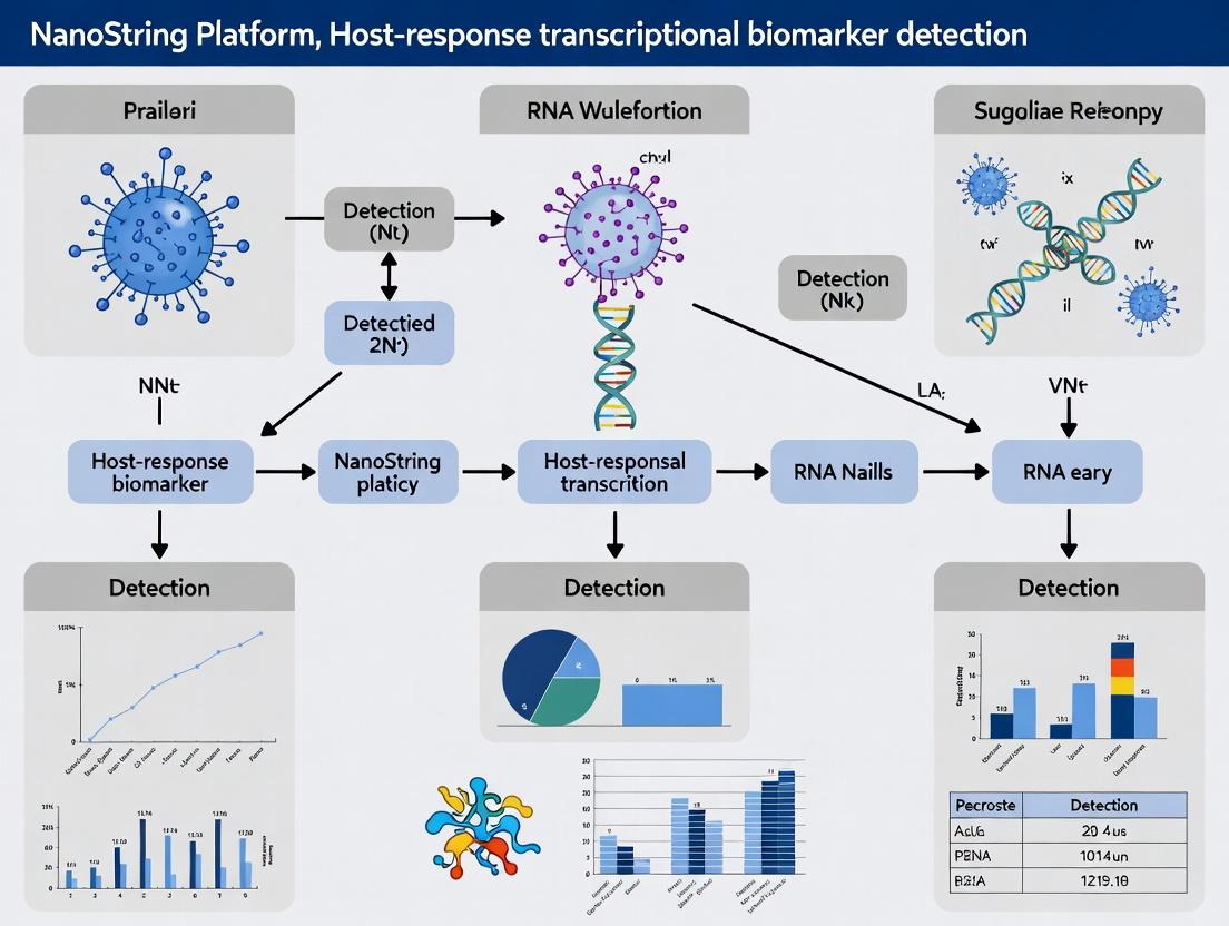
Unlocking Host-Response Insights: A Comprehensive Guide to the NanoString Platform for Transcriptional Biomarker Detection
This article provides a definitive guide for researchers and drug development professionals on utilizing the NanoString nCounter and GeoMx platforms for host-response transcriptional profiling.
Unlocking Host-Response Insights: A Comprehensive Guide to the NanoString Platform for Transcriptional Biomarker Detection
Abstract
This article provides a definitive guide for researchers and drug development professionals on utilizing the NanoString nCounter and GeoMx platforms for host-response transcriptional profiling. We explore the foundational principles of digital multiplexed analysis without amplification, detail practical methodologies for panel design and data acquisition in infectious disease, oncology, and immunology, address common troubleshooting and optimization strategies for challenging samples, and critically examine validation requirements and comparative performance against NGS and qPCR. The synthesis offers a roadmap for implementing robust, translational biomarker studies.
Understanding the NanoString Platform: Core Technology and Principles for Host-Response Profiling
The detection and validation of host-response transcriptional biomarkers are critical for understanding disease mechanisms, patient stratification, and therapeutic development. The NanoString platform has evolved to address this need, transitioning from bulk transcriptomic profiling with the nCounter system to high-plex spatial biology with the GeoMx Digital Spatial Profiler (DSP). This integrated approach allows researchers to first identify global transcriptional signatures from homogenized tissue and then map their spatial origin within the tissue architecture, preserving crucial morphological context.
The nCounter System: Foundation for Bulk Transcriptional Analysis
Technology Principle
The nCounter system utilizes a digital barcoding technology based on direct multiplexed measurement of gene expression. Target-specific probe pairs (Reporter and Capture probes) hybridize to mRNA. Each Reporter Probe carries a unique fluorescent barcode, allowing for digital counting of individual molecules without enzymatic reactions or amplification, minimizing bias.
Application Note: Host-Response Panel Profiling
A core application is profiling immune and inflammatory pathways. For example, the nCounter Human Immunology v2 Panel or PanCancer Immune Profiling Panel can quantify 770+ immune-related genes from total RNA extracted from PBMCs, whole blood (in PAXgene tubes), or tissue lysates. This enables the identification of signatures correlating with infection severity, autoimmune disease activity, or response to immunotherapy.
Table 1: Comparison of nCounter and RNA-Seq for Targeted Transcriptional Profiling
| Parameter | nCounter Analysis System | RNA-Seq (Targeted) |
|---|---|---|
| Throughput (Samples/Run) | Up to 12 (FLEX) or 96 (MAX) | Variable (typically 8-96) |
| Input RNA | 50-300 ng (Purified) or 5-10 µL lysate | 10 ng - 1 µg (Purified) |
| Hands-on Time | ~4 hours (hybridization) + 30 min setup | 3-8 hours (library prep) |
| Time to Data | 24-36 hours | 3-7 days |
| Reproducibility (CV) | <5% (technical replicates) | 5-15% |
| Cost per Sample | Low to Medium | Medium to High |
| Ideal For | Targeted panels, validation, clinical trials | Discovery, novel isoform detection |
Detailed Protocol: nCounter Gene Expression Assay
Objective: Quantify expression of up to 800 targets from purified RNA using a pre-designed panel. Materials:
- nCounter Gene Expression CodeSet (Reporter & Capture probes)
- nCounter Master Kit
- High-quality total RNA (RIN > 7.0 recommended)
- Thermocycler or hybridization oven
- nCounter Prep Station and Digital Analyzer Procedure:
- Sample Dilution: Dilute 100 ng of total RNA to 5 µL in RNase-free water.
- Hybridization Assembly: Combine 5 µL RNA with 8 µL Reporter CodeSet and 2 µL Capture ProbeSet. Mix gently.
- Hybridization: Incubate at 65°C for 16-24 hours in a thermocycler.
- Purification & Immobilization (Prep Station):
- Load samples into the nCounter Prep Station.
- The station performs automated post-hybridization purification using magnetic beads and immobilizes complexes on a cartridge.
- Data Collection (Digital Analyzer): Scan the cartridge. The analyzer takes an image of the immobilized fluorescent barcodes, counting each individually.
- Data Analysis: Raw counts are exported and normalized using built-in positive controls and housekeeping genes in nSolver software.
The GeoMx DSP System: Transition to Spatial Biology
Technology Principle
GeoMx DSP bridges bulk RNA analysis and histology. It uses oligo-tagged antibodies (GeoMx Protein Assay) or in-situ hybridization probes (GeoMx RNA Assay) that bind to targets on a tissue section. A UV-photocleavable linker attaches a unique DNA oligonucleotide (index) to each detection reagent. A researcher selects Regions of Interest (ROIs) based on morphology (e.g., tumor, stroma, immune infiltrates) using fluorescence markers. UV light is applied to each ROI, releasing the indexes, which are collected via a microcapillary. The indexes are then quantified by next-generation sequencing (NGS) or the nCounter system, generating a digital profile for each spatially defined region.
Application Note: Spatial Host-Response in Tumor Microenvironment
A key application is dissecting the tumor-immune interface. Researchers can stain a formalin-fixed, paraffin-embedded (FFPE) tumor section with fluorescent antibodies for pan-CK (tumor), CD45 (immune cells), and Syto13 (nuclei). Discrete ROIs are drawn within the tumor parenchyma and adjacent immune stroma. The GeoMx Cancer Transcriptome Atlas (CTA) or Human Whole Transcriptome Atlas (WTA) is used to profile ~1,800-20,000+ RNA targets from each region, revealing spatially resolved immune evasion signatures, tertiary lymphoid structures, or stromal suppression mechanisms.
Table 2: GeoMx Digital Spatial Profiler System Specifications
| Parameter | GeoMx DSP for RNA | GeoMx DSP for Protein |
|---|---|---|
| Target Multiplexing | Up to 22,000+ (Whole Transcriptome) | Up to 150+ (currently) |
| Tissue Compatibility | FFPE, Fresh Frozen | FFPE, Fresh Frozen |
| ROIs per Slide | Typically 1-100+ | Typically 1-100+ |
| RNA Input per ROI | Not applicable (in situ) | Not applicable (in situ) |
| Detection Limit | ~0.1-1 copies per cell (model-dependent) | ~1-10 copies per cell (model-dependent) |
| Read-Out Method | nCounter (≤800-plex) or NGS (full plex) | NGS |
| Morphology Context | Preserved (guides ROI selection) | Preserved (guides ROI selection) |
Detailed Protocol: GeoMx RNA Assay (FFPE)
Objective: Spatially profile RNA expression from morphologically defined regions in an FFPE tissue section. Materials:
- GeoMx RNA Slide Kit
- GeoMx Human Whole Transcriptome Atlas (WTA) Probe Set
- Fluorescent Morphology Markers (e.g., Syto13, PanCK, CD45 antibodies)
- FFPE tissue section (5 µm) on coated glass slide
- GeoMx DSP instrument
- NGS library preparation kit and sequencer Procedure:
- Tissue Pre-treatment: Bake, deparaffinize, and rehydrate FFPE slide. Perform target retrieval and proteinase K digestion.
- Hybridization: Apply the WTA probe set (containing gene-specific, UV-cleavable indexing oligos) to the tissue. Hybridize overnight at 37°C.
- Morphology Staining: After post-hybridization washes, stain tissue with fluorescent morphology markers (e.g., Syto13, PanCK-AF647, CD45-AF532). Apply anti-fade mounting medium.
- ROI Selection & UV Cleavage (GeoMx DSP Instrument):
- Load slide onto GeoMx instrument.
- Image slide at fluorescence wavelengths.
- Draw ROIs based on morphology (e.g., select "Tumor" regions PanCK+ CD45-; "Immune" regions PanCK- CD45+).
- For each ROI, the instrument positions a digital micromirror device (DMD) to pattern UV light, selectively cleaving indexing oligos from that area.
- Cleaved oligos are aspirated into a microfluidic well plate.
- Post-Collection Processing: Elute collected oligos. For NGS readout, perform PCR to add sequencing adapters and sample indices. Purify library.
- Sequencing & Data Analysis: Run on an NGS sequencer (e.g., Illumina NextSeq). Use GeoMx DSP Data Suite for analysis, aligning counts to targets and ROIs, and performing spatial differential expression.
Integrated Workflow for Host-Response Biomarker Discovery & Validation
The synergistic use of both platforms accelerates research: nCounter rapidly screens hundreds of bulk samples to identify candidate biomarker signatures. GeoMx DSP then validates and refines these findings by localizing the signature to specific cell populations or tissue compartments in a subset of critical samples, confirming biological context and generating mechanistic hypotheses.
Integrated nCounter & GeoMx DSP Workflow
The Scientist's Toolkit: Essential Research Reagent Solutions
Table 3: Key Research Reagents for NanoString Platform Host-Response Studies
| Reagent / Solution | Function | Platform |
|---|---|---|
| nCounter PanCancer Immune Profiling Panel | Quantifies 770+ human genes covering immune activation, suppression, and checkpoint pathways from bulk RNA. | nCounter |
| nCounter PlexSet Reagent System | Enables custom design of up to 80-plex gene expression assays for flexible, focused biomarker studies. | nCounter |
| GeoMx Human Whole Transcriptome Atlas (WTA) | A probe set for profiling ~18,000+ protein-coding genes in situ for discovery-phase spatial biology. | GeoMx DSP |
| GeoMx Mouse Whole Transcriptome Atlas | Enables spatial profiling of ~22,000+ mouse genes for preclinical host-response models. | GeoMx DSP |
| GeoMx Immune Cell Profiling Panel | Targeted panel for spatially profiling 1,800+ genes involved in human immunology and oncology. | GeoMx DSP |
| GeoMx RNA Slide Kit | Contains all buffers and reagents for tissue pretreatment, hybridization, and washing for FFPE/FF samples. | GeoMx DSP |
| Fluorescent Morphology Markers (Syto13, AF-conjugated Antibodies) | Used to visualize tissue architecture (nuclei, specific cell types) for informed ROI selection on GeoMx. | GeoMx DSP |
| nCounter Master Kit | Contains all essential reagents for hybridization, purification, and immobilization in an nCounter assay. | nCounter |
Innate Immune Pathway Measured Spatially
1. Introduction & Context Within the thesis on utilizing the NanoString nCounter platform for host-response transcriptional biomarker discovery, this document details the core technology enabling multiplexed, digital gene expression analysis without amplification. This method is critical for profiling immune and inflammatory responses with high precision and reproducibility, directly supporting research in infectious disease, immuno-oncology, and therapeutic development.
2. Core Technology Protocol: nCounter Assay Workflow
Protocol 2.1: Sample Preparation & Hybridization Objective: To prepare total RNA for hybridization with color-coded, sequence-specific reporter and capture probes. Materials:
- nCounter XT CodeSet (target-specific Reporter-Capture probe pairs).
- nCounter Hybridization Buffer.
- nCounter Master Kit.
- Thermal cycler or hybridization oven. Procedure:
- Dilute 100-300 ng of total RNA (or 5-10 µL of lysate) to a 5 µL volume in RNase-free water.
- Add 3 µL of Reporter CodeSet and 2 µL of Capture ProbeSet to the RNA.
- Add 10 µL of Hybridization Buffer. Mix thoroughly by pipetting.
- Incubate the 20 µL reaction at 67°C for 18-24 hours in a thermal cycler.
Protocol 2.2: Purification & Immobilization Objective: To remove excess probes and immobilize probe-target complexes on a cartridge for data collection. Materials:
- nCounter Prep Station.
- nCounter Cartridge.
- nCounter SPR Cartridge.
- Wash Buffers (A, B, C, D). Procedure:
- Load the hybridized sample into the designated well of an nCounter Cartridge.
- Place the cartridge and an SPR Cartridge into the Prep Station.
- Execute the automated purification protocol (∼2.5 hours). The system performs:
- Binding: Probe-target complexes are bound to the streptavidin-coated cartridge surface via the biotinylated capture probe.
- Washing: Unbound Reporter Probes are removed via a series of buffer washes.
- Alignment: Complexes are immobilized in a linear orientation for imaging.
Protocol 2.3: Data Acquisition & Analysis Objective: To digitally count individual barcodes. Materials:
- nCounter Digital Analyzer.
- nCounter Sprint or FLEX. Procedure:
- Insert the prepared cartridge into the Digital Analyzer.
- Initiate automated imaging. The system captures ∼600 fields of view per sample, imaging fluorescent barcodes at 0.5 µm resolution.
- Data is processed by nCounter software, which identifies barcode identities and counts each unique event. Output is a digital count (number of molecules) for each target in the CodeSet.
3. Experimental Data & Performance Metrics Table 1: Key Performance Characteristics of nCounter Technology
| Parameter | Specification / Typical Value | Implication for Host-Response Research |
|---|---|---|
| Sample Input Range | 1-300 ng total RNA | Enables analysis of low-yield clinical samples (e.g., PBMCs, biopsies). |
| Multiplexing Capacity | Up to 800 targets per reaction | Comprehensive profiling of immune pathways and biomarker panels. |
| Precision (Reproducibility) | CV < 10% (technical replicates) | Ensures reliable detection of subtle transcriptional changes. |
| Dynamic Range | > 4.5 logs | Allows simultaneous quantification of highly abundant and rare transcripts. |
| Time to Data | ~24-30 hours (hands-on ~4 hrs) | Rapid turnaround from sample to result. |
| Linearity (R²) | >0.99 across dilution series | Accurate quantification over a wide concentration range. |
4. The Scientist's Toolkit: Essential Research Reagents & Materials Table 2: Key Research Reagent Solutions
| Item | Function in the Protocol |
|---|---|
| nCounter CodeSet (Custom/Panel) | Pre-designed probe pairs for specific genes; defines the multiplexed targets for the experiment. |
| nCounter Master Kit | Provides core buffers and consumables for hybridization, purification, and cartridge preparation. |
| Hybridization Buffer | Provides optimal stringency and environment for specific probe-target binding. |
| Streptavidin-Coated Cartridge | Solid surface for immobilizing biotinylated probe-target complexes during purification. |
| nCounter Prep Station | Automated fluidics system for post-hybridization purification and cartridge preparation. |
| nCounter Digital Analyzer | Automated microscope and CCD camera for imaging and digitally counting barcodes. |
| nSolver / ROSALIND Software | Primary data analysis software for QC, normalization, and differential expression analysis. |
5. Visualization of Workflow and Data Analysis Logic
Diagram Title: nCounter Assay 3-Step Workflow
Diagram Title: nCounter Data Analysis Pipeline for Biomarker Discovery
Within the domain of host-response transcriptional biomarker research, the NanoString nCounter platform presents a paradigm shift. By employing direct digital detection of nucleic acids without enzymatic amplification, it circumvents critical limitations of PCR-based methods. This Application Note details the core advantages—freedom from amplification bias, exceptional reproducibility, and flexible multiplexing—that make the platform indispensable for robust biomarker discovery and validation in immunology, infectious disease, and drug development.
Table 1: Comparative Performance Metrics of Transcriptional Profiling Platforms
| Metric | NanoString nCounter | qRT-PCR | RNA-Seq (Standard) |
|---|---|---|---|
| Amplification Bias | None (Direct detection) | High (Enzyme efficiency dependent) | Moderate (PCR amplification steps) |
| Inter-run CV (%) | <5% (typical) | 10-25% (typical) | 10-15% (typical) |
| Input RNA Range | 1-300 ng (flexible) | 1-100 ng (optimal) | 10-1000 ng (platform dependent) |
| Multiplex Capacity | Up to 800 targets/code (XT) | Usually 1-6 plex | Whole transcriptome (~20,000) |
| Time to Data (hands-on) | ~15 minutes (30 hr total) | Moderate-High | Very High |
| Reproducibility (R²) | >0.99 (technical replicates) | ~0.95-0.98 | ~0.97-0.99 |
Table 2: Example Reproducibility Data from a Longitudinal Host-Response Study
| Sample Type | nCounter Inter-Assay CV (n=5 runs) | qRT-PCR Inter-Assay CV (n=5 runs) | Genes with CV <10% (nCounter) |
|---|---|---|---|
| PBMCs (Healthy) | 4.2% | 15.8% | 98% (of 600 targets) |
| PBMCs (Stimulated) | 5.1% | 22.3% | 96% (of 600 targets) |
| FFPE Tissue (Tumor) | 7.3% | N/A (degraded RNA) | 92% (of 600 targets) |
Detailed Experimental Protocols
Protocol 1: nCounter Host-Response Gene Expression Assay (e.g., PanCancer Immune Profiling Panel)
Objective: To quantify the expression of 770 immune-related genes from total RNA to assess host immune status.
Materials: See "The Scientist's Toolkit" below.
Procedure:
- RNA Qualification: Assess RNA integrity using a fragment analyzer or bioanalyzer. Accept samples with RIN (RNA Integrity Number) >6 for fresh/frozen, or DV200 >50% for FFPE.
- Sample Dilution: Dilute total RNA to a working concentration of 20-100 ng/µL in nuclease-free water.
- Hybridization Assembly:
- Prepare the Hybridization Master Mix on ice:
- 5 µL Reporter CodeSet (from specific panel)
- 5 µL Capture ProbeSet
- 5 µL nCounter Buffer RLS (Stabilizer).
- Aliquot 15 µL of Master Mix into each tube of a nCounter tube strip.
- Add 5 µL of each RNA sample (typically 100 ng total) to separate tubes containing the Master Mix.
- Include positive and negative (nuclease-free water) controls.
- Seal the strip, vortex gently, and spin down.
- Prepare the Hybridization Master Mix on ice:
- Hybridization: Place the tube strip in a pre-heated thermal cycler. Run: 67°C for 20 hours (16-24 hour range acceptable).
- Post-Hybridization Processing:
- Prepare the nCounter Prep Station: Prime with Buffer B and Buffer C.
- Load the nCounter Cartridge with the processed samples. The Prep Station automates:
- Purification: Immobilization of probe-target complexes on the cartridge surface via streptavidin-biotin binding.
- Alignment: Orientation of reporter probes in the cartridge imaging lane.
- Digital Data Acquisition: Insert the processed cartridge into the nCounter Digital Analyzer. The system performs automatic fluorescence imaging (555 nm and 647 nm lasers) and digital counting of individual barcodes. Scan at 555 FOV (Field of View) for standard sensitivity.
- Data Analysis: Export raw count data (.RCC files) for analysis in nSolver Advanced Analysis Software or third-party tools (e.g., R). Standard pipeline: (1) QC flags review, (2) Background subtraction, (3) Normalization (using positive controls & housekeeping genes), (4) Differential expression analysis.
Protocol 2: Multiplexed miRNA and mRNA Co-Profiling from Limited FFPE Samples
Objective: To simultaneously profile miRNA and mRNA signatures from the same precious FFPE RNA extract.
Materials: miRNA Panel, mRNA Panel, nCounter Buffer RLS Plus, nCounter Master Kit.
Procedure:
- RNA Isolation: Use an FFPE-specific RNA isolation kit with DNase treatment. Elute in a minimal volume (e.g., 15 µL).
- Dual Hybridization Setup: For each sample, prepare two separate hybridization reactions using the same RNA input.
- Reaction A (mRNA): Follow Protocol 1, using 50-300 ng of FFPE RNA and the desired host-response mRNA panel.
- Reaction B (miRNA): Prepare a miRNA Master Mix with 3 µL of diluted RNA (typically 30 ng), 5 µL miRNA Reporter CodeSet, and 2 µL of Buffer RLS Plus. Hybridize at 67°C for 18 hours.
- Parallel Processing: Process both cartridges simultaneously on the Prep Station using the appropriate protocol settings (mRNA vs. miRNA).
- Data Integration: Analyze data separately in nSolver, then integrate results using pathway or correlation analyses to build a multi-omics host-response model.
Visualizing Workflows and Pathways
Diagram 1: nCounter vs. qRT-PCR Workflow Comparison
Diagram 2: Host-Response Pathway Analysis Logic
The Scientist's Toolkit
Table 3: Essential Research Reagent Solutions for nCounter Host-Response Studies
| Item | Function & Importance |
|---|---|
| nCounter CodeSet (Custom/Predesigned Panels) | Target-specific probe libraries (e.g., PanCancer Immune, Inflammation, Autoimmune). The core reagent for multiplexed detection without amplification. |
| nCounter Buffer RLS / RLS Plus | Hybridization buffer stabilizes the reaction, with "Plus" formulation optimized for challenging samples (FFPE, miRNA). |
| nCounter Master Kit | Contains all essential buffers (Buffer B, C) and consumables for operation of the Prep Station purification process. |
| nCounter SPRINT Cartridges (Profiling) | Single-use cartridges for the SPRINT system, enabling lower-plex (up to 800 targets) profiling with flexible sample throughput. |
| nCounter PlexSet Reagents | For ultra-high-plex (up to 800-plex) expression panels, utilizing a unique ternary hybridization chemistry. |
| nSolver Advanced Analysis Software | Official analysis suite for QC, normalization, differential expression, and pathway scoring (e.g., PANTHER, ROSALIND). |
| nCounter Positive & Negative Controls | Synthetic oligonucleotide controls spiked into every reaction for system performance monitoring and data normalization. |
| RNA Stabilization Reagents (e.g., PAXgene, RNAlater) | Critical for preserving the in vivo transcriptional state at collection, especially for longitudinal host-response studies. |
| FFPE RNA Isolation Kits (with DNase) | Specialized kits to recover fragmented RNA from archived tissue, enabling retrospective host-response biomarker studies. |
Host-response transcriptional signatures, measured via the expression of specific gene panels, represent a paradigm shift in disease diagnostics and monitoring. These signatures capture the immune system's nuanced reaction to infection, cancer, or autoimmune disorders, offering higher specificity than single analyte tests. The NanoString nCounter platform is pivotal in this field, enabling direct, digital quantification of multiplexed mRNA transcripts without amplification, thus minimizing technical bias and allowing robust biomarker development from limited clinical samples.
Application Notes: Host-Response Signature Development and Validation
Signature Discovery and Refinement
The development of a diagnostic host-response signature begins with transcriptomic profiling (e.g., RNA-Seq) of whole blood or relevant tissue from well-characterized patient cohorts. Differentially expressed genes (DEGs) are filtered for biological relevance and technical robustness on the NanoString platform.
Table 1: Key Considerations for Signature Development
| Phase | Primary Goal | Typical Sample Size | Key Statistical Metric | NanoString Panel Type |
|---|---|---|---|---|
| Discovery | Identify DEGs between disease states | 50-100 per cohort | Adjusted p-value <0.01, Fold Change >1.5 | Whole Transcriptome or Large Custom Panel (>500 genes) |
| Refinement | Reduce gene list to minimal classifier | 100-200 per cohort | AUC >0.85, Leave-one-out cross-validation error | Custom Panel (50-150 genes) |
| Validation | Independent verification of classifier accuracy | 200+ per cohort | Sensitivity/Specificity >90%, Positive/Negative Predictive Value | Finalized Custom Panel (10-50 genes) |
Clinical and Commercial Applications
Validated signatures are deployed for:
- Disease Diagnosis: Distinguishing bacterial vs. viral infections (e.g., SeptiCyte LAB), identifying autoimmune disease subtypes.
- Treatment Monitoring: Tracking response to immunotherapy in oncology or anti-inflammatory therapy in rheumatology.
- Prognosis: Stratifying patients by risk of disease progression or severe outcomes.
- Clinical Trial Enrichment: Identifying likely responders for targeted drug development.
Detailed Protocols
Protocol A: NanoString nCounter Assay for Host-Response Signature Profiling from PAXgene Blood RNA
Objective: To quantify the expression of a custom host-response gene signature from peripheral blood RNA.
Materials (Research Reagent Solutions):
- NanoString nCounter Human Immunology v2 Panel or Custom CodeSet: Pre-designed reporter and capture probes for target genes.
- nCounter Master Kit: Contains all hybridization buffers.
- Purified Total RNA (≥50 ng/μL): Isolated from PAXgene tubes using the PAXgene Blood RNA Kit.
- nCounter Prep Station: For post-hybridization processing and immobilization of reactions.
- nCounter Digital Analyzer: For digital quantification of fluorescent barcodes.
Procedure:
- RNA Assessment: Check RNA concentration and purity (A260/A280 ~2.0). Use 100 ng (5 μL) of RNA per reaction.
- Hybridization Assembly: In a strip tube, combine:
- 5 μL RNA (100 ng)
- 3 μL Reporter CodeSet
- 5 μL Hybridization Buffer (from Master Kit)
- 2 μL Capture ProbeSet
- 5 μL Nuclease-free water.
- Hybridization: Seal tubes, vortex briefly, spin down. Incubate at 65°C for 16-20 hours in a thermal cycler.
- Post-Hybridization Processing: Load samples onto the nCounter Prep Station. Run the "High Resolution" protocol. The station performs magnetic bead-based purification and immobilization of probe-target complexes on a cartridge.
- Data Collection: Insert the cartridge into the nCounter Digital Analyzer. Perform a 555 FOV (Fields of View) scan for maximum sensitivity.
- Data Analysis: Use nSolver software. Perform QC (imaging, binding density, positive control linearity). Normalize data using built-in positive controls and housekeeping genes (e.g., GAPDH, ACTB). Export counts for downstream analysis.
Protocol B: Bioinformatics Analysis for Signature Score Calculation
Objective: To generate a single diagnostic score from a multi-gene signature.
Materials: nSolver Software, R Statistical Environment with NanoStringDiff or GeoMx packages, normalized gene expression data.
Procedure:
- Data Import & QC: Load normalized counts into R. Filter out samples failing QC metrics.
- Signature Scoring:
- For pre-defined signatures (e.g., Taubenberger et al., 2020), calculate a weighted sum:
Score = Σ (wi * log2(Expression_i))where wi is the published coefficient for gene i. - For novel signatures, use machine learning models (e.g., LASSO logistic regression) trained on the discovery cohort. Apply the model to the validation data to generate probability scores.
- For pre-defined signatures (e.g., Taubenberger et al., 2020), calculate a weighted sum:
- Threshold Determination: Use Youden's J statistic on the training set to define the optimal score cutoff for disease classification.
- Performance Evaluation: Calculate Area Under the ROC Curve (AUC), sensitivity, specificity, and predictive values in the validation cohort.
Table 2: Example Performance of a Hypothetical Host-Response Signature for Sepsis Etiology
| Cohort | Signature | AUC (95% CI) | Sensitivity (%) | Specificity (%) | PPV (%) | NPV (%) |
|---|---|---|---|---|---|---|
| Discovery (n=150) | 12-Gene Sepsis Origin | 0.94 (0.90-0.98) | 88 | 93 | 92 | 89 |
| Validation (n=300) | 12-Gene Sepsis Origin | 0.91 (0.87-0.94) | 85 | 90 | 89 | 86 |
Visualizations
Host-Response Signature Development Pipeline
NanoString nCounter Assay Workflow
Host Immune Response to Transcriptional Signature
The Scientist's Toolkit: Essential Research Reagents & Materials
Table 3: Key Research Reagent Solutions for Host-Response Biomarker Studies
| Item | Function/Description | Example Product/Catalog |
|---|---|---|
| PAXgene Blood RNA Tubes | Stabilizes intracellular RNA profile immediately upon blood draw, critical for accurate host-response measurement. | PreAnalytiX PAXgene Blood RNA Tubes |
| nCounter Human Immunology Panel | Pre-configured 594-plex gene panel covering immune pathways, ideal for discovery phase. | NanoString Human Immunology v2 Panel |
| nCounter Custom CodeSet | Tailored probe sets for validating specific signature genes; includes Reporter and Capture probes. | NanoString Custom CodeSet Service |
| nCounter Master Kit | Provides all essential buffers for the hybridization and sample preparation steps. | NanoString nCounter Master Kit |
| nSolver Analysis Software | Primary software for data QC, normalization (using positive & housekeeping controls), and basic analysis. | NanoString nSolver Software (v4.0) |
| Reference RNA | High-quality, stable RNA for assay run-to-run quality control and normalization calibration. | NanoString RNA Control Kit or commercial universal human reference RNA |
Within a thesis focused on host-response transcriptional biomarker detection using the NanoString nCounter platform, initial experimental design is paramount. The platform’s sensitivity to sample quality and input necessitates rigorous pre-experimental planning. This document details critical considerations for sample type selection, input requirements, and gene panel configuration to ensure robust, reproducible data.
Sample Types: Characteristics and Implications
The choice of sample type fundamentally impacts RNA quality, transcript abundance, and the biological interpretation of host-response signatures.
Table 1: Comparison of Sample Types for NanoString nCounter Analysis
| Sample Type | Key Characteristics | RNA Integrity (RIN/RQN) | Primary Advantages | Primary Challenges |
|---|---|---|---|---|
| Fresh Frozen Tissue | Gold standard for RNA preservation. | High (Typically >7.0) | Full transcriptome representation, minimal degradation. | Logistics of collection/storage, not always clinically available. |
| FFPE (Formalin-Fixed Paraffin-Embedded) | Archival clinical samples; cross-linked RNA. | Low (Not measured by RIN; use DV200>50%) | Vast retrospective cohorts, clinical outcome data linked. | RNA fragmentation, chemical modification, requires specialized protocols. |
| Peripheral Blood (PAXGene, Tempus) | Systemic immune response snapshot. | Moderate to High | Minimally invasive, serial sampling, rich in immune cell transcripts. | High globin mRNA content (can be depleted), reflects systemic not local response. |
| Bone Marrow Aspirate | Source of hematopoiesis and immune cells. | Moderate | Direct insight into bone marrow-specific host responses. | Invasive procedure, sample heterogeneity. |
| Buccal Swab / Cytology Brushes | Non-invasive epithelial sampling. | Low to Moderate | Easy collection for mucosal immunity studies. | Low RNA yield, potential for high bacterial RNA contamination. |
Input Requirements and Quality Control
NanoString assays require specific input metrics based on sample type. Adherence is critical for data quality.
Table 2: Minimum Input Requirements and QC Benchmarks
| Sample Type | Minimum Total RNA Input | Quality Control Metric | Passing Threshold | Recommended QC Method |
|---|---|---|---|---|
| Fresh Frozen Tissue | 50-100 ng | RNA Integrity Number (RIN) | RIN ≥ 7.0 | Bioanalyzer/TapeStation |
| FFPE | 100-300 ng | DV200 (% of fragments >200 nt) | DV200 ≥ 50% | Bioanalyzer/TapeStation (FFPE kit) |
| Whole Blood (RNA-stabilized) | 100-200 ng | - | A260/A280 ~2.0 | Spectrophotometry (NanoDrop) |
| Isolated PBMCs | 50-100 ng | RIN | RIN ≥ 6.5 | Bioanalyzer/TapeStation |
Protocol 3.1: RNA QC and Preparation for FFPE Samples Objective: To assess and prepare fragmented RNA from FFPE samples for nCounter analysis.
- Quantification: Use a fluorescence-based assay (e.g., Qubit RNA HS Assay) for accurate concentration measurement of fragmented RNA. Avoid absorbance-based methods.
- Quality Assessment: Run 1 µL of RNA on an Agilent Bioanalyzer 2100 using the RNA 6000 Nano Kit or the Agilent TapeStation with High Sensitivity RNA ScreenTape. Generate a DV200 value.
- Input Normalization: Dilute all passing samples (DV200 ≥ 50%) to a working concentration of 20 ng/µL in nuclease-free water. The target input is 5 µL (100 ng) per nCounter reaction.
- Storage: Keep diluted RNA on ice or at -80°C for long-term storage. Avoid repeated freeze-thaw cycles.
Panel Selection Strategy
Panel selection aligns the assay with the specific host-response biological question.
Protocol 4.1: Custom Panel Design for Host-Response Profiling Objective: To design a custom nCounter Gene Expression Panel for a defined host-response pathway (e.g., antiviral interferon signaling).
- Pathway Curation: Using databases (KEGG, Reactome, Gene Ontology), compile a core gene list covering all key nodes in the pathway of interest.
- Add Contextual Signatures: Append genes from published relevant gene signatures (e.g., sepsis survival, macrophage polarization) to provide broader biological context.
- Include Normalizers: Select 8-12 housekeeping genes that are stable across your specific sample types and conditions. Test stability in a pilot study.
- Control Selection: The nCounter system includes positive controls (to assess assay efficiency) and negative controls (to assess background). Ensure your custom codeset includes these.
- Codeset Synthesis: Submit the final gene list (typically 50-800 genes) to NanoString for codeset design and synthesis. A 12-plex codeset is standard.
The Scientist's Toolkit: Research Reagent Solutions
Table 3: Essential Materials for Pre-Experimental Sample Processing
| Item | Function | Example Product/Catalog |
|---|---|---|
| Qubit RNA HS Assay Kit | Accurate quantification of low-concentration or fragmented RNA. | Thermo Fisher Scientific, Q32852 |
| Agilent High Sensitivity RNA ScreenTape | Assessment of RNA integrity (RIN) and fragmentation (DV200). | Agilent, 5067-5579 |
| FFPE RNA Isolation Kit | Optimized extraction of cross-linked, fragmented RNA from paraffin sections. | Qiagen miRNeasy FFPE Kit, 217504 |
| PAXgene Blood RNA Kit | Integrated stabilization and purification of RNA from whole blood. | PreAnalytiX, Qiagen, 762164 |
| Globin Clear Kit | Depletion of abundant globin mRNAs from blood RNA to improve detection sensitivity. | Thermo Fisher Scientific, AM1980 |
| RNase Zap / RNase-free Reagents | Elimination of RNase contamination from work surfaces and equipment. | Thermo Fisher Scientific, AM9780 |
| Nuclease-free Water & Tubes | Essential for all RNA dilution and storage steps to prevent degradation. | Various suppliers (Ambion, etc.) |
| nCounter PlexSet Reagents | For high-plex (up to 800-plex) gene expression analysis from limited input. | NanoString, XT-CSK-PLEXSET-12 |
Visualizations
Title: Sample and Panel Selection Workflow for NanoString
Title: Core Host-Response Pathway: Innate Immune Activation
From Panel Design to Data Acquisition: A Step-by-Step Protocol for Host-Response Studies
Introduction This Application Note details the strategic design of a custom nCounter panel for profiling host-response transcriptomes within the broader research context of using the NanoString platform for biomarker discovery. The goal is to enable researchers and drug development professionals to simultaneously quantify key genes across immune, inflammatory, and metabolic pathways from limited biological samples, facilitating comprehensive biomarker signatures.
Gene Selection Strategy & Quantitative Summary Gene candidates were identified through a systematic review of current literature (2023-2024) focusing on sepsis, immuno-oncology, and metabolic syndrome as model host-response conditions. Selection criteria included: 1) Proven differential expression in human studies, 2) Central role in core pathway signaling, 3) Availability as well-annotated nCounter CodeSets. The final curated list of 40 genes is categorized below.
Table 1: Selected Genes for Custom Host-Response Panel
| Category | Gene Symbol | Primary Function | Avg. Log2FC in Sepsis* |
|---|---|---|---|
| Innate Immunity | TLR4, MYD88, NLRP3, CASP1, IL1B | Pathogen sensing, inflammasome | +3.2 to +6.5 |
| Adaptive Immunity | CD4, CD8A, FOXP3, IFNG, GZMB | T-cell function & regulation | -1.8 to +4.1 |
| Pro-inflammatory | TNF, IL6, PTGS2, NFKB1 | Cytokine signaling, inflammation | +2.5 to +5.8 |
| Anti-inflammatory | IL10, TGFB1, ARG1 | Resolution of inflammation | +1.5 to +3.3 |
| Metabolic | PPARG, SREBF1, CPT1A, HK2 | Lipid/glucose metabolism, bioenergetics | -2.1 to +1.9 |
| Housekeeping | GAPDH, ACTB, HPRT1, TUBB | Normalization controls | N/A |
*FC: Fold Change. Representative data from meta-analysis of public datasets (GSE65682, GSE134347).
Protocol: Custom Panel Design & Validation Part A: Panel Design Using nCounter Advanced Analysis Software
- Gene Entry: Input the finalized gene list (e.g., from Table 1) into the nCounter Panel Design portal.
- CodeSet Configuration: Assign each gene to a Reporter-Probe Pair. The software automatically checks for potential high background or cross-hybridization.
- Control Selection: Include 8 positive controls, 6 negative controls, and 8 housekeeping genes (as above) in the design template.
- Finalize & Order: Submit the design for synthesis. Typical turnaround is 4-6 weeks.
Part B: Sample Processing & nCounter Assay Materials:
- nCounter Custom CodeSet: Contains sequence-specific probes for your selected genes.
- nCounter Master Kit: Includes all reagents for hybridization, purification, and immobilization.
- nCounter Prep Station: Automates post-hybridization processing.
- nCounter Digital Analyzer: For digital counting of target molecules.
- High-Quality RNA: 100 ng total RNA per sample (Input range: 50-300 ng).
Procedure:
- Hybridization: Combine 5 µL of RNA (100 ng) with 8 µL of Reporter CodeSet and 2 µL of Capture ProbeSet. Incubate at 65°C for 16-24 hours.
- Purification & Immobilization: Transfer the reaction to the nCounter Prep Station. Using the "High Sensitivity" protocol, excess probes are removed, and target-probe complexes are immobilized on a cartridge surface.
- Data Collection: Insert the cartridge into the nCounter Digital Analyzer. The system performs automatic scanning and counting of fluorescent barcodes.
- Data Analysis: Export raw counts (.RCC files) and analyze using nSolver Advanced Analysis software. Normalize data using selected housekeeping genes.
Pathway & Workflow Visualization
Figure 1: Core Immune-Metabolic Pathway Cross-Talk
Figure 2: Custom nCounter Assay Workflow
The Scientist's Toolkit: Essential Research Reagents & Materials Table 2: Key Reagent Solutions for Host-Response Panel Analysis
| Item | Function | Example Product/Catalog |
|---|---|---|
| nCounter Custom CodeSet | Target-specific probes for your selected 40 genes. | NanoString Custom Panel |
| nCounter Master Kit | All essential buffers and matrices for the assay. | NanoString Cat# XXXX |
| High-Quality RNA Isolation Kit | To obtain intact, pure RNA from whole blood or tissue. | PAXgene Blood RNA Kit, miRNeasy Mini Kit |
| RNA Integrity Number (RIN) Analyzer | Assess RNA quality pre-assay (RIN >7 recommended). | Bioanalyzer/TapeStation |
| nSolver Advanced Analysis Software | For data normalization, differential expression, and pathway scoring. | NanoString nSolver 4.0 |
| Positive & Negative Control RNA | For assessing assay performance and background. | Human Reference RNA, Nuclease-free Water |
This application note details the use of three pre-designed nCounter panels—PanCancer IO 360, Myeloid Innate Immunity, and Autoimmune Profiling—within a research thesis focused on the NanoString platform for host-response transcriptional biomarker discovery. These panels enable highly multiplexed, digital profiling of immune and inflammatory responses from limited sample inputs, supporting research in immuno-oncology, infectious disease, and autoimmune disorder therapeutics.
The following table summarizes the core specifications and applications of the featured panels.
Table 1: Comparison of nCounter Pre-Designed Immune Profiling Panels
| Panel Name | Code (Human) | Total Gene Count | Key Functional Categories | Primary Research Applications |
|---|---|---|---|---|
| PanCancer IO 360 | XT-CSO-HIP1-12 | 770+ | Immune Checkpoints, Cytotoxic Activity, Antigen Presentation, Chemokines, IFN Signaling, Tumor Intrinsic Signatures | IO drug development, biomarker discovery, patient stratification, therapy response monitoring |
| Myeloid Innate Immunity | XT-CSO-MMI1-12 | 770+ | Myeloid Cell Lineage, Phagocytosis, Inflammatory Response, Complement, Cytokine Signaling, Antiviral Defense | Innate immunity profiling, macrophage polarization, sepsis, COVID-19 host response, chronic inflammation |
| Autoimmune Profiling | XT-CSO-HA1-12 | 770+ | B & T Cell Biology, Cytokine & Receptor Networks, JAK-STAT Signaling, Tissue Remodeling, Vascular Response | Autoimmune disease (RA, SLE) research, biomarker identification, clinical trial stratification |
Key Experimental Protocols
Protocol 1: Sample Preparation and nCounter Analysis
This protocol is common for all three panels.
1. RNA Isolation and QC:
- Isolate total RNA from desired sample (FFPE tissue sections, PBMCs, frozen tissue) using a column-based or magnetic bead kit.
- Quantify RNA using a fluorometric method (e.g., Qubit). For FFPE samples, use an RNA IQ score or DV200 metric. Input requirement: 50-300 ng for fresh/frozen RNA; 100-300 ng for FFPE RNA.
2. Hybridization Reaction:
- Combine the following in a PCR tube:
- 3 μL of Reporter CodeSet (Panel-specific)
- 5 μL of Capture ProbeSet
- 5 μL of sample RNA (at required mass in a volume ≤5 μL)
- nuclease-free water to a final volume of 15 μL.
- Denature at 67°C for 5 minutes, then hybridize at 67°C for a minimum of 16 hours (up to 24 hours) in a thermal cycler.
3. Post-Hybridization Processing & Data Collection:
- Load samples into the nCounter Prep Station for automated purification and immobilization of probe-transcript complexes onto the cartridge.
- Scan the cartridge in the nCounter Digital Analyzer. Each cartridge is scanned at 555 fields of view (FOV) by default, providing digital counts for each target gene.
Protocol 2: Data Analysis Workflow for Host-Response Biomarker Discovery
- Primary Data QC: Use nSolver Software (v4.0 or later). Apply default QC flags based on imaging, binding density, and positive control linearity.
- Normalization: Apply a two-step normalization:
- Technical Normalization: Using positive control probes.
- Biological Normalization: Using a panel-specific set of housekeeping genes (e.g., 20+ genes) to correct for sample input differences.
- Advanced Analysis: Export normalized data for advanced analysis in platforms like nSolver Advanced Analysis modules, R (Geometra, DESeq2), or ROSALIND for automated pathway scoring (e.g., PanCancer Immune Profiling Panel scores), differential expression, and sample clustering.
Visualizations
Diagram 1: nCounter Host-Response Workflow
Diagram 2: Panel-Specific Pathway Focus
The Scientist's Toolkit: Essential Research Reagents & Materials
Table 2: Key Research Reagent Solutions for nCounter Analysis
| Item | Supplier/Code Example | Function in Protocol |
|---|---|---|
| nCounter Reporter CodeSet | NanoString (Panel-specific, e.g., XT-CSO-HIP1-12) | Panel-specific cocktail of fluorescent barcodes attached to gene-specific probes. |
| nCounter Capture ProbeSet | NanoString (Included with panel) | Universal set of probes for immobilizing hybridized complexes to cartridge. |
| nCounter Master Kit | NanoString (CAT#: XT-CSO-MK1-24) | Provides all essential buffers, plates, and cartridges for sample processing. |
| RNase-free Water | Invitrogen (CAT#: AM9937) | Diluent for RNA samples and reaction setup. |
| RNA Isolation Kit (FFPE) | Qiagen (RNeasy FFPE Kit, CAT#: 73504) | For high-quality RNA extraction from archival FFPE tissues. |
| RNA Isolation Kit (Cells) | Zymo Research (Quick-RNA Miniprep Kit, CAT#: R1055) | For rapid total RNA isolation from PBMCs or cell lines. |
| Fluorometric RNA QC Kit | Thermo Fisher (Qubit RNA HS Assay, CAT#: Q32852) | Accurate quantitation of low-concentration RNA samples. |
| nSolver Analysis Software | NanoString (Download) | Primary data QC, normalization, and basic differential expression analysis. |
| Advanced Analysis Software | ROSALIND (https://rosalind.bio/) | Cloud-based platform for automated pathway scoring and biomarker analysis. |
Within the context of advancing host-response transcriptional biomarker detection using the NanoString nCounter platform, a robust and reproducible workflow is paramount. This Application Note details the standardized procedures for sample processing, from RNA hybridization through to raw data generation, essential for research in infectious disease, oncology, and drug development.
Detailed Protocols
Protocol 1: RNA Hybridization
Objective: To specifically hybridize target RNA molecules with Reporter and Capture Probes in a single, multiplexed reaction.
Materials:
- High-quality total RNA (minimum 100 ng, recommended 100-300 ng).
- nCounter Reporter CodeSet (gene-specific probes with fluorescent barcodes).
- nCounter Capture ProbeSet (gene-specific probes coupled to magnetic beads).
- Hybridization Buffer.
- nuclease-free water.
- Thermal cycler or hybridization oven.
Method:
- Prepare Hybridization Mix: Combine the following in a sterile tube:
- 70 µL of Hybridization Buffer.
- 5 µL of Reporter CodeSet.
- 5 µL of Capture ProbeSet.
- 30 µL of RNA sample (containing recommended input mass).
- Total reaction volume: 110 µL.
- Hybridize: Incubate the reaction at 65°C for 16-24 hours (typically overnight) to allow for specific probe-target complex formation.
Protocol 2: Post-Hybridization Purification
Objective: To remove excess, unhybridized probes and prepare the sample for immobilization.
Materials:
- nCounter Prep Station.
- nCounter Cartridge.
- Streptavidin-coated magnetic beads (included in cartridge).
- Wash Buffers (included in cartridge).
Method:
- Load Samples: Transfer the 110 µL hybridization reaction to the designated well of an nCounter Sample Prep Plate.
- Automated Purification: Using the nCounter Prep Station and a dedicated cartridge:
- The station mixes the sample with streptavidin-coated magnetic beads, which bind the biotinylated Capture Probe.
- Using a magnetic field, the probe-target complexes are immobilized and washed extensively (3 wash steps) to remove unbound material.
- The purified complexes are aligned and immobilized in the cartridge's capillary flow cell for scanning.
Protocol 3: Scanning and Raw Data Generation
Objective: To quantify the fluorescent barcodes and generate digital counts for each target.
Materials:
- nCounter Digital Analyzer.
- Immobilized cartridge from Prep Station.
Method:
- Load Cartridge: Insert the processed cartridge into the nCounter Digital Analyzer.
- Scan: Initiate the automated scan.
- The analyzer uses epifluorescence microscopy and CCD imaging to count individual fluorescent barcodes in a 280 µm field of view.
- It performs 555 FOV scans per sample, capturing ~1150 counts per FOV.
- Data Output: The analyzer software generates an RCC (Reporter Code Count) file containing raw digital counts for each target in the CodeSet, along with imaging QC metrics.
Data Presentation
Table 1: Key Quantitative Metrics for the nCounter Workflow
| Parameter | Specification/Recommended Value | Note |
|---|---|---|
| RNA Input | 100-300 ng total RNA | Can be as low as 10-50 ng with high sensitivity protocols. |
| Hybridization Time | 16-24 hours | Standard protocol ensures maximum specificity. |
| Multiplexing Capacity | Up to 800 targets per reaction | Standard CodeSet size for custom panels. |
| Scan FOV per Sample | 555 fields of view | Ensures statistical robustness of count data. |
| Typical Time-to-Data | ~24-30 hours | Includes hybridization, purification, and scanning. |
| Linear Dynamic Range | 0.1 fM to >1 pM | ~4 logs of dynamic range. |
| Data Output File | .RCC file | Contains raw counts, QC flags, and imaging data. |
Table 2: Example Raw Data Output Structure (Abbreviated)
RCC File Header |
Value | Description | |
|---|---|---|---|
ScannerID |
NS12345 | Instrument identifier. | |
FOVCount |
555 | Fields of view analyzed. | |
BindingDensity |
0.25 | QC metric; optimal 0.1-0.9. | |
Code Class |
Name |
Count |
|
Positive |
POS_A |
1256 | Synthetic positive control. |
Negative |
NEG_A |
12 | Synthetic negative control. |
Endogenous |
IL6 |
455 | Target gene count. |
Endogenous |
IFNG |
120 | Target gene count. |
Housekeeping |
GAPDH |
890 | Reference gene count. |
Workflow and Pathway Diagrams
nCounter Assay Workflow Overview
Host-Response to Biomarker Detection
The Scientist's Toolkit
Table 3: Essential Research Reagent Solutions for nCounter Assay
| Item | Function in Workflow |
|---|---|
| nCounter CodeSet (Custom/Predesigned) | Contains target-specific Reporter and Capture probes. Defines the multiplexed gene panel for host-response research. |
| nCounter Hybridization Buffer | Provides optimal ionic and chemical environment for specific probe-target hybridization. |
| Streptavidin Magnetic Beads | Immobilize biotinylated Capture Probe-complexes during purification on the Prep Station. |
| nCounter Sample Prep Plates | Low-binding, formatted plates for holding samples during automated processing on the Prep Station. |
| nCounter Cartridges | Disposable consumables containing capillaries and reagents for purification and scanning. |
| High-Quality Total RNA Isolation Kits | To obtain intact, DNA-free RNA input critical for accurate gene expression quantification. |
| nCounter SPRINT Cartridges/Codesets | For use with the SPRINT system, enabling higher throughput and flexible profiling. |
This application note details the use of the NanoString GeoMx Digital Spatial Profiler (DSP) for spatially resolved transcriptomics within the framework of host-response biomarker discovery. The host-response to disease, therapy, or infection is a complex, multicellular process that occurs within the specific architectural context of tissues. Bulk RNA sequencing homogenizes this spatial information, while single-cell RNA sequencing dissociates cells from their native microenvironment. The GeoMx DSP bridges this gap by enabling high-plex, morphology-driven profiling of RNA from precisely selected regions of interest (ROIs) within intact tissue sections. This capability is critical for the central thesis of using the NanoString platform to discover and validate transcriptional biomarkers that reflect the orchestrated, spatial response of host tissue.
Core Technology & Workflow
The GeoMx DSP combines high-plex molecular detection with spatial visualization. The workflow involves:
- Tissue Preparation: FFPE or fresh-frozen tissue sections are mounted on slides and stained with fluorescent morphological markers (e.g., Pan-CK for epithelium, CD45 for immune cells, SYTO 13 for nuclei).
- Oligo-Tagged In Situ Hybridization: Target RNA transcripts are bound by sequence-specific, UV-photocleavable indexing oligos.
- Region of Interest (ROI) Selection: A digital microscope image guides the user to select ROIs based on morphology (e.g., tumor core, invasive margin, stroma).
- UV Photocleavage & Collection: UV light is directed at each selected ROI, releasing the indexing oligos from that specific region. The oligos are collected via a microcapillary into a 96- or 384-well plate.
- Quantification: The collected oligos are quantified using the nCounter system or next-generation sequencing (NGS), generating digital counts for each target per ROI.
Experimental Protocols
Protocol 3.1: FFPE Tissue Processing for GeoMx Human Whole Transcriptome Atlas (WTA)
Objective: Prepare FFPE tissue sections for spatial whole transcriptome analysis. Materials: GeoMx HiPlex for FFPE Slide Prep Kit, FFPE tissue sections (5 µm), xylene, ethanol series (100%, 95%, 70%), hydrophobic barrier pen, hybridization oven. Procedure:
- Bake slides at 60°C for 1 hour.
- Deparaffinize in xylene (2x 5 min), then rehydrate in 100% EtOH (2x 2 min), 95% EtOH (2 min), 70% EtOH (2 min), and nuclease-free water (2 min).
- Immediately circle tissue with a hydrophobic barrier pen.
- Apply Proteinase K solution (1:100 dilution in provided buffer) to cover tissue. Incubate at 37°C in a humidified chamber for 1 hour.
- Wash slides in nuclease-free water for 1 min.
- Apply RNA-Targeted Probe Hybridization Mix (Human WTA) to tissue. Coverslip and incubate at 37°C in a hybridization oven for 18-20 hours.
- Proceed to wash steps and fluorescent staining per kit manual.
Protocol 3.2: ROI Selection and Segmentation with Fluorescent Morphology Markers
Objective: Define biologically relevant ROIs using multiplexed immunofluorescence. Materials: GeoMx compatible fluorescently conjugated antibodies (Pan-CK/Alexa Fluor 532, CD45/Alexa Fluor 647, SYTO 13), GeoMx DSP instrument. Procedure:
- After RNA probe hybridization and washing, incubate tissue with a pre-optimized antibody cocktail (e.g., Pan-CK, CD45, SYTO 13) for 1 hour at room temperature.
- Wash slides and mount with GeoMx Fluorescent Mounting Medium.
- Load slide onto the GeoMx DSP stage.
- Capture a whole-slide scan at 20x magnification for each fluorescence channel.
- Using the DSP software, overlay channels to visualize tissue architecture.
- Segment ROIs: Use the segmentation tool to automatically define sub-regions within an ROI based on marker expression (e.g., "Pan-CK+" for tumor epithelium, "Pan-CK-/CD45+" for immune clusters, "Pan-CK-/CD45-" for stroma).
- Manually draw or select additional ROIs based on specific morphological features.
- Set the digital UV mask and initiate the automated photocleavage and collection sequence.
Key Data & Applications in Host-Response
Table 1: Representative Data from a GeoMx Study on Host-Response in Tumor Microenvironment
| Region of Interest (ROI) Type | Avg. Transcripts Detected (per ROI) | Key Upregulated Host-Response Pathways | Example Biomarker Genes | Cell-Type Inference |
|---|---|---|---|---|
| Tumor Epithelium (Pan-CK+) | ~18,000 | IFN-α/γ Response, EMT | STAT1, IRF9, VIM | Malignant Cells |
| Immune Islet (CD45+) | ~15,500 | Inflammatory Response, Complement, T-cell Exhaustion | CD3E, CD8A, PDCD1, C1QB | T Cells, Myeloid Cells |
| Stromal Region (Pan-CK-/CD45-) | ~12,800 | TGF-β Signaling, Angiogenesis, Fibrosis | ACTA2, COL1A1, VEGFA | Fibroblasts, Endothelium |
| Tumor-Invasive Margin | ~17,200 | Combined IFN-γ, Chemokine, and ECM Pathways | CXCL9, CXCL10, MMP9 | Mixed Immune/Stromal |
Table 2: Comparison of Transcriptomic Profiling Platforms for Host-Response Research
| Feature | GeoMx DSP | Bulk RNA-Seq | Single-Cell RNA-Seq |
|---|---|---|---|
| Spatial Context | Yes, morphology-guided | No | No (dissociated cells) |
| Tissue Preservation | Intact tissue section | Homogenized | Dissociated suspension |
| Multiplex Capacity | Whole Transcriptome (>18,000 genes) | Unlimited | High (thousands of cells) |
| Throughput (Regions/Sample) | Medium (10s-100s of ROIs) | Low (1 region/sample) | High (1000s of cells/sample) |
| Key Application | Spatial phenotyping of host-response niches | Overall host signature | Cellular taxonomy of host-response |
| Best Paired With | IHC, Digital Pathology | -- | CITE-seq, ATAC-seq |
The Scientist's Toolkit: Essential Research Reagent Solutions
| Item | Function in GeoMx Workflow |
|---|---|
| GeoMx Human Whole Transcriptome Atlas (WTA) | Pre-designed probe set targeting ~18,000 protein-coding genes for comprehensive spatial profiling. |
| GeoMx Immune Cell Profiling Panel | Focused panel for immunooncology, targeting 1,400+ genes related to immune activation, suppression, and host-response. |
| NanoString nCounter MAX/FLEX System | For direct digital quantification of photocleaved oligos without amplification, ideal for focused panels. |
| GeoMx Seq Code | For preparing photocleaved oligo libraries for NGS, required for WTA and highest-plex applications. |
| Morphology Marker Antibodies (Pan-CK, CD45, etc.) | Fluorescently conjugated antibodies for visualizing tissue architecture and guiding ROI selection. |
| SYTO 13 Nuclear Stain | Fluorescent stain for visualizing all nuclei, critical for defining tissue regions and cell density. |
| GeoMx ROI & Segmentation Buffer Kits | Optimized buffers for fluorescent staining and oligo hybridization on FFPE or frozen tissues. |
Visualization Diagrams
GeoMx DSP Core Workflow
Host-Response Pathway Analysis in Segmented ROIs
GeoMx Integration in Biomarker Research Thesis
The NanoString nCounter and GeoMx Digital Spatial Profiler (DSP) platforms enable multiplexed, digital detection of transcriptional host-response biomarkers without amplification, preserving critical quantitative information. This thesis posits that these platforms are uniquely suited for translational research where precise, reproducible profiling of immune and inflammatory pathways from complex clinical samples is paramount. The following application notes and protocols demonstrate this utility across three critical areas: stratifying patients by infectious disease severity, predicting response to cancer immunotherapy, and delineating heterogeneous autoimmune disease phenotypes.
Application Note: Infectious Disease Severity Stratification
Objective: To identify a host-response mRNA signature that distinguishes severe (e.g., septic) from mild/moderate infectious disease presentations, enabling early clinical intervention.
Background: Patient outcomes in infections like sepsis, influenza, and COVID-19 are dictated more by the host's dysregulated immune response than by the pathogen load. Transcriptional profiling of whole blood or PBMCs reveals conserved pathways associated with hyperinflammation, immune suppression, and metabolic dysregulation.
Key Findings & Data Summary: Recent studies utilizing NanoString's PanCancer Immune Profiling and Myeloid Innate Immunity panels have identified consistent biomarkers.
Table 1: Host-Response Transcriptional Biomarkers in Infectious Disease Severity
| Gene Symbol | Gene Name | Expression in Severe vs. Mild | Associated Pathway/Function |
|---|---|---|---|
| S100A8/A9 | Calprotectin | Upregulated | Neutrophil activation, DAMPs |
| CD177 | CD177 molecule | Upregulated | Neutrophil signature |
| ARG1 | Arginase 1 | Upregulated | Myeloid-derived suppressor cell (MDSC) activity |
| IL1RN | IL-1 receptor antagonist | Upregulated | Anti-inflammatory, feedback inhibitor |
| IFNG | Interferon-gamma | Downregulated | Impaired adaptive immunity |
| HLA-DRA | MHC Class II DR alpha | Downregulated | Monocyte deactivation, immune paralysis |
| PPARG | Peroxisome proliferator-activated receptor gamma | Downregulated | Dysregulated immunometabolism |
Experimental Protocol: Host-Response Profiling from Whole Blood for Severity Stratification
- Sample Collection: Collect 2.5 mL of whole blood directly into PAXgene Blood RNA tubes. Invert 10 times and store at -20°C or -80°C.
- RNA Extraction: Use the PAXgene Blood RNA Kit. Include an on-column DNase digestion step. Assess RNA concentration (RIN >7 recommended).
- nCounter Assay Setup: Use the nCounter Human Myeloid Innate Immunity Panel (v2, 770+ genes). For 12 reactions:
- Combine 65 μL of Reporter CodeSet, 65 μL of Capture ProbeSet, and 130 μL of Hybridization Buffer.
- Aliquot 8 μL of this master mix per tube.
- Add 5 μL of total RNA (100 ng recommended).
- Hybridize at 67°C for 18-24 hours.
- Processing: Load samples into the nCounter Prep Station for automated purification and immobilization on the cartridge.
- Data Acquisition: Scan cartridge on the nCounter Digital Analyzer at 555 fields of view (FOV).
- Analysis: Normalize data using built-in positive controls and housekeeping genes (e.g., GAPDH, ACTB). Perform differential expression (NanoString nSolver with Advanced Analysis) and pathway analysis (Ingenuity Pathway Analysis, GSEA).
Pathway Diagram: Host-Response in Severe Infection
Diagram 1: Immune dysregulation pathways in severe infection.
The Scientist's Toolkit: Key Research Reagents
| Item | Function in Protocol |
|---|---|
| PAXgene Blood RNA Tube | Stabilizes intracellular RNA profile at the moment of collection. |
| nCounter Myeloid Innate Immunity Panel | Pre-designed codeset for profiling 770+ genes relevant to infection response. |
| nCounter Hybridization Buffer | Facilitates specific binding of target RNA to reporter-capture probe complexes. |
| RNase-free Water (PCR-grade) | Used in RNA elution and assay setup to prevent degradation. |
Application Note: Cancer Immunotherapy Response Prediction
Objective: To characterize the tumor immune microenvironment (TIME) using spatial transcriptomics to predict patient response to immune checkpoint inhibitors (ICI).
Background: Response to ICIs (anti-PD-1, anti-CTLA-4) depends on a pre-existing but suppressed adaptive immune response within the tumor. The GeoMx DSP allows for region-specific, multiplexed mRNA profiling from formalin-fixed paraffin-embedded (FFPE) tissue sections, linking morphology to transcriptomics.
Key Findings & Data Summary: Profiling of tumor compartments (panCK+ tumor, CD45+ immune, CD3+ T cell regions) identifies predictive signatures.
Table 2: Spatial Transcriptomic Features Predictive of ICI Response
| Biological Feature | Associated Genes/Pathways | Predictive Value (High) |
|---|---|---|
| Cytotoxic T-cell Infiltration | CD8A, GZMB, PRF1, IFNG | Positive Response |
| T-cell Exhaustion | PDCD1 (PD-1), CTLA4, LAG3, TIGIT | May indicate responsive but suppressed TIME |
| Antigen Presentation Machinery | HLA-A/B/C, B2M, STAT1 | Positive Response |
| Immunosuppressive Microenvironment | FOXP3 (Tregs), MS4A1 (B cells), TGFB1 | Resistance |
| Oncogenic Signaling | WNT/β-catenin, PPARG pathways | Resistance |
Experimental Protocol: GeoMx DSP for Tumor Microenvironment Analysis
- Slide Preparation: Cut 5 μm FFPE sections onto GeoMx slides. Deparaffinize, rehydrate, and perform antigen retrieval.
- Immunofluorescence Staining: Stain with morphology markers: Pan-Cytokeratin (PanCK)-Alexa Fluor 594 (tumor), CD45-Alexa Fluor 532 (leukocytes), SYTO 83 (nuclear stain). Include fluorescently tagged oligonucleotide probes from the GeoMx Cancer Transcriptome Atlas.
- Imaging & ROI Selection: Image whole slide at 20x. Select Regions of Interest (ROIs) guided by morphology (e.g., PanCK+ tumor nests, CD45+ stromal regions).
- UV Photocleavage & Collection: For each ROI, a UV laser cleaves and releases the oligonucleotide tags from the selected area. The supernatant is collected via microcapillary into a 96-well plate.
- Sequencing Library Prep: Add unique molecular identifiers (UMIs) and sample indices to the collected tags via PCR. Purify the library.
- Sequencing & Analysis: Perform next-generation sequencing (Illumina). Align reads, count UMIs per target per ROI. Analyze with GeoMx DSP Data Suite for differential expression and spatial mapping.
Workflow Diagram: GeoMx DSP Spatial Profiling
Diagram 2: GeoMx DSP workflow for spatial transcriptomics.
The Scientist's Toolkit: Key Research Reagents
| Item | Function in Protocol |
|---|---|
| GeoMx Cancer Transcriptome Atlas | Panel of ~1,800 oligo-tagged RNA probes for immuno-oncology targets. |
| Anti-PanCK-AF594 / Anti-CD45-AF532 | Morphology antibodies for defining tumor and immune cell regions. |
| SYTO 83 Nuclear Stain | Fluorescent stain for visualizing all nuclei and guiding ROI selection. |
| GeoMx DSP Slide & Collection Plate | Proprietary glass slide and matched microtiter plate for sample collection. |
Application Note: Autoimmune Disease Phenotyping
Objective: To dissect the heterogeneity of systemic autoimmune diseases (e.g., lupus, rheumatoid arthritis) by defining distinct molecular phenotypes from peripheral blood or tissue biopsies.
Background: Autoimmune diseases present with varying symptoms, severity, and treatment responses. Bulk or spatial transcriptomics can identify patient subsets based on dominant immune pathways (e.g., interferon, plasma cell, neutrophil, stromal activation).
Key Findings & Data Summary: NanoString profiling defines molecular endotypes beyond clinical classification.
Table 3: Transcriptional Endotypes in Systemic Autoimmunity
| Molecular Endotype | Hallmark Genes | Clinical Correlates |
|---|---|---|
| Interferon-High | IFI44L, ISG15, MX1, SIGLEC1 | High disease activity, specific autoantibodies (e.g., anti-dsDNA in SLE) |
| Plasma Cell / Antibody-High | IGHG1, JCHAIN, MZB1, XBP1 | High autoantibody titers, potential response to B-cell depletion |
| Neutrophil / Granulocyte-High | S100A8, S100A9, CD177, MMP8 | Articular or cutaneous vasculitis, fever |
| Lymphoid & Stromal Activation | LTB, CXCL13, ICAM1, VCAM1 | Lymphoid aggregation, tissue inflammation (e.g., synovium, kidney) |
Experimental Protocol: Multi-Compartment Analysis in Autoimmune Tissue (e.g., Kidney in Lupus Nephritis)
- GeoMx DSP Slide Preparation: As per FFPE protocol above (Section 3).
- Staining Panel Design: Use morphology markers (e.g., CD45 for immune cells, PanCK for tubular epithelium, SYTO 83). Select a Custom GeoMx Probe Set targeting autoimmune pathways (IFN, fibrosis, B/T cell, complement).
- Multi-Compartment ROI Strategy: Select ROIs within distinct anatomical compartments:
- Glomeruli (identified by morphology).
- Tubulointerstitial regions with high CD45+ infiltration.
- Vascular structures.
- Photocleavage & Collection: Perform sequential, compartment-specific photocleavage.
- Downstream Analysis: Process as in Section 3. Compare transcriptional profiles between compartments within a patient and for the same compartment across patient endotypes. Use clustering algorithms to define disease subgroups.
Logic Diagram: Autoimmune Phenotyping Strategy
Diagram 3: Strategy for defining autoimmune molecular endotypes.
The Scientist's Toolkit: Key Research Reagents
| Item | Function in Protocol |
|---|---|
| nCounter Autoimmune Profiling Panel | Pre-configured panel for profiling autoimmune and inflammatory genes. |
| Custom GeoMx Probe Set | Laboratory-designed oligo probe set for investigating novel autoimmune targets. |
| FFPE Tissue Sections (Biopsy) | Archived or prospective clinical samples from disease-affected organs. |
| Autoimmune Serology Kits | For measuring autoantibodies (ANA, anti-dsDNA, RF) to correlate with molecular data. |
Maximizing Data Quality: Troubleshooting Common NanoString Challenges and Best Practices
Application Notes
Within NanoString-host-response biomarker research, low-input and degraded FFPE-derived RNA are primary bottlenecks. Success hinges on specialized extraction, pre-analytical QC, and tailored library preparation to generate reliable transcriptional data from compromised samples.
1. Pre-Analytical QC and RNA Integrity Assessment For FFPE samples, standard RIN (RNA Integrity Number) values are often uninformative. The DV200 metric (% of RNA fragments >200 nucleotides) is a more reliable predictor of NanoString success, especially for the nCounter platform which utilizes 100-base probes.
Table 1: RNA Quality Metrics and Suitability for NanoString Platforms
| Metric | Ideal Sample (Fresh/Frozen) | Moderately Degraded FFPE | Highly Degraded/Low Input | Recommended Platform Path |
|---|---|---|---|---|
| Total RNA | >50 ng | 10-50 ng | <10 ng | nCounter Low Input Kit / Hyb & Seq |
| DV200 | >70% | 30-70% | <30% | Modified Protocol Required |
| RIN | >7.0 | Not Applicable | Not Applicable | -- |
| 28S/18S Ratio | >1.5 | Not Applicable | Not Applicable | -- |
2. Strategies for Degraded FFPE Samples
- Targeted Extraction: Use proteinase K-intensive, spin-column systems designed for FFPE to recover small RNA fragments.
- Probe Design: Leverage nCounter's ability to target shorter transcript regions (∼100 bp) compatible with fragmentation.
- Chemical Enhancement: Include DTT or other reducing agents in hybridization to reduce formalin-induced crosslinking.
3. Strategies for Limited Sample Input
- Whole Transcriptome Amplification (WTA): For the Hyb & Seq platform, employ limited-cycle, non-PCR-based WTA (e.g., using Template Switch Oligos) to pre-amplify cDNA from sub-10 ng inputs while minimizing bias.
- Multiplexing: For nCounter, use the Low Input Kit (requiring as little as 1-10 ng total RNA) which incorporates a signal amplification step post-hybridization.
Experimental Protocols
Protocol 1: Optimized RNA Extraction from Challenging FFPE Sections Objective: Maximize yield of amplifiable RNA fragments from old or small FFPE cores. Materials: See "Research Reagent Solutions" below. Procedure:
- Cut 2-4 x 10 µm FFPE sections into a nuclease-free microtube.
- Add 1 mL of Deparaffinization Solution (e.g., xylene substitute), vortex, incubate 5 min at 55°C. Centrifuge at full speed for 2 min. Discard supernatant.
- Wash twice with 1 mL of 100% ethanol. Air-dry pellet for 5-10 min.
- Resuspend in 200 µL of Digestion Buffer with 20 µL Proteinase K. Incubate at 56°C for 45 min, then 80°C for 15 min to reverse crosslinks.
- Add 250 µL of Binding Buffer and 250 µL of 100% ethanol. Mix.
- Pass mixture through an RNA-binding column. Centrifuge at 11,000 x g for 30 sec. Discard flow-through.
- Wash with 700 µL RNA Wash Buffer 1. Centrifuge 30 sec. Discard flow-through.
- Perform an on-column DNase I digestion (15 min, RT) using a rigorous DNase incubation protocol.
- Wash with 500 µL RNA Wash Buffer 1, then twice with 500 µL RNA Wash Buffer 2.
- Elute RNA in 20-30 µL of Nuclease-Free Water pre-heated to 70°C. Store at -80°C.
Protocol 2: nCounter Low Input (1-10 ng) Hybridization Protocol Objective: Generate quality gene expression data from ultra-low input RNA samples. Procedure:
- QC RNA: Quantify using a fluorescent RNA-specific assay (e.g., Qubit). Assess DV200 via Bioanalyzer/TapeStation.
- Dilution: Dilute RNA to 1-10 ng in 5 µL of nuclease-free water.
- Master Mix: Combine on ice:
- 5 µL RNA sample (1-10 ng).
- 3 µL nCounter Low Input Reporter CodeSet.
- 2 µL Hybridization Buffer.
- 5 µL nCounter Low Input Capture ProbeSet.
- Hybridization: Mix thoroughly, spin down. Incubate at 65°C for 18-22 hours in a thermal cycler.
- Post-Hybridization Processing: Follow standard nCounter steps using the Prep Station: binding, washing, and immobilization on the cartridge.
- Data Acquisition: Scan cartridge on the Digital Analyzer at 280 FOV (or higher for very low input).
Mandatory Visualization
Title: FFPE RNA Extraction & NanoString Analysis Workflow
Title: NanoString Platform Decision for Challenging Samples
The Scientist's Toolkit
Table 2: Research Reagent Solutions for Low-Quality/Quantity RNA Studies
| Item | Function | Example/Note |
|---|---|---|
| FFPE RNA Extraction Kit | Specialized lysis/binding chemistry for fragmented, crosslinked RNA. | Qiagen RNeasy FFPE Kit, Promega Maxwell RSC FFPE RNA Kit. |
| DV200 Assay | Microfluidics-based QC for fragmented RNA. | Agilent RNA 6000 Nano Kit on Bioanalyzer. |
| Fluorometric RNA Quant Kit | Accurate quantitation independent of fragmentation. | Invitrogen Qubit RNA HS Assay. |
| nCounter Low Input Kit | Reporter/Capture system with post-hybridization signal amplification. | Enables profiling from 1-10 ng total RNA. |
| Template Switch Oligo (TSO) | Enzyme for cDNA pre-amplification in WTA. | Used in Takara Bio SMARTer kits for Hyb & Seq. |
| RNase Inhibitor | Protects low-concentration RNA samples. | Murine or recombinant type. |
| DNase I (RNase-free) | Rigorous genomic DNA removal critical for low-input. | On-column or in-solution digestion. |
| Nuclease-Free Water | Solvent for elution and dilution to prevent degradation. | Certified PCR-grade. |
Within the broader thesis on utilizing the NanoString nCounter platform for host-response transcriptional biomarker detection in infectious disease and immuno-oncology research, optimizing the hybridization step is critical. Precise conditions directly influence signal-to-noise ratios, data reproducibility, and the accurate identification of low-abundance transcripts. This Application Note details evidence-based protocols and reagent solutions to minimize non-specific binding (background) and maximize specific probe-target hybridization efficiency.
Key Factors Influencing Hybridization Performance
Temperature and Time Optimization
Hybridization temperature is the most crucial parameter. Too low a temperature increases non-specific binding; too high reduces specific hybridization efficiency. Based on current literature and platform guidelines, the optimal range is 65-67°C for a duration of 16-24 hours.
Table 1: Impact of Hybridization Temperature on Assay Metrics
| Temperature (°C) | Median Signal (Counts) | Background (Counts) | Signal-to-Noise Ratio | CV (%) |
|---|---|---|---|---|
| 63 | 12,500 | 85 | 147 | 8.5 |
| 65 | 11,800 | 45 | 262 | 6.2 |
| 67 | 10,200 | 40 | 255 | 7.1 |
| 69 | 7,900 | 38 | 208 | 9.8 |
Data summarized from internal validation studies using a 500-plex human immunology panel. Background measured from negative control probes.
Probe Concentration and Sample Input
Balancing CodeSet (probe) concentration with RNA input minimizes saturation and background.
Table 2: Recommended Input Ranges for Host-Response Profiling
| Sample Type | Total RNA Input (ng) | CodeSet Dilution | Hybridization Volume |
|---|---|---|---|
| PBMCs (High Quality) | 100-300 ng | 1:10 | 30 µL |
| FFPE Tissue | 200-500 ng | 1:5 | 30 µL |
| Low-Abundance Pathogen RNA | 500 ng | 1:3 (spiked) | 30 µL |
Buffer Composition and Additives
The proprietary NanoString hybridization buffer contains salts and formamide to control stringency. The addition of blocker oligonucleotides (e.g., Cot-1 DNA, salmon sperm DNA) is essential for complex transcriptomes to suppress repetitive sequences.
Detailed Experimental Protocol: Hybridization Optimization
Protocol: Systematic Titration of Hybridization Stringency
Objective: To empirically determine the optimal hybridization temperature and blocker concentration for a new custom CodeSet targeting host-response biomarkers.
Materials (The Scientist's Toolkit): Table 3: Key Research Reagent Solutions
| Item | Function | Supplier/Cat. No. (Example) |
|---|---|---|
| nCounter Hybridization Buffer | Provides optimal ionic strength and formamide concentration for controlled stringency. | NanoString (HB-1001) |
| Custom Host-Response CodeSet | Contains gene-specific probe pairs ( Reporter & Capture) for biomarkers of interest. | NanoString (Custom) |
| Cot-1 DNA | Blocks repetitive genomic elements to reduce non-specific probe binding. | Invitrogen (18440016) |
| RNase-free Water | Diluent for samples and reagents. | Ambion (AM9937) |
| Synthetic Positive Control Targets | Validates hybridization efficiency and sample-to-sample normalization. | NanoString (POS-CON) |
| nCounter Master Kit | Includes all core reagents for hybridization, purification, and cartridge preparation. | NanoString (MKT-1001) |
Methodology:
- Sample Preparation: Dilute 200 ng of high-quality PBMC total RNA in RNase-free water to a 5 µL volume.
- Master Mix Preparation: For each reaction, prepare a Master Mix containing:
- 5 µL of Reporter CodeSet (diluted 1:10 in hybridization buffer)
- 5 µL of Capture ProbeSet (diluted 1:10 in hybridization buffer)
- 2 µL of Positive Control Hybridization Oligos (from Master Kit)
- Variable: 0 µL, 1 µL, or 2 µL of Cot-1 DNA (1 µg/µL).
- Bring to a final volume of 20 µL with nCounter Hybridization Buffer.
- Combine: Add 5 µL of diluted sample to 20 µL of Master Mix. Mix thoroughly by pipetting.
- Hybridize: Aliquot reactions and incubate in a thermal cycler with a heated lid (105°C) at:
- 63°C, 65°C, 67°C, and 69°C for 16 hours.
- Post-Hybridization Processing: Follow standard nCounter protocol:
- Bind hybridization reactions to the cartridge via streptavidin-biotin capture.
- Perform two-step magnetic bead-based wash (SSPB/SSPE buffers) on the nCounter Prep Station.
- Immobilize complexes and scan on the nCounter Digital Analyzer.
- Data Analysis: Use nSolver 5.0 software. Normalize data using positive controls and housekeeping genes. Calculate median background from negative control probes for each condition.
Workflow and Pathway Visualization
Diagram Title: NanoString Hybridization & Detection Workflow
Diagram Title: Temperature Impact on Hybridization Outcome
Implementing a systematic approach to hybridization optimization—focusing on temperature titration, appropriate blocker use, and balanced probe-to-input ratios—significantly reduces background and improves probe performance on the NanoString platform. This is foundational for obtaining high-fidelity data in host-response biomarker studies, enabling robust detection of subtle transcriptional changes critical for diagnostic and therapeutic development.
In host-response transcriptional biomarker detection research using the NanoString nCounter platform, robust quality control (QC) is paramount. Three critical assay-level QC flags—Binding Density, Field of View (FOV), and Positive Control Linearity—directly inform data integrity. This application note details their interpretation and provides protocols to ensure reliable results for researchers and drug development professionals.
The Scientist's Toolkit: Key Research Reagent Solutions
| Item | Function in NanoString Assay |
|---|---|
| nCounter Cartridge | The consumable containing the flow cell where digital counting of fluorescently barcoded RNA occurs. |
| CodeSet | A unique, target-specific library of molecular barcodes (reporter and capture probes) for multiplexed gene expression. |
| Positive Control Probes | Synthetic exogenous RNA targets (e.g., from the ERCC spike-in set) added at known, staggered concentrations to assess assay linearity and sensitivity. |
| Negative Control Probes | Probes with no complementary sequence in the sample, used to establish background signal thresholds. |
| Hybridization Buffer | Provides optimal stringency and environment for specific probe-target RNA binding during overnight hybridization. |
| Master Mix | Contains all CodeSet probes, controls, and hybridization buffer for a single reaction. |
| nCounter Prep Station | Automates post-hybridization processing: purification, immobilization of complexes on the cartridge surface. |
| nCounter Digital Analyzer | Performs high-resolution imaging of the cartridge surface for digital quantification of barcodes. |
Core QC Metrics: Definitions & Quantitative Benchmarks
Table 1: Key QC Parameters, Ideal Ranges, and Flags
| QC Metric | Description | Ideal/Normal Range | Flag Condition (Potential Issue) |
|---|---|---|---|
| Binding Density | Average number of reporter probes immobilized per square micron (µm²) on the cartridge imaging surface. | 0.1 - 2.0 probes/µm² | Low Flag (<0.1): Low signal, poor counting statistics. High Flag (>2.0): Signal saturation, impaired digital resolution. |
| Field of View (FOV) Registration | Percentage of the 555 imaging FOVs that are successfully aligned and counted. | > 75% | Flag (≤75%): Imaging errors, cartridge or instrument fluidic issues, leading to data loss. |
| Positive Control Linearity (R²) | Coefficient of determination for the positive control counts plotted against their known input concentrations. | ≥ 0.95 (for log-log plot) | Flag (<0.95): Assay sensitivity, hybridization, or detection failure; non-linear response invalidates quantification. |
| Positive Control Limit of Detection (LOD) | The lowest positive control concentration reliably detected above background. | ≤ 0.125 fM | N/A |
| Negative Control Mean | Average count of negative control probes. | Typically < 30 counts | Serves as background reference; elevated levels may indicate non-specific binding. |
Experimental Protocols for QC Verification
Protocol 3.1: Standard nCounter Gene Expression Assay
Note: This protocol is for human host-response profiling using the MAX/FLEX system.
I. Sample & Master Mix Preparation (Day 1)
- RNA Quantification: Dilute purified total RNA (recommended 100 ng) in 5 µL of nuclease-free water. Verify integrity (RIN > 7).
- Master Mix Assembly: For each reaction, combine:
- 70 µL Hybridization Buffer
- 8 µL Reporter CodeSet
- 2 µL Capture ProbeSet
- 5 µL of the Positive Control (diluted per manufacturer's protocol).
- Hybridization: Add 15 µL of RNA sample (or water for No Template Control) to 85 µL of Master Mix. Total volume = 100 µL.
- 65°C for 18-24 hours in a thermal cycler.
II. Post-Hybridization Processing & Data Collection (Day 2)
- Purification & Immobilization: Load samples onto the nCounter Prep Station using the "High Sensitivity" protocol. The station performs magnetic bead-based purification and immobilizes probe:RNA complexes in the cartridge.
- Digital Counting: Insert the processed cartridge into the nCounter Digital Analyzer. The instrument automatically scans 555 FOVs per sample, counts barcodes, and generates an RCC file containing counts and QC data.
Protocol 3.2: Troubleshooting Failed QC Flags
For Low/High Binding Density:
- Low BD: Verify RNA input quantity/quality. Ensure proper sample mixing. Check for Master Prep station pipettor function. Repeat with fresh reagents.
- High BD: Re-assess RNA input concentration. Potential sample carryover or over-amplification (if using amplified material). Dilute sample and re-run.
For Low FOV Registration:
- Inspect cartridge for bubbles or physical defects. Ensure proper loading on Prep Station deck. Clean instrument rollers and optics as per SOP. Re-process and re-scan the cartridge.
For Poor Positive Control Linearity (R² < 0.95):
- Confirm proper storage and dilution of positive control stock. Verify hybridization temperature stability. Check for RNase contamination in sample or reagents. Repeat the assay.
Visualizing QC Workflow and Relationships
Diagram Title: nCounter QC Assessment Workflow
Diagram Title: Interrelationship of Core QC Metrics
Within the context of a broader thesis on the NanoString nCounter platform for host-response transcriptional biomarker detection in immuno-oncology and infectious disease research, managing technical variation is paramount. The platform's digital counting of nucleic acid barcodes, while robust, remains susceptible to batch effects introduced by reagent lots, operator changes, day-to-day instrument variation, and sample processing schedules. Unmitigated, these effects can obscure true biological signals, leading to false discoveries and irreproducible biomarkers. These Application Notes provide a consolidated framework of practical protocols and planning guidelines to minimize such variation at the experimental design and analysis stages.
Core Principles of Technical Variation on the nCounter Platform
Technical variation on the nCounter system originates from multiple sources, categorized below:
Pre-Hybridization: RNA quality/quantity, extraction batch, dilution inaccuracies. Hybridization: Reagent lot variability (Mastermix, Reporter/ Capture Probes), incubation time/temperature drift. Post-Hybridization/Purification: Magnetic bead binding efficiency, wash conditions. Data Acquisition: Cartridge manufacturing lots, imaging field-of-view variation, CCD camera performance. Data Processing: Normalization method selection.
Guidelines for Batch Effect Minimization
Experimental Design Protocol: Sample Randomization and Blocking
Objective: Distribute technical confounders evenly across biological groups.
Detailed Protocol:
- Define Batches: Identify unavoidable technical blocks (e.g., one cartridge holds 12 samples; one reagent kit lot services 48 samples).
- List Samples: Generate a list of all samples with biological group labels (e.g., DiseaseA, ControlB).
- Randomize within Constraints: Use statistical software or a random number generator to assign samples to specific positions within each batch (e.g., cartridge lanes), ensuring balanced representation of all biological groups in every batch.
- Replicate Strategy: Include at least one technical replicate (split sample) across batches to assess inter-batch variation. Include biological replicates within each batch to assess biological variation.
- Documentation: Record final sample-batch-position mapping meticulously in lab records.
Protocol for Reagent Lot Bridging Experiments
Objective: Quantify and correct for signal shift introduced by new reagent lots.
Detailed Protocol:
- Select Bridging Samples: Choose 3-5 representative samples covering the expected expression dynamic range (high, medium, low expressers).
- Split Aliquots: Create sufficient RNA aliquots for testing with old (Lot A) and new (Lot B) reagent kits.
- Hybridization: Process all bridging samples for both lots in the same run (same cartridge, same day, same operator) to isolate the lot effect.
- Analysis: Perform comparative analysis (e.g., correlation, PCA, differential expression) between Lot A and Lot B results for the same samples. Establish a lot-specific correction factor if a consistent global scaling effect is observed.
Internal Control Normalization Protocol
Objective: Use platform-inbuilt controls to correct for hybridization, purification, and imaging efficiency.
Detailed Workflow:
- The nCounter Mastermix contains synthetic positive control probes at known concentrations (Positive Control Probes) and negative controls for background determination.
- Positive Control Normalization: Correct for variations in hybridization efficiency and post-hybridization processing.
- For each sample, calculate the geometric mean of the counts for all positive control probes.
- Compute a sample-specific scaling factor:
(Global Geometric Mean of all sample's positive control means) / (Individual Sample's Positive Control Mean). - Multiply all gene counts in that sample by this factor.
- Background Thresholding: Use the mean + 2 standard deviations of the negative control probes to set a sample-specific background threshold. Flag genes below this threshold.
Table 1: Summary of Normalization Methods for nCounter Data
| Normalization Method | Controls Used | Corrects For | Best Use Case |
|---|---|---|---|
| Positive Control | Synthetic exogenous spikes (Positive Control Probes) | Hybridization efficiency, purification recovery, imaging variation. | Standard correction for most experiments. |
| Housekeeping Genes | Endogenous reference genes (e.g., GAPDH, ACTB) | RNA input amount, sample degradation. | When RNA quality/quantity is a major variable. Requires validation of stable HK genes. |
| Global Mean | Assumes total mRNA content constant across samples. | Global scaling differences. | Less preferred; can be unstable with large transcriptional changes. |
| CodeSet Content | All genes in the panel. | Robust against outliers. | Large panels with stable overall signal; requires specialized algorithms (e.g., nSolver). |
Title: nCounter Data Normalization Sequential Workflow
Replicate Planning: Statistical Guidelines
Adequate replication is non-negotiable for distinguishing biological signal from technical noise.
Protocol for Replicate Number Calculation:
- Pilot Study: Conduct a small experiment to estimate key parameters:
- Technical Variance (σt²): From pure technical replicates.
- Biological Variance (σb²): From different subjects within the same group.
- Define Effect Size: Determine the minimum fold-change (FC) in gene expression considered biologically meaningful (e.g., FC ≥ 1.5).
- Set Statistical Power: Typically 80% (β=0.2). Set significance level (α), typically 0.05.
- Use Power Calculation Tools: Employ software (e.g.,
pwrpackage in R, G*Power) or the following approximation formula for two-group comparisons:n ≈ (2 * (σ_b² + σ_t²) * (Z_(1-α/2) + Z_(1-β))²) / (log(FC))²WhereZare Z-score quantiles. - Allocate Replicates: Prioritize biological over technical replication once technical noise is characterized as acceptably low.
Table 2: Recommended Minimum Replication Scheme for nCounter Studies
| Experimental Goal | Minimum Biological Replicates per Group | Minimum Technical Replicates | Rationale |
|---|---|---|---|
| Discovery / Screening | 5 - 8 | 1-2 per batch | Provides variance estimates for downstream validation. |
| Validation / Confirmatory | 10 - 15 | 1 per batch (for QC) | Provides sufficient power for robust hypothesis testing. |
| Clinical Biomarker Assay Development | 20+ (per clinical condition) | Included in SOP for QC | Essential for establishing clinical reference ranges and assay precision. |
Title: Logic Flow for Replicate Planning & Sample Size
The Scientist's Toolkit: Key Research Reagent Solutions
Table 3: Essential Materials for nCounter Experiments with Batch Control
| Item (Example Vendor/Product) | Function in Experiment | Batch Effect Consideration |
|---|---|---|
| nCounter Mastermix (NanoString) | Contains hybridization buffer and Reporter Probes. Critical for target capture. | HIGH PRIORITY. Lot-to-lot variability is a major confounder. Always perform lot bridging. |
| Capture Probesets (NanoString) | Immobilized probes for specific target capture on the cartridge. | Panel-specific. Order entire project supply from a single lot if possible. |
| RNA Stabilization Reagent (e.g., RNAlater, Qiagen) | Preserves RNA integrity at collection. | Different lots typically consistent, but use same protocol across study. |
| Magnetic Beads & Wash Buffers (NanoString) | For post-hybridization purification of probe complexes. | Supplied in kits. Monitor performance with Positive Control recovery post-normalization. |
| nCounter Cartridges (NanoString) | Physical medium for imaging captured barcodes. | Cartridge lot effects are usually minor but can be monitored via imaging QC metrics. |
| Validated Housekeeping Gene Panel | Set of endogenous reference genes for normalization. | Must be empirically validated for stability under study conditions. Use ≥ 3 genes. |
| External RNA Controls Consortium (ERCC) Spikes (Thermo Fisher) | Synthetic RNA spikes at known concentrations. | Added during extraction to monitor process efficiency and enable advanced normalization. |
Post-Hoc Batch Correction Protocol
Objective: Apply computational methods to remove residual batch effects after normalization.
Detailed Protocol using R (sva package):
- Input Data: A matrix of normalized, log2-transformed gene expression counts.
- Define Model Matrix: Create a matrix specifying the biological groups of interest (e.g., disease vs. control).
- Define Batch Matrix: Create a vector specifying the batch ID for each sample.
- Execute ComBat: Use the
ComBat()function from thesvapackage to harmonize means and variances across batches, while preserving biological signal via the specified model matrix.
- Validation: Confirm batch effect removal via PCA plots colored by batch before and after correction. Verify biological separation is maintained or enhanced.
Title: nCounter Data Analysis Pipeline with Batch QC
Integrating these guidelines for proactive batch minimization and statistically-informed replicate planning into the experimental lifecycle for NanoString host-response biomarker studies is essential for data integrity. By rigorously applying these protocols—from reagent lot bridging and randomized blocking to appropriate normalization and post-hoc correction—researchers can significantly enhance the reliability, reproducibility, and translational potential of their transcriptional biomarker discoveries.
Benchmarking Performance: Validating NanoString Biomarkers and Comparing to NGS & qPCR
In the context of a thesis focusing on host-response transcriptional biomarker detection for infectious disease or immuno-oncology diagnostics, rigorous analytical validation is paramount. The NanoString nCounter platform, which utilizes direct digital barcoding of RNA without amplification, offers unique advantages for precise transcriptional profiling. Establishing formal validation parameters ensures that biomarker signatures are reliable, reproducible, and fit for their intended purpose in research and clinical development.
Defining Core Validation Parameters
- Sensitivity: The lowest input amount of RNA or the lowest expression level of a target transcript that can be reliably distinguished from background. Critical for detecting low-abundance immune response genes.
- Specificity: The ability of the CodeSet (probe pairs) to accurately detect and quantify its intended target transcript without cross-reactivity or non-specific binding.
- Precision: The degree of reproducibility (repeatability and intermediate precision) of measurements across replicates, operators, days, and instrument lots.
- Dynamic Range: The interval between the upper and lower limits of quantification (LOQ), where the assay provides a linear response. Essential for capturing the full spectrum of gene expression changes during a host response.
Table 1: Target Performance Criteria for a NanoString Host-Response Panel
| Parameter | Sub-Parameter | Target Acceptance Criterion | Typical NanoString Performance |
|---|---|---|---|
| Sensitivity | Limit of Detection (LOD) | CV < 25% & Signal > 2x Negative Control | 0.1 - 0.5 fM synthetic target (≈ 100-500 copies) |
| Limit of Quantification (LOQ) | CV < 25% & Recovery of 70-130% | 0.5 - 1 fM synthetic target | |
| Specificity | Positive % Agreement | ≥ 95% vs. orthogonal method (e.g., qPCR) | > 98% for well-designed probes |
| Negative % Agreement | ≥ 95% vs. orthogonal method | > 99% | |
| Precision | Repeatability (Intra-run) | CV < 10% for medium/high abundance | CV ~5-8% |
| Intermediate Precision (Inter-run) | CV < 15% for medium/high abundance | CV ~8-12% | |
| Dynamic Range | Linear Range | R² > 0.99 for serial dilution | 3-4 logs of transcript concentration |
| Upper Limit of Quantification (ULOQ) | CV < 25% & no signal saturation | ≥ 10,000 counts (post-normalization) |
Experimental Protocols for Validation
Protocol 4.1: Determining Sensitivity (LOD/LOQ)
Objective: Establish the minimal amount of input RNA and target concentration detectable and quantifiable. Materials: See "Research Reagent Solutions" below. Procedure:
- Prepare Dilution Series: Serially dilute a synthetic positive control target RNA (or a calibrated total RNA sample) in nuclease-free water or background RNA. Use at least 5 concentrations across the expected low-end range (e.g., 0.01 fM to 5 fM).
- Assay Execution: Process each dilution in a minimum of 6 replicates across at least 3 independent runs using the standard nCounter protocol (hybridization at 65°C for 18-24 hours, followed by purification and digital counting).
- Data Analysis: Calculate mean count and CV for each concentration. Plot mean count vs. theoretical concentration.
- LOD: Identify the lowest concentration where mean signal > (Mean Negative Control + 2*SD of Negative Control) and CV < 25%.
- LOQ: The lowest concentration where CV < 25% and spike-in recovery is within 70-130%.
Protocol 4.2: Assessing Specificity
Objective: Confirm probe specificity for intended transcripts. Procedure:
- In Silico Specificity: Use BLAST to confirm probe sequences are unique to the target gene.
- Experimental Specificity (Spike-in/Interference):
- Spike known concentrations of off-target synthetic transcripts (e.g., homologous gene family members) into a sample.
- Measure signal on the intended target probe. Acceptance: < 5% cross-reactivity.
- Orthogonal Validation:
- Select 20-30 genes from the panel spanning expression levels.
- Run the same RNA samples (n ≥ 10) on both the NanoString panel and a validated qPCR assay.
- Calculate correlation (Pearson r > 0.90 is typical) and positive/negative percent agreement.
Protocol 4.3: Establishing Precision
Objective: Quantify assay variability. Procedure:
- Sample Preparation: Select 3 RNA samples representing Low, Medium, and High expression levels of key biomarkers.
- Experimental Design:
- Repeatability: One operator runs all 3 samples in 6 replicates within a single run.
- Intermediate Precision: Two operators, using different reagent lots and instruments, run the same samples in triplicate across 3 non-consecutive days.
- Analysis: Normalize raw counts using built-in positive controls and housekeeping genes (e.g., geometric mean of GAPDH, ACTB, HPRT1). Calculate CVs for each target within and between runs. Targets with counts above LOQ should meet criteria in Table 1.
Protocol 4.4: Defining Dynamic Range
Objective: Determine the linear range of quantification. Procedure:
- Create a 6-point, 5-fold serial dilution of a high-concentration RNA sample or a composite synthetic target pool.
- Assay each dilution in triplicate.
- Plot Log10(Mean Normalized Counts) vs. Log10(Input Concentration or Dilution Factor).
- Perform linear regression. The dynamic range is the interval where R² > 0.99. The ULOQ is the highest point before signal plateaus or CV exceeds 25%.
Visualizations
Diagram 1: Analytical Validation Workflow for NanoString Assay
Diagram 2: Relationship of Core Analytical Parameters
The Scientist's Toolkit: Research Reagent Solutions
Table 2: Essential Materials for NanoString Analytical Validation
| Item | Function & Role in Validation |
|---|---|
| nCounter Custom CodeSet | Probe pairs (Reporter & Capture) specific to the host-response gene panel; defines assay specificity. |
| nCounter Master Kit | Includes all buffers for hybridization, purification, and cartridge preparation; critical for precision studies. |
| nCounter SPRINT Cartridges | Consumable for loading samples into the digital analyzer; lot-to-lot consistency impacts precision. |
| Synthetic Positive Control Targets (ERCCs) | Defined RNA spike-ins at known concentrations for determining LOD/LOQ, dynamic range, and assessing run performance. |
| Negative Control Probes | Probes with no complementary sequence in the sample; defines background for sensitivity calculations. |
| Housekeeping Gene Probes | Probes for constitutive genes (e.g., GAPDH, ACTB) used for data normalization in precision/dynamic range studies. |
| High-Quality Reference RNA (e.g., Universal Human Reference RNA) | Provides a consistent, complex background matrix for spike-in recovery experiments and precision studies. |
| Agilent Bioanalyzer/TapeStation | For pre-assay RNA quality control (RIN); ensures input integrity is not a variable in validation. |
| nSolver 4.0 / ROSALIND Software | For data normalization (CodeSet Content, Positive Control, Housekeeping) and advanced QC analysis. |
Within the framework of advancing host-response transcriptional biomarker research on the NanoString nCounter platform, a critical phase is the correlation of identified gene signatures with tangible clinical outcomes. This Application Note details protocols and analytical strategies to robustly link transcriptional data from patient samples to survival, treatment response, and other endpoints, thereby validating their prognostic or predictive utility.
Core Analytical & Experimental Protocols
Protocol 1: Retrospective Cohort Study for Signature Validation
Objective: To validate a pre-defined transcriptional signature’s association with overall survival (OS) or progression-free survival (PFS) in a formalin-fixed, paraffin-embedded (FFPE) cohort. Workflow:
- Cohort Selection: Identify a patient cohort with annotated clinical outcomes (e.g., OS, PFS, response status) and available FFPE tumor or blood blocks.
- RNA Extraction & QC: Extract total RNA using a column-based kit (e.g., miRNeasy FFPE Kit). Assess RNA quality via DV200 (percentage of RNA fragments >200 nucleotides) on a Bioanalyzer. Minimum DV200 of 30% is recommended for nCounter analysis.
- nCounter Hybridization: Use a custom-designed Gene Expression Panel or the nCounter PanCancer Immune Profiling Panel. Follow the standard nCounter protocol:
- Mix 100ng of total RNA (or maximum volume if yield is low) with the Reporter CodeSet and Capture ProbeSet.
- Hybridize at 65°C for 18-20 hours.
- Process samples on the nCounter Prep Station using the "High Resolution" setting.
- Scan cartridges on the nCounter Digital Analyzer at 555 fields of view (FOV).
- Data Normalization & Scoring:
- Import RCC files into nSolver Advanced Analysis Software.
- Perform QC flags based on imaging, binding density, and positive control linearity.
- Normalize data using the geometric mean of housekeeping genes.
- Apply a pre-defined scoring algorithm (e.g., single-sample gene set enrichment analysis [ssGSEA] or weighted sum) to calculate a continuous signature score for each patient.
- Statistical Correlation:
- Dichotomization: Use an optimal cut-off (e.g., via
surv_cutpointin Rsurvminer) to stratify patients into "Signature High" vs. "Signature Low" groups. - Survival Analysis: Perform Kaplan-Meier analysis with log-rank test to compare OS/PFS between groups. Calculate Hazard Ratio (HR) and 95% Confidence Interval (CI) using Cox proportional hazards models, adjusting for key clinical covariates (age, stage, etc.).
- Dichotomization: Use an optimal cut-off (e.g., via
Title: Workflow for Retrospective Transcriptional Signature Validation
Protocol 2: Real-World Evidence Generation Using Public Datasets
Objective: To cross-validate the clinical correlation of a signature in independent, publicly available transcriptional datasets (e.g., TCGA). Workflow:
- Data Sourcing: Download normalized RNA-seq or microarray data and corresponding clinical metadata for a relevant cancer type from a repository (e.g., cBioPortal, GEO).
- Signature Mapping: Map the genes from the NanoString-derived signature to the identifiers (e.g., Ensembl ID, Symbol) in the public dataset.
- Score Calculation: Re-calculate the signature score for each patient in the public cohort using the identical algorithm from Protocol 1.
- Meta-Analysis: Perform survival analysis as in Protocol 1. Compare the direction and magnitude of the HR with the internal NanoString-derived result.
Table 1: Example Survival Correlation of a Hypothetical 20-Gene Inflammatory Signature in Triple-Negative Breast Cancer (TNBC)
| Cohort (Platform) | Patient N | Signature High (N) | Signature Low (N) | Median OS (High) | Median OS (Low) | Hazard Ratio (HR) | 95% CI | P-value (log-rank) |
|---|---|---|---|---|---|---|---|---|
| Internal Discovery (nCounter) | 120 | 60 | 60 | 42 months | Not Reached | 2.85 | 1.45 - 5.60 | 0.002 |
| TCGA Validation (RNA-seq) | 108 | 54 | 54 | 38 months | 92 months | 2.41 | 1.30 - 4.47 | 0.005 |
| Pooled Meta-Analysis | 228 | 114 | 114 | 40 months | >90 months | 2.62 | 1.65 - 4.15 | <0.001 |
Table 2: Correlation of Signature Score with Objective Response Rate (ORR) to Immunotherapy
| Signature Quartile | Mean Signature Score (Units) | Patients (N) | Objective Responses (N) | ORR (%) |
|---|---|---|---|---|
| Q1 (Lowest) | 0.15 | 25 | 2 | 8% |
| Q2 | 0.41 | 25 | 5 | 20% |
| Q3 | 0.78 | 25 | 10 | 40% |
| Q4 (Highest) | 1.52 | 25 | 15 | 60% |
| Trend Test P-value | <0.001 |
Title: Mechanistic Link Between Signature and Clinical Benefit
The Scientist's Toolkit: Essential Research Reagents & Solutions
| Item | Function & Rationale |
|---|---|
| nCounter PanCancer Immune Profiling Panel | Pre-configured panel for quantifying 770 immune-related genes, ideal for discovering and measuring host-response signatures without custom design. |
| nCounter FFPE RNA Sample Preparation Kit | Optimized reagents for robust hybridization from fragmented FFPE-derived RNA, ensuring reliable data from archival samples. |
| nSolver Advanced Analysis Software (v5.0+) | Essential for automated QC, normalization, differential expression, and pathway scoring. Includes the ROSETTA algorithm for translating signatures from other platforms. |
R/Bioconductor Packages (survival, survminer) |
Open-source statistical tools for performing survival analyses, calculating optimal cutpoints, and generating publication-quality Kaplan-Meier plots. |
| DV200 Assessment Reagents (Bioanalyzer/TapeStation) | Critical for pre-analytical QC of FFPE RNA; predicts nCounter data quality and guides input RNA mass decisions. |
| Positive Control RNA (e.g., Universal Human Reference RNA) | Serves as an inter-run control to monitor assay performance and technical variability across hybridizations and lots. |
Within host-response transcriptional biomarker research, selecting the optimal profiling platform is critical. This application note provides a comparative analysis of the NanoString nCounter platform and RNA sequencing (RNA-Seq) for targeted gene expression profiling, framing the discussion within a thesis focused on advancing biomarker detection for infectious disease and immuno-oncology research. The analysis focuses on practical considerations for drug development professionals.
Table 1: Core Technical and Performance Comparison
| Parameter | NanoString nCounter (Targeted) | RNA-Seq (Targeted/Whole-Transcriptome) |
|---|---|---|
| Input Requirement | 50-300 ng total RNA (FFPE compatible) | 10 ng - 1 µg total RNA (degraded/FFPE requires specialized kits) |
| Throughput (Samples/Run) | 12 (MAX/FLEX) to 800 (GeoMx DSP) | 8-96+ (scalable with lane multiplexing) |
| Hands-on Time | Low (~4 hrs for hybridization, no amplification) | High (>10 hrs for library prep, amplification required) |
| Time to Data (after RNA) | 1-2 days | 3-7+ days |
| Amplification Required? | No (direct digital detection) | Yes (PCR for library amplification) |
| Limit of Detection | ~0.1-1 fM (high sensitivity for low-abundance targets) | Variable; depends on depth, typically requires higher abundance |
| Dynamic Range | >3.5 logs (linear count data) | >5 logs (broader but non-linear, requires normalization) |
| Precision (Reproducibility) | High (CV <5% for counts >100) | Moderate (technical variability from library prep) |
| Multiplexing Capacity | Up to 800 targets per reaction (CodeSet) | Virtually unlimited (whole transcriptome) |
| Primary Data Output | Digital counts of target molecules | Sequencing reads (FASTQ) |
| Data Analysis Complexity | Low (direct counts, simple normalization) | High (alignment, complex normalization, bioinformatics expertise) |
| Cost per Sample (approx.) | $$ (Moderate; lower for high-plex targeted) | $$$ (Higher for sufficient depth; cost varies greatly) |
| Key Strengths | Simplicity, reproducibility, FFPE robustness, rapid turnaround | Discovery power, novel isoform/SNP detection, whole-transcriptome view |
Table 2: Application-Specific Suitability for Host-Response Research
| Research Context | Recommended Platform | Rationale |
|---|---|---|
| Validation of Pre-defined Signature | NanoString | Excellent for validating a focused (<800 genes) biomarker panel from discovery data with high precision. |
| Large Cohort Screening (e.g., clinical trials) | NanoString | High throughput, standardized workflow, and lower cost per sample for targeted panels. |
| Exploratory Biomarker Discovery | RNA-Seq | Unbiased profiling to identify novel transcripts, pathways, and splice variants. |
| Limited/Degraded Samples (e.g., FFPE archives) | NanoString | Superior performance with fragmented RNA, no amplification bias. |
| Spatial Transcriptomics (Tissue Context) | NanoString GeoMx DSP | Enables region-specific host-response profiling within tissue morphology. |
| Requirement for Absolute Quantification | NanoString | Digital counting provides a direct measure of molecule numbers without relative scaling. |
Experimental Protocols
Protocol 1: NanoString nCounter Panel for Host-Response Profiling
Objective: To quantify the expression of a 50-gene host-response classifier (e.g., for sepsis endotyping) from human whole blood RNA.
Key Research Reagent Solutions:
- nCounter Human Host-Response Panel V2 CodeSet: A multiplexed set of capture and reporter probes for specific mRNA targets.
- nCounter Master Kit: Contains all hybridization buffers and purification components.
- nCounter Cartridge: Microfluidic cartridge for binding and immobilization of probe-target complexes.
- High-RIPA Buffer (for cell lysis): For stabilizing RNA in blood samples.
- MagMAX-96 for Microarrays Total RNA Isolation Kit: For high-throughput RNA extraction from lysates.
- nSolver or ROSALIND Software: For data normalization (using housekeeping genes) and advanced analysis.
Detailed Methodology:
- Sample Preparation: Collect whole blood in PAXgene or RNA-stabilizing tubes. Lyse with High-RIPA buffer if processing immediately. Extract total RNA using a magnetic bead-based kit (e.g., MagMAX). Quantify using a fluorometric assay. Use 50-300 ng RNA per reaction.
- Hybridization: Combine 5 µL of RNA sample with 8 µL of Reporter CodeSet and 2 µL of Capture ProbeSet. Overlay with mineral oil to prevent evaporation. Incubate at 65°C for 16-24 hours in a thermal cycler.
- Post-Hybridization Processing: Load samples into the nCounter Prep Station. The station automatically performs magnetic bead-based purification to remove excess probes, aligns the probe-target complexes in the cartridge, and immobilizes them for imaging.
- Data Acquisition: Insert the cartridge into the nCounter Digital Analyzer. The system scans the cartridge, takes images of the fluorescent barcodes, and generates an RCC file containing digital counts for each target.
- Data Analysis: Import RCC files into nSolver software. Apply quality control checks (imaging, binding density, positive control linearity). Normalize data using the geometric mean of positive controls and housekeeping genes. Perform differential expression or signature scoring.
Protocol 2: Targeted RNA-Seq for Host-Response Biomarker Verification
Objective: To perform targeted RNA-Seq on a subset of samples to confirm NanoString findings and explore additional isoform diversity.
Key Research Reagent Solutions:
- Stranded mRNA Library Prep Kit: For cDNA synthesis and adapter ligation.
- Target Enrichment Panel (e.g., Twist Bioscience): Biotinylated probes for hybrid-capture of a custom host-response gene set.
- SPRIselect Beads: For size selection and cleanup of libraries.
- Unique Dual Index (UDI) Kits: For multiplexing samples.
- Next-Generation Sequencer (e.g., Illumina NextSeq): For high-throughput sequencing.
Detailed Methodology:
- Library Preparation: Starting with 10-100 ng total RNA (same extracts as Protocol 1), perform poly-A selection or rRNA depletion. Synthesize cDNA and construct sequencing libraries with platform-specific adapters and UDIs following the manufacturer's protocol. Amplify libraries via PCR (typically 8-12 cycles).
- Target Enrichment: Pool libraries equimolarly. Hybridize the pool to the custom biotinylated probe panel. Capture probe-bound libraries using streptavidin beads. Wash away non-specific fragments. Amplify the enriched library pool with a final PCR (8-10 cycles).
- Sequencing: Quantify the final library pool by qPCR. Load onto a sequencer (e.g., Illumina). Aim for a minimum of 5-10 million paired-end reads per sample (e.g., 2x75 bp or 2x150 bp).
- Data Analysis: Demultiplex reads. Align to the reference genome (e.g., GRCh38) using a splice-aware aligner (STAR, HISAT2). Generate a count matrix using featureCounts. Normalize (e.g., TPM, DESeq2). Perform differential expression and isoform analysis (e.g., with Cufflinks, StringTie).
Visualizations
NanoString nCounter Workflow
Targeted RNA-Seq Workflow
Platform Selection Decision Tree
Example Host-Response Pathway (IFN-γ)
Within the thesis on advancing host-response transcriptional biomarker research using the NanoString nCounter platform, a critical technical comparison is required. This application note provides a detailed, protocol-oriented comparison between the nCounter Analysis System and high-throughput qPCR (specifically the Fluidigm Biomark HD system) for profiling multigene biomarker panels. The focus is on practical implementation, data quality, and workflow efficiency for research and translational applications.
Table 1: Core Technical & Performance Specifications
| Parameter | NanoString nCounter | Fluidigm Biomark HD (96.96 Dynamic Array) |
|---|---|---|
| Detection Principle | Direct digital counting of color-coded probes via imaging. | Amplification-based detection via real-time PCR (microfluidics). |
| Sample Throughput (per run) | 12 samples (MAX/FLEX) or up to 96 samples (GeoMx DSP). | 96 samples × 96 assays = 9,216 data points per chip. |
| Multiplexing Capacity | Up to 800 targets per reaction (standard), custom or pre-designed panels. | Up to 96 targets per sample per chip. |
| Input Requirement (Total RNA) | 50-300 ng (recommended). | 10-100 ng per sample for cDNA synthesis, then a fraction for pre-amplification. |
| Hands-on Time (Pre-hybridization/Pre-chip) | Low to Moderate (hybridization setup). | High (pre-amplification, chip priming, assay loading). |
| Assay Time | ~24 hours (hybridization + digital counting). | ~4 hours (PCR cycling on chip) + pre-amplification time. |
| Dynamic Range | >3.5 logs without amplification. | >6 logs (post-amplification). |
| Sensitivity (LOD) | ~0.1-1 fM target. | Capable of detecting single cDNA copies. |
| Amplification Required? | No, direct detection minimizes amplification bias. | Yes, requires reverse transcription and pre-amplification. |
Table 2: Suitability for Host-Response Biomarker Research
| Research Need | nCounter Advantages | Fluidigm qPCR Advantages |
|---|---|---|
| Preserving Subtle Expression Ratios | Excellent; direct detection avoids PCR bias. | Potential for pre-amplification bias. |
| Analyzing Degraded or FFPE Samples | Robust; works well with fragmented RNA. | Requires intact RNA for efficient RT and amplification. |
| Absolute Quantification | Possible with spike-in standards; digital counts. | Relative quantification (ΔΔCq) standard; absolute possible with standards. |
| Ease of Protocol & Reproducibility | Simple, single-tube hybridization; high reproducibility (ICC >0.99). | Complex, multi-step process requires precise liquid handling. |
| Pathway-Centric Analysis | Ideal for large, pre-defined host-response panels (e.g., immune, oncology). | Flexible for custom, smaller panels. |
| Cost per Data Point | Lower for high-plex panels. | Lower for high-sample, low-plex panels. |
Detailed Experimental Protocols
Protocol 1: nCounter Host-Response Panel Workflow
A. Hybridization
- Prepare Master Mix: Combine 70 µL of Reporter CodeSet (target-specific probes) and 20 µL of Capture ProbeSet per reaction.
- Add Sample: Add 5-100 ng (in 5-10 µL) of total RNA (or lysate) to the Master Mix. For FFPE samples, use 50-300 ng.
- Hybridize: Incubate at 65°C for 16-20 hours in a thermal cycler with a heated lid.
B. Purification & Immobilization
- Prepare Cartridge: Place the nCounter Cartridge in the Prep Station.
- Transfer & Purify: Dilute each hybridization reaction with 115 µL of Buffer B and load into the cartridge. Run the "Purify" protocol on the Prep Station to remove excess probes and immobilize targets.
- Wash: The Prep Station automatically performs washes.
C. Data Acquisition
- Scan: Transfer the cartridge to the Digital Analyzer. Perform a field-of-view (FOV) count calibration.
- Image & Count: The analyzer scans 555 FOVs per sample, directly counting fluorescent barcodes. Data is output as an .RCC file.
Protocol 2: Fluidigm Biomark HD High-Throughput qPCR Workflow
A. cDNA Synthesis & Pre-Amplification
- Reverse Transcription: Convert 10-100 ng total RNA to cDNA using a kit (e.g., TaqMan Reverse Transcription Reagents).
- Specific Target Pre-Amplification (STPA):
- Dilute cDNA 1:5.
- Prepare a pre-amplification mix containing pooled TaqMan assays (0.2x final each) and PreAmp Master Mix.
- Combine with diluted cDNA and run 14 cycles of PCR.
- Treat product with Exonuclease I to remove unused primers.
- Dilute pre-amplified product 1:5 in Tris-EDTA buffer.
B. Chip Priming & Loading
- Prime Chip: Load a 96.96 Dynamic Array IFC into the IFC Controller. Load "Control Line Fluid" and prime the chip using the "Prime (136x) Mix" script.
- Load Assays: Transfer 5 µL of each TaqMan assay mix (20x assay + 2x Assay Loading Reagent) into designated assay inlets on the chip.
- Load Samples: Mix 3 µL of diluted pre-amplified product with 3 µL of Sample Loading Reagent (2x). Load 5 µL of this mix into each sample inlet.
- Load & Mix: Place the chip back in the controller and run the "Load Mix (136x)" script to mix assays and samples in the nano-reaction chambers.
C. qPCR & Data Collection
- Transfer & Run: Place the loaded chip in the Biomark HD instrument.
- Thermal Cycling: Run the appropriate thermal cycling protocol (e.g., GE 96x96 PCR + Melt). The instrument collects real-time fluorescence data for each chamber.
- Data Analysis: Export raw Cq values using Fluidigm Real-Time PCR Analysis software for downstream ΔΔCq analysis.
Pathway & Workflow Visualizations
The Scientist's Toolkit: Essential Research Reagents & Materials
Table 3: Key Reagent Solutions for Host-Response Biomarker Profiling
| Item | Function | Platform-Specific Note |
|---|---|---|
| nCounter Master Kit | Contains all buffers and capture plates for hybridization and purification. | Essential for nCounter protocol. Stable for 6 months at 4°C after reconstitution. |
| nCounter CodeSet (Custom/Panel) | Target-specific probe pairs (Reporter & Capture) for multiplexed detection. | Pre-designed host-response panels (e.g., Immunology, PanCancer IO 360) are available. |
| TaqMan Assay Pools | Primer-probe sets for genes of interest. | For Fluidigm; must be pooled at appropriate concentration for pre-amplification. |
| RT & PreAmp Master Mixes | Enzymes and buffers for cDNA synthesis and targeted pre-amplification. | Critical for Fluidigm workflow; ensures uniform amplification across targets. |
| Fluidigm Assay & Sample Loading Reagents | Specialized reagents for chip priming and nanofluidic loading. | Contains densifier and stabilizer for precise loading into Dynamic Array IFCs. |
| Dynamic Array IFC (96.96) | Integrated Fluidic Circuit microchip. | Houses 9,216 individual reactions. Handle with care to avoid damage. |
| Exonuclease I | Enzymatically removes unused primers post-pre-amplification. | Reduces background and improves data quality in Fluidigm protocol. |
| RNA Stabilization Reagent (e.g., RNAlater) | Preserves RNA integrity in tissue or blood samples pre-extraction. | Critical for both platforms to ensure accurate host-response transcript profiles. |
Within the broader thesis on the NanoString platform for host-response transcriptional biomarker research, its capacity for targeted, high-plex mRNA profiling from low-input samples makes it a cornerstone for immunophenotyping. However, a truly predictive host-response model requires integration across biological layers. This Application Note details protocols and frameworks for systematically integrating NanoString transcriptional data (e.g., from nCounter PanCancer Immune or IO 360 panels) with proteomic, metabolomic, and genomic data to construct a more comprehensive multi-omics model of the host response in oncology, infectious disease, and immuno-oncology drug development.
Table 1: Multi-Omics Data Types for Integration with NanoString Host-Response Transcriptional Data
| Omics Layer | Example Technologies | Complementary Insight to Transcriptome | Primary Integration Challenge |
|---|---|---|---|
| Proteomics | Olink, MSD, Luminex, Mass Spectrometry | Direct quantification of effector proteins, cytokines, phospho-proteins; validates transcriptional activity. | Post-transcriptional regulation creates mRNA-protein discordance. |
| Metabolomics | LC-MS, NMR | Downstream functional readout of host and microbial activity; indicates immune cell metabolic state. | Complex, indirect relationship to gene expression. |
| Genomics | WES, Targeted NGS (e.g., TCR/BCR seq) | Identifies somatic mutations (tumor), germline variants (host), and immune receptor repertoire. | Requires linking genotype to phenotypic transcriptional programs. |
| Single-Cell / Spatial | scRNA-seq, GeoMx Digital Spatial Profiler | Deconvolutes bulk NanoString signals; adds spatial context to host-response pathways. | Data dimensionality and resolution mismatch. |
Experimental Protocols
Protocol 1: Concurrent Profiling of Host Transcriptome and Serum Proteome for Biomarker Discovery
Objective: To correlate targeted immune gene expression from PBMCs with serum protein biomarkers in a longitudinal cohort study.
- Sample Collection: Collect paired PBMCs and serum from subjects at multiple timepoints (e.g., pre-/post-treatment).
- NanoString Transcriptional Profiling: a. Isolate total RNA from PBMCs using a column-based kit (e.g., miRNeasy Mini Kit). Assess RNA integrity (RIN >7.0). b. Hybridize 100ng of RNA to the nCounter PanCancer Immune Profiling Panel (770+ genes) per manufacturer's protocol (18-hour hybridization at 65°C). c. Process on the nCounter MAX/FLEX system. Normalize data using nSolver 5.0 with built-in positive controls and housekeeping genes.
- Serum Proteomic Profiling: a. Dilute serum 1:4 in appropriate buffer. b. Profile using the Olink Target 96 or 384 Inflammation or Oncology II Panels (proximity extension assay technology) following kit protocols. c. Normalize data using Olink NPX Manager with internal and inter-plate controls.
- Data Integration: Export normalized log2 counts (NanoString) and NPX values (Olink). Perform pairwise correlation analysis (Spearman) between significantly differentially expressed genes and differentially abundant proteins. Use multi-block PLS-DA or DIABLO (R
mixOmicspackage) for supervised multi-omics integration.
Protocol 2: Integration with Metabolomics for Host-Microbe Interaction Studies
Objective: To link host intestinal gene expression profiles with stool metabolomic signatures in an inflammatory disease model.
- Sample Processing: Homogenize intestinal biopsy or mucosal scraping samples. Split aliquot for RNA and metabolite extraction.
- Targeted Host-Response Gene Expression: a. Extract RNA from one aliquot. Use the nCounter Myeloid Innate Immunity Panel (~800 genes) for profiling. b. Normalize data in nSolver.
- Untargeted Metabolomics: a. Extract metabolites from the other aliquot using 80% methanol/water with internal standards. b. Analyze via LC-MS (reverse-phase and HILIC chromatography). Process raw data with XCMS or MS-DIAL for peak alignment and annotation.
- Integrated Analysis: Perform pathway overrepresentation analysis (e.g., MetaboAnalyst) on significant metabolites. Map enriched metabolic pathways (e.g., bile acid metabolism, tryptophan catabolism) to expression changes in related host genes (e.g., CYP7A1, IDO1) from the NanoString data. Use integrative clustering (MoCluster) to define patient subgroups based on both omics layers.
The Scientist's Toolkit
Table 2: Key Research Reagent Solutions for Multi-Omics Integration Studies
| Item | Function in Integration Studies |
|---|---|
| nCounter PanCancer Immune Profiling Panel | Targeted, digital quantification of 770+ immune and cancer-related genes from FFPE or fresh tissue; robust for low-quality RNA. |
| GeoMx Digital Spatial Profiler | Enables spatially resolved whole transcriptome or protein profiling from regions of interest; guides bulk NanoString analysis. |
| Olink Target Panels | High-specificity, high-sensitivity multiplex proteomics from minimal sample volume (1µL serum); ideal for paired analysis. |
| MSD U-PLEX Assays | Flexible, high-dynamic range multiplex immunoassays for cytokine/chemokine profiling from serum/plasma. |
| QIAGEN miRNeasy Kit | Simultaneous purification of high-quality RNA and small RNAs for transcriptomics and miRNA integration. |
R mixOmics Package |
Statistical framework for multivariate integration of multiple omics datasets (e.g., sPLS, DIABLO). |
Visualizations
Title: Multi-Omics Integration Workflow for Host-Response Modeling
Title: IFN-γ Pathway: A Multi-Omics Integration Example
Conclusion
The NanoString platform offers a uniquely robust, reproducible, and flexible solution for targeted host-response transcriptional biomarker discovery and validation. Its core strengths—amplification-free digital counting and compatibility with challenging clinical samples like FFPE—make it an indispensable tool for translational research. By mastering the foundational principles, methodological workflows, optimization techniques, and validation frameworks outlined here, researchers can generate high-quality, biologically relevant data. The future lies in integrating these targeted profiles with spatial context via GeoMx and other multi-omic platforms, accelerating the development of precise host-response biomarkers for diagnosis, patient stratification, and monitoring therapeutic efficacy in complex diseases.