Validating Cell Viability Assays: A Strategic Guide to Metabolic Activity vs. Membrane Integrity Methods
This article provides a comprehensive guide for researchers and drug development professionals on the critical validation of two primary classes of cell viability assays: those measuring metabolic activity and those...
Validating Cell Viability Assays: A Strategic Guide to Metabolic Activity vs. Membrane Integrity Methods
Abstract
This article provides a comprehensive guide for researchers and drug development professionals on the critical validation of two primary classes of cell viability assays: those measuring metabolic activity and those assessing membrane integrity. We explore the foundational principles, including the OECD classification of viability methods, to establish a common framework. A detailed methodological comparison covers key assays such as tetrazolium reduction, ATP detection, lactate dehydrogenase (LDH) release, and dye exclusion techniques. The content addresses common pitfalls, optimization strategies, and the essential role of multiplexing for data verification. Finally, we present a rigorous comparative analysis to guide assay selection, ensuring accurate, reliable, and interpretable viability data in preclinical research and toxicology.
Defining Cell Viability: From Fundamental Concepts to the OECD Framework
What is Cell Viability? Understanding the Difference Between Life and Death in Cells
Cell viability is a fundamental concept in cell biology, defined as the measure of the proportion of live, healthy cells within a population [1]. It serves as a critical indicator of cellular health in response to various experimental conditions, extracellular stimuli, or therapeutic treatments [2]. Assessing cell viability is essential for optimizing culture conditions, determining experimental outcomes, and measuring cell survival after exposure to compounds such as during drug screening [1].
The accurate measurement of cell viability hinges on distinguishing live cells from dead cells, a process that relies on detecting key physiological differences between them. Live cells typically exhibit an intact cell membrane, active metabolism, functional enzymes, and the ability to proliferate under appropriate conditions. In contrast, dead cells demonstrate compromised membrane integrity, loss of metabolic activity, and leakage of cellular contents [1] [3]. Understanding this distinction is crucial for researchers across biological disciplines, from basic science to translational applications in drug discovery and development.
Core Principles of Viability Assessment
The measurement of cell viability employs two fundamental biological principles: metabolic activity and membrane integrity. These principles form the basis for most viability assays used in research settings today.
Metabolic Activity as a Viability Indicator
Metabolically active cells maintain energy production, synthesize biomolecules, and perform specialized functions. Viability assays based on metabolic activity detect:
- Enzyme activity from viable cells that convert substrates into detectable products [1]
- ATP production as an indicator of active metabolism [1]
- Reduction potential through the conversion of tetrazolium salts or resazurin [4]
These assays operate on the principle that when cells die, they rapidly lose the ability to convert substrates to products, providing a clear distinction between viable and non-viable cell populations [4].
Membrane Integrity as a Viability Indicator
The cell membrane serves as a selective barrier between the intracellular and extracellular environments. Viable cells maintain membrane integrity, while dead or dying cells exhibit compromised membranes [5]. Assays based on membrane integrity utilize:
- Vital dyes that are excluded by viable cells but taken up by dead cells [2]
- Enzyme release such as lactate dehydrogenase (LDH) from damaged cells [1]
- Protease release from cells with compromised membranes [6]
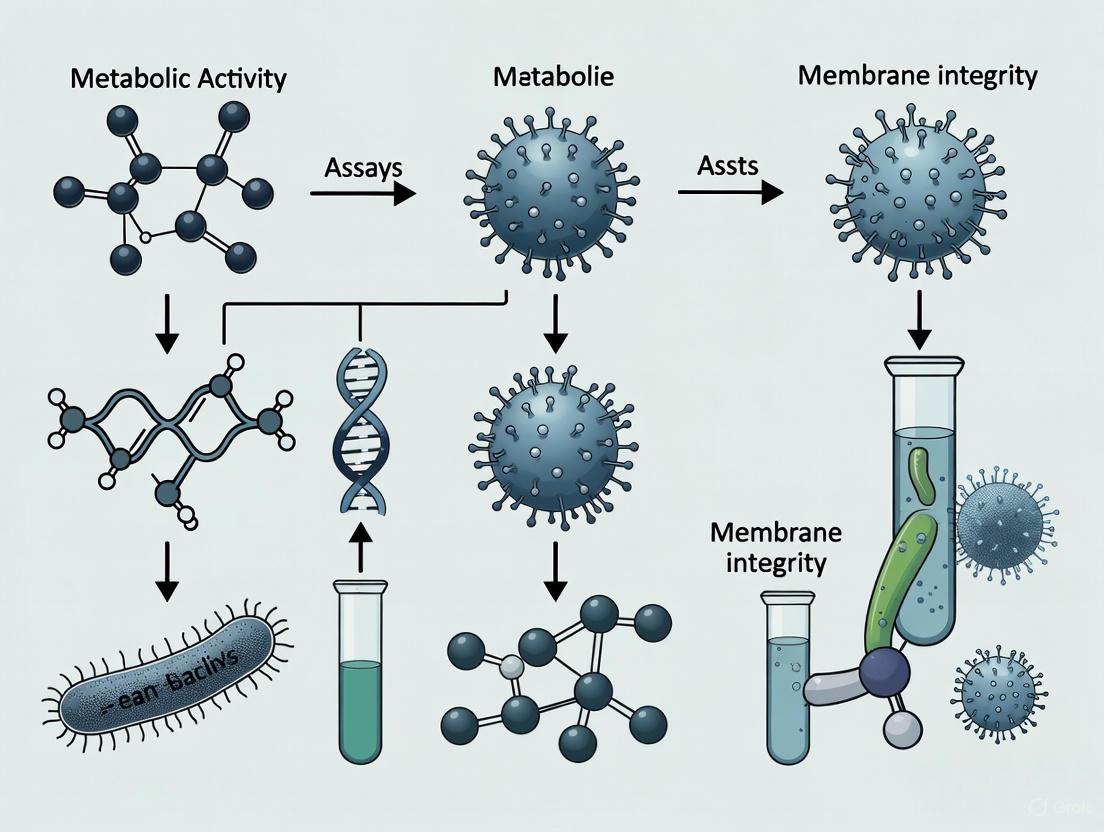
The relationship between these assessment principles and cell viability status is illustrated in the following diagram:
Comparative Analysis of Viability Assay Methods
Cell viability assays can be broadly categorized based on their underlying detection principles. The following table summarizes the major assay types, their methodologies, and key applications:
Comparison of Major Cell Viability Assay Technologies
| Assay Type | Detection Principle | Measurement Method | Key Advantages | Key Limitations |
|---|---|---|---|---|
| Metabolic Activity Assays | ||||
| Tetrazolium Reduction (MTT, MTS, XTT) [1] [4] | Mitochondrial enzyme reduction of tetrazolium to formazan | Colorimetric (absorbance) | Well-established, suitable for high-throughput | Formazan crystals may harm cells [4], long incubation (1-4 hours) [6] |
| ATP Detection [1] [6] | ATP concentration in metabolically active cells | Luminescence | Highly sensitive, rapid (<10 minutes), broad linear range [6] | Requires cell lysis, affected by compounds altering ATP levels |
| Resazurin Reduction [4] [6] | Cellular reduction of resazurin to fluorescent resorufin | Fluorometric | More sensitive than tetrazolium assays, relatively inexpensive [6] | Fluorescent compounds may interfere, long incubation (1-4 hours) |
| Membrane Integrity Assays | ||||
| Trypan Blue Exclusion [1] [3] | Dye penetration through compromised membranes | Manual or automated counting | Simple, cost-effective, versatile [7] | Subjective, small event count, dye can be toxic to cells [3] [7] |
| Propidium Iodide/7-AAD Staining [2] [7] | DNA-binding dyes enter dead cells | Flow cytometry | Objective, high-throughput, multi-parameter analysis [7] | Requires flow cytometer, potential dye toxicity |
| LDH Release [1] [6] | Cytosolic enzyme release from damaged cells | Colorimetric or fluorometric | Measures membrane damage directly, suitable for high-throughput | Background release from healthy cells, serum contains LDH |
| Live-Cell Protease Activity [6] | Intracellular protease activity in viable cells | Fluorometric | Short incubation (30-60 min), compatible with multiplexing | May not detect metabolically inactive cells with intact membranes |
Experimental Protocols for Key Viability Assays
Metabolic Activity Assay: MTT Tetrazolium Reduction
The MTT assay is one of the most widely used metabolic viability assays, particularly in academic laboratories [4].
Principle: Viable cells with active metabolism convert the yellow tetrazolium salt MTT into purple formazan crystals through mitochondrial dehydrogenases and other cellular reductases [1] [4].
Protocol:
- Cell Preparation: Seed cells in a 96-well plate and allow to attach under appropriate culture conditions.
- Treatment: Apply experimental treatments or compounds to the cells for the desired duration.
- MTT Application: Add MTT solution to each well at a final concentration of 0.2-0.5 mg/mL [4].
- Incubation: Incubate plates for 1-4 hours at 37°C to allow formazan crystal formation [4].
- Solubilization: Add solubilization solution (DMSO, acidified isopropanol, or SDS-containing buffer) to dissolve formazan crystals [4].
- Measurement: Record absorbance at 570 nm using a plate reader, with 630 nm as an optional reference wavelength [4].
Critical Considerations:
- The amount of formazan signal is proportional to the number of metabolically active viable cells [4].
- Culture conditions that alter cellular metabolism (e.g., nutrient depletion, confluence) affect MTT reduction rates [4].
- Chemical reducing agents (ascorbic acid, glutathione) can cause non-enzymatic reduction and false positives [4].
Membrane Integrity Assay: Trypan Blue Exclusion
The trypan blue exclusion method is a standard membrane integrity-based viability assay.
Principle: Trypan blue dye (960 Da) cannot penetrate the intact plasma membrane of viable cells but enters dead cells with compromised membranes, staining intracellular proteins blue [3].
Protocol:
- Sample Preparation: Prepare a single-cell suspension and dilute appropriately in balanced salt solution.
- Staining: Mix cell suspension with 0.4% trypan blue solution in a 1:1 ratio [3] [7].
- Loading: Transfer stained sample to a hemocytometer or automated cell counter chamber.
- Counting: Count unstained (viable) and blue-stained (non-viable) cells under a microscope or using automated imaging [3] [7].
- Calculation: Determine viability percentage as (number of viable cells / total cells) × 100 [7].
Critical Considerations:
- Staining should be assessed immediately (within minutes) as extended exposure can be toxic to cells [3].
- Cells with temporarily permeable membranes may be stained even though they are not dead, leading to underestimation of viability [3].
- Red blood cells in primary samples can be mistaken for dead cells, affecting accuracy [3].
The experimental workflow for these fundamental assays follows a structured process:
Research Reagent Solutions for Viability Assessment
Successful viability assessment requires appropriate selection of reagents and tools. The following table outlines essential materials used in cell viability research:
Essential Reagents for Cell Viability Assays
| Reagent/Kit | Function | Application Notes |
|---|---|---|
| Tetrazolium Salts (MTT, MTS, XTT) [1] [4] | Mitochondrial reduction to formazan products | MTT requires solubilization; MTS/XTT yield soluble formazan [6] |
| ATP Detection Kits (CellTiter-Glo) [6] | Luciferase-based ATP quantification | Highly sensitive, broad linear range, requires cell lysis [6] |
| Vital Dyes (Trypan Blue, PI, 7-AAD) [2] [3] | Membrane integrity assessment | Trypan blue for manual counting; PI/7-AAD for flow cytometry [2] [7] |
| Fluorescent Viability Stains (AO/PI, SYTOX) [2] [7] | Nucleic acid binding in dead cells | AO/PI staining allows live (green)/dead (red) differentiation [3] [7] |
| LDH Assay Kits [1] [6] | Detection of released lactate dehydrogenase | Measures membrane damage, suitable for supernatant analysis [6] |
| Resazurin-Based Kits (CellTiter-Blue) [6] | Cellular reduction to fluorescent resorufin | More sensitive than tetrazolium assays, relatively inexpensive [6] |
| Protease-Based Kits (CellTiter-Fluor) [6] | Detection of live-cell protease activity | Short incubation (30-60 min), compatible with multiplexing [6] |
Market Landscape and Research Applications
The cell viability assays market reflects the critical importance of these methods in biomedical research. The market size was estimated at USD 2.05 billion in 2025 and is projected to reach USD 4.24 billion by 2034, growing at a compound annual growth rate (CAGR) of 8.54% [8]. This growth is driven by increasing R&D activities in pharmaceutical and biotechnology sectors, rising prevalence of cancer, and regulatory requirements for cytotoxicity testing [8].
Key market segments demonstrate distinct trends:
- Product Type: Reagents and kits dominate with 65% market share in 2024 due to continuous requirements for consumables [8]
- Assay Type: Metabolic activity-based assays lead with 50% market share in 2024 [8]
- Application: Pharmaceutical and biotech research accounts for 60% market share [8]
- Technology: Colorimetric methods hold the largest revenue share (45%), while luminescent assays show the fastest growth [8]
Geographically, North America leads the market with 40% share, while the Asia-Pacific region is expected to witness the fastest growth during the forecast period [8].
The validation of metabolic activity versus membrane integrity viability assays remains a critical consideration in cell-based research. The choice between these approaches should be guided by several factors:
Assay Selection Criteria:
- Research Question: Determine whether measurement of live cells, dead cells, or both is required [6]
- Sample Characteristics: Consider cell type, homogeneity, and availability [7]
- Experimental Conditions: Account for treatment effects on specific cellular functions [4]
- Technical Resources: Evaluate instrument availability and expertise [7]
- Throughput Needs: Consider manual versus automated methods based on sample number [7]
Metabolic activity assays are ideal for assessing overall cellular health and function, while membrane integrity assays provide a direct measurement of cell death. For comprehensive assessment, multiplexing approaches that combine both principles may provide the most robust viability data [6].
The expanding market for cell viability assays and continuous technological innovations underscore the fundamental importance of accurately distinguishing between life and death at the cellular level. As research progresses, the integration of automated systems and artificial intelligence is poised to enhance the efficiency, accuracy, and reproducibility of viability measurements, further strengthening their role in biomedical discovery and therapeutic development [8].
Accurately determining cell viability is fundamental to biomedical research and drug development, yet the plethora of available methods has historically complicated data interpretation and cross-study comparisons. The Organisation for Economic Co-operation and Development (OECD) provides a crucial framework to address this challenge through a standardized classification system for cell viability methods [9]. This classification offers researchers, scientists, and drug development professionals a unified language and systematic approach for selecting and interpreting viability assays.
The OECD categorizes these methods into four distinct groups: non-invasive cell structure damage, invasive cell structure damage, cell growth, and cellular metabolism [9]. This blueprint is particularly valuable for a critical research question: validating metabolic activity assays against membrane integrity-based methods. While a cell is considered viable if it can perform essential functions, it is considered dead when it irreversibly loses plasma membrane barrier function, forms apoptotic bodies, or is engulfed by phagocytes [9]. This distinction is paramount because assays measuring these different principles can yield divergent results, potentially impacting conclusions about compound toxicity or treatment efficacy. This guide objectively compares assay performance within the OECD framework, providing the experimental data and methodologies necessary for informed assay selection in validation studies.
The OECD Classification Framework
The OECD classification system organizes cell viability assessment methods based on their fundamental operating principles and what they measure. Understanding these categories is the first step in selecting an appropriate assay and correctly interpreting its results, especially in the context of comparing metabolic activity with membrane integrity.
The following diagram illustrates the logical decision pathway for classifying viability assays according to the OECD framework:
This structured approach helps researchers quickly identify the appropriate assay category based on the specific cellular parameter they need to measure. The classification highlights a critical dichotomy: assays based on cell membrane integrity (Categories 1 and 2) versus those based on metabolic activity (Category 4). This distinction is central to validation studies, as these two principles can sometimes yield conflicting results, particularly when cells are metabolically compromised but maintain membrane integrity, or vice versa.
Comparative Analysis of Viability Assays
Quantitative Performance Data
The following table summarizes the key characteristics of the most common viability assays within the OECD framework, providing a direct comparison of their mechanisms, advantages, and limitations [4] [9] [6].
Table 1: Comprehensive Comparison of Cell Viability Assays Based on OECD Classification
| Assay Name | OECD Category | Measurement Principle | Detection Method | Throughput | Advantages | Disadvantages |
|---|---|---|---|---|---|---|
| MTT Assay | Cellular Metabolism | Tetrazolium reduction by cellular dehydrogenases | Absorbance | Medium | Inexpensive, widely established [4] [10] | Formazan insolubility requires solubilization step, endpoint measurement only [4] [10] |
| ATP Assay | Cellular Metabolism | ATP quantification in metabolically active cells | Luminescence | High | Highly sensitive, broad linear range, rapid signal generation [6] | Requires cell lysis, measures current metabolic state only [6] |
| Resazurin Assay | Cellular Metabolism | Resazurin reduction to fluorescent resorufin | Fluorescence | Medium | Inexpensive, more sensitive than tetrazolium assays [4] [10] | Potential fluorescence interference, extended incubation can be toxic [4] [10] |
| LDH Release Assay | Non-Invasive Cell Structure Damage | Cytoplasmic enzyme release upon membrane damage | Absorbance/Fluorescence/Luminescence | Medium | Easy to perform, directly measures cytotoxicity [9] [6] | Can leak from stressed but viable cells, background issues in serum [9] |
| Trypan Blue Exclusion | Invasive Cell Structure Damage | Dye penetration into membrane-compromised cells | Microscopy/Image Analysis | Low | Cost-effective, direct cell counting [9] | Manual counting labor-intensive, short incubation critical to avoid false positives [9] |
| Propidium Iodide Uptake | Invasive Cell Structure Damage | DNA binding in membrane-compromised cells | Fluorescence | Medium-High | Compatible with flow cytometry, can be multiplexed with other dyes [9] | Requires specialized equipment, potential false positives from transient permeability [9] |
Metabolic Activity vs. Membrane Integrity: Key Validation Studies
The core thesis of validating metabolic activity against membrane integrity assays reveals critical insights into cellular responses to toxic insults. Research indicates that membrane integrity assays typically detect later stages of cell death, while metabolic assays can reveal earlier perturbations in cellular health [9] [6]. This temporal relationship is crucial for understanding the mechanism of action of experimental compounds.
Experimental data demonstrate that metabolic assays like MTT can directly substitute for traditional proliferation assays like tritiated thymidine incorporation in many experimental situations [4]. However, it's crucial to note that tetrazolium reduction reflects viable cell metabolism rather than specifically cell proliferation, a distinction often overlooked without proper controls [4]. Discrepancies become particularly evident when assessing compounds that affect mitochondrial function without immediate membrane rupture, where metabolic assays show cytotoxicity earlier than membrane integrity tests [9] [10].
A significant validation challenge arises from the observation that cytoplasmic enzymes like LDH can leak from cells under stress conditions even when the plasma membrane remains technically intact [9]. This phenomenon can lead to overestimation of cytotoxicity in membrane integrity assays. Conversely, metabolic assays have their own limitations; for instance, the MTT reagent itself exhibits cytotoxicity with prolonged exposure, and its formazan crystals can physically harm cells [4]. These findings underscore the importance of using orthogonal methods when validating critical results.
Experimental Protocols for Key Assays
MTT Assay for Metabolic Activity
Principle: Metabolically active cells reduce the yellow tetrazolium salt MTT(3-(4,5-dimethylthiazol-2-yl)-2,5-diphenyltetrazolium bromide) to purple formazan crystals via cellular dehydrogenases [4].
Reagents:
- MTT solution: 5 mg/ml in Dulbecco's Phosphate Buffered Saline, pH 7.4 [4]
- Solubilization solution: 40% dimethylformamide, 2% glacial acetic acid, 16% SDS, pH adjusted to 4.7 [4]
Procedure:
- Prepare cells in a 96-well plate with appropriate test compounds and controls [4].
- Incubate under experimental conditions for desired duration.
- Add MTT solution to each well to a final concentration of 0.2-0.5 mg/ml [4].
- Incubate plates for 1-4 hours at 37°C [4].
- Add solubilization solution to dissolve formazan crystals [4].
- Measure absorbance at 570 nm using a plate reader [4].
Critical Notes:
- The amount of signal depends on MTT concentration, incubation time, cell number, and metabolic activity [4].
- Culture conditions that alter cellular metabolism will affect MTT reduction rates [4].
- Chemical interference from reducing compounds can cause false positives [4].
LDH Release Assay for Membrane Integrity
Principle: Lactate dehydrogenase (LDH), a stable cytoplasmic enzyme, is released upon cell membrane damage. The released LDH catalyzes the conversion of lactate to pyruvate, generating NADH which can be measured through various coupled reactions [6].
Reagents:
- LDH assay kit (e.g., CytoTox 96 Non-Radioactive Cytotoxicity Assay) [6]
- Lysis solution (for maximum LDH release control)
- Stop solution (if required by specific kit)
Procedure:
- Plate cells and treat with experimental compounds, including untreated control (low LDH) and lysed cell control (high LDH) [6].
- Following treatment incubation, centrifuge plates to pellet cells and transfer supernatant to new plate.
- Add LDH assay reagent to supernatant and incubate for 30 minutes protected from light.
- Measure absorbance at 490-500 nm or fluorescence (depending on kit specifications) [6].
- Calculate cytotoxicity percentage: (Experimental LDH - Spontaneous LDH) / (Maximum LDH - Spontaneous LDH) × 100.
Critical Notes:
- Serum-containing media often have high LDH background; use serum-free media during assay incubation when possible [9].
- Avoid freeze-thaw cycles which can damage membranes and artificially elevate LDH [9].
- The assay measures membrane integrity at a single time point rather than providing kinetic data [6].
The workflow for these two fundamentally different assay types highlights their distinct approaches to viability assessment:
Research Reagent Solutions
Selecting appropriate reagents is crucial for obtaining reliable, reproducible results in viability assays. The following table details essential materials and their functions for the featured experiments.
Table 2: Essential Research Reagents for Viability Assays
| Reagent/Material | Function/Principle | Example Applications |
|---|---|---|
| MTT (Thiazolyl Blue Tetrazolium Bromide) | Positively charged tetrazolium salt penetrated viable cells and reduced to insoluble formazan by cellular dehydrogenases [4] [10] | MTT assay for metabolic activity measurement [4] |
| Resazurin Sodium Salt | Cell-permeable blue dye reduced to pink, fluorescent resorufin in viable cells [4] [10] | Resazurin assay for metabolic activity, more sensitive than tetrazolium assays [4] |
| CellTiter-Glo Reagent | Luciferase-based system detecting ATP from lysed viable cells, generating luminescent signal [6] | ATP assay for highly sensitive viability quantification [6] |
| Propidium Iodide | Membrane-impermeant DNA intercalating dye that fluoresces upon binding nucleic acids in dead cells [9] | Flow cytometry and fluorescence microscopy to identify membrane-compromised cells [9] |
| Trypan Blue Solution | Diazo dye excluded by intact membranes but stains dead cells blue [9] | Manual cell counting with hemocytometer for viability assessment [9] |
| LDH Assay Kit | Measures lactate dehydrogenase enzyme released from cytosol of membrane-damaged cells [9] [6] | Quantifying cytotoxicity through enzymatic activity in supernatant [9] |
| Calcein AM | Cell-permeable, non-fluorescent compound converted to green-fluorescent calcein by intracellular esterases in live cells [10] | Fluorescent labeling of viable cells, often multiplexed with propidium iodide [10] |
The OECD classification blueprint provides an invaluable framework for navigating the complex landscape of cell viability assays. By categorizing methods based on their fundamental principles—non-invasive cell structure damage, invasive cell structure damage, cell growth, and cellular metabolism—this system enables researchers to make informed decisions about assay selection and interpretation [9].
The critical comparison between metabolic activity assays (e.g., MTT, Resazurin, ATP) and membrane integrity assays (e.g., LDH, Trypan Blue, propidium iodide) reveals that these methods provide complementary rather than interchangeable data [9] [6] [10]. Metabolic assays typically detect earlier signs of cellular stress, while membrane integrity assays confirm later-stage cell death. This temporal relationship, combined with the potential for artifacts in both systems, underscores the importance of the OECD framework for selecting orthogonal methods in validation studies.
For researchers validating viability assays within the context of drug development and toxicology, the evidence suggests that a multiplexed approach—combining metabolic activity and membrane integrity assessments—provides the most comprehensive understanding of cellular responses to experimental compounds. The standardized protocols and reagent information provided here offer a foundation for implementing these critical assessments with the rigor required for regulatory compliance and scientific advancement.
In biomedical research and drug development, accurately determining cell viability is fundamental for assessing cellular responses to treatments, toxins, or environmental stressors. Cell viability assays are crucial tools in pharmacology, toxicology, and cancer research for predicting in vivo pharmacological and toxicological effects [11]. While viability can be assessed through various cellular properties, assays measuring metabolic activity—specifically redox potential and adenosine triphosphate (ATP) levels—have become dominant proxies for cell health and functionality. The global cell viability assays market, where metabolic activity-based assays hold a leading share, is projected to grow significantly, underscoring their importance in pharmaceutical and biotech research [8].
This guide provides a comparative analysis of metabolic activity-based viability assays against other methodologies, with a focus on their mechanistic principles, experimental applications, and validation data. We specifically examine the relationship between cellular energy metabolism and viability, exploring how measurements of redox potential and ATP content serve as sensitive indicators of cellular health before the loss of membrane integrity occurs [11].
Comparative Analysis of Viability Assay Categories
Cell viability assays are broadly categorized based on the fundamental cellular properties they measure. The Organisation for Economic Co-operation and Development (OECD) provides a classification system that includes categories for structural cell damage (non-invasive and invasive), cell growth, and cellular metabolism [12]. Table 1 compares the core principles, key examples, and key differentiators of the main assay categories.
Table 1: Comparison of Major Cell Viability Assay Categories
| Assay Category | Core Principle | Key Examples | What is Actually Measured | Key Differentiators |
|---|---|---|---|---|
| Metabolic Activity | Measures key biochemical processes indicative of cellular metabolism and energy production [11] [12]. | ATP assays, Tetrazolium reduction (MTT, XTT), Resazurin reduction [11] [12]. | Enzymatic activity, redox potential, or ATP levels as proxies for cellular activity [11]. | Detects early metabolic decline; higher sensitivity to early stress; measures potential viability [11]. |
| Membrane Integrity | Assesses the integrity of the plasma membrane, which is compromised during cell death [11] [12]. | Trypan Blue, Propidium Iodide, LDH release [11] [12]. | Permeability to dyes or leakage of cytoplasmic enzymes [11]. | Definitively identifies dead/necrotic cells; often used with metabolic assays for orthogonal validation [11]. |
| Apoptosis | Measures specific markers of programmed cell death [11]. | Annexin V, caspase activation, DNA fragmentation [11]. | Phosphatidylserine exposure, protease activity, and DNA cleavage [11]. | Distinguishes between regulated apoptosis and passive necrosis [11]. |
| Proliferation & Biomass | Evaluates the ability of cells to grow and divide over time [11]. | CFSE tracking, DNA content assays [11]. | Changes in cell number or biomass over time [11]. | Monitors dynamic growth; differentiates cytostatic vs. cytotoxic effects [11]. |
Metabolic activity assays are particularly valuable because they can detect early signs of cell stress and cytotoxicity before irreversible membrane damage occurs. A cell is considered viable if it can perform its essential functions, and since these functions are energy-dependent, metabolic competence is a fundamental indicator of cell health [12]. In contrast, membrane integrity assays identify cells that have already passed the point of no return, making metabolic assays more sensitive for early-stage toxicity screening [11].
Metabolic Activity Assays: Mechanisms and Methodologies
ATP Quantification Assays
ATP serves as the primary energy currency of living cells, and its concentration is tightly regulated. The measurement of intracellular ATP levels provides a direct and sensitive snapshot of cellular energy status and viability [13] [11].
- Mechanistic Principle: ATP is maintained at millimolar concentrations within cells through rapid regeneration from adenosine diphosphate (ADP) and adenosine monophosphate (AMP). Almost all essential cellular functions, including active transport, biosynthesis, and signaling, consume ATP [14]. A decline in cellular ATP levels is therefore a direct indicator of metabolic compromise.
- Protocol: Luminescent ATP Assay: The standard methodology involves cell lysis followed by a bioluminescent reaction. The enzyme luciferase catalyzes a reaction between ATP and its substrate, D-luciferin, producing light. The emitted luminescence is directly proportional to the ATP concentration in the sample [11] [8]. This assay is highly sensitive, with a wide dynamic range, and can be performed in a high-throughput format using multi-well plate readers [11].
- Key Validation Data: ATP assays have been rigorously validated against other viability methods. A seminal 1993 study demonstrated a strong correlation between intracellular ATP levels and cell viability after hyperthermia, with the ATP assay showing a closer correlation to the gold-standard colony formation assay than other methods like the succinate dehydrogenase (SD) assay [13].
Redox Potential Assays
Redox assays measure the cellular reducing capacity, which is linked to the activity of metabolic pathways and electron transport systems [15].
- Mechanistic Principle: Metabolically active cells contain reducing equivalents like NADH and NADPH. Tetrazolium salts (e.g., MTT, XTT, WST-1) are membrane-permeable compounds that are reduced by cellular oxidoreductases to colored, formazan products. The rate and amount of formazan formation are proportional to the number of metabolically active cells and their overall metabolic activity [11] [15]. Similarly, resazurin is reduced from a blue, non-fluorescent state to a pink, highly fluorescent molecule called resorufin [11].
- Protocol: Tetrazolium-Based Colorimetric Assay (e.g., MTT): Cells are incubated with the tetrazolium compound for a designated period (typically 1-4 hours). The formed formazan crystals are then solubilized using an organic solvent like DMSO or isopropanol. The absorbance of the solubilized product is measured at a specific wavelength (570 nm for MTT) using a spectrophotometric plate reader [11]. The color intensity is directly proportional to the metabolic activity of the cell population.
- Considerations and Limitations: It is crucial to understand that redox assays measure metabolic activity and redox capacity, not viability directly. Metabolic shifts can occur in normal physiology and may be reversible, meaning these assays are sensitive but do not provide definitive proof of cell death [11]. Furthermore, the choice of tetrazolium salt matters; some salts like CTC can be toxic to certain cells, and different salts may be reduced by different enzyme systems, complicating cross-study comparisons [15].
The following diagram illustrates the core metabolic pathways and detection principles underlying these key assays.
Experimental Data and Comparative Performance
Quantitative Comparison of Assay Performance
The choice of viability assay can significantly impact the interpretation of a compound's cytotoxicity. Different assays can yield varying IC₅₀ values for the same substance, highlighting the importance of selecting a biologically relevant method [11]. Table 2 summarizes key performance characteristics and comparative data for the major assay types.
Table 2: Performance Characteristics and Validation Data of Key Viability Assays
| Assay Type | Specific Example | Key Correlative Finding | Reported Limitations & Artifacts |
|---|---|---|---|
| ATP Assay | Luminescence-based ATP assay | Correctly reflected cell viability after hyperthermia, showing 2.0% and 0.7% viability in two cell lines, closely matching the 0.9% and 0% result from the colony formation gold standard [13]. | Requires cell lysis, preventing subsequent analysis of the same sample. |
| Redox Assay | Tetrazolium (MTT) | Used as a proxy for overall metabolic activity and number of active cells; production of formazan per cell increases with growth rate [15]. | Susceptible to interference from abiotic reductants and compounds that affect redox state; can overestimate viability in late stationary phase [11] [15]. |
| Membrane Integrity | Lactate Dehydrogenase (LDH) Release | Correlates with permanent membrane disruption, a definitive marker of cell death [12]. | Can be released from viable cells under stress; high background in some media; can underestimate cytotoxicity in co-cultures [12]. |
| Membrane Integrity | Trypan Blue Dye Exclusion | Selectively penetrates dead cells with damaged plasma membranes [12]. | Prolonged incubation can stain viable cells due to dye aggregate dissociation; may underestimate dead cells with short incubation [12]. |
Orthogonal Validation and the Case for Multi-Parametric Assessment
No single assay can fully capture all aspects of cell viability, as cell death is a complex, context-dependent sequence [11]. Relying on a single parameter can lead to misinterpretation. For instance, a study found that the LDH method can underestimate cytotoxicity in co-culture systems with bacteria [12]. Similarly, tetrazolium reduction might be poor in non-growing but still viable cells during the late stationary phase [15].
Therefore, orthogonal validation—pairing a metabolic assay with a membrane integrity assay—is a recommended best practice. This approach provides a more comprehensive picture, distinguishing between cells that are metabolically compromised but still intact and those that are unequivocally dead [11]. For example, a cell population treated with a toxic compound might show a significant drop in ATP levels (measured by luminescence) while still excluding trypan blue, indicating early-stage toxicity that has not yet progressed to membrane lysis.
The Scientist's Toolkit: Essential Reagents and Instruments
Implementing these assays requires specific reagents and instrumentation. The following table details key solutions for conducting metabolic and membrane integrity assays.
Table 3: Research Reagent Solutions for Cell Viability Assessment
| Item Name | Category / Assay Type | Core Function |
|---|---|---|
| RealTime-Glo MT Cell Viability Assay | Metabolic Activity / ATP | Uses a luciferase pro-substrate and extracellular luciferin to measure ATP levels in real-time without lysis [16]. |
| CellTiter-Glo Luminescent Cell Viability Assay | Metabolic Activity / ATP | A homogeneous, lysis-based method to determine the number of viable cells based on quantitation of ATP [11]. |
| Tetrazolium Salts (MTT, XTT, WST-1/8) | Metabolic Activity / Redox Potential | Serve as chromogenic substrates reduced by metabolically active cells to form formazan dyes, enabling colorimetric quantification [11] [15]. |
| Resazurin Sodium Salt | Metabolic Activity / Redox Potential | A blue, non-fluorescent dye that is reduced to pink, fluorescent resorufin in viable cells, allowing fluorometric or colorimetric readout [11]. |
| ToxiLight BioAssay Kit | Membrane Integrity / Non-Invasive | Measures the release of adenylate kinase (AK) from cells with damaged membranes [12]. |
| CytoTox-Fluor Cytotoxicity Assay | Membrane Integrity / Non-Invasive | Measures a dead-cell protease activity that is released upon membrane damage [12]. |
| Propidium Iodide (PI) | Membrane Integrity / Invasive | A membrane-impermeant fluorescent nucleic acid stain that enters dead cells, used in microscopy and flow cytometry [11] [12]. |
| Automated Cell Counter (e.g., Countess II) | Instrumentation / Membrane Integrity | Automated microscope-based systems that often incorporate fluorescence channels to count and differentiate live/dead cells stained with dyes like trypan blue or PI [12]. |
| Microplate Reader (e.g., GloMax Explorer) | Instrumentation / Multi-Detection | An essential instrument for high-throughput assays, capable of measuring luminescence, fluorescence, and absorbance in multi-well plates [16]. |
Metabolic activity assays, particularly those measuring ATP and redox potential, are indispensable tools for assessing cell viability with high sensitivity and throughput. Their ability to detect early signs of metabolic stress before the loss of membrane integrity provides a significant advantage in drug discovery and toxicology. However, the data from these assays must be interpreted with a clear understanding of their mechanistic principles and limitations.
The future of viability assessment is moving toward more complex and physiologically relevant 3D cell culture models, such as spheroids and organoids [11]. These models present new challenges for viability assays, including limited reagent penetration and cellular heterogeneity, which will likely drive the adoption of multimodal assessment strategies that combine metabolic readouts with other parameters. Furthermore, the integration of artificial intelligence (AI) is poised to enhance the efficiency, accuracy, and reproducibility of viability assays by automating data analysis and interpretation, allowing researchers to focus on biological insights [8]. As these technologies evolve, the link between metabolic activity and cell health will remain a cornerstone of robust cellular analysis.
In cellular biology, the integrity of the plasma membrane serves as a fundamental proxy for determining cell viability across diverse research and clinical applications. The plasma membrane's selective permeability creates a crucial barrier between the intracellular milieu and extracellular environment, maintaining homeostasis through precise regulation of molecular transit [17]. When this barrier function is compromised, the uncontrolled exchange of ions and molecules signals a loss of cellular viability, often representing one of the final stages in the cell death process [18]. This definitive characteristic makes membrane integrity assessment a cornerstone technique in fields ranging from biomaterial cytotoxicity testing to drug development and cell therapy validation.
The scientific and clinical reliance on membrane integrity stems from its clear distinction between living and dead cells. As described by the Nomenclature Committee on Cell Death, a cell is considered dead when "the plasma membrane's barrier function is irreversibly lost" [9]. This binary determination provides a straightforward, reliable metric compared to more complex assessments of metabolic activity or cellular function, which may exist on a continuum. While membrane integrity tests effectively differentiate between dead (membrane-permeable) and non-dead (membrane-impermeable) cells, it is crucial to recognize they "fail to distinguish healthy cells from cells that have entered the cell death process but have not reached the membrane permeabilization step" [18]. This limitation underscores the importance of understanding both the power and constraints of membrane integrity as a viability proxy within the broader context of cell death mechanisms.
Methodological Comparison: Quantitative Assessment of Membrane Integrity Techniques
Researchers employ diverse methodologies to assess membrane integrity, each with distinct advantages, limitations, and appropriate applications. The most common approaches utilize membrane-impermeable dyes that penetrate only compromised membranes, providing visual or quantitative indicators of viability.
Core Membrane Integrity Assessment Technologies
The following table summarizes the primary techniques used for membrane integrity-based viability assessment:
Table 1: Comparison of Membrane Integrity Assessment Methods
| Method | Key Dyes/Markers | Detection Principle | Applications | Advantages | Limitations |
|---|---|---|---|---|---|
| Fluorescence Microscopy (FM) | FDA/PI, Acridine Orange/Propidium Iodide [19] [18] | Visual imaging of fluorescent staining in compromised cells | Biomaterial cytotoxicity [19], qualitative viability screening | Direct cell visualization, spatial context | Subjective quantification, sampling bias, autofluorescence interference [19] |
| Flow Cytometry (FCM) | Propidium Iodide, 7-AAD, Hoechst dyes [19] [18] | High-throughput single-cell analysis in suspension | Quantitative viability, apoptosis/necrosis distinction [19] | Statistical robustness, multi-parameter analysis, objective quantification [19] | Requires single-cell suspension, specialized instrumentation |
| Microplate-Based Imaging Cytometry | Hoechst 33342, Propidium Iodide [17] | Automated imaging and counting of stained cells in multi-well plates | High-content screening, membrane repair studies [17] | Combines imaging with statistical power, suitable for adhesion cells | Lower throughput than flow cytometry |
| Enzyme Leakage Assays | LDH, AK, G3PDH, G6PD detection [9] | Measure cytoplasmic enzyme release into supernatant | High-throughput toxicity screening, | Amenable to automation, compatible with multi-well formats | Potential background interference, may detect reversible permeability [9] |
| Specialized Nanopore Detection | Tl+ with FluxOR dye [20] | Fluorescent detection of thallium ion uptake through sub-1.5nm pores | Detection of nanoscale membrane defects [20] | Detects pores too small for conventional dyes, high sensitivity | Specialized dye requirement, limited adoption |
Comparative Performance of FM versus FCM in Particulate Systems
Recent research has directly compared the performance of fluorescence microscopy and flow cytometry for viability assessment in challenging particulate systems. A 2025 study evaluating Bioglass 45S5 cytotoxicity on SAOS-2 osteoblast-like cells demonstrated that while both techniques identified the same trends (increased cytotoxicity with smaller particles and higher concentrations), flow cytometry provided superior precision and sensitivity, particularly under high cytotoxic stress [19].
Table 2: Quantitative Comparison of FM and FCM Viability Measurements with Bioglass 45S5 Particles [19]
| Particle Size | Concentration (mg/mL) | FM Viability (%) | FCM Viability (%) |
|---|---|---|---|
| Control | - | >97% | >97% |
| <38 μm | 25 mg/mL | 62% (3h), 65% (72h) | 58% (3h), 60% (72h) |
| <38 μm | 50 mg/mL | 35% (3h), 38% (72h) | 30% (3h), 32% (72h) |
| <38 μm | 100 mg/mL | 9% (3h), 10% (72h) | 0.2% (3h), 0.7% (72h) |
The study reported a strong correlation between FM and FCM data (r = 0.94, R² = 0.8879, p < 0.0001), validating both methods for basic viability assessment [19]. However, FCM demonstrated distinct advantages in detecting early apoptotic events and provided more precise quantification under extreme cytotoxic conditions where viability dropped below 10% [19]. This enhanced precision makes flow cytometry particularly valuable for quantifying subtle viability differences in rigorous comparative studies.
Experimental Protocols: Standardized Methods for Membrane Integrity Assessment
Protocol A: Flow Cytometry with Multiparametric Viability Staining
This protocol enables high-precision quantification of viability and distinction between apoptotic and necrotic populations using multiparametric flow cytometry, as validated in comparative methodological studies [19].
Reagents and Equipment:
- Cell suspension (0.5-1×10⁶ cells/mL)
- Propidium iodide (PI) stock solution (50 μg/mL)
- Hoechst 33342 stock solution (100 μg/mL)
- Annexin V-FITC conjugate
- DiIC1 staining solution
- Flow cytometry staining buffer (PBS with 1% FBS)
- Flow cytometer with 488nm and 355nm lasers
- Centrifuge maintained at 4°C
Procedure:
- Cell Preparation: Harvest cells and wash twice with cold staining buffer. Adjust concentration to 0.5-1×10⁶ cells/mL.
- Staining Cocktail: Combine 100 μL cell suspension with Hoechst 33342 (final concentration 1 μg/mL), DiIC1 (as recommended), Annexin V-FITC (5 μL/test), and PI (final concentration 0.5 μg/mL).
- Incubation: Protect samples from light and incubate for 15 minutes at room temperature.
- Analysis: Add 400 μL staining buffer to each tube and analyze immediately by flow cytometry.
- Gating Strategy:
- Exclude debris based on forward and side scatter
- Identify viable cells: Hoechst⁺/PI⁻/Annexin V⁻
- Early apoptotic: Hoechst⁺/PI⁻/Annexin V⁺
- Late apoptotic/necrotic: Hoechst⁺/PI⁺/Annexin V⁺
This protocol's key advantage is its ability to "distinguish early and late apoptosis from necrosis" while demonstrating "superior precision, particularly under high cytotoxic stress" [19].
Protocol B: Endpoint Membrane Integrity Assessment Using Imaging Cytometry
This endpoint assay provides a population-based measure of membrane integrity following mechanical or chemical injury, suitable for screening multiple experimental conditions [17].
Reagents and Equipment:
- Adherent cells in 96-well plates
- Hoechst 33342 solution (5 μg/mL)
- Propidium iodide solution (1 μg/mL)
- Injury induction reagents (glass beads or digitonin)
- Celigo Imaging Cytometer or similar plate-based imaging system
- Plasma membrane wounding apparatus (for mechanical injury)
Procedure:
- Cell Preparation: Plate cells in 96-well plates and grow to 70-80% confluence.
- Membrane Injury:
- Staining: Add Hoechst 33342 (5 μg/mL) and PI (1 μg/mL) directly to culture media.
- Incubation: Incubate for 15-20 minutes at 37°C protected from light.
- Imaging and Analysis:
- Image each well using brightfield and fluorescence channels
- Count total nuclei (Hoechst 33342 positive)
- Count membrane-compromised cells (PI positive)
- Calculate viability percentage: (Total cells - PI positive cells)/Total cells × 100
This method's advantage is its ability to "provide a population-based measure of membrane permeabilization and cell death" at the single-cell level while being simpler than real-time imaging approaches [17].
Experimental Workflow for Membrane Integrity Assessment
Research Reagent Solutions: Essential Tools for Membrane Integrity Studies
Successful membrane integrity assessment requires specific reagents and tools optimized for different experimental needs. The following table catalogues essential research solutions with their respective applications:
Table 3: Essential Research Reagents for Membrane Integrity Studies
| Category | Specific Reagents | Function and Mechanism | Applications and Considerations |
|---|---|---|---|
| Membrane-Impermeant Nucleic Acid Stains | Propidium Iodide (PI) [18], 7-AAD [18], Ethidium Homodimer [18] | Bind nucleic acids upon entering through compromised membranes; excluded by intact membranes | Standard viability assessment; PI requires 488nm excitation; 7-AAD preferred for multicolor experiments |
| Vital Stains for Live Cells | Fluorescein Diacetate (FDA) [18], Calcein-AM [18], Hoechst 33342 [17] | FDA/Calcein-AM converted by esterases to fluorescent products retained in viable cells; Hoechst stains all nuclei | Counterstains for viability assessment; Hoechst 33342 concentration-critical for selective permeability |
| Apoptosis Detection Reagents | Annexin V-FITC [19], Yo-Pro-1 [20], YO-PRO-3 | Annexin V binds phosphatidylserine externalized in apoptosis; Yo-Pro dyes enter apoptotic cells earlier than PI | Distinguishing apoptotic vs. necrotic death; requires calcium-containing buffer for Annexin V binding |
| Specialized Nanopore Detection | FluxOR Tl+ sensing dye [20], Thallium salts | Fluorescent detection of Tl+ uptake through sub-1.5nm pores; Tl+ van der Waals diameter = 0.392nm | Detecting nanoscale membrane defects invisible to conventional dyes; specialized application |
| Membrane Injury Agents | Digitonin [17], Glass beads (100-500μm) [17], Streptolysin O (SLO) | Controlled membrane disruption; digitonin binds cholesterol; glass beads cause mechanical injury | Standardizing injury models; digitonin concentration critical for sublytic effects |
| Instrumentation | Flow cytometers [19], Fluorescence microscopes [19], Imaging cytometers [17] | Quantification and visualization of dye incorporation | Flow cytometry offers statistical power; microscopy provides spatial context |
Limitations and Considerations in Membrane Integrity Interpretation
While membrane integrity assays provide valuable viability proxies, researchers must acknowledge their methodological constraints to avoid misinterpretation. A significant limitation is that membrane permeabilization represents a relatively late stage in cell death, meaning that "membrane integrity tests differentiate only between dead (membrane permeable) and non-dead (membrane impermeable) cells but fail to distinguish healthy cells from cells that have entered the cell death process but have not reached the membrane permeabilization step" [18]. This temporal limitation means that cells with activated death pathways may still register as viable by membrane integrity assays alone.
Technical considerations also significantly impact assay accuracy. Dye concentration and incubation time require precise optimization, as "prolonged incubation can result in viable cell staining due to dissociation of dye aggregates" while insufficient incubation may "lead to underestimation of dead cells" [9]. Autofluorescence from biomaterials can "inhibit fluorescence imaging" and limit analysis of attached cells, particularly problematic with polymeric and glass materials [19]. Additionally, false positives may occur "due to changes in osmolarity, metabolism, or spontaneous invagination" that permit dye penetration without true loss of viability [9].
For comprehensive cell health assessment, membrane integrity should be complemented with other viability measures. Metabolic activity assays (e.g., MTT, ATP content), mitochondrial function tests (e.g., JC-1, TMRE), and oxygen consumption rate measurements provide orthogonal data about cellular states that may detect stress before membrane compromise occurs [18]. This multi-parameter approach is particularly valuable when evaluating subtle cellular responses to toxic insults or experimental treatments.
Membrane integrity assessment remains an essential tool for viability determination across biological research and preclinical applications. The plasma membrane's role as the definitive barrier between living cells and their environment provides a functionally relevant endpoint for distinguishing viable from non-viable cells. Current methodologies span from simple microscopic examination to sophisticated multiparametric flow cytometry, each with appropriate applications and limitations.
The continuing evolution of membrane integrity assessment focuses on addressing current limitations through improved quantification methods, novel detection technologies for subtle membrane alterations, and integration with complementary viability measures. As research advances, the combination of membrane integrity with metabolic profiling and functional assessments will provide increasingly comprehensive understanding of cellular responses in diverse experimental contexts. For researchers, selecting the appropriate membrane integrity method requires careful consideration of experimental goals, sample characteristics, and necessary throughput to ensure accurate viability interpretation within the broader landscape of cell health assessment.
In cell-based research, accurately determining cellular state is fundamental, yet the distinction between a dead, senescent, or proliferating cell is often blurred by the limitations of common assays. Many studies rely on single-parameter viability tests, which can be misleading. A cell may be viable but non-proliferative due to senescence, or a cytotoxicity assay might miss early apoptotic events. This guide provides a structured comparison of methods to objectively distinguish between cell death, cellular senescence, and active proliferation, with a specific focus on validating findings through the complementary use of metabolic activity and membrane integrity assays.
Deciphering the Cellular Phenotypes
The first step in accurate distinction is to understand the defining hallmarks of each cellular state.
- Cell Death: This is an irreversible endpoint. The two primary forms are apoptosis, a programmed, controlled process characterized by cell shrinkage, nuclear fragmentation, and membrane blebbing without immediate rupture, and necrosis, an unregulated form of death involving cellular swelling and plasma membrane rupture [21]. Secondary necrosis can occur when apoptotic cells are not cleared.
- Cellular Senescence: This is a state of irreversible, long-term cell cycle arrest in which cells remain viable but cease dividing [22]. Senescent cells are not dead; they are metabolically active and display characteristic features like an enlarged, flattened morphology, increased activity of senescence-associated β-galactosidase (SA-β-gal) at pH 6.0, and a distinct secretome known as the Senescence-Associated Secretory Phenotype (SASP) [21] [23].
- Cell Proliferation: This state describes active cell division and an increase in cell number. Proliferating cells progress through the cell cycle (G1, S, G2, M) and can be identified by markers of DNA synthesis or specific cell cycle proteins [21].
Table 1: Core Characteristics of Cellular States
| Cellular State | Proliferation | Key Defining Features | Common Markers |
|---|---|---|---|
| Cell Death | No | Loss of membrane integrity; cellular fragmentation (apoptosis) or swelling (necrosis). | Propidium iodide uptake; Caspase activation; LDH release [21] [2]. |
| Senescence | No | Permanent cell cycle arrest; enlarged, flat morphology; SASP. | SA-β-gal activity (pH 6.0); p16INK4a; p21; Lamin B1 loss [22] [21]. |
| Proliferation | Yes | Active progression through the cell cycle; DNA replication. | BrdU/EdU incorporation; Ki-67; Phospho-Histone H3; DNA content analysis [21]. |
Assay Comparison: Principles and Limitations
A variety of assays are available, each measuring a different aspect of cellular health. The choice of assay is critical, as metabolic activity does not always equate to proliferation, and membrane integrity only identifies late-stage death.
Metabolic Activity Assays
These assays measure indicators of cellular metabolism, often as a proxy for viability.
- Principle: They typically rely on the reducing potential of the cell, using tetrazolium salts (e.g., MTT, XTT) or ATP content. Metabolically active cells convert MTT into an insoluble purple formazan, while ATP assays quantify the concentration of ATP, the primary energy currency [21].
- Limitations: Results can be skewed by changes in metabolic rate that are unrelated to viability or proliferation. For instance, senescent cells or confluent cultures may have altered metabolic activity, and the results do not differentiate between an increase in cell number and an increase in per-cell metabolism [21].
Membrane Integrity Assays
These assays are a direct indicator of cell death, as a compromised plasma membrane is a hallmark of late-stage apoptosis and necrosis.
- Principle: They utilize cell-impermeant dyes that only enter dead cells and become fluorescent upon binding to intracellular components, typically DNA. Examples include propidium iodide (PI), 7-AAD, and the SYTOX family of dyes [2].
- Limitations: They identify only dead cells and cannot detect early-stage apoptosis or other viable but non-proliferative states like senescence. A cell negative for PI is viable but could be senescent, quiescent, or proliferating [24] [2].
Table 2: Comparison of Key Viability and Cytotoxicity Assays
| Assay Type | Measured Parameter | Example Methods | Pros | Cons |
|---|---|---|---|---|
| Metabolic Activity | Reducing potential / ATP content | MTT/XTT Assay; ATP Luminescence [21] | High-throughput; sensitive | Does not distinguish proliferation from viability; affected by metabolic state |
| Membrane Integrity | Plasma membrane permeability | Propidium Iodide (PI); SYTOX dyes; LDH release [21] [2] | Direct measure of cell death; easy to use | Only detects late-stage death; misses early apoptosis/senescence |
| Proliferation | DNA synthesis / Cell division | BrdU/EdU incorporation; CFSE dilution; Ki-67 staining [21] | Direct measure of cell division; can track cycles | Does not indicate why a cell has stopped proliferating |
Experimental Protocols for Distinction
To reliably differentiate between these states, a multi-parametric approach is necessary. Below are key experimental workflows.
Simultaneous Assessment of Viability and Proliferation
Objective: To quantify the proportion of cells that are actively proliferating within a viable cell population. Workflow:
- Cell Labeling: Incubate cells with 10 µM 5-bromo-2´-deoxyuridine (BrdU) for 1-4 hours to label cells in S-phase [21].
- Cell Harvesting and Fixation: Pellet cells and fix with cold 70% ethanol.
- DNA Denaturation and Staining: Treat fixed cells with DNase or 4M HCl to denature DNA, then incubate with an anti-BrdU antibody (e.g., detected with a green-fluorescent Alexa Fluor 488 secondary antibody) [21].
- Viability and DNA Content Staining: Counterstain with a nucleic acid-binding dye like Propidium Iodide (PI) to assess total DNA content and identify dead cells. Treat with RNase to ensure PI only binds to DNA [21].
- Analysis: Analyze by flow cytometry. Viable, proliferating cells are BrdU-positive and PI-negative (or low). Viable, non-proliferating cells are BrdU-negative and PI-negative. Dead cells are PI-positive.
Identification of Senescent Cells
Objective: To confirm a state of senescence, distinguishing it from quiescence or cell death. Workflow:
- Induction and Culture: Treat cells with a senescence-inducing agent (e.g., 12.5 µM etoposide for 24 hours) and allow for a recovery period of several days [21].
- SA-β-gal Staining (Key Marker): Fix cells and incubate with the SA-β-gal staining solution at pH 6.0. Senescent cells will develop blue cytoplasmic precipitate after several hours of incubation [21].
- Immunofluorescence for Cell Cycle Arrest: Co-stain for key senescence effectors like p16INK4a or p21 using specific antibodies and fluorescent secondary antibodies [22] [23].
- Nuclear Counterstain: Use a dye like DAPI to visualize all nuclei.
- Analysis: Image using fluorescence microscopy. Senescent cells are positive for SA-β-gal and show strong nuclear staining for p16/p21, but have intact nuclei (DAPI-positive without fragmentation).
Distinguishing Apoptosis from Necrosis
Objective: To differentiate between the two major forms of cell death. Workflow:
- Cell Treatment: Expose cells to the agent of interest.
- Annexin V/Propidium Iodide Staining: Harvest cells and resuspend in binding buffer.
- Add Annexin V (e.g., conjugated to FITC), which binds to phosphatidylserine (PS) exposed on the outer leaflet of the plasma membrane in early apoptosis.
- Add Propidium Iodide (PI), which enters cells only when the membrane is compromised (late apoptosis/necrosis) [2].
- Analysis by Flow Cytometry:
- Viable cells: Annexin V-negative, PI-negative.
- Early Apoptotic cells: Annexin V-positive, PI-negative.
- Late Apoptotic/Dead cells: Annexin V-positive, PI-positive.
- Necrotic cells: Annexin V-negative (or weakly positive), PI-positive.
Pathway Diagrams and Experimental Logic
Understanding the molecular pathways underlying senescence and the logic of experimental distinction is crucial for data interpretation.
Signaling Pathways in Senescence Induction
Cellular senescence can be triggered by multiple stressors, which converge on the activation of key tumor suppressor pathways to halt the cell cycle [22].
Logical Workflow for State Distinction
A decision tree based on sequential assays provides a robust framework for classifying cellular states.
The Scientist's Toolkit: Essential Research Reagents
Successful experimentation relies on high-quality, well-characterized reagents. The following table details key tools for the assays discussed.
Table 3: Essential Reagents for Cellular State Analysis
| Reagent / Kit | Function / Target | Key Characteristics | Example Application |
|---|---|---|---|
| Propidium Iodide (PI) | Membrane integrity / DNA binding | Cell-impermeant; red fluorescent upon DNA binding. Inexpensive [2]. | Flow cytometric live/dead discrimination; cell cycle analysis [21]. |
| SYTOX Dead Cell Stains | Membrane integrity / DNA binding | Cell-impermeant; multiple colors available; low background fluorescence [2]. | Flexible multiplexed viability staining for flow cytometry and microscopy [2]. |
| BrdU / EdU | DNA synthesis / Proliferation | Incorporated into DNA during S-phase. EdU detection is faster than BrdU (click chemistry vs. immunoassay) [21]. | Labeling and quantification of actively proliferating cell populations [21]. |
| Anti-Ki-67 Antibody | Proliferation marker | Binds to a nuclear antigen present in all active phases of the cell cycle (G1, S, G2, M) but absent in G0 [21]. | Immunohistochemistry or IF to identify the growth fraction of a cell population [21]. |
| SA-β-gal Staining Kit | Senescence biomarker | Detects lysosomal β-galactosidase activity at suboptimal pH 6.0, which is enriched in senescent cells [21]. | Histochemical or fluorescent identification of senescent cells in culture or tissue [21]. |
| Annexin V Apoptosis Kits | Apoptosis detection | Binds to phosphatidylserine (PS) exposed on the outer membrane leaflet during early apoptosis [2]. | Differentiating early apoptotic (Annexin V+/PI-) from late apoptotic/necrotic (Annexin V+/PI+) cells [2]. |
Moving "beyond viability" requires a shift from single-parameter assays to a multi-faceted analytical approach. Relying solely on metabolic activity or basic membrane integrity can lead to misinterpretation of cellular states. By integrating specific assays for proliferation (e.g., BrdU), senescence (e.g., SA-β-gal, p16), and stage-specific cell death (e.g., Annexin V/PI), researchers can generate a comprehensive and accurate picture of their cell population. This rigorous, multi-parametric validation is essential for robust conclusions in basic research, drug discovery, and the development of novel therapies.
A Practical Guide to Metabolic and Membrane Integrity Assay Technologies
Cell viability assays are indispensable tools in biomedical research, playing a pivotal role in screening potential therapeutic agents, determining safe dosage ranges, and assessing the safety of chemicals and environmental pollutants [12]. Among these, tetrazolium salt-based assays represent a cornerstone methodology for measuring cellular metabolic activity as a proxy for cell viability. These assays provide a convenient, sensitive, and reproducible means to quantify viable cells based on their metabolic competence, making them particularly valuable in pharmaceutical development and toxicology studies [4]. The Organisation for Economic Co-operation and Development (OECD) classification system categorizes these methods under "cellular metabolism," highlighting their regulatory importance in standardized toxicological assessment [12].
The fundamental principle underlying tetrazolium assays involves the enzymatic reduction of tetrazolium salts to colored formazan products by metabolically active cells. This reduction process primarily depends on the activity of mitochondrial dehydrogenases and cellular reducing equivalents such as NADH and NADPH [4] [25]. The amount of formazan produced is directly proportional to the number of viable cells present in the sample, allowing for quantitative assessment of cell viability [25]. While these assays are often described as measuring mitochondrial activity, the exact cellular mechanism of tetrazolium reduction is not completely understood and likely involves reaction with multiple reducing molecules within the cell [4].
This review provides a comprehensive comparison of four major tetrazolium salts—MTT, MTS, XTT, and WST-1—examining their underlying principles, experimental protocols, advantages, limitations, and applications within the context of validating metabolic activity versus membrane integrity viability assays.
Fundamental Biochemical Principles of Tetrazolium Reduction
The Electron Transfer Pathway in Tetrazolium Salt Reduction
Tetrazolium salts undergo reduction through a complex electron transfer process that involves cellular dehydrogenases and intermediate electron carriers. The biochemical pathway can be summarized as follows: reducing equivalents (NADH, NADPH, FADH2) generated through cellular metabolism donate electrons to mitochondrial and cytosolic dehydrogenases. These enzymes transfer electrons either directly to tetrazolium salts (in the case of membrane-permeable salts like MTT) or to intermediate electron acceptors that shuttle electrons to tetrazolium salts (for membrane-impermeable salts like MTS, XTT, and WST-1). The reduced tetrazolium salts then form colored formazan products that can be quantified spectrophotometrically [4] [25].
The following diagram illustrates the key electron transfer pathways for membrane-permeable and membrane-impermeable tetrazolium salts:
This electron transfer process is fundamentally dependent on cellular metabolic activity. When cells die, they rapidly lose the ability to convert the substrate to product, providing the basis for distinguishing between viable and non-viable cell populations [4]. It's important to note that culture conditions that alter cellular metabolism—such as changes in pH, nutrient depletion, or contact inhibition—can significantly affect the rate of tetrazolium reduction independently of actual cell viability, necessitating careful experimental design and interpretation [4].
Key Enzymes and Cellular Components Involved
The reduction of tetrazolium salts involves several key enzymatic systems within viable cells. Mitochondrial dehydrogenases, particularly succinate dehydrogenase (also known as succinate-tetrazolium reductase), play a significant role in the process [25]. This enzyme oxidizes succinate to fumarate in the Krebs cycle, transferring electrons to ubiquinone and subsequently to the electron transport chain, where they can reduce tetrazolium salts [25]. Additionally, various cytosolic dehydrogenases that utilize NADH and NADPH as cofactors contribute to the reduction process [4].
The cellular localization of the reduction reaction differs among tetrazolium salts. For MTT, which readily penetrates cell membranes, reduction occurs intracellularly, leading to the accumulation of insoluble formazan crystals within cells and in the culture medium [4]. In contrast, MTS, XTT, and WST-1 are negatively charged and do not readily penetrate cells, so their reduction occurs primarily in the extracellular space through electron shuttle systems [4] [25]. This fundamental difference in cellular handling has significant implications for assay procedures, particularly regarding the need for solubilization steps and the potential for cellular toxicity during the assay incubation.
Comparative Analysis of Tetrazolium Salt Assays
Chemical Properties and Reduction Mechanisms
The four tetrazolium salts discussed in this review share a common chemical basis but exhibit important differences in their structural properties and reduction mechanisms:
MTT (3-(4,5-dimethylthiazol-2-yl)-2,5-diphenyltetrazolium bromide) is positively charged and readily penetrates viable eukaryotic cells [4]. Its reduction leads to the formation of purple-colored formazan crystals that are insoluble in aqueous solutions [4] [10]. This insolubility necessitates a solubilization step using organic solvents such as DMSO, isopropanol, or acidified SDS before absorbance measurement [4] [26].
MTS (3-(4,5-dimethylthiazol-2-yl)-5-(3-carboxymethoxyphenyl)-2-(4-sulfophenyl)-2H-tetrazolium) is negatively charged and does not readily penetrate cell membranes [4] [10]. Its reduction produces a water-soluble formazan dye, eliminating the need for solubilization steps [10]. However, MTS requires an intermediate electron acceptor, such as phenazine methosulfate (PMS), to shuttle electrons from intracellular reductants to the tetrazolium salt [10].
XTT (2,3-bis-(2-methoxy-4-nitro-5-sulfophenyl)-2H-tetrazolium-5-carboxanilide) similarly bears a negative charge and cannot easily cross cell membranes [4]. Like MTS, it produces a water-soluble formazan product and requires an electron coupling reagent such as PMS for efficient reduction [10].
WST-1 (water-soluble tetrazolium salt 1) is also negatively charged and membrane-impermeable [25]. It undergoes extracellular reduction mediated by electron coupling reagents (e.g., 1-methoxy phenazine methosulfate) that transfer electrons from the mitochondrial electron transport chain across the cell membrane [25]. The resulting formazan product is water-soluble, allowing direct measurement without additional processing steps [25].
Performance Characteristics and Comparative Data
The table below summarizes the key characteristics and performance metrics of the four tetrazolium salt assays:
| Assay Parameter | MTT | MTS | XTT | WST-1 |
|---|---|---|---|---|
| Cell Membrane Permeability | Positive charge, readily penetrates cells [4] | Negative charge, does not penetrate cells [4] | Negative charge, does not penetrate cells [4] | Negative charge, does not penetrate cells [25] |
| Formazan Solubility | Insoluble (requires solubilization) [4] [10] | Water-soluble [10] | Water-soluble [10] | Water-soluble [25] |
| Intermediate Electron Acceptor Required | No [4] | Yes (e.g., PES) [10] | Yes (e.g., PMS) [10] | Yes (e.g., 1-methoxy PMS) [25] |
| Assay Procedure | Endpoint only (requires cell death) [4] | Multiple readings possible [10] | Multiple readings possible [10] | Multiple readings possible [25] |
| Typical Incubation Time | 1-4 hours [4] | 1-4 hours [10] | 1-4 hours [10] | 0.5-4 hours [25] |
| Detection Sensitivity | Lower [25] [10] | Intermediate [10] | Intermediate [10] | Higher [25] [10] |
| Cellular Toxicity | Higher (intracellular crystal formation) [4] [10] | Intermediate (from electron acceptor) [10] | Intermediate (from electron acceptor) [10] | Lower (extracellular reduction) [25] |
| Absorbance Maximum | ~570 nm [4] | ~490-500 nm [10] | ~450-500 nm [10] | ~440-450 nm [25] |
WST-1 is generally considered the most sensitive among these tetrazolium assays, capable of detecting minute changes in cell viability and proliferation [25] [10]. The water-soluble nature of the formazan products in MTS, XTT, and WST-1 assays provides significant practical advantages by eliminating the need for organic solvents for solubilization, reducing assay complexity, and enabling time-course studies from the same well [25] [10]. However, these assays typically require intermediate electron acceptors that may potentially exhibit toxicity to cells or interfere with assay results [25].
Experimental Protocols and Methodologies
General Workflow for Tetrazolium-Based Assays
The following diagram illustrates the core experimental workflow for tetrazolium-based cell viability assays, highlighting key steps and decision points:
Step-by-Step Protocol for MTT Assay
The MTT assay protocol requires careful optimization of several parameters to ensure reliable results:
Cell Seeding: Seed cells into 96-well tissue culture plates at an optimized density (typically 5×10³–2×10⁴ cells/well) and culture for 24-96 hours under standard conditions [4] [27]. The optimal cell density should be determined empirically for each cell type to ensure signal linearity with cell number.
Experimental Treatment: Expose cells to test compounds at various concentrations for the desired treatment period. Include appropriate controls: blank wells (medium only, no cells), untreated control wells (cells with vehicle only), and positive controls (cells treated with known cytotoxic agent) [4].
MTT Solution Preparation: Prepare MTT solution in Dulbecco's Phosphate Buffered Saline (DPBS) at 5 mg/ml. Filter-sterilize through a 0.2 µM filter into a sterile, light-protected container [4]. Store protected from light at 4°C for frequent use or -20°C for long-term storage.
MTT Incubation: Add MTT solution to each well at a final concentration of 0.2-0.5 mg/ml [4]. Incubate plates for 1-4 hours at 37°C with 5% CO₂. The optimal incubation time should be determined for each cell type to ensure sufficient formazan production without excessive cytotoxicity.
Solubilization Solution Preparation: Prepare solubilization solution containing 40% (vol/vol) dimethylformamide (DMF) in 2% (vol/vol) glacial acetic acid. Add 16% (wt/vol) sodium dodecyl sulfate (SDS) and dissolve completely. Adjust to pH 4.7 and store at room temperature [4].
Formazan Solubilization: Carefully remove the culture medium containing MTT reagent. Add the solubilization solution to each well and mix thoroughly to dissolve all formazan crystals. Incubate for several hours or overnight to ensure complete dissolution [4].
Absorbance Measurement: Measure the absorbance of each well using a plate-reading spectrophotometer at 570 nm. A reference wavelength of 630 nm may be used for background correction, though this is not essential for most assay conditions [4].
Data Analysis: Subtract background absorbance from blank wells. Normalize treatment group values to untreated controls (100% viability) and maximal lysis (0% viability) [27]. Perform appropriate statistical analyses and generate dose-response curves as needed.
Protocol Modifications for MTS, XTT, and WST-1 Assays
For MTS, XTT, and WST-1 assays, the general procedure follows similar steps with important modifications:
Reagent Preparation: Use commercially available ready-to-use solutions or kits following manufacturer recommendations [25] [10]. For WST-1, add 10 µL of reagent per 100 µL of culture medium, though specific manufacturer guidelines should be followed for optimal performance [25].
Incubation Conditions: Incubate typically for 0.5-4 hours at 37°C [25]. Monitor color development periodically to determine the ideal endpoint. For WST-1, incubation times between 30 minutes and 4 hours are typical [25].
Elimination of Solubilization Step: Since these assays produce water-soluble formazan products, no solubilization step is required [25] [10]. Simply add the reagent directly to cells in culture medium and measure absorbance after incubation.
Multiple Reading Capability: The non-destructive nature of these assays allows for multiple readings from the same well at different time points, enabling kinetic assessment of cell viability [25].
Essential Research Reagent Solutions
The table below outlines key reagents and materials required for performing tetrazolium-based viability assays:
| Reagent/Material | Function/Application | Specific Examples & Notes |
|---|---|---|
| Tetrazolium Salts | Substrate reduced by metabolically active cells to form formazan products | MTT, MTS, XTT, WST-1 [4] [25] [10] |
| Cell Culture Plates | Platform for cell growth and assay performance | 96-well flat-bottom tissue culture plates [25] |
| Solubilization Reagents | Dissolve insoluble formazan crystals (MTT assay only) | DMSO, acidified isopropanol, SDS-containing solutions [4] |
| Intermediate Electron Acceptors | Shuttle electrons for membrane-impermeable tetrazolium salts | Phenazine methosulfate (PMS), phenazine ethyl sulfate (PES) [25] [10] |
| Detection Instrument | Quantify formazan production through absorbance measurement | Plate reader capable of measuring at appropriate wavelengths [25] |
| Cell Culture Medium | Maintain cell viability during assay procedure | DMEM, RPMI, or other appropriate media with serum supplementation [25] |
| Positive Control Compounds | Verify assay responsiveness and cytotoxicity detection | Triton X-100, staurosporine, known cytotoxic agents [27] |
Critical Methodological Considerations and Validation
Technical Challenges and Interference Factors
Tetrazolium reduction assays are susceptible to various technical challenges and interference factors that must be addressed during experimental design and data interpretation:
Chemical Interference: Reducing compounds such as ascorbic acid, sulfhydryl-containing compounds (reduced glutathione, coenzyme A, dithiothreitol), and antioxidant compounds can non-enzymatically reduce tetrazolium salts, leading to artificially elevated absorbance values and false viability signals [4]. Test compounds should be screened for intrinsic absorbance or fluorescence at the detection wavelengths, and appropriate "no-cell" controls should be included to account for chemical interference [27].
Cell Culture Conditions: Factors such as medium composition, pH, cell density, and nutrient availability can significantly influence tetrazolium reduction rates independently of actual cell viability [4]. For instance, when adherent cells approach confluence and become contact-inhibited, their metabolic activity may decrease, reducing the amount of tetrazolium reduction per cell [4]. Similarly, depletion of essential nutrients like glucose or altered pH conditions can markedly affect cellular reduction capacity [4].
Cellular Toxicity of Assay Components: The MTT reagent itself exhibits concentration-dependent toxicity to cells, necessitating optimization of concentration and incubation time to balance signal generation with cellular health [4]. For membrane-impermeable tetrazolium salts, the intermediate electron acceptors required for the assay may also pose potential toxicity to cells [25] [10].
Background and Signal Optimization: WST-1 assays typically show higher background absorbance compared to MTT assays, which can reduce assay sensitivity [25]. Background levels can be influenced by culture medium composition and pH, requiring careful optimization of assay conditions [25].
Validation Against Complementary Viability Assays
Given the limitations and potential artifacts associated with tetrazolium reduction assays, validation using complementary viability methods is essential, particularly when investigating novel compounds or experimental conditions:
Membrane Integrity Assays: Techniques such as lactate dehydrogenase (LDH) release assays provide a complementary approach by measuring loss of plasma membrane integrity, a definitive marker of cell death [12] [27]. LDH assays measure the release of cytoplasmic enzymes into the culture medium upon membrane damage, correlating with necrotic cell death [12] [27]. However, these assays also have limitations, including potential background interference from serum-containing media and spontaneous enzyme leakage from stressed but viable cells [12] [27].
ATP Detection Assays: Luminescence-based ATP assays offer exceptional sensitivity for detecting viable cells based on intracellular ATP content [8] [27]. Since damaged cells rapidly lose the ability to synthesize ATP and dramatically reduce ATP levels, this method provides a direct correlation with viable cell number [26]. ATP assays are typically more sensitive than tetrazolium-based methods and are not susceptible to chemical interference from reducing compounds [8].
Dye Exclusion Methods: Techniques such as trypan blue exclusion provide direct assessment of membrane integrity by selectively penetrating dead cells with compromised membranes [12] [26]. While simple and cost-effective, these methods can underestimate cell death with short incubation times and may stain viable cells with prolonged exposure [12].
The integration of multiple assay platforms measuring different aspects of cell viability (metabolic activity, membrane integrity, enzyme activity) provides a more comprehensive and reliable assessment of cellular responses to experimental treatments, strengthening conclusions drawn from viability testing [27] [10].
Tetrazolium salt-based assays represent powerful tools for assessing cell viability through measurement of cellular metabolic activity. Each assay format—MTT, MTS, XTT, and WST-1—offers distinct advantages and limitations that must be considered in experimental design. While MTT remains widely used, the water-soluble tetrazolium salts (MTS, XTT, WST-1) provide significant practical benefits through simplified procedures and reduced cellular toxicity. WST-1 in particular offers enhanced sensitivity and suitability for kinetic studies. However, researchers must remain cognizant of the potential interference factors and technical limitations associated with these methods. Proper validation using complementary viability assays measuring different cellular parameters is essential for generating reliable, interpretable data. When applied with appropriate controls and validation, tetrazolium reduction assays continue to provide valuable insights in drug discovery, toxicology assessment, and basic cell biology research.
In the critical field of cell viability assessment, a fundamental divide exists between assays measuring markers of metabolic activity and those assessing membrane integrity. Within this research context, the accurate quantification of adenosine triphosphate (ATP)—the primary energy currency of living cells—has emerged as a paramount technique for determining viable cell number. The bioluminescent ATP detection assay, exemplified by the CellTiter-Glo platform, is frequently recognized as the gold standard for sensitivity in modern laboratories [28]. Its supremacy stems from the direct and stoichiometric relationship between cellular ATP content and viability; since ATP levels plummet rapidly during cell death, its measurement provides a direct window into the metabolic state of a cell population. This guide provides an objective, data-driven comparison of ATP-based viability assays against other common technologies, offering researchers the experimental evidence needed to validate its application in their specific workflow.
Comparative Assay Performance: Quantitative Data Analysis
Sensitivity and Limit of Detection
The superior sensitivity of the CellTiter-Glo assay is its most distinguishing feature, enabling researchers to work with smaller cell numbers or detect subtle biological effects.
Table 1: Assay Sensitivity and Dynamic Range Comparison
| Assay Method | Readout | Principle | Limit of Detection (Cells/well, 384-well) | Dynamic Range | Key Interfering Compounds |
|---|---|---|---|---|---|
| CellTiter-Glo (ATP) | Luminescence | ATP-dependent luciferase reaction | <10 cells [29] | >4 orders of magnitude [29] | Compounds affecting ATP metabolism (e.g., Oligomycin A) [30] |
| AlamarBlue (Resazurin) | Fluorescence | Cellular reduction of resazurin | Higher than ATP assay [29] | Moderate | Reducing compounds (e.g., Ascorbic Acid) [31] |
| MTT Tetrazolium | Absorbance | Reduction to formazan precipitate | 200-1,000 cells [31] | Narrower, saturates at high density [29] | Reducing compounds, plant extracts, polyphenols [31] |
A direct comparison of viability assays revealed that CellTiter-Glo provided the lowest limit of detection (LOD), capable of reliably measuring signals from fewer than 10 cells per well in a 384-well format [29]. This sensitivity is orders of magnitude greater than that of colorimetric tetrazolium assays like MTT, which typically detect between 200 and 1,000 cells per well under optimal conditions [31]. This lack of sensitivity in absorbance-based methods severely limits their utility in miniaturized, high-throughput screening (HTS) applications where scarce primary cells are often used [31].
Performance in Advanced 3D Cell Culture Models
The transition from 2D monolayers to 3D microtissues presents a significant challenge for many viability assays, but it is an area where ATP-based assays demonstrate a particular advantage.
Table 2: Performance in 3D Microtissue Cultures
| Assay | 3D Penetration & Lysis | Signal-to-Background | Suitability for 3D | Key Evidence |
|---|---|---|---|---|
| CellTiter-Glo 3D | Excellent (specifically formulated for enhanced lytic capacity) [32] | Orders of magnitude above background [32] | Validated and Recommended | Recovers up to 40% more ATP from larger spheroids (>500µm) vs. classic formulation [32] |
| Classic ATP Assay | Moderate | High | Moderate (may under-lyse) | - |
| AlamarBlue | Good (non-lytic) | Modestly higher than control [32] | Good for kinetic monitoring | - |
| MTT | Poor | Modestly higher than control [32] | Not Recommended | Formazan precipitation limits penetration [31] |
A specialized CellTiter-Glo 3D assay was developed to address the challenges of thicker microtissues. This formulation boasts enhanced lytic capacity that allows the reagent to penetrate large spheroids and more effectively release intracellular ATP. In a direct comparison, while luminescent signals from the CellTiter-Glo 3D Assay were orders of magnitude above background, signals from non-lytic assays like alamarBlue (fluorescence) or MTT (absorbance) were only modestly higher than their no-cell controls [32]. Furthermore, the classic ATP assay formulation significantly underperformed compared to the 3D-specific version in spheroids larger than 450µm in diameter, with the 3D assay recovering up to 40% more ATP [32].
Experimental Protocols and Methodologies
Detailed Protocol: CellTiter-Glo 2.0 Assay
The following endpoint protocol is standard for measuring viability in 2D monolayer cultures, as used in comparative studies [29] [33].
- Cell Seeding: Plate cells in a white-walled, clear-bottom 96- or 384-well microplate suitable for luminescence reading. Include a medium-only background control. Culture the cells overnight under normal growth conditions (e.g., 37°C, 5% CO₂).
- Treatment: Apply the experimental compounds or treatments for the desired duration.
- Reagent Equilibration: Remove the assay plate from the incubator and allow it to equilibrate to room temperature for approximately 30 minutes. Simultaneously, thaw and equilibrate the CellTiter-Glo 2.0 reagent.
- Reagent Addition: Add a volume of CellTiter-Glo 2.0 reagent equal to the volume of medium present in each well (e.g., add 100µL of reagent to 100µL of medium in a 96-well plate).
- Mixing and Lysis: Shake the plate on an orbital shaker for 2 minutes to mix the contents and induce cell lysis.
- Signal Incubation: Incubate the plate at room temperature for 10 minutes to stabilize the luminescent signal.
- Signal Detection: Record luminescence using a microplate reader with integration time typically set to 0.8 seconds/well [29].
Detailed Protocol: CellTiter-Glo 3D Assay
This protocol is optimized for 3D microtissues, such as spheroids cultured in ultra-low attachment (ULA) plates or embedded in Matrigel [32].
- Microtissue Formation: Generate uniform 3D microtissues using your preferred method (e.g., hanging drop, ULA plates, Matrigel).
- Treatment: Apply compounds to the 3D cultures. The assay is compatible with various 3D culture methods, having shown equivalent results in hanging-drop, ULA, and Matrigel platforms [32].
- Reagent Equilibration: Equilibrate both the assay plate and the CellTiter-Glo 3D reagent to room temperature.
- Reagent Addition: Add an equal volume of CellTiter-Glo 3D reagent to each well (e.g., for a 100µL culture, add 100µL of reagent).
- Mixing and Penetration: Shake the plate vigorously on an orbital shaker for 5 minutes to ensure the reagent fully penetrates the 3D microtissues.
- Signal Incubation: Incubate the plate at room temperature for 25-30 minutes to allow the luminescent signal to stabilize.
- Signal Detection: Record luminescence using a microplate reader.
Key Experimental Considerations
- Linearity and Saturation: While ATP assays have a broad dynamic range, saturation can occur at high cell concentrations due to limited substrate availability. The linear range must be determined for each cell type and culture condition [29].
- Multiplexing: ATP assays are endpoint and lytic. For multiplexing with other assays (e.g., cytotoxicity), the companion assay must be performed first on the same well [28]. Real-time viability assays like RealTime-Glo MT offer a non-lytic alternative for continuous monitoring [33] [28].
- Metabolic Interference: A crucial limitation of ATP-based and other metabolic assays is that they can produce misleading results when testing compounds that directly affect cell metabolism, such as oligomycin A (an ATP synthase inhibitor) or 2-deoxy-D-glucose (a glycolytic inhibitor) [30] [34]. In such cases, orthogonal methods like confluency monitoring or membrane integrity assays are required for an accurate viability assessment [34].
Signaling Pathways and Experimental Workflows
The following diagram illustrates the core biochemical principle of the ATP assay and its position in a typical experimental workflow.
The Scientist's Toolkit: Essential Research Reagent Solutions
Table 3: Key Reagents and Materials for ATP-Based Viability Assays
| Item | Function & Rationale | Example Product/Catalog # |
|---|---|---|
| ATP-Based Viability Assay | Core reagent for homogeneous, "add-mix-measure" luminescent detection of viable cells. | CellTiter-Glo 2.0 (G9241) [29], CellTiter-Glo 3D (G9681) [32] |
| Optimal Microplate | White-walled plates with clear bottoms maximize luminescence signal reflection and allow for microscopic inspection if needed. | 96-well white plate (#655073) [29] |
| Cell Lines for Validation | Fast-growing, robust cell lines useful for assay optimization and as positive controls. | HeLa [29], U-2 OS [33] |
| Real-Time Viability Assay | For kinetic, non-destructive monitoring of cell viability without lysis, enabling multiplexing. | RealTime-Glo MT (G9711) [33] [28] |
| Cytotoxicity Assay for Multiplexing | Measures a complementary marker (e.g., dead-cell protease activity or LDH release) to provide a more complete picture of cell health. | CytoTox-Fluor Assay, LDH-Glo Assay [28] |
| Microplate Reader with Gas Control | Enables long-term, multi-day incubation and reading inside the reader, maintaining cell health for kinetic assays. | SpectraMax iD series with aer Gas Mixer [33] |
The comprehensive experimental data confirms that ATP-based detection assays, particularly the CellTiter-Glo platform, rightfully hold the status of a gold standard for sensitivity in cell viability assessment. Their unrivalled low limit of detection, broad dynamic range, and robust performance in complex 3D culture systems make them an indispensable tool for modern drug discovery and toxicology studies [29] [32]. However, the broader thesis on metabolic activity versus membrane integrity assays is clear: no single assay can universally capture all forms of cellular injury [30]. The choice of assay must be guided by the biological question and the nature of the compound being tested. For the most accurate and comprehensive cytotoxicity assessment, a multimodal approach—potentially combining a sensitive ATP assay with a membrane integrity assay and confluency monitoring—is highly recommended to illuminate the full spectrum of treatment-induced cellular damage [34] [30].
Resazurin reduction assays are a cornerstone of in vitro cell viability assessment, valued for their simplicity, sensitivity, and unique capability for continuous monitoring of living cells. These assays leverage the oxidation-reduction (redox) indicator resazurin, a dark blue, non-fluorescent, and cell-permeable compound. In viable cells with active metabolism, resazurin is reduced to resorufin, a pink, highly fluorescent molecule. The amount of fluorescence or absorbance produced is directly proportional to the number of metabolically active cells, providing a quantitative measure of cell viability [35] [6].
This article focuses on two commercially available resazurin-based reagents: PrestoBlue and CellTiter-Blue. Unlike endpoint assays that require cell lysis, these reagents are non-toxic and do not require cell washing, lysis, or transfer of the supernatant, allowing for real-time, kinetic monitoring of the same cell population over time. This characteristic is particularly valuable for long-term experiments, such as tracking the effects of drug treatments or observing slow-acting cytostatic compounds, where time-point measurements provide a more comprehensive picture than a single endpoint readout [36] [6]. The ability to perform continuous monitoring makes these assays powerful tools for researchers in drug discovery, toxicology, and bioprocess development.
Core Chemistry and Signaling Pathways
The fundamental mechanism of resazurin-based assays is a reduction reaction driven by the metabolic activity of living cells. The process involves a series of redox changes that result in a detectable signal shift.
The diagram above illustrates the core chemical pathway. Resazurin readily diffuses across the cell membrane into the cytoplasm. Within viable cells, reductases and dehydrogenases associated with metabolic activity—primarily within the mitochondrial electron transport chain—catalyze the reduction of resazurin. This initial reduction yields resorufin, which is fluorescent and can be detected. Resorufin may be further reduced to hydroresorufin, a colorless and non-fluorescent molecule [15]. The continuous conversion of resazurin by metabolically active cells ensures that the fluorescent signal is dynamic and reflects real-time viability.
This pathway can be monitored using either fluorescence or absorbance. For fluorescence, typical excitation/emission wavelengths are 560/590 nm. For absorbance, resorufin can be measured at 570 nm [35] [37]. The fluorescence readout generally provides higher sensitivity due to a larger dynamic range and lower background interference compared to absorbance.
Comparative Analysis of Resazurin-Based Reagents
While all resazurin-based assays operate on the same core principle, the performance of specific reagents can vary significantly based on their formulation and purity.
Performance Characteristics: PrestoBlue vs. Standard Resazurin
The key differentiator among reagents is the purity of the resazurin compound. Standard resazurin reagents often contain varying amounts of resorufin as a manufacturing byproduct, which leads to higher background fluorescence and a reduced signal-to-background (S/B) ratio. PrestoBlue, and particularly its high-sensitivity version PrestoBlue HS (High Sensitivity), is formulated with purified resazurin to minimize this contamination [38] [35].
Table 1: Key Performance Metrics of PrestoBlue HS vs. Standard Resazurin Reagents
| Performance Metric | PrestoBlue HS | Standard Resazurin Reagents |
|---|---|---|
| Resazurin Purity | High (Purified) [35] | Variable, with resorufin contamination [38] [35] |
| Background Fluorescence | Low (≥50% reduction) [35] | Moderate to High [35] |
| Signal-to-Background (S/B) Ratio | High (≥100% increase) [35] | Moderate [35] |
| Assay Dynamic Range | High (2-fold increase) [35] | Moderate, reduced by contamination [35] |
| Recommended Incubation Time | As little as 10 minutes [35] | Typically 1-4 hours [6] |
This improved formulation translates directly to enhanced experimental outcomes. The higher S/B ratio and larger dynamic range allow for more precise discrimination between different levels of cell viability, which is crucial for generating reliable dose-response curves in cytotoxicity studies [38] [35].
Comparison with Other Metabolic Assay Formats
Resazurin assays occupy a specific niche within the broader ecosystem of cell viability assays. The table below situates them among other common metabolic and membrane integrity assays.
Table 2: Comparison of Resazurin Assays with Other Common Viability/Cytotoxicity Assays
| Assay Type | Core Principle | Key Advantages | Key Limitations |
|---|---|---|---|
| Resazurin Reduction (PrestoBlue, CellTiter-Blue) | Reduction of resazurin to fluorescent resorufin by metabolically active cells [6]. | Non-toxic; allows continuous monitoring; simple "add-mix-measure" protocol [35] [6]. | Signal depends on cumulative product; slow metabolic changes may be missed; potential for compound interference [6]. |
| Tetrazolium Reduction (MTT, MTS, XTT) | Reduction of tetrazolium salts to colored formazan products [11] [6]. | Well-established, widely used [11]. | MTT product is insoluble, requires solubilization; can be toxic to cells; mostly endpoint only [36] [6]. |
| ATP Detection (CellTiter-Glo) | Measurement of ATP levels using luciferase-generated luminescence [6]. | Highly sensitive; rapid signal stabilization; broad linear range; less prone to artifacts [6]. | Requires cell lysis (endpoint); cost may be higher [6]. |
| Membrane Integrity (LDH Release, Propidium Iodide) | Detection of enzyme activity leakage or dye uptake due to compromised membrane [11] [9]. | Measures a definitive marker of cell death (loss of membrane integrity) [9]. | Enzyme can be released from stressed but viable cells; can have high background [9]. |
Experimental Protocols and Validation
Standardized Workflow for Continuous Monitoring
A major advantage of resazurin-based assays is their straightforward protocol. The following workflow is adapted from manufacturer instructions and validated experimental studies [36] [35] [37].
Key Protocol Details:
- Reagent Volume: Typically, a 1:10 ratio of reagent to cell culture medium is used (e.g., 20 µL reagent to 200 µL medium in a 96-well plate) [37].
- Incubation Time: This can vary from as little as 10 minutes for PrestoBlue HS with high cell densities to 1-4 hours for standard protocols [35] [6]. Optimization is recommended.
- Data Acquisition: Fluorescence is the preferred readout for sensitivity. The plate can be read multiple times over the course of an experiment. For long-term kinetics, it is critical to maintain sterile conditions and correct for evaporation, often by ensuring the outer perimeter wells of the plate are filled with PBS or water [37].
Quantitative Validation and Correlation with Cell Number
A critical step in validating resazurin assays for continuous monitoring is establishing a quantitative correlation between the resazurin conversion rate and the actual cell number. Studies have demonstrated that under controlled conditions, the fluorescence signal from PrestoBlue shows a strong linear correlation with cell number during the expansion phase of culture.
For instance, one study using human periosteal-derived cells (hPDCs) in both 2D and 3D culture systems established that the PrestoBlue conversion rate could be used as a quantitative online monitoring tool for cell proliferation in a 3D perfusion bioreactor. This correlation, however, is system-specific and must be validated for each cell type and culture condition, as factors like differentiation status and 3D scaffold diffusion can influence the metabolic conversion rate [36].
In cytotoxicity testing, resazurin assays have been shown to produce IC₅₀ values (half-maximal inhibitory concentration) for potent cytotoxic agents like valinomycin that are consistent with those obtained from other standard assays like MTT. However, discrepancies can arise with less potent compounds or due to solvent effects, underscoring the importance of understanding the specific biological context and potential assay interferences [38].
The Scientist's Toolkit: Essential Reagents and Materials
Table 3: Key Research Reagent Solutions for Resazurin-Based Assays
| Item | Function/Description | Example Products/Catalog Numbers |
|---|---|---|
| Resazurin Viability Reagent | The core reagent containing resazurin in a buffered solution for metabolic conversion. | PrestoBlue Cell Viability Reagent (Thermo Fisher, A13262) [35]; CellTiter-Blue (Promega, G8081) [6] |
| High-Sensitivity Viability Reagent | A purified resazurin formulation for lower background and higher S/B ratio. | PrestoBlue HS Cell Viability Reagent (Thermo Fisher, P50200) [35] |
| Cell Culture Microplates | Optically clear, sterile plates compatible with fluorescence reading. | Black or clear 96-well and 384-well tissue culture-treated plates [37] |
| Fluorescence Microplate Reader | Instrument to detect resorufin fluorescence. Must have appropriate filters/optics. | Instruments with ~560/590 nm Ex/Em capability (e.g., Tecan Infinite series) [37] |
| Positive Cytotoxicity Control | A compound to induce cell death for assay validation. | Staurosporine [37], Valinomycin [38], Triton X-100 [24] |
Critical Considerations and Limitations
While resazurin assays are powerful, researchers must be aware of their limitations to avoid misinterpretation.
- Measuring Metabolism, Not Direct Viability: These assays measure metabolic (redox) capacity, not cell death directly. A change in signal indicates a change in metabolic activity, which can occur independently of cell death. For example, a cytostatic agent that arrests cell cycle without killing cells may still cause a drop in metabolic signal, potentially being misinterpreted as cytotoxicity [11] [15]. Orthogonal validation with a membrane integrity assay (e.g., an LDH or propidium iodide assay) is highly recommended to confirm cell death [11] [9].
- Interference: Test compounds that are intrinsically fluorescent at the assay's wavelengths can cause interference. Some compounds may also chemically reduce resazurin or inhibit cellular reductases, leading to false results [6].
- Dynamic Range and Incubation Time: The signal is cumulative, and over-incubation can lead to saturation, where the signal plateaus and is no longer proportional to cell number. Conversely, under-incubation may fail to detect subtle differences. The rate of conversion, not just the final signal intensity, can be a more informative metric in kinetic studies [36] [15].
- Influence of Culture Conditions: The metabolic activity of cells is sensitive to their environment. Factors such as pH, glucose concentration, and cell confluence can all influence the reduction rate of resazurin, independent of any experimental treatment [38] [15].
Resazurin reduction assays, exemplified by PrestoBlue and CellTiter-Blue, offer a robust, flexible, and non-destructive method for assessing cell viability and metabolic activity. The development of high-sensitivity formulations like PrestoBlue HS has further enhanced their performance by reducing background and increasing the dynamic range. Their unique suitability for continuous monitoring makes them indispensable for kinetic studies in drug discovery, toxicology, and bioprocess development.
However, the data generated must be interpreted with a clear understanding that these assays measure a proxy for viability—metabolic activity—and not viability itself. The most rigorous research strategies employ resazurin assays as part of a multiplexed or orthogonal approach, combining them with membrane integrity or other functional assays to build a comprehensive and accurate picture of cellular health and compound effects [11] [9].
Cell viability assessment is a cornerstone of biological research, toxicology, and drug development. Within this field, membrane integrity assays are among the most direct and widely used methods for distinguishing live from dead cells based on a fundamental biological principle: live cells maintain intact plasma membranes that exclude certain substances, whereas dead or dying cells with compromised membranes cannot [11] [12]. Dye exclusion assays leverage this principle by utilizing membrane-impermeant dyes that only enter and stain cells that have lost membrane integrity [39]. These assays provide a rapid snapshot of cell population health and are instrumental in predicting in vivo pharmacological and toxicological effects [11].
This guide focuses on three pivotal dye exclusion agents: Trypan Blue, Propidium Iodide (PI), and SYTOX Green. Despite sharing a common mechanism of action, these dyes differ significantly in their properties, applications, and limitations. Understanding these differences is crucial for researchers validating membrane integrity assays against metabolic activity readouts, as cellular stress often manifests as metabolic compromise before the loss of membrane integrity [11].
Core Principles and Key Dyes
Fundamental Mechanism of Dye Exclusion
The underlying principle of dye exclusion assays is straightforward: a cell with an intact plasma membrane acts as a selective barrier, preventing the passage of impermeant molecules. Viable cells actively exclude these dyes and remain unstained, while non-viable cells with disrupted membrane integrity allow dye penetration, leading to intracellular staining [11] [39]. The point of "no return" in cell death is often considered the irreversible loss of this barrier function [12].
Diagram 1: Fundamental mechanism of dye exclusion assays for assessing cell viability based on membrane integrity.
Classification and Properties of Key Dyes
Dye exclusion assays can be broadly categorized by their detection method and the nature of their target. Trypan Blue is a colorimetric dye visualized using light microscopy, while Propidium Iodide and SYTOX Green are fluorochromes detected by fluorescence microscopy, flow cytometry, or automated counters [11] [12]. All three are nucleic acid-binding dyes, but they differ in their spectral properties, membrane permeability kinetics, and affinity for DNA.
Table 1: Characteristics of Key Dye Exclusion Stains
| Dye Name | Detection Method | Excitation/Emission (nm) | Binding Target | Common Applications |
|---|---|---|---|---|
| Trypan Blue | Colorimetric/Light Microscopy | N/A (Blue color) | Intracellular proteins | Basic cell culture, quick viability checks, hemocytometer counting [39] [40] |
| Propidium Iodide (PI) | Fluorometric/Flow Cytometry, Microscopy | ~536/617 [41] | DNA/RNA | Flow cytometry, apoptosis detection (Annexin V/PI), fluorescence microscopy [11] [12] |
| SYTOX Green | Fluorometric/Flow Cytometry, Microscopy | ~504/523 [41] | DNA | High-throughput screening, real-time viability assessment, flow cytometry [41] |
Comparative Performance Analysis
Quantitative Comparison of Key Parameters
Choosing the appropriate dye requires a balanced consideration of sensitivity, specificity, cost, and technical requirements. The following table synthesizes experimental data from direct comparisons and individual dye validations.
Table 2: Experimental Performance Comparison of Dye Exclusion Assays
| Parameter | Trypan Blue | Propidium Iodide (PI) | SYTOX Green |
|---|---|---|---|
| Reported Viability Concordance | Lower vs. fluorescence methods; overestimates viability in stressed cultures [42] | Can underestimate viability in biofilms due to eDNA [43] | High robustness in HTS; correlates well with cytotoxicity [41] |
| Dead Cell Detection Sensitivity | Lower; undercounts dead cells, especially in late apoptosis/necrosis [42] | High, but confounded by extracellular nucleic acids [43] | High; consistently identifies more dead cells than Trypan Blue [42] [41] |
| Incubation Time | 3-5 minutes [39] | 15-30 minutes (varies by protocol) | ~30 minutes [44] |
| Key Limitations | Subjective counting, short incubation window, membrane integrity only [39] [40] | Binds RNA, requires RNase for DNA specificity; penetrates cells with reversibly damaged membranes [11] [43] | Can underestimate dead cells in starved populations with degraded DNA [44] |
| Instrument Requirement | Light microscope/Hemocytometer | Flow cytometer/Fluorescence microscope | Flow cytometer/Fluorescence microplate reader |
Head-to-Head Experimental Findings
Direct comparisons between these dyes reveal critical practical differences. A time-course study on Jurkat cells demonstrated that Trypan Blue exclusion reported significantly higher viability percentages (~80%) compared to AO/PI staining (~70%) at the 24-hour time point. This discrepancy was attributed to the undercounting of dead cells by Trypan Blue, particularly as cells underwent death processes that led to diffuse, dim morphology difficult to identify via brightfield microscopy [42]. Furthermore, SYTOX Green has been validated for real-time high-throughput screening, allowing it to be added at the beginning of an assay without requiring additional steps, unlike many endpoint assays [41].
A critical limitation for PI in certain applications is its interaction with extracellular nucleic acids (eNA). In bacterial biofilms, which contain abundant eDNA, PI stains this eDNA, leading to a false "dead" signal and a significant underestimation of true viability. One study showed that while 96% of E. coli biofilm cells stained PI-positive (red), 68% were metabolically active, and 82% were cultivable [43].
Detailed Experimental Protocols
Trypan Blue Exclusion Assay
This protocol is adapted from standard methodologies for a manual viability count [39].
- Materials: PBS or serum-free medium, 0.4% Trypan Blue solution, cell suspension, centrifuge, hemocytometer, light microscope.
- Procedure:
- Prepare Cells: Centrifuge an aliquot of the cell suspension for 5 minutes at 100 × g. Discard the supernatant. Note: Serum proteins can bind Trypan Blue and cause background; use serum-free solutions. [39]
- Resuspend: Resuspend the cell pellet in 1 mL of PBS or serum-free medium.
- Stain: Mix 10-20 µL of the cell suspension with an equal volume of 0.4% Trypan Blue solution (e.g., in a microtiter plate well or small tube). Incubate for approximately 3 minutes at room temperature [39].
- Count: Within 3-5 minutes of mixing, apply a drop of the mixture to a hemocytometer. Place it on the microscope stage and focus.
- Calculate: Count unstained (viable) and blue-stained (non-viable) cells separately in the hemacytometer grids.
- Total viable cells per mL = (Number of viable cells) × 2 (dilution factor).
- Total cells per mL = (Number of viable + non-viable cells) × 2.
- Percentage Viability = (Total viable cells per mL / Total cells per mL) × 100 [39].
Propidium Iodide Staining for Flow Cytometry
This protocol is for a co-staining approach, commonly used with Annexin V to differentiate apoptotic and necrotic populations.
- Materials: Propidium Iodide stock solution (e.g., 1 mg/mL), binding buffer (e.g., 10 mM HEPES, 140 mM NaCl, 2.5 mM CaCl₂, pH 7.4), cell suspension, flow cytometry tubes, flow cytometer.
- Procedure:
- Harvest and Wash: Harvest cells (by trypsinization if adherent) and wash once with cold PBS.
- Resuspend: Resuspend cells in cold binding buffer at a density of ~1 × 10⁶ cells/mL.
- Stain: Add PI to a final concentration of 0.5-2 µg/mL. For Annexin V/PI staining, add Annexin V conjugate (e.g., FITC-labeled) first, incubate for 15 minutes in the dark, then add PI just before analysis.
- Analyze: Analyze samples on a flow cytometer within 1 hour. PI fluorescence is typically detected in the FL2 or FL3 channel (~617 nm emission). Viable cells are PI-negative; late apoptotic and necrotic cells are PI-positive.
SYTOX Green Staining for High-Throughput Screening
This protocol is adapted for a plate-reader format to assess cytotoxicity in real-time [41].
- Materials: SYTOX Green stock solution (e.g., 5 mM in DMSO), cell culture medium, opaque-walled multi-well plates, fluorescent microplate reader.
- Procedure:
- Plate Cells: Seed cells in the multi-well plate and allow them to adhere and grow under experimental conditions.
- Add Dye: Add SYTOX Green directly to the culture medium to a final working concentration (e.g., 125 nM to 1 µM, requires optimization). The dye can be added at the start of the experiment for continuous monitoring.
- Incubate and Read: Incubate the plate at 37°C for 30 minutes to 1 hour protected from light. Measure fluorescence intensity using ~504 nm excitation and ~523 nm emission.
- Data Analysis: Include control wells with maximally cytotoxic agents (e.g., saponin) for 100% lethality and untreated cells for background. Calculate percentage cytotoxicity based on the fluorescence signal relative to controls.
The Scientist's Toolkit: Essential Research Reagents
Table 3: Key Reagents for Dye Exclusion Assays
| Reagent / Solution | Function / Purpose |
|---|---|
| Trypan Blue (0.4%) | A colorimetric, membrane-impermeant dye for rapid viability assessment via light microscopy [39]. |
| Propidium Iodide (PI) | A red-fluorescent, membrane-impermeant nucleic acid stain for flow cytometry and fluorescence-based detection [11] [12]. |
| SYTOX Green | A high-affinity green-fluorescent nucleic acid stain that is impermeant to live cells and useful for HTS and real-time assays [41]. |
| Phosphate-Buffered Saline (PBS) | An isotonic, pH-balanced solution for washing cells and preparing dye dilutions without damaging membranes. |
| Hemocytometer | A specialized microscope slide with a counting grid for manual cell counting in Trypan Blue assays [39]. |
| Annexin V Binding Buffer | Provides the appropriate calcium-containing environment for Annexin V binding to phosphatidylserine during apoptosis assays. |
| Dimethyl Sulfoxide (DMSO) | A solvent for preparing stock solutions of fluorescent dyes like SYTOX Green and PI [41] [44]. |
Limitations and Validation Strategies
A core thesis in viability assay validation is that membrane integrity is just one facet of cell health. Metabolic dysfunction often precedes the loss of membrane integrity. Therefore, dye exclusion assays may fail to detect early-stage apoptotic cells or cells with compromised metabolism but intact membranes (e.g., viable but not culturable state in bacteria) [11] [43]. Conversely, they can overestimate death if membranes are temporarily or reversibly damaged [39].
To ensure robust conclusions, researchers should adopt an orthogonal validation strategy. This involves pairing a membrane integrity assay with a method that measures a different physiological aspect, such as:
- Metabolic Activity: MTT, XTT, or Alamar Blue assays [11] [45].
- Enzymatic Leakage: Lactate Dehydrogenase (LDH) release assay [11] [12].
- Clonogenic Survival: The gold standard for confirming a cell's long-term proliferative potential.
Diagram 2: The importance of orthogonal validation: metabolic assays can detect early stress missed by membrane integrity assays, which are definitive for late-stage death.
Furthermore, technique-specific artifacts must be considered. Trypan Blue requires strict adherence to short incubation times to avoid false positives from dye aggregate dissociation [12]. For PI and SYTOX Green, the presence of extracellular nucleic acids, particularly in biofilms or 3D culture models, can lead to significant overestimation of cell death, necessitating confirmation with culture-based methods or metabolic probes [43].
The accurate assessment of cell viability and cytotoxicity is a cornerstone of in vitro research, playing a critical role in drug discovery, toxicology, and basic biological studies [12]. While assays measuring metabolic activity are widely used, their results can be conflated with changes in cellular metabolism that are unrelated to cell death [46] [47]. This underscores the importance of membrane integrity assays, which directly measure the irreversible loss of plasma membrane function—a definitive marker of cell death [48] [12]. Among these, enzyme release assays detect the leakage of cytoplasmic enzymes into the culture medium upon membrane compromise, providing a direct and quantitative measure of cytotoxic events [49] [48]. This guide provides a comparative analysis of three principal enzyme release assays: Lactate Dehydrogenase (LDH), Adenylate Kinase (AK), and Dead-Cell Protease.
Principles and Mechanisms of Enzyme Release Assays
The fundamental principle underlying enzyme release assays is the loss of plasma membrane integrity in dead and dying cells. In viable cells, the plasma membrane serves as a selective barrier, retaining intracellular components, including a suite of cytoplasmic enzymes. When a cell undergoes necrosis, or in the late stages of apoptosis, this barrier function is irreversibly lost [48] [12]. This permits the passive diffusion of enzymes from the cytoplasm into the surrounding cell culture medium.
The detection of these enzymes in the supernatant forms the basis of the assay. A specific biochemical reaction is used to quantify the activity of the target enzyme, which is directly proportional to the number of cells that have lost membrane integrity [6] [49]. The three enzymes discussed herein were selected for their ubiquity, stability, and the well-characterized detection chemistries available for their quantification.
Comparative Analysis of Key Enzyme Release Assays
The following table provides a direct comparison of the core characteristics of the LDH, AK, and dead-cell protease assays to guide researchers in their selection.
Table 1: Core Characteristics of Enzyme Release Assays
| Feature | Lactate Dehydrogenase (LDH) | Adenylate Kinase (AK) | Dead-Cell Protease |
|---|---|---|---|
| Enzyme Measured | Lactate Dehydrogenase [6] | Adenylate Kinase [50] [49] | A Distinct, Dead-Cell Specific Protease [6] [49] |
| Principle | Conversion of lactate to pyruvate, reducing NAD+ to NADH; NADH drives a coupled reaction to generate color or light [6] [51]. | Conversion of ADP to ATP using the high-energy phosphate donor in the assay; ATP is detected via a luciferase-luciferin reaction [50]. | Cleavage of a peptide sequence in a luminogenic or fluorogenic substrate by the released protease [6] [49]. |
| Primary Readout | Colorimetric (Absorbance), Fluorometric, or Luminescent [6] | Luminescent [50] | Luminescent or Fluorometric [6] |
| Key Advantage | Well-established, widely used, multiple kit formats available [6] [12]. | High sensitivity, suitable for biofilms and small colony variants, broad dynamic range [50]. | High specificity for dead cells, homogenous "add-mix-measure" protocol, excellent for HTS [49]. |
| Key Limitation | Susceptible to interference (e.g., bacterial contamination, medium acidification) [47]. Instability over time requires fresh samples or preservation buffer [51]. | Requires ATP-depletion steps for certain applications to avoid background [50]. | Less historical data available compared to LDH. |
Performance Data and Experimental Validation
Beyond their core characteristics, the assays differ significantly in their sensitivity, dynamic range, and applicability to challenging experimental models. The table below summarizes key performance metrics as demonstrated in experimental studies.
Table 2: Experimental Performance and Validation Data
| Assay | Evidence of Superior Sensitivity/Linearity | Application in Challenging Models | Key Experimental Findings |
|---|---|---|---|
| Adenylate Kinase (AK) | Significantly improved signal-to-noise and detection sensitivity over growth-based assays [50]. | Effective for screening antimicrobials against planktonic bacteria, biofilms, and small-colony variants (SCVs) [50]. | Identified 38 drugs with no previously known antibacterial activity from a library screen; two (terfenadine, tamoxifen) were validated in vitro and in vivo [50]. |
| Dead-Cell Protease | Demonstrated a wider dynamic range and superior linearity over AK and GAPDH assays in a 1536-well HTS format [49]. | Validated for high-throughput screening (HTS) in 1536-well format; robustly identified membrane disruptors from a 1408-compound library [49]. | The bioluminescent protease assay was identified as a robust and scalable method for large-scale library screening due to its homogenous protocol and robust signal [49]. |
| Lactate Dehydrogenase (LDH) | Standard sensitivity; requires normalization (e.g., to total protein) for complex 3D cultures to ensure accuracy [51]. | Used in 3D cerebral organoids; requires protocol optimization (preservation buffer, protein normalization) for longitudinal studies [51]. | Acidification of medium by K. pneumoniae and protease production by P. aeruginosa were shown to cause significant underestimation of cytotoxicity [47]. |
Detailed Experimental Protocols
To ensure reproducibility, this section outlines standard protocols for each assay. These are generalized workflows; researchers should optimize conditions for their specific cell models.
Lactate Dehydrogenase (LDH) Release Assay
The LDH assay is a staple in cytotoxicity testing. The following protocol is adapted for a 96-well plate format using a colorimetric detection method [6] [47] [51].
- Sample Preparation: Seed cells in a 96-well plate and treat according to the experimental design. Include a background control (culture medium alone), a low control (untreated cells), and a high control (cells treated with 1-2% Triton X-100 to induce maximum LDH release).
- Supernatant Collection: At the end of the treatment period, centrifuge the plate (e.g., 400 x g for 5 minutes) to pellet cells and debris.
- Reaction Mixture: Transfer a defined volume of the supernatant (e.g., 50 µL) to a new clear-bottom 96-well plate. Add an equal volume of the reaction mixture containing lactate, NAD+, INT (tetrazolium salt), and diaphorase.
- Incubation and Detection: Incubate the plate for 30 minutes at room temperature, protected from light. The reaction is stopped with a weak acid (e.g., 1N acetic acid), and the absorbance is measured at 490-500 nm.
- Data Analysis: Calculate cytotoxicity using the formula:
- Cytotoxicity (%) = (Experimental - Low Control) / (High Control - Low Control) x 100
Critical Note for 3D Cultures: For 3D models like organoids, LDH activity in the supernatant must be normalized to the total protein content of the culture to account for heterogeneity in size and cell density. Furthermore, LDH stability is limited; for longitudinal studies, supernatant samples should be mixed with a dedicated LDH preservation buffer and stored at -20°C if not assayed immediately [51].
Adenylate Kinase (AK) Release Assay
The AK assay is a highly sensitive, luminescent method ideal for detecting low levels of cell death. This protocol is based on its use for antibacterial screening [50] [49].
- Sample Preparation: Treat cells or bacteria in a white-walled, clear-bottom 96-well or 384-well plate. Include background and maximum lysis controls.
- Luminescent Detection: Add an equal volume of the AK detection reagent (containing ADP and luciferase-luciferin mix) directly to the well. The released AK converts ADP to ATP, which is subsequently used by luciferase to produce light.
- Incubation and Measurement: Incubate the plate for 5-15 minutes at room temperature. Measure the luminescent signal using a plate-reading luminometer.
- Data Analysis: Calculate the percentage of cell death similarly to the LDH assay, using the luminescence values from the controls.
Dead-Cell Protease Release Assay
This homogenous, luminescent assay offers high specificity and is exceptionally suited for HTS. The protocol involves a single "add-mix-measure" step [6] [49].
- Sample Preparation: Culture and treat cells in a white-walled assay plate. The assay is non-lytic, so the same wells can be used for multiplexing with other viability assays.
- Reagent Addition: Add a proprietary, cell-impermeable, luminogenic substrate for the dead-cell protease directly to the culture well. The substrate is a peptide sequence linked to a luciferin derivative.
- Incubation and Measurement: Incubate the plate for 15-60 minutes at room temperature. The protease released from dead cells cleaves the substrate, releasing aminoluciferin, which is consumed by luciferase in the reagent to generate a "glow-type" luminescent signal.
- Data Analysis: The luminescent signal is directly proportional to the number of dead cells. Results are normalized to background and maximum lysis controls as with other assays.
The Scientist's Toolkit: Essential Research Reagents
Table 3: Key Reagent Solutions for Enzyme Release Assays
| Reagent / Kit Name | Assay Type | Primary Function | Key Feature |
|---|---|---|---|
| CytoTox 96 Non-Radioactive Assay [6] | LDH | Measures LDH activity via a colorimetric (formazan) readout. | Robust, well-established method. |
| LDH-Glo Cytotoxicity Assay [6] | LDH | Measures LDH activity via a luminescent readout. | Higher sensitivity and broader dynamic range than colorimetric. |
| ToxiLight BioAssay Kit [50] [49] | AK | Measures released adenylate kinase via a bioluminescent reaction. | High sensitivity for detecting low levels of cell death. |
| CytoTox-Glo Cytotoxicity Assay [6] [49] | Dead-Cell Protease | Measures dead-cell protease activity via a luminogenic reaction. | Homogenous, HTS-friendly, and highly specific for dead cells. |
| CellTiter-Glo Luminescent Cell Viability Assay [6] | ATP (Multiplexing Control) | Measures ATP as a marker of viable cell number. | Commonly multiplexed with cytotoxicity assays for normalized data. |
| RealTime-Glo MT Cell Viability Assay [6] | Metabolic (Multiplexing Control) | Measures viable cells in real-time via a luciferase-based system. | Allows for kinetic viability monitoring without cell lysis. |
The choice of an enzyme release assay is critical for generating accurate cytotoxicity data. The traditional LDH assay remains a viable option for standard 2D cultures but requires careful optimization and normalization for complex 3D models and is susceptible to interference. The AK assay provides superior sensitivity and is particularly powerful for applications involving difficult-to-lyse cells, such as bacteria in biofilms or small colony variants. For modern high-throughput screening, the dead-cell protease assay offers an excellent combination of specificity, a simple homogenous protocol, and a robust luminescent signal, making it a premier choice for HTS workflows. Ultimately, the optimal assay depends on the specific experimental model, the required sensitivity, and the screening format.
Cell viability assays are indispensable tools in biomedical research, providing critical insights into cell health, proliferation, and death in response to experimental conditions. These assays form the foundation of drug discovery, toxicology studies, and cancer research, enabling researchers to quantify the number of living cells and assess cellular responses to treatments [11] [8]. The global market for these assays, projected to reach USD 4.24 billion by 2034, reflects their essential role in pharmaceutical and biotechnology industries [8].
Within the broader context of viability assay validation research, a fundamental distinction exists between assays measuring metabolic activity and those assessing membrane integrity. Metabolic activity assays, such as MTT and ATP-based luminescence assays, evaluate cellular energy production and enzymatic function as proxies for viability [11] [27]. In contrast, membrane integrity assays, including trypan blue exclusion and lactate dehydrogenase (LDH) release, detect the loss of plasma membrane integrity that occurs during cell death [11] [12]. Each category operates on different biological principles and possesses unique advantages and limitations that become particularly evident when applied across different culture models.
The transition from traditional two-dimensional (2D) monolayers to more physiologically relevant three-dimensional (3D) cultures and co-culture systems has complicated viability assessment. In 2D monolayers, cells grow on flat, rigid surfaces, resulting in homogeneous exposure to nutrients and test compounds [52] [53]. However, this simplicity does not adequately mimic the natural cellular microenvironment, potentially providing misleading data for in vivo responses [52] [54]. Meanwhile, 3D culture systems—including spheroids, organoids, and scaffold-based cultures—more accurately recapitulate the complex architecture, cell-cell interactions, and nutrient gradients found in living tissues [52] [55]. This enhanced physiological relevance introduces technical challenges for viability assessment, including limited reagent penetration and cellular heterogeneity [11].
This guide provides an objective, data-driven comparison of viability assay performance across different culture models, focusing specifically on the validation of metabolic activity versus membrane integrity approaches. By synthesizing experimental data and established protocols, we aim to equip researchers with the knowledge to select the most appropriate viability assessment method for their specific experimental system.
Comparative Analysis of Cell Culture Models
Fundamental Characteristics of Monolayer (2D), 3D, and Co-culture Systems
Understanding the fundamental differences between culture models is essential for selecting appropriate viability assays. Each system offers distinct advantages and limitations that significantly influence cellular behavior and experimental outcomes.
Table 1: Key Characteristics of Different Cell Culture Models
| Characteristic | 2D Monolayer | 3D Culture Systems | Co-culture Systems |
|---|---|---|---|
| Growth Environment | Flat, rigid surfaces (e.g., tissue culture plastic) [52] | Scaffold-based or scaffold-free 3D matrices [52] [53] | Combination of multiple cell types in 2D or 3D configurations [55] |
| Cell Morphology | Flat, stretched, unnatural shape [52] | Natural, in vivo-like morphology [52] | Varies based on culture configuration |
| Cell-Cell & Cell-ECM Interactions | Limited to horizontal plane; unnatural adhesion [52] | Enhanced, physiologically relevant interactions in all dimensions [53] [55] | Enables study of heterotypic cell interactions [55] |
| Proliferation Rate | Generally higher, more uniform [52] | Reduced, heterogeneous (depends on location in spheroid/matrix) [52] [54] | Varies based on cell type interactions |
| Nutrient & Oxygen Gradients | Absent; homogeneous distribution [52] | Present; creates microenvironments (proliferating, quiescent, necrotic zones) [52] [55] | Can create complex gradient systems |
| Drug Response | Often overestimated; lacks penetration barriers [52] [54] | More predictive; includes penetration resistance and microenvironment effects [52] [54] [55] | More clinically relevant; includes paracrine signaling |
| Experimental Throughput | High; easy to image and analyze [52] | Moderate to low; challenges with imaging and analysis [11] | Varies with complexity |
| Physiological Relevance | Low; does not mimic tissue architecture [52] [53] | High; better mimics tissue/organ morphology [52] [55] | High; incorporates multiple cell types |
The architectural complexity of 3D models significantly impacts cellular physiology. Cells cultured in 3D environments demonstrate notable differences in gene expression, protein synthesis, and metabolic profiles compared to their 2D counterparts [52] [55]. For instance, colorectal cancer cell lines (Caco-2, HCT-116) in 3D cultures show distinct morphological and transcriptomic variations, along with differential responsiveness to chemotherapeutic agents like 5-fluorouracil and cisplatin compared to 2D monolayers [54]. Similarly, prostate cancer cells (LNCaP, PC3) in 3D culture exhibit upregulated expression of CXCR7 and CXCR4 chemokine receptors due to enhanced cell-ECM interactions [55].
These physiological differences directly impact viability assessment, as assays developed for 2D systems may not perform reliably in more complex 3D environments. The presence of nutrient and oxygen gradients in 3D spheroids creates distinct microenvironments with proliferating cells at the periphery and quiescent or necrotic cells in the core [52] [55]. This heterogeneity necessitates validation of assay penetration and uniform detection across all viability states.
Impact of Culture Model Selection on Experimental Outcomes
The choice of culture model significantly influences experimental outcomes, particularly in drug discovery applications. Comparative studies between 2D and 3D models consistently demonstrate differential responses to therapeutic agents. Research on colorectal cancer cell lines revealed that cells grown in 3D cultures showed significant differences in proliferation patterns, cell death profiles, and responsiveness to 5-fluorouracil, cisplatin, and doxorubicin compared to 2D cultures [54]. Similarly, JIMT1 breast cancer cells exhibited context-dependent proliferation rates in 3D cultures—growing 1.86-fold faster in Matrigel but 7.2-fold slower on polyHEMA scaffolds compared to 2D culture [52].
These differences extend to epigenetic regulation and gene expression profiles. A comprehensive transcriptomic study using RNA sequencing revealed significant dissimilarity in gene expression profiles between 2D and 3D cultures, involving thousands of up/down-regulated genes across multiple pathways in various cell lines [54]. Furthermore, 3D cultures and patient-derived Formalin-Fixed Paraffin-Embedded (FFPE) samples shared similar methylation patterns and microRNA expression, while 2D cells showed elevated methylation rates and altered microRNA expression [54].
The enhanced predictive power of 3D cultures translates to better in vivo correlation. It has been demonstrated that cell responses in 3D cultures more closely resemble in vivo behavior compared to 2D cultures [52]. This improved clinical relevance makes 3D models particularly valuable for preclinical drug screening, where they can help eliminate ineffective compounds earlier in the development process, potentially reducing the high attrition rates in clinical trials [52] [54].
Viability Assay Principles and Methodologies
Metabolic Activity Assays
Metabolic activity assays measure key biochemical processes within live cells, including enzymatic activity, redox potential, and ATP production [11]. These assays provide an indirect indication of viability by detecting metabolic competence, which often declines early in the cell death process [11] [27].
Table 2: Metabolic Activity-Based Viability Assays
| Assay Type | Principle | Detection Method | Key Applications | Limitations |
|---|---|---|---|---|
| Tetrazolium Reduction (MTT, XTT, WST) | Mitochondrial reduction of tetrazolium salts to formazan products [11] [27] | Colorimetric (absorbance) | High-throughput screening; cytotoxicity studies [27] | Susceptible to interference from test compounds; non-linear response to cell number [27] |
| ATP Assay | Quantification of ATP using luciferase-luciferin reaction [11] [8] | Luminescence | Highly sensitive viability measurement; real-time monitoring [8] | Requires cell lysis; sensitive to temperature fluctuations [11] |
| Resazurin Reduction (AlamarBlue) | Reduction of resazurin to fluorescent resorufin by metabolically active cells [27] | Fluorometric | Non-destructive; allows repeated measurements; long-term monitoring [27] | Potential signal saturation with high metabolic activity [27] |
| Glucose Uptake/Lactate Production | Measurement of metabolic flux through glycolytic pathway [11] | Colorimetric/Fluorometric | Metabolic profiling; cancer cell metabolism studies [11] | Requires specialized assay kits; not a direct viability measure |
The MTT assay, which measures the enzymatic reduction of tetrazolium salts to insoluble formazan crystals through mitochondrial and cytosolic dehydrogenases, long served as a "gold standard" for cytotoxicity assessment [27]. However, it is now recognized to be susceptible to several artifacts, including non-specific reduction by test compounds or medium components, difficulties in formazan solubilisation, and potential underestimation of viability in 3D cultures due to limited reagent penetration [27].
ATP assays represent one of the most sensitive approaches for viability assessment, as ATP levels correlate directly with the number of metabolically active cells [11]. The luminescence-based detection provides a broad dynamic range and is less prone to interference from colored compounds, making it particularly suitable for 3D culture systems [8]. However, the requirement for cell lysis prevents repeated measurements of the same culture.
Membrane Integrity Assays
Membrane integrity assays evaluate plasma membrane permeability as an indicator of cell viability, operating on the principle that intact membranes exclude certain dyes and retain intracellular enzymes [11] [12].
Table 3: Membrane Integrity-Based Viability Assays
| Assay Type | Principle | Detection Method | Key Applications | Limitations |
|---|---|---|---|---|
| Dye Exclusion (Trypan Blue) | Membrane-impermeant dye enters dead cells with compromised membranes [11] [12] | Brightfield microscopy | Basic viability assessment; cell counting [12] | Short incubation periods required; may underestimate dead cells [12] |
| Propidium Iodide (PI) / 7-AAD | DNA-binding dyes that enter membrane-compromised cells [11] [12] | Fluorometry/Flow cytometry | Distinguishing necrotic cells; flow cytometry applications [11] | Cannot detect early apoptotic cells with intact membranes [11] |
| LDH Release | Measures lactate dehydrogenase enzyme released from damaged cells [11] [12] | Colorimetric/Fluorometric | Quantification of cytotoxic effects; necrotic cell death [27] | Background LDH in serum; spontaneous leakage from stressed cells [12] [27] |
| Enzyme Leakage (AK, G3PDH) | Detection of other cytoplasmic enzymes (adenylate kinase, glyceraldehyde-3-phosphate dehydrogenase) [12] | Luminescence/Colorimetric | Alternative to LDH; high sensitivity [12] | Variable enzyme stability; potential background signals [12] |
Dye exclusion methods like trypan blue represent one of the oldest and most straightforward approaches to viability assessment [11] [12]. The selective staining mechanism is related to the impermeability of trypan blue aggregates to intact membranes, though prolonged incubation can result in viable cell staining due to dye aggregate dissociation [12]. While cost-effective and widely used, these methods typically provide lower sensitivity than fluorometric or luminometric approaches [11].
LDH release assays offer a more quantitative approach to assessing membrane integrity by measuring the extracellular activity of this stable cytosolic enzyme [27]. The assay can be adapted to high-throughput formats but requires careful optimization to account for serum-derived LDH background and potential chemical interference with the detection reaction [12] [27]. Studies have noted that LDH methods may underestimate cytotoxicity in certain contexts, such as in co-culture with bacteria or long-term assays with medium changes [12].
Diagram 1: Membrane Integrity Assay Principle. This workflow illustrates how dyes or enzyme detection methods distinguish viable and non-viable cells based on plasma membrane integrity.
Application-Based Assay Selection Guide
Comparative Performance Across Culture Models
The performance of viability assays varies significantly across different culture models due to differences in architecture, cellular heterogeneity, and reagent accessibility. Understanding these variations is crucial for appropriate assay selection and data interpretation.
Table 4: Assay Performance Across Different Culture Models
| Assay Category | 2D Monolayers | 3D Cultures (Spheroids/Organoids) | Co-culture Systems |
|---|---|---|---|
| Metabolic Activity Assays | Excellent performance; uniform reagent distribution; established protocols [27] | Variable penetration; potential underestimation of core cell viability; gradient effects [11] | May measure combined metabolism of all cell types; difficult to attribute signals to specific populations [11] |
| Membrane Integrity Assays | Reliable detection; easy wash steps; minimal background [12] | Limited reagent penetration to core; may underestimate necrosis in spheroid centers [11] [12] | Can detect overall cytotoxicity but may not identify which cell type is affected [11] |
| Colorimetric Detection | Robust and reproducible; minimal path length variation [27] | Signal attenuation in thicker structures; inhomogeneous color development [11] | Potential for differential uptake between cell types; signal averaging [11] |
| Fluorometric Detection | High sensitivity; multiplexing capability [11] | Limited by light penetration and scattering in thick samples [11] | Spectral overlap may limit multiplexing with multiple cell types [11] |
| Luminescent Detection | High sensitivity; low background; broad dynamic range [8] | Good penetration with luciferase-based assays (e.g., ATP); less affected by 3D structure [11] [8] | Typically requires lysis; prevents longitudinal tracking of specific populations [11] |
In 3D culture systems, reagent penetration represents a significant challenge for many viability assays. The dense cellular architecture and extracellular matrix in spheroids and organoids can limit the diffusion of dyes, substrates, and detection reagents [11]. This can result in underestimation of viability in the core regions of 3D structures, particularly for larger spheroids (typically >500 μm diameter) [55]. Metabolic activity assays that rely on the conversion of substrates like tetrazolium salts may show reduced formazan production in hypoxic core regions, not necessarily due to reduced viability but because of metabolic adaptation to low oxygen conditions [52] [55].
For co-culture systems, the primary challenge lies in attributing viability signals to specific cell types within the mixed population. While membrane integrity assays can identify dead cells through dye inclusion, they typically cannot distinguish which cell type has been affected without additional labeling [11]. Metabolic assays provide a composite measure of viability across all cell types present, which may mask selective toxicity against one population [11]. Solutions include using cell-type-specific labels or reporter systems, or physically separating cell populations after treatment for individual analysis [11].
Experimental Protocols for Different Culture Models
Protocol for 3D Culture Viability Assessment Using ATP Assay
The ATP assay represents one of the most reliable methods for assessing viability in 3D cultures due to its excellent sensitivity and reagent penetration characteristics [11] [8].
Materials:
- 3D spheroids/organoids in white-walled multiwell plates
- ATP assay lysis buffer
- Luciferin-luciferase substrate
- Luminescence plate reader
Procedure:
- Sample Preparation: Transfer 3D cultures to white-walled assay plates carefully to maintain spheroid integrity. Include appropriate background control wells without cells.
- Lysis: Add an equal volume of ATP assay lysis buffer to each well. Gently mix on an orbital shaker for 5 minutes to ensure complete lysis of all cell layers.
- Substrate Addition: Add luciferin-luciferase substrate to each well according to manufacturer's instructions. The half-life of the luminescent signal is typically more than five hours, providing flexibility in measurement timing [8].
- Detection: Measure luminescence using a plate reader with integration time appropriate for signal intensity.
- Data Analysis: Subtract background luminescence from control wells. Generate a standard curve using known ATP concentrations or cell numbers for quantification.
Validation Notes: For 3D cultures, confirm complete lysis by microscopic examination of a subset of samples. The assay typically demonstrates linearity across a wide dynamic range (from 10 to 25,000 cells per well) [8]. For larger spheroids (>500 μm), consider extending the lysis time or incorporating a mechanical disruption step to ensure complete release of ATP from core regions.
Protocol for Comparative 2D/3D Drug Sensitivity Testing Using Multiparametric Approach
Combining metabolic activity and membrane integrity assays provides a more comprehensive assessment of compound effects, particularly when comparing 2D and 3D culture responses [54] [27].
Materials:
- 2D monolayers and 3D spheroids of the same cell line
- Test compounds in appropriate solvent
- MTS reagent (3-(4,5-dimethylthiazol-2-yl)-5-(3-carboxymethoxyphenyl)-2-(4-sulfophenyl)-2H-tetrazolium)
- LDH release assay reagents
- Multiwell plate reader capable of absorbance measurements
Procedure:
- Treatment: Expose 2D monolayers and 3D spheroids to a concentration range of test compounds for 72 hours. Include vehicle controls and maximum LDH release controls (lysed with Triton X-100).
- Metabolic Activity (MTS) Assessment:
- Transfer 100 μL of culture medium to a new 96-well plate.
- Add 20 μL of MTS reagent to each well.
- Incubate for 1-4 hours at 37°C.
- Measure absorbance at 490 nm [54].
- Membrane Integrity (LDH) Assessment:
- Data Analysis:
- Calculate metabolic activity: (Absorbance treated / Absorbance control) × 100
- Calculate cytotoxicity: (LDH release treated - LDH spontaneous) / (LDH maximum - LDH spontaneous) × 100
- Compare IC₅₀ values and dose-response curves between 2D and 3D cultures
Validation Notes: Significant differences in IC₅₀ values between 2D and 3D models are commonly observed. For example, studies with colorectal cancer cell lines demonstrated markedly different responsiveness to 5-fluorouracil, cisplatin, and doxorubicin between culture formats [54]. The MTS assay may show reduced signal in 3D cultures due to limited penetration of the tetrazolium reagent, potentially requiring longer incubation times [54].
Research Reagent Solutions
Selecting appropriate reagents and assay kits is essential for obtaining reliable viability data across different culture models. The following table summarizes key solutions for viability assessment in monolayers, 3D cultures, and co-culture systems.
Table 5: Essential Research Reagents for Viability Assessment
| Reagent/Kits | Primary Function | Compatible Culture Models | Key Features | Considerations |
|---|---|---|---|---|
| MTT/XTT/WST Reagents | Tetrazolium-based metabolic activity measurement [27] | Primarily 2D monolayers; some applications in small spheroids [27] | Colorimetric detection; no specialized equipment needed [27] | Formazan solubility issues (MTT); penetration limits in 3D cultures [27] |
| CellTiter-Glo 3D | ATP-based viability measurement optimized for 3D cultures [11] [8] | Spheroids, organoids, scaffold-based 3D cultures [11] | Enhanced lysis efficiency for 3D structures; high sensitivity [8] | Requires luminescence-capable plate reader; destructive assay [8] |
| Live/Dead Staining Kits | Simultaneous assessment of viable and dead cells [12] | 2D, 3D, and co-culture systems [12] | Visualization of viability distribution; can be combined with microscopy [12] | Qualitative to semi-quantitative; imaging challenges in thick 3D samples [12] |
| LDH Cytotoxicity Assay Kits | Membrane integrity assessment via enzyme release [12] [27] | 2D monolayers, some 3D applications [27] | Non-destructive; time-course measurements possible [27] | Serum background interference; spontaneous leakage issues [12] [27] |
| Annexin V Apoptosis Kits | Detection of apoptotic cells via phosphatidylserine exposure [11] | 2D, 3D (with dissociation), co-cultures [11] | Distinguishes apoptosis from necrosis; flow cytometry compatible [11] | Requires careful handling to avoid artifactual apoptosis; may need single-cell suspension [11] |
| Matrigel/BME | Basement membrane matrix for 3D culture [52] [55] | 3D organoid and spheroid cultures [52] [55] | Biologically active ECM components; promotes 3D organization [55] | Lot-to-lot variability; potential interference with some assays [55] |
| Ultra-Low Attachment Plates | Scaffold-free spheroid formation [54] | 3D spheroid generation [54] | Simple protocol; uniform spheroid size [54] | Limited control over ECM composition; size-dependent penetration issues [54] |
When establishing viability assays for a new culture model, validation using multiple assay principles is recommended. Combining a metabolic activity assay (e.g., ATP measurement) with a membrane integrity assay (e.g., LDH release or dye exclusion) provides orthogonal verification of results and helps identify potential artifacts [11] [27]. This multiparametric approach is particularly valuable when transitioning from 2D to 3D culture systems, where assay performance characteristics may differ significantly from established protocols.
For advanced applications such as co-culture systems, consider incorporating cell-type-specific labels or genetic reporters that enable attribution of viability signals to particular populations. Fluorescent cell trackers, genetic expression of fluorescent proteins, or antibody-based detection of cell-type-specific markers can facilitate this discrimination [11].
Diagram 2: Viability Assay Selection Workflow. This decision pathway guides researchers through key considerations when selecting viability assays for different culture models and experimental requirements.
The selection of appropriate viability assays represents a critical decision point in experimental design, particularly when comparing results across different culture models. Our comparative analysis demonstrates that assay performance varies significantly between 2D monolayers, 3D cultures, and co-culture systems, necessitating careful validation for each experimental context.
Metabolic activity assays, particularly ATP-based luminescence methods, generally offer superior performance in 3D culture systems due to their excellent sensitivity, reagent penetration, and minimal interference from culture components. However, these assays provide limited information about cell death mechanisms and may not detect early apoptotic events. Membrane integrity assays complement metabolic approaches by specifically detecting late-stage cell death but face challenges with reagent penetration in dense 3D structures.
The integration of orthogonal validation methods—combining metabolic activity and membrane integrity assessments—provides the most comprehensive approach to viability measurement, particularly when transitioning from traditional 2D cultures to more physiologically relevant 3D models. This multiparametric strategy helps identify potential artifacts and ensures robust, interpretable results across all culture platforms.
As the field advances toward increasingly complex culture systems, including patient-derived organoids and organ-on-chip platforms, continued development and validation of viability assessment methods will remain essential for generating physiologically relevant and predictive data in biomedical research.
Pitfalls and Best Practices: Ensuring Robust and Reproducible Viability Data
Thesis Context: This guide validates the critical need for orthogonal assessment in cell viability studies by systematically comparing the performance and limitations of metabolic activity and membrane integrity assays. The data demonstrate that reliance on a single method risks significant artifacts, whereas a combined approach ensures robust and physiologically relevant results in pharmaceutical development.
Cell viability assays are indispensable tools in drug discovery and toxicology, serving as primary indicators of cellular response to therapeutic candidates and chemical agents [12] [11]. These assays predominantly fall into two mechanistic categories: those measuring metabolic activity and those assessing membrane integrity [12] [10]. However, the biological phenomena these assays detect are not synonymous with cell death itself but are proxies for it [11]. This disconnect is a fundamental source of artifacts. Metabolic assays measure processes that can be reversibly altered, while membrane integrity assays detect a later, irreversible event [11]. The choice between them is not merely a matter of convenience but has profound implications for data accuracy and biological interpretation. This guide provides a systematic comparison of these assay categories, focusing on their susceptibility to common artifacts, to empower researchers in designing validated, reliable viability assessment protocols.
Comparative Analysis of Viability Assay Artifacts
The performance of metabolic activity and membrane integrity assays is differentially affected by various sources of interference. The table below summarizes the common artifacts and their impact on these two primary assay classes.
Table 1: Common Artifacts in Metabolic Activity and Membrane Integrity Viability Assays
| Artifact Category | Specific Interference | Impact on Metabolic Assays (e.g., MTT, Resazurin) | Impact on Membrane Integrity Assays (e.g., PI, LDH, Trypan Blue) |
|---|---|---|---|
| Chemical Interference | Compound Autofluorescence | High false positives/negatives in fluorometric assays (Resazurin) [10] | Lower impact, though possible with specific dyes [56] |
| Redox Interference | Direct chemical reduction of tetrazolium salts (e.g., MTT) causing false high viability [11] | Generally not susceptible to redox interference [12] | |
| Fluorescence Quenching | Quenching of fluorescent signal (e.g., Resorufin) causing false low viability [56] | Possible quenching of DNA-binding dyes [56] | |
| Cellular & Biological Noise | Altered Metabolic State | Changes in metabolic activity without cell death (e.g., senescence, quiescence) cause false low viability [12] | Minimal impact; measures definitive late-stage death [12] |
| Transient Membrane Permeability | Generally not susceptible | Dye entry into viable cells under stress causes false positives [12] | |
| Enzyme/Ligand Availability | Variable levels of target enzymes (e.g., NAD(P)H) affect signal [12] | High background of enzymes like LDH in serum causes false positives [12] | |
| Assay Procedure & Environment | Serum & Media Components | Riboflavins in media can autofluoresce, increasing background [56] | FBS contains enzymes that can elevate LDH background [12] |
| Cell Seeding Density & Loss | Signal depends on cell number and metabolic health [56] | Cell loss from adhesion defects can be misread as cytotoxicity [56] | |
| Incubation Time | Prolonged incubation can be toxic to cells (e.g., MTT, Resazurin) [10] | Prolonged incubation with dyes (e.g., Trypan Blue) can stain viable cells [12] |
Experimental Protocols for Identifying and Mitigating Artifacts
Protocol for Flagging Statistical Outliers in High-Content Screening
This protocol is designed to identify compound-mediated interference, such as cytotoxicity or autofluorescence, that can invalidate results in high-content screening (HCS) assays [56].
- Image Acquisition and Analysis: Perform the HCS assay according to the established protocol. Use the image analysis algorithm to extract multiparameter data, with a primary focus on nuclear counts and the fluorescence intensity of the nuclear stain [56].
- Data Compilation: Compile the nuclear count and mean fluorescence intensity data for all test compounds and control wells (e.g., vehicle-only and maximum effect controls) [56].
- Statistical Analysis: Perform statistical analysis on the compiled data. Compounds that induce substantial cell loss or dramatic morphological changes will appear as statistical outliers. Their values for nuclear counts will fall outside the normal distribution ranges observed in control wells [56].
- Hit Flagging: Flag any compounds identified as outliers for further investigation in orthogonal or counter-screen assays. A significant reduction in analyzed cell count impairs the statistical significance of the image analysis parameters [56].
Protocol for Orthogonal Assay Validation
This protocol uses a combination of metabolic and membrane integrity assays to confirm true cytotoxicity and rule out technology-specific interference [11].
- Primary Screening: Conduct the initial viability screen using a metabolic assay, such as a tetrazolium reduction (e.g., MTS) or resazurin reduction assay [10].
- Confirmatory Testing: For compounds identified as hits in the primary screen, perform a confirmatory assay using a membrane integrity assay based on a fundamentally different detection technology. Suitable assays include:
- Propidium Iodide (PI) / SYTOX Staining: Use a nucleic acid-binding, membrane-impermeant dye. Incubate cells with the dye (e.g., 5 nM SYTOX Red) for 15-30 minutes, then analyze by flow cytometry or fluorescence microscopy [2].
- Lactate Dehydrogenase (LDH) Release Assay: Measure the activity of the cytoplasmic enzyme LDH in the cell culture supernatant according to the manufacturer's protocol. This activity correlates with plasma membrane damage [12] [11].
- Data Interpretation: Compare results from both assays.
- True Cytotoxic Compound: Will show a concentration-dependent decrease in metabolic activity and a concurrent increase in membrane permeability (PI+/SYTOX+ cells) or LDH release.
- Interfering Compound: May show a signal in the metabolic assay but no corresponding loss of membrane integrity, indicating potential chemical interference [11] [56].
Workflow for Orthogonal Viability Assay Validation
The following diagram illustrates the logical decision process for validating viability data using orthogonal methods.
Research Reagent Solutions for Robust Viability Assessment
Selecting appropriate reagents is fundamental to a robust viability assessment strategy. The table below details key solutions for mitigating artifacts.
Table 2: Key Reagent Solutions for Cell Viability Research
| Reagent / Kit Name | Assay Type | Function / Mechanism | Key Considerations |
|---|---|---|---|
| SYTOX Dead Cell Stains [2] | Membrane Integrity | Cell-impermeant nucleic acid stain. Enters only dead cells, fluoresces upon DNA binding. | Non-fluorescent in solution; low background; no wash step required [2]. |
| LIVE/DEAD Fixable Viability Stains [2] | Membrane Integrity | Amine-reactive dye. Dim stain on live cells (surface amines); bright stain on dead cells (internal amines). | Covalent binding allows sample fixation and permeabilization [2]. |
| Resazurin (Alamar Blue) [10] | Metabolic Activity | Viable cells reduce blue, non-fluorescent resazurin to pink, fluorescent resorufin. | Fluorometric readout is more sensitive than colorimetric tetrazolium assays [10]. |
| WST-1 Assay [10] | Metabolic Activity | Tetrazolium salt reduced to soluble formazan by cellular dehydrogenases. | More sensitive than MTT; does not require solubilization step [10]. |
| Annexin V Apoptosis Kits [11] | Apoptosis | Binds to phosphatidylserine (PS) externalized on the outer leaflet of the plasma membrane in early apoptosis. | Typically used with a viability dye (e.g., PI) to distinguish early apoptotic (Annexin V+/PI-) from late apoptotic/necrotic (Annexin V+/PI+) cells [11]. |
| Membrane Potential Dyes (e.g., Fluovolt) [57] | Membrane Integrity / Function | Fluorescence intensity changes with membrane potential. Permanent depolarization indicates loss of viability. | Directly measures a fundamental property of viable cells; can detect early metabolic compromise [57]. |
The data and protocols presented herein validate a core thesis in cell-based assay design: no single method can fully capture the complex biology of cell death. Metabolic activity assays, while sensitive, are vulnerable to chemical interference and changes in cellular physiology that are not lethal. Membrane integrity assays, though more definitive for late-stage death, can miss early apoptotic events and suffer from permeability artifacts. The most reliable strategy to minimize false positives and negatives is orthogonal validation—corroborating findings with a second assay based on a distinct biochemical principle. Integrating these complementary approaches, and being mindful of the artifact profiles detailed in this guide, is essential for generating robust, reproducible, and physiologically relevant data in drug development.
In the broader validation of metabolic activity versus membrane integrity viability assays, establishing a linear relationship between cell number and assay signal is a fundamental prerequisite for generating reliable, quantitative data. Assay linearity and dynamic range define the working conditions under which an assay can accurately reflect viable cell number, directly impacting the interpretation of cytotoxicity, proliferation, and drug efficacy studies [11]. A critical, yet often overlooked, factor controlling these parameters is optimal cell seeding density. Seeding too few cells yields weak signals that are difficult to distinguish from background noise, while excessively high densities can lead to nutrient depletion, altered cellular metabolism, and signal saturation, ultimately compromising data accuracy and reproducibility [58] [59]. This guide objectively compares the performance of prominent viability assays—covering both metabolic and membrane integrity principles—and provides supporting experimental data to establish robust, linear working ranges for each, empowering researchers to make informed, fit-for-purpose decisions in their assay design.
Comparative Analysis of Viability Assay Mechanisms
Cell viability assays can be broadly categorized based on the biological phenomenon they measure. The two most prevalent categories in routine screening are metabolic activity assays and membrane integrity assays [11]. A third category, ATP detection, offers a highly sensitive alternative.
- Metabolic Activity Assays measure the capacity of living cells to catalyze specific biochemical reactions. These assays, including those based on tetrazolium reduction (e.g., MTT, MTS, WST-1) or resazurin reduction, rely on the presence of active enzymes (e.g., mitochondrial dehydrogenases) to convert a substrate into a colored or fluorescent product. The signal generated is proportional to the number of metabolically active cells [4] [11].
- Membrane Integrity Assays operate on the principle that viable cells possess intact plasma membranes that exclude certain dyes or molecules. When cells die, their membranes become compromised, allowing impermeant dyes like Trypan Blue, Propidium Iodide (PI), or 7-AAD to enter and stain cellular components. The level of staining is thus inversely proportional to the number of viable cells [11] [7].
- ATP Detection Assays quantify cellular ATP concentration using a luciferase enzyme, which produces light in the presence of ATP. Since ATP is rapidly degraded in dying cells, its concentration strongly correlates with the number of viable, metabolically active cells [60].
The diagram below illustrates the core mechanisms and workflow for these primary assay types.
Establishing Linearity: Optimal Seeding Densities Across Assays
The linear dynamic range of an assay is highly dependent on cell type, assay duration, and the specific detection chemistry. The table below summarizes quantitative data from key studies, providing a starting point for optimizing seeding densities for various assay formats.
Table 1: Experimentally Determined Optimal Seeding Densities for Linear Response
| Cell Line / Type | Assay Type | Assay Duration | Optimal Seeding Density (cells/well) | Key Findings | Source |
|---|---|---|---|---|---|
| HepG2, Huh7, HT29, SW480, MCF-7, MDA-MB-231 | MTT | 24, 48, 72 h | 2,000 | This density yielded consistent linear viability across all six cancer cell lines and time points. | [58] |
| Peripheral Blood Mononuclear Cells (PBMCs) | Flow Cytometry (7-AAD/PI) | N/A | N/A | All methods (TB, flow cytometry, image cytometry) were accurate for fresh cells, but cryopreservation introduced variability. | [7] |
| hESCs | Colony Formation | 72 h | Low Density (Modeled) | A quantitative model showed high seeding densities lead to colony merging, breaking clonality and linearity. | [59] |
| Various (Theoretical Guideline) | Tetrazolium (MTT, XTT, WST) | 1-4 h | Density Titration Required | Signal depends on MTT concentration, incubation time, and cell metabolic activity. Must be optimized for each cell type. | [4] |
Case Study: MTT Assay Optimization in Cancer Cell Lines
A systematic 2025 study investigated the effects of cell density on the reliability of MTT assay results across six human cancer cell lines [58]. The researchers seeded cells at densities ranging from 125 to 8,000 cells per well in a 96-well plate and measured viability at 24, 48, and 72 hours.
Experimental Protocol:
- Cell Seeding: Cells were harvested during exponential growth and seeded in 100 µL of culture medium into 96-well plates at densities of 125, 250, 500, 1000, 2000, 4000, and 8000 cells per well.
- MTT Assay: At each time point, 10 µL of MTT reagent was added to each well.
- Incubation: Plates were incubated for 4 hours at 37°C to allow formazan crystal formation.
- Solubilization: Formazan crystals were dissolved using 100 µL of a solubilization solution.
- Absorbance Measurement: Absorbance was measured at 570 nm with a reference wavelength of 630 nm.
- Data Analysis: A standard curve was generated, and linear regression analysis established the correlation between cell number and absorbance [58].
Result: The study concluded that a density of 2,000 cells per well provided a consistent and linear response across all six diverse cancer cell lines over 72 hours, establishing a robust starting point for MTT-based cytotoxicity screening [58].
Comparative Performance Data: Metabolic Activity vs. Membrane Integrity
Different assays exhibit distinct performance characteristics, strengths, and limitations. The choice between metabolic activity and membrane integrity assays should be guided by the experimental question, as they can yield different information about cell health.
Table 2: Performance Comparison of Common Viability Assays
| Assay Name | Principle | Detection Mode | Key Advantages | Key Limitations | Best Suited For |
|---|---|---|---|---|---|
| MTT | Metabolic Activity (Tetrazolium Reduction) | Colorimetric (Absorbance) | Inexpensive; widely established in literature. | Requires solubilization step; cytotoxic; prone to chemical interference. | Endpoint measurements in academic labs with budget constraints. |
| WST-1 | Metabolic Activity (Tetrazolium Reduction) | Colorimetric (Absorbance) | Water-soluble formazan; no solubilization step; higher sensitivity than MTT/XTT. | Still measures metabolic activity, not direct viability. | Fast, convenient, and sensitive proliferation/cytotoxicity screening. |
| LDH Release | Membrane Integrity (Cytoplasmic Enzyme Leakage) | Colorimetric (Absorbance) | Measures dead/damaged cells directly; can be multiplexed with viability assays. | Background from FBS; can underestimate death in apoptotic cells. | Quantifying necrosis or chemical-induced cytotoxicity. |
| ATP Detection (e.g., CellTiter-Glo 2.0) | ATP Concentration | Luminescence | Highly sensitive; broad linear range; fast "add-mix-measure" protocol. | Higher cost per sample; lyses cells (endpoint). | High-throughput screening; highly accurate viability counts. |
| Flow Cytometry (PI/7-AAD) | Membrane Integrity (Dye Exclusion) | Fluorescence | Multiparametric; can analyze heterogenous populations; objective. | Requires specialized, expensive instrumentation; complex data analysis. | Deep phenotyping of complex samples (e.g., PBMCs, cultured T-cells). |
Experimental Data Highlighting Assay Discrepancies
The performance differences outlined in Table 2 can lead to significantly different experimental outcomes. A study on cryopreserved cellular therapy products (PBSCs, PBMCs, CAR-T cells) directly compared multiple viability methods [7].
Experimental Protocol:
- Samples: Fresh and cryopreserved cellular products were used.
- Assays Compared: Manual Trypan Blue (TB) exclusion, flow cytometry with 7-AAD/PI, image-based cytometry with Acridine Orange (AO)/PI, and the Vi-Cell BLU analyzer (TB-based).
- Procedure: For flow cytometry, samples were stained with 7-AAD or PI and acquired without washing. For manual TB and Vi-Cell, standard dye exclusion principles were followed. All measurements were performed in triplicate.
- Analysis: Viability was calculated as the percentage of dye-negative cells. Precision and accuracy were assessed using mixtures of live and dead cells [7].
Result: While all methods provided accurate and consistent viability measurements for fresh cellular products, significant variability was observed when assessing cryopreserved products [7]. This underscores that membrane integrity assays (like dye exclusion) can be confounded by the debris and dead cells present post-thaw, and that a method validated for fresh cells may not be directly transferable to cryopreserved samples. This reinforces the need for a fit-for-purpose assay selection, particularly within the context of cell therapy manufacturing where accurate viability is a critical quality attribute.
The Scientist's Toolkit: Essential Reagents for Viability Assessment
Table 3: Key Research Reagent Solutions for Cell Viability Assays
| Item | Function | Example Assays/Notes |
|---|---|---|
| Tetrazolium Salts (MTT, XTT, WST-1) | Substrates reduced by metabolically active cells to generate a colored formazan product. | MTT Assay, WST-1 Assay [4] [61]. |
| Lactate Dehydrogenase (LDH) | Enzyme released upon membrane damage; its activity is quantified to measure cytotoxicity. | LDH Release Assay [11] [61]. |
| Viability Dyes (Propidium Iodide, 7-AAD) | Membrane-impermeant dyes that bind nucleic acids in dead cells with compromised membranes. | Flow Cytometry, Fluorescence Microscopy [11] [7]. |
| ATP Detection Reagent | Luciferase-based reagent that produces light proportional to ATP concentration from viable cells. | CellTiter-Glo Assay [60]. |
| Acridine Orange / Propidium Iodide | Dual-fluorescence stain for automated cell counters (AO labels all nuclei, PI labels dead cells). | Image-based Cytometry (e.g., Cellometer) [7] [62]. |
This comparison guide demonstrates that there is no single "best" viability assay; rather, the optimal choice depends on the cell type, experimental context, and required information. Metabolic assays (MTT, WST-1) are excellent for high-throughput screening of proliferation and cytotoxicity but can be influenced by external factors that alter cell metabolism without causing immediate death [4]. Membrane integrity assays (Trypan Blue, LDH, PI staining) provide a more direct assessment of cell death but may not detect early-stage apoptosis [11] [7].
For any new experimental system, researchers should follow a standardized workflow to establish a robust and linear assay. The following diagram outlines the key steps in this validation process, from initial setup to final selection of a fit-for-purpose assay.
To ensure reliable results, begin by titrating a wide range of cell seeding densities around the expected optimal density. Using a systematic approach to establish the linear dynamic range for your specific cell system and chosen assay is the most critical step in validating both metabolic activity and membrane integrity assays for credible, reproducible research.
In the context of validating metabolic activity against membrane integrity for viability assessment, the choice of measurement timing is not merely a procedural detail but a fundamental determinant of data quality and biological insight. Endpoint and real-time kinetic assays represent two distinct philosophies in experimental design. Endpoint assays provide a single snapshot of cellular activity after the reaction has been stopped, typically measuring the cumulative product formed over a fixed period [63]. In contrast, real-time kinetic assays continuously monitor biological processes as they unfold, capturing the dynamic progression of signals from the same sample well over multiple time points [64]. This distinction is particularly crucial when investigating the relationship between metabolic activity (often measured by tetrazolium reduction or ATP detection) and membrane integrity (frequently assessed with dye exclusion assays), as these cellular parameters can change at different rates in response to experimental treatments [4].
The evolution from endpoint to kinetic methodologies reflects a broader shift in life sciences toward capturing dynamic biological complexity rather than relying on static snapshots. This comparison guide will objectively examine the performance characteristics, experimental requirements, and practical applications of both approaches within the framework of viability assay validation, providing researchers with the evidence needed to select the appropriate method for their specific research questions.
Fundamental Principles and Key Differences
Defining the Measurement Approaches
Endpoint Measurements are characterized by a single data collection point after a predetermined incubation period, at which point the reaction is terminated. These assays measure the total accumulated product without capturing the dynamics of the reaction process [63]. In cell viability contexts, this might involve measuring formazan crystal formation from MTT reduction after lysing cells [4], or detecting fluorescence from resazurin reduction after extended incubation.
Real-Time Kinetic Measurements (e.g., RealTime-Glo MT Cell Viability Assay) continuously monitor biological activity throughout the experimental timeframe, generating progress curves for each sample well [64] [65]. This approach preserves temporal information, allowing researchers to observe precisely when and how cellular responses occur rather than just the final outcome.
Comparative Analysis of Fundamental Characteristics
Table 1: Core Characteristics of Endpoint vs. Real-Time Kinetic Assays
| Characteristic | Endpoint Assays | Real-Time Kinetic Assays |
|---|---|---|
| Measurement Timing | Single measurement after reaction termination [63] | Continuous monitoring throughout experiment [64] |
| Data Output | Single timepoint snapshot | Full progress curves with temporal resolution [65] |
| Temporal Resolution | Limited to chosen endpoint | High-resolution time-course data [66] |
| Sample Consumption | Destructive; requires separate wells for each time point [64] | Non-destructive; same well monitored throughout [65] |
| Workflow Complexity | Simpler initial setup, but multiple plates needed for time courses [64] | More complex initial optimization, but streamlined long-term data collection [64] |
| Information Content | Cumulative signal at plateau phase | Reaction rates, onset timing, and dynamic responses [67] |
| Assumption Dependency | Assumes linearity between initial rate and endpoint signal [67] | Directly measures reaction rates without interpolation [67] |
Measurement Workflow Visualization
The fundamental differences in how endpoint and real-time kinetic assays operate can be visualized through their distinct workflows:
Performance Comparison: Experimental Data and Applications
Quantitative Performance Metrics
Table 2: Experimental Performance Comparison Across Applications
| Application/Parameter | Endpoint Method | Real-Time Kinetic Method | Performance Implications |
|---|---|---|---|
| Cell Viability Detection | MTT: 1-4 hour incubation, signal plateaus [4] | RealTime-Glo: Monitoring up to 72 hours, dynamic response [65] | Kinetic reveals time-dependent effects missed by endpoint |
| PCR Quantification | Measures at plateau phase; high variability between replicates [68] | Measures during exponential phase (Cq); high precision [69] [68] | qPCR provides accurate quantification; endpoint is qualitative |
| Enzyme Kinetics (KM, Vmax) | Requires multiple endpoints; interpolated parameters [63] | Direct determination from progress curves [70] | Kinetic provides more accurate Michaelis-Menten parameters |
| Inhibitor Characterization | May miss time-dependent inhibition [67] | Detects slow-binding inhibitors and off-rates [67] | Critical for understanding mechanism of action in drug discovery |
| Biomolecular Interactions | Risk of false negatives for transient interactions [71] | Detects short-lived complexes; measures ka, kd [71] | SPR reduces false negatives in off-target screening |
| Workflow Efficiency | Multiple plates for time courses; high reagent consumption [64] | Single plate for full time course; reduced reagents [64] | Kinetic saves time and materials for longitudinal studies |
Experimental Protocols for Key Applications
RealTime-Glo MT Cell Viability Assay Protocol
The RealTime-Glo MT Cell Viability Assay exemplifies the kinetic approach to monitoring metabolic activity via cellular reducing potential. This protocol enables continuous, non-destructive monitoring of cell viability in the same well for up to 72 hours [65].
Key Reagents and Materials:
- RealTime-Glo MT Cell Viability Assay Kit (NanoLuc Enzyme and MT Cell Viability Substrate)
- Appropriate cell culture medium
- Multi-well plates (96-, 384-, or 1536-well format)
- Luminescence-compatible plate reader with environmental control (37°C, 5% CO₂)
Procedure:
- Cell Plating: Plate cells in appropriate density in culture medium. For adherent cells, ensure proper attachment before treatment.
- Reagent Preparation: Dilute the 1000X NanoLuc Enzyme and 1000X MT Cell Viability Substrate in culture medium to 1X working concentration.
- Reagent Addition: Add prepared reagent directly to cells either at plating, during treatment, or at endpoint. No media removal or washing is required.
- Kinetic Measurement: Place plate in controlled-environment plate reader and initiate kinetic measurement program. Luminescence readings can be taken as frequently as every 30 minutes to 2 hours depending on experimental needs.
- Data Analysis: Plot luminescence versus time to generate viability progress curves. Normalize data to untreated controls if necessary.
Mechanism of Action: The MT Cell Viability Substrate diffuses into cells where it is reduced by metabolic activity to form a NanoLuc substrate. This substrate exits the cell and is rapidly consumed by NanoLuc Luciferase in the media, generating a luminescent signal proportional to viable cell number [65].
Endpoint MTT Viability Assay Protocol
The MTT assay represents a classic endpoint approach to measuring metabolic activity through cellular reduction of tetrazolium salts to formazan products [4].
Key Reagents and Materials:
- MTT reagent (3-(4,5-dimethylthiazol-2-yl)-2,5-diphenyltetrazolium bromide)
- Solubilization solution (SDS, DMF, or DMSO with acidification)
- Multi-well plates
- Plate reading spectrophotometer
Procedure:
- Cell Preparation: Plate cells and apply experimental treatments in appropriate experimental design.
- MTT Application: After treatment period, prepare MTT solution in physiologically balanced buffer (0.2-0.5 mg/mL final concentration) and add to cells.
- Incubation: Incubate cells with MTT for 1-4 hours at 37°C. The optimal incubation time depends on cell type and density.
- Reaction Termination and Solubilization: Carefully remove medium containing MTT and add solubilization solution (e.g., acidified isopropanol, DMSO, or SDS-containing buffer).
- Absorbance Measurement: Measure absorbance at 570 nm with a reference wavelength of 630-650 nm to correct for background.
- Data Analysis: Calculate viability relative to untreated controls.
Technical Considerations: The formazan product is insoluble and accumulates inside cells and in the culture medium, requiring solubilization before measurement. The cytotoxicity of MTT and formation of crystals that may harm cells limits this strictly to endpoint applications [4].
Decision Framework for Assay Selection
The choice between endpoint and kinetic approaches depends on multiple experimental factors and research goals, as illustrated in the following decision pathway:
Essential Research Tools and Reagent Solutions
The Scientist's Toolkit: Key Research Reagents
Table 3: Essential Reagents for Endpoint and Kinetic Assays
| Category | Specific Reagents/Assays | Primary Function | Application Context |
|---|---|---|---|
| Metabolic Activity (Endpoint) | MTT, MTS, XTT, WST-1 [4] | Tetrazolium reduction to formazan products | Measures metabolic activity as viability marker; requires reaction termination |
| Metabolic Activity (Kinetic) | RealTime-Glo MT Cell Viability Assay [65] | Continuous monitoring of cellular reducing potential | Real-time viability measurement without lysis; multiplexing compatible |
| Membrane Integrity (Endpoint) | Trypan Blue, Propidium Iodide, LDH release [4] | Dye exclusion or release of cytoplasmic enzymes | Assesses plasma membrane integrity as viability indicator |
| Membrane Integrity (Kinetic) | CellTox Green Cytotoxicity Assay [64] | Real-time monitoring with membrane-impermeable DNA dye | Kinetic assessment of membrane compromise alongside other assays |
| Apoptosis/Necrosis (Kinetic) | RealTime-Glo Annexin V Apoptosis and Necrosis Assay [64] | PS externalization and membrane integrity simultaneously | Distinguishes apoptotic and necrotic death pathways kinetically |
| ATP Detection (Endpoint) | Luminescent ATP assays [4] | Quantifies cellular ATP content after lysis | Sensitive metabolic snapshot; destructive method |
| Biomolecular Interaction (Kinetic) | SPR biosensing (e.g., SPOC technology) [71] | Label-free real-time monitoring of molecular binding | Measures binding kinetics (ka, kd, KD) without washing steps |
| Genetic Analysis (Endpoint) | Conventional PCR with gel electrophoresis [69] | Qualitative detection of DNA sequences | Presence/absence detection; low equipment requirements |
| Genetic Analysis (Kinetic) | Quantitative Real-Time PCR (qPCR) [69] [68] | Quantitative measurement of DNA amplification during exponential phase | Accurate quantification of gene expression or DNA copy number |
The comparison between endpoint and real-time kinetic methodologies reveals a consistent trade-off between procedural simplicity and informational richness. Endpoint assays provide a accessible, cost-effective approach suitable for high-throughput screening and qualitative assessments where temporal dynamics are not critical [63]. However, their limitation to single timepoints and vulnerability to missing transient biological events must be considered in experimental design [71].
Real-time kinetic assays offer superior mechanistic insight through continuous monitoring, enabling researchers to capture the dynamic nature of biological processes [66] [67]. The ability to monitor the same sample throughout an experiment not only provides richer data but can also reduce materials and labor compared to multiple endpoint time courses [64]. For research focused on validating metabolic activity against membrane integrity in viability assessment, kinetic approaches are particularly valuable as they can reveal discordances between these parameters that occur over different timeframes.
The strategic integration of both approaches often provides the most efficient research pathway: utilizing endpoint methods for initial screening followed by kinetic characterization for lead candidates or mechanistic studies. This tiered approach maximizes both throughput and insight while allocating resources efficiently. As kinetic technologies continue to become more accessible and user-friendly, their adoption is likely to expand across basic research and drug development, providing unprecedented resolution into the temporal dimension of biological function.
In the field of cell-based research and drug development, accurately assessing cell viability is paramount. Two fundamental cellular properties serve as primary indicators: metabolic activity, which reflects the functional health of cells, and membrane integrity, which signifies structural viability. Historically, these parameters were measured in isolation, often leading to incomplete or conflicting data. The advent of multiplexing technologies enables the simultaneous correlation of these vital signs, providing a more nuanced and comprehensive picture of cellular health. This integrated approach is revolutionizing how researchers validate cell viability assays, particularly in high-throughput screening for pharmaceutical development.
Core Concepts: Metabolic Activity vs. Membrane Integrity
Cell viability assays can be broadly categorized based on the fundamental cellular properties they measure. The table below compares the two core approaches discussed in this guide.
Table 1: Fundamental Principles of Viability Assay Categories
| Parameter | Metabolic Activity Assays | Membrane Integrity Assays |
|---|---|---|
| What is Measured | Mitochondrial function, enzymatic activity, ATP production, and redox potential [11] | Loss of selective permeability, leakage of cellular components, uptake of impermeant dyes [11] |
| Primary Readout | Indirect indicator of cell health and proliferation [72] | Direct marker of cell death or severe damage [24] |
| Typical Assays | MTT, XTT, PrestoBlue, ATP measurement [72] [73] | Trypan Blue, Propidium Iodide, LIVE/DEAD stains, LDH release [73] [11] |
| Key Advantage | High sensitivity; can detect early stress [11] | Specificity for irreversible cell death [24] |
| Key Limitation | Measures activity, not death; prone to redox interference [11] | May miss early-stage apoptosis or stressed but viable cells [24] |
Experimental Data: A Side-by-Side Comparison
Multiplexed flow cytometry studies provide direct, quantitative evidence of how these two assay types can yield different yet complementary results. The following data, derived from a high-throughput screening study, illustrates this critical point.
Table 2: Comparative IC₅₀ Values (μM) from a Flow Cytometry Multiplexing Study [73]
| Cell Type & Condition | PrestoBlue (Metabolic Readout) | LIVE/DEAD Aqua (Membrane Integrity Readout) |
|---|---|---|
| Jurkat Cells (19% O₂) | ~0.57 μM | 4.96 μM |
| Jurkat Cells (1% O₂) | 0.26 μM | 6.60 μM |
| Ramos Cells (19% O₂) | ~23.30 μM | ~71.57 μM |
| Ramos Cells (1% O₂) | ~20.17 μM | ~64.95 μM |
The data shows that the measured potency of a drug (Amsacrine) varies significantly depending on the cell type, oxygen level, and, most importantly, the mechanism being probed. The metabolic activity assay (PrestoBlue) consistently reported lower IC₅₀ values than the membrane integrity assay (LIVE/DEAD). This suggests that the drug's disruption of cellular metabolism occurs at lower concentrations and earlier than the eventual breakdown of the plasma membrane [73]. Relying on a single assay could therefore lead to an over- or under-estimation of a compound's true cytotoxic potential.
Detailed Experimental Protocols
To ensure reproducibility and provide a clear framework for understanding the multiplexed data, the key methodologies from the cited studies are outlined below.
This protocol demonstrates the simultaneous assessment of metabolic activity and membrane integrity in the same cell population.
- Cell Culture and Treatment: Jurkat (T lymphocyte) and Ramos (B lymphocyte) cells are cultured under standard conditions. Cells are plated at 40,000 cells per well and treated with compounds of interest (e.g., 10 µM from a drug library). Cultures are maintained under different oxygen levels (1% for hypoxia, 19% for hyperoxia) for 24–72 hours.
- Metabolic Activity Staining: At the desired time point, PrestoBlue Cell Viability Reagent is added to the culture medium. The plate is incubated for 30 minutes. PrestoBlue contains resazurin, which is reduced to fluorescent resorufin by the metabolic activity of living cells.
- Membrane Integrity Staining: Cells are stained with Invitrogen LIVE/DEAD Fixable Aqua Dead Cell Stain for 40 minutes. This dye is membrane-impermeant and reacts with free amines in the interior of cells only upon loss of membrane integrity.
- Analysis: Samples are analyzed on an acoustic flow cytometer (e.g., Attune NxT). Metabolic activity is determined by the mean fluorescent intensity (MFI) of the total cell population for PrestoBlue, while dead cells are identified by their positive signal for the LIVE/DEAD stain.
This novel protocol directly correlates membrane integrity with the electrophysiological property of membrane potential.
- Cell Preparation: MCF-7 breast cancer epithelial cells or primary blood macrophages (TLT) are cultured to the desired confluence.
- Dye Loading and Validation: Cells are loaded with the FluoVolt (FV) Membrane Potential Kit. The dye's suitability is validated by exposing cells to media with increasing K⁺ concentrations (5 mM to 50 mM), which induces predictable membrane depolarization, confirming an increase in FV fluorescence intensity.
- Toxicant Exposure: Cells are exposed to various toxicants (e.g., Arsenic(V), H₂O₂, Triton X-100) to induce different death pathways.
- Image Acquisition and Analysis: Cells are imaged using a fluorescence microscope or an image flow cytometer (e.g., ImageStream MK2). A viable cell with a negative resting potential shows a baseline FV signal. A depolarized cell (increased FV signal) indicates a loss of membrane integrity and is considered non-viable [24]. This can be correlated with traditional markers like propidium iodide uptake.
Signaling Pathways and Experimental Workflows
The following diagram illustrates the logical relationship between the cellular events measured by these assays and the process of cell death, providing a framework for interpreting experimental data.
The Scientist's Toolkit: Essential Research Reagents
Successful multiplexed experiments require carefully selected reagents. The table below lists key solutions used in the featured studies.
Table 3: Key Reagent Solutions for Multiplexed Viability Analysis
| Reagent / Kit | Function / Principle | Experimental Context |
|---|---|---|
| PrestoBlue Cell Viability Reagent | Measures metabolic activity via reduction of resazurin to fluorescent resorufin [73]. | Used in multiplexed flow cytometry to screen compound libraries [73]. |
| LIVE/DEAD Fixable Aqua Stain | Membrane-impermeant dye that labels intracellular amines in dead cells [73]. | Paired with PrestoBlue for simultaneous metabolic and integrity readouts [73]. |
| FluoVolt Membrane Potential Kit | Fluorescent dye whose intensity changes with shifts in cell membrane potential [24]. | Used in the MPCVA to detect loss of membrane integrity via depolarization [24]. |
| Propidium Iodide (PI) | Membrane-impermeant nucleic acid stain that fluoresces upon binding DNA in dead cells [24] [72]. | A classic marker for membrane integrity; used for comparison in novel assay validation [24]. |
| MTT / XTT Reagents | Tetrazolium salts reduced by mitochondrial enzymes to colored formazan products [72]. | Common colorimetric metabolic assays; MTT requires solubilization, XTT yields aqueous-soluble product [72] [11]. |
The multiplexing advantage is clear: correlating metabolic health with membrane integrity provides a more robust and informative assessment of cell viability than any single assay can offer. As the data shows, metabolic assays often detect cellular stress earlier, while membrane integrity assays confirm irreversible cell death. For researchers in drug development and toxicology, adopting a multiplexed approach is no longer a luxury but a necessity for generating high-quality, reliable data that accurately reflects complex cellular states. This integrated methodology is a critical component of a modern thesis on assay validation, ensuring that conclusions about cellular health and compound toxicity are both precise and comprehensive.
Determining cell viability is a cornerstone of biomedical research, toxicology, and drug development. Traditional two-dimensional (2D) cell cultures have long been the standard for these assessments, but they fail to capture the physiological complexity of in vivo environments [74]. The limitations of 2D models have accelerated the adoption of three-dimensional (3D) tissue models and heightened attention to complex environmental samples like viable but non-culturable (VBNC) bacteria [75] [76]. These advanced models introduce significant challenges for conventional viability assays, particularly in distinguishing between metabolic activity and true membrane integrity—the definitive marker of cell death [12] [2].
This comparison guide examines the critical performance differences between metabolic activity and membrane integrity assays when applied to complex models. We provide experimental data and protocols to help researchers select appropriate methods, avoid common pitfalls, and generate more physiologically relevant viability data for both therapeutic and environmental applications.
Fundamental Principles: Two Approaches to Viability
Metabolic Activity Assays
Metabolic activity assays measure indicators of cellular function, such as mitochondrial reductase activity (MTT, PrestoBlue, AlamarBlue), ATP production, or enzyme-mediated dye conversion [11]. These assays operate on the principle that viable cells maintain active metabolism, while dead cells do not. However, metabolically active cells are not always viable, and metabolic decline can be an early, potentially reversible event in the cell death pathway [11] [12].
Membrane Integrity Assays
Membrane integrity assays detect the compromise of the plasma membrane, which is widely considered the point of irreversible cell death [12] [2]. These assays utilize dyes like propidium iodide, SYTOX stains, and 7-AAD that are excluded by intact membranes but penetrate and fluoresce in membrane-compromised cells [2]. Enzyme release assays (e.g., LDH) similarly detect the leakage of cytoplasmic components through damaged membranes [11] [12].
Table 1: Core Characteristics of Viability Assay Categories
| Parameter | Metabolic Activity Assays | Membrane Integrity Assays |
|---|---|---|
| Measured Parameter | Mitochondrial function, enzymatic activity, ATP levels | Plasma membrane permeability, enzyme release |
| Primary Applications | High-throughput screening, early toxicity indicators | Definitive cell death confirmation, necrotic vs. apoptotic distinction |
| Key Advantages | High sensitivity to early stress, compatibility with live-cell monitoring | Direct measurement of irreversible death, less susceptible to metabolic fluctuations |
| Key Limitations | Can overestimate viability in stressed cells, susceptible to redox interference | May miss early-stage toxicity, requires membrane disruption for detection |
Experimental Challenges in Complex Models
3D Tissue Models
The physical structure of 3D models creates significant barriers to assay accuracy. Nutrient, oxygen, and metabolic waste gradients in 3D structures can lead to heterogeneous microenvironments where cells in the core exhibit reduced metabolic activity despite remaining viable [74] [77]. Additionally, limited reagent penetration can prevent uniform dye distribution throughout 3D constructs, resulting in false negatives or underestimation of viability [11] [77].
Experimental data from hydrogel-based 3D tumor models demonstrates that metabolic assays alone can overestimate cell growth. In pancreatic cancer models, PrestoBlue and AlamarBlue assays detected similar cell responses to drug treatment but consistently overestimated cell numbers compared to DNA quantification methods like CyQuant [77]. This overestimation was particularly pronounced in multicellular 3D cultures containing cancer-associated fibroblasts and immune cells [77].
Environmental Samples and VBNC Bacteria
Environmental microbiology faces the unique challenge of detecting VBNC bacteria, which maintain membrane integrity and limited metabolic function but cannot be cultured by standard methods [75]. Traditional culture-based methods completely fail to detect these organisms, creating significant gaps in risk assessment for food, water, and environmental samples [75].
Membrane integrity assays have proven particularly valuable here, as VBNC bacteria maintain intact membranes despite their non-culturable state. Dye exclusion methods and nucleic acid-binding dyes can identify these organisms when culture methods cannot [75]. However, dormant VBNC bacteria with silenced metabolism may not be detected by metabolic assays alone, requiring complementary approaches for comprehensive viability assessment [75].
Comparative Experimental Data
Table 2: Performance Comparison in 3D Models and Environmental Samples
| Assay Type | Specific Assay | 3D Model Performance | Environmental Sample Performance | Key Limitations |
|---|---|---|---|---|
| Metabolic Activity | AlamarBlue/PrestoBlue | Concentration and incubation time-dependent signal; overestimates growth in multicellular cultures [77] | Detects metabolically active VBNC bacteria; misses dormant populations [75] | Signal saturation at high cell density; redox interference [11] [77] |
| Metabolic Activity | MTT/XTT/WST | Limited penetration in dense spheroids; formazan crystal formation problematic in hydrogels [74] [27] | Not typically used for bacterial viability assessment | Solubilization challenges; nonlinear response to cell number [27] |
| Membrane Integrity | Propidium Iodide/SYTOX | Reliable dead cell identification in 3D models; requires validation of penetration efficiency [2] | Effectively identifies membrane-compromised bacteria; distinguishes VBNC with intact membranes [75] | Misses early apoptosis; requires membrane disruption for detection [12] [2] |
| Membrane Integrity | LDH Release | Gradient effects may cause underestimation in necrotic cores; background interference possible [12] [27] | Not typically used for bacterial viability assessment | Spontaneous release from stressed cells; serum background interference [12] [27] |
| Enzyme-Based | Esterase Cleavage (FDA, Calcein-AM) | Penetration limitations in thick constructs; pH sensitivity affects signal stability [75] | FDA hydrolysis indicates enzymatic activity in VBNC bacteria; pH-dependent efflux [75] | Enzyme activity dependent but membrane integrity not guaranteed [75] [12] |
Integrated Workflow for Validated Viability Assessment
The limitations of single-parameter assessments necessitate integrated approaches. The following workflow diagram illustrates a recommended strategy for comprehensive viability assessment in complex models:
Detailed Experimental Protocols
Adapted Metabolic Activity Protocol for 3D Hydrogel Cultures
This protocol is modified from tumor-engineered 3D model research [77]:
Reagents and Materials:
- PrestoBlue or AlamarBlue reagent (10% final concentration in culture medium)
- 3D hydrogel constructs (e.g., gelatin methacryloyl, collagen)
- Appropriate positive control (e.g., cells treated with 70% methanol for 15 minutes)
- Negative control (hydrogel without cells)
- Microplate reader compatible with resorufin fluorescence (Ex/Em ~560/590 nm)
Procedure:
- Culture 3D constructs for desired duration under standard conditions.
- Prepare working solution of metabolic dye at 10% concentration in pre-warmed culture medium.
- Carefully remove existing culture medium from 3D constructs and replace with dye solution.
- Incubate for 2 hours at 37°C, protected from light.
- Transfer 100 µL of supernatant to a black 96-well plate for fluorescence measurement.
- Measure fluorescence using appropriate filters (Ex/Em ~560/590 nm).
- Normalize values to background fluorescence (negative control) and confirm with orthogonal membrane integrity assay.
Critical Notes: Higher reagent concentrations and extended incubation times increase signal intensity in 3D models but may lead to overestimation of viability. Always combine with DNA quantification or membrane integrity assays for validation [77].
Membrane Integrity Staining Protocol for VBNC Bacteria
This protocol adapts membrane integrity assessment for environmental samples containing VBNC bacteria [75]:
Reagents and Materials:
- SYTOX Green nucleic acid stain (50 nM final concentration) or propidium iodide (10 µg/mL)
- Phosphate-buffered saline (PBS), pH 7.4
- Positive control (heat-killed bacteria, 80°C for 15 minutes)
- Negative control (untreated bacteria)
- Fluorescence microscope or flow cytometer
Procedure:
- Harvest bacterial samples by centrifugation at 5,000 × g for 10 minutes.
- Wash cells twice with PBS to remove debris and extracellular components.
- Resuspend bacterial pellet in PBS to approximately 10⁶ cells/mL.
- Add SYTOX Green to a final concentration of 50 nM or propidium iodide to 10 µg/mL.
- Incubate for 15 minutes at room temperature, protected from light.
- Analyze by fluorescence microscopy or flow cytometry.
- For SYTOX Green: Ex/Em ~504/523 nm; for PI: Ex/Em ~535/617 nm.
Critical Notes: SYTOX Green provides superior brightness compared to PI for many bacterial species. This method identifies bacteria with compromised membranes but cannot confirm culturability, requiring complementary approaches for complete viability assessment [75].
Research Reagent Solutions
Table 3: Essential Reagents for Viability Assessment in Complex Models
| Reagent Category | Specific Examples | Primary Function | Considerations for Complex Models |
|---|---|---|---|
| Metabolic Dyes | AlamarBlue, PrestoBlue, MTT, WST-8 | Measure cellular reductase activity | Concentration and incubation time must be optimized for 3D penetration [77] |
| Membrane Integrity Dyes | Propidium Iodide, SYTOX series, 7-AAD | Identify membrane-compromised cells | Validate penetration into 3D structures; check for background in environmental samples [2] |
| Enzyme Substrates | Fluorescein diacetate (FDA), Calcein-AM | Detect intracellular esterase activity | FDA efflux affected by pH; limited penetration in dense constructs [75] |
| Fixable Viability Dyes | LIVE/DEAD Fixable Stains | Covalently label amine groups in dead cells | Allow subsequent fixation/permeabilization; useful for spatial analysis in 3D models [2] |
| Enzyme Release Assays | LDH, adenylate kinase (AK) | Detect cytoplasmic enzyme leakage | Background levels in serum; spontaneous release from stressed cells [12] [27] |
Adapting viability assays for 3D tissues and environmental samples requires careful consideration of the fundamental differences between metabolic activity and membrane integrity as viability indicators. Metabolic assays offer sensitivity for early detection but risk overestimating viability in stressed cells, while membrane integrity assays provide definitive evidence of cell death but may miss early toxicity signals.
The experimental data and protocols presented here demonstrate that orthogonal validation using both approaches generates the most reliable viability assessment in complex models. Researchers should select methods based on their specific model characteristics, detection requirements, and the need for either screening sensitivity or definitive death confirmation. As 3D models and complex environmental assessments continue to advance, integrated viability assessment will remain crucial for generating physiologically relevant data in both therapeutic development and environmental safety evaluation.
Head-to-Head Comparison: Validating Assay Performance for Regulatory and Research Use
Direct Comparison of Sensitivity, Dynamic Range, and Reproducibility
Within the context of validating metabolic activity versus membrane integrity viability assays, selecting the appropriate cell viability assay is a critical decision that directly impacts the reliability, accuracy, and interpretability of research data, particularly in drug development and biomedical research. Cell viability assays are indispensable tools for quantifying the health and proliferation of cells in response to experimental treatments, and they can be broadly categorized based on the fundamental biological properties they measure [11]. The two primary categories are metabolic activity assays, which evaluate cellular energy production and enzymatic function, and membrane integrity assays, which distinguish live from dead cells based on the structural state of the plasma membrane [11]. This guide provides a direct, data-driven comparison of these two overarching classes, focusing on the core performance metrics of sensitivity, dynamic range, and reproducibility to inform method selection for specific research objectives.
Core Principles of Viability Assay Classes
Metabolic Activity Assays
Metabolic activity assays function as indirect indicators of viability by measuring key biochemical processes within living cells. These assays typically detect:
- Intracellular enzymatic activity: Often through the reduction of tetrazolium salts (e.g., in MTT, CCK-8 assays) by cellular dehydrogenases [11] [78].
- ATP production: Using luminescent methods, where the amount of ATP, the primary energy currency, is directly proportional to the number of metabolically active cells [11] [8].
- Redox state: Through the autofluorescence of metabolic cofactors like NADH and FAD, providing insights into the oxidative metabolism of cells [79].
A core strength of these assays is their ability to detect early metabolic changes that often precede the loss of membrane integrity, offering higher sensitivity to early-stage cytotoxicity [11].
Membrane Integrity Assays
Membrane integrity assays operate on the principle that viable cells possess intact plasma membranes that exclude certain dyes, whereas non-viable cells with compromised membranes do not. Key methods include:
- Dye exclusion: Using membrane-impermeant dyes like Trypan Blue, Propidium Iodide (PI), or 7-AAD that stain dead cells [11] [7].
- Enzyme leakage assays: Detecting the release of intracellular enzymes, such as Lactate Dehydrogenase (LDH), into the culture medium as a marker of membrane disruption [11].
- Live/Dead staining: Employing a combination of dyes to simultaneously label live cells (via calcein-AM or similar) and dead cells (via PI) in the same sample [78].
These assays provide a more direct assessment of cell death, specifically indicating necrotic processes, but may lack sensitivity to early apoptotic changes [11].
The following tables summarize the key performance characteristics of common assays within each class, based on aggregated experimental data from the literature.
Table 1: Performance Comparison of Metabolic Activity Assays
| Assay Name | Core Principle | Sensitivity & Dynamic Range | Reproducibility (CV) | Key Advantages | Key Limitations |
|---|---|---|---|---|---|
| CCK-8 [78] | Reduction of WST-8 to water-soluble formazan by cellular dehydrogenases. | High sensitivity; suitable for low cell numbers. Broad dynamic range. | High reproducibility due to simple "add-measure" protocol and homogenous format [78]. | Non-radioactive; more convenient than MTT as no solubilization is needed; allows for continuous monitoring [78]. | Higher reagent cost compared to MTT; can be influenced by metabolic activity per cell. |
| MTT [11] [78] | Reduction of MTT to insoluble purple formazan by metabolically active cells. | Moderate sensitivity. Dynamic range can be limited by formazan solubility [11]. | Can suffer from variability due to the required solubilization step and crystal formation [78]. | Inexpensive; well-established and widely used protocol [78]. | Endpoint assay only; insoluble formazan requires solubilization step, introducing variability; can be cytotoxic [78]. |
| ATP Assay [11] [8] | Quantification of ATP using luciferase reaction (luminescence). | Very high sensitivity; can detect low cell numbers. Linear dynamic range over several orders of magnitude [8]. | High reproducibility; the luciferase reaction is highly specific and consistent [8]. | Fast, highly sensitive, and proportional to metabolically active cell number [11] [8]. | Requires luminescence-capable reader; sensitive to compounds that quench luminescence. |
| Optical Redox Ratio [79] | Ratio of FAD to NADH autofluorescence using TPEF microscopy. | Sensitive to metabolic shifts (e.g., during stem cell differentiation) [79]. | Reproducible when imaging conditions are standardized; correlates with LC/MS-MS measurements (R=0.765) [79]. | Label-free, non-destructive; provides spatial mapping of metabolism in live cells and tissues [79]. | Requires specialized TPEF microscopy; signal interpretation is complex due to enzyme-binding effects. |
Table 2: Performance Comparison of Membrane Integrity Assays
| Assay Name | Core Principle | Sensitivity & Dynamic Range | Reproducibility (CV) | Key Advantages | Key Limitations |
|---|---|---|---|---|---|
| Trypan Blue (Manual) [11] [7] | Microscopic counting of cells that exclude (live) or uptake (dead) the dye. | Low to moderate sensitivity; prone to error from cell debris and subjective counting [11] [7]. | Lower reproducibility due to subjectivity and small number of cells typically counted [7]. | Simple, inexpensive, and fast; requires only a light microscope [11] [7]. | Low-throughput; subjective; lacks audit trail; difficult to distinguish apoptotic from live cells [11] [7]. |
| Flow Cytometry (PI/7-AAD) [11] [7] | Quantitative detection of fluorescent dyes that enter dead cells with compromised membranes. | High sensitivity and objective quantification of large cell numbers (10,000+ events). | High reproducibility in technical replicates when using standardized protocols [7]. | Objective, high-throughput; allows multiplexing with phenotypic markers for specific cell populations [11] [7]. | Requires flow cytometer; more complex protocol; potential for dye aggregation or non-specific binding. |
| Live/Dead Staining [78] | Two-color fluorescence: live cells (calcein-AM, green) and dead cells (PI/EtHD, red). | High sensitivity for distinguishing live vs. dead states; provides spatial information. | Reproducible with careful control of staining and imaging conditions to avoid artifacts [78]. | Provides visual confirmation and spatial distribution of viability within a sample [78]. | Requires fluorescence microscopy; semi-quantitative without image analysis software; potential phototoxicity. |
| LDH Release [11] | Colorimetric measure of LDH enzyme activity released from damaged cells into the supernatant. | Moderate sensitivity; can be affected by serum in culture media and enzyme stability. | Reproducible if background LDH (e.g., from FBS) is properly controlled [11]. | Can be adapted for high-throughput screening; measures a direct consequence of membrane rupture. | Indirect measurement; background signal from serum can interfere; not a direct cell count. |
Detailed Experimental Protocols
To ensure reproducibility and facilitate direct comparison, detailed methodologies for key assays representing each class are provided below.
CCK-8 Assay Protocol (Metabolic Activity)
The CCK-8 assay is a colorimetric method that uses the highly water-soluble tetrazolium salt WST-8 [78].
- Cell Seeding and Treatment: Seed cells in a 96-well plate at an optimal density (e.g., 1,000-10,000 cells/well depending on cell type) and culture for 24-48 hours. Apply experimental treatments.
- CCK-8 Reagent Addition: Add 10 µL of CCK-8 reagent directly to each 100 µL of culture medium. Gently swirl the plate to ensure mixing.
- Incubation: Incubate the plate at 37°C for 1-4 hours. The incubation time may require optimization based on cell type and metabolic activity.
- Absorbance Measurement: Measure the absorbance of each well at 450 nm using a microplate reader. A reference wavelength of 600-650 nm can be used for background subtraction.
- Data Analysis: Calculate cell viability by normalizing the absorbance of treated samples to that of untreated control wells. The signal is directly proportional to the number of viable cells.
Flow Cytometry with 7-AAD Protocol (Membrane Integrity)
This protocol allows for the objective quantification of viability and can be combined with cell surface marker staining [7].
- Cell Harvesting and Washing: Harvest adherent cells using a gentle dissociation method to avoid artificial damage. Wash cells once with cold Flow Cytometry Staining Buffer (e.g., PBS with 1-2% FBS).
- Staining: Resuspend the cell pellet (approximately 0.5-1 x 10^6 cells) in 100 µL of staining buffer. Add the recommended volume of 7-AAD stock solution (e.g., 5-20 µL) and incubate for 5-10 minutes at room temperature in the dark. Note: For immunophenotyping, perform surface antibody staining first, wash cells, then perform 7-AAD staining.
- Acquisition: Without washing, acquire samples on a flow cytometer within 1 hour. Use the FL3 channel (or equivalent for your instrument, typically ~650 nm LP) for 7-AAD detection.
- Analysis: Gate on the cell population of interest based on forward and side scatter. Viable cells are identified as the 7-AAD negative population. The percentage of 7-AAD positive cells represents the non-viable fraction.
Optical Redox Ratio Imaging Protocol (Metabolic Activity)
This label-free protocol uses two-photon excited fluorescence (TPEF) microscopy to quantify the intrinsic fluorescence of metabolic cofactors [79].
- Sample Preparation: Plate cells on glass-bottom dishes or imaging-compatible plates. Allow cells to adhere and undergo treatment.
- Microscope Setup: Use a two-photon microscope with a tunable Ti:Sapphire laser. Set excitation wavelength to ~740 nm for simultaneous excitation of NADH and FAD.
- Emission Collection: Split the emitted light using a dichroic mirror. Collect NADH fluorescence through a 460/40 nm bandpass filter and FAD fluorescence through a 525/50 nm bandpass filter using non-descanned detectors.
- Image Acquisition: Acquire images from multiple random fields per sample, ensuring laser power and detector gains are kept constant across all samples to enable quantitative comparison. Avoid saturation.
- Data Analysis: Calculate the Optical Redox Ratio as FAD/(NADH + FAD) on a pixel-by-pixel or cell-by-cell basis. Normalize the fluorescence intensities as needed and correlate with mass spectrometry data for validation [79].
The Scientist's Toolkit: Essential Research Reagents
Table 3: Key Reagents and Materials for Featured Viability Assays
| Item | Function & Application | Example Assays |
|---|---|---|
| WST-8 / CCK-8 Reagent | Water-soluble tetrazolium salt reduced by cellular dehydrogenases to an orange-colored, water-soluble formazan, eliminating the need for a solubilization step [78]. | CCK-8 Assay |
| MTT Reagent | Yellow tetrazolium salt reduced by metabolically active cells to purple, insoluble formazan crystals, which must be dissolved with a solvent before reading [78]. | MTT Assay |
| 7-AAD (7-Aminoactinomycin D) | Fluorescent DNA intercalator that is excluded by viable cells but penetrates compromised membranes of dead cells, used for flow cytometry-based viability [7]. | Flow Cytometry Viability |
| Propidium Iodide (PI) | Another membrane-impermeant DNA stain used to label dead cells in flow cytometry and fluorescence microscopy applications [11] [7]. | Flow Cytometry, Live/Dead Staining |
| ATP Detection Reagent | Contains luciferase and its substrate D-luciferin. In the presence of ATP, light is produced, the intensity of which is proportional to the ATP concentration [8]. | Luminescent ATP Assay |
| LDH Assay Kit | Typically contains a catalyst to convert released LDH into a colored or fluorescent product, allowing quantification of cytotoxicity [11]. | LDH Release Assay |
| Hoechst 33342 | Cell-permeant nuclear stain used for cell counting and normalization in imaging assays, and for assessing cell cycle distribution [80]. | Imaging, Flow Cytometry |
| XF Assay Media | Specialized, bicarbonate-free culture media designed for use with the Seahorse Bioanalyzer to ensure stable pH and oxygen measurements during metabolic flux analysis [80]. | Seahorse Metabolic Flux Assay |
The direct comparison of sensitivity, dynamic range, and reproducibility reveals a clear trade-off between the two primary classes of viability assays. Metabolic activity assays, such as CCK-8 and ATP assays, generally offer higher sensitivity, a broader dynamic range, and excellent reproducibility, making them ideal for high-throughput screening and detecting subtle changes in cell health during early-stage cytotoxicity [11] [8] [78]. In contrast, membrane integrity assays, particularly flow cytometry with 7-AAD/PI, provide robust, objective, and reproducible quantification of cell death and are indispensable for analyzing specific subpopulations in heterogeneous samples [7].
The choice between these assays should be guided by the specific research question. For studies focused on early metabolic stress, drug efficacy screening, or where high-throughput and sensitivity are paramount, metabolic assays are superior. For definitive quantification of dead cells, necrotic processes, or when multiplexing with phenotypic markers is required, membrane integrity assays are the more appropriate tool. Ultimately, for a comprehensive understanding of cellular responses, an orthogonal approach using one assay from each class is highly recommended to confirm findings and avoid the limitations inherent to any single method [11].
In the field of cell biology and toxicology, accurately determining cell viability is paramount for reliable research outcomes and drug development. However, researchers frequently encounter a significant challenge: discordant results emerging from different types of viability assays. This case study explores the scientific basis for these discrepancies, focusing on the fundamental differences between metabolic activity assays and membrane integrity assays. We examine a specific research scenario that highlights how certain cytotoxic agents can systematically confound standard measurements, providing a framework for proper assay selection and data interpretation within the broader context of validating cell viability methodologies.
The Fundamental Divide: Metabolic Activity vs. Membrane Integrity Assays
Cell viability assays generally fall into two categories that measure distinct physiological aspects of cell health. Understanding their different mechanisms is the first step in resolving conflicting data.
- Metabolic Assays measure indicators of healthy cell function, such as enzymatic activity or ATP production. They identify viable cells based on metabolic competence but do not distinguish whether a reduction in cell number is due to cytotoxic or anti-proliferative effects [10].
- Membrane Integrity Assays measure severe cell damage by detecting the loss of plasma membrane integrity, a key feature of cell death. The integrity of this membrane is essential for cellular homeostasis, and its permanent loss leads to irreversible depolarization and cell death [81] [24].
The following diagram illustrates the core principles and common examples of these two fundamental assay categories.
Analyzing a Case of Discordant Results
The Experimental Contradiction
A seminal study highlighted this discordance by comparing the toxicity of eosinophil granule major basic protein (MBP) and melittin on K562 and HL-60 cells using five different cytotoxicity methods [82]. The results were strikingly inconsistent.
Table 1: Discordant Cytotoxicity Results for Major Basic Protein (MBP)
| Assay Method | Cytotoxicity at 1 Hour | Cytotoxicity at 4 Hours (Standard Protocol) | Cytotoxicity at 4 Hours (Modified Protocol) |
|---|---|---|---|
| Trypan Blue Staining | > 75% damaged | < 10% damaged | Not Applicable |
| Propidium Iodide/ CellTracker Green | > 75% damaged | Information Not Provided | Information Not Provided |
| 14C-Leucine Incorporation | > 75% damaged | Information Not Provided | 48% (with SDS solubilization & TCA precipitation) |
| Lactic Dehydrogenase (LDH) Release | Low damage (most cells damaged only at 20h) | Information Not Provided | 58% |
| 51Cr Release Assay | Low damage (most cells damaged only at 20h) | Information Not Provided | 34% |
All methods agreed that melittin damaged nearly all cells by 1 hour. The central mystery was why some assays indicated MBP caused rapid damage within an hour, while others (LDH and 51Cr release) showed significant damage only after 20 hours [82].
Root Cause: The "Sticky" Nature of the Toxin
Further investigation revealed that MBP's fundamental property of causing molecular and cellular adhesion was systematically confounding the standard assays. The protein was interfering with the assay methodologies themselves, leading to false readings [82].
- Dye Staining & Isotope Incorporation Assays: The initial high cytotoxicity readings were likely artifacts. MBP may have caused rapid, but not necessarily lethal, membrane changes that allowed dye entry or altered metabolic uptake, without immediate irreversible cell death.
- LDH & 51Cr Release Assays: The delayed signal in these standard protocols was due to MBP binding to and trapping the release markers (LDH enzyme and 51Cr) inside the cells, preventing their detection in the supernatant even after membrane integrity was compromised.
The researchers validated this hypothesis by modifying the assay protocols. For instance, using a modified 14C-leucine assay with sodium dodecyl sulfate (SDS) solubilization and trichloroacetic acid (TCA) precipitation provided a more valid measure of MBP's actual cytotoxicity at 4 hours [82]. This demonstrated that the true cytotoxic effect of MBP was intermediate and timed between the wildly divergent results of the standard assays.
A Guide to Resolving Discordance in Your Research
When faced with conflicting viability data, a systematic approach is essential. The following workflow diagrams a logical pathway for troubleshooting, rooted in the principles demonstrated by the case study.
Key Recommendations for Assay Selection and Use
- Understand Toxin Mechanism: Consider the biological action of your cytotoxic agent. Pore-forming toxins, detergents, and oxidative stressors cause different types of membrane damage (physical breaches vs. chemical disruptions), which may be detected with different kinetics and efficiencies by various assays [81].
- Combine Assay Types: As suggested in the technical literature, using a viability assay (metabolic or proliferative) alongside a cytotoxicity assay provides a more comprehensive view of cell health [10]. This can help distinguish between a true cytotoxic effect and a mere anti-proliferative one.
- Consider Next-Generation Methods: Newer technologies, such as assays that measure the electrical potential of the cell membrane, can provide a more direct assessment of membrane integrity. Since a permanent loss of membrane potential is a definitive marker of cell death, these methods may be less prone to the chemical interference that plagues dye-based assays [24].
- Troubleshoot with Modified Protocols: As demonstrated in the MBP case study, if a particular assay is suspected of interference, explore protocol modifications. This could include adding solubilization steps, changing detection markers, or altering incubation times to unmask the true biological effect [82].
The Scientist's Toolkit: Essential Reagents and Methods
Table 2: Key Reagent Solutions for Cell Viability and Cytotoxicity Assessment
| Reagent / Assay | Function | Key Considerations |
|---|---|---|
| MTT | Tetrazolium salt reduced to insoluble formazan by metabolically active cells. | Requires solubilization step (DMSO). Toxic to cells, so it is an endpoint assay [10]. |
| MTS/XTT/WST-1 | Tetrazolium salts reduced to soluble formazan products. | Requires an intermediate electron acceptor. No solubilization needed, allowing for kinetic measurements [10]. |
| Resazurin | Cell-permeable dye reduced to fluorescent resorufin in viable cells. | Higher sensitivity than tetrazolium assays. Risk of fluorescence interference from test compounds [10]. |
| Propidium Iodide (PI) | DNA-binding dye that is excluded by intact membranes. Stains dead cells. | Standard for flow cytometry and fluorescence microscopy. Often used in combination with a live-cell stain [10]. |
| Lactate Dehydrogenase (LDH) Assay | Measures release of cytosolic LDH enzyme upon membrane damage. | Convenient colorimetric or fluorometric readout. Can be confounded by factors that absorb at 340-500 nm or that inhibit LDH [82] [10]. |
| Calcein AM | Cell-permeable, cleaved by intracellular esterases to produce a fluorescent green product retained in live cells. | Excellent for visualizing live cells. Potential for fluorescent interference [10]. |
| Membrane Potential Dyes (e.g., FLUOVOLT) | Measure changes in cell membrane electrical potential, a direct indicator of membrane integrity loss. | Provides a direct functional readout of viability. Requires careful control of experimental conditions [24]. |
Discordant results from metabolic and membrane integrity assays are not merely experimental noise; they are often insightful data points revealing the complex interaction between a cytotoxic agent and the cell. The case of Major Basic Protein underscores that the choice of viability assay can predetermine the experimental outcome. A deep understanding of each assay's mechanism, a critical eye for potential interference, and the strategic use of orthogonal methods are all essential for accurate interpretation. By moving beyond a single-assay approach and adopting a more holistic validation strategy, researchers can ensure their findings truly reflect biology, rather than assay-specific artifacts.
In biomedical research, cell viability assays are indispensable tools, but their true value is only realized when the results can be correlated with critical functional outcomes. The fundamental question remains: how well do viability measurements predict whether cells will successfully engraft in a patient or whether a drug candidate will demonstrate efficacy in complex disease models? Researchers must navigate the challenging landscape of assay selection, where methods based on metabolic activity and membrane integrity often provide complementary yet distinct information. This guide provides an objective comparison of viability assay performance in two crucial contexts: hematopoietic stem cell (HSC) transplantation and drug efficacy screening in physiologically relevant three-dimensional (3D) models. With the global cell viability assays market projected to grow from USD 2.05 billion in 2025 to USD 4.24 billion by 2034, driven largely by cancer research and drug discovery, understanding these functional correlations has never been more critical [8].
The central thesis of validation research in this field posits that while membrane integrity assays provide robust, structural assessment of cell death, metabolic activity assays often deliver superior predictive power for long-term functional outcomes because they measure active biological processes essential for cell proliferation and engraftment potential. However, as the data will demonstrate, context-specific validation remains essential, as no single method universally outperforms across all applications [12].
Viability Assay Fundamentals: Metabolic Activity vs. Membrane Integrity
Cell viability assays operate on distinct biochemical principles, each with characteristic strengths and limitations for functional prediction. The Organisation for Economic Co-operation and Development (OECD) categorizes these methods into groups including non-invasive cell structure damage, invasive cell structure damage, and cellular metabolism [12].
Metabolic activity-based assays measure processes intrinsic to living cells, including:
- Enzymatic reduction potential: Detection of mitochondrial reductase enzymes through tetrazolium salt reduction (MTT, XTT) or resazurin reduction (Alamar Blue) [83] [84]
- ATP quantification: Measurement of cellular ATP levels using luciferase-based luminescence, reflecting energy status [83]
- Esterase activity: Conversion of non-fluorescent substrates (calcein AM) to fluorescent products by intracellular esterases [85]
Membrane integrity-based assays identify compromised plasma membranes characteristic of dead cells:
- Dye exclusion: Vital dyes (trypan blue, propidium iodide, 7-AAD) penetrate only dead cells with compromised membranes [85] [86]
- Enzyme release: Detection of cytoplasmic enzymes (lactate dehydrogenase-LDH) released upon membrane disruption [87] [12]
The following diagram illustrates the decision pathway for selecting appropriate viability assays based on research goals and functional correlation requirements:
Correlation with Transplantation Success: Clinical Validation Data
In hematopoietic stem cell transplantation, post-thaw viability assessment critically informs graft quality and predicts engraftment success. Recent clinical evidence demonstrates how different viability methods correlate with functional outcomes in patients.
Clinical Study Design and Experimental Protocol
A 2025 retrospective study analyzed 72 cryopreserved stem cell products from 25 patients stored at -80°C for a median of 868 days (approximately 2.4 years) [88]. The experimental protocol directly compared two viability methods:
- Acridine Orange (AO) Staining: A membrane-permeant nucleic acid binding dye that emits green fluorescence when intercalated with DNA, often used with ethidium bromide (EB) for dead cell counterstaining [88]
- 7-Aminoactinomycin D (7-AAD) Flow Cytometry: A membrane-impermeant dye that fluoresces upon binding GC-rich DNA regions, excluded by viable cells [88]
Methodological details: Viability was assessed at three time points: collection (T0), pre-infusion (T1), and delayed post-thaw evaluation (T2). CD34+ cell enumeration used single-platform flow cytometry per International Society of Hematotherapy and Graft Engineering (ISHAGE) guidelines. Patients received myeloablative conditioning regimens tailored to diagnosis and comorbidities, including high-dose melphalan for multiple myeloma and busulfan-cyclophosphamide for acute leukemias [88].
Quantitative Transplantation Outcomes Data
The following table summarizes the correlation between viability measurements and clinical engraftment outcomes:
Table 1: Viability Method Correlation with Transplantation Outcomes
| Viability Method | Median Post-Thaw Viability | Time-Dependent Decline | Sensitivity to Delayed Damage | Correlation with Engraftment |
|---|---|---|---|---|
| Acridine Orange (AO) | 94.8% | ~1.02% per 100 days (R²=0.283, p<0.001) | Higher sensitivity (9.2% mean loss at T2) | Strong correlation with neutrophil/platelet recovery |
| 7-AAD Flow Cytometry | 94.8% | ~1.02% per 100 days (R²=0.283, p<0.001) | Lower sensitivity (6.6% mean loss at T2) | Strong correlation with neutrophil/platelet recovery |
| Key Finding | Both methods showed high baseline viability | Significant time-dependent decline observed | AO demonstrated significantly greater sensitivity to delayed degradation (p<0.001) | Engraftment kinetics preserved despite gradual viability decline |
The study revealed that despite a moderate time-dependent decline in viability (~1.02% per 100 days), long-term cryopreservation at -80°C maintained HSC viability sufficient for durable engraftment [88]. Notably, storage duration and donor age showed no significant association with engraftment outcomes, suggesting that disease biology and remission status primarily dictated transplant success. The AO staining method provided enhanced sensitivity for detecting delayed cellular damage, offering a practical framework for predicting product quality in resource-constrained settings [88].
Correlation with Drug Efficacy: 3D Model Validation Data
In drug discovery, viability assays must accurately predict therapeutic efficacy in physiologically relevant models. Research comparing assay performance in 2D monolayers versus 3D microtissues demonstrates significant differences in functional correlation.
Experimental Protocol for 3D Drug Efficacy Assessment
A comprehensive study established a 3D non-small cell lung cancer (NSCLC) model using the GravityPLUS hanging-drop system to form Colo699 cell line microtissues [87]. The experimental design included:
- Culture Conditions: Comparison of 2D monolayers (5 days) vs. 3D microtissues (5 and 10 days)
- Drug Treatments: 24-48 hour exposure to afatinib (10-80 µM, TKI), cisplatin (100-800 µM, DNA replication inhibitor), and vinorelbine (25-200 µM, microtubule polymerization inhibitor)
- Viability Assays Run in Parallel:
- ATP quantification (CellTiter-Glo luminescent cell viability assay)
- Membrane integrity (CytoTox-ONE homogeneous membrane integrity assay measuring LDH release)
- Apoptosis detection (Annexin V/PI staining with flow cytometry)
- Cell cycle analysis (DNA content measurement)
- Morphological Assessment: Phase contrast microscopy of microtissue size and structure [87]
Quantitative Drug Efficacy Correlation Data
The following table compares the performance of viability assays in predicting drug efficacy in 3D models:
Table 2: Viability Assay Correlation with Drug Efficacy in 3D Models
| Viability Assay | Mechanism Assessed | Correlation Between Methods | Drug Resistance Detection in 3D | 2D vs 3D Discrepancy |
|---|---|---|---|---|
| ATP Quantification | Metabolic activity (ATP levels) | Strong (0.717-0.996) with LDH for most treatments | Detected 3D resistance to cisplatin and vinorelbine | Partially significant differences in drug efficacy between 2D and 3D |
| LDH Release | Membrane integrity (cytotoxicity) | Moderate to strong (-0.993 to 0.729) with ATP | Confirmed 3D vulnerability to afatinib | Enhanced drug resistance in 3D models for specific mechanisms |
| Annexin V/PI | Apoptosis vs necrosis detection | Complementary to metabolic assays | Identified differential cell death mechanisms | Improved detection of pathway-specific responses |
| Key Finding | Multiparameter approach recommended | Correlation strength varied by drug mechanism | 3D microtissues remained vulnerable to afatinib despite resistance to other drugs | 3D models provide more physiologically relevant efficacy prediction |
The correlation between ATP and LDH assays varied significantly by drug mechanism, with very strong correlations (0.996) for afatinib treatment in 3D cultures, but weaker correlations (0.717) for vinorelbine [87]. This mechanism-dependent variation highlights the importance of assay selection based on therapeutic mode of action. Critically, the study demonstrated that 3D microtissues developed resistance to cisplatin and vinorelbine but remained vulnerable to afatinib, findings confirmed across all assessment methods [87]. This differential resistance profile underscores how 3D models more accurately recapitulate in vivo drug responses, with important implications for lead optimization in drug development.
The Scientist's Toolkit: Essential Research Reagent Solutions
Selecting appropriate reagents is fundamental to obtaining viability data that correlates with functional outcomes. The following table details key solutions for transplantation and drug efficacy applications:
Table 3: Essential Research Reagent Solutions for Functional Viability Assessment
| Reagent Category | Specific Examples | Functional Application | Considerations for Selection |
|---|---|---|---|
| Membrane Integrity Dyes | Propidium iodide, 7-AAD, Trypan Blue, Nuclear DCS1 dyes [85] | Transplantation quality control, acute cytotoxicity | 7-AAD preferred for fixed cells; PI requires RNase for DNA content analysis [85] |
| Metabolic Activity Probes | Calcein AM, MTT, XTT, Resazurin (Alamar Blue), ATP Lite kits [85] [83] [84] | Long-term engraftment potential, drug mechanism studies | Calcein AM provides superior photostability for live cell tracking [85] |
| Apoptosis Detection Kits | Annexin V/PI kits, caspase-3/7 activity assays, CellTox Green dye [83] [89] | Differentiation of cell death mechanisms in drug screening | Annexin V requires calcium buffer; caspase assays offer early apoptosis detection [89] |
| 3D Culture Systems | GravityPLUS hanging-drop plates, extracellular matrix hydrogels [87] | Physiologically relevant drug efficacy assessment | Enable formation of uniform microtissues with in vivo-like microenvironment [87] |
| Fixable Viability Dyes | Cell Meter fixable viability dyes (multiple fluorophores) [85] | Flow cytometry with intracellular staining for immunophenotyping | Amine-reactive dyes enable cell fixation after staining [85] |
Integrated Workflow for Functional Validation
The experimental workflow below illustrates how to integrate multiple viability assessment methods to maximize functional correlation in both transplantation and drug development applications:
The correlation between viability data and functional outcomes depends significantly on selecting appropriate assessment methods for specific applications. For hematopoietic stem cell transplantation, membrane integrity assays (7-AAD, AO) provide excellent correlation with engraftment success, with AO demonstrating superior sensitivity to delayed cellular damage [88]. For drug development, metabolic activity assays (ATP quantification) in 3D models better predict in vivo efficacy, particularly for targeted therapies like kinase inhibitors [87].
These findings validate the central thesis that metabolic activity assays generally offer superior predictive power for long-term functional outcomes, while membrane integrity methods excel in acute toxicity assessment. Researchers should implement parallel assessment strategies when correlating viability with critical functional outcomes, particularly in translational applications where prediction accuracy directly impacts clinical success or compound advancement decisions.
Future directions include integrating artificial intelligence for improved viability outcome prediction [8], developing advanced microphysiological systems that better recapitulate human physiology [90], and creating standardized validation frameworks for correlating viability measurements with specific functional endpoints across research domains.
In the rigorous pathway of drug development, preclinical studies serve as the critical gatekeeper, ensuring candidate safety and efficacy before human trials [91]. The reliability of these studies hinges on the standardization of the biological methods used, particularly cell viability assays, which are fundamental for screening toxicity and therapeutic potential. A lack of standardization can lead to variable data, hindering regulatory evaluation and potentially allowing unsafe compounds to progress or efficacious ones to be abandoned. This guide objectively compares two principal classes of viability assays—those measuring metabolic activity versus those assessing membrane integrity—within the context of assay validation for regulatory compliance. By providing a clear comparison of their performance, underlying protocols, and fit-for-purpose applications, this analysis aims to equip researchers with the knowledge to make informed, defensible choices in their preclinical workflows.
Core Assay Principles and Comparison
Cell viability assays are broadly categorized based on the biological property they measure. Two of the most critical categories for preclinical toxicology are metabolic activity assays and membrane integrity assays [11]. The distinction is biologically meaningful and directly influences data interpretation and regulatory acceptance.
Metabolic activity assays function as indicators of cellular health by measuring processes like mitochondrial function, intracellular enzyme activity, and ATP production. A decline in metabolic function is often an early event in cell death [11] [12]. Membrane integrity assays operate on a more definitive principle: the intact plasma membrane of a live cell acts as a barrier to certain dyes and molecules. The irreversible loss of this barrier function is a recognized marker of cell death [24] [12].
The table below provides a structured, quantitative comparison of representative assays from each category, highlighting their distinct performance characteristics.
Table 1: Quantitative Comparison of Metabolic Activity and Membrane Integrity Viability Assays
| Assay Name | Measured Parameter | Principle | Key Advantages | Key Limitations | Throughput | Regulatory Readiness |
|---|---|---|---|---|---|---|
| MTT/XTT Assay [11] [92] | Metabolic Activity (Redox Potential) | Enzymatic reduction of tetrazolium salt to colored formazan. | Cost-effective; widely established. | Susceptible to interference from test compounds; does not directly measure cell death [11]. | High | Well-established; commonly used. |
| ATP Assay [92] | Metabolic Activity (ATP Concentration) | Quantification of ATP using luciferase-generated luminescence. | Highly sensitive; rapid signal. | Measures metabolic activity, not death; signal dependent on metabolic state [11]. | High | High; robust and reproducible. |
| LDH Release Assay [11] [12] | Membrane Integrity (Enzyme Leakage) | Measures lactate dehydrogenase (LDH) released from damaged cells. | Easy to perform; labels only dead/damaged cells. | Can have high background; enzyme may leak from stressed but viable cells [12]. | High | Well-accepted for cytotoxicity. |
| Dye Exclusion (Trypan Blue) [11] [12] | Membrane Integrity (Dye Permeability) | Microscopic counting of cells that uptake the impermeant dye. | Direct and inexpensive. | Prone to user subjectivity; short dye incubation can underestimate death [12]. | Low | Accepted, but requires careful standardization. |
| Membrane Potential Assay (MPCVA) [24] | Membrane Integrity (Electrical Potential) | Fluorescence-based detection of permanent membrane depolarization. | "Direct" determination of death; low false-negative rate [24]. | Novel method; requires validation for wider application [24]. | Medium | Emerging; promising for definitive death marking. |
Experimental Protocols for Validation
To ensure data is robust and reproducible for regulatory submissions, following detailed and standardized experimental protocols is paramount. Below are generalized methodologies for a representative metabolic assay (MTT) and a membrane integrity assay (LDH), which can be adapted to specific research contexts.
This colorimetric assay measures the metabolic reduction of a yellow tetrazolium salt (MTT) to a purple formazan product.
- Cell Seeding and Treatment: Seed cells at an optimal density (e.g., 5,000-50,000 cells/well) in a 96-well plate and culture overnight for attachment. Treat cells with the test compound(s) and include negative (vehicle) and positive (e.g., a cytotoxic agent) controls for the desired exposure period.
- MTT Incubation: Replace the culture medium with fresh medium containing a defined concentration of MTT reagent (typically 0.5 mg/mL). Incubate the plate for 2-4 hours at 37°C in a cell culture incubator.
- Solubilization: Carefully remove the MTT-containing medium. Add a solvent, such as dimethyl sulfoxide (DMSO) or isopropanol, to each well to dissolve the formed purple formazan crystals.
- Absorbance Measurement: Gently mix the plate to ensure complete solubilization and measure the absorbance of each well at a wavelength of 570 nm, using a microplate reader. The signal intensity is proportional to the number of viable, metabolically active cells.
This assay quantifies the release of the cytosolic enzyme lactate dehydrogenase (LDH) from cells with compromised membranes.
- Sample Collection: After treating cells as described in the MTT protocol, centrifuge the cell culture supernatant to pellet any floating cells or debris.
- Reaction Mixture: Transfer a defined volume of the clear supernatant to a new 96-well plate. Add the LDH assay reaction mixture, which typically contains NAD+, lactate, and a tetrazolium salt that is reduced to a colored formazan product in the presence of LDH.
- Incubation and Measurement: Incubate the plate for 30 minutes at room temperature, protected from light. The reaction can be stopped with a weak acid, and the absorbance is measured at 490-500 nm. The amount of color formed is directly proportional to the number of dead or membrane-damaged cells.
Signaling Pathways & Experimental Workflows
Understanding the biological context of viability assays is crucial for accurate data interpretation. The following diagrams, generated using DOT language, illustrate the core signaling pathways influencing metabolic activity and the logical workflow for orthogonal assay validation.
Metabolic Regulation Pathways
This diagram outlines key signaling pathways that directly influence cellular metabolism, which is measured by metabolic activity assays. mTOR and AMPK act as central regulators, integrating nutrient and energy status to control cell growth and metabolism [93].
Viability Assay Validation Workflow
This workflow advocates for an orthogonal validation strategy, combining assays that measure different biological endpoints (metabolic activity and membrane integrity) to conclusively determine the mechanism of cell death and enhance data reliability for regulatory reviews [11].
The Scientist's Toolkit: Research Reagent Solutions
Selecting the right reagents is a fundamental step in designing a robust preclinical study. The following table details key materials and their functions for the viability assays discussed.
Table 2: Essential Research Reagents for Viability Assays
| Reagent / Kit | Function in Assay | Key Characteristics |
|---|---|---|
| MTT Tetrazolium Salt [92] | Substrate reduced by mitochondrial enzymes in metabolically active cells to form a purple formazan precipitate. | Yellow tetrazolium salt; requires solubilization step before reading. |
| ATP Assay Kit [92] | Utilizes luciferase enzyme to generate luminescence in direct proportion to ATP concentration. | Highly sensitive and rapid; requires a luminometer for detection. |
| Lactate Dehydrogenase (LDH) [12] | Cytosolic enzyme measured in supernatant; release indicates loss of membrane integrity. | Common biomarker for cytotoxicity; available in optimized kit formats. |
| Trypan Blue Dye [11] [12] | Azo dye excluded by intact membranes; stains the cytoplasm of dead cells blue. | Used in simple dye-exclusion methods; analysis can be automated with cell counters. |
| Propidium Iodide (PI) [11] [92] | DNA-intercalating dye that is membrane-impermeant. Labels nuclei of dead cells with compromised membranes. | Fluorescent stain used in flow cytometry and microscopy; excites at 535 nm, emits at 617 nm. |
| FluoVolt Dye [24] | Voltage-sensitive dye that increases fluorescence upon membrane depolarization, indicating loss of integrity. | Enables "direct" measurement of viability via membrane potential (MPCVA). |
Navigating the complex regulatory landscapes for preclinical studies demands a strategic and scientifically rigorous approach to assay selection and validation. As demonstrated, metabolic activity and membrane integrity assays provide complementary, not interchangeable, data. Metabolic assays like MTT and ATP are sensitive indicators of cell health but can be confounded by off-target compound effects and do not specifically mark cell death. In contrast, membrane integrity assays like LDH release and dye exclusion offer a more definitive measure of cell death but may miss early cytotoxic events. The emerging membrane potential assay (MPCVA) presents a promising "direct" method for determining death by focusing on the irreversible loss of membrane barrier function [24]. For a robust regulatory submission, an orthogonal strategy that combines these assay types is highly recommended. This approach provides a comprehensive view of a compound's biological impact, differentiating between cytostatic and cytotoxic effects and ultimately strengthening the validity and defensibility of preclinical data.
Cell viability assessment serves as a cornerstone of biomedical research, pharmaceutical development, and toxicological testing. The fundamental question of whether a cell is alive or dead, however, is deceptively complex, as a cell is considered viable only when it can perform its essential functions, while death is characterized by the irreversible loss of plasma membrane barrier function, formation of apoptotic bodies, or phagocytosis [12]. This complexity has led to the development of numerous assessment methods, each with distinct principles, advantages, and limitations. Researchers often face the challenge of selecting the most appropriate assay for their specific experimental context, particularly when navigating the critical validation process between two dominant assay categories: those measuring metabolic activity and those assessing membrane integrity [12] [57]. This guide provides a structured framework for this decision-making process, comparing assay performance through experimental data and offering protocols for robust validation, all within the broader context of ensuring accurate cell viability measurement.
Core Principles of Viability Assessment: Metabolic Activity vs. Membrane Integrity
Understanding the fundamental mechanisms of different viability assays is prerequisite to selecting and validating them. The Organisation for Economic Co-operation and Development (OECD) provides a useful classification system, categorizing methods based on their measurement endpoints [12] [9]. Two of the most prevalent categories are explored below.
Metabolic Activity Assays
These assays operate on the principle that viable cells maintain metabolic processes, and they typically measure specific aspects of this activity as a proxy for viability.
- Mechanism: They often rely on the reduction of tetrazolium salts (e.g., in MTT, XTT assays) by mitochondrial and cytosolic dehydrogenases into colored formazan products, or they quantify cellular ATP content using luciferase-based luminescence reactions [12] [94]. A reducing environment in metabolically active cells drives these reactions.
- Interpretation with Caution: It is crucial to recognize that a positive signal indicates metabolic activity, which does not always equate to a fully viable cell. Cells under stress or in a senescent state may exhibit altered metabolism, potentially leading to false positives or negatives [12] [57]. Furthermore, the MTT assay is known to be susceptible to artefacts, including non-specific reduction by test compounds or medium components [27].
Membrane Integrity Assays
These assays are grounded in the fact that an intact plasma membrane is a defining characteristic of a live cell, and its compromise is a definitive marker of cell death [12] [94].
- Mechanism: They employ dyes or markers that are normally excluded from cells with intact membranes.
- Dye Exclusion: Dyes like trypan blue or propidium iodide (PI) enter and stain only dead cells with compromised membranes [12] [94].
- Enzyme Release: Cytoplasmic enzymes, such as Lactate Dehydrogenase (LDH), are retained in viable cells but leak into the supernatant upon membrane rupture, where they can be quantified [12] [9].
- Potential Pitfalls: False positives can occur if dyes like trypan blue dissociate after prolonged incubation, staining viable cells, or if cytoplasmic enzymes leak from stressed but not yet dead cells [12]. High background LDH in serum-containing media can also interfere [27].
The following diagram illustrates the core mechanisms of these two primary assay categories.
Quantitative Comparison of Assay Performance
Selecting an assay requires a clear understanding of its performance characteristics under different experimental conditions. The following tables summarize key metrics and experimental data from a multimodal study that highlights how different assays can yield varying results based on the mechanism of cytotoxic injury [30].
Table 1: Key Characteristics of Common Cell Viability Assays
| Assay Name | OECD Category [12] | Primary Mechanism | Key Advantages | Key Limitations |
|---|---|---|---|---|
| MTT/XTT | Cellular Metabolism | Reduction of tetrazolium salts by metabolically active cells [94]. | Inexpensive; well-established [27]. | Formazan crystals insoluble (MTT); susceptible to chemical interference [27] [94]. |
| ATP Assay | Cellular Metabolism | Quantification of ATP via luciferase luminescence [94]. | Highly sensitive; rapid signal loss upon cell death [30]. | Sensitive to compounds affecting ATP pools; reagent cost [12]. |
| Trypan Blue | Structural Damage (Invasive) | Dye exclusion by intact membranes [12]. | Direct cell counting; cost-effective [12]. | Endpoint only; can underestimate dead cells with short incubation [12]. |
| LDH Release | Structural Damage (Non-Invasive) | Measures release of cytoplasmic enzyme LDH [12]. | Easy to perform; can be multiplexed [27]. | Background LDH in serum; can leak from stressed viable cells [12] [9]. |
| Caspase 3/7 | - (Apoptosis-specific) | Measures activation of executioner caspases [30]. | Highly specific for apoptosis. | Does not measure other cell death pathways (e.g., necrosis) [30]. |
| Live/Dead Stain | Structural Damage (Invasive) | Simultaneous staining with membrane-permeant and -impermeant dyes [30]. | Distinguishes live, dead, and apoptotic populations. | Requires fluorescence detection (microscopy/flow cytometry) [95]. |
Table 2: Experimental LC₅₀ Data from a Multimodal Study on 3D Microtissues [30]
| Treatment (Mechanism) | "Gold-Standard" Assay | LC₅₀ in HepG2 Microtissues | ATP Assay LC₅₀ | Live/Dead Assay LC₅₀ | Caspase Assay LC₅₀ |
|---|---|---|---|---|---|
| 2DG (Glycolysis Inhibitor) | ATP Assay | 4.1 mM | 4.1 mM | 15.2 mM | 12.8 mM |
| Oligomycin A (ATP Synthase Inhibitor) | ATP Assay | 5.2 µM | 5.2 µM | 48.1 µM | 39.5 µM |
| Melittin (Pore-Forming Toxin) | Live/Dead Assay | 12.5 µg/mL | 19.8 µg/mL | 12.5 µg/mL | 14.1 µg/mL |
| Cisplatin (DNA Crosslinker) | Caspase Assay | 71.3 µM | 89.5 µM | 105.2 µM | 71.3 µM |
Data Interpretation: The data in Table 2 underscores a critical point: the measured LC₅₀ is highly dependent on the assay used. For example, the metabolic inhibitors 2DG and Oligomycin A were most potently detected by the ATP assay, whereas the membrane-disrupting agent Melittin was most effectively quantified by the Live/Dead assay. Relying on a single, mismatched assay would have significantly overestimated the safe concentration of these treatments [30]. This highlights the necessity of a multimodal approach for a comprehensive understanding of cytotoxicity.
A Structured Decision Framework for Assay Selection
Navigating the various assay options requires a systematic strategy. The following workflow and detailed explanations guide you through the key decision points to ensure your chosen method aligns with your research objectives.
Define Your Primary Endpoint
The first and most critical step is to align your assay choice with the biological question.
- For General Viability Screening: If your goal is a rapid assessment of overall cell health after a treatment, robust and well-established assays like metabolic (MTT, ATP) or membrane integrity (LDH, Trypan Blue) assays are suitable starting points [12] [27].
- For Specific Cell Death Pathways: If your hypothesis involves a particular mechanism, such as apoptosis, a general viability assay is insufficient. In this case, you should select a pathway-specific assay, such as caspase 3/7 activation or Annexin V staining for phosphatidylserine externalization [30] [94].
Consider Your Experimental Model and Timeline
The biological relevance and practical constraints of your model system directly impact assay selection.
- Endpoint vs. Real-Time Monitoring: Most colorimetric assays (MTT, LDH) provide a single snapshot in time. If tracking kinetics is important, consider real-time cell analysis (e.g., impedance-based systems) or fluorescent dyes compatible with live-cell imaging [27].
- Model Complexity: For simple 2D monolayers, all assays are generally applicable. When using 3D models like microtissues or organoids, ensure the assay reagents can adequately penetrate the structure. Assays like ATP content measurement have been successfully adapted for 3D cultures [30].
Evaluate Technical and Resource Constraints
Be realistic about the equipment, expertise, and budget available.
- Limited Resources: For labs with standard equipment, Trypan Blue counting with a hemocytometer or MTT assays with a plate reader are cost-effective choices [12].
- Advanced Capabilities: Labs with access to flow cytometers or high-content imagers can leverage multiparametric assays. These platforms can simultaneously assess multiple parameters, such as membrane integrity (PI), metabolic activity (esterase probes like cFDA), and apoptotic markers (Annexin V) on a single-cell basis, providing a deeply nuanced view of cell population health [95] [27].
Essential Experimental Protocols for Validation
Validating your viability assay is crucial for generating reliable data. Here are detailed protocols for two common assays, representing the two core principles, which can be used in tandem for cross-validation.
Protocol: ATP Assay for Metabolic Activity
This luminescence-based assay is highly sensitive and widely used for measuring metabolically active cells [30] [94].
Principle: The assay relies on the enzyme luciferase to catalyze the formation of light from ATP and luciferin. The emitted light intensity is directly proportional to the ATP concentration, which is itself proportional to the number of viable cells.
Key Reagent: CellTiter-Glo Reagent (or equivalent).
Workflow:
- Cell Seeding: Plate cells in a white-walled, clear-bottom 96-well plate at an optimized density (e.g., 5,000-20,000 cells/well) and culture for the desired treatment period.
- Reagent Equilibration: Remove the plate from the incubator and allow it to equilibrate to room temperature for approximately 30 minutes. Thaw and equilibrate the CellTiter-Glo Reagent.
- Reagent Addition: Add a volume of CellTiter-Glo Reagent equal to the volume of cell culture medium present in each well.
- Mixing and Lysis: Mix the contents on an orbital shaker for 2 minutes to induce cell lysis.
- Signal Stabilization: Incubate the plate at room temperature for 10 minutes to stabilize the luminescent signal.
- Measurement: Record the luminescence using a plate-reading luminometer.
Protocol: Live/Dead Staining for Membrane Integrity
This fluorescence-based assay provides visual confirmation of cell viability and can be analyzed via microscopy or flow cytometry [30].
Principle: The assay uses two dyes: calcein-AM, a cell-permeant esterase substrate that fluoresces green in live cells (indicating esterase activity and an intact membrane), and propidium iodide (PI), a cell-impermeant DNA dye that fluoresces red only in cells with compromised membranes.
Key Reagents: Calcein-AM and Propidium Iodide (PI) solutions.
Workflow:
- Solution Preparation: Prepare a working solution of Calcein-AM (e.g., 0.5-2 µM) and PI (e.g., 1-5 µM) in a buffer like PBS or serum-free culture medium.
- Staining: Remove the culture medium from treated cells. Add the prepared Calcein-AM/PI working solution to the cells.
- Incubation: Incubate the cells for 15-30 minutes at 37°C, protected from light.
- Analysis:
- For microscopy: Gently rinse with buffer and immediately observe under a fluorescence microscope. Live cells display green fluorescence, dead cells display red fluorescence, and apoptotic cells may show both.
- For flow cytometry: Harvest the cells (if adherent, use gentle trypsinization), resuspend in buffer, and analyze on the flow cytometer. Use untested and single-stained controls to set compensation and gating.
The Scientist's Toolkit: Key Research Reagent Solutions
A successful viability assessment relies on high-quality reagents and tools. The following table lists essential items for setting up and running the described experiments.
Table 3: Essential Reagents and Tools for Cell Viability Assessment
| Item Name | Function/Description | Example Assays/Applications |
|---|---|---|
| Tetrazolium Salts (MTT, XTT) | Substrates reduced by metabolically active cells to form colored formazan products [94]. | MTT Assay, XTT Assay |
| CellTiter-Glo Reagent | Luciferase-based reagent for quantifying cellular ATP levels via luminescence [30]. | ATP Assay |
| Propidium Iodide (PI) | Membrane-impermeant nucleic acid stain that labels dead cells with compromised membranes [12] [95]. | Flow Cytometry, Live/Dead Staining, Fluorescence Microscopy |
| Calcein-AM | Cell-permeant dye converted to green-fluorescent calcein by intracellular esterases, labeling live cells [95]. | Live/Dead Staining (paired with PI) |
| LDH Assay Kit | Kit to quantify lactate dehydrogenase enzyme activity released from damaged cells [12] [9]. | LDH Release Cytotoxicity Assay |
| Caspase-Glo 3/7 Reagent | Luminescent reagent to measure the activity of executioner caspases-3 and -7 [30]. | Apoptosis Detection |
| SYTO 24 / Green Nucleic Acid Stain | Cell-permeant green fluorescent nucleic acid stain used to label all cells in a population [95]. | Multiplexed staining with PI for viability count via flow cytometry [95]. |
| Automated Cell Counter | Instrument for automated cell counting and viability analysis (often supports brightfield and fluorescence). | Trypan Blue Exclusion Assay |
| 96-well Cell Culture Plates | Standard plates for cell-based assays; use clear-bottom for microscopy, white-walled for luminescence. | All plate-based viability assays |
No single viability assay is universally perfect. Each method probes a different aspect of cell physiology, and as the experimental data shows, the choice of assay can dramatically influence the calculated potency of a compound [30]. The most robust strategy for validating your assay strategy involves a multimodal approach. This means using at least two assays from different OECD categories—for instance, one measuring metabolic activity (e.g., ATP assay) and another assessing membrane integrity (e.g., Live/Dead staining or LDH release) [12] [30] [27]. This cross-validation controls for the limitations of any single method and provides a more comprehensive and reliable assessment of cell viability. By applying the structured decision framework outlined in this guide, researchers can make informed choices, implement robust protocols, and confidently interpret their data, thereby strengthening the foundation of their scientific conclusions.
Conclusion
The validation of cell viability assays is not a one-size-fits-all endeavor. A comprehensive understanding reveals that metabolic activity and membrane integrity assays provide complementary, not interchangeable, data on cellular health. Metabolic assays are superior for detecting early, subtle shifts in cell physiology, while membrane integrity assays confirm later-stage, irreversible cell death. The key takeaway is that robust validation often requires a multiplexed approach, correlating data from both assay classes to build a complete picture of compound effects or culture health. For the future, the field must move beyond simplistic viability measures and embrace standardized, kinetic, and high-content methodologies that can deconvolute complex cellular states, ultimately leading to more predictive models in drug discovery and toxicology.
References
- 1. Overview of Cell Viability and Survival [https://www.cellsignal.com/science-resources/cell-viability-and-survival?srsltid=AfmBOoo0QvQWpf5A4AhPM2Ou57nLcGzhlxZNzEoGziDV9P4FKXWKdkDA]
- 2. Cell Viability Assays [https://www.thermofisher.com/us/en/home/life-science/cell-analysis/cell-viability-and-regulation/cell-viability.html]
- 3. The Ultimate Guide to Cell Viability Measurement [https://logosbio.com/the-ultimate-guide-to-cell-viability-measurement/]
- 4. Cell Viability Assays - Assay Guidance Manual - NCBI Bookshelf [https://www.ncbi.nlm.nih.gov/books/NBK144065/]
- 5. Cell membrane integrity and distinguishing between ... [https://pubmed.ncbi.nlm.nih.gov/25797787/]
- 6. How to Measure Cell Viability [https://www.promega.com/resources/guides/cell-biology/cell-viability/]
- 7. Assessment and Comparison of Viability Assays for ... [https://pmc.ncbi.nlm.nih.gov/articles/PMC10872314/]
- 8. Cell Viability Assays Market 2025 North America Leads ... [https://www.towardshealthcare.com/insights/cell-viability-assays-market-sizing]
- 9. An Overview of the Current State of Cell Viability ... [https://www.mdpi.com/1422-0067/26/1/220]
- 10. Cell viability assays [https://www.abcam.com/en-us/technical-resources/guides/cell-health-guide/cell-viability-assays]
- 11. From dye exclusion to high-throughput screening: A review ... [https://www.sciencedirect.com/science/article/pii/S0734975025002502]
- 12. An Overview of the Current State of Cell Viability ... [https://pmc.ncbi.nlm.nih.gov/articles/PMC11719996/]
- 13. Predictive value of intracellular ATP level for cell viability ... [https://pubmed.ncbi.nlm.nih.gov/8433030/]
- 14. ATP Metabolism of Astrocytes: Consumption, Regeneration ... [https://pmc.ncbi.nlm.nih.gov/articles/PMC12612006/]
- 15. A Review of Methods to Determine Viability, Vitality, and ... [https://www.frontiersin.org/journals/microbiology/articles/10.3389/fmicb.2020.547458/full]
- 16. The impact of common redox mediators on cellular health [https://pubs.rsc.org/en/content/articlehtml/2025/an/d5an00017c]
- 17. Plasma Membrane Wounding and Repair Assays for ... [https://pmc.ncbi.nlm.nih.gov/articles/PMC9244498/]
- 18. Membrane Integrity Test - an overview [https://www.sciencedirect.com/topics/engineering/membrane-integrity-test]
- 19. Evaluating cell viability assessment techniques [https://pmc.ncbi.nlm.nih.gov/articles/PMC12492793/]
- 20. Analysis of Plasma Membrane Integrity by Fluorescent ... [https://pmc.ncbi.nlm.nih.gov/articles/PMC2922847/]
- 21. Overview of Cell Viability and Survival [https://www.cellsignal.com/science-resources/cell-viability-and-survival?srsltid=AfmBOorNfL1SBKO3buEnRipUOH-dNdZEgP-Mbm6OkeynbmY8HdzOdgHB]
- 22. Hallmarks and mechanisms of cellular senescence in ... [https://www.nature.com/articles/s41420-025-02655-x]
- 23. Cellular senescence in cancer: from mechanism paradoxes to ... [https://pmc.ncbi.nlm.nih.gov/articles/PMC12333312/]
- 24. A Promising Method for the Determination of Cell Viability [https://pmc.ncbi.nlm.nih.gov/articles/PMC9367465/]
- 25. WST-1 Assay: principles, protocol & best practices for cell ... [https://www.abcam.com/en-us/knowledge-center/cell-biology/wst-1-assay-principles-protocols-and-best-practices-for-cell-viability]
- 26. A review of cytotoxicity testing methods and in vitro study of ... [https://pmc.ncbi.nlm.nih.gov/articles/PMC12534221/]
- 27. Advances in Cytotoxicity Testing: From In Vitro Assays to In ... [https://www.mdpi.com/1422-0067/26/22/11202]
- 28. Cell Viability Assays | Cytotoxicity Assays [https://www.promega.com/products/cell-health-assays/cell-viability-and-cytotoxicity-assays/]
- 29. Viable cell count detection assays [https://www.bmglabtech.com/en/application-notes/viability-assays-a-comparison-of-luminescence-fluorescence-and-absorbance-based-assays-to-determine-viable-cell-counts/]
- 30. Multi-assay assessment of cytotoxicity reveals multiple ... [https://www.nature.com/articles/s41598-025-86792-4]
- 31. Is Your MTT Assay the Right Choice? [https://www.promega.com/resources/pubhub/is-your-mtt-assay-really-the-best-choice/]
- 32. CellTiter-Glo® 3D Cell Viability Assay [https://www.promega.com/products/cell-health-assays/cell-viability-and-cytotoxicity-assays/celltiter-glo-3d-cell-viability-assay/]
- 33. Cell Viability for Multi-Day Cell-Based Microplate Reader ... [https://www.moleculardevices.com/en/assets/app-note/br/improved-cell-viability-multi-day-cell-based-microplate-assays]
- 34. A Systematic Comparison Identifies an ATP-Based Viability ... [https://pmc.ncbi.nlm.nih.gov/articles/PMC4871979/]
- 35. PrestoBlue Assays for Cell Viability [https://www.thermofisher.com/us/en/home/life-science/cell-analysis/fluorescence-microplate-assays/microplate-assays-cell-viability/prestoblue-cell-viability-reagent.html]
- 36. Quantitative Validation of the Presto Blue™ Metabolic ... [https://pmc.ncbi.nlm.nih.gov/articles/PMC4442584/]
- 37. Improved Cell Viability Assay Performance Using PrestoBlue [https://www.genengnews.com/insights/advertorial-improved-cell-viability-assay-performance-using-prestoblue/]
- 38. An evaluation of a new high-sensitivity PrestoBlue assay ... [https://www.sciencedirect.com/science/article/pii/S0887233322001047]
- 39. Trypan Blue Exclusion Test of Cell Viability - PMC [https://pmc.ncbi.nlm.nih.gov/articles/PMC6716531/]
- 40. Trypan Blue Exclusion - (Cell Biology) [https://fiveable.me/key-terms/cell-biology/trypan-blue-exclusion]
- 41. Evaluation of Impermeant, DNA-Binding Dye Fluorescence ... [https://pmc.ncbi.nlm.nih.gov/articles/PMC4026211/]
- 42. Trypan Blue vs AO/PI Staining: Cell Viability Comparison [https://www.revvity.com/ask/trypan-blue-vs-aopi-staining]
- 43. Propidium iodide staining underestimates viability of ... [https://www.nature.com/articles/s41598-019-42906-3]
- 44. Effectiveness of SYTOX Green Stain for Bacterial Viability ... [https://pmc.ncbi.nlm.nih.gov/articles/PMC106447/]
- 45. Comparison of cell viability assessment and visualization ... [https://pmc.ncbi.nlm.nih.gov/articles/PMC9646680/]
- 46. Comparison of the LDH and MTT assays for quantifying ... [https://www.sciencedirect.com/science/article/abs/pii/S0165027099001934]
- 47. Bacterial Interference With Lactate Dehydrogenase Assay ... [https://pmc.ncbi.nlm.nih.gov/articles/PMC7523407/]
- 48. Cytotoxicity Assays: In Vitro Methods to Measure Dead Cells [https://www.ncbi.nlm.nih.gov/books/NBK540958/]
- 49. A bioluminescent cytotoxicity assay for assessment of ... [https://www.sciencedirect.com/science/article/abs/pii/S0887233308000489]
- 50. Adenylate Kinase Release as a High-Throughput ... [https://pmc.ncbi.nlm.nih.gov/articles/PMC3535927/]
- 51. Development and Optimization of a Lactate ... [https://www.mdpi.com/2674-1172/3/2/8]
- 52. Three-Dimensional Cell Culture Systems and Their ... [https://pmc.ncbi.nlm.nih.gov/articles/PMC4026212/]
- 53. 3D Cell Culture Methods and Applications [https://elveflow.com/microfluidic-reviews/3d-cell-culture-methods-and-applications-a-short-review/]
- 54. Comparative analysis between 2D and 3D colorectal ... [https://www.nature.com/articles/s41598-023-45144-w]
- 55. Scaffold-based 3D cell culture models in cancer research [https://jbiomedsci.biomedcentral.com/articles/10.1186/s12929-024-00994-y]
- 56. Interference and Artifacts in High-content Screening - NCBI - NIH [https://www.ncbi.nlm.nih.gov/books/NBK615088/]
- 57. A Promising Method for the Determination of Cell Viability [https://www.mdpi.com/2073-4409/11/15/2314]
- 58. Optimizing Cell Density and Unveiling Cytotoxic Profiles of ... [https://pmc.ncbi.nlm.nih.gov/articles/PMC12388702/]
- 59. Seeding hESCs to achieve optimal colony clonality [https://www.nature.com/articles/s41598-019-51897-0]
- 60. CellTiter-Glo® 2.0 Cell Viability Assay [https://www.promega.com/products/cell-health-assays/cell-viability-and-cytotoxicity-assays/celltiter_glo-2_0-assay/]
- 61. Cell viability and proliferation measurement [https://www.takarabio.com/learning-centers/gene-function/cell-biology-assays/technical-notes/cell-viability-and-proliferation-measurement?srsltid=AfmBOophtPcSS24bbFyl7K-3Sg7SaEMIRBOCA43I8GrZra9PN9koZfQP]
- 62. Practical Characterization Strategies for Comparison ... [https://pubmed.ncbi.nlm.nih.gov/37736833]
- 63. Endpoint vs Kinetic Enzyme Activity Assays: When to Use ... [https://synapse.patsnap.com/article/endpoint-vs-kinetic-enzyme-activity-assays-when-to-use-each]
- 64. Live-Cell Kinetic Assays for Time Course Analysis [https://www.promega.com/resources/guides/cell-biology/time-course-analysis/]
- 65. RealTime-Glo™ MT Cell Viability Assay [https://www.promega.com/products/cell-health-assays/cell-viability-and-cytotoxicity-assays/realtime_glo-mt-cell-viability-assay/]
- 66. Unlocking the Power of Live-Cell Kinetics in Degrader ... [https://www.promegaconnections.com/unlocking-the-power-of-live-cell-kinetics-in-degrader-development/]
- 67. Continuous vs. Endpoint Kinase Assays: What You Need to ... [https://www.assayquant.com/blog/continuous-vs.-endpoint-kinase-assays-what-you-need-to-know]
- 68. Why is Real-time PCR is superior to end-point PCR? [https://help.medicinalgenomics.com/en/qpcr-vs-end-point-pcr]
- 69. Real-Time vs. Endpoint PCR: Choosing the Right Method ... [https://www.labx.com/resources/real-time-vs-endpoint-pcr-choosing-the-right-method-for-your-experiment/5578]
- 70. Enzyme kinetics by real-time quantitative NMR (qNMR) ... [https://www.sciencedirect.com/science/article/pii/S0003269722003797]
- 71. Real-Time SPR Biosensing to Detect and Characterize ... [https://pmc.ncbi.nlm.nih.gov/articles/PMC12190930/]
- 72. Overview of Cell Viability and Survival [https://www.cellsignal.com/science-resources/cell-viability-and-survival?srsltid=AfmBOorvx678NUcplIze2VcY0941blc7jvg7NoAt_9lHSV8p2vWodnWw]
- 73. Flow Cytometry Multiplexing Used to Analyze Metabolic ... [https://www.thermofisher.com/us/en/home/life-science/cell-analysis/flow-cytometry/flow-cytometry-learning-center/flow-cytometry-resource-library/flow-cytometry-application-notes/flow-cytometry-multiplexing-used-analyze-metabolic-activity-assess-pharmaceutical-compound-toxicity-as-high-throughput-screening-tool.html]
- 74. A Comprehensive Review on 3D Tissue Models [https://pmc.ncbi.nlm.nih.gov/articles/PMC10843844/]
- 75. Recent Methods for the Viability Assessment of Bacterial ... [https://pmc.ncbi.nlm.nih.gov/articles/PMC9500772/]
- 76. The Growing Importance of Three-Dimensional Models and ... [https://pmc.ncbi.nlm.nih.gov/articles/PMC10467068/]
- 77. Tailoring metabolic activity assays for tumour-engineered ... [https://pubmed.ncbi.nlm.nih.gov/39561578/]
- 78. Cell Viability Assays Compared: MTT vs CCK-8 vs Live ... [https://synapse.patsnap.com/article/cell-viability-assays-compared-mtt-vs-cck-8-vs-livedead-staining]
- 79. Quantitative metabolic imaging using endogenous ... - PMC [https://pmc.ncbi.nlm.nih.gov/articles/PMC3851884/]
- 80. High-content fluorescence imaging with the metabolic flux ... [https://www.nature.com/articles/s42003-020-0988-z]
- 81. Plasma membrane integrity: implications for health and disease [https://bmcbiol.biomedcentral.com/articles/10.1186/s12915-021-00972-y]
- 82. Discordant and anomalous results among cytotoxicity assays [https://pubmed.ncbi.nlm.nih.gov/10485250/]
- 83. Overview of Cell Viability & Proliferation Assays | Danaher Life Sciences [https://lifesciences.danaher.com/us/en/products/microplate-readers/topics/cell-viability-proliferation-assays-drug-screening.html]
- 84. Viability Assay - an overview | ScienceDirect Topics [https://www.sciencedirect.com/topics/pharmacology-toxicology-and-pharmaceutical-science/viability-assay]
- 85. Cell Viability Assays | AAT Bioquest [https://www.aatbio.com/catalog/cell-viability-assays]
- 86. Viability assay [https://grokipedia.com/page/Viability_assay]
- 87. Evaluation of assays for drug efficacy in a three- ... [https://pmc.ncbi.nlm.nih.gov/articles/PMC4978763/]
- 88. Viable and Functional: Long-Term −80 °C Cryopreservation ... [https://pmc.ncbi.nlm.nih.gov/articles/PMC12525932/]
- 89. Cell Based Functional Assay including Cytotoxicity Assays [https://njbio.com/cell-based-functional-assay-including-cytotoxicity-assays/]
- 90. Merging metabolic modeling and imaging for screening ... [https://www.nature.com/articles/s41540-025-00494-1]
- 91. Preclinical Studies in Drug Development [https://www.ppd.com/what-is-a-cro/preclinical-studies-in-drug-development/]
- 92. Overview of Cell Viability and Survival [https://www.cellsignal.com/science-resources/cell-viability-and-survival?srsltid=AfmBOorPMO2uk7ZNAQLtgzmA8mS_1PRONNhMrlUtrKfhhRcX35veHpW-]
- 93. Which assays can be used to measure metabolism? - CST Blog [https://blog.cellsignal.com/how-do-i-measure-metabolism]
- 94. Overview of Cell Viability and Survival [https://www.cellsignal.com/science-resources/cell-viability-and-survival?srsltid=AfmBOooVxomIMqNcRhuJ2KxBFkVKT49v1DP-6f11YxZpUrlgelSNOwJA]
- 95. Exploring ultrasound-induced metabolic attenuation in ... [https://pmc.ncbi.nlm.nih.gov/articles/PMC12241099/]